EP1340424B1 - Doppel-transgenisches, nicht-humanes Säugertiermodell für die Alzheimer Krankheit - Google Patents
Doppel-transgenisches, nicht-humanes Säugertiermodell für die Alzheimer Krankheit Download PDFInfo
- Publication number
- EP1340424B1 EP1340424B1 EP20030003183 EP03003183A EP1340424B1 EP 1340424 B1 EP1340424 B1 EP 1340424B1 EP 20030003183 EP20030003183 EP 20030003183 EP 03003183 A EP03003183 A EP 03003183A EP 1340424 B1 EP1340424 B1 EP 1340424B1
- Authority
- EP
- European Patent Office
- Prior art keywords
- human
- beta
- app
- coding sequence
- transgenic
- Prior art date
- Legal status (The legal status is an assumption and is not a legal conclusion. Google has not performed a legal analysis and makes no representation as to the accuracy of the status listed.)
- Expired - Lifetime
Links
Images


Classifications
-
- C—CHEMISTRY; METALLURGY
- C12—BIOCHEMISTRY; BEER; SPIRITS; WINE; VINEGAR; MICROBIOLOGY; ENZYMOLOGY; MUTATION OR GENETIC ENGINEERING
- C12N—MICROORGANISMS OR ENZYMES; COMPOSITIONS THEREOF; PROPAGATING, PRESERVING, OR MAINTAINING MICROORGANISMS; MUTATION OR GENETIC ENGINEERING; CULTURE MEDIA
- C12N15/00—Mutation or genetic engineering; DNA or RNA concerning genetic engineering, vectors, e.g. plasmids, or their isolation, preparation or purification; Use of hosts therefor
- C12N15/09—Recombinant DNA-technology
- C12N15/63—Introduction of foreign genetic material using vectors; Vectors; Use of hosts therefor; Regulation of expression
- C12N15/79—Vectors or expression systems specially adapted for eukaryotic hosts
- C12N15/85—Vectors or expression systems specially adapted for eukaryotic hosts for animal cells
- C12N15/8509—Vectors or expression systems specially adapted for eukaryotic hosts for animal cells for producing genetically modified animals, e.g. transgenic
-
- A—HUMAN NECESSITIES
- A01—AGRICULTURE; FORESTRY; ANIMAL HUSBANDRY; HUNTING; TRAPPING; FISHING
- A01K—ANIMAL HUSBANDRY; AVICULTURE; APICULTURE; PISCICULTURE; FISHING; REARING OR BREEDING ANIMALS, NOT OTHERWISE PROVIDED FOR; NEW BREEDS OF ANIMALS
- A01K67/00—Rearing or breeding animals, not otherwise provided for; New or modified breeds of animals
- A01K67/027—New or modified breeds of vertebrates
- A01K67/0275—Genetically modified vertebrates, e.g. transgenic
- A01K67/0278—Knock-in vertebrates, e.g. humanised vertebrates
-
- A—HUMAN NECESSITIES
- A01—AGRICULTURE; FORESTRY; ANIMAL HUSBANDRY; HUNTING; TRAPPING; FISHING
- A01K—ANIMAL HUSBANDRY; AVICULTURE; APICULTURE; PISCICULTURE; FISHING; REARING OR BREEDING ANIMALS, NOT OTHERWISE PROVIDED FOR; NEW BREEDS OF ANIMALS
- A01K2207/00—Modified animals
- A01K2207/15—Humanized animals
-
- A—HUMAN NECESSITIES
- A01—AGRICULTURE; FORESTRY; ANIMAL HUSBANDRY; HUNTING; TRAPPING; FISHING
- A01K—ANIMAL HUSBANDRY; AVICULTURE; APICULTURE; PISCICULTURE; FISHING; REARING OR BREEDING ANIMALS, NOT OTHERWISE PROVIDED FOR; NEW BREEDS OF ANIMALS
- A01K2217/00—Genetically modified animals
-
- A—HUMAN NECESSITIES
- A01—AGRICULTURE; FORESTRY; ANIMAL HUSBANDRY; HUNTING; TRAPPING; FISHING
- A01K—ANIMAL HUSBANDRY; AVICULTURE; APICULTURE; PISCICULTURE; FISHING; REARING OR BREEDING ANIMALS, NOT OTHERWISE PROVIDED FOR; NEW BREEDS OF ANIMALS
- A01K2217/00—Genetically modified animals
- A01K2217/05—Animals comprising random inserted nucleic acids (transgenic)
-
- A—HUMAN NECESSITIES
- A01—AGRICULTURE; FORESTRY; ANIMAL HUSBANDRY; HUNTING; TRAPPING; FISHING
- A01K—ANIMAL HUSBANDRY; AVICULTURE; APICULTURE; PISCICULTURE; FISHING; REARING OR BREEDING ANIMALS, NOT OTHERWISE PROVIDED FOR; NEW BREEDS OF ANIMALS
- A01K2227/00—Animals characterised by species
- A01K2227/10—Mammal
- A01K2227/105—Murine
-
- A—HUMAN NECESSITIES
- A01—AGRICULTURE; FORESTRY; ANIMAL HUSBANDRY; HUNTING; TRAPPING; FISHING
- A01K—ANIMAL HUSBANDRY; AVICULTURE; APICULTURE; PISCICULTURE; FISHING; REARING OR BREEDING ANIMALS, NOT OTHERWISE PROVIDED FOR; NEW BREEDS OF ANIMALS
- A01K2267/00—Animals characterised by purpose
- A01K2267/03—Animal model, e.g. for test or diseases
- A01K2267/0306—Animal model for genetic diseases
- A01K2267/0312—Animal model for Alzheimer's disease
-
- A—HUMAN NECESSITIES
- A01—AGRICULTURE; FORESTRY; ANIMAL HUSBANDRY; HUNTING; TRAPPING; FISHING
- A01K—ANIMAL HUSBANDRY; AVICULTURE; APICULTURE; PISCICULTURE; FISHING; REARING OR BREEDING ANIMALS, NOT OTHERWISE PROVIDED FOR; NEW BREEDS OF ANIMALS
- A01K2267/00—Animals characterised by purpose
- A01K2267/03—Animal model, e.g. for test or diseases
- A01K2267/0306—Animal model for genetic diseases
- A01K2267/0318—Animal model for neurodegenerative disease, e.g. non- Alzheimer's
-
- C—CHEMISTRY; METALLURGY
- C12—BIOCHEMISTRY; BEER; SPIRITS; WINE; VINEGAR; MICROBIOLOGY; ENZYMOLOGY; MUTATION OR GENETIC ENGINEERING
- C12N—MICROORGANISMS OR ENZYMES; COMPOSITIONS THEREOF; PROPAGATING, PRESERVING, OR MAINTAINING MICROORGANISMS; MUTATION OR GENETIC ENGINEERING; CULTURE MEDIA
- C12N2830/00—Vector systems having a special element relevant for transcription
- C12N2830/008—Vector systems having a special element relevant for transcription cell type or tissue specific enhancer/promoter combination
Definitions
- the present invention relates to novel double transgenic non-human mammals useful to model diseases involving amyloidopathies, in particular Alzheimer's disease.
- Such transgenic mammals will have utility in developping specific and general therapies for the treatment of amyloidopathies and in screening methods to identify novel anti-amyloidogenic compounds.
- the present invention is further directed to a method for the generation of such transgenic mammals, to the evaluation of the in vivo effects of beta-secretase activity on amyloid peptide generation, amyloidosis, neurodegeneration and Alzheimer's pathology through the use of such novel transgenic animals.
- AD Alzheimer's disease
- Clinical diagnosis of Alzheimer's disease report of the NINCDS-ADRDA Work Group under the auspices of Department of Health and Human Services Task Force on Alzheimer's Disease. Neurology. 1984. 34:939-44 ).
- the prevalence of AD approximately doubles with every five years over the age of 65. Overall, approximately 5 to 10% of those over 65 are affected ( Scorer CA. Preclinical and clinical challenges in the development of disease-modifying therapies for Alzheimer's disease. DDT. 2001. 6: 1207-1219 ).
- the clinical diagnosis of AD remains an exclusion diagnosis.
- AD Alzheimer's disease caused by mutations at codon 717 of the beta-amyloid precursor protein gene. Nature. 1991.
- Presenilin1 (Psen1) ( Sherrington R, Rogaev EI, Liang Y, Rogaeva EA, Levesque G, Ikeda M, Chi H, Lin C, Li G, Holman K, et al. Cloning of a gene bearing missense mutations in early-onset familial Alzheimer's disease. Nature. 1995.
- Presenilin 2 Presen2 ( Levy-Lahad E, Wijsman EM, Nemens E, Anderson L, Goddard KA, Weber JL, Bird TD, Schellenberg GD. A familial Alzheimer's disease locus on chromosome 1. Science. 1995. 269:970-3 ).
- APP can be processed by at least 3 secretases: alpha-, beta-, and gamma-secretases.
- Beta-secretase initiates A-beta peptide generation by cleaving APP after Methionine 671 (APP770 numbering) leading to a 12kd retained membrane carboxyterminal fragment ( Citron M, Teplow DB, Selkoe DJ. Generation of amyloid beta protein from its precursor is sequence specific. Neuron. 1995. 14:661-70 ).
- the 12kd fragment may then undergo gamma-secretase cleavage within the hydrophobic transmembrane domain to release the 40, 42, or 43 residue A-beta peptides ( Seubert P, Vigo-Pelfrey C, Esch F, Lee M, Dovey H, Davis D, Sinha S, Schlossmacher M, Whaley J, Swindlehurst C. Isolation and quantification of soluble Alzheimer's beta-peptide from biological fluids. Nature. 1992. 359: 325-7 ).
- AD Alzheimer's disease
- disease modifying agents that slow the course of the disease and prevent or delay the disease in susceptible individuals.
- the development of such agents requires, among others, progress in the understanding of the molecular basis of the disease and in the development of animal models.
- the strategies used to reproduce the disease in animal models mainly reflects the divergent causes of AD: aging, APP and Psen FAD mutations.
- a number of transgenic mouse lines overexpressing either human wildtype APP, or human APP with FAD mutations at the beta-secretase cleavage site (APP Swedish mutant; Hsiao K, Chapman P, Nilsen S, Eckman C, Harigaya Y, Younkin S, Yang F, Cole G. Correlative memory deficits, Abeta elevation, and amyloid plaques in transgenic mice. Science. 1996. 274:99-102 ) and/or at the gamma-secretase cleavage site (APP London mutant; Games D, Adams D, Alessandrini R, Barbour R, Berthelette P, Blackwell C, Carr T, Clemens J, Donaldson T, Gillespie F.
- BACE1 is the major beta-secretase for generation of A-beta peptides by neurons. Nat. Neuroscience 2001. 4, 233 ; Luo Y, Bolon B, Kahn S, Bennett BD, Babu-Khan S, Denis P, Fan W, Kha H, Zhang J, Gong Y, Martin L, Louis JC, Yan Q, Richards WG, Citron M, Vassar R. Mice deficient in BACE1, the Alzheimer's beta-secretase, have normal phenotype and abolished beta-amyloid generation. Nat Neurosci. 2001.
- BACE knockout mice are healthy despite lacking the primary beta-secretase activity in brain: implications for Alzheimer's disease therapeutics.
- Hum Mol Genet. 2001. 10:1317-24 have been shown to lack A-beta peptides in the brain demonstrating the absolute need of BACE for the cleavage of APP.
- mice overexpressing human APP with FAD mutation at the beta-secretase site (APP Swedish) and overexpressing human BACE (beta secretase activity) have been reported ( Chiocco MJ and Lamb BT. Generation and characterization of genomic-based beta-secretase transgenic mice. Soc. Neurosci. Abstr. No. 13647. 2001. 27 ; Bodendorf U, Sturchler-Pierrat C, Christnacher A, Sommer B, Staufenbiel M, and Paganetti P. Mice transgenic for human BACE show increased beta-secretase activity in vivo. Soc. Neurosci. Abstr. No. 13445. 2001. 27 ).
- the above-defined technical problem is solved by the present invention which provides a novel animal model comprising the association of human APP with FAD mutation at the gamma-secretase cleavage site and human BACE (beta secretase activity) resulting in increased cleavage at both the beta-secretase and gamma-secretase sites of APP which correspond to N- and C-termini of amyloid peptides, respectively.
- mutations in the transmembrane domain of APP have been shown to alter gamma-secretase activity in favour of A-beta peptide 42 ( Lichtenthaler SF, Ida N, Multhaup G, Masters CL, Beyreuther K.
- the present invention relates to novel double transgenic non-human mammals useful to model diseases involving amyloidopathies, in particular Alzheimer's disease. More particularly the invention relates to a mammalian animal model involving transgenic manipulation of amyloid precursor protein (APP) and beta-secretase (BACE). Transgenic mice were generated overexpressing human BACE and human APP London. Such transgenic mammals will have utility in developing specific and general therapies for the treatment of amyloidopathies and in screening methods to identify novel anti-amyloidogenic compounds.
- APP amyloid precursor protein
- BACE beta-secretase
- the present invention is further directed to a method for the generation of such transgenic mammals, to cells derived from such animals as well as to a kit comprising these cells for screening of compounds useful in the treatment of amyloidopathies, and to the evaluation of the in vivo effects of beta-secretase activity on A-beta generation, amyloidosis, neurodegeneration and AD pathology through the use of such novel transgenic mammals.
- the present invention provides a novel non-human mammalian animal model, such as mice, to model neurodegenerative diseases involving amyloidopathies, in particular, AD. More particularly, the present invention provides a transgenic non-human mammal whose genome incorporates DNA comprising a coding sequence which encodes the human APP London operably linked to a regulatory sequence and a coding sequence encoding the human beta-secretase operably linked to a regulatory sequence.
- beta-secretase is defined as an aspartyl-protease generating the N-terminus of A-beta.
- Preferred beta-secretases are human BACE and BACE-2. Most preferred is human BACE with the amino acid sequence as disclosed in SEQ ID NO:9.
- the BACE amino acid sequence of SEQ ID NO:9 may be encoded by the nucleotide sequence as disclosed in SEQ ID NO:8.
- the beta-secretase can be a full-length beta-secretase or a truncated beta-secretase at least exhibiting the active site.
- beta-secretase is a full-length beta-secretase.
- Beta-secretases may contain amino acid substitutions if such substitutions do not generally alter the beta-secretase activity.
- Amino acid substitutions in proteins and polypeptides which do not essentially alter biological activity are known in the art and are described by H. Neurath and R.L. Hill in "The Proteins", Academic Press, New York (1979 ).
- Six general classes of amino acid side chains, categorized as described above, include: Class I (Cys); Class II (Ser, Thr, Pro, Ala, Gly); Class III (Asn, Asp, Gln, Glu); Class IV (His, Arg, Lys); Class V (Ile, Leu, Val, Met); and Class VI (Phe, Tyr, Trp).
- Beta-secretases can additionally contain sequences of several amino acids which are encoded for by "linker” sequences. These sequences arise as a result from the expression vectors used for recombinant expression of beta-secretases. Beta-secretases of the present invention can also contain specific sequences attached to the N-terminus that are signal sequences.
- APP A number of cDNA forms of APP have been identified, encoding among others the three most abundant isoforms APP695, APP751, and APP770. These forms arise from a single precursor RNA by alternate splicing. The gene spans more than 175 kb with 18 exons ( Yoshikai S, Sasaki H, Doh-ura K, Furuya H, Sakaki Y. Genomic organization of the human amyloid beta-protein precursor gene. Gene. 1990. 87(2):257-63 ). APP contains an extracellular domain, a transmembrane region and a cytoplasmic domain.
- A-beta consists of up to 28 amino acids just outside the hydrophobic transmembrane domain and up to 15 residues of this transmembrane domain.
- A-beta is a cleavage product derived from APP which is normally found in brain and other tissues such as heart, kidney and spleen. However, A-beta deposits are usually found in abundance only in the brain.
- the larger alternate forms of APP consist of APP695 plus one or two additional domains.
- APP751 consists of all 695 amino acids of APP695 plus an additional 56 amino acids which has homology to the Kunitz family of serine protease inhibitors (KPI) ( Tanzi RE, McClatchey AI, Lamperti ED, Villa- Komaroff L, Gusella JF, Neve RL. Protease inhibitor domain encoded by an amyloid protein precursor mRNA associated with Alzheimer's disease. Nature. 1988. 331:528-30 ; Weidemann A, Konig G, Bunke D, Fischer P, Salbaum JM, Masters CL, Beyreuther K. Identification, biogenesis, and localization of precursors of Alzheimer's disease A4 amyloid protein. Cell. 1989 Apr 7;57(1):115-26 .
- KPI serine protease inhibitors
- APP770 contains all 751 amino acids of APP751 and an additional 19 amino acid domain homologous to the neuron cell surface antigen OX-2 (Weidemann et al. 1989; Kitaguchi et al. 1988). Unless otherwise noted, the amino acid positions referred to herein are the positions as they appear in APP770. The amino acid number of equivalent positions in APP695 and APP751 differ in some cases due to the absence of the OX-2 and KPI domains. By convention, the amino acid positions of all forms of APP are referenced by the equivalent positions in the APP770 form. Unless otherwise noted, this convention is followed herein.
- APP is post-translationally modified by the removal of the leader sequence and by the addition of sulfate and sugar groups.
- APP London is defined as an APP isoform as defined above containing one or more of the either natural or artificial mutations which affect the production of amyloid peptides towards a specific increased production of A-beta 42 peptide.
- Preferred are natural APP mutations around the gamma-secretase cleavage site with pathological relevance including
- human APP London containing the natural V717F mutation. More preferred is human APP London encoded by SEQ ID NO:1 (AppLo).
- Degenerative brain condition is defined as one or several of the following parameters: neuronal cell death, neuronal cell loss, dystrophic neurites, synaptic disappearance, increase of oxidative damage intermediates such as 3-nitrotyrosine and 4-hydroxy-2-nonenal, presence of inflammatory markers. Examples of degenerative brain conditions include Alzheimer's disease.
- Amyloidopathies are diseases characterized by the accumulation of amyloid in tissues. Examples of amyloidopathies include Alzheimer's disease (AD), Down's syndrome (DS), familial British dementia (FBD), familial amyloidotic polyneuropathy (FAP), and Gerstmann-St Hurssler syndrome (GSS).
- AD Alzheimer's disease
- DS Down's syndrome
- BBD familial British dementia
- FAP familial amyloidotic polyneuropathy
- GSS Gerstmann-St syndrome
- the present invention relates to a transgenic non-human mammal model, expressing human APP London at levels higher than endogenous APP ( Fig. 9 ) and expressing human BACE at levels higher than the amount of endogenous BACE ( Fig. 6 ).
- the present invention relates to a double transgenic mammalian animal model overexpressing human BACE and human APP London ([BACE x APP London] double transgenic mice; hBACE/hAPPLo) which develops with age amyloid plaques and amyloid deposits in the vessels ( Fig. 10 ).
- the present invention relates to a double transgenic mammalian animal model overexpressing human BACE and human APP London where congophilic staining can be detected at the age of 11 months.
- the transgenic non-human mammals of the present invention are preferably rodents. Most preferably, the animals are mice.
- the present invention also relates to descendants of the transgenic non-human mammals obtained by breeding with the same or with another phenotype, and retaining the sequence encoding human ⁇ -secretase and APP London.
- the present invention further provides a cell line or primary cell culture as well as an organotypic brain slice culture derived from the transgenic non-human mammal or its descendants.
- a further objective of the present invention is the use of the transgenic non-human mammal or a cell line or an organotypic brain slice culture as a model for amyloidopathies, especially as a model for AD.
- transgenic non-human mammal or animal cells derived thereof can be used to investigate the pathological course of AD and to screen for compounds preventing or altering the pathological course of AD as measured by their effect on the amount of APP cleavage products, neuropathology and behavioral alterations.
- the present invention further provides a method of producing a transgenic non-human mammal whose genome incorporates DNA comprising a coding sequence which encodes the human APP London operably linked to a regulatory promoter sequence and a coding sequence encoding human beta-secretase operably linked to a regulatory promoter sequence.
- Transgenic mice are achieved routinely in the art using the technique of microinjection, as described in U.S. Patent No. 4,736,866 issued to Leder et al ., and as provided by B. Hogan et al. entitled “Manipulating the Mouse Embryo: A Laboratory Manual", Ed. 2, pp. 89-204. Plainview, NY: Cold Spring Harbor Laboratory, USA (1995 ).
- transgenic non-human mammal for example a transgenic mouse
- the constructs are introduced into non human mammalian embryos using standard techniques such as microinjection or embryonic stem cells.
- Cell culture based models can also be prepared by two methods. Cell cultures can be isolated from the transgenic animals or prepared from established cell cultures using the same constructs with standard cell transfection techniques.
- the method for producing double transgenic non-human mammal whose genome incorporates DNA comprising a coding sequence which encodes the human APP London operably linked to a regulatory promoter sequence and a coding sequence encoding human beta-secretase operably linked to a regulatory promoter sequence comprises
- the method for producing double transgenic non-human mammals might also comprise co-injecting the two different DNA constructs mentioned above (linked in the same vector construct or independent on two different vectors) into a single cell or embryo and generating a double transgenic animal.
- One of the vector constructs used in the method of the present invention comprises a coding sequence which encodes human beta-secretase and a regulatory sequence which is operably linked to the coding sequence.
- the coding sequence of human beta-secretase is interrupted by intron sequences of the human beta-secretase gene.
- the other vector construct used in the method of the present invention comprises a coding sequence which encodes the human APP containing a London mutation operably linked to a regulatory promoter sequence.
- the coding sequence of human APP London is interrupted by intron sequences of the human APP gene.
- the regulatory sequences direct the expression of the transgenes in brain tissue.
- the regulatory sequence operably linked to the coding sequence of human beta-secretase is the prion promoter sequence.
- the prion promoter sequence is the mouse prion promoter sequence.
- the regulatory sequence operably linked to the coding sequence of human APP containing the London mutation is the Thy-1 promoter sequence.
- the Thy-1 promoter sequence is the mouse Thy-1.2 promoter sequence.
- promoters may be used which may be selected for the following criteria:
- Enhancers may be used which may be selected from the following: immunoglobulin kappa 3'-enhancer, lambda enhancer, IgH 3'-enhancer, T cell receptor alpha enhancer, alpha HS-26 enhancer, alpha HS-40 enhancer, SV40 enhancer and rat insulin II gene enhancer.
- Integration of the transgene can be detected by various methods comprising genomic southern blot and PCR analysis of 5' and 3' regions using DNA isolated from tail biopsies of two- to three-weeks old mice.
- RNA level comprising mRNA quantification by reverse transcriptase polymerase chain reaction (RT-PCR) or by Northern blot, in situ hybridization, as well as methods at the protein level comprising histochemistry, immunoblot analysis and in vitro binding studies.
- Quantification of the expression levels of the transgenes can moreover be determined by the ELISA technology which is common to those knowledgeable in the art.
- transcript levels can be measured using RT-PCR and hybridization methods including RNase protection, Northern blot analysis, and RNA dot analysis.
- APP and A-beta levels can be assayed by ELISA, Western blot analysis, and by comparison of immunohistochemically stained tissue sections.
- Immunohistochemical staining as well as immuno-electron microscopy can also be used to assay localization of APP and A-beta to particular tissues and cell types. Specific examples of such assays are provided below.
- Polynucleotide and “nucleic acid” refer to single or double-stranded molecules which may be DNA, comprised of the nucleotide bases A, T, C and G, or RNA, comprised of the bases A, U (substitutes for T), C, and G.
- the polynucleotide may represent a coding strand or its complement.
- Polynucleotide molecules may be identical in sequence to the sequence which is naturally occurring or may include alternative codons which encode the same amino acid as that which is found in the naturally occurring sequence (See, Lewin "Genes V" Oxford University Press Chapter 7, 1994, 171-174 ).
- polynucleotide molecules may include codons which represent conservative substitutions of amino acids as described.
- the polynucleotide may represent genomic DNA or DNA.
- the transgenic mammals may be further characterized by investigating the neuropathology by methods known in the art comprising immunohistochemistry, electron microscopy, Magnetic Resonance Imaging (MRI) and by behavioral studies addressing neurological and cognitive functions. Examples of behavioral tests are: grip strength, wire manoeuvre, swim test, rotarod, locomotor activity, Morris water maze, Y-maze, light-dark preference, passive and active avoidance tests.
- MRI Magnetic Resonance Imaging
- a method of screening test compounds for use in the treatment of a degenerative brain condition which method comprises administering a compound to a transgenic non-human animal of the invention and determining the effect of the compound on a disease marker comprising the amount of APP cleavage products, neuropathology, and behavioral alterations is provided by the present invention.
- the method of screening can also employ organotypic brain slice culture or cells derived from the transgenic animals which method comprises administering the test compound to organotypic brain slice culture or to cells derived from the transgenic mammal and determining the effect of the test compound on a disease marker comprising the amount of APP cleavage products.
- the present invention provides a method of screening test compounds for use in the prevention of a degenerative brain condition which method comprises administering a compound to a transgenic non-human mammal of the invention and determining the effect of the compound on a disease marker comprising the amount of APP cleavage products, neuropathology, and behavioral alterations.
- a method for testing compounds for an effect on an AD marker which method comprises administering the compound to a mammal of the invention and determining the effect of the compound on a disease marker is provided.
- APP forms are preferred markers for assays to assess the potential for compounds to affect Alzheimer's disease.
- the absolute level of APP and APP transcripts, the relative levels of the different APP forms and their cleavage products, and localization of APP expression or processing are all markers associated with Alzheimer's disease that can be used to measure the effect of treatment with potential therapeutic compounds.
- the amount and localization of APP cleavage products to plaques and neuritic tissue is an especially preferred target for these assays.
- the invention provides a kit for screening compounds useful in the treatment of a degenerative brain condition including AD comprising a cell line or primary cell culture derived from a transgenic non-human mammal of the invention and a means for determining whether a test compound inhibits the effects associated with overexpression of transgenes in said cell.
- the kit may also be used to screen for compounds useful in the prevention of a degenerative brain condition, including AD.
- test compound is intended to mean any compound which is being screened for preventing, inhibiting or reversing degenerative brain condition e.g. AD using the transgenic mammals of the invention as well as organotypic brain slice cultures or cells derived thereof described herein. It is also understood that a “test compound”, which is active in preventing, inhibiting or reversing AD, can subsequently be used in pharmaceutical compositions for the treatment of degenerative brain conditions involving A-beta production, preferably for the treatment of AD.
- the compounds which can be tested and identified according to a method of the invention may be expression libraries, e.g., cDNA expression libraries, peptides, proteins, nucleic acids, antibodies, small organic compounds, hormones, peptidomimetics, PNAs or the like ( Milner J. DNA damage, p53 and anticancer therapies. Nat Med. 1995. 1:879-880 ; Hupp TR. Small peptides activate the latent sequence-specific DNA binding function of p53. Cell. 1995. 83:237-245 ; Gibbs JB, Oliff A. Pharmaceutical research in molecular oncology, Cell, 1994 Oct 21;79(2):193-8 .).
- expression libraries e.g., cDNA expression libraries, peptides, proteins, nucleic acids, antibodies, small organic compounds, hormones, peptidomimetics, PNAs or the like
- the compounds isolated by the above methods can also serve as lead compounds for the development of analog compounds.
- Identification of analog compounds can be performed through use of techniques such as self-consistent field (SCF) analysis, configuration interaction (CI) analysis, and normal mode dynamics analysis.
- Computer programs for implementing these techniques are available; e.g., Rein, Computer-Assisted Modeling of Receptor-Ligand Interactions (Alan Liss, New York, 1989 ).
- Methods for the preparation of chemical derivatives and analogues are well known to those skilled in the art and are described in, for example, Beilstein, Handbook of Organic Chemistry, Springer edition New York Inc., 175 Fifth Avenue, New York, N.Y. 10010 U.S.A . and Organic Synthesis, Wiley, New York, USA .
- derivatives and analogues can be tested for their effects according to methods known in the art; see also supra.
- peptidomimetics and/or computer aided design of appropriate derivatives and analogues can be used, for example, according to the methods described above.
- Methods for the lead generation in drug discovery also include using proteins and detection methods such as mass spectrometry ( Cheng et al. J. Am. Chem. Soc. 1995. 117:8859-60 ) and some nuclear magnetic resonance (NMR) methods ( Fejzo et al., Chem. Biol. 1999. 6:755-69 ; Lin et al., J. Org. Chem. 1997. 62:8930-8931 ).
- transgenic non-human mammal of the invention or a cell line or primary cell culture or an organotypic brain slice culture derived thereof for the screening of compounds useful in the prevention or treatment of degenerative brain diseases, is provided.
- Transgenic mouse lines express human APP with the London mutation (V717F) under the control of the mouse Thyl promoter ( Malherbe P, Richards J.G, Bluethmann H, Martin J.R, Bleuel Z, Thomas, B, Fischer C, Diener C and Huber G. Transgenic mice overexpressing three isoforms of human mutant amyloid precursor protein driven by the neuron-specific elements of the thy-1 gene promoter. Soc. Neurosci., 1997. Abstr.23, 1636 ). These mouse lines were denominated AD124.
- the London familial AD mutation (FAD, V717F substitution) was introduced in human ⁇ APP 695 cDNA by site-directed mutagenesis as described previously ( Malherbe P, Richards JG, Martin JR, Bluethmann H, Maggio J, Huber G. Lack of beta-amyloidosis in transgenic mice expressing low levels of familial Alzheimer's disease missense mutations. Neurobiol Aging. 1996. 17:205-14 ).
- the 477-bp BamH1(SDM)-Xho1 fragment in ⁇ APP 695 FAD cDNA was then replaced with 2.83-kbp BamH1(SDM)-Xho1 fragment from mini-hAPP gene to give 4.74-kbp h ⁇ APP-FAD(V717F) mini-gene (SEQ ID NO:1).
- the MThy-1.2-h ⁇ APP-FAD-mini-gene construct was generated by insertion of the 4.74-kbp h ⁇ APP-FAD(V717F) mini-gene fragment into the XhoI site of a expression vector containing mouse Thy-1.2 glycoprotein gene and promoter (Acc No: M12379).
- This expression vector has a 6.8 kbp NotI-fragment comprising mouse Thy-1.2 gene ( Vidal M, Morris R, Grosveld F, Spanopoulou E. Tissue-specific control elements of the Thy-1 gene. EMBO J. 1990. 9:833-40 ) in which 1.5 kbp BanI-XhoI fragment located on exon 2 and exon 4 was replaced by the XhoI cloning site.
- the MThy- 1.2-h ⁇ APP.FAD mini-gene vector was digested with NotI, which releases 11.5-kbp MThy-1.2-h ⁇ APP-FAD mini-gene fragment (termed, AD124) ( Fig. 1 ).
- Linear AD124 fragment was microinjected into the male pronuclei of B6D2F1 zygotes ( Hogan B, Constantini F, Lacy E. 1995. Manipulating the mouse embryo: a laboratory manual, Ed 2 pp. 89-204. Plainview, NY: Cold Spring Harbor Laboratory ). After microinjection, zygotes were reimplanted into the oviducts of pseudopregnant B6CBAF1 females. Integration of the transgene was detected both by genomic southern blot and PCR analysis of 5' and 3' regions, using DNA isolated from tail biopsies of 2- to 3- weeks old mice.
- PCR was performed at 95°C (2 min) for one cycle and then at 92°C (1 min), 56°C (1 min), 72°C (1 min) for 35 cycles. The final cycle had an extension time of 10 min.
- Halothane-anesthetized mice were decapitated, the brains removed and immediately frozen on dry ice then stored at -80°C until used.
- Parasagittal cryostat sections (12 ⁇ m) of brain were mounted on slides, previously coated with 2% 3-aminopropyl-triethoxysilane in acetone, and then fixed in 4% paraformaldehyde (in phosphate-buffered saline, PBS, pH 7.4) for 20min followed by three 5min washing steps in PBS.
- 60-mer antisense polynucleotide probes specific for the OX-2 (h ⁇ APP 770 )(SEQ ID NO:6) and KPI (h ⁇ APP 751+770 )(Seq ID NO:7) domains were used to compare the mRNA levels in transgenic mouse lines with those of wild-type controls of the same age.
- the oligomers were labeled at the 3'-end using terminal deoxynucleotide transferase and 35 S-dATP (NEN) and the procedure for hybridization histochemistry was essentially as previously described ( Luque JM, Malherbe P, Richards JG.
- tissue sections were brought to room temperature for 1 h before carrying out the hybridization.
- the incubation buffer 50 ⁇ l included: 4xSSC (standard saline citrate), 20% dextran sulphate, 0.25 ⁇ g/ ⁇ l tRNA, 0.25 ⁇ g/ ⁇ l PolyA, 0.25 ⁇ g/ ⁇ l Herring sperm DNA, 50% deionized formamide, 0.1M dithiothreitol, 0.5x Denhard's solution and 35 S-labelled probe (3 X 10 5 d.p.m.).
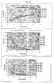
- the hybridization signal was quantified by first exposing the labeled sections to BAS-IP SR 2025 universal imaging plates which were then scanned in a Fujifilm BAS5000 high resolution phosphor imager and finally measured a MCID M2 image analysis system (imaging Research, St. Catherines, Ontario, Canada). The hybridization signal was expressed as photostimulated luminescence (PSL) per mm 2 ( Figs. 2 and 3 ).
- the tissues from wild-type and transgenic mice were prepared as for hybridization histochemistry except that they were not fixed.
- the sections were incubated in vitro with 0.1 nM 125 I- ⁇ -amyloid peptide (Amersham) for 2h at 20°C, washed dried and exposed to tritium-sensitive imaging plates (BAS-TR2025) or x-ray film for 4-14 days.
- mice were killed by decapitation, brains removed, frozen in dry ice and kept at - 70°C until use.
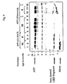
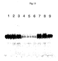
- tissues from 3 littermates were pooled. Tissues were homogenized as already described (Malherbe et al. 1996). Extracted proteins (50 ⁇ g/slot) were separated on 10% Tris-Tricine gels, transferred to nitrocellulose and probed with either 22C11mAb, 1:200 (Boehringer-Mannheim) or rabbit anti-KPI at 0.8 ⁇ g/ml ( Löffler J, Huber G. Beta-amyloid precursor protein isoforms in various rat brain regions and during brain development. J Neurochem. 1992 Oct;59(4):1316-24 )( Fig. 4 ).
- transgenic mouse lines AD124 ⁇ APP mini-gene
- AD124/82, AD124/68, and AD124/64 were compared with AD124/15 and with 4 wild-type controls.
- the results of this analysis indicates that, for lines 82, 68, and 64, there was on average a 5-9-fold, 8-13-fold and 14-23-fold increase in transgenic whole brain, cortex and hippocampus CA-1, respectively.
- Transgenic mouse lines express human BACE protein under the control of the mouse prion promoter. These mouse lines were denominated Prp-Asp.
- the BACE 1 cDNA (SEQ ID NO:8) encoding protein sequence of SEQ ID NO:9 and comprising the complete open reading frame (ORF) plus flanking restriction sites was inserted into a mouse prion minigene vector where the prion ORF had been replaced by a suitable unique cloning site.
- the original minigene construct pPrPHg was described by Fischer et al. ( Fischer M, Rulicke T, Raeber A, Sailer A, Moser M, Oesch B, Brandner S, Aguzzi A, Weissmann C. Prion protein (PrP) with amino-proximal deletions restoring susceptibility of PrP knockout mice to scrapie. EMBO J. 1996. 15:1255-64 ).
- mouse prion ORF between an upstream KpnI site and a downstream NarI site was removed and replaced by a SceI restriction site.
- the BACE1 containing fragment was inserted by blunt ligation and its direction relative to the prion promoter verified by sequencing.
- the resulting fusion gene ( FIG. 5 ) was freed of vector sequences, purified and microinjected using standard protocols into one-cell embryos of the cross C57B1/6 x DBA/2 F2 mice.
- Ten transgenic founders (F0) were identified as having the transgenic by PCR analysis of tail DNA and subsequently crossed with wild-type C57B1/6 mice to establish transgenic lines. Briefly, genomic DNA was extracted following the protocol described by Laird PW, Zijderveld A, Linders K, Rudnicki M.A, Jaenisch R, and Berns A. Simplified mammalian DNA isolation procedure. Nucleic Acids Res. 1991. 19, 4293 .
- PCR was run at 30 cycles: 95°C x 30 seconds, 59°C x 1 minute, 72°C x 1 minute; followed by 72°C x 5 minutes, using prion sense (Seq ID NO:10) and BACE anti-sense (Seq ID NO: 11) primers. Eight out of the ten founders had integrated the transgene in the germ line and eight transgenic lines could be established.
- mice from the F1 generation were sacrificed in order to characterize the transgenic lines at the DNA level (Southern blot), RNA level (Northern blot and RT-PCR) and protein level (Western blot).
- Transgene copy number was determined by Southern blotting using as references known amounts of transgene fragment mixed to genomic DNA isolated from nontransgenic littermates. Ten ⁇ g of genomic XbaI restriction fragments were fractionated by gel electrophoresis and blotted onto Nylon membranes (Roche Molecular Biosciences). A 1.1kb DNA probe (XmnI digestion fragment of the transgene) was labeled with [ 33 P]dCTP by the random primer method using the Ready-to-Go DNA labeling kit (Pharmacia Biotech). Hybridization was performed overnight at 65°C in 6x standard saline citrate, 10% dextran sulfate, 0.5% SDS.
- Blots were washed in 2x standard saline citrate (+0.1% SDS) at 65°C for 20 minutes followed by a second wash at 65°C in 0.2x standard saline citrate (+0.1% SDS). Blots were imaged with a Phosphorimager and quantified using the ImageQuant software.
- the mixture was then chilled on ice and incubated with 8 ⁇ l 5 ⁇ reverse transcriptase buffer (250 mM Tris-HCl, pH 8.4, 375 mM KCl, 15 mM MgCl 2 ), 2 ⁇ l dNTP mix (10mM), 4 ⁇ l 0.1 M dithiothreitol, and 4 ⁇ l of Superscript II RT Moloney Murine Leukemia virus reverse transcriptase (200 U/ ⁇ l) at 42°C for 60 min.
- the reaction mixtures were further incubated for 5 min at 95°C and diluted to a final volume of 100 ⁇ l with ice-cold ddH 2 0.
- the cDNA was stored at -20°C until use.
- Quantitative PCR was performed in an optical 96-well plate (PE Applied Biosystems, CA) using a Perkin Elmer 7700 sequence detector.
- the PCR reaction consisted of the following steps: 1) 50°C for 2 min to prevent carry over of DNA; 2) 95°C for 10 min to activate ampliTaq Gold polymerase; 3) 50 cycles each consisting of 95°C for 15 sec, 60° for 15 sec, and 72°C for 45 sec.
- DNA was mixed with loading buffer and separated on 3% agarose gels by electrophoresis. Ethidium bromide-stained DNA fragments
- RNA purified from 50 ⁇ g of total RNA was separated on a 0.7% formaldehyde-agarose gel.
- RNA was then blotted onto a Hybond + membrane (Amersham Pharmacia Biotech) and hybridized to a mixture of 60 bp single-stranded oligonucleotide probes specific for RNA of the human BACE transgene beginning at nucleotide 30 (probe 659, SEQ ID NO:16), at nucleotide 570 (probe 660, SEQ ID NO:17), at nucleotide 1186 (probe 661, SEQ ID NO:18) of human BACE open reading frame.
- Probes have been 3'end-labeled by the terminal deoxynucleotidyl transferase reaction with alpha-[ 32 P]dATP (6000 Ci/mmol; Amersham Pharmacia Biotech) dATP for 60 min at 37°C, following standard procedure.
- Hybridization was done in Rapid Hybridization Buffer (Amersham Pharmacia Biotech) for 2.5 hours at 65°C. Blots were washed in 2x SSPE (0.1%SDS) at room temperature for 10 minutes, followed by a second wash for 15 minutes at 65°C and 2 more washes for 10 minutes in 1X SSPE (0.1%SDS) at 65°C. Membranes were exposed overnight on Phosphoimager for quantification. Hybridized probes were stripped from the blots by boiling in H20/0.1%SDS.
- blots were then hybridized for 90 minutes at 60°C with a [ 32 P] labeled single-stranded oligonucleotide probe specific for RNA of the mouse ⁇ -actin (SEQ ID NO:19). After the washing steps, membranes were exposed overnight on Phosphoimager.
- mice were killed by decapitation, brains removed and halved in a sagittal plane before freezing in dry ice.
- Fozen brains were kept at -70°C until use.
- Half brains were homogenized in 1 ml 50 mM Hepes buffer pH 7.3 containing 0.1% Triton X-100 and 1% inhibitorcocktail III (Pierce).
- the proteins contained in 5 ⁇ l of each sample were fractionated by sodium dodecyl sulphate-polyacrylamide gel electrophoresis using 4-20% Novex (Invitrogen) gels.
- the level of BACE was analyzed in two mice and compared to the level of endogenous BACE as measured in non-transgenic littermates.
- Line 13 and 24 the two highest expressing lines were estimated to express human BACE at levels corresponding to 5-10 fold the amount of endogenous mouse BACE.
- Line 10 which was defined as a low expressing transgenic line expressed human BACE at levels of about 2 fold the endogenous BACE levels ( Fig. 6A and 6B ).
- APPV717F high expressor transgenic line AD124/68 was crossed with BACE high expressor transgenic lines Prp-Asp13 or Prp-Asp24.
- the first type of crossbreeding involved homozygotic APPV717F mice and heterozygotic Prp-Asp transgenic mice leading to bigenic littermates (APPV717+/- and BACE +/-) and singly transgenic littermates (APPV717+/-).
- the second type of crossbreeding involved heterozygotic APPV717F transgenic mice and heterozygotic Prp-Asp transgenic mice resulting in the generation of non-transgenic littermates (APPV717 -/- and BACE -/-), bigenic littermates (APPV717 +/- and BACE +/-), singly transgenic littermates (APPV717 +/-), and singly transgenic littermates (BACE +/-).
- the preparation of brain slices was performed following a protocol by Metzger et al. ( Metzger F, Kapfhammer JP. Protein kinase C activity modulates dendritic differentiation of rat Purkinje cells in cerebellar slice cultures. Eur J Neurosci. 2000 12:1993-2005 ). Shortly, post natal day 8-10 mice were decapitated and the brain was aseptically removed. The cerebellum was dissected in ice cold medium (MEM with 2 mM glutamax I, Life Technologies) and the meninges were carefully removed.
- MEM ice cold medium
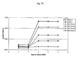
- A-beta 40 in culture supernatant of cerebellum slices from wild-type, APP London single transgenic and [APP London x BACE] double transgenic mice was monitored ( Fig. 7A ).
- An increase of A-beta 40 peptide is clearly seen after 3 days of culture for APP London single and [APP London x BACE] double transgenic mice.
- the APP London single transgenic mouse shows A-beta 40 levels of about 0.7 ng/ml/day whereas all double transgenic mice show levels above 1 ng/ml/day. Confirmation of those results and extension of the analysis to the assessement of sAPP alpha and sAPP beta fragments was performed with supernatants from brain slices originating from one litter ( Fig.
- Double [APP London x BACE] transgenics show increased levels of sAPP beta when compared to APP London littermates.
- the level of sAPPalpha fragment was below detection limit in [APP London x BACE] double transgenic mice brain slices; in comparison, levels between 15 and 20 ng/ml/day were measured after 1 day of culture for APP London single transgenic mice.
- mice were sacrificed and their brains removed.
- One cerebral hemisphere was homogenized using FastPrep homogenization Tubes (Qbiogene, Carlsbad, CA) in 1 ml of DEA buffer (2% diethanolamine, 50 mM NaCl).
- Supernatants containing soluble amyloid were collected after centrifugation and neutralized to pH 8 with 1M Tris-HCl as already described ( Savage MJ, Trusko SP, Howland DS, Pinsker LR, Mistretta S, Reaume AG, Greenberg BD, Siman R, Scott RW. Turnover of amyloid beta-protein in mouse brain and acute reduction of its level by phorbol ester. J Neurosci. 1998. 18: 1743-5 ).
- pellets were resuspended in 0.5 ml 9M urea, incubated overnight under rotation at 4°C and supernatants collected after centrifugation. Both types of supernatants (collected after either DEA or urea extraction) were tested in ELISA.
- 6E10 and 4G8 monoclonal antibodies are commercially available (Senetek, Maryland Heights, Missouri).
- the BAP-1 antibody is specific for the epitope A-beta 4-7 which is present in soluble APP alpha, but not in soluble APP beta.
- Monoclonal antibodies BAP17 and BAP24 are specific for amyloid peptide A-beta 40, whereas monoclonal antibody BAP15 is specific for amyloid peptide A-beta 42 ( Brockhaus M, Grunberg J, Rohrig S, Loetscher H, Wittenburg N, Baumeister R, Jacobsen H, Haass C.
- the peptide conjugate was emulsified in Freunds complete adjuvants (Difco) and injected at 4 sites subcutanously. The injections were repeated 3 times at 4 weeks intervals using Freunds incomplete adjuvants (Difco). A high titered rabbit serum was obtained. It was purified by affinity chromatography on sulfolink gel (Pierce) to which the peptide CISEVKM had been coupled according to the instructions of the manufacturer. Since recombinant sAPP alpha was void of reactivity in the assay for sAPP beta, it was considered to be specific for the C-terminal neoepitope that is generated by the action of beta-secretase on APP.
- affinity purified antibodies were conjugated to horseradish peroxidase (Roche Molecular Biochemicals) using periodate activation ( Nakane PK. Recent progress in the peroxidase-labeled antibody method. Ann N Y Acad Sci. 1975. 254:203-11 ).
- the antibody combination used for the detection of amyloid peptides in brain extracts are biotinylated 6E10 or biotinylated 4G8 monoclonal antibodies for the capture and HRP-BAP24 or HRP-BAP15 monoclonal antibodies for the detection of A-beta 40 and A-beta 42, respectively (Brockhaus et al. 1998). Soluble APP alpha- and APP beta-fragments were detected using biotinylated ABS monoclonal antibody for the capture and HRP-BAP1 monoclonal antibody or HRP-rbVKM for the detection, respectively.
- the ELISA was performed as previously described (Brockhaus et al. 1998). Briefly, microtiter plates (Nunc Maxisorb, Life Technologies) were coated first with streptavidin (Roche Molecular Biochemicals) at 5 ⁇ g/ml in 100 mM bicarbonate buffer then incubated with capture biotinylated mAb. After extensive washings of the plates with Tris buffered saline 0.05% Tween 20 (TBS-T), five fold serial dilutions of supernatants were added and incubated for 24h at 4°C in incubation buffer buffer buffer (Tris 50mM, NaCl 140 mM, EDTA 5 mM, NP40 0.05%, gelatin 0.25%, BSA 1%).
- the minimal dilutions were for culture supernatants: 1 fold, for DEA/Tris supernatants: 4 fold, for urea supernatants: 20 fold.
- the detection of the amyloid fragments was performed with Horse-Raddish Peroxidase (HRP) coupled monoclonal antibodies. Excess of HRP-mAbs was eliminated through three washing steps using TBS-T buffer. The tetramethylbenzidine (TMB) substrate was added to the plates for 5-10 minutes and the Optical Density was read at 450nm after having stopped the reaction with sulfuric acid 2.5N. Purified A-beta or soluble APP fragments were used for calibration of the assay.
- A-beta 40 and A-beta 42 peptides were measured in [BACE x APP London] double transgenics.
- A-beta 42 peptide levels were about 10 fold higher than A-beta 40 peptide levels.
- Similar results were obtained with double transgenic lines for APP London x BACE line 13 or APP London x BACE line 24, ruling out an effect due to the transgene integration site.
- the increase of pathogenic amyloid peptide deposition is induced by the expression of beta-secretase.
- the levels of soluble APPalpha fragment and soluble APPbeta fragment in brains of [BACE x APP London] double transgenic mice were also measured ( Fig.8 ).
- beta-secretase levels were compared in double and single transgenic mice.
- three mice (2 males and 1 female) were sacrificed at the age of 3 months from the [BACE line 13 x APP London] double transgenic mice, from the APP London single transgenic mice (negative control for the human BACE) and from the BACE line 13 single transgenic mice.
- No significant difference was observed between the levels of beta-secretase detected in the [BACE line 13 x APP London] double transgenic mice and the levels detected in the parental BACE 13 line ( Fig. 11 ).
- Brain tissue from transgenic or non-transgenic littermate control mice was homogenized on ice in 0.32 M sucrose solution containing protease inhibitors (CompleteTM, Boehringer-Mannheim, Germany) using FastPrep homogenization Tubes (Qbiogene, Carlsbad, CA). The protein concentration in the supernatant was measured using the BCA protein Assay (Pierce, USA). For APP and A-beta detection 25 ⁇ g of protein extract was denatured at 95°C for 10 min in Novex NuPage loading buffer (Invitrogen). The proteins were fractionated by sodium dodecyl sulphate-polyacrylamide gel electrophoresis using 10% Novex NuPage (Invitrogen) gels.
- the filter was heated in PBS for 5min to increase sensitivity subsequently blocked with 5% (w/V) non-fat dry milk in PBST (PBS, 0.05% (V/V) Tween 20) and incubated overnight at 4°C with the antibody WO-2 at a concentration of 0.1 ⁇ g/ml. Binding of the primary antibody was detected with horseradish peroxidase conjugated anti-IgG antibody (Amersham) followed by enhanced chemiluminescence detection system (Amersham) according to the manufacturer's instructions.
- Fluothane R -anesthetised mice were fixed by transcardiac perfusion of 4% formaldehyde in PBS at 22°C after which the brains were removed, halved in a sagittal plane and immersed in the same fixative for 24h. One brain half was then cryoprotected overnight in 30% sucrose before freezing in dry-ice and the other half dehydrated and embedded in paraffin wax. Cryostat or paraffin sections were cut at 10 ⁇ m.
- Fibrillar A ⁇ deposits were detected with Accustain R amyloid stain, Congo red (Sigma Diagnostics HT60).
- the following antibodies were used to detect antigenic sites with a Vectastain R ABC kit Elite and peroxidase (ImmunoPure® Metal Enhanced DAB Substrate Pierce 34065): BAP-2 (monoclonal mouse IgG1 raised against A ⁇ 1-40; recognizes amino acids 4-7, Dr.
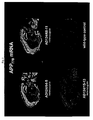
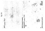
- Amyloid deposition in plaque structures was clearly detected in 11 months old [BACE x APP London] double transgenic mouse ( Fig. 10A ). Some of the plaques were congophilic. Strong congophilic staining was also observed on vascular amyloid deposition. No significant difference in the brain distribution of amyloid deposits was observed between [BACE x APP London] double transgenic mice and APP LondonH single transgenics, but immunostaining using BAP-2 mAb showed a consistant stronger staining pattern on amyloid deposits from [BACE x APP London] double transgenic mice when compared to single transgenic mice suggesting some difference in the amyloid plaque structure or composition.
- Beta-secretase was detected most probably in association with the amyloid deposits as demonstrated by the positive immunostaining with BSC-1 mAb ( Fig 10B ).
- Amyloid deposition detected in [BACE x APP London] double transgenic mice was also accompanied by the presence of inflammatory markers: GFAP and CD45 ( Fig. 10B ).
- Perfusion-fixed brain tissue while immersed in fixative, was cut into approximately 100 ⁇ m thick sections using a vibratome and cryoprotected by immersion in increasing concentrations of glycerol (10-20-30 % v/v) in 10 mM phosphate buffer, pH 7.4 for 0.5 h per concentration. Vibratome sections were carefully picked up by forceps and plunged into liquid ethane cooled by liquid nitrogen. Frozen tissue slices were stored in liquid nitrogen before being transferred to the pre-cooled chamber (-90°C) of an automated freeze-substitution apparatus (AFS, Reichert, Austria). The tissue was immersed overnight in anhydrous methanol at -90°C, containing 0.5 % (w/v) uranyl acetate.
- Ultrathin sections were prepared on a Reichert Ultracut S using a diamond knife (Diatome) and collected onto formvar/carbon-coated 200-mesh nickel grids.
- the sections were floated on 0.05 mol/L glycine in PBS for 15 min to inactivate free aldehyde groups and then on 2.5% (w/v) hen egg white albumin (Fluka) with 2.5% (w/v) bovine serum albumin (BSA, fraction V, Boehringer) in PBS for 15 min to block nonspecific binding sites.
- Sections were incubated with 10 ⁇ g/ml mAb BAP-2 in 2% BSA/PBS for 1 hour.
- Immuno-electron microscopy was carried out on low temperature-embedded mouse brain tissue of 18 months old animals. The method combines the advantage of superior preservation of ultrastructure and antigenicity for efficient immunocyto-chemical staining.
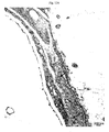
- Immunogold labeling using a monoclonal antibody against the N-terminal part of the A-beta-peptide (BAP-2), revealed fibrillar A-beta within the media of arteries and arterioles. Bundles of fibrillar A-beta occassionally penetrate into the basal membrane which might interfere with the integrity of the blood brain barrier ( Fig. 12A ). Fibrillar A-beta is found at considerable density at the layer of smooth muscle cells ( Fig.
- CAA cerebral amyloid angiopathy
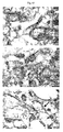
- Dense cored plaques which can be detected by congo red staining are detectable at low frequency while diffuse A-beta deposits that are not stained by congo red but are detectable by immunohistochemistry are more frequent.
- the ultrastructure of A-beta within diffuse plaques was analysed by immuno-gold labeling against A-beta in the frontal cortex of 18 months old animals ( Fig. 13 ). Immunoreactivity was found on discrete patches of electron-dense material adjacent to the cell membrane of neurites and dendrites. Remarkably, no clear fibrillar ultrastructure, as observed in the media of blood vessels is detected.
- A-beta resembles early aggregation clusters that are intermediate precursors to the well-characterized mature A-beta fibrils.
- Indications for early A-beta aggregation that precedes significant neurodegeneration is particularly important to study the mechanism(s) ofA-beta-mediated neurotoxicity and characterize therapeutics for the treatment of AD.
- mice expressing APP London and [BACE x APP London] double transgenic mice were compared with age-matched wild type mice. Mice were weighed weekly and general appearance was checked. All mice were individually housed. In addition, body temperature, coat appearance, secretory signs, body posture were also noted.
- Wire manouvre Mice were placed by forepaws on an elevated wire rod and the latency to fall was noted. Cut-off time was 60s and the best score from 3 attempts was recorded.
- Grip strength Mice were forced to pull on a strain gauge and the release point is recorded. The best score from 5 attempts is recorded.
- Rotarod Mice were placed on a constant speed rotarod and the latency to fall was noted. Cut-off time was 120s and the best score from 3 trials was taken. 2 speeds were used: 16rev/min, 32rev/min.
- Locomotor activity The mice were placed into a novel test chamber for a 1 h period which consists of a Plexiglas ® box (20 cm x 20 cm x 27 cm) with sawdust bedding on the floor. The animals' movements were recorded using an electronic monitoring system (Omnitech Electronics Inc., Columbus, Ohio, USA). Movement of the animal results in interruption of an array of photobeams from vertically and horizontally located infrared sources placed around the test chamber. Total distance travelled (cm) and number of rears were measured.
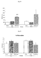
- mice expressing hBACE/hAPPLo had increased locomotor activity over the one hour test period, as demonstrated by a significant increase in total mobile counts during this period. Consistent with this, mobile time was also significantly elevated in male and female hBACE/hAppLo mice, and whilst not significant, a similar trend was seen in the total number of rear counts. Thus, both male and female mice expressing hBACE/hAPPLo were hyperactive when compared with wild type controls ( Fig. 14 ).
- mice were placed in a Y-maze made of black perpex (each arm is 53 cm long, 15 cm wide and 30 cm in height) for 5 min. A camera was positioned above the maze and the animals were observed on a monitor in an adjoining room. The number of arm entries and their entry sequence was noted to calculate an alternation measure.
- mice There was no effect of genotype in male mice. However, female hBACE/hAPPLo mice showed a deficit in this task of working memory, as measured by a reduction in percentage alternation when compared with wild type controls ( Fig. 15 ). Notably, there was no difference in percentage position bias between gender or genotype. Interestingly, whilst not significant, both male and female hBACE/hAPPLo mice showed a tendency for an increased number of arm entries, consistent with these mice being more active than wild type controls, as demonstrated previously in the locomotor activity test (see Fig. 14 ). Thus, female, but not male, mice expressing hBACE/hAPPLo were impaired in this task of working memory.
- the water maze consisted of a grey circular tank (1m diameter) filled with water made opaque by the addition of a latex solution (E-308; Induchem, Voletswil, Switzerland). Pool temperature was maintained at 21+1°C.
- the escape platform (8cm diameter) was positioned 1cm below water level in the centre of one of the pool quadrants.
- platform position was signalled by the addition of a small black flag which is positioned in the centre of the submerged platform.
- the walls surrounding the water maze were hung with posters and flags which serve as visual cues and are visible during all stages of training and testing. Movement of the mice within the pool was tracked and analysed with a computer based video tracking system (HVS Image, Hampton, UK).
- mice were placed in the pool facing the edge at one of four start positions (NE, SE, SW, NW), and were required to locate the flagged platform whose position varies across trials. Each mouse received a total of 12 trials (three trials per block, 2 blocks per day, 2 day duration). Intertrial intervals averaged 10 min, and maximum trial length was 60s. If mice failed to find the platform within 60s, they were guided to its position by the experimenter. All mice were allowed to remain on the platform for a 10s period before being removed and returned to the homecage. The cued task was followed by the place task, in which mice were required to locate a submerged hidden platform whose position remained fixed throughout training. Platform location was balanced within groups.
- Each mouse received 8 blocks of training trials over four consecutive days (three trials per block, timing as per cued test) in which they were placed in the pool at one of four start positions, and allowed to locate the hidden platform. Assessment of spatial learning was conducted in probe trials performed both 30 min after block 4, and 24h following the final trial. In each probe trial the platform was removed from the pool, and the path swam by each mouse recorded over a 60s period. In cued acquisition there was neither a gender and genotype nor gender, genotype and trials interaction indicating that visual acuity and motivation to find the escape platform was equivalent between genotypes.
- both male and female wild type mice had learnt the position of the hidden platform such that they spent significantly longer exploring the quadrant in which the platform was located during training when compared with time spent in the other three quadrants ( Fig. 17 ).
- male and female mice expressing both hAPPLo and hBACE/hAPPLo spent equivalent times exploring all four quadrants showing they had not learnt the position of the hidden platform.
- both male and female wild type mice spent significantly longer exploring the quadrant in which the platform was located during training showing that they had learnt the position of the hidden platform.
- mice expressing hAPPLo spend equal time exploring the four quadrants showing that they had not learnt the position of the hidden platform, in contrast, hAPPLo female mice had learnt the platform position.
- Both male and female mice expressing hBACE/hAPPLo showed a tendency towards spending more time in the quadrant containing the platform during training, although this was not significant in all cases, showing that these mice had not learnt the task to the same precision as the wild type controls.
- mice were placed into a 2-compartment chamber within which they could freely pass between compartments (San Diego Instruments, USA). Each trial began with the side currently occupied by the mouse being illuminated by a 10s light (CS), which is used to signal a footshock (0.2mA) of maximum duration 20s .(NB. the mice never receive this shock duration for they either escape within 1s to the other (unshocked) compartment, or learn to avoid the shock altogether). This was followed by a variable timeout period (mean 20s, range 15-25s) (no light) in which the mouse can freely explore the chamber. Following the timeout, the next trial begins. Shock can be avoided either by a shuttle to the next compartment during the CS period, (i.e avoidance) or escape at any time during the shock presentation. Ten daily test sessions were run with each session consisting of 20 trials. The dependant measure is % avoidance.
- mice did not differ in their shock threshold nor did they display elevated locomotor activity ( Fig. 14 ) when compared with wild type controls, thus, the active avoidance deficit represents a true learning deficit.
- female mice expressing hBACE/hAPPLo demonstrated an enhanced jump response when compared with wild type controls which could suggest an elevated shock perception and additionally these mice were also hyperactive when compared to wild type controls. Both of these factors could account for the marginal (but non-significant) increase in active avoidance learning seen.
- both male and female mice expressing hAPPLo, but not hBACE/hAPPLo were impaired in active avoidance learning.
- Shock Threshold Minimum shock (mA) required to elicit a flinch response Minimum shock (mA) required to elicit a jump response Minimum shock (mA) required to elicit vocalisations
Landscapes
- Life Sciences & Earth Sciences (AREA)
- Health & Medical Sciences (AREA)
- Engineering & Computer Science (AREA)
- Zoology (AREA)
- Genetics & Genomics (AREA)
- Environmental Sciences (AREA)
- Biotechnology (AREA)
- Veterinary Medicine (AREA)
- General Health & Medical Sciences (AREA)
- Biomedical Technology (AREA)
- Biodiversity & Conservation Biology (AREA)
- Animal Husbandry (AREA)
- Bioinformatics & Cheminformatics (AREA)
- Animal Behavior & Ethology (AREA)
- Wood Science & Technology (AREA)
- General Engineering & Computer Science (AREA)
- Chemical & Material Sciences (AREA)
- Organic Chemistry (AREA)
- Biophysics (AREA)
- Biochemistry (AREA)
- Microbiology (AREA)
- Plant Pathology (AREA)
- Molecular Biology (AREA)
- Physics & Mathematics (AREA)
- Medicines That Contain Protein Lipid Enzymes And Other Medicines (AREA)
- Micro-Organisms Or Cultivation Processes Thereof (AREA)
Claims (23)
- Verfahren zur Erzeugung eines transgenen nicht-humanen Säugers, dessen Genom DNA inkorporiert, umfassend eine kodierende Sequenz, die humane beta-Secretase kodiert, die operabel mit einer regulatorischen Promotorsequenz verknüpft ist, und eine kodierende Sequenz, die humanes APP London kodiert, das operabel mit einer regulatorischen Promotorsequenz verknüpft ist, wobei das Verfahren:(a) das Einführen eines DNA-Konstruktes, umfassend eine kodierende Sequenz, die humane beta-Secretase kodiert, und eine Regulationssequenz, die operabel mit der kodierenden Sequenz verknüpft ist, in eine Zelle oder einen Embryo des nicht-humanen Säugers;(b) das Einführen eines DNA-Konstruktes, umfassend eine kodierende Sequenz, die humanes APP London kodiert, und eine Regulationssequenz, die operabel mit der kodierenden Sequenz verknüpft ist, in eine Zelle oder einen Embryo des nicht-humanen Säugers;(c) das Erzeugen eines transgenen nicht-humanen Säugers aus den jeweiligen Zellen oder Embryonen und(d) das Kreuzen der transgenen nicht-humanen Säuger umfaßt.
- Verfahren nach Anspruch 1, wobei eine Prionenpromotorregulationssequenz operabel mit der kodierenden Sequenz von humaner beta-Secretase verknüpft ist und eine Thy-1-Promotorregulationssequenz operabel mit der kodierenden Sequenz von humanem APP London verknüpft ist.
- Verfahren nach Anspruch 1 oder 2, wobei die kodierende Sequenz für humane beta-Secretase SEQ ID NR.: 8 ist.
- Verfahren nach den Ansprüchen 1 bis 3, wobei die kodierende Sequenz für humanes APP London SEQ ID NR.: 1 ist.
- Transgener nicht-humaner Säuger, dessen Genom DNA inkorporiert, umfassend eine kodierende Sequenz, die das humane APP London kodiert, das operabel mit einer Regulationssequenz verknüpft ist, und eine kodierende Sequenz, die die humane beta-Secretase kodiert, die operabel mit einer Regulationssequenz verknüpft ist.
- Transgener nicht-humaner Säuger nach Anspruch 5, wobei die kodierende Sequenz humaner beta-Secretase operabel mit einer Prionenpromotorsequenz verknüpft ist und die kodierende Sequenz von humanem APP London operabel mit einer Thy-1-Promotorsequenz verknüpft ist.
- Transgener nicht-humaner Säuger nach Anspruch 5 oder 6, wobei die kodierende Sequenz für humane beta-Secretase SEQ ID NR.: 8 ist.
- Transgener nicht-humaner Säuger nach den Ansprüchen 5 bis 7, wobei die kodierende Sequenz für humanes APP London SEQ ID NR.: 1 ist.
- Transgener nicht-humaner Säuger nach den Ansprüchen 5 bis 8, der eine oder mehrere Histopathologien ähnlich denen der Alzheimer-Krankheit zeigt.
- Transgener nicht-humaner Säuger nach den Ansprüchen 5 bis 9, wobei der Säuger ein Nagetier ist.
- Transgener nicht-humaner Säuger nach den Ansprüchen 5 bis 10, wobei der Säuger eine Maus ist.
- Nachkomme des transgenen nicht-humanen Säugers nach einem der Ansprüche 5 bis 11, erhalten durch Züchten des transgenen Säugers, wobei der Nachkomme die kodierende Sequenz, die humane beta-Secretase kodiert, und die kodierende Sequenz, die humanes APP London kodiert, wie in Anspruch 1 definiert, behält.
- Zellinie oder primäre Zellkultur, gewonnen aus einem transgenen nicht-humanen Säuger oder seinen Nachkommen nach den Ansprüchen 5 bis 12.
- Organotypische Kultur einer Gehirnscheibe, gewonnen aus einem transgenen nicht-humanen Säuger oder seinen Nachkommen nach den Ansprüchen 5 bis 12.
- Verwendung eines transgenen nicht-humanen Säugers nach den Ansprüchen 5 bis 12 oder einer Zellinie oder primären Zellkultur nach Anspruch 13 oder einer organotypischen Kultur einer Gehirnscheibe nach Anspruch 14 als ein Modell für Amyloidosen, einschließlich Alzheimer-Krankheit.
- Verfahren zum Screenen von Testverbindungen zur Verwendung bei der Vorbeugung oder Behandlung eines degenerativen Zustandes des Gehirns, wobei das Verfahren die Verabreichung einer Testverbindung an einen transgenen nicht-humanen Säuger nach den Ansprüchen 5 bis 12 und die Bestimmung der Wirkung der Verbindung auf einen Krankheitsmarker, umfassend die Menge an APP-Spaltprodukten, die Neuropathologie und Verhaltensveränderungen umfaßt.
- Verfahren nach Anspruch 16, wobei der degenerative Zustand des Gehirns die Alzheimer-Krankheit ist.
- Verfahren zum Screenen von Testverbindungen zur Verwendung bei der Vorbeugung oder Behandlung eines degenerativen Zustandes des Gehirns, wobei das Verfahren die Verabreichung einer Testverbindung an eine organotypische Kultur einer Gehirnscheibe nach Anspruch 14 und die Bestimmung der Wirkung der Verbindung auf einen Krankheitsmarker, umfassend die Menge an APP-Spaltprodukten, umfaßt.
- Verfahren nach Anspruch 18, wobei der degenerative Zustand des Gehirns die Alzheimer-Krankheit ist.
- Verfahren zum Screenen von Testverbindungen zur Verwendung bei der Vorbeugung oder Behandlung eines degenerativen Zustandes des Gehirns, wobei das Verfahren die Verabreichung einer Testverbindung an eine Zellinie oder primäre Zellkultur nach Anspruch 13 und die Bestimmung der Wirkung der Verbindung auf einen Krankheitsmarker, umfassend die Menge an APP-Spaltprodukten, umfaßt.
- Verfahren nach Anspruch 20, wobei der degenerative Zustand des Gehirns die Alzheimer-Krankheit ist.
- Kit zum Screenen von Testverbindungen, die zur Vorbeugung oder Behandlung eines degenerativen Zustandes des Gehirns, einschließlich der Alzheimer-Krankheit, verwendbar sind, umfassend eine Zellinie oder primäre Zellkultur nach Anspruch 13 und ein Mittel zur Bestimmung, ob eine Testverbindung die Wirkungen, die mit der Überexpression der Transgene in der Zelle in Verbindung stehen, zeigt.
- Verwendung eines transgenen nicht-humanen Säugers nach den Ansprüchen 5 bis 12 oder einer Zellinie oder einer primären Zellkultur nach Anspruch 13 oder einer organotypischen Kultur einer Gehirnscheibe nach Anspruch 14 zum Screenen von Verbindungen, die zur Vorbeugung oder Behandlung degenerativer Gehirnerkrankungen verwendbar sind.
Priority Applications (1)
| Application Number | Priority Date | Filing Date | Title |
|---|---|---|---|
| EP20030003183 EP1340424B1 (de) | 2002-03-01 | 2003-02-19 | Doppel-transgenisches, nicht-humanes Säugertiermodell für die Alzheimer Krankheit |
Applications Claiming Priority (3)
| Application Number | Priority Date | Filing Date | Title |
|---|---|---|---|
| EP02004331 | 2002-03-01 | ||
| EP02004331 | 2002-03-01 | ||
| EP20030003183 EP1340424B1 (de) | 2002-03-01 | 2003-02-19 | Doppel-transgenisches, nicht-humanes Säugertiermodell für die Alzheimer Krankheit |
Publications (2)
| Publication Number | Publication Date |
|---|---|
| EP1340424A1 EP1340424A1 (de) | 2003-09-03 |
| EP1340424B1 true EP1340424B1 (de) | 2008-12-31 |
Family
ID=27736044
Family Applications (1)
| Application Number | Title | Priority Date | Filing Date |
|---|---|---|---|
| EP20030003183 Expired - Lifetime EP1340424B1 (de) | 2002-03-01 | 2003-02-19 | Doppel-transgenisches, nicht-humanes Säugertiermodell für die Alzheimer Krankheit |
Country Status (1)
| Country | Link |
|---|---|
| EP (1) | EP1340424B1 (de) |
Families Citing this family (3)
| Publication number | Priority date | Publication date | Assignee | Title |
|---|---|---|---|---|
| WO2004062627A2 (en) * | 2003-01-13 | 2004-07-29 | The Regents Of The University Of California | In vivo screening models for treatment of alzheimer's disease and other neurodegenerative disorders |
| ES2368054B1 (es) * | 2009-06-16 | 2012-09-13 | Universidad Del Pais Vasco | Modelo organotipico de enfermedades neuro-degenerativas, proceso de obtencion y usos del mismo |
| CN113957095A (zh) * | 2021-10-22 | 2022-01-21 | 江苏集萃药康生物科技股份有限公司 | 一种用于构建阿尔兹海默症动物模型的构建方法及其核酸组合物和应用 |
-
2003
- 2003-02-19 EP EP20030003183 patent/EP1340424B1/de not_active Expired - Lifetime
Also Published As
| Publication number | Publication date |
|---|---|
| EP1340424A1 (de) | 2003-09-03 |
Similar Documents
| Publication | Publication Date | Title |
|---|---|---|
| Richards et al. | PS2APP transgenic mice, coexpressing hPS2mut and hAPPswe, show age-related cognitive deficits associated with discrete brain amyloid deposition and inflammation | |
| US7186881B2 (en) | Testing compounds for effects on synaptophysin in transgenic mice expressing an Alzheimer's disease FAD DNA sequence | |
| Moechars et al. | Expression in brain of amyloid precursor protein mutated in the alpha‐secretase site causes disturbed behavior, neuronal degeneration and premature death in transgenic mice. | |
| Kumar-Singh et al. | Behavioral disturbances without amyloid deposits in mice overexpressing human amyloid precursor protein with Flemish (A692G) or Dutch (E693Q) mutation | |
| CA2257852C (en) | Transgenic non-human mammals with progressive neurologic disease | |
| AU747691B2 (en) | Apolipoprotein E transgenic animals and assay methods | |
| JPH07132033A (ja) | アルツハイマー病モデルトランスジェニック動物 | |
| JPH09511388A (ja) | 進行性神経疾患を持つヒト以外のトランスジェニック哺乳類 | |
| JP2001517065A (ja) | トランスジェニック動物モデルを用いてアルツハイマー病治療薬を同定する方法 | |
| US7247766B2 (en) | Double transgenic mice overexressing human beta secretase and human APP-London | |
| JP5070236B2 (ja) | 神経変性性障害のトランスジェニック動物モデル | |
| WO1996040896A1 (en) | Method for identifying alzheimer's disease therapeutics using transgenic animal models | |
| US7767879B2 (en) | Transgenic animal model of neurodegenerative disorders | |
| EP1340424B1 (de) | Doppel-transgenisches, nicht-humanes Säugertiermodell für die Alzheimer Krankheit | |
| US20090081128A1 (en) | Model of Alzheimer's Disease | |
| Ozmen et al. | BACE/APPV717F double-transgenic mice develop cerebral amyloidosis and inflammation | |
| CA2814737C (en) | Use of an a.beta.42 peptide to reduce a cognitive deficit in a subject | |
| GB2380196A (en) | Transgenic animals with mutant human amyloid precursor protein sequences | |
| HK1152200A (en) | Transgenic animal model of neurodegenerative disorders |
Legal Events
| Date | Code | Title | Description |
|---|---|---|---|
| PUAI | Public reference made under article 153(3) epc to a published international application that has entered the european phase |
Free format text: ORIGINAL CODE: 0009012 |
|
| AK | Designated contracting states |
Kind code of ref document: A1 Designated state(s): AT BE BG CH CY CZ DE DK EE ES FI FR GB GR HU IE IT LI LU MC NL PT SE SI SK TR |
|
| AX | Request for extension of the european patent |
Extension state: AL LT LV MK RO |
|
| 17P | Request for examination filed |
Effective date: 20040303 |
|
| AKX | Designation fees paid |
Designated state(s): CH DE FR GB LI |
|
| 17Q | First examination report despatched |
Effective date: 20050927 |
|
| 17Q | First examination report despatched |
Effective date: 20050927 |
|
| RTI1 | Title (correction) |
Free format text: DOUBLE TRANSGENIC, NON-HUMAN MAMMALIAN ANIMAL MODEL FOR ALZHEIMER'S DISEASE |
|
| GRAP | Despatch of communication of intention to grant a patent |
Free format text: ORIGINAL CODE: EPIDOSNIGR1 |
|
| GRAS | Grant fee paid |
Free format text: ORIGINAL CODE: EPIDOSNIGR3 |
|
| GRAA | (expected) grant |
Free format text: ORIGINAL CODE: 0009210 |
|
| AK | Designated contracting states |
Kind code of ref document: B1 Designated state(s): CH DE FR GB LI |
|
| REG | Reference to a national code |
Ref country code: CH Ref legal event code: EP Ref country code: GB Ref legal event code: FG4D |
|
| REF | Corresponds to: |
Ref document number: 60325519 Country of ref document: DE Date of ref document: 20090212 Kind code of ref document: P |
|
| PLBE | No opposition filed within time limit |
Free format text: ORIGINAL CODE: 0009261 |
|
| STAA | Information on the status of an ep patent application or granted ep patent |
Free format text: STATUS: NO OPPOSITION FILED WITHIN TIME LIMIT |
|
| 26N | No opposition filed |
Effective date: 20091001 |
|
| REG | Reference to a national code |
Ref country code: FR Ref legal event code: PLFP Year of fee payment: 13 |
|
| REG | Reference to a national code |
Ref country code: FR Ref legal event code: PLFP Year of fee payment: 14 |
|
| REG | Reference to a national code |
Ref country code: FR Ref legal event code: PLFP Year of fee payment: 15 |
|
| REG | Reference to a national code |
Ref country code: FR Ref legal event code: PLFP Year of fee payment: 16 |
|
| PGFP | Annual fee paid to national office [announced via postgrant information from national office to epo] |
Ref country code: GB Payment date: 20220125 Year of fee payment: 20 Ref country code: DE Payment date: 20220112 Year of fee payment: 20 Ref country code: CH Payment date: 20220112 Year of fee payment: 20 |
|
| PGFP | Annual fee paid to national office [announced via postgrant information from national office to epo] |
Ref country code: FR Payment date: 20220119 Year of fee payment: 20 |
|
| REG | Reference to a national code |
Ref country code: DE Ref legal event code: R071 Ref document number: 60325519 Country of ref document: DE |
|
| REG | Reference to a national code |
Ref country code: CH Ref legal event code: PL |
|
| REG | Reference to a national code |
Ref country code: GB Ref legal event code: PE20 Expiry date: 20230218 |
|
| PG25 | Lapsed in a contracting state [announced via postgrant information from national office to epo] |
Ref country code: GB Free format text: LAPSE BECAUSE OF EXPIRATION OF PROTECTION Effective date: 20230218 |