EP1308893A2 - Method and apparatus of determining and displaying a helical artifact index - Google Patents
Method and apparatus of determining and displaying a helical artifact index Download PDFInfo
- Publication number
- EP1308893A2 EP1308893A2 EP02257448A EP02257448A EP1308893A2 EP 1308893 A2 EP1308893 A2 EP 1308893A2 EP 02257448 A EP02257448 A EP 02257448A EP 02257448 A EP02257448 A EP 02257448A EP 1308893 A2 EP1308893 A2 EP 1308893A2
- Authority
- EP
- European Patent Office
- Prior art keywords
- artifact
- data
- pixels
- data values
- operator
- Prior art date
- Legal status (The legal status is an assumption and is not a legal conclusion. Google has not performed a legal analysis and makes no representation as to the accuracy of the status listed.)
- Withdrawn
Links
Images


Classifications
-
- G—PHYSICS
- G06—COMPUTING OR CALCULATING; COUNTING
- G06T—IMAGE DATA PROCESSING OR GENERATION, IN GENERAL
- G06T12/00—Tomographic reconstruction from projections
- G06T12/10—Image preprocessing, e.g. calibration, positioning of sources or scatter correction
-
- G—PHYSICS
- G06—COMPUTING OR CALCULATING; COUNTING
- G06V—IMAGE OR VIDEO RECOGNITION OR UNDERSTANDING
- G06V10/00—Arrangements for image or video recognition or understanding
- G06V10/98—Detection or correction of errors, e.g. by rescanning the pattern or by human intervention; Evaluation of the quality of the acquired patterns
- G06V10/987—Detection or correction of errors, e.g. by rescanning the pattern or by human intervention; Evaluation of the quality of the acquired patterns with the intervention of an operator
-
- A—HUMAN NECESSITIES
- A61—MEDICAL OR VETERINARY SCIENCE; HYGIENE
- A61B—DIAGNOSIS; SURGERY; IDENTIFICATION
- A61B6/00—Apparatus or devices for radiation diagnosis; Apparatus or devices for radiation diagnosis combined with radiation therapy equipment
- A61B6/02—Arrangements for diagnosis sequentially in different planes; Stereoscopic radiation diagnosis
- A61B6/027—Arrangements for diagnosis sequentially in different planes; Stereoscopic radiation diagnosis characterised by the use of a particular data acquisition trajectory, e.g. helical or spiral
-
- Y—GENERAL TAGGING OF NEW TECHNOLOGICAL DEVELOPMENTS; GENERAL TAGGING OF CROSS-SECTIONAL TECHNOLOGIES SPANNING OVER SEVERAL SECTIONS OF THE IPC; TECHNICAL SUBJECTS COVERED BY FORMER USPC CROSS-REFERENCE ART COLLECTIONS [XRACs] AND DIGESTS
- Y10—TECHNICAL SUBJECTS COVERED BY FORMER USPC
- Y10S—TECHNICAL SUBJECTS COVERED BY FORMER USPC CROSS-REFERENCE ART COLLECTIONS [XRACs] AND DIGESTS
- Y10S128/00—Surgery
- Y10S128/92—Computer assisted medical diagnostics
- Y10S128/922—Computer assisted medical diagnostics including image analysis
-
- Y—GENERAL TAGGING OF NEW TECHNOLOGICAL DEVELOPMENTS; GENERAL TAGGING OF CROSS-SECTIONAL TECHNOLOGIES SPANNING OVER SEVERAL SECTIONS OF THE IPC; TECHNICAL SUBJECTS COVERED BY FORMER USPC CROSS-REFERENCE ART COLLECTIONS [XRACs] AND DIGESTS
- Y10—TECHNICAL SUBJECTS COVERED BY FORMER USPC
- Y10S—TECHNICAL SUBJECTS COVERED BY FORMER USPC CROSS-REFERENCE ART COLLECTIONS [XRACs] AND DIGESTS
- Y10S378/00—X-ray or gamma ray systems or devices
- Y10S378/901—Computer tomography program or processor
Definitions
- the present invention relates generally to medical imaging and developing imaging protocols and, more particularly, to a method and apparatus to determine a likelihood of artifact presence in a reconstructed image and displaying the likelihood of artifact presence to an operator for evaluation.
- the present invention enables redevelopment and/or redesigning of the imaging protocol based upon the likelihood of artifact presence in a reconstructed image.
- helical reconstruction algorithms produce artifacts in a reconstructed image due to data inconsistencies generated by a patient translation in a z direction during gantry rotation. While the intensity of artifacts depends in large part on the particular scanning parameters of the scanning session, generally these artifacts are most intense around high contrast interfaces such as bone/tissue (ribs) or air/tissue cavities. Additionally, artifact intensity typically increases with pitch but may also change depending upon the implemented helical reconstruction algorithm and detector width used to acquire imaging data.
- the scanning operators typically utilize the helical pitch of the scan as an indication of expected artifact. This can be difficult however for a new or inexperienced operator or for a CT system that has numerous operating modes. Furthermore, in multi-slice CT systems, with various pitch and detector width selections, an operator or technologist may find it very difficult to remember what to expect for each set of operating conditions.
- An apparatus and process overcoming the aforementioned drawbacks includes determining and displaying a helical artifact index (HAI) to a system operator.
- the HAI is determined by acquiring and processing imaging data of a phantom.
- the HAI is then displayed to the operator on a console so that the operator may, if necessary, reset the scanning parameters or select a new scanning protocol that will result in a reconstructed image of a patient having reduced artifact presence.
- the present invention eliminates the need for the operator to recall those scanning profiles that are susceptible to high artifact presence.
- a method of generating a helical artifact score includes acquiring a set of data values and setting a subset of the set of data values to an initial value. After setting the subset of data values to an initial value, the method includes filtering the set of data values. Next, a likelihood of artifact presence is determined from the filtered set of data values.
- a computer-readable medium having stored thereon a computer program that, when executed by one or more computers, causes the one or more computers to acquire imaging data of a phantom from an external device.
- the imaging data includes a plurality of pixels.
- the computer program further causes the one or more computers to isolate a first set and a second set of pixels and set one of the first set and the second set to an initial value. After setting one of the first set and the second set to an initial value, the computer program causes the one or more computers to filter the imaging data and determine a helical artifact index (HAI) therefrom.
- HAI helical artifact index
- a CT system comprises a rotatable gantry having an opening and a high frequency electromagnetic energy projection source to project high frequency energy toward an object.
- the CT system further includes a scintillator array having a plurality of scintillators to receive high frequency electromagnetic energy attenuated by the object.
- a photodiode array is provided having a plurality of photodiodes. The photodiode array is optically coupled to the scintillator array and is configured to detect light energy emitted therefrom.
- the CT system further includes a plurality of electrical interconnects configured to transmit photodiode outputs to a data processing system and a computer program to acquire and process data to determine a likelihood of an artifact risk presence in a reconstructed image.
- the computer of the CT system is further programmed to notify an operator of the determined likelihood.
- Fig. 6 is a histogram correlating the assignment of artifact presence values by the process of the present invention versus artifact value assignment by skilled observers.
- CT computed tomography
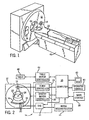
- an exemplary computed tomography (CT) imaging system 10 is shown as including a gantry 12 representative of a "third generation" CT scanner.
- Gantry 12 has an x-ray source 14 that projects a beam of x-rays 16 toward a detector array 18 on the opposite side of the gantry 12.
- Detector array 18 is formed by a plurality of detectors 20 which together sense the projected x-rays that pass through a medical patient 22.
- Each detector 20 produces an electrical signal that represents the intensity of an impinging x-ray beam and hence the attenuated beam as it passes through the patient 22.
- gantry 12 and the components mounted thereon rotate about a center of rotation 24.
- Control mechanism 26 includes an x-ray controller 28 that provides power and timing signals to an x-ray source 14 and a gantry motor controller 30 that controls the rotational speed and position of gantry 12.
- a data acquisition system (DAS) 32 in control mechanism 26 samples analog data from detectors 20 and converts the data to digital signals for subsequent processing.
- An image re-constructor 34 receives sampled and digitized x-ray data from DAS 32 and performs high speed reconstruction. The reconstructed image is applied as an input to a computer 36 which stores the image in a mass storage device 38.
- DAS data acquisition system
- Computer 36 also receives commands and scanning parameters from an operator via console 40 that has a keyboard.
- An associated cathode ray tube display 42 allows the operator to observe the reconstructed image and other data from computer 36.
- the operator supplied commands and parameters are used by computer 36 to provide control signals and information to DAS 32, x-ray controller 28 and gantry motor controller 30.
- computer 36 operates a table motor controller 44 which controls a motorized table 46 to position patient 22 and gantry 12. Particularly, table 46 moves portions of patient 22 through a gantry opening 48.
- a process 100 for acquiring imaging data of a phantom and displaying a helical artifact index (HAI) of a reconstructed image based on the acquired imaging data of the phantom begins at 110 with the prescription of an imaging scan for a subject such as a medical patient by a scan operator or other healthcare provider.
- a system operator determines scan and reconstruction parameters necessary to satisfy the prescription prescribed at 110.
- a phantom is positioned in an imaging space.
- the present invention is applicable with any phantom designed to simulate an anatomical region of a subject.
- the phantom may include an anthropomorphic shaped helical body phantom (HBP) having an elliptical shape and having diagonal rods at the periphery to simulate ribs of a subject.
- HBP anthropomorphic shaped helical body phantom
- Other phantoms may also be implemented that simulate other anatomical regions such as, a heart, brain, tissue, etc.
- a scan is initiated and imaging data is acquired at 140 in accordance with the scan parameters defined at 120.
- a mask is then generated at 150 from the acquired data and filtered at 160.
- the mask is generated by identifying those pixels of the acquired imaging data within ⁇ 40 CT numbers of an expected uniform material value. This is accomplished by measuring the mean for a region of pixels near the center of the phantom absent any visual artifact.
- process 100 After the mask is filtered at 160, process 100 generates a helical artifact index (HAI) at 170 as will be discussed with particular reference to Fig. 4.
- HAI helical artifact index
- the HAI is then displayed on a console at 180 whereupon process 100 ends at 190.
- an algorithm 200 for determining the HAI begins at 210 with the determining of a mean value of pixels within the mask at 220.
- the mean value of the pixels within the mask is then subtracted from the image at 230.
- the pixels outside the mask are then set to an initial value.
- the initial value is zero. This however is illustrative of only one preferred embodiment. That is, the present invention is applicable with non-zero initial values.
- the imaging data acquired at 140, Fig. 3 is filtered, in a preferred embodiment, with a 5 x 5 Hanning kernel.
- An illustrative 5 x 5 Hanning kernel applicable with the present invention is set forth in the table below.
- a standard deviation of the pixels within the mask is determined at 260.
- the standard deviation is determined by squaring the pixels within the mask, summing the squares, and dividing the sum by the total number of pixels within the mask.
- the standard deviation is then multiplied by a scalar suitable to obtain a maximum score of 10 for the worst case of artifact presence in a reconstructed image and a minimum score of 1 for the best case of artifact risk over the set of parameters available to the user. Setting a worst case score to 10 and a best case score to 1 is illustrative of only one embodiment that may be used to ascertain an artifact risk value.
- Process 200 then ends at 280 with the visual displaying of the artifact risk value, in a preferred embodiment, as a visual bar graph on a scan Rx console at 180 of Fig. 3 as was heretofore discussed.
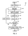
- algorithm 300 sets forth the steps of an acquisition, pre-processing, and reconstruction process in accordance with the present invention.
- Algorithm 300 begins at 310 with the selection of a scanning protocol at 320. Once the appropriate protocol is selected at 320, scan parameters are identified and selected at 330 that satisfy the requirements of the prescribed imaging session. Imaging data is then acquired and processed of a phantom at 340 whereupon an HAI index is determined at 345 in accordance with acts 110-190 of process 100, Fig. 3. The determined HAI is then evaluated by the system operator at 350 to determine if the implemented protocol selected at 320 is acceptable 360.
- the operator may determine at 360 that the protocol is unacceptable. However, if the artifact presence is low or that a high artifact presence is tolerable, the operator may determine that the protocol selected and implemented at 320 is acceptable.
- the pre-processing process is complete and a scan of a patient using the acceptable protocol is executed at 370 whereupon algorithm 300 concludes at 380 with the reconstruction of an image.
- the pre-processing process continues at 390 with a revising of the protocol at 390. Revising the reconstruction protocol allows the system operator to alter the reconstruction algorithm used to process the data or select new scan parameters at 330 that will facilitate image reconstruction with minimal artifact presence.
- pre-processing continues at 340 with the acquisition and processing of data of a phantom and subsequent HAI determination and evaluation at 345 and 350.
- These pre-processing steps continue until an acceptable protocol is determined at 360. That is, regeneration of an HAI during pre-processing allows the system to provide visual feedback in the form of a bar graph to the operator so that a correct protocol may be selected and implemented.
- a histogram illustrating a comparison of artifact score assessment of the process of the present invention versus artifact score assessment by human observers is shown. That is, a set of helical body phantom images were evaluated using the helical artifact index generation process of the instant application. Those images were then presented to a set of skilled observers who were asked to sort and grade the images on a scale of 1 to 10 ranging from best to worst. As shown in Fig. 6, the artifact index generation process of the instant application scored the images in the same sequence as the skilled human observers thereby confirming that the helical artifact index generation process of the instant application correctly assigned index values to the images presented to the skilled observers.
- a method of generating a helical artifact score includes acquiring a set of data values and setting a subset of the set of data values to an initial value. After setting the subset of data values to an initial value, the method includes filtering the set of data values. Next, a likelihood of artifact presence is determined from the filtered set of data values.
- a computer-readable medium having stored thereon a computer program that, when executed by one or more computers, causes the one or more computers to acquire imaging data of a phantom from an external device.
- the imaging data includes a plurality of pixels.
- the computer program further causes the one or more computers to isolate a first set and a second set of pixels and set one of the first set and the second set to an initial value. After setting one of the first set and the second set to an initial value, the computer program causes the one or more computers to filter the imaging data and determine a helical artifact index (HAI) therefrom.
- HAI helical artifact index
- a CT system comprises a rotatable gantry having an opening and a high frequency electromagnetic energy projection source to project high frequency energy toward an object.
- the CT system further includes a scintillator array having a plurality of scintillators to receive high frequency electromagnetic energy attenuated by the object.
- a photodiode array is provided having a plurality of photodiodes. The photodiode array is optically coupled to the scintillator array and is configured to detect light energy emitted therefrom.
- the CT system further includes a plurality of electrical interconnects configured to transmit photodiode outputs to a data processing system and a computer program to acquire and process data to determine a likelihood of an artifact risk presence in a reconstructed image.
- the computer of the CT system is further programmed to notify an operator of the determined likelihood.
Landscapes
- Engineering & Computer Science (AREA)
- Physics & Mathematics (AREA)
- General Physics & Mathematics (AREA)
- Theoretical Computer Science (AREA)
- Quality & Reliability (AREA)
- Multimedia (AREA)
- Apparatus For Radiation Diagnosis (AREA)
- Image Processing (AREA)
Applications Claiming Priority (2)
| Application Number | Priority Date | Filing Date | Title |
|---|---|---|---|
| US09/682,914 US6680995B2 (en) | 2001-10-31 | 2001-10-31 | Method and apparatus of determining and displaying a helical artifact index |
| US682914 | 2001-10-31 |
Publications (1)
| Publication Number | Publication Date |
|---|---|
| EP1308893A2 true EP1308893A2 (en) | 2003-05-07 |
Family
ID=24741742
Family Applications (1)
| Application Number | Title | Priority Date | Filing Date |
|---|---|---|---|
| EP02257448A Withdrawn EP1308893A2 (en) | 2001-10-31 | 2002-10-25 | Method and apparatus of determining and displaying a helical artifact index |
Country Status (3)
| Country | Link |
|---|---|
| US (3) | US6680995B2 (enExample) |
| EP (1) | EP1308893A2 (enExample) |
| JP (1) | JP2003180677A (enExample) |
Families Citing this family (15)
| Publication number | Priority date | Publication date | Assignee | Title |
|---|---|---|---|---|
| US7136630B2 (en) * | 2000-12-22 | 2006-11-14 | Broadcom Corporation | Methods of recording voice signals in a mobile set |
| US6680995B2 (en) | 2001-10-31 | 2004-01-20 | Ge Medical Systems Global Technology Co., Llc | Method and apparatus of determining and displaying a helical artifact index |
| JP4509903B2 (ja) * | 2005-09-27 | 2010-07-21 | ジーイー・メディカル・システムズ・グローバル・テクノロジー・カンパニー・エルエルシー | X線ct装置 |
| DE102005050917A1 (de) * | 2005-10-24 | 2007-04-26 | Siemens Ag | Verfahren und Tomographiegerät zur Rekonstruktion einer tomographischen Darstellung eines Objektes |
| US8489537B2 (en) * | 2009-01-26 | 2013-07-16 | Microsoft Corporation | Segmenting sequential data with a finite state machine |
| KR101698561B1 (ko) * | 2009-03-26 | 2017-01-20 | 코닌클리케 필립스 엔.브이. | 관류 이미징 |
| TWI440312B (zh) * | 2010-11-15 | 2014-06-01 | Anpec Electronics Corp | 類比數位轉換的功能裝置 |
| US10186056B2 (en) * | 2011-03-21 | 2019-01-22 | General Electric Company | System and method for estimating vascular flow using CT imaging |
| CN104463828B (zh) * | 2013-09-18 | 2018-04-10 | 株式会社日立制作所 | Ct图像评价装置及ct图像评价方法 |
| WO2015186513A1 (ja) * | 2014-06-05 | 2015-12-10 | 株式会社 日立メディコ | 画像処理装置及び再構成条件設定方法 |
| CN107209236B (zh) * | 2015-02-02 | 2020-08-04 | 皇家飞利浦有限公司 | 用于确定mr系统的性能退化的mr指纹识别 |
| US10426424B2 (en) | 2017-11-21 | 2019-10-01 | General Electric Company | System and method for generating and performing imaging protocol simulations |
| KR20190103816A (ko) | 2018-02-28 | 2019-09-05 | 삼성전자주식회사 | 컴퓨터 단층 촬영 영상을 보정하는 방법 및 장치 |
| EP4164249A1 (en) * | 2021-10-07 | 2023-04-12 | Starkey Laboratories, Inc. | Artifact detection and logging for tuning of feedback canceller |
| CN114299188A (zh) * | 2021-12-31 | 2022-04-08 | 上海联影医疗科技股份有限公司 | 一种投影数据异常处理方法和系统 |
Family Cites Families (21)
| Publication number | Priority date | Publication date | Assignee | Title |
|---|---|---|---|---|
| US5602891A (en) * | 1995-11-13 | 1997-02-11 | Beth Israel | Imaging apparatus and method with compensation for object motion |
| JPH11511376A (ja) * | 1996-03-13 | 1999-10-05 | アナロジック コーポレーション | コンピュータ・トモグラフィの自己校正リング抑制フィルタ |
| US5818896A (en) * | 1996-11-18 | 1998-10-06 | General Electric Company | Methods and apparatus for three-dimensional and maximum intensity projection image reconstruction in a computed tomography system |
| US6023494A (en) * | 1996-12-19 | 2000-02-08 | General Electric Company | Methods and apparatus for modifying slice thickness during a helical scan |
| US5809105A (en) * | 1997-03-19 | 1998-09-15 | General Electric Company | Noise filter for digital x-ray imaging system |
| WO1999000054A1 (en) * | 1997-06-26 | 1999-01-07 | Koninklijke Philips Electronics N.V. | Adjustable computer tomography device |
| US6501849B1 (en) * | 1997-09-02 | 2002-12-31 | General Electric Company | System and method for performing image-based diagnosis over a network |
| US6115489A (en) * | 1997-09-02 | 2000-09-05 | General Electric Company | System and method for performing image-based diagnosis |
| US6115487A (en) * | 1998-01-08 | 2000-09-05 | General Electric Company | Correction algorithm for bone-induced spectral artifacts in computed tomograph imaging |
| US6222642B1 (en) * | 1998-08-10 | 2001-04-24 | Xerox Corporation | System and method for eliminating background pixels from a scanned image |
| US6215841B1 (en) * | 1998-09-29 | 2001-04-10 | General Electric Company | Methods and apparatus for 3D artifact reduction |
| US6325539B1 (en) * | 1998-12-31 | 2001-12-04 | General Electric Company | Calibration simplification for a computed tomograph system |
| US6385279B1 (en) * | 1999-08-27 | 2002-05-07 | General Electric Company | Methods and apparatus for positioning a CT imaging x-ray beam |
| EP1526480A1 (en) * | 2000-10-17 | 2005-04-27 | Fuji Photo Film Co., Ltd | Apparatus for suppressing noise by adapting filter characteristics to input image signal based on characteristics of input image signal |
| US6438195B1 (en) * | 2001-01-26 | 2002-08-20 | Ge Medical Systems Global Technology Company, Llc | Methods and apparatus for compensating for view aliasing artifacts |
| US6480560B2 (en) * | 2001-03-16 | 2002-11-12 | Ge Medical Systems Global Technology Company, Llc | Methods and apparatus for motion gating using CT projection data |
| US6366638B1 (en) * | 2001-03-16 | 2002-04-02 | Ge Medical Systems Global Technology Company, Llc | Methods and apparatus for CT scout image processing |
| US6529574B1 (en) * | 2001-07-18 | 2003-03-04 | Ge Medical Systems Global Technology Company, Llc | Methods and apparatus for FOV-dependent aliasing artifact reduction |
| US6680995B2 (en) * | 2001-10-31 | 2004-01-20 | Ge Medical Systems Global Technology Co., Llc | Method and apparatus of determining and displaying a helical artifact index |
| US6587537B1 (en) * | 2002-04-01 | 2003-07-01 | Ge Medical Systems Global Technology Company, Llc | Methods and apparatus for multi-slice image reconstruction |
| US6600802B1 (en) * | 2002-04-01 | 2003-07-29 | Ge Medical Systems Global Technology Company, Llc | Image space correction for multi-slice helical reconstruction with z-smoothing |
-
2001
- 2001-10-31 US US09/682,914 patent/US6680995B2/en not_active Expired - Fee Related
-
2002
- 2002-10-18 US US10/065,450 patent/US6983180B2/en not_active Expired - Fee Related
- 2002-10-25 EP EP02257448A patent/EP1308893A2/en not_active Withdrawn
- 2002-10-31 JP JP2002317443A patent/JP2003180677A/ja not_active Withdrawn
-
2005
- 2005-06-06 US US11/160,027 patent/US7593761B1/en not_active Expired - Fee Related
Also Published As
| Publication number | Publication date |
|---|---|
| JP2003180677A (ja) | 2003-07-02 |
| US6680995B2 (en) | 2004-01-20 |
| US20030083565A1 (en) | 2003-05-01 |
| US6983180B2 (en) | 2006-01-03 |
| US7593761B1 (en) | 2009-09-22 |
| US20030083561A1 (en) | 2003-05-01 |
Similar Documents
| Publication | Publication Date | Title |
|---|---|---|
| EP1393682B1 (en) | Determining the minimum radiation dose for obtaining a computer tomography image | |
| CN100374080C (zh) | 用于截断补偿的方法和装置 | |
| US20130202079A1 (en) | System and Method for Controlling Radiation Dose for Radiological Applications | |
| US7593761B1 (en) | Method and apparatus of determining and displaying an artifact index | |
| US10593022B2 (en) | Medical image processing apparatus and medical image diagnostic apparatus | |
| EP1249790B1 (en) | Method and apparatus for motion-free CT imaging | |
| EP2433566A2 (en) | System and method for blood vessel stenosis visualization and quantification using spectral CT analysis | |
| US20110116594A1 (en) | X-ray ct apparatus | |
| WO2008012710A1 (en) | X-ray detector gain calibration depending on the fraction of scattered radiation | |
| US7054409B2 (en) | Volumetric CT system and method utilizing multiple detector panels | |
| JP2004174264A (ja) | 計算機式断層写真法(ct)スカウト画像を形成する方法及び装置 | |
| KR20150095140A (ko) | 컴퓨터 단층 촬영 장치 및 그에 따른 ct 영상 복원 방법 | |
| JP5697422B2 (ja) | X線診断装置 | |
| JP3908993B2 (ja) | X線ct装置 | |
| US20040017936A1 (en) | Method, system and computer product for calculating mass scores | |
| US20050002484A1 (en) | Method and apparatus for correcting bone induced spectral artifacts | |
| EP3817662B1 (en) | A method of obtaining x-ray images | |
| US7907757B2 (en) | Methods and apparatus for new useful metrics | |
| JP2002325760A (ja) | 2パスct撮像の方法および装置 | |
| EP1103221A1 (en) | Methods and apparatus for optimizing CT image quality with optimized data acquisition | |
| WO2006090321A1 (en) | Determination of the coverage of a ct scan | |
| JP5854658B2 (ja) | X線ct装置 | |
| CN114901147A (zh) | 一种获得x射线图像的方法 | |
| JP4653351B2 (ja) | X線量補正方法およびx線ct装置 | |
| Edyvean et al. | A methodical approach for comparison of CT image quality relative to dose |
Legal Events
| Date | Code | Title | Description |
|---|---|---|---|
| PUAI | Public reference made under article 153(3) epc to a published international application that has entered the european phase |
Free format text: ORIGINAL CODE: 0009012 |
|
| AK | Designated contracting states |
Designated state(s): AT BE BG CH CY CZ DE DK EE ES FI FR GB GR IE IT LI LU MC NL PT SE SK TR |
|
| AX | Request for extension of the european patent |
Extension state: AL LT LV MK RO SI |
|
| STAA | Information on the status of an ep patent application or granted ep patent |
Free format text: STATUS: THE APPLICATION IS DEEMED TO BE WITHDRAWN |
|
| 18D | Application deemed to be withdrawn |
Effective date: 20080502 |