EP1294938B1 - Zellbindende nukleinsäuremoleküle (aptamere) - Google Patents
Zellbindende nukleinsäuremoleküle (aptamere) Download PDFInfo
- Publication number
- EP1294938B1 EP1294938B1 EP01929568A EP01929568A EP1294938B1 EP 1294938 B1 EP1294938 B1 EP 1294938B1 EP 01929568 A EP01929568 A EP 01929568A EP 01929568 A EP01929568 A EP 01929568A EP 1294938 B1 EP1294938 B1 EP 1294938B1
- Authority
- EP
- European Patent Office
- Prior art keywords
- nucleic acid
- acid molecule
- acid molecules
- nucleotide sequence
- aptamers
- Prior art date
- Legal status (The legal status is an assumption and is not a legal conclusion. Google has not performed a legal analysis and makes no representation as to the accuracy of the status listed.)
- Expired - Lifetime
Links
Images


Classifications
-
- C—CHEMISTRY; METALLURGY
- C12—BIOCHEMISTRY; BEER; SPIRITS; WINE; VINEGAR; MICROBIOLOGY; ENZYMOLOGY; MUTATION OR GENETIC ENGINEERING
- C12Q—MEASURING OR TESTING PROCESSES INVOLVING ENZYMES, NUCLEIC ACIDS OR MICROORGANISMS; COMPOSITIONS OR TEST PAPERS THEREFOR; PROCESSES OF PREPARING SUCH COMPOSITIONS; CONDITION-RESPONSIVE CONTROL IN MICROBIOLOGICAL OR ENZYMOLOGICAL PROCESSES
- C12Q1/00—Measuring or testing processes involving enzymes, nucleic acids or microorganisms; Compositions therefor; Processes of preparing such compositions
- C12Q1/68—Measuring or testing processes involving enzymes, nucleic acids or microorganisms; Compositions therefor; Processes of preparing such compositions involving nucleic acids
- C12Q1/6876—Nucleic acid products used in the analysis of nucleic acids, e.g. primers or probes
- C12Q1/6881—Nucleic acid products used in the analysis of nucleic acids, e.g. primers or probes for tissue or cell typing, e.g. human leukocyte antigen [HLA] probes
-
- C—CHEMISTRY; METALLURGY
- C12—BIOCHEMISTRY; BEER; SPIRITS; WINE; VINEGAR; MICROBIOLOGY; ENZYMOLOGY; MUTATION OR GENETIC ENGINEERING
- C12Q—MEASURING OR TESTING PROCESSES INVOLVING ENZYMES, NUCLEIC ACIDS OR MICROORGANISMS; COMPOSITIONS OR TEST PAPERS THEREFOR; PROCESSES OF PREPARING SUCH COMPOSITIONS; CONDITION-RESPONSIVE CONTROL IN MICROBIOLOGICAL OR ENZYMOLOGICAL PROCESSES
- C12Q1/00—Measuring or testing processes involving enzymes, nucleic acids or microorganisms; Compositions therefor; Processes of preparing such compositions
- C12Q1/68—Measuring or testing processes involving enzymes, nucleic acids or microorganisms; Compositions therefor; Processes of preparing such compositions involving nucleic acids
- C12Q1/6876—Nucleic acid products used in the analysis of nucleic acids, e.g. primers or probes
- C12Q1/6883—Nucleic acid products used in the analysis of nucleic acids, e.g. primers or probes for diseases caused by alterations of genetic material
- C12Q1/6886—Nucleic acid products used in the analysis of nucleic acids, e.g. primers or probes for diseases caused by alterations of genetic material for cancer
Definitions
- the present invention relates to nucleic acid molecules that selectively bind or label endothelial cells, methods for detecting a tumor disease, the nucleic acid molecules used in the methods, and the use of the nucleic acid molecules.
- Aptamers are high-affinity RNA and DNA oligo- or polynucleotides that have a high affinity for a target molecule due to their specific spatial structure.
- a combinatorial approach is often used in which randomly a large number of oligonucleotides of different sequence and secondary structure is generated enzymatically, from which oligonucleotides with high affinity for a target molecule are picked and enriched. This technique is also called SELEX ("systematic evolution of ligands by exponential enrichment").
- oligonucleotide / polynucleotide aptamer
- it can also be synthesized, e.g. by using a DNA / RNA synthesizer.
- Target structures for aptamers may be receptors and other proteins, lipid or carbohydrate structures.
- high-affinity aptamers have been tested against small molecules such as theophylin Jenison et al., High-Resolution Molecular Discrimination by RNA, Science 1994; 263 (5152): 1425-9 , against ATP of Tang et al. "Rational design of allosteric ribozymes", Chem. Biol. 1997; 4 (6): 453-9 , and found against arginine, see Geiger et al., "RNA aptamers that bind L-arginine with sub-micromolar dissociation constants and high enancio-selectivity" Nucl. Acids Res.
- VEGF vascular endothelial growth factor
- Nucleic acid molecules are known which can bind to erythrocyte ghosts as well as to glioblastoma cells.
- nucleic acid molecule of the type mentioned in particular one which is capable of is to selectively bind to endothelial cells or to mark them.
- this object is achieved by a nucleic acid molecule having the nucleotide sequence SEQ ID No. 9 from the attached sequence listing.
- nucleic acid molecule having the above nucleotide sequence recognizes and binds native biological material, not only parts of such, and especially endothelial cells produced by technical methods.
- Nucleic acid molecules containing the listed sequence are characterized by high specificity for the biological material used according to the inventors.
- the nucleic acid molecule is a nucleic acid molecule having the nucleotide sequence SEQ ID No. 9 from the attached sequence listing.
- nucleic acid molecule with the claimed nucleotide sequence in the method mentioned above, the underlying object of the invention is completely solved.
- the secondary structures of the nucleic acid molecules given by the nucleotide sequence are sufficient to mediate highly specific binding to the biological material.
- At least one nucleotide may be replaced or absent in its non-functional portions.
- nucleic acid molecules or aptamers which differ in their sequence in nonfunctional sections, can bind to the same structure.
- Such distinctions include both nucleotide exchanges and deletions of single or multiple nucleotides, as well as strand shortening or terminal alterations.
- the present invention also provides a method in which endothelial cells are selectively bound or labeled using a nucleic acid molecule which contains the nucleotide sequence SEQ ID No. 9, sequence variations being possible here as well in which no functional segments of the nucleic acid molecule at least one nucleotide is replaced or missing.
- sequences SEQ ID No. 1 to SEQ ID No. 21 specifically bind to endothelial cells. These cells, which coat the luminal side of blood vessels, play, inter alia, an important role in the vasculogenesis, angiogenesis and transcytosis of substances via blood-tissue barriers.
- the method is particularly suitable for studying the differentiation of endothelial cells and for marking tumor-supplying blood vessels or inflammatory endothelial cell-specific lesions.
- nucleic acid molecule is coupled to a therapeutic agent.
- the claimed nucleic acid molecules have the potential to act as a targeted carrier for transporting therapeutics to their target site.
- the inventors hope in particular a reduction of the dosage or a drastic reduction of unwanted side effects, since by such a "drug targeting" the unwanted interaction with non-pathogenic structures is largely avoided.
- Targeted structures are especially tumorous tissues. Indeed, recent studies in cancer research have shown that interrupting the blood supply to a tumor is a promising approach to therapy (reviewed in Cheresh, DA, Death to a Blood Vessel, Nat. Med. 1998, 4 (4): 395-6 ; Molema et al., "Rocking the foundations of solid tumor growth by attacking the tumor's blood supply", Immunol. Today 1998, 19 (9): 392-4 ). Such a method can also be realized advantageously with the new nucleic acid sequences.
- the inventors have found that by means of a nucleic acid molecule containing the nucleotide sequence SEQ ID NO: 9 from the enclosed sequence listing, tumor-specific structures are selectively recognized. As a result, by means of this nucleic acid molecule, highly specific discrimination of degenerated tumor tissue from healthy tissue becomes possible. The inventors thus provide a tool that, through its use, provides us with a reliable basis for appropriate targeted therapy.
- nucleic acid molecule with the nucleotide sequence SEQ ID No. 9 from the attached sequence listing the discrimination of tumor tissue against healthy tissue via the homolog to the mouse / bovine protein 'pigpen'.
- This protein is a relatively unknown endothelial protein with a molecular weight of 67 kDa. Its association with tumor tissue was also unknown until now.
- this protein has hitherto been described as a carbohydrate-binding protein with RNA binding and transactivation domains possibly associated with the nuclear membrane; see eg Alliegro et al., "Protein Heterogeneity in the Coiled Body Compartment," Experimental Cell Research 239, 60-68 (1998). ,
- the present invention is both the use of a nucleic acid molecule containing the nucleotide sequence SEQ ID NO. 9 from the attached sequence listing, for detecting a tumor disease of endothelial origin in the rat, wherein the detection of the protein 'pigpen' is done also generally a method for detecting a tumor disease, wherein the detection of the human homolog to the mouse / bovine protein 'pigpen' takes place.
- nucleic acid molecules in the transport of therapeutics to inflammatory endothelial lesions.
- Possible therapeutics are therefore, for example, antiangiogenic or anti-inflammatory substances.
- the inventors of the present application have recognized for the first time that the claimed nucleic acid molecules can be used for the therapy of the mentioned clinical pictures.
- nucleic acid molecule is coupled to a diagnostic agent.
- modified nucleic acid molecules or aptamers have a number of advantages over the monoclonal antibodies commonly used in diagnostics and also in the "drug targeting" mentioned above. Due to the sequence-related formation of secondary structures, the repertoire of potentially binding ligands is substantially larger than the available immuno-repertoire for the production of monoclonal antibodies.
- aptamers are much faster and cheaper to access.
- millions of potential ligands can be studied.
- the production of antibody-producing hybridoma cells must be via vaccinations of experimental animals. These Vaccinations take several weeks.
- months are required to study several hundred hydridom cell lines.
- nucleic acid molecules or aptamers Since no extracellular oligo- / polynucleotides are known in the organism, which perform functions due to their structure, such nucleic acid molecules or aptamers also have the potential to recognize "unusual" epitopes that escape detection by antibodies and thus the "usual" immune system.
- a particular advantage of the claimed nucleic acid molecules is that they achieve the binding specificity and affinity of good monoclonal antibodies. They are thus particularly suitable to be used in a diagnostic method or a method for "drug targeting". With regard to a diagnostic method, a method is primarily intended in which the diagnostic agent is used outside the human body.
- fluorescent compounds e.g. Fluorescine isothiocyanate (FITC), biotin, digoxigenin and their derivatives, enzymatic markers, infrared markers and chelating agents. Therefore, particularly preferred is a method wherein the diagnostic agent coupled to the nucleic acid molecule is selected from the group consisting of the above substances.
- the invention furthermore relates to the use of the above nucleic acid molecules for histological examinations of tissue sections.
- the inventors have recognized that the direct use of the claimed nucleic acid molecules as a histological agent is possible, allowing in situ localization of the biological target structure within the tissue.
- the particular advantage of such use lies in the specificity of the claimed nucleic acid molecules for complex biological material, preferably endothelial cells, mentioned at the outset. This allows targeted staining of these structures without disturbing background signals or nonspecific cross-reactions.
- nucleic acid molecules in any of the described methods when the nucleic acid molecules selectively bind to native complex biological material.
- the inventors provide nucleic acid molecules which bind to complex native biological structures or endothelial cells.
- sequence motifs which are components of the nucleic acid molecules used there or aptamers, however, bind to artificially produced vesicles from erythrocyte cell membranes.
- the integrity of membranes produced in this way does not necessarily correspond to that of physiological erythrocyte cell membranes.
- erythrocyte components are characterized by a high degree of differentiation. They differ in a variety of properties of other typical cells of an organism: no protein synthesis, no involvement in a cell structure, high specialization in terms of oxygen transport, etc.
- the inventors have used native cells of an endothelial cell line as the biological material for the selection of their specific nucleic acid molecules. This has the advantage that these nucleic acid molecules can also be used reliably in vivo .
- the inventors have recognized that for the purpose of finding specific nucleic acid molecules or aptamers, the enrichment of the selected nucleic acid molecules does not have to be carried out as usual by radioactive labeling, but instead a fluorescence label can be used in combination with suitable detection methods. It was not to be expected that the specific binding properties of the functional nucleic acid molecules can be retained by such a fluorescent label, which is preferably carried out at the 5 'or 3' end of the sequence.
- the enrichment of functional nucleic acid molecules in the pools of the progressive selection rounds has been exclusive controlled by scintillation measurement of the radioactive dNTPs incorporated during the enrichment.
- the new enrichment process has proven to be very economical. It not only allows simultaneous selection against multiple targets embedded in their natural environment from whole endothelial cells by repetitive cycles of FACS measurements. Rather, an in situ evaluation of individual, specifically binding aptamers with fluorescence labeling by FACS is possible.
- Example 1 Endothelial cells as biological target structure
- the target for the selection of the specific aptamers is the cell line YPEN-1 (CRL-2222, American Type Culture Collection, Manassas, USA), a rat endothelial prostate cell line.
- the cells are harvested by scraping the subconfluent endothelial cell layer.
- the concentration of the cells is estimated by counting the number of intact cells after staining with trypan blue in a Neubauer counting chamber.
- the synthetic start library (MWG Biotech AG, Ebersberg, Germany), from which the specific nucleic acid molecules or aptamers are selected, consists of approximately 96mer polynucleotides, wherein the 60 randomly sequenced bases (60N) of 18 nucleotides of defined sequence, the primer hybridization sites , flanked (5'-ATA CCA GCT TAT TCA ATT-
- FITC-labeled and biotin-labeled, HPLC-purified primers (5'-FITC-18C-ATA CCA GCT TAT TCA ATT-3 'and 5'-BBB-AGA TTG CAC TTA CTA TCT. 3 '), where 18C stands for an 18-carbonethylene glycol spacer, "FITC” and “B” stands for a FITC molecule or biotin molecule respectively), which are synthesized by means of solid-phase synthesis (Operon Technologies, Inc., Alamenda USA) ,
- Approximately 1.7 nmol of the DNA from the start library from Example 2 (1 ⁇ 10 15 sequences) are added to selection buffer (50 mM Tris-HCl (pH 7.4), 5 mM KCl, 100 mM NaCl, 1 mM MgCl 2 , 0.1% NaN 3 ) (1 ml in the first round, 200 ⁇ l in the following rounds).
- the DNA is denatured (80 ° C, 10 min) and renatured at 0 ° C for 10 min.
- tRNA Gibco, Düsseldorf, Germany
- BSA bovine serum albumin
- the DNA solution is preincubated with 3 x 10 7 N9 microglial cells for 30 min at 37 ° C / 5% CO 2 to remove high affinity DNA molecules to irrelevant surface proteins.
- microglial cells are spun down and the supernatant is incubated with 10 6 YPEN endothelial cells at 37 ° C / 5% CO 2 . After washing three times by centrifugation with 1 ml of selection buffer (plus 0.2% BSA), the endothelial cell-bound nucleic acid molecules are used as templates to amplify aptamers by PCR (Taq polymerase and dNTPs are from Promega, Mannheim, Germany).
- PCR is performed under conditions according to Crameri and Stemmer, "10 (20) -fold aptamer library amplification without gel purification", Nucl. Acids Res. 1993; 21 (18): 4410.
- the primers used are the FITC- and biotin-labeled primers described above.
- FITC-labeled ssDNA is displayed as follows: FITC-conjugated ssDNA is removed from the biotinylated strand by isolation of the PCR dsDNA products with the appropriate amount of magnetic streptavidin beads (Dynabeads M-280 streptavidin, Dynal Hamburg, Germany) in a magnetic stand followed by alkaline denaturation according to the manufacturer's instructions, purified and taken up in a final volume of 200 ⁇ l of selection buffer. The present ssDNA solution serves as a pool for the following selection round.
- the new FITC ssDNA pool is again preincubated against microglia in polystyrene tubes.
- the selection takes place (also in polystyrene reaction vessels) against 1.5 x 10 5 endothelial cells. After incubation at 37 ° C. for 30 min, the cells are freed by washing three times with 1 ml of selection buffer plus 0.2% BSA of less specifically binding DNA and of sequences whose 5'-modification leads to binding loss.
- the fluorescence of the endothelial-nucleic acid molecule complexes is measured on the flow cytometer (FACS).
- the amplification of the ssDNA proceeds as in the previous round.
- the separation of separation matrix-binding sequences is ensured by replacing the polystyrene reaction vessel with a polypropylene reaction vessel.
- the 30-minute incubation with the washing of the cells (as in the preceding rounds) again follows the determination of the fluorescence intensity on the flow cytometer (counting 5,000 cells per round of selection).
- Cell-bound nucleic acid molecules are PCR-amplified, as already described.
- the specific nucleic acid molecules from the seventh round of selection (round 8 pool) are PCR amplified with unmodified primers and cloned in E. coli (TA cloning, Invitrogen Groningen, the Netherlands).
- the plasmids of the individual clones are isolated by alkaline lysis and the insert (PCR product) as in the rounds of selection with the modified primers PCR-amplified. The single strand extraction takes place as already described.
- the binding of the individual FITC-modified nucleic acid molecules or aptamers obtained is tested by histological binding studies on rat glioblastoma cryostat tissue sections.
- the tissue sections are preincubated with 1 ⁇ g / ⁇ l tRNA in selection buffer (20 min, 4 ° C).
- the preincubated tissue sections are covered with the FITC aptamer solution described above (plus 1 ⁇ g / ⁇ l tRNA) and incubated at room temperature for 40 minutes.
- the tissue sections are washed twice with selection buffer and the immunofluorescence is determined by fluorescence microscopy.
- nucleic acid molecules or aptamers are distinguished by the fact that they selectively stain the newly formed microvessels of the glioblastoma tissue and show minimal cross-reaction with other non-endothelial structures.
- the nucleic acid molecules are therefore particularly suitable for use as histological probes for the staining of endothelial cells.
- Example 4 Specific nucleic acid molecules or aptamers against (endothelial cell) specific antigen
- nucleotide sequences were obtained by the above method and are part of the present invention.
- the primer hybridization sites are 5 'and 3' of the actual aptamer sequences (in bold).
- Figure 1 illustrates the secondary structure of a hypothetical specific nucleotide sequence or aptamer sequence.
- the illustrated nucleic acid molecule is biotin labeled (BBB) at the 5 'end via a linker portion (s). Filled circles symbolize base pairings. Positions indicate the location of the nucleotides in the molecule.
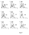
- FIGS. 2 and 3 show the FACS histograms of the nucleic acid molecules found.
- the fluorescence intensity (F) is plotted in arbitrary logarithmic units
- the vertical axis shows the number of signaling events (E) plotted in arbitrary linear units.
- the histograms demonstrate the specificity of the binding of the nucleic acid molecules SEQ ID Nos. 1 to 21 to the target structure.
- the shift in the histogram curve of the nucleic acid molecules (SEQ ID Nos. 1 to 21, in bold) towards increasing stronger fluorescence shows the high affinity of these molecules for the target structure compared to the reference.
- the reference is in each case the fluorescence signal when using nonspecific similar FITC-coupled single-stranded DNA (unspecified ssDNA, gray printed). All 21 nucleic acid molecules are thus characterized by a high affinity for the endothelial cells used.
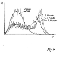
- Example 7 Histograms of the nucleic acid molecules in the complex pools of the selection rounds
- Fig. 4 the FACS histograms of the aptamer pools are shown superimposed on the second (----), fourth (dividing) and seventh (- ⁇ ⁇ ⁇ ⁇ ⁇ ⁇ ⁇ ⁇ ) selection rounds.
- the shift in the histogram curve of the selection rounds shown in the direction of increasing stronger fluorescence illustrates the increase of specifically binding nucleic acid molecules (aptamers) in the pools of the progressing rounds of selection.
- the fluorescence signal is displayed using nonspecific similar FITC-coupled single-stranded DNA (nonspecific ssDNA, gray printed).
- nucleic acid molecule having the nucleotide sequence SEQ ID NO: 9 on rat glioblastoma tissue slices of all the new nucleic acid molecules has the strongest fluorescent signal, specifically in FIG Areas of strong tumor growth, shows. This is shown in FIG. 5.
- Partial image A shows the selective staining of microvessels entering the complex tissue of the microvessel Glioblastoms are embedded.
- Partial image B shows the counterstaining of the cell nuclei with DAPI, whereby the selective marking of the microvessels with the aptamer mentioned in the cell-rich tumor region (the filamentous bright structures on the right side of the image represent the stained microvessels), but not from the vessels into the peritumoral Areas (left side of the picture) becomes apparent.
- Panel C shows a double staining with the commercially available endothelial monoclonal antibody CD-31 (Dako, Hamburg, Germany), resulting in a superposition of both fluorescence signals, whereby the determined by the subject aptamer cells are determined as endothelial cells.
- Part D shows a staining with the monoclonal antibody CD-31 and a nonspecific sequence-randomized aptamer, wherein no double staining of endothelial cells or vascular-associated structures or no signal superimposition can be recognized.
- Panel E shows a staining of an endothelial cell culture after wounding a confluent cell layer with a pipette tip.
- the aptamer in question exhibits significantly increased binding to the endothelial cells in the subconfluent wounded circular area.
- the scale mark corresponds to 50 ⁇ m.
- the staining of tumor tissue sections with mouse IgG directed against endothelial cells, ie for example with the abovementioned CD-31 antibody and the subsequent labeling with the aptamer in question confirms the aptamer target as endothelial cell structure.
- the staining of injured endothelial cell tissue shows a marked upregulation of the molecular target of the aptamer in the area of the lesion, ie in a region characterized by increased proliferation compared to the areas of the confluent monolayer.
- the procedure is as follows: 1 mg (100 ⁇ l) of magnetic streptavidin beats are incubated with 200 pmol of triplicate biotinylated (trB) aptamer in 1 ml of selection buffer (30 minutes, room temperature) coupled (MWG-Biotech AG, Ebersberg, Germany). As a control, 100 ⁇ l of magnetic beats are coupled with 200 pmol of nonspecific FITC ssDNA (trB-96-nt).
- 1.5 x 10 8 YPEN-1 endothelial cells are lysed as described in Klocker et al., Journal of Neuroscience 19, 8517-8527 (1999) , After centrifugation, the protein pellet is resuspended in 400 ⁇ l of selection buffer, ultrasonified (0 ° C, 20 seconds) and in selection buffer (total volume 1.5 ml, 0 ° C, 15 minutes) with the aptamer-covered magnetic beats with the addition of a hundredfold excess of tRNA (20 nmol) as a non-specific competitor incubated.
- the complex of protein and the coupled aptamers is dissolved in a magnetic stand and washed five times (1st wash: 1 ml of selection buffer with 150 mM NaCl; 2nd to 5th wash: 200 ⁇ l of selection buffer with 100 mM NaCl with 2 nmol tRNA).
- the protein is removed from the aptamer-coated beats by incubation in 30 ⁇ l of 1 M NaCl (0 ° C, 30 minutes) and analyzed by PAGE followed by staining with Comassie blue.
- FIG. 1 Such an experiment is shown in FIG.
- lane A the molecular marker is plotted
- lane B the target isolated with the aptamer having the nucleotide sequence SEQ ID NO: 9
- lane C the isolate recovered with nonspecific ssDNA (96-nt) and in lane D untreated endothelial cells are applied.
- the target band isolated by the aptamer in question is characterized by a star and has a molecular weight of about 67 kDa.
- This target protein is identified as follows:
- the appropriate gel piece is cut out and tryptic digestion is performed as described, for example, in US Pat Shevchenko et al., Anal. Chem. 68, 850-858 (1996) , It will be made in the connection shortly described, modifications.
- the excised protein band is completely decolorized and digested for three hours with porcine trypsin (sequencing grade, modified, Promega, Mannheim, Germany) at a concentration of 67 ng per ⁇ l in 25 mM ammonium bicarbonate (pH 8.1 at 37 ° C).
- the peptide mixture Prior to peptide mapping and sequencing of the tryptic fragments by tandem mass spectrometry, the peptide mixture is extracted from the gel by treatment with a solution of 50% trifluoroacetic acid / 50% water, followed by treatment with 50% trifluoroacetic acid / 50% acetonitrile. The extracts are vacuum dried. The dried ones Peptides are resuspended in 0.1% trifluoroacetic acid and purified using a ready-to-go pipette tip with C18 spherical silica reverse phase material (ZipTip C18 TM, Millipore). The peptides are eluted with 10 ⁇ l of 50% methanol / 1% formic acid and sequencing is performed by nano-electrospray tandem mass spectrometry (Q-Tof, Micromass, Manchester, England).
- the inventors have subjected the target recognized by the aptamer with the nucleotide sequence SEQ ID No. 9 to such an analysis. Three tryptic peptide fragments of the protein were analyzed or sequenced by mass spectrometry or tandem mass spectrometry. It turned out that the target recognized by the aptamer in question is a homolog of mouse 'pigpen' protein.
Landscapes
- Chemical & Material Sciences (AREA)
- Life Sciences & Earth Sciences (AREA)
- Health & Medical Sciences (AREA)
- Organic Chemistry (AREA)
- Proteomics, Peptides & Aminoacids (AREA)
- Immunology (AREA)
- Analytical Chemistry (AREA)
- Zoology (AREA)
- Engineering & Computer Science (AREA)
- Wood Science & Technology (AREA)
- Genetics & Genomics (AREA)
- Physics & Mathematics (AREA)
- Pathology (AREA)
- Microbiology (AREA)
- Biotechnology (AREA)
- Biophysics (AREA)
- Biochemistry (AREA)
- Bioinformatics & Cheminformatics (AREA)
- General Engineering & Computer Science (AREA)
- General Health & Medical Sciences (AREA)
- Molecular Biology (AREA)
- Cell Biology (AREA)
- Oncology (AREA)
- Hospice & Palliative Care (AREA)
- Measuring Or Testing Involving Enzymes Or Micro-Organisms (AREA)
- Pharmaceuticals Containing Other Organic And Inorganic Compounds (AREA)
- Medicines That Contain Protein Lipid Enzymes And Other Medicines (AREA)
- Medicinal Preparation (AREA)
Applications Claiming Priority (3)
| Application Number | Priority Date | Filing Date | Title |
|---|---|---|---|
| DE10019154 | 2000-04-18 | ||
| DE10019154A DE10019154A1 (de) | 2000-04-18 | 2000-04-18 | Zellbindende Nukleinsäuremoleküle (Aptamere) |
| PCT/EP2001/004340 WO2001079538A2 (de) | 2000-04-18 | 2001-04-17 | Zellbindende nukleinsäuremoleküle (aptamere) |
Publications (2)
| Publication Number | Publication Date |
|---|---|
| EP1294938A2 EP1294938A2 (de) | 2003-03-26 |
| EP1294938B1 true EP1294938B1 (de) | 2007-12-26 |
Family
ID=7639154
Family Applications (1)
| Application Number | Title | Priority Date | Filing Date |
|---|---|---|---|
| EP01929568A Expired - Lifetime EP1294938B1 (de) | 2000-04-18 | 2001-04-17 | Zellbindende nukleinsäuremoleküle (aptamere) |
Country Status (5)
| Country | Link |
|---|---|
| EP (1) | EP1294938B1 (https=) |
| JP (1) | JP2004503214A (https=) |
| AT (1) | ATE382098T1 (https=) |
| DE (2) | DE10019154A1 (https=) |
| WO (1) | WO2001079538A2 (https=) |
Families Citing this family (3)
| Publication number | Priority date | Publication date | Assignee | Title |
|---|---|---|---|---|
| DE10258924A1 (de) * | 2002-12-17 | 2004-07-08 | Eberhard-Karls-Universität Tübingen Universitätsklinikum | Mit die Adhäsion von biologischem Material vermittelnden Substanzen beschichtete Vorrichtung |
| JP5964746B2 (ja) * | 2009-04-08 | 2016-08-03 | ザ リージェンツ オブ ザ ユニバーシティ オブ カリフォルニア | Dna−細胞コンジュゲート |
| EP2481800B1 (en) * | 2009-06-01 | 2018-02-21 | Sungkyunkwan University Foundation For Corporate Collaboration | Nucleic acid aptamer specifically binding to pancreatic cancer cells or tissues and use thereof |
Family Cites Families (3)
| Publication number | Priority date | Publication date | Assignee | Title |
|---|---|---|---|---|
| US6127119A (en) * | 1990-06-11 | 2000-10-03 | Nexstar Pharmaceuticals, Inc. | Nucleic acid ligands of tissue target |
| CA2219807C (en) * | 1995-05-03 | 2008-12-30 | Nexstar Pharmaceuticals, Inc. | Systematic evolution of ligands by exponential enrichment: tissue selex |
| US6013443A (en) * | 1995-05-03 | 2000-01-11 | Nexstar Pharmaceuticals, Inc. | Systematic evolution of ligands by exponential enrichment: tissue SELEX |
-
2000
- 2000-04-18 DE DE10019154A patent/DE10019154A1/de not_active Ceased
-
2001
- 2001-04-17 DE DE50113417T patent/DE50113417D1/de not_active Expired - Fee Related
- 2001-04-17 EP EP01929568A patent/EP1294938B1/de not_active Expired - Lifetime
- 2001-04-17 JP JP2001577520A patent/JP2004503214A/ja active Pending
- 2001-04-17 AT AT01929568T patent/ATE382098T1/de not_active IP Right Cessation
- 2001-04-17 WO PCT/EP2001/004340 patent/WO2001079538A2/de not_active Ceased
Also Published As
| Publication number | Publication date |
|---|---|
| DE50113417D1 (de) | 2008-02-07 |
| JP2004503214A (ja) | 2004-02-05 |
| DE10019154A1 (de) | 2001-10-31 |
| WO2001079538A2 (de) | 2001-10-25 |
| WO2001079538A3 (de) | 2003-01-16 |
| ATE382098T1 (de) | 2008-01-15 |
| EP1294938A2 (de) | 2003-03-26 |
Similar Documents
| Publication | Publication Date | Title |
|---|---|---|
| EP0517024B1 (de) | Tetravalente bispezifische Rezeptoren, ihre Herstellung und Verwendung | |
| DE69431285T2 (de) | Herstellungsmethode von markierten genen; transcripten und proteinen | |
| DE112005002742B4 (de) | Verbindungen und Methoden zur Behandlung, Diagnose und Prognose bei Pankreaserkrankungen | |
| Kragler et al. | Peptide antagonists of the plasmodesmal macromolecular trafficking pathway | |
| DE69838544T2 (de) | Isolierung von gewebespezifischen peptidliganden und ihre verwendung zur ausrichtung von pharmazeutika auf zielorgane | |
| DE69920885T2 (de) | C-myc-bindendes protein "c-myc coding region determinant-binding protein (crd-bp)" | |
| EP1453531B1 (de) | Verwendung von hmgb-proteinen und dafür codierenden nukleinsäuren | |
| EP1294938B1 (de) | Zellbindende nukleinsäuremoleküle (aptamere) | |
| EP1588172A2 (de) | Verfahren zur identifizierung bhs-spezifischer proteine und fragmente davon | |
| EP0805204B1 (de) | Nebenhoden-spezifisches Rezeptorprotein und dessen Verwendung | |
| DE10339820A1 (de) | Verwendung von an GPR49 bindenden Substanzen zur Diagnose und Behandlung von Krebs | |
| DE69631041T2 (de) | Promotor des utrophingens | |
| DE10021834A1 (de) | mRNA Moleküle zur Verwendung als Indikatoren für den Aktivierungs- und Funktionszustand von T-Lymphozyten | |
| US20140369934A1 (en) | dsRNA/DNA Hybrid Genome Replication Intermediate Of Metakaryotic Stem Cells | |
| EP1551873B1 (de) | Agonisten und antagonisten des humanen duftstoffrezeptors or17-4 sowie verwendungen davon | |
| EP1371729A2 (de) | Verwendungen von TFF3 bindenden Substanzen zur Diagnose und Behandlung von Krebserkrankungen | |
| DE10351627B4 (de) | Modulation der Angiogenese durch Bartonella henselae | |
| DE68916905T2 (de) | Spezifische Nukleinsäurefragmente des menschlichen Villingens, ihre Anwendung für diagnostische Zwecke. | |
| DE10118043B4 (de) | Hochspezifisches Detektionssystem | |
| DE10135996A1 (de) | Verfahren zur Identifizierung metastasierender Tumorzellen | |
| DE10248751A1 (de) | Dyslokalisationsmoleküle und deren Verwendung | |
| WO2004016810A2 (de) | Verwendung von an mrp4 bindenden substanzen zur diagnose und behandlung von krebserkrankungen | |
| DE102005059242A1 (de) | Molekulare Marker für eine Tumordiagnose und -therapie | |
| DE10016527A1 (de) | Verfahren zur Diagnose von Krebserkrankungen und/oder neurodegenerativen Krankheitszuständen sowie Produkte dafür | |
| DE102005046526A1 (de) | Variante des humanen SV2A-Proteins und deren Einfluss auf die Therapieantwort bei Epilepsie |
Legal Events
| Date | Code | Title | Description |
|---|---|---|---|
| PUAI | Public reference made under article 153(3) epc to a published international application that has entered the european phase |
Free format text: ORIGINAL CODE: 0009012 |
|
| 17P | Request for examination filed |
Effective date: 20021109 |
|
| AK | Designated contracting states |
Kind code of ref document: A2 Designated state(s): AT BE CH CY DE DK ES FI FR GB GR IE IT LI LU MC NL PT SE TR |
|
| RIN1 | Information on inventor provided before grant (corrected) |
Inventor name: BLANK, MICHAEL Inventor name: SCHLUESENER, HERMANN |
|
| 17Q | First examination report despatched |
Effective date: 20040506 |
|
| GRAP | Despatch of communication of intention to grant a patent |
Free format text: ORIGINAL CODE: EPIDOSNIGR1 |
|
| GRAS | Grant fee paid |
Free format text: ORIGINAL CODE: EPIDOSNIGR3 |
|
| GRAA | (expected) grant |
Free format text: ORIGINAL CODE: 0009210 |
|
| AK | Designated contracting states |
Kind code of ref document: B1 Designated state(s): AT BE CH CY DE DK ES FI FR GB GR IE IT LI LU MC NL PT SE TR |
|
| REG | Reference to a national code |
Ref country code: GB Ref legal event code: FG4D Free format text: NOT ENGLISH |
|
| REG | Reference to a national code |
Ref country code: IE Ref legal event code: FG4D Free format text: LANGUAGE OF EP DOCUMENT: GERMAN |
|
| REG | Reference to a national code |
Ref country code: CH Ref legal event code: EP Ref country code: CH Ref legal event code: NV Representative=s name: TROESCH SCHEIDEGGER WERNER AG |
|
| REF | Corresponds to: |
Ref document number: 50113417 Country of ref document: DE Date of ref document: 20080207 Kind code of ref document: P |
|
| PG25 | Lapsed in a contracting state [announced via postgrant information from national office to epo] |
Ref country code: SE Free format text: LAPSE BECAUSE OF FAILURE TO SUBMIT A TRANSLATION OF THE DESCRIPTION OR TO PAY THE FEE WITHIN THE PRESCRIBED TIME-LIMIT Effective date: 20080326 |
|
| PG25 | Lapsed in a contracting state [announced via postgrant information from national office to epo] |
Ref country code: FI Free format text: LAPSE BECAUSE OF FAILURE TO SUBMIT A TRANSLATION OF THE DESCRIPTION OR TO PAY THE FEE WITHIN THE PRESCRIBED TIME-LIMIT Effective date: 20071226 Ref country code: NL Free format text: LAPSE BECAUSE OF FAILURE TO SUBMIT A TRANSLATION OF THE DESCRIPTION OR TO PAY THE FEE WITHIN THE PRESCRIBED TIME-LIMIT Effective date: 20071226 |
|
| NLV1 | Nl: lapsed or annulled due to failure to fulfill the requirements of art. 29p and 29m of the patents act | ||
| GBV | Gb: ep patent (uk) treated as always having been void in accordance with gb section 77(7)/1977 [no translation filed] | ||
| PG25 | Lapsed in a contracting state [announced via postgrant information from national office to epo] |
Ref country code: ES Free format text: LAPSE BECAUSE OF FAILURE TO SUBMIT A TRANSLATION OF THE DESCRIPTION OR TO PAY THE FEE WITHIN THE PRESCRIBED TIME-LIMIT Effective date: 20080406 |
|
| PGFP | Annual fee paid to national office [announced via postgrant information from national office to epo] |
Ref country code: CH Payment date: 20080415 Year of fee payment: 8 Ref country code: DE Payment date: 20080529 Year of fee payment: 8 |
|
| PGFP | Annual fee paid to national office [announced via postgrant information from national office to epo] |
Ref country code: AT Payment date: 20080415 Year of fee payment: 8 |
|
| PG25 | Lapsed in a contracting state [announced via postgrant information from national office to epo] |
Ref country code: PT Free format text: LAPSE BECAUSE OF FAILURE TO SUBMIT A TRANSLATION OF THE DESCRIPTION OR TO PAY THE FEE WITHIN THE PRESCRIBED TIME-LIMIT Effective date: 20080526 |
|
| REG | Reference to a national code |
Ref country code: IE Ref legal event code: FD4D |
|
| EN | Fr: translation not filed | ||
| BERE | Be: lapsed |
Owner name: EBERHARD-KARLS-UNIVERSITAT TUBINGEN UNIVERSITATSK Effective date: 20080430 |
|
| PG25 | Lapsed in a contracting state [announced via postgrant information from national office to epo] |
Ref country code: DK Free format text: LAPSE BECAUSE OF FAILURE TO SUBMIT A TRANSLATION OF THE DESCRIPTION OR TO PAY THE FEE WITHIN THE PRESCRIBED TIME-LIMIT Effective date: 20071226 Ref country code: IE Free format text: LAPSE BECAUSE OF FAILURE TO SUBMIT A TRANSLATION OF THE DESCRIPTION OR TO PAY THE FEE WITHIN THE PRESCRIBED TIME-LIMIT Effective date: 20071226 |
|
| PLBE | No opposition filed within time limit |
Free format text: ORIGINAL CODE: 0009261 |
|
| STAA | Information on the status of an ep patent application or granted ep patent |
Free format text: STATUS: NO OPPOSITION FILED WITHIN TIME LIMIT |
|
| PG25 | Lapsed in a contracting state [announced via postgrant information from national office to epo] |
Ref country code: MC Free format text: LAPSE BECAUSE OF NON-PAYMENT OF DUE FEES Effective date: 20080430 |
|
| 26N | No opposition filed |
Effective date: 20080929 |
|
| PG25 | Lapsed in a contracting state [announced via postgrant information from national office to epo] |
Ref country code: GB Free format text: LAPSE BECAUSE OF FAILURE TO SUBMIT A TRANSLATION OF THE DESCRIPTION OR TO PAY THE FEE WITHIN THE PRESCRIBED TIME-LIMIT Effective date: 20071226 |
|
| PG25 | Lapsed in a contracting state [announced via postgrant information from national office to epo] |
Ref country code: GR Free format text: LAPSE BECAUSE OF FAILURE TO SUBMIT A TRANSLATION OF THE DESCRIPTION OR TO PAY THE FEE WITHIN THE PRESCRIBED TIME-LIMIT Effective date: 20080327 |
|
| PG25 | Lapsed in a contracting state [announced via postgrant information from national office to epo] |
Ref country code: BE Free format text: LAPSE BECAUSE OF NON-PAYMENT OF DUE FEES Effective date: 20080430 |
|
| PG25 | Lapsed in a contracting state [announced via postgrant information from national office to epo] |
Ref country code: FR Free format text: LAPSE BECAUSE OF FAILURE TO SUBMIT A TRANSLATION OF THE DESCRIPTION OR TO PAY THE FEE WITHIN THE PRESCRIBED TIME-LIMIT Effective date: 20081017 |
|
| PG25 | Lapsed in a contracting state [announced via postgrant information from national office to epo] |
Ref country code: CY Free format text: LAPSE BECAUSE OF FAILURE TO SUBMIT A TRANSLATION OF THE DESCRIPTION OR TO PAY THE FEE WITHIN THE PRESCRIBED TIME-LIMIT Effective date: 20071226 |
|
| REG | Reference to a national code |
Ref country code: CH Ref legal event code: PL |
|
| PG25 | Lapsed in a contracting state [announced via postgrant information from national office to epo] |
Ref country code: DE Free format text: LAPSE BECAUSE OF NON-PAYMENT OF DUE FEES Effective date: 20091103 Ref country code: AT Free format text: LAPSE BECAUSE OF NON-PAYMENT OF DUE FEES Effective date: 20090417 Ref country code: CH Free format text: LAPSE BECAUSE OF NON-PAYMENT OF DUE FEES Effective date: 20090430 Ref country code: LI Free format text: LAPSE BECAUSE OF NON-PAYMENT OF DUE FEES Effective date: 20090430 |
|
| PG25 | Lapsed in a contracting state [announced via postgrant information from national office to epo] |
Ref country code: LU Free format text: LAPSE BECAUSE OF NON-PAYMENT OF DUE FEES Effective date: 20080417 |
|
| PG25 | Lapsed in a contracting state [announced via postgrant information from national office to epo] |
Ref country code: TR Free format text: LAPSE BECAUSE OF FAILURE TO SUBMIT A TRANSLATION OF THE DESCRIPTION OR TO PAY THE FEE WITHIN THE PRESCRIBED TIME-LIMIT Effective date: 20071226 |
|
| PG25 | Lapsed in a contracting state [announced via postgrant information from national office to epo] |
Ref country code: IT Free format text: LAPSE BECAUSE OF NON-PAYMENT OF DUE FEES Effective date: 20080430 |