EP1294853B1 - Universelles sammelmedium - Google Patents
Universelles sammelmedium Download PDFInfo
- Publication number
- EP1294853B1 EP1294853B1 EP01984035A EP01984035A EP1294853B1 EP 1294853 B1 EP1294853 B1 EP 1294853B1 EP 01984035 A EP01984035 A EP 01984035A EP 01984035 A EP01984035 A EP 01984035A EP 1294853 B1 EP1294853 B1 EP 1294853B1
- Authority
- EP
- European Patent Office
- Prior art keywords
- medium
- cell
- analysis
- cells
- rna
- Prior art date
- Legal status (The legal status is an assumption and is not a legal conclusion. Google has not performed a legal analysis and makes no representation as to the accuracy of the status listed.)
- Expired - Lifetime
Links
Images


Classifications
-
- C—CHEMISTRY; METALLURGY
- C12—BIOCHEMISTRY; BEER; SPIRITS; WINE; VINEGAR; MICROBIOLOGY; ENZYMOLOGY; MUTATION OR GENETIC ENGINEERING
- C12Q—MEASURING OR TESTING PROCESSES INVOLVING ENZYMES, NUCLEIC ACIDS OR MICROORGANISMS; COMPOSITIONS OR TEST PAPERS THEREFOR; PROCESSES OF PREPARING SUCH COMPOSITIONS; CONDITION-RESPONSIVE CONTROL IN MICROBIOLOGICAL OR ENZYMOLOGICAL PROCESSES
- C12Q1/00—Measuring or testing processes involving enzymes, nucleic acids or microorganisms; Compositions therefor; Processes of preparing such compositions
- C12Q1/02—Measuring or testing processes involving enzymes, nucleic acids or microorganisms; Compositions therefor; Processes of preparing such compositions involving viable microorganisms
Definitions
- the present invention is generally related to the field of cytological and molecular assays and specifically to the area of assays for the assessment of conditions using cytological and molecular assays.
- the detection and diagnosis of human conditions is of obvious importance for the treatment of disease. Numerous characteristics of diseases have been identified and many are used for their diagnosis. Many diseases are preceded by, and are characterized by, changes in the state of the affected cells. Changes can include the expression of viral genes in infected cells, changes in the expression patterns of genes in affected cells, and changes in cell morphology. The detection, diagnosis, and monitoring of diseases can be aided by the assessment of such cell states.
- a sample of cells is taken from the patient for analysis.
- a sample is in the form of a swipe or cellular scrape from the area primarily affected by the disease.
- swipes usually collect a mixture of normal and diseased cells with a very limited total number of cells.
- the collected cells are traditionally smeared onto a slide for further analysis.
- biochemical analysis was attempted, it was done at the expense of a cytological analysis and was done via qualitative methods such as in situ hybridization.
- the cervical sample obtained for conventional cytology is smeared onto a slide for morphological analysis. If this sample identifies potential disease by cell cytology, the patient must return for colposcopy to have a second sample collected for repeat cytology and/or genetic analysis and other molecular tests such DNA, RNA or protein.
- liquid cytology media have appeared on the market, which provide for enhanced morphology. These media were discovered to bet amendable to molecular tests such as for HPV DNA, however, cells are routinely collected into 10-20 ml of preserving agents, which excessively dilute DNA, RNA and other asssayable biomolecules, making molecular testing less than ideal. Further, while the current preserving reagents preserve cellular morphology these reagents allow degradation of DNA and RNA, such that quantitative analysis becomes difficult or impossible upon storage.
- present day analysis requires at least two samples to be obtained from a patient in order to determine cell morphology and quantitative genetic analysis.
- Current cytology methods use large volumes of a preserving agent which excessively dilute DNA, RNA and other assayable biomolecules. Further, while the current preserving reagents preserve cellular morphology these reagents allow degradation of individual biomolecules such as DNA, RNA and protein, such that quantitative analysis becomes impossible.
- WO 99/31273 describes a water-based universal collection medium that comprises a preservative, an anti-degradation agent, and a cross-linking agent, such as formaldehyde and glutaraldehyde.
- US 4,784,873 relates to a method for producing durable, flexible biological specimens from initial specimens containing tissue water.
- the tissue-water is removed from the specimen in vacuum and simultaneously the specimen is treated with polyethylene glycol.
- the tissue water is evaporated at a rate that allows direct and continuous substitution of the tissue water by the polyethylene glycol.
- a formaldehyde-free tissue fixation fluid described in US 5,679,333 is an aqueous solution of ethanol, ethanediol, PEG, acetic acid, dimethyl sulfoxide, and ethanedial having a neutral pH of about 6.8 to about 7.8.
- US 5,357,977 relates to a method and device for sampling cytological material.
- the method and device are particularly suited for preparing Papanicolaou smears.
- the device contains a fixative.
- the fixative may be in the form of a liquid, gel, foam, or powder and may include ethanol, acetone, methanol, osmiumtetroxide, mercuric chloride, formaldehyde, polypropylene glycol, polyethylene glycol, EDTA or a combination thereof.
- US 4,578,282 relates to a fixative and preservative composition for histological, cytological, immunological and proteinaceous preparations and to devices and test systems using this composition.
- the fixative is preapplied to a slide or other test surface and presents a substantially dry, non-fluid surface to which the sample is applied.
- the basic composition comprises a four component mixture of pyrrolid-2-one, a polyol, urea and a zinc salt of a non-oxidizing organic or inorganic acid.
- the methods and compositions of this invention solve problems encountered in non-solution-based methods such as in situ hybridization or non-direct methods which require separation of the biomolecule of interest from other cellular components before analysis.
- the present invention relates to the detection, analysis and monitoring of cellular disease.
- a new cell collection medium is disclosed which preserves both cell morphology and cellular biomolecules for quantitative analysis in a cell sample so that multiple assays can be carried out from a single patient sample.
- the state of the cells can be assessed using a device for collecting cellular samples in a small volume.
- One embodiment of the present invention involves examining the cell morphology and detecting a specific DNA sequence or measuring the levels of expression of genes involved in a cell state, and comparing their expression to each other or to reference genes in a specific ratio, as an indication of the state of a disease in the cells.
- This method can be used to detect and/or monitor the onset or progression of any human condition which causes a change in cell morphology or in levels or structures of specific biomolecules.
- the present invention can be used to assess predisposition to a particular disease or to assess the stage or risk of a disease as indicated by the state of the cells. It can also be used to guide or assess the effectiveness of a therapy for a disease by identifying appropriate therapy based on the indicated cell state or by indicating any change in the state of cells subjected to the therapy.
- a universal cell collection medium is disclosed. This medium allows simultaneous preservation of cell morphology and biomolecules in a small volume. Also embodied in the present invention is a device for collecting such cell samples.
- RNA, DNA, protein, carbohydrate or any combination thereof are provided wherein a sample is analyzed according to cell morphology and biochemical analysis in solution phase.
- the biochemical analysis is either qualitative or quantitative and directly analyzes RNA, DNA, protein, carbohydrate or any combination thereof.
- the present invention relates to a universal cell collection medium that makes it possible to conveniently collect and preserve cells and their contents for assessment of the existence or progression of a disease isolated from a single small patient sample, using cytological assays, molecular assays, or both.
- the instant universal cell collection medium preserves cell morphology and preserves macromolecules in a cell sample for either qualitative or quantitative analysis.
- One useful form of the disclosed cell collection medium preserves nucleic acids in the cells. Such preservation can be limited to refrigerated samples. Alternatively, preserved samples can be kept at ambient temperatures. Different forms of the universal collection medium preserve a sample for days or weeks or more.
- the universal cell collection medium can be used to collect cell samples for any purpose and is not limited to use with any particular assay method.
- Some forms of the universal collection medium contain a buffered saline isotonic solution or an alcoholic solution such as methanol, ethanol, or a similar alcohol, an RNase inhibitor such as RNasin, and a protease inhibitor such as pepstatin.
- nuclease inhibitors are known in the art, including, for example, vanadate complexes, chelating agents and detergent-based compounds as well as specific inhibitors such as RNasin. Any known nuclease and/or protease inhibitors can be employed in the present invention as a component of the universal collection medium in order to preserve the particular molecules of interest in the a sample.
- the formulations of this invention provide for the first time means for performing cytological and molecular analysis on cells which are contained in a single sample.
- the cells are obtained from a patient and stored in the UCM of this invention. From this single sample, cells are extracted and a cytological examination is performed, the cellular DNA is qualitatively or quantitatively examined, the cellular RNA is qualitatively or quantitatively examined, or any combination of analysis is performed.
- the different analyses are performed concurrently or, for example, after the results of the cytological analysis are obtained, the cells are subjected to molecular analysis days, weeks or even months later.
- the sample is retrieved for cytological analysis, days, weeks or even months later.
- the universal collection medium (UCM) formulations of this invention are buffered, water-based solutions which comprise a preservative such as a mixture of one or more alcohols, a fixative, said fixative being poly (ethylene glycol), and an agent to inhibit degradation of RNA, DNA and protein.
- a preservative such as a mixture of one or more alcohols
- a fixative said fixative being poly (ethylene glycol)
- an agent to inhibit degradation of RNA, DNA and protein is further enhanced by the addition of an antimicrobial agent.
- any non-viscous alcohol can be used to formulate the UCM, for example, any C1 to C10 alcohols or mixtures thereof can be used.
- Preferred alcohols include methanol, ethanol, propanols, butanols, and pentanols. Most preferred are ethanol and n-butanol.
- the alcohol can comprise a significant percentage of the formulation.
- the alcohol(s) component can comprise about 1% to about 75% of the UCM formulations. More preferred is the percentage range of about 1 % to about 50% alcohol and more preferred is about 5% to about 30% alcohol in the UCM formulation. A range of about 5% to about 15% alcohol is also preferred.
- the pH range of the UCM formulation is important for maintaining the cellular biochemical and morphological integrity of the cells.
- a pH range of about 2.5 to about 6 is used to formulate the UCM of this invention. More preferred is a pH range of about 3 to about 5 and most preferred is a pH range of about 3.5 to about 4.5.
- Buffer(s) are used to maintain the pH of the UCM at a constant value. Any buffer that has buffering capacity in the indicated pH range can be used in the UCM of this invention.
- Non limiting examples of buffer components include glycine, maleic, phosphoric, tartaric, citric, formic, or acetic acids the like.
- the cross-linking agents used in UCM formulations of the prior art comprise about 1% to about 25% of the UCM formulation. Typically, the cross-linking agents comprise about 1% to about 15% or from about 1% to about 10%. Most typically, the cross-linking agent comprises about 1% to about 5% of the UCM formulation.
- Cross-linking agents also known as fixatives
- Non-limiting examples of the cross-linking agents for use in the UCM formulation include aldehydes such formaldehyde, glutaraldehyde and the like.
- An example of a cross-linking agent is glutaraldehydebisulfite.
- Fixatives are defined by Stedman's as “serving to fix, bind, or make firm or stable.” They are substances "used for the preservation of gross and histologic specimens of tissue, or individual cells, usually by denaturing and precipitation or cross-linking the protein constituents.”
- Non-limiting examples of such fixatives for use according to the present invention are polymers such as poly(ethylene glycols) Poly(ethylene glycols) are preferred and poly(ethylene glycols). (“PEG”) with a molecular weight of between about 600 and about 4,600 are more preferred. PEG having a molecular weight of 1500 (“PEG-1500”) is most preferred.
- Fixatives, which function in an equivalent manner to cross-linking agents for the purpose of this invention, are formulated as described above for cross-linking agents.
- Agents able to inhibit degradation of RNA, DNA and/or protein are well-known in the art. They can work by either inhibiting enzymes or sequestering metal ions or both. Nuclease or protease inhibitors such as RNasin or pepstatin or chelating agents can be used according to this invention. Preferred agents to inhibit degradation of RNA, DNA and/or protein are chelating agents. Chelating agents are well-known in the art ( see, for example, Data For Biochemical Research, Third Ed., Rex M. C. Dawson et al., Oxford University Press 1986, at chapter 17 ) and are known to both attenuate metal ion-induced and enzymatic degradation of biopolymers.
- Non-limiting examples of chelating agents for use in this invention include murexide, chromotropic acid, 1-(1-hydroxy-2-naphthylazo)-2-hydroxy-5-nitronaphthalene-4-sulphonic acid, EDTA (ethylenediaminetetraacetic acid), o-phenanthroline, thiourea and the like.
- a preferred chelating agent is EDTA.
- Antimicrobial agents for use in this invention are those known in the art.
- Non-limiting examples of antimicrobial agents are aminoglycosides, ⁇ -lactams, cephalosporins, macrolides, penicillins, azides and the like.
- a preferred antimicrobial agent is sodium azide.
- the universal collection medium can be used for a combination of two or more assays of different characteristics related to a cell state of interest.
- the assay or assays refer to detection or measurement of specific characteristics, the results of which may be combined with other such measurements of other characteristics to provide an overall assessment of a cell suspected of being infected with one or more diseases.
- These assays may include, for example, a combination of morphological analysis and quanitation of a particular RNA or DNA or protein or carbohydrate structure whose presence or levels provide a specific indication of the presence or progression of a disease.
- the universal collection medium can be used to collect any desired cell sample.
- Cell samples are collected in any suitable manner, including scrapings, biopsy, or washings, and from any suitable source. Numerous cell collection techniques are known and any can be used with the present invention.
- the source of cells for a cell sample is chosen based on the known or likely tissue affected by the cell state of interest.
- Cell samples for use in the present invention can be collected and stored in liquid medium.
- useful cell collection media are PreservCyt® (Cytyc), and CytoRich (Autocyte). These media were developed for the collection of cytological samples but can be adapted for use with molecular assays when modified as described herein.
- Nucleic acid detection generally benefits from the use of a reagent capable of preventing nucleic acid degradation prior to performing the assay if the assay is not performed soon after sample collection.
- a useful medium is a preservative based collection medium that has stabilizers for nucleic acids (both RNA and DNA) and proteins and that preserves cell morphology, such as the universal collection medium of the present invention.
- One method useful with the present invention involves measuring the levels of expression of genes involved in a disease state, and comparing their expression to each other or to reference genes, as an indication of the state of the cells. Such measurements can be combined with other assays to increase the accuracy and reliability of the assessment of the disease state.
- the present invention can be used to assess the stage of a disease as indicated by the state of the cells. This embodiment can also be used to guide or assess the effectiveness of a therapy for a disease by identifying appropriate therapy based on the indicated disease state or by indicating any change in the state of cells subjected to the therapy.
- a cell collection medium for preserving cell morphology and cellular biomolecules in a cell sample so that multiple assays can be carried out on the same sample.
- diseases and other human conditions are characterized by specific cellular phenotypes and gene expression patterns. Such diseases and conditions can be identified and/or monitored by assessment of specific cellular morphology or levels or structures of particular biomolecules. For example, neoplastic and cancerous cells generally exhibit certain distinctive morphologies and growth characteristics. Molecular characteristics, such as gene mutations and gene expression patterns are also a good indicator of disease progression. Virally infected cells can exhibit different morphologies and gene expression patterns, including expression of viral genes. Using the present invention, the characteristics of the cell state, such as changes in cell morphology and/or expression of genes can be determined from a patient sample.
- cytological characteristics are characteristics such as, for example, overall cell shape and appearance of the cell and its organelles.
- Identification of cytological characteristics is generally slow, requires a relatively high level of training, and generally cannot be easily automated.
- molecular characteristics are the presence and/or absence and state of particular molecular species, such as proteins, nucleic acids, carbohydrates and metabolites. Such molecular characteristics are generally identified by detecting and/or quantifying the particular molecules of interest.
- the present invention allows both cytological and molecular characteristics to be analyzed from a small patient sample.
- the characteristics assayed can include additional or surrogate marker characteristics that are not a direct cause or result of the disease but that are related to certain disease and cell states.
- additional markers include polymorphic markers, human leukocyte antigens (HLA) such as B7 that predispose women for cervical carcinomas, oncogenes, p53 mutations, BRCA1/2 mutations, other cancer markers, oncosuppressors, cytokines, growth factor receptors, and hormones.
- HLA human leukocyte antigens
- Such markers can be present in, or absent from, cells exhibiting state- or disease-specific characteristics, and such presence or absence can be indicative of, for example, a more severe or less severe disease state.
- markers can be used in conjunction with disclosed methods to infer either higher or lower risk of neoplastic disease depending on the number of abnormal scores or the magnitude of change in quantitative markers.
- Examples of disease states for assessment using the present invention include, but are not limited to, autoimmune disorders, neoplasias, and cancer.
- Other disease states of interest include HPV-based disease including HPV infection, cervical intraepithelial neoplasia (CIN), and cancer, atypical squamous cells of undetermined significance (ASCUS), warts, epidermo dysplasia verruciformis and other skin diseases, laryngeal papilloma, oral papilloma, conjunctival papilloma and prostate disease including enlarged prostate and prostate cancer, chlamydia, and viral infections such as HIV and herpes.
- HPV-based disease including HPV infection, cervical intraepithelial neoplasia (CIN), and cancer, atypical squamous cells of undetermined significance (ASCUS), warts, epidermo dysplasia verruciformis and other skin diseases, laryngeal papillo
- a cell sample as the term is used herein is primarily a collection of cells from a patient.
- One method of obtaining cells is through non-invasive means, which is defined herein as obtained without the puncturing of a patient.
- non-invasive means are, for example, cell samples obtained from urine or a nasal, epithelial, cervical or other cell surface scrape.
- Other methods for obtaining a cell sample are by needle biopsy, or tissue biopsy.
- the cells are collected into volumes of less than 10 ml. More preferably, the cells are collected into volumes of less than about 5 ml, and most preferred the cells are collected into volumes of less than about 2 ml.
- Combinations of multiple assays may be used with the media and device of the present invention and can be carried out from the collection and use of a single sample.
- An important aspect of the combination of assays is the use of a universal cell collection medium that allows a single cell sample to be used for multiple assays of different types with a minimum number of assay-specific processing steps required.
- cells for assaying cytological characteristics are typically collected in a large volume of liquid which leaves the cell sample too dilute for most assays of molecular characteristics.
- Prior art methods do not provide for direct methods for both cytological and molecular analysis. The prior art methods require several extra steps, such as a separate concentration step, which is inconvenient and may result in many of the molecular components of the cell being degraded. Cytological cell samples, while optimized for preserving cell morphology, generally do not preserve nucleic acids well; thus making the sample unusable for many molecular assays.
- Cell samples for use in the method of the present invention can be fixed or processed in any manner consistent with the assays to be performed.
- both cytological and molecular assays can be performed using cells fixed on a solid substrate such as a slide.
- molecular assays are done in solution. The requirements of the assays to be performed will generally determine the sample processing to be used.
- the types of comparisons described above can also be used with many different genes and disease states. That is, the measured level of expression of a gene of interest can be compared, for example, to the level of expression of the same type of gene in a different cell sample (such as an earlier cell sample from the same source or appropriate reference cells), to the level of expression of a different type of gene in the same or a different cell sample, to the level of expression of a reference gene in the same cell sample, or to the level of expression of a reference gene in reference cells.
- a different cell sample such as an earlier cell sample from the same source or appropriate reference cells
- RNA can be detected using hybridization, amplification, or sequencing techniques, and protein, like carbohydrate, can be detected using specific antibodies.
- Many techniques for the specific detection of gene expression, by detection of expression products, are known and can be used with the disclosed UCM.
- One technique for detecting and measuring the level of expression of genes of interest is detection of RNA transcribed from the genes of interest. For the most reliable comparisons, expression levels that are to be compared should be measured using the same technique and be performed in the same manner.
- Useful techniques for measuring the level of expression of a gene of interest in a cell sample include the hybrid capture technique described in WO 93/10263 by Digene , PCR in situ hybridization techniques described by (Nuovo, 1997)), branched DNA assays (Chernoff (1997), transcription mediated amplification (TMA); Stoflet (1988)), and polymerase chain reaction (PCR), ligase chain reaction (LCR), self-sustained sequence replication (3SR), nucleic acid sequence based amplification (NASBA), strand displacement amplification (SDA), and amplification with QB replicase (Birkenmeyer and Mushahwar, (1991); Landegren, (1993)).
- Numerous assays for the detection and measurement of gene expression products are known and can be adapted for the determination of the level of expression of genes of interest using the UCM-collected samples.
- the present invention provides methods for analyzing cells that previously were unavailable. For example, a combination of cancerous and non-cancerous cells were placed in a Universal Collection Medium according to this invention. The morphology of the cells was examined after storage at either 4 °C or room temperature for periods of 12 hours, 10 days, 3 weeks and 6 weeks. Slides were prepared by staining using procedures well-known in the art, for example, Papanicolaou staining and Hematoxylin and Eosin (H&E). When the slides were examined under a light microscope, all the features characteristic for cancer cells were visible and well-preserved in all cases.
- H&E Hematoxylin and Eosin
- the slides showed large and hyperchromatic nuclei, usually with oval shape and irregular, granular chromatin, scanty cytoplasm, multinucleated cells, presence of nucleoli and presence of mitotic figures. In addition, the slides showed that there was good cell dispersion and no cell clumping. Both types of cells (normal and cancerous) had distinct and sharp shapes of both the nuclei and cytoplasm. The nuclei were well-stained and the normal cells showed a different color of cytoplasm depending on the maturity of the cell.
- the cells were first lysed by adding a proteolytic enzyme to the cells contained in wells of a microtiter plate.
- enzymes for use in the present invention include proteinase K or Pronase.
- Cells can also be subjected to detergent lysis or osmotic lysis or a French Press. After incubation, biotinylated DNA probes were added to each well.
- the RNA:DNA hybrids were captured onto a solid phase by transferring to streptavidin coated microplates.
- RNA:DNA hybrids Alkaline phosphatase-conjugated antibodies to RNA:DNA hybrids were added to each well in the hybridization microplate and signals were generated by adding a chemiluminescent reagent such as CDP-StarTM with Emerald II (Tropix) to each well. The signal was read from the microplate.
- the solution based DNA analysis was performed similarly to the RNA analysis except that the microtiter plates were coated with anti-RNA:DNA hybrid antibodies and the probes were unlabeled RNA probes.
- kits that include one or more of the materials needed for the method, such as reagents and sample collection and handling materials.
- kits can include cell collection medium including sample preserving reagents, reagents for specific detection of DNA sequences, RNA sequences and/or expression products (mRNA or protein) of one or more DNA sequences, and sample handling containers.
- Useful reagents for detection of DNA sequences and/or RNA sequences are nucleic acid probes or protein nucleic acid probes for those sequences.
- Useful reagents for detection of DNA sequence expression products (proteins) are antibodies. Aberrant carbohydrate antigens associated with cancerous cells can also be detected by antibodies.
- a kit may also contain control samples or reagents, or reagents and materials for performing other assays to be combined with the disclosed assay.
- the present invention can be performed using devices adapted to the method.
- Numerous devices for performing similar assays are known and in use and can be adapted for use with the disclosed UCM formulations, assays and methods.
- devices are known for automating all or a part of sample assays and sample handling in assays.
- All or part of the disclosed method can be controlled or managed using special purpose computer programs.
- the data collected from the disclosed method, and data from any other assay used in combination, can be compiled, analyzed, and output in various forms and for various purposes using special purpose computer programs.
- Such programs can be used with, or combined into, other patient or data management computer programs. The usefulness of such a program increases with the number of measurements or assessments to be combined, and the relative importance of each type of measurement to the overall assessment.
- Computer programs for use with the disclosed method can be used on general purpose computers, or can be incorporated into special purpose computers or computerized devices for controlling the disclosed method, handling and analyzing data from the disclosed method, or both.
- PreservCyt ® (Cytyc Corporation) Contains buffered methanol Storage limits with cytologic samples: 3 weeks at 4 °C - 37 °C
- CytoRich TM (AutoCyte Corporation) Contains less than 24% alcohol Storage 15°C- 30°C
- the assay for nucleic acids follows in general principle the method for detecting HIV RNA by the Digene Hybrid Capture HIV Test, described in WO 93/10263 by Digene . Briefly, following lysis, 50 ⁇ l of probe mix (containing DNA biotinylated probe) was added to each well. The plate was sealed and incubated at 65 °C for 1.5 hours for hybridization to occur. After hybridization, samples were transferred to a strepavidin-coated microplate, and 25 ⁇ L of anti-hybrid antibody was added to each well. The plate was agitated at 1100 RPM, for 1 hour, at room temperature. Wells were washed 6X times with 65 °C wash buffer, followed by one wash using distilled water.
- chemiluminescent substrate 100 ⁇ l of a chemiluminescent substrate was added to each well and the plate was incubated at room temperature for 30 minutes. The plate was then read in the DML 2000 luminometer. The data was then expressed as signal-to-noise. Using a calibration curve, the chemiluminescent signal generated by each specimen was converted into mRNA copies per cell.
- HPV 16 positive cancer cells were placed in UCM 127, 128, 141,130, 149 and in two commercially available fixatives PreservCyt (Cytyc Corp.) and CytoRich (AutoCyte). The samples were then stored at ambient temperature. The baseline slides were prepared after 12 hours of storage. Then slides were prepared after 3 and 6 weeks. In addition slides were prepared from UCM 141 and 149 after 6 weeks at RT. The slides were prepared by spotting 200 ⁇ l of cell suspension onto polycarbonate filter. The filter was then placed on the glass slide and blotted. The filter was then removed and the slides fixed in 95% ethanol for 5 minutes.
- the slides were stained using routine Papanicolaou staining and Hematoxylin and Eosin (H&E). The slides were evaluated under a light microscope using different magnifications, and documentation in the form of color pictures was prepared. Additionally a smaller study was performed using a mixture of normal human cervical cells and CaSki cells. This mixture was placed in two UCM formulations 141 and 149 and in PreservCyt and CytoRich controls. Each sample was split into two tubes and placed at RT and at 4 °C. After 12 hours (baseline), 10 days and three weeks of storage, slides were prepared and stained with Papanicolaou staining.
- H&E Hematoxylin and Eosin
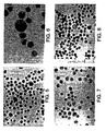
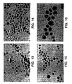
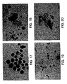
- Figures 1-24 show the morphology of CaSki cells stored in different UCM and in PreservCyt and CytoRich controls stored at RT for 12 hours, 3 weeks and 6 weeks. All features characteristic for CaSki cells (carcinoma cells) were visible and well preserved in all fixatives tested after 3 and 6 weeks: large and hyperchromatic nuclei usually with oval shape and irregular granular chromatin, scanty cytoplasm, multinucleated cells, presence of nucleoli and presence of mitotic figures. In addition, the slides showed that there was good cell dispersion and no cell clumping in all media tested.
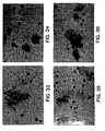
- Figures 25 -72 show the morphology of normal human cervical cells mixed with CaSki cells and stored in UCM 141,149 and PreservCyt and CytoRich controls for 12 hours, 10 days and 3 weeks at room temperature and at 4 °C. The evaluation of these slides showed that the morphology was well preserved in UCM and in PreservCyt and CytoRich controls. After 3 weeks at 4 °C and room temperature both types of cells (normal cervical and CaSki cells) had distinct and sharp shapes (both nuclei and cytoplasm). The nuclei were well stained and the normal cells showed a different color of cytoplasm depending on the maturity.
- Figures 73-75 were taken from archived routine Pap smears to show the morphology and staining of samples stored fixed to slides for comparison.
- UCM formulations 127, 128, 130 and the STMTM (Digene) control were tested using the Hybrid Capture II HPV DNA Test.
- a standard HC II HPV Test kit (Digene catalog number 5101-1096) was used.
- Each collection medium (1 mL) was spiked with 0.8 x 10 6 CaSki cells ( ⁇ 500 copies/cell). This concentration of CaSki cells was chosen because an adequate clinical specimen usually contains about 1 x10 6 cells.
- the same stock of CaSki cells was then used for morphology study and DNA and RNA testing.
- a standard volume of 50 ⁇ l was used per assay as described in the Package Insert, without any sample preparation modification.
- STM - this medium is the current medium used for HPV testing. It preserves DNA and RNA but not cell morphology).
- the HC II HPV test was performed at day “0" (baseline) and after one and six weeks of storage at room temperature. Table 1 shows the results obtained for each medium at the baseline and after one and six weeks expressed as S/N ratios.
- UCM formulations 127 and 128 had the highest S/N ratios comparable to the STM control S/N ratios at the baseline and retained these values after storage at RT for six weeks.
- the S/N ratios for UM 130 obtained at the baseline were slightly lower when compared to UCM 127 and 128.
- UCM 130 retained its signal after 6 weeks.
- Table 1 Signal to Noise ratios obtained for UCM 127, 128, 130 and STM at baseline and after one week and six weeks storage at RT.
- Medium Baseline S/N Week 1 Week 6 S/N %of original signal S/N %of original signal 127 5666 5852 103 6298 111 128 5760 5669 98 5932 103 130 4407 5135 117 5500 125 STM 5557 6151 110 6127 110
- Formulations UCM 141 and 149 were tested in the HC II HPV DNA assay at baseline and then after one, two and four weeks of storage at room temperature. CaSki cells (1.5 x 10 6 ) were spiked in these media and 50 ⁇ l were used per assay. Table 2 shows the S/N ratios. Both formulations retained close to 100% (UCM 141 98.4% and UCM 149 97.3%) of the original signal after four weeks of storage at room temperature. Table 2. S/N ratios obtained for formulations UC 141 and 149 at baseline and after 1, 2 and 4 weeks of storage at RT.
Claims (34)
- Zell- oder Gewebeaufnahmemedium, das sowohl cytologische als auch molekulare Verfahren zur Analyse der darin aufgenommenen Zellen oder Gewebe gestattet, wobei das molekulare Verfahren der Analyse entweder RNA-Analyse oder DNA-Analyse oder Proteinanalyse oder die Analyse von sowohl RNA als auch DNA umfasst, und wobei das Medium auf Wasser basiert und eine Pufferkomponente mit einer Pufferkapazität innerhalb eines pH-Bereichs von ungefähr 2,5 bis ungefähr 6, wenigstens eine Alkoholkomponente, ein Fixierungsmittel und ein Antidegradationsmittel zum Inhibieren der Degradation wenigstens eines aus der Gruppe bestehend aus RNA, DNA und Protein umfasst; und wobei die Menge des Fixierungsmittels von ungefähr 1% bis ungefähr 15% des Mediums beträgt; und wobei das Fixierungsmittel Poly(ethylenglykol) ist.
- Zell- oder Gewebeaufnahmemedium gemäß Anspruch 1, wobei die Menge des Fixierungsmittels von ungefähr 1% bis ungefähr 10% des Mediums beträgt.
- Medium nach Anspruch 1 oder 2, wobei das Fixierungsmittel ungefähr 1% bis ungefähr 5% des Mediums umfasst.
- Medium nach Anspruch 1 oder 2, wobei das Medium aus einem Volumen von weniger als 10 ml besteht.
- Medium nach Anspruch 1 oder 2, wobei das Medium aus einem Volumen von weniger als ungefähr 5 ml besteht.
- Medium nach Anspruch 1 oder 2, wobei das Medium aus einem Volumen von weniger als ungefähr 2 ml besteht.
- Medium nach Anspruch 1, wobei die Pufferkomponente eine Pufferkapazität innerhalb eines pH-Bereichs von ungefähr 3 bis ungefähr 5 aufweist.
- Medium nach Anspruch 7, wobei die Pufferkomponente eine Pufferkapazität innerhalb eines pH-Bereichs von ungefähr 3,5 bis ungefähr 4,5 aufweist.
- Medium nach Anspruch 1, wobei die Alkoholkomponente einen C1- bis C 10-Alkohol umfasst.
- Medium nach Anspruch 9, wobei die Alkoholkomponente ausgewählt ist aus der Gruppe bestehend aus Methanol, Ethanol, Propanolen, Butanolen und Pentanolen.
- Medium nach Anspruch 10, wobei die Alkoholkomponente Ethanol oder n-Butanol umfasst.
- Medium nach Anspruch 1, wobei das Poly(ethylenglykol) ein Molekulargewicht zwischen ungefähr 600 und ungefähr 4600 aufweist.
- Medium nach Anspruch 12, wobei das Poly(ethylenglykol) ein Molekulargewicht von ungefähr 1500 aufweist.
- Medium nach Anspruch 1, wobei das Mittel zum Inhibieren der Degradation wenigstens eines aus der Gruppe bestehend aus RNA, DNA und Protein wenigstens ein Mittel umfasst, das aus der Gruppe bestehend aus einem Nuklease-Inhibitor, einem Protease-Inhibitor und einem Chelatbildner ausgewählt ist.
- Medium nach Anspruch 14, wobei das Mittel zum Inhibieren der Degradation wenigstens eines aus der Gruppe bestehend aus RNA, DNA und Protein einen Chelatbildner umfasst.
- Medium nach Anspruch 14, wobei der Chelatbildner ausgewählt ist aus der Gruppe bestehend aus Murexid, Chromotropsäure, 1-(1-Hydroxy-2-naphthylazo)-2-hydroxy-5-nitronaphthalin-4-sulfonsäure, Ethylendiamintetraessigsäure, o-Phenanthrolin und Thioharnstoff.
- Verfahren nach Anspruch 16, wobei der Chelatbildner Ethylendiamintetraessigsäure (EDTA) umfasst.
- Medium nach Anspruch 1, wobei der Alkohol ausgewählt ist aus der Gruppe bestehend aus Methanol, Ethanol, Propanolen, Butanolen und Pentanolen, wobei das Mittel zum Inhibieren der Degradation ein Chelatbildner, ausgewählt aus der Gruppe bestehend aus Murexid, Chromotropsäure, 1-(1-Hydroxy-2-naphthylazo)-2-hydroxy-5-nitronaphthalin-4-sulfonsäure, Ethylendiamintetraessigsäure, o-Phenanthrolin und Thioharnstoff ist, und wobei das Fixierungsmittel Poly(ethylenglykol) ist.
- Medium nach Anspruch 1, bestehend aus 9% Butanol, 0,05% NaN3, 5 mM EDTA, 2% PEG-1500 und 10 mM NaOAc-HOAc und mit einem pH von 4,5.
- Verfahren zum Durchführen einer cytologischen und molekularen Analyse an einer Zelle oder einem Gewebe, wobei das Verfahren umfasst:Erhalten einer konservierten Zelle oder eines konservierten Gewebes, wobei sich die konservierte Zelle oder das konservierte Gewebe in einem auf Wasser basierenden Zell- oder Gewebeaufnahmemedium gemäß Anspruch 1 oder 2 befindet;direktes Analysieren der Morphologie der in dem Medium konservierten Zelle oder des in dem Medium konservierten Gewebes; unddirektes Analysieren der RNA, der DNA oder des Proteins, welche(s) in der in dem Medium konservierten Zelle oder in dem in dem Medium konservierten Gewebe enthalten ist.
- Verfahren nach Anspruch 20, wobei das Fixierungsmittel ungefähr 1% bis ungefähr 5% des Mediums umfasst.
- Erzeugnis zum Aufbewahren einer Zellprobe, umfassend einen Behälter, der weniger als 2 ml des Mediums gemäß Anspruch 1 oder 2 fasst, und einen Deckel, der auf den Behälter passt.
- Erzeugnis nach Anspruch 22, zusätzlich umfassend eine Zellaufnahmevorrichtung mit einem langgestreckten Bauteil, wobei ein distales Ende des langgestreckten Bauteils einen nicht-absorbierenden, vergrößerten Oberflächenbereich aufweist.
- Erzeugnis nach Anspruch 23, wobei das distale Ende des verlängerten Bauteils eine Bürste ist.
- Erzeugnis gemäß Anspruch 22, wobei das Poly(ethylenglykol) ein Molekulargewicht zwischen ungefähr 600 und ungefähr 4600 aufweist.
- Erzeugnis gemäß Anspruch 25, wobei das Poly(ethylenglykol) ein Molekulargewicht von ungefähr 1500 aufweist.
- Verfahren zur Aufnahme einer Zellprobe, das die Ermittlung der Zellmorphologie und die quantitative Analyse wenigstens eines aus der Gruppe bestehend aus RNA, DNA, Protein, Kohlenhydrat und Lipid aus einer einzigen Probe gestattet, wobei das Verfahren umfasst:Erhalten einer konservierten Zelle oder eines konservierten Gewebes, wobei sich die konservierte Zelle oder das konservierte Gewebe in dem Medium gemäß Anspruch 1 oder 2 befindet;Entnehmen eines Aliquots von Zellen aus dem Medium zur Analyse der Zellmorphologie; undEntnehmen eines Aliquots von Zellen aus dem Medium für eine quantitative Analyse, ausgewählt aus der Gruppe bestehend aus DNA-Analyse, RNA-Analyse, Protein-Analyse, Kohlenhydrat-Analyse und Lipid-Analyse.
- Verfahren gemäß Anspruch 27, wobei die quantitative Analyse ausgewählt ist aus der Gruppe bestehend aus DNA-Analyse und RNA-Analyse.
- Verfahren gemäß Anspruch 27, wobei die Zellen in einer Probe von weniger als 10 ml aufbewahrt werden.
- Verfahren gemäß Anspruch 27, wobei die Zellen in einer Probe von weniger als ungefähr 5 ml aufbewahrt werden.
- Verfahren gemäß Anspruch 27, wobei die Zellen in einer Probe von weniger als ungefähr 2 ml aufbewahrt werden.
- Verfahren gemäß Anspruch 27, wobei das Poly(ethylenglykol) ein Molekulargewicht zwischen ungefähr 600 und ungefähr 4600 aufweist.
- Verfahren gemäß Anspruch 32, wobei das Poly(ethylenglykol) ein Molekulargewicht von ungefähr 1500 aufweist.
- Verfahren gemäß Anspruch 27, wobei das Fixierungsmittel ungefähr 1% bis ungefähr 5% des Mediums umfasst.
Applications Claiming Priority (3)
| Application Number | Priority Date | Filing Date | Title |
|---|---|---|---|
| US59857100A | 2000-06-21 | 2000-06-21 | |
| US598571 | 2000-06-21 | ||
| PCT/US2001/041091 WO2001098542A2 (en) | 2000-06-21 | 2001-06-21 | Universal collection medium |
Publications (2)
| Publication Number | Publication Date |
|---|---|
| EP1294853A2 EP1294853A2 (de) | 2003-03-26 |
| EP1294853B1 true EP1294853B1 (de) | 2010-05-05 |
Family
ID=24396098
Family Applications (1)
| Application Number | Title | Priority Date | Filing Date |
|---|---|---|---|
| EP01984035A Expired - Lifetime EP1294853B1 (de) | 2000-06-21 | 2001-06-21 | Universelles sammelmedium |
Country Status (9)
| Country | Link |
|---|---|
| US (1) | US7371518B2 (de) |
| EP (1) | EP1294853B1 (de) |
| JP (1) | JP2012034699A (de) |
| AT (1) | ATE466930T1 (de) |
| AU (3) | AU2002215488B8 (de) |
| BR (1) | BR0112385A (de) |
| CA (1) | CA2414102A1 (de) |
| DE (1) | DE60142048D1 (de) |
| WO (1) | WO2001098542A2 (de) |
Families Citing this family (30)
| Publication number | Priority date | Publication date | Assignee | Title |
|---|---|---|---|---|
| ES2246546T3 (es) * | 1997-12-12 | 2006-02-16 | Digene Corporation | Valoracion de enfermedades relacionadas con el virus de papiloma humano. |
| US20040115692A1 (en) * | 2000-04-03 | 2004-06-17 | Cytyc Corporation | Methods, compositions and apparatuses for detecting a target in a preservative solution |
| US7439016B1 (en) * | 2000-06-15 | 2008-10-21 | Digene Corporation | Detection of nucleic acids by type-specific hybrid capture method |
| US7601497B2 (en) | 2000-06-15 | 2009-10-13 | Qiagen Gaithersburg, Inc. | Detection of nucleic acids by target-specific hybrid capture method |
| CA2445204C (en) | 2002-10-16 | 2014-08-12 | Streck Laboratories, Inc. | Method and device for collecting and preserving cells for analysis |
| EP1605244A1 (de) * | 2004-06-09 | 2005-12-14 | Boon, Mathilde Elisabeth | Fixierungszusammensetzung |
| JP5566111B2 (ja) | 2007-11-20 | 2014-08-06 | オリンパス株式会社 | Rna回収方法 |
| JP2011518333A (ja) * | 2008-04-17 | 2011-06-23 | キアジェン ゲイサーズバーグ インコーポレイテッド | 標的核酸の存在を判別するための合成プローブを用いた組成物、方法、およびキット |
| EP2287331A4 (de) | 2008-05-12 | 2012-01-25 | Olympus Corp | Verfahren zur verarbeitung von exkrementen und behälter zur verarbeitung von exkrementen |
| US8288520B2 (en) | 2008-10-27 | 2012-10-16 | Qiagen Gaithersburg, Inc. | Fast results hybrid capture assay and system |
| EP2379752B1 (de) * | 2009-01-19 | 2015-09-30 | Biomérieux S.A. | Verfahren zur bestimmung der anfälligkeit eines subjekts für eine nosokomiale infektion und zur prognose der entwicklung eines septischen syndroms |
| US11634747B2 (en) | 2009-01-21 | 2023-04-25 | Streck Llc | Preservation of fetal nucleic acids in maternal plasma |
| EP3290530B1 (de) | 2009-02-18 | 2020-09-02 | Streck Inc. | Konservierung von zellfreien nukleinsäuren |
| US9797000B2 (en) | 2009-05-01 | 2017-10-24 | Qiagen Gaithersburg Inc. | Non-target amplification method for detection of RNA splice-forms in a sample |
| EP2478087B1 (de) | 2009-09-14 | 2017-01-18 | QIAGEN Gaithersburg, Inc. | Zusammensetzungen und verfahren zur rückgewinnung von nukleinsäuren oder proteinen aus in zytologischen medien fixierten gewebeproben |
| CN102822189B (zh) * | 2010-01-29 | 2016-06-29 | 奇亚根盖瑟斯堡股份有限公司 | 用于核酸的序列特异性纯化和多重复合体分析(multiplex analysis)的方法和组合物 |
| JP2013517803A (ja) | 2010-01-29 | 2013-05-20 | キアジェン ゲイサーズバーグ インコーポレイテッド | サンプル中のhpvの存在を決定および確認する方法 |
| US8980548B2 (en) | 2010-05-19 | 2015-03-17 | Metabolon, Inc. | Methods and reagents for metabolomics and histology in a biological sample and a kit for the same |
| JP2013528049A (ja) | 2010-05-19 | 2013-07-08 | キアゲン ガイサーズバーグ アイエヌシー. | 核酸の配列特異的な精製及び多重分析のための方法及び組成物 |
| JP2013531987A (ja) | 2010-06-14 | 2013-08-15 | キアゲン ゲーエムベーハー | 固定生物学的試料から生体分子を抽出するためのターゲット細胞または組織を決定するための方法 |
| EP2638396B1 (de) | 2010-11-12 | 2017-03-01 | incellDX, Inc. | Verfahren und systeme zur vorhersage einer läsion eines patienten durch zervikale intraepitheliale neoplasie augrund einer suspensionsprobe von zervixzellen |
| US9885092B2 (en) | 2011-02-24 | 2018-02-06 | Qiagen Gaithersburg Inc. | Materials and methods for detection of HPV nucleic acids |
| WO2012151391A2 (en) | 2011-05-04 | 2012-11-08 | Streck, Inc. | Inactivated virus compositions and methods of preparing such compositions |
| WO2015013244A1 (en) | 2013-07-24 | 2015-01-29 | Streck, Inc. | Compositions and methods for stabilizing circulating tumor cells |
| AU2015233500B2 (en) * | 2014-03-18 | 2018-11-01 | Qiagen Gmbh | Stabilization and isolation of extracellular nucleic acids |
| US11168351B2 (en) | 2015-03-05 | 2021-11-09 | Streck, Inc. | Stabilization of nucleic acids in urine |
| US10365189B2 (en) * | 2015-05-07 | 2019-07-30 | Steven Wheeler | Histological specimen treatment |
| US20170145475A1 (en) | 2015-11-20 | 2017-05-25 | Streck, Inc. | Single spin process for blood plasma separation and plasma composition including preservative |
| WO2018022991A1 (en) | 2016-07-29 | 2018-02-01 | Streck, Inc. | Suspension composition for hematology analysis control |
| CN106929421B (zh) * | 2017-02-27 | 2020-12-01 | 中国计量科学研究院 | 定量保存微生物的冷冻干燥小球的制备方法 |
Citations (4)
| Publication number | Priority date | Publication date | Assignee | Title |
|---|---|---|---|---|
| US4578282A (en) * | 1982-02-04 | 1986-03-25 | Harrison James S | Composition for diagnostic reagents |
| EP0511430A2 (de) * | 1991-05-01 | 1992-11-04 | Cytyc Corporation | Konservierungslösung für Zellen |
| US5357977A (en) * | 1993-04-23 | 1994-10-25 | St. Mary's Hospital And Medical Center, Inc. | Cytological sampling method and device |
| US5679333A (en) * | 1996-10-25 | 1997-10-21 | Dunphy; Brian William | Formaldehyde-free tissue preservative compositions |
Family Cites Families (14)
| Publication number | Priority date | Publication date | Assignee | Title |
|---|---|---|---|---|
| US133972A (en) * | 1872-12-17 | Improvement in bolt-trimmers | ||
| US129223A (en) * | 1872-07-16 | Improvement in privy-seats | ||
| US4493821A (en) * | 1982-02-04 | 1985-01-15 | Harrison James S | Preservative and fixative preparations for biological systems |
| US4465078A (en) * | 1982-09-30 | 1984-08-14 | Medtest Corporation | Method for cell sampling in a body cavity |
| DE3319564A1 (de) * | 1983-05-30 | 1984-12-06 | Arthur Pfeiffer Vakuumtechnik Wetzlar Gmbh, 6334 Asslar | Verfahren zur herstellung flexibler, dauerhaltbargemachter nicht konservierter und konservierter biologischer materialien |
| US4888278A (en) * | 1985-10-22 | 1989-12-19 | University Of Massachusetts Medical Center | In-situ hybridization to detect nucleic acid sequences in morphologically intact cells |
| US5580970A (en) * | 1989-12-01 | 1996-12-03 | Amoco Corporation | Detection of HPV transcripts |
| AU6966991A (en) | 1989-12-01 | 1991-06-26 | Gene-Trak Systems | Detection of hpv transcripts |
| US5543294A (en) * | 1991-07-19 | 1996-08-06 | The Trustees Of Columbia University In The City Of New York | Polymerase chain reaction/restriction fragment length polymorphism method for the detection and typing of myobacteria |
| IL106273A0 (en) | 1992-07-17 | 1993-11-15 | Res Dev Foundation | Rapid detection of biopolymers in stained specimens |
| US5370128A (en) * | 1993-12-02 | 1994-12-06 | Wainwright; Sharon R. | Pap brush and pap unit container systems |
| CA2147593C (en) * | 1994-04-22 | 2008-07-29 | Hyman C. Birnboim | Dual purpose tissue fixative |
| GB9709728D0 (en) * | 1997-05-13 | 1997-07-02 | Dynal As | Single step method |
| ES2246546T3 (es) * | 1997-12-12 | 2006-02-16 | Digene Corporation | Valoracion de enfermedades relacionadas con el virus de papiloma humano. |
-
2001
- 2001-06-21 WO PCT/US2001/041091 patent/WO2001098542A2/en active IP Right Grant
- 2001-06-21 AU AU2002215488A patent/AU2002215488B8/en not_active Ceased
- 2001-06-21 AT AT01984035T patent/ATE466930T1/de not_active IP Right Cessation
- 2001-06-21 BR BR0112385-8A patent/BR0112385A/pt not_active IP Right Cessation
- 2001-06-21 AU AU1548802A patent/AU1548802A/xx active Pending
- 2001-06-21 CA CA002414102A patent/CA2414102A1/en not_active Abandoned
- 2001-06-21 EP EP01984035A patent/EP1294853B1/de not_active Expired - Lifetime
- 2001-06-21 DE DE60142048T patent/DE60142048D1/de not_active Expired - Lifetime
-
2003
- 2003-01-24 US US10/351,149 patent/US7371518B2/en not_active Expired - Fee Related
-
2007
- 2007-07-19 AU AU2007203372A patent/AU2007203372B2/en not_active Ceased
-
2011
- 2011-09-30 JP JP2011216080A patent/JP2012034699A/ja active Pending
Patent Citations (4)
| Publication number | Priority date | Publication date | Assignee | Title |
|---|---|---|---|---|
| US4578282A (en) * | 1982-02-04 | 1986-03-25 | Harrison James S | Composition for diagnostic reagents |
| EP0511430A2 (de) * | 1991-05-01 | 1992-11-04 | Cytyc Corporation | Konservierungslösung für Zellen |
| US5357977A (en) * | 1993-04-23 | 1994-10-25 | St. Mary's Hospital And Medical Center, Inc. | Cytological sampling method and device |
| US5679333A (en) * | 1996-10-25 | 1997-10-21 | Dunphy; Brian William | Formaldehyde-free tissue preservative compositions |
Also Published As
| Publication number | Publication date |
|---|---|
| JP2004500897A (ja) | 2004-01-15 |
| JP2012034699A (ja) | 2012-02-23 |
| AU2002215488B2 (en) | 2007-04-19 |
| AU2007203372A1 (en) | 2007-08-09 |
| BR0112385A (pt) | 2003-06-17 |
| ATE466930T1 (de) | 2010-05-15 |
| DE60142048D1 (de) | 2010-06-17 |
| WO2001098542A3 (en) | 2002-06-20 |
| CA2414102A1 (en) | 2001-12-27 |
| EP1294853A2 (de) | 2003-03-26 |
| AU2002215488B8 (en) | 2007-11-15 |
| AU1548802A (en) | 2002-01-02 |
| US7371518B2 (en) | 2008-05-13 |
| WO2001098542A2 (en) | 2001-12-27 |
| JP4879447B2 (ja) | 2012-02-22 |
| AU2007203372B2 (en) | 2011-02-03 |
| US20030119049A1 (en) | 2003-06-26 |
Similar Documents
| Publication | Publication Date | Title |
|---|---|---|
| EP1294853B1 (de) | Universelles sammelmedium | |
| AU748022B2 (en) | Universal collection medium | |
| AU2002215488A1 (en) | Universal collection medium | |
| Ambros et al. | Pathology and biology guidelines for resectable and unresectable neuroblastic tumors and bone marrow examination guidelines | |
| US7138226B2 (en) | Preservation of RNA and morphology in cells and tissues | |
| US6337189B1 (en) | Fixative system, method and composition for biological testing | |
| EP3225685B1 (de) | Konservierungslösung für zellen, verwendung davon und verfahren zur herstellung von konservierungslösung für zellen | |
| CA2314663C (en) | A composition for providing long term stability to cells for diagnostic testing | |
| Abati et al. | If cells could talk: the application of new techniques to cytopathology | |
| JP4879447B6 (ja) | 汎用収集用保存液 | |
| KR20050043882A (ko) | 고형 담체에 투여되어 저장되고 그리고 탐지되는 핵산 | |
| AU5350200A (en) | A method for providing long term stability to cells for diagnostic testing | |
| JP2004500897A5 (de) | ||
| Grody | Fixed and Embedded Samples 5 Screening for Pathogenic DNA Sequences in Clinically Collected Human Tissues | |
| Carr | Human Papillomavirus DNA Detection and Typing |
Legal Events
| Date | Code | Title | Description |
|---|---|---|---|
| PUAI | Public reference made under article 153(3) epc to a published international application that has entered the european phase |
Free format text: ORIGINAL CODE: 0009012 |
|
| 17P | Request for examination filed |
Effective date: 20030109 |
|
| AK | Designated contracting states |
Kind code of ref document: A2 Designated state(s): AT BE CH CY DE DK ES FI FR GB GR IE IT LI LU MC NL PT SE TR |
|
| AX | Request for extension of the european patent |
Extension state: AL LT LV MK RO SI |
|
| 17Q | First examination report despatched |
Effective date: 20061213 |
|
| RAP1 | Party data changed (applicant data changed or rights of an application transferred) |
Owner name: QIAGEN GAITHERSBURG, INC. |
|
| GRAP | Despatch of communication of intention to grant a patent |
Free format text: ORIGINAL CODE: EPIDOSNIGR1 |
|
| GRAS | Grant fee paid |
Free format text: ORIGINAL CODE: EPIDOSNIGR3 |
|
| GRAA | (expected) grant |
Free format text: ORIGINAL CODE: 0009210 |
|
| AK | Designated contracting states |
Kind code of ref document: B1 Designated state(s): AT BE CH CY DE DK ES FI FR GB GR IE IT LI LU MC NL PT SE TR |
|
| AX | Request for extension of the european patent |
Extension state: AL LT LV MK RO SI |
|
| REG | Reference to a national code |
Ref country code: GB Ref legal event code: FG4D |
|
| REG | Reference to a national code |
Ref country code: CH Ref legal event code: EP |
|
| REG | Reference to a national code |
Ref country code: IE Ref legal event code: FG4D |
|
| REF | Corresponds to: |
Ref document number: 60142048 Country of ref document: DE Date of ref document: 20100617 Kind code of ref document: P |
|
| REG | Reference to a national code |
Ref country code: CH Ref legal event code: NV Representative=s name: HANS ULRICH SEIFERT SEIFERT & PARTNER |
|
| REG | Reference to a national code |
Ref country code: NL Ref legal event code: VDEP Effective date: 20100505 |
|
| LTIE | Lt: invalidation of european patent or patent extension |
Effective date: 20100505 |
|
| PG25 | Lapsed in a contracting state [announced via postgrant information from national office to epo] |
Ref country code: SE Free format text: LAPSE BECAUSE OF FAILURE TO SUBMIT A TRANSLATION OF THE DESCRIPTION OR TO PAY THE FEE WITHIN THE PRESCRIBED TIME-LIMIT Effective date: 20100505 Ref country code: ES Free format text: LAPSE BECAUSE OF FAILURE TO SUBMIT A TRANSLATION OF THE DESCRIPTION OR TO PAY THE FEE WITHIN THE PRESCRIBED TIME-LIMIT Effective date: 20100816 Ref country code: NL Free format text: LAPSE BECAUSE OF FAILURE TO SUBMIT A TRANSLATION OF THE DESCRIPTION OR TO PAY THE FEE WITHIN THE PRESCRIBED TIME-LIMIT Effective date: 20100505 |
|
| PG25 | Lapsed in a contracting state [announced via postgrant information from national office to epo] |
Ref country code: AT Free format text: LAPSE BECAUSE OF FAILURE TO SUBMIT A TRANSLATION OF THE DESCRIPTION OR TO PAY THE FEE WITHIN THE PRESCRIBED TIME-LIMIT Effective date: 20100505 Ref country code: FI Free format text: LAPSE BECAUSE OF FAILURE TO SUBMIT A TRANSLATION OF THE DESCRIPTION OR TO PAY THE FEE WITHIN THE PRESCRIBED TIME-LIMIT Effective date: 20100505 |
|
| PG25 | Lapsed in a contracting state [announced via postgrant information from national office to epo] |
Ref country code: GR Free format text: LAPSE BECAUSE OF FAILURE TO SUBMIT A TRANSLATION OF THE DESCRIPTION OR TO PAY THE FEE WITHIN THE PRESCRIBED TIME-LIMIT Effective date: 20100806 Ref country code: CY Free format text: LAPSE BECAUSE OF FAILURE TO SUBMIT A TRANSLATION OF THE DESCRIPTION OR TO PAY THE FEE WITHIN THE PRESCRIBED TIME-LIMIT Effective date: 20100505 |
|
| PG25 | Lapsed in a contracting state [announced via postgrant information from national office to epo] |
Ref country code: PT Free format text: LAPSE BECAUSE OF FAILURE TO SUBMIT A TRANSLATION OF THE DESCRIPTION OR TO PAY THE FEE WITHIN THE PRESCRIBED TIME-LIMIT Effective date: 20100906 Ref country code: DK Free format text: LAPSE BECAUSE OF FAILURE TO SUBMIT A TRANSLATION OF THE DESCRIPTION OR TO PAY THE FEE WITHIN THE PRESCRIBED TIME-LIMIT Effective date: 20100505 Ref country code: MC Free format text: LAPSE BECAUSE OF NON-PAYMENT OF DUE FEES Effective date: 20100630 |
|
| PG25 | Lapsed in a contracting state [announced via postgrant information from national office to epo] |
Ref country code: BE Free format text: LAPSE BECAUSE OF FAILURE TO SUBMIT A TRANSLATION OF THE DESCRIPTION OR TO PAY THE FEE WITHIN THE PRESCRIBED TIME-LIMIT Effective date: 20100505 |
|
| PLBE | No opposition filed within time limit |
Free format text: ORIGINAL CODE: 0009261 |
|
| STAA | Information on the status of an ep patent application or granted ep patent |
Free format text: STATUS: NO OPPOSITION FILED WITHIN TIME LIMIT |
|
| 26N | No opposition filed |
Effective date: 20110208 |
|
| PG25 | Lapsed in a contracting state [announced via postgrant information from national office to epo] |
Ref country code: IE Free format text: LAPSE BECAUSE OF NON-PAYMENT OF DUE FEES Effective date: 20100621 |
|
| REG | Reference to a national code |
Ref country code: DE Ref legal event code: R097 Ref document number: 60142048 Country of ref document: DE Effective date: 20110207 |
|
| PG25 | Lapsed in a contracting state [announced via postgrant information from national office to epo] |
Ref country code: LU Free format text: LAPSE BECAUSE OF NON-PAYMENT OF DUE FEES Effective date: 20100621 |
|
| PG25 | Lapsed in a contracting state [announced via postgrant information from national office to epo] |
Ref country code: TR Free format text: LAPSE BECAUSE OF FAILURE TO SUBMIT A TRANSLATION OF THE DESCRIPTION OR TO PAY THE FEE WITHIN THE PRESCRIBED TIME-LIMIT Effective date: 20100505 |
|
| PGFP | Annual fee paid to national office [announced via postgrant information from national office to epo] |
Ref country code: DE Payment date: 20130430 Year of fee payment: 13 Ref country code: GB Payment date: 20130425 Year of fee payment: 13 Ref country code: CH Payment date: 20130424 Year of fee payment: 13 |
|
| PGFP | Annual fee paid to national office [announced via postgrant information from national office to epo] |
Ref country code: IT Payment date: 20130429 Year of fee payment: 13 Ref country code: FR Payment date: 20130531 Year of fee payment: 13 |
|
| REG | Reference to a national code |
Ref country code: DE Ref legal event code: R119 Ref document number: 60142048 Country of ref document: DE |
|
| REG | Reference to a national code |
Ref country code: CH Ref legal event code: PL |
|
| GBPC | Gb: european patent ceased through non-payment of renewal fee |
Effective date: 20140621 |
|
| REG | Reference to a national code |
Ref country code: FR Ref legal event code: ST Effective date: 20150227 |
|
| PG25 | Lapsed in a contracting state [announced via postgrant information from national office to epo] |
Ref country code: IT Free format text: LAPSE BECAUSE OF NON-PAYMENT OF DUE FEES Effective date: 20140621 Ref country code: LI Free format text: LAPSE BECAUSE OF NON-PAYMENT OF DUE FEES Effective date: 20140630 Ref country code: DE Free format text: LAPSE BECAUSE OF NON-PAYMENT OF DUE FEES Effective date: 20150101 Ref country code: CH Free format text: LAPSE BECAUSE OF NON-PAYMENT OF DUE FEES Effective date: 20140630 |
|
| REG | Reference to a national code |
Ref country code: DE Ref legal event code: R119 Ref document number: 60142048 Country of ref document: DE Effective date: 20150101 |
|
| PG25 | Lapsed in a contracting state [announced via postgrant information from national office to epo] |
Ref country code: FR Free format text: LAPSE BECAUSE OF NON-PAYMENT OF DUE FEES Effective date: 20140630 Ref country code: GB Free format text: LAPSE BECAUSE OF NON-PAYMENT OF DUE FEES Effective date: 20140621 |