EP1246919B1 - Progenitor cell preservation factors and related methods and products - Google Patents
Progenitor cell preservation factors and related methods and products Download PDFInfo
- Publication number
- EP1246919B1 EP1246919B1 EP99967798A EP99967798A EP1246919B1 EP 1246919 B1 EP1246919 B1 EP 1246919B1 EP 99967798 A EP99967798 A EP 99967798A EP 99967798 A EP99967798 A EP 99967798A EP 1246919 B1 EP1246919 B1 EP 1246919B1
- Authority
- EP
- European Patent Office
- Prior art keywords
- fril
- cells
- days
- cell
- family member
- Prior art date
- Legal status (The legal status is an assumption and is not a legal conclusion. Google has not performed a legal analysis and makes no representation as to the accuracy of the status listed.)
- Expired - Lifetime
Links
Images
Classifications
-
- C—CHEMISTRY; METALLURGY
- C07—ORGANIC CHEMISTRY
- C07K—PEPTIDES
- C07K14/00—Peptides having more than 20 amino acids; Gastrins; Somatostatins; Melanotropins; Derivatives thereof
- C07K14/415—Peptides having more than 20 amino acids; Gastrins; Somatostatins; Melanotropins; Derivatives thereof from plants
- C07K14/42—Lectins, e.g. concanavalin, phytohaemagglutinin
-
- A—HUMAN NECESSITIES
- A61—MEDICAL OR VETERINARY SCIENCE; HYGIENE
- A61P—SPECIFIC THERAPEUTIC ACTIVITY OF CHEMICAL COMPOUNDS OR MEDICINAL PREPARATIONS
- A61P35/00—Antineoplastic agents
-
- A—HUMAN NECESSITIES
- A61—MEDICAL OR VETERINARY SCIENCE; HYGIENE
- A61P—SPECIFIC THERAPEUTIC ACTIVITY OF CHEMICAL COMPOUNDS OR MEDICINAL PREPARATIONS
- A61P39/00—General protective or antinoxious agents
-
- A—HUMAN NECESSITIES
- A61—MEDICAL OR VETERINARY SCIENCE; HYGIENE
- A61P—SPECIFIC THERAPEUTIC ACTIVITY OF CHEMICAL COMPOUNDS OR MEDICINAL PREPARATIONS
- A61P43/00—Drugs for specific purposes, not provided for in groups A61P1/00-A61P41/00
-
- A—HUMAN NECESSITIES
- A61—MEDICAL OR VETERINARY SCIENCE; HYGIENE
- A61P—SPECIFIC THERAPEUTIC ACTIVITY OF CHEMICAL COMPOUNDS OR MEDICINAL PREPARATIONS
- A61P7/00—Drugs for disorders of the blood or the extracellular fluid
-
- A—HUMAN NECESSITIES
- A61—MEDICAL OR VETERINARY SCIENCE; HYGIENE
- A61K—PREPARATIONS FOR MEDICAL, DENTAL OR TOILETRY PURPOSES
- A61K38/00—Medicinal preparations containing peptides
Definitions
- the invention relates to the preservation of progenitor cells. More specifically, the invention relates to the in vivo preservation of progenitor cells, such as hematopoietic progenitor cells.
- hematopoiesis involves the process of producing a balanced supply of different blood cells from such progenitor cells found in the adult bone marrow.
- the development of other cell types also depends upon production of the differentiated cells from such progenitor cells.
- Progenitor cells are activated by signals, such as cell-cell contact or soluble regulators, to generate daughter cells that are identical to the parent ( i . e ., self-renewal of the parent) and/or to generate daughter cells that are more differentiated than the parent, thus beginning an irreversible process that ends with the production of differentiated, functional cells.
- signals such as cell-cell contact or soluble regulators
- proliferation is coupled to proliferation as a progenitor cell gives rise to more differentiated daughter cells that progressively become committed to producing only one blood cell type.
- potent soluble regulators e.g ., colony stimulating factors and cytokines
- progenitor cells eventually produce so many of the mature cells of the body, they occur only rarely. Moreover, typically the more primitive (i.e. , undifferentiated) the progenitor cell, the more rare the progenitor cell. For example, the currently believed most primitive of the hematopoietic progenitor cells, which are called hematopoietic stem cells, occur at a frequency of only from about 1 in 10,000 to about 1 in 100,000 of the cells in the bone marrow. Hematopoietic stem cells have the capacity to generate more than 10 13 mature blood cells of all lineages, including other progenitor cells which, although more differentiated than hematopoietic stem cells, are themselves capable of giving rise to several different types of mature blood cells.
- Hematopoietic stem cells are responsible for sustaining blood cell production over the life of an animal.
- the small population of hematopoietic stem cells is sufficient to produce all the mature blood cells in a healthy individual; however, some unhealthy individuals suffer from a lack of a sufficient number of progenitor cells and/or mature blood cells.
- cancer patients receivmg chemotherapeutic or radiotherapy treatments designed to kill the rapidly dividing cancer cells also suffer from the depletion of white blood cells and platelets, thus exposing these patients to life-threatening opportunistic infections and bleeding episodes.
- this hematopoietic progenitor cell-depleting activity is the dose-limiting factor for most of these chemotherapeutic and radiotherapeutic agents.
- cytokines including G-CSF, GM-CSF, SCF, Erythropoietin, and IL-11, to accelerate restoration of hematopoiesis following chemotherapy (Moore, M.A., Blood 78: 1-19, 1991).
- these cytokines lead to the irreversible differentiation of hematopoietic progenitor cells, including hematopoietic stem cells, into more differentiated daughter cells.
- better protection of hematopoietic progenitor cells is needed during chemotherapy.
- the invention relates to the use of a therapeutically effective amount of a composition containing a FRIL protein comprising the amino acid sequence set forth in SEQ ID NO: 6 for the preparation of a medicament for treatment of a condition characterized by hematopoietic progenitor cell-depleting activity of a radiotherapeutic or a chemotherapeutic treatment or a combination of a radiotherapeutic and a chemotherapeutic treatment, wherein the therapeutically effective amount of said composition alleviates or reduces the hematopoietic progenitor cell-depleting activity of said therapeutic treatment in the patient, and wherein the medicament is to be administered prior to the radio therapeutic or chemotherapeutic treatment; wherein the medicament is to be administered about 5 days to about 2 hours before the therapeutic treatment and is to be administered daily.
- the FRIL family member is from the legume Phaseolus vulgaris .
- the invention provides a recombinant nucleic acid molecule encoding a composition of a member of the FRIL family of progenitor cell preservation factors comprising the amino acid sequence SEQ ID NO:6 for the claimed use.
- the invention provides a pharmaceutical formulation comprising an essentially pure composition of the FRIL family of progenitor cell preservation factor identified above and a pharmaceutically acceptable carrier.
- the invention relates to administration of a therapeutically effective amount of the formulation to a patient suffering from a condition whereby the patient's hematopoietic progenitor cells are depleted, and wherein the parmaceutical formulation alleviates and/or reduces the condition in the patient.
- the invention relates to the administration of a therapeutically effective amount of the formulation to a patient prior to treatment of the patient with a therapeutic treatment having a hematopoietic progenitor cell-depleting activity alleviates and/or reduces the hematopoietic progenitor cell-depleting activity of the therapeutic treatment in the patient.
- the patient is a human or is a domesticated mammal.
- the patient has cancer.
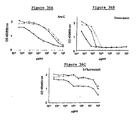
- the therapeutic treatment is a radiotherapeutic or a chemotherapeutic treatment, including, without limitation, cytarabine (Ara-C), doxorubicin (Dox), or 5-fluorouracil (5-FU), or a combination of a radiotherapeutic and a chemotherapeutic.
- a radiotherapeutic or a chemotherapeutic treatment including, without limitation, cytarabine (Ara-C), doxorubicin (Dox), or 5-fluorouracil (5-FU), or a combination of a radiotherapeutic and a chemotherapeutic.
- the invention provides a method for preserving progenitor cells in vivo , comprising administering to a patient an effective amount of a composition of a FRIL family member for an effective period of time, wherein the progenitor cells in the patient are rendered quiescent.
- the patient is a human or a domesticated animal.
- the patient is a cancer patient.
- the effective amount of the composition of a FRIL family member is administered prior to the treatment of the patient with a therapeutic treatment having a hematopoietic progenitor cell-depleting activity wherein the medicament is to be administered about 5 days to about 2 hours before the therapeutic treatment and is to be administered daily.
- the therapeutic treatment is a radiotherapeutic or a chemotherapeutic treatment, including, without limitation, cytarabine (Ara-C), doxorubicin (Dox), or 5-fluorouracil (5-FU), or a combination of a radiotherapeutic and a chemotherapeutic.
- compositions of a FRIL family member may be used as therapeutic agents to preserve progenitor cells in patients, such as cancer patients receiving chemotherapy, who suffer from a condition that diminishes their progenitor cells.
- compositions of a FRIL family member according to claim 1 may be administered with a pharmaceutically-acceptable carrier (e . g ., physiological sterile saline solution) via any route of administration to a cancer patient receiving chemotherapy in an attempt to reduce the progenitor cell-depleting effects of the chemotherapeutic so that the patient can receive a higher dose of the chemotherapeutic and, preferably, recover from cancer.
- a pharmaceutically-acceptable carrier e . g ., physiological sterile saline solution
- Pharmaceutically-acceptable carriers and their formulations are well-known and generally described in, for example, Remington's Pharmaceutical Sciences (18th Edition, ed. A. Gennaro, Mack Publishing Co., Easton, PA, 1990).
- the invention relates to the use of a therapeutically effective amount of a composition containing a FRIL protein comprising the amino acid sequence set forth in SEQ ID NO: 6 for the preparation of a medicament for treatment of a condition characterized by hematopoietic progenitor cell-depleting activity of a radiotherapeutic or a chemotherapeutic treatment or a combination of a radiotherapeutic and a chemotherapeutic treatment, wherein the therapeutically effective amount of said composition alleviates or reduces the hematopoietic progenitor cell-depleting activity of said therapeutic treatment in the patient, and wherein the medicament is to be administered prior to the radio therapeutic or chemotherapeutic treatment; wherein the medicament is to be administered about 5 days to about 2 hours before the therapeutic treatment and is to be administered daily.
- FRIL family of progenitor cell preservation factors is used to mean a family of lectins, wherein each FRIL family member molecule binds to a normally glycosylated FLT3 receptor, wherein each FRIL family member molecule preserves progenitor cells, and wherein one FRIL family member molecule that is isolated from a hyacinth bean ( i.e., Dolichos lab lab ) has an amino acid sequence which comprises the following eight amino acid sequence: TNNVLQXT (SEQ ID NO: 24).
- FRIL family member or "FRIL family member molecule” is meant one or more molecules of the FRIL family of progenitor cell preservation factors.
- Both amino acid sequence identity and nucleic acid sequence identity between two proteins or two nucleic acid molecules can be measured according to standard methods. For example, in order to compare a first amino acid sequence to a second amino acid sequence or a first nucleic acid sequence to a second nucleic acid sequence for the purpose of determining percentage identity between the two sequences, the sequences are aligned so as to maximize the number of identical amino acid or nucleic acid residues.
- the sequences of proteins sharing at least 50% amino acid sequence identity or the sequences of nucleic acids sharing at least 45% nucleic acid sequence identity can usually be aligned by visual inspection. If visual inspection is insufficient, the proteins or nucleic acids may be aligned in accordance with the FASTA method in accordance with Pearson and Lipman ( Proc.
- sequence analysis software Another method for determining amino acid or nucleic acid sequence identity between two proteins or nucleic acids is by using sequence analysis software with the default parameters specified therein.
- sequence analysis software with the default parameters specified therein.
- Various software packages exist including Sequence Analysis Software Package of the Genetics Computer Group (University of Wisconsin Biotechnology Center, Madison, WI), and the various BLAST programs of the National Center for Biotechnology (National Library of Medicine, Bethesda, MD).
- percentage of amino acid sequence identity or percentage of nucleic acid sequence identity is determined using the basic BLAST program of the National Center for Biotechnology (National Library of Medicine, Bethesda, MD), using the default settings defined therein.
- Another test for percentage identity of two nucleic acid sequences is whether they hybridize under normal hybridization conditions, preferably under stringent hybridization conditions.
- proteins that are encoded by nucleic acid molecules that hybridize under high stringent conditions to a sequence complementary to SEQ ID NO: 1, SEQ ID NO: 3, SEQ ID NO: 5, and/or SEQ ID NO: 7.
- stringent conditions is equivalent to "high stringent conditions” and "high stringency.” These terms are used interchangeably in the art.
- Stringent conditions are defined in a number of ways. In one definition, stringent conditions are selected to be about 50°C lower than the thermal melting point (T m ) for a specific sequence at a defined ionic strength and pH. The T m is the temperature (under defined ionic strength and pH) at which 50% of the target sequence hybridizes to a perfectly matched sequence. Typical stringent conditions are those in which the salt concentration is at least about 0.02 M at pH 7 and the temperature is at least about 60°C. "Stringent conditions,” in referring to percentage identity ( e . g ., homology) or substantial similarity in the hybridization context, can be combined conditions of salt, temperature, organic solvents or other parameters that are typically known to control hybridization reactions.
- stringent conditions can be provided in a variety of ways such as overnight incubation at 42°C in a solution comprising: 20% formamide, 5 x SSC (150 mM NaCl, 15 mM trisodium citrate), 50 mM sodium phosphate (pH 7.6), 5 x Denhardt's solution, 10% dextran sulfate, and 20 ⁇ g/ml denatured, sheared salmon sperm DNA.
- the stringent conditions are characterized by a hybridization buffer comprising 30% formamide in 5 x SSPE (0.18 M NaCl, 0.01 M NaPO 4 , pH 7.7, 0.0001 M EDTA) buffer at a temperature of 42°C, and subsequent washing at 42°C with 0.2 x SSPE.
- stringent conditions involve the use of a hybridization buffer comprising 50% formamide in 5 x SSPE at a temperature of 42°C and washing at the same temperature with 0.2 x SSPE.
- progenitor cells an ability of a FRIL family member molecule (or mutant thereof or fusion protein comprising a FRIL family member molecule or mutant thereof) to retain ( i.e., preserve) progenitor cells in an undifferentiated state, which can be determined using the assays described below (e.g. , the SCID mouse reconstituting cell assay and the methylcellulose or other semi-solid medium based hematopoietic progenitor cell assay).
- progenitor cell refers to any normal somatic cell that has the capacity to generate fully differentiated, functional progeny by differentiation and proliferation.
- Progenitor cells include progenitors from any tissue or organ system, including, but not limited to, blood, mesenchymal, embryonic, nerve, muscle, skin, gut, bone, kidney, liver, pancreas, thymus, brain and the like.
- Progenitor cells are distinguished from "differentiated cells," the latter being defined as those cells that may or may not have the capacity to proliferate, i.e., self-replicate, but that are unable to undergo further differentiation to a different celt type under normal physiological conditions.
- progenitor cells are further distinguished from abnormal cells such as neoplastic cells, as defined herein. For example, leukemia cells proliferate (self-replicate), but generally do not further differentiate, despite appearing to be immature or undifferentiated.
- Progenitor cells include all the cells in a lineage of differentiation and proliferation prior to the most differentiated or the fully mature cell.
- progenitors include the skin progenitor in the mature individual.
- the skin progenitor is capable of differentiation to only one type of cell, but is itself not fully mature or fully differentiated.
- hematopoiesis is meant the development of mature, functional blood cells.
- the progenitor cells that give rise to mature, functional blood cells are called hematopoietic progenitor cells.
- the most primitive, undifferentiated hematopoietic progenitor cell is called a hematopoietic stem cell.
- Hematopoietic stem cells typically reside in the bone marrow primarily in a quiescent state, and may form identical daughter cells through a process called "self-renewal.”
- hematopoietic progenitor cells include those cells that are capable of successive cycles of differentiation and proliferation to yield up to eight different mature hematopoietic cell lineages.
- Uncommitted progenitor cells such as hematopoietic stem cells
- totipotent i . e ., both necessary and sufficient for generating all types of mature cells.
- Progenitor cells that retain a capacity to generate all cell lineages, but that can not self-renew, are termed “pluripotent.”
- Pluripotent Cells that can produce some but not all blood lineages and cannot self-renew are termed “multipotent.”
- Progenitor cells can be defined by mRNA levels of genes that either specifically regulate progenitors or serve as markers of lineage commitment.
- genes induced in primitive human hematopoietic progenitor cells include those encoding the shared beta subunits of the IL3, IL5, and/or granulocyte-macrophage colony-stimulating factor (GM-CSF) receptors, termed the beta common chain (McClanahan et al., Blood 81:3903-2915,1993); CD34 genes; and/or the receptors for Kit (Turner et al., Blood 88:3383-3390, 1996), FLT1, FLT4 (Galland et al., Oncogene 8:3233-1240, 1993), FLK1 (Broxmeyer et al., Int.
- genes for intermediate progenitors include the c-Fms, G-CSF receptor, and/or CD34 genes; and the IL-7 receptor gene, a gene induced for B lymphopoiesis.
- Murine primitive progenitor populations include receptors for interleukin-1 alpha (IL-1 ⁇ ), IL-3, IL-6, granulocyte colony-stimulating factor (G- CSF), and/or FLK1-1 (the murine homologue of human KDR which binds VEGF) (Broxmeyer, supra ), but lack receptors for macrophage colony-stimulating factor (M-CSF), granulocyte-macrophage colony stimulating factor (GM-CSF), and leukemia inhibitory factor (LIF).
- Cells within the intermediate progenitor cell population include receptors for GM-CSF, G-CSP, IL-6, and/or IL-1 ⁇ .
- bind binds to a normally glycosylated FLT3 receptor with an affinity higher than the affinity with which the FLT3-Ligand binds the FLT3 receptor.
- a FRIL family member molecule binds to a normally glycosylated FLT3 receptor with an affinity that is at least as high as the affinity with which an antibody binds its specific ligand.
- a FRIL family member molecule of the invention binds to a normally glycosylated FLT3 receptor with an affinity that is higher than the affinity with which an antibody binds its specific ligand.
- a FRIL family member molecule of the invention binds to a normally glycosylated FLT3 receptor with a dissociation constant (K D ) of at least 10 -7 M, more preferably 10 -8 M, even more preferably 10 -9 M, still more preferably, at least 10 -10 M, and most preferably, a FRIL family member molecule of the invention binds to a normally glycosylated FLT3 receptor with a dissociation constant (K D ) of at least 10 -11 M. Standard methods for determining binding and binding affinity are known.
- normally glycosylated FLT3 receptor an FLT3 receptor that has a glycosylation pattern of an FLT3 cell glycosylated by a normal cell.
- normal cell as used herein in accordance with the present invention, is meant a cell that is not neoplastic.
- neoplastic cell is meant a cell that shows aberrant proliferation, particularly increased proliferation, that is not regulated by such factors as cell-cell contact inhibition and soluble regulators (e.g.
- cytokines or hormones cytokines or hormones
- abnormally glycosylates the FLT3 receptor such that the glycosylation pattern on the FLT3 receptor on the neoplastic cells is abnormal and such that the FLT3 receptor on the neoplastic cell is not bound by a FRIL family member molecule.
- essentially pure means a molecule, such as a nucleic acid or protein (e.g. , a FRIL family member molecule), or composition of a molecule that is more free from other organic molecules (e.g. , carbohydrates, nucleic acids, proteins, and lipids) that naturally occur with an impure molecule, and is substantially free as well of materials used during the purification process.
- a protein or nucleic acid molecule is considered to be essentially pure if it is at least approximately 60%, preferably at least approximately 75%, more preferably approximately at least 85%, most preferably approximately at least 90%, and optimally approximately at least 95% pure, i .
- a FRIL family member can also be purified by binding to a mannose, which may be coupled on a solid support (e.g. , a sepharose bead).
- nucleic acids Methods for purifying nucleic acids are known in the art and include, without limitation, Guanidine-HCl extraction, polymerase chain reaction, CsCl gradient fractionation, phenol: chloroform extraction, ethanol precipitation, and standard recombinant DNA methodologies. Standard methods for purifying both proteins and nucleic acid molecules are provided in, e.g., Ausubel et al., Current Protocols in Molecular Biology, John Wiley & Sons, New York, NY, 1994; Sambrook et al., supra.
- a FRIL family member molecule may be purified from a natural source by methods well known in the art. For example, the purification of Dl-FRIL from Dolichos lab lab is described below in Example 1. The purification of Pv-FRIL from Phaseolus vulgaris is described below in Example 5. The purification of YarnFRIL from Sphenostylis stenocarpa is described below in Example 22. Such methods also include, for example, those described by Moore in PCT application PCT/US97/22486 and by Gowda et al., supra .
- a suitable natural source from which to purify a FRIL family member molecule includes plants, especially legume plants.
- Legumes such as the garden pea or the common bean
- Plants are plants (“leguminous plants”) from a family ( Leguminosae ) of dicotyledonous herbs, shrubs, and trees bearing (nitrogen-fixing bacteria) nodules on their roots. These plants are commonly associated with their seeds ( e.g. , the garden pea or the common bean)
- a FRIL family member molecule can be purified from members of the tribe Phaseoleae.
- a FRIL family member molecule can be purified from Dolichos lab lab (e.g., hyacinth beans, which is also known by other common names throughout the world).
- a FRIL family member molecule can be purified from varieties of the common bean ( Phaseolus vulgaris ) ( e.g., red kidney beans and white kidney beans), from the yarn bean ( Sphenostylis stenocarpa ) or from Vigna sinensis , commonly known as the black-eyed pea.
- a native FRIL family member molecule can be easily purified from legumes, such as hyacinth beans (pesticide-free), by mannose-affinity chromatography or ovalbumin affinity chromatography, and is more than 100 times cheaper to produce than recombinant cytokines.
- FRIL family member molecule is a mannose/glucose-specific legume lectin.
- lectin is meant a protein that binds sugar residues with high affinity.
- FRIL family member molecules and compositions of FRIL family member molecules, have many attributes as reagents to either alleviate the progenitor cell-depleting activity of a therapeutic (e.g. , a chemotherapeutic) or to alleviate the symptoms of a condition where the patient's progenitor cells are depleted.
- a therapeutic e.g. , a chemotherapeutic
- FRIL family members have unique properties and are the first soluble regulators reported to preserve hematopoietic stem cells and progenitors in a dormant state for extended periods, even in the presence of potent stimulators of proliferation and differentiation.
- mice tolerate very high levels of compositions of FRIL family members, this may permit more effective protection of stem cells and progenitors by preventing their recruitment during aggressive dose intensification regimens aimed at increasing frequency and dosage levels of chemotherapy. While the biological activity of Dl-FRIL is similar to cytokines (ng/ml range), as demonstrated in the examples below, mice tolerated up to a 1,000-fold more Dl-FRIL than cytokines.
- one or more members of the FRIL family allows for the administration of a broad range of cell cycle active chemotherapy drugs with greater frequency and higher dose.
- administration of a composition of one or more FRIL family members may permit more aggressive dose-intensification chemotherapy regimens for a broad range of chemotherapy drugs.
- Administration of a composition of a FRIL family member also provides for a larger reservoir of progenitor cells which could rapidly respond to stimulatory signals after completing chemotherapy.
- the FRIL family member which is used according to the invention is from a legume (e . g ., a bean plant), i.e. , from a red kidney bean ( i.e., Phaseolus vulgaris ).
- the FRIL family member of the invention isolated from a red kidney bean has the amino acid sequence of SEQ ID NO: 6, and, even more preferably, is encoded by a nucleic acid having the nucleic acid sequence of SEQ ID NO: 5.
- FRIL family member molecule which is a fusion protein comprising a first portion and a second portion, wherein the first portion is derived from a second member of the FRIL family.
- fusion protein is meant a molecule comprising at least two proteins or polypeptide fragments thereof joined together, wherein the proteins or polypeptide fragments thereof are not joined together in the naturally-occurring organism from which the proteins or polypeptide fragments thereof were derived.
- the two proteins or polypeptide fragments thereof of a fusion protein may be joined by any means, including, without limitation, a chemical linker, a peptide bond, or a noncovalent bond, such as an ionic bond.
- protein or “polypeptide” is meant a chain of two or more amino acid residues joined with a peptide bond regardless of length or post-translational modification such as acetylation, glycosylation, lipidation, or phosphorylation.
- a FRIL family member molecule that is a fusion protein may comprise a first portion derived from a FRIL family member and a second portion derived from a protein or other molecule not related to the FRIL family (e.g. , the heavy chain of an antibody).
- An additional FRIL family member molecule that is a fusion protein is FRIL family member comprising the ⁇ subunit from a first FRIL family member and a ⁇ subunit from a second FRIL. family member.
- a fusion protein may be generated, for example, by joining a nucleic acid sequence encoding the ⁇ subunit of the first FRIL family member in frame with a nucleic acid sequence encoding the ⁇ subunit of the second FRIL family member.
- the nucleic acid encoding such a fusion protein can be engineered to encode an enzyme-specific cleavage site between the portion encoding the ⁇ subunit of the first FRIL family member and the portion encoding the ⁇ subunit of the second FRIL family member.
- identity of the fusion protein as a FRIL family member is determined by the sequence identity between the FRIL family member-derived portion of the fusion protein and a second FRIL family member, where the FRIL family member-derived portion of the fusion protein and the second FRIL family member share at least about 45% amino acid sequence identity, even more preferably at least about 50% identity, even more preferably at least about 55% identity, still more preferably at least about 60% identity, still more preferably at least about 65% identity yet more preferably at least about 75% sequence identity, still more preferably at least about 85% identity, and most preferably at least about 95% identity, with the amino acid sequence of a second member of the FRIL family.
- a FRIL family member in accordance with the first aspect of the invention can also be a recombinant protein made by expressing a recombinant nucleic acid that encodes FRIL in a suitable host
- the invention features an essentially pure nucleic acid molecule encoding a member of the FRIL family of progenitor cell preservation factors for the use as claimed. Exemplary purifications of the nucleic acid molecules of the invention from Dolichos lab lab and Phaseolus vulgaris are described below.
- nucleic acid in accordance to this aspect of the invention need not have a naturally occurring sequence, but need only encode a FRIL family member according to the first aspect of the invention.
- a “recombinant nucleic acid” is meant a nucleic acid which encodes a FRIL family member molecule, or a portion encoding at least 15 contiguous amino acids thereof, or a mutant thereof, or a fusion protein comprising the molecule, portion thereof or mutant thereof or is capable of expressing an antisense molecule specifically complementary thereto, or a sense molecule that shares nucleic acid sequence identity thereto wherein the recombinant nucleic acid may be in the form of linear DNA or RNA, covalently closed circular DNA or RNA, or as part of a chromosome, provided however that it cannot be the native chromosomal locus for a FRIL family member molecule.
- Preferred recombinant nucleic acids of the invention are vectors, which may include an origin of replication and are thus capable of replication in one or more cell type. Certain preferred recombinant nucleic acids are expression vectors, and further comprise at least a promoter and passive terminator, thereby allowing transcription of the recombinant nucleic acid in a bacterial, fungal, plant, insect or mammalian cell.
- nucleic acid or “nucleic acid molecule” as used herein, means any deoxyribonucleic acid (DNA) or ribonucleic acid (RNA), including, without limitation, complementary DNA (cDNA), genomic DNA, RNA, hnRNA, messenger RNA (mRNA), DNA/RNA hybrids, or synthetic nucleic acids (e.g., an oligonucleotide) comprising ribonucleic and/or deoxyribonucleic acids or synthetic variants thereof.
- the nucleic acid of the invention includes, without limitation, an oligonucleotide or a polynucleotide.
- the nucleic acid can be single stranded, or partially or completely double stranded (duplex).
- Duplex nucleic acids can be homoduplex or heteroduplex.
- a nucleic acid encoding a FRIL family member has at least about 50% nucleic acid sequence identity with a nucleic acid encoding another member of the FRIL family, preferably at least about 55% nucleic acid sequence identity, more preferably at least about 60% nucleic acid sequence identity, more preferably at least about 65% nucleic acid sequence identity, still more preferably at least about 75% nucleic acid sequence identity, still more preferably at least about 85% nucleic acid sequence identity, and most preferably at least about 95% nucleic acid sequence identity with a nucleic acid encoding another member of the FRIL family. Percentage nucleic acid sequence identity can be determined as described for the first aspect of the invention.
- a recombinant nucleic acid a can also be chemically synthesized by methods known in the art.
- recombinant DNA can be synthesized chemically from the four nucleotides in whole or in part by methods known in the art. Such methods include those described in Caruthers, M.H., Science 230(4723):281-285, 1985.
- DNA can also be synthesized by preparing overlapping double-stranded oligonucleotides, filling in the gaps, and ligating the ends together. See, generally, Sambrook et al., supra , and Glover and Hames, eds., DNA Cloning, 2d ed., Vols. 1-4, IRL Press, Oxford, 1995.
- a recombinant nucleic acid molecule encoding a mutant FRIL family member can be prepared from wild-type DNA by site-directed mutagenesis (see, for example, Zoller and Smith, Nucleic. Acids. Res . 10:6487-6500, 1982; Zoller, M.J., Methods Enzymol . 100:468-500, 1983; Zoller, M.J., DNA 3(6):479-488, 1984; and McPherson, M.J., ed., Directed Mutagenesis: A Practical Approach, IRL Press, Oxford, 1991.
- a recombinant nucleic acid can be amplified by methods known in the art.
- PCR polymerase chain reaction
- the invention further includes vectors (e.g. , plasmids, phages, and cosmids) which incorporate a nucleotide sequence of the invention, especially vectors which include the recombinant nucleic acid molecule of the invention for expression of a FRIL family member.
- vectors e.g. , plasmids, phages, and cosmids
- a recombinant nucleic acid of the invention can be replicated and used to express a FRIL family member following insertion into a wide variety of host cells in a wide variety of doning and expression vectors.
- the host can be prokaryotic or eukaryotic.
- the nucleic acid can be obtained from natural sources and, optionally, modified.
- the genes can also be synthesized in whole or in part.
- Cloning vectors can comprise segments of chromosomal, non-chromosomal and synthetic DNA sequences.
- Some suitable prokaryotic cloning vectors include plasmids from E. coli, such as colE1, pCR1, pBR322, pMB9, pUC, pKSM, and RP4.
- Prokaryotic vectors also include derivatives of phage DNA such as M13fd, and other filamentous single-stranded DNA phages.
- Vectors for expressing proteins in bacteria are also known.
- Such vectors include the pK233 (or any of the tac family of plasmids), T7, and lambda P L .
- Examples of vectors that express fusion proteins are PATH vectors described in Dieckmann and Tzagoloff ( J. Biol. Chem . 260(3):1513-1520, 1985). These vectors contain DNA sequences that encode anthranilate synthetase (TrpE) followed by a polylinker at the carboxy terminus.
- TrpE anthranilate synthetase
- beta-galactosidase pEX
- pMAL maltose binding protein
- pGST glutathione S-transferase
- Vectors useful for cloning and expression in yeast are also available.
- a suitable example is the 2 ⁇ circle plasmid.
- Suitable cloning/expression vectors for use in mammalian cells are also known.
- Such vectors include well-known derivatives of SV-40, adenovirus, cytomegalovirus (CMV) and retrovirus-derived DNA sequences. Any such vectors, when coupled with vectors derived from a combination of plasmids and phage DNA, i.e. , shuttle vectors, allow for the isolation and identification of protein coding sequences in prokaryotes.
- the expression vectors useful in the present invention contain at least one expression control sequence that is operatively linked to the recombinant nucleic acid molecule or fragment thereof to be expressed.
- the control sequence is inserted in the vector in order to control and to regulate the expression of the recombinant nucleic acid of the invention.
- useful expression control sequences are the lac system, the trp system, the tac system, the trc system, major operator and promoter regions of phage lambda, the control region of fd coat protein, the glycolytic promoters of yeast, e . g ., the promoter for 3-phosphoglycerate kinase, the promoters of yeast acid phosphatase, e.g.
- Pho5 the promoters of the yeast alpha-mating factors, and promoters derived from polyoma, adenovirus, retrovirus, and simian virus, e.g. , the early and late promoters or SV40, and other sequences known to control the expression of genes of prokaryotic or eukaryotic cells and their viruses or combinations thereof.
- Useful expression hosts for expressing the recombinant nucleic acids of the invention include well-known prokaryotic and eukaryotic cells.
- Some suitable prokaryotic hosts include, for example, E . coli, such as E . coli SG-936, E. coli HB101, E. coli W3110, E . coli X1776, E . coli X2282, E . coli DHI, and E . coli MRCl, Pseudomonas, Bacillus, such as B. subtilis , and Streptomyces .
- Suitable eukaryotic cells include yeasts and other fungi, insect, animal cells, such as COS cells and CHO cells, human cells and plant cells in tissue culture.
- oligonucleotides or polynucleotides for use as primers in nucleic acid amplification procedures, such as the polymerase chain reaction (PCR), ligase chain reaction (LCR), Repair Chain Reaction (RCR), PCR oligonucleotide ligation assay (PCR-OLA), and the like.
- Oligonucleotides useful as probes in hybridization studies, such as in situ hybridization can be constructed. Numerous methods for labeling such probes with radioisotopes, fluorescent tags, enzymes, binding moieties ( e . g ., biotin), and the like are known, so that the probes can be adapted for easy detectability.
- Oligonucleotides can also be designed and manufactured for other purposes.
- the invention enables the artisan to design antisense oligonucleotides, and triplex-forming oligonucleotides, and the like, for use in the study of structure/function relationships.
- Homologous recombination can be implemented by adaptation of the nucleic acid of the invention for use as targeting means.
- Recombinant nucleic acids of the invention produced as described above can further be modified to alter biophysical or biological properties by means of techniques known in the art.
- the recombinant nucleic acid can be modified to increase its stability against nuc1eases (e.g. , "end-capping"), or to modify its lipophilicity, solubility, or binding affinity to complementary sequences. Methods for modifying nucleic acids to achieve specific purposes are disclosed in the art, for example, in Sambrook et al., supra.
- the recombinant nucleic acid of the invention can include one or more portions of nucleotide sequence that are non-coding for a FRIL family member.
- the invention provides a pharmaceutical formulation for the claimed use comprising an essentially pure composition of one or more members of the FRIL family of progenitor cell preservation factors and a pharmaceutically acceptable carrier.
- pharmaceutically acceptable carrier is meant any inert carrier that is non-toxic to the animal to which it is administered and that retains the therapeutic properties of the compound with which it is administered ( i.e. , the FRIL family member).
- Pharmaceutically acceptable carriers and their formulations are well-known and generally described in, for example, Remington's Pharmaceutical Sciences (18th Edition, ed. A. Gennaro, Mack Publishing Co., Easton, PA, 1990).
- One exemplary pharmaceutically acceptable carrier is physiological saline.
- Pharmaceutical formulations of the invention may employ any pharmaceutically acceptable carrier, depending upon the route of administration of the composition.
- compositions of FRIL family members may be used safely and efficaciously as therapeutics.
- the gastrointestinal tracts of animals come in constant contact with lectins, such as FRIL family members, in raw and/or cooked vegetables and fruits. Many lectins pass through the gastrointestinal tract biologically intact (Pusztai, A., Eur. J. Clin. Nutr. 47: 691-699,1993). Some lectins interact with the gut and are transported into the peripheral blood circulation.
- PNA peanut agglutinin
- Antibodies to dietary lectins are commonly found in people at levels of ⁇ 1 ⁇ g/ml (Tchemychev and Wilchek, FEBS Lett. 397: 139-142,1996). These circulating antibodies do not block carbohydrate binding of the lectins.
- the invention describes administration of a therapeutically effective amount of the pharmaceutical formulation to a patient suffering from a condition whereby the patient's hematopoietic progenitor cells are depleted alleviates and/or reduces the condition in the patient.
- a therapeutically effective amount is meant a dosage of a composition of a FRIL family member or pharmaceutical formulation comprising a composition of a FRIL family member that is effective to alleviate and/or reduce either a condition whereby the patient's hematopoietic progenitor cells are depleted or a hematopoietic progenitor cell-depleting activity of a therapeutic (e.g. , a chemotherapeutic).
- a therapeutically effective amount is an amount of between about 500 ng of the FRIL family member/kg total body weight and about 5 mg/kg total body weight per day.
- a therapeutically effective amount is between about 500 ng/kg and 500 ⁇ g/kg total body weight of the FRIL family member per day. Still more preferably, a therapeutically effective amount is between about 5 ⁇ g/kg and 50 ⁇ g/kg total body weight of the FRIL family member per day. Most preferably, a therapeutically effective amount is an amount that delivers about 50 ⁇ g/kg total body weight of the FRIL family member per day.
- a composition of a FRIL family member of the invention and pharmaceutical formulation comprising a composition of a FRIL family member of the invention may be administered to patients having, or predisposed to developing, a condition whereby the patient's hematopoietic progenitor cells are depleted.
- a condition may be congenital.
- the patient may have severe combined immunodeficiency or aplastic anemia.
- the condition may also be induced by a drug.
- administration of a therapeutically effective amount of the pharmaceutical formulation to a patient prior to treatment of the patient with a therapeutic treatment having a hematopoietic progenitor cell-depleting activity alleviates the hematopoietic progenitor cell-depleting activity of the therapeutic in the patient.
- cancer patients are often treated with radiotherapeutics or chemotherapeutics that have hematopoietic progenitor cell-depleting activity.
- hematopoietic progenitor cell-depleting activity an activity of a therapeutic treatment whereby the hematopoietic progenitor cells in the patient being treated with the therapeutic treatment are depleted, either by killing the progenitor cells or by inducing the progenitor cells to undergo irreversible differendation.
- therapeutic treatments having hematopoietic progenitor cell-depleting activity are the chemotherapeutic agents cytarabine (Ara-C), doxorubicin (Dox), daunorubicin, and 5-fluorouracil (5-FU).
- administration of the pharmaceutical formulation of the invention to a patient prior to the treatment of the patient with a therapeutic treatment having a hematopoietic progenitor cell-depleting activity enables treatment of the patient with a higher dosage of the therapeutic treatment.
- the higher dosage of the therapeutic treatment may be accomplished by either an increased dose of the therapeutic treatment and/or an increased duration of treatment with the therapeutic treatment.
- AML Acute Myelogenous Leukemia
- a child diagnosed with childhood Acute Myelogenous Leukemia (AML) is typically initially treated for the first seven days with daunorubicin at 45 mg/m 2 on Days 1-3 plus Ara-C at 100 mg/m 2 for 7 days plus GTG at 100 mg/m 2 for 7 days.
- the same child pretreated with a composition in accordance with this aspect of the invention may be able to tolerate a higher dosage (i . e ., higher dose and/or prolonged treatment period) of any or all of these chemotherapeutics.
- a higher dosage i . e ., higher dose and/or prolonged treatment period
- Such an increase in dosage tolerance of a therapeutic treatment e.g. , a chemotherapeutic
- a chemotherapeutic having a hematopoietic progenitor cell-depleting activity in a cancer patient is desirable since a higher dosage may result in the destruction of more cancerous cells.
- the pharmaceutical formulations and/or compositions of the invention may be administered by any appropriate means.
- the pharmaceutical formulations and/or compositions of the invention may be administered to a mammal within a pharmaceutically-acceptable diluent, carrier, or excipient, in unit dosage form according to conventional pharmaceutical practice. Administration may begin before the mammal is symptomatic for a condition whereby the patient's hematopoietic progenitor cells are depleted.
- administration of the pharmaceutical formulations of the third aspect of the invention to a cancer patient may begin before the patient receives radiotherapy and/or chemotherapy treatment.
- any appropriate route of administration of a pharmaceutical formulation and/or composition of the invention may be employed, including, without limitation, parenteral, intravenous, intra-arterial, subcutaneous, sublingual, transdermal, topical, intrapulmonary, intramuscular, intraperitoneal, by inhalation, intranasal, aerosol, intrarectal, intravaginal, or by oral administration.
- Pharmaceutical formulations and/or compositions of the invention may be in the form of liquid solutions or suspensions; for oral administration, formulations may be in the form of tablets or capsules; and for intranasal formulations, in the form of powders, nasal drops, or aerosols.
- the pharmaceutical formulations and/or compositions may be administered locally to the area affected by a condition whereby the patient's hematopoietic progenitor cells are depleted.
- the pharmaceutical formulations and/or compositions of the invention may be administered directly into the patient's bone marrow.
- the pharmaceutical formulations and/or compositions of the invention may be administered systemically.
- the patient is a human or a domesticated animal.
- domesticated animal is meant an animal domesticated by humans, including, without limitation, a cat a dog, an elephant, a horse, a sheep, a cow, a pig, and a goat.
- the patient has cancer.
- the treatment is a radiotherapeutic or a chemotherapeutic treatment, including, without limitation, cytarabine (Ara-C), doxorubicin (Dox), or 5-fluorouracil (5-FU), or a combination of a radiotherapeutic and a chemotherapeutic.
- a radiotherapeutic or a chemotherapeutic treatment including, without limitation, cytarabine (Ara-C), doxorubicin (Dox), or 5-fluorouracil (5-FU), or a combination of a radiotherapeutic and a chemotherapeutic.
- the invention relates to the claimed use for preserving progenitor cells in vivo , comprising administering to a patient a therapeutically effective amount of a composition of a FRIL family member for a therapeutically effective period of time, wherein the progenitor cells in the patient are rendered quiescent "FRIL family member" and "progenitor cell” are as described above for the first aspect of the invention.
- “Therapeutically effective amount” is as described for the third aspect of the invention.
- a therapeutically effective period of time is meant treatment for a period of time effective to preserve progenitor cells. Where administered to a patient, preferably, such administration is systemic ( e.g. , by intravenous injection). Effective amounts and effective periods of time can be determined using the models and assays described herein. For example, the examples below describe the preservation of progenitor cells that have SCID-reconstituting ability.
- a therapeutically effective period of time is injecting a therapeutically effective amount of a composition and/or pharmaceutical formulation of a FRIL family member between about 5 days before the patient receives treatment with a therapeutic treatment ( e .
- a preferred therapeutically effective period of time is injecting a patient (e . g ., a cancer patient) with a therapeutically effective amount of a composition and/or pharmaceutical formulation of a FRIL family member between about 2 days before the patient receives treatment with a therapeutic treatment ( e .
- a chemotherapeutic having a progenitor cell-depleting activity to about 1 day prior to treatment with the therapeutic treatment, wherein the therapeutically effective amount of a composition and/or pharmaceutical formulation of a FRIL is administered daily.
- the therapeutically effective amount of a composition and/or pharmaceutical formulation of a FRIL family member may be different from the therapeutically effective amount of the a composition and/or pharmaceutical formulation of a FRIL family member that the patient received prior to receiving treatment with the therapeutic treatment
- the patient is a human or a domesticated animal.
- Domesticated animal is as described in the invention.
- the patient is a cancer patient.
- the effective amount of the composition of the FRIL family member is administered prior to the treatment of the patient with a therapeutic treatment having a hematopoietic progenitor cell-depleting activity.
- the therapeutic treatment is a radiotherapeutic or a chemotherapeutic treatment, including, without limitation, cytarabine (Ara-C), doxorubicin (Dox), or 5-fluorouracil (5-FU), or a combination of a radiotherapeutic and a chemotherapeutic.
- Seeds from the hyacinth beans were purchased from Stokes Seeds (Buffalo, NY) and grown in a greenhouse. Dry seeds were ground in a coffee mill and the powder was extracted in 5 volumes of 50 mM Tris/HCl containing 1 nM each of MgCl 2 and CaCl 2 for 4 hours at 4°C. Bean solids were pelleted by centrifugation at 10,000 x g for 20 min. The pH of the supernatant was acidified to pH 4.0 with acetic acid, followed by a second centrifugation to clarify the supernatant, and finally the pH was readjusted to 8.0 with sodium hydroxide. This crude extract was stored at -20°C.
- FRIL family member Single-step purification of the FRIL family member was achieved by binding to a mannose-Sepharose matrix (Sigma).
- the gel i.e. , matrix
- the gel was tumbled with the thawed crude extract for 4-12 hours at 4°C, carefully washed several times with 50 mM Tris/HCl containing 1 nM each of MgCl 2 and CaCl 2 , and then eluted with 20 mM ⁇ -methyl ⁇ -D-mannoside. Because this FRIL family member was isolated from Dolichos lab lab, it is referred to herein as Dl-FRIL.
- RNA was prepared from mid-maturatio Dolichos lab lab seeds stored at -70°C following the procedure of Pawloski et al. Mol. Plant Biol. Manual 5: 1- 13, 1994.
- Poly (A + ) RNA was obtained from this total RNA using the PolyATract mRNA Isolation System (Promega) according to the manufacturer's instructions.
- Avian myeloblastosis virus reverse transcriptase was used to generate cDNA from 0.5 ⁇ g poly(A + ) RNA, or from 3.0 ⁇ g of total RNA, using 1 ⁇ g of oligo(dT) in standard reaction conditions (Sambrook et al., Molecular Cloning. A Laboratory Manual , 2d ed., Cold Spring Harbor Laboratory, Cold Spring Harbor, 1989).
- a 500+ bp product was amplified from cDNA prepared as described abpve, by 30 cycles of polymerase chain reaction (PCR), each cycle comprising 40 seconds at 94°C, 40 seconds at 50°C, 60 seconds at 72°C, followed by an extension step at 72°C for 10 min. Reactions were performed in 50 ⁇ L containing 30 pmol of each primer, 0.2 mM deoxyribonucleotides, and 0.5 unit of AmpliTaq polymerase (Perkin Elmer, Norwalk, CT) in the corresponding buffer.
- PCR polymerase chain reaction
- the 500 bp product obtained by PCR was cloned in the cloning vector, pCR2.1 (Invitrogen, Carlsbad, CA), and sequenced by sequenase dideoxy chain termination (United States Biochemicals) using the following primers:
- Dl-FRILa This sequence was designated "Dl-FRILa,” as relating to the gene encoding the protein of interest, designated “Dl-FRIL” as noted above.
- the MLX and MLI primers were used in combination to amplify a 480 bp product from cDNA prepared as described above, through 30 PCR cycles using the same conditions described above. This secondary amplified fragment was cloned in the pCR2.1 vector and sequenced as described above, and was designated "Dl-FRILb.”
- the 3' end of Dl-FRIL was obtained through rapid amplification of cDNA ends by polymerase chain reaction (RACE-PCR) (see, e . g ., Frohman "RACE: Rapid amplification of cDNA ends," pp. 28-38 in PCR Protocols: A Guide to Methods and Applications, Innis MA, Gelfand DH, Sninsky JJ, and White TJ, eds., Academic Press, San Diego, 1990) using the 5'/3' RACEKTT (Boehringer Mannheim, Indianapolis, IN) according to the manufacturer's instructions.
- an oligo(dT) anchor primer supplied with the kit was used, at a concentration of 32.5 ⁇ M, using the standard conditions described earlier in this Example.
- Nested PCR amplifications were performed using the AP anchor primer in combination with a specific primer having the following sequence:
- the amplification conditions were again 30 cycles of 40 seconds at 94°C, 40 seconds at 55°C, 60 seconds at 72°C each, with an extension step at 72°C for 10 min.
- a 900+ bp product was obtained, which was subcloned in pCR2.1 and sequenced as described above, and was designated "Dl-FRILc" (SEQ ID NO:1).
- the anchor primer AP was used in combination with a specific primer corresponding to the first 5 amino acids encoded at the 5'-terminus:
- the full length cDNA was obtained through 30 cycles of PCR, each cycle comprising 60 seconds at 94°C, 60 seconds at 58°C, 90 seconds at 72°C, with an extension step at 72°C for 10 min.
- the reaction was performed in 100 ⁇ L containing 30 pmol of each primer, 0.2 mM deoxyribonucleotide, 1.0 unit of Pfu polymerase (Stratagene, La Jolla, CA).
- the MLII and AP primers were designed to generate an Eco RI site at each end (3' and 5') of the polynucleotide sequence.
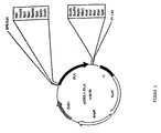
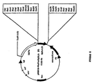
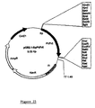
- the full length cDNA was ligated into the Eco RI site of the cloning vector pCR2.1, resulting in the final product "pCR2.1-DLA" illustrated schematically in Fig. 1.
- the Dl-FRILc clone was sequenced completely using the dideoxy chain termination method.
- the nucleotide sequence of the full-length cDNA was determined to be:
- the Dl-FRIL nucleotide sequence enabled inference of the following derived amino acid sequence for the Dl-FRIL protein:
- Dl-FRIL The naturally-occurring signal sequence from the FRIL family member isolated from Dolichos lab lab (i.e. , Dl-FRIL) has the following sequence:
- This sequence is located directly N-terminal to the first amino acid of SEQ ID NO: 2.
- the nucleic acid sequence of the naturally-occurring Dl-FRIL protein is provided below.
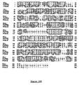
- FIG. 2 A comparative illustration of the derived Dl-FRIL amino acid sequence with the reported amino acid sequence of the mannose lectin as determined by Gowda et al. ( J. Biol. Chem . 269:18789-18793, 1994) is shown in Fig. 2.
- the single sequence derived for Dl-FRIL protein comprises domains that correspond directly and with substantial homology to the ⁇ subunit (SEQ ID NO:12) and ⁇ subunit (SEQ ID NO: 11 ) of the protein described by Gowda et al., supra. When the ⁇ subunit of the Gowda et al.
- Dl-FRIL (supra) protein is assigned to the N-terminal domain and is followed linearly by the ⁇ subunit, the arrangement of the polypeptides shows homology to other legume lectins.
- the derived Dl-FRIL amino acid sequence comprises an additional of eight amino acid residues (aa27-34) that does not occur in the amino add sequence described Gowda et al., supra .
- Several other differences between the amino acid sequences of Dl-FRIL and the amino acid sequence described by Gowda et al., supra, are also readily discernible from Fig. 2.
- Both the primary and the secondary PCR reactions were performed in 100 ⁇ L containing 50 pmol of each primer, 0.4 mM deoxyribonucleotide and 1.0 unit Pfu polymerase (Stratagene) in the corresponding buffer.
- the primary PCR reaction amplified the two separate fragments in 30 cycles, each cycle comprising 40 seconds at 94°C, 40 seconds at 50°C, 60 seconds at 72°C, with an extension step at 72°C for 10 min.
- the second PCR reaction amplified the recombinant fragment in 12 cycles using the same conditions described above.
- the resulting full-length fragment contained the mutation.
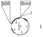
- the recombinant mutated product was cloned in the Eco RI site of the cloning vector pCR2.1, as illustrated schematically in Fig. 3, and sequenced as described above. This plasmid is referred to as "pCR2.1-DLA(D)."
- Recombinant PCR was used to modify the 5' ends of both the wild-type and the mutant Dl-FRIL clones, to introduce a signal peptide for entry of the protein into the endoplasmic reticulum.
- sequence encoding the signal peptide and the full-length cDNA clones were amplified in two separate primary PCR reactions.
- the signal peptide sequence was obtained from the amplification of the binary vector pTA4, harboring the complete sequence of the ⁇ -amylase inhibitor gene (Hoffman et al., Nucleic Acids Res. 10:7819-7828, 1982; Moreno et al., Proc. Natl. Acad. Sci. USA 86:7885-7889,1989).
- the primers used for the secondary reactions were Sigforw and AP Eco RI, which were designed to generate Eco RI sites at the 5' and the 3' ends. Both the primary and the secondary PCR reactions were performed as discussed above for the site-directed mutagenesis.
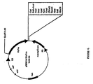
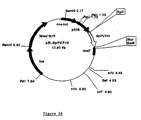
- the wild-type recombinant product SpDLA was cloned in the Eco RI site of the pBluescript SK+ cloning vector (Stratagene) to give the vector pBS-SpDLA, as shown in Fig. 4.
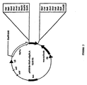
- the mutant SpDLA(D) was cloned in the same site of the cloning vector pCR2.1 to give the vector pCR2.1-SpM1, as shown in Fig. 5.
- the nucleotide sequence of each PCR product was determined as described above to verify the correct attachment of the signal peptide.
- the nucleotide sequence of SpDLA is defined by SEQ ID NO:22, and the derived amino acid sequence is defined by SEQ ID NO:23.
- sequences of SEQ ID NO: 22 and SEQ ID NO: 23 are as follows:
- a binary vector was constructed for seed-specific expression of Dl-FRIL.
- the vicilin promoter obtained from the pCW66 Higgins et al., Plant Mol. Biol . 11:683-695, 1988
- the SpDLA cDNA sequence was ligated into the Eco RI/ Sac I site, giving rise to the pBINVicPro-SpDLA, which is illustrated in Fig. 7.
- the mutated cDNA clone SpDLA(D) was ligated in Eco RI site of the pBINVicPro vector to yield pBINVicPro-SpDLA(D), which is illustrated in Fig. 8. No additional termination sequences were added, relying instead on the stop codons and the polyadenylation site of the DLA and DLA(D) cDNA clones. Both vectors were transferred into Agrobacterium tumefaciens strain LBA4404 according to the freeze-thaw procedure reported by An et al., "Binary vectors," in Plant Molecular Biology Manual, Vol. A3, Gelvin SB, Schilperoort RA, and Verma DPS, eds., Kluwer Academic Publisher, Dordrecht, The Netherlands, pp. 1-19,1988).
- Nicotiana tabacum leaf disks were carried out and assayed as described (Horsch et al., Science 227:1229-1231, 1985) using LBA4404 harboring the seed-specific expression vector pBINVicPro-SpDLA ( Figure 9).
- Kanamycin-resistant plants resistance being conferred by transformation with the pBIN19-based vectors that carry the gene
- Dl-FRIL fusion proteins were cleaved by the transformed plant cells in vivo.
- the transformed plant cells produced mature Dl-FRIL which, when purified, had a molecular mass of 60 kDa and comprised four subunits, two alpha subunits and two beta subunits ( i.e. , an ⁇ 2 ⁇ 2 heterodimer), where each subunit is about 15-18 kDa.
- the Dl-FRIL wild-type cDNA and mutant clones were ligated into the Eco RI/ Sal I and Eco RI/ Xho I of the expression vector pGEX 4T-1 (Pharmacia Biotech, Uppsala, Sweden), to form the expression constructs pGEX-M1 and pGEX M1(D), respectively illustrated in Figs. 9 and 10.
- the host E The Dl-FRIL wild-type cDNA and mutant clones (without signal peptides), were ligated into the Eco RI/ Sal I and Eco RI/ Xho I of the expression vector pGEX 4T-1 (Pharmacia Biotech, Uppsala, Sweden), to form the expression constructs pGEX-M1 and pGEX M1(D), respectively illustrated in Figs. 9 and 10.
- coli strain, BL21(D3) was purchased from Novagen (Madison, WI, and transformed with the above construct using the calcium chloride method (see Sambrook et al., supra; Gelvin and Schilperoort, Plant Molecular Biology Manual, Kluwer Academic Publishers, Dordrecht, The Netherlands, 1988; Altabella et al., Plant Physiol 93:805-810,1990; and Pueyo et al., Planta 196:586-596,1995).
- the induction of the tac promoter was achieved by adding IPTG (isopropyl- ⁇ -D-thiogalactopyranoside) (Sigma) at a 1.0 mM final concentration when the cells reached an optical density of 0.4 - 0.6 at 600 nm.
- IPTG isopropyl- ⁇ -D-thiogalactopyranoside
- the cultures were allowed to grow for 12 hours at 37°C after the addition of IPTG. Control non-induced cultures were maintained under similar conditions.
- the cells were lysed by treatment with 4 mg/mL lysozyme in phosphate-buffered saline containing 1% TRITON® X-100.
- Total cellular protein was extracted from transformed E. coli cells and analyzed on SDS-PAGE on a 15% gel using a standard procedure (Sambrook et al., supra).
- the cells from 1 mL of E. coli culture were suspended in the same volume of loading buffer (50 mM Tris HCl pH 6.8, 100 mM DTT, 2% SDS, 10% glycerol, 0.1% bromophenol blue) and vortexed. Following transfer to a nitrocellulose membrane, protein was stained with Coomassie Brilliant Blue R250. A representative separation is shown in Fig. 11, with the lanes identified in Table 1, below.
- Fig. 11 shows that the induced cells (lanes 3, 5) both produced an abundant polypeptide having a molecular mass of about 60 kDa (indicated by arrow). The non-induced cells failed to produce any significant amount of this protein (Fig. 11, lanes 2, 4).
- Induced E . coli cells (200 mL) as described above were harvested after 12 hour induction at 37°C by centrifugation at 5000 g for 10 min. The pellet was washed with 50 mM Tris-HCl pH 8.0, 2 mM EDTA, and resuspended in 1/10 vol of 1% TRITON surfactant in TBS (20 mM Tris pH 7.5, 500 mM NaCl). The cells were lysed by adding 4 mg/mL of lysozyme and incubating at room temperature for 30-60 min.
- the recombinant fusion protein solubilized by guanidine-HCl was purified on GST-Sepharose beads (Pharmacia Biotech, Uppsala, Sweden) according the manufacturer's instructions and eluted in 1 mL of reduced glutathione (Sigma). Samples of the purified fusion proteins were cleaved with thrombin (Novagen) using 5 cleavage units/mL purified fusion protein.
- Blotting was followed by incubation with a primary antibody (a polyclonal rabbit serum raised against the N-terminal peptide of the ⁇ -subunit of Phaseolus vulgaris FRIL (i.e. , Pv-FRIL), 1:100 dilution, 3 hours; described below in Example 5), followed by incubation with a secondary antibody (goat anti-rabbit IgG conjugated to horseradish peroxidase at 1:1000 dilution for 1 hour). The blots were washed and the color developed with the color development reagent (Bio-Rad). A representative result is shown in Fig. 12, with the lanes identified in Table 2, below.
- a primary antibody a polyclonal rabbit serum raised against the N-terminal peptide of the ⁇ -subunit of Phaseolus vulgaris FRIL (i.e. , Pv-FRIL), 1:100 dilution, 3 hours; described below in Example 5
- a secondary antibody goat anti-rabbit IgG conjugated to horseradish
- Fig. 12 demonstrates that the two forms of fusion protein have similar molecular masses of about 60 kDa, and that thrombin cleaved both types of fusion protein to produce a new polypeptide of molecular mass 30 kDa.
- Recombinant Dl-FRIL Specifically Stimulates Proliferation of 3T3 Cells Expressing the FLT3 Receptor
- Dl-FRIL interacts with the mammalian FLK2/FLT3 tyrosine kinase receptor.
- 3T3 cells cultured in tissue culture plates were removed from the plates by washing cells twice in Hank's buffered saline solution (HBSS; Gibco Laboratories, Grand Island, NY).
- HBSS Hank's buffered saline solution
- Non-enzymatic cell dissociation buffer Gibco was added for 15 minutes at room temperature. The resulting cells wee washed in medium.
- FLT3 3T3 cells were cultured at a final concentration of 3,000 cells per well in a volume of 100 ⁇ L of serum-defined medium containing 10 mg/mL rhIL1- ⁇ , 10% AIMV (Gibco, Grand Island, NY) and 90% Dulbecco's modification of Eagle's medium (DMEM; Gibco) in 96 well plates. Under these assay conditions, cells die after two to four days of culture in a humidified incubator at 37°C and 5% CO 2 unless exogenously added ligand rescues cells from death. Each 96 well plate contained wells of cells containing calf serum, which stimulates all 3T3 cells, as a positive control and wells of cells containing medium only as a negative control ("blank").
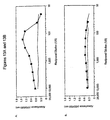
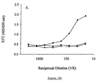
- Figs. 13A and 13B The crude protein extract from the E. coli cultures described in Example 1, above, was tested to determine whether expressed recombinant Dl-FRIL possessed any capacity to stimulate FLT3 3T3 cells using this assay.
- the data from this experiment are summarized in Figs. 13A and 13B.
- Fig. 13A is a graph showing that the crude extract of the E. coli culture containing expressed Dl-FRIL specifically stimulates hFLT3 cells
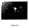
- Figure 13B is a graph showing that the same extract does not stimulate untransfected 3T3 cells.
- medium control is represented by a solid line.
- the ordinate indicates cell viability measured by XTT at three days; the abscissa shows the reciprocal dilution of the extract sample.
- the apparent inhibition of proliferation observed at higher concentrations (Fig. 13A) is not understood, but may relate to toxic components in the crude E. coli extract or the consequences of dose-related preservation of the 3T3 fibroblasts.
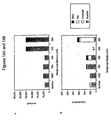
- Figs. 14A and 14B, and Table 3 illustrate the results of experiments in which recombinant Dl-FRIL was shown to act in a dose-responsive manner to preserve human cord blood progenitors.
- CB mnc Cord blood mononuclear cells
- FICOLL-PAQUE® separation Pharmacia Biotech, Piscataway, NJ
- X-VIVO 10 medium BioWhittaker, Walkersville, MD
- CB mnc were then cultured in six well tissue culture plates (Corning Inc., Corning, NY) at a concentration of 200,000 cells/mL in a volume of 4 mL of X-VIVO 10 (i.e. , 800,000 cells total per well).
- Dl-FRIL and/or recombinant E Dl-FRIL and/or recombinant E.
- coli Flt3-L (recFL; BioSource International, Camarillo, CA) were added at a concentration of 40 ng/ml at the outset (with no addition as a control). Cultures were incubated in humidified chambers without medium changes for up to 29 days.
- the cultured CB mnc cells were harvested by washing in X-VIVO 10 (i.e. , harvested cells were pelleted and resuspended in X-VIVO 10) to remove the Dl-FRIL and/or recFL, and then determining viable cell number by trypan blue (Sigma) exclusion.
- Fig. 14A The progenitor number and capacity of harvested cells were assessed by plating the washed cells in triplicate in fetal bovine serum-free, methylcellulose colony assay medium containing IL-2, granulocyte-macrophage CSF, and kit ligand (StemCell Technologies, Vancouver, BC, Canada).
- Figs. 14A and 14B show that recombinant Dl-FRIL preserved cord blood mononuclear cells and progenitors in a dose-responsive manner in liquid culture.
- Table 3 shows the resulting colonies after 15, 21, or 29 days of incubation, demonstrating that Dl-FRIL, but not recFL, preserved progenitors in suspension culture.
- Day Medium Myeloid Erythroid Mix Blast 15 Dl-FRIL 1,033 ⁇ 12 67 ⁇ 12 7 ⁇ 12 0 rccFL 40 ⁇ 69 0 0 0 Dl-FRIL + recFL 933 ⁇ 250 167 ⁇ 95 0 0 No Addition 0 0 0 0 21 Dl-FRIL 387 ⁇ 83 7 ⁇ 12 0 167 ⁇ 64 recFL 0 0 0 0 Dl-FRIL + recFL 473 ⁇ 133 53 ⁇ 42 0 300 ⁇ 34 No Addition 0 0 0 0 29 Dl-FRIL 0 0 0 80 ⁇ 72 recFL 0 0 0 0 Dl-FRIL + recFL 0 0 0 40 ⁇ 20 No Addition 0 0 0 0 0 0
- the reported experiment is a representative of four experiments.
- Figs. 14A and 14B in Table 3, "blast” refers to colonies consisting of primitive, morphologically undifferentiated cells; "mix” refers to colonies consisting of myeloid and erythroid cells; “erythroid” refers to colonies consisting of erythroid cells; and “myeloid” refers to colonies consisting of myeloid cells.
- cell number (Fig. 14A) or colony number (Fig. 14B) is shown on the ordinate; the abscissa shows the reciprocal dilution of the sample.
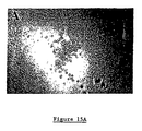
- Fig. 15A in addition to myeloid and erythroid colonies, day 21 cultures contained small colonies consisting of undifferentiated cells.
- Fig. 15B shows that only blast-like colonies were observed when the cells were cultured in Dl-FRIL for 29 days (see also Table 3).
- the frequency of blast-like colonies cultured in Dl-FRIL alone decreased by 2.1 fold from day 21 to day 29, from 1 in 4,790 to 1 in 10,000, and 7.5 fold in Dl-FRIL + recFL cultures from 1 in 2,667 to 1 in 20,000 of the initial CB mnc cells cultures.
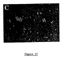
- small, diffuse, blast-like colonies were detected at a frequency of 1 in 67 (900 colonies/600,000 CB mnc) exclusively in dishes of cells initially cultured in Dl-FRIL alone, and at a frequency of 1 in 132 (990 colonies/131,000 CB mnc) for cells cultured in Dl-FRIL + recFL (see examplary colonies in Fig.17).
- cord blood mononuclear cells were first enriched for progenitors expressing the CD34 antigen by immunomagnetic bead isolation (Dynal Corp., Lake Success, NY).
- CD34 + cells Five hundred CD34 + cells were placed into wells containing 100 ⁇ L of serum-free medium (BIT9500, StemCell Technologies, Vancouver, BC, Canada) either in the presence of recFL (PeproTech, Princeton, NJ) or a cytokine cocktail of recombinant human interleukin 3 (rhIL3) + recombinant human interleukin 6 (rhIL6) + recombinant human interleukin 11 (rhIL11) + rhTpo Thrombopoietin + FL (FLT3-Ligand) (BioSource International, Camarillo, CA) in 96-well plates and cultured for four weeks without medium changes.
- recFL PeproTech, Princeton, NJ
- rhIL11 recombinant human interleukin 6
- rhIL11 recombinant human interleukin 11
- FL FLT3-Ligand
- the mFlt3/Fms 3T3 cell line is a 3T3 cell line transfected with nucleic acid encoding a fusion protein consisting of the murine extracellular domain of the Flt3 receptor fused to the transmembrane and intracellular domains of the human Fms (provided by Dr. Ihor Lemischka, Princeton University, Princeton, NJ).
- the Stk 3T3 cell line is a 3T3 cell line transfected with the full-length human Flt3 receptor (provided by Dr.
- the human FMS 3T3 cell line is a 3T3 cell line transfected with the full-length human Fms receptor (provided by Dr. Charles Sherr, Saint Jude Children's Research Hospital, Memphis, TN.
- Parent 3T3 cells were purchased from American Type Culture Collection ("ATCC"; Manassas, VA). Receptor-transfected cells contained Neo resistance genes and were maintained in medium containing 750 g/ml of G418 (Life Technologies, Rockville, MD).
- 3T3 cell lines were assayed in 96 well plates (Becton Dickinson Labware, Lincoln Park, NJ) containing 3,000 cells in 100 ⁇ L of serum-free medium consisting of 10% AIMV (Life Technologies) and 90% DMEM. In each experiment, samples were serially diluted two-fold across rows starting at a 1:10 dilution. Viable cells were quantitated after 3-5 days by XTT (2,3-bis[Methoxy-4-nitro-5sulfophenyl)-2H-tetrazolium-5 carboxanilide inner salt) (Sigma, St. Louis, MO) (Roehm et al., supra ). Relative activity (units/ml) and specific activity (units/mg) are defined as the reciprocal dilution at which half-maximal stimulation of cells was detected.
- the FMS 3T3 cells (transfected with cDNA encoding the human Fms tyrosine kinase receptor) and its ligand, human M-CSF, served as a model system.
- Recombinant human M-CSF stock of 1 ⁇ g/mL was serially diluted (where the first dilution, 1:20, was 50 ng/mL) was used to stimulate Fms3T3 cells.
- Fms 3T3 cells responded to M-CSF in a dose-responsive manner.
- PHA phytohemagglutinin
- PHA-LCM PHA leukocyte-conditioned medium
- Mononuclear cells were isolated by FICOLL-PAQUE®, washed in AIMV, and cultured at a concentration of 2 x 10 6 cells/ml in AIMV containing a 1% volume of crude red kidney bean extract containing PHA from Life Technologies (catalog number 10576-015) in either T150 flasks (Becton Dickinson Labware, Lincoln Park, NJ) or roller bottles (Becton Dickinson Labware) for one week. Cells and debris were removed by centrifugation and conditioned medium was stored at -20°C.
- PHA-LCM induced proliferation of mFlt3/Fms 3T3 cells and Stk 3T3 (expressing the human Flt3 receptor) in an indistinguishable manner at approximately 200 units/mL (Fig. 19B). Untransfected 3T3 cells did not respond to PHA-LCM ( Figure 19B) and Fms 3T3 cells responded weakly (data not shown).
- PHA-LCM Each batch of PHA-LCM was generated in serum-free medium with cells from individual normal donors. To start purification, the PHA-LCM with Flt3 3T3 activity was pooled in 30 liter lots with approximately 10 7 Flt3 3T3 units. Twenty-five liters of PHA-LCM was diafiltered into 50 mM Tris-HCl, pH 7.6, 50 mM NaCl and then concentrated to 2-2.5 liters by tangential flow ultrafiltration on a 10 kDa molecular weight cutoff membrane (Pellicon, Millipore, Bedford, MA).
- a phenyl-sepharose HP (Pharmacia Biotech) column (1.6 cm x 10 cm) was equilibrated with 20 mM phosphate, pH 7, 1.5 M NH 4 SO 4 .
- the pooled sample from Q-sepharose was adjusted to 1.5 M NH 4 SO 4 and applied to the phenyl-sepharose column.
- Fractions (1 ml) were collected and tested for Flt3 3T3 activity.
- Fractions with Flt3 3T3 activity were pooled and dialyzed against 50 mM Tris-HCl, pH 7.2, 100 mM NaCl and concentrated by vacuum centrifugation.
- a Superdex 75 (Pharmacia Biotech) column (1.6 cm x 60 cm) was equilibrated with 50 mM Tris-HCl, pH 7.4, 100 mM NaCl. The pooled sample from phenyl-sepharose was applied to the Superdex 75 column and eluted at 0.6 ml/min. Fractions (1.8 ml) were collected and tested for Flt3 3T3 activity. The active fractions were dialyzed against Tris-HCl, pH 7.2 and concentrated by vacuum centrifugation.
- a C4 reverse-phase column (4.6 mm x 100 mm, Vydac, Hesperia, CA) was equilibrated with 0.1% trifluoroacetic acid (TFA) in HPLC grade H2O. Pooled and concentrated sample from Superdex 75 chromatography was applied to the C4 column and the column was eluted with a gradient of 10-55% acetonitrile, 0.1% TFA over 70 min at 0.5 ml/min. Fractions of 0.5 mL were collected and evaporated by vacuum centrifugation.
- TFA trifluoroacetic acid
- cytokines IL1- ⁇ , IL1- ⁇ , IL2, IL3, IL4, IL6, GM-CSF, G-CSF and SCF
- cytokines IL1- ⁇ , IL1- ⁇ , IL2, IL3, IL4, IL6, GM-CSF, G-CSF and SCF
- AQS-peptide a New Zealand White rabbit (HRP, Denver, PA) was immunized with crude PHA-LCM, boosted with increasingly purified samples containing Flt3 3T3 activity, and finally immunized with a peptide corresponding to Pv-FRIL AQSLSF[N, C, S]FTKFDLD (SEQ ID NOS: 32-34), referred to as the AQS-peptide.
- Samples were glutaraldehyde conjugated to keyhole limpet hemocyanin (KLH, Sigma).
- the rabbit was immunized with KLH-AQS peptide-containing samples using either Complete Freund's Adjuvant (Sigma) or Hunter's Titermax (Vaxcel, Inc., Norcross, GA).
- Antiserum demonstrated a 1:5,000 titer to the AQS peptide in an ELISA (data not shown). Since the antiserum contained reactivities to other proteins, further enrichment for AQS peptide-specific antibodies was achieved by either depletion of cross-reactive antibodies or by affinity purification using a AQS peptide covalently linked to an agarose support (AminoLink coupling gel, Pierce).
- An anti-AQS affinity column was prepared either by purifying IgG from high titer rabbit antiserum by protein A affinity chromatography (ImmunoPure kit, Pierce) or antibody isolated from the AQS peptide column and then covalently linking antibody to an activated agarose support (AminoLink coupling gel, Pierce).
- a suspension culture of human cord blood cells enriched for Flt3 + progenitors by CD34 immunomagnetic bead selection was adapted to a 96 well plate format.
- umbilical cord blood from healthy donors was collected in 100 units/ml of heparin (Fujisawa Healthcare, Deerfield, IL).
- Mononuclear cells were isolated by FICOLL-PAQUE® (Pharmacia Biotech, Piscataway, NJ), washed in HBSS, and resuspended in serum-defined medium, either AIMV or XVIVO-10 (BioWhittaker, Walkersville, MD). Mononuclear cells were enriched for Flt3 + progenitors by CD34 immunomagnetic bead selection (Dynal Corporation, Lake Success, NY).
- CD34 + cells were cultured in 6 well plates (Becton Dickinson Labware) in at a concentration of 10 5 cells in 1 mL of DMEM containing 10 ng/mL recombinant human IL3 (BioSource International, Camarillo, CA) and 10% fetal calf serum. The number of refractive cells present in culture wells was scored microscopically.
- FIG. 20A shows that cord blood cells responded to column fractions in two regions of the material eluted from an anion exchange column. The first region of activity corresponded with Flt3 3T3 stimulatory activity (Figs. 20B and 20C); the second associated with an activity detected with Fms 3T3 cells (Fig. 20D); no response was detected in untransfected 3T3 cells (data not shown).
- the active material, corresponding to Flt3 3T3 activity peak one in Fig.
- cytokines that act on hematopoietic progenitors (interleukin 1- ⁇ (IL1- ⁇ ), interleukin 1- ⁇ (IL1- ⁇ ), interleukin 2 (IL2), interleukin 3 (IL3), interleukin 4 (IL4), interleukin 6 (IL6), granulocyte-macrophage colony stimulating factor (GM-CSF), G-CSF (granulocyte colony stimulating factor), and stem cell factor (SCF)) in fractions containing Pv-FRIL purified to near homogeneity revealed that IL1- ⁇ had remained with Pv-FRIL through every step of purification (data not shown).
- IL1- ⁇ interleukin 1- ⁇
- IL1- ⁇ interleukin 2
- IL3 interleukin 3
- IL4 interleukin 4
- IL6 interleukin 6
- GM-CSF granulocyte-macrophage colony stimulating factor
- G-CSF granulocyte colony
- Flt3 3T3 activity was depleted but not eliminated either by adding neutralizing antibodies to IL1 or by removing IL1 by antibody affinity chromatography (data not shown).
- levels ⁇ 1 ng/mL
- exogenous recombinant hIL1- ⁇ by itself had no stimulatory activity (data not shown).
- This observation suggested the possibility that IL1- ⁇ may act as a necessary co-factor to obtain maximal stimulation by Pv-FRIL.
- the co-factor requirement was met by the addition of either IL1- ⁇ or PHA-LCM added at a concentration that did not stimulate Flt3 3T3 cells by itself.
- SDS-PAGE showed the purified material to contain a limited number of polypeptides and the molecular size of the active material was determined by eluting the protein from SDS-PAGE gel slices run under non-reducing conditions and assaying the activity of the eluted material.
- Flt3 3T3 activity was always found in a gel slice that contained 14-22 kDa polypeptides and sometimes in a gel slice containing 32-43 kDa polypeptides (data not shown).
- polypeptide(s) in the active fraction corresponding to the 14-22 kDa range were subjected to aminoterminal sequencing by Edman degradation.
- an 18 kDa species from C4 reverse-phase chromatography was resolved by SDS-PAGE, electroblotted onto polyvinylidene difluoride (PVDF) membrane (Immobilon-P, Millipore) and stained with Ponceau S (Bio-Rad Laboratories, Inc., Hercules, CA).
- PVDF polyvinylidene difluoride
- the 18 kDa band was cut from the PVDF membrane and N-terminal sequence was determined by automated Edman degradation on an ABI model 477A protein sequencer (PE Applied Biosystems, Foster, CA).
- the derived peptide sequences were compared against the SwissProt protein sequence database.
- sequence AQSLSFXFTKDLD (SEQ ID NO: 35) was obtained from a polypeptide of 18 kDa (where X is an unknown amino acid).
- aminoterminal sequence of TDSRWAVEFDXFP (SEQ ID NO: 36) was found twice.
- the amino terminus of a smooth muscle protein (SM22- ⁇ ) was found twice and the amino terminus of myoglobin identified once. Since the sequence starting with AQS was the only sequence identified in each experiment, this polypeptide was concluded to be responsible for Flt3 3T3 activity.
- FIG. 22A-22D shows results of an experiment where the pooled fractions were assayed on cord blood cells in the presence of IL3. After two weeks of suspension culture, the number of viable cells and status of CD34 expression was evaluated. A representative of three AQS affinity chromatography experiments is shown in Figs. 22A-22D.
- Cell cultures supplemented with the two early column fraction pools contained approximately four-fold more cells (426,000 cells and 466,250 cells, respectively) than at the 100,000 cells seeded and no appreciable CD34 staining (Figs. 22A and 22B). The increase in cell number and loss of CD34 expression is attributed to the expected consequences of IL3-induced proliferation and differentiation. In contrast, cell cultures treated with the late-eluting fraction pool (Fig.
- PHA is derived form red kidney bean extract and because a FRIL family member, Dl-FRIL, was isolated from another legume, namely Dolichos lab lab, mannose-binding lectins were isolated from red kidney bean ( Phaseolus vulgaris ) extract using standard methods, such as the procedure of Rudiger, H., Isolation of Plant Lectins , H.-J. Gabius and S. Gabius, eds., pp. 31-46, Berlin, 1993).
- kidney bean mannose-binding lectin consisted of polypeptides with molecular weights of 18 kDa and 15 kDa, and the aminoteimini of these two polypeptides started with AQSLSFXFKFDPN (SEQ ID NO: 37) and TDSRVVAVEDF (SEQ ID NO: 38), respectively (where X is an unknown amino acid).
- Pv-FRIL isolated from Phaseolus vulgaris was tested for activity in the Flt3 3T3 cell assay. As shown in Fig. 23, Flt3 3T3 cells responded in a dose dependent manner to Pv-FRIL, while parent untransfected 3T3 cells did not
- Dry seeds from the red and white kidney beans ( Phaseolus vulgaris ) were purchased from W. Atlee Burpee & Company, Warminster, PA. Lectins were eluted using a standard protocol. Briefly, beans were pulverized in a home coffee grinder and added to buffer of 50 mM Tris/HCl, pH. 8.0, 1 nM each of MgCl 2 and CaCl 2 for 4 hours at 4°C with constant mixing. Bean solids were pelleted by centrifugation at 10,000 x g for 20 min.
- the pH of the supernatant was modified to pH 4.0 with acetic acid and constant mixing to remove contaminating storage proteins, followed by centrifugation to clarify the supernatant, and finally the pH was readjusted to 8.0 with sodium hydroxide before storing at -20°C.
- PCR Two sequential polymerase chain reactions (PCR) were performed.
- 10 ng of bean genomic DNA was amplified by 30 cycles of PCR, each cycle comprising 40 seconds at 94 °C, 40 seconds at 50 °C, 60 seconds at 72 °C, and an extension step at 72 °C for 10 min.
- the reactions were performed in 50 ⁇ l containing 30 pmol of each primer, PVbeta 1 and PValfa1, 0.2 mM deoxyribonucleotides and 0.5 unit of Ampli-Taq polymerase (Perkin Elmer) in the corresponding buffer.
- One microliter of the PCR product was amplified by 30 PCR cycles using the same conditions as described above.
- the reaction was performed in 50 ⁇ L containing 30 pmol of the two primers, PVbeta 2 and PV alfa2, using 0.2 mM deoxyribonucleotides and 0.5 unit of Ampli-Taq polymerase (Perkin Elmer) in the corresponding buffer.
- the 460 bp fragment obtained was cloned in a T/A plasmid, pCR2.1 (Invitrogen) and sequenced by sequenase dideoxy chain termination (United States Biochemicals).
- RNA was prepared from mid-maturation Phaseolus vulgaris seeds stored at -70°C following the procedure reported by Pawloski et al. ( Mol. Plant Biol. Manual 5: 1 -13, 1994).
- the 5'/3' RACEKIT Boehringer Mannheim was used to generate cDNA from 5.0 ⁇ g total RNA used according to the manufacturer's instructions.
- the oligo(dT) anchor primer was at the concentration of 32.5 ⁇ M, in the standard conditions.
- a specific primer (SPV1) was used at the concentration of 32.5 ⁇ M.
- SPV1 specific primer
- Pv-FRIL The 3' end of Pv-FRIL was obtained through rapid amplification of cDNA ends by polymerase chain reaction (RACE-PCR) using the 5'/3' RACEKIT (Boehringer Mannheim) used according to the manufacturer's instructions. Nested PCR amplifications were performed using the PCR-Anchor primer with the specific primers (PV3 and PV4) in two successive amplification reactions. The sequences of these primers is as follows:
- the amplification conditions were 30 cycles of 40 seconds at 94 °C, 40 seconds at 55 °C, 60 seconds at 72 °C each and an extension step at 72 °C for 10 min.
- the reactions were performed in 50 ⁇ L containing 30 pmol of each primer, PV3 and PCR-Anchor primer in the first, and PV4 and PCR-Anchor primer in the second, 0.2 mM deoxyribonucleotides and 0.5 units of Ampli-Taq polymerase (Perkin Elmer) in the corresponding buffer.
- the 831 bp product obtained was sub-cloned in pCR2.1 and sequenced as above reported.
- recombinant PCR was performed (Higuchi R, supra). Two PCR reactions were carried out separately, one on the 5' fragment and the other on the 3' RACE product using primers with an overlapping region. The overlapping primary products were subsequently re-amplified using the flanking primers resulting in a full-length fragment.
- the primary PCR products were purified with the QIAquick PCR Purification kit (QIAGEN) used according to the manufacturer's instructions, and amplified together in a single second reaction.
- QIAGEN QIAquick PCR Purification kit
- the primers PVEcoRI and the APEcoRI were used. The sequences of these primers is as follows:
- Both primary and secondary PCR reactions were performed in 100 ⁇ L containing 50 pmol of each primer, 0.4 mM deoxyribonucleotide and 1.0 unit Pfu polymerase (Stratagene) in the corresponding buffer.
- the primary PCR reaction amplified the two separate fragments by 30 cycles, each cycle comprising 40 seconds at 94°C, 40 seconds at 50°C, 60 seconds at 72°C and an extension step at 72°C for 10 min.
- the second PCR reaction amplified the recombinant fragment in 12 cycles using the same conditions reported above.
- the full-length product was cloned in the EcoRI site of the cloning vector pCR2.1 (Fig. 24A) and sequenced as noted above. This plasmid is referred to as pCR2.1-Pv-FRIL.
- the nucleic acid sequence of the Pv-FRIL cDNA is as follows:
- amino acid sequence of Pv-FRIL is as follows:
- the amino acid sequence of Pv-FRIL was compared to the amino acid sequences of Dl-FRIL and of the PHA-E lectin. This comparison is shown on Fig. 24B.
- Recombinant PCR was used to introduce a signal peptide for entry of Pv-FRIL into the endoplasmic reticulum at the 5' end of the Pv-FRIL cDNA clone.
- the signal peptide and the full length cDNA clone were amplified in two separate primary PCR reactions.
- the signal peptide was obtained from the amplification of the binary vector pTA4, harboring the complete sequence of the bean ⁇ -amylase inhibitor gene (Hoffman et al., L.M., Y. Ma and R.F. Barker, Nucleic Acid Res . 10: 7819-7828, 1982; Moreno and Chrispeels, Proc. Natl. Acad. Sci. USA 86: 7885-7889, 1989).
- the primers used for the two primary reactions are the following:
- a binary vector was constructed for constitutive expression of Pv-FRIL in tobacco plants.
- the recombinant SpPv-FRIL was cloned in BglII/XhoI sites of a plant expression vector resulting in the formation of pM-SpPv-FRIL (Fig. 26).
- the binary vector was transferred in Agrobacterium tumefaciens strain C58 according to the freeze-thaw procedure reported of An et al., supra.
- Agrobacterium-mediated transformation of Nicotiana tabacum leaf disks was carried out as described by Horsh et al. ( Science 227: 1229 - 1231, 1985) using C58 harboring the expression vector pM-SpPv-FRIL.
- Kanamycin resistant plants were scored for their ability to form roots in two consecutive steps of propagation in Murashige-Skoog medium containing 3% sucrose and kanamycin sulfate (Sigma) at 100 mg/L.
- Tobacco plants transformed with this construct were grown in a growth room under controlled conditions.
- the leaves (20 grams) of young plants were harvested and frozen in liquid nitrogen and powdered in a mortar with a pestle.
- the powder was stirred in a buffer mixture consisting of 1 x phosphate buffered saline containing 1mM CaCl 2 with a cocktail of protease inhibitors (PMSF, Pepstatin and Leupeptin). This slurry was centrifuged at 2000 rpm and the supernatant was centrifuged at 40,000 rpm in a Beckman ultracentrifuge. The clear supernatant was tumbled with 1 ml of ovalbumin-Sepharose for 3 hours.
- PMSF protease inhibitors
- the beads were washed with the same buffer and tumbled overnight with 200 mM trehalose in 1/10 phosphate buffered saline containing the cocktail of protease inhibitors. Coomassie blue stained gel showed this preparation to be pure Pv-FRIL. An immunoblot showed the presence of both alpha and beta subunits, thus, the single polypeptide chain encoding both subunits was cleaved by the transformed cells in vivo . Binding to the ovalbumin-sepharose and release by trehalose shows that the product of the transgene is an active lectin.
- Dl-FRIL Supports Prolonged ex vivo Maintenance of Quiescent Human CD34+,CD38-/SCID-Repopulating Cells
- CB samples Human umbilical cord blood (CB) samples were obtained from full term deliveries. The blood samples were diluted 1:1 in phosphate-buffered-saline (PBS) without Mg +2 /Ca +2 , supplemented with 10% fetal bovine serum (FBS). Low density mononuclear cells were collected after standard separation on Ficoll-Paque (Pharmacia Biotech, Uppsala, Sweden), and washed in RPMI with 10% FBS. Some samples were frozen in 10% DMSO, while the others were used fresh. Enrichment of CD34 + cells was performed with mini MACS separation kit (Miltenyi Biotec, Bergisch Gladbach, Germany) according to the manufacturer's instructions.
- PBS phosphate-buffered-saline
- FBS fetal bovine serum
- CD34 + CD38 -/low cells were purified by FACS sorting (FACStar + , Becton Dickinson, San Jose, CA) after staining CD34 + enriched cells with mAb anti human CD34-FITC (Becton Dickinson) and anti human CD38 PE (Coulter, Miami FL USA) (Purity >99%).
- NOD-SCID mice and NOD/SCID ⁇ 2 microglobulin knockout mice hereafter termed NOD/SCID B2M null (Christianson et al., J. Immunol . 158:3578-3586,1997), bred and maintained under defined flora conditions in sterile micro isolator cages, were irradiated with a sublethal dose of 375 cGy at 67cGy/ min. from a cobalt ( 60 Co) source prior to transplantation.
- irradiated mice Human cells were injected into the tail vein of irradiated mice in 0.5 mL of RPMI with 10% FBS.
- non engrafting irradiated (1500 cGy) CD34 - cells served as carrier cells, and were cotransplanted with cultured cells at a final concentration of 0.5x10 6 cells/mouse.
- Mice were sacrificed 1 month post transplantation, and bone marrow (BM) cells were flushed from the 8 bones of each mouse (femurs, tibias, humeri, and pelvis).
- BM bone marrow
- Human CD34 + enriched cells were cultured in 24 well plates (2-4x10 5 cells in 0.5 mL), containing RPMI supplemented with 10% FBS+1% BSA. Ex vivo cultures contained the following cytokine combination: Stem cell factor (SCF) - 100 ng/mL and Flt3 ligand (Flt3-L) - 100 ng/mL (R&D Systems Inc.
- SCF Stem cell factor
- Flt3 ligand Flt3-L
- the growth factor cocktail included 300 ng/mL SCF, 300 ng/mL Flt3-L, 50 ng/mL G-CSF, 10 ng/mL IL-3 (R&D Systems), and 10 ng/mL IL-6.
- the cultures were incubated at 37°C in a humidified atmosphere containing 5% CO 2 .
- BM cells from engrafted mice were cultured with 100 ng/mL of SCF and IL-15 (R&D) for 10-14 days.
- Dl-FRIL was isolated from the seeds of hyacinth beans ( Dolichos lab lab ) using the protocol described above in Example 1.
- Semisolid cultures were performed in order to detect the levels of human progenitors in ex vivo cultures, and in the marrow of transplanted mice.
- the cells were plated (4x10 3 cells/mL) in 0.9% methylcellulose (Sigma, St. Louis, MO, USA), 30% FBS, 5x10 -5 M 2ME, 50 ng/mL SCF, 5 ng/mL IL-3, 5 ng/mL GM-CSF (R&D), and 2 u/mL Erythropoietin (Ortho Bio Tech, Don Mills, ON, Canada).
- Human cells from the BM of engrafted mice were plated (2 x10 5 cells/mL) in 15% FBS + 15% human plasma, selective for human colonies only. The cultures were incubated at 37°C in a humidified atmosphere containing 5% CO 2 and scored 14 days later with an inverted microscope for myeloid, erythroid, and mixed colonies by morphologic criteria.
- Cells were analyzed for their DNA content by staining with propidium iodide (Sigma). The cells were cultured in ex vivo cultures as described, for 3, 6, 10 and 13 days as indicated. At each time point, the cells were collected, resuspended to a final concentration of 0.1-1x10 6 cells/mL, and incubated with 0.1% Triton X 100 (Sigma) for 20 minutes on ice. 50 mg/mL propidium iodide (PI) were added before analysis. Flow cytometeric analyses were performed using FACSort (Becton Dickinson, San Jose, CA).
- Human and mouse Fc receptors on BM cells from transplanted mice were blocked by using human plasma (1:50) and anti mouse Fc receptor blockers (anti mouse CD16/CD32 mAb, Pharmingen, San Diego, CA, USA). Isotype control mAb were used in order to exclude false positive cells (Coulter, Miami FL USA). The purity of enriched subpopulations after magnetic bead separation, were analyzed by two color staining, using anti-human CD34 FTTC (Becton Dickinson, San Jose, CA, USA) and anti-human CD38 PE (Coulter).
- the levels of human cells and lymphoid lineages in the marrow of engrafted mice were detected by double staining with anti-human CD45 FITC ( Immuno Quality Products, Groningen, The Netherlands ) together with anti-human CD19 PE (Coulter) for detection of pre B cells, or with anti-human CD56 PE (Coulter) for detection of NK cells.
- Cells were washed with PBS supplemented with 1% FBS and 0.02% azide, suspended to a volume of 0.1-1x10 6 cells/mL, stained with direct-labeled mAb and incubated for 25 minutes on ice. After staining, cells were washed once in the same buffer and analyzed on a FACSort (Becton Dickinson). Analysis was performed using CELLquest software (Becton Dickinson).
- the levels of human cell engraftment were determined by both flow cytometry for analysis of human myeloid CD45 + and lymphoid CD45 + CD19 + pre B cells and quantification of human DNA as previously described (Lapidot et al., Science 255:1137, 1992; Larochelle et al., Nat. Med. 2:1329-1337,1996; Peled et al., Science 283:845-848,1999). Briefly, high molecular weight DNA was obtained from the BM of transplanted mice by phenol/chloroform extraction.
- DNA (5 g) was digested with EcoRI, subjected to electrophoresis on 0.6% agarose gel, blotted onto a nylon membrane, and hybridized with a human chromosome 17-specific ⁇ -satellite probe (p17H8) labeled with 32 P (Lapidot et al., supra ).
- the intensity of the bands in the samples were compared to artificial human/mouse DNA mixtures (0%, 0.1%, 1%, and 10% human DNA) to quantify the human DNA (lanes to the right of lane 4 in Fig. 31). Multiple exposures of the autoradiographs were taken to ensure sensitivity down to 0.01% human DNA.
- a transplanted mouse was scored positive when both human myeloid and lymphoid cells and human DNA were detected in its BM.
- Dl-FRIL maintains but does not expand CB CD34 + progenitors in suspension culture
- Dl-FRIL by itself preserves immature CB progenitor cells up to a month in suspension culture without medium changes (see, e.g., Figs 14A and 14B, and Table 3).
- cytokines SCF, Flt3-L, IL-6, and sIL6-R
- enriched CB CD34 + cells were cultured with either Dl-FRIL or cytokines for 3, 6, 10, or 13 days in medium containing 10% FBS and 1% BSA in RPMI. Fresh media and Dl-FRIL and/or cytokines were added on day 6. Cells harvested at each time point were counted and assayed for clonogenic progenitor cells by plating ( i . e ., seeding) in semisolid media.
- Fig. 27A the total number of cells in Dl-FRIL cultures gradually declined over time from 2x10 5 cells initially seeded to 1.26x10 5 cells at day 13, in contrast to the expected 14.3-fold increase to 2.8x10 6 cells in cytokine cultures at day 13.
- Fig. 27B the levels of progenitor cells in Dl-PRIL cultures also remained relatively constant until day 10, after which they declined to 9 % of the starting population (from 24.7x10 3 colonies on day 0 to 2.3x10 3 colonies on day 13).
- Progenitor levels increased in cytokine cultures by 10-fold from the outset to day 13 of culture (Fig. 27B).
- Dl-FRIL maintains the expansion capacity of CD34 + progenitors up to 2 weeks in ex vivo culture
- CD34 + cells cultured for 6 days with Dl-FRIL were washed and exposed to cytokines (without Dl-FRIL) for an additional 4 days.
- Total cell counts and clonogenic progenitor assays were performed and the results were compared to cells cultured for 10 days with Dl-FRIL.
- Fresh media and Dl-FRIL were added on day 6.
- the Dl-FRFL-cultured cells proliferated in response to cytokine stimulation, resulting in a 3.4-fold increase in cell numbers (Fig. 28A) and 13-fold increase in progenitor levels (Fig. 28B).
- Dl-FRIL maintains SCID repopulating stem cells (SRC) in ex vivo cultures
- mice were sacrificed and their bone marrows were harvested and assayed for the presence of human myeloid and lymphoid cells.
- human DNA levels in the bone marrow of individual transplanted mice was quantitated by Southern blotting analysis. A representative Southern blot analysis showing the detection of a 0%, 0.1%, 1%, and 10% human DNA per murine DNA is provided in Fig. 29E.
- Figs. 29A-29D show representative Southern blot analyses of the BM of mice transplanted with ex vivo cultured cells.
- Fig. 29A is a representative Southern blot showing human DNA in the marrow of mice transplanted with cells cultured with FRIL for 6 days (lane 1), FRIL for 10 days (lane 2), or with FRIL for 6 days followed by 4 days with cytokine stimulation (lane 3).
- the levels of engraftment by cells cultured with Dl-FRIL for 10 days decreased by approximately 10-fold from cells cultured with Dl-FRIL for 6 days (Fig. 29A, lanes 1 and 2).
- FIG. 29C shows the results of another experiment, where the difference between engraftment levels obtained by cells cultured with Dl-FRIL for 10 days (lane 1) or with Dl-FRIL for 6 days followed by 4 days of cytokine stimulation (lane 2) are minor. Nevertheless, a 10-fold increase was observed for cells cultured either with Dl-FRIL for 13 days (Fig. 29D, lane 3) or with Dl-FRIL for 10 days followed by 3 days of cytokine stimulation (Fig. 29D, lane 4) when compared to cells cultured for 10 days (Fig. 29D, lanes 1 and 2).
- Cells cultured with Dl-FRIL followed by cytokines, engrafted at levels 7.4-fold greater than cells cultured with Dl-FRIL alone for 10 days (Fig. 30, p 0.05).
- CD34 + cells isolated by immunomagnetic beads were further sorted by flow cytometry based on the absence of CD38 expression.
- CD34 + CD38 -/low cells (99% purity) cultured with either Dl-FRIL alone or Dl-FRIL followed by cytokines showed patterns of engraftment in NOD/SCID B2M null mice (Christianson et al., supra), similar to those observed for CD34 + cells. These mice have been used successfully for secondary human stem cell transplantation (Peled et al., supra), have less innate immunity due to lack of NK activity and thus require fewer (10 fold) human cells for engraftment compared to NOD/SCID mice.
- Fig. 31 is a representative Southern blot (1 out of 4 experiments) showing the relative level of engraftment of cells that were cultured with Dl-FRIL for 10 days (lane 4) compared to 6 days with Dl-FRIL followed by 4 days cytokine stimulation (lanes 1-3) prior to transplantation into mice.
- High molecular weight DNA was obtained from the BM of transplanted mice and subjected to Southern blotting analysis for the presence of human DNA.
- mice transplanted with cells that were treated with Dl-FRIL followed by cytokines were about 10-fold higher compared to the mouse transplanted with cells cultured with Dl-FRIL alone (Fig. 31, lane 4).
- Dl-FRIL preserves long-term repopulating stem cells.
- Bone marrows harvested from NOD/SCID mice that were initially transplanted with CD34+ cord blood cells cultured in Dl-FRIL were transplanted into a second set of sublethaly irradiated NOD/SCID mouse recipients. After one month, bone marrows from the second set of recipients were analyzed for the presence of human hematopoiesis, as determined using the assays described above. Multilineage engraftment was observed in the second set sublethaly irradiated NOD/SCID mouse recipients. This observed persistence of repopulating cells in this serial transplantation study indicated the presence of long-term repopulating stem cells.
- Dl-FRIL preserves SRC potential of multilineage differentiation in the murine BM
- BM of transplanted mice was collected 1 month later and the cells were either seeded in semisolid media selective for human colonies (results shown in Fig. 33A) or were analyzed for lineage specific markers by flow cytometry (results shown in Figs. 33B-33F).
- Fig. 33A shows that the levels of human progenitors in the bone marrow of transplanted mice increased 2.3-fold when SRC were stimulated with cytokines for 4 days after 6 days of Dl-FRIL incubation compared to Dl-FRIL alone for 10 days prior to transplantation. Moreover, both myeloid and erythroid colonies formed in the colony assays (data not shown) as well as human B cell differentiation that was determined by flow cytometry (Figs.
- mice To determine whether human lymphoid precursors in the marrow of transplanted mice have the potential to differentiate in addition to myeloid cells also into lymphoid NK cells, cells from transplanted murine bone marrow were also cultured with SCF and IL-15 for 10 days. Cells harvested from cultures were analyzed for human NK cells by flow cytometry using the human-specific monoclonal antibodies to CD45 and the NK cell antigen, CD56. Cells from mice transplanted with CD34 + or CD34 + CD38 -/low cells are shown (1% or 7.7% of CD56 + cells; Figs. 33E and 33F, respectively).
- Dl-FRIL was identified by its ability to stimulate proliferation of NIH 3T3 cells transfected with Flt3 and not by untransfected cells or cells transfected with the related Fms tyrosine kinase receptor (Moore et al., Blood 90 supp.1:308,1997). Although Dl-FRIL and Flt3-L both stimulate proliferation of Flt3 3T3 cells, they exert different activities on CB progenitor cells. Flt3-L induces quiescent primitive cells into cycle (Lyman and Jacobsen, Blood 91:1101-11345, 1998). In contrast, Dl-FRIL by itself maintains progenitors in serum-defined medium from 15-29 days in culture without medium changes (Colucci et al., Proc.
- Fig. 34A the number of total cells harvested after 10 days of suspension culture in Dl-FRIL increased minimally by 1.9-fold, whereas cells cultured in either Flt3-L alone or in combination with Dl-FRIL increased by 4.5-fold and 5.4-fold, respectively (p ⁇ 0.05).
- Dl-FRIL maintains higher levels of CD34 + cells in G 0 /G 1 phase of cell cycle compared to cytokine treated cells
- CD34 + cells are predominantly quiescent, non-cycling cells (Young et al., Blood 87:545, 1996; Movassagh et al., Stem Cells 15:214-222,1997; Ladd et al., Blood 90:658-668, 1997). Since Dl-FRIL maintains relatively constant levels of progenitor cells over two weeks in suspension culture that can subsequently respond to cytokine stimulation (see Fig. 27), the cell cycle status of CD34 + cells incubated with Dl-FRIL was investigated and compared to cultures with cytokine stimulation. DNA content of CB CD34 + cells was analyzed by flow cytometry immediately after isolation of cells. A mean value of 96.6% of cells were observed in the G 0 /G 1 phase of cell cycle (Fig. 35A).
- CB CD34 + cells The cell cycle status of CB CD34 + cells was analyzed in 5 indepenent experiments with cells cultured for 3-13 days with either Dl-FRIL or a cytokine combination (SCF, Flt3-L, G-CSF, IL-3, IL-6) (Bhatia et al., J. Exp. Med. 186:619-624, 1997; Conneally et al., Proc. Natl. Acad. Sci. USA 94:9836-9841,1997).
- SCF cytokine combination
- the number of cells in SG 2 M phase during culture differed from the 6.8x 10 3 cells in SG 2 M phase initially seeded.
- the average number of cycling cells in Dl-FRIL cultures remained relatively constant from 35x10 3 cells at day 3 and 43.6x10 3 cells at day 6 to a reduced level of 26.4x10 3 cells at day 10 and 11.4x10 3 cells at day 13.
- This pattern of cells in cycle explains the low levels of total cell numbers and progenitor cells (see Fig. 27).
- the expected number of cycling cells in cytokine cultures increased dramatically.
- Figs. 35A-35G show representative cell cycle histograms of CD34 + cells cultured with no addition (Fig. 35A), Dl-FRIL alone (Fig. 35B) or various cytokines or combinations thereof (Figs. 35C-35G) for 3 days, and the mean percentage of cells in each cell cycle phase is summarized in Table 6.
- Dl-FRIL maintains a significantly higher percentage of quiescent cells and lower percentage of cycling cells (Fig. 35B and Table 6) compared to stimulation with cytokines (Figs. 35C-35G and Table 6).
- Dl-FRIL Preserves CB Progenitors in a Dormant State in the Presence of Potent Stimulators
- Dl-FRIL was purified from Dolichos lab lab as described above in Example 1. Dl-FRIL was assayed over a 5-log dose range (10 ng/mL-1,000 ng/mL) on human CB MNC cultured in serum-defined medium containing agar-leukocyte conditioned medium, a potent source of a broad range of stimulators. The number of viable MNC and progenitors were evaluated after 5 days in culture. Results from 1 of 3 representative experiments are shown in Table 7 below. The number of viable MNC at the end of culture was reduced by 1.7 - to 5-fold in cultures containing 10-1,000 ng/ml of Dl-FRIL.
- Dl-FRIL MNC Progenitor frequency (ng/ml) (x 10 -4 ) (Colonies/10 -5 MNC) Myeloid Erythroid 1,000 20 90 +/- 85 150 +/- 42 100 55 44 +/- 15 85 +/-12 10 70 77 +/-12 103 +/- 6 1 120 16 +/- 2 38 +/- 7 0.1 115 21+/-7 50+/-7 0 120 38+/-7 63 +/- 32 Culture of CB MNC in Dl-FRIL results in fewer MNC and a greater frequency of progenitors.
- CB MNC 4 x 10 6 CB MNC were cultured in 2 mL of AIMV (Life Technologies) containing Agar-SCM (StemCell Technologies) and varying concentrations of Dl-FRIL. Cultures were harvested after 5 days and the number of viable MNC and progenitors were assessed. The colony data shown were normalized as the frequency of progenitors per 10 5 MNC.
- Dl-FRIL protects CB MNC from the toxicity of chemotherapy drugs
- Dl-FRIL can prevent proliferation and differentiation of CB progenitors in cultures containing potent stimulators indicated that Dl-FRIL protects progenitors from the toxicity of cell cycle-active chemotherapy drugs.
- the culture system described above was adapted to a 96 well plate format and the widely used chemotherapeutics, cytarabine (Ara-C) (Fig. 36A), doxorubicin (Dox) (Fig. 36B), and 5-fluorouracil (5-FU) (Fig. 36C) over a 5-log dose range were assayed on CB MNC cultured in the presence and absence of Dl-FRIL (see Figs. 36A-36C).
- Dl-FRIL was purified from Dolichos lab lab as described above in Example 1. Cultures containing Dl-FRIL (either at 10 ng/ml or 100 ng/ml) resulted in a 2-3 log dose shift of CB MNC to Ara-C (Fig. 36A and Dox (Fig. 36B). As shown in Fig. 36C, the presence of Dl-FRIL in 5-FU cultures increased viability over a large dose range. Differences between the dose shift of Ara-C and Dox by Dl-FRIL compared to 5-FU may be explained by recent reports demonstrating that 5-FU acts via an RNA mechanism rather than as a DNA-specific drug (Bunz et al., J. Clin. Invest . 104:263-269,1999).
- Dl-FRIL The in vivo toxicity of Dl-FRIL was determined in mice. Initially, Dl-FRIL was administered intravenously to mice over a 3-log dose range of 0.006-1 mg/kg (0.32-20 ⁇ g/mouse). Dl-FRIL was well tolerated and these mice have subsequently received 2 monthly challenges of Dl-FRIL without any observable short- or long-term adverse effects.
- Protocols to test chemoprotective properties of cytokines in mice typically involve daily pre-treatment (bolus or continuous delivery) regimens of 4-10 days before starting chemotherapy. Using this framework as a starting point, Dl-FRIL was injected at 5 mg/kg (100 ⁇ g/mouse) intravenously daily for 4 days. No gross adverse effects have been observed in over 150 mice treated with this dose regimen. Dl-FRIL was purified from Dolichos lab lab as described above in Example 1.
- Dl-FRIL toxicity were next explored by injecting a single bolus intraperitoneal injection of Dl-FRIL (to accommodate 1 mL volume) at 500 mg/kg and monitored the survival of mice for 48 hours. Of the four BALB/c mice receiving this treatment (2 males and 2 females, aged 5 months), only 1 mouse (a male) survived 48 hours. The surviving mouse's weight decreased by approximately 15% in the first 2 days and returned to normal by day 4. The surviving mouse's blood counts were in the normal range 3 days after injection of FRIL. The results demonstrate that even a very large dose of Dl-FRIL is not completely toxic.
- Dl-FRIL was purified from Dolichos lab lab as described above in Example 1. Weight-matched BALB/c mice (females, aged 8 weeks) were injected intravenously with either 0.2 ml of Dl-FRIL (500 mg/ml) or 0.2 ml of HBSS daily for 4 days. Two mice from each group were evaluated at 3 days, 5 days, and 7 days after completing Dl-FRIL treatment. Blood was collected in heparinized tubes by eye bleeds prior to sacrificing mice by CO 2 .
- the peripheral blood counts (red blood cell (RBC) and white blood cell (WBC)) of Dl-FRIL-treated mice were found to be reduced by 1.7-and 2.9-fold, respectively, at 3 days, and returned to normal by day 5.
- Bone marrow (BM) cellularity was also reduced by 2.5-fold at day 3, and returned to normal after 7 days. The spleen cellularity was lower at day 5 but normal at day 3 and day 7.
- the frequency of progenitors was slightly increased in bone marrow, by 1.6- to 1.85-fold at day 3, but the total number of progenitors in the bone marrow remained unchanged.
- a compensatory organ during hematopoiesis stress a 2.83-fold higher frequency and 3-35-fold higher number of erythroid progenitors were observed in the spleen at day 3 (see Table 9).
- the frequencies and total number of progenitors in the bone marrow appeared normal at day 5 but fewer mature erythroid progenitors (CFU-E) and more primitive progenitors (BFU-E/mix) were observed at day 7.
- CFU-E mature erythroid progenitors
- BFU-E/mix primitive progenitors
- Dl-FRIL protects mice from 5-FU induced death in the critical first week
- the pretreatment dose regimen for Dl-FRIL described above (5 mg/kg x 4 days) was selected based on the requirement of treating animals with cytokines for several days prior to starting chemotherapy and because Dl-FRIL-treated mice easily tolerated doses (5 mg/kg) that were 10- to 100-fold greater than that used for cytokines.
- this pretreatment dose regimen was used to test whether Dl-FRIL can protect mice from 5-FU administered at 2 intervals in the first week: 5-FU (150 mg/kg) was injected at day 0 and then a second dose (also at 150 mg/kg) was injected either on day 3 (d0/3 dose interval) or day 5 (d0/5 dose interval). No survival on the day 3 interval was observed by de Haan et al. ( supra ) at day 3.
- Dl-FRIL was purified from Dolichos lab lab as described above in Example 1.
- mice were injected intravenously with either with 0.2 mL of Dl-FRIL (500 mg/mL) or 0.2 mL of HBSS daily for 4 days. Two hours after the final treatment of Dl-FRIL, mice were injected intraperitoneally with 5-FU (150 mg/kg). Groups of mice received a second dose of 5-FU (150 mg/kg) at either day 3 or day 5. No mice died from a single dose of 5-FU.
- Dl-FRIL pretreatment improved survival of mice in two separate experiments.
- 5-FU Dose Interval Exp. Mice D0/3 D0/5 FRIL HBSS FRIL HBSS 1 Males, 16 wk 3/10 0/10 N.T. N.T. 2 Females, 8 wk 0/10 0/10 4/10 1/10 Improved survival of mice pretreated with FRIL (5mg/kg x 4 days) prior to undergoing 5-FU dose intervals of d0/3 and d0/5.
- mice survived a d0/3 dose interval of 5-FU compared to no mice in the HBSS pretreatment control.
- 4 of 10 mice pretreated with Dl-FRIL survived a d0/5 dose interval of 5-FU compared to 1 of 10 for HBSS pretreated mice.
- FRIL family members are relatively abundant in legumes. For example, Dl-FRIL accounts for approximately 0.02% of the mass of hyacinth beans.
- Dl-FRIL is purified by carbohydrate affinity chromatography as described in Example 1, and is evaluated for purity by SDS-PAGE (5 discrete bands are visualized on an overloaded gel; see Fig. 37); is analyzed for mass and composition by amino acid analysis; and is assayed in the cord blood progenitor assay described above.
- the murine 5-FU chemoprotection model (Lerner and Harrison, supra; de Haan, supra ) is used to empirically derive the optimal dose regimen of Dl-FRIL to protect mice from death.
- Dl-FRIL is administered intravenously to mice over a 3-log dose range (5 - 5,000 mg/kg) under various regimens that include Dl-FRIL treatment prior to chemotherapy (daily from 3 days to 2 hour before initiation of chemotherapy) and during chemotherapy.
- mice used in these studies are BALB/c female mice, 8-10 weeks at outset of experiments (Jackson Laboratory, Bar Harbor, ME), weight matched each for experiment, where there are 10 mice per group. Organs from mice receiving a dose of 5,000 mg/kg of FRIL (with no 5-FU treatment) are collected for toxicity studies
- the five doses of Dl-FRIL are 0, 5, 50, 500, and 5,000 ⁇ g/kg.
- the four dose regimens of Dl-FRIL will be -2hour; -1day and -2hour; -2day, -1day, and -2hour; - 3day, -2day, -1day, and -2hour prior to 5-FU treatment.
- the two maintenance regimens are either daily x 7 day (-2hour to day 7) or every other day (days 0, 2, 4, 6).
- one group of mice will receive a dose of Dl-FRIL daily for 7 days; while the second group will receive a dose of Dl-FRIL every other day for 7 days.
- the 5-FU dose intervals of 150 mg/kg are at dose intervals of d0/3, d0/5, and d0/7.
- a FRIL family member has chemoprotective properties with widely used cell cycle-active chemotherapeutics
- FRIL family member After establishing the optimal dose regimen of a FRIL family member, the FRIL family member's ability to protect mice from death by cytarabine (Ara-C) and doxorubicin.
- Peripheral blood counts and the status of hematopoietic progenitors are characterized in mice during and after receiving the optimal dose regimen of a FRIL family member.
- mice injected with a dose regimen of a FRIL family member with no 5-FU treatment are evaluated daily during and for one week after treatment with the FRIL family member.
- the mice are evaluated for the following hematopoietic parameters: WBC and RBC counts; bone marrow and spleen cellularities; and progenitor status, which includes hematopoietic colony assays (StemCell Technologies).
- the progenitors assayed are myeloid (CFU-C), erythroid (CFU-E), and primitive, multipotential (BFU-E/Mix).
- the frequencies and total numbers are determined, as well as the cycling status of these cells, as measured by 3 H-thymidine suicide assay (Moore et al., Exp. Hematol. 14:222-229,1986).
- the clearance of a FRIL family member from the circulation and its accumulation in the body is determined by preliminary pharmacokinetics.
- 125 I-FRIL is injected into mice (dosage of FRIL based on optimization results). Clearance of FRIL from the blood is evaluated at the following timepoints after injection: 5 min., 15 min., 30 min., 1 hour, 2 hours, 4 hours, 8 hours, 12 hours, 36 hours, 48 hours. Two mice are evaluated at each timepoint. Following this study, the animals are sacrificed and the organs collected and evaluated.
- Dose-range finding experiments determine the maximal tolerated dose of a FRIL family member by various routes of administration (intravenous, intraperitoneal, subcutaneous, and oral).
- routes of administration intravenous, intraperitoneal, subcutaneous, oral.
- the highest dose of a FRIL family member is 1 g/kg.
- the dosage of the FRIL family member is reduced by 2-fold until mice survive. Survival is measured at 48 hours after treatment.
- Other clinical observations are made, including behavorial, lethargy, vocalization, diarrhea.
- CBC analyses are made. The animals are evaluated for any necropsy ( i . e ., gross lesions). Following this study, the animals are sacrificed and the organs collected and evaluated.
- Acute dose toxicity studies allow identify target organs that may develop lesions after exposure to a FRIL family member. For these acute dose toxicity studies, a dose is selected, and a FRIL family member is injected daily for 7 days. Acute toxicity is evaluated at 7 days, and recovery from acute toxicity is evaluated at 21 days. Blood chemistries, target organs, bone marrow and blood, and other health indicator are evaluated.
- Hypersensitivity studies in guinea pigs are performed to test for any adverse immunologic reactions. To do this, fifteen guinea pigs (5 FRIL, 5 DNCB positive control, 5 saline negative control) are used. A FRIL family member is intradermally injected at 0.1 mL. Daily clinical observations at site for redness and edema are compared to the DNCB positive control. The FRIL guinea pigs are challenged at 2 weeks with 0.05 mL of the FRIL family member, and daily clinical observations are made.
- a determination of development of mouse anti-FRIL family member antibodies in mice receiving treatment with a FRIL family member is made to determine the extent and nature of the body's response to a FRIL family member.
- a FRIL family member is attached to Dynal's tosylactivated magnetic beads.
- the FRIL family member-coated beads are incubated with plasma (pooled or individual) from a mouse who has received treatment with the FRIL family member.
- plasma pooled or individual
- the presence of FRIL family member-specific antibodies is evaluated by SDS-PAGE and Western blot analysis (horseradish peroxidase or chemiluminesce).
- a determination is made as to whether sugar blocks antibody binding ( ⁇ -D-mannopyranoside and negative control).
- Dl-FRIL was purified from Dolichos lab lab seeds as described in Example 1.
- Dl-FRIL can be immobilized on magnetic beads (M-280 Dynabeads Tosylactivated, Lake Success, NY) via amino- and sulfhydryl-groups of the lectin according to the manufacturer's directions.
- Dl-FRIL can also immobilized on magnetic beads by a biotin-strepavidin interaction.
- Dl-FRIL was immobilized on magnetic beads by a biotin-strepavidin interaction.
- Biotinylation of Dl-FRIL via primary amine-groups EZ-Link Sulfo-NHS-LC-LC-Biotin, Pierce Chemical Company, Rockford, IL
- Biotinylated Dl-FRIL was incubated with strepavidin-labeled magnetic beads (Dynal or Miltenyi Biotec, Auburn, CA) according to the manufacturer's directions.
- CB Human cord blood
- peripheral blood and bone marrow, collected in sterile receptacles containing anticoagulant (e.g. , heparin, EDTA), was processed to isolate mononuclear cells (mnc) within six hours of collection by density centrifugation on Ficoll-Paque PLUS (Pharmacia Biotech, Piscataway, NJ) according to the manufacturer's directions.
- Mononuclear cells harvested at the interface of plasma and Ficoll-Paque were washed and resuspended in serum-defined medium (e.g. , XVIVO-10, Biowhittaker, Walkerville, MD or AIM-V, Life Technologies, Rockville, MD).
- serum-defined medium e.g. , XVIVO-10, Biowhittaker, Walkerville, MD or AIM-V, Life Technologies, Rockville, MD).
- Dl-FRIL-coated beads specifically bound a minor mnc population found in CB, peripheral blood, and bone marrow. A ten-fold excess of Dl-FRIL-beads was incubated with the cells. For CB, where Dl-FRIL-beads captured approximately 1% of mnc, the ratio of beads to cells was 1:10, or 10-fold greater number of beads for every target cell. The ratio for other FLT3-expressing cell populations, was hematopoietic and non-hematopoietic, was experimentally determined by serial exposure of cells to fresh Dl-FRIL-beads.
- Dl-FRIL-beads were washed twice in serum-defined medium prior to use. An aliquot of Dl-FRIL-beads was added to 10 mLof serum-defined medium in a 15 mL conical centrifuge tube (Falcon, Becton-Dickinson, Lincoln, NJ), mixed, and placed in a magnet (Dynal or Miltenyi Biotec, depending on source of magnetic beads) for ten minutes. Medium was aspirated with a 10 mL pipette without disturbing beads bound to walls of centrifuge tube by the magnet charge from the magnet. After washing, 0.5 mL of serum-defined medium was added to the tubes to wash the beads from the walls to the bottom of the conical tube.
- Dl-FRIL-beads detachment of Dl-FRIL-beads from cells.
- binding studies have demonstrated that excess mannose and ⁇ -methyl ⁇ -D-mannoside prevent Dl-FRIL from binding to Flt3, neither sugar released tightly bound Dl-FRIL-beads from CB mnc.
- the cells were incubated in 100 mM trehalose (Sigma, St. Louis, MO) for one hour on a rocker in the cold room.
- Miltenyi beads are very small (approximately 50 nm) as compared to Dynal beads (approximately 10 ⁇ m), when Miltenyi beads were used to purify Dl-FRIL-binding progenitor cells, the beads were allowed to remain attached to the purified progenitor cells.
- Receptor tyrosine kinase gene expression was characterized by RT-PCR.
- the progenitor capacity of Dl-FRIL-selected CB mnc was tested in a methylcellulose colony assay under conditions to promote proliferation and differentiation along either the hematopoietic or endothelial lineages (as described above.
- Table 11 shows the number of hematopoietic colonies (myeloid, erythroid, and mix) and endothelial colonies (other) that formed after culture of unselected cells (CB mnc), Dl-FRIL-selected cells (Dl-FRIL + ) and CB mnc that did not bind to Dl-FRIL-beads (Dl-FRIL - ).
- Dl-FRIL-selected cells responded to hematopoietic and endothelial stimuli Stimulator Myeloid Erythroid Mix Other Total + CSFs CB mnc 1 3 2 0 6 Dl-FRIL + 14 10 4 0 28 Dl-FRIL - 1 1 2 0 4 + VEGF CB mnc 0 0 0 2 2 Dl-FRIL + 0 0 0 19 19 Dl-FRIL - 0 0 0 5 5 5
- Cord blood mononuclear cells were isolated by Ficoll-Paque, bead selected, and plated in MethoCult® StemCell Technologies, Vancouver, BC, Canada).
- Dl-FRIL-selection increased the number of hematopoietic colonies (stimulated with colony stimulating factors (CSFs) by 14-fold for myeloid colonies, 3.3- to 10-fold for erythroid colonies (CB mnc and Dl-FRIL- cells, respectively), and by 2-fold for mixed colonies. Similar levels of Dl-FRIL-bead enrichment was observed for endothelial colonies (stimulated with vascular endothelial growth factor (VEGF)): 9.5-fold over CB mnc and 3.8-fold for Dl-FRIL cells.
- CSFs colony stimulating factors
- VEGF vascular endothelial growth factor
- the cell surface phenotypic properties of Dl-FRIL bead-selected CB mnc was characterized by flow cytometry.
- Table 12 shows the phenotypes (by percentage of cells expressing the indicated cell surface phenotype marker) of the three CB cell populations: (1) cells not selected by Dl-FRIL-beads (Dl-FRIL - ); (2) cells that detached from Dl-FRIL-beads after overnight incubation in the coldroom on a rocker (Dl-FRIL + ); and (3) cells that retained Dl-FRIL-beads after overnight incubation (Dl-FRIL ++ ).
- Dl-FRIL-beads did not capture CB mnc that express CD34, the hallmark marker of hematopoietic stem cells and progenitors. This observation was unexpected because Dl-FRIL-selected cells enrich for progenitors (see Table 11). Although CB CD34 + cells uniformly express FLT3, only 70% of Flt 3+ CB mnc also express CD34 (Rappold et al., Blood 90:111-125, 1997). Consequently, 30% of CB mnc expressed the phenotype of CD34 Flt3 + . Dl-FRIL-beads appeared to capture this latter population of cells.
- Dl-FRIL ++ cells Cells that retained Dl-FRIL-beads after overnight incubation on a rocker in the cold room (Dl-FRIL ++ cells) were observed as single cells or as clumps of beadbound cells. These clumps could not be disrupted either by mechanical means or by elution with competing sugars, mannose or mannose derivatives (data not shown). From studies to characterize the carbohydrate-binding properties of Dl-FRIL, ⁇ , ⁇ -trehalose demonstrated a 3.6-fold greater potency than mannose and a 1.6- to 2.1-fold greater potency than ⁇ -methyl ⁇ -D-mannoside derivatives that were tested (Mo et al., Glycobiology 9:173-179, 1999). Incubation of clumped Dl-FRIL-bead bound cells with 100 mM Trehalose effectively disrupted the clumped cells and removed most of the Dl-FRIL-beads from cells.
- the difference in cell surface phenotypes between Dl-FRIL ++ cells and Dl-FRIL +++ cells in Table 13 was greater than those observed for Dl-FRIL + cells and Dl-FRIL ++ cells in Table 12.
- the percentage of CD3, CD13, CD32, and CD33 cells decreased by 1.6- to 3.7-fold in Dl-FRIL +++ cells compared to Dl-PRIL ++ cells.
- the percentage of CD11b, CD11c, CD19, CD117, and CD135 cells increased by 1.5- to 6-fold in Dl-FRIL +++ cells compared to Dl-FRIL ++ cells. Again no CD34 was observed in either cell population.
- Dl-FRIL has neither an extending carbohydrate combining-binding site nor a hydrophobic binding site adjacent to it (Mo et al., Glycobiology 9:173-179, 1999).
- Dl-FRIL binds most tightly to a trimannoysl structure that is the basis for N-linked glycosylation in mammals. Consequently, Dl-FRIL may bind cells that have undergone less processing of glycosylation, which is consistent with more primitive cells. This property of Dl-FRIL-binding may provide a unique method to isolate primitive cells not currently possible by antibodies to CD34.
- Dl-FRIL binds the normally glycosylated FLT3 receptor more tightly than the FLT3-Ligand binds to normally glycosylated FLT3.
- Dl-FRIL binds normally glycosylated FLT3 receptor more tightly than the typical antibody binds its specific ligand.
- the number of cell surface receptors and markers increases as pluripotent hematopoietic stem cells proliferate and differentiate.
- the number of functional receptors on the most primitive cells is probably less than ten.
- the levels of detection for flow cytometry are probably in the range of several hundred cell surface molecules. Consequently, analysis of primitive cell populations cannot be analyzed by flow cytometry.
- the presence or absence of functional tyrosine kinase receptors on primitive cells was further characterized by RT-PCR.
- Expression of Flt3, Kit, Fms, Flk1, Flt1, and Flt4 mRNA was determined for CB mnc, CD34-selected cells, and Dl-FRIL-selected cells.
- CB mnc the PCR products for the receptors were either faint or not detectable.
- the pattern of gene expression for cells selected by CD34-beads or Dl-FRIL-beads was the same; all tyrosine kinase receptors showed stronger PCR signals.
- CD34 - CD38 - Lin A rare human stem cell population with the phenotype of CD34 - CD38 - Lin has been identified by its ability to establish multilineage engraftment in NOD/SCID mice (Bhatia et al., Nat. Med. 4:1038-1045, 1998). These repopulating cells give rise to stem cells that express the hallmark CD34 marker. Isolating CD34 - CD38 - Lin - cells is labor-intensive methods of negative selection that includes the use of immunomagnetic beads and flow cytometry to deplete cells that express CD34, CD38, and lineage markers. Rapid, efficient, positive selection of CD34 - CD38 - Lin - cells would be preferable for clinical uses. However, the absence of the highly expressed CD34 marker and low number of functional receptors on this rare population of cells will prevent use of antibodies for cell isolation.
- FRIL attached to magnetic beads are used in a unique method to isolate the rare CD34 CD38 Lin cell population by binding primitive cells that express this phenotype. Isolation of CD34 - CD 38- Lin - is achieved by a single-step cell isolation. However, since FRIL-beads also recognize cells that express CD11b, CD11c, and CD38, optimal isolation of CD34 - CD38 - Lin - cells is improved by first negatively selecting unwanted cells by immunomagnetic beads that bind to CD11b, CD11c, and/or CD38.
- FRIL does not interact with two cell leukemic lines tested with the CD34 + Flt3 + phenotype (KG1-A and ML-1). FRIL neither effects growth of these leukemic cell lines nor do FRIL-beads capture appreciable numbers of cells (data not shown). Since FRIL-beads select normal progenitors with the phenotype of CD34 - Flt3 + , FRIL-beads provide a unique method that distinguishes between normal and leukemic cells.
- a FRIL family member attached to magnetic beads is used to isolate normal hematopoietic stem cells and progenitors from the bone marrow and peripheral blood of leukemia patients. This is accomplished using a method similar to leukopheresis, where blood is passed through a device that retains cells of interest.
- FRIL-beads binds CD34 - Flt3 + normal cells. Since FRIL also interacts with Flt3-expressing CD11b and CD11c cells, prior exposure and removal of the cells that immunomagnetic beads that bind to CD11b and/or CD11c ( i . e ., negative selection) may permit enrichment of primitive cells.
- FRIL-beads to isolate dendritic progenitors and mature cells from normal individuals
- DC Dendritic cells
- Antigens on DC can activate naive and quiescent T cells and small numbers of DC pulsed with lose dosages of antigens stimulate strong T cell responses. Under certain circumstances, DC also induce T cell tolerance. Consequently, the unique properties of DC has generated significant interest to use these cells to treat cancer and AIDS.
- DC are derived from CD34 + progenitors in the bone marrow of humans.
- the cytokines GM-CSF, TNF- ⁇ , and Flt3 ligand (FL) influence DC development (Banchereau and Steinman, supra ; Pulendran et al., J. Immunol . 159:2222-2231, 1997). Injection of FLT3-Ligand in mice dramatically increases the number of DC (Pulendran et al., supra).
- FRIL interacts with the Flt3 receptor on DC, and FRIL-beads capture cells with the dendritic phenotype of CD11b and CD11c. Selecting dendritic cells with FRIL-beads from human bone marrow, peripheral blood, or cord blood allows the efficient and effective isolation of DC for clinical use.
- Endothelial stem cells and progenitors give rise to cells that form blood vessels in a process called angiogenesis. During strokes and heart attacks, new blood vessels are needed to repair damage. Activation of endothelial stem cells and progenitors to produce more mature cells is mediated by the cytokines that activate the FLkT/KDR, Flt1, and Flt4 tyrosine kinase receptors. F1t3 is expressed on very primitive endothelial progenitors. FRIL-beads are used to capture a population of cells from cord blood that express all of these receptors.
- FRIL family members and Non-FRIL family member lectins to alter signal transduction and other cellular pathways
- FRIL is used as a targeting vehicle to deliver small molecules to Flt3-expressing cells such as stem cells, progenitors, and dendritic cells.
- FRIL has several advantages for drug delivery: 1) FRIL is specific for Flt3; 2) FRIL is stable in the cytoplasm; 3) FRIL is capable of undergoing conjugation with small molecules; 4) FRIL can be delivered in dose-responsive manner; and 5) FRIL provides specificity for overlapping pathways of signal transduction.
- lectins can also deliver small molecule drugs to specific cell populations.
- the lectins PHA and ConA both bind to CD3-T cell receptor complex and the FC-gamma receptor (CD32) (Leca et al. Scand. J. Immunol . 23:535-544, 1986); UDA binds the V ⁇ domain of the T cell receptor (Galelli et al., J. Immunol. 151:1821-1831, 1993).
- Standard methods of conjugation are used to attach small molecules, oligos, or enzymes to plant lectins.
- Yam bean Sphenostylis stenocarpa
- the powder was extracted with 5 volumes of 10 mM Na Acetate buffer, pH 5.2, containing 1 mM CaCl 2 for 1 hour at 4°C. After centrifugation, the clear supernatant was neutralized with Tris-HCl pH 8.0.
- YamFril purification was achieved through and absorption on a ovalbumin gel affinity column (Sigma) and was eluted with 200 mM trehalose.
- the resulting protein was fractionated into 2 polypeptides that were submitted to N-terminal amino acid sequences.
- Reverse-transcriptase PCR was performed on total RNA obtained from International Institute of Tropical Agriculture (Ibadan, Oyo State, Nigeria), using degenerate primers based on the alpha and beta N-terminus sequences ( i.e. , SEQ ID NO: 10 and SEQ ID NO: 9, respectively).
Landscapes
- Health & Medical Sciences (AREA)
- Chemical & Material Sciences (AREA)
- Organic Chemistry (AREA)
- Life Sciences & Earth Sciences (AREA)
- General Health & Medical Sciences (AREA)
- Medicinal Chemistry (AREA)
- General Chemical & Material Sciences (AREA)
- Pharmacology & Pharmacy (AREA)
- Nuclear Medicine, Radiotherapy & Molecular Imaging (AREA)
- Animal Behavior & Ethology (AREA)
- Chemical Kinetics & Catalysis (AREA)
- Public Health (AREA)
- Veterinary Medicine (AREA)
- Bioinformatics & Cheminformatics (AREA)
- Engineering & Computer Science (AREA)
- Biophysics (AREA)
- Toxicology (AREA)
- Genetics & Genomics (AREA)
- Molecular Biology (AREA)
- Proteomics, Peptides & Aminoacids (AREA)
- Gastroenterology & Hepatology (AREA)
- Botany (AREA)
- Diabetes (AREA)
- Hematology (AREA)
- Biochemistry (AREA)
- Medicines That Contain Protein Lipid Enzymes And Other Medicines (AREA)
- Medicines Containing Material From Animals Or Micro-Organisms (AREA)
- Micro-Organisms Or Cultivation Processes Thereof (AREA)
- Agricultural Chemicals And Associated Chemicals (AREA)
- Immobilizing And Processing Of Enzymes And Microorganisms (AREA)
- Measuring Or Testing Involving Enzymes Or Micro-Organisms (AREA)
- Peptides Or Proteins (AREA)
- Investigating Or Analysing Biological Materials (AREA)
Priority Applications (1)
| Application Number | Priority Date | Filing Date | Title |
|---|---|---|---|
| EP05014080A EP1584686A1 (en) | 1999-12-30 | 1999-12-30 | Progenitor cell preservation factors and related methods and products |
Applications Claiming Priority (1)
| Application Number | Priority Date | Filing Date | Title |
|---|---|---|---|
| PCT/US1999/031307 WO2001049851A1 (en) | 1999-12-30 | 1999-12-30 | Progenitor cell preservation factors and related methods and products |
Related Child Applications (2)
| Application Number | Title | Priority Date | Filing Date |
|---|---|---|---|
| EP04029673.3 Division-Into | 2004-12-15 | ||
| EP05014080.5 Division-Into | 2005-06-29 |
Publications (2)
| Publication Number | Publication Date |
|---|---|
| EP1246919A1 EP1246919A1 (en) | 2002-10-09 |
| EP1246919B1 true EP1246919B1 (en) | 2005-08-24 |
Family
ID=22274423
Family Applications (1)
| Application Number | Title | Priority Date | Filing Date |
|---|---|---|---|
| EP99967798A Expired - Lifetime EP1246919B1 (en) | 1999-12-30 | 1999-12-30 | Progenitor cell preservation factors and related methods and products |
Country Status (10)
| Country | Link |
|---|---|
| EP (1) | EP1246919B1 (ja) |
| JP (1) | JP2003521900A (ja) |
| CN (1) | CN1406280A (ja) |
| AT (1) | ATE302848T1 (ja) |
| AU (1) | AU784361C (ja) |
| CA (1) | CA2395625A1 (ja) |
| DE (1) | DE69926920T2 (ja) |
| ES (1) | ES2249058T3 (ja) |
| HK (1) | HK1050027B (ja) |
| WO (1) | WO2001049851A1 (ja) |
Families Citing this family (20)
| Publication number | Priority date | Publication date | Assignee | Title |
|---|---|---|---|---|
| US20020132017A1 (en) | 1996-12-09 | 2002-09-19 | Moore Jeffrey G. | Composition and method for preserving progenitor cells |
| CN101612402A (zh) | 2001-01-09 | 2009-12-30 | 贝勒研究院 | 干扰素拮抗剂和Flt3L拮抗剂的用途 |
| CA2439297A1 (en) * | 2001-02-27 | 2002-09-06 | Phylogix Llc | Compositions and methods for protecting tissues and cells from damage, and for repairing damaged tissues |
| CA2545709A1 (en) * | 2003-11-12 | 2005-05-26 | Morningside Venture Investments Limited | Methods and compositions for the treatment of b cell lymphomas and other cancers |
| EP1901766A2 (en) * | 2005-04-08 | 2008-03-26 | Morningside Venture Investments Limited | Use of fril proteins for reducing the production of pro-inflammatory cytokines |
| JP2010516239A (ja) | 2007-01-18 | 2010-05-20 | スオメン プナイネン リスティ,ヴェリパルベル | 細胞の産生に対する新規方法および試薬 |
| DK2173859T3 (en) | 2007-06-29 | 2016-08-22 | Makoto Funaki | Soft-gel systems for the modulation of stem cell development |
| US9677042B2 (en) | 2010-10-08 | 2017-06-13 | Terumo Bct, Inc. | Customizable methods and systems of growing and harvesting cells in a hollow fiber bioreactor system |
| JP6247472B2 (ja) * | 2013-08-06 | 2017-12-13 | 日本メナード化粧品株式会社 | 幹細胞の未分化状態維持剤及び増殖促進剤 |
| WO2015073913A1 (en) | 2013-11-16 | 2015-05-21 | Terumo Bct, Inc. | Expanding cells in a bioreactor |
| JP6783143B2 (ja) | 2014-03-25 | 2020-11-11 | テルモ ビーシーティー、インコーポレーテッド | 培地の受動的補充 |
| WO2016049421A1 (en) | 2014-09-26 | 2016-03-31 | Terumo Bct, Inc. | Scheduled feed |
| WO2017004592A1 (en) | 2015-07-02 | 2017-01-05 | Terumo Bct, Inc. | Cell growth with mechanical stimuli |
| US11965175B2 (en) | 2016-05-25 | 2024-04-23 | Terumo Bct, Inc. | Cell expansion |
| US11104874B2 (en) | 2016-06-07 | 2021-08-31 | Terumo Bct, Inc. | Coating a bioreactor |
| US11685883B2 (en) | 2016-06-07 | 2023-06-27 | Terumo Bct, Inc. | Methods and systems for coating a cell growth surface |
| JP7393945B2 (ja) | 2017-03-31 | 2023-12-07 | テルモ ビーシーティー、インコーポレーテッド | 細胞増殖 |
| US11624046B2 (en) | 2017-03-31 | 2023-04-11 | Terumo Bct, Inc. | Cell expansion |
| US12043823B2 (en) | 2021-03-23 | 2024-07-23 | Terumo Bct, Inc. | Cell capture and expansion |
| CN114262683B (zh) * | 2022-03-01 | 2022-06-17 | 中国科学院动物研究所 | 一种表达vegfr3 d2多肽的细菌制剂及其构建方法和应用 |
Family Cites Families (3)
| Publication number | Priority date | Publication date | Assignee | Title |
|---|---|---|---|---|
| US6455678B1 (en) * | 1996-04-26 | 2002-09-24 | Amcell Corporation | Human hematopoietic stem and progenitor cell antigen |
| US6084060A (en) * | 1996-12-09 | 2000-07-04 | Imclone Systems Incorporated | Composition and method for preserving progenitor cells |
| US6310195B1 (en) * | 1997-06-24 | 2001-10-30 | Imclone Systems Incorporated | Nucleic acid encoding a lectin-derived progenitor cell preservation factor |
-
1999
- 1999-12-30 DE DE69926920T patent/DE69926920T2/de not_active Expired - Fee Related
- 1999-12-30 WO PCT/US1999/031307 patent/WO2001049851A1/en active IP Right Grant
- 1999-12-30 JP JP2001550379A patent/JP2003521900A/ja active Pending
- 1999-12-30 AU AU24014/00A patent/AU784361C/en not_active Ceased
- 1999-12-30 ES ES99967798T patent/ES2249058T3/es not_active Expired - Lifetime
- 1999-12-30 EP EP99967798A patent/EP1246919B1/en not_active Expired - Lifetime
- 1999-12-30 CN CN99817102A patent/CN1406280A/zh active Pending
- 1999-12-30 AT AT99967798T patent/ATE302848T1/de not_active IP Right Cessation
- 1999-12-30 CA CA002395625A patent/CA2395625A1/en not_active Abandoned
-
2003
- 2003-03-24 HK HK03102128.8A patent/HK1050027B/zh not_active IP Right Cessation
Non-Patent Citations (1)
| Title |
|---|
| MOORE J. G. ET AL.: "A new lectin in red kidney beans specifically stimulates proliferation of 3T3 fibroblasts transfected with the Flk2/flt3 receptor", BLOOD, vol. 90, no. 10, 15 November 1997 (1997-11-15), pages 308A, XP001147834 * |
Also Published As
| Publication number | Publication date |
|---|---|
| CN1406280A (zh) | 2003-03-26 |
| AU2401400A (en) | 2001-07-16 |
| HK1050027A1 (en) | 2003-06-06 |
| CA2395625A1 (en) | 2001-07-12 |
| ES2249058T3 (es) | 2006-03-16 |
| AU784361B2 (en) | 2006-03-16 |
| ATE302848T1 (de) | 2005-09-15 |
| AU784361C (en) | 2007-03-15 |
| DE69926920T2 (de) | 2006-06-29 |
| DE69926920D1 (de) | 2005-09-29 |
| HK1050027B (zh) | 2006-04-13 |
| JP2003521900A (ja) | 2003-07-22 |
| WO2001049851A1 (en) | 2001-07-12 |
| EP1246919A1 (en) | 2002-10-09 |
Similar Documents
| Publication | Publication Date | Title |
|---|---|---|
| EP1246919B1 (en) | Progenitor cell preservation factors and related methods and products | |
| EP1017789B1 (en) | Nucleic acid encoding a lectin-derived progenitor cell preservation factor | |
| US6280724B1 (en) | Composition and method for preserving progenitor cells | |
| KR101493474B1 (ko) | 조혈 줄기 세포를 증가시키고 이동시키는 방법들 | |
| JP2022088551A (ja) | 増殖造血幹細胞/前駆細胞集団の利用 | |
| CZ307995A3 (en) | Ligands for flt3 receptors | |
| US20050181997A1 (en) | Progenitor cell preservation factors and methods for and products of their use | |
| AU693032B2 (en) | Methods for increasing hematopoietic cells | |
| EP1584686A1 (en) | Progenitor cell preservation factors and related methods and products | |
| CN114174328B (zh) | Gaba激动剂和拮抗剂影响造血干细胞和巨核系祖细胞的分化 | |
| Selleri et al. | Danazol: in vitro effects on human hemopoiesis and in vivo activity in hypoplastic and myelodysplastic disorders | |
| US20020132017A1 (en) | Composition and method for preserving progenitor cells | |
| KR20210065874A (ko) | Il-2 단백질 및 cd80 단백질을 포함하는 융합단백질 및 nk 세포를 포함하는 항암 치료용 조성물 | |
| US20080241109A1 (en) | Method for Ex-Vivo Purging in Autologous Transplantation | |
| EP3292869A1 (en) | Kal1 protein controlling cell cycle of hematopoietic stem cell, and use thereof | |
| DE DK et al. | NUKLEINSÄURE, WELCHE EINEN VON LEKTIN ABSTAMMENDEN FAKTOR ZUR PRÄSERVIERUNG VON PROGENITORZELLEN KODIERT ACIDE NUCLEIQUE CODANT UN FACTEUR DE CONSERVATION DE CELLULES PROGENITRICES DERIVE DE LA LECTINE | |
| Sharkis | STEM CELL PLASTICITY: WILL IT BE USEFUL TO REPAIR INJURY? | |
| MXPA97009244A (en) | Methods to increase hematopoyeti cells | |
| Sawadogo et al. | Characterization of peripheral blood steady-state progenitor cells preserved in liquid culture conditions with or without GM-CSF and IL2 |
Legal Events
| Date | Code | Title | Description |
|---|---|---|---|
| PUAI | Public reference made under article 153(3) epc to a published international application that has entered the european phase |
Free format text: ORIGINAL CODE: 0009012 |
|
| 17P | Request for examination filed |
Effective date: 20020726 |
|
| AK | Designated contracting states |
Kind code of ref document: A1 Designated state(s): AT BE CH CY DE DK ES FI FR GB GR IE IT LI LU MC NL PT SE |
|
| AX | Request for extension of the european patent |
Free format text: AL;LT;LV;MK;RO;SI |
|
| RIN1 | Information on inventor provided before grant (corrected) |
Inventor name: MOORE, JEFFREY, G. Inventor name: CHRISPEELS, MAARTEN, J. Inventor name: COLUCCI, M., GABRIELLA |
|
| 17Q | First examination report despatched |
Effective date: 20030911 |
|
| RAP1 | Party data changed (applicant data changed or rights of an application transferred) |
Owner name: THE REGENTS OF THE UNIVERSITY OF CALIFORNIA Owner name: IMCLONE SYSTEMS INCORPORATED |
|
| GRAP | Despatch of communication of intention to grant a patent |
Free format text: ORIGINAL CODE: EPIDOSNIGR1 |
|
| RAP1 | Party data changed (applicant data changed or rights of an application transferred) |
Owner name: THE REGENTS OF THE UNIVERSITY OF CALIFORNIA Owner name: IMCLONE SYSTEMS INCORPORATED |
|
| GRAS | Grant fee paid |
Free format text: ORIGINAL CODE: EPIDOSNIGR3 |
|
| RAP1 | Party data changed (applicant data changed or rights of an application transferred) |
Owner name: THE REGENTS OF THE UNIVERSITY OF CALIFORNIA Owner name: IMCLONE SYSTEMS INCORPORATED |
|
| RIN1 | Information on inventor provided before grant (corrected) |
Inventor name: MOORE, JEFFREY, G. Inventor name: CHRISPEELS, MAARTEN, J. Inventor name: COLUCCI, M. GABRIELLA |
|
| GRAA | (expected) grant |
Free format text: ORIGINAL CODE: 0009210 |
|
| AK | Designated contracting states |
Kind code of ref document: B1 Designated state(s): AT BE CH CY DE DK ES FI FR GB GR IE IT LI LU MC NL PT SE |
|
| PG25 | Lapsed in a contracting state [announced via postgrant information from national office to epo] |
Ref country code: FI Free format text: LAPSE BECAUSE OF FAILURE TO SUBMIT A TRANSLATION OF THE DESCRIPTION OR TO PAY THE FEE WITHIN THE PRESCRIBED TIME-LIMIT Effective date: 20050824 Ref country code: AT Free format text: LAPSE BECAUSE OF FAILURE TO SUBMIT A TRANSLATION OF THE DESCRIPTION OR TO PAY THE FEE WITHIN THE PRESCRIBED TIME-LIMIT Effective date: 20050824 |
|
| REG | Reference to a national code |
Ref country code: GB Ref legal event code: FG4D |
|
| REG | Reference to a national code |
Ref country code: CH Ref legal event code: EP |
|
| REG | Reference to a national code |
Ref country code: IE Ref legal event code: FG4D |
|
| REF | Corresponds to: |
Ref document number: 69926920 Country of ref document: DE Date of ref document: 20050929 Kind code of ref document: P |
|
| PG25 | Lapsed in a contracting state [announced via postgrant information from national office to epo] |
Ref country code: SE Free format text: LAPSE BECAUSE OF FAILURE TO SUBMIT A TRANSLATION OF THE DESCRIPTION OR TO PAY THE FEE WITHIN THE PRESCRIBED TIME-LIMIT Effective date: 20051124 Ref country code: GR Free format text: LAPSE BECAUSE OF FAILURE TO SUBMIT A TRANSLATION OF THE DESCRIPTION OR TO PAY THE FEE WITHIN THE PRESCRIBED TIME-LIMIT Effective date: 20051124 Ref country code: DK Free format text: LAPSE BECAUSE OF FAILURE TO SUBMIT A TRANSLATION OF THE DESCRIPTION OR TO PAY THE FEE WITHIN THE PRESCRIBED TIME-LIMIT Effective date: 20051124 |
|
| PG25 | Lapsed in a contracting state [announced via postgrant information from national office to epo] |
Ref country code: IE Free format text: LAPSE BECAUSE OF NON-PAYMENT OF DUE FEES Effective date: 20051230 Ref country code: CY Free format text: LAPSE BECAUSE OF FAILURE TO SUBMIT A TRANSLATION OF THE DESCRIPTION OR TO PAY THE FEE WITHIN THE PRESCRIBED TIME-LIMIT Effective date: 20051230 |
|
| REG | Reference to a national code |
Ref country code: CH Ref legal event code: NV Representative=s name: SERVOPATENT GMBH |
|
| PG25 | Lapsed in a contracting state [announced via postgrant information from national office to epo] |
Ref country code: MC Free format text: LAPSE BECAUSE OF NON-PAYMENT OF DUE FEES Effective date: 20051231 Ref country code: LU Free format text: LAPSE BECAUSE OF NON-PAYMENT OF DUE FEES Effective date: 20051231 |
|
| PG25 | Lapsed in a contracting state [announced via postgrant information from national office to epo] |
Ref country code: PT Free format text: LAPSE BECAUSE OF FAILURE TO SUBMIT A TRANSLATION OF THE DESCRIPTION OR TO PAY THE FEE WITHIN THE PRESCRIBED TIME-LIMIT Effective date: 20060124 |
|
| REG | Reference to a national code |
Ref country code: ES Ref legal event code: FG2A Ref document number: 2249058 Country of ref document: ES Kind code of ref document: T3 |
|
| REG | Reference to a national code |
Ref country code: HK Ref legal event code: GR Ref document number: 1050027 Country of ref document: HK |
|
| ET | Fr: translation filed | ||
| PLBE | No opposition filed within time limit |
Free format text: ORIGINAL CODE: 0009261 |
|
| STAA | Information on the status of an ep patent application or granted ep patent |
Free format text: STATUS: NO OPPOSITION FILED WITHIN TIME LIMIT |
|
| 26N | No opposition filed |
Effective date: 20060526 |
|
| REG | Reference to a national code |
Ref country code: IE Ref legal event code: MM4A |
|
| PGFP | Annual fee paid to national office [announced via postgrant information from national office to epo] |
Ref country code: NL Payment date: 20071223 Year of fee payment: 9 Ref country code: ES Payment date: 20071226 Year of fee payment: 9 |
|
| REG | Reference to a national code |
Ref country code: CH Ref legal event code: PFA Owner name: IMCLONE SYSTEMS INCORPORATED Free format text: IMCLONE SYSTEMS INCORPORATED#180 VARICK STREET#NEW YORK, NY 10014 (US) $ THE REGENTS OF THE UNIVERSITY OF CALIFORNIA#1111 FRANKLIN STREET, 5TH FLOOR#OAKLAND, CA 94607 (US) -TRANSFER TO- IMCLONE SYSTEMS INCORPORATED#180 VARICK STREET#NEW YORK, NY 10014 (US) $ THE REGENTS OF THE UNIVERSITY OF CALIFORNIA#1111 FRANKLIN STREET, 5TH FLOOR#OAKLAND, CA 94607 (US) |
|
| PGFP | Annual fee paid to national office [announced via postgrant information from national office to epo] |
Ref country code: CH Payment date: 20071228 Year of fee payment: 9 |
|
| PGFP | Annual fee paid to national office [announced via postgrant information from national office to epo] |
Ref country code: GB Payment date: 20071227 Year of fee payment: 9 |
|
| PGFP | Annual fee paid to national office [announced via postgrant information from national office to epo] |
Ref country code: IT Payment date: 20071228 Year of fee payment: 9 Ref country code: DE Payment date: 20080131 Year of fee payment: 9 |
|
| PGFP | Annual fee paid to national office [announced via postgrant information from national office to epo] |
Ref country code: FR Payment date: 20071217 Year of fee payment: 9 |
|
| PGFP | Annual fee paid to national office [announced via postgrant information from national office to epo] |
Ref country code: BE Payment date: 20071231 Year of fee payment: 9 |
|
| BERE | Be: lapsed |
Owner name: THE *REGENTS OF THE UNIVERSITY OF CALIFORNIA Effective date: 20081231 Owner name: *IMCLONE SYSTEMS INC. Effective date: 20081231 |
|
| REG | Reference to a national code |
Ref country code: CH Ref legal event code: PL |
|
| GBPC | Gb: european patent ceased through non-payment of renewal fee |
Effective date: 20081230 |
|
| NLV4 | Nl: lapsed or anulled due to non-payment of the annual fee |
Effective date: 20090701 |
|
| PG25 | Lapsed in a contracting state [announced via postgrant information from national office to epo] |
Ref country code: BE Free format text: LAPSE BECAUSE OF NON-PAYMENT OF DUE FEES Effective date: 20081231 |
|
| REG | Reference to a national code |
Ref country code: FR Ref legal event code: ST Effective date: 20090831 |
|
| PG25 | Lapsed in a contracting state [announced via postgrant information from national office to epo] |
Ref country code: LI Free format text: LAPSE BECAUSE OF NON-PAYMENT OF DUE FEES Effective date: 20081231 Ref country code: DE Free format text: LAPSE BECAUSE OF NON-PAYMENT OF DUE FEES Effective date: 20090701 Ref country code: CH Free format text: LAPSE BECAUSE OF NON-PAYMENT OF DUE FEES Effective date: 20081231 |
|
| PG25 | Lapsed in a contracting state [announced via postgrant information from national office to epo] |
Ref country code: NL Free format text: LAPSE BECAUSE OF NON-PAYMENT OF DUE FEES Effective date: 20090701 Ref country code: GB Free format text: LAPSE BECAUSE OF NON-PAYMENT OF DUE FEES Effective date: 20081230 |
|
| REG | Reference to a national code |
Ref country code: ES Ref legal event code: FD2A Effective date: 20081231 |
|
| PG25 | Lapsed in a contracting state [announced via postgrant information from national office to epo] |
Ref country code: FR Free format text: LAPSE BECAUSE OF NON-PAYMENT OF DUE FEES Effective date: 20081231 Ref country code: ES Free format text: LAPSE BECAUSE OF NON-PAYMENT OF DUE FEES Effective date: 20081231 |
|
| PG25 | Lapsed in a contracting state [announced via postgrant information from national office to epo] |
Ref country code: IT Free format text: LAPSE BECAUSE OF NON-PAYMENT OF DUE FEES Effective date: 20081230 |
