EP1172445A1 - A method for direct genetic analysis of target cells by using fluorescence probes - Google Patents
A method for direct genetic analysis of target cells by using fluorescence probes Download PDFInfo
- Publication number
- EP1172445A1 EP1172445A1 EP00115268A EP00115268A EP1172445A1 EP 1172445 A1 EP1172445 A1 EP 1172445A1 EP 00115268 A EP00115268 A EP 00115268A EP 00115268 A EP00115268 A EP 00115268A EP 1172445 A1 EP1172445 A1 EP 1172445A1
- Authority
- EP
- European Patent Office
- Prior art keywords
- cell
- cells
- target
- fetal
- mrna
- Prior art date
- Legal status (The legal status is an assumption and is not a legal conclusion. Google has not performed a legal analysis and makes no representation as to the accuracy of the status listed.)
- Withdrawn
Links
- 238000000034 method Methods 0.000 title claims abstract description 75
- 239000000523 sample Substances 0.000 title claims description 52
- 238000012252 genetic analysis Methods 0.000 title claims description 7
- 238000000684 flow cytometry Methods 0.000 claims abstract description 11
- 238000011065 in-situ storage Methods 0.000 claims abstract description 8
- 230000001605 fetal effect Effects 0.000 claims description 74
- 230000008774 maternal effect Effects 0.000 claims description 31
- 230000000295 complement effect Effects 0.000 claims description 30
- 238000000926 separation method Methods 0.000 claims description 29
- 108020004999 messenger RNA Proteins 0.000 claims description 28
- 210000004369 blood Anatomy 0.000 claims description 25
- 239000008280 blood Substances 0.000 claims description 25
- 238000001514 detection method Methods 0.000 claims description 23
- 238000002826 magnetic-activated cell sorting Methods 0.000 claims description 17
- 108020004707 nucleic acids Proteins 0.000 claims description 17
- 102000039446 nucleic acids Human genes 0.000 claims description 17
- 150000007523 nucleic acids Chemical class 0.000 claims description 17
- 238000012360 testing method Methods 0.000 claims description 14
- 238000010186 staining Methods 0.000 claims description 13
- 238000011282 treatment Methods 0.000 claims description 9
- 238000003793 prenatal diagnosis Methods 0.000 claims description 7
- 230000003321 amplification Effects 0.000 claims description 6
- 238000003199 nucleic acid amplification method Methods 0.000 claims description 6
- 230000009089 cytolysis Effects 0.000 claims description 5
- 238000000432 density-gradient centrifugation Methods 0.000 claims description 5
- 230000035935 pregnancy Effects 0.000 claims description 5
- 208000031404 Chromosome Aberrations Diseases 0.000 claims description 4
- 108010044495 Fetal Hemoglobin Proteins 0.000 claims description 4
- 238000001943 fluorescence-activated cell sorting Methods 0.000 claims description 4
- 239000012528 membrane Substances 0.000 claims description 4
- 230000003612 virological effect Effects 0.000 claims description 4
- 206010044688 Trisomy 21 Diseases 0.000 claims description 3
- 230000001580 bacterial effect Effects 0.000 claims description 3
- 201000010374 Down Syndrome Diseases 0.000 claims description 2
- 206010016654 Fibrosis Diseases 0.000 claims description 2
- 239000003795 chemical substances by application Substances 0.000 claims description 2
- 231100000005 chromosome aberration Toxicity 0.000 claims description 2
- 230000004154 complement system Effects 0.000 claims description 2
- 238000010212 intracellular staining Methods 0.000 claims description 2
- 238000012545 processing Methods 0.000 claims description 2
- 230000035939 shock Effects 0.000 claims description 2
- 230000006378 damage Effects 0.000 claims 2
- 102000011022 Chorionic Gonadotropin Human genes 0.000 claims 1
- 108010062540 Chorionic Gonadotropin Proteins 0.000 claims 1
- 102000003886 Glycoproteins Human genes 0.000 claims 1
- 108090000288 Glycoproteins Proteins 0.000 claims 1
- 102000011782 Keratins Human genes 0.000 claims 1
- 108010076876 Keratins Proteins 0.000 claims 1
- 102000004576 Placental Lactogen Human genes 0.000 claims 1
- 108010003044 Placental Lactogen Proteins 0.000 claims 1
- 239000000381 Placental Lactogen Substances 0.000 claims 1
- 102000007238 Transferrin Receptors Human genes 0.000 claims 1
- 108010033576 Transferrin Receptors Proteins 0.000 claims 1
- 102000013529 alpha-Fetoproteins Human genes 0.000 claims 1
- 108010026331 alpha-Fetoproteins Proteins 0.000 claims 1
- 229940015047 chorionic gonadotropin Drugs 0.000 claims 1
- 210000004027 cell Anatomy 0.000 description 188
- 108020004414 DNA Proteins 0.000 description 39
- 238000009396 hybridization Methods 0.000 description 30
- 238000003752 polymerase chain reaction Methods 0.000 description 25
- 108091003079 Bovine Serum Albumin Proteins 0.000 description 19
- 229940098773 bovine serum albumin Drugs 0.000 description 19
- 239000000243 solution Substances 0.000 description 18
- 239000000203 mixture Substances 0.000 description 17
- LFQSCWFLJHTTHZ-UHFFFAOYSA-N Ethanol Chemical compound CCO LFQSCWFLJHTTHZ-UHFFFAOYSA-N 0.000 description 15
- 210000003743 erythrocyte Anatomy 0.000 description 15
- 239000008188 pellet Substances 0.000 description 15
- 239000000872 buffer Substances 0.000 description 14
- 238000006243 chemical reaction Methods 0.000 description 14
- 230000002068 genetic effect Effects 0.000 description 14
- WSFSSNUMVMOOMR-UHFFFAOYSA-N Formaldehyde Chemical compound O=C WSFSSNUMVMOOMR-UHFFFAOYSA-N 0.000 description 12
- TWRXJAOTZQYOKJ-UHFFFAOYSA-L Magnesium chloride Chemical compound [Mg+2].[Cl-].[Cl-] TWRXJAOTZQYOKJ-UHFFFAOYSA-L 0.000 description 12
- 125000003729 nucleotide group Chemical group 0.000 description 12
- 108090000623 proteins and genes Proteins 0.000 description 12
- 108091032973 (ribonucleotides)n+m Proteins 0.000 description 11
- 210000000349 chromosome Anatomy 0.000 description 11
- 239000003298 DNA probe Substances 0.000 description 10
- 238000004458 analytical method Methods 0.000 description 10
- 238000011534 incubation Methods 0.000 description 10
- 239000002773 nucleotide Substances 0.000 description 10
- 238000005406 washing Methods 0.000 description 10
- XLYOFNOQVPJJNP-UHFFFAOYSA-N water Chemical compound O XLYOFNOQVPJJNP-UHFFFAOYSA-N 0.000 description 10
- FWBHETKCLVMNFS-UHFFFAOYSA-N 4',6-Diamino-2-phenylindol Chemical compound C1=CC(C(=N)N)=CC=C1C1=CC2=CC=C(C(N)=N)C=C2N1 FWBHETKCLVMNFS-UHFFFAOYSA-N 0.000 description 9
- 108091034117 Oligonucleotide Proteins 0.000 description 9
- 210000003754 fetus Anatomy 0.000 description 9
- 238000005070 sampling Methods 0.000 description 9
- 102000007469 Actins Human genes 0.000 description 8
- 108010085238 Actins Proteins 0.000 description 8
- ZHNUHDYFZUAESO-UHFFFAOYSA-N Formamide Chemical compound NC=O ZHNUHDYFZUAESO-UHFFFAOYSA-N 0.000 description 8
- 230000035772 mutation Effects 0.000 description 8
- 239000002953 phosphate buffered saline Substances 0.000 description 8
- 238000003745 diagnosis Methods 0.000 description 7
- 239000006228 supernatant Substances 0.000 description 7
- YBJHBAHKTGYVGT-ZKWXMUAHSA-N (+)-Biotin Chemical compound N1C(=O)N[C@@H]2[C@H](CCCCC(=O)O)SC[C@@H]21 YBJHBAHKTGYVGT-ZKWXMUAHSA-N 0.000 description 6
- WCKQPPQRFNHPRJ-UHFFFAOYSA-N 4-[[4-(dimethylamino)phenyl]diazenyl]benzoic acid Chemical compound C1=CC(N(C)C)=CC=C1N=NC1=CC=C(C(O)=O)C=C1 WCKQPPQRFNHPRJ-UHFFFAOYSA-N 0.000 description 6
- WEVYAHXRMPXWCK-UHFFFAOYSA-N Acetonitrile Chemical compound CC#N WEVYAHXRMPXWCK-UHFFFAOYSA-N 0.000 description 6
- 238000000137 annealing Methods 0.000 description 6
- 238000003556 assay Methods 0.000 description 6
- 230000008901 benefit Effects 0.000 description 6
- 238000005119 centrifugation Methods 0.000 description 6
- 230000000694 effects Effects 0.000 description 6
- 235000019688 fish Nutrition 0.000 description 6
- 239000012634 fragment Substances 0.000 description 6
- 238000002372 labelling Methods 0.000 description 6
- 210000000265 leukocyte Anatomy 0.000 description 6
- 229910001629 magnesium chloride Inorganic materials 0.000 description 6
- 229920002477 rna polymer Polymers 0.000 description 6
- VKIGAWAEXPTIOL-UHFFFAOYSA-N 2-hydroxyhexanenitrile Chemical compound CCCCC(O)C#N VKIGAWAEXPTIOL-UHFFFAOYSA-N 0.000 description 5
- SJQRQOKXQKVJGJ-UHFFFAOYSA-N 5-(2-aminoethylamino)naphthalene-1-sulfonic acid Chemical compound C1=CC=C2C(NCCN)=CC=CC2=C1S(O)(=O)=O SJQRQOKXQKVJGJ-UHFFFAOYSA-N 0.000 description 5
- 239000000427 antigen Substances 0.000 description 5
- 102000036639 antigens Human genes 0.000 description 5
- 108091007433 antigens Proteins 0.000 description 5
- 238000004925 denaturation Methods 0.000 description 5
- 230000036425 denaturation Effects 0.000 description 5
- 239000000975 dye Substances 0.000 description 5
- 238000005516 engineering process Methods 0.000 description 5
- 238000002474 experimental method Methods 0.000 description 5
- MHMNJMPURVTYEJ-UHFFFAOYSA-N fluorescein-5-isothiocyanate Chemical compound O1C(=O)C2=CC(N=C=S)=CC=C2C21C1=CC=C(O)C=C1OC1=CC(O)=CC=C21 MHMNJMPURVTYEJ-UHFFFAOYSA-N 0.000 description 5
- 238000010166 immunofluorescence Methods 0.000 description 5
- 230000005291 magnetic effect Effects 0.000 description 5
- 239000006249 magnetic particle Substances 0.000 description 5
- 210000005259 peripheral blood Anatomy 0.000 description 5
- 239000011886 peripheral blood Substances 0.000 description 5
- 108091028043 Nucleic acid sequence Proteins 0.000 description 4
- 229920001213 Polysorbate 20 Polymers 0.000 description 4
- 238000002123 RNA extraction Methods 0.000 description 4
- 239000011324 bead Substances 0.000 description 4
- 230000001413 cellular effect Effects 0.000 description 4
- 239000002299 complementary DNA Substances 0.000 description 4
- 239000007850 fluorescent dye Substances 0.000 description 4
- 239000011325 microbead Substances 0.000 description 4
- 239000002245 particle Substances 0.000 description 4
- 239000000256 polyoxyethylene sorbitan monolaurate Substances 0.000 description 4
- 235000010486 polyoxyethylene sorbitan monolaurate Nutrition 0.000 description 4
- 238000011002 quantification Methods 0.000 description 4
- 210000002993 trophoblast Anatomy 0.000 description 4
- JKMHFZQWWAIEOD-UHFFFAOYSA-N 2-[4-(2-hydroxyethyl)piperazin-1-yl]ethanesulfonic acid Chemical compound OCC[NH+]1CCN(CCS([O-])(=O)=O)CC1 JKMHFZQWWAIEOD-UHFFFAOYSA-N 0.000 description 3
- QTBSBXVTEAMEQO-UHFFFAOYSA-N Acetic acid Chemical compound CC(O)=O QTBSBXVTEAMEQO-UHFFFAOYSA-N 0.000 description 3
- CSCPPACGZOOCGX-UHFFFAOYSA-N Acetone Chemical compound CC(C)=O CSCPPACGZOOCGX-UHFFFAOYSA-N 0.000 description 3
- 108091093088 Amplicon Proteins 0.000 description 3
- 108020003215 DNA Probes Proteins 0.000 description 3
- 102000007260 Deoxyribonuclease I Human genes 0.000 description 3
- 108010008532 Deoxyribonuclease I Proteins 0.000 description 3
- 102000016911 Deoxyribonucleases Human genes 0.000 description 3
- 108010053770 Deoxyribonucleases Proteins 0.000 description 3
- 239000007995 HEPES buffer Substances 0.000 description 3
- 101000738771 Homo sapiens Receptor-type tyrosine-protein phosphatase C Proteins 0.000 description 3
- OKKJLVBELUTLKV-UHFFFAOYSA-N Methanol Chemical compound OC OKKJLVBELUTLKV-UHFFFAOYSA-N 0.000 description 3
- ZMXDDKWLCZADIW-UHFFFAOYSA-N N,N-Dimethylformamide Chemical compound CN(C)C=O ZMXDDKWLCZADIW-UHFFFAOYSA-N 0.000 description 3
- 101100384865 Neurospora crassa (strain ATCC 24698 / 74-OR23-1A / CBS 708.71 / DSM 1257 / FGSC 987) cot-1 gene Proteins 0.000 description 3
- 102000057297 Pepsin A Human genes 0.000 description 3
- 108090000284 Pepsin A Proteins 0.000 description 3
- 102100037422 Receptor-type tyrosine-protein phosphatase C Human genes 0.000 description 3
- JLCPHMBAVCMARE-UHFFFAOYSA-N [3-[[3-[[3-[[3-[[3-[[3-[[3-[[3-[[3-[[3-[[3-[[5-(2-amino-6-oxo-1H-purin-9-yl)-3-[[3-[[3-[[3-[[3-[[3-[[5-(2-amino-6-oxo-1H-purin-9-yl)-3-[[5-(2-amino-6-oxo-1H-purin-9-yl)-3-hydroxyoxolan-2-yl]methoxy-hydroxyphosphoryl]oxyoxolan-2-yl]methoxy-hydroxyphosphoryl]oxy-5-(5-methyl-2,4-dioxopyrimidin-1-yl)oxolan-2-yl]methoxy-hydroxyphosphoryl]oxy-5-(6-aminopurin-9-yl)oxolan-2-yl]methoxy-hydroxyphosphoryl]oxy-5-(6-aminopurin-9-yl)oxolan-2-yl]methoxy-hydroxyphosphoryl]oxy-5-(6-aminopurin-9-yl)oxolan-2-yl]methoxy-hydroxyphosphoryl]oxy-5-(6-aminopurin-9-yl)oxolan-2-yl]methoxy-hydroxyphosphoryl]oxyoxolan-2-yl]methoxy-hydroxyphosphoryl]oxy-5-(5-methyl-2,4-dioxopyrimidin-1-yl)oxolan-2-yl]methoxy-hydroxyphosphoryl]oxy-5-(4-amino-2-oxopyrimidin-1-yl)oxolan-2-yl]methoxy-hydroxyphosphoryl]oxy-5-(5-methyl-2,4-dioxopyrimidin-1-yl)oxolan-2-yl]methoxy-hydroxyphosphoryl]oxy-5-(5-methyl-2,4-dioxopyrimidin-1-yl)oxolan-2-yl]methoxy-hydroxyphosphoryl]oxy-5-(6-aminopurin-9-yl)oxolan-2-yl]methoxy-hydroxyphosphoryl]oxy-5-(6-aminopurin-9-yl)oxolan-2-yl]methoxy-hydroxyphosphoryl]oxy-5-(4-amino-2-oxopyrimidin-1-yl)oxolan-2-yl]methoxy-hydroxyphosphoryl]oxy-5-(4-amino-2-oxopyrimidin-1-yl)oxolan-2-yl]methoxy-hydroxyphosphoryl]oxy-5-(4-amino-2-oxopyrimidin-1-yl)oxolan-2-yl]methoxy-hydroxyphosphoryl]oxy-5-(6-aminopurin-9-yl)oxolan-2-yl]methoxy-hydroxyphosphoryl]oxy-5-(4-amino-2-oxopyrimidin-1-yl)oxolan-2-yl]methyl [5-(6-aminopurin-9-yl)-2-(hydroxymethyl)oxolan-3-yl] hydrogen phosphate Polymers Cc1cn(C2CC(OP(O)(=O)OCC3OC(CC3OP(O)(=O)OCC3OC(CC3O)n3cnc4c3nc(N)[nH]c4=O)n3cnc4c3nc(N)[nH]c4=O)C(COP(O)(=O)OC3CC(OC3COP(O)(=O)OC3CC(OC3COP(O)(=O)OC3CC(OC3COP(O)(=O)OC3CC(OC3COP(O)(=O)OC3CC(OC3COP(O)(=O)OC3CC(OC3COP(O)(=O)OC3CC(OC3COP(O)(=O)OC3CC(OC3COP(O)(=O)OC3CC(OC3COP(O)(=O)OC3CC(OC3COP(O)(=O)OC3CC(OC3COP(O)(=O)OC3CC(OC3COP(O)(=O)OC3CC(OC3COP(O)(=O)OC3CC(OC3COP(O)(=O)OC3CC(OC3COP(O)(=O)OC3CC(OC3COP(O)(=O)OC3CC(OC3CO)n3cnc4c(N)ncnc34)n3ccc(N)nc3=O)n3cnc4c(N)ncnc34)n3ccc(N)nc3=O)n3ccc(N)nc3=O)n3ccc(N)nc3=O)n3cnc4c(N)ncnc34)n3cnc4c(N)ncnc34)n3cc(C)c(=O)[nH]c3=O)n3cc(C)c(=O)[nH]c3=O)n3ccc(N)nc3=O)n3cc(C)c(=O)[nH]c3=O)n3cnc4c3nc(N)[nH]c4=O)n3cnc4c(N)ncnc34)n3cnc4c(N)ncnc34)n3cnc4c(N)ncnc34)n3cnc4c(N)ncnc34)O2)c(=O)[nH]c1=O JLCPHMBAVCMARE-UHFFFAOYSA-N 0.000 description 3
- 230000005856 abnormality Effects 0.000 description 3
- 230000027455 binding Effects 0.000 description 3
- 230000015572 biosynthetic process Effects 0.000 description 3
- 229960002685 biotin Drugs 0.000 description 3
- 235000020958 biotin Nutrition 0.000 description 3
- 239000011616 biotin Substances 0.000 description 3
- 210000003855 cell nucleus Anatomy 0.000 description 3
- 210000004252 chorionic villi Anatomy 0.000 description 3
- 230000002759 chromosomal effect Effects 0.000 description 3
- 210000003714 granulocyte Anatomy 0.000 description 3
- 238000010438 heat treatment Methods 0.000 description 3
- 238000002955 isolation Methods 0.000 description 3
- 239000003550 marker Substances 0.000 description 3
- 239000002609 medium Substances 0.000 description 3
- 210000004379 membrane Anatomy 0.000 description 3
- 210000001616 monocyte Anatomy 0.000 description 3
- 210000003924 normoblast Anatomy 0.000 description 3
- 229940111202 pepsin Drugs 0.000 description 3
- 238000002360 preparation method Methods 0.000 description 3
- 239000000047 product Substances 0.000 description 3
- 238000011084 recovery Methods 0.000 description 3
- 238000003757 reverse transcription PCR Methods 0.000 description 3
- 210000002966 serum Anatomy 0.000 description 3
- 239000000725 suspension Substances 0.000 description 3
- 238000003786 synthesis reaction Methods 0.000 description 3
- 210000001519 tissue Anatomy 0.000 description 3
- 238000013519 translation Methods 0.000 description 3
- IJRKANNOPXMZSG-SSPAHAAFSA-N 2-hydroxypropane-1,2,3-tricarboxylic acid;(2r,3s,4r,5r)-2,3,4,5,6-pentahydroxyhexanal Chemical compound OC[C@@H](O)[C@@H](O)[C@H](O)[C@@H](O)C=O.OC(=O)CC(O)(C(O)=O)CC(O)=O IJRKANNOPXMZSG-SSPAHAAFSA-N 0.000 description 2
- 102100035248 Alpha-(1,3)-fucosyltransferase 4 Human genes 0.000 description 2
- XKRFYHLGVUSROY-UHFFFAOYSA-N Argon Chemical compound [Ar] XKRFYHLGVUSROY-UHFFFAOYSA-N 0.000 description 2
- 241000972773 Aulopiformes Species 0.000 description 2
- 108090001008 Avidin Proteins 0.000 description 2
- 206010008805 Chromosomal abnormalities Diseases 0.000 description 2
- 238000000116 DAPI staining Methods 0.000 description 2
- 108010014303 DNA-directed DNA polymerase Proteins 0.000 description 2
- 102000016928 DNA-directed DNA polymerase Human genes 0.000 description 2
- AHCYMLUZIRLXAA-SHYZEUOFSA-N Deoxyuridine 5'-triphosphate Chemical compound O1[C@H](COP(O)(=O)OP(O)(=O)OP(O)(O)=O)[C@@H](O)C[C@@H]1N1C(=O)NC(=O)C=C1 AHCYMLUZIRLXAA-SHYZEUOFSA-N 0.000 description 2
- 102100035716 Glycophorin-A Human genes 0.000 description 2
- 101001022185 Homo sapiens Alpha-(1,3)-fucosyltransferase 4 Proteins 0.000 description 2
- 241000725303 Human immunodeficiency virus Species 0.000 description 2
- 208000026350 Inborn Genetic disease Diseases 0.000 description 2
- KFZMGEQAYNKOFK-UHFFFAOYSA-N Isopropanol Chemical compound CC(C)O KFZMGEQAYNKOFK-UHFFFAOYSA-N 0.000 description 2
- VHJLVAABSRFDPM-IMJSIDKUSA-N L-1,4-dithiothreitol Chemical compound SC[C@H](O)[C@@H](O)CS VHJLVAABSRFDPM-IMJSIDKUSA-N 0.000 description 2
- 241001529936 Murinae Species 0.000 description 2
- 101710163270 Nuclease Proteins 0.000 description 2
- 108091005804 Peptidases Proteins 0.000 description 2
- 239000004365 Protease Substances 0.000 description 2
- 238000011529 RT qPCR Methods 0.000 description 2
- 229920005654 Sephadex Polymers 0.000 description 2
- 239000012507 Sephadex™ Substances 0.000 description 2
- UIIMBOGNXHQVGW-UHFFFAOYSA-M Sodium bicarbonate Chemical compound [Na+].OC([O-])=O UIIMBOGNXHQVGW-UHFFFAOYSA-M 0.000 description 2
- 241000700605 Viruses Species 0.000 description 2
- 238000010521 absorption reaction Methods 0.000 description 2
- 210000004381 amniotic fluid Anatomy 0.000 description 2
- 238000013459 approach Methods 0.000 description 2
- 210000003651 basophil Anatomy 0.000 description 2
- 230000000903 blocking effect Effects 0.000 description 2
- 210000000170 cell membrane Anatomy 0.000 description 2
- 239000006285 cell suspension Substances 0.000 description 2
- 239000004568 cement Substances 0.000 description 2
- 239000003153 chemical reaction reagent Substances 0.000 description 2
- 210000004748 cultured cell Anatomy 0.000 description 2
- 230000002559 cytogenic effect Effects 0.000 description 2
- NHVNXKFIZYSCEB-XLPZGREQSA-N dTTP Chemical compound O=C1NC(=O)C(C)=CN1[C@@H]1O[C@H](COP(O)(=O)OP(O)(=O)OP(O)(O)=O)[C@@H](O)C1 NHVNXKFIZYSCEB-XLPZGREQSA-N 0.000 description 2
- 230000018044 dehydration Effects 0.000 description 2
- 238000006297 dehydration reaction Methods 0.000 description 2
- 229960000633 dextran sulfate Drugs 0.000 description 2
- LOKCTEFSRHRXRJ-UHFFFAOYSA-I dipotassium trisodium dihydrogen phosphate hydrogen phosphate dichloride Chemical compound P(=O)(O)(O)[O-].[K+].P(=O)(O)([O-])[O-].[Na+].[Na+].[Cl-].[K+].[Cl-].[Na+] LOKCTEFSRHRXRJ-UHFFFAOYSA-I 0.000 description 2
- 201000010099 disease Diseases 0.000 description 2
- 208000037265 diseases, disorders, signs and symptoms Diseases 0.000 description 2
- 230000008030 elimination Effects 0.000 description 2
- 238000003379 elimination reaction Methods 0.000 description 2
- 210000003979 eosinophil Anatomy 0.000 description 2
- 210000000267 erythroid cell Anatomy 0.000 description 2
- -1 fixation Substances 0.000 description 2
- 239000000834 fixative Substances 0.000 description 2
- GNBHRKFJIUUOQI-UHFFFAOYSA-N fluorescein Chemical compound O1C(=O)C2=CC=CC=C2C21C1=CC=C(O)C=C1OC1=CC(O)=CC=C21 GNBHRKFJIUUOQI-UHFFFAOYSA-N 0.000 description 2
- 238000002866 fluorescence resonance energy transfer Methods 0.000 description 2
- 208000016361 genetic disease Diseases 0.000 description 2
- 239000011521 glass Substances 0.000 description 2
- 210000000224 granular leucocyte Anatomy 0.000 description 2
- 230000003394 haemopoietic effect Effects 0.000 description 2
- 238000004128 high performance liquid chromatography Methods 0.000 description 2
- 238000010348 incorporation Methods 0.000 description 2
- 230000003834 intracellular effect Effects 0.000 description 2
- 239000007788 liquid Substances 0.000 description 2
- 210000004185 liver Anatomy 0.000 description 2
- 238000007885 magnetic separation Methods 0.000 description 2
- 238000005259 measurement Methods 0.000 description 2
- 230000031864 metaphase Effects 0.000 description 2
- WSFSSNUMVMOOMR-NJFSPNSNSA-N methanone Chemical compound O=[14CH2] WSFSSNUMVMOOMR-NJFSPNSNSA-N 0.000 description 2
- 239000003068 molecular probe Substances 0.000 description 2
- 238000007837 multiplex assay Methods 0.000 description 2
- 239000013642 negative control Substances 0.000 description 2
- 210000000440 neutrophil Anatomy 0.000 description 2
- 230000009871 nonspecific binding Effects 0.000 description 2
- 238000012758 nuclear staining Methods 0.000 description 2
- 210000004940 nucleus Anatomy 0.000 description 2
- 108091033319 polynucleotide Proteins 0.000 description 2
- 102000040430 polynucleotide Human genes 0.000 description 2
- 239000002157 polynucleotide Substances 0.000 description 2
- 239000000843 powder Substances 0.000 description 2
- 238000001556 precipitation Methods 0.000 description 2
- 125000002924 primary amino group Chemical group [H]N([H])* 0.000 description 2
- 102000004169 proteins and genes Human genes 0.000 description 2
- 238000000746 purification Methods 0.000 description 2
- 230000005855 radiation Effects 0.000 description 2
- 239000011541 reaction mixture Substances 0.000 description 2
- 230000003252 repetitive effect Effects 0.000 description 2
- 239000003161 ribonuclease inhibitor Substances 0.000 description 2
- 235000019515 salmon Nutrition 0.000 description 2
- SQGYOTSLMSWVJD-UHFFFAOYSA-N silver(1+) nitrate Chemical compound [Ag+].[O-]N(=O)=O SQGYOTSLMSWVJD-UHFFFAOYSA-N 0.000 description 2
- 238000009987 spinning Methods 0.000 description 2
- 239000012192 staining solution Substances 0.000 description 2
- 125000003396 thiol group Chemical group [H]S* 0.000 description 2
- 125000002221 trityl group Chemical group [H]C1=C([H])C([H])=C([H])C([H])=C1C([*])(C1=C(C(=C(C(=C1[H])[H])[H])[H])[H])C1=C([H])C([H])=C([H])C([H])=C1[H] 0.000 description 2
- 238000003260 vortexing Methods 0.000 description 2
- 239000003643 water by type Substances 0.000 description 2
- PROQIPRRNZUXQM-UHFFFAOYSA-N (16alpha,17betaOH)-Estra-1,3,5(10)-triene-3,16,17-triol Natural products OC1=CC=C2C3CCC(C)(C(C(O)C4)O)C4C3CCC2=C1 PROQIPRRNZUXQM-UHFFFAOYSA-N 0.000 description 1
- ABEXEQSGABRUHS-UHFFFAOYSA-N 16-methylheptadecyl 16-methylheptadecanoate Chemical compound CC(C)CCCCCCCCCCCCCCCOC(=O)CCCCCCCCCCCCCCC(C)C ABEXEQSGABRUHS-UHFFFAOYSA-N 0.000 description 1
- YXHLJMWYDTXDHS-IRFLANFNSA-N 7-aminoactinomycin D Chemical compound C[C@H]1OC(=O)[C@H](C(C)C)N(C)C(=O)CN(C)C(=O)[C@@H]2CCCN2C(=O)[C@@H](C(C)C)NC(=O)[C@H]1NC(=O)C1=C(N)C(=O)C(C)=C2OC(C(C)=C(N)C=C3C(=O)N[C@@H]4C(=O)N[C@@H](C(N5CCC[C@H]5C(=O)N(C)CC(=O)N(C)[C@@H](C(C)C)C(=O)O[C@@H]4C)=O)C(C)C)=C3N=C21 YXHLJMWYDTXDHS-IRFLANFNSA-N 0.000 description 1
- 108700012813 7-aminoactinomycin D Proteins 0.000 description 1
- QGZKDVFQNNGYKY-UHFFFAOYSA-O Ammonium Chemical compound [NH4+] QGZKDVFQNNGYKY-UHFFFAOYSA-O 0.000 description 1
- 101000623895 Bos taurus Mucin-15 Proteins 0.000 description 1
- 238000012756 BrdU staining Methods 0.000 description 1
- UXVMQQNJUSDDNG-UHFFFAOYSA-L Calcium chloride Chemical compound [Cl-].[Cl-].[Ca+2] UXVMQQNJUSDDNG-UHFFFAOYSA-L 0.000 description 1
- 241000283707 Capra Species 0.000 description 1
- 108020004635 Complementary DNA Proteins 0.000 description 1
- 102000004127 Cytokines Human genes 0.000 description 1
- 108090000695 Cytokines Proteins 0.000 description 1
- 108010017826 DNA Polymerase I Proteins 0.000 description 1
- 102000004594 DNA Polymerase I Human genes 0.000 description 1
- SHIBSTMRCDJXLN-UHFFFAOYSA-N Digoxigenin Natural products C1CC(C2C(C3(C)CCC(O)CC3CC2)CC2O)(O)C2(C)C1C1=CC(=O)OC1 SHIBSTMRCDJXLN-UHFFFAOYSA-N 0.000 description 1
- 108010067770 Endopeptidase K Proteins 0.000 description 1
- 102000004190 Enzymes Human genes 0.000 description 1
- 108090000790 Enzymes Proteins 0.000 description 1
- 108060002716 Exonuclease Proteins 0.000 description 1
- WQZGKKKJIJFFOK-GASJEMHNSA-N Glucose Natural products OC[C@H]1OC(O)[C@H](O)[C@@H](O)[C@@H]1O WQZGKKKJIJFFOK-GASJEMHNSA-N 0.000 description 1
- SXRSQZLOMIGNAQ-UHFFFAOYSA-N Glutaraldehyde Chemical compound O=CCCCC=O SXRSQZLOMIGNAQ-UHFFFAOYSA-N 0.000 description 1
- 108091005250 Glycophorins Proteins 0.000 description 1
- 108010054147 Hemoglobins Proteins 0.000 description 1
- 102000001554 Hemoglobins Human genes 0.000 description 1
- HTTJABKRGRZYRN-UHFFFAOYSA-N Heparin Chemical compound OC1C(NC(=O)C)C(O)OC(COS(O)(=O)=O)C1OC1C(OS(O)(=O)=O)C(O)C(OC2C(C(OS(O)(=O)=O)C(OC3C(C(O)C(O)C(O3)C(O)=O)OS(O)(=O)=O)C(CO)O2)NS(O)(=O)=O)C(C(O)=O)O1 HTTJABKRGRZYRN-UHFFFAOYSA-N 0.000 description 1
- 101000848653 Homo sapiens Tripartite motif-containing protein 26 Proteins 0.000 description 1
- XQFRJNBWHJMXHO-RRKCRQDMSA-N IDUR Chemical compound C1[C@H](O)[C@@H](CO)O[C@H]1N1C(=O)NC(=O)C(I)=C1 XQFRJNBWHJMXHO-RRKCRQDMSA-N 0.000 description 1
- 102100034343 Integrase Human genes 0.000 description 1
- 241000764238 Isis Species 0.000 description 1
- 108010013709 Leukocyte Common Antigens Proteins 0.000 description 1
- 102000017095 Leukocyte Common Antigens Human genes 0.000 description 1
- KWYHDKDOAIKMQN-UHFFFAOYSA-N N,N,N',N'-tetramethylethylenediamine Chemical compound CN(C)CCN(C)C KWYHDKDOAIKMQN-UHFFFAOYSA-N 0.000 description 1
- 239000004677 Nylon Substances 0.000 description 1
- QMGALPCDHIETLP-SZRPRPAPSA-N OP(O)(O)=O.NC1=NC=NC2=C1NC=N2.OC[C@@H](O)[C@@H](O)[C@H](O)[C@@H](O)C=O.OC(=O)CC(O)(C(O)=O)CC(O)=O Chemical compound OP(O)(O)=O.NC1=NC=NC2=C1NC=N2.OC[C@@H](O)[C@@H](O)[C@H](O)[C@@H](O)C=O.OC(=O)CC(O)(C(O)=O)CC(O)=O QMGALPCDHIETLP-SZRPRPAPSA-N 0.000 description 1
- 241000283973 Oryctolagus cuniculus Species 0.000 description 1
- 101710160107 Outer membrane protein A Proteins 0.000 description 1
- 108020002230 Pancreatic Ribonuclease Proteins 0.000 description 1
- 102000005891 Pancreatic ribonuclease Human genes 0.000 description 1
- 102000035195 Peptidases Human genes 0.000 description 1
- 108010059712 Pronase Proteins 0.000 description 1
- 239000013614 RNA sample Substances 0.000 description 1
- 108010092799 RNA-directed DNA polymerase Proteins 0.000 description 1
- 108091081062 Repeated sequence (DNA) Proteins 0.000 description 1
- 102100037486 Reverse transcriptase/ribonuclease H Human genes 0.000 description 1
- 102000006382 Ribonucleases Human genes 0.000 description 1
- 108010083644 Ribonucleases Proteins 0.000 description 1
- 108091028664 Ribonucleotide Proteins 0.000 description 1
- UIIMBOGNXHQVGW-DEQYMQKBSA-M Sodium bicarbonate-14C Chemical compound [Na+].O[14C]([O-])=O UIIMBOGNXHQVGW-DEQYMQKBSA-M 0.000 description 1
- FAPWRFPIFSIZLT-UHFFFAOYSA-M Sodium chloride Chemical compound [Na+].[Cl-] FAPWRFPIFSIZLT-UHFFFAOYSA-M 0.000 description 1
- 210000001744 T-lymphocyte Anatomy 0.000 description 1
- 108010006785 Taq Polymerase Proteins 0.000 description 1
- 239000007984 Tris EDTA buffer Substances 0.000 description 1
- 239000007983 Tris buffer Substances 0.000 description 1
- 210000002593 Y chromosome Anatomy 0.000 description 1
- UYRDHEJRPVSJFM-VSWVFQEASA-N [(1s,3r)-3-hydroxy-4-[(3e,5e,7e,9e,11z)-11-[4-[(e)-2-[(1r,3s,6s)-3-hydroxy-1,5,5-trimethyl-7-oxabicyclo[4.1.0]heptan-6-yl]ethenyl]-5-oxofuran-2-ylidene]-3,10-dimethylundeca-1,3,5,7,9-pentaenylidene]-3,5,5-trimethylcyclohexyl] acetate Chemical compound C[C@@]1(O)C[C@@H](OC(=O)C)CC(C)(C)C1=C=C\C(C)=C\C=C\C=C\C=C(/C)\C=C/1C=C(\C=C\[C@]23[C@@](O2)(C)C[C@@H](O)CC3(C)C)C(=O)O\1 UYRDHEJRPVSJFM-VSWVFQEASA-N 0.000 description 1
- 210000003815 abdominal wall Anatomy 0.000 description 1
- 238000002835 absorbance Methods 0.000 description 1
- 239000002250 absorbent Substances 0.000 description 1
- 230000002745 absorbent Effects 0.000 description 1
- 230000002411 adverse Effects 0.000 description 1
- 239000011543 agarose gel Substances 0.000 description 1
- 238000007605 air drying Methods 0.000 description 1
- 125000003282 alkyl amino group Chemical group 0.000 description 1
- 230000004075 alteration Effects 0.000 description 1
- 238000002669 amniocentesis Methods 0.000 description 1
- 208000036878 aneuploidy Diseases 0.000 description 1
- 231100001075 aneuploidy Toxicity 0.000 description 1
- 230000000692 anti-sense effect Effects 0.000 description 1
- 239000000074 antisense oligonucleotide Substances 0.000 description 1
- 238000012230 antisense oligonucleotides Methods 0.000 description 1
- 229910052786 argon Inorganic materials 0.000 description 1
- 210000003719 b-lymphocyte Anatomy 0.000 description 1
- 239000003157 biological pigment Substances 0.000 description 1
- 238000001574 biopsy Methods 0.000 description 1
- 210000000601 blood cell Anatomy 0.000 description 1
- 239000007853 buffer solution Substances 0.000 description 1
- 239000007975 buffered saline Substances 0.000 description 1
- 238000010804 cDNA synthesis Methods 0.000 description 1
- 239000001110 calcium chloride Substances 0.000 description 1
- 229910001628 calcium chloride Inorganic materials 0.000 description 1
- 230000022131 cell cycle Effects 0.000 description 1
- 239000013592 cell lysate Substances 0.000 description 1
- 230000008859 change Effects 0.000 description 1
- 229930002875 chlorophyll Natural products 0.000 description 1
- 235000019804 chlorophyll Nutrition 0.000 description 1
- ATNHDLDRLWWWCB-AENOIHSZSA-M chlorophyll a Chemical compound C1([C@@H](C(=O)OC)C(=O)C2=C3C)=C2N2C3=CC(C(CC)=C3C)=[N+]4C3=CC3=C(C=C)C(C)=C5N3[Mg-2]42[N+]2=C1[C@@H](CCC(=O)OC\C=C(/C)CCC[C@H](C)CCC[C@H](C)CCCC(C)C)[C@H](C)C2=C5 ATNHDLDRLWWWCB-AENOIHSZSA-M 0.000 description 1
- 210000003040 circulating cell Anatomy 0.000 description 1
- 150000001875 compounds Chemical class 0.000 description 1
- 238000001218 confocal laser scanning microscopy Methods 0.000 description 1
- 239000005289 controlled pore glass Substances 0.000 description 1
- 238000004132 cross linking Methods 0.000 description 1
- 238000012258 culturing Methods 0.000 description 1
- 230000001351 cycling effect Effects 0.000 description 1
- SUYVUBYJARFZHO-RRKCRQDMSA-N dATP Chemical compound C1=NC=2C(N)=NC=NC=2N1[C@H]1C[C@H](O)[C@@H](COP(O)(=O)OP(O)(=O)OP(O)(O)=O)O1 SUYVUBYJARFZHO-RRKCRQDMSA-N 0.000 description 1
- SUYVUBYJARFZHO-UHFFFAOYSA-N dATP Natural products C1=NC=2C(N)=NC=NC=2N1C1CC(O)C(COP(O)(=O)OP(O)(=O)OP(O)(O)=O)O1 SUYVUBYJARFZHO-UHFFFAOYSA-N 0.000 description 1
- RGWHQCVHVJXOKC-SHYZEUOFSA-J dCTP(4-) Chemical compound O=C1N=C(N)C=CN1[C@@H]1O[C@H](COP([O-])(=O)OP([O-])(=O)OP([O-])([O-])=O)[C@@H](O)C1 RGWHQCVHVJXOKC-SHYZEUOFSA-J 0.000 description 1
- HAAZLUGHYHWQIW-KVQBGUIXSA-N dGTP Chemical compound C1=NC=2C(=O)NC(N)=NC=2N1[C@H]1C[C@H](O)[C@@H](COP(O)(=O)OP(O)(=O)OP(O)(O)=O)O1 HAAZLUGHYHWQIW-KVQBGUIXSA-N 0.000 description 1
- 230000001419 dependent effect Effects 0.000 description 1
- 238000013461 design Methods 0.000 description 1
- 239000003599 detergent Substances 0.000 description 1
- 238000011161 development Methods 0.000 description 1
- 239000012973 diazabicyclooctane Substances 0.000 description 1
- QONQRTHLHBTMGP-UHFFFAOYSA-N digitoxigenin Natural products CC12CCC(C3(CCC(O)CC3CC3)C)C3C11OC1CC2C1=CC(=O)OC1 QONQRTHLHBTMGP-UHFFFAOYSA-N 0.000 description 1
- SHIBSTMRCDJXLN-KCZCNTNESA-N digoxigenin Chemical compound C1([C@@H]2[C@@]3([C@@](CC2)(O)[C@H]2[C@@H]([C@@]4(C)CC[C@H](O)C[C@H]4CC2)C[C@H]3O)C)=CC(=O)OC1 SHIBSTMRCDJXLN-KCZCNTNESA-N 0.000 description 1
- BFMYDTVEBKDAKJ-UHFFFAOYSA-L disodium;(2',7'-dibromo-3',6'-dioxido-3-oxospiro[2-benzofuran-1,9'-xanthene]-4'-yl)mercury;hydrate Chemical compound O.[Na+].[Na+].O1C(=O)C2=CC=CC=C2C21C1=CC(Br)=C([O-])C([Hg])=C1OC1=C2C=C(Br)C([O-])=C1 BFMYDTVEBKDAKJ-UHFFFAOYSA-L 0.000 description 1
- 239000012153 distilled water Substances 0.000 description 1
- 238000009826 distribution Methods 0.000 description 1
- 230000009977 dual effect Effects 0.000 description 1
- 238000001962 electrophoresis Methods 0.000 description 1
- 238000010828 elution Methods 0.000 description 1
- 210000002308 embryonic cell Anatomy 0.000 description 1
- 229940088598 enzyme Drugs 0.000 description 1
- PROQIPRRNZUXQM-ZXXIGWHRSA-N estriol Chemical compound OC1=CC=C2[C@H]3CC[C@](C)([C@H]([C@H](O)C4)O)[C@@H]4[C@@H]3CCC2=C1 PROQIPRRNZUXQM-ZXXIGWHRSA-N 0.000 description 1
- 229960001348 estriol Drugs 0.000 description 1
- 238000012869 ethanol precipitation Methods 0.000 description 1
- 238000002270 exclusion chromatography Methods 0.000 description 1
- 102000013165 exonuclease Human genes 0.000 description 1
- 238000005562 fading Methods 0.000 description 1
- 210000004700 fetal blood Anatomy 0.000 description 1
- 108010081400 fluorescein isothiocyante avidin Proteins 0.000 description 1
- 238000001506 fluorescence spectroscopy Methods 0.000 description 1
- 238000001997 free-flow electrophoresis Methods 0.000 description 1
- 239000000499 gel Substances 0.000 description 1
- 231100000118 genetic alteration Toxicity 0.000 description 1
- 230000004077 genetic alteration Effects 0.000 description 1
- 239000008103 glucose Substances 0.000 description 1
- PCHJSUWPFVWCPO-UHFFFAOYSA-N gold Chemical compound [Au] PCHJSUWPFVWCPO-UHFFFAOYSA-N 0.000 description 1
- 229910052737 gold Inorganic materials 0.000 description 1
- 239000010931 gold Substances 0.000 description 1
- 230000036541 health Effects 0.000 description 1
- 229960002897 heparin Drugs 0.000 description 1
- 229920000669 heparin Polymers 0.000 description 1
- 208000006454 hepatitis Diseases 0.000 description 1
- 231100000283 hepatitis Toxicity 0.000 description 1
- 102000046101 human AFP Human genes 0.000 description 1
- 210000003917 human chromosome Anatomy 0.000 description 1
- 230000036571 hydration Effects 0.000 description 1
- 238000006703 hydration reaction Methods 0.000 description 1
- XMBWDFGMSWQBCA-UHFFFAOYSA-N hydrogen iodide Chemical compound I XMBWDFGMSWQBCA-UHFFFAOYSA-N 0.000 description 1
- 238000005417 image-selected in vivo spectroscopy Methods 0.000 description 1
- 238000007901 in situ hybridization Methods 0.000 description 1
- 238000001727 in vivo Methods 0.000 description 1
- 238000012739 integrated shape imaging system Methods 0.000 description 1
- 230000003993 interaction Effects 0.000 description 1
- 230000016507 interphase Effects 0.000 description 1
- 238000011835 investigation Methods 0.000 description 1
- 208000032839 leukemia Diseases 0.000 description 1
- 230000000670 limiting effect Effects 0.000 description 1
- 239000002502 liposome Substances 0.000 description 1
- 210000004698 lymphocyte Anatomy 0.000 description 1
- 238000007898 magnetic cell sorting Methods 0.000 description 1
- 239000000463 material Substances 0.000 description 1
- QVDXUKJJGUSGLS-LURJTMIESA-N methyl L-leucinate Chemical compound COC(=O)[C@@H](N)CC(C)C QVDXUKJJGUSGLS-LURJTMIESA-N 0.000 description 1
- 210000005087 mononuclear cell Anatomy 0.000 description 1
- 239000012120 mounting media Substances 0.000 description 1
- 230000000869 mutational effect Effects 0.000 description 1
- 229920001778 nylon Polymers 0.000 description 1
- 229920002113 octoxynol Polymers 0.000 description 1
- 230000003287 optical effect Effects 0.000 description 1
- 239000003960 organic solvent Substances 0.000 description 1
- 230000005298 paramagnetic effect Effects 0.000 description 1
- 230000036961 partial effect Effects 0.000 description 1
- 230000000149 penetrating effect Effects 0.000 description 1
- UTIQDNPUHSAVDN-UHFFFAOYSA-N peridinin Natural products CC(=O)OC1CC(C)(C)C(=C=CC(=CC=CC=CC=C2/OC(=O)C(=C2)C=CC34OC3(C)CC(O)CC4(C)C)C)C(C)(O)C1 UTIQDNPUHSAVDN-UHFFFAOYSA-N 0.000 description 1
- 230000002093 peripheral effect Effects 0.000 description 1
- 125000002467 phosphate group Chemical group [H]OP(=O)(O[H])O[*] 0.000 description 1
- 108060006184 phycobiliprotein Proteins 0.000 description 1
- 210000002826 placenta Anatomy 0.000 description 1
- 230000003169 placental effect Effects 0.000 description 1
- 210000005059 placental tissue Anatomy 0.000 description 1
- 239000013612 plasmid Substances 0.000 description 1
- 229920000642 polymer Polymers 0.000 description 1
- 238000006116 polymerization reaction Methods 0.000 description 1
- 238000011533 pre-incubation Methods 0.000 description 1
- 239000002244 precipitate Substances 0.000 description 1
- 239000002243 precursor Substances 0.000 description 1
- 238000004321 preservation Methods 0.000 description 1
- 230000002265 prevention Effects 0.000 description 1
- 230000037452 priming Effects 0.000 description 1
- 230000008569 process Effects 0.000 description 1
- 230000002035 prolonged effect Effects 0.000 description 1
- 235000019419 proteases Nutrition 0.000 description 1
- 230000001681 protective effect Effects 0.000 description 1
- 238000010791 quenching Methods 0.000 description 1
- 230000000171 quenching effect Effects 0.000 description 1
- 239000001397 quillaja saponaria molina bark Substances 0.000 description 1
- 238000011897 real-time detection Methods 0.000 description 1
- 230000002829 reductive effect Effects 0.000 description 1
- 108091035233 repetitive DNA sequence Proteins 0.000 description 1
- 102000053632 repetitive DNA sequence Human genes 0.000 description 1
- 238000010839 reverse transcription Methods 0.000 description 1
- 238000004007 reversed phase HPLC Methods 0.000 description 1
- 230000002441 reversible effect Effects 0.000 description 1
- 238000012552 review Methods 0.000 description 1
- 239000002336 ribonucleotide Substances 0.000 description 1
- 125000002652 ribonucleotide group Chemical group 0.000 description 1
- 210000003705 ribosome Anatomy 0.000 description 1
- 238000012502 risk assessment Methods 0.000 description 1
- 229930182490 saponin Natural products 0.000 description 1
- 150000007949 saponins Chemical class 0.000 description 1
- 238000012216 screening Methods 0.000 description 1
- 230000035945 sensitivity Effects 0.000 description 1
- 238000012163 sequencing technique Methods 0.000 description 1
- 238000004904 shortening Methods 0.000 description 1
- 229910001961 silver nitrate Inorganic materials 0.000 description 1
- 238000004513 sizing Methods 0.000 description 1
- 229910000030 sodium bicarbonate Inorganic materials 0.000 description 1
- 235000017557 sodium bicarbonate Nutrition 0.000 description 1
- 210000004872 soft tissue Anatomy 0.000 description 1
- 230000003595 spectral effect Effects 0.000 description 1
- 238000002798 spectrophotometry method Methods 0.000 description 1
- 238000001228 spectrum Methods 0.000 description 1
- 208000000995 spontaneous abortion Diseases 0.000 description 1
- 230000002269 spontaneous effect Effects 0.000 description 1
- 239000011550 stock solution Substances 0.000 description 1
- 210000001768 subcellular fraction Anatomy 0.000 description 1
- 239000000126 substance Substances 0.000 description 1
- 230000001629 suppression Effects 0.000 description 1
- 238000001356 surgical procedure Methods 0.000 description 1
- 239000012085 test solution Substances 0.000 description 1
- 238000012956 testing procedure Methods 0.000 description 1
- 238000004448 titration Methods 0.000 description 1
- 238000013518 transcription Methods 0.000 description 1
- 230000035897 transcription Effects 0.000 description 1
- 230000005758 transcription activity Effects 0.000 description 1
- 238000012546 transfer Methods 0.000 description 1
- IMNIMPAHZVJRPE-UHFFFAOYSA-N triethylenediamine Chemical compound C1CN2CCN1CC2 IMNIMPAHZVJRPE-UHFFFAOYSA-N 0.000 description 1
- LENZDBCJOHFCAS-UHFFFAOYSA-N tris Chemical compound OCC(N)(CO)CO LENZDBCJOHFCAS-UHFFFAOYSA-N 0.000 description 1
- 230000035899 viability Effects 0.000 description 1
- 230000000007 visual effect Effects 0.000 description 1
- 238000012800 visualization Methods 0.000 description 1
Images

Classifications
-
- C—CHEMISTRY; METALLURGY
- C12—BIOCHEMISTRY; BEER; SPIRITS; WINE; VINEGAR; MICROBIOLOGY; ENZYMOLOGY; MUTATION OR GENETIC ENGINEERING
- C12Q—MEASURING OR TESTING PROCESSES INVOLVING ENZYMES, NUCLEIC ACIDS OR MICROORGANISMS; COMPOSITIONS OR TEST PAPERS THEREFOR; PROCESSES OF PREPARING SUCH COMPOSITIONS; CONDITION-RESPONSIVE CONTROL IN MICROBIOLOGICAL OR ENZYMOLOGICAL PROCESSES
- C12Q1/00—Measuring or testing processes involving enzymes, nucleic acids or microorganisms; Compositions therefor; Processes of preparing such compositions
- C12Q1/68—Measuring or testing processes involving enzymes, nucleic acids or microorganisms; Compositions therefor; Processes of preparing such compositions involving nucleic acids
- C12Q1/6813—Hybridisation assays
- C12Q1/6841—In situ hybridisation
Definitions
- This invention pertains to a method of separating target cells from non-target cells, especially fetal cells from maternal blood cells for prenatal diagnosis.
- the determination of the fetus genetic condition with respect to sex and/or potential genetic diseases caused by known or unknown small mutations or chromosomal abnormalities requires the examination of the fetal chromosomes isolated from fetal cells. Chromosomal examination is generally performed using cytogenetic technologies, which require the sampling of living cells of the fetus followed by cell culturing for some weeks in order to grow a sufficient amount of cells for further analysis. The detection of small mutations however can be performed with a small quantity of cell sample using polymerase-chain-reaction (PCR) technologies combined with PCR-sequencing or other post-PCR mutational detection techniques.
- PCR polymerase-chain-reaction
- the sampling of living fetal cells is usually performed during the second trimesters of pregnancy using standardised surgical procedures in specialised outpatient clinics.
- a small biopsy is taken either from tissue of the placental chorionic villi (chorionic villi sampling, CVS) or from the amniotic fluid (amniocentesis) by inserting a needle through the mothers abdominal wall.
- CVS chorionic villi sampling
- amniocentesis amniotic fluid
- non-invasive procedures of risk assessment for fetal chromosomal abnormalities are common, e.g. the biochemical screening for alpha-fetoprotein, human chorionic gonadothropin and unconjugated estriol in the maternal blood, known as the "triple-test".
- Other methods include the analysis of fetal cells including the recovery of nucleated fetal cells from mothers blood stream. Extracted from the mother's blood these cells can serve as a source of information about the genetic status of the developing fetus and therefore can serve for prenatal diagnosis.
- the target cells for prenatal diagnostics are trophoblasts, nucleated fetal red blood cells, granulocytes and/or a subpopulation of white blood cells, i.e. polymorphonuclear leukocytes: neutrophils, basophiles and eosinophiles (An investigation of methods for enriching trophoblast from maternal blood, Prenatal Diagnosis, Vol. 15: 921-931 (1995); German patent DE 4222573; international patent application WO 96/27420; US-Patent 5,714,325).
- transcervical cells Non or minimally invasive prenatal Diagnostic on maternal blood samples or transcervical cells, Prenatal Diagnosis, Vol. 15: 889-896 (1995)).
- More specific methods are based on incubating the blood sample with a target cell specific, fluorescent labelled antibody, which enables the selection of the target cell by a fluorescent activated cell sorting (FACS).
- FACS fluorescent activated cell sorting
- This system is based on the detection of fluorescence of each single cell passing an adequate detection system. According to the fluorescence the cells are electrically charged and selected into different portions.
- Another method utilises metallic beads labelled, target cell specific antibodies, which are identified with magnetic activated cell sorting (MACS). These techniques are known e.g. from the international patent application WO 9107660 or German patent application 42 225 73.
- the antibody fraction can exhibit a non-specific binding to other cellular components which leads to a high signal and noise ratio and impairs the later detection.
- antibodies binding with fetal cell surface antigen, e.g. ⁇ -chain of fetal haemoglobin often also bind to maternal cells.
- samples obtained by commonly known cell sorting contain contaminating maternal cells.
- PCT publication WO 91/07660 describes a method for isolating fetal nucleated erythrocytes using an antigen present on the cell surface of fetal erythrocytes.
- the PCT publication WO 91/16452 and US-patent 5,153,117 disclose a method using a combination of different antibodies to fetal cells which were labelled with different fluorochromes.
- the US-patent 5,858,649 describes a method of enriching fetal cells from maternal blood and identifying such cells. It comprises the sampling of maternal blood and the amplification of a target specific, i.e. fetal cell specific, ribonucleic acid. Later in-situ hybridisation of the cells take place with a hybridisation medium containing labelled ribonucleic acid probes complementary to the target ribonucleic acid of fetal cells. Nevertheless, since non-hybrised labelled ribonucleic acid probes remain within the cell they lead to a high signal/noise ration and thus to a low reliability of analysis results.
- a target specific i.e. fetal cell specific, ribonucleic acid.
- This method suggests a step of enrichment of fetal cells either by positive or by negative selection preceding the hybridisation i.e. density gradient centrifugation or flow cytometry.
- fetal cells are separated from maternal cells by FACS or MACS technology using antibodies against fetal antigen or antigens, being enriched on fetal cell surface.
- the techniques for cell separation suggested by US 5,858,649 exhibit the drawbacks and problems outlined above.
- Sokol et al. (Deborah L. Sokol, Zangh X., Ponzy, L, Gewirtz A.M.; Real time detection of DNA•RNA hybridisation in living cell; Proceedings of National Academy of Science 1998, 95 pp. 11538-11543) provide a method to detect hybridisation of a labelled nucleic probes with target nucleic acid. As labelled sequences they utilise so called molecular beacons which are subsequently detected by spectrofluorimeter and or confocal laser scanning microscopy. These detection systems are time consuming and thus are not suitable for the routine practice of analysing high quantities of samples. Furthermore, they do not allow for a separation of target cell from non target cell.
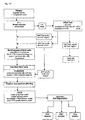
- a primary objective of the present invention is to provide a method which improves the identification and/or separation of cells and to improve genetic diagnosis, especially direct genetic diagnosis in living cells.
- a further objective of the invention is to improve the diagnosis by providing a device and a method for a better sampling and/or processing of the investigated cells.
- the underlying problem is solved by providing a method for cell identification according to claim one.
- the basic idea of the invention is to identify target cells and to detect genetic alterations within target cells in vivo by hybridising target sequences with complementary or partly complementary labelled sequences and subsequently detect the hybridised labelled sequence within the cell by flow cytometry.
- a step of cell sorting according to the detection of the hybridised molecular beacon and further analysis may follow.
- This method identification with subsequent cell separation is less time consuming and requires less expertise than common practices.
- the presented method enables to increase the overall sampling rate of target cell within a blood sample per patient.
- Another advantage is the simultaneous detection of different cell types or lines in a fast multiplex assay which can be combined with the detection of various genetic differences within these cells at once.
- the quantification can also provide further discrimination criterion between target and non target cells. Even rare sequences can be detected.
- the invention allows for the diagnosis of diseases which are due to or characterised in the transcription of genes which are not expressed in the wild type cell, or diseases characterised in the absence or altered expression or expression rates of certain genes or parts by quantifying the level of abundance of the transcript products.
- the method according to the invention can provide for prenatal diagnosis.
- the separation of the target sequence carrying cell may be advantageous, e.g. to isolate fetus derived cells out of a maternal blood sample.
- the cell separation can be conducted e.g. by photographic or visual methods.
- the cell sorting is performed with commonly known fluorescence activated cell sorting (FACS; e.g. international patent application WO 9 107 660 or German patent application 42 22 573).
- FACS fluorescence activated cell sorting
- nucleic acid hybridisation techniques are based on the ability of single-stranded nucleic acid to pair with a complementary nucleic acid strand. Therefore, a hybridisation reaction prescribes the development of labelled complementary specific sequences (subsequently also labelled sequence) directed against a target sequence or combination of complementary specific sequences in order to identify the presence or absence of target sequences of genes (DNA) and/or their transcribed polynucleotide sequences (RNA) or even the presence of a specific mutation within a sequence and/or a transcribed gene product.
- the target sequence can be a wild type are genetically altered nucleic acid.
- a mixture of at least two complementary sequences directed each against a specific target sequence can be used which are labelled each with a different fluorochrome.
- These fluorochromes advantageously exhibit a different fluorescent emission. This can allow for the detection of different target sequences in one assay. Therefore, also the different target sequences carrying cells can be identified and/or sorted in one assay (multiplex assay).
- a selected labelled sequence can be complementary to a genetic marker of the target cell, for example for the gender or any fetal specific gene transcript or for other genetic characteristics of the target cell.
- labelled sequences can be directed against bacterial or viral nucleic acids being included in the target cell or part thereof.
- viral targets can be the human immunodeficiency virus (HIV), or the herpes or hepatitis virus.
- Other detection targets of particular interest and therefore included are polynucleotide transcripts of the X- or Y-chromosome or the chromosome 1, 13, 16, 18 and 21.
- the target nucleotides are hybridised with a complementary labelled sequence that has a nucleic acid target complement sequence flanked by members of an affinity pair or arms, that under assay conditions - in the absence of the target sequence - interact with one another to form a stem duplex.
- Hybridising of the sequence with the target sequence produces a conformational change in the sequence forcing the arms apart and eliminating the stem duplex. Due to the elimination of the stem duplex the sequence becomes detectable.
- the above mentioned oligonucleotide (which subsequently will be referred to as molecular beacon (MB)) matches a donor and acceptor chromophores on their 5' and 3' ends.
- the MB remains in a stem-loop, i.e. stem-duplex, conformation where fluorescence resonance energy transfer prevents signal emission.
- the stem-loop structures opens increasing the distance between the donor and the acceptor moieties thereby reducing fluorescence resonance energy transfer and allowing detectable signal to be emitted when the MB is excited by light of appropriate wavelength. Without hybridisation the donor and acceptor remain in distance and due to the above described effect of quenching no fluorescence signal is detectable. Thus, non-hybridised MB do not emit fluorescent light.
- the method according to the invention does not require the availability of monoclonal antibodies. Nevertheless, substituting labelled antibodies with labelled probes the cell separation protocols commonly known for FACS or MACS can be applied.
- fetal cells are separated from a variety of sample specimens including maternal peripheral blood, placental tissue, chorionic villi, amniotic fluid and embryonic tissue.
- a preferred specimen is a maternal peripheral blood sample.
- the invention can be used to identify and sort fetal nucleated red blood cells, but any other fetal cell type carrying a nucleus and having gene transcription activity can be included with no further difficulties.
- a suitable target sequence can be chosen within the cell, which is either specific in quality and/or quantity to fetal or embryonic cells.
- the preferred target sequences to detect are fetal-gene-specific chromosomal transcripts like messenger ribonucleic acids (mRNAs) or ribosomal ribonucleic acids (rRNAs), without limiting the invention to them, e.g. the mRNA for fetal hemoglobin (HbF) or embryonic hemoglobin.
- mRNAs messenger ribonucleic acids
- rRNAs ribosomal ribonucleic acids
- HbF fetal hemoglobin
- different expression levels of genes can be utilised which are specifically active in the fetal cell.
- labelled sequences complementary to cell line or cell type marker can be used in one assay together with labelled sequence complementary to genetically altered target sequences.
- a maximum information can be obtained at once: i.) The presence or absence of a target cell type or line and ii) The absence or presence of a genetically altered target sequence. Therefore, a cell identification and a direct genetic diagnosis can be performed in a combined assay.
- two sets of molecular beacons can be used, e.g. one set can be directed against target cell lines specific RNA, e.g. against fetal hemoglobin mRNA, and a second set is directed against a DNA sequence of interest, e.g. a sequence with single nucleotide alteration. Both sets are labelled with differently coloured fluorophores.
- the cell identification and possible subsequent cell separation can be performed according to a FACS protocol. It allows the identification of certain genetic abnormalities as a direct genetic diagnosis of the gene DNA and the analysis of the expression level of the genes by quantifying the mRNA simultaneously. Thus, genetic abnormalities can be detected in an 'online' fashion during the FACS procedure. In case of fetal cells it allows to directly determine a certain genetic condition of the fetus without a genetic testing procedure following the separation from maternal cells.
- multiple target sequences can be selected detecting different cell types, which for example share the same origin. This for example allows for multiple fetal cell types to be collected at once out of a maternal blood sample, thereby maximising the total amount of cells being collected compared to a technique that is optimised for collecting specific fetal derived cells like nucleated red blood cells (NRBCs).
- NRBCs nucleated red blood cells
- the blood samples for later cell identification and genetic analysis are taken in a test tube comprising at least one compartment with incubation media containing hybridisation media including at least one group of labelled sequences, preferably molecular beacons.
- This test tube provides the advantage of shortening the period between sampling and hybridisation considerably, which can result in an increase in reliability and quality of the test results.
- the test tube comprises two or more compartments each with different incubation- or test-solutions, e.g. fixation, or staining solution and/or anticoaglutants.
- the samples is incubated with one media, after a defined period if time, a second media from a second compartment can have access to the sample e.g. by opening a membrane separating the compartments.
- this pre-test is conducted as a quantitative online-PCR approach determining the Ct value of the PCR for fetal hemoglobin which is further on compared to a positive and negative blind sample.
- the sex of the fetus can be preferably determined by using molecular beacons complementary to the middle of PCR amplicons from mRNA sequences of zfy.
- the method according to claim one the invention is combined with current methods of negative separation such as density gradient centrifugation and/or techniques like antibody derived magnetic separation (MACS) of unwanted cells and with positive separation techniques on the basis of physical parameters like the cell surface charge by using a free buffer-flow electrophoresis device.
- current methods of negative separation such as density gradient centrifugation and/or techniques like antibody derived magnetic separation (MACS) of unwanted cells and with positive separation techniques on the basis of physical parameters like the cell surface charge by using a free buffer-flow electrophoresis device.
- the step of negative selection of maternal cells is conducted before the in situ hybridising and prior to fluorescence activated cell-sorting procedure (FACS).
- FACS fluorescence activated cell-sorting procedure
- Methods of negative selection of unwanted cells include the application of a hypotonic shock, which leads specifically to the lysis of erythrocytes first. Lysis solutions are readily commercially available e.g. from PARTEC, DAKO, Caltaq or MEDAC.
- complement Another method to remove enucleated red blood cells is the complement system.
- Complement a group of serum factors that can destroy antibody marked cells, is used strictly for negative selection, i.e. elimination of unwanted cells.
- monocytes can be reduced by the LME Treatment.
- LME L-Leucin-methyl-ester
- LME is a lysomotropic agent and destroys monocytes.
- This positive selection can be performed using for example intracellular staining techniques (US 5,422,277) or magnetic cell sorting (MACS).
- MACS sell sorting from complex cell mixtures, such as peripheral blood, hematopoietic tissue or cultured cells. Since small magnetic particles (20 - 150 nm in diameter) exhibit faster kinetics of the cell-bead reaction, a lower degree of non specific cell bead interactions, a lower risk of non specific entrapping of cells in particle aggregates, and less adverse effects of particles on viability and optical properties of labelled cells when compared with large magnetic particles (0,5 - 5 ⁇ m in diameter), they can be applied advantageously. Especially a MACS technology with small super- paramagnetic particles and high gradient magnetic fields is advantageous. Nevertheless, it can be combined with large magnetic particles as well for example large magnetic beads from Dynal and depletion columns from Miltenyi.
- MACS can be used for negative selection, i.e. for example for the depletion of white blood cells.
- CD45 antigen is expressed on all cells of hematopoietic origin except erythrocytes, platelets and their precursor cells
- CD45 Micro Beads can be used for the depletion of leukocytes from peripherel blood.
- a combination of CD45 and CD15 Micro Beads is recommended due to the weak expression of CD45 in the granulocyte / monocyte lineage. Therefore, the combination of MicroBeads can result in an enrichment of fetal erythroblasts from maternal blood.
- a resuspending medium such as EDTA, bovine serum albumin (BSA) or serum
- BSA bovine serum albumin
- charge flow separation technique especially in the continuous free-flow electrophoreses method as disclosed US patent 4,061,560, (fully incorporated into the text hereby) can be used. It can be either used without or including staining with target cell specific antibodies (ASEC).
- ASEC target cell specific antibodies
- the latter is reviewed by Hansen et al., 1982, (Antigen-specific electrophoretic cell separation (ASECS): Isolation of human T and B lymphocyte subpoulation by free-flow electrophoresis after reaction with antibodies. J. Immunol. Methods 51 : 197 - 208 ).
- the method can be improved by using a second antibody directed against the first in a so called "sandwich technique". If this is still not sufficient, a third antibody can be used, directed against the second.
- antibodies with side groups containing a higher negative charge than a normal antibody can be used.
- antibodies coupled with very small magnetic particles can be advantageous.
- fluorescence activated cell sorting is performed.
- fluorescence is just one possible staining system since other dye systems may also be employed (US-patent 4,933,293) and no staining is necessary for light-scatter measurements or electrical sizing.
- Flow cytometry apparatus are commercially available e.g. from MICROCYTE, Becton Dickinson's FACScan, FACStrak, FACSort, FACSCalibur, FACStar, FACSVantage, Bio-Rad's BRYTE-HS, Coulter's PROFILE and EPICS, Cytomation's MoFlow, Ortho's CYTORON and Partec's PAS machines.
- the PAS- System from PARTEC Germany is preferred.
- the antibodies can be directed against intracellular substances or surface markers. Treatments of cells with a fixative as described in US Patent NR. 5422277 and 6004762 allow antibodies or other desired components to enter the cell through the cellular membrane.
- fixation and permeabilisation of cell membranes saponin can be used successfully, including assessment of cytokines (see also Willingham: An atlas of immunofluorescence in cultured cells. Vol. II Academic press).
- cytokines see also Willingham: An atlas of immunofluorescence in cultured cells. Vol. II Academic press.
- a cross linking fixative formaldehyde can be applied with allows for good preservation of cell morphology.
- other detergents like NP40 in combination with fixation by formaldehyde, or of organic solvents, like 70 % methanol or ethanol/acetic acid (95/5), which fix and permeabilise cells in one step can be used.
- fixation with alcohol is preferable.
- fixation/permeabiiisation steps e.g. 70 % alcohol followed by formaldehyde / Tween 20 for BrdU-staining.
- absorption of polyclonal antibodies on liver powder (acetone precipitate of liver) or on irrelevant cells (2:1, volume of antibody solution (1mg/ml) : packed cells) can reduce unspecific staining.
- Optimally specific staining can be blocked by preincubation of the antibodies with molar excess of purified antigen.
- nucleic acid refers to sequences of nucleotides of all kind and thus comprises oligomers and polymers of desoxyribonucleotides, as well as all kinds of ribonucleotides. Also nucleotides with analoga, chromosomes and viral or bacterial nucleic acids or parts thereof, plasmids, recombinant nucleotides and all kinds of synthetic sequences are included.
- target cells refers to cells of interest, which are to be selected or purified respectively.
- fetal cells include especially trophoblasts, nucleated fetal red blood cells, granulocytes and/or a subpopulation of white blood cells, i.e. polymorphonuclear leukocytes such as neutrophils, basophiles and eosinophiles.
- target sequence refers to all nucleic acids, which are to be hybridised with the labelled complementary sequence. This term comprises sequences specific for the target cells in terms of quality and/or quantity. It comprises wild type sequences as well as genetically altered nucleic acids of all kind.
- labelled sequence refers to nucleic acids complementary or partially complementary to target sequences, which are labelled with at least one marker such as chromophores, fluorophore, magnetic particle or others allowing the later detection.
- molecular beacon refers to a labelled sequences according to one of the claims of US patent 5,925,517.
- RNA refers to all kinds of RNA, including mRNA and rRNA as well as derivates and parts thereof.
- DNA refers to all kinds of naturally occurring or synthetically derived DANN as well as derivates and parts thereof.
- hybridisation refers to the phenomenon that single strand nucleic acids or parts thereof are forming pairs with complementary or partly complementary single nucleic acid strands.
- In situ hybridisation refers to hybridisation under conditions maintaining the cell substantially intact.
- fluorescence refer to emission of detectable radiation as a result of excitement with radiation of a different wavelength than the emitted.
- FITC zfy specific molecular beacon
- a negative result in the pre-test indicates a female and therefore, a HbF mRNA (FITC) specific molecular beacon is chosen.
- FITC HbF mRNA
- a HbF specific antibody PE
- a nuclear staining with DAPI PARTEC Germany
- the target cells are identified, gated and automatically separated according to the protocol of the operating manual for the PAS-III, (PAS-III: Particle Analysing and separation System, Operating Manual; Partec Germany, see also under step 8).
- PAS-III Particle Analysing and separation System, Operating Manual; Partec Germany, see also under step 8).
- one molecular beacon is used to distinguish fetal from maternal cells.
- a zfy specific mRNA molecular beacon (FITC) is used to differentiate between fetal and maternal cells.
- FITC mRNA molecular beacon
- a fetal specific mRNA - molecular beacon for the HBF-gene and a DAPI nuclear staining is applied.
- the molecular beacon for the determination of the wild type sequence is labelled with EDANS and a mutation specific beacon is labelled with the fluorescens HEX (according to step 3).
- a fetal cell is to be distinguished by FITC and DAPI fluorescence.
- the genetic status is determined by the subsequent possible combinations: 1 DAPI and FITC positiv (fetal cell) 1a: HEX positiv, EDANS negativ (fetal cell wildtyp homozygous). 1b: HEX positiv, EDANS positiv (fetal cell, wildtyp and mutation) 1c: HEX negativ and EDANS positiv (fetal cell, mutation homozygous).
- ACD acid-citrate-dextrose
- EDTA ethylendiamine-tetracid
- CPDA-S-Monovette citrate-phosphate-dextrose-adenine
- 300 ⁇ l of the whole blood sample is added to a 1.5 ml microfuge tube containing 900 ⁇ l RBC Lysis solution (Quantum Prep ApuaPure RNA Isolation, BioRad) and mixed by inverting the tube for 10 min at room temperature. After centrifugation at 13,000-16,000 x g in a microcentrifuge for 20 sec the supernatant is removed with a pipet leaving behind the visible white cell pellet and about 10-20 ⁇ l of residual liquid. The white cells are re-suspended by vortexing vigorously until the pellet has disappeared.
- RBC Lysis solution Quantum Prep ApuaPure RNA Isolation, BioRad
- RNA lysis solution (Quantum Prep ApuaPure RNA Isolation, BioRad) is added to this mixture by pipeting up and down for 3 times.
- 100 ⁇ l of DNA-precipitation solution (Quantum Prep ApuaPure RNA Isolation, BioRad) is added to the cell lysate, mixed by inverting the tube and placed into an ice bath for 5 min before centrifugation at 16,000 x g. The precipitated protein and DNA form a tight pellet. The supernatant is placed into a clean sterile 1,5 ml micorcentrifuge tube containing 300 ⁇ l pure isopropanol.
- RNA is visible as a small translucent pellet.
- the supernatant is poured and the tube is dried in an absorbent paper.
- the pellet is washed twice with 70% ethanol, air dried for 30 min and stored at -80°C until used.
- First strand complementary DNA is synthesized by priming with random hexamers (Clontech). The air dried and frozen RNA sample is resuspended in an 8.5 ⁇ l solution consisting of :
- the hexamers are annealed by incubating the sample at 70°C for 5 min and quenched on ice. Reverse transcription is performed by addition of 11.5 ⁇ l containing
- RNA Hydration Solution Prior to be used in a RT-PCR, 5 ⁇ l RNA Hydration Solution (Quantum Prep ApuaPure RNA Isolation, BioRad) is added to the RNA. The mixture is subsequently vortexed heavily and 5 ⁇ l are used in a 50 ⁇ l PCR experiment.
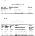
- a multiplexed quantitative real-time PCR is performed utilizing molecular beacons that are complementary to the middle of PCR amplified fragments from mRNA sequences of beta-actin, zfy and HbF.
- the length of the arm sequences of the molecular beacons is chosen in order to allow a stem being formed at the annealing temperature of the PCR (table 1) whereas the length and sequence of the loop is chosen to provide a probe-target hybrid being stable at this step of PCR.
- the design of molecular beacons has been tested before by thermal denaturation profiles using loop-antisense oligonucleotides and a real-time thermal cycler (BioRad). Only molecular beacons showing desired thermal profiles are included in the subsequent PCR at concentrations similar to the amplification primers
- the molecular beacons assume a random coil conformation and fluoresce. As the temperature is lowered to allow annealing of the molecular beacon to the target sequence at the single stranded PCR fragments, loop-target hybrids are formed which are able to continue to fluorescing. Superfluous molecular beacon however rapidly form stable intra-molecular stem-hybrids that prevents them from fluoresce. As the temperature raises to allow primer prolongation, the molecular beacons dissociate from the target sequences and do not interfere with the polymerization step. A new molecular beacon hybridization step takes place in every annealing step during PCR cycling while the increase of the resulting fluorescence is monitored and indicated the amount of the accumulated target amplicon.
- At least two different target sequences are simultaneously amplified in one tube.
- At least two different molecular beacons are used simultaneously, each of them labelled with a different fluorescent dye with no spectral overlap at emission wavelength.
- the resulting fluorescence is monitored at the appropriate wavelength of each of the molecular beacons during the annealing step of the PCR.
- Beta-actin is widely used its mRNA is ubiquitously abundant in almost every cell type. Beta-actin amplification therefore not only indicates a successful polymerase chain reaction but is highly proportional to the total amount of target cells used in the PCR.
- the primers and molecular beacons used in the real-time quantitative PCR experiment are shown in table 1.
- the amount of zfy-gen-specific mRNA is quantified and the result is compared to a positive and a negative control sample, which is the total mRNA from a blood sample of a mother carrying a male or a female fetus respectively.
- Real-time quantitative RT-PCR is performed by using the iCycler Thermal Cycler (BioRad).
- the PCR for zfy mRNA and HbF mRNA is conducted in separate reactions but multiplexed with a PCR for beta-actin. Each 50 ⁇ l reaction contained
- the 50 ⁇ l volume reaction is loaded into the appropriate PCR-tubes and placed into the iCycler (BioRad).
- the cDNA is denatured and heating to 96°C for 10 min activates the Taq Polymerase.
- denaturation is performed at 95°C for 30 sec, annealing of the primers and molecular beacons at 60-68°C for 30 sec and extension at 72°C for 30 sec. Fluorescence data is acquired during the annealing steps of the reaction and the level of fluorescence is monitored as a function of cycle number.
- the reaction without any template does not exhibit any increase in fluorescence and functions as a baseline. Over a wide range of template concentrations the cycle with a fluorescence signal exceeding detectably over the baseline is inversely proportional to the logarithm of the initial number of template molecules.
- the level of beta-actin fluorescence in each well is a suitable further control parameter indicating the amount of total cDNA target used for the PCR.
- the fluorescence intensity (F) of each molecular beacons is normalized by calculating (F - F min )/(F max - F min ).
- the value "0" represents fluorescence before target amplification
- "1" represents the maximum level of fluorescence after PCR.
- the threshold cycle is determined when the intensity of the fluorescent signal exceeds 10 times the standard deviation of the background baseline fluorescence. For quantification of the target a comparison of the determined threshold cycles with a standard curve is performed.
- nucleotide-oligodesoxynucleotides are synthesized consisting of a 15- to 20-nucleotide-target sequence (antisense to mRNA) sandwiched by a complementary 5-nucleotide-arm sequence (stem sequence), being covalently linked to a fluorescent dye (fluorescein or EDANS) at the 5'-end and to a DABCYL as a quencher dye at the 3'-end.
- fluorescent dye fluorescein or EDANS
- the initial oligonucleotide for this synthesis of the molecular beacon is conducted contains a sulfhydryl group at its 5'-end and a primary amino group at its 3'-end. Its synthesis is conducted with a standard A-, C-, G-, T-phosphoamidate chemistry on an automatic DNA synthesizer (Perkin Elmer 394) using 1-dimethoxytrityloxy-3-fluorenylmethoxycarbonylaminohexane-2-methylsuccinyl-long chain alkylamino-controlled pore glass (C7-CPG, Perseptive Biosystems) as the 3'-aminomodifier.
- C7-CPG 1-dimethoxytrityloxy-3-fluorenylmethoxycarbonylaminohexane-2-methylsuccinyl-long chain alkylamino-controlled pore glass
- a trityl-hexylthiol linker ((S-trityl-6-mercaptohexyl)-(2-cyanoethyl)-(N,N-diisopropyl)-phosphoamidate, PerSeptive Biosystems) is coupled as a final step to the nucleotide' s 5'-end.
- the oligonucleotide is detached from the support using 28% ammonium at 55°C for 6 h. This treatment also removes the protective moieties at the amino and phosphate groups except for the trityl moiety at the 5'-sulfhydryl end.
- the detached oligonucleotides are purified by reverse phase cartridge column (Waters Sep-Pak C18, Millipore) and then fractioned by HPLC on a C18 column with a linear gradient of 5-40% acetonitrile dissolved in 0.05 M triethylammonium acetate (pH 7.0) running for 30 min at a flow rate of 1.0 ml/min at 40°C and detected at 254 nm.
- oligonucleotide About 200 nmoles of the dried oligonucleotide was dissolved in 0.5 ml of 0.1 M sodium bicarbonate, pH 8.5. Subsequently the mixture is incubated with 20 mg of DABCYL (4-(4'-dimethylaminophenylazo)benzoic acid) succinimidyl ester, ( Molecular Probes ) in 0.1 ml N,N-dimethylformamide in 0.01 ml aliquots at 20 min intervals. The mixture was kept for 3 days at room temperature in the dark.
- DABCYL 4-(4'-dimethylaminophenylazo)benzoic acid
- succinimidyl ester succinimidyl ester
- the eluate of approximately is filtered through a 0.2 ⁇ m filter (Centrex MF-0.4, Schleicher & Schüll) and loaded on a C-18 reverse phase HPLC column (Waters), utilizing a linear elution gradient of 20% to 70% 0.1M triethylammonium acetate in 75% acetonitrile (pH 6.5) in 0.1 M triethylammonium acetate (pH6.5) for 25 min at a flow rate of 1 mll/min.
- the absorption by DABCYL is monitored by spectrophotometry. The peak absorbing at 260 and 491 nm is collected.
- the dried and DABCYL-coupled oligonucleotides are dissolved in 0.25 ml 0.1 M triethylammonium acetate (pH 6.5) and incubated with 0.01 ml of 0.15 M silver nitrate for 30 min at room temperature. To this solution 0.015 ml of 0.15 M DTT is added. The supernatant is removed from the pellet by spinning and transferred into a solution consisting of 40 mg 5-iodoactamidofluorescein (Molecular Probes) in 0.25 ml of 0.1 M sodium bicarbonate (pH 9.0). Incubation is performed at room temperature for one day in the dark.
- Excess fluorescein is removed by gel exclusion chromatography (Nap-5, Amersham Pharmacia) and the oligonucleotide are purified by HPLC. The fraction absorbing at wavelength 260 nm and 491 nm is collected. It is precipitated and re-dissolved in 0.1 ml TE buffer. The yield is estimated by determining the absorbance at 260 nm.
- the upper layer to within 0,5 cm of the opaque interface containing mononuclear cells is aspirated.
- the white band, including nucleated erythroid cells and lymphocytes, is directly below the plasma layer.
- Histopaque®- 119 containing a broad diffuse band containing other nucleated erythroid cells and immature red cells with densities heavier than the white layer but lighter than the mature erythrocyte pellet, which is immediately below the nucleated cells and the erythrocyte pellet at the very bottom of the tube.
- the cells are transferred in a new tube add 30 ml Phosphate Buffered Saline solution with 0,1 % BSA is added, mixed by gentle aspiration and subsequently centrifuged at 250 x g for 10 minutes. The supernatant is discarded.
- the pellet is resuspended with 10 ml Phosphate Buffered Saline solution with 0,1 % BSA and centrifuged at 250 x g for 10 minutes again. The step of washing is repeated and the supernatant is discardes.
- the cell pellet is re-suspended in 750 ⁇ l Buffered Saline solution with 0,1 % BSA.
- CD 45 and CD15 are used (Miltenyi Biotec GmbH, Bergisch Gladbach, Germany; Order No. 458-01 and No. 466-01).
- a resuspension buffer is supplemented with EDTA and bovine serum albumin (BSA) or serum (PBS pH 7,2 with 2mM EDTA and 0,5% BSA) and dead cells are removed prior magnetic labelling.
- BSA bovine serum albumin
- PBS pH 7,2 with 2mM EDTA and 0,5% BSA serum
- the buffer is degassed by applying vacuum, preferentially with buffer at room temperature.
- the magnetic labelling is performed in the refrigerator at a temperature between 6° and 12° C for 15 minutes.
- Clumps are removed by passing the cells through a nylon mesh (30 ⁇ m) or filter (Miltenyi Biotec GmbH, Bergisch Gladbach, Germany; Order No. 414-07).
- a filter is wetted by vigorously pipetting 500 ⁇ l of resuspension buffer in the reservoir prior the filter process and the effluent is discarded. Subsequently a minimum of 500 ⁇ l cell suspension with 10 8 runs through the filter, which is washed 2 to 3 times with 500 ⁇ l buffer.
- MidiMACS MidiMACS system
- MidiMACS MidiMACS system
- LD-Separation column order No. 429-01 for the magnetic activated cell sorting the MidiMACS system is used (Miltenyi Biotec GmbH, Bergisch Gladbach, Germany; Order No. 423-01) including the LD-Separation column order No. 429-01.
- the magnetic labelled cells suspended in buffer 500 ⁇ l buffer/10 8 total cells
- the effluent is collected as the positive fraction. This step can be repeated.
- the effluents are compiled and centrifuged with 200 x g for 10 minutes. The pellet is resuspended in 1,5 ml PBS + 0,5 % BSA for further procedures.
- the aforementioned MideMACS system is operated for the enrichment of fetal erythrocytes by using Glycopherin A (GPA; order No. 422-01) and CD (order No.462-01) conjugated MicroBeads and the buffer system as described under MACS depletion (order No. 424-01).
- Glycopherin A Glycopherin A
- CD order No.462-01 conjugated MicroBeads
- MACS depletion order No. 424-01
- the cell suspension is allowing to pass the column. Whereas the effluent is discarded the cells having remained within the column are firmly flushed out and collected as the positive fraction.
- Enriched fetal cells are washed with HEPES (145 mM NaCI, 5mM KCI, 1mM MgCl 2 , 1mM CaCl 2 , 10 mM glucose, 10 mM HEPES, pH 7.4), and precipitated by centrifugation.
- HEPES 145 mM NaCI, 5mM KCI, 1mM MgCl 2 , 1mM CaCl 2 , 10 mM glucose, 10 mM HEPES, pH 7.4
- 2 ⁇ g of the fluorescent molecular beacons are dissolved in 2 ⁇ l of water and mixed with 9 ⁇ l of liposomes comprising 0.4 mg of N,N,N',N'-tetramethyl-N-N'-bis(2-hydroxyethyl)-2,3-dioleoyloxy-1,4-butanediammonium iodide and 0.3 mg of L-dioleoyl phosphatidylehtanolamine suspended in 400 ⁇ l of nuclease free water (Tfx-50 Reagent, Promega). The mixture is vortexed gently and incubated for 10 min at room temperature. Subsequnetly the cells are washed in HEPES.
- red blood cells (RBC) within the sample 2
- 5 x 10 7 cells are added to 1 ml of cold 0,05 % Glutaraldehyde for 10 min at room temperature. This mixture is washed three times with 2 ml cold PBS- 0,1 % BSA (pH 7,4). Subsequently, the cells are resusupended and incubated in 0,5 ml 0,1 % Triton X 100 for 3-5 minutes at room temperature and subsequently washed in 2 ml of PBS-0,1 % BSA. Afterwards the cells are taken in 0,5 ml PBS- 0,1 % BSA by vortexing or gentle pippeting.
- 10 ⁇ l of this suspension are added to 10 ⁇ l antibody (Caltaq Laboratories Burlingame, CA 94010, Product code: MHFH04; Gamma chain antibody) and 70 ⁇ l of PBS-0,1 % BSA and incubated at room temperature in the dark for 15 minutes. Two washing steps with 2 ml PBS-0,1 % BSA each are following.
- the pellet is suspended in 500 ⁇ l PBS-0,1 % BSA and 1ml DAPI staining solution (PARTEC, Germany) is added. The incubation takes place for 10 minutes at romm temperature. The pellet is washed twice with 2 ml PBS-0,1 % BSA and finally resuspended by vortex in 0,5 ml 1 % formaldehyde. The cells are stored in tubes in the dark in the refrigerator.
- PE Phycoerithrin
- PASIII - system (PARTEC Germany), is used applying a 488 nm argon laser and an ultraviolet lamp to distinguish different fluorescent dyes within the cells.
- chromosome or cell nuclear suspension In order to prepare slides one drop of chromosome or cell nuclear suspension is placed on a very clean glass slide. Slides are rinsed in 2 x SSC, pH 7.0 for 5 min. An incubation for at least 30 min at 37°C in 2 x SSC containing 100 ⁇ g/ml RNase A follows. Subsequently the washing of slides is performed for 3 x 5 min in 2 x SSC, pH 7.0 . RNase treatment serves to remove endogenous RNA which may hybridize with homologous probe DNA sequences, forming RNA-DNA hybrids. This improves the signal to noise ratio in hybridization to DNA targets.
- Dehydration of slides in an ethanol series (70%, 85%, 100%) is performed for 5 min each and air-dry. Then 200 ⁇ l of denaturation solution, consisting of 70% deionized formamide and 2 x SSC , is placed on each slide and cover with a coverslip. The slide is placed in 90°C oven for approximately 60 sec. The coverslip is shacked off and immediately put in ice-cold 70% ethanol. Dehydration is performed again in an ice-cold ethanol series (70%, 85%, 100%). After air-drying, the slides are ready for in situ hybridization.
- the DNA template can be supercoiled or linear. After nicking the DNA with DNase I, the 5'-3' exonuclease activity of DNA polymerase removes nucleotides and the DNA polymerase activity replaces the excised nucleotides with dNTPs from the reaction mixture including the labeled nucleotide.
- the procedures for incorporating biotin, digoxigenin or fluorochromes are nearly identical.
- the final size of the nick-translated probe DNA fragments is very important. Labeled probe sequences should not be larger than 500 bp because of difficulty penetrating the specimen and a tendency to stick non-specifically to both the glass and the cellular material, resulting in high background which obscures the signal. Sequences shorter than 100 bp do not hybridize efficiently under routine conditions of stringency.
- DNA probes are mixed on ice, in a microcentrifuge tube: DNA (in liquid format) 1 ⁇ g 10 x nick-translation buffer (Tris/HCl 1M, pH 8; MgCl 2 1M; BSA 10%) 5 ⁇ l 0.1 M-mercaptoethanol 5 ⁇ l 10 x cold dNTP solution (0.5 mM each of dATP, dCTP, dGTP) 5 ⁇ l 10 x 1:3 labeled dUTP/cold dTTP solution (0.125 mM labeled dUTP, i.e. biotin-16-dUTP or digoxigenin-11-dUTP, and 0.375 mM dTTP) 5 ⁇ l
- a labeling density of one modified nucleotide at approximately every 20-25 th position in the nick-translated DNA is optimal for most FISH experiments.
- 10 x DNase I 5 ⁇ l (DNase stock solution with an activity of 10 U/ ⁇ l is diluted 1:3000-1:4000 with double-distilled H 2 O)Optimal DNase concentration must be determined by titration, as the size distribution of the probe depends on the amount of enzyme added. Each new stock of DNase I should be tested to determine the appropriate working concentration.
- DNA polymerase I (5 U/ ⁇ l) 1.3 ⁇ l DdH 2 O is added as needed to achieve final reaction volume of 50 ⁇ l
- reaction volumes can be scaled up to 250 ⁇ l.
- the reaction mixture is incubated for 2 h at 15°C.
- the time of incubation should at least be 2 h, as the initial incorporation rate of modified nucleotides is less than that of unsubstituted dNTP.
- the size of the nick-translated DNA fragments is checked on a 1.5% agarose gel, along with suitable size markers (0.1-1 kb range). If the DNA fragments are still too large, a second aliquot of DNase (optional) is added and incubated for another 30-60 min at 15°C.
- termination of the reaction follows by adding stop buffer (1.25 ⁇ l 0.5 M EDTA and 0.5 ⁇ l 10% SDS) to the 50 ⁇ l of nick-translation mixture. The tube is heated to 68°C for 10 min.
- hapten reporter molecules i.e. biotin- or digoxigenin-dUTP, being introduced into the DNA probe, are detected by affinity cytochemistry after the hybridization reaction.
- Fluorescent (strept)avidin molecules bind specifically to biotin-labeled probe-target hybrids.
- Fluorescent anti-digoxigenin antibodies are used to detect digoxigenated probes.
- the fluorescence signals of indirectly labeled probes may be up to tenfold brighter than those produced by direct methods.
- small hapten molecules such as biotin are efficiently incorporated into DNA than most fluorescent dNTPs.
- Nick-translated DNA probes have a final concentration of approximately 20 ng/ ⁇ l (in nick-translation buffer).
- First, differentially labeled probes which are to be hybridized on human specimen are precipitated together with an approximately 50-fold excess of cot-1 competitor DNA (Gibco BRL) or a 500-fold excess of fragmented (100-500 bp) total genomic DNA, and a 50-fold excess of fragmented salmon sperm DNA which is used as a carrier.
- the optimal DNA concentrations in the hybridization mixture (30 ⁇ l for a whole slide) depend mainly on the probe types. For large-insert clones, 10-20 ng/ ⁇ l in the hybridization mixture (or 300-600 ng per slide) are recommended.
- BAC-DNA probes are hybridized together on the same slide for identification of male foetal cells that are affected by trisomie 21.
- the DNA is precipitated with 1/20 volume 3 M Naacetate and 2 volumes 100% EtOH (p.a.) in a microcentrifuge tube.
- the sample is mixed well and uncubated at least 30 min at -70°C or overnight at -20°C.
- the precipitation is done in a centrifuge spinning for 30 min at 15,000 rpm at 4°C.
- the supernatant is discarded and the pellet are dried in a vacuum system or alternatively at 37°C in a heating block.
- the DNA probe is resuspended with 15 ⁇ l of deionized formamide and shaken at 37°C for at least 15 min to resuspend the probe DNA. Then an equal volume of 2 x hybridization mixture (20% dextran sulfate, 4 x SSC) is added followed by vigorously shaking for at least 15 min.
- Conditions of stringency are 50% formamide, 10% dextran sulfate, 2 x SSC in the hybridization mixture. Denaturation of hybridization mixture (containing the probe DNA) is performed at 80°C for 10 min. Probes are centrifuged for 2-5 sec in order to pellet the condensed water.
- the tube with the denatured competitor and probe DNA is incubated at 37°C for at least 15 min.
- DNA probes (30 ⁇ l) are placed on the denatured slide and cover with a coverslip. The edges are sealed with rubber-cement and incubated overnight (or longer) at 37°C in a moist chamber.
- washing of slides follows for 3 x 5 min at 42°C in 50% formamide, 2 x SSC and then 2 x 5 min at 60°C in 0.1 x SSC.
- the washing solutions are pre-warmed in different water baths.
- the Coplin jar with slides and pre-warmed washing solution are agitated on a platform shaker.
- the probe DNA hybridizes not only to the target DNA but also non-specifically to sequences which bear partial homology to the probe sequence. Since such non-specific hybrids are less stable than perfectly matched DNA hybrids, they can be dissociated by post-hybridization washes of various stringencies.
- Fluorescent (strept)avidin and anti-digoxigenin antibodies i.e. FITC-avidin and Cy3-conjugated anti-digoxigenin, are diluted 1:800 and 1:200, respectively (or according to recommendations of the supplier) in 1% BSA, 0.1% Tween 20, 4 x SSC. Cover slides with 200 ⁇ l of detection solution and a coverslip. Incubation is performed in a moist chamber in the dark at 37°C for 30 min. Coverslips are shaken off the and washing slides 3 x 5 min in a Coplin jar with 0.1% Tween 20, 4 x SSC at 42°C.
- DAPI 4,',6-diamidino-2-phenylindole
- Digoxigenated DNA probes binding fluorescent mouse anti-digoxigenin antibody are amplified with a second layer of fluorescent rabbit anti-mouse antibodies and, if necessary, with a third layer of fluorescent goat anti-rabbit antibodies.
Landscapes
- Chemical & Material Sciences (AREA)
- Organic Chemistry (AREA)
- Life Sciences & Earth Sciences (AREA)
- Zoology (AREA)
- Wood Science & Technology (AREA)
- Proteomics, Peptides & Aminoacids (AREA)
- Health & Medical Sciences (AREA)
- Engineering & Computer Science (AREA)
- Microbiology (AREA)
- Biochemistry (AREA)
- Physics & Mathematics (AREA)
- Molecular Biology (AREA)
- Biotechnology (AREA)
- Biophysics (AREA)
- Analytical Chemistry (AREA)
- Immunology (AREA)
- Bioinformatics & Cheminformatics (AREA)
- General Engineering & Computer Science (AREA)
- General Health & Medical Sciences (AREA)
- Genetics & Genomics (AREA)
- Measuring Or Testing Involving Enzymes Or Micro-Organisms (AREA)
- Investigating Or Analysing Biological Materials (AREA)
Priority Applications (8)
| Application Number | Priority Date | Filing Date | Title |
|---|---|---|---|
| EP00115268A EP1172445A1 (en) | 2000-07-14 | 2000-07-14 | A method for direct genetic analysis of target cells by using fluorescence probes |
| AU2001289664A AU2001289664A1 (en) | 2000-07-14 | 2001-07-16 | A method for direct genetic analysis of target cells by using flourescence probes |
| IL15390601A IL153906A0 (en) | 2000-07-14 | 2001-07-16 | Method for direct genetic analysis of target cells |
| JP2002512414A JP2004504036A (ja) | 2000-07-14 | 2001-07-16 | 蛍光プローブを使用することによる標的細胞の直接的な遺伝分析のための方法 |
| CA002415731A CA2415731A1 (en) | 2000-07-14 | 2001-07-16 | A method for direct genetic analysis of target cells by using flourescence probes |
| PCT/EP2001/008202 WO2002006524A2 (en) | 2000-07-14 | 2001-07-16 | A method for direct genetic analysis of target cells by using flourescence probes |
| NO20030158A NO20030158L (no) | 2000-07-14 | 2003-01-13 | Fremgangsmåte for direkte genetisk analyse på målceller ved anvendelse av fluorescerende prober |
| US10/342,450 US20040091880A1 (en) | 2000-07-14 | 2003-01-14 | Method for direct genetic analysis of target cells by using fluorescence probes |
Applications Claiming Priority (1)
| Application Number | Priority Date | Filing Date | Title |
|---|---|---|---|
| EP00115268A EP1172445A1 (en) | 2000-07-14 | 2000-07-14 | A method for direct genetic analysis of target cells by using fluorescence probes |
Publications (1)
| Publication Number | Publication Date |
|---|---|
| EP1172445A1 true EP1172445A1 (en) | 2002-01-16 |
Family
ID=8169252
Family Applications (1)
| Application Number | Title | Priority Date | Filing Date |
|---|---|---|---|
| EP00115268A Withdrawn EP1172445A1 (en) | 2000-07-14 | 2000-07-14 | A method for direct genetic analysis of target cells by using fluorescence probes |
Country Status (8)
| Country | Link |
|---|---|
| US (1) | US20040091880A1 (enExample) |
| EP (1) | EP1172445A1 (enExample) |
| JP (1) | JP2004504036A (enExample) |
| AU (1) | AU2001289664A1 (enExample) |
| CA (1) | CA2415731A1 (enExample) |
| IL (1) | IL153906A0 (enExample) |
| NO (1) | NO20030158L (enExample) |
| WO (1) | WO2002006524A2 (enExample) |
Cited By (3)
| Publication number | Priority date | Publication date | Assignee | Title |
|---|---|---|---|---|
| WO2003008639A3 (de) * | 2001-07-12 | 2003-09-18 | Praenadia Gmbh | Verfahren zum nachweis einer nukleinsäure |
| EP2264165A3 (en) * | 1999-11-23 | 2011-02-16 | Chromocell Corporation | Selection and isolation of living cells using mRNA-binding probes |
| US8916503B2 (en) | 2004-02-18 | 2014-12-23 | Chromocell Corporation | Methods and materials using signaling probes |
Families Citing this family (34)
| Publication number | Priority date | Publication date | Assignee | Title |
|---|---|---|---|---|
| AU2008202162B2 (en) * | 2002-08-28 | 2011-04-07 | Chromocell Corporation | Selection and Isolation of Living Cells Using mRNA-Binding Probes |
| US20040226556A1 (en) | 2003-05-13 | 2004-11-18 | Deem Mark E. | Apparatus for treating asthma using neurotoxin |
| EP2500439B2 (en) | 2005-06-20 | 2017-08-16 | Advanced Cell Diagnostics, Inc. | kits and products for detecting nucleic acids in individual cells and of identifying rare cells from large heterogeneous cell populations. |
| US10052465B2 (en) | 2005-07-22 | 2018-08-21 | The Foundry, Llc | Methods and systems for toxin delivery to the nasal cavity |
| US12433837B2 (en) | 2005-07-22 | 2025-10-07 | The Foundry, Llc | Systems and methods for delivery of a therapeutic agent |
| US9511210B2 (en) | 2006-05-19 | 2016-12-06 | The Foundry, Llc | Apparatus for toxin delivery to the nasal cavity |
| JP5826450B2 (ja) | 2005-07-22 | 2015-12-02 | ザ ファウンドリー, エルエルシー | 治療薬の送達のためのシステムおよび方法 |
| EP1977006A2 (en) * | 2005-12-23 | 2008-10-08 | ALVitae Pharmaceuticals | Methods and applications of molecular beacon imaging for identifying and validating genomic targets, and for drug screening |
| ITTO20070307A1 (it) * | 2007-05-04 | 2008-11-05 | Silicon Biosystems Spa | Metodo e dispositivo per la diagnosi prenatale non-invasiva |
| US8483831B1 (en) | 2008-02-15 | 2013-07-09 | Holaira, Inc. | System and method for bronchial dilation |
| CA2723806C (en) | 2008-05-09 | 2022-05-31 | Innovative Pulmonary Solutions, Inc. | Systems, assemblies, and methods for treating a bronchial tree |
| WO2010010951A1 (ja) * | 2008-07-24 | 2010-01-28 | Kuroda Masahiko | 子宮内膜症関連疾患の指標mRNAの発現の検出方法およびそれに用いるプローブ |
| JP2009072198A (ja) * | 2008-10-24 | 2009-04-09 | Chromocell Corp | mRNA結合プローブを用いる生存細胞の選択および単離 |
| US20110001963A1 (en) * | 2009-07-02 | 2011-01-06 | Durack Gary P | System and method for the measurement of multiple emissions from multiple parallel flow channels in a flow cytometry system |
| US20130023433A1 (en) | 2009-09-28 | 2013-01-24 | Yuling Luo | Methods of detecting nucleic acid sequences with high specificity |
| CN112089394A (zh) | 2009-10-27 | 2020-12-18 | 努瓦拉公司 | 具有可冷却的能量发射组件的递送装置 |
| CN106618731B (zh) | 2009-11-11 | 2020-08-07 | 努瓦拉公司 | 用于处理组织和控制狭窄的系统、装置和方法 |
| US8911439B2 (en) | 2009-11-11 | 2014-12-16 | Holaira, Inc. | Non-invasive and minimally invasive denervation methods and systems for performing the same |
| WO2011089603A1 (en) | 2010-01-21 | 2011-07-28 | Biocep Ltd. | Magnetic separation of rare cells |
| US20130171621A1 (en) | 2010-01-29 | 2013-07-04 | Advanced Cell Diagnostics Inc. | Methods of in situ detection of nucleic acids |
| US8774488B2 (en) | 2010-03-11 | 2014-07-08 | Cellscape Corporation | Method and device for identification of nucleated red blood cells from a maternal blood sample |
| US8445284B2 (en) * | 2011-06-17 | 2013-05-21 | Constitution Medical, Inc. | Fixative and staining solutions |
| WO2012178166A1 (en) * | 2011-06-24 | 2012-12-27 | Arryx, Inc. | Method and apparatus for fractionating genetically distinct cells and cellular components |
| JP5753134B2 (ja) * | 2012-07-20 | 2015-07-22 | クロモセル コーポレイション | mRNA結合プローブを用いる生存細胞の選択および単離 |
| US9398933B2 (en) | 2012-12-27 | 2016-07-26 | Holaira, Inc. | Methods for improving drug efficacy including a combination of drug administration and nerve modulation |
| CN107075545A (zh) | 2014-07-30 | 2017-08-18 | 哈佛学院院长及董事 | 探针文库构建 |
| US20160177396A1 (en) * | 2014-12-22 | 2016-06-23 | Enzo Biochem, Inc. | Comprehensive and comparative flow cytometry-based methods for identifying the state of a biological system |
| JP6716198B2 (ja) * | 2015-03-27 | 2020-07-01 | シスメックス株式会社 | 検体分析方法および検体分析装置 |
| ES2893100T3 (es) | 2015-10-12 | 2022-02-08 | Advanced Cell Diagnostics Inc | Detección in situ de variantes de nucleótidos en muestras con un elevado nivel de ruido, y composiciones y métodos relacionados con ésta |
| CN110140175A (zh) * | 2016-11-08 | 2019-08-16 | 哈佛学院院长及董事 | 基质印迹和清除 |
| US11788123B2 (en) | 2017-05-26 | 2023-10-17 | President And Fellows Of Harvard College | Systems and methods for high-throughput image-based screening |
| EP3894583A4 (en) | 2018-12-13 | 2022-11-09 | President and Fellows of Harvard College | AMPLIFICATION METHODS AND SYSTEMS FOR MERFISH AND OTHER APPLICATIONS |
| JP7684914B2 (ja) | 2019-06-07 | 2025-05-28 | アルセディ バイオテック エーピーエス | Facsを用いた胎児細胞の単離 |
| CN114939450B (zh) * | 2022-06-13 | 2023-10-10 | 广州金域医学检验中心有限公司 | 试管及试管防溢判读方法 |
Citations (6)
| Publication number | Priority date | Publication date | Assignee | Title |
|---|---|---|---|---|
| WO1995003431A1 (en) * | 1993-07-19 | 1995-02-02 | Aprogenex, Inc. | Enriching and identifying fetal cells in maternal blood for in situ hybridization |
| US5506098A (en) * | 1991-09-04 | 1996-04-09 | Daikin Industries, Ltd. | In situ hybridization method |
| EP0745690A2 (en) * | 1995-05-12 | 1996-12-04 | The Public Health Research Institute Of The City Of New York, Inc. | Detectably labeled dual conformation oligonucleotide Probes, Assays and Kits |
| US5641628A (en) * | 1989-11-13 | 1997-06-24 | Children's Medical Center Corporation | Non-invasive method for isolation and detection of fetal DNA |
| WO1998058083A2 (en) * | 1997-06-16 | 1998-12-23 | Medical College Of Ohio | Method for quantitative measurement of gene expression using multiplex competitive reverse transcriptase-polymerase chain reaction |
| US5858649A (en) * | 1992-07-17 | 1999-01-12 | Aprogenex, Inc. | Amplification of mRNA for distinguishing fetal cells in maternal blood |
-
2000
- 2000-07-14 EP EP00115268A patent/EP1172445A1/en not_active Withdrawn
-
2001
- 2001-07-16 CA CA002415731A patent/CA2415731A1/en not_active Abandoned
- 2001-07-16 WO PCT/EP2001/008202 patent/WO2002006524A2/en not_active Ceased
- 2001-07-16 AU AU2001289664A patent/AU2001289664A1/en not_active Abandoned
- 2001-07-16 JP JP2002512414A patent/JP2004504036A/ja active Pending
- 2001-07-16 IL IL15390601A patent/IL153906A0/xx unknown
-
2003
- 2003-01-13 NO NO20030158A patent/NO20030158L/no not_active Application Discontinuation
- 2003-01-14 US US10/342,450 patent/US20040091880A1/en not_active Abandoned
Patent Citations (6)
| Publication number | Priority date | Publication date | Assignee | Title |
|---|---|---|---|---|
| US5641628A (en) * | 1989-11-13 | 1997-06-24 | Children's Medical Center Corporation | Non-invasive method for isolation and detection of fetal DNA |
| US5506098A (en) * | 1991-09-04 | 1996-04-09 | Daikin Industries, Ltd. | In situ hybridization method |
| US5858649A (en) * | 1992-07-17 | 1999-01-12 | Aprogenex, Inc. | Amplification of mRNA for distinguishing fetal cells in maternal blood |
| WO1995003431A1 (en) * | 1993-07-19 | 1995-02-02 | Aprogenex, Inc. | Enriching and identifying fetal cells in maternal blood for in situ hybridization |
| EP0745690A2 (en) * | 1995-05-12 | 1996-12-04 | The Public Health Research Institute Of The City Of New York, Inc. | Detectably labeled dual conformation oligonucleotide Probes, Assays and Kits |
| WO1998058083A2 (en) * | 1997-06-16 | 1998-12-23 | Medical College Of Ohio | Method for quantitative measurement of gene expression using multiplex competitive reverse transcriptase-polymerase chain reaction |
Non-Patent Citations (2)
| Title |
|---|
| BORZI R M ET AL: "A fluorescent in situ hybridization method in flow cytometry to detect HIV-1 specific RNA", JOURNAL OF IMMUNOLOGICAL METHODS,NL,ELSEVIER SCIENCE PUBLISHERS B.V.,AMSTERDAM, vol. 193, no. 2, 21 June 1996 (1996-06-21), pages 167 - 176, XP004020810, ISSN: 0022-1759 * |
| WALLNER G ET AL: "OPTIMIZING FLUORESCENT IN SITU HYBRIDIZATION WITH RRNA-TARGETED OLIGONUCLEOTIDE PROBES FOR FLOW CYTOMETRIC IDENTIFICATION OF MICROORGANISMS", CYTOMETRY,ALAN LISS, NEW YORK,US, vol. 14, no. 2, 1993, pages 136 - 143, XP000916755, ISSN: 0196-4763 * |
Cited By (3)
| Publication number | Priority date | Publication date | Assignee | Title |
|---|---|---|---|---|
| EP2264165A3 (en) * | 1999-11-23 | 2011-02-16 | Chromocell Corporation | Selection and isolation of living cells using mRNA-binding probes |
| WO2003008639A3 (de) * | 2001-07-12 | 2003-09-18 | Praenadia Gmbh | Verfahren zum nachweis einer nukleinsäure |
| US8916503B2 (en) | 2004-02-18 | 2014-12-23 | Chromocell Corporation | Methods and materials using signaling probes |
Also Published As
| Publication number | Publication date |
|---|---|
| WO2002006524A3 (en) | 2002-06-27 |
| AU2001289664A1 (en) | 2002-01-30 |
| NO20030158L (no) | 2003-03-10 |
| CA2415731A1 (en) | 2002-01-24 |
| JP2004504036A (ja) | 2004-02-12 |
| IL153906A0 (en) | 2003-07-31 |
| NO20030158D0 (no) | 2003-01-13 |
| WO2002006524A2 (en) | 2002-01-24 |
| US20040091880A1 (en) | 2004-05-13 |
Similar Documents
| Publication | Publication Date | Title |
|---|---|---|
| EP1172445A1 (en) | A method for direct genetic analysis of target cells by using fluorescence probes | |
| CA2140278C (en) | Enriching and identyfing fetal cells in maternal blood for in situ hybridization | |
| US5861253A (en) | Intracellular antigens for identifying fetal cells in maternal blood | |
| Bauman et al. | Flow cytometric detection of ribosomal RNA in suspended cells by fluorescent in situ hybridization | |
| EP0430402B1 (en) | Methods and compositions for chromosome-specific staining | |
| Tkachuk et al. | Clinical applications of fluorescence in situ hybridization | |
| US20240209422A1 (en) | Hybridization compositions and methods using formamide | |
| US20050049793A1 (en) | Prenatal diagnosis method on isolated foetal cell of maternal blood | |
| WO1995003431A1 (en) | Enriching and identifying fetal cells in maternal blood for in situ hybridization | |
| US20020177130A1 (en) | Chromosome-specific staining to detect genetic rearrangements | |
| Speel | Detection and amplification systems for sensitive, multiple-target DNA and RNA in situ hybridization: looking inside cells with a spectrum of colors | |
| WO2008081451A2 (en) | Methods and kits for analyzing genetic material of a fetus | |
| US6524798B1 (en) | High efficiency methods for combined immunocytochemistry and in-situ hybridization | |
| EP0935672B1 (en) | In situ hybridization methods on slides | |
| Orsetti et al. | Fetal cells in maternal blood: the use of primed in situ (PRINS) labelling technique for fetal cell detection and sex assessment | |
| Hamada et al. | Mid‐trimester fetal sex determination from maternal peripheral blood by fluorescence in situ hybridization without enrichment of fetal cells | |
| CA2243197A1 (en) | Solid phase enrichment of intact cells using intracellular constituents | |
| Bryndorf et al. | Rapid detection of numerical aberrations of chromosomes 13, 18 and 21 in chorionic mesenchymal cells | |
| West | The use of DNA probes in preimplantation and prenatal diagnosis | |
| He et al. | Fluorescence in situ hybridization of metaphase chromosomes in suspension | |
| WO2000071987A2 (en) | Isolation and culturing of fetal cells | |
| Krabchi et al. | Identification of all fetal nucleated cells in maternal blood using PRINS technique | |
| Svennevik | Development and evaluation of molecular cytogenetic techniques for use in clinical diagnostics | |
| Bischoff et al. | Prenatal Diagnosis of Chromosomal Abnormalities Using Maternal Blood | |
| NZ245427A (en) | Methods and compositions for chromosome-specific staining |
Legal Events
| Date | Code | Title | Description |
|---|---|---|---|
| PUAI | Public reference made under article 153(3) epc to a published international application that has entered the european phase |
Free format text: ORIGINAL CODE: 0009012 |
|
| AK | Designated contracting states |
Kind code of ref document: A1 Designated state(s): AT BE CH CY DE DK ES FI FR GB GR IE IT LI LU MC NL PT SE |
|
| AX | Request for extension of the european patent |
Free format text: AL;LT;LV;MK;RO;SI |
|
| 17P | Request for examination filed |
Effective date: 20020712 |
|
| AKX | Designation fees paid |
Free format text: AT BE CH CY DE DK ES FI FR GB GR IE IT LI LU MC NL PT SE |
|
| 17Q | First examination report despatched |
Effective date: 20020906 |
|
| STAA | Information on the status of an ep patent application or granted ep patent |
Free format text: STATUS: THE APPLICATION IS DEEMED TO BE WITHDRAWN |
|
| 18D | Application deemed to be withdrawn |
Effective date: 20040810 |