EP1165745B1 - Apparatus and method for measuring cell activity - Google Patents
Apparatus and method for measuring cell activity Download PDFInfo
- Publication number
- EP1165745B1 EP1165745B1 EP00916095A EP00916095A EP1165745B1 EP 1165745 B1 EP1165745 B1 EP 1165745B1 EP 00916095 A EP00916095 A EP 00916095A EP 00916095 A EP00916095 A EP 00916095A EP 1165745 B1 EP1165745 B1 EP 1165745B1
- Authority
- EP
- European Patent Office
- Prior art keywords
- membrane
- cells
- electromagnetic radiation
- film
- pores
- Prior art date
- Legal status (The legal status is an assumption and is not a legal conclusion. Google has not performed a legal analysis and makes no representation as to the accuracy of the status listed.)
- Expired - Lifetime
Links
- 238000000034 method Methods 0.000 title claims abstract description 95
- 230000000694 effects Effects 0.000 title claims abstract description 47
- 239000012528 membrane Substances 0.000 claims abstract description 226
- 239000011148 porous material Substances 0.000 claims abstract description 113
- 238000003556 assay Methods 0.000 claims abstract description 67
- 238000001514 detection method Methods 0.000 claims abstract description 38
- 230000005670 electromagnetic radiation Effects 0.000 claims abstract description 22
- 230000002209 hydrophobic effect Effects 0.000 claims description 24
- 239000012530 fluid Substances 0.000 claims description 16
- 150000001875 compounds Chemical class 0.000 claims description 10
- 239000000126 substance Substances 0.000 claims description 9
- 238000000576 coating method Methods 0.000 claims description 8
- NCGICGYLBXGBGN-UHFFFAOYSA-N 3-morpholin-4-yl-1-oxa-3-azonia-2-azanidacyclopent-3-en-5-imine;hydrochloride Chemical compound Cl.[N-]1OC(=N)C=[N+]1N1CCOCC1 NCGICGYLBXGBGN-UHFFFAOYSA-N 0.000 claims description 7
- 239000011248 coating agent Substances 0.000 claims description 7
- 238000000151 deposition Methods 0.000 claims description 3
- KCXVZYZYPLLWCC-UHFFFAOYSA-N EDTA Chemical compound OC(=O)CN(CC(O)=O)CCN(CC(O)=O)CC(O)=O KCXVZYZYPLLWCC-UHFFFAOYSA-N 0.000 claims description 2
- 230000001678 irradiating effect Effects 0.000 claims description 2
- 230000003100 immobilizing effect Effects 0.000 claims 2
- 230000035605 chemotaxis Effects 0.000 abstract description 9
- 239000002245 particle Substances 0.000 abstract description 6
- 210000004027 cell Anatomy 0.000 description 122
- 238000004519 manufacturing process Methods 0.000 description 20
- 238000011002 quantification Methods 0.000 description 17
- 239000000243 solution Substances 0.000 description 16
- 239000006285 cell suspension Substances 0.000 description 10
- 239000005482 chemotactic factor Substances 0.000 description 10
- 230000005284 excitation Effects 0.000 description 10
- 230000008569 process Effects 0.000 description 10
- 238000012360 testing method Methods 0.000 description 10
- 239000000463 material Substances 0.000 description 9
- 239000000499 gel Substances 0.000 description 8
- 238000013537 high throughput screening Methods 0.000 description 8
- 239000000975 dye Substances 0.000 description 7
- 238000011534 incubation Methods 0.000 description 7
- 238000005530 etching Methods 0.000 description 6
- 239000007850 fluorescent dye Substances 0.000 description 6
- 239000013642 negative control Substances 0.000 description 6
- 230000004044 response Effects 0.000 description 6
- 230000008901 benefit Effects 0.000 description 5
- -1 e.g. Proteins 0.000 description 5
- 230000005012 migration Effects 0.000 description 5
- 238000013508 migration Methods 0.000 description 5
- 230000003287 optical effect Effects 0.000 description 5
- 238000013207 serial dilution Methods 0.000 description 5
- 239000007789 gas Substances 0.000 description 4
- 230000033001 locomotion Effects 0.000 description 4
- 239000013641 positive control Substances 0.000 description 4
- 239000012085 test solution Substances 0.000 description 4
- 230000033115 angiogenesis Effects 0.000 description 3
- 230000004069 differentiation Effects 0.000 description 3
- 238000009826 distribution Methods 0.000 description 3
- 230000012010 growth Effects 0.000 description 3
- 230000003993 interaction Effects 0.000 description 3
- 230000009545 invasion Effects 0.000 description 3
- 239000002609 medium Substances 0.000 description 3
- 239000004033 plastic Substances 0.000 description 3
- 229920003023 plastic Polymers 0.000 description 3
- 230000035755 proliferation Effects 0.000 description 3
- 239000000523 sample Substances 0.000 description 3
- 230000035945 sensitivity Effects 0.000 description 3
- CURLTUGMZLYLDI-UHFFFAOYSA-N Carbon dioxide Chemical compound O=C=O CURLTUGMZLYLDI-UHFFFAOYSA-N 0.000 description 2
- 108010035532 Collagen Proteins 0.000 description 2
- 102000008186 Collagen Human genes 0.000 description 2
- 230000009471 action Effects 0.000 description 2
- 230000004888 barrier function Effects 0.000 description 2
- 230000005540 biological transmission Effects 0.000 description 2
- 239000007767 bonding agent Substances 0.000 description 2
- 239000012482 calibration solution Substances 0.000 description 2
- 238000000423 cell based assay Methods 0.000 description 2
- 239000000919 ceramic Substances 0.000 description 2
- 230000008859 change Effects 0.000 description 2
- 230000001659 chemokinetic effect Effects 0.000 description 2
- 230000003399 chemotactic effect Effects 0.000 description 2
- 229920001436 collagen Polymers 0.000 description 2
- 239000000835 fiber Substances 0.000 description 2
- 230000008014 freezing Effects 0.000 description 2
- 238000007710 freezing Methods 0.000 description 2
- 239000011521 glass Substances 0.000 description 2
- 238000003367 kinetic assay Methods 0.000 description 2
- 239000002184 metal Substances 0.000 description 2
- 229910052751 metal Inorganic materials 0.000 description 2
- 229920000728 polyester Polymers 0.000 description 2
- 229920000642 polymer Polymers 0.000 description 2
- 230000002829 reductive effect Effects 0.000 description 2
- 238000011160 research Methods 0.000 description 2
- 238000012216 screening Methods 0.000 description 2
- 238000010408 sweeping Methods 0.000 description 2
- 102000012422 Collagen Type I Human genes 0.000 description 1
- 108010022452 Collagen Type I Proteins 0.000 description 1
- 239000006144 Dulbecco’s modified Eagle's medium Substances 0.000 description 1
- 108010037362 Extracellular Matrix Proteins Proteins 0.000 description 1
- 102000010834 Extracellular Matrix Proteins Human genes 0.000 description 1
- 108010010803 Gelatin Proteins 0.000 description 1
- 206010028980 Neoplasm Diseases 0.000 description 1
- 229910000831 Steel Inorganic materials 0.000 description 1
- 206010064390 Tumour invasion Diseases 0.000 description 1
- 238000002679 ablation Methods 0.000 description 1
- 230000004075 alteration Effects 0.000 description 1
- 229910052782 aluminium Inorganic materials 0.000 description 1
- XAGFODPZIPBFFR-UHFFFAOYSA-N aluminium Chemical compound [Al] XAGFODPZIPBFFR-UHFFFAOYSA-N 0.000 description 1
- 238000004458 analytical method Methods 0.000 description 1
- QVGXLLKOCUKJST-UHFFFAOYSA-N atomic oxygen Chemical compound [O] QVGXLLKOCUKJST-UHFFFAOYSA-N 0.000 description 1
- 230000015572 biosynthetic process Effects 0.000 description 1
- BQRGNLJZBFXNCZ-UHFFFAOYSA-N calcein am Chemical compound O1C(=O)C2=CC=CC=C2C21C1=CC(CN(CC(=O)OCOC(C)=O)CC(=O)OCOC(C)=O)=C(OC(C)=O)C=C1OC1=C2C=C(CN(CC(=O)OCOC(C)=O)CC(=O)OCOC(=O)C)C(OC(C)=O)=C1 BQRGNLJZBFXNCZ-UHFFFAOYSA-N 0.000 description 1
- 201000011510 cancer Diseases 0.000 description 1
- 230000009400 cancer invasion Effects 0.000 description 1
- 229910002092 carbon dioxide Inorganic materials 0.000 description 1
- 239000001569 carbon dioxide Substances 0.000 description 1
- 230000032823 cell division Effects 0.000 description 1
- 230000010261 cell growth Effects 0.000 description 1
- 230000009087 cell motility Effects 0.000 description 1
- 239000013043 chemical agent Substances 0.000 description 1
- 239000002975 chemoattractant Substances 0.000 description 1
- 230000001010 compromised effect Effects 0.000 description 1
- 230000003247 decreasing effect Effects 0.000 description 1
- 230000000994 depressogenic effect Effects 0.000 description 1
- 238000010586 diagram Methods 0.000 description 1
- 238000009792 diffusion process Methods 0.000 description 1
- 238000010790 dilution Methods 0.000 description 1
- 239000012895 dilution Substances 0.000 description 1
- 230000026058 directional locomotion Effects 0.000 description 1
- 201000010099 disease Diseases 0.000 description 1
- 208000037265 diseases, disorders, signs and symptoms Diseases 0.000 description 1
- 231100000673 dose–response relationship Toxicity 0.000 description 1
- 238000009509 drug development Methods 0.000 description 1
- 238000007876 drug discovery Methods 0.000 description 1
- 230000005672 electromagnetic field Effects 0.000 description 1
- 210000002889 endothelial cell Anatomy 0.000 description 1
- 238000005516 engineering process Methods 0.000 description 1
- 230000008020 evaporation Effects 0.000 description 1
- 238000001704 evaporation Methods 0.000 description 1
- 238000002474 experimental method Methods 0.000 description 1
- 239000006260 foam Substances 0.000 description 1
- 239000008273 gelatin Substances 0.000 description 1
- 229920000159 gelatin Polymers 0.000 description 1
- 235000019322 gelatine Nutrition 0.000 description 1
- 235000011852 gelatine desserts Nutrition 0.000 description 1
- 239000003349 gelling agent Substances 0.000 description 1
- 239000003292 glue Substances 0.000 description 1
- 210000002865 immune cell Anatomy 0.000 description 1
- 238000000338 in vitro Methods 0.000 description 1
- 238000001727 in vivo Methods 0.000 description 1
- 230000002401 inhibitory effect Effects 0.000 description 1
- 238000011835 investigation Methods 0.000 description 1
- 150000002500 ions Chemical class 0.000 description 1
- 238000002372 labelling Methods 0.000 description 1
- 239000007788 liquid Substances 0.000 description 1
- 108010082117 matrigel Proteins 0.000 description 1
- 150000002739 metals Chemical class 0.000 description 1
- 239000000203 mixture Substances 0.000 description 1
- 238000012986 modification Methods 0.000 description 1
- 230000004048 modification Effects 0.000 description 1
- 239000003068 molecular probe Substances 0.000 description 1
- 239000011368 organic material Substances 0.000 description 1
- 229910052760 oxygen Inorganic materials 0.000 description 1
- 239000001301 oxygen Substances 0.000 description 1
- 229920006255 plastic film Polymers 0.000 description 1
- 239000002985 plastic film Substances 0.000 description 1
- 229920000515 polycarbonate Polymers 0.000 description 1
- 239000004417 polycarbonate Substances 0.000 description 1
- 229920006289 polycarbonate film Polymers 0.000 description 1
- 229920006267 polyester film Polymers 0.000 description 1
- 230000009467 reduction Effects 0.000 description 1
- 238000009877 rendering Methods 0.000 description 1
- 239000012488 sample solution Substances 0.000 description 1
- 238000009987 spinning Methods 0.000 description 1
- 238000010186 staining Methods 0.000 description 1
- 239000010935 stainless steel Substances 0.000 description 1
- 229910001220 stainless steel Inorganic materials 0.000 description 1
- 239000010959 steel Substances 0.000 description 1
- 239000000725 suspension Substances 0.000 description 1
- 210000001519 tissue Anatomy 0.000 description 1
Images


Classifications
-
- C—CHEMISTRY; METALLURGY
- C12—BIOCHEMISTRY; BEER; SPIRITS; WINE; VINEGAR; MICROBIOLOGY; ENZYMOLOGY; MUTATION OR GENETIC ENGINEERING
- C12M—APPARATUS FOR ENZYMOLOGY OR MICROBIOLOGY; APPARATUS FOR CULTURING MICROORGANISMS FOR PRODUCING BIOMASS, FOR GROWING CELLS OR FOR OBTAINING FERMENTATION OR METABOLIC PRODUCTS, i.e. BIOREACTORS OR FERMENTERS
- C12M41/00—Means for regulation, monitoring, measurement or control, e.g. flow regulation
- C12M41/30—Means for regulation, monitoring, measurement or control, e.g. flow regulation of concentration
- C12M41/36—Means for regulation, monitoring, measurement or control, e.g. flow regulation of concentration of biomass, e.g. colony counters or by turbidity measurements
-
- C—CHEMISTRY; METALLURGY
- C12—BIOCHEMISTRY; BEER; SPIRITS; WINE; VINEGAR; MICROBIOLOGY; ENZYMOLOGY; MUTATION OR GENETIC ENGINEERING
- C12M—APPARATUS FOR ENZYMOLOGY OR MICROBIOLOGY; APPARATUS FOR CULTURING MICROORGANISMS FOR PRODUCING BIOMASS, FOR GROWING CELLS OR FOR OBTAINING FERMENTATION OR METABOLIC PRODUCTS, i.e. BIOREACTORS OR FERMENTERS
- C12M41/00—Means for regulation, monitoring, measurement or control, e.g. flow regulation
- C12M41/46—Means for regulation, monitoring, measurement or control, e.g. flow regulation of cellular or enzymatic activity or functionality, e.g. cell viability
Definitions
- the present invention relates to methods and apparatus for cell activity assay (CAA) investigation of chemotaxis, migration, invasion, angiogenesis, growth, proliferation, differentiation, or interaction of cells in response to various chemical environments.
- CAA cell activity assay
- Chemotaxis is the directional movement (migration) of biological cells or organisms in response to concentration gradients of chemicals. Invasion is the movement (migration) of cells into or through a barrier. Tumor invasion is such action initiated by cancer cells into or through biological tissue in vivo, or, into or through extra cellular matrix proteins, e.g. , collagen or matrigel, into or through barriers made of other substances, in vitro.
- Angiogenesis is the migration and formation of capillary blood vessels by endothelial cells. Growth is the increase in the size, form, or complexity of cells.
- Proliferation is growth of cells by cell division. Differentiation is the process by which cells change from a less specialized to a more specialized state usually associated with different functional roles and the expression of new and different traits.
- Cell activity assay apparatus Interaction of cells is the alteration of cell behavior such as movement, invasion, angiogenesis, growth, proliferation, or differentiation in response to the presence and action of nearby cells of the same or different type.
- activities and similar activities are referred to herein collectively as “cell activity,” and the apparatus employed to do the assays is referred to herein as “cell activity assay apparatus.”
- chemotaxis chambers Two kind of single-site conventional cell activity assay apparatus referred to variously in the literature as “chemotaxis chambers,” “Boyden chambers,” “Boyden chemotaxis chambers,” “blind well chambers,” or “microchemotaxis chambers,” comprises two compartments separated by a membrane, with one or both of the compartments open to air. Multi-site apparatus are referred to as “multi-well chemotaxis chambers,” or “multi-well Boyden chambers,” and have the same basic site structure but have multiple sites. ( See U.S. Patent Nos.
- Assays employing this kind of apparatus pipette cells suspended in media into the upper compartments, and pipette chemotactic factors and controls into the bottom compartments.
- the chemotactic factors can be used in various dilutions to get a dose-response curve.
- the controls are generally of three kinds: (a) negative, when the same media that is used to suspend the cells is also used below the membrane, (b) chemokinetic, when a chemotactic factor is placed at the same concentration in the media with the cells and in the well on the opposite side of the membrane, and (c) positive, when a known chemoattractant is placed in the bottom wells. Chemokinetic controls allow the user to distinguish heightened random activity of the cells, due to contact with the chemotactic factor, from directional response in a concentration gradient of that chemotactic factor.
- Cell activity assay apparatus can also be used to measure the response of cells of different origins - e.g. , immune cells obtained from patients suffering from diseases - to a chemotactic factor of known chemotactic activity.
- the cells in question are interrogated by both a negative control and a known chemotactic factor to see if the differential response is depressed or normal.
- Chemotactic activity is measured by establishing a stable concentration gradient in the cell activity assay apparatus; incubating it for a predetermined time; and then counting the cells that have migrated through the membrane (or into the membrane). A comparison is then made between the activity of the cells in a concentration gradient of the chemotactic factor being tested, and the activity of the cells in the absence of the concentration gradient.
- the chemotactic response is measured by physically counting the number of cells on the membrane surface closest to the chamber containing the chemical agent.
- An example of this type of cell activity assay apparatus is described in U.S. Patent No. 5,210,021 (Goodwin, Jr.).
- One prior art method of obtaining quantitative data is to remove the membrane from the cell activity assay apparatus, remove the cells from the membrane surface closest to the chamber containing the original cell suspension, fix and stain the remaining cells, and then observe and count the stained cells under a microscope. Because of the time and expense associated with examining the entire membrane, only representative areas of the membrane are counted, rendering results less accurate than would otherwise be the case if the entire membrane were examined and counted.
- Cell activity assays using a disposable ninety-six well microplate format for example the ChemoTx TM System (available from Neuro Probe, Inc., Gaithersburg, MD), is amenable to different methods of quantification of results.
- the manual staining and counting method described above can be used, but is not recommended due to the time involved.
- a preferred method is to centrifuge the microplate with filter attached, such that, the cells that have migrated through the filter are deposited onto the bottom of the lower wells. The cells are then stained with MTT, MTS (available from Promega, Madison, Wis.), or similar dye, and then read in a standard automated laboratory densitometric reader (sometimes referred to as an Elisa plate reader).
- Another method of obtaining quantitative data with this apparatus is to dye the cells with a fluorescent material, e.g. , Calcein AM (available from Molecular Probes, Eugene, Ore.); centrifuge the migrated cells into the microplate; and count cells with an automatic fluorescence plate reader (e.g. , Cytofluor available from PE Biosystems, Foster City, Ca., Victor 2 available from EG&G Wallac, Gaithersburg, Md., or fmax available from Molecular Devices, Sunnyvale, Ca.).
- the automatic plate reader excites the fluorescent dye in the migrated cells with one wavelength of light and reads the light emitted at a second wavelength.
- the cells that have not migrated are removed from the top of each site, and the plate with framed membrane attached is read in the automatic fluorescent plate reader without spinning the cells into the plate, thereby counting the cells that have fallen off the filter into the lower well as well as those on the bottom of the membrane and in the pores of the membrane.
- the chemotactic response is also measured by labeling the cells with a fluorescent dye, as above.
- the membrane is made of film opaque to the excitation and emission wavelengths of the fluorescent dye so that the cells on one side of the membrane can be counted without removing the cells from the opposite side.
- Tchao's method is an example of a kinetic assay. In such assays, the side of the membrane toward which the cells are migrating is illuminated with the excitation wavelength of the dye, and the cells on that side are periodically counted by measuring the intensity of light emitted in the emission wavelength. This gives the researcher data on the rate at which cells are moving through the membrane.
- the membrane must be opaque because the researcher cannot remove the cells from the side from which they originated without ending the assay, which makes a kinetic study impossible.
- membranes that are R-opaque @P% are not necessarily substantially R-opaque @P%, ⁇ -normal R-opaque @P%, or geometrically R-opaque @P%, since they may have perpendicular pores which allow substantially normal light to pass straight through them.
- a membrane suitable for cell activity assays will preferably have P greater than 99.0%, and the pore diameter will be greater than 3 microns so that the open area of the membrane formed by the pores will be larger than 2% of the membrane. More specifically, membranes for these applications cannot be R-opaque at 99% if the pores are substantially perpendicular because ER substantially perpendicular to the surface will pass straight through the pores.
- the membrane can be substantially R-opaque @P% , ⁇ -normal R-opaque @P%, and geometrically R-opaque @P%
- the present invention provides CAAA employing membranes and methods for using the CAAA for HTS, CBHTS, and cell based screening, as well as basic research in cell activity.
- the present invention is a CAAA using a class of membranes that are substantially R-opaque @P%.
- the present invention also includes the membranes used in the CAAA and methods for their fabrication.
- the membranes of this invention are substantially R-opaque @P% to the ER wavelengths of the instruments used for detection and quantification with CAAA, detection and counting of cells from one side of the membrane will not be influenced by cells on the opposite side of the membrane or by simultaneous or by subsequent detection and counting of cells on the other side of the membrane. This yields more accurate results than can be obtained with prior art methods.
- this allows the use of a method of detection and quantification that eliminates the errors due to both the volumetric inaccuracies of pipetting and the variations in the distribution of cells in the media in which the cells are suspended. This lowers the CV of the assays such that they are appropriate for HTS and CBHTS in drug discovery and development.
- Tchao's membrane is made of film opaque to the wavelengths of excitation and emission of some fluorescent dyes. That is, the Tchao membrane is R-opaque @P%, where R is the range of wavelengths used by the detection and quantification system, and P is the percent of light blocked.
- the Tchao membrane is specifically required to have substantially perpendicular transverse pores. It is therefore neither a “geometrically R-opaque @P%” membrane, nor a " ⁇ -normal R-opaque @P%” membrane, where " ⁇ " is the largest angle of incidence of light in the detection beam, and "R” is a range of wavelengths of light. It is therefore not a “substantially R-opaque @P%” membrane.
- ER from detection beams of standard detectors will pass straight through the pores since they are substantially perpendicular. Therefore, with membranes commonly used for cell activity assays which have between 5% and 15% open area (the total area of the pores), the amount of light passing through the pores of the membrane is significant.
- the transmission of wavelengths normal to the surface of the membrane from the excitation beam will pass through substantially perpendicular pores, and cells on the opposite side of the membrane from the detection beam will be counted if they are over a pore.
- light that is ⁇ -normal where ⁇ is less than 15° will cross the membrane, excite cells that are over pores which will emit light and be counted (since that light will pass back through the substantially perpendicular pores).
- the membrane's open area is only between 5% and 15%, and the number of cells that are used for an assay can be set so that they cover only 10% of the membrane. This reduces the number of a cells starting out over pores.
- the important parameter is the rate of change, and the fact that cells on the origination side of the membrane are counted when they are over pores means only that the detector will count them much earlier than it would with an opaque membrane made out of the same film with pores that are off axis so that the excitation beam cannot pass directly through them.
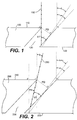
- Figure I is an enlarged cross-sectional view of a portion of an idealized embodiment of a membrane of the present invention drawn with strictly normal ER 150.
- the membrane 100 of Figure 1 has a first surface 110, a second surface 120, and a plurality of pores 130.
- the pores of membrane 100 are off-axis such that the least angle of incidence of any pore is ⁇ .
- the pore 130 has an angle of incidence such that the detection beam cannot travel directly through the pore from the second surface to the first surface of the membrane or vice versa .
- FIG. 2 is an enlarged cross-sectional schematic view of a portion of the membrane of the present invention.
- the ⁇ -normal opaque membrane 200 has a top surface 210, a bottom surface 220 and a plurality of pores 230.
- the pore diameter pd and the membrane thickness mt are as indicated.
- the angle of a pore of the membrane of least angle of incidence is ⁇ .
- Figure 3 is an enlarged cross-sectional schematic view of a portion of an embodiment of the membrane of the present invention having curved pores 300.
- the top surface 310, the bottom surface 320, and a pore 330 are indicated.
- Ray 360 emitted from a ⁇ -normal detection beam, having the maximum angle of incidence of the detection beam, ⁇ , is drawn in Figure 3.
- the pore diameter pd and the membrane thickness mt are as indicated.
- the least angle of incidence, ⁇ , of a pore of the membrane is determined as indicated in Figure 3.
- FIG 4a is a reduced cross-sectional schematic view of an apparatus used in fabrication of embodiments of the membrane of the present invention.
- the film from which the membrane is manufactured is in the form of a web on a roll called the unwind roll 410, which is unwound and held with a flat section 420 between the unwind roll 410 and a second roll, the rewind roll 430, which contains the fabricated membrane.
- the flat section of the film 420, between the unwind roll 410 and the rewind roll 430 passes under multiple beams of excimer laser light 440, each beam of which has a diameter slightly smaller than the pores that are being fabricated. How much smaller is determined by the thickness of the film, what the film is made of, and the wavelength of the ER from the laser.
- Excimer lasers are used because the wavelength of their ER is very short, typically between 200nm and 400 nm. This allows pores of less than a micron to be made.
- the pore sizes of preferred embodiments of the present invention are 1.0, 2.0, 3.0, 5.0, 8.0, 10.0, 12.0, and 14.0 microns, which are relatively easy to make compared to the submicron pores.
- the beams of laser light 440 strike the film at angle ⁇ , as shown in Figure 4a.
- ⁇ is between 15° and 70° depending on the particular ⁇ -normal opaque membrane required, the membrane thickness, pore diameter and the nature of the cell activity assays in which it is to be employed.
- the shortest possible pores are the most desirable, and in others, longer pores are optimal. For example, if the assay is designed for a minimum incubation period, the pores are preferably short. On the other hand, if maximum sensitivity is paramount, longer pores may be better, since a lower number of negative control cells will pass through the membrane.
- the multiple beams of excimer laser ER are created by projecting the output of the laser onto a mask which forms multiple discrete beams, e.g. , thousands of beams, which are then focused on the film.
- the laser ER ablates the film creating straight pores through the material.
- the power of the laser, the composition of the film material, and its thickness, determines the duration of the ablation.
- the details are well known to those practiced in the art.
- One advantage of membrane fabrication with a laser is that the exact position and number of the pores can be controlled. This is advantageous since it lowers the CV of the assays by removing the variation associated with different sites of the cell activity apparatus having different numbers and positions of pores.
- One preferred embodiment of the present invention employs a microplate sized apparatus (5.030"x3.365" footprint) with 1536 sites of 1 mm diameter.
- Preferred embodiments of the ⁇ -normal opaque membrane employed in this apparatus have pore diameters of 1, 2, 3, 5, 8, 10, 12, or 14 microns, and the thickness ranges between 5 and 50 microns.
- the pore densities range from 1x10 3 to 2x10 7 pores/cm 2 .
- the smaller the area of membrane per assay site the larger the errors become that are associated with pore distribution irregularity. This negatively affects the CV of the whole assay. Consequently, excimer laser fabrication with virtually no variation in the size, number, angle, and position of the pores, is an important advantage.
- Figure 4b is a schematic oblique top view and Figure 4c is a side view of a preferred fabrication method of the membranes of the present invention.
- the membranes are constructed from film by ablating pores with a laser apparatus as in Figure 4a.
- frames of a CAAA 460 with the film 461 bonded to one side are precisely positioned in an angled jig 470 with a sliding member 471 as shown in Figure 4c.
- Multiple laser beams 440 each with a diameter proportional to the diameter of the pores being fabricated, strike the film at an angle of incidence ⁇ , as indicated.
- Equation (2) must be satisfied to construct a ⁇ -normal opaque membrane.
- the framed film 461 is moved in the direction indicated by arrow 475 in incremental steps under the laser beams 440, preserving the angle ⁇ at which the laser beams strike the film.
- the beams 440 sequentially ablate rows of pores in a pattern defined by a mask (not shown) that forms clusters of discrete beams in the optical path between the source of the laser light (not shown) and a focusing lens system 450 that focuses the clusters of laser beams on the film.
- the laser beams 440 are grouped into a pattern 480 corresponding to the sites of the CAAA represented in Figure 4b by the 32x48 array of 1536 sites.
- This fabrication technique has several advantages: (a) the number of pores at each site is fixed and (b) said pores can be positioned in any fixed and uniform pattern across the whole framed film 461. This means that site to site variation in CAAA using the framed membrane is practically zero with respect to the membranes. It also means that the application of the hydrophobic mask of some preferred embodiments (described below) is no longer as positionally and dimensionally critical to the uniformity of the sites, and hence to the CV of the assays.
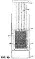
- Figure 4d shows the method of fabrication of the membranes of the present invention from film in which the pores are machined, burned, or ablated with a laser as in Figure 4a.
- the film from which the membranes are manufactured is in the form of a web on an unwind roll 410, which is unwound and passes around secondary roller 490 where it changes direction 90 degrees and passes around the fabrication roll 495 of very small diameter.
- the diameter of fabrication roll 495 is proportional to the radius of curvature (rc) of the pores fabricated with this method: the smaller the diameter of 495 the smaller the rc of the pores.
- the laser ER strikes the film at a range of angles on a very narrow part of the area of the film where it is bent around the fabrication roller 495.
- the laser ER ablates straight holes in the bent film, so when the membranes are flat, the pores are bent (see Figure 3)
- the membrane web then makes a quarter turn around roll 491 and hence to the rewind roll 430 where it is rewound as fabricated membrane.
- the film moves in very small incremental steps in the direction indicated by arrow 475, the step increment being determined by the width of the area where the laser ablates the pores in the film.
- the laser beams 440 sequentially ablate rows of pores, the diameter and pattern of which are defined by a mask (not shown). The range of angles of the pores will be proportional to the diameter of the fabrication roll 495 and the width of the area where the laser beams 440 strike the film.
- Figure 5a is a reduced cross-sectional schematic view of a second kind of apparatus used in fabrication of embodiments of the membranes of the present invention.
- the film from which the membrane is manufactured is in the form of a web on unwind roll 510, which is unwound and held flat 520 between the intermediate rolls 530 and 540, and then moves to the rewind roll 550, which contains the irradiated membrane.
- the section of the film held flat and taught 520 between the intermediate rolls 530 and 540 passes under a beam 560 of high energy charged particles emitted by a cyclotron (not shown).
- the diameter of the high energy particle beam in one preferred embodiment is approximately 6 centimeters, and it is swept back and forth across the web by deflecting it with an electromagnetic field from sweeping magnet 570 as shown in Figure 5b.
- the beam sweeps back and forth across the width of the film in a path perpendicular to the line of motion of the web.
- the beam (or fog of ions) is about 5 meters from sweeping magnet 570 when it penetrates the film.
- the film moves under it at a steady rate which is determined by the pore density desired for the membrane.
- the charged particles pass through it, breaking the molecular bonds of the polymer chains composing the film.
- the energy of the particles in the beam ranges between 1 and 2 million electron volts and must be sufficient to pass completely through the film at the angle of incidence ⁇ of the beam.
- This angle ⁇ is set between 15° and 70°.
- the optimal angle for a particular ⁇ -normal opaque membrane is determined by the membrane thickness, the pore diameter, and the nature of the cell activity assays in which it is to be employed.
- the second step of the process is not shown and consists of submersing the irradiated film in an etching bath. This etches pores along the straight paths of broken polymers through the film created by the high energy particles from the cyclotron. The diameter of the pores is determined by the duration of the etching process, and the temperature and concentration of the etching bath. The specifications for this process are well known to those practiced in the art of fabricating track-etch membranes.
- Figures 6a-6c are schematic diagrams of an embodiment of the present invention.
- the multi-site CAAA 600 has a ⁇ -normal R-opaque @P% membrane, specifically the substantially R-opaque @P% membrane described above.
- Figure 6a is a top and side cross-section view of the whole instrument, and Figures 6b and 6c are enlarged cross-section views of portions of the apparatus.
- the assay apparatus 600 comprises three rigid parts: an upper part consisting of a transparent film 621 bonded to a rigid frame 620, a middle part consisting of the membrane 651 bonded to a second rigid frame 650, and a bottom part consisting of a transparent injection-molded microplate 680.
- the membrane 651 is fabricated from film, which can be made from a variety of materials including plastic, metal, glass, ceramic, organic material, or combinations thereof.
- the membrane pores 695 can be fabricated in a number of ways, two of which are illustrated and described above.
- the frame 650 can be plastic, steel, stainless steel, aluminum, or another suitable material.
- the frame must be rigid enough to keep the membrane, and any grids, coatings, or site-delimiting devices attached thereto, substantially flat.
- the membrane can be attached to the frame by any suitable fastening means, including glue, heat seals, ultrasonic seals, or mechanical means.
- Figure 6a shows the ninety six (96) assay sites 630 of CAAA 600 arranged in an 8x12 array.
- Each assay site 630 comprises a discrete, delimited area 656 of membrane 651, along with two three-dimensional compartments (wells)--the upper volume or well 693, above the membrane 651, and the lower volume or well 692, in the microplate 680 below the membrane 651.
- the film 621 delimits the top of each upper volume 693 and creates optically advantageous flat surfaces, which minimize reflection and refraction of ER from detection beams and ER emitted from the upper well of the test sites 630.
- An opaque mask 625 affixed to the bottom surface of film 621, surrounds and separates the transparent spots 627 at the tops of the assay sites 630. This mask is also hydrophobic and circumscribes the top perimeters of the upper volumes 693.
- Figure 6c shows a greatly enlarged view of a section of the membrane 651, which, with the frame 650, forms the middle part of the CAAA 600.
- Figure 6c illustrates the off-axis pores 695 of substantially R-opaque @P% membrane 651.
- This figure also shows a membrane section at the edge of an assay site 630 and illustrates the two hydrophobic masks that are affixed to the membrane 651, one (655) on the top surface 653 and one (654) on the bottom surface 652.
- Hydrophobic mask 655 delimits the bottom perimeter of the upper volume of site 630, and circumscribes the membrane area 656 where cell activity can occur across the membrane.
- Hydrophobic mask 654 helps circumscribe the top surface of the lower volume, as described below.
- the upper volume (upper well) 693 and lower volume (lower well) 692 of each site 630 are filled with fluid solutions containing chemical compounds and/or biological cells in suspension as shown by the shaded areas in Figure 6b and the cross-section view of Figure 6a.
- Hydrophobic coating 686 affixed to the rim 685 of the lower volume 692, forms a shield seal with the hydrophobic mask 654 on the under side 652 of membrane 651.
- the membrane 651 is positioned and held on top of and against the rims 685 of the lower well 692, which confines the lower-volume fluid.
- the flat transparent bottoms 682 of the wells in the microplate 680 are optically advantageous surfaces through which light can pass into and out of the lower volumes 692.
- Figures 7a, and 7a 2 show the top view and a side view, respectively, of a CAAA 700 having one thousand five hundred and thirty six (1536) assay sites.
- the assay sites 730 are arranged in a 32x48 array within the footprint of a standard microplate (5.030"x3.365").
- the apparatus 700 is composed of three rigid parts: an upper rigid frame 720, a middle rigid frame 750 and a lower rigid frame 780.
- the upper rigid frame 720 is bonded to a transparent film 721.
- the middle rigid frame 750 is bonded to substantially R-opaque @ P% membrane 751.
- the lower rigid frame 780 is bonded to transparent film 781.
- Each of the assay sites 730 has an upper volume 793 and a lower volume 792, as shown in Figure 7b.
- Figure 7b is a cross-sectional, enlarged, schematic view of a portion of CAAA 700.
- the upper film 721 has a hydrophobic mask 725 bonded to its bottom surface 723.
- the membrane 751 has a top hydrophobic mask 755 bonded to its top surface 753, and a bottom hydrophobic mask 754 bonded to its bottom surface 752.
- the lower film 781 has a top surface 782 with a hydrophobic mask 784 bonded to it.
- Each assay site 730 consists of the following elements: the transparent area 727 of upper film 721 and hydrophobic mask 725 on lower surface 723 surrounding transparent area 727, the open area 756 of membrane 751 and hydrophobic mask 755 on the top surface 753 of membrane 751 surrounding 756, and hydrophobic mask 754 on bottom surface 752 of the membrane 751, the transparent area 786 of the lower film 781, the hydrophobic mask 784 on the top surface 782 of the lower film 781, and the upper volume 793 and the lower volume 792.
- the upper air space 797 and lower air space 798 surround and separate each of the sites.
- the volumes 793 and 792 are between 0.5 ⁇ l and 2.5 ⁇ l, and the distances between the centers of sites 730 is 2.25 mm.
- Hydrophobic masks 725 and 784 are opaque and surround transparent areas of the top film 727 and bottom film 786, respectively, that are directly above and below each assay site.
- ER from the detection beams 760 and 761 and the ER 762 and 763 emitted by the fluorescent dye in the upper and lower volumes pass in and out through upper transparent area 727 and lower transparent area 786, respectively.
- the detector/quantification apparatus above the upper film 721 is composed of a housing 740, a fiber optic bundle 742 for collecting emitted ER 762 from the top volume 793 and top surface 753 of the site 730, and another optical conduit 744 for delivering the excitation ER 760 to the upper volume 793 and upper surface 753 of the membrane of the sites 730.
- the detector/quantification apparatus below the lower film 781 is composed of a housing 741, a fiber optic bundle 743 for collecting emitted ER 763 from the bottom volume 792 of the site 730, and another optical conduit 745 for delivering the excitation ER 761 to the lower volume 792 and lower surface 752 of the membrane of the site 730.
- the interfaces between the three rigid frames 780, 750, and 720 can be either sealed to gas exchange or not as required by the assay.
- airflow needs to be minimized to prevent excessive evaporation, without overly inhibiting diffusion of oxygen and carbon dioxide. This can be accomplished in a number of ways including: positioning a thin layer of open cell foam between the frames, providing multiple small channels between the frames, providing film material for 721 and/or 781 with sufficient gas exchange rates, providing microscopic holes in 721 and/or 781 between the test sites, or providing enough microscopic pores in 721 and/or 781 at the test sites for sufficient gas exchange.
- polyester film is used with various dyes that make it opaque at various wavelength ranges that match fluorescent excitation and emission bands of the various dyes used to tag or label the cells used in the cell activity assays.
- the embodiments, described above, manufactured with the cyclotron bombardment and etching technology require materials that can be etched.
- Polyester and polycarbonate have excellent etching characteristics and are widely used. Polyester is preferred because it is easy to introduce dye into the film after it is initially fabricated. Polycarbonate film is now fabricated with dye incorporated into the basic film, but at this time the other characteristics of this film, in particular the uniformity of its thickness, make it a poor candidate.
- Coating or depositing metallic layers on plastic films and/or membranes is another method of making the membranes opaque.
- advantages and disadvantages to this fabrication technique For example, with a dyed substantially opaque membrane, some light passes through the pores via reflections within the pores. The amount of light which passes through, however, is insignificant and is not a practical problem since standard detection/quantification systems used for cell activity assays are not sensitive enough to measure it. If the membrane is coated with metallic atoms, however, the interior surfaces of the pores can be extremely reflective. This is counter-productive to the goal of fabricating a substantially R-opaque @P% membrane where P is above 99.9%. Furthermore, the deposited coating must not interfere or affect the activity of the cells used in the assays.
- cells to be interrogated are positioned on one side of the membrane and solutions to be assayed for their influence on cell activity are positioned directly opposite on the other side of the membrane.
- the preferred membrane of this embodiment is substantially R-opaque @P%, where R is 400nm-580nm and P% is 99.9% and the membrane thickness is 31 microns, the pore diameter is 8 microns, the range of angles of incidence of the off axis pores is between 44° and 46°.
- the contribution of the cells on the opposite side of the membrane from the excitation/detection/quantification system is less than .0005% of the count at any site.
- the membrane could have a pore density between 1x10 3 and 1x10 9 pores/cm 2 , a thickness between 1 and 1000 microns, and pores with angle of incidence between about 15° and 70°.
- CAAA 600 Several methods using CAAA 600 or similar apparatus are described in more detail below.
- Method I comprises the following steps:
- Method II comprises the same steps as Method I, in addition to the following step, step 7a, performed after step 7:
- Method III comprises the same steps as Method I, in addition to the following step, step 7b, performed after step 7:
- Methods II and III are preferred over Method I in cases where the cell activity is so rapid that the accuracy of the results will be compromised by the difference in elapsed time the cells are active at different test sites. This can be very significant if the incubation time is short, e.g. , less than thirty minutes, and the detection/quantification time is long, e.g. , ten minutes. With the large number of sites used for HTS and ultra-HTS (e.g. , 1536 and 3456 sites) the cells must be killed or immobilized for accurate results.
- Method IV is a simplified version of Method I. In this method, all of the steps of method I are performed, with the exception of step (6). Thus, in this simplified version, the readings of the emissions from the upper and lower wells are obtained only once.
- Method V is a simplified version of Method II and Method VI is a simplified version of Method III. That is, as described above, the readings of step (6) are not performed.
- Method VII is essentially a class of methods.
- Method VII comprises all of the Methods I through VI, using an apparatus having a membrane that is R-opaque @P%, but not substantially R-opaque @P%. That is, the membrane used in this class of methods has pores that allow substantially normal ER, such as that used by state-of-the-art detection/quantification systems, to pass straight through the membrane. This will yield adequate results in some CBHTS contexts, although the sensitivity will be lower, particularly in comparison with Methods I through III.
- Method VIII is another class of methods. These methods use a CAAA such as that disclosed above and illustrated in Figures 7a 1 , 7a 2 , 7b, and 7c. In these methods the first five steps of Method I through VII are modified as follows:
- any of Methods I through VIII commencing with step (6)(as described above) can be followed, making the necessary changes in the procedure required by the changes in the hardware.
- the detector beams of the automatic fluorescent reader have to be smaller, as would the optical collection system that collects and measures the amount of light emitted from the upper and lower volumes of the sites.
- the methods described above, using apparatus 600 or 700 or similar apparatus, allow the detection and quantification at each site of (a) the number of cells pipetted, (b) the number of cells that have migrated through the membrane, (c) the number that have not migrated, (d) the number that have migrated into the membrane but have not passed through, and (e) the total number that have migrated. From this data, the percent of the cells that have migrated at any site can be calculated as well as the percent of cells that have passed through the membrane at that site. Since these results are calculated independently for each site, both the errors associated with pipetting (variations in volume) and the errors associated with the uneven distribution of cells in a unit volume are eliminated.
- substantially R-opaque @P% membrane used in apparatus 600 or 700 was fabricated according to the method disclosed in Figure 4b and Figure 4c, further reduction in the CV is achieved due to complete uniformity in the number of pores from site to site.
- Method VIII When Method VIII is used with CAAA that employ R-opaque @P% membrane that is not substantially R-opaque @P%, the results will be adequate in some CBHTS contexts. These include assays where it is desirable to use (a) very low pore densities, (b) large pore diameters, (c) thin membranes, and (d) low cell densities.
- the system must also have very low background fluorescence and the detection/quantification system must be very sensitive. In these contexts, the detection system detects cells when they migrate over pores. New detection instruments are being developed e.g. , Cellomics (Pittsburgh, Pa.) which have high resolution optical systems that may accomplish this.
- Migration of cells in this context would not be measured by how many cells passed through the membrane, but how many migrated over the top of a pore. Such assays are rare and expensive.
- the methods used would most likely be designed to acquire data about the kinetics of cell activity, and these methods are different from the ones described here.
- the cells can be suspended in an ungelled gel solution, as described above, and applied to the sites on the bottom side of the membrane when it is inverted.
- the inverted lower framed film is then positioned over and attached to the inverted framed membrane.
- the solutions gel the apparatus is inverted and test solutions and controls are applied to the top sites of the membrane, and then the upper framed film is positioned and attached.
- the cells now migrate up through the filter. Similar results can be achieved with CAAA similar to 600, making the changes necessary to accommodate the differences in the apparatus.
- each site is a well provided with an insert which divides the well into an upper chamber and a lower chamber.
- the inserts are attached together in the form of a plate.
- the insert incorporates the membrane of the present invention.
- the present invention could also encompass assays in which the sample is not necessarily in a liquid medium but, for example, may be carried by a gas medium so long as the membrane is substantially opaque to the ER wavelengths introduced and detected.
Landscapes
- Chemical & Material Sciences (AREA)
- Life Sciences & Earth Sciences (AREA)
- Organic Chemistry (AREA)
- Zoology (AREA)
- Health & Medical Sciences (AREA)
- Engineering & Computer Science (AREA)
- Bioinformatics & Cheminformatics (AREA)
- Wood Science & Technology (AREA)
- Sustainable Development (AREA)
- General Health & Medical Sciences (AREA)
- Biotechnology (AREA)
- Biomedical Technology (AREA)
- Analytical Chemistry (AREA)
- Biochemistry (AREA)
- General Engineering & Computer Science (AREA)
- Microbiology (AREA)
- Genetics & Genomics (AREA)
- Cell Biology (AREA)
- Apparatus Associated With Microorganisms And Enzymes (AREA)
- Measuring Or Testing Involving Enzymes Or Micro-Organisms (AREA)
- Investigating, Analyzing Materials By Fluorescence Or Luminescence (AREA)
- Investigating Or Analysing Biological Materials (AREA)
Applications Claiming Priority (3)
| Application Number | Priority Date | Filing Date | Title |
|---|---|---|---|
| US271765 | 1999-03-18 | ||
| US09/271,765 US6329164B1 (en) | 1999-03-18 | 1999-03-18 | Method for using a cell activity assay apparatus |
| PCT/US2000/005760 WO2000055298A1 (en) | 1999-03-18 | 2000-03-06 | Cell activity assay apparatus and methods for making and using same |
Publications (3)
| Publication Number | Publication Date |
|---|---|
| EP1165745A1 EP1165745A1 (en) | 2002-01-02 |
| EP1165745A4 EP1165745A4 (en) | 2004-10-06 |
| EP1165745B1 true EP1165745B1 (en) | 2006-05-24 |
Family
ID=23036978
Family Applications (1)
| Application Number | Title | Priority Date | Filing Date |
|---|---|---|---|
| EP00916095A Expired - Lifetime EP1165745B1 (en) | 1999-03-18 | 2000-03-06 | Apparatus and method for measuring cell activity |
Country Status (7)
| Country | Link |
|---|---|
| US (3) | US6329164B1 (enExample) |
| EP (1) | EP1165745B1 (enExample) |
| JP (1) | JP4558949B2 (enExample) |
| AT (1) | ATE327315T1 (enExample) |
| AU (1) | AU3725400A (enExample) |
| DE (1) | DE60028192T2 (enExample) |
| WO (1) | WO2000055298A1 (enExample) |
Families Citing this family (27)
| Publication number | Priority date | Publication date | Assignee | Title |
|---|---|---|---|---|
| US6329164B1 (en) * | 1999-03-18 | 2001-12-11 | Neuro Probe, Incorporated | Method for using a cell activity assay apparatus |
| US6864065B2 (en) * | 2000-11-08 | 2005-03-08 | Surface Logix, Inc. | Assays for monitoring cell motility in real-time |
| US6844184B2 (en) * | 2000-11-08 | 2005-01-18 | Surface Logix, Inc. | Device for arraying biomolecules and for monitoring cell motility in real-time |
| US7326563B2 (en) | 2000-11-08 | 2008-02-05 | Surface Logix, Inc. | Device and method for monitoring leukocyte migration |
| US6699665B1 (en) * | 2000-11-08 | 2004-03-02 | Surface Logix, Inc. | Multiple array system for integrating bioarrays |
| US6893851B2 (en) | 2000-11-08 | 2005-05-17 | Surface Logix, Inc. | Method for arraying biomolecules and for monitoring cell motility in real-time |
| US7033819B2 (en) | 2000-11-08 | 2006-04-25 | Surface Logix, Inc. | System for monitoring cell motility in real-time |
| US7374906B2 (en) | 2000-11-08 | 2008-05-20 | Surface Logix, Inc. | Biological assays using gradients formed in microfluidic systems |
| US7033821B2 (en) * | 2000-11-08 | 2006-04-25 | Surface Logix, Inc. | Device for monitoring cell motility in real-time |
| US7123764B2 (en) * | 2000-11-08 | 2006-10-17 | Surface Logix Inc. | Image processing method for use in analyzing data of a chemotaxis or haptotaxis assay |
| TWI241343B (en) | 2000-12-07 | 2005-10-11 | Effector Cell Inst Inc | Well unit for detecting cell chemotaxis and separating chemotactic cells |
| JP3738899B2 (ja) * | 2000-12-07 | 2006-01-25 | 株式会社 エフェクター細胞研究所 | 微量試料処理装置 |
| US6923939B1 (en) * | 2001-07-05 | 2005-08-02 | Uop Llc | Heat activated membrane introduction apparatus and method for screening materials |
| US20030082632A1 (en) * | 2001-10-25 | 2003-05-01 | Cytoprint, Inc. | Assay method and apparatus |
| EP1490520A4 (en) * | 2002-03-12 | 2006-06-07 | Surface Logix Inc | TEST APPARATUS ANALYZING THE ABSORPTION, METABOLISM, PERMEABILITY AND / OR TOXICITY OF A CANDIDATE COMPOUND |
| TW200506364A (en) * | 2003-04-09 | 2005-02-16 | Effector Cell Inst Inc | Apparatus for detecting cell chemo-taxis |
| US7417726B2 (en) * | 2003-09-19 | 2008-08-26 | Applied Biosystems Inc. | Normalization of data using controls |
| US6901945B2 (en) * | 2003-09-30 | 2005-06-07 | Nalco Company | System for feeding solid materials to a pressurized pipeline |
| US6972184B2 (en) * | 2003-12-23 | 2005-12-06 | Millipore Corporation | Cell motility assay |
| US20050202052A1 (en) * | 2004-03-15 | 2005-09-15 | Lee Clemie M. | Flavored medicaments to deter or attract and kill microorganisms |
| US7547525B2 (en) * | 2004-08-12 | 2009-06-16 | Neuro Probe Incorporated | Point source diffusion cell activity assay |
| US8673628B2 (en) | 2011-06-10 | 2014-03-18 | Essen Instruments, Inc. | Methods and apparatus for improving in vitro measurements using boyden chambers |
| US9644177B2 (en) * | 2012-07-12 | 2017-05-09 | Board Of Regents, The University Of Texas System | Extracellular matrix films and methods of making and using same |
| US10384207B2 (en) | 2015-07-21 | 2019-08-20 | Neuro Probe Incorporated | Assay apparatus and methods |
| WO2022187684A1 (en) * | 2021-03-05 | 2022-09-09 | Enumerix, Inc. | Systems and methods for generating droplets and performing digital analyses |
| CN117241878A (zh) | 2021-03-05 | 2023-12-15 | 伊努梅里斯公司 | 用于生成微滴和执行数字分析的系统和方法 |
| US12252745B2 (en) | 2021-09-02 | 2025-03-18 | Enumerix, Inc. | Detection and digital quantitation of multiple targets |
Family Cites Families (15)
| Publication number | Priority date | Publication date | Assignee | Title |
|---|---|---|---|---|
| US3888770A (en) | 1971-10-21 | 1975-06-10 | Shlomo Avital | Plural-sample filter device |
| US3929583A (en) | 1975-08-14 | 1975-12-30 | Canadian Patents Dev | Apparatus for enumerating microorganisms |
| US4317726A (en) | 1981-02-12 | 1982-03-02 | The United States Of America As Represented By The Secretary Of The Army | Microbial filter assembly |
| US4514495A (en) | 1982-05-18 | 1985-04-30 | Spiral Systems Instruments, Inc. | Method for testing microbial interaction with growth affecting substances |
| US4493815A (en) | 1983-07-28 | 1985-01-15 | Bio-Rad Laboratories, Inc. | Supporting and filtering biochemical test plate assembly |
| US4714674A (en) | 1985-02-28 | 1987-12-22 | Genentech, Inc. | Chemotactic assay for immunogenicity |
| US5137023A (en) * | 1990-04-19 | 1992-08-11 | Worcester Polytechnic Institute | Method and apparatus for monitoring blood analytes noninvasively by pulsatile photoplethysmography |
| US4912057A (en) | 1989-06-13 | 1990-03-27 | Cancer Diagnostics, Inc. | Cell chamber for chemotaxis assay |
| DE69129121T2 (de) * | 1990-12-21 | 1998-11-19 | Curative Technologies, Inc., Setauket, N.Y. | Angiogene peptide |
| US5210021A (en) | 1991-03-20 | 1993-05-11 | Neuro Probe, Inc. | Multiple-site chemotactic test apparatus and method |
| US5284753A (en) | 1991-03-20 | 1994-02-08 | Neuro Probe, Inc. | Multiple-site chemotactic test apparatus and method |
| GB2264169B (en) * | 1992-02-07 | 1995-08-02 | Alan John Hayes | Fluid monitoring |
| US5302515A (en) | 1992-08-20 | 1994-04-12 | Neuro Probe, Inc. | Chemotactic test apparatus and method |
| US5601997A (en) * | 1995-02-03 | 1997-02-11 | Tchao; Ruy | Chemotaxis assay procedure |
| US6329164B1 (en) * | 1999-03-18 | 2001-12-11 | Neuro Probe, Incorporated | Method for using a cell activity assay apparatus |
-
1999
- 1999-03-18 US US09/271,765 patent/US6329164B1/en not_active Expired - Lifetime
-
2000
- 2000-03-06 WO PCT/US2000/005760 patent/WO2000055298A1/en not_active Ceased
- 2000-03-06 DE DE60028192T patent/DE60028192T2/de not_active Expired - Lifetime
- 2000-03-06 EP EP00916095A patent/EP1165745B1/en not_active Expired - Lifetime
- 2000-03-06 AT AT00916095T patent/ATE327315T1/de not_active IP Right Cessation
- 2000-03-06 AU AU37254/00A patent/AU3725400A/en not_active Abandoned
- 2000-03-06 JP JP2000605716A patent/JP4558949B2/ja not_active Expired - Lifetime
-
2001
- 2001-09-28 US US09/964,391 patent/US6468786B2/en not_active Expired - Lifetime
- 2001-09-28 US US09/964,686 patent/US6395505B2/en not_active Expired - Lifetime
Also Published As
| Publication number | Publication date |
|---|---|
| EP1165745A1 (en) | 2002-01-02 |
| EP1165745A4 (en) | 2004-10-06 |
| US20020019024A1 (en) | 2002-02-14 |
| ATE327315T1 (de) | 2006-06-15 |
| WO2000055298A1 (en) | 2000-09-21 |
| JP2002538816A (ja) | 2002-11-19 |
| AU3725400A (en) | 2000-10-04 |
| DE60028192T2 (de) | 2006-11-02 |
| US6329164B1 (en) | 2001-12-11 |
| DE60028192D1 (de) | 2006-06-29 |
| US6395505B2 (en) | 2002-05-28 |
| US6468786B2 (en) | 2002-10-22 |
| US20020009796A1 (en) | 2002-01-24 |
| JP4558949B2 (ja) | 2010-10-06 |
Similar Documents
| Publication | Publication Date | Title |
|---|---|---|
| EP1165745B1 (en) | Apparatus and method for measuring cell activity | |
| US6486947B2 (en) | Devices and methods for sample analysis | |
| US6667159B1 (en) | Optical fiber biosensor array comprising cell populations confined to microcavities | |
| US7241569B2 (en) | Method and a device for the evaluation of biopolymer fitness | |
| US6582903B1 (en) | Method and a device for the evaluation of biopolymer fitness | |
| US7201836B2 (en) | Multiaperture sample positioning and analysis system | |
| JP2755889B2 (ja) | 少なくとも1個の電極およびまたはセンサを形成するための物体 | |
| GB2297615A (en) | Chemotaxis assay procedure and membrane for use therein | |
| US8486655B2 (en) | Point source diffusion cell activity assay apparatus | |
| US10385305B2 (en) | Device for measuring activity of cultured cells, microchamber and method of measuring activity of cultured cells | |
| WO2000005336A9 (en) | Devices and methods for sample analysis | |
| MXPA01002431A (es) | Metodo para el examen rapido de analitos. | |
| MacDonald et al. | Combined spectroscopic and electrical recording techniques in membrane research: prospects for single channel studies | |
| Niles et al. | Miniaturization technologies for high-throughput biology | |
| RU2246349C2 (ru) | Испытательный планшет с множественными сквозными каналами для высокопроизводительного скрининга | |
| HK1109427B (en) | Point source diffusion cell activity assay apparatuses and methods |
Legal Events
| Date | Code | Title | Description |
|---|---|---|---|
| PUAI | Public reference made under article 153(3) epc to a published international application that has entered the european phase |
Free format text: ORIGINAL CODE: 0009012 |
|
| 17P | Request for examination filed |
Effective date: 20011017 |
|
| AK | Designated contracting states |
Kind code of ref document: A1 Designated state(s): AT BE CH CY DE DK ES FI FR GB GR IE IT LI LU MC NL PT SE |
|
| AX | Request for extension of the european patent |
Free format text: AL;LT;LV;MK;RO;SI |
|
| A4 | Supplementary search report drawn up and despatched |
Effective date: 20040825 |
|
| 17Q | First examination report despatched |
Effective date: 20050405 |
|
| RTI1 | Title (correction) |
Free format text: APPARATUS AND METHOD FOR MEASURING CELL ACTIVITY |
|
| GRAP | Despatch of communication of intention to grant a patent |
Free format text: ORIGINAL CODE: EPIDOSNIGR1 |
|
| GRAS | Grant fee paid |
Free format text: ORIGINAL CODE: EPIDOSNIGR3 |
|
| GRAA | (expected) grant |
Free format text: ORIGINAL CODE: 0009210 |
|
| AK | Designated contracting states |
Kind code of ref document: B1 Designated state(s): AT BE CH CY DE DK ES FI FR GB GR IE IT LI LU MC NL PT SE |
|
| PG25 | Lapsed in a contracting state [announced via postgrant information from national office to epo] |
Ref country code: IT Free format text: LAPSE BECAUSE OF FAILURE TO SUBMIT A TRANSLATION OF THE DESCRIPTION OR TO PAY THE FEE WITHIN THE PRESCRIBED TIME-LIMIT;WARNING: LAPSES OF ITALIAN PATENTS WITH EFFECTIVE DATE BEFORE 2007 MAY HAVE OCCURRED AT ANY TIME BEFORE 2007. THE CORRECT EFFECTIVE DATE MAY BE DIFFERENT FROM THE ONE RECORDED. Effective date: 20060524 Ref country code: LI Free format text: LAPSE BECAUSE OF FAILURE TO SUBMIT A TRANSLATION OF THE DESCRIPTION OR TO PAY THE FEE WITHIN THE PRESCRIBED TIME-LIMIT Effective date: 20060524 Ref country code: AT Free format text: LAPSE BECAUSE OF FAILURE TO SUBMIT A TRANSLATION OF THE DESCRIPTION OR TO PAY THE FEE WITHIN THE PRESCRIBED TIME-LIMIT Effective date: 20060524 Ref country code: BE Free format text: LAPSE BECAUSE OF FAILURE TO SUBMIT A TRANSLATION OF THE DESCRIPTION OR TO PAY THE FEE WITHIN THE PRESCRIBED TIME-LIMIT Effective date: 20060524 Ref country code: NL Free format text: LAPSE BECAUSE OF FAILURE TO SUBMIT A TRANSLATION OF THE DESCRIPTION OR TO PAY THE FEE WITHIN THE PRESCRIBED TIME-LIMIT Effective date: 20060524 Ref country code: CH Free format text: LAPSE BECAUSE OF FAILURE TO SUBMIT A TRANSLATION OF THE DESCRIPTION OR TO PAY THE FEE WITHIN THE PRESCRIBED TIME-LIMIT Effective date: 20060524 Ref country code: FI Free format text: LAPSE BECAUSE OF FAILURE TO SUBMIT A TRANSLATION OF THE DESCRIPTION OR TO PAY THE FEE WITHIN THE PRESCRIBED TIME-LIMIT Effective date: 20060524 |
|
| REG | Reference to a national code |
Ref country code: GB Ref legal event code: FG4D |
|
| REG | Reference to a national code |
Ref country code: CH Ref legal event code: EP |
|
| REG | Reference to a national code |
Ref country code: IE Ref legal event code: FG4D |
|
| REF | Corresponds to: |
Ref document number: 60028192 Country of ref document: DE Date of ref document: 20060629 Kind code of ref document: P |
|
| PG25 | Lapsed in a contracting state [announced via postgrant information from national office to epo] |
Ref country code: DK Free format text: LAPSE BECAUSE OF FAILURE TO SUBMIT A TRANSLATION OF THE DESCRIPTION OR TO PAY THE FEE WITHIN THE PRESCRIBED TIME-LIMIT Effective date: 20060824 Ref country code: SE Free format text: LAPSE BECAUSE OF FAILURE TO SUBMIT A TRANSLATION OF THE DESCRIPTION OR TO PAY THE FEE WITHIN THE PRESCRIBED TIME-LIMIT Effective date: 20060824 |
|
| PG25 | Lapsed in a contracting state [announced via postgrant information from national office to epo] |
Ref country code: ES Free format text: LAPSE BECAUSE OF FAILURE TO SUBMIT A TRANSLATION OF THE DESCRIPTION OR TO PAY THE FEE WITHIN THE PRESCRIBED TIME-LIMIT Effective date: 20060904 |
|
| RAP2 | Party data changed (patent owner data changed or rights of a patent transferred) |
Owner name: NEURO PROBE INCORPORATED |
|
| RIN2 | Information on inventor provided after grant (corrected) |
Inventor name: GOODWIN, RICHARD H. |
|
| PG25 | Lapsed in a contracting state [announced via postgrant information from national office to epo] |
Ref country code: PT Free format text: LAPSE BECAUSE OF FAILURE TO SUBMIT A TRANSLATION OF THE DESCRIPTION OR TO PAY THE FEE WITHIN THE PRESCRIBED TIME-LIMIT Effective date: 20061024 |
|
| NLV1 | Nl: lapsed or annulled due to failure to fulfill the requirements of art. 29p and 29m of the patents act | ||
| REG | Reference to a national code |
Ref country code: GB Ref legal event code: 732E |
|
| REG | Reference to a national code |
Ref country code: CH Ref legal event code: PL |
|
| PLBE | No opposition filed within time limit |
Free format text: ORIGINAL CODE: 0009261 |
|
| STAA | Information on the status of an ep patent application or granted ep patent |
Free format text: STATUS: NO OPPOSITION FILED WITHIN TIME LIMIT |
|
| 26N | No opposition filed |
Effective date: 20070227 |
|
| EN | Fr: translation not filed | ||
| PG25 | Lapsed in a contracting state [announced via postgrant information from national office to epo] |
Ref country code: MC Free format text: LAPSE BECAUSE OF NON-PAYMENT OF DUE FEES Effective date: 20070331 Ref country code: IE Free format text: LAPSE BECAUSE OF NON-PAYMENT OF DUE FEES Effective date: 20070306 |
|
| PG25 | Lapsed in a contracting state [announced via postgrant information from national office to epo] |
Ref country code: GR Free format text: LAPSE BECAUSE OF FAILURE TO SUBMIT A TRANSLATION OF THE DESCRIPTION OR TO PAY THE FEE WITHIN THE PRESCRIBED TIME-LIMIT Effective date: 20060825 Ref country code: FR Free format text: LAPSE BECAUSE OF FAILURE TO SUBMIT A TRANSLATION OF THE DESCRIPTION OR TO PAY THE FEE WITHIN THE PRESCRIBED TIME-LIMIT Effective date: 20070309 |
|
| PG25 | Lapsed in a contracting state [announced via postgrant information from national office to epo] |
Ref country code: FR Free format text: LAPSE BECAUSE OF FAILURE TO SUBMIT A TRANSLATION OF THE DESCRIPTION OR TO PAY THE FEE WITHIN THE PRESCRIBED TIME-LIMIT Effective date: 20060524 |
|
| PG25 | Lapsed in a contracting state [announced via postgrant information from national office to epo] |
Ref country code: CY Free format text: LAPSE BECAUSE OF FAILURE TO SUBMIT A TRANSLATION OF THE DESCRIPTION OR TO PAY THE FEE WITHIN THE PRESCRIBED TIME-LIMIT Effective date: 20060524 Ref country code: LU Free format text: LAPSE BECAUSE OF NON-PAYMENT OF DUE FEES Effective date: 20070306 |
|
| PGFP | Annual fee paid to national office [announced via postgrant information from national office to epo] |
Ref country code: IT Payment date: 20160323 Year of fee payment: 17 |
|
| PG25 | Lapsed in a contracting state [announced via postgrant information from national office to epo] |
Ref country code: IT Free format text: LAPSE BECAUSE OF NON-PAYMENT OF DUE FEES Effective date: 20170306 |
|
| PGFP | Annual fee paid to national office [announced via postgrant information from national office to epo] |
Ref country code: DE Payment date: 20190327 Year of fee payment: 20 |
|
| PGFP | Annual fee paid to national office [announced via postgrant information from national office to epo] |
Ref country code: GB Payment date: 20190404 Year of fee payment: 20 |
|
| REG | Reference to a national code |
Ref country code: DE Ref legal event code: R071 Ref document number: 60028192 Country of ref document: DE |
|
| REG | Reference to a national code |
Ref country code: GB Ref legal event code: PE20 Expiry date: 20200305 |
|
| PG25 | Lapsed in a contracting state [announced via postgrant information from national office to epo] |
Ref country code: GB Free format text: LAPSE BECAUSE OF EXPIRATION OF PROTECTION Effective date: 20200305 |
|
| P01 | Opt-out of the competence of the unified patent court (upc) registered |
Effective date: 20230517 |