EP1162997B1 - Staphylococcus antigen and vaccine - Google Patents
Staphylococcus antigen and vaccine Download PDFInfo
- Publication number
- EP1162997B1 EP1162997B1 EP00916405A EP00916405A EP1162997B1 EP 1162997 B1 EP1162997 B1 EP 1162997B1 EP 00916405 A EP00916405 A EP 00916405A EP 00916405 A EP00916405 A EP 00916405A EP 1162997 B1 EP1162997 B1 EP 1162997B1
- Authority
- EP
- European Patent Office
- Prior art keywords
- antigen
- staphylococcus
- antibody
- antibodies
- composition
- Prior art date
- Legal status (The legal status is an assumption and is not a legal conclusion. Google has not performed a legal analysis and makes no representation as to the accuracy of the status listed.)
- Expired - Lifetime
Links
Images

Classifications
-
- C—CHEMISTRY; METALLURGY
- C07—ORGANIC CHEMISTRY
- C07K—PEPTIDES
- C07K16/00—Immunoglobulins [IGs], e.g. monoclonal or polyclonal antibodies
- C07K16/12—Immunoglobulins [IGs], e.g. monoclonal or polyclonal antibodies against material from bacteria
- C07K16/1267—Immunoglobulins [IGs], e.g. monoclonal or polyclonal antibodies against material from bacteria from Gram-positive bacteria
- C07K16/1271—Immunoglobulins [IGs], e.g. monoclonal or polyclonal antibodies against material from bacteria from Gram-positive bacteria from Micrococcaceae (F), e.g. Staphylococcus
-
- A—HUMAN NECESSITIES
- A61—MEDICAL OR VETERINARY SCIENCE; HYGIENE
- A61K—PREPARATIONS FOR MEDICAL, DENTAL OR TOILETRY PURPOSES
- A61K39/00—Medicinal preparations containing antigens or antibodies
- A61K39/02—Bacterial antigens
- A61K39/085—Staphylococcus
-
- A—HUMAN NECESSITIES
- A61—MEDICAL OR VETERINARY SCIENCE; HYGIENE
- A61P—SPECIFIC THERAPEUTIC ACTIVITY OF CHEMICAL COMPOUNDS OR MEDICINAL PREPARATIONS
- A61P19/00—Drugs for skeletal disorders
- A61P19/08—Drugs for skeletal disorders for bone diseases, e.g. rachitism, Paget's disease
-
- A—HUMAN NECESSITIES
- A61—MEDICAL OR VETERINARY SCIENCE; HYGIENE
- A61P—SPECIFIC THERAPEUTIC ACTIVITY OF CHEMICAL COMPOUNDS OR MEDICINAL PREPARATIONS
- A61P31/00—Antiinfectives, i.e. antibiotics, antiseptics, chemotherapeutics
- A61P31/04—Antibacterial agents
-
- A—HUMAN NECESSITIES
- A61—MEDICAL OR VETERINARY SCIENCE; HYGIENE
- A61P—SPECIFIC THERAPEUTIC ACTIVITY OF CHEMICAL COMPOUNDS OR MEDICINAL PREPARATIONS
- A61P9/00—Drugs for disorders of the cardiovascular system
-
- C—CHEMISTRY; METALLURGY
- C07—ORGANIC CHEMISTRY
- C07K—PEPTIDES
- C07K14/00—Peptides having more than 20 amino acids; Gastrins; Somatostatins; Melanotropins; Derivatives thereof
- C07K14/195—Peptides having more than 20 amino acids; Gastrins; Somatostatins; Melanotropins; Derivatives thereof from bacteria
- C07K14/305—Peptides having more than 20 amino acids; Gastrins; Somatostatins; Melanotropins; Derivatives thereof from bacteria from Micrococcaceae (F)
- C07K14/31—Peptides having more than 20 amino acids; Gastrins; Somatostatins; Melanotropins; Derivatives thereof from bacteria from Micrococcaceae (F) from Staphylococcus (G)
-
- A—HUMAN NECESSITIES
- A61—MEDICAL OR VETERINARY SCIENCE; HYGIENE
- A61K—PREPARATIONS FOR MEDICAL, DENTAL OR TOILETRY PURPOSES
- A61K39/00—Medicinal preparations containing antigens or antibodies
- A61K2039/60—Medicinal preparations containing antigens or antibodies characteristics by the carrier linked to the antigen
- A61K2039/6031—Proteins
- A61K2039/6037—Bacterial toxins, e.g. diphteria toxoid [DT], tetanus toxoid [TT]
-
- A—HUMAN NECESSITIES
- A61—MEDICAL OR VETERINARY SCIENCE; HYGIENE
- A61K—PREPARATIONS FOR MEDICAL, DENTAL OR TOILETRY PURPOSES
- A61K39/00—Medicinal preparations containing antigens or antibodies
- A61K2039/60—Medicinal preparations containing antigens or antibodies characteristics by the carrier linked to the antigen
- A61K2039/6031—Proteins
- A61K2039/6068—Other bacterial proteins, e.g. OMP
-
- Y—GENERAL TAGGING OF NEW TECHNOLOGICAL DEVELOPMENTS; GENERAL TAGGING OF CROSS-SECTIONAL TECHNOLOGIES SPANNING OVER SEVERAL SECTIONS OF THE IPC; TECHNICAL SUBJECTS COVERED BY FORMER USPC CROSS-REFERENCE ART COLLECTIONS [XRACs] AND DIGESTS
- Y10—TECHNICAL SUBJECTS COVERED BY FORMER USPC
- Y10S—TECHNICAL SUBJECTS COVERED BY FORMER USPC CROSS-REFERENCE ART COLLECTIONS [XRACs] AND DIGESTS
- Y10S435/00—Chemistry: molecular biology and microbiology
- Y10S435/975—Kit
-
- Y—GENERAL TAGGING OF NEW TECHNOLOGICAL DEVELOPMENTS; GENERAL TAGGING OF CROSS-SECTIONAL TECHNOLOGIES SPANNING OVER SEVERAL SECTIONS OF THE IPC; TECHNICAL SUBJECTS COVERED BY FORMER USPC CROSS-REFERENCE ART COLLECTIONS [XRACs] AND DIGESTS
- Y10—TECHNICAL SUBJECTS COVERED BY FORMER USPC
- Y10S—TECHNICAL SUBJECTS COVERED BY FORMER USPC CROSS-REFERENCE ART COLLECTIONS [XRACs] AND DIGESTS
- Y10S530/00—Chemistry: natural resins or derivatives; peptides or proteins; lignins or reaction products thereof
- Y10S530/866—Chemistry: natural resins or derivatives; peptides or proteins; lignins or reaction products thereof involving immunoglobulin or antibody fragment, e.g. fab', fab, fv, fc, heavy chain or light chain
Definitions
- the present invention relates to a novel Staphylococcus antigen, and to a method for obtaining and using the antigen.
- Staphylococcus causes several diseases by various pathogenic mechanisms. The most frequent and serious of these diseases are bacteremia and its complications in hospitalized patients. In particular, Staphylococcus can cause wound infections and infections associated with catheters and prosthetic devices. Serious infections associated with Staphylococcus bacteremia include osteomyelitis, invasive endocarditis and septicemia. The problem is compounded by multiple antibiotic resistance in hospital strains, which severely limits the choice of therapy. In the majority of cases the causative organism is a strain of S. aureus, S. epidermidis, S. haemolyticus or S. hominis, or a combination of these. The problem with Staphylococcus is compounded by multiple antibiotic resistance in hospital strains, which severely limits the choice of therapy.
- a S. aureus vaccine would provide a solution for the problem of antibiotic resistance.
- An antigen common to multiple Staphylococcus species would enable production of a vaccine containing a single antigen that would be effective against a wide variety of staph infections.
- an isolated Staphylococcus antigen that (a) comprises amino acids and a N-acetylated hexosamine in an ⁇ configuration, (b) contains no O-acetyl groups detectable by nuclear magnetic resonance spectroscopy, and (3) specifically binds with antibodies to a Staphylococcus strain deposited under ATCC 202176.
- composition comprising the Staphylococcus antigen, and a sterile, pharmaceutically-acceptable carrier therefor.
- An immunotherapy method comprises a step of administering to a subject an immunostimulatory amount of such a composition.
- a whole cell vaccine comprising cells from a strain of Staphylococcus that carries the antigen.
- composition comprising the whole cell vaccine, and a sterile, pharmaceutically-acceptable carrier therefor.
- the vaccine can be administered to a subject to provide protection against Staphylococcus infection.
- An immunotherapeutic agent against Staphylococcus infection can be prepared by immunizing subjects with a composition according to the invention, collecting plasma from the immunized subjects, and harvesting a hyperimmune globulin that contains antibodies directed against Staphylococcus from the collected plasma.
- the hyperimmune globulin contains antibodies directed against the antigen.
- An immunotherapy method comprises a step of administering this hyperimmune globulin to a subject.
- the present invention also provides a catheter coated with an antigen according to the invention, and a method for preventing adherence of Staphylococcus bacteria to a catheter, comprising treating a catheter with antigen according to the invention.
- the catheter is an intravenous catheter.
- the antigen represents the basis for a vaccine that provides protection against infection by a large number of clinically-significant Staphylococcus isolates.
- a "clinically-significant" isolate is an isolate that is pathogenic.
- the present inventors found that a majority of Staphylococcus clinical isolates reacted very strongly with antigen/conjugate antibody sera, and thus were typeable as strains that contain the antigen. More particularly, typing of clinical isolates obtained from various sources has shown that approximately 60% of S. epidermidis, 50% of S. haemolyticus and 40% of S. hominis isolates express the antigen, as determined by slide agglutination. When enzymatic digests of the S. haemolyticus and S. hominis isolates were subjected to an immunodiffusion assay, all of the isolates tested positive for the presence of the antigen.
- Antibodies to the antigen do not cross-react with polysaccharides isolated from any of S. aureus Type 5, Type 8, Type 4, or K73 (a Type 5 variant strain).
- the antigen therefore is specific, that is, it produces a single band only with antiserum from homologous strains.
- the antigen can be obtained in recoverable amount, from certain Staphylococcus isolates cultured pursuant to the protocols described herein, in substantially pure form.
- purified antigen contains less than 1% nucleic acids.
- a "recoverable" amount in this regard means that the isolated amount of the antigen is detectable by a methodology less sensitive than radiolabeling, such as immunoassay, and can be subjected to further manipulations involving transfer of the antigen per se into solution.
- an isolate according to the invention first is fermented in a modified Columbia Broth supplemented with and 4% NaCl. Following fermentation, cells are killed and then centrifuged to separate the cells from the supernatant .
- Antigen is extracted from cell paste. Some of the antigen is present in the supernatant, but the amount is insignificant as compared to the amount found in the cell paste. Because of the low yield, and the risk of hexose contamination from the media, extraction from supernatant is not preferred.
- a suspension of the cell paste is treated with pronase, lysostaphin, DNase and RNase.
- the suspension is made 10% in trichloroacetic acid (TCA) and incubated at 60°C. After centrifugation, the supernatant is neutralized with 1M NaOH to pH 7.0, followed by sequential precipitation with 25-75% cold ethanol/CaCl 2 to remove nucleic acids and high molecular weight proteins and then precipitate antigen-containing material.
- TCA trichloroacetic acid
- the crude precipitate is dissolved in water and residual ethanol is removed by dialysis. Residual teichoic acid is removed by anion-exchange chromatography. Fractions are tested by capillary precipitation with antibodies specific for the antigen to determine antigen-containing fractions, which are pooled, dialyzed, lyophilized and treated with lysozyme to digest residual peptidoglycan.
- the enzyme-treated crude antigen-containing fraction is resuspended and rechromatographed on an anion-exchange column in a 0.1-0.25 M NaCl linear gradient in tris-HCl buffer, pH 7.0.
- Phosphorus-negative and antigen-positive fractions as determined by colorimeteric assay and capillary precipitation with monospecific antiserum, respectively, are pooled, dialyzed against water and lyophilized. Most of the antigen elutes at 0.2 M NaCl. The crude antigen is further purified by column chromatography, and antigen-containing fractions are pooled to produce substantially pure antigen.
- Purified antigen produces a single precipitin band when reacted with whole cell antisera in a double immunodiffusion assay.
- Immunoelectrophoresis of purified antigen and elution pattern on ion-exchange column during the purification process indicate a negatively-charged molecule.
- GlcNAc gas liquid chromatography-mass spectroscopy
- Signals at ⁇ 24.860 ppm (NAac-CH 3 ) and ⁇ 176.814 ppm (NAc-CO) in the 13C NMR spectrum suggest that the glucosamine is N-acetylated, so that the carbohydrate portion of the antigen is postulated to be 2-acetamido-2-deoxy- ⁇ -D-glucopyranoside.
- Amino acid analysis of the antigen shows the presence of serine, alanine, aspartic acid/asparagine, valine, and threonine in molar ratios of approximately 39:25:16:10:7. Amino acids constitute about 32% by weight of the antigen molecule.
- Antibodies induced by a vaccine containing the antigen facilitate type-specific phagocytosis.
- the in vitro phagocytosis assays thus indicate that antibodies to the antigen are protective against infection by Staphylococcus strains that carry the antigen.
- a vaccine based on the antigen can be used to protect against infection from a majority of clinical Staphylococcus strains.
- In vivo results obtained with the mouse lethality model and the mouse bacteremia model are consistent with results of in vitro opsonophagocytosis assays, and show that antibodies to antigen conjugates lowered mortality and bacteremia in mice challenged with strains of Staphylococcus that carry the antigen.
- a composition of the antigen or of whole cells containing the antigen according to the present invention "consists essentially of" the antigen, or cells that contain the antigen.
- the phrase "consists essentially of” means that the composition does not contain any material that interferes with elicitation of an immune response to the antigen (and to other antigens, if present) when the composition is administered to a subject as a vaccine, or with the antigen-antibody coupling characteristic of a diagnostic assay when the antigen is used in diagnosis.
- the antigen according to the invention is useful in the production of diagnostic assays for detecting the presence of Staphylococcus antigen and/or anti- Staphylococcus antibody in a sample.
- the antigen, or antibody specific to the antigen alone or in combination with other Staphylococcus antigens or antibodies, is mixed with a sample suspected of containing Staphylococcus antigen or antibody and monitored for antigen-antibody binding.
- the antigen or antibody is labelled with a radioactive or enzyme label.
- antigen or antibody is immobilized on a solid matrix such that the antigen or antibody are accessible to complementary antibody or antigen contacting a surface of the matrix. The sample then is brought into contact with the surface of the matrix, and the surface is monitored for antigen-antibody binding.
- the antigen or antibody can be used in an enzyme-linked immunosorbent assay (ELISA), in which antigen or antibody are bound to a solid phase and an enzyme-antibody or enzyme-antigen conjugate is used to detect and/or quantify antibody or antigen present in a sample.
- ELISA enzyme-linked immunosorbent assay
- a western blot assay can be used in which solubilized and separated antigen(s) is bound to nitrocellulose paper.
- the antibody then is detected by an enzyme or label-conjugated anti-immunoglobulin (Ig), such as horseradish peroxidase-Ig conjugate by incubating the filter paper in the presence of a precipitable or detectable substrate.
- Ig enzyme-linked immunosorbent assay
- an immunocarrier usually a polypeptide or protein
- T and B cells for the induction of an immune response against the antigen.
- An immunocarrier enhances immunogenicity both for active immunization and for preparing high-titered antisera in volunteers for passive immunization.
- Suitable immunocarriers according to the present invention include tetanus toxoid, diphtheria toxoid, Pseudomonas aeruginosa Exotoxin A or its derivatives, recombinantly-produced non-toxic mutant strains of exotoxin A, as described, for example, in Fattom et al., Inf. and Imm. 61 : 1023-1032 (1993), as well as other proteins commonly used as immunocarriers.
- the antigen is first derivatized.
- Various methods can be used to derivatize antigen and covalently link it to an immunocarrier.
- Activated carboxylate groups of the antigen can be derivatized with ADH, cystamine or PDPH, and then the antigen can be coupled to a carrier protein either by a carbodiimide-mediated reaction of the partially-amidated antigen to a carboxylate group on the carrier protein or by disulfide interchange of thiolated antigen with an SPDP-derivatized carrier protein.
- Hydroxyl groups on the antigen can be activated using cyanogen bromide or 1-cyano-4-dimethylamino-pyridinium tetrafluoroborate, and then the antigen can be derivatized with the six carbon bifunctional spacer adipic acid dihydrazide (ADH), according to techniques known in the art, according to the method of Kohn et al. FEBS Lett. 154 : 209:210 (1993).
- ADH six carbon bifunctional spacer adipic acid dihydrazide
- This material then is linked to diphtheria toxoid (Dtd), recombinant exoprotein A from Pseudomonas aeruginosa (rEPA), tetanus toxoid (TTd) or another suitable carrier protein by 1-ethyl-3-(3-dimethylaminopropyl) carbodiimide (EDAC).
- Dtd diphtheria toxoid
- rEPA Pseudomonas aeruginosa
- TTd tetanus toxoid
- EDAC 1-ethyl-3-(3-dimethylaminopropyl) carbodiimide
- the antigen or antigen conjugate is administered without an adjuvant in order to avoid adjuvant-induced toxicity.
- an adjuvant it is preferred to use one which promotes the protective IgG subtype 2 antibodies.
- Typical adjuvants include aluminum hydroxide, complete Freund's adjuvant (CFA) and incomplete Freund's adjuvant (IFA).
- CFA complete Freund's adjuvant
- IFA incomplete Freund's adjuvant
- Dextran sulfate has been shown to be a potent stimulator of IgG 2 antibody against staphylococcal cell surface antigens, and also is suitable as an adjuvant.
- the present invention also relates to the use of the antigen to produce polyclonal antibodies or monoclonal antibodies (mouse or human) that bind to or neutralize Staphylococcus strains that carry the antigen. Protocols for producing these antibodies are described in Ausubel, et al. (eds.), Molecular Cloning: A Laboratory Manual, Cold Spring Harbor Laboratory, (Cold Spring Harbor, NY)., Chapter 11; in METHODS OF HYBRIDOMA FORMATION 257-271, Bartal & Hirshaut (eds.), Humana Press, Clifton, NJ (1988); in Vitetta et al., Immunol. Rev. 62:159-83 (1982); and in Raso, Immunol. Rev. 62 :93-117 (1982).
- Inoculum for polyclonal antibody production typically is prepared by dispersing the antigen-immunocarrier in a physiologically-tolerable diluent such as saline, to form an aqueous composition.
- An immunostimulatory amount of inoculum, with or without adjuvant, is administered to a mammal and the inoculated mammal is then maintained for a time period sufficient for the antigen to induce protecting anti-antigen antibodies.
- Boosting doses of the antigen-immunocarrier may be used in individuals that are not already primed to respond to the antigen.
- Antibodies can include antibody preparations from a variety of commonly used animals, e.g. , goats, primates, donkeys, swine, rabbits, horses, hens, guinea pigs, rats, and mice, and even human antibodies after appropriate selection, fractionation and purification. Animal antisera may also be raised by inoculating the animals with formalin-killed strains of Staphylococcus that carry the antigen, by conventional methods, bleeding the animals and recovering serum or plasma for further processing.
- the antibodies induced in this fashion can be harvested and isolated to the extent desired by well known techniques, such as by alcohol fractionation and column chromatography, or by immunoaffinity chromatography; that is, by binding antigen to a chromatographic column packing like SephadexTM, passing the antiserum through the column, thereby retaining specific antibodies and separating out other immunoglobulins (IgGs) and contaminants, and then recovering purified antibodies by elution with a chaotropic agent, optionally followed by further purification, for example, by passage through a column of bound blood group antigens or other non-pathogen species.
- well known techniques such as by alcohol fractionation and column chromatography, or by immunoaffinity chromatography; that is, by binding antigen to a chromatographic column packing like SephadexTM, passing the antiserum through the column, thereby retaining specific antibodies and separating out other immunoglobulins (IgGs) and contaminants, and then recovering purified antibodies by elution with a chaotropic agent, optionally
- This procedure may be preferred when isolating the desired antibodies from the sera or plasma of humans that have developed an antibody titer against the pathogen in question, thus assuring the retention of antibodies that are capable of binding to the antigen. They can then be used in preparations for passive immunization against strains of Staphylococcus that carry the antigen.
- a monoclonal antibody composition contains, within detectable limits, only one species of antibody combining site capable of effectively binding to the antigen.
- Suitable antibodies in monoclonal form can be prepared using conventional hybridoma technology.
- a myeloma or other self-perpetuating cell line is fused with lymphocytes obtained from peripheral blood, lymph nodes or the spleen of a mammal hyperimmunized with the antigen. It is preferred that the myeloma cell line be from the same species as the lymphocytes. Splenocytes are typically fused with myeloma cells using polyethylene glycol 1500. Fused hybrids are selected by their sensitivity to HAT. Hybridomas secreting the antibody molecules of this invention can be identified using an ELISA.
- a Balb/C mouse spleen, human peripheral blood, lymph nodes or splenocytes are the preferred materials for use in preparing murine or human hybridomas.
- Suitable mouse myelomas for use in the present invention include the hypoxanthine-aminopterin-thymidine-sensitive (HAT) cell lines, a preferred myeloma being P3X63-Ag8.653.
- HAT hypoxanthine-aminopterin-thymidine-sensitive
- the preferred fusion partner for human monoclonal antibody production is SHM-D33, a heteromyeloma available from ATCC, Rockville, Md. under the designation CRL 1668.
- a monoclonal antibody composition of the present invention can be produced by initiating a monoclonal hybridoma culture comprising a nutrient medium containing a hybridoma that secretes antibody molecules of the appropriate specificity.
- the culture is maintained under conditions and for a time period sufficient for the hybridoma to secrete the antibody molecules into the medium.
- the antibody-containing medium is then collected.
- the antibody molecules then can be isolated further by well known techniques.
- Media useful for the preparation of these compositions are both well known in the art and commercially available, and include synthetic culture media, inbred mice and the like.
- An exemplary synthetic medium is Dulbecco's Minimal essential medium supplemented with 20% fetal calf serum.
- An exemplary inbred mouse strain is the Balb/c.
- monoclonal antibodies are produced to the antigen using methods similar to those described for type-specific antibodies to S. aureus Type 5 and Type 8.
- the purified monoclonal antibodies are characterized by bacterial agglutination assays using a collection of clinical isolates.
- the monoclonal and polyclonal antibody compositions produced according to the present description can be used by passive immunization to induce an immune response for the prevention or treatment of infection by strains of Staphylococcus that carry the antigen.
- the antibody preparation can be a polyclonal composition.
- a polyclonal composition includes antibodies that bind to the antigen, and additionally may include antibodies that bind to the antigens that characterize other strains of Staphylococcus.
- the polyclonal antibody component can be a polyclonal antiserum, preferably affinity purified, from an animal which has been challenged with the antigen, and possibly also with other Staphylococcus antigens.
- an "engineered oligoclonal" mixture may be used, which is a mixture of monoclonal antibodies to the antigen, and monoclonal antibodies to other Staphylococcus antigens.
- bivalent F(ab') 2 hybrid fragments by mixing two different F(ab') 2 fragments produced, e.g., by pepsin digestion of two different antibodies, reductive cleavage to form a mixture of Fab' fragments, followed by oxidative reformation of the disulfide linkages to produce a mixture of F(ab') 2 fragments including hybrid fragments containing a Fab' portion specific to each of the original antigens.
- bivalent fragments that are entirely heterospecific, e.g., use of bifunctional linkers to join cleaved fragments.
- Recombinant molecules are known that incorporate the light and heavy chains of an antibody, e.g ., according to the method of Boss et al ., U.S. patent No. 4,816,397.
- Analogous methods of producing recombinant or synthetic binding molecules having the characteristics of antibodies are included in the present invention. More than two different monospecific antibodies or antibody fragments can be linked using various linkers known in the art.
- An antibody component produced in accordance with the present invention can include whole antibodies, antibody fragments, or subfragments.
- Antibodies can be whole immunoglobulin of any class, e.g., IgG, IgM, IgA, IgD, IgE, chimeric antibodies or hybrid antibodies with dual or multiple antigen or epitope specificities, or fragments, e.g., F(ab') 2 , Fab', Fab and the like, including hybrid fragments, and additionally includes any immunoglobulin or any natural, synthetic or genetically engineered protein that acts like an antibody by binding to a specific antigen to form a complex.
- Fab molecules can be expressed and assembled in a genetically transformed host like E. coli.
- a lambda vector system is available thus to express a population of Fab's with a potential diversity equal to or exceeding that of subject generating the predecessor antibody. See Huse, W.D. , et al., Science 246 : 1275-81 (1989).
- the antigen according to the present invention can be the active ingredient in a composition, further comprising a pharmaceutically acceptable carrier for the active ingredient, which can be used as a vaccine to induce a cellular immune response and/or production in vivo of antibodies which combat Staphylococcus infection.
- a pharmaceutically acceptable carrier is a material that can be used as a vehicle for administering a medicament because the material is inert or otherwise medically acceptable, as well as compatible with the active agent, in the context of vaccine administration.
- a pharmaceutically acceptable carrier can contain conventional vaccine additives like diluents, adjuvants, antioxidants, preservatives and solubilizing agents.
- cells that carry the antigen are used in a whole cell vaccine.
- Cells that carry the antigen can be identified and selected for use in the whole cell vaccine by using antibodies to a strain known to carry the antigen, and more preferably by using monoclonal antibodies to isolated antigen as described herein.
- a simple slide agglutination experiment in which antibodies to the antigen are mixed with cells can be used.
- Deposited strain ATCC 202176 is a representative strain of Staphylococcus that carries the antigen, and it can be used to produce antibodies useful in identifying other strains that carry the antigen. It is not, however, necessary to use the deposited strain in order to produce either the antigen, the whole cell vaccine or antibodies useful in identifying other cells that carry the antigen. ATCC 202176 merely provides one immunologic means of identifying such cells.
- the whole cell vaccine also comprises a pharmaceutically acceptable carrier.
- the whole cell vaccine also optionally may contain conventional vaccine additives like diluents, adjuvants, antioxidants, preservatives and solubilizing agents.
- the whole cell vaccine contains only cells which carry the antigen, and does not include cells from strains of Staphylococcus that do not carry the antigen.
- Vaccines according to the invention can be administered to a subject not already infected with Staphylococcus, thereby to induce a Staphylococcus- protective immune response (humoral or cellular) in that subject.
- vaccines within the present invention can be administered to a subject in which Staphylococcus infection already has occurred but is at a sufficiently early stage that the immune response produced to the vaccine effectively inhibits further spread of infection.
- a vaccine of the present invention can be administered to a subject who then acts as a source for globulin, produced in response to challenge from the specific vaccine ("hyperimmune globulin"), that contains antibodies directed against Staphylococcus.
- a subject thus treated would donate plasma from which hyperimmune globulin would then be obtained, via conventional plasma-fractionation methodology, and administered to another subject in order to impart resistance against or to treat Staphylococcus infection.
- Hyperimmune globulins according to the invention are particularly useful for immune-compromised individuals, for individuals undergoing invasive procedures or where time does not permit the individual to produce his own antibodies in response to vaccination.
- monoclonal or polyclonal anti- Staphylococcus antibodies produced according to the present invention can be conjugated to an immunotoxin, and administered to a subject in whom Staphylococcus infection already has occurred but has not become widely spread.
- antibody material produced pursuant to the present description would be administered in a pharmaceutically acceptable carrier, as defined herein.
- ATCC 202176 a strain of S. epidermidis that carries the antigen, was fermented in a 50-liter fermentor in Columbia Broth supplemented with and 4% NaCl. The fermentation was started with one liter of a 16 hour old seed culture.Cells were fermented for 24 hours with low aeration (1 v.v.m.) and mild agitation (200 rpm) at 37°C. Following fermentation, cells were killed with 2% final concentration of phenol to ethanol (1:1) and then centrifuged to separate the cells from the supernatant.
- Cells to be used as a vaccine to prepare whole cell antiserum were 3% formalin-fixed overnight at room temperature.
- Cells for purification of antigen were killed by adding phenol-ethanol (1:1, vol/vol) to the fermentor to a final concentration of 2%, and mixing slowly for 2 hours at 15-20°C. No viable cells were detected after this treatment. The cells then were harvested by centrifugation at 14,500 x g and stored at -70°C until use.
- Antigen was extracted from cell paste.
- a suspension of the cell paste (0.5 g/ml) was treated for 4 hours at 37°C with pronase (1 mg/g of cells) and then with lysostaphin (175 U/g of cells), DNase and RNase (0.1 mg/g of each) and then stored overnight at 4°C.
- the suspension was made 10% in trichloroacetic acid (TCA) and incubated for 4 hours at 60°C. After centrifugation, the supernatant was neutralized with 1M NaOH to pH 7.0, followed by sequential precipitation with 25-75% cold ethanol in the presence of 10 mM CaCl 2 .
- Nucleic acids and high molecular weight proteins were precipitated from neutralized supernatant by adjusting it to 25% ethanol and incubating at 4°C for 4 hours. After centrifugation, the supernatant was adjusted to 75% ethanol and incubated at 4°C for 4-12 hours to precipitate antigen-containing material.
- the precipitate containing the crude antigen was dissolved in water and residual ethanol was removed by dialysis.
- the dialyzed material was adjusted to 0.01M Tris-HCl pH 7.0, 0.3 M NaCl and residual teichoic acid was removed by anion-exchange chromatography on a Q-Sepharose column in flow through mode with 0.01 Tris-HCl pH 7.0, 0.3 M NaCl.
- Fractions were tested by capillary precipitation with antibodies specific for the antigen to determine antigen-containing fractions.
- Antigen-containing fractions were dialyzed against water (3 x 5L), lyophilized and treated with lysozyme (0.5 mg/g cell paste) for 3 hours at 37°C to digest residual peptidoglycan.
- the enzyme-treated, crude antigen-containing fraction was resuspended in 0.01 M in Tris-HCl pH 7.0 and rechromatographed on a Q-Sepharose column in 0.1-0.25 M NaCl linear gradient.
- Phosphorus-negative and antigen-positive fractions as determined by colorimetric assay and capillary precipitation with monospecific antiserum from Example 2, respectively, were pooled, dialyzed against water and lyophilized. Most of the antigen eluted at 0.2 M NaCl.
- the crude antigen was further purified on a Sepharose CL6B column to obtain substantially pure antigen. Antigen-containing fractions were pooled, dialyzed against water and freeze-dried.
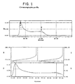
- Other C13-NMR spectrum signals appear at ⁇ 75.122, 73.953, 73.865, 73.807, 72.939, 69.847, 69.638, 69.585, 63.515, and 56.321, respectively.
- Amino acid analysis of the antigen showed the presence of serine, alanine, aspartic acid/asparagine, valine, and threonine in molar ratios of approximately 39:25:16:10:7. Amino acids constituted about 32% by weight of the antigen molecule.
- the mobility of purified antigen upon immunoelectrophoresis indicated the presence of negatively-charged groups.
- the purified antigen did not contain neutral sugars as detected by the phenol sulfuric assay.
- Purified antigen was partially depolymerized by hydrolysis in 50 mM acetic acid at 100°C for 30 minutes, or at 80°C for 90 minutes. The reaction mixture was freeze-dried and acetic acid was removed lyophilization. A partially hydrolyzed antigen was dissolved in 0.5 M ADH to its final concentration of 5 mg/mL and pH was adjusted to 5.6 with 0.1 M HCl. An antigen solution was made 100 mM EDAC by adding EDAC (as a powder) and pH was maintained at 5.6 with 0.1 M HCl for 1 hour. The reaction mixture then was dialyzed against 0.2 M NaCl (1X), then desalted on a Sephadex G25 column (2.6x 30 cm) and freeze-dried. The amount of ADH incorporated into antigen was determined colorimetrically by trinitrobenzene sulfonic acid (TNBS) assay using ADH as a standard.
- TNBS trinitrobenzene sulfonic acid
- ADH-derivatized antigen (10 mg) was dissolved in 1795 ⁇ L of O.1M MES buffer pH 5.6 and 205 ⁇ L of rEPA solution (48 mg/mL) representing 10 mg of rEPA was added.
- the reaction mixture was made 100 mM EDAC by adding EDAC (as a powder) and mixture was stirred at room temperature for 60 minutes. The reaction was stopped by bringing the pH to 7.0 with 1M MES-sodium salt (pH 9.21). Pure conjugate was obtained by size exclusion chromatography on Sephacryl S-300 column eluted with PBS. The amount of antigen and protein in the conjugate was determined by competitive ELISA and Coomasie Blue assay (Pierce) using the corresponding antigen or BSA as standards. The similar procedure was used to prepare antigen-Dtd conjugate.
- White female New Zealand rabbits were immunized by subcutaneous injection with 50 ⁇ g of antigen-immunocarrier conjugate prepared according to Example 5 on days 0, 14 and 28.
- the first injection was given with an equal volume of complete Freund's adjuvant (CFA) and subsequent injections were given with incomplete Freund's adjuvant (IFA).
- CFA complete Freund's adjuvant
- IFA incomplete Freund's adjuvant
- Test bleeds taken from rabbits were monitored for the presence of precipitating rabbit antibodies specific to the antigen with which they were immunized. Further injections were given as needed to boost the titer.
- Rabbits were bled to obtain high-titered rabbit antisera that contained antibodies specific to the antigen with which they were immunized. The antibodies were able to mediate killing of cells carrying the antigen by HL 60 in the presence of complement. Rabbits immunized with antigen-rEPA or antigen Dtd conjugates were also able to elicit antigen specific antibodies. These antibodies gave precipitates with the antigen in capillary.
- Conjugate IgG was used in opsonophagocytosis assays to evaluate the ability of the specific antibodies to mediate opsonophagocytosis of corresponding Staphylococcus bacteria by HL-60 cells in in vitro assays, and in animal models to evaluate efficacy in vivo.
- PMNs were obtained from HL-60 cells adjusted to a concentration of 1.0 x 10 6 cells per ml in MEM medium supplemented with 0.1% gelatin.
- the PMN cells were centrifuged at 1000 rpm for 10 minutes at 30-35°C.
- the pelleted cells were resuspended in five milliliters of MEM medium supplemented with 0.1% gelatin, and centrifuged at 1000 rpm for 10 minutes.
- the pelleted cells were resuspended in one milliliter of MEM medium supplemented with 0.1% gelatin to yield a working concentration of 1x10 6 /ml.
- a human complement prepared from human serum was diluted to 1:80 in MEM medium supplemented with 0.1% gelatin.
- the reaction mixture in the microtiter plate wells contained 50 ⁇ l of bacteria [10 6 cells/ml], 50 ⁇ l of diluted sera, 50 ⁇ l PMN [1x10 6 cells/ml] and 50 ⁇ l of complement [1:80], to give a total volume of 200 ⁇ l.
- a 10 ⁇ l sample from the reaction plate was serially diluted 1:5, 1:10, and 1:50.
- a 10 ⁇ l sample from each dilution was plated onto a tryptic soy agar (TSA) plate. The TSA plates were incubated overnight 37°C, 5% CO 2 .
- TSA tryptic soy agar
- reaction plate was incubated at 37°C for 90 minutes. The samples were remixed. A 10 ⁇ l sample from the reaction plate was serially diluted 1:5, 1:10, and 1:50. A 10 ⁇ l sample from each dilution was plated onto a TSA plates, which then were incubated overnight 37°C, 5% CO 2 .
- % kill No. of colonies at T 0 ⁇ no. of colonies at T 90 ⁇ 100 number of colonies at T 0
- Example 8 In vivo protection of mice from lethal Staphylococcus challenge by vaccination with antigen-rEPA conjugate
- mice A total of 24 mice were divided into three groups with 8 mice in each group.
- the mice in the first group were immunized with an intraperitoneal injection of 2.5 ⁇ g of purified antigen-rEPA conjugate produced according to Example 5 and IFA.
- the mice in the second group were injected with PBS plus IFA, while the mice in the third group were injected with PBS.
- the mice were boosted twice, at two week intervals, with the same vaccine or control dose.
- mice were challenged with 1.15 x 10 8 cfu of strain #V01048, a slime-producing strain of S . epidermidis that carries the antigen in 5% hog mucin.
- the challenged mice were monitored for morbidity and mortality. The results showed that 75% (2/8) of the mice in the first group survived lethal challenge, and were still alive at day 216 following challenge, while all mice in the second and third groups died by day 41 following challenge.
- Example 9 In vivo protection of mice from lethal Staphylococcus challenge with antigen-specific monoclonal antibody
- Example 10 In vivo protection of mice from bacteremia by vaccination with antigen-Dtd conjugate
- mice A total of 80 mice were divided into two groups with 40 mice in each group.
- the mice in the first group were immunized with a subcutaneous injection of 2.5 ⁇ g of purified antigen-Dtd conjugate produced according to Example 5 and IFA.
- the mice in the second group were injected with MEP conjugate (a conjugate of mucoid exopolysaccharide from Pseudomonas aeruginosa and Dtd) plus IFA.
- MEP conjugate a conjugate of mucoid exopolysaccharide from Pseudomonas aeruginosa and Dtd
- the mice were boosted twice, at two week intervals, with the same vaccine or control dose.
- mice were challenged intraperitoneally with a sub-lethal dose (5.0 x 10 7 cfu) of strain #V01048, a slime-producing strain of S. epidermidis that carries the antigen in 5% hog mucin.
- Challenged mice were exsanguinated and tested for positive bacterial cultures at 6, 24, 30 and 48 hours (ten mice at each time point). Results showed that immunization with antigen-rEPA conjugate significantly reduced bacteremia in the challenged mice, by facilitating clearance of bacteria from the blood.
- the control group immunized with the MEP conjugate were not protected against bacteremia.
- Example 11 Ability of antigen to block adherence of Staphylococcus bacteria to intravenous catheters in vitro
- the ability of the antigen to mediate adherence of the slime-producing S. epidermidis strain #977 that carries the antigen and S. haemolyticus strain 4162 that carries the antigen (determined by double immunodiffusion of crude bacterial extract) to intravenous catheters was evaluated in an in vitro adherence assay.
- the bacteria were grown overnight at 37°C on a Columbia agar plate supplemented with 4% sodium chloride. The following morning an isolated colony from this plate was inoculated into 5 ml of Columbia broth supplemented with 4% sodium chloride. This culture was then grown for 4 hours with shaking at 37°C and then adjusted to an OD of 0.12 at 650nM.
- a single 11 ⁇ 4" IV Insyte catheter [Radiopaque Vialon® material, Becton Dickinson Vascular Access, Sandy, Utah] was incubated at 37°C for 30 minutes in a 1 ml volume of a 0.5 mg/ml antigen solution in PBS. Catheters were then gently washed with cold PBS, immersed in 1 ml of bacterial suspension and incubated for 30 minutes at 37°C without shaking. Catheters were then gently washed again with cold PBS solution and sliced into three even pieces. The sliced catheters were immersed in 500 ⁇ L of PBS and sonicated for 1 minute on ice to sonicate off catheter attached bacterial cells.
- This suspension was diluted to 1:10, 1:100, and 1:1000 in PBS and plated onto TSA plates and incubated 18-20 hours at 37°C. The bacterial colonies were counted, and differences in bacterial recovery from different antigen-coated catheters were determined.
- Results showed that preincubation of the intravenous catheters with antigen isolated from S. epidermidis 202176 reduced by 97.5% the adherence of the slime-producing S. epidermidis strain #977 that carries the antigen, and reduced by 92% the adherence of S. haemolyticus .
- the antigen identical to the antigen purified from S. epidermidis 202176 was detected in crude cell wall extracts of S . haemolyticus and S . hominis, suggesting that S. epidermidis antigen is responsible for adherence of coagulase-negative staphylococci to intravenous catheters.
- Example 12 Ability of Fab fragments from antigen-specific antibodies to block adherence, of Staphylococcus bacteria to intravenous catheters in vitro
- the ability of the Fab fragments prepared from antigen-specific antibodies to block adherence of the slime-producing S. epidermidis strain RP62A, a strain that carries the antigen, to intravenous catheter preincubated in human plasma was evaluated in and in vitro adherence assay.
- the bacteria was grown overnight at 37°C on a Columbia agar plate supplemented with 4% sodium chloride. The following morning, an isolated colony from this plate was inoculated into 5 ml of Columbia broth supplemented with 4% sodium chloride. This culture was grown for 4 hours with shaking at 37°C and then adjusted to an OD of 0.12 at 650nm. Bacterial suspension (1 mL) was centrifuged at 3000 rpm and the pellet was resuspended in 1 mL of Fab solution (1 mg/mL) and incubated for 30 minutes at 37°C.
- a single 11 ⁇ 4" IV Insyte catheter [Radiopaque Vialon® material, Becton Dickinson Vascular Access, Sandy, Utah] was incubated at 37°C for 30 minutes in a 1 ml volume of a 0.5 mg/ml antigen solution in PBS. Catheters were then gently washed with cold PBS, immersed in 1 ml of bacterial suspension and incubated for 30 minutes at 37°C without shaking. Catheters were then gently washed again with cold PBS solution and sliced into three even pieces. The sliced catheters were immersed in 500 ⁇ L of PBS and sonicated for 1 minute on ice to sonicate off catheter attached bacterial cells.
- This suspension was diluted to 1:10, 1:100, and 1:1000 in PBS and plated onto TSA plates and incubated 18-20 hours at 37°C. The bacterial colonies were counted, and differences in bacterial recovery from different antigen-coated catheters were determined.
Applications Claiming Priority (3)
| Application Number | Priority Date | Filing Date | Title |
|---|---|---|---|
| US272359 | 1988-11-17 | ||
| US09/272,359 US6936258B1 (en) | 1999-03-19 | 1999-03-19 | Staphylococcus antigen and vaccine |
| PCT/US2000/006922 WO2000056357A2 (en) | 1999-03-19 | 2000-03-17 | Staphylococcus antigen and vaccine |
Publications (2)
| Publication Number | Publication Date |
|---|---|
| EP1162997A2 EP1162997A2 (en) | 2001-12-19 |
| EP1162997B1 true EP1162997B1 (en) | 2006-03-15 |
Family
ID=23039462
Family Applications (1)
| Application Number | Title | Priority Date | Filing Date |
|---|---|---|---|
| EP00916405A Expired - Lifetime EP1162997B1 (en) | 1999-03-19 | 2000-03-17 | Staphylococcus antigen and vaccine |
Country Status (15)
| Country | Link |
|---|---|
| US (2) | US6936258B1 (es) |
| EP (1) | EP1162997B1 (es) |
| JP (1) | JP4656728B2 (es) |
| AT (1) | ATE320265T1 (es) |
| AU (1) | AU773226B2 (es) |
| BR (1) | BR0009157B1 (es) |
| CA (1) | CA2366433C (es) |
| CY (1) | CY1105500T1 (es) |
| DE (1) | DE60026691T2 (es) |
| DK (1) | DK1162997T3 (es) |
| ES (1) | ES2261192T3 (es) |
| MX (1) | MXPA01009476A (es) |
| NZ (1) | NZ514455A (es) |
| PT (1) | PT1162997E (es) |
| WO (1) | WO2000056357A2 (es) |
Families Citing this family (59)
| Publication number | Priority date | Publication date | Assignee | Title |
|---|---|---|---|---|
| US6294177B1 (en) | 1996-09-11 | 2001-09-25 | Nabi | Staphylococcus aureus antigen-containing whole cell vaccine |
| US6936258B1 (en) * | 1999-03-19 | 2005-08-30 | Nabi Biopharmaceuticals | Staphylococcus antigen and vaccine |
| AT410798B (de) * | 2001-01-26 | 2003-07-25 | Cistem Biotechnologies Gmbh | Verfahren zur identifizierung, isolierung und herstellung von antigenen gegen ein spezifisches pathogen |
| EP1470237A4 (en) * | 2001-12-21 | 2006-02-01 | Biosynexus Inc | AGAINST PEPTIDOGLYCAN GRAMPOSITIVE BACTERIA MULTIFUNCTIONAL MONOCLONAL ANTIBODIES |
| US20060134141A1 (en) * | 2004-12-14 | 2006-06-22 | Nabi Biopharmaceuticals | Glycoconjugate vaccines containing peptidoglycan |
| WO2006076058A1 (en) * | 2005-01-10 | 2006-07-20 | Nabi Biopharmaceuticals | Method of treating staphylococcus aureus infection |
| JP5096326B2 (ja) | 2005-06-13 | 2012-12-12 | グラクソスミスクライン バイオロジカルズ ソシエテ アノニム | ブドウ球菌感染を治療および予防するためのPanton−Valentineロイコシジンの使用 |
| WO2007008904A2 (en) * | 2005-07-08 | 2007-01-18 | The Government Of The United States Of America As Represented By The Secretary Of The Department Of Health And Human Services | Targeting poly-gamma-glutamic acid to treat staphylococcus epidermidis and related infections |
| US20090317421A1 (en) | 2006-01-18 | 2009-12-24 | Dominique Missiakas | Compositions and methods related to staphylococcal bacterium proteins |
| EP3141261A1 (en) | 2006-03-30 | 2017-03-15 | GlaxoSmithKline Biologicals S.A. | Immunogenic composition |
| CN101466406B (zh) | 2006-06-12 | 2012-06-27 | 葛兰素史密斯克蓝生物品公司 | α-毒素在治疗和预防葡萄球菌感染上的用途 |
| EP2185190B1 (en) | 2007-08-31 | 2015-06-24 | University Of Chicago | Methods and compositions related to immunizing against staphylococcal lung diseases and conditions |
| EP2341929B1 (en) | 2008-10-06 | 2017-01-25 | University Of Chicago | Compositions and methods related to bacterial emp proteins |
| US8101190B2 (en) * | 2009-03-03 | 2012-01-24 | Ingen Biosciences | Method for diagnosing staphylococcal infections |
| PL2414387T3 (pl) | 2009-04-03 | 2016-06-30 | Univ Chicago | Kompozycje i sposoby związane z wariantami białka A (SpA) |
| WO2011127032A1 (en) | 2010-04-05 | 2011-10-13 | University Of Chicago | Compositions and methods related to protein a (spa) antibodies as an enhancer of immune response |
| PE20110023A1 (es) | 2009-06-22 | 2011-01-31 | Wyeth Llc | Composiciones inmunogenicas de antigenos de staphylococcus aureus |
| TW201544119A (zh) | 2009-06-22 | 2015-12-01 | Wyeth Llc | 組合物及製備金黃色葡萄球菌血清型5及8莢膜多醣結合物之免疫原性組合物之方法 |
| GB0913680D0 (en) | 2009-08-05 | 2009-09-16 | Glaxosmithkline Biolog Sa | Immunogenic composition |
| GB0913681D0 (en) | 2009-08-05 | 2009-09-16 | Glaxosmithkline Biolog Sa | Immunogenic composition |
| US10266585B2 (en) | 2009-08-28 | 2019-04-23 | The Board Of Regents Of The Univerity Of Texas System | Methods of treating brain injury |
| ES2812523T3 (es) | 2009-09-30 | 2021-03-17 | Glaxosmithkline Biologicals Sa | Conjugación de polisacáridos capsulares de Staphylococcus aureus de tipo 5 y de tipo 8 |
| PT2493498T (pt) | 2009-10-30 | 2017-05-24 | Glaxosmithkline Biologicals Sa | Purificação de sacáridos capsulares de staphylococcus aureus tipo 5 e tipo 8 |
| KR20130093084A (ko) | 2010-07-02 | 2013-08-21 | 더 유니버시티 오브 시카고 | 단백질 A(SpA) 변이체와 관련된 조성물 및 방법 |
| US9095540B2 (en) | 2010-09-09 | 2015-08-04 | The University Of Chicago | Methods and compositions involving protective staphylococcal antigens |
| CA2817973C (en) | 2010-10-15 | 2019-06-25 | The Board Of Regents Of The University Of Texas System | Antibodies that bind amyloid oligomers |
| MX350170B (es) | 2010-12-22 | 2017-08-28 | Wyeth Llc | Composición inmunogénica estable de antígenos de staphylococcus aureus. |
| US8945588B2 (en) | 2011-05-06 | 2015-02-03 | The University Of Chicago | Methods and compositions involving protective staphylococcal antigens, such as EBH polypeptides |
| CN103764171B (zh) | 2011-07-08 | 2016-08-17 | 诺华股份有限公司 | 酪氨酸连接方法 |
| CA2845259A1 (en) | 2011-08-15 | 2013-02-21 | The University Of Chicago | Compositions and methods related to antibodies to staphylococcal protein a |
| AU2012335208B2 (en) | 2011-11-07 | 2017-08-31 | Glaxosmithkline Biologicals S.A. | Carrier molecule comprising a spr0096 and a spr2021 antigen |
| WO2013090682A1 (en) | 2011-12-14 | 2013-06-20 | Indicator Systems International, Inc. | Trisubstituted methyl alcohols and their polymerizable derivatives |
| EP3805395A1 (en) | 2012-04-26 | 2021-04-14 | University Of Chicago | Staphylococcal coagulase antigens and methods of their use |
| EP2841101B1 (en) | 2012-04-26 | 2019-08-07 | University Of Chicago | Compositions and methods related to antibodies that neutralize coagulase activity during staphylococcus aureus disease |
| HUE049531T2 (hu) | 2012-08-16 | 2020-10-28 | Pfizer | Glikokonjugációs eljárások és kompozíciók |
| WO2014097099A2 (en) | 2012-12-20 | 2014-06-26 | Pfizer Inc. | Glycoconjugation process |
| WO2014116721A1 (en) | 2013-01-22 | 2014-07-31 | The Arizona Board Of Regents For And On Behalf Of Arizona State University | Geminiviral vector for expression of rituximab |
| GB201310008D0 (en) | 2013-06-05 | 2013-07-17 | Glaxosmithkline Biolog Sa | Immunogenic composition for use in therapy |
| CA2918076A1 (en) | 2013-07-11 | 2015-01-15 | Novartis Ag | Site-specific chemoenzymatic protein modifications |
| US11160855B2 (en) | 2014-01-21 | 2021-11-02 | Pfizer Inc. | Immunogenic compositions comprising conjugated capsular saccharide antigens and uses thereof |
| EP3096783B1 (en) | 2014-01-21 | 2021-07-07 | Pfizer Inc. | Streptococcus pneumoniae capsular polysaccharides and conjugates thereof |
| BR112016015525A2 (pt) | 2014-01-21 | 2017-10-24 | Pfizer | composições imunogênicas compreendendo antígenos sacarídeos capsulares conjugados e uso |
| KR20210032013A (ko) | 2014-01-21 | 2021-03-23 | 화이자 인코포레이티드 | 스트렙토코쿠스 뉴모니아에 피막 폴리사카라이드 및 그의 접합체 |
| EP3443983B1 (en) | 2014-02-14 | 2022-07-20 | Pfizer Inc. | Immunogenic glycoprotein conjugates |
| EP3229833A1 (en) | 2014-12-10 | 2017-10-18 | GlaxoSmithKline Biologicals SA | Method of treatment |
| DK3244917T3 (da) | 2015-01-15 | 2023-05-22 | Pfizer | Immunogene sammensætninger til anvendelse i pneumokokvacciner |
| NZ736238A (en) | 2015-05-04 | 2022-07-01 | Pfizer | Group b streptococcus polysaccharide-protein conjugates, methods for producing conjugates, immunogenic compositions comprising conjugates, and uses thereof |
| IL303998A (en) | 2015-07-21 | 2023-08-01 | Pfizer | Immunogenic preparations containing conjugated capsular sugar antigens, kits containing them and their uses |
| EP3377098A1 (en) | 2015-11-20 | 2018-09-26 | Pfizer Inc | Immunogenic compositions for use in pneumococcal vaccines |
| WO2017173398A1 (en) | 2016-04-01 | 2017-10-05 | Duke University | Alpha-helical peptide nanofibers as a self-adjuvanting vaccine platform |
| US10751402B2 (en) | 2016-11-09 | 2020-08-25 | Pfizer Inc. | Immunogenic compositions and uses thereof |
| EP3570879B1 (en) | 2017-01-20 | 2022-03-30 | Pfizer Inc. | Immunogenic compositions for use in pneumococcal vaccines |
| US11413336B2 (en) | 2018-03-23 | 2022-08-16 | Board Of Regents, The University Of Texas System | Coccidioides antigens and methods of their use |
| CA3120922A1 (en) | 2018-12-12 | 2020-06-18 | Pfizer Inc. | Immunogenic multiple hetero-antigen polysaccharide-protein conjugates and uses thereof |
| CA3136278A1 (en) | 2019-04-10 | 2020-10-15 | Pfizer Inc. | Immunogenic compositions comprising conjugated capsular saccharide antigens, kits comprising the same and uses thereof |
| EP4203995A1 (en) | 2020-08-26 | 2023-07-05 | Pfizer Inc. | Group b streptococcus polysaccharide-protein conjugates, methods for producing conjugates, immunogenic compositions comprising conjugates, and uses thereof |
| MX2023005221A (es) | 2020-11-04 | 2023-05-16 | Pfizer | Composiciones inmunogenicas para uso en vacunas neumococicas. |
| WO2022234416A1 (en) | 2021-05-03 | 2022-11-10 | Pfizer Inc. | Vaccination against pneumoccocal and covid-19 infections |
| EP4333879A1 (en) | 2021-05-03 | 2024-03-13 | Pfizer Inc. | Vaccination against bacterial and betacoronavirus infections |
Family Cites Families (12)
| Publication number | Priority date | Publication date | Assignee | Title |
|---|---|---|---|---|
| FR2619122B1 (fr) | 1987-08-03 | 1990-03-09 | Pasteur Institut | Procede d'obtention de polyosides capsulaires de staphylocoques, polyosides obtenus, applications de ces polyosides et souches pour la mise en oeuvre du procede |
| US7279162B1 (en) * | 1990-10-22 | 2007-10-09 | Henry M. Jackson Foundation For The Advancement Of Military Medicine | Isolated broadly reactive opsonic immunoglobulin for treating a pathogenic coagulase-negative staphylococcus infection |
| FR2682388B1 (fr) * | 1991-10-10 | 1995-06-09 | Pasteur Merieux Serums Vacc | Procede de preparation d'un oligoside par depolymerisation d'un polyoside issu d'un agent pathogene, oligoside ainsi obtenu et son utilisation notamment comme agent vaccinal. |
| AU681573B2 (en) | 1991-11-22 | 1997-09-04 | Glaxosmithkline Biologicals Sa | Type I and type II surface antigens associated with (staphylococcus epidermidis) |
| US5770208A (en) | 1996-09-11 | 1998-06-23 | Nabi | Staphylococcus aureus B-linked hexosamine antigen |
| US6294177B1 (en) * | 1996-09-11 | 2001-09-25 | Nabi | Staphylococcus aureus antigen-containing whole cell vaccine |
| WO1998028002A1 (en) | 1996-12-20 | 1998-07-02 | Research Foundation Of The City University Of New York | A novel pathogenic coccoid organism, staphylococcus leei, with trophism for gastric mucin |
| US5965374A (en) * | 1997-10-16 | 1999-10-12 | Biowhittaker Technologies, Inc. | Glucan-specific assay |
| WO2000015238A1 (en) * | 1998-09-14 | 2000-03-23 | Nabi | COMPOSITIONS OF β-GLUCANS AND SPECIFIC IGIV |
| US7030101B2 (en) * | 1998-09-14 | 2006-04-18 | Nabi Biopharmaceuticals | Compositions of β-glucans and specific antibodies |
| US6936258B1 (en) * | 1999-03-19 | 2005-08-30 | Nabi Biopharmaceuticals | Staphylococcus antigen and vaccine |
| US20030113350A1 (en) * | 2001-09-19 | 2003-06-19 | Fattom Ali I. | Glycoconjugate vaccines for use in immune-compromised populations |
-
1999
- 1999-03-19 US US09/272,359 patent/US6936258B1/en not_active Expired - Fee Related
-
2000
- 2000-03-17 EP EP00916405A patent/EP1162997B1/en not_active Expired - Lifetime
- 2000-03-17 AT AT00916405T patent/ATE320265T1/de active
- 2000-03-17 ES ES00916405T patent/ES2261192T3/es not_active Expired - Lifetime
- 2000-03-17 DK DK00916405T patent/DK1162997T3/da active
- 2000-03-17 MX MXPA01009476A patent/MXPA01009476A/es active IP Right Grant
- 2000-03-17 JP JP2000606261A patent/JP4656728B2/ja not_active Expired - Fee Related
- 2000-03-17 NZ NZ514455A patent/NZ514455A/en not_active IP Right Cessation
- 2000-03-17 CA CA2366433A patent/CA2366433C/en not_active Expired - Fee Related
- 2000-03-17 PT PT00916405T patent/PT1162997E/pt unknown
- 2000-03-17 DE DE60026691T patent/DE60026691T2/de not_active Expired - Lifetime
- 2000-03-17 AU AU37513/00A patent/AU773226B2/en not_active Ceased
- 2000-03-17 WO PCT/US2000/006922 patent/WO2000056357A2/en active IP Right Grant
- 2000-03-17 BR BRPI0009157-0B1A patent/BR0009157B1/pt not_active IP Right Cessation
-
2004
- 2004-12-20 US US11/014,997 patent/US7531633B2/en not_active Expired - Fee Related
-
2006
- 2006-05-12 CY CY20061100620T patent/CY1105500T1/el unknown
Also Published As
| Publication number | Publication date |
|---|---|
| WO2000056357A2 (en) | 2000-09-28 |
| CA2366433A1 (en) | 2000-09-28 |
| JP4656728B2 (ja) | 2011-03-23 |
| EP1162997A2 (en) | 2001-12-19 |
| CY1105500T1 (el) | 2010-04-28 |
| NZ514455A (en) | 2003-11-28 |
| ATE320265T1 (de) | 2006-04-15 |
| ES2261192T3 (es) | 2006-11-16 |
| DE60026691D1 (de) | 2006-05-11 |
| BR0009157B1 (pt) | 2013-12-24 |
| AU773226B2 (en) | 2004-05-20 |
| PT1162997E (pt) | 2006-07-31 |
| US20050118190A1 (en) | 2005-06-02 |
| WO2000056357A3 (en) | 2001-02-01 |
| MXPA01009476A (es) | 2003-08-19 |
| DK1162997T3 (da) | 2006-07-17 |
| JP2002539272A (ja) | 2002-11-19 |
| US7531633B2 (en) | 2009-05-12 |
| DE60026691T2 (de) | 2006-09-07 |
| CA2366433C (en) | 2012-01-03 |
| AU3751300A (en) | 2000-10-09 |
| BR0009157A (pt) | 2002-04-16 |
| US6936258B1 (en) | 2005-08-30 |
Similar Documents
| Publication | Publication Date | Title |
|---|---|---|
| EP1162997B1 (en) | Staphylococcus antigen and vaccine | |
| US6756361B1 (en) | Enterococcus antigens and vaccines | |
| CA2372633C (en) | Staphylococcus aureus antigen-containing whole cell vaccine | |
| US6194161B1 (en) | Staphylococcus aureus antigen | |
| US5961975A (en) | Type I surface antigen associated with staphylococcus epidermidis | |
| US7754225B2 (en) | Method of protecting against staphylococcal infection | |
| MXPA00003674A (es) | Antigenos y vacunas de enterococcus |
Legal Events
| Date | Code | Title | Description |
|---|---|---|---|
| PUAI | Public reference made under article 153(3) epc to a published international application that has entered the european phase |
Free format text: ORIGINAL CODE: 0009012 |
|
| 17P | Request for examination filed |
Effective date: 20010919 |
|
| AK | Designated contracting states |
Kind code of ref document: A2 Designated state(s): AT BE CH CY DE DK ES FI FR GB GR IE IT LI LU MC NL PT SE |
|
| AX | Request for extension of the european patent |
Free format text: AL;LT;LV;MK;RO;SI |
|
| RAP1 | Party data changed (applicant data changed or rights of an application transferred) |
Owner name: NABI BIOPHARMACEUTICALS |
|
| 17Q | First examination report despatched |
Effective date: 20040330 |
|
| GRAP | Despatch of communication of intention to grant a patent |
Free format text: ORIGINAL CODE: EPIDOSNIGR1 |
|
| TPAC | Observations filed by third parties |
Free format text: ORIGINAL CODE: EPIDOSNTIPA |
|
| GRAS | Grant fee paid |
Free format text: ORIGINAL CODE: EPIDOSNIGR3 |
|
| GRAA | (expected) grant |
Free format text: ORIGINAL CODE: 0009210 |
|
| AK | Designated contracting states |
Kind code of ref document: B1 Designated state(s): AT BE CH CY DE DK ES FI FR GB GR IE IT LI LU MC NL PT SE |
|
| REG | Reference to a national code |
Ref country code: GB Ref legal event code: FG4D Ref country code: CH Ref legal event code: EP |
|
| REG | Reference to a national code |
Ref country code: IE Ref legal event code: FG4D |
|
| REF | Corresponds to: |
Ref document number: 60026691 Country of ref document: DE Date of ref document: 20060511 Kind code of ref document: P |
|
| REG | Reference to a national code |
Ref country code: GR Ref legal event code: EP Ref document number: 20060401714 Country of ref document: GR |
|
| REG | Reference to a national code |
Ref country code: SE Ref legal event code: TRGR |
|
| REG | Reference to a national code |
Ref country code: DK Ref legal event code: T3 |
|
| REG | Reference to a national code |
Ref country code: PT Ref legal event code: SC4A Effective date: 20060512 Ref country code: CH Ref legal event code: NV Representative=s name: ISLER & PEDRAZZINI AG |
|
| ET | Fr: translation filed | ||
| REG | Reference to a national code |
Ref country code: ES Ref legal event code: FG2A Ref document number: 2261192 Country of ref document: ES Kind code of ref document: T3 |
|
| PLBE | No opposition filed within time limit |
Free format text: ORIGINAL CODE: 0009261 |
|
| STAA | Information on the status of an ep patent application or granted ep patent |
Free format text: STATUS: NO OPPOSITION FILED WITHIN TIME LIMIT |
|
| 26N | No opposition filed |
Effective date: 20061218 |
|
| REG | Reference to a national code |
Ref country code: CH Ref legal event code: PCAR Free format text: ISLER & PEDRAZZINI AG;POSTFACH 1772;8027 ZUERICH (CH) |
|
| REG | Reference to a national code |
Ref country code: CH Ref legal event code: PUE Owner name: GLAXOSMITHKLINE BIOLOGICALS SA Free format text: NABI BIOPHARMACEUTICALS#12280 WILKINS AVENUE#ROCKVILLE, MD 20852 (US) -TRANSFER TO- GLAXOSMITHKLINE BIOLOGICALS SA#RUE DE L'INSTITUT 89#1330 RIXENSART (BE) |
|
| REG | Reference to a national code |
Ref country code: PT Ref legal event code: PC4A Owner name: GLAXOSMITHKLINE BIOLOGICALS, S.A., BE Effective date: 20100813 |
|
| REG | Reference to a national code |
Ref country code: NL Ref legal event code: SD Effective date: 20100812 |
|
| REG | Reference to a national code |
Ref country code: GB Ref legal event code: 732E Free format text: REGISTERED BETWEEN 20100902 AND 20100908 |
|
| REG | Reference to a national code |
Ref country code: FR Ref legal event code: TP |
|
| REG | Reference to a national code |
Ref country code: ES Ref legal event code: PC2A Owner name: GLAXOSMITHKLINE BIOLOGICALS SA Effective date: 20110216 |
|
| REG | Reference to a national code |
Ref country code: FR Ref legal event code: PLFP Year of fee payment: 16 |
|
| PGFP | Annual fee paid to national office [announced via postgrant information from national office to epo] |
Ref country code: ES Payment date: 20150318 Year of fee payment: 16 Ref country code: MC Payment date: 20150227 Year of fee payment: 16 Ref country code: CH Payment date: 20150126 Year of fee payment: 16 Ref country code: IE Payment date: 20150226 Year of fee payment: 16 Ref country code: NL Payment date: 20150310 Year of fee payment: 16 Ref country code: IT Payment date: 20150320 Year of fee payment: 16 Ref country code: PT Payment date: 20150226 Year of fee payment: 16 Ref country code: DK Payment date: 20150223 Year of fee payment: 16 Ref country code: LU Payment date: 20150318 Year of fee payment: 16 Ref country code: FI Payment date: 20150305 Year of fee payment: 16 |
|
| PGFP | Annual fee paid to national office [announced via postgrant information from national office to epo] |
Ref country code: FR Payment date: 20150224 Year of fee payment: 16 Ref country code: GR Payment date: 20150226 Year of fee payment: 16 Ref country code: SE Payment date: 20150306 Year of fee payment: 16 Ref country code: AT Payment date: 20150223 Year of fee payment: 16 Ref country code: GB Payment date: 20150224 Year of fee payment: 16 |
|
| PGFP | Annual fee paid to national office [announced via postgrant information from national office to epo] |
Ref country code: DE Payment date: 20150331 Year of fee payment: 16 Ref country code: CY Payment date: 20150212 Year of fee payment: 16 |
|
| PGFP | Annual fee paid to national office [announced via postgrant information from national office to epo] |
Ref country code: BE Payment date: 20150317 Year of fee payment: 16 |
|
| PG25 | Lapsed in a contracting state [announced via postgrant information from national office to epo] |
Ref country code: BE Free format text: LAPSE BECAUSE OF NON-PAYMENT OF DUE FEES Effective date: 20160331 |
|
| REG | Reference to a national code |
Ref country code: DE Ref legal event code: R119 Ref document number: 60026691 Country of ref document: DE |
|
| REG | Reference to a national code |
Ref country code: DK Ref legal event code: EBP Effective date: 20160331 |
|
| PG25 | Lapsed in a contracting state [announced via postgrant information from national office to epo] |
Ref country code: LU Free format text: LAPSE BECAUSE OF NON-PAYMENT OF DUE FEES Effective date: 20160317 Ref country code: FI Free format text: LAPSE BECAUSE OF NON-PAYMENT OF DUE FEES Effective date: 20160317 Ref country code: MC Free format text: LAPSE BECAUSE OF NON-PAYMENT OF DUE FEES Effective date: 20160331 |
|
| REG | Reference to a national code |
Ref country code: CH Ref legal event code: PL |
|
| REG | Reference to a national code |
Ref country code: SE Ref legal event code: EUG |
|
| REG | Reference to a national code |
Ref country code: AT Ref legal event code: MM01 Ref document number: 320265 Country of ref document: AT Kind code of ref document: T Effective date: 20160317 |
|
| REG | Reference to a national code |
Ref country code: NL Ref legal event code: MM Effective date: 20160401 |
|
| GBPC | Gb: european patent ceased through non-payment of renewal fee |
Effective date: 20160317 |
|
| PG25 | Lapsed in a contracting state [announced via postgrant information from national office to epo] |
Ref country code: SE Free format text: LAPSE BECAUSE OF NON-PAYMENT OF DUE FEES Effective date: 20160318 Ref country code: PT Free format text: LAPSE BECAUSE OF NON-PAYMENT OF DUE FEES Effective date: 20160919 Ref country code: CY Free format text: LAPSE BECAUSE OF NON-PAYMENT OF DUE FEES Effective date: 20160317 |
|
| REG | Reference to a national code |
Ref country code: IE Ref legal event code: MM4A |
|
| REG | Reference to a national code |
Ref country code: FR Ref legal event code: ST Effective date: 20161130 |
|
| PG25 | Lapsed in a contracting state [announced via postgrant information from national office to epo] |
Ref country code: DE Free format text: LAPSE BECAUSE OF NON-PAYMENT OF DUE FEES Effective date: 20161001 Ref country code: IE Free format text: LAPSE BECAUSE OF NON-PAYMENT OF DUE FEES Effective date: 20160317 Ref country code: CH Free format text: LAPSE BECAUSE OF NON-PAYMENT OF DUE FEES Effective date: 20160331 Ref country code: GB Free format text: LAPSE BECAUSE OF NON-PAYMENT OF DUE FEES Effective date: 20160317 Ref country code: NL Free format text: LAPSE BECAUSE OF NON-PAYMENT OF DUE FEES Effective date: 20160401 Ref country code: FR Free format text: LAPSE BECAUSE OF NON-PAYMENT OF DUE FEES Effective date: 20160331 Ref country code: LI Free format text: LAPSE BECAUSE OF NON-PAYMENT OF DUE FEES Effective date: 20160331 |
|
| REG | Reference to a national code |
Ref country code: GR Ref legal event code: ML Ref document number: 20060401714 Country of ref document: GR Effective date: 20161006 |
|
| PG25 | Lapsed in a contracting state [announced via postgrant information from national office to epo] |
Ref country code: IT Free format text: LAPSE BECAUSE OF NON-PAYMENT OF DUE FEES Effective date: 20160317 Ref country code: GR Free format text: LAPSE BECAUSE OF NON-PAYMENT OF DUE FEES Effective date: 20161006 Ref country code: AT Free format text: LAPSE BECAUSE OF NON-PAYMENT OF DUE FEES Effective date: 20160317 |
|
| REG | Reference to a national code |
Ref country code: ES Ref legal event code: FD2A Effective date: 20170428 |
|
| PG25 | Lapsed in a contracting state [announced via postgrant information from national office to epo] |
Ref country code: DK Free format text: LAPSE BECAUSE OF NON-PAYMENT OF DUE FEES Effective date: 20160331 |
|
| PG25 | Lapsed in a contracting state [announced via postgrant information from national office to epo] |
Ref country code: ES Free format text: LAPSE BECAUSE OF NON-PAYMENT OF DUE FEES Effective date: 20160318 |