EP1141003B9 - Verfahren zur Aktivierung von SGK durch Phosphorylierung. - Google Patents
Verfahren zur Aktivierung von SGK durch Phosphorylierung. Download PDFInfo
- Publication number
- EP1141003B9 EP1141003B9 EP99961205A EP99961205A EP1141003B9 EP 1141003 B9 EP1141003 B9 EP 1141003B9 EP 99961205 A EP99961205 A EP 99961205A EP 99961205 A EP99961205 A EP 99961205A EP 1141003 B9 EP1141003 B9 EP 1141003B9
- Authority
- EP
- European Patent Office
- Prior art keywords
- sgk
- polypeptide
- residue
- activity
- phosphorylation
- Prior art date
- Legal status (The legal status is an assumption and is not a legal conclusion. Google has not performed a legal analysis and makes no representation as to the accuracy of the status listed.)
- Expired - Lifetime
Links
- 238000000034 method Methods 0.000 title claims abstract description 143
- 230000026731 phosphorylation Effects 0.000 title claims abstract description 136
- 238000006366 phosphorylation reaction Methods 0.000 title claims abstract description 136
- 230000004913 activation Effects 0.000 title claims description 115
- 108090000765 processed proteins & peptides Proteins 0.000 claims abstract description 178
- 230000000694 effects Effects 0.000 claims abstract description 177
- 102000004196 processed proteins & peptides Human genes 0.000 claims abstract description 161
- 229920001184 polypeptide Polymers 0.000 claims abstract description 150
- 150000001875 compounds Chemical class 0.000 claims abstract description 102
- 102000001253 Protein Kinase Human genes 0.000 claims abstract description 84
- 108060006633 protein kinase Proteins 0.000 claims abstract description 84
- 101001117143 Homo sapiens [Pyruvate dehydrogenase (acetyl-transferring)] kinase isozyme 2, mitochondrial Proteins 0.000 claims abstract description 43
- 102100024150 [Pyruvate dehydrogenase (acetyl-transferring)] kinase isozyme 2, mitochondrial Human genes 0.000 claims abstract description 42
- 210000002966 serum Anatomy 0.000 claims abstract description 34
- 150000001413 amino acids Chemical class 0.000 claims abstract description 30
- 230000003213 activating effect Effects 0.000 claims abstract description 24
- 239000003862 glucocorticoid Substances 0.000 claims abstract description 22
- 108091035707 Consensus sequence Proteins 0.000 claims abstract description 21
- 238000002360 preparation method Methods 0.000 claims abstract description 21
- 101001117146 Homo sapiens [Pyruvate dehydrogenase (acetyl-transferring)] kinase isozyme 1, mitochondrial Proteins 0.000 claims abstract description 10
- 125000000539 amino acid group Chemical group 0.000 claims abstract description 9
- 125000001165 hydrophobic group Chemical group 0.000 claims abstract description 8
- 239000004475 Arginine Substances 0.000 claims abstract description 7
- ODKSFYDXXFIFQN-UHFFFAOYSA-N arginine Natural products OC(=O)C(N)CCCNC(N)=N ODKSFYDXXFIFQN-UHFFFAOYSA-N 0.000 claims abstract description 7
- 125000002059 L-arginyl group Chemical group O=C([*])[C@](N([H])[H])([H])C([H])([H])C([H])([H])C([H])([H])N([H])C(=N[H])N([H])[H] 0.000 claims abstract description 3
- 101000600756 Homo sapiens 3-phosphoinositide-dependent protein kinase 1 Proteins 0.000 claims abstract 8
- 102100024148 [Pyruvate dehydrogenase (acetyl-transferring)] kinase isozyme 1, mitochondrial Human genes 0.000 claims abstract 8
- 101000864831 Homo sapiens Serine/threonine-protein kinase Sgk3 Proteins 0.000 claims description 210
- 102100030071 Serine/threonine-protein kinase Sgk3 Human genes 0.000 claims description 110
- 101000864800 Homo sapiens Serine/threonine-protein kinase Sgk1 Proteins 0.000 claims description 105
- 102100030070 Serine/threonine-protein kinase Sgk1 Human genes 0.000 claims description 104
- 239000000758 substrate Substances 0.000 claims description 75
- 108010022404 serum-glucocorticoid regulated kinase Proteins 0.000 claims description 71
- MHAJPDPJQMAIIY-UHFFFAOYSA-N Hydrogen peroxide Chemical compound OO MHAJPDPJQMAIIY-UHFFFAOYSA-N 0.000 claims description 59
- 230000004927 fusion Effects 0.000 claims description 56
- 239000012634 fragment Substances 0.000 claims description 50
- 238000000338 in vitro Methods 0.000 claims description 49
- AYFVYJQAPQTCCC-GBXIJSLDSA-N L-threonine Chemical compound C[C@@H](O)[C@H](N)C(O)=O AYFVYJQAPQTCCC-GBXIJSLDSA-N 0.000 claims description 44
- MTCFGRXMJLQNBG-UHFFFAOYSA-N Serine Natural products OCC(N)C(O)=O MTCFGRXMJLQNBG-UHFFFAOYSA-N 0.000 claims description 38
- KJAXEBRGQOHHOY-VXRVIWLSSA-N (4s)-4-[[(2s)-2-[[(2s)-2-[[(2s)-2-[[(2s)-2-[[(2s,3r)-2-[[(2s)-2-[[(2s)-1-[(2s)-2-[(2-aminoacetyl)amino]-5-(diaminomethylideneamino)pentanoyl]pyrrolidine-2-carbonyl]amino]-5-(diaminomethylideneamino)pentanoyl]amino]-3-hydroxybutanoyl]amino]-3-hydroxypropan Chemical compound N([C@@H](CCCN=C(N)N)C(=O)N[C@@H]([C@H](O)C)C(=O)N[C@@H](CO)C(=O)N[C@@H](CO)C(=O)N[C@@H](CC=1C=CC=CC=1)C(=O)N[C@@H](C)C(=O)N[C@@H](CCC(O)=O)C(=O)NCC(O)=O)C(=O)[C@@H]1CCCN1C(=O)[C@H](CCCN=C(N)N)NC(=O)CN KJAXEBRGQOHHOY-VXRVIWLSSA-N 0.000 claims description 37
- 102000001267 GSK3 Human genes 0.000 claims description 36
- CKLJMWTZIZZHCS-REOHCLBHSA-N L-aspartic acid Chemical compound OC(=O)[C@@H](N)CC(O)=O CKLJMWTZIZZHCS-REOHCLBHSA-N 0.000 claims description 32
- 108091033319 polynucleotide Proteins 0.000 claims description 30
- 239000002157 polynucleotide Substances 0.000 claims description 30
- 102000040430 polynucleotide Human genes 0.000 claims description 30
- 239000013598 vector Substances 0.000 claims description 30
- AYFVYJQAPQTCCC-UHFFFAOYSA-N Threonine Natural products CC(O)C(N)C(O)=O AYFVYJQAPQTCCC-UHFFFAOYSA-N 0.000 claims description 28
- 239000004473 Threonine Substances 0.000 claims description 28
- 235000001014 amino acid Nutrition 0.000 claims description 28
- 229940024606 amino acid Drugs 0.000 claims description 26
- KDXKERNSBIXSRK-UHFFFAOYSA-N Lysine Natural products NCCCCC(N)C(O)=O KDXKERNSBIXSRK-UHFFFAOYSA-N 0.000 claims description 24
- 230000001965 increasing effect Effects 0.000 claims description 24
- 230000003993 interaction Effects 0.000 claims description 23
- 239000004472 Lysine Substances 0.000 claims description 18
- 230000000865 phosphorylative effect Effects 0.000 claims description 17
- 125000003295 alanine group Chemical group N[C@@H](C)C(=O)* 0.000 claims description 15
- 239000000203 mixture Substances 0.000 claims description 15
- 229940009098 aspartate Drugs 0.000 claims description 13
- WHUUTDBJXJRKMK-VKHMYHEASA-N L-glutamic acid Chemical compound OC(=O)[C@@H](N)CCC(O)=O WHUUTDBJXJRKMK-VKHMYHEASA-N 0.000 claims description 11
- QNAYBMKLOCPYGJ-REOHCLBHSA-N L-alanine Chemical compound C[C@H](N)C(O)=O QNAYBMKLOCPYGJ-REOHCLBHSA-N 0.000 claims description 9
- 235000004279 alanine Nutrition 0.000 claims description 9
- CKLJMWTZIZZHCS-REOHCLBHSA-L aspartate group Chemical group N[C@@H](CC(=O)[O-])C(=O)[O-] CKLJMWTZIZZHCS-REOHCLBHSA-L 0.000 claims description 9
- 229930195712 glutamate Natural products 0.000 claims description 9
- 238000007423 screening assay Methods 0.000 claims description 9
- 238000011161 development Methods 0.000 claims description 8
- 238000004519 manufacturing process Methods 0.000 claims description 8
- 150000002611 lead compounds Chemical class 0.000 claims description 7
- 101000611262 Caenorhabditis elegans Probable protein phosphatase 2C T23F11.1 Proteins 0.000 claims description 6
- 101000688229 Leishmania chagasi Protein phosphatase 2C Proteins 0.000 claims description 6
- 229910019142 PO4 Inorganic materials 0.000 claims description 5
- WHUUTDBJXJRKMK-VKHMYHEASA-L glutamate group Chemical group N[C@@H](CCC(=O)[O-])C(=O)[O-] WHUUTDBJXJRKMK-VKHMYHEASA-L 0.000 claims description 5
- NBIIXXVUZAFLBC-UHFFFAOYSA-K phosphate Chemical compound [O-]P([O-])([O-])=O NBIIXXVUZAFLBC-UHFFFAOYSA-K 0.000 claims description 5
- 239000010452 phosphate Substances 0.000 claims description 5
- 238000012258 culturing Methods 0.000 claims description 4
- 230000003247 decreasing effect Effects 0.000 claims description 4
- 101000741929 Caenorhabditis elegans Serine/threonine-protein phosphatase 2A catalytic subunit Proteins 0.000 claims 2
- 108060006662 GSK3 Proteins 0.000 claims 2
- NFVJNJQRWPQVOA-UHFFFAOYSA-N n-[2-chloro-5-(trifluoromethyl)phenyl]-2-[3-(4-ethyl-5-ethylsulfanyl-1,2,4-triazol-3-yl)piperidin-1-yl]acetamide Chemical compound CCN1C(SCC)=NN=C1C1CN(CC(=O)NC=2C(=CC=C(C=2)C(F)(F)F)Cl)CCC1 NFVJNJQRWPQVOA-UHFFFAOYSA-N 0.000 claims 2
- 108090000723 Insulin-Like Growth Factor I Proteins 0.000 claims 1
- 102000004218 Insulin-Like Growth Factor I Human genes 0.000 claims 1
- 125000003275 alpha amino acid group Chemical group 0.000 abstract description 43
- 210000004027 cell Anatomy 0.000 description 195
- 238000001994 activation Methods 0.000 description 111
- 108091008611 Protein Kinase B Proteins 0.000 description 105
- 102100033810 RAC-alpha serine/threonine-protein kinase Human genes 0.000 description 102
- 101000864806 Homo sapiens Serine/threonine-protein kinase Sgk2 Proteins 0.000 description 96
- 102100030069 Serine/threonine-protein kinase Sgk2 Human genes 0.000 description 96
- 108090000623 proteins and genes Proteins 0.000 description 63
- 102000001708 Protein Isoforms Human genes 0.000 description 54
- 108010029485 Protein Isoforms Proteins 0.000 description 54
- 101000599951 Homo sapiens Insulin-like growth factor I Proteins 0.000 description 48
- 102000004169 proteins and genes Human genes 0.000 description 47
- 102100037852 Insulin-like growth factor I Human genes 0.000 description 46
- 235000018102 proteins Nutrition 0.000 description 46
- MTCFGRXMJLQNBG-REOHCLBHSA-N (2S)-2-Amino-3-hydroxypropansäure Chemical compound OC[C@H](N)C(O)=O MTCFGRXMJLQNBG-REOHCLBHSA-N 0.000 description 40
- 230000014509 gene expression Effects 0.000 description 37
- 235000004400 serine Nutrition 0.000 description 37
- 108010014905 Glycogen Synthase Kinase 3 Proteins 0.000 description 34
- 108020004999 messenger RNA Proteins 0.000 description 31
- 150000003905 phosphatidylinositols Chemical class 0.000 description 28
- 108020004414 DNA Proteins 0.000 description 27
- 229960002163 hydrogen peroxide Drugs 0.000 description 27
- 230000035772 mutation Effects 0.000 description 26
- 241000282414 Homo sapiens Species 0.000 description 24
- DGVVWUTYPXICAM-UHFFFAOYSA-N β‐Mercaptoethanol Chemical compound OCCS DGVVWUTYPXICAM-UHFFFAOYSA-N 0.000 description 24
- 108010070675 Glutathione transferase Proteins 0.000 description 21
- 102100029100 Hematopoietic prostaglandin D synthase Human genes 0.000 description 21
- 102220577947 Serine/threonine-protein kinase Sgk1_S422D_mutation Human genes 0.000 description 21
- 238000003556 assay Methods 0.000 description 20
- 238000002474 experimental method Methods 0.000 description 20
- NOESYZHRGYRDHS-UHFFFAOYSA-N insulin Chemical compound N1C(=O)C(NC(=O)C(CCC(N)=O)NC(=O)C(CCC(O)=O)NC(=O)C(C(C)C)NC(=O)C(NC(=O)CN)C(C)CC)CSSCC(C(NC(CO)C(=O)NC(CC(C)C)C(=O)NC(CC=2C=CC(O)=CC=2)C(=O)NC(CCC(N)=O)C(=O)NC(CC(C)C)C(=O)NC(CCC(O)=O)C(=O)NC(CC(N)=O)C(=O)NC(CC=2C=CC(O)=CC=2)C(=O)NC(CSSCC(NC(=O)C(C(C)C)NC(=O)C(CC(C)C)NC(=O)C(CC=2C=CC(O)=CC=2)NC(=O)C(CC(C)C)NC(=O)C(C)NC(=O)C(CCC(O)=O)NC(=O)C(C(C)C)NC(=O)C(CC(C)C)NC(=O)C(CC=2NC=NC=2)NC(=O)C(CO)NC(=O)CNC2=O)C(=O)NCC(=O)NC(CCC(O)=O)C(=O)NC(CCCNC(N)=N)C(=O)NCC(=O)NC(CC=3C=CC=CC=3)C(=O)NC(CC=3C=CC=CC=3)C(=O)NC(CC=3C=CC(O)=CC=3)C(=O)NC(C(C)O)C(=O)N3C(CCC3)C(=O)NC(CCCCN)C(=O)NC(C)C(O)=O)C(=O)NC(CC(N)=O)C(O)=O)=O)NC(=O)C(C(C)CC)NC(=O)C(CO)NC(=O)C(C(C)O)NC(=O)C1CSSCC2NC(=O)C(CC(C)C)NC(=O)C(NC(=O)C(CCC(N)=O)NC(=O)C(CC(N)=O)NC(=O)C(NC(=O)C(N)CC=1C=CC=CC=1)C(C)C)CC1=CN=CN1 NOESYZHRGYRDHS-UHFFFAOYSA-N 0.000 description 20
- 230000001419 dependent effect Effects 0.000 description 18
- 102000004190 Enzymes Human genes 0.000 description 17
- 108090000790 Enzymes Proteins 0.000 description 17
- 238000011534 incubation Methods 0.000 description 17
- 108010013238 70-kDa Ribosomal Protein S6 Kinases Proteins 0.000 description 16
- 108091060211 Expressed sequence tag Proteins 0.000 description 16
- 230000003197 catalytic effect Effects 0.000 description 16
- 239000003814 drug Substances 0.000 description 15
- 102000003993 Phosphatidylinositol 3-kinases Human genes 0.000 description 14
- 108090000430 Phosphatidylinositol 3-kinases Proteins 0.000 description 14
- 238000001727 in vivo Methods 0.000 description 14
- 230000000638 stimulation Effects 0.000 description 14
- 229920002684 Sepharose Polymers 0.000 description 13
- 230000006907 apoptotic process Effects 0.000 description 13
- 230000004044 response Effects 0.000 description 13
- 238000012216 screening Methods 0.000 description 13
- 108091032973 (ribonucleotides)n+m Proteins 0.000 description 12
- ZKHQWZAMYRWXGA-KNYAHOBESA-N [[(2r,3s,4r,5r)-5-(6-aminopurin-9-yl)-3,4-dihydroxyoxolan-2-yl]methoxy-hydroxyphosphoryl] dihydroxyphosphoryl hydrogen phosphate Chemical compound C1=NC=2C(N)=NC=NC=2N1[C@@H]1O[C@H](COP(O)(=O)OP(O)(=O)O[32P](O)(O)=O)[C@@H](O)[C@H]1O ZKHQWZAMYRWXGA-KNYAHOBESA-N 0.000 description 12
- 240000004808 Saccharomyces cerevisiae Species 0.000 description 11
- 235000014680 Saccharomyces cerevisiae Nutrition 0.000 description 11
- 239000007983 Tris buffer Substances 0.000 description 11
- 239000000556 agonist Substances 0.000 description 11
- 210000004556 brain Anatomy 0.000 description 11
- 239000002299 complementary DNA Substances 0.000 description 11
- DEFVIWRASFVYLL-UHFFFAOYSA-N ethylene glycol bis(2-aminoethyl)tetraacetic acid Chemical compound OC(=O)CN(CC(O)=O)CCOCCOCCN(CC(O)=O)CC(O)=O DEFVIWRASFVYLL-UHFFFAOYSA-N 0.000 description 11
- 210000003734 kidney Anatomy 0.000 description 11
- 210000001519 tissue Anatomy 0.000 description 11
- LENZDBCJOHFCAS-UHFFFAOYSA-N tris Chemical compound OCC(N)(CO)CO LENZDBCJOHFCAS-UHFFFAOYSA-N 0.000 description 11
- 102000004877 Insulin Human genes 0.000 description 10
- 108090001061 Insulin Proteins 0.000 description 10
- 108091000080 Phosphotransferase Proteins 0.000 description 10
- 102220577979 Serine/threonine-protein kinase Sgk1_T256A_mutation Human genes 0.000 description 10
- 210000004899 c-terminal region Anatomy 0.000 description 10
- 108010073357 cyanoginosin LR Proteins 0.000 description 10
- UREBDLICKHMUKA-CXSFZGCWSA-N dexamethasone Chemical compound C1CC2=CC(=O)C=C[C@]2(C)[C@]2(F)[C@@H]1[C@@H]1C[C@@H](C)[C@@](C(=O)CO)(O)[C@@]1(C)C[C@@H]2O UREBDLICKHMUKA-CXSFZGCWSA-N 0.000 description 10
- 239000013604 expression vector Substances 0.000 description 10
- 102000037865 fusion proteins Human genes 0.000 description 10
- 108020001507 fusion proteins Proteins 0.000 description 10
- RWSXRVCMGQZWBV-WDSKDSINSA-N glutathione Chemical compound OC(=O)[C@@H](N)CCC(=O)N[C@@H](CS)C(=O)NCC(O)=O RWSXRVCMGQZWBV-WDSKDSINSA-N 0.000 description 10
- 229940125396 insulin Drugs 0.000 description 10
- ZYZCGGRZINLQBL-GWRQVWKTSA-N microcystin-LR Chemical compound C([C@H](OC)[C@@H](C)\C=C(/C)\C=C\[C@H]1[C@@H](C(=O)N[C@H](CCC(=O)N(C)C(=C)C(=O)N[C@H](C)C(=O)N[C@@H](CC(C)C)C(=O)N[C@H]([C@H](C)C(=O)N[C@@H](CCCNC(N)=N)C(=O)N1)C(O)=O)C(O)=O)C)C1=CC=CC=C1 ZYZCGGRZINLQBL-GWRQVWKTSA-N 0.000 description 10
- DIDLWIPCWUSYPF-UHFFFAOYSA-N microcystin-LR Natural products COC(Cc1ccccc1)C(C)C=C(/C)C=CC2NC(=O)C(NC(CCCNC(=N)N)C(=O)O)NC(=O)C(C)C(NC(=O)C(NC(CC(C)C)C(=O)O)NC(=O)C(C)NC(=O)C(=C)N(C)C(=O)CCC(NC(=O)C2C)C(=O)O)C(=O)O DIDLWIPCWUSYPF-UHFFFAOYSA-N 0.000 description 10
- 102000020233 phosphotransferase Human genes 0.000 description 10
- 239000013612 plasmid Substances 0.000 description 10
- 230000002829 reductive effect Effects 0.000 description 10
- WEVYAHXRMPXWCK-UHFFFAOYSA-N Acetonitrile Chemical compound CC#N WEVYAHXRMPXWCK-UHFFFAOYSA-N 0.000 description 9
- DTQVDTLACAAQTR-UHFFFAOYSA-N Trifluoroacetic acid Chemical compound OC(=O)C(F)(F)F DTQVDTLACAAQTR-UHFFFAOYSA-N 0.000 description 9
- 238000007792 addition Methods 0.000 description 9
- 230000027455 binding Effects 0.000 description 9
- 238000006243 chemical reaction Methods 0.000 description 9
- 229960003957 dexamethasone Drugs 0.000 description 9
- 239000000499 gel Substances 0.000 description 9
- 239000002773 nucleotide Substances 0.000 description 9
- 125000003729 nucleotide group Chemical group 0.000 description 9
- QDLHCMPXEPAAMD-QAIWCSMKSA-N wortmannin Chemical compound C1([C@]2(C)C3=C(C4=O)OC=C3C(=O)O[C@@H]2COC)=C4[C@@H]2CCC(=O)[C@@]2(C)C[C@H]1OC(C)=O QDLHCMPXEPAAMD-QAIWCSMKSA-N 0.000 description 9
- QDLHCMPXEPAAMD-UHFFFAOYSA-N wortmannin Natural products COCC1OC(=O)C2=COC(C3=O)=C2C1(C)C1=C3C2CCC(=O)C2(C)CC1OC(C)=O QDLHCMPXEPAAMD-UHFFFAOYSA-N 0.000 description 9
- 239000012190 activator Substances 0.000 description 8
- 238000004458 analytical method Methods 0.000 description 8
- 239000011324 bead Substances 0.000 description 8
- 238000010367 cloning Methods 0.000 description 8
- 201000010099 disease Diseases 0.000 description 8
- 208000037265 diseases, disorders, signs and symptoms Diseases 0.000 description 8
- 210000002950 fibroblast Anatomy 0.000 description 8
- 230000007246 mechanism Effects 0.000 description 8
- 230000001404 mediated effect Effects 0.000 description 8
- 239000000047 product Substances 0.000 description 8
- 230000001105 regulatory effect Effects 0.000 description 8
- 239000000523 sample Substances 0.000 description 8
- 238000002821 scintillation proximity assay Methods 0.000 description 8
- 125000003607 serino group Chemical group [H]N([H])[C@]([H])(C(=O)[*])C(O[H])([H])[H] 0.000 description 8
- 108010092277 Leptin Proteins 0.000 description 7
- 241000699666 Mus <mouse, genus> Species 0.000 description 7
- 230000008859 change Effects 0.000 description 7
- 108010024505 crosstide peptide Proteins 0.000 description 7
- 230000018109 developmental process Effects 0.000 description 7
- 230000002255 enzymatic effect Effects 0.000 description 7
- 239000003112 inhibitor Substances 0.000 description 7
- 210000004185 liver Anatomy 0.000 description 7
- 210000004962 mammalian cell Anatomy 0.000 description 7
- 239000000463 material Substances 0.000 description 7
- 210000000496 pancreas Anatomy 0.000 description 7
- PHEDXBVPIONUQT-RGYGYFBISA-N phorbol 13-acetate 12-myristate Chemical compound C([C@]1(O)C(=O)C(C)=C[C@H]1[C@@]1(O)[C@H](C)[C@H]2OC(=O)CCCCCCCCCCCCC)C(CO)=C[C@H]1[C@H]1[C@]2(OC(C)=O)C1(C)C PHEDXBVPIONUQT-RGYGYFBISA-N 0.000 description 7
- 230000004083 survival effect Effects 0.000 description 7
- 125000000341 threoninyl group Chemical group [H]OC([H])(C([H])([H])[H])C([H])(N([H])[H])C(*)=O 0.000 description 7
- 238000011144 upstream manufacturing Methods 0.000 description 7
- 108020004705 Codon Proteins 0.000 description 6
- 108010051815 Glutamyl endopeptidase Proteins 0.000 description 6
- 102000016267 Leptin Human genes 0.000 description 6
- TWRXJAOTZQYOKJ-UHFFFAOYSA-L Magnesium chloride Chemical compound [Mg+2].[Cl-].[Cl-] TWRXJAOTZQYOKJ-UHFFFAOYSA-L 0.000 description 6
- 206010028980 Neoplasm Diseases 0.000 description 6
- 108091028043 Nucleic acid sequence Proteins 0.000 description 6
- 108010001441 Phosphopeptides Proteins 0.000 description 6
- 108020004511 Recombinant DNA Proteins 0.000 description 6
- 239000002253 acid Substances 0.000 description 6
- 230000003281 allosteric effect Effects 0.000 description 6
- 239000000872 buffer Substances 0.000 description 6
- 239000013592 cell lysate Substances 0.000 description 6
- 210000000170 cell membrane Anatomy 0.000 description 6
- 230000000302 ischemic effect Effects 0.000 description 6
- 150000002632 lipids Chemical class 0.000 description 6
- 238000006467 substitution reaction Methods 0.000 description 6
- 238000001890 transfection Methods 0.000 description 6
- QFVHZQCOUORWEI-UHFFFAOYSA-N 4-[(4-anilino-5-sulfonaphthalen-1-yl)diazenyl]-5-hydroxynaphthalene-2,7-disulfonic acid Chemical compound C=12C(O)=CC(S(O)(=O)=O)=CC2=CC(S(O)(=O)=O)=CC=1N=NC(C1=CC=CC(=C11)S(O)(=O)=O)=CC=C1NC1=CC=CC=C1 QFVHZQCOUORWEI-UHFFFAOYSA-N 0.000 description 5
- 108010024636 Glutathione Proteins 0.000 description 5
- 108090000315 Protein Kinase C Proteins 0.000 description 5
- 102000003923 Protein Kinase C Human genes 0.000 description 5
- 108700008625 Reporter Genes Proteins 0.000 description 5
- FAPWRFPIFSIZLT-UHFFFAOYSA-M Sodium chloride Chemical compound [Na+].[Cl-] FAPWRFPIFSIZLT-UHFFFAOYSA-M 0.000 description 5
- 230000002378 acidificating effect Effects 0.000 description 5
- 230000035578 autophosphorylation Effects 0.000 description 5
- 230000001580 bacterial effect Effects 0.000 description 5
- 230000033228 biological regulation Effects 0.000 description 5
- 230000030609 dephosphorylation Effects 0.000 description 5
- 238000006209 dephosphorylation reaction Methods 0.000 description 5
- 229940079593 drug Drugs 0.000 description 5
- 238000001962 electrophoresis Methods 0.000 description 5
- 239000012909 foetal bovine serum Substances 0.000 description 5
- 229960003180 glutathione Drugs 0.000 description 5
- 230000000977 initiatory effect Effects 0.000 description 5
- NRYBAZVQPHGZNS-ZSOCWYAHSA-N leptin Chemical compound O=C([C@H](CO)NC(=O)[C@H](CC(C)C)NC(=O)[C@H](CC(O)=O)NC(=O)[C@H](CC(C)C)NC(=O)[C@H](CCC(N)=O)NC(=O)[C@H](CC=1C2=CC=CC=C2NC=1)NC(=O)[C@H](CC(C)C)NC(=O)[C@@H](NC(=O)[C@H](CC(O)=O)NC(=O)[C@H](CCC(N)=O)NC(=O)[C@H](CC(C)C)NC(=O)[C@H](CO)NC(=O)CNC(=O)[C@H](CCC(N)=O)NC(=O)[C@@H](N)CC(C)C)CCSC)N1CCC[C@H]1C(=O)NCC(=O)N[C@@H](CS)C(O)=O NRYBAZVQPHGZNS-ZSOCWYAHSA-N 0.000 description 5
- 229940039781 leptin Drugs 0.000 description 5
- 239000012528 membrane Substances 0.000 description 5
- 230000002018 overexpression Effects 0.000 description 5
- -1 phosphoamino Chemical group 0.000 description 5
- 229920002401 polyacrylamide Polymers 0.000 description 5
- 238000012163 sequencing technique Methods 0.000 description 5
- 230000009466 transformation Effects 0.000 description 5
- 230000014616 translation Effects 0.000 description 5
- TZCPCKNHXULUIY-RGULYWFUSA-N 1,2-distearoyl-sn-glycero-3-phosphoserine Chemical compound CCCCCCCCCCCCCCCCCC(=O)OC[C@H](COP(O)(=O)OC[C@H](N)C(O)=O)OC(=O)CCCCCCCCCCCCCCCCC TZCPCKNHXULUIY-RGULYWFUSA-N 0.000 description 4
- 241000588724 Escherichia coli Species 0.000 description 4
- 102220591263 Glucokinase regulatory protein_S419D_mutation Human genes 0.000 description 4
- ZWZWYGMENQVNFU-UHFFFAOYSA-N Glycerophosphorylserin Natural products OC(=O)C(N)COP(O)(=O)OCC(O)CO ZWZWYGMENQVNFU-UHFFFAOYSA-N 0.000 description 4
- 241000238631 Hexapoda Species 0.000 description 4
- 125000002842 L-seryl group Chemical group O=C([*])[C@](N([H])[H])([H])C([H])([H])O[H] 0.000 description 4
- 241001529936 Murinae Species 0.000 description 4
- 238000000636 Northern blotting Methods 0.000 description 4
- 108700026244 Open Reading Frames Proteins 0.000 description 4
- 102000006478 Protein Phosphatase 2 Human genes 0.000 description 4
- 108010058956 Protein Phosphatase 2 Proteins 0.000 description 4
- 238000012300 Sequence Analysis Methods 0.000 description 4
- 102100026180 Serine/threonine-protein kinase N2 Human genes 0.000 description 4
- 101710125348 Serine/threonine-protein kinase N2 Proteins 0.000 description 4
- 102220577972 Serine/threonine-protein kinase Sgk1_T256D_mutation Human genes 0.000 description 4
- 102220577950 Serine/threonine-protein kinase Sgk2_S356D_mutation Human genes 0.000 description 4
- 201000011510 cancer Diseases 0.000 description 4
- 108020001778 catalytic domains Proteins 0.000 description 4
- 230000001413 cellular effect Effects 0.000 description 4
- 238000009826 distribution Methods 0.000 description 4
- 238000005516 engineering process Methods 0.000 description 4
- 210000003527 eukaryotic cell Anatomy 0.000 description 4
- 206010073071 hepatocellular carcinoma Diseases 0.000 description 4
- 238000003780 insertion Methods 0.000 description 4
- 230000037431 insertion Effects 0.000 description 4
- 239000012139 lysis buffer Substances 0.000 description 4
- 238000001840 matrix-assisted laser desorption--ionisation time-of-flight mass spectrometry Methods 0.000 description 4
- WTJKGGKOPKCXLL-RRHRGVEJSA-N phosphatidylcholine Chemical compound CCCCCCCCCCCCCCCC(=O)OC[C@H](COP([O-])(=O)OCC[N+](C)(C)C)OC(=O)CCCCCCCC=CCCCCCCCC WTJKGGKOPKCXLL-RRHRGVEJSA-N 0.000 description 4
- 230000004936 stimulating effect Effects 0.000 description 4
- 229940037128 systemic glucocorticoids Drugs 0.000 description 4
- XLYOFNOQVPJJNP-UHFFFAOYSA-N water Substances O XLYOFNOQVPJJNP-UHFFFAOYSA-N 0.000 description 4
- 102100025064 Cellular tumor antigen p53 Human genes 0.000 description 3
- FBPFZTCFMRRESA-FSIIMWSLSA-N D-Glucitol Natural products OC[C@H](O)[C@H](O)[C@@H](O)[C@H](O)CO FBPFZTCFMRRESA-FSIIMWSLSA-N 0.000 description 3
- 229920002527 Glycogen Polymers 0.000 description 3
- 101000945093 Homo sapiens Ribosomal protein S6 kinase alpha-4 Proteins 0.000 description 3
- 101000945096 Homo sapiens Ribosomal protein S6 kinase alpha-5 Proteins 0.000 description 3
- CZQHHVNHHHRRDU-UHFFFAOYSA-N LY294002 Chemical compound C1=CC=C2C(=O)C=C(N3CCOCC3)OC2=C1C1=CC=CC=C1 CZQHHVNHHHRRDU-UHFFFAOYSA-N 0.000 description 3
- 241001465754 Metazoa Species 0.000 description 3
- 101100477483 Mus musculus Sgk2 gene Proteins 0.000 description 3
- 102000012434 Phosphofructokinase-2 Human genes 0.000 description 3
- 108010022678 Phosphofructokinase-2 Proteins 0.000 description 3
- 108010065084 Phosphorylase a Proteins 0.000 description 3
- 229940124158 Protease/peptidase inhibitor Drugs 0.000 description 3
- 241000700159 Rattus Species 0.000 description 3
- 102000007056 Recombinant Fusion Proteins Human genes 0.000 description 3
- 108010008281 Recombinant Fusion Proteins Proteins 0.000 description 3
- 102100033644 Ribosomal protein S6 kinase alpha-4 Human genes 0.000 description 3
- 102100033645 Ribosomal protein S6 kinase alpha-5 Human genes 0.000 description 3
- 108020005038 Terminator Codon Proteins 0.000 description 3
- 241000251539 Vertebrata <Metazoa> Species 0.000 description 3
- 125000000637 arginyl group Chemical group N[C@@H](CCCNC(N)=N)C(=O)* 0.000 description 3
- 230000015572 biosynthetic process Effects 0.000 description 3
- 239000003153 chemical reaction reagent Substances 0.000 description 3
- 239000000356 contaminant Substances 0.000 description 3
- 238000012217 deletion Methods 0.000 description 3
- 230000037430 deletion Effects 0.000 description 3
- 238000013461 design Methods 0.000 description 3
- 206010012601 diabetes mellitus Diseases 0.000 description 3
- 238000004520 electroporation Methods 0.000 description 3
- 229940096919 glycogen Drugs 0.000 description 3
- 238000010438 heat treatment Methods 0.000 description 3
- 102000050558 human SGK3 Human genes 0.000 description 3
- 230000006698 induction Effects 0.000 description 3
- 230000002401 inhibitory effect Effects 0.000 description 3
- 229910001629 magnesium chloride Inorganic materials 0.000 description 3
- 238000002703 mutagenesis Methods 0.000 description 3
- 231100000350 mutagenesis Toxicity 0.000 description 3
- 230000036542 oxidative stress Effects 0.000 description 3
- 230000037361 pathway Effects 0.000 description 3
- 239000000137 peptide hydrolase inhibitor Substances 0.000 description 3
- 238000002264 polyacrylamide gel electrophoresis Methods 0.000 description 3
- 238000003752 polymerase chain reaction Methods 0.000 description 3
- 230000007115 recruitment Effects 0.000 description 3
- 230000010076 replication Effects 0.000 description 3
- 238000002741 site-directed mutagenesis Methods 0.000 description 3
- 210000002027 skeletal muscle Anatomy 0.000 description 3
- 239000011780 sodium chloride Substances 0.000 description 3
- 238000002415 sodium dodecyl sulfate polyacrylamide gel electrophoresis Methods 0.000 description 3
- 239000007790 solid phase Substances 0.000 description 3
- 239000000600 sorbitol Substances 0.000 description 3
- 238000010186 staining Methods 0.000 description 3
- 238000012360 testing method Methods 0.000 description 3
- 238000013518 transcription Methods 0.000 description 3
- 230000035897 transcription Effects 0.000 description 3
- 230000002103 transcriptional effect Effects 0.000 description 3
- 230000009261 transgenic effect Effects 0.000 description 3
- 238000013519 translation Methods 0.000 description 3
- QORWJWZARLRLPR-UHFFFAOYSA-H tricalcium bis(phosphate) Chemical compound [Ca+2].[Ca+2].[Ca+2].[O-]P([O-])([O-])=O.[O-]P([O-])([O-])=O QORWJWZARLRLPR-UHFFFAOYSA-H 0.000 description 3
- 238000001086 yeast two-hybrid system Methods 0.000 description 3
- YKJYKKNCCRKFSL-RDBSUJKOSA-N (-)-anisomycin Chemical compound C1=CC(OC)=CC=C1C[C@@H]1[C@H](OC(C)=O)[C@@H](O)CN1 YKJYKKNCCRKFSL-RDBSUJKOSA-N 0.000 description 2
- 108010082078 3-Phosphoinositide-Dependent Protein Kinases Proteins 0.000 description 2
- 102100037263 3-phosphoinositide-dependent protein kinase 1 Human genes 0.000 description 2
- KFDVPJUYSDEJTH-UHFFFAOYSA-N 4-ethenylpyridine Chemical compound C=CC1=CC=NC=C1 KFDVPJUYSDEJTH-UHFFFAOYSA-N 0.000 description 2
- YKJYKKNCCRKFSL-UHFFFAOYSA-N Anisomycin Natural products C1=CC(OC)=CC=C1CC1C(OC(C)=O)C(O)CN1 YKJYKKNCCRKFSL-UHFFFAOYSA-N 0.000 description 2
- 241000894006 Bacteria Species 0.000 description 2
- 108091003079 Bovine Serum Albumin Proteins 0.000 description 2
- OBMZMSLWNNWEJA-XNCRXQDQSA-N C1=CC=2C(C[C@@H]3NC(=O)[C@@H](NC(=O)[C@H](NC(=O)N(CC#CCN(CCCC[C@H](NC(=O)[C@@H](CC4=CC=CC=C4)NC3=O)C(=O)N)CC=C)NC(=O)[C@@H](N)C)CC3=CNC4=C3C=CC=C4)C)=CNC=2C=C1 Chemical compound C1=CC=2C(C[C@@H]3NC(=O)[C@@H](NC(=O)[C@H](NC(=O)N(CC#CCN(CCCC[C@H](NC(=O)[C@@H](CC4=CC=CC=C4)NC3=O)C(=O)N)CC=C)NC(=O)[C@@H](N)C)CC3=CNC4=C3C=CC=C4)C)=CNC=2C=C1 OBMZMSLWNNWEJA-XNCRXQDQSA-N 0.000 description 2
- 241000282693 Cercopithecidae Species 0.000 description 2
- 101100007328 Cocos nucifera COS-1 gene Proteins 0.000 description 2
- 108020004635 Complementary DNA Proteins 0.000 description 2
- SRUWWOSWHXIIIA-UKPGNTDSSA-N Cyanoginosin Chemical compound N1C(=O)[C@H](CCCN=C(N)N)NC(=O)[C@@H](C)[C@H](C(O)=O)NC(=O)[C@H](CC(C)C)NC(=O)[C@H](C)NC(=O)C(=C)N(C)C(=O)CC[C@H](C(O)=O)N(C)C(=O)[C@@H](C)[C@@H]1\C=C\C(\C)=C\[C@H](C)[C@@H](O)CC1=CC=CC=C1 SRUWWOSWHXIIIA-UKPGNTDSSA-N 0.000 description 2
- KCXVZYZYPLLWCC-UHFFFAOYSA-N EDTA Chemical compound OC(=O)CN(CC(O)=O)CCN(CC(O)=O)CC(O)=O KCXVZYZYPLLWCC-UHFFFAOYSA-N 0.000 description 2
- WHUUTDBJXJRKMK-UHFFFAOYSA-N Glutamic acid Natural products OC(=O)C(N)CCC(O)=O WHUUTDBJXJRKMK-UHFFFAOYSA-N 0.000 description 2
- 108010001483 Glycogen Synthase Proteins 0.000 description 2
- 108700002232 Immediate-Early Genes Proteins 0.000 description 2
- 108020005350 Initiator Codon Proteins 0.000 description 2
- 102220516627 Kynurenine-oxoglutarate transaminase 1_T256E_mutation Human genes 0.000 description 2
- 102100034069 MAP kinase-activated protein kinase 2 Human genes 0.000 description 2
- 108010041955 MAP-kinase-activated kinase 2 Proteins 0.000 description 2
- 241000283973 Oryctolagus cuniculus Species 0.000 description 2
- 238000012408 PCR amplification Methods 0.000 description 2
- 101710176384 Peptide 1 Proteins 0.000 description 2
- 102000005877 Peptide Initiation Factors Human genes 0.000 description 2
- 108010044843 Peptide Initiation Factors Proteins 0.000 description 2
- 102000004160 Phosphoric Monoester Hydrolases Human genes 0.000 description 2
- 108090000608 Phosphoric Monoester Hydrolases Proteins 0.000 description 2
- NBIIXXVUZAFLBC-UHFFFAOYSA-N Phosphoric acid Chemical compound OP(O)(O)=O NBIIXXVUZAFLBC-UHFFFAOYSA-N 0.000 description 2
- 101710189648 Serine/threonine-protein phosphatase Proteins 0.000 description 2
- 229930006000 Sucrose Natural products 0.000 description 2
- CZMRCDWAGMRECN-UGDNZRGBSA-N Sucrose Chemical compound O[C@H]1[C@H](O)[C@@H](CO)O[C@@]1(CO)O[C@@H]1[C@H](O)[C@@H](O)[C@H](O)[C@@H](CO)O1 CZMRCDWAGMRECN-UGDNZRGBSA-N 0.000 description 2
- 229920004890 Triton X-100 Polymers 0.000 description 2
- 239000013504 Triton X-100 Substances 0.000 description 2
- 108090000631 Trypsin Proteins 0.000 description 2
- 102000004142 Trypsin Human genes 0.000 description 2
- 208000027418 Wounds and injury Diseases 0.000 description 2
- 150000007513 acids Chemical class 0.000 description 2
- 230000009471 action Effects 0.000 description 2
- OIRDTQYFTABQOQ-KQYNXXCUSA-N adenosine Chemical compound C1=NC=2C(N)=NC=NC=2N1[C@@H]1O[C@H](CO)[C@@H](O)[C@H]1O OIRDTQYFTABQOQ-KQYNXXCUSA-N 0.000 description 2
- 239000011543 agarose gel Substances 0.000 description 2
- 239000000427 antigen Substances 0.000 description 2
- 102000036639 antigens Human genes 0.000 description 2
- 108091007433 antigens Proteins 0.000 description 2
- 235000003704 aspartic acid Nutrition 0.000 description 2
- 238000000376 autoradiography Methods 0.000 description 2
- OQFSQFPPLPISGP-UHFFFAOYSA-N beta-carboxyaspartic acid Natural products OC(=O)C(N)C(C(O)=O)C(O)=O OQFSQFPPLPISGP-UHFFFAOYSA-N 0.000 description 2
- 229940098773 bovine serum albumin Drugs 0.000 description 2
- 239000007853 buffer solution Substances 0.000 description 2
- 230000000747 cardiac effect Effects 0.000 description 2
- 239000000969 carrier Substances 0.000 description 2
- 230000015556 catabolic process Effects 0.000 description 2
- 238000000423 cell based assay Methods 0.000 description 2
- 238000004590 computer program Methods 0.000 description 2
- 210000000172 cytosol Anatomy 0.000 description 2
- 230000006378 damage Effects 0.000 description 2
- 230000007423 decrease Effects 0.000 description 2
- 238000006731 degradation reaction Methods 0.000 description 2
- 230000002708 enhancing effect Effects 0.000 description 2
- 238000009472 formulation Methods 0.000 description 2
- 230000002068 genetic effect Effects 0.000 description 2
- 102000006602 glyceraldehyde-3-phosphate dehydrogenase Human genes 0.000 description 2
- 108020004445 glyceraldehyde-3-phosphate dehydrogenase Proteins 0.000 description 2
- 229940088597 hormone Drugs 0.000 description 2
- 239000005556 hormone Substances 0.000 description 2
- 102000048810 human PDK1 Human genes 0.000 description 2
- 239000003018 immunosuppressive agent Substances 0.000 description 2
- 230000002779 inactivation Effects 0.000 description 2
- 238000010348 incorporation Methods 0.000 description 2
- 230000001939 inductive effect Effects 0.000 description 2
- 208000014674 injury Diseases 0.000 description 2
- 238000002372 labelling Methods 0.000 description 2
- 238000013507 mapping Methods 0.000 description 2
- 239000003550 marker Substances 0.000 description 2
- 239000011159 matrix material Substances 0.000 description 2
- 108010067094 microcystin Proteins 0.000 description 2
- 230000003278 mimic effect Effects 0.000 description 2
- 238000010369 molecular cloning Methods 0.000 description 2
- 208000010125 myocardial infarction Diseases 0.000 description 2
- 230000006654 negative regulation of apoptotic process Effects 0.000 description 2
- 230000008723 osmotic stress Effects 0.000 description 2
- 230000036961 partial effect Effects 0.000 description 2
- 239000008194 pharmaceutical composition Substances 0.000 description 2
- USRGIUJOYOXOQJ-GBXIJSLDSA-N phosphothreonine Chemical compound OP(=O)(O)O[C@H](C)[C@H](N)C(O)=O USRGIUJOYOXOQJ-GBXIJSLDSA-N 0.000 description 2
- 239000013600 plasmid vector Substances 0.000 description 2
- 238000000159 protein binding assay Methods 0.000 description 2
- 239000012268 protein inhibitor Substances 0.000 description 2
- 229940121649 protein inhibitor Drugs 0.000 description 2
- 238000001243 protein synthesis Methods 0.000 description 2
- 238000000746 purification Methods 0.000 description 2
- ZAHRKKWIAAJSAO-UHFFFAOYSA-N rapamycin Natural products COCC(O)C(=C/C(C)C(=O)CC(OC(=O)C1CCCCN1C(=O)C(=O)C2(O)OC(CC(OC)C(=CC=CC=CC(C)CC(C)C(=O)C)C)CCC2C)C(C)CC3CCC(O)C(C3)OC)C ZAHRKKWIAAJSAO-UHFFFAOYSA-N 0.000 description 2
- 230000009467 reduction Effects 0.000 description 2
- 230000001177 retroviral effect Effects 0.000 description 2
- 230000035939 shock Effects 0.000 description 2
- 230000019491 signal transduction Effects 0.000 description 2
- 229960002930 sirolimus Drugs 0.000 description 2
- QFJCIRLUMZQUOT-HPLJOQBZSA-N sirolimus Chemical compound C1C[C@@H](O)[C@H](OC)C[C@@H]1C[C@@H](C)[C@H]1OC(=O)[C@@H]2CCCCN2C(=O)C(=O)[C@](O)(O2)[C@H](C)CC[C@H]2C[C@H](OC)/C(C)=C/C=C/C=C/[C@@H](C)C[C@@H](C)C(=O)[C@H](OC)[C@H](O)/C(C)=C/[C@@H](C)C(=O)C1 QFJCIRLUMZQUOT-HPLJOQBZSA-N 0.000 description 2
- FQENQNTWSFEDLI-UHFFFAOYSA-J sodium diphosphate Chemical compound [Na+].[Na+].[Na+].[Na+].[O-]P([O-])(=O)OP([O-])([O-])=O FQENQNTWSFEDLI-UHFFFAOYSA-J 0.000 description 2
- 229940048086 sodium pyrophosphate Drugs 0.000 description 2
- JUJBNYBVVQSIOU-UHFFFAOYSA-M sodium;4-[2-(4-iodophenyl)-3-(4-nitrophenyl)tetrazol-2-ium-5-yl]benzene-1,3-disulfonate Chemical compound [Na+].C1=CC([N+](=O)[O-])=CC=C1N1[N+](C=2C=CC(I)=CC=2)=NC(C=2C(=CC(=CC=2)S([O-])(=O)=O)S([O-])(=O)=O)=N1 JUJBNYBVVQSIOU-UHFFFAOYSA-M 0.000 description 2
- 239000007787 solid Substances 0.000 description 2
- 230000035882 stress Effects 0.000 description 2
- 238000005556 structure-activity relationship Methods 0.000 description 2
- 239000005720 sucrose Substances 0.000 description 2
- 239000006228 supernatant Substances 0.000 description 2
- 238000003786 synthesis reaction Methods 0.000 description 2
- 235000019818 tetrasodium diphosphate Nutrition 0.000 description 2
- 239000001577 tetrasodium phosphonato phosphate Substances 0.000 description 2
- 208000037816 tissue injury Diseases 0.000 description 2
- 230000001131 transforming effect Effects 0.000 description 2
- 230000014621 translational initiation Effects 0.000 description 2
- IHIXIJGXTJIKRB-UHFFFAOYSA-N trisodium vanadate Chemical compound [Na+].[Na+].[Na+].[O-][V]([O-])([O-])=O IHIXIJGXTJIKRB-UHFFFAOYSA-N 0.000 description 2
- 239000012588 trypsin Substances 0.000 description 2
- 230000004580 weight loss Effects 0.000 description 2
- 210000005253 yeast cell Anatomy 0.000 description 2
- CDUQMGQIHYISOP-RMKNXTFCSA-N (e)-2-cyano-3-phenylprop-2-enoic acid Chemical compound OC(=O)C(\C#N)=C\C1=CC=CC=C1 CDUQMGQIHYISOP-RMKNXTFCSA-N 0.000 description 1
- 108700020469 14-3-3 Proteins 0.000 description 1
- 102000004899 14-3-3 Proteins Human genes 0.000 description 1
- 108020004463 18S ribosomal RNA Proteins 0.000 description 1
- 108020005096 28S Ribosomal RNA Proteins 0.000 description 1
- 108010012196 90-kDa Ribosomal Protein S6 Kinases Proteins 0.000 description 1
- 102000019050 90-kDa Ribosomal Protein S6 Kinases Human genes 0.000 description 1
- 102220470273 Aquaporin-2_S256A_mutation Human genes 0.000 description 1
- 241000228212 Aspergillus Species 0.000 description 1
- 244000063299 Bacillus subtilis Species 0.000 description 1
- 235000014469 Bacillus subtilis Nutrition 0.000 description 1
- 102100026189 Beta-galactosidase Human genes 0.000 description 1
- 206010006187 Breast cancer Diseases 0.000 description 1
- 208000026310 Breast neoplasm Diseases 0.000 description 1
- 101000583086 Bunodosoma granuliferum Delta-actitoxin-Bgr2b Proteins 0.000 description 1
- 230000005724 C-terminal phosphorylation Effects 0.000 description 1
- 239000002126 C01EB10 - Adenosine Substances 0.000 description 1
- 241000244203 Caenorhabditis elegans Species 0.000 description 1
- 108010035563 Chloramphenicol O-acetyltransferase Proteins 0.000 description 1
- 108091026890 Coding region Proteins 0.000 description 1
- 241000699802 Cricetulus griseus Species 0.000 description 1
- 102000008130 Cyclic AMP-Dependent Protein Kinases Human genes 0.000 description 1
- 108010049894 Cyclic AMP-Dependent Protein Kinases Proteins 0.000 description 1
- 102000004594 DNA Polymerase I Human genes 0.000 description 1
- 108010017826 DNA Polymerase I Proteins 0.000 description 1
- 230000004544 DNA amplification Effects 0.000 description 1
- 102000004163 DNA-directed RNA polymerases Human genes 0.000 description 1
- 108090000626 DNA-directed RNA polymerases Proteins 0.000 description 1
- 229920002307 Dextran Polymers 0.000 description 1
- 102100030011 Endoribonuclease Human genes 0.000 description 1
- 101710199605 Endoribonuclease Proteins 0.000 description 1
- 102000003837 Epithelial Sodium Channels Human genes 0.000 description 1
- 108090000140 Epithelial Sodium Channels Proteins 0.000 description 1
- 102000012673 Follicle Stimulating Hormone Human genes 0.000 description 1
- 108010079345 Follicle Stimulating Hormone Proteins 0.000 description 1
- 241000233866 Fungi Species 0.000 description 1
- 102000038624 GSKs Human genes 0.000 description 1
- 108091007911 GSKs Proteins 0.000 description 1
- WQZGKKKJIJFFOK-GASJEMHNSA-N Glucose Natural products OC[C@H]1OC(O)[C@H](O)[C@@H](O)[C@@H]1O WQZGKKKJIJFFOK-GASJEMHNSA-N 0.000 description 1
- 101150009006 HIS3 gene Proteins 0.000 description 1
- 101001076292 Homo sapiens Insulin-like growth factor II Proteins 0.000 description 1
- 101001074439 Homo sapiens Polycystin-2 Proteins 0.000 description 1
- 101000798015 Homo sapiens RAC-beta serine/threonine-protein kinase Proteins 0.000 description 1
- 101001026882 Homo sapiens Serine/threonine-protein kinase D2 Proteins 0.000 description 1
- 102000008394 Immunoglobulin Fragments Human genes 0.000 description 1
- 108010021625 Immunoglobulin Fragments Proteins 0.000 description 1
- 206010061218 Inflammation Diseases 0.000 description 1
- 102100025947 Insulin-like growth factor II Human genes 0.000 description 1
- 108091092195 Intron Proteins 0.000 description 1
- FFEARJCKVFRZRR-BYPYZUCNSA-N L-methionine Chemical compound CSCC[C@H](N)C(O)=O FFEARJCKVFRZRR-BYPYZUCNSA-N 0.000 description 1
- 108060001084 Luciferase Proteins 0.000 description 1
- 239000005089 Luciferase Substances 0.000 description 1
- 241000244206 Nematoda Species 0.000 description 1
- 108091092724 Noncoding DNA Proteins 0.000 description 1
- BZQFBWGGLXLEPQ-UHFFFAOYSA-N O-phosphoryl-L-serine Natural products OC(=O)C(N)COP(O)(O)=O BZQFBWGGLXLEPQ-UHFFFAOYSA-N 0.000 description 1
- 208000008589 Obesity Diseases 0.000 description 1
- 108091034117 Oligonucleotide Proteins 0.000 description 1
- 206010061535 Ovarian neoplasm Diseases 0.000 description 1
- 206010061902 Pancreatic neoplasm Diseases 0.000 description 1
- 108091005804 Peptidases Proteins 0.000 description 1
- 102000035195 Peptidases Human genes 0.000 description 1
- 101800004192 Peptide P1 Proteins 0.000 description 1
- 101800004193 Peptide P3 Proteins 0.000 description 1
- 101800004196 Peptide P4 Proteins 0.000 description 1
- 108700019535 Phosphoprotein Phosphatases Proteins 0.000 description 1
- 102000045595 Phosphoprotein Phosphatases Human genes 0.000 description 1
- 108010073135 Phosphorylases Proteins 0.000 description 1
- 102000009097 Phosphorylases Human genes 0.000 description 1
- 102100030264 Pleckstrin Human genes 0.000 description 1
- 102000010995 Pleckstrin homology domains Human genes 0.000 description 1
- 108050001185 Pleckstrin homology domains Proteins 0.000 description 1
- 101710092489 Protein kinase 2 Proteins 0.000 description 1
- 229940122454 Protein phosphatase 2A inhibitor Drugs 0.000 description 1
- 102000005765 Proto-Oncogene Proteins c-akt Human genes 0.000 description 1
- 108010045717 Proto-Oncogene Proteins c-akt Proteins 0.000 description 1
- 102100032315 RAC-beta serine/threonine-protein kinase Human genes 0.000 description 1
- 102220541167 RAC-beta serine/threonine-protein kinase_T308A_mutation Human genes 0.000 description 1
- 108091034057 RNA (poly(A)) Proteins 0.000 description 1
- 230000004570 RNA-binding Effects 0.000 description 1
- 238000011530 RNeasy Mini Kit Methods 0.000 description 1
- 101100394989 Rhodopseudomonas palustris (strain ATCC BAA-98 / CGA009) hisI gene Proteins 0.000 description 1
- 241000283984 Rodentia Species 0.000 description 1
- 101100203314 Saccharomyces cerevisiae (strain ATCC 204508 / S288c) SKG3 gene Proteins 0.000 description 1
- 101710113029 Serine/threonine-protein kinase Proteins 0.000 description 1
- 102100037312 Serine/threonine-protein kinase D2 Human genes 0.000 description 1
- 108091081024 Start codon Proteins 0.000 description 1
- 102000019259 Succinate Dehydrogenase Human genes 0.000 description 1
- 108010012901 Succinate Dehydrogenase Proteins 0.000 description 1
- IQHUITKNHOKGFC-MIMYLULJSA-N Thr-Phe Chemical compound C[C@@H](O)[C@H](N)C(=O)N[C@H](C(O)=O)CC1=CC=CC=C1 IQHUITKNHOKGFC-MIMYLULJSA-N 0.000 description 1
- ZSXJENBJGRHKIG-UWVGGRQHSA-N Tyr-Ser Chemical compound OC[C@@H](C(O)=O)NC(=O)[C@@H](N)CC1=CC=C(O)C=C1 ZSXJENBJGRHKIG-UWVGGRQHSA-N 0.000 description 1
- 241000700605 Viruses Species 0.000 description 1
- 241000269370 Xenopus <genus> Species 0.000 description 1
- JLCPHMBAVCMARE-UHFFFAOYSA-N [3-[[3-[[3-[[3-[[3-[[3-[[3-[[3-[[3-[[3-[[3-[[5-(2-amino-6-oxo-1H-purin-9-yl)-3-[[3-[[3-[[3-[[3-[[3-[[5-(2-amino-6-oxo-1H-purin-9-yl)-3-[[5-(2-amino-6-oxo-1H-purin-9-yl)-3-hydroxyoxolan-2-yl]methoxy-hydroxyphosphoryl]oxyoxolan-2-yl]methoxy-hydroxyphosphoryl]oxy-5-(5-methyl-2,4-dioxopyrimidin-1-yl)oxolan-2-yl]methoxy-hydroxyphosphoryl]oxy-5-(6-aminopurin-9-yl)oxolan-2-yl]methoxy-hydroxyphosphoryl]oxy-5-(6-aminopurin-9-yl)oxolan-2-yl]methoxy-hydroxyphosphoryl]oxy-5-(6-aminopurin-9-yl)oxolan-2-yl]methoxy-hydroxyphosphoryl]oxy-5-(6-aminopurin-9-yl)oxolan-2-yl]methoxy-hydroxyphosphoryl]oxyoxolan-2-yl]methoxy-hydroxyphosphoryl]oxy-5-(5-methyl-2,4-dioxopyrimidin-1-yl)oxolan-2-yl]methoxy-hydroxyphosphoryl]oxy-5-(4-amino-2-oxopyrimidin-1-yl)oxolan-2-yl]methoxy-hydroxyphosphoryl]oxy-5-(5-methyl-2,4-dioxopyrimidin-1-yl)oxolan-2-yl]methoxy-hydroxyphosphoryl]oxy-5-(5-methyl-2,4-dioxopyrimidin-1-yl)oxolan-2-yl]methoxy-hydroxyphosphoryl]oxy-5-(6-aminopurin-9-yl)oxolan-2-yl]methoxy-hydroxyphosphoryl]oxy-5-(6-aminopurin-9-yl)oxolan-2-yl]methoxy-hydroxyphosphoryl]oxy-5-(4-amino-2-oxopyrimidin-1-yl)oxolan-2-yl]methoxy-hydroxyphosphoryl]oxy-5-(4-amino-2-oxopyrimidin-1-yl)oxolan-2-yl]methoxy-hydroxyphosphoryl]oxy-5-(4-amino-2-oxopyrimidin-1-yl)oxolan-2-yl]methoxy-hydroxyphosphoryl]oxy-5-(6-aminopurin-9-yl)oxolan-2-yl]methoxy-hydroxyphosphoryl]oxy-5-(4-amino-2-oxopyrimidin-1-yl)oxolan-2-yl]methyl [5-(6-aminopurin-9-yl)-2-(hydroxymethyl)oxolan-3-yl] hydrogen phosphate Polymers Cc1cn(C2CC(OP(O)(=O)OCC3OC(CC3OP(O)(=O)OCC3OC(CC3O)n3cnc4c3nc(N)[nH]c4=O)n3cnc4c3nc(N)[nH]c4=O)C(COP(O)(=O)OC3CC(OC3COP(O)(=O)OC3CC(OC3COP(O)(=O)OC3CC(OC3COP(O)(=O)OC3CC(OC3COP(O)(=O)OC3CC(OC3COP(O)(=O)OC3CC(OC3COP(O)(=O)OC3CC(OC3COP(O)(=O)OC3CC(OC3COP(O)(=O)OC3CC(OC3COP(O)(=O)OC3CC(OC3COP(O)(=O)OC3CC(OC3COP(O)(=O)OC3CC(OC3COP(O)(=O)OC3CC(OC3COP(O)(=O)OC3CC(OC3COP(O)(=O)OC3CC(OC3COP(O)(=O)OC3CC(OC3COP(O)(=O)OC3CC(OC3CO)n3cnc4c(N)ncnc34)n3ccc(N)nc3=O)n3cnc4c(N)ncnc34)n3ccc(N)nc3=O)n3ccc(N)nc3=O)n3ccc(N)nc3=O)n3cnc4c(N)ncnc34)n3cnc4c(N)ncnc34)n3cc(C)c(=O)[nH]c3=O)n3cc(C)c(=O)[nH]c3=O)n3ccc(N)nc3=O)n3cc(C)c(=O)[nH]c3=O)n3cnc4c3nc(N)[nH]c4=O)n3cnc4c(N)ncnc34)n3cnc4c(N)ncnc34)n3cnc4c(N)ncnc34)n3cnc4c(N)ncnc34)O2)c(=O)[nH]c1=O JLCPHMBAVCMARE-UHFFFAOYSA-N 0.000 description 1
- 238000002835 absorbance Methods 0.000 description 1
- 239000004480 active ingredient Substances 0.000 description 1
- 229960005305 adenosine Drugs 0.000 description 1
- 210000001789 adipocyte Anatomy 0.000 description 1
- 210000000577 adipose tissue Anatomy 0.000 description 1
- 230000002411 adverse Effects 0.000 description 1
- 238000001042 affinity chromatography Methods 0.000 description 1
- 238000001261 affinity purification Methods 0.000 description 1
- 230000004075 alteration Effects 0.000 description 1
- 229910000147 aluminium phosphate Inorganic materials 0.000 description 1
- 210000004102 animal cell Anatomy 0.000 description 1
- 230000002424 anti-apoptotic effect Effects 0.000 description 1
- 230000008901 benefit Effects 0.000 description 1
- WQZGKKKJIJFFOK-VFUOTHLCSA-N beta-D-glucose Chemical compound OC[C@H]1O[C@@H](O)[C@H](O)[C@@H](O)[C@@H]1O WQZGKKKJIJFFOK-VFUOTHLCSA-N 0.000 description 1
- 108010005774 beta-Galactosidase Proteins 0.000 description 1
- 230000003115 biocidal effect Effects 0.000 description 1
- 239000001506 calcium phosphate Substances 0.000 description 1
- 229960001714 calcium phosphate Drugs 0.000 description 1
- 229910000389 calcium phosphate Inorganic materials 0.000 description 1
- 235000011010 calcium phosphates Nutrition 0.000 description 1
- 239000008004 cell lysis buffer Substances 0.000 description 1
- 230000019522 cellular metabolic process Effects 0.000 description 1
- 210000002230 centromere Anatomy 0.000 description 1
- 238000012512 characterization method Methods 0.000 description 1
- 238000004587 chromatography analysis Methods 0.000 description 1
- 210000000349 chromosome Anatomy 0.000 description 1
- 238000007398 colorimetric assay Methods 0.000 description 1
- 230000000295 complement effect Effects 0.000 description 1
- 238000007796 conventional method Methods 0.000 description 1
- 235000021438 curry Nutrition 0.000 description 1
- 125000000151 cysteine group Chemical group N[C@@H](CS)C(=O)* 0.000 description 1
- 230000009089 cytolysis Effects 0.000 description 1
- SUYVUBYJARFZHO-RRKCRQDMSA-N dATP Chemical compound C1=NC=2C(N)=NC=NC=2N1[C@H]1C[C@H](O)[C@@H](COP(O)(=O)OP(O)(=O)OP(O)(O)=O)O1 SUYVUBYJARFZHO-RRKCRQDMSA-N 0.000 description 1
- SUYVUBYJARFZHO-UHFFFAOYSA-N dATP Natural products C1=NC=2C(N)=NC=NC=2N1C1CC(O)C(COP(O)(=O)OP(O)(=O)OP(O)(O)=O)O1 SUYVUBYJARFZHO-UHFFFAOYSA-N 0.000 description 1
- 230000009849 deactivation Effects 0.000 description 1
- 230000002939 deleterious effect Effects 0.000 description 1
- 229950006137 dexfosfoserine Drugs 0.000 description 1
- VHJLVAABSRFDPM-QWWZWVQMSA-N dithiothreitol Chemical compound SC[C@@H](O)[C@H](O)CS VHJLVAABSRFDPM-QWWZWVQMSA-N 0.000 description 1
- 239000003937 drug carrier Substances 0.000 description 1
- 210000002919 epithelial cell Anatomy 0.000 description 1
- 230000007717 exclusion Effects 0.000 description 1
- 239000013613 expression plasmid Substances 0.000 description 1
- 230000003328 fibroblastic effect Effects 0.000 description 1
- 238000011049 filling Methods 0.000 description 1
- 229940028334 follicle stimulating hormone Drugs 0.000 description 1
- 239000008103 glucose Substances 0.000 description 1
- 235000013922 glutamic acid Nutrition 0.000 description 1
- 239000004220 glutamic acid Substances 0.000 description 1
- 230000004121 glycogenesis Effects 0.000 description 1
- 230000034659 glycolysis Effects 0.000 description 1
- 238000004128 high performance liquid chromatography Methods 0.000 description 1
- 238000013537 high throughput screening Methods 0.000 description 1
- 238000009396 hybridization Methods 0.000 description 1
- 238000007654 immersion Methods 0.000 description 1
- 230000001900 immune effect Effects 0.000 description 1
- 238000003119 immunoblot Methods 0.000 description 1
- 230000001976 improved effect Effects 0.000 description 1
- 230000000415 inactivating effect Effects 0.000 description 1
- 230000000937 inactivator Effects 0.000 description 1
- 230000004054 inflammatory process Effects 0.000 description 1
- 230000005764 inhibitory process Effects 0.000 description 1
- 238000002347 injection Methods 0.000 description 1
- 239000007924 injection Substances 0.000 description 1
- 230000010354 integration Effects 0.000 description 1
- 238000007918 intramuscular administration Methods 0.000 description 1
- 238000001038 ionspray mass spectrometry Methods 0.000 description 1
- 208000037906 ischaemic injury Diseases 0.000 description 1
- 210000003292 kidney cell Anatomy 0.000 description 1
- 229940043355 kinase inhibitor Drugs 0.000 description 1
- 239000002502 liposome Substances 0.000 description 1
- 208000019423 liver disease Diseases 0.000 description 1
- 238000011068 loading method Methods 0.000 description 1
- 239000011777 magnesium Substances 0.000 description 1
- 159000000003 magnesium salts Chemical class 0.000 description 1
- 238000012423 maintenance Methods 0.000 description 1
- 230000036210 malignancy Effects 0.000 description 1
- 210000001161 mammalian embryo Anatomy 0.000 description 1
- RKWPZPDLTYBKCL-RVZGXXANSA-N meproscillarin Chemical compound O[C@@H]1[C@H](O)[C@@H](OC)[C@H](C)O[C@H]1O[C@@H]1C=C2CC[C@H]3[C@@]4(O)CC[C@H](C5=COC(=O)C=C5)[C@@]4(C)CC[C@@H]3[C@@]2(C)CC1 RKWPZPDLTYBKCL-RVZGXXANSA-N 0.000 description 1
- 230000002503 metabolic effect Effects 0.000 description 1
- WSFSSNUMVMOOMR-NJFSPNSNSA-N methanone Chemical compound O=[14CH2] WSFSSNUMVMOOMR-NJFSPNSNSA-N 0.000 description 1
- 229930182817 methionine Natural products 0.000 description 1
- 230000002438 mitochondrial effect Effects 0.000 description 1
- 239000003226 mitogen Substances 0.000 description 1
- 210000004165 myocardium Anatomy 0.000 description 1
- 210000004897 n-terminal region Anatomy 0.000 description 1
- 230000001537 neural effect Effects 0.000 description 1
- 239000002547 new drug Substances 0.000 description 1
- 210000004940 nucleus Anatomy 0.000 description 1
- 235000015097 nutrients Nutrition 0.000 description 1
- 235000020824 obesity Nutrition 0.000 description 1
- 210000000287 oocyte Anatomy 0.000 description 1
- 210000003463 organelle Anatomy 0.000 description 1
- 230000003204 osmotic effect Effects 0.000 description 1
- 230000002611 ovarian Effects 0.000 description 1
- 210000001672 ovary Anatomy 0.000 description 1
- 230000001575 pathological effect Effects 0.000 description 1
- JTJMJGYZQZDUJJ-UHFFFAOYSA-N phencyclidine Chemical compound C1CCCCN1C1(C=2C=CC=CC=2)CCCCC1 JTJMJGYZQZDUJJ-UHFFFAOYSA-N 0.000 description 1
- YBYRMVIVWMBXKQ-UHFFFAOYSA-N phenylmethanesulfonyl fluoride Chemical compound FS(=O)(=O)CC1=CC=CC=C1 YBYRMVIVWMBXKQ-UHFFFAOYSA-N 0.000 description 1
- 229930004090 phosphatidylinositide Natural products 0.000 description 1
- 239000002935 phosphatidylinositol 3 kinase inhibitor Substances 0.000 description 1
- 229940080469 phosphocellulose Drugs 0.000 description 1
- 229930029653 phosphoenolpyruvate Natural products 0.000 description 1
- DTBNBXWJWCWCIK-UHFFFAOYSA-N phosphoenolpyruvic acid Chemical compound OC(=O)C(=C)OP(O)(O)=O DTBNBXWJWCWCIK-UHFFFAOYSA-N 0.000 description 1
- BZQFBWGGLXLEPQ-REOHCLBHSA-N phosphoserine Chemical compound OC(=O)[C@@H](N)COP(O)(O)=O BZQFBWGGLXLEPQ-REOHCLBHSA-N 0.000 description 1
- 239000003757 phosphotransferase inhibitor Substances 0.000 description 1
- 238000000053 physical method Methods 0.000 description 1
- 108010026735 platelet protein P47 Proteins 0.000 description 1
- 230000019260 positive regulation of glycolysis Effects 0.000 description 1
- 230000029279 positive regulation of transcription, DNA-dependent Effects 0.000 description 1
- 230000003389 potentiating effect Effects 0.000 description 1
- 238000011533 pre-incubation Methods 0.000 description 1
- 230000008569 process Effects 0.000 description 1
- 210000001236 prokaryotic cell Anatomy 0.000 description 1
- 235000019833 protease Nutrition 0.000 description 1
- 230000009822 protein phosphorylation Effects 0.000 description 1
- 230000004850 protein–protein interaction Effects 0.000 description 1
- 230000017854 proteolysis Effects 0.000 description 1
- 239000002510 pyrogen Substances 0.000 description 1
- 230000005855 radiation Effects 0.000 description 1
- 238000011084 recovery Methods 0.000 description 1
- 238000011160 research Methods 0.000 description 1
- 108091008146 restriction endonucleases Proteins 0.000 description 1
- 230000002441 reversible effect Effects 0.000 description 1
- 238000013077 scoring method Methods 0.000 description 1
- 230000009834 selective interaction Effects 0.000 description 1
- 238000000926 separation method Methods 0.000 description 1
- 150000003354 serine derivatives Chemical class 0.000 description 1
- 230000007781 signaling event Effects 0.000 description 1
- 150000003384 small molecules Chemical class 0.000 description 1
- 241000894007 species Species 0.000 description 1
- 238000010561 standard procedure Methods 0.000 description 1
- 230000004960 subcellular localization Effects 0.000 description 1
- 238000007920 subcutaneous administration Methods 0.000 description 1
- 239000000725 suspension Substances 0.000 description 1
- 230000002459 sustained effect Effects 0.000 description 1
- 238000012546 transfer Methods 0.000 description 1
- 230000005945 translocation Effects 0.000 description 1
- 230000017105 transposition Effects 0.000 description 1
- YNJBWRMUSHSURL-UHFFFAOYSA-N trichloroacetic acid Chemical compound OC(=O)C(Cl)(Cl)Cl YNJBWRMUSHSURL-UHFFFAOYSA-N 0.000 description 1
- 238000000108 ultra-filtration Methods 0.000 description 1
- 241000701447 unidentified baculovirus Species 0.000 description 1
- 238000011179 visual inspection Methods 0.000 description 1
- 238000003158 yeast two-hybrid assay Methods 0.000 description 1
Images









Classifications
-
- C—CHEMISTRY; METALLURGY
- C12—BIOCHEMISTRY; BEER; SPIRITS; WINE; VINEGAR; MICROBIOLOGY; ENZYMOLOGY; MUTATION OR GENETIC ENGINEERING
- C12N—MICROORGANISMS OR ENZYMES; COMPOSITIONS THEREOF; PROPAGATING, PRESERVING, OR MAINTAINING MICROORGANISMS; MUTATION OR GENETIC ENGINEERING; CULTURE MEDIA
- C12N9/00—Enzymes; Proenzymes; Compositions thereof; Processes for preparing, activating, inhibiting, separating or purifying enzymes
- C12N9/10—Transferases (2.)
- C12N9/12—Transferases (2.) transferring phosphorus containing groups, e.g. kinases (2.7)
- C12N9/1205—Phosphotransferases with an alcohol group as acceptor (2.7.1), e.g. protein kinases
-
- A—HUMAN NECESSITIES
- A61—MEDICAL OR VETERINARY SCIENCE; HYGIENE
- A61P—SPECIFIC THERAPEUTIC ACTIVITY OF CHEMICAL COMPOUNDS OR MEDICINAL PREPARATIONS
- A61P3/00—Drugs for disorders of the metabolism
- A61P3/08—Drugs for disorders of the metabolism for glucose homeostasis
- A61P3/10—Drugs for disorders of the metabolism for glucose homeostasis for hyperglycaemia, e.g. antidiabetics
-
- A—HUMAN NECESSITIES
- A61—MEDICAL OR VETERINARY SCIENCE; HYGIENE
- A61P—SPECIFIC THERAPEUTIC ACTIVITY OF CHEMICAL COMPOUNDS OR MEDICINAL PREPARATIONS
- A61P35/00—Antineoplastic agents
-
- A—HUMAN NECESSITIES
- A61—MEDICAL OR VETERINARY SCIENCE; HYGIENE
- A61P—SPECIFIC THERAPEUTIC ACTIVITY OF CHEMICAL COMPOUNDS OR MEDICINAL PREPARATIONS
- A61P43/00—Drugs for specific purposes, not provided for in groups A61P1/00-A61P41/00
-
- A—HUMAN NECESSITIES
- A61—MEDICAL OR VETERINARY SCIENCE; HYGIENE
- A61P—SPECIFIC THERAPEUTIC ACTIVITY OF CHEMICAL COMPOUNDS OR MEDICINAL PREPARATIONS
- A61P9/00—Drugs for disorders of the cardiovascular system
- A61P9/10—Drugs for disorders of the cardiovascular system for treating ischaemic or atherosclerotic diseases, e.g. antianginal drugs, coronary vasodilators, drugs for myocardial infarction, retinopathy, cerebrovascula insufficiency, renal arteriosclerosis
Definitions
- the present invention relates to screening methods for new drugs or lead compounds.
- PKB Protein kinase B
- IGF-1 insulin-like growth factor-1
- PKB is activated within a few minutes in response to insulin or survival factors via a phosphatidylinositol (PtdIns) 3-kinase dependent mechanism [3-5]. Activation results from the phosphorylation of Thr308 and Ser473, and the phosphorylation of both residues is prevented by inhibitors of PtdIns 3-kinase [6].
- PDK1 3-phosphoinositide-dependent protein kinase-1 (PDK1) phosphorylates Thr308, which is located in the "activation loop" between subdomains VII and VIII of the catalytic domain, in vitro [7-9], and can be converted to a form that phosphorylates Ser473, which lies close to the C-terminus, as well as Thr308 upon interaction with a peptide corresponding to the C-terminal sequence of protein kinase C-related kinase-2 (PRK2) ( Balendran et al (1999) Curr. Biol. 9, 393-404 ). However, whether Ser473 is phosphorylated in vivo by PDK1 remains to be established.
- PtdIns(3,4,5)P 3 is associated with the inner leaflet of the plasma membrane.
- the insulin or IGF-1 induced increase in PtdIns(3,4,5)P 3 is therefore accompanied by the recruitment of PKB from the cytosol to the plasma membrane where it becomes activated [12, 13].
- Membrane recruitment appears to facilitate activation by PDK1 and PDK2 [12].
- PDK1 binds to PtdIns(3,4,5)P 3 much more avidly than does PKB [10], and an interaction of PDK1 with the low levels of PtdIns(3,4,5)P 3 present under basal conditions may explain, at least in part, its constitutive association with the plasma membrane. Translocation of PDK1 from the cytosol to the plasma membrane in response to IGF-1 or PDGF may occur [14].
- PDK1 activates p70 S6 kinase [15, 16], PKC ⁇ and PKC ⁇ [17, Chou et al (1998) Curr Biol 8, 1069-1077 ] in vitro, and in cotransfection experiments, by phosphorylating the residue equivalent to Thr308 of PKB.
- the activation of p70 S6 kinase by PDK1 in vitro is not affected by PtdIns(3,4,5)P 3 .
- PDK2 protein kinase activity termed PDK2 (Phe-Xaa-Xaa-(Phe/Tyr)-(Ser/Thr)-(Phe/Tyr)), which is always located 160-165 residues C-terminal to the PDK1 phosphorylation site ( Fig 1 ).
- PDK1 and the protein kinase activity termed PDK2 may participate together in the activation of a number of protein kinases.
- a PDK2 activity has been characterised ( Balendran et al (1999) Curr. Biol. 9, 393-404 ; UK patent application No 9906245.7 filed 19 March 1999 and Alessi et al US patent application filed 2 December 1999).
- Serum and glucocorticoid-induced protein kinase was originally identified in a differential screen aimed at finding glucocorticoid-inducible transcripts [18].
- the levels of SGK mRNA and protein are both elevated 5-10 fold, with a half time of 30 min in Rat2 fibroblasts, following cell stimulation with serum or glucocorticoids [19].
- SGK is also induced in response to other stimuli, such as follicle-stimulating hormone [20], increased extracellular osmolarity [21], injury of the brain [22, 23] and by transfection of mammary epithelial cells with p53 [24].
- the glucocorticoid-stimulated induction of SGK is suppressed by wild type p53 in Rat2 fibroblasts, but not by mutant p53 [25].
- the structure of SGK is similar to PKB, but it lacks a PH domain.
- the catalytic domain of SGK is 54 % identical to PKB, 50 % identical to p70 S6 kinase and 48 % identical to PKC.
- SGK is activated by IGF-1 or an oxidative stress in 293 cells via a PtdIns 3-kinase-dependent pathway. Activation appears to result from phosphorylation. Further, SGK possesses a threonine residue at the position equivalent to Thr308 of PKB and a serine residue equivalent to Ser473, and the sequences surrounding these residues are consistent with an ability to be phosphorylated by PDK1 and a PDK2 activity ( Fig 1 ). Activation appears to result from phosphorylation of the residues equivalent to Thr308 and Ser473, and the residue equivalent to Thr308 is phosphorylated and SGK activated by PDK1 in vitro.
- SGK preferentially phosphorylates serine and threonine residues that lie in an Arg-Xaa-Arg-Xaa-Xaa-(Ser/Thr)- motif, and inactivates glycogen synthase kinase-3 (GSK3) at similar rates to PKB in vitro.
- SGK2 and SGK3 novel isoforms of SGK, termed SGK2 and SGK3, that are activated in a similar manner to the previously identified SGK [18] (herein termed SGK1).
- the catalytic domains of SGK2 and SGK3 share 80% amino acid sequence identity with each other and with SGK1.
- a first aspect of the invention is an in vitro method of activating serum and glucocorticoid-induced protein kinase (SGK) wherein the SGK is phosphorylated.
- the SGK that is activated by the method may be partially or fully deactivated/dephosphorylated SGK.
- a further aspect of the invention is an in vitro method of reducing the activity of SGK wherein the SGK is dephosphorylated.
- the SGK the activity of which is reduced may be partially or fully activated/phosphorylated SGK.
- activating is meant that the enzymatic activity of SGK is increased.
- reducing the activity is meant that the enzymatic activity of SGK is reduced.
- the enzymatic activity that may be increased or reduced is protein kinase activity, preferably Serine/Threonine protein kinase activity ie the phosphorylation of a protein/polypeptide on one or more serine or threonine residues.
- the enzymatic activity may be increased or reduced by an alteration in the V max or the K m (or both) of the SGK for a particular substrate. For example, activity may be increased by an increased V max or decreased K m .
- V max or K m may not be necessary to determine whether the SGK has been activated or deactivated. It will be appreciated that dephosphorylated (deactivated) SGK may retain some enzymatic activity.
- Activity may be measured as the amount of a substrate phosphorylated in a given time; a change of activity may therefore be detected as a change in the amount of substrate (for example, at a single concentration) that is phosphorylated in a given time, as described in Example 1.
- the substrate may be a polypeptide comprising the consensus sequence Arg-X-(X/Arg)-X-X-( Ser/Thr )-Z where X indicates any amino acid, X/Arg indicates any amino acid, with a preference for arginine, and Z indicates that the amino acid residue is preferably a hydrophobic residue.
- An example of such a polypeptide is the peptide Crosstide (GRPRTS S FAEG).
- the underlined residue may be phosphorylated by the said protein kinase.
- polypeptides that may be substrates of SGK are shown in Table 1 and Table 2.
- Glycogen synthase kinase-3 (GSK3) may be a substrate of SGK, as discussed in Example 1.
- SGK isolated from cells may be heterogeneous with regard to its phosphorylation/activation state.
- fully activated/phosphorylated, fully deactivated/dephosphorylated and/or partially activated/phosphorylated molecules of SGK may be present in a single cell or group/culture of cells.
- the activity is increased or decreased, as appropriate, by at least 2, preferably 5, 10, 15, 20, 25, 30 or 50-fold.
- serum and glucocorticoid-induced protein kinase is included a polypeptide, preferably a protein kinase, encoded by I.M.A.G.E Consortium clone ID 42669 (which comprises a full-length cDNA from a human infant brain library identified as encoding human serum and glucocorticoid-induced protein kinase).
- the term serum and glucocorticoid-induced protein kinase is well known to those skilled in the art (see, for example, references 18 to 25).
- the amino acid sequence of human serum and glucocorticoid-induced protein kinase (herein termed SGK1) may have the Genbank accession reference Y10032.
- the amino acid sequence of rat serum and glucocorticoid-induced protein kinase may have the Genbank accession reference L01624.
- Nematode (Caenorhabditis elegans) serum and glucocorticoid-induced protein kinase may be encoded by a portion of the 26 kb fragment from chromosome 3 genomic DNA with the accession number 281140.
- SGK serum and glucocorticoid-induced protein kinase
- SGK2 glucocorticoid-induced protein kinase
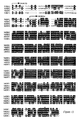
- the first sequence is that of human SGK2 ⁇ , the second that of human SGK2 ⁇ and the second sequence is that of mouse SGK2.
- SGK2 ⁇ has an N-terminal 60 amino acids that are not present in SGK ⁇ .
- SGK serum and glucocorticoid-induced protein kinase
- the said variant, fragment, fusion or derivative thereof is preferably a protein kinase, more preferably a protein kinase that is capable of phosphorylating a polypeptide comprising the consensus Arg-X-(X/Arg)-X-X-( Ser/Thr )-Z, as discussed further below, and is not SGK1, PKB ⁇ , PKC ⁇ , PKC ⁇ , p70 S6 kinase, MSK1 or MSK2 or a fragment or fusion thereof.
- the polypeptides whose amino acid sequences are shown above are considered to be serum and glucocorticoid-induced protein kinases.
- the SGK2 mRNA is present at highest levels in liver, kidney and pancreas and is also present in the brain, whereas levels of mRNA encoding SGK1 and SGK3 are similar in all tissues tested (see Example 5).
- substantially pure we mean that the said polypeptide is substantially free of other proteins.
- any composition that includes at least 30% of the protein content by weight as the said polypeptide, preferably at least 50%, more preferably at least 70%, still more preferably at least 90 % and most preferably at least 95% of the protein content is the said polypeptide.
- compositions comprising, the said polypeptide and a contaminant wherein the contaminant comprises less than 70% of the composition by weight, preferably less than 50% of the composition, more preferably less than 30% of the composition, still more preferably less than 10% of the composition and most preferably less than 5% of the composition by weight.
- SGK for example the activated or deactivated SGK obtained or obtainable by the above methods of the invention, may be useful, for example in screening methods, such as those set out below.
- the term SGK as used herein includes a polypeptide comprising an amino acid sequence given above for SGK2 or SGK3 or the amino acid sequence of the polypeptide, preferably a protein kinase, encoded by I.M.A.G.E Consortium clone ID 42669, or a variant, fragment, fusion or derivative thereof, or a fusion of a said variant or fragment or derivative, wherein the said polypeptide is a protein kinase.
- the said polypeptide is a protein kinase that is capable of phosphorylating a polypeptide comprising the consensus Arg-X-(X/Arg)-X-X- (Ser/Thr) -Z where X indicates any amino acid, X/Arg indicates any amino acid, with a preference for arginine, and Z indicates that the amino acid residue is preferably a hydrophobic residue.
- a polypeptide is Crosstide (GRPRTS S FAEG).
- the underlined residue may be phosphorylated by the said protein kinase.
- Other examples of polypeptides that may be phosphorylated by the said protein kinase are shown in Table 1 and may include GSK3, as discussed in Example 1.
- serine 279 of SGK2 lies in a sequence conforming to the consensus sequence for phosphorylation by SGK and PKB.
- SGK2 may therefore be capable of being phosphorylated by the said protein kinase, for example SGK2.
- the said protein kinase is not PKB (for example, PKB ⁇ ), p70 S6 kinase, protein kinase C (for example, PKC ⁇ or PKC ⁇ ), MAPKAP kinase-1 ( Leighton et al (1995) FEBS Lett 375, 289-293 ) or MSK1 or MSK2 (see, for example, UK patent applications 9817303.2, filed 10 August 1998 and 9813467.9, filed 24 June 1998 ).
- SGK3 may also be capable of phosphorylating a serine residue equivalent to serine 77 or serine 79 of full-length human SGK3.
- a said polypeptide may be useful, for example in some of the screening assays as set out below, even if it does not possess protein kinase activity as defined above. It will further be appreciated that the phosphorylation of such a polypeptide may be detected by means described herein other than by detecting a change in enzymatic, in particular protein kinase, activity of the polypeptide.
- the SGK comprises an amino acid sequence corresponding to the consensus sequence B- T -F-C-G-T-(P/I)-(D/E)-Y-(L/I/M)-A-P-E, where B is a basic residue, the consensus sequence for phosphorylation of a polypeptide by PDK1, and/or the consensus sequence Phe-Xaa-Xaa-(Phe/Tyr)-( Ser/Thr ) -(Phe/Tyr), the consensus sequence proposed for phosphorylation of a polypeptide by PDK2 protein kinase activity.
- both consensus sequences are present.
- the Ser/Thr in either consensus sequence may be replaced by an aspartate or glutamate (ie acidic) residue; it is preferred that only one such consensus sequence in a polypeptide is replaced in this way. It is preferred that the Ser/Thr in the consensus sequence for phosphorylation by PDK1 is not replaced in this way; as described in Example 1, replacement of Thr256 to Asp in human SGK1 may abolish activation of SGK.
- variants of the polypeptide we include insertions, deletions and substitutions, either conservative or non-conservative. In particular we include variants of the polypeptide where such changes do not substantially alter the activity of the said protein kinase.
- substitutions are intended combinations such as Gly, Ala; Val, Ile, Leu; Asp, Glu; Asn, Gln; Ser, Thr; Lys, Arg; and Phe, Tyr.
- the SGK variant has an amino acid sequence which has at least 65% identity with the amino acid sequence of the polypeptide, preferably a protein kinase, encoded by I.M.A.G.E Consortium clone ID 42669, more preferably at least 70%, 71%, 72 % , 73% or 74%, still more preferably at least 75%, yet still more preferably at least 80 %, in further preference at least 85%, in still further preference at least 90 % and most preferably at least 95 % or 97 % identity with the amino acid sequence encoded by I.M.A.G.E Consortium clone ID 42669.
- a SGK2 or SGK3 variant has an amino acid sequence which has at least 65 % identity with the amino acid sequence of SGK2 or SGK3, as appropriate, indicated above, more preferably at least 70%, 71%, 72%, 73% or 74%, still more preferably at least 75%, yet still more preferably at least 80 %, in further preference at least 85%, in still further preference at least 90 % and most preferably at least 95 % or 97 % identity with the amino acid sequence of SGK2 or SGK3 indicated above.
- the SGK variant has an amino acid sequence which has at least 65% identity with the amino acid sequence of the catalytic domain of the polypeptide, preferably a protein kinase, encoded by I.M.A.G.E Consortium clone ID 42669, more preferably at least 70%, 71%, 72 %, 73 % or 74%, still more preferably at least 75%, yet still more preferably at least 80%, in further preference at least 83 or 85%, in still further preference at least 90 % and most preferably at least 95 % or 97 % identity with the amino acid sequence defined above.
- a protein kinase encoded by I.M.A.G.E Consortium clone ID 42669, more preferably at least 70%, 71%, 72 %, 73 % or 74%, still more preferably at least 75%, yet still more preferably at least 80%, in further preference at least 83 or 85%, in still further preference at least 90 % and most preferably at least 95 % or
- a SGK2 or SGK3 variant has an amino acid sequence which has at least 65 % identity with the amino acid sequence of the catalytic domain of an SGK2 or SGK3 polypeptide whose sequence is indicated above, as appropriate, more preferably at least 70 %, 71%, 72 % , 73% or 74%, still more preferably at least 75%, yet still more preferably at least 80%, in further preference at least 83 or 85%, in still further preference at least 90 % and most preferably at least 95 % or 97 % identity with the SGK2 or SGK3 amino acid sequence defined above.
- the catalytic domain of a protein kinase-related polypeptide may be readily identified by a person skilled in the art, for example using sequence comparisons as described below, and as described in relation to SGK in, for example, Webster et al (1993) Mol Cell Biol 13(4), 2031-2040 .
- the amino acid sequences of the three SGK isoforms are about 80 % identical to one another in the catalytic domain, while the short C-terminal non-catalytic domains are less similar (44-68 % identity).
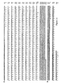
- the N-terminal 85 residues that precede the catalytic domain are much less similar. In this region there is only about 25 % identity between SGK1 and SGK3 and almost no identity between SGK2 and the other isoforms ( Fig 13 ).
- the percent sequence identity between two polypeptides may be determined using suitable computer programs, for example the GAP program of the University of Wisconsin Genetic Computing Group and it will be appreciated that percent identity is calculated in relation to polypeptides whose sequence has been aligned optimally.
- the alignment may alternatively be carried out using the Clustal W program (Thompson et al., 1994).
- the parameters used may be as follows:
- SGK2 and SGK3 are examples of SGK variants as defined above.
- the SGK is a polypeptide which consists of the amino acid sequence of the protein kinase encoded by I.M.A.G.E Consortium clone ID 42669, or SGK2 or SGK3, the amino acid sequences of which are given above, or naturally occurring allelic variants (including splice variants) thereof.
- the SGK is one of the following:
- SGK in which the residue equivalent to threonine 256 of full-length SGK1 is replaced may not be capable of being significantly activated by phosphorylation and may therefore be less preferred than some other SGKs for use in the screening methods of the invention.
- GST-SGK[T256D], GST-SGK[T256E], GST-SGK[T256A], as described in Example 1 may not be capable of being significantly activated by phosphorylation and may therefore be less preferred than some other SGKs for use in the screening methods of the invention.
- the residue equivalent to serine 422 of SGK1 in SGK2 ⁇ is serine 356 and in SGK3 is serine 419.
- the residue equivalent to threonine 256 of SGK1 in SGK2 ⁇ is threonine 193 and in SGK3 is threonine 253.
- SGK in which the residue equivalent to serine 422 of full-length SGK1 is replaced by an amino acid residue other than glutamate or aspartate (ie an acidic residue) or a residue that may be phosphorylated, for example by an alanine residue may not be capable of being activated by phosphorylation or may be less capable of being activated by phosphorylation than SGK in which the said residue equivalent to serine 422 is not so replaced.
- a mutated SGK which does not have any protein kinase activity may be phosphorylated in a similar manner to SGK that does have protein kinase activity, but that no change in protein kinase activity may be discernible.
- Such a mutated SGK may be SGK in which the residue equivalent to lysine 127 of full-length SGK1 (which may lie in the ATP binding site) is replaced by alanine, as described in Example 1.
- SGK may be phosphorylated by phosphatidylinositol dependent kinase 1 (PDK1), or a variant, fragment, fusion or derivative thereof, or a fusion of a said variant, fragment or derivative.
- PDK1 is well known to those skilled in the art (see, for example, references 7 to 9) and includes, in particular, PDK1 as described and expressed in reference 9 and in co-pending application PCT/GB98/00777 .
- the said variant, fragment, fusion or derivative of PDK1, or a fusion of a said variant, fragment or derivative is a protein kinase, preferably a protein kinase capable of phosphorylating a polypeptide comprising an amino acid sequence corresponding to the consensus sequence B- T -F-C-G-T-(P/I)-(D/E)-Y-(L/I/M)-A-P-E.
- Such variants of PDK1 may be functional equivalents of PDK1 and include Pkh1 and Pkh2 from Saccharomyces cerevisiae as described in the US patent application entitled "Screening Methods" filed on the same day as this application.
- SGK may be dephosphorylated by a serine/threonine protein phosphatase, for example PP1 ( Berndt et al (1987) FEBS Lett 223, 340-346 ), PP2C ( Mann et al (1992) Biochem et Biophys Acta 1130; 100-104 or protein phosphorylase 2A (PP2A; da Cruz E Silva (1987) FEBS Lett 221, 415-422 ) or a variant, fragment, fusion or derivative any thereof, or a fusion of a said variant, fragment or derivative.
- PP1 Berndt et al (1987) FEBS Lett 223, 340-346
- PP2C Mann et al (1992) Biochem et Biophys Acta 1130; 100-104 or protein phosphorylase 2A (PP2A; da Cruz E Silva (1987) FEBS Lett 221, 415-422 ) or a variant, fragment, fusion or derivative any thereof, or a fusion
- PP1, PP2C or PP2A or a variant, fragment, fusion or derivative any thereof, or a fusion of a said variant, fragment or derivative is capable of dephosphorylating phosphorylase a.
- the terms PP1, PP2C, PP2A and phosphorylase a are well known to those skilled in the art.
- PP2A may be inactivated by microcystin-LR at 1 ⁇ M.
- a further aspect of the invention is the use of a serine/threonine protein phosphatase, for example PP1, PP2C, PP2A or a variant, fragment, fusion or derivative any thereof, or a fusion of a said variant, fragment or derivative (as defined above) in an in vitro method of deactivating and/or dephosphorylating SGK.
- the SGK may be phosphorylated SGK, as discussed above.
- a still further aspect of the invention is the use of a protein kinase, preferably PDK1 or a variant, fragment, fusion or derivative thereof, or a fusion of a said variant, fragment or derivative (as defined above) in an in vitro method of activating and/or phosphorylating SGK.
- the SGK may be dephosphorylated SGK, as discussed above .
- a further aspect of the invention is an in vitro method of activating SGK (as defined above) wherein SGK is phosphorylated on the residue equivalent to Thr256 of full-length human SGK1.
- SGK for example full-length human SGK and ⁇ N-SGK(61-431), may be phosphorylated on the residue equivalent to Thr 256 of full-length human SGK1 by PDK1.
- SGK may be activated by phosphorylation on this residue, as described in Example 1 and Example 5.
- SGK in which the residue equivalent to serine 422 of full-length SGK1 is replaced by an acidic residue, for example glutamate or aspartate may be more readily phosphorylated and/or activated than SGK in which the said residue equivalent to serine 422 is not so replaced.
- recombinant SGK in which the residue equivalent to serine 422 of full-length SGK1 is replaced by an acidic residue, for example glutamate or aspartate may be substantially phosphorylated on the residue equivalent to Thr256 of full-length human SGK1 when expressed in a eukaryotic, preferably mammalian, cell.
- Such recombinant SGK may be useful in screening assays in which activated SGK is required, as discussed further below.
- a protein kinase capable of phosphorylating SGK on the residue equivalent to Thr256 of full-length human SGK1 may be identified by determining whether any particular protein kinase is capable of activating SGK and/or determining what, if any residue of SGK may be phosphorylated by the said particular protein kinase, for example as described in Example 1.
- Thr256 of full-length human SGK1 is included the meaning that the amino acid residue that occupies a position in the native three dimensional structure of a protein kinase corresponding to the position occupied by Thr 256 in the native three dimensional structure of human full-length SGK1. It will be appreciated that Thr256 of human full-length SGK1 is located in the "activation loop" between subdomains VII and VIII of the catalytic domain.
- Protein kinases show a conserved catalytic core, as reviewed in Johnson et al (1996) Cell, 85, 149-158 and Taylor & Radzio-Andzelm (1994) Structure 2, 345-355 . This core folds into a small N-terminal lobe largely comprising anti-parallel ⁇ -sheet, and a large C-terminal lobe which is mostly ⁇ -helical.
- the residue equivalent to, for example, Thr 256 of full-length human SGK1 may be identified by alignment of the sequence of the polypeptide with that of full-length human SGK1 in such a way as to maximise the match between the sequences.
- the alignment may be carried out by visual inspection and/or by the use of suitable computer programs, for example the GAP program of the University of Wisconsin Genetic Computing Group, which will also allow the percent identity of the polypeptides to be calculated.
- the Align program Pearson (1994) in: Methods in Molecular Biology, Computer Analysis of Sequence Data, Part II (Griffin, AM and Griffin, HG eds) pp 365-389, Humana Press, Clift on).
- residues identified in this manner are also "equivalent residues”.
- a further aspect of the invention is an in vitro method of activating SGK (as defined above) wherein SGK is phosphorylated on the residue equivalent to Ser422 of full-length human SGK.
- SGK for example full-length human SGK and ⁇ N-SGK(61-431), may be phosphorylated on the residue equivalent to Ser 422 of full-length human SGK1 by a preparation containing PDK2 activity.
- SGK may be activated by phosphorylation on this residue, as described in Example 1.
- the method may be performed in a cell, for example a mammalian cell that is stimulated by, for example, IGF-1 and/or hydrogen peroxide.
- the method may comprise culturing a host cell comprising a recombinant polynucleotide or a replicable vector which encodes SGK, stimulating the cell, for example with IGF-1 and/or hydrogen peroxide and isolating said polypeptide from said host cell.
- Methods of cultivating host cells and isolating recombinant proteins are well known in the art.
- residue equivalent to Ser422 or other residue of full-length human SGK1 may be identified by methods analagous to those described above for the residue equivalent to Thr256 of full-length human SGK1.
- Ser422 of human full-length SGK1 lies towards the C-terminus of the polypeptide.
- a preparation containing PDK2 activity may phosphorylate polypeptides comprising an amino acid sequence corresponding to the consensus sequence Phe-Xaa-Xaa-Phe/Tyr-Ser/Thr-Phe/Tyr.
- SGK may be phosphorylated on the residue equivalent to Ser422 of full-length human SGK1, for example in vitro, by a protein kinase that may not have the physiological characteristics of PDK2 or may not require the consensus sequence thought to be required by PDK2.
- SGK may be phosphorylated by a protein kinase that may not phosphorylate SGK in a physiologically relevant manner in vivo and may not correspond to an activity identified as PDK2.
- MAPKAP Kinase-2 does not require the consensus sequence identified as required by PDK2.
- a protein kinase that is capable of phosphorylating the residue equivalent to Ser422 of full-length human SGK1 may be identified by methods similar to those described above and in Example 1 for identifying a protein kinase that may phosphorylate the residue equivalent to Thr256 of full-length human SGK1.
- the phosphorylation of SGK may be PtdIns(3,4,5)P 3 dependent.
- the phosphorylation of the residue equivalent to Ser422 for example by a preparation containing PDK2 activity, may be PtdIns(3,4,5)P 3 dependent.
- the phosphorylation of the residue equivalent to Ser422 may be dependent upon the activity of PtdIns 3-kinase.
- the phosphorylation of SGK on the residue equivalent to Thr256 of full-length human SGK1 may be dependent upon the phosphorylation of the residue equivalent to Ser 422 of full-length human SGK1.
- serine 422 is replaced by an aspartate residue, as described in Examples 1 and 5, then the phosphorylation of the residue equivalent to Thr256, for example by PDK1, may be potentiated.
- the phosphorylation of Thr256 may require that (or be enhanced if) the residue equivalent to Ser422 is phosphorylated or is replaced by an aspartate residue.
- SGK2 may be phosphorylated at residues Ser279 and/or Ser334.
- Ser279 may be phosphorylated by SGK, for example SGK2, or PKB. It may be necessary for the SGK, for example SGK2, or PKB to be activated, for example by phosphorylation by PDK1.
- Ser334 may be phosphorylated by PDK1.
- SGK3 may be phosphorylated at residues Ser77 and/or Ser79. These residues may be phosphorylated by SGK, for example SGK3, or PKB.
- SGK for example SGK3, or PKB
- phosphorylation of Ser77 and Ser79 of SGK3 may take place in the absence of phosphorylation by PDK1.
- the fusion polypeptide may comprise, for example, a fragment of SGK or variant or derivative thereof wherein the residues equivalent to the N-terminal about 20, 30, 40, 50 or 60 amino acids of human full-length SGK1 are deleted.
- the fusion polypeptide may further comprise glutathione-S-transferase.
- the glutathione-S-transferase may be fused to the N-terminus or C-terminus of SGK or the said fragment variant or derivative thereof; preferably it is fused to the N-terminus, for example as shown in Examples 1 and 5.
- the fusion polypeptide has at least 5%, 10%, 15%, 20%, 25% or 30% of the enzyme activity of SGK, for example SGK1, with respect to the phosphorylation of Crosstide (GRPRTSSFAEG; see Example 1). It is more preferred if the fusion polypeptide has at least 50 %, preferably at least 70 % and more preferably at least 90% of the enzyme activity of SGK with respect to the phosphorylation of Crosstide. It is preferred that the fusion polypeptide comprises the residue equivalent to Thr256 and/or the residue equivalent to Ser 422 of full-length human SGK1.
- a fusion polypeptide with low enzymic activity may be useful in applications including high throughput screening, for example if it is expressed in high amounts or may be easily purified or stored.
- fusion polypeptides which are devoid of enzymatic activity may nevertheless also be useful, for example by interacting with another polypeptide, or as antigens in raising antibodies, or the phosphorylation of such variants or fusions or derivatives or fragments may be measured.
- fusions of PKB ⁇ , PKC ⁇ , PKC ⁇ , p70 S6 kinase, MSK1 and MSK2 are not polypeptides of the invention, unless they include all or part of SGK.
- a further aspect of the invention is a polypeptide comprising the amino acid sequence of human SGK or a fragment, variant, derivative or fusion thereof wherein the residue equivalent to serine 422 of full-length human SGK1 is replaced (for example by an aspartate, glutamate or alanine residue) and/or the residue equivalent to threonine 256 of full-length human SGK1 is replaced (for example by an alanine residue), and/or the residue equivalent to lysine 127 of full-length human SGK1 is replaced (for example by an alanine residue).
- the residue equivalent to lysine 127 of full-length human SGK1 may be identified in a manner analogous to that described above for the residue equivalent to serine 422, and may lie in the ATP binding site.
- the residue equivalent to serine 422 of full-length human SGK1 may be replaced by a residue that is capable of carrying a negative charge (for example an aspartate or glutamate residue), which may mimic the effect of phosphorylation of serine 422.
- the residue equivalent to serine 422 of full-length human SGK1 may be replaced by a residue, for example alanine, that cannot be phosphorylated and is not capable of carrying a negative charge, and preferably is of a similar bulk to serine or threonine.
- the residue equivalent to threonine 256 of full-length human SGK1 may similarly be replaced by a residue that is capable of carrying a negative charge (for example an aspartate or glutamate residue), or by a residue, for example alanine, that cannot be phosphorylated and is not capable of carrying a negative charge and preferably is of a similar bulk to serine or threonine.
- the residue equivalent to lysine 127 of full-length human SGK1 may be replaced by a residue that is not capable of carrying a positive charge, for example an alanine residue.
- residue equivalent to serine 422 of full-length human SGK1 is replaced by an aspartate residue and the residues equivalent to threonine 256 and lysine 127 of the full-length human SGK1 are unmutated, ie are threonine and lysine respectively.
- amino acids equivalent to the N-terminal 20, 30, 40, 50 or 60 amino acids of full-length human SGK1 are not present, ie are deleted in relation to SGK1, SGK2 ⁇ or SGK3.
- human SGK2 ⁇ does not have an amino acid sequence equivalent to the N-terminal 60 amino acids of full-length human SGK1. It will further be appreciated that this may have the consequence that the interaction with a cellular component may not be the same for human SGK2 ⁇ as for other SGKs, for example SGK1, SGK2 ⁇ or SGK3.
- SGK2 is capable of being phosphorylated at Ser279 and Ser334, as well as Thr193.
- residue equivalent to Ser279 and/or Ser334 of full length SGK2 is replaced by a residue that is capable of carrying a negative charge (for example an aspartate or glutamate residue), or by a residue, for example alanine, that cannot be phosphorylated and is not capable of carrying a negative charge and preferably is of a similar bulk to serine or threonine.
- SGK3 is capable of being phosphorylated at Ser77 and Ser79.
- the residue equivalent to Ser77 and/or Ser79 of full length SGK3 is replaced by a residue that is capable of carrying a negative charge (for example an aspartate or glutamate residue), or by a residue, for example alanine, that cannot be phosphorylated and is not capable of carrying a negative charge and preferably is of a similar bulk to serine or threonine.
- phosphorylation of SGK2 at Ser279 and/or Ser334 or phosphorylation of SGK3 at Ser77 and/or Ser79 may modulate the activity or, for example, the cellular location of SGK2 or SKG3.
- a polypeptide comprising the amino acid sequence of human SGK or a fragment, variant, derivative or fusion thereof wherein the residue equivalent to threonine 256 of full-length human SGK1 is replaced by an alanine residue, and/or the residue equivalent to lysine 127 of full-length human SGK1 is replaced by an alanine residue may not be capable of being activated by phosphorylation and/or may be devoid of protein kinase activity, respectively, and therefore may not be useful in screening methods of the invention.
- polynucleotide encoding a polypeptide of the invention, or encoding SGK2 or SGK3 or encoding a variant or fragment or derivative of fusion of said SGK2 or SGK3 or a fusion of a said variant or fragment or derivative.
- Preferences and exclusions for the said polynucleotide variant are the same as in the first aspect of the invention, except that the following Expressed Sequence Tags (ESTs) are also excluded.
- the polynucleotide may be a recombinant polynucleotide. The following Expressed Sequence Tags (ESTs) are excluded:
- the polynucleotide may be a vector suitable for replication and/or expression of the polypeptide in a mammalian/eukaryotic cell.
- the polynucleotide or recombinant polynucleotide may be DNA or RNA, preferably DNA.
- the polynucleotide may or may not contain introns in the coding sequence; preferably the polynucleotide is or comprises a cDNA.
- the polynucleotide comprises the nucleotide sequence or or a variant, fragment, fusion or derivative thereof.
- the first nucleotide sequence encodes SGK2 ⁇ and the second encodes SGK2 ⁇ .
- the nucleotide sequences are shown in Figure 1 together with the translations of the relevant open reading frames.
- polynucleotide comprises the nucleotide sequence or a variant, fragment, fusion or derivative thereof.
- the nucleotide sequence encodes SGK3 and is shown in Figure 2 together with the translation of the relevant open reading frame.
- Site-directed mutagenesis or other techniques can be employed to create single or multiple mutations, such as replacements, insertions, deletions, and transpositions, as described in Botstein and Shortle, "Strategies and Applications of In Vitro Mutagenesis," Science, 229: 193-210 (1985) , which is incorporated herein by reference. Polymerase chain reaction based methods of site-directed mutagenesis may be used, as well known to those skilled in the art, for example as described in Example 1.
- the polynucleotide is a polynucleotide that may be translated to form the polypeptide, for example RNA, or that the polynucleotide (which is preferably DNA) encoding the polypeptide of the invention is inserted into an expression vector, such as a plasmid, in proper orientation and correct reading frame for expression.
- the polynucleotide may be linked to the appropriate transcriptional and translational regulatory control nucleotide sequences recognised by any desired host; such controls may be incorporated in the expression vector.
- vectors suitable for replication in mammalian/eukaryotic cells are well known to those skilled in the art, and examples are given below. It will be appreciated that a vector may be suitable for replication in both prokaryotic and eukaryotic cells.
- a desirable way to modify the DNA encoding a polypeptide of the invention is to use the polymerase chain reaction as disclosed by Saiki et al (1988) Science 239, 487-491 .
- This method may be used for introducing the DNA into a suitable vector, for example by engineering in suitable restriction sites, or it may be used to modify the DNA in other useful ways as is known in the art.
- the DNA to be enzymatically amplified is flanked by two specific primers which themselves become incorporated into the amplified DNA.
- the said specific primers may contain restriction endonuclease recognition sites which can be used for cloning into expression vectors using methods known in the art.
- the DNA (or in the case of retroviral vectors, RNA) is then expressed in a suitable host to produce a polypeptide comprising the compound of the invention.
- the DNA encoding the polypeptide constituting the compound of the invention may be used in accordance with known techniques, appropriately modified in view of the teachings contained herein, to construct an expression vector, which is then used to transform an appropriate host cell for the expression and production of the polypeptide of the invention.
- Such techniques include those disclosed in US Patent Nos.
- DNA (or in the case of retroviral vectors, RNA) encoding the polypeptide constituting the compound of the invention may be joined to a wide variety of other DNA sequences for introduction into an appropriate host.
- the companion DNA will depend upon the nature of the host, the manner of the introduction of the DNA into the host, and whether episomal maintenance or integration is desired.
- the DNA is inserted into an expression vector, such as a plasmid, in proper orientation and correct reading frame for expression.
- an expression vector such as a plasmid
- the DNA may be linked to the appropriate transcriptional and translational regulatory control nucleotide sequences recognised by the desired host, although such controls are generally available in the expression vector.
- the vector is then introduced into the host through standard techniques. Generally, not all of the hosts will be transformed by the vector. Therefore, it will be necessary to select for transformed host cells.
- One selection technique involves incorporating into the expression vector a DNA sequence, with any necessary control elements, that codes for a selectable trait in the transformed cell, such as antibiotic resistance.
- the gene for such selectable trait can be on another vector, which is used to co-transform the desired host cell.
- Host cells that have been transformed by the recombinant DNA of the invention are then cultured for a sufficient time and under appropriate conditions known to those skilled in the art in view of the teachings disclosed herein to permit the expression of the polypeptide, which can then be recovered.
- the host cell for example a mammalian cell such as 293 cells as described in Example 1 or Example 5, may be stimulated, for example using IGF-1 of hydrogen peroxide, such that the SGK polypeptide may be phosphorylated and/or activated in the host cell.
- the activated SGK polypeptide may then be recovered, if necessary in the presence of phosphatase inhibitors, for example microcystin, for example as described in Example 1. Recovery may entail purification on glutathione-Sepharose, as described in Example 1.
- 293 cells are human transformed primary embryonal kidney cells that may be obtained from the American Type Culture Collection (ATCC), 12301 Parklawn Drive, Rockville, Maryland 20852-1776; catalogue reference ATCC CRL 1573.
- ATCC American Type Culture Collection
- bacteria for example E . coli and Bacillus subtilis
- yeasts for example Saccharomyces cerevisiae
- filamentous fungi for example Aspergillus
- plant cells animal cells and insect cells.
- the vectors include a prokaryotic replicon, such as the ColE1 ori, for propagation in a prokaryote, even if the vector is to be used for expression in other, non-prokaryotic, cell types.
- the vectors can also include an appropriate promoter such as a prokaryotic promoter capable of directing the expression (transcription and translation) of the genes in a bacterial host cell, such as E. coli, transformed therewith.
- a promoter is an expression control element formed by a DNA sequence that permits binding of RNA polymerase and transcription to occur.
- Promoter sequences compatible with exemplary bacterial hosts are typically provided in plasmid vectors containing convenient restriction sites for insertion of a DNA segment of the present invention.
- Typical prokaryotic vector plasmids are pUC18, pUC19, pBR322 and pBR329 available from Biorad Laboratories, (Richmond, CA, USA) and pTrc99A and pKK223-3 available from Pharmacia, Piscataway, NJ, USA.
- a typical mammalian cell vector plasmid is pSVL available from Pharmacia, Piscataway, NJ, USA. This vector uses the SV40 late promoter to drive expression of cloned genes, the highest level of expression being found in T antigen-producing cells, such as COS-1 cells.
- an inducible mammalian expression vector is pMSG, also available from Pharmacia. This vector uses the glucocorticoid-inducible promoter of the mouse mammary tumour virus long terminal repeat to drive expression of the cloned gene.
- Useful yeast plasmid vectors are pRS403-406 and pRS413-416 and are generally available from Stratagene Cloning Systems, La Jolla, CA 92037 , USA.
- Plasmids pRS403, pRS404, pRS405 and pRS406 are Yeast Integrating plasmids (YIps) and incorporate the yeast selectable markers HIS3, TRP1, LEU2 and URA3.
- Plasmids pRS413-416 are Yeast Centromere plasmids (YCps).
- the present invention also relates to a host cell transformed with a polynucleotide vector construct of the present invention.
- the host cell can be either prokaryotic or eukaryotic.
- Bacterial cells are preferred prokaryotic host cells and typically are a strain of E . coli such as, for example, the E . coli strains DH5 available from Bethesda Research Laboratories Inc., Bethesda, MD, USA, and RR1 available from the American Type Culture Collection (ATCC) of Rockville, MD, USA (No ATCC 31343).
- Preferred eukaryotic host cells include yeast, insect and mammalian cells, preferably vertebrate cells such as those from a mouse, rat, monkey or human fibroblastic cell line.
- Yeast host cells include YPH499, YPH500 and YPH501 which are generally available from Stratagene Cloning Systems, La Jolla, CA 92037 , USA.
- Preferred mammalian host cells include human embryonic kidney 293 cells (see Example 1), Chinese hamster ovary (CHO) cells available from the ATCC as CCL61, NIH Swiss mouse embryo cells NIH/3T3 available from the ATCC as CRL 1658, and monkey kidney-derived COS-1 cells available from the ATCC as CRL 1650.
- Preferred insect cells are Sf9 cells which can be transfected with baculovirus expression vectors.
- Transformation of appropriate cell hosts with a DNA construct of the present invention is accomplished by well known methods that typically depend on the type of vector used. With regard to transformation of prokaryotic host cells, see, for example, Cohen et al (1972) Proc. Natl. Acad. Sci. USA 69, 2110 and Sambrook et al (1989) Molecular Cloning, A Laboratory Manual, Cold Spring Harbor Laboratory, Cold Spring Harbor, NY . Transformation of yeast cells is described in Sherman et al (1986) Methods In Yeast Genetics, A Laboratory Manual, Cold Spring Harbor, NY . The method of Beggs (1978) Nature 275, 104-109 is also useful.
- reagents useful in transfecting such cells for example calcium phosphate and DEAE-dextran or liposome formulations, are available from Stratagene Cloning Systems, or Life Technologies Inc., Gaithersburg, MD 20877, USA.
- Electroporation is also useful for transforming and/or transfecting cells and is well known in the art for transforming yeast cell, bacterial cells, insect cells and vertebrate cells.
- Successfully transformed cells ie cells that contain a DNA construct of the present invention
- cells resulting from the introduction of an expression construct of the present invention can be grown to produce the polypeptide of the invention.
- Cells can be harvested and lysed and their DNA content examined for the presence of the DNA using a method such as that described by Southern (1975) J. Mol. Biol. 98, 503 or Berent et al (1985) Biotech. 3, 208 .
- the presence of the protein in the supernatant can be detected using antibodies as described below.
- successful transformation can be confirmed by well known immunological methods when the recombinant DNA is capable of directing the expression of the protein.
- cells successfully transformed with an expression vector produce proteins displaying appropriate antigenicity. Samples of cells suspected of being transformed are harvested and assayed for the protein using suitable antibodies.
- the present invention also contemplates a culture of those cells, preferably a monoclonal (clonally homogeneous) culture, or a culture derived from a monoclonal culture, in a nutrient medium.
- a further aspect of the invention provides a method of making the polypeptide of the invention the method comprising culturing a host cell comprising a recombinant polynucleotide or a replicable vector which encodes said polypeptide, and isolating said polypeptide from said host cell.
- Methods of cultivating host cells and isolating recombinant proteins are well known in the art.
- a still further aspect of the invention provides a method of making and activating the polypeptide of the invention the method comprising culturing a host cell, preferably a eukaryotic cell, comprising a recombinant polynucleotide or a replicable vector which encodes said polypeptide, stimulating the cell, for example with IGF-1 and/or hydrogen peroxide and isolating said polypeptide from said host cell.
- a host cell preferably a eukaryotic cell, comprising a recombinant polynucleotide or a replicable vector which encodes said polypeptide, stimulating the cell, for example with IGF-1 and/or hydrogen peroxide and isolating said polypeptide from said host cell.
- Methods of cultivating host cells and isolating recombinant proteins are well known in the art.
- the host cell may be a mammalian cell that is stimulated by, for example, IGF-1 and/or hydrogen peroxide, for example a 293 cell
- a further aspect of the invention is a polypeptide obtainable by the above methods of the invention.
- a still further aspect of the invention is a method of identifying a compound that modulates the activity of SGK wherein activated SGK is used.
- Activated SGK is SGK activated by phosphorylation
- a further aspect of the invention is the use of activated SGK in a screening assay for a drug-like compound or lead compound for the development of a drug-like compound.
- a drug-like compound or lead compound for the development of a drug-like compound are included compounds that may be useful in medicine and compounds that may be useful in the development of a compound that may be useful in medicine. Examples of such screening assays may include the methods of the invention described below.
- a further aspect of the invention is a method of identifying a compound that modulates the activity of SGK (as defined above), the method comprising contacting a compound with SGK and determining whether the activity of the said polypeptide is changed compared to the activity of the said SGK in the absence of said compound wherein the activity of SGK is measured by measuring the phosphorylation by SGK, in the presence of a suitable phosphate donor, of a polypeptide comprising an amino acid sequence corresponding to the consensus sequence (Arg/Lys; preferably Arg)-X-(X/Arg)-X-X-(Ser/Thr)-Z wherein X indicates any amino acid, X/Arg indicates any amino acid, with a preference for arginine, and Z indicates that the amino acid residue is preferably a hydrophobic residue.
- the polypeptide may be Crosstide or other suitable polypeptide described in Table 1 or Table 2 or preferably a physiological substrate of SGK, which may be GSK3. It will be appreciated that SGK2 and SGK3 may be capable of autophosphorylation.
- SGK2 comprises a sequence corresponding to the above consensus sequence, surrounding Ser279; thus the polypeptide may be SGK2 or SGK3 or a fragment of either that comprises a potential autophosphorylation site, as discussed in Example 1, ie Ser279 of SGK2 or Ser77 or Ser 79 of SGK3.
- Suitable phosphate donors will be known to those skilled in the art and include ATP, for example as the magnesium salt (MgATP), as described in Example 1.
- the SGK is full-length human SGK or N-terminally truncated SGK (for example, 20, 30, 40, 50 or 60 amino acids of full-length human SGK, for example full-length human SGK1, may be deleted, such as in ⁇ N-SGK(61-431)) or a fusion (preferably a GST fusion) of full-length human SGK or N-terminally truncated SGK wherein the residue equivalent to serine 422 of full-length human SGK1 is replaced by an aspartate residue and the residues equivalent to threonine 256 and lysine 127 of the full-length human SGK1 are unmutated, ie remain as threonine and lysine respectively.
- the SGK is activated by phosphorylation according to a method of the invention. It will be appreciated that if the SGK is an SGK in which the residue equivalent to serine 422 of full-length human SGK1 is replaced by an aspartate residue, for example the preferred SGK described above, that this residue will not be phosphoryated, but that the SGK may be activated by phosphorylation of the residue (preferably threonine) equivalent to threonine 256 of full-length human SGK1.
- the SGK may be phosphorylated and/or activated in a cell by stimulating the said cell with, for example, IGF-1 or hydrogen peroxide.
- the cell may be an eukaryotic, preferably mammalian, for example, 293 cell in which a recombinant SGK is expressed.
- the SGK may be recovered and purified as described in Example 1, for example using glutathione sepharose (for a GST-SGK fusion polypeptide), as described above and in Examples 1 and 5.
- the compound may act by interacting with the polypeptide of the invention and modulating, ie inhibiting or enhancing, its activation by, for example, PDK1 and/or a preparation containing PDK2 activity or other activating protein kinase or protein kinases.
- reagents and conditions used in the method may be chosen such that the interactions between the said SGK and its activating protein kinase or protein kinases are substantially the same as between human SGK and its activating protein kinase or protein kinases in vivo, which may be PDK1 and a polypeptide with PDK2 activity.
- the activating protein kinase used in the assay is a physiological activating protein kinase of SGK, for example PDK1 or functional equivalent thereof and/or a preparation containing PDK2 activity for which interactions between the said SGK and the functional equivalent are substantially the same as between human SGK and the said physiological activating protein kinase.
- the compound may bind to the SGK or may bind to an activating protein kinase.
- a functional equivalent of a physiological activating protein kinase of SGK for example PDK1
- a protein kinase with substantially the same substrate specificity as the physiological activating protein kinase for example PDK1 (ie capable of phosphorylating the underlined residue in the consensus sequence B- T -F-C-G-T-(P/I)-(D/E)-Y-(L/I/M)-A-P-E, where B is a basic residue), and/or capable of phosphorylating SGK on the same residue as the said physiological activating protein kinase.
- the substrate used in the assay is a physiological substrate of SGK or a fragment, variant, derivative or fusion thereof or fusion of a said fragment, variant or derivative (defined in an analagous manner to the definitions given above in relation to SGK) for which interactions between the said SGK and the said fragment, variant, derivative or fusion are substantially the same as between human SGK and the said physiological substrate.
- a substrate of SGK for example SGK1
- GSK3 GSK3.
- Physiological substrates of SGK may include substrates identified, for example in overexpression studies referred to in Example 1, as physiological substrates for PKB.
- the finding presented here concerning the activation and substrate specificity of SGK and its similarity to the activation and substrate specificity of PKB may indicate that substrates previously identified as physiological substrates for PKB may additionally or alternatively be physiological substrates for SGK, for example SGK1, SGK2 and/or SGK3.
- PKB has been thought to mediate a number of the actions of insulin, including stimulation of glucose and amino acid uptake, glycogen and protein synthesis and cardiac muscle glycolysis (reviewed in [7, 8]) as well as regulation of the transcription of specific genes [9, 10].
- the PKB ⁇ isoform is overexpressed in a significant percentage of ovarian and pancreatic cancers [11, 12] and the PKB ⁇ isoform in some breast cancers [2].
- PKB or SGK may be capable of providing a survival signal that protects cells from apoptosis induced in a variety of ways (reviewed in [8, 13]).
- the activation of PKB or SGK by gene amplification and other mechanisms may therefore contribute to the generation of malignancies that are able to flourish in the absence of extracellular survival signals.
- PKB phosphorylates proteins and peptides at serine and threonine residues that lie in Arg-Xaa-Arg,-Xaa-Xaa-Ser/Thr- sequences [14].
- two physiological substrates of PKB appear to be the protein kinase -glycogen synthase kinase-3 (GSK3) [15, 16] and the cardiac isoform of phosphofructokinase-2 (PFK2) [8, 17].
- GSK3 protein kinase -glycogen synthase kinase-3
- PFK2 phosphofructokinase-2
- Dephosphorylation and activation of glycogen synthase and protein synthesis initiation factor eIF2B [18] may contribute to the insulin-induced stimulation of glycogen synthesis and protein synthesis, respectively.
- PKB activates cardiac PFK2, which seems to underlie the insulin-induced stimulation of glycolysis in the heart.
- BAD appears to be one of the physiological substrates of PKB (and may therefore also be a physiological substrate of SGK).
- This protein in its dephosphorylated form, interacts with the Bcl family member Bcl XL , thereby inducing apoptosis in some cells.
- the polypeptide BAD which may be involved in control of apoptosis, may be a physiological substrate of SGK, for example SGK1, 2 and/or 3.
- the compound decreases the activity of said SGK polypeptide.
- the compound may bind substantially reversibly or substantially irreversibly to the active site of said polypeptide.
- the compound may bind to a portion of said polypeptide that is not the active site so as to interfere with the binding of the said polypeptide to its substrate.
- the compound may bind to a portion of said polypeptide so as to decrease said polypeptide's activity by an allosteric effect.
- This allosteric effect may be an allosteric effect that is involved in the natural regulation of the said polypeptide's activity, for example in the activation of the said polypeptide by an "upstream activator" such as PDK1 and/or PDK2 (or a preparation containing PKD2 activity).
- an upstream activator such as PDK1 and/or PDK2 (or a preparation containing PKD2 activity).
- the compound increases the activity of said polypeptide.
- the compound may bind to a portion of said polypeptide that is not the active site so as to aid the binding of the said polypeptide to its substrate.
- the compound may bind to a portion of said polypeptide so as to increase said polypeptide's activity by an allosteric effect.
- This allosteric effect may be an allosteric effect that is involved in the natural regulation of the said polypeptide's activity for example in the activation of the said polypeptide by an "upstream activator" such as PDK1 and/or PDK2 (or a preparation containing PDK2 activity).
- the method makes use of the fact that SGK phosphorylates Crosstide as described in Example 1, but any suitable substrate, for example a physiological substrate of SGK, may be used.
- any suitable substrate for example a physiological substrate of SGK
- the phosphorylation of Crosstide may be measured using techniques well known to those skilled in the art.
- the method makes use of an assay which may be substantially the same as that described in Example 1.
- an assay which may be substantially the same as that described in Example 1.
- phosphorylation of Crosstide by SGK is measured. It is preferred that the SGK is recombinant SGK.
- a change in the activity of the substrate may be measured. This may be done in a whole cell system or using purified or partially purified components.
- the phosphorylation of the substrate may be measured by measuring a change in the activity of the substrate.
- the substrate may be GSK3 and the phosphorylation of GSK3 may be measured by measuring the activity of GSK3, as described in Example 1. It will be appreciated that it may be necessary to determine the effect of the compound on the activity of the substrate, for example by measuring the activity of the substrate when exposed to the compound (1) after exposure of the substrate to SGK, (2) before exposure of the substrate to SGK and/or (3) without exposure to SGK.
- RNA transcribed from a promoter regulated (directly or indirectly) by a substrate of SGK may be measured.
- Expression of leptin may be affected by PKB ( Barthel et al (1997) Endocrinol 183, 3559-3562 ) and/or SGK.
- the expression of the ob gene product leptin in adipose tissues has been previously described to be regulated by insulin in vivo and vitro.
- IGF-1 binding protein may be affected by PKB ( Cichy et al (1998) J Biol Chem 273, 6482-6487 ). Expression of phosphoenolpyruvate carboxykinase (PEPCK) may also be affected by PKB ( Sutherland et al (1998) J Biol Chem 273, 3198-3204 ).
- the protein may be one that is physiologically regulated by a substrate of SGK or may be a "reporter" protein, as well known to those skilled in the art (ie a recombinant construct may be used).
- a reporter protein may be one whose activity may easily be assayed, for example ⁇ -galactosidase, chloramphenicol acetyltransferase or luciferase (see, for example, Tan et al (1996)).
- a still further aspect of the invention provides a method of identifying a compound which binds to a physiological substrate of SGK, as discussed above, which may be, for example, GSK3 or BAD, and either enhances or prevents its activation and/or phosphorylation by SGK, the method comprising determining whether a compound enhances or prevents the interaction of the said substrate with activated SGK or determining whether the compound substantially blocks phosphorylation and/or activation of the said substrate by activated SGK.
- SGK physiological substrate of SGK
- Suitable assays may be similar to those described above.
- a further aspect of the invention is a method of identifying a compound which modulates (for example, inhibits or enhances) the activation of SGK by an interacting polypeptide, preferably a physiological interacting polypeptide, as discussed above, such as PDK1 or a polypeptide with PDK2 activity (which may be present in a preparation containing PDK2 activity), the method comprising determining whether a compound enhances or disrupts the interaction between (a) SGK and (b) the interacting polypeptide, such as PDK1 or a preparation containing PDK2 activity or a functional equivalent of PDK1 or PDK2, or determining whether the compound modulates (for example, enhances, or inhibits, including substantially blocks) activation of the said SGK by the interacting polypeptide, such as PDK1 or a functional equivalent thereof or a preparation containing PDK2 activity or a functional equivalent thereof.
- an interacting polypeptide preferably a physiological interacting polypeptide, as discussed above, such
- a still further aspect of the invention provides a method of identifying a polypeptide that interacts with activated SGK, the method comprising (1) contacting (a) the said SGK with (b) a composition that may contain a polypeptide that interacts with the said activated SGK, (2) detecting the presence of a complex containing the said SGK and a polypeptide, and optionally (3) identifying any polypeptide bound to the said protein kinase.
- the composition may comprise material from cells.
- the cells may be selected from the following types: (1) cells which do not express SGK even when stimulated, (2) cells which express SGK after exposure to a stimulus, but which have not been so exposed, (3) cells of type 2 after exposure to the stimulus and (4) cells of type 3 after exposure to a stimulus that activates SGK (for example exposure of 293 cells to hydrogen peroxide or IGF-1).
- Polypeptides that are found in a subset only of types 1-4 are of particular interest and may be characterised further. Such a polypeptide may be an activator of SGK. Alternatively, it may be an inactivator of SGK.
- the method may be performed within a cell, for example using the yeast two hybrid system as is well known in the art.
- cDNAs copied from mRNA from the three cell types described above would be used.
- a transgenic animal in which a SGK gene is altered and/or a recombinant SGK gene is present may be useful in, for example, identifying a substrate of SGK, for example a substrate of SGK1, SGK2 and/or SGK3.
- a said transgenic animal in which each SGK gene present is altered may be particularly useful (for example, in which at least the SGK1, SGK2 and SGK3 genes are altered).
- a said transgenic animal in which one or more, including all, PKB genes are also altered and/or a recombinant PKB gene is present may also be useful.
- the SGK used in a method of the invention is one which is produced by recombinant DNA technology ie is recombinant SGK.
- the PDK1 or other "upstream activator" used in the method is one which is produced by recombinant DNA technology.
- the substrate for example GSK3 or Crosstide, is produced by recombinant DNA technology or automated synthetic techniques, as well known to those skilled in the art. GSK3 may alternatively be purified from rabbit skeletal muscle (see Example 1 and references therein).
- the SGK is activated in vitro by treating the polypeptide with PDK1 and MgATP, as described in Examples 1 and 5.
- Recombinant or endogenous SGK may be activated in a cell, for example a 293 cell, for example by stimulation of the cell with IGF-1 or hydrogen peroxide, as described in Examples 1 and 5. It is particularly preferred if the SGK is the recombinant polypeptide produced according to the methods of the invention.
- the SGK is SGK in which the residue equivalent to serine 422 of full-length human SGK1 is replaced by an aspartate residue and the residues equivalent to threonine 256 and lysine 127 of the full-length human SGK1 remain as threonine and lysine respectively.
- the said components in the method are those that have interactions or activities which are substantially the same as those of SGK or a physiological substrate of SGK or Crosstide or GSK3 or other substrates, or the upstream activator such as PDK1 as the case may be but which may be more convenient to use in an assay.
- fusions of SGK and PDK1 are particularly useful since said fusion may contain a moiety which may allow the fusion to be purified readily.
- reporter gene constructs may be prepared by methods known to those skilled in the art, using the teaching herein.
- a reporter gene construct may be made with a leptin gene promoter sequence or IGF-1 binding protein gene promoter sequence or other PKB and/or SGK-dependent promoter sequence.
- This construct may be introduced together with an SGK construct into a cell line, in the parent cell line of which leptin is activated in response to known stimuli, and in which the endogenous SGK gene or genes (and optionally the PKB gene or genes) have been inactivated.
- the reporter gene construct could be introduced into the cell line in which SGK and a substrate of SGK is activated in response to known stimuli.
- the expression of the reporter gene will be dependent on the activity of SGK and thus the effect of compounds can be measured.
- the reporter gene may be fatal to the cells, or alternatively may allow cells to survive under otherwise fatal conditions. Cell survival can then be measured, for example using colorimetric assays for mitochondrial activity, such as reduction of WST-1 (Boehringer).
- WST-1 is a formosan dye that undergoes a change in absorbance on receiving electrons via succinate dehydrogenase.
- the yeast two-hybrid system is used.
- the enhancement or disruption of the interaction between SGK and an interacting polypeptide as defined above, or suitable derivatives, fragments, fusions or variants can be measured in vitro using methods well known in the art of biochemistry and include any methods which can be used to assess protein-protein interactions.
- the said interaction can also be measured within a cell, for example using the yeast two hybrid system as is well known in the art.
- the invention provides screening assays for drugs which may be useful in modulating, for example either enhancing or inhibiting, the activity of SGK or its interactions with upstream activators.
- the compounds identified in the methods may themselves be useful as a drug or they may represent lead compounds for the design and synthesis of more efficacious compounds.
- screening assays which are capable of high throughput operation will be particularly preferred.
- Examples may include the cell based assays described and protein-protein binding assays.
- a further example is an SPA-based (Scintillation Proximity Assay) system as described in Example 2.
- the compound may be a drug-like compound or lead compound for the development of a drug-like compound for each of the above methods of identifying a compound. It will be appreciated that the said methods may be useful as screening assays in the development of pharmaceutical compounds or drugs, as well known to those skilled in the art.
- drug-like compound is well known to those skilled in the art, and may include the meaning of a compound that has characteristics that may make it suitable for use in medicine, for example as the active ingredient in a medicament.
- a drug-like compound may be a molecule that may be synthesised by the techniques of organic chemistry, less preferably by techniques of molecular biology or biochemistry, and is preferably a small molecule, which may be of less than 5000 daltons and which may be water-soluble.
- a drug-like compound may additionally exhibit features of selective interaction with a particular protein or proteins and be bioavailable and/or able to penetrate target cellular membranes, but it will be appreciated that these features are not essential.
- lead compound is similarly well known to those skilled in the art, and may include the meaning that the compound, whilst not itself suitable for use as a drug (for example because it is only weakly potent against its intended target, non-selective in its action, unstable, poorly soluble, difficult to synthesise or has poor bioavailability) may provide a starting-point for the design of other compounds that may have more desirable characteristics.
- screening assays which are capable of high throughput operation will be particularly preferred.
- Examples may include cell based assays and protein-protein binding assays.
- An SPA-based (Scintillation Proximity Assay; Amersham International) system may be used.
- beads comprising scintillant and a substrate polypeptide, for example Crosstide or a peptide comprising the amino acid sequence of Crosstide may be prepared.
- the beads may be mixed with a sample comprising 32 P- or 33 P- ⁇ -labelled ATP, SGK (as defined above) and with the test compound. Conveniently this is done in a 96-well format.
- the plate is then counted using a suitable scintillation counter, using known parameters for 32 P or 33 P SPA assays. Only 32 P or 33 P that is in proximity to the scintillant, i.e. only that bound to Crosstide that is bound to the beads, is detected. Variants of such an assay, for example in which the substrate polypeptide is immobilised on the scintillant beads via binding to an antibody or antibody fragment, may also be used.
- FRET Fluorescence Energy Resonance Transfer
- a further aspect of the invention is a kit of parts useful in carrying out a method, for example a screening method, of the invention.
- a kit may comprise SGK (as defined above), PDK1 or a functional equivalent thereof or a preparation containing PDK2 activity or a functional equivalent therof and/or a substrate of SGK, for example Crosstide or GSK3 or BAD.
- SGK as defined above
- PDK1 or a functional equivalent thereof or a preparation containing PDK2 activity or a functional equivalent therof and/or a substrate of SGK, for example Crosstide or GSK3 or BAD.
- such a compound does not modulate the activity or activation of PKB, for example PKB ⁇ , or of PDK1.
- Methods of assessing the effect of any compound on the activity or activation of PKB will be known to the person skilled in the art and may be similar to the methods described herein for assessing the effect of any compound on the activity or activation of SGK, with the substitution of PKB or suitable variant, fragment derivative or fusion or fusion of a suitable variant, fragment or derivative, as defined in an analagous manner to SGK, above, for SGK.
- the method may be a method in which the effect of a compound that inhibits SGK but does not inhibit PDK1 on cellular metabolism is assessed.
- the effect of such a compound on signalling events thought to be mediated by activation of PKB ⁇ may be assessed, as discussed in Example 1. It will be appreciated that such a method may be carried out in whole cells or on partially or semi-purified cellular components.
- a compound that modulates the activity of, for example inhibits, PKB, for example PKB ⁇ but does not modulate the activity of, for example inhibit SGK, for may be used in a method of identifying a substrate of SKG in a similar manner to that described above.
- the patient is mammalian. It is further preferred that the patient is human.
- a compound that is capable of reducing the activity of SGK may be useful in treating cancer.
- PKB and/or SGK may be capable of providing a survival signal that protects cells from apoptosis induced in a variety of ways (reviewed in [8, 13]).
- Such compounds may aid apoptosis.
- Reduction of the activity of SGK may promote apoptosis and may therefore be useful in treating cancer.
- Conditions in which aiding apoptosis may be of benefit may also include resolution of inflammation.
- a patient in need of modulation of the activity of SGK2 may have a disease or condition that affects the liver, kidney, pancreas and/or brain.
- a compound is capable of increasing the activity of SGK may be useful in treating diabetes or obesity, or may be useful in inhibiting apoptosis.
- Increased activity of PKB and/or SGK may lead to increased levels of leptin, as discussed above, which may lead to weight loss; thus such compounds may lead to weight loss.
- such compounds may suppress apoptosis, which may aid cell survival during or following cell damaging processes. It is believed that such compounds are useful in treating disease in which apoptosis is involved. Examples of such diseases include, but are not limited to, mechanical (including heat) tissue injury or ischaemic disease, for example stroke and myocardial infarction, neural injury and myocardial infarction.
- the patient in need of modulation of the activity of SGK may be a patient with cancer or with diabetes, or a patient in need of inhibition of apoptosis, for example a patient suffering from tissue injury or ischaemic injury, including stroke.
- the compound that is used in the preparation of the medicament is capable of reducing the activity of SGK. If the patient is a patient with diabetes or a patient in need of inhibition of apoptosis, for example a patient with ischaemic disease, it is preferred that the compound that is used in the preparation of the medicament is capable of increasing the activity of SGK.
- a compound may be capable of modulating the activity of SGK1, SGK2 and/or SGK3 to different extents. It will be appreciated that it may be desirable to select a compound with a particular profile of activity against the above forms of SGK for a particular use.
- SGK2 mRNA is present at highest levels in liver, kidney, pancreas and brain, whereas mRNA encoding SGK1 and SGK3 is similar in all tissues tested. Thus, it may be important to select a compound that is capable of increasing the activity of SGK2 for use in the preparation of a medicament for treating ischaemic liver disease.
- the aforementioned compounds or a formulation thereof may be administered by any conventional method including oral and parenteral (e.g. subcutaneous or intramuscular) injection.
- the treatment may consist of a single dose or a plurality of doses over a period of time.
- a compound Whilst it is possible for a compound to be administered alone, it is preferable to present it as a pharmaceutical formulation, together with one or more acceptable carriers.
- the carrier(s) must be "acceptable” in the sense of being compatible with the compound of the invention and not deleterious to the recipients thereof.
- the carriers will be water or saline which will be sterile and pyrogen free.
- compositions comprising the compound identifiable by the screening methods of the invention and a pharmaceutically acceptable carrier.
- Activation of SGK by agonists that activate phosphatidylinositide 3-kinase is mediated by PDK1 and PDK2.
- PtdIns(3,4,5)P 3 -dependent activation of protein kinase B (PKB) by 3-phosphoinositide-dependent protein kinases-1 and 2 (PDK1, PDK2) is a key event in mediating the effects of signals that activate PtdIns 3-kinase.
- Serum and glucocorticoid-induced protein kinase (SGK) is most similar to PKB (54 % identity in the catalytic domain) and, although it lacks the PtdIns(3,4,5)P 3 -binding pleckstrin-homology (PH) domain, it retains the residues in PKB that are phosphorylated by PDK1 (Thr308) and PDK2 (Ser473).
- SGK is activated via a PtdIns 3-kinase-dependent pathway when 293 cells are stimulated with insulin-like growth factor-1 (IGF-1) or exposed to hydrogen peroxide.
- IGF-1 insulin-like growth factor-1
- PDK1 activates SGK in vitro by phosphorylating Thr256, the residue equivalent to Thr308 of PKB, but the rate of activation is unaffected by PtdIns(3,4,5)P 3 .
- the PDK1-induced activation of SGK in vitro is greatly potentiated by the mutation of Ser422 to Asp (the residue equivalent to Ser473 of PKB).
- Human PDK1 was expressed as a glutathione-S-transferase (GST) fusion protein in 293 cells and purified on glutathione-Sepharose [9].
- GST glutathione-S-transferase
- V8 protease protease (protease Glu-C) and complete protease inhibitor cocktail were purchased from Boehringer Mannheim. Residues 5-24 of the specific protein inhibitor of cAMP-dependent protein kinase (PKI) and all other peptides were synthesised at the Department of MRC Protein Phosphorylation Unit, University of Dundee, by Mr. F.B. Caudwell. Sources of other materials are given in [6].
- Buffer solutions Buffer A was 50 mM Tris/HCl pH 7.5, 1 mM EDTA, 1 mM EGTA, 1 % (w/v) Triton X-100, 1 mM sodium orthovanadate, 50 mM NaF, 5 mM sodium pyrophosphate, 0.27 M sucrose, 0.1 % (v/v) 2-mercaptoethanol.
- Lysis buffer was Buffer A containing 1 ⁇ M microcystin-LR, plus complete protease inhibitor cocktail (one tablet /50ml) .
- Buffer B was 50 mM Tris/HCl pH 7.5, 1 mM EGTA, 0.1 % (v/v) 2-mercaptoethanol.
- Plasmids expressing SGK and site-directed mutagenesis A full length cDNA encoding human SGK from an infant brain library was obtained from I.M.A.G.E. Consortium (clone ID 42669) and modified by PCR amplification to introduce a BamHI site at the 5' end (in frame with the GST sequence of the pEBG2T vector) and a KpnI site at the 3' end using, oligonucleotides 5'-ACA CGG ATC CGC CAC CAT GTA TCC ATA TGA TGT TCC AGA TTA TGC TAC GGT GAA AAC TGA GGC TGC TAA GGG C-3' and 5'-ACA CGG TAC CGT CGA CTC AGA GGA AAG AGT CCG TGG GAG G-3'.
- the PCR product was digested with BamHI and KpnI and inserted into the cloning site of pEBG-2T.
- a truncated form of SGK ( ⁇ N-SGK) lacking the N-terminal 60 residues was obtained by removing the fragment between the BamHI site at the 5' of the cDNA and a BgIII site located 175bp from the initiation codon, followed by religation after filling the gap with Klenow fragment. All of the point mutations in the SGK gene were introduced by in vitro mutagenesis using PCR.
- Cells were transfected with 10 ⁇ g (10 cm dish) or 3 ⁇ g (6 cm dish) of the pEBG-SGK constructs using a modified calcium phosphate gel method [6].
- the cells were deprived of serum for 16 h, then incubated for 10 min with or without 100 nM wortmannin or for 1 h with or without other inhibitors, followed by stimulation with 50 ng/ml IGF-1 or 2 mM hydrogen peroxide.
- the cells were lysed in 1ml of ice-cold lysis buffer, centrifuged for 5 min at 13,000 g and GST-SGK was purified on glutathione-Sepharose as described for PKB [6].
- the glutathione-Sepharose eluate was stored in aliquots at -80°C.
- Phosphorylation and assay of SGK were carried out at 30°C in 50 ⁇ l incubations containing 50 mM Tris/HCl pH 7.5, 0.1 mM EGTA, 0.1% (v/v) 2-mercaptoethanol, 2.5 ⁇ M PKI, 1 ⁇ M microcystin-LR, 10 mM magnesium chloride and 100 ⁇ M ATP. Where indicated lipid vesicles containing phosphatidylcholine (100 ⁇ M), phosphatidyl serine (100 ⁇ M) and PtdIns(3,4,5)P 3 (1 ⁇ M) were also included.
- SGK activity was assayed exactly as described for PKB [6] using the peptide GRPRTSSFAEG ("Crosstide”) at 30 ⁇ M as substrate.
- One unit of activity (U) was that amount which catalysed the phosphorylation of 1 nmole of Crosstide in one min.
- the SGK was phosphorylated by incubation for 30 min at 30°C with GST-PDK1 (2 ⁇ g) and [ ⁇ 32 P]ATP (5,000 cpm/pmol). The reaction was stopped after 30 min by adding SDS and 2-mercaptoethanol to final concentrations of 1 % (w/v) and 1 % (v/v), respectively, followed by heating at 95°C for 5 min.
- the sample was then incubated for 1 h at 30°C with 2 % (v/v) 4-vinylpyridine to alkylate cysteine residues, electrophoresed on a 10% SDS/polyacrylamide gel, and the 32 P-labelled SGK was eluted from the gel and precipitated by the addition of trichloroacetic acid to 20 % (w/v).
- the precipitated protein was washed six times with 0.2 ml of water, resuspended in 0.3 ml of 50 mM NH 4 HCO 3 (pH 7.8), digested for 18h with V8 protease (1 ⁇ g) and then chromatographed on a Vydac C18 column as described under Results.
- Peptide fractions were analysed on a Perceptive Biosystems (Framingham, MA) Elite STR MALDI-TOF mass spectrometer in the linear and relectron mode, using 10 mg/ml ⁇ -cyanocinnamic acid as the matrix. Phosphoamino acid analysis was carried out as described in [26].
- the activity of GST-SGK purified from 293 cells was decreased by 70 % following incubation with protein phosphatase 2A (PP2A) ( Fig 3 ), a serine/threonine-specific phosphatase, and inactivation was prevented by microcystin, a specific PP2A inhibitor [27].
- PP2A protein phosphatase 2A
- Fig 3 protein phosphatase 2A
- microcystin a specific PP2A inhibitor
- GST-SGK could be phosphorylated by PDK1 ( Fig 4A ) and phosphorylation was accompanied by a 10-fold increase in the basal activity ( Fig 4B ). Almost the same level of activity was attained if GST-SGK was first inactivated by treatment with PP2A and then phosphorylated with PDK1 (data not shown). However, PDK1 phosphorylated GST-SGK much more slowly than PKB ⁇ and the maximal stoichiometry of phosphorylation that could be attained was 0.05 mol/mol, compared to 0.5 mol/mol for PKB ⁇ .
- the phosphorylation ( Fig 4A ) and activation ( Fig 4B ) of SGK was unaffected by PtdIns(3,4,5)P 3 in the presence ( Fig 4 ) or absence (data not shown) of lipid vesicles containing phosphatidylserine and phosphatidylcholine.
- PKB ⁇ can be partially activated (5-fold) by mutation of Thr308 to Asp or by mutation of Ser473 to Asp, and almost fully activated (20-fold) when both mutations are combined. It was therefore of interest to study the effect of the equivalent mutations on the activity of SGK.
- GST-SGK[T256D], GST-SGK[T256E] or GST-SGK[T256A] purified from transfected 293 cells were all much less active than wild-type SGK, the activities being similar to wild-type SGK that had been treated with PP2A ( Fig 3 ).
- GST-SGK(S422D) purified from 293 cells had a specific activity more than ten-fold higher than that of the wild-type enzyme ( Fig 3 ).
- GST-SGK[S422A] purified from 293 cells also had very low activity, similar to GST-SGK[T256A] or the wild-type enzyme after treatment with PP2A.
- the mutation of Ser422 to Ala did not affect the extent of phosphorylation of SGK by PDK1 ( Fig 5A ), but reduced the extent of activation by 60% ( Fig 5B ). In contrast, the mutation of Thr256 to Ala (or mutation of both Thr256 and Ser422 to Ala) reduced phosphorylation by 80-90% ( Fig 5A ) and abolished activation by PDK1 ( Fig 5B ).
- Lys 127 in the ATP binding site was mutated to Ala to create a "kinase dead" protein.
- This mutant was phosphorylated at the same rate, and to the same extent, as wild type SGK ( Fig 5A ) and, as expected, could not be activated by PDK1 ( Fig 5B ).
- PP2A-treated GST- ⁇ N-SGK[S422D] was phosphorylated by PDK1 to a stoichiometry of 0.6 mol/mol protein, and the 32 P-labelled enzyme digested with V8 protease and chromatographed on a C18 column.
- One major peak and several minor peaks of 32 P radioactivity were observed as shown in Fig 6A .
- Analysis of the major peak by MALDI-TOF mass spectroscopy showed that its molecular mass (1923.15) was identical to that expected for the peptide comprising residues 247-262 of SGK (NEEHNSTTSTFCGTPE) containing one phosphorylated residue.
- Residue 247 is preceded by glutamic acid, as expected from the specificity of V8 protease.
- Phosphoamino acid analysis indicated that this peptide contained phosphothreonine only (inset to Fig 6A ).
- 32 P-radioactivity was released after the 10th cycle of Edman degradation corresponding to Thr256 ( Fig 6B ).
- Glycogen synthase kinase-3 (GSK-3) is a protein thought to be a physiological substrate for PKB [28]. When PKB ⁇ and SGK were matched for activity towards Crosstide, both enzymes inactivated GSK3 at similar rates ( Fig 7 ).
- PKB is activated in 293 cells in response to agonists that activate PtdIns 3-kinase, such as insulin or IGF-1 or certain adverse stimuli (heat shock, oxidative stress). Since the activation of PKB results from the phosphorylation of Thr308 and Ser473, and these residues and the sequences surrounding them are highly conserved in SGK ( Fig 1 ), we investigated whether SGK could be activated by these and/or other signals in 293 cells.
- Figs 8A and 8B Cells overexpressing GST-SGK were stimulated with IGF-1 or exposed to hydrogen peroxide, which induced a 2.5-4-fold activation ( FIGs 8A and 8B ). Activation was strongly suppressed if the cells were first preincubated with the PtdIns 3-kinase inhibitor wortmannin ( Fig 8B ). In contrast, the immunosuppressant drug rapamycin that prevents the activation of p70 S6 kinase, had no effect on the activation of SGK (data not shown). SGK[T256A] could not be activated by either IGF-1 or hydrogen peroxide.
- the IGF-1 and hydrogen peroxide-induced activation was caused by increased phosphorylation because, like the basal activity, the stimulated activity was abolished by treatment with PP2A ( Fig 9 ).
- activation by these stimuli was not observed when SGK[T256A] was transfected instead of wild-type SGK ( Fig 8A ).
- the activation of GSTSGK induced by IGF-1 or hydrogen peroxide was slightly slower than activation of GST-PKB ⁇ under the same conditions ( Fig 9 ).
- Phorbol myristate acetate 400 ng / ml, 30 min
- EGF 100 ng / ml, 30 min
- MAP mitogen-activated protein
- GST-SGK[S422D] The activity of GST-SGK[S422D] was high in 293 cells that had not been stimulated with IGF-1 ( Fig 3 ) and, interestingly, could not be activated further by IGF-1 ( Fig 10A ). Moreover, preincubation of 293 cells with 100 nM wortmannin (30 min) did not affect the high basal activity of SGK (data not shown). In contrast, GST-SGK[S422A] had very low activity in 293 cells that was not increased by IGF-1 ( Fig 10A ).
- GST- ⁇ N-SGK was activated similarly to full-length wild-type SGK in response to IGF-1 ( Figs 10B and 10C ), indicating that the N-terminal non-catalytic domain is not required for activation in 293 cells.
- SGK is phosphorylated and activated by PDK1 in vitro ( Fig 4 ) and the major site of phosphorylation is Thr256 ( Fig 6 ). Activation is abolished, and phosphorylation greatly reduced, if Thr256 is mutated to Ala ( Fig 5 ). Thus the PDK1-induced activation of SGK is mediated by the phosphorylation of Thr256. However, the effects of phosphorylation could not be mimicked by mutating Thr256 to an acidic residue, and such mutations actually reduced activity to the level of the PP2A-treated wild-type enzyme ( Fig 3 ).
- the Ser422 to Asp mutation also greatly increases the activity of SGK expressed in 293 cells ( Figs 3 and 9 ), and this results from increased phosphorylation (presumably at Thr256) because it is abolished by treatment with PP2A ( Fig 3 ) and because the Thr256Ala/Ser422Asp and Thr256Asp/Ser421-Asp double mutants are inactive.
- the mechanism of activation of SGK differs from that of PKB in several respects. Firstly, the binding of PtdIns(3,4,5)P 3 to the PH domain of PKB is essential before any activation can take place [7]. Secondly, the interaction of PtdIns(3,4,5)P 3 with the PH domain of PKB causes its recruitment to the plasma membrane, which may facilitate its activation by menbrane associated PDK1 and PDK2. Thirdly, PDK1 greatly facilitates the PtdIns(3,4,5)P 3 -dependent activation of PKB in lipid vesicles in vitro, and may therefore also be critical for the activation of PKB at the plasma membrane in vivo.
- SGK Like PKB and p70 S6 kinase, SGK phosphorylates peptides at serine or threonine residues that lie in sequences with arginine residues at positions n-3 and n-5 and a large hydrophobic residue at position n+1, where n is the site of phosphorylation.
- the physiological substrates of SGK are unknown.
- SGK is activated by a PtdIns 3-kinase dependent pathway and (unlike p70 S6 kinase) activation is not prevented by the immunosuppressant drug rapamycin.
- SGK and PKB ⁇ catalysed the inactivation of GSK3 at similar rates ( Fig 7 ). Since GSK3 is thought to be a physiological substrate of PKB [30], it is therefore possible that some of the physiological roles ascribed to PKB based on the overexpression of constitutively active mutants (reviewed in [2]), may actually be mediated by SGK. Since PKB and SGK may well be activated by the same protein kinases in vivo (PDK1 and PDK2), it may not be possible to distinguish their physiological roles by the use of dominant negative mutants.
- Table 1 Comparison of the activity of SGK and PKB ⁇ towards synthetic peptides related to Crosstide. The experiments were carried out with GST- ⁇ N-SGK and GST-PKB ⁇ purified from 293 cells and activated by PDK1. The activities are presented relative to peptide 1 (Crosstide). The concentration of each peptide was 30 ⁇ M. Amino acid substitutions are underlined. The serine residue in Crosstide that is phosphorylated by PKB is marked with an asterisk.
- a Scintillation Proximity Assay (SPA) system (Amersham International) is used to assess the incorporation of 32 P radioactivity into Crosstide.
- the sample containing GST- ⁇ N-SGK activated by PDK1 or GST- ⁇ N-SGK[Ser422Asp] activated by PDK1
- the compound to be tested and [ ⁇ - 32 P]ATP or [ ⁇ - 33 P]ATP is mixed with beads comprising scintillant and antibodies that bind Crosstide. Conveniently this is done in a 96-well format.
- the plate is then counted using a suitable scintillation counter, using known parameters for 32 P or 33 P SPA assays. Only 32 P or 33 P that is in proximity to the scintillant, i.e. only that bound to Crosstide that is then bound by the antibody, is detected.
- Example 3 Assay for compounds which modulate SGK activity
- An assay is set up with Crosstide, as described in Example 1 or Example 2. Compounds are tested in the assay and those that give rise to inhibition or activation of SGK are selected for further study. Compounds are further tested for effects on PKB ⁇ and those that do not affect the phosphorylation of Crosstide by PKB ⁇ are selected.
- the compounds tested may be compounds selected on the basis of known properties, for example ability to inhibit other protein kinases, such as PKB ⁇ , or may be part of a library of compounds assembled for testing in a variety of screens, for example in a "lead generation" screening programme.
- the compounds may be natural or synthetic, and may be generated by combinatorial chemistry, as known to those skilled in the art.
- the selected compounds may be used in the design of further compounds for manufacture and test, in order to develop a structure-activity relationship (SAR).
- SAR structure-activity relationship
- a yeast two hybrid assay system is set up to identify polynucleotides encoding polypeptides that are capable of associating with SGK in a stable enough manner to allow transcriptional activation to occur.
- the polynucleotides are (in separate experiments) cDNAs copied from mRNA from cells that are capable of expressing SGK, before or after stimulation capable of stimulating expression of SGK, before or after further stimulation capable of activating the expressed SGK, and from cells which do not express SGK. Interactions which are found in a subset only of these cell types are of particular interest.
- polypeptide encoded by the polynucleotide is determined by sequencing the insert by the Sanger method as described in Example 1 and well known to those skilled in the art to obtain a predicted amino acid sequence.
- SGK serum and glucocorticoid-regulated protein kinase
- PKB protein kinase B, PDK1, 3-phosphoinositide-dependent protein kinase-1
- protein kinase C-related protein kinase-2 (PRK2) protein kinase C-related protein kinase-2
- PI phosphatidylinositol
- IGF1 insulin-like growth factor-1
- GSK3 glycogen synthase kinase-3
- GST glutathione S-transferase
- PMA phorbol myristate acetate
- PP2A protein phosphatase 2A
- PCR polymerase chain reaction.
- SGK is also activated within minutes via a phosphorylation mechanism when cells are stimulated with insulin, insulin-like growth factor-1 (IGF1) or serum, or exposed to oxidative stress.
- IGF1 insulin-like growth factor-1
- SGK2 and SGK3 are the products of distinct genes, and compare their tissue distribution, induction by serum and glucocorticoids, activation by phosphorylation and substrate specificity with SGK (hereafter termed SGK1).
- SGK1 tissue distribution, induction by serum and glucocorticoids, activation by phosphorylation and substrate specificity with SGK
- SGK1 mRNA Like SGK1, the mRNA encoding SGK3 is expressed in all tissues examined, but SGK2 mRNA is only present at significant levels in liver, kidney and pancreas and, at lower levels, in the brain.
- the levels of SGK2 mRNA in H4IIE cells and SGK3 mRNA in Rat2 fibroblasts are not increased by stimulation with serum or dexamethasone, whereas the level of SGK1 mRNA is increased greatly by one or both agonists.
- SGK2 and SGK3 are activated in vitro by PDK1, albeit more slowly than SGK1, and their activation is accompanied by the phosphorylation of Thr193 and Thr253, respectively, the residues equivalent to the Thr in the "activation loop" of PKB that is targetted by PDK1.
- the PDK1-catalysed phosphorylation and activation of SGK2 and SGK3, like SGK1, is greatly potentiated by mutating Ser356 and Ser419, respectively, to Asp, these residues being equivalent to the C-terminal phosphorylation site of PKB.
- SGK2 and SGK3 are activated five-fold via a phosphorylation mechanism when cells are exposed to hydrogen peroxide but, in contrast to SGK1, activation is only suppressed partially by inhibitors of PI 3-kinase.
- SGK2 and SGK3 are activated to a smaller extent by IGF1 (2-fold) than SGK1 (5-fold).
- SGK2 and SGK3 preferentially phosphorylate Ser and Thr residues that lie in Arg-Xaa-Arg-Xaa-Xaa-Ser/Thr motifs. This raises the possibility that some physiological roles ascribed to PKB on the basis of overexpression studies with constitutively active mutant enzyme, may be mediated by isoforms of SGK.
- Human PDK1 was expressed as a glutathione S-transferase (GST) fusion protein in 293 cells and purified by affinity chromatography on glutathione-Sepharose [5].
- GST glutathione S-transferase
- Dexamethasone, wortmannin and LY 294002 were purchased from Sigma (Poole, U.K.). All peptides were synthesized by Dr Graham Blumberg (Department of Biochemistry, University of Bristol, U.K.).
- Expression plasmids of SGK1 and an N-terminally truncated form of SGK1 ⁇ N-SGK1[61-431]) were identical to pEBG-SGK and pEBG- ⁇ N-SGK[61-431] described in Example 1 and [18].
- the pEBG-2T-2 vector was made by insertion with 5'-GAT CTC GGA TCC ACT AAC GGT AC-3' and 5'-CGT TAG TGG ATC CGA-3' between BamHI and KpnI sites of pEBG-2T. This introduced a new BamHI site which was located in a different reading frame from the original pEBG-2T vector. Buffer solutions.
- Buffer A comprised 50 mM Tris/HCl, pH 7.5, 1 mM EDTA, 1 mM EGTA, 1% (w/v) Triton X-100, 1 mM sodium orthovanadate, 50 mM NaF, 5 mM sodium pyrophosphate, 0.27 M sucrose, and 0.1 % (v/v) 2-mercaptoethanol.
- Buffer B comprised 50 mM Tris/HCl, pH 7.5, 1 mM EGTA, 0.1 % (v/v) 2-mercaptoethanol.
- Buffer C comprised 50 mM Tris/HCl, pH 7.5, 150 mM NaCl, 0.1 mM EGTA , 0.5 mM dithiothreitol and 1 mg/ml bovine serum albumin.
- Cell lysis buffer was Buffer A containing 1 ⁇ M microcystin-LR, 0.2 mM phenylmethylsulphonyl fluoride and protease-inhibitor cocktail (one tablet / 500 ml).
- Cloning of SGK isoforms A full length clone encoding human SGK2 was obtained as follows. Analysis of expressed sequence tag (EST) AA130828 (IMAGE Consortium, St.
- a mouse clone (accession number AI 386362) was also identified by interrogating the NCBI EST database with the sequence of human SGK2 ⁇ , that was predicted to encode a full length protein of mouse SGK2 ⁇ .
- a full length clone encoding human SGK3 was obtained as follows. Analysis of EST AA219166 (IMAGE Consortium) showed that it encoded the partial sequence of a novel protein kinase which had homology to SGK1 and SGK2. Since this cDNA contained a gap in the central portion, a PCR reaction was carried out using a human skeletal muscle library as a template. The Primers used for the PCR were 5'-AACATCCGTTTTTGGTTGGATTGC-3' and 5'-GGGTAGATGTTAGTGTAAAC-3'. Since this clone did not contain an initiating ATG codon, 5'-RACE was carried out using human kidney cDNA as a template.
- the primers used for 5'-RACE were 5'-ATA AAG TTC TGG ATA CCT AAC TAG G-3', 5'-GAA GGA ATG CTC TGA CAT CTG GAT GG-3' and 5'-GAT CTG ACT GGT GTT TTG GAC TGT CC-3').
- SGK2 which showed sharp bands of PCR products when analysed on an agarose gel
- SGK3 produced a smear.
- PCR amplification was performed using the following primers: SGK2a: 5'-GGA TCC AGC TGC CTG ATC ATT GCT AC-3' and 5-GCG GCC GCC TAG CAA TCC AAG ATG TCA TC-3; SGK2 ⁇ : 5'-GGA TCC CAG GGG TTG CTT ACC TCG GG-3'and 5'-GCG GCC GCC TAG CAA TCC AAG ATG TCA TC-3' ; full length SGK3 : 5'-GGA TCC AAG CCC TGA AGA AGA TTC CTG CC-3' and 5'-GCG GCC GCT CAC AAA AAT AAG TCT TC-3' ; SGK3 lacking the N-terminal 52 residues ( ⁇ N-SGK3[53-4
- PCR products were cloned into a pCR-2.1-TOPO vector (Invitrogen) and sequenced. Each fragment was digested with BamHI and NotI and inserted into the cloning site of pEBG-2T-2. Ser356 of SGK2 ⁇ and Ser419 of SGK3 were mutated to Asp by in vitro mutagenesis using PCR.
- the cells were deprived of serum for 16 h, then stimulated in one of the following ways: 50 ng/ml IGF-1 (10 min), 400 ng/ml phorbolmyristate acetate (PMA) (30 min), 10 % foetal bovine serum (30 min), 1 ⁇ M dexamethasone (60 min), 2 mM H 2 O 2 (25 min), 5 ⁇ g/ml anisomycin (30 min), 0.4 M sorbitol (30 min), 30 min of hypoosmotic stress (3 ml of DMEM diluted to 4 ml with water) and UV-C irradiation (200 J/m 2 ). After each treatment, the cells were lysed and the activity of SGK isoforms was measured after affinity purification of the GST-fusion proteins on glutathione Sepharose, as described previously for SGK1 [18].
- PMA phorbolmyristate acetate
- Each GST-SGK isoform (0.4 ⁇ g) was incubated for 20 min at 30°C with protein phosphatase 2A (PP2A, 30 mU/ml) in a 10 ⁇ l incubation containing 20 mM Tris/HCl, pH 7.5, 0.1 % (v/v) 2-mercaptoethanol, 0.1 mM EGTA and 1 mg/ml bovine serum albumin.
- P2A protein phosphatase 2A
- SGK (0.4 ⁇ g) was phosphorylated for 30 min at 30°C in a mixture (20 ⁇ l) containing 50 mM Tris/HCl, pH 7.5, 10 mM MgC12, 0.1 % (v/v) 2-mercaptoethanol, 0.1 mM EGTA, GST-PDK1 (0.03-300 ng) diluted in Buffer C and 100 ⁇ M unlabelled ATP.
- SGK assays were initiated by the addition of 30 ⁇ l of a mixture containing 50 mM Tris/HCl, pH 7.5, 10 mM MgCl 2 , 0.1 % (v/v) 2-mercaptoethanol, 0.1 mM EGTA, 100 ⁇ M [ ⁇ - 32 P] ATP (500 c.p.m /pmol), 50 ⁇ M of the peptide substrate Crosstide (GRPRTSSFAEG) and 4.2 ⁇ M of residues 5-24 of the specific protein inhibitor of adenosine 3'5' cyclic monophosphate-dependent protein kinase.
- GRPRTSSFAEG peptide substrate Crosstide
- RNA and Northern blotting Rat2 fibroblasts or H4IIE hepatoma cells (10 6 cells) were plated on to 10 cm dishes and incubated for 24 h in Dulbecco's modified Eagles' medium (DMEM) containing 10 % foetal bovine serum (FBS).
- DMEM Dulbecco's modified Eagles' medium
- RNA was isolated from the cells using an RNeasy Mini kit (QIAGEN,West Wales, UK). RNA (5 ⁇ g) was electrophoresed through 1.2 % agarose gels in presence of 0.67 M formaldehyde, transferred to Hybond-N+ (Amersham Pharmacia Biotech, UK) and cross-linked under ultraviolet light (UV Stratalinker, Stratagene, La Jolla, CA, USA).
- 32 P-labelled probes were generated by labelling the open reading frames of each SGK isoform with [ ⁇ - 32 P] dATP using a Multiprime DNA labelling kit (Amersham Pharmacia Biotech). Hybridization was performed in Rapid-hyb buffer (Amersham Pharmacia Biotech) for 2 h at 65°C. Membranes were washed with 2 x SSC/0.1 % SDS for 30 min at room temperature and then for 15 min at 65°C, then washed with 0.2 x SSC/0.1 % SDS for 15 min at 65°C and exposed to X-ray film (Hyperfilm, Amersham) at -70°C for 3-5 days.
- X-ray film Hyperfilm, Amersham
- SGK was originally identified as a glucocorticoid and osmotic stress-responsive gene (see Introduction). Searching for new SGK isoforms, the NCBI database was interrogated with the sequence of SGK. This search identified two human ESTs that showed considerable similarity to the original gene. Sequence analysis of these ESTs revealed that they represent two new isoforms of the SGK gene family, that we term SGK2 and SGK3. The original SGK isoform is therefore termed SGK1. As none of these ESTs were full length clones, 5' RACE and PCR reactions were carried out to obtain the missing parts of the sequence (see Methods).
- SGK2 ⁇ encodes a protein of 368 residues with a calculated molecular mass of 41.1 kDa
- SGK2 ⁇ a protein of 428 amino acids with a calculated molecular mass of 47.6 kDa.
- An EST encoding murine SGK2 ⁇ was also identified ( Fig 13 ).
- the murine cDNA contains an in-frame stop codon immediately preceding the initiating Met. This establishes that the shortest splice variant does indeed start at this position.
- the nucleotide sequence of SGK3 contained two in frame ATG codons ( Fig 12 ).
- the sequence surrounding the second ATG conforms more closely to the consensus that frequently surrounds initiation codons.
- a stop codon (TAA) is present immediately upstream of the first ATG ( Fig 12 ).
- TAA stop codon
- the site of initiation is assumed to be the second ATG. This would encode a 430 residue 49.0 kDa protein. If the first ATG is used for initiation, it would generate a polypeptide 62 amino acids longer.
- the amino acid sequences of the three SGK isoforms are about 80 % identical to one another in the catalytic domain, while the short C-terminal non-catalytic domains are less similar (44-68 % identity).
- the N-terminal 85 residues that precede the catalytic domain are much less similar. In this region there is only about 25% identity between SGK1 and SGK3 and almost no identity between SGK2 and the other isoforms ( Fig 13 ).
- Tissue distribution of SGK isoform mRNAs As reported previously by others [13], the level of SGK1 mRNA was similar in all tissues tested, with a major 2.6 kb transcript and a minor 7 kb transcript. The highest levels of both transcripts were in the pancreas.
- the level of SGK3 mRNA was also similar in each tissue, with a major 5 kb transcript in each case.
- mRNA which hybridises with a probe that recognises SGK2 ⁇ and SGK2 ⁇ had a much more restricted tissue distribution, 2.1 kb and 4.0 kb transcripts being present in liver, kidney, pancreas and brain ( Fig 14 ).
- An SGK2 ⁇ -specific probe also recognised transcripts in liver and kidney, but the levels were much lower and the signal was rather diffuse.
- SGK1 is an immediate early gene and its mRNA increases greatly within one hour of stimulation with serum and glucocorticoids in several cell lines [11].
- SGK1 mRNA was strongly induced by serum or dexamethasone after 2 h as expected, whereas the level of SGK3 mRNA was not affected by these agonists.
- SGK2 mRNA was not expressed in Rat2 fibroblasts ( Fig 15A ).
- SGK isoforms in 293 cells. All three isoforms were expressed as GST-fusion proteins in 293 cells. The sizes of GST-SGK2 ⁇ (70 kDa), GST-SGK3 (75 kDa) and GST- ⁇ N-SGK3 (70 kDa) were consistent with their calculated molecular masses ( Fig 16 ). However, nearly all the GST-SGK2 ⁇ was expressed as free GST (25 kDa) presumably due to proteolysis of the full length fusion protein ( Fig 16 , lane 3).
- Wild type SGK2 ⁇ and SGK3 were also activated by PDK1 in vitro, although higher concentrations of PDK1 were required for activation ( Figs 17B and 17C ) and phosphorylation ( Figs 17E and 17F ), especially with SGK2. Maximal activation of all SGK isoforms was observed after incubation for 30 min with 1.5 ⁇ g/ml PDK1 ( Figs 17A, 17B and 17C ). At this concentration of PDK1, the stoichiometries of phosphorylation were close to 1 mol / mol for SGK1 and SGK3 and 0.2 mol/mol for SGK2 (data not shown).
- SGK3[Ser419Asp] could not be inactivated by PP2A and incubation with PDK1 and MgATP did not increase activity further ( Fig 17C ).
- SGK3[Ser419Asp] did however become phosphorylated upon incubation with MgATP even in the absence of PDK1 ( Fig 17F ), presumably as a result of autophosphorylation. Phosphorylation did not appear to be catalysed by PDK1 present as a contaminant, because no PDK1 could be detected in the preparation by immunoblotting (data not shown).
- the SGK2[Ser356Asp] mutant and wild type SGK3 were incubated for 30 min at 30°C using 1.5 ⁇ g/ml PDK1 and MgATP which led to the incorporation of 1.2 and 1.3 mol phosphate per mol protein, respectively.
- the reactions were terminated, digested with trypsin and phosphopeptides chromatographed on a C18 column as in Example 1 and [18] ( Fig 18 ).
- phosphoserine-containing tryptic peptides P1, P3 and P4 were also analysed by MALDI-TOF mass spectrometry and Edman sequencing. These experiments revealed that peptide P1 corresponded to residues 277-287 phosphorylated at Ser279, while peptide P3 comprised residues 334-367 phosphorylated at Ser334. Peptide P4 corresponded to residues 73 to 99 and was phosphorylated at either Ser75 or Ser77; the latter was the major site of phosphorylation.
- SGK isoforms Activation of SGK isoforms in 293 cells.
- Each SGK isoform was expressed in 293 cells and their ability to be activated acutely by a variety of stimuli was examined.
- stimulation with IGF1 induced a four to five-fold increase in SGK1 activity ( Fig 19 ), which reached a maximum after 10 min (data not shown).
- SGK2 ⁇ and SGK3 were only activated two-fold by IGF1 ( Fig 19 ). Activation was maximal after 5 min and sustained for at least 20 min (data not shown).
- SGK2 mRNA and SGK3 mRNA are not increased by cell stimulation with serum or glucocorticoids ( Fig 15 ), whereas SGK1 mRNA levels increase greatly within 1h of exposure to these agonists ([11], Fig 15 ).
- SGK2 and SGK3 are activated by PDK1 in vitro, albeit more slowly than SGK1 ( Fig 17 ), and activation is accompanied by the phosphorylation of the same residue in the "activation loop" between kinase subdomains VII and VIII (Thr256 in SGK1, Thr193 in SGK2 and Thr253 in SGK3).
- PDK1 is converted to a form that phosphorylates PKB at Ser473 as well as Thr308 by interaction with a peptide corresponding to the C-terminal sequence of PRK2 [6]. Whether PDK1 can be converted to a form that phosphorylates SGK isoforms at both sites by interaction with a different protein, or whether this residue is phosphorylated by a distinct protein kinase in vivo, remains to be established.
- SGK2 In contrast to SGK1, which is phosphorylated by PDK1 at one major site (Thr256; Example 1 and [18]), SGK2 and SGK3 become phosphorylated at two additional serine residues when incubated with PDK1 and MgATP ( Fig 18 ). SGK2 is phosphorylated at Ser279 and Ser334, as well as Thr193.
- Ser334 (Ser-Ile-Gly-Cys-Thr-Pro-Asp-Thr-Val-Ala) resembles that surrounding Thr193 (Thr-Phe-Cys-Gly-Thr-Pro-Glu-Tyr-Leu-Ala) ( Fig 13 ), suggesting that Ser334 is phosphorylated by PDK1 and raising the possibility that physiological substrates of PDK1 might include proteins that are not kinases.
- Serines equivalent to Ser334 are present in SGK1 and SGK3 ( Fig 3 ), but are not phosphorylated significantly by PDK1 [18] ( Fig 18 ). The reason for this is unclear, but may be related to the particular amino acid replacements that occur in the vicinity of this residue.
- Ser279 lies in an Arg-Xaa-Arg-Xaa-Xaa-Ser sequence ( Fig 13 ), which conforms to the optimal consensus for phosphorylation by SGK and PKB [18, 22].
- Ser279 phosphorylation is likely to be an autophosphorylation event catalysed by SGK2 itself after it has been activated by PDK1.
- Ser279 may be phosphorylated by another protein kinase in vivo, such as another SGK isoform or PKB. It will clearly be of considerable interest to examine whether Ser279 phosphorylation occurs in cells and tissues and whether it alters the catalytic and regulatory properties of SGK2. Ser279 is not conserved in either SGK1 or SGK3 ( Fig 13 ).
- SGK3 also became phosphorylated at two serines (Ser77 and Ser79) upon incubation with PDK1 and MgATP, which are not conserved in SGK1 or SGK2 ( Fig 13 ). Although these residues do not lie in optimal consensus sequences for either SGK or PDK1, they are probably autophosphorylation events catalysed by SGK3 for two reasons.
- activated SGK3 autophosphorylates in the absence of PDK1 ( Fig 17F ); secondly the SGK3 concentration in these incubations is very high compared to those used with optimal peptide substrates; thirdly, basic residues (Lys74 and His 76) are present three residues N-terminal to Ser 77 and Ser79, which is one of the prerequisites for phosphorylation by SGK.
- the response of SGK2 and SGK3 to agonists also differs in several respects from SGK1.
- SGK2 and SGK3 are activated by IGF1 to a smaller extent (although just as rapidly) as SGK1 ( Fig 19 ).
- the activation in response to hydrogen peroxide is similar for all three isoforms (five fold), but inhibitors of PI 3-kinase only suppress the activation of SGK2 and SGK3 partially, whereas the activation of SGK1 is abolished ( Fig 20 ).
- SGK2 and SGK3 activities are unaffected by exposure to osmotic stress, whereas SGK1 ( Fig 19 ) and PKB [23] activities are virtually abolished.
- the subcellular localisation of SGK2 and SGK3 may be compared with that of SGK1. These isoforms may translocate to the nucleus or another organelle(s) in response to different agonists in a similar manner to SGK1 [19].
- the development of isoform-specific antibodies sufficiently sensitive to detect the endogenous levels of SGK2 and SGK3 may be useful in determining the subcellular location of SGK2 and SGK3.
- SGK isoforms have a similar specificity to PKB (Table 2) and are activated in vivo by the same stimuli.
- SGK1 can mimic the ability of PKB to inactivate glycogen synthase kinase 3 (GSK3) in vitro and in cotransfection experiments (Example 1 and [18]). It is therefore possible that some physiological roles thought to be performed by PKB are mediated by an SGK isoform.
- activation of the epithelial sodium channel produced by coexpression with SGK1 in Xenopus oocytes [16] may really mediated by another SGK isoform or by PKB.
Landscapes
- Health & Medical Sciences (AREA)
- Life Sciences & Earth Sciences (AREA)
- Chemical & Material Sciences (AREA)
- Engineering & Computer Science (AREA)
- Organic Chemistry (AREA)
- Bioinformatics & Cheminformatics (AREA)
- Medicinal Chemistry (AREA)
- General Health & Medical Sciences (AREA)
- Veterinary Medicine (AREA)
- Public Health (AREA)
- Animal Behavior & Ethology (AREA)
- Pharmacology & Pharmacy (AREA)
- Chemical Kinetics & Catalysis (AREA)
- General Chemical & Material Sciences (AREA)
- Nuclear Medicine, Radiotherapy & Molecular Imaging (AREA)
- Diabetes (AREA)
- Genetics & Genomics (AREA)
- Molecular Biology (AREA)
- Wood Science & Technology (AREA)
- Zoology (AREA)
- Microbiology (AREA)
- Hematology (AREA)
- Biochemistry (AREA)
- Biotechnology (AREA)
- Urology & Nephrology (AREA)
- Vascular Medicine (AREA)
- Cardiology (AREA)
- Heart & Thoracic Surgery (AREA)
- Biomedical Technology (AREA)
- Emergency Medicine (AREA)
- Endocrinology (AREA)
- General Engineering & Computer Science (AREA)
- Obesity (AREA)
- Measuring Or Testing Involving Enzymes Or Micro-Organisms (AREA)
- Enzymes And Modification Thereof (AREA)
- Transition And Organic Metals Composition Catalysts For Addition Polymerization (AREA)
- Steroid Compounds (AREA)
- Preparation Of Compounds By Using Micro-Organisms (AREA)
- Micro-Organisms Or Cultivation Processes Thereof (AREA)
- Medicines That Contain Protein Lipid Enzymes And Other Medicines (AREA)
Claims (31)
- In-vitro-Verfahren zur Aktivierung einer durch Serum und Glukokortikoid induzierten Proteinkinase (SGK) bei dem die SGK phosphoryliert wird.
- In-vitro-Verfahren zur Verminderung der Aktivität von phosphorylierter SGK, bei dem die SGK dephosphoryliert wird.
- Verfahren nach Anspruch 1, bei dem die SGK durch PDK1 oder eine Variante, ein Fragment, ein Fusionsprodukt oder ein Derivat hiervon oder ein Fusionsprodukt der Variante, des Fragments oder Derivates phosphoryliert wird.
- Verfahren nach Anspruch 2, bei dem die SGK durch PP1 oder PP2C oder PP2A oder eine Variante, ein Fragment, ein Fusionsprodukt oder ein Derivat hiervon, oder ein Fusionsprodukt der Variante, des Fragments oder Derivats dephosphoryliert wird.
- Verwendung von PP1 oder PP2C oder PP2A oder einer Variante, eines Fragments, eines Fusionsproduktes oder eines Derivates hiervon, oder eines Fusionsproduktes der Variante, des Fragments oder Derivats in einem In-vitro-Verfahren zum Deaktivieren einer SGK durch Dephosphorylierung.
- Verwendung von PDK1 oder einer Variante, eines Fragments, eines Fusionsproduktes oder eines Derivats hiervon, oder eines Fusionsproduktes der Variante, des Fragments oder Derivats bei einem In-vitro-Verfahren zum Aktivieren einer SGK durch Phosphorylierung.
- In-vitro-Verfahren zum Aktivieren einer SGK, wobei die SGK an dem Residuen-Äquivalent von Thr256 der humanen Voilllängen-SGK1 phosphoryliert wird.
- In-vitro-Verfahren zum Aktivieren einer SGK, wobei die SGK an dem Residuen-Äquivalent von Ser422 der humanen Volllängen-SGK1 phosphoryliert wird.
- In-vitro-Verfahren nach Anspruch 8, bei dem die SGK weiterhin an dem Residuen-Äquivalent von Thr256 der humanen Volllängen-SGK1 phosphoryliert wird.
- In-vitro-Verfahren nach Anspruch 1, bei dem eine SGK durch eine Zubereitung aktiviert und/oder phosphoryliert wird, die PDK2-Aktivität enthält.
- Polypeptid, das die Aminosäure-Sequenz von humaner SGK oder ein Fragment oder eine Variante hiervon enthält, die eine Proteinkinase ist, die in der Lage ist, ein Polypeptid zu phosphorylieren, das die Erkennungssequenz Arg-X-(X/Arg)-X-X-(Ser/Thr)-Z umfasst, wobei X für irgendeine Aminosäure steht, X/Arg für irgendeine Aminosäure mit einer Präferenz für Arginin steht, und Z anzeigt, dass das Aminosäure-Residuum vorzugsweise ein hydrophobes Fusions-Residuum hiervon ist, wobei das Residuen-Äquivalent für Serin 422 der humanen Volllängen-SGK1 ersetzt ist und/oder das Residuen-Äquivalent von Threonin 256 der humanen Volllängen-SGK1 ersetzt ist und/oder das Residuen-Äquivalent von Lysin 127 der humanen Volllängen-SGK1 ersetzt ist, wobei das Residuen-Äquivalent von Serin 422 der humanen Volllängen-SGK1 durch ein Aspartat, Glutamat, Alanin oder ein anderes nicht phosphorylierbares Residuum ersetzt ist und/oder das Residuen-Äquivalent von Threonin 256 der humanen Volllängen-SGK1 durch ein Alanin oder ein anderes nicht phosphorylierbares Residuum ersetzt ist und/oder das Residuen-Äquivalent von Lysin 127 der humanen Volllängen-SGK1 durch ein Alanin-Residuum ersetzt ist.
- Polypeptid nach Anspruch 11, bei dem das Residuen-Äquivalent von Serin 422 der humanen Volllängen-SGK1-Sequenz durch ein Aspartat-Residuum ersetzt ist und das Residuen-Äquivalent von Threonin 256 und Lysin 127 der humanen Volllängen-SGK1 als Threonin bzw. Lysin verbleiben.
- Polypeptid nach Anspruch 12, bei dem das Residuen-Äquivalent zu den N-Terminal-20-, -30-, -40-, -50- oder -60-Aminosäuren der humanen Volllängen-SGK1 nicht vorhanden ist.
- Polynukleotid, das ein Polypeptid gemäß einem der Ansprüche 11 bis 13 kodiert.
- Rekombinantes Polynukleotid, das ein Polynukleotid nach Anspruch 14 umfasst und für die Expression eines Polypeptids nach einem der Ansprüche 11 bis 13 geeignet ist.
- Wirtszelle, die ein Polynukleotid nach Anspruch 14 oder 15 umfasst.
- Verfahren zur Herstellung des Polypeptids nach einem der Ansprüche 11 bis 13, wobei das Verfahren die Kultivierung einer Wirtszelle umfasst, die ein rekombinantes Polynukleotid oder einen replizierbaren Vektor umfasst, der dieses Polypeptid kodiert, und Isolieren des Polypeptids aus dieser Wirtszelle.
- Verfahren nach Anspruch 17, das weiterhin den Schritt umfasst, die Wirtszelle so zu behandeln, dass das Polypeptid phosphoryliert wird.
- Verfahren nach Anspruch 18, bei dem die Wirtszelle in der Weise behandelt wird, dass sie IGF-1 und/oder Wasserstoffperoxid ausgesetzt wird.
- Polypeptid, das durch das Verfahren nach einem der Ansprüche 17 bis 19 erhalten werden kann.
- Verfahren zur Identifizierung einer Verbindung, welche die Aktivität einer SGK modifiziert, wobei aktivierte SGK verwendet wird, wobei die SGK eine durch Phosphorylierung aktivierte SGK ist.
- Verfahren zum Identifizieren einer Verbindung, welche die Aktivität einer SGK modifiziert, wobei das Verfahren das in Berührung Bringen einer Verbindung mit aktivierter SGK und die Ermittelung umfasst, ob sich die Aktivität dieser SGK in Vergleich zur Aktivität dieser SGK in Abwesenheit dieser Verbindung verändert hat, wobei die Aktivität der SGK dadurch gemessen wird, dass die durch die SGK in Anwesenheit eines geeigneten Phosphat-Spenders erfolgende Phosphorylierung eines Polypeptids gemessen wird, das eine Aminosäuresequenz umfasst, die der Erkennungssequenz (Arg/Lys; vorzugsweise Arg)-X-(X/Arg)-X-X-(Ser/Thr)-Z entspricht, wobei X für irgendeine Aminosäure steht, X/Arg für irgendeine Aminosäure mit Präferenz für Arginin steht und Z anzeigt, dass das Aminosäure-Residuum vorzugsweise ein hydrophobes Residuum ist, wobei die SGK eine durch Phosphorylierung aktivierte SGK ist.
- Verfahren nach Anspruch 21 oder 22, bei dem die aktivierte SGK eine SGK ist, bei welcher das Residuen-Äquivalent von Serin 422 der humanen Volllängen-SGK1 durch ein Aspartat-Residuum oder ein Glutamat-Residuum ersetzt ist und das Residuen-Äquivalent von Threonin 256 und Lysin 127 der humanen Volllängen-SGK1 als Threonin bzw. Lysin verbleibt.
- Verfahren nach einem der Ansprüche 21 bis 23, bei dem die Aktivität der SGK in Anwesenheit der Verbindung vermindert wird.
- Verfahren nach einem der Ansprüche 21 oder 22, bei dem die Aktivität der SGK in Anwesenheit der Verbindung erhöht wird.
- Verfahren zum Identifizieren einer Verbindung, die sich an ein physiologisches Substrat einer SGK wie z.B. BAD oder GSK3 bindet und dessen Aktivierung und/oder Phosphorylierung durch die SGK entweder verstärkt oder verhindert, wobei das Verfahren die Ermittelung umfasst, ob eine Verbindung die Wechselwirkung des Substrates mit einer aktivierten SGK verstärkt oder verhindert, oder die Ermittelung, ob die Verbindung im Wesentlichen die Phosphorylierung und/oder Aktivierung des Substrates durch eine aktivierte SGK blockiert, wobei die SGK eine durch Phosphorylierung aktivierte SGK ist.
- Verfahren zum Identifizieren einer Verbindung, welche die Aktivierung durch Phosphorylierung einer SGK durch ein wechselwirkendes Polypeptid, wie z.B. PDK1 oder ein Polypeptid mit PDK2-Aktivität modifiziert, wobei das Verfahren die Ermittelung umfasst, ob eine Verbindung die Wechselwirkung zwischen (a) einer SGK und (b) dem wechselwirkenden Polypeptid wie z.B. PDK1 oder einem funktionalen Äquivalent von PDK1 oder einer Zubereitung, die PDK2-Aktivität enthält, erhöht oder unterbricht, oder die Ermittelung, ob die Verbindung die Aktivierung der SGK durch das wechselwirkende Polypeptid, wie z.B. PDK1 oder eine Zubereitung, die PDK2-Aktivität enthält, oder ein funktionales Äquivalent hiervon modifiziert.
- Verfahren zum Identifizieren eines Polypeptids, das mit einer aktivierten SGK in Wechselwirkung tritt, wobei das Verfahren folgende Schritte umfasst (1) in Berührung Bringen (a) der aktivierten SGK mit (b) einer Zusammensetzung, die ein Polypeptid enthalten kann, das mit der aktivierten SGK in Wechselwirkung tritt, (2) Ermitteln des Vorhandenseins eines Komplexes, der die SGK und ein Polypeptid enthält, und optional (3) Identifizieren irgendeines Polypeptids, das an die Proteinkinase gebunden ist, wobei die SGK eine durch Phosphorylierung aktivierte SGK ist.
- Teilesatz zur Verwendung bei der Durchführung eines Verfahrens nach einem der Ansprüche 21 bis 28, der eine SGK und eine oder mehrere der folgenden Substanzen umfasst (i) PDK1 oder ein funktionales Äquivalent hiervon, (II) eine Zubereitung, die PDK2-Aktivität enthält, oder ein funktionales Äquivalent hiervon, und (III) ein Substrat der SGK, beispielsweise Crosstide oder BAD oder GSK3.
- Verwendung von aktivierter SGK bei einer Screening-Studie für eine medikamentenartige Verbindung oder Leitverbindung für die Entwicklung einer medikamentenartigen Verbindung, wobei die SGK eine durch Phosphorylierung aktivierte SGK ist.
- Verfahren, Verwendung, Polypeptid oder Teilesatz nach einem der Ansprüche 1 bis 14, 21 bis 28, 29 oder 30, wobei die SGK eine der folgenden Substanzen ist: SGK1, SGK2α, SGK2β oder SGK3, wobei SGK2α die SequenzSGK2β die Sequenz

 und SGK3 die Sequenz
 aufweist.
Applications Claiming Priority (5)
| Application Number | Priority Date | Filing Date | Title |
|---|---|---|---|
| US11221798P | 1998-12-14 | 1998-12-14 | |
| US112217P | 1998-12-14 | ||
| GB9919676 | 1999-08-19 | ||
| GBGB9919676.8A GB9919676D0 (en) | 1999-08-19 | 1999-08-19 | Methods |
| PCT/GB1999/004232 WO2000035946A1 (en) | 1998-12-14 | 1999-12-14 | Methods |
Publications (3)
| Publication Number | Publication Date |
|---|---|
| EP1141003A1 EP1141003A1 (de) | 2001-10-10 |
| EP1141003B1 EP1141003B1 (de) | 2007-09-19 |
| EP1141003B9 true EP1141003B9 (de) | 2008-07-02 |
Family
ID=26315862
Family Applications (1)
| Application Number | Title | Priority Date | Filing Date |
|---|---|---|---|
| EP99961205A Expired - Lifetime EP1141003B9 (de) | 1998-12-14 | 1999-12-14 | Verfahren zur Aktivierung von SGK durch Phosphorylierung. |
Country Status (5)
| Country | Link |
|---|---|
| EP (1) | EP1141003B9 (de) |
| JP (1) | JP2002533063A (de) |
| AT (1) | ATE373676T1 (de) |
| DE (1) | DE69937159T2 (de) |
| WO (1) | WO2000035946A1 (de) |
Families Citing this family (16)
| Publication number | Priority date | Publication date | Assignee | Title |
|---|---|---|---|---|
| CA2383244A1 (en) * | 1999-05-28 | 2000-12-07 | Sugen, Inc. | Protein kinases |
| AU2002223950A1 (en) * | 2000-09-20 | 2002-04-02 | Qlt Inc. | Cancer associated protein kinases and their uses |
| US6830925B2 (en) | 2000-09-20 | 2004-12-14 | Qlt Inc. | CaMK-X1 and its uses |
| US6933127B2 (en) | 2001-07-05 | 2005-08-23 | Eucro European Contract Research Gmbh & Co. Kg | Substrate for PP2C |
| DE10149393A1 (de) * | 2001-09-28 | 2003-04-24 | Florian Lang | sgk1 als diagnostisches und therapeutisches target |
| GB0213186D0 (en) | 2002-06-08 | 2002-07-17 | Univ Dundee | Methods |
| JP2004018524A (ja) * | 2002-06-13 | 2004-01-22 | Eucro European Contract Research Gmbh & Co Kg | 動脈硬化症を処置するための方法 |
| DE10305212A1 (de) * | 2003-02-07 | 2004-08-19 | Florian Prof. Dr.med. Lang | Verwendung der sgk-Genfamilie zur Diagnose und zur Therapie von Katarakt und Glaukom |
| DE10305213A1 (de) * | 2003-02-07 | 2004-08-26 | Florian Prof. Dr.med. Lang | Verwendung eines neuen Polymorphismus im hsgk1-Gen zur Diagnose der Hypertonie und Verwendung der sgk-Genfamilie zur Diagnose und Therapie des Long-Q/T-Syndroms |
| US20050053565A1 (en) * | 2003-09-09 | 2005-03-10 | Florian Lang | Modulation of hair growth via SGK3 |
| CN1929846A (zh) * | 2004-03-11 | 2007-03-14 | 默克专利有限公司 | 包括采用血清和糖皮质激素诱导的激酶的调节物来调节谷氨酸受体以治疗神经精神障碍的方法 |
| WO2005118832A2 (en) * | 2004-06-01 | 2005-12-15 | Bayer Healthcare Ag | Diagnostics and therapeutics for diseases associated with serum/glucocorticoid regulated kinase-like protein (sgkl) |
| DE102004059781A1 (de) * | 2004-12-10 | 2006-06-22 | Sanofi-Aventis Deutschland Gmbh | Verwendung der Serum-/Glucocorticoid regulierten Kinase |
| WO2007037560A1 (ja) * | 2005-09-30 | 2007-04-05 | Link Genomics, Inc. | Sgk2遺伝子の治療的又は診断的用途 |
| CA2684321A1 (en) * | 2007-03-21 | 2008-09-25 | Effat Emamian | Compositions comprising pkac fragments and uses thereof for inhibiting akt1, p70s6k or abl activity |
| KR20210008193A (ko) * | 2019-07-10 | 2021-01-21 | 한양대학교 산학협력단 | 염증성 신경 질환의 치료제로서 Sgk1 저해제의 용도 |
-
1999
- 1999-12-14 WO PCT/GB1999/004232 patent/WO2000035946A1/en not_active Ceased
- 1999-12-14 JP JP2000588203A patent/JP2002533063A/ja active Pending
- 1999-12-14 AT AT99961205T patent/ATE373676T1/de not_active IP Right Cessation
- 1999-12-14 EP EP99961205A patent/EP1141003B9/de not_active Expired - Lifetime
- 1999-12-14 DE DE69937159T patent/DE69937159T2/de not_active Expired - Lifetime
Also Published As
| Publication number | Publication date |
|---|---|
| WO2000035946A1 (en) | 2000-06-22 |
| ATE373676T1 (de) | 2007-10-15 |
| EP1141003B1 (de) | 2007-09-19 |
| DE69937159D1 (de) | 2007-10-31 |
| JP2002533063A (ja) | 2002-10-08 |
| EP1141003A1 (de) | 2001-10-10 |
| DE69937159T2 (de) | 2008-06-26 |
Similar Documents
| Publication | Publication Date | Title |
|---|---|---|
| Kobayashi et al. | Activation of serum-and glucocorticoid-regulated protein kinase by agonists that activate phosphatidylinositide 3-kinase is mediated by 3-phosphoinositide-dependent protein kinase-1 (PDK1) and PDK2 | |
| Kobayashi et al. | Characterization of the structure and regulation of two novel isoforms of serum-and glucocorticoid-induced protein kinase | |
| EP1141003B1 (de) | Verfahren zur Aktivierung von SGK durch Phosphorylierung. | |
| Sutherland et al. | Inactivation of glycogen synthase kinase-3 β by phosphorylation: new kinase connections in insulin and growth-factor signalling | |
| Alessi et al. | Characterization of a 3-phosphoinositide-dependent protein kinase which phosphorylates and activates protein kinase Bα | |
| Alessi et al. | 3-Phosphoinositide-dependent protein kinase 1 (PDK1) phosphorylates and activates the p70 S6 kinase in vivo and in vitro | |
| Casamayor et al. | Functional counterparts of mammalian protein kinases PDK1 and SGK in budding yeast | |
| Vitari et al. | WNK1, the kinase mutated in an inherited high-blood-pressure syndrome, is a novel PKB (protein kinase B)/Akt substrate | |
| EP1651673B1 (de) | Verfahren zur anwendung eines an lkb1/strad/mo25-komplexes | |
| Greene et al. | Modulation of human insulin receptor substrate-1 tyrosine phosphorylation by protein kinase Cdelta | |
| US7842488B2 (en) | Polynucleotides encoding a human 3-phosphoinositide- dependent protein kinase | |
| CA2241080A1 (en) | Control of protein synthesis, and screening method for agents | |
| US20070196883A1 (en) | Enzyme | |
| AU727733B2 (en) | Map kinases: polypeptides, polynucleotides and uses thereof | |
| US7083511B1 (en) | Methods | |
| US7122360B1 (en) | Polypeptides, polynucleotides and uses thereof | |
| US6734001B1 (en) | 3-phosphoinositide-dependent protein kinase | |
| Dong et al. | Cloning and characterization of a testis and brain-specific isoform of mouse 3′-phosphoinositide-dependent protein kinase-1, mPDK-1β | |
| HUP0200241A2 (hu) | Hisztidin-fehérjefoszfatáz | |
| WO2001011042A1 (en) | Dapp1, a dual adaptor for phosphotyrosine and 3-phosphoinositides | |
| Liu | Regulation of PP1G function in glycogen metabolism and insulin action | |
| Huang | Characterization of p21 G-protein-activated protein kinase γ-PAK in mammalian cells |
Legal Events
| Date | Code | Title | Description |
|---|---|---|---|
| PUAI | Public reference made under article 153(3) epc to a published international application that has entered the european phase |
Free format text: ORIGINAL CODE: 0009012 |
|
| 17P | Request for examination filed |
Effective date: 20010630 |
|
| AK | Designated contracting states |
Kind code of ref document: A1 Designated state(s): AT BE CH CY DE DK ES FI FR GB GR IE IT LI LU MC NL PT SE |
|
| 17Q | First examination report despatched |
Effective date: 20050311 |
|
| RTI1 | Title (correction) |
Free format text: METHODS OF ACTIVATION OF SGK BY PHOSPHORYLATION. |
|
| GRAP | Despatch of communication of intention to grant a patent |
Free format text: ORIGINAL CODE: EPIDOSNIGR1 |
|
| GRAS | Grant fee paid |
Free format text: ORIGINAL CODE: EPIDOSNIGR3 |
|
| GRAA | (expected) grant |
Free format text: ORIGINAL CODE: 0009210 |
|
| AK | Designated contracting states |
Kind code of ref document: B1 Designated state(s): AT BE CH CY DE DK ES FI FR GB GR IE IT LI LU MC NL PT SE |
|
| REG | Reference to a national code |
Ref country code: GB Ref legal event code: FG4D |
|
| REG | Reference to a national code |
Ref country code: CH Ref legal event code: EP |
|
| RAP2 | Party data changed (patent owner data changed or rights of a patent transferred) |
Owner name: THE UNIVERSITY OF DUNDEE |
|
| REF | Corresponds to: |
Ref document number: 69937159 Country of ref document: DE Date of ref document: 20071031 Kind code of ref document: P |
|
| REG | Reference to a national code |
Ref country code: IE Ref legal event code: FG4D |
|
| PG25 | Lapsed in a contracting state [announced via postgrant information from national office to epo] |
Ref country code: FI Free format text: LAPSE BECAUSE OF FAILURE TO SUBMIT A TRANSLATION OF THE DESCRIPTION OR TO PAY THE FEE WITHIN THE PRESCRIBED TIME-LIMIT Effective date: 20070919 |
|
| PGFP | Annual fee paid to national office [announced via postgrant information from national office to epo] |
Ref country code: NL Payment date: 20071203 Year of fee payment: 9 |
|
| PG25 | Lapsed in a contracting state [announced via postgrant information from national office to epo] |
Ref country code: AT Free format text: LAPSE BECAUSE OF FAILURE TO SUBMIT A TRANSLATION OF THE DESCRIPTION OR TO PAY THE FEE WITHIN THE PRESCRIBED TIME-LIMIT Effective date: 20070919 Ref country code: CH Free format text: LAPSE BECAUSE OF FAILURE TO SUBMIT A TRANSLATION OF THE DESCRIPTION OR TO PAY THE FEE WITHIN THE PRESCRIBED TIME-LIMIT Effective date: 20070919 Ref country code: LI Free format text: LAPSE BECAUSE OF FAILURE TO SUBMIT A TRANSLATION OF THE DESCRIPTION OR TO PAY THE FEE WITHIN THE PRESCRIBED TIME-LIMIT Effective date: 20070919 |
|
| NLV1 | Nl: lapsed or annulled due to failure to fulfill the requirements of art. 29p and 29m of the patents act | ||
| PG25 | Lapsed in a contracting state [announced via postgrant information from national office to epo] |
Ref country code: BE Free format text: LAPSE BECAUSE OF FAILURE TO SUBMIT A TRANSLATION OF THE DESCRIPTION OR TO PAY THE FEE WITHIN THE PRESCRIBED TIME-LIMIT Effective date: 20070919 |
|
| REG | Reference to a national code |
Ref country code: CH Ref legal event code: PL |
|
| PG25 | Lapsed in a contracting state [announced via postgrant information from national office to epo] |
Ref country code: NL Free format text: LAPSE BECAUSE OF FAILURE TO SUBMIT A TRANSLATION OF THE DESCRIPTION OR TO PAY THE FEE WITHIN THE PRESCRIBED TIME-LIMIT Effective date: 20070919 Ref country code: GR Free format text: LAPSE BECAUSE OF FAILURE TO SUBMIT A TRANSLATION OF THE DESCRIPTION OR TO PAY THE FEE WITHIN THE PRESCRIBED TIME-LIMIT Effective date: 20071220 Ref country code: ES Free format text: LAPSE BECAUSE OF FAILURE TO SUBMIT A TRANSLATION OF THE DESCRIPTION OR TO PAY THE FEE WITHIN THE PRESCRIBED TIME-LIMIT Effective date: 20071230 |
|
| EN | Fr: translation not filed | ||
| PG25 | Lapsed in a contracting state [announced via postgrant information from national office to epo] |
Ref country code: PT Free format text: LAPSE BECAUSE OF FAILURE TO SUBMIT A TRANSLATION OF THE DESCRIPTION OR TO PAY THE FEE WITHIN THE PRESCRIBED TIME-LIMIT Effective date: 20080219 |
|
| PGFP | Annual fee paid to national office [announced via postgrant information from national office to epo] |
Ref country code: GR Payment date: 20071116 Year of fee payment: 9 |
|
| PG25 | Lapsed in a contracting state [announced via postgrant information from national office to epo] |
Ref country code: SE Free format text: LAPSE BECAUSE OF FAILURE TO SUBMIT A TRANSLATION OF THE DESCRIPTION OR TO PAY THE FEE WITHIN THE PRESCRIBED TIME-LIMIT Effective date: 20071219 |
|
| PLBE | No opposition filed within time limit |
Free format text: ORIGINAL CODE: 0009261 |
|
| STAA | Information on the status of an ep patent application or granted ep patent |
Free format text: STATUS: NO OPPOSITION FILED WITHIN TIME LIMIT |
|
| PG25 | Lapsed in a contracting state [announced via postgrant information from national office to epo] |
Ref country code: MC Free format text: LAPSE BECAUSE OF NON-PAYMENT OF DUE FEES Effective date: 20071231 Ref country code: DK Free format text: LAPSE BECAUSE OF FAILURE TO SUBMIT A TRANSLATION OF THE DESCRIPTION OR TO PAY THE FEE WITHIN THE PRESCRIBED TIME-LIMIT Effective date: 20070919 |
|
| 26N | No opposition filed |
Effective date: 20080620 |
|
| PG25 | Lapsed in a contracting state [announced via postgrant information from national office to epo] |
Ref country code: FR Free format text: LAPSE BECAUSE OF FAILURE TO SUBMIT A TRANSLATION OF THE DESCRIPTION OR TO PAY THE FEE WITHIN THE PRESCRIBED TIME-LIMIT Effective date: 20080523 |
|
| ET | Fr: translation filed | ||
| REG | Reference to a national code |
Ref country code: FR Ref legal event code: EERR Free format text: CORRECTION DE BOPI 08/21 - BREVETS EUROPEENS DONT LA TRADUCTION N A PAS ETE REMISE A L INPI. IL Y A LIEU DE SUPPRIMER : LA MENTION DE LA NON-REMISE. LA REMISE DE LA TRADUCTION EST PUBLIEE DANS LE PRESENT BOPI. |
|
| PG25 | Lapsed in a contracting state [announced via postgrant information from national office to epo] |
Ref country code: IE Free format text: LAPSE BECAUSE OF NON-PAYMENT OF DUE FEES Effective date: 20071214 |
|
| PG25 | Lapsed in a contracting state [announced via postgrant information from national office to epo] |
Ref country code: CY Free format text: LAPSE BECAUSE OF FAILURE TO SUBMIT A TRANSLATION OF THE DESCRIPTION OR TO PAY THE FEE WITHIN THE PRESCRIBED TIME-LIMIT Effective date: 20070919 |
|
| PG25 | Lapsed in a contracting state [announced via postgrant information from national office to epo] |
Ref country code: LU Free format text: LAPSE BECAUSE OF NON-PAYMENT OF DUE FEES Effective date: 20071214 |
|
| PG25 | Lapsed in a contracting state [announced via postgrant information from national office to epo] |
Ref country code: IT Free format text: LAPSE BECAUSE OF NON-PAYMENT OF DUE FEES Effective date: 20071231 |
|
| PGFP | Annual fee paid to national office [announced via postgrant information from national office to epo] |
Ref country code: GB Payment date: 20101208 Year of fee payment: 12 |
|
| PGFP | Annual fee paid to national office [announced via postgrant information from national office to epo] |
Ref country code: DE Payment date: 20101208 Year of fee payment: 12 |
|
| PGFP | Annual fee paid to national office [announced via postgrant information from national office to epo] |
Ref country code: FR Payment date: 20111219 Year of fee payment: 13 |
|
| GBPC | Gb: european patent ceased through non-payment of renewal fee |
Effective date: 20121214 |
|
| REG | Reference to a national code |
Ref country code: FR Ref legal event code: ST Effective date: 20130830 |
|
| REG | Reference to a national code |
Ref country code: DE Ref legal event code: R119 Ref document number: 69937159 Country of ref document: DE Effective date: 20130702 |
|
| PG25 | Lapsed in a contracting state [announced via postgrant information from national office to epo] |
Ref country code: DE Free format text: LAPSE BECAUSE OF NON-PAYMENT OF DUE FEES Effective date: 20130702 |
|
| PG25 | Lapsed in a contracting state [announced via postgrant information from national office to epo] |
Ref country code: GB Free format text: LAPSE BECAUSE OF NON-PAYMENT OF DUE FEES Effective date: 20121214 Ref country code: FR Free format text: LAPSE BECAUSE OF NON-PAYMENT OF DUE FEES Effective date: 20130102 |