EP1127944B9 - Verfahren zum screenen von verbindungen, die die produktion von entzündungszytokinen inhibieren - Google Patents
Verfahren zum screenen von verbindungen, die die produktion von entzündungszytokinen inhibieren Download PDFInfo
- Publication number
- EP1127944B9 EP1127944B9 EP99949347A EP99949347A EP1127944B9 EP 1127944 B9 EP1127944 B9 EP 1127944B9 EP 99949347 A EP99949347 A EP 99949347A EP 99949347 A EP99949347 A EP 99949347A EP 1127944 B9 EP1127944 B9 EP 1127944B9
- Authority
- EP
- European Patent Office
- Prior art keywords
- tak1
- tab1
- protein
- amino acid
- binding
- Prior art date
- Legal status (The legal status is an assumption and is not a legal conclusion. Google has not performed a legal analysis and makes no representation as to the accuracy of the status listed.)
- Expired - Lifetime
Links
- 230000002757 inflammatory effect Effects 0.000 title claims abstract description 117
- 230000002401 inhibitory effect Effects 0.000 title claims abstract description 88
- 150000001875 compounds Chemical class 0.000 title claims description 134
- 238000000034 method Methods 0.000 title claims description 123
- 238000012216 screening Methods 0.000 title claims description 39
- 230000016396 cytokine production Effects 0.000 title claims description 17
- 102100026888 Mitogen-activated protein kinase kinase kinase 7 Human genes 0.000 claims abstract description 281
- 102000004127 Cytokines Human genes 0.000 claims abstract description 87
- 108090000695 Cytokines Proteins 0.000 claims abstract description 87
- 108010002352 Interleukin-1 Proteins 0.000 claims abstract description 34
- 108090001005 Interleukin-6 Proteins 0.000 claims abstract description 22
- 108091008743 testicular receptors 4 Proteins 0.000 claims abstract 27
- 101000611183 Homo sapiens Tumor necrosis factor Proteins 0.000 claims abstract 4
- 102100021228 TGF-beta-activated kinase 1 and MAP3K7-binding protein 1 Human genes 0.000 claims description 176
- 230000027455 binding Effects 0.000 claims description 78
- 238000012360 testing method Methods 0.000 claims description 78
- 108090000765 processed proteins & peptides Proteins 0.000 claims description 63
- 230000004071 biological effect Effects 0.000 claims description 58
- 230000000694 effects Effects 0.000 claims description 58
- 239000000758 substrate Substances 0.000 claims description 37
- 102000000589 Interleukin-1 Human genes 0.000 claims description 33
- 108700008625 Reporter Genes Proteins 0.000 claims description 23
- 239000000126 substance Substances 0.000 claims description 22
- 230000014509 gene expression Effects 0.000 claims description 21
- 230000026731 phosphorylation Effects 0.000 claims description 20
- 238000006366 phosphorylation reaction Methods 0.000 claims description 20
- 101000674731 Homo sapiens TGF-beta-activated kinase 1 and MAP3K7-binding protein 1 Proteins 0.000 claims description 18
- 108060001084 Luciferase Proteins 0.000 claims description 14
- 230000008859 change Effects 0.000 claims description 14
- 239000005089 Luciferase Substances 0.000 claims description 13
- 102000004190 Enzymes Human genes 0.000 claims description 12
- 108090000790 Enzymes Proteins 0.000 claims description 12
- 108010035563 Chloramphenicol O-acetyltransferase Proteins 0.000 claims description 10
- 108010005774 beta-Galactosidase Proteins 0.000 claims description 9
- 230000004044 response Effects 0.000 claims description 9
- 102100023401 Dual specificity mitogen-activated protein kinase kinase 6 Human genes 0.000 claims description 8
- 108010043121 Green Fluorescent Proteins Proteins 0.000 claims description 8
- 102000004144 Green Fluorescent Proteins Human genes 0.000 claims description 8
- 101000624426 Homo sapiens Dual specificity mitogen-activated protein kinase kinase 6 Proteins 0.000 claims description 8
- 239000005090 green fluorescent protein Substances 0.000 claims description 8
- 102100023275 Dual specificity mitogen-activated protein kinase kinase 3 Human genes 0.000 claims description 5
- 101001115394 Homo sapiens Dual specificity mitogen-activated protein kinase kinase 3 Proteins 0.000 claims description 5
- 102000003814 Interleukin-10 Human genes 0.000 claims description 3
- 108090000174 Interleukin-10 Proteins 0.000 claims description 3
- 102000005936 beta-Galactosidase Human genes 0.000 claims 2
- 238000004519 manufacturing process Methods 0.000 abstract description 50
- 230000019491 signal transduction Effects 0.000 abstract description 46
- 102000004889 Interleukin-6 Human genes 0.000 abstract description 19
- 230000009471 action Effects 0.000 abstract description 15
- 102100040247 Tumor necrosis factor Human genes 0.000 abstract 1
- 108010029223 MAP kinase kinase kinase 7 Proteins 0.000 description 262
- 108090000623 proteins and genes Proteins 0.000 description 237
- 235000018102 proteins Nutrition 0.000 description 179
- 102000004169 proteins and genes Human genes 0.000 description 179
- 101710178958 TGF-beta-activated kinase 1 and MAP3K7-binding protein 1 Proteins 0.000 description 162
- 210000004027 cell Anatomy 0.000 description 113
- 108020004414 DNA Proteins 0.000 description 73
- 125000003275 alpha amino acid group Chemical group 0.000 description 73
- 150000001413 amino acids Chemical class 0.000 description 67
- 239000000523 sample Substances 0.000 description 59
- 239000013604 expression vector Substances 0.000 description 33
- 241000894007 species Species 0.000 description 31
- 108091000080 Phosphotransferase Proteins 0.000 description 23
- 102000020233 phosphotransferase Human genes 0.000 description 23
- 241000699666 Mus <mouse, genus> Species 0.000 description 22
- 108060008682 Tumor Necrosis Factor Proteins 0.000 description 22
- 102000000852 Tumor Necrosis Factor-alpha Human genes 0.000 description 22
- 239000002158 endotoxin Substances 0.000 description 21
- 229920006008 lipopolysaccharide Polymers 0.000 description 21
- FWMNVWWHGCHHJJ-SKKKGAJSSA-N 4-amino-1-[(2r)-6-amino-2-[[(2r)-2-[[(2r)-2-[[(2r)-2-amino-3-phenylpropanoyl]amino]-3-phenylpropanoyl]amino]-4-methylpentanoyl]amino]hexanoyl]piperidine-4-carboxylic acid Chemical compound C([C@H](C(=O)N[C@H](CC(C)C)C(=O)N[C@H](CCCCN)C(=O)N1CCC(N)(CC1)C(O)=O)NC(=O)[C@H](N)CC=1C=CC=CC=1)C1=CC=CC=C1 FWMNVWWHGCHHJJ-SKKKGAJSSA-N 0.000 description 20
- 241000282414 Homo sapiens Species 0.000 description 20
- 102000004196 processed proteins & peptides Human genes 0.000 description 20
- 125000000539 amino acid group Chemical group 0.000 description 19
- 239000003623 enhancer Substances 0.000 description 19
- 239000000243 solution Substances 0.000 description 19
- 241001465754 Metazoa Species 0.000 description 18
- 239000000427 antigen Substances 0.000 description 18
- 102000036639 antigens Human genes 0.000 description 18
- 108091007433 antigens Proteins 0.000 description 18
- 239000003112 inhibitor Substances 0.000 description 18
- 238000007792 addition Methods 0.000 description 17
- 238000005259 measurement Methods 0.000 description 17
- 230000000638 stimulation Effects 0.000 description 17
- 238000006467 substitution reaction Methods 0.000 description 17
- 239000013598 vector Substances 0.000 description 17
- 230000004913 activation Effects 0.000 description 16
- 238000011534 incubation Methods 0.000 description 16
- 241000588724 Escherichia coli Species 0.000 description 15
- 102000007056 Recombinant Fusion Proteins Human genes 0.000 description 15
- 108010008281 Recombinant Fusion Proteins Proteins 0.000 description 15
- 238000001514 detection method Methods 0.000 description 15
- 210000004408 hybridoma Anatomy 0.000 description 15
- 239000013642 negative control Substances 0.000 description 15
- 241000124008 Mammalia Species 0.000 description 14
- 238000012217 deletion Methods 0.000 description 14
- 230000037430 deletion Effects 0.000 description 14
- 238000002474 experimental method Methods 0.000 description 14
- 230000005764 inhibitory process Effects 0.000 description 14
- 241000196324 Embryophyta Species 0.000 description 13
- 239000000872 buffer Substances 0.000 description 13
- LOKCTEFSRHRXRJ-UHFFFAOYSA-I dipotassium trisodium dihydrogen phosphate hydrogen phosphate dichloride Chemical compound P(=O)(O)(O)[O-].[K+].P(=O)(O)([O-])[O-].[Na+].[Na+].[Cl-].[K+].[Cl-].[Na+] LOKCTEFSRHRXRJ-UHFFFAOYSA-I 0.000 description 13
- 239000012634 fragment Substances 0.000 description 13
- 239000002609 medium Substances 0.000 description 13
- 239000002953 phosphate buffered saline Substances 0.000 description 13
- UIIMBOGNXHQVGW-UHFFFAOYSA-M Sodium bicarbonate Chemical compound [Na+].OC([O-])=O UIIMBOGNXHQVGW-UHFFFAOYSA-M 0.000 description 12
- 210000004102 animal cell Anatomy 0.000 description 12
- 239000013613 expression plasmid Substances 0.000 description 12
- 238000002372 labelling Methods 0.000 description 12
- 210000002540 macrophage Anatomy 0.000 description 12
- 239000013641 positive control Substances 0.000 description 12
- 241000699670 Mus sp. Species 0.000 description 11
- 229940088598 enzyme Drugs 0.000 description 11
- -1 for example Proteins 0.000 description 11
- 230000006698 induction Effects 0.000 description 11
- 239000000203 mixture Substances 0.000 description 11
- 239000002773 nucleotide Substances 0.000 description 11
- 125000003729 nucleotide group Chemical group 0.000 description 11
- 108091008146 restriction endonucleases Proteins 0.000 description 11
- 238000011830 transgenic mouse model Methods 0.000 description 11
- 238000010396 two-hybrid screening Methods 0.000 description 11
- 241000283707 Capra Species 0.000 description 10
- 108010076504 Protein Sorting Signals Proteins 0.000 description 10
- FAPWRFPIFSIZLT-UHFFFAOYSA-M Sodium chloride Chemical compound [Na+].[Cl-] FAPWRFPIFSIZLT-UHFFFAOYSA-M 0.000 description 10
- 210000002865 immune cell Anatomy 0.000 description 10
- 230000001235 sensitizing effect Effects 0.000 description 10
- 239000011534 wash buffer Substances 0.000 description 10
- 206010035226 Plasma cell myeloma Diseases 0.000 description 9
- 238000002835 absorbance Methods 0.000 description 9
- 230000000903 blocking effect Effects 0.000 description 9
- 238000006243 chemical reaction Methods 0.000 description 9
- 238000000338 in vitro Methods 0.000 description 9
- 238000002347 injection Methods 0.000 description 9
- 239000007924 injection Substances 0.000 description 9
- 201000000050 myeloid neoplasm Diseases 0.000 description 9
- 210000003024 peritoneal macrophage Anatomy 0.000 description 9
- 238000003752 polymerase chain reaction Methods 0.000 description 9
- 108090000744 Mitogen-Activated Protein Kinase Kinases Proteins 0.000 description 8
- 102000004232 Mitogen-Activated Protein Kinase Kinases Human genes 0.000 description 8
- 240000004808 Saccharomyces cerevisiae Species 0.000 description 8
- 235000014680 Saccharomyces cerevisiae Nutrition 0.000 description 8
- 239000002260 anti-inflammatory agent Substances 0.000 description 8
- 229940121363 anti-inflammatory agent Drugs 0.000 description 8
- 230000007910 cell fusion Effects 0.000 description 8
- 239000000284 extract Substances 0.000 description 8
- 239000007788 liquid Substances 0.000 description 8
- 239000013612 plasmid Substances 0.000 description 8
- 238000002360 preparation method Methods 0.000 description 8
- 102100026189 Beta-galactosidase Human genes 0.000 description 7
- 108091054455 MAP kinase family Proteins 0.000 description 7
- 230000003213 activating effect Effects 0.000 description 7
- 238000005516 engineering process Methods 0.000 description 7
- 230000004927 fusion Effects 0.000 description 7
- 238000013537 high throughput screening Methods 0.000 description 7
- 108020004999 messenger RNA Proteins 0.000 description 7
- 230000001766 physiological effect Effects 0.000 description 7
- 238000000746 purification Methods 0.000 description 7
- 210000002966 serum Anatomy 0.000 description 7
- 230000009261 transgenic effect Effects 0.000 description 7
- WVDDGKGOMKODPV-UHFFFAOYSA-N Benzyl alcohol Chemical compound OCC1=CC=CC=C1 WVDDGKGOMKODPV-UHFFFAOYSA-N 0.000 description 6
- 238000002965 ELISA Methods 0.000 description 6
- 241000238631 Hexapoda Species 0.000 description 6
- 102000043136 MAP kinase family Human genes 0.000 description 6
- 244000061176 Nicotiana tabacum Species 0.000 description 6
- 235000002637 Nicotiana tabacum Nutrition 0.000 description 6
- 229920001213 Polysorbate 20 Polymers 0.000 description 6
- 238000004458 analytical method Methods 0.000 description 6
- 230000003197 catalytic effect Effects 0.000 description 6
- 239000003795 chemical substances by application Substances 0.000 description 6
- 238000004587 chromatography analysis Methods 0.000 description 6
- 239000002299 complementary DNA Substances 0.000 description 6
- 239000013024 dilution buffer Substances 0.000 description 6
- 230000002255 enzymatic effect Effects 0.000 description 6
- 239000001963 growth medium Substances 0.000 description 6
- 230000001404 mediated effect Effects 0.000 description 6
- 239000000256 polyoxyethylene sorbitan monolaurate Substances 0.000 description 6
- 235000010486 polyoxyethylene sorbitan monolaurate Nutrition 0.000 description 6
- 230000034190 positive regulation of NF-kappaB transcription factor activity Effects 0.000 description 6
- 229910000030 sodium bicarbonate Inorganic materials 0.000 description 6
- 241000282693 Cercopithecidae Species 0.000 description 5
- 206010061218 Inflammation Diseases 0.000 description 5
- 241000829100 Macaca mulatta polyomavirus 1 Species 0.000 description 5
- 102000018745 NF-KappaB Inhibitor alpha Human genes 0.000 description 5
- 108010052419 NF-KappaB Inhibitor alpha Proteins 0.000 description 5
- 102000040739 Secretory proteins Human genes 0.000 description 5
- 108091058545 Secretory proteins Proteins 0.000 description 5
- 239000004480 active ingredient Substances 0.000 description 5
- 238000001042 affinity chromatography Methods 0.000 description 5
- 238000003556 assay Methods 0.000 description 5
- 239000011324 bead Substances 0.000 description 5
- 230000015556 catabolic process Effects 0.000 description 5
- 210000004978 chinese hamster ovary cell Anatomy 0.000 description 5
- 238000006731 degradation reaction Methods 0.000 description 5
- 235000013601 eggs Nutrition 0.000 description 5
- 230000004054 inflammatory process Effects 0.000 description 5
- 210000004698 lymphocyte Anatomy 0.000 description 5
- 230000009871 nonspecific binding Effects 0.000 description 5
- 210000000056 organ Anatomy 0.000 description 5
- 210000001236 prokaryotic cell Anatomy 0.000 description 5
- 230000001105 regulatory effect Effects 0.000 description 5
- 239000011780 sodium chloride Substances 0.000 description 5
- 210000001519 tissue Anatomy 0.000 description 5
- 238000013518 transcription Methods 0.000 description 5
- YBJHBAHKTGYVGT-ZKWXMUAHSA-N (+)-Biotin Chemical compound N1C(=O)N[C@@H]2[C@H](CCCCC(=O)O)SC[C@@H]21 YBJHBAHKTGYVGT-ZKWXMUAHSA-N 0.000 description 4
- 102000002260 Alkaline Phosphatase Human genes 0.000 description 4
- 108020004774 Alkaline Phosphatase Proteins 0.000 description 4
- 108091003079 Bovine Serum Albumin Proteins 0.000 description 4
- KCXVZYZYPLLWCC-UHFFFAOYSA-N EDTA Chemical compound OC(=O)CN(CC(O)=O)CCN(CC(O)=O)CC(O)=O KCXVZYZYPLLWCC-UHFFFAOYSA-N 0.000 description 4
- LFQSCWFLJHTTHZ-UHFFFAOYSA-N Ethanol Chemical compound CCO LFQSCWFLJHTTHZ-UHFFFAOYSA-N 0.000 description 4
- 101710154606 Hemagglutinin Proteins 0.000 description 4
- 102000008394 Immunoglobulin Fragments Human genes 0.000 description 4
- 108010021625 Immunoglobulin Fragments Proteins 0.000 description 4
- 102000001291 MAP Kinase Kinase Kinase Human genes 0.000 description 4
- TWRXJAOTZQYOKJ-UHFFFAOYSA-L Magnesium chloride Chemical compound [Mg+2].[Cl-].[Cl-] TWRXJAOTZQYOKJ-UHFFFAOYSA-L 0.000 description 4
- CSNNHWWHGAXBCP-UHFFFAOYSA-L Magnesium sulfate Chemical compound [Mg+2].[O-][S+2]([O-])([O-])[O-] CSNNHWWHGAXBCP-UHFFFAOYSA-L 0.000 description 4
- 101710175625 Maltose/maltodextrin-binding periplasmic protein Proteins 0.000 description 4
- 108030005453 Mitogen-activated protein kinase kinase kinases Proteins 0.000 description 4
- 241000699660 Mus musculus Species 0.000 description 4
- 101710093908 Outer capsid protein VP4 Proteins 0.000 description 4
- 101710135467 Outer capsid protein sigma-1 Proteins 0.000 description 4
- 239000002202 Polyethylene glycol Substances 0.000 description 4
- 101710176177 Protein A56 Proteins 0.000 description 4
- PXIPVTKHYLBLMZ-UHFFFAOYSA-N Sodium azide Chemical compound [Na+].[N-]=[N+]=[N-] PXIPVTKHYLBLMZ-UHFFFAOYSA-N 0.000 description 4
- 210000001744 T-lymphocyte Anatomy 0.000 description 4
- 102000004887 Transforming Growth Factor beta Human genes 0.000 description 4
- 108090001012 Transforming Growth Factor beta Proteins 0.000 description 4
- 241000700605 Viruses Species 0.000 description 4
- 239000002671 adjuvant Substances 0.000 description 4
- 239000012228 culture supernatant Substances 0.000 description 4
- 208000037265 diseases, disorders, signs and symptoms Diseases 0.000 description 4
- 210000003527 eukaryotic cell Anatomy 0.000 description 4
- 239000012894 fetal calf serum Substances 0.000 description 4
- MHMNJMPURVTYEJ-UHFFFAOYSA-N fluorescein-5-isothiocyanate Chemical compound O1C(=O)C2=CC(N=C=S)=CC=C2C21C1=CC=C(O)C=C1OC1=CC(O)=CC=C21 MHMNJMPURVTYEJ-UHFFFAOYSA-N 0.000 description 4
- 239000000185 hemagglutinin Substances 0.000 description 4
- 230000016784 immunoglobulin production Effects 0.000 description 4
- 230000001939 inductive effect Effects 0.000 description 4
- 230000017306 interleukin-6 production Effects 0.000 description 4
- 239000000463 material Substances 0.000 description 4
- 239000012528 membrane Substances 0.000 description 4
- 235000013336 milk Nutrition 0.000 description 4
- 239000008267 milk Substances 0.000 description 4
- 210000004080 milk Anatomy 0.000 description 4
- 230000011234 negative regulation of signal transduction Effects 0.000 description 4
- 102000039446 nucleic acids Human genes 0.000 description 4
- 108020004707 nucleic acids Proteins 0.000 description 4
- 150000007523 nucleic acids Chemical class 0.000 description 4
- 229920001223 polyethylene glycol Polymers 0.000 description 4
- 239000000047 product Substances 0.000 description 4
- 238000011160 research Methods 0.000 description 4
- UCSJYZPVAKXKNQ-HZYVHMACSA-N streptomycin Chemical compound CN[C@H]1[C@H](O)[C@@H](O)[C@H](CO)O[C@H]1O[C@@H]1[C@](C=O)(O)[C@H](C)O[C@H]1O[C@@H]1[C@@H](NC(N)=N)[C@H](O)[C@@H](NC(N)=N)[C@H](O)[C@H]1O UCSJYZPVAKXKNQ-HZYVHMACSA-N 0.000 description 4
- ZRKFYGHZFMAOKI-QMGMOQQFSA-N tgfbeta Chemical compound C([C@H](NC(=O)[C@H](C(C)C)NC(=O)CNC(=O)[C@H](CCC(O)=O)NC(=O)[C@H](CCCNC(N)=N)NC(=O)[C@H](CC(N)=O)NC(=O)[C@H](CC(C)C)NC(=O)[C@H]([C@@H](C)O)NC(=O)[C@H](CCC(O)=O)NC(=O)[C@H]([C@@H](C)O)NC(=O)[C@H](CC(C)C)NC(=O)CNC(=O)[C@H](C)NC(=O)[C@H](CO)NC(=O)[C@H](CCC(N)=O)NC(=O)[C@@H](NC(=O)[C@H](C)NC(=O)[C@H](C)NC(=O)[C@@H](NC(=O)[C@H](CC(C)C)NC(=O)[C@@H](N)CCSC)C(C)C)[C@@H](C)CC)C(=O)N[C@@H]([C@@H](C)O)C(=O)N[C@@H](C(C)C)C(=O)N[C@@H](CC=1C=CC=CC=1)C(=O)N[C@@H](C)C(=O)N1[C@@H](CCC1)C(=O)N[C@@H]([C@@H](C)O)C(=O)N[C@@H](CC(N)=O)C(=O)N[C@@H](CCC(O)=O)C(=O)N[C@@H](C)C(=O)N[C@@H](CC=1C=CC=CC=1)C(=O)N[C@@H](CCCNC(N)=N)C(=O)N[C@@H](C)C(=O)N[C@@H](CC(C)C)C(=O)N1[C@@H](CCC1)C(=O)N1[C@@H](CCC1)C(=O)N[C@@H](CCCNC(N)=N)C(=O)N[C@@H](CCC(O)=O)C(=O)N[C@@H](CCCNC(N)=N)C(=O)N[C@@H](CO)C(=O)N[C@@H](CCCNC(N)=N)C(=O)N[C@@H](CC(C)C)C(=O)N[C@@H](CC(C)C)C(O)=O)C1=CC=C(O)C=C1 ZRKFYGHZFMAOKI-QMGMOQQFSA-N 0.000 description 4
- 230000035897 transcription Effects 0.000 description 4
- 230000002103 transcriptional effect Effects 0.000 description 4
- 238000005406 washing Methods 0.000 description 4
- 238000001086 yeast two-hybrid system Methods 0.000 description 4
- 108091032973 (ribonucleotides)n+m Proteins 0.000 description 3
- HZAXFHJVJLSVMW-UHFFFAOYSA-N 2-Aminoethan-1-ol Chemical compound NCCO HZAXFHJVJLSVMW-UHFFFAOYSA-N 0.000 description 3
- 241000255789 Bombyx mori Species 0.000 description 3
- 241000233866 Fungi Species 0.000 description 3
- WQZGKKKJIJFFOK-GASJEMHNSA-N Glucose Natural products OC[C@H]1OC(O)[C@H](O)[C@@H](O)[C@@H]1O WQZGKKKJIJFFOK-GASJEMHNSA-N 0.000 description 3
- 101150009006 HIS3 gene Proteins 0.000 description 3
- 108010001336 Horseradish Peroxidase Proteins 0.000 description 3
- 108010057466 NF-kappa B Proteins 0.000 description 3
- 102000003945 NF-kappa B Human genes 0.000 description 3
- 241000283973 Oryctolagus cuniculus Species 0.000 description 3
- ISWSIDIOOBJBQZ-UHFFFAOYSA-N Phenol Chemical compound OC1=CC=CC=C1 ISWSIDIOOBJBQZ-UHFFFAOYSA-N 0.000 description 3
- DNIAPMSPPWPWGF-UHFFFAOYSA-N Propylene glycol Chemical compound CC(O)CO DNIAPMSPPWPWGF-UHFFFAOYSA-N 0.000 description 3
- 239000012980 RPMI-1640 medium Substances 0.000 description 3
- 241000700159 Rattus Species 0.000 description 3
- DBMJMQXJHONAFJ-UHFFFAOYSA-M Sodium laurylsulphate Chemical compound [Na+].CCCCCCCCCCCCOS([O-])(=O)=O DBMJMQXJHONAFJ-UHFFFAOYSA-M 0.000 description 3
- 239000007864 aqueous solution Substances 0.000 description 3
- 230000001580 bacterial effect Effects 0.000 description 3
- 230000037396 body weight Effects 0.000 description 3
- 239000002775 capsule Substances 0.000 description 3
- 238000004113 cell culture Methods 0.000 description 3
- 230000009137 competitive binding Effects 0.000 description 3
- 239000003599 detergent Substances 0.000 description 3
- 201000010099 disease Diseases 0.000 description 3
- 229940079593 drug Drugs 0.000 description 3
- 239000003814 drug Substances 0.000 description 3
- 238000011156 evaluation Methods 0.000 description 3
- 230000001747 exhibiting effect Effects 0.000 description 3
- 239000008103 glucose Substances 0.000 description 3
- 238000002649 immunization Methods 0.000 description 3
- 230000003053 immunization Effects 0.000 description 3
- 230000003993 interaction Effects 0.000 description 3
- 238000000021 kinase assay Methods 0.000 description 3
- 239000006166 lysate Substances 0.000 description 3
- 239000012139 lysis buffer Substances 0.000 description 3
- 238000002156 mixing Methods 0.000 description 3
- 239000003921 oil Substances 0.000 description 3
- 235000019198 oils Nutrition 0.000 description 3
- 230000037361 pathway Effects 0.000 description 3
- 238000002821 scintillation proximity assay Methods 0.000 description 3
- 238000000926 separation method Methods 0.000 description 3
- 238000002415 sodium dodecyl sulfate polyacrylamide gel electrophoresis Methods 0.000 description 3
- 238000010561 standard procedure Methods 0.000 description 3
- 238000002198 surface plasmon resonance spectroscopy Methods 0.000 description 3
- 238000010361 transduction Methods 0.000 description 3
- 230000026683 transduction Effects 0.000 description 3
- 238000012795 verification Methods 0.000 description 3
- 102000040650 (ribonucleotides)n+m Human genes 0.000 description 2
- QKNYBSVHEMOAJP-UHFFFAOYSA-N 2-amino-2-(hydroxymethyl)propane-1,3-diol;hydron;chloride Chemical compound Cl.OCC(N)(CO)CO QKNYBSVHEMOAJP-UHFFFAOYSA-N 0.000 description 2
- 102000011767 Acute-Phase Proteins Human genes 0.000 description 2
- 108010062271 Acute-Phase Proteins Proteins 0.000 description 2
- 108020000948 Antisense Oligonucleotides Proteins 0.000 description 2
- IJGRMHOSHXDMSA-UHFFFAOYSA-N Atomic nitrogen Chemical compound N#N IJGRMHOSHXDMSA-UHFFFAOYSA-N 0.000 description 2
- 108090001008 Avidin Proteins 0.000 description 2
- 244000063299 Bacillus subtilis Species 0.000 description 2
- 235000014469 Bacillus subtilis Nutrition 0.000 description 2
- 241000894006 Bacteria Species 0.000 description 2
- 241000283690 Bos taurus Species 0.000 description 2
- 241000701822 Bovine papillomavirus Species 0.000 description 2
- HEDRZPFGACZZDS-UHFFFAOYSA-N Chloroform Chemical compound ClC(Cl)Cl HEDRZPFGACZZDS-UHFFFAOYSA-N 0.000 description 2
- 102000008186 Collagen Human genes 0.000 description 2
- 108010035532 Collagen Proteins 0.000 description 2
- 102000029816 Collagenase Human genes 0.000 description 2
- 108060005980 Collagenase Proteins 0.000 description 2
- 229920002261 Corn starch Polymers 0.000 description 2
- 241000699800 Cricetinae Species 0.000 description 2
- 235000019750 Crude protein Nutrition 0.000 description 2
- FBPFZTCFMRRESA-KVTDHHQDSA-N D-Mannitol Chemical compound OC[C@@H](O)[C@@H](O)[C@H](O)[C@H](O)CO FBPFZTCFMRRESA-KVTDHHQDSA-N 0.000 description 2
- 230000004568 DNA-binding Effects 0.000 description 2
- IAZDPXIOMUYVGZ-UHFFFAOYSA-N Dimethylsulphoxide Chemical compound CS(C)=O IAZDPXIOMUYVGZ-UHFFFAOYSA-N 0.000 description 2
- WSFSSNUMVMOOMR-UHFFFAOYSA-N Formaldehyde Chemical compound O=C WSFSSNUMVMOOMR-UHFFFAOYSA-N 0.000 description 2
- ZHNUHDYFZUAESO-UHFFFAOYSA-N Formamide Chemical compound NC=O ZHNUHDYFZUAESO-UHFFFAOYSA-N 0.000 description 2
- 102100039556 Galectin-4 Human genes 0.000 description 2
- 108010010803 Gelatin Proteins 0.000 description 2
- 102000005720 Glutathione transferase Human genes 0.000 description 2
- 108010070675 Glutathione transferase Proteins 0.000 description 2
- 101000608765 Homo sapiens Galectin-4 Proteins 0.000 description 2
- 102100034343 Integrase Human genes 0.000 description 2
- 108090001007 Interleukin-8 Proteins 0.000 description 2
- 102000004890 Interleukin-8 Human genes 0.000 description 2
- 239000007836 KH2PO4 Substances 0.000 description 2
- 241000283953 Lagomorpha Species 0.000 description 2
- 108010075654 MAP Kinase Kinase Kinase 1 Proteins 0.000 description 2
- 102100033115 Mitogen-activated protein kinase kinase kinase 1 Human genes 0.000 description 2
- NQTADLQHYWFPDB-UHFFFAOYSA-N N-Hydroxysuccinimide Chemical compound ON1C(=O)CCC1=O NQTADLQHYWFPDB-UHFFFAOYSA-N 0.000 description 2
- 239000004677 Nylon Substances 0.000 description 2
- 241000282577 Pan troglodytes Species 0.000 description 2
- 241000282515 Papio hamadryas Species 0.000 description 2
- 241001494479 Pecora Species 0.000 description 2
- 229930182555 Penicillin Natural products 0.000 description 2
- JGSARLDLIJGVTE-MBNYWOFBSA-N Penicillin G Chemical compound N([C@H]1[C@H]2SC([C@@H](N2C1=O)C(O)=O)(C)C)C(=O)CC1=CC=CC=C1 JGSARLDLIJGVTE-MBNYWOFBSA-N 0.000 description 2
- 241000288906 Primates Species 0.000 description 2
- 102000004245 Proteasome Endopeptidase Complex Human genes 0.000 description 2
- 108090000708 Proteasome Endopeptidase Complex Proteins 0.000 description 2
- 108010092799 RNA-directed DNA polymerase Proteins 0.000 description 2
- 108010052090 Renilla Luciferases Proteins 0.000 description 2
- 241000283984 Rodentia Species 0.000 description 2
- 108091081024 Start codon Proteins 0.000 description 2
- 241000282898 Sus scrofa Species 0.000 description 2
- 108010022394 Threonine synthase Proteins 0.000 description 2
- IQFYYKKMVGJFEH-XLPZGREQSA-N Thymidine Chemical compound O=C1NC(=O)C(C)=CN1[C@@H]1O[C@H](CO)[C@@H](O)C1 IQFYYKKMVGJFEH-XLPZGREQSA-N 0.000 description 2
- 102000006601 Thymidine Kinase Human genes 0.000 description 2
- 108020004440 Thymidine kinase Proteins 0.000 description 2
- 101710120037 Toxin CcdB Proteins 0.000 description 2
- 238000005377 adsorption chromatography Methods 0.000 description 2
- 239000011543 agarose gel Substances 0.000 description 2
- 230000003321 amplification Effects 0.000 description 2
- 230000003110 anti-inflammatory effect Effects 0.000 description 2
- 230000000692 anti-sense effect Effects 0.000 description 2
- 239000000074 antisense oligonucleotide Substances 0.000 description 2
- 238000012230 antisense oligonucleotides Methods 0.000 description 2
- 210000003719 b-lymphocyte Anatomy 0.000 description 2
- 235000019445 benzyl alcohol Nutrition 0.000 description 2
- SESFRYSPDFLNCH-UHFFFAOYSA-N benzyl benzoate Chemical compound C=1C=CC=CC=1C(=O)OCC1=CC=CC=C1 SESFRYSPDFLNCH-UHFFFAOYSA-N 0.000 description 2
- 102000006995 beta-Glucosidase Human genes 0.000 description 2
- 108010047754 beta-Glucosidase Proteins 0.000 description 2
- 239000011230 binding agent Substances 0.000 description 2
- 239000011616 biotin Substances 0.000 description 2
- 229960002685 biotin Drugs 0.000 description 2
- 235000020958 biotin Nutrition 0.000 description 2
- 210000004369 blood Anatomy 0.000 description 2
- 239000008280 blood Substances 0.000 description 2
- 239000006143 cell culture medium Substances 0.000 description 2
- 230000036755 cellular response Effects 0.000 description 2
- 239000001913 cellulose Substances 0.000 description 2
- 229920002678 cellulose Polymers 0.000 description 2
- 229920001436 collagen Polymers 0.000 description 2
- 229960002424 collagenase Drugs 0.000 description 2
- 238000004040 coloring Methods 0.000 description 2
- 238000004440 column chromatography Methods 0.000 description 2
- 239000008120 corn starch Substances 0.000 description 2
- RGWHQCVHVJXOKC-SHYZEUOFSA-J dCTP(4-) Chemical compound O=C1N=C(N)C=CN1[C@@H]1O[C@H](COP([O-])(=O)OP([O-])(=O)OP([O-])([O-])=O)[C@@H](O)C1 RGWHQCVHVJXOKC-SHYZEUOFSA-J 0.000 description 2
- 238000000502 dialysis Methods 0.000 description 2
- 102000004419 dihydrofolate reductase Human genes 0.000 description 2
- 238000010790 dilution Methods 0.000 description 2
- 239000012895 dilution Substances 0.000 description 2
- 238000000605 extraction Methods 0.000 description 2
- 239000000945 filler Substances 0.000 description 2
- 239000000796 flavoring agent Substances 0.000 description 2
- 235000013355 food flavoring agent Nutrition 0.000 description 2
- 230000006870 function Effects 0.000 description 2
- 102000037865 fusion proteins Human genes 0.000 description 2
- 108020001507 fusion proteins Proteins 0.000 description 2
- 239000008273 gelatin Substances 0.000 description 2
- 229920000159 gelatin Polymers 0.000 description 2
- 235000019322 gelatine Nutrition 0.000 description 2
- 235000011852 gelatine desserts Nutrition 0.000 description 2
- 238000002523 gelfiltration Methods 0.000 description 2
- 239000000833 heterodimer Substances 0.000 description 2
- 238000004128 high performance liquid chromatography Methods 0.000 description 2
- HNDVDQJCIGZPNO-UHFFFAOYSA-N histidine Natural products OC(=O)C(N)CC1=CN=CN1 HNDVDQJCIGZPNO-UHFFFAOYSA-N 0.000 description 2
- 102000046443 human TAB1 Human genes 0.000 description 2
- 238000009396 hybridization Methods 0.000 description 2
- 230000002209 hydrophobic effect Effects 0.000 description 2
- FDGQSTZJBFJUBT-UHFFFAOYSA-N hypoxanthine Chemical compound O=C1NC=NC2=C1NC=N2 FDGQSTZJBFJUBT-UHFFFAOYSA-N 0.000 description 2
- 238000000099 in vitro assay Methods 0.000 description 2
- 238000001727 in vivo Methods 0.000 description 2
- 206010022000 influenza Diseases 0.000 description 2
- 230000003834 intracellular effect Effects 0.000 description 2
- 230000031146 intracellular signal transduction Effects 0.000 description 2
- 238000004255 ion exchange chromatography Methods 0.000 description 2
- 210000003734 kidney Anatomy 0.000 description 2
- 239000007791 liquid phase Substances 0.000 description 2
- 229910001629 magnesium chloride Inorganic materials 0.000 description 2
- HQKMJHAJHXVSDF-UHFFFAOYSA-L magnesium stearate Chemical compound [Mg+2].CCCCCCCCCCCCCCCCCC([O-])=O.CCCCCCCCCCCCCCCCCC([O-])=O HQKMJHAJHXVSDF-UHFFFAOYSA-L 0.000 description 2
- 229910052943 magnesium sulfate Inorganic materials 0.000 description 2
- 210000004962 mammalian cell Anatomy 0.000 description 2
- 210000001161 mammalian embryo Anatomy 0.000 description 2
- 230000013011 mating Effects 0.000 description 2
- 239000007758 minimum essential medium Substances 0.000 description 2
- 229910000402 monopotassium phosphate Inorganic materials 0.000 description 2
- 238000003199 nucleic acid amplification method Methods 0.000 description 2
- 229920001778 nylon Polymers 0.000 description 2
- 210000003101 oviduct Anatomy 0.000 description 2
- 229940094443 oxytocics prostaglandins Drugs 0.000 description 2
- 230000036961 partial effect Effects 0.000 description 2
- 229940049954 penicillin Drugs 0.000 description 2
- 239000008194 pharmaceutical composition Substances 0.000 description 2
- 239000008363 phosphate buffer Substances 0.000 description 2
- 239000002504 physiological saline solution Substances 0.000 description 2
- 238000007694 polyacrylamide gel isoelectric focusing Methods 0.000 description 2
- GNSKLFRGEWLPPA-UHFFFAOYSA-M potassium dihydrogen phosphate Chemical compound [K+].OP(O)([O-])=O GNSKLFRGEWLPPA-UHFFFAOYSA-M 0.000 description 2
- 150000003180 prostaglandins Chemical class 0.000 description 2
- 238000012514 protein characterization Methods 0.000 description 2
- 238000001742 protein purification Methods 0.000 description 2
- 238000003127 radioimmunoassay Methods 0.000 description 2
- 230000006798 recombination Effects 0.000 description 2
- 238000005215 recombination Methods 0.000 description 2
- 238000004366 reverse phase liquid chromatography Methods 0.000 description 2
- PYWVYCXTNDRMGF-UHFFFAOYSA-N rhodamine B Chemical compound [Cl-].C=12C=CC(=[N+](CC)CC)C=C2OC2=CC(N(CC)CC)=CC=C2C=1C1=CC=CC=C1C(O)=O PYWVYCXTNDRMGF-UHFFFAOYSA-N 0.000 description 2
- 238000005185 salting out Methods 0.000 description 2
- 230000003248 secreting effect Effects 0.000 description 2
- 239000012090 serum-supplement Substances 0.000 description 2
- DAEPDZWVDSPTHF-UHFFFAOYSA-M sodium pyruvate Chemical compound [Na+].CC(=O)C([O-])=O DAEPDZWVDSPTHF-UHFFFAOYSA-M 0.000 description 2
- 239000002904 solvent Substances 0.000 description 2
- 230000009870 specific binding Effects 0.000 description 2
- 239000003381 stabilizer Substances 0.000 description 2
- 229960005322 streptomycin Drugs 0.000 description 2
- 230000001629 suppression Effects 0.000 description 2
- 208000024891 symptom Diseases 0.000 description 2
- 238000003786 synthesis reaction Methods 0.000 description 2
- 238000003160 two-hybrid assay Methods 0.000 description 2
- 238000000108 ultra-filtration Methods 0.000 description 2
- 241001515965 unidentified phage Species 0.000 description 2
- 239000003981 vehicle Substances 0.000 description 2
- XLYOFNOQVPJJNP-UHFFFAOYSA-N water Substances O XLYOFNOQVPJJNP-UHFFFAOYSA-N 0.000 description 2
- 210000005253 yeast cell Anatomy 0.000 description 2
- MZOFCQQQCNRIBI-VMXHOPILSA-N (3s)-4-[[(2s)-1-[[(2s)-1-[[(1s)-1-carboxy-2-hydroxyethyl]amino]-4-methyl-1-oxopentan-2-yl]amino]-5-(diaminomethylideneamino)-1-oxopentan-2-yl]amino]-3-[[2-[[(2s)-2,6-diaminohexanoyl]amino]acetyl]amino]-4-oxobutanoic acid Chemical compound OC[C@@H](C(O)=O)NC(=O)[C@H](CC(C)C)NC(=O)[C@H](CCCN=C(N)N)NC(=O)[C@H](CC(O)=O)NC(=O)CNC(=O)[C@@H](N)CCCCN MZOFCQQQCNRIBI-VMXHOPILSA-N 0.000 description 1
- JKMHFZQWWAIEOD-UHFFFAOYSA-N 2-[4-(2-hydroxyethyl)piperazin-1-yl]ethanesulfonic acid Chemical compound OCC[NH+]1CCN(CCS([O-])(=O)=O)CC1 JKMHFZQWWAIEOD-UHFFFAOYSA-N 0.000 description 1
- PMUNIMVZCACZBB-UHFFFAOYSA-N 2-hydroxyethylazanium;chloride Chemical compound Cl.NCCO PMUNIMVZCACZBB-UHFFFAOYSA-N 0.000 description 1
- CYDQOEWLBCCFJZ-UHFFFAOYSA-N 4-(4-fluorophenyl)oxane-4-carboxylic acid Chemical compound C=1C=C(F)C=CC=1C1(C(=O)O)CCOCC1 CYDQOEWLBCCFJZ-UHFFFAOYSA-N 0.000 description 1
- XZKIHKMTEMTJQX-UHFFFAOYSA-N 4-Nitrophenyl Phosphate Chemical compound OP(O)(=O)OC1=CC=C([N+]([O-])=O)C=C1 XZKIHKMTEMTJQX-UHFFFAOYSA-N 0.000 description 1
- TVZGACDUOSZQKY-LBPRGKRZSA-N 4-aminofolic acid Chemical compound C1=NC2=NC(N)=NC(N)=C2N=C1CNC1=CC=C(C(=O)N[C@@H](CCC(O)=O)C(O)=O)C=C1 TVZGACDUOSZQKY-LBPRGKRZSA-N 0.000 description 1
- UZOVYGYOLBIAJR-UHFFFAOYSA-N 4-isocyanato-4'-methyldiphenylmethane Chemical compound C1=CC(C)=CC=C1CC1=CC=C(N=C=O)C=C1 UZOVYGYOLBIAJR-UHFFFAOYSA-N 0.000 description 1
- 244000215068 Acacia senegal Species 0.000 description 1
- 102100030374 Actin, cytoplasmic 2 Human genes 0.000 description 1
- 108010085238 Actins Proteins 0.000 description 1
- 229920000936 Agarose Polymers 0.000 description 1
- 241000589155 Agrobacterium tumefaciens Species 0.000 description 1
- 102000007698 Alcohol dehydrogenase Human genes 0.000 description 1
- 108010021809 Alcohol dehydrogenase Proteins 0.000 description 1
- GUBGYTABKSRVRQ-XLOQQCSPSA-N Alpha-Lactose Chemical compound O[C@@H]1[C@@H](O)[C@@H](O)[C@@H](CO)O[C@H]1O[C@@H]1[C@@H](CO)O[C@H](O)[C@H](O)[C@H]1O GUBGYTABKSRVRQ-XLOQQCSPSA-N 0.000 description 1
- 241000024188 Andala Species 0.000 description 1
- 206010003445 Ascites Diseases 0.000 description 1
- 241000228212 Aspergillus Species 0.000 description 1
- 241000228245 Aspergillus niger Species 0.000 description 1
- 101100136076 Aspergillus oryzae (strain ATCC 42149 / RIB 40) pel1 gene Proteins 0.000 description 1
- 241000416162 Astragalus gummifer Species 0.000 description 1
- 208000035143 Bacterial infection Diseases 0.000 description 1
- DWRXFEITVBNRMK-UHFFFAOYSA-N Beta-D-1-Arabinofuranosylthymine Natural products O=C1NC(=O)C(C)=CN1C1C(O)C(O)C(CO)O1 DWRXFEITVBNRMK-UHFFFAOYSA-N 0.000 description 1
- 241000167854 Bourreria succulenta Species 0.000 description 1
- 101710155857 C-C motif chemokine 2 Proteins 0.000 description 1
- 102100021943 C-C motif chemokine 2 Human genes 0.000 description 1
- 238000011740 C57BL/6 mouse Methods 0.000 description 1
- 238000011746 C57BL/6J (JAX™ mouse strain) Methods 0.000 description 1
- UXVMQQNJUSDDNG-UHFFFAOYSA-L Calcium chloride Chemical compound [Cl-].[Cl-].[Ca+2] UXVMQQNJUSDDNG-UHFFFAOYSA-L 0.000 description 1
- 102000014914 Carrier Proteins Human genes 0.000 description 1
- 102000011632 Caseins Human genes 0.000 description 1
- 108010076119 Caseins Proteins 0.000 description 1
- 241000282692 Catarrhini Species 0.000 description 1
- 241000700199 Cavia porcellus Species 0.000 description 1
- 108010012236 Chemokines Proteins 0.000 description 1
- 102000019034 Chemokines Human genes 0.000 description 1
- 102000011022 Chorionic Gonadotropin Human genes 0.000 description 1
- 108010062540 Chorionic Gonadotropin Proteins 0.000 description 1
- FDEQQCOTLPPCAO-UHFFFAOYSA-N Cl.OC(O)=O Chemical compound Cl.OC(O)=O FDEQQCOTLPPCAO-UHFFFAOYSA-N 0.000 description 1
- UNPLRYRWJLTVAE-UHFFFAOYSA-N Cloperastine hydrochloride Chemical compound Cl.C1=CC(Cl)=CC=C1C(C=1C=CC=CC=1)OCCN1CCCCC1 UNPLRYRWJLTVAE-UHFFFAOYSA-N 0.000 description 1
- 108020004705 Codon Proteins 0.000 description 1
- 108700010070 Codon Usage Proteins 0.000 description 1
- FBPFZTCFMRRESA-FSIIMWSLSA-N D-Glucitol Natural products OC[C@H](O)[C@H](O)[C@@H](O)[C@H](O)CO FBPFZTCFMRRESA-FSIIMWSLSA-N 0.000 description 1
- FBPFZTCFMRRESA-JGWLITMVSA-N D-glucitol Chemical compound OC[C@H](O)[C@@H](O)[C@H](O)[C@H](O)CO FBPFZTCFMRRESA-JGWLITMVSA-N 0.000 description 1
- WQZGKKKJIJFFOK-QTVWNMPRSA-N D-mannopyranose Chemical compound OC[C@H]1OC(O)[C@@H](O)[C@@H](O)[C@@H]1O WQZGKKKJIJFFOK-QTVWNMPRSA-N 0.000 description 1
- 102000053602 DNA Human genes 0.000 description 1
- 229920002307 Dextran Polymers 0.000 description 1
- 239000006144 Dulbecco’s modified Eagle's medium Substances 0.000 description 1
- 238000008157 ELISA kit Methods 0.000 description 1
- 108010067770 Endopeptidase K Proteins 0.000 description 1
- 241001131785 Escherichia coli HB101 Species 0.000 description 1
- 241000206602 Eukaryota Species 0.000 description 1
- 241000282326 Felis catus Species 0.000 description 1
- 241000287828 Gallus gallus Species 0.000 description 1
- 244000059224 Gaultheria adenothrix Species 0.000 description 1
- 235000001721 Gaultheria adenothrix Nutrition 0.000 description 1
- 102000006771 Gonadotropins Human genes 0.000 description 1
- 108010086677 Gonadotropins Proteins 0.000 description 1
- 229920000084 Gum arabic Polymers 0.000 description 1
- 239000007995 HEPES buffer Substances 0.000 description 1
- 108010068250 Herpes Simplex Virus Protein Vmw65 Proteins 0.000 description 1
- 101001030211 Homo sapiens Myc proto-oncogene protein Proteins 0.000 description 1
- 206010020649 Hyperkeratosis Diseases 0.000 description 1
- 108010091358 Hypoxanthine Phosphoribosyltransferase Proteins 0.000 description 1
- 102000018251 Hypoxanthine Phosphoribosyltransferase Human genes 0.000 description 1
- UGQMRVRMYYASKQ-UHFFFAOYSA-N Hypoxanthine nucleoside Natural products OC1C(O)C(CO)OC1N1C(NC=NC2=O)=C2N=C1 UGQMRVRMYYASKQ-UHFFFAOYSA-N 0.000 description 1
- 108060003951 Immunoglobulin Proteins 0.000 description 1
- 102000009786 Immunoglobulin Constant Regions Human genes 0.000 description 1
- 108010009817 Immunoglobulin Constant Regions Proteins 0.000 description 1
- 108090000467 Interferon-beta Proteins 0.000 description 1
- 108010002350 Interleukin-2 Proteins 0.000 description 1
- 108090000978 Interleukin-4 Proteins 0.000 description 1
- 102000004388 Interleukin-4 Human genes 0.000 description 1
- 102000019145 JUN kinase activity proteins Human genes 0.000 description 1
- GUBGYTABKSRVRQ-QKKXKWKRSA-N Lactose Natural products OC[C@H]1O[C@@H](O[C@H]2[C@H](O)[C@@H](O)C(O)O[C@@H]2CO)[C@H](O)[C@@H](O)[C@H]1O GUBGYTABKSRVRQ-QKKXKWKRSA-N 0.000 description 1
- 241000282560 Macaca mulatta Species 0.000 description 1
- 244000246386 Mentha pulegium Species 0.000 description 1
- 235000016257 Mentha pulegium Nutrition 0.000 description 1
- 235000004357 Mentha x piperita Nutrition 0.000 description 1
- 241000711408 Murine respirovirus Species 0.000 description 1
- 239000000020 Nitrocellulose Substances 0.000 description 1
- 108091028043 Nucleic acid sequence Proteins 0.000 description 1
- 108010079246 OMPA outer membrane proteins Proteins 0.000 description 1
- 238000010222 PCR analysis Methods 0.000 description 1
- 108090000526 Papain Proteins 0.000 description 1
- 102000057297 Pepsin A Human genes 0.000 description 1
- 108090000284 Pepsin A Proteins 0.000 description 1
- 241000009328 Perro Species 0.000 description 1
- 206010035664 Pneumonia Diseases 0.000 description 1
- 241001505332 Polyomavirus sp. Species 0.000 description 1
- 239000004793 Polystyrene Substances 0.000 description 1
- HCBIBCJNVBAKAB-UHFFFAOYSA-N Procaine hydrochloride Chemical compound Cl.CCN(CC)CCOC(=O)C1=CC=C(N)C=C1 HCBIBCJNVBAKAB-UHFFFAOYSA-N 0.000 description 1
- 239000004365 Protease Substances 0.000 description 1
- 229940079156 Proteasome inhibitor Drugs 0.000 description 1
- 101800004937 Protein C Proteins 0.000 description 1
- 102000017975 Protein C Human genes 0.000 description 1
- 102000001253 Protein Kinase Human genes 0.000 description 1
- 206010037660 Pyrexia Diseases 0.000 description 1
- 108020005091 Replication Origin Proteins 0.000 description 1
- 108091027981 Response element Proteins 0.000 description 1
- 241000235070 Saccharomyces Species 0.000 description 1
- 101800001700 Saposin-D Proteins 0.000 description 1
- 206010070834 Sensitisation Diseases 0.000 description 1
- 229920002684 Sepharose Polymers 0.000 description 1
- 206010040047 Sepsis Diseases 0.000 description 1
- 241000700584 Simplexvirus Species 0.000 description 1
- 108010090804 Streptavidin Proteins 0.000 description 1
- 229930006000 Sucrose Natural products 0.000 description 1
- CZMRCDWAGMRECN-UGDNZRGBSA-N Sucrose Chemical compound O[C@H]1[C@H](O)[C@@H](CO)O[C@@]1(CO)O[C@@H]1[C@H](O)[C@@H](O)[C@H](O)[C@@H](CO)O1 CZMRCDWAGMRECN-UGDNZRGBSA-N 0.000 description 1
- 230000006044 T cell activation Effects 0.000 description 1
- 108020005038 Terminator Codon Proteins 0.000 description 1
- 241000723873 Tobacco mosaic virus Species 0.000 description 1
- 229920001615 Tragacanth Polymers 0.000 description 1
- 102000004357 Transferases Human genes 0.000 description 1
- 108090000992 Transferases Proteins 0.000 description 1
- 239000007983 Tris buffer Substances 0.000 description 1
- 102000004243 Tubulin Human genes 0.000 description 1
- 108090000704 Tubulin Proteins 0.000 description 1
- 101100068489 Vicia faba AGPC gene Proteins 0.000 description 1
- 208000027418 Wounds and injury Diseases 0.000 description 1
- 108010027570 Xanthine phosphoribosyltransferase Proteins 0.000 description 1
- 241000269368 Xenopus laevis Species 0.000 description 1
- 230000005856 abnormality Effects 0.000 description 1
- 239000000205 acacia gum Substances 0.000 description 1
- 235000010489 acacia gum Nutrition 0.000 description 1
- 239000000654 additive Substances 0.000 description 1
- 238000000246 agarose gel electrophoresis Methods 0.000 description 1
- 235000010443 alginic acid Nutrition 0.000 description 1
- 239000000783 alginic acid Substances 0.000 description 1
- 229920000615 alginic acid Polymers 0.000 description 1
- 229960001126 alginic acid Drugs 0.000 description 1
- 150000004781 alginic acids Chemical class 0.000 description 1
- 229940126575 aminoglycoside Drugs 0.000 description 1
- 229960003896 aminopterin Drugs 0.000 description 1
- 238000012197 amplification kit Methods 0.000 description 1
- 239000003708 ampul Substances 0.000 description 1
- 230000008485 antagonism Effects 0.000 description 1
- 239000003963 antioxidant agent Substances 0.000 description 1
- 230000003078 antioxidant effect Effects 0.000 description 1
- 208000006673 asthma Diseases 0.000 description 1
- 208000022362 bacterial infectious disease Diseases 0.000 description 1
- 229960002903 benzyl benzoate Drugs 0.000 description 1
- IQFYYKKMVGJFEH-UHFFFAOYSA-N beta-L-thymidine Natural products O=C1NC(=O)C(C)=CN1C1OC(CO)C(O)C1 IQFYYKKMVGJFEH-UHFFFAOYSA-N 0.000 description 1
- 108091008324 binding proteins Proteins 0.000 description 1
- 230000015572 biosynthetic process Effects 0.000 description 1
- 210000001124 body fluid Anatomy 0.000 description 1
- 239000010839 body fluid Substances 0.000 description 1
- 210000004900 c-terminal fragment Anatomy 0.000 description 1
- 210000004899 c-terminal region Anatomy 0.000 description 1
- 238000010804 cDNA synthesis Methods 0.000 description 1
- 238000010805 cDNA synthesis kit Methods 0.000 description 1
- 239000001110 calcium chloride Substances 0.000 description 1
- 229910001628 calcium chloride Inorganic materials 0.000 description 1
- 229940057801 calcium lactate pentahydrate Drugs 0.000 description 1
- 239000001506 calcium phosphate Substances 0.000 description 1
- 229960001714 calcium phosphate Drugs 0.000 description 1
- 229910000389 calcium phosphate Inorganic materials 0.000 description 1
- 235000011010 calcium phosphates Nutrition 0.000 description 1
- JCFHGKRSYPTRSS-UHFFFAOYSA-N calcium;2-hydroxypropanoic acid;hydrate Chemical compound O.[Ca].CC(O)C(O)=O JCFHGKRSYPTRSS-UHFFFAOYSA-N 0.000 description 1
- 230000024245 cell differentiation Effects 0.000 description 1
- 238000003163 cell fusion method Methods 0.000 description 1
- 239000013592 cell lysate Substances 0.000 description 1
- 210000004671 cell-free system Anatomy 0.000 description 1
- 239000007795 chemical reaction product Substances 0.000 description 1
- 235000019693 cherries Nutrition 0.000 description 1
- 238000010367 cloning Methods 0.000 description 1
- 238000010276 construction Methods 0.000 description 1
- 238000012258 culturing Methods 0.000 description 1
- 230000006378 damage Effects 0.000 description 1
- 238000004925 denaturation Methods 0.000 description 1
- 230000036425 denaturation Effects 0.000 description 1
- 239000005546 dideoxynucleotide Substances 0.000 description 1
- 230000029087 digestion Effects 0.000 description 1
- 238000003113 dilution method Methods 0.000 description 1
- 208000035475 disorder Diseases 0.000 description 1
- 239000012153 distilled water Substances 0.000 description 1
- 239000002552 dosage form Substances 0.000 description 1
- 239000008298 dragée Substances 0.000 description 1
- 238000004520 electroporation Methods 0.000 description 1
- 229940031098 ethanolamine Drugs 0.000 description 1
- 229940073579 ethanolamine hydrochloride Drugs 0.000 description 1
- 239000010685 fatty oil Substances 0.000 description 1
- 230000004720 fertilization Effects 0.000 description 1
- 238000007667 floating Methods 0.000 description 1
- 238000001943 fluorescence-activated cell sorting Methods 0.000 description 1
- 235000003599 food sweetener Nutrition 0.000 description 1
- 238000009472 formulation Methods 0.000 description 1
- 238000001502 gel electrophoresis Methods 0.000 description 1
- 238000010353 genetic engineering Methods 0.000 description 1
- 239000011521 glass Substances 0.000 description 1
- 230000013595 glycosylation Effects 0.000 description 1
- 238000006206 glycosylation reaction Methods 0.000 description 1
- 239000002622 gonadotropin Substances 0.000 description 1
- XLYOFNOQVPJJNP-ZSJDYOACSA-N heavy water Substances [2H]O[2H] XLYOFNOQVPJJNP-ZSJDYOACSA-N 0.000 description 1
- 210000003958 hematopoietic stem cell Anatomy 0.000 description 1
- 208000006454 hepatitis Diseases 0.000 description 1
- 231100000283 hepatitis Toxicity 0.000 description 1
- 230000003054 hormonal effect Effects 0.000 description 1
- 229940088597 hormone Drugs 0.000 description 1
- 239000005556 hormone Substances 0.000 description 1
- 235000001050 hortel pimenta Nutrition 0.000 description 1
- 102000053563 human MYC Human genes 0.000 description 1
- 229940084986 human chorionic gonadotropin Drugs 0.000 description 1
- 230000036039 immunity Effects 0.000 description 1
- 238000003018 immunoassay Methods 0.000 description 1
- 238000010166 immunofluorescence Methods 0.000 description 1
- 102000018358 immunoglobulin Human genes 0.000 description 1
- 230000006882 induction of apoptosis Effects 0.000 description 1
- 230000008595 infiltration Effects 0.000 description 1
- 238000001764 infiltration Methods 0.000 description 1
- 208000014674 injury Diseases 0.000 description 1
- 238000003780 insertion Methods 0.000 description 1
- 230000037431 insertion Effects 0.000 description 1
- 238000010253 intravenous injection Methods 0.000 description 1
- 239000000644 isotonic solution Substances 0.000 description 1
- 101150066555 lacZ gene Proteins 0.000 description 1
- 239000008101 lactose Substances 0.000 description 1
- 210000000265 leukocyte Anatomy 0.000 description 1
- 230000000670 limiting effect Effects 0.000 description 1
- 239000002502 liposome Substances 0.000 description 1
- 210000004185 liver Anatomy 0.000 description 1
- 230000007774 longterm Effects 0.000 description 1
- 239000000314 lubricant Substances 0.000 description 1
- 235000019359 magnesium stearate Nutrition 0.000 description 1
- 235000010355 mannitol Nutrition 0.000 description 1
- 239000003550 marker Substances 0.000 description 1
- 210000004379 membrane Anatomy 0.000 description 1
- 244000005700 microbiome Species 0.000 description 1
- 239000003094 microcapsule Substances 0.000 description 1
- 238000000520 microinjection Methods 0.000 description 1
- 210000001616 monocyte Anatomy 0.000 description 1
- 210000004898 n-terminal fragment Anatomy 0.000 description 1
- 230000010807 negative regulation of binding Effects 0.000 description 1
- 201000008383 nephritis Diseases 0.000 description 1
- 210000002569 neuron Anatomy 0.000 description 1
- 229920001220 nitrocellulos Polymers 0.000 description 1
- 229910052757 nitrogen Inorganic materials 0.000 description 1
- 235000015097 nutrients Nutrition 0.000 description 1
- 210000000287 oocyte Anatomy 0.000 description 1
- 229940055729 papain Drugs 0.000 description 1
- 235000019834 papain Nutrition 0.000 description 1
- 238000007911 parenteral administration Methods 0.000 description 1
- 101150040383 pel2 gene Proteins 0.000 description 1
- 101150050446 pelB gene Proteins 0.000 description 1
- 229940111202 pepsin Drugs 0.000 description 1
- 210000001322 periplasm Anatomy 0.000 description 1
- 210000003200 peritoneal cavity Anatomy 0.000 description 1
- 239000008177 pharmaceutical agent Substances 0.000 description 1
- 238000002205 phenol-chloroform extraction Methods 0.000 description 1
- 150000002989 phenols Chemical class 0.000 description 1
- 239000000419 plant extract Substances 0.000 description 1
- 229920002401 polyacrylamide Polymers 0.000 description 1
- 239000000244 polyoxyethylene sorbitan monooleate Substances 0.000 description 1
- 235000010482 polyoxyethylene sorbitan monooleate Nutrition 0.000 description 1
- 229920001184 polypeptide Polymers 0.000 description 1
- 229920001282 polysaccharide Polymers 0.000 description 1
- 239000005017 polysaccharide Substances 0.000 description 1
- 150000004804 polysaccharides Chemical class 0.000 description 1
- 229920000053 polysorbate 80 Polymers 0.000 description 1
- 229940068968 polysorbate 80 Drugs 0.000 description 1
- 229920002223 polystyrene Polymers 0.000 description 1
- 238000001556 precipitation Methods 0.000 description 1
- 239000003755 preservative agent Substances 0.000 description 1
- 230000002335 preservative effect Effects 0.000 description 1
- 230000002265 prevention Effects 0.000 description 1
- 125000002924 primary amino group Chemical group [H]N([H])* 0.000 description 1
- 229960001309 procaine hydrochloride Drugs 0.000 description 1
- 230000008569 process Effects 0.000 description 1
- 230000000770 proinflammatory effect Effects 0.000 description 1
- 230000035755 proliferation Effects 0.000 description 1
- 239000003207 proteasome inhibitor Substances 0.000 description 1
- 229960000856 protein c Drugs 0.000 description 1
- 230000006916 protein interaction Effects 0.000 description 1
- 108060006633 protein kinase Proteins 0.000 description 1
- 230000004850 protein–protein interaction Effects 0.000 description 1
- 238000012207 quantitative assay Methods 0.000 description 1
- 230000005855 radiation Effects 0.000 description 1
- 239000002994 raw material Substances 0.000 description 1
- 102000005962 receptors Human genes 0.000 description 1
- 108020003175 receptors Proteins 0.000 description 1
- 102000037983 regulatory factors Human genes 0.000 description 1
- 108091008025 regulatory factors Proteins 0.000 description 1
- 206010039073 rheumatoid arthritis Diseases 0.000 description 1
- CVHZOJJKTDOEJC-UHFFFAOYSA-N saccharin Chemical compound C1=CC=C2C(=O)NS(=O)(=O)C2=C1 CVHZOJJKTDOEJC-UHFFFAOYSA-N 0.000 description 1
- 235000019204 saccharin Nutrition 0.000 description 1
- 229940081974 saccharin Drugs 0.000 description 1
- 239000000901 saccharin and its Na,K and Ca salt Substances 0.000 description 1
- 230000028327 secretion Effects 0.000 description 1
- 230000008313 sensitization Effects 0.000 description 1
- 239000008159 sesame oil Substances 0.000 description 1
- 235000011803 sesame oil Nutrition 0.000 description 1
- 229910052710 silicon Inorganic materials 0.000 description 1
- 239000010703 silicon Substances 0.000 description 1
- 239000007974 sodium acetate buffer Substances 0.000 description 1
- 239000001509 sodium citrate Substances 0.000 description 1
- NLJMYIDDQXHKNR-UHFFFAOYSA-K sodium citrate Chemical compound O.O.[Na+].[Na+].[Na+].[O-]C(=O)CC(O)(CC([O-])=O)C([O-])=O NLJMYIDDQXHKNR-UHFFFAOYSA-K 0.000 description 1
- 239000001540 sodium lactate Substances 0.000 description 1
- 235000011088 sodium lactate Nutrition 0.000 description 1
- 229940005581 sodium lactate Drugs 0.000 description 1
- 229940054269 sodium pyruvate Drugs 0.000 description 1
- 229960002920 sorbitol Drugs 0.000 description 1
- 238000001179 sorption measurement Methods 0.000 description 1
- 239000003549 soybean oil Substances 0.000 description 1
- 235000012424 soybean oil Nutrition 0.000 description 1
- 238000001228 spectrum Methods 0.000 description 1
- 210000004989 spleen cell Anatomy 0.000 description 1
- 230000002269 spontaneous effect Effects 0.000 description 1
- 239000008174 sterile solution Substances 0.000 description 1
- 239000005720 sucrose Substances 0.000 description 1
- 150000005846 sugar alcohols Polymers 0.000 description 1
- 239000007940 sugar coated tablet Substances 0.000 description 1
- 239000006228 supernatant Substances 0.000 description 1
- 239000000725 suspension Substances 0.000 description 1
- 239000003765 sweetening agent Substances 0.000 description 1
- 230000008961 swelling Effects 0.000 description 1
- 230000002194 synthesizing effect Effects 0.000 description 1
- 239000000057 synthetic resin Substances 0.000 description 1
- 229920003002 synthetic resin Polymers 0.000 description 1
- 239000003826 tablet Substances 0.000 description 1
- 229940126622 therapeutic monoclonal antibody Drugs 0.000 description 1
- 229940104230 thymidine Drugs 0.000 description 1
- 230000002463 transducing effect Effects 0.000 description 1
- 238000002054 transplantation Methods 0.000 description 1
- QORWJWZARLRLPR-UHFFFAOYSA-H tricalcium bis(phosphate) Chemical compound [Ca+2].[Ca+2].[Ca+2].[O-]P([O-])([O-])=O.[O-]P([O-])([O-])=O QORWJWZARLRLPR-UHFFFAOYSA-H 0.000 description 1
- LENZDBCJOHFCAS-UHFFFAOYSA-N tris Chemical compound OCC(N)(CO)CO LENZDBCJOHFCAS-UHFFFAOYSA-N 0.000 description 1
- 238000005199 ultracentrifugation Methods 0.000 description 1
- 241000701161 unidentified adenovirus Species 0.000 description 1
- 241000701447 unidentified baculovirus Species 0.000 description 1
- 210000003556 vascular endothelial cell Anatomy 0.000 description 1
- 210000003501 vero cell Anatomy 0.000 description 1
- 235000021247 β-casein Nutrition 0.000 description 1
Images


Classifications
-
- C—CHEMISTRY; METALLURGY
- C12—BIOCHEMISTRY; BEER; SPIRITS; WINE; VINEGAR; MICROBIOLOGY; ENZYMOLOGY; MUTATION OR GENETIC ENGINEERING
- C12N—MICROORGANISMS OR ENZYMES; COMPOSITIONS THEREOF; PROPAGATING, PRESERVING, OR MAINTAINING MICROORGANISMS; MUTATION OR GENETIC ENGINEERING; CULTURE MEDIA
- C12N9/00—Enzymes; Proenzymes; Compositions thereof; Processes for preparing, activating, inhibiting, separating or purifying enzymes
- C12N9/10—Transferases (2.)
- C12N9/12—Transferases (2.) transferring phosphorus containing groups, e.g. kinases (2.7)
- C12N9/1205—Phosphotransferases with an alcohol group as acceptor (2.7.1), e.g. protein kinases
-
- C—CHEMISTRY; METALLURGY
- C07—ORGANIC CHEMISTRY
- C07K—PEPTIDES
- C07K14/00—Peptides having more than 20 amino acids; Gastrins; Somatostatins; Melanotropins; Derivatives thereof
- C07K14/435—Peptides having more than 20 amino acids; Gastrins; Somatostatins; Melanotropins; Derivatives thereof from animals; from humans
- C07K14/46—Peptides having more than 20 amino acids; Gastrins; Somatostatins; Melanotropins; Derivatives thereof from animals; from humans from vertebrates
- C07K14/47—Peptides having more than 20 amino acids; Gastrins; Somatostatins; Melanotropins; Derivatives thereof from animals; from humans from vertebrates from mammals
-
- C—CHEMISTRY; METALLURGY
- C12—BIOCHEMISTRY; BEER; SPIRITS; WINE; VINEGAR; MICROBIOLOGY; ENZYMOLOGY; MUTATION OR GENETIC ENGINEERING
- C12Q—MEASURING OR TESTING PROCESSES INVOLVING ENZYMES, NUCLEIC ACIDS OR MICROORGANISMS; COMPOSITIONS OR TEST PAPERS THEREFOR; PROCESSES OF PREPARING SUCH COMPOSITIONS; CONDITION-RESPONSIVE CONTROL IN MICROBIOLOGICAL OR ENZYMOLOGICAL PROCESSES
- C12Q1/00—Measuring or testing processes involving enzymes, nucleic acids or microorganisms; Compositions therefor; Processes of preparing such compositions
- C12Q1/48—Measuring or testing processes involving enzymes, nucleic acids or microorganisms; Compositions therefor; Processes of preparing such compositions involving transferase
-
- G—PHYSICS
- G01—MEASURING; TESTING
- G01N—INVESTIGATING OR ANALYSING MATERIALS BY DETERMINING THEIR CHEMICAL OR PHYSICAL PROPERTIES
- G01N33/00—Investigating or analysing materials by specific methods not covered by groups G01N1/00 - G01N31/00
- G01N33/48—Biological material, e.g. blood, urine; Haemocytometers
- G01N33/50—Chemical analysis of biological material, e.g. blood, urine; Testing involving biospecific ligand binding methods; Immunological testing
- G01N33/53—Immunoassay; Biospecific binding assay; Materials therefor
- G01N33/573—Immunoassay; Biospecific binding assay; Materials therefor for enzymes or isoenzymes
-
- A—HUMAN NECESSITIES
- A61—MEDICAL OR VETERINARY SCIENCE; HYGIENE
- A61K—PREPARATIONS FOR MEDICAL, DENTAL OR TOILETRY PURPOSES
- A61K38/00—Medicinal preparations containing peptides
-
- G—PHYSICS
- G01—MEASURING; TESTING
- G01N—INVESTIGATING OR ANALYSING MATERIALS BY DETERMINING THEIR CHEMICAL OR PHYSICAL PROPERTIES
- G01N2500/00—Screening for compounds of potential therapeutic value
- G01N2500/02—Screening involving studying the effect of compounds C on the interaction between interacting molecules A and B (e.g. A = enzyme and B = substrate for A, or A = receptor and B = ligand for the receptor)
Definitions
- the present invention relates to methods for screening compounds inhibiting inflammatory cytokine production. Also described are compounds that can be isolated by the screening methods and the use thereof. Further described are inhibitors of the signal transduction through inflammatory cytokines, for example, inhibitors of the action and production of inflammatory cytokines, and anti-inflammatory agents, which contain as an active ingredient compounds inhibiting the signal transduction through TAK1.
- mitogen-activated protein kinase is known as a system involved in intracellular signal transduction.
- MAPK system is a conserved eukaryotic signal transduction system, by which the receptor-mediated signals are converted to a variety of actions.
- MAPK system contains three types of protein kinases, namely, mitogen-activated protein kinase kinase kinase (MAPKKK), mitogen-activated protein kinase kinase (MAPKK), and mitogen-activated protein kinase (MAPK).
- MAPKKK phosphorylates and activates MAPK
- MAPKKK phosphorylates and activates MAPKK ( Nishida, E. et al., Trends Biochem. Sci. (1993) 18, 128 ; Blumer, K. J. et al., Trends Biochem. Sci. (1993) 19, 236 ; David R. J. et al., Trends Biochem. Sci. (1993) 19, 470 ; Marchall, C. J. et al., Cell (1995) 80, 179 ).
- TGF- ⁇ -activated kinase 1 TGF- ⁇ -activated kinase 1
- TAK1 TGF- ⁇ -activated kinase 1
- the TAK1 protein was identified by Yamaguchi, K. et al. ( Yamaguchi, K. et al., Science (1995) 270, 2008 ). The protein has been revealed to be involved in the signal transduction of TGF- ⁇ and to be activated by TGF- ⁇ .
- TAK1 binding protein 1 (TAB1) binds to TAK1 and participates in the signal transduction system of TGF- ⁇ that activates TAK1.
- the TAB1 protein was identified by Shibuya, H. et al. ( Shibuya, H. et al., Science (1996) 272, 1179-1182 ). TAB1 binds to TAK1 and activates the kinase activity of TAK1, thereby transducing the TGF- ⁇ signal.
- TAK1 tumor necrosis factor
- IL-1 interleukin-1
- Another report discloses that the stimulation by inflammatory cytokines may lead to TAK1 activation ( Matsumoto, K., Research Report Compilation on Priority Areas in the Cancer Research (1997), Ed: Monbusho Gan Kenkyuu ni Kakawaru Juuten Ryoiki Kenkyu, Sogo Gan Sokatsuhan (March 1998), 720-723 ).
- both reports fail to teach that the inhibition of TAK1 signal transduction can actually cause inhibition of downstream cell response including inflammatory cytokine production.
- TAK1 the inhibition of signal transduction through TAK1 actually results in the inhibition of cellular responses to the inflammatory stimulation such as LPS or cytokine stimulation, for example, the inhibition of signal transduction through an inflammatory cytokine as an inflammatory mediator as well as results in the inhibition of inflammatory cytokine actions and further the inhibition of inflammatory cytokine production.
- the present invention provides methods for screening compounds inhibiting TAK1-associated production of inflammatory cytokines.
- the present invention is defined by the claims. Also described are compounds which can be isolated by the screening methods and the uses thereof. Further described are.inhibitors of the signal transduction through inflammatory cytokines, which contain as an active ingredient a compound inhibiting the signal transduction through TAK1, for example, inhibitors of the action and production of inflammatory cytokines, and anti-inflammatory agents.
- the present invention has revealed that the inhibition of signal transduction through TAK1 leads to the inhibition of inflammatory cytokine actions, further the inhibition of production of inflammatory cytokines such as IL-1 and TNF, of which production is induced by inflammatory stimulation, and also the inhibition of production of other inflammatory cytokines such as IL-6, of which production is induced by the inflammatory cytokines.
- the present invention was based on this finding.
- the present invention provides:
- peptide means a compound in which amino acids are bonded to each other by peptide bond. Accordingly, said peptide includes long chains of amino acids, namely, polypeptides and proteins.
- TAK1 used in the present invention, as far as the TAK1 has the activity of TAB1 binding.
- TAK1 includes not only the complete TAK1 having the amino acid sequence from Met at amino acid 1 to Ser at amino acid 579 in the amino acid sequence of SEQ ID NO: 2 but also a TAK1 species without the kinase activity of TAK1.
- TAB1 binds to a region containing TAK1 catalytic domain having the amino acid sequence from Met at amino acid 1 to Glu at amino acid 303 shown in SEQ ID NO: 2, and thereby activating TAK1. It has been disclosed herein that TAB1 binds to the amino acid sequence from Val at amino acid 76 to Gln at amino acid 303 of TAK1 sequence shown in SEQ ID NO: 2.
- a TAK1 species which has the amino acid sequence from Val at amino acid 76 to Gln at amino acid 303 of TAK1 sequence shown in SEQ ID NO: 2, does not exhibit the kinase activity, but has the activity of binding to TAB1, and thus the species can be used in the present invention.
- TAK1 to be used in the present invention can be a TAK1 species which has the amino acid sequence from Val at amino acid 76 to Gln at amino acid 303 in SEQ ID NO: 2 and which has a amino acid sequence modified by one or more amino acid substitutions, deletions, and/or additions within the amino acid sequence from Met at amino acid 1 to Ile at amino acid 75 and within the amino acid sequence from Tyr at amino acid 304 to Ser at amino acid 579.
- TAK1 can be the TAK1 species of which amino acid sequence is modified by one or more amino acid substitutions, deletions, and/or additions in the amino acid sequence from Val at amino acid 76 to Gln at amino acid 303 in SEQ ID NO: 2.
- TAK1 in the screening of compounds based on inhibiting TAK1 kinase activity, there is no particular limitation on TAK1 as far as the TAK1 species has the kinase activity.
- TAK1 to be used includes, for example, the complete TAK1 having the amino acid sequence from Met at amino acid 1 to Ser at amino acid 579 in the amino acid sequence shown in SEQ ID NO: 2 and having the biological activity of TAK1. It has been found that the biological activities of TAK1 are the binding activity to TAB1 as well as the kinase activity to MAPKK in its activated state. Namely, TAK1 is activated upon the binding with TAB1 and then exhibits the kinase activity.
- TAK1 exhibits the kinase activity (phosphorylation) in its activated state and the kinase activity is responsible for the phosphorylation of MAPKK, for example, MKK3 ( Moriguchi, T. et al., J. Biol. Chem. (1996) 271, 13675-13679 ), MKK6 ( Moriguchi, T. et al., J. Biol. Chem. (1996) 271, 13675-13679 ), or XMEK2/SEKI ( Shibuya, H. et al., Science (1996) 272, 1179-1182 ); and the phosphorylation activates the kinase activity of MAPKK.
- MKK3 Moriguchi, T. et al., J. Biol. Chem. (1996) 271, 13675-13679
- MKK6 Moriguchi, T. et al., J. Biol. Chem. (1996) 271, 13675-13679
- XMEK2/SEKI Shi
- TAK1 to be used in the present invention can also be a TAK1 species having the biological activity of TAK1 and having the amino acid sequence which is modified by one or more amino acid substitutions, deletions, and/or additions in the amino acid sequence shown in SEQ ID NO: 2. More specifically, TAK1 to be used in the present invention may have the amino acid sequence shown in SEQ ID NO: 2, in which one, two or more amino acid residues, preferably 1 to 20 amino acid residues, more preferably 1 to 10 amino acid residues are substituted, as far as the TAK1 species has the biological activity of TAK1.
- TAK1 to be used in the present invention may also have the amino acid sequence shown in SEQ ID NO: 2, in which one, two or more amino acid residues, preferably 1 to 276 amino acid residues, more preferably 1 to 10 amino acid residues are deleted.
- TAK1 to be used in the present invention may also have the amino acid sequence shown in SEQ ID NO: 2, in which one, two or more amino acid residues, preferably 1 to 30 amino acid residues, more preferably 1 to 20 amino acid residues are added.
- TAK1 of which amino acid sequence is modified by one or more amino acid substitutions, deletions, and/or additions in the amino acid sequence of human TAK1 shown in SEQ ID NO: 2 is a mouse-derived TAK1 in which Ser is substituted for Gly at amino acid 16, Arg is substituted for His at amino acid 372, Val is substituted for Ala at amino acid 400, Ala is substituted for Thr at amino acid 403, and Ala is substituted for Thr at amino acid 449.
- TAK1 to be used in the present invention can be a TAK1 species having the amino acid sequence from Met at amino acid 1 to Gln at amino acid 303 in SEQ ID NO: 2, and having the amino acid sequence which is modified by one or more amino acid substitutions, deletions, and/or additions within.the amino acid sequence from Tyr at amino acid 304 to Ser at amino acid 579.
- TAK1 can be a TAK1 species having an amino acid sequence which is modified by one or more amino acid substitutions, deletions, and/or additions within the amino acid sequence from Met at amino acid 1 to Gln at amino acid 303 in SEQ ID NO: 2, as far as the TAK1 species has the biological activity.
- TAK1 lacking at least 21 amino acid residues at the amino terminal end (N terminus) still has the original biological activity, and thus such a TAK1 species can be used in the present invention.
- TAB1 In the screening of compounds based on inhibiting the binding between TAK1 and TAB1, there is no particular limitation on TAB1 to be used in the present invention as far as the TAB1 species has the TAK1-binding activity.
- the TAB1 species may lack in the biological activity of TAB1.
- TAB1 to be used in the present invention can be a TAB1 species having an amino acid sequence which is modified by one or more amino acid substitutions, deletions, and/or additions in the amino acid sequence from Met at amino acid 1 to Pro at amino acid 504 in SEQ ID NO: 4, as far as the TAB1 species has the TAK1-binding activity.
- TAB1 in the screening of compounds based on inhibiting the kinase activity of TAK1, there is no particular limitation on TAB1 as far as the TAB1 species has the activity of activating the kinase activity of TAK1.
- TAB1 to be used includes the complete TAB1 having the amino acid sequence from Met at amino acid 1 to Pro at amino acid 504 shown in SEQ ID NO: 4, and having the biological activity of TAB1. It has been found that the biological activity of TAB1 is the activity of activating TAK1 by binding to the TAK1.
- TAB1 is the activity of activating the kinase activity of TAK1 to MAPKK by the binding of TAB1 to a region containing TAK1 catalytic domain comprising the amino acid sequence from Met at amino acid 1 to Glu at amino acid 303 of TAK1.
- TAB1 to be used can be a TAB1 species having the biological activity of TAB1, and having an amino acid sequence modified by one or more amino acid substitutions, deletions, and/or additions in the amino acid sequence shown in SEQ ID NO: 4. More specifically, TAB1 used in the present invention can be a TAB1 species having 1, 2 or more amino acid substitutions, preferably 1 to 20 amino acid substitutions, more preferably 1 to 10 amino acid substitutions in the amino acid shown in SEQ ID NO: 4, as far as the TAB1 species has the biological activity of TAB1.
- TAB1 to be used may also have the amino acid sequence shown in SEQ ID NO: 4, in which one, two or more amino acid residues, preferably 1 to 436 amino acid residues, more preferably 1 to 10 amino acid residues are deleted.
- TAB1 to be used may also have the amino acid sequence shown in SEQ ID NO: 4, in which one, two or more amino acid residues, preferably 1 to 30 amino acid residues, more preferably 1 to 20 amino acid residues are added.
- TAB1 used in the present invention may also have the amino acid which is modified by the above-mentioned substitutions, deletions, and/or additions in combination.
- TAB1 used in the present invention can be a TAB1 species having the amino acid sequence from Gln at amino acid 437 to Pro at amino acid 504 in SEQ ID NO: 4, and having the amino acid sequence which is modified by one or more amino acid substitutions, deletions, and/or auditions within the amino acid sequence from Met at amino acid 4 to Asn at amino acid 436.
- TAB1 can also be a TAB1 species having an amino acid sequence which is modified by one or more amino acid substitutions, deletions, and/or additions in the amino acid sequence from Gln at amino acid 437 to Pro at amino acid 504 in SEQ ID NO: 4, as far as the TAB1 species has the biological activity of TAB1.
- TAB1 of which amino acid sequence is modified by one or more amino acid substitutions, deletions, and/or additions in the amino acid sequence of SEQ ID NO: 4 is a TAB1 species having the amino acid sequence in which Arg is substituted for Ser at amino acid 52 or having the amino acid sequence from Gln at amino acid 437 to Pro at amino acid 504.
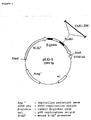
- An E. coli strain containing plasmid TABI-f-4 which comprises a DNA encoding a human TAB1 peptide having the amino acid sequence from Met at amino acid 1 to Pro at amino acid 504 in the TAB1 amino acid sequence shown in SEQ ID NO: 4, was named Escherichia coli DH5a (TABI-f-4) and has been deposited internationally under the Budapest Treaty as an accession No. FERM BP-5599 in The National Institute of Bioscience and Human-Technology, The Agency of Industrial Science and Technology (1-1-3 Higashi, Tsukuba, Ibaraki, Japan) on July 19 th , 1996.
- TAK1 and/or TAB1 there can be various molecular species of TAK1 and/or TAB1 to be used in the present invention, which are different in amino acid sequence, molecule weight, isoelectric point, the presence or absence and position of glycosylation, sugar chain structure, phosphorylation state, and/or the presence or absence of disulfide bond, depending on the origin, host producing them, and/or the type of purification method.
- human-derived TAK1 and/or TAB1 are preferable while the proteins may have any structures, as far as the proteins can be suitably used in the present invention.
- the above-mentioned proteins used in the present invention can be fusion peptides of the above-mentioned proteins and other peptides.
- the fusion peptides can be prepared by already-known methods.
- the other peptides to be used for the fusion with the above-mentioned proteins can be any peptides as far as the peptides are effectively used in the present invention.
- the peptide is exemplified by FLAG ( Hopp, T. P. et al., BioTechnology (1988) 6, 1204-1210 ), 6 x His of six histidine (His) residues, 10 x His, influenza hemagglutinin (HA), human c-myc fragment, a fragment of VSV-GP, a fragment of p18HIV, T7-tag, HSV-tag, E-tag, a fragment of SV40 T antigen, lck tag, a fragment of ⁇ -tubulin, B-tag, a fragment of Protein C, and the like.
- FLAG Hopp, T. P. et al., BioTechnology (1988) 6, 1204-1210
- His histidine residues
- 10 x His influenza hemagglutinin
- HA influenza hemagglutinin
- human c-myc fragment a fragment of VSV-GP
- a fragment of p18HIV T7-tag
- GST glutathione-S-transferase
- HA influenza hemagglutinin
- MBP maltose-binding protein
- the above-mentioned proteins to be used in the present invention can be the proteins encoded by DNAs encoding proteins having the biological activities and capable of hybridizing to the DNA encoding the above-mentioned proteins.
- DNA encoding TAK1 to be used in the present invention is exemplified by a DNA having the nucleotide sequence from A at nucleotide 183 to A at nucleotide 1919 in SEQ ID NO: 1.
- DNA encoding TAB1 to be used in the present invention is exemplified by a DNA having the nucleotide sequence from A at nucleotide 30 to G at nucleotide 1541 in SEQ ID NO: 3.
- Each of DNAs encoding the proteins to be used in the present invention can be a DNA capable of hybridizing to the nucleotide sequence of SEQ ID NO: 1 or 3 and encoding a protein having the biological activity.
- Hybridization condition for the DNA encoding the protein to be used in the present invention may be the conditions with suitable stringency.
- Such a hybridization condition can be, for example, the condition with low stringency.
- the condition with low stringency is, for example, a washing condition of 42°C, 5 x SSC, 0.1% sodium dodecyl sulfate, and 50% formamide.
- the conditions with high stringency are more preferable.
- the condition with high stringency is, for example, a washing condition of 60°C, 0.1 x SSC, and 0.1% sodium dodecyl sulfate. It has been already known that a protein encoded by a DNA capable of hybridizing to a nucleotide sequence encoding a certain protein under a condition with suitable stringency shares the same biological activity with the latter protein.
- DNA encoding TAK1 or TAB1 to be used in the present invention can be a DNA of any origin, as far as the DNA contains the nucleotide sequence of SEQ ID NO: 1 or 3, respectively.
- DNAs include, for example, genomic DNA, cDNA, and synthetic DNA. These DNAs may originate from a variety of cells, tissue, or organ, or cDNA library or genomic library derived from any species in addition to human.
- the libraries may be commercially available DNA libraries.
- the vector used for the libraries can be any vector including plasmid, bacteriophage, and YAC vectors.
- TAK1 or TAB1 to be used in the present invention can be obtained as a recombinant protein produced by the use of gene recombination techniques using DNAs encoding these proteins.
- the nucleotide sequence of a gene encoding each protein described herein can be cloned from cell, tissue, or organ expressing the protein, and then inserted into an appropriate vector, and the resulting DNA construct is introduced into a host to produce the recombinant protein.
- Such recombinant proteins can be also usable in the present invention.
- mRNA of the gene encoding the protein to be used in the present invention is isolated from cell, tissue, or organ expressing the protein.
- the mRNA is isolated from total RNA prepared by a known method, for example, guanidine-ultracentrifugation method ( Chirgwin, J. M. et al., Biochemistry (1979) 18, 5294-5299 ) and AGPC method ( Chomczynski, P. and Sacchi, N., Anal. Biochem. (1987) 162, 156-159 ) and purified by using an mRNA Purification Kit (Pharmacia) and such.
- the mRNA can be directly prepared by using a QuickPrep mRNA Purification Kit (Pharmacia).
- Messenger RNA obtained is used for synthesizing cDNA corresponding to the gene by using reverse transcriptase.
- the cDNA synthesis can be performed by using an AMV Reverse Transcriptase First-strand cDNA Synthesis Kit (Seikagaku Co.) or the like. Synthesis and amplification of cDNA can be carried out by using Marathon cDNA Amplification kit (CLONTECH) and 5'-RACE ( Frohman, M. A. et al., Proc. Natl. Acad. Sci. U.S.A. (1988) 85, 8998-9002 ; Belyavsky, A. et al., Nucleic Acids Res. (1989) 17, 2919-2932 ) using polymerase chain reaction (PCR).
- PCR polymerase chain reaction
- DNA fragment of interest is prepared from the PCR products obtained, and then ligated with a vector DNA to give a recombinant vector.
- the ligated DNA is then introduced into E. coli or the like, and resulting colonies are selected to prepare a desired recombinant vector.
- Nucleotide sequence of the target DNA is verified by a known method, for example, dideoxy nucleotide chain termination method. If the DNA of interest is obtained, it is integrated into an expression vector.
- DNA encoding the protein to be used in the present invention may originate from a variety of cells, tissue, or organ, or cDNA library or genomic library obtained from any species in addition to human.
- the libraries may be commercially available DNA libraries.
- the vector used for the libraries can be any vector including plasmid, bacteriophage, and YAC vectors.
- DNA encoding the protein to be used in the present invention can be constructed from the above-mentioned DNAs by a known method and by using commercially available kits.
- DNA manipulation includes the digestion by restriction enzyme, linker ligation, and insertion of initiation codon (ATG) and/or termination codon (TAA, TGA, or TAG).
- Expression vector for expressing the protein to be used in the present invention can be any type of vector, as far as the expression vector is used suitably in the present invention.
- Such expression vectors include, for example, mammal-derived expression vector (for example, pEF and pCDM8), insect cell-derived expression vector (for example, pBacPAK8), plant-derived expression vector (for example, pMH1 and pMH2), animal virus-derived expression vector (for example, pHSV and pMV), yeast-derived expression vector (for example, pNV11), Bacillus subtilis -derived expression vector (for example, pPL608 and pKTH50), and E. coli- derived expression vector (for example, pGEX, pGEMEX, and pMALp2).
- mammal-derived expression vector for example, pEF and pCDM8
- insect cell-derived expression vector for example, pBacPAK8
- plant-derived expression vector for example, pMH1 and pMH2
- the protein to be used in the present invention can be produced, for example, by connecting a DNA encoding the protein downstream of a promoter in the expression vector.
- the promoter/enhancer includes, for example, mammal-derived promoter/enhancer (for example, EF1- ⁇ promoter/enhancer and ⁇ -actin promoter/enhancer), insect virus-derived promoter/enhancer (for example, polyhedrosis virus promoter/enhancer), plant-derived promoter/enhancer (for example, tobacco mosaic virus promoter/enhancer), animal virus-derived promoter/enhancer (for example, SV40 promoter/enhancer and human CMV promoter/enhancer), yeast-derived promoter/enhancer (for example, alcohol dehydrogenase promoter/enhancer), and E. coli- derived promoter/enhancer (for example, Lac promoter/enhancer, Trp promoter/enhancer, and Tac promoter/enhancer).
- the SV40 promoter/enhancer can be readily utilized according to the method of Mulligan et al. (Nature (1979) 277, 108 ).
- the HEF1 ⁇ promoter/enhancer can be readily used according to the method of Mizushima et al. (Nucleic Acids Res. (1990) 18, 5322 ).
- a commonly used useful promoter, a secretory signal sequence, and a gene to be expressed are functionally connected together, for the expression in E. coli.
- the promoter includes lacZ promoter and araB promoter.
- LacZ promoter can be used according to the method of Ward et al. (Nature (1989) 341, 544-546 ; FASEB J. (1992) 6, 2422-2427 ), or araB promoter according to the method of Better et al. (Science (1988) 240, 1041-1043 ).
- a signal sequence suitable for the host can be added.
- a signal sequence includes, for example, a signal sequence of a secretory protein.
- the signal sequence of secretory protein is exemplified by a signal sequence of mammal-derived secretory protein, for example, the signal sequence of immunoglobulin.
- the signal sequence of secretory protein is exemplified by signal sequences of E. coli -derived secretory proteins, for example, periplasm secretory signal sequences such as OmpA and pelB ( Lei, S. P. et al J. Bacteriol. (1987) 169, 4379 ).
- the expression vector thus prepared can be introduced into a host by a known method.
- the method for the introduction into host is exemplified by electroporation ( EMBO J. (1982) 1, 841-845 ), calcium-phosphate method ( Virology (1973) 52, 456-467 ), and liposome method.
- the replication origin to be used can be derived from SV40, polyoma virus, adenovirus, bovine papilloma virus (BPV), etc.
- the expression vector can contain as a selection marker aminoglycoside transferase (APH) gene, thymidine kinase (TK) gene, E. coli xanthine guanine phosphoribosyl transferase (HPRT) gene, dihydrofolate reductase (DHFR) gene, and the like.
- APH aminoglycoside transferase
- TK thymidine kinase
- HPRT E. coli xanthine guanine phosphoribosyl transferase
- DHFR dihydrofolate reductase
- Any production system can be used for the production of the recombinant protein to be used in the present invention.
- the production systems that can be used for the production of recombinant proteins include in vitro and in vivo production systems.
- the in vitro production system includes, for example, production systems with eukaryotic cells as well as the systems with prokaryotic cells.
- the production systems with eukaryotic cells are, for example, those with animal cells, plant cells, and fungus cells.
- animal cells include mammalian cells, e.g., CHO cell ( J. Exp. Med. (1995) 108, 945 )-, COS cell, myeloma cells baby hamster kidney (BHK) cell, HeLa cell, and Vero cell; amphibian cells, e.g., Xenopus laevis oocyte ( Valle, et al., Nature (1981) 291, 358-340 ) ; and insect cells, e.g., sf9, sf21, and Tn5.
- mammalian cells e.g., CHO cell ( J. Exp. Med. (1995) 108, 945 )-, COS cell, myeloma cells baby hamster kidney (BHK) cell, HeLa cell, and Vero cell
- amphibian cells e.g., Xenopus la
- CHO cell particularly preferable to use are dhfr - CHO cell, whichlacksDHFRgene ( Proc. Natl. Acad. Sci. USA (1980) 77, 4216-4220 ), and CHO K-1 cell ( Proc. Natl. Acad. Sci. USA (1968) 60, 1275 ).
- Nicotiana tabacum -derived cell An example of plant cell is Nicotiana tabacum -derived cell.
- the callus culture may be used for this plant cell.
- Fungus cells include yeast, for example, the genus Saccharomyces such as Saccharomyces cerevisiae, and filamentous fungus, for example, the genus Aspergillus such as Aspergillus niger.
- Prokaryotic cells that provide the production system are bacterial cells, including for example E. coli and Bacillus subtilis.
- Those cells may be transformed with DNA of interest, and the resulting transformed cells are cultured in vitro to produce the recombinant protein.
- the cell culture can be performed according to a known method.
- culture media to be utilized include DMEM, MEM, RPMI1640, and IMDM. These media may be used alone or in combination with serum supplement such as fetal calf serum (FCS) for the culture.
- FCS fetal calf serum
- the pH of the culture is preferably about 6 to 8.
- the cells are generally cultivated at about 30 to 40°C for about 15 to 200 hours. If necessary, the culture medium may be changed with fresh one, aerated, and/or stirred.
- the recombinant protein is produced in the cells, in which the gene has been introduced, and recovered from the cells.
- the in vivo production system is exemplified by the production system with animal or with plant.
- a DNA of interest is introduced into an animal or plant, and the animal or plant produces the recombinant protein in its body. Then the protein is recovered from the body.
- Animals for the production system can be a mammal or insect.
- the mammals include goat, pig, sheep, mouse, and cow ( Vicki Glaser, SPECTRUM Biotechnology Applications, 1993 ).
- a transgenic animal can also be used.
- a DNA of interest is inserted into a gene encoding a protein, such as goat ⁇ -casein, inherently produced into milk.
- a DNA fragment comprising the fusion gene, in which the DNA of interest is inserted is injected into a goat embryo, and the embryo is introduced into a female goat.
- the recipient goat may give birth to transgenic babies.
- the created transgenic individuals or the progenies thereof may give milk, and the desired recombinant protein can be obtained from the milk.
- an adequate hormonal treatment of the transgenic goat can be utilized ( Ebert, K.M. et al., Bio/Technology (1994) 12, 699-702 ).
- An insect that can be used for the production system is, for example, silk worm.
- baculovirus containing a DNA of interest is infected into a silk worm, and the desired recombinant protein can be obtained from the body fluid of the silk worm ( Maeda, S. et al., Nature (1985) 315, 592-594 ).
- a plant for the production system can be, for example, tobacco.
- a DNA of interest is inserted into a plant expression vector, for example, pMON 530.
- the resulting vector is introduced into bacteria such as Agrobacterium tumefaciens.
- the bacteria are infected to tobacco, for example, Nicotiana tabacum.
- the desired recombinant protein can be obtained from the leaves of the tobacco ( Julian, K.-C. Ma et al., Eur. J. Immunol. (1994) 24, 131-138 ).
- the gene is introduced into any of these animal and plant as described above.
- the animal or plant produces the recombinant protein in the body thereof, then the protein is recovered from the body.
- the recombinant protein expressed and produced as mentioned above may be separated from intracellular or extracellular materials, or the host, and then purified into homogeneity. Separation and purification methods for protein to be used in the present invention may be commonly used ones, which are not to be restricted to any specific ones.
- the separation and purification of the protein can be performed by the appropriately selected and combined use of column chromatographies such as affinity chromatography, filter, ultrafiltration, salting-out, dialysis, SDS polyacrylamide gel electrophoresis, isoelectric focusing, and others.
- column chromatographies such as affinity chromatography, filter, ultrafiltration, salting-out, dialysis, SDS polyacrylamide gel electrophoresis, isoelectric focusing, and others.
- the chromatography includes, for example, affinity chromatography, ion-exchange chromatography, hydrophobic chromatography, gel filtration, reverse-phase chromatography, adsorption chromatography, and the like ( Strategies for Protein Purification and Characterization: A Laboratory Course Manual. Ed Daniel R. Marshak et al., Cold Spring Harbor Laboratory Press, 1996 ).
- the chromatographic procedures can be carried out by liquid-phase chromatography such as HPLC, FPLC, or the like.
- Protein concentration can be measured by using a known method, for example, a method with measuring absorbance or Bradford's method:
- the samples to be tested by the inventive screening method include, for example, peptide, purified or crude protein, synthetic compound, materials fermented with microorganism, extract from marine organism, plant extract, prokaryotic cell extract, extract from single-cell eukaryote, or extract from animal cells, or the libraries thereof.
- test samples contain a compound that is expected to inhibit or suppress the binding between TAK1 and TAB1, a compound that is expected to inhibit or suppress the of kinase activity of TAK1 (phosphorylation), or a compound that is expected to inhibit or suppress the kinase activity of TAK1 (phosphorylation) that is generated by TAK1 and TAB1, or contain these compounds.
- These compounds which can be isolated by the inventive screening method, as described above, inhibit or suppress the TAK1 activity, or TAK1 and TAB1 activities, and thereby inhibiting or suppressing the signal transduction through inflammatory cytokines; inhibiting or suppressing the production of inflammatory cytokines; inhibiting or suppressing physiological activity of inflammatory cytokines; or inhibiting or suppressing the signal transduction by inflammatory stimulation.
- an "inflammatory cytokine” indicates a common cytokine involved in inflammatory reaction, and specifically IL-1 (for example, IL-1 ⁇ and IL-1 ⁇ ), TNF (for example, TNF ⁇ and TNF ⁇ ), IL-6, IL-10, IL-4, chemokine such as IL-8, MCP-1, etc.
- One embodiment of the inventive screening method is conducted by contacting TAK1 and TAB1 with a test sample, then detecting the binding between TAK1 and TAB1, and selecting a compound inhibiting the binding between TAK1 and TAB1.
- the screening system provided by the present invention is to be conducted as an in vitro assay system.
- a specific example of such in vitro assay system is a cell-free system.
- TAK1 and TAB1 are used in combination, and either of them is linked to a support, the other and a test sample are added thereto and incubated. After the support is washed, the binding of the latter protein to the former linked to the support is detected or measured.
- the proteins to be used in the present invention may be purified or crude proteins that are produced by cells inherently expressing the proteins, cells in which DNAs encoding the proteins are introduced, or animals or plants in which DNAs encoding the proteins are introduced.
- the proteins to be used in the present invention may be linked to a support.
- the immobilization of the protein on a support may be performed by a standard method.
- Supports for the protein immobilization are, for example, insoluble polysaccharides such as agarose, dextran, and cellulose, synthetic resins such as polystyrene, polyacrylamide, and silicon, etc. More specifically, commercially available beads or plates, which are manufactured from them as raw materials, are used. In the case of beads, a column or the like may be used as packed with the beads.
- Such a plate can be a multi-well plate (96-well multi-well plate, etc.) or a biosensor chip.
- the immobilization of protein on a support can be performed by a commonly used method in which chemical bonding, physical adsorption, or the like is used.
- protein can be linked to a support where an antibody specifically recognizing the protein is pre-immobilized by binding the antibody to the protein. Protein can also be immobilized via avidin/biotin.
- the binding between TAK1 and TAB1 is generally allowed in a buffer.
- the buffer is, for example, phosphate buffer, Tris buffer, or the like.
- Incubation condition may be a commonly used one, for example, a condition where incubation is carried out at 4°C to room temperature for 1 to 24 hours. Washing condition after the incubation can be any of possible condition as far as the wash does not have any negative effects on the protein binding, and for example, buffers containing detergents are usable.
- Such detergents include, for example, 0.05% Tween20.
- TAK1, TAB1, and a test sample are incubated under an adequate condition, and then washed to separate the specific binding from non-specific one. The degree of TAK1-TAB1 binding may be then evaluated.
- TAK1 or TAB1 is linked to a support.
- TAK1 is first immobilized on a support, and then a pre-mixture of TAB1 and a test sample may be added thereto, or alternatively TAB1 may be added thereto after addition of a test sample.
- TAB1 is intended to be immobilized on a support
- a pre-mixture of TAK1 and a test sample may be added, or alternatively TAK1 may be added after addition of a test sample.
- TAK1, TAB1, and a test sample added in the order as described above are incubated under an adequate condition, and then the degree of TAK1-TAB1 binding can be evaluated.
- control group may be used in the inventive screening method.
- Such control groups can be either of a negative control group without any test samples or positive control group, or both the groups.
- the bound protein When the bound protein is detected or measured by the inventive method, the bound protein may be just detected or quantitatively measured. In both cases, the target compound can be detected by comparing the results obtained for a negative control group without test samples, a group containing test samples, and/or a positive control group.
- the activity of the target compound can be quantitatively measured by comparing values obtained from these results.
- the values obtained for the negative control group without test samples are compared with those for the groups with test samples, to detect the target compound. As compared with that for the negative control, the value with a test sample is lower, then the test sample can be judged to contain the target compound.
- the sample can be quantified based on a standard curve prepared by using values for a positive control group containing known amounts of a compound that is clarified to be capable of inhibiting the binding between TAK1 and TAB1.
- a positive control group containing known amounts of a compound that is clarified to be capable of inhibiting the binding between TAK1 and TAB1.
- the protein bound is larger in quantity, then it can be estimated that the compound has only lower activity for inhibition of the protein binding.
- the amount of protein bound is smaller, the binding-inhibitory activity of the compound to inhibit the binding of the protein is presumed to be stronger.
- a biosensor which is based on surface plasmon resonance phenomenon, can be used to detect or measure the binding protein.
- the biosensor based on surface plasmon resonance phenomenon the protein-protein interaction can be observed in real time as a surface plasmon resonance signal without labeling even when the amount of protein is a trace level (for example, BIAcore, Pharmacia). Accordingly, the binding of the proteins used in the present invention can be estimated by using a biosensor such as BIAcore.
- TAK1 and TAB1 either one is immobilized on the sensor chip, the other protein is contacted with the sensor chip, then the latter protein bound to the immobilized one is detected as a differential resonance signal.
- a sensor chip CM5 Biosensor
- TAK1 or TAB1 is immobilized on the sensor chip.
- an aqueous EDC/NHS solution 200 mM EDC (N-ethyl-N'-(3-dimethylaminopropyl) carbonate hydrochloride) and 50 mM N-hydroxysuccinimide (NHS)
- HBS buffer 10 mM HEPES pH7.4, 150 mM NaCl, 3.4 mM EDTA, 0.05% Tween20.
- a test sample is injected after the injection of the protein capable of interacting the other protein.
- a control group may be used.
- Such control groups can be either of a negative control group without any test samples or positive control group, or both the groups.
- the protein bound can be measured quantitatively as a differential resonance signal value.
- the results obtained for the negative control group without test samples are compared with those for the groups with test samples and/or for the positive control group, to detect and identify the target compound.
- either of the proteins is labeled, and the label on the protein can be utilized to detect or measure the protein binding.
- one protein which is to be mixed with a test sample and to be contacted with the other protein, is pre-labeled, and then incubated in combination with the test sample. After wash, the bound protein is detected or measured based on the label on the protein.
- the labeled protein and the test sample are preferably contacted with the other protein linked to the support. After the incubation, the support is washed, and then the label on the bound protein is detected or measured.
- the proteins to be used in the present invention can be labeled according to a common method.
- Labeling substances include, for example, radioisotope, enzyme, fluorescent substance, biotin/avidin, and the like. These labeling substances may be commercially available ones. Examples of radioisotope are 32 P, 33 P, 131 I, 125 I, 3 H, 14 C, and 35 S.
- the enzyme includes, for example, alkaline phosphatase, horseradish peroxidase, ⁇ -galactosidase, ⁇ -glucosidase, and the like.
- the fluorescent substance is exemplified by fluorescein isothiocyanate (FITC), and rhodamine. All these are commercially available, and the labeling with them can be performed by a known method.
- Procedures are specifically as follows. Namely, a solution containing either of the proteins is added to a plate, and the plate is allowed to stand still overnight. After wash of the plate; blocking, for example with BSA, is conducted to avoid non-specific binding of proteins. The plate is washed again, and a test sample and the labeled protein are added thereto. A negative control group without test samples and/or positive control group is contained in this experiment. All to be tested are incubated, and then the plates are washed, and finally the bound protein is detected or measured. In the step of detection or measurement, radioisotope can be detected or measured by using liquid scintillation.
- a substrate is added to the system and the detection or measurement is based on an enzymatic change of substrate, for example, coloration, which can be detected or measured in an absorbance spectrometer. Fluorescent substances may be detected or measured in a fluorophotometer. These results are compared with values obtained for the control groups to identify the target compound.
- a primary antibody specifically recognizing either of the proteins, in the combination of TAK1 and TAB1, can be used to detect or measure the bound protein.
- one protein is contacted and incubated with the other protein, after wash, the bound protein is detected or measured with a primary antibody specifically recognizing the protein.
- the protein and the test sample are preferably contacted with the other protein linked to the support. After the incubation, the support is washed, and the bound protein is detected or measured by utilizing the primary antibody specifically recognizing the bound protein.
- the primary antibody is preferably labeled with a labeling substance.
- Such an experiment can be conducted specifically as follows. A solution containing either of the proteins is added to a plate, and the plate is allowed to stand still overnight. After wash of the plate, blocking, for example with BSA, is conducted on the plate to avoid non-specific binding of proteins. The plate is washed again, and a test sample and the other protein are added thereto. A negative control group without test samples and/or positive control group is contained in this experiment.
- radioisotope can be detected or measures by using liquid scintillation.
- the label is an enzyme
- a substrate is added to the system, and the detection or measurement is based on an enzymatic change of substrate, for example, coloration, which can be detected or measured in an absorbance spectrometer.
- Fluorescent substances may be detected or measured in a fluorophotometer. The results obtained are compared with those for the control groups to identify the target compound.
- a primary antibody specifically recognizing other peptides that are fused with the protein to be used in the present invention can be used to detect or measure the bound protein.
- the protein and the test sample are preferably contacted with the other protein linked to the support. After the incubation, the support is washed, and then the bound protein is detected or measured by the primary antibody specifically recognizing the peptide fused with the bound protein.
- the primary antibody is preferably labeled with a labeling substance.
- Such an experiment can be conducted specifically as follows. Namely, a solution containing either of the proteins is added to a plate, and the plate is allowed to stand still overnight. After wash of the plate, blocking, for example with BSA, is conducted on the plate to avoid non-specific binding of proteins. The plate is washed again, and a test sample and the other protein fused with a peptide are added thereto. A negative control group without test samples and/or positive control is contained in this experiment.
- radioisotope can be detected or measured by using liquid scintillation.
- the label is an enzyme
- a substrate is added to the system, and the detection or measurement is based on an enzymatic change of substrate, for example, coloration, which can be detected or measured in an absorbance spectrometer.
- Fluorescent substances may be detected or measured in a fluorophotometer. The results obtained are compared with those for the control groups to identify the target compound.
- a primary antibody specifically recognizing the protein to be used in the present invention and a secondary antibody specifically recognizing the primary antibody can be used to detect or measure the bound protein.
- the bound protein is detected or measured with a primary antibody specifically recognizing the protein and the secondary antibody specifically recognizing the primary antibody.
- the protein and the test sample are preferably contacted with the other protein linked to the support.
- the bound protein is detected or measured by the primary antibody specifically recognizing the protein and the secondary antibody specifically recognizing the primary antibody.
- the secondary antibody is preferably labeled with a labeling substance.
- Such an experiment can be conducted specifically as follows. Namely, a solution containing either of the proteins is added to a plate, and the plate is allowed to stand still overnight. After wash of the plate, blocking, for example with BSA, is conducted on the plate to avoid non-specific binding of proteins. The plate is washed again, and a test sample and the other protein are added thereto. A negative control group without test samples and/or positive control group is contained in this experiment.
- radioisotope can be detected or measured by using liquid scintillation.
- the label is an enzyme
- a substrate is added to the system, and the detection or measurement is based on an enzymatic change of substrate, for example, coloration, which can be detected or measured in an absorbance spectrometer.
- Fluorescent substances may be detected or measured in a fluorophotometer. The results obtained are compared with those for the control groups to select the target compound.
- a primary antibody specifically recognizing other peptides that are fused with the protein and a secondary antibody specifically recognizing the primary antibody can be used to detect or measure the bound protein.
- the protein and the test sample are preferably contacted with the other protein linked to the support. After the incubation, the support is washed, and then the bound protein is detected or measured by the primary antibody specifically recognizing the peptide fused with the bound protein and the secondary antibody specifically recognizing the primary antibody.
- the secondary antibody is preferably labeled with a labeling substance.
- Such an experiment can be conducted specifically as follows. Namely, a solution containing either of the proteins is added to a plate, and the plate is allowed to stand still overnight. After wash of the plate, blocking, for example with BSA, is conducted to avoid non-specific binding of proteins. The plate is washed again, and a test sample and the other protein fused with a peptide are added thereto. A negative control group without test samples and/or positive control group is contained in this experiment.
- the label is an enzyme
- a substrate is added to the system, and the detection or measurement is based on an enzymatic change of substrate, for example, coloration, which can be detected or measured in an absorbance spectrometer.
- Fluorescent substances may be detected or measured in a fluorophotometer. The results obtained are compared with those for the control groups to determine the target compound.
- the present invention can be conducted particularly preferably by Enzyme-linked Immunosorbent Assay (ELISA) as follows. Specifically, TAK1 fused with a peptide, for example 6 x His, is diluted with an immobilization buffer (0.1 M NaHCO 3 , 0.02% NaN 3 , pH9.6). Appropriate amounts of the aqueous solution of dilution are added to respective wells of a 96-well immuno-plate (Nunc) and incubated at 4°C overnight.
- an immobilization buffer 0.1 M NaHCO 3 , 0.02% NaN 3 , pH9.6
- each well is washed 3 times with the washing buffer, adequate amounts of TAB1 fused with a peptide (e.g., FLAG), which has been diluted with a dilution buffer (1% BSA and 0.5% Tween20 in PBS) and a test sample are added thereto.
- a dilution buffer 1% BSA and 0.5% Tween20 in PBS
- the resulting mixture is incubated at room temperature for 1 hour.
- Each well is washed 3 times with the washing buffer, and a 100- ⁇ l aliquot of mouse anti-FLAG M2 antibody (IBI), which has been diluted to 3 ⁇ g/ml with the dilution buffer, is added to each well. The plate is incubated at room temperature for 1 hour.
- Each well is washed 3 times with a washing buffer, and a 100- ⁇ l aliquot of alkaline phosphatase-labeled goat anti-mouse IgG antibody (ZYMED), which has been diluted 1000 times with the dilution buffer, is added to each well.
- the plate is incubated at room temperature for 1 hour.
- Each well is washed 5 times with a washing buffer, and a 100 ⁇ l aliquot of a coloring solution (substrate buffer (50 mM NaHCO 3 , 10 mM MgCl 2 , pH9.8) containing 1 mg/ml p -phenylphosphate; SIGMA) is added to each well.
- the plate is incubated at room temperature, and then absorbance at 405 nm is measured by using a microplate reader (Model3550, BIO-RAD). The results obtained are compared with those for the negative control group and/or positive control group to identify the target compound.
- protein G or protein A instead of the secondary antibody can be utilized in the detection or measurement using the antibody described herein.
- the inventive screening method can be practiced by using High Throughput Screening (HTS). Specifically, the steps from the start to the blocking are performed manually, and the remaining reactions are automated by using a robot, thereby achieving a high-throughput screening.
- HTS High Throughput Screening
- TAK1 fused with a peptide for example 6 x His
- an immobilization buffer 0.1 M NaHCO 3 , 0.02% NaN 3 , pH9.6
- Appropriate amounts of the aqueous solution of dilution are added to respective wells of a 96-well immuno-plate (Nunc) and incubated at 4°C overnight.
- the immuno-plate subjected to the blocking treatment is placed in a Biomek2000 HTS system (Beckman), and the device is operated by a system control program.
- a dispenser such as Biomek 2000 dispenser (Beckman) or Multipipette 96-well dispenser (Sagian) can be used to dispense solutions to each well or to remove them from well of the immuno-plate.
- EL404 microplate washer Bio Tek
- Absorbance can be measured by using a SPECTRAmax250 plate reader (Molecular Devices).
- the operating program is set to conduct the following steps. Specifically, each well is washed 3 times with the washing buffer, and then adequate amounts of a test sample and TAB1 fused with a peptide, for example maltose-binding protein (MBP), which has been diluted with a dilution buffer (1% BSA and 0.5% Tween20 in PBS), are added thereto. A negative control group without test samples and a positive control are simultaneously managed. These all samples are incubated at room temperature for 1 hour.
- MBP maltose-binding protein
- Each well is washed 3 times with a washing buffer, and a 100- ⁇ l aliquot of rabbit anti-MBP antiserum (New England Biolabs), which has been diluted 5000 times with the dilution buffer, is added thereto. The plate is incubated at room temperature for 1 hour. Each well is washed 3 times with a washing buffer, and a 100- ⁇ l aliquot of alkaline phosphatase-labeled goat anti-rabbit IgG antibody (TAGO), which has been diluted 5000 times with the dilution buffer, is added to each well. The plate is incubated at room temperature for 1 hour
- Each well is washed 5 times with a washing buffer, and a 100- ⁇ l aliquot of a coloring solution (substrate buffer (50 mM NaHCO 3 , 10 mM MgCl 2 , pH9.8) containing 1 mg/ml p -nitrophenylphosphate; SIGMA) is added to each well.
- a coloring solution (substrate buffer (50 mM NaHCO 3 , 10 mM MgCl 2 , pH9.8) containing 1 mg/ml p -nitrophenylphosphate; SIGMA) is added to each well.
- the plate is incubated at room temperature, and then absorbance at 405 nm is measured by using a microplate reader, Biomekplate reader (Beckman/Molecular Devices). The results obtained are compared with those for control groups to identify the target compound.
- Antibodies to be used in the present invention can be commercially available antibodies and antibodies contained in commercially available kits as well as monoclonal or polyclonal antibodies that can be prepared by using well-known methods.
- Monoclonal antibody can be prepared by using a desired antigen for sensitization. Immunization is performed by using this antigen according to a commonly used method. The resulting immune-cells are fused with publicly known parental cells by a standard cell fusion method. Cells producing monoclonal antibody are screened by a standard screening method.
- monoclonal antibody or polyclonal antibody is generated as follows.
- the origin of sensitizing antigen to obtain antibody is not restricted to a particular animal species, but it is preferred to use an antigen that is of the same mammalian origin as the peptide actually used in the present invention, for example, human, mouse, or rat.
- the sensitizing antigen originating from human is particularly preferable.
- human TAK1 or human TAB1 when intended to use as the sensitizing antigen, their nucleotide sequences and amino acid sequences can be obtained by the gene sequences disclosed herein.
- the peptides can be prepared by chemical synthesis or genetic engineering.
- the protein or peptide to be used as the sensitizing antigen may be the full-length one or the fragment thereof.
- the fragment includes, for example, a C-terminal fragment or N-terminal fragment.
- the mammal to be immunized with the sensitizing antigen there is no particular limitation on the mammal to be immunized with the sensitizing antigen, but it is preferable to select with considering compatibility to a parental cell to be used for cell fusion.
- Generally used are animals belonging to Rodentia, Lagomorpha, and Primates.
- Rodentia is exemplified, for example, by mouse, rat, and hamster.
- Lagomorpha animal includes, for example, rabbit.
- An animal of Primates includes, for example, monkey. Monkeys such as those of Catarrhini (old-world monkey)including crab-eating monkey, rhesus monkey, hamadryas baboon, and chimpanzee can be used.
- an animal is immunized with a sensitizing antigen according to a known method.
- the sensitizing antigen is intraperitoneally or subcutaneously injected to a mammal.
- the sensitizing antigen is appropriately diluted and/or suspended in Phosphate-Buffered Saline (PBS), physiological saline, etc.
- PBS Phosphate-Buffered Saline
- the solution is mixed with an adequate amount of a standard adjuvant e.g., Freund's complete adjuvant, emulsified, and preferably given to a mammal several times at 4 to 21 day intervals.
- An appropriate carrier may also be used at the time of the immunization with the sensitizing antigen. After the immunization, increases in the serum level of desired antibody are confirmed by a commonly used method.
- polyclonal antibody In order to obtain polyclonal antibody, after the verification of elevated serum level of desired antibody, blood is collected from the mammal sensitized with the antigen. Serum is separated from the blood by a known method. As polyclonal antibody, serum containing the polyclonal antibody may be used, or alternatively, if needed, a fraction containing the polyclonal antibody may be further isolated from the serum.
- immune-cells are collected from the mammal.
- the cells are subjected to the cell fusion.
- the immune-cells to be utilized for the cell fusion are spleen cells in particular.
- the other parental cells to be fused with the above-mentioned immune-cells include mammalian myeloma cells. Such cells used preferably may be various known cell lines.
- cell fusion between the above-mentioned immune-cells and myeloma cells can be performed according to a known method including, for example, the method of Milstein et al. ( Galfre, G. and Milstein, C., Methods Enzymol. (1981) 73, 3-46 ), etc.
- the above-mentioned cell fusion may be conducted, for example, in the presence of cell fusion-enhancing agent in a commonly used nutrient culture medium.
- the cell fusion-enhancing agent is, for example, polyethylene glycol (PEG), Sendai virus (HVJ), or the like.
- An adjuvant, such as dimethyl sulfoxide or the like, may optionally be used together to enhance the efficiency of fusion.
- a mixing ratio between immune-cells and myeloma cells preferably ranges, for example, from 1:1 to 1:10 of myeloma cell vs. immune-cells.
- Cell culture medium for the above-mentioned cell fusion includes, for example, RPMI1640 and MEM media that are preferred for the proliferation of the myeloma cell lines, and other standard culture media that are used in this type of cell culture. Serum supplements such as fetal calf serum (FCS) can be used together.
- FCS fetal calf serum
- the cell fusion is carried out as follows.
- the above-mentioned immune-cells are mixed well with myeloma cells at a given ratio in the above-mentioned medium.
- a solution of PEG for example, averaged molecule weight of about 1000 to 6000
- W/V concentration of 30 to 60%
- the agent for cell fusion and other agents which are not preferable for the growth of hybridomas, can be removed by repeatedly conducting the following procedures: adding an appropriate culture medium to the cell mixture; centrifuging them; and removing the supernatant.
- the hybridomas are selected by culturing the cells in a standard selection medium, for example, HAT medium (a medium containing hypoxanthine, aminopterin, and thymidine).
- HAT medium a medium containing hypoxanthine, aminopterin, and thymidine.
- the culture in the HAT medium is continued till all cells except the hybridomas of interest (non-fused cells) are sufficiently killed, generally, for several days to several weeks.
- the commonly used limiting dilution method is employed to screen and clone the hybridomas producing antibodies of interest.
- Human lymphocytes for example, EB virus-infected human lymphocytes are in vitro sensitized with a protein or peptide, cells expressing them, or lysate of the cells.
- the sensitized lymphocytes are fused with human-derived myeloma cells (e.g., U266 cell) having permanent division potential to obtain hybridomas producing desired human antibodies having the activity of binding to the peptide (Unexamined Published Japanese Patent Application No. Sho 63-17688 ).
- a transgenic animal having human antibody gene repertoire is immunized with an antigen, which may be a protein or peptide, cells expressing them, or lysate of the cells, to obtain cells producing antibody.
- the cells are fused with myeloma cells to prepare hybridomas, and thereby obtaining human antibodies against the protein or peptide to be used in the present invention (refer to WO92-03918 , WO93-2227 , W094-02602 , WO94-25585 , WO96-33735 , and WO96-34096 ).
- the prepared hybridomas producing monoclonal antibody can be cultivated and passaged in a standard culture medium, and also can be stored in liquid nitrogen over the long term.
- Monoclonal antibodies can be prepared from the hybridomas by the following methods and such.
- the hybridomas are cultured according to a standard procedure, and the monoclonal antibody can be obtained as the culture supernatant.
- the hybridomas are transplanted and grown in a mammal that has the compatibility to the hybridomas, and the monoclonal antibody can be obtained as the ascites.
- the former method is suitable for the preparation of highly pure antibody, and the latter is suitable for the preparation of antibody in large quantity.
- oncogene-immortalized immune-cells such as sensitized lymphocytes producing antibody of interest can be used for this purpose.
- the monoclonal antibody thus obtained can also be prepared as a recombinant antibody produced by utilizing gene recombination techniques.
- the recombinant antibody can be produced by cloning the antibody gene from the immune-cells such as hybridomas or antibody-producing sensitized lymphocytes, inserting the gene into an appropriate vector, and introducing the vector into a host.
- the recombinant antibody can be used (refer to, for example, Borrebaeck, C. A. K. and Larrick, J. W., THERAPEUTIC MONOCLONAL ANTIBODIES, Published in the United Kingdom by MACMILLAN PUBLISHERS LTD, 1990 ).
- the antibody to be used in the present invention may be an antibody fragment thereof or modified antibody, as far as it has the desired binding activity.
- an antibody fragment includes Fab, F(ab')2, Fv, or single chain Fv (scFv) in which Fv of H chain and Fv of L chain are linked to each other with an appropriate linker.
- an antibody is treated with an enzyme, for example, papain or pepsin, to provide an antibody fragment.
- genes encoding the antibody fragments are constructed and inserted into an expression vector, and the resulting construct is expressed in an appropriate host cell.
- the antibody expressed and produced as described above can be separated from intracellular and/or extracellular material or the hosts and then purified to homogeneity.
- the antibody can be separated and/or purified by the appropriately selected and combined use of column chromatographies such as affinity chromatography and the like, filter, ultrafiltration, salting-out, dialysis, SDS polyacrylamide gel electrophoresis, isoelectric focusing, and others ( Antibodies: A Laboratory Manual. Ed Harlow and David Lane, Cold Spring Harbor Laboratory, 1988 ).
- column chromatographies such as affinity chromatography and the like, filter, ultrafiltration, salting-out, dialysis, SDS polyacrylamide gel electrophoresis, isoelectric focusing, and others.
- protein A column used in affinity chromatography is exemplified by protein A column or protein G column.
- protein A column includes Hyper D, POROS, and Sepharose F.F. (Pharmacia).
- the chromatographic method includes, for example, ion-exchange chromatography, hydrophobic chromatography, gel filtration, reverse-phase chromatography, adsorption chromatography, and the like ( Strategies for Protein Purification and Characterization: A Laboratory Course Manual. Ed Daniel R. Marshak et al., Cold Spring Harbor Laboratory Press, 1996 ).
- the chromatographic procedures can be carried out by liquid-phase chromatography such as HPLC, FPLC, or the like.
- the concentration or activity of the antibody obtained above can be measured by a known method, for example, ELISA, enzymatic immunoassay (EIA), radioimmunoassay (RIA), or immunofluorescence
- the primary antibody or secondary antibody obtained above can be labeled according to a commonly-known method.
- Labeling substances include, for example, radioisotope, enzyme, fluorescent substance, and the like. These labeling substances may be commercially available ones. Examples of radioisotope are 32 P, 33 P, 131 I, 125 I, 3 H, 14 C, and 35 S.
- the enzyme includes, for example, alkaline phosphatase, horseradish peroxidase, ⁇ -galactosidase, ⁇ -glucosidase, and the like.
- the fluorescent substance is exemplified by fluorescein isothiocyanate (FITC) and rhodamine.
- FITC fluorescein isothiocyanate
- the labeling can be performed by a known method by obtaining these commercially available labeling substances.
- the binding between TAK1 and TAB1 can also be detected and/or measured by observing change in the expression level of a reporter gene which is activated in response to the binding of the proteins.
- Luciferase, ⁇ -galactosidase, HIS3 gene, chloramphenicol acetyltransferase (CAT), green fluorescent protein (GFP) gene, or the like can be used as the reporter gene which is activated in response to the biological activity produced by the binding between TAK1 and TAB1.
- TAK1 and TAB1 expressed in cells may be fused proteins with other peptides.
- the peptides to be fused with the proteins can be any peptides as far as the peptides are usable in the inventive screening method, but are preferably transcriptional regulatory factors.
- TAK1 and TAB1 DNAs are respectively fused with DNAs encoding respective subunits of heterodimeric transcriptional regulatory factor that is known to bind to a DNA and thereby activating the transcription of a certain reporter gene,
- the fusion genes are inserted into expression vectors, and the resulting DNA constructs are introduced into cells.
- the respective subunits fused with TAK1 and TAB1 form the heterodimer.
- the heterodimeric transcriptional regulatory factor binds to the DNA and thereby activating the reporter gene.
- a test sample contains a compound inhibiting the binding between TAK1 and TAB1, the compound inhibits the binding between TAK1 and TAB1.
- the subunits of the transcriptional regulatory factor cannot form the heterodimer, and thus the transcription of the reporter gene is not induced.
- the target compound can be detected or measured by observing change in the expression level of a reporter gene.
- the two-hybrid system Fields, S., and Sternglanz, R., Trends. Genet. (1994) 10, 286-292 ) can be used when change in the expression level of a reporter gene is intended to be assayed in such a system.
- the two-hybrid system may be constructed by using a commonly used method or by utilizing a commercially available kit.
- Examples of the commercially available two-hybrid system kit are MATCHMAKER Two-Hybrid System, Mammalian MATCHMAKER Two-Hybrid Assay Kit (both are products from CLONTECH), and HybriZAP Two-Hybrid Vector System (Stratagene).
- the gene encoding TAK1 is ligated with the gene encoding LexA DNA-binding domain to provide an expression vector.
- the gene encoding full-length TAK1 of SEQ ID NO: 2 or its segment having amino acids 1 to 418 is inserted in frame into an expression plasmid pBTM116 ( Vojtek, A.B., et al., Cell (1993) 74, 205-214 ) for the yeast two-hybrid system to construct an expression plasmid.
- the gene encoding full-length TAB1 of SEQ ID NO: 4 or its segment containing amino acids 1 to 504 is ligated with the gene encoding GAL4-transcription activation domain to prepare an expression vector.
- the expression vector can be constructed, for example, by inserting the TAB1-encoding gene in frame into an expression plasmid pGAD10 (CLONTECH) for the yeast two-hybrid system.
- Each of the two-hybrid expression plasmids is transformed into cells of yeast L40 strain containing the HIS3 gene of which transcription is regulated by a promoter with a LexA-binding motif.
- the presence of the protein interaction results in yeast growth.
- the degree of transformant growth indicates increases in the expression level of the reporter gene, and the target compound can be screened.
- TAK1 kinase activity generated by TAK1, or by TAK1 and TAB1 is used as an index.
- TAK1 kinase activity is used as an index.
- a TAK1 molecule lacking the N-terminal 22 amino acid residues constitutively shows the kinase activity.
- the TAK1 molecule lacking the N-terminal 22 amino acid residues is expressed in animal cells such as COS cell, bacterial cells such as E. coli , yeast cells, and so on, and then a test sample is added and incubated with TAK1.
- a substrate protein for TAK1 such as MKK6, together with 32 P-ATP, is added to TAK1 separated with an anti-TAK1 antibody or the like to perform kinase reaction.
- the activity is then detected or measured.
- the amount of 32 P-ATP incorporated into the substrate protein by the phosphorylation thereof is assayed to evaluate the TAK1 kinase activity. The result is compared to that of a negative control without any test sample to identify a compound directly inhibiting TAK1 kinase activity.
- TAK1 and TAB1 expressed in animal cells such as COS cell, E. coli , yeast, and such are incubated with a test sample in vitro , and a substrate protein, such as MKK6, together with 32 P-ATP, is added to TAK1 separated with an anti-TAK1 antibody or the like to perform kinase reaction. The activity is then detected or measured.
- the amount of 32 P-ATP incorporated into the substrate protein by the phosphorylation thereof is assayed to evaluate the TAK1 kinase activity.
- the result is compared to that of a negative control without any test sample to identify a compound inhibiting TAK1 kinase activity.
- the screening method using TAK1 kinase activity as an index can be used for High Throughput Screening (HTS). Specifically, the steps of addition, mixing, and respective reactions for each sample are automated by using a robot, the phosphorylation degree of substrate protein is detected by scintillation proximity assay method ( Bothworth, N. and Towers, P., Nature, 341: 167-168, 1989 ), and thereby achieving a high-throughput screening.
- HTS High Throughput Screening
- TAK1 and TAB1 expressed in animal cells such as COS cell, E. coli , or yeast
- 32 P-ATP and a substrate protein e.g., MKK6
- an anti-MKK6 antibody is added to each well, and then SPA beads (Amersham) which are coated by protein A or an antibody specifically recognizing other antibodies in a species-specific manner are added to each well.
- radioactivity incorporated into the substrate protein is measured by using a MicroBeta scintillation counter (Wallac).
- streptavidin-coated SPA beads can be used for the assay.
- the result obtained by these methods is compared to that of a control group to identify a test sample containing a compound inhibiting TAK1 kinase activity.
- the assay is performed by using cells in vitro . Specifically, a test sample is introduced into and/or contacted with cells expressing TAK1 and TAB1, and then TAK1- and TAB1-mediated transduction of biological activity is detected and/or measured. A compound reducing the biological activity is selected to obtain the target compound.
- proteins to be used are expressed in cells used for the assay. Examples described later have revealed that the inhibition of binding between TAK1 and TAB1 inhibits the signal transduction through inflammatory cytokines, which further inhibits the production of other inflammatory cytokines involved in the cytokine network.
- the detection and/or measurement of the TAK1-, or TAK1- and TAB1-mediated transduction of biological activity makes it possible to inhibit or suppress the signal transduction through inflammatory cytokine of interest, and based on the inhibition or suppression, makes it possible to inhibit or suppress the production of the inflammatory cytokine, to inhibit or suppress physiological activity of the inflammatory cytokine, or possible to screen a compound inhibiting or suppressing the inflammatory stimulation-induced signal transduction.
- Cells expressing TAK1, or TAK1 and TAB1 can be preferably prepared by gene engineering techniques, by introducing the herein-described DNAs encoding them into cells that naturally express none of them.
- Cells usable for the present invention include prokaryotic and eukaryotic cells.
- the prokaryotic cells include bacterial cells
- eukaryotic cells include mammalian cells, insect cells, and yeast cells.
- the genes encoding the proteins to be used in the present invention are introduced into these cell lines, and a test sample is added thereto. Then, actions reflecting the signal transduction through inflammatory cytokines, production of inflammatory cytokines, physiological activity of inflammatory cytokines, inflammatory stimulation-induced signal transduction, and so on are detected or measured.
- the introduction and contact of a test sample may be achieved by adding the test sample into a cell culture medium.
- Biological activities transduced through the mediation of TAK1, or TAK1 and TAB1 include, for example, biological activities of inflammatory cytokines, the activity of inducing inflammatory mediators, and change in the expression levels of reporter genes.
- inflammatory cytokine examples include IL-1 (e.g., IL-1 ⁇ and IL-1 ⁇ ), TNF (e.g., TNF ⁇ and TNF ⁇ ), IL-6, etc.
- the biological activity of IL-1 includes the enhancement of antibody production, activation of T and/or B cells, induction of production of other cytokines including IL-6, IL-2, and IFN- ⁇ based on cytokine network, induction of acute-phase proteins, fever, activation of vascular endothelial cells, infiltration of leukocytes, and production of collagenase and/or collagen. Accordingly, any of these actions can be detected or measured to detect the biological activity of IL-1 as an inflammatory cytokine.
- the biological activity of TNF includes the activation of T cell and macrophage, induction of production of other cytokines including IL-1, IL-6, and IL-8 based on cytokine network, induction of apoptosis, production of collagenase and/or collagen, and production of prostaglandins. Accordingly, any of these actions can be detected or measured to detect the biological activity of TNF as an inflammatory cytokine.
- the biological activity of IL-6 includes the induction of acute-phase proteins, enhancement of antibody production, increase in hematopoietic stem cell, enhancement of neuronal cell differentiation. Accordingly, any of these actions can be detected or measured to detect the biological activity of IL-6 as an inflammatory cytokine.
- Cell lines and such that are already known to show these actions can be used in the detection or measurement of these actions in order to detect the biological activity of inflammatory cytokines.
- the activity of inducing cytokine production can be detected or measured quantitatively by assaying the amount of cytokine produced in a T cell line by ELISA or PCR.
- TAK1 in order to detect and/or measure the activity of inducing inflammatory mediators such as prostaglandins, as a biological activity mediated by TAK1, or TAK1 and TAB1, quantitative assay can be performed by using a macrophage cell line and a commercially available kit, etc.
- the production amount of antibody can be assayed by using a B cell line by ELISA.
- the evaluation can be done, for example, by analyzing cell surface markers on a T cell line by FACS or the like.
- the biological activity mediated by TAK1, or TAK1 and TAB1 can be detected and/or measured by observing change in the expression level of a reporter gene which is activated in response to the biological activity.
- Luciferase, ⁇ -galactosidase, HIS3 gene, chloramphenicol acetyltransferase (CAT), green fluorescent protein (GFP) gene, or the like can be used as the reporter gene which is activated in response to the biological activity produced by the binding between TAK1 and TAB1.
- CAT chloramphenicol acetyltransferase
- GFP green fluorescent protein
- inflammatory cytokines such as IL-6 and IL-1 is increased upon inflammatory stimuli such as IL-1 or LPS in monocyte/macrophage cells.
- inflammatory stimuli such as IL-1 or LPS in monocyte/macrophage cells.
- an inflammatory stimulus such as IL-1 or LPS is given to the cells, thereby evaluating the signal transduction through inflammatory cytokines and the associated production of the inflammatory cytokines, physiological activity of inflammatory cytokines, the signal transduction through inflammatory stimulation, and/or the expression of inflammatory cytokines.
- inflammatory stimulus means herein an stimulus inducing inflammatory reaction, including bacterial infection or injury that is a cause of disorders in living tissues, physical stimulation such as heat, coldness, radiation, or electrostimulation, chemical substance, or abnormality of biological immunity.
- the inflammatory stimuli include, for example, IL-1 and LPS.
- Expression vectors for expressing desired proteins can be constructed by inserting genes encoding ' them into suitable restriction enzyme sites of expression vectors.
- a reporter gene construct containing the luciferase gene which is regulated by an NF-KB response element derived from the IFN- ⁇ gene (p55IgkLuc; Fujita, T., et al., Gene, Dev., (1993) 7, 1354-1363 ) is introduced into 293 cells in which the respective expression plasmids are introduced and also into the control cells in which an expression vector without gene insert has been introduced.
- the respective cells are cultured in medium with or without 10 ng/ml IL-1. Luciferase activity is then assayed in the cell extracts.
- the addition of IL-1 increases the luciferase activity.
- the luciferase activity is not increased in the cells.
- a target compound in the test sample inhibits the IL-1-induced elevation of reporter gene expression.
- increases in expression level of the reporter gene can be measured, and thereby successfully screening the target compound.
- an inhibitor of the signal transduction through inflammatory cytokines inhibitor of the production of inflammatory cytokines, and an anti-inflammatory agent, which contain as an active ingredient a compound inhibiting the signal transduction through TAK1.
- an inhibitor of the physiological activity of inflammatory cytokines and an inhibitor of the signal transduction through inflammatory stimulation which contain as an active ingredient a compound inhibiting the signal transduction through TAK1.
- a compound inhibiting the signal transduction through TAK1 means a compound inhibiting the biological activity of TAK1 as described herein.
- Such a compound inhibiting the signal transduction through TAK1 includes, for example, a compound inhibiting the binding between TAK1 and TAB1, a compound inhibiting the kinase activity of TAK1, and a compound inhibiting antagonism of substrate for TAK1.
- a compound inhibiting the binding between TAK1 and TAB1 indicates a compound inhibiting the interaction between TAK1 and TAB1.
- Such a compound inhibiting the binding between TAK1 and TAB1 includes, for example, a compound that specifically binds to TAK1 or a compound that specifically binds to TAB1.
- These compounds may be compounds that specifically bind to the respective targets, compete for the targets with the proteins which bind to the targets, and the competitive binding keeps the proteins which bind to the targets free from the targets.
- These compounds may be compounds that specifically bind to the region where TAK1 and TAB1 bind to each other and thereby inhibiting the binding between TAK1 and TAB1.
- the compounds inhibiting the binding between TAK1 and TAB1 may be compounds that specifically bind to TAK1 or TAB1 and, for example, have the ability of binding to the target but lack in the ability of activation of TAK1 and/or TAB1. More preferably, the compounds inhibiting the binding between TAK1 and TAB1 may be compounds that specifically bind to the binding site of TAK1 to TAB1, or the binding site of TAB1 to TAK1.
- Such compounds inhibiting the binding between TAK1 and TAB1 include proteins, peptides, and chemically synthesized compounds, for example, TAK1 or TAB1 mutants, partial peptides of TAK1 or TAB1, antibodies against TAK1 or TAB1, compounds that specifically bind to TAK1, TAB1, or both, or antisense oligonucleotides of TAK1 or TAB1.
- TAK1-DN A specific example of the compound inhibiting the binding between TAK1 and TAB1 is TAK1-DN as described below in Examples.
- TAK1-DN has the amino acid sequence from amino acid 77 Glu to amino acid 303 Gln in the amino acid sequence of SEQ ID NO: 2.
- TAK1-DN has TAB1-binding activity but lacks in TAK1 kinase activity. Accordingly, TAK1-DN can bind to TAB1 but not induces the signal transduction through TAK1. As a consequence, TAK1-DN inhibits the binding between normal TAK1 and TAB1.
- TAK1-DN can be a specific example of the compound inhibiting the signal transduction through TAK1.
- the compound inhibiting the binding between TAK1 and TAB1 can be obtained by using the screening system with TAK1 and TAB1.
- a system screening with TAK1 and TAB1 includes the screening system using ELISA or two-hybrid system as described herein.
- the compound inhibiting the kinase activity of TAK1 may be a compound inhibiting TAK1 activation or a compound inhibiting the catalytic function of TAK1.
- the compound inhibiting TAK1 activation includes, for example, the above-mentioned compound inhibiting the binding between TAK1 and TAB1, or a compound inhibiting TAK1 activation accompanying the binding with TAB1.
- These compounds may be compounds that specifically bind to the respective targets, compete for the targets with the proteins which bind to the targets, and the competitive binding keeps the proteins which bind to the targets free from the targets.
- These compounds may be compounds that specifically bind to the region where TAK1 and TAB1 bind to each other or to the TAK-1 activating region of the TAB1 and thereby inhibiting the binding between TAK1 and TAB1 or inhibiting TAK1 activation accompanying the binding.
- the compounds inhibiting the kinase activity of TAK1 can also be compounds that act on the catalytic region of TAK1 and inhibit the phosphorylation ability of activated TAK1.
- such compounds can be compounds that inhibit the catalytic action in the phosphorylation or the interaction with the substrate.
- These compounds may be compounds that specifically bind to the respective targets, compete for the targets with the proteins which bind to the targets, and the competitive binding keeps the proteins which bind to the targets free from the targets.
- These compounds may be compounds that specifically bind to the catalytic region of TAK1 or the region binding to substrate protein in TAK1, TAK1-binding region of a substrate protein, or the region to be phosphorylated by TAK1, and thereby inhibiting the binding between TAK1 and its substrate or inhibiting the phosphorylation by TAK1 accompanying the binding.
- Such a compound inhibiting TAK1 kinase activity can be obtained by using the in vitro kinase assay system as described above.
- Such compounds inhibiting the signal transduction through TAK1 include proteins, peptides, and chemically synthesized compounds, for example, TAK1 or TAB1 mutants, partial peptides of TAK1 or TAB1, antibodies against TAK1 or TAB1, compounds that specifically bind TAK1, TAB1, or both, or antisense oligonucleotides of TAK1 or TAB1.
- TAK1 or TAB1 mutants include proteins, peptides, and chemically synthesized compounds, for example, TAK1 or TAB1 mutants, partial peptides of TAK1 or TAB1, antibodies against TAK1 or TAB1, compounds that specifically bind TAK1, TAB1, or both, or antisense oligonucleotides of TAK1 or TAB1.
- the compound inhibiting the signal transduction through TAK1 includes a compound of which structure is in part modified by addition, deletion, and/or substitution.
- the compound inhibiting the signal transduction through TAK1 inhibits the production of inflammatory cytokines such as IL-1, TNF, and IL-6 induced by an inflammatory stimulus (LPS) and also inhibits IL-6 production that is induced by an inflammatory cytokine (IL-1). Accordingly, the compound inhibiting the signal transduction through TAK1 is useful for inhibiting the signal transduction through these inflammatory cytokines, the production of inflammatory cytokines induced thereby, the physiological activity of inflammatory cytokines, the signal transduction through inflammatory stimulation, and/or inflammatory stimulation. It is known that cytokines such as IL-1, TNF, and IL-6 are involved in inflammatory reaction and therefore are collectively called inflammatory cytokine.
- compositions which contain a compound inhibiting the signal transduction through TAK1, can be used for the treatment and/or prevention of these diseases.
- the inhibition of inflammatory stimulation-induced or cytokine network-associated production of inflammatory cytokines as well as the inhibition of physiological action of inflammatory cytokine reduces inflammation and suppresses the inflammatory action.
- the compound inhibiting the signal transduction through TAK1 has the anti-inflammatory effect suppressing the inflammatory stimulation and/or the physiological action of inflammatory cytokines. Accordingly, the compound inhibiting the signal transduction through TAK1 is useful as an anti-inflammatory agent.
- the compound inhibiting the signal transduction through TAK1 can be used for the expectation of the anti-inflammatory effect in treating diseases with inflammation.
- inhibitor of the signal transduction through inflammatory cytokines inhibitor of the production of inflammatory cytokines, anti-inflammatory agent, or pharmaceutical composition
- the herein described inhibitor, agent, or composition can be used orally as a sugar coated tablet, capsule, elixir, or microcapsule, or used parenterally as an injection of sterile solution or suspension thereof in water or any other pharmaceutically acceptable liquid.
- the product can be manufactured by mixing the herein described inhibitor of the production of inflammatory cytokines or anti-inflammatory agent with a physiologically acceptable carrier, flavoring agent, filler, vehicle, preservative, stabilizer, and/or binder in generally acceptable unit dose formulation which is required for drug manufacture.
- a physiologically acceptable carrier for example, a pharmaceutically acceptable sulfate, a pharmaceutically acceptable sulfate, a pharmaceutically acceptable sulfate, sulfate, g., g., g., g., g., a pharmaceutically acceptable containing a pharmaceutically acceptable containing a pharmaceutically acceptable containing a pharmaceutically acceptable containing a pharmaceutically acceptable containing a pharmaceutically acceptable containing a pharmaceutically acceptable administrados, a pharmaceutically acceptable containing a pharmaceutically acceptable sulfate, a pharmaceutically acceptable sulfate, a pharmaceutically acceptable sulfate, a pharmaceutically acceptable sulfate, a pharmaceutically acceptable sulfate, a pharmaceutical
- Additives that can be mixed to tablet and capsule include, for example, binders such as gelatin, corn starch, gum tragacanth, and gum Arabic, fillers such as crystalline cellulose, swelling agents such as corn starch, gelatin, and alginic acid, lubricants such as magnesium stearate, sweetener such as sucrose, lactose, and saccharin, and flavoring agents such as peppermint, Gaultheria adenothrix oil, and cherry.
- binders such as gelatin, corn starch, gum tragacanth, and gum Arabic
- fillers such as crystalline cellulose
- swelling agents such as corn starch, gelatin, and alginic acid
- lubricants such as magnesium stearate
- sweetener such as sucrose, lactose, and saccharin
- flavoring agents such as peppermint, Gaultheria adenothrix oil, and cherry.
- the dosage form When the dosage form is a capsule, it can contain a liquid carrier such as fatty oil in addition to the above-mentioned materials.
- a liquid carrier such as fatty oil
- the sterile composition for injection can be formulated with a vehicle such as distilled water for injection according to a commonly used procedure for drug manufacture.
- Aqueous solutions for injection include, for example, physiological saline, isotonic solution containing glucose or other adjuvants, for example, D-sorbitol, D-mannose, D-mannitol, or sodium chloride, which can be used together with an appropriate solubilizer, for example, alcohol, specifically, ethanol, polyalcohol, for example, propylene glycol, polyethylene glycol, nonionic detergent, for example, polysorbate 80 or HCO-50.
- an appropriate solubilizer for example, alcohol, specifically, ethanol, polyalcohol, for example, propylene glycol, polyethylene glycol, nonionic detergent, for example, polysorbate 80 or HCO-50.
- the oil is exemplified by sesame oil and soybean oil, and it can be used together with benzyl benzoate or benzyl alcohol as a solubilizer.
- a buffer e.g., phosphate buffer and sodium acetate buffer
- pain-killer e.g., procaine hydrochloride
- stabilizer e.g., benzyl alcohol and phenol
- antioxidant can be formulated into the preparation.
- the formulated solution for injection is generally filled into an appropriate ampul.
- Dosage of the herein described inhibitor of the production of inflammatory cytokines or anti-inflammatory agent varies depending on symptoms but generally is about 0.1 to 100 mg a day, preferably about 1.0 to 50 mg a day, and more preferably about 1.0 to 20 mg a day, for human adult (in the case of body weight 60 kg) when given orally.
- parenteral administration its single dose can vary depending on the type of subject and organ to be given, symptoms, and administration procedures but it is preferable to typically administer, for example, about 0.01 to 30 mg a day, preferably about 0.1 to 20 mg a day, more preferably about 0.1 to 10 mg a day by intravenous injection for human adult (in the case of body weight 60 kg) when given as an injection.
- the drug can be given to other animals and the dose to be given can be converted based on the dosage for body weight of 60 kg.
- a TAK1-DN expression vector that acts as a dominant negative inhibitor was prepared to demonstrate that the signal transduction through LPS or inflammatory cytokines can be inhibited by inhibiting the specific binding between human TAK1 and human TAB1.
- TAK1-DN has the amino acid sequence, corresponding to the TAB1-binding region of TAK1, which covers a stretch from amino acid 77 Glu to amino acid 303 Gln in the amino acid sequence of SEQ ID NO: 2.
- the TAK1-DN-encoding gene fragment was amplified by using ph-TAK1 (refer to Unexamined Published Japanese Patent Application (JP-A) No. Hei 9-163990 ) as a template DNA by PCR.
- a sense primer TAK1S (SEQ ID NO: 5) containing a recognition site for restriction enzyme EcoRI and the initiation codon ATG and an antisense primer TAK1AS (SEQ ID NO: 6) containing a recognition site for restriction enzyme EcoRI and the stop codon were used to amplify the TAK1-DN-encoding DNA fragment.
- the resulting PCR products were digested with restriction enzyme EcoRI and then inserted at an EcoRI site of animal cell expression vector pLG-1 (see Figure 1 ) containing H-2L d promoter to prepare TAK1-DN transgenic vector pLG-TAKDN.
- DNA was introduced by microinjection under an inverted microscope (LEICA) with Nomarski differential-interference device by using glass micropipettes connected with a micromanipulator.
- LICA inverted microscope
- 2 pl of solution of TAK1-DN-encoding DNA molecule (about 500 molecules of in 1-pl PBS) was injected into a male pronucleus in a fertilized egg from C57BL/6J mouse.
- the gene fragment for the introduction was prepared by digesting pLG-TAKDN with restriction enzyme XhoI and then separating and purifying it by agarose gel electrophoresis.
- the manipulated egg with introduced DNA was transplanted in an oviduct of pseudopregnant female mouse (recipient mouse) of ICR strain.
- the pseudopregnancy was induced by mating with a male mouse that had undergone vasoligation.
- the neonatal mice were born by normal spontaneous delivery or by Caesarean section.
- the neonatal mice born by Caesarean section were nursed by another female mouse of ICR strain that had been arranged previously as a foster mother.
- DNAs were prepared from tails of the neonatal mice at the age of 3 to 4 weeks, to test the presence of the introduced sequence by PCR.
- the DNAs for the analysis were prepared as follows. Specifically, each 2-cm tail section was lysed in a 1-ml lysis buffer (20 mM Tris-HCl, pH 7.0, 100 mM NaCl, 20 mM EDTA, 1% SDS, 1 mg/ml Proteinase K) at 55°C overnight. After a series of phenol extraction, phenol-chloroform extraction, and chloroform extraction, the DNA was extracted by isopropanol-precipitation. The DNA obtained was dissolved in 100 ⁇ l TE (10 mM Tris-HCl, pH 8.0, 1 mM EDTA).
- PCR analysis was carried out by using the DNA (100 ng) as a template and PCR primers Rb-glo1 (SEQ ID NO: 7) and TA12A (SEQ ID NO: 8). The analysis revealed that, of the 22 mice, 3 males and 2 females had the new gene.
- Figure 2 shows a result of genomic Southern analysis. Specifically, EcoRI-digested genomic DNA (50 ⁇ g) was electrophoresed on a 0.7% agarose gel, and transferred onto a nylon membrane Hybond N+ (Amersham). The membrane was hybridized to a probe of TAK1-DN DNA (50 ng) labeled with [ ⁇ - 32 P] dCTP according to a usual method. The result showed that bands of interest were detected in only TAK1-DN transgenic mice, and the presence of introduced gene was confirmed.
- mice A line (female) of the above-mentioned created mice was mated with a C57BL/6J male mouse for the first generation. The obtained mice were analyzed for the presence or absence of the introduced gene in the same manner as described above. The result showed that the introduced gene was inherited in 11 of the 25 F 1 mice.
- Figure 3 shows a result of verification of actual mRNA expression of TAK1-DN in F 1 mice. Specifically, total RNAs were prepared from the livers and kidneys of F 1 mice, and 30- ⁇ g aliquots of them were electrophoresed on a denaturation agarose gel with 1% formalin. The RNAs were transferred onto a nylon membrane Hybond N+ (Amersham) and hybridized to a probe of TAK1-DN DNA (50 ng) labeled with [ ⁇ - 32 P] dCTP according to a usual method. The result showed that bands of interest were detected in only F 1 mice, and the expression of introduced gene was confirmed.
- the transgenic mouse lines were subcultured and maintained by using external fertilization. Specifically, 48 hours after the injection of 5 IU pregnant mare serum gonadotropin (PMSG, TEIKOKU HORMONE Mfg. Co.), a dose of the same units of human chorionic gonadotropin (hCG, Sankyo) was injected in a C57BL6/J female mouse, followed by mating with a F 1 male mouse. Then, 16 to 17 hours after the injection of hCG, sperms were collected from the caudal portion of a male mouse.
- PMSG pregnant mare serum gonadotropin
- hCG human chorionic gonadotropin
- the sperms were incubated in a TYH medium (the 1-ml medium contains NaCl 6.97 mg, KCl 0.36 mg, CaCl 2 • 2H 2 O 0.25 mg, KH 2 PO 4 0.16 mg, MgSO 4 • 7H 2 O 0.29 mg, NaHCO 3 2.11 mg, sodium pyruvate 0.11 mg, streptomycin 50 ⁇ g, penicillin 75 ⁇ g, and glucose 1.0 mg, pH 7.0) containing 4 mg/ml BSA at 37°C for 1.5 hours.
- eggs were collected from a C57BL6/J female mouse and placed in a TYH medium containing 4 mg/ml BSA under oil.
- the sperms were added to the medium containing the eggs, and the mixture was incubated at 37°C for 4 to 6 hours.
- the resulting fertilized eggs were transferred in a Whitten's medium (the 1-ml medium contains NaCl 5.14 mg, KCl 0.36 mg, KH 2 PO 4 0.16 mg, MgSO 4 • 7H 2 O 0.29 mg, NaHCO 3 1.90 mg, calcium lactate pentahydrate 0.53 mg, streptomycin 50 ⁇ g, penicillin 80 ⁇ g, glucose 1.0 mg, and 3.7 mg of 60% sodium lactate) containing 100 mM EDTA and 4 mg/ml BSA, and transplanted in the oviduct of a female mouse after removing the sperms.
- the 1-ml medium contains NaCl 5.14 mg, KCl 0.36 mg, KH 2 PO 4 0.16 mg, MgSO 4 • 7H 2 O 0.29 mg, NaHCO 3 1.90 mg, calcium lactate pentahydrate 0.53 mg, streptomycin 50 ⁇
- Peritoneal macrophages were prepared from TAK1-DN-expressing transgenic mice, and the responses of the cells to IL-1 and lipopolysaccharide (LPS; Sigma) were examined.
- TAK1-DN-expressing transgenic mouse TAK1-DN Tgm
- C57BL/6 mouse wild-type mouse
- the mouse was given a massage for 30 seconds, and then the solution was recovered from the mouse.
- the solution recovered was centrifuged, and the precipitated cells were resuspended in RPMI1640 (GIBCO-BRL) containing 10% FBS (GIBCO-BRL).
- RPMI1640 GEBCO-BRL
- FBS GEBCO-BRL
- LPS LPS, at a final concentration of 10 ⁇ g/ml, or IL-1 ⁇ (Genzyme), at a final concentration of 10 ng/ml
- IL-1 ⁇ IL-1 ⁇
- the peritoneal macrophages were cultured in wells of a 96-well plate at a cell density of 1x10 6 /well.
- LPS (1 ⁇ g/ml) or IL-1 ⁇ (10 ng/ml) was added to the culture in the presence or absence of proteasome inhibitor MG132 (40 ⁇ M). The cells were harvested after 30 or 60 minutes.
- the cells were washed with PBS (-), and then a lysis buffer was added thereto for the preparation of cell lysate.
- An aliquot of each lysate corresponding to about 2x10 5 cells was subjected to gel electrophoresis by 9% SDS-PAGE.
- the fractionated proteins were blotted onto a nitrocellulose membrane and then incubated with anti-I ⁇ B ⁇ antibody (SantaCruz) as a primary antibody for 1.5 hours. After wash, horseradish peroxidase-conjugated goat anti-rabbit antibody was used as a secondary antibody. After the reaction of the secondary antibody, the reaction product was detected by using ECL (Amersham).
- TAB1 peptide and TAK1 peptide were used to analyze whether or not a peptide (TAK1-DN) from Glu at amino acid 77 to Gln at amino acid 303 in the TAK1 peptide of SEQ ID NO: 2 is capable of inhibiting the binding between TAK1 peptide and TAB1 peptide and further capable of inhibiting TAK1 peptide activation in animal cells.
- TAK1-DN a peptide from Glu at amino acid 77 to Gln at amino acid 303 in the TAK1 peptide of SEQ ID NO: 2 is capable of inhibiting the binding between TAK1 peptide and TAB1 peptide and further capable of inhibiting TAK1 peptide activation in animal cells.
- each of genes encoding full-length TAK1 and TAK1-DN was ligated with the gene encoding GAL4 DNA-binding domain (GAL4-BD) to prepare an expression vector.
- the gene encoding full-length TAK1 was obtained from yeast two-hybrid expression plasmid pBTMHu11F (Shibuya H. et al., (1996) 272, 1179-1182) by digesting it with restriction enzymes EcoRI and PstI. Subsequently, the gene was ligated between EcoRI and PstI sites of an expression vector pM (CLONTECH) containing the GAL4-BD gene to obtain an animal cell two-hybrid expression plasmid pM-TAK1.
- the gene encoding TAK1-DN was amplified by PCR using a sense primer DNTAK5' (SEQ ID NO: 9) with a recognition site for restriction enzyme EcoRI and an antisense primer DNTAK3' (SEQ ID NO: 10) with a site for restriction enzyme PstI by using plasmid pBTMHu11F as a template DNA.
- the amplified DNA was digested with restriction enzymes EcoRI and PstI, and then ligated into pM vector, thereby constructing animal cell two-hybrid expression plasmid pM-TAK1DN.
- TAB1C68 consisting of C-terminal 68 amino acid residues of TAB1 peptide was ligated with the gene encoding transcription activation domain (VP16-AD) from VP16 protein of herpes simplex virus to prepare an expression vector.
- the gene encoding TAB1C68 was prepared by digesting yeast two-hybrid expression plasmid pGAD-TAB1 (Shibuya H.
- the reporter plasmid used was pG5-Luc, which was constructed from pG5CAT (CLONTECH) containing 5 consecutive GAL4-binding sites and the chloramphenicol acetyltransferase (CAT) gene downstream thereof.
- pG5-Luc contains the luciferase gene substituted instead of the CAT-encoding gene in the pG5CAT.
- CHO cells (5x10 4 cells/each well) were washed with PBS.
- GAL4-BD fusion protein expression plasmid any one of pM, pM-TAK1, and pM-TAK1DN
- 500 ng of VP16-AD fusion protein expression plasmid pVP16 or p
- the culture further continued for 72 hours, and then luciferase activities in each cell extract were assayed by using Dual-Luciferase TM Reporter Assay System (Promega). Specifically, the cells were washed with PBS, 250 ⁇ l of Passive Lysis Buffer attached to the kit was added thereto, and the mixture was incubated at room temperature for 15 minutes. A 20- ⁇ l aliquot of each cell extract was used for the assay. The efficiency of gene introduction was normalized by using measured values for the activity of Renilla luciferase derived from the introduced pRL-SV40. The result is shown in Figure 7 .
- TAK1-DN is also capable of binding to TAB1.
- the present invention has revealed that TAK1 participates in the signal transduction through inflammatory cytokines such as IL-1, TNF, or IL-6, and that compounds inhibiting the TAK1-associated signal transduction inhibit the production of inflammatory cytokines such as IL-1, TNF, and IL-6. Accordingly, the inventive screening method is useful to screen compounds inhibiting the production of inflammatory cytokines. As further described, compounds inhibiting the TAK1-associated signal transduction are useful as inhibitors of the signal transduction through inflammatory cytokines, inhibitors of the production of inflammatory cytokines, inhibitors of the physiological activities of inflammatory cytokines, and inhibitors of the signal transduction through inflammatory stimulation. Further, compounds inhibiting the signal transduction through TAK1 are useful as anti-inflammatory agents.
Landscapes
- Health & Medical Sciences (AREA)
- Life Sciences & Earth Sciences (AREA)
- Chemical & Material Sciences (AREA)
- Organic Chemistry (AREA)
- Engineering & Computer Science (AREA)
- Molecular Biology (AREA)
- Zoology (AREA)
- Immunology (AREA)
- Wood Science & Technology (AREA)
- Biochemistry (AREA)
- General Health & Medical Sciences (AREA)
- Genetics & Genomics (AREA)
- Microbiology (AREA)
- Medicinal Chemistry (AREA)
- Bioinformatics & Cheminformatics (AREA)
- Biomedical Technology (AREA)
- Biotechnology (AREA)
- Proteomics, Peptides & Aminoacids (AREA)
- Hematology (AREA)
- Biophysics (AREA)
- Urology & Nephrology (AREA)
- General Engineering & Computer Science (AREA)
- Physics & Mathematics (AREA)
- Analytical Chemistry (AREA)
- Toxicology (AREA)
- Cell Biology (AREA)
- Gastroenterology & Hepatology (AREA)
- Food Science & Technology (AREA)
- General Physics & Mathematics (AREA)
- Pathology (AREA)
- Peptides Or Proteins (AREA)
- Micro-Organisms Or Cultivation Processes Thereof (AREA)
- Medicines That Contain Protein Lipid Enzymes And Other Medicines (AREA)
- Measuring Or Testing Involving Enzymes Or Micro-Organisms (AREA)
- Pharmaceuticals Containing Other Organic And Inorganic Compounds (AREA)
- Investigating Or Analysing Biological Materials (AREA)
Claims (19)
- Verfahren zum Screenen einer Verbindung, die die Produktion von Entzündungscytokinen hemmt, wobei das Verfahren die folgenden Schritte umfasst:(a) Inkontaktbringen einer Testprobe mit TAK1 und TAB1;(b) Nachweisen der Bindung zwischen TAK1 und TAB1; und(c) Auswählen einer Verbindung, die die Bindung hemmt.
- Verfahren nach Anspruch 1, wobei TAK1 und/oder TAB1 mit einem Peptid fusioniert ist.
- Verfahren nach Anspruch 1 oder 2, wobei TAK1 oder TAB1 an einen Träger gebunden ist.
- Verfahren nach einem der Ansprüche 1 bis 3, wobei eine Markierung an TAK1 oder TAB1 gebunden wird und wobei die Bindung durch Nachweis oder Messen der Markierung nachgewiesen wird.
- Verfahren nach einem der Ansprüche 1 bis 3, wobei die Bindung durch Nachweis oder Messen von an TAK1 gebundenem TAB1 nachgewiesen wird mithilfe(a) eines primären Antikörpers gegen TAB1 oder eines primären Antikörpers gegen das mit TAB1 fusionierte Peptid;(b) eines primären Antikörpers gegen TAK1 oder eines primären Antikörpers gegen das mit TAK1 fusionierte Peptid;(c) eines primären Antikörpers gegen TAB1 oder eines primären Antikörpers gegen das mit TAB1 fusionierte Peptid und eines sekundären Antikörpers gegen den primären Antikörper; oder(d) eines primären Antikörpers gegen TAK1 oder eines primären Antikörpers gegen das mit TAK1 fusionierte Peptid und eines sekundären Antikörpers gegen den primären Antikörper.
- Verfahren nach Anspruch 5, wobei der primäre Antikörper oder der sekundäre Antikörper mit einem Radioisotop, einem Enzym oder einem fluoreszierenden Stoff markiert ist.
- Verfahren nach Anspruch 2, wobei die Bindung anhand - als Indikator - der Änderung des Expressionsspiegels eines Reportergens nachgewiesen wird, das in Reaktion auf die Bindung aktiviert wird.
- Verfahren nach Anspruch 7, wobei das Reportergen Luciferase, Chloramphenicol-Acetyltransferase, grün fluoreszierendes Protein oder β-Galactosidase ist.
- Verfahren zum Screenen einer Verbindung, die die Produktion von Entzündungscytokinen hemmt, wobei das Verfahren die folgenden Schritte umfasst:(a) Inkontaktbringen einer Testprobe mit TAK1;(b) Hinzufügen eines Substrats für TAK1, wobei das Substrat für TAK1 MKK6 und/oder MKK3 ist;(c) Nachweisen der Phosphorylierung des Substrats durch TAK1; und(d) Auswählen einer Verbindung, die die Phosphorylierung hemmt.
- Verfahren zum Screenen einer Verbindung, die die Produktion von Entzündungscytokinen hemmt, wobei das Verfahren die folgenden Schritte umfasst:(a) Inkontaktbringen einer Testprobe mit TAK1 und TAB1;(b) Hinzufügen eines Substrats für TAK1, wobei das Substrat für TAK1 MKK6 und/oder MKK3 ist;(c) Nachweisen der Phosphorylierung des Substrats durch TAK1; und(d) Auswählen einer Verbindung, die die Phosphorylierung hemmt.
- Verfahren nach Anspruch 9 oder 10, wobei TAK1 mit einem Peptid fusioniert ist.
- Verfahren nach einem der Ansprüche 9 bis 11, wobei TAK1 an einen Träger gebunden ist.
- Verfahren zum Screenen einer Verbindung, die die Produktion von Entzündungscytokinen hemmt, wobei das Verfahren die folgenden Schritte umfasst:(a) Einbringen einer Testprobe in und/oder Inkontaktbringen der Probe mit TAK1 exprimierenden Zellen;(b) Nachweisen und/oder Messen einer durch TAK1 transduzierten biologischen Aktivität, wobei die biologische Aktivität eine biologische Aktivität eines Entzündungscytokins ist, wobei das Entzündungscytokin ausgewählt ist aus IL-1, TNF und IL-6; und(c) Auswählen einer Verbindung, die die biologische Aktivität reduziert.
- Verfahren zum Screenen einer Verbindung, die die Produktion von Entzündungscytokinen hemmt, wobei das Verfahren die folgenden Schritte umfasst:(a) Einbringen einer Testprobe in und/oder Inkontaktbringen der Probe mit TAK1 und TAB1 exprimierenden Zellen;(b) Nachweisen und/oder Messen einer durch TAK1 und TAB1 transduzierten biologischen Aktivität, wobei die biologische Aktivität eine biologische Aktivität eines Entzündungscytokins ist, wobei das Entzündungscytokin ausgewählt ist aus IL-1, TNF und IL-6; und(c) Auswählen einer Verbindung, die die biologische Aktivität reduziert.
- Verfahren nach Anspruch 13 oder 14, wobei die biologische Aktivität anhand - als Indikator - der Änderung des Expressionsspiegels eines Reportergens nachgewiesen wird, das in Reaktion auf die Aktivität aktiviert wird.
- Verfahren nach Anspruch 15, wobei das Reportergen Luciferase, Chloramphenicol-Acetyltransferase, grün fluoreszierendes Protein oder β-Galactosidase ist.
- Verfahren nach einem der Ansprüche 13 bis 16, wobei die Zellen einen Entzündungsstimulus erhalten, und wobei die durch TAK1 oder durch TAK1 und TAB1 transduzierte biologische Aktivität nachgewiesen und/oder gemessen wird.
- Verfahren nach Anspruch 17, wobei der Entzündungsstimulus IL-1, TNF oder LPS ist.
- Verfahren nach einem der Ansprüche 1 bis 12, wobei das Entzündungscytokin IL-1, TNF, IL-10 oder IL-6 ist.
Applications Claiming Priority (3)
| Application Number | Priority Date | Filing Date | Title |
|---|---|---|---|
| JP29996298 | 1998-10-21 | ||
| JP29996298 | 1998-10-21 | ||
| PCT/JP1999/005817 WO2000023610A1 (en) | 1998-10-21 | 1999-10-21 | Method for screening compound inhibiting signal transduction of inflammatory cytokine |
Publications (4)
| Publication Number | Publication Date |
|---|---|
| EP1127944A1 EP1127944A1 (de) | 2001-08-29 |
| EP1127944A4 EP1127944A4 (de) | 2004-12-29 |
| EP1127944B1 EP1127944B1 (de) | 2009-02-04 |
| EP1127944B9 true EP1127944B9 (de) | 2009-08-12 |
Family
ID=17879081
Family Applications (1)
| Application Number | Title | Priority Date | Filing Date |
|---|---|---|---|
| EP99949347A Expired - Lifetime EP1127944B9 (de) | 1998-10-21 | 1999-10-21 | Verfahren zum screenen von verbindungen, die die produktion von entzündungszytokinen inhibieren |
Country Status (6)
| Country | Link |
|---|---|
| US (3) | US6989244B1 (de) |
| EP (1) | EP1127944B9 (de) |
| AT (1) | ATE422205T1 (de) |
| AU (1) | AU6227899A (de) |
| DE (1) | DE69940383D1 (de) |
| WO (1) | WO2000023610A1 (de) |
Families Citing this family (27)
| Publication number | Priority date | Publication date | Assignee | Title |
|---|---|---|---|---|
| DE69940383D1 (de) * | 1998-10-21 | 2009-03-19 | Chugai Pharmaceutical Co Ltd | Verfahren zum screenen von verbindungen, die die produktion von entzündungszytokinen inhibieren |
| GB0306071D0 (en) * | 2003-03-17 | 2003-04-23 | Novartis Ag | Organic compounds |
| WO2004087862A2 (en) * | 2003-04-01 | 2004-10-14 | Yissum Research Development Company Of The Hebrew University Of Jerusalem | Tak1-mediated inhibition of osteogenesis |
| US20060281699A1 (en) * | 2004-05-12 | 2006-12-14 | Merchiers Pascal G | Methods, compositions and compound assays for inhibiting amyloid-beta protein production |
| WO2008005418A2 (en) * | 2006-06-30 | 2008-01-10 | The Trustees Of Columbia University In The City Of New York | Assays for tak1 activity |
| WO2008018517A1 (en) * | 2006-08-09 | 2008-02-14 | Zeria Pharmaceutical Co., Ltd. | Therapeutic and/or preventive agent for disease accompanied by cell overgrowth, and polynucleotide useful as the active ingredient |
| US7736860B2 (en) * | 2006-11-09 | 2010-06-15 | Univeristy Of Massachusetts | Methods of identifying compounds for the treatment of sterile inflammation |
| JP5859202B2 (ja) * | 2007-05-21 | 2016-02-10 | アルダーバイオ・ホールディングズ・エルエルシー | 新規のウサギ抗体ヒト化方法及びヒト化ウサギ抗体 |
| US8404235B2 (en) | 2007-05-21 | 2013-03-26 | Alderbio Holdings Llc | Antagonists of IL-6 to raise albumin and/or lower CRP |
| US8178101B2 (en) * | 2007-05-21 | 2012-05-15 | Alderbio Holdings Inc. | Use of anti-IL-6 antibodies having specific binding properties to treat cachexia |
| US20090238825A1 (en) * | 2007-05-21 | 2009-09-24 | Kovacevich Brian R | Novel rabbit antibody humanization methods and humanized rabbit antibodies |
| US7906117B2 (en) | 2007-05-21 | 2011-03-15 | Alderbio Holdings Llc | Antagonists of IL-6 to prevent or treat cachexia, weakness, fatigue, and/or fever |
| US8062864B2 (en) | 2007-05-21 | 2011-11-22 | Alderbio Holdings Llc | Nucleic acids encoding antibodies to IL-6, and recombinant production of anti-IL-6 antibodies |
| US9701747B2 (en) | 2007-05-21 | 2017-07-11 | Alderbio Holdings Llc | Method of improving patient survivability and quality of life by anti-IL-6 antibody administration |
| US8252286B2 (en) | 2007-05-21 | 2012-08-28 | Alderbio Holdings Llc | Antagonists of IL-6 to prevent or treat thrombosis |
| SI3187506T1 (sl) * | 2007-05-21 | 2019-08-30 | Alderbio Holdings Llc | Protitelesa proti IL-6 in njihova uporaba |
| US9212223B2 (en) | 2008-11-25 | 2015-12-15 | Alderbio Holdings Llc | Antagonists of IL-6 to prevent or treat thrombosis |
| US9452227B2 (en) | 2008-11-25 | 2016-09-27 | Alderbio Holdings Llc | Methods of treating or diagnosing conditions associated with elevated IL-6 using anti-IL-6 antibodies or fragments |
| US8323649B2 (en) | 2008-11-25 | 2012-12-04 | Alderbio Holdings Llc | Antibodies to IL-6 and use thereof |
| US8992920B2 (en) | 2008-11-25 | 2015-03-31 | Alderbio Holdings Llc | Anti-IL-6 antibodies for the treatment of arthritis |
| US8420089B2 (en) | 2008-11-25 | 2013-04-16 | Alderbio Holdings Llc | Antagonists of IL-6 to raise albumin and/or lower CRP |
| US8337847B2 (en) | 2008-11-25 | 2012-12-25 | Alderbio Holdings Llc | Methods of treating anemia using anti-IL-6 antibodies |
| EP2504030A4 (de) | 2009-11-24 | 2013-06-26 | Alderbio Holdings Llc | Il-6-antagonisten zur erhöhung von albumin und/oder senkung von crp |
| US9775921B2 (en) | 2009-11-24 | 2017-10-03 | Alderbio Holdings Llc | Subcutaneously administrable composition containing anti-IL-6 antibody |
| EP2643018B1 (de) | 2010-11-23 | 2020-10-14 | AlderBio Holdings LLC | Anti-il-6-antikörper zur behandlung von mundschleimhautentzündungen |
| US9988625B2 (en) | 2013-01-10 | 2018-06-05 | Dharmacon, Inc. | Templates, libraries, kits and methods for generating molecules |
| WO2018060452A1 (en) * | 2016-09-30 | 2018-04-05 | Fundació Institut Mar D'investigacions Mèdiques (Imim) | Therapeutic approaches to cancer |
Family Cites Families (10)
| Publication number | Priority date | Publication date | Assignee | Title |
|---|---|---|---|---|
| CA2001774C (en) * | 1988-10-28 | 2001-10-16 | James A. Wells | Method for identifying active domains and amino acid residues in polypeptides and hormone variants |
| US5945301A (en) | 1995-09-29 | 1999-08-31 | Chugai Seiyaku Kabushiki Kaisha | Kinase in TGF-β family signal transduction system |
| JP3907247B2 (ja) | 1996-04-24 | 2007-04-18 | 中外製薬株式会社 | Tab1蛋白質及びそれをコードするdna |
| DE69731682T2 (de) | 1996-04-24 | 2005-12-01 | Chugai Seiyaku Kabushiki Kaisha | TAB1 Protein und dafür kodierende DNA |
| WO1998003663A1 (en) * | 1996-07-24 | 1998-01-29 | Chugai Seiyaku Kabushiki Kaisya | Human taki dna encoding the same |
| FR2751687B1 (fr) | 1996-07-26 | 1999-01-15 | Coudert Michel | Abri pliable equipe de poutres articulees a parallelogrammes deformables |
| KR20010031325A (ko) | 1997-10-22 | 2001-04-16 | 나가야마 오사무 | 티지에프-베타 저해물질의 스크리닝 방법 |
| JP2000197500A (ja) | 1998-02-06 | 2000-07-18 | Tanabe Seiyaku Co Ltd | TAK1を標的とするNF―κB活性化抑制薬及びその同定方法 |
| AU2076499A (en) * | 1998-02-06 | 1999-08-23 | Tanabe Seiyaku Co., Ltd. | Nf-kb activation inhibitors targeting on tak1 and method for identifying thesame |
| DE69940383D1 (de) * | 1998-10-21 | 2009-03-19 | Chugai Pharmaceutical Co Ltd | Verfahren zum screenen von verbindungen, die die produktion von entzündungszytokinen inhibieren |
-
1999
- 1999-10-21 DE DE69940383T patent/DE69940383D1/de not_active Expired - Lifetime
- 1999-10-21 AU AU62278/99A patent/AU6227899A/en not_active Abandoned
- 1999-10-21 EP EP99949347A patent/EP1127944B9/de not_active Expired - Lifetime
- 1999-10-21 WO PCT/JP1999/005817 patent/WO2000023610A1/ja not_active Ceased
- 1999-10-21 US US09/830,144 patent/US6989244B1/en not_active Expired - Fee Related
- 1999-10-21 AT AT99949347T patent/ATE422205T1/de not_active IP Right Cessation
-
2005
- 2005-10-27 US US11/261,346 patent/US20060040316A1/en not_active Abandoned
-
2008
- 2008-02-06 US US12/026,932 patent/US8105799B2/en not_active Expired - Fee Related
Also Published As
| Publication number | Publication date |
|---|---|
| EP1127944B1 (de) | 2009-02-04 |
| ATE422205T1 (de) | 2009-02-15 |
| US20060040316A1 (en) | 2006-02-23 |
| WO2000023610A1 (en) | 2000-04-27 |
| US6989244B1 (en) | 2006-01-24 |
| AU6227899A (en) | 2000-05-08 |
| US20080160541A1 (en) | 2008-07-03 |
| DE69940383D1 (de) | 2009-03-19 |
| US8105799B2 (en) | 2012-01-31 |
| EP1127944A4 (de) | 2004-12-29 |
| EP1127944A1 (de) | 2001-08-29 |
Similar Documents
| Publication | Publication Date | Title |
|---|---|---|
| US8105799B2 (en) | Methods of screening compounds inhibiting signal transduction through inflammatory cytokines | |
| Cole et al. | Pheromone-induced phosphorylation of a G protein β subunit in S. cerevisiae is associated with an adaptive response to mating pheromone | |
| US8491900B2 (en) | Antibodies to signal transduction protein TAB2 | |
| WO2002074805A1 (fr) | Protéine rage soluble | |
| WO2001009316A1 (en) | Novel genes encoding protein kinase/protein phosphatase | |
| WO2001009345A1 (en) | Novel genes encoding protein kinase/protein phosphatase | |
| US20030104500A1 (en) | Enzymatic assays for screening anti-cancer agents | |
| JP2005524395A (ja) | プロテアーゼ活性化受容体(par1)を用いた、内皮細胞シグナル伝達に関連した組成物と方法 | |
| US6551840B2 (en) | Method of screening TGF-β-inhibiting substances | |
| JP4203144B2 (ja) | Xiapとの結合を阻害する物質のスクリーニング方法 | |
| JPWO2001038527A1 (ja) | 新規tab2蛋白質 | |
| JPWO1999021010A1 (ja) | TGF−β阻害物質のスクリーニング方法 | |
| US8067540B2 (en) | Human ALEX1 proteins | |
| US20050250166A1 (en) | Cdc7-ask kinase complex, substrates of the kinase complex, antibody specific to the substrate, and method of screening compound capable of inhibiting cdc7-ask kinase using the same | |
| JP2010131026A (ja) | 新規tab2蛋白質 | |
| JPWO2000023610A1 (ja) | 炎症性サイトカインのシグナル伝達を阻害する化合物をスクリーニングする方法 | |
| JP2006180738A (ja) | 新規plc様タンパク質およびその利用 | |
| Lee et al. | Running title: TORC2 controls Dictyostelium aggregation | |
| JPWO2001009316A1 (ja) | プロテインキナーゼ・プロテインフォスファターゼをコードする新規遺伝子 | |
| JPWO2001009345A1 (ja) | プロテインキナーゼ・プロテインフォスファターゼをコードする新規遺伝子 |
Legal Events
| Date | Code | Title | Description |
|---|---|---|---|
| PUAI | Public reference made under article 153(3) epc to a published international application that has entered the european phase |
Free format text: ORIGINAL CODE: 0009012 |
|
| 17P | Request for examination filed |
Effective date: 20010521 |
|
| AK | Designated contracting states |
Kind code of ref document: A1 Designated state(s): AT BE CH CY DE DK ES FI FR GB GR IE IT LI LU MC NL PT SE |
|
| AX | Request for extension of the european patent |
Free format text: AL;LT;LV;MK;RO;SI |
|
| A4 | Supplementary search report drawn up and despatched |
Effective date: 20041116 |
|
| RIC1 | Information provided on ipc code assigned before grant |
Ipc: 7C 12Q 1/48 B Ipc: 7G 01N 33/53 B Ipc: 7C 12Q 1/02 B Ipc: 7A 61K 45/00 B Ipc: 7G 01N 33/577 B Ipc: 7G 01N 33/15 B Ipc: 7G 01N 33/564 B Ipc: 7C 12N 15/09 B Ipc: 7C 12N 9/12 B Ipc: 7C 07K 14/47 A |
|
| 17Q | First examination report despatched |
Effective date: 20050406 |
|
| RIN1 | Information on inventor provided before grant (corrected) |
Inventor name: MATSUMOTO, KUNIHIRO Inventor name: SUGAMATA, YASUHIRO,CHUGAI SEIYAKU K.K Inventor name: OHTOMO, TOSHIHIKO,CHUGAI SEIYAKU K.K Inventor name: TSUCHIYA, MASAYUKI,CHUGAI SEIYAKU K.K |
|
| 17Q | First examination report despatched |
Effective date: 20050406 |
|
| GRAP | Despatch of communication of intention to grant a patent |
Free format text: ORIGINAL CODE: EPIDOSNIGR1 |
|
| RTI1 | Title (correction) |
Free format text: METHODS FOR SCREENING COMPOUNDS INHIBITING INFLAMMATORY CYTOKINE PRODUCTION |
|
| GRAS | Grant fee paid |
Free format text: ORIGINAL CODE: EPIDOSNIGR3 |
|
| GRAA | (expected) grant |
Free format text: ORIGINAL CODE: 0009210 |
|
| RIN1 | Information on inventor provided before grant (corrected) |
Inventor name: MATSUMOTO, KUNIHIRO Inventor name: SUGAMATA, YASUHIRO,CHUGAI SEIYAKU K.K Inventor name: OHTOMO, TOSHIHIKO,CHUGAI SEIYAKU K.K Inventor name: TSUCHIYA, MASAYUKI,CHUGAI SEIYAKU K.K |
|
| AK | Designated contracting states |
Kind code of ref document: B1 Designated state(s): AT BE CH CY DE DK ES FI FR GB GR IE IT LI LU MC NL PT SE |
|
| REG | Reference to a national code |
Ref country code: GB Ref legal event code: FG4D |
|
| REG | Reference to a national code |
Ref country code: CH Ref legal event code: NV Representative=s name: VOSSIUS & PARTNER Ref country code: CH Ref legal event code: EP |
|
| REG | Reference to a national code |
Ref country code: IE Ref legal event code: FG4D |
|
| REF | Corresponds to: |
Ref document number: 69940383 Country of ref document: DE Date of ref document: 20090319 Kind code of ref document: P |
|
| REG | Reference to a national code |
Ref country code: SE Ref legal event code: TRGR |
|
| NLV1 | Nl: lapsed or annulled due to failure to fulfill the requirements of art. 29p and 29m of the patents act | ||
| PG25 | Lapsed in a contracting state [announced via postgrant information from national office to epo] |
Ref country code: NL Free format text: LAPSE BECAUSE OF FAILURE TO SUBMIT A TRANSLATION OF THE DESCRIPTION OR TO PAY THE FEE WITHIN THE PRESCRIBED TIME-LIMIT Effective date: 20090204 Ref country code: FI Free format text: LAPSE BECAUSE OF FAILURE TO SUBMIT A TRANSLATION OF THE DESCRIPTION OR TO PAY THE FEE WITHIN THE PRESCRIBED TIME-LIMIT Effective date: 20090204 Ref country code: ES Free format text: LAPSE BECAUSE OF FAILURE TO SUBMIT A TRANSLATION OF THE DESCRIPTION OR TO PAY THE FEE WITHIN THE PRESCRIBED TIME-LIMIT Effective date: 20090515 |
|
| PG25 | Lapsed in a contracting state [announced via postgrant information from national office to epo] |
Ref country code: AT Free format text: LAPSE BECAUSE OF FAILURE TO SUBMIT A TRANSLATION OF THE DESCRIPTION OR TO PAY THE FEE WITHIN THE PRESCRIBED TIME-LIMIT Effective date: 20090204 |
|
| PG25 | Lapsed in a contracting state [announced via postgrant information from national office to epo] |
Ref country code: BE Free format text: LAPSE BECAUSE OF FAILURE TO SUBMIT A TRANSLATION OF THE DESCRIPTION OR TO PAY THE FEE WITHIN THE PRESCRIBED TIME-LIMIT Effective date: 20090204 |
|
| PG25 | Lapsed in a contracting state [announced via postgrant information from national office to epo] |
Ref country code: PT Free format text: LAPSE BECAUSE OF FAILURE TO SUBMIT A TRANSLATION OF THE DESCRIPTION OR TO PAY THE FEE WITHIN THE PRESCRIBED TIME-LIMIT Effective date: 20090706 Ref country code: DK Free format text: LAPSE BECAUSE OF FAILURE TO SUBMIT A TRANSLATION OF THE DESCRIPTION OR TO PAY THE FEE WITHIN THE PRESCRIBED TIME-LIMIT Effective date: 20090204 |
|
| PLBE | No opposition filed within time limit |
Free format text: ORIGINAL CODE: 0009261 |
|
| STAA | Information on the status of an ep patent application or granted ep patent |
Free format text: STATUS: NO OPPOSITION FILED WITHIN TIME LIMIT |
|
| 26N | No opposition filed |
Effective date: 20091105 |
|
| PG25 | Lapsed in a contracting state [announced via postgrant information from national office to epo] |
Ref country code: MC Free format text: LAPSE BECAUSE OF NON-PAYMENT OF DUE FEES Effective date: 20091031 |
|
| PG25 | Lapsed in a contracting state [announced via postgrant information from national office to epo] |
Ref country code: IE Free format text: LAPSE BECAUSE OF NON-PAYMENT OF DUE FEES Effective date: 20091021 Ref country code: GR Free format text: LAPSE BECAUSE OF FAILURE TO SUBMIT A TRANSLATION OF THE DESCRIPTION OR TO PAY THE FEE WITHIN THE PRESCRIBED TIME-LIMIT Effective date: 20090505 |
|
| PG25 | Lapsed in a contracting state [announced via postgrant information from national office to epo] |
Ref country code: IT Free format text: LAPSE BECAUSE OF FAILURE TO SUBMIT A TRANSLATION OF THE DESCRIPTION OR TO PAY THE FEE WITHIN THE PRESCRIBED TIME-LIMIT Effective date: 20090204 |
|
| PG25 | Lapsed in a contracting state [announced via postgrant information from national office to epo] |
Ref country code: LU Free format text: LAPSE BECAUSE OF NON-PAYMENT OF DUE FEES Effective date: 20091021 |
|
| PG25 | Lapsed in a contracting state [announced via postgrant information from national office to epo] |
Ref country code: CY Free format text: LAPSE BECAUSE OF FAILURE TO SUBMIT A TRANSLATION OF THE DESCRIPTION OR TO PAY THE FEE WITHIN THE PRESCRIBED TIME-LIMIT Effective date: 20090204 |
|
| PGFP | Annual fee paid to national office [announced via postgrant information from national office to epo] |
Ref country code: DE Payment date: 20121017 Year of fee payment: 14 Ref country code: CH Payment date: 20121012 Year of fee payment: 14 Ref country code: FR Payment date: 20121018 Year of fee payment: 14 |
|
| PGFP | Annual fee paid to national office [announced via postgrant information from national office to epo] |
Ref country code: SE Payment date: 20121011 Year of fee payment: 14 Ref country code: GB Payment date: 20121017 Year of fee payment: 14 |
|
| REG | Reference to a national code |
Ref country code: CH Ref legal event code: PL |
|
| REG | Reference to a national code |
Ref country code: SE Ref legal event code: EUG |
|
| GBPC | Gb: european patent ceased through non-payment of renewal fee |
Effective date: 20131021 |
|
| REG | Reference to a national code |
Ref country code: DE Ref legal event code: R119 Ref document number: 69940383 Country of ref document: DE Effective date: 20140501 |
|
| PG25 | Lapsed in a contracting state [announced via postgrant information from national office to epo] |
Ref country code: CH Free format text: LAPSE BECAUSE OF NON-PAYMENT OF DUE FEES Effective date: 20131031 Ref country code: GB Free format text: LAPSE BECAUSE OF NON-PAYMENT OF DUE FEES Effective date: 20131021 Ref country code: LI Free format text: LAPSE BECAUSE OF NON-PAYMENT OF DUE FEES Effective date: 20131031 |
|
| REG | Reference to a national code |
Ref country code: FR Ref legal event code: ST Effective date: 20140630 |
|
| PG25 | Lapsed in a contracting state [announced via postgrant information from national office to epo] |
Ref country code: DE Free format text: LAPSE BECAUSE OF NON-PAYMENT OF DUE FEES Effective date: 20140501 Ref country code: FR Free format text: LAPSE BECAUSE OF NON-PAYMENT OF DUE FEES Effective date: 20131031 Ref country code: SE Free format text: LAPSE BECAUSE OF NON-PAYMENT OF DUE FEES Effective date: 20131022 |