EP1096257A2 - Verfahren, Vorrichtung und farbstoffmarkiertes Peptid zum Nachweis eines Moleküls - Google Patents
Verfahren, Vorrichtung und farbstoffmarkiertes Peptid zum Nachweis eines Moleküls Download PDFInfo
- Publication number
- EP1096257A2 EP1096257A2 EP00123053A EP00123053A EP1096257A2 EP 1096257 A2 EP1096257 A2 EP 1096257A2 EP 00123053 A EP00123053 A EP 00123053A EP 00123053 A EP00123053 A EP 00123053A EP 1096257 A2 EP1096257 A2 EP 1096257A2
- Authority
- EP
- European Patent Office
- Prior art keywords
- solution
- electrical charge
- molecule
- dye
- net electrical
- Prior art date
- Legal status (The legal status is an assumption and is not a legal conclusion. Google has not performed a legal analysis and makes no representation as to the accuracy of the status listed.)
- Withdrawn
Links
- 238000001514 detection method Methods 0.000 title claims abstract description 37
- 238000000034 method Methods 0.000 title claims abstract description 28
- 108090000765 processed proteins & peptides Proteins 0.000 title claims description 49
- 238000006243 chemical reaction Methods 0.000 claims abstract description 23
- 230000002163 immunogen Effects 0.000 claims abstract description 10
- 239000000427 antigen Substances 0.000 claims abstract description 9
- 102000036639 antigens Human genes 0.000 claims abstract description 9
- 108091007433 antigens Proteins 0.000 claims abstract description 9
- 230000007935 neutral effect Effects 0.000 claims abstract description 9
- 230000005684 electric field Effects 0.000 claims abstract description 7
- 230000003287 optical effect Effects 0.000 claims abstract description 4
- 150000001413 amino acids Chemical class 0.000 claims description 18
- 125000002924 primary amino group Chemical group [H]N([H])* 0.000 claims description 6
- 238000004519 manufacturing process Methods 0.000 claims description 2
- 230000001419 dependent effect Effects 0.000 claims 4
- 101001133056 Homo sapiens Mucin-1 Proteins 0.000 abstract description 59
- 102100034256 Mucin-1 Human genes 0.000 abstract description 49
- 239000000439 tumor marker Substances 0.000 abstract description 19
- 210000002966 serum Anatomy 0.000 abstract description 18
- 210000004881 tumor cell Anatomy 0.000 abstract description 14
- 206010028980 Neoplasm Diseases 0.000 abstract description 12
- 206010006187 Breast cancer Diseases 0.000 abstract description 11
- 208000026310 Breast neoplasm Diseases 0.000 abstract description 11
- 210000004369 blood Anatomy 0.000 abstract description 4
- 239000008280 blood Substances 0.000 abstract description 4
- 125000003588 lysine group Chemical group [H]N([H])C([H])([H])C([H])([H])C([H])([H])C([H])([H])C([H])(N([H])[H])C(*)=O 0.000 abstract description 3
- 239000000975 dye Substances 0.000 description 44
- 235000001014 amino acid Nutrition 0.000 description 8
- 238000012360 testing method Methods 0.000 description 7
- 239000004065 semiconductor Substances 0.000 description 5
- 239000000126 substance Substances 0.000 description 5
- 239000000523 sample Substances 0.000 description 4
- 230000003595 spectral effect Effects 0.000 description 4
- 241000219739 Lens Species 0.000 description 3
- 108010063954 Mucins Proteins 0.000 description 3
- 230000002860 competitive effect Effects 0.000 description 3
- 238000011161 development Methods 0.000 description 3
- 230000018109 developmental process Effects 0.000 description 3
- 238000001917 fluorescence detection Methods 0.000 description 3
- 238000004020 luminiscence type Methods 0.000 description 3
- 230000005012 migration Effects 0.000 description 3
- 238000013508 migration Methods 0.000 description 3
- 238000012545 processing Methods 0.000 description 3
- 230000035945 sensitivity Effects 0.000 description 3
- 238000002560 therapeutic procedure Methods 0.000 description 3
- 238000002965 ELISA Methods 0.000 description 2
- 208000007660 Residual Neoplasm Diseases 0.000 description 2
- 125000000637 arginyl group Chemical group N[C@@H](CCCNC(N)=N)C(=O)* 0.000 description 2
- 230000008878 coupling Effects 0.000 description 2
- 238000010168 coupling process Methods 0.000 description 2
- 238000005859 coupling reaction Methods 0.000 description 2
- 230000005284 excitation Effects 0.000 description 2
- 238000013507 mapping Methods 0.000 description 2
- 238000011084 recovery Methods 0.000 description 2
- 210000000278 spinal cord Anatomy 0.000 description 2
- PVGATNRYUYNBHO-UHFFFAOYSA-N (2,5-dioxopyrrolidin-1-yl) 4-(2,5-dioxopyrrol-1-yl)butanoate Chemical compound O=C1CCC(=O)N1OC(=O)CCCN1C(=O)C=CC1=O PVGATNRYUYNBHO-UHFFFAOYSA-N 0.000 description 1
- FARHYDJOXLCMRP-UHFFFAOYSA-N 2-[4-[2-(2,3-dihydro-1H-inden-2-ylamino)pyrimidin-5-yl]-1-[2-oxo-2-(2,4,6,7-tetrahydrotriazolo[4,5-c]pyridin-5-yl)ethyl]pyrazol-3-yl]oxyacetic acid Chemical compound C1C(CC2=CC=CC=C12)NC1=NC=C(C=N1)C=1C(=NN(C=1)CC(N1CC2=C(CC1)NN=N2)=O)OCC(=O)O FARHYDJOXLCMRP-UHFFFAOYSA-N 0.000 description 1
- 206010003445 Ascites Diseases 0.000 description 1
- QNAYBMKLOCPYGJ-REOHCLBHSA-N L-alanine Chemical compound C[C@H](N)C(O)=O QNAYBMKLOCPYGJ-REOHCLBHSA-N 0.000 description 1
- 240000004322 Lens culinaris Species 0.000 description 1
- 239000004472 Lysine Substances 0.000 description 1
- KDXKERNSBIXSRK-UHFFFAOYSA-N Lysine Natural products NCCCCC(N)C(O)=O KDXKERNSBIXSRK-UHFFFAOYSA-N 0.000 description 1
- MTCFGRXMJLQNBG-UHFFFAOYSA-N Serine Natural products OCC(N)C(O)=O MTCFGRXMJLQNBG-UHFFFAOYSA-N 0.000 description 1
- AYFVYJQAPQTCCC-UHFFFAOYSA-N Threonine Natural products CC(O)C(N)C(O)=O AYFVYJQAPQTCCC-UHFFFAOYSA-N 0.000 description 1
- 239000004473 Threonine Substances 0.000 description 1
- 102100023935 Transmembrane glycoprotein NMB Human genes 0.000 description 1
- 101800001690 Transmembrane protein gp41 Proteins 0.000 description 1
- 230000002745 absorbent Effects 0.000 description 1
- 239000002250 absorbent Substances 0.000 description 1
- 238000010521 absorption reaction Methods 0.000 description 1
- 235000004279 alanine Nutrition 0.000 description 1
- CKLJMWTZIZZHCS-REOHCLBHSA-N aspartic acid group Chemical group N[C@@H](CC(=O)O)C(=O)O CKLJMWTZIZZHCS-REOHCLBHSA-N 0.000 description 1
- 230000001588 bifunctional effect Effects 0.000 description 1
- 230000008033 biological extinction Effects 0.000 description 1
- 239000012472 biological sample Substances 0.000 description 1
- 210000001124 body fluid Anatomy 0.000 description 1
- 239000010839 body fluid Substances 0.000 description 1
- 210000001185 bone marrow Anatomy 0.000 description 1
- 210000002798 bone marrow cell Anatomy 0.000 description 1
- 210000000481 breast Anatomy 0.000 description 1
- 150000001732 carboxylic acid derivatives Chemical group 0.000 description 1
- 210000004027 cell Anatomy 0.000 description 1
- 230000008859 change Effects 0.000 description 1
- 125000000151 cysteine group Chemical group N[C@@H](CS)C(=O)* 0.000 description 1
- 230000007423 decrease Effects 0.000 description 1
- 238000006073 displacement reaction Methods 0.000 description 1
- 150000002148 esters Chemical class 0.000 description 1
- 239000007850 fluorescent dye Substances 0.000 description 1
- 125000000291 glutamic acid group Chemical group N[C@@H](CCC(O)=O)C(=O)* 0.000 description 1
- 230000013595 glycosylation Effects 0.000 description 1
- 238000006206 glycosylation reaction Methods 0.000 description 1
- 125000000487 histidyl group Chemical group [H]N([H])C(C(=O)O*)C([H])([H])C1=C([H])N([H])C([H])=N1 0.000 description 1
- 210000000987 immune system Anatomy 0.000 description 1
- 230000006872 improvement Effects 0.000 description 1
- 230000004048 modification Effects 0.000 description 1
- 238000012986 modification Methods 0.000 description 1
- 108091005601 modified peptides Proteins 0.000 description 1
- 238000012544 monitoring process Methods 0.000 description 1
- 150000004893 oxazines Chemical class 0.000 description 1
- 229920000515 polycarbonate Polymers 0.000 description 1
- 239000004417 polycarbonate Substances 0.000 description 1
- 230000008569 process Effects 0.000 description 1
- 235000018102 proteins Nutrition 0.000 description 1
- 102000004169 proteins and genes Human genes 0.000 description 1
- 108090000623 proteins and genes Proteins 0.000 description 1
- 238000006862 quantum yield reaction Methods 0.000 description 1
- 238000011160 research Methods 0.000 description 1
- PYWVYCXTNDRMGF-UHFFFAOYSA-N rhodamine B Chemical compound [Cl-].C=12C=CC(=[N+](CC)CC)C=C2OC2=CC(N(CC)CC)=CC=C2C=1C1=CC=CC=C1C(O)=O PYWVYCXTNDRMGF-UHFFFAOYSA-N 0.000 description 1
- 239000001022 rhodamine dye Substances 0.000 description 1
- 230000028327 secretion Effects 0.000 description 1
- 238000004611 spectroscopical analysis Methods 0.000 description 1
- -1 succinimidyl ester Chemical class 0.000 description 1
- 238000001356 surgical procedure Methods 0.000 description 1
- 230000004083 survival effect Effects 0.000 description 1
- 230000002123 temporal effect Effects 0.000 description 1
- 108091007466 transmembrane glycoproteins Proteins 0.000 description 1
- 239000004474 valine Substances 0.000 description 1
Images


Classifications
-
- G—PHYSICS
- G01—MEASURING; TESTING
- G01N—INVESTIGATING OR ANALYSING MATERIALS BY DETERMINING THEIR CHEMICAL OR PHYSICAL PROPERTIES
- G01N33/00—Investigating or analysing materials by specific methods not covered by groups G01N1/00 - G01N31/00
- G01N33/48—Biological material, e.g. blood, urine; Haemocytometers
- G01N33/50—Chemical analysis of biological material, e.g. blood, urine; Testing involving biospecific ligand binding methods; Immunological testing
- G01N33/58—Chemical analysis of biological material, e.g. blood, urine; Testing involving biospecific ligand binding methods; Immunological testing involving labelled substances
- G01N33/582—Chemical analysis of biological material, e.g. blood, urine; Testing involving biospecific ligand binding methods; Immunological testing involving labelled substances with fluorescent label
-
- C—CHEMISTRY; METALLURGY
- C07—ORGANIC CHEMISTRY
- C07K—PEPTIDES
- C07K1/00—General methods for the preparation of peptides, i.e. processes for the organic chemical preparation of peptides or proteins of any length
- C07K1/13—Labelling of peptides
-
- C—CHEMISTRY; METALLURGY
- C07—ORGANIC CHEMISTRY
- C07K—PEPTIDES
- C07K14/00—Peptides having more than 20 amino acids; Gastrins; Somatostatins; Melanotropins; Derivatives thereof
- C07K14/435—Peptides having more than 20 amino acids; Gastrins; Somatostatins; Melanotropins; Derivatives thereof from animals; from humans
- C07K14/46—Peptides having more than 20 amino acids; Gastrins; Somatostatins; Melanotropins; Derivatives thereof from animals; from humans from vertebrates
- C07K14/47—Peptides having more than 20 amino acids; Gastrins; Somatostatins; Melanotropins; Derivatives thereof from animals; from humans from vertebrates from mammals
- C07K14/4701—Peptides having more than 20 amino acids; Gastrins; Somatostatins; Melanotropins; Derivatives thereof from animals; from humans from vertebrates from mammals not used
- C07K14/4727—Mucins, e.g. human intestinal mucin
Definitions
- the invention relates to a method for the detection of a first molecule in a Solution, wherein the first molecule in the solution bind to a second molecule can.
- the invention further relates to a method for the detection of a third molecule in a solution, with the third molecule in the solution attached to a first molecule and can bind to a second molecule.
- the invention further relates to a device for the detection of a molecule in a solution.
- the invention also relates to a dye-labeled peptide.
- the invention relates to a method for the preparation of a dye-labeled peptide and its use.
- the invention is particularly concerned with the field of early detection of breast cancer and progress control.
- patients with Breast cancers are found at the time of the primary surgery and after usual ones Therapies micrometastatic tumor cells in the bone marrow.
- the chances of recovery these patients are bad.
- the earliest possible and reliable proof of primary tumor cells or those remaining after treatment (residual) or micrometastatic tumor cells is of outstanding importance for the Therapy planning, follow-up and the chances of recovery for everyone Tumor disease.
- the tumor cells in breast cancer contain a transmembrane glycoprotein MUC1, which is under-glycosylated compared to healthy cells (E. Petrakou, A. Murray, M.R. Price: “Epitope Mapping of Anti-MUC1 Mucin Protein Core Monocional Antibodies ", Tumor Biology Volume 19 (1998) pp. 2 1-29; S. Kaul, S. Windecker, G. Bastert: "Monoclonal antibodies reactive with tumor-associated epitopes of breast mucin glycoproteins ", Proceedings of the American Association of Cancer Research Volume 30 (1989) pp. 349-355). Due to the under glycosylation, MUC1 tumor-specific and has an increased immunogenic effect. MUC1 is also overexpressed and amplified by the tumor cells in body fluids such as serum and Ascites excreted. The increased secretion enables the detection of the tumor cells of breast cancer. MUC1 thus serves as a tumor marker for breast cancer.
- Naturally occurring anti-MUC1 antibodies are also a good prognostic Criteria for early breast cancer.
- the object of the invention is a simple and highly sensitive method, a corresponding device and a substance for the detection of molecules create, and specify a process for the preparation of the substance and its use.
- the first molecule to be detected can e.g. be negatively charged twice. It then migrates in the electrical state in the unbound state Field to the anode. Mark the dye added to the solution in excess second molecules (e.g. dye-labeled MUC1 epitopes) were modified in this way or chosen that their net electrical charge in the solution an opposite Has a sign, i.e. in this example they are positively charged. The amount their charge is smaller than the charge of the first molecule, i.e. in this example about just positive. The second molecules therefore migrate in the unbound State in the electric field against the antibodies to the cathode.
- Mark the dye added to the solution in excess second molecules e.g. dye-labeled MUC1 epitopes
- second molecules bind to the anti-MUC1 antibodies to be detected, i.e. to the first molecules.
- the negative landings compensate on the antibody the positive charges of the epitope and the conjugate consisting of Epitope and antibody migrate to the anode due to the excess negative charge.
- the first molecules can be by detection of the dye coupled to the second molecules prove sensitive.
- MUC1 proteins there is a third molecule in the sample or solution (in this example such as tumor-specific MUC1 proteins), the second molecules (dye-labeled MUC1 epitopes) partly due to the MUC1 proteins with their epitope regions displaced by the first molecules (anti-MUC1 antibodies).
- the antigen-antibody complex from MUC1 proteins and anti-MUC1 antibodies (complex from the first and third molecule) is negatively charged. It wanders in the electric field to the anode. Due to the positive charge of the released dye-marked in this example MUC1 epitopes (first molecules) migrate them to the cathode, where they can be demonstrated.
- Their amount is a measure of the amount of MUC1 proteins (third molecules) in the sample and thus a measure of the number of Breast cancer tumor cells in this example.
- Rhodamine dyes performed that absorb in the wavelength range of 480-580 nm.
- biological samples such as sera, show strong self-luminescence.
- An elegant way to reduce this The background is the excitation with red or near-infrared light.
- the self-luminescence decreases sharply in the spectral range above 600 nm since there are only a few natural ones Chromophores exist that absorb in this spectral range.
- the Use of inexpensive diode lasers in highly sensitive spectroscopy However, it failed for a long time due to the lack of suitable dyes, i.e. of dyes with boring shifted absorption.
- Rhodamine and oxazine derivatives are developed that work efficiently with diode lasers Excite in the wavelength range of 620-670 nm.
- An example of these dyes is Cy5 (M. Sauer, K.-T. Han, R. Müller, A. Schulz, R. Tadday, S. Seeger, J. Wolfrum, J. Arden-Jacob, G. Deltau, N.J. Marx, K.H. Drexhage: "New fluorescent dyes for time-resolved biodiagnostics ", Journal of Fluorescence Volume 3 (1993) pp. 131-139; M. Sauer, J. Arden-Jacob, G. Deltau, K.-T.
- a further improvement in the discrimination between fluorescent light and Background signal can be obtained when the dyes are resolved by time Fluorescence detection can be detected because the dyes have characteristic fluorescence lifetimes which differ from the luminescence decay times of the background differentiate. With time-resolved fluorescence detection, binding can also of dye-labeled antibody to the tumor marker MUC1 by the occurrence a shortened fluorescence lifetime observed after binding online become.
- microtiter plates with 96 reaction wells can be used. This allows a high degree of parallelization and automation of the tests to be performed. Allow the electrodes it, the migration speed and direction of the molecules by applying a control suitable voltage.
- each reaction well has a ring electrode and inside each reaction well one Electrode tip arranged.
- the detection focus is then just above the tip of the electrode placed so that the molecules sought that reach the inner electrode be detected efficiently.
- the core of this peptide consists of at least 3 consecutive amino acids the peptide sequence corresponding to an immunogenic epitope (e.g. MUC1 epitope).
- an immunogenic epitope e.g. MUC1 epitope.
- Amino acids change the charge of the epitope so that the changed Peptide at neutral or slightly basic pH with opposite sign and the amount less than the net electrical charge in the solution of the Antibody is loaded, in the described example of the human anti-MUC1 antibody is less than twice positively charged.
- it can be done by coupling of a suitably charged dye can be influenced in its charge. Thereby it receives the direction of movement necessary to carry out the procedures in the electrical field.
- the peptide, which has a different charge is also used for optical detection coupled with a dye.
- the dye-labeled peptide is an essential one Tools for performing the method according to the invention.
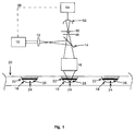
- FIG. 1 shows a pulsed semiconductor laser 10 with a wavelength of 635 nm.
- a pulsed semiconductor laser 10 With a wavelength of 635 nm.
- its beam is coupled into a microscope objective 16 in a reflected light arrangement via a lens 12 and a beam splitter 14.
- the microscope objective 16 focuses the laser beam in a reaction trough 18 of a microtiter plate 20.
- the reaction trough 18 there is an upper edge Ring electrode 22 and at the foot of the reaction trough 18 an electrode tip 24.
- the beam of a semiconductor laser 10 is focused in such a way that its focal point lies in the interior of the solution 18 near the electrode tip 24.
- a patient is used to detect any tumor cells from breast cancer Blood drawn.
- Serum 26 is separated from the blood. Serum 26 is filled into a reaction well 18 of the microtiter plate 20. In the reaction trough 18, the serum 26 is in contact with the ring electrode 22 and the electrode tip 24 at the foot of the reaction trough 18.
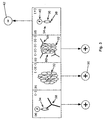
- FIG. 2 (a) shows a human anti-MUC1 antibody 28, as found, for example, in the blood serum 26 of a patient can be located in the reaction trough 18. His charges are at the top right of the figure indicated.
- the human antibody 28 is e.g. at pH values between 7 and 9 charged twice negatively and therefore moves to the anode when voltage is applied 30th
- Fig. 2 (b) shows a MUC1 glycoprotein (tumor marker) 32, which is also in the patient's blood serum 26. His loads are in the top right hinted at the figure.
- the tumor marker 32 is neutral or slightly basic The pH value is also negatively charged twice and therefore moves when it is applied Voltage also to the anode 30.
- the MUC1 glycoprotein is characterized by 30 to 90 peptide sequences of 20 amino acids arranged in tandem one behind the other.
- the 20 amino acid sequence of the tandem repeat is: PDTRPAPGS TAPPAHGV TSA, ie proline-aspartic acid-threonine-arginine-proline-alanine-proline-glycine- serine- threonine-atanine-proline-proline-alanine-histidine-glycine-valine- threonine Serine alanine.
- the bold section forms the epitope STAPPAHGV, ie serine-threonine-alanine-proline-proline-alanine-histidine-glycine-valine.
- the epitope is the immune system's key to MUC1, to which most of the previously developed MUC1-specific monoclonal antibodies bind.
- the substance 34 shown in FIG. 2 (c) must be added in excess to the serum 26.
- 2 (c) shows a peptide 36 containing the immunogenic epitope of the tandem repeat of MUC1.
- the peptide 36 consists of a variable number from 3 to 40 amino acids. In the preferred exemplary embodiment, 20 amino acids were selected, specifically the sequence of the tandem repeat of the MUC1 glycoprotein comprising 20 amino acids.
- sequence of peptide 36 is therefore: PDTRPAPGSTAPPAHGVTSA, ie proline-aspartic acid-threonine-arginine-proline-alanine-proline-glycine-serine-threonine-alanine-proline-proline-alanine-histidine-glycine-valine-threonine-serine-alanine.
- Lysine residues 38 had a double positive charge. It can also be caused by additional arginine residues or histidine residues are charged positively. Amino acids are generally suitable for this with a pK value of the side chain of more than 9.
- the modified peptide 36 is synthesized using generally known methods Method.
- a Cy5 molecule 40 is coupled to the carboxylic acid residue of the Cy5 is converted into an active ester and under basic Conditions to form a peptide bond with amino functions of the peptide connected.
- the dye can be coupled by changing the reaction conditions also about the amino functions of the additional lysine or arginine residues or via the amino functions of additionally coupled cysteine residues (-SH).
- Cy5 is in the solution at pH values between 7 and 11 and after coupling simply negatively charged electrically.
- the net electrical charge of the dye-labeled MUC1 epitops 34 is therefore simply positive.
- the epitope can be caused by aspartic acid residues or glutamic acid residues be negatively charged.
- the molecule 34 prepared in the preferred embodiment is shown in FIG Detail shown.
- the charge of the molecule 34 prepared in the preferred embodiment is indicated at the top right in Fig. 2 (c).
- the molecule is at pH values between 7 and 11 34 e.g. just positively charged. It therefore migrates to cathode 42.
- Figure 2 (d) shows the antigen-antibody complex 44 of dye-labeled MUC1 epitope 34 and human anti-MUC1 antibody 28. Its charge is up indicated on the right in the figure. Complex 44 is at pH values between 7 and 9 simply negatively charged and therefore moves to the anode when voltage is applied 30th
- Figure 2 (e) shows the antigen-antibody complex 46 from MUC1 tumor marker 32 and human anti-MUC1 antibody 28. Its charge is at the top right of the figure indicated. Complex 46 is fourfold negative at pH values between 7 and 9 charged and therefore moves to anode 30 when voltage is applied.
- the free dye-labeled MUC1 epitope 34 is drawn to the cathode 42, while all other substances, especially the bound dye-labeled MUC1 epitope 34, are drawn to anode 30.
- the detection of the fluorescence signal is confocal via the above Microscope objective 16.
- the light collected by microscope objective 16 passes through Beam splitter 14. This is designed such that it receives light at the excitation wavelength of 635 nm and the red-shifted fluorescent light is transmitted.
- the fluorescent light passes through a dielectric one Bandpass filter 48.
- a pinhole 50 is used that arises outside the laser focus.
- the collected Fluorescence light is detected by means of an avalanche photodiode 52 and converted into electrical impulses.
- the avalanche photodiode 52 is suitable for individual photons with a temporal Detect resolution below 1 ns. It is therefore for the time-resolved Suitable for single photon counting.
- the signals of the avalanche photodiode 52 are in a data processing unit 54 further processed via a connecting line 56 to the semiconductor laser 10 is connected to receive trigger pulses from this.
- the laser focus in the reaction trough 18 just above the electrode tip 24 has a volume in the femtoliter range.
- the low detection volume allows the detection of individual dye-labeled complexes.
- the concentration of MUC1 complexes in the solution can therefore be several orders of magnitude below 10 -9 mol, for example 10 -13 mol.
- MUC1 glycoprotein (tumor marker) 32 is detected in many Steps analogous to the detection of human anti-MUC1 antibodies 28 according to the first embodiment. Therefore, only the differences are highlighted.
- FIG. 3. 3 (a) shows a sugar-specific, monoclonal anti-MUC1 antibody 58, e.g. BM7 or BM8 (E. Petrakou, A. Murray, M.R. Price: "Epitope Mapping of Anti-MUC1 Mucin Protein Core Monoclonal Antibodies ", Tumor Biology Volume 19 (1998) pp. 21-29), which is labeled with dye MUC1 epitope 34 is loaded. This complex is in the serum in the Reaction well 18 added. His charges are at the top right of the figure indicated.
- the dye-loaded antibody 58 is at pH values between 7 and 9 simply negatively charged and therefore moves to the anode when voltage is applied 30th
- FIG. 3 (b) corresponds to FIG. 2 (b).
- the tumor markers 32 are now displacing in the serum - depending on theirs Concentration - the dye-labeled MUC1 epitope 34 partially from the antigen binding sites of the monoclonal anti-MUC1 antibodies 58. They form the in Fig. 3 (c) shown antigen-antibody complex 60 from MUC1 tumor marker 32 and monoclonal anti-MUC1 antibody 58.
- FIG. 3 (c) corresponds otherwise to FIG. 2 (e).
- the competitive displacement reaction makes dye-labeled MUC1 epitopes 34 released as shown in Fig. 3 (d). 3 (d) corresponds to FIG. 2 (c).
- the released dye-labeled MUC1 epitope 34 is at neutral or slightly basic pH value positively charged and migrate to the cathode 42. All others The substances shown migrate to the anode 30.
- the ring electrode 22 forms in the reaction wells 18 of the microtiter plate 20 in the second embodiment, therefore, the positive pole (anode 30), while the Electrode tip 24 forms the cathode 42 at the foot of the reaction troughs 18.
- the positive pole anode 30
- the Electrode tip 24 forms the cathode 42 at the foot of the reaction troughs 18.
- the antigen-antibody complex 60 and the dye-loaded antibody 58 migrate due to their negative excess charge to the ring electrode 22. Only the released dye-labeled MUC1 epitope 34 migrate to the tip electrode due to their positive landing 24 near the detection volume.
- first, second and third molecules on the binding between an antibody and the associated antigen or epitope limited.
- the binding can also be realized by a biotin-streptavidin binding become. Any other binding that is suitable in principle for a competitive test can be used. Therefore, the first, second and third molecules also not be proteins.
- the device shown in FIG. 1 can be used in addition to the ring electrodes 22 can also be realized with two pointed electrodes, an anode and a cathode, which are inserted from below through the microtiter plate into the reaction well 18. This is particularly possible with polycarbonate microtiter plates.
- the human anti-MUC1 antibody 28 was negatively charged twice. Other antibodies are positively charged. In general, however, any antibody can be negatively charged, so that it has a negative excess charge.
- a succinimidyl ester (such as a bifunctional one such as GMBS) can be coupled to the NH 2 functions of the antibody. This converts the NH 2 functions into COO - / H + functions.
Landscapes
- Chemical & Material Sciences (AREA)
- Health & Medical Sciences (AREA)
- Life Sciences & Earth Sciences (AREA)
- Molecular Biology (AREA)
- Organic Chemistry (AREA)
- Medicinal Chemistry (AREA)
- General Health & Medical Sciences (AREA)
- Biochemistry (AREA)
- Engineering & Computer Science (AREA)
- Genetics & Genomics (AREA)
- Immunology (AREA)
- Biophysics (AREA)
- Proteomics, Peptides & Aminoacids (AREA)
- Biomedical Technology (AREA)
- Analytical Chemistry (AREA)
- Urology & Nephrology (AREA)
- Hematology (AREA)
- Biotechnology (AREA)
- Microbiology (AREA)
- Cell Biology (AREA)
- Food Science & Technology (AREA)
- Physics & Mathematics (AREA)
- General Physics & Mathematics (AREA)
- Pathology (AREA)
- Toxicology (AREA)
- Zoology (AREA)
- Gastroenterology & Hepatology (AREA)
- Investigating Or Analysing Biological Materials (AREA)
- Investigating, Analyzing Materials By Fluorescence Or Luminescence (AREA)
- Peptides Or Proteins (AREA)
Abstract
Description
- Fig. 1
- eine schematische Darstellung der erfindungsgemäßen Vorrichtung;
- Fig. 2
- eine schematische Darstellung der am Nachweis von anti-MUC1 Antikörpern beteiligten Moleküle, ihrer Ladungen und Wanderungsbewegung;
- Fig. 3
- eine schematische Darstellung der am Nachweis von MUC1-Glykoprotein (Tumormarker) beteiligten Moleküle, ihrer Ladungen und Wanderungsbewegung; und
- Fig. 4
- eine schematische Darstellung des farbstoffmarkierten Peptids gemäß dem bevorzugten Ausführungsbeispiel mit der zugehörigen Peptidsequenz.
Ringelektrode 22 und am Fuß der Reaktionsmulde 18 eine Elektrodenspitze 24. Mit Hilfe des Mikroskopobjektivs 16 wird der Strahl eines Halbleiterlasers 10 so fokussiert, daß dessen Brennpunkt im Inneren der Lösung 18 nahe der Elektrodenspitze 24 liegt.
PDTRPAPGSTAPPAHGVTSA, d.h. Prolin-Asparaginsäure-Threonin-Arginin-Prolin-Alanin-Prolin-Glycin-Serin-Threonin-Alanin-Prolin-Prolin-Alanin-Histidin-Glycin-Valin-Threonin-Serin-Alanin.
- 10
- Halbleiterlaser
- 12
- Linse
- 14
- Strahlteiler
- 16
- Mikroskopobjektiv
- 18
- Reaktionsmulde
- 20
- Mikrotiterplatte
- 22
- Ringelektrode
- 24
- Elektrodenspitze
- 26
- Serum
- 28
- humaner anti-MUC1 Antikörper
- 30
- Anode
- 32
- MUC1-Glykoprotein (Tumormarker)
- 34
- farbstoffmarkiertes MUC1-Epitop
- 36
- Peptid, das das immunogene Epitop des Tandemrepeats von MUC1-Glykoprotein 32 enthält
- 38
- Lysinreste
- 40
- Farbstoff Cy5
- 42
- Kathode
- 44
- Antigen-Antikörper-Komplex 44 aus farbstoffmarkiertem MUC1-Epitop 36 und humanem anti-MUC1 Antikörper 28
- 46
- Antigen-Antikörper-Komplex 46 aus MUC1-Tumormarker 32 und humanem anti-MUC1 Antikörper 28
- 48
- Bandpaßfilter
- 50
- Lochblende
- 52
- Avalanche-Photodiode
- 54
- Datenverarbeitungseinheit
- 56
- Verbindungsleitung zwischen Datenverarbeitungseinheit 54 und Halbleiterlaser 10
- 58
- zuckerspezifischer, monoklonaler anti-MUC1 Antikörper
- 60
- Antigen-Antikörper-Komplex aus MUC1-Tumormarker 32 und monoklonalem anti-MUC1 Antikörper 58



Claims (17)
- Verfahren zum Nachweis eines ersten Moleküls (28) in einer Lösung (26), wobei das erste Molekül in der Lösung an ein zweites Molekül (36) binden kann, mit folgenden Schritten:a) ein Farbstoff (40) wird an das zweite Molekül (36) gekoppelt;b) die elektrische Nettoladung in der Lösung (26) des farbstoffinarkierten zweiten Moleküls (36) wird mit entgegengesetztem Vorzeichen und dem Betrag nach kleiner als die elektrische Nettoladung in der Lösung des ersten Moleküls (28) gewählt;c) das farbstoffmarkierte zweite Molekül (34) wird der Lösung (26) zugegeben;d) ein elektrisches Feld wird zwischen einer Anode (30) und einer Kathode (42) durch die Lösung (26) geführt; unde) der Farbstoff (40) wird, falls die elektrische Nettoladung in der Lösung des ersten Moleküls (28) negativ ist, an der Anode (30) und, falls die elektrische Nettoladung in der Lösung des ersten Moleküls (28) positiv ist, an der Kathode (42) optisch nachgewiesen.
- Verfahren zum Nachweis eines dritten Moleküls (32) in einer Lösung (26), wobei das dritte Molekül (32) in der Lösung an ein erstes Molekül (58) und an ein zweites Molekül (36) binden kann, mit folgenden Schritten:f) ein Farbstoff (40) wird an das zweite Molekül (36) gekoppelt;g) die elektrische Nettoladung in der Lösung (26) des farbstoffmarkierten zweiten Moleküls (36) wird mit entgegengesetztem Vorzeichen und dem Betrag nach kleiner als die elektrische Nettoladung in der Lösung des ersten Moleküls (58) gewählt;h) gebundene Komplexe aus erstem Molekül (58) und zweitem Molekül (34) werden hergestellt;i) die gebundenen Komplexe werden der Lösung (26) zugegeben;j) ein elektrisches Feld wird zwischen einer Anode (30) und einer Kathode (42) durch die Lösung (26) geführt; undk) der Farbstoff (40) wird, falls die elektrische Nettoladung in der Lösung des ersten Moleküls (58) negativ ist, an der Kathode (42) und, falls die elektrische Nettoladung des ersten Moleküls (28) positiv ist, an der Anode (30) optisch nachgewiesen.
- Verfahren nach Anspruch 1 oder 2, dadurch gekennzeichnet,daß als zweites Molekül (36) ein Peptid gewählt wird; unddaß in Schritt b) bzw. g) die elektrische Nettoladung in der Lösung (26) des Peptids (36) abhängig von der elektrischen Nettoladung in der Lösung des ersten Moleküls (28, 58) durch zusätzliche, bei neutralem oder leicht basischem pH-Wert positiv oder negativ geladene Aminosäuren (38) verändert wird.
- Verfahren nach einem der Ansprüche 1 bis 3, dadurch gekennzeichnet, daß in Schritt b) bzw. g) die elektrische Nettoladung in der Lösung (26) des zweiten Moleküls (36) abhängig von der elektrischen Nettoladung in der Lösung des ersten Moleküls (28, 58) dadurch verändert wird, daß der Farbstoff (40) derart gewählt wird, daß seine elektrische Nettoladung in der Lösung positiv oder negativ ist.
- Verfahren nach einem der Ansprüche 1 bis 4, dadurch gekennzeichnet,daß als erstes Molekül (28, 58) ein Antikörper und als drittes Molekül (32) ein zugehöriges Antigen gewählt wird; unddaß das zweite Molekül (36) als Peptid gewählt wird, dessen Peptidsequenz mindestens 3 aufeinanderfolgende Aminosäuren der einem immunogenen Epitop entsprechenden Peptidsequenz des Antigens (32) aufweist.
- Verfahren nach Anspruch 5, dadurch gekennzeichnet, daß die elektrische Nettoladung in der Lösung (26) des Peptids (36) durch 2 bis 4 zusätzliche Aminoreste (38) positiv aufgeladen wird.
- Vorrichtung zum Nachweis eines Moleküls (28, 32) in einer Lösung (26) mit:l) einer Lichtquelle (10) einer vorgegebenen Wellenlänge;m) einer Mikrotiterplatte (20) mit einer Mehrzahl von Reaktionsmulden (18);n) einem Paar von Elektroden (22, 24) in jeder Reaktionsmulde (18); undo) einer auf ein Volumen nahe einer der beiden Elektroden (22, 24) gerichteten optischen Einrichtung (12, 14, 16, 48, 50, 52, 54, 56) zum Detektieren von Licht aus dem Volumen.
- Vorrichtung nach Anspruch 7, dadurch gekennzeichnet,daß am oberen Rand jeder Reaktionsmulde (18) eine Ringelektrode (22) und im Inneren jeder Reaktionsmulde eine Elektrodenspitze (24) angeordnet ist; unddaß die optische Einrichtung (12, 14, 16, 48, 50, 52, 54, 56) auf ein Volumen nahe der Elektrodenspitze (24) ausgerichtet ist.
- Farbstoffmarkiertes Peptid (34) herstellbar durch folgende Schritte:p) die einem immunogenen Epitop eines Antigens (32) entsprechende Peptidsequenz (36) wird bestimmt;q) ein Peptid (36) wird aus mindestens 3 aufeinanderfolgenden Aminosäuren der dem immunogenen Epitop entsprechenden Peptidsequenz synthetisiert;r) ein Farbstoff (40) wird an das Peptid (36) gekoppelt;s) die elektrische Nettoladung eines in einer Lösung (26) gelösten Antikörpers (28, 58) gegen das Antigen (32) und die elektrische Nettoladung in der Lösung des farbstoffmarkierten Peptids (36) wird bestimmt; undt) die elektrische Nettoladung in der Lösung (26) des farbstoffmarkierten Peptids (36) wird mit entgegengesetztem Vorzeichen und dem Betrag nach kleiner als die elektrische Nettoladung in der Lösung des Antikörpers (28, 58) gewählt.
- Farbstoffmarkiertes Peptid (34) nach Anspruch 9, dadurch gekennzeichnet, daß in Schritt t) die elektrische Nettoladung in der Lösung (26) des Peptids (36) abhängig von der elektrischen Nettoladung in der Lösung des Antikörpers (28, 58) durch zusätzliche, bei neutralem oder leicht basischem pH-Wert positiv oder negativ geladene Aminosäuren (38) verändert wird.
- Farbstoffmarkiertes Peptid (34) nach Anspruch 9 oder 10, dadurch gekennzeichnet, daß in Schritt t) die elektrische Nettoladung in der Lösung (26) des Peptids (36) abhängig von der elektrischen Nettoladung in der Lösung des Antikörpers (28, 58) dadurch verändert wird, daß der Farbstoff (40) derart gewählt wird, daß seine elektrische Nettoladung in der Lösung positiv oder negativ ist.
- Farbstoffmarkiertes Peptid (34) nach einem der Ansprüche 9 bis 11, dadurch gekennzeichnet, daß die elektrische Nettoladung in der Lösung (26) des Peptids (36) durch 2 bis 4 zusätzliche Aminoreste (38) positiv aufgeladen ist.
- Verfahren zur Herstellung eines farbstoffmarkierten Peptids (36) mit folgenden Schritten:u) die einem immunogenen Epitop eines Antigens (32) entsprechende Peptidsequenz (36) wird bestimmt;v) ein Peptid (36) wird aus mindestens 3 aufeinanderfolgenden Aminosäuren der dem immunogenen Epitop entsprechenden Peptidsequenz synthetisiert;w) ein Farbstoff (40) wird an das Peptid (36) gekoppelt;x) die elektrische Nettoladung eines in einer Lösung (26) gelösten Antikörpers (28, 58) gegen das Antigen (32) und die elektrische Nettoladung in der Lösung des farbstoffmarkierten Peptids (36) wird bestimmt; undy) die elektrische Nettoladung in der Lösung (26) des farbstoffhiarkierten Peptids (36) wird mit entgegengesetztem Vorzeichen und dem Betrag nach kleiner als die elektrische Nettoladung in der Lösung des Antikörpers (28, 58) gewählt.
- Verfahren nach Anspruch 13, dadurch gekennzeichnet, daß in Schritt y) die elektrische Nettoladung in der Lösung (26) des Peptids (36) abhängig von der elektrischen Nettoladung in der Lösung des Antikörpers (28, 58) durch zusätzliche, bei neutralem oder leicht basischem pH-Wert positiv oder negativ geladene Aminosäuren (38) verändert wird.
- Verfahren nach Anspruch 13 oder 14, dadurch gekennzeichnet, daß in Schritt y) die elektrische Nettoladung in der Lösung (26) des Peptids (36) abhängig von der elektrischen Nettoladung in der Lösung des Antikörpers (28, 58) dadurch verändert wird, daß der Farbstoff (40) derart gewählt wird, daß seine elektrische Nettoladung in der Lösung positiv oder negativ ist.
- Verfahren nach einem der Ansprüche 13 bis 15, dadurch gekennzeichnet, daß die elektrische Nettoladung in der Lösung (26) des Peptids (36) durch 2 bis 4 zusätzliche Aminoreste (38) positiv aufgeladen wird.
- Verwendung des farbstoffmarkierten Peptids (34) nach einem der Ansprüche 9 bis 12 zum Nachweis eines ersten oder dritten Moleküls (28, 32) in einer Lösung (26) mittels des Verfahrens nach einem der Ansprüche 1 bis 6.
Applications Claiming Priority (2)
| Application Number | Priority Date | Filing Date | Title |
|---|---|---|---|
| DE19952160 | 1999-10-29 | ||
| DE1999152160 DE19952160A1 (de) | 1999-10-29 | 1999-10-29 | Verfahren, Vorrichtung und farbstoffmarkiertes Peptid zum Nachweis eines Moleküls, sowie Verfahren zur Herstellung eines farbstoffmarkierten Peptids und dessen Verwendung |
Publications (2)
| Publication Number | Publication Date |
|---|---|
| EP1096257A2 true EP1096257A2 (de) | 2001-05-02 |
| EP1096257A3 EP1096257A3 (de) | 2003-01-15 |
Family
ID=7927325
Family Applications (1)
| Application Number | Title | Priority Date | Filing Date |
|---|---|---|---|
| EP00123053A Withdrawn EP1096257A3 (de) | 1999-10-29 | 2000-10-24 | Verfahren, Vorrichtung und farbstoffmarkiertes Peptid zum Nachweis eines Moleküls |
Country Status (2)
| Country | Link |
|---|---|
| EP (1) | EP1096257A3 (de) |
| DE (1) | DE19952160A1 (de) |
Cited By (1)
| Publication number | Priority date | Publication date | Assignee | Title |
|---|---|---|---|---|
| WO2003048178A3 (en) * | 2001-11-29 | 2003-11-27 | Amersham Biosciences Uk Ltd | Nucleotide analogues and their use for nucleic acid sequencing |
Families Citing this family (1)
| Publication number | Priority date | Publication date | Assignee | Title |
|---|---|---|---|---|
| DE10127045C2 (de) * | 2001-06-02 | 2003-10-30 | November Ag Molekulare Medizin | Verfahren zum Nachweis einer Substanz und Mikrotiterplatte |
Family Cites Families (9)
| Publication number | Priority date | Publication date | Assignee | Title |
|---|---|---|---|---|
| US5019521A (en) * | 1990-03-12 | 1991-05-28 | Photest Diagnostics, Inc. | Paired polypeptides |
| WO1994003583A1 (en) * | 1992-07-29 | 1994-02-17 | Oncotherapeutics | An improved system for electronically monitoring and recording cell cultures |
| US5215883A (en) * | 1990-07-09 | 1993-06-01 | The Research Foundation | Electrophoretic mobility of fluorophore labeled particles in gels by fluorophore movement after photobleaching |
| WO1996027025A1 (en) * | 1995-02-27 | 1996-09-06 | Ely Michael Rabani | Device, compounds, algorithms, and methods of molecular characterization and manipulation with molecular parallelism |
| DE19646505A1 (de) * | 1996-11-12 | 1998-05-14 | Itt Ind Gmbh Deutsche | Vorrichtung zur Durchführung von Untersuchungen an Zellproben und dergleichen |
| DE19718016A1 (de) * | 1997-04-29 | 1998-11-05 | Drexhage Karl Heinz | Verfahren zur optischen Detektion von Analytmolekülen in einem natürlichen biologischen Medium |
| DE19751706C2 (de) * | 1997-11-21 | 1999-10-21 | Meinhard Knoll | Vorrichtung und Verfahren zum Nachweis von Analyten |
| DE19803753C1 (de) * | 1998-01-30 | 1999-12-02 | Max Planck Gesellschaft | Vorrichtung und Verfahren zur Kapillarelektrophorese |
| DE19826153C2 (de) * | 1998-06-12 | 2002-11-07 | November Ag Molekulare Medizin | Verfahren und Vorrichtung zum Nachweis einer in einer Probe ggf. enthaltenen Nukleotidsequenz |
-
1999
- 1999-10-29 DE DE1999152160 patent/DE19952160A1/de not_active Withdrawn
-
2000
- 2000-10-24 EP EP00123053A patent/EP1096257A3/de not_active Withdrawn
Cited By (1)
| Publication number | Priority date | Publication date | Assignee | Title |
|---|---|---|---|---|
| WO2003048178A3 (en) * | 2001-11-29 | 2003-11-27 | Amersham Biosciences Uk Ltd | Nucleotide analogues and their use for nucleic acid sequencing |
Also Published As
| Publication number | Publication date |
|---|---|
| DE19952160A1 (de) | 2001-05-03 |
| EP1096257A3 (de) | 2003-01-15 |
Similar Documents
| Publication | Publication Date | Title |
|---|---|---|
| EP1287348B1 (de) | Verfahren und vorrichtung zur qualitativen und/oder quantitativen bestimmung eines protein- und/oder peptidmusters einer flüssigkeitsprobe, die dem menschlichen oder tierischen körper entnommen wird | |
| WO2022079260A1 (de) | Verfahren und fluoreszenzmikroskop zur ortsbestimmung einzelner fluoreszierender farbstoffmoleküle durch adaptive abtastung | |
| EP0979402B1 (de) | Verfahren zur optischen detektion von analytmolekülen in einem natürlichen biologischen medium | |
| WO2004090546A1 (de) | Bestimmung eines midregionalen proadrenomedullin-teilpeptids in biologischen flüssigkeiten zu diagnostischen zwecken sowie immunoassays für die durchführung einer solchen bestimmung | |
| WO2022136361A1 (de) | Verfahren und mikroskop zur aufnahme von trajektorien einzelner partikel in einer probe | |
| DE102011057021A1 (de) | Verfahren zur selektiven Quantifizierung von A-Beta-Aggregaten | |
| EP2142911A2 (de) | Biochip für die fluoreszenzanalyse von einzelnen transportern | |
| EP3639024A1 (de) | Verfahren zum nachweis von extrazellulären vesikeln in einer probe | |
| DE19903576C2 (de) | Quantitative Bestimmung von Analyten in einem heterogenen System | |
| EP0765470A2 (de) | Verfahren und vorrichtung zur gezielten entnahme von komponenten aus komplexen mischungen | |
| EP1999457A1 (de) | Verfahren zur bestimmung von molekülen oder molekülteilen in biologischen proben | |
| EP1096257A2 (de) | Verfahren, Vorrichtung und farbstoffmarkiertes Peptid zum Nachweis eines Moleküls | |
| DE202018005287U1 (de) | Vorrichtung zur Bestimmung der Fluoreszenz und der Anzahl von Antikörpern auf Exosomen | |
| DE10103304A1 (de) | Verfahren zur fluoreszenzspektroskopischen, insbesondere fluoreszenzkorrelationsspektroskopischen Untersuchung einer Messprobe sowie Einrichtung zur Durchführung desselben | |
| DE112019000209T5 (de) | Immunfärbeverfahren, Immunfärbesystem und Immunfärbekit | |
| DE102019005761A1 (de) | Neuer Testkit, enthaltend Pankreas-Elastase-1-spezifische Antikörper sowie eine Probenvorbereitungsvorrichtung | |
| EP2986985A2 (de) | Automatisierbares verfahren zur identifizierung, quantifizierung und diskriminierung von spezifischen signalen gegenüber unspezifischen signalen in nachweisverfahren mittels detektor | |
| EP1440315B1 (de) | Nachweis humaner auto-antikörper | |
| DE102007005147A1 (de) | Verfahren und Vorrichtung zur Untersuchung der Anheftung oder Ablösung lebender oder toter Zellen oder zellähnlicher Partikel oder sonstiger Oberflächenbelegung an Oberflächen mittels Plasmonenresonanz sowie Verwendung dieses Verfahrens und dieser Vorrichtung | |
| DE102017010455A1 (de) | Verfahren zum Nachweis von Aggregaten biotherapeutischer Substanzen in einer Probe | |
| DE102018008980B4 (de) | Vorrichtung und Verfahren zur Bestimmung der Fluoreszenz und der Anzahl von Antikörpern auf Exosomen. | |
| DE10320956B4 (de) | Untersuchungsverfahren für biologische Zellen und zugehörige Untersuchungseinrichtung | |
| WO2003072699A2 (de) | Vorrichtung und verfahren zur bestimmung der relativen syntheseleistung einzelner zellen und/oder von positionskoordinaten | |
| WO2002003061A2 (de) | Verfahren zur identifizierung von die für behandlung von virusinfektionen geeigneten stoffen | |
| DE102022109027A1 (de) | Verfahren zum Kartieren der Oberfläche eines Makromoleküls |
Legal Events
| Date | Code | Title | Description |
|---|---|---|---|
| PUAI | Public reference made under article 153(3) epc to a published international application that has entered the european phase |
Free format text: ORIGINAL CODE: 0009012 |
|
| AK | Designated contracting states |
Kind code of ref document: A2 Designated state(s): AT BE CH CY DE DK ES FI FR GB GR IE IT LI LU MC NL PT SE |
|
| AX | Request for extension of the european patent |
Free format text: AL;LT;LV;MK;RO;SI |
|
| PUAL | Search report despatched |
Free format text: ORIGINAL CODE: 0009013 |
|
| AK | Designated contracting states |
Kind code of ref document: A3 Designated state(s): AT BE CH CY DE DK ES FI FR GB GR IE IT LI LU MC NL PT SE |
|
| AX | Request for extension of the european patent |
Free format text: AL;LT;LV;MK;RO;SI |
|
| 17P | Request for examination filed |
Effective date: 20030610 |
|
| AKX | Designation fees paid |
Designated state(s): AT BE CH CY DE DK ES FI FR GB GR IE IT LI LU MC NL PT SE |
|
| 17Q | First examination report despatched |
Effective date: 20050609 |
|
| STAA | Information on the status of an ep patent application or granted ep patent |
Free format text: STATUS: THE APPLICATION IS DEEMED TO BE WITHDRAWN |
|
| 18D | Application deemed to be withdrawn |
Effective date: 20050503 |