EP1051619B1 - Verfahren zum identifizieren von t-zell-stimulierenden proteinfragmenten - Google Patents
Verfahren zum identifizieren von t-zell-stimulierenden proteinfragmenten Download PDFInfo
- Publication number
- EP1051619B1 EP1051619B1 EP99930888A EP99930888A EP1051619B1 EP 1051619 B1 EP1051619 B1 EP 1051619B1 EP 99930888 A EP99930888 A EP 99930888A EP 99930888 A EP99930888 A EP 99930888A EP 1051619 B1 EP1051619 B1 EP 1051619B1
- Authority
- EP
- European Patent Office
- Prior art keywords
- cell
- cells
- protein
- identification
- protein fragment
- Prior art date
- Legal status (The legal status is an assumption and is not a legal conclusion. Google has not performed a legal analysis and makes no representation as to the accuracy of the status listed.)
- Revoked
Links
- 239000012634 fragment Substances 0.000 title claims description 130
- 102000004169 proteins and genes Human genes 0.000 title claims description 130
- 108090000623 proteins and genes Proteins 0.000 title claims description 130
- 238000000034 method Methods 0.000 title claims description 56
- 230000004936 stimulating effect Effects 0.000 title claims description 35
- 210000001744 T-lymphocyte Anatomy 0.000 claims description 120
- 108090000765 processed proteins & peptides Proteins 0.000 claims description 59
- 210000004027 cell Anatomy 0.000 claims description 48
- 150000001413 amino acids Chemical class 0.000 claims description 42
- 108090000695 Cytokines Proteins 0.000 claims description 26
- 102000004127 Cytokines Human genes 0.000 claims description 26
- 102000004196 processed proteins & peptides Human genes 0.000 claims description 26
- 108091007433 antigens Proteins 0.000 claims description 25
- 102000036639 antigens Human genes 0.000 claims description 25
- 239000000427 antigen Substances 0.000 claims description 23
- 239000000725 suspension Substances 0.000 claims description 19
- 230000000638 stimulation Effects 0.000 claims description 18
- 230000004913 activation Effects 0.000 claims description 13
- 238000011534 incubation Methods 0.000 claims description 12
- 239000003550 marker Substances 0.000 claims description 10
- 210000004369 blood Anatomy 0.000 claims description 8
- 239000008280 blood Substances 0.000 claims description 8
- 239000003814 drug Substances 0.000 claims description 7
- 108010074328 Interferon-gamma Proteins 0.000 claims description 6
- 238000000684 flow cytometry Methods 0.000 claims description 6
- 102000035118 modified proteins Human genes 0.000 claims description 6
- 108091005573 modified proteins Proteins 0.000 claims description 6
- 230000008569 process Effects 0.000 claims description 6
- 125000006239 protecting group Chemical group 0.000 claims description 6
- 238000010532 solid phase synthesis reaction Methods 0.000 claims description 6
- 102000043129 MHC class I family Human genes 0.000 claims description 5
- 108091054437 MHC class I family Proteins 0.000 claims description 5
- 241001465754 Metazoa Species 0.000 claims description 5
- 239000007791 liquid phase Substances 0.000 claims description 5
- 230000002093 peripheral effect Effects 0.000 claims description 5
- 102000008070 Interferon-gamma Human genes 0.000 claims description 4
- 230000001413 cellular effect Effects 0.000 claims description 4
- 238000000586 desensitisation Methods 0.000 claims description 4
- 230000006870 function Effects 0.000 claims description 4
- 229960003130 interferon gamma Drugs 0.000 claims description 4
- 210000000265 leukocyte Anatomy 0.000 claims description 4
- 238000012986 modification Methods 0.000 claims description 4
- 230000004048 modification Effects 0.000 claims description 4
- 230000035755 proliferation Effects 0.000 claims description 4
- 210000001519 tissue Anatomy 0.000 claims description 4
- 238000002255 vaccination Methods 0.000 claims description 4
- 108060008682 Tumor Necrosis Factor Proteins 0.000 claims description 3
- 102000000852 Tumor Necrosis Factor-alpha Human genes 0.000 claims description 3
- 238000004113 cell culture Methods 0.000 claims description 3
- 210000000170 cell membrane Anatomy 0.000 claims description 3
- 230000008030 elimination Effects 0.000 claims description 3
- 238000003379 elimination reaction Methods 0.000 claims description 3
- 238000002360 preparation method Methods 0.000 claims description 3
- 230000014616 translation Effects 0.000 claims description 3
- 241000206602 Eukaryota Species 0.000 claims description 2
- 108010002350 Interleukin-2 Proteins 0.000 claims description 2
- 210000004899 c-terminal region Anatomy 0.000 claims description 2
- 210000001175 cerebrospinal fluid Anatomy 0.000 claims description 2
- 238000012217 deletion Methods 0.000 claims description 2
- 230000037430 deletion Effects 0.000 claims description 2
- 238000003780 insertion Methods 0.000 claims description 2
- 230000037431 insertion Effects 0.000 claims description 2
- 210000001165 lymph node Anatomy 0.000 claims description 2
- 238000006467 substitution reaction Methods 0.000 claims description 2
- 230000002194 synthesizing effect Effects 0.000 claims description 2
- 125000003275 alpha amino acid group Chemical group 0.000 claims 5
- MZOFCQQQCNRIBI-VMXHOPILSA-N (3s)-4-[[(2s)-1-[[(2s)-1-[[(1s)-1-carboxy-2-hydroxyethyl]amino]-4-methyl-1-oxopentan-2-yl]amino]-5-(diaminomethylideneamino)-1-oxopentan-2-yl]amino]-3-[[2-[[(2s)-2,6-diaminohexanoyl]amino]acetyl]amino]-4-oxobutanoic acid Chemical compound OC[C@@H](C(O)=O)NC(=O)[C@H](CC(C)C)NC(=O)[C@H](CCCN=C(N)N)NC(=O)[C@H](CC(O)=O)NC(=O)CNC(=O)[C@@H](N)CCCCN MZOFCQQQCNRIBI-VMXHOPILSA-N 0.000 claims 1
- 102000000588 Interleukin-2 Human genes 0.000 claims 1
- 210000001185 bone marrow Anatomy 0.000 claims 1
- -1 i.e. Proteins 0.000 claims 1
- 210000004988 splenocyte Anatomy 0.000 claims 1
- 238000002560 therapeutic procedure Methods 0.000 claims 1
- 235000018102 proteins Nutrition 0.000 description 96
- 229940024606 amino acid Drugs 0.000 description 20
- 235000001014 amino acid Nutrition 0.000 description 19
- 239000000203 mixture Substances 0.000 description 10
- 238000004519 manufacturing process Methods 0.000 description 8
- YGNUDKAPJARTEM-GUBZILKMSA-N Met-Val-Ala Chemical compound CSCC[C@H](N)C(=O)N[C@@H](C(C)C)C(=O)N[C@@H](C)C(O)=O YGNUDKAPJARTEM-GUBZILKMSA-N 0.000 description 7
- 102000043131 MHC class II family Human genes 0.000 description 6
- 108091054438 MHC class II family Proteins 0.000 description 6
- QGVBFDIREUUSHX-IFFSRLJSSA-N Thr-Val-Gln Chemical compound [H]N[C@@H]([C@@H](C)O)C(=O)N[C@@H](C(C)C)C(=O)N[C@@H](CCC(N)=O)C(O)=O QGVBFDIREUUSHX-IFFSRLJSSA-N 0.000 description 6
- 230000008901 benefit Effects 0.000 description 5
- 238000007796 conventional method Methods 0.000 description 4
- 108010078144 glutaminyl-glycine Proteins 0.000 description 4
- IETUUAHKCHOQHP-KZVJFYERSA-N Ala-Thr-Val Chemical compound CC(C)[C@H](NC(=O)[C@@H](NC(=O)[C@H](C)N)[C@@H](C)O)C(O)=O IETUUAHKCHOQHP-KZVJFYERSA-N 0.000 description 3
- BULIVUZUDBHKKZ-WDSKDSINSA-N Gly-Gln-Asn Chemical compound NCC(=O)N[C@@H](CCC(N)=O)C(=O)N[C@@H](CC(N)=O)C(O)=O BULIVUZUDBHKKZ-WDSKDSINSA-N 0.000 description 3
- 241000701024 Human betaherpesvirus 5 Species 0.000 description 3
- 108700018351 Major Histocompatibility Complex Proteins 0.000 description 3
- AUYKOPJPKUCYHE-SRVKXCTJSA-N Pro-Met-Val Chemical compound CC(C)[C@@H](C(=O)O)NC(=O)[C@H](CCSC)NC(=O)[C@@H]1CCCN1 AUYKOPJPKUCYHE-SRVKXCTJSA-N 0.000 description 3
- 108091008874 T cell receptors Proteins 0.000 description 3
- 102000016266 T-Cell Antigen Receptors Human genes 0.000 description 3
- SLLKXDSRVAOREO-KZVJFYERSA-N Val-Ala-Thr Chemical compound C[C@H]([C@@H](C(=O)O)NC(=O)[C@H](C)NC(=O)[C@H](C(C)C)N)O SLLKXDSRVAOREO-KZVJFYERSA-N 0.000 description 3
- WANVRBAZGSICCP-SRVKXCTJSA-N Val-Pro-Met Chemical compound CSCC[C@H](NC(=O)[C@@H]1CCCN1C(=O)[C@@H](N)C(C)C)C(O)=O WANVRBAZGSICCP-SRVKXCTJSA-N 0.000 description 3
- 241000700605 Viruses Species 0.000 description 3
- 125000004432 carbon atom Chemical group C* 0.000 description 3
- 238000001514 detection method Methods 0.000 description 3
- 229940079593 drug Drugs 0.000 description 3
- 210000005087 mononuclear cell Anatomy 0.000 description 3
- 230000020382 suppression by virus of host antigen processing and presentation of peptide antigen via MHC class I Effects 0.000 description 3
- 238000003786 synthesis reaction Methods 0.000 description 3
- 108700028369 Alleles Proteins 0.000 description 2
- BVBKBQRPOJFCQM-DCAQKATOSA-N Arg-Asn-Leu Chemical compound [H]N[C@@H](CCCNC(N)=N)C(=O)N[C@@H](CC(N)=O)C(=O)N[C@@H](CC(C)C)C(O)=O BVBKBQRPOJFCQM-DCAQKATOSA-N 0.000 description 2
- NCFJQJRLQJEECD-NHCYSSNCSA-N Asn-Leu-Val Chemical compound [H]N[C@@H](CC(N)=O)C(=O)N[C@@H](CC(C)C)C(=O)N[C@@H](C(C)C)C(O)=O NCFJQJRLQJEECD-NHCYSSNCSA-N 0.000 description 2
- 241000894006 Bacteria Species 0.000 description 2
- 102100025137 Early activation antigen CD69 Human genes 0.000 description 2
- WHUUTDBJXJRKMK-UHFFFAOYSA-N Glutamic acid Chemical group OC(=O)C(N)CCC(O)=O WHUUTDBJXJRKMK-UHFFFAOYSA-N 0.000 description 2
- DHMQDGOQFOQNFH-UHFFFAOYSA-N Glycine Chemical group NCC(O)=O DHMQDGOQFOQNFH-UHFFFAOYSA-N 0.000 description 2
- 101000934374 Homo sapiens Early activation antigen CD69 Proteins 0.000 description 2
- 102100037850 Interferon gamma Human genes 0.000 description 2
- DCXYFEDJOCDNAF-REOHCLBHSA-N L-asparagine Chemical group OC(=O)[C@@H](N)CC(N)=O DCXYFEDJOCDNAF-REOHCLBHSA-N 0.000 description 2
- AGPKZVBTJJNPAG-WHFBIAKZSA-N L-isoleucine Chemical compound CC[C@H](C)[C@H](N)C(O)=O AGPKZVBTJJNPAG-WHFBIAKZSA-N 0.000 description 2
- ROHFNLRQFUQHCH-YFKPBYRVSA-N L-leucine Chemical compound CC(C)C[C@H](N)C(O)=O ROHFNLRQFUQHCH-YFKPBYRVSA-N 0.000 description 2
- COLNVLDHVKWLRT-QMMMGPOBSA-N L-phenylalanine Chemical group OC(=O)[C@@H](N)CC1=CC=CC=C1 COLNVLDHVKWLRT-QMMMGPOBSA-N 0.000 description 2
- FDBTVENULFNTAL-XQQFMLRXSA-N Leu-Val-Pro Chemical compound CC(C)C[C@@H](C(=O)N[C@@H](C(C)C)C(=O)N1CCC[C@@H]1C(=O)O)N FDBTVENULFNTAL-XQQFMLRXSA-N 0.000 description 2
- KDXKERNSBIXSRK-UHFFFAOYSA-N Lysine Natural products NCCCCC(N)C(O)=O KDXKERNSBIXSRK-UHFFFAOYSA-N 0.000 description 2
- 210000000612 antigen-presenting cell Anatomy 0.000 description 2
- 238000013459 approach Methods 0.000 description 2
- 230000015572 biosynthetic process Effects 0.000 description 2
- 210000001151 cytotoxic T lymphocyte Anatomy 0.000 description 2
- 238000010586 diagram Methods 0.000 description 2
- 239000012636 effector Substances 0.000 description 2
- 230000000694 effects Effects 0.000 description 2
- 239000007850 fluorescent dye Substances 0.000 description 2
- 238000013507 mapping Methods 0.000 description 2
- 125000001360 methionine group Chemical group N[C@@H](CCSC)C(=O)* 0.000 description 2
- 244000005700 microbiome Species 0.000 description 2
- 230000024717 negative regulation of secretion Effects 0.000 description 2
- 210000005259 peripheral blood Anatomy 0.000 description 2
- 239000011886 peripheral blood Substances 0.000 description 2
- 210000003819 peripheral blood mononuclear cell Anatomy 0.000 description 2
- 230000004044 response Effects 0.000 description 2
- 238000010561 standard procedure Methods 0.000 description 2
- 238000012360 testing method Methods 0.000 description 2
- 229960005486 vaccine Drugs 0.000 description 2
- DVWVZSJAYIJZFI-FXQIFTODSA-N Ala-Arg-Asn Chemical compound [H]N[C@@H](C)C(=O)N[C@@H](CCCNC(N)=N)C(=O)N[C@@H](CC(N)=O)C(O)=O DVWVZSJAYIJZFI-FXQIFTODSA-N 0.000 description 1
- 239000004475 Arginine Substances 0.000 description 1
- DCXYFEDJOCDNAF-UHFFFAOYSA-N Asparagine Natural products OC(=O)C(N)CC(N)=O DCXYFEDJOCDNAF-UHFFFAOYSA-N 0.000 description 1
- 241000283690 Bos taurus Species 0.000 description 1
- 102000014914 Carrier Proteins Human genes 0.000 description 1
- 208000007212 Foot-and-Mouth Disease Diseases 0.000 description 1
- 241000710198 Foot-and-mouth disease virus Species 0.000 description 1
- 241000233866 Fungi Species 0.000 description 1
- CLPQUWHBWXFJOX-BQBZGAKWSA-N Gln-Gly-Gln Chemical compound [H]N[C@@H](CCC(N)=O)C(=O)NCC(=O)N[C@@H](CCC(N)=O)C(O)=O CLPQUWHBWXFJOX-BQBZGAKWSA-N 0.000 description 1
- 102000011786 HLA-A Antigens Human genes 0.000 description 1
- 108010075704 HLA-A Antigens Proteins 0.000 description 1
- 102000025850 HLA-A2 Antigen Human genes 0.000 description 1
- 108010074032 HLA-A2 Antigen Proteins 0.000 description 1
- 108700039791 Hepatitis C virus nucleocapsid Proteins 0.000 description 1
- 101000851376 Homo sapiens Tumor necrosis factor receptor superfamily member 8 Proteins 0.000 description 1
- 206010020751 Hypersensitivity Diseases 0.000 description 1
- 206010061598 Immunodeficiency Diseases 0.000 description 1
- 208000029462 Immunodeficiency disease Diseases 0.000 description 1
- QNAYBMKLOCPYGJ-REOHCLBHSA-N L-alanine Chemical compound C[C@H](N)C(O)=O QNAYBMKLOCPYGJ-REOHCLBHSA-N 0.000 description 1
- ROHFNLRQFUQHCH-UHFFFAOYSA-N Leucine Natural products CC(C)CC(N)C(O)=O ROHFNLRQFUQHCH-UHFFFAOYSA-N 0.000 description 1
- 239000004472 Lysine Substances 0.000 description 1
- 108010089430 Phosphoproteins Proteins 0.000 description 1
- 102000007982 Phosphoproteins Human genes 0.000 description 1
- ONIBWKKTOPOVIA-UHFFFAOYSA-N Proline Natural products OC(=O)C1CCCN1 ONIBWKKTOPOVIA-UHFFFAOYSA-N 0.000 description 1
- MTCFGRXMJLQNBG-UHFFFAOYSA-N Serine Natural products OCC(N)C(O)=O MTCFGRXMJLQNBG-UHFFFAOYSA-N 0.000 description 1
- 230000005867 T cell response Effects 0.000 description 1
- AKHDFZHUPGVFEJ-YEPSODPASA-N Thr-Val-Gly Chemical compound [H]N[C@@H]([C@@H](C)O)C(=O)N[C@@H](C(C)C)C(=O)NCC(O)=O AKHDFZHUPGVFEJ-YEPSODPASA-N 0.000 description 1
- AYFVYJQAPQTCCC-UHFFFAOYSA-N Threonine Natural products CC(O)C(N)C(O)=O AYFVYJQAPQTCCC-UHFFFAOYSA-N 0.000 description 1
- 239000004473 Threonine Substances 0.000 description 1
- IQFYYKKMVGJFEH-XLPZGREQSA-N Thymidine Chemical compound O=C1NC(=O)C(C)=CN1[C@@H]1O[C@H](CO)[C@@H](O)C1 IQFYYKKMVGJFEH-XLPZGREQSA-N 0.000 description 1
- QIVBCDIJIAJPQS-UHFFFAOYSA-N Tryptophan Natural products C1=CC=C2C(CC(N)C(O)=O)=CNC2=C1 QIVBCDIJIAJPQS-UHFFFAOYSA-N 0.000 description 1
- 102100036857 Tumor necrosis factor receptor superfamily member 8 Human genes 0.000 description 1
- XEYUMGGWQCIWAR-XVKPBYJWSA-N Val-Gln-Gly Chemical compound CC(C)[C@@H](C(=O)N[C@@H](CCC(=O)N)C(=O)NCC(=O)O)N XEYUMGGWQCIWAR-XVKPBYJWSA-N 0.000 description 1
- KZSNJWFQEVHDMF-UHFFFAOYSA-N Valine Natural products CC(C)C(N)C(O)=O KZSNJWFQEVHDMF-UHFFFAOYSA-N 0.000 description 1
- 230000009471 action Effects 0.000 description 1
- 230000003213 activating effect Effects 0.000 description 1
- 125000002252 acyl group Chemical group 0.000 description 1
- 235000004279 alanine Nutrition 0.000 description 1
- 125000003545 alkoxy group Chemical group 0.000 description 1
- 125000002877 alkyl aryl group Chemical group 0.000 description 1
- 125000004448 alkyl carbonyl group Chemical group 0.000 description 1
- 125000000217 alkyl group Chemical group 0.000 description 1
- 230000007815 allergy Effects 0.000 description 1
- 125000003277 amino group Chemical group 0.000 description 1
- 238000004458 analytical method Methods 0.000 description 1
- 238000004873 anchoring Methods 0.000 description 1
- 230000030741 antigen processing and presentation Effects 0.000 description 1
- 238000003782 apoptosis assay Methods 0.000 description 1
- 230000006907 apoptotic process Effects 0.000 description 1
- ODKSFYDXXFIFQN-UHFFFAOYSA-N arginine Natural products OC(=O)C(N)CCCNC(N)=N ODKSFYDXXFIFQN-UHFFFAOYSA-N 0.000 description 1
- 125000000637 arginyl group Chemical group N[C@@H](CCCNC(N)=N)C(=O)* 0.000 description 1
- 125000003710 aryl alkyl group Chemical group 0.000 description 1
- 125000005129 aryl carbonyl group Chemical group 0.000 description 1
- 125000003118 aryl group Chemical group 0.000 description 1
- 125000004104 aryloxy group Chemical group 0.000 description 1
- 235000009582 asparagine Nutrition 0.000 description 1
- 229960001230 asparagine Drugs 0.000 description 1
- 230000006472 autoimmune response Effects 0.000 description 1
- 125000003236 benzoyl group Chemical group [H]C1=C([H])C([H])=C(C([H])=C1[H])C(*)=O 0.000 description 1
- 108091008324 binding proteins Proteins 0.000 description 1
- 210000000601 blood cell Anatomy 0.000 description 1
- 210000002798 bone marrow cell Anatomy 0.000 description 1
- 125000003178 carboxy group Chemical group [H]OC(*)=O 0.000 description 1
- 230000004663 cell proliferation Effects 0.000 description 1
- 238000011109 contamination Methods 0.000 description 1
- 235000018417 cysteine Nutrition 0.000 description 1
- XUJNEKJLAYXESH-UHFFFAOYSA-N cysteine Natural products SCC(N)C(O)=O XUJNEKJLAYXESH-UHFFFAOYSA-N 0.000 description 1
- 108010021994 cytomegalovirus matrix protein 65kDa Proteins 0.000 description 1
- 231100000135 cytotoxicity Toxicity 0.000 description 1
- 230000003013 cytotoxicity Effects 0.000 description 1
- 231100000263 cytotoxicity test Toxicity 0.000 description 1
- 238000011156 evaluation Methods 0.000 description 1
- 230000005284 excitation Effects 0.000 description 1
- 239000012530 fluid Substances 0.000 description 1
- 235000013922 glutamic acid Nutrition 0.000 description 1
- 239000004220 glutamic acid Substances 0.000 description 1
- 125000000291 glutamic acid group Chemical group N[C@@H](CCC(O)=O)C(=O)* 0.000 description 1
- ZDXPYRJPNDTMRX-UHFFFAOYSA-N glutamine Natural products OC(=O)C(N)CCC(N)=O ZDXPYRJPNDTMRX-UHFFFAOYSA-N 0.000 description 1
- 125000000404 glutamine group Chemical group N[C@@H](CCC(N)=O)C(=O)* 0.000 description 1
- 208000006454 hepatitis Diseases 0.000 description 1
- 231100000283 hepatitis Toxicity 0.000 description 1
- HNDVDQJCIGZPNO-UHFFFAOYSA-N histidine Natural products OC(=O)C(N)CC1=CN=CN1 HNDVDQJCIGZPNO-UHFFFAOYSA-N 0.000 description 1
- 230000009097 homeostatic mechanism Effects 0.000 description 1
- 230000002519 immonomodulatory effect Effects 0.000 description 1
- 230000001900 immune effect Effects 0.000 description 1
- 230000008105 immune reaction Effects 0.000 description 1
- 230000003053 immunization Effects 0.000 description 1
- 238000002649 immunization Methods 0.000 description 1
- 230000007813 immunodeficiency Effects 0.000 description 1
- 230000002998 immunogenetic effect Effects 0.000 description 1
- 230000003308 immunostimulating effect Effects 0.000 description 1
- 238000010348 incorporation Methods 0.000 description 1
- 230000006698 induction Effects 0.000 description 1
- 230000001939 inductive effect Effects 0.000 description 1
- 208000015181 infectious disease Diseases 0.000 description 1
- 230000003834 intracellular effect Effects 0.000 description 1
- AGPKZVBTJJNPAG-UHFFFAOYSA-N isoleucine Natural products CCC(C)C(N)C(O)=O AGPKZVBTJJNPAG-UHFFFAOYSA-N 0.000 description 1
- 229960000310 isoleucine Drugs 0.000 description 1
- 125000003588 lysine group Chemical group [H]N([H])C([H])([H])C([H])([H])C([H])([H])C([H])([H])C([H])(N([H])[H])C(*)=O 0.000 description 1
- 239000000463 material Substances 0.000 description 1
- 239000011159 matrix material Substances 0.000 description 1
- 238000005259 measurement Methods 0.000 description 1
- 229930182817 methionine Natural products 0.000 description 1
- 125000001038 naphthoyl group Chemical group C1(=CC=CC2=CC=CC=C12)C(=O)* 0.000 description 1
- 244000045947 parasite Species 0.000 description 1
- COLNVLDHVKWLRT-UHFFFAOYSA-N phenylalanine Natural products OC(=O)C(N)CC1=CC=CC=C1 COLNVLDHVKWLRT-UHFFFAOYSA-N 0.000 description 1
- 125000002924 primary amino group Chemical group [H]N([H])* 0.000 description 1
- 238000012545 processing Methods 0.000 description 1
- 230000005522 programmed cell death Effects 0.000 description 1
- 238000011002 quantification Methods 0.000 description 1
- 238000012216 screening Methods 0.000 description 1
- 238000011896 sensitive detection Methods 0.000 description 1
- 210000004989 spleen cell Anatomy 0.000 description 1
- 125000000341 threoninyl group Chemical group [H]OC([H])(C([H])([H])[H])C([H])(N([H])[H])C(*)=O 0.000 description 1
- 210000001541 thymus gland Anatomy 0.000 description 1
- 230000024664 tolerance induction Effects 0.000 description 1
- 125000000430 tryptophan group Chemical group [H]N([H])C(C(=O)O*)C([H])([H])C1=C([H])N([H])C2=C([H])C([H])=C([H])C([H])=C12 0.000 description 1
- OUYCCCASQSFEME-UHFFFAOYSA-N tyrosine Natural products OC(=O)C(N)CC1=CC=C(O)C=C1 OUYCCCASQSFEME-UHFFFAOYSA-N 0.000 description 1
- 125000001493 tyrosinyl group Chemical group [H]OC1=C([H])C([H])=C(C([H])=C1[H])C([H])([H])C([H])(N([H])[H])C(*)=O 0.000 description 1
- 239000004474 valine Substances 0.000 description 1
- 125000002987 valine group Chemical group [H]N([H])C([H])(C(*)=O)C([H])(C([H])([H])[H])C([H])([H])[H] 0.000 description 1
- 230000035899 viability Effects 0.000 description 1
- 238000010626 work up procedure Methods 0.000 description 1
Images

Classifications
-
- G—PHYSICS
- G01—MEASURING; TESTING
- G01N—INVESTIGATING OR ANALYSING MATERIALS BY DETERMINING THEIR CHEMICAL OR PHYSICAL PROPERTIES
- G01N33/00—Investigating or analysing materials by specific methods not covered by groups G01N1/00 - G01N31/00
- G01N33/48—Biological material, e.g. blood, urine; Haemocytometers
- G01N33/50—Chemical analysis of biological material, e.g. blood, urine; Testing involving biospecific ligand binding methods; Immunological testing
- G01N33/68—Chemical analysis of biological material, e.g. blood, urine; Testing involving biospecific ligand binding methods; Immunological testing involving proteins, peptides or amino acids
- G01N33/6863—Cytokines, i.e. immune system proteins modifying a biological response such as cell growth proliferation or differentiation, e.g. TNF, CNF, GM-CSF, lymphotoxin, MIF or their receptors
Definitions
- the T-cell stimulating protein fragments comprise T-cell epitopes that are specifically recognized by T-cell receptors and by means of which, among others, stimulate the T-cells to biosynthesize cytokines which are usually secreted.
- T-cell stimulating protein fragments One known method for identifying T-cell stimulating protein fragments is to divide a protein whose amino acid sequence is known into individual overlapping protein fragments. The corresponding synthetically produced protein fragments are incubated individually or in groups with T cells. After one to three weeks, there may be cell lines or cell clones that could be specifically stimulated by the or at least one of the protein fragment used. The specificity of these lines or clones can be demonstrated by cytotoxicity testing on corresponding target cells. Due to the experimental setup, the stimulated cell lines or cell clones can be assigned to the corresponding T cell stimulating protein fragments. This method is described in detail in P. WALDEN et al. (1996) Current Opinion in Immunology, Vol. 8, pp 68-74.
- cell proliferation at 1 week may be determined by incorporation of 3H-thymidine, but with greater nonspecificity. Disadvantage of these two methods is the high expenditure on equipment, personnel and time. In addition, stimulated T cells are likely to die off during the long incubation period, eg, by activation-induced programmed cell death (apoptosis) and result in false negative results.
- apoptosis activation-induced programmed cell death
- the main purpose of this experimental set-up is to determine whether MHC class II-presented epitopes are present in a protein or complex antigen, or whether an individual has specific MHC class II restricted T cells against such potentially or known epitopes, and how high the frequency of these cells is (quantification of antigen-specific T cells).
- further properties of the stimulated T cells can be determined (surface markers, etc.). But, neither the amino acid sequence of existing epitopes nor the frequency of such epitopes can be determined.
- the method should also work with a small number of T cells, without T-cell lines or clones must be available. Furthermore, it should be possible to find out from a large number of protein fragments, those which stimulate T cells.
- the advantage of this method according to the invention is that within a very short time and in comparison with the conventional method, a protein fragment known with respect to the sequence can be identified as a T-cell stimulating protein fragment with little effort.
- the time between first incubation of T cells and flow cytometric evaluation can be six hours. In this case, the smallest cell numbers can be sufficient.
- the classical method requires a cell count of approximately 8 ⁇ 10 6 peripheral white blood cells per protein fragment or mixture of protein fragments, in order to subsequently be able to successfully carry out a cytotoxicity test.
- the method according to the invention is a method which can be used with high efficiency for T-cell epitope mapping of protein antigens.
- T cell lines or T cell clones are not necessary for this method of the invention. This results in time advantages during incubation and continues to be very important, an advantage with regard to the viability of the T cells, which are present in the short incubation period as a large pool with high variability. Selection and proliferation, which is accompanied by a targeted elimination of certain T cells, does not occur due to the short incubation times in the method according to the invention.
- Preferred sources of T cells to be stimulated are those donors which have previously established an immunological primary response to the antigen. This may have occurred, for example, as part of an infection or as part of an immunization. Even with an autoimmune response this situation is given.
- MHC type of the donor need not be known. For example, protein fragments of 9 amino acids from a protein are incubated with the T cells without knowing the MHC type of the blood or cell donor. Nevertheless, the T-cell stimulating protein fragments can be identified. Thus, knowledge of the MHC type is not required to identify the epitope. In the classical test using cytotoxic T cell lines or clones, the target cell lines (target cell lines) in the MHC must match the effector cells.
- This method readily identifies T cells that are specifically stimulable by certain selected protein fragments.
- T-cell-stimulating protein fragments bind to defined MHC molecules and on the other hand contain amino acid sequences (epitopes) which can bind to the antigen-binding region of the T-cell receptor (Paratop).
- protein or peptide have as an essential feature the sequence of at least nine amino acids. It does not matter how the sequence has been determined. Thus, in the case of a new protein, the sequence can be analyzed for the first time or, in the case of known protein, read from a database. It is only important that the amino acid sequence of the protein fragment is determined. The subdivision of the protein or peptide sequence can also be different. Thus, the protein fragments can be derived stepwise with the variation of one amino acid from one protein. Other overlaps are also conceivable. It is the classical method of protein mapping.
- T cell-containing suspensions are characterized by the fact that they contain cells which can present MHC-bound peptides.
- the presenting cells can be, for example, T cells in addition to the antigen-presenting cells.
- the inventive method for identifying T-cell stimulating protein fragments since the identification of at least one
- T cell cytokine or activation marker is done at the single cell level. Even the smallest amounts of T cells, which contain cytokines bound intracellularly or to the cell membrane, are sufficient.
- Preferred is a method according to the invention for identifying T cell-stimulating protein fragments, in which the suspensions containing T cells contain cells containing the protein fragment substantially with MHC class I or II (major histocompatibility complex, MHC Major Histocompatibility Complex ) present.
- MHC Major Histocompatibility Complex
- binding anchor certain sequences must be present, which are specifically recognized by a T-cell receptor (T-cell epitopes), so that the protein fragment as a T-cell epitope works.
- the protein fragments are sufficiently uptaken by the MHC molecules located on the cell surface to allow unambiguous identification of stimulated T cells after, for example, six hours. Furthermore, if short protein fragments (class I with 9 amino acids and class II with preferably 11-15 amino acids) are used, the epitope present in a stimulating amino acid sequence can be limited to a maximum.
- the suspension containing T cells is a suspension of whole blood, peripheral white blood cells (PWBC), spleen cells, thymus cells, bone marrow, cerebrospinal fluid and / or lymph node cells.
- PWBC peripheral white blood cells
- spleen cells thymus cells
- bone marrow a suspension of whole blood
- cerebrospinal fluid / or lymph node cells.
- the process is considerably simplified by the fact that suspensions containing T cells can come from a wide variety of sources.
- a workup of the T cells is not required. So the T-cells do not need to be enriched, further removing or destroying other cells is not necessary.
- the inventive method can be handled more easily routinely.
- the method is not as susceptible to interference by culture conditions, contamination, culture-related selections and selection of specific clones as the conventional method.
- a representative picture of T cells in general and T cells stimulated by protein fragments can be obtained by this method.
- the T cell-containing suspension is from a patient
- identification of which protein fragment / epitope of a virus antigen will induce a T cell response will be detectable.
- Such a protein fragment / epitope can then be used specifically for stimulating further T cells of the patient.
- the cells thus induced and stimulated to proliferate can be expanded and subsequently retransfused into the patient.
- the method according to the invention can also be used in veterinary medicine. Different types of animals and also constellations of animal patients and donors are conceivable as a source of suspension containing T cells.
- T-cell stimulating protein fragments in which the antigens, which are proteins or peptides, originate from microorganisms, from macroorganisms, from cells, cell cultures and / or tissues of donors or patients.
- Microorganisms are, for example, viruses, bacteria, fungi, protozoa, parasites.
- macroorganisms are, for example, viruses, bacteria, fungi, protozoa, parasites.
- all multicellular eukaryotes fall under macroorganisms. This very source is important for influencing allergies. This includes animals and plants. Cells, cell cultures or whole tissues consisting of one or more layers or cell types can be used.
- T-cell cytokines are of the interferon- ⁇ , TNF- ⁇ (tumor necrosis factor-alpha) or interieukin 2 type.
- TNF- ⁇ tumor necrosis factor-alpha
- interieukin 2 interieukin 2 type
- activation markers can be identified that are expressed or enhanced in expression due to T-cell stimulation by the protein fragments.
- the marker CD69 is exemplary for this. When identifying activation markers which are located on the cell surface or are not secreted, the inhibition of secretion may no longer be necessary. Cytokines and surface markers are described in detail in Abul K. ABBAS et al. (1997) Cellular and Molecular Immunology, Philadelphia, 3rd Edition, ISBN 0-7216-4024-9.
- T-cell stimulating protein fragments wherein the T-cell cytokines are present intracellularly upon inhibition of secretion. Significantly, the stimulation is clearly attributable to T cells.
- a method for identifying T-cell-stimulating protein fragments is preferred, wherein the stimulation is detected by means of a flow cytometer.
- the principle is essential that markers located in the cell or on its surface, such as cytokines or surface markers with a specific detector, for example, an antibody in contact, wherein the detector is loaded with a fluorescent dye.
- the flow cytometer Upon excitation of this fluorescent dye on the cells focused in a fluid stream by laser light, the flow cytometer records the emitted scattered light and fluorescence signals, allowing for the simultaneous or later analysis of the cells.
- cytokines Upon excitation of this fluorescent dye on the cells focused in a fluid stream by laser light, the flow cytometer records the emitted scattered light and fluorescence signals, allowing for the simultaneous or later analysis of the cells.
- the inventive method further comprises the preparation of a protein fragment / peptide which is T-cell stimulating and its amino acid sequence or external amino acid sequence according to the inventive method for Identifying T-cell stimulating protein fragments has been found, wherein the protein fragment / peptide is prepared by the solid phase method, the liquid phase method or by protein biosynthesis in a host.
- Solid Phase Synthesis Solid phase synthesis is described in detail in Solid Phase Synthesis, E. ATHERTON and RC SHEPPARD (1989) IRL Press, ISBN 1-85221-133-4, and Amino Acid and Peptide Syntheses, J. JONES, Oxford Science Publication (1992) ISBN 0-19-855668-3.
- Liquid phase synthesis The liquid phase synthesis or solution technique is shown in Methods of Organic Chemistry (HOUBEN / WEYL), Vol. 15 / Nos. 1 and 2, E. WÜNSCH (publisher), Thieme Verlag Stuttgart, 1974.
- Particularly advantageous is a method for producing a protein fragment / peptide of the previous type, wherein the protein fragment / peptide has at least one further natural or unnatural amino acid and / or a protective group at the N-terminal and / or C-terminal end (extended modification), wherein the extended modified protein fragment / peptide has essentially the same function with respect to the stimulation of T - Having cells that has the unmodified protein fragment / peptide.
- the protecting group or its variants for the N-terminus may consist of: alkyl, aryl, alkylaryl, aralkyl, alkylcarbonyl or arylcarbonyl groups of 1 to 10 carbon atoms, preferred are naphthoyl, naphthylacetyl, naphthylpropionyl, benzoyl or a Acyl group of 1 to 7 carbon atoms.
- the protecting group or its variants for the C-terminus may consist of: an alkoxy or aryloxy group having 1 to 10 carbon atoms or an amino group.
- T cell-stimulating protein fragments as a drug
- the method according to the invention further comprises the use of a protein fragment / peptide whose amino acid sequence or outgoing amino acid sequence has been found by the method according to the invention for the identification of T-cell-stimulating protein fragments and which has been produced by the production method according to the invention, for the production of a medicament for immunostimulation.
- a protein fragment / peptide wherein the immune stimulation is vaccination or desensitization.
- the vaccination consists of subdividing as antigens proteins from viruses, bacteria eukaryotic single cells or multicellular cells after determination of their sequence into protein fragments which according to the invention are added to T cell-containing suspensions.
- the positive approaches, in which a T cell-stimulating protein fragment is located, are used as a starting point for the production of a vaccine
- Desensitization consists of detecting protein fragments / peptides which elicit the undesired immunological reaction. Subsequently, the T-cell stimulating protein fragments / peptides or the drugs produced therefrom according to the manufacturing process are administered to the patient.
- the desired effect is achieved or enhanced by the nature and site of administration as well as the dose (eg, high-dose or low-dose tolerance induction) and concomitant administration of, for example, stimulating or tolerating cytokines or similar immunomodulatory active drugs.
- Protein fragments which have not been found by this method according to the invention have already been used successfully as medicaments, for example in the vaccination of cattle against foot-and-mouth disease (Collen et al., J Immunol 1991, 146: 749-755).
- the peptide identified in our example was found in parallel by conventional technique from another group and is being tested as a vaccine (Diamond et al., Blood 1997, 5: 1751-1767).
- Mononuclear cells were prepared from venous puncture peripheral blood of an HLA-typed patient who had the MHC class II allele HLA-DR11. The patient also had antibodies against the human cytomegalovirus.
- the cells prepared by standard method were incubated for six hours under optimized conditions with mixtures of 11 or 12 15-amino acid peptides each having 11 overlaps corresponding to the sequence of the pp65 matrix phosphoprotein (Swiss-Prot PO6725) (138 peptides in total). The peptide concentration was 1 ⁇ g / ml per peptide. Three of the 24 mixtures clearly stimulated CD4 + T cells.
Landscapes
- Life Sciences & Earth Sciences (AREA)
- Health & Medical Sciences (AREA)
- Engineering & Computer Science (AREA)
- Immunology (AREA)
- Molecular Biology (AREA)
- Chemical & Material Sciences (AREA)
- Urology & Nephrology (AREA)
- Biomedical Technology (AREA)
- Cell Biology (AREA)
- Hematology (AREA)
- Medicinal Chemistry (AREA)
- Biochemistry (AREA)
- Biotechnology (AREA)
- Food Science & Technology (AREA)
- Proteomics, Peptides & Aminoacids (AREA)
- Physics & Mathematics (AREA)
- Analytical Chemistry (AREA)
- Microbiology (AREA)
- General Health & Medical Sciences (AREA)
- General Physics & Mathematics (AREA)
- Pathology (AREA)
- Peptides Or Proteins (AREA)
- Measuring Or Testing Involving Enzymes Or Micro-Organisms (AREA)
- Investigating Or Analysing Biological Materials (AREA)
Description
- Die T-Zell-stimulierenden Proteinfragmente umfassen T-Zell-Epitope, die von T-Zell-Rezeptoren spezifisch erkannt werden und mittels dieser Erkennung unter anderem die T-Zellen zur Biosynthese von Zytokinen, die üblicher Weise sekretiert werden, anregen.
- Ein bekanntes Verfahren zur Identifizierung von T-Zell stimulierenden Proteinfragmenten besteht darin, daß ein Protein, dessen Aminosäure-Sequenz bekannt ist, in einzelne überlappende Proteinfragmente aufgeteilt wird. Die entsprechenden synthetisch hergestellten Proteinfragmente werden einzeln oder in Gruppen mit T-Zellen inkubiert. Nach ein bis drei Wochen liegen gegebenenfalls Zell-Linien oder Zell-Klone vor, die spezifisch durch das bzw. mindestens eines der eingesetzten Proteinfragment stimuliert werden konnten. Die Spezifität dieser Linien oder Klone kann durch Zytotoxizitatstestung an ensprechenden Zielzellen (engl.: target cells) nachgewiesen werden. Aufgrund der Versuchsanordnung können die stimulierten Zell-Linien oder Zell-Klone den entsprechenden T-Zell stimulierenden Proteinfragmenten zugeordnet werden. Diese Methode ist ausführlich in P. WALDEN et al. (1996) Current Opinion in Immunology, Vol. 8, pp 68-74 beschrieben. Alternativ kann die Proliferation von Zellen nach 1 Woche durch Inkorporation von 3H-Thymidin bestimmt werden, was aber mit größerer Unspezifität behaftet ist. Nachteil dieser beiden Methoden ist der hohe apparative, personelle und zeitliche Aufwand. Außerdem ist es wahrscheinlich, daß stimulierte T-Zellen während der langen Inkubationszeit absterben, z.B. durch den aktivierungsinduzierten, programmierten Zelltod (Apoptose) und falsch negative Ergebnisse daraus resultieren.
- Ein Verfahren zur durchflußzytometrischen Identifizierung von Antigen-spezifischen T-Zellen nach S. L. WALDROP et al., (1997) Determination of antigen-specific memory/effector CD4+ T cell frequencies by flow cytometry: evidence for a novel antigen-specific homeostatic mechanism in HIV-associated immunodeficiency. J Clin. Invest. Vol 99, pp 1739-1750 besteht darin, daß Proteine als Antigen mit peripheren mononukleären Zellen (PBMC = peripheral blood mononuclear cells) inkubiert werden. Dabei werden diese Proteine von Antigen-präsentierenden Zellen prozessiert und präsentiert. Diese Prozessierung führt zu Proteinfragmenten, mit welchen MHC-Klasse-II-Moleküle beladen werden und dann zur Zelloberfläche gelangen (Antigenpräsentation). Die durch die Erkennung von Proteinfragmenten jeweils stimulierten T-Zellen werden durchflußzytometrisch identifiziert. Dabei ist es weder Möglich die stimulierenden Proteinframente zu ermitteln, noch die spezifisch induzierten T-Zellen den induzierenden Proteinfragmenten zuzuordnen. Aufgabe dieser Versuchsanordnung ist es vor allem, festzustellen, ob MHC-Klasse-II präsentierte Epitope in einem Protein oder komplexen Antigen vorhanden sind bzw. ob ein Individuum gegen solche möglicherweise oder bekanntermaßen vorhandenen Epitope spezifische MHC-Klasse-II restringierte T-Zellen besitzt und wie hoch die Frequenz dieser Zellen ist (Quantifizierung der antigenspezifischen T-Zellen). Zusätzlich lassen sich weitere Eigenschaften der stimulierten T-Zellen ermitteln (Oberflächenmarker etc.). Aber, weder die Aminosäure-Sequenz vorhandener Epitope noch die Häufigkeit solcher Epitope läßt sich ermitteln.
- Weiterhin beschreibt R. P. Woitas in J. Immunol. 159(2):1012-1018 (1997) die Induktion von CD30 und Cytokinen durch die Wirkung des HCV Core Proteins oder Fragmente dessen auf periphere mononukleäre Zellen von Hepatitis Patienten. Die Inkubationsdauer der Peptide mit den Zellen beträgt 40 h. Die T-Zell Cytokine wie z. B. IL-2 und IFN-γ werden durchflusszytometrisch und damit auf Einzelzell-Ebene bestimmt.
- Es ist daher Aufgabe der Erfindung, ein Verfahren anzubieten, mit dem Proteinfragmente, deren Aminosäure-Sequenzen bekannt sind, in kurzer Zeit als stimulierende Proteinfragmente identifiziert werden können. Dabei soll die Methode auch bei kleiner Anzahl an T-Zellen arbeiten, ohne daß T-Zell-Linien oder -Klone zur Verfügung stehen müssen. Weiterhin soll es möglich sein, aus einer großen Anzahl an Proteinfragmenten, diejenigen herauszufinden, welche T-Zellen stimulieren.
- Die Aufgabe wird gelöst durch ein Verfahren zum Identifizieren von T-Zell-stimulierenden Proteinfragmenten, die folgenden Schritte umfassend:
- a) Ermitteln der Aminosäuresequenz eines Antigens, welches ein Protein oder Peptid ist,
- b) Unterteilen der gefundenen Aminosäuresequenz des Antigens in Proteinfragmente,
- c) Synthetisieren von mindestens einem Proteinfragment mit einer Länge von 8 bis 30 Aminosäuren oder Spalten der Aminosäuresequenz des Antigens zu mindestens einem Proteinfragment mit einer Länge von 8 bis 30 Aminosäuren,
dabei ist das Proteinfragment eine Teilsequenz der ermittelten Aminosäuresequenz des Antigens, - d) Inkubieren einer T-Zellen enthaltenden Suspension mit dem oder den Proteinfragmenten in Versuchsansätzen,
- e) Identifizieren
- (i) von mindestens einem T-Zell-Zytokin, das durch das oder die Proteinfragmente induziert und in den T-Zellen synthetisiert wurde, dabei liegen das oder die T-Zell-Zytokine intrazellulär oder an die Zellmembran gebunden vor, und / oder
- (ii) von mindestens einem Aktivierungsmarker, der durch das oder die Proteinfragmente induziert oder in seiner Expression gesteigert wurde und in den T-Zellen exprimiert wird,
dabei kann der Aktivierungsmarker intrazellulär vorliegen oder auf der Zelloberfläche exprimiert sein
- f) Zuordnen der Versuchsansätze, bei denen T-Zellen stimuliert wurden und diese T-Zell-Stimulation durch das Identifizieren von einem T-Zell-Zytokin oder mehreren T-Zeil-Zytokinen und / oder einem oder mehreren Aktivierungsmarkern erkannt wurde, zu der oder den Aminosäuresequenzen der Proteinfragmente, welche mit den T-Zellen inkubiert wurden, dadurch gekennzeichnet, dass die Inkubationszeit so lang ist, dass das oder die Proteinfragmente von den MHC Molekülen, die sich auf der Zelloberfläche befinden, ausreichend aufgenommen werden, wobei die Aufnahme dann ausreichend ist, wenn eine eindeutige Identifizierung stimulierter T-Zellen möglich ist, und, dass die Inkubationszeit der T-Zellen enthaltenden Suspension mit dem oder den Proteinfragmenten so kurz ist, dass eine Selektion oder Proliferation, die mit einer gezielten Eliminierung bestimmter T-Zellen einhergeht, nicht erfolgt.
- Der Vorteil dieses erfindungsgemäßen Verfahrens besteht darin, daß innerhalb von sehr kurzer Zeit und im Vergleich zur konventionellen Methode mit sehr geringem Aufwand ein bezüglich der Sequenz bekanntes Proteinfragment als ein T-Zell stimulierendes Proteinfragment identifiziert werden kann. Die Zeit zwischen erster Inkubation von T-Zellen und durchflußzytometrischer Auswertung kann sechs Stunden betragen. Dabei können kleinste Zell-Zahlen ausreichen. Wenn mit einer Anzahl von 1 • 106 peripheren weißen Blutzellen gestartet wird, kann zweifelsfrei eine positive Antwort noch festgestellt werden, wenn 0,1% der Ausgangs-T-Zellzahl stimulierte T-Zellen sind. Dagegen benötigt die klassische Methode eine Zellzahl von etwa 8 • 106 peripheren weißen Blutzellen je Proteinfragment oder Mischung aus Proteinfragmenten, um anschließend einen Zytotoxizitätstest erfolgreich durchführen zu können. Das erfindungsgemäße Verfahren ist also ein Verfahren, welches mit hoher Effizienz zum T-Zell-Epitopmapping von Proteinantigenen eingesetzt werden kann.
- Weiterhin können Gemische aus frisch isolierten zellulären Blutzellen oder Gewebezellen verwendet werden. T-Zell-Linien oder T-Zell-Klone sind nicht für dieses erfindungsgemäße Verfahren notwendig. Hierdurch ergeben sich Zeitvorteile bei der Inkubation und weiterhin sehr wesentlich, ein Vorteil bezüglich der Viabilität der T-Zellen, welche in der kurzen Inkubationszeit als großer Pool mit hoher Variabilität vorliegen. Eine Selektion und Proliferation, die mit einer gezielte Eliminierung bestimmter T-Zellen einhergeht, erfolgt aufgrund der kurzen Inkubationszeiten bei dem erfindungsgemäßen Verfahren nicht.
- Bevorzugt als Quelle der zu stimulierenden T-Zellen sind solche Spender, welche zuvor eine immunologische Primärantwort gegen das Antigen aufgebaut haben. Dies kann beispielsweise im Rahmen einer Infektion stattgefunden haben oder auch im Rahmen einer Immunisierung. Auch bei einer Autoimmunantwort ist diese Situation gegeben.
- Ein weiterer Vorteil besteht darin, daß der MHC-Typ des Spenders nicht bekannt sein muß. So werden zum Beispiel Proteinfragmente mit 9 Aminosäuren aus einem Protein mit den T-Zellen inkubiert, ohne daß man den MHC-Typ des Blut-oder Zellspenders kennt. Dennoch lassen sich die T-Zell stimulierenden Proteinfragmente identifizieren. Somit ist zum Identifizieren des Epitops die Kenntnis des MHC-Typs nicht erforderlich. Beim klassischen Test mittels zytotoxischen T-Zell-Linien oder Klonen müssen die Zielzell-Linien (Target-Zell-Linien) im MHC mit den Effektor-Zellen übereinstimmen.
- Das Erstellen von Target-Zell-Linien aus Spenderblut bedeutet einen zusätzlichen materiellen und zeitlichen Aufwand.
- Weiterhin kann mit dem erfindungsgemäßen Verfahren eine große Anzahl an Proteinfragmenten zur selben Zeit inkubiert werden. Geringe Zell-Zahlen und hochsensitive Detektion stimulierter T-Zellen erlauben eine zeitlich deutlich vorteilhafte Identifizierung der T-Zell stimulierenden Proteinfragmente.
- Da die Anzahl der zu untersuchenden Proteinfragmente aufgrund des geringen notwendigen Arbeitsaufwandes sehr hoch sein kann, ist es nicht notwendig mögliche Epitope mittels theoretischer Vorhersagen einzugrenzen. Die Epitope werden rein empirisch gefunden, und es können deshalb auch solche T-Zell-Epitope gefunden werden, die sich aufgrund einer theoretischen Voraussage nicht ergeben würden.
- Mit diesem Verfahren lassen sich leicht T-Zellen identifizieren, die spezifisch durch bestimmte ausgewählte Proteinfragmente stimulierbar sind.
- T-Zell-stimulierende Proteinfragmente binden einerseits an definierte MHC-Moleküle und andererseits enthalten sie Aminosäuresequenzen (Epitope), welche mit der Antigenbindungsregion des T-Zell-Rezeptors (Paratop) eine Bindung eingehen können.
- Die Begriffe Protein oder Peptid haben als wesentliches Merkmal die Sequenz von mindestens neun Aminosäuren. Dabei ist gleichgültig, wie die Sequenz ermittelt worden ist. So kann bei einem neuen Protein die Sequenz zum erstenmal analysiert werden oder bei bekannten Protein aus einer Datenbank abgelesen werden. Wichtig ist nur, daß die Aminosäuresequenz des Proteinfragments bestimmt ist. Auch die Unterteilung der Protein-oder Peptidsequenz kann unterschiedlich ausfallen. So können die Proteinfragmente schrittweise mit der Variation von einer Aminosäure aus einem Protein abgeleitet werden. Andere Überlappungen sind ebenfalls denkbar. Es handelt sich dabei um das klassische Verfahren eines Protein-Mappings.
- T-Zellen enthaltende Suspensionen im Sinne dieser Anmeldung zeichnen sich dadurch aus, daß sie Zellen enthalten, welche MHC-gebundene Peptide präsentieren können. So können die präsentierenden Zellen neben den Antigen-präsentierenden Zellen auch zum Beispiel T-Zellen sein.
- Vorteilhaft ist das erfindungsgemäße Verfahren zum Identifizieren von T-Zell-stimulierenden Proteinfragmenten, da das Identifizieren von mindestens einem
- T-Zell-Zytokin oder Aktivierungsmarker auf der Einzelzell-Ebene erfolgt. Schon kleinste Mengen an T-Zellen, welche Zytokine intrazellulär oder an die Zellmembran gebunden enthalten, reichen aus.
- Bevorzugt ist ein erfindungsgemäßes Verfahren zum Identifizieren von T-Zell-stimulierenden Proteinfragmenten, bei dem die T-Zellen enthaltenden Suspensionen Zellen enthalten, die das Proteinfragment im wesentlichen mit MHC-Klasse-I oder-II (Haupt Histokompatibillitäts Komplex, MHC = Major Histocompatibility Complex) präsentieren. Neben den zur Verankerung in der Spalte des MHC-Moleküls dienenden Aminosäuren (Bindungsanker) müssen bestimmte Sequenzen vorhanden sein, die von einem T-Zell-Rezeptor spezifisch erkannt werden (T-Zell-Epitope), damit das Proteinfragment als T-Zell-Epitop funktioniert.
- Bevorzugt ist ein erfindungsgemäßes Verfahren zum Identifizieren von T-Zell-stimulierenden Proteinfragmenten, bei dem das Proteinfragment bei der Klasse I restringierten Präsentation 9 bis 11 Aminosäuren umfaßt und das Proteinfragment bei der Klasse II restringierten Präsentation mindestens 11 Aminosäuren umfaßt. Es ist bekannt, daß an Moleküle der MHC-Klasse I (MHC = Major Histocompatibility Complex) bindende Proteinfragmente in der Regel eine Länge von 9 Aminosäuren aufweisen, während Proteinfragmente, welche an MHC-Klasse II Moleküle binden, etwas länger und in der Länge stärker variabel sind.
- Vorteilhaft ist, daß die Proteinfragmente trotz der kurzen Inkubationszeit von den MHC-Molekülen, die sich auf der Zelloberfläche befinden, ausreichend aufgenommen werden, um eine eindeutige Identifizierung stimulierter T-Zellen nach zum Beispiel sechs Stunden zu ermöglichen. Werden weiterhin kurze Proteinfragmente (Klasse I mit 9 Aminosäuren und Klasse II mit vorzugsweise 11-15 Aminosäuren) verwendet, läßt sich das in einer stimulierenden Aminosäuresequenz vorhandene Epitop maximal eingrenzen.
- Bevorzugt ist ein erfindungsgemäßes Verfahren zum Identifizieren von T-Zell-stimulierenden Proteinfragmenten, bei dem die T-Zellen enthaltende Suspension eine Suspension ist aus Vollblut, peripheren weißen Blutzellen (PWBC), Milzzellen, Thymuszellen, Knochenmark, Liquor und / oder aus Lymphknotenzellen. Das Verfahren wird erheblich dadurch vereinfacht, daß die T-Zellen enthaltenden Suspensionen aus unterschiedlichster Quelle stammen können. Weiterhin ist besonders vorteilhaft, daß eine Aufarbeitung der T-Zellen nicht erforderlich ist. So müssen die T-Zellen nicht angereichert werden, weiterhin ist ein Entfernen oder Zerstören von anderen Zellen nicht notwendig. Hierdurch läßt sich das erfindungsgemäße Verfahren einfacher routinemäßig handhaben. Das Verfahren ist nicht so störanfällig durch Kulturbedingungen, Kontaminationen, kulturbedingte Selektionen und Selektionierung von spezifischen Klonen wie das konventionelle Verfahren. Ein repräsentatives Bild von T-Zellen allgemein und T-Zellen, die durch Proteinfragmente stimuliert werden, läßt sich mit diesem Verfahren ermitteln.
- Bevorzugt ist ein erfindungsgemäßes Verfahren zum Identifizieren von T-Zell-stimulierenden Proteinfragmenten, bei dem die T-Zellen enthaltende Suspension aus den Patienten, die therapiert werden sollen, aus Spendern oder aus Tieren stammen. Stammt die T-Zellen enthaltende Suspension aus einem Patienten, so läßt sich mit der Identifizierung zum Beispiel feststellen, gegen welches Proteinfragment/Epitop eines Virus-Antigens sich eine T-Zell-Antwort induzieren läßt. Ein solches Proteinfragment/Epitop kann dann zur Stimulation weiterer T-Zellen des Patienten gezielt eingesetzt werden. Die so induzierten und zur Proliferation angeregten Zellen können so expandiert und anschließend dem Patienten retranfusioniert werden. Das erfindungsgemäße Verfahren läßt sich auch in der Tiermedizin verwenden. Dabei sind unterschiedlichste Tierarten und auch Konstellationen von Tierpatienten und Spendern als Quelle der T-Zellen enthaltenden Suspension denkbar.
- Vorteilhaft ist ein erfindungsgemäßes Verfahren zum Identifizieren von T-Zell-stimulierenden Proteinfragmenten, bei dem die Antigene, welche Proteine oder Peptide sind, aus Mikroorganismen, aus Makroorganismen, aus Zellen, Zellkulturen und / oder Geweben von Spendern oder Patienten stammen. Mikroorganismen sind zum Beispiel Viren, Bakterien, Pilze, Einzeller, Parasiten. Unter Makroorganismen fallen zum Beispiel alle mehrzelligen Eukaryoten. Gerade diese Quelle ist für die Beeinflussung von Allergien wichtig. Hierunter fallen Tiere und Pflanzen. Es können Zellen, Zellkulturen oder auch ganze Gewebe bestehend aus einer oder mehreren Schichten oder Zell-Typen verwendet werden.
- Bevorzugt ist ein erfindungsgemäßes Verfahren zum Identifizieren von T-Zell-stimulierenden Proteinfragmenten, bei dem die T-Zell-Zytokine vom Typ Interferon-γ, TNF-α (Tumor-Nekrose-Faktor-alpha) oder Interieukin 2 sind. Jedoch sind auch andere Zytokine möglich. Hier ist allein von Bedeutung, daß diese Zytokine fluoreszenzmarkiert werden können.
- Auch können Aktivierungsmarker identifiziert werden, die aufgrund der T-Zell-Stimulation durch die Proteinfragmente exprimiert oder in der Expression gesteigert werden. Der Marker CD69 ist hierfür beispielhaft. Beim Identifizieren von Aktivierungsmarkern die sich auf der Zelloberfläche befinden oder nicht sekretiert werden ist gegebenenfalls die Inhibition der Sekretion nicht mehr erforderlich. Zytokine und Oberflächenmarker sind ausführlich beschrieben in Abul K. ABBAS et al. (1997) Cellular and Molecular Immunology, Philadelphia, 3. Auflage, ISBN 0-7216-4024-9.
- Mehr bevorzugt ist ein erfindungsgemäßes Verfahren zum Identifizieren von T-Zell-stimulierenden Proteinfragmenten, bei dem die T-Zell-Zytokine nach einer Inhibition der Sekretion intrazellulär vorliegen. Bedeutsam ist, daß die erfolgte Stimulation eindeutig T-Zellen zuzuordnen ist.
- Bevorzugt ist ein erfindungsgemäßes Verfahren zum Identifizieren von T-Zell-stimulierenden Proteinfragmenten, wobei die Stimulation mittels eines Durchflußzytometers erfaßt wird Wesentlich ist dabei das Prinzip, daß Marker, die sich in der Zelle oder auf deren Oberfläche befinden, wie beispielsweise Zytokine oder Oberflächenmarker mit einem spezifischen Detektor, zum Beispiel einem Antikörper in Kontakt treten, wobei der Detektor mit einem Fluoreszenzfarbstoff beladen ist. Nach Anregung dieses Fluoreszenzfarbstoffes auf den in einem Flüssigkeitsstrom fokussierten Zellen durch Laserlicht zeichnet das Durchflußzytometer die emittierten Streulicht und Fluoreszenzsignale auf, was die zeitgleiche oder spätere Analyse der Zellen ermöglicht. Ausführlich sind solche Techniken beschreiben in Howard M. SHAPIRO (1995) Practical Flow Cytometry, New York, 3. Auflage, ISBN 0-471-30376-3. Die Detektion der intrazellulären Zytokine ist beschrieben in L J. PICKER et al. (1995) Blood, Vol. 86, pp 1408.
- Das erfindungsgemäße Verfahren umfasst weiterhin die Herstellung eines Proteinfragmentes/Peptides, das T-Zell-stimulierend ist und dessen Aminosäuresequenz oder ausgängliche Aminosäuresequenz nach dem erfindungsgemäßen Verfahren zum Identifizieren von T-Zell-stimulierenden Proteinfragmenten gefunden worden ist, wobei das Proteinfragment/Peptid mit der Festphasenmethode, der Flüssigphasenmethode oder mittels der Proteinbiosynthese in einem Wirt hergestellt wird.
Festphasen-Synthese: Die Festphasen-Synthese ist ausführlich beschreiben in Solid Phase Synthesis, E. ATHERTON and R.C. SHEPPARD (1989) IRL Press, ISBN 1-85221-133-4 und Amino Acid and Peptide Syntheses, J. JONES, Oxford Science Publication (1992) ISBN 0-19-855668-3.
Flüssigphasen-Synthese: Die Flüssigphasen-Synthese oder Lösungstechnik ist in Methoden der Organischen Chemie (HOUBEN/WEYL), Bd. 15 / Nr. 1 und 2, E. WÜNSCH (Herausgeber), Thieme Verlag Stuttgart, 1974 dargestellt. - Vorteilhaft ist weiterhin ein Verfahren zur Herstellung eines Proteinfragmentes/Peptides, das T-Zell-stimulierend ist und dessen Aminosäuresequenz oder ausgängliche Aminosäuresequenz nach dem erfindungsgemäßen Verfahren zum Identifizieren von T-Zell-stimulierenden Proteinfragmenten gefunden worden ist, wobei das Proteinfragment/Peptid mit der Festphasenmethode, der Flüssigphasenmethode oder mittels der Proteinbiosynthese in einem Wirt hergestellt wird, dabei weist das Proteinfragment/Peptid Insertionen, Deletionen oder Substituierungen auf (Modifikationen), wobei eine, zwei, drei oder mehrere Aminosäuren ausgetauscht, deletiert oder inseriert sind,
wobei das modifizierte Proteinfragment/Peptid im wesentlichen dieselbe Funktion bezüglich der Stimulation von T - Zellen aufweist, die das nicht modifizierte Proteinfragment/Peptid besitzt. - Besonders vorteilhaft ist ein Verfahren zur Herstellung eines Proteinfragmentes/Peptides der vorherigen Art,
wobei das Proteinfragment / Peptid am N-terminalen und / oder C-terminalen Ende mindestens eine weitere natürliche oder nichtnatürliche Aminosäure und / oder eine Schutzgruppe besitzt (erweiterte Modifizierung), wobei das erweitert modifizierte Proteinfragment/Peptid im wesentlichen dieselbe Funktion bezüglich der Stimulation von T - Zellen aufweist, die das nicht modifizierte Proteinfragment/Peptid besitzt. - Abkürzungen: Die im Text verwendeten Abkürzungen sind durch die Regeln bestimmt, die von der IUPAC-IUB Kommission für biochemische Nomenklatur festgelegt worden sind (Biochemistry 11: 1726 (1972) und Biochem. J. 219: 345 (1984)). Folgende übliche Abkürzungen werden verwendet Ala = A = Alanin; Arg = R= Arginin; Asn =N = Asparagin; Cys = C = Cystein; Gln = Q = Glutamin; Glu = E = Glutaminsäure; Gly = G = Glycin; His = H = Histidin; Ile = I = Isoleucin; Leu = L = Leucin; Lys = K = Lysin; Met = M = Methionin; Phe = F = Phenylalanin; Pro = P = Prolin; Ser = S = Serin; Thr =T = Threonin; Trp = W = Tryptophan; Tyr = Y = Tyrosin und Val = V = Valin.
- Vorteilhaft ist, wenn die Proteinfragmente, die an MHC-Klasse-II-Moleküle gebunden präsentiert werden, je nach Ende, Amino-Schutzgruppen oder CarboxylSchutzgruppen oder deren Varianten aufweisen.
Die Schutzgruppe oder deren Varianten für den N-Terminus kann bestehen aus: Alkyl-, Aryl-, Alkylaryl-, Aralkyl-, Alkylcarbonyl-oder Arylcarbonylgruppen mit 1 bis 10 Kohlenstoffatomen, bevorzugt sind Naphthoyl-, Naphthylacetyl-, Naphthylpropionyl-, Benzoylgruppe oder einer Acylgruppe mit 1 bis 7 Kohlenstoffatomen.
Die Schutzgruppe oder deren Varianten für den C-Terminus können bestehen aus: Einer Alkoxy-oder Aryloxygruppe mit 1 bis 10 Kohlenstoffatomen oder aus einer Aminogruppe. - Das erfindungsgemäße Verfahren umfasst weiterhin die Verwendung von einem Proteinfragment/Peptid, dessen Aminosäuresequenz bzw. ausgängliche Aminosäuresequenz nach dem erfindungsgemäßen Verfahren zur Identifikation T-Zellen-stimulierender Proteinfragmente gefunden wurde und welches nach dem erfindungsgemäßen Herstellungsverfahren produziert worden ist, zur Herstellung eines Medikaments zur Immunstimulation.
- Am meisten bevorzugt ist die Verwendung eines Proteinfragmentes/Peptides, wobei die Immunstimulation eine Vakzinierung oder Desensibilisierung ist.
Die Vakzinierung besteht darin, daß als Antigen Proteine von Viren, Bakterien eukaryotischen Einzellem oder Vielzellem nach der Ermittlung ihrer Sequenz in Proteinfragmente aufgeteilt werden, die gemäß der Erfindung zu T-Zell-enthaltenden Suspensionen gegeben werden. Die positiven Ansätze, in denen sich ein T-Zellstimutierendes Proteinfragment befindet, werden als Ausgangspunkt für die Herstellung einer Vakzine verwendet - Die Desensibilisierung besteht darin, daß Proteinfragmente/Peptide ermittelt werden, die die unerwünschte, immunologische Reaktion auslösen. Anschließend werden die T-Zell-stimulierenden Proteinfragmente/Peptide bzw. die daraus entsprechend dem Herstellungsverfahren hergestellten Medikamente dem Patienten verabreicht. Der jeweils gewünschte Effekt (Stimulation oder Desensibilisierung) wird über die Art und den Ort der Anwendung sowie die Dosis (z.B. Hochdosis-oder Niedrigdosistoleranzinduktion) und die begleitende Verabreichung beispielweise stimulierender oder tolerisierender Zytokine oder ähnlicher immunmodulatorisch aktiver Medikamente erreicht bzw. verstärkt. Proteinfragmente, die nicht nach diesem erfindungsgemäßen Verfahren aufgefunden worden sind, wurden bereits erfolgreich als Medikamente eingesetzt, so z.B. bei der Vakzinierung von Rindern gegen Maul- und Klauenseuche (Collen et al.; J Immunol 1991; 146:749-755). Das in unserem Beispiel identifizierte Peptid wurde parallel durch konventionelle Technik von einer anderen Gruppe gefunden und befindet sich als Vakzine in Erprobung (Diamond et al. Blood 1997; 5:1751-1767).
- Mononukleäre Zellen wurden aus dem durch venöse Punktion gewonnenen peripheren Blut einer HLA-typisierten Patientin präpariert, welche das MHC-Klasse-I Allel HLA-A*0201 besaß. Die Patientin besaß außerdem Antikörper gegen das humane Cytomegalie-Virus. Die nach Standardmethode präparierten Zellen wurden für sechs Stunden unter optimierten Bedingungen mit den unten angegebenen Peptiden inkubiert. Diese stellen Bruchstücke eines aus der Literatur bekannten Proteinfragmentes des pp 65-Proteins des humanen Cytomegalie-Virus (Swiss-Prot PO6725) von 15 Aminosäuren Länge dar (Ala Arg Asn Leu Val Pro Met Val Ala Thr Val Gln Gly Gln Asn, pp65493-507). Dieses Proteinfragment ist bekannt dafür, daß es in der Bulk-Kultur HLA-A2 restringierte, zytotoxische T-Zellen induzieren kann, also ein mit HLA-A2 präsentiertes T-Zell-Epitop enthält (M. R. WILLS et al. (1996) J. Virol. Vol. 70, pp 7569-5779). Die Länge von 9 Aminosäuren für die zu testende Bruchstücke wurde gewählt, da dieses die typische Länge von Epitopen ist, welche mit MHC-Klasse-I-Molekülen präsentiert werden (H. G. RAMMENSEE et al. (1995) Immunogenetics, Vol 41, pp 178-228). Die verwendeten Peptide überlappen sich um jeweils 8 Aminosäuren und stellen somit alle möglichen Bruchstücke dieser Länge dar. Die Peptide wurden als Mischung aus allen Peptiden oder einzeln eingesetzt. Die Peptidkonzentration im gezeigten Beispiel betrug 1 µg/ml je Peptid.
Folgenden Peptide wurden eingesetzt: - 1) Ala Arg Asn Leu Val Pro Met Val Ala
- 2) Arg Asn Leu Val Pro Met Val Ala Thr
- 3) Asn Leu Val Pro Met Val Ala Thr Val
- 4) Leu Val pro Met Val Ala Thr Val Gln
- 5) Val pro Met Val Ala Thr Val Gln Gly
- 6) pro Met Val Ala Thr Val Gln Gly Gln
- 7) Met Val Ala Thr Val Gln Gly Gln Asn
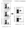
- Die Inkubation mit der Mischung aus allen Peptiden (Abbildung: Diagramm oben links) sowie Peptid 3 allein (Abbildung: Diagramm in der Mitte, zweites von oben) führten zur Produktion von IFN-γ in T-Zellen, welches durch Messung am Durchflußzytometer auf Einzel-Zellebene (J. L. PICKER et al., (1995) Blood, Vol 86, pp 1408-1419) nachgewiesen wurde, Keines der anderen einzeln getesteten Peptide hatte diesen Effekt. Eine in der Literatur veröffentlichte Untersuchung identifizierte exakt das gleiche Epitop innerhalb des gleichen Proteinsegments durch konventionelle Methoden und bestätigt unser Ergebnis eindeutig (D. J. DIAMOND et al. (1997) Blood, Vol 90, pp 1751-1767).
- Detektion von intrazellulär vorliegendem Interferon-γ in CD8+ T-Lymphozyten nach Stimulation mit der Mischung aus den 7 angegebenen Peptiden (Oben, ganz links) beziehungsweise den einzelnen Peptiden, pp65493-501 bis pp65499-507. Der Marker CD69 wurde als Aktivierungsmarker verwendet. Die Darstellung ist auf CD3+/CD8+ Ereignisse eingegrenzt, angegeben ist die mittlere Fluoreszenzintensität.
- Mononukleäre Zellen wurden aus dem durch venöse Punktion gewonnenen peripheren Blut einer HLA-typisierten Patientin präpariert, welche das MHC-Klasse-II Allel HLA-DR11 besaß. Die Patientin besaß außerdem Antikörper gegen das humane Cytomegalie-Virus. Die nach Standardmethode präparierten Zellen wurden für sechs Stunden unter optimierten Bedingungen mit Mischungen aus 11 oder 12 jeweils 15-Aminosäuren-langen Peptiden mit jeweils 11 Überlappungen entsprechend der Sequenz des pp65 Matrix Phosphoproteins (Swiss-Prot PO6725) inkubiert (insgesamt 138 Peptide). Die Peptidkonzentration betrug 1 µg/ml je Peptid. Drei der insgesamt 24 Mischungen stimulierten eindeutig CD4+ T-Zellen. Aufgrund der Versuchsanordnung (Vorkommen bestimmter Peptide in bestimmten Mischungen) ließen sich damit eindeutig 2 Peptide identifizieren, welche für diese Stimulation verantwortlich waren. Dieses Ergebnis wurde durch die Stimulation mit den jeweils einzelnen Peptiden unter ansonsten gleichen Bedingungen bestätigt. Die identifizierten Peptide waren die benachbarten Peptide pp65365-379 und pp65369-383. Diese Sequenzen decken sich weitgehend mit folgenden in der Literatur beschriebenen HLA-DR11 präsentierten Peptidesequenzen, welche auf konventionelle Weise als T-Zellen-stimulierende Sequenzen identifiziert wurden: pp65361-376 und pp65369-384 (Khattab et al. (1998) Joumal of Medical Virology, Vol. 52, pp68-76), d.h, die stimulierenden Peptide finden sich innerhalb des Abschnitts definiert durch die Aminosäuren 361 und 384. Eine weitere Einengung der Epitopsequenz auf die zu postulierende Länge von 11 Aminosäuren ist noch nicht erfolgt.
- Detektion von intrazellulär vorliegendem Interferon-γ in CD3+/CD8- (links) nach Stimulation mit den Peptidmischungen 8, 9, und 20, bzw. CD3+/CD4+ T-Lymphozyten (rechts) nach Stimulation mit den einzelnen Peptiden pp65365-379 und pp65369-383. Beim Screening (rechts) wurden Peptidmischungen verwendet und CD3 und CD8 als T-Zellmarker. Da die INF-γ+ Populationen links CD3+/CD8- sind wurde beim Nachtesten der Marker CD4 verwendet. Die Stimulierten T-Zellen sind eindeutig CD4+. Dargestellt sind ausschließlich CD3+ Zellen, angegeben ist die mittlere Fluoreszenzintensität.
Claims (13)
- Verfahren zum Identifizieren von T-Zell-stimulierenden Proteinfragmenten, die folgenden Schritte umfassend:a) Ermitteln der Aminosäuresequenz eines Antigens, welches ein Protein oder Peptid ist,b) Unterteilen der gefundenen Aminosäuresequenz des Antigens in Proteinfragmente,c) Synthetisieren von mindestens einem Proteinfragment mit einer Länge von B bis 30 Aminosäuren oder Spalten der Aminosäuresequenz des Antigens zu mindestens einem Proteinfragment mit einer Länge von 8 bis 30 Aminosäuren,
dabei ist das Proteinfragment eine Teilsequenz der ermittelten Aminosäuresequenz des Antigens.d) Inkubieren einer T-Zellen enthaltenden Suspension mit dem oder den Proteinfragmenten in Versuchsansätzen,e) Identifizieren(i) von mindestens einem T-Zell-Zytokin,
das durch das oder die Proteinfragmente induziert und in den T-Zellen synthetisiert wurde,
dabei liegen das oder die T-Zell-Zytokine intrazellulär oder an die Zellmembran gebunden vor,
und / oder(ii) von mindestens einem aufgrund der T-Zellstimulation durch das oder die Proteinfragmente exprimierten oder in seiner Expression gesteigerten Aktivierungsmarker, der durch das oder die Proteinfragmente induziert oder in seiner Expression gesteigert wurde und in den T-Zellen exprimiert wird,dabei werden das oder die T-Zell-Zytokine oder Aktivierungsmarker durchflußzytometrisch identifiziert, und
dabei kann der Aktivierungsmarker intrazellulär vorliegen oder auf der Zelloberfläche exprimiert seinf) Zuordnen der Versuchsansätze, bei denen T-Zellen stimuliert wurden und diese T-Zell-Stimulation durch das Identifizieren von einem T-Zell-Zytokin oder mehreren T-Zell-Zytokinen und / oder einem oder mehreren Aktivierungsmarkern erkannt wurde, zu der oder den Aminosäuresequenzen der Proteinfragmente, welche mit den T-Zellen inkubiert wurden,
dadurch gekennzeichnet,
daß die Inkubationszeit so lang ist,
daß das oder die Proteinfragmente von den MHC Molekülen,
die sich auf der Zelloberfläche befinden,
ausreichend aufgenommen werden,
wobei die Aufnahme dann ausreichend ist,
wenn eine eindeutige Identifizierung stimulierter T-Zellen möglich ist,
und,
daß die Inkubationszeit der T-Zellen enthaltenden Suspension mit dem oder den Proteinfragmenten so kurz ist,
daß eine Selektion und Proliferation, die mit einer gezielten Eliminierung bestimmter T-Zellen einhergeht, nicht erfolgt. - Verfahren zum Identifizieren von T-Zell-stimulierenden Proteinfragmenten nach Anspruch 1, wobei das Identifizieren von mindestens einem T-Zell-Zytokin oder Aktivierungsmarker auf der Einzelzell-Ebene erfolgt.
- Verfahren zum Identifizieren von T-Zell-stimulierenden Proteinfragmenten nach einem der vorherigen Ansprüche, wobei die T-Zellen enthaltende Suspensionen Zellen enthalten,
die das Proteinfragment im wesentlichen an MHC-Klasse-I oder Klasse-II-Moleküle gebunden präsentieren. - Verfahren zum Identifizieren von T-Zell-stimulierenden Proteinfragmenten nach einem der vorherigen Ansprüche, wobei das Proteinfragment bei der Klasse I restringierten Präsentation 9 bis 11 Aminosäuren umfaßt und das Proteinfragment bei der Klasse II restringierten Präsentation mindestens 11 Aminosäuren umfaßt.
- Verfahren zum Identifizieren von T-Zell-stimulierenden Proteinfragmenten nach einem der vorherigen Ansprüche, wobei die T-Zellen enthaltende Suspension eine Suspension ist aus Vollblut, peripheren weißen Blutzellen (PWBC), Milzzellen, Thymuszellen, Knochenmark, Liquor und / oder aus Lymphknotenzellen.
- Verfahren zum Identifizieren von T-Zell-stimulierenden Proteinfragmenten nach einem der vorherigen Ansprüche, wobei die T-Zellen enthaltende Suspension aus den Patienten, die therapiert werden sollen, aus Spendern oder aus Tieren stammen.
- Verfahren zum Identifizieren von T-Zell-stimulierenden Proteinfragmenten nach einem der vorherigen Ansprüche, wobei die Antigene, welche Proteine oder Peptide sind aus mehrzelligen Eukaryoten, aus Zellen, Zellkulturen und / oder Geweben von Spendern oder Patienten stammen.
- Verfahren zum Identifizieren von T-Zell-stimulierenden Proteinfragmenten nach einem der vorherigen Ansprüche, wobei die T-Zell-Zytokine vom Typ Interteron-γ, TNF-α oder Interleukin 2 sind.
- Verfahren zum Identifizieren von T-Zell-stimulierenden Proteinfragmenten nach einem der vorherigen Ansprüche weiterhin umfassend das Herstellen von identifizierten Proteinfragment/Peptid mit der Festphasenmethode, der Flüssigphasenmethode oder mittels der Proteinbiosynthese in einem Wirt.
- Verfahren zum Identifizieren von T-Zell-stimulierenden Proteinfragmenten nach Anspruch 9, wobei das Proteinfragment/Peptid Insertionen, Deletionen oder Substituierungen (Modifikationen) aufweist, wobei eine, zwei, drei oder mehrere Aminosäuren ausgetauscht, deletiert oder inserieret sind, und wobei das modifizierte Proteinfragment/Peptid im wesentlichen dieselbe Funktion bezüglich der Stimulation von T - Zellen aufweist, die das nicht modifizierte Proteinfragment/Peptid besitzt.
- Verfahren zum Identifizieren von T-Zell-stimulierenden Proteinfragmenten nach Anspruch 9 oder 10, wobei das Proteinfragment / Peptid am N-terminalen und / oder C-terminalen Ende mindestens eine weitere natürliche oder nichtnatürliche Aminosäure und / oder eine Schutzgruppe besitzt (erweiterte Modifizierung), und
wobei das erweitert modifizierte Proteinfragment/Peptid im wesentlichen dieselbe Funktion bezüglich der Stimulation von T - Zellen aufweist, die das nicht modifizierte Proteinfragment/Peptid besitzt. - Verfahren zur Identifizierung von T-Zell-stimulierenden Proteinfragmenten nach einem oder mehreren der Ansprüche 9-11, wobei die Proteinfragmente/Peptide zur Herstellung eines Medikaments zur Immunstimulation geeignet sind.
- Verfahren zur Identifizierung von T-Zell-stimulierenden Proteinfragmenten nach Anspruch 12, wobei die Immunstimulation eine Vakzinierung oder Desensibilisierung ist.
Applications Claiming Priority (5)
| Application Number | Priority Date | Filing Date | Title |
|---|---|---|---|
| DE19802174 | 1998-01-19 | ||
| DE1998102174 DE19802174A1 (de) | 1998-01-19 | 1998-01-19 | Verfahren zur Identifikation von T-Lymphozyten-stimulierenden Peptiden |
| DE1998134932 DE19834932A1 (de) | 1998-07-28 | 1998-07-28 | Verfahren zum Identifizieren von T-Zell-stimulierenden Proteinfragmenten |
| DE19834932 | 1998-07-28 | ||
| PCT/DE1999/000175 WO1999036568A2 (de) | 1998-01-19 | 1999-01-15 | Verfahren zum identifizieren von t-zell-stimulierenden proteinfragmenten |
Publications (2)
| Publication Number | Publication Date |
|---|---|
| EP1051619A2 EP1051619A2 (de) | 2000-11-15 |
| EP1051619B1 true EP1051619B1 (de) | 2006-08-09 |
Family
ID=26043259
Family Applications (1)
| Application Number | Title | Priority Date | Filing Date |
|---|---|---|---|
| EP99930888A Revoked EP1051619B1 (de) | 1998-01-19 | 1999-01-15 | Verfahren zum identifizieren von t-zell-stimulierenden proteinfragmenten |
Country Status (8)
| Country | Link |
|---|---|
| US (1) | US8932806B1 (de) |
| EP (1) | EP1051619B1 (de) |
| JP (1) | JP2002509241A (de) |
| AU (1) | AU3246399A (de) |
| DE (2) | DE19980037D2 (de) |
| DK (1) | DK1051619T3 (de) |
| ES (1) | ES2272074T3 (de) |
| WO (1) | WO1999036568A2 (de) |
Cited By (2)
| Publication number | Priority date | Publication date | Assignee | Title |
|---|---|---|---|---|
| US8268964B2 (en) | 2007-03-26 | 2012-09-18 | Dako Denmark A/S | MHC peptide complexes and uses thereof in infectious diseases |
| US9404916B2 (en) | 2008-09-20 | 2016-08-02 | University College Cardiff Consultants Limited | Use of a protein kinase inhibitor to detect immune cells, such as T cells |
Families Citing this family (19)
| Publication number | Priority date | Publication date | Assignee | Title |
|---|---|---|---|---|
| DE10009341A1 (de) * | 2000-02-22 | 2001-09-06 | Florian Kern | Verfahren zur antigen-spezifischen Stimulation von T-Lymphozyten |
| JP2003527847A (ja) | 2000-03-21 | 2003-09-24 | ジェンザイム・コーポレーション | 治療用抗サイトメガロウイルス化合物 |
| EP1146119A1 (de) * | 2000-04-12 | 2001-10-17 | Institut National De La Sante Et De La Recherche Medicale (Inserm) | Verfahren zur Gewinnung von T-Lymphozyten und zur Identifizierung von unbekannten Epitopen |
| EP1146120A1 (de) * | 2000-04-12 | 2001-10-17 | Institut National De La Sante Et De La Recherche Medicale (Inserm) | Verfahren zur Gewinnung von T-Lymphozyten und zur Identifizierung von unbekannten Epitopen |
| CA2420944A1 (en) | 2000-08-30 | 2002-03-07 | Chemocentryx, Inc. | Inhibition of cmv infection and dissemination |
| WO2002062296A2 (en) | 2001-02-02 | 2002-08-15 | Chemocentryx, Inc. | Methods and compositions useful for stimulating an immune response |
| WO2002072627A2 (en) * | 2001-03-09 | 2002-09-19 | Callistogen Ag | Induction of anti-tumor cytotoxic t-lymphocytes in humans using peptide epitopes found by computer based algorithms for vaccination |
| EP1774332B1 (de) * | 2004-05-24 | 2010-01-20 | Baylor Research Institute | Verfahren zur beurteilung von immunreaktionen |
| EP2167537A2 (de) | 2007-07-03 | 2010-03-31 | Dako Denmark A/S | Zusammengestellte verfahren für die analyse und sortierung von proben |
| US10611818B2 (en) | 2007-09-27 | 2020-04-07 | Agilent Technologies, Inc. | MHC multimers in tuberculosis diagnostics, vaccine and therapeutics |
| DK2254592T3 (da) | 2008-02-28 | 2019-09-09 | Dako Denmark As | MHC-multimerer til Borrelia-diagnostik og sygdom |
| WO2010009735A2 (en) | 2008-07-23 | 2010-01-28 | Dako Denmark A/S | Combinatorial analysis and repair |
| US11992518B2 (en) | 2008-10-02 | 2024-05-28 | Agilent Technologies, Inc. | Molecular vaccines for infectious disease |
| WO2010037402A1 (en) | 2008-10-02 | 2010-04-08 | Dako Denmark A/S | Molecular vaccines for infectious disease |
| GB201122297D0 (en) | 2011-12-23 | 2012-02-01 | Retroscreen Virology Ltd | Vaccine - screening method |
| EP2795321A2 (de) * | 2011-12-23 | 2014-10-29 | Retroscreen Virology Ltd | Impfstoffscreeningverfahren |
| CN107663239A (zh) * | 2016-12-28 | 2018-02-06 | 天津天锐生物科技有限公司 | 一种识别hla‑a2/nlvpmvatv的单域抗体 |
| CN111886338A (zh) | 2018-03-22 | 2020-11-03 | 柏林夏瑞蒂医科大学 | Crispr相关蛋白反应性t细胞免疫 |
| WO2020127222A2 (en) | 2018-12-17 | 2020-06-25 | Immudex Aps | Panel comprising borrelia mhc multimers |
Family Cites Families (1)
| Publication number | Priority date | Publication date | Assignee | Title |
|---|---|---|---|---|
| US5750356A (en) * | 1996-05-31 | 1998-05-12 | Anergen, Inc. | Method for monitoring T cell reactivity |
-
1999
- 1999-01-15 DE DE19980037T patent/DE19980037D2/de not_active Expired - Lifetime
- 1999-01-15 DE DE59913761T patent/DE59913761D1/de not_active Expired - Lifetime
- 1999-01-15 EP EP99930888A patent/EP1051619B1/de not_active Revoked
- 1999-01-15 ES ES99930888T patent/ES2272074T3/es not_active Expired - Lifetime
- 1999-01-15 AU AU32463/99A patent/AU3246399A/en not_active Abandoned
- 1999-01-15 WO PCT/DE1999/000175 patent/WO1999036568A2/de not_active Ceased
- 1999-01-15 DK DK99930888T patent/DK1051619T3/da active
- 1999-01-15 US US09/600,564 patent/US8932806B1/en not_active Expired - Fee Related
- 1999-01-15 JP JP2000540269A patent/JP2002509241A/ja active Pending
Cited By (2)
| Publication number | Priority date | Publication date | Assignee | Title |
|---|---|---|---|---|
| US8268964B2 (en) | 2007-03-26 | 2012-09-18 | Dako Denmark A/S | MHC peptide complexes and uses thereof in infectious diseases |
| US9404916B2 (en) | 2008-09-20 | 2016-08-02 | University College Cardiff Consultants Limited | Use of a protein kinase inhibitor to detect immune cells, such as T cells |
Also Published As
| Publication number | Publication date |
|---|---|
| ES2272074T3 (es) | 2007-04-16 |
| EP1051619A2 (de) | 2000-11-15 |
| DE59913761D1 (de) | 2006-09-21 |
| WO1999036568A3 (de) | 2000-04-06 |
| US8932806B1 (en) | 2015-01-13 |
| JP2002509241A (ja) | 2002-03-26 |
| DK1051619T3 (da) | 2006-12-11 |
| WO1999036568A2 (de) | 1999-07-22 |
| DE19980037D2 (de) | 2001-03-22 |
| AU3246399A (en) | 1999-08-02 |
Similar Documents
| Publication | Publication Date | Title |
|---|---|---|
| EP1051619A2 (de) | Verfahren zum identifizieren von t-zell-stimulierenden proteinfragmenten | |
| EP1257290B1 (de) | Verfahren zur antigen-spezifischen stimulation von t-lymphozyten mit synthetischen peptidbibliotheken | |
| DE69332485T2 (de) | Immunmodulierende peptide | |
| DE102004026135A1 (de) | An MHC-Moleküle bindende Tumor-assoziierte Peptide | |
| EP1507795A1 (de) | An mhc-moleküle bindende tumor-assoziierte peptide | |
| EP0584136A1 (de) | Bestimmung von peptidmotiven auf mhc-molekülen | |
| WO2000075180A2 (de) | Peptide zur vakzinierung gegen das humane cmv | |
| WO1997019169A1 (de) | Tumorvakzine und verfahren zu ihrer herstellung | |
| EP0669001A1 (de) | Bestimmung von peptidmotiven auf mhc-molekülen. | |
| Ramaswamy et al. | Evidence for the presence of a pheromonotropic factor in hemolymph and regulation of sex pheromone production in Helicoverpa zea | |
| DE69933679T2 (de) | Darstellung des profils und katalogisierung von exprimierten proteinmarkern | |
| DE19917195B4 (de) | Peptid zur Auslösung einer Immunreaktion gegen Tumorzellen, diese enthaltende pharmzeutische Zusammensetzungen, dessen Verwendungen, dafür codierende Nukleinsäure und diese Nukleinsäure enthaltender Expressionsvektor | |
| DE10333406A1 (de) | T-regulatorische-Zellen enthaltend Galectine zur Therapie und Diagnose von Erkrankungen | |
| DE69532964T2 (de) | Neue proteine aus ektoparasitenspeichel und vorrichtung zur sammlung davon | |
| DE10024383B4 (de) | Mit toxischen Substanzen beladene dendritische Zellen | |
| EP0637964B1 (de) | Medikamente zur tumortherapie mit kontrolliertem und reguliertem immunsystem sowie verwendung der einzelsubstanzen zur kombinierten therapie | |
| WO2004085461A2 (de) | An mhc-moleküle bindende tumor-assoziierte peptide | |
| DE19834932A1 (de) | Verfahren zum Identifizieren von T-Zell-stimulierenden Proteinfragmenten | |
| Shelanski | Intracellular ionic calcium and the cytoskeleton in living cells | |
| DE60317557T2 (de) | Verfahren zum screening für modulatoren von mitochondrialer funktion | |
| EP1195381A1 (de) | CD4+ T-Lymphozyten spezifische Hepatitis C Virus-Epitope | |
| DE102015106731A1 (de) | Peptide für die Krebsimmuntherapie | |
| DE19802174A1 (de) | Verfahren zur Identifikation von T-Lymphozyten-stimulierenden Peptiden | |
| DE10212867B4 (de) | Verwendung Harnstoff-adjuvierter Polypeptide zur Diagnose, Prophylaxe und Therapie | |
| DE69230943T2 (de) | Zellzyklus- Kontrollzusammensetzungen und Methoden diese zu benutzen. |
Legal Events
| Date | Code | Title | Description |
|---|---|---|---|
| PUAI | Public reference made under article 153(3) epc to a published international application that has entered the european phase |
Free format text: ORIGINAL CODE: 0009012 |
|
| 17P | Request for examination filed |
Effective date: 20000711 |
|
| AK | Designated contracting states |
Kind code of ref document: A2 Designated state(s): BE CH DE DK ES FR GB IT LI NL |
|
| 17Q | First examination report despatched |
Effective date: 20041007 |
|
| GRAP | Despatch of communication of intention to grant a patent |
Free format text: ORIGINAL CODE: EPIDOSNIGR1 |
|
| GRAS | Grant fee paid |
Free format text: ORIGINAL CODE: EPIDOSNIGR3 |
|
| GRAA | (expected) grant |
Free format text: ORIGINAL CODE: 0009210 |
|
| AK | Designated contracting states |
Kind code of ref document: B1 Designated state(s): BE CH DE DK ES FR GB IT LI NL |
|
| PG25 | Lapsed in a contracting state [announced via postgrant information from national office to epo] |
Ref country code: IT Free format text: LAPSE BECAUSE OF FAILURE TO SUBMIT A TRANSLATION OF THE DESCRIPTION OR TO PAY THE FEE WITHIN THE PRESCRIBED TIME-LIMIT;WARNING: LAPSES OF ITALIAN PATENTS WITH EFFECTIVE DATE BEFORE 2007 MAY HAVE OCCURRED AT ANY TIME BEFORE 2007. THE CORRECT EFFECTIVE DATE MAY BE DIFFERENT FROM THE ONE RECORDED. Effective date: 20060809 |
|
| REG | Reference to a national code |
Ref country code: GB Ref legal event code: FG4D Free format text: NOT ENGLISH |
|
| REG | Reference to a national code |
Ref country code: CH Ref legal event code: EP |
|
| REF | Corresponds to: |
Ref document number: 59913761 Country of ref document: DE Date of ref document: 20060921 Kind code of ref document: P |
|
| GBT | Gb: translation of ep patent filed (gb section 77(6)(a)/1977) |
Effective date: 20061113 |
|
| REG | Reference to a national code |
Ref country code: DK Ref legal event code: T3 |
|
| REG | Reference to a national code |
Ref country code: CH Ref legal event code: NV Representative=s name: A. BRAUN, BRAUN, HERITIER, ESCHMANN AG PATENTANWAE |
|
| ET | Fr: translation filed | ||
| REG | Reference to a national code |
Ref country code: ES Ref legal event code: FG2A Ref document number: 2272074 Country of ref document: ES Kind code of ref document: T3 |
|
| PLBI | Opposition filed |
Free format text: ORIGINAL CODE: 0009260 |
|
| PLAX | Notice of opposition and request to file observation + time limit sent |
Free format text: ORIGINAL CODE: EPIDOSNOBS2 |
|
| 26 | Opposition filed |
Opponent name: MILTENYI BIOTEC GMBH Effective date: 20070504 |
|
| NLR1 | Nl: opposition has been filed with the epo |
Opponent name: MILTENYI BIOTEC GMBH |
|
| PLAF | Information modified related to communication of a notice of opposition and request to file observations + time limit |
Free format text: ORIGINAL CODE: EPIDOSCOBS2 |
|
| PLAF | Information modified related to communication of a notice of opposition and request to file observations + time limit |
Free format text: ORIGINAL CODE: EPIDOSCOBS2 |
|
| PLBB | Reply of patent proprietor to notice(s) of opposition received |
Free format text: ORIGINAL CODE: EPIDOSNOBS3 |
|
| REG | Reference to a national code |
Ref country code: CH Ref legal event code: PFA Owner name: KERN, FLORIAN Free format text: KERN, FLORIAN#WOLLINER STRASSE 9#10435 BERLIN (DE) -TRANSFER TO- KERN, FLORIAN#WOLLINER STRASSE 9#10435 BERLIN (DE) |
|
| RDAF | Communication despatched that patent is revoked |
Free format text: ORIGINAL CODE: EPIDOSNREV1 |
|
| APBM | Appeal reference recorded |
Free format text: ORIGINAL CODE: EPIDOSNREFNO |
|
| APBP | Date of receipt of notice of appeal recorded |
Free format text: ORIGINAL CODE: EPIDOSNNOA2O |
|
| APAH | Appeal reference modified |
Free format text: ORIGINAL CODE: EPIDOSCREFNO |
|
| APBQ | Date of receipt of statement of grounds of appeal recorded |
Free format text: ORIGINAL CODE: EPIDOSNNOA3O |
|
| PLBP | Opposition withdrawn |
Free format text: ORIGINAL CODE: 0009264 |
|
| APAH | Appeal reference modified |
Free format text: ORIGINAL CODE: EPIDOSCREFNO |
|
| PGFP | Annual fee paid to national office [announced via postgrant information from national office to epo] |
Ref country code: DK Payment date: 20131218 Year of fee payment: 16 |
|
| PGFP | Annual fee paid to national office [announced via postgrant information from national office to epo] |
Ref country code: CH Payment date: 20131218 Year of fee payment: 16 Ref country code: GB Payment date: 20131219 Year of fee payment: 16 |
|
| PGFP | Annual fee paid to national office [announced via postgrant information from national office to epo] |
Ref country code: NL Payment date: 20131219 Year of fee payment: 16 Ref country code: ES Payment date: 20131219 Year of fee payment: 16 |
|
| PGFP | Annual fee paid to national office [announced via postgrant information from national office to epo] |
Ref country code: BE Payment date: 20131219 Year of fee payment: 16 |
|
| PGFP | Annual fee paid to national office [announced via postgrant information from national office to epo] |
Ref country code: DE Payment date: 20140124 Year of fee payment: 16 |
|
| REG | Reference to a national code |
Ref country code: DE Ref legal event code: R103 Ref document number: 59913761 Country of ref document: DE Ref country code: DE Ref legal event code: R064 Ref document number: 59913761 Country of ref document: DE |
|
| APBU | Appeal procedure closed |
Free format text: ORIGINAL CODE: EPIDOSNNOA9O |
|
| PGFP | Annual fee paid to national office [announced via postgrant information from national office to epo] |
Ref country code: IT Payment date: 20131223 Year of fee payment: 16 Ref country code: FR Payment date: 20131227 Year of fee payment: 16 |
|
| REG | Reference to a national code |
Ref country code: CH Ref legal event code: PCAR Free format text: NEW ADDRESS: HOLBEINSTRASSE 36-38, 4051 BASEL (CH) |
|
| RDAG | Patent revoked |
Free format text: ORIGINAL CODE: 0009271 |
|
| STAA | Information on the status of an ep patent application or granted ep patent |
Free format text: STATUS: PATENT REVOKED |
|
| REG | Reference to a national code |
Ref country code: CH Ref legal event code: PLX |
|
| 27W | Patent revoked |
Effective date: 20140506 |
|
| GBPR | Gb: patent revoked under art. 102 of the ep convention designating the uk as contracting state |
Effective date: 20140506 |
|
| REG | Reference to a national code |
Ref country code: DE Ref legal event code: R107 Ref document number: 59913761 Country of ref document: DE |
|
| PG25 | Lapsed in a contracting state [announced via postgrant information from national office to epo] |
Ref country code: LI Free format text: LAPSE BECAUSE OF THE APPLICANT RENOUNCES Effective date: 20060809 Ref country code: CH Free format text: LAPSE BECAUSE OF THE APPLICANT RENOUNCES Effective date: 20060809 |