WO2020127222A2 - Panel comprising borrelia mhc multimers - Google Patents
Panel comprising borrelia mhc multimers Download PDFInfo
- Publication number
- WO2020127222A2 WO2020127222A2 PCT/EP2019/085592 EP2019085592W WO2020127222A2 WO 2020127222 A2 WO2020127222 A2 WO 2020127222A2 EP 2019085592 W EP2019085592 W EP 2019085592W WO 2020127222 A2 WO2020127222 A2 WO 2020127222A2
- Authority
- WO
- WIPO (PCT)
- Prior art keywords
- mhc
- seq
- borrelia
- peptide
- antigenic
- Prior art date
Links
- 241000589968 Borrelia Species 0.000 title claims abstract description 332
- 208000016604 Lyme disease Diseases 0.000 title claims abstract description 330
- 108090000765 processed proteins & peptides Proteins 0.000 claims abstract description 762
- 230000000890 antigenic effect Effects 0.000 claims abstract description 695
- 102000004196 processed proteins & peptides Human genes 0.000 claims abstract description 581
- 229920001184 polypeptide Polymers 0.000 claims abstract description 291
- 108010091867 peptide P Proteins 0.000 claims abstract description 276
- 101100431670 Rattus norvegicus Ybx3 gene Proteins 0.000 claims abstract description 255
- 210000001744 T-lymphocyte Anatomy 0.000 claims abstract description 139
- 238000001514 detection method Methods 0.000 claims abstract description 36
- 208000037265 diseases, disorders, signs and symptoms Diseases 0.000 claims abstract description 19
- 238000000034 method Methods 0.000 claims description 292
- 241000589972 Borrelia sp. Species 0.000 claims description 242
- 230000027455 binding Effects 0.000 claims description 155
- 239000011324 bead Substances 0.000 claims description 80
- 235000001014 amino acid Nutrition 0.000 claims description 75
- 108090000623 proteins and genes Proteins 0.000 claims description 67
- 108091054437 MHC class I family Proteins 0.000 claims description 66
- 210000004027 cell Anatomy 0.000 claims description 66
- 229940024606 amino acid Drugs 0.000 claims description 63
- 150000001413 amino acids Chemical class 0.000 claims description 62
- 108700028369 Alleles Proteins 0.000 claims description 58
- 102000004169 proteins and genes Human genes 0.000 claims description 58
- 235000018102 proteins Nutrition 0.000 claims description 57
- 102000043129 MHC class I family Human genes 0.000 claims description 53
- 239000000427 antigen Substances 0.000 claims description 39
- 108091007433 antigens Proteins 0.000 claims description 39
- 102000036639 antigens Human genes 0.000 claims description 39
- 239000013642 negative control Substances 0.000 claims description 38
- -1 FlhF and FlaB Proteins 0.000 claims description 37
- 125000005647 linker group Chemical group 0.000 claims description 36
- 125000003275 alpha amino acid group Chemical group 0.000 claims description 32
- 238000005259 measurement Methods 0.000 claims description 28
- 108020004707 nucleic acids Proteins 0.000 claims description 26
- 102000039446 nucleic acids Human genes 0.000 claims description 26
- 150000007523 nucleic acids Chemical class 0.000 claims description 26
- 102000008949 Histocompatibility Antigens Class I Human genes 0.000 claims description 24
- 239000007787 solid Substances 0.000 claims description 23
- 230000003993 interaction Effects 0.000 claims description 22
- 229960005486 vaccine Drugs 0.000 claims description 20
- 239000013641 positive control Substances 0.000 claims description 19
- YBJHBAHKTGYVGT-ZKWXMUAHSA-N (+)-Biotin Chemical compound N1C(=O)N[C@@H]2[C@H](CCCCC(=O)O)SC[C@@H]21 YBJHBAHKTGYVGT-ZKWXMUAHSA-N 0.000 claims description 17
- 125000000539 amino acid group Chemical group 0.000 claims description 17
- 239000000178 monomer Substances 0.000 claims description 17
- 241000894007 species Species 0.000 claims description 17
- 229920002307 Dextran Polymers 0.000 claims description 16
- 108010090804 Streptavidin Proteins 0.000 claims description 16
- 238000006467 substitution reaction Methods 0.000 claims description 16
- 102100028972 HLA class I histocompatibility antigen, A alpha chain Human genes 0.000 claims description 14
- 108010075704 HLA-A Antigens Proteins 0.000 claims description 14
- 238000002372 labelling Methods 0.000 claims description 14
- 229920000642 polymer Polymers 0.000 claims description 14
- ROHFNLRQFUQHCH-UHFFFAOYSA-N Leucine Natural products CC(C)CC(N)C(O)=O ROHFNLRQFUQHCH-UHFFFAOYSA-N 0.000 claims description 13
- 235000005772 leucine Nutrition 0.000 claims description 13
- 210000001519 tissue Anatomy 0.000 claims description 13
- 108020004414 DNA Proteins 0.000 claims description 12
- 229920001282 polysaccharide Polymers 0.000 claims description 12
- 239000005017 polysaccharide Substances 0.000 claims description 12
- 241001148605 Borreliella garinii Species 0.000 claims description 11
- 102000004190 Enzymes Human genes 0.000 claims description 11
- 108090000790 Enzymes Proteins 0.000 claims description 11
- 108010088652 Histocompatibility Antigens Class I Proteins 0.000 claims description 11
- ROHFNLRQFUQHCH-YFKPBYRVSA-N L-leucine Chemical compound CC(C)C[C@H](N)C(O)=O ROHFNLRQFUQHCH-YFKPBYRVSA-N 0.000 claims description 11
- 108091008874 T cell receptors Proteins 0.000 claims description 11
- 238000004458 analytical method Methods 0.000 claims description 11
- 238000010205 computational analysis Methods 0.000 claims description 11
- 229940088598 enzyme Drugs 0.000 claims description 11
- 102000016266 T-Cell Antigen Receptors Human genes 0.000 claims description 10
- 239000011230 binding agent Substances 0.000 claims description 10
- 229960002685 biotin Drugs 0.000 claims description 10
- 239000011616 biotin Substances 0.000 claims description 10
- 238000000684 flow cytometry Methods 0.000 claims description 10
- 238000011502 immune monitoring Methods 0.000 claims description 10
- 210000000612 antigen-presenting cell Anatomy 0.000 claims description 9
- 102000015736 beta 2-Microglobulin Human genes 0.000 claims description 9
- 108010081355 beta 2-Microglobulin Proteins 0.000 claims description 9
- 230000000694 effects Effects 0.000 claims description 9
- 239000000203 mixture Substances 0.000 claims description 9
- 241000282414 Homo sapiens Species 0.000 claims description 8
- 235000020958 biotin Nutrition 0.000 claims description 8
- 239000012634 fragment Substances 0.000 claims description 8
- 239000012528 membrane Substances 0.000 claims description 8
- 108091023037 Aptamer Proteins 0.000 claims description 7
- FFEARJCKVFRZRR-BYPYZUCNSA-N L-methionine Chemical compound CSCC[C@H](N)C(O)=O FFEARJCKVFRZRR-BYPYZUCNSA-N 0.000 claims description 7
- KDXKERNSBIXSRK-UHFFFAOYSA-N Lysine Natural products NCCCCC(N)C(O)=O KDXKERNSBIXSRK-UHFFFAOYSA-N 0.000 claims description 7
- 239000004472 Lysine Substances 0.000 claims description 7
- 238000004873 anchoring Methods 0.000 claims description 7
- 238000003556 assay Methods 0.000 claims description 7
- 210000004369 blood Anatomy 0.000 claims description 7
- 239000008280 blood Substances 0.000 claims description 7
- 230000028993 immune response Effects 0.000 claims description 7
- 235000018977 lysine Nutrition 0.000 claims description 7
- 235000006109 methionine Nutrition 0.000 claims description 7
- 229930182817 methionine Natural products 0.000 claims description 7
- 239000000126 substance Substances 0.000 claims description 7
- 108090001008 Avidin Proteins 0.000 claims description 6
- 241001148604 Borreliella afzelii Species 0.000 claims description 6
- 241000589969 Borreliella burgdorferi Species 0.000 claims description 6
- KDXKERNSBIXSRK-YFKPBYRVSA-N L-lysine Chemical compound NCCCC[C@H](N)C(O)=O KDXKERNSBIXSRK-YFKPBYRVSA-N 0.000 claims description 6
- KZSNJWFQEVHDMF-BYPYZUCNSA-N L-valine Chemical compound CC(C)[C@H](N)C(O)=O KZSNJWFQEVHDMF-BYPYZUCNSA-N 0.000 claims description 6
- 230000005867 T cell response Effects 0.000 claims description 6
- KZSNJWFQEVHDMF-UHFFFAOYSA-N Valine Natural products CC(C)C(N)C(O)=O KZSNJWFQEVHDMF-UHFFFAOYSA-N 0.000 claims description 6
- 210000003719 b-lymphocyte Anatomy 0.000 claims description 6
- 238000012217 deletion Methods 0.000 claims description 6
- 230000037430 deletion Effects 0.000 claims description 6
- 239000003550 marker Substances 0.000 claims description 6
- 238000000386 microscopy Methods 0.000 claims description 6
- 238000012360 testing method Methods 0.000 claims description 6
- 235000014393 valine Nutrition 0.000 claims description 6
- 239000004474 valine Substances 0.000 claims description 6
- ZCAYUOKEIPMTMF-JPDWDDBRSA-N (8s,9s,10r,11r,13s,14s,16r,17r)-11,17-dihydroxy-17-(2-hydroxyacetyl)-10,13,16-trimethyl-2,6,7,8,9,11,12,14,15,16-decahydro-1h-cyclopenta[a]phenanthren-3-one Chemical compound C1CC2=CC(=O)CC[C@]2(C)[C@@H]2[C@@H]1[C@@H]1C[C@@H](C)[C@@](C(=O)CO)(O)[C@@]1(C)C[C@H]2O ZCAYUOKEIPMTMF-JPDWDDBRSA-N 0.000 claims description 5
- 239000004475 Arginine Substances 0.000 claims description 5
- 108091005804 Peptidases Proteins 0.000 claims description 5
- 239000004365 Protease Substances 0.000 claims description 5
- ODKSFYDXXFIFQN-UHFFFAOYSA-N arginine Natural products OC(=O)C(N)CCCNC(N)=N ODKSFYDXXFIFQN-UHFFFAOYSA-N 0.000 claims description 5
- 235000009697 arginine Nutrition 0.000 claims description 5
- 230000006287 biotinylation Effects 0.000 claims description 5
- 238000007413 biotinylation Methods 0.000 claims description 5
- 238000010828 elution Methods 0.000 claims description 5
- MHMNJMPURVTYEJ-UHFFFAOYSA-N fluorescein-5-isothiocyanate Chemical compound O1C(=O)C2=CC(N=C=S)=CC=C2C21C1=CC=C(O)C=C1OC1=CC(O)=CC=C21 MHMNJMPURVTYEJ-UHFFFAOYSA-N 0.000 claims description 5
- 108010065889 glycyl-leucyl-cysteinyl-threonyl-leucyl-valyl-alanyl-methionyl-leucine Proteins 0.000 claims description 5
- 108010061181 influenza matrix peptide (58-66) Proteins 0.000 claims description 5
- 210000002540 macrophage Anatomy 0.000 claims description 5
- 230000008569 process Effects 0.000 claims description 5
- 102000005962 receptors Human genes 0.000 claims description 5
- 108020003175 receptors Proteins 0.000 claims description 5
- 238000002255 vaccination Methods 0.000 claims description 5
- 206010061591 Borrelia infection Diseases 0.000 claims description 4
- 241001536361 Borrelia lonestari Species 0.000 claims description 4
- 102100037486 Reverse transcriptase/ribonuclease H Human genes 0.000 claims description 4
- 239000000969 carrier Substances 0.000 claims description 4
- 210000004443 dendritic cell Anatomy 0.000 claims description 4
- 239000000539 dimer Substances 0.000 claims description 4
- 230000006870 function Effects 0.000 claims description 4
- 238000001727 in vivo Methods 0.000 claims description 4
- 230000003834 intracellular effect Effects 0.000 claims description 4
- 108091005601 modified peptides Proteins 0.000 claims description 4
- 108060003951 Immunoglobulin Proteins 0.000 claims description 3
- 108091028043 Nucleic acid sequence Proteins 0.000 claims description 3
- 125000003118 aryl group Chemical group 0.000 claims description 3
- 102000018358 immunoglobulin Human genes 0.000 claims description 3
- 238000000338 in vitro Methods 0.000 claims description 3
- 230000001965 increasing effect Effects 0.000 claims description 3
- 238000002955 isolation Methods 0.000 claims description 3
- 239000000693 micelle Substances 0.000 claims description 3
- 210000001616 monocyte Anatomy 0.000 claims description 3
- 229910052698 phosphorus Inorganic materials 0.000 claims description 3
- 125000002924 primary amino group Chemical group [H]N([H])* 0.000 claims description 3
- 230000017854 proteolysis Effects 0.000 claims description 3
- 241000448700 Borrelia afzelii ACA-1 Species 0.000 claims description 2
- 241000962525 Borrelia afzelii K78 Species 0.000 claims description 2
- 241000833568 Borrelia afzelii PKo Species 0.000 claims description 2
- 241000589974 Borrelia anserina Species 0.000 claims description 2
- 241001551899 Borrelia burgdorferi 118a Species 0.000 claims description 2
- 241000448697 Borrelia burgdorferi 156a Species 0.000 claims description 2
- 241000644875 Borrelia burgdorferi 29805 Species 0.000 claims description 2
- 241000644810 Borrelia burgdorferi 64b Species 0.000 claims description 2
- 241001551896 Borrelia burgdorferi 72a Species 0.000 claims description 2
- 241000644873 Borrelia burgdorferi 80a Species 0.000 claims description 2
- 241001551914 Borrelia burgdorferi 94a Species 0.000 claims description 2
- 241000276440 Borrelia burgdorferi B31 Species 0.000 claims description 2
- 241000448698 Borrelia burgdorferi Bol26 Species 0.000 claims description 2
- 241000644876 Borrelia burgdorferi CA-11.2A Species 0.000 claims description 2
- 241000644874 Borrelia burgdorferi WI91-23 Species 0.000 claims description 2
- 241000448699 Borrelia burgdorferi ZS7 Species 0.000 claims description 2
- 241000589975 Borrelia coriaceae Species 0.000 claims description 2
- 241001148533 Borrelia crocidurae Species 0.000 claims description 2
- 241000124827 Borrelia duttonii Species 0.000 claims description 2
- 241000976185 Borrelia duttonii Ly Species 0.000 claims description 2
- 241000589978 Borrelia hermsii Species 0.000 claims description 2
- 241001271279 Borrelia hermsii DAH Species 0.000 claims description 2
- 241000124828 Borrelia hispanica Species 0.000 claims description 2
- 241000216520 Borrelia miyamotoi Species 0.000 claims description 2
- 241000589976 Borrelia parkeri Species 0.000 claims description 2
- 241000180132 Borrelia persica Species 0.000 claims description 2
- 241000180135 Borrelia recurrentis Species 0.000 claims description 2
- 241000976186 Borrelia recurrentis A1 Species 0.000 claims description 2
- 241000124829 Borrelia sp. strain Spain Species 0.000 claims description 2
- 241000167563 Borrelia theileri Species 0.000 claims description 2
- 241000334732 Borrelia turcica Species 0.000 claims description 2
- 241000589977 Borrelia turicatae Species 0.000 claims description 2
- 241001271277 Borrelia turicatae 91E135 Species 0.000 claims description 2
- 241000142472 Borreliella andersonii Species 0.000 claims description 2
- 241000908527 Borreliella bissettii Species 0.000 claims description 2
- 241000141790 Borreliella californiensis Species 0.000 claims description 2
- 241001478201 Borreliella japonica Species 0.000 claims description 2
- 241001446608 Borreliella lusitaniae Species 0.000 claims description 2
- 241000004488 Borreliella sinica Species 0.000 claims description 2
- 241000019016 Borreliella spielmanii Species 0.000 claims description 2
- 241000644814 Borreliella spielmanii A14S Species 0.000 claims description 2
- 241000565673 Borreliella tanukii Species 0.000 claims description 2
- 241000582024 Borreliella turdi Species 0.000 claims description 2
- 241000876423 Borreliella valaisiana Species 0.000 claims description 2
- 241000448701 Borreliella valaisiana VS116 Species 0.000 claims description 2
- 101100457838 Caenorhabditis elegans mod-1 gene Proteins 0.000 claims description 2
- 241001358276 Candidatus Borrelia texasensis Species 0.000 claims description 2
- 102100033041 Carbonic anhydrase 13 Human genes 0.000 claims description 2
- WQZGKKKJIJFFOK-QTVWNMPRSA-N D-mannopyranose Chemical compound OC[C@H]1OC(O)[C@@H](O)[C@@H](O)[C@@H]1O WQZGKKKJIJFFOK-QTVWNMPRSA-N 0.000 claims description 2
- 101000867860 Homo sapiens Carbonic anhydrase 13 Proteins 0.000 claims description 2
- 101150110972 ME1 gene Proteins 0.000 claims description 2
- 241001465754 Metazoa Species 0.000 claims description 2
- 239000002253 acid Substances 0.000 claims description 2
- 125000004122 cyclic group Chemical group 0.000 claims description 2
- 230000004069 differentiation Effects 0.000 claims description 2
- 239000012636 effector Substances 0.000 claims description 2
- 238000004519 manufacturing process Methods 0.000 claims description 2
- 230000001404 mediated effect Effects 0.000 claims description 2
- 150000003384 small molecules Chemical group 0.000 claims description 2
- 239000000725 suspension Substances 0.000 claims description 2
- 239000000654 additive Substances 0.000 claims 21
- 230000006641 stabilisation Effects 0.000 claims 19
- 238000011105 stabilization Methods 0.000 claims 19
- 239000002096 quantum dot Substances 0.000 claims 18
- 239000000975 dye Substances 0.000 claims 15
- 239000012530 fluid Substances 0.000 claims 13
- 230000004048 modification Effects 0.000 claims 13
- 238000012986 modification Methods 0.000 claims 13
- 238000007385 chemical modification Methods 0.000 claims 12
- 238000006243 chemical reaction Methods 0.000 claims 12
- 108020001507 fusion proteins Proteins 0.000 claims 12
- 102000037865 fusion proteins Human genes 0.000 claims 12
- 239000004793 Polystyrene Chemical class 0.000 claims 10
- 150000004804 polysaccharides Chemical class 0.000 claims 10
- 229920002223 polystyrene Chemical class 0.000 claims 10
- QNAYBMKLOCPYGJ-REOHCLBHSA-N L-alanine Chemical compound C[C@H](N)C(O)=O QNAYBMKLOCPYGJ-REOHCLBHSA-N 0.000 claims 9
- 235000004279 alanine Nutrition 0.000 claims 9
- 229920001223 polyethylene glycol Polymers 0.000 claims 9
- ZDXPYRJPNDTMRX-VKHMYHEASA-N L-glutamine Chemical compound OC(=O)[C@@H](N)CCC(N)=O ZDXPYRJPNDTMRX-VKHMYHEASA-N 0.000 claims 8
- 239000002202 Polyethylene glycol Substances 0.000 claims 8
- 235000004554 glutamine Nutrition 0.000 claims 8
- ZDXPYRJPNDTMRX-UHFFFAOYSA-N glutamine Natural products OC(=O)C(N)CCC(N)=O ZDXPYRJPNDTMRX-UHFFFAOYSA-N 0.000 claims 8
- RWSXRVCMGQZWBV-WDSKDSINSA-N glutathione Chemical compound OC(=O)[C@@H](N)CCC(=O)N[C@@H](CS)C(=O)NCC(O)=O RWSXRVCMGQZWBV-WDSKDSINSA-N 0.000 claims 8
- 125000000896 monocarboxylic acid group Chemical group 0.000 claims 8
- 230000015572 biosynthetic process Effects 0.000 claims 7
- 230000012743 protein tagging Effects 0.000 claims 7
- PEDCQBHIVMGVHV-UHFFFAOYSA-N Glycerine Chemical compound OCC(O)CO PEDCQBHIVMGVHV-UHFFFAOYSA-N 0.000 claims 6
- AGPKZVBTJJNPAG-WHFBIAKZSA-N L-isoleucine Chemical compound CC[C@H](C)[C@H](N)C(O)=O AGPKZVBTJJNPAG-WHFBIAKZSA-N 0.000 claims 6
- OUYCCCASQSFEME-QMMMGPOBSA-N L-tyrosine Chemical compound OC(=O)[C@@H](N)CC1=CC=C(O)C=C1 OUYCCCASQSFEME-QMMMGPOBSA-N 0.000 claims 6
- 235000014705 isoleucine Nutrition 0.000 claims 6
- AGPKZVBTJJNPAG-UHFFFAOYSA-N isoleucine Natural products CCC(C)C(N)C(O)=O AGPKZVBTJJNPAG-UHFFFAOYSA-N 0.000 claims 6
- 229960000310 isoleucine Drugs 0.000 claims 6
- 239000002773 nucleotide Substances 0.000 claims 6
- 238000003786 synthesis reaction Methods 0.000 claims 6
- 235000002374 tyrosine Nutrition 0.000 claims 6
- OUYCCCASQSFEME-UHFFFAOYSA-N tyrosine Natural products OC(=O)C(N)CC1=CC=C(O)C=C1 OUYCCCASQSFEME-UHFFFAOYSA-N 0.000 claims 6
- MTCFGRXMJLQNBG-REOHCLBHSA-N (2S)-2-Amino-3-hydroxypropansäure Chemical compound OC[C@H](N)C(O)=O MTCFGRXMJLQNBG-REOHCLBHSA-N 0.000 claims 5
- 229920000936 Agarose Polymers 0.000 claims 5
- WHUUTDBJXJRKMK-VKHMYHEASA-N L-glutamic acid Chemical compound OC(=O)[C@@H](N)CCC(O)=O WHUUTDBJXJRKMK-VKHMYHEASA-N 0.000 claims 5
- MTCFGRXMJLQNBG-UHFFFAOYSA-N Serine Natural products OCC(N)C(O)=O MTCFGRXMJLQNBG-UHFFFAOYSA-N 0.000 claims 5
- 108010004469 allophycocyanin Proteins 0.000 claims 5
- 238000003776 cleavage reaction Methods 0.000 claims 5
- 239000005090 green fluorescent protein Substances 0.000 claims 5
- 230000005291 magnetic effect Effects 0.000 claims 5
- 238000002703 mutagenesis Methods 0.000 claims 5
- 231100000350 mutagenesis Toxicity 0.000 claims 5
- 230000035772 mutation Effects 0.000 claims 5
- 125000003729 nucleotide group Chemical group 0.000 claims 5
- 230000007017 scission Effects 0.000 claims 5
- 235000004400 serine Nutrition 0.000 claims 5
- 239000012099 Alexa Fluor family Substances 0.000 claims 4
- 229920001661 Chitosan Polymers 0.000 claims 4
- WHUUTDBJXJRKMK-UHFFFAOYSA-N Glutamic acid Natural products OC(=O)C(N)CCC(O)=O WHUUTDBJXJRKMK-UHFFFAOYSA-N 0.000 claims 4
- 108010024636 Glutathione Proteins 0.000 claims 4
- DHMQDGOQFOQNFH-UHFFFAOYSA-N Glycine Chemical compound NCC(O)=O DHMQDGOQFOQNFH-UHFFFAOYSA-N 0.000 claims 4
- 108010021625 Immunoglobulin Fragments Proteins 0.000 claims 4
- 102000008394 Immunoglobulin Fragments Human genes 0.000 claims 4
- ODKSFYDXXFIFQN-BYPYZUCNSA-P L-argininium(2+) Chemical compound NC(=[NH2+])NCCC[C@H]([NH3+])C(O)=O ODKSFYDXXFIFQN-BYPYZUCNSA-P 0.000 claims 4
- COLNVLDHVKWLRT-QMMMGPOBSA-N L-phenylalanine Chemical compound OC(=O)[C@@H](N)CC1=CC=CC=C1 COLNVLDHVKWLRT-QMMMGPOBSA-N 0.000 claims 4
- AYFVYJQAPQTCCC-GBXIJSLDSA-N L-threonine Chemical compound C[C@@H](O)[C@H](N)C(O)=O AYFVYJQAPQTCCC-GBXIJSLDSA-N 0.000 claims 4
- QIVBCDIJIAJPQS-VIFPVBQESA-N L-tryptophane Chemical compound C1=CC=C2C(C[C@H](N)C(O)=O)=CNC2=C1 QIVBCDIJIAJPQS-VIFPVBQESA-N 0.000 claims 4
- 229920002684 Sepharose Polymers 0.000 claims 4
- AYFVYJQAPQTCCC-UHFFFAOYSA-N Threonine Natural products CC(O)C(N)C(O)=O AYFVYJQAPQTCCC-UHFFFAOYSA-N 0.000 claims 4
- 239000004473 Threonine Substances 0.000 claims 4
- QIVBCDIJIAJPQS-UHFFFAOYSA-N Tryptophan Natural products C1=CC=C2C(CC(N)C(O)=O)=CNC2=C1 QIVBCDIJIAJPQS-UHFFFAOYSA-N 0.000 claims 4
- 108010046516 Wheat Germ Agglutinins Proteins 0.000 claims 4
- 230000029936 alkylation Effects 0.000 claims 4
- 238000005804 alkylation reaction Methods 0.000 claims 4
- 229920002678 cellulose Polymers 0.000 claims 4
- 235000010980 cellulose Nutrition 0.000 claims 4
- 239000013522 chelant Substances 0.000 claims 4
- 150000001875 compounds Chemical class 0.000 claims 4
- 230000008878 coupling Effects 0.000 claims 4
- 238000010168 coupling process Methods 0.000 claims 4
- 238000005859 coupling reaction Methods 0.000 claims 4
- 150000002148 esters Chemical class 0.000 claims 4
- 239000007850 fluorescent dye Substances 0.000 claims 4
- 108010021843 fluorescent protein 583 Proteins 0.000 claims 4
- 239000011521 glass Substances 0.000 claims 4
- 235000013922 glutamic acid Nutrition 0.000 claims 4
- 239000004220 glutamic acid Substances 0.000 claims 4
- 229960003180 glutathione Drugs 0.000 claims 4
- 230000037431 insertion Effects 0.000 claims 4
- 238000003780 insertion Methods 0.000 claims 4
- 150000002500 ions Chemical class 0.000 claims 4
- 229910052747 lanthanoid Inorganic materials 0.000 claims 4
- 150000002602 lanthanoids Chemical class 0.000 claims 4
- 229910052751 metal Inorganic materials 0.000 claims 4
- 239000002184 metal Substances 0.000 claims 4
- 230000000394 mitotic effect Effects 0.000 claims 4
- COLNVLDHVKWLRT-UHFFFAOYSA-N phenylalanine Natural products OC(=O)C(N)CC1=CC=CC=C1 COLNVLDHVKWLRT-UHFFFAOYSA-N 0.000 claims 4
- 235000008729 phenylalanine Nutrition 0.000 claims 4
- 239000011347 resin Substances 0.000 claims 4
- 229920005989 resin Polymers 0.000 claims 4
- 235000008521 threonine Nutrition 0.000 claims 4
- 125000002088 tosyl group Chemical group [H]C1=C([H])C(=C([H])C([H])=C1C([H])([H])[H])S(*)(=O)=O 0.000 claims 4
- 108091032973 (ribonucleotides)n+m Proteins 0.000 claims 3
- OYPRJOBELJOOCE-UHFFFAOYSA-N Calcium Chemical compound [Ca] OYPRJOBELJOOCE-UHFFFAOYSA-N 0.000 claims 3
- 229920002101 Chitin Polymers 0.000 claims 3
- 108091026890 Coding region Proteins 0.000 claims 3
- 108010070675 Glutathione transferase Proteins 0.000 claims 3
- 102100029100 Hematopoietic prostaglandin D synthase Human genes 0.000 claims 3
- CKLJMWTZIZZHCS-REOHCLBHSA-N L-aspartic acid Chemical compound OC(=O)[C@@H](N)CC(O)=O CKLJMWTZIZZHCS-REOHCLBHSA-N 0.000 claims 3
- HNDVDQJCIGZPNO-YFKPBYRVSA-N L-histidine Chemical compound OC(=O)[C@@H](N)CC1=CN=CN1 HNDVDQJCIGZPNO-YFKPBYRVSA-N 0.000 claims 3
- 208000008551 Lyme Neuroborreliosis Diseases 0.000 claims 3
- DNIAPMSPPWPWGF-UHFFFAOYSA-N Propylene glycol Chemical compound CC(O)CO DNIAPMSPPWPWGF-UHFFFAOYSA-N 0.000 claims 3
- 102100024554 Tetranectin Human genes 0.000 claims 3
- 230000004913 activation Effects 0.000 claims 3
- 235000003704 aspartic acid Nutrition 0.000 claims 3
- OQFSQFPPLPISGP-UHFFFAOYSA-N beta-carboxyaspartic acid Natural products OC(=O)C(N)C(C(O)=O)C(O)=O OQFSQFPPLPISGP-UHFFFAOYSA-N 0.000 claims 3
- 239000011575 calcium Substances 0.000 claims 3
- 229910052791 calcium Inorganic materials 0.000 claims 3
- 150000001720 carbohydrates Chemical class 0.000 claims 3
- 235000014633 carbohydrates Nutrition 0.000 claims 3
- 239000001913 cellulose Substances 0.000 claims 3
- AFOSIXZFDONLBT-UHFFFAOYSA-N divinyl sulfone Chemical group C=CS(=O)(=O)C=C AFOSIXZFDONLBT-UHFFFAOYSA-N 0.000 claims 3
- 239000008273 gelatin Substances 0.000 claims 3
- 229920000159 gelatin Polymers 0.000 claims 3
- HNDVDQJCIGZPNO-UHFFFAOYSA-N histidine Natural products OC(=O)C(N)CC1=CN=CN1 HNDVDQJCIGZPNO-UHFFFAOYSA-N 0.000 claims 3
- 235000014304 histidine Nutrition 0.000 claims 3
- 210000004408 hybridoma Anatomy 0.000 claims 3
- RAXXELZNTBOGNW-UHFFFAOYSA-N imidazole Natural products C1=CNC=N1 RAXXELZNTBOGNW-UHFFFAOYSA-N 0.000 claims 3
- 238000003364 immunohistochemistry Methods 0.000 claims 3
- 210000001821 langerhans cell Anatomy 0.000 claims 3
- 125000002950 monocyclic group Chemical group 0.000 claims 3
- 239000002245 particle Substances 0.000 claims 3
- 229920002401 polyacrylamide Polymers 0.000 claims 3
- 230000004481 post-translational protein modification Effects 0.000 claims 3
- 235000004252 protein component Nutrition 0.000 claims 3
- 230000028327 secretion Effects 0.000 claims 3
- 210000002966 serum Anatomy 0.000 claims 3
- XSYUPRQVAHJETO-WPMUBMLPSA-N (2s)-2-[[(2s)-2-[[(2s)-2-[[(2s)-2-[[(2s)-2-[[(2s)-2-amino-3-(1h-imidazol-5-yl)propanoyl]amino]-3-(1h-imidazol-5-yl)propanoyl]amino]-3-(1h-imidazol-5-yl)propanoyl]amino]-3-(1h-imidazol-5-yl)propanoyl]amino]-3-(1h-imidazol-5-yl)propanoyl]amino]-3-(1h-imidaz Chemical compound C([C@H](N)C(=O)N[C@@H](CC=1NC=NC=1)C(=O)N[C@@H](CC=1NC=NC=1)C(=O)N[C@@H](CC=1NC=NC=1)C(=O)N[C@@H](CC=1NC=NC=1)C(=O)N[C@@H](CC=1NC=NC=1)C(O)=O)C1=CN=CN1 XSYUPRQVAHJETO-WPMUBMLPSA-N 0.000 claims 2
- MYRTYDVEIRVNKP-UHFFFAOYSA-N 1,2-Divinylbenzene Chemical compound C=CC1=CC=CC=C1C=C MYRTYDVEIRVNKP-UHFFFAOYSA-N 0.000 claims 2
- QEQDLKUMPUDNPG-UHFFFAOYSA-N 2-(7-amino-4-methyl-2-oxochromen-3-yl)acetic acid Chemical compound C1=C(N)C=CC2=C1OC(=O)C(CC(O)=O)=C2C QEQDLKUMPUDNPG-UHFFFAOYSA-N 0.000 claims 2
- 108010011170 Ala-Trp-Arg-His-Pro-Gln-Phe-Gly-Gly Proteins 0.000 claims 2
- IKYJCHYORFJFRR-UHFFFAOYSA-N Alexa Fluor 350 Chemical compound O=C1OC=2C=C(N)C(S(O)(=O)=O)=CC=2C(C)=C1CC(=O)ON1C(=O)CCC1=O IKYJCHYORFJFRR-UHFFFAOYSA-N 0.000 claims 2
- WEJVZSAYICGDCK-UHFFFAOYSA-N Alexa Fluor 430 Chemical compound CC[NH+](CC)CC.CC1(C)C=C(CS([O-])(=O)=O)C2=CC=3C(C(F)(F)F)=CC(=O)OC=3C=C2N1CCCCCC(=O)ON1C(=O)CCC1=O WEJVZSAYICGDCK-UHFFFAOYSA-N 0.000 claims 2
- 102000002260 Alkaline Phosphatase Human genes 0.000 claims 2
- 108020004774 Alkaline Phosphatase Proteins 0.000 claims 2
- 102000000584 Calmodulin Human genes 0.000 claims 2
- 108010041952 Calmodulin Proteins 0.000 claims 2
- 244000045232 Canavalia ensiformis Species 0.000 claims 2
- 235000010520 Canavalia ensiformis Nutrition 0.000 claims 2
- 108010062580 Concanavalin A Proteins 0.000 claims 2
- 206010062488 Erythema migrans Diseases 0.000 claims 2
- KOSRFJWDECSPRO-WDSKDSINSA-N Glu-Glu Chemical compound OC(=O)CC[C@H](N)C(=O)N[C@@H](CCC(O)=O)C(O)=O KOSRFJWDECSPRO-WDSKDSINSA-N 0.000 claims 2
- 239000004471 Glycine Substances 0.000 claims 2
- 108010043121 Green Fluorescent Proteins Proteins 0.000 claims 2
- 102000004144 Green Fluorescent Proteins Human genes 0.000 claims 2
- HVLSXIKZNLPZJJ-TXZCQADKSA-N HA peptide Chemical compound C([C@@H](C(=O)N[C@@H](CC(O)=O)C(=O)N[C@@H](C(C)C)C(=O)N1[C@@H](CCC1)C(=O)N[C@@H](CC(O)=O)C(=O)N[C@@H](CC=1C=CC(O)=CC=1)C(=O)N[C@@H](C)C(O)=O)NC(=O)[C@H]1N(CCC1)C(=O)[C@@H](N)CC=1C=CC(O)=CC=1)C1=CC=C(O)C=C1 HVLSXIKZNLPZJJ-TXZCQADKSA-N 0.000 claims 2
- 108010001336 Horseradish Peroxidase Proteins 0.000 claims 2
- 102000008070 Interferon-gamma Human genes 0.000 claims 2
- 108010074328 Interferon-gamma Proteins 0.000 claims 2
- 102000004195 Isomerases Human genes 0.000 claims 2
- 108090000769 Isomerases Proteins 0.000 claims 2
- 108090001090 Lectins Proteins 0.000 claims 2
- 102000004856 Lectins Human genes 0.000 claims 2
- 101710175625 Maltose/maltodextrin-binding periplasmic protein Proteins 0.000 claims 2
- 108010021466 Mutant Proteins Proteins 0.000 claims 2
- 102000008300 Mutant Proteins Human genes 0.000 claims 2
- 239000004677 Nylon Substances 0.000 claims 2
- 108010033276 Peptide Fragments Proteins 0.000 claims 2
- 102000007079 Peptide Fragments Human genes 0.000 claims 2
- 108010053210 Phycocyanin Proteins 0.000 claims 2
- 108010004729 Phycoerythrin Proteins 0.000 claims 2
- 239000004952 Polyamide Substances 0.000 claims 2
- 102000003923 Protein Kinase C Human genes 0.000 claims 2
- 108090000315 Protein Kinase C Proteins 0.000 claims 2
- VYPSYNLAJGMNEJ-UHFFFAOYSA-N Silicium dioxide Chemical compound O=[Si]=O VYPSYNLAJGMNEJ-UHFFFAOYSA-N 0.000 claims 2
- SXEHKFHPFVVDIR-UHFFFAOYSA-N [4-(4-hydrazinylphenyl)phenyl]hydrazine Chemical compound C1=CC(NN)=CC=C1C1=CC=C(NN)C=C1 SXEHKFHPFVVDIR-UHFFFAOYSA-N 0.000 claims 2
- DZBUGLKDJFMEHC-UHFFFAOYSA-N acridine Chemical class C1=CC=CC2=CC3=CC=CC=C3N=C21 DZBUGLKDJFMEHC-UHFFFAOYSA-N 0.000 claims 2
- 230000010933 acylation Effects 0.000 claims 2
- 238000005917 acylation reaction Methods 0.000 claims 2
- 125000001931 aliphatic group Chemical group 0.000 claims 2
- KOSRFJWDECSPRO-UHFFFAOYSA-N alpha-L-glutamyl-L-glutamic acid Natural products OC(=O)CCC(N)C(=O)NC(CCC(O)=O)C(O)=O KOSRFJWDECSPRO-UHFFFAOYSA-N 0.000 claims 2
- 239000003242 anti bacterial agent Substances 0.000 claims 2
- 229940088710 antibiotic agent Drugs 0.000 claims 2
- 125000002619 bicyclic group Chemical group 0.000 claims 2
- 230000008859 change Effects 0.000 claims 2
- 230000006329 citrullination Effects 0.000 claims 2
- 238000006471 dimerization reaction Methods 0.000 claims 2
- POULHZVOKOAJMA-UHFFFAOYSA-N dodecanoic acid Chemical compound CCCCCCCCCCCC(O)=O POULHZVOKOAJMA-UHFFFAOYSA-N 0.000 claims 2
- 239000012039 electrophile Substances 0.000 claims 2
- 150000002084 enol ethers Chemical class 0.000 claims 2
- GNBHRKFJIUUOQI-UHFFFAOYSA-N fluorescein Chemical compound O1C(=O)C2=CC=CC=C2C21C1=CC=C(O)C=C1OC1=CC(O)=CC=C21 GNBHRKFJIUUOQI-UHFFFAOYSA-N 0.000 claims 2
- 238000002866 fluorescence resonance energy transfer Methods 0.000 claims 2
- 108091006047 fluorescent proteins Proteins 0.000 claims 2
- 102000034287 fluorescent proteins Human genes 0.000 claims 2
- 108010055341 glutamyl-glutamic acid Proteins 0.000 claims 2
- KWIUHFFTVRNATP-UHFFFAOYSA-N glycine betaine Chemical compound C[N+](C)(C)CC([O-])=O KWIUHFFTVRNATP-UHFFFAOYSA-N 0.000 claims 2
- 229940072221 immunoglobulins Drugs 0.000 claims 2
- 230000002779 inactivation Effects 0.000 claims 2
- 238000011534 incubation Methods 0.000 claims 2
- 230000006698 induction Effects 0.000 claims 2
- 230000006882 induction of apoptosis Effects 0.000 claims 2
- 229960003130 interferon gamma Drugs 0.000 claims 2
- 239000004816 latex Substances 0.000 claims 2
- 229920000126 latex Polymers 0.000 claims 2
- 239000002523 lectin Substances 0.000 claims 2
- 150000002632 lipids Chemical class 0.000 claims 2
- 239000002502 liposome Substances 0.000 claims 2
- 108020004999 messenger RNA Proteins 0.000 claims 2
- 238000002493 microarray Methods 0.000 claims 2
- 102000035118 modified proteins Human genes 0.000 claims 2
- 108091005573 modified proteins Proteins 0.000 claims 2
- 239000012038 nucleophile Substances 0.000 claims 2
- 238000010534 nucleophilic substitution reaction Methods 0.000 claims 2
- 229920001778 nylon Polymers 0.000 claims 2
- VYNDHICBIRRPFP-UHFFFAOYSA-N pacific blue Chemical compound FC1=C(O)C(F)=C2OC(=O)C(C(=O)O)=CC2=C1 VYNDHICBIRRPFP-UHFFFAOYSA-N 0.000 claims 2
- 230000005298 paramagnetic effect Effects 0.000 claims 2
- 229920002647 polyamide Polymers 0.000 claims 2
- 125000003367 polycyclic group Chemical group 0.000 claims 2
- 210000004896 polypeptide structure Anatomy 0.000 claims 2
- 229920002635 polyurethane Polymers 0.000 claims 2
- XJMOSONTPMZWPB-UHFFFAOYSA-M propidium iodide Chemical compound [I-].[I-].C12=CC(N)=CC=C2C2=CC=C(N)C=C2[N+](CCC[N+](C)(CC)CC)=C1C1=CC=CC=C1 XJMOSONTPMZWPB-UHFFFAOYSA-M 0.000 claims 2
- 108010054624 red fluorescent protein Proteins 0.000 claims 2
- 239000012508 resin bead Substances 0.000 claims 2
- QSHGUCSTWRSQAF-FJSLEGQWSA-N s-peptide Chemical compound C([C@@H](C(=O)N[C@@H](CC(O)=O)C(=O)N[C@@H]([C@@H](C)CC)C(=O)N[C@@H](CC(N)=O)C(=O)N[C@@H](CC=1C=CC(OS(O)(=O)=O)=CC=1)C(=O)N[C@@H](CC=1C=CC(O)=CC=1)C(=O)N[C@@H]([C@@H](C)O)C(=O)N[C@@H](CO)C(=O)N[C@@H](CCC(O)=O)C(=O)N1[C@@H](CCC1)C(=O)N[C@@H](CO)C(=O)N[C@@H](CCC(N)=O)C(=O)N[C@@H](CCCCN)C(O)=O)NC(=O)[C@@H](NC(=O)[C@H]1N(CCC1)C(=O)[C@H](CO)NC(=O)[C@H](CO)NC(=O)[C@@H](NC(=O)[C@H](CCC(N)=O)NC(=O)[C@H](CC=1C=CC(O)=CC=1)NC(=O)[C@H](CC(O)=O)NC(=O)[C@@H](N)CCSC)C(C)C)[C@@H](C)CC)C1=CC=C(OS(O)(=O)=O)C=C1 QSHGUCSTWRSQAF-FJSLEGQWSA-N 0.000 claims 2
- 150000003839 salts Chemical class 0.000 claims 2
- 238000012163 sequencing technique Methods 0.000 claims 2
- 239000007790 solid phase Substances 0.000 claims 2
- 238000010532 solid phase synthesis reaction Methods 0.000 claims 2
- 239000000243 solution Substances 0.000 claims 2
- 150000003431 steroids Chemical class 0.000 claims 2
- 239000000758 substrate Substances 0.000 claims 2
- 229920001059 synthetic polymer Polymers 0.000 claims 2
- 108010013645 tetranectin Proteins 0.000 claims 2
- 230000009258 tissue cross reactivity Effects 0.000 claims 2
- FDKWRPBBCBCIGA-REOHCLBHSA-N (2r)-2-azaniumyl-3-$l^{1}-selanylpropanoate Chemical compound [Se]C[C@H](N)C(O)=O FDKWRPBBCBCIGA-REOHCLBHSA-N 0.000 claims 1
- OMDQUFIYNPYJFM-XKDAHURESA-N (2r,3r,4s,5r,6s)-2-(hydroxymethyl)-6-[[(2r,3s,4r,5s,6r)-4,5,6-trihydroxy-3-[(2s,3s,4s,5s,6r)-3,4,5-trihydroxy-6-(hydroxymethyl)oxan-2-yl]oxyoxan-2-yl]methoxy]oxane-3,4,5-triol Chemical compound O[C@@H]1[C@@H](O)[C@@H](O)[C@@H](CO)O[C@@H]1OC[C@@H]1[C@@H](O[C@H]2[C@H]([C@@H](O)[C@H](O)[C@@H](CO)O2)O)[C@H](O)[C@H](O)[C@H](O)O1 OMDQUFIYNPYJFM-XKDAHURESA-N 0.000 claims 1
- FUOSTELFLYZQCW-UHFFFAOYSA-N 1,2-oxazol-3-one Chemical compound OC=1C=CON=1 FUOSTELFLYZQCW-UHFFFAOYSA-N 0.000 claims 1
- HPYNSUICTRJSES-UHFFFAOYSA-N 12-perylen-3-yldodecanoic acid Chemical compound C=12C3=CC=CC2=CC=CC=1C1=CC=CC2=C1C3=CC=C2CCCCCCCCCCCC(=O)O HPYNSUICTRJSES-UHFFFAOYSA-N 0.000 claims 1
- OOGKDHCQSZAEQI-UHFFFAOYSA-N 2-(7-hydroxy-4-methyl-2-oxochromen-3-yl)acetic acid Chemical compound C1=C(O)C=CC2=C1OC(=O)C(CC(O)=O)=C2C OOGKDHCQSZAEQI-UHFFFAOYSA-N 0.000 claims 1
- HQMBLJOHUDYJLK-UHFFFAOYSA-N 2-[7-(dimethylamino)-2-oxochromen-4-yl]acetic acid Chemical compound OC(=O)CC1=CC(=O)OC2=CC(N(C)C)=CC=C21 HQMBLJOHUDYJLK-UHFFFAOYSA-N 0.000 claims 1
- GOJUJUVQIVIZAV-UHFFFAOYSA-N 2-amino-4,6-dichloropyrimidine-5-carbaldehyde Chemical group NC1=NC(Cl)=C(C=O)C(Cl)=N1 GOJUJUVQIVIZAV-UHFFFAOYSA-N 0.000 claims 1
- NIYRYQYZEATYFF-UHFFFAOYSA-N 2-iodo-n-methyl-n-[2-[methyl-(4-nitro-2,1,3-benzoxadiazol-7-yl)amino]ethyl]acetamide Chemical compound ICC(=O)N(C)CCN(C)C1=CC=C([N+]([O-])=O)C2=NON=C12 NIYRYQYZEATYFF-UHFFFAOYSA-N 0.000 claims 1
- SVTBMSDMJJWYQN-UHFFFAOYSA-N 2-methylpentane-2,4-diol Chemical compound CC(O)CC(C)(C)O SVTBMSDMJJWYQN-UHFFFAOYSA-N 0.000 claims 1
- WHNPOQXWAMXPTA-UHFFFAOYSA-N 3-methylbut-2-enamide Chemical compound CC(C)=CC(N)=O WHNPOQXWAMXPTA-UHFFFAOYSA-N 0.000 claims 1
- FWBHETKCLVMNFS-UHFFFAOYSA-N 4',6-Diamino-2-phenylindol Chemical compound C1=CC(C(=N)N)=CC=C1C1=CC2=CC=C(C(N)=N)C=C2N1 FWBHETKCLVMNFS-UHFFFAOYSA-N 0.000 claims 1
- HUDPLKWXRLNSPC-UHFFFAOYSA-N 4-aminophthalhydrazide Chemical compound O=C1NNC(=O)C=2C1=CC(N)=CC=2 HUDPLKWXRLNSPC-UHFFFAOYSA-N 0.000 claims 1
- QXYRRCOJHNZVDJ-UHFFFAOYSA-N 4-pyren-1-ylbutanoic acid Chemical compound C1=C2C(CCCC(=O)O)=CC=C(C=C3)C2=C2C3=CC=CC2=C1 QXYRRCOJHNZVDJ-UHFFFAOYSA-N 0.000 claims 1
- VZWXNOBHWODXCW-ZOBUZTSGSA-N 5-[(3as,4s,6ar)-2-oxo-1,3,3a,4,6,6a-hexahydrothieno[3,4-d]imidazol-4-yl]-n-[2-(4-hydroxyphenyl)ethyl]pentanamide Chemical compound C1=CC(O)=CC=C1CCNC(=O)CCCC[C@H]1[C@H]2NC(=O)N[C@H]2CS1 VZWXNOBHWODXCW-ZOBUZTSGSA-N 0.000 claims 1
- ODHCTXKNWHHXJC-VKHMYHEASA-N 5-oxo-L-proline Chemical compound OC(=O)[C@@H]1CCC(=O)N1 ODHCTXKNWHHXJC-VKHMYHEASA-N 0.000 claims 1
- ZMERMCRYYFRELX-UHFFFAOYSA-N 5-{[2-(iodoacetamido)ethyl]amino}naphthalene-1-sulfonic acid Chemical compound C1=CC=C2C(S(=O)(=O)O)=CC=CC2=C1NCCNC(=O)CI ZMERMCRYYFRELX-UHFFFAOYSA-N 0.000 claims 1
- BZTDTCNHAFUJOG-UHFFFAOYSA-N 6-carboxyfluorescein Chemical compound C12=CC=C(O)C=C2OC2=CC(O)=CC=C2C11OC(=O)C2=CC=C(C(=O)O)C=C21 BZTDTCNHAFUJOG-UHFFFAOYSA-N 0.000 claims 1
- 102100031126 6-phosphogluconolactonase Human genes 0.000 claims 1
- 108010029731 6-phosphogluconolactonase Proteins 0.000 claims 1
- WHCPTFFIERCDSB-UHFFFAOYSA-N 7-(diethylamino)-2-oxochromene-3-carboxylic acid Chemical compound C1=C(C(O)=O)C(=O)OC2=CC(N(CC)CC)=CC=C21 WHCPTFFIERCDSB-UHFFFAOYSA-N 0.000 claims 1
- LKLWLDOUZJEHDY-UHFFFAOYSA-N 7-hydroxy-2-oxochromene-3-carboxylic acid Chemical compound C1=C(O)C=C2OC(=O)C(C(=O)O)=CC2=C1 LKLWLDOUZJEHDY-UHFFFAOYSA-N 0.000 claims 1
- OXEUETBFKVCRNP-UHFFFAOYSA-N 9-ethyl-3-carbazolamine Chemical compound NC1=CC=C2N(CC)C3=CC=CC=C3C2=C1 OXEUETBFKVCRNP-UHFFFAOYSA-N 0.000 claims 1
- GJCOSYZMQJWQCA-UHFFFAOYSA-N 9H-xanthene Chemical compound C1=CC=C2CC3=CC=CC=C3OC2=C1 GJCOSYZMQJWQCA-UHFFFAOYSA-N 0.000 claims 1
- 230000005730 ADP ribosylation Effects 0.000 claims 1
- 108010022752 Acetylcholinesterase Proteins 0.000 claims 1
- 102000012440 Acetylcholinesterase Human genes 0.000 claims 1
- 108010000239 Aequorin Proteins 0.000 claims 1
- 102000007698 Alcohol dehydrogenase Human genes 0.000 claims 1
- 108010021809 Alcohol dehydrogenase Proteins 0.000 claims 1
- 102000013142 Amylases Human genes 0.000 claims 1
- 108010065511 Amylases Proteins 0.000 claims 1
- 229920000945 Amylopectin Polymers 0.000 claims 1
- 241001120493 Arene Species 0.000 claims 1
- AILDTIZEPVHXBF-UHFFFAOYSA-N Argentine Natural products C1C(C2)C3=CC=CC(=O)N3CC1CN2C(=O)N1CC(C=2N(C(=O)C=CC=2)C2)CC2C1 AILDTIZEPVHXBF-UHFFFAOYSA-N 0.000 claims 1
- 108010024976 Asparaginase Proteins 0.000 claims 1
- 102000015790 Asparaginase Human genes 0.000 claims 1
- DCXYFEDJOCDNAF-UHFFFAOYSA-N Asparagine Natural products OC(=O)C(N)CC(N)=O DCXYFEDJOCDNAF-UHFFFAOYSA-N 0.000 claims 1
- 102100026189 Beta-galactosidase Human genes 0.000 claims 1
- 101100454807 Caenorhabditis elegans lgg-1 gene Proteins 0.000 claims 1
- 101100454808 Caenorhabditis elegans lgg-2 gene Proteins 0.000 claims 1
- 101100217502 Caenorhabditis elegans lgg-3 gene Proteins 0.000 claims 1
- 241000282836 Camelus dromedarius Species 0.000 claims 1
- 229920002134 Carboxymethyl cellulose Polymers 0.000 claims 1
- 206010062746 Carditis Diseases 0.000 claims 1
- 102100035882 Catalase Human genes 0.000 claims 1
- 108010053835 Catalase Proteins 0.000 claims 1
- KEJCWVGMRLCZQQ-YJBYXUATSA-N Cefuroxime axetil Chemical compound N([C@@H]1C(N2C(=C(COC(N)=O)CS[C@@H]21)C(=O)OC(C)OC(C)=O)=O)C(=O)\C(=N/OC)C1=CC=CO1 KEJCWVGMRLCZQQ-YJBYXUATSA-N 0.000 claims 1
- 241000282693 Cercopithecidae Species 0.000 claims 1
- 229920002567 Chondroitin Polymers 0.000 claims 1
- 102000008186 Collagen Human genes 0.000 claims 1
- 108010035532 Collagen Proteins 0.000 claims 1
- 244000303965 Cyamopsis psoralioides Species 0.000 claims 1
- 229920000858 Cyclodextrin Polymers 0.000 claims 1
- 102000004127 Cytokines Human genes 0.000 claims 1
- 108090000695 Cytokines Proteins 0.000 claims 1
- FBPFZTCFMRRESA-FSIIMWSLSA-N D-Glucitol Natural products OC[C@H](O)[C@H](O)[C@@H](O)[C@H](O)CO FBPFZTCFMRRESA-FSIIMWSLSA-N 0.000 claims 1
- IGXWBGJHJZYPQS-SSDOTTSWSA-N D-Luciferin Chemical compound OC(=O)[C@H]1CSC(C=2SC3=CC=C(O)C=C3N=2)=N1 IGXWBGJHJZYPQS-SSDOTTSWSA-N 0.000 claims 1
- FBPFZTCFMRRESA-KVTDHHQDSA-N D-Mannitol Chemical compound OC[C@@H](O)[C@@H](O)[C@H](O)[C@H](O)CO FBPFZTCFMRRESA-KVTDHHQDSA-N 0.000 claims 1
- FDKWRPBBCBCIGA-UWTATZPHSA-N D-Selenocysteine Natural products [Se]C[C@@H](N)C(O)=O FDKWRPBBCBCIGA-UWTATZPHSA-N 0.000 claims 1
- FBPFZTCFMRRESA-JGWLITMVSA-N D-glucitol Chemical compound OC[C@H](O)[C@@H](O)[C@H](O)[C@H](O)CO FBPFZTCFMRRESA-JGWLITMVSA-N 0.000 claims 1
- 108020005124 DNA Adducts Proteins 0.000 claims 1
- CYCGRDQQIOGCKX-UHFFFAOYSA-N Dehydro-luciferin Natural products OC(=O)C1=CSC(C=2SC3=CC(O)=CC=C3N=2)=N1 CYCGRDQQIOGCKX-UHFFFAOYSA-N 0.000 claims 1
- 101710088194 Dehydrogenase Proteins 0.000 claims 1
- BWGNESOTFCXPMA-UHFFFAOYSA-N Dihydrogen disulfide Chemical compound SS BWGNESOTFCXPMA-UHFFFAOYSA-N 0.000 claims 1
- 238000002965 ELISA Methods 0.000 claims 1
- 238000004435 EPR spectroscopy Methods 0.000 claims 1
- LYCAIKOWRPUZTN-UHFFFAOYSA-N Ethylene glycol Chemical group OCCO LYCAIKOWRPUZTN-UHFFFAOYSA-N 0.000 claims 1
- BJGNCJDXODQBOB-UHFFFAOYSA-N Fivefly Luciferin Natural products OC(=O)C1CSC(C=2SC3=CC(O)=CC=C3N=2)=N1 BJGNCJDXODQBOB-UHFFFAOYSA-N 0.000 claims 1
- 244000307700 Fragaria vesca Species 0.000 claims 1
- 235000016623 Fragaria vesca Nutrition 0.000 claims 1
- 235000011363 Fragaria x ananassa Nutrition 0.000 claims 1
- 229920000926 Galactomannan Polymers 0.000 claims 1
- 108010010803 Gelatin Proteins 0.000 claims 1
- 108010073178 Glucan 1,4-alpha-Glucosidase Proteins 0.000 claims 1
- 102100022624 Glucoamylase Human genes 0.000 claims 1
- 239000004366 Glucose oxidase Substances 0.000 claims 1
- 108010015776 Glucose oxidase Proteins 0.000 claims 1
- 108010018962 Glucosephosphate Dehydrogenase Proteins 0.000 claims 1
- 229920002488 Hemicellulose Polymers 0.000 claims 1
- HTTJABKRGRZYRN-UHFFFAOYSA-N Heparin Chemical compound OC1C(NC(=O)C)C(O)OC(COS(O)(=O)=O)C1OC1C(OS(O)(=O)=O)C(O)C(OC2C(C(OS(O)(=O)=O)C(OC3C(C(O)C(O)C(O3)C(O)=O)OS(O)(=O)=O)C(CO)O2)NS(O)(=O)=O)C(C(O)=O)O1 HTTJABKRGRZYRN-UHFFFAOYSA-N 0.000 claims 1
- 238000010867 Hoechst staining Methods 0.000 claims 1
- 230000006133 ISGylation Effects 0.000 claims 1
- XEEYBQQBJWHFJM-UHFFFAOYSA-N Iron Chemical group [Fe] XEEYBQQBJWHFJM-UHFFFAOYSA-N 0.000 claims 1
- XUJNEKJLAYXESH-REOHCLBHSA-N L-Cysteine Chemical compound SC[C@H](N)C(O)=O XUJNEKJLAYXESH-REOHCLBHSA-N 0.000 claims 1
- DCXYFEDJOCDNAF-REOHCLBHSA-N L-asparagine Chemical compound OC(=O)[C@@H](N)CC(N)=O DCXYFEDJOCDNAF-REOHCLBHSA-N 0.000 claims 1
- RHGKLRLOHDJJDR-BYPYZUCNSA-N L-citrulline Chemical compound NC(=O)NCCC[C@H]([NH3+])C([O-])=O RHGKLRLOHDJJDR-BYPYZUCNSA-N 0.000 claims 1
- ZFOMKMMPBOQKMC-KXUCPTDWSA-N L-pyrrolysine Chemical compound C[C@@H]1CC=N[C@H]1C(=O)NCCCC[C@H]([NH3+])C([O-])=O ZFOMKMMPBOQKMC-KXUCPTDWSA-N 0.000 claims 1
- 239000000232 Lipid Bilayer Substances 0.000 claims 1
- 108060001084 Luciferase Proteins 0.000 claims 1
- 239000005089 Luciferase Substances 0.000 claims 1
- DDWFXDSYGUXRAY-UHFFFAOYSA-N Luciferin Natural products CCc1c(C)c(CC2NC(=O)C(=C2C=C)C)[nH]c1Cc3[nH]c4C(=C5/NC(CC(=O)O)C(C)C5CC(=O)O)CC(=O)c4c3C DDWFXDSYGUXRAY-UHFFFAOYSA-N 0.000 claims 1
- 102000013460 Malate Dehydrogenase Human genes 0.000 claims 1
- 108010026217 Malate Dehydrogenase Proteins 0.000 claims 1
- 229920000057 Mannan Polymers 0.000 claims 1
- 229930195725 Mannitol Natural products 0.000 claims 1
- 108010059724 Micrococcal Nuclease Proteins 0.000 claims 1
- 101710116852 Molybdenum cofactor sulfurase 1 Proteins 0.000 claims 1
- 108010085220 Multiprotein Complexes Proteins 0.000 claims 1
- 102000007474 Multiprotein Complexes Human genes 0.000 claims 1
- 208000009525 Myocarditis Diseases 0.000 claims 1
- NIPNSKYNPDTRPC-UHFFFAOYSA-N N-[2-oxo-2-(2,4,6,7-tetrahydrotriazolo[4,5-c]pyridin-5-yl)ethyl]-2-[[3-(trifluoromethoxy)phenyl]methylamino]pyrimidine-5-carboxamide Chemical compound O=C(CNC(=O)C=1C=NC(=NC=1)NCC1=CC(=CC=C1)OC(F)(F)F)N1CC2=C(CC1)NN=N2 NIPNSKYNPDTRPC-UHFFFAOYSA-N 0.000 claims 1
- RHGKLRLOHDJJDR-UHFFFAOYSA-N Ndelta-carbamoyl-DL-ornithine Natural products OC(=O)C(N)CCCNC(N)=O RHGKLRLOHDJJDR-UHFFFAOYSA-N 0.000 claims 1
- 239000000020 Nitrocellulose Substances 0.000 claims 1
- 241000283973 Oryctolagus cuniculus Species 0.000 claims 1
- 229920001734 PEG propionaldehyde Polymers 0.000 claims 1
- 229910019142 PO4 Inorganic materials 0.000 claims 1
- 241001494479 Pecora Species 0.000 claims 1
- 102000009658 Peptidylprolyl Isomerase Human genes 0.000 claims 1
- 108010020062 Peptidylprolyl Isomerase Proteins 0.000 claims 1
- 108700020962 Peroxidase Proteins 0.000 claims 1
- 102000003992 Peroxidases Human genes 0.000 claims 1
- 206010072369 Pharyngeal exudate Diseases 0.000 claims 1
- ABLZXFCXXLZCGV-UHFFFAOYSA-N Phosphorous acid Chemical class OP(O)=O ABLZXFCXXLZCGV-UHFFFAOYSA-N 0.000 claims 1
- 229920001744 Polyaldehyde Polymers 0.000 claims 1
- 239000004698 Polyethylene Substances 0.000 claims 1
- 239000004743 Polypropylene Substances 0.000 claims 1
- 239000004372 Polyvinyl alcohol Substances 0.000 claims 1
- 244000308495 Potentilla anserina Species 0.000 claims 1
- 235000016594 Potentilla anserina Nutrition 0.000 claims 1
- 206010036790 Productive cough Diseases 0.000 claims 1
- ONIBWKKTOPOVIA-UHFFFAOYSA-N Proline Natural products OC(=O)C1CCCN1 ONIBWKKTOPOVIA-UHFFFAOYSA-N 0.000 claims 1
- AUNGANRZJHBGPY-SCRDCRAPSA-N Riboflavin Chemical compound OC[C@@H](O)[C@@H](O)[C@@H](O)CN1C=2C=C(C)C(C)=CC=2N=C2C1=NC(=O)NC2=O AUNGANRZJHBGPY-SCRDCRAPSA-N 0.000 claims 1
- 102000006382 Ribonucleases Human genes 0.000 claims 1
- 108010083644 Ribonucleases Proteins 0.000 claims 1
- BUGBHKTXTAQXES-UHFFFAOYSA-N Selenium Chemical compound [Se] BUGBHKTXTAQXES-UHFFFAOYSA-N 0.000 claims 1
- 108010074686 Selenoproteins Proteins 0.000 claims 1
- 102000008114 Selenoproteins Human genes 0.000 claims 1
- 239000012506 Sephacryl® Substances 0.000 claims 1
- 229920002472 Starch Polymers 0.000 claims 1
- CZMRCDWAGMRECN-UGDNZRGBSA-N Sucrose Chemical compound O[C@H]1[C@H](O)[C@@H](CO)O[C@@]1(CO)O[C@@H]1[C@H](O)[C@@H](O)[C@H](O)[C@@H](CO)O1 CZMRCDWAGMRECN-UGDNZRGBSA-N 0.000 claims 1
- 229930006000 Sucrose Natural products 0.000 claims 1
- 108700005078 Synthetic Genes Proteins 0.000 claims 1
- 230000033540 T cell apoptotic process Effects 0.000 claims 1
- 230000006052 T cell proliferation Effects 0.000 claims 1
- QHNORJFCVHUPNH-UHFFFAOYSA-L To-Pro-3 Chemical compound [I-].[I-].S1C2=CC=CC=C2[N+](C)=C1C=CC=C1C2=CC=CC=C2N(CCC[N+](C)(C)C)C=C1 QHNORJFCVHUPNH-UHFFFAOYSA-L 0.000 claims 1
- 101710120037 Toxin CcdB Proteins 0.000 claims 1
- 108020004566 Transfer RNA Proteins 0.000 claims 1
- 108010046334 Urease Proteins 0.000 claims 1
- JTAZJWMEQAUKHE-UHFFFAOYSA-N acetyl azide;pyrene-1,2,3-trisulfonic acid Chemical compound CC(=O)N=[N+]=[N-].C1=CC=C2C=CC3=C(S(O)(=O)=O)C(S(=O)(=O)O)=C(S(O)(=O)=O)C4=CC=C1C2=C43 JTAZJWMEQAUKHE-UHFFFAOYSA-N 0.000 claims 1
- 229940022698 acetylcholinesterase Drugs 0.000 claims 1
- DPKHZNPWBDQZCN-UHFFFAOYSA-N acridine orange free base Chemical compound C1=CC(N(C)C)=CC2=NC3=CC(N(C)C)=CC=C3C=C21 DPKHZNPWBDQZCN-UHFFFAOYSA-N 0.000 claims 1
- 206010000594 acrodermatitis chronica atrophicans Diseases 0.000 claims 1
- 239000000556 agonist Substances 0.000 claims 1
- 150000001299 aldehydes Chemical class 0.000 claims 1
- 150000001336 alkenes Chemical class 0.000 claims 1
- 229920013820 alkyl cellulose Polymers 0.000 claims 1
- AWUCVROLDVIAJX-UHFFFAOYSA-N alpha-glycerophosphate Natural products OCC(O)COP(O)(O)=O AWUCVROLDVIAJX-UHFFFAOYSA-N 0.000 claims 1
- WLDHEUZGFKACJH-UHFFFAOYSA-K amaranth Chemical group [Na+].[Na+].[Na+].C12=CC=C(S([O-])(=O)=O)C=C2C=C(S([O-])(=O)=O)C(O)=C1N=NC1=CC=C(S([O-])(=O)=O)C2=CC=CC=C12 WLDHEUZGFKACJH-UHFFFAOYSA-K 0.000 claims 1
- 230000009435 amidation Effects 0.000 claims 1
- 238000007112 amidation reaction Methods 0.000 claims 1
- 238000010640 amide synthesis reaction Methods 0.000 claims 1
- BFNBIHQBYMNNAN-UHFFFAOYSA-N ammonium sulfate Chemical compound N.N.OS(O)(=O)=O BFNBIHQBYMNNAN-UHFFFAOYSA-N 0.000 claims 1
- 229910052921 ammonium sulfate Inorganic materials 0.000 claims 1
- 235000011130 ammonium sulphate Nutrition 0.000 claims 1
- LSQZJLSUYDQPKJ-NJBDSQKTSA-N amoxicillin Chemical compound C1([C@@H](N)C(=O)N[C@H]2[C@H]3SC([C@@H](N3C2=O)C(O)=O)(C)C)=CC=C(O)C=C1 LSQZJLSUYDQPKJ-NJBDSQKTSA-N 0.000 claims 1
- 229960003022 amoxicillin Drugs 0.000 claims 1
- 235000019418 amylase Nutrition 0.000 claims 1
- 229940025131 amylases Drugs 0.000 claims 1
- 238000000137 annealing Methods 0.000 claims 1
- 239000005557 antagonist Substances 0.000 claims 1
- 230000009833 antibody interaction Effects 0.000 claims 1
- 230000009831 antigen interaction Effects 0.000 claims 1
- 239000012062 aqueous buffer Substances 0.000 claims 1
- 230000010516 arginylation Effects 0.000 claims 1
- 150000004945 aromatic hydrocarbons Chemical class 0.000 claims 1
- 238000006254 arylation reaction Methods 0.000 claims 1
- 229960003272 asparaginase Drugs 0.000 claims 1
- DCXYFEDJOCDNAF-UHFFFAOYSA-M asparaginate Chemical compound [O-]C(=O)C(N)CC(N)=O DCXYFEDJOCDNAF-UHFFFAOYSA-M 0.000 claims 1
- 235000009582 asparagine Nutrition 0.000 claims 1
- 229960001230 asparagine Drugs 0.000 claims 1
- 238000000429 assembly Methods 0.000 claims 1
- 230000000712 assembly Effects 0.000 claims 1
- 229960004099 azithromycin Drugs 0.000 claims 1
- MQTOSJVFKKJCRP-BICOPXKESA-N azithromycin Chemical compound O([C@@H]1[C@@H](C)C(=O)O[C@@H]([C@@]([C@H](O)[C@@H](C)N(C)C[C@H](C)C[C@@](C)(O)[C@H](O[C@H]2[C@@H]([C@H](C[C@@H](C)O2)N(C)C)O)[C@H]1C)(C)O)CC)[C@H]1C[C@@](C)(OC)[C@@H](O)[C@H](C)O1 MQTOSJVFKKJCRP-BICOPXKESA-N 0.000 claims 1
- 150000001555 benzenes Chemical group 0.000 claims 1
- 108010005774 beta-Galactosidase Proteins 0.000 claims 1
- 229960003237 betaine Drugs 0.000 claims 1
- 238000001574 biopsy Methods 0.000 claims 1
- 229920001743 bis-succinimidyl carbonate PEG Polymers 0.000 claims 1
- 230000000903 blocking effect Effects 0.000 claims 1
- 210000001185 bone marrow Anatomy 0.000 claims 1
- 244000309466 calf Species 0.000 claims 1
- 239000001768 carboxy methyl cellulose Substances 0.000 claims 1
- 235000010948 carboxy methyl cellulose Nutrition 0.000 claims 1
- 150000001732 carboxylic acid derivatives Chemical class 0.000 claims 1
- 239000008112 carboxymethyl-cellulose Substances 0.000 claims 1
- CZPLANDPABRVHX-UHFFFAOYSA-N cascade blue Chemical compound C=1C2=CC=CC=C2C(NCC)=CC=1C(C=1C=CC(=CC=1)N(CC)CC)=C1C=CC(=[N+](CC)CC)C=C1 CZPLANDPABRVHX-UHFFFAOYSA-N 0.000 claims 1
- 229960004261 cefotaxime Drugs 0.000 claims 1
- AZZMGZXNTDTSME-JUZDKLSSSA-M cefotaxime sodium Chemical compound [Na+].N([C@@H]1C(N2C(=C(COC(C)=O)CS[C@@H]21)C([O-])=O)=O)C(=O)\C(=N/OC)C1=CSC(N)=N1 AZZMGZXNTDTSME-JUZDKLSSSA-M 0.000 claims 1
- 229960004755 ceftriaxone Drugs 0.000 claims 1
- VAAUVRVFOQPIGI-SPQHTLEESA-N ceftriaxone Chemical compound S([C@@H]1[C@@H](C(N1C=1C(O)=O)=O)NC(=O)\C(=N/OC)C=2N=C(N)SC=2)CC=1CSC1=NC(=O)C(=O)NN1C VAAUVRVFOQPIGI-SPQHTLEESA-N 0.000 claims 1
- 229960002620 cefuroxime axetil Drugs 0.000 claims 1
- 230000024245 cell differentiation Effects 0.000 claims 1
- 239000006285 cell suspension Substances 0.000 claims 1
- 230000001413 cellular effect Effects 0.000 claims 1
- 210000001175 cerebrospinal fluid Anatomy 0.000 claims 1
- DLGJWSVWTWEWBJ-HGGSSLSASA-N chondroitin Chemical compound CC(O)=N[C@@H]1[C@H](O)O[C@H](CO)[C@H](O)[C@@H]1OC1[C@H](O)[C@H](O)C=C(C(O)=O)O1 DLGJWSVWTWEWBJ-HGGSSLSASA-N 0.000 claims 1
- 235000013477 citrulline Nutrition 0.000 claims 1
- 229960002173 citrulline Drugs 0.000 claims 1
- 229920001436 collagen Polymers 0.000 claims 1
- 210000001520 comb Anatomy 0.000 claims 1
- 230000002860 competitive effect Effects 0.000 claims 1
- 238000009833 condensation Methods 0.000 claims 1
- 230000005494 condensation Effects 0.000 claims 1
- 229920001577 copolymer Polymers 0.000 claims 1
- 238000006352 cycloaddition reaction Methods 0.000 claims 1
- XUJNEKJLAYXESH-UHFFFAOYSA-N cysteine Natural products SCC(N)C(O)=O XUJNEKJLAYXESH-UHFFFAOYSA-N 0.000 claims 1
- 235000018417 cysteine Nutrition 0.000 claims 1
- 125000000151 cysteine group Chemical group N[C@@H](CS)C(=O)* 0.000 claims 1
- 230000006240 deamidation Effects 0.000 claims 1
- 230000003247 decreasing effect Effects 0.000 claims 1
- 230000017858 demethylation Effects 0.000 claims 1
- 238000010520 demethylation reaction Methods 0.000 claims 1
- 239000003599 detergent Substances 0.000 claims 1
- 230000029087 digestion Effects 0.000 claims 1
- 238000010790 dilution Methods 0.000 claims 1
- 239000012895 dilution Substances 0.000 claims 1
- KCIDZIIHRGYJAE-YGFYJFDDSA-L dipotassium;[(2r,3r,4s,5r,6r)-3,4,5-trihydroxy-6-(hydroxymethyl)oxan-2-yl] phosphate Chemical class [K+].[K+].OC[C@H]1O[C@H](OP([O-])([O-])=O)[C@H](O)[C@@H](O)[C@H]1O KCIDZIIHRGYJAE-YGFYJFDDSA-L 0.000 claims 1
- BFMYDTVEBKDAKJ-UHFFFAOYSA-L disodium;(2',7'-dibromo-3',6'-dioxido-3-oxospiro[2-benzofuran-1,9'-xanthene]-4'-yl)mercury;hydrate Chemical compound O.[Na+].[Na+].O1C(=O)C2=CC=CC=C2C21C1=CC(Br)=C([O-])C([Hg])=C1OC1=C2C=C(Br)C([O-])=C1 BFMYDTVEBKDAKJ-UHFFFAOYSA-L 0.000 claims 1
- 230000003828 downregulation Effects 0.000 claims 1
- 229960003722 doxycycline Drugs 0.000 claims 1
- XQTWDDCIUJNLTR-CVHRZJFOSA-N doxycycline monohydrate Chemical compound O.O=C1C2=C(O)C=CC=C2[C@H](C)[C@@H]2C1=C(O)[C@]1(O)C(=O)C(C(N)=O)=C(O)[C@@H](N(C)C)[C@@H]1[C@H]2O XQTWDDCIUJNLTR-CVHRZJFOSA-N 0.000 claims 1
- 150000002081 enamines Chemical class 0.000 claims 1
- 230000005284 excitation Effects 0.000 claims 1
- 210000000416 exudates and transudate Anatomy 0.000 claims 1
- 230000006126 farnesylation Effects 0.000 claims 1
- 230000001605 fetal effect Effects 0.000 claims 1
- ZFKJVJIDPQDDFY-UHFFFAOYSA-N fluorescamine Chemical compound C12=CC=CC=C2C(=O)OC1(C1=O)OC=C1C1=CC=CC=C1 ZFKJVJIDPQDDFY-UHFFFAOYSA-N 0.000 claims 1
- 230000022244 formylation Effects 0.000 claims 1
- 238000006170 formylation reaction Methods 0.000 claims 1
- 230000004927 fusion Effects 0.000 claims 1
- 230000006251 gamma-carboxylation Effects 0.000 claims 1
- 239000000499 gel Substances 0.000 claims 1
- 235000019322 gelatine Nutrition 0.000 claims 1
- 235000011852 gelatine desserts Nutrition 0.000 claims 1
- 230000006127 geranylation Effects 0.000 claims 1
- 229940116332 glucose oxidase Drugs 0.000 claims 1
- 235000019420 glucose oxidase Nutrition 0.000 claims 1
- 229930195712 glutamate Natural products 0.000 claims 1
- 230000006237 glutamylation Effects 0.000 claims 1
- 230000036252 glycation Effects 0.000 claims 1
- 230000013595 glycosylation Effects 0.000 claims 1
- 238000006206 glycosylation reaction Methods 0.000 claims 1
- 230000006238 glycylation Effects 0.000 claims 1
- PCHJSUWPFVWCPO-UHFFFAOYSA-N gold Chemical compound [Au] PCHJSUWPFVWCPO-UHFFFAOYSA-N 0.000 claims 1
- 239000010931 gold Substances 0.000 claims 1
- 229910052737 gold Inorganic materials 0.000 claims 1
- 150000003278 haem Chemical group 0.000 claims 1
- 210000004209 hair Anatomy 0.000 claims 1
- 229920000669 heparin Polymers 0.000 claims 1
- 229960002897 heparin Drugs 0.000 claims 1
- 239000012510 hollow fiber Substances 0.000 claims 1
- 229920001519 homopolymer Polymers 0.000 claims 1
- 239000000017 hydrogel Substances 0.000 claims 1
- 229920001600 hydrophobic polymer Polymers 0.000 claims 1
- 229920013821 hydroxy alkyl cellulose Polymers 0.000 claims 1
- 230000033444 hydroxylation Effects 0.000 claims 1
- 238000005805 hydroxylation reaction Methods 0.000 claims 1
- 229940127121 immunoconjugate Drugs 0.000 claims 1
- 238000010348 incorporation Methods 0.000 claims 1
- 208000021646 inflammation of heart layer Diseases 0.000 claims 1
- 230000026045 iodination Effects 0.000 claims 1
- 238000006192 iodination reaction Methods 0.000 claims 1
- 239000002555 ionophore Substances 0.000 claims 1
- 230000000236 ionophoric effect Effects 0.000 claims 1
- SZVJSHCCFOBDDC-UHFFFAOYSA-N iron(II,III) oxide Inorganic materials O=[Fe]O[Fe]O[Fe]=O SZVJSHCCFOBDDC-UHFFFAOYSA-N 0.000 claims 1
- 230000006122 isoprenylation Effects 0.000 claims 1
- 150000002576 ketones Chemical class 0.000 claims 1
- 230000031700 light absorption Effects 0.000 claims 1
- 230000006144 lipoylation Effects 0.000 claims 1
- 238000011068 loading method Methods 0.000 claims 1
- 238000004020 luminiscence type Methods 0.000 claims 1
- HWYHZTIRURJOHG-UHFFFAOYSA-N luminol Chemical compound O=C1NNC(=O)C2=C1C(N)=CC=C2 HWYHZTIRURJOHG-UHFFFAOYSA-N 0.000 claims 1
- 210000002751 lymph Anatomy 0.000 claims 1
- 229920001427 mPEG Polymers 0.000 claims 1
- 238000002595 magnetic resonance imaging Methods 0.000 claims 1
- 239000000594 mannitol Substances 0.000 claims 1
- 235000010355 mannitol Nutrition 0.000 claims 1
- 239000002923 metal particle Substances 0.000 claims 1
- 230000011987 methylation Effects 0.000 claims 1
- 238000007069 methylation reaction Methods 0.000 claims 1
- 238000002156 mixing Methods 0.000 claims 1
- 230000007498 myristoylation Effects 0.000 claims 1
- UATCLPJEZJKNHE-UHFFFAOYSA-N n-(3',6'-dihydroxy-3-oxospiro[2-benzofuran-1,9'-xanthene]-5-yl)-2-iodoacetamide Chemical compound O1C(=O)C2=CC(NC(=O)CI)=CC=C2C21C1=CC=C(O)C=C1OC1=CC(O)=CC=C21 UATCLPJEZJKNHE-UHFFFAOYSA-N 0.000 claims 1
- 239000002159 nanocrystal Substances 0.000 claims 1
- 229930014626 natural product Natural products 0.000 claims 1
- 230000003472 neutralizing effect Effects 0.000 claims 1
- 229920001220 nitrocellulos Polymers 0.000 claims 1
- BRJCLSQFZSHLRL-UHFFFAOYSA-N oregon green 488 Chemical compound OC(=O)C1=CC(C(=O)O)=CC=C1C1=C2C=C(F)C(=O)C=C2OC2=CC(O)=C(F)C=C21 BRJCLSQFZSHLRL-UHFFFAOYSA-N 0.000 claims 1
- 125000000885 organic scaffold group Chemical group 0.000 claims 1
- 239000003960 organic solvent Substances 0.000 claims 1
- 230000003647 oxidation Effects 0.000 claims 1
- 238000007254 oxidation reaction Methods 0.000 claims 1
- LSQZJLSUYDQPKJ-UHFFFAOYSA-N p-Hydroxyampicillin Natural products O=C1N2C(C(O)=O)C(C)(C)SC2C1NC(=O)C(N)C1=CC=C(O)C=C1 LSQZJLSUYDQPKJ-UHFFFAOYSA-N 0.000 claims 1
- 230000006320 pegylation Effects 0.000 claims 1
- 210000003819 peripheral blood mononuclear cell Anatomy 0.000 claims 1
- 239000012071 phase Substances 0.000 claims 1
- RXNXLAHQOVLMIE-UHFFFAOYSA-N phenyl 10-methylacridin-10-ium-9-carboxylate Chemical compound C12=CC=CC=C2[N+](C)=C2C=CC=CC2=C1C(=O)OC1=CC=CC=C1 RXNXLAHQOVLMIE-UHFFFAOYSA-N 0.000 claims 1
- 125000001997 phenyl group Chemical group [H]C1=C([H])C([H])=C(*)C([H])=C1[H] 0.000 claims 1
- 239000010452 phosphate Substances 0.000 claims 1
- 150000003905 phosphatidylinositols Chemical class 0.000 claims 1
- 230000005261 phosphopantetheinylation Effects 0.000 claims 1
- 230000026731 phosphorylation Effects 0.000 claims 1
- 238000006366 phosphorylation reaction Methods 0.000 claims 1
- ZWLUXSQADUDCSB-UHFFFAOYSA-N phthalaldehyde Chemical compound O=CC1=CC=CC=C1C=O ZWLUXSQADUDCSB-UHFFFAOYSA-N 0.000 claims 1
- 108060006184 phycobiliprotein Proteins 0.000 claims 1
- 210000002381 plasma Anatomy 0.000 claims 1
- 229920003023 plastic Polymers 0.000 claims 1
- 239000004033 plastic Substances 0.000 claims 1
- 229920000191 poly(N-vinyl pyrrolidone) Polymers 0.000 claims 1
- 229920003229 poly(methyl methacrylate) Polymers 0.000 claims 1
- 229920001583 poly(oxyethylated polyols) Polymers 0.000 claims 1
- 229920000573 polyethylene Polymers 0.000 claims 1
- 239000004926 polymethyl methacrylate Substances 0.000 claims 1
- 108091033319 polynucleotide Proteins 0.000 claims 1
- 102000040430 polynucleotide Human genes 0.000 claims 1
- 239000002157 polynucleotide Substances 0.000 claims 1
- 229920001155 polypropylene Polymers 0.000 claims 1
- 229920001451 polypropylene glycol Polymers 0.000 claims 1
- 229920002451 polyvinyl alcohol Polymers 0.000 claims 1
- 239000002244 precipitate Substances 0.000 claims 1
- 238000001556 precipitation Methods 0.000 claims 1
- 230000013823 prenylation Effects 0.000 claims 1
- 230000035755 proliferation Effects 0.000 claims 1
- 230000001681 protective effect Effects 0.000 claims 1
- 239000012460 protein solution Substances 0.000 claims 1
- 230000004850 protein–protein interaction Effects 0.000 claims 1
- KXXXUIKPSVVSAW-UHFFFAOYSA-K pyranine Chemical compound [Na+].[Na+].[Na+].C1=C2C(O)=CC(S([O-])(=O)=O)=C(C=C3)C2=C2C3=C(S([O-])(=O)=O)C=C(S([O-])(=O)=O)C2=C1 KXXXUIKPSVVSAW-UHFFFAOYSA-K 0.000 claims 1
- JEXVQSWXXUJEMA-UHFFFAOYSA-N pyrazol-3-one Chemical compound O=C1C=CN=N1 JEXVQSWXXUJEMA-UHFFFAOYSA-N 0.000 claims 1
- 229940043131 pyroglutamate Drugs 0.000 claims 1
- 230000006340 racemization Effects 0.000 claims 1
- 230000002285 radioactive effect Effects 0.000 claims 1
- 239000001044 red dye Substances 0.000 claims 1
- PYWVYCXTNDRMGF-UHFFFAOYSA-N rhodamine B Chemical compound [Cl-].C=12C=CC(=[N+](CC)CC)C=C2OC2=CC(N(CC)CC)=CC=C2C=1C1=CC=CC=C1C(O)=O PYWVYCXTNDRMGF-UHFFFAOYSA-N 0.000 claims 1
- 210000003296 saliva Anatomy 0.000 claims 1
- HFHDHCJBZVLPGP-UHFFFAOYSA-N schardinger α-dextrin Chemical compound O1C(C(C2O)O)C(CO)OC2OC(C(C2O)O)C(CO)OC2OC(C(C2O)O)C(CO)OC2OC(C(O)C2O)C(CO)OC2OC(C(C2O)O)C(CO)OC2OC2C(O)C(O)C1OC2CO HFHDHCJBZVLPGP-UHFFFAOYSA-N 0.000 claims 1
- 238000007790 scraping Methods 0.000 claims 1
- 229910052711 selenium Inorganic materials 0.000 claims 1
- 239000011669 selenium Substances 0.000 claims 1
- 235000016491 selenocysteine Nutrition 0.000 claims 1
- ZKZBPNGNEQAJSX-UHFFFAOYSA-N selenocysteine Natural products [SeH]CC(N)C(O)=O ZKZBPNGNEQAJSX-UHFFFAOYSA-N 0.000 claims 1
- 229940055619 selenocysteine Drugs 0.000 claims 1
- 239000000377 silicon dioxide Substances 0.000 claims 1
- 235000020183 skimmed milk Nutrition 0.000 claims 1
- 210000003491 skin Anatomy 0.000 claims 1
- AWUCVROLDVIAJX-GSVOUGTGSA-N sn-glycerol 3-phosphate Chemical compound OC[C@@H](O)COP(O)(O)=O AWUCVROLDVIAJX-GSVOUGTGSA-N 0.000 claims 1
- 159000000000 sodium salts Chemical class 0.000 claims 1
- LGADQSQCRISRRQ-UHFFFAOYSA-M sodium;6-[4-(2,5-dioxopyrrol-1-yl)anilino]naphthalene-2-sulfonate Chemical compound [Na+].C1=CC2=CC(S(=O)(=O)[O-])=CC=C2C=C1NC(C=C1)=CC=C1N1C(=O)C=CC1=O LGADQSQCRISRRQ-UHFFFAOYSA-M 0.000 claims 1
- 239000000600 sorbitol Substances 0.000 claims 1
- 210000003802 sputum Anatomy 0.000 claims 1
- 208000024794 sputum Diseases 0.000 claims 1
- 230000000087 stabilizing effect Effects 0.000 claims 1
- 239000008107 starch Substances 0.000 claims 1
- 235000019698 starch Nutrition 0.000 claims 1
- 108010051423 streptavidin-agarose Proteins 0.000 claims 1
- 239000005720 sucrose Substances 0.000 claims 1
- 230000019635 sulfation Effects 0.000 claims 1
- 238000005670 sulfation reaction Methods 0.000 claims 1
- 230000010741 sumoylation Effects 0.000 claims 1
- 210000004243 sweat Anatomy 0.000 claims 1
- 210000001179 synovial fluid Anatomy 0.000 claims 1
- 210000005222 synovial tissue Anatomy 0.000 claims 1
- 125000005490 tosylate group Chemical group 0.000 claims 1
- 238000013519 translation Methods 0.000 claims 1
- 229920001733 tresyl monomethoxy PEG Polymers 0.000 claims 1
- 238000005829 trimerization reaction Methods 0.000 claims 1
- 230000034512 ubiquitination Effects 0.000 claims 1
- 238000010798 ubiquitination Methods 0.000 claims 1
- 230000003827 upregulation Effects 0.000 claims 1
- 210000002700 urine Anatomy 0.000 claims 1
- 238000006886 vinylation reaction Methods 0.000 claims 1
- 238000005406 washing Methods 0.000 claims 1
- 229920001285 xanthan gum Polymers 0.000 claims 1
- 229920001221 xylan Polymers 0.000 claims 1
- 150000004823 xylans Chemical class 0.000 claims 1
- 238000003745 diagnosis Methods 0.000 abstract description 3
- 238000012544 monitoring process Methods 0.000 abstract description 3
- 238000011282 treatment Methods 0.000 abstract description 3
- 108700018351 Major Histocompatibility Complex Proteins 0.000 description 426
- 230000020382 suppression by virus of host antigen processing and presentation of peptide antigen via MHC class I Effects 0.000 description 409
- 238000013459 approach Methods 0.000 description 13
- 108091054438 MHC class II family Proteins 0.000 description 9
- 239000003814 drug Substances 0.000 description 9
- 238000013461 design Methods 0.000 description 8
- 239000000523 sample Substances 0.000 description 8
- 102000018713 Histocompatibility Antigens Class II Human genes 0.000 description 6
- 108091035707 Consensus sequence Proteins 0.000 description 5
- 241000282412 Homo Species 0.000 description 5
- 125000003178 carboxy group Chemical group [H]OC(*)=O 0.000 description 5
- 239000003153 chemical reaction reagent Substances 0.000 description 5
- 201000010099 disease Diseases 0.000 description 5
- 230000002797 proteolythic effect Effects 0.000 description 5
- 101000738771 Homo sapiens Receptor-type tyrosine-protein phosphatase C Proteins 0.000 description 4
- 102000043131 MHC class II family Human genes 0.000 description 4
- 102100037422 Receptor-type tyrosine-protein phosphatase C Human genes 0.000 description 4
- 239000002671 adjuvant Substances 0.000 description 4
- 230000004071 biological effect Effects 0.000 description 4
- 239000000047 product Substances 0.000 description 4
- 241000271566 Aves Species 0.000 description 3
- 206010052057 Neuroborreliosis Diseases 0.000 description 3
- 239000003795 chemical substances by application Substances 0.000 description 3
- 230000000875 corresponding effect Effects 0.000 description 3
- 229940079593 drug Drugs 0.000 description 3
- 241000238876 Acari Species 0.000 description 2
- 206010003399 Arthropod bite Diseases 0.000 description 2
- 241000283690 Bos taurus Species 0.000 description 2
- 208000035473 Communicable disease Diseases 0.000 description 2
- WQZGKKKJIJFFOK-GASJEMHNSA-N Glucose Natural products OC[C@H]1OC(O)[C@H](O)[C@@H](O)[C@@H]1O WQZGKKKJIJFFOK-GASJEMHNSA-N 0.000 description 2
- 108010024164 HLA-G Antigens Proteins 0.000 description 2
- 241000699666 Mus <mouse, genus> Species 0.000 description 2
- 241000699670 Mus sp. Species 0.000 description 2
- 229930003756 Vitamin B7 Natural products 0.000 description 2
- 231100000433 cytotoxic Toxicity 0.000 description 2
- 230000001472 cytotoxic effect Effects 0.000 description 2
- 238000013480 data collection Methods 0.000 description 2
- 239000008103 glucose Substances 0.000 description 2
- 150000004676 glycans Chemical class 0.000 description 2
- 210000002865 immune cell Anatomy 0.000 description 2
- 210000000987 immune system Anatomy 0.000 description 2
- 230000036039 immunity Effects 0.000 description 2
- 230000002147 killing effect Effects 0.000 description 2
- 125000001909 leucine group Chemical group [H]N(*)C(C(*)=O)C([H])([H])C(C([H])([H])[H])C([H])([H])[H] 0.000 description 2
- 210000000265 leukocyte Anatomy 0.000 description 2
- 239000002609 medium Substances 0.000 description 2
- 210000004379 membrane Anatomy 0.000 description 2
- 230000004044 response Effects 0.000 description 2
- 238000010845 search algorithm Methods 0.000 description 2
- 238000001542 size-exclusion chromatography Methods 0.000 description 2
- 230000009870 specific binding Effects 0.000 description 2
- 230000001225 therapeutic effect Effects 0.000 description 2
- 238000002560 therapeutic procedure Methods 0.000 description 2
- 235000011912 vitamin B7 Nutrition 0.000 description 2
- 239000011735 vitamin B7 Substances 0.000 description 2
- QCVGEOXPDFCNHA-UHFFFAOYSA-N 5,5-dimethyl-2,4-dioxo-1,3-oxazolidine-3-carboxamide Chemical compound CC1(C)OC(=O)N(C(N)=O)C1=O QCVGEOXPDFCNHA-UHFFFAOYSA-N 0.000 description 1
- YBJHBAHKTGYVGT-UHFFFAOYSA-N 5-(2-oxo-1,3,3a,4,6,6a-hexahydrothieno[3,4-d]imidazol-4-yl)pentanoic acid Chemical compound N1C(=O)NC2C(CCCCC(=O)O)SCC21 YBJHBAHKTGYVGT-UHFFFAOYSA-N 0.000 description 1
- 102100030346 Antigen peptide transporter 1 Human genes 0.000 description 1
- 102100030343 Antigen peptide transporter 2 Human genes 0.000 description 1
- 208000023275 Autoimmune disease Diseases 0.000 description 1
- 241000894006 Bacteria Species 0.000 description 1
- 210000001266 CD8-positive T-lymphocyte Anatomy 0.000 description 1
- 108010078791 Carrier Proteins Proteins 0.000 description 1
- 241000938605 Crocodylia Species 0.000 description 1
- 150000008574 D-amino acids Chemical group 0.000 description 1
- 108010000912 Egg Proteins Proteins 0.000 description 1
- 102000002322 Egg Proteins Human genes 0.000 description 1
- 102000003886 Glycoproteins Human genes 0.000 description 1
- 108090000288 Glycoproteins Proteins 0.000 description 1
- 241000941423 Grom virus Species 0.000 description 1
- 102100028976 HLA class I histocompatibility antigen, B alpha chain Human genes 0.000 description 1
- 108010058607 HLA-B Antigens Proteins 0.000 description 1
- 108010010378 HLA-DP Antigens Proteins 0.000 description 1
- 102000015789 HLA-DP Antigens Human genes 0.000 description 1
- 108010062347 HLA-DQ Antigens Proteins 0.000 description 1
- 108010058597 HLA-DR Antigens Proteins 0.000 description 1
- 102000006354 HLA-DR Antigens Human genes 0.000 description 1
- 108010027412 Histocompatibility Antigens Class II Proteins 0.000 description 1
- 101000716102 Homo sapiens T-cell surface glycoprotein CD4 Proteins 0.000 description 1
- 206010061218 Inflammation Diseases 0.000 description 1
- 108091092195 Intron Proteins 0.000 description 1
- 240000007839 Kleinhovia hospita Species 0.000 description 1
- 150000008575 L-amino acids Chemical group 0.000 description 1
- 101710167887 Major outer membrane protein P.IA Proteins 0.000 description 1
- 108010023335 Member 2 Subfamily B ATP Binding Cassette Transporter Proteins 0.000 description 1
- 101150076359 Mhc gene Proteins 0.000 description 1
- 206010028980 Neoplasm Diseases 0.000 description 1
- 101100080112 Parietaria judaica PMAI gene Proteins 0.000 description 1
- 102000035195 Peptidases Human genes 0.000 description 1
- 108010079855 Peptide Aptamers Proteins 0.000 description 1
- 206010057249 Phagocytosis Diseases 0.000 description 1
- 206010037660 Pyrexia Diseases 0.000 description 1
- 108091008103 RNA aptamers Proteins 0.000 description 1
- 108020004459 Small interfering RNA Proteins 0.000 description 1
- 241000186983 Streptomyces avidinii Species 0.000 description 1
- 239000012505 Superdex™ Substances 0.000 description 1
- 230000006044 T cell activation Effects 0.000 description 1
- 102100036011 T-cell surface glycoprotein CD4 Human genes 0.000 description 1
- 101800000849 Tachykinin-associated peptide 2 Proteins 0.000 description 1
- 208000004374 Tick Bites Diseases 0.000 description 1
- 241000251539 Vertebrata <Metazoa> Species 0.000 description 1
- 108010067390 Viral Proteins Proteins 0.000 description 1
- 238000002835 absorbance Methods 0.000 description 1
- 230000009471 action Effects 0.000 description 1
- 210000005006 adaptive immune system Anatomy 0.000 description 1
- 125000003277 amino group Chemical group 0.000 description 1
- 125000000637 arginyl group Chemical group N[C@@H](CCCNC(N)=N)C(=O)* 0.000 description 1
- 238000013528 artificial neural network Methods 0.000 description 1
- 239000012131 assay buffer Substances 0.000 description 1
- 230000005784 autoimmunity Effects 0.000 description 1
- 230000006399 behavior Effects 0.000 description 1
- 201000011510 cancer Diseases 0.000 description 1
- 150000001721 carbon Chemical group 0.000 description 1
- 229910052799 carbon Inorganic materials 0.000 description 1
- 125000004432 carbon atom Chemical group C* 0.000 description 1
- 230000015556 catabolic process Effects 0.000 description 1
- 210000000170 cell membrane Anatomy 0.000 description 1
- 230000000295 complement effect Effects 0.000 description 1
- 239000002299 complementary DNA Substances 0.000 description 1
- 239000013068 control sample Substances 0.000 description 1
- 150000001924 cycloalkanes Chemical class 0.000 description 1
- 210000000172 cytosol Anatomy 0.000 description 1
- 230000001086 cytosolic effect Effects 0.000 description 1
- 238000002784 cytotoxicity assay Methods 0.000 description 1
- 231100000263 cytotoxicity test Toxicity 0.000 description 1
- 238000007405 data analysis Methods 0.000 description 1
- 230000002498 deadly effect Effects 0.000 description 1
- 238000006731 degradation reaction Methods 0.000 description 1
- 238000011161 development Methods 0.000 description 1
- LOKCTEFSRHRXRJ-UHFFFAOYSA-I dipotassium trisodium dihydrogen phosphate hydrogen phosphate dichloride Chemical compound P(=O)(O)(O)[O-].[K+].P(=O)(O)([O-])[O-].[Na+].[Na+].[Cl-].[K+].[Cl-].[Na+] LOKCTEFSRHRXRJ-UHFFFAOYSA-I 0.000 description 1
- 238000010494 dissociation reaction Methods 0.000 description 1
- 230000005593 dissociations Effects 0.000 description 1
- 210000003162 effector t lymphocyte Anatomy 0.000 description 1
- 210000000969 egg white Anatomy 0.000 description 1
- 235000014103 egg white Nutrition 0.000 description 1
- 230000012202 endocytosis Effects 0.000 description 1
- 239000012645 endogenous antigen Substances 0.000 description 1
- 238000005516 engineering process Methods 0.000 description 1
- 210000003743 erythrocyte Anatomy 0.000 description 1
- 238000011156 evaluation Methods 0.000 description 1
- 238000001215 fluorescent labelling Methods 0.000 description 1
- 238000010230 functional analysis Methods 0.000 description 1
- 238000002825 functional assay Methods 0.000 description 1
- 125000000524 functional group Chemical group 0.000 description 1
- 210000002443 helper t lymphocyte Anatomy 0.000 description 1
- VKYKSIONXSXAKP-UHFFFAOYSA-N hexamethylenetetramine Chemical compound C1N(C2)CN3CN1CN2C3 VKYKSIONXSXAKP-UHFFFAOYSA-N 0.000 description 1
- 230000005847 immunogenicity Effects 0.000 description 1
- 239000002955 immunomodulating agent Substances 0.000 description 1
- 238000009169 immunotherapy Methods 0.000 description 1
- 230000001939 inductive effect Effects 0.000 description 1
- 208000015181 infectious disease Diseases 0.000 description 1
- 230000004054 inflammatory process Effects 0.000 description 1
- 238000011998 interferon-gamma release assay Methods 0.000 description 1
- 239000003446 ligand Substances 0.000 description 1
- 125000003473 lipid group Chemical group 0.000 description 1
- 125000003588 lysine group Chemical group [H]N([H])C([H])([H])C([H])([H])C([H])([H])C([H])([H])C([H])(N([H])[H])C(*)=O 0.000 description 1
- 210000003071 memory t lymphocyte Anatomy 0.000 description 1
- 230000004060 metabolic process Effects 0.000 description 1
- 238000003032 molecular docking Methods 0.000 description 1
- 230000004001 molecular interaction Effects 0.000 description 1
- 239000002105 nanoparticle Substances 0.000 description 1
- 244000052769 pathogen Species 0.000 description 1
- 230000001717 pathogenic effect Effects 0.000 description 1
- 102000014187 peptide receptors Human genes 0.000 description 1
- 108010011903 peptide receptors Proteins 0.000 description 1
- 230000008782 phagocytosis Effects 0.000 description 1
- 230000000144 pharmacologic effect Effects 0.000 description 1
- 239000002953 phosphate buffered saline Substances 0.000 description 1
- 230000003234 polygenic effect Effects 0.000 description 1
- 230000003389 potentiating effect Effects 0.000 description 1
- 238000002360 preparation method Methods 0.000 description 1
- 230000000069 prophylactic effect Effects 0.000 description 1
- 238000000159 protein binding assay Methods 0.000 description 1
- 238000001243 protein synthesis Methods 0.000 description 1
- 238000000746 purification Methods 0.000 description 1
- 210000003289 regulatory T cell Anatomy 0.000 description 1
- 238000010186 staining Methods 0.000 description 1
- 235000000346 sugar Nutrition 0.000 description 1
- 230000008685 targeting Effects 0.000 description 1
- 229940124597 therapeutic agent Drugs 0.000 description 1
- 231100000041 toxicology testing Toxicity 0.000 description 1
- 239000003053 toxin Substances 0.000 description 1
- 231100000765 toxin Toxicity 0.000 description 1
- 108700012359 toxins Proteins 0.000 description 1
- 238000013518 transcription Methods 0.000 description 1
- 230000035897 transcription Effects 0.000 description 1
- 230000014616 translation Effects 0.000 description 1
Classifications
-
- C—CHEMISTRY; METALLURGY
- C07—ORGANIC CHEMISTRY
- C07K—PEPTIDES
- C07K14/00—Peptides having more than 20 amino acids; Gastrins; Somatostatins; Melanotropins; Derivatives thereof
- C07K14/195—Peptides having more than 20 amino acids; Gastrins; Somatostatins; Melanotropins; Derivatives thereof from bacteria
- C07K14/20—Peptides having more than 20 amino acids; Gastrins; Somatostatins; Melanotropins; Derivatives thereof from bacteria from Spirochaetales (O), e.g. Treponema, Leptospira
-
- A—HUMAN NECESSITIES
- A61—MEDICAL OR VETERINARY SCIENCE; HYGIENE
- A61K—PREPARATIONS FOR MEDICAL, DENTAL OR TOILETRY PURPOSES
- A61K39/00—Medicinal preparations containing antigens or antibodies
- A61K39/02—Bacterial antigens
- A61K39/0225—Spirochetes, e.g. Treponema, Leptospira, Borrelia
-
- A—HUMAN NECESSITIES
- A61—MEDICAL OR VETERINARY SCIENCE; HYGIENE
- A61K—PREPARATIONS FOR MEDICAL, DENTAL OR TOILETRY PURPOSES
- A61K49/00—Preparations for testing in vivo
- A61K49/06—Nuclear magnetic resonance [NMR] contrast preparations; Magnetic resonance imaging [MRI] contrast preparations
- A61K49/08—Nuclear magnetic resonance [NMR] contrast preparations; Magnetic resonance imaging [MRI] contrast preparations characterised by the carrier
- A61K49/10—Organic compounds
- A61K49/14—Peptides, e.g. proteins
-
- A—HUMAN NECESSITIES
- A61—MEDICAL OR VETERINARY SCIENCE; HYGIENE
- A61P—SPECIFIC THERAPEUTIC ACTIVITY OF CHEMICAL COMPOUNDS OR MEDICINAL PREPARATIONS
- A61P31/00—Antiinfectives, i.e. antibiotics, antiseptics, chemotherapeutics
- A61P31/04—Antibacterial agents
-
- C—CHEMISTRY; METALLURGY
- C07—ORGANIC CHEMISTRY
- C07K—PEPTIDES
- C07K14/00—Peptides having more than 20 amino acids; Gastrins; Somatostatins; Melanotropins; Derivatives thereof
- C07K14/435—Peptides having more than 20 amino acids; Gastrins; Somatostatins; Melanotropins; Derivatives thereof from animals; from humans
- C07K14/705—Receptors; Cell surface antigens; Cell surface determinants
- C07K14/70503—Immunoglobulin superfamily
- C07K14/70539—MHC-molecules, e.g. HLA-molecules
-
- G—PHYSICS
- G01—MEASURING; TESTING
- G01N—INVESTIGATING OR ANALYSING MATERIALS BY DETERMINING THEIR CHEMICAL OR PHYSICAL PROPERTIES
- G01N15/00—Investigating characteristics of particles; Investigating permeability, pore-volume, or surface-area of porous materials
- G01N15/10—Investigating individual particles
- G01N15/14—Electro-optical investigation, e.g. flow cytometers
- G01N15/1434—Electro-optical investigation, e.g. flow cytometers using an analyser being characterised by its optical arrangement
-
- G—PHYSICS
- G01—MEASURING; TESTING
- G01N—INVESTIGATING OR ANALYSING MATERIALS BY DETERMINING THEIR CHEMICAL OR PHYSICAL PROPERTIES
- G01N33/00—Investigating or analysing materials by specific methods not covered by groups G01N1/00 - G01N31/00
- G01N33/48—Biological material, e.g. blood, urine; Haemocytometers
- G01N33/50—Chemical analysis of biological material, e.g. blood, urine; Testing involving biospecific ligand binding methods; Immunological testing
- G01N33/53—Immunoassay; Biospecific binding assay; Materials therefor
- G01N33/569—Immunoassay; Biospecific binding assay; Materials therefor for microorganisms, e.g. protozoa, bacteria, viruses
- G01N33/56911—Bacteria
-
- A—HUMAN NECESSITIES
- A61—MEDICAL OR VETERINARY SCIENCE; HYGIENE
- A61K—PREPARATIONS FOR MEDICAL, DENTAL OR TOILETRY PURPOSES
- A61K39/00—Medicinal preparations containing antigens or antibodies
- A61K2039/57—Medicinal preparations containing antigens or antibodies characterised by the type of response, e.g. Th1, Th2
- A61K2039/572—Medicinal preparations containing antigens or antibodies characterised by the type of response, e.g. Th1, Th2 cytotoxic response
-
- A—HUMAN NECESSITIES
- A61—MEDICAL OR VETERINARY SCIENCE; HYGIENE
- A61K—PREPARATIONS FOR MEDICAL, DENTAL OR TOILETRY PURPOSES
- A61K39/00—Medicinal preparations containing antigens or antibodies
- A61K2039/60—Medicinal preparations containing antigens or antibodies characteristics by the carrier linked to the antigen
- A61K2039/6031—Proteins
- A61K2039/605—MHC molecules or ligands thereof
-
- C—CHEMISTRY; METALLURGY
- C07—ORGANIC CHEMISTRY
- C07K—PEPTIDES
- C07K2319/00—Fusion polypeptide
- C07K2319/33—Fusion polypeptide fusions for targeting to specific cell types, e.g. tissue specific targeting, targeting of a bacterial subspecies
-
- G01N15/01—
-
- G—PHYSICS
- G01—MEASURING; TESTING
- G01N—INVESTIGATING OR ANALYSING MATERIALS BY DETERMINING THEIR CHEMICAL OR PHYSICAL PROPERTIES
- G01N15/00—Investigating characteristics of particles; Investigating permeability, pore-volume, or surface-area of porous materials
- G01N15/10—Investigating individual particles
- G01N2015/1006—Investigating individual particles for cytology
-
- G—PHYSICS
- G01—MEASURING; TESTING
- G01N—INVESTIGATING OR ANALYSING MATERIALS BY DETERMINING THEIR CHEMICAL OR PHYSICAL PROPERTIES
- G01N2333/00—Assays involving biological materials from specific organisms or of a specific nature
- G01N2333/195—Assays involving biological materials from specific organisms or of a specific nature from bacteria
- G01N2333/20—Assays involving biological materials from specific organisms or of a specific nature from bacteria from Spirochaetales (O), e.g. Treponema, Leptospira
-
- G—PHYSICS
- G01—MEASURING; TESTING
- G01N—INVESTIGATING OR ANALYSING MATERIALS BY DETERMINING THEIR CHEMICAL OR PHYSICAL PROPERTIES
- G01N2333/00—Assays involving biological materials from specific organisms or of a specific nature
- G01N2333/435—Assays involving biological materials from specific organisms or of a specific nature from animals; from humans
- G01N2333/705—Assays involving receptors, cell surface antigens or cell surface determinants
- G01N2333/70503—Immunoglobulin superfamily, e.g. VCAMs, PECAM, LFA-3
- G01N2333/70539—MHC-molecules, e.g. HLA-molecules
Definitions
- the present disclosure relates to a panel comprising one or more MHC multimers; and a panel comprising one or more pools of MHC multimers, wherein each pool comprises one or more MHC multimers; wherein said MHC multimers comprise an antigenic peptide P derived from a Borrelia antigenic polypeptide selected from the group consisting of OppA, DbpA, FlhF, FlaB and P37-42; as well as uses thereof in the detection of Borrelia-specific T cells and the diagnosis, treatment and monitoring of Borrelia disease in an individual.
- a Borrelia antigenic polypeptide selected from the group consisting of OppA, DbpA, FlhF, FlaB and P37-42
- the adaptive immune system is directed through specific interactions between immune cells and antigen-presenting cells (e.g. dendritic cells, B-cells, monocytes and macrophages) or target cells (e.g. virus infected cells, bacteria infected cells or cancer cells).
- antigen-presenting cells e.g. dendritic cells, B-cells, monocytes and macrophages
- target cells e.g. virus infected cells, bacteria infected cells or cancer cells.
- T-lymphocytes T-cells
- TCR clonotypic T-cell receptor
- MHC Major Histocompatibility Complex
- HLA human leukocyte antigens
- the MHC molecules carries a peptide cargo - antigenic peptide epitope, and this peptide is decisive for T-cell recognition.
- the antigenic peptides are bound to MHC class I or MHC class II, respectively.
- MHC complexes are recognized by different subsets of T cells; Cytotoxic CD8+ T cells recognizing MHC class I and CD4+ helper cells recognizing MHC class II.
- TCR recognition of MHC-peptide complexes result in T cell activation, clonal expansion and differentiation of the T cells into effector, memory and regulatory T cells.
- MHC complexes function as antigenic peptide receptors, collecting peptides inside the cell and transporting them to the cell surface, where the MHC-peptide complex can be recognized by T-lymphocytes.
- MHC class I and II Two classes of classical MHC complexes exist, MHC class I and II. The most important difference between these two molecules lies in the protein source from which they obtain their associated peptides.
- MHC class I molecules present peptides derived from endogenous antigens degraded in the cytosol and are thus able to display fragments of viral proteins and unique proteins derived from cancerous cells. Almost all nucleated cells express MHC class I on their surface even though the expression level varies among different cell types.
- MHC class II molecules bind peptides derived from exogenous antigens.
- MHC class II molecules are only expressed on professional antigen presenting cells like B cells and macrophages.
- MHC class I molecules consist of two polypeptide chains, a heavy chain, a, spanning the membrane and a light chain, p2-microglobulin (p2m).
- the heavy chain is encoded in the gene complex termed the major histocompatibility complex (MHC), and its extracellular portion comprises three domains, a1 , a2 and a3.
- MHC major histocompatibility complex
- the p2m chain is not encoded in the MHC gene and consists of a single domain, which together with the a3 domain of the heavy chain make up a folded structure that closely resembles that of the immunoglobulin.
- the a1 and a2 domains pair to form the peptide binding cleft, consisting of two segmented a helices lying on a sheet of eight p-strands.
- MHC class I molecule In humans as well as in mice three different types of MHC class I molecule exist. HLA- A, B, C are found in humans while MHC class I molecules in mice are designated H- 2K, H-2D and H-2L.
- MHC genes A remarkable feature of MHC genes is their polymorphism accomplished by multiple alleles at each gene.
- the polygenic and polymorphic nature of MHC genes is reflected in the peptide-binding cleft so that different MHC complexes bind different sets of peptides.
- the variable amino acids in the peptide binding cleft form pockets where the amino acid side chains of the bound peptide can be buried. This permits a specific variant of MHC to bind some peptides better than others.
- MHC multimers have been developed. These are complexes that include multiple copies of MHC-peptide complexes, providing these complexes with an increased affinity and half-life of interaction, compared to that of the monomer MHC-peptide complex. The multiple copies of MHC-peptide complexes are attached, covalently or non-covalently, to a multimerization domain.
- MHC multimers include MHC-dimers with an IgG-multimerization domain, MHC- tetramers in complex with a streptavidin tetramer protein (US 5,635,363), MHC pentamers with a self-assembling coiled-coil domain (US 2004209295), MHC streptamers having 8-12 MHC molecules attached to streptactin, and MHC dextramers having a larger number of MHC-peptide complexes, typically more than ten, attached to a dextran polymer.
- T-cell recognition experienced a dramatic technological breakthrough with the discovery in 1996 that multimerization of single peptide-MHC molecules into tetramers would allow sufficient binding-strength (avidity) between the peptide-MHC molecules and the TCR to determine this interaction through a fluorescence label attached to the MHC-multimer.
- Fluorescent-labelled MHC multimers (of both class I and class II molecules) are now widely used for detecting T-cells and determining T-cell specificity.
- the MHC multimer associated fluorescence can be determined by e.g. flow cytometry or microscopy, or T-cells can be selected based on this fluorescence label through e.g. flow cytometry or bead-based sorting.
- the MHC multimer techniques have since been developed e.g. to enable the detection of low- affinity T-cells by the provision of MHC multimers with a flexible backbone, namely the MHC dextramer technology (see e.g. WO 2002/072631), and to better match the enormous diversity in T-cell recognition with the aim to enable detection of multiple different T-cell specificities in a single sample.
- Multiplex detection of antigen specific T- cells may be achieved with combinatorial encoded MHC multimers using a
- MHC multimers are crucial reagents in monitoring of antigen-specific T cells.
- polypeptides a and b together form a functional MHC protein capable of binding peptide P
- (a-b-P) is a MHC-peptide complex formed when peptide P binds to the functional MHC protein
- each MHC-peptide complex of a MHC multimer is associated with one or more multimerization domains
- each MHC multimer comprises an antigenic peptide P derived from a Borrelia antigenic polypeptide selected from the group consisting of OppA, DbpA, FlhF, FlaB and P37-42.
- polypeptides a and b together form a functional MHC protein capable of binding peptide P
- (a-b-P) is a MHC-peptide complex formed when peptide P binds to the functional MHC protein
- each MHC-peptide complex of a MHC multimer is associated with one or more multimerization domains
- each pool comprises one or more MHC multimers each comprising an antigenic peptide P derived from a Borrelia antigenic polypeptide selected from the group consisting of OppA, DbpA, FlhF, FlaB and P37-42.
- the individual antigenic peptides P of each MHC-peptide complex of said MHC multimer in said panel comprising one or more MHC multimers, and in said panel comprising one or more pools of MHC multimers are identical. In one embodiment the individual antigenic peptides P of each MHC-peptide complex of said MHC multimer in said panel comprising one or more MHC multimers, and in said panel comprising one or more pools of MHC multimers, are different.
- said MHC protein is MHC Class I
- the antigenic peptides P are selected from the group consisting of 8-, 9-, 10,- 11-, and 12-mer peptides that binds to MHC Class I.
- FIG. 1 Size-exclusion chromatography of folded HLA-A*0201 ⁇ 2m-peptide-complex. Purification of HLA-A*0201 ⁇ 2m-peptide-complex by size exclusion chromatography on a HiLoad 16/60 Superdex 75 column. Eluted protein was followed by measurement of the absorbance at 280 nm. The elution profile consisted of 4 peaks, corresponding to aggregated Heavy Chain, correctly folded MHC-complex, b2hi and excess biotin and peptide.
- HLA-A*0201 ⁇ 2m-YLNTKSNGNYEI (SEQ ID NO: 359) peptide-complex
- B) H LA-A*0201 -b2 m- F LS I FT QG YT (SEQ ID NO: 241) HLA-A*0201 ⁇ 2m-FLSIFTQGYT (SEQ ID NO: 241) peptide-complex
- D) HLA-A*0201 ⁇ 2m-YIKDINEFI (SEQ ID NO: 4479) peptide- complex
- E) HLA-A*0201 ⁇ 2m-IQIEIEQLTDEI (SEQ ID NO: 5126) peptide-complex
- F) HLA-A*0201 ⁇ 2m-RMISDQRANLGA (SEQ ID NO: 5127) peptide-complex
- FIG. 1 MHC-SHIFT assay. The degree of biotinylation of MHC-peptide monomer is determined by comparing the intensity of the heavy chain band that has not shifted (30- 40 kDa) in the lane containing 1.0 pg MHC + 1.8 pg streptavidin with the intensity of the heavy chain band in the lanes containing 0.1 , 0.25 and 1.0 pg MHC without streptavidin.
- A) MHC-YLNTKSNGNYEI (SEQ ID NO: 359) monomer; B) MHC- FLSIFTQGYT (SEQ ID NO: 241) monomer; C) MHC-GIYDLILNA (SEQ ID NO: 2761) monomer; D) MHC-YIKDINEFI (SEQ ID NO: 4479) monomer; E) MHC-IQIEIEQLTDEI (SEQ ID NO: 5126) monomer; F) MHC-RMISDQRANLGA (SEQ ID NO: 5127) monomer; G) MHC-SQGGVNSPV (SEQ ID NO: 5112) monomer; H) MHC- MLDEAKDKL (SEQ ID NO: 5516) monomer; I) MHC-FMEQATNSWI (SEQ ID NO: 5530) monomer; J) MHC-NLVFSSLFL (SEQ ID NO: 5510) monomer; K) MHC- KLAESIYKRL (SEQ ID NO: 5531) mono
- Figure 3 Composition of Fluorescein-linker molecule.
- A Schematic presentation of an example of a Fluorescein-linker molecule.
- B Composition of a L15 linker.
- FIG. 5 Gating strategy for no-lyse no-wash procedure.
- Whole blood is stained with MHC multimer, anti-CD8/APC, anti-CD3/PB and CD45/CY antibody in a no-lyse no wash procedure.
- MHC multimer anti-CD8/APC
- anti-CD3/PB anti-CD3/PB
- CD45/CY antibody for no-lyse no wash procedure.
- CD45/PB antibody is used to set a trigger
- the term "a”, “an” or “the” is meant to be one or more, i. e. at least one.
- “8 mers” are peptides consisting of 8 amino acids.“9 mers” are peptides consisting of 9 amino acids.“10 mers” are peptides consisting of 10 amino acids.“11 mers” are peptides consisting of 11 amino acids.“12 mers” are peptides consisting of 13 amino acids.
- An“amino acid residue” can be a natural or non-natural amino acid residue linked by peptide bonds or bonds different from peptide bonds.
- the amino acid residues can be in D-configuration or L-configuration.
- An amino acid residue comprises an amino terminal part (NH2) and a carboxy terminal part (COOH) separated by a central part comprising a carbon atom, or a chain of carbon atoms, at least one of which comprises at least one side chain or functional group.
- NH2 refers to the amino group present at the amino terminal end of an amino acid or peptide
- COOH refers to the carboxy group present at the carboxy terminal end of an amino acid or peptide.
- the generic term amino acid comprises both natural and non-natural amino acids as are known to the skilled person.
- non-natural amino acid residues include, but are not limited to, modified amino acid residues, L-amino acid residues, and stereoisomers of D-amino acid residues.
- adjuvants are drugs that have few or no pharmacological effects by themselves, but can increase the efficacy or potency of other drugs when given at the same time.
- an adjuvant is an agent which, while not having any specific antigenic effect in itself, can stimulate the immune system, increasing the response to a vaccine.
- Anchor amino acid is used interchangeably herein with anchor residue and is an amino acid of antigenic peptide having amino acid sidechains that bind into pockets lining the peptide-binding groove of MHC molecules thereby anchoring the peptide to the MHC molecule.
- Anchor residues being responsible for the main anchoring of peptide to MHC molecule are aclled primary anchor amino acids.
- Amino acids contributing to the binding of antigenic peptide to MHC molecule but in a lesser extent than primary anchor amino acids are called secondary anchor amino acids.
- Anchor motif The pattern of anchor residues in an antigenic peptide binding a certain MHC molecule. Peptides binding different MHC molecules have different anchor motifs defined by the patterns of anchor residues in the peptide sequence.
- Anchor residue is used interchangeably herein with anchor amino acid
- Anchor position The position of an anchor amino acid in antigenic peptide sequence. For MHC II the anchor positions is defined in the 9-mer core motif.
- Antigen presenting cell An antigen-presenting cell (APC) as used herein is a cell that displays foreign antigen complexed with MHC on its surface.
- Antigenic peptide, Antigenic peptide P Used interchangeably with P, binding peptide, peptide epitope P or simply epitope. Any peptide molecule that is bound or able to bind into the binding groove of an MHC molecule.
- Antigenic polypeptide A polypeptide or protein expressed in an organism that contains one or more antigenic peptides.
- Aptamer the term aptamer as used herein is defined as oligonucleic acid or peptide molecules that bind a specific target molecule. Aptamers are usually created by selecting them from a large random sequence pool, but natural aptamers also exist. Aptamers can be divided into DNA amtamers, RNA aptamers and peptide aptamers.
- Avidin as used herein is a glycoprotein found in the egg white and tissues of birds, reptiles and amphibians. It contains four identical subunits having a combined mass of 67,000-68,000 daltons. Each subunit consists of 128 amino acids and binds one molecule of biotin.
- Biologically active molecule A biologically active molecule is a molecule having itself a biological activity/effect or is able to induce a biological activity/effect when
- Bioly active molecules include adjuvants, immune targets (e.g. antigens), enzymes, regulators of receptor activity, receptor ligands, immune potentiators, drugs, toxins, cytotoxic molecules, co-receptors, proteins and peptides in general, sugar moieties, lipid groups, nucleic acids including siRNA, nanoparticles, small molecules.
- immune targets e.g. antigens
- enzymes e.g. antigens
- regulators of receptor activity e.g. antigens
- receptor ligands e.g. antigens
- immune potentiators e.g., drugs, toxins, cytotoxic molecules, co-receptors, proteins and peptides in general, sugar moieties, lipid groups, nucleic acids including siRNA, nanoparticles, small molecules.
- Biotin as used herein, is also known as vitamin H or B 7 .
- Niotin has the chemical formula C 10 H 16 N 2 O 3 S.
- Bispecific capture molecule Molecule that have binding specificities for at least two different antigens.
- the molecule can also be trispecific or multispecific.
- a carrier as used herein can be any type of molecule that is directly or indirectly associated with the MHC peptide complex.
- a carrier will typically refer to a functionalized polymer (e.g. dextran) that is capable of reacting with MHC-peptide complexes, thus covalently attaching the MHC-peptide complex to the carrier, or that is capable of reacting with scaffold molecules (e.g. streptavidin), thus covalently attaching streptavidin to the carrier; the streptavidin then may bind MHC- peptide complexes.
- scaffold molecules e.g. streptavidin
- Coiled-coil polypeptide Used interchangeably with coiled-coil peptide and coiled-coil structure.
- the term coiled-coil polypeptide as used herein is a structural motif in proteins, in which 2-7 alpha-helices are coiled together like the strands of a rope
- Dextran the term dextran as used herein is a complex, branched polysaccharide made of many glucose molecules joined into chains of varying lengths.
- the straight chain consists of a1->6 glycosidic linkages between glucose molecules, while branches begin from a1->3 linkages (and in some cases, a1->2 and a1->4 linkages as well).
- Immune monitoring of the present disclosure refers to testing of immune status in the diagnosis and therapy of infectious disease. It also refers to testing of immune status before, during and after vaccination procedures.
- Immune monitoring process a series of one or more immune monitoring analysis
- Label herein is used interchangeable with labeling molecule. Label as described herein is an identifiable substance that is detectable in an assay and that can be attached to a molecule creating a labeled molecule. The behavior of the labeled molecule can then be studied.
- Labelling herein means attachment of a label to a molecule.
- Linker molecule Linker molecule and linker is used interchangeable herein.
- a linker molecule is a molecule that covalently or non-covalently connects two or more molecules, thereby creating a larger complex consisting of all molecules including the linker molecule.
- Immuno profiling as used herein defines the profiling of an individual’s antigen-specific T-cell repertoire
- Marker is used interchangeably with marker molecule herein.
- a marker is molecule that specifically associates covalently or non-covalently with a molecule belonging to or associated with an entity.
- MHC I is used interchangeably herein with MHC class I and denotes the major histocompatibility complex class I.
- MHC II is used interchangeably herein with MHC class II and denotes the major histocompatibility complex class I.
- MHC molecule a MHC molecule as used everywhere herein is defined as any MHC class I molecule or MHC class II molecule as defined herein, preferably a MHC class I molecule.
- A“MHC Class I molecule” as used everywhere herein is used interchangeably with MHC I molecule and is defined as a molecule which comprises 1-3 subunits, including a MHC I heavy chain, a MHC I heavy chain combined with a MHC I beta2microglobulin chain, a MHC I heavy chain combined with MHC I beta2microglobulin chain through a flexible linker, a MHC I heavy chain combined with an antigenic peptide, a MHC I heavy chain combined with an antigenic peptide through a linker, a MHC I heavy chain/ MHC I beta2microglobulin dimer combined with an antigenic peptide, and a MHC I heavy chain/ MHC I beta2microglobulin dimer combined with an antigenic peptide through a flexible linker to the heavy chain or beta2micro
- MHC complex is herein used interchangeably with MHC-peptide complex, and defines any MHC I and/or MHC II molecule combined with antigenic peptide unless it is specified that the MHC complex is empty, i.e. is not complexed with antigenic peptide
- MHC Class I like molecules include CD1d, HLA E, HLA G, HLA F, HLA H, MIC A, MIC B, ULBP-1 , ULBP-2, and ULBP-3.
- A“peptide free MHC Class I molecule” is used interchangeably herein with“peptide free MHC I molecule” and as used everywhere herein is meant to be a MHC Class I molecule as defined above with no peptide.
- Peptide free MHC Class molecules are also called“empty” MHC molecules.
- the MHC molecule may suitably be a vertebrate MHC molecule such as a human, a mouse, a rat, a porcine, a bovine or an avian MHC molecule.
- a vertebrate MHC molecule such as a human, a mouse, a rat, a porcine, a bovine or an avian MHC molecule.
- Such MHC complexes from different species have different names. E.g. in humans, MHC complexes are denoted HLA. The person skilled in the art will readily know the name of the MHC complexes from various species.
- MHC molecule is intended to include all alleles.
- HLA A, HLA B, HLA C, HLA D, HLA E, HLA F, HLA G, HLA H, HLA DR, HLA DQ and HLA DP alleles are of interest shall be included, and in the mouse system, H-2 alleles are of interest shall be included.
- RT1-alleles in the porcine system SLA-alleles, in the bovine system BoLA, in the avian system e.g. chicken-B alleles, are of interest shall be included.
- MHC complexes and“MHC constructs” are used interchangeably herein.
- MHC complexes and“MHC multimers” as used herein are meant such complexes and multimers thereof, which are capable of performing at least one of the functions attributed to said complex or multimer.
- the terms include both classical and non-classical MHC complexes.
- the meaning of“classical” and“non-classical” in connection with MHC complexes is well known to the person skilled in the art.
- Non- classical MHC complexes are subgroups of MHC-like complexes.
- MHC complex includes MHC Class I molecules, MHC Class II molecules, as well as MHC- like molecules (both Class I and Class II), including the subgroup non-classical MHC Class I and Class II molecules.
- MHC multimer The terms MHC multimer, MHC-multimer, MHCmer and MHC’mer herein are used interchangeably, to denote a complex comprising more than one MHC- peptide complexes, held together by covalent or non-covalent bonds.
- a multimerization domain is a molecule, a complex of molecules, or a solid support, to which one or more MHC or MHC-peptide complexes can be attached.
- a multimerization domain consist of one or more carriers and/or one or more scaffolds and may also contain one or more linkers connecting carrier to scaffold, carrier to carrier, scaffold to scaffold.
- the multimerization domain may also contain one or more linkers that can be used for attachment of MHC complexes and/or other molecules to the multimerization domain. Multimerization domains thus include IgG, streptavidin, avidin, streptactin, micelles, cells, polymers, dextran,
- polysaccharides polysaccharides, beads and other types of solid support, and small organic molecules carrying reactive groups or carrying chemical motifs that can bind MHC complexes and other molecules; such as identified in detail herein elsewhere.
- One or more" as used everywhere herein is intended to include one and a plurality. This applies i.a. to the MHC peptide complex and the binding entity.
- the multimerization domain such as a scaffold or a carrier molecule
- the number of MHC peptide complexes need only be limited by the capacity of the multimerization domain.
- a scaffold is typically an organic molecule carrying reactive groups, capable of reacting with reactive groups on a MHC-peptide complex.
- Particularly small organic molecules of cyclic structure e.g. functionalized cycloalkanes or functionalized aromatic ring structures
- Scaffold and carrier are used interchangeably herein where scaffold typically refers to smaller molecules of a multimerization domain and carrier typically refers to larger molecule and/or cell like structures.
- Staining specific or unspecific labelling of cells by binding labelled molecules to defined proteins or other structures on the surface of cells or inside cells.
- the cells are either in suspension or part of a tissue.
- the labelled molecules can be MHC multimers, antibodies or similar molecules capable of binding specific structures on the surface of cells.
- Streptavidin as used herein is a tetrameric protein purified from the bacterium Streptomyces avidinii. Streptavidin is widely use in molecular biology through its extraordinarily strong affinity for biotin.
- a vaccine is an antigenic preparation used to establish immunity to a disease or illness and thereby protect or cure the body from a specific disease or illness.
- Vaccines are either prophylactic and prevent disease or therapeutic and treat disease. Vaccines may contain more than one type of antigen and is then called a combined vaccine.
- Vaccination The introduction of vaccine into the body of human or animals for the purpose of inducing immunity.
- the immune system is very complex. Each individual has a very large repertoire of specific T cells (on the order of 10 6 -10 9 different T cell specificities, differing in the identity of the T cell receptor), which again is only a small subset of the total T cell repertoire of a population of individuals. It is estimated that the Caucasian population represents a T cell diversity of 10 1 °-10 12 .
- the T cell receptor recognizes MHC peptide complexes, embedded in the cell membrane. Each individual has between 3 and 6 MHC I alleles and 3 and 8 MHC II alleles. Each of these MHC alleles forms complexes with short antigenic peptides generated by proteolytic degradation and prematurely terminated protein synthesis.
- an antigenic peptide-based or a MHC multimer-based immune monitoring reagent or immunotherapeutic agent is important when designing an antigenic peptide-based or a MHC multimer-based immune monitoring reagent or immunotherapeutic agent. If an agent is sought that should be generally applicable to the majority of individuals in a population, one should try to identify peptide epitopes and MHC alleles that are common to the majority of individuals of a population. As described elsewhere herein, such peptide epitopes can be identified through computerized search algorithms and/or experimental testing of a large set of individuals.
- the product of the present invention is a panel of MHC multimers comprising antigenic peptides P derived from a Borrelia antigenic polypeptide selected from the group consisting of OppA, DbpA, FlhF, FlaB and P37-42 as described herein elsewhere.
- MHC multimers will be used interchangeably with the terms MHC’mers and MHCmers, and will include any number, (larger than one) of MHC- peptide complexes, held together in a large complex by covalent or non-covalent interactions between a multimerization domain and one or more MHC-peptide complexes, and will also include the monomeric form of the MHC-peptide complex, i.e.
- MHC-peptide complex that is not attached to a multimerization domain.
- the multimerization domain consists of one or more carriers and/or one or more scaffolds while the MHC-peptide complex consists of MHC molecule and antigenic peptide.
- MHC-peptide complexes may be attached to the multimerization domain through one or more linkers.
- antigenic peptide P and antigenic peptide will be used interchangeably with the term binding peptide, binding peptide P, peptide epitope P and simply P and refers to any peptide molecule that is bound or able to bind into the binding groove of MHC proteins, especially MHC class 1.
- each MHC-peptide complex of a MHC multimer is associated with one or more multimerization domains
- each MHC multimer comprises an antigenic peptide P derived from a Borrelia antigenic polypeptide selected from the group consisting of OppA, DbpA, FlhF, FlaB and P37-42.
- each MHC-peptide complex of an MHC multimer may be identical in terms of peptide P, or different in terms of peptide P.
- the antigenic peptide P in each MHC multimer is identical, meaning that each MHC multimer comprises the same peptide P;
- each MHC-peptide complex of an MHC multimer is identical in terms of peptide P, meaning that each MHC-peptide complex of an MHC multimer comprises the same peptide P.
- said panel comprises one MHC multimer, wherein said MHC multimer comprises an antigenic peptide P derived from a Borrelia antigenic
- polypeptide selected from the group consisting of OppA, DbpA, FlhF, FlaB and P37-42.
- said panel comprises two or more MHC multimers, wherein each MHC multimer comprises an antigenic peptide P derived from a Borrelia antigenic polypeptide selected from the group consisting of OppA, DbpA, FlhF, FlaB and P37-42.
- said panel comprises one or more MHC multimers, for example 2 or more MHC multimers, such as 3 or more MHC multimers, for example 4 or more MHC multimers, such as 5 or more MHC multimers, for example 6 or more MHC multimers, such as 7 or more MHC multimers, for example 8 or more MHC multimers such as 9 or more MHC multimers, for example 10 or more MHC multimers, for example 11 or more MHC multimers, such as 12 or more MHC multimers, for example 13 or more MHC multimers, such as 14 or more MHC multimers, for example 15 or more MHC multimers, such as 16 or more MHC multimers, for example 17 or more MHC multimers such as 18 or more MHC multimers, for example 19 or more MHC multimers, for example 20 or more MHC multimers,
- each MHC multimer comprises an antigenic peptide P derived from a Borrelia antigenic polypeptide selected from the group consisting of OppA, DbpA, FlhF, FlaB and P37-42.
- said panel comprises 1 MHC multimer, for example 2 MHC multimers, such as 3 MHC multimers, for example 4 MHC multimers, such as 5 MHC multimers, for example 6 MHC multimers, such as 7 MHC multimers, for example 8 MHC multimers such as 9 MHC multimers, for example 10 MHC multimers, for example 11 MHC multimers, such as 12 MHC multimers, for example 13 MHC multimers, such as 14 MHC multimers, for example 15 MHC multimers, such as 16 MHC multimers, for example 17 MHC multimers such as 18 MHC multimers, for example 19 MHC multimers, for example 20 MHC multimers,
- each MHC multimer comprises an antigenic peptide P derived from a Borrelia antigenic polypeptide selected from the group consisting of OppA, DbpA, FlhF, FlaB and P37-42.
- FlaB (SEQ ID NOs:29-37), and/or
- an antigenic peptide P derived from Borrelia antigenic polypeptide OppA such as OppA (SEQ ID NOs:1-9);
- an antigenic peptide P derived from Borrelia antigenic polypeptide DbpA such as DbpA (SEQ ID NOs: 10-20);
- an antigenic peptide P derived from Borrelia antigenic polypeptide FlhF such as FlhF (SEQ ID NOs:21-28);
- an antigenic peptide P derived from Borrelia antigenic polypeptide FlaB such as FlaB (SEQ ID NOs:29-37);
- an antigenic peptide P derived from Borrelia antigenic polypeptide P37-42 such as P37-42 (SEQ ID NOs:38-39).
- each of said one or more MHC multimers comprises an antigenic peptide P derived from Borrelia antigenic polypeptide OppA and DbpA; OppA and FlhF; OppA and FlaB; OppA and P37-42; DbpA and FlhF; DbpA and FlaB; DbpA and P37- 42; FlhF and FlaB; FlhF and P37-42; or FlaB and P37-42.
- each of said one or more MHC multimers comprises an antigenic peptide P derived from Borrelia antigenic polypeptide OppA, DbpA and FlhF; OppA, DbpA and FlaB; OppA, DbpA and P37-42; OppA, FlhF and FlaB; OppA, FlhF and P37- 42; OppA, FlaB and P37-42; DbpA, FlhF and FlaB; DbpA, FlhF and P37-42; or FlhF, FlaB and P37-42.
- an antigenic peptide P derived from Borrelia antigenic polypeptide OppA, DbpA and FlhF; OppA, DbpA and FlaB; OppA, DbpA and P37-42; OppA, FlhF and FlaB; or FlhF, FlaB and P37-42.
- each of said one or more MHC multimers comprises an antigenic peptide P derived from Borrelia antigenic polypeptide OppA, DbpA, FlhF and FlaB; OppA, DbpA, FlhF, P37-42; OppA, FlhF, FlaB and P37-42; OppA, DbpA, FlaB and P37-42; OppA, DbpA, FlhF and P37-42; DbpA, FlhF, FlaB and P37-42; or OppA,
- said one or more antigenic peptides P are derived from a Borrelia antigenic polypeptide OppA selected from the group consisting of (SEQ ID NO: 1), (SEQ ID NO:2), (SEQ ID NO:3), (SEQ ID NO:4), (SEQ ID NO:5), (SEQ ID NO:6), (SEQ ID NO:7), (SEQ ID NO:8) and (SEQ ID NO:9).
- said one or more antigenic peptides P are derived from a Borrelia antigenic polypeptide DbpA selected from the group consisting of (SEQ ID NO: 10), (SEQ ID NO:11), (SEQ ID NO:12), (SEQ ID NO:13), (SEQ ID NO:14), (SEQ ID NO:15), (SEQ ID NO:16), (SEQ ID NO:17), (SEQ ID NO:18), (SEQ ID NO:19), and (SEQ ID NO:20).
- a Borrelia antigenic polypeptide DbpA selected from the group consisting of (SEQ ID NO: 10), (SEQ ID NO:11), (SEQ ID NO:12), (SEQ ID NO:13), (SEQ ID NO:14), (SEQ ID NO:15), (SEQ ID NO:16), (SEQ ID NO:17), (SEQ ID NO:18), (SEQ ID NO:19), and (SEQ ID NO:20).
- said one or more antigenic peptides P are derived from a Borrelia antigenic polypeptide FlhF selected from the group consisting of (SEQ ID NO:21), (SEQ ID NO:22), (SEQ ID NO:23), (SEQ ID NO:24), (SEQ ID NO:25), (SEQ ID NO:26), (SEQ ID NO:27), and (SEQ ID NO:28).
- said one or more antigenic peptides P are derived from a Borrelia antigenic polypeptide FlaB selected from the group consisting of (SEQ ID NO:29),
- said one or more antigenic peptides P are derived from a Borrelia antigenic polypeptide P37-42 selected from the group consisting of (SEQ ID NO:38) and (SEQ ID NO:39).
- a panel comprising one or more MHC multimers, wherein each of said one or more MHC multimers comprises an antigenic peptide P derived from a Borrelia antigenic polypeptide selected from the group consisting of OppA, DbpA, FlhF, FlaB and P37-42;
- one or more of said one or more MHC multimers comprises an antigenic peptide P derived from Borrelia antigenic polypeptide OppA;
- one or more of said one or more MHC multimers comprises an antigenic peptide P derived from Borrelia antigenic polypeptide DbpA; and/or
- one or more of said one or more MHC multimers comprises an antigenic peptide P derived from Borrelia antigenic polypeptide FlhF;
- one or more of said one or more MHC multimers comprises an antigenic peptide P derived from Borrelia antigenic polypeptide FlaB;
- one or more of said one or more MHC multimers comprises an antigenic peptide P derived from Borrelia antigenic polypeptide P37-42.
- a panel comprising one or more MHC multimers, wherein each of said one or more MHC multimers comprises an antigenic peptide P derived from a Borrelia antigenic polypeptide selected from the group consisting of OppA, DbpA, FlhF, FlaB and P37-42.
- said one or more antigenic peptides P are derived from a Borrelia antigenic polypeptide selected from the group consisting of OppA, DbpA, FlhF, FlaB and P37-42 each derived from a Borrelia species or subspecies selected from the group consisting of: Borrelia anserina, Borrelia barbouri, Borrelia afzelii, Borrelia afzelii ACA-1 , Borrelia afzelii K78, Borrelia afzelii PKo, Borrelia andersonii, Borrelia bissettii, Borrelia burgdorferi, Borrelia burgdorferi 118a, Borrelia burgdorferi 156a, Borrelia burgdorferi 29805, Borrelia burgdorferi 64b, Borrelia burgdorferi 72a, Borrelia burgdorferi 80a, Borrelia burgdorferi 94a, Borrelia burgdorferi B
- CA13 Borrelia sp. CA134, Borrelia sp. CA142, Borrelia sp. CA20, Borrelia sp. CA22, Borrelia sp. CA27, Borrelia sp. CA28, Borrelia sp. CA29, Borrelia sp. CA31 , Borrelia sp. CA33, Borrelia sp. CA370, Borrelia sp. CA372, Borrelia sp. CA378, Borrelia sp. CA388, Borrelia sp. CA393,
- Borrelia parkeri Borrelia persica, Borrelia recurrentis, Borrelia recurrentis A1 , Borrelia sinica, Borrelia theileri, Borrelia turcica, Borrelia turicatae, Borrelia turicatae 91 E135, Borrelia sp., Borrelia sp. 'Lake Gaillard', Borrelia sp. 000133, Borrelia sp. 010298, Borrelia sp. 10MT, Borrelia sp. 5145, Borrelia sp. 57Nsk, Borrelia sp. 5MT, Borrelia sp. 6T04-2, Borrelia sp. BR, Borrelia sp.
- Borrelia sp. LB-2001 Borrelia sp. LB-M56, Borrelia sp. LB-W100, Borrelia sp. MK-N61 , Borrelia sp. NR-N8, Borrelia sp. OkME1 , Borrelia sp. PAnz, Borrelia sp. PJes, Borrelia sp. PMai, Borrelia sp. PMew, Borrelia sp. R57, Borrelia sp. strain Spain, Borrelia sp. TA1 , Borrelia sp. TM, Borrelia sp. TM1 and Borrelia sp. TM2.
- a panel comprising one or more MHC multimers as disclosed herein is meant to potentially also include other components, such as one or more negative control MHC multimers and/or positive control MHC multimers.
- a panel comprising one or more MHC multimers as defined herein, further comprising one or more negative control MHC multimers.
- a negative control MHC multimer in one embodiment is a MHC multimer comprising a negative control peptide P.
- Said negative control peptide P is in one embodiment selected from the group consisting of a nonsense peptide, a nonsense chemically modified peptide, a naturally occurring peptide different from the peptide used for analysis of specific T cells in the sample, a peptide which is not derived from a Borrelia antigenic polypeptide, and a peptide which is not derived from a Borrelia antigenic polypeptide selected from the group consisting of OppA, DbpA, FlhF, FlaB and P37-42.
- the negative control MHC multimer is an empty MHC multimer.
- a negative control MHC multimer comprises peptide ALIAPVHAV (SEQ ID NO: 5913).
- a panel comprising one or more MHC multimers as defined herein, further comprising one or more positive control MHC multimers, such as a MHC multimer comprising a positive control peptide P.
- a positive control MHC multimer comprises a peptide selected from the group consisting of NLVPMVATV (SEQ ID NO: 5914), GLCTLVAML (SEQ ID NO: 5915) and GILGFVFTL (SEQ ID NO: 5916).
- compositions comprising a panel comprising one or more MHC multimers as disclosed herein.
- said composition comprises the MHC multimers in a solubilising medium, and/or immobilised onto a solid or semi-solid support.
- polypeptides a and b together form a functional MHC protein capable of binding peptide P
- (a-b-P) is a MHC-peptide complex formed when peptide P binds to the functional MHC protein
- each MHC-peptide complex of a MHC multimer is associated with one or more multimerization domains
- each pool comprises one or more MHC multimers each comprising an antigenic peptide P derived from a Borrelia antigenic polypeptide selected from the group consisting of OppA, DbpA, FlhF, FlaB and P37-42.
- each MHC-peptide complex of an MHC multimer may be identical in terms of peptide P, or different in terms of peptide P.
- the antigenic peptide P in each MHC multimer is identical, meaning that each MHC multimer comprises the same peptide P;
- each MHC-peptide complex of an MHC multimer is identical in terms of peptide P, meaning that each MHC-peptide complex of an MHC multimer comprises the same peptide P.
- said panel comprises one or more pools of MHC multimers, such as 1 pool of MHC multimers, for example 2 pools of MHC multimers, such as 3 pools of MHC multimers, for example 4 pools of MHC multimers, such as 5 pools of MHC multimers, for example 6 pools of MHC multimers, such as 7 pools of MHC multimers, for example 8 pools of MHC multimers such as 9 pools of MHC multimers, for example 10 pools of MHC multimers,
- MHC multimers such as 1 pool of MHC multimers, for example 2 pools of MHC multimers, such as 3 pools of MHC multimers, for example 4 pools of MHC multimers, such as 5 pools of MHC multimers, for example 6 pools of MHC multimers, such as 7 pools of MHC multimers, for example 8 pools of MHC multimers such as 9 pools of MHC multimers, for example 10 pools of MHC multimers,
- each pool comprises one or more MHC multimers, such as 1 MHC multimer, for example 2 MHC multimers, such as 3 MHC multimers, for example 4 MHC multimers, such as 5 MHC multimers, for example 6 MHC multimers, such as 7 MHC multimers, for example 8 MHC multimers such as 9 MHC multimers, for example 10 MHC multimers, for example 11 MHC multimers, such as 12 MHC multimers, for example 13 MHC multimers, such as 14 MHC multimers, for example 15 MHC multimers, such as 16 MHC multimers, for example 17 MHC multimers such as 18 MHC multimers, for example 19 MHC multimers, for example 20 MHC multimers, wherein each MHC multimer comprises an antigenic peptide P derived from a Borrelia antigenic polypeptide selected from the group consisting of OppA, DbpA, FlhF, FlaB and P37-42.
- each MHC multimer comprises an antigenic peptide P derived from a Borrelia antigenic polypeptide selected
- said panel comprises one or more pools of MHC multimers, wherein each pool comprises two or more MHC multimers each comprising an antigenic peptide P derived from a Borrelia antigenic polypeptide selected from the group consisting of OppA, DbpA, FlhF, FlaB and P37-42.
- said panel comprises two or more pools of MHC multimers, wherein each pool comprises one or more MHC multimers each comprising an antigenic peptide P derived from a Borrelia antigenic polypeptide selected from the group consisting of OppA, DbpA, FlhF, FlaB and P37-42.
- said panel comprises two or more pools of MHC multimers, wherein each pool comprises two or more MHC multimers each comprising an antigenic peptide P derived from a Borrelia antigenic polypeptide selected from the group consisting of OppA, DbpA, FlhF, FlaB and P37-42.
- said panel comprises one pool of MHC multimers, wherein said pool comprises one MHC multimer comprising an antigenic peptide P derived from a Borrelia antigenic polypeptide selected from the group consisting of OppA, DbpA, FlhF, FlaB and P37-42. In one embodiment said panel comprises one pool of MHC multimers, wherein said pool comprises two or more MHC multimers each comprising an antigenic peptide P derived from a Borrelia antigenic polypeptide selected from the group consisting of OppA, DbpA, FlhF, FlaB and P37-42.
- said panel comprises two or more pools of MHC multimers, wherein each pool comprises one or more MHC multimers each comprising an antigenic peptide P derived from a Borrelia antigenic polypeptide selected from the group consisting of
- FlaB (SEQ ID NOs:29-37), and/or
- said panel comprises two or more pools of MHC multimers, wherein each pool comprises one or more MHC multimers each comprising
- an antigenic peptide P derived from Borrelia antigenic polypeptide OppA such as OppA (SEQ ID NOs: 1-9);
- an antigenic peptide P derived from Borrelia antigenic polypeptide DbpA such as DbpA (SEQ ID NOs: 10-20);
- an antigenic peptide P derived from Borrelia antigenic polypeptide FlhF such as FlhF (SEQ ID NOs:21-28);
- an antigenic peptide P derived from Borrelia antigenic polypeptide FlaB such as FlaB (SEQ ID NOs:29-37); and/or
- an antigenic peptide P derived from Borrelia antigenic polypeptide P37-42 such as P37-42 (SEQ ID NOs:38-39).
- said panel comprises two or more pools of MHC multimers, wherein each pool comprises one or more MHC multimers each comprising an antigenic peptide P derived from a Borrelia antigenic polypeptide selected from the group consisting of OppA, DbpA, FlhF, FlaB and P37-42,
- one of said two or more pools comprises an MHC multimer comprising an antigenic peptide P derived from a Borrelia antigenic polypeptide selected from the group consisting of OppA, DbpA, FlhF, FlaB and P37-42; and/or wherein one of said two or more pools comprises 2 MHC multimers each comprising an antigenic peptide P derived from a Borrelia antigenic polypeptide selected from the group consisting of OppA, DbpA, FlhF, FlaB and P37-42; and/or
- one of said two or more pools comprises 3 MHC multimers each comprising an antigenic peptide P derived from a Borrelia antigenic polypeptide selected from the group consisting of OppA, DbpA, FlhF, FlaB and P37-42; and/or
- one of said two or more pools comprises 4 MHC multimers each comprising an antigenic peptide P derived from a Borrelia antigenic polypeptide selected from the group consisting of OppA, DbpA, FlhF, FlaB and P37-42; and/or
- one of said two or more pools comprises 5 MHC multimers each comprising an antigenic peptide P derived from a Borrelia antigenic polypeptide selected from the group consisting of OppA, DbpA, FlhF, FlaB and P37-42; and/or
- one of said two or more pools comprises 6 MHC multimers each comprising an antigenic peptide P derived from a Borrelia antigenic polypeptide selected from the group consisting of OppA, DbpA, FlhF, FlaB and P37-42; and/or
- one of said two or more pools comprises 7 MHC multimers each comprising an antigenic peptide P derived from a Borrelia antigenic polypeptide selected from the group consisting of OppA, DbpA, FlhF, FlaB and P37-42; and/or
- one of said two or more pools comprises 8 MHC multimers each comprising an antigenic peptide P derived from a Borrelia antigenic polypeptide selected from the group consisting of OppA, DbpA, FlhF, FlaB and P37-42; and/or
- one of said two or more pools comprises 9 MHC multimers each comprising an antigenic peptide P derived from a Borrelia antigenic polypeptide selected from the group consisting of OppA, DbpA, FlhF, FlaB and P37-42; and/or
- one of said two or more pools comprises 10 MHC multimers each comprising an antigenic peptide P derived from a Borrelia antigenic polypeptide selected from the group consisting of OppA, DbpA, FlhF, FlaB and P37-42.
- said panel comprises two or more pools of MHC multimers, wherein each pool comprises one or more MHC multimers each comprising an antigenic peptide, wherein the antigenic peptide P in each of said one or more MHC multimers in each pool may be identical or different. In one embodiment said panel comprises two or more pools of MHC multimers, wherein each pool comprises one or more MHC multimers each comprising an antigenic peptide P derived from a Borrelia antigenic polypeptide selected from the group consisting of OppA, DbpA, FlhF, FlaB and P37-42,
- one of said two or more pools comprises one or more MHC multimers comprising an antigenic peptide P derived from a Borrelia antigenic polypeptide selected from the group consisting of OppA, DbpA, FlhF, FlaB and P37-42; and/or wherein one of said two or more pools comprises 2 or more MHC multimers each comprising an antigenic peptide P derived from Borrelia antigenic polypeptide selected from the group consisting of: OppA and DbpA; OppA and FlhF; OppA and FlaB; OppA and P37-42; DbpA and FlhF; DbpA and FlaB; DbpA and P37-42; FlhF and FlaB; FlhF and P37-42; FlaB and P37-42; and/or
- one of said two or more pools comprises 3 or more MHC multimers each comprising an antigenic peptide P derived from Borrelia antigenic polypeptide selected from the group consisting of: OppA, DbpA and FlhF; OppA, DbpA and FlaB; OppA, DbpA and P37-42; OppA, FlhF and FlaB; OppA, FlhF and P37-42; OppA, FlaB and P37-42; DbpA, FlhF and FlaB; DbpA, FlhF and P37-42; FlhF, FlaB and P37-42; and/or wherein one of said two or more pools comprises 4 or more MHC multimers each comprising an antigenic peptide P derived from Borrelia antigenic polypeptide selected from the group consisting of: OppA, DbpA, FlhF and FlaB; OppA, DbpA, FlhF, P37-42; OppA, FlhF, FlaB and P37-
- one of said two or more pools comprises 5 or more MHC multimers each comprising an antigenic peptide P derived from Borrelia antigenic polypeptide each derived from Borrelia antigenic polypeptide selected from the group consisting of OppA, DbpA, FlhF, FlaB and P37-42.
- a panel comprising one or more pools of MHC multimers, wherein each pool comprises one or more MHC multimers, as disclosed herein, is meant to potentially also include other components, such as one or more pools of negative control MHC multimers and/or one or more pools of positive control MHC multimers.
- a panel comprising one or more pools of MHC multimers, wherein each pool comprises one or more MHC multimers each comprising an antigenic peptide P derived from a Borrelia antigenic polypeptide selected from the group consisting of OppA, DbpA, FlhF, FlaB and P37-42,
- a negative control MHC multimer in one embodiment is a MHC multimer comprising a negative control peptide P.
- Said negative control peptide P is in one embodiment selected from the group consisting of a nonsense peptide, a nonsense chemically modified peptide, a naturally occurring peptide different from the peptide used for analysis of specific T cells in the sample, a peptide which is not derived from a Borrelia antigenic polypeptide, and a peptide which is not derived from a Borrelia antigenic polypeptide selected from the group consisting of OppA, DbpA, FlhF, FlaB and P37-42.
- a negative control MHC multimer is an empty MHC multimer.
- a negative control MHC multimer comprises peptide ALIAPVHAV (SEQ ID NO: 5913).
- a panel comprising one or more pools of MHC multimers, wherein each pool comprises one or more MHC multimers each comprising an antigenic peptide P derived from a Borrelia antigenic polypeptide selected from the group consisting of OppA, DbpA, FlhF, FlaB and P37-42,
- a positive control MHC multimer comprises a peptide selected from the group consisting of NLVPMVATV (SEQ ID NO: 5914), GLCTLVAML (SEQ ID NO: 5915) and GILGFVFTL (SEQ ID NO: 5916).
- each MHC-peptide complex of said MHC multimer in said panel comprising one or more pools of MHC multimers are identical.
- the individual antigenic peptides P of each MHC-peptide complex of said MHC multimer in said panel comprising one or more pools of MHC multimers are different.
- the present disclosure relates to compositions comprising a panel comprising one or more pools of MHC multimers as disclosed herein.
- said composition comprises the MHC multimers in a solubilising medium, and/or immobilised onto a solid or semi-solid support.
- Antigenic peptides of the present disclosure may be used in the disclosed processes either as part of MHC multimers or used themselves as a product. Antigenic peptide products will later in the process they are used for, bind MHC molecules and thereby generate MHC multimers, e.g. when used as a vaccine the antigenic peptides may bind MHC molecules on cells inside the body or when used for an immune monitoring process antigenic peptides binds MHC molecules present in the sample they are applied to.
- the features of and principles for design and generation of antigenic peptides according to the present disclosure will be described in more detail in the following.
- MHC class 1 protein typically binds octa-, nona-, deca- or ondecamer (8-, 9-, 10,- 11- mer) peptides in their peptide binding groove, in some instances up to 12mer peptides.
- the individual MHC class 1 alleles have individual preferences for the peptide length within the given range.
- MHC class 2 proteins typically bind peptides with a total length of 13-18 amino acids, comprising a 9’-mer core motif containing the important amino acid anchor residues. However the total length is not strictly defined, as opposed to most MHC class 1 molecules.
- a given peptide is a binder it is not necessarily a functional T-cell epitope. Functionality needs to be confirmed by a functional analysis e.g. ELISPOT, CTL killing assay or flow cytometry assay as described elsewhere herein.
- the antigenic peptides can in one embodiment be generated by computational prediction e.g. using NetMHC (www.cbs.dtu.dk/services/NetMHC/) or by selection of specific 8, 9, 10, 11 , 12-mer amino acid sequences.
- the binding affinity of the peptides can for some MHC molecules be predicted in databases such as www.syfpeithi.de; http://www-bimas.cit.nih.gov/molbio/hla_bind/; www.cbs.dtu.dk/services/NetMHC/; and www.cbs.dtu.dk/services/NetMHCII/.
- the first step in the design of binding peptides P is obtaining the amino acid sequence of the protein or antigenic polypeptide of interest.
- the amino acid sequences of Borrelia proteins OppA, DbpA, FlhF, FlaB and P37-42 were retrieved from the NCBI protein database (http://www.ncbi.nlm.nih.gov), as described in Examples 16-21.
- the amino acid sequence of the protein from which antigenic peptides have to be identified from are known.
- the DNA sequence needs to be translated in all three reading frames in both directions leading to a total of six amino acid sequences for a given genome.
- binding peptides can then be identified as described below.
- the present approach must be modified accordingly, to identify peptide sequence motifs that are derived by combination of amino acid sequences derived partly from two separate introns.
- cDNA sequences can be translated into the actual amino acid sequences to allow peptide identification. In cases where the protein sequence is known, these can directly be used to predict peptide epitopes.
- Binding peptide sequences can be predicted from any protein sequence by either a total approach, generating binding peptide sequences for potentially any MHC allele, or by a directed approach, identifying a subset of binding peptides with certain preferred characteristics such as affinity for MHC protein, specificity for MHC protein, likelihood of being formed by proteolysis in the cell, and other important characteristics. Design of MHC class 1 binding peptide sequence
- MHC-peptide complex Important characteristics of the MHC-peptide complex are physical and chemical (e.g. proteolytic) stability. The relevance of these parameters must be considered for the production of the antigenic peptides, P, the MHC-peptide complexes and the MHC multimers, as well as for their use in a given application. As an example, the stability of the MHC-peptide complex in assay buffer (e.g. PBS), in blood, or in the body can be very important for a particular application.
- assay buffer e.g. PBS
- MHC-peptide complexes In the interaction of the MHC-peptide complex with the TCR, a number of additional characteristics must be considered, including binding affinity and specificity for the TCR, degree of cross-talk, undesired binding or interaction with other TCRs. Finally, a number of parameters must be considered for the interaction of MHC-peptide complexes, MHC multimers or antigenic peptides with the sample or individual it is being applied to. These include immunogenicity, allergenicity, as well as side effects resulting from un-desired interaction with“wrong” T cells, including cross-talk with e.g. autoimmune diseases and un-desired interaction with other cells than antigen-specific T cells.
- binding peptides of that antigen are included in the application (i.e. the“total approach” for the design of binding peptides described below).
- vaccines it may be adequate to include a few or just one binding peptide for each of the HLA-alleles included in the application (i.e. the“directed approach” whereby only the most potent binding peptides can be included).
- Personalized diagnostics, therapeutics and vaccines will often fall in- between these two extremes, as it will only be necessary to include a few or just one binding peptide in e.g.
- the MHC class 1 binding peptide, P, prediction is done as follows using the total approach.
- the actual protein sequence is split up into 8-, 9-, 10-, and 11-mer peptide sequences. This is performed by starting at amino acid position 1 identifying the first 8- mer; then move the start position by one amino acid identifying the second 8-mer; then move the start position by one amino acid, identifying the third 8-mer. This procedure continues by moving start position by one amino acid for each round of peptide identification. Generated peptides will be amino acid position 1-8, 2-9, 3-10 etc. This procedure can be carried out manually or by means of a software program (such as disclosed in Figure 2 of WO 2009/106073). This procedure is then repeated in an identical fashion for 9-, 10, 11- and 12-mers, respectively.
- the directed approach identifies a preferred subset of binding peptides, P, from the binding peptides generated in the total approach. This preferred subset is of particularly value in a given context.
- One way to select subsets of antigenic peptides (P) is to use consensus sequences to choose a set of relevant binding peptides able to bind the individual MHC allele and that will suit the“average” individual.
- Such consensus sequences often solely consider the affinity of the binding peptide for the MHC protein; in other words, a subset of binding peptides is identified where the designed binding peptides have a high probability of forming stable MHC-peptide complexes, but where it is uncertain whether this MHC-peptide complex is of high relevance in a population, and more uncertain whether this MHC-peptide complex is of high relevance in a given individual.
- n 8 9, 10, or 11
- X represents one of the twenty naturally occurring amino acids, optionally modified as described elsewhere in this application.
- X1-Xn can be further defined.
- Antigenic peptide-binding by MHC I is accomplished by interaction of specific amino acid side chains of the antigenic peptide with discrete pockets within the peptide binding groove of the MHC molecule.
- the peptide-binding groove is formed by the a1 and a2 domains of the MHC I heavy chain and contains six pockets denoted A, B, C,
- the main binding energy associating antigenic peptide to MHC I is provided by interaction of amino acids in position 2 and at the c- terminus of the antigenic peptide with the B and F binding pockets of the MHC I molecule.
- the amino acids of the antigenic peptide being responsible for the main anchoring of the peptide to the MHC molecule are in the following called primary anchor amino acids and the motif they form for primary anchor motif.
- Other amino acid side chains of an antigenic peptide may also contribute to the anchoring of the antigenic peptide to the MHC molecule but to a lesser extent. Such amino acids are often referred to as secondary anchor amino acids and form a secondary anchor motif.
- HLA alleles have different amino acids lining the various pockets of the peptide-binding groove enabling the various alleles to bind unique repertoires of antigenic peptides with specific anchor amino acid motifs.
- certain positions are the so-called anchor positions and the selection of useful amino acids for these positions is limited to those able to fit into the corresponding binding pockets in the HLA molecule.
- X2 and X9 are primary anchor positions docking into the B and F pocket of the HLA molecule respectively, and useful amino acids at these two positions in the binding peptide are preferable limited to leucine or methionine for X2 and to valine or leucine at position X9.
- the primary anchor positions of peptides binding HLA-B*08 are X3, X5 and X9 and the corresponding preferred amino acids at these positions are lysine at position X3, lysine or arginine at position X5 and leucine at position X9.
- antigenic peptides can be selected on their ability to bind a given HLA molecule or a given HLA supertype on the basis of their amino acid sequence, e.g. the identity of the primary anchor motif.
- Antigenic peptide primary anchor motifs of special interest of the present disclosure are listed in the below table I.
- Table I HLA I supertype families and their antigenic peptide anchor motifs. Examples of useful amino acids binding in pocket B and pocket F are shown as one letter code.
- Antigenic peptides P able to bind a given MHC molecule do not necessarily have primary anchor amino acid residues compatible with both main anchoring pockets of the MHC molecule but may have one or no primary anchor amino acids suitable for binding the MHC molecule in question. However, having the preferred primary anchor motif for a given MHC allele increases the affinity of the antigenic peptide for that given allele and thereby the likelihood of making a stable and useful MHC-peptide molecule.
- antigenic peptides can be identified and selected on their ability to bind a given HLA or other MHC molecule based on what amino acids they have at primary anchor positions and/or secondary anchor positions.
- Another useful parameter for prediction and selection of useful antigenic peptides are the probability of the binding peptide in question to be generated in vivo by the proteolytic machinery inside cells. For example for a given antigen the combined action of endosolic, cytosolic and membrane bound protease activities as well as the TAP1 and TAP2 transporter specificities can be taken into consideration. However, the proteolytic activity varies a lot among individuals, and for personalized diagnostics, treatment or vaccination it may be desirable to disregard these general proteolytic data.
- An example of a program predicting the ability of antigenic peptides to be processed is www.cbs.dtu.dk/services/NetCTI_/.
- the present disclosure relates in one embodiment to antigenic peptides P (or binding peptides P) derived from Borrelia antigenic polypeptides or proteins OppA, DbpA, FlhF, FlaB and P37-42.
- the one or more antigenic peptides P in one embodiment comprises one or more fragments derived from one or more Borrelia antigens capable of interacting with one or more MHC class 1 molecules (MHC protein) to provide an MHC- peptide complex.
- MHC class 1 molecules MHC protein
- the antigenic peptides P are in one embodiment derived from a Borrelia antigen OppA, DbpA, FlhF, FlaB and P37-42 from any Borrelia species, and any strain of Borrelia species, such as the Borrelia species and strains listed herein.
- the antigenic peptides P are selected from the group consisting of 8-, 9-, 10,- 11-, and 12-mer peptides P and which are derived from a Borrelia antigenic polypeptide selected from the group consisting of OppA, DbpA, FlhF, FlaB and P37-42.
- the MHC protein employed within the context of the present disclosure is MHC Class I
- the antigenic peptides P are selected from the group consisting of 8-, 9-, 10,- 11-, and 12-mer peptides that binds to MHC Class I.
- Antigenic peptides P (or binding peptides P) derived from Borrelia antigenic
- polypeptides OppA, DbpA, FlhF, FlaB and P37-42 means that the antigenic peptides are predicted from, identified from and/or generated from Borrelia antigenic
- polypeptides OppA, DbpA, FlhF, FlaB and P37-42 by any means known to the skilled person, such as those means disclosed herein.
- the generation, identification or prediction of antigenic peptides P derived from a Borrelia antigenic polypeptide selected from the group consisting of OppA, DbpA, FlhF, FlaB and P37-42 for a given MHC allele comprises computational analysis, such as computational analysis using prediction software.
- binding peptides P were identified
- binding peptides P derived from the amino acid sequences of the Borrelia proteins OppA,
- DbpA, FlhF, FlaB and P37-42 are generated by computational prediction using the NetMHC prediction software. Two parameters may be employed when assessing the binding affinity or strength of a given binding peptide P:
- %Rank is defined as the rank score of a given peptide relative to a large number of random natural peptides for a given allele. For example a %Rank score of 2 means that only 2% of random peptides bind the allele with a predicted affinity stronger than the affinity of the peptide in question.
- the antigenic peptides are generated from a Borrelia antigen OppA, DbpA, FlhF, FlaB and P37-42 derived from any Borrelia species, and any strain of Borrelia species, such as the Borrelia species and strains listed in this application, including but not limited to:
- the identification of the antigenic peptides P derived from a Borrelia antigenic polypeptide selected from the group consisting of OppA (SEQ ID NOs:1-9), DbpA (SEQ ID NOs:10-20), FlhF (SEQ ID NOs:21-28), FlaB (SEQ ID NOs:29-37) and P37-42 (SEQ ID NOs:38-39) for a given MHC allele comprises computational analysis of the prediction of theoretical binding affinity of the peptide P to the MHC molecules using the prediction software NetMHC (http://www.cbs.dtu.dk/services/NetMHC/).
- the identification of the antigenic peptides P comprises prediction of a theoretical binding affinity of the peptide P to the one or more MHC Class I alleles with a binding affinity threshold (nM) (or simply‘affinity threshold’).
- nM binding affinity threshold
- the identification of the antigenic peptides P comprises prediction of a theoretical binding affinity of the peptide P to the MHC Class I molecules with an affinity threshold of 1000 nM (binder), 500 nM (weak binder) or 50 nM (strong binder).
- identification of binding peptides P from Borrelia proteins OppA, DbpA, FlhF, FlaB and P37-42 is carried out using an affinity threshold of 1000nM for a number of individual HLA class I allele. In one embodiment prediction of binding peptides P from Borrelia proteins OppA, DbpA, FlhF, FlaB and P37-42 is carried out using an affinity threshold of 1000nM for all known HLA class I alleles. In one embodiment prediction of binding peptides P from Borrelia proteins OppA, DbpA, FlhF, FlaB and P37-42 is carried out using an affinity threshold of 1000nM for HLA class I alleles of interest.
- identification of binding peptides P from the Borrelia proteins OppA, DbpA, FlhF, FlaB and P37-42 is carried out using an affinity threshold of 50nM, such as 100 nM, such as 150nM, such as 200 nM, such as 250nM, such as 300 nM, such as 400nM, such as 500nM, such as 600 nM, such as 700 nM, such as 800 nM, such as 900 nM, such as 1000 nM, such as 1250 nM, such as 1500 nM, such as 1750 nM, such as 2000 nM, such as 2500 nM, such as 3000 nM, such as 3500 nM, such as 4000 nM, such as 4500 nM, such as 5000 nM, such as 6000 nM, such as 7000 nM, such as 8000 nM, such as 9000 nM, such as 10000 nM.
- 50nM such as 100
- the antigenic peptides P has a theoretical binding affinity of less than 50nM, such as 50-100 nM, such as 100-150nM, such as 150-200 nM, such as 200-250nM, such as 250-300 nM, such as 300-400nM, such as 400-500nM, such as 500-600 nM, such as 600-700 nM, such as 700-800 nM, such as 800-900 nM, such as 900-1000 nM, such as 1000-1250 nM, such as 1250-1500 nM, such as 1500-1750 nM, such as 1750-2000 nM, such as 2000-2500 nM, such as 2500-3000 nM, such as 3000- 3500 nM, such as 3500-4000 nM, such as 4000-4500 nM, such as 4500-5000 nM, such as 5000-6000 nM, such as 6000-7000 nM, such as 7000-8000 nM,
- the identification of the antigenic peptides P comprises prediction of a theoretical binding affinity of the peptide P to the MHC Class I molecules with an affinity threshold for one or more HLA class 1 alleles.
- the identification of the antigenic peptides P comprises prediction of a theoretical binding affinity of the peptide P to the MHC Class I molecules with an affinity threshold for one or more HLA class 1 alleles selected from the group consisting of A*0201, C*0701, A*0101, A*0301, C*0702, C*0401, B 402, B*0702, B*0801,
- the identification of the antigenic peptides P comprises prediction of a theoretical binding affinity of the peptide P to the MHC Class I molecules with an affinity threshold for one or more HLA class 1 alleles selected from the group consisting of HLA-A*A0101 , A0201 , A0301 , A1101 , A2402, A2501 , A2601 , A2902, A3101 , A3201 , A6801 , B0702, B0801 , B1503, B1801 , B3501 , B4002, B4402, B4501 and B5101.
- the identification of the antigenic peptides P comprises prediction of a theoretical binding affinity of the peptide P to the MHC Class I molecules with an affinity threshold of 50nM, such as 100 nM, such as 150nM, such as 200 nM, such as 250nM, such as 300 nM, such as 400nM, such as 500nM, such as 600 nM, such as 700 nM, such as 800 nM, such as 900 nM, such as 1000 nM, such as 1250 nM, such as 1500 nM, such as 1750 nM, such as 2000 nM, such as 2500 nM, such as 3000 nM, such as 3500 nM, such as 4000 nM, such as 4500 nM, such as 5000 nM, such as 6000 nM, such as 7000 nM, such as 8000 nM, such as 9000 nM, such as 10000 nM for one or more HLA class 1 alleles selected from 50n
- the identification of the antigenic peptides P comprises prediction of a rank score relative binding strength of the peptide P to the one or more MHC Class I alleles with a relative binding strength threshold (%Rank).
- the identification of the antigenic peptides P derived from a Borrelia antigenic polypeptide selected from the group consisting of OppA (SEQ ID NOs:1-9), DbpA (SEQ ID NOs:10-20), FlhF (SEQ ID NOs:21-28), FlaB (SEQ ID NOs:29-37) and P37-42 (SEQ ID NOs:38-39) for a given MHC allele comprises computational analysis of the prediction of a rank score relative binding strength of the peptide P to the one or more MHC Class I alleles with a relative binding strength threshold (%Rank)using the prediction software NetMHC (http://www.cbs.dtu.dk/services/NetMHC/).
- the identification of the antigenic peptides P comprises prediction of the relative binding strength of the peptide P to the one or more MHC Class I alleles with a threshold of 0.5 %Rank, such as 1 %Rank, such as 1 ,5 %Rank, such as
- the antigenic peptides P has a rank score relative binding strength of less than 0.5 %Rank, such as 0,5-1 %Rank, such as 1-1 ,5 %Rank, such as 1 ,5-2, such as 2-2,5, such as 2,5-3, such as 3-3,5 such as 3,5-4, such as 4-4,5, such as 4,5-5 %Rank.
- the present disclosure relates in one embodiment to one or more antigenic peptides P, MHC-peptide complexes comprising one or more antigenic peptides P and one or more MHC multimers comprising one or more antigenic peptides P, such as the antigenic peptides P disclosed in Tables A to E.
- the antigenic peptide P of the present disclosure is selected from the sequences included in Tables A to E herein above (Tables A1-A20, B1-B19, C1- C20, D1-D20 and E1-E20).
- Preferred binding peptides P derived or predicted from Borrelia protein OppA capable of interacting with one or more MHC class 1 molecules are listed in Table A:
- Table A Predicted MHC class 1 binding peptides P derived from Borrelia antigen OppA (8-, 9-, 10-, 11-, and 12-mers), predicted as detailed in Example 17. The binding peptides P are sorted per HLA-allele.
- binding peptides P derived or predicted from Borrelia protein DbpA capable of interacting with one or more MHC class 1 molecules are listed in Table B: Table B: Predicted MHC class 1 binding peptides P derived from Borrelia antigen DbpA (8-, 9-, 10-, 11-, and 12-mers), predicted as detailed in Example 18. The binding peptides P are sorted per HLA-allele.
- Preferred binding peptides P derived or predicted from Borrelia protein FlhF capable of interacting with one or more MHC class 1 molecules are listed in Table C:
- Table C Predicted MHC class 1 binding peptides P derived from Borrelia antigen FlhF (8- , 9-, 10-, 11-, and 12-mers), predicted as detailed in Example 19.
- the binding peptides P are sorted per HLA-allele.
- Preferred binding peptides P derived or predicted from Borrelia protein FlaB capable of interacting with one or more MHC class 1 molecules are listed in Table D:
- Table D Predicted MHC class 1 binding peptides P derived from Borrelia antigen FlaB (8- , 9-, 10-, 11-, and 12-mers), predicted as detailed in Example 20.
- the binding peptides P are sorted per HLA-allele.
- binding peptides P derived or predicted from Borrelia protein P37-42 capable of interacting with one or more MHC class 1 molecules are listed in Table E: Table E: Predicted MHC class 1 binding peptides P derived from Borrelia antigen P37-42 (8-, 9-, 10-, 11-, and 12-mers), predicted as detailed in Example 21. The binding peptides P are sorted per HLA-allele.
- a panel comprising one or more MHC multimers, wherein each of said one or more MHC multimers comprise an antigenic peptide P selected from the group consisting of:
- a panel comprising one or more MHC multimers, wherein each of said one or more MHC multimers comprise an antigenic peptide P selected from the group consisting of: i) antigenic peptides P derived from Borrelia antigenic polypeptide OppA (SEQ ID NO: i) antigenic peptides P derived from Borrelia antigenic polypeptide OppA (SEQ ID NO: i) antigenic peptides P derived from Borrelia antigenic polypeptide OppA (SEQ ID
- Table A-1 Table A-2, Table A-3, Table A-4, Table A-5, Table A-6, Table A-7, Table A-8, Table A-9, Table A-10, Table A-11 , Table A-12, Table A-13, Table A-14, Table A-15, table A-16, Table A-17, Table A- 18, Table A-19, or Table A-20,
- Table B-1 Table B-2, Table B-3, Table B-4, Table B-5, Table B-6, Table B-7, Table B-8, Table B-9, Table B-10, Table B-11 , Table B-12, Table B-13, Table B-14, Table B-15, table B-16, Table B-17, Table B- 18, or Table B-19,
- Table C-1 Table C-2, Table C-3, Table C-4, Table C-5, Table C-6, Table C-7, Table C-8, Table C-9, Table C-10, Table C-11 , Table C-12, Table C-13, Table C-14, Table C-15, table C-16, Table C-17, Table C- 18, Table C-19, or Table C-20,
- Table D-1 Table D-2, Table D-3, Table D-4, Table D-5, Table D-6, Table D-7, Table D-8, Table D-9, Table D-10, Table D-11 , Table D-12, Table D-13, Table D-14, Table D-15, table D-16, Table D-17, Table D- 18, Table D-19, or Table D-20, and/or
- Table E-1 Table E-2, Table E-3, Table E-4, Table E-5, Table E-6, Table E-7, Table E-8, Table E-9, Table E-10, Table E-11 , Table E-12, Table E-13, Table E-14, Table E-15, table E-16, Table E-17, Table E-18, Table E-19, or Table E-20.
- a panel comprising one or more MHC multimers, wherein each of said one or more MHC multimers comprise an antigenic peptide P selected from the group consisting of: YLNTKSNGNYEI (SEQ ID NO: 359),
- a panel comprising one or more MHC multimers, wherein each of said one or more MHC multimers comprise an antigenic peptide P selected from the group consisting of:
- GIYDLILNA (SEQ ID NO: 2761) derived from DbpA
- IQIEIEQLTDEI SEQ ID NO: 5126
- RMISDQRANLGA SEQ ID NO: 5127
- SQGGVNSPV SEQ ID NO: 5112
- FMEQATNSWI SEQ ID NO: 5530
- NLVFSSLFL SEQ ID NO: 5510
- KLAESIYKRL SEQ ID NO: 5531
- a panel comprising one or more MHC multimers, wherein each of said one or more MHC multimers comprise an antigenic peptide P selected from the group consisting of:
- YLNTKSNGNYEI (SEQ ID NO: 359) derived from OppA, FLSIFTQGYT (SEQ ID NO: 241) derived from OppA, GIYDLILNA (SEQ ID NO: 2761) derived from DbpA, and YIKDINEFI (SEQ ID NO: 4479) derived from FlhF,
- SQGGVNSPV (SEQ ID NO: 5112) derived from FlaB, and/or
- FMEQATNSWI SEQ ID NO: 5530
- NLVFSSLFL SEQ ID NO: 5510
- KLAESIYKRL SEQ ID NO: 5531
- a panel comprising or consisting of 11 MHC multimers wherein each of said 11 MHC multimers comprise an antigenic peptide P selected from the group consisting of: YLNTKSNGNYEI (SEQ ID NO: 359),
- FLSIFTQGYT (SEQ ID NO: 241), GIYDLILNA (SEQ ID NO: 2761), YIKDINEFI (SEQ ID NO: 4479), IQIEIEQLTDEI (SEQ ID NO: 5126), RMISDQRANLGA (SEQ ID NO: 5127), SQGGVNSPV (SEQ ID NO: 5112), MLDEAKDKL (SEQ ID NO: 5516), FMEQATNSWI (SEQ ID NO: 5530), NLVFSSLFL (SEQ ID NO: 5510) and KLAESIYKRL (SEQ ID NO: 5531).
- a panel comprising or consisting of an MHC multimer comprising YLNTKSNGNYEI (SEQ ID NO: 359), an MHC multimer comprising FLSIFTQGYT (SEQ ID NO: 241), an MHC multimer comprising GIYDLILNA (SEQ ID NO: 2761), an MHC multimer comprising YIKDINEFI (SEQ ID NO: 4479), an MHC multimer comprising IQIEIEQLTDEI (SEQ ID NO: 5126), an MHC multimer comprising RMISDQRANLGA (SEQ ID NO: 5127), an MHC multimer comprising SQGGVNSPV (SEQ ID NO: 5112), an MHC multimer comprising MLDEAKDKL (SEQ ID NO: 5516), an MHC multimer comprising FMEQATNSWI (SEQ ID NO: 5530), an MHC multimer comprising NLVFSSLFL (SEQ ID NO: 5510) and an MHC multimer comprising KLAESI
- a panel comprising one or more pools, or two or more pools, of MHC multimers, wherein each pool comprises one or more MHC multimers each comprising:
- a panel comprising one or more pools, or two or more pools, of MHC multimers, wherein each pool comprises one or more MHC multimers each comprising:
- a panel comprising one or more pools, or two or more pools, of MHC multimers, wherein each pool comprises one or more MHC multimers each comprising:
- Table A-1 Table A-2, Table A-3, Table A-4, Table A-5, Table A-6, Table A-7, Table A-8, Table A-9, Table A-10, Table A-11 , Table A-12, Table A-13, Table A-14, Table A-15, table A-16, Table A-17, Table A- 18, Table A-19, or Table A-20,
- Table B-1 Table B-2, Table B-3, Table B-4, Table B- 5, Table B-6, Table B-7, Table B-8, Table B-9, Table B-10, Table B-11 ,
- Table C-1 Table C-2, Table C-3, Table C-4, Table C-5, Table C-6, Table C-7, Table C-8, Table C-9, Table C-10, Table C-11 , Table C-12, Table C-13, Table C-14, Table C-15, table C-16, Table C-17, Table C-18, Table C-19, or Table C-20,
- Table D-1 Table D-2, Table D-3, Table D-4, Table D-5, Table D-6, Table D-7, Table D-8, Table D-9, Table D-10, Table D-11 , Table D-12, Table D-13, Table D-14, Table D-15, table D-16, Table D-17, Table D-18, Table D-19, or Table D-20, and/or
- a panel comprising two or more pools of MHC multimers, wherein one or more of said pools comprises one or more MHC multimers each MHC multimer comprising an antigenic peptide P selected from the group consisting of:
- antigenic peptides P selected from the group consisting of YLNTKSNGNYEI (SEQ ID NO: 359), FLSIFTQGYT (SEQ ID NO: 241), GIYDLILNA (SEQ ID NO: 2761), and YIKDINEFI (SEQ ID NO: 4479),
- antigenic peptides P selected from the group consisting of IQIEIEQLTDEI (SEQ ID NO: 5126), RMISDQRANLGA (SEQ ID NO: 5127) and SQGGVNSPV (SEQ ID NO: 5112), and/or
- antigenic peptides P selected from the group consisting of IQIEIEQLTDEI (SEQ ID NO: 5126), MLDEAKDKL (SEQ ID NO: 5516), FMEQATNSWI (SEQ ID NO:
- NLVFSSLFL SEQ ID NO: 5510
- KLAESIYKRL SEQ ID NO: 5510
- a panel comprising two or more pools of MHC multimers, wherein one or more of said pools comprises one or more MHC multimers each MHC multimer comprising antigenic peptides P selected from the group consisting of YLNTKSNGNYEI (SEQ ID NO: 359), FLSIFTQGYT (SEQ ID NO: 241), GIYDLILNA (SEQ ID NO: 2761), YIKDINEFI (SEQ ID NO: 4479), IQIEIEQLTDEI (SEQ ID NO: 5126), RMISDQRANLGA (SEQ ID NO: 5127), SQGGVNSPV (SEQ ID NO: 5112), MLDEAKDKL (SEQ ID NO: 5516), FMEQATNSWI (SEQ ID NO: 5530), NLVFSSLFL (SEQ ID NO: 5510) and KLAESIYKRL (SEQ ID NO: 5531).
- YLNTKSNGNYEI SEQ ID NO: 359
- a panel comprising 3 or more pools of MHC multimers, wherein i) Pool 1 comprises one or more MHC multimers comprising YLNTKSNGNYEI (SEQ ID NO: 359), one or more MHC multimers comprising FLSIFTQGYT (SEQ ID NO: 241), one or more MHC multimers comprising GIYDLILNA (SEQ ID NO: 2761), and one or more MHC multimers comprising YIKDINEFI (SEQ ID NO: 4479),
- Pool 2 comprises one or more MHC multimers comprising IQIEIEQLTDEI (SEQ ID NO: 5126), one or more MHC multimers comprising RMISDQRANLGA (SEQ ID NO: 5127) and one or more MHC multimers comprising SQGGVNSPV (SEQ ID NO: 5112), and
- Pool 3 comprises one or more MHC multimers comprising MLDEAKDKL (SEQ ID NO: 5516), one or more MHC multimers comprising FMEQATNSWI (SEQ ID NO: 5530), one or more MHC multimers comprising NLVFSSLFL (SEQ ID NO: 5510) and one or more MHC multimers comprising KLAESIYKRL (SEQ ID NO: 5531) ;
- a panel comprising 5 pools of MHC multimers, wherein
- Pool 1 comprises one or more MHC multimers comprising YLNTKSNGNYEI (SEQ ID NO: 359), one or more MHC multimers comprising FLSIFTQGYT (SEQ ID NO: 241), one or more MHC multimers comprising GIYDLILNA (SEQ ID NO: 2761), and one or more MHC multimers comprising YIKDINEFI (SEQ ID NO: 4479),
- Pool 2 comprises one or more MHC multimers comprising IQIEIEQLTDEI (SEQ ID NO: 5126), one or more MHC multimers comprising RMISDQRANLGA (SEQ ID NO: 5127) and one or more MHC multimers comprising SQGGVNSPV (SEQ ID NO: 5112),
- Pool 3 comprises one or more MHC multimers comprising MLDEAKDKL (SEQ ID NO: 5516), one or more MHC multimers comprising FMEQATNSWI (SEQ ID NO: 5530), one or more MHC multimers comprising NLVFSSLFL (SEQ ID NO: 5510) and one or more MHC multimers comprising KLAESIYKRL (SEQ ID NO: 5531),
- Pool 4 comprises one or more MHC multimers comprising ALIAPVHAV (SEQ ID NO: 5913), and v) Pool 5 comprises one or more MHC multimers comprising NLVPMVATV (SEQ ID NO: 5914), one or more MHC multimers comprising GLCTLVAML (SEQ ID NO: 5915) and one or more MHC multimers comprising GILGFVFTL (SEQ ID NO: 5916).
- individual peptides or a subset of peptides able to bind one or more types of MHC molecules and make stable MHC-peptide complexes can be identified.
- the identified peptides can then be tested for biological relevance in functional assays such as interferon gamma release assays (e.g. ELISPOT), cytotoxicity assays (e.g. CTL killing assays) or using other methods as described elsewhere herein.
- functional assays such as interferon gamma release assays (e.g. ELISPOT), cytotoxicity assays (e.g. CTL killing assays) or using other methods as described elsewhere herein.
- cytotoxicity assays e.g. CTL killing assays
- the ability of the identified antigenic peptides to bind selected MHC molecules may be determined in binding assays like Biacore measurement, competition assays or other assays useful for measurement of binding of peptide to MHC molecules, known by skilled persons.
- MHC Class I molecules will accommodate peptides of from about 8 amino acids in length to about 11-12 amino acids.
- antigenic peptides P, derived from Borrelia antigenic polypeptides selected from the group consisting of OppA, DbpA, FlhF, FlaB and P37-42.
- P derived from Borrelia antigenic polypeptides selected from the group consisting of OppA, DbpA, FlhF, FlaB and P37-42.
- Borrelia bacteria and their genes and proteins from which these Borrelia antigenic polypeptides may derive from are described in more detail.
- the various species of Borrelia are known to humans in the form of Lyme disease and recurring fever, transmitted through tick or flea bite.
- the cycle of Borrelia through animals is related to the tick's life cycle.
- the tick has four stages in its two-year life cycle, egg, larva, nymph and adult. Between each stage the tick needs a blood meal in order to mature.
- the tick usually acquires the spirochaete during its larval stage, when it feeds on small animals such as rodents or birds.
- small animals such as rodents or birds.
- the tick picks up Borrelia from the white-footed mouse, which is commonly infected.
- the tick then becomes the host for the spirochaete.
- the bacteria resides in the digestive tract of the host for its next nymph and adult stages during which it is passed on to other animals, and sometimes humans.
- Borrelia species are known to humans in the form of Lyme disease and recurring fever, transmitted through tick or flea bite.
- Borrelia is a genus of bacteria of the spirochete class. It is a zoonotic, vector-borne disease transmitted primarily by ticks and some by lice, depending on the species. There are at least 50 known species of Borrelia. Different species of Borrelia results in different clinical symptoms. Of the 37 known species of Borrelia, 12 of these species are known to cause Lyme disease or borreliosis and are transmitted by ticks. The major Borrelia species causing Lyme disease are Borrelia burgdorferi, Borrelia afzelii, Borrelia garinii and Borrelia valaisiana.
- antigenic peptides, P derived from Borrelia antigenic polypeptides selected from the group consisting of OppA, DbpA, FlhF, FlaB and P37-42, wherein said Borrelia antigenic polypeptides are derived from a borrelia species selected from the group consisting of Borrelia burgdorferi, Borrelia afzelii, Borrelia garinii and Borrelia valaisiana.
- antigenic peptides, P derived from Borrelia burgdorferi antigenic polypeptides selected from the group consisting of OppA, DbpA, FlhF, FlaB and P37-42.
- antigenic peptides, P derived from Borrelia afzelii antigenic polypeptides selected from the group consisting of OppA, DbpA, FlhF, FlaB and P37-42.
- antigenic peptides, P derived from Borrelia garinii antigenic polypeptides selected from the group consisting of OppA, DbpA, FlhF, FlaB and P37-42.
- antigenic peptides, P derived from Borrelia valaisiana antigenic polypeptides selected from the group consisting of OppA, DbpA, FlhF, FlaB and P37-42.
- B. recurrentis Infects the person via mucous membranes and then invades the bloodstream.
- tick-borne relapsing infections are acquired from other species, such as Borrelia hermsii or Borrelia Parkeri, which can be spread from rodents, and serve as a reservoir for the infection, via a tick vector.
- Borrelia hermsii and Borrelia recurrentis cause very similar diseases although the disease associated with Borrelia hermsii has more relapses and is responsible for more fatalities, while the disease caused by B.
- the present disclosure relates in one embodiment to one or more antigenic peptides P or to MHC-peptide complexes or MHC multimers comprising one or more antigenic peptides P comprising one or more sequences from one or more Borrelia species including the ones mentioned with the present disclosure; wherein said antigenic peptides P are derived from a Borrelia antigenic polypeptide selected from the group consisting of OppA, DbpA, FlhF, FlaB and P37-42.
- the disclosure relates to one or more antigenic peptides comprising one or more sequences from Borrelia burgdorferi such as Borrelia burgdorferi B31 or to one or more MHC-peptide complexes or MHC multimers comprising one or more such antigenic peptides.
- Borrelia burgdorferi is species of bacteria of the spirochete class of the genus Borrelia. B. burgdorferi is predominant in North America, but also exists in Europe, and is the agent of Lyme disease.
- B. burgdorferi is one of the few pathogenic bacteria that can survive without iron, having replaced all of its iron-sulphur cluster enzymes with enzymes that use manganese, thus avoiding the problem many pathogenic bacteria face in acquiring iron.
- B. burgdorferi infections have been linked to non-Hodgkin lymphomas.
- the B. burgdorferi genome (B31 strain) contains 910,725 base pairs and 853 genes. Borrelia afzelii
- the disclosure relates to one or more antigenic peptides comprising one or more sequences from Borrelia afzelii such as borrelia afzelii Pko or one or more MHC-peptide complexes or one or more MHC multimers comprising one or more such antigenic peptides .
- Borrelia afzelii is considered a seperate species of the Genus Borrelia and considered homologous to Borrelia burgdorferi with regard to phenotypic, genetic, and
- the disclosure relates to one or more antigenic peptides comprising one or more sequences from Borrelia garinii such as garinii PBi or to one or more MHC-peptide complexes or MHC multimers comprising one or more such antigenic peptides.
- Borrelia garinii is one of two major strains found in Europe. It usually causes Lyme Disease symptoms of the neurological kind - such as extreme back- and leg-pains, meningitis and partial facial paralysis, Lyme arthritis due to B garinii may be associated in susceptible hosts with amoxicillin resistance or treatment resistance.
- the disclosure relates to one or more antigenic peptides or to one or more MHC-peptide complexes or MHC multimers comprising one or more such sequences or antigenic peptides, wherein said antigenic peptides are derived from a borrelia species selected from the group consisting of Borrelia anserina, Borrelia barbouri, Borrelia afzelii, Borrelia afzelii ACA-1 , Borrelia afzelii K78, Borrelia afzelii PKo, Borrelia andersonii, Borrelia bissettii, Borrelia burgdorferi, Borrelia burgdorferi 118a, Borrelia burgdorferi 156a, Borrelia burgdorferi 29805, Borrelia burgdorferi 64b, Borrelia burgdorferi 72a, Borrelia burgdorferi 80a, Borrelia burgdorferi 94a, Borrelia burgdorferi
- Borrelia sp. CA552 Borrelia sp. CA8, Borrelia sp. D22, Borrelia sp. D35, Borrelia sp. FD-1 , Borrelia sp. FL18, Borrelia sp. FL27, Borrelia sp. FL35, Borrelia sp. FL42,
- MI-9 Borrelia sp. MOD-1 , Borrelia sp. MOD-5, Borrelia sp. MOK-3a, Borrelia sp. MOS-1b, Borrelia sp. NE49, Borrelia sp. NE581 , Borrelia sp. PHaP, Borrelia sp. PSigll, Borrelia sp. SCGT-10, Borrelia sp. SCGT-8a, Borrelia sp. SCI-2, Borrelia sp. SCW-30h, Borrelia sp. SI-1 , Borrelia sp. SI-10, Borrelia sp. SM-1 , Borrelia sp.
- the present disclosure further relates to one or more MHC-peptide complexes or MHC multimers, wherein the one or more Borrelia antigenic peptides are encoded by one or more gene variants.
- the present disclosure also relates to one or more antigenic Borrelia peptides that are encoded by one or more gene variants, such as a gene variant of a Borrelia antigenic polypeptide selected from the group consisting of OppA, DbpA, FlhF, FlaB and P37-42.
- variant gene refers to nucleic acid molecules that encode a polypeptide having an amino acid sequence that is a modification of the polypeptides according to the present invention.
- variants include naturally-occurring polymorphisms of genes, as well as synthetic genes that contain conservative amino acid substitutions of the amino acid sequence of a polypeptide according to the present disclosure.
- genes are nucleic acid molecules that contain insertions or deletions of the nucleotide sequences described herein.
- a variant gene can be identified by determining whether the gene hybridizes with a nucleic acid molecule having the nucleotide sequence of an antigenic polypeptide according to the present disclosure, or its complement, under stringent conditions.
- the present disclosure further relates to one or more Borrelia antigenic peptides as defined herein that have one or more modifications, such as one or more modifications compared to the one or more Borrelia antigenic peptides predicted from a Borrelia antigenic polypeptide selected from the group consisting of OppA, DbpA, FlhF, FlaB and P37-42 according to the present disclosure.
- the antigenic peptide P according to the present disclosure comprises or consists of a modified sequence obtained by modification of an antigenic peptide P as defined herein above.
- the antigenic peptide P comprises or consists of a modified sequence obtained by modification of a sequence selected from the sequences included in Tables A to E herein above (Tables A1-A20, B1-B19, C1-C20, D1-D20 and E1-E20).
- said modified antigenic peptide P has at least 50 %, such as at least 60 %, such as at least 70 %, such as at least 85 %, such as at least 95 % or such as at least 99 % sequence identity with a sequence included in Tables A to E herein above (Tables A1-A20, B1-B19, C1-C20, D1-D20 and E1-E20).
- the present disclosure further relates to one or more Borrelia antigenic peptides P that have one or more amino acid substitutions, such as 1 , 2, 3, 4, 5, 6, 7, or 8 amino acid substitutions, as well as MHC-peptide complexes or MHC multimers as disclosed herein comprising one or more Borrelia antigenic peptides having one or more amino acid substitutions such as such as 1 , 2, 3, 4, 5, 6, 7, or 8 amino acid substitutions.
- the one or more Borrelia antigenic peptides are in one embodiment those predicted from a Borrelia antigenic polypeptide selected from the group consisting of OppA, DbpA, FlhF, FlaB and P37-42 according to the present disclosure.
- one or more amino acid substitutions are within the amino acid anchor motif. In another embodiment the one or more amino acid substitutions are outside the amino acid anchor motif. In one embodiment the one or more amino acid substitutions are within the 9-mer core motif. In another embodiment the one or more amino acid substitutions are outside the 9-mer core motif.
- amino acid substitutions comprise substitution with an “equivalent amino acid residue”.
- An“equivalent amino acid residue” refers to an amino acid residue capable of replacing another amino acid residue in a polypeptide without substantially altering the structure and/or functionality of the polypeptide. Equivalent amino acids thus have similar properties such as bulkiness of the side-chain, side chain polarity (polar or non-polar), hydrophobicity (hydrophobic or hydrophilic), pH (acidic, neutral or basic) and side chain organization of carbon molecules
- equivalent amino acids refers in one embodiment to the following classes: 1) HRK, 2) DENQ, 3) C, 4) STPAG, 5) MILV and 6) FYW.
- one amino acid may be substituted for another, in one embodiment, within the groups of amino acids indicated herein below:
- Amino acids having polar side chains (Asp, Glu, Lys, Arg, His, Asn, Gin, Ser, Thr, Tyr, and Cys,)
- Amino acids having non-polar side chains (Gly, Ala, Val, Leu, lie, Phe, Trp, Pro, and Met)
- Amino acids having aliphatic side chains (Gly, Ala Val, Leu, lie)
- Amino acids having acidic side chains (Asp, Glu)
- a Venn diagram is another method for grouping of amino acids according to their properties (Livingstone & Barton, CABIOS, 9, 745-756, 1993). In another preferred embodiment one or more amino acids may be substituted with another within the same Venn diagram group.
- amino acid substitutions comprise substitution with a “non-equivalent amino acid residue”.
- Non-equivalent amino acid residues are amino acid residues with dissimilar properties to the properties of the amino acid they substitute according to the groupings described above.
- the modified antigenic peptide P comprises an anchor motif selected from the group of HLA motifs included in Table I herein above; such as comprises a primary anchoring amino acid residue in amino acid position 2 and/or 9 in accordance with Table I herein above. In one embodiment the modified antigenic peptide P comprises a substitution of the amino acid residue in position 2 with an amino acid residue selected from the group consisting of:
- the antigenic peptide P or modified antigenic peptide P comprises a substitution of the amino acid residue in position 9 or 10 with an amino acid residue selected from the group consisting of:
- phenylalanine phenylalanine, tryptophan, tyrosine, leucine, isoleucine, valine, glutamine, alanine, argentine, histidine, lysine and methionine,
- phenylalanine phenylalanine, tryptophan, tyrosine, leucine, isoleucine, valine, glutamine, alanine and methionine,
- amino acid substitutions increases the affinity of the peptide for the MHC molecule and thereby increase the stability of the MHC-peptide complex.
- amino acid substitutions decreases the affinity of the peptide for the MHC molecule and thereby increase the stability of the MHC-peptide complex. In one embodiment the amino acid substitutions increases the overall affinity of one or more T-cell receptors for the MHC-peptide complex containing the modified antigenic peptide. In another embodiment the amino acid substitutions decreases the overall affinity of one or more T-cell receptors for the MHC-peptide complex containing the modified antigenic peptide.
- the present disclosure further relates to fragments of one or more Borrelia antigenic peptides as well as MHC monomers, MHC-peptide complexes or MHC multimers as disclosed herein comprising said antigenic peptide fragments, wherein said one or more Borrelia antigenic peptides are in one embodiment those predicted from a Borrelia antigenic polypeptide selected from the group consisting of OppA, DbpA, FlhF, FlaB and P37-42 according to the present disclosure.
- the one or more antigenic peptides in one embodiment comprise or consist of a fragment of one or more antigenic peptides according to the present disclosure, such as a fragment consisting of 1 , 2, 3, 4, 5, 6, 7, 8, 9, 10, 11 or 12 amino acids of said one or more antigenic peptide P, such as a fragment consisting of 1 , 2, 3, 4, 5, 6, 7, 8, 9,
- binding peptides designed by the total approach and/or directed approach
- homologous peptides and peptides that have been modified in the amino acid side chains or in the backbone can be used as binding peptides.
- the antigenic peptides according to the present disclosure are modified by one or more type(s) of post-translational modifications such as one or more of the post-translational modifications disclosed herein elsewhere.
- the same or different types of post-translational modification can occur on one or more amino acids in the antigenic peptide.
- any one amino acid may be modified once, twice or three times with the same or different types of modifications.
- said identical and/or different modification may be present on 1 , 2, 3, 4, 5, 6, 7, 8, 9, 10, 11 or 12 of the amino acid residues of the binding peptide according to the present disclosure.
- Homologous peptides Homologous peptides
- Homologues MHC peptide sequences may arise from the existence of multiple strongly homologous alleles, from small insertions, deletions, inversions or substitutions. If they are sufficiently homologous to peptides derived by the total approach, i.e. have an amino acid sequence identity greater than e.g. more than 90%, more than 80%, or more than 70%, or more than 60%, to one or two binding peptides derived by the total approach, they may be good candidates. Identity is often most important for the anchor residues.
- a MHC binding peptide may be of split- or combinatorial epitope origin i.e. formed by linkage of peptide fragments derived from two different peptide fragments and/or proteins.
- Such peptides can be the result of either genetic recombination on the DNA level or due to peptide fragment association during the complex break down of proteins during protein turnover. Possibly it could also be the result of faulty reactions during protein synthesis i.e. caused by some kind of mixed RNA handling.
- a kind of combinatorial peptide epitope can also be seen if a portion of a longer peptide make a loop out leaving only the terminal parts of the peptide bound in the groove.
- Peptides having un-common amino acids such as selenocysteine and pyrrolysine, may be bound in the MHC groove as well.
- Artificial amino acids e.g. having the isomeric D-form may also make up isomeric D-peptides that can bind in the binding groove of the MHC molecules.
- Bound peptides may also contain amino acids that are chemically modified or being linked to reactive groups that can be activated to induce changes in or disrupt the peptide. Example post-translational modifications are shown below. However, chemical modifications of amino acid side chains or the peptide backbone can also be performed.
- any of the modifications can be found individually or in combination at any position of the peptide, e.g. position 1 , 2, 3, 4, 5, 6, etc. up to n.
- amino acids of the antigenic peptides, P can also be modified in various ways dependent on the amino acid in question, or the modification can affect the amino- or carboxy-terminal end of the peptide (see table immediately herein above). Such peptide modifications occur naturally as the result of post-translational processing of the parental protein. A non-exhaustive description of the major post-translational modifications is given below, divided into three main types: a) Modifications that add a chemical moiety to the binding peptide, P:
- Acetylation the addition of an acetyl group, usually at the N-terminus of the protein.
- Alkylation the addition of an alkyl group (e.g. methyl, ethyl).
- Methylation the addition of a methyl group, usually at lysine or arginine residues is a type of alkylation.
- Demethylation involves the removal of a methyl-group.
- Biotinylation acylation of conserved lysine residues with a biotin appendage formylation.
- Glutamylation covalent linkage of glutamic acid residues to tubulin and some other proteins by means of tubulin polyglutamylase.
- Glycosylation the addition of a glycosyl group to either asparagine, hydroxylysine, serine, or threonine, resulting in a glycoprotein. Distinct from glycation, which is regarded as a nonenzymatic attachment of sugars.
- Glycylation covalent linkage of one to more than 40 glycine residues to the tubulin C-terminal tail.
- Heme moiety may be covalently attached.
- Hydroxylation is any chemical process that introduces one or more hydroxyl groups (-OH) into a compound (or radical) thereby oxidizing it.
- the principal residue to be hydroxylated is Proline.
- the hydroxilation occurs at the C Y atom, forming hydroxyproline (Hyp).
- proline may be hydroxylated instead on its C p atom.
- Lysine may also be hydroxylated on its C 6 atom, forming hydroxylysine (Hyl). lodination.
- Isoprenylation the addition of an isoprenoid group (e.g. farnesol and
- Lipoylation attachment of a lipoate functionality, as in prenylation, GPI anchor formation, myristoylation, farnesylation, geranylation.
- Nucleotides or derivatives thereof may be covalently attached, as in ADP- ribosylation and flavin attachment.
- Oxidation, lysine can be oxidized to aldehyde.
- Typical reactive amino acids include lysine, cysteine, histidine, arginine, aspartic acid, glutamic acid, serine, threonine, tyrosine.
- the N-terminal amino group and the C-terminal carboxylic acid can also be used
- Phosphatidylinositol may be covalently attached.
- Phosphopantetheinylation the addition of a 4'-phosphopantetheinyl moiety from coenzyme A, as in fatty acid, polyketide, non-ribosomal peptide and leucine biosynthesis.
- Phosphorylation the addition of a phosphate group, usually to serine, tyrosine, threonine or histidine.
- ISGylation the covalent linkage to the ISG15 protein (Interferon-Stimulated Gene 15).
- Deamidation the conversion of glutamine to glutamic acid or asparagine to aspartic acid.
- the peptide modifications can occur as modification of a single amino acid or more than one i.e. alone or in combinations. Modifications can be present on any position within the peptide i.e. on position 1, 2, 3, 4, 5, etc. for the entire length of the peptide P.
- binding peptides can be obtained from natural sources by enzymatic digestion or proteolysis of natural proteins or proteins derived by in vitro translation of mRNA. Binding peptides may also be eluted from the MHC binding groove. b) From recombinant sources
- peptides can be produced recombinantly by transfected cells either as monomeric antigenic peptides or as multimeric (concatemeric) antigenic peptides.
- the Multimeric antigenic peptides are cleaved to form monomeric antigenic peptides before binding to MHC protein.
- Binding peptides may also constitute a part of a bigger recombinant protein e.g.
- Peptide-linker ⁇ 2m, b2hi being full length or truncated
- Peptide-linker-MHC class 1 heavy chain the heavy chain being full length or truncated.
- the truncated class I heavy chain will consist of the extracellular part i.e the a1 , D a2, and a domains.
- the heavy chain fragment may also only contain the a1 and a2 domains, or a1 domain alone, or any fragment or full length b2hi or heavy chain attached to a designer domain(s) or protein fragment(s).
- MHC binding peptide may also be chemically synthesized by solid phase or fluid phase synthesis, according to standard protocols.
- the protocol for the synthesis of the full-length antigen on solid support is modified by adding a partial cleavage step after each coupling of an amino acid.
- the starting point for the synthesis is a solid support to which has been attached a cleavable linker.
- the first amino acid X1 (corresponding to the C-terminal end of the antigen) is added and a coupling reaction performed.
- the solid support now carries the molecule “Nnker-X1”.
- a fraction (e.g. 10%) of the cleavable linkers are now cleaved, to release into solution X1.
- the supernatant is transferred to a collection container. Additional solid support carrying a cleavable linker is added, e.g.
- the second amino acid X2 is added and coupled to X1 or the cleavable linker, to form on solid support the molecules“Nnker-X2” and“Nnker-X1-X2”.
- a fraction e.g. 10%
- the supernatant is collected into the collection container, which therefore now contains X1 , X2, and X1-X2.
- Additional solid support carrying a cleavable linker is added, e.g. corresponding to 10% of the initial amount of solid support.
- the third amino acid X3 is added and coupled to X2 or the cleavable linker, to form on solid support the molecules“Nnker-X3”,“Nnker-X2-X3” and“Nnker-X1-X2-X3”.
- a fraction e.g. 10%
- the supernatant is collected into the collection container, which therefore now contains X1 , X2, X3, X1-X2, X2-X3 and X1-X2-X3.
- Additional solid support carrying a cleavable linker is added, e.g.
- the collection container will now contain a large number of peptides of different length and sequence.
- a large fraction of the peptides will be 8’-mers, 9’- mers, 10’-mers and 1 T-mers, corresponding to class I antigenic peptides.
- the 8’-mers will consist of the sequences X1-X2-
- the used (inactivated) linkers on solid support can be regenerated, in order to maintain a high fraction of linkers available for synthesis.
- the collection of antigenic peptides can be used as a pool for e.g. the display by APCs to stimulate CTLs in ELISPOT assays, or the antigenic peptides may be mixed with one or more MHC alleles, to form a large number of different MHC-peptide complexes which can e.g. be used to form a large number of different MHC multimers which can e.g. be used in flow cytometry experiments.
- MHC alleles class 1 and 2 are known in humans; for many of these, the peptide binding characteristics are known.
- Figure 3 of WO 2009/106073 presents a list of the HLA class 1 alleles. The frequency of the different HLA alleles varies considerably, also between different ethnic groups - as illustrated for top 30 HLA class I alleles (See eg.g Fig. 4 in WO 2009/106073). Thus it is of outmost importance to carefully select the MHC alleles that corresponds to the population that one wish to study.
- the MHC protein of the present disclosure is selected from the group of HLA alleles consisting of: A * 0201 , C * 0701 , A * 0101 , A * 0301 , C * 0702, C * 0401 , B*4402, B*0702, B*0801 , C*0501 , C*0304, C*0602, A*1101 , B*4001 , A*2402, B*3501 , C * 0303, B * 5101 , C * 1203, B * 1501 , A * 2902, A * 2601 , A * 3201 , C * 0802, A * 2501 , B * 5701 , B*1402, C*0202, B*1801 , B 403, C*0401 , C*0701 , C*0602, A*0201 , A*2301 , C*0202, A * 0301 , C * 0702, B * 5301 , B * 0702, C * 1601 , B * 1503, B
- the MHC protein of the present disclosure is selected from the group of HLA alleles consisting of: HLA-A*A0101 , A0201 , A0301 , A1101 , A2402,
- binding peptides and which MHC alleles are available.
- characteristics of binding peptides and MHC alleles are important when using the MHC-peptide complex or MHC-multimer for various different purposes.
- a first preferred embodiment employs binding peptides of particularly high affinity for the MHC proteins. This may be done in order to increase the stability of the MHC- peptide complex. A higher affinity of the binding peptide for the MHC proteins may in some instances also result in increased rigidity of the MHC-peptide complex, which in turn often will result in higher affinity and/or specificity of the MHC-peptide complex for the T-cell receptor. A higher affinity and specificity will in turn have consequences for the immunogenicity and allergenicity, as well as possible side-effects of the MHC- peptide complex in e.g. the body.
- Binding peptides of particularly high affinity for the MHC proteins may be identified by several means, including the following
- binding peptides that have most frequently been associated with MHC proteins typically will represent high-affinity binding peptides.
- the identification of binding peptides with particularly high-affinity may involve enrichment of binding peptides, e.g. incubation of candidate peptides with immobilized MHC molecules, removal of non-binding peptides by e.g. washing, elution of binding peptides.
- This pool of peptides enriched for binding to the chosen MHC molecules may then be identified e.g. by mass spectrometry or HPLC and amino acid sequencing or the pool can be further enriched by another round of incubation with immobilized MHC.
- Candidate binding peptides may be compared to consensus sequences for the binding to a specific MHC allele.
- the consensus 8’mer sequence may be given by the sequence“X1-X2-X3-X4-X5-X6-X7-X8”, where each of the X1-X8 amino acids can be chosen from a specific subset of amino acids, as described above.
- binding peptides that correlate the best with the consensus sequence are expected to have particularly high affinity for the MHC allele in question.
- a second preferred embodiment employs binding peptides with medium affinity for the MHC molecule.
- a medium affinity of the peptide for the MHC protein will often lead to lower physical and chemical stability of the MHC-peptide complex, which can be an advantage for certain applications. As an example, it is often desirable to administer a drug on a daily basis due to convenience. An MHC-peptide complex-based drug with high stability in the body would not allow this.
- a binding peptide with medium or low affinity for the MHC protein can be an advantage for such applications, since these functional MHC-peptide molecules will be cleared more rapidly from the body due to their lower stability.
- MHC-peptide complexes containing binding peptides from different strains of a given species
- a medium or low affinity of the binding peptide for the MHC protein can be an advantage.
- these MHC-peptide complexes are often more structurally flexible, allowing the MHC-peptide complexes to interact with several structurally related TCRs.
- the affinity of a given peptide for a MHC protein should only be considered a guideline to its real affinity.
- the affinity can vary a lot depending on the conditions in the environment, e.g. the affinity in blood may be very different from the affinity in a biochemical assay.
- the flexibility of the MHC-peptide complex can sometimes be an important parameter for overall avidity.
- the affinity of the binding peptide for MHC protein is preferably high, medium, or low; the physical and/or chemical stability of the MHC-peptide complex is preferably high, medium or low; the binding peptide is preferably a very common or very rare epitope in a given population; etc.
- binding peptides generated by the total approach have important applications.
- relevant MHC multimers that suit the different applications with regard to e.g. personalized or general targeting, or with regard to affinity, avidity, specificity, immunogenicity, stimulatory efficiency, or stability, one must be able to choose from the whole set of binding peptides generated by the total approach.
- Loading of the peptides into the MHCmer MHC class 1 can be performed in a number of ways depending on the source of the peptide and the MHC, and depending on the application.
- the antigenic peptide may be added to the other peptide chain(s) at different times and in different forms, as follows
- Antigenic peptide is added as a free peptide MHC class I molecules are most often loaded with peptide during assembly in vitro by the individual components in a folding reaction i.e. consisting of purified recombinant heavy chain a with the purified recombinant b2 microglobulin and a peptide or a peptide mix.
- Antigenic peptide is part of a recombinant protein construct
- the peptide to be folded into the binding groove can be encoded together with e.g. the a heavy chain or fragment hereof by a gene construct having the structure, heavy chain-flexible linker- peptide. This recombinant molecule is then folded in vitro with p2-microglobulin.
- Antigenic peptide replaces another antigenic peptide by an exchange reaction: a. Exchange reaction“in solution”
- Loading of desired peptide can also be made by an in vitro exchange reaction where a peptide already in place in the binding groove are being exchanged by another peptide species.
- Peptide exchange reactions can also take place when the parent molecule is attached to other molecules, structures, surfaces, artificial or natural membranes and nano-particles.
- This method can be refined by making the parent construct with a peptide containing a meta-stable amino acid analogue that is split by either light or chemically induction thereby leaving the parent structure free for access of the desired peptide in the binding groove.
- Loading of MHC class I molecules expressed on the cell surface with the desired peptides can be performed by an exchange reaction.
- cells can be transfected by the peptides themselves or by the mother proteins that are then being processed leading to an in vivo analogous situation where the peptides are bound in the groove during the natural cause of MHC expression by the transfected cells.
- professional antigen presenting cells e.g. dendritic cells, macrophages, Langerhans cells
- the proteins and peptides can be taken up by the cells themselves by phagocytosis and then bound to the MHC complexes the natural way and expressed on the cell surface in the correct MHC context.
- the MHC multimer is between 50,000 Da and 1 ,000,000 Da, such as from 50,000 Da to 980,000; for example from 50,000 Da to 960,000; such as from 50,000 Da to 940,000; for example from 50,000 Da to 920,000; such as from 50,000 Da to 900,000; for example from 50,000 Da to 880,000; such as from 50,000 Da to 860,000; for example from 50,000 Da to 840,000; such as from 50,000 Da to 820,000; for example from 50,000 Da to 800,000; such as from 50,000 Da to 780,000; for example from 50,000 Da to 760,000; such as from 50,000 Da to 740,000; for example from 50,000 Da to 720,000; such as from 50,000 Da to 700,000; for example from 50,000 Da to 680,000; such as from 50,000 Da to 660,000; for example from 50,000 Da to 640,000; such as from 50,000 Da to 620,000; for example from 50,000 Da to 600,000; such as from 50,000 Da to 580,000; for example from 50,000 Da to 560,000; such as from 50,000 Da to 540,000;
- the MHC multimer is between 1 ,000,000 Da and 3,000,000 Da, such as from 1 ,000,000 Da to 2,800,000; for example from 1 ,000,000 Da to 2,600,000; such as from 1 ,000,000 Da to 2,400,000; for example from 1 ,000,000 Da to 2,200,000; such as from 1 ,000,000 Da to 2,000,000; for example from 1 ,000,000 Da to 1 ,800,000; such as from 1 ,000,000 Da to 1 ,600,000; for example from 1 ,000,000 Da to 1 ,400,000.
- Dimers, multimerization can for example be based on IgG scaffold,
- streptavidin with two MHC’s streptavidin with two MHC’s, coiled-coil dimerization e.g. Fos.Jun dimerization
- Trimers, multimerization can for example be based on streptavidin as scaffold with three MHC’s, TNFalpha-MHC hybrids, triplex DNA-MHC conjugates or other trimer structures
- n 4
- Dextramers as multimerization domain polymers such as polypeptides, polysaccharides and Dextrans can for example be used.
- Multimerization can for example make use of dendritic cells (DC),
- antigen-presenting cells APC
- micelles liposomes
- beads surfaces e.g. microtiterplate, tubes, microarray devices, micro-fluidic systems I) 1 ⁇ n, n in billions or trillions or higher, multimerization can for example take place on beads, and surfaces e.g. microtiterplate, tubes, microarray devices, micro-fluidic systems
- the panel of the present disclosure comprises MHC multimers (a-b- P) n , wherein n > 1 , comprising two or more MHC proteins each in complex with an antigenic peptide P to form an MHC-peptide complex.
- MHC proteins are class I MHC proteins.
- the panel of the present disclosure comprises MHC multimers (a-b- P) n , wherein the value of n is selected from the group consisting of 1 , 2, 3, 4, 5, 6, 7, 8, 9, 10, 1 1 , 12, 13, 14, 15, 16, 17, 18, 19, 20, 21 , 22, 23, 24, 25, 26, 27, 28, 29, 30, 31 , 32, 33, 34, 35, 36, 37, 38, 39, 40, 41 , 42, 43, 44, 45, 46, 47, 48, 50, 55, 60, 65, 70, 75, 80, 85, 90, 95, 100, 110, 120, 130, 140, 150, 160, 170, 180, 190, 200, 250, 300, 350, 400, 450, 500, 550, 600, 650, 700, 750, 800, 850, 900, 950 and 1000.
- the panel of the present disclosure comprises MHC multimers (a-b- P) n , wherein the value of n is 1 ⁇ n 3 1000, such as between 2-3, 3-4, 4-5, 5-6, 6-7, 7- 8, 8-9, 9-10, 10-11 , 11-12, 12-13, 13-14, 14-15, 15-16, 16-17, 17-18, 18-19, 19-20, 20- 21 , 21-22, 22-23, 23-24, 24-25, 25-26, 26-27, 27-28, 28-29, 29-30, 30-35, 35-40, 40- 45, 45-50, 50-55, 55-60, 60-65, 65-70, 75-80, 80-85, 85-90, 90-95, 95-100, 100-1 10, 110-120, 120-130, 130-140, 140-150, 150-160, 160-170, 170-180, 180-190, 190-200, 200-225, 225-250, 250-275, 275-300, 300-325, 325-350, 350-375,
- the panel of the present disclosure comprises MHC multimers (a-b- P) n , wherein the value of n is >1 , such as 2, such as >2, such as 32, such as 3, such as >3, such as 33, such as 4, such as >4, such as 34, such as 5, such as >5, such as 35, such as 6, such as >6, such as 36, such as 7, such as >7, such as 37, such as 8, such as >8, such as 38, such as 9, such as >9, such as 39, such as 10, such as >10, such as 310.
- n is >1 , such as 2, such as >2, such as 32, such as 3, such as >3, such as 33, such as 4, such as >4, such as 34, such as 5, such as >5, such as 35, such as 6, such as >6, such as 36, such as 7, such as >7, such as 37, such as 8, such as >8, such as 38, such as 9, such as >9, such as 39, such as 10, such as >10, such as 310.
- MHC multimers thus include MHC-dimers, MHC-trimers, MHC-tetramers, MHC- pentamers, MHC-hexamers, and MHC n-mers, as well as organic molecules, cells, membranes, polymers and particles that comprise two or more MHC-peptide complexes.
- Example organic molecule-based multimers include functionalized cyclic structures such as benzene rings where e.g. a benzene ring is functionalized and covalently linked to e.g.
- example cell-based MHC multimers include dendritic cells and antigen presenting cells (APCs);
- example membrane-based MHC multimers include liposomes and micelles carrying MHC-peptide complexes in their membranes;
- example polymer-based MHC multimers include MHC-dextramers (dextran to which a number of MHC-peptide complexes are covalently or non- covalently attached) and
- example particles include beads or other solid supports with MHC complexes immobilized on the surface.
- any kind of multimerization domain can be used, including any kind of cell, polymer, protein or other molecular structure, or particles and solid supports.
- Any of the three components of a MHC complex can be of any of the below mentioned origins.
- the list is non-exhaustive. A complete list would encompass all Chordate species.
- origin is meant that the sequence is identical or highly homologous to a naturally occurring sequence of the specific species.
- the MHC of the present disclosure is a MHC class I complex of HLA-type A. In one embodiment the MHC is a MHC class I complex of HLA-type B. In one embodiment the MHC is a MHC class I complex of HLA-type C.
- HLA-A*0101 HLA-A*2601
- HLA-A*2602 HLA-A2603, HLA-A*3002
- HLA-A*3201 HLA-A*0101 , HLA-A*2601 , HLA-A*2602, HLA-A2603, HLA-A*3002, HLA-A*3003, HLA-A*3004, HLA-A*3201
- HLA-A01 A03 eg. HLA-A*3001
- HLA-A01 A024 eg. HLA-A*2902
- HLA-A2 eg. HLA-A*0201 , HLA-A*0202, HLA-A*0203, HLA- A*0204, HLA-A*0205, HLA-A*0206, HLA-A*0207, HLA-A*0214, HLA-A*0217, HLA-A*6802, HLA-A*6901
- - of supertype HLA-A3 eg. HLA-A*0301, HLA-A*1101, HLA-A*3101, HLA- A*3301 , HLA-A*3303, HLA-A*6601, HLA-A*6801, HLA-A*7401
- HLA-A24 eg. HLA-A*2301 , HLA-A*2402
- HLA-B8 eg. HLA-B*0801, HLA-B*0802
- HLA-B*1402 HLA-B*1503, HLA-B*1509, HLA- B*1510, HLA-B*1518, HLA-B*2702, HLA-B*2703, HLA-B*2704, HLA-B*2705, HLA-B*2706, HLA-B*2707, HLA-B*2708, HLA-B*2709, HLA-B*3801, HLA- B*3901
- HLA-B44 eg. HLA-B*1801, HLA-B*3701, HLA-B 001, HLA- B 002, HLA-B 006, HLA-B 402, HLA-B 403, HLA-B 501
- HLA-B*1801, HLA-B*3701, HLA-B 001, HLA- B 002, HLA-B 006, HLA-B 402, HLA-B 403, HLA-B 501 eg. HLA-B*1801, HLA-B*3701, HLA-B 001, HLA- B 002, HLA-B 006, HLA-B 402, HLA-B 403, HLA-B 501
- HLA-B*1516, HLA-B*1517, HLA-B*5701, HLA- B*5702, HLA-B*5801, HLA-B*5802 eg. HLA-B*1516, HLA-B*1517, HLA-B*5701, HLA- B*5702, HLA-B*5801, HLA-B*5802
- HLA-B62 eg. HLA-B*1501, HLA-B*1502, HLA-B*1512, HLA- B*1513, HLA-B 601, HLA-B*5201), and/or
- the MHC of the present disclosure is a MHC class I complex, which binds peptides
- the MHC of the present disclosure is a MHC class I complex, which in the B pocket selectively binds small or aliphatic peptides (e.g. HLA-A*0101 , HLA- A*2601 , HLA-A*2602, HLA-A*2603, HLA-A*3002, HLA-A*3003, HLA-A*3004, HLA- A*3201 , HLA-A*0201 , HLA-A*0202, HLA-A*0203, HLA-A*0204, HLA-A*0205, HLA- A*0206, HLA-A*0207, HLA-A*0214, HLA-A*0217, HLA-A*6802, HLA-A*6901 , HLA- A*0301 , HLA-A*1101 , HLA-A*3101 , HLA-A*3301 , HLA-A*3303, HLA-A*6601 , HLA- A*6801
- the MHC of the present disclosure is a MHC class I complex, which in the F pocket selectively binds aliphatic peptides (e.g. HLA-A*0101 , HLA- A*2601...2603, HLA-A*3002, HLA-A*3003, HLA-A*3004, HLA-A*3201 , HLA-A*0201 , HLA-A*0202, HLA-A*0203, HLA-A*0204, HLA-A*0205, HLA-A*0206, HLA-A*0207, HLA-A*0214, HLA-A*0217, HLA-A*6802, HLA-A*6901 , HLA-A*0301 , HLA-A*1101 , HLA-A*3101 , HLA-A*3301 , HLA-A*3303, HLA-A*6601 , HLA-A*6801 , HLA-A*7401 , HLA-
- MHC multimers can be generated by first expressing and purifying the individual protein components of the MHC protein, and then combining the MHC protein components and the peptide, to form the MHC-peptide complex. Then an appropriate number of MHC- peptide complexes are linked together by covalent or non-covalent bonds to a multimerization domain.
- reactive groups of the multimerization domain e.g. vinyl sulfone functionalities on a dextran polymer
- reactive groups on the MHC protein e.g. amino groups on the protein surface
- non-covalent interaction between a part of the MHC protein e.g. a biotinylated peptide component
- the multimerization domain e.g. four binding sites for biotin on the strepavidin tetrameric protein.
- the MHC multimer can be formed by the non-covalent association of amino acid helices fused to one component of the MHC protein, to form a pentameric MHC multimer, held together by five helices in a coiled-coil structure making up the multimerization domain.
- multimerization domain include nucleophilic substitution by activation of electrophiles (e.g. acylation such as amide formation, pyrazolone formation, isoxazolone formation; alkylation; vinylation; disulfide formation), addition to carbon-hetero multiple bonds (e.g. alkene formation by reaction of phosphonates with aldehydes or ketones; arylation; alkylation of arenes/hetarenes by reaction with alkyl boronates or enolethers), nucleophilic substitution using activation of nucleophiles (e.g. condensations; alkylation of aliphatic halides or tosylates with enolethers or enamines), and cycloadditions.
- electrophiles e.g. acylation such as amide formation, pyrazolone formation, isoxazolone formation; alkylation; vinylation; disulfide formation
- carbon-hetero multiple bonds e.g. alkene formation
- Appropriate molecules capable of providing non-covalent interactions between the multimerization domain and the MHC-peptide complex, involve the following molecule pairs and molecules: streptavidin/biotin, avidin/biotin, antibody/antigen, DNA/DNA, DNA/PNA, DNA/RNA, PNA/PNA, LNA/DNA, leucine zipper e.g.
- Fos/Jun IgG dimeric protein, IgM multivalent protein, acid/base coiled-coil helices, chelate/metal ion-bound chelate, streptavidin (SA) and avidin and derivatives thereof, biotin, immunoglobulins, antibodies (monoclonal, polyclonal, and recombinant), antibody fragments and derivatives thereof, leucine zipper domain of AP-1 (jun and fos), hexa-his (metal chelate moiety), hexa-hat GST (glutathione S-transferase) glutathione affinity,
- Calmodulin-binding peptide CBP
- Strep-tag Cellulose Binding Domain
- Maltose Binding Protein S-Peptide Tag
- Chitin Binding Tag Immuno-reactive Epitopes, Epitope Tags, E2Tag, HA Epitope Tag, Myc Epitope, FLAG Epitope, AU1 and AU5 Epitopes, Glu-Glu Epitope, KT3 Epitope, IRS Epitope, Btag Epitope, Protein Kinase-C Epitope, VSV Epitope, lectins that mediate binding to a diversity of compounds, including carbohydrates, lipids and proteins, e.g.
- binding entity can be an“anti-tag”.
- anti-tag is meant an antibody binding to the tag and any other molecule capable of binding to such tag.
- MHC multimers When employing MHC multimers for diagnostic purposes, it is preferable to use a MHC allele that corresponds to the tissue type of the person or animal to be diagnosed.
- a peptide derived from the antigenic protein may be chosen.
- the choice will depend on factors such as known or expected binding affinity of the MHC protein and the various possible peptide fragments that may be derived from the full sequence of the antigenic peptide, and will depend on the expected or known binding affinity and specificity of the MHC-peptide complex for the TCR.
- the affinity of the peptide for the MHC molecule, and the affinity and specificity of the MHC-peptide complex for the TCR should be high.
- modifications to the protein structure may be advantageous for some diagnostics purposes, because of e.g. increased stability, while in for vaccine purposes modifications to the MHC protein structure may induce undesired allergenic responses.
- MHC class I heavy chain (HC) and b2-hi ⁇ Gq ⁇ uI ⁇ h (b2hi) can be obtained from a variety of sources.
- HC heavy chain
- b2hi b2-hi ⁇ Gq ⁇ uI ⁇ h
- the molecules can be obtained by recombinant means e.g. using.
- HC and/or b2hi gene transfected cells of mammalian, yeast, bacterial or other origin. This last method will normally be the method of choice.
- the genetic material used for transfection/transformation can be:
- the genetic material can encode all or only a fragment of b2hi, all or only a fragment of MHC class 1 heavy chain.
- MHC class 1 heavy chain fragments consisting of, the complete chain minus the intramembrane domain, a chain consisting of only the extracellular a1 and a2 class 1 heavy chain domains, or any of the mentioned b2hi and heavy chain fragments containing modified or added designer domain(s) or sequence(s).
- MHC I complexes modified in any way as described above, can bind TCR.
- Modifications include mutations (substitutions, deletions or insertions of natural or non natural amino acids, or any other organic molecule.
- the mutations are not limited to those that increase the stability of the MHC complex, and could be introduced anywhere in the MHC complex.
- One example of special interest is mutations introduced in the a3 subunit of MHC I heavy chain. The a3-subunit interacts with CD8 molecules on the surface of T cells. To minimize binding of MHC multimer to CD8 molecules on the surface of non-specific T cells, amino acids in a3 domain involved in the interaction with CD8 can be mutated. Such a mutation can result in altered or abrogated binding of MHC to CD8 molecules.
- Another example of special interest is mutations in areas of the p2-domain of MHC II molecules responsible for binding CD4 molecules.
- Another embodiment is chemically modified MHC complexes where the chemical modification could be introduced anywhere in the complex, e.g. a MHC complex where the peptide in the peptide-binding cleft has a dinitrophenyl group attached.
- Modified MHC complexes could also be MHC I or MHC II fusion proteins where the fusion protein is not necessarily more stable than the native protein.
- MHC complexes fused with genes encoding an amino acid sequence capable of being biotinylated with a Bir A enzyme (Schatz, P. J.,(1993), Biotechnology
- This biotinylation sequence could be fused with the COOH-terminal of p2m or the heavy chain of MHC I molecules or the COOH-terminal of either the a- chain or b-chain of MHC II.
- other sequences capable of being enzymatically or chemically modified can be fused to the NH2 or COOH-terminal ends of the MHC complex.
- Classical MHC complexes are in nature embedded in the membrane.
- a preferred embodiment includes multimers comprising a soluble form of MHC where the transmembrane and cytosolic domains of the membrane-anchored MHC complexes are removed. The removal of the membrane-anchoring parts of the molecules can influence the stability of the MHC complexes. The stability of MHC complexes is an important parameter when generating and using MHC multimers.
- MHC I complexes consist of a single membrane-anchored heavy chain that contains the complete peptide binding groove and is stable in the soluble form when complexed with p2m.
- the long-term stability is dependent on the binding of peptide in the peptide binding groove. Without a peptide in the peptide binding groove the heavy chain and p2m tend to dissociate.
- peptides with high affinity for binding in the peptide binding groove will typically stabilize the soluble form of the MHC complex while peptides with low affinity for the peptide-binding groove will typically have a smaller stabilizing effect.
- MHC I molecules consist of a heavy chain combined with p2m, and a peptide of typically 8-1 1 amino acids.
- MHC I molecules also include molecules consisting of a heavy chain and p2m (empty MHC), or a heavy chain combined with a peptide or a truncated heavy chain comprising a1 and a2 subunits combined with a peptide, or a full-length or truncated heavy chain combined with a full-length or truncated p2m chain.
- empty MHC empty MHC
- MHC I molecules can be produced in E.coli as
- MHC I molecules can be stabilized by introduction of one or more linkers between the individual components of the MHC I complex.
- This could be a complex consisting of a heavy chain fused with p2m through a linker and a soluble peptide, a heavy chain fused to p2m through a linker, a heavy chain /p2m dimer covalently linked to a peptide through a linker to either heavy chain or p2m, and where there can or can not be a linker between the heavy chain and p2m, a heavy chain fused to a peptide through a linker, or the a1 and a2 subunits of the heavy chain fused to a peptide through a linker.
- each of the heavy chain, p2m and the peptide can be truncated.
- the linker could be a flexible linker, e.g. made of glycine and serine and e.g.
- the linker could also be rigid with a defined structure, e.g. made of amino acids like glutamate, alanine, lysine, and leucine creating e.g. a more rigid structure.
- the COOH terminus of p2m can be covalently linked to the NH2 terminus of the heavy chain, or the NH2 terminus of p2m can be linked to the COOH terminus of the heavy chain.
- the fusion-protein can also comprise a p2m domain, or a truncated p2m domain, inserted into the heavy chain, to form a fusion- protein of the form“heavy chain (first part)-p2m-heavy chain (last part)”.
- the fusion-protein can comprise a heavy chain domain, or a truncated heavy chain, inserted into the p2m chain, to form a fusion-protein of the form “P2m(first part)-heavy chain-p2m(last part)”.
- the COOH terminus of the peptide is preferable linked to the NH2 terminus of p2m but the peptide can also be linked to the COOH terminal of p2m via its NH2 terminus.
- heavy chain-peptide fusion proteins it is preferred to fuse the NH2 terminus of the heavy chain to the COOH terminus of the peptide, but the fusion can also be between the COOH terminus of the heavy chain and the NH2 terminus of the peptide.
- NH2 terminus of the heavy chain can be fused to the COOH terminus of p2m and the NH2 terminus of p2m can be fused to the COOH terminus of the peptide.
- Non-covalent binding of unnatural components to the MHC I complexes can lead to increased stability.
- the unnatural component can bind to both the heavy chain and the p2m, and in this way promote the assemble of the complex, and/or stabilize the formed complex.
- the unnatural component can bind to either p2m or heavy chain, and in this way stabilize the polypeptide in its correct conformation, and in this way increase the affinity of the heavy chain for p2m and/or peptide, or increase the affinity of p2m for peptide.
- unnatural components mean antibodies, peptides, aptamers or any other molecule with the ability to bind peptides stretches of the MHC complex.
- Antibody is here to be understood as truncated or full-length antibodies (of isotype IgG, IgM,
- IgA, IgE), Fab, scFv or bi-Fab fragments or diabodies IgA, IgE), Fab, scFv or bi-Fab fragments or diabodies.
- An example of special interest is an antibody binding the MHC I molecule by interaction with the heavy chain as well as p2m.
- the antibody can be a bispecific antibody that binds with one arm to the heavy chain and the other arm to the p2m of the MHC complex.
- the antibody can be monospecific, and bind at the interface between heavy chain and p2m.
- Another example of special interest is an antibody binding the heavy chain but only when the heavy chain is correct folded. Correct folded is here a conformation where the MHC complex is able to bind and present peptide in such a way that a restricted T cell can recognize the MHC-peptide complex and be activated.
- This type of antibody can be an antibody like the one produced by the clone W6/32 (M0736 from Dako, Denmark) that recognizes a conformational epitope on intact human and some monkey MHC complexes containing p2m, heavy chain and peptide.
- One way to improve stability of a MHC I complex am to increase the affinity of the binding peptide for the MHC complex. This can be done by mutation/substitution of amino acids at relevant positions in the peptide, by chemical modifications of amino acids at relevant positions in the peptide or introduction by synthesis of non-natural amino acids at relevant positions in the peptide. Alternatively, mutations, chemical modifications, insertion of natural or non-natural amino acids or deletions could be introduced in the peptide binding cleft, i.e. in the binding pockets that accommodate peptide side chains responsible for anchoring the peptide to the peptide binding cleft. Moreover, reactive groups can be introduced into the antigenic peptide;
- the reactive groups can react with amino acid residues of the peptide binding cleft, thus covalently linking the peptide to the binding pocket.
- Mutations/substitutions, chemical modifications, insertion of natural or non-natural amino acids or deletions could also be introduced in the heavy chain and/or p2m at positions outside the peptide-binding cleft.
- substitution of XX with YY in position nn of human b2Gh enhance the biochemical stability of MHC Class I molecule complexes and thus may lead to more efficient antigen presentation of subdominant peptide epitopes.
- a preferred embodiment is removal of“unwanted cysteine residues” in the heavy chain by mutation, chemical modification, amino acid exchange or deletion.
- “Unwanted cysteine residues” is here to be understood as cysteines not involved in the correct folding of the final MHC I molecule.
- the presence of cysteine not directly involved in the formation of correctly folded MHC I molecules can lead to formation of intra molecular disulfide bridges resulting in a non-correct folded MHC complex during in vitro refolding.
- Another method for covalent stabilization of MHC I complex am to covalently attach a linker between two of the subunits of the MHC complex. This can be a linker between peptide and heavy chain or between heavy chain and beta2microglobulin.
- the stability of proteins in aqueous solution depends on the composition of the solution. Addition of salts, detergents organic solvent, polymers etc. can influence the stability. Salts, detergents, organic solvent, polymers and any other soluble additives can be added to increase the stability of MHC complexes. Of special interest are additives that increase surface tension of the MHC molecule without binding the molecule. Examples are sucrose, mannose, glycine, betaine, alanine, glutamine, glutamic acid and ammoniumsulfate. Glycerol, mannitol and sorbitol are also included in this group even though they are able to bind polar regions.
- Another group of additives of special interest are able to increase surface tension of the MHC molecule and simultaneously interact with charged groups in the protein.
- Examples are MgSCL, NaCI, polyethylenglycol, 2-methyl-2,4-pentandiol and guanidiniumsulfate.
- All of the above mentioned soluble additives can be added to any solution containing MHC complexes in order to increase the stability of the molecule. This can be during the refolding process, to the formed MHC complex, to the soluble MHC monomer, to a solution of MHC multimers comprising one or more MHC complexes or to solutions used during analysis of MHC specific T cells with MHC multimers.
- amino group at the N-terminal of both chains and of the peptide, as well as amino groups of lysine side chains, are nucleophilic and can be used in a number of chemical reactions, including nucleophilic substitution by activation of electrophiles (e.g. acylation such as amide formation, pyrazolone formation, isoxazolone formation; alkylation; vinylation; disulfide formation), addition to carbon-hetero multiple bonds (e.g. alkene formation by reaction of phosphonates with aldehydes or ketones; arylation; alkylation of arenes/hetarenes by reaction with alkyl boronates or enolethers), nucleophilic substitution using activation of nucleophiles (e.g.
- Example reagents that can be used in a reaction with the amino groups are activated carboxylic acids such as NHS-ester, tetra and pentafluoro phenolic esters, anhydrides, acid chlorides and fluorides, to form stable amide bonds.
- carboxylic acids such as NHS-ester, tetra and pentafluoro phenolic esters, anhydrides, acid chlorides and fluorides, to form stable amide bonds.
- sulphonyl chlorides can react with these amino groups to form stable sulphone-amides.
- Iso-Cyanates can also react with amino groups to form stable ureas, and isothiocyanates can be used to introduce thio urea linkages.
- Aldehydes such as formaldehyde and glutardialdehyde will react with amino groups to form shiffs bases, than can be further reduced to secondary amines.
- the guanidino group on the side chain of arginine will undergo similar reactions with the same type of reagents.
- Another very useful amino acid is cysteine.
- the thiol on the side chain is readily alkylated by maleimides, vinyl sulphones and halides to form stable thioethers, and reaction with other thiols will give rise to disulphides.
- Carboxylic acids at the C-terminal of both chains and peptide, as well as on the side chains of glutamic and aspartic acid, can also be used to introduce cross-links. They will require activation with reagents such as carbodiimides, and can then react with amino groups to give stable amides.
- GMBS MesetrachloroButyryloxy-Succinimide ester
- GMBS can be used to introduce maleimides on the a-chain
- iminothiolane can be used to introduce thiols on the b-chain; the malemide and thiol can then form a thioether link between the two chains.
- the folded MHC-complex can be reacted with dextrans possessing a large number (up to many hundreds) of vinyl sulphones. These can react with lysine residues on both the a and b chains as well as with lysine residues on the peptide protruding from the binding site, effectively cross linking the entire MHC-complex.
- Such cross linking is indeed a favored reaction because as the first lysine residue reacts with the dextran, the MHC-complex becomes anchored to the dextran favoring further reactions between the MHC complex and the dextran multimerization domain.
- Another great advantage of this dextran chemistry is that it can be combined with fluorochrome labelling; i.e. the dextran is reacted both with one or several MHC- complexes and one or more fluorescent protein such as APC.
- Another valuable approach is to combine the molecular biological tools described above with chemical cross linkers.
- one or more lysine residues can be inserted into the a-chain, juxtaposed with glutamic acids in the b-chain, where after the introduced amino groups and carboxylic acids are reacted by addition of carbodiimide.
- Such reactions are usually not very effective in water, unless as in this case, the groups are well positioned towards reaction. This implies that one avoids excessive reactions that could otherwise end up denaturing or changing the conformation of the MHC-complex.
- a dextran multimerization domain can be cross-linked with appropriately modified MHC-complexes; i.e. one or both chains of the MHC complex can be enriched with lysine residues, increasing reactivity towards the vinylsulphone dextran.
- the lysine’s can be inserted at positions opposite the peptide binding cleft, orienting the MHC-complexes favorably for T-cell recognition.
- An extended linker will allow the two chains to interact with little or no strain resulting from the linker that connects them, while keeping the chains in the vicinity of each other should the complex dissociate. An excess of peptide should further favour reformation of dissociated MHC-complex.
- a number of MHC complexes associate with a multimerization domain to form a MHC multimer.
- the size of the multimerization domain spans a wide range, from
- multimerisation domains based on small organic molecule scaffolds to large multimers based on a cellular structure or solid support.
- the multimerization domain may thus be based on different types of carriers or scaffolds, and likewise, the attachment of MHC complexes to the multimerization domain may involve covalent or non-covalent linkers. Characteristics of different kinds of multimerization domains are described below.
- the multimerization domain(s) is preferably less than 1 ,000 Da (small molecule scaffold).
- small molecule scaffold examples include short peptides (e.g. comprising 10 amino acids), and various small molecule scaffolds (e.g. aromatic ring structures).
- the multimerization domain(s) is preferably between 1 ,000 Da and 10,000 Da (small molecule scaffold, small peptides, small polymers).
- small molecule scaffold, small peptides, small polymers examples include polycyclic structures of both aliphatic and aromatic compounds, peptides comprising e.g. 10-100 amino acids, and other polymers such as dextran, polyethylenglycol, and polyureas.
- the multimerization domain(s) is between 10,000 Da and 100,000 Da (Small molecule scaffold, polymers e.g. dextran, streptavidin, IgG, pentamer structure). Examples include proteins and large polypeptides, small molecule scaffolds such as steroids, dextran, dimeric streptavidin, and multi-subunit proteins such as used in Pentamers.
- the multimerization domain(s) is preferably between 100,000 Da and 1 ,000,000 Da (Small molecule scaffold, polymers e.g. dextran, streptavidin, IgG, pentamer structure). Typical examples include larger polymers such as dextran (used in e.g. Dextramers), and streptavidin tetramers.
- the multimerization domain(s) is preferably larger than 1 ,000,000 Da (Small molecule scaffold, polymers e.g. dextran, streptavidin,
- IgG pentamer structure
- cells liposomes
- artificial lipid bilayers polystyrene beads and other beads.
- Most examples of this size involve cells or cell-based structures such as micelles and liposomes, as well as beads and other solid supports.
- multimerisation domains can comprise carrier molecules, scaffolds or combinations of the two.
- any kind of carrier or scaffold can be used as multimerization domain, including any kind of cell, polymer, protein or other molecular structure, or particles and solid supports. Below different types and specific examples of multimerization domains are listed.
- Cells can be used as carriers. Cells can be either alive and mitotic active, alive and mitotic inactive as a result of irradiation or chemically treatment, or the cells may be dead.
- the MHC expression may be natural (i.e. not stimulated) or may be induced/stimulated by e.g. Inf-y.
- natural antigen presenting cells APCs
- APCs natural antigen presenting cells
- Cell-like carriers include membrane-based structures carrying MHC-peptide complexes in their membranes such as micelles, liposomes, and other structures of membranes, and phages such as filamentous phages.
- Solid support includes beads, particulate matters and other surfaces.
- a preferred embodiment include beads (magnetic or non-magnetic beads) that carry electrophilic groups e.g. divinyl sulfone activated polysaccharide, polystyrene beads that have been functionalized with tosyl-activated esters, magnetic polystyrene beads functionalized with tosyl-activated esters), and where MHC complexes may be covalently immobilized to these by reaction of nucleophiles comprised within the MHC complex with the electrophiles of the beads.
- Beads may be made of sepharose, sephacryl, polystyrene, agarose, polysaccharide, polycarbamate or any other kind of beads that can be suspended in aqueous buffer.
- Another embodiment includes surfaces, i.e. solid supports and particles carrying immobilized MHC complexes on the surface.
- surfaces i.e. solid supports and particles carrying immobilized MHC complexes on the surface.
- surfaces i.e. solid supports and particles carrying immobilized MHC complexes on the surface.
- reagent tubes i.e. reagent tubes, glass slides or other supports for use in microarray analysis, tubings or channels of micro fluidic chambers or devices, Biacore chips and beads
- Multimerization domains may also be molecules or complexes of molecules held together by non-covalent bonds.
- the molecules constituting the multimerization domain can be small organic molecules or large polymers, and may be flexible linear molecules or rigid, globular structures such as e.g. proteins. Different kinds of molecules used in multimerization domains are described below.
- Small organic molecules here includes steroids, peptides, linear or cyclic structures, and aromatic or aliphatic structures, and many others.
- the prototypical small organic scaffold is a functionalized benzene ring, i.e. a benzene ring functionalized with a number of reactive groups such as amines, to which a number of MHC molecules may be covalently linked.
- the types of reactive groups constituting the linker connecting the MHC complex and the multimerization domain, as well as the type of scaffold structure can be chosen from a long list of chemical structures. A non-comprehensive list of scaffold structures are listed below.
- Typical scaffolds include aromatic structures, benzodiazepines, hydantoins, piperazines, indoles, furans, thiazoles, steroids, diketopiperazines,
- Typical carriers include linear and branched polymers such as peptides, polysaccharides, nucleic acids, and many others.
- Multimerization domains based on small organic or polymer molecules thus include a wealth of different structures, including small compact molecules, linear structures, polymers, polypeptides, polyureas, polycarbamates, cyclic structures, natural compound derivatives, alpha-, beta-, gamma-, and omega-peptides, mono-, di- and tri- substituted peptides, L- and D-form peptides, cyclohexane- and cyclopentane- backbone modified beta-peptides, vinylogous polypeptides, glycopolypeptides, polyamides, vinylogous sulfonamide peptide, Polysulfonamide-conjugated peptide (i.e., having prosthetic groups), Polyesters, Polysaccharides such as dextran and aminodextran, polycarbamates, polycarbonates, polyureas, poly- peptidylphosphonates, Azatides, peptoids (oligo N-substituted glycine
- Hydrocarbons Bridged Polycyclic Hydrocarbones, Monofunctional, Difunctional, Trifunctional and Oligofunctional Nonaromatic, Heterocycles, Monocyclic, Bicyclic, Tricyclic and Polycyclic Heterocycles, bridged Polycyclic Heterocycles,
- Monofunctional, Difunctional, Trifunctional and Oligofunctional Aromatic Carbo cycles Monocyclic, Bicyclic, Tricyclic and Polycyclic Aromatic Carbocycles, Monofunctional, Difunctional, Trifunctional and Oligofunctional Aromatic Hetero cycles. Monocyclic, Bicyclic, Tricyclic and Polycyclic Heterocycles. Chelates, fullerenes, and any combination of the above and many others.
- Bio polymers include peptides, proteins (including antibodies, coiled-coil helices, streptavidin and many others), nucleic acids such as DNA and RNA, and polysaccharides such as dextran.
- the biological polymers may be reacted with MHC complexes (e.g. a number of MHC complexes chemically coupled to e.g. the amino groups of a protein), or may be linked through e.g. DNA duplex formation between a carrier DNA molecule and a number of DNA oligonucleotides each coupled to a MHC complex.
- streptavidin-based tetramer Another type of multimerization domain based on a biological polymer is the streptavidin-based tetramer, where a streptavidin binds up to four biotinylated MHC complexes, as described above (see Background of the invention).
- MHC multimers based on a molecule multimerization domain involve one or more of the above- mentioned types of multimerization domains.
- MHC dextramers can be made by coupling MHC complexes to dextran via a streptavidin-biotin interaction.
- biotin-streptavidin can be replaced by any dimerization domain, where one half of the dimerization domain is coupled to the MHC- peptide complex and the other half is coupled to dextran.
- an acidic helix one half of a coiled-coil dimer
- a basic helix other half of a coiled-coil dimmer
- Antibodies can be used as scaffolds by using their capacity to bind to a carefully selected antigen found naturally or added as a tag to a part of the MHC molecule not involved in peptide binding. For example, IgG and IgE will be able to bind two MHC molecules, IgM having a pentameric structure will be able to bind 10 MHC molecules.
- the antibodies can be full-length or truncated; a standard antibody-fragment includes the Fab2 fragment.
- Peptides involved in coiled-coil structures can act as scaffold by making stable dimeric, trimeric, tetrameric and pentameric interactions.
- Examples hereof are the Fos-Jun heterodimeric coiled coil, the E. coli homo-trimeric coiled-coil domain Lpp-56, the engineered Trp-zipper protein forming a discrete, stable, a-helical pentamer in water at physiological pH.
- scaffolds, carriers and linkers are streptavidin (SA) and avidin and derivatives thereof, biotin, immunoglobulins, antibodies (monoclonal, polyclonal, and recombinant), antibody fragments and derivatives thereof, leucine zipper domain of AP-1 (jun and fos), hexa-his (metal chelate moiety), hexa-hat GST (glutathione S-tranferase), glutathione, Calmodulin-binding peptide (CBP), Strep-tag, Cellulose Binding Domain, Maltose Binding Protein, S-Peptide Tag, Chitin Binding Tag, Immuno-reactive Epitopes, Epitope Tags, E2Tag, HA Epitope Tag, Myc Epitope, FLAG Epitope, AU1 and AU5 Epitopes, Glu-Glu Epitope, KT3 Epitope, IRS Epitope, Btag Epitope, Protein Kinase-C
- binding entity can be an“anti-tag”.
- anti-tag is meant an antibody binding to the tag, or any other molecule capable of binding to such tag.
- MHC complexes can be multimerized by other means than coupling or binding to a multimerization domain.
- the multimerization domain may be formed during the multimerization of MHCs.
- One such method is to extend the bound antigenic peptide with dimerization domains.
- One end of the antigenic peptide is extended with dimerization domain A (e.g. acidic helix, half of a coiled-coil dimer) and the other end is extended with dimerization domain B (e.g. basic helix, other half of a coiled-coil dimer).
- dimerization domain A e.g. acidic helix, half of a coiled-coil dimer
- dimerization domain B e.g. basic helix, other half of a coiled-coil dimer
- the antigenic peptides in the mixture can either be identical or a mixture of peptides with comparable extended dimerization domains. Alternatively both ends of a peptide are extended with the same dimerization domain A and another peptide (same amino acid sequence or a different amino acid sequence) is extended with dimerization domain B.
- MHC and peptides are mixed the following structures are formed: A-MHC-AB-MHC-BA- MHC-AB-MHC-B etc.
- Multimerization of MHC complexes by extension of peptides are restricted to MHC II molecules since the peptide binding groove of MHC I molecules is typically closed in both ends thereby limiting the size of peptide that can be embedded in the groove, and therefore preventing the peptide from extending out of the groove.
- Another multimerization approach applicable to MHC complexes is based on extension of the N- and/or C-terminal of the MHC complex.
- the N-terminus of the MHC complex is extended with dimerization domain A and the C-terminus is extended with dimerization domain B.
- MHC complexes are incubated together they pair with each other and form multimers like: A-MHC-BA-MHC-BA-MHC-BA-MHC-B etc.
- the N-terminus and the C-terminus of a MHC complex are both extended with dimerization domain A and the N-terminal and C-terminal of another preparation of MHC complex (either the same or a different MHC) are extended with dimerization domain B.
- multimers will be formed: A-MHC-AB-MHC-BA-MHC-AB-MHC-B etc.
- extension can be either chemically coupled to the peptide/MHC complex or introduced as extension by gene fusion.
- Dimerization domain AB can be any molecule pair able to bind to each other, such as acid/base coiled-coil helices, antibody-antigen, DNA-DNA, PNA-PNA, DNA-PNA, DNA- RNA, LNA-DNA, leucine zipper e.g. Fos/Jun, streptavidin-biotin and other molecule pairs as described elsewhere herein.
- MHC complexes associate with a multimerization domain to form a MHC multimer.
- the attachment of MHC complexes to the multimerization domain may involve covalent or non-covalent linkers, and may involve small reactive groups as well as large protein-protein interactions.
- the coupling of multimerization domains and MHC complexes involve the association of an entity X (attached to or part of the multimerization domain) and an entity Y (attached to or part of the MHC complex).
- entity X attached to or part of the multimerization domain
- entity Y attached to or part of the MHC complex
- multimerization domain and the MHC complex comprises an XY portion.
- Covalent linker The XY linkage can be covalent, in which case X and Y are
- X can be a nucleophilic group (such as -NH 2 , -OH, - SH, -NH-NH 2 ), and Y an electrophilic group (such as CHO, COOH, CO) that react to form a covalent bond XY; or Y can be a nucleophilic group and X an electrophilic group that react to form a covalent bond XY.
- the reactive groups can be a radical, capable of reacting with the other reactive group.
- X and Y can be reactive groups naturally comprised within the multimerization domain and/or the MHC complex, or they can be artificially added reactive groups.
- linkers containing reactive groups can be linked to either of the
- the introduced reactive group(s) can be used to covalently link the multimerization domain and MHC complex.
- Example natural reactive groups of MHC complexes include amino acid side chains comprising -NH2, -OH, -SH, and -NH-.
- Example natural reactive groups of multimerization domains include hydroxyls of polysaccharides such as dextrans, but also include amino acid side chains comprising -NH2, -OH, -SH, and -NH- of polypeptides, when the polypeptide is used as a multimerization domain.
- one of the polypeptides of the MHC complex i.e. the b2M, heavy chain or the antigenic peptide
- the multimerization domain is linked by a protein fusion to the multimerization domain.
- Example MHC multimers where the bond between the multimerization domain and the MHC complex is covalent and results from reaction between natural reactive groups, include MHC-pentamers (described in US patent
- Example artificial reactive groups include reactive groups that are attached to the multimerization domain or MHC complex, through association of a linker molecule comprising the reactive group.
- dextran multimerization domain the dextran polymer
- MHC complex an alternative activation of the dextran multimerization domain involves a multistep reaction that results in the decoration of the dextran with maleimide groups, as described in the patent Siiman et al. US 6,387,622.
- the amino groups of MHC complexes are converted to -SH groups, capable of reacting with the maleimide groups of the activated dextran.
- both the reactive group of the multimerization domain (the maleimide) and the reactive group of the MHC complex (the thiol) are artificially introduced.
- activating reagents are used in order to make the reactive groups more reactive.
- acids such as glutamate or aspartate can be converted to activated esters by addition of e.g. carbodiimid and NHS or nitrophenol, or by converting the acid moiety to a tosyl-activated ester.
- the activated ester reacts efficiently with a nucleophile such as -IMH2, -SH, -OH, etc.
- the multimerization domains (including small organic scaffold molecules, proteins, protein complexes, polymers, beads, liposomes, micelles, cells) that form a covalent bond with the MHC complexes can be divided into separate groups, depending on the nature of the reactive group that the multimerization domain contains.
- One group comprise multimerization domains that carry nucleophilic groups (e.g. -NH2, -OH, -SH, -CN, -NH-NH2), exemplified by polysaccharides, polypeptides containing e.g. lysine, serine, and cysteine; another group of multimerization domains carry electrophilic groups (e.g.
- the multimerization domains appropriate for this disclosure thus include those that contain any of the reactive groups shown in Figure 5 of WO 2009/106073 or that can react with other reactive groups to form the bonds shown in Figure 5 of WO 2009/106073.
- MHC complexes can be divided into separate groups, depending on the nature of the reactive group comprised within the MHC complex.
- One group comprise MHCs that carry nucleophilic groups (e.g. -NH2, -OH, -SH, -CN, -NH- NH2), e.g. lysine, serine, and cysteine; another group of MHCs carry electrophilic groups (e.g. -COOH, -CHO, -CO, NHS-ester, tosyl-activated ester, and other activated esters, acid-anhydrides), exemplified by e.g. glutamate and aspartate; yet another group of MHCs carry radicals or conjugated double bonds.
- nucleophilic groups e.g. -NH2, -OH, -SH, -CN, -NH- NH2
- electrophilic groups e.g. -COOH, -CHO, -CO, NHS-ester, tosyl-activated este
- the reactive groups of the MHC complex are either carried by the amino acids of the MHC-peptide complex (and may be comprised by any of the peptides of the MHC-peptide complex, including the antigenic peptide), or alternatively, the reactive group of the MHC complex has been introduced by covalent or non- covalent attachment of a molecule containing the appropriate reactive group.
- Preferred reactive groups in this regard include -CSO2OH, phenylchloride, -SH, - SS, aldehydes, hydroxyls, isocyanate, thiols, amines, esters, thioesters, carboxylic acids, triple bonds, double bonds, ethers, acid chlorides, phosphates, imidazoles, halogenated aromatic rings, any precursors thereof, or any protected reactive groups, and many others.
- Example pairs of reactive groups, and the resulting bonds formed, are shown in figure 5 of WO 2009/106073.
- Reactions that may be employed include acylation (formation of amide, pyrazolone, isoxazolone, pyrimidine, comarine, quinolinon, phthalhydrazide, diketopiperazine, benzodiazepinone, and hydantoin), alkylation, vinylation, disulfide formation, Wittig reaction, Horner-Wittig-Emmans reaction, arylation (formation of biaryl or vinylarene), condensation reactions, cycloadditions ((2+4), (3+2)), addition to carbon-carbon multiplebonds, cycloaddition to multiple bonds, addition to carbon- hetero multiple bonds, nucleophilic aromatic substitution, transition metal catalyzed reactions, and may involve formation of ethers, thioethers, secondary amines, tertiary amines, beta-hydroxy ethers, beta-hydroxy thioethers, beta-hydroxy amines, beta-amino ethers, amides, thioamides,
- MHC dextramers can be made by covalent coupling of MHC complexes to the dextran backbone, e.g. by chemical coupling of MHC complexes to dextran backbones.
- the MHC complexes can be coupled through either heavy chain or b2- microglobulin if the MHC complexes are MHC I or through a-chain or p-chain if the MHC complexes are MHC II.
- MHC complexes can be coupled as folded complexes comprising heavy chain/beta2microglobulin or a-chain/p-chain or either
- either of the protein chains can be coupled to dextran and then folded in vitro together with the other chain of the MHC complex not coupled to dextran and together with peptide.
- Direct coupling of MHC complexes to dextran multimerization domain can be via an amino group or via a sulphide group. Either group can be a natural component of the MHC complex or attached to the MHC complex chemically. Alternatively, a cysteine may be introduced into the genes of either chain of the MHC complex.
- Another way to covalently link MHC complexes to dextran multimerization domains is to use the antigenic peptide as a linker between MHC and dextran.
- Linker containing antigenic peptide at one end is coupled to dextran.
- Antigenic peptide here means a peptide able to bind MHC complexes in the peptide-binding cleft.
- 10 or more antigenic peptides may be coupled to one dextran molecule.
- MHC complexes When MHC complexes are added to such peptide-dextran construct the MHC complexes will bind the antigenic peptides and thereby MHC-peptide complexes are displayed around the dextran multimerization domain.
- the antigenic peptides can be identical or different from each other.
- MHC complexes can be either identical or different from each other as long as they are capable of binding one or more of the peptides on the dextran multimerization domain.
- Non-covalent linker The linker that connects the multimerization domain and the MHC complex comprises an XY portion. Above different kinds of covalent linkages XY were described. However, the XY linkage can also be non-covalent.
- Non-covalent XY linkages can comprise natural dimerization pairs such as antigen- antibody pairs, DNA-DNA interactions, or can include natural interactions between small molecules and proteins, e.g. between biotin and streptavidin.
- Artificial XY examples include XY pairs such as HiS 6 tag (X) interacting with Ni-NTA (Y) and PNA-PNA interations.
- the non-covalent linker may comprise a complex of two or more polypeptides or proteins, held together by non-covalent interactions.
- Example polypeptides and proteins belonging to this group include Fos/Jun, Acid/Base coiled coil structure, antibody/antigen (where the antigen is a peptide), and many others.
- Another preferred embodiment involves the use of antibodies, with affinity for the surface of MHC opposite to the peptide-binding groove.
- an anti-MHC antibody with its two binding site, will bind two MHC complexes and in this way generate a bivalent MHC multimer.
- the antibody can stabilize the MHC complex through the binding interactions. This is particularly relevant for MHC class II complexes, as these are less stable than class I MHC complexes.
- the non-covalent linker may comprise nucleotides that interact non-covalently.
- Example interactions include PNA/PNA, DNA/DNA, RNA/RNA, LNA/DNA, and any other nucleic acid duplex structure, and any combination of such natural and unnatural polynucleotides such as DNA/PNA, RNA/DNA, and PNA/LNA.
- the non-covalent linker may comprise a macromolecule (e.g. protein, polynucleotide) and a small molecule ligand of the macromolecule.
- the interaction may be natural (i.e. , found in Nature, such as the Streptavidin/biotin interaction) or non-natural (e.g. His-tag peptide/Ni-NTA interaction).
- Example interactions include Streptavidin/biotin and anti-biotin antibody/biotin.
- Combinations - non-covalent linker molecules Other combinations of proteins, polynucleotides, small organic molecules, and other molecules, may be used to link the MHC to the multimerization domain. These other combinations include protein- DNA interactions (e.g. DNA binding protein such as the gene regulatory protein CRP interacting with its DNA recognition sequence), RNA aptamer-protein interactions (e.g. RNA aptamer specific for growth hormone interacting with growth hormone) Synthetic molecule-synthetic molecule interaction.
- the non-covalent linker may comprise a complex of two or more organic molecules, held together by non- covalent interactions. Example interactions are two chelate molecules binding to the same metal ion (e.g. EDTA-Ni ++ -NTA), or a short polyhistidine peptide (e.g.
- the multimerization domain is a bead.
- the bead is covalently or non-covalently coated with MHC multimers or single MHC complexes, through non-cleavable or cleavable linkers.
- the bead can be coated with streptavidin monomers, which in turn are associated with biotinylated MHC complexes; or the bead can be coated with streptavidin tetramers, each of which are associated with 0, 1 , 2, 3, or 4 biotinylated MHC complexes; or the bead can be coated with MHC-dextramers where e.g. the reactive groups of the MHC-dextramer (e.g. the divinyl sulfone-activated dextran backbone) has reacted with nucleophilic groups on the bead, to form a covalent linkage between the dextran of the dextramer and the beads.
- the reactive groups of the MHC-dextramer e.g. the divinyl
- the MHC multimers described above (e.g. where the multimerization domain is a bead) further contains a flexible or rigid, and water soluble, linker that allows for the immobilized MHC complexes to interact efficiently with cells, such as T-cells with affinity for the MHC complexes.
- the linker is cleavable, allowing for release of the MHC complexes from the bead. If T-cells have been immobilized, by binding to the MHC complexes, the T-cells can very gently be released by cleavage of this cleavable linker.
- Appropriate cleavable linkers are shown in Figure 6 of WO 2009/106073. Most preferably, the linker is cleaved at physiological conditions, allowing for the integrity of the isolated cells.
- the abovementioned dimerization- and multimerization domains represent specific binding interactions.
- Another type of non-covalent interactions involves the non-specific adsorption of e.g. proteins onto surfaces.
- the non-covalent adsorption of proteins onto glass beads represents this class of XY interactions.
- the interaction of MHC complexes (comprising full-length polypeptide chains, including the transmembrane portion) with the cell membrane of for example dendritic cells is an example of a non-covalent, primarily non-specific XY interaction.
- multimerization domains e.g. streptavidin tetramers bound to biotinylated MHC complexes
- another multimerization domain e.g. the bead
- MHC multimer, antigenic peptide or antigenic polypeptide product Additional components may be coupled to carrier or added as individual components not coupled to carrier.
- T-cells normally respond to a single signal stimulus by going into apoptosis.
- T cells needs a second signal in order to become activated and start development into a specific activation state e.g. become an active cytotoxic T cell, helper T cell or regulatory T cell.
- the MHC multimer of the invention may further comprise one or more additional substituents.
- additional substituents include one or more additional substituents.
- Such biologically active molecules may be attached to the construct in order to affect the characteristics of the constructs, e.g. with respect to binding properties, effects, MHC molecule specificities, solubility, stability, or detectability. For instance, spacing could be provided between the MHC complexes, one or both chromophores of a Fluorescence Resonance Energy Transfer (FRET) donor/acceptor pair could be inserted, functional groups could be attached, or groups having a biological activity could be attached.
- FRET Fluorescence Resonance Energy Transfer
- MHC multimers can be covalently or non-covalently associated with various molecules: having adjuvant effects; being immune targets e.g. antigens; having biological activity e.g. enzymes, regulators of receptor activity, receptor ligands, immune potentiators, drugs, toxins, co-receptors, proteins and peptides in general; sugar moieties; lipid groups; nucleic acids including siRNA; nano particles; small molecules. In the following these molecules are collectively called biologically active molecules.
- Such molecules can be attached to the MHC multimer using the same principles as those described for attachment of MHC complexes to multimerisation domains as described elsewhere herein.
- attachment can be done by chemical reactions between reactive groups on the biologically active molecule and reactive groups of the multimerisation domain and/or between reactive groups on the biologically active molecule and reactive groups of the MHC-peptide complex.
- attachment is done by non- covalent interaction between part of the multimerisation domain and part of the biological active molecule or between part of the MHC-peptide complex and part of the biological active molecule.
- a linker molecule can connect the two.
- the linker molecule can be covalent or non-covalent attached to both molecules. Examples of linker molecules are described elsewhere herein.
- Bioactive molecules can be attached repetitively aiding to recognition by and stimulation of the innate immune system via Toll or other receptors.
- MHC multimers carrying one or more additional groups can be used as therapeutic or vaccine reagents.
- the biologically active molecule may be selected from:
- proteins such as MHC Class l-like proteins like MIC A, MIC B, CD1d, HLA E, HLA F, HLA G, HLA H, ULBP-1 , ULBP-2, and ULBP-3,
- co-stimulatory molecules such as CD2, CD3, CD4, CD5, CD8, CD9, CD27, CD28, CD30, CD69, CD134 (0X40), CD137 (4-1 BB), CD147, CDw150 (SLAM), CD152 (CTLA-4), CD153 (CD30L), CD40L (CD154), NKG2D, ICOS, HVEM, HLA Class II, PD-1 , Fas (CD95), FasL expressed on T and/or NK cells, CD40, CD48, CD58, CD70, CD72, B7.1 (CD80), B7.2 (CD86), B7RP-1 , B7-H3, PD-L1 , PD-L2, CD134L, CD137L, ICOSL, LIGHT expressed on APC and/or tumour cells,
- cell modulating molecules such as CD16, NKp30, NKp44, NKp46, NKrdO, 2B4, KIR, LIR, CD94/NKG2A, CD94/NKG2C expressed on NK cells, IFN-alpha, IFN- beta, IFN-gamma, IL-1 , IL-2, IL-3, IL-4, IL-6, IL-7, IL-8, IL-10, IL-11 , IL-12, IL-15, CSFs (colony-stimulating factors), vitamin D3, IL-2 toxins, cyclosporin, FK-506, rapamycin, TGF-beta, clotrimazole, nitrendipine, and charybdotoxin,
- VCAM CD49a,b,c,d,e,f/CD29
- adhesion molecules such as ICAM-1 , ICAM-2, GlyCAM-1 , CD34, anti-LFA-1 , anti-CD44, anti-beta7, chemokines, CXCR4, CCR5, anti-selectin L, anti-selectin E, and anti-selectin P,
- chemiluminescent substances such as cyclophosphamide, methrotrexate,
- Antibody derivatives or fragments thereof may also be used.
- Bioactive molecules as described above may also be attached to antigenic peptide products or antigenic polypeptide products using same principles for attachment.
- the product of the present invention may be used for immune monitoring, diagnosis, therapy and/or vaccination.
- the generation of product may follow some or all of the following general steps: 1. Design of antigenic peptides
- Production of a MHC multimer, antigenic peptide or antigenic polypeptide diagnostic or immune monitoring reagent may follow some or all of the following steps.
- MHC allele(s) Decide on needed MHC allele population coverage. If a broad coverage of a given population is needed (i.e. when a generally applicable reagents are sought) the most frequently expressed MHC alleles by the population of interest may be chosen e.g. using the database
- the patient is tissue typed (HLA type) and then MHC alleles may be selected according to that.
- HLA type tissue typed
- MHC alleles may be selected according to that.
- step 5 If searching for broadly applicable epitope sequences, a good alternative to step 5 is to run the“intelligent” peptide epitope prediction programs on the selected amino acid sequences of step 3 using the selected MHC alleles from step 4 e.g. using epitope prediction programs like http://www.syfpeithi.de/, http://www.cbs.dtu.dk/services/NetMHC/, and
- This step can also be used supplementary to step 5 by running selected or all epitopes from the general peptide epitope generator program through one or more of the intelligent peptide epitope prediction programs.
- the epitopes with highest binding score or the most likely proteolytic products of the species in question for the chosen MHC alleles and run them through the BLAST program (http://www.ncbi.nlm.nih.gov/blast/Blast.cgi) to validate the uniqueness of the peptides. If the peptide sequences are present in other species, evaluate the potential risk of disease states caused by the non- relevant species in relation to causing false positive results. If considered being a potential problem for evaluating the future analysis outcome, leave out the peptide. Preferably, choose unique peptide sequences only present in the selected protein.
- the MHC multimer reagents may be used in a diagnostic procedure or kit for testing patient and control samples e.g. by flow cytometry, immune histochemistry, Elispot or other methods as described herein.
- epitopes that covers several species and/or strains. If a protein from Borrelia bacteria is very heterologous among Borrelia strains the amino acid sequence of this protein isolated from different Borrelia isolates varies. In such cases the amino acid sequence from several species and/or strains can be aligned e.g. using protein alignment programs (e.g. Vector NTI from Invitrogen) and a homologous sequence for all species and/or strains can be identified. This homologous sequence can be run through the general peptide epitope generator program and/or the“intelligent” peptide epitope prediction programs as described above to identify epitopes able to bind selected MHC alleles. The identified epitope may not necessarily have 100% homology to amino acid stretches in any of the proteins selected for alignment. The selected epitopes can be used for generation of MHC multimer diagnostic, immune monitoring or therapeutic reagents.
- protein alignment programs e.g. Vector NTI from Invitrogen
- the present disclosure is also directed to generating MHC multimers for detecting and analysing receptors on MHC-peptide recognising cells, such as epitope specific T-cell clones or other immune competent effector cells.
- a method for generating the MHC multimers comprising one or more steps of i) providing one or more antigenic peptides P derived from a Borrelia antigenic polypeptide selected from the group consisting of OppA, DbpA, FlhF, FlaB and P37- 42;
- a method for generating the MHC multimers comprising one or more steps of i) providing one or more MHC-peptide complexes (a-b-P) comprising a functional MHC protein (a-b) bound to peptide P, wherein each MHC-peptide complex comprises an antigenic peptide P derived from a Borrelia antigenic polypeptide selected from the group consisting of OppA, DbpA, FlhF, FlaB and P37-42;
- said methods for generating the MHC multimers comprise providing one or more multimerization domains, or providing one or more functional MHC proteins associated with one or more multimerization domains, wherein said one or more multimerization domains is defined as disclosed herein elsewhere.
- said one or more multimerization domains comprises one or more biological cells and/or cell-like structures, membranes, liposomes and/or micelles; wherein said one or more biological cells in one embodiment is selected from the group consisting of:
- antigen presenting cells dendritic cells, macrophages, Kupfer cells, Langerhans cells B-cells,
- MHC expressing cells one or more transfected cells expressing MHC or one or more hybridoma cells expressing MHC.
- said methods for generating the MHC multimers comprise providing one or more antigenic peptides P which are selected from the group consisting of 8-, 9-, 10,- 11-, and 12-mer peptides that binds to MHC Class I, such as wherein said one or more antigenic peptides P are derived from a Borrelia antigenic polypeptide selected from the group consisting of OppA (SEQ ID NOs:1-9), DbpA (SEQ ID NOs:10-20), FlhF (SEQ ID NOs:21-28), FlaB (SEQ ID NOs:29-37), and P37-42 (SEQ ID NOs:38-39).
- a Borrelia antigenic polypeptide selected from the group consisting of OppA (SEQ ID NOs:1-9), DbpA (SEQ ID NOs:10-20), FlhF (SEQ ID NOs:21-28), FlaB (SEQ ID NOs:29-37), and P37-42 (SEQ ID NOs:38-39).
- antigenic peptides P derived from Borrelia antigenic polypeptide FlaB SEQ ID NOs:29-37 listed in Table D (Table D-1 to Table D-20), and/or v) antigenic peptides P derived from Borrelia antigenic polypeptide P37-42 (SEQ ID NOS:38-39) listed in Table E (Table E-1 to Table E-20). Also provided are methods for confirming the antigenicity of an antigenic peptide P and/or determining the sequence of an antigenic peptide P, thereby confirming or generating one or more optionally modified peptides P of general utility in diagnosing or treating a disease in an individual.
- the methods for generating MHC multimers comprise the further step of confirming the antigenicity of antigenic peptide P and/or determining the sequence of antigenic peptide P, said method comprising the steps of
- the identification of the one or more peptides P derived from a specific protein for a given MHC allele comprises computational analysis using prediction software. In one embodiment the identification of the one or more peptides P derived from a identification of the one or more peptides P derived from a specific protein for a given MHC allele comprises computational analysis of the prediction of theoretical binding affinity of the peptide P to the MHC (HLA) molecules using prediction software.
- the identification of the one or more peptides P derived from a identification of the one or more peptides P derived from a specific protein for a given MHC allele comprises computational analysis of the prediction of a rank score relative binding strength of the peptide P to the one or more MHC Class I alleles with a relative binding strength threshold (%Rank).
- the identification of the one or more peptides P derived from a the computational analysis comprises prediction of MHC Class I binding peptides P derived from a Borrelia antigenic polypeptide selected from the group consisting of OppA,
- MHC-peptide recognising cells are intended to mean cells which are able to recognise and bind to MHC multimers.
- MHC-peptide recognising cells may also be called MHC-peptide recognising cell clones, target cells, target MHC-peptide recognising cells, target MHC molecule recognising cells, MHC molecule receptors, MHC receptors, MHC peptide specific receptors, or peptide-specific cells.
- MHC-peptide recognising cells is intended to include all subsets of normal, abnormal and defect cells, which recognise and bind to the MHC molecule. Actually, it is the receptor on the MHC-peptide recognising cell that binds to the MHC molecule.
- MHC-peptide recognising cells In diseases and various conditions peptides are displayed by means of MHC multimers, which are recognised by the immune system, and cells targeting such MHC multimers are produced (MHC-peptide recognising cells). Thus, the presence of such MHC protein recognising cells is a direct indication of the presence of MHC multimers displaying the peptides recognised by the MHC protein recognising cells. The peptides displayed are indicative and may be involved in various diseases and conditions.
- the MHC multimers and antigenic peptides of the present disclosure have numerous uses and are a valuable and powerful tools e.g. in the fields of therapy, diagnosis, prognosis, monitoring, stratification, and determining the status of diseases or conditions.
- the MHC multimers and antigenic peptides may be applied in the various methods involving the detection of MHC-peptide recognising cells and in a number of applications, including analyses such as flow cytometry,
- the present disclosure also relates generally to the field of therapy.
- the present disclosure relates per se to the antigenic peptides and/or the panels of MHC multimers as defined herein for use as medicaments, and for use in in vivo and ex vivo therapy.
- the present disclosure relates to therapeutic compositions comprising as active ingredients the panels of MHC multimers and/or the antigenic peptides as defined herein, as well as effective amounts of MHC-peptide recognising cells obtained using the MHC multimers as defined herein to isolate relevant MHC-peptide recognising cells.
- the binding of the MHC multimer makes possible these options, since the binding is indicative for the presence of the MHC-peptide recognising cells in the sample, and accordingly the presence of MHC multimers displaying the peptide.
- the methods for detecting the presence of MHC-peptide recognising cells are in one embodiment employed as a method of monitoring MHC-peptide recognising cells.
- Methods of monitoring MHC-peptide recognising cells are a powerful tool in monitoring the progress of a disease, e.g. to closely follow the effect of a treatment.
- the method can i.a. be used to manage or control the disease in a better way, to ensure the patient receives the optimum treatment regime, to adjust the treatment, to confirm remission or recurrence, and to ensure the patient is not treated with a medicament which does not cure or alleviate the disease.
- the binding of the MHC multimer makes these options possible, since the binding is indicative for the presence of the MHC-peptide recognising cells in the sample, and accordingly the presence of MHC multimers displaying the peptide.
- the methods for detecting the presence of MHC-peptide recognising cells are in one embodiment employed as a method of establishing a prognosis of a disease involving MHC-peptide recognising cells.
- Methods of establishing a prognosis of a disease involving MHC-peptide recognising cells are a valuable tool in order to manage diseases, i.a. to ensure the patient is not treated without effect, to ensure the disease is treated in the optimum way, and to predict the chances of survival or cure. In this way, it may also be possible to gain information about aberrant cells, which emerge through the progress of the disease or condition, or to investigate whether and how T-cell specificity is affected, thereby being able to establish a prognosis.
- the binding of the MHC multimer makes possible these options, since the binding is indicative for the presence of the MHC-peptide recognising cells in the sample, and accordingly the presence of MHC-peptide complexes displaying the peptide.
- the methods for detecting the presence of MHC-peptide recognising cells are in one embodiment employed as a method for determining the status of a disease involving MHC-peptide recognising cells.
- Methods for determining the status of a disease involving MHC-peptide recognising cells are a valuable tool in managing and controlling disease.
- a disease could, e.g. change from one stage to another, and thus it is important to be able to determine the disease status. In this way, it may also be possible to gain information about aberrant cells which emerge through the progress of the disease or condition, or to investigate whether and how T-cell specificity is affected, thereby determining the status of a disease or condition.
- the binding of the MHC-peptide complex makes possible these options, since the binding is indicative for the presence of the MHC-peptide recognising cells in the sample, and accordingly the presence of MHC-peptide complexes displaying the peptide.
- the methods for detecting the presence of MHC-peptide recognising cells are in one embodiment employed as a method for determining the effectiveness of a medicament against a disease involving MHC-peptide recognising cells.
- Method for determining the effectiveness of a medicament against a disease involving MHC-peptide recognising cells are a valuable tool in several ways.
- the methods may be used to determine whether a treatment is effectively combating the disease.
- the method may also provide information about aberrant cells which emerge through the progress of the disease or condition as well as whether and how T-cell specificity is affected, thereby providing information of the effectiveness of a medicament in question.
- the binding of the MHC multimer makes possible these options, since the binding is indicative for the presence of the MHC-peptide recognising cells in the sample, and accordingly the presence of MHC multimers displaying the peptide.
- Such methods are especially valuable as applied in the field of histochemical methods, as the binding pattern and distribution of the MHC multimers can be observed directly.
- the sample is treated so as to preserve the morphology of the individual cells of the sample. The information gained is important i.a. in diagnostic procedures as sites affected can be observed directly.
- ex vivo methods are a powerful tool to generate antigen-specific, long-lived human effector T-cel I populations that, when re-introduced to the subject, enable killing of target cells and has a great potential for use in immunotherapy applications against various types of infectious diseases.
- the disclosure also relates to methods for obtaining MHC-peptide recognising cells by using the MHC multimers as described herein.
- the methods for detecting the presence of MHC-peptide recognising cells are in one embodiment employed as a method for the diagnosis of a disease involving MHC- peptide recognising cells
- Such diagnostic methods are a powerful tool in the diagnosis of disease.
- Establishing a diagnosis is important in several ways. A diagnosis gives information about the disease, thus the patient can be offered a suitable treatment regime. Also, establishing a more specific diagnosis may give important information about a subtype of a disease for which a particular treatment will be beneficial (i.e. various subtypes of diseases may involve display of different peptides which are recognised by MHC-peptide recognising cells, and thus treatment can be targeted effectively against a particular subtype).
- Valuable information may also be obtained about aberrant cells emerging through the progress of the disease or condition as well as whether and how T-cell specificity is affected.
- the binding of the MHC multimer makes possible these options, since the binding is indicative for the presence of the MHC-peptide recognising cells in the sample, and accordingly the presence of MHC multimers displaying the peptide.
- MHC multimers to multimerization domains, such as scaffolds and/or carrier molecules, will enable the development of a novel analytical and therapeutic tool for monitoring immune responses and contribute to a rational platform for novel therapy and“vaccine” applications.
- compositions that stimulate specific T-cell proliferation by peptide-specific stimulation is indeed a possibility within the present invention.
- quantitative analysis and ligand-based detection of specific T-cells that proliferate by the peptide specific stimulation should be performed simultaneously to monitoring the generated response.
- said vaccine further comprises one or more cells, wherein the one or more cells are selected from the group consisting of: cells expressing MHC molecules and cells expressing MHC molecules that have been loaded with one or more antigenic peptides P according to the present disclosure.
- MHC multimers that comprise a polymer such as dextran, or that are cell-based (e.g. specialized dendritic cells such as described by Banchereau and Palucka, Nature Reviews, Immunology, 2005, vol. 5, p. 296-306).
- Preventive vaccination leading to prophylaxis/sterile immunity by inducing memory in the immune system may be obtained by immunizing/vaccinating an individual or animal with MHC alone, or with MHC in combination with other molecules as mentioned elsewhere herein.
- Therapeutic vaccination i.e. vaccination“teaching” the immune system to fight an existing infection or disease, may be obtained by immunizing/vaccinating an individual or animal with MHC alone, or with MHC in combination with other molecules including e,g. adjuvant(s)
- MHC multimers and antigenic peptides as described herein can be used to identify and isolate specific T cells in a wide array of applications. In principle all kind of samples possessing T cells can be analysed using MHC multimers and/or antigenic peptides creating one or more MHC multimers in sample.
- T cells are pivotal for mounting an adaptive immune response. It is therefore of importance to be able to measure the number of specific T cells when performing a monitoring of a given immune response.
- the adaptive immune response is monitored by measuring the specific antibody response, which is only one of the effector arms of the immune system. This can lead to miss-interpretation of the actual clinical immune status.
- intruders of the organism can hide away inside the cells, which cannot provoke a humoral response.
- the intruder mutates fast, particularly in the genes encoding the proteins that are targets for the humoral response. Examples include the influenza and HIV viruses.
- the high rate of mutagenesis renders the humoral response unable to cope with the infection.
- the immune system relies on the cellular immune response. When developing vaccines against such targets one needs to provoke the cellular response in order to get an efficient vaccine.
- MHC multimers and/or antigenic peptides can be used for monitoring immune responses elicited by vaccines.
- One embodiment of the present disclosure is monitoring the effect of vaccines against infectious disease, e.g. vaccines against Lyme Borreliosis.
- Lyme Borreliosis is a dangerous, multisystem and multi-organ disease caused by infection with the bacterial spirochete Borrelia and therefore the possibilities of prophylaxis are of great importance.
- Many Borrelia vaccines aim at eliciting an antibody response, but since the Borrelia bacteria have proven to be able to go inside cells a vaccine eliciting a cellular cytotoxic response is desirable.
- MHC multimers can be used to monitor the
- MHC multimers are any MHC multimer that can be added to a sample or one or more MHC multimers generated in sample by addition of antigenic peptide.
- MHC multimers and/or antigenic polypeptides themselves can also be valuable as a Borrelia vaccine in order to elicit a cellular immune response.
- MHC multimers having many MHC molecules attached to the multimerisation domain are used as vaccine.
- Such MHC multimers are able to bind several TCR simultaneously thereby crosslinking the receptors resulting in activation of the T cell in question.
- Example MHC multimers useful as vaccine are MHC dextramers where many MHC molecules are coupled to dextran. In principle any MHC multimer as described elsewhere herein that are able to bind several TCR’s simultaneously can be used.
- antigenic peptides are used as a vaccine eliciting a T cell responses directed against the peptide(s). Following administration of such vaccine antigenic peptides are taken up by antigen presenting cells, processed inside cell and displayed as MHC-peptide complexes on the surface of the antigen presenting cells thereby generating cell-based MHC multimers in sample. Such antigen presenting cells displaying antigenic peptide by MHC molecules may then bind TCR on antigen specific T cells and elicit a specific T cell immune response against the antigenic peptides.
- the antigenic peptides used as vaccine themselves or bound in the peptide binding cleft of MHC molecules in MHC multimers are derived from antigenic borrelia proteins, such as a Borrelia antigenic polypeptide selected from the group consisting of OppA, DbpA, FlhF, FlaB and P37-42.
- T cell stimulatory molecules can be coupled to the multimerisation domain together with MHC or may be added as soluble adjuvant together with the MHC multimer, antigenic peptide and/or antigenic polypeptide.
- Example T cell stimulatory molecules include but are not limited to IL-2, CD80 (B7.1), CD86 (B7.2), anti-CD28 antibody, CD40,
- CD37ligand (4-1 BBL), IL-6, IL-15,IL-21 , IFN-g, IFN-a, IFN-b, CD27 ligand, CD30 ligand, IL-23, IL-1a and IL-1 b.
- T cell stimulatory molecules may be added together with or coupled to MHC multimer and/or antigenic peptide.
- adjuvants or molecules enhancing or otherwise affecting the cellular, humoral or innate immune response may be coupled to or added.
- the number of antigen-specific cytotoxic T cells can be used as surrogate markers for the overall wellness of the immune system.
- the immune system can be compromised severely by natural causes such as HIV infections or big traumas or by immuno suppressive therapy in relation to transplantation.
- MHC multimers and/or antigenic peptide can be of importance in diagnosis of infections caused by bacteria, especially those hiding inside cells.
- Clinical symptoms of a chronic infection can be unrecognizable in otherwise healthy individuals, even though such persons still are disease-carriers and at risk of becoming spontaneously ill if being compromised by other diseases or stress. In some acute infections the clinical symptoms are similar to clinical symptoms of completely unrelated diseases and therefore diagnosis of the disease is difficult based on clinical symptoms alone.
- Many infectious agents can be detected directly e.g. by measurement in serum. However, serum titres of the infectious agent can for other infectious agents be very low and therefore hard to measure.
- For intracellular pathogens direct measurement of the disease-causing organisms by e.g. PCR can be very difficult because the host cells are not identified or are inaccessible. Instead of detecting the infectious agent directly the immune response elicited by the infections agent may be measured.
- An alternative to measure antibodies could be measurement of CD4+ T cells which are important for establishing a humoral response. Likewise infections caused by intracellular bacteria can be detected by measurement of CD8+ T cells specific for the infectious agent.
- An example of an infectious disease where MHC multimers and/or antigenic peptides are useful is in diagnosis of Borreliosis also called Lyme disease caused by Borrelia bacteria.
- Borrelia bacteria are very difficult to detect directly due to low serum titres and their ability to stay inside cells of unknown origin.
- Clinical symptoms caused by infection with borrelia bacteria depend on the type of bacteria and are similar to clinical symptoms of other diseases.
- Borrelia specific CD4+ and CD8+ T cells can be measured in serum, cerebrospinal fluid and/or joint fluid from infected individuals.
- the presence of a Borrelia infection can be measured using MHC multimers able to detect Borrelia specific T cells in liquid samples, preferably blood, from patients suspected of having borreliosis.
- Such MHC multimers may be added directly to sample or alternatively antigenic peptides may be added to sample and then cell-based MHC multimer generated in sample as described elsewhere herein.
- MHC multimers can in principle be applied to diagnosis of any infection caused by an infectious agent eliciting a cellular immune response by measurement of antigen- specific T cells or changes in the amount antigen-specific T cells in the circulation.
- Antigen-specific T helper cells and regulatory T cells have been implicated in the development of autoimmune disorders. In most cases the timing of events leading to autoimmune disease is unknown and the exact role of the immune cells not clear. Use of MHC multimers to study these diseases will lead to greater understanding of the disease-causing scenario and make provisions for development of therapies and vaccines for these diseases.
- MHC multimers and/or antigenic peptide are possible, either directly or as part of therapeutic vaccines.
- therapies involving T cells e.g. treatment with in vitro amplified antigen-specific effector T cells
- the T cells often do not home effectively to the correct target sites but ends up in undesired parts of the body. If the molecules responsible for interaction with the correct homing receptor can be identified these can be added to the MHC multimer making a dual, triple or multiple molecular structure that are able to aid the antigen-specific T cells home to the correct target, as the MHC multimer will bind to the specific T cell and the additional molecules will mediate binding to the target cells.
- MHC multimers bound to other functional molecules are employed to directly block, regulate or kill the targeted cells.
- MHC multimers bound to other functional molecules are employed to directly block, regulate or kill these cells.
- modulation of regulatory T cells could be part of a treatment.
- diseases where the function of regulatory T cells it may be possible to directly block, regulate or kill these regulatory cells by means of MHC multimers that besides MHC-peptide complexes also features other functional molecules.
- the MHC multimers specifically recognize the target regulatory T cells and direct the action of the other functional molecules to this target T cell.
- MHC multimers and/or antigenic peptides can be used in immune monitoring, diagnostics, prognostics, therapy and vaccines for diseases caused by Borrelia bacteria, including but not limited to infectious disease caused by Borrelia burgdorferi, Borrelia Garinii, Borrelia Afzelii and others disclosed herein elsewhere.
- MHC multimer and/or antigenic peptide For each application of a MHC multimer and/or antigenic peptide a number of choices must be made. These include: A. Disease (to be e.g. treated, prevented, diagnosed, monitored); Borrelia infection
- Bioactive molecule e.g. should a biologically active molecule such as an interleukin be added or chemically linked to the complex
- Peptide sequence e.g. decide on the sequence of one or more antigenic
- MHC e.g. use a MHC allele that does not interfere with the patient’s immune system in an undesired way. This step is directly relevant for use of MHC multimers but also indirectly applicable when using antigenic peptides since these will following addition to assay/individual bind MHC molecules in sample/individual generating MHC multimer in sample/individual as described elsewhere herein).
- a number of Borrelia infections Ai-A n relevant in connection with MHC multimers and antigenic peptides have been described herein, and includes in particular infection with B. burgdorferi, B. afzelii and/or B. garinii;
- a number of applications Bi-B n , relevant in connection with MHC multimers and antigenic peptides have been described herein;
- a number of Labels CrC n relevant in connection with MHC multimers have been described herein;
- a number of biologically active molecules Di-D n relevant in connection with MHC multimers and antigenic peptides have been described herein;
- a number of peptide sequences Ei-E n relevant in connection with MHC multimers and antigenic peptides have been described herein;
- a number of MHC molecules Fi- F n relevant in connection with MHC multimers have been described herein.
- n specifies a number between 1 and n.
- n is different for different choices A, B, C, D, E, or F. Consequently, the present disclosure describes a large number of approaches to the diagnosis, monitoring, prognosis, therapeutic or vaccine treatment of disease.
- the total number of approaches, as defined by these parameters, are n(A) x n(B) x n(C) x n(D) x n(E) x n(F),
- n(A) describes the number of different diseases A described herein
- n(B) describes the number of different applications B described herein, etc.
- each MHC multimer comprises an antigenic peptide P derived from a Borrelia antigenic polypeptide selected from the group consisting of OppA, DbpA, FlhF, FlaB and P37-42
- the panels comprising one or more pools of MHC multimers wherein each pool comprises one or more MHC multimer each comprising an antigenic peptide P derived from a Borrelia antigenic polypeptide selected from the group consisting of OppA, DbpA, FlhF, FlaB and P37-42, can thus be employed in methods relevant for Borrelia including immune monitoring of borrelia disease, diagnosing of borrelia disease, isolation of borrelia antigen-specific T cells and detection of borrelia antigen-specific T cell responses.
- each pool comprises one or more MHC multimers
- each pool comprises one or more MHC multimers
- said method for diagnosing a Borrelia disease further comprises one or more steps of treating said Borrelia disease.
- said further steps of treating said Borrelia disease comprises administration of one or more antibiotics, such as one or more antibiotics selected from the group consisting of doxycycline, amoxicillin, cefuroxime axetil, azithromycin, ceftriaxone, and cefotaxime.
- one or more antibiotics selected from the group consisting of doxycycline, amoxicillin, cefuroxime axetil, azithromycin, ceftriaxone, and cefotaxime.
- said Borrelia disease is a Borrelia infection.
- said Borrelia disease is a Borrelia infection caused by a borrelia species.
- said Borrelia disease is selected from the group consisting of Lyme disease (Lyme borreliosis), Erythema migrans (EM), Borrelial lymphocytoma, Lyme neuroborreliosis (LB), Carditis, Lyme Arthritis (LA), Acrodermatitis chronica atrophicans (ACA).
- Lyme disease Lyme borreliosis
- EM Erythema migrans
- LB Lyme neuroborreliosis
- LA Lyme Arthritis
- ACA Acrodermatitis chronica atrophicans
- each pool comprises one or more MHC multimers
- each pool comprises one or more MHC multimers
- a panel comprising one or more antigenic peptides P each derived from a Borrelia antigenic polypeptide selected from the group consisting of OppA, DbpA, FlhF, FlaB and P37-42, or
- each pool comprises one or more antigenic peptides P each derived from a Borrelia antigenic polypeptide selected from the group consisting of OppA, DbpA, FlhF, FlaB and P37-42,
- said sample comprising a population of T cells is a sample comprising T cells, monocytes and/or B cells.
- said sample comprising a population of T cells is selected from a blood sample, a whole blood sample and a PBMC sample.
- said sample comprising a population of MHC expressing cells comprises one or more of antigen presenting cells, dendritic cells, macrophages,
- Kupfer cells Langerhans cells, monocytes and B-cells.
- said sample comprising a population of MHC expressing cells comprise cells with a natural expression of MHC (i.e. not stimulated); and/or cells that needs to be induced/stimulated by e.g. Inf-y to express MHC.
- said sample comprising a population of MHC expressing cells comprise transfected cells expressing MHC and hybridoma cells expressing MHC.
- said antigen-specific T cells are specific to a Borrelia antigenic polypeptide selected from the group consisting of OppA, DbpA, FlhF, FlaB and P37-42.
- said panel comprises one or more antigenic peptides P each derived from a Borrelia antigenic polypeptide selected from the group consisting of OppA (SEQ ID NOs:1-9), DbpA (SEQ ID NOs:10-20), FlhF (SEQ ID NOs:21-28), FlaB (SEQ ID NOs:29-37), and P37-42 (SEQ ID NOs:38-39).
- a Borrelia antigenic polypeptide selected from the group consisting of OppA (SEQ ID NOs:1-9), DbpA (SEQ ID NOs:10-20), FlhF (SEQ ID NOs:21-28), FlaB (SEQ ID NOs:29-37), and P37-42 (SEQ ID NOs:38-39).
- said panel comprises 1 antigenic peptide P, such as 2, for example 3, such as 4, for example 5, such as 6, for example 7, such as 8, for example 9, such as 10, for example 11 , such as 12, for example 13, such as 14, for example 15, such as 16, for example 17, such as 18, for example 19, such as 20 antigenic peptides P each derived from a Borrelia antigenic polypeptide selected from the group consisting of OppA, DbpA, FlhF, FlaB and P37-42;
- step ii) said panel comprises
- one or more antigenic peptides P such as 1 antigenic peptide P, such as 2, for example 3, such as 4, for example 5, such as 6, for example 7, such as 8, for example 9, such as 10 antigenic peptides P each derived from Borrelia antigenic polypeptide OppA,
- one or more antigenic peptides P such as 1 antigenic peptide P, such as 2, for example 3, such as 4, for example 5, such as 6, for example 7, such as 8, for example 9, such as 10 antigenic peptides P each derived from Borrelia antigenic polypeptide DbpA,
- one or more antigenic peptides P such as 1 antigenic peptide P, such as 2, for example 3, such as 4, for example 5, such as 6, for example 7, such as 8, for example 9, such as 10 antigenic peptides P each derived from Borrelia antigenic polypeptide FlhF,
- one or more antigenic peptides P such as 1 antigenic peptide P, such as 2, for example 3, such as 4, for example 5, such as 6, for example 7, such as 8, for example 9, such as 10 antigenic peptides P each derived from Borrelia antigenic polypeptide FlaB, and/or
- one or more antigenic peptides P such as 1 antigenic peptide P, such as 2, for example 3, such as 4, for example 5, such as 6, for example 7, such as 8, for example 9, such as 10 antigenic peptides P each derived from Borrelia antigenic polypeptide P37-42.
- said panel comprises one or more antigenic peptides P derived from Borrelia antigenic polypeptides: OppA and DbpA; OppA and FlhF; OppA and FlaB; OppA and P37-42; DbpA and FlhF; DbpA and FlaB; DbpA and P37-42; FlhF and FlaB; FlhF and P37-42; or FlaB and P37-42.
- said panel comprises one or more antigenic peptides P derived from Borrelia antigenic polypeptides: OppA, DbpA and FlhF; OppA, DbpA and FlaB; OppA, DbpA and P37-42; OppA, FlhF and FlaB; OppA, FlhF and P37-42; OppA, FlaB and P37-42; DbpA, FlhF and FlaB; DbpA, FlhF and P37-42; or FlhF, FlaB and P37-42.
- P peptides P derived from Borrelia antigenic polypeptides OppA, DbpA and FlhF; OppA, DbpA and FlaB; OppA, DbpA and P37-42; OppA, FlhF and P37-42; or FlhF, FlaB and P37-42.
- said panel comprises one or more antigenic peptides P derived from Borrelia antigenic polypeptides: OppA, DbpA, FlhF and FlaB; OppA, DbpA, FlhF, P37-42; OppA, FlhF, FlaB and P37-42; OppA, DbpA, FlaB and P37-42; OppA, DbpA, FlhF and P37-42; DbpA, FlhF, FlaB and P37-42; or OppA, DbpA, FlhF, FlaB and P37-42.
- said panel comprises one or more antigenic peptides P each selected from the group consisting of: i) antigenic peptides P derived from Borrelia antigenic polypeptide OppA (SEQ ID NOs: 1-9) listed in Table A (Table A-1 to Table A-20),
- said panel comprises one or more antigenic peptides P each selected from the group consisting of YLNTKSNGNYEI (SEQ ID NO: 359), FLSIFTQGYT (SEQ ID NO: 241), GIYDLILNA (SEQ ID NO: 2761), YIKDINEFI (SEQ ID NO: 4479), IQIEIEQLTDEI (SEQ ID NO: 5126), RMISDQRANLGA (SEQ ID NO: 5127), SQGGVNSPV (SEQ ID NO: 5112), MLDEAKDKL (SEQ ID NO: 5516), FMEQATNSWI (SEQ ID NO: 5530), NLVFSSLFL (SEQ ID NO: 5510) and KLAESIYKRL (SEQ ID NO: 5531).
- YLNTKSNGNYEI SEQ ID NO: 359
- FLSIFTQGYT SEQ ID NO: 241
- GIYDLILNA SEQ ID NO: 2761
- each pool comprises one or more antigenic peptides P each derived from a Borrelia antigenic polypeptide selected from the group consisting of OppA (SEQ ID NOs: 1-9), DbpA (SEQ ID NOs: 10-20), FlhF (SEQ ID NOs:21-28), FlaB (SEQ ID NOs:29-37), and/or P37-42 (SEQ ID NOs:38-39).
- said panel comprises one or more pools of antigenic peptides P, wherein each pool comprises two or more antigenic peptides P, or wherein in step ii) said panel comprises two or more pools of antigenic peptides P, wherein each pool comprises one or more antigenic peptides P, or
- step ii) said panel comprises two or more pools of antigenic peptides P, wherein each pool comprises two or more antigenic peptides P;
- each antigenic peptide P is derived from a Borrelia antigenic polypeptide selected from the group consisting of OppA, DbpA, FlhF, FlaB and P37-42.
- said panel comprises one or more pools of antigenic peptides P, such as 1 pool, for example 2 pools, such as 3 pools, for example 4 pools, such as 5 pools, for example 6 pools, such as 7 pools, for example 8 pools, such as 9 pools, for example 10 pools of antigenic peptides P,
- each pool comprises one or more antigenic peptides P, such as 1 antigenic peptide P, for example 2, such as 3, for example 4, such as 5, for example 6, such as 7, for example 8, such as 9, for example 10, for example 11 , such as 12, for example
- one or more pools comprises one or more antigenic peptides P each selected from the group consisting of:
- one or more pools comprises one or more antigenic peptides P each selected from the group consisting of:
- antigenic peptides P derived from Borrelia antigenic polypeptide OppA SEQ ID NOs: 1-9) listed in Table A (Table A-1 to Table A-20), and including at least one or both of YLNTKSNGNYEI (SEQ ID NO: 359) and FLSIFTQGYT (SEQ ID NO: 241);
- antigenic peptides P derived from Borrelia antigenic polypeptide DbpA listed in Table B (Table B-1 to Table B-19), and including at least GIYDLILNA (SEQ ID NO: 2761);
- antigenic peptides P derived from Borrelia antigenic polypeptide FlhF listed in Table C (Table C-1 to Table C-20), and including at least YIKDINEFI (SEQ ID NO: 4479);
- antigenic peptides P derived from Borrelia antigenic polypeptide FlaB listed in Table D (Table D-1 to Table D-20), and including at least one or more of IQIEIEQLTDEI (SEQ ID NO: 5126), RMISDQRANLGA (SEQ ID NO: 5127), and SQGGVNSPV (SEQ ID NO: 5112); and/or
- antigenic peptides P derived from Borrelia antigenic polypeptide P37-42 listed in Table E (Table E-1 to Table E-20) and including at least one or more of MLDEAKDKL (SEQ ID NO: 5516), FMEQATNSWI (SEQ ID NO: 5530), NLVFSSLFL (SEQ ID NO: 5510) and KLAESIYKRL (SEQ ID NO: 5531).
- one or more pools comprises one or more antigenic peptides P each derived from Borrelia antigenic polypeptides: OppA and DbpA; OppA and FlhF; OppA and FlaB; OppA and P37-42; DbpA and FlhF; DbpA and FlaB; DbpA and P37-42; FlhF and FlaB; FlhF and P37-42; or FlaB and P37-42.
- one or more pools comprises one or more antigenic peptides P each derived from Borrelia antigenic polypeptides: OppA, DbpA and FlhF; OppA, DbpA and FlaB; OppA, DbpA and P37-42; OppA, FlhF and FlaB; OppA, FlhF and P37-42; OppA, FlaB and P37-42; DbpA, FlhF and FlaB; DbpA, FlhF and P37-42; or FlhF, FlaB and P37-42.
- one or more pools comprises one or more antigenic peptides P each derived from Borrelia antigenic polypeptides: OppA, DbpA, FlhF and FlaB; OppA, DbpA, FlhF, P37-42; OppA, FlhF, FlaB and P37-42; OppA, DbpA, FlaB and P37-42; OppA, DbpA, FlhF and P37-42; DbpA, FlhF, FlaB and P37-42; or OppA, DbpA, FlhF, FlaB and P37-42.
- one or more pools comprises one or more antigenic peptides P each selected from the group consisting of
- GIYDLILNA (SEQ ID NO: 2761)and YIKDINEFI (SEQ ID NO: 4479), ii) IQIEIEQLTDEI (SEQ ID NO: 5126), RMISDQRANLGA (SEQ ID NO: 5127) and SQGGVNSPV (SEQ ID NO: 5112), and/or
- one or more pools comprises one or more antigenic peptides P each selected from the group consisting of YLNTKSNGNYEI (SEQ ID NO: 359), FLSIFTQGYT (SEQ ID NO: 241), GIYDLILNA (SEQ ID NO: 2761), YIKDINEFI (SEQ ID NO: 4479), IQIEIEQLTDEI (SEQ ID NO: 5126), RMISDQRANLGA (SEQ ID NO: 5127) and SQGGVNSPV (SEQ ID NO: 5112), IQIEIEQLTDEI (SEQ ID NO: 5126), MLDEAKDKL (SEQ ID NO: 5516), FMEQATNSWI (SEQ ID NO: 5530), NLVFSSLFL (SEQ ID NO: 5510) and KLAESIYKRL (SEQ ID NO: 5531).
- one or more pools comprises one or more antigenic peptides P each selected from the group consisting of YLNTKSNGNYEI (SEQ ID
- step ii) there is provided a panel comprising 3 pools of antigenic peptides P wherein
- Pool 1 comprises YLNTKSNGNYEI (SEQ ID NO: 359), FLSIFTQGYT (SEQ ID NO: 241), GIYDLILNA (SEQ ID NO: 2761), and YIKDINEFI (SEQ ID NO: 4479),
- Pool 2 comprises IQIEIEQLTDEI (SEQ ID NO: 5126), RMISDQRANLGA (SEQ ID NO: 5127) and SQGGVNSPV (SEQ ID NO: 5112), and
- Pool 3 comprises MLDEAKDKL (SEQ ID NO: 5516), FMEQATNSWI (SEQ ID NO: 5530), NLVFSSLFL (SEQ ID NO: 5510) and KLAESIYKRL (SEQ ID NO: 5531).
- said panels further comprise one or more negative control peptides P and/or one or more positive control peptides P, such as negative control peptide ALIAPVHAV (SEQ ID NO: 5913) and/or one or more positive control peptides selected form the group consisting of NLVPMVATV (SEQ ID NO: 5914), GLCTLVAML (SEQ ID NO: 5915) and comprising GILGFVFTL (SEQ ID NO: 5916).
- negative control peptide ALIAPVHAV SEQ ID NO: 5913
- Diagnostic procedures, immune monitoring and some therapeutic processes of the present disclosure all involve identification and/or enumeration and/or isolation of antigen-specific T cells. Identification and enumeration of antigen-specific T cells may be done in a number of ways, and several assays are currently employed to provide this information.
- MHC multimers and/or antigenic peptides as described herein can be used to detect specific T cell receptors (TCRs) and thereby antigen-specific T cells in a variety of methods and assays.
- detection includes detection of the presence of antigen-specific TCR/ T cells in a sample, detection of and isolation of cells or entities with antigen-specific TCR from a sample and detection and enrichment of cells or entities with antigen-specific TCR in a sample.
- the sample may be a biological sample including solid tissue, solid tissue section and fluid samples such as, but not limited to, whole blood, serum, plasma, nasal secretions, sputum, urine, sweat, saliva, transdermal exudates, pharyngeal exudates,
- bronchoalveolar lavage tracheal aspirations, cerebrospinal fluid, synovial fluid, fluid from joints, vitreous fluid, vaginal or urethral secretions, semen, or the like.
- disaggregated cellular tissues such as, for example, hair, skin, synovial tissue, tissue biopsies and nail scrapings are also considered as biological samples.
- Blood samples includes but is not limited to whole blood samples or blood processed to remove erythrocytes and platelets (e.g., by Ficoll density centrifugation or other such methods known to one of skill in the art) and the remaining PBMC sample, which includes the T-cells of interest, as well as B- cells, macrophages and dendritic cells, is used directly. Also included are blood samples processed in other ways e.g. isolating various subsets of blood cells by selecting or deselecting cells or entities in blood.
- labels and marker molecules can be used.
- Marker molecules are molecules or complexes of molecules that bind to other molecules. Marker molecules thus may bind to molecules on entities, including the desired entities as well as undesired entities. Labelling molecules are molecules that may be detected in a certain analysis, i.e. the labelling molecules provide a signal detectable by the used method. Marker molecules, linked to labelling molecules, constitute detection molecules. Likewise labelling molecules linked to MHC multimers also constitute detection molecules but in contrast to detection molecules made up of marker and labelling molecule labelled MHC multimers are specific for TCR.
- marker molecules in itself provides a detectable signal, wherefore attachment to a labelling molecule is not necessary.
- Marker molecules are typically antibodies or antibody fragments but can also be aptamers, proteins, peptides, small organic molecules, natural compounds (e.g.
- steroids non-peptide polymers, or any other molecules that specifically and efficiently bind to other molecules are also marker molecules.
- Labelling molecules are molecules that can be detected in a certain analysis, i.e. the labelling molecules provide a signal detectable by the used method.
- the amount of labelling molecules may in one embodiment be quantified.
- the labelling molecule may be any labelling molecule suitable for direct or indirect detection.
- direct is meant that the labelling molecule can be detected per se without the need for a secondary molecule, i.e. is a“primary” labelling molecule.
- indirect is meant that the labelling molecule can be detected by using one or more“secondary” molecules, i.e. the detection is performed by the detection of the binding of the secondary molecule(s) to the primary molecule.
- the labelling molecule is attached to the multimerization domain.
- the labelling molecule is attached to the MHC molecule.
- the labelling molecule may further be attached via a suitable linker.
- Linkers suitable for attachment to labelling molecules would be readily known by the person skilled in the art and as described elsewhere herein for attachment of MHC molecules to
- labelling compounds examples include polymers, nucleic acids, oligonucleotides, peptides, fluorescent labels, phosphorescent labels, enzyme labels, chemiluminescent labels, bioluminescent labels, haptens, antibodies, dyes,
Abstract
Disclosed herein is a panel comprising one or more MHC multimers; and a panel comprising one or more pools of MHC multimers, wherein each pool comprises one or more MHC multimers; wherein said MHC multimers comprise an antigenic peptide P derived from a Borrelia antigenic polypeptide selected from the group consisting of OppA, DbpA, FlhF, FlaB and P37-42; as well as uses thereof in the detection of Borrelia-specific T cells and the diagnosis, treatment and monitoring of Borrelia disease in an individual.
Description
Panel comprising Borrelia MHC multimers
All patent and non-patent references cited in the present application, are hereby incorporated by reference in their entirety.
Technical field
The present disclosure relates to a panel comprising one or more MHC multimers; and a panel comprising one or more pools of MHC multimers, wherein each pool comprises one or more MHC multimers; wherein said MHC multimers comprise an antigenic peptide P derived from a Borrelia antigenic polypeptide selected from the group consisting of OppA, DbpA, FlhF, FlaB and P37-42; as well as uses thereof in the detection of Borrelia-specific T cells and the diagnosis, treatment and monitoring of Borrelia disease in an individual.
Background
The adaptive immune system is directed through specific interactions between immune cells and antigen-presenting cells (e.g. dendritic cells, B-cells, monocytes and macrophages) or target cells (e.g. virus infected cells, bacteria infected cells or cancer cells). In important field in immunology relates to the understanding of the molecular interaction between an immune cell and the target cell.
Specifically for T-lymphocytes (T-cells), this interaction is mediated through binding between a clonotypic T-cell receptor (TCR) and the Major Histocompatibility Complex (MHC) class I or class II, called human leukocyte antigens (HLA) in man. The MHC molecules carries a peptide cargo - antigenic peptide epitope, and this peptide is decisive for T-cell recognition. Depending on the type of pathogen, being intracellular or extracellular, the antigenic peptides are bound to MHC class I or MHC class II, respectively. The two classes of MHC complexes are recognized by different subsets of T cells; Cytotoxic CD8+ T cells recognizing MHC class I and CD4+ helper cells recognizing MHC class II. In general, TCR recognition of MHC-peptide complexes result in T cell activation, clonal expansion and differentiation of the T cells into effector, memory and regulatory T cells.
MHC complexes function as antigenic peptide receptors, collecting peptides inside the cell and transporting them to the cell surface, where the MHC-peptide complex can be
recognized by T-lymphocytes. Two classes of classical MHC complexes exist, MHC class I and II. The most important difference between these two molecules lies in the protein source from which they obtain their associated peptides. MHC class I molecules present peptides derived from endogenous antigens degraded in the cytosol and are thus able to display fragments of viral proteins and unique proteins derived from cancerous cells. Almost all nucleated cells express MHC class I on their surface even though the expression level varies among different cell types. MHC class II molecules bind peptides derived from exogenous antigens. Exogenous proteins enter the cells by endocytosis or phagocytosis, and these proteins are degraded by proteases in acidified intracellular vesicles before presentation by MHC class II molecules. MHC class II molecules are only expressed on professional antigen presenting cells like B cells and macrophages.
The three-dimensional structure of MHC class I and II molecules are very similar but important differences exist. MHC class I molecules consist of two polypeptide chains, a heavy chain, a, spanning the membrane and a light chain, p2-microglobulin (p2m). The heavy chain is encoded in the gene complex termed the major histocompatibility complex (MHC), and its extracellular portion comprises three domains, a1 , a2 and a3. The p2m chain is not encoded in the MHC gene and consists of a single domain, which together with the a3 domain of the heavy chain make up a folded structure that closely resembles that of the immunoglobulin. The a1 and a2 domains pair to form the peptide binding cleft, consisting of two segmented a helices lying on a sheet of eight p-strands. In humans as well as in mice three different types of MHC class I molecule exist. HLA- A, B, C are found in humans while MHC class I molecules in mice are designated H- 2K, H-2D and H-2L.
A remarkable feature of MHC genes is their polymorphism accomplished by multiple alleles at each gene. The polygenic and polymorphic nature of MHC genes is reflected in the peptide-binding cleft so that different MHC complexes bind different sets of peptides. The variable amino acids in the peptide binding cleft form pockets where the amino acid side chains of the bound peptide can be buried. This permits a specific variant of MHC to bind some peptides better than others.
Due to the short half-life of the peptide-MHC-T cell receptor ternary complex (typically between 10 and 25 seconds) it is difficult to label specific T cells with labelled MHC- peptide complexes, and like-wise, it is difficult to employ such monomers of MHC-
peptide for therapeutic and vaccine purposes because of their weak binding. In order to circumvent this problem, MHC multimers have been developed. These are complexes that include multiple copies of MHC-peptide complexes, providing these complexes with an increased affinity and half-life of interaction, compared to that of the monomer MHC-peptide complex. The multiple copies of MHC-peptide complexes are attached, covalently or non-covalently, to a multimerization domain. Known examples of such MHC multimers include MHC-dimers with an IgG-multimerization domain, MHC- tetramers in complex with a streptavidin tetramer protein (US 5,635,363), MHC pentamers with a self-assembling coiled-coil domain (US 2004209295), MHC streptamers having 8-12 MHC molecules attached to streptactin, and MHC dextramers having a larger number of MHC-peptide complexes, typically more than ten, attached to a dextran polymer.
The understanding of T-cell recognition experienced a dramatic technological breakthrough with the discovery in 1996 that multimerization of single peptide-MHC molecules into tetramers would allow sufficient binding-strength (avidity) between the peptide-MHC molecules and the TCR to determine this interaction through a fluorescence label attached to the MHC-multimer. Fluorescent-labelled MHC multimers (of both class I and class II molecules) are now widely used for detecting T-cells and determining T-cell specificity. The MHC multimer associated fluorescence can be determined by e.g. flow cytometry or microscopy, or T-cells can be selected based on this fluorescence label through e.g. flow cytometry or bead-based sorting. The MHC multimer techniques have since been developed e.g. to enable the detection of low- affinity T-cells by the provision of MHC multimers with a flexible backbone, namely the MHC dextramer technology (see e.g. WO 2002/072631), and to better match the enormous diversity in T-cell recognition with the aim to enable detection of multiple different T-cell specificities in a single sample. Multiplex detection of antigen specific T- cells may be achieved with combinatorial encoded MHC multimers using a
combinatorial fluorescence labelling approach that allows for the detection of numerous different T-cell populations in a single sample, and more recently with the use of nucleotide-labelling of MHC multimers (WO 2015/188839 & WO 2015/185067). WO 2009/106073 discloses MHC complexes comprising Borrelia peptides.
Summary
Measurement of antigen-specific T cells during an immune response are important parameters in vaccine development, therapy and infectious diseases, inflammation, autoimmunity, toxicity studies etc. MHC multimers are crucial reagents in monitoring of antigen-specific T cells.
It is an aspect of the present invention to provide a panel comprising one or more MHC multimers comprising (a-b-P)n, wherein n > 1 ,
wherein polypeptides a and b together form a functional MHC protein capable of binding peptide P, and (a-b-P) is a MHC-peptide complex formed when peptide P binds to the functional MHC protein,
wherein each MHC-peptide complex of a MHC multimer is associated with one or more multimerization domains;
wherein each MHC multimer comprises an antigenic peptide P derived from a Borrelia antigenic polypeptide selected from the group consisting of OppA, DbpA, FlhF, FlaB and P37-42.
It is also an aspect of the present invention to provide a panel comprising one or more pools of MHC multimers comprising (a-b-P)n, wherein n > 1 ,
wherein polypeptides a and b together form a functional MHC protein capable of binding peptide P, and (a-b-P) is a MHC-peptide complex formed when peptide P binds to the functional MHC protein,
wherein each MHC-peptide complex of a MHC multimer is associated with one or more multimerization domains;
wherein each pool comprises one or more MHC multimers each comprising an antigenic peptide P derived from a Borrelia antigenic polypeptide selected from the group consisting of OppA, DbpA, FlhF, FlaB and P37-42.
In one embodiment the individual antigenic peptides P of each MHC-peptide complex of said MHC multimer in said panel comprising one or more MHC multimers, and in said panel comprising one or more pools of MHC multimers, are identical.
In one embodiment the individual antigenic peptides P of each MHC-peptide complex of said MHC multimer in said panel comprising one or more MHC multimers, and in said panel comprising one or more pools of MHC multimers, are different.
In one embodiment said MHC protein is MHC Class I, and the antigenic peptides P are selected from the group consisting of 8-, 9-, 10,- 11-, and 12-mer peptides that binds to MHC Class I.
It is also an aspect of the present invention to provide a method for generating the MHC multimers in said panels and pools, a method for immune monitoring of a Borrelia disease, a method for diagnosing a Borrelia disease, a method for isolation of one or more antigen-specific T cells, and a method for detecting an antigen-specific T cell response.
Description of Drawings
Figure 1. Size-exclusion chromatography of folded HLA-A*0201^2m-peptide-complex. Purification of HLA-A*0201^2m-peptide-complex by size exclusion chromatography on a HiLoad 16/60 Superdex 75 column. Eluted protein was followed by measurement of the absorbance at 280 nm. The elution profile consisted of 4 peaks, corresponding to aggregated Heavy Chain, correctly folded MHC-complex, b2hi and excess biotin and peptide. A) HLA-A*0201^2m-YLNTKSNGNYEI (SEQ ID NO: 359) peptide-complex; B) H LA-A*0201 -b2 m- F LS I FT QG YT (SEQ ID NO: 241) HLA-A*0201^2m-FLSIFTQGYT (SEQ ID NO: 241) peptide-complex; C) HLA-A*0201^2m-GIYDLILNA (SEQ ID NO: 2761) peptide-complex; D) HLA-A*0201^2m-YIKDINEFI (SEQ ID NO: 4479) peptide- complex; E) HLA-A*0201^2m-IQIEIEQLTDEI (SEQ ID NO: 5126) peptide-complex; F) HLA-A*0201^2m-RMISDQRANLGA (SEQ ID NO: 5127) peptide-complex; G) HLA- A*0201^2m-SQGGVNSPV (SEQ ID NO: 51 12) peptide-complex; H) HLA-A*0201- b2Gh-MI_ϋEAKOKI_ (SEQ ID NO: 5516) peptide-complex; I) HI_A-A*0201-b2GP- FMEQATNSWI (SEQ ID NO: 5530) peptide-complex; J) H1_A-A*0201-b2GP- NLVFSSLFL (SEQ ID NO: 5510) peptide-complex; K) HLA-A*0201^2m-KLAESIYKRL (SEQ ID NO: 5531) peptide-complex.
Figure 2. MHC-SHIFT assay. The degree of biotinylation of MHC-peptide monomer is determined by comparing the intensity of the heavy chain band that has not shifted (30- 40 kDa) in the lane containing 1.0 pg MHC + 1.8 pg streptavidin with the intensity of the heavy chain band in the lanes containing 0.1 , 0.25 and 1.0 pg MHC without
streptavidin. A) MHC-YLNTKSNGNYEI (SEQ ID NO: 359) monomer; B) MHC- FLSIFTQGYT (SEQ ID NO: 241) monomer; C) MHC-GIYDLILNA (SEQ ID NO: 2761) monomer; D) MHC-YIKDINEFI (SEQ ID NO: 4479) monomer; E) MHC-IQIEIEQLTDEI (SEQ ID NO: 5126) monomer; F) MHC-RMISDQRANLGA (SEQ ID NO: 5127) monomer; G) MHC-SQGGVNSPV (SEQ ID NO: 5112) monomer; H) MHC- MLDEAKDKL (SEQ ID NO: 5516) monomer; I) MHC-FMEQATNSWI (SEQ ID NO: 5530) monomer; J) MHC-NLVFSSLFL (SEQ ID NO: 5510) monomer; K) MHC- KLAESIYKRL (SEQ ID NO: 5531) monomer.
Figure 3. Composition of Fluorescein-linker molecule. (A) Schematic presentation of an example of a Fluorescein-linker molecule. (B) Composition of a L15 linker.
Figure 4. Results of clinical evaluation. A) Flow cytometry results from Borrelia
Dextramer panel analysis of a healthy seronegative control sample (top row) and sample from a neuroborreliosis patient (bottom row). Each sample was tested with three pools of Borrelia-specific Dextramers and positive and negative control
Dextramer pool. B) Overall results of all samples tested shown as the Borrelia-specific T cell response measured in samples from neuroborreliosis compared to response measured in seronegative, seropositive and H LA-mismatched (MM) samples from healthy control subjects. The healthy seropositive control group include subjects with a past cleared borrelia infection as well as forest workers continuously exposed to ticks. *p<0,05, **p<0.01. NB: neuroborreliosis, SN: seronegative, SP: seropositive. C) The population of forest workers (FW) tested seropositive for borrelia but diagnosed as being healthy is included separately from the subjects with a past cleared borrelia infection (SP) that are constitutively exposed to ticks. See Example 26 for more details.
Figure 5. Gating strategy for no-lyse no-wash procedure. Whole blood is stained with MHC multimer, anti-CD8/APC, anti-CD3/PB and CD45/CY antibody in a no-lyse no wash procedure. For further details see text in example 39. During analysis of data the following gating strategy is used. CD45/PB antibody is used to set a trigger
discriminator to allow the flow cytometer to distinguish between red blood cells and stained white blood cells. This is done during data collection by gating on CD45/PB positive cells in a CD45/PB vs. side scatter dot plot as shown in A. After data collection and during data analysis CD3 positive cells are selected by gating CD3/FITC positive cells in a CD3/FITC vs side scatter plot as shown in B.
Figure 6. Summary flow chart ELISPOT. Summary flow chart showing measurement of antigen reactive T-cells by IFN-y-capture in blood samples by ELISPOT. See example 31 for more detailed information.
Definitions and abbreviations
As used everywhere herein, the term "a", "an" or "the" is meant to be one or more, i. e. at least one.
“8 mers” are peptides consisting of 8 amino acids.“9 mers” are peptides consisting of 9 amino acids.“10 mers” are peptides consisting of 10 amino acids.“11 mers” are peptides consisting of 11 amino acids.“12 mers” are peptides consisting of 13 amino acids.
An“amino acid residue” can be a natural or non-natural amino acid residue linked by peptide bonds or bonds different from peptide bonds. The amino acid residues can be in D-configuration or L-configuration. An amino acid residue comprises an amino terminal part (NH2) and a carboxy terminal part (COOH) separated by a central part comprising a carbon atom, or a chain of carbon atoms, at least one of which comprises at least one side chain or functional group. NH2 refers to the amino group present at the amino terminal end of an amino acid or peptide, and COOH refers to the carboxy group present at the carboxy terminal end of an amino acid or peptide. The generic term amino acid comprises both natural and non-natural amino acids as are known to the skilled person. Also, non-natural amino acid residues include, but are not limited to, modified amino acid residues, L-amino acid residues, and stereoisomers of D-amino acid residues.
Adjuvant: adjuvants are drugs that have few or no pharmacological effects by themselves, but can increase the efficacy or potency of other drugs when given at the same time. In another embodiment, an adjuvant is an agent which, while not having any specific antigenic effect in itself, can stimulate the immune system, increasing the response to a vaccine.
Anchor amino acid: Anchor amino acid is used interchangeably herein with anchor residue and is an amino acid of antigenic peptide having amino acid sidechains that
bind into pockets lining the peptide-binding groove of MHC molecules thereby anchoring the peptide to the MHC molecule. Anchor residues being responsible for the main anchoring of peptide to MHC molecule are aclled primary anchor amino acids. Amino acids contributing to the binding of antigenic peptide to MHC molecule but in a lesser extent than primary anchor amino acids are called secondary anchor amino acids.
Anchor motif: The pattern of anchor residues in an antigenic peptide binding a certain MHC molecule. Peptides binding different MHC molecules have different anchor motifs defined by the patterns of anchor residues in the peptide sequence.
Anchor residue: Anchor residue is used interchangeably herein with anchor amino acid
Anchor position: The position of an anchor amino acid in antigenic peptide sequence. For MHC II the anchor positions is defined in the 9-mer core motif.
Antigen presenting cell: An antigen-presenting cell (APC) as used herein is a cell that displays foreign antigen complexed with MHC on its surface.
Antigenic peptide, Antigenic peptide P: Used interchangeably with P, binding peptide, peptide epitope P or simply epitope. Any peptide molecule that is bound or able to bind into the binding groove of an MHC molecule.
Antigenic polypeptide: A polypeptide or protein expressed in an organism that contains one or more antigenic peptides.
Aptamer: the term aptamer as used herein is defined as oligonucleic acid or peptide molecules that bind a specific target molecule. Aptamers are usually created by selecting them from a large random sequence pool, but natural aptamers also exist. Aptamers can be divided into DNA amtamers, RNA aptamers and peptide aptamers.
Avidin: Avidin as used herein is a glycoprotein found in the egg white and tissues of birds, reptiles and amphibians. It contains four identical subunits having a combined mass of 67,000-68,000 daltons. Each subunit consists of 128 amino acids and binds one molecule of biotin.
Biologically active molecule: A biologically active molecule is a molecule having itself a biological activity/effect or is able to induce a biological activity/effect when
administered to a biological system. Biologically active molecules include adjuvants, immune targets (e.g. antigens), enzymes, regulators of receptor activity, receptor ligands, immune potentiators, drugs, toxins, cytotoxic molecules, co-receptors, proteins and peptides in general, sugar moieties, lipid groups, nucleic acids including siRNA, nanoparticles, small molecules.
Biotin: Biotin, as used herein, is also known as vitamin H or B7. Niotin has the chemical formula C10H16N2O3S.
Bispecific capture molecule: Molecule that have binding specificities for at least two different antigens. The molecule can also be trispecific or multispecific.
Carrier: A carrier as used herein can be any type of molecule that is directly or indirectly associated with the MHC peptide complex. In this disclosure, a carrier will typically refer to a functionalized polymer (e.g. dextran) that is capable of reacting with MHC-peptide complexes, thus covalently attaching the MHC-peptide complex to the carrier, or that is capable of reacting with scaffold molecules (e.g. streptavidin), thus covalently attaching streptavidin to the carrier; the streptavidin then may bind MHC- peptide complexes. Carrier and scaffold are used interchangeably herein where scaffold typically refers to smaller molecules of a multimerization domain and carrier typically refers to larger molecule and/or cell like structures.
Coiled-coil polypeptide: Used interchangeably with coiled-coil peptide and coiled-coil structure. The term coiled-coil polypeptide as used herein is a structural motif in proteins, in which 2-7 alpha-helices are coiled together like the strands of a rope
Dextran: the term dextran as used herein is a complex, branched polysaccharide made of many glucose molecules joined into chains of varying lengths. The straight chain consists of a1->6 glycosidic linkages between glucose molecules, while branches begin from a1->3 linkages (and in some cases, a1->2 and a1->4 linkages as well).
Folding: in vitro or in vivo folding of proteins in a tertiary structure.
Immune monitoring: Immune monitoring of the present disclosure refers to testing of immune status in the diagnosis and therapy of infectious disease. It also refers to testing of immune status before, during and after vaccination procedures.
Immune monitoring process: a series of one or more immune monitoring analysis
Label: Label herein is used interchangeable with labeling molecule. Label as described herein is an identifiable substance that is detectable in an assay and that can be attached to a molecule creating a labeled molecule. The behavior of the labeled molecule can then be studied.
Labelling: Labelling herein means attachment of a label to a molecule.
Linker molecule: Linker molecule and linker is used interchangeable herein. A linker molecule is a molecule that covalently or non-covalently connects two or more molecules, thereby creating a larger complex consisting of all molecules including the linker molecule.
Immuno profiling: Immuno profiling as used herein defines the profiling of an individual’s antigen-specific T-cell repertoire
Marker: Marker is used interchangeably with marker molecule herein. A marker is molecule that specifically associates covalently or non-covalently with a molecule belonging to or associated with an entity.
MHC I is used interchangeably herein with MHC class I and denotes the major histocompatibility complex class I. MHC II is used interchangeably herein with MHC class II and denotes the major histocompatibility complex class I.
MHC molecule: a MHC molecule as used everywhere herein is defined as any MHC class I molecule or MHC class II molecule as defined herein, preferably a MHC class I molecule.
A“MHC Class I molecule” as used everywhere herein is used interchangeably with MHC I molecule and is defined as a molecule which comprises 1-3 subunits, including a MHC I heavy chain, a MHC I heavy chain combined with a MHC I beta2microglobulin chain, a MHC I heavy chain combined with MHC I beta2microglobulin chain through a flexible linker, a MHC I heavy chain combined with an antigenic peptide, a MHC I heavy chain combined with an antigenic peptide through a linker, a MHC I heavy chain/ MHC I beta2microglobulin dimer combined with an antigenic peptide, and a MHC I heavy chain/ MHC I beta2microglobulin dimer combined with an antigenic peptide through a flexible linker to the heavy chain or beta2microglobulin. The MHC I molecule chains can be changed by substitution of single or by cohorts of native amino acids, or by inserts, or deletions to enhance or impair the functions attributed to said molecule. MHC complex: MHC complex is herein used interchangeably with MHC-peptide complex, and defines any MHC I and/or MHC II molecule combined with antigenic peptide unless it is specified that the MHC complex is empty, i.e. is not complexed with antigenic peptide
MHC Class I like molecules (including non-classical MHC Class I molecules) include CD1d, HLA E, HLA G, HLA F, HLA H, MIC A, MIC B, ULBP-1 , ULBP-2, and ULBP-3.
A“peptide free MHC Class I molecule” is used interchangeably herein with“peptide free MHC I molecule” and as used everywhere herein is meant to be a MHC Class I molecule as defined above with no peptide. Peptide free MHC Class molecules are also called“empty” MHC molecules.
The MHC molecule may suitably be a vertebrate MHC molecule such as a human, a mouse, a rat, a porcine, a bovine or an avian MHC molecule. Such MHC complexes from different species have different names. E.g. in humans, MHC complexes are denoted HLA. The person skilled in the art will readily know the name of the MHC complexes from various species.
In general, the term“MHC molecule” is intended to include all alleles. By way of example, in humans e.g. HLA A, HLA B, HLA C, HLA D, HLA E, HLA F, HLA G, HLA H, HLA DR, HLA DQ and HLA DP alleles are of interest shall be included, and in the mouse system, H-2 alleles are of interest shall be included. Likewise, in the rat system
RT1-alleles, in the porcine system SLA-alleles, in the bovine system BoLA, in the avian system e.g. chicken-B alleles, are of interest shall be included.
“MHC complexes” and“MHC constructs” are used interchangeably herein.
By the terms“MHC complexes” and“MHC multimers” as used herein are meant such complexes and multimers thereof, which are capable of performing at least one of the functions attributed to said complex or multimer. The terms include both classical and non-classical MHC complexes. The meaning of“classical” and“non-classical” in connection with MHC complexes is well known to the person skilled in the art. Non- classical MHC complexes are subgroups of MHC-like complexes. The term“MHC complex” includes MHC Class I molecules, MHC Class II molecules, as well as MHC- like molecules (both Class I and Class II), including the subgroup non-classical MHC Class I and Class II molecules.
MHC multimer: The terms MHC multimer, MHC-multimer, MHCmer and MHC’mer herein are used interchangeably, to denote a complex comprising more than one MHC- peptide complexes, held together by covalent or non-covalent bonds.
Multimerization domain: A multimerization domain is a molecule, a complex of molecules, or a solid support, to which one or more MHC or MHC-peptide complexes can be attached. A multimerization domain consist of one or more carriers and/or one or more scaffolds and may also contain one or more linkers connecting carrier to scaffold, carrier to carrier, scaffold to scaffold. The multimerization domain may also contain one or more linkers that can be used for attachment of MHC complexes and/or other molecules to the multimerization domain. Multimerization domains thus include IgG, streptavidin, avidin, streptactin, micelles, cells, polymers, dextran,
polysaccharides, beads and other types of solid support, and small organic molecules carrying reactive groups or carrying chemical motifs that can bind MHC complexes and other molecules; such as identified in detail herein elsewhere.
"A plurality" as used everywhere herein should be interpreted as two or more.
One or more" as used everywhere herein is intended to include one and a plurality.
This applies i.a. to the MHC peptide complex and the binding entity. When a plurality of MHC peptide complexes is attached to the multimerization domain, such as a scaffold or a carrier molecule, the number of MHC peptide complexes need only be limited by the capacity of the multimerization domain.
Scaffold: A scaffold is typically an organic molecule carrying reactive groups, capable of reacting with reactive groups on a MHC-peptide complex. Particularly small organic molecules of cyclic structure (e.g. functionalized cycloalkanes or functionalized aromatic ring structures) are termed scaffolds. Scaffold and carrier are used interchangeably herein where scaffold typically refers to smaller molecules of a multimerization domain and carrier typically refers to larger molecule and/or cell like structures.
Staining: specific or unspecific labelling of cells by binding labelled molecules to defined proteins or other structures on the surface of cells or inside cells. The cells are either in suspension or part of a tissue. The labelled molecules can be MHC multimers, antibodies or similar molecules capable of binding specific structures on the surface of cells.
Streptavidin: Streptavidin as used herein is a tetrameric protein purified from the bacterium Streptomyces avidinii. Streptavidin is widely use in molecular biology through its extraordinarily strong affinity for biotin.
Vaccine: A vaccine is an antigenic preparation used to establish immunity to a disease or illness and thereby protect or cure the body from a specific disease or illness.
Vaccines are either prophylactic and prevent disease or therapeutic and treat disease. Vaccines may contain more than one type of antigen and is then called a combined vaccine.
Vaccination: The introduction of vaccine into the body of human or animals for the purpose of inducing immunity.
Detailed description
The immune system is very complex. Each individual has a very large repertoire of specific T cells (on the order of 106-109 different T cell specificities, differing in the
identity of the T cell receptor), which again is only a small subset of the total T cell repertoire of a population of individuals. It is estimated that the Caucasian population represents a T cell diversity of 101°-1012.
The T cell receptor recognizes MHC peptide complexes, embedded in the cell membrane. Each individual has between 3 and 6 MHC I alleles and 3 and 8 MHC II alleles. Each of these MHC alleles forms complexes with short antigenic peptides generated by proteolytic degradation and prematurely terminated protein synthesis.
Individuals of a population differ in their pattern of peptide degradation. The MHC allele diversity combined with this variation among individuals’ proteolytic metabolism further enhances the variation among different individuals’ immune responses. As a result, each individual has its own characteristic immune response profile, comprising its unique set of alleles and peptide combinations.
This is important when designing an antigenic peptide-based or a MHC multimer-based immune monitoring reagent or immunotherapeutic agent. If an agent is sought that should be generally applicable to the majority of individuals in a population, one should try to identify peptide epitopes and MHC alleles that are common to the majority of individuals of a population. As described elsewhere herein, such peptide epitopes can be identified through computerized search algorithms and/or experimental testing of a large set of individuals.
This approach will be advantageous in many cases, but because of the variability among immune response profiles of different individuals, is likely to be inefficient in certain individuals, because of these individuals’ non-average profile. In these latter cases one may have to turn to personalized medicine. In the case of immune monitoring and immunotherapy, this may involve testing a large number of different epitopes from a given antigen, in order to find peptide epitopes that applies to the given individual.
When considering the patient population as a whole, a large fraction of the epitopes that theoretically may be generated from a given antigen, for use as a free antigenic peptide agent or to be included in a MHC I or MHC II multimer reagent, are therefore of relevance in personalized medicine. For the individual patient only a small fraction of
these will be efficient; and in order to make generally applicable diagnostics, vaccines or therapeutics, even less epitopes are of relevance. Only in the case where one wants to generate a therapeutic agent or diagnostic reagent that is applicable to the majority of individuals of a population can the large majority of epitope sequences be said to be irrelevant, and those identified by computerized search algorithms and experimental testing be said to be the only epitopes of value. For the odd individual with the odd immune response these disregarded peptide epitopes may be the epitopes that provide an efficient diagnostic reagent or cures that individual from a deadly disease.
In conclusion, a large fraction of the theoretical epitopes that can be generated from an antigen are of great practical value for use in personalized diagnostics, vaccines and therapeutics.
Product
In one embodiment the product of the present invention is a panel of MHC multimers comprising antigenic peptides P derived from a Borrelia antigenic polypeptide selected from the group consisting of OppA, DbpA, FlhF, FlaB and P37-42 as described herein elsewhere. The term“MHC multimers” will be used interchangeably with the terms MHC’mers and MHCmers, and will include any number, (larger than one) of MHC- peptide complexes, held together in a large complex by covalent or non-covalent interactions between a multimerization domain and one or more MHC-peptide complexes, and will also include the monomeric form of the MHC-peptide complex, i.e. a MHC-peptide complex that is not attached to a multimerization domain. The multimerization domain consists of one or more carriers and/or one or more scaffolds while the MHC-peptide complex consists of MHC molecule and antigenic peptide. MHC-peptide complexes may be attached to the multimerization domain through one or more linkers.
As used herein the term antigenic peptide P and antigenic peptide will be used interchangeably with the term binding peptide, binding peptide P, peptide epitope P and simply P and refers to any peptide molecule that is bound or able to bind into the binding groove of MHC proteins, especially MHC class 1.
It is an aspect of the present invention to provide a panel comprising one or more MHC multimers comprising (a-b-P)n, wherein n > 1 ,
wherein polypeptides a and b together form a functional MHC protein capable of binding peptide P, and (a-b-P) is a MHC-peptide complex formed when peptide P binds to the functional MHC protein,
wherein each MHC-peptide complex of a MHC multimer is associated with one or more multimerization domains;
wherein each MHC multimer comprises an antigenic peptide P derived from a Borrelia antigenic polypeptide selected from the group consisting of OppA, DbpA, FlhF, FlaB and P37-42.
The antigenic peptide P in each MHC multimer may be identical or different, i.e. each MHC-peptide complex of an MHC multimer may be identical in terms of peptide P, or different in terms of peptide P.
In a preferred embodiment, the antigenic peptide P in each MHC multimer is identical, meaning that each MHC multimer comprises the same peptide P;
i.e. each MHC-peptide complex of an MHC multimer is identical in terms of peptide P, meaning that each MHC-peptide complex of an MHC multimer comprises the same peptide P.
In one embodiment said panel comprises one MHC multimer, wherein said MHC multimer comprises an antigenic peptide P derived from a Borrelia antigenic
polypeptide selected from the group consisting of OppA, DbpA, FlhF, FlaB and P37-42.
In one embodiment said panel comprises two or more MHC multimers, wherein each MHC multimer comprises an antigenic peptide P derived from a Borrelia antigenic polypeptide selected from the group consisting of OppA, DbpA, FlhF, FlaB and P37-42.
In one embodiment said panel comprises one or more MHC multimers, for example 2 or more MHC multimers, such as 3 or more MHC multimers, for example 4 or more MHC multimers, such as 5 or more MHC multimers, for example 6 or more MHC multimers, such as 7 or more MHC multimers, for example 8 or more MHC multimers such as 9 or more MHC multimers, for example 10 or more MHC multimers, for example 11 or more MHC multimers, such as 12 or more MHC multimers, for example 13 or more MHC multimers, such as 14 or more MHC multimers, for example 15 or more MHC multimers, such as 16 or more MHC multimers, for example 17 or more
MHC multimers such as 18 or more MHC multimers, for example 19 or more MHC multimers, for example 20 or more MHC multimers,
wherein each MHC multimer comprises an antigenic peptide P derived from a Borrelia antigenic polypeptide selected from the group consisting of OppA, DbpA, FlhF, FlaB and P37-42.
In one embodiment said panel comprises 1 MHC multimer, for example 2 MHC multimers, such as 3 MHC multimers, for example 4 MHC multimers, such as 5 MHC multimers, for example 6 MHC multimers, such as 7 MHC multimers, for example 8 MHC multimers such as 9 MHC multimers, for example 10 MHC multimers, for example 11 MHC multimers, such as 12 MHC multimers, for example 13 MHC multimers, such as 14 MHC multimers, for example 15 MHC multimers, such as 16 MHC multimers, for example 17 MHC multimers such as 18 MHC multimers, for example 19 MHC multimers, for example 20 MHC multimers,
wherein each MHC multimer comprises an antigenic peptide P derived from a Borrelia antigenic polypeptide selected from the group consisting of OppA, DbpA, FlhF, FlaB and P37-42.
In one embodiment each of said one or more MHC multimers comprises an antigenic peptide P derived from a Borrelia antigenic polypeptide selected from the group consisting of
i) OppA (SEQ ID NOs:1-9),
ii) DbpA (SEQ ID NOs: 10-20),
iii) FlhF (SEQ ID NOs:21-28),
iv) FlaB (SEQ ID NOs:29-37), and/or
v) P37-42 (SEQ ID NOs:38-39).
In one embodiment each of said one or more MHC multimers comprises
i) an antigenic peptide P derived from Borrelia antigenic polypeptide OppA, such as OppA (SEQ ID NOs:1-9);
ii) an antigenic peptide P derived from Borrelia antigenic polypeptide DbpA, such as DbpA (SEQ ID NOs: 10-20);
iii) an antigenic peptide P derived from Borrelia antigenic polypeptide FlhF, such as FlhF (SEQ ID NOs:21-28);
iv) an antigenic peptide P derived from Borrelia antigenic polypeptide FlaB, such as FlaB (SEQ ID NOs:29-37); and/or
v) an antigenic peptide P derived from Borrelia antigenic polypeptide P37-42, such as P37-42 (SEQ ID NOs:38-39).
In one embodiment each of said one or more MHC multimers comprises an antigenic peptide P derived from Borrelia antigenic polypeptide OppA and DbpA; OppA and FlhF; OppA and FlaB; OppA and P37-42; DbpA and FlhF; DbpA and FlaB; DbpA and P37- 42; FlhF and FlaB; FlhF and P37-42; or FlaB and P37-42.
In one embodiment each of said one or more MHC multimers comprises an antigenic peptide P derived from Borrelia antigenic polypeptide OppA, DbpA and FlhF; OppA, DbpA and FlaB; OppA, DbpA and P37-42; OppA, FlhF and FlaB; OppA, FlhF and P37- 42; OppA, FlaB and P37-42; DbpA, FlhF and FlaB; DbpA, FlhF and P37-42; or FlhF, FlaB and P37-42.
In one embodiment each of said one or more MHC multimers comprises an antigenic peptide P derived from Borrelia antigenic polypeptide OppA, DbpA, FlhF and FlaB; OppA, DbpA, FlhF, P37-42; OppA, FlhF, FlaB and P37-42; OppA, DbpA, FlaB and P37-42; OppA, DbpA, FlhF and P37-42; DbpA, FlhF, FlaB and P37-42; or OppA,
DbpA, FlhF, FlaB and P37-42.
In one embodiment said one or more antigenic peptides P are derived from a Borrelia antigenic polypeptide OppA selected from the group consisting of (SEQ ID NO: 1), (SEQ ID NO:2), (SEQ ID NO:3), (SEQ ID NO:4), (SEQ ID NO:5), (SEQ ID NO:6), (SEQ ID NO:7), (SEQ ID NO:8) and (SEQ ID NO:9).
In one embodiment said one or more antigenic peptides P are derived from a Borrelia antigenic polypeptide DbpA selected from the group consisting of (SEQ ID NO: 10), (SEQ ID NO:11), (SEQ ID NO:12), (SEQ ID NO:13), (SEQ ID NO:14), (SEQ ID NO:15), (SEQ ID NO:16), (SEQ ID NO:17), (SEQ ID NO:18), (SEQ ID NO:19), and (SEQ ID NO:20).
In one embodiment said one or more antigenic peptides P are derived from a Borrelia antigenic polypeptide FlhF selected from the group consisting of (SEQ ID NO:21),
(SEQ ID NO:22), (SEQ ID NO:23), (SEQ ID NO:24), (SEQ ID NO:25), (SEQ ID NO:26), (SEQ ID NO:27), and (SEQ ID NO:28).
In one embodiment said one or more antigenic peptides P are derived from a Borrelia antigenic polypeptide FlaB selected from the group consisting of (SEQ ID NO:29),
(SEQ ID NO:30), (SEQ ID NO:31), (SEQ ID NO:32), (SEQ ID NO:33), (SEQ ID NO:34), (SEQ ID NO:35), (SEQ ID NO:36), and (SEQ ID NO:37).
In one embodiment said one or more antigenic peptides P are derived from a Borrelia antigenic polypeptide P37-42 selected from the group consisting of (SEQ ID NO:38) and (SEQ ID NO:39).
In one embodiment there is provided a panel comprising one or more MHC multimers, wherein each of said one or more MHC multimers comprises an antigenic peptide P derived from a Borrelia antigenic polypeptide selected from the group consisting of OppA, DbpA, FlhF, FlaB and P37-42;
wherein one or more of said one or more MHC multimers comprises an antigenic peptide P derived from Borrelia antigenic polypeptide OppA; and/or
wherein one or more of said one or more MHC multimers comprises an antigenic peptide P derived from Borrelia antigenic polypeptide DbpA; and/or
wherein one or more of said one or more MHC multimers comprises an antigenic peptide P derived from Borrelia antigenic polypeptide FlhF; and/or
wherein one or more of said one or more MHC multimers comprises an antigenic peptide P derived from Borrelia antigenic polypeptide FlaB; and/or
wherein one or more of said one or more MHC multimers comprises an antigenic peptide P derived from Borrelia antigenic polypeptide P37-42.
In one embodiment there is provided a panel comprising one or more MHC multimers, wherein each of said one or more MHC multimers comprises an antigenic peptide P derived from a Borrelia antigenic polypeptide selected from the group consisting of OppA, DbpA, FlhF, FlaB and P37-42.
In one embodiment said one or more antigenic peptides P are derived from a Borrelia antigenic polypeptide selected from the group consisting of OppA, DbpA, FlhF, FlaB and P37-42 each derived from a Borrelia species or subspecies selected from the
group consisting of: Borrelia anserina, Borrelia barbouri, Borrelia afzelii, Borrelia afzelii ACA-1 , Borrelia afzelii K78, Borrelia afzelii PKo, Borrelia andersonii, Borrelia bissettii, Borrelia burgdorferi, Borrelia burgdorferi 118a, Borrelia burgdorferi 156a, Borrelia burgdorferi 29805, Borrelia burgdorferi 64b, Borrelia burgdorferi 72a, Borrelia burgdorferi 80a, Borrelia burgdorferi 94a, Borrelia burgdorferi B31 , Borrelia burgdorferi Bol26, Borrelia burgdorferi CA-11.2a, Borrelia burgdorferi WI91-23, Borrelia burgdorferi ZS7, Borrelia californiensis, Borrelia garini, Borrelia garini PBi, Borrelia garini PBr, Borrelia genomosp. 1 , Borrelia genomosp. 2, Borrelia japonica, Borrelia lusitaniae, Borrelia spielmanii, Borrelia spielmanii A14S, Borrelia tanukii, Borrelia turdi, Borrelia valaisiana, Borrelia valaisiana VS116, Candidatus Borrelia texasensis, Borrelia sp. AA4Pool, Borrelia sp. AI-1 , Borrelia sp. B31 , Borrelia sp. BC-1 , Borrelia sp. CA1133, Borrelia sp. CA1176, Borrelia sp. CA128, Borrelia sp. CA13, Borrelia sp. CA134, Borrelia sp. CA142, Borrelia sp. CA20, Borrelia sp. CA22, Borrelia sp. CA27, Borrelia sp. CA28, Borrelia sp. CA29, Borrelia sp. CA31 , Borrelia sp. CA33, Borrelia sp. CA370, Borrelia sp. CA372, Borrelia sp. CA378, Borrelia sp. CA388, Borrelia sp. CA393,
Borrelia sp. CA394, Borrelia sp. CA395, Borrelia sp. CA399, Borrelia sp. CA400,
Borrelia sp. CA401 , Borrelia sp. CA402, Borrelia sp. CA404, Borrelia sp. CA411 ,
Borrelia sp. CA426, Borrelia sp. CA443, Borrelia sp. CA446, Borrelia sp. CA448,
Borrelia sp. CA462, Borrelia sp. CA468, Borrelia sp. CA502, Borrelia sp. CA504,
Borrelia sp. CA507, Borrelia sp. CA547, Borrelia sp. CA552, Borrelia sp. CA8, Borrelia sp. D22, Borrelia sp. D35, Borrelia sp. FD-1 , Borrelia sp. FL18, Borrelia sp. FL27, Borrelia sp. FL35, Borrelia sp. FL42, Borrelia sp. HN6, Borrelia sp. HN7, Borrelia sp. HN8, Borrelia sp. HNM13, Borrelia sp. HNM14, Borrelia sp. HNM19, Borrelia sp. IA1 , Borrelia sp. lr-3519, Borrelia sp. lr-4721 , Borrelia sp. lr-4812, Borrelia sp. lr-5215, Borrelia sp. LV5, Borrelia sp. MI-2, Borrelia sp. MI-5, Borrelia sp. MI-6, Borrelia sp. MI- 8, Borrelia sp. MI-9, Borrelia sp. MOD-1 , Borrelia sp. MOD-5, Borrelia sp. MOK-3a, Borrelia sp. MOS-1b, Borrelia sp. NE49, Borrelia sp. NE581 , Borrelia sp. PHaP, Borrelia sp. PSigll, Borrelia sp. SCGT-10, Borrelia sp. SCGT-8a, Borrelia sp. SCI-2, Borrelia sp. SCW-30h, Borrelia sp. SI-1 , Borrelia sp. SI-10, Borrelia sp. SM-1 , Borrelia sp. SV1 , Borrelia sp. W97F51 , Borrelia sp. Z41293, Borrelia sp. Z41493, Borrelia coriaceae, Borrelia crocidurae, Borrelia duttonii, Borrelia duttonii Ly, Borrelia hermsii, Borrelia hermsii DAH, Borrelia hispanica, Borrelia lonestari, Borrelia miyamotoi,
Borrelia parkeri, Borrelia persica, Borrelia recurrentis, Borrelia recurrentis A1 , Borrelia sinica, Borrelia theileri, Borrelia turcica, Borrelia turicatae, Borrelia turicatae 91 E135, Borrelia sp., Borrelia sp. 'Lake Gaillard', Borrelia sp. 000133, Borrelia sp. 010298,
Borrelia sp. 10MT, Borrelia sp. 5145, Borrelia sp. 57Nsk, Borrelia sp. 5MT, Borrelia sp. 6T04-2, Borrelia sp. BR, Borrelia sp. BR 2007, Borrelia sp. C5-N52, Borrelia sp. CB- A1 , Borrelia sp. CB-A11 , Borrelia sp. CB-A3, Borrelia sp. EFL-S0100110, Borrelia sp. IK/23, Borrelia sp. IM/16, Borrelia sp. IM/19, Borrelia sp. KR1 , Borrelia sp. KR3,
Borrelia sp. LB-2001 , Borrelia sp. LB-M56, Borrelia sp. LB-W100, Borrelia sp. MK-N61 , Borrelia sp. NR-N8, Borrelia sp. OkME1 , Borrelia sp. PAnz, Borrelia sp. PJes, Borrelia sp. PMai, Borrelia sp. PMew, Borrelia sp. R57, Borrelia sp. strain Spain, Borrelia sp. TA1 , Borrelia sp. TM, Borrelia sp. TM1 and Borrelia sp. TM2.
A panel comprising one or more MHC multimers as disclosed herein is meant to potentially also include other components, such as one or more negative control MHC multimers and/or positive control MHC multimers.
Hence in one embodiment there is provided a panel comprising one or more MHC multimers as defined herein, further comprising one or more negative control MHC multimers.
A negative control MHC multimer in one embodiment is a MHC multimer comprising a negative control peptide P. Said negative control peptide P is in one embodiment selected from the group consisting of a nonsense peptide, a nonsense chemically modified peptide, a naturally occurring peptide different from the peptide used for analysis of specific T cells in the sample, a peptide which is not derived from a Borrelia antigenic polypeptide, and a peptide which is not derived from a Borrelia antigenic polypeptide selected from the group consisting of OppA, DbpA, FlhF, FlaB and P37-42.
In one embodiment the negative control MHC multimer is an empty MHC multimer.
In one embodiment a negative control MHC multimer comprises peptide ALIAPVHAV (SEQ ID NO: 5913).
In another embodiment there is provided a panel comprising one or more MHC multimers as defined herein, further comprising one or more positive control MHC multimers, such as a MHC multimer comprising a positive control peptide P.
In one embodiment a positive control MHC multimer comprises a peptide selected from the group consisting of NLVPMVATV (SEQ ID NO: 5914), GLCTLVAML (SEQ ID NO: 5915) and GILGFVFTL (SEQ ID NO: 5916).
Furthermore, the present disclosure relates to compositions comprising a panel comprising one or more MHC multimers as disclosed herein. In one embodiment said composition comprises the MHC multimers in a solubilising medium, and/or immobilised onto a solid or semi-solid support.
It is also an aspect of the present invention to provide a panel comprising one or more pools of MHC multimers comprising (a-b-P)n, wherein n > 1 ,
wherein polypeptides a and b together form a functional MHC protein capable of binding peptide P, and (a-b-P) is a MHC-peptide complex formed when peptide P binds to the functional MHC protein,
wherein each MHC-peptide complex of a MHC multimer is associated with one or more multimerization domains;
wherein each pool comprises one or more MHC multimers each comprising an antigenic peptide P derived from a Borrelia antigenic polypeptide selected from the group consisting of OppA, DbpA, FlhF, FlaB and P37-42.
Again, for the pools as well as the panel of the present disclosure, the antigenic peptide P in each MHC multimer may be identical or different, i.e. each MHC-peptide complex of an MHC multimer may be identical in terms of peptide P, or different in terms of peptide P.
In a preferred embodiment, the antigenic peptide P in each MHC multimer is identical, meaning that each MHC multimer comprises the same peptide P;
i.e. each MHC-peptide complex of an MHC multimer is identical in terms of peptide P, meaning that each MHC-peptide complex of an MHC multimer comprises the same peptide P.
In one embodiment said panel comprises one or more pools of MHC multimers, such as 1 pool of MHC multimers, for example 2 pools of MHC multimers, such as 3 pools of MHC multimers, for example 4 pools of MHC multimers, such as 5 pools of MHC multimers, for example 6 pools of MHC multimers, such as 7 pools of MHC
multimers, for example 8 pools of MHC multimers such as 9 pools of MHC multimers, for example 10 pools of MHC multimers,
wherein each pool comprises one or more MHC multimers, such as 1 MHC multimer, for example 2 MHC multimers, such as 3 MHC multimers, for example 4 MHC multimers, such as 5 MHC multimers, for example 6 MHC multimers, such as 7 MHC multimers, for example 8 MHC multimers such as 9 MHC multimers, for example 10 MHC multimers, for example 11 MHC multimers, such as 12 MHC multimers, for example 13 MHC multimers, such as 14 MHC multimers, for example 15 MHC multimers, such as 16 MHC multimers, for example 17 MHC multimers such as 18 MHC multimers, for example 19 MHC multimers, for example 20 MHC multimers, wherein each MHC multimer comprises an antigenic peptide P derived from a Borrelia antigenic polypeptide selected from the group consisting of OppA, DbpA, FlhF, FlaB and P37-42.
In one embodiment said panel comprises one or more pools of MHC multimers, wherein each pool comprises two or more MHC multimers each comprising an antigenic peptide P derived from a Borrelia antigenic polypeptide selected from the group consisting of OppA, DbpA, FlhF, FlaB and P37-42.
In one embodiment said panel comprises two or more pools of MHC multimers, wherein each pool comprises one or more MHC multimers each comprising an antigenic peptide P derived from a Borrelia antigenic polypeptide selected from the group consisting of OppA, DbpA, FlhF, FlaB and P37-42.
In one embodiment said panel comprises two or more pools of MHC multimers, wherein each pool comprises two or more MHC multimers each comprising an antigenic peptide P derived from a Borrelia antigenic polypeptide selected from the group consisting of OppA, DbpA, FlhF, FlaB and P37-42.
In one embodiment said panel comprises one pool of MHC multimers, wherein said pool comprises one MHC multimer comprising an antigenic peptide P derived from a Borrelia antigenic polypeptide selected from the group consisting of OppA, DbpA, FlhF, FlaB and P37-42.
In one embodiment said panel comprises one pool of MHC multimers, wherein said pool comprises two or more MHC multimers each comprising an antigenic peptide P derived from a Borrelia antigenic polypeptide selected from the group consisting of OppA, DbpA, FlhF, FlaB and P37-42.
In one embodiment said panel comprises two or more pools of MHC multimers, wherein each pool comprises one or more MHC multimers each comprising an antigenic peptide P derived from a Borrelia antigenic polypeptide selected from the group consisting of
i) OppA (SEQ ID NOs:1-9),
ii) DbpA (SEQ ID NOs: 10-20),
iii) FlhF (SEQ ID NOs:21-28),
iv) FlaB (SEQ ID NOs:29-37), and/or
v) P37-42 (SEQ ID NOs:38-39).
In one embodiment said panel comprises two or more pools of MHC multimers, wherein each pool comprises one or more MHC multimers each comprising
i) an antigenic peptide P derived from Borrelia antigenic polypeptide OppA, such as OppA (SEQ ID NOs: 1-9);
ii) an antigenic peptide P derived from Borrelia antigenic polypeptide DbpA, such as DbpA (SEQ ID NOs: 10-20);
iii) an antigenic peptide P derived from Borrelia antigenic polypeptide FlhF, such as FlhF (SEQ ID NOs:21-28);
iv) an antigenic peptide P derived from Borrelia antigenic polypeptide FlaB, such as FlaB (SEQ ID NOs:29-37); and/or
v) an antigenic peptide P derived from Borrelia antigenic polypeptide P37-42, such as P37-42 (SEQ ID NOs:38-39).
In one embodiment said panel comprises two or more pools of MHC multimers, wherein each pool comprises one or more MHC multimers each comprising an antigenic peptide P derived from a Borrelia antigenic polypeptide selected from the group consisting of OppA, DbpA, FlhF, FlaB and P37-42,
wherein one of said two or more pools comprises an MHC multimer comprising an antigenic peptide P derived from a Borrelia antigenic polypeptide selected from the group consisting of OppA, DbpA, FlhF, FlaB and P37-42; and/or
wherein one of said two or more pools comprises 2 MHC multimers each comprising an antigenic peptide P derived from a Borrelia antigenic polypeptide selected from the group consisting of OppA, DbpA, FlhF, FlaB and P37-42; and/or
wherein one of said two or more pools comprises 3 MHC multimers each comprising an antigenic peptide P derived from a Borrelia antigenic polypeptide selected from the group consisting of OppA, DbpA, FlhF, FlaB and P37-42; and/or
wherein one of said two or more pools comprises 4 MHC multimers each comprising an antigenic peptide P derived from a Borrelia antigenic polypeptide selected from the group consisting of OppA, DbpA, FlhF, FlaB and P37-42; and/or
wherein one of said two or more pools comprises 5 MHC multimers each comprising an antigenic peptide P derived from a Borrelia antigenic polypeptide selected from the group consisting of OppA, DbpA, FlhF, FlaB and P37-42; and/or
wherein one of said two or more pools comprises 6 MHC multimers each comprising an antigenic peptide P derived from a Borrelia antigenic polypeptide selected from the group consisting of OppA, DbpA, FlhF, FlaB and P37-42; and/or
wherein one of said two or more pools comprises 7 MHC multimers each comprising an antigenic peptide P derived from a Borrelia antigenic polypeptide selected from the group consisting of OppA, DbpA, FlhF, FlaB and P37-42; and/or
wherein one of said two or more pools comprises 8 MHC multimers each comprising an antigenic peptide P derived from a Borrelia antigenic polypeptide selected from the group consisting of OppA, DbpA, FlhF, FlaB and P37-42; and/or
wherein one of said two or more pools comprises 9 MHC multimers each comprising an antigenic peptide P derived from a Borrelia antigenic polypeptide selected from the group consisting of OppA, DbpA, FlhF, FlaB and P37-42; and/or
wherein one of said two or more pools comprises 10 MHC multimers each comprising an antigenic peptide P derived from a Borrelia antigenic polypeptide selected from the group consisting of OppA, DbpA, FlhF, FlaB and P37-42.
In one embodiment said panel comprises two or more pools of MHC multimers, wherein each pool comprises one or more MHC multimers each comprising an antigenic peptide, wherein the antigenic peptide P in each of said one or more MHC multimers in each pool may be identical or different.
In one embodiment said panel comprises two or more pools of MHC multimers, wherein each pool comprises one or more MHC multimers each comprising an antigenic peptide P derived from a Borrelia antigenic polypeptide selected from the group consisting of OppA, DbpA, FlhF, FlaB and P37-42,
wherein one of said two or more pools comprises one or more MHC multimers comprising an antigenic peptide P derived from a Borrelia antigenic polypeptide selected from the group consisting of OppA, DbpA, FlhF, FlaB and P37-42; and/or wherein one of said two or more pools comprises 2 or more MHC multimers each comprising an antigenic peptide P derived from Borrelia antigenic polypeptide selected from the group consisting of: OppA and DbpA; OppA and FlhF; OppA and FlaB; OppA and P37-42; DbpA and FlhF; DbpA and FlaB; DbpA and P37-42; FlhF and FlaB; FlhF and P37-42; FlaB and P37-42; and/or
wherein one of said two or more pools comprises 3 or more MHC multimers each comprising an antigenic peptide P derived from Borrelia antigenic polypeptide selected from the group consisting of: OppA, DbpA and FlhF; OppA, DbpA and FlaB; OppA, DbpA and P37-42; OppA, FlhF and FlaB; OppA, FlhF and P37-42; OppA, FlaB and P37-42; DbpA, FlhF and FlaB; DbpA, FlhF and P37-42; FlhF, FlaB and P37-42; and/or wherein one of said two or more pools comprises 4 or more MHC multimers each comprising an antigenic peptide P derived from Borrelia antigenic polypeptide selected from the group consisting of: OppA, DbpA, FlhF and FlaB; OppA, DbpA, FlhF, P37-42; OppA, FlhF, FlaB and P37-42; OppA, DbpA, FlaB and P37-42; OppA, DbpA, FlhF and P37-42; DbpA, FlhF, FlaB and P37-42; and/or
wherein one of said two or more pools comprises 5 or more MHC multimers each comprising an antigenic peptide P derived from Borrelia antigenic polypeptide each derived from Borrelia antigenic polypeptide selected from the group consisting of OppA, DbpA, FlhF, FlaB and P37-42.
A panel comprising one or more pools of MHC multimers, wherein each pool comprises one or more MHC multimers, as disclosed herein, is meant to potentially also include other components, such as one or more pools of negative control MHC multimers and/or one or more pools of positive control MHC multimers.
Hence in one embodiment there is provided a panel comprising one or more pools of MHC multimers, wherein each pool comprises one or more MHC multimers each comprising an antigenic peptide P derived from a Borrelia antigenic polypeptide selected from the group consisting of OppA, DbpA, FlhF, FlaB and P37-42,
further comprising a pool comprising one or more negative control MHC multimers.
A negative control MHC multimer in one embodiment is a MHC multimer comprising a negative control peptide P. Said negative control peptide P is in one embodiment selected from the group consisting of a nonsense peptide, a nonsense chemically modified peptide, a naturally occurring peptide different from the peptide used for analysis of specific T cells in the sample, a peptide which is not derived from a Borrelia antigenic polypeptide, and a peptide which is not derived from a Borrelia antigenic polypeptide selected from the group consisting of OppA, DbpA, FlhF, FlaB and P37-42.
In one embodiment a negative control MHC multimer is an empty MHC multimer.
In one embodiment a negative control MHC multimer comprises peptide ALIAPVHAV (SEQ ID NO: 5913).
In another embodiment there is provided a panel comprising one or more pools of MHC multimers, wherein each pool comprises one or more MHC multimers each comprising an antigenic peptide P derived from a Borrelia antigenic polypeptide selected from the group consisting of OppA, DbpA, FlhF, FlaB and P37-42,
further comprising a pool comprising one or more positive control MHC multimers.
In one embodiment a positive control MHC multimer comprises a peptide selected from the group consisting of NLVPMVATV (SEQ ID NO: 5914), GLCTLVAML (SEQ ID NO: 5915) and GILGFVFTL (SEQ ID NO: 5916).
In a preferred embodiment the individual antigenic peptides P of each MHC-peptide complex of said MHC multimer in said panel comprising one or more pools of MHC multimers, are identical.
In one embodiment the individual antigenic peptides P of each MHC-peptide complex of said MHC multimer in said panel comprising one or more pools of MHC multimers, are different.
Furthermore, the present disclosure relates to compositions comprising a panel comprising one or more pools of MHC multimers as disclosed herein. In one embodiment said composition comprises the MHC multimers in a solubilising medium, and/or immobilised onto a solid or semi-solid support.
In the following the design and generation of antigenic peptides and the different components of MHC multimers are described.
Design and generation of antigenic peptides
Antigenic peptides of the present disclosure may be used in the disclosed processes either as part of MHC multimers or used themselves as a product. Antigenic peptide products will later in the process they are used for, bind MHC molecules and thereby generate MHC multimers, e.g. when used as a vaccine the antigenic peptides may bind MHC molecules on cells inside the body or when used for an immune monitoring process antigenic peptides binds MHC molecules present in the sample they are applied to. The features of and principles for design and generation of antigenic peptides according to the present disclosure will be described in more detail in the following.
MHC class 1 protein typically binds octa-, nona-, deca- or ondecamer (8-, 9-, 10,- 11- mer) peptides in their peptide binding groove, in some instances up to 12mer peptides. The individual MHC class 1 alleles have individual preferences for the peptide length within the given range. MHC class 2 proteins typically bind peptides with a total length of 13-18 amino acids, comprising a 9’-mer core motif containing the important amino acid anchor residues. However the total length is not strictly defined, as opposed to most MHC class 1 molecules.
For some of the MHC alleles the optimal peptide length and the preferences for specific amino acid residues in the so called anchor positions are known.
To identify high-affinity binding peptides derived from a specific protein for a given MHC allele it is necessary to systematically work through the amino acid sequence of the protein to identify the putative high-affinity binding peptides. Although a given peptide is a binder it is not necessarily a functional T-cell epitope. Functionality needs
to be confirmed by a functional analysis e.g. ELISPOT, CTL killing assay or flow cytometry assay as described elsewhere herein.
The antigenic peptides can in one embodiment be generated by computational prediction e.g. using NetMHC (www.cbs.dtu.dk/services/NetMHC/) or by selection of specific 8, 9, 10, 11 , 12-mer amino acid sequences. The binding affinity of the peptides can for some MHC molecules be predicted in databases such as www.syfpeithi.de; http://www-bimas.cit.nih.gov/molbio/hla_bind/; www.cbs.dtu.dk/services/NetMHC/; and www.cbs.dtu.dk/services/NetMHCII/.
Design of binding peptides, P
The first step in the design of binding peptides P is obtaining the amino acid sequence of the protein or antigenic polypeptide of interest. For the purposes of the present disclosure, the amino acid sequences of Borrelia proteins OppA, DbpA, FlhF, FlaB and P37-42, each from a number of Borrelia species and strains, were retrieved from the NCBI protein database (http://www.ncbi.nlm.nih.gov), as described in Examples 16-21.
In many cases the amino acid sequence of the protein from which antigenic peptides have to be identified from are known. However, when only the genomic DNA sequences are known, i.e. the reading frame and direction of transcription of the genes is unknown, the DNA sequence needs to be translated in all three reading frames in both directions leading to a total of six amino acid sequences for a given genome.
From these amino acid sequences binding peptides can then be identified as described below. In organisms having intron/exon gene structure the present approach must be modified accordingly, to identify peptide sequence motifs that are derived by combination of amino acid sequences derived partly from two separate introns. cDNA sequences can be translated into the actual amino acid sequences to allow peptide identification. In cases where the protein sequence is known, these can directly be used to predict peptide epitopes.
Binding peptide sequences can be predicted from any protein sequence by either a total approach, generating binding peptide sequences for potentially any MHC allele, or by a directed approach, identifying a subset of binding peptides with certain preferred characteristics such as affinity for MHC protein, specificity for MHC protein, likelihood of being formed by proteolysis in the cell, and other important characteristics.
Design of MHC class 1 binding peptide sequence
Many parameters influence the design of the individual binding peptide, P, as well as the choice of the set of binding peptides to be used in a particular application.
Important characteristics of the MHC-peptide complex are physical and chemical (e.g. proteolytic) stability. The relevance of these parameters must be considered for the production of the antigenic peptides, P, the MHC-peptide complexes and the MHC multimers, as well as for their use in a given application. As an example, the stability of the MHC-peptide complex in assay buffer (e.g. PBS), in blood, or in the body can be very important for a particular application.
In the interaction of the MHC-peptide complex with the TCR, a number of additional characteristics must be considered, including binding affinity and specificity for the TCR, degree of cross-talk, undesired binding or interaction with other TCRs. Finally, a number of parameters must be considered for the interaction of MHC-peptide complexes, MHC multimers or antigenic peptides with the sample or individual it is being applied to. These include immunogenicity, allergenicity, as well as side effects resulting from un-desired interaction with“wrong” T cells, including cross-talk with e.g. autoimmune diseases and un-desired interaction with other cells than antigen-specific T cells.
For some applications, e.g. immuno-profiling of an individual’s immune response focused on one antigen, it is preferred that all possible binding peptides of that antigen are included in the application (i.e. the“total approach” for the design of binding peptides described below). For other applications, e.g. vaccines it may be adequate to include a few or just one binding peptide for each of the HLA-alleles included in the application (i.e. the“directed approach” whereby only the most potent binding peptides can be included). Personalized diagnostics, therapeutics and vaccines will often fall in- between these two extremes, as it will only be necessary to include a few or just one binding peptide in e.g. a vaccine targeting a given individual, but the specific binding peptide may have to be picked from binding peptides designed by the total approach, and identified through the use of immuno-profiling studies involving all possible binding peptides. The principles of immuno-profiling is described elsewhere herein.
a) Total approach
The MHC class 1 binding peptide, P, prediction is done as follows using the total approach. The actual protein sequence is split up into 8-, 9-, 10-, and 11-mer peptide sequences. This is performed by starting at amino acid position 1 identifying the first 8- mer; then move the start position by one amino acid identifying the second 8-mer; then move the start position by one amino acid, identifying the third 8-mer. This procedure continues by moving start position by one amino acid for each round of peptide identification. Generated peptides will be amino acid position 1-8, 2-9, 3-10 etc. This procedure can be carried out manually or by means of a software program (such as disclosed in Figure 2 of WO 2009/106073). This procedure is then repeated in an identical fashion for 9-, 10, 11- and 12-mers, respectively. b) Directed approach
The directed approach identifies a preferred subset of binding peptides, P, from the binding peptides generated in the total approach. This preferred subset is of particularly value in a given context. One way to select subsets of antigenic peptides (P) is to use consensus sequences to choose a set of relevant binding peptides able to bind the individual MHC allele and that will suit the“average” individual. Such consensus sequences often solely consider the affinity of the binding peptide for the MHC protein; in other words, a subset of binding peptides is identified where the designed binding peptides have a high probability of forming stable MHC-peptide complexes, but where it is uncertain whether this MHC-peptide complex is of high relevance in a population, and more uncertain whether this MHC-peptide complex is of high relevance in a given individual.
For class I MHC-alleles, the consensus sequence for a binding peptide is generally given by the formula
X1-X2-X3-X4-....-Xn,
where n equals 8, 9, 10, or 11 , and where X represents one of the twenty naturally occurring amino acids, optionally modified as described elsewhere in this application. X1-Xn can be further defined. Thus certain positions in the consensus sequence are more likely to contribute to binding to a given MHC molecule than others.
Antigenic peptide-binding by MHC I is accomplished by interaction of specific amino acid side chains of the antigenic peptide with discrete pockets within the peptide binding groove of the MHC molecule. The peptide-binding groove is formed by the a1
and a2 domains of the MHC I heavy chain and contains six pockets denoted A, B, C,
D, E, F. For human HLA molecules the main binding energy associating antigenic peptide to MHC I is provided by interaction of amino acids in position 2 and at the c- terminus of the antigenic peptide with the B and F binding pockets of the MHC I molecule. The amino acids of the antigenic peptide being responsible for the main anchoring of the peptide to the MHC molecule are in the following called primary anchor amino acids and the motif they form for primary anchor motif. Other amino acid side chains of an antigenic peptide may also contribute to the anchoring of the antigenic peptide to the MHC molecule but to a lesser extent. Such amino acids are often referred to as secondary anchor amino acids and form a secondary anchor motif.
Different HLA alleles have different amino acids lining the various pockets of the peptide-binding groove enabling the various alleles to bind unique repertoires of antigenic peptides with specific anchor amino acid motifs. Thus for a selected consensus sequence certain positions are the so-called anchor positions and the selection of useful amino acids for these positions is limited to those able to fit into the corresponding binding pockets in the HLA molecule. For example for peptides binding HLA-A*02, X2 and X9 are primary anchor positions docking into the B and F pocket of the HLA molecule respectively, and useful amino acids at these two positions in the binding peptide are preferable limited to leucine or methionine for X2 and to valine or leucine at position X9. In contrast the primary anchor positions of peptides binding HLA-B*08 are X3, X5 and X9 and the corresponding preferred amino acids at these positions are lysine at position X3, lysine or arginine at position X5 and leucine at position X9.
However, the different HLA alleles can be grouped into clusters or supertypes where the alleles of the supertype share peptide-binding pocket similarities in that they are able to recognize the same type of antigenic peptide primary anchor motif. Therefore antigenic peptides can be selected on their ability to bind a given HLA molecule or a given HLA supertype on the basis of their amino acid sequence, e.g. the identity of the primary anchor motif.
Antigenic peptide primary anchor motifs of special interest of the present disclosure are listed in the below table I.
Table I. HLA I supertype families and their antigenic peptide anchor motifs. Examples of useful amino acids binding in pocket B and pocket F are shown as one letter code.


Antigenic peptides P able to bind a given MHC molecule do not necessarily have primary anchor amino acid residues compatible with both main anchoring pockets of the MHC molecule but may have one or no primary anchor amino acids suitable for binding the MHC molecule in question. However, having the preferred primary anchor motif for a given MHC allele increases the affinity of the antigenic peptide for that given allele and thereby the likelihood of making a stable and useful MHC-peptide molecule.
Therefore in one embodiment antigenic peptides can be identified and selected on their ability to bind a given HLA or other MHC molecule based on what amino acids they have at primary anchor positions and/or secondary anchor positions.
Software programs are available that use neural networks or established binding preferences to predict the interaction of specific binding peptides with specific MHC class I alleles. Examples of such programs are www.syfpeithi.de;
www.imtech.res.in/raghava/propred1/index.html; and
www.cbs.dtu.dk/services/NetMHC/.
Another useful parameter for prediction and selection of useful antigenic peptides are the probability of the binding peptide in question to be generated in vivo by the proteolytic machinery inside cells. For example for a given antigen the combined action of endosolic, cytosolic and membrane bound protease activities as well as the TAP1 and TAP2 transporter specificities can be taken into consideration. However, the proteolytic activity varies a lot among individuals, and for personalized diagnostics, treatment or vaccination it may be desirable to disregard these general proteolytic data. An example of a program predicting the ability of antigenic peptides to be processed is www.cbs.dtu.dk/services/NetCTI_/.
The present disclosure relates in one embodiment to antigenic peptides P (or binding peptides P) derived from Borrelia antigenic polypeptides or proteins OppA, DbpA, FlhF, FlaB and P37-42. The one or more antigenic peptides P in one embodiment comprises one or more fragments derived from one or more Borrelia antigens capable of interacting with one or more MHC class 1 molecules (MHC protein) to provide an MHC- peptide complex. The antigenic peptides P are in one embodiment derived from a Borrelia antigen OppA, DbpA, FlhF, FlaB and P37-42 from any Borrelia species, and any strain of Borrelia species, such as the Borrelia species and strains listed herein.
In one embodiment the antigenic peptides P are selected from the group consisting of 8-, 9-, 10,- 11-, and 12-mer peptides P and which are derived from a Borrelia antigenic polypeptide selected from the group consisting of OppA, DbpA, FlhF, FlaB and P37-42.
In one embodiment the MHC protein employed within the context of the present disclosure is MHC Class I, and the antigenic peptides P are selected from the group consisting of 8-, 9-, 10,- 11-, and 12-mer peptides that binds to MHC Class I.
Antigenic peptides P (or binding peptides P) derived from Borrelia antigenic
polypeptides OppA, DbpA, FlhF, FlaB and P37-42 means that the antigenic peptides are predicted from, identified from and/or generated from Borrelia antigenic
polypeptides OppA, DbpA, FlhF, FlaB and P37-42 by any means known to the skilled person, such as those means disclosed herein.
In one embodiment the generation, identification or prediction of antigenic peptides P derived from a Borrelia antigenic polypeptide selected from the group consisting of OppA, DbpA, FlhF, FlaB and P37-42 for a given MHC allele comprises computational analysis, such as computational analysis using prediction software.
For the purposes of the present disclosure, binding peptides P were identified
(predicted, generated) by computational prediction using the NetMHC prediction software (http://www.cbs.dtu.dk/services/NetMHC/). Thus, in one embodiment binding peptides P derived from the amino acid sequences of the Borrelia proteins OppA,
DbpA, FlhF, FlaB and P37-42, each from a number of Borrelia species and strains, are generated by computational prediction using the NetMHC prediction software.
Two parameters may be employed when assessing the binding affinity or strength of a given binding peptide P:
i) prediction of the theoretical binding affinity (dissociation constant) of the peptide P to the Class I MHC molecules (such as one or more HLA Class I alleles), given in nM, wherein peptides are defined as strong binders if the predicted binding affinity is below 50 nM, and weak binders if the predicted binding affinity is greater than 500 nM,
ii) another parameter used to indicate the relative binding strength of a peptide is %Rank. %Rank is defined as the rank score of a given peptide relative to a large number of random natural peptides for a given allele. For example a %Rank score of 2 means that only 2% of random peptides bind the allele with a predicted affinity stronger than the affinity of the peptide in question.
In one embodiment the antigenic peptides are generated from a Borrelia antigen OppA, DbpA, FlhF, FlaB and P37-42 derived from any Borrelia species, and any strain of Borrelia species, such as the Borrelia species and strains listed in this application, including but not limited to:
• the OppA protein encoded by the species Borrelia afzelii (strains ACA-1 , PKO and HLJ01 ; SEQ ID Nos:1-3), Borrelia Garinii (strains PBI, PBR and NMJW1 ; SEQ ID Nos:4-6) and Borrelia burgdorferi (strains JD1 , LF7A and ZS7; SEQ ID
Nos:7-9),
• the DbpA protein encoded by the species Borrelia afzelii (strains PKO, ACA-1 , A91 and U01 ; SEQ ID Nos:10-13), Borrelia Garinii (strains PBI, PREF, VS461 AND S40; SEQ ID Nos:14-17) and Borrelia burgdorferi (strains B31 , PMAI and CA-11.2A; SEQ ID Nos: 18-20)
• the Flhf protein encoded by the species Borrelia afzelii (strains ACA-1 and
PKO; SEQ ID Nos:21-22), Borrelia Garinii (strains FAR04, PBI and PBR; SEQ ID Nos:23-25) and Borrelia burgdorferi (strains B31 , N40 and ZS7; SEQ ID Nos:26-28),
· the FlaB protein encoded by the species Borrelia afzelii (strains PKO, 9W10-04,
P-GAU and VS461 ; SEQ ID Nos:29-32), Borrelia Garinii (strains PBI, BGVIR and 20047; SEQ ID Nos:33-35) and Borrelia burgdorferi (strains A1 and CA8; SEQ ID Nos: 36-37), and
• the P37-42 protein encoded by the species Borrelia Garinii (strains noname;
SEQ ID Nos:38) and Borrelia burgdorferi (strains noname; SEQ ID Nos:39).
In one embodiment the identification of the antigenic peptides P derived from a Borrelia antigenic polypeptide selected from the group consisting of OppA (SEQ ID NOs:1-9), DbpA (SEQ ID NOs:10-20), FlhF (SEQ ID NOs:21-28), FlaB (SEQ ID NOs:29-37) and P37-42 (SEQ ID NOs:38-39) for a given MHC allele comprises computational analysis of the prediction of theoretical binding affinity of the peptide P to the MHC molecules using the prediction software NetMHC (http://www.cbs.dtu.dk/services/NetMHC/).
In one embodiment the identification of the antigenic peptides P comprises prediction of a theoretical binding affinity of the peptide P to the one or more MHC Class I alleles with a binding affinity threshold (nM) (or simply‘affinity threshold’).
In one embodiment the identification of the antigenic peptides P comprises prediction of a theoretical binding affinity of the peptide P to the MHC Class I molecules with an affinity threshold of 1000 nM (binder), 500 nM (weak binder) or 50 nM (strong binder).
In one embodiment identification of binding peptides P from Borrelia proteins OppA, DbpA, FlhF, FlaB and P37-42 is carried out using an affinity threshold of 1000nM for a number of individual HLA class I allele. In one embodiment prediction of binding peptides P from Borrelia proteins OppA, DbpA, FlhF, FlaB and P37-42 is carried out using an affinity threshold of 1000nM for all known HLA class I alleles. In one embodiment prediction of binding peptides P from Borrelia proteins OppA, DbpA, FlhF, FlaB and P37-42 is carried out using an affinity threshold of 1000nM for HLA class I alleles of interest.
In one embodiment identification of binding peptides P from the Borrelia proteins OppA, DbpA, FlhF, FlaB and P37-42 is carried out using an affinity threshold of 50nM, such as 100 nM, such as 150nM, such as 200 nM, such as 250nM, such as 300 nM, such as 400nM, such as 500nM, such as 600 nM, such as 700 nM, such as 800 nM, such as 900 nM, such as 1000 nM, such as 1250 nM, such as 1500 nM, such as 1750 nM, such as 2000 nM, such as 2500 nM, such as 3000 nM, such as 3500 nM, such as 4000 nM, such as 4500 nM, such as 5000 nM, such as 6000 nM, such as 7000 nM, such as 8000 nM, such as 9000 nM, such as 10000 nM.
In one embodiment the antigenic peptides P has a theoretical binding affinity of less than 50nM, such as 50-100 nM, such as 100-150nM, such as 150-200 nM, such as 200-250nM, such as 250-300 nM, such as 300-400nM, such as 400-500nM, such as 500-600 nM, such as 600-700 nM, such as 700-800 nM, such as 800-900 nM, such as 900-1000 nM, such as 1000-1250 nM, such as 1250-1500 nM, such as 1500-1750 nM, such as 1750-2000 nM, such as 2000-2500 nM, such as 2500-3000 nM, such as 3000- 3500 nM, such as 3500-4000 nM, such as 4000-4500 nM, such as 4500-5000 nM, such as 5000-6000 nM, such as 6000-7000 nM, such as 7000-8000 nM, such as 8000- 9000 nM, such as 9000-10000 nM for individual HLA class I alleles.
In one embodiment the identification of the antigenic peptides P comprises prediction of a theoretical binding affinity of the peptide P to the MHC Class I molecules with an affinity threshold for one or more HLA class 1 alleles.
In one embodiment the identification of the antigenic peptides P comprises prediction of a theoretical binding affinity of the peptide P to the MHC Class I molecules with an affinity threshold for one or more HLA class 1 alleles selected from the group consisting of A*0201, C*0701, A*0101, A*0301, C*0702, C*0401, B 402, B*0702, B*0801,
C*0501 , C*0304, C*0602, A*1101, B*4001, A*2402, B*3501, C*0303, B*5101, C*1203, B*1501, A*2902, A*2601, A*3201, C*0802, A*2501, B*5701, B*1402, C*0202, B*1801, B*4403, C*0401 , C*0701, C*0602, A*0201, A*2301, C*0202, A*0301, C*0702, B*5301, B*0702, C*1601 , B*1503, B*5801, A*6802, C*1701, B 501, B*4201, A*3001, B*3501, A*0101 , C*0304, A*3002, B*0801 , A*3402, A*7401, A*3303, C*1801 , A*2902, B 403, B*4901, A*0201, C*0401 , A*2402, C*0702, C*0701, C*0304, A*0301, B*0702, B*3501, C*0602, C*0501, A*0101, A*1101 , B*5101, C*1601, B 403, C*0102, A*2902, C*0802, B*1801 , A*3101 , B*5201, B*1402, C*0202, C*1203, A*2601, A*6801, B*0801, A*3002, B*4402, A* 1101, A*2402, C*0702, C*0102, A*3303, C*0801, C*0304, A*0201, B*4001, C*0401 , B*5801 , B 601, B*5101, C*0302, B*3802, A*0207, B*1501 , A*0206, C*0303, B*1502, A*0203, B 403, C*1402, B*3501, C*0602, B*5401, B*1301, B 002, B*5502 and A*2601.
In one embodiment the identification of the antigenic peptides P comprises prediction of a theoretical binding affinity of the peptide P to the MHC Class I molecules with an affinity threshold for one or more HLA class 1 alleles selected from the group consisting
of HLA-A*A0101 , A0201 , A0301 , A1101 , A2402, A2501 , A2601 , A2902, A3101 , A3201 , A6801 , B0702, B0801 , B1503, B1801 , B3501 , B4002, B4402, B4501 and B5101.
In one embodiment the identification of the antigenic peptides P comprises prediction of a theoretical binding affinity of the peptide P to the MHC Class I molecules with an affinity threshold of 50nM, such as 100 nM, such as 150nM, such as 200 nM, such as 250nM, such as 300 nM, such as 400nM, such as 500nM, such as 600 nM, such as 700 nM, such as 800 nM, such as 900 nM, such as 1000 nM, such as 1250 nM, such as 1500 nM, such as 1750 nM, such as 2000 nM, such as 2500 nM, such as 3000 nM, such as 3500 nM, such as 4000 nM, such as 4500 nM, such as 5000 nM, such as 6000 nM, such as 7000 nM, such as 8000 nM, such as 9000 nM, such as 10000 nM for one or more HLA class 1 alleles selected from the group consisting of HLA-A*A0101 ,
A0201 , A0301 , A1101 , A2402, A2501 , A2601 , A2902, A3101 , A3201 , A6801 , B0702, B0801 , B1503, B1801 , B3501 , B4002, B4402, B4501 and B5101.
In another embodiment the identification of the antigenic peptides P comprises prediction of a rank score relative binding strength of the peptide P to the one or more MHC Class I alleles with a relative binding strength threshold (%Rank).
In one embodiment the identification of the antigenic peptides P derived from a Borrelia antigenic polypeptide selected from the group consisting of OppA (SEQ ID NOs:1-9), DbpA (SEQ ID NOs:10-20), FlhF (SEQ ID NOs:21-28), FlaB (SEQ ID NOs:29-37) and P37-42 (SEQ ID NOs:38-39) for a given MHC allele comprises computational analysis of the prediction of a rank score relative binding strength of the peptide P to the one or more MHC Class I alleles with a relative binding strength threshold (%Rank)using the prediction software NetMHC (http://www.cbs.dtu.dk/services/NetMHC/).
In one embodiment the identification of the antigenic peptides P comprises prediction of the relative binding strength of the peptide P to the one or more MHC Class I alleles with a threshold of 0.5 %Rank, such as 1 %Rank, such as 1 ,5 %Rank, such as
2% Rank.
In one embodiment the the antigenic peptides P has a rank score relative binding strength of less than 0.5 %Rank, such as 0,5-1 %Rank, such as 1-1 ,5 %Rank, such as
1 ,5-2, such as 2-2,5, such as 2,5-3, such as 3-3,5 such as 3,5-4, such as 4-4,5, such as 4,5-5 %Rank.
The present disclosure relates in one embodiment to one or more antigenic peptides P, MHC-peptide complexes comprising one or more antigenic peptides P and one or more MHC multimers comprising one or more antigenic peptides P, such as the antigenic peptides P disclosed in Tables A to E.
In one embodiment the antigenic peptide P of the present disclosure is selected from the sequences included in Tables A to E herein above (Tables A1-A20, B1-B19, C1- C20, D1-D20 and E1-E20).
Preferred binding peptides P derived or predicted from Borrelia protein OppA capable of interacting with one or more MHC class 1 molecules are listed in Table A:
Table A: Predicted MHC class 1 binding peptides P derived from Borrelia antigen OppA (8-, 9-, 10-, 11-, and 12-mers), predicted as detailed in Example 17. The binding peptides P are sorted per HLA-allele.


Preferred binding peptides P derived or predicted from Borrelia protein DbpA capable of interacting with one or more MHC class 1 molecules are listed in Table B: Table B: Predicted MHC class 1 binding peptides P derived from Borrelia antigen DbpA (8-, 9-, 10-, 11-, and 12-mers), predicted as detailed in Example 18. The binding peptides P are sorted per HLA-allele.

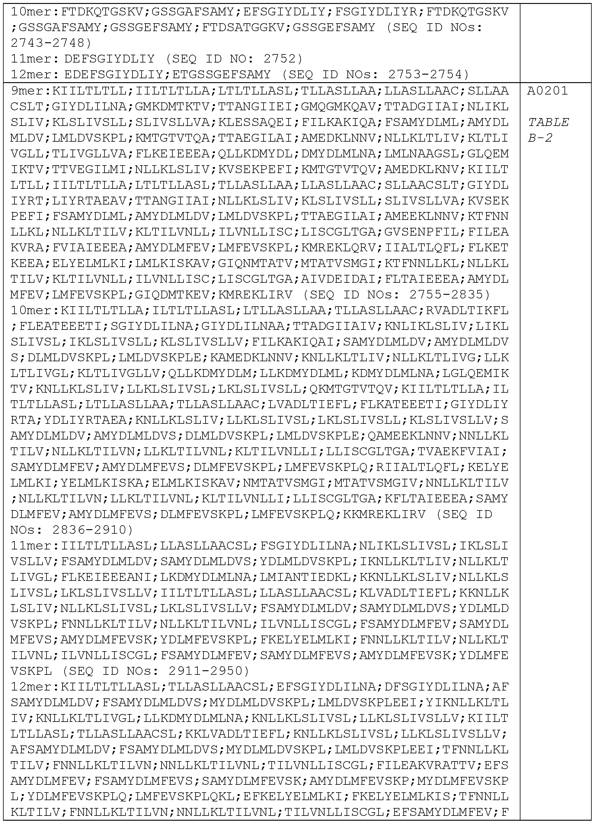
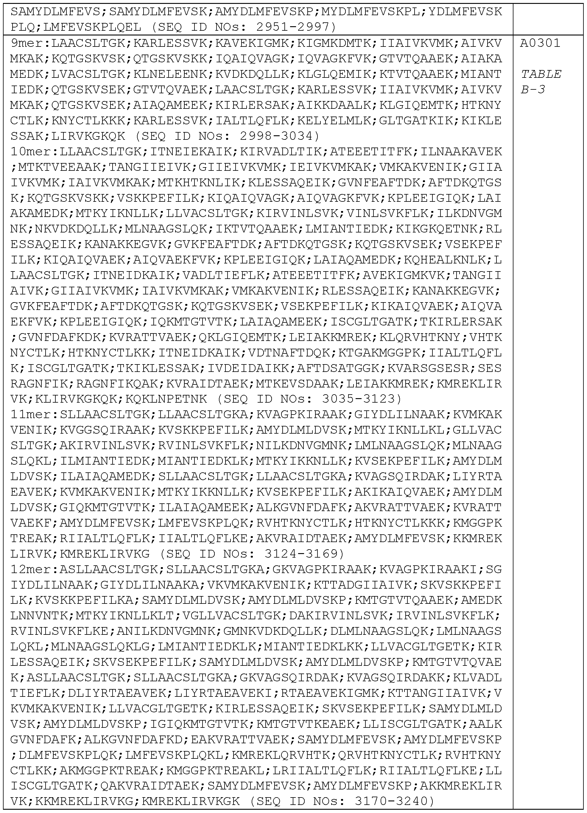


Preferred binding peptides P derived or predicted from Borrelia protein FlhF capable of interacting with one or more MHC class 1 molecules are listed in Table C:
Table C: Predicted MHC class 1 binding peptides P derived from Borrelia antigen FlhF (8- , 9-, 10-, 11-, and 12-mers), predicted as detailed in Example 19. The binding peptides P are sorted per HLA-allele.


Preferred binding peptides P derived or predicted from Borrelia protein FlaB capable of interacting with one or more MHC class 1 molecules are listed in Table D:
Table D: Predicted MHC class 1 binding peptides P derived from Borrelia antigen FlaB (8- , 9-, 10-, 11-, and 12-mers), predicted as detailed in Example 20. The binding peptides P are sorted per HLA-allele.


Preferred binding peptides P derived or predicted from Borrelia protein P37-42 capable of interacting with one or more MHC class 1 molecules are listed in Table E: Table E: Predicted MHC class 1 binding peptides P derived from Borrelia antigen P37-42 (8-, 9-, 10-, 11-, and 12-mers), predicted as detailed in Example 21. The binding peptides P are sorted per HLA-allele.


In one embodiment there is provided a panel comprising one or more MHC multimers, wherein each of said one or more MHC multimers comprise an antigenic peptide P selected from the group consisting of:
i) antigenic peptides P derived from Borrelia antigenic polypeptide OppA (SEQ ID
NOs:1-9) listed in Table A (Table A-1 to Table A-20),
ii) antigenic peptides P derived from Borrelia antigenic polypeptide DbpA (SEQ ID
NOs: 10-20) listed in Table B (Table B-1 to Table B-19),
iii) antigenic peptides P derived from Borrelia antigenic polypeptide FlhF (SEQ ID NOs:21-28) listed in Table C (Table C-1 to Table C-20),
iv) antigenic peptides P derived from Borrelia antigenic polypeptide FlaB (SEQ ID
NOs:29-37) listed in Table D (Table D-1 to Table D-20), and/or v) antigenic peptides P derived from Borrelia antigenic polypeptide P37-42 (SEQ
ID NOS:38-39) listed in Table E (Table E-1 to Table E-20).
In one embodiment there is provided a panel comprising one or more MHC multimers, wherein each of said one or more MHC multimers comprise an antigenic peptide P selected from the group consisting of:
i) antigenic peptides P derived from Borrelia antigenic polypeptide OppA (SEQ ID
NOs:1-9) listed in Table A-1 , Table A-2, Table A-3, Table A-4, Table A-5, Table A-6, Table A-7, Table A-8, Table A-9, Table A-10, Table A-11 , Table A-12, Table A-13, Table A-14, Table A-15, table A-16, Table A-17, Table A- 18, Table A-19, or Table A-20,
ii) antigenic peptides P derived from Borrelia antigenic polypeptide DbpA (SEQ ID
NOs: 10-20) listed in Table B-1 , Table B-2, Table B-3, Table B-4, Table B-5, Table B-6, Table B-7, Table B-8, Table B-9, Table B-10, Table B-11 , Table B-12, Table B-13, Table B-14, Table B-15, table B-16, Table B-17, Table B- 18, or Table B-19,
iii) antigenic peptides P derived from Borrelia antigenic polypeptide FlhF (SEQ ID
NOs:21-28) listed in Table C-1 , Table C-2, Table C-3, Table C-4, Table C-5, Table C-6, Table C-7, Table C-8, Table C-9, Table C-10, Table C-11 , Table C-12, Table C-13, Table C-14, Table C-15, table C-16, Table C-17, Table C- 18, Table C-19, or Table C-20,
iv) antigenic peptides P derived from Borrelia antigenic polypeptide FlaB (SEQ ID
NOs:29-37) listed in Table D-1 , Table D-2, Table D-3, Table D-4, Table D-5, Table D-6, Table D-7, Table D-8, Table D-9, Table D-10, Table D-11 , Table D-12, Table D-13, Table D-14, Table D-15, table D-16, Table D-17, Table D- 18, Table D-19, or Table D-20, and/or
v) antigenic peptides P derived from Borrelia antigenic polypeptide P37-42 (SEQ
ID NOS:38-39) listed in Table E-1 , Table E-2, Table E-3, Table E-4, Table E-5, Table E-6, Table E-7, Table E-8, Table E-9, Table E-10, Table E-11 , Table E-12, Table E-13, Table E-14, Table E-15, table E-16, Table E-17, Table E-18, Table E-19, or Table E-20.
In one embodiment there is provided a panel comprising one or more MHC multimers, wherein each of said one or more MHC multimers comprise an antigenic peptide P selected from the group consisting of: YLNTKSNGNYEI (SEQ ID NO: 359),
FLSIFTQGYT (SEQ ID NO: 241), GIYDLILNA (SEQ ID NO: 2761), YIKDINEFI (SEQ ID NO: 4479), IQIEIEQLTDEI (SEQ ID NO: 5126), RMISDQRANLGA (SEQ ID NO: 5127), SQGGVNSPV (SEQ ID NO: 5112), MLDEAKDKL (SEQ ID NO: 5516), FMEQATNSWI (SEQ ID NO: 5530), NLVFSSLFL (SEQ ID NO: 5510) and KLAESIYKRL (SEQ ID NO: 5531).
In one embodiment there is provided a panel comprising one or more MHC multimers, wherein each of said one or more MHC multimers comprise an antigenic peptide P selected from the group consisting of:
i) YLNTKSNGNYEI (SEQ ID NO: 359) and FLSIFTQGYT (SEQ ID NO:
241)derived from OppA,
ii) GIYDLILNA (SEQ ID NO: 2761) derived from DbpA,
iii) YIKDINEFI (SEQ ID NO: 4479) derived from FlhF,
iv) IQIEIEQLTDEI (SEQ ID NO: 5126), RMISDQRANLGA (SEQ ID NO: 5127) and SQGGVNSPV (SEQ ID NO: 5112) derived from FlaB, and/or
v) IQIEIEQLTDEI (SEQ ID NO: 5126), MLDEAKDKL (SEQ ID NO: 5516),
FMEQATNSWI (SEQ ID NO: 5530), NLVFSSLFL (SEQ ID NO: 5510) and KLAESIYKRL (SEQ ID NO: 5531) derived from P37-42.
In one embodiment there is provided a panel comprising one or more MHC multimers, wherein each of said one or more MHC multimers comprise an antigenic peptide P selected from the group consisting of:
i) YLNTKSNGNYEI (SEQ ID NO: 359) derived from OppA, FLSIFTQGYT (SEQ ID NO: 241) derived from OppA, GIYDLILNA (SEQ ID NO: 2761) derived from DbpA, and YIKDINEFI (SEQ ID NO: 4479) derived from FlhF,
ii) IQIEIEQLTDEI (SEQ ID NO: 5126), RMISDQRANLGA (SEQ ID NO: 5127) and
SQGGVNSPV (SEQ ID NO: 5112) derived from FlaB, and/or
iii) IQIEIEQLTDEI (SEQ ID NO: 5126), MLDEAKDKL (SEQ ID NO: 5516),
FMEQATNSWI (SEQ ID NO: 5530), NLVFSSLFL (SEQ ID NO: 5510) and KLAESIYKRL (SEQ ID NO: 5531) derived from P37-42.
In one embodiment there is provided a panel comprising or consisting of 11 MHC multimers wherein each of said 11 MHC multimers comprise an antigenic peptide P selected from the group consisting of: YLNTKSNGNYEI (SEQ ID NO: 359),
FLSIFTQGYT (SEQ ID NO: 241), GIYDLILNA (SEQ ID NO: 2761), YIKDINEFI (SEQ ID NO: 4479), IQIEIEQLTDEI (SEQ ID NO: 5126), RMISDQRANLGA (SEQ ID NO: 5127), SQGGVNSPV (SEQ ID NO: 5112), MLDEAKDKL (SEQ ID NO: 5516), FMEQATNSWI (SEQ ID NO: 5530), NLVFSSLFL (SEQ ID NO: 5510) and KLAESIYKRL (SEQ ID NO: 5531).
In one embodiment there is provided a panel comprising or consisting of an MHC multimer comprising YLNTKSNGNYEI (SEQ ID NO: 359), an MHC multimer comprising FLSIFTQGYT (SEQ ID NO: 241), an MHC multimer comprising GIYDLILNA (SEQ ID NO: 2761), an MHC multimer comprising YIKDINEFI (SEQ ID NO: 4479), an MHC multimer comprising IQIEIEQLTDEI (SEQ ID NO: 5126), an MHC multimer comprising RMISDQRANLGA (SEQ ID NO: 5127), an MHC multimer comprising SQGGVNSPV (SEQ ID NO: 5112), an MHC multimer comprising MLDEAKDKL (SEQ ID NO: 5516), an MHC multimer comprising FMEQATNSWI (SEQ ID NO: 5530), an MHC multimer comprising NLVFSSLFL (SEQ ID NO: 5510) and an MHC multimer comprising KLAESIYKRL (SEQ ID NO: 5531).
In one embodiment there is provided a panel comprising one or more pools, or two or more pools, of MHC multimers, wherein each pool comprises one or more MHC multimers each comprising:
i) an antigenic peptide P derived from Borrelia antigenic polypeptide OppA (SEQ
ID NOs:1-9) listed in Table A (Table A-1 to Table A-20),
ii) an antigenic peptide P derived from Borrelia antigenic polypeptide DbpA (SEQ
ID NOs: 10-20) listed in Table B (Table B-1 to Table B-19),
iii) an antigenic peptide P derived from Borrelia antigenic polypeptide FlhF (SEQ
ID NOs:21-28) listed in Table C (Table C-1 to Table C-20),
iv) an antigenic peptide P derived from Borrelia antigenic polypeptide FlaB (SEQ
ID NOs:29-37) listed in Table D (Table D-1 to Table D-20), and/or v) an antigenic peptide P derived from Borrelia antigenic polypeptide P37-42 (SEQ
ID NOS:38-39) listed in Table E (Table E-1 to Table E-20).
In one embodiment there is provided a panel comprising one or more pools, or two or more pools, of MHC multimers, wherein each pool comprises one or more MHC multimers each comprising:
i) an antigenic peptide P derived from Borrelia antigenic polypeptide OppA (SEQ
ID NOs:1-9) listed in Table A (Table A-1 to Table A-20), and including at least one or both of YLNTKSNGNYEI (SEQ ID NO: 359) and FLSIFTQGYT
(SEQ ID NO: 241);
ii) an antigenic peptide P derived from Borrelia antigenic polypeptide DbpA (SEQ
ID NOs:10-20) listed in Table B (Table B-1 to Table B-19), and including at least GIYDLILNA (SEQ ID NO: 2761);
iii) an antigenic peptide P derived from Borrelia antigenic polypeptide FlhF (SEQ
ID NOs:21-28) listed in Table C (Table C-1 to Table C-20), and including at least YIKDINEFI (SEQ ID NO: 4479);
iv) an antigenic peptide P derived from Borrelia antigenic polypeptide FlaB (SEQ
ID NOs:29-37) listed in Table D (Table D-1 to Table D-20), and including at least one or more of IQIEIEQLTDEI (SEQ ID NO: 5126), RMISDQRANLGA (SEQ ID NO: 5127) and SQGGVNSPV (SEQ ID NO: 5112); and/or v) an antigenic peptide P derived from Borrelia antigenic polypeptide P37-42 (SEQ
ID NOS:38-39) listed in Table E (Table E-1 to Table E-20), and including at least one or more of MLDEAKDKL (SEQ ID NO: 5516), FMEQATNSWI (SEQ ID NO: 5530), NLVFSSLFL (SEQ ID NO: 5510) and KLAESIYKRL (SEQ ID NO: 5531).
In one embodiment there is provided a panel comprising one or more pools, or two or more pools, of MHC multimers, wherein each pool comprises one or more MHC multimers each comprising:
i) an antigenic peptide P derived from Borrelia antigenic polypeptide OppA (SEQ
ID NOs:1-9) listed in Table A-1 , Table A-2, Table A-3, Table A-4, Table A-5, Table A-6, Table A-7, Table A-8, Table A-9, Table A-10, Table A-11 , Table A-12, Table A-13, Table A-14, Table A-15, table A-16, Table A-17, Table A- 18, Table A-19, or Table A-20,
ii) an antigenic peptide P derived from Borrelia antigenic polypeptide DbpA (SEQ
ID NOs: 10-20) listed in Table B-1 , Table B-2, Table B-3, Table B-4, Table B- 5, Table B-6, Table B-7, Table B-8, Table B-9, Table B-10, Table B-11 ,
Table B-12, Table B-13, Table B-14, Table B-15, table B-16, Table B-17, Table B-18, or Table B-19,
iii) an antigenic peptide P derived from Borrelia antigenic polypeptide FlhF (SEQ
ID NOs:21-28) listed in Table C-1 , Table C-2, Table C-3, Table C-4, Table C-5, Table C-6, Table C-7, Table C-8, Table C-9, Table C-10, Table C-11 , Table C-12, Table C-13, Table C-14, Table C-15, table C-16, Table C-17, Table C-18, Table C-19, or Table C-20,
iv) an antigenic peptide P derived from Borrelia antigenic polypeptide FlaB (SEQ
ID NOs:29-37) listed in Table D-1 , Table D-2, Table D-3, Table D-4, Table D-5, Table D-6, Table D-7, Table D-8, Table D-9, Table D-10, Table D-11 ,
Table D-12, Table D-13, Table D-14, Table D-15, table D-16, Table D-17, Table D-18, Table D-19, or Table D-20, and/or
v) an antigenic peptide P derived from Borrelia antigenic polypeptide P37-42 (SEQ ID NOS:38-39) listed in Table E-1 , Table E-2, Table E-3, Table E-4, Table E-5, Table E-6, Table E-7, Table E-8, Table E-9, Table E-10, Table E-11 , Table E-12, Table E-13, Table E-14, Table E-15, table E-16, Table E-17, Table E-18, Table E-19, or Table E-20.
In one embodiment there is provided a panel comprising two or more pools of MHC multimers, wherein one or more of said pools comprises one or more MHC multimers each MHC multimer comprising an antigenic peptide P selected from the group consisting of:
i) antigenic peptides P selected from the group consisting of YLNTKSNGNYEI (SEQ ID NO: 359), FLSIFTQGYT (SEQ ID NO: 241), GIYDLILNA (SEQ ID NO: 2761), and YIKDINEFI (SEQ ID NO: 4479),
ii) antigenic peptides P selected from the group consisting of IQIEIEQLTDEI (SEQ ID NO: 5126), RMISDQRANLGA (SEQ ID NO: 5127) and SQGGVNSPV (SEQ ID NO: 5112), and/or
iii) antigenic peptides P selected from the group consisting of IQIEIEQLTDEI (SEQ ID NO: 5126), MLDEAKDKL (SEQ ID NO: 5516), FMEQATNSWI (SEQ ID NO:
5530), NLVFSSLFL (SEQ ID NO: 5510) and KLAESIYKRL (SEQ ID NO:
5531).
In one embodiment there is provided a panel comprising two or more pools of MHC multimers, wherein one or more of said pools comprises one or more MHC multimers each MHC multimer comprising antigenic peptides P selected from the group consisting of YLNTKSNGNYEI (SEQ ID NO: 359), FLSIFTQGYT (SEQ ID NO: 241), GIYDLILNA (SEQ ID NO: 2761), YIKDINEFI (SEQ ID NO: 4479), IQIEIEQLTDEI (SEQ ID NO: 5126), RMISDQRANLGA (SEQ ID NO: 5127), SQGGVNSPV (SEQ ID NO: 5112), MLDEAKDKL (SEQ ID NO: 5516), FMEQATNSWI (SEQ ID NO: 5530), NLVFSSLFL (SEQ ID NO: 5510) and KLAESIYKRL (SEQ ID NO: 5531).
In one embodiment there is provided a panel comprising 3 or more pools of MHC multimers, wherein
i) Pool 1 comprises one or more MHC multimers comprising YLNTKSNGNYEI (SEQ ID NO: 359), one or more MHC multimers comprising FLSIFTQGYT (SEQ ID NO: 241), one or more MHC multimers comprising GIYDLILNA (SEQ ID NO: 2761), and one or more MHC multimers comprising YIKDINEFI (SEQ ID NO: 4479),
ii) Pool 2 comprises one or more MHC multimers comprising IQIEIEQLTDEI (SEQ ID NO: 5126), one or more MHC multimers comprising RMISDQRANLGA (SEQ ID NO: 5127) and one or more MHC multimers comprising SQGGVNSPV (SEQ ID NO: 5112), and
iii) Pool 3 comprises one or more MHC multimers comprising MLDEAKDKL (SEQ ID NO: 5516), one or more MHC multimers comprising FMEQATNSWI (SEQ ID NO: 5530), one or more MHC multimers comprising NLVFSSLFL (SEQ ID NO: 5510) and one or more MHC multimers comprising KLAESIYKRL (SEQ ID NO: 5531) ;
iv) and optionally Pool 4 comprising one or more negative control MHC multimers, v) and optionally Pool 5 comprising one or more positive control MHC multimers.
In one embodiment there is provided a panel comprising 5 pools of MHC multimers, wherein
i) Pool 1 comprises one or more MHC multimers comprising YLNTKSNGNYEI (SEQ ID NO: 359), one or more MHC multimers comprising FLSIFTQGYT (SEQ ID NO: 241), one or more MHC multimers comprising GIYDLILNA (SEQ ID NO: 2761), and one or more MHC multimers comprising YIKDINEFI (SEQ ID NO: 4479),
ii) Pool 2 comprises one or more MHC multimers comprising IQIEIEQLTDEI (SEQ ID NO: 5126), one or more MHC multimers comprising RMISDQRANLGA (SEQ ID NO: 5127) and one or more MHC multimers comprising SQGGVNSPV (SEQ ID NO: 5112),
iii) Pool 3 comprises one or more MHC multimers comprising MLDEAKDKL (SEQ ID NO: 5516), one or more MHC multimers comprising FMEQATNSWI (SEQ ID NO: 5530), one or more MHC multimers comprising NLVFSSLFL (SEQ ID NO: 5510) and one or more MHC multimers comprising KLAESIYKRL (SEQ ID NO: 5531),
iv) Pool 4 comprises one or more MHC multimers comprising ALIAPVHAV (SEQ ID NO: 5913), and
v) Pool 5 comprises one or more MHC multimers comprising NLVPMVATV (SEQ ID NO: 5914), one or more MHC multimers comprising GLCTLVAML (SEQ ID NO: 5915) and one or more MHC multimers comprising GILGFVFTL (SEQ ID NO: 5916).
Using the above described principles individual peptides or a subset of peptides able to bind one or more types of MHC molecules and make stable MHC-peptide complexes can be identified. The identified peptides can then be tested for biological relevance in functional assays such as interferon gamma release assays (e.g. ELISPOT), cytotoxicity assays (e.g. CTL killing assays) or using other methods as described elsewhere herein. Alternatively or complementary hereto the ability of the identified antigenic peptides to bind selected MHC molecules may be determined in binding assays like Biacore measurement, competition assays or other assays useful for measurement of binding of peptide to MHC molecules, known by skilled persons.
Borrelia sequences
In general, MHC Class I molecules will accommodate peptides of from about 8 amino acids in length to about 11-12 amino acids. Of special interest of the present disclosure are antigenic peptides, P, derived from Borrelia antigenic polypeptides selected from the group consisting of OppA, DbpA, FlhF, FlaB and P37-42. In the following Borrelia bacteria and their genes and proteins from which these Borrelia antigenic polypeptides may derive from are described in more detail.
Borrelia bacteria
The various species of Borrelia are known to humans in the form of Lyme disease and recurring fever, transmitted through tick or flea bite. The cycle of Borrelia through animals is related to the tick's life cycle. The tick has four stages in its two-year life cycle, egg, larva, nymph and adult. Between each stage the tick needs a blood meal in order to mature. The tick usually acquires the spirochaete during its larval stage, when it feeds on small animals such as rodents or birds. Usually the tick picks up Borrelia from the white-footed mouse, which is commonly infected. The tick then becomes the host for the spirochaete. The bacteria resides in the digestive tract of the host for its next nymph and adult stages during which it is passed on to other animals, and sometimes humans.
Borrelia species
Borrelia is a genus of bacteria of the spirochete class. It is a zoonotic, vector-borne disease transmitted primarily by ticks and some by lice, depending on the species. There are at least 50 known species of Borrelia. Different species of Borrelia results in different clinical symptoms. Of the 37 known species of Borrelia, 12 of these species are known to cause Lyme disease or borreliosis and are transmitted by ticks. The major Borrelia species causing Lyme disease are Borrelia burgdorferi, Borrelia afzelii, Borrelia garinii and Borrelia valaisiana.
In one embodiment of the present disclosure there is provided antigenic peptides, P, derived from Borrelia antigenic polypeptides selected from the group consisting of OppA, DbpA, FlhF, FlaB and P37-42, wherein said Borrelia antigenic polypeptides are derived from a borrelia species selected from the group consisting of Borrelia burgdorferi, Borrelia afzelii, Borrelia garinii and Borrelia valaisiana.
In one embodiment of the present disclosure there is provided antigenic peptides, P, derived from Borrelia burgdorferi antigenic polypeptides selected from the group consisting of OppA, DbpA, FlhF, FlaB and P37-42.
In one embodiment of the present disclosure there is provided antigenic peptides, P, derived from Borrelia afzelii antigenic polypeptides selected from the group consisting of OppA, DbpA, FlhF, FlaB and P37-42.
In one embodiment of the present disclosure there is provided antigenic peptides, P, derived from Borrelia garinii antigenic polypeptides selected from the group consisting of OppA, DbpA, FlhF, FlaB and P37-42.
In one embodiment of the present disclosure there is provided antigenic peptides, P, derived from Borrelia valaisiana antigenic polypeptides selected from the group consisting of OppA, DbpA, FlhF, FlaB and P37-42.
Other Borrelia species cause relapsing fever such as Borrelia recurrentis, caused by the human body louse. No animal reservoir of B. recurrentis exists. Lice that feed on infected humans acquire the Borrelia organisms that then multiply in the gut of the louse. When an infected louse feeds on an uninfected human, the organism gains
access when the victim crushes the louse or scratches the area where the louse is feeding. B. recurrentis infects the person via mucous membranes and then invades the bloodstream.
Other tick-borne relapsing infections are acquired from other species, such as Borrelia hermsii or Borrelia Parkeri, which can be spread from rodents, and serve as a reservoir for the infection, via a tick vector. Borrelia hermsii and Borrelia recurrentis cause very similar diseases although the disease associated with Borrelia hermsii has more relapses and is responsible for more fatalities, while the disease caused by B.
recurrentis has longer febrile and afebrile intervals and a longer incubation period.
The present disclosure relates in one embodiment to one or more antigenic peptides P or to MHC-peptide complexes or MHC multimers comprising one or more antigenic peptides P comprising one or more sequences from one or more Borrelia species including the ones mentioned with the present disclosure; wherein said antigenic peptides P are derived from a Borrelia antigenic polypeptide selected from the group consisting of OppA, DbpA, FlhF, FlaB and P37-42.
Borrelia burgdorferi
In one preferred embodiment the disclosure relates to one or more antigenic peptides comprising one or more sequences from Borrelia burgdorferi such as Borrelia burgdorferi B31 or to one or more MHC-peptide complexes or MHC multimers comprising one or more such antigenic peptides.
Borrelia burgdorferi is species of bacteria of the spirochete class of the genus Borrelia. B. burgdorferi is predominant in North America, but also exists in Europe, and is the agent of Lyme disease.
It is a zoonotic, vector-borne disease transmitted by ticks and is named after the researcher Willy Burgdorfer who first isolated the bacterium in 1982. B. burgdorferi is one of the few pathogenic bacteria that can survive without iron, having replaced all of its iron-sulphur cluster enzymes with enzymes that use manganese, thus avoiding the problem many pathogenic bacteria face in acquiring iron. B. burgdorferi infections have been linked to non-Hodgkin lymphomas. The B. burgdorferi genome (B31 strain) contains 910,725 base pairs and 853 genes.
Borrelia afzelii
In one preferred embodiment the disclosure relates to one or more antigenic peptides comprising one or more sequences from Borrelia afzelii such as borrelia afzelii Pko or one or more MHC-peptide complexes or one or more MHC multimers comprising one or more such antigenic peptides .
Borrelia afzelii is considered a seperate species of the Genus Borrelia and considered homologous to Borrelia burgdorferi with regard to phenotypic, genetic, and
immunological characteristics. Diseases linked to this species of Borrelia are Lyme disease and Acrodermatitis chronica atrophicans (ACA). Better understanding of the structure and function of this pathogen will create better methods of treatment to people with the diseases it causes.
Borrelia garinii
In one preferred embodiment the disclosure relates to one or more antigenic peptides comprising one or more sequences from Borrelia garinii such as garinii PBi or to one or more MHC-peptide complexes or MHC multimers comprising one or more such antigenic peptides.
Borrelia garinii is one of two major strains found in Europe. It usually causes Lyme Disease symptoms of the neurological kind - such as extreme back- and leg-pains, meningitis and partial facial paralysis, Lyme arthritis due to B garinii may be associated in susceptible hosts with amoxicillin resistance or treatment resistance.
Other Borrelia species
In another embodiment the disclosure relates to one or more antigenic peptides or to one or more MHC-peptide complexes or MHC multimers comprising one or more such sequences or antigenic peptides, wherein said antigenic peptides are derived from a borrelia species selected from the group consisting of Borrelia anserina, Borrelia barbouri, Borrelia afzelii, Borrelia afzelii ACA-1 , Borrelia afzelii K78, Borrelia afzelii PKo, Borrelia andersonii, Borrelia bissettii, Borrelia burgdorferi, Borrelia burgdorferi 118a, Borrelia burgdorferi 156a, Borrelia burgdorferi 29805, Borrelia burgdorferi 64b, Borrelia burgdorferi 72a, Borrelia burgdorferi 80a, Borrelia burgdorferi 94a, Borrelia burgdorferi B31 , Borrelia burgdorferi Bol26, Borrelia burgdorferi CA-11.2a, Borrelia burgdorferi WI91-23, Borrelia burgdorferi ZS7, Borrelia californiensis, Borrelia garinii, Borrelia garinii PBi, Borrelia garinii PBr, Borrelia genomosp. 1 , Borrelia genomosp. 2, Borrelia japonica, Borrelia lusitaniae, Borrelia spielmanii, Borrelia spielmanii A14S,
Borrelia tanukii, Borrelia turdi, Borrelia valaisiana, Borrelia valaisiana VS116,
Candidatus Borrelia texasensis, Borrelia sp. AA4Pool, Borrelia sp. AI-1 , Borrelia sp. B31 , Borrelia sp. BC-1 , Borrelia sp. CA1133, Borrelia sp. CA1176, Borrelia sp. CA128, Borrelia sp. CA13, Borrelia sp. CA134, Borrelia sp. CA142, Borrelia sp. CA20, Borrelia sp. CA22, Borrelia sp. CA27, Borrelia sp. CA28, Borrelia sp. CA29, Borrelia sp. CA31 , Borrelia sp. CA33, Borrelia sp. CA370, Borrelia sp. CA372, Borrelia sp. CA378,
Borrelia sp. CA388, Borrelia sp. CA393, Borrelia sp. CA394, Borrelia sp. CA395,
Borrelia sp. CA399, Borrelia sp. CA400, Borrelia sp. CA401 , Borrelia sp. CA402,
Borrelia sp. CA404, Borrelia sp. CA411 , Borrelia sp. CA426, Borrelia sp. CA443,
Borrelia sp. CA446, Borrelia sp. CA448, Borrelia sp. CA462, Borrelia sp. CA468,
Borrelia sp. CA502, Borrelia sp. CA504, Borrelia sp. CA507, Borrelia sp. CA547,
Borrelia sp. CA552, Borrelia sp. CA8, Borrelia sp. D22, Borrelia sp. D35, Borrelia sp. FD-1 , Borrelia sp. FL18, Borrelia sp. FL27, Borrelia sp. FL35, Borrelia sp. FL42,
Borrelia sp. HN6, Borrelia sp. HN7, Borrelia sp. HN8, Borrelia sp. HNM13, Borrelia sp. HNM14, Borrelia sp. HNM19, Borrelia sp. IA1 , Borrelia sp. lr-3519, Borrelia sp. lr-4721 , Borrelia sp. lr-4812, Borrelia sp. lr-5215, Borrelia sp. LV5, Borrelia sp. MI-2, Borrelia sp. MI-5, Borrelia sp. MI-6, Borrelia sp. MI-8, Borrelia sp. MI-9, Borrelia sp. MOD-1 , Borrelia sp. MOD-5, Borrelia sp. MOK-3a, Borrelia sp. MOS-1b, Borrelia sp. NE49, Borrelia sp. NE581 , Borrelia sp. PHaP, Borrelia sp. PSigll, Borrelia sp. SCGT-10, Borrelia sp. SCGT-8a, Borrelia sp. SCI-2, Borrelia sp. SCW-30h, Borrelia sp. SI-1 , Borrelia sp. SI-10, Borrelia sp. SM-1 , Borrelia sp. SV1 , Borrelia sp. W97F51 , Borrelia sp. Z41293, Borrelia sp. Z41493, Borrelia coriaceae, Borrelia crocidurae, Borrelia duttonii, Borrelia duttonii Ly, Borrelia hermsii, Borrelia hermsii DAH, Borrelia hispanica, Borrelia lonestari, Borrelia miyamotoi, Borrelia parkeri, Borrelia persica, Borrelia recurrentis, Borrelia recurrentis A1 , Borrelia sinica, Borrelia theileri, Borrelia turcica, Borrelia turicatae, Borrelia turicatae 91 E135, Borrelia sp., Borrelia sp. 'Lake Gaillard', Borrelia sp. 000133, Borrelia sp. 010298, Borrelia sp. 10MT, Borrelia sp. 5145, Borrelia sp. 57Nsk, Borrelia sp. 5MT, Borrelia sp. 6T04-2, Borrelia sp. BR, Borrelia sp. BR 2007, Borrelia sp. C5-N52, Borrelia sp. CB-A1 , Borrelia sp. CB-A11 , Borrelia sp. CB- A3, Borrelia sp. EFL-S0100110, Borrelia sp. IK/23, Borrelia sp. IM/16, Borrelia sp. IM/19, Borrelia sp. KR1 , Borrelia sp. KR3, Borrelia sp. LB-2001 , Borrelia sp. LB-M56, Borrelia sp. LB-W100, Borrelia sp. MK-N61 , Borrelia sp. NR-N8, Borrelia sp. OkME1 , Borrelia sp. PAnz, Borrelia sp. PJes, Borrelia sp. PMai, Borrelia sp. PMew, Borrelia sp. R57, Borrelia sp. strain Spain, Borrelia sp. TA1 , Borrelia sp. TM, Borrelia sp. TM1 and/or Borrelia sp. TM2.
Gene variants
The present disclosure further relates to one or more MHC-peptide complexes or MHC multimers, wherein the one or more Borrelia antigenic peptides are encoded by one or more gene variants. The present disclosure also relates to one or more antigenic Borrelia peptides that are encoded by one or more gene variants, such as a gene variant of a Borrelia antigenic polypeptide selected from the group consisting of OppA, DbpA, FlhF, FlaB and P37-42.
The term "variant gene" refers to nucleic acid molecules that encode a polypeptide having an amino acid sequence that is a modification of the polypeptides according to the present invention. Such variants include naturally-occurring polymorphisms of genes, as well as synthetic genes that contain conservative amino acid substitutions of the amino acid sequence of a polypeptide according to the present disclosure.
Additional variant forms of genes are nucleic acid molecules that contain insertions or deletions of the nucleotide sequences described herein. A variant gene can be identified by determining whether the gene hybridizes with a nucleic acid molecule having the nucleotide sequence of an antigenic polypeptide according to the present disclosure, or its complement, under stringent conditions.
Variant antigenic peptides P
The present disclosure further relates to one or more Borrelia antigenic peptides as defined herein that have one or more modifications, such as one or more modifications compared to the one or more Borrelia antigenic peptides predicted from a Borrelia antigenic polypeptide selected from the group consisting of OppA, DbpA, FlhF, FlaB and P37-42 according to the present disclosure.
In one embodiment the antigenic peptide P according to the present disclosure comprises or consists of a modified sequence obtained by modification of an antigenic peptide P as defined herein above.
In one embodiment the antigenic peptide P comprises or consists of a modified sequence obtained by modification of a sequence selected from the sequences included in Tables A to E herein above (Tables A1-A20, B1-B19, C1-C20, D1-D20 and E1-E20).
In one embodiment said modified antigenic peptide P has at least 50 %, such as at least 60 %, such as at least 70 %, such as at least 85 %, such as at least 95 % or such as at least 99 % sequence identity with a sequence included in Tables A to E herein above (Tables A1-A20, B1-B19, C1-C20, D1-D20 and E1-E20).
Antigenic peptides P with amino acid substitutions
The present disclosure further relates to one or more Borrelia antigenic peptides P that have one or more amino acid substitutions, such as 1 , 2, 3, 4, 5, 6, 7, or 8 amino acid substitutions, as well as MHC-peptide complexes or MHC multimers as disclosed herein comprising one or more Borrelia antigenic peptides having one or more amino acid substitutions such as such as 1 , 2, 3, 4, 5, 6, 7, or 8 amino acid substitutions. The one or more Borrelia antigenic peptides are in one embodiment those predicted from a Borrelia antigenic polypeptide selected from the group consisting of OppA, DbpA, FlhF, FlaB and P37-42 according to the present disclosure.
In one embodiment the one or more amino acid substitutions are within the amino acid anchor motif. In another embodiment the one or more amino acid substitutions are outside the amino acid anchor motif. In one embodiment the one or more amino acid substitutions are within the 9-mer core motif. In another embodiment the one or more amino acid substitutions are outside the 9-mer core motif.
In one embodiment these amino acid substitutions comprise substitution with an “equivalent amino acid residue”. An“equivalent amino acid residue” refers to an amino acid residue capable of replacing another amino acid residue in a polypeptide without substantially altering the structure and/or functionality of the polypeptide. Equivalent amino acids thus have similar properties such as bulkiness of the side-chain, side chain polarity (polar or non-polar), hydrophobicity (hydrophobic or hydrophilic), pH (acidic, neutral or basic) and side chain organization of carbon molecules
(aromatic/aliphatic). As such,“equivalent amino acid residues” can be regarded as “conservative amino acid substitutions”.
The classification of equivalent amino acids refers in one embodiment to the following classes: 1) HRK, 2) DENQ, 3) C, 4) STPAG, 5) MILV and 6) FYW.
Within the meaning of the term“equivalent amino acid substitution” as applied herein, one amino acid may be substituted for another, in one embodiment, within the groups of amino acids indicated herein below:
Amino acids having polar side chains (Asp, Glu, Lys, Arg, His, Asn, Gin, Ser, Thr, Tyr, and Cys,)
Amino acids having non-polar side chains (Gly, Ala, Val, Leu, lie, Phe, Trp, Pro, and Met)
Amino acids having aliphatic side chains (Gly, Ala Val, Leu, lie)
Amino acids having cyclic side chains (Phe, Tyr, Trp, His, Pro)
Amino acids having aromatic side chains (Phe, Tyr, Trp)
Amino acids having acidic side chains (Asp, Glu)
Amino acids having basic side chains (Lys, Arg, His)
Amino acids having amide side chains (Asn, Gin)
Amino acids having hydroxy side chains (Ser, Thr)
Amino acids having sulphor-containing side chains (Cys, Met),
Neutral, weakly hydrophobic amino acids (Pro, Ala, Gly, Ser, Thr)
Hydrophilic, acidic amino acids (Gin, Asn, Glu, Asp), and
Hydrophobic amino acids (Leu, lie, Val)
A Venn diagram is another method for grouping of amino acids according to their properties (Livingstone & Barton, CABIOS, 9, 745-756, 1993). In another preferred embodiment one or more amino acids may be substituted with another within the same Venn diagram group.
In another embodiment these amino acid substitutions comprise substitution with a “non-equivalent amino acid residue”. Non-equivalent amino acid residues are amino acid residues with dissimilar properties to the properties of the amino acid they substitute according to the groupings described above.
In one embodiment the modified antigenic peptide P comprises an anchor motif selected from the group of HLA motifs included in Table I herein above; such as comprises a primary anchoring amino acid residue in amino acid position 2 and/or 9 in accordance with Table I herein above.
In one embodiment the modified antigenic peptide P comprises a substitution of the amino acid residue in position 2 with an amino acid residue selected from the group consisting of:
i) alanine, threonine, serine, valine, leucine, isoleucine, methionine, glutamine, phenylalanine, tryptophan and tyrosine,
ii) alanine, threonine, serine, valine, leucine, isoleucine, methionine and glutamine iii) arginine, histidine and lysine,
iv) aspartic acid and glutamic acid, or
v) alanine, threonine and serine.
In one embodiment the antigenic peptide P or modified antigenic peptide P comprises a substitution of the amino acid residue in position 9 or 10 with an amino acid residue selected from the group consisting of:
i) phenylalanine, tryptophan, tyrosine, leucine, isoleucine, valine, glutamine, alanine, argentine, histidine, lysine and methionine,
ii) phenylalanine, tryptophan, tyrosine, leucine, isoleucine, valine, glutamine, alanine and methionine,
iii) leucine, isoleucine, valine, glutamine, alanine and methionine,
iv) phenylalanine, tryptophan, tyrosine, leucine, isoleucine and methionine v) glutamine and alanine, and
vi) tyrosine, arginine and lysine.
In one embodiment the amino acid substitutions increases the affinity of the peptide for the MHC molecule and thereby increase the stability of the MHC-peptide complex.
In another embodiment the amino acid substitutions decreases the affinity of the peptide for the MHC molecule and thereby increase the stability of the MHC-peptide complex. In one embodiment the amino acid substitutions increases the overall affinity of one or more T-cell receptors for the MHC-peptide complex containing the modified antigenic peptide.
In another embodiment the amino acid substitutions decreases the overall affinity of one or more T-cell receptors for the MHC-peptide complex containing the modified antigenic peptide.
Antigenic peptides P fragments
The present disclosure further relates to fragments of one or more Borrelia antigenic peptides as well as MHC monomers, MHC-peptide complexes or MHC multimers as disclosed herein comprising said antigenic peptide fragments, wherein said one or more Borrelia antigenic peptides are in one embodiment those predicted from a Borrelia antigenic polypeptide selected from the group consisting of OppA, DbpA, FlhF, FlaB and P37-42 according to the present disclosure.
The one or more antigenic peptides in one embodiment comprise or consist of a fragment of one or more antigenic peptides according to the present disclosure, such as a fragment consisting of 1 , 2, 3, 4, 5, 6, 7, 8, 9, 10, 11 or 12 amino acids of said one or more antigenic peptide P, such as a fragment consisting of 1 , 2, 3, 4, 5, 6, 7, 8, 9,
10, 11 or 12 amino acids of one or more antigenic peptides P included in Tables A to E herein above (Tables A1-A20, B1-B19, C1-C20, D1-D20 and E1-E20).
Other peptide modifications
In addition to the binding peptides designed by the total approach and/or directed approach, homologous peptides and peptides that have been modified in the amino acid side chains or in the backbone can be used as binding peptides.
In one embodiment the antigenic peptides according to the present disclosure are modified by one or more type(s) of post-translational modifications such as one or more of the post-translational modifications disclosed herein elsewhere. The same or different types of post-translational modification can occur on one or more amino acids in the antigenic peptide. Thus, in one embodiment, any one amino acid may be modified once, twice or three times with the same or different types of modifications. Furthermore, said identical and/or different modification may be present on 1 , 2, 3, 4, 5, 6, 7, 8, 9, 10, 11 or 12 of the amino acid residues of the binding peptide according to the present disclosure.
Homologous peptides
Homologues MHC peptide sequences may arise from the existence of multiple strongly homologous alleles, from small insertions, deletions, inversions or substitutions. If they are sufficiently homologous to peptides derived by the total approach, i.e. have an amino acid sequence identity greater than e.g. more than 90%, more than 80%, or more than 70%, or more than 60%, to one or two binding peptides derived by the total approach, they may be good candidates. Identity is often most important for the anchor residues.
A MHC binding peptide may be of split- or combinatorial epitope origin i.e. formed by linkage of peptide fragments derived from two different peptide fragments and/or proteins. Such peptides can be the result of either genetic recombination on the DNA level or due to peptide fragment association during the complex break down of proteins during protein turnover. Possibly it could also be the result of faulty reactions during protein synthesis i.e. caused by some kind of mixed RNA handling. A kind of combinatorial peptide epitope can also be seen if a portion of a longer peptide make a loop out leaving only the terminal parts of the peptide bound in the groove.
Uncommon, artificial and chemically modified amino acids
Peptides having un-common amino acids, such as selenocysteine and pyrrolysine, may be bound in the MHC groove as well. Artificial amino acids e.g. having the isomeric D-form may also make up isomeric D-peptides that can bind in the binding groove of the MHC molecules. Bound peptides may also contain amino acids that are chemically modified or being linked to reactive groups that can be activated to induce changes in or disrupt the peptide. Example post-translational modifications are shown below. However, chemical modifications of amino acid side chains or the peptide backbone can also be performed.
Any of the modifications can be found individually or in combination at any position of the peptide, e.g. position 1 , 2, 3, 4, 5, 6, etc. up to n.


Table: Post-translational modification of peptides
Post translationally modified peptides
The amino acids of the antigenic peptides, P, can also be modified in various ways dependent on the amino acid in question, or the modification can affect the amino- or carboxy-terminal end of the peptide (see table immediately herein above). Such peptide modifications occur naturally as the result of post-translational processing of the parental protein. A non-exhaustive description of the major post-translational modifications is given below, divided into three main types: a) Modifications that add a chemical moiety to the binding peptide, P:
Acetylation, the addition of an acetyl group, usually at the N-terminus of the protein. Alkylation, the addition of an alkyl group (e.g. methyl, ethyl).
Methylation, the addition of a methyl group, usually at lysine or arginine residues is a type of alkylation. Demethylation involves the removal of a methyl-group.
Amidation at C-terminus.
Biotinylation, acylation of conserved lysine residues with a biotin appendage formylation.
Gamma-carboxylation dependent on Vitamin K.
Glutamylation, covalent linkage of glutamic acid residues to tubulin and some other proteins by means of tubulin polyglutamylase.
Glycosylation, the addition of a glycosyl group to either asparagine, hydroxylysine, serine, or threonine, resulting in a glycoprotein. Distinct from glycation, which is regarded as a nonenzymatic attachment of sugars.
Glycylation, covalent linkage of one to more than 40 glycine residues to the tubulin C-terminal tail.
Heme moiety may be covalently attached.
Hydroxylation, is any chemical process that introduces one or more hydroxyl groups (-OH) into a compound (or radical) thereby oxidizing it. The principal residue to be hydroxylated is Proline. The hydroxilation occurs at the CY atom, forming hydroxyproline (Hyp). In some cases, proline may be hydroxylated instead on its Cp atom. Lysine may also be hydroxylated on its C6 atom, forming hydroxylysine (Hyl). lodination.
Isoprenylation, the addition of an isoprenoid group (e.g. farnesol and
geranylgeraniol).
Lipoylation, attachment of a lipoate functionality, as in prenylation, GPI anchor formation, myristoylation, farnesylation, geranylation.
Nucleotides or derivatives thereof may be covalently attached, as in ADP- ribosylation and flavin attachment.
Oxidation, lysine can be oxidized to aldehyde.
Pegylation, addition of poly-ethylen-glycol groups to a protein. Typical reactive amino acids include lysine, cysteine, histidine, arginine, aspartic acid, glutamic acid, serine, threonine, tyrosine. The N-terminal amino group and the C-terminal carboxylic acid can also be used
Phosphatidylinositol may be covalently attached.
Phosphopantetheinylation, the addition of a 4'-phosphopantetheinyl moiety from coenzyme A, as in fatty acid, polyketide, non-ribosomal peptide and leucine biosynthesis.
Phosphorylation, the addition of a phosphate group, usually to serine, tyrosine, threonine or histidine.
Pyroglutamate formation as a result of N-terminal glutamine self-attack, resulting in formation of a cyclic pyroglutamate group.
Racemization of proline by prolyl isomerase.
tRNA-mediated addition of amino acids such as arginylation.
Sulfation, the addition of a sulfate group to a tyrosine.
Selenoylation (co-translational incorporation of selenium in selenoproteins).
b) Modification that adds protein or peptide:
ISGylation, the covalent linkage to the ISG15 protein (Interferon-Stimulated Gene 15).
SUMOylation, the covalent linkage to the SUMO protein (Small Ubiquitin-related Modifier).
Ubiquitination, the covalent linkage to the protein ubiquitin. c) Modification that converts one or more amino acids to different amino acids:
Citrullination, or deimination the conversion of arginine to citrulline.
Deamidation, the conversion of glutamine to glutamic acid or asparagine to aspartic acid.
The peptide modifications can occur as modification of a single amino acid or more than one i.e. alone or in combinations. Modifications can be present on any position within the peptide i.e. on position 1, 2, 3, 4, 5, etc. for the entire length of the peptide P.
Sources of binding peptides
a) From natural sources
The binding peptides can be obtained from natural sources by enzymatic digestion or proteolysis of natural proteins or proteins derived by in vitro translation of mRNA. Binding peptides may also be eluted from the MHC binding groove. b) From recombinant sources
1) as monomeric or multimeric peptide
Alternatively peptides can be produced recombinantly by transfected cells either as monomeric antigenic peptides or as multimeric (concatemeric) antigenic peptides. Optionally, the Multimeric antigenic peptides are cleaved to form monomeric antigenic peptides before binding to MHC protein.
2) as part of a bigger recombinant protein
Binding peptides may also constitute a part of a bigger recombinant protein e.g.
consisting of;
2a) For MHC class 1 binding peptides;
Peptide-linker^2m, b2hi being full length or truncated;
Peptide-linker-MHC class 1 heavy chain, the heavy chain being full length or truncated. Most importantly the truncated class I heavy chain will consist of the extracellular part i.e the a1 , D a2, and a domains. The heavy chain fragment may also only contain the a1 and a2 domains, or a1 domain alone, or any fragment or full length b2hi or heavy chain attached to a designer domain(s) or protein fragment(s). c) From chemical synthesis
MHC binding peptide may also be chemically synthesized by solid phase or fluid phase synthesis, according to standard protocols.
Comprehensive collections of antigenic peptides, derived from one antigen, may be prepared by a modification of the solid phase synthesis protocol, as described in the following and exemplified in Example 24.
The protocol for the synthesis of the full-length antigen on solid support is modified by adding a partial cleavage step after each coupling of an amino acid. Thus, the starting point for the synthesis is a solid support to which has been attached a cleavable linker. Then the first amino acid X1 (corresponding to the C-terminal end of the antigen) is added and a coupling reaction performed. The solid support now carries the molecule “Nnker-X1”. After washing, a fraction (e.g. 10%) of the cleavable linkers are now cleaved, to release into solution X1. The supernatant is transferred to a collection container. Additional solid support carrying a cleavable linker is added, e.g.
corresponding to 10% of the initial amount of solid support.
Then the second amino acid X2 is added and coupled to X1 or the cleavable linker, to form on solid support the molecules“Nnker-X2” and“Nnker-X1-X2”. After washing, a fraction (e.g. 10%) of the cleavable linker is cleaved, to release into solution X2 and X1-X2. The supernatant is collected into the collection container, which therefore now contains X1 , X2, and X1-X2. Additional solid support carrying a cleavable linker is added, e.g. corresponding to 10% of the initial amount of solid support.
Then the third amino acid X3 is added and coupled to X2 or the cleavable linker, to form on solid support the molecules“Nnker-X3”,“Nnker-X2-X3” and“Nnker-X1-X2-X3”. After washing, a fraction (e.g. 10%) of the cleavable linker is cleaved, to release into solution X3, X2-X3 and X1-X2-X3. The supernatant is collected into the collection container, which therefore now contains X1 , X2, X3, X1-X2, X2-X3 and X1-X2-X3. Additional solid support carrying a cleavable linker is added, e.g. corresponding to 10% of the initial amount of solid support.
This step-wise coupling and partial cleavage of the linker is continued until the N- terminal end of the antigen is reached. The collection container will now contain a large number of peptides of different length and sequence. In the present example where a 10% partial cleavage was employed, a large fraction of the peptides will be 8’-mers, 9’- mers, 10’-mers and 1 T-mers, corresponding to class I antigenic peptides. As an example, for a 100 amino acid antigen the 8’-mers will consist of the sequences X1-X2-
X3-X4-X5-X6-X7-X8, X2-X3-X4-X5-X6-X7-X8-X9, . , X93-X94-X95-X96-X97-X98-
X99-X100.
Optionally, after a number of coupling and cleavage steps or after each coupling and cleavage step, the used (inactivated) linkers on solid support can be regenerated, in order to maintain a high fraction of linkers available for synthesis. The collection of antigenic peptides can be used as a pool for e.g. the display by APCs to stimulate CTLs in ELISPOT assays, or the antigenic peptides may be mixed with one or more MHC alleles, to form a large number of different MHC-peptide complexes which can e.g. be used to form a large number of different MHC multimers which can e.g. be used in flow cytometry experiments.
Choice of MHC allele for generation of MHC monomers and MHC multimers
More than 600 MHC alleles (class 1 and 2) are known in humans; for many of these, the peptide binding characteristics are known. Figure 3 of WO 2009/106073 presents a list of the HLA class 1 alleles. The frequency of the different HLA alleles varies considerably, also between different ethnic groups - as illustrated for top 30 HLA class I alleles (See eg.g Fig. 4 in WO 2009/106073). Thus it is of outmost importance to carefully select the MHC alleles that corresponds to the population that one wish to study.
In one embodiment the MHC protein of the present disclosure is selected from the group of HLA alleles consisting of: A*0201 , C*0701 , A*0101 , A*0301 , C*0702, C*0401 , B*4402, B*0702, B*0801 , C*0501 , C*0304, C*0602, A*1101 , B*4001 , A*2402, B*3501 , C*0303, B*5101 , C*1203, B*1501 , A*2902, A*2601 , A*3201 , C*0802, A*2501 , B*5701 , B*1402, C*0202, B*1801 , B 403, C*0401 , C*0701 , C*0602, A*0201 , A*2301 , C*0202, A*0301 , C*0702, B*5301 , B*0702, C*1601 , B*1503, B*5801 , A*6802, C*1701 , B 501 , B*4201 , A*3001 , B*3501 , A*0101 , C*0304, A*3002, B*0801 , A*3402, A*7401 , A*3303, C*1801 , A*2902, B 403, B*4901 , A*0201 , C*0401 , A*2402, C*0702, C*0701 , C*0304, A*0301 , B*0702, B*3501 , C*0602, C*0501 , A*0101 , A*1101 , B*5101 , C*1601 , B 403,
C*0102, A*2902, C*0802, B*1801 , A*3101 , B*5201 , B*1402, C*0202, C*1203, A*2601 , A*6801 , B*0801 , A*3002, B 402, A*1101 , A*2402, C*0702, C*0102, A*3303, C*0801 , C*0304, A*0201 , B 001 , C*0401 , B*5801 , B 601 , B*5101 , C*0302, B*3802, A*0207, B*1501 , A*0206, C*0303, B*1502, A*0203, B 403, C*1402, B*3501 , C*0602, B*5401 , B*1301 , B*4002, B*5502 and A*2601.
In one embodiment the MHC protein of the present disclosure is selected from the group of HLA alleles consisting of: HLA-A*A0101 , A0201 , A0301 , A1101 , A2402,
A2501 , A2601 , A2902, A3101 , A3201 , A6801 , B0702, B0801 , B1503, B1801 , B3501 , B4002, B4402, B4501 and B5101.
The combined choice of peptide, MHC and carrier
Herein above it has been described how to generate binding peptides, and which MHC alleles are available. Herein below it is further described how one may modify the binding peptides in order to increase the stability, affinity, specificity and other features of the MHC-peptide complex or the MHC multimer. In the following it is described what characteristics of binding peptides and MHC alleles are important when using the MHC-peptide complex or MHC-multimer for various different purposes.
A first preferred embodiment employs binding peptides of particularly high affinity for the MHC proteins. This may be done in order to increase the stability of the MHC- peptide complex. A higher affinity of the binding peptide for the MHC proteins may in some instances also result in increased rigidity of the MHC-peptide complex, which in turn often will result in higher affinity and/or specificity of the MHC-peptide complex for the T-cell receptor. A higher affinity and specificity will in turn have consequences for the immunogenicity and allergenicity, as well as possible side-effects of the MHC- peptide complex in e.g. the body.
Binding peptides of particularly high affinity for the MHC proteins may be identified by several means, including the following
• Incubation of candidate binding peptides and MHC proteins, followed by
analysis of the resulting complexes to identify those binding peptides that have most frequently been associated with MHC proteins. The binding peptides that have most frequently been associated with MHC proteins typically will represent high-affinity binding peptides. The identification of binding peptides with particularly high-affinity may involve enrichment of binding peptides, e.g.
incubation of candidate peptides with immobilized MHC molecules, removal of non-binding peptides by e.g. washing, elution of binding peptides. This pool of peptides enriched for binding to the chosen MHC molecules may then be identified e.g. by mass spectrometry or HPLC and amino acid sequencing or the pool can be further enriched by another round of incubation with immobilized MHC.
• Candidate binding peptides may be compared to consensus sequences for the binding to a specific MHC allele. Thus, for a given class 1 allele, the consensus 8’mer sequence may be given by the sequence“X1-X2-X3-X4-X5-X6-X7-X8”, where each of the X1-X8 amino acids can be chosen from a specific subset of amino acids, as described above.
Those binding peptides that correlate the best with the consensus sequence are expected to have particularly high affinity for the MHC allele in question.
• Based on a large data set of affinities of binding peptides for specific MHC alleles, software programs (often involving neural networks) have been developed that allow a relatively accurate prediction of the affinity of a given candidate binding peptide for a given MHC allele. By examining candidate binding peptides using such software programs, one can identify binding peptides of expected high-affinity for the MHC molecule.
A second preferred embodiment employs binding peptides with medium affinity for the MHC molecule. A medium affinity of the peptide for the MHC protein will often lead to lower physical and chemical stability of the MHC-peptide complex, which can be an advantage for certain applications. As an example, it is often desirable to administer a drug on a daily basis due to convenience. An MHC-peptide complex-based drug with high stability in the body would not allow this. In contrast a binding peptide with medium or low affinity for the MHC protein can be an advantage for such applications, since these functional MHC-peptide molecules will be cleared more rapidly from the body due to their lower stability.
For some applications where some level of cross-talk is desired, e.g. in applications where the target is a number of T cell clones that interact with a number of structurally related MHC-peptide complexes, e.g. MHC-peptide complexes containing binding peptides from different strains of a given species, a medium or low affinity of the binding peptide for the MHC protein can be an advantage. Thus, these MHC-peptide
complexes are often more structurally flexible, allowing the MHC-peptide complexes to interact with several structurally related TCRs.
The affinity of a given peptide for a MHC protein, predicted by a software program or by its similarity to a consensus sequence, should only be considered a guideline to its real affinity. Moreover, the affinity can vary a lot depending on the conditions in the environment, e.g. the affinity in blood may be very different from the affinity in a biochemical assay. Further, in the context of a MHC multimer, the flexibility of the MHC-peptide complex can sometimes be an important parameter for overall avidity.
In summary, a lot of factors must be considered for the choice of binding peptides in a certain application. Some applications benefit from the use of all possible binding peptides for an antigen (“total approach”), other applications benefit from the selective choice of just a few binding peptides. Depending on the application, the affinity of the binding peptide for MHC protein is preferably high, medium, or low; the physical and/or chemical stability of the MHC-peptide complex is preferably high, medium or low; the binding peptide is preferably a very common or very rare epitope in a given population; etc.
It is obvious from the above preferred embodiments that most or all of the binding peptides generated by the total approach have important applications. In other words, in order to make relevant MHC multimers that suit the different applications with regard to e.g. personalized or general targeting, or with regard to affinity, avidity, specificity, immunogenicity, stimulatory efficiency, or stability, one must be able to choose from the whole set of binding peptides generated by the total approach.
Loading of the peptide into the MHC multimer
Loading of the peptides into the MHCmer MHC class 1 can be performed in a number of ways depending on the source of the peptide and the MHC, and depending on the application.
The antigenic peptide may be added to the other peptide chain(s) at different times and in different forms, as follows
a) Loading of antigenic peptide during MHC complex folding:
a. Antigenic peptide is added as a free peptide
MHC class I molecules are most often loaded with peptide during assembly in vitro by the individual components in a folding reaction i.e. consisting of purified recombinant heavy chain a with the purified recombinant b2 microglobulin and a peptide or a peptide mix.
b. Antigenic peptide is part of a recombinant protein construct
Alternatively the peptide to be folded into the binding groove can be encoded together with e.g. the a heavy chain or fragment hereof by a gene construct having the structure, heavy chain-flexible linker- peptide. This recombinant molecule is then folded in vitro with p2-microglobulin. b) Antigenic peptide replaces another antigenic peptide by an exchange reaction: a. Exchange reaction“in solution”
Loading of desired peptide can also be made by an in vitro exchange reaction where a peptide already in place in the binding groove are being exchanged by another peptide species.
b. Exchange reaction“in situ”
Peptide exchange reactions can also take place when the parent molecule is attached to other molecules, structures, surfaces, artificial or natural membranes and nano-particles.
c. Aided exchange reaction.
This method can be refined by making the parent construct with a peptide containing a meta-stable amino acid analogue that is split by either light or chemically induction thereby leaving the parent structure free for access of the desired peptide in the binding groove.
d. Display by in vivo loading
Loading of MHC class I molecules expressed on the cell surface with the desired peptides can be performed by an exchange reaction. Alternatively cells can be transfected by the peptides themselves or by the mother proteins that are then being processed leading to an in vivo analogous situation where the peptides are bound in the groove during the natural cause of MHC expression by the transfected cells. In the case of professional antigen presenting cells e.g. dendritic cells, macrophages, Langerhans cells, the proteins and peptides can be taken up by the cells themselves by phagocytosis and then bound to the MHC complexes the natural way and expressed on the cell surface in the correct MHC context.
Other features of product
In one preferred embodiment the MHC multimer is between 50,000 Da and 1 ,000,000 Da, such as from 50,000 Da to 980,000; for example from 50,000 Da to 960,000; such as from 50,000 Da to 940,000; for example from 50,000 Da to 920,000; such as from 50,000 Da to 900,000; for example from 50,000 Da to 880,000; such as from 50,000 Da to 860,000; for example from 50,000 Da to 840,000; such as from 50,000 Da to 820,000; for example from 50,000 Da to 800,000; such as from 50,000 Da to 780,000; for example from 50,000 Da to 760,000; such as from 50,000 Da to 740,000; for example from 50,000 Da to 720,000; such as from 50,000 Da to 700,000; for example from 50,000 Da to 680,000; such as from 50,000 Da to 660,000; for example from 50,000 Da to 640,000; such as from 50,000 Da to 620,000; for example from 50,000 Da to 600,000; such as from 50,000 Da to 580,000; for example from 50,000 Da to 560,000; such as from 50,000 Da to 540,000; for example from 50,000 Da to 520,000; such as from 50,000 Da to 500,000; for example from 50,000 Da to 480,000; such as from 50,000 Da to 460,000; for example from 50,000 Da to 440,000; such as from 50,000 Da to 420,000; for example from 50,000 Da to 400,000; such as from 50,000 Da to 380,000; for example from 50,000 Da to 360,000; such as from 50,000 Da to 340,000; for example from 50,000 Da to 320,000; such as from 50,000 Da to 300,000; for example from 50,000 Da to 280,000; such as from 50,000 Da to 260,000; for example from 50,000 Da to 240,000; such as from 50,000 Da to 220,000; for example from 50,000 Da to 200,000; such as from 50,000 Da to 180,000; for example from 50,000 Da to 160,000; such as from 50,000 Da to 140,000; for example from 50,000 Da to 120,000; such as from 50,000 Da to 100,000; for example from 50,000 Da to 80,000; such as from 50,000 Da to 60,000; such as from 100,000 Da to 980,000; for example from 100,000 Da to 960,000; such as from 100,000 Da to 940,000; for example from 100,000 Da to 920,000; such as from 100,000 Da to 900,000; for example from 100,000 Da to 880,000; such as from 100,000 Da to 860,000; for example from 100,000 Da to 840,000; such as from 100,000 Da to 820,000; for example from 100,000 Da to 800,000; such as from 100,000 Da to 780,000; for example from 100,000 Da to 760,000; such as from 100,000 Da to 740,000; for example from 100,000 Da to 720,000; such as from 100,000 Da to 700,000; for example from 100,000 Da to 680,000; such as from 100,000 Da to 660,000; for example from 100,000 Da to 640,000; such as from 100,000 Da to 620,000; for example from 100,000 Da to 600,000; such as from 100,000 Da to 580,000; for
example from 100,000 Da to 560,000; such as from 100,000 Da to 540,000; for example from 100,000 Da to 520,000; such as from 100,000 Da to 500,000; for example from 100,000 Da to 480,000; such as from 100,000 Da to 460,000; for example from 100,000 Da to 440,000; such as from 100,000 Da to 420,000; for example from 100,000 Da to 400,000; such as from 100,000 Da to 380,000; for example from 100,000 Da to 360,000; such as from 100,000 Da to 340,000; for example from 100,000 Da to 320,000; such as from 100,000 Da to 300,000; for example from 100,000 Da to 280,000; such as from 100,000 Da to 260,000; for example from 100,000 Da to 240,000; such as from 100,000 Da to 220,000; for example from 100,000 Da to 200,000; such as from 100,000 Da to 180,000; for example from 100,000 Da to 160,000; such as from 100,000 Da to 140,000; for example from 100,000 Da to 120,000; such as from 150,000 Da to 980,000; for example from 150,000 Da to 960,000; such as from 150,000 Da to 940,000; for example from 150,000 Da to 920,000; such as from 150,000 Da to 900,000; for example from 150,000 Da to 880,000; such as from 150,000 Da to 860,000; for example from 150,000 Da to 840,000; such as from 150,000 Da to 820,000; for example from 150,000 Da to 800,000; such as from 150,000 Da to 780,000; for example from 150,000 Da to 760,000; such as from 150,000 Da to 740,000; for example from 150,000 Da to 720,000; such as from 150,000 Da to 700,000; for example from 150,000 Da to 680,000; such as from 150,000 Da to 660,000; for example from 150,000 Da to 640,000; such as from 150,000 Da to 620,000; for example from 150,000 Da to 600,000; such as from 150,000 Da to 580,000; for example from 150,000 Da to 560,000; such as from 150,000 Da to 540,000; for example from 150,000 Da to 520,000; such as from 150,000 Da to 500,000; for example from 150,000 Da to 480,000; such as from 150,000 Da to 460,000; for example from 150,000 Da to 440,000; such as from 150,000 Da to 420,000; for example from 150,000 Da to 400,000; such as from 150,000 Da to 380,000; for example from 150,000 Da to 360,000; such as from 150,000 Da to 340,000; for example from 150,000 Da to 320,000; such as from 150,000 Da to 300,000; for example from 150,000 Da to 280,000; such as from 150,000 Da to 260,000; for example from 150,000 Da to 240,000; such as from 150,000 Da to 220,000; for example from 150,000 Da to 200,000; such as from 150,000 Da to 180,000; for example from 150,000 Da to 160,000.
In another embodiment the MHC multimer is between 1 ,000,000 Da and 3,000,000 Da, such as from 1 ,000,000 Da to 2,800,000; for example from 1 ,000,000 Da to 2,600,000; such as from 1 ,000,000 Da to 2,400,000; for example from 1 ,000,000 Da to 2,200,000; such as from 1 ,000,000 Da to 2,000,000; for example from 1 ,000,000 Da to 1 ,800,000; such as from 1 ,000,000 Da to 1 ,600,000; for example from 1 ,000,000 Da to 1 ,400,000.
Above it was described how to design and produce the key components of the MHC multimers, i.e. the MHC-peptide complex. In the following it is described how to generate the MHC monomer or MHC multimer products of the present disclosure.
Number of MHC complexes per multimer
A non-exhaustive list of possible MHC mono- and multimers illustrates the possibilities ‘n’ indicates the number of MHC complexes comprised in the multimer af the present disclsoure: a) n=1 , Monomers
b) n=2, Dimers, multimerization can for example be based on IgG scaffold,
streptavidin with two MHC’s, coiled-coil dimerization e.g. Fos.Jun dimerization
c) n=3, Trimers, multimerization can for example be based on streptavidin as scaffold with three MHC’s, TNFalpha-MHC hybrids, triplex DNA-MHC conjugates or other trimer structures
d) n=4, Tetramers, multimerization can for example be based on streptavidin with all four binding sites occupied by MHC molecules or based on dimeric IgA e) n=5, Pentamers, multimerization for example can take place around a
pentameric coil-coil structure
f) n=6, Hexamers
g) n=7, Heptamers
h) n=8-12, Octa- dodecamers, multimerization can for example use Streptactin i) n=10, Decamers, multimerization can for example use IgM
j) 1 <n<100, Dextramers, as multimerization domain polymers such as polypeptides, polysaccharides and Dextrans can for example be used.
k) 1 <n<1000, Multimerization can for example make use of dendritic cells (DC),
antigen-presenting cells (APC), micelles, liposomes, beads, surfaces e.g. microtiterplate, tubes, microarray devices, micro-fluidic systems
I) 1<n, n in billions or trillions or higher, multimerization can for example take place on beads, and surfaces e.g. microtiterplate, tubes, microarray devices, micro-fluidic systems
In one embodiment the panel of the present disclosure comprises MHC multimers (a-b- P)n, wherein n > 1 , comprising two or more MHC proteins each in complex with an antigenic peptide P to form an MHC-peptide complex. In a preferred embodiment the MHC proteins are class I MHC proteins.
In one embodiment the panel of the present disclosure comprises MHC multimers (a-b- P)n, wherein the value of n is selected from the group consisting of 1 , 2, 3, 4, 5, 6, 7, 8, 9, 10, 1 1 , 12, 13, 14, 15, 16, 17, 18, 19, 20, 21 , 22, 23, 24, 25, 26, 27, 28, 29, 30, 31 , 32, 33, 34, 35, 36, 37, 38, 39, 40, 41 , 42, 43, 44, 45, 46, 47, 48, 50, 55, 60, 65, 70, 75, 80, 85, 90, 95, 100, 110, 120, 130, 140, 150, 160, 170, 180, 190, 200, 250, 300, 350, 400, 450, 500, 550, 600, 650, 700, 750, 800, 850, 900, 950 and 1000.
In one embodiment the panel of the present disclosure comprises MHC multimers (a-b- P)n, wherein the value of n is 1 < n ³ 1000, such as between 2-3, 3-4, 4-5, 5-6, 6-7, 7- 8, 8-9, 9-10, 10-11 , 11-12, 12-13, 13-14, 14-15, 15-16, 16-17, 17-18, 18-19, 19-20, 20- 21 , 21-22, 22-23, 23-24, 24-25, 25-26, 26-27, 27-28, 28-29, 29-30, 30-35, 35-40, 40- 45, 45-50, 50-55, 55-60, 60-65, 65-70, 75-80, 80-85, 85-90, 90-95, 95-100, 100-1 10, 110-120, 120-130, 130-140, 140-150, 150-160, 160-170, 170-180, 180-190, 190-200, 200-225, 225-250, 250-275, 275-300, 300-325, 325-350, 350-375, 375-400, 400-450, 450-500, 500-550, 550-600, 600-650, 650-700, 700-750, 750-800, 800-850, 850-900, 900-950, 950-1000.
In one embodiment the panel of the present disclosure comprises MHC multimers (a-b- P)n, wherein the value of n is >1 , such as 2, such as >2, such as ³2, such as 3, such as >3, such as ³3, such as 4, such as >4, such as ³4, such as 5, such as >5, such as ³5, such as 6, such as >6, such as ³6, such as 7, such as >7, such as ³7, such as 8, such as >8, such as ³8, such as 9, such as >9, such as ³9, such as 10, such as >10, such as ³10.
MHC multimers thus include MHC-dimers, MHC-trimers, MHC-tetramers, MHC- pentamers, MHC-hexamers, and MHC n-mers, as well as organic molecules, cells, membranes, polymers and particles that comprise two or more MHC-peptide
complexes. Example organic molecule-based multimers include functionalized cyclic structures such as benzene rings where e.g. a benzene ring is functionalized and covalently linked to e.g. three MHC complexes; example cell-based MHC multimers include dendritic cells and antigen presenting cells (APCs); example membrane-based MHC multimers include liposomes and micelles carrying MHC-peptide complexes in their membranes; example polymer-based MHC multimers include MHC-dextramers (dextran to which a number of MHC-peptide complexes are covalently or non- covalently attached) and example particles include beads or other solid supports with MHC complexes immobilized on the surface. Obviously, any kind of multimerization domain can be used, including any kind of cell, polymer, protein or other molecular structure, or particles and solid supports.
Any of the three components of a MHC complex can be of any of the below mentioned origins. The list is non-exhaustive. A complete list would encompass all Chordate species. By origin is meant that the sequence is identical or highly homologous to a naturally occurring sequence of the specific species.
List of origins: Human, Mouse, Primate (including Chimpansee, Gorilla, Orang Utan), Monkey (including Macaques), Porcine (Swine/Pig), Bovine (Cattle/Antilopes), Equine (Horse), Camelides (Camels), Ruminants (Deer), Canine (Dog), Feline (Cat), Bird (including Chicken, Turkey), Fish, Reptiles and Amphibians.
In one embodiment the MHC of the present disclosure is a MHC class I complex of HLA-type A. In one embodiment the MHC is a MHC class I complex of HLA-type B. In one embodiment the MHC is a MHC class I complex of HLA-type C.
In one embodiment the MHC of the present disclosure is a MHC class I complex
- of supertype HLA-A1 (eg. HLA-A*0101 , HLA-A*2601 , HLA-A*2602, HLA-A2603, HLA-A*3002, HLA-A*3003, HLA-A*3004, HLA-A*3201),
- of supertype HLA-A01 A03 (eg. HLA-A*3001),
- of supertype HLA-A01 A024 (eg. HLA-A*2902),
- of supertype HLA-A2 (eg. HLA-A*0201 , HLA-A*0202, HLA-A*0203, HLA- A*0204, HLA-A*0205, HLA-A*0206, HLA-A*0207, HLA-A*0214, HLA-A*0217, HLA-A*6802, HLA-A*6901),
- of supertype HLA-A3 (eg. HLA-A*0301, HLA-A*1101, HLA-A*3101, HLA- A*3301 , HLA-A*3303, HLA-A*6601, HLA-A*6801, HLA-A*7401),
- of supertype HLA-A24 (eg. HLA-A*2301 , HLA-A*2402),
- of supertype HLA-B7 (eg. HLA-B*0702, HLA-B*0703, HLA-B*0705, HLA
B*1508, HLA-B*3501, HLA-B*3503, HLA-B 201, HLA-B*5101, HLA-B*5102, HLA-B*5103, HLA-B*5301, HLA-B*5401, HLA-B*5501, HLA-B*5502, HLA- B*5601 , HLA-B*6701, HLA-B*7801),
- of supertype HLA-B8 (eg. HLA-B*0801, HLA-B*0802),
- of supertype HLA-B27 (eg. HLA-B*1402, HLA-B*1503, HLA-B*1509, HLA- B*1510, HLA-B*1518, HLA-B*2702, HLA-B*2703, HLA-B*2704, HLA-B*2705, HLA-B*2706, HLA-B*2707, HLA-B*2708, HLA-B*2709, HLA-B*3801, HLA- B*3901 , HLA-B*3902, HLA-B*3909, HLA-B 801, HLA-B*7301),
- of supertype HLA-B44 (eg. HLA-B*1801, HLA-B*3701, HLA-B 001, HLA- B 002, HLA-B 006, HLA-B 402, HLA-B 403, HLA-B 501),
- of supertype HLA-B58 (eg. HLA-B*1516, HLA-B*1517, HLA-B*5701, HLA- B*5702, HLA-B*5801, HLA-B*5802),
- of supertype HLA-B62 (eg. HLA-B*1501, HLA-B*1502, HLA-B*1512, HLA- B*1513, HLA-B 601, HLA-B*5201), and/or
- of supertype HLA Cw 1-8 (eg. HLA-C*01, HLA-C*02, HLA-C*03, HLA-C*04, HLA-C*05, HLA-C*06, HLA-C*07, HLA-C*08).
In one embodiment the MHC of the present disclosure is a MHC class I complex, which binds peptides
- with an acidic amino acid on 3rd position (eg. HLA-A*0101, HLA-A*2601, HLA- A*2602, HLA-A*2603, HLA-A*3002, HLA-A*3003, HLA-A*3004, HLA-A*3201),
- with a hydrophobic amino acid on 9th position (eg. HLA-A*0201...0207, A*0214, A*0217, A*6802, A*6901, HLA-B*1516, B*1517, B*5701, B*5702, B*5801, B*5802),
- with a Basic amino acid on 9th position (eg. HLA-A*0301, HLA-A*1101, HLA- A*3101 , HLA-A*3301, HLA-A*3303, HLA-A*6601, HLA-A*6801, HLA-AV401),
- with a Tyrosine amino acid on 2nd position (eg. HLA-A*2301 , HLA-A*2402),
- with a Proline amino acid on 2nd position (eg. HLA-B*0702, HLA-B*0703, HLA- B*0705, HLA-B*1508, HLA-B*3501, HLA-B*3503, HLA-B 201, HLA-B*5101, HLA-B*5102, HLA-B*5103, HLA-B*5301, HLA-B*5401, HLA-B*5501, HLA- B*5502, HLA-B*5601, HLA-B*6701, HLA-B*7801),
- with a Lysine amino acid on 3rd and 5th position (eg. HLA-B*0801 , B*0802),
- with a Arginine amino acid on 2nd position (eg. HLA-B*1402, HLA-B*1503, HLA- B*1509, HLA-B*1510, HLA-B*1518, HLA-B*2702, HLA-B*2703, HLA-B*2704, HLA-B*2705, HLA-B*2706, HLA-B*2707, HLA-B*2708, HLA-B*2709, HLA- B*3801 , HLA-B*3901 , HLA-B*3902, HLA-B*3909, HLA-B 801 , HLA-BV301), and/or
- with a Glutamic acid amino acid on 2nd position (eg. HLA-B*1801 , HLA-B*3701 , HLA-B 001 , HLA-B 002, HLA-B 006, HLA-B 402, HLA-B 403, HLA- B 501),
- with a Tyrosine amino acid on 9th position (eg. HLA-B*1501 , HLA-B*1502, HLA- B*1512, HLA-B*1513, HLA-B 601 , HLA-B*5201).
In one embodiment the MHC of the present disclosure is a MHC class I complex, which in the B pocket selectively binds small or aliphatic peptides (e.g. HLA-A*0101 , HLA- A*2601 , HLA-A*2602, HLA-A*2603, HLA-A*3002, HLA-A*3003, HLA-A*3004, HLA- A*3201 , HLA-A*0201 , HLA-A*0202, HLA-A*0203, HLA-A*0204, HLA-A*0205, HLA- A*0206, HLA-A*0207, HLA-A*0214, HLA-A*0217, HLA-A*6802, HLA-A*6901 , HLA- A*0301 , HLA-A*1101 , HLA-A*3101 , HLA-A*3301 , HLA-A*3303, HLA-A*6601 , HLA- A*6801 , HLA-A*7401).
In one embodiment the MHC of the present disclosure is a MHC class I complex, which in the F pocket selectively binds aliphatic peptides (e.g. HLA-A*0101 , HLA- A*2601...2603, HLA-A*3002, HLA-A*3003, HLA-A*3004, HLA-A*3201 , HLA-A*0201 , HLA-A*0202, HLA-A*0203, HLA-A*0204, HLA-A*0205, HLA-A*0206, HLA-A*0207, HLA-A*0214, HLA-A*0217, HLA-A*6802, HLA-A*6901 , HLA-A*0301 , HLA-A*1101 , HLA-A*3101 , HLA-A*3301 , HLA-A*3303, HLA-A*6601 , HLA-A*6801 , HLA-A*7401 , HLA-B*1501 , HLA-B*1502, HLA-B*1512, HLA-B*1513, HLA-B 601 , HLA-B*5201).
Generation of MHC multimers
Different approaches to the generation of various types of MHC multimers are described in US patent 5,635,363 (Altman et al.), patent application WO 02/072631 A2 (Winther et al.), patent application WO 99/42597, US patent 2004209295, US patent 5,635,363, and is described elsewhere in the present disclosure as well. In brief, MHC multimers can be generated by first expressing and purifying the individual protein components of the MHC protein, and then combining the MHC protein components and
the peptide, to form the MHC-peptide complex. Then an appropriate number of MHC- peptide complexes are linked together by covalent or non-covalent bonds to a multimerization domain. This can be done by chemical reactions between reactive groups of the multimerization domain (e.g. vinyl sulfone functionalities on a dextran polymer) and reactive groups on the MHC protein (e.g. amino groups on the protein surface), or by non-covalent interaction between a part of the MHC protein (e.g. a biotinylated peptide component) and the multimerization domain (e.g. four binding sites for biotin on the strepavidin tetrameric protein). As an alternative, the MHC multimer can be formed by the non-covalent association of amino acid helices fused to one component of the MHC protein, to form a pentameric MHC multimer, held together by five helices in a coiled-coil structure making up the multimerization domain.
Appropriate chemical reactions for the covalent coupling of MHC and the
multimerization domain include nucleophilic substitution by activation of electrophiles (e.g. acylation such as amide formation, pyrazolone formation, isoxazolone formation; alkylation; vinylation; disulfide formation), addition to carbon-hetero multiple bonds (e.g. alkene formation by reaction of phosphonates with aldehydes or ketones; arylation; alkylation of arenes/hetarenes by reaction with alkyl boronates or enolethers), nucleophilic substitution using activation of nucleophiles (e.g. condensations; alkylation of aliphatic halides or tosylates with enolethers or enamines), and cycloadditions.
Appropriate molecules, capable of providing non-covalent interactions between the multimerization domain and the MHC-peptide complex, involve the following molecule pairs and molecules: streptavidin/biotin, avidin/biotin, antibody/antigen, DNA/DNA, DNA/PNA, DNA/RNA, PNA/PNA, LNA/DNA, leucine zipper e.g. Fos/Jun, IgG dimeric protein, IgM multivalent protein, acid/base coiled-coil helices, chelate/metal ion-bound chelate, streptavidin (SA) and avidin and derivatives thereof, biotin, immunoglobulins, antibodies (monoclonal, polyclonal, and recombinant), antibody fragments and derivatives thereof, leucine zipper domain of AP-1 (jun and fos), hexa-his (metal chelate moiety), hexa-hat GST (glutathione S-transferase) glutathione affinity,
Calmodulin-binding peptide (CBP), Strep-tag, Cellulose Binding Domain, Maltose Binding Protein, S-Peptide Tag, Chitin Binding Tag, Immuno-reactive Epitopes, Epitope Tags, E2Tag, HA Epitope Tag, Myc Epitope, FLAG Epitope, AU1 and AU5 Epitopes, Glu-Glu Epitope, KT3 Epitope, IRS Epitope, Btag Epitope, Protein Kinase-C Epitope, VSV Epitope, lectins that mediate binding to a diversity of compounds, including
carbohydrates, lipids and proteins, e.g. Con A ( Canavalia ensiformis) or WGA (wheat germ agglutinin) and tetranectin or Protein A or G (antibody affinity). Combinations of such binding entities are also comprised. In particular, when the MHC complex is tagged, the binding entity can be an“anti-tag”. By“anti-tag” is meant an antibody binding to the tag and any other molecule capable of binding to such tag.
Generation of components of MHC
When employing MHC multimers for diagnostic purposes, it is preferable to use a MHC allele that corresponds to the tissue type of the person or animal to be diagnosed.
Once the MHC allele has been chosen, a peptide derived from the antigenic protein may be chosen. The choice will depend on factors such as known or expected binding affinity of the MHC protein and the various possible peptide fragments that may be derived from the full sequence of the antigenic peptide, and will depend on the expected or known binding affinity and specificity of the MHC-peptide complex for the TCR. Preferably, the affinity of the peptide for the MHC molecule, and the affinity and specificity of the MHC-peptide complex for the TCR, should be high.
Similar considerations apply to the choice of MHC allele and peptide for therapeutic and vaccine purposes. In addition, for some of these applications the effect of binding the MHC multimer to the TCR is also important. Thus, in these cases the effect on the T-cell’s general state must be considered, e.g. it must be decided whether the desired end result is apoptosis or proliferation of the T-cell.
Likewise, it must be decided whether stability is important. For some applications low stability may be an advantage, e.g. when a short-term effect is desired; in other instances, a long-term effect is desired and MHC multimers of high stability is desired. Stabilities of the MHC protein and of the MHC-peptide complex may be modified as described elsewhere herein.
Finally, modifications to the protein structure may be advantageous for some diagnostics purposes, because of e.g. increased stability, while in for vaccine purposes modifications to the MHC protein structure may induce undesired allergenic responses.
Generation of protein chains of MHC
b) The molecules can be obtained by recombinant means e.g. using.
a. in vitro translation of mRNA obtained from cells naturally expressing the MHC or b2hi molecules in question
b. by expression and purification of HC and/or b2hi gene transfected cells of mammalian, yeast, bacterial or other origin. This last method will normally be the method of choice. The genetic material used for transfection/transformation can be:
i. of natural origin isolated from cells, tissue or organisms
ii. of synthetic origin i.e. synthetic genes identical to the natural DNA
sequence or it could be modified to introduce molecular changes or to ease recombinant expression.
The genetic material can encode all or only a fragment of b2hi, all or only a fragment of MHC class 1 heavy chain. Of special interest are MHC class 1 heavy chain fragments consisting of, the complete chain minus the intramembrane domain, a chain consisting of only the extracellular a1 and a2 class 1 heavy chain domains, or any of the mentioned b2hi and heavy chain fragments containing modified or added designer domain(s) or sequence(s).
Modified MHC I complexes
MHC I complexes modified in any way as described above, can bind TCR.
Modifications include mutations (substitutions, deletions or insertions of natural or non natural amino acids, or any other organic molecule. The mutations are not limited to those that increase the stability of the MHC complex, and could be introduced anywhere in the MHC complex. One example of special interest is mutations introduced in the a3 subunit of MHC I heavy chain. The a3-subunit interacts with CD8 molecules on the surface of T cells. To minimize binding of MHC multimer to CD8 molecules on the surface of non-specific T cells, amino acids in a3 domain involved in the interaction with CD8 can be mutated. Such a mutation can result in altered or abrogated binding of MHC to CD8 molecules. Another example of special interest is
mutations in areas of the p2-domain of MHC II molecules responsible for binding CD4 molecules.
Another embodiment is chemically modified MHC complexes where the chemical modification could be introduced anywhere in the complex, e.g. a MHC complex where the peptide in the peptide-binding cleft has a dinitrophenyl group attached.
Modified MHC complexes could also be MHC I or MHC II fusion proteins where the fusion protein is not necessarily more stable than the native protein. Of special interest is MHC complexes fused with genes encoding an amino acid sequence capable of being biotinylated with a Bir A enzyme (Schatz, P. J.,(1993), Biotechnology
1 1 ( 10) : 1 138-1143). This biotinylation sequence could be fused with the COOH-terminal of p2m or the heavy chain of MHC I molecules or the COOH-terminal of either the a- chain or b-chain of MHC II. Similarly, other sequences capable of being enzymatically or chemically modified can be fused to the NH2 or COOH-terminal ends of the MHC complex.
Stabilization of empty MHC complexes and MHC-peptide complexes
Classical MHC complexes are in nature embedded in the membrane. A preferred embodiment includes multimers comprising a soluble form of MHC where the transmembrane and cytosolic domains of the membrane-anchored MHC complexes are removed. The removal of the membrane-anchoring parts of the molecules can influence the stability of the MHC complexes. The stability of MHC complexes is an important parameter when generating and using MHC multimers.
MHC I complexes consist of a single membrane-anchored heavy chain that contains the complete peptide binding groove and is stable in the soluble form when complexed with p2m. The long-term stability is dependent on the binding of peptide in the peptide binding groove. Without a peptide in the peptide binding groove the heavy chain and p2m tend to dissociate. Similarly, peptides with high affinity for binding in the peptide binding groove will typically stabilize the soluble form of the MHC complex while peptides with low affinity for the peptide-binding groove will typically have a smaller stabilizing effect.
In nature MHC I molecules consist of a heavy chain combined with p2m, and a peptide of typically 8-1 1 amino acids. Herein, MHC I molecules also include molecules
consisting of a heavy chain and p2m (empty MHC), or a heavy chain combined with a peptide or a truncated heavy chain comprising a1 and a2 subunits combined with a peptide, or a full-length or truncated heavy chain combined with a full-length or truncated p2m chain. These MHC I molecules can be produced in E.coli as
recombinant proteins, purified and refolded in vitro (Garboczi et al. , (1992), Proc. Natl. Acad. Sci. 89, 3429-33). Alternatively, insect cell systems or mammalian cell systems can be used. To produce stable MHC I complexes and thereby generate reliable MHC I multimers several strategies can be followed. Stabilization strategies for MHC I complexes are described in the following.
Stabilization strategies for MHC I complexes
o Generation of covalent protein-fusions
MHC I molecules can be stabilized by introduction of one or more linkers between the individual components of the MHC I complex. This could be a complex consisting of a heavy chain fused with p2m through a linker and a soluble peptide, a heavy chain fused to p2m through a linker, a heavy chain /p2m dimer covalently linked to a peptide through a linker to either heavy chain or p2m, and where there can or can not be a linker between the heavy chain and p2m, a heavy chain fused to a peptide through a linker, or the a1 and a2 subunits of the heavy chain fused to a peptide through a linker. In all of these example protein-fusions, each of the heavy chain, p2m and the peptide can be truncated.
The linker could be a flexible linker, e.g. made of glycine and serine and e.g.
between 5-20 residues long. The linker could also be rigid with a defined structure, e.g. made of amino acids like glutamate, alanine, lysine, and leucine creating e.g. a more rigid structure.
In heavy chain-p2m fusion proteins the COOH terminus of p2m can be covalently linked to the NH2 terminus of the heavy chain, or the NH2 terminus of p2m can be linked to the COOH terminus of the heavy chain. The fusion-protein can also comprise a p2m domain, or a truncated p2m domain, inserted into the heavy chain, to form a fusion- protein of the form“heavy chain (first part)-p2m-heavy chain (last part)”.
Likewise, the fusion-protein can comprise a heavy chain domain, or a truncated heavy chain, inserted into the p2m chain, to form a fusion-protein of the form “P2m(first part)-heavy chain-p2m(last part)”.
In peptide-p2m fusion proteins the COOH terminus of the peptide is preferable linked to the NH2 terminus of p2m but the peptide can also be linked to the COOH terminal of p2m via its NH2 terminus. In heavy chain-peptide fusion proteins it is preferred to fuse the NH2 terminus of the heavy chain to the COOH terminus of the peptide, but the fusion can also be between the COOH terminus of the heavy chain and the NH2 terminus of the peptide. In heavy chain-p2m-peptide fusion proteins the NH2 terminus of the heavy chain can be fused to the COOH terminus of p2m and the NH2 terminus of p2m can be fused to the COOH terminus of the peptide. o Non-covalent stabilization by binding to an unnatural component
Non-covalent binding of unnatural components to the MHC I complexes can lead to increased stability. The unnatural component can bind to both the heavy chain and the p2m, and in this way promote the assemble of the complex, and/or stabilize the formed complex. Alternatively, the unnatural component can bind to either p2m or heavy chain, and in this way stabilize the polypeptide in its correct conformation, and in this way increase the affinity of the heavy chain for p2m and/or peptide, or increase the affinity of p2m for peptide.
Here, unnatural components mean antibodies, peptides, aptamers or any other molecule with the ability to bind peptides stretches of the MHC complex. Antibody is here to be understood as truncated or full-length antibodies (of isotype IgG, IgM,
IgA, IgE), Fab, scFv or bi-Fab fragments or diabodies.
An example of special interest is an antibody binding the MHC I molecule by interaction with the heavy chain as well as p2m. The antibody can be a bispecific antibody that binds with one arm to the heavy chain and the other arm to the p2m of the MHC complex. Alternatively the antibody can be monospecific, and bind at the interface between heavy chain and p2m.
Another example of special interest is an antibody binding the heavy chain but only when the heavy chain is correct folded. Correct folded is here a conformation where the MHC complex is able to bind and present peptide in such a way that a
restricted T cell can recognize the MHC-peptide complex and be activated. This type of antibody can be an antibody like the one produced by the clone W6/32 (M0736 from Dako, Denmark) that recognizes a conformational epitope on intact human and some monkey MHC complexes containing p2m, heavy chain and peptide. o Generation of modified proteins or protein components
One way to improve stability of a MHC I complex am to increase the affinity of the binding peptide for the MHC complex. This can be done by mutation/substitution of amino acids at relevant positions in the peptide, by chemical modifications of amino acids at relevant positions in the peptide or introduction by synthesis of non-natural amino acids at relevant positions in the peptide. Alternatively, mutations, chemical modifications, insertion of natural or non-natural amino acids or deletions could be introduced in the peptide binding cleft, i.e. in the binding pockets that accommodate peptide side chains responsible for anchoring the peptide to the peptide binding cleft. Moreover, reactive groups can be introduced into the antigenic peptide;
before, during or upon binding of the peptide, the reactive groups can react with amino acid residues of the peptide binding cleft, thus covalently linking the peptide to the binding pocket.
Mutations/substitutions, chemical modifications, insertion of natural or non-natural amino acids or deletions could also be introduced in the heavy chain and/or p2m at positions outside the peptide-binding cleft. By example, it has been shown that substitution of XX with YY in position nn of human b2Gh enhance the biochemical stability of MHC Class I molecule complexes and thus may lead to more efficient antigen presentation of subdominant peptide epitopes.
A preferred embodiment is removal of“unwanted cysteine residues” in the heavy chain by mutation, chemical modification, amino acid exchange or deletion.
“Unwanted cysteine residues” is here to be understood as cysteines not involved in the correct folding of the final MHC I molecule. The presence of cysteine not directly involved in the formation of correctly folded MHC I molecules can lead to formation of intra molecular disulfide bridges resulting in a non-correct folded MHC complex during in vitro refolding.
Another method for covalent stabilization of MHC I complex am to covalently attach a linker between two of the subunits of the MHC complex. This can be a linker between peptide and heavy chain or between heavy chain and beta2microglobulin.
Other stabilization of MHC I complexes
o Stabilization with soluble additives.
The stability of proteins in aqueous solution depends on the composition of the solution. Addition of salts, detergents organic solvent, polymers etc. can influence the stability. Salts, detergents, organic solvent, polymers and any other soluble additives can be added to increase the stability of MHC complexes. Of special interest are additives that increase surface tension of the MHC molecule without binding the molecule. Examples are sucrose, mannose, glycine, betaine, alanine, glutamine, glutamic acid and ammoniumsulfate. Glycerol, mannitol and sorbitol are also included in this group even though they are able to bind polar regions.
Another group of additives of special interest are able to increase surface tension of the MHC molecule and simultaneously interact with charged groups in the protein. Examples are MgSCL, NaCI, polyethylenglycol, 2-methyl-2,4-pentandiol and guanidiniumsulfate.
Correct formation of MHC complexes is dependent on binding of peptide in the peptide-binding cleft; the bound peptide appears to stabilize the complex in its correct conformation. Addition of molar excess of peptide will force the equilibrium towards correctly folded MHC-peptide complexes. Likewise is excess p2m also expected to drive the folding process in direction of correct folded MHC I complexes. Therefore peptide identical to the peptide bound in the peptide-binding cleft and/or p2m are included as stabilizing soluble additives.
Other additives of special interest for stabilization of MHC II molecules are BSA, fetal and bovine calf serum or individual protein components in serum with a protein stabilizing effect.
All of the above mentioned soluble additives can be added to any solution containing MHC complexes in order to increase the stability of the molecule. This can be during the refolding process, to the formed MHC complex, to the soluble MHC monomer, to a solution of MHC multimers comprising one or more MHC
complexes or to solutions used during analysis of MHC specific T cells with MHC multimers.
Other additives of special interest for stabilization of MHC molecules are BSA, fetal and bovine calf serum or individual protein components in serum with a protein stabilizing effect. o Chemically modified MHC I complexes
There are a number of amino acids that are particularly reactive towards chemical cross linkers. In the following, chemical reactions are described that are particularly preferable for the cross-linking or modification of MHC I complexes.
The amino group at the N-terminal of both chains and of the peptide, as well as amino groups of lysine side chains, are nucleophilic and can be used in a number of chemical reactions, including nucleophilic substitution by activation of electrophiles (e.g. acylation such as amide formation, pyrazolone formation, isoxazolone formation; alkylation; vinylation; disulfide formation), addition to carbon-hetero multiple bonds (e.g. alkene formation by reaction of phosphonates with aldehydes or ketones; arylation; alkylation of arenes/hetarenes by reaction with alkyl boronates or enolethers), nucleophilic substitution using activation of nucleophiles (e.g. condensations; alkylation of aliphatic halides or tosylates with enolethers or enamines), and cycloadditions. Example reagents that can be used in a reaction with the amino groups are activated carboxylic acids such as NHS-ester, tetra and pentafluoro phenolic esters, anhydrides, acid chlorides and fluorides, to form stable amide bonds. Likewise, sulphonyl chlorides can react with these amino groups to form stable sulphone-amides. Iso-Cyanates can also react with amino groups to form stable ureas, and isothiocyanates can be used to introduce thio urea linkages.
Aldehydes, such as formaldehyde and glutardialdehyde will react with amino groups to form shiffs bases, than can be further reduced to secondary amines.
The guanidino group on the side chain of arginine will undergo similar reactions with the same type of reagents.
Another very useful amino acid is cysteine. The thiol on the side chain is readily alkylated by maleimides, vinyl sulphones and halides to form stable thioethers, and reaction with other thiols will give rise to disulphides.
Carboxylic acids at the C-terminal of both chains and peptide, as well as on the side chains of glutamic and aspartic acid, can also be used to introduce cross-links. They will require activation with reagents such as carbodiimides, and can then react with amino groups to give stable amides.
Thus, a large number of chemistries can be employed to form covalent cross-links. The crucial point is that the chemical reagents are bi-functional, being capable of reacting with two amino acid residues.
They can be either homo bi-functional, possessing two identical reactive moieties, such as glutardialdehyde or can be hetero bi-functional with two different reactive moieties, such as GMBS (MaleimidoButyryloxy-Succinimide ester).
Alternatively, two or more reagents can be used; i.e. GMBS can be used to introduce maleimides on the a-chain, and iminothiolane can be used to introduce thiols on the b-chain; the malemide and thiol can then form a thioether link between the two chains.
For the present invention some types of cross-links are particularly useful. The folded MHC-complex can be reacted with dextrans possessing a large number (up to many hundreds) of vinyl sulphones. These can react with lysine residues on both the a and b chains as well as with lysine residues on the peptide protruding from the binding site, effectively cross linking the entire MHC-complex. Such cross linking is indeed a favored reaction because as the first lysine residue reacts with the dextran, the MHC-complex becomes anchored to the dextran favoring further reactions between the MHC complex and the dextran multimerization domain. Another great advantage of this dextran chemistry is that it can be combined with fluorochrome labelling; i.e. the dextran is reacted both with one or several MHC- complexes and one or more fluorescent protein such as APC.
Another valuable approach is to combine the molecular biological tools described above with chemical cross linkers. As an example, one or more lysine residues can
be inserted into the a-chain, juxtaposed with glutamic acids in the b-chain, where after the introduced amino groups and carboxylic acids are reacted by addition of carbodiimide. Such reactions are usually not very effective in water, unless as in this case, the groups are well positioned towards reaction. This implies that one avoids excessive reactions that could otherwise end up denaturing or changing the conformation of the MHC-complex.
Likewise a dextran multimerization domain can be cross-linked with appropriately modified MHC-complexes; i.e. one or both chains of the MHC complex can be enriched with lysine residues, increasing reactivity towards the vinylsulphone dextran. The lysine’s can be inserted at positions opposite the peptide binding cleft, orienting the MHC-complexes favorably for T-cell recognition.
Another valuable chemical tool is to use extended and flexible cross-linkers. An extended linker will allow the two chains to interact with little or no strain resulting from the linker that connects them, while keeping the chains in the vicinity of each other should the complex dissociate. An excess of peptide should further favour reformation of dissociated MHC-complex.
Multimerization domain
A number of MHC complexes associate with a multimerization domain to form a MHC multimer. The size of the multimerization domain spans a wide range, from
multimerisation domains based on small organic molecule scaffolds to large multimers based on a cellular structure or solid support. The multimerization domain may thus be based on different types of carriers or scaffolds, and likewise, the attachment of MHC complexes to the multimerization domain may involve covalent or non-covalent linkers. Characteristics of different kinds of multimerization domains are described below.
Molecular weight of multimerization domain
• In one embodiment the multimerization domain(s) is preferably less than 1 ,000 Da (small molecule scaffold). Examples include short peptides (e.g. comprising 10 amino acids), and various small molecule scaffolds (e.g. aromatic ring structures).
• In another embodiment the multimerization domain(s) is preferably between 1 ,000 Da and 10,000 Da (small molecule scaffold, small peptides, small
polymers). Examples include polycyclic structures of both aliphatic and aromatic compounds, peptides comprising e.g. 10-100 amino acids, and other polymers such as dextran, polyethylenglycol, and polyureas.
• In another embodiment the multimerization domain(s) is between 10,000 Da and 100,000 Da (Small molecule scaffold, polymers e.g. dextran, streptavidin, IgG, pentamer structure). Examples include proteins and large polypeptides, small molecule scaffolds such as steroids, dextran, dimeric streptavidin, and multi-subunit proteins such as used in Pentamers.
• In another embodiment the multimerization domain(s) is preferably between 100,000 Da and 1 ,000,000 Da (Small molecule scaffold, polymers e.g. dextran, streptavidin, IgG, pentamer structure). Typical examples include larger polymers such as dextran (used in e.g. Dextramers), and streptavidin tetramers.
• In another embodiment the multimerization domain(s) is preferably larger than 1 ,000,000 Da (Small molecule scaffold, polymers e.g. dextran, streptavidin,
IgG, pentamer structure, cells, liposomes, artificial lipid bilayers, polystyrene beads and other beads. Most examples of this size involve cells or cell-based structures such as micelles and liposomes, as well as beads and other solid supports.
As mentioned elsewhere herein multimerisation domains can comprise carrier molecules, scaffolds or combinations of the two.
Type of multimerization domain
In principle any kind of carrier or scaffold can be used as multimerization domain, including any kind of cell, polymer, protein or other molecular structure, or particles and solid supports. Below different types and specific examples of multimerization domains are listed.
Cell. Cells can be used as carriers. Cells can be either alive and mitotic active, alive and mitotic inactive as a result of irradiation or chemically treatment, or the cells may be dead. The MHC expression may be natural (i.e. not stimulated) or may be induced/stimulated by e.g. Inf-y. Of special interest are natural antigen presenting cells (APCs) such as dendritic cells, macrophages, Kupfer cells, Langerhans cells, B-cells and any MHC expressing cell either naturally expressing, being transfected or being a hybridoma.
Cell-like structures. Cell-like carriers include membrane-based structures carrying MHC-peptide complexes in their membranes such as micelles, liposomes, and other structures of membranes, and phages such as filamentous phages.
Solid support. Solid support includes beads, particulate matters and other surfaces. A preferred embodiment include beads (magnetic or non-magnetic beads) that carry electrophilic groups e.g. divinyl sulfone activated polysaccharide, polystyrene beads that have been functionalized with tosyl-activated esters, magnetic polystyrene beads functionalized with tosyl-activated esters), and where MHC complexes may be covalently immobilized to these by reaction of nucleophiles comprised within the MHC complex with the electrophiles of the beads. Beads may be made of sepharose, sephacryl, polystyrene, agarose, polysaccharide, polycarbamate or any other kind of beads that can be suspended in aqueous buffer.
Another embodiment includes surfaces, i.e. solid supports and particles carrying immobilized MHC complexes on the surface. Of special interest are wells of a microtiter plate or other plate formats, reagent tubes, glass slides or other supports for use in microarray analysis, tubings or channels of micro fluidic chambers or devices, Biacore chips and beads
Molecule. Multimerization domains may also be molecules or complexes of molecules held together by non-covalent bonds. The molecules constituting the multimerization domain can be small organic molecules or large polymers, and may be flexible linear molecules or rigid, globular structures such as e.g. proteins. Different kinds of molecules used in multimerization domains are described below.
Small organic molecules. Small organic molecules here includes steroids, peptides, linear or cyclic structures, and aromatic or aliphatic structures, and many others. The prototypical small organic scaffold is a functionalized benzene ring, i.e. a benzene ring functionalized with a number of reactive groups such as amines, to which a number of MHC molecules may be covalently linked. However, the types of reactive groups constituting the linker connecting the MHC complex and the multimerization domain, as well as the type of scaffold structure, can be chosen from a long list of chemical structures. A non-comprehensive list of scaffold structures are listed below.
Typical scaffolds include aromatic structures, benzodiazepines, hydantoins, piperazines, indoles, furans, thiazoles, steroids, diketopiperazines,
morpholines, tropanes, coumarines, qinolines, pyrroles, oxazoles, amino acid precursors, cyclic or aromatic ring structures, and many others.
Typical carriers include linear and branched polymers such as peptides, polysaccharides, nucleic acids, and many others. Multimerization domains based on small organic or polymer molecules thus include a wealth of different structures, including small compact molecules, linear structures, polymers, polypeptides, polyureas, polycarbamates, cyclic structures, natural compound derivatives, alpha-, beta-, gamma-, and omega-peptides, mono-, di- and tri- substituted peptides, L- and D-form peptides, cyclohexane- and cyclopentane- backbone modified beta-peptides, vinylogous polypeptides, glycopolypeptides, polyamides, vinylogous sulfonamide peptide, Polysulfonamide-conjugated peptide (i.e., having prosthetic groups), Polyesters, Polysaccharides such as dextran and aminodextran, polycarbamates, polycarbonates, polyureas, poly- peptidylphosphonates, Azatides, peptoids (oligo N-substituted glycines),
Polyethers, ethoxyformacetal oligomers, poly-thioethers, polyethylene, glycols (PEG), polyethylenes, polydisulfides, polyarylene sulfides, Polynucleotides, PNAs, LNAs, Morpholinos, oligo pyrrolinone, polyoximes, Polyimines, Polyethyleneimine, Polyacetates, Polystyrenes, Polyacetylene, Polyvinyl, Lipids, Phospholipids, Glycolipids, polycycles, (aliphatic), polycycles (aromatic), polyheterocycles, Proteoglycan, Polysiloxanes, Polyisocyanides, Polyisocyanates, polymethacrylates, Monofunctional, Difunctional, Trifunctional and Oligofunctional open-chain hy drocarbons, Monofunctional, Difunctional, Trifunctional and Oligofunctional Nonaromat Carbocycles, Monocyclic, Bicyclic, Tricyclic and Polycyclic
Hydrocarbons, Bridged Polycyclic Hydrocarbones, Monofunctional, Difunctional, Trifunctional and Oligofunctional Nonaromatic, Heterocycles, Monocyclic, Bicyclic, Tricyclic and Polycyclic Heterocycles, bridged Polycyclic Heterocycles,
Monofunctional, Difunctional, Trifunctional and Oligofunctional Aromatic Carbo cycles, Monocyclic, Bicyclic, Tricyclic and Polycyclic Aromatic Carbocycles, Monofunctional, Difunctional, Trifunctional and Oligofunctional Aromatic Hetero cycles. Monocyclic, Bicyclic, Tricyclic and Polycyclic Heterocycles. Chelates, fullerenes, and any combination of the above and many others.
Biological polymers. Biological molecules here include peptides, proteins (including antibodies, coiled-coil helices, streptavidin and many others), nucleic acids such as DNA and RNA, and polysaccharides such as dextran. The biological polymers may be reacted with MHC complexes (e.g. a number of MHC complexes chemically coupled to e.g. the amino groups of a protein), or may be linked through e.g. DNA duplex formation between a carrier DNA
molecule and a number of DNA oligonucleotides each coupled to a MHC complex. Another type of multimerization domain based on a biological polymer is the streptavidin-based tetramer, where a streptavidin binds up to four biotinylated MHC complexes, as described above (see Background of the invention).
Self-assembling multimeric structures. Several examples of commercial MHC multimers exist where the multimer is formed through self-assembling. Thus, the Pentamers are formed through formation of a coiled-coil structure that holds together 5 MHC complexes in an apparently planar structure. In a similar way, the Streptamers are based on the Streptactin protein which oligomerizes to form a MHC multimer comprising several MHC complexes (see Background of the invention).
In the following, alternative ways to make MHC multimers based on a molecule multimerization domain are described. They involve one or more of the above- mentioned types of multimerization domains.
MHC dextramers can be made by coupling MHC complexes to dextran via a streptavidin-biotin interaction. In principle, biotin-streptavidin can be replaced by any dimerization domain, where one half of the dimerization domain is coupled to the MHC- peptide complex and the other half is coupled to dextran. For example, an acidic helix (one half of a coiled-coil dimer) is coupled or fused to MHC, and a basic helix (other half of a coiled-coil dimmer) is coupled to dextran. Mixing the two results in MHC binding to dextran by forming the acid/base coiled-coil structure.
Antibodies can be used as scaffolds by using their capacity to bind to a carefully selected antigen found naturally or added as a tag to a part of the MHC molecule not involved in peptide binding. For example, IgG and IgE will be able to bind two MHC molecules, IgM having a pentameric structure will be able to bind 10 MHC molecules. The antibodies can be full-length or truncated; a standard antibody-fragment includes the Fab2 fragment.
Peptides involved in coiled-coil structures can act as scaffold by making stable dimeric, trimeric, tetrameric and pentameric interactions. Examples hereof are the Fos-Jun heterodimeric coiled coil, the E. coli homo-trimeric coiled-coil domain Lpp-56, the
engineered Trp-zipper protein forming a discrete, stable, a-helical pentamer in water at physiological pH.
Further examples of suitable scaffolds, carriers and linkers are streptavidin (SA) and avidin and derivatives thereof, biotin, immunoglobulins, antibodies (monoclonal, polyclonal, and recombinant), antibody fragments and derivatives thereof, leucine zipper domain of AP-1 (jun and fos), hexa-his (metal chelate moiety), hexa-hat GST (glutathione S-tranferase), glutathione, Calmodulin-binding peptide (CBP), Strep-tag, Cellulose Binding Domain, Maltose Binding Protein, S-Peptide Tag, Chitin Binding Tag, Immuno-reactive Epitopes, Epitope Tags, E2Tag, HA Epitope Tag, Myc Epitope, FLAG Epitope, AU1 and AU5 Epitopes, Glu-Glu Epitope, KT3 Epitope, IRS Epitope, Btag Epitope, Protein Kinase-C Epitope, VSV Epitope, lectins that mediate binding to a diversity of compounds, including carbohydrates, lipids and proteins, e.g. Con A ( Canavalia ensiformis) or WGA (wheat germ agglutinin) and tetranectin or Protein A or G (antibody affinity). Combinations of such binding entities are also comprised. Non limiting examples are streptavidin-biotin and jun-fos. In particular, when the MHC molecule is tagged, the binding entity may be an“anti-tag”. By“anti-tag” is meant an antibody binding to the tag, or any other molecule capable of binding to such tag.
MHC complexes can be multimerized by other means than coupling or binding to a multimerization domain. Thus, the multimerization domain may be formed during the multimerization of MHCs. One such method is to extend the bound antigenic peptide with dimerization domains. One end of the antigenic peptide is extended with dimerization domain A (e.g. acidic helix, half of a coiled-coil dimer) and the other end is extended with dimerization domain B (e.g. basic helix, other half of a coiled-coil dimer). When MHC complexes are loaded/mixed with these extended peptides the following multimer structure will be formed: A-MHC-BA-MHC-BA-MHC-B etc. The antigenic peptides in the mixture can either be identical or a mixture of peptides with comparable extended dimerization domains. Alternatively both ends of a peptide are extended with the same dimerization domain A and another peptide (same amino acid sequence or a different amino acid sequence) is extended with dimerization domain B. When MHC and peptides are mixed the following structures are formed: A-MHC-AB-MHC-BA- MHC-AB-MHC-B etc. Multimerization of MHC complexes by extension of peptides are restricted to MHC II molecules since the peptide binding groove of MHC I molecules is
typically closed in both ends thereby limiting the size of peptide that can be embedded in the groove, and therefore preventing the peptide from extending out of the groove.
Another multimerization approach applicable to MHC complexes is based on extension of the N- and/or C-terminal of the MHC complex. For example the N-terminus of the MHC complex is extended with dimerization domain A and the C-terminus is extended with dimerization domain B. When MHC complexes are incubated together they pair with each other and form multimers like: A-MHC-BA-MHC-BA-MHC-BA-MHC-B etc. Alternatively the N-terminus and the C-terminus of a MHC complex are both extended with dimerization domain A and the N-terminal and C-terminal of another preparation of MHC complex (either the same or a different MHC) are extended with dimerization domain B. When these two types of MHC complexes are incubated together multimers will be formed: A-MHC-AB-MHC-BA-MHC-AB-MHC-B etc.
In all the above-described examples the extension can be either chemically coupled to the peptide/MHC complex or introduced as extension by gene fusion.
Dimerization domain AB can be any molecule pair able to bind to each other, such as acid/base coiled-coil helices, antibody-antigen, DNA-DNA, PNA-PNA, DNA-PNA, DNA- RNA, LNA-DNA, leucine zipper e.g. Fos/Jun, streptavidin-biotin and other molecule pairs as described elsewhere herein.
Linker molecules
A number of MHC complexes associate with a multimerization domain to form a MHC multimer. The attachment of MHC complexes to the multimerization domain may involve covalent or non-covalent linkers, and may involve small reactive groups as well as large protein-protein interactions.
The coupling of multimerization domains and MHC complexes involve the association of an entity X (attached to or part of the multimerization domain) and an entity Y (attached to or part of the MHC complex). Thus, the linker that connects the
multimerization domain and the MHC complex comprises an XY portion.
• Covalent linker. The XY linkage can be covalent, in which case X and Y are
reactive groups. In this case, X can be a nucleophilic group (such as -NH2, -OH, - SH, -NH-NH2), and Y an electrophilic group (such as CHO, COOH, CO) that react
to form a covalent bond XY; or Y can be a nucleophilic group and X an electrophilic group that react to form a covalent bond XY. Other possibilities exist, e.g either of the reactive groups can be a radical, capable of reacting with the other reactive group. A number of reactive groups X and Y, and the bonds that are formed upon reaction of X and Y, are shown in figure 5 of WO 2009/106073.
X and Y can be reactive groups naturally comprised within the multimerization domain and/or the MHC complex, or they can be artificially added reactive groups. Thus, linkers containing reactive groups can be linked to either of the
multimerization domain and MHC complex; subsequently the introduced reactive group(s) can be used to covalently link the multimerization domain and MHC complex.
Example natural reactive groups of MHC complexes include amino acid side chains comprising -NH2, -OH, -SH, and -NH-. Example natural reactive groups of multimerization domains include hydroxyls of polysaccharides such as dextrans, but also include amino acid side chains comprising -NH2, -OH, -SH, and -NH- of polypeptides, when the polypeptide is used as a multimerization domain. In some MHC multimers, one of the polypeptides of the MHC complex (i.e. the b2M, heavy chain or the antigenic peptide) is linked by a protein fusion to the multimerization domain. Thus, during the translation of the fusion protein, an acyl group (reactive group X or Y) and an amino group (reactive group Y or X) react to form an amide bond. Example MHC multimers where the bond between the multimerization domain and the MHC complex is covalent and results from reaction between natural reactive groups, include MHC-pentamers (described in US patent
2004209295) and MHC-dimers, where the linkage between multimerization domain and MHC complex is in both cases generated during the translation of the fusion protein.
Example artificial reactive groups include reactive groups that are attached to the multimerization domain or MHC complex, through association of a linker molecule comprising the reactive group. The activation of dextran by reaction of the dextran hydroxyls with divinyl sulfone, introduces a reactive vinyl group that can react with e.g. amines of the MHC complex, to form an amine that now links the
multimerization domain (the dextran polymer) and the MHC complex. An alternative activation of the dextran multimerization domain involves a multistep reaction that results in the decoration of the dextran with maleimide groups, as described in the patent Siiman et al. US 6,387,622. In this approach, the amino groups of MHC
complexes are converted to -SH groups, capable of reacting with the maleimide groups of the activated dextran. Thus, in the latter example, both the reactive group of the multimerization domain (the maleimide) and the reactive group of the MHC complex (the thiol) are artificially introduced.
Sometimes activating reagents are used in order to make the reactive groups more reactive. For example, acids such as glutamate or aspartate can be converted to activated esters by addition of e.g. carbodiimid and NHS or nitrophenol, or by converting the acid moiety to a tosyl-activated ester. The activated ester reacts efficiently with a nucleophile such as -IMH2, -SH, -OH, etc.
For the purpose of this invention, the multimerization domains (including small organic scaffold molecules, proteins, protein complexes, polymers, beads, liposomes, micelles, cells) that form a covalent bond with the MHC complexes can be divided into separate groups, depending on the nature of the reactive group that the multimerization domain contains. One group comprise multimerization domains that carry nucleophilic groups (e.g. -NH2, -OH, -SH, -CN, -NH-NH2), exemplified by polysaccharides, polypeptides containing e.g. lysine, serine, and cysteine; another group of multimerization domains carry electrophilic groups (e.g. -COOH, -CHO, - CO, NHS-ester, tosyl-activated ester, and other activated esters, acid-anhydrides), exemplified by polypeptides containing e.g. glutamate and aspartate, or vinyl sulfone activated dextran; yet another group of multimerization domains carry radicals or conjugated double bonds.
The multimerization domains appropriate for this disclosure thus include those that contain any of the reactive groups shown in Figure 5 of WO 2009/106073 or that can react with other reactive groups to form the bonds shown in Figure 5 of WO 2009/106073.
Likewise, MHC complexes can be divided into separate groups, depending on the nature of the reactive group comprised within the MHC complex. One group comprise MHCs that carry nucleophilic groups (e.g. -NH2, -OH, -SH, -CN, -NH- NH2), e.g. lysine, serine, and cysteine; another group of MHCs carry electrophilic groups (e.g. -COOH, -CHO, -CO, NHS-ester, tosyl-activated ester, and other
activated esters, acid-anhydrides), exemplified by e.g. glutamate and aspartate; yet another group of MHCs carry radicals or conjugated double bonds.
The reactive groups of the MHC complex are either carried by the amino acids of the MHC-peptide complex (and may be comprised by any of the peptides of the MHC-peptide complex, including the antigenic peptide), or alternatively, the reactive group of the MHC complex has been introduced by covalent or non- covalent attachment of a molecule containing the appropriate reactive group.
Preferred reactive groups in this regard include -CSO2OH, phenylchloride, -SH, - SS, aldehydes, hydroxyls, isocyanate, thiols, amines, esters, thioesters, carboxylic acids, triple bonds, double bonds, ethers, acid chlorides, phosphates, imidazoles, halogenated aromatic rings, any precursors thereof, or any protected reactive groups, and many others. Example pairs of reactive groups, and the resulting bonds formed, are shown in figure 5 of WO 2009/106073.
Reactions that may be employed include acylation (formation of amide, pyrazolone, isoxazolone, pyrimidine, comarine, quinolinon, phthalhydrazide, diketopiperazine, benzodiazepinone, and hydantoin), alkylation, vinylation, disulfide formation, Wittig reaction, Horner-Wittig-Emmans reaction, arylation (formation of biaryl or vinylarene), condensation reactions, cycloadditions ((2+4), (3+2)), addition to carbon-carbon multiplebonds, cycloaddition to multiple bonds, addition to carbon- hetero multiple bonds, nucleophilic aromatic substitution, transition metal catalyzed reactions, and may involve formation of ethers, thioethers, secondary amines, tertiary amines, beta-hydroxy ethers, beta-hydroxy thioethers, beta-hydroxy amines, beta-amino ethers, amides, thioamides, oximes, sulfonamides, di- and tri functional compounds, substituted aromatic compounds, vinyl substituted aromatic compounds, alkyn substituted aromatic compounds, biaryl compounds, hydrazines, hydroxylamine ethers, substituted cycloalkenes, substituted cyclodienes, substituted 1 , 2, 3 triazoles, substituted cycloalkenes, beta-hydroxy ketones, beta- hydroxy aldehydes, vinyl ketones, vinyl aldehydes, substituted alkenes, substituted alkenes, substituted amines, and many others.
MHC dextramers can be made by covalent coupling of MHC complexes to the dextran backbone, e.g. by chemical coupling of MHC complexes to dextran backbones. The MHC complexes can be coupled through either heavy chain or b2-
microglobulin if the MHC complexes are MHC I or through a-chain or p-chain if the MHC complexes are MHC II. MHC complexes can be coupled as folded complexes comprising heavy chain/beta2microglobulin or a-chain/p-chain or either
combination together with peptide in the peptide-binding cleft. Alternatively either of the protein chains can be coupled to dextran and then folded in vitro together with the other chain of the MHC complex not coupled to dextran and together with peptide. Direct coupling of MHC complexes to dextran multimerization domain can be via an amino group or via a sulphide group. Either group can be a natural component of the MHC complex or attached to the MHC complex chemically. Alternatively, a cysteine may be introduced into the genes of either chain of the MHC complex.
Another way to covalently link MHC complexes to dextran multimerization domains is to use the antigenic peptide as a linker between MHC and dextran. Linker containing antigenic peptide at one end is coupled to dextran. Antigenic peptide here means a peptide able to bind MHC complexes in the peptide-binding cleft. As an example, 10 or more antigenic peptides may be coupled to one dextran molecule. When MHC complexes are added to such peptide-dextran construct the MHC complexes will bind the antigenic peptides and thereby MHC-peptide complexes are displayed around the dextran multimerization domain. The antigenic peptides can be identical or different from each other. Similarly MHC complexes can be either identical or different from each other as long as they are capable of binding one or more of the peptides on the dextran multimerization domain.
• Non-covalent linker. The linker that connects the multimerization domain and the MHC complex comprises an XY portion. Above different kinds of covalent linkages XY were described. However, the XY linkage can also be non-covalent.
Non-covalent XY linkages can comprise natural dimerization pairs such as antigen- antibody pairs, DNA-DNA interactions, or can include natural interactions between small molecules and proteins, e.g. between biotin and streptavidin. Artificial XY examples include XY pairs such as HiS6 tag (X) interacting with Ni-NTA (Y) and PNA-PNA interations.
Protein-protein interactions. The non-covalent linker may comprise a complex of two or more polypeptides or proteins, held together by non-covalent interactions.
Example polypeptides and proteins belonging to this group include Fos/Jun, Acid/Base coiled coil structure, antibody/antigen (where the antigen is a peptide), and many others.
A preferred embodiment involving non-covalent interactions between polypeptides and/or proteins are represented by the Pentamer structure described in US patent 2004209295.
Another preferred embodiment involves the use of antibodies, with affinity for the surface of MHC opposite to the peptide-binding groove. Thus, an anti-MHC antibody, with its two binding site, will bind two MHC complexes and in this way generate a bivalent MHC multimer. In addition, the antibody can stabilize the MHC complex through the binding interactions. This is particularly relevant for MHC class II complexes, as these are less stable than class I MHC complexes.
Polynucleotide-polynucleotide interactions. The non-covalent linker may comprise nucleotides that interact non-covalently. Example interactions include PNA/PNA, DNA/DNA, RNA/RNA, LNA/DNA, and any other nucleic acid duplex structure, and any combination of such natural and unnatural polynucleotides such as DNA/PNA, RNA/DNA, and PNA/LNA.
Protein-small molecule interactions. The non-covalent linker may comprise a macromolecule (e.g. protein, polynucleotide) and a small molecule ligand of the macromolecule. The interaction may be natural (i.e. , found in Nature, such as the Streptavidin/biotin interaction) or non-natural (e.g. His-tag peptide/Ni-NTA interaction). Example interactions include Streptavidin/biotin and anti-biotin antibody/biotin.
Combinations - non-covalent linker molecules. Other combinations of proteins, polynucleotides, small organic molecules, and other molecules, may be used to link the MHC to the multimerization domain. These other combinations include protein- DNA interactions (e.g. DNA binding protein such as the gene regulatory protein CRP interacting with its DNA recognition sequence), RNA aptamer-protein interactions (e.g. RNA aptamer specific for growth hormone interacting with growth hormone)
Synthetic molecule-synthetic molecule interaction. The non-covalent linker may comprise a complex of two or more organic molecules, held together by non- covalent interactions. Example interactions are two chelate molecules binding to the same metal ion (e.g. EDTA-Ni++-NTA), or a short polyhistidine peptide (e.g.
His6) bound to NTA-Ni++.
In another preferred embodiment the multimerization domain is a bead. The bead is covalently or non-covalently coated with MHC multimers or single MHC complexes, through non-cleavable or cleavable linkers. As an example, the bead can be coated with streptavidin monomers, which in turn are associated with biotinylated MHC complexes; or the bead can be coated with streptavidin tetramers, each of which are associated with 0, 1 , 2, 3, or 4 biotinylated MHC complexes; or the bead can be coated with MHC-dextramers where e.g. the reactive groups of the MHC-dextramer (e.g. the divinyl sulfone-activated dextran backbone) has reacted with nucleophilic groups on the bead, to form a covalent linkage between the dextran of the dextramer and the beads.
In another preferred embodiment, the MHC multimers described above (e.g. where the multimerization domain is a bead) further contains a flexible or rigid, and water soluble, linker that allows for the immobilized MHC complexes to interact efficiently with cells, such as T-cells with affinity for the MHC complexes. In yet another embodiment, the linker is cleavable, allowing for release of the MHC complexes from the bead. If T-cells have been immobilized, by binding to the MHC complexes, the T-cells can very gently be released by cleavage of this cleavable linker. Appropriate cleavable linkers are shown in Figure 6 of WO 2009/106073. Most preferably, the linker is cleaved at physiological conditions, allowing for the integrity of the isolated cells.
Further examples of linker molecules that may be employed in the present invention include Calmodulin-binding peptide (CBP), 6xHIS, Protein A, Protein G, biotin, Avidin, Streptavidin, Strep-tag, Cellulose Binding Domain, Maltose Binding Protein, S-Peptide Tag, Chitin Binding Tag, Immuno-reactive Epitopes, Epitope Tags, GST tagged proteins, E2Tag, HA Epitope Tag, Myc Epitope, FLAG Epitope, AU1 and AU5
Epitopes, Glu-Glu Epitope, KT3 Epitope, IRS Epitope, Btag Epitope, Protein Kinase-C Epitope, VSV Epitope.
The list of dimerization- and multimerization domains, described elsewhere in this document, define alternative non-covalent linkers between the multimerization domain and the MHC complex.
The abovementioned dimerization- and multimerization domains represent specific binding interactions. Another type of non-covalent interactions involves the non-specific adsorption of e.g. proteins onto surfaces. As an example, the non-covalent adsorption of proteins onto glass beads represents this class of XY interactions. Likewise, the interaction of MHC complexes (comprising full-length polypeptide chains, including the transmembrane portion) with the cell membrane of for example dendritic cells is an example of a non-covalent, primarily non-specific XY interaction.
In some of the abovementioned embodiments, several multimerization domains (e.g. streptavidin tetramers bound to biotinylated MHC complexes) are linked to another multimerization domain (e.g. the bead). For the purpose of this disclosure we shall call both the smaller and the bigger multimerization domain, as well as the combined multimerization domain, for multimerization domain.
Additional features of MHC multimer, antigenic peptide or antigenic polypeptide product Additional components may be coupled to carrier or added as individual components not coupled to carrier.
Atachment of biologically active molecules to MHC multimers
Engagement of MHC complex to the specific T cell receptor leads to a signalling cascade in the T cell. However, T-cells normally respond to a single signal stimulus by going into apoptosis. T cells needs a second signal in order to become activated and start development into a specific activation state e.g. become an active cytotoxic T cell, helper T cell or regulatory T cell.
It is to be understood that the MHC multimer of the invention may further comprise one or more additional substituents. The definition of the terms“one or more”,“a plurality”, “a”,“an”, and“the” also apply here. Such biologically active molecules may be attached to the construct in order to affect the characteristics of the constructs, e.g. with respect to binding properties, effects, MHC molecule specificities, solubility, stability, or detectability. For instance, spacing could be provided between the MHC complexes,
one or both chromophores of a Fluorescence Resonance Energy Transfer (FRET) donor/acceptor pair could be inserted, functional groups could be attached, or groups having a biological activity could be attached.
MHC multimers can be covalently or non-covalently associated with various molecules: having adjuvant effects; being immune targets e.g. antigens; having biological activity e.g. enzymes, regulators of receptor activity, receptor ligands, immune potentiators, drugs, toxins, co-receptors, proteins and peptides in general; sugar moieties; lipid groups; nucleic acids including siRNA; nano particles; small molecules. In the following these molecules are collectively called biologically active molecules. Such molecules can be attached to the MHC multimer using the same principles as those described for attachment of MHC complexes to multimerisation domains as described elsewhere herein. In brief, attachment can be done by chemical reactions between reactive groups on the biologically active molecule and reactive groups of the multimerisation domain and/or between reactive groups on the biologically active molecule and reactive groups of the MHC-peptide complex. Alternatively, attachment is done by non- covalent interaction between part of the multimerisation domain and part of the biological active molecule or between part of the MHC-peptide complex and part of the biological active molecule. In both covalent and non-covalent attachment of the biologically molecule to the multimerisation domain a linker molecule can connect the two. The linker molecule can be covalent or non-covalent attached to both molecules. Examples of linker molecules are described elsewhere herein. Some of the MHCmer structures better allows these kind of modifications than others.
Biological active molecules can be attached repetitively aiding to recognition by and stimulation of the innate immune system via Toll or other receptors.
MHC multimers carrying one or more additional groups can be used as therapeutic or vaccine reagents.
In particular, the biologically active molecule may be selected from:
• proteins such as MHC Class l-like proteins like MIC A, MIC B, CD1d, HLA E, HLA F, HLA G, HLA H, ULBP-1 , ULBP-2, and ULBP-3,
• co-stimulatory molecules such as CD2, CD3, CD4, CD5, CD8, CD9, CD27, CD28, CD30, CD69, CD134 (0X40), CD137 (4-1 BB), CD147, CDw150 (SLAM), CD152 (CTLA-4), CD153 (CD30L), CD40L (CD154), NKG2D, ICOS, HVEM, HLA Class II, PD-1 , Fas (CD95), FasL expressed on T and/or NK cells, CD40,
CD48, CD58, CD70, CD72, B7.1 (CD80), B7.2 (CD86), B7RP-1 , B7-H3, PD-L1 , PD-L2, CD134L, CD137L, ICOSL, LIGHT expressed on APC and/or tumour cells,
• cell modulating molecules such as CD16, NKp30, NKp44, NKp46, NKrdO, 2B4, KIR, LIR, CD94/NKG2A, CD94/NKG2C expressed on NK cells, IFN-alpha, IFN- beta, IFN-gamma, IL-1 , IL-2, IL-3, IL-4, IL-6, IL-7, IL-8, IL-10, IL-11 , IL-12, IL-15, CSFs (colony-stimulating factors), vitamin D3, IL-2 toxins, cyclosporin, FK-506, rapamycin, TGF-beta, clotrimazole, nitrendipine, and charybdotoxin,
• accessory molecules such as LFA-1 , CD11a/18, CD54 (ICAM-1), CD106
(VCAM), and CD49a,b,c,d,e,f/CD29 (VLA-4),
• adhesion molecules such as ICAM-1 , ICAM-2, GlyCAM-1 , CD34, anti-LFA-1 , anti-CD44, anti-beta7, chemokines, CXCR4, CCR5, anti-selectin L, anti-selectin E, and anti-selectin P,
• toxic molecules selected from toxins, enzymes, antibodies, radioisotopes,
chemiluminescent substances, bioluminescent substances, polymers, metal particles, and haptens, such as cyclophosphamide, methrotrexate,
Azathioprine, mizoribine, 15-deoxuspergualin, neomycin, staurosporine, genestein, herbimycin A, Pseudomonas exotoxin A, saporin, Rituxan, Ricin, gemtuzumab ozogamicin, Shiga toxin, heavy metals like inorganic and organic mercurials, and FN18-CRM9, radioisotopes such as incorporated isotopes of iodide, cobalt, selenium, tritium, and phosphor, and haptens such as DNP, and digoxiginin,
and combinations of any of the foregoing, as well as antibodies (monoclonal, polyclonal, and recombinant) to the foregoing, where relevant. Antibody derivatives or fragments thereof may also be used.
Biological active molecules as described above may also be attached to antigenic peptide products or antigenic polypeptide products using same principles for attachment.
Design and generation of product to be used for immune monitoring, diagnosis, therapy or vaccination
The product of the present invention may be used for immune monitoring, diagnosis, therapy and/or vaccination. The generation of product may follow some or all of the following general steps:
1. Design of antigenic peptides
2. Choice of MHC allele
3. Generation of product
4. Validation and optimization of product
Production of a MHC multimer, antigenic peptide or antigenic polypeptide diagnostic or immune monitoring reagent may follow some or all of the following steps.
1. Identify disease of interest: Borrelia infection
2. Identify relevant protein antigen(s): Borrelia antigenic polypeptide OppA, DbpA, FlhF, FlaB and P37-42
3. Identify the protein sequence. Amino acid sequences can be directly found in databases or deduced from gene- or mRNA sequence e.g. using the following link http://www.ncbi.nlm.nih.gov/Genbank/index.html. If not in databases relevant proteins or genes encoding relevant proteins may be isolated and sequenced. In some cases only DNA sequences will be available without knowing which part of the sequence is protein coding. Then the DNA sequence is translated into amino acid sequence in all reading frames.
4. Choose MHC allele(s). Decide on needed MHC allele population coverage. If a broad coverage of a given population is needed (i.e. when a generally applicable reagents are sought) the most frequently expressed MHC alleles by the population of interest may be chosen e.g. using the database
http://www.allelefrequencies.net/test/default1.asp or
http://epitope.liai. org:8080/tools/population/iedb_input.
In case of personalized medicine the patient is tissue typed (HLA type) and then MHC alleles may be selected according to that.
5. Run the general peptide epitope generator program described elsewhere herein on all selected amino acid sequences from step 3, thereby generating all possible epitopes of defined length.
6. If searching for broadly applicable epitope sequences, a good alternative to step 5 is to run the“intelligent” peptide epitope prediction programs on the selected amino acid sequences of step 3 using the selected MHC alleles from step 4 e.g. using epitope prediction programs like http://www.syfpeithi.de/, http://www.cbs.dtu.dk/services/NetMHC/, and
http://www.cbs.dtu.dk/services/NetMHCII/.
This step can also be used supplementary to step 5 by running selected or all epitopes from the general peptide epitope generator program through one or more of the intelligent peptide epitope prediction programs.
7. If searching for broadly applicable epitope sequences, one may choose to
select the epitopes with highest binding score, or the most likely proteolytic products of the species in question for the chosen MHC alleles and run them through the BLAST program (http://www.ncbi.nlm.nih.gov/blast/Blast.cgi) to validate the uniqueness of the peptides. If the peptide sequences are present in other species, evaluate the potential risk of disease states caused by the non- relevant species in relation to causing false positive results. If considered being a potential problem for evaluating the future analysis outcome, leave out the peptide. Preferably, choose unique peptide sequences only present in the selected protein.
8. Produce selected peptides as described elsewhere herein, e.g. by standard organic synthesis, and optionally test for binding to the desired MHC alleles by e.g in vitro folding, peptide exchange of already preloaded MHC complexes or another method able to test for peptide binding to MHC molecules.
9. Generate desired MHC multimer by covalently or non-covalently attaching MHC-peptide complex(es) to multimerization domain, and optionally attach a fluorophore to the MHC multimer, as described elsewhere herein. Optionally, test efficacy in detecting specific T-cells using e.g. the methods described in the section“Detection”.
The MHC multimer reagents may be used in a diagnostic procedure or kit for testing patient and control samples e.g. by flow cytometry, immune histochemistry, Elispot or other methods as described herein.
In some applications it is desirable to identify epitopes that covers several species and/or strains. If a protein from Borrelia bacteria is very heterologous among Borrelia strains the amino acid sequence of this protein isolated from different Borrelia isolates varies. In such cases the amino acid sequence from several species and/or strains can be aligned e.g. using protein alignment programs (e.g. Vector NTI from Invitrogen) and a homologous sequence for all species and/or strains can be identified. This homologous sequence can be run through the general peptide epitope generator program and/or the“intelligent” peptide epitope prediction programs as described
above to identify epitopes able to bind selected MHC alleles. The identified epitope may not necessarily have 100% homology to amino acid stretches in any of the proteins selected for alignment. The selected epitopes can be used for generation of MHC multimer diagnostic, immune monitoring or therapeutic reagents.
Methods for generating panels of MHC multimers
The present disclosure is also directed to generating MHC multimers for detecting and analysing receptors on MHC-peptide recognising cells, such as epitope specific T-cell clones or other immune competent effector cells.
It is thus an aspect to provide a method for generating the MHC multimers in the panels and pools comprising said MHC multimers according to the present disclosure, said method comprising one or more steps of
i) providing one or more antigenic peptides P derived from a Borrelia antigenic polypeptide selected from the group consisting of OppA, DbpA, FlhF, FlaB and P37-42; as disclosed herein elsewhere,
ii) providing one or more functional MHC proteins,
iii) providing one or more multimerization domains, and
iv) contacting or reacting the one or more peptides P and the one or more functional MHC proteins and the one or more multimerization domains simultaneously or sequentially in any order, thereby obtaining said MHC multimers.
In one embodiment there is provided a method for generating the MHC multimers according to the present disclosure, said method comprising one or more steps of i) providing one or more antigenic peptides P derived from a Borrelia antigenic polypeptide selected from the group consisting of OppA, DbpA, FlhF, FlaB and P37- 42;
ii) providing one or more functional MHC proteins (a-b) capable of binding peptide P, wherein each functional MHC protein is associated with one or more multimerization domains; and
iii) contacting or reacting the one or more peptides P and the one or more functional MHC proteins associated with one or more multimerization domains thereby obtaining said MHC multimers.
In another embodiment there is provided a method for generating the MHC multimers according to the present disclosure, said method comprising one or more steps of i) providing one or more MHC-peptide complexes (a-b-P) comprising a functional MHC protein (a-b) bound to peptide P, wherein each MHC-peptide complex comprises an antigenic peptide P derived from a Borrelia antigenic polypeptide selected from the group consisting of OppA, DbpA, FlhF, FlaB and P37-42;
ii) providing one or more multimerization domains, and
iii) contacting or reacting the one or more MHC-peptide complexes and the one or more multimerization domains thereby obtaining said MHC multimers.
In one embodiment said methods for generating the MHC multimers comprise providing one or more multimerization domains, or providing one or more functional MHC proteins associated with one or more multimerization domains, wherein said one or more multimerization domains is defined as disclosed herein elsewhere.
In a particular aspect, said one or more multimerization domains comprises one or more biological cells and/or cell-like structures, membranes, liposomes and/or micelles; wherein said one or more biological cells in one embodiment is selected from the group consisting of:
i) antigen presenting cells, dendritic cells, macrophages, Kupfer cells, Langerhans cells B-cells,
ii) alive and mitotic active; alive and mitotic inactive; or dead cells,
iii) cells with a natural expression of MHC; or that have to be induced/stimulated to express MHC, and/or
iv) MHC expressing cells, one or more transfected cells expressing MHC or one or more hybridoma cells expressing MHC.
In one embodiment said methods for generating the MHC multimers comprise providing one or more antigenic peptides P which are selected from the group consisting of 8-, 9-, 10,- 11-, and 12-mer peptides that binds to MHC Class I, such as wherein said one or more antigenic peptides P are derived from a Borrelia antigenic polypeptide selected from the group consisting of OppA (SEQ ID NOs:1-9), DbpA (SEQ ID NOs:10-20), FlhF (SEQ ID NOs:21-28), FlaB (SEQ ID NOs:29-37), and P37-42 (SEQ ID NOs:38-39).
In one embodiment there is provided methods for generating one or more MHC multimers each MHC multimer comprising an antigenic peptide P selected from the group consisting of
i) antigenic peptides P derived from Borrelia antigenic polypeptide OppA
(SEQ ID NOs:1-9) listed in Table A (Table A-1 to Table A-20),
ii) antigenic peptides P derived from Borrelia antigenic polypeptide DbpA (SEQ ID NOs: 10-20) listed in Table B (Table B-1 to Table B-19),
iii) antigenic peptides P derived from Borrelia antigenic polypeptide FlhF (SEQ ID NOs:21-28) listed in Table C (Table C-1 to Table C-20),
iv) antigenic peptides P derived from Borrelia antigenic polypeptide FlaB (SEQ ID NOs:29-37) listed in Table D (Table D-1 to Table D-20), and/or v) antigenic peptides P derived from Borrelia antigenic polypeptide P37-42 (SEQ ID NOS:38-39) listed in Table E (Table E-1 to Table E-20). Also provided are methods for confirming the antigenicity of an antigenic peptide P and/or determining the sequence of an antigenic peptide P, thereby confirming or generating one or more optionally modified peptides P of general utility in diagnosing or treating a disease in an individual.
In one embodiment the methods for generating MHC multimers comprise the further step of confirming the antigenicity of antigenic peptide P and/or determining the sequence of antigenic peptide P, said method comprising the steps of
i) selecting one or more MHC Class I alleles;
ii) obtaining and optionally identifying one or more peptides P of a predetermined length derived from a specific protein, and
iii) assaying the association formed between a) said one or more peptides P, when forming part of a functional MHC protein in a MHC multimer, and b) a T-cell receptor representative of the selected MHC Class I allele, and
iv) optionally modifying said one or more peptides P.
In one embodiment the identification of the one or more peptides P derived from a specific protein for a given MHC allele comprises computational analysis using prediction software.
In one embodiment the identification of the one or more peptides P derived from a identification of the one or more peptides P derived from a specific protein for a given MHC allele comprises computational analysis of the prediction of theoretical binding affinity of the peptide P to the MHC (HLA) molecules using prediction software.
In one embodiment the identification of the one or more peptides P derived from a identification of the one or more peptides P derived from a specific protein for a given MHC allele comprises computational analysis of the prediction of a rank score relative binding strength of the peptide P to the one or more MHC Class I alleles with a relative binding strength threshold (%Rank).
In one embodiment the identification of the one or more peptides P derived from a the computational analysis comprises prediction of MHC Class I binding peptides P derived from a Borrelia antigenic polypeptide selected from the group consisting of OppA,
DbpA, FlhF, FlaB and P37-42.
Methods involving panels of MHC multimers and antigenic peptides
It is an aspect of the present disclosure to determine binding of MHC multimers provided in panels as defined herein with MHC-peptide recognising cells in a sample, such as a sample suspected of comprising MHC-peptide recognising cells.
As used everywhere herein, the term“MHC-peptide recognising cells” are intended to mean cells which are able to recognise and bind to MHC multimers. The intended meaning of“MHC multimers” is given above. MHC-peptide recognising cells may also be called MHC-peptide recognising cell clones, target cells, target MHC-peptide recognising cells, target MHC molecule recognising cells, MHC molecule receptors, MHC receptors, MHC peptide specific receptors, or peptide-specific cells. The term “MHC-peptide recognising cells” is intended to include all subsets of normal, abnormal and defect cells, which recognise and bind to the MHC molecule. Actually, it is the receptor on the MHC-peptide recognising cell that binds to the MHC molecule.
In diseases and various conditions peptides are displayed by means of MHC multimers, which are recognised by the immune system, and cells targeting such MHC multimers are produced (MHC-peptide recognising cells). Thus, the presence of such MHC protein recognising cells is a direct indication of the presence of MHC multimers
displaying the peptides recognised by the MHC protein recognising cells. The peptides displayed are indicative and may be involved in various diseases and conditions.
The MHC multimers and antigenic peptides of the present disclosure have numerous uses and are a valuable and powerful tools e.g. in the fields of therapy, diagnosis, prognosis, monitoring, stratification, and determining the status of diseases or conditions. Thus, the MHC multimers and antigenic peptides may be applied in the various methods involving the detection of MHC-peptide recognising cells and in a number of applications, including analyses such as flow cytometry,
immunohistochemistry (IHC), and ELISA-like analyses.
The present disclosure also relates generally to the field of therapy. Thus, the present disclosure relates per se to the antigenic peptides and/or the panels of MHC multimers as defined herein for use as medicaments, and for use in in vivo and ex vivo therapy.
The present disclosure relates to therapeutic compositions comprising as active ingredients the panels of MHC multimers and/or the antigenic peptides as defined herein, as well as effective amounts of MHC-peptide recognising cells obtained using the MHC multimers as defined herein to isolate relevant MHC-peptide recognising cells.
The present disclosure in one embodiment relates to methods for detecting the presence of MHC-peptide recognising cells in a sample comprising one or more steps of:
(a) providing a sample suspected of comprising MHC-peptide recognising cells,
(b) contacting the sample with a panel of MHC multimers as defined above, and
(c) determining binding of the MHC multimer to MHC-peptide recognising cells in said sample,
or
(a) providing a sample suspected of comprising MHC-peptide recognising cells,
(b) contacting the sample with a panel of antigenic peptides as defined herein, and
(c) determining binding of the MHC multimers generated as a consequence of addition of antigenic peptide, wherein binding indicates the presence of MHC-peptide recognising cells in said sample.
Methods for detecting the presence of MHC-peptide recognising cells in a sample are a powerful tool in diagnosing disease. Establishing a diagnosis is important in several ways. A diagnosis provides information about the disease, thus the patient can be offered a suitable treatment regime. Also, establishing a more specific diagnosis may give important information about a subtype of a disease for which a particular treatment will be beneficial (i.e. various subtypes of diseases may involve display of different peptides which are recognised by MHC-peptide recognising cells, and thus treatment can be targeted effectively against a particular subtype). In this way, it may also be possible to gain information about aberrant cells, which emerge through the progress of the disease or condition, or to investigate whether and how T-cell specificity is affected. The binding of the MHC multimer makes possible these options, since the binding is indicative for the presence of the MHC-peptide recognising cells in the sample, and accordingly the presence of MHC multimers displaying the peptide.
The methods for detecting the presence of MHC-peptide recognising cells are in one embodiment employed as a method of monitoring MHC-peptide recognising cells.
Methods of monitoring MHC-peptide recognising cells are a powerful tool in monitoring the progress of a disease, e.g. to closely follow the effect of a treatment. The method can i.a. be used to manage or control the disease in a better way, to ensure the patient receives the optimum treatment regime, to adjust the treatment, to confirm remission or recurrence, and to ensure the patient is not treated with a medicament which does not cure or alleviate the disease. In this way, it may also be possible to monitor aberrant cells, which emerge through the progress of the disease or condition, or to investigate whether and how T-cell specificity is affected during treatment. The binding of the MHC multimer makes these options possible, since the binding is indicative for the presence of the MHC-peptide recognising cells in the sample, and accordingly the presence of MHC multimers displaying the peptide.
The methods for detecting the presence of MHC-peptide recognising cells are in one embodiment employed as a method of establishing a prognosis of a disease involving MHC-peptide recognising cells.
Methods of establishing a prognosis of a disease involving MHC-peptide recognising cells are a valuable tool in order to manage diseases, i.a. to ensure the patient is not
treated without effect, to ensure the disease is treated in the optimum way, and to predict the chances of survival or cure. In this way, it may also be possible to gain information about aberrant cells, which emerge through the progress of the disease or condition, or to investigate whether and how T-cell specificity is affected, thereby being able to establish a prognosis. The binding of the MHC multimer makes possible these options, since the binding is indicative for the presence of the MHC-peptide recognising cells in the sample, and accordingly the presence of MHC-peptide complexes displaying the peptide.
The methods for detecting the presence of MHC-peptide recognising cells are in one embodiment employed as a method for determining the status of a disease involving MHC-peptide recognising cells.
Methods for determining the status of a disease involving MHC-peptide recognising cells are a valuable tool in managing and controlling disease. A disease could, e.g. change from one stage to another, and thus it is important to be able to determine the disease status. In this way, it may also be possible to gain information about aberrant cells which emerge through the progress of the disease or condition, or to investigate whether and how T-cell specificity is affected, thereby determining the status of a disease or condition. The binding of the MHC-peptide complex makes possible these options, since the binding is indicative for the presence of the MHC-peptide recognising cells in the sample, and accordingly the presence of MHC-peptide complexes displaying the peptide.
The methods for detecting the presence of MHC-peptide recognising cells are in one embodiment employed as a method for determining the effectiveness of a medicament against a disease involving MHC-peptide recognising cells.
Method for determining the effectiveness of a medicament against a disease involving MHC-peptide recognising cells are a valuable tool in several ways. The methods may be used to determine whether a treatment is effectively combating the disease. The method may also provide information about aberrant cells which emerge through the progress of the disease or condition as well as whether and how T-cell specificity is affected, thereby providing information of the effectiveness of a medicament in question. The binding of the MHC multimer makes possible these options, since the
binding is indicative for the presence of the MHC-peptide recognising cells in the sample, and accordingly the presence of MHC multimers displaying the peptide.
The methods for detecting the presence of MHC-peptide recognising cells are in one embodiment employed as a method of correlating cellular morphology with the presence of MHC-peptide recognising cells in a sample
Such methods are especially valuable as applied in the field of histochemical methods, as the binding pattern and distribution of the MHC multimers can be observed directly. In such methods, the sample is treated so as to preserve the morphology of the individual cells of the sample. The information gained is important i.a. in diagnostic procedures as sites affected can be observed directly.
The present disclosure in one embodiment relates to methods for manipulating MHC- peptide recognising cell populations comprising one or more steps of:
(a) providing a sample comprising MHC-peptide recognising cells,
(b) contacting the sample with a panel of MHC multimers as defined above immobilised onto a solid support,
(c) isolating the relevant MHC-peptide recognising cells, and
(d) expanding such cells to a clinically relevant number, with or without further manipulation.
Such ex vivo methods are a powerful tool to generate antigen-specific, long-lived human effector T-cel I populations that, when re-introduced to the subject, enable killing of target cells and has a great potential for use in immunotherapy applications against various types of infectious diseases.
The disclosure also relates to methods for obtaining MHC-peptide recognising cells by using the MHC multimers as described herein.
The methods for detecting the presence of MHC-peptide recognising cells are in one embodiment employed as a method for the diagnosis of a disease involving MHC- peptide recognising cells
Such diagnostic methods are a powerful tool in the diagnosis of disease. Establishing a diagnosis is important in several ways. A diagnosis gives information about the disease, thus the patient can be offered a suitable treatment regime. Also, establishing a more specific diagnosis may give important information about a subtype of a disease for which a particular treatment will be beneficial (i.e. various subtypes of diseases may involve display of different peptides which are recognised by MHC-peptide recognising cells, and thus treatment can be targeted effectively against a particular subtype).
Valuable information may also be obtained about aberrant cells emerging through the progress of the disease or condition as well as whether and how T-cell specificity is affected. The binding of the MHC multimer makes possible these options, since the binding is indicative for the presence of the MHC-peptide recognising cells in the sample, and accordingly the presence of MHC multimers displaying the peptide.
Vaccine
It is a further object of the present disclosure to provide new and powerful strategies for the development of curative vaccines. This in turn will improve the possibilities for directed and efficient immune manipulations against diseases such as infection by pathogenic agent like bacteria. The ability to generate and optionally attach
recombinant MHC multimers to multimerization domains, such as scaffolds and/or carrier molecules, will enable the development of a novel analytical and therapeutic tool for monitoring immune responses and contribute to a rational platform for novel therapy and“vaccine” applications.
Therapeutic compositions (e.g.“therapeutic vaccines”) that stimulate specific T-cell proliferation by peptide-specific stimulation is indeed a possibility within the present invention. Thus, quantitative analysis and ligand-based detection of specific T-cells that proliferate by the peptide specific stimulation should be performed simultaneously to monitoring the generated response.
It is thus an aspect of the present disclosure to provide a vaccine comprising a panel comprising one or more MHC multimers as described herein; and a vaccine comprising a panel comprising one or more pools of MHC multimers as described herein.
In one embodiment said vaccine further comprises one or more cells, wherein the one or more cells are selected from the group consisting of: cells expressing MHC
molecules and cells expressing MHC molecules that have been loaded with one or more antigenic peptides P according to the present disclosure.
For the purpose of making vaccines it can be desirable to employ MHC multimers that comprise a polymer such as dextran, or that are cell-based (e.g. specialized dendritic cells such as described by Banchereau and Palucka, Nature Reviews, Immunology, 2005, vol. 5, p. 296-306).
Preventive vaccination leading to prophylaxis/sterile immunity by inducing memory in the immune system may be obtained by immunizing/vaccinating an individual or animal with MHC alone, or with MHC in combination with other molecules as mentioned elsewhere herein.
Therapeutic vaccination i.e. vaccination“teaching” the immune system to fight an existing infection or disease, may be obtained by immunizing/vaccinating an individual or animal with MHC alone, or with MHC in combination with other molecules including e,g. adjuvant(s)
Application of methods involving panels of MHC multimers and antigenic peptides
MHC multimers and antigenic peptides as described herein can be used to identify and isolate specific T cells in a wide array of applications. In principle all kind of samples possessing T cells can be analysed using MHC multimers and/or antigenic peptides creating one or more MHC multimers in sample.
MHC multimers detect antigen-specific T cells of the various T cell subsets. T cells are pivotal for mounting an adaptive immune response. It is therefore of importance to be able to measure the number of specific T cells when performing a monitoring of a given immune response. Typically, the adaptive immune response is monitored by measuring the specific antibody response, which is only one of the effector arms of the immune system. This can lead to miss-interpretation of the actual clinical immune status.
In many cases intruders of the organism can hide away inside the cells, which cannot provoke a humoral response. In other cases, e.g. in the case of certain viruses the intruder mutates fast, particularly in the genes encoding the proteins that are targets for the humoral response. Examples include the influenza and HIV viruses. The high rate
of mutagenesis renders the humoral response unable to cope with the infection. In these cases the immune system relies on the cellular immune response. When developing vaccines against such targets one needs to provoke the cellular response in order to get an efficient vaccine.
MHC multimers and/or antigenic peptides can be used for monitoring immune responses elicited by vaccines.
One embodiment of the present disclosure is monitoring the effect of vaccines against infectious disease, e.g. vaccines against Lyme Borreliosis. Lyme Borreliosis is a dangerous, multisystem and multi-organ disease caused by infection with the bacterial spirochete Borrelia and therefore the possibilities of prophylaxis are of great importance. Many Borrelia vaccines aim at eliciting an antibody response, but since the Borrelia bacteria have proven to be able to go inside cells a vaccine eliciting a cellular cytotoxic response is desirable. MHC multimers can be used to monitor the
effectiveness of such a vaccine, where MHC multimers are any MHC multimer that can be added to a sample or one or more MHC multimers generated in sample by addition of antigenic peptide.
MHC multimers and/or antigenic polypeptides themselves can also be valuable as a Borrelia vaccine in order to elicit a cellular immune response. In one embodiment of the present disclosure MHC multimers having many MHC molecules attached to the multimerisation domain are used as vaccine. Such MHC multimers are able to bind several TCR simultaneously thereby crosslinking the receptors resulting in activation of the T cell in question. Example MHC multimers useful as vaccine are MHC dextramers where many MHC molecules are coupled to dextran. In principle any MHC multimer as described elsewhere herein that are able to bind several TCR’s simultaneously can be used.
In another embodiment of the present disclosure antigenic peptides are used as a vaccine eliciting a T cell responses directed against the peptide(s). Following administration of such vaccine antigenic peptides are taken up by antigen presenting cells, processed inside cell and displayed as MHC-peptide complexes on the surface of the antigen presenting cells thereby generating cell-based MHC multimers in sample. Such antigen presenting cells displaying antigenic peptide by MHC molecules may
then bind TCR on antigen specific T cells and elicit a specific T cell immune response against the antigenic peptides.
In a Borrelia vaccine the antigenic peptides used as vaccine themselves or bound in the peptide binding cleft of MHC molecules in MHC multimers are derived from antigenic borrelia proteins, such as a Borrelia antigenic polypeptide selected from the group consisting of OppA, DbpA, FlhF, FlaB and P37-42.
To further enhance the MHC-peptide specific stimulation of the T cell bound, T cell stimulatory molecules can be coupled to the multimerisation domain together with MHC or may be added as soluble adjuvant together with the MHC multimer, antigenic peptide and/or antigenic polypeptide. Example T cell stimulatory molecules include but are not limited to IL-2, CD80 (B7.1), CD86 (B7.2), anti-CD28 antibody, CD40,
CD37ligand (4-1 BBL), IL-6, IL-15,IL-21 , IFN-g, IFN-a, IFN-b, CD27 ligand, CD30 ligand, IL-23, IL-1a and IL-1 b.
One or more T cell stimulatory molecules may be added together with or coupled to MHC multimer and/or antigenic peptide. Likewise other adjuvants or molecules enhancing or otherwise affecting the cellular, humoral or innate immune response may be coupled to or added.
The number of antigen-specific cytotoxic T cells can be used as surrogate markers for the overall wellness of the immune system. The immune system can be compromised severely by natural causes such as HIV infections or big traumas or by immuno suppressive therapy in relation to transplantation.
MHC multimers and/or antigenic peptide can be of importance in diagnosis of infections caused by bacteria, especially those hiding inside cells. Clinical symptoms of a chronic infection can be unrecognizable in otherwise healthy individuals, even though such persons still are disease-carriers and at risk of becoming spontaneously ill if being compromised by other diseases or stress. In some acute infections the clinical symptoms are similar to clinical symptoms of completely unrelated diseases and therefore diagnosis of the disease is difficult based on clinical symptoms alone. Many infectious agents can be detected directly e.g. by measurement in serum. However, serum titres of the infectious agent can for other infectious agents be very low and
therefore hard to measure. For intracellular pathogens direct measurement of the disease-causing organisms by e.g. PCR can be very difficult because the host cells are not identified or are inaccessible. Instead of detecting the infectious agent directly the immune response elicited by the infections agent may be measured.
Infections caused by extracellular bacteria and parasites often mediate a humoral immune response that can be monitored by measuring the specific antibody response.
An alternative to measure antibodies could be measurement of CD4+ T cells which are important for establishing a humoral response. Likewise infections caused by intracellular bacteria can be detected by measurement of CD8+ T cells specific for the infectious agent.
An example of an infectious disease where MHC multimers and/or antigenic peptides are useful is in diagnosis of Borreliosis also called Lyme disease caused by Borrelia bacteria. Borrelia bacteria are very difficult to detect directly due to low serum titres and their ability to stay inside cells of unknown origin. Clinical symptoms caused by infection with borrelia bacteria depend on the type of bacteria and are similar to clinical symptoms of other diseases. Borrelia specific CD4+ and CD8+ T cells can be measured in serum, cerebrospinal fluid and/or joint fluid from infected individuals. The presence of a Borrelia infection can be measured using MHC multimers able to detect Borrelia specific T cells in liquid samples, preferably blood, from patients suspected of having borreliosis.
Such MHC multimers may be added directly to sample or alternatively antigenic peptides may be added to sample and then cell-based MHC multimer generated in sample as described elsewhere herein.
MHC multimers can in principle be applied to diagnosis of any infection caused by an infectious agent eliciting a cellular immune response by measurement of antigen- specific T cells or changes in the amount antigen-specific T cells in the circulation.
Antigen-specific T helper cells and regulatory T cells have been implicated in the development of autoimmune disorders. In most cases the timing of events leading to autoimmune disease is unknown and the exact role of the immune cells not clear. Use of MHC multimers to study these diseases will lead to greater understanding of the
disease-causing scenario and make provisions for development of therapies and vaccines for these diseases.
Therapeutic use of MHC multimers and/or antigenic peptide is possible, either directly or as part of therapeutic vaccines. In therapies involving T cells, e.g. treatment with in vitro amplified antigen-specific effector T cells, the T cells often do not home effectively to the correct target sites but ends up in undesired parts of the body. If the molecules responsible for interaction with the correct homing receptor can be identified these can be added to the MHC multimer making a dual, triple or multiple molecular structure that are able to aid the antigen-specific T cells home to the correct target, as the MHC multimer will bind to the specific T cell and the additional molecules will mediate binding to the target cells.
In a preferable embodiment, MHC multimers bound to other functional molecules are employed to directly block, regulate or kill the targeted cells.
In a preferable embodiment, MHC multimers bound to other functional molecules are employed to directly block, regulate or kill these cells.
In another aspect of the present disclosure modulation of regulatory T cells could be part of a treatment. In diseases where the function of regulatory T cells is understood it may be possible to directly block, regulate or kill these regulatory cells by means of MHC multimers that besides MHC-peptide complexes also features other functional molecules. The MHC multimers specifically recognize the target regulatory T cells and direct the action of the other functional molecules to this target T cell.
Diseases
MHC multimers and/or antigenic peptides can be used in immune monitoring, diagnostics, prognostics, therapy and vaccines for diseases caused by Borrelia bacteria, including but not limited to infectious disease caused by Borrelia burgdorferi, Borrelia Garinii, Borrelia Afzelii and others disclosed herein elsewhere.
Approaches to the analysis or treatment of diseases
For each application of a MHC multimer and/or antigenic peptide a number of choices must be made. These include:
A. Disease (to be e.g. treated, prevented, diagnosed, monitored); Borrelia infection
B. Application (e.g. analyse by flow cytometry, isolate specific cells, induce an immune response)
C. Label (e.g. should the MHC multimer be labelled with a fluorophore or a
chromophore)
D. Biologically active molecule (e.g. should a biologically active molecule such as an interleukin be added or chemically linked to the complex)
E. Peptide sequence (e.g. decide on the sequence of one or more antigenic
peptide to be complexed with MHC or used as product itself)
F. MHC (e.g. use a MHC allele that does not interfere with the patient’s immune system in an undesired way. This step is directly relevant for use of MHC multimers but also indirectly applicable when using antigenic peptides since these will following addition to assay/individual bind MHC molecules in sample/individual generating MHC multimer in sample/individual as described elsewhere herein).
A number of Borrelia infections Ai-An relevant in connection with MHC multimers and antigenic peptides have been described herein, and includes in particular infection with B. burgdorferi, B. afzelii and/or B. garinii; A number of applications Bi-Bn, relevant in connection with MHC multimers and antigenic peptides have been described herein; a number of Labels CrCn, relevant in connection with MHC multimers have been described herein; a number of biologically active molecules Di-Dn, relevant in connection with MHC multimers and antigenic peptides have been described herein; a number of peptide sequences Ei-En, relevant in connection with MHC multimers and antigenic peptides have been described herein; and a number of MHC molecules Fi- Fn, relevant in connection with MHC multimers have been described herein.
Thus, each approach involves a choice to be made regarding all or some of the parameters A-F. A given application and the choices it involves can thus be described as follows:
Ai x Bi x Ci x Di x Ei x Fi
where i specifies a number between 1 and n. n is different for different choices A, B, C, D, E, or F. Consequently, the present disclosure describes a large number of approaches to the diagnosis, monitoring, prognosis, therapeutic or vaccine treatment of disease. The total number of approaches, as defined by these parameters, are
n(A) x n(B) x n(C) x n(D) x n(E) x n(F),
where n(A) describes the number of different diseases A described herein, n(B) describes the number of different applications B described herein, etc.
Methods involving panels of MHC multimers and antigenic peptides in Borrelia
The panels comprising one or more MHC multimers wherein each MHC multimer comprises an antigenic peptide P derived from a Borrelia antigenic polypeptide selected from the group consisting of OppA, DbpA, FlhF, FlaB and P37-42, and the panels comprising one or more pools of MHC multimers, wherein each pool comprises one or more MHC multimer each comprising an antigenic peptide P derived from a Borrelia antigenic polypeptide selected from the group consisting of OppA, DbpA, FlhF, FlaB and P37-42, can thus be employed in methods relevant for Borrelia including immune monitoring of borrelia disease, diagnosing of borrelia disease, isolation of borrelia antigen-specific T cells and detection of borrelia antigen-specific T cell responses.
It is therefore an aspect of the present disclosure to provide a method for immune monitoring of a Borrelia disease comprising one or more steps of:
i) providing a panel comprising one or more MHC multimers
or
providing a panel comprising one or more pools of MHC multimers wherein each pool comprises one or more MHC multimers,
as disclosed herein elsewhere
ii) providing a sample comprising a population of T cells, and
iii) measuring the presence, frequency, number, activity and/or state of T cells specific for said panel comprising MHC multimers, thereby immune monitoring said Borrelia disease.
It is also an aspect of the present disclosure to provide a method for diagnosing a Borrelia disease comprising one or more steps of:
i) providing a panel comprising one or more MHC multimers
or
providing a panel comprising one or more pools of MHC multimers wherein each pool comprises one or more MHC multimers,
as disclosed herein elsewhere
ii) providing a sample comprising a population of T cells,
iii) measuring the presence, frequency, number, activity and/or state of T cells specific for said panel comprising MHC multimers, thereby diagnosing said Borrelia disease.
In one embodiment said method for diagnosing a Borrelia disease further comprises one or more steps of treating said Borrelia disease.
In one embodiment said further steps of treating said Borrelia disease comprises administration of one or more antibiotics, such as one or more antibiotics selected from the group consisting of doxycycline, amoxicillin, cefuroxime axetil, azithromycin, ceftriaxone, and cefotaxime.
In one embodiment said Borrelia disease is a Borrelia infection.
In one embodiment said Borrelia disease is a Borrelia infection caused by a borrelia species.
In one embodiment said Borrelia disease is selected from the group consisting of Lyme disease (Lyme borreliosis), Erythema migrans (EM), Borrelial lymphocytoma, Lyme neuroborreliosis (LB), Carditis, Lyme Arthritis (LA), Acrodermatitis chronica atrophicans (ACA).
It is also an aspect of the present disclosure to provide a method for isolation of one or more antigen-specific T cells, said method comprising one or more steps of i) providing a panel comprising one or more MHC multimers
or
providing a panel comprising one or more pools of MHC multimers wherein each pool comprises one or more MHC multimers,
ii) providing a sample comprising a population of T cells,
iii) contacting said panel with said sample comprising a population of T cells, and iv) isolating T cells specific for said panel comprising MHC multimers.
Detecting response
It is also an aspect of the present disclosure to provide a method for detecting an antigen-specific T cell response comprising one or more steps of:
i) providing a sample comprising a population of T cells,
ii) providing a panel comprising one or more MHC multimers,
or
providing a panel comprising one or more pools of MHC multimers wherein each pool comprises one or more MHC multimers,
as disclosed herein elsewhere,
iii) contacting said panel with said sample, and
iv) measuring the presence, frequency, number, activity and/or state of T cells specific for said panel comprising MHC multimers, thereby detecting said antigen-specific T cell response.
It is also an aspect of the present disclosure to provide a method for detecting an antigen-specific T cell response comprising one or more steps of:
i) providing a sample comprising a population of T cells and a population of MHC expressing cells,
ii) providing a panel comprising one or more antigenic peptides P each derived from a Borrelia antigenic polypeptide selected from the group consisting of OppA, DbpA, FlhF, FlaB and P37-42, or
providing a panel comprising one or more pools of antigenic peptides P wherein each pool comprises one or more antigenic peptides P each derived from a Borrelia antigenic polypeptide selected from the group consisting of OppA, DbpA, FlhF, FlaB and P37-42,
iii) contacting said sample with said panel, thereby allowing said antigenic peptides P and said MHC expressing cells to interact to provide MHC multimers, and iv) measuring the presence, frequency, number, activity and/or state of T cells specific for said panel comprising antigenic peptides P, thereby detecting said antigen-specific T cell response.
In one embodiment said sample comprising a population of T cells is a sample comprising T cells, monocytes and/or B cells.
In one embodiment said sample comprising a population of T cells is selected from a blood sample, a whole blood sample and a PBMC sample.
In one embodiment said sample comprising a population of MHC expressing cells comprises one or more of antigen presenting cells, dendritic cells, macrophages,
Kupfer cells, Langerhans cells, monocytes and B-cells.
In one embodiment said sample comprising a population of MHC expressing cells comprise cells with a natural expression of MHC (i.e. not stimulated); and/or cells that needs to be induced/stimulated by e.g. Inf-y to express MHC.
In one embodiment said sample comprising a population of MHC expressing cells comprise transfected cells expressing MHC and hybridoma cells expressing MHC.
In one embodiment said antigen-specific T cells are specific to a Borrelia antigenic polypeptide selected from the group consisting of OppA, DbpA, FlhF, FlaB and P37-42.
In one embodiment in step ii) said panel comprises one or more antigenic peptides P each derived from a Borrelia antigenic polypeptide selected from the group consisting of OppA (SEQ ID NOs:1-9), DbpA (SEQ ID NOs:10-20), FlhF (SEQ ID NOs:21-28), FlaB (SEQ ID NOs:29-37), and P37-42 (SEQ ID NOs:38-39).
In one embodiment in step ii) said panel comprises 1 antigenic peptide P, such as 2, for example 3, such as 4, for example 5, such as 6, for example 7, such as 8, for example 9, such as 10, for example 11 , such as 12, for example 13, such as 14, for example 15, such as 16, for example 17, such as 18, for example 19, such as 20 antigenic peptides P each derived from a Borrelia antigenic polypeptide selected from the group consisting of OppA, DbpA, FlhF, FlaB and P37-42;
such as an antigenic peptide P derived from a Borrelia antigenic polypeptide selected from the group consisting of OppA (SEQ ID NOs:1-9), DbpA (SEQ ID NOs:10-20), FlhF (SEQ ID NOs:21-28), FlaB (SEQ ID NOs:29-37), and P37-42 (SEQ ID NOs:38-39).
In one embodiment in step ii) said panel comprises
i) one or more antigenic peptides P, such as 1 antigenic peptide P, such as 2, for example 3, such as 4, for example 5, such as 6, for example 7, such as 8, for example 9, such as 10 antigenic peptides P each derived from Borrelia antigenic polypeptide OppA,
ii) one or more antigenic peptides P, such as 1 antigenic peptide P, such as 2, for example 3, such as 4, for example 5, such as 6, for example 7, such as 8, for
example 9, such as 10 antigenic peptides P each derived from Borrelia antigenic polypeptide DbpA,
iii) one or more antigenic peptides P, such as 1 antigenic peptide P, such as 2, for example 3, such as 4, for example 5, such as 6, for example 7, such as 8, for example 9, such as 10 antigenic peptides P each derived from Borrelia antigenic polypeptide FlhF,
iv) one or more antigenic peptides P, such as 1 antigenic peptide P, such as 2, for example 3, such as 4, for example 5, such as 6, for example 7, such as 8, for example 9, such as 10 antigenic peptides P each derived from Borrelia antigenic polypeptide FlaB, and/or
v) one or more antigenic peptides P, such as 1 antigenic peptide P, such as 2, for example 3, such as 4, for example 5, such as 6, for example 7, such as 8, for example 9, such as 10 antigenic peptides P each derived from Borrelia antigenic polypeptide P37-42.
In one embodiment in step ii) said panel comprises one or more antigenic peptides P derived from Borrelia antigenic polypeptides: OppA and DbpA; OppA and FlhF; OppA and FlaB; OppA and P37-42; DbpA and FlhF; DbpA and FlaB; DbpA and P37-42; FlhF and FlaB; FlhF and P37-42; or FlaB and P37-42.
In one embodiment in step ii) said panel comprises one or more antigenic peptides P derived from Borrelia antigenic polypeptides: OppA, DbpA and FlhF; OppA, DbpA and FlaB; OppA, DbpA and P37-42; OppA, FlhF and FlaB; OppA, FlhF and P37-42; OppA, FlaB and P37-42; DbpA, FlhF and FlaB; DbpA, FlhF and P37-42; or FlhF, FlaB and P37-42.
In one embodiment in step ii) said panel comprises one or more antigenic peptides P derived from Borrelia antigenic polypeptides: OppA, DbpA, FlhF and FlaB; OppA, DbpA, FlhF, P37-42; OppA, FlhF, FlaB and P37-42; OppA, DbpA, FlaB and P37-42; OppA, DbpA, FlhF and P37-42; DbpA, FlhF, FlaB and P37-42; or OppA, DbpA, FlhF, FlaB and P37-42.
In one embodiment in step ii) said panel comprises one or more antigenic peptides P each selected from the group consisting of:
i) antigenic peptides P derived from Borrelia antigenic polypeptide OppA (SEQ ID NOs: 1-9) listed in Table A (Table A-1 to Table A-20),
ii) antigenic peptides P derived from Borrelia antigenic polypeptide DbpA (SEQ ID NOs: 10-20) listed in Table B (Table B-1 to Table B-19),
iii) antigenic peptides P derived from Borrelia antigenic polypeptide FlhF (SEQ ID NOs:21-28) listed in Table C (Table C-1 to Table C-20),
iv) antigenic peptides P derived from Borrelia antigenic polypeptide FlaB (SEQ ID NOs:29-37) listed in Table D (Table D-1 to Table D-20), and/or v) antigenic peptides P derived from Borrelia antigenic polypeptide P37-42 (SEQ ID NOS:38-39) listed in Table E (Table E-1 to Table E-20).
In one embodiment in step ii) said panel comprises one or more antigenic peptides P each selected from the group consisting of YLNTKSNGNYEI (SEQ ID NO: 359), FLSIFTQGYT (SEQ ID NO: 241), GIYDLILNA (SEQ ID NO: 2761), YIKDINEFI (SEQ ID NO: 4479), IQIEIEQLTDEI (SEQ ID NO: 5126), RMISDQRANLGA (SEQ ID NO: 5127), SQGGVNSPV (SEQ ID NO: 5112), MLDEAKDKL (SEQ ID NO: 5516), FMEQATNSWI (SEQ ID NO: 5530), NLVFSSLFL (SEQ ID NO: 5510) and KLAESIYKRL (SEQ ID NO: 5531).
In one embodiment in step ii) each pool comprises one or more antigenic peptides P each derived from a Borrelia antigenic polypeptide selected from the group consisting of OppA (SEQ ID NOs: 1-9), DbpA (SEQ ID NOs: 10-20), FlhF (SEQ ID NOs:21-28), FlaB (SEQ ID NOs:29-37), and/or P37-42 (SEQ ID NOs:38-39).
In one embodiment in step ii) said panel comprises one or more pools of antigenic peptides P, wherein each pool comprises two or more antigenic peptides P, or wherein in step ii) said panel comprises two or more pools of antigenic peptides P, wherein each pool comprises one or more antigenic peptides P, or
wherein in step ii) said panel comprises two or more pools of antigenic peptides P, wherein each pool comprises two or more antigenic peptides P;
wherein each antigenic peptide P is derived from a Borrelia antigenic polypeptide selected from the group consisting of OppA, DbpA, FlhF, FlaB and P37-42. In one embodiment in step ii) said panel comprises one or more pools of antigenic peptides P, such as 1 pool, for example 2 pools, such as 3 pools, for example 4 pools,
such as 5 pools, for example 6 pools, such as 7 pools, for example 8 pools, such as 9 pools, for example 10 pools of antigenic peptides P,
wherein each pool comprises one or more antigenic peptides P, such as 1 antigenic peptide P, for example 2, such as 3, for example 4, such as 5, for example 6, such as 7, for example 8, such as 9, for example 10, for example 11 , such as 12, for example
13, such as 14, for example 15, such as 16, for example 17, such as 18, for example 19, such as 20 antigenic peptides P.
In one embodiment in step ii) one or more pools comprises one or more antigenic peptides P each selected from the group consisting of:
i) antigenic peptides P derived from Borrelia antigenic polypeptide OppA (SEQ ID NOs:1-9) listed in Table A (Table A-1 to Table A-20), ii) antigenic peptides P derived from Borrelia antigenic polypeptide DbpA (SEQ ID NOs: 10-20) listed in Table B (Table B-1 to Table B-19), iii) antigenic peptides P derived from Borrelia antigenic polypeptide FlhF (SEQ ID NOs:21-28) listed in Table C (Table C-1 to Table C-20), iv) antigenic peptides P derived from Borrelia antigenic polypeptide FlaB (SEQ ID NOs:29-37) listed in Table D (Table D-1 to Table D-20), and/or v) antigenic peptides P derived from Borrelia antigenic polypeptide P37-42 (SEQ ID NOS:38-39) listed in Table E (Table E-1 to Table E-20).
In one embodiment in step ii) one or more pools comprises one or more antigenic peptides P each selected from the group consisting of:
i) antigenic peptides P derived from Borrelia antigenic polypeptide OppA (SEQ ID NOs: 1-9) listed in Table A (Table A-1 to Table A-20), and including at least one or both of YLNTKSNGNYEI (SEQ ID NO: 359) and FLSIFTQGYT (SEQ ID NO: 241);
ii) antigenic peptides P derived from Borrelia antigenic polypeptide DbpA (SEQ ID NOs: 10-20) listed in Table B (Table B-1 to Table B-19), and including at least GIYDLILNA (SEQ ID NO: 2761);
iii) antigenic peptides P derived from Borrelia antigenic polypeptide FlhF (SEQ ID NOs:21-28) listed in Table C (Table C-1 to Table C-20), and including at least YIKDINEFI (SEQ ID NO: 4479);
iv) antigenic peptides P derived from Borrelia antigenic polypeptide FlaB (SEQ ID NOs:29-37) listed in Table D (Table D-1 to Table D-20), and including at least one or more of IQIEIEQLTDEI (SEQ ID NO: 5126),
RMISDQRANLGA (SEQ ID NO: 5127), and SQGGVNSPV (SEQ ID NO: 5112); and/or
v) antigenic peptides P derived from Borrelia antigenic polypeptide P37-42 (SEQ ID NOS:38-39) listed in Table E (Table E-1 to Table E-20) and including at least one or more of MLDEAKDKL (SEQ ID NO: 5516), FMEQATNSWI (SEQ ID NO: 5530), NLVFSSLFL (SEQ ID NO: 5510) and KLAESIYKRL (SEQ ID NO: 5531).
In one embodiment in step ii) one or more pools comprises one or more antigenic peptides P each derived from Borrelia antigenic polypeptides: OppA and DbpA; OppA and FlhF; OppA and FlaB; OppA and P37-42; DbpA and FlhF; DbpA and FlaB; DbpA and P37-42; FlhF and FlaB; FlhF and P37-42; or FlaB and P37-42.
In one embodiment in step ii) one or more pools comprises one or more antigenic peptides P each derived from Borrelia antigenic polypeptides: OppA, DbpA and FlhF; OppA, DbpA and FlaB; OppA, DbpA and P37-42; OppA, FlhF and FlaB; OppA, FlhF and P37-42; OppA, FlaB and P37-42; DbpA, FlhF and FlaB; DbpA, FlhF and P37-42; or FlhF, FlaB and P37-42.
In one embodiment in step ii) one or more pools comprises one or more antigenic peptides P each derived from Borrelia antigenic polypeptides: OppA, DbpA, FlhF and FlaB; OppA, DbpA, FlhF, P37-42; OppA, FlhF, FlaB and P37-42; OppA, DbpA, FlaB and P37-42; OppA, DbpA, FlhF and P37-42; DbpA, FlhF, FlaB and P37-42; or OppA, DbpA, FlhF, FlaB and P37-42.
In one embodiment in step ii) one or more pools comprises one or more antigenic peptides P each selected from the group consisting of
i) YLNTKSNGNYEI (SEQ ID NO: 359), FLSIFTQGYT (SEQ ID NO: 241),
GIYDLILNA (SEQ ID NO: 2761)and YIKDINEFI (SEQ ID NO: 4479), ii) IQIEIEQLTDEI (SEQ ID NO: 5126), RMISDQRANLGA (SEQ ID NO: 5127) and SQGGVNSPV (SEQ ID NO: 5112), and/or
iii) IQIEIEQLTDEI (SEQ ID NO: 5126), MLDEAKDKL (SEQ ID NO: 5516),
FMEQATNSWI (SEQ ID NO: 5530), NLVFSSLFL (SEQ ID NO: 5510) and KLAESIYKRL (SEQ ID NO: 5531).
In one embodiment in step ii) one or more pools comprises one or more antigenic peptides P each selected from the group consisting of YLNTKSNGNYEI (SEQ ID NO: 359), FLSIFTQGYT (SEQ ID NO: 241), GIYDLILNA (SEQ ID NO: 2761), YIKDINEFI (SEQ ID NO: 4479), IQIEIEQLTDEI (SEQ ID NO: 5126), RMISDQRANLGA (SEQ ID NO: 5127) and SQGGVNSPV (SEQ ID NO: 5112), IQIEIEQLTDEI (SEQ ID NO: 5126), MLDEAKDKL (SEQ ID NO: 5516), FMEQATNSWI (SEQ ID NO: 5530), NLVFSSLFL (SEQ ID NO: 5510) and KLAESIYKRL (SEQ ID NO: 5531).
In one embodiment in step ii) there is provided a panel comprising 3 pools of antigenic peptides P wherein
i) Pool 1 comprises YLNTKSNGNYEI (SEQ ID NO: 359), FLSIFTQGYT (SEQ ID NO: 241), GIYDLILNA (SEQ ID NO: 2761), and YIKDINEFI (SEQ ID NO: 4479), ii) Pool 2 comprises IQIEIEQLTDEI (SEQ ID NO: 5126), RMISDQRANLGA (SEQ ID NO: 5127) and SQGGVNSPV (SEQ ID NO: 5112), and
iii) Pool 3 comprises MLDEAKDKL (SEQ ID NO: 5516), FMEQATNSWI (SEQ ID NO: 5530), NLVFSSLFL (SEQ ID NO: 5510) and KLAESIYKRL (SEQ ID NO: 5531).
In one embodiment in step ii) said panels further comprise one or more negative control peptides P and/or one or more positive control peptides P, such as negative control peptide ALIAPVHAV (SEQ ID NO: 5913) and/or one or more positive control peptides selected form the group consisting of NLVPMVATV (SEQ ID NO: 5914), GLCTLVAML (SEQ ID NO: 5915) and comprising GILGFVFTL (SEQ ID NO: 5916).
Detection principles
Diagnostic procedures, immune monitoring and some therapeutic processes of the present disclosure all involve identification and/or enumeration and/or isolation of antigen-specific T cells. Identification and enumeration of antigen-specific T cells may be done in a number of ways, and several assays are currently employed to provide this information.
In the following it is described how MHC multimers and/or antigenic peptides as described herein can be used to detect specific T cell receptors (TCRs) and thereby antigen-specific T cells in a variety of methods and assays. In the present disclosure detection includes detection of the presence of antigen-specific TCR/ T cells in a
sample, detection of and isolation of cells or entities with antigen-specific TCR from a sample and detection and enrichment of cells or entities with antigen-specific TCR in a sample.
The sample may be a biological sample including solid tissue, solid tissue section and fluid samples such as, but not limited to, whole blood, serum, plasma, nasal secretions, sputum, urine, sweat, saliva, transdermal exudates, pharyngeal exudates,
bronchoalveolar lavage, tracheal aspirations, cerebrospinal fluid, synovial fluid, fluid from joints, vitreous fluid, vaginal or urethral secretions, semen, or the like. Herein, disaggregated cellular tissues such as, for example, hair, skin, synovial tissue, tissue biopsies and nail scrapings are also considered as biological samples.
Many of the assays and methods described in the present disclosure are particularly useful for assaying T-cells in blood samples. Blood samples includes but is not limited to whole blood samples or blood processed to remove erythrocytes and platelets (e.g., by Ficoll density centrifugation or other such methods known to one of skill in the art) and the remaining PBMC sample, which includes the T-cells of interest, as well as B- cells, macrophages and dendritic cells, is used directly. Also included are blood samples processed in other ways e.g. isolating various subsets of blood cells by selecting or deselecting cells or entities in blood.
In order to be able to detect specific T cells by MHC multimers, labels and marker molecules can be used.
Marker molecules
Marker molecules are molecules or complexes of molecules that bind to other molecules. Marker molecules thus may bind to molecules on entities, including the desired entities as well as undesired entities. Labelling molecules are molecules that may be detected in a certain analysis, i.e. the labelling molecules provide a signal detectable by the used method. Marker molecules, linked to labelling molecules, constitute detection molecules. Likewise labelling molecules linked to MHC multimers also constitute detection molecules but in contrast to detection molecules made up of marker and labelling molecule labelled MHC multimers are specific for TCR.
Sometimes a marker molecule in itself provides a detectable signal, wherefore attachment to a labelling molecule is not necessary.
Marker molecules are typically antibodies or antibody fragments but can also be aptamers, proteins, peptides, small organic molecules, natural compounds (e.g.
steroids), non-peptide polymers, or any other molecules that specifically and efficiently bind to other molecules are also marker molecules.
Labelling molecules
Labelling molecules are molecules that can be detected in a certain analysis, i.e. the labelling molecules provide a signal detectable by the used method. The amount of labelling molecules may in one embodiment be quantified.
The labelling molecule may be any labelling molecule suitable for direct or indirect detection. By the term“direct” is meant that the labelling molecule can be detected per se without the need for a secondary molecule, i.e. is a“primary” labelling molecule. By the term“indirect” is meant that the labelling molecule can be detected by using one or more“secondary” molecules, i.e. the detection is performed by the detection of the binding of the secondary molecule(s) to the primary molecule.
In one embodiment the labelling molecule is attached to the multimerization domain.
In one embodiment the labelling molecule is attached to the MHC molecule.
The labelling molecule may further be attached via a suitable linker. Linkers suitable for attachment to labelling molecules would be readily known by the person skilled in the art and as described elsewhere herein for attachment of MHC molecules to
multimerization domains.
Examples of such suitable labelling compounds are polymers, nucleic acids, oligonucleotides, peptides, fluorescent labels, phosphorescent labels, enzyme labels, chemiluminescent labels, bioluminescent labels, haptens, antibodies, dyes,
nanoparticle labels, elements, metal particles, heavy metal labels, isotope labels, radioisotopes, stable isotopes, chains of isotopes and single atoms.
Labels may be organic or inorganic molecules or particles.
Organic molecules labels include ribonucleic acids (e.g. RNA, DNA or unnatural DNA, RNA, and XNA (e.g. PNA, LNA, GNA, TNA) and mononucleotides, peptides and other
polyamides (e.g. peptides comprising b-amino acid residues), lipids, carbohydrates, amino acids, and many other molecules.
Inorganic molecule labels include the elements (e.g. Lanthanum, Cerium,
Praseodymium, Neodymium, Promethium, Samarium, Europium, Gadolinium, Terbium, Dysprosium, and the rest of the elements known). The elements may be coupled to the linker by way of chelates that coordinate the ions (interact non-covalently with the ions), where the chelates are then linked to the linker (in cases such as Gadolinium where the element can exist on ionic form), or the element may be contained in micelles. For some applications, rare elements are particularly favourable. For other applications heavy metals are particularly favourable.
A molecule label may have a molecular weight of between 1 Da and several million Da. In some instances a very low molecular weight is preferred, such as a molecular weight of 1-10 Da, 11-50 Da, 50-250 Da, or 251-500 Da. This may for example be the case when mass spectrometry is used to detect the identity of element labels (e.g.
Gadolinium, Gd). In other cases a low molecular weight, e.g. 501-2000 Da, 2001-5000 Da, or 5001-10000 Da may be preferred. This may be the case when e.g. peptide labels are used, where the peptide label comprises around 10-40 amino acid residues. In yet other cases, a high molecular weight of the molecule label is practical, and the molecular weight of the molecule label may be 10001-50000 Da, 50001-200000 Da, or 200000-1000000 Da. This may be the case e.g. in cases where a ribonucleic acid label is used, where the coding region (also called the barcode region or barcode sequence) is of significant length (e.g. 10-20 nucleotides) and where it is practical to have flanking primer binding regions of each 10-20 nucleotides, plus other sequences of different practical use. The resulting oligonucleotide label may in these cases be 30-1000 nt long, corresponding to molecular weights of about 10000-600000 Da. Finally, multi molecule structures, such as in cases where a number of different fluorescent proteins are ordered in an array by binding to specific regions in a template DNA, where the total label thus comprises a long oligonucleotide to which is bound a number of proteins, and the total molecular weight of the label may thus be 50000-200000 Da, 200001-100000, or 1000001-10000000 Da.
In one embodiment the labels are fluorophores and other molecules that emit or absorb radiation. The fluorophores and other molecules emitting or absorbing radiation may be
of organic or inorganic nature, and can be e.g. small molecules as well as large proteins. In one embodiment, it is particularly favourable if all the fluorophores and other molecules that emit or absorb radiation are within the same narrow range of emission wavelength optimum, such as having wavelength optima in the range 1-10 nm, 11-30 nm, 31-100 nm, 101-200 nm, 201-300 nm, 301-400 nm, 401-500 nm, 501- 600 nm, 601-700 nm, 701-800 nm, 800-900 nm, 901-1200 nm, 1201-1500 nm, or larger than 1500 nm. As an example, if the instrument has a narrow range of wavelengths that can be detected, it is advantageous that all labels fall within this range of detection. On the other hand, if the instrument used to detect the radiation emitted by the labels has a wide span of detectable wavelengths, it is desirable that the different labels used in an experiment fall in several of the above-mentioned ranges, as this will result in little overlap between emission of different labels, and therefore more accurate detection of relative abundance of the different labels of an experiment. Emitted radiation may be phosphorescence, luminescence, fluorescence and more.
The labelling compound may suitably be selected: from fluorescent labels such as 5-(and 6)-carboxyfluorescein, 5- or 6-carboxy- fluorescein, 6-(fluorescein)-5-(and 6)-carboxamido hexanoic acid, fluorescein isothio cyanate (FITC), rhodamine, tetramethylrhodamine, and dyes such as Cy2, Cy3, and Cy5, optionally substituted coumarin including AMCA, PerCP, phycobiliproteins including R-phycoerythrin (RPE) and allophycoerythrin (APC), Texas Red, Princeston Red, Green fluorescent protein (GFP) and analogues thereof, and conjugates of R- phycoerythrin or allophycoerythrin and e.g. Cy5 or Texas Red, and inorganic fluorescent labels based on semiconductor nanocrystals (like quantum dot and Qdot™ nanocrystals), and time-resolved fluorescent labels based on lanthanides like Eu3+ and Sm3+, from haptens such as DNP, biotin, and digoxiginin, from enzymatic labels such as horse radish peroxidase (HRP), alkaline phosphatase (AP), beta-galactosidase (GAL), glucose-6-phosphate dehydrogenase, beta-N-acetyl- glucosaminidase, b-glucuronidase, invertase, Xanthine Oxidase, firefly luciferase and glucose oxidase (GO),
from luminescence labels such as luminol, isoluminol, acridinium esters, 1 ,2- dioxetanes and pyridopyridazines, from radioactivity labels such as incorporated isotopes of iodide, cobalt, selenium, tritium, and phosphor, and from single atoms such as zinc (Zn), iron (Fe), magnesium (Mg), any of the lanthanides (Ln) including La, Ce, Pr, Nd, Pm, Sm, Eu, Gd, Tb, Dy, Ho, Er, Tm, Yb and Lu;
scandium (Sc) and yttrium (Y).
Radioactive labels may in particular be interesting in connection with labelling of the peptides harboured by the MHC multimers.
Different principles of labelling and detection exist, based on the specific property of the labelling molecule. Examples of different types of labelling are emission of radioactive radiation (radionuclide, isotopes), absorption of light (e.g. dyes, chromophores), emission of light after excitation (fluorescence from fluorochromes), NMR (nuclear magnetic resonance form paramagnetic molecules) and reflection of light (scatter from e.g. such as gold-, plastic- or glass -beads/particles of various sizes and shapes). Alternatively, the labelling molecules can have an enzymatic activity, by which they catalyze a reaction between chemicals in the near environment of the labelling molecules, producing a signal, which include production of light (chemi-luminescence), precipitation of chromophor dyes, or precipitates that can be detected by an additional layer of detection molecules. The enzymatic product can deposit at the location of the enzyme or, in a cell based analysis system, react with the membrane of the cell or diffuse into the cell to which it is attached. Examples of labelling molecules and associated detection principles are shown the table below:
Examples of labelling molecules and associated detection principles


“Photometry; is to be understood as any method that can be applied to detect the intensity, analyse the wavelength spectra, and or measure the accumulation of light derived form a source emitting light of one or multiple wavelength or spectra.
Labelling molecules can be used to label MHC multimers as well as other reagents used together with MHC multimers, e.g. antibodies, aptamers or other proteins or molecules able to bind specific structures in another protein, in sugars, in DNA or in other molecules. In the following molecules able to bind a specific structure in another molecule are named a marker.
Labelling molecules can be attached to a given MHC multimer or any other protein marker by covalent linkage as described for attachment of MHC multimers to multimerization domains elsewhere herein. The attachment can be directly between reactive groups in the labelling molecule and reactive groups in the marker molecule or the attachment can be through a linker covalently attached to labelling molecule and marker, both as described elsewhere herein. When labelling MHC multimers the label can be attached either to the MHC complex (heavy chain, p2m or peptide) or to the multimerization domain.
In particular, one or more labelling molecules may be attached to the carrier molecule, or
one or more labelling molecules may be attached to one or more of the scaffolds, or one or more labelling compounds may be attached to one or more of the MHC complexes, or one or more labelling compounds may be attached to the carrier molecule and/or one or more of the scaffolds and/or one or more of the MHC
complexes, or one or more labelling compounds may be attached to the peptide harboured by the MHC molecule.
A single labelling molecule on a marker does not always generate sufficient signal intensity. The signal intensity can be improved by assembling single label molecules into large multi-labelling compounds, containing two or more label molecule residues. Generation of multi-label compounds can be achieved by covalent or non-covalent, association of labelling molecules with a major structural molecule. Examples of such structures are synthetic or natural polymers (e.g. dextramers), proteins (e.g.
streptavidin), or polymers. The labelling molecules in a multi-labelling compound can all be of the same type or can be a mixture of different labelling molecules.
In some applications, it may be advantageous to apply different MHC complexes, either as a combination or in individual steps. Such different MHC multimers can be differently labelled (i.e. by labelling with different labelling compounds) enabling visualisation of different target MHC-peptide recognising cells. Thus, if several different MHC multimers with different labelling compounds are present, it is possible
simultaneously to identify more than one specific receptor, if each of the MHC multimers presents a different peptide.
Detection principles can be applied to flow cytometry, stationary cytometry, and batch- based analysis. Most batch-based approaches can use any of the labelling substances depending on the purpose of the assay. Flow cytometry primarily employs
fluorescence, whereas stationary cytometry primarily employs light absorption, e.g. dyes or chromophore deposit from enzymatic activity. In the following section, principles involving fluorescence detection will be exemplified for flow cytometry, and principles involving chromophore detection will be exemplified in the context of stationary cytometry. However, the labelling molecules can be applied to any of the analyses described in this disclosure.
Labelling molecules of particular utility in Flow Cytometry
In flow cytometry the typical label is detected by its fluorescence. Most often a positive detection is based on the presents of light from a single fluorochrome, but in other techniques the signal is detected by a shift in wavelength of emitted light; as in FRET based techniques, where the exited fluorochrome transfer its energy to an adjacent bound fluorochrome that emits light, or when using Ca2+ chelating fluorescent props, which change the emission (and absorption) spectra upon binding to calcium.
Preferable labelling molecules employed in flow cytometry are illustrated in Tables herein and described in the following:
Simple fluorescent labels:
• Fluor dyes, Pacific Blue™, Pacific Orange™, Cascade Yellow™,
• AlexaFluor®(AF);
o AF405, AF488.AF500, AF514, AF532, AF546, AF555, AF568, AF594, AF610, AF633, AF635, AF647, AF680, AF700, AF710, AF750, AF800
• Quantum Dot based dyes, QDot® Nanocrystals (Invitrogen, MolecularProbs) o Qdot®525, Qdot®565, Qdot®585, Qdot®605, Qdot®655, Qdot®705, Qdot®800
• DyLight™ Dyes (Pierce) (DL);
o DL549, DL649, DL680, DL800
• Fluorescein (Flu) or any derivate of that, ex. FITC
• Cy-Dyes
o Cy2, Cy3, Cy3.5, Cy5, Cy5.5, Cy7
• Fluorescent Proteins;
o RPE, PerCp, APC
o Green fluorescent proteins;
■ GFP and GFP-derived mutant proteins; BFP.CFP, YFP, DsRed,
T1 , Dimer2, mRFP1 ,MBanana, mOrange, dTomato, tdTo ato, Tangerine, Strawberry, mCherry
• Tandem dyes:
o RPE-Cy5, RPE-Cy5.5, RPE-Cy7, RPE-AlexaFluor® tandem conjugates;
RPE-Alexa610, RPE-TxRed
o APC-Aleca600, APC-Alexa610, APC-Alexa750, APC-Cy5, APC-Cy5.5
• lonophors; ion chelating fluorescent props
o Props that change wavelength when binding a specific ion, such as Calcium
Props that change intensity when binding to a specific ion, such as
Calcium
• Combinations of fluorochromes on the same marker. Thus, the marker is not identified by a single fluorochrome but by a code of identification being a specific combination of fluorochromes, as well as inter related ratio of intensities.
Example: Antibody Ab1 and Ab2, are conjugated to both. FITC and BP but Ab1 have 1 FITC to 1 BP whereas Ab2 have 2 FITC to 1 BP. Each antibody may then be identified individually by the relative intensity of each fluorochrome. Any such combinations of n fluorochromes with m different ratios can be generated.
Examples of preferred fluorochromes


Examples of preferres fluorochrome families
Fluorochrome family Example fluorochrome
AlexaFluor®(AF) AF®350, AF405, AF430, AF488,AF500, AF514, AF532,
AF546, AF555, AF568, AF594, AF610, AF633, AF635, AF647, AF680, AF700, AF710, AF750, AF800
Quantum Dot (Qdot®) Qdot®525, Qdot®565, Qdot®585, Qdot®605, based dyes Qdot®655, Qdot®705, Qdot®800
DyLight™ Dyes (DL) DL549, DL649, DL680, DL800
Small fluorescing dyes FITC, Pacific Blue™, Pacific Orange™, Cascade
Yellow™, Marina blue™, DSred, DSred-2, 7-AAD, TO-
Pro-3,
Cy-Dyes Cy2, Cy3, Cy3.5, Cy5, Cy5.5, Cy7
Phycobili Proteins: R-Phycoerythrin (RPE), PerCP, Allophycocyanin (APC),
B-Phycoerythrin, C-Phycocyanin
Fluorescent Proteins (E)GFP and GFP ((enhanced) green fluorescent
protein) derived mutant proteins; BFP, CFP, YFP,
DsRed, T1 , Dimer2, mRFP1 ,MBanana, mOrange,
dTomato, tdTomato, mTangerine, mStrawberry,
mCherry
Tandem dyes with RPE RPE-Cy5, RPE-Cy5.5, RPE-Cy7, RPE-AlexaFluor®
tandem conjugates; RPE-Alexa610, RPE-TxRed
Tandem dyes with APC APC-Aleca600, APC-Alexa610, APC-Alexa750, APC- Cy5, APC-Cy5.5
Calcium dyes lndo-1-Ca2+ lndo-2-Ca2+
Preferred labelling molecules employed in stationary cytometry and IHC
• Enzymatic labelling, as exemplified in the below Table:
o Horse radish peroxidase; reduces peroxides (H C> ),and the signal is generated by the Oxygen acceptor when being oxidized.
■ Precipitating dyes; Dyes that when they arereduced they are soluble, and precipitate when oxidized, generating a coloured deposit at the site of the reaction.
■ Precipitating agent, carrying a chemical residue, a hapten, for second layer binding of marker molecules, for amplification of the primary signal.
■ Luminol reaction, generating a light signal at the site of reaction o Other enzymes, such as Alkaline Phosphatase, capable of converting a chemical compound from a non-detectable molecule to a precipitated detectable molecule, which can be coloured, or carries a hapten as described above.
• Fluorescent labels, as exemplified in the above Tables; as those described for Flow cytometry are likewise important for used in stationary cytometry, such as in fluorescent microscopy. Examples of preferred labels for stationary cytometry


In one embodiment the label comprises a connector molecule, which connector molecule is able to interact with a component on the multimerization domain and/or MHC molecule. In one embodiment the connector molecule is biotin or avidin. In one embodiment the multimerization domain comprises streptavidin to which the label binds via its biotin or avidin connector molecule.
Nucleic acid label
In one embodiment a MHC monomer or MHC multimer as defined herein comprises at least one nucleic acid label, such as a nucleotide label, for example an oligonucleotide label. Such nucleic acids labels are disclosed in WO 2015/188839 and WO
2015/185067.
In a particular embodiment the label is an oligonucleotide, such as a DNA
oligonucleotide (DNA label).
The terms nucleic acid label, nucleic acid molecule, nucleotide label, oligonucleotide label, DNA molecule, DNA label, DNA tag, DNA oligonucleotides and nucleic acid component may be used interchangeably herein.
In one embodiment the nucleic acid label comprises one or more of the following components:
barcode region,
5’ first primer region (forward)
3’ second primer region (reverse),
random nucleotide region,
connector molecule
stability-increasing components
short nucleotide linkers in between any of the above-mentioned components
adaptors for sequencing
annealing region
Preferably the nucleic acid label comprises at least a barcode region (i.e. barcode sequence). A barcode region comprises a sequence of consecutive nucleic acids.
A nucleic acid label of the present invention comprises a number of consecutive nucleic acids. The nucleic acid can be any type of nucleic acid or modifications thereof, naturally occurring or synthetically made (artificial nucleic acids).
In one embodiment the nucleic acid label comprises or consists of DNA.
In another embodiment the nucleic acid label comprises or consists of RNA.
In yet another embodiment the nucleic acid label comprises or consists of artificial nucleic acids or Xeno nucleic acid (XNA).
Artificial nucleic acid analogs have been designed and synthesized by chemists, and include peptide nucleic acid (PNA), morpholino- and locked nucleic acid (LNA), as well as glycol nucleic acid (GNA), threose nucleic acid (TNA), HNA and CeNA. Each of these is distinguished from naturally occurring DNA or RNA by changes to the backbone of the molecule.
In yet another embodiment the nucleic acid label comprises or consists of one or more of XNA, PNA, LNA, TNA, GNA, HNA and CeNA,
In a further embodiment the at least one nucleic acid molecule comprises or consists of DNA, RNA, and/or artificial nucleotides such as PLA or LNA. Preferably DNA, but other nucleotides may be included to e.g. increase stability.
In a particular embodiment the oligonucleotide used in the invention is a natural oligonucleotide such as DNA or RNA, or it may be PNA, LNA, or another type of unnatural oligonucleotide. The oligonucleotides may be modified on the base entity, the sugar entity, or in the linker connecting the individual nucleotides.
The length of the nucleic acid molecule may also vary. Thus, in one embodiment the at least one nucleic acid molecule has a length in the range 20-100 nucleotides, such as 30-100, such as 30-80, such as 30-50 nucleotides. In one embodiment the label is an oligonucleotide of length 1 , 2 , 3 , 4, 5, 6, 7, 8, 9, 10, 11 , 12, 13, 14, 15, 16, 17, 18, 19, 20, 21 , 22, 23, 24, 25, 26, 27, 28, 29, 30, 31-35, 36- 50, 51-100, or more than 100 nucleotides.
In one embodiment the nucleic acid label comprises 1 to 1 ,000,000 nucleic acids, such as 1 , 2, 3, 4, 5, 6, 7, 8, 9, 10, 11 , 12, 13, 14, 15, 16, 17, 18, 19, 20, 21 , 22, 23, 24, 25,
26, 27, 28, 29, 30, 31 , 32, 33, 34, 35, 36, 37, 38, 39, 40 nucleic acids; for example 1-3, 3-5, 5-10, 10-15, 15-20, 20-25, 25-30, 30-35, 35-40, 40-50, 50-60, 60-70, 70-80, 80-90, 90-100, 100-110, 110-120, 120-130, 130-140, 140-150, 150-175, 175-200, 200-250, 250-300, 300-400, 400-500, 500-600, 600-700, 700-800, 800-900, 900-1000, 1000- 1500, 1500-2000, 2000-3000, 3000-4000, 4000-5000, 5000-7500, 7500-10,000,
10,000-100,000, 100,000-1 ,000,000 nucleic acids.
A nucleic acid label of the present invention as minimum comprises a number of consecutive nucleic acids. The sequence of the nucleic acids serves as a code that can be identified, such as amplified and/or sequenced.
The identifiable consecutive nucleic acids, or the identifiable sequence, of the nucleic acid label are denoted a‘barcode’,‘barcode region’,‘nucleic acid barcode’,‘unique sequence’,‘unique nucleotide sequence’ and‘coding sequence’ herein (used interchangably). The barcode region comprises of a number of consecutive nucleic acids making up a nucleic acid sequence.
In one embodiment the nucleic acid label comprises a central stretch of nucleic acids (barcode region) designed to be amplified by e.g. PCR.
In one embodiment, a nucleic acid barcode is a unique oligo-nucleotide sequence ranging for 10 to more than 50 nucleotides. In this embodiment, the barcode has shared amplification sequences in the 3’ and 5’ ends, and a unique sequence in the middle. This unique sequence can be revealed by sequencing and can serve as a specific barcode for a given MHC multimer.
The unique sequence, the barcode, is composed of a series of nucleotides that together forms a sequence (series of nucleotides) that can be specifically identified based on its composition. This sequence composition enables barcode#1 to be distinguishable from barcode#2, #3, #4 etc, up to more than 100.000 barcodes, based solely on the unique sequence of each barcode. The complete nucleotide barcode may also be composed of a combination of series of unique nucleotide sequences linked to each other. The series of unique sequences will together assign the barcode.
In one embodiment, each unique nucleotide sequence (barcode) holds 1 , 2, 3, 4, 5, 6,
7, 8, 9, 10, 11 , 12, 13, 14, 15, 16, 17, 18, 19, 20, 21 , 22, 23, 24, 25, 26, 27, 28, 29, 30,
31-35, 36-50, 51-100, or more than 100 nucleotides (nucleic acids).
In a preferred embodiment the label is an oligonucleotide, where the unique sequence has a length of 1 , 2, 3, 4, 5, 6, 7, 8, 9, 10, 11 , 12, 13, 14, 15, 16, 17, 18, 19, 20, 21 , 22, 23, 24, 25, 26, 27, 28, 29, 30, 31-35, 36-50, 51-100, or more than 100 nucleotides. In one embodiment the unique sequence is shorter than the total length of the label.
In one embodiment the barcode region comprises or consists of 2-5, 5-10, 10-15, 15- 20, 20-25, 25-30, 30-40, 40-45, 45-50, 50-60, 60-70, 70-80, 80-90, 90-100, 100-110,
110-120, 120-130, 130-140, 140-150, 150-175, 175-200, 200-250, 250-300, 300-400, 400-500 nucleic acids.
The unique nucleotide sequence (barcode) is solely used as an identification tag for the molecular interaction between the MHC molecule and its target. The unique nucleotide sequences preferably are not identical to any natural occurring DNA sequence, although sequence similarities or identities may occur.
Each nucleic acid barcode should hold sufficient difference from the additional barcodes in a given experiment to allow specific identification of a given barcode, distinguishable from the others.
The nucleic acid component (preferably DNA) has a special structure. Thus, in an embodiment the at least one nucleic acid molecule (label) is composed of at least a 5’
first primer region, a central region (barcode region), and a 3’ second primer region. In this way the central region (the barcode region) can be amplified by a primer set.
The coupling of the nucleic acid molecule to the multimerization domain may also vary. Thus, in one embodiment the at least one nucleic acid molecule is linked to said multimerization domain via a streptavidin-biotin binding and/or streptavidin-avidin binding. Other coupling moieties may also be used.
In one embodiment the nucleic acid label comprises a connector molecule, which connector molecule is able to interact with a component on multimerization domain or the MHC molecule. In one embodiment the connector molecule is biotin or avidin. In one embodiment the linker comprises streptavidin to which the label binds via its biotin or avidin connector molecule.
In one embodiment the nucleic acid label comprises a random nucleotide region. This random nt region is a potential tool for detecting label contaminants. A random nt region of the invention in one embodiment comprises from 3-20 nucleotides, such as 3, 4, 5, 6, 7, 8, 9, 10, 11 , 12, 13,1 4,1 5, 16, 17, 18, 19 or 20 nt.
In some embodiments, the different labels used in an experiment possess the same amplification properties and share common primer regions: Common primer regions together with shared amplification properties will ensure that all labels that are present after cellular interaction and sorting are amplified equally whereby no sequences will be biased due to the sequencing reaction.
With identical primer regions on differing labels there is an inherent risk of
contaminating one label with another - especially following amplification reactions. To be able to trace potential contaminants a short‘random nucleotide region’ can be included in the nucleic acid label. Since the random nucleotide region is unique for each label, it will be possible to inspect the sequencing data and see whether numerous reads of a given label is present. I.e. the random nucleotide region is a clonality control region. In one embodiment the random nucleotide region consist of 2- 20 nucleic acids; such as 2, 3, 4, 5, 6, 7, 8, 9, 10, 11 , 12, 13, 14, 15, 16, 17, 18, 19 or 20 nucleic acids. A random nucleotide region consisting of 6 nucleotides may be denoted ΊM6’ herein, and so forth.
In one embodiment the nucleic acid label comprises one or more stability-increasing components (such as HEG or TEG)
The label is preferably stable when mixing with cells: as this may expose the label to nuclease digestion. A measure to minimize this may be to add modifications in the form of hexaethylene glycol (HEG) or TEG at one or both ends of the oligonucleotide label.
Additionally stability can be accounted for in the buffers applied by adding constituents that exert a protective effect towards the oligo-nucleotides, e.g herring DNA and EDTA
In one embodiment the nucleic acid label comprises a sample identifying sequence. To be able to analyze more than a single sample in each sequencing reaction the nucleic acid labels may be appointed an additional recognition feature, namely a sample identifying sequence. The sample identifying sequence is not a part of the initial design of the label, but will be appointed after cellular interaction and sorting via primers in a PCR - thus all cells originating from the same sample, will have the same sample identification sequence. In one embodiment the sample identifying sequence is a short sequence, consisting of 2-20 nucleic acids; such as 2, 3, 4, 5, 6, 7, 8, 9, 10, 1 1 , 12, 13, 14, 15, 16, 17, 18, 19 or 20 nucleic acids. The sample identifying sequence may be attached to a primer, such as the forward primer.
The nucleic acid label is in one embodiment a Ί oligo system’ comprising a forward primer, a barcode region and a reverse primer.
The nucleic acid label is in one embodiment a‘2 oligo system’ with two sequences, the first comprising a forward primer, a barcode region and a annealing region; and the second comprising an annealing region, a barcode region and a reverse primer.
Peptide label
In one embodiment the label is a peptide label comprising a stretch of consecutive amino acid residues. This is the’coding region’ the identity of which can be determined.
In one embodiment the peptide label comprises or consists of a defined number of consecutive amino acids. It follows that the nucleic acid label in one embodiment
comprises 2 or more consecutive amino acids, such as 2-3, 3-4, 4-5, 5-6, 6-7, 7-8, 8-9, 9-10, 10-11 , 11-12, 12-13, 13-14, 14-15, 15-16, 16-17, 17-18, 18-19, 19-20, 20-21 , 21- 22, 22-23, 23-24, 24-25, 25-26, 26-27, 27-28, 28-29, 29-30, 30-31 , 31-32, 32-33, 33- 34, 34-35, 35-36, 36-37, 37-38, 38-39, 39-40, 40-45, 45-50, 50-55, 55-60, 60-65, 65- 70,70-75, 75-80, 80-85, 85-90, 90-95, 95-100, 100-110, 110-120, 120-130, 130-140, 140-150, 150-160, 160-170,170-180, 180-190, 190-200, 200-225, 225-250, 250-275, 275-300, 300-350, 350-400, 400-450, 450-500, 500-600, 600-700, 700-800, 800-900, 900-1000, 1000-1500, 1500-2000, or more than 2000, consecutive amino acids.
In one embodiment the peptide label comprises a stretch of consecutive amino acid residues (coding region) and a protease cleavage site. The protease cleavage site is preferably located proximal to the linker that connects the label to the MHC multimer.
When the MHC multimer is brought into proximity of a protease, the peptide label is cleaved and the coding region released from the MHC multimer. The sample cells may be precipitated and the supernatant can be analysed by mass spectrometry to determine the identity and amount of the labels that was released.
Proteases capable of cleaving the peptide labels may be coated on the surface of sample cells, for example by adding antibody-protease conjugates where the antibody recognizes a particular cell surface structure.
In one embodiment the peptide label comprises natural (or standard) amino acids. In another embodiment the peptide label comprises non-naturally occurring amino acids (non-proteinogenic or non-standard). In one embodiment the peptide label comprises standard and non-standard amino acids.
A natural amino acid is a naturally occurring amino acid existing in nature and being naturally incorporated into polypeptides (proteinogenic). They consist of the 20 genetically encoded amino acids Ala, Arg, Asn, Asp, Cys, Gin, Glu, Gly, His, lie, Leu, Lys, Met, Phe, Pro, Ser, Tyr, Thr, Trp, Val, and 2 which are incorporated into proteins by unique synthetic mechanisms: Sec (selenocysteine, or U) and Pyl (pyrrolysine, O). These are all L-stereoisomers.
Aside from the 22 natural or standard amino acids, there are many other non-naturally occurring amino acids (non-proteinogenic or non-standard They are either not found in proteins, or are not produced directly and in isolation by standard cellular machinery. Non-standard amino acids are usually formed through modifications to standard amino acids, such as post-translational modifications.
Any amino acids according to the present invention may be in the L- or D-configuration.
The standard and/or non-standard amino acids may be linked by peptide bonds to form a linear peptide chain.
The term peptide also embraces post-translational modifications introduced by chemical or enzyme-catalyzed reactions, as are known in the art. Also, functional equivalents may comprise chemical modifications such as ubiquitination, labeling (e.g., with radionuclides, various enzymes, etc.), pegylation (derivatization with polyethylene glycol), or by insertion (or substitution by chemical synthesis) of amino acids (amino acids) which do not normally occur in human proteins.
Protein post-translational modification (PTM) increases the functional diversity of the proteome by the covalent addition of functional groups or proteins, proteolytic cleavage of regulatory subunits or degradation of entire proteins. These modifications include phosphorylation, glycosylation, ubiquitination, nitrosylation, methylation, acetylation, lipidation (C-terminal glycosyl phosphatidylinositol (GPI) anchor, N-terminal
myristoylation, S-myristoylation, S-prenylation), amidation, and proteolysis and influence almost all aspects of normal cell biology and pathogenesis.
Sterically similar compounds may be formulated to mimic the key portions of the peptide structure and that such compounds may also be used in the same manner as the peptides of the invention. This may be achieved by techniques of modelling and chemical designing known to those of skill in the art. For example, esterification and other alkylations may be employed to modify the amino terminus of e.g a di-arginine peptide backbone, to mimic a tetra peptide structure. It will be understood that all such sterically similar constructs fall within the scope of the present invention.
Peptides with N-terminal alkylations and C-terminal esterifications are also
encompassed within the present invention. Functional equivalents also comprise glycosylated and covalent or aggregative conjugates formed with the same molecules, including dimers or unrelated chemical moieties. Such functional equivalents are prepared by linkage of functionalities to groups which are found in fragment including at any one or both of the N- and C-termini, by means known in the art.
Detection methods and principles
MHC multimers can be used to detect T-cell receptors (TCR ) e.g. T-cells carrying specific TCR. For example, the MHC multimers can be labelled with fluorophores and used in flow cytometer, the MHC multimers can be labelled with chromophores, in order to specifically stain specific T-cells carrying TCRs that specifically bind the MHC multimer in question in e.g. sections of solid tissues using IHC.
ELISA and ELISA-like analyses can be performed with MHC multimers that are labelled with e.g. chromophores, fluorophores or enzymes.
MHC multimers can be used in a wide range of other methods using various principles. In the following methods and principles using MHC multimers for detection of TCR are outlined.
Detection of TCRs with MHC multimers may be direct or indirect.
Direct detection
Direct detection of TCRs is detection directly of the binding interaction between the specific T cell receptor and the MHC multimer. The MHC multimer may be generated and then added to sample or alternatively MHC multimers are generated in sample by addition of antigenic peptide and MHC molecules attached to multimerization domain to sample.
Direct detection includes detection of TCR when TCR is attached to lipid bilayer (e.g. T cells), when TCR is attached to or in a solid medium or when TCR is in solution.
Direct detection of TCR attached to lipid bilayer
One type of TCRs to detect and measure are TCRs attached to lipid bilayer including but not limited to naturally occurring T cells (from blood, spleen, lymphnode, brain or any other tissue containing T cells), TCR transfected cells, T cell hybridomas, TCRs embedded in liposomes or any other membrane structure. In the following methods for
direct detection of entities of TCRs attached to lipid bilayer will be described and any entity consisting of TCR attached to lipid bilayer will be referred to as T cells.
T cells can be directly detected either when in a fluid solution or when immobilized to a solid support.
Direct detection of T cells in fluid sample
T cells can be detected in fluid samples using the methods described below including but not limited to detection of T cells in culture media, in buffers, in water or in other liquids and also suspensions of disrupted tissues e.g. homogenized tissue
resuspended in the fluids described above. T cells in fluid samples can be detected individually or detected as populations of T cells. In the following different methods for direct detction of T cells in fluid samples are described.
Direct detection of individual T cells
o Direct detection of individual T cells using flow cytometry
An example of direct detection of individual T cells by flow cytometry is measurement of antigen specific T cells using MHC multimers like Tetramers, Pentamers,
Dextramers or similar types of reagents.
Briefly, a suspension of T cells are added MHC multimers, the sample washed and then the amount of MHC multimer bound to each cell are measured. Bound MHC multimers may be labelled directly or measured through addition and binding of labelled marker molecules. The sample is analysed using a flow cytometer, able to detect and count individual cells passing in a stream through a laser beam. For identification of specific T cells using MHC multimers, cells are stained with
fluorescently labelled MHC multimer by incubating cells with fluorochrome labelled MHC multimer and then forcing the cells with a large volume of liquid through a nozzle creating a stream of spaced cells. Each cell passes through a laser beam and any fluorochrome bound to the cell is excited and thereby fluoresces. Sensitive
photomultipliers detect emitted fluorescence, providing information about the amount of MHC multimer bound to the cell. By this method MHC multimers can be used to identify specific T cell populations in liquid samples such as blood, CSF, synovial fluid, cell cultures or any other liquid sample containing T cells.
When analysing blood samples whole blood can be used with or without lysis of red blood cells. Alternatively lymphocytes can be purified from blood before flow cytometry analysis e.g. using a standard procedure like a Ficoll-Hypaque gradient. Another possibility is to isolate lymphocytes, subgroups of lymphocytes, T cells or subgroups of T cells from the blood sample for example by affinity purification like binding to antibody coated surfaces, followed by elution of bound cells. This purified lymphocyte or T cell population can then be used for flow cytometry analysis together with MHC multimers. Instead of actively isolating T cells or subgroups of lymphocytes unwanted cells like B cells, NK cells or any other unwanted cells or substances can be removed prior to the analysis. One way to do this is by affinity purification e.g. using columns or beads or other surfaces coated with antibodies specific for the unwanted cells. Alternatively, specific antibodies recognizing the unwanted cells can be added to the blood sample together with complement proteins, thereby killing cells recognized by the antibodies. Various gating reagents can be included in the analysis. Gating reagents here means labelled antibodies or other labelled marker molecules identifying subsets of cells by binding to unique surface proteins. Preferred gating reagents when using MHC multimers are antibodies or other marker molecules directed against CD3, CD4, and CD8 identifying major subsets of T cells. Other preferred gating reagents are antibodies or marker molecules specifically binding CD14, CD15, CD19, CD25, CD56, CD27, CD28, CD45, CD45RA, CD45RO, CCR7, CCR5, CD62L, Foxp3, CD95, CD127, CD7, CD57, CD154 or other specific proteins or molecules unique for different lymphocytes of the immune system.
Following labelling with MHC multimers and before analysis on a flow cytometer stained cells can be treated with a fixation reagent e.g. formaldehyde to cross-link bound MHC multimer to the cell surface. Stained cells can also be analyzed directly without fixation.
The number of cells in a sample can vary. When the target cells are rare, it is preferable to analyze large amounts of cells. In contrast, fewer cells are required when looking at T cell lines or samples containing many cells of the target cell type.
The flow cytometer can be equipped to separate and collect particular types of cells. This is called cell sorting. MHC multimers in combination with sorting on a flow cytometer can be used to isolate specific T cell populations. Isolated specific T cell
populations can then be further manipulated as described elsewhere herein, e.g.
expanded in vitro. This can be useful in autologous cancer therapy.
Amounts of MHC-peptide specific T cells in a blood sample can be determined by flow cytometry by calculating the amount of MHC multimer labeled cells in a given volumen of sample with a given cell density and then back calculate. Exact enumeration of specific T cells is better achieved by incubating sample with MHC multimers (and optionally gating reagents) together with an exact amount of counting beads followed by flow cytometry analysis. Counting beads is here to be understood as any fluorescent bead with a size that can be visualized in a sample containing T cells by flow
cytometry. The beads could e.g. be made of polystyrene with a size of about 1-10 mhi. They could also be made of agarose, polyacrylamide, silica, or any other material, and have any size between 0,1 mhi and 100 mhi. The counting beads are used as reference population to measure the exact volume of analyzed sample. The sample are analyzed on a flow cytometer and the amount of MHC-specific T cell detected can then be correlated with the amount of counting beads in the same volume of the sample and an exact number of MHC-peptide specific T cells determined using the following equation:
Concentration of MHC-specific T-cell in sample = (number of MHC-peptide specific T cells counted/number of counting beads counted) x concentration of counting beads in sample
Alternatively MHC multimers are added to one tube (below denoted tube 1) together with sample and counting beads are added to a separate tube (below denoted tube 2) containing the same sample but no MHC multimers. To both tubes one or more gating reagents are added able to identify other cell subsets in sample e.g. CD3+, CD4+, CD8+, CD19+, CD56+ cells. The exact amount of one of the cell subsets for which gating reagents are included are then calculated from the tube containing counting beads. For example if CD8+ cells are measured in both tubes the following equation can be used to determine the exact concentration of CD8+ cells in the sample:
(((number of CD8+ cells counted (tube 2)) / (number of counting beads counted (tube 2)) ) x (concentration of counting beads in sample) = exact concentration of CD8+ cells in sample
The exact concentration of CD8+ cells in sample are then used to determine the exact concentration of MHC-specific T cells in sample using the equation:
(Calculated exact concentration of CD8+ cells in sample (calculated from tube 2)) x (MHC-specific T cells counted as percentage of CD8+ events counted in sample (tube 1)) = concentration of MHC-specific T-cell in sample o Direct detection of individual T cells in fluid sample by microscopy
A suspension of T cells are added MHC multimers, the sample washed and then the amount of MHC multimer bound to each cell are measured. Bound MHC multimers may be labelled directly or labelled through addition of labelled marker molecules. The sample is then spread out on a slide or similar in a thin layer able to distinguish individual cells and labelled cells identified using a microscope. Depending on the type of label different types of microscopes may be used, e.g. if fluorescent labels are used a fluorescent microscope is used for the analysis. For example MHC multimers can be labeled with a fluorochrome or bound MHC multimer detected with a fluorescent antibody. Cells with bound fluorescent MHC multimers can then be visualized using an immunofluorescence microscope or a confocal fluorescence microscope.
Direct detection of populations of T cells
Cell suspensions are added labeled MHC multimer, samples are washed and then total signal from label are measured. The MHC multimers may be labeled themselves or detected through a labeled marker molecule.
Cell suspensions are added labeled MHC multimer, samples are washed and then signal from label are amplified and then total signal from label and/or amplifier are measured.
Direct detection of immobilized T cells
T cells may be immobilized and then detected directly. Immobilization can be on solid support, in solid tissue or in fixator (e.g. paraffin, a sugar matrix or another medium fixing the T cells).
Direct detection of T cells immobilized on solid support
In a number of applications, it may be advantageous to immobilize the T cell onto a solid or semi-solid support. Such support may be any which is suited for immobilisation, separation etc. Non-limiting examples include particles, beads, biodegradable particles, sheets, gels, filters, membranes (e. g. nylon membranes), fibres, capillaries, needles, microtitre strips, tubes, plates or wells, combs, pipette tips, micro arrays, chips, slides, or indeed any solid surface material. The solid or semi-solid support may be labelled, if this is desired. The support may also have scattering properties or sizes, which enable discrimination among supports of the same nature, e.g. particles of different sizes or scattering properties, colour or intensities.
Conveniently the support may be made of glass, silica, latex, plastic or any polymeric material. The support may also be made from a biodegradable material.
Generally speaking, the nature of the support is not critical and a variety of materials may be used. The surface of support may be hydrophobic or hydrophilic.
Preferred are materials presenting a high surface area for binding of the T cells. Such supports may for example be porous or particulate e.g. particles, beads, fibres, webs, sinters or sieves. Particulate materials like particles and beads are generally preferred due to their greater binding capacity. Particularly polymeric beads and particles may be of interest.
Conveniently, a particulate support (e.g. beads or particles) may be substantially spherical. The size of the particulate support is not critical, but it may for example have a diameter of at least 1 pm and preferably at least 2 pm, and have a maximum diameter of preferably not more than 10 pm and more preferably not more than 6 pm. For example, particulate supports having diameters of 2.8 pm and 4.5 pm will work well.
An example of a particulate support is monodisperse particles, i.e. such which are substantially uniform in size (e. g. size having a diameter standard deviation of less than 5%). Such have the advantage that they provide very uniform reproducibility of reaction. Monodisperse particles, e.g. made of a polymeric material, produced by the technique described in US 4,336,173 (ref. 25) are especially suitable.
Non-magnetic polymer beads may also be applicable. Such are available from a wide range of manufactures, e.g. Dynal Particles AS, Qiagen, Amersham Biosciences, Serotec, Seradyne, Merck, Nippon Paint, Chemagen, Promega, Prolabo, Polysciences, Agowa, and Bangs Laboratories.
Another example of a suitable support is magnetic beads or particles. The term “magnetic” as used everywhere herein is intended to mean that the support is capable of having a magnetic moment imparted to it when placed in a magnetic field, and thus is displaceable under the action of that magnetic field. In other words, a support comprising magnetic beads or particles may readily be removed by magnetic aggregation, which provides a quick, simple and efficient way of separating out the beads or particles from a solution. Magnetic beads and particles may suitably be paramagnetic or superparamagnetic. Superparamagnetic beads and particles are e.g. described in EP 0 106 873 (Sintef, ref. 26). Magnetic beads and particles are available from several manufacturers, e.g. Dynal Biotech ASA (Oslo, Norway, previously Dynal AS, e.g. Dynabeads®).
The support may suitably have a functionalised surface. Different types of
functionalisation include making the surface of the support positively or negatively charged, or hydrophilic or hydrophobic. This applies in particular to beads and particles. Various methods therefore are e.g. described in US 4,336,173 (ref. 25), US 4,459,378 (ref. 27) and US 4,654,267 (ref. 28).
Immobilized T cells may be detected in several ways including:
o Direct detection of T cells directly immobilized on solid support.
T cells may be directly immobilized on solid support e.g. by non-specific adhesion.
Then MHC multimers are added to the immobilized T cells thereby allowing specific T cells to bind the MHC multimers. Bound MHC multimer may be measured through label directly attached to the multimer or through labeled marker molecules. Individual T cells may be detected if the method for analysis is able to distinguish individual labelled cells, e.g. cells are immobilized in a monolayer on a cell culture well or a glass slide. Following staining with labelled multimer a digital picture is taken and labelled cells identified and counted.
Alternatively a population of T cells is detected by measurement of total signal from all labelled T cells, e.g. cells are plated to wells of a microtiter plate, stained with labelled MHC multimer and total signal from each well are measured. o Direct detection of T cells immobilized on solid support through linker molecule T cells can also be immobilized to solid support through a linker molecule. The linker molecule can be an antibody specific for the T cell, a MHC multimer, or any molecule capable of binding T cells. In any case the linker may be attached directly to the solid support, to the solid support through another linker, or the linker molecule may be embedded in a matrix, e.g. a sugar matrix.
Then MHC multimers are added to the immobilized T cells thereby allowing specific T cells to bind the MHC multimers. Bound MHC multimer may be measured through label directly attached to the multimer or through labeled marker molecules. Individual T cells may be detected if the method for analysis is able to distinguish individual labelled cells, e.g. a digital picture is taken and labelled cells identified and counted.
By using a specific MHC multimer both for the immobilization of the T-cells and for the labelling of immobilized cells (e.g. by labelling immobilized cells with chromophore- or fluorophore-labelled MHC multimer), a very high analytical specificity may be achieved because of the low background noise that results.
Alternatively a population of T cells is detected by measurement of total signal from all labeled T cells.
o Immuno profiling: Phenotyping T cell sample using MHC multimer beads or arrays.
Different MHC multimers are immobilized to different beads with different
characteristics (e.g. different size, different fluorophores or different fluorescence intensities) where each kind of bead has a specific type of MHC multimer molecule immobilized. The immobilization may be direct or through a linker molecule as described above. The amount of bound T cells to a specific population of beads can be analyzed, thereby phenotyping the sample. The TCR on the T cell is defined by the MHC multimer and hence the bead to which it binds.
Likewise, MHC multimers can be immobilized in an array, e.g. on a glass plate or pin array so that the position in the array specifies the identity of the MHC multimer. Again, the immobilization may be direct or through a linker molecule as described above. After addition of T cells, the amount of bound T cells at a specified position in the array can
be determined by addition of a label or labelled marker that binds to cells in general, or that binds specifically to the cells of interest. For example, the cells may be generally labelled by the addition of a labelled molecule that binds to all kinds of cells, or specific cell types, e.g. CD4+ T-cells, may be labelled with anti-CD4 antibodies that are labelled with e.g. a chromophore or fluorophore. Either of these approaches allow a
phenotyping of the sample. An example for the use of immuno profiling is given below.
• Profiling of an individual’s disease-specific T-cell repertoire.
Mass profiling of the T-cells of an individual may be done by first immobilizing specific MHC multimers (e.g. 10-106 different MHC multimers, each comprising a specific MHC- peptide combination) in an array (e.g. a glass plate), adding e.g. a blood sample from the individual, and then after washing the unbound cells off, label the immobilized cells. Positions in the array of particularly high staining indicate MHC-peptide combinations that recognize specific T-cells of particularly high abundance or affinity. Thus, an immuno profiling of the individual with regard to the tested MHC-peptide combinations is achieved. A similar profiling of an individual’s disease may be made using MHC multimers immobilized to different beads as described above.
Whether the profiling is performed using beads or arrays, the profiling may entail a number of diseases, a specific disease, a set of specific antigens implicated in one or more diseases, or a specific antigen (e.g. implicated in a specific disease or set of diseases).
In a preferred embodiment, an individual’s immuno profile for a particular antigen is obtained. Thus, peptides corresponding to all possible 8’-, 9’- 10’- and 1 T-mer peptide sequences derived from the peptide antigen sequence are generated, for example by standard organic synthesis or combinatorial chemistry, and the corresponding MHC multimers are produced, using one or more of the class I MHC-alleles of the individual in question. Further, peptides of e.g. 13, 14, 15, 16 and up to 25 amino acids length may be generated, for example by organic synthesis or combinatorial chemistry, corresponding to all 13’, 14’, 15’, 16’ and up to 25’-mers of the antigen, and the corresponding class II MHC multimers are produced, using one or more of the class II MHC-alleles of the individual in question. For a complete profiling for this particular antigen, all of the HLA-alleles of the individual in question should be used for the generation of the array; i.e. , if the HLA class I haplotype of the individual is HLA-A*02,
HLA-A*03, HLA-B*08 and HLA-B*07, all these HLA class I alleles should be combined with every tested peptide and similarly for all HLA class II alleles of the given individual. Based on the profile, a personalized drug, -vaccine or -diagnostic test may be produced.
The principle described above may also be employed to distinguish between the immune response raised against a disease (e.g. an infection with a bacterium or the formation of a tumour), and the immune response raised against a vaccine for the same disease (in the example, a vaccine against the bacterium or the tumour). Most vaccines consists of subcomponents of the pathogen /tumour they are directed against and/or are designed to elicit an immune response different from the natural occurring immune response i.e. the T cell epitopes of the two immune responses differs. Thus, by establishing the immuno profile, using a comprehensive array (i.e. an array that comprises all possible epitopes from one or more antigen(s)) or a subset of these epitopes, it is possible to deduce whether the immune response has been generated against the disease or the vaccine, or against both the disease and the vaccine. If the vaccine raises a response against a particular epitope or a particular set of epitopes, the corresponding positions in the array will give rise to high signals (compared to the remaining positions). Similarly a natural generated immune response will be directed against other and/or more particular epitopes and therefore give rise to high signals in other positions and/or more positions in the array. When an individual is vaccinated the immuno profile will reflect the effect of the vaccination on the immune response, and even if the individual has encountered the disease before and has generated a general immune response towards this disease, it will still be possible to deduce from the profiling whether this individual also has generated a specific response against the vaccine.
In another preferred embodiment, an individual’s immuno profile for a set of antigens implicated in a specific disease is obtained. A subset of epitopes from a number of antigens is used. Thus, this is not a comprehensive profiling of this individual with regard to these antigens, but careful selection of the epitopes used may ensure that the profiling data can be used afterwards to choose between e.g. a limited set of vaccines available, or the data can be used to evaluate the immune response of the individual following an infection, where the epitopes used have been selected in order to avoid interference from related infectious diseases.
As above, a personalized drug, -vaccine or -diagnostic test may be produced based on the information obtained from the immuno profiling.
In yet another preferred embodiment, the array comprising all possible 8’-, 9’- 10’- and 11’-mer peptide sequences derived from a given peptide antigen, and all 13, 14, 15 and 16’-mers of the same antigen, are synthesized and assembled in MHC multimers, and immobilized in an array. Then, the ability of the individual peptide to form a complex with MHC is tested. As an example, one may add labelled W6/32 antibody, an antibody that binds correctly folded MHC I heavy chain, when this heavy chain is assembled together with antigenic peptide and beta2microglobulin, and which therefore can be used to detect formation of MHC-peptide complex, as binding of W6/32 antibody is usually considered a strong indication that the MHC-peptide complex has been formed. The ability of different peptides to enter into a MHC-peptide complex may also be promoted by the addition to the array of T-cells. Specific T-cells will drive the formation of the corresponding specific MHC-peptide complexes. Thus, after addition of T-cells to the array, the MHC-peptide complex integrity can be examined by addition of the labelled W6/32 antibody or other antibodies specific for correct conformation.
Positions on the array that have strong signals indicate that the peptide that was added to MHC and immobilized at this position, was capable of forming the MHC-peptide complex in the presence of specific T-cells. Alternatively, the binding of the specific T- cells to the corresponding MHC-peptide complexes may be detected directly through a labbelled antibody specific for the T cell.
Direct detection of immobilized T cells followed by sorting
Specific T cells or specific T cell subsets can be isolated from a sample containing other T cells, T cell subsets and/or other cells by immobilization of the wanted specific T cells in sample to solid support as described above followed by washing and elution. For example, MHC multimers are immobilized to a support e.g. beads, immunotubes, wells of a microtiterplate, CD, mircrochip or similar as described elsewhere herein, then a suspension of T cells (the sample) are added allowing specific T cells to bind MHC multimer molecules. Following washing bound T cells are recovered/eluted (e.g. using acid or competition with one or more competitor molecules) and counted.
Specific T-cells can e.g. be isolated through the use of bead-based MHC multimers. Bead-based MHC multimers are beads whereto monomer MHC-peptide complexes or MHC multimers are immobilized.
The isolated T cells can following elution optionally be manipulated further before final use. For example the isolated cells can be activated (to differentiate or proliferate), they can undergo induced apoptosis, or undesired cells of the isolated cell population can be removed. Then, the manipulated cell population can be re-introduced into the patient from which the sample originate, or can be introduced into another patient.
A typical cell sorting experiment, based on bead-based MHC multimers, would follow some of the steps of the general procedure outlined in general terms in the following:
- Acquire the sample, e.g. a cell sample from the blood or bone marrow of a cancer patient.
Block the sample with a protein solution, e.g. BSA or skim milk.
Block the beads coated with MHC complexes or MHC multimers, with BSA or skim milk.
Mix MHC-coated beads and the cell sample, and incubate.
Wash the beads with washing buffer, to remove unbound cells and non- specifically bound cells.
Isolate the immobilized cells, by either cleavage of the linker that connects MHC complex/MHC multimer and bead; or alternatively, release the cells by a change in pH, salt-concentration addition of competitive binding molecule or the like. Preferably, the cells are released under conditions that do not disrupt the integrity of the cells.
Manipulate the isolated cells (e.g. induce apoptosis, proliferation or differentiation)
Direct detection of T cells in solid tissue
o Direct detection of T cells in solid tissue in vitro.
Example direct detection of T cells in solid tissue in vitro include but is not limited to Immunohsitochemistry (IHC). IHC is here referred to as the detection of antigens in solid tissue by antibodies or other marker molecules labelled with a labelling molecule as described elsewhere herein.
For in vitro methods of the present invention solid tissue includes tissue, tissue biopsies, frozen tissue or tissue biopsies, paraffin embedded tissue or tissue biopsies and sections of either of the above mentioned. In a preferred method of this invention sections of fixed or frozen tissues are incubated with MHC multimer, allowing MHC multimer to bind to specific T cells in the tissue section. The MHC multimer may be labeled directly or through a labeled marker molecule. As an example, the MHC
multimer can be labeled with a tag that can be recognized by e.g. a secondary antibody, optionally labeled with HRP or another label. The bound MHC multimer is then detected by its fluorescence or absorbance (for fluorophore or chromophore), or by addition of an enzyme-labeled antibody directed against this tag, or another component of the MHC multimer (e.g. one of the protein chains, a label on the multimerization domain). The enzyme can be Horse Raddish Peroxidase (HRP) or Alkaline Phosphatase (AP), both of which convert a colorless substrate into a colored reaction product in situ. This colored deposit identifies the binding site of the MHC multimer, and can be visualized under a light microscope. The MHC multimer can also be directly labeled with e.g. HRP or AP, and used in IHC without an additional antibody.
The tissue sections may derive from blocks of tissue or tissue biopsies embedded in paraffin, and tissue sections from this paraffin-tissue block fixed in formalin before staining. This procedure may influence the structure of the TCR in the fixed T cells and thereby influence the ability to recognize specific MHC complexes. In this case, the native structure of TCR needs to be at least partly preserved in the fixed tissue.
Fixation of tissue therefore should be gentle. Alternatively, the staining is performed on frozen tissue sections, and the fixation is done after MHC multimer staining. o Direct detection of T cells in solid tissue in vivo
For in vivo detection of T cells labeled MHC multimers are injected in to the body of the individual to be investigated. The MHC multimers may be labeled with e.g. a paramagnetic isotope. Using a magnetic resonance imaging (MRI) scanner or electron spin resonance (ESR) scanner MHC multimer binding T cells can then be measured and localized. In general, any conventional method for diagnostic imaging visualization can be utilized. Usually gamma and positron emitting radioisotopes are used for camera and paramagnetic isotopes for MRI.
The methods described above for direct detection of TCR embedded in lipid bilayers collectively called T cells using MHC multimers also applies to detection of TCR in solution and detection of TCR attached to or in a solid medium. Though detection of individual TCRs may not be possible when TCR is in solution.
Indirect detection of TCR
Indirect detection of TCR is primarily useful for detection of TCRs embedded in lipid bilayer, preferably natural occurring T cells, T cell hybridomas or transfected T cells. MHC multimers used for the indirect detection may be generated and then added to sample. Alternatively MHC multimers are generated in sample by addition of antigenic peptide and/or antigenic polypeptide to sample and optionally also addition of MHC molecules, components of MHC molecules or MHC molecules coupled to carrier. For example when antigenic peptide and/or antigenic polypeptide is added to a sample containing antigen presenting cells, the antigenic peptide and/or antigenic polypeptide are taken up by antigen presenting cells in sample, processed inside cells and displayed on their surface by binding MHC molecules, or the antigenic peptides added are bound directly to MHC molecules displayed on the surface of antigen presenting cells by exchange with peptide already present in the peptide binding cleft of the MHC molecules.
In indirect detection, the number or activity of T cells are measured, by detection of events that are the result of TCR-MHC-peptide complex interaction. Interaction between MHC multimer and T cell may stimulate the T cell resulting in activation of T cells, in cell division and proliferation of T cell populations or alternatively result in inactivation of T cells. All these mechanism can be measured using various detection methods.
Indirect detection of T cells by measurement of activation
MHC multimers, e.g. antigen presenting cells, can stimulate T cells resulting in activation of the stimulated T cells. Activation of T cell can be detected by
measurement of production of specific soluble factor from the stimulated T cell, e.g. production of cytokines like INFy and IL2. Stimulation of T cells can also be detected by measurement of changes in expression of specific surface receptors, or by
measurement of T cell effector functions.
Measurement of activation of T cells involves the following steps:
a) Antigenic peptide is added to a sample of T cells containing antigen presenting cells, preferably a suspension of cells e.g. blood. The antigenic peptide have to be able to bind MHC I or MHC II molecules of one or more antigen presenting cells in the sample thereby generating one or more cell based MHC multimer(s) in sample. Alternatively, antigenic polypeptide protein or protein fragment containing one or more antigenic peptides sequences is added to such sample. The protein/protein
fragments antigenic polypeptide is then taken up by antigen presenting cells in sample, processed into antigenic peptides and presented by MHC molecules on the surface of antigen presenting cells thereby creating cell based MHC multimers in the sample. Several different antigenic peptides or antigenic
proteins polypeptides may be added to the sample. The peptide-loaded antigen presenting cells (the cell based MHC multimers) can then stimulate specific T cells in sample, and thereby induce the production of soluble factor, up- or down- regulation of surface receptors, or mediate other changes in the T cell, e.g.
enhancing effector functions.
Alternatively, one or more MHC multimer(s) containing one or more antigenic peptide(s) are added to a sample containing T cells, preferably a suspension of cells, to stimulate MHC multimer specific T cells in sample and thereby induce production of soluble factor, up- or down-regulation of surface receptor and/or other changes in the T cell.
Following addition of antigenic peptide, antigenic proteinantigenic polypeptide or MHC multimer to sample, a second soluble factor, e.g. cytokine and/or growth factor(s) may optionally be added to facilitate continued activation and expansion of antigen-specific T cells b) Detection of the presence of produced soluble factor, the presence/ absence of surface receptor or detection of effector function.
Correlate the measured result with presence of T cells. The measured
signal/response indicates the presence of specific T cells that have been stimulated with particular MHC multimer. The signal/response of a T lymphocyte population is a measure of the overall response in sample.
The frequency of specific T cells able to respond to a given MHC multimer can be determined by including a limiting-dilution culture in the assay also called a Limiting dilution assay.
The limiting-dilution culture method involves the following steps:
/. Sample of T cells in suspension are plated into culture wells at increasing dilutions.
//. Antigen presenting cells are provided into the sample if not already in
sample and then antigenic peptide or protein polypeptide containing one or
more antigenic peptide sequence(s) is added to the sample as described above thereby creating cell based MHC multimers in sample able to stimulate antigen-specific T cells in the sample. Alternatively, already generated MHC multimers are added to sample to stimulate specific T cells.
Optionally growth factors, cytokines or other factors helping T cells to proliferate are added.
///. Cells are allowed to grow and proliferate (½- several days). Each well that initially contained a specific T cell will make a response to the MHC multimer and divide.
iv. Wells are tested for a specific response e.g. production of soluble factors, cell proliferation, cytotoxicity or other effector functions.
The assay is replicated with different numbers of T cells in the sample, and each well that originally contained a specific T cell will make a response to the MHC multimer. The frequency of specific T cells in the sample equals the reciprocal of the number of cells added to each well when 37% of the wells are negative, because due to Poisson distrubtion each well then on average contained one specific T cell at the beginning of the culture.
Optionally step i) and ii) from above maybe reversed, e.g. adding sample containing T cells in various dilutions to wells or containers containing MHC multimer, antigenic peptide, antigenic peptide + antigen presenting cells, antigenic polypeptide or antigenic polypeptide+ antigen presenting cells or MHC multimer.
In the following various methods to measure production of specific soluble factor, expression of surface receptors, effector functions or proliferation is described.
Indirect detection of T cells by measurement of production of soluble factors.
Indirect detection of T cells by measurement of secreted soluble factors
Secreted soluble factors can be measured directly in fluid suspension or the soluble factor captured by immobilization on solid support and then detected or an effect of the secreted soluble factor can be detected.
Examples of such detection methods are interferon gamma release assays (IGRA’s) like Quantiferon, enzyme-linked immunospot (ELISPOT) and cytokine flow cytometry (CFC), where INF-y released from antigen stimulated T cells are measured. Principles of the various and alternative assays are described in more details below.
o Indirect detection of T cells by measurement of secreted soluble factor directly in fluid sample.
A sample of T cells are added MHC multimer and/or antigenic peptide as described above to induce production and secretion of soluble factors from antigen-specific T cells. The secreted soluble factors can be measured directly in the supernatant using e.g. mass spectrometry. o Indirect detection of T cells by capture of secreted soluble factor on solid
support.
A sample of T cells are added MHC multimer and/or antigenic peptide as described above to induce production and secretion of soluble factors from antigen-specific T cells. Secreted soluble factors in the supernatant are then immobilized on a solid support either directly or through a linker as described for immobilization of T cells elsewhere herein. Then immobilized soluble factors can be detected using labeled marker molecules.
Soluble factors secreted from individual T cells can be detected using ELISPOT assays or related techniques. The principle is capturing of the secreted soluble factors locally by marker molecules, e.g antibodies specific for the soluble factor. Soluble factor recognized by marker molecules are immobilized on a solid support together with T cells and soluble factors secreted by individual T cells are thereby captured in the proximity of each T cell. Bound soluble factor can then be measured using labelled marker molecules specific for the captured soluble factor. The number of T cells that has given rise to labelled spots on solid support can then be enumerated and these spots indicate the presence of specific T cells that have been stimulated with particular MHC multimer and/or antigenic peptide and/or antigenic polypeptide.
Soluble factors secreted from a population of T cells are detected by capture and detection of soluble factor secreted from the entire population of specific T cells. In this case soluble factor do not have to be captured locally close to each T cell but the secreted soluble factors my be captured and detected in the same well or container as where the T cells are, or supernatant containing secreted soluble factor transferred to another solid support with marker molecules for capture e.g. beads or wells of ELISA
plate. An example of such an assay is QuantiFERON or QuantiFERON like assays measuring secretion of INF-y from antigen stimulated T cells. o Indirect detection of T cells immobilized to solid support in a defined pattern. Different MHC multimers, or MHC-peptide complexes are immobilized to a support to form a spatial array in a defined pattern, where the position specifies the identity of the MHC multimer/MHC-peptide complex immobilized at this position. Marker molecules able to bind T cell secreted soluble factors are co-spotted together with MHC multimer/MHC-peptide complex. Such marker molecules can e.g. be antibodies specific for cytokines like INFy or IL-2. The immobilization may be direct or through a linker molecule as described above. Then a suspension of labeled T cells are added or passed over the array of MHC multimers/MHC-peptide complexes and specific T cells will bind to the immobilized MHC multimers/MHC-peptide complexes and upon binding be stimulated to secrete soluble factors e.g. cytokines like INFy ord IL-2. Soluble factors secreted by individual T cells are then captured in the proximity of each T cell and bound soluble factor can be measured using labelled marker molecule specific for the soluble factor. The number and position of different specific T cells that has given rise to labelled spots on solid support can then be identified and enumerated. In this way T cells bound to defined areas of the support are analyzed, thereby, phenotyping the sample. Each individual T cell is defined by the TCR it expose and depending on these TCRs each entity will bind to different types of MHC multimers/MHC-peptide complexes immobilized at defined positions on the solid support.
Alternatively to MHC multimers or MHC-peptide complexes antigenic peptides or antigenic polypeptides can be immobilized to a support to form a spatial array in a defined pattern, where the position specifies the identity of the antigenic
peptide/antigenic polypeptide immobilized. As described above marker molecules able to bind T cell secreted soluble factors are co-spotted together with antigenic peptide/antigenic polypeptide. Before or together with addition of the suspension of T cells, MHC molecules, components of MHC molecules or MHC molecules attached to carrier are added to the array thereby generating MHC multimers. Then antigen specific T cells in sample are detected as described above. o Indirect detection of T cells by measurement of secreted soluble factor on surface of T cell
An alternative way to detect secretion of soluble factor from individual cells is to use soluble factor capture on the surface of the T cell secreting the soluble factor. This can be done by using a bispecific capture molecule able to bind a component on the surface of the T cell with one part of the capture molecule and bind the secreted soluble factor by another part of the capture molecule. Example useful capture molecules are bispecific antibodies in which two different heavy- and light chain pairs from different antibodies are combined in one antibody resulting in an antibody molecule with the two antigen-binding sites recognizing different ligands.
Activated T cells in a sample can then be detected by adding the bispecific capture molecules to the sample. These molecules will then bind all T cells with on part of the molecule. T cells secreting soluble factor (due to activation) will then capture the secreted soluble factor on their surface by the soluble factor binding part of the capture molecule. Bound soluble factor can then be detected by addition of a labelled marker molecule specific for the soluble factor in question. o Indirect detection of T cells by measurement of effect of secreted soluble
factor.
Secreted soluble factors can be measured and quantified indirectly by measurement of the effect of the soluble factor on other cell systems. Briefly, a sample of T cells are added MHC multimer or antigenic peptide as described above to induce secretion of soluble factors from antigen-specific T cells. The supernatant containing secreted soluble factor are transferred to another cell system and the effect measured. The soluble factor may induce proliferation, secretion of other soluble factors,
expression/downregulation of receptors, or the soluble factor may have cytotoxic effects on these other cells,. All effects can be measured as described elsewhere herein.
Indirect detection of T cells by measurement of produced soluble factors intracellularly Production of soluble factors can be measured intracellularly using flow cytometry. Often cytokines are measured and the method is therefore referred to as cytokine flow cytometry (CFC). The principles are described below.
Soluble factor production by stimulated T cells can also be measured intracellular by e.g. flow cytometry. This can be done using block of secretion of soluble factor (e.g. by
monensin), permeabilization of cell (by e.g. saponine) followed by immunofluorescent staining. The method involves the following steps: 1) Stimulation of T cells e.g. by binding specific MHC multimers: The MHC multimers may be generated and added to sample containing T cells or antigenic peptide or protein polypeptide containing antigenic peptide sequences can be added to sample and MHC multimers generated in sample as described elsewhere herein. An example of useful MHC multimers for stimulation of specific T cells is antigen presenting cells displaying MHC molecules containing antigenic peptide. A reagent able to block extracellular secretion of cytokine is added during stimulation, e.g. monensin that interrupt intracellular transport processes leading to accumulation of produced soluble factor, e.g. cytokine in the Golgi complex. Other soluble factors may be added to the T cell sample during stimulation to enhance activation and/or expansion .This other soluble factor can be cytokine and or growth factors. 2) addition of one or more labelled marker molecules able to detect special surface receptors (e.g. marker molecules able to bind CD8, CD4, CD3, CD27, CD28, CD2). 3) Fixation of cell membrane using mild fixator followed by
permeabilization of cell membrane e.g. by saponine. 4) Addition of labelled marker specific for the produced soluble factor to be determined, e.g. INFy, IL-2, IL-4, IL-10. 5) Measurement of labelled cells using a flow cytometer.
An alternative to this procedure is to trap secreted soluble factors on the surface of the secreting T cell as described elsewhere herein or as described by Manz, R. et al. , Proc. Natl. Acad. Sci. USA 92:1921 (1995).
Indirect detection of T cells by measurement of expression of receptors
Activation of T cells can be detected by measurement of expression and/or down regulation of specific surface receptors. The method includes the following steps. A sample of T cells are added MHC multimer, antigenic peptide as described elsewhere herein to stimulate T cell and thereby induce expression or downregulation of specific surface receptors on antigen-specific T cells. These receptors include but are not limited to CD28, CD27, CCR7, CD45RO, CD45RA, IL2-receptor, CD62L, CCR5. Their expression level can be detected by addition of labelled marker specific for the desired receptor and then measure the amount of labelled cells using flow cytometry, microscopy, immobilization of activated T cell on solid support or any other method like those described for direct detection of TCR.
Indirect detection of T cells by measurement of effector function
Activation of T cells can be detected indirectly by measurement of effector functions. A sample of T cells are added MHC multimer, antigenic peptide as described elsewhere herein to stimulate T cell and thereby induce one or more effector functions of the antigen-specific T cells. The one or more effector function(s) are then measured. For example activation of antigen-specific CD8 positive T cells can be determined by measurement of killing of target cells, i.e. cells displaying specific MHC-peptide complexes recognized by the activated antigen-specific CD8 positive T cell. This method is often referred to as cytotoxicity assays or CTL killing assays and involves the following steps:
1) Sample containing antigen-specific CD8 positive cells are stimulated by addition of MHC multimer, antigenic peptide as described elsewhere herein. 2) Another sample containing live target cells displaying MHC I molecules containing specific antigenic peptide are added labelled molecules that can be taken up by live cells but that are not spontaneously released by the target cells following uptake e.g. radioactive labelled compounds. 3) Stimulated and activated T cells from step 1 are then added to target cells of step 2. target cells displaying the MHC complexes containing specific antigenic peptide(s) are then killed releasing labelled compound from the target cells and the presence of this labelled compound may be detected in the supernatant of mixtures of target and cytotoxic cells. Alternatively, amount of labelled compound in cells that are not killed by the CD8 positive T cells are measured, by removing labelled compound released by killed target cells followed by measurement of label inside remaining cells either directly or by release of the labelled compound from these remaining cells.
Indirect detection of T cells by measurement of proliferation
T cells can be stimulated to proliferate upon binding specific MHC multimers.
Proliferation of T cells can be measured several ways including but not limited to: o Detection of mRNA
Proliferation of T cells can be detected by measurement of mRNA inside cell. Cell division and proliferation requires production of new protein in each cell which as an initial step requires production of mRNA encoding the proteins to be synthesized.
A sample of T cells are added MHC multimer or antigenic peptide as described above to induce proliferation of antigen-specific T cells. Detection of levels of mRNA inside the proliferating T cells can be done by quantitative PCR and indirectly measure
activation of a T cell population as a result of interaction with MHC multimer. An example is measurement of cytokine mRNA by in situ hybridization. o Detection of incorporation of thymidine
The proliferative capacity of T cells in response to stimulation by MHC multimer can be determined by a radioactive assay based on incorporation of [3H]thymidine ([3H]TdR) into newly generated DNA followed by measurement of radioactive signal. o Detection of incorporation of BrdU
T cell proliferation can also be detected by of incorporation of bromo-2'-deoxyuridine (BrdU) followed by measurement of incorporated BrdU using a labeled anti-BrdU antibody in an ELISA based analysis.
Viability of cells may be measured by measurement ATP in a cell culture.
Indirect detection of T cells by measurement of inactivation
Not all MHC multimers will lead to activation of the T cells they bind. Under certain circumstances some MHC multimers may rather inactivate the T cells they bind to.
Indirect detection of T cells by measurement of effect of blockade of TCR
Inactivation of T cells by MHC multimers may be measured be measuring the effect of blocking TCR on antigen-specific T cells. MHC multimers, e.g. MHC-peptide complexes coupled to IgG scaffold can block the TCR of an antigen-specific T cell by binding the TCR, thereby prevent the blocked T cell receptor interacting with e.g.
antigen presenting cells. Blockade of TCRs of a T cell can be detected in any of the above described methods for detection of TCR by addition of an unlabeled blocking MHC multimer together with the labelled MHC multimer and then measuring the effect of the blockade on the readout.
Indirect detection of T cells by measurement of induction of apoptosis
Inactivation of T cells by MHC multimers may be measured be measuring apoptosis of the antigen-specific T cell. Binding of some MHC multimers to specific T cells may lead to induction of apoptosis. Inactivation of T cells by binding MHC multimer may therefore be detected by measuring apoptosis in the T cell population. Methods to measure apoptosis in T cells include but are not limited to measurement of the following:
DNA fragmentation
• Alterations in membrane asymmetry (phosphatidylserine translocation)
• Activation of apoptotic caspases
• Release of cytochrome C and AIF from mitochondria into the cytoplasm Positive control experiments for the use of MHC multimers in flow cytometry and related techniques
When performing flow cytometry experiments, or when using similar technologies, it is important to include appropriate positive and negative controls. In addition to establishing proper conditions for the experiments, positive and negative control reagents can also be used to evaluate the quality (e.g. specificity and affinity) and stability (e.g. shelf life) of produced MHC multimers.
The quality and stability of a given MHC multimer can be tested in a number of different ways, including:
· Measurement of specific MHC multimer binding to beads, other types of solid support, or micelles and liposomes, to which TCR’s have been immobilized. Other kinds of molecules that recognize specifically the MHC-peptide complex can be immobilized and used as well. Depending on the nature of the solid support or membrane structure to which the TCR is immobilized, the TCR can be full-length (i.e. comprise the intracellular- and intra-membrane domains), or can be truncated (e.g. only comprise the extracellular domains). Likewise, the TCR can be recombinant, and can be chemically or enzymatically modified.
• Measurement of MHC multimer binding to beads, other types of solid support, or micelles and liposomes, to which aptamers, antibodies or other kinds of molecules that recognize correctly folded MHC-peptide complexes have been immobilized.
• Measurement of specific MHC multimer binding to specific cell lines (e.g. T-cell lines) displaying MHC multimer-binding molecules, e.g. displaying TCRs with appropriate specificity and affinity for the MHC multimer in question.
· Measurement of specific MHC multimer binding to cells in blood samples,
preparations of purified lymphocytes (HPBMCs), or other bodily fluids that contain cells carrying receptor molecules specific for the MHC multimer in question.
• Measurement of specific MHC multimer binding to soluble TCRs, aptamers, antibodies, or other soluble MHC-peptide complex-binding molecules, by density-gradient centrifugation (e.g. in CsCI) or by size exclusion
chromatography, PAGE or other type of chromatographic method.
Measurement of specific MHC binding to TCRs, aptamers, antibodies, streptavidin, or other MHC-peptide complex-binding molecules immobilized on a solid surface (e.g. a microtiter plate). The degree of MHC multimer binding can be visualized with a secondary component that binds the MHC multimer, e.g. a biotinylated fluorophore in cases where the MHC multimer contains streptavidin proteins, not fully loaded with biotin. Alternatively, the secondary component is unlabelled, and a labelled second component-specific compound is employed (e.g. EnVision System, Dako) for visualization. This solid surface can be beads, immunotubes, microtiterplates act. The principle for purification are basically the same l.e. T cells are added to the solid with immobilized MHC’mer, non-binding T cells are washed away and MHC-peptide specific T cells can be retrieved by elution with mild acid or a competitive binding reagent.
• Measurement of specific MHC multimer binding to TCRs, aptamers, antibodies, streptavidin, or other MHC-peptide complex-binding molecules immobilized on a solid surface (e.g. a microtiter plate) visualized with a secondary component specific to MHC multimer (e.g. TCRs, aptamers, antibodies, streptavidin, or other MHC-peptide binding complex-binding molecules). Alternatively the secondary receptor is unlabelled, and a labelled second receptor-specific compound is employed (e.g. EnVision System, Dako) before visualization. In the above mentioned approaches, positive control reagents include MHC multimers comprising correctly folded MHC, complexed with an appropriate peptide that allows the MHC multimer to interact specifically and efficiently with its cognate TCR. Negative control reagents include empty MHC multimers, or correctly folded MHC multimers complexed with so-called nonsense peptides that support a correct conformation of the MHC-peptide complex, but that do not efficiently bind TCRs through the peptide binding site of the MHC complex.
Negative control reagents and negative control experiments for the use of MHC multimers in flow cytometry and related technigues
Experiments with MHC multimers require a negative control in order to determine background staining with MHC multimer. Background staining can be due to unwanted binding of any of the individual components of the MHC multimer, e.g., MHC complex or individual components of the MHC complex, multimerization domain or label molecules. The unwanted binding can be to any surface or intracellular protein or other cellular structure of any cell in the test sample, e.g. undesired binding to B cells, NK cells or T cells. Unwanted binding to certain cells or certain components on cells can normally be corrected for during the analysis, by staining with antibodies that bind to unique surface markers of these specific cells, and thus identifies these as false positives, or alternatively, that bind to other components of the target cells, and thus identifies these cells as true positives. A negative control reagent can be used in any experiment involving MHC multimers, e.g. flow cytometry analysis, other cytometric methods, immunohistochemistry (IHC) and ELISA. Negative control reagents include the following:
• MHC complexes or MHC multimers comprising MHC complexes carrying nonsense peptides. A nonsense peptide is here to be understood as a peptide that binds the MHC protein efficiently, but that does not support binding of the resultant MHC- peptide complex to the desired TCR. An example nonsense peptide is a peptide with an amino acid sequence different from the linear sequence of any peptide derived from any known protein. When choosing an appropriate nonsense peptide the following points are taken into consideration. The peptide should ideally have appropriate amino acids at relevant positions that can anchor the peptide to the peptide-binding groove of the MHC. The remaining amino acids should ideally be chosen in such a way that possible binding to TCR (through interactions with the peptide or peptide-binding site of MHC) are minimized. The peptide should ideally be soluble in water to make proper folding with MHC alpha chain and p2m possible in aqueous buffer. The length of the peptide should ideally match the type and allele of MHC complex. The final peptide sequence should ideally be taken through a blast search or similar analysis, to ensure that it is not identical with any peptide sequence found in any known naturally occurring proteins.
• MHC complexes or MHC multimers comprising MHC complexes carrying a
chemically modified peptide in the peptide-binding groove. The modification should ideally allow proper conformation of the MHC-peptide structure, yet should not
allow efficient interaction of the peptide or peptide-binding site of MHC with the TCR.
• MHC complexes or MHC multimers comprising MHC complexes carrying a
naturally occurring peptide different from the peptide used for analysis of specific T cells in the sample. When choosing the appropriate natural peptide the following should be taken into consideration. The peptide in complex with the MHC protein should ideally not be likely to bind a TCR of any T cell in the sample with such an affinity that it can be detected with the applied analysis method. The peptide should ideally be soluble in water to make proper folding with MHC alpha chain and p2m possible in aqueous buffer. The length of the peptide should match the type and allele of MHC complex.
• Empty MHC complexes or MHC multimers comprising empty MHC complexes, meaning any correctly folded MHC complex without a peptide in the peptide binding groove.
• MHC heavy chain or MHC multimers comprising MHC heavy chain, where MHC heavy chain should be understood as full-length MHC I or MHC II heavy chain or any truncated version of MHC I or MHC II heavy chain. The MHC heavy chains can be either folded or unfolded. Of special interest is MHC I alpha chains containing the cx3 domain that binds CD8 molecules on cytotoxic T cells. Another embodiment of special interest is MHC II b chains containing the b2 domain that binds CD4 on the surface of helper T cells.
• Beta2microglobulin or subunits of beta2microglobulin, or MHC multimers
comprising Beta2microglobulin or subunits of beta2microglobulin, folded or unfolded.
• MHC-like complexes or MHC multimers comprising MHC-like complexes, folded or unfolded. An example could be CD1 molecules that are able to bind peptides in a peptide-binding groove that can be recognized by T cells (Russano et al. (2007). CD1 -restricted recognition of exogenous and self-lipid antigens by duodenal gammadelta+ T lymphocytes. J Immunol. 178(6):3620-6 )
• Multimerization domains without MHC or MHC-like molecules, e.g. dextran,
streptavidin, IgG, coiled-coil-domain liposomes.
• Labels, e.g. FITC, PE, APC, pacific blue, cascade yellow, or any other label listed elsewhere herein.
Negative controls 1-4 can provide information about potentially undesired binding of the MHC multimer, through interaction of a surface of the MHC-peptide complex different from the peptide-binding groove and its surroundings. Negative control 5 and 6 can provide information about binding through interactions through the MHC I or MHC II proteins (in the absence of peptide). Negative control 7 can provide information about binding through surfaces of the MHC complex that is not unique to the MHC complex. Negative controls 8 and 9 provide information about potential undesired interactions between non-MHC-peptide complex components of the MHC multimer and cell constituents.
Minimization of undesired binding of the MHC multimer
Identification of MHC-peptide specific T cells can give rise to background signals due to unwanted binding to cells that do not carry TCRs. This undesired binding can result from binding to cells or other material, by various components of the MHC multimer, e.g. the dextran in a MHC dextramer construct, the labelling molecule (e.g. FITC), or surface regions of the MHC-peptide complex that do not include the peptide and the peptide-binding cleft.
MHC-peptide complexes bind to specific T cells through interaction with at least two receptors in the cell membrane of the T-cell. These two receptors are the T-cell receptor (TCR) and CD8 for MHC l-peptide complexes and TCR and CD4 receptor protein for MHC I l-peptide complexes. Therefore, a particularly interesting example of undesired binding of a MHC multimer is its binding to the CD8 or CD4 molecules of T cells that do not carry a TCR specific for the actual MHC-peptide complex. The interaction of CD8 or CD4 molecules with the MHC is not very strong; however, because of the avidity gained from the binding of several MHC complexes of a MHC multimer, the interaction between the MHC multimer and several CD8 or CD4 receptors potentially can result in undesired but efficient binding of the MHC multimer to these T cells. In an analytical experiment this would give rise to an unwanted background signal; in a cell sorting experiment undesired cells might become isolated. Other particular interesting examples of undesired binding is binding to lymphoid cells different from T cells, e.g. NK-cells, B-cells, monocytes, dendritic cells, and
granulocytes like eosinophils, neutrophils and basophiles.
Apart from the MHC complex, other components in the MHC multimer can give rise to unspecific binding. Of special interest are the multimerization domain, multimerization domain molecules, and labelling molecules.
One way to overcome the problem with unwanted binding is to include negative controls in the experiment and subtract this signal from signals derived from the analyzed sample, as described elsewhere in the invention.
Alternatively, unwanted binding could be minimized or eliminated during the
experiment. Methods to minimize or eliminate background signals include:
• Mutations in areas of the MHC complex responsible for binding to unwanted cells can be introduced. Mutations here mean substitution, insertion, or deletion of natural or non-natural amino acids. Sub-domains in the MHC complex can be responsible for unwanted binding of the MHC multimer to cells without a TCR specific for the MHC-peptide complex contained in the MHC multimer. One example of special interest is a small region in the a3-domain of the a-chain of MHC I molecules that is responsible for binding to CD8 on all cytotoxic T cells. Mutations in this area can alter or completely abolish the interaction between CD8 on cytotoxic T cells and MHC multimer (Neveu et al. (2006) Int Immunol. 18, 1139- 45). Similarly a sub domain in the b2 domain of the b-chain of MHC II molecules is responsible for binding CD4 molecules on all CD4 positive T cells. Mutations in this sub domain can alter or completely abolish the interaction between MHC II and CD4.
Another embodiment is to mutate other areas of MHC I /MHC II complexes that are involved in interactions with T cell surface receptors different from TCR, CD8 and CD4, or that bind surface receptors on B cells, NK cells, Eosiniophils, Neutrophils, Basophiles, Dendritic cells or monocytes.
• Chemical alterations in areas of the MHC complex responsible for binding to
unwanted cells can be employed in order to minimize unwanted binding of MHC multimer to irrelevant cells. Chemical alteration here means any chemical modification of one or more amino acids. Regions in MHC complexes that are of special interest are as mentioned above the a3 domain of the a-chain in MHC I molecules and b2 domains in the b-chain of MHC II molecules. Other regions in MHC I /MHC II molecules that can be chemically modified to decrease the extent of undesired binding are regions involved in interaction with T cell surface receptors different from TCR, CD8 and CD4, or that bind surface receptors on B cells, NK cells, Eosiniophils, Neutrophils, Basophiles, Dendritic cells or monocytes.
• Another method to minimize undesired binding involves the addition of one or more components of a MHC multimer, predicted to be responsible for the unwanted binding. The added component is not labeled, or carries a label different from the label of the MHC multimer used for analysis. Of special interest is addition of MHC multimers that contain nonsense peptides, i.e. peptides that interact efficiently with the MHC protein, but that expectably do not support specific binding of the MHC multimer to the TCR in question. Another example of interest is addition of soluble MHC complexes not coupled to a multimerization domain, and with or without peptide bound in the peptide binding cleft. In another embodiment, individual components of the MHC complex can be added to the sample, e.g. I a-chain or subunits of MHC I a-chain either folded or unfolded, beta2microglobulin or subunits thereof either folded or unfolded, a/b-chain of MHC II or subunits thereof either folded or unfolded. Any of the above mentioned individual components can also be attached to a multimerization domain identical or different from the one used in the MHC multimer employed in the analysis.
Of special interest is also addition of multimerization domain similar or identical to the multimerization domain used in the MHC multimer or individual components of the multimerization domain.
• Reagents able to identify specific cell types either by selection or exclusion can be included in the analysis to help identify the population of T cells of interest, and in this way deselect the signal arising from binding of the MHC multimer to undesired cells.
Of special interest is the use of appropriate gating reagents in flow cytometry experiments. Thus, fluorescent antibodies directed against specific surface markers can be used for identification of specific subpopulations of cells, and in this way help to deselect signals resulting from MHC multimers binding to undesired cells. Gating reagents of special interest that helps identify the subset of T cells of interest when using MHC I multimers are reagents binding to CD3 and CD8 identifying all cytotoxic T cells. These reagents are preferably antibodies but can be any labeled molecule capable of binding CD3 or CD8. Gating reagents directed against CD3 and CD8 are preferably used together. As they stain overlapping cell populations they are preferably labeled with distinct fluorochromes. However, they can also be used individually in separate samples. In experiments with MHC II multimers reagents binding to CD3 and CD4 identifying T helper cells can be used. These reagents are preferably antibodies but can be any labeled molecule capable
of binding CD3 or CD4. Gating reagents directed against CD3 and CD4 are preferable used together. As they stain overlapping cell populations they are preferably labeled with distinct fluorochromes. However, they can also be used individually in separate samples.
Other gating reagents of special interest in experiments with any MHC multimer, are reagents binding to the cell surface markers CD2, CD27, CD28, CD45RA, CD45RO, CD62L and CCR7. These surface markers are unique to T cells in various
differentiation states. Co staining with either of these reagents or combinations thereof together with MHC multimers helps to select MHC multimer binding T cells expressing a correct TCR. These reagents can also be combined with reagents directed against CD3, CD4 and/or CD8.
Another flow cytometric method of special interest to remove signals from MHC multimer stained cells not expressing the specific TCR, is to introduce an exclusion gate. Antibodies or other reagents specific for surface markers unique to the unwanted cells are labeled with a fluorochrome and added to the test sample together with the MHC multimer. The number of antibodies or surface marker specific reagents are not limited to one but can be two, three, four, five, six, seven, eight, nine, ten or more individual reagents recognizing different surface markers, all of which are unique to the unwanted cells. During or after collection of data all events representing cells labeled with these antibodies are dumped in the same gate and removed from the dataset.
This is possible because all the antibodies/reagents that bind to the wrong cells are labeled with the same fluorochrome.
Reagents of special interest that exclude irrelevant cells include reagents against CD45 expressed on red blood cells, CD19 expressed on B cells, CD56 expressed on NK cells, CD4 expressed on T helper cells and CD8 expressed on cytotoxic T cells, CD14 expressed on monocytes and CD15 expressed on granulocytes and monocytes.
EXAMPLES
Example 1
This example describes how to make an MHC class I complex with a peptide in the peptide binding-groove using in vitro refolding. The MHC-complex in this example consisted of light chain p2m, the MHC class I Heavy Chain allele HLA-A*0201 (a truncated version in which the intracellular and transmembrane domains have been deleted) and one of the peptides YLNTKSNGNYEI (SEQ ID NO: 359), FLSIFTQGYT (SEQ ID NO: 241), GIYDLILNA (SEQ ID NO: 2761), YIKDINEFI (SEQ ID NO: 4479), IQIEIEQLTDEI (SEQ ID NO: 5126), RMISDQRANLGA (SEQ ID NO: 5127),
SQGGVNSPV (SEQ ID NO: 5112), MLDEAKDKL (SEQ ID NO: 5516), FMEQATNSWI (SEQ ID NO: 5530), NLVFSSLFL (SEQ ID NO: 5510) or KLAESIYKRL (SEQ ID NO: 5531).
MHC l-complexes consists of 3 components; Light Chain (p2m), Heavy Chain and a peptide of typically 8-10 amino acids. In this example MHC-complexes was generated by in vitro refolding of heavy chain, p2m and peptide in a buffer containing reduced and oxidized glutathione. By incubation in this buffer a non-covalent complex between Heavy Chain, p2m and peptide was formed. Heavy chain and p2m was expressed as inclusion bodies in E.coii prior to in vitro refolding following standard procedures as described in Garboczi et al., (1996), Nature 384, 134-141. Following refolding the MHC complexes was biotinylated using BirA enzyme able to biotinylate a specific amino acid residue in a recognition sequence fused to the C-terminal of the Heavy Chain by genetic fusion. Monomer MHC complexes was then purified by size exclusion chromatography.
1. 200 ml of refolding buffer (100 mM Tris, 400 mM L-arginin-HCL, 2 mM NaEDTA,
0.5 mM oxidized Gluthathione, 5 mM reduced Glutathione, pH 8.0) was supplied with protease inhibitors PMSF (phenylmethylsulphonyl fluoride), Pepstatin A and Leupeptin (to a final concentration of 1 mM, 1 mg/I and 1 mg/I, respectively). The refolding buffer was placed at 10 °C on a stirrer.
2. 12 mg of peptide YLNTKSNGNYEI (SEQ ID NO: 359), FLSIFTQGYT (SEQ ID NO:
241), GIYDLILNA (SEQ ID NO: 2761), YIKDINEFI (SEQ ID NO: 4479),
IQIEIEQLTDEI (SEQ ID NO: 5126), RMISDQRANLGA (SEQ ID NO: 5127),
SQGGVNSPV (SEQ ID NO: 5112), MLDEAKDKL (SEQ ID NO: 5516),
FMEQATNSWI (SEQ ID NO: 5530), NLVFSSLFL (SEQ ID NO: 5510) or
KLAESIYKRL (SEQ ID NO: 5531) was dissolved in DMSO or another suitable solvent (300-500 mI) and added drop-wise to the refolding buffer at vigorous stirring.
3. 4.4 mg of human Light Chain p2m was added drop-wise to the refolding buffer at vigorous stirring.
4. 6.2 mg of Heavy Chain HLA-A*0201 (supplied with DTT to a concentration of 0.1 mM) was added drop-wise to the refolding buffer at vigorous stirring.
5. The folding reaction was placed at 10°C at slow stirring for 4-8 hours.
6. After 4-8 hours, step 3 and 4 was repeated and the folding reaction is placed at 10°C at slow stirring O/N.
7. Step 3 and 4 was repeated, and the folding reaction is placed at 10°C at slow
stirring for 6-8 hours.
Optionally, steps 5-7 may be done in less time, e.g. a total of 0.5-5 hours.
8. After 6-8 hours the folding reaction was filtrated through a 0.2 mhi filter to remove aggregates.
9. The folding reaction was concentrated O/N at 10°C shaking gently in a suitable concentrator with a 5000 mw cut-off filter. The folding reaction was concentrated to approximately 5-10 ml. (Optionally the filtrate can be stored at 4°C and reused for another folding with the same peptide and heavy chain.)
10. The concentrated folding reaction was buffer-exchanged at least 8 times, into an MHC-buffer (20 mM Tris-HCI, 50 mM NaCI, pH 8.0) and concentrated (at 10°C in a suitable concentrator with a 5000 mw cut-off filter) down to approximately 1 ml.
11. The heavy chain part of the MHC-complex was biotinylated by mixing the following components: approximately 1000 mI folded MHC-complex, 100 mI each of Biomix-A, Biomix-B and d-Biotin (all 3 from Biotin Protein Ligase Kit from Avidity, 10 mI birA enzyme (3 mg/ml, from Biotin Protein Ligase Kit from Avidity, 0.5 mI Pepstatin A (2 mg/ml) and 0.5 mI Leupeptin (2 mg/ml). The above was gently mixed and incubated O/N at room temperature.
12. The biotinylated and folded MHC-complex solution was centrifuged for 5 min. at 1700x g, room temperature.
13. Correctly folded MHC-complex was separated and purified from excess biotin, excess p2m, excess heavy chain and aggregates thereof, by size exclusion chromatography on a column that separates proteins in the 10-100 kDa range. Correctly folded monomer MHC-complex was eluted with a MHC-buffer (20 mM
Tris-HCI, 50 mM NaCI, pH 8.0). The elution profile consisted of 4 peaks, corresponding to aggregated Heavy Chain, correctly folded monomer MHC- complex, p2m and excess biotin and peptide (See figure 7A-K).
14. Fractions containing the folded MHC-complex were pooled and concentrated to approximately 1 ml in a suitable concentrator with a 5000 mw cut-off filter. The protein-concentration was estimated from its absorption at 280 nm.
15. Folded MHC-complex can optionally be stored at -170°C before further use.
16. The grade of biotinylation was analysed by an SDS PAGE SHIFT-assay with
Streptavidin (figure 8A-K) and correct folding was confirmed by ELISA, using the antibody W6/32 that recognizes correctly folded MHC-peptide complex.
The above procedure may be used for folding any MHC I complexes consisting of any p2m, any heavy chain and any peptide approx. 8-11 amino acids long. Either of the components can be truncated or otherwise modified. The above procedure can also be used for generation of“empty” MHC I complex consisting of p2m and heavy chain without peptide.
Example 2
This is an example of how to make an MHC multimer that is a tetramer and where the MHC are attached to the multimerization domain through a non-covalent interaction. The multimerization domain consist of Streptavidin. The MHC molecule is biotinylated, folded MHC-complex composed of p2m, HLA-A*0201 heavy chain and the peptide GIYDLILNA (SEQ ID NO: 2761) derived from the borrelia specific antigen, DbpA. The biotinylated MHC-peptide complexes are generated as described in a previous example herein.
Fluorescent HLA-A*0201 -peptide tetramer complexes are assembled by addition of ultra-avidin-R-PE (Leinco Technologies, St. Louis, MO) at a final molar ratio of biotinylated to HLA-A*0201 -peptide ultra-avidin-R-PE of 6:1. The resulting HLA- A*0201 -peptide multimer complexes are subjected to size exclusion on a Superdex- 200 column to separate the tetramer complexes from protein aggregates and lower molecular weight complexes and excess free HLA-A*0201 -peptide. The tetramer complexes are concentrated using Centicon-30 concentrators and stored at 0.1 -0.3 mg/ml in a mixture of protease inhibitors.
Example 3
This is an example of generation of a multimerization domain. The multimerization domain, an activated divinylsylfone-dextran(270kDa)(VS-dex270), was coupled with streptavidin (SA) and labelled with Allophycocyanin (APC).
1. Streptavidin (approx. 100 mg SA/ml in 10 mM HEPES, 0,1 M NaCI, pH 7.85) was dialysed with gentle stirring for 2 days against 10 mM HEPES, 0.1M NaCI, pH 7.85 (20 fold excess volume) at 2-8°C with 1 buffer change/day.
2. 5 ml of APC from a homogen suspension (approx. 10 mg/ml) was centrifuged 40 min. at 3000 rpm. The supernatant was discharged, and the precipitate dissolved in 5 ml of 10 mM HEPES, 0,1 M NaCI, pH 7.85. This APC solution was dialysed with gentle stirring in the dark for 2 days against 10 mM HEPES, 0.1 M NaCI, pH 7.85 (20 fold excess volume) at 2-8°C with 1 buffer change/day.
3. The APC-solution was concentrated to 1 ml and the concentration measured to 47 g/L at UV 650nm. The A650/A278-ratio was measured to 4.2.
4. The SA-solution was filtrated through a 0.45 mhi filter and the protein
concentration was measured to 61.8 g SA/L at UV 278nm .
5. Conjugation: The reagents was mixed to a total volume of 500 mI in the
following order with 8.1 mol SA/mol Dex and 27 mol APC/mol Dex.:
a) 90 mI water
b) 160 mI activated VS-dex270
c) 23 mI SA (61 ,8 g/L) ~ 8.1 equivalents,
d) 177 mI APC (47 g/L) ~ 27 equivalents,
e) 50 mI of 100 mM HEPES, 1 M NaCI, pH 8
The reaction was placed in a water bath with stirring at 30°C in the dark for 18 hours.
6. The coupling was stopped by adding 50 mI 0,1 M ethanolamine, pH 8.0.
7. The conjugate was purified on a Sephacryl S-200 column with 10 mM HEPES, 0,1 M NaCI buffer, pH 7.2.
8. 3 peaks were collected (peak 1 : APC-SA-dex270; peak 2: Free APC; peak 3:
Free SA). Volume, UV A650 and UV A278 were measured.
9. The concentration of dextran270, APC/Dex and SA/Dex were calculated to 22.4x1 O 8 M; 3.48 and 9.54 respectively.
10. The conjugate was added NaN3 and BSA to a final concentration of 15 mM and 1% respectively. The volume was adjusted with 10 mM HEPES, 0.1 M NaCI, pH 7.2 to a final concentration of 16x108 M Dex270.
11. The conjugate was kept at 2-8°C in dark until further use.
Example 4
This is an example of generation of a multimerization domain. The multimerization domain is an activated divinylsylfone-dextran(270kDa)(VS-dex270) coupled with streptavidin (SA) and the label is R-phycoerythrin (RPE). The coupling procedure described for coupling of SA and APC to VS-dex270 (as described elsewhere herein) was followed with the exception that APC was replaced with RPE.
Example 5
This is an example of how to couple MHC-peptide complexes to a multimerization domain, where the multimerization domain is dextran.
This example describes how to couple an empty MHC or a MHC-complex to a dextran multimerization domain through a non-covalent coupling, to generate a MHC- dextramer. The MHC-dextramer in this example consisted of APC-streptavidin (APC- SA)-conjugated 270kDA dextran and a biotinylated, folded MHC-complex composed of P2m, HLA-A*0201 heavy chain and the peptide NLVPMVATV (SEQ ID NO: 5914).
The APC-SA conjugated 270kDA dextran contained 3,7 molecules of SA per dextran (each SA can bind 3 MHC-complexes) and the concentration was 16x1 O 8 M. The concentration of the HLA-A*0201/ NLVPMVATV (SEQ ID NO: 5914)-complex was 4 mg/ml (1 mg = 20,663 pmol). The molecular concentration of the MHC-complex was 8,27x10⁻⁵M .
The MHC-complex was attached to the dextran by a non-covalent Biotin-Streptavidin interaction between the biotinylated Heavy Chain part of the MHC-complex and the SA, conjugated to dextran.
Here follows a protocol for how to produce 1000 mI of a MHC-dextramer solution with a final concentration of approximately 32x109M:
200 m\- 270 kDA vinylsulfone-activated dextran, corresponding to 3,2x10 11 mol, and 4 mI MHC-complex, corresponding to 3,55x1 O 10 mol was mixed and incubated at room temperature in the dark for 30 min.
1. A buffer of 0,05M Tris-HCI, 15 mM NaN3, 1% BSA, pH 7,2 was added to a total volume of 1000 mI.
2. The resulting MHC-dextramer preparation may now be used in flow cytometry
experiments.
Example 6
This is an example of how to make and use MHC multimers that are trimers consisting of a streptavidin multimerization domain with 3 biotinylated MHC complexes and 1 fluorophore molecule attached to the biotin binding pockets of streptavidin.
MHC complexes consisting of HLA-A*0201 heavy chain, beta2microglobulin and NLVPMVATV peptide (SEQ ID NO: 5914) or the negative control peptide
GLAGDVSAV (SEQ ID NO: 5917) were generated as described elsewhere herein. The fluorophore in this example was Fluorescein-linker molecules as shown in figure 9. Each of these molecules consist of a linker-biotin molecule mounted with 4 trippel fluorescein-linker molecules. The linker-biotin molecule was here H-L30-Lys(NH2)-L30- Lys(NH2)-L30-Lys(NH2)L300Lys(caproylamidobiotin)-NH2 where L30 was a 30 atom large linker and L300 was a 300 atom large linker. Both L30 and L300 was composed of multiple L15 linkers with the structure shown in figure 9B. Linker-biotin molecules were generated as follows: Downloaded Boc-L300-Lys(Fmoc) resin (100 mg) was deprotected and subjected to coupling with Boc-Lys(2CIZ)-OH, Boc-L30-OH, Boc- Lys(2CIZ)-OH, Boc-L30-OH, Boc-Lys(2CIZ)-OH then Boc-L30-OH. The resin was Fmoc deprotected and reacted twice (2x 2 h) with caproylamido biotin NHS ester (25 mg in 0.5 mL NMP + 25 microL DIPEA). The resin was washed with TFA and the product cleaved off with TFA:TFMSA:mCresol:thioanisol (6 : 2 : 1 : 1 ) , 1 mL, precipitated with diethyl ether and purified by RP-HPLC. MS calculated for C300H544N64O137S is 7272.009 Da, found 7271.19 Da.
Alternatively linker-biotin molecule was H-L60- Lys(NH2)-L60- Lys(NH2)-L60- Lys(NH2)L300Lys(caproylamidobiotin)-NH2 and made from downloaded Boc-L300- Lys(Fmoc) resin (100 mg), and then prepared analogously to H-L30-Lys(NH2)-L30- Lys(NH2)-L30-Lys(NH2)L300Lys(caproylamidobiotin)-NH2. MS calculated for
C360H652N76O167S is 8749.5848 Da and was found to be 7271.19 Da. Yield 3 mg.
The triple fluorescein-linker molecules was here betaalanin-L90-Lys(Flu)-L90-Lys(Flu)- L90-Lys(Flu)-NH2 where Lys = Lysine, Flu = Fluorescein and L90 is a 90 atom linker (see figure 3 for further details). The triple-fluorescein-linker molecule was generated as follows: Downloaded Boc-Lys(Fmoc) resin, 2 g,was Boc deprotected and subjected to 3 x coupling with Boc-L30-OH, Boc-Lys(Fmoc)-OH, 3 x Boc-L30-OH, Boc- Lys(Fmoc)-OH, 3 x Boc-L30-OH. The three Fmoc groups were removed and carboxyfluorescein, 301 mg, activated with HATU, 274 mg, and DIPEA, 139 pL, in 8 mL NMP, was added to the resin twice for 30 min. The resin was Boc deprotected and subjected to 2 x 30 min coupling with beta-alanine-N,N-diacetic acid benzyl ester,
followed by 5 min treatment with 20 % piperidine in NMP. The resin was washed with DCM, then TFA and the product was cleaved off the resin, precipitated with diethyl ether and purified by RP-HPLC. Yield was 621 mg. MS calculated for
C268H402N44O116 is 6096.384 Da, while MS found was 6096 Da.
Biotin-linker molecule were coupled together with 4 triple fluorescein-linker molecules as follows: (500 nmol) was dissolved in 88 microliter NMP + 2 mI pyridine and activated for 10 min at room temperature (conversion to cyclic anhydride) by addition of 10 mI N,N' diisopropylcarbodiimide. Following activation, the tripele fluorescein-linker was precipitated with diethyl ether and redissolved in 100 microliter NMP containing 10 nmol biotin-linker. Once dissolved the coupling was initiated by addition of 5 mI diisopropyl ethyl amine, and was complete after 30 min.
Streptavidin and Fluorescein-linker molecules are then mixed in a molar ration of 1 :1 and incubated for ½ hour. Then MHC complexes are added in 3-fold molar excess in respect to streptavidin and incubated for another ½ hour. Alternatively, MHC complexes are added first, then Fluorescein-linker molecules or MHC complexes are mixed with Fluorescein-linker molecules before addition to Streptavidin.
These MHC multimers are then used to stain CMV specific T cells in a flow Cytometry experiment. 1x10⁶ purified HPBMC from a donor with T cells specific for HLA- A*0201 (NLVPMVATV) (SEC ID NO: 5914) are incubated with 10 mI of each of the two HLA-A*0201(peptide)/Fluorescein constructs described above for 10 minutes in the dark at room temperature with a cell concentration of 2x10⁷ cells/ml. 10 mI of mouse- anti-human CD8/PB (clone DK25 from Dako) are added and the incubation continued for another 20 minutes at 4°C in the dark. The samples are then washed by adding 2 ml PBS; pH =7.2 followed by centrifugation for 5 minutes at 200xg and the supernatant removed. The cells are resuspended in 400-500 mI PBS; pH=7.2 and analysed on a flow cytometer.
In the above described example the Fluorescein-linker is as shown in figure 3 but the linker molecule can be any linker molecule such as those described in WO
2007/015168 A2 (Lohse (2007)) or alternatively chemical biotinylated fluorochrome can be used instead of Fluorescein-linker molecules. The MHC complexes described in this example is a MHC I molecule composed of HLA-A*0201 heavy chain,
beta2microglobulin and NLVPMVATV (SEC ID NO: 5914) peptide but can in principle be any MHC complex or MHC like molecule as described elsewhere herein.
Example 7
This is an example of how to make MHC multimers where the multimerization domain is dextran and the MHC complexes are chemically conjugated to the dextran multimerization domain.
MHC complexes consisting of HLA-A*0201 heavy chain, beta2microglobulin and NLVPMVATV (SEQ ID NO: 5914) peptide or the negative control peptide
GLAGDVSAV (SEQ ID NO: 5917) are generated as described elsewhere herein. Dextran with a molecular weight of 270 kDa is activated with divinylsulfone. Activated Dextran is then incubated with MHC and RPE in a 0.05 M NaCHCh buffer; pH = 9.5 with a molar ratio between MHC and Dextran of 30-60 and a molar ratio between RPE and dextran of 3-7 :1 The mixture is placed in a water bath at 30°C for 16 hours.
Excess fluorochrome, MHC and dextran are removed by FPLC using a sephacryl S- 300 column.
These MHC/RPE dextramers are then used to stain CMV specific T cells in a flow Cytometry experiment. Briefly, 1x10⁶ purified HPBMC from a donor with T cells specific for HLA-A*0201 (NLVPMVATV) (SEQ ID NO: 5914) are incubated with 10 mI of each of the two HLA-A*0201(peptide)/RPE constructs described above for 10 minutes in the dark at room temperature with a cell concentration of 2x10⁷ cells/ml. 10 mI mouse-anti- human CD8/PB antibody (clone DK25 from Dako) are added and the incubation continued for another 20 minutes at 4°C in the dark. The samples are then washed by adding 2 ml PBS; pH =7.2 followed by centrifugation for 5 minutes at 200xg and the supernatant removed. The cells are then resuspended in 400-500 mI PBS; pH=7.2 and analysed on a flow cytometer.
Example 8
This is an example of how to make MHC multimers where the multimerization domain is dextran and MHC complexes are MHC I molecules chemically conjugated to dextran multimerization domain and the dextran multimerization domain also have
fluorochrome chemically coupled.
Human beta2microglobulin is coupled to dextran as follows. Dextran with a molecular weight of 270 kDa is activated with divinylsulfone. Activated dextran is incubated with human beta2microglobulin and RPE in a 0.05 M NaCHOs buffer; pH = 9.5 with a molar ratio between beta2microglobulin and Dextran of 30-60 and a molar ratio between RPE and dextran of 3-7:1. The molar ratio of the final product is preferable 4-6 RPE and 15- 24 beta2microglobulin per dextran. The mixture is placed in a water bath at 30°C for 16
hours. Excess fluorochrome, beta2microglobulin and dextran are removed by FPLC using a sephacryl S-300 column. The beta2microglobulin-RPE-dextran construct is then refolded in vitro together with heavy chain and peptide using the following procedure. 200 ml refolding buffer (100 mM Tris, 400 mM L-arginin-HCL, 2 mM NaEDTA, 0,5 mM oxidized Gluthathione, 5 mM reduced Glutathione, pH 8.0) supplied with protease inhibitors PMSF, Pepstatin A and Leupeptin (to a final concentration of 1 mM, 1 mg/I and 1 mg/I, respectively) is made and cooled to 10°C. 12 mg NLVPMVATV (SEQ ID NO: 5914) peptide is dissolved in DMSO and added to the refolding buffer together with 20-30 mg beta2microglobulin-RPE-dex and 6 mg HLA-A*0201 heavy chain. Incubation at 10°C for 4-8 hours, then 20-30 mg beta2microglobulin-RPE-dex and 6 mg HLA-A*0201 heavy chain is added and incubation continued for 4-8 hours. Another 20-30 mg beta2microglobulin-RPE-dex and 6 mg HLA-A*0201 heavy chain is added and incubation continued for 6-8 hours. The folding reaction is filtrated through a 0,2 mGh filter to remove larger aggregates and then buffer exchanged into a buffer containing 20 mM Tris-HCI, 50 nM NaCI; pH = 8.0 followed by concentration to 1-2 ml sample. Dextran-RPE-MHC complexes are then separated from excess heavy chain and peptide by size exclusion chromatography using a sephacryl S-300, S-400 or sephacryl S-500 column.
These MHC/RPE dextramers may be used to stain CMV specific T cells in a flow Cytometry experiment. Briefly, 1x10⁶ purified HPBMC from a donor with T cells specific for HLA-A*0201 (NLVPMVATV) (SEQ ID NO: 5914) are incubated with 10 mI of each of the two HLA-A*0201(peptide)/RPE constructs described above for 10 minutes in the dark at room temperature with a cell concentration of 2x10⁷ cells/ml. 10 mI of mouse- anti-human CD8/PB antibody (clone DK25 from Dako) are added and the incubation continued for another 20 minutes at 4°C in the dark. The samples are then washed by adding 2 ml PBS; pH =7.2 followed by centrifugation for 5 minutes at 200xg and the supernatant removed. The cells are then resuspended in 400-500 mI PBS; pH=7.2 and analysed on a flow cytometer.
Example 9
This is an example of how to couple MHC-peptide complexes to a multimerization domain, where the multimerization domain is dextran.
This example describes how to couple an empty MHC or a MHC-complex to a dextran multimerization domain through a non-covalent coupling, to generate a MHC- dextramer. The MHC-dextramer in this example consisted of APC-streptavidin (APC-
SA)-conjugated 270kDA dextran and a biotinylated, folded MHC-complex composed of P2m, HLA-A*0201 heavy chain and either of the following peptides: YLNTKSNGNYEI (SEQ ID NO: 359) (derived from the borrelia-specific antigen OppA), FLSIFTQGYT (SEQ ID NO: 241) (derived from the borrelia-specific antigen OppA), GIYDLILNA (SEQ ID NO: 2761) (derived from the borrelia-specific antigen DbpA), YIKDINEFI (SEQ ID NO: 4479) (derived from the borrelia-specific antigen Flhf), IQIEIEQLTDEI (SEQ ID NO: 5126) (derived from the borrelia-specific antigen FlaB), RMISDQRANLGA (SEQ ID NO: 5127) (derived from the borrelia-specific antigen FlaB), SQGGVNSPV (SEQ ID NO: 5112) (derived from the borrelia-specific antigen FlaB), MLDEAKDKL (SEQ ID NO: 5516) (derived from the borrelia-specific antigen P37-42), FMEQATNSWI (SEQ ID NO: 5530) (derived from the borrelia-specific antigen P37-42), NLVFSSLFL (SEQ ID NO: 5510) (derived from the borrelia-specific antigen P37-42), or KLAESIYKRL (SEQ ID NO: 5531) (derived from the borrelia-specific antigen P37-42).
The APC-SA conjugated 270kDA dextran contained 3,7 molecules of SA per dextran (each SA can bind 3 MHC-complexes) and the concentration was 16x1 O 8 M. The concentration of the HLA-A*0201/ peptide-complex was 4 mg/ml (1 mg = 20,663 pmol). The molecular concentration of the MHC-complex was 8,27x10⁵M .
The MHC-complex was attached to the dextran by a non-covalent Biotin-Streptavidin interaction between the biotinylated Heavy Chain part of the MHC-complex and the SA, conjugated to dextran.
Here follows a protocol for how to produce 1000 mI of a MHC-dextramer solution with a final concentration of approximately 32x109M:
1. 200 mI_ 270 kDA vinylsulfone-activated dextran, corresponding to 3,2x10 11 mol, and 4 mI MHC-complex, corresponding to 3,55x1 O 10 mol was mixed and incubated at room temperature in the dark for 30 min.
2. A buffer of 0,05M Tris-HCI, 15 mM NaN3, 1% BSA, pH 7,2 was added to a total volume of 1000 mI.
3. The resulting MHC-dextramer preparation was used in flow cytometry experiments (see example AA7).
Example 10
This is an example of how to make and use MHC multimers that are trimers consisting of a streptavidin multimerization domain with 3 biotinylated MHC complexes and 1 fluorophore molecule attached to the biotin binding pockets of streptavidin.
MHC complexes consisting of HLA-A*0201 heavy chain, beta2microglobulin and either of the following peptides YLNTKSNGNYEI (SEQ ID NO: 359) (derived from the borrelia-specific antigen OppA), FLSIFTQGYT (SEQ ID NO: 241) (derived from the borrelia-specific antigen OppA), GIYDLILNA (SEQ ID NO: 2761) (derived from the borrelia-specific antigen DbpA), YIKDINEFI (SEQ ID NO: 4479) (derived from the borrelia-specific antigen Flhf), IQIEIEQLTDEI (SEQ ID NO: 5126) (derived from the borrelia-specific antigen FlaB), RMISDQRANLGA (SEQ ID NO: 5127) (derived from the borrelia-specific antigen FlaB), SQGGVNSPV (SEQ ID NO: 5112) (derived from the borrelia-specific antigen FlaB), MLDEAKDKL (SEQ ID NO: 5516) (derived from the borrelia-specific antigen P37-42), FMEQATNSWI (SEQ ID NO: 5530) (derived from the borrelia-specific antigen P37-42), NLVFSSLFL (SEQ ID NO: 5510) (derived from the borrelia-specific antigen P37-42), or KLAESIYKRL (SEQ ID NO: 5531) (derived from the borrelia-specific antigen P37-42), or the negative control peptide ALIAPVHAV (SEQ ID NO: 5913) were generated as described elsewhere herein.
The fluorophore in this example was Fluorescein-linker molecules as shown in figure 3. Each of these molecules consist of a linker-biotin molecule mounted with 4 triple fluorescein-linker molecules. The linker-biotin molecule was here H-L30-Lys(NH2)-L30- Lys(NH2)-L30-Lys(NH2)L300Lys(caproylamidobiotin)-NH2 where L30 was a 30 atom large linker and L300 was a 300 atom large linker. Both L30 and L300 was composed of multiple L15 linkers with the structure shown in figure 3B. Linker-biotin molecules were generated as follows: Downloaded Boc-L300-Lys(Fmoc) resin (100 mg) was deprotected and subjected to coupling with Boc-Lys(2CIZ)-OH, Boc-L30-OH, Boc- Lys(2CIZ)-OH, Boc-L30-OH, Boc-Lys(2CIZ)-OH then Boc-L30-OH. The resin was Fmoc deprotected and reacted twice (2x 2 h) with caproylamido biotin NHS ester (25 mg in 0.5 mL NMP + 25 microL DIPEA). The resin was washed with TFA and the product cleaved off with TFA:TFMSA:mCresol:thioanisol (6:2: 1 :1), 1 mL, precipitated with diethyl ether and purified by RP-HPLC. MS calculated for C300H544N64O137S is 7272.009 Da, found 7271.19 Da.
Alternatively linker-biotin molecule was H-L60- Lys(NH2)-L60- Lys(NH2)-L60- Lys(NH2)L300Lys(caproylamidobiotin)-NH2 and made from downloaded Boc-L300- Lys(Fmoc) resin (100 mg), and then prepared analogously to H-L30-Lys(NH2)-L30- Lys(NH2)-L30-Lys(NH2)L300Lys(caproylamidobiotin)-NH2. MS calculated for
C360H652N76O167S is 8749.5848 Da and was found to be 7271.19 Da. Yield 3 mg.
The triple fluorescein-linker molecules was here betaalanin-L90-Lys(Flu)-L90-Lys(Flu)- L90-Lys(Flu)-NH2 where Lys = Lysine, Flu = Fluorescein and L90 is a 90 atom linker
(se figure 9 for further details). The triple-fluorescein-linker molecule was generated as follows: Downloaded Boc-Lys(Fmoc) resin, 2 g,was Boc deprotected and subjected to 3 x coupling with Boc-L30-OH, Boc-Lys(Fmoc)-OH, 3 x Boc-L30-OH, Boc-Lys(Fmoc)- OH, 3 x BOC-L30-OH. The three Fmoc groups were removed and carboxyfluorescein, 301 mg, activated with HATU, 274 mg, and DIPEA, 139 pl_, in 8 ml_ NMP, was added to the resin twice for 30 min. The resin was Boc deprotected and subjected to 2 x 30 min coupling with beta-alanine-N,N-diacetic acid benzyl ester, followed by 5 min treatment with 20 % piperidine in NMP. The resin was washed with DCM, then TFA and the product was cleaved off the resin, precipitated with diethyl ether and purified by RP-HPLC. Yield was 621 mg. MS calculated for C268H402N44O116 is 6096.384 Da, while MS found was 6096 Da.
Biotin-linker molecule were coupled together with 4 triple fluorescein-linker molecules as follows: (500 nmol) was dissolved in 88 microliter NMP + 2 mI pyridine and activated for 10 min at room temperature (conversion to cyclic anhydride) by addition of 10 mI N,N' diisopropylcarbodiimide. Following activation the triple fluorescein-linker was precipitated with diethyl ether and redissolved in 100 microliter NMP containing 10 nmol biotin-linker. Once dissolved the coupling was initiated by addition of 5 mI diisopropyl ethyl amine, and was complete after 30 min.
Streptavidin and Fluorescein-linker molecules are then mixed in a molar ration of 1 :1 and incubated for ½ hour. Then MHC complexes are added in 3-fold molar excess in respect to streptavidin and incubated for another ½ hour. Alternatively, MHC complexes are added first, then Fluorescein-linker molecules or MHC complexes are mixed with Fluorescein-linker molecules before addition to Streptavidin.
These MHC multimers are then used to stain borrelia specific T cells in a flow
Cytometry experiment. 1x10⁶ purified HPBMC from a donor with T cells specific for either of the following peptides: YLNTKSNGNYEI (SEC ID NO: 359) (derived from the borrelia-specific antigen OppA), FLSIFTGGYT (SEC ID NO: 241) (derived from the borrelia-specific antigen OppA), GIYDLILNA (SEC ID NO: 2761) (derived from the borrelia-specific antigen DbpA), YIKDINEFI (SEC ID NO: 4479) (derived from the borrelia-specific antigen Flhf), IQIEIEQLTDEI (SEQ ID NO: 5126) (derived from the borrelia-specific antigen FlaB), RMISDQRANLGA (SEQ ID NO: 5127) (derived from the borrelia-specific antigen FlaB), SQGGVNSPV (SEQ ID NO: 5112) (derived from the borrelia-specific antigen FlaB), MLDEAKDKL (SEQ ID NO: 5516) (derived from the borrelia-specific antigen P37-42), FMEQATNSWI (SEQ ID NO: 5530) (derived from the borrelia-specific antigen P37-42), NLVFSSLFL (SEQ ID NO: 5510) (derived from the
borrelia-specific antigen P37-42), or KLAESIYKRL (SEQ ID NO: 5531) (derived from the borrelia-specific antigen P37-42), are incubated with 10 mI of each HLA- A*0201(peptide)/Fluorescein constructs described above for 10 minutes in the dark at room temperature with a cell concentration of 2x10⁷ cells/ml. 10 mI of mouse-anti- human CD8/PB (clone DK25 from Dako) are added and the incubation continued for another 20 minutes at 4°C in the dark. The samples are then washed by adding 2 ml PBS; pH =7.2 followed by centrifugation for 5 minutes at 200xg and the supernatant removed. The cells are resuspended in 400-500 mI PBS; pH=7.2 and analysed on a flow cytometer.
In the above described example, the Fluorescein-linker is as shown in figure 3 but the linker molecule can be any linker molecule for example as described in WO
2007/015168 A2 (Lohse (2007)) or alternatively chemical biotinylated fluorochrome can be used instead of Fluorescein-linker molecules. The MHC complexes described in this example is a MHC I molecule composed of HLA-A*0201 heavy chain,
beta2microglobulin and either of the following peptides YLNTKSNGNYEI (SEQ ID NO: 359) (derived from the borrelia-specific antigen OppA), FLSIFTQGYT (SEQ ID NO:
241) (derived from the borrelia-specific antigen OppA), GIYDLILNA (SEQ ID NO: 2761) (derived from the borrelia-specific antigen DbpA), YIKDINEFI (SEQ ID NO: 4479) (derived from the borrelia-specific antigen Flhf), IQIEIEQLTDEI (SEQ ID NO: 5126) (derived from the borrelia-specific antigen FlaB), RMISDQRANLGA (SEQ ID NO: 5127) (derived from the borrelia-specific antigen FlaB), SQGGVNSPV (SEQ ID NO: 5112) (derived from the borrelia-specific antigen FlaB), MLDEAKDKL (SEQ ID NO: 5516) (derived from the borrelia-specific antigen P37-42), FMEQATNSWI (SEQ ID NO: 5530) (derived from the borrelia-specific antigen P37-42), NLVFSSLFL (SEQ ID NO: 5510) (derived from the borrelia-specific antigen P37-42), KLAESIYKRL (SEQ ID NO: 5531) (derived from the borrelia-specific antigen P37-42), but can in principle be any MHC complex or MHC like molecule as described elsewhere herein.
Example 11
This is an example of how to make and use MHC multimers that are trimers.
This is an example of how to make MHC multimers consisting of a streptavidin multimerization domain with 12 biotinylated MHC complexes attached to the biotin binding pockets of streptavidin and how to use such trimer MHC complexes to detect specific T cells by direct detection of individual cells in a flow cytometry experiment by
addition of a biotinylated fluorophore molecule. In this example the fluorophore is Fluorescein linker molecules constructed as described elsewhere herein.
MHC complexes consisting of HLA-A*0201 heavy chain, beta2microglobulin and peptide are generated as described elsewhere. MHC complexes are incubated with streptavidin in a molar ratio of 3:1 for ½ hour.
These trimer MHC multimers are then used to stain borrelia specific T cells in a flow Cytometry experiment. 1x10⁶ purified HPBMC from a donor with T cells specific for either of the following peptides YLNTKSNGNYEI (SEQ ID NO: 359) (derived from the borrelia-specific antigen OppA), FLSIFTQGYT (SEQ ID NO: 241) (derived from the borrelia-specific antigen OppA), GIYDLILNA (SEQ ID NO: 2761) (derived from the borrelia-specific antigen DbpA), YIKDINEFI (SEQ ID NO: 4479) (derived from the borrelia-specific antigen Flhf), IQIEIEQLTDEI (SEQ ID NO: 5126) (derived from the borrelia-specific antigen FlaB), RMISDQRANLGA (SEQ ID NO: 5127) (derived from the borrelia-specific antigen FlaB), SQGGVNSPV (SEQ ID NO: 5112) (derived from the borrelia-specific antigen FlaB), MLDEAKDKL (SEQ ID NO: 5516) (derived from the borrelia-specific antigen P37-42), FMEQATNSWI (SEQ ID NO: 5530) (derived from the borrelia-specific antigen P37-42), NLVFSSLFL (SEQ ID NO: 5510) (derived from the borrelia-specific antigen P37-42), and KLAESIYKRL (SEQ ID NO: 5531) (derived from the borrelia-specific antigen P37-42) are incubated with 10 mI HLA-A*0201 (peptide) multimer construct for 10 minutes in the dark at room temperature with a cell concentration of 2x10⁷ cells/ml. Then Fluorescein linker molecules (as described in Example 9) are added and incubation continued for 5 minutes. 10 mI mouse-anti- human CD8/PB antibody (clone DK25 from Dako) is added and the incubation continued for another 20 minutes at 4°C in the dark. The samples are then washed by addition of 2 ml PBS; pH =7.2 followed by centrifugation for 5 minutes at 200xg and the supernatant removed. Cells are resuspended in 400-500 mI PBS; pH=7.2 and analysed on a flow cytometer.
In this example the Fluorescein-linker is as shown in figure 3 but the linker molecule can be any linker molecule as described for example in WO 2007/015168 A2 or alternative chemically biotinylated fluorochrome may be used. The MHC complexes described in this example is a MHC I molecule composed of HLA-A*0201 heavy chain, beta2microglobulin either of the following peptides YLNTKSNGNYEI (SEQ ID NO: 359) (derived from the borrelia-specific antigen OppA), FLSIFTQGYT (SEQ ID NO: 241) (derived from the borrelia-specific antigen OppA), GIYDLILNA (SEQ ID NO: 2761)
(derived from the borrelia-specific antigen DbpA), YIKDINEFI (SEQ ID NO: 4479) (derived from the borrelia-specific antigen Flhf), IQIEIEQLTDEI (SEQ ID NO: 5126) (derived from the borrelia-specific antigen FlaB), RMISDQRANLGA (SEQ ID NO: 5127) (derived from the borrelia-specific antigen FlaB), SQGGVNSPV (SEQ ID NO: 5112) (derived from the borrelia-specific antigen FlaB), MLDEAKDKL (SEQ ID NO: 5516) (derived from the borrelia-specific antigen P37-42), FMEQATNSWI (SEQ ID NO: 5530) (derived from the borrelia-specific antigen P37-42), NLVFSSLFL (SEQ ID NO: 5510) (derived from the borrelia-specific antigen P37-42), and KLAESIYKRL (SEQ ID NO: 5531) (derived from the borrelia-specific antigen P37-42) but can in principle be any MHC complex or MHC like molecule as described elsewhere herein.
Example 12
This is an example of how to make MHC multimers where the multimerization domain is dextran and the MHC complexes are chemically conjugated to the dextran multimerization domain.
MHC complexes consisting of HLA-A*0201 heavy chain, beta2microglobulin either of the following peptides YLNTKSNGNYEI (SEQ ID NO: 359) (derived from the borrelia- specific antigen OppA), FLSIFTQGYT (SEQ ID NO: 241) (derived from the borrelia- specific antigen OppA), GIYDLILNA (SEQ ID NO: 2761) (derived from the borrelia- specific antigen DbpA), YIKDINEFI (SEQ ID NO: 4479) (derived from the borrelia- specific antigen Flhf), IQIEIEQLTDEI (SEQ ID NO: 5126) (derived from the borrelia- specific antigen FlaB), RMISDQRANLGA (SEQ ID NO: 5127) (derived from the borrelia-specific antigen FlaB), SQGGVNSPV (SEQ ID NO: 5112) (derived from the borrelia-specific antigen FlaB), MLDEAKDKL (SEQ ID NO: 5516) (derived from the borrelia-specific antigen P37-42), FMEQATNSWI (SEQ ID NO: 5530) (derived from the borrelia-specific antigen P37-42), NLVFSSLFL (SEQ ID NO: 5510) (derived from the borrelia-specific antigen P37-42), KLAESIYKRL (SEQ ID NO: 5531) (derived from the borrelia-specific antigen P37-42), or the negative control peptide ALIAPVHAV (SEQ ID NO: 5913) are generated as described elsewhere herein. Dextran with a molecular weight of 270 kDa is activated with divinylsulfone. Activated Dextran is then incubated with MHC and RPE in a 0.05 M NaCHCh buffer; pH = 9.5 with a molar ratio between MHC and Dextran of 30-60 and a molar ratio between RPE and dextran of 3-7 :1 The mixture is placed in a water bath at 30°C for 16 hours. Excess fluorochrome, MHC and dextran are removed by FPLC using a sephacryl S-300 column.
These MHC/RPE dextramers are then used to stain borrelia specific T cells in a flow Cytometry experiment. Briefly, 1x10⁶ purified HPBMC from a donor with T cells specific for either of the following peptides YLNTKSNGNYEI (SEQ ID NO: 359) (derived from the borrelia-specific antigen OppA), FLSIFTQGYT (SEQ ID NO: 241) (derived from the borrelia-specific antigen OppA), GIYDLILNA (SEQ ID NO: 2761) (derived from the borrelia-specific antigen DbpA), YIKDINEFI (SEQ ID NO: 4479) (derived from the borrelia-specific antigen Flhf), IQIEIEQLTDEI (SEQ ID NO: 5126) (derived from the borrelia-specific antigen FlaB), RMISDQRANLGA (SEQ ID NO: 5127) (derived from the borrelia-specific antigen FlaB), SQGGVNSPV (SEQ ID NO: 5112) (derived from the borrelia-specific antigen FlaB), MLDEAKDKL (SEQ ID NO: 5516) (derived from the borrelia-specific antigen P37-42), FMEQATNSWI (SEQ ID NO: 5530) (derived from the borrelia-specific antigen P37-42), NLVFSSLFL (SEQ ID NO: 5510) (derived from the borrelia-specific antigen P37-42), and KLAESIYKRL (SEQ ID NO: 5531) (derived from the borrelia-specific antigen P37-42) are incubated with 10 mI of each of the two HLA- A*0201(peptide)/RPE constructs described above for 10 minutes in the dark at room temperature with a cell concentration of 2x10⁷ cells/ml. 10 mI mouse-anti-human CD8/PB antibody (clone DK25 from Dako) are added and the incubation continued for another 20 minutes at 4°C in the dark. The samples are then washed by adding 2 ml PBS; pH =7.2 followed by centrifugation for 5 minutes at 200xg and the supernatant removed. The cells are then resuspended in 400-500 mI PBS; pH=7.2 and analysed on a flow cytometer.
Example 13
This is an example of how to make MHC multimers where the multimerization domain is dextran and MHC complexes are MHC I molecules chemically conjugated to dextran multimerization domain and the dextran multimerization domain also have
fluorochrome chemically coupled.
Human beta2microglobulin is coupled to dextran as follows. Dextran with a molecular weight of 270 kDa is activated with divinylsulfone. Activated dextran is incubated with human beta2microglobulin and RPE in a 0.05 M NaCHOs buffer; pH = 9.5 with a molar ratio between beta2microglobulin and Dextran of 30-60 and a molar ratio between RPE and dextran of 3-7:1. The molar ratio of the final product is preferable 4-6 RPE and 15- 24 beta2microglobulin per dextran. The mixture is placed in a water bath at 30°C for 16 hours. Excess fluorochrome, beta2microglobulin and dextran are removed by FPLC using a sephacryl S-300 column. The beta2microglobulin-RPE-dextran construct is
then refolded in vitro together with heavy chain and peptide using the following procedure. 200 ml refolding buffer (100 mM Tris, 400 mM L-arginin-HCL, 2 mM
NaEDTA, 0,5 mM oxidized Gluthathione, 5 mM reduced Glutathione, pH 8.0) supplied with protease inhibitors PMSF, Pepstatin A and Leupeptin (to a final concentration of 1 mM, 1 mg/I and 1 mg/I, respectively) is made and cooled to 10°C. 12 mg of either of the following peptides YLNTKSNGNYEI (SEQ ID NO: 359) (derived from the borrelia- specific antigen OppA), FLSIFTQGYT (SEQ ID NO: 241) (derived from the borrelia- specific antigen OppA), GIYDLILNA (SEQ ID NO: 2761) (derived from the borrelia- specific antigen DbpA), YIKDINEFI (SEQ ID NO: 4479) (derived from the borrelia- specific antigen Flhf), IQIEIEQLTDEI (SEQ ID NO: 5126) (derived from the borrelia- specific antigen FlaB), RMISDQRANLGA (SEQ ID NO: 5127) (derived from the borrelia-specific antigen FlaB), SQGGVNSPV (SEQ ID NO: 5112) (derived from the borrelia-specific antigen FlaB), MLDEAKDKL (SEQ ID NO: 5516) (derived from the borrelia-specific antigen P37-42), FMEQATNSWI (SEQ ID NO: 5530) (derived from the borrelia-specific antigen P37-42), NLVFSSLFL (SEQ ID NO: 5510) (derived from the borrelia-specific antigen P37-42), and KLAESIYKRL (SEQ ID NO: 5531) (derived from the borrelia-specific antigen P37-42)
is dissolved in DMSO and added to the refolding buffer together with 20-30 mg beta2microglobulin-RPE-dex and 6 mg HLA-A*0201 heavy chain. Incubation at 10°C for 4-8 hours, then 20-30 mg beta2microglobulin-RPE-dex and 6 mg HLA-A*0201 heavy chain is added, and incubation continued for 4-8 hours. Another 20-30 mg beta2microglobulin-RPE-dex and 6 mg HLA-A*0201 heavy chain is added, and incubation continued for 6-8 hours. The folding reaction is filtrated through a 0,2 mhi filter to remove larger aggregates and then buffer exchanged into a buffer containing 20 mM Tris-HCI, 50 nM NaCI; pH = 8.0 followed by concentration to 1-2 ml sample. Dextran-RPE-MHC complexes are then separated from excess heavy chain and peptide by size exclusion chromatography using a sephacryl S-300, S-400 or sephacryl S-500 column.
These MHC/RPE dextramers may be used to stain borrelia specific T cells in a flow Cytometry experiment. Briefly, 1x10⁶ purified HPBMC from a donor with T cells specific for either of the following peptides YLNTKSNGNYEI (SEQ ID NO: 359) (derived from the borrelia-specific antigen OppA), FLSIFTQGYT (SEQ ID NO: 241) (derived from the borrelia-specific antigen OppA), GIYDLILNA (SEQ ID NO: 2761) (derived from the borrelia-specific antigen DbpA), YIKDINEFI (SEQ ID NO: 4479) (derived from the borrelia-specific antigen Flhf), IQIEIEQLTDEI (SEQ ID NO: 5126) (derived from the
borrelia-specific antigen FlaB), RMISDQRANLGA (SEQ ID NO: 5127) (derived from the borrelia-specific antigen FlaB), SQGGVNSPV (SEQ ID NO: 5112) (derived from the borrelia-specific antigen FlaB), MLDEAKDKL (SEQ ID NO: 5516) (derived from the borrelia-specific antigen P37-42), FMEQATNSWI (SEQ ID NO: 5530) (derived from the borrelia-specific antigen P37-42), NLVFSSLFL (SEQ ID NO: 5510) (derived from the borrelia-specific antigen P37-42), and KLAESIYKRL (SEQ ID NO: 5531) (derived from the borrelia-specific antigen P37-42)
are incubated with 10 mI of each HLA-A*0201(peptide)/RPE constructs described above for 10 minutes in the dark at room temperature with a cell concentration of 2x10⁷ cells/ml. 10 mI of mouse-anti-human CD8/PB antibody (clone DK25 from Dako) are added and the incubation continued for another 20 minutes at 4°C in the dark. The samples are then washed by adding 2 ml PBS; pH =7.2 followed by centrifugation for 5 minutes at 200xg and the supernatant removed. The cells are then resuspended in 400-500 mI PBS; pH=7.2 and analysed on a flow cytometer.
Example 14
The preparation of a Pentamer is described in e.g. US 20040209295. Briefly, the following steps leads to a fluorescent Pentamer reagent:
The following is a detailed example for cloning, expressing, and purifying a pentameric class I MHC complex, which comprises a chimeric fusion of .beta.2m with COMP. The chimeric beta.2m-COMP protein is expressed in insoluble inclusion bodies in E. coli and subsequently assembled as pentameric beta.2m-COMP in vitro. The pentameric class I MHC peptide complex is then formed in a second refolding reaction by combining beta.2m-COMP pentamers and the human MHC class I .alpha molecule known as HLA-A*0201 , in the presence of an appropriate synthetic binding peptide representing the T cell antigen. In this example, a well characterized antigen derived from Epstein-Barr virus BMLF1 protein, GLCTLVAML (SEQ ID NO: 5915) (a.a. 289- 297), is used. The resultant complex is labelled with a fluorescent entity and used as a staining reagent for detecting antigen-specific T cells from a mixed lymphocyte population, in a flow cytometry application.
The strategy involves the sequential cloning into pET-24c vector of .beta.2m, yielding a construct referred to as pETBMCCH , followed by the insertion of the oligomerisation domain of cartilage oligomeric matrix protein (COMP) with a biotin acceptor sequence
(BP) for site-specific biotinylation with the biotin-protein ligase BirA, yielding a construct referred to as pETBMC02. Thirdly a polyglycine linker is cloned in between .beta.2m and COMP, yielding a construct referred to as pETBMC03, and finally, a serine-residue is removed by site-directed mutagenesis, which serine residue precedes the poly glycine linker, to give the final beta.2m-COMP/pET-24c construct, referred to as pETBMC04. Removal of the serine residue is carried out to avoid steric hindrance when the .beta.2m molecule is associated with the MHC class I chain protein.
The extracellular portion of .beta.2m comprises of 99 amino acids (equivalent to Ile1- Met99 of the mature protein) encoded by 74 bp-370 bp of the DNA sequence. This region of the .beta.2m sequence is amplified from a normal human lymphocyte cDNA library, by polymerase chain reaction (PCR) beta.2m PCR product is purified from the above reaction mix using a QIAquick.RTM. PCR purification kit according to the manufacturer's instructions (Qiagen). 200 ng of purified PCR product and 1 .mu.g pET-24c vector (Novagen) are each digested with BamH I (10 U) and Nde I (10 U) restriction enzymes (New England Biolabs, NEB) for 4 h at 37. degree. C., in accordance with the manufacturer's instructions, and purified.
The gel-purified insert and vector DNA are ligated at a 1 :3 molar ratio (vector: insert, 50 ng: 7.5 ng) using T4 DNA ligase (5 U; Bioline), in T4 DNA ligase buffer (as supplied) for 16 hrs at 16. degree. C.
The ligation mixtures and appropriate controls are subsequently transformed into XL1- Blue strain competent E. coli cells, according to the manufacturer's instructions
(Stratagene). Successful transformants are selected by plating the cells on Luria- Bertani (LB) agar plates containing 30 .mu.g/ml kanamycin, and incubating overnight at 37. degree. C.
A selection of single colonies from the bacterial transformation plates are screened by PCR with T7 promoter (1 .mu.M) and T7 terminator (1 .mu.M) primers (Sigma
Genosys, see Appendix I for primer sequences), which are complementary to regions of the pET vector flanking the cloning site. Amplification is carried out using Taq DNA polymerase (1 U, Bioline) in Taq reaction buffer (as supplied), supplemented with 2 mM MgSO.sub.4 and 0.2 mM dNTPs, using 25 thermal-cycling reactions as detailed above. Successful transformants generated a DNA fragment of approximately 500 bp,
ascertained by 1.5% agarose gel electrophoresis.
Bacterial transformants that generated the correct size of PCR products are inoculated into 6 ml of sterile LB-kanamycin medium and incubated overnight at 37. degree. C. with 200 rpm shaking. pETBMCOI plasmid DNA is recovered from the bacterial cultures using a QIAprep.RTM. Spin Mini-prep kit according to the manufacturer's instructions (Qiagen). The presence of the .beta.2m fragment in these plasmids is further verified by automated DNA sequencing.
The sequence of the oligomerisation domain of COMP is obtained from the Genbank database (accession #1705995) and a region encoding the coiled-coil domain (amino acids 21-85) is selected based on self-association experiments of COMP (Efinov et al., FEBS Letters 341 :54-58 (1994)). A biotin acceptor sequence 'BR':
SLNDIFEAQKIEWHE (SEQ ID NO: 5918) is incorporated at the C terminus and an additional 14 amino acid linker, PQPQPKPQPKPEPET (SEQ ID NO: 5919) is included to provide a physical separation between the COMP oligomerising domain and BP.
The whole region is synthesized using the overlapping complementary
oligonucleotides, and purified COMP-BP and 1 .mu.g pETBMCOI vector are digested for 4 hrs at 37. degree. C. using Hind III (10 U) and Xho I (10 U) restriction enzymes (NEB), as described in Section 1.1. The digestion products are purified, ligated, transformed and PCR screened as in Section 1.1. Plasmids positive from the screen are purified and sequenced as described in Section 1.1.
The poly-glycine linker is synthesized by annealing overlapping oligonucleotides. Since the nucleotide sequence of the polyGlycine linker only incorporates the 5' overhang of the cut BamH I restriction site, and the 3' overhang of the cut Hind III nucleotide recognition motifs, there is no need to digest the annealed product to produce the complementary single-stranded overhangs suitable for subsequent ligation. The oligonucleotides are phosphorylated and annealed as described in Section 1.2. pETBMC02 is digested with BamH I (10 U) and Hind III (10 U) . Ligation of the annealed poly-glycine linker into pETBMC02 was as described previously (Section 1.1), assuming 96 fmoles of annealed oligonucleotide/.mu.l. The transformation and PCR-screening reactions are as described in Section 1.1 , but in addition, the presence
of an inserted linker is verified by a restriction enzyme digestion of the PCR screen product to ascertain the presence or absence of a Sal I restriction site. Successful transformants are not susceptible to Sal I digestion, given the removal of the site from the plasmid vector backbone. Purification of pETBMC03 and automated sequencing is as described in Section 1.1.
Analysis of X-ray crystallography models of MHC class I molecules reveal that the C terminus of .beta.2m closely abuts the .alpha.3 domain of the .alpha chain. It is therefore desirable to achieve maximum flexibility at the start of the poly-glycine linker.
The extracellular portion of HLA-A*0201 .alpha chain (EMBL M84379) comprises of 276 amino acids (equivalent to Gly1-Pro276 of the mature protein) encoded by bases 73-900 of the messenger RNA sequence. This region of the A*0201 sequence is amplified from a normal human lymphocyte cDNA library by PCR, using the primers A2S#1 and A2S#2 which incorporated Ncol and BamHI restriction sites respectively. The procedure for cloning the A*0201 insert into Nco l/BamH l-digested pET-11d vector (Novagen) is essentially as described for .beta.2m in Section 1.1.
An identical procedure is carried out to produce either .beta.2m-COMP or A*0201 .alpha chain proteins. Plasmid DNA is transformed into an E. coli expression host strain in preparation for a large scale bacterial prep. Protein is produced as insoluble inclusion bodies within the bacterial cells, and is recovered by sonication. Purified inclusion bodies are solubilised in denaturing buffer and stored at -80. degree. C. until required.
Purified plasmid DNA is transformed into the BL21 (DE3)pLysS E. coli strain, which carries a chromosomal copy of the T7 RNA polymerase required to drive protein expression from pET-based constructs. Transformations into BL21 (DE3)pLysS competent cells (Stratagene) are carried out with appropriate controls.
A single bacterial transformant colony is inoculated into 60 ml sterile LB medium, containing appropriate antibiotics for selection, and left to stand overnight in a warm room (.about.24. degree. C.) The resulting overnight culture is added to 6 litres of LB and grown at 37. degree. C. with shaking (.about.240 rpm), up to mid-log phase (OD. sub.600=0.3-0.4). Protein expression is induced at this stage by addition of 1.0 ml
of 1M IPTG to each flask. The cultures are left for a further 4 h at 37. degree. C. with shaking, after which the cells are harvested by centrifugation and the supernatant discarded.
The bacterial cell pellet is resuspended in ice-cold balanced salt solution and sonicated (XL series sonicator; Misonix Inc., USA) in a small glass beaker on ice in order to lyse the cells and release the protein inclusion bodies. Once the cells are completely lysed the inclusion bodies are spun down in 50 ml polycarbonate Oak Ridge centrifuge tubes in a Beckman high-speed centrifuge (J2 series) at 15,000 rpm for 10 min. The inclusion bodies are then washed three times in chilled Triton. RTM. wash This is followed by a final wash in detergent-free wash buffer.
The resultant purified protein preparation is solubilised in 20-50 ml of 8 M urea buffer, containing 50 mM MES, pH 6.5, 0.1 mM EDTA and 1 mM DTT, and left on an end- over-end rotator overnight at 4. degree. C. Insoluble particles are removed by centrifugation and the protein yield is determined using Bradford's protein assay reagent (Bio-Rad Laboratories) and by comparison with known standards. Urea- solubilised protein is dispensed in 10 mg aliquots and stored at -80. degree. C. for future use.
Assembly of .beta.2m-COMP from the urea-solubilised inclusion bodies is performed by diluting the protein into 20 mM CAPS buffer, pH 11.0, containing 0.2 M sodium chloride and 1 mM EDTA, to give a final protein concentration of 1.5 mg/ml. The protein is oxidised at room temperature by addition of oxidised and reduced glutathione to final concentrations of 20 mM and 2 mM, respectively. Following an overnight incubation, disulphide bond formation is analysed by non-reducing SDS-PAGE on 10% bis-tricine gels (Invitrogen).
The protein mixture is subsequently buffer exchanged into 20 mM Tris, pH 8.0, 50 mM sodium chloride ('S200 buffer'), and concentrated to a final volume of 4.5 ml, in preparation for enzymatic biotinylation with BirA (Affinity, Denver, Colo.). 0.5 ml of 10. times. BirA reaction buffer (as supplied) is added, and recombinant BirA enzyme at 10 .mu.M final concentration, supplemented with 10 mM ATP, pH 7.0. A selection of protease inhibitors is also used to preserve the proteins: 0.2 mM PMSF, 2 .mu.g/ml pepstatin and 2 .mu.g/ml leupeptin. The reaction is left for 4 hours at room
temperature.
Biotinylated .beta.2m-COMP is purified by size exclusion chromatography (SEC) on a Superdex.RTM.200 HR 26/60 column (Amersham Biosciences), running S200 buffer.
500 ml of refolding buffer is prepared as follows: 100 mM Tris, pH 8.0, 400 mM Larginine hydrochloride, 2 mM EDTA, 5 mM reduced glutathione and 0.5 mM oxidised glutathione, dissolved in deionised water and left stirring at 4. degree. C. 15 mg of lyophilised synthetic peptide GLCTLVAML (SEQ ID NO: 5915) is dissolved in 0.5 ml dimethylsulfoxide and added to the refolding buffer whilst stirring. 50 mg of biotinylated pentameric .beta.2m-COMP and 30 mg of A*0201 .alpha chain is added sequentially, injected through a 23gauge hypodermic needle directly into the vigorously-stirred buffer, to ensure adequate dispersion. The refolding mixture is then left stirring gently for 16 hours at 4. degree. C.
The protein refolding mixture is subsequently concentrated from 500 ml to 20 ml using a MiniKros hollow fibre ultrafiltration cartridge (Spectrum Labs, Rancho Dominguez, Calif.) with a 30 kD molecular weight cutoff. Further concentration of the complex from 20 ml to 5 ml is carried out in Centricon Plus-20 centrifugal concentrators (30 kD molecular weight cut-off) according to the manufacturer’s instructions, followed by buffer exchange into S200 buffer using disposable PD10 desalting columns
(Amersham Biosciences), according to the manufacturer's instructions. Final volume is 7.5 ml. The concentrated protein refold mixture is first purified by SEC on a
Superdex.RTM. 200 HR 26/60 gel filtration chromatography column, as in Section 4.2. Fractions containing protein complexes in the region of 310 kD is collected.
Fractions collected from SEC are pooled and subjected to further purification by anion exchange chromatography on a MonoQ.RTM. HR 5/5 column (Amersham
Biosciences), running a salt gradient from 0-0.5 M sodium chloride in 20 mM Tris over 15 column volumes. The dominant peak is collected. Protein recovery is determined using the Bradford assay.
Since each streptavidin molecule is able to bind up to 4 biotin entities, final labelling with phycoerythrin (PE)-conjugated streptavidin is carried out in a molar ratio of 1 :0.8, streptavidin to biotinylated pentamer complex respectively, taking into account the
initial biotinylation efficiency measurement made for .beta.2m-COMP in Section 4.2. The total required amount of pentamer complex is subdivided (e.g. into 5 equal amounts) and titrated successively into streptavidin-PE. The concentration of A*0201 pentamer-streptavidin complex is adjusted to 1 mg/ml with phosphate buffered saline (PBS), supplemented with 0.01 % azide and 1% BSA. This resultant fluorescent Pentamer reagent is stored at 4. degree. C.
Example 16
Prediction of binding peptides (antigenic peptides, P)
Amino acid sequences of Borrelia proteins OppA, DbpA, FlhF, FlaB and P37-42 from different strains and species were retrieved from the NCBI protein database
(http://www.ncbi.nlm.nih.gov). Binding peptides were generated by computational prediction using the prediction software, NetMHC
(http://www.cbs.dtu.dk/services/NetMHC/). Prediction was carried out using an affinity threshold of 1000nM for the top 20 most frequent HLA class 1 alleles in the Caucasian population (A0101 , A0201 , A0301 , A1101 , A2402, A2501 , A2601 , A2902, A3101 , A3201 , A6801 , B0702, B0801 , B1503, B1801 , B3501 , B4002, B4402, B4501 , B5101). The purpose of the prediction software was to identify at least all strong and weak 9-, 10-, 11-, and 12-mer peptide binders of the 20 HLA class 1 alleles. The MHC class 1 alleles for which no binders were predicted and/or for which the affinity exceed the cut off value at 1000nM were omitted. Strong binders were defined as binders with an affinity less than 50nM and weak binders with a value of less than 500nM. Peptides with an affinity below 1000nm were transferred to an excel spreadsheet. Here, peptides were listed according to length and HLA-allele. Duplicate/repeat peptides that were predicted more than once in the different species and strains for the same antigen were removed, whereas potential duplicates/repeats between HLA-alleles were preserved. The results can be seen in Tables A-E.
Example 17
Prediction of OppA binding peptides:
This example describes the directed approach to predict OppA peptide sequences that bind to MHC class 1 molecules, applied to known protein sequences; the Borrelia protein OppA. Specifically, the OppA protein encoded by the species Borrelia afzelii (strains ACA-1 , PKO and HLJ01 ; SEQ ID Nos:1-3), Borrelia Garinii (strains PBI, PBR
and NMJW1 ; SEQ ID Nos:4-6) and Borrelia burgdorferi (strains JD1 , LF7A and ZS7; SEQ ID Nos:7-9) were included in the prediction of OppA binding peptides.
Prediction was carried out using the known preferences of the top 20 most common HLA class 1 alleles using NetMHC database
(http://www.cbs.dtu.dk/services/NetMHC/). With the result of the prediction software, we identified all strong and weak 9-, 10-, 11-, and 12-mer peptide binders for each OppA protein (SEQ ID Nos:1-9). Duplicate/repeat peptides that were predicted more than once between any one of SEQ ID Nos: 1-9 were removed, and hence the binding peptides listed for each allele includes peptides predicted for at least one of OppA SEQ ID Nos: 1-9. The predicted peptides for each of the 20 alleles with an affinity below 1000nm were listed according to peptide length. The result can be seen in Table A.
Example 18
Prediction of DbpA binding peptides:
This example describes the directed approach to predict DbpA peptide sequences that bind to MHC class 1 molecules, applied to known protein sequences; the Borrelia protein DbpA. Specifically, the DbpA protein encoded by the species Borrelia afzelii (strains PKO, ACA-1 , A91 and U01 ; SEQ ID Nos:10-13), Borrelia Garinii (strains PBI, PREF, VS461 AND S40; SEQ ID Nos:14-17) and Borrelia burgdorferi (strains B31 , PMAI and CA-11.2A; SEQ ID Nos:18-20) were included in the prediction of DbpA binding peptides.
Prediction was carried out using the known preferences of the top 20 most common HLA class 1 alleles using NetMHC database
(http://www.cbs.dtu.dk/services/NetMHC/). Wth the result of the prediction software, we identified all strong and weak 9-, 10-, 11-, and 12-mer peptide binders for each DbpA protein (SEQ ID Nos:10-20). Duplicate/repeat peptides that were predicted more than once between any one of SEQ ID Nos: 10-20 were removed, and hence the binding peptides listed for each allele includes peptides predicted for at least one of DbpA SEQ ID Nos: 10-20. The predicted peptides for each of the 20 alleles with an affinity below 1000nm were listed according to peptide length. The result can be seen in Table B.
No binding peptides were predicted for HLA-B*5101 and this allele is therefore omitted.
Example 19
Prediction of Flhf binding peptides:
This example describes the directed approach to predict Flhf peptide sequences that bind to MHC class 1 molecules, applied to known protein sequences; the Borrelia protein Flhf. Specifically, the Flhf protein encoded by the species Borrelia afzelii (strains ACA-1 and PKO; SEQ ID Nos:21-22), Borrelia Garinii (strains FAR04, PBI and PBR; SEQ ID Nos:23-25) and Borrelia burgdorferi (strains B31 , N40 and ZS7; SEQ ID Nos:26-28) were included in the prediction of Flhf binding peptides.
Prediction was carried out using the known preferences of the top 20 most common HLA class 1 alleles using NetMHC database
(http://www.cbs.dtu.dk/services/NetMHC/). With the result of the prediction software, we identified all strong and weak 9-, 10-, 11-, and 12-mer peptide binders for each Flhf protein (SEQ ID Nos:21-28). Duplicate/repeat peptides that were predicted more than once between any one of SEQ ID Nos:21-28 are removed, and hence the binding peptides listed for each allele includes peptides predicted for at least one of Flhf SEQ ID Nos:21-28. The predicted peptides for each of the 20 alleles with an affinity below 1000nm were listed according to peptide length. The result can be seen in Table C.
Example 20
Prediction of FlaB binding peptides:
This example describes the directed approach to predict FlaB peptide sequences that bind to MHC class 1 molecules, applied to known protein sequences; the Borrelia protein FlaB. Specifically, the FlaB protein encoded by the species Borrelia afzelii (strains PKO, 9W10-04, P-GAU and VS461 ; SEQ ID Nos:29-32), Borrelia Garinii (strains PBI, BGVIR and 20047; SEQ ID Nos:33-35) and Borrelia burgdorferi (strains A1 and CA8; SEQ ID Nos:36-37) were included in the prediction of FlaB binding peptides.
Prediction was carried out using the known preferences of the top 20 most common HLA class 1 alleles using NetMHC database
(http://www.cbs.dtu.dk/services/NetMHC/). Wth the result of the prediction software, we identified all strong and weak 9-, 10-, 11-, and 12-mer peptide binders for each FlaB protein (SEQ ID Nos:29-37). Duplicate/repeat peptides that were predicted more than once between any one of SEQ ID Nos:29-37 were removed, and hence the binding peptides listed for each allele includes peptides predicted for at least one of FlaB SEQ ID Nos:29-37. The predicted peptides for each of the 20 alleles with an affinity below 1000nm were listed according to peptide length. The result can be seen in Table D.
Example 21
Prediction of P37-42 binding peptides:
This example describes the directed approach to predict P37-42 peptide sequences that bind to MHC class 1 molecules, applied to known protein sequences; the Borrelia protein P37-42. Specifically, the P37-42 protein encoded by the species Borrelia Garinii (strains noname; SEQ ID Nos:38) and Borrelia burgdorferi (strains noname; SEQ ID Nos:39) were included in the prediction of P37-42 binding peptides.
Prediction was carried out using the known preferences of the top 20 most common HLA class 1 alleles using NetMHC database
(http://www.cbs.dtu.dk/services/NetMHC/). With the result of the prediction software, we identified all strong and weak 9-, 10-, 11-, and 12-mer peptide binders for each P37-42 protein (SEQ ID Nos:38-39). Duplicate/repeat peptides that were predicted more than once between any one of SEQ ID Nos:38-39 are removed, and hence the binding peptides listed for each allele includes peptides predicted for at least one of P37-42 SEQ ID Nos:38-39. The predicted peptides for each of the 20 alleles with an affinity below 1000nm were listed according to peptide length. The result can be seen in Table E.
Example 22
Test of predicted DbpA, OppA, Flhf, FlaB, and P37-42 9-12 mer binding peptide functionality in ELISPOT
In examples 17-21 above 9-12 mer binding peptides derived from DbpA, OppA, Flhf, FlaB, and P37-42 for HLA-A*0201 were identified as YLNTKSNGNYEI (SEQ ID NO: 359) (OppA), FLSIFTQGYT (SEQ ID NO: 241) (OppA), GIYDLILNA (SEQ ID NO:
2761) (DbpA), YIKDINEFI (SEQ ID NO: 4479) (Flhf), IQIEIEQLTDEI (SEQ ID NO:
5126) (FlaB), RMISDQRANLGA (SEQ ID NO: 5127) (FlaB), SQGGVNSPV (SEQ ID NO: 5112) (FlaB), MLDEAKDKL (SEQ ID NO: 5516) (P37-42), FMEQATNSWI (SEQ ID NO: 5530) (P37-42), NLVFSSLFL (SEQ ID NO: 5510) (P37-42) and KLAESIYKRL (SEQ ID NO: 5531) (P37-42). These peptides can be tested in ELISPOT to see if they are able to detect the presence of DbpA, OppA, Flhf, FlaB, and P37-42-specific CD8 positive T cells in PBL (Peripheral Blood Lymphocytes) from a breast cancer patient. PBL from a breast cancer patient is analysed by ELISPOT ex vivo either with or without the abovementioned peptides, 106 PBL/well in doublets. The number of spots is counted using the Immunospot Series 2.0 Analyzer (CTL Analysers). The results will
show the presence of DbpA, OppA, Flhf, FlaB, and P37-42-specific T-cells and thereby the functionality of the peptides as compared to the absence of added peptide. This example is inspired from Cancer Immunol Immunother Apr; 56(4) 527-33.
Example 23
Use of DbpA, OppA, Flhf, FlaB, and P37-42 specific MHC Dextramer for sorting of antigen-specific CD8 T cells from patient sample
In examples 17-21 binding peptides from DbpA, OppA, Flhf, FlaB, and P37-42 9-12 for HLA-A*0201 were identified as YLNTKSNGNYEI (SEQ ID NO: 359) (OppA),
FLSIFTQGYT (SEQ ID NO: 241) (OppA), GIYDLILNA (SEQ ID NO: 2761) (DbpA), YIKDINEFI (SEQ ID NO: 4479) (Flhf), IQIEIEQLTDEI (SEQ ID NO: 5126) (FlaB), RMISDQRANLGA (SEQ ID NO: 5127) (FlaB), SQGGVNSPV (SEQ ID NO: 5112) (FlaB), MLDEAKDKL (SEQ ID NO: 5516) (P37-42), FMEQATNSWI (SEQ ID NO: 5530) (P37-42), NLVFSSLFL (SEQ ID NO: 5510) (P37-42), and KLAESIYKRL (SEQ ID NO: 5531) (P37-42). In the present example we show how MHC multimers displaying the selected peptides can be used to sort Borrelia-specific T cells from a blood sample from a patient infected with Borrelia. The sample is analysed ex vivo with MHC
Dextramer displaying the predicted epitopes in the context of HLA-A*0201 (HLA- A*0201/YLNTKSNGNYEI (OppA) (SEQ ID NO: 359), HLA-A*0201 /FLSIFTQGYT (SEQ ID NO: 241) (OppA), HLA-A*0201/GIYDLILNA (SEQ ID NO: 2761) (DbpA), HLA- A*0201 /YIKDINEFI (SEQ ID NO: 4479) (Flhf), HLA-A*0201/IQIEIEQLTDEI (SEQ ID NO: 5126) (FlaB), HLA-A*0201/RMISDQRANLGA (SEQ ID NO: 5127) (FlaB), HLA- A*0201 /SQGGVNSPV (SEQ ID NO: 5112) (FlaB), HLA-A*0201/MLDEAKDKL (SEQ ID NO: 5516) (P37-42), H LA-A*0201 /FM EQATNSWI (SEQ ID NO: 5530) (P37-42), HLA- A*0201 /NLVFSSLFL (SEQ ID NO: 5510) (P37-42), and HLA-A*0201/KLAESIYKRL (SEQ ID NO: 5531) (P37-42)) by flow cytometry to identify DbpA, OppA, Flhf, FlaB, and P37-42-specific CD8 T cells. The antigen-specific CD8 positive T-cells are then sorted out during using flow cytometry sorting . The detectable population of dextramer positive CD8 T cells is sorted as single cells into 96 well plates using the following protocol:
Small lymphocytes are gated by forward and side scatter profile, before cloning according to CD8/MHC-multimer double staining. CD8/MHC-multimer double-positive cells are sorted as single cells into 96 well plates (Nunc) already containing 105 cloning mix cells/well. The cloning mix is prepared containing 106 irradiated (20 Gy)
lymphocytes from three healthy donors per ml in X-vivo with 5% heat-inactivated human serum, 25 mM HEPES buffer (GibcoBRL), 1 pg/ml phytohemagglutinin (PHA) (Peprotech) and 120 U/ml IL-2. The cloning mix is incubated for two hours at 37°C/5 %C02, prior to cloning. After cloning, the plates are incubated at 37°C/5 %CC>2. Every 3 - 4 days 50 pi fresh media are added containing IL-2 to a final concentration of
120U/ml. Following 10 - 14 days of incubation, growing clones are further expanded using cloning mix cells. Consequently, each of the growing clones are transferred (split) into two or three wells (depending on the number of growing cells) of a new 96 well plate containing 5 x 10⁴ cloning mix cells/well. Clones that are not growing at this time are incubated for another week with IL-2, and then expanded. Subsequently, the specificity of the growing clones is tested in a 51Cr-release assay or by FACS. This example is inspired from Cancer Immunol Immunother Apr; 56(4) 527-33.
Example 24
Synthesis of a comprehensive library of antigenic peptides of variable size derived from a full-length antigen sequence.
In this example it is described how virtually all of the possible 8’- to 14’-mer peptide epitopes of an antigen may be synthetically prepared by modification of the standard Fmoc peptide synthesis protocol.
N-<-amino acids are incorporated into a peptide of the desired sequence with one end of the sequence remaining attached to a solid support matrix. All soluble reagents can be removed from the peptide-solid support matrix by filtration and washed away at the end of each coupling step. After each of the coupling steps, and after the removal of reagents, a fraction of the generated peptides are removed and recovered from the polymeric support by cleavage of the cleavable linker that links the growing peptide to solid support.
The solid support can be a synthetic polymer that bears reactive groups such as -OH. These groups are made so that they can react easily with the carboxyl group of an N-<- protected amino acid, thereby covalently binding it to the polymer. The amino protecting group can then be removed and a second N-<-protected amino acid can be coupled to the attached amino acid. These steps are repeated until the desired sequence is obtained. At the end of the synthesis, a different reagent is applied to cleave the bond between the C-terminal amino acid and the polymer support; the peptide then goes into solution and can be obtained from the solution.
Initially, the first Fmoc amino acid (starting at the C-terminal end of the antigen sequence) is coupled to a precursor molecule on an insoluble support resin via an acid labile linker. Deprotection of Fmoc is accomplished by treatment of the amino acid with a base, usually piperidine. Before coupling the next amino acid, a fraction of the synthesized peptide (for example 0.1%) is detached from the solid support, and recovered. Then additional beads carrying only the precursor molecule including the linker (for example corresponding to 0.1% of the total amount of solid support in the reaction) is added. Then the next Fmoc amino acid is coupled utilizing a pre-activated species or in situ activation.
This cycle of amino acid coupling, removal of reagents, detachment of a small fraction of synthesized peptide and recovery of these, and activation of the immobilized peptide to prepare for the next round of coupling, goes on until the entire antigen sequence has been processed.
The recovered peptides thus represent different fragments of the antigen, with varying lengths. The peptide pool thus contains most or all of the possible peptide epitopes of the antigen and may be used in the preparation of MHC multimers as a pool.
The entire process, including the detachment of a fraction of the peptides after each round of coupling, follows standard Fmoc peptide synthesis protocols, and involves weak acids such as TFA or TMSBr, typical scavengers such as thiol compounds, phenol and water, and involves standard protecting groups.
Example 25
This is an example of how MHC multimers may be used for diagnosis of Lyme Disease in blood samples from humans infected with Borrelia bacteria.
In this example the MHC multimer used are MHC complexes coupled to fluorophor- labelled dextran (Dextramers). The dextramers are used for direct detection of TCR in flow Cytometry. The antigen origin is Borrelia, thus, immune monitoring of a Borrelia infection.
Lyme disease is caused by infection by Borrelia bacteria. During acute infection Borrelia specific activated T cells will be present in increased amounts in an activated state compared to healthy individuals. The presences of an increased amount of activated Borrelia specific T cells may thereby act as a surrogate marker for infection with Borrelia bacterium. MHC multimers carrying borrelia specific antigenic peptides is in this example used to detect the presence of Borrelia specific T cells in the blood of patients infected with Borrelia.
Purified MHC-peptide complexes consisting of HLA-A*0201 heavy chain, human beta2microglobulin and a peptide derived from borrelia antigens OppA (Table A), DbpA (Table B), Flhf (Table C), FlaB (Table D) and P37-42 (Table E) from the three species Borrelia Burgdorferi, Borrelia Garinii and Borrelia Afzelii, or a negative control peptide, are generated by in vitro refolding, purified and biotinylated as described elsewhere herein. Biotinylated MHC-peptide complexes are then coupled to a 270 kDa dextran multimerization domain labelled with APC by interaction with streptavidin (SA) on the dextran multimerization domain. The dextran-APC-SA multimerization domain is generated as described elsewhere herein. MHC-peptide complexes are added in an amount corresponding to a ratio of three MHC- peptide molecules per SA molecule and each molecule dextran contains 3.7 SA molecule and 8.95 molecules APC. The final concentration of dextran is 3.8x1 Oe-8 M.
The following MHC(peptide)/APC dextran constructs are made:
1. APC-SA conjugated 270 kDa dextran coupled with HLA-A*0201 in complex with beta2microglobulin and the peptide YLNTKSNGNYEI (SEQ ID NO: 359) derived from the antigen OppA.
2. APC-SA conjugated 270 kDa dextran coupled with HLA-A*0201 in complex with beta2microglobulin and the peptide FLSIFTQGYT (SEQ ID NO: 241) derived from the antigen OppA.
3. APC-SA conjugated 270 kDa dextran coupled with HLA-A*0201 in complex with beta2microglobulin and the peptide GIYDLILNA (SEQ ID NO: 2761) derived from the antigen DbpA.
4. APC-SA conjugated 270 kDa dextran coupled with HLA-A*0201 in complex with beta2microglobulin and the peptide YIKDINEFI (SEQ ID NO: 4479) derived from the antigen Flhf.
5. APC-SA conjugated 270 kDa dextran coupled with HLA-A*0201 in complex with beta2microglobulin and the peptide IQIEIEQLTDEI (SEQ ID NO: 5126) derived from the antigen FlaB.
6. APC-SA conjugated 270 kDa dextran coupled with HLA-A*0201 in complex with beta2microglobulin and the peptide RMISDQRANLGA (SEQ ID NO: 5127) derived from the antigen FlaB.
7. APC-SA conjugated 270 kDa dextran coupled with HLA-A*0201 in complex with beta2microglobulin and the peptide SQGGVNSPV (SEQ ID NO: 5112) derived from the antigen FlaB.
8. APC-SA conjugated 270 kDa dextran coupled with HLA-A*0201 in complex with beta2microglobulin and the peptide MLDEAKDKL (SEQ ID NO: 5516) derived from the antigen P37-42.
9. APC-SA conjugated 270 kDa dextran coupled with HLA-A*0201 in complex with beta2microglobulin and the peptide FMEQATNSWI (SEQ ID NO: 5530) derived from the antigen P37-42.
10. APC-SA conjugated 270 kDa dextran coupled with HLA-A*0201 in complex with beta2microglobulin and the peptide NLVFSSLFL (SEQ ID NO: 5510) derived from the antigen P37-42.
11. APC-SA conjugated 270 kDa dextran coupled with HLA-A*0201 in complex with beta2microglobulin and the peptide KLAESIYKRL (SEQ ID NO: 5531) derived from the antigen P37-42.
12. APC-SA conjugated 270 kDa dextran coupled with HLA-A*0201 in complex with beta2microglobulin and the nonsense peptide ALIAPVHAV (SEQ ID NO: 5913).
The binding of the above described MHC(peptide)/APC dextran is used to determine the presence of DbpA-, OppA-, Flhf-, FlaB-, and P37-42-specific T cells in the blood from Borrelia infected individuals by flow cytometry following a standard flow cytometry protocol.
Blood from a patient with Lyme disease is isolated and 100 ul of this blood is incubated with 10 mI of each of the MHC(peptide)/APC dextran constructs described above for 10 minutes in the dark at room temperature. 5 mI of each of each of the antibodies mouse- anti-human CD3/PB (clone UCHT1 from Dako), mouse-anti-human CD4/FITC (clone MT310 from Dako) and mouse-anti-human CD8/PE (clone DK25 from Dako) are added and the incubation continues for another 20 minutes at 4°C in the dark. The samples are then washed by adding 2 ml PBS; pH =7.2 followed by centrifugation for 5 minutes at 300xg and the supernatant removed. The washing step is repeated twice. The washed cells are resuspended in 400-500 mI PBS + 1% BSA; pH=7.2 and analysed on a flow cytometer.
The presence of cells labelled with anti-CD3/PB, anti-CD8/PE and the
MHC(peptide)/APC dextran constructs described above identify the presence of Borrelia specific T cells and indicate that the patient are infected with Borrelia bacteria.
Blood analysed with MHC(peptide)/APC dextran construct 12 can be used as negative control. The sensitivity of the above described diagnostic test may be enhanced by addition of labelled antibodies specific for activation markers expressed in or on the surface of the Borrelia specific T cells.
We conclude that the MHC(peptide)/APC dextran constructs can be used to detect the presence of Borrelia specific T cells in the blood of patients infected with Borrelia.
Example 26
This study shows how a panel of Borrelia-specific Dextramers can identify Borrelia- specific T cells in blood samples from patients and hence the potential of using measurement of Borrelia-specific T cells as a diagnostic relevant biomarker has been investigated.
The following Borrelia-specific MHC Dextramer constructs and controls were made for the panel:
1. Fluorochrome and SA conjugated 270 kDa dextran coupled with HLA- A*0201 in complex with beta2microglobulin and the peptide YLNTKSNGNYEI (SEQ ID NO: 359) derived from the antigen OppA.
2. Fluorochrome and SA conjugated 270 kDa dextran coupled with HLA- A*0201 in complex with beta2microglobulin and the peptide FLSIFTQGYT (SEQ ID NO: 241) derived from the antigen OppA.
3. Fluorochrome and SA conjugated 270 kDa dextran coupled with HLA- A*0201 in complex with beta2microglobulin and the peptide GIYDLILNA (SEQ ID NO: 2761) derived from the antigen DbpA.
4. Fluorochrome and SA conjugated 270 kDa dextran coupled with HLA- A*0201 in complex with beta2microglobulin and the peptide YIKDINEFI (SEQ ID NO: 4479) derived from the antigen Flhf.
5. Fluorochrome and SA conjugated 270 kDa dextran coupled with HLA- A*0201 in complex with beta2microglobulin and the peptide IQIEIEQLTDEI (SEQ ID NO: 5126) derived from the antigen FlaB.
6. Fluorochrome and SA conjugated 270 kDa dextran coupled with HLA- A*0201 in complex with beta2microglobulin and the peptide RMISDQRANLGA (SEQ ID NO: 5127) derived from the antigen FlaB.
7. Fluorochrome and SA conjugated 270 kDa dextran coupled with HLA- A*0201 in complex with beta2microglobulin and the peptide SQGGVNSPV (SEQ ID NO: 5112) derived from the antigen FlaB.
8. Fluorochrome and SA conjugated 270 kDa dextran coupled with HLA- A*0201 in complex with beta2microglobulin and the peptide MLDEAKDKL (SEQ ID NO: 5516) derived from the antigen P37-42.
9. Fluorochrome and SA conjugated 270 kDa dextran coupled with HLA- A*0201 in complex with beta2microglobulin and the peptide FMEQATNSWI (SEQ ID NO: 5530) derived from the antigen P37-42.
10. Fluorochrome and SA conjugated 270 kDa dextran coupled with HLA- A*0201 in complex with beta2microglobulin and the peptide NLVFSSLFL (SEQ ID NO: 5510) derived from the antigen P37-42.
11. Fluorochrome and SA conjugated 270 kDa dextran coupled with HLA- A*0201 in complex with beta2microglobulin and the peptide KLAESIYKRL (SEQ ID NO: 5531) derived from the antigen P37-42.
12. Fluorochrome and SA conjugated 270 kDa dextran coupled with HLA- A*0201 in complex with beta2microglobulin and and the nonsense peptide ALIAPVHAV (SEQ ID NO: 5913).
13. Fluorochrome and SA conjugated 270 kDa dextran coupled with HLA- A*0201 in complex with beta2microglobulin and and the positive control peptide NLVPMVATV (SEQ ID NO: 5914).
14. Fluorochrome and SA conjugated 270 kDa dextran coupled with HLA- A*0201 in complex with beta2microglobulin and and the positive control peptide GLCTVAML.
15. Fluorochrome and SA conjugated 270 kDa dextran coupled with HLA- A*0201 in complex with beta2microglobulin and and the positive control peptide GILGFVFTL (SEQ ID NO: 5916).
All the above Dextramer reagents were made in 2 variants: 1 where the fluorochrome was PE and 1 where the fluorochrome was APC. The Borrelia-specific MHC
Dextramers were used to determine the presence of OppA-, DbpA-, Flhf-, FlaB- and/or P37-42-specific T cells in blood from Borrelia infected individuals by flow cytometry following a standard flow cytometry protocol as described elsewhere herein. In brief, the flow cytometry analysis protocol was:
1) Prepare lymphocytes
2) Viability staining of cells
3) Stain cells with Dextramer panel
4) Stain cells with anti-CD3-PE-CY7, anti-CD14-FITC and anti-CD8-PB
5) Wash cells and analyze by flow cytometry
6) Exclude dead cells, monocytes and CD8- cells
7) Identify Borrelia-specific CD8+ T cells
Blood samples from 14 neuroborreliosis patients, 16 healthy seronegative control subjects, 18 healthy seropositive controls and 19 H LA-mismatched controls were analyzed with the Dextramer panel. The 11 Borrelia Dextramers (construct 1-11) described above were tested in 3 pools on each sample together with a negative pool (construct 12) and a positive control pool (construct 13-15):
1. The first Borrelia-specific Dextramer pool included construct 1 , 2, 3 and 4 displaying the peptides YLNTKSNGNYEI (SEQ ID NO: 359) and FLSIFTQGYT (SEQ ID NO: 241) derived from the antigen OppA, GIYDLILNA (SEQ ID NO: 2761) derived from the antigen DbpA and YIKDINEFI (SEQ ID NO: 4479) derived from the antigen Flhf.
2. The second pool included construct 5-7 displaying the peptides IQIEIEQLTDEI (SEQ ID NO: 5126), RMISDQRANLGA (SEQ ID NO: 5127) and SQGGVNSPV (SEQ ID NO: 5112) derived from the antigen FlaB.
3. The third pool included construct 8-11 displaying the peptides MLDEAKDKL (SEQ ID NO: 5516), FMEQATNSWI (SEQ ID NO: 5530), NLVFSSLFL (SEQ ID NO: 5510) and KLAESIYKRL (SEQ ID NO: 5531) derived from the antigen P37-42.
Flow cytometry results from Borrelia Dextramer panel analysis of a healthy control sample and sample from a neuroborreliosis patient is shown in Fig. 4 A. The results of the analysis of all samples showed that Neuroborreliosis patients had significantly elevated numbers of Borrelia-specific T cells compared to seronegative and
seropositive healthy individuals. The healthy seropositive (SP) control group include subjects with a past cleared borrelia infection as well as forest workers continuously exposed to ticks. Also, no borrelia-specific T cells were detected in H LA-mismatched control samples, verifying the specificity of the Borrelia Dextramer panel (Fig. 4 B). In Fig. 4 C the population of forest workers (FW) that was tested seropositive for borrelia but has been diagnosed as being healthy is included separately from the subjects with
a past cleared borrelia infection (SP). This study is a proof of concept that the assay does not give false positives in populations that are constitutively exposed to ticks.
These findings show that the Borrelia Dextramer panel can identify Borrelia-specific T cell responses in neuroborreliosis patients. The Borrelia-specific T cell responses are significantly elevated in neuroborreliosis patients when compared to healthy
seronegative controls. Furthermore, the Borrelia Dextramer panel has the potential to discriminate between active and past cleared Borrelia infection.
Example 27
This is an example of how MHC multimers may be used for diagnosis of Lyme Disease in blood samples from humans infected with Borrelia bacteria.
In this example the MHC multimer used are MHC complexes coupled to the
fluorophore-labelled multimerization domain Streptavidin (SA), used for direct detection of TCR in flow Cytometry. The antigen origin is Borrelia, thus, immune monitoring of a Borrelia infection.
Lyme disease is caused by infection by Borrelia bacteria. During acute infection Borrelia specific activated T cells will be present in increased amounts in an activated state compared to healthy individuals. The presences of an increased amount of activated Borrelia specific T cells may thereby act as a surrogate marker for infection with Borrelia bacterium. MHC multimers carrying borrelia specific antigenic peptides is in this example used to detect the presence of Borrelia specific T cells in the blood of patients infected with Borrelia.
Purified MHC-peptide complexes consisting of HLA-A*0201 heavy chain, human beta2microglobulin and peptide derived from regions in OppA (Table A), DbpA (Table B), Flhf (Table C), FlaB (Table D), P37-42 (Table E) from the three species Borrelia Burgdorferi, Borrelia Garinii and Borrelia Afzelii or a negative control peptide were generated by in vitro refolding, purified and biotinylated as described elsewhere herein. Biotinylated MHC-peptide complexes are then coupled SA labelled with APC. MHC- peptide complexes were added in an amount corresponding to a ratio of 5 MHC- peptide molecules per SA molecule. Then SA/APC carrying four MHC complexes were purified from free SA, free monomeric MHC complex, SA carrying three, two and one MHC complexes.
The following SA- MHC(peptide)/APC tetramers are made:
1. APC-SA conjugated 270 kDa dextran coupled with HLA-A*0201 in complex with beta2microglobulin and the peptide YLNTKSNGNYEI (SEQ ID NO: 359) derived from the antigen OppA.
2. APC-SA conjugated 270 kDa dextran coupled with HLA-A*0201 in complex with beta2microglobulin and the peptide FLSIFTQGYT (SEQ ID NO: 241) derived from the antigen OppA.
3. APC-SA conjugated 270 kDa dextran coupled with HLA-A*0201 in complex with beta2microglobulin and the peptide GIYDLILNA (SEQ ID NO: 2761) derived from the antigen DbpA.
4. APC-SA conjugated 270 kDa dextran coupled with HLA-A*0201 in complex with beta2microglobulin and the peptide YIKDINEFI (SEQ ID NO: 4479) derived from the antigen Flhf.
5. APC-SA conjugated 270 kDa dextran coupled with HLA-A*0201 in complex with beta2microglobulin and the peptide IQIEIEQLTDEI (SEQ ID NO: 5126) derived from the antigen FlaB.
6. APC-SA conjugated 270 kDa dextran coupled with HLA-A*0201 in complex with beta2microglobulin and the peptide RMISDQRANLGA (SEQ ID NO: 5127) derived from the antigen FlaB.
7. APC-SA conjugated 270 kDa dextran coupled with HLA-A*0201 in complex with beta2microglobulin and the peptide SQGGVNSPV (SEQ ID NO: 5112) derived from the antigen FlaB.
8. APC-SA conjugated 270 kDa dextran coupled with HLA-A*0201 in complex with beta2microglobulin and the peptide MLDEAKDKL (SEQ ID NO: 5516) derived from the antigen P37-42.
9. APC-SA conjugated 270 kDa dextran coupled with HLA-A*0201 in complex with beta2microglobulin and the peptide FMEQATNSWI (SEQ ID NO: 5530) derived from the antigen P37-42.
10. APC-SA conjugated 270 kDa dextran coupled with HLA-A*0201 in complex with beta2microglobulin and the peptide NLVFSSLFL (SEQ ID NO: 5510) derived from the antigen P37-42.
11. APC-SA conjugated 270 kDa dextran coupled with HLA-A*0201 in complex with beta2microglobulin and the peptide KLAESIYKRL (SEQ ID NO: 5531) derived from the antigen P37-42.
12. APC-SA conjugated 270 kDa dextran coupled with HLA-A*0201 in complex with beta2microglobulin and the nonsense peptide ALIAPVHAV (SEQ ID NO: 5913).
The binding of the above described MHC(peptide)/APC dextran can be used to determine the presence of DbpA, OppA, Flhf, FlaB, and P37-42 specific T cells in the blood from Borrelia infected individuals by flow cytometry following a standard flow cytometry protocol.
Blood from a patient with Lyme disease is isolated and 100 ul of this blood is incubated with either of the four SA- MHC(peptide)/APC tetramers described above for 10 minutes in the dark at room temperature. 5 mI of each of each of the antibodies mouse- anti-human CD3/PB (clone UCHT1 from Dako) and mouse-anti-human CD8/PE (clone DK25 from Dako) are added and the incubation continued for another 20 minutes at 4°C in the dark. The samples are then washed by adding 2 ml PBS; pH =7.2 followed by centrifugation for 5 minutes at 200xg and the supernatant removed. The washing step is repeated. The washed cells are resuspended in 400-500 mI PBS; pH=7.2 and analysed on flow cytometer.
The presence of cells labelled with anti-CD3/PB, anti-CD8/PE and either of the SA- MHC(peptide)/APC tetramers described above and thereby the presence of Borrelia specific T cells will indicate that the patient are infected with Borrelia bacteria. Blood analysed with SA- MHC(peptide)/APC tetramers 12 should show no staining of CD3 and CD8 positive cells with this SA- MHC(peptide)/APC tetramer.
The sensitivity of the above described diagnostic test may be enhanced by addition of labelled antibodies specific for activation markers expressed in or on the surface of the Borrelia specific T cells.
We conclude that the APC-SA coupled MHC(peptide) constructs may be used to detect the presence of Borrelia specific T cells in the blood of patients infected with Borrelia.
Example 28
This is an example of how MHC multimers may be used for diagnosis of Lyme Disease in blood samples from humans infected with Borrelia bacteria.
In this example the MHC multimer used are MHC complexes coupled to any fluorophore-labelled multimerization as described elsewhere herein. The MHC multimers are used for direct detection of TCR in flow cytometry. The antigen origin is Borrelia, thus, immune monitoring of a Borrelia infection.
Lyme disease is caused by infection by Borrelia bacteria. During acute infection Borrelia specific activated T cells will be present in increased amounts in an activated state compared to healthy individuals. The presences of an increased amount of activated Borrelia specific T cells may thereby act as a surrogate marker for infection with Borrelia bacterium. MHC multimers carrying borrelia specific antigenic peptides is in this example used to detect the presence of Borrelia specific T cells in the blood of patients infected with Borrelia.
Purified MHC-peptide complexes consisting of HLA-A*0201 heavy chain, human beta2microglobulin and peptide derived from OppA (Table A), DbpA (Table B), Flhf (Table C), FlaB (Table D), and P37-42 (Table E) from the three species Borrelia Burgdorferi, Borrelia Garinii and Borrelia Afzelii or a negative control peptide were generated by in vitro refolding and purified or purified from antigen presenting cells. MHC-peptide complexes are then coupled to a multimerization domain together with APC.
The following MHC(peptide)/APC multimers are made:
1. APC-SA conjugated 270 kDa dextran coupled with HLA-A*0201 in complex with beta2microglobulin and the peptide YLNTKSNGNYEI (SEQ ID NO: 359) derived from the antigen OppA.
2. APC-SA conjugated 270 kDa dextran coupled with HLA-A*0201 in complex with beta2microglobulin and the peptide FLSIFTQGYT (SEQ ID NO: 241) derived from the antigen OppA.
3. APC-SA conjugated 270 kDa dextran coupled with HLA-A*0201 in complex with beta2microglobulin and the peptide GIYDLILNA (SEQ ID NO: 2761) derived from the antigen DbpA.
4. APC-SA conjugated 270 kDa dextran coupled with HLA-A*0201 in complex with beta2microglobulin and the peptide YIKDINEFI (SEQ ID NO: 4479) derived from the antigen Flhf.
5. APC-SA conjugated 270 kDa dextran coupled with HLA-A*0201 in complex with beta2microglobulin and the peptide IQIEIEQLTDEI (SEQ ID NO: 5126) derived from the antigen FlaB.
6. APC-SA conjugated 270 kDa dextran coupled with HLA-A*0201 in complex with beta2microglobulin and the peptide RMISDQRANLGA (SEQ ID NO: 5127) derived from the antigen FlaB.
7. APC-SA conjugated 270 kDa dextran coupled with HLA-A*0201 in complex with beta2microglobulin and the peptide SQGGVNSPV (SEQ ID NO: 5112) derived from the antigen FlaB.
8. APC-SA conjugated 270 kDa dextran coupled with HLA-A*0201 in complex with beta2microglobulin and the peptide MLDEAKDKL (SEQ ID NO: 5516) derived from the antigen P37-42.
9. APC-SA conjugated 270 kDa dextran coupled with HLA-A*0201 in complex with beta2microglobulin and the peptide FMEQATNSWI (SEQ ID NO: 5530) derived from the antigen P37-42.
10. APC-SA conjugated 270 kDa dextran coupled with HLA-A*0201 in complex with beta2microglobulin and the peptide NLVFSSLFL (SEQ ID NO: 5510) derived from the antigen P37-42.
11. APC-SA conjugated 270 kDa dextran coupled with HLA-A*0201 in complex with beta2microglobulin and the peptide KLAESIYKRL (SEQ ID NO: 5531) derived from the antigen P37-42.
12. APC-SA conjugated 270 kDa dextran coupled with HLA-A*0201 in complex with beta2microglobulin and the nonsense peptide ALIAPVHAV (SEQ ID NO: 5913).
The binding of the above described MHC(peptide)/APC multimers can be used to determine the presence of DbpA, OppA, Flhf, FlaB, and P37-42 specific T cells in the blood from Borrelia infected individuals by flow cytometry following a standard flow cytometry protocol.
Blood from a patient with Lyme disease is isolated and 100 ul of this blood is incubated with either of the four MHC(peptide)/APC multimers described above for 10 minutes in the dark at room temperature. 5 mI of each of each of the antibodies mouse-anti-human CD3/PB (clone UCHT1 from Dako) and mouse-anti- human CD8/PE (clone DK25 from Dako) are added and the incubation continued for another 20 minutes at 4°C in the dark. The samples are then washed by adding 2 ml PBS; pH =7.2 followed by centrifugation for 5 minutes at 200xg and the supernatant removed. The washing step is repeated. The washed cells are resuspended in 400-500 mI PBS; pH=7.2 and analysed on flow cytometer.
The presence of cells labelled with anti-CD3/PB, anti-CD8/PE and either of the
MHC(peptide)/APC multimers described above and thereby the presence of Borrelia specific T cells will indicate that the patient are infected with Borrelia bacteria. Blood analysed with MHC(peptide)/APC multimer 12 should show no staining of CD3 and CD8 positive cells with this SA- MHC(peptide)/APC multimer.
The sensitivity of the above described diagnostic test may be enhanced by addition of labelled antibodies specific for activation markers expressed in or on the surface of the Borrelia specific T cells.
We conclude that the APC-multimerization domain coupled MHC(peptide) constructs may be used to detect the presence of Borrelia specific T cells in the blood of patients infected with Borrelia.
Example 29
This is an example of how MHC multimers may be used for diagnosis of Lyme Disease in blood samples from humans infected with Borrelia bacteria.
In this example the MHC multimer used are MHC complexes coupled to fluorophore- labelled dextran (Dextramers). The dextramers are used for direct detection of TCR in flow Cytometry. The antigen origin is Borrelia, thus, immune monitoring of a Borrelia infection.
Lyme disease is caused by infection by Borrelia bacteria. During acute infection Borrelia specific activated T cells will be present in increased amounts in an activated state compared to healthy individuals. The presences of an increased amount of activated Borrelia specific T cells may thereby act as a surrogate marker for infection with Borrelia bacterium. MHC multimers carrying borrelia specific antigenic peptides is in this example used to detect the presence of Borrelia specific T cells in the blood of patients infected with Borrelia.
Purified MHC-peptide complexes consisting of HLA-A*0301 heavy chain, human beta2microglobulin and peptide derived from OppA (Table A), DbpA (Table B), Flhf (Table C), FlaB (Table D), and P37-42 (Table E) in the three species Borrelia
Burgdorferi, Borrelia Garinii and Borrelia Afzelii or a negative control peptide were generated by in vitro refolding, purified and biotinylated as described elsewhere herein. Biotinylated MHC-peptide complexes were then coupled to a 270 kDa dextran multimerization domain labelled with APC by interaction with streptavidin (SA) on the dextran multimerization domain. The dextran-APC-SA multimerization domain was generated as described elsewhere herein. MHC-peptide complexes were added in an amount corresponding to a ratio of three MHC- peptide molecules per SA molecule and each molecule dextran contained 3.7 SA molecule and 8.95 molecules APC. The final concentration of dextran was 3.8x1 Oe-8 M. The following MHC(peptide)/APC dextran constructs were made:
1. APC-SA conjugated 270 kDa dextran coupled with HLA-A*0301 in complex with beta2microglobulin and the peptide VTSGPFKLK (SEQ ID NO: 413) derived from OppA.
2. APC-SA conjugated 270 kDa dextran coupled with HLA-A*0301 in complex with beta2microglobulin and the peptide KLKERIPNEK (SEQ ID NO: 1412) derived from OppA.
3. APC-SA conjugated 270 kDa dextran coupled with HLA-A*0301 in complex with beta2microglobulin and the peptide NMVTSGPFK (SEQ ID NO: 683) derived from OppA.
4. APC-SA conjugated 270 kDa dextran coupled with HLA-A*0301 in complex with beta2microglobulin and the peptide IIAIVKVMK (SEQ ID NO: 3002) derived from DbpA.
5. APC-SA conjugated 270 kDa dextran coupled with HLA-A*0301 in complex with beta2microglobulin and the peptide KARLESSVK (SEQ ID NO: 3019) derived from DbpA.
6. APC-SA conjugated 270 kDa dextran coupled with HLA-A*0301 in complex with beta2microglobulin and the peptide LLAACSLTGK (SEQ ID NO: 3035) derived from DbpA.
7. APC-SA conjugated 270 kDa dextran coupled with HLA-A*0301 in complex with beta2microglobulin and the peptide KNARVMTYK (SEQ ID NO: 4515) derived from Flhf.
8. APC-SA conjugated 270 kDa dextran coupled with HLA-A*0301 in complex with beta2microglobulin and the peptide VLKEVKSLK (SEQ ID NO: 4516) derived from Flhf.
9. APC-SA conjugated 270 kDa dextran coupled with HLA-A*0301 in complex with beta2microglobulin and the peptide SLKTELAHK (SEQ ID NO: 4517) derived from Flhf.
10. APC-SA conjugated 270 kDa dextran coupled with HLA-A*0301 in complex with beta2microglobulin and the peptide NQMHMLSNK (SEQ ID NO: 5132) derived from FlaB.
11. APC-SA conjugated 270 kDa dextran coupled with HLA-A*0301 in complex with beta2microglobulin and the peptide SINAANLSK (SEQ ID NO: 5133) derived from FlaB.
12. APC-SA conjugated 270 kDa dextran coupled with HLA-A*0301 in complex with beta2microglobulin and the peptide SQASRNTSK (SEQ ID NO: 5162) derived from FlaB.
13. APC-SA conjugated 270 kDa dextran coupled with HLA-A*0301 in complex with beta2microglobulin and the peptide RLYNGNSYR (SEQ ID NO: 5552) derived from P37-42.
14. APC-SA conjugated 270 kDa dextran coupled with HLA-A*0301 in complex with beta2microglobulin and the peptide ATNSWISAK (SEQ ID NO: 5550) derived from P37-42.
15. APC-SA conjugated 270 kDa dextran coupled with HLA-A*0301 in complex with beta2microglobulin and the peptide KLAESIYKR (SEQ ID NO: 5551) derived from P37-42.
16. APC-SA conjugated 270 kDa dextran coupled with HLA-A*0301 in complex with beta2microglobulin and the HIV peptide QVPLRPMTYK (SEQ ID NO: 5920).
The binding of the above described MHC(peptide)/APC dextran can be used to determine the presence of DbpA, OppA, Flhf, FlaB, and P37-42 specific T cells in the blood from Borrelia infected individuals by flow cytometry following a standard flow cytometry protocol.
Blood from a patient with Lyme disease is isolated and 100 ul of this blood is incubated with 10 mI of one of the MHC(peptide)/APC dextran constructs described above for 10 minutes in the dark at room temperature. 5 mI of each of the antibodies mouse-anti- human CD3/PB (clone UCHT1 from Dako) and mouse-anti-human CD8/PE (clone DK25 from Dako) are added and the incubation continued for another 20 minutes at 4°C in the dark. The samples are then washed by adding 2 ml PBS; pH =7.2 followed by centrifugation for 5 minutes at 200xg and the supernatant removed. The washing step is repeated. The washed cells are resuspended in 400-500 mI PBS; pH=7.2 and analysed on flow cytometer.
The presence of cells labelled with anti-CD3/PB, anti-CD8/PE and either of the MHC(peptide)/APC dextran constructs described above and thereby the presence of Borrelia specific T cells will indicate that the patient are infected with Borrelia bacteria. Blood analysed with the HIVMHC(peptide)/APC dextran construct should show no staining of CD3 and CD8 positive cells with this MHC(peptide)/APC dextran construct.
The sensitivity of the above described diagnostic test may be enhanced by addition of labelled antibodies specific for activation markers expressed in or on the surface of the Borrelia specific T cells.
We conclude that the APC-SA conjugated 270 kDa dextran coupled MHC(peptide) constructs may be used to detect the presence of Borrelia specific T cells in the blood of patients infected with Borrelia.
Example 30
This is an example of how MHC multimers may be used for diagnosis of Lyme Disease in blood samples from humans infected with Borrelia bacteria.
In this example the MHC multimer used are MHC complexes coupled to the
fluorophore-labelled multimerization domain Streptavidin (SA), used for direct detection of TCR in flow Cytometry. The antigen origin is Borrelia, thus, immune monitoring of a Borrelia infection.
Lyme disease is caused by infection by Borrelia bacteria. During acute infection Borrelia specific activated T cells will be present in increased amounts in an activated state compared to healthy individuals. The presences of an increased amount of activated Borrelia specific T cells may thereby act as a surrogate marker for infection with Borrelia bacterium. MHC multimers carrying borrelia specific antigenic peptides is in this example used to detect the presence of Borrelia specific T cells in the blood of patients infected with Borrelia.
Purified MHC-peptide complexes consisting of HLA-A*0301 heavy chain, human beta2microglobulin and peptide derived from OppA (Table A), DbpA (Table B), Flhf (Table C), FlaB (Table D), and P37-42 (Table E) in the three species Borrelia
Burgdorferi, Borrelia Garinii and Borrelia Afzelii or a negative control peptide were generated by in vitro refolding, purified and biotinylated as described elsewhere herein. Biotinylated MHC-peptide complexes are then coupled SA labelled with APC. MHC- peptide complexes were added in an amount corresponding to a ratio of 5 MHC- peptide molecules per SA molecule. Then SA/APC carrying four MHC complexes were purified from free SA, free monomeric MHC complex, SA carrying three, two and one MHC complexes.
The following SA- MHC(peptide)/APC tetramers are made:
1. APC-SA coupled with HLA-A*0301 in complex with beta2microglobulin and the peptide VTSGPFKLK (SEQ ID NO: 413) derived from OppA.
2. APC-SA coupled with HLA-A*0301 in complex with beta2microglobulin and the peptide KLKERIPNEK (SEQ ID NO: 1412) derived from OppA.
3. APC-SA coupled with HLA-A*0301 in complex with beta2microglobulin and the peptide NMVTSGPFK (SEQ ID NO: 683) derived from OppA
4. APC-SA coupled with HLA-A*0301 in complex with beta2microglobulin and the peptide IIAIVKVMK (SEQ ID NO: 3002) derived from DbpA.
5. APC-SA coupled with HLA-A*0301 in complex with beta2microglobulin and the peptide KARLESSVK (SEQ ID NO: 3019) derived from DbpA.
6. APC-SA coupled with HLA-A*0301 in complex with beta2microglobulin and the peptide LLAACSLTGK (SEQ ID NO: 3035) derived from DbpA
7. APC-SA coupled with HLA-A*0301 in complex with beta2microglobulin and the peptide KNARVMTYK (SEQ ID NO: 4515) derived from Flhf.
8. APC-SA coupled with HLA-A*0301 in complex with beta2microglobulin and the peptide VLKEVKSLK (SEQ ID NO: 4516) derived from Flhf.
9. APC-SA coupled with HLA-A*0301 in complex with beta2microglobulin and the peptide SLKTELAHK (SEQ ID NO: 4517) derived from Flhf
10. APC-SA coupled with HLA-A*0301 in complex with beta2microglobulin and the peptide NQMHMLSNK (SEQ ID NO: 5132) derived from FlaB.
11. APC-SA coupled with HLA-A*0301 in complex with beta2microglobulin and the peptide SINAANLSK (SEQ ID NO: 5133) derived from FlaB.
12. APC-SA coupled with HLA-A*0301 in complex with beta2microglobulin and the peptide SQASRNTSK (SEQ ID NO: 5162) derived from FlaB
13. APC-SA coupled with HLA-A*0301 in complex with beta2microglobulin and the peptide RLYNGNSYR (SEQ ID NO: 5552) derived from P37-42.
14. APC-SA coupled with HLA-A*0301 in complex with beta2microglobulin and the peptide ATNSWISAK (SEQ ID NO: 5550) derived from P37-42.
15. APC-SA coupled with HLA-A*0301 in complex with beta2microglobulin and the peptide KLAESIYKR (SEQ ID NO: 5551) derived from P37-42
16. APC-SA coupled with HLA-A*0201 in complex with beta2microglobulin and the HIV peptide QVPLRPMTYK (SEQ ID NO: 5920).
The binding of the above described MHC(peptide)/APC dextran can be used to determine the presence of DbpA, OppA, Flhf, FlaB, and P37-42 specific T cells in the
blood from Borrelia infected individuals by flow cytometry following a standard flow cytometry protocol.
Blood from a patient with Lyme disease is isolated and 100 ul of this blood is incubated with either of the four SA- MHC(peptide)/APC tetramers described above for 10 minutes in the dark at room temperature. 5 mI of each of the antibodies mouse-anti- human CD3/PB (clone UCHT1 from Dako) and mouse-anti-human CD8/PE (clone DK25 from Dako) are added and the incubation continued for another 20 minutes at 4°C in the dark. The samples are then washed by adding 2 ml PBS; pH =7.2 followed by centrifugation for 5 minutes at 200xg and the supernatant removed. The washing step is repeated. The washed cells are resuspended in 400-500 mI PBS; pH=7.2 and analysed on flow cytometer.
The presence of cells labelled with anti-CD3/PB, anti-CD8/PE and either of the SA- MHC(peptide)/APC tetramers described above and thereby the presence of Borrelia specific T cells will indicate that the patient are infected with Borrelia bacteria. Blood analysed with the HIV SA- MHC(peptide)/APC tetramer should show no staining of CD3 and CD8 positive cells with this SA- MHC(peptide)/APC tetramer.
The sensitivity of the above described diagnostic test may be enhanced by addition of labelled antibodies specific for activation markers expressed in or on the surface of the Borrelia specific T cells.
We conclude that the APC-SA coupled MHC(peptide) constructs may be used to detect the presence of Borrelia specific T cells in the blood of patients infected with Borrelia.
Example 31
This is an example of measurement of antigen reactive T-Cells by IFN-y capture in blood samples by ELISPOT.
This is an example of indirect detection of TCR, where individual cells are immobilized and measured by a chromogen assay.
The example provides a sensitive assay for the detection of T-cells reactive to an antigen by detecting a soluble factor whose secretion is induced by stimulation of the T-cell by the antigen.
A summary flow chart of the method is shown in Figure 6. In brief, peripheral blood is diluted threefold in Dulbecco's phosphate buffered saline (DPBS), underlain with 15 ml of Ficoll (Pharmacia Ficoll-Paque #17-0840-02, Piscataway, N.J.) per 40 ml diluted blood in a 50 ml polypropylene centrifuge tube, and spun at 2000 RPM for 20 minutes
in a Beckman CS-6R centrifuge (Beckman Inc., Palo Alto, Calif.). The buffy layer at the DPBS/Ficoll interface is removed, washed twice with DPBS and once with human tissue culture medium (hTCM: aMEM+5% heat inactivated human AB serum
(Ultraserum, BioWhittaker, Walkersville, Md.), penicillin/streptomycin, 1-glutamine) at low RCF to remove platelets. Sixty percent of the PBMCs are resuspended in freezing medium (10% dimethyl sulfoxide(Sigma Chenical Co., St. Louis, Mo.), 90% fetal bovine serum to a concentration of 5x10⁶ cells/ml, frozen in a programmable Cryo-Med (New Baltimore, Mich.) cell freezer, and stored under liquid nitrogen until needed.
The purified PBMCs are plated at 2x10⁵ cells/well at a volume of 0.1 ml in 96 well Costar cell culture plates. An equal volume of antigen at 10 pg/ml is added to triplicate or sextuplet sets of wells and the plate is incubated in a 37°C., 5% CO2 incubator. On day five, 10 mI/well of 100 U/ml stock recombinant IL-2 (Advanced Biotechnologies Inc., Columbia, Md.) is added to each well. On day 8, frozen PBMCs are thawed, washed in DPBS+0.5% bovine serum albumin (BSA) to remove DMSO, resuspended to a concentration of 4x10⁶ cells/ml in hTCM, and y-irradiated (3,000 RADS). Fifty microliters/well are dispensed along with 50 mI of the appropriate antigen at a stock concentration of 40 mI/ml to give a final antigen concentration of 10 pg/ml.
To prepare a capture plate, IFN-g capture antibody (monoclonal mouse anti-human IFN-g, Endogen #M700A, Cambridge, Mass.) is diluted to 10 pg/ml in sterile 0.1 M Na(CC>3)2 pH 8.2 buffer, aliquoted at 50 pl/well in flat bottomed 96 well sterile microtiter plates (Corning Costar Corp.), and incubated at 4°C for a minimum of 24 hours. Prior to use, excess antibody is removed and wells are washed twice with dPBS+1% Tween 20 (PBST). To block further nonspecific protein binding, plates are incubated with 250 pl/well of PBS+5% BSA at room temperature for 1 hour. After discarding the blocking solution, wells are washed once with PBST (0.1 % Tween), followed by hTCM in preparation for the antigen stimulated cells.
On day 9 of the assay, twenty four hours after the second antigen stimulation, the stimulation plate is spun for 5 minutes at 1500 RPM in a Beckman CS-6R centrifuge and 90 mI of supernatant is carefully removed from each well with a micropipette. The pelleted cells are resuspended in 100 mI of hTCM, pooled in sterile tubes (Corning Costar corp sterile ClusterTAb #4411 , Cambridge, Mass.), mixed and transferred into an equal number of wells of an anti IFN- g capture plate. Capture plates are incubated
undisturbed at 37°C for 16-20 hours. At the end of the IFN-g secretion phase, the cells are discarded and the plates are washed three times with 0.1 % PBST. A final aliquot of PBST is added to the wells for ten minutes, removed, and 100 pi of a 1 :500 dilution of rabbit anti-human IFN-g polyclonal antibody (Endogen #P700, Cambridge, Mass.) in PBST+1% BSA is added to each well for 3.5 hours at room temperature with gentle rocking. Unbound anti-IFN-g polyclonal antibody is removed by three washes with PBST, followed by a wash with 250 mI of 1X Tris-buffered saline+0.05% Tween 20 (TBST). Next, a 100 mI aliquot of 1 :5000 alkaline phosphatase-conjugated mouse anti rabbit polyclonal antibody (Jackson Immunological #211-055-109, West Grove, Pa.) diluted in TBST is added to each well and incubated at room temperature for 1.5-2 hours with gentle rocking. Excess enzyme-conjugated antibody is removed by three washes with PBST and two washes with alkaline phosphatase buffer (APB=0.1 M NaCI, 0.05 M MgCl.sub.2, 0.1 M Tris HCI, pH 9.5) followed by addition of the substrate mix of p-Toluidine salt and nitroblue tetrazolium chloride (BCIP/NBT, GIBCO BRL #18280-016, Gaithersburg, Md.). To stop the calorimetric reaction, plates were washed three times in dH20, inverted to minimize deposition of dust in the wells, and dried overnight at 28°C in a dust free drying oven.
Images of the spots corresponding to the lymphokine secreted by individual antigen- stimulated T cells are captured with a CCD video camera and the image is analysed by NIH image software. Captured images are enhanced using the Look Up Table which contrasts the images. Thresholding is then applied to every image and a wand tool is used to highlight the border to effectively subtract the edge of the well so that background counts won't be high and artificial. Density slicing over a narrow range is then used to highlight the spots produced from secreting cells. Pixel limits are set to subtract out small debris and large particles, and the number of spots falling within the prescribed pixel range are counted by the software program. Totals from each well are then manually recorded for future analysis. Alternatively, spots can be counted by other commercially available or customized software applications, or may be quantitated manually by a technician using standard light microscopy. Spots can also be counted manually under a light microscope.
We conclude that the protocol detailed above can be used for the enumeration of single IFN-g secreting T cells.
Example 32
This is an example of measurement of antigen reactive T-Cells by IFN-g capture in blood samples by ELISPOT.
This is an example of indirect detection of TCR, where individual cells are immobilized and measured by a chromogen assay. The antigenic peptide origin is a library of antigens.
The example provides a sensitive assay for the detection of T-cells reactive to the antigen of a library generated as described in Example 24, by detecting a soluble factor whose secretion is induced by stimulation of the T-cell by the antigen.
This example is similar to the experiment above. PMBC are isolated, prepared and stored as described in the example above. The purified PBMCs are plated at 2x10⁵ cells/well at a volume of 0.1 ml in 96 well Costar cell culture plates. An equal volume of antigens from the library, at 10 pg/ml is added to triplicate or sextuplet sets of wells and the plate is incubated in a 37°C, 5% CO2 incubator. On day five, 10 mI/well of 100 U/ml stock recombinant IL-2 is added to each well. On day 8, frozen PBMCs are thawed, washed in DPBS+0.5% BSA to remove DMSO, resuspended to a concentration of 4x10⁶ cells/ml in hTCM, and g-irradiated (3,000 RADS). 50 mI/well are dispensed along with 50 mI of the appropriate antigen at a stock concentration of 40 mI/ml to give a final antigen concentration of 10 pg/ml. A capture plate with IFN-g antibody is prepared, washed and blocked as described in the example above.
On day 9 of the assay, twenty-four hours after the second antigen stimulation, the stimulation plate is spun for 5 minutes at 1500 RPM and 90 mI of supernatant is carefully removed from each well with a micropipette. The pelleted cells are
resuspended in 100 mI of hTCM, pooled in sterile tubes, mixed and transferred into an equal number of wells of an anti IFN- g capture plate. Capture plates are incubated undisturbed at 37°C for 16-20 hours. At the end of the IFN-g secretion phase, the cells are discarded and the plates are washed three times with 0.1 % PBST. A final aliquot of PBST is added to the wells for ten minutes, removed, and 100 mI of a 1 :500 dilution of rabbit anti-human IFN-g polyclonal antibody in PBST+1 % BSA is added to each well for 3.5 hours at room temperature with gentle rocking. Unbound anti-IFN-g polyclonal antibody is removed by three washes with PBST, followed by a wash with 250 mI of 1X Tris-buffered saline+0.05% Tween 20 (TBST). Next, a 100 mI aliquot of 1 :5000 alkaline phosphatase-conjugated mouse anti-rabbit polyclonal antibody diluted in TBST is
added to each well and incubated at room temperature for 1.5-2 hours with gentle rocking. Excess enzyme-conjugated antibody is removed by three washes with PBST and two washes with alkaline phosphatase followed by addition of the substrate mix of p-Toluidine salt and nitroblue tetrazolium chloride. To stop the calorimetric reaction, plates were washed three times in dhhO, inverted to minimize deposition of dust in the wells, and dried overnight at 28°C in a dust free drying oven.
Images of the spots corresponding to the lymphokine secreted by individual antigen- stimulated T cells are captured with a CCD video camera and the image is analysed as described in the example above
We conclude that the experiment detailed above can be used for the enumeration of single IFN-y secreting T cells in blood.
Example 33
This is an example of measurement of antigen reactive T-Cells by IFN-g capture in blood samples from Borrelia patients by ELISPOT.
This is an example of indirect detection of TCR, where individual cells are immobilized and measured by a chromogen assay. The antigenic peptide origin is Borrelia, thus, immune monitoring of Borrelia.
The example provides a sensitive assay for the detection of T-cells reactive to one or more of the borrelia antigens DbpA, OppA, Flhf, FlaB and/or P37-42 by detecting a soluble factor whose secretion is induced by stimulation of the T-cell by the antigen.
This example is similar to the experiment above. PBMCs from Borrelia patients are isolated, prepared and stored as described in the example above. The purified PBMCs are plated at 2x10⁵ cells/well at a volume of 0.1 ml in 96 well Costar cell culture plates. An equal volume of a mix of antigenic peptides from DbpA, OppA, Flhf, FlaB, P37-42 protein, at 10 pg/ml is added to triplicate or sextuplet sets of wells and the plate is incubated in a 37°C, 5% CO2 incubator. On day five, 10 mI/well of 100 U/ml stock recombinant IL-2 is added to each well. On day 8, frozen PBMCs are thawed, washed in DPBS+0.5% BSA to remove DMSO, resuspended to a concentration of 4x10⁶ cells/ml in hTCM, and y-irradiated (3,000 RADS). 50 mI/well are dispensed along with 50 mI of the appropriate antigen at a stock concentration of 40 mI/ml to give a final antigen concentration of 10 pg/ml. A capture plate with IFN-g antibody is prepared, washed and blocked as described in the example above.
On day 9 of the assay, twenty-four hours after the second antigen stimulation, the stimulation plate is spun for 5 minutes at 1500 RPM and 90 pi of supernatant is carefully removed from each well with a micropipette. The pelleted cells are
resuspended in 100 mI of hTCM, pooled in sterile tubes, mixed and transferred into an equal number of wells of an anti IFN- g capture plate. Capture plates are incubated undisturbed at 37°C for 16-20 hours. At the end of the IFN-g secretion phase, the cells are discarded and the plates are washed three times with 0.1 % PBST. A final aliquot of PBST is added to the wells for ten minutes, removed, and 100 mI of a 1 :500 dilution of rabbit anti-human IFN-g polyclonal antibody in PBST+1 % BSA is added to each well for 3.5 hours at room temperature with gentle rocking. Unbound anti-IFN-g polyclonal antibody is removed by three washes with PBST, followed by a wash with 250 mI of 1X Tris-buffered saline+0.05% Tween 20 (TBST). Next, a 100 mI aliquot of 1 :5000 alkaline phosphatase-conjugated mouse anti-rabbit polyclonal antibody diluted in TBST is added to each well and incubated at room temperature for 1.5-2 hours with gentle rocking. Excess enzyme-conjugated antibody is removed by three washes with PBST and two washes with alkaline phosphatase followed by addition of the substrate mix of p-Toluidine salt and nitroblue tetrazolium chloride. To stop the calorimetric reaction, plates were washed three times in dhhO, inverted to minimize deposition of dust in the wells, and dried overnight at 28°C in a dust free drying oven. Images of the spots corresponding to the lymphokine secreted by individual antigen-stimulated T cells are captured with a CCD video camera and the image is analysed as described in the example above
We conclude that the experiment detailed above can be used for the enumeration of single IFN-g secreting T cells in blood from Borrelia patients.
Example 34
This is an example of how antigen-specific T-cells can be detected using a direct detection method detecting T cell immobilized in solid tissue. In this example MHC dextramers are used to detect antigen-specific T cells on frozen tissue sections using enzymatic chromogenic precipitation detection.
Equilibrate the cryosection tissue (e.g. section of spleen from transgenic mice) to - 20°C in the cryostate. Cut 5 pm sections and then dry sections on slides at room temperature. Store slides frozen until use at -20°C.
Equilibrate frozen sections to room temperature. Fix with acetone for 5 min.
Immediately after fixation transfer slides to TBS buffer (50 mM Tris-HCL pH 7,6, 150 mM NaCI) for 10 min.
Incubate slides with FITC-conjugated MHC-dextramers at appropriate dilution (1 :40- 1 :80) and incubate for 30 min at room temperature. Other dilution ranges, as well as incubation time and temperature, may be desirable.
Decant solution and gently tap slides against filter paper, submerge in TBS buffer. Decant and wash for 10 min in TBS buffer.
Incubate with rabbit polyclonal anti-FITC antibody (Dako P5100) at 1 :100 dilution in TBS at room temperature for 30 min.
Repeat step 5 and 6.
Incubate with Envision anti-Rabbit HRP (Dako K4003) at room temperature for 30 min. Other visualization systems may be used.
Repeat step 5 and 6.
Develop with DAB+ (Dako K3468) in fume hood for 10 min. Other substrates may be used
Rinse slides in tap-water for 5 min.
Counterstain with hematoxylin (Dako S3309) for 2 min.
Repeat step 12, mount slides.
The slides stained with MHC-Dextramers can now be evaluated by microscopy.
Example 35
This is an example of how antigen-specific T-cells can be detected using a direct detection method detecting T cell immobilized in solid tissue. In this example MHC dextramers are used to detect antigen-specific T cells on paraffin embedded tissue sections using enzymatic chromogenic precipitation detection.
Formaldehyde fixed paraffin-embedded tissue are cut in section and mounted on the glass slice, for subsequent IHC staining with MHC-dextramers. Tissue fixed and prepared according to other protocols may be used as well. E.g. fresh tissue, lightly fixed tissue section (e.g. tissue fixed in 2% formaldehyde) or formalin-fixed, paraffin- embedded tissue section.
Optimal staining may require target retrieval treatment with enzymes as well as heating in a suitable buffer before incubation with antibodies and MHC-dextramer.
The sample is stained for DNA using DAPI stain, followed by incubated with an antigen-specific MHCdex/FITC reagent, followed by addition of anti-FITC antibody labelled with HRP.
Then the substrate for HRP,“DAP” is added and the reaction allows to progress.
The sample is analysed by light microscopy for the present of a coloured precipitate on the cells (DAPI stained nucleus) positive for the specific MHC/dex reagent.
A digital image of the stained sample is obtained, and this can be analysed manually in the same way as by microscopy. However, a digital image may be used for automatic determination of where and how many cells that are positive, related to the total amount of cells, determined by the DAPI staining, or other criteria or stainings.
Example 36
This example describes the generation and application of negative controls, where the MHC complex is HLA-A*0201 loaded with the nonsense peptide ALIAPVHAV (SEQ ID NO: 5913) and these MHC complexes are coupled to a 270 kDa dextran
multimerization domain. The nonsense peptide have an amino acid sequence different from the linear sequence of any peptide derived from any known naturally occurring protein. This was analysed by a blast search. The amino acids at position 2 and 9 can serve as anchor residues when binding to HLA-A*0201 molecules.
Purified MHC(peptide) molecules consisting of the allele HLA-A*0201 , human beta2microglobulin and peptide was generated by in vitro refolding, purified and biotinylated as described elsewhere herein. Biotinylated HLA-A*0201 (peptide) was mixed with APC-SA-conjugated 270 kDa dextran in an amount corresponding to a ratio of three biotinylated HLA-A*0201 (peptide) molecules per SA molecule and incubated for 30 minutes in the dark at room temperature. The APC-SA-conjugated 270 kDa dextran contained 9 molecules APC and 3,7 molecules SA per dextran molecule. Following incubation the mixture was diluted into a buffer containing 0,05M Tris/HCI, 15 nM NaN3 and 1 % BSA to a final concentration of 3,8x10⁸ M dextran.
By this procedure the following MHC multimer constructs were made:
A negative control construct comprising APC-SA-conjugated 270 kDa dextran and biotinylated HLA-A*0201 in complex with beta2microglobulin and the nonsense peptide ALIAPVHAV (SEQ ID NO: 5913).
A construct comprising APC-SA-conjugated 270 kDa dextran and biotinylated HLA- A*0201 in complex with beta2microglobulin and the peptide YLNTKSNGNYEI (SEQ ID NO: 359) derived from the antigen OppA.
A construct comprising APC-SA-conjugated 270 kDa dextran and biotinylated HLA- A*0201 in complex with beta2microglobulin and the peptide FLSIFTQGYT (SEQ ID NO: 241) derived from the antigen OppA.
A construct comprising APC-SA-conjugated 270 kDa dextran and biotinylated HLA- A*0201 in complex with beta2microglobulin and the peptide GIYDLILNA (SEQ ID NO: 2761) derived from the antigen DbpA.
A construct comprising APC-SA-conjugated 270 kDa dextran and biotinylated HLA- A*0201 in complex with beta2microglobulin and the peptide YIKDINEFI (SEQ ID NO: 4479) derived from the antigen Flhf.
A construct comprising APC-SA-conjugated 270 kDa dextran and biotinylated HLA- A*0201 in complex with beta2microglobulin and the peptide IQIEIEQLTDEI (SEQ ID NO: 5126) derived from the antigen FlaB.
A construct comprising APC-SA-conjugated 270 kDa dextran and biotinylated HLA- A*0201 in complex with beta2microglobulin and the peptide RMISDQRANLGA (SEQ ID NO: 5127) derived from the antigen FlaB.
A construct comprising APC-SA-conjugated 270 kDa dextran and biotinylated HLA- A*0201 in complex with beta2microglobulin and the peptide SQGGVNSPV (SEQ ID NO: 5112) derived from the antigen FlaB.
A construct comprising APC-SA-conjugated 270 kDa dextran and biotinylated HLA- A*0201 in complex with beta2microglobulin and the peptide MLDEAKDKL (SEQ ID NO: 5516) derived from the antigen P37-42.
A construct comprising APC-SA-conjugated 270 kDa dextran and biotinylated HLA- A*0201 in complex with beta2microglobulin and the peptide FMEQATNSWI (SEQ ID NO: 5530) derived from the antigen P37-42.
A construct comprising APC-SA-conjugated 270 kDa dextran and biotinylated HLA- A*0201 in complex with beta2microglobulin and the peptide NLVFSSLFL (SEQ ID NO: 5510) derived from the antigen P37-42.
A construct comprising APC-SA-conjugated 270 kDa dextran and biotinylated HLA- A*0201 in complex with beta2microglobulin and the peptide KLAESIYKRL (SEQ ID NO: 5531) derived from the antigen P37-42.
The binding of the HLA-A*0201(peptide)/APC dextran constructs to Human Peripheral Blood Mononuclear Cells (HPBMC) from various donors is analysed by flow cytometry following a standard flow cytometry protocol. HPBMC from the blood of 9 individual donors is isolated by a standard protocol using Ficoll-Hypaque. 1x10⁶ purified HPBMC at a concentration of 2x10⁷ cells/ml are incubated with 10 mI of one of the HLA- A*0201(peptide)/APC dextran constructs described above for 10 minutes in the dark at room temperature. 10 mI of each of the antibodies mouse-anti-human CD3/PE (clone UCHT 1 from Dako) and mouse-anti-human CD8/PB (clone DK25 from Dako) are added and the incubation continued for another 20 minutes at 4°C in the dark. The samples are then washed by adding 2 ml PBS; pH =7.2 followed by centrifugation for 5 minutes at 200xg and the supernatant removed. The cells are then resuspended in 400-500 mI PBS; pH=7.2 and analysed on a CYAN ADP flow cytometer.
This experiment will demonstrate that the negative MHC multimer construct did not stain any specific T cells in lymphocyte preparations from different donors. Donors known to have specific T cells for either of the abovementioned peptides will demonstrate positive staining with the corresponding MHC multimer construct. The nonsense peptide multimer construct is therefore a suitable negative controls when using HLA-A*0201 (peptide) multimers for detection of specific T cells in Flow
Cytometry.
Example 37
This is an example of how to generate and use negative controls, where the MHC complex is any MHC I complex of human, mouse, rabbit, rat, swine or monkey origin loaded with a nonsense peptide. A nonsense peptide is here to be understood as a peptide having an amino acid sequence different from any peptide derived from any known naturally occurring protein, and preferably is not recognized by any T cell when presented by a MHC complex. The nonsense peptide has amino acid residues at relevant positions that anchor the peptide to the peptide-binding groove of the MHC complex. The MHC(nonsense peptide) complex is coupled to a 270 kDa dextran multimerization domain.
Purified MHC(peptide) molecules consisting of the alpha chain, human
beta2microglobulin and peptide is generated by in vitro refolding, purified and biotinylated as described elsewhere. Biotinylated MHC(peptide) is mixed with APC-SA- conjugated 270 kDa dextran in amounts corresponding to a ratio of three biotinylated MHC(peptide) molecules per SA molecule and incubated for 30 minutes in the dark at room temperature. The APC-SA-conjugated 270 kDa dextran contains 9 molecules APC and 3,7 molecules SA per dextran molecule. Following incubation the mixture is diluted into a buffer containing 0,05M Tris/HCI, 15 nM Nal\l3 and 1 % BSA to a final concentration of 3,8x1 O 8 M dextran.
By this procedure the following MHC complex constructs are made:
1. A negative control construct comprising APC-SA-conjugated 270 kDa dextran and biotinylated alpha chain in complex with beta2microglobulin and a corresponding nonsense peptide. A nonsense peptide is here to be understood as a peptide with an amino acid sequence different from any peptide derived from any known naturally occurring protein and the nonsense peptide is not recognized by any T cell when presented by an MHC complex.
2. A construct comprising APC-SA-conjugated 270 kDa dextran and biotinylated alpha chain in complex with beta2microglobulin and a peptide derived from a known protein.
The binding of the MHC(peptide)/APC dextran constructs to Human Peripheral Blood Mononuclear Cells (HPBMC) from various donors is analysed by flow cytometry following a standard flow cytometry protocol. Briefly, HPBMC from the blood of 9 individual donors are isolated by a standard protocol using Ficoll-Hypaque. 1x10⁶ purified HPBMC at a concentration of 2x10⁷ cells/ml is incubated with 10 mI of one of the MHC(peptide)/APC dextran constructs described above for 10 minutes in the dark at room temperature. 10 mI of each of the antibodies mouse-anti-human CD3/PE (clone UCHT 1 from Dako) and mouse-anti-human CD8/PB (clone DK25 from Dako) are added and the incubation continued for another 20 minutes at 4°C in the dark. The samples are then washed by adding 2 ml PBS; pH =7.2 followed by centrifugation for 5 minutes at 200xg and the supernatant removed. The washed cells are resuspended in 400-500 mI PBS; pH=7.2 and analysed on a CYAN ADP flow cytometer. The staining
patterns of flow cytometry analysis with the two MHC(peptide)/APC constructs 1 and 2 are compared. There will be no staining observed with construct 1.
Example 38
This is an example of how to generate negative controls, where the MHC complexes is any MHC I molecule of human, mouse, rabbit, rat, swine, monkey or any other origin loaded with a nonsense peptide and where the MHC(nonsense peptide) complexes are coupled to any multimerization domain. A nonsense peptide is here to be understood as a peptide that have an amino acid sequence different from any peptide derived from any known naturally occurring protein and cannot be recognized by any T cell when presented by a MHC complex. The nonsense peptide carries amino acid residues at relevant positions that anchor the peptide to the peptide-binding groove of the MHC complex.
The MHC(nonsense peptide) complex can be made as described elsewhere herein, and can then be coupled to a relevant multimerization domain. The labelling of the multimerization domain can be optimized depending on later use of the negative control e.g. in flow cytometry analysis, IHC, ELISA or similar.
Example 39
This example describes how to identify specific T cells in a blood sample with MHC multimers using flow cytometry analysis without lysis of red blood cells and without washing the cells after staining. MHC complexes in this example consisted of HLA- A*0201 heavy chain, human beta2microglobulin and different peptides, and the MHC complexes were coupled to a 270 kDa dextran multimerization domain.
Purified MHC-peptide complexes consisting of human heavy chain, human
beta2microglobulin and peptide are generated by in vitro refolding, purified and biotinylated as described elsewhere herein. Biotinylated MHC-peptide complexes are then coupled to a 270 kDa dextran multimerization domain labelled with PE by interaction with streptavidin (SA) on the dextran multimerization domain. The SA-PE- dextran is made as described elsewhere herein. MHC-peptide complexes are added in an amount corresponding to a ratio of three MHC-peptide molecules per SA molecule and each molecule dextran contained 6.1 SA molecule and 3.9 molecules PE. The final concentration of dextran is 3.8x1 Oe-8 M. The following constructs are made:
1. PE conjugated 270 kDa dextran coupled with HLA-A*0201 in complex with beta2microglobulin and the peptide YLNTKSNGNYEI (SEQ ID NO: 359) derived from the antigen OppA.
2. PE conjugated 270 kDa dextran coupled with HLA-A*0201 in complex with beta2microglobulin and the peptide FLSIFTQGYT (SEQ ID NO: 241) derived from the antigen OppA.
3. PE conjugated 270 kDa dextran coupled with HLA-A*0201 in complex with beta2microglobulin and the peptide GIYDLILNA (SEQ ID NO: 2761) derived from the antigen DbpA.
4. PE conjugated 270 kDa dextran coupled with HLA-A*0201 in complex with beta2microglobulin and the peptide YIKDINEFI (SEQ ID NO: 4479) derived from the antigen Flhf.
5. PE conjugated 270 kDa dextran coupled with HLA-A*0201 in complex with beta2microglobulin and the peptide IQIEIEQLTDEI (SEQ ID NO: 5126) derived from the antigen FlaB.
6. PE conjugated 270 kDa dextran coupled with HLA-A*0201 in complex with beta2microglobulin and the peptide RMISDQRANLGA (SEQ ID NO: 5127) derived from the antigen FlaB.
7. PE conjugated 270 kDa dextran coupled with HLA-A*0201 in complex with beta2microglobulin and the peptide SQGGVNSPV (SEQ ID NO: 5112) derived from the antigen FlaB.
8. PE conjugated 270 kDa dextran coupled with HLA-A*0201 in complex with beta2microglobulin and the peptide MLDEAKDKL (SEQ ID NO: 5516) derived from the antigen P37-42.
9. PE conjugated 270 kDa dextran coupled with HLA-A*0201 in complex with beta2microglobulin and the peptide FMEQATNSWI (SEQ ID NO: 5530) derived from the antigen P37-42.
10. PE conjugated 270 kDa dextran coupled with HLA-A*0201 in complex with beta2microglobulin and the peptide NLVFSSLFL (SEQ ID NO: 5510) derived from the antigen P37-42.
11. PE conjugated 270 kDa dextran coupled with HLA-A*0201 in complex with beta2microglobulin and the peptide KLAESIYKRL (SEQ ID NO: 5531) derived from the antigen P37-42.
12. PE conjugated 270 kDa dextran coupled with HLA-A*0201 in complex with beta2microglobulin and the nonsense peptide ALIAPVHAV (SEQ ID NO: 5913).
These twelve MHC multimer constructs are used for detection of specific T cells in flow cytometry analysis using a no-lyse no-wash procedure. Blood samples from three individual donors are analysed. The donors had previously been screened for the presence of specific T cells using a general staining procedure including lysis and wash of the cell sample. In this experiment blood from each donor are analysed with a MHC multimer . The negative control is a nonsense peptide is here to be understood as a peptide with an amino acid sequence different from any peptide derived from any known naturally occurring protein.
The blood is stained as follows:
100 mI EDTA stabilized blood are incubated with 5 mI MHC(peptide)/PE dextran for 5 minutes at room temperature. Anti-CD45/PB, anti-CD3/FITC and anti-CD8/APC antibody in an amount of 0.4-1.2 mg/sample is added to each tube and the incubation continues for another 15 minutes. 850 mI PBS; pH = 7.2 is added and the sample analysed on a CyAn ADP flowcytometry instrument with a speed of 150 mI/minute. A total of 20.000 CD8 positive cells are acquired. During analysis CD45/PB antibody is used to set a trigger discriminator to allow the flow cytometer to distinguish between red blood cells and stained white blood cells (see Fig. 5A). Furthermore CD3/FITC antibody is used to select CD3 positive cells in a second gating strategy.
We have here exemplified how MHC multimers can be used to identify specific T cells in blood samples from three different donors using an approach without lysing red blood cells and without wash following staining with MHC multimer. This method is simple, fast and interfere as little as possible with cells in the blood sample.
Example 40
This example describes how to identify specific T cells in a blood sample with MHC multimers using flow cytometry analysis without lysis of red blood cells and without washing cells upon staining. The MHC complex is here any MHC I molecule of human, rodent, bovine, monkey or any other origin loaded with any peptide able to bind the peptide-binding cleft of the MHC complex and where the MHC-peptide complexes are coupled to any multimerization domain.
Purified MHC-peptide complexes are generated as described elsewhere herein and coupled to any multimerization domain labelled with a fluorescent dye, preferable FITC,
PE, APC, pacific blue, cascade yellow or any other fluorochrome. These MHC multimers are used for detection of specific T cells by flow cytometry using the following procedure:
EDTA stabilized blood are incubated with MHC multimer at room temperature. The amount of blood analysed is preferable 50-150 mI but could be any volume ranging from 1 -1000 mI. The amount of MHC multimer depends on the multimer construct and the volume of blood and should be determined by titration prior to this type of experiment. The incubation time with MHC multimer is preferably 5-20 minutes but could be anything between 0 minutes and 1 hour. Then anti-CD45/CY, anti-CD3/APC and anti-CD8/PB antibody is added and the incubation continued. The incubation time is preferably 5-20 minutes but can be anything between 1 minute and 1 hour. The amount of antibody is preferable 0.4-1.2 mg /100 mI blood but these limits can be extended and should be determined by titration prior to this kind of experiments. The antibodies can be labelled with any fluorochrome as long as the fluorochrome is different from the fluorochrome on the MHC multimer. Next PBS; pH = 7.0-8.0 is added and the sample analysed by a flow cytometer. The amount of PBS added is preferable 500-1000 mI but can also be more than 1000 mI and less than 500 mI. During analysis anti-CD45 antibody is used to set a trigger discriminator to allow the flow cytometer to distinguish between red blood cells and stained white blood cells. Different gating strategies can then be applied to analyse data. Preferably cells are first gated on CD3 positive cells and then for CD8 positive cells, but can also be gated only for CD8 positive cells of only for CD3 positive cells. Alternatively“dump” gates can be applied excluding unwanted cells, e.g. B-cells, CD4-positive cells, NK-cells.
In the above example MHC multimers are added prior to antibodies but MHC multimers and antibodies can also be added simultaneously to the blood sample and incubated for preferably 5-30 minutes but the incubation time can be anything between 1 minute and 2 hours.
This method can be used to identify specific T cells in blood samples from different donors using an approach without lysing red blood cells and without wash following staining with MHC multimer. This method is simple, fast and interfere as little as possible with cells in the blood sample
Example 41
This is an example of how MHC multimers may be used for diagnosis of Lyme Disease in blood samples from humans infected with Borrelia bacteria. The MHC multimer used are MHC complexes coupled to labelled dextran.
Lyme disease is caused by infection by Borrelia bacteria. During acute infection Borrelia specific activated T cells will be present in increased amounts in an activated state compared to healthy individuals. The presences of an increased amount of activated Borrelia specific T cells may thereby act as a surrogate marker for infection with Borrelia bacterium. MHC multimers carrying borrelia specific antigenic peptides is in this example used to detect the presence of Borrelia specific T cells in the blood of a patient infected with Borrelia.
Purified MHC-peptide complexes consisting of HLA-A*0201 heavy chain, human beta2microglobulin and peptide derived from OppA (Table A), DbpA (Table B), Flhf (Table C), FlaB (Table D), and P37-42 (Table E) conserved among the three species Borrelia Burgdorferi, Borrelia Garinii and Borrelia Afzelii or a negative control peptide were generated by in vitro refolding, purified and biotinylated as described elsewhere herein. Biotinylated MHC-peptide complexes were then coupled to a 270 kDa dextran multimerization domain labelled with APC by interaction with streptavidin (SA) on the dextran multimerization domain. The dextran-APC-SA multimerization domain was generated as described elsewhere herein. MHC-peptide complexes were added in an amount corresponding to a ratio of three MHC-peptide molecules per SA molecule and each molecule dextran contained 3.7 SA molecule and 8.95 molecules APC. The final concentration of dextran was 3.8x1 Oe-8 M.
The following MHC(peptide)/APC dextran constructs were made:
1. APC-SA conjugated 270 kDa dextran coupled with HLA-A*0201 in complex with beta2microglobulin and the peptide YLNTKSNGNYEI (SEQ ID NO: 359) derived from the antigen OppA.
2. APC-SA conjugated 270 kDa dextran coupled with HLA-A*0201 in complex with beta2microglobulin and the peptide FLSIFTQGYT (SEQ ID NO: 241) derived from the antigen OppA.
3. APC-SA conjugated 270 kDa dextran coupled with HLA-A*0201 in complex with beta2microglobulin and the peptide GIYDLILNA (SEQ ID NO: 2761) derived from the antigen DbpA.
4. APC-SA conjugated 270 kDa dextran coupled with HLA-A*0201 in complex with beta2microglobulin and the peptide YIKDINEFI (SEQ ID NO: 4479) derived from the antigen Flhf.
5. APC-SA conjugated 270 kDa dextran coupled with HLA-A*0201 in complex with beta2microglobulin and the peptide IQIEIEQLTDEI (SEQ ID NO: 5126) derived from the antigen FlaB.
6. APC-SA conjugated 270 kDa dextran coupled with HLA-A*0201 in complex with beta2microglobulin and the peptide RMISDQRANLGA (SEQ ID NO: 5127) derived from the antigen FlaB.
7. APC-SA conjugated 270 kDa dextran coupled with HLA-A*0201 in complex with beta2microglobulin and the peptide SQGGVNSPV (SEQ ID NO: 5112) derived from the antigen FlaB.
8. APC-SA conjugated 270 kDa dextran coupled with HLA-A*0201 in complex with beta2microglobulin and the peptide MLDEAKDKL (SEQ ID NO: 5516) derived from the antigen P37-42.
9. APC-SA conjugated 270 kDa dextran coupled with HLA-A*0201 in complex with beta2microglobulin and the peptide FMEQATNSWI (SEQ ID NO: 5530) derived from the antigen P37-42.
10. APC-SA conjugated 270 kDa dextran coupled with HLA-A*0201 in complex with beta2microglobulin and the peptide NLVFSSLFL (SEQ ID NO: 5510) derived from the antigen P37-42.
11. APC-SA conjugated 270 kDa dextran coupled with HLA-A*0201 in complex with beta2microglobulin and the peptide KLAESIYKRL (SEQ ID NO: 5531) derived from the antigen P37-42.
12. APC-SA conjugated 270 kDa dextran coupled with HLA-A*0201 in complex with beta2microglobulin and the nonsense peptide ALIAPVHAV (SEQ ID NO: 5913).
The binding of the above described MHC(peptide)/APC dextran can be used to determine the presence of OppA, DbpA, Flhf, FlaB and P37-42 specific T cells in the blood from Borrelia infected individuals by flow cytometry following a standard flow cytometry protocol.
Blood from a patient with Lyme disease is isolated and 100 ul of this blood is incubated with 10 mI of one of the MHC(peptide)/APC dextran constructs described above for 10 minutes in the dark at room temperature. 5 mI of each of each of the antibodies mouse- anti-human CD3/PB (clone UCHT1 from Dako) and mouse-anti-human CD8/PE (clone
DK25 from Dako) are added and the incubation continued for another 20 minutes at 4°C in the dark. The samples are then washed by adding 2 ml PBS; pH =7.2 followed by centrifugation for 5 minutes at 200xg and the supernatant removed. The washing step is repeated. The washed cells are resuspended in 400-500 mI PBS; pH=7.2 and analysed on flow cytometer.
The presence of cells labelled with anti-CD3/PB, anti-CD8/PE and either of the MHC(peptide)/APC dextran constructs described above and thereby the presence of Borrelia specific T cells will indicate that the patient are infected with Borrelia bacteria. Blood analysed with MHC(peptide)/APC dextran construct 12 should show no staining of CD3 and CD8 positive cells with this MHC(peptide)/APC dextran construct.
The sensitivity of the above described diagnostic test may be enhanced by addition of labelled antibodies specific for activation markers expressed in or on the surface of the Borrelia specific T cells.
Example 42
This is an example of how MHC multimers may be used for diagnosis of Lyme Disease in blood samples from humans infected with Borrelia bacteria. The MHC multimer used are MHC complexes coupled to the multimerisation domain Streptavidin (SA).
Lyme disease is caused by infection by Borrelia bacteria. During acute infection Borrelia specific activated T cells will be present in increased amounts in an activated state compared to healthy individuals. The presences of an increased amount of activated Borrelia specific T cells may thereby act as a surrogate marker for infection with Borrelia bacterium. MHC multimers carrying borrelia specific antigenic peptides is in this example used to detect the presence of Borrelia specific T cells in the blood of patients infected with Borrelia.
Purified MHC-peptide complexes consisting of HLA-A*0201 heavy chain, human beta2microglobulin and peptide derived from regions in OppA (Table A), DbpA (Table B), Flhf (Table C), FlaB (Table D), and P37-42 (Table E) conserved among the three species Borrelia Burgdorferi, Borrelia Garinii and Borrelia Afzelii or a negative control peptide were generated by in vitro refolding, purified and biotinylated as described elsewhere herein. Biotinylated MHC-peptide complexes are then coupled SA labelled with APC. MHC-peptide complexes were added in an amount corresponding to a ratio of 5 MHC-peptide molecules per SA molecule. Then SA/APC carrying four MHC
complexes were purified from free SA, free monomeric MHC complex, SA carrying three, two and one MHC complexes.
The following SA- MHC(peptide)/APC tetramers are made:
1. APC-SA coupled with HLA-A*0201 in complex with beta2microglobulin and the peptide YLNTKSNGNYEI (SEQ ID NO: 359) derived from the antigen OppA.
2. APC-SA coupled with HLA-A*0201 in complex with beta2microglobulin and the peptide FLSIFTQGYT (SEQ ID NO: 241) derived from the antigen OppA.
3. APC-SA coupled with HLA-A*0201 in complex with beta2microglobulin and the peptide GIYDLILNA (SEQ ID NO: 2761) derived from the antigen DbpA.
4. APC-SA coupled with HLA-A*0201 in complex with beta2microglobulin and the peptide YIKDINEFI (SEQ ID NO: 4479) derived from the antigen Flhf.
5. APC-SA coupled with HLA-A*0201 in complex with beta2microglobulin and the peptide IQIEIEQLTDEI (SEQ ID NO: 5126) derived from the antigen FlaB.
6. APC-SA coupled with HLA-A*0201 in complex with beta2microglobulin and the peptide RMISDQRANLGA (SEQ ID NO: 5127) derived from the antigen FlaB.
7. APC-SA coupled with HLA-A*0201 in complex with beta2microglobulin and the peptide SQGGVNSPV (SEQ ID NO: 5112) derived from the antigen FlaB.
8. APC-SA coupled with HLA-A*0201 in complex with beta2microglobulin and the peptide MLDEAKDKL (SEQ ID NO: 5516) derived from the antigen P37-42.
9. APC-SA coupled with HLA-A*0201 in complex with beta2microglobulin and the peptide FMEQATNSWI (SEQ ID NO: 5530) derived from the antigen P37-42.
10. APC-SA coupled with HLA-A*0201 in complex with beta2microglobulin and the peptide NLVFSSLFL (SEQ ID NO: 5510) derived from the antigen P37-42.
11. APC-SA coupled with HLA-A*0201 in complex with beta2microglobulin and the peptide KLAESIYKRL (SEQ ID NO: 5531) derived from the antigen P37-42.
12. APC-SA coupled with HLA-A*0201 in complex with beta2microglobulin and the non-sense peptide ALIAPVHAV (SEQ ID NO: 5913). The binding of the above described MHC(peptide)/APC dextran can be used to determine the presence of OppA, DbpA, Flhf, FlaB and P37-42 specific T cells in the blood from Borrelia infected individuals by flow cytometry following a standard flow cytometry protocol.
Blood from a patient with Lyme disease is isolated and 100 ul of this blood is incubated with either of the four SA- MHC(peptide)/APC tetramers described above for 10
minutes in the dark at room temperature. 5 mI of each of each of the antibodies mouse- anti-human CD3/PB (clone UCHT1 from Dako) and mouse-anti-human CD8/PE (clone DK25 from Dako) are added and the incubation continued for another 20 minutes at 4°C in the dark. The samples are then washed by adding 2 ml PBS; pH =7.2 followed by centrifugation for 5 minutes at 200xg and the supernatant removed. The washing step is repeated. The washed cells are resuspended in 400-500 mI PBS; pH=7.2 and analysed on flow cytometer.
The presence of cells labelled with anti-CD3/PB, anti-CD8/PE and either of the SA- MHC(peptide)/APC tetramers described above and thereby the presence of Borrelia specific T cells will indicate that the patient are infected with Borrelia bacteria. Blood analysed with SA- MHC(peptide)/APC tetramers 12 should show no staining of CD3 and CD8 positive cells with this SA- MHC(peptide)/APC tetramer.
The sensitivity of the above described diagnostic test may be enhanced by addition of labelled antibodies specific for activation markers expressed in or on the surface of the Borrelia specific T cells.
Example 43
This is an example of how MHC multimers may be used for diagnosis of Lyme Disease in blood samples from humans infected with Borrelia bacteria. The MHC multimer used MHC complexes coupled to any multimerization as described elsewhere herein.
Lyme disease is caused by infection by Borrelia bacteria. During acute infection Borrelia specific activated T cells will be present in increased amounts in an activated state compared to healthy individuals. The presences of an increased amount of activated Borrelia specific T cells may thereby act as a surrogate marker for infection with Borrelia bacterium. MHC multimers carrying borrelia specific antigenic peptides is in this example used to detect the presence of Borrelia specific T cells in the blood of a patient infected with Borrelia.
Purified MHC-peptide complexes consisting of HLA-A*0201 heavy chain, human beta2microglobulin and peptide derived from regions in OppA (Table A), DbpA (Table B), Flhf (Table C), FlaB (Table D), and P37-42 (Table E) conserved among the three species Borrelia Burgdorferi, Borrelia Garinii and Borrelia Afzelii or a negative control peptide were generated by in vitro refolding and purified or purified from antigen presenting cells. MHC-peptide complexes are then coupled to a multimerization domain together with APC.
The following MHC(peptide)/APC multimers are made:
APC-multimerisation domain coupled with HLA-A*0201 in complex with
beta2microglobulin and the peptide YLNTKSNGNYEI (SEQ ID NO: 359) derived from the antigen OppA.
APC-multimerisation domain coupled with HLA-A*0201 in complex with
beta2microglobulin and the peptide FLSIFTQGYT (SEQ ID NO: 241) derived from the antigen OppA.
APC-multimerisation domain coupled with HLA-A*0201 in complex with
beta2microglobulin and the peptide GIYDLILNA (SEQ ID NO: 2761) derived from the antigen DbpA.
APC-multimerisation domain coupled with HLA-A*0201 in complex with
beta2microglobulin and the peptide YIKDINEFI (SEQ ID NO: 4479) derived from the antigen Flhf.
APC-multimerisation domain coupled with HLA-A*0201 in complex with
beta2microglobulin and the peptide IQIEIEQLTDEI (SEQ ID NO: 5126) derived from the antigen FlaB.
APC-multimerisation domain coupled with HLA-A*0201 in complex with
beta2microglobulin and the peptide RMISDQRANLGA (SEQ ID NO: 5127) derived from the antigen FlaB.
APC-multimerisation domain coupled with HLA-A*0201 in complex with
beta2microglobulin and the peptide SQGGVNSPV (SEQ ID NO: 5112) derived from the antigen FlaB
APC-multimerisation domain coupled with HLA-A*0201 in complex with
beta2microglobulin and the peptide MLDEAKDKL (SEQ ID NO: 5516) derived from the antigen P37-42
APC-multimerisation domain coupled with HLA-A*0201 in complex with
beta2microglobulin and the peptide FMEQATNSWI (SEQ ID NO: 5530) derived from the antigen P37-42.
APC-multimerisation domain coupled with HLA-A*0201 in complex with
beta2microglobulin and the peptide NLVFSSLFL (SEQ ID NO: 5510) derived from the antigen P37-42.
APC-multimerisation domain coupled with HLA-A*0201 in complex with
beta2microglobulin and the peptide KLAESIYKRL (SEQ ID NO: 5531) derived from the antigen P37-42.
APC-multimerisation domain coupled with HLA-A*0201 in complex with
beta2microglobulin and the non-sense peptide ALIAPVHAV (SEQ ID NO: 5913).
The binding of the above described MHC(peptide)/APC multimers can be used to determine the presence of OppA, DbpA, Flhf, FlaB and P37-42 specific T cells in the blood from Borrelia infected individuals by flow cytometry following a standard flow cytometry protocol.
Blood from a patient with Lyme disease is isolated and 100 ul of this blood is incubated with either of the four MHC(peptide)/APC multimers described above for 10 minutes in the dark at room temperature. 5 mI of each of each of the antibodies mouse-anti-human CD3/PB (clone UCHT1 from Dako) and mouse-anti-human CD8/PE (clone DK25 from Dako) are added and the incubation continued for another 20 minutes at 4°C in the dark. The samples are then washed by adding 2 ml PBS; pH =7.2 followed by centrifugation for 5 minutes at 200xg and the supernatant removed. The washing step is repeated. The washed cells are resuspended in 400-500 mI PBS; pH=7.2 and analysed on flow cytometer.
The presence of cells labelled with anti-CD3/PB, anti-CD8/PE and either of the MHC(peptide)/APC multimers described above and thereby the presence of Borrelia specific T cells will indicate that the patient are infected with Borrelia bacteria. Blood analysed with MHC(peptide)/APC multimer 12 should show no staining of CD3 and CD8 positive cells with this SA- MHC(peptide)/APC tetramer.
The sensitivity of the above described diagnostic test may be enhanced by addition of labelled antibodies specific for activation markers expressed in or on the surface of the Borrelia specific T cells.
Example 44
This is an example of how MHC multimers may be used for diagnosis of Lyme Disease in blood samples from humans infected with Borrelia bacteria. This is an example of indirect detection of individual specific T cells by measurement of produced soluble factor.
The MHC multimer used are antigen presenting cells expressing MHC molecules complexed with peptides derived from Borrelia antigen.
Lyme disease is caused by infection by Borrelia bacteria. During acute infection Borrelia specific activated T cells will be present in increased amounts in an activated
state compared to healthy individuals. The presences of an increased amount of activated Borrelia specific T cells may thereby act as a surrogate marker for infection with Borrelia bacterium. MHC multimers carrying borrelia specific antigenic peptides is in this example used to detect the presence of Borrelia specific T cells in the blood of a patient infected with Borrelia.
Activated effector T cells can be detected directly, via their secretion of cytokines, when presented with specific antigens. In this example the activation of individual effector T cells is detected by measurement of cytokines secreted from each individual T cell by capturing of the secreted cytokines in the proximity of each cell. The effector T cells secrete specific cytokine in response to stimulation by peptides from the Borrelia antigens OppA, DbpA, Flhf, FlaB and P37-42 when these peptides are presented by antigen presenting cells, briefly:
A patient's blood sample is collected ficoll purified using aVacutainer® Cell Preparation Tubes™, which are spun to separate white blood cells, known as peripheral blood mononuclear cells (PBMCs)
PBMCs are washed in culture media to remove any background interference, counted to correct for a patient's immune status, and added in even amounts to the wells of a 96-well microtiter plate. These plate wells are pre-coated with a monoclonal antibody specific for the cytokine INFy released by effector T cells in response to contact with specific antigens presented on antigen presenting cells (e.g. B cells that are a population in the PBMC pool).
The peptides YLNTKSNGNYEI (SEQ ID NO: 359) (derived from the borrelia-specific antigen OppA), FLSIFTQGYT (SEQ ID NO: 241) (derived from the borrelia-specific antigen OppA), GIYDLILNA (SEQ ID NO: 2761) (derived from the borrelia-specific antigen DbpA), YIKDINEFI (SEQ ID NO: 4479) (derived from the borrelia-specific antigen Flhf), IQIEIEQLTDEI (SEQ ID NO: 5126) (derived from the borrelia-specific antigen FlaB), RMISDQRANLGA (SEQ ID NO: 5127) (derived from the borrelia-specific antigen FlaB), SQGGVNSPV (SEQ ID NO: 5112) (derived from the borrelia-specific antigen FlaB), MLDEAKDKL (SEQ ID NO: 5516) (derived from the borrelia-specific antigen P37-42), FMEQATNSWI (SEQ ID NO: 5530) (derived from the borrelia-specific antigen P37-42), NLVFSSLFL (SEQ ID NO: 5510) (derived from the borrelia-specific antigen P37-42), and KLAESIYKRL (SEQ ID NO: 5531) (derived from the borrelia- specific antigen P37-42)
from the Borrelia bacteria are added to the appropriate wells to stimulate cytokine release (positive and nil controls are used as internal assay controls). A negative control is a non-sense peptide unable to stimulate antigen-specific T cells.
The assay plate is placed in a CO2 incubator overnight to allow the effector T cells to encounter the antigen.
The plates are washed, removing both the T cells and the antigen from the wells and leaving any T cell secreted cytokine captured by the antibodies lining the wells.
An enzyme-conjugated secondary antibody that binds to another epitope on the captured cytokine is then added and incubated for 1 hour at room temperature.
The plates are washed to remove unbound secondary antibody.
Coloured spots are generated by the conjugated enzyme upon application of a colorimetric substrate. These spots reveal a footprint of the antigen-specific effector T cells in the sample.
The spots are counted to determine the number of T cells in the sample that reacted to the antigen relative to the negative control, thereby identifying infection.
Example 45
This is an example of how MHC multimers may be used for diagnosis of Lyme Disease in blood samples from humans infected with Borrelia bacteria. This is an example of indirect detection of a population of specific T cells by measurement of produced soluble factor.
The MHC multimer used are antigen presenting cells expressing MHC molecules complexed with peptides derived from Borrelia antigen.
Lyme disease is caused by infection by Borrelia bacteria. During acute infection Borrelia specific activated T cells will be present in increased amounts in an activated state compared to healthy individuals. The presences of an increased amount of activated Borrelia specific T cells may thereby act as a surrogate marker for infection with Borrelia bacterium. MHC multimers carrying borrelia specific antigenic peptides is in this example used to detect the presence of Borrelia specific T cells in the blood of a patient infected with Borrelia.
Activated effector T cells can be detected directly, via their secretion of cytokines, when presented with specific antigens. In this example the activation of a pool of effector T cells is detected by measurement of cytokines secreted from the activated T cells in the analysed sample. In this example the effector T cells secrete the cytokine INFy in
response to stimulation by peptides derived from the Borrelia antigens OppA, DbpA, Flhf, FlaB and P37-42 when these peptides are presented by antigen presenting cells. Briefly, the procedure is as follows: Blood is collected in a heparin-containing tube, then the sample is aliquoted into the wells of a microtiterplate. Appropriate wells are incubated with the peptides YLNTKSNGNYEI (SEQ ID NO: 359) (derived from the borrelia-specific antigen OppA), FLSIFTQGYT (SEQ ID NO: 241) (derived from the borrelia-specific antigen OppA), GIYDLILNA (SEQ ID NO: 2761) (derived from the borrelia-specific antigen DbpA), YIKDINEFI (SEQ ID NO: 4479) (derived from the borrelia-specific antigen Flhf), IQIEIEQLTDEI (SEQ ID NO: 5126) (derived from the borrelia-specific antigen FlaB), RMISDQRANLGA (SEQ ID NO: 5127) (derived from the borrelia-specific antigen FlaB), SQGGVNSPV (SEQ ID NO: 5112) (derived from the borrelia-specific antigen FlaB), MLDEAKDKL (SEQ ID NO: 5516) (derived from the borrelia-specific antigen P37-42), FMEQATNSWI (SEQ ID NO: 5530) (derived from the borrelia-specific antigen P37-42), NLVFSSLFL (SEQ ID NO: 5510) (derived from the borrelia-specific antigen P37-42), and KLAESIYKRL (SEQ ID NO: 5531) (derived from the borrelia-specific antigen P37-42)
from Borrelia bacteria within 12 hours of blood collection. Mix on a shaker, incubate for a day-ish. The supernatant plasma, is transferred to wells of another microtiterplate in various dilutions. The wells of the microtiterplate are previously coated with antibody specific for INFy. An IFN-gamma standard dilution series is included as positive control. Mix, incubate, wash six times. An enzyme-conjugated secondary antibody that binds to another epitope on the captured INF is then added and incubated for 1 hour at room temperature Add enzyme substrate solution, incubate, add enzyme stopping solution. The OD of each well is determined. The OD value may be correlated to the amount of INF-gamma in the sample which is a surrogate marker for the presence of activated Borrelia specific T cells in the sample. The presence of activated Borrelia specific T cells in the sample is a surrogate marker for infection
Example 46
This is an example of how MHC multimers may be used for diagnosis of Lyme Disease in blood samples from humans infected with Borrelia bacteria. The MHC multimer used are MHC complexes coupled to labelled dextran, where the MHC molecules are complexed with peptides derived from Borrelia antigen.
Lyme disease is caused by infection by Borrelia bacteria. During acute infection Borrelia specific activated T cells will be present in increased amounts in an activated
state compared to healthy individuals. The presences of an increased amount of activated Borrelia specific T cells may thereby act as a surrogate marker for infection with Borrelia bacterium. MHC multimers carrying borrelia specific antigenic peptides is in this example used to detect the presence of Borrelia specific T cells in the blood of a patient infected with Borrelia.
Activated effector T cells can be detected directly, via their production of cytokines, when presented with specific antigens. In this example the activation of individual effector T cells is detected by measurement of the intracellular production of cytokines in individual T cell in response to stimulation by peptides from the Borrelia antigen OppA, DbpA, Flhf, FlaB and P37-42 when these peptides are presented by antigen presenting cells.
Briefly, a patient's blood sample is collected ficoll purified using aVacutainer® Cell Preparation Tubes™, which are spun to separate white blood cells, known as peripheral blood mononuclear cells (PBMCs)
PBMCs are washed in culture media to remove any background interference, counted to correct for a patient's immune status, and added in even amounts to the wells of cell culture plate.
The peptides YLNTKSNGNYEI (SEQ ID NO: 359) (derived from the borrelia-specific antigen OppA), FLSIFTQGYT (SEQ ID NO: 241) (derived from the borrelia-specific antigen OppA), GIYDLILNA (SEQ ID NO: 2761) (derived from the borrelia-specific antigen DbpA), YIKDINEFI (SEQ ID NO: 4479) (derived from the borrelia-specific antigen Flhf), IQIEIEQLTDEI (SEQ ID NO: 5126) (derived from the borrelia-specific antigen FlaB), RMISDQRANLGA (SEQ ID NO: 5127) (derived from the borrelia-specific antigen FlaB), SQGGVNSPV (SEQ ID NO: 5112) (derived from the borrelia-specific antigen FlaB), MLDEAKDKL (SEQ ID NO: 5516) (derived from the borrelia-specific antigen P37-42), FMEQATNSWI (SEQ ID NO: 5530) (derived from the borrelia-specific antigen P37-42), NLVFSSLFL (SEQ ID NO: 5510) (derived from the borrelia-specific antigen P37-42), and KLAESIYKRL (SEQ ID NO: 5531) (derived from the borrelia- specific antigen P37-42)
from the Borrelia bacteria are added to the appropriate wells to stimulate cytokine release (positive and nil controls are used as internal assay controls). A negative control is a non-sense peptide unable to stimulate antigen-specific T cells.
The assay plate is placed in a CO2 incubator overnight to allow the effector T cells to encounter the antigen.
T cells from each well of the plate is transferred to 12 x 75 m polystyrene test tubes and then analysed by flow cytometry using a procedure allowing for intracellular staining:
5 mI of each of the antibodies mouse-anti-human CD3/PB (clone UCHT 1 from Dako) and mouse-anti-human CD8/PE (clone DK25 from Dako) are added to each sample and incubated for 20 minutes at room temperature in the dark. The samples are then washed by adding 2 ml PBS; pH =7.2 followed by centrifugation for 5 minutes at 200xg and the supernatant removed.
5 mI of each of the antibodies mouse-anti-human CD3/PB (clone UCHT 1 from Dako) and mouse-anti-human CD8/PE (clone DK25 from Dako) are added to each sample and incubated for 20 minutes at room temperature in the dark.
The samples are then washed by adding 2 ml PBS; pH =7.2 followed by centrifugation for 5 minutes at 200xg and the supernatant removed.
Fixative is added. Mix gently with a vortex mixer to ensure that the cells are in suspension.
Incubate in the dark at room temperature for 15 minutes.
The samples are then washed by adding 2 ml PBS; pH =7.2 followed by centrifugation for 5 minutes at 200xg and then aspirate the supernatant, leaving approximately 50 pl_ of fluid. Mix thoroughly to ensure that the cells are in suspension.
Permeabilization reagent is added, to each test tube. Add an appropriate volume of fluorochrome-conjugated antibody specific for the intracellular antigen to be stained. Mix gently to ensure that the cells are in suspension.
Incubate in the dark at room temperature for 15 minutes.
The samples are then washed by adding 2 ml PBS; pH =7.2 followed by centrifugation for 5 minutes at 200xg and the supernatant removed.
Resuspend the pellet in an appropriate fluid for flow cytometric analysis.
Analyse on a flow cytometer
The presence of cells labelled with anti-CD3/PB, anti-CD8/PE and the antibody used for intracellular staining are cells that have been activated by Borrelia antigens and are thereby Borrelia specific T cells. The presence of activated Borrelia specific T cells in the sample is a surrogate marker for infection.
The sensitivity of the above described diagnostic test may be enhanced by addition of fluorochrome labelled MHC multimers able to measure antigen-specific T cells and/or by addition of labelled antibodies specific for activation markers expressed in or on the surface of the Borrelia specific T cells.
Example 47
This is an example of a CTL killing assay.
T cell clones can be tested for specificity for a given borrelia derived antigenic peptide able to bind a given HLA molecule using a CTL killing assay. In this example expanded T cell clones are tested for specificity for the borrelia derived antigens OppA, DbpA,
Flhf, FlaB and P37-42 by analysis in a standard 51 -Cr release assay. For this purpose, T2 cells expressing HLA-A*0201 molecules on their surface are loaded with either YLNTKSNGNYEI (SEQ ID NO: 359) (derived from the borrelia-specific antigen OppA), FLSIFTQGYT (SEQ ID NO: 241) (derived from the borrelia-specific antigen OppA), GIYDLILNA (SEQ ID NO: 2761) (derived from the borrelia-specific antigen DbpA), YIKDINEFI (SEQ ID NO: 4479) (derived from the borrelia-specific antigen Flhf), IQIEIEQLTDEI (SEQ ID NO: 5126) (derived from the borrelia-specific antigen FlaB), RMISDQRANLGA (SEQ ID NO: 5127) (derived from the borrelia-specific antigen FlaB), SQGGVNSPV (SEQ ID NO: 5112) (derived from the borrelia-specific antigen FlaB), MLDEAKDKL (SEQ ID NO: 5516) (derived from the borrelia-specific antigen P37-42), FMEQATNSWI (SEQ ID NO: 5530) (derived from the borrelia-specific antigen P37-42), NLVFSSLFL (SEQ ID NO: 5510) (derived from the borrelia-specific antigen P37-42), and KLAESIYKRL (SEQ ID NO: 5531) (derived from the borrelia-specific antigen P37- 42)
and able to bind HLA-A*0201 or an irrelevant peptide ALIAPVHAV (SEQ ID NO: 5913) and used as target cells. CD8 T-cell clones that effectively lyse T2 cells pulsed with one of the abovementioned peptides without killing of T2 cells pulsed with the irrelevant peptide ALIAPVHAV (SEQ ID NO: 5913) are T cell clones specific for the specific epitope. Clones that do not show specific lysis against T2 cells pulsed with one of the abovementioned peptides are not specific for the corresponding antigen.
Example 48
This is an example of a CTL killing assay.
T cell clones can be tested for specificity for a given borrelia derived antigenic peptide able to bind a given HLA molecule using a CTL killing assay. In this example expanded
T cell clones are tested for specificity for any borrelia derived protein by analysis in a standard 51-Cr release assay. For this purpose, T2 cells expressing HLA-A*0201 or any other cell type expressing specific HLA molecules are loaded with either antigenic peptides derived from borrelia proteins and able to bind HLA molecules in question or an irrelevant peptide and used as target cells. CD8 T-cell clones that effectively lyse T2 cells pulsed with antigenic peptides derived from borrelia proteins without killing of T2 cells pulsed with the irrelevant peptide are T cell clones specific for the tested borrelia protein. Clones that do not show specific lysis against T2 cells pulsed with borrelia derived antigenic peptide are not specific for the borrelia-specific antigen in question.
Example 49
This is an example of how several borrelia derived antigenic peptides can be used for vaccination of humans against Lyme disease.
In this example 10-20 different antigenic peptides with sequences derived from 1-20 different borrelia proteins are used to generate a vaccine against Lyme disease. Each sequence is selected to be homologous among many Borrelia species and strains. Genes encoding the antigenic peptides with these sequences are synthesized, expressed and produced.
The antigenic peptides are used as vaccine components in humans to protect against Borrelia burgdorferi, Borrelia Garinii and/or Borrelia afzelii strains.
The antigenic peptides are given in doses suitable for humans and together with an adjuvant useful in humans. The vaccine is given either as a single dose or as multiple doses.
It is expected that vaccination with these antigenic peptides are effective against a challenge a wide range of Borrelia species and strain since these strains have homologous sequences of the amino acid sequence of the selected antigenic peptides. A multivalent vaccine comprising one or more types of Borrelia derived proteins should be enough to protect humans against most Lyme disease Borrelia strains. In general, any antigenic peptides comprising Borrelia derived amino acid sequences as described in the present application can be used.
Example 50
This is an example of how several borrelia derived antigenic peptides can be used for vaccination of humans against Lyme disease.
In this example YLNTKSNGNYEI (SEQ ID NO: 359) (derived from the borrelia-specific antigen OppA), FLSIFTQGYT (SEQ ID NO: 241) (derived from the borrelia-specific antigen OppA), GIYDLILNA (SEQ ID NO: 2761) (derived from the borrelia-specific antigen DbpA), YIKDINEFI (SEQ ID NO: 4479) (derived from the borrelia-specific antigen Flhf), IQIEIEQLTDEI (SEQ ID NO: 5126) (derived from the borrelia-specific antigen FlaB), RMISDQRANLGA (SEQ ID NO: 5127) (derived from the borrelia-specific antigen FlaB), SQGGVNSPV (SEQ ID NO: 5112) (derived from the borrelia-specific antigen FlaB), MLDEAKDKL (SEQ ID NO: 5516) (derived from the borrelia-specific antigen P37-42), FMEQATNSWI (SEQ ID NO: 5530) (derived from the borrelia-specific antigen P37-42), NLVFSSLFL (SEQ ID NO: 5510) (derived from the borrelia-specific antigen P37-42), and KLAESIYKRL (SEQ ID NO: 5531) (derived from the borrelia- specific antigen P37-42)
are used to generate a vaccine against Lyme disease. Each sequence is selected to be homologous among many Borrelia species and strains.
Genes encoding the antigenic peptides with these sequences are synthesized, expressed and produced.
The antigenic peptides are used as vaccine components in humans to protect against Borrelia burgdorferi, Borrelia Garinii and/or Borrelia afzelii strains.
The antigenic peptides are given in doses suitable for humans and together with an adjuvant useful in humans. The vaccine is given either as a single dose or as multiple doses.
It is expected that vaccination with these antigenic peptides are effective against a challenge a wide range of Borrelia species and strain since these strains have homologous sequences of the amino acid sequence of the selected antigenic peptides. A multivalent vaccine comprising one or more types of Borrelia derived proteins should be enough to protect humans against most Lyme disease Borrelia strains. In general, any antigenic peptides comprising Borrelia derived amino acid sequences as described in the present application can be used.
Claims
1. A panel comprising one or more MHC multimers comprising (a-b-P)n,
wherein n > 1 ,
wherein polypeptides a and b together form a functional MHC protein capable of binding peptide P, and (a-b-P) is a MHC-peptide complex formed when peptide P binds to the functional MHC protein,
wherein each MHC-peptide complex of a MHC multimer is associated with one or more multimerization domains;
wherein each MHC multimer comprises an antigenic peptide P derived from a Borrelia antigenic polypeptide selected from the group consisting of OppA, DbpA, FlhF, FlaB and P37-42.
2. The panel according to claim 1 comprising one MHC multimer, wherein said MHC multimer comprises an antigenic peptide P derived from a Borrelia antigenic polypeptide selected from the group consisting of OppA, DbpA, FlhF, FlaB and P37- 42.
3. The panel according to claim 1 comprising two or more MHC multimers, wherein each MHC multimer comprises an antigenic peptide P derived from a Borrelia antigenic polypeptide selected from the group consisting of OppA, DbpA, FlhF, FlaB and P37-42.
4. The panel according to any of the preceding claims comprising one or more MHC multimers, for example 2 or more MHC multimers, such as 3 or more MHC multimers, for example 4 or more MHC multimers, such as 5 or more MHC multimers, for example 6 or more MHC multimers, such as 7 or more MHC multimers, for example 8 or more MHC multimers such as 9 or more MHC multimers, for example 10 or more MHC multimers, for example 11 or more MHC multimers, such as 12 or more MHC multimers, for example 13 or more MHC multimers, such as 14 or more MHC multimers, for example 15 or more MHC multimers, such as 16 or more MHC multimers, for example 17 or more MHC multimers such as 18 or more MHC multimers, for example 19 or more MHC multimers, for example 20 or more MHC multimers,
wherein each MHC multimer comprises an antigenic peptide P derived from a Borrelia antigenic polypeptide selected from the group consisting of OppA, DbpA, FlhF, FlaB and P37-42.
5. The panel according to any of the preceding claims comprising 1 MHC multimer, for example 2 MHC multimers, such as 3 MHC multimers, for example 4 MHC multimers, such as 5 MHC multimers, for example 6 MHC multimers, such as 7 MHC multimers, for example 8 MHC multimers such as 9 MHC multimers, for example 10 MHC multimers, for example 11 MHC multimers, such as 12 MHC multimers, for example 13 MHC multimers, such as 14 MHC multimers, for example 15 MHC multimers, such as 16 MHC multimers, for example 17 MHC multimers such as 18 MHC multimers, for example 19 MHC multimers, for example 20 MHC multimers, wherein each MHC multimer comprises an antigenic peptide P derived from a Borrelia antigenic polypeptide selected from the group consisting of OppA, DbpA, FlhF, FlaB and P37-42.
6. The panel according to any of the preceding claims, wherein each of said one or more MHC multimers comprises an antigenic peptide P derived from a Borrelia antigenic polypeptide selected from the group consisting of OppA, DbpA, FlhF, FlaB and P37-42;
i) wherein one or more of said one or more MHC multimers comprises an antigenic peptide P derived from Borrelia antigenic polypeptide OppA; and/or
ii) wherein one or more of said one or more MHC multimers comprises an antigenic peptide P derived from Borrelia antigenic polypeptide DbpA; and/or
iii) wherein one or more of said one or more MHC multimers comprises an antigenic peptide P derived from Borrelia antigenic polypeptide FlhF; and/or iv) wherein one or more of said one or more MHC multimers comprises an antigenic peptide P derived from Borrelia antigenic polypeptide FlaB; and/or v) wherein one or more of said one or more MHC multimers comprises an antigenic peptide P derived from Borrelia antigenic polypeptide P37-42.
7. The panel according to any of the preceding claims, wherein each of said one or more MHC multimers comprises an antigenic peptide P derived from a Borrelia antigenic polypeptide selected from the group consisting of
i) OppA (SEQ ID NOs:1-9),
ii) DbpA (SEQ ID NOs: 10-20),
iii) FlhF (SEQ ID NOs:21-28),
iv) FlaB (SEQ ID NOs:29-37), and/or
v) P37-42 (SEQ ID NOs:38-39).
8. The panel according to any of the preceding claims, wherein each of said one or more MHC multimers comprises
i) an antigenic peptide P derived from Borrelia antigenic polypeptide OppA, such as OppA (SEQ ID NOs:1-9);
ii) an antigenic peptide P derived from Borrelia antigenic polypeptide DbpA, such as DbpA (SEQ ID NOs: 10-20);
iii) an antigenic peptide P derived from Borrelia antigenic polypeptide FlhF, such as FlhF (SEQ ID NOs:21-28);
iv) an antigenic peptide P derived from Borrelia antigenic polypeptide FlaB, such as FlaB (SEQ ID NOs:29-37); and/or
v) an antigenic peptide P derived from Borrelia antigenic polypeptide P37-42, such as P37-42 (SEQ ID NOs:38-39).
9. The panel according to any of the preceding claims, wherein each of said one or more MHC multimers comprises an antigenic peptide P derived from Borrelia antigenic polypeptide OppA, such as OppA (SEQ ID NOs: 1-9).
10. The panel according to any of the preceding claims, wherein each of said one or more MHC multimers comprises an antigenic peptide P derived from Borrelia antigenic polypeptide DbpA, such as DbpA (SEQ ID NOs: 10-20).
11. The panel according to any of the preceding claims, wherein each of said one or more MHC multimers comprises an antigenic peptide P derived from Borrelia antigenic polypeptide FlhF, such as FlhF (SEQ ID NOs:21-28).
12. The panel according to any of the preceding claims, wherein each of said one or more MHC multimers comprises an antigenic peptide P derived from Borrelia antigenic polypeptide FlaB, such as FlaB (SEQ ID NOs:29-37).
13. The panel according to any of the preceding claims, wherein each of said one or more MHC multimers comprises an antigenic peptide P derived from Borrelia antigenic polypeptide P37-42, such as P37-42 (SEQ ID NOs:38-39).
14. The panel according to any of the preceding claims, wherein each of said one or more MHC multimers comprises an antigenic peptide P derived from Borrelia antigenic polypeptide:
OppA and DbpA,
OppA and FlhF,
OppA and FlaB,
OppA and P37-42,
DbpA and FlhF,
DbpA and FlaB,
DbpA and P37-42,
FlhF and FlaB,
FlhF and P37-42, or
FlaB and P37-42.
15. The panel according to any of the preceding claims, wherein each of said one or more MHC multimers comprises an antigenic peptide P derived from Borrelia antigenic polypeptide:
OppA (SEQ ID NOs:1-9) and DbpA (SEQ ID NOs:10-20),
OppA (SEQ ID NOs:1-9) and FlhF (SEQ ID NOs:21-28),
OppA (SEQ ID NOs:1-9) and FlaB (SEQ ID NOs:29-37),
OppA (SEQ ID NOs:1-9) and P37-42 (SEQ ID NOS:38-39),
DbpA (SEQ ID NOs:10-20) and FlhF (SEQ ID NOs:21-28),
DbpA (SEQ ID NOs:10-20) and FlaB (SEQ ID NOs:29-37),
DbpA (SEQ ID NOs: 10-20) and P37-42 (SEQ ID NOS:38-39),
FlhF (SEQ ID NOs:21-28) and FlaB (SEQ ID NOs:29-37),
FlhF (SEQ ID NOs:21-28) and P37-42 (SEQ ID NOS:38-39), or
FlaB (SEQ ID NOs:29-37) and P37-42 (SEQ ID NOS:38-39).
16. The panel according to any of the preceding claims, wherein each of said one or more MHC multimers comprises an antigenic peptide P derived from Borrelia antigenic polypeptide:
OppA, DbpA and FlhF,
OppA, DbpA and FlaB,
OppA, DbpA and P37-42,
OppA, FlhF and FlaB,
OppA, FlhF and P37-42,
OppA, FlaB and P37-42,
DbpA, FlhF and FlaB,
DbpA, FlhF and P37-42, or
FlhF, FlaB and P37-42.
17. The panel according to any of the preceding claims, wherein each of said one or more MHC multimers comprises an antigenic peptide P derived from Borrelia antigenic polypeptide:
OppA (SEQ ID NOs: 1-9), DbpA (SEQ ID NOs: 10-20) and FlhF (SEQ ID NOs:21- 28),
OppA (SEQ ID NOs: 1-9), DbpA (SEQ ID NOs: 10-20) and FlaB (SEQ ID NOs:29- 37),
OppA (SEQ ID NOs: 1-9), DbpA (SEQ ID NOs: 10-20) and P37-42 (SEQ ID NOS:38-39),
OppA (SEQ ID NOs: 1-9), FlhF (SEQ ID NOs:21-28) and FlaB (SEQ ID NOs:29- 37),
OppA (SEQ ID NOs: 1-9), FlhF (SEQ ID NOs:21-28) and P37-42 (SEQ ID NOS:38-39),
OppA (SEQ ID NOs: 1-9), FlaB (SEQ ID NOs:29-37) and P37-42 (SEQ ID NOS:38-39),
DbpA (SEQ ID NOs: 10-20), FlhF (SEQ ID NOs:21-28) and FlaB (SEQ ID NOs:29-37),
DbpA (SEQ ID NOs: 10-20), FlhF (SEQ ID NOs:21-28) and P37-42 (SEQ ID NOS:38-39), or
FlhF (SEQ ID NOs:21-28), FlaB (SEQ ID NOs:29-37) and P37-42 (SEQ ID NOS:38-39).
18. The panel according to any of the preceding claims, wherein each of said one or more MHC multimers comprises an antigenic peptide P derived from Borrelia antigenic polypeptide:
OppA, DbpA, FlhF and FlaB,
OppA, DbpA, FlhF, P37-42,
OppA, FlhF, FlaB and P37-42,
OppA, DbpA, FlaB and P37-42,
OppA, DbpA, FlhF and P37-42, or
DbpA, FlhF, FlaB and P37-42.
19. The panel according to any of the preceding claims, wherein each of said one or more MHC multimers comprises an antigenic peptide P derived from Borrelia antigenic polypeptide:
OppA (SEQ ID NOs:1-9), DbpA (SEQ ID NOs:10-20), FlhF (SEQ ID NOs:21-28) and FlaB (SEQ ID NOs:29-37),
OppA (SEQ ID NOs:1-9), DbpA (SEQ ID NOs:10-20), FlhF (SEQ ID NOs:21-28), P37-42 (SEQ ID NOS:38-39),
OppA (SEQ ID NOs:1-9), FlhF (SEQ ID NOs:21-28), FlaB (SEQ ID NOs:29-37) and P37-42 (SEQ ID NOS:38-39),
OppA (SEQ ID NOs:1-9), DbpA (SEQ ID NOs:10-20), FlaB (SEQ ID NOs:29-37) and P37-42 (SEQ ID NOS:38-39),
OppA (SEQ ID NOs:1-9), DbpA (SEQ ID NOs:10-20), FlhF (SEQ ID NOs:21-28) and P37-42 (SEQ ID NOS:38-39), and/or
DbpA (SEQ ID NOs:10-20), FlhF (SEQ ID NOs:21-28), FlaB (SEQ ID NOs:29-37) and P37-42 (SEQ ID NOS:38-39).
20. The panel according to any of the preceding claims, wherein each of said one or more MHC multimers comprises an antigenic peptide P derived from Borrelia antigenic polypeptide OppA, DbpA, FlhF, FlaB and P37-42.
21. The panel according to any of the preceding claims, wherein each of said one or more MHC multimers comprises an antigenic peptide P derived from Borrelia antigenic polypeptide OppA (SEQ ID NOs:1-9), DbpA (SEQ ID NOs: 10-20), FlhF (SEQ ID NOs:21-28), FlaB (SEQ ID NOs:29-37) and P37-42 (SEQ ID NOS:38-39).
22. The panel according to any of the preceding claims, wherein each of said one or more MHC multimers comprises an antigenic peptide P selected from the group consisting of:
i) antigenic peptides P derived from Borrelia antigenic polypeptide OppA
(SEQ ID NOs: 1-9) listed in Table A (Table A-1 to Table A-20),
ii) antigenic peptides P derived from Borrelia antigenic polypeptide DbpA (SEQ ID NOs: 10-20) listed in Table B (Table B-1 to Table B-19),
iii) antigenic peptides P derived from Borrelia antigenic polypeptide FlhF (SEQ ID NOs:21-28) listed in Table C (Table C-1 to Table C-20),
iv) antigenic peptides P derived from Borrelia antigenic polypeptide FlaB (SEQ ID NOs:29-37) listed in Table D (Table D-1 to Table D-20), and/or v) antigenic peptides P derived from Borrelia antigenic polypeptide P37-42 (SEQ ID NOS:38-39) listed in Table E (Table E-1 to Table E-20).
23. The panel according to any of the preceding claims, wherein each of said one or more MHC multimers comprises an antigenic peptide P selected from the group consisting of:
i) antigenic peptides P derived from Borrelia antigenic polypeptide OppA
(SEQ ID NOs: 1-9) listed in Table A-1 , Table A-2, Table A-3, Table A-4,
Table A-5, Table A-6, Table A-7, Table A-8, Table A-9, Table A-10, Table A- 11 , Table A-12, Table A-13, Table A-14, Table A-15, table A-16, Table A-17, Table A-18, Table A-19, or Table A-20,
ii) antigenic peptides P derived from Borrelia antigenic polypeptide DbpA (SEQ ID NOs: 10-20) listed in Table B-1 , Table B-2, Table B-3, Table B-4, Table B- 5, Table B-6, Table B-7, Table B-8, Table B-9, Table B-10, Table B-11 ,
Table B-12, Table B-13, Table B-14, Table B-15, table B-16, Table B-17, Table B-18, or Table B-19,
iii) antigenic peptides P derived from Borrelia antigenic polypeptide FlhF (SEQ ID NOs:21-28) listed in Table C-1 , Table C-2, Table C-3, Table C-4, Table C-5, Table C-6, Table C-7, Table C-8, Table C-9, Table C-10, Table C-11 , Table C-12, Table C-13, Table C-14, Table C-15, table C-16, Table C-17, Table C-18, Table C-19, or Table C-20,
iv) antigenic peptides P derived from Borrelia antigenic polypeptide FlaB (SEQ ID NOs:29-37) listed in Table D-1 , Table D-2, Table D-3, Table D-4, Table D-5, Table D-6, Table D-7, Table D-8, Table D-9, Table D-10, Table D-11 , Table D-12, Table D-13, Table D-14, Table D-15, table D-16, Table D-17, Table D-18, Table D-19, or Table D-20, and/or
v) antigenic peptides P derived from Borrelia antigenic polypeptide P37-42 (SEQ ID NOS:38-39) listed in Table E-1 , Table E-2, Table E-3, Table E-4, Table E-5, Table E-6, Table E-7, Table E-8, Table E-9, Table E-10, Table E-
11 , Table E-12, Table E-13, Table E-14, Table E-15, table E-16, Table E-17, Table E-18, Table E-19, or Table E-20.
24. The panel according to any of the preceding claims, wherein each of said one or more MHC multimers comprises an antigenic peptide P selected from the group consisting of: YLNTKSNGNYEI (SEQ ID NO: 359), FLSIFTQGYT (SEQ ID NO: 241), GIYDLILNA (SEQ ID NO: 2761), YIKDINEFI (SEQ ID NO: 4479), IQIEIEQLTDEI (SEQ ID NO: 5126), RMISDQRANLGA (SEQ ID NO: 5127), SQGGVNSPV (SEQ ID NO: 5112), MLDEAKDKL (SEQ ID NO: 5516), FMEQATNSWI (SEQ ID NO: 5530), NLVFSSLFL (SEQ ID NO: 5510) and KLAESIYKRL (SEQ ID NO: 5531).
25. The panel according to any of the preceding claims, wherein each of said one or more MHC multimers comprises an antigenic peptide P selected from the group consisting of:
i) YLNTKSNGNYEI (SEQ ID NO: 359) and FLSIFTQGYT (SEQ ID NO: 241)
derived from OppA,
ii) GIYDLILNA (SEQ ID NO: 2761) derived from DbpA,
iii) YIKDINEFI (SEQ ID NO: 4479) derived from FlhF,
iv) IQIEIEQLTDEI (SEQ ID NO: 5126), RMISDQRANLGA (SEQ ID NO: 5127) and SQGGVNSPV (SEQ ID NO: 5112) derived from FlaB, and/or
v) IQIEIEQLTDEI (SEQ ID NO: 5126), MLDEAKDKL (SEQ ID NO: 5516),
FMEQATNSWI (SEQ ID NO: 5530), NLVFSSLFL (SEQ ID NO: 5510) and KLAESIYKRL (SEQ ID NO: 5531) derived from P37-42.
26. The panel according to any of the preceding claims, wherein each of said one or more MHC multimers comprises an antigenic peptide P selected from the group consisting of:
i) YLNTKSNGNYEI (SEQ ID NO: 359) derived from OppA, FLSIFTQGYT (SEQ ID NO: 241) derived from OppA, GIYDLILNA (SEQ ID NO: 2761) derived from DbpA, and YIKDINEFI (SEQ ID NO: 4479) derived from FlhF,
ii) IQIEIEQLTDEI (SEQ ID NO: 5126), RMISDQRANLGA (SEQ ID NO: 5127) and SQGGVNSPV (SEQ ID NO: 5112) derived from FlaB, and/or
iii) IQIEIEQLTDEI (SEQ ID NO: 5126), MLDEAKDKL (SEQ ID NO: 5516),
FMEQATNSWI (SEQ ID NO: 5530), NLVFSSLFL (SEQ ID NO: 5510) and KLAESIYKRL (SEQ ID NO: 5531) derived from P37-42.
27. The panel according to any of the preceding claims, said panel comprising or consisting of 11 MHC multimers, wherein each of said 11 MHC multimers comprise an antigenic peptide P selected from the group consisting of: YLNTKSNGNYEI (SEQ ID NO: 359), FLSIFTQGYT (SEQ ID NO: 241), GIYDLILNA (SEQ ID NO: 2761), YIKDINEFI (SEQ ID NO: 4479), IQIEIEQLTDEI (SEQ ID NO: 5126),
RMISDQRANLGA (SEQ ID NO: 5127), SQGGVNSPV (SEQ ID NO: 5112),
MLDEAKDKL (SEQ ID NO: 5516), FMEQATNSWI (SEQ ID NO: 5530), NLVFSSLFL (SEQ ID NO: 5510) and KLAESIYKRL (SEQ ID NO: 5531).
28. The panel according to any of the preceding claims, said panel comprising or consisting of an MHC multimer comprising YLNTKSNGNYEI (SEQ ID NO: 359), an MHC multimer comprising FLSIFTQGYT (SEQ ID NO: 241), an MHC multimer comprising GIYDLILNA (SEQ ID NO: 2761), an MHC multimer comprising
YIKDINEFI (SEQ ID NO: 4479), an MHC multimer comprising IQIEIEQLTDEI (SEQ ID NO: 5126), an MHC multimer comprising RMISDQRANLGA (SEQ ID NO: 5127), an MHC multimer comprising SQGGVNSPV (SEQ ID NO: 5112), an MHC multimer comprising MLDEAKDKL (SEQ ID NO: 5516), an MHC multimer comprising
FMEQATNSWI (SEQ ID NO: 5530), an MHC multimer comprising NLVFSSLFL (SEQ ID NO: 5510) and an MHC multimer comprising KLAESIYKRL (SEQ ID NO: 5531).
29. The panel according to any of the preceding claims, wherein said one or more antigenic peptides P are derived from Borrelia antigenic polypeptide OppA (SEQ ID NOs:1-9), such as derived from Borrelia antigenic polypeptide OppA of species Afzelii (SEQ ID NOs:1-3), or derived from Borrelia antigenic polypeptide OppA of species Garinii (SEQ ID NOs:4-6), or derived from Borrelia antigenic polypeptide OppA of species Burgdorferi (SEQ ID NOs:7-9).
30. The panel according to any of the preceding claims, wherein said one or more antigenic peptides P are derived from Borrelia antigenic polypeptide OppA selected from the group consisting of (SEQ ID NO:1), (SEQ ID NO:2), (SEQ ID NO:3), (SEQ ID NO:4), (SEQ ID NO:5), (SEQ ID NO:6), (SEQ ID NO:7), (SEQ ID NO:8) and (SEQ ID NO:9).
31. The panel according to any of the preceding claims, wherein said one or more antigenic peptides P are derived from Borrelia antigenic polypeptide DbpA (SEQ ID NOs: 10-20), such as derived from Borrelia antigenic polypeptide OppA of species
Afzelii (SEQ ID NOs:10-13), or derived from Borrelia antigenic polypeptide DbpA of species Garinii (SEQ ID NOs:14-17), or derived from Borrelia antigenic polypeptide DbpA of species Burgdorferi (SEQ ID NOs: 18-20).
32. The panel according to any of the preceding claims, wherein said one or more antigenic peptides P are derived from Borrelia antigenic polypeptide DbpA selected from the group consisting of (SEQ ID NO:10), (SEQ ID NO:11), (SEQ ID NO:12), (SEQ ID NO:13), (SEQ ID NO:14), (SEQ ID NO:15), (SEQ ID NO:16), (SEQ ID NO:17), (SEQ ID NO:18), (SEQ ID NO:19), and (SEQ ID NO:20).
33. The panel according to any of the preceding claims, wherein said one or more antigenic peptides P are derived from Borrelia antigenic polypeptide FlhF (SEQ ID NOs:21-28), such as derived from Borrelia antigenic polypeptide FlhF of species Afzelii (SEQ ID NOs:21-22), or derived from Borrelia antigenic polypeptide FlhF of species Garinii (SEQ ID NOs:23-25), or derived from Borrelia antigenic polypeptide FlhF of species Burgdorferi (SEQ ID NOs:26-28).
34. The panel according to any of the preceding claims, wherein said one or more antigenic peptides P are derived from Borrelia antigenic polypeptide FlhF selected from the group consisting of (SEQ ID NO:21), (SEQ ID NO:22), (SEQ ID NO:23), (SEQ ID NO:24), (SEQ ID NO:25), (SEQ ID NO:26), (SEQ ID NO:27), and (SEQ ID NO:28).
35. The panel according to any of the preceding claims, wherein said one or more antigenic peptides P are derived from Borrelia antigenic polypeptide FlaB (SEQ ID NOs:29-37), such as derived from Borrelia antigenic polypeptide FlaB of species Afzelii (SEQ ID NOs:29-32), or derived from Borrelia antigenic polypeptide FlaB of species Garinii (SEQ ID NOs:33-35), or derived from Borrelia antigenic polypeptide FlaB of species Burgdorferi (SEQ ID NOs:36-37).
36. The panel according to any of the preceding claims, wherein said one or more antigenic peptides P are derived from Borrelia antigenic polypeptide FlaB selected from the group consisting of (SEQ ID NO:29), (SEQ ID NO:30), (SEQ ID NO:31), (SEQ ID NO:32), (SEQ ID NO:33), (SEQ ID NO:34), (SEQ ID NO:35), (SEQ ID NO:36), and (SEQ ID NO:37).
37. The panel according to any of the preceding claims, wherein said one or more antigenic peptides P are derived from Borrelia antigenic polypeptide P37-42 (SEQ ID NOs:38-39), such as derived from Borrelia antigenic polypeptide P37-42 of species Garinii (SEQ ID NO:38), or derived from Borrelia antigenic polypeptide FlaB of species Burgdorferi (SEQ ID NOs:39).
38. The panel according to any of the preceding claims, wherein said one or more antigenic peptides P are derived from Borrelia antigenic polypeptide P37-42 selected from the group consisting of (SEQ ID NO:38) and (SEQ ID NO:39).
39. The panel according to any of the preceding claims, wherein said one or more antigenic peptides P are derived from a Borrelia antigenic polypeptide selected from the group consisting of OppA, DbpA, FlhF, FlaB and P37-42 each derived from a Borrelia species or subspecies selected from the group consisting of: Borrelia anserina, Borrelia barbouri, Borrelia afzelii, Borrelia afzelii ACA-1 , Borrelia afzelii K78, Borrelia afzelii PKo, Borrelia andersonii, Borrelia bissettii, Borrelia burgdorferi, Borrelia burgdorferi 118a, Borrelia burgdorferi 156a, Borrelia burgdorferi 29805, Borrelia burgdorferi 64b, Borrelia burgdorferi 72a, Borrelia burgdorferi 80a, Borrelia burgdorferi 94a, Borrelia burgdorferi B31 , Borrelia burgdorferi Bol26, Borrelia burgdorferi CA-11.2a, Borrelia burgdorferi WI91-23, Borrelia burgdorferi ZS7, Borrelia californiensis, Borrelia garini, Borrelia garini PBi, Borrelia garini PBr, Borrelia genomosp. 1 , Borrelia genomosp. 2, Borrelia japonica, Borrelia lusitaniae, Borrelia spielmanii, Borrelia spielmanii A14S, Borrelia tanukii, Borrelia turdi, Borrelia valaisiana, Borrelia valaisiana VS116, Candidatus Borrelia texasensis, Borrelia sp. AA4Pool, Borrelia sp. AI-1 , Borrelia sp. B31 , Borrelia sp. BC-1 , Borrelia sp. CA1133, Borrelia sp. CA1176, Borrelia sp. CA128, Borrelia sp. CA13, Borrelia sp. CA134, Borrelia sp. CA142, Borrelia sp. CA20, Borrelia sp. CA22, Borrelia sp. CA27, Borrelia sp. CA28, Borrelia sp. CA29, Borrelia sp. CA31 , Borrelia sp. CA33, Borrelia sp. CA370, Borrelia sp. CA372, Borrelia sp. CA378, Borrelia sp. CA388, Borrelia sp.
CA393, Borrelia sp. CA394, Borrelia sp. CA395, Borrelia sp. CA399, Borrelia sp.
CA400, Borrelia sp. CA401 , Borrelia sp. CA402, Borrelia sp. CA404, Borrelia sp.
CA411 , Borrelia sp. CA426, Borrelia sp. CA443, Borrelia sp. CA446, Borrelia sp.
CA448, Borrelia sp. CA462, Borrelia sp. CA468, Borrelia sp. CA502, Borrelia sp.
CA504, Borrelia sp. CA507, Borrelia sp. CA547, Borrelia sp. CA552, Borrelia sp.
CA8, Borrelia sp. D22, Borrelia sp. D35, Borrelia sp. FD-1 , Borrelia sp. FL18, Borrelia sp. FL27, Borrelia sp. FL35, Borrelia sp. FL42, Borrelia sp. HN6, Borrelia sp. HN7,
Borrelia sp. HN8, Borrelia sp. HNM13, Borrelia sp. HNM14, Borrelia sp. HNM19, Borrelia sp. IA1 , Borrelia sp. lr-3519, Borrelia sp. lr-4721 , Borrelia sp. lr-4812,
Borrelia sp. lr-5215, Borrelia sp. LV5, Borrelia sp. MI-2, Borrelia sp. MI-5, Borrelia sp. MI-6, Borrelia sp. MI-8, Borrelia sp. MI-9, Borrelia sp. MOD-1 , Borrelia sp. MOD-5, Borrelia sp. MOK-3a, Borrelia sp. MOS-1 b, Borrelia sp. NE49, Borrelia sp. NE581 , Borrelia sp. PHaP, Borrelia sp. PSigll, Borrelia sp. SCGT-10, Borrelia sp. SCGT-8a, Borrelia sp. SCI-2, Borrelia sp. SCW-30h, Borrelia sp. SI-1 , Borrelia sp. SI-10,
Borrelia sp. SM-1 , Borrelia sp. SV1 , Borrelia sp. W97F51 , Borrelia sp. Z41293, Borrelia sp. Z41493, Borrelia coriaceae, Borrelia crocidurae, Borrelia duttonii, Borrelia duttonii Ly, Borrelia hermsii, Borrelia hermsii DAH, Borrelia hispanica, Borrelia lonestari, Borrelia miyamotoi, Borrelia parkeri, Borrelia persica, Borrelia recurrentis, Borrelia recurrentis A1 , Borrelia sinica, Borrelia theileri, Borrelia turcica, Borrelia turicatae, Borrelia turicatae 91 E135, Borrelia sp., Borrelia sp. 'Lake Gaillard', Borrelia sp. 000133, Borrelia sp. 010298, Borrelia sp. 10MT, Borrelia sp. 5145, Borrelia sp. 57Nsk, Borrelia sp. 5MT, Borrelia sp. 6T04-2, Borrelia sp. BR, Borrelia sp. BR 2007, Borrelia sp. C5-N52, Borrelia sp. CB-A1 , Borrelia sp. CB-A11 , Borrelia sp. CB-A3, Borrelia sp. EFL-S0100110, Borrelia sp. IK/23, Borrelia sp. IM/16, Borrelia sp. IM/19, Borrelia sp. KR1 , Borrelia sp. KR3, Borrelia sp. LB-2001 , Borrelia sp. LB-M56,
Borrelia sp. LB-W100, Borrelia sp. MK-N61 , Borrelia sp. NR-N8, Borrelia sp. OkME1 , Borrelia sp. PAnz, Borrelia sp. PJes, Borrelia sp. PMai, Borrelia sp. PMew, Borrelia sp. R57, Borrelia sp. strain Spain, Borrelia sp. TA1 , Borrelia sp. TM, Borrelia sp. TM1 and Borrelia sp. TM2.
40. The panel according to any of the preceding claims, wherein said panel further comprises one or more negative control MHC multimers.
41. The panel according to any of the preceding claims, wherein said panel further comprises one or more negative control MHC multimers wherein each MHC multimer comprises a negative control peptide P.
42. The panel according to any of the preceding claims, wherein said negative control peptide P is a nonsense peptide.
43. The panel according to any of the preceding claims, wherein said negative control peptide P is a nonsense chemically modified peptide.
44. The panel according to any of the preceding claims, wherein said negative control peptide P is a naturally occurring peptide different from the peptide used for analysis of specific T cells in the sample.
45. The panel according to any of the preceding claims, wherein said negative control peptide P is a peptide which is not derived from a Borrelia antigenic polypeptide.
46. The panel according to any of the preceding claims, wherein said negative control peptide P is a peptide which is not derived from a Borrelia antigenic polypeptide selected from the group consisting of OppA, DbpA, FlhF, FlaB and P37- 42.
47. The panel according to any of the preceding claims, wherein said one or more negative control MHC multimers are empty MHC multimers.
48. The panel according to any of the preceding claims, wherein said panel further comprises one or more positive control MHC multimers.
49. The panel according to any of the preceding claims, wherein said panel further comprises one or more positive control MHC multimers wherein each MHC multimer comprises a positive control peptide P.
50. A panel comprising one or more pools of MHC multimers comprising (a-b-P)n, wherein n > 1 ,
wherein polypeptides a and b together form a functional MHC protein capable of binding peptide P, and (a-b-P) is a MHC-peptide complex formed when peptide P binds to the functional MHC protein,
wherein each MHC-peptide complex of a MHC multimer is associated with one or more multimerization domains;
wherein each pool comprises one or more MHC multimers each comprising an antigenic peptide P derived from a Borrelia antigenic polypeptide selected from the group consisting of OppA, DbpA, FlhF, FlaB and P37-42.
51. The panel according to any of the preceding claims, comprising one or more pools of MHC multimers, wherein each pool comprises one or more MHC multimers
each comprising an antigenic peptide P derived from a Borrelia antigenic polypeptide selected from the group consisting of OppA, DbpA, FlhF, FlaB and P37-42.
52. The panel according to any of the preceding claims, comprising one or more pools of MHC multimers, wherein each pool comprises two or more MHC multimers each comprising an antigenic peptide P derived from a Borrelia antigenic polypeptide selected from the group consisting of OppA, DbpA, FlhF, FlaB and P37-42.
53. The panel according to any of the preceding claims, comprising two or more pools of MHC multimers, wherein each pool comprises one or more MHC multimers each comprising an antigenic peptide P derived from a Borrelia antigenic polypeptide selected from the group consisting of OppA, DbpA, FlhF, FlaB and P37-42.
54. The panel according to any of the preceding claims, comprising two or more pools of MHC multimers, wherein each pool comprises two or more MHC multimers each comprising an antigenic peptide P derived from a Borrelia antigenic polypeptide selected from the group consisting of OppA, DbpA, FlhF, FlaB and P37-42.
55. The panel according to any of the preceding claims, comprising one or more pools of MHC multimers, such as 1 pool of MHC multimers, for example 2 pools of MHC multimers, such as 3 pools of MHC multimers, for example 4 pools of MHC multimers, such as 5 pools of MHC multimers, for example 6 pools of MHC multimers, such as 7 pools of MHC multimers, for example 8 pools of MHC multimers such as 9 pools of MHC multimers, for example 10 pools of MHC multimers, wherein each pool comprises one or more MHC multimers each comprising an antigenic peptide P derived from a Borrelia antigenic polypeptide selected from the group consisting of OppA, DbpA, FlhF, FlaB and P37-42.
56. The panel according to any of the preceding claims, comprising one or more pools of MHC multimers,
wherein each pool comprises one or more MHC multimers, for example 2 or more MHC multimers, such as 3 or more MHC multimers, for example 4 or more MHC multimers, such as 5 or more MHC multimers, for example 6 or more MHC multimers, such as 7 or more MHC multimers, for example 8 or more MHC multimers such as 9 or more MHC multimers, for example 10 or more MHC multimers, for example 11 or more MHC multimers, such as 12 or more MHC multimers, for
example 13 or more MHC multimers, such as 14 or more MHC multimers, for example 15 or more MHC multimers, such as 16 or more MHC multimers, for example 17 or more MHC multimers such as 18 or more MHC multimers, for example 19 or more MHC multimers, for example 20 or more MHC multimers, wherein each MHC multimer comprises an antigenic peptide P derived from a Borrelia antigenic polypeptide selected from the group consisting of OppA, DbpA, FlhF, FlaB and P37-42.
57. The panel according to any of the preceding claims, comprising one or more pools of MHC multimers,
wherein each pool comprises one or more MHC multimers, such as 1 MHC multimer, for example 2 MHC multimers, such as 3 MHC multimers, for example 4 MHC multimers, such as 5 MHC multimers, for example 6 MHC multimers, such as 7 MHC multimers, for example 8 MHC multimers such as 9 MHC multimers, for example 10 MHC multimers, for example 11 MHC multimers, such as 12 MHC multimers, for example 13 MHC multimers, such as 14 MHC multimers, for example 15 MHC multimers, such as 16 MHC multimers, for example 17 MHC multimers such as 18 MHC multimers, for example 19 MHC multimers, for example 20 MHC multimers, wherein each MHC multimer comprises an antigenic peptide P derived from a Borrelia antigenic polypeptide selected from the group consisting of OppA, DbpA, FlhF, FlaB and P37-42.
58. The panel according to any of the preceding claims, comprising one or more pools of MHC multimers, such as 1 pool of MHC multimers, for example 2 pools of MHC multimers, such as 3 pools of MHC multimers, for example 4 pools of MHC multimers, such as 5 pools of MHC multimers, for example 6 pools of MHC multimers, such as 7 pools of MHC multimers, for example 8 pools of MHC multimers such as 9 pools of MHC multimers, for example 10 pools of MHC multimers, wherein each pool comprises one or more MHC multimers, such as 1 MHC multimer, for example 2 MHC multimers, such as 3 MHC multimers, for example 4 MHC multimers, such as 5 MHC multimers, for example 6 MHC multimers, such as 7 MHC multimers, for example 8 MHC multimers such as 9 MHC multimers, for example 10 MHC multimers, for example 11 MHC multimers, such as 12 MHC multimers, for
example 13 MHC multimers, such as 14 MHC multimers, for example 15 MHC multimers, such as 16 MHC multimers, for example 17 MHC multimers such as 18 MHC multimers, for example 19 MHC multimers, for example 20 MHC multimers, wherein each MHC multimer comprises an antigenic peptide P derived from a Borrelia antigenic polypeptide selected from the group consisting of OppA, DbpA,
FlhF, FlaB and P37-42.
59. The panel according to any of the preceding claims comprising one pool of MHC multimers, wherein said pool comprises one MHC multimer comprising an antigenic peptide P derived from a Borrelia antigenic polypeptide selected from the group consisting of OppA, DbpA, FlhF, FlaB and P37-42 as defined in any one of the preceding claims.
60. The panel according to any of the preceding claims comprising one pool of MHC multimers, wherein said pool comprises two or more MHC multimers each comprising an antigenic peptide P derived from a Borrelia antigenic polypeptide selected from the group consisting of OppA, DbpA, FlhF, FlaB and P37-42 as defined in any one of the preceding claims.
61. The panel according to any of the preceding claims comprising two or more pools of MHC multimers, wherein each of said two or more pools comprises one or more MHC multimers each comprising an antigenic peptide P derived from a Borrelia antigenic polypeptide selected from the group consisting of OppA, DbpA, FlhF, FlaB and P37-42 as defined in any one of the preceding claims.
62. The panel according to any of the preceding claims comprising two or more pools of MHC multimers, wherein each pool comprises one or more MHC multimers each comprising an antigenic peptide P derived from a Borrelia antigenic polypeptide selected from the group consisting of
i) OppA (SEQ ID NOs:1-9),
ii) DbpA (SEQ ID NOs: 10-20),
iii) FlhF (SEQ ID NOs:21-28),
iv) FlaB (SEQ ID NOs:29-37), and/or
v) P37-42 (SEQ ID NOs:38-39).
63. The panel according to any of the preceding claims comprising two or more pools of MHC multimers, wherein each pool comprises one or more MHC multimers each MHC multimer comprising
i) an antigenic peptide P derived from Borrelia antigenic polypeptide OppA, such as OppA (SEQ ID NOs:1-9);
ii) an antigenic peptide P derived from Borrelia antigenic polypeptide DbpA, such as DbpA (SEQ ID NOs: 10-20);
iii) an antigenic peptide P derived from Borrelia antigenic polypeptide FlhF, such as FlhF (SEQ ID NOs:21-28);
iv) an antigenic peptide P derived from Borrelia antigenic polypeptide FlaB, such as FlaB (SEQ ID NOs:29-37); and/or
v) an antigenic peptide P derived from Borrelia antigenic polypeptide P37-42, such as P37-42 (SEQ ID NOs:38-39).
64. The panel according to any of the preceding claims comprising two or more pools of MHC multimers, wherein each pool comprises one or more MHC multimers each MHC multimer comprising
i) an antigenic peptide P derived from Borrelia antigenic polypeptide OppA (SEQ ID NOs: 1-9) listed in Table A (Table A-1 to Table A-20),
ii) an antigenic peptide P derived from Borrelia antigenic polypeptide DbpA (SEQ ID NOs: 10-20) listed in Table B (Table B-1 to Table B-19), iii) an antigenic peptide P derived from Borrelia antigenic polypeptide FlhF (SEQ ID NOs:21-28) listed in Table C (Table C-1 to Table C-20), iv) an antigenic peptide P derived from Borrelia antigenic polypeptide FlaB (SEQ ID NOs:29-37) listed in Table D (Table D-1 to Table D-20), and/or v) an antigenic peptide P derived from Borrelia antigenic polypeptide P37-42 (SEQ ID NOS:38-39) listed in Table E (Table E-1 to Table E-20).
65. The panel according to any of the preceding claims comprising two or more pools of MHC multimers, wherein each pool comprises one or more MHC multimers each
MHC multimer comprising
i) an antigenic peptide P derived from Borrelia antigenic polypeptide OppA
(SEQ ID NOs:1-9) listed in Table A (Table A-1 to Table A-20), and including at least one or both of YLNTKSNGNYEI (SEQ ID NO: 359) and
FLSIFTQGYT (SEQ ID NO: 241);
ii) an antigenic peptide P derived from Borrelia antigenic polypeptide DbpA (SEQ ID NOs: 10-20) listed in Table B (Table B-1 to Table B-19), and including at least GIYDLILNA (SEQ ID NO: 2761);
iii) an antigenic peptide P derived from Borrelia antigenic polypeptide FlhF
(SEQ ID NOs:21-28) listed in Table C (Table C-1 to Table C-20), and including at least YIKDINEFI (SEQ ID NO: 4479);
iv) an antigenic peptide P derived from Borrelia antigenic polypeptide FlaB
(SEQ ID NOs:29-37) listed in Table D (Table D-1 to Table D-20), and including at least one or more of IQIEIEQLTDEI (SEQ ID NO: 5126), RMISDQRANLGA (SEQ ID NO: 5127) and SQGGVNSPV (SEQ ID NO: 5112); and/or
v) an antigenic peptide P derived from Borrelia antigenic polypeptide P37-42 (SEQ ID NOS:38-39) listed in Table E (Table E-1 to Table E-20), and including at least one or more of MLDEAKDKL (SEQ ID NO: 5516),
FMEQATNSWI (SEQ ID NO: 5530), NLVFSSLFL (SEQ ID NO: 5510) and KLAESIYKRL (SEQ ID NO: 5531).
66. The panel according to any of the preceding claims comprising two or more pools of MHC multimers, wherein each pool comprises one or more MHC multimers each MHC multimer comprising
i) an antigenic peptide P derived from Borrelia antigenic polypeptide OppA (SEQ ID NOs: 1-9) listed in Table A-1 , Table A-2, Table A-3, Table A-4,
Table A-5, Table A-6, Table A-7, Table A-8, Table A-9, Table A-10, Table A- 11 , Table A-12, Table A-13, Table A-14, Table A-15, table A-16, Table A-17, Table A-18, Table A-19, or Table A-20,
ii) an antigenic peptide P derived from Borrelia antigenic polypeptide DbpA (SEQ ID NOs: 10-20) listed in Table B-1 , Table B-2, Table B-3, Table B-4, Table B-5, Table B-6, Table B-7, Table B-8, Table B-9, Table B-10, Table B- 11 , Table B-12, Table B-13, Table B-14, Table B-15, table B-16, Table B-17, Table B-18, or Table B-19,
iii) an antigenic peptide P derived from Borrelia antigenic polypeptide FlhF
(SEQ ID NOs:21-28) listed in Table C-1 , Table C-2, Table C-3, Table C-4, Table C-5, Table C-6, Table C-7, Table C-8, Table C-9, Table C-10, Table C-11 , Table C-12, Table C-13, Table C-14, Table C-15, table C-16, Table C- 17, Table C-18, Table C-19, or Table C-20,
iv) an antigenic peptide P derived from Borrelia antigenic polypeptide FlaB (SEQ ID NOs:29-37) listed in Table D-1 , Table D-2, Table D-3, Table D-4, Table D-5, Table D-6, Table D-7, Table D-8, Table D-9, Table D-10, Table D-11 , Table D-12, Table D-13, Table D-14, Table D-15, table D-16, Table D- 17, Table D-18, Table D-19, or Table D-20, and/or
v) an antigenic peptide P derived from Borrelia antigenic polypeptide P37-42 (SEQ ID NOS:38-39) listed in Table E-1 , Table E-2, Table E-3, Table E-4, Table E-5, Table E-6, Table E-7, Table E-8, Table E-9, Table E-10, Table E- 11 , Table E-12, Table E-13, Table E-14, Table E-15, table E-16, Table E-17, Table E-18, Table E-19, or Table E-20.
67. The panel according to any of the preceding claims comprising two or more pools of MHC multimers, wherein each pool comprises one or more MHC multimers each comprising an antigenic peptide P derived from a Borrelia antigenic polypeptide selected from the group consisting of OppA, DbpA, FlhF, FlaB and P37-42,
i) wherein one of said two or more pools comprises an MHC multimer
comprising an antigenic peptide P derived from a Borrelia antigenic polypeptide selected from the group consisting of OppA, DbpA, FlhF, FlaB and P37-42; and/or
ii) wherein one of said two or more pools comprises 2 MHC multimers each comprising an antigenic peptide P derived from a Borrelia antigenic polypeptide selected from the group consisting of OppA, DbpA, FlhF, FlaB and P37-42; and/or
iii) wherein one of said two or more pools comprises 3 MHC multimers each comprising an antigenic peptide P derived from a Borrelia antigenic polypeptide selected from the group consisting of OppA, DbpA, FlhF, FlaB and P37-42; and/or
iv) wherein one of said two or more pools comprises 4 MHC multimers each comprising an antigenic peptide P derived from a Borrelia antigenic polypeptide selected from the group consisting of OppA, DbpA, FlhF, FlaB and P37-42; and/or
v) wherein one of said two or more pools comprises 5 MHC multimers each comprising an antigenic peptide P derived from a Borrelia antigenic polypeptide selected from the group consisting of OppA, DbpA, FlhF, FlaB and P37-42; and/or
vi) wherein one of said two or more pools comprises 6 MHC multimers each comprising an antigenic peptide P derived from a Borrelia antigenic polypeptide selected from the group consisting of OppA, DbpA, FlhF, FlaB and P37-42; and/or
vii) wherein one of said two or more pools comprises 7 MHC multimers each comprising an antigenic peptide P derived from a Borrelia antigenic polypeptide selected from the group consisting of OppA, DbpA, FlhF, FlaB and P37-42; and/or
viii) wherein one of said two or more pools comprises 8 MHC multimers each comprising an antigenic peptide P derived from a Borrelia antigenic polypeptide selected from the group consisting of OppA, DbpA, FlhF, FlaB and P37-42; and/or
ix) wherein one of said two or more pools comprises 9 MHC multimers each comprising an antigenic peptide P derived from a Borrelia antigenic polypeptide selected from the group consisting of OppA, DbpA, FlhF, FlaB and P37-42; and/or
x) wherein one of said two or more pools comprises 10 MHC multimers each comprising an antigenic peptide P derived from a Borrelia antigenic polypeptide selected from the group consisting of OppA, DbpA, FlhF, FlaB and P37-42
68. The panel according to any of the preceding claims comprising two or more pools of MHC multimers, wherein each pool comprises one or more MHC multimers each MHC multimer comprising an antigenic peptide P derived from Borrelia antigenic polypeptide selected from the group consisting of OppA, DbpA, FlhF, FlaB and P37-
42,
wherein one of said two or more pools comprises one or more MHC multimers each comprising an antigenic peptide P derived from a Borrelia antigenic polypeptide selected from the group consisting of OppA, DbpA, FlhF, FlaB and P37-42; and/or wherein one of said two or more pools comprises 2 or more MHC multimers each MHC multimer comprising an antigenic peptide P derived from Borrelia antigenic polypeptide selected from the group consisting of:
OppA and DbpA,
OppA and FlhF,
OppA and FlaB,
OppA and P37-42,
DbpA and FlhF,
DbpA and FlaB,
DbpA and P37-42,
FlhF and FlaB,
FlhF and P37-42, or
FlaB and P37-42; and/or
wherein one of said two or more pools comprises 3 or more MHC multimers each MHC multimer comprising an antigenic peptide P derived from Borrelia antigenic polypeptide selected from the group consisting of:
OppA, DbpA and FlhF,
OppA, DbpA and FlaB,
OppA, DbpA and P37-42,
OppA, FlhF and FlaB,
OppA, FlhF and P37-42,
OppA, FlaB and P37-42,
DbpA, FlhF and FlaB,
DbpA, FlhF and P37-42, or
FlhF, FlaB and P37-42; and/or
wherein one of said two or more pools comprises 4 or more MHC multimers each MHC multimer comprising an antigenic peptide P derived from Borrelia antigenic polypeptide selected from the group consisting of:
OppA, DbpA, FlhF and FlaB,
OppA, DbpA, FlhF, P37-42,
OppA, FlhF, FlaB and P37-42,
OppA, DbpA, FlaB and P37-42,
OppA, DbpA, FlhF and P37-42, or
DbpA, FlhF, FlaB and P37-42; and/or
wherein one of said two or more pools comprises 5 or more MHC multimers each MHC multimer comprising an antigenic peptide P derived from Borrelia antigenic polypeptide each derived from Borrelia antigenic polypeptide selected from the group consisting of OppA, DbpA, FlhF, FlaB and P37-42.
69. The panel according to any of the preceding claims comprising two or more pools of MHC multimers, wherein one or more of said pools comprises one or more MHC multimers each MHC multimer comprising an antigenic peptide P selected from the group consisting of
i) antigenic peptides P selected from the group consisting of YLNTKSNGNYEI (SEQ ID NO: 359), FLSIFTQGYT (SEQ ID NO: 241), GIYDLILNA (SEQ ID NO: 2761), and YIKDINEFI (SEQ ID NO: 4479),
ii) antigenic peptides P selected from the group consisting of IQIEIEQLTDEI (SEQ ID NO: 5126), RMISDQRANLGA (SEQ ID NO: 5127) and SQGGVNSPV (SEQ ID NO: 5112), and/or
iii) antigenic peptides P selected from the group consisting of IQIEIEQLTDEI (SEQ ID NO: 5126), MLDEAKDKL (SEQ ID NO: 5516), FMEQATNSWI (SEQ ID NO: 5530), NLVFSSLFL (SEQ ID NO: 5510) and KLAESIYKRL (SEQ ID NO: 5531).
70. The panel according to any of the preceding claims comprising two or more pools of MHC multimers, wherein one or more of said pools comprises one or more MHC multimers each MHC multimer comprising an antigenic peptide P selected from the group consisting of YLNTKSNGNYEI (SEQ ID NO: 359), FLSIFTQGYT (SEQ ID NO: 241), GIYDLILNA (SEQ ID NO: 2761), YIKDINEFI (SEQ ID NO: 4479),
IQIEIEQLTDEI (SEQ ID NO: 5126), RMISDQRANLGA (SEQ ID NO: 5127),
SQGGVNSPV (SEQ ID NO: 5112), IQIEIEQLTDEI (SEQ ID NO: 5126),
MLDEAKDKL (SEQ ID NO: 5516), FMEQATNSWI (SEQ ID NO: 5530), NLVFSSLFL (SEQ ID NO: 5510) and KLAESIYKRL (SEQ ID NO: 5531).
71. The panel according to any of the preceding claims comprising 3 pools of MHC multimers, wherein
i) Pool 1 comprises one or more MHC multimers comprising
YLNTKSNGNYEI(SEQ ID NO: 359), one or more MHC multimers comprising FLSIFTQGYT (SEQ ID NO: 241), one or more MHC multimers comprising GIYDLILNA (SEQ ID NO: 2761), and one or more MHC multimers comprising YIKDINEFI (SEQ ID NO: 4479),
ii) Pool 2 comprises one or more MHC multimers comprising IQIEIEQLTDEI (SEQ ID NO: 5126), one or more MHC multimers comprising RMISDQRANLGA (SEQ ID NO: 5127) and one or more MHC multimers comprising SQGGVNSPV (SEQ ID NO: 5112), and
iii) Pool 3 comprises one or more MHC multimers comprising MLDEAKDKL (SEQ ID NO: 5516), one or more MHC multimers comprising FMEQATNSWI (SEQ ID NO: 5530), one or more MHC multimers comprising NLVFSSLFL (SEQ ID NO: 5510) and one or more MHC multimers comprising KLAESIYKRL (SEQ ID NO: 5531).
72. The panel according to any of the preceding claims comprising one or more pools of MHC multimers, wherein each pool comprises one or more MHC multimers each MHC multimer comprising an antigenic peptide P derived from a Borrelia antigenic polypeptide selected from the group consisting of OppA, DbpA, FlhF, FlaB and P37- 42,
wherein said panel further comprises one or more pools of control MHC multimers, such as one or more pools of negative control MHC multimers and/or one or more pools of positive control MHC multimers.
73. The panel according to any of the preceding claims, wherein said panel further comprises one or more pools of negative control MHC multimers.
74. The panel according to any of the preceding claims, wherein said panel further comprises one or more pools of negative control MHC multimers wherein each MHC multimer comprises a negative control peptide P.
75. The panel according to any of the preceding claims, wherein said negative control peptide P is a nonsense peptide.
76. The panel according to any of the preceding claims, wherein said negative control peptide P is a nonsense chemically modified peptide.
77. The panel according to any of the preceding claims, wherein said negative control peptide P is a naturally occurring peptide different from the peptide used for analysis of specific T cells in the sample.
78. The panel according to any of the preceding claims, wherein said negative control peptide P is a peptide which is not derived from a Borrelia antigenic polypeptide.
79. The panel according to any of the preceding claims, wherein said negative control peptide P is a peptide which is not derived from a Borrelia antigenic polypeptide selected from the group consisting of OppA, DbpA, FlhF, FlaB and P37- 42.
80. The panel according to any of the preceding claims, wherein said negative control MHC multimer comprises peptide ALIAPVHAV (SEQ ID NO: 5913).
81. The panel according to any of the preceding claims, wherein said one or more negative control MHC multimers are empty MHC multimers.
82. The panel according to any of the preceding claims, wherein said panel further comprises one or more pools of positive control MHC multimers.
83. The panel according to any of the preceding claims, wherein said panel further comprises one or more pools of positive control MHC multimers, wherein each MHC multimer comprises a positive control peptide P.
84. The panel according to any of the preceding claims, wherein said positive control MHC multimer comprises a peptide selected from the group consisting of
NLVPMVATV (SEQ ID NO: 5914), GLCTLVAML (SEQ ID NO: 5915) and
GILGFVFTL (SEQ ID NO: 5916).
85. The panel according to any of the preceding claims comprising 5 pools of MHC multimers, wherein
i) Pool 1 comprises one or more MHC multimers comprising YLNTKSNGNYEI (SEQ ID NO: 359), one or more MHC multimers comprising FLSIFTQGYT (SEQ ID NO: 241), one or more MHC multimers comprising GIYDLILNA (SEQ ID NO: 2761), and one or more MHC multimers comprising YIKDINEFI (SEQ ID NO: 4479),
ii) Pool 2 comprises one or more MHC multimers comprising IQIEIEQLTDEI (SEQ ID NO: 5126), one or more MHC multimers comprising RMISDQRANLGA (SEQ ID NO: 5127) and one or more MHC multimers comprising SQGGVNSPV (SEQ ID NO: 5112),
iii) Pool 3 comprises one or more MHC multimers comprising MLDEAKDKL (SEQ ID NO: 5516), one or more MHC multimers comprising FMEQATNSWI (SEQ ID NO: 5530), one or more MHC multimers comprising NLVFSSLFL (SEQ ID NO:
5510) and one or more MHC multimers comprising KLAESIYKRL (SEQ ID NO: 5531),
iv) Pool 4 comprises one or more negative control MHC multimers, and v) Pool 5 comprises one or more positive control MHC multimers.
86. The panel according to any of the preceding claims comprising 5 pools of MHC multimers, wherein
i) Pool 1 comprises one or more MHC multimers comprising YLNTKSNGNYEI (SEC ID NO: 359), one or more MHC multimers comprising FLSIFTGGYT (SEC ID NO: 241), one or more MHC multimers comprising GIYDLILNA (SEC ID NO: 2761), and one or more MHC multimers comprising YIKDINEFI (SEC ID NO: 4479),
ii) Pool 2 comprises one or more MHC multimers comprising IGIEIEGLTDEI (SEC ID NO: 5126), one or more MHC multimers comprising RMISDGRANLGA (SEC ID NO: 5127) and one or more MHC multimers comprising SGGGVNSPV (SEC ID NO: 5112),
iii) Pool 3 comprises one or more MHC multimers comprising MLDEAKDKL (SEC ID NO: 5516), one or more MHC multimers comprising FMEGATNSWI (SEC ID NO: 5530), one or more MHC multimers comprising NLVFSSLFL (SEC ID NO: 5510) and one or more MHC multimers comprising KLAESIYKRL (SEC ID NO: 5531),
iv) Pool 4 comprises one or more MHC multimers comprising ALIAPVHAV (SEC ID NO: 5913), and
v) Pool 5 comprises one or more MHC multimers comprising NLVPMVATV (SEC ID NO: 5914), one or more MHC multimers comprising GLCTLVAML (SEC ID NO: 5915) and one or more MHC multimers comprising GILGFVFTL (SEC ID NO: 5916).
87. The panel comprising one or more MHC multimers, or the panel comprising one or more pools of MHC multimers according to any of the preceding claims, wherein the individual antigenic peptides P of each MHC-peptide complex of said MHC multimer are identical.
88. The panel comprising one or more MHC multimers, or the panel comprising one or more pools of MHC multimers according to any of the preceding claims, wherein
the individual antigenic peptides P of each MHC-peptide complex of said MHC multimer are different.
89. The panel comprising one or more MHC multimers, or the panel comprising one or more pools of MHC multimers according to any of the preceding claims, wherein some of the individual antigenic peptides P of each MHC-peptide complex of said MHC multimer are identical and some are different.
90. The panel comprising one or more MHC multimers, or the panel comprising one or more pools of MHC multimers according to any of the preceding claims, wherein the individual antigenic peptides P of each MHC-peptide complex of said MHC multimer are identical or different, wherein each antigenic peptide P are derived from the same Borrelia antigenic polypeptide selected from the group consisting of OppA, DbpA, FlhF, FlaB and P37-42.
91. The panel comprising one or more MHC multimers, or the panel comprising one or more pools of MHC multimers according to any of the preceding claims, wherein the individual antigenic peptides P of each MHC-peptide complex of said MHC multimer are identical or different, wherein each antigenic peptide P are derived from one or more different Borrelia antigenic polypeptides selected from the group consisting of OppA, DbpA, FlhF, FlaB and P37-42.
92. The panel according to any of the previous claims, wherein the value of n of said one or more MHC multimers comprising (a-b-P)n, is 1 < n ³ 1000, such as between 2-3, 3-4, 4-5, 5-6, 6-7, 7-8, 8-9, 9-10, 10-11 , 11-12, 12-13, 13-14, 14-15, 15-16, 16- 17, 17-18, 18-19, 19-20, 20-21 , 21-22, 22-23, 23-24, 24-25, 25-26, 26-27, 27-28, 28- 29, 29-30, 30-35, 35-40, 40-45, 45-50, 50-55, 55-60, 60-65, 65-70, 75-80, 80-85, 85- 90, 90-95, 95-100, 100-110, 110-120, 120-130, 130-140, 140-150,150-160, 160-170, 170-180, 180-190, 190-200, 200-225, 225-250, 250-275, 275-300, 300-325, 325-350, 350-375, 375-400, 400-450, 450-500, 500-550, 550-600, 600-650, 650-700, 700-750, 750-800, 800-850, 850-900, 900-950, 950-1000.
93. The panel according to any of the previous claims, wherein the value of n of said one or more MHC multimers comprising (a-b-P)n, is >1 , such as 2, such as >2, such as ³2, such as 3, such as >3, such as ³3, such as 4, such as >4, such as ³4, such as 5, such as >5, such as ³5, such as 6, such as >6, such as ³6, such as 7, such as >7,
such as ³7, such as 8, such as >8, such as ³8, such as 9, such as >9, such as ³9, such as 10, such as >10, such as ³10.
94. The panel according to any of the previous claims, wherein said MHC protein is MHC Class I.
95. The panel according to any of the previous claims, wherein said MHC protein is MHC Class I and the antigenic peptides P are selected from the group consisting of 8-, 9-, 10,- 11-, and 12-mer peptides that binds to MHC Class I.
96. The panel according to any of the previous claims, wherein said MHC protein is selected from the group of HLA alleles consisting of:
A*01010101 B*070201 Cw*010201 E*01010101 F*01010101 G*01010101 ,
A*01010102NB*070202Cw*010202E*01010102F*01010102G*01010102,
A*010102B*070203Cw*010203E*01010103F*01010103G*01010103,
A*010103B*070204Cw*010204E*01030101 F*01010104G*01010104,
A*010104B*0703Cw*0103E*01030102F*01010105G*01010105,
A*0102 B*0704Cw*0104E*010302 F*01010106G*01010201 ,
A*0103B*070501 Cw*0105E*010303 F*01010107G*01010202 ,
A*0104N B*070502Cw*0106E*010304 F*01010108G*010103,
A*0106B*070503Cw*0107E*0104 F*01010201 G*010104,
A*0107B*0706Cw*0108 F*01010202G*010105,
A*0108B*0707Cw*0109F*01010203G*010106,
A*0109B*0708Cw*01 10F*01010204G*010107,
A*0110B*0709Cw*01 11 F*01010205G*010108,
A*011 1 NB*0710Cw*01 12F*01010301 G*010109,
A*0112B*0711 Cw*01 13F*01010302G*010110,
A*0113B*0712Cw*020201 F*01010303G*0102,
A*0114 B*0713Cw*020202 F*01010304G*0103,
A*0115N B*0714Cw*020203F*0102G*010401 ,
A*0116N B*0715Cw*020205F*01030101 G*010402,
A*0117B*0716Cw*0203F*01030102G*010403,
A*0118N B*0717Cw*0204F*0104G*0105N , A*0119B*0718Cw*0205G*0106,
A*0120B*0719Cw*0206G*0107, A*02010101 B*0720Cw*0207,
A*02010102LB*0721 Cw*0208, A*020102B*0722Cw*0209,
A*020103B*0723Cw*0210, A*020104B*0724Cw*021 1 , A*020105B*0725Cw*0212, A*020106B*0726Cw*0213, A*020107B*0727Cw*0214, A*020108B*0728Cw*0215, A*020109B*0729Cw*0216, A*0201 10B*0730Cw*0217, A*02011 1 B*0731 Cw*030201 , A*020112B*0732Cw*030202 , A*0202 B*0733Cw*030301 ,
A*020301 B*0734Cw*030302 , A*020302 B*0735Cw*030303,
A*0204B*0736Cw*030304, A*0205B*0737Cw*030305, A*020601 B*0738Cw*030401 , A*020602B*0739Cw*030402, A*020603B*0740Cw*030403,
A*0207B*0741 Cw*030404, A*0208B*0742Cw*030405, A*0209B*0743Cw*0305, A*0210B*0744Cw*0306, A*0211 B*0745Cw*0307, A*0212B*0746Cw*0308,
A*0213B*0747Cw*0309, A*0214B*0748Cw*0310, A*0215NB*0749NCw*031101 ,
A*0216B*0750Cw*031102 , A*021701 B*0751 Cw*0312 , A*021702 B*080101 Cw*0313, A*0218B*080102Cw*0314, A*0219B*080103Cw*0315, A*022001 B*0802Cw*0316, A*022002B*0803Cw*0317, A*0221 B*0804Cw*0318, A*0222B*0805Cw*0319,
A*0224B*0806Cw*0320N, A*0225B*0807Cw*0321 , A*0226B*0808NCw*0322Q, A*0227B*0809Cw*0323, A*0228B*0810Cw*0324, A*0229B*0811 Cw*0325,
A*0230B*0812Cw*0326, A*0231 B*0813Cw*0327, A*0232NB*0814Cw*0328, A*0233B*0815Cw*0329, A*0234B*0816Cw*0330, A*023501 B*0817Cw*0331 , A*023502 B*0818Cw*0332 , A*0236B*0819NCw*0333, A*0237B*0820Cw*0334, A*0238B*0821 Cw*0335, A*0239B*0822Cw*04010101 , A*0240B*0823Cw*04010102, A*0241 B*0824Cw*040102 , A*0242B*0825Cw*040103, A*0243NB*0826Cw*040104, A*0244B*0827 Cw*0403, A*0245B*0828Cw*040401 , A*0246B*0829Cw*040402, A*0247B*0830NCw*0405, A*0248B*0831 Cw*0406, A*0249B*1301Cw*0407, A*0250B*130201 Cw*0408, A*0251 B*130202Cw*0409N, A*0252B*130203Cw*0410, A*0253NB*1303Cw*0411 , A*0254B*1304Cw*0412, A*0255B*1306Cw*0413, A*0256B*1307NCw*0414, A*0257B*1308Cw*0415, A*0258B*1309Cw*0416, A*0259B* 1310Cw*0417, A*0260B*131 1Cw*0418, A*0261 B*1312Cw*0419,
A*0262B* 1313Cw*0420, A*0263B*1314Cw*0421 , A*0264B*1315Cw*0423,
A*0265B* 1316Cw*0424, A*0266B*1317Cw*050101 , A*0267B*1401 Cw*050102, A*0268B*140201 Cw*050103, A*0269B*140202Cw*0502, A*0270B*1403Cw*0503, A*0271 B* 1404Cw*0504, A*0272 B* 1405Cw*0505, A*0273B* 140601 Cw*0506, A*027401 B* 140602Cw*0507N , A*027402B*1407NCw*0508,
A*0275 B* 15010101 Cw*0509, A*0276B*15010102NCw*0510,
A*0277B*150102Cw*051 1 , A*0278B*150103Cw*0512, A*0279B*150104Cw*0513, A*0280B* 1502Cw*0514, A*0281 B* 1503Cw*0515, A*0282NB* 1504Cw*06020101 , A*0283N B* 1505Cw*06020102 , A*0284B*1506Cw*060202, A*0285B*1507Cw*0603, A*0286B*1508Cw*0604, A*0287B*1509Cw*0605, A*0288NB*1510Cw*0606, A*0289B*151101 Cw*0607, A*0290B*151 102Cw*0608, A*0291 B*151103Cw*0609, A*0292B* 1512Cw*0610, A*0293B*1513Cw*061 1 , A*0294NB*1514Cw*0612, A*0295B* 1515Cw*0613 , A*0296B*1516Cw*0614, A*0297B*15170101Cw*070101 , A*0299B*15170102Cw*070102, A*03010101 B*151702Cw*070103,
A*03010102NB*1518Cw*070104, A*03010103B*1519Cw*070105,
A*030102 B* 1520Cw*070106, A*030103B*1521Cw*070107,
A*030104B*1523Cw*07020101 , A*030105B*1524Cw*07020102,
A*0302B*1525Cw*07020103, A*0303NB*1526NCw*0703,
A*0304B* 1527 Cw*070401 , A*0305B*1528Cw*070402, A*0306B*1529Cw*0705, A*0307B*1530Cw*0706, A*0308B*1531Cw*0707, A*0309B*1532Cw*0708,
A*0310B*1533Cw*0709, A*031 1 NB*1534Cw*0710, A*0312B*1535Cw*071 1 ,
A*0313 B* 1536Cw*0712 , A*0314B*1537Cw*0713, A*0315B*1538Cw*0714,
A*0316B*1539Cw*0715, A*0317B*1540Cw*0716, A*0318B*1542Cw*0717,
A*0319B*1543Cw*0718, A*0320B*1544Cw*0719, A*0321 NB*1545Cw*0720, A*0322B*1546Cw*0721 , A*0323B*1547Cw*0722, A*0324B*1548Cw*0723,
A*0325B* 1549Cw*0724, A*0326B* 1550Cw*0725, A*1 10101 B* 1551 Cw*0726,
A*1 10102B*1552Cw*0727, A*110103B*1553Cw*0728, A*1 10104B*1554Cw*0729, A*1 10105B*1555Cw*0730, A*110106B*1556Cw*0731 , A*1 10201 B*1557Cw*0732N, A*1 10202B*1558Cw*0733N, A*1103B*1560Cw*0734, A*1104B*1561Cw*0735,
A*1 105B*1562Cw*0736, A*1 106B*1563Cw*0737, A*1107B*1564Cw*0738,
A*1 108B*1565Cw*0739, A*1109B*1566Cw*0740, A*1 110B*1567Cw*0741 ,
A* 1 11 1 B* 1568Cw*0742 , A*11 12B*1569Cw*0743, A*1 113B*1570Cw*0744,
A* 1 114B* 1571 Cw*0745, A* 11 15 B* 1572Cw*080101 , A* 1 116B* 1573Cw*080102 ,
A*1 117B*1574Cw*0802, A*1 1 18B*1575Cw*0803, A*1301 B*1576Cw*0804,
A* 1 120B* 1577Cw*0805 , A* 1121 N B* 1578Cw*0806 , A* 1122 B* 1579N Cw*0807,
A*1 123B*1580Cw*0808, A*1124B*1581Cw*0809, A*1 125B*1582Cw*0810,
A*1 126B*1583Cw*081 1 , A*1127B*1584Cw*0812, A*1 128B*1585Cw*0813,
A*1 129B*1586Cw*0814, A*2301 B*1587Cw*120201 , A*2302B*1588Cw*120202, A*2303B* 1589Cw* 120203, A*2304B* 1590Cw* 12030101 ,
A*2305B*1591 Cw*12030102, A*2306B*1592Cw*120302,
A*2307N B* 1593Cw* 120303, A*2308NB* 1594NCw* 120304,
A*2309B*1595Cw* 120401 , A*2310B*1596Cw* 120402, A*231 1 NB*1597Cw*1205, A*2312 B* 1598Cw* 1206, A*2313B*1599Cw*1207, A*2314B*9501 Cw*1208,
A*24020101 B*9502Cw* 1209, A*24020102 LB*9503Cw* 1210,
A*240202B*9504Cw*121 1 , A*240203B*9505Cw*1212, A*240204B*9506Cw*1213, A*240205B*9507 Cw* 1214, A*240206B*9508Cw*1215, A*240207B*9509Cw*1216, A*240208B*9510Cw* 1217, A*240209B*9511 NCw*1218, A*240210B*9512Cw*1219, A*24021 1 B*9513Cw* 140201 , A*240212 B*9514Cw* 140202 ,
A*240213B*9515Cw* 140203 , A*240301 B*9516Cw* 140204,
A*240302 B*9517 Cw* 1403, A*2404B*9518Cw*1404, A*2405B*9519Cw*1405, A*2406B*9520Cw*1406, A*2407B*9521 Cw*1407N, A*2408B*9522Cw*1408, A*2409N B*180101 Cw*150201 , A*2410B*180102Cw*150202,
A*241 1 NB*180103Cw*150203, A*2413B*1802Cw*1503, A*2414B*1803Cw*1504, A*2415B*1804Cw*150501 , A*2417B*1805Cw* 150502, A*2418B*1806Cw*150503, A*2419B* 1807Cw* 150504, A*2420B* 1808Cw* 1506, A*2421 B* 1809Cw* 1507, A*2422 B* 181 OCw* 1508, A*2423B*181 1Cw*1509, A*2424B*1812Cw*1510,
A*2425B*1813Cw*151 1 , A*2426B*1814Cw*1512, A*2427B*1815Cw*1513,
A*2428B*1817NCw*1514, A*2429B*1818Cw*1515, A*2430B*1819Cw*1516,
A*2431 B* 1820Cw* 1517, A*2432 B* 1821 Cw* 160101 , A*2433 B* 1822Cw* 160102, A*2434B* 1823 N Cw* 1602, A*2435B*1824Cw* 160401 , A*2436NB*2701Cw*1606, A*2437B*2702Cw*1607, A*2438B*2703Cw*1608, A*2439B*270401 Cw*1609, A*2440NB*270402Cw*1701 , A*2441 B*270502Cw*1702, A*2442B*270503Cw*1703, A*2443B*270504Cw*1704, A*2444B*270505Cw*1801 , A*2445NB*270506Cw*1802, A*2446B*270507, A*2447B*270508, A*2448NB*270509, A*2449B*2706,
A*2450B*2707, A*2451 B*2708, A*2452B*2709, A*2453B*2710, A*2454B*271 1 , A*2455B*2712, A*2456B*2713, A*2457B*2714, A*2458B*2715, A*2459B*2716, A*2460NB*2717, A*2461 B*2718, A*2462B*2719, A*2463B*2720, A*2464B*2721 , A*2465B*2723, A*2466B*2724, A*2467B*2725, A*2468B*2726, A*250101 B*2727, A*250102B*2728, A*2502B*2729, A*2503B*2730, A*2504B*2731 , A*2505B*2732, A*2506B*2733, A*260101 B*2734, A*260102B*2735, A*260103B*2736,
A*260104B*350101 , A*2602B*350102, A*2603B*350103, A*2604B*350104, A*2605B*350105, A*2606B*350106, A*260701 B*350201 , A*260702B*350202, A*2608B*3503, A*2609B*350401 , A*2610B*350402, A*261 1 NB*3505,
A*2612B*3506, A*2613B*3507, A*2614B*350801 , A*2615B*350802,
A*2616B*350901 , A*2617B*350902, A*2618B*3510, A*2619B*3511 , A*2620B*3512, A*2621 B*3513, A*2622B*351401 , A*2623B*351402, A*2624B*3515,
A*2625NB*3516, A*2626B*3517, A*2627B*3518, A*2628B*3519, A*2629B*3520, A*2630B*3521 , A*2631 B*3522, A*2632B*3523, A*2633B*3524, A*2634B*3525, A*29010101 B*3526, A*29010102NB*3527, A*290201 B*3528, A*290202B*3529, A*290203B*3530, A*2903B*3531 , A*2904B*3532, A*2905B*3533, A*2906B*3534, A*2907B*3535, A*2908NB*3536, A*2909B*3537, A*2910B*3538, A*2911 B*3539, A*2912B*3540N, A*2913B*3541 , A*2914B*3542, A*2915B*3543, A*2916B*3544, A*300101 B*3545, A*300102B*3546, A*300201 B*3547, A*300202B*3548,
A*300203B*3549, A*3003B*3550, A*3004B*3551 , A*3006B*3552, A*3007B*3553N, A*3008B*3554, A*3009B*3555, A*3010B*3556, A*3011 B*3557, A*3012B*3558, A*3013B*3559, A*3014LB*3560, A*3015B*3561 , A*3016B*3562, A*3017B*3563, A*3018B*3564, A*3019B*3565Q, A*310102B*3566, A*3102B*3567, A*3103B*3568, A*3104B*3569, A*3105B*3570, A*3106B*3571 , A*3107B*3572, A*3108B*370101 , A*3109B*370102, A*31 10B*370103, A*311 1 B*370104, A*31 12B*3702,
A*3113B*3703N, A*31 14NB*3704, A*3115B*3705, A*3201 B*3706, A*3202B*3707, A*3203B*3708, A*3204B*3709, A*3205B*3710, A*3206B*3711 , A*3207B*3712, A*3208B*380101 , A*3209B*380102, A*3210B*380201 , A*321 1QB*380202,
A*3212B*3803, A*3213B*3804, A*3214B*3805, A*3301 B*3806, A*330301 B*3807,
A*330302B*3808, A*3304B*3809, A*3305B*3810, A*3306B*3811 , A*3307B*3812, A*3308B*3813, A*3309B*3814, A*3401B*3815, A*3402B*39010101,
A*3403B*39010102L, A*3404B*390103, A*3405B*390104, A*3406B*390201 , A*3407B*390202 , A*3408B*3903, A*3601 B*3904, A*3602B*3905, A*3603B*390601 , A*3604B*390602 , A 301 B*3907, A*6601 B*3908, A*6602B*3909, A*6603B*3910, A*6604B*3911 , A*6605B*3912, A*6606B*391301 , A*680101 B*391302,
A*680102B*3914, A*680103B*3915, A*680104B*3916, A*680105B*3917,
A*68020101 B*3918 , A*68020102B*3919, A*680301B*3920, A*680302B*3922, A*6804B*3923, A*6805B*3924, A*6806B*3925N, A*6807B*3926, A*6808B*3927, A*6809B*3928, A*6810B*3929, A*6811 NB*3930, A*6812B*3931 , A*6813B*3932, A*6814B*3933, A*6815B*3934, A*6816B*3935, A*6817B*3936, A*6818NB*3937, A*6819B*3938Q, A*6820B*3939, A*6821B*3940N, A*6822B*3941,
A*6823B*400101 , A*6824B*400102, A*6825B*400103, A*6826B*400104,
A*6827B*400105, A*6828B*400201, A*6829B*400202, A*6830B*400203,
A*6831 B 003, A*6832B*4004, A*6833B*4005, A*6834B*40060101,
A*6835B*40060102, A*6836B*400602, A*6901B*4007, A*7401 B 008,
A*7402B*4009, A*7403B*4010, A*7404B*4011 , A*7405B*4012, A*7406B*4013, A*7407B*401401 , A*7408B*401402, A*7409B*401403, A*7410B*4015,
A*7411B 016, A*7412NB*4018, A*8001B*4019, A*9201 B 020, A*9202B*4021, A*9203B*4022N, A*9204B*4023, A*9205B*4024, A*9206B*4025, A*9207B*4026, A*9208B*4027, A*9209B*4028, B 029, B 030, B 031 , B 032, B 033, B 034, B*4035, B 036, B 037, B 038, B 039, B 040, B 042, B 043, B 044, B*4045, B 046, B 047, B 048, B 049, B 050, B 051, B 052, B 053, B*4054, B 055, B 056, B 057, B 058, B 059, B 060, B 061, B 062, B*4063, B 064, B 065, B 066, B 067, B 068, B 069, B 070, B 101, B*4102, B 103, B 104, B 105, B 106, B 107, B 108, B 201, B 202, B*4204, B 20501, B 20502, B 206, B 207, B 208, B 209, B 4020101, B 4020102S, B 40202, B 40203, B 40204, B 40301, B 40302, B 404, B*4405, B 406, B 407, B 408, B 409, B 410, B 411, B 412, B 413, B*4414, B 415, B 416, B 417, B 418, B 419N, B 420, B 421, B 422, B 423N, B 424, B 425, B 426, B 427, B 428, B 429, B 430, B 431, B*4432, B 433, B 434, B 435, B 436, B 437, B 438, B 439, B 440, B*4441, B 442, B 443, B 444, B 445, B 446, B 447, B 448, B 449, B*4450, B 451, B 501, B 502, B 503, B 504, B 505, B 506, B 507, B 60101, B 60102, B 602, B 603, B 604, B 605, B 606, B 607N, B 608, B*4609, B 7010101, B 7010102, B 702, B 703, B 704, B 705, B 801, B 802, B 80301, B 80302, B 804, B 805, B 806, B 807, B 808, B 809, B*4810, B 811, B 812, B 813, B 814, B 815, B 816, B 901, B 902, B*4903, B 904, B 905, B*5001, B*5002, B*5004, B*510101, B*510102, B*510103, B*510104, B*510105, B*510106, B*510107, B*510201, B*510202, B*5103, B*5104, B*5105, B*5106, B*5107, B*5108, B*5109, B*5110, B*5111N, B*5112, B*511301 , B*511302, B*5114, B*5115, B*5116, B*5117, B*5118, B*5119, B*5120, B*5121, B*5122, B*5123, B*5124, B*5126, B*5127N, B*5128, B*5129, B*5130, B*5131, B*5132, B*5133, B*5134, B*5135, B*5136, B*5137, B*5138, B*5139, B*5140, B*5141N, B*5142, B*5143, B*5144N, B*5145, B*5146, B*520101, B*520102, B*520103, B*520104, B*5202, B*5203, B*5204, B*5205, B*5206, B*5207, B*5208, B*5209, B*5210, B*530101, B*530102, B*530103, B*530104, B*5302, B*5303, B*5304, B*5305, B*5306, B*5307, B*5308, B*5309, B*5310, B*5311, B*5312, B*5401, B*5402, B*5403, B*5404, B*5405N, B*5406, B*5407, B*5408N, B*5409, B*5410, B*5411, B*5412, B*550101, B*550102, B*550103, B*550104, B*550201, B*550202, B*5503, B*5504, B*5505, B*5507, B*5508, B*5509, B*5510, B*5511, B*5512, B*5513, B*5514, B*5515, B*5516, B*5517, B*5518, B*5519, B*5520, B*5521, B*5522, B*5523, B*5524, B*5601, B*5602, B*5603, B*5604, B*560501,
B*560502, B*5606, B*5607, B*5608, B*5609, B*5610, B*5611, B*5612, B*5613, B*5614, B*5615, B*5616, B*5617, B*5618, B*5619N, B*570101, B*570102,
B*570103, B*5702, B*570301, B*570302, B*5704, B*5705, B*5706, B*5707, B*5708, B*5709, B*5710, B*5711, B*5801, B*5802, B*5804, B*5805, B*5806, B*5807, B*5808, B*5809, B*5810N, B*5811, B*5812, B*5813, B*5814, B*5901, B*5902,
B*670101 , B*670102, B*6702, B*7301, B*7801, B*780201, B*780202, B*7803, B*7804, B*7805, B*8101, B*8102, B*8201, B*8202, B*8301,
H*01010101J*01010101K*01010101L*01010101P*01010101,
H*01010102J*01010102K*01010102L*01010102P*01010102,
H*01010103J*01010103K*01010103L*01010103P*02010101,
H*0102J*01010104K*01010104L*010102P*02010102,
1-1*02010101 J*01010105K*0102L*0102, H*02010102J*01010106K*0103,
H*0202J*01010107, H*0203J*01010108, H*0204J*0201, H*0205, H*0206 and, H*0301
97. The panel according to any of the previous claims, wherein said MHC protein is selected from the group of HLA alleles consisting of: A*0201, C*0701, A*0101, A*0301, C*0702, C*0401, B*4402, B*0702, B*0801, C*0501, C*0304, C*0602,
A*1101, B*4001, A*2402, B*3501, C*0303, B*5101, C*1203, B*1501, A*2902, A*2601, A*3201, C*0802, A*2501, B*5701, B*1402, C*0202, B*1801, B*4403,
C*0401 , C*0701, C*0602, A*0201 , A*2301 , C*0202, A*0301, C*0702, B*5301, B*0702, C*1601 , B*1503, B*5801 , A*6802, C*1701, B*4501, B*4201, A*3001, B*3501, A*0101, C*0304, A*3002, B*0801, A*3402, A*7401, A*3303, C*1801, A*2902, B*4403, B*4901, A*0201, C*0401, A*2402, C*0702, C*0701, C*0304, A*0301, B*0702, B*3501, C*0602, C*0501, A*0101, A*1101, B*5101 , C*1601 ,
B*4403, C*0102, A*2902, C*0802, B*1801, A*3101, B*5201, B*1402, C*0202, C*1203, A*2601 , A*6801 , B*0801, A*3002, B*4402, A*1101, A*2402, C*0702, C*0102, A*3303, C*0801, C*0304, A*0201, B*4001, C*0401, B*5801, B*4601, B*5101, C*0302, B*3802, A*0207, B*1501 , A*0206, C*0303, B*1502, A*0203, B*4403, C*1402, B*3501 , C*0602, B*5401 , B*1301 , B*4002, B*5502 and A*2601.
98. The panel according to any of the preceding claims, wherein said MHC protein is selected from the group of HLA alleles consisting of: HLA-A*A0101, A0201, A0301, A1101, A2402, A2501 , A2601 , A2902, A3101, A3201, A6801, B0702, B0801, B1503, B1801 , B3501 , B4002, B4402, B4501 and B5101.
99. The panel according to any of the preceding claims, wherein the antigenic peptides P are selected from the group consisting of 8-, 9-, 10,- 11-, and 12-mer peptides P that binds to MHC Class I and which are derived from a Borrelia antigenic
polypeptide selected from the group consisting of OppA, DbpA, FlhF, FlaB and P37- 42.
100. The panel according to any of the preceding claims, wherein generation, identification or prediction of the antigenic peptides P derived from a Borrelia antigenic polypeptide selected from the group consisting of OppA, DbpA, FlhF, FlaB and P37-42 for a given MHC (HLA) allele comprises computational analysis.
101. The panel according to any of the preceding claims, wherein identification of the antigenic peptides P comprises computational analysis using prediction software.
102. The panel according to any of the preceding claims, wherein the prediction software is NetMHC (http://www.cbs.dtu.dk/services/NetMHC/).
103. The panel according to any of the preceding claims, wherein identification of the antigenic peptides P comprises prediction of theoretical binding affinity of the peptide P to the MHC (HLA) molecules.
104. The panel according to any of the preceding claims, wherein identification of the antigenic peptides P derived from a Borrelia antigenic polypeptide selected from the group consisting of OppA (SEC ID NOs:1-9), DbpA (SEC ID NOs: 10-20), FlhF (SEC ID NOs:21-28), FlaB (SEC ID NOs:29-37) and P37-42 (SEC ID NOs:38-39) for a given MHC allele comprises computational analysis of the prediction of theoretical binding affinity of the peptide P to the MHC molecules using the prediction software
N etM H C (http://www. cbs . dtu . d k/services/N etM H C/) .
105. The panel according to any of the preceding claims, wherein identification of the antigenic peptides P comprises prediction of a theoretical binding affinity of the peptide P to the one or more MHC Class I alleles with a binding affinity threshold (nM).
106. The panel according to any of the preceding claims, wherein identification of the antigenic peptides P comprises prediction of a theoretical binding affinity of the peptide P to the one or more MHC Class I molecules with a binding affinity threshold of 1000 nM.
107. The panel according to any of the preceding claims, wherein identification of the antigenic peptides P comprises prediction of a theoretical binding affinity of the
peptide P to the MHC Class I molecules with an affinity threshold of 50nM, such as 100 nM, such as 150nM, such as 200 nM, such as 250nM, such as 300 nM, such as 400nM, such as 500nM, such as 600 nM, such as 700 nM, such as 800 nM, such as 900 nM, such as 1000 nM, such as 1250 nM, such as 1500 nM, such as 1750 nM, such as 2000 nM, such as 2500 nM, such as 3000 nM, such as 3500 nM, such as 4000 nM, such as 4500 nM, such as 5000 nM, such as 6000 nM, such as 7000 nM, such as 8000 nM, such as 9000 nM, such as 10000 nM for individual HLA class I alleles.
108. The panel according to any of the preceding claims, wherein said antigenic peptides P has a theoretical binding affinity of less than 50nM, such as 50-100 nM, such as 100-150nM, such as 150-200 nM, such as 200-250nM, such as 250-300 nM, such as 300-400nM, such as 400-500nM, such as 500-600 nM, such as 600-700 nM, such as 700-800 nM, such as 800-900 nM, such as 900-1000 nM, such as 1000- 1250 nM, such as 1250-1500 nM, such as 1500-1750 nM, such as 1750-2000 nM, such as 2000-2500 nM, such as 2500-3000 nM, such as 3000-3500 nM, such as 3500-4000 nM, such as 4000-4500 nM, such as 4500-5000 nM, such as 5000-6000 nM, such as 6000-7000 nM, such as 7000-8000 nM, such as 8000-9000 nM, such as 9000-10000 nM for individual HLA class I alleles.
109. The panel according to any of the preceding claims, wherein said antigenic peptides P has a theoretical binding affinity of less than 50 nM (strong binder), or less than 500 nM (weak binder), or less than 1000 nM (binder).
110. The panel according to any of the preceding claims, wherein identification of the antigenic peptides P comprises prediction of a rank score relative binding strength of the peptide P to the one or more MHC Class I alleles with a relative binding strength threshold (%Rank).
111. The panel according to any of the preceding claims, wherein identification of the antigenic peptides P derived from a Borrelia antigenic polypeptide selected from the group consisting of OppA (SEQ ID NOs:1-9), DbpA (SEQ ID NOs: 10-20), FlhF (SEQ ID NOs:21-28), FlaB (SEQ ID NOs:29-37) and P37-42 (SEQ ID NOs:38-39) for a given MHC allele comprises computational analysis of the prediction of a rank score relative binding strength of the peptide P to the one or more MHC Class I alleles with
a relative binding strength threshold (%Rank)using the prediction software NetMHC (http://www.cbs.dtu.dk/services/NetMHC/).
112. The panel according to any of the preceding claims, wherein identification of the antigenic peptides P comprises prediction of the relative binding strength of the peptide P to the one or more MHC Class I alleles with a threshold of 0.5 %Rank, such as 1 %Rank, such as 1 ,5 %Rank, such as 2%Rank.
113. The panel according to any of the preceding claims, wherein said antigenic peptides P has a rank score relative binding strength of less than 0.5 %Rank, such as 0,5-1 %Rank, such as 1-1 ,5 %Rank, such as 1 ,5-2, such as 2-2,5, such as 2,5-3, such as 3-3,5 such as 3,5-4, such as 4-4,5, such as 4,5-5 %Rank.
114. The panel according to any of the preceding claims, wherein said antigenic peptide P is selected from the sequences included in Tables A to E herein above (Tables A1-A20, B1-B19, C1-C20, D1-D20 and E1-E20).
115. The panel according to any of the preceding claims, wherein said antigenic peptide P comprises or consists of a modified sequence obtained by modification of said antigenic peptide P.
116. The panel according to any of the preceding claims, wherein said antigenic peptide P comprises or consists of a modified sequence obtained by modification of a sequence selected from the sequences included in Tables A to E herein above (Tables A1-A20, B1-B19, C1-C20, D1-D20 and E1-E20).
117. The panel according to any of the preceding claims, wherein said modified antigenic peptide P has at least 50 %, such as at least 60 %, such as at least 70 %, such as at least 85 %, such as at least 95 % or such as at least 99 % sequence identity with a sequence included in Tables A to E herein above (Tables A1-A20, B1- B19, C1-C20, D1-D20 and E1-E20).
118. The panel according to any of the preceding claims, wherein said modified antigenic peptide P has one or more amino acid substitutions, such as 1 amino acid substitution, such as 2 amino acid substitutions, such as 3 amino acid substitutions, such as 4 amino acid substitutions, such as 5 amino acid substitutions, such as 6 amino acid substitutions.
119. The panel according to any of the preceding claims, wherein said modified antigenic peptide P has one or more equivalent amino acid substitutions.
120. The panel according to any of the preceding claims, wherein said modified antigenic peptide P has one or more non-equivalent amino acid substitutions.
121. The panel according to any of the preceding claims, wherein said antigenic peptide P or modified antigenic peptide P comprises an anchor motif selected from the group of HLA motifs included in Table I herein above.
122. The panel according to any of the preceding claims, wherein said antigenic peptide P or modified antigenic peptide P comprises a primary anchoring amino acid residue in amino acid position 2 and/or 9 in accordance with Table I herein above.
123. The panel according to any of the preceding claims, wherein said antigenic peptide P or modified antigenic peptide P comprises a substitution of the amino acid residue in position 2 with an amino acid residue selected from the group consisting of:
i) alanine, threonine, serine, valine, leucine, isoleucine, methionine, glutamine, phenylalanine, tryptophan and tyrosine,
ii) alanine, threonine, serine, valine, leucine, isoleucine, methionine and glutamine iii) arginine, histidine and lysine,
iv) aspartic acid and glutamic acid, or
v) alanine, threonine and serine.
124. The panel according to any of the preceding claims, wherein said antigenic peptide P or modified antigenic peptide P comprises a substitution of the amino acid residue in position 9 or 10 with an amino acid residue selected from the group consisting of:
i) phenylalanine, tryptophan, tyrosine, leucine, isoleucine, valine, glutamine, alanine, argentine, histidine, lysine and methionine,
ii) phenylalanine, tryptophan, tyrosine, leucine, isoleucine, valine, glutamine, alanine and methionine,
iii) leucine, isoleucine, valine, glutamine, alanine and methionine,
iv) phenylalanine, tryptophan, tyrosine, leucine, isoleucine and methionine v) glutamine and alanine, and
vi) tyrosine, arginine and lysine.
125. The panel according to any of the preceding claims, wherein the association between the one or more of each MHC protein or MHC-peptide complex of a multimeric MHC, and the one or more multimerization domains, is a covalent association.
126. The panel according to any of the preceding claims, wherein the association between the one or more of each MHC protein or MHC-peptide complex of a multimeric MHC, and the one or more multimerization domains, is a non-covalent association.
127. The panel according to any of the preceding claims, wherein the one or more multimerization domains comprises one or more multimerization domain connector molecules.
128. The panel according to any of the preceding claims, wherein one or more of each MHC protein or MHC-peptide complex of a multimeric MHC comprises one or more MHC connector molecules.
129. The panel according to any of the preceding claims, wherein the one or more multimerization domain connector molecules comprises one or more streptavidins and/or one or more avidins, or any derivatives thereof.
130. The panel according to any of the preceding claims, wherein the one or more streptavidins comprises one or more tetrameric streptavidin variants or one or more monomeric streptavidin variants.
131. The panel according to any of the preceding claims, wherein the one or more MHC connector molecules is biotin.
132. The panel according to any of the preceding claims, wherein one or more of each MHC protein or MHC-peptide complex of a MHC multimer is associated with one or more multimerization domains via a streptavidin-biotin linkage or an avidin- biotin linkage.
133. The panel according to any of the preceding claims, wherein one or more of each MHC protein or MHC-peptide complex of a MHC multimer is associated with one or more multimerization domains by a linker moiety.
134. The panel according to any of the preceding claims, wherein one or more of each MHC protein or MHC-peptide complex of a MHC multimer is associated with one or more multimerization domains by a natural dimerization and/or a protein- protein interaction, such as selected from the group consisting of leucine zipper e.g. leucine zipper domain of AP-1 , Fos/Jun interactions, acid/base coiled coil structure based interactions (e.g. helices), antibody/antigen interactions, polynucleotide- polynucleotide interactions e.g. DNA/DNA, DNA/PNA, DNA/RNA, PNA/PNA,
LNA/DNA; synthetic molecule-synthetic molecule interactions and protein-small molecule interactions, IgG dimeric protein, IgM multivalent protein, chelate/metal ion- bound chelate, strep immunoglobulins, antibodies (monoclonal, polyclonal, and recombinant), antibody fragments and derivatives thereof, hexa-his (metal chelate moiety), hexa-hat GST (glutathione S-transferase) glutathione affinity, Calmodulin binding peptide (CBP), Strep-tag, Cellulose Binding Domain, Maltose Binding
Protein, S-Peptide Tag, Chitin Binding Tag, Immuno-reactive Epitopes, Epitope Tags, E2Tag, HA Epitope Tag, Myc Epitope, FLAG Epitope, AU1 and AU5 Epitopes, Glu- Glu Epitope, KT3 Epitope, IRS Epitope, Btag Epitope, Protein Kinase-C Epitope, VSV Epitope, lectins that mediate binding to a diversity of compounds, including carbohydrates, lipids and proteins, e.g. Con A (Canavalia ensiformis) or WGA (wheat germ agglutinin) and tetranectin or Protein A or G (antibody affinity).
135. The panel according to any of the preceding claims, wherein the one or more multimerization domains comprises one or more scaffolds.
136. The panel according to any of the preceding claims, wherein the one or more multimerization domains comprises one or more carriers.
137. The panel according to any of the preceding claims, wherein the one or more multimerization domains comprises at least one scaffold and at least one carrier.
138. The panel according to any of the preceding claims, wherein the one or more multimerization domains comprise one or more optionally substituted organic molecules.
139. The panel according to any of the preceding claims, wherein the optionally substituted organic molecule comprises one or more functionalized cyclic structures, such as one or more benzene rings.
140. The panel according to any of the preceding claims, wherein the optionally substituted organic molecule comprises a scaffold molecule comprising at least three reactive groups, or at least three sites suitable for non-covalent attachment.
141. The panel according to any of the preceding claims, wherein the one or more multimerization domains comprises one or more biological cells and/or cell-like structures.
142. The panel according to any of the preceding claims, wherein the one or more biological cells are selected from the group consisting of antigen presenting cells, dendritic cells, macrophages, Kupfer cells, Langerhans cells, and B-cells.
143. The panel according to any of the preceding claims, wherein the one or more biological cells are alive and mitotic active; are alive and mitotic inactive; or are dead.
144. The panel according to any of the preceding claims, wherein the one or more biological cells have a natural expression of MHC (i.e. not stimulated); or wherein the one or more biological cells have to be induced/stimulated by e.g. Inf-y to express MHC.
145. The panel according to any of the preceding claims, wherein the one or more biological cells are MHC expressing cells, are one or more transfected cells expressing MHC or are one or more hybridoma cells expressing MHC.
146. The panel according to any of the preceding claims, wherein the one or more multimerization domains comprises one or more membranes.
147. The panel according to any of the preceding claims, wherein the one or more membranes comprises liposomes or micelles.
148. The panel according to any of the preceding claims, wherein the one or more multimerization domains comprises one or more polymers such as one or more synthetic polymers.
149. The panel according to any of the preceding claims, wherein the one or more polymers are selected from the group consisting of polysaccharides.
150. The panel according to any of the preceding claims, wherein the polysaccharide comprises one or more dextran moieties.
151. The panel according to any of the preceding claims, wherein the one or more dextran moieties are covalently attached to one or more MHC peptide complexes.
152. The panel according to any of the preceding claims, wherein the one or more dextran moieties are non-covalently attached to one or more MHC peptide complexes.
153. The panel according to any of the preceding claims, wherein the one or more dextran moieties are modified.
154. The panel according to any of the preceding claims, wherein the one or more dextran moieties comprises one or more amino-dextrans, such as one or more amino-dextrans modified with divinyl sulfone.
155. The panel according to any of the preceding claims, wherein the one or more dextran moieties comprises one or more dextrans with a molecular weight of from 1 ,000 to 50,000 Da, such as from 1 ,000 to 5,000, for example 5,000 to 10,000, such as from 10,000 to 15,000, for example 15,000 to 20,000, such as from 20,000 to 25,000, for example 25,000 to 30,000, such as from 30,000 to 35,000, for example 35,000 to 40,000, such as from 40,000 to 45,000, for example 45,000 to 50,000 Da, including any consecutive combination of the afore-mentioned ranges.
156. The panel according to any of the preceding claims, wherein the one or more dextran moieties comprises one or more dextrans with a molecular weight of from 50,000 to 150,000 Da, such as from 50,000 to 60,000, for example 60,000 to 70,000,
such as from 70,000 to 80,000, for example 80,000 to 90,000, such as from 90,000 to 100,000, for example 100,000 to 110,000, such as from 110,000 to 120,000, for example 120,000 to 130,000, such as from 130,000 to 140,000, for example 140,000 to 150,000 Da, including any consecutive combination of the afore-mentioned ranges.
157. The panel according to any of the preceding claims, wherein the one or more dextran moieties comprises one or more dextrans with a molecular weight of from 150, 000-270, OOODa such as from 150,000 to 160,000, for example 160,000 to 170,000, such as from 170,000 to 180,000, for example 180,000 to 190,000, such as from 190,000 to 200,000, for example 200,000 to 210,000, such as from 210,000 to 220,000, for example 220,000 to 230,000, such as from 230,000 to 240,000, for example 240,000 to 250,000, such as from 250,000 to 260,000, for example 260,000 to 270,000, such as from 270,000 to 280,000, for example 280,000 to 290,000, such as from 290,000 to 300,000, for example 300,000 to 310,000 such as from 310,000 to 320,000, for example 320,000 to 330,000 such as from 330,000 to 340,000, for example 340,000 to 350,000 such as from 350,000 to 360,000, for example 360,000 to 370,000 such as from 370,000 to 380,000, for example 380,000 to 390,000, such as from 390,000 to 400,000, for example 400,000 to 410,000, such as from 410,000 to 420,000, for example 420,000 to 430,000, such as from 430,000 to 440,000, for example 440,000 to 450,000, such as from 450,000 to 460,000, for example 460,000 to 470,000, such as from 470,000 to 480,000, for example 480,000 to 490,000, such as from 490,000 to 500,000, for example 500,000 to 550,000, such as from 550,000 to 600,000, for example 600,000 to 650,000, such as from 650,000 to 700,000, for example 700,000 to 750,000, such as from 750,000 to 800,000, for example 800,000 to 850,000, such as from 850,000 to 900,000, for example 900,000 to 950,000, such as from 950,000 to 1 ,000,000 Da, including any consecutive combination of the afore-mentioned ranges.
158. The panel according to any of the preceding claims, wherein the one or more dextran moieties are linear.
159. The panel according to any of the preceding claims, wherein the one or more dextran moieties are branched.
160. The panel according to any of the preceding claims, wherein the one or more synthetic polymers are selected from the group consisting of PNA, polyamide and PEG.
161. The panel according to any of the preceding claims, wherein the one or more multimerization domains comprises one or more entities selected from the group consisting of an IgG domain, a coiled-coil polypeptide structure, a DNA duplex, a nucleic acid duplex, PNA-PNA, PNA-DNA and DNA-RNA.
162. The panel according to any of the preceding claims, wherein the one or more multimerization domains comprises an antibody.
163. The panel according to any of the preceding claims, wherein the antibody is selected from the group consisting of polyclonal antibody, monoclonal antibody, IgA, IgG, IgM, IgD, IgE, lgG1 , lgG2, lgG3, lgG4, lgA1 , lgA2, lgM1 , lgM2, humanized antibody, humanized monoclonal antibody, chimeric antibody, mouse antibody, rat antibody, rabbit antibody, human antibody, camel antibody, sheep antibody, engineered human antibody, epitope-focused antibody, agonist antibody, antagonist antibody, neutralizing antibody, naturally-occurring antibody, isolated antibody, monovalent antibody, bispecific antibody, trispecific antibody, multispecific antibody, heteroconjugate antibody, immunoconjugates, immunoliposomes, labeled antibody, antibody fragment, domain antibody, nanobody, minibody, maxibody, diabody and fusion antibody.
164. The panel according to any of the preceding claims, wherein the one or more multimerization domains comprises one or more small organic scaffold molecules or small organic molecules.
165. The panel according to any of the preceding claims, wherein the one or more small organic molecules comprises one or more steroids, one or more peptides, one or more aromatic organic molecules.
166. The panel according to any of the preceding claims, wherein the one or more aromatic organic molecules comprises one or more one or more dicyclic structures, one or more polycyclic structures or one or more monocyclic structures.
167. The panel according to any of the preceding claims, wherein the one or more monocyclic structures comprises one or more optionally functionalized or substituted benzene rings.
168. The panel according to any of the preceding claims, wherein the one or more small organic molecules comprises one or more aliphatic molecules.
169. The panel according to any of the preceding claims, wherein the one or more aliphatic molecules comprises one or more monocyclic molecules, one or more dicyclic molecules, or one or more polycyclic molecules.
170. The panel according to any of the preceding claims, wherein the one or more multimerization domains comprises one or more monomeric molecules able to polymerize.
171. The panel according to any of the preceding claims, wherein the one or more multimerization domains comprises one or more biological polymers such as one or more proteins.
172. The panel according to any of the preceding claims, wherein the one or more multimerization domains comprises one or more small molecule scaffolds.
173. The panel according to any of the preceding claims, wherein the one or more multimerization domains comprises one or more supramolecular structure(s) such as one or more nanoclusters.
174. The panel according to any of the preceding claims, wherein the one or more multimerization domains comprises one or more protein complexes.
175. The panel according to any of the preceding claims, wherein the one or more multimerization domains comprises one or more beads.
176. The panel according to any of the preceding claims, wherein the one or more beads are selected from the group consisting of beads that carry electrophilic groups e.g. divinyl sulfone activated polysaccharide, polystyrene beads that have been
functionalized with tosyl-activated esters, magnetic polystyrene beads functionalized with tosyl-activated esters, and beads where MHC complexes have been covalently immobilized to these by reaction of nucleophiles comprised within the MHC complex with the electrophiles of the beads.
177. The panel according to any of the preceding claims, wherein the one or more beads are selected from the groups consisting of sepharose beads, sephacryl beads, polystyrene beads, agarose beads, polysaccharide beads, polycarbamate beads and any other kind of beads that can be suspended in an aqueous buffer.
178. The panel according to any of the preceding claims, wherein the multimerization domain comprises one or more compounds selected from the group consisting of agarose, sepharose, resin beads, glass beads, pore-glass beads, glass particles coated with a hydrophobic polymer, chitosan-coated beads, SH beads, latex beads .spherical latex beads, allele-type beads, SPA bead, PEG-based resins, PEG-coated bead, PEG-encapsulated bead, polystyrene beads, magnetic polystyrene beads, glutathione agarose beads, magnetic bead, paramagnetic beads, protein A and/or protein G sepharose beads, activated carboxylic acid bead, macroscopic beads, microscopic beads, insoluble resin beads, silica-based resins, cellulosic resins, cross-linked agarose beads, polystyrene beads, cross-linked polyacrylamide resins, beads with iron cores, metal beads, dynabeads, Polymethylmethacrylate beads activated with NHS, streptavidin-agarose beads, polypropylene, polyethylene, dextran, nylon, amylases, natural and modified celluloses, nitrocellulose,
polyacrylamides, gabbros, magnetite, polymers, oligomers, non-repeating moieties, polyethylene glycol (PEG), monomethoxy-PEG, mono-(Ci-Cio)alkoxy-PEG, aryloxy- PEG, poly-(N-vinyl pyrrolidone)PEG, tresyl monomethoxy PEG, PEG
propionaldehyde, bis-succinimidyl carbonate PEG, polystyrene bead crosslinked with divinylbenzene, propylene glycol homopolymers, a polypropylene oxide/ethylene oxide co-polymer, polyoxyethylated polyols (e.g., glycerol), polyvinyl alcohol, dextran, aminodextran, carbohydrate-based polymers, cross-linked dextran beads, polysaccharide beads, polycarbamate beads, divinyl sulfone activated
polysaccharide, polystyrene beads that have been functionalized with tosyl-activated esters, magnetic polystyrene beads functionalized with tosyl-activated esters, streptavidin beads, streptaivdin-monomer coated beads, streptaivdin-tetramer coated beads, Streptavidin Coated Compel Magnetic beads, avidin coated beads, dextramer
coated beads, divinyl sulfone-activated dextran, Carboxylate-modified bead, amine- modified beads, antibody coated beads, cellulose beads, grafted co-poly beads, poly acrylamide beads, dimethylacrylamide beads optionally crosslinked with N-N'-bis- acryloylethylenediamine, hollow fiber membranes, fluorescent beads, collagen- agarose beads, gelatin beads, collagen-gelatin beads, collagen-fibronectin-gelatin beads, collagen beads, chitosan beads, collagen-chitosan beads, protein-based beads, hydrogel beads, hemicellulose, alkyl cellulose, hydroxyalkyl cellulose, carboxymethylcellulose, sulfoethylcellulose, starch, xylan, amylopectine, chondroitin, hyarulonate, heparin, guar, xanthan, mannan, galactomannan, chitin and chitosan.
179. The panel according to any of the preceding claims, wherein the one or more multimerization domains comprises a dimerization domain.
180. The panel according to any of the preceding claims, wherein the one or more multimerization domains comprises a trimerization domain.
181. The panel according to any of the preceding claims, wherein the one or more multimerization domains comprises a tetramerization domain.
182. The panel according to any of the preceding claims, wherein the one or more multimerization domains comprises a pentamerization domain.
183. The panel according to any of the preceding claims, wherein the
pentamerization domain comprises a coiled-coil polypeptide structure.
184. The panel according to any of the preceding claims, wherein the one or more multimerization domains comprises a hexamerization domain.
185. The panel according to any of the preceding claims, wherein the
hexamerization domain comprises three IgG domains.
186. The panel according to any of the preceding claims, wherein the one or more multimerization domains comprises a polymer structure to which is attached one or more scaffolds.
187. The panel according to any of the preceding claims, wherein the polymer structure comprises a polysaccharide.
188. The panel according to any of the preceding claims, wherein the polysaccharide comprises one or more dextran moieties.
189. The panel according to any of the preceding claims, wherein the one or more multimerization domains comprises a polyamide and/or a polyethylene glycol and/or a polysaccharide and/or a sepharose.
190. The panel according to any of the preceding claims, wherein the one or more multimerization domains comprises a carboxy methyl dextran and/or a dextran polyaldehyde and/or a carboxymethyl dextran lactone and/or a cyclodextrin.
191. The panel according to any of the preceding claims, wherein the one or more multimerization domains have a molecular weight of less than 1 ,000 Da; of from 1 ,000 Da to less than 10,000 Da; of from 10,000 Da to less than 100,000 Da; of from 100,000 Da to less than 1 ,000,000 Da; or of more than 1 ,000,000 Da.
192. The panel according to any of the preceding claims wherein said one or more MHC multimers further comprising one or more scaffolds, carriers and/or linkers selected from the group consisting of streptavidin (SA) and avidin and derivatives thereof, biotin, immunoglobulins, antibodies (monoclonal, polyclonal, and
recombinant), antibody fragments and derivatives thereof, leucine zipper domain of AP-1 (jun and fos), hexa-his (metal chelate moiety), hexa-hat GST (glutathione S- tranferase) glutathione affinity, Calmodulin-binding peptide (CBP), Strep-tag,
Cellulose Binding Domain, Maltose Binding Protein, S-Peptide Tag, Chitin Binding Tag, Immuno-reactive Epitopes, Epitope Tags, E2Tag, HA Epitope Tag, Myc Epitope, FLAG Epitope, AU1 and AU5 Epitopes, Glu-Glu Epitope, KT3 Epitope, IRS Epitope, Btag Epitope, Protein Kinase-C Epitope, VSV Epitope, lectins that mediate binding to a diversity of compounds, including carbohydrates, lipids and proteins, e.g. Con A (Canavalia ensiformis) or WGA (wheat germ agglutinin) and tetranectin or Protein A or G (antibody affinity).
193. The panel according to any of the preceding claims, wherein said MHC multimer comprises a plurality of identical or different multimerization domains linked by a multimerization domain linking moiety.
194. The panel according to any of the preceding claims, wherein said MHC multimer comprises a first multimerization domain linked to a second multimerization domain.
195. The panel according to any of the preceding claims wherein said one or more MHC multimers comprises one or more covalently attached labels.
196. The panel according to any of the preceding claims wherein said one or more MHC multimers comprises one or more non-covalently attached labels.
197. The panel according to any of the preceding claims, wherein the one or more labels are attached to the MHC polypeptide a.
198. The panel according to any of the preceding claims, wherein the one or more labels are attached to the MHC polypeptide b.
199. The panel according to any of the preceding claims, wherein the one or more labels are attached to the peptide P.
200. The panel according to any of the preceding claims, wherein the one or more labels are attached to the one or more multimerization domains.
201. The panel according to any of the preceding claims, wherein the one or more labels are attached to (a-b-P)n.
202. The panel according to any of the preceding claims comprising one label.
203. The panel according to any of the preceding claims comprising more than one label, such as two or more labels, wherein said two or more labels are identical or different.
204. The panel according to any of the preceding claims, wherein the one or more labels is attached to (a-b-P)n via a streptavidin-biotin linkage.
205. The panel according to any of the preceding claims, wherein the one or more labels is a fluorophore label.
206. The panel according to any of the preceding claims, wherein the one or more labels is a fluorophore label selected from the group of fluorescein isothiocyanate, rhodamine, phycoerythrin, phycocyanin, allophycocyanin, o-phthaldehyde, fluorescamine; 2-(4'-maleimidylanilino)naphthalene-6- sulfonic acid, sodium salt; 5- ((((2-iodoacetyl)amino)ethyl)amino) naphthalene-1-sulfonic acid; Pyrene-1 -butanoic acid; AlexaFluor 350 (7-amino-6-sulfonic acid-4-methyl coumarin-3-acetic acid;
AMCA (7-amino-4-methyl coumarin-3-acetic acid); 7-hydroxy-4-methyl coumarin-3- acetic acid; Marina Blue (6,8-difluoro-7-hydroxy-4-methyl coumarin-3-acetic acid); 7- dimethylamino-coumarin-4-acetic acid; Fluorescamin-N-butyl amine adduct; 7- hydroxy-coumarine-3-carboxylic acid; CascadeBlue (pyrene-trisulphonic acid acetyl azide; Cascade Yellow; Pacific Blue (6,8 difluoro-7-hydroxy coumarin-3-carboxylic acid; 7-diethylamino-coumarin-3-carboxylic acid; N-(((4-azidobenzoyl)amino)ethyl)- 4- amino-3,6-disulfo-1 ,8-naphthalimide, dipotassium salt; Alexa Fluor 430; 3- perylenedodecanoic acid; 8-hydroxypyrene-1 ,3,6-trisulfonic acid, trisodium salt; 12- (N-(7-nitrobenz-2-oxa-1 ,3- diazol-4-yl)amino)dodecanoic acid; N,N'-dimethyl-N- (iodoacetyl)-N'-(7-nitrobenz-2- oxa-1 ,3-diazol-4-yl)ethylenediamine; Oregon Green 488 (difluoro carboxy fluorescein); 5-iodoacetamidofluorescein; propidium iodide- DNA adduct; and Carboxy fluorescein.
207. The panel according to any of the preceding claims, wherein the one or more labels is a fluorescent label, such as a simple fluorescent label.
208. The panel according to any of the preceding claims, wherein the one or more fluorescent label is selected from the group Fluor dyes, Pacific Blue™, Pacific Orange™, Cascade Yellow™; AlexaFluor®(AF), AF405, AF488,AF500, AF514, AF532, AF546, AF555, AF568, AF594, AF610, AF633, AF635, AF647, AF680, AF700, AF710, AF750, AF800; Quantum Dot based dyes, QDot® Nanocrystals (Invitrogen, MolecularProbs), Qdot®525, Qdot®565, Qdot®585, Qdot®605,
Qdot®655, Qdot®705, Qdot®800; DyLight™ Dyes (Pierce) (DL); DL549, DL649, DL680, DL800; Fluorescein (Flu) or any derivate of that, such as FITC; Cy-Dyes,
Cy2, Cy3, Cy3.5, Cy5, Cy5.5, Cy7; Fluorescent Proteins, RPE, PerCp, APC, Green
fluorescent proteins; GFP and GFP derivated mutant proteins; BFP.CFP, YFP, DsRed, T1 , Dimer2, mRFP1 ,MBanana, mOrange, dTomato, tdTomato, mTangerine, Strawberry, mCherry; Tandem dyes, RPE-Cy5, RPE-Cy5.5, RPE-Cy7, RPE- AlexaFluor® tandem conjugates; RPE-Alexa610, RPE-TxRed, APC-Aleca600, APC- Alexa610, APC-Alexa750, APC-Cy5, APC-Cy5.5; multi fluorochrome assemblies, Multiple fluorochromes attached to a polymer molecule, such as a peptide/protein, Dextrane, polysaccharide, any combination of the fluorescent dyes involving in generation of FRET (Fluorescence resonance energy transfer) based techniques; ionophors; and ion chelating fluorescent props, props that change wavelength when binding a specific ion, such as Calcium, props that change intensity when binding to a specific ion, such as Calcium.
209. The panel according to any of the preceding claims, wherein the one or more labels is capable of absorption of light, such as a chromophore and a dye.
210. The panel according to any of the preceding claims, wherein the one or more labels is capable of emission of light after excitation, such as one or more
fluorochromes.
211. The panel according to any of the preceding claims, wherein the one or more fluorochromes is selected from the AlexaFluor®(AF) family, which include AF®350, AF405, AF430, AF488,AF500, AF514, AF532, AF546, AF555, AF568, AF594,
AF610, AF633, AF635, AF647, AF680, AF700, AF710, AF750 and AF800; selected from the Quantum Dot (Qdot®) based dye family, which include Qdot®525,
Qdot®565, Qdot®585, Qdot®605, Qdot®655, Qdot®705, Qdot®800; selected from the DyLight™ Dyes (DL) family, which include DL549, DL649, DL680, DL800;
selected from the family of Small fluorescing dyes, which include FITC, Pacific Blue™, Pacific Orange™, Cascade Yellow™, Marina blue™, DSred, DSred-2, 7- AAD, TO-Pro-3; selected from the family of Cy-Dyes, which include Cy2, Cy3, Cy3.5, Cy5, Cy5.5, Cy7; selected from the family of Phycobili Proteins, which include R- Phycoerythrin (RPE), PerCP, Allophycocyanin (APC), B-Phycoerythrin, C- Phycocyanin; selected from the family of Fluorescent Proteins, which include (E)GFP and GFP ((enhanced) green fluorescent protein) derived mutant proteins; BFP.CFP, YFP, DsRed, T1 , Dimer2, mRFP1 ,MBanana, mOrange, dTomato, tdTomato, mTangerine; selected from the family of Tandem dyes with RPE, which include RPE-
Cy5, RPE-Cy5.5, RPE-Cy7, RPE-AlexaFluor® tandem conjugates; RPE-Alexa610, RPE-TxRed; selected from the family of Tandem dyes with APC, which include APC- Aleca600, APC-Alexa610, APC-Alexa750, APC-Cy5, APC-Cy5.5; selected from the family of Calcium dyes, which include lndo-1-Ca2+ lndo-2-Ca2+.
212. The panel according to any of the preceding claims, wherein the one or more labels is capable of reflection of light, such as gold, plastic, glass, polystyrene and/or pollen.
213. The panel according to any of the preceding claims, wherein the one or more labels is a chemiluminescent label, such as a chemiluminescent label selected from the group luminol, isoluminol, theromatic acridinium ester, imidazole, acridinium salt and oxalate ester.
214. The panel according to any of the preceding claims, wherein the one or more labels is a bioluminescent label, such as a bioluminescent label selected from the group luciferin, luciferase and aequorin.
215. The panel according to any of the preceding claims, wherein the one or more labels is a radioactive label, such as a radionuclide, an isotope, comprising a rays, comprising b rays or comprising g rays.
216. The panel according to any of the preceding claims, wherein the one or more labels is an enzyme label.
217. The panel according to any of the preceding claims, wherein the one or more labels is an enzyme label, wherein
the enzyme catalyze a reaction between chemicals in the near environment of the labeling molecules, producing a light signal (chemi-luminescence),
the enzyme catalyze a reaction between chemicals in the near environment of the labeling molecules, resulting in precipitation of chromophor dyes, or
the enzyme catalyze a reaction between chemicals in the near environment of the labeling molecules, resulting in precipitates that can be detected by an additional layer of detection molecules.
218. The panel according to any of the preceding claims, wherein the one or more labels is an enzyme label selected from the group consisting of peroxidases, malate dehydrogenase, staphylococcal nuclease, delta-5-steroid isomerase, yeast alcohol dehydrogenase, alpha-glycerophosphate, dehydrogenase, those phosphate isomerase, horseradish peroxidase, alkaline phosphatase, asparaginase, glucose oxidase, beta-galactosidase, ribonuclease, urease, catalase, glucose-6-phosphate dehydrogenase, glucoamylase and acetylcholinesterase.
219. The panel according to any of the preceding claims, wherein the enzyme label is horseradish peroxidase, and optionally the substrate is selected from the group consisting of diaminobenzidine (DAB), 3-amino-9-ethyl-carbazole (AEC+), biotinyl tyramide and fluorescein tyramide.
220. The panel according to any of the preceding claims, wherein the enzyme label is alkaline phosphatase, and optionally the substrate is Fast red dye.
221. The panel according to any of the preceding claims wherein the one or more labels is a ionophore or chelating chemical compound binding to specific ions such as Ca2+.
222. The panel according to any of the preceding claims, wherein the one or more labels is a lanthanide; such as a lanthanide that comprises fluorescence, a lanthanide that comprises Phosphorescence, or a paramagnetic lanthanide.
223. The panel according to any of the preceding claims, wherein the one or more labels is a DNA fluorescing stain.
224. The panel according to any of the preceding claims, wherein the one or more labels is a DNA fluorescing stain selected from the group consisting of Propidium iodide, Hoechst stain, DAPI, AMC, DraQ5™ and Acridine orange.
225. The panel according to any of the preceding claims, wherein said label is a nucleic acid label.
226. The panel according to any of the preceding claims, wherein said label is a nucleic acid label selected from the group consisting of a DNA label, an RNA label, and an artificial nucleic acid label.
227. The panel according to any of the preceding claims, wherein said label is a nucleic acid label comprising one or more nucleotides individually derived from one or more of DNA, RNA, and an artificial nucleic acid.
228. The panel according to any of the preceding claims, wherein said artificial nucleic acid is selected from the group consisting of XNA, LNA, PNA, GNA, TNA, HNA, CeNA, and morpholino-nucleic acids.
229. The panel according to any of the preceding claims, wherein said label is a DNA label.
230. The panel according to any of the preceding claims, wherein said nucleic acid label comprises 1 to 1 ,000,000 nucleic acids, such as 1 , 2, 3, 4, 5, 6, 7, 8, 9, 10, 11 , 12, 13, 14, 15, 16, 17, 18, 19, 20, 21 , 22, 23, 24, 25, 26, 27, 28, 29, 30, 31 , 32, 33, 34, 35, 36, 37, 38, 39, 40 nucleic acids; for example 1-3, 3-5, 5-10, 10-15, 15-20, 20- 25, 25-30, 30-35, 35-40, 40-50, 50-60, 60-70, 70-80, 80-90, 90-100, 100-110, 11Q- 120, 120-130, 130-140, 140-150, 150-175, 175-200, 200-250, 250-300, 300-400, 400-500, 500-600, 600-700, 700-800, 800-900, 900-1000, 1000-1500, 1500-2000, 2000-3000, 3000-4000, 4000-5000, 5000-7500, 7500-10,000, 10,000-100,000, 100,000-1 ,000,000 nucleic acids.
231. The panel according to any of the preceding claims, wherein said label is a nucleic acid label comprising one or more of
a) barcode region,
b) 5’ first primer region (forward)
c) 3’ second primer region (reverse),
d) random nucleotide region,
e) connector molecule
f) stability-increasing components
g) short nucleotide linkers in between any of the above- mentioned components
h) adaptors for sequencing
i) annealing region
232. The panel according to any of the preceding claims, wherein said label is a nucleic acid label comprising at least a barcode region.
233. The panel according to any of the preceding claims, wherein said label is a nucleic acid label comprising at least a barcode region, wherein said barcode region comprises a sequence of consecutive nucleic acids.
234. The panel according to any of the preceding claims, wherein the barcode region of said nucleic acid comprises 1-3, 3-5, 5-10, 10-15, 15-20, 20-25, 25-30, 30-35, 35- 40, 40-50, 50-60, 60-70, 70-80, 80-90, 90-100, 100-110, 110-120, 120-130, 130-140, 140-150, 150-175, 175-200, 200-250, 250-300, 300-400, 400-500, 500-600, 600-700, 700-800, 800-900, 900-1000 nucleic acids.
235. The panel according to any of the preceding claims, wherein said barcode region comprises or consists of 5-10, 10-15, 15-20, 20-25, 25-30, 30-40, 40-45, 45- 50 nucleic acids.
236. The panel according to any of the preceding claims, wherein said label is a nucleic acid label comprising at least a 3’ primer region, a barcode region, and a 5’ primer region.
237. The panel according to any of the preceding claims, wherein said label is a nucleic acid label comprising at least a 3’ primer region, a barcode region, and a 5’ primer region, wherein said barcode region is designed to be amplified by e.g. PCR and identified by e.g. sequencing.
238. The panel according to any of the preceding claims, wherein the primer regions of said nucleic acid label are identical for subsets of MHC multimers comprising different labels.
239. The panel according to any of the preceding claims, wherein said nucleic acid label comprises a random nucleotide region comprising 3-20 nucleotides, such as 3, 4, 5, 6, 7, 8, 9, 10, 11 , 12, 13,1 4,1 5, 16, 17, 18, 19 or 20 nt.
240. The panel according to any of the preceding claims, wherein said nucleic acid label comprises one or more stability-increasing components, such as HEG or TEG.
241. The panel according to any of the preceding claims, wherein a sample identifying sequence is attached to the nucleic acid label such as by attachment to one of the primers capable of binding to the primer regions of the nucleic acid label.
242. The panel according to any of the preceding claims, wherein said label is a peptide label.
243. The panel according to any of the preceding claims, wherein said peptide label comprises a stretch of consecutive amino acid residues (coding region).
244. The panel according to any of the preceding claims, wherein said peptide label comprises a stretch of consecutive amino acid residues (coding region) and a protease cleavage site.
245. The panel according to any of the preceding claims, wherein said protease cleavage site in said peptide label is located proximal to the linker that connects the label to the MHC multimer.
246. The panel according to any of the preceding claims, wherein said peptide label comprising a protease cleavage site that allows for cleavage of the stretch of consecutive amino acid residues (coding region) and release thereof from the MHC multimer.
247. The panel according to any of the preceding claims, wherein said label is a peptide label comprising 2 or more consecutive amino acids, such as 2-3, 3-4, 4-5, 5- 6, 6-7, 7-8, 8-9, 9-10, 10-11 , 11-12, 12-13, 13-14, 14-15, 15-16, 16-17, 17-18, 18-19, 19-20, 20-21 , 21-22, 22-23, 23-24, 24-25, 25-26, 26-27, 27-28, 28-29, 29-30, 30-31 , 31-32, 32-33, 33-34, 34-35, 35-36, 36-37, 37-38, 38-39, 39-40, 40-45, 45-50, 50-55,
55-60, 60-65, 65-70,70-75, 75-80, 80-85, 85-90, 90-95, 95-100, 100-110, 110-120, 120-130, 130-140, 140-150, 150-160, 160-170,170-180, 180-190, 190-200, 200-225, 225-250, 250-275, 275-300, 300-350, 350-400, 400-450, 450-500, 500-600, 600-700, 700-800, 800-900, 900-1000, 1000-1500, 1500-2000, or more than 2000,
consecutive amino acids.
248. The panel according to any of the preceding claims, wherein said peptide label comprises proteinogenic and/or non-proteinogenic amino acids.
249. The panel according to any of the preceding claims, wherein the MHC multimer comprises one or more further polypeptides in addition to a and b.
250. The panel according to any of the preceding claims, wherein P is chemically modified.
251. The panel according to any of the preceding claims, wherein P is pegylated, phosphorylated and/or glycosylated.
252. The panel according to any of the preceding claims, wherein one of the amino acid residues of the peptide P is substituted with another amino acid.
253. The panel according to any of the preceding claims, wherein a and b are both full-length peptides.
254. The panel according to any of the preceding claims, wherein a is a full-length peptide.
255. The panel according to any of the preceding claims, wherein b is a full-length peptide.
256. The panel according to any of the preceding claims, wherein a is truncated.
257. The panel according to any of the preceding claims, wherein b is truncated.
258. The panel according to any of the preceding claims, wherein a and b are both truncated.
259. The panel according to any of the preceding claims, wherein a is covalently linked to b.
260. The panel according to any of the preceding claims, wherein a is covalently linked to P.
261. The panel according to any of the preceding claims, wherein b is covalently linked to P.
262. The panel according to any of the preceding claims, wherein a, b and P are all covalently linked.
263. The panel according to any of the preceding claims, wherein a is non-covalently linked to b.
264. The panel according to any of the preceding claims, wherein a is non-covalently linked to P.
265. The panel according to any of the preceding claims, wherein b is non-covalently linked to P.
266. The panel according to any of the preceding claims, wherein a, b and P are all non-covalently linked.
267. The panel according to any of the preceding claims, wherein a is not included in the (a-b-P) complex.
268. The panel according to any of the preceding claims, wherein b is not included in the (a-b-P) complex.
269. The panel according to any of the preceding claims, wherein P is not included in the (a-b-P) complex.
270. The panel according to any of the preceding claims, wherein one of the polypeptides of the MHC-peptide complex is a b2M polypeptide.
271. The panel according to any of the preceding claims, wherein one of the polypeptides of the MHC-peptide complex is a heavy chain polypeptide.
272. A method for generating the MHC multimers according to any of the preceding claims, said method comprising the steps of
i) providing one or more antigenic peptides P derived from a Borrelia antigenic polypeptide selected from the group consisting of OppA, DbpA, FlhF, FlaB and P37- 42;
ii) providing one or more functional MHC proteins,
iii) providing one or more multimerization domains, and
iv) contacting or reacting the one or more peptides P and the one or more functional MHC proteins and the one or more multimerization domains simultaneously or sequentially in any order, thereby obtaining said MHC multimers.
273. The method of claim 272 comprising the steps of
i) providing one or more antigenic peptides P derived from a Borrelia antigenic polypeptide selected from the group consisting of OppA, DbpA, FlhF, FlaB and P37- 42;
ii) providing one or more functional MHC proteins (a-b) capable of binding peptide P, wherein each functional MHC protein is associated with one or more multimerization domains; and
iii) contacting or reacting the one or more peptides P and the one or more functional MHC proteins associated with one or more multimerization domains thereby obtaining said MHC multimers.
274. The method of claim 272 comprising the steps of
i) providing one or more MHC-peptide complexes (a-b-P) comprising a functional MHC protein (a-b) bound to peptide P, wherein each MHC-peptide complex comprises an antigenic peptide P derived from a Borrelia antigenic polypeptide selected from the group consisting of OppA, DbpA, FlhF, FlaB and P37-42;
ii) providing one or more multimerization domains, and
iii) contacting or reacting the one or more MHC-peptide complexes and the one or more multimerization domains thereby obtaining said MHC multimers.
275. The method of any of the preceding claims, wherein said one or more multimerization domains is defined in any of the preceding claims.
276. The method of any of the preceding claims, wherein said one or more multimerization domains comprises one or more biological cells and/or cell-like structures, membranes, liposomes and/or micelles.
277. The method of any of the preceding claims, wherein said one or more multimerization domains comprises one or more biological cells which are:
i) selected from the group consisting of antigen presenting cells, dendritic cells, macrophages, Kupfer cells, Langerhans cells and B-cells,
ii) alive and mitotic active; are alive and mitotic inactive; or are dead
iii) a natural expression of MHC; or have to be induced/stimulated to express MHC. iv) are MHC expressing cells, are one or more transfected cells expressing MHC or are one or more hybridoma cells expressing MHC.
278. The method of any of the preceding claims, wherein each of said one or more antigenic peptides P are selected from the group consisting of 8-, 9-, 10,- 11-, and 12-mer peptides that binds to MHC Class I.
279. The method of any of the preceding claims, wherein each of said one or more antigenic peptides P are derived from a Borrelia antigenic polypeptide selected from the group consisting of OppA (SEQ ID NOs:1-9), DbpA (SEQ ID NOs:10-20), FlhF (SEQ ID NOs:21-28), FlaB (SEQ ID NOs:29-37), and P37-42 (SEQ ID NOs:38-39).
280. The method of any of the preceding claims, wherein each of said one or more antigenic peptides P are selected from the group consisting of
i) antigenic peptides P derived from Borrelia antigenic polypeptide OppA (SEQ ID NOs:1-9) listed in Table A (Table A-1 to Table A-20),
ii) antigenic peptides P derived from Borrelia antigenic polypeptide DbpA (SEQ ID NOs: 10-20) listed in Table B (Table B-1 to Table B-19),
iii) antigenic peptides P derived from Borrelia antigenic polypeptide FlhF (SEQ ID NOs:21-28) listed in Table C (Table C-1 to Table C-20),
iv) antigenic peptides P derived from Borrelia antigenic polypeptide FlaB (SEQ ID NOs:29-37) listed in Table D (Table D-1 to Table D-20), and/or
v) antigenic peptides P derived from Borrelia antigenic polypeptide P37-42 (SEQ ID NOS:38-39) listed in Table E (Table E-1 to Table E-20).
281. The method of any of the preceding claims, wherein said reaction(s) involves a covalent coupling of the MHC-peptide complex and the multimerization domain(s) comprising nucleophilic substitution by activation of electrophiles, preferably acylation, such as amide formation, pyrazolone formation, isoxazolone formation; alkylation; vinylation; and disulfide formation.
282. The method of any of the preceding claims, wherein said reaction(s) involves a covalent coupling of the MHC-peptide complex and the multimerization domain(s) comprising addition to carbon-hetero multiple bonds, preferably by alkene formation by reaction of phosphonates with aldehydes or ketones; or arylation; alkylation of arenes/hetarenes by reaction with alkyl boronates or enolethers.
283. The method of any of the preceding claims, wherein said reaction(s) involves a covalent coupling of the MHC-peptide complex and the multimerization domain comprising nucleophilic substitution using activation of nucleophiles, preferably by condensations; alkylation of aliphatic halides or tosylates with enolethers or enamines.
284. The method of any of the preceding claims, wherein said reaction involves a covalent coupling of the MHC-peptide complex and the multimerization domain comprises cycloadditions.
285. The method of any of the preceding claims, wherein the one or more MHC peptide complexes comprises MHC class I heavy chain and/or beta2-Microglobulin.
286. The method of any of the preceding claims, wherein the MHC class I heavy chain and/or beta2-Microglobulin is derived from natural sources.
287. The method of any of the preceding claims, wherein the MHC class I heavy chain and/or beta2-Microglobulin is generated by recombinant means.
288. The method of any of the preceding claims, wherein the recombinant means comprises in vitro translation of mRNA obtained from expressing cells.
289. The method of any of the preceding claims, wherein the recombinant means comprises use of beta2-microglubulin and/or MHC class I Heavy chain transfected cells.
290. The method of any of the preceding claims, wherein the use of beta-2- microglubulin and/or MHC class I Heavy chain transfected cells comprises use of genetic material isolated from natural origin such as cells, one or more tissues and/or one or more organisms.
291. The method of claim 807, wherein the use of beta-2-microglubulin and/or MHC class I Heavy chain transfected cells comprises use of one or more synthetic genes identical to natural DNA sequence and/or modified.
292. The method of any of the preceding claims, wherein the beta-2-microglubulin is full length.
293. The method of any of the preceding claims, wherein the beta-2-microglubulin is a fragment.
294. The method of any of the preceding claims, wherein the MHC class I Heavy chain is full length.
295. The method of any of the preceding claims, wherein the MHC class I Heavy chain is a fragment.
296. The method of any of the preceding claims, wherein the MHC class I Heavy chain fragment comprises and/or consists of the complete chain minus the intramembrane domain.
297. The method of any of the preceding claims, wherein the MHC class I Heavy chain fragment comprises and/or consists of the alphal and alpha 2 class 1 heavy chain domains.
298. The method of any of the preceding claims, wherein the beta2-microglubulin and/or MHC class I Heavy chain fragment comprises one or more added designer domain(s) and/or sequences(s).
299. The method of any of the preceding claims, wherein the MHC class I heavy chain and/or beta2-Microglobulin is generated by chemical synthesis.
300. The method of any of the preceding claims, wherein the chemical synthesis comprises solid phase synthesis.
301. The method of any of the preceding claims, wherein the one or more MHC peptide complexes are modified.
302. The method of any of the preceding claims, wherein the one or more MHC peptide complexes that are modified are one or more MHC class I complexes.
303. The method of any of the preceding claims, wherein the one or more MHC class I complexes are modified by mutagenesis.
304. The method of any of the preceding claims, wherein the mutagenesis comprises one or more substitutions of one or more natural and/or non-natural amino acids.
305. The method of any of the preceding claims, wherein the mutagenesis comprises one or more deletions of one or more natural and/or non-natural amino acids.
306. The method of any of the preceding claims, wherein the mutagenesis comprises one or more insertions of one or more natural and/or non-natural amino acids.
307. The method of any of the preceding claims, wherein the mutagenesis comprises one or more mutations in the a3 subunit of MHC I heavy chain.
308. The method of any of the preceding claims, wherein the one or more chemical modification(s) comprises one or more chemical modification(s) of the peptide in the peptide-binding cleft such as attachment of a dinitrophenyl group.
309. The method of any of the preceding claims, wherein the one or more chemical modification(s) comprises one or more chemical modification(s) of MHC I fusion proteins.
310. The method of any of the preceding claims, wherein the one or more chemical modification(s) comprises one or more chemical modification(s) of MHC complexes fused with genes encoding an amino acid sequence (biotinylation sequence) capable of being biotinylated with a Bir A enzyme.
311. The method of any of the preceding claims, wherein the biotinylation sequence is fused with the COOH-terminal of b2hi or the heavy chain of MHC I molecules.
312. The method of any of the preceding claims, wherein the biotinylation sequence is fused with the NH2-terminal of b2hi or the heavy chain of MHC I molecules.
313. The method of any of the preceding claims, wherein the one or more chemical modification(s) comprises one or more chemical modification(s) of MHC complexes fused with genes encoding an amino acid sequence capable of being chemically modified.
314. The method of any of the preceding claims, wherein the one or more genes encoding an amino acid sequence capable of being chemically modified are fused with the COOH-terminal of b2hi or the heavy chain of MHC I molecules.
315. The method of any of the preceding claims, wherein the one or more genes encoding an amino acid sequence capable of being chemically modified are fused with the NH2-terminal of b2hi or the heavy chain of MHC I molecules.
316. The method of any of the preceding claims, wherein the one or more MHC peptide complexes and/or empty MHC complexes are stabilized.
317. The method of any of the preceding claims, wherein the one or more MHC peptide complexes and/or empty MHC complexes that are stabilized are one or more MHC class I complexes.
318. The method of any of the preceding claims, wherein the stabilization of one or more MHC class I complexes comprises generation of covalent protein-fusions.
319. The method of any of the preceding claims, wherein the generation of covalent protein-fusions comprises introduction of one or more linkers between the individual components of the MHC I complex.
320. The method of any of the preceding claims, wherein the generation of covalent protein-fusions comprises introduction of one or more linkers resulting in generation of a complex comprising a heavy chain fused with p2m through a linker and a soluble peptide.
321. The method of any of the preceding claims, wherein the generation of covalent protein-fusions comprises introduction of one or more linkers resulting in generation of a complex comprising a heavy chain fused to p2m through a linker.
322. The method of any of the preceding claims, wherein the generation of covalent protein-fusions comprises introduction of one or more linkers resulting in generation of a complex comprising a heavy chain /p2m dimer covalently linked to a peptide through a linker to either heavy chain or p2m, and where there can or can not be a linker between the heavy chain and p2m.
323. The method of any of the preceding claims, wherein the generation of covalent protein-fusions comprises introduction of one or more linkers resulting in generation of a complex comprising a heavy chain fused to a peptide through a linker.
324. The method of any of the preceding claims, wherein the generation of covalent protein-fusions comprises introduction of one or more linkers resulting in generation of a complex comprising the a1 and a2 subunits of the heavy chain fused to a peptide through a linker.
325. The method of any of the preceding claims, wherein the heavy chain, beta2- microglubulin and/or the peptide is truncated.
326. The method of any of the preceding claims, wherein the linker is a flexible linker.
327. The method of any of the preceding claims, wherein the flexible linker is made of glycine and/or serine.
328. The method of any of the preceding claims, wherein the flexible linker is between 5-20 residues long such as 6, 7, 8, 9, 10, 11 , 12, 13, 14, 15, 16, 17, 18 or 19 residues long.
329. The method of any of the preceding claims, wherein the linker is rigid with a defined structure.
330. The method of any of the preceding claims, wherein the rigid linker is made of amino acids like glutamate, alanine, lysine, and leucine creating e.g. a more rigid structure.
331. The method of any of the preceding claims, wherein the rigid linker is between 5-20 residues long such as 6, 7, 8, 9, 10, 11 , 12, 13, 14, 15, 16, 17, 18 or 19 residues long.
332. The method of any of the preceding claims, wherein the one or more stabilized MHC peptide complexes and/or empty MHC complexes comprises heavy chain-p2m fusion proteins.
333. The method of any of the preceding claims, wherein the heavy chain-p2m fusion proteins is constructed so that the COOH terminus of p2m is covalently linked to the NH2 terminus of the heavy chain.
334. The method of any of the preceding claims, wherein the heavy chain-p2m fusion proteins is constructed so that the NH2 terminus of p2m is linked to the COOH terminus of the heavy chain.
335. The method of any of the preceding claims, wherein the one or more stabilized MHC peptide complexes and/or empty MHC complexes comprises one or more peptide-p2m fusion proteins.
336. The method of any of the preceding claims, wherein one or more peptide-p2m fusion proteins are constructed so that the COOH terminus of the peptide is linked to the NH2 terminus of p2m.
337. The method of any of the preceding claims, wherein one or more peptide-p2m fusion proteins are constructed so that the peptide is linked to the COOH terminal of p2m via its NH2 terminus.
338. The method of any of the preceding claims, wherein the one or more stabilized MHC peptide complexes and/or empty MHC complexes comprises one or more heavy chain-peptide fusion proteins.
339. The method of any of the preceding claims, wherein the one or more heavy chain-peptide fusion proteins are constructed so that the NH2 terminus of the heavy chain is fused to the COOH terminus of the peptide.
340. The method of any of the preceding claims, wherein the one or more heavy chain-peptide fusion proteins are constructed so that the COOH terminus of the heavy chain is fused to the NH2 terminus of the peptide.
341. The method of any of the preceding claims, wherein the one or more stabilized MHC peptide complexes and/or empty MHC complexes comprises one or more heavy chain-p2m-peptide fusion proteins.
342. The method of any of the preceding claims, wherein the one or more heavy chain-p2m-peptide fusion proteins is constructed so that the NH2 terminus of the heavy chain is fused to the COOH terminus of p2m and the NH2 terminus of p2m is fused to the COOH terminus of the peptide.
343. The method of any of the preceding claims, wherein the stabilization of one or more MHC class I complexes comprises non-covalent stabilization by binding to one or more unnatural component.
344. The method of any of the preceding claims, wherein the one or more unnatural component bind to both the heavy chain and the p2m and thereby promote the assemble of the complex, and/or stabilize the formed complex.
345. The method of any of the preceding claims, wherein the one or more unnatural component bind to either p2m or heavy chain, and in this way stabilize the polypeptide in its correct conformation, and in this way increase the affinity of the heavy chain for p2m and/or peptide, or increase the affinity of p2m for peptide.
346. The method of any of the preceding claims, wherein the one or more unnatural component comprises one or more antibodies.
347. The method of any of the preceding claims, wherein the one or more antibodies can be selected from the group consisting of truncated or full-length antibodies of isotype IgG, IgM, IgA, IgE, Fab, scFv or bi-Fab fragments or diabodies.
348. The method of any of the preceding claims, wherein the one or more antibodies comprises one or more antibodies binding the MHC I molecule by interaction with the heavy chain as well as p2m.
349. The method of any of the preceding claims, wherein the one or more antibodies comprises one or more bispecific antibodies that binds with one arm to the heavy chain and the other arm to the p2m of the MHC complex.
350. The method of any of the preceding claims, wherein the one or more antibodies comprises one or more monospecific antibodies which bind at the interface between heavy chain and p2m.
351. The method of any of the preceding claims, wherein the one or more antibodies comprises one or more antibodies that bind the correctly folded heavy chain with higher affinity than non-correctly folded heavy chain.
352. The method of any of the preceding claims, wherein the one or more antibodies comprises an antibody like the one produced by the clone W6/32 (M0736 from Dako, Denmark) that recognizes a conformational epitope on intact human and some monkey MHC complexes containing p2m, heavy chain and peptide.
353. The method of any of the preceding claims, wherein the one or more unnatural component comprises one or more peptides.
354. The method of any of the preceding claims, wherein the one or more unnatural component comprises one or more aptamers.
355. The method of any of the preceding claims, wherein the one or more unnatural component comprises one or more molecule with the ability to bind to the surface of the MHC complex.
356. The method of any of the preceding claims, wherein the stabilization of one or more MHC class I complexes comprises generation of modified proteins or protein components.
357. The method of any of the preceding claims, wherein the generation of modified proteins or protein components results in an increase of the affinity of the peptide P for the MHC complex.
358. The method of any of the preceding claims, wherein the increase of the affinity of the peptide P for the MHC complex is caused by mutation/substitution of amino acids at relevant positions in the peptide.
359. The method of any of the preceding claims, wherein the increase of the affinity of the peptide P for the MHC complex is caused by chemical modifications of amino acids at relevant positions in the peptide.
360. The method of any of the preceding claims, wherein the increase of the affinity of the peptide P for the MHC complex is caused by introduction of non-natural amino acids at relevant positions in the peptide.
361. The method of any of the preceding claims, wherein the increase of the affinity of the peptide P for the MHC complex is caused by mutations, chemical
modifications, insertion of natural or non-natural amino acids or deletions in the peptide binding cleft, i.e. in the binding pockets that accommodate peptide side chains responsible for anchoring the peptide to the peptide binding cleft.
362. The method of any of the preceding claims, wherein the increase of the affinity of the peptide P for the MHC complex is caused by introduction of reactive groups into the antigenic peptide; before, during or upon binding of the peptide.
363. The method of any of the preceding claims, wherein the increase of the affinity of the peptide P for the MHC complex is caused by mutations/substitutions, chemical modifications, insertion of natural or non-natural amino acids or deletions in the heavy chain and/or p2m at positions outside the peptide-binding cleft.
364. The method of any of the preceding claims, wherein the increase of the affinity of the peptide P for the MHC complex is caused by removal of cysteine residues in the heavy chain by mutation, chemical modification, amino acid exchange or deletion.
365. The method of any of the preceding claims, wherein the stabilization of one or more MHC class I complexes comprises stabilization with soluble additives.
366. The method of any of the preceding claims, wherein the stabilization with soluble additives comprises addition of one or more additives selected from the group consisting of salts, detergents organic solvent and polymers.
367. The method of any of the preceding claims, wherein the stabilization with soluble additives comprises addition one or more additives that increase surface tension of the MHC molecule without binding the molecule.
368. The method of any of the preceding claims, wherein the one or more additives that increase surface tension of the MHC molecule without binding the molecule can be selected from the group consisting of sucrose, mannose, glycine, betaine, alanine, glutamine, glutamic acid and ammonium sulfate.
369. The method of any of the preceding claims, wherein the stabilization with soluble additives comprises addition one or more additives that increase surface tension of the MHC molecule.
370. The method of any of the preceding claims, wherein the one or more additives that increase surface tension of the MHC molecule can be selected from the group consisting of glycerol, mannitol and sorbitol.
371. The method of any of the preceding claims, wherein the stabilization with soluble additives comprises addition one or more additives that increase surface tension of the MHC molecule and simultaneously interact with charged groups in the protein.
372. The method of any of the preceding claims, wherein the one or more additives that increase surface tension of the MHC molecule and simultaneously interact with charged groups in the protein can be selected from the group consisting of MgSCU, NaCI, polyethylenglycol, 2-methyl-2,4-pentandiol and guanidiniumsulfate.
373. The method of any of the preceding claims, wherein the stabilization with soluble additives comprises addition of molar excess of peptide.
374. The method of any of the preceding claims, wherein the stabilization with soluble additives comprises addition of excess of p2m.
375. The method of any of the preceding claims, wherein the stabilization with soluble additives comprises addition BSA, fetal and bovine calf serum and/or individual protein components in serum with a protein stabilizing effect.
376. The method of any of the preceding claims, wherein the stabilization of one or more MHC class I complexes comprises stabilization with soluble additives, said additives are added during the refolding process.
377. The method of any of the preceding claims, wherein the stabilization of one or more MHC class I complexes comprises stabilization with soluble additives, said additives are added to the soluble monomer.
378. The method of any of the preceding claims, wherein the stabilization of one or more MHC class I complexes comprises stabilization with soluble additives, said additives are added to a solutions containing MHC I bound to a carrier.
379. The method of any of claims 272-378, said method comprising the further step of confirming the antigenicity of an antigenic peptide P and/or determining the sequence of an antigenic peptide P, said method comprising the steps of
i) selecting one or more MHC Class I alleles;
ii) obtaining and optionally identifying one or more peptides P of a predetermined length derived from a specific protein, and
iii) assaying the association formed between a) said one or more peptides P, when forming part of a functional MHC protein in a MHC multimer, and b) a T-cell receptor representative of the selected MHC Class I allele, and
iv) optionally modifying said one or more peptides P.
380. The method of any of the preceding claims, wherein identification of the one or more peptides P derived from a specific protein for a given MHC allele comprises computational analysis using prediction software.
381. The method of any of the preceding claims, wherein identification of the one or more peptides P derived from a specific protein for a given MHC allele comprises computational analysis of the prediction of theoretical binding affinity of the peptide P to the MHC (HLA) molecules using prediction software.
382. The method of any of the preceding claims, wherein identification of the one or more peptides P derived from a specific protein for a given MHC allele comprises computational analysis of the prediction of a rank score relative binding strength of the peptide P to the one or more MHC Class I alleles with a relative binding strength threshold (%Rank).
383. The method of any of the preceding claims, wherein the computational analysis comprises prediction of MHC Class I binding peptides P derived from a Borrelia
antigenic polypeptide selected from the group consisting of OppA, DbpA, FlhF, FlaB and P37-42.
384. The method of any of the preceding claims, wherein the modification of the one or more antigenic peptides P comprises the use of one or more peptides homologous to the predicted peptide sequences; said peptides homologous having an amino acid sequence identity greater than e.g. more than 90%, more than 80% or more than 70%.
385. The method of any of the preceding claims, wherein the one or more homologous peptide sequences arise from the existence of multiple strongly homologous alleles; arise from one or more insertions, arise from one or more deletions, arise from one or more inversions, or arise from one or more substitutions.
386. The method of any of the preceding claims, wherein the modification of the one or more peptides P comprises the use of one or more peptides with one or more un common amino acids such as selenocysteine and/or pyrrolysine.
387. The method of any of the preceding claims, wherein the modification of the one or more peptides P comprises the use of one or more peptides with one or more artificial amino acids such as the isomeric D-form.
388. The method of any of the preceding claims, wherein the modification of the one or more peptides P comprises the use of one or more peptides with one or more chemically modified amino acids.
389. The method of any of the preceding claims, wherein the modification of the one or more peptides P comprises the use of one or more split- or combinatorial epitope origin i.e. formed by linkage of peptide fragments derived from two different peptide fragments and/or proteins.
390. The method of any of the preceding claims, wherein the modification of the one or more peptides P comprises one or more modifications at any position of the one or more peptides e.g. at position 1 , 2, 3, 4, 5, 6, 7, 8, 9, 10, 11 , 12, 13, 14, 15, 16, 17 and/or 18.
391. The method of any of the preceding claims, wherein the modification of the one or more peptides P comprises one or more post translational modifications.
392. The method of any of the preceding claims, wherein the one or more post translational modifications comprises acylation, alkylation, methylation.
demethylation, amidation at C-terminus, biotinylation, formylation, gamma- carboxylation, glutamylation, glycosylation, glycation, glycylation, covalent attachment of one or more heme moieties, hydroxylation, iodination, isoprenylation, lipoylation, prenylation, GPI anchor formation, myristoylation, farnesylation.
geranylation covalent attachment of nucleotides or derivatives thereof, ADP- ribosylation, flavin attachment, oxidation, pegylation, addition of poly-ethylen-glycol groups to a protein; modification of one or more reactive amino acids include lysine, cysteine, histidine, arginine, aspartic acid, glutamic acid, serine, threonine, tyrosine; covalent attachment of phosphatidylinositol; phosphopantetheinylation,
phosphorylation, pyroglutamate formation, racemization of proline by prolyl isomerase, tRNA-mediated addition of amino acids such as arginylation, sulfation, selenoylation (co-translational incorporation of selenium in selenoproteins),
ISGylation, SUMOylation, ubiquitination; citrullination, or deimination the conversion of arginine to citrulline; deamidation, the conversion of glutamine to glutamic acid or asparagine to aspartic acid.
393. The method of any of the preceding claims, wherein the one or more post translational modifications comprises modifications of a single amino acid or comprises modifications of more than one amino acid i.e. in combination.
394. The method of any of the preceding claims, wherein the one or more antigenic peptides P are obtained by digestion and/or proteolysis of a Borrelia antigenic polypeptide selected from the group consisting of OppA, DbpA, FlhF, FlaB and P37- 42.
395. The method of any of the preceding claims, wherein the sequence of the one or more antigenic peptides is determined by elution of peptides from the MHC binding groove of a naturally existing MHC complex.
396. The method of any of the preceding claims, wherein the one or more antigenic peptides are derived from one or more recombinant sources, such as one or more MHC class 1 peptides P.
397. The method of any of the preceding claims, wherein the one or more antigenic peptides P are obtained from chemical synthesis.
398. The method of any of the preceding claims, wherein the chemical synthesis comprises solid phase synthesis or fluid phase synthesis.
399. The method of any of the preceding claims, wherein the chemical synthesis method comprises the further step of loading of the peptide into the functional MHC protein.
400. The method of any of the preceding claims, further comprising a step to verify correct folding of the one or more MHC-peptide complexes.
401. A composition comprising a panel comprising one or more MHC multimers according to any of the preceding claims.
402. A composition comprising a panel comprising one or more pools of MHC multimers according to any of the preceding claims.
403. A composition comprising a plurality of MHC multimers according to any of the preceding claims.
404. A vaccine comprising a panel comprising one or more MHC multimers according to any of the preceding claims.
405. A vaccine comprising a panel comprising one or more pools of MHC multimers according to any of the preceding claims
406. The vaccine according to any of the preceding claims further comprising one or more cells, wherein the one or more cells are selected from the group consisting of: cells expressing MHC molecules and cells expressing MHC molecules that have been loaded with said one or more antigenic peptides P.
407. A method for performing a vaccination of an individual in need thereof, said method comprising the steps
i) providing a vaccine according to any of the claims 404-406, and
ii) administering said vaccine to said individual and obtaining a protective immune response and thereby performing a vaccination of the said individual.
408. A method for immune monitoring a Borrelia disease comprising one or more steps of:
i) providing a panel comprising one or more MHC multimers or a panel
comprising one or more pools of MHC multimers wherein each pool comprises one or more MHC multimers, according to any of the preceding claims, ii) providing a sample comprising a population of T cells, and
iii) measuring the presence, frequency, number, activity and/or state of T cells specific for said panel comprising MHC multimers, thereby immune monitoring said Borrelia disease.
409. A method for diagnosing a Borrelia disease comprising one or more steps of: i) providing a panel comprising one or more MHC multimers or a panel
comprising one or more pools of MHC multimers wherein each pool comprises one or more MHC multimers, according to any of the preceding claims, ii) providing a sample comprising a population of T cells, and
iii) measuring the presence, frequency, number, activity and/or state of T cells specific for said panel comprising MHC multimers, thereby diagnosing said Borrelia disease.
410. The methods according to any of claims 408-409, said methods further comprising one or more steps of treating said Borrelia disease.
411. The methods according to any of the preceding claims, said methods further comprising one or more steps of treating said Borrelia disease by administering one or more antibiotics.
412. The methods according to any of the preceding claims, said methods further comprising one or more steps of treating said Borrelia disease by administering one
or more antibiotics selected from the group consisting of doxycycline, amoxicillin, cefuroxime axetil, azithromycin, ceftriaxone, and cefotaxime.
413. The methods according to any of the preceding claims, wherein said Borrelia disease is a Borrelia infection.
414. The methods according to any one of the preceding claims, wherein said Borrelia disease is a Borrelia infection caused by a borrelia species.
415. The methods according to any one of the preceding claims, wherein said Borrelia disease is selected from the group consisting of Lyme disease (Lyme borreliosis), Erythema migrans (EM), Borrelial lymphocytoma, Lyme neuroborreliosis (LB), Carditis, Lyme Arthritis (LA), Acrodermatitis chronica atrophicans (ACA).
416. A method for isolation of one or more antigen-specific T cells,
said method comprising one or more steps of
i) providing a sample comprising a population of T cells,
ii) providing a panel comprising one or more MHC multimers, or a panel
comprising one or more pools of MHC multimers wherein each pool comprises one or more MHC multimers, according to any of the preceding claims, iii) contacting said panel with said sample comprising a population of T cells, and iv) isolating T cells specific for said panel comprising MHC multimers.
417. A method for detecting an antigen-specific T cell response comprising one or more steps of:
i) providing a sample comprising a population of T cells,
ii) providing a panel comprising one or more MHC multimers, or a panel
comprising one or more pools of MHC multimers wherein each pool comprises one or more MHC multimers, according to any of the preceding claims, iii) contacting said panel with said sample, and
iv) measuring the presence, frequency, number, activity and/or state of T cells specific for said panel comprising MHC multimers, thereby detecting said antigen-specific T cell response.
418. A method for detecting an antigen-specific T cell response comprising one or more steps of:
i) providing a sample comprising a population of T cells and a population of MHC expressing cells,
ii) providing a panel comprising one or more antigenic peptides P each derived from a Borrelia antigenic polypeptide selected from the group consisting of OppA, DbpA, FlhF, FlaB and P37-42, or
a panel comprising one or more pools of antigenic peptides P wherein each pool comprises one or more antigenic peptides P each derived from a Borrelia antigenic polypeptide selected from the group consisting of OppA, DbpA, FlhF, FlaB and P37-42,
iii) contacting said sample with said panel, thereby allowing said antigenic peptides P and said MHC expressing cells to interact to provide MHC multimers, and iv) measuring the presence, frequency, number, activity and/or state of T cells specific for said panel comprising antigenic peptides P, thereby detecting said antigen-specific T cell response.
419. The methods according to any of claims 408-418, wherein said sample comprising a population of T cells is a sample comprising T cells, monocytes and/or B cells.
420. The methods according to any of the preceding claims, wherein said sample comprising a population of T cells is selected from a blood sample, a whole blood sample and a PBMC sample.
421. The methods according to any of the preceding claims, wherein said sample comprising a population of MHC expressing cells comprises one or more of antigen presenting cells, dendritic cells, macrophages, Kupfer cells, Langerhans cells, monocytes and B-cells.
422. The methods according to any of the preceding claims, wherein said sample comprising a population of MHC expressing cells comprise cells with a natural expression of MHC (i.e. not stimulated); and/or cells that needs to be
induced/stimulated by e.g. Inf-y to express MHC.
423. The methods according to any of the preceding claims, wherein said sample comprising a population of MHC expressing cells comprise transfected cells expressing MHC and hybridoma cells expressing MHC.
424. The methods according to any of the preceding claims, wherein said antigen- specific T cells are specific to a Borrelia antigenic polypeptide selected from the group consisting of OppA, DbpA, FlhF, FlaB and P37-42.
425. The method according to any of claims 416-424, wherein in step ii) said panel comprises one or more antigenic peptides P each derived from a Borrelia antigenic polypeptide selected from the group consisting of OppA (SEQ ID NOs:1-9), DbpA (SEQ ID NOs: 10-20), FlhF (SEQ ID NOs:21-28), FlaB (SEQ ID NOs:29-37), and P37-42 (SEQ ID NOs:38-39).
426. The method according to any of claims 416-425, wherein in step ii) said panel comprises 1 antigenic peptide P, such as 2, for example 3, such as 4, for example 5, such as 6, for example 7, such as 8, for example 9, such as 10, for example 11 , such as 12, for example 13, such as 14, for example 15, such as 16, for example 17, such as 18, for example 19, such as 20 antigenic peptides P each derived from a Borrelia antigenic polypeptide selected from the group consisting of OppA, DbpA, FlhF, FlaB and P37-42,
such as an antigenic peptide P derived from a Borrelia antigenic polypeptide selected from the group consisting of OppA (SEQ ID NOs:1-9), DbpA (SEQ ID NOs:10-20), FlhF (SEQ ID NOs:21-28), FlaB (SEQ ID NOs:29-37), and P37-42 (SEQ ID NOs:38- 39).
427. The method according to any one of claims 416-426, wherein in step ii) said panel comprises
i) one or more antigenic peptides P, such as 1 antigenic peptide P, such as 2, for example 3, such as 4, for example 5, such as 6, for example 7, such as 8, for example 9, such as 10 antigenic peptides P each derived from Borrelia antigenic polypeptide OppA,
ii) one or more antigenic peptides P, such as 1 antigenic peptide P, such as 2, for example 3, such as 4, for example 5, such as 6, for example 7, such as 8, for example 9, such as 10 antigenic peptides P each derived from Borrelia antigenic polypeptide DbpA,
iii) one or more antigenic peptides P, such as 1 antigenic peptide P, such as 2, for example 3, such as 4, for example 5, such as 6, for example 7, such as
8, for example 9, such as 10 antigenic peptides P each derived from
Borrelia antigenic polypeptide FlhF,
iv) one or more antigenic peptides P, such as 1 antigenic peptide P, such as 2, for example 3, such as 4, for example 5, such as 6, for example 7, such as 8, for example 9, such as 10 antigenic peptides P each derived from
Borrelia antigenic polypeptide FlaB, and/or
v) one or more antigenic peptides P, such as 1 antigenic peptide P, such as 2, for example 3, such as 4, for example 5, such as 6, for example 7, such as 8, for example 9, such as 10 antigenic peptides P each derived from
Borrelia antigenic polypeptide P37-42.
428. The method according to any one of claims 416-427, wherein in step ii) said panel comprises one or more antigenic peptides P each derived from Borrelia antigenic polypeptides: OppA and DbpA; OppA and FlhF; OppA and FlaB; OppA and P37-42; DbpA and FlhF; DbpA and FlaB; DbpA and P37-42; FlhF and FlaB; FlhF and
P37-42; or FlaB and P37-42.
429. The method according to any one of claims 416-428, wherein in step ii) said panel comprises one or more antigenic peptides P each derived from Borrelia antigenic polypeptides: OppA, DbpA and FlhF; OppA, DbpA and FlaB; OppA, DbpA and P37-42; OppA, FlhF and FlaB; OppA, FlhF and P37-42; OppA, FlaB and P37-42; DbpA, FlhF and FlaB; DbpA, FlhF and P37-42; or FlhF, FlaB and P37-42.
430. The method according to any one of claims 416-429, wherein in step ii) said panel comprises one or more antigenic peptides P each derived from Borrelia antigenic polypeptides: OppA, DbpA, FlhF and FlaB; OppA, DbpA, FlhF, P37-42; OppA, FlhF, FlaB and P37-42; OppA, DbpA, FlaB and P37-42; OppA, DbpA, FlhF and P37-42; DbpA, FlhF, FlaB and P37-42; or OppA, DbpA, FlhF, FlaB and P37-42
431. The method according to any one of claims 416-430, wherein in step ii) said panel comprises one or more antigenic peptides P each selected from the group consisting of:
antigenic peptides P derived from Borrelia antigenic polypeptide OppA (SEQ ID NOs:1-9) listed in Table A (Table A-1 to Table A-20),
antigenic peptides P derived from Borrelia antigenic polypeptide DbpA (SEQ ID
NOs: 10-20) listed in Table B (Table B-1 to Table B-19),
antigenic peptides P derived from Borrelia antigenic polypeptide FlhF (SEQ ID NOs:21-28) listed in Table C (Table C-1 to Table C-20),
antigenic peptides P derived from Borrelia antigenic polypeptide FlaB (SEQ ID NOs:29-37) listed in Table D (Table D-1 to Table D-20), and/or
antigenic peptides P derived from Borrelia antigenic polypeptide P37-42 (SEQ ID
NOS:38-39) listed in Table E (Table E-1 to Table E-20).
432. The method according to any one of claims 416-431 , wherein in step ii) said panel comprises one or more antigenic peptides P selected from the group consisting of YLNTKSNGNYEI (SEQ ID NO: 359), FLSIFTQGYT (SEQ ID NO: 241),
GIYDLILNA (SEQ ID NO: 2761), YIKDINEFI (SEQ ID NO: 4479), IQIEIEQLTDEI (SEQ ID NO: 5126), RMISDQRANLGA (SEQ ID NO: 5127), SQGGVNSPV (SEQ ID NO: 5112), MLDEAKDKL (SEQ ID NO: 5516), FMEQATNSWI (SEQ ID NO: 5530), NLVFSSLFL (SEQ ID NO: 5510) or KLAESIYKRL (SEQ ID NO: 5531).
433. The method according to any one of claims 416-432, wherein in step ii) said panel consist of antigenic peptide YLNTKSNGNYEI (SEQ ID NO: 359),
FLSIFTQGYT (SEQ ID NO: 241), GIYDLILNA (SEQ ID NO: 2761), YIKDINEFI (SEQ ID NO: 4479), IQIEIEQLTDEI (SEQ ID NO: 5126), RMISDQRANLGA (SEQ ID NO:
5127), SQGGVNSPV (SEQ ID NO: 5112), MLDEAKDKL (SEQ ID NO: 5516), FMEQATNSWI (SEQ ID NO: 5530), NLVFSSLFL (SEQ ID NO: 5510) or
KLAESIYKRL (SEQ ID NO: 5531).
434. The method according to any one of claims 416-433, wherein in step ii) each pool comprises one or more antigenic peptides P each derived from a Borrelia antigenic polypeptide selected from the group consisting of OppA (SEQ ID NOs:1-9), DbpA (SEQ ID NOs:10-20), FlhF (SEQ ID NOs:21-28), FlaB (SEQ ID NOs:29-37), and/or P37-42 (SEQ ID NOs:38-39).
435. The method according to any one of claims 416-434, wherein in step ii) said panel comprises one or more pools of antigenic peptides P, wherein each pool comprises two or more antigenic peptides P, or
wherein in step ii) said panel comprises two or more pools of antigenic peptides P, wherein each pool comprises one or more antigenic peptides P, or
wherein in step ii) said panel comprises two or more pools of antigenic peptides P, wherein each pool comprises two or more antigenic peptides P;
wherein each antigenic peptide P is derived from a Borrelia antigenic polypeptide selected from the group consisting of OppA, DbpA, FlhF, FlaB and P37-42.
436. The method according to any one of claims 416-435, wherein in step ii) said panel comprises one or more pools of antigenic peptides P, such as 1 pool, for example 2 pools, such as 3 pools, for example 4 pools, such as 5 pools, for example 6 pools, such as 7 pools, for example 8 pools, such as 9 pools, for example 10 pools of antigenic peptides P,
wherein each pool comprises one or more antigenic peptides P, such as 1 antigenic peptide P, for example 2, such as 3, for example 4, such as 5, for example 6, such as 7, for example 8, such as 9, for example 10, for example 11 , such as 12, for example 13, such as 14, for example 15, such as 16, for example 17, such as 18, for example 19, such as 20 antigenic peptides P.
437. The method according to any one of claims 416-436, wherein in step ii) one or more pools comprises one or more antigenic peptides P each selected from the group consisting of:
i) antigenic peptides P derived from Borrelia antigenic polypeptide OppA (SEQ ID NOs:1-9) listed in Table A (Table A-1 to Table A-20), ii) antigenic peptides P derived from Borrelia antigenic polypeptide DbpA (SEQ ID NOs: 10-20) listed in Table B (Table B-1 to Table B-19), iii) antigenic peptides P derived from Borrelia antigenic polypeptide FlhF (SEQ ID NOs:21-28) listed in Table C (Table C-1 to Table C-20), iv) antigenic peptides P derived from Borrelia antigenic polypeptide FlaB (SEQ ID NOs:29-37) listed in Table D (Table D-1 to Table D-20), and/or v) antigenic peptides P derived from Borrelia antigenic polypeptide P37-42 (SEQ ID NOS:38-39) listed in Table E (Table E-1 to Table E-20).
438. The method according to any one of claims 416-437, wherein in step ii) one or more pools comprises one or more antigenic peptides P each selected from the group consisting of:
i) antigenic peptides P derived from Borrelia antigenic polypeptide OppA (SEQ ID NOs:1-9) listed in Table A (Table A-1 to Table A-20), and including at least one or both of YLNTKSNGNYEI (SEQ ID NO: 359) and FLSIFTQGYT (SEQ ID NO: 241);
ii) antigenic peptides P derived from Borrelia antigenic polypeptide DbpA (SEQ ID NOs: 10-20) listed in Table B (Table B-1 to Table B-19), and including at least GIYDLILNA (SEQ ID NO: 2761);
iii) antigenic peptides P derived from Borrelia antigenic polypeptide FlhF (SEQ ID NOs:21-28) listed in Table C (Table C-1 to Table C-20), and including at least YIKDINEFI (SEQ ID NO: 4479);
iv) antigenic peptides P derived from Borrelia antigenic polypeptide FlaB (SEQ ID NOs:29-37) listed in Table D (Table D-1 to Table D-20), and including at least one or more of IQIEIEQLTDEI (SEQ ID NO: 5126), RMISDQRANLGA (SEQ ID NO: 5127) and SQGGVNSPV (SEQ ID NO: 5112); and/or
v) antigenic peptides P derived from Borrelia antigenic polypeptide P37-42 (SEQ ID NOS:38-39) listed in Table E (Table E-1 to Table E-20) and including at least one or more of MLDEAKDKL (SEQ ID NO: 5516), FMEQATNSWI (SEQ ID NO: 5530), NLVFSSLFL (SEQ ID NO: 5510) and KLAESIYKRL (SEQ ID NO: 5531).
439. The method according to any one of claims 416-438, wherein in step ii) one or more pools comprises one or more antigenic peptides P each derived from Borrelia antigenic polypeptides: OppA and DbpA; OppA and FlhF; OppA and FlaB; OppA and P37-42; DbpA and FlhF; DbpA and FlaB; DbpA and P37-42; FlhF and FlaB; FlhF and
P37-42; or FlaB and P37-42.
440. The method according to any one of claims 416-439, wherein in step ii) one or more pools comprises one or more antigenic peptides P each derived from Borrelia antigenic polypeptides: OppA, DbpA and FlhF; OppA, DbpA and FlaB; OppA, DbpA and P37-42; OppA, FlhF and FlaB; OppA, FlhF and P37-42; OppA, FlaB and P37-42; DbpA, FlhF and FlaB; DbpA, FlhF and P37-42; or FlhF, FlaB and P37-42.
441. The method according to any one of claims 416-438, wherein in step ii) one or more pools comprises one or more antigenic peptides P each derived from Borrelia
antigenic polypeptides: OppA, DbpA, FlhF and FlaB; OppA, DbpA, FlhF, P37-42; OppA, FlhF, FlaB and P37-42; OppA, DbpA, FlaB and P37-42; OppA, DbpA, FlhF and P37-42; DbpA, FlhF, FlaB and P37-42; or OppA, DbpA, FlhF, FlaB and P37-42.
442. The method according to any one of claims 416-441 , wherein in step ii) one or more pools comprises one or more antigenic peptides P selected from the group consisting of
i) YLNTKSNGNYEI (SEQ ID NO: 359), FLSIFTQGYT (SEQ ID NO: 241),
GIYDLILNA (SEQ ID NO: 2761), and YIKDINEFI (SEQ ID NO: 4479), ii) IQIEIEQLTDEI (SEQ ID NO: 5126), RMISDQRANLGA (SEQ ID NO: 5127) and
SQGGVNSPV (SEQ ID NO: 5112), and/or
iii) IQIEIEQLTDEI (SEQ ID NO: 5126), MLDEAKDKL (SEQ ID NO: 5516),
FMEQATNSWI (SEQ ID NO: 5530), NLVFSSLFL (SEQ ID NO: 5510) and KLAESIYKRL (SEQ ID NO: 5531).
443. The method according to any one of claims 416-442, wherein in step ii) one or more pools comprises one or more antigenic peptides P selected from the group consisting of YLNTKSNGNYEI (SEQ ID NO: 359), FLSIFTQGYT (SEQ ID NO: 241), GIYDLILNA (SEQ ID NO: 2761), YIKDINEF (SEQ ID NO: 4479), IQIEIEQLTDEI (SEQ ID NO: 5126), RMISDQRANLGA (SEQ ID NO: 5127) and SQGGVNSPV (SEQ
ID NO: 5112), IQIEIEQLTDEI (SEQ ID NO: 5126), MLDEAKDKL (SEQ ID NO: 5516), FMEQATNSWI (SEQ ID NO: 5530), NLVFSSLFL (SEQ ID NO: 5510) and
KLAESIYKRL (SEQ ID NO: 5531).
444. The method according to any one of claims 416-443, wherein step ii) comprises 3 or more pools of antigenic peptides P wherein
i) Pool 1 comprises YLNTKSNGNYEI (SEQ ID NO: 359), FLSIFTQGYT (SEQ ID NO: 241), GIYDLILNA (SEQ ID NO: 2761), and YIKDINEFI (SEQ ID NO: 4479), ii) Pool 2 comprises IQIEIEQLTDEI (SEQ ID NO: 5126), RMISDQRANLGA (SEQ ID NO: 5127) and SQGGVNSPV (SEQ ID NO: 5112), and
iii) Pool 3 comprises MLDEAKDKL (SEQ ID NO: 5516), FMEQATNSWI (SEQ ID NO: 5530), NLVFSSLFL (SEQ ID NO: 5510) and KLAESIYKRL (SEQ ID NO: 5531).
445. The method according to any one of claims 416-444, further comprising one or more negative control peptides P and/or one or more positive control peptides P.
446. The methods according to any one of the preceding claims, wherein the measurement of T cells comprises the step of counting of T cells specific for said MHC multimer.
447. The methods according to any one of the preceding claims, wherein the measurement of T cells comprises the step of sorting of T cells specific for said MHC multimer.
448. The methods according to any one of the preceding claims, wherein the measurement of T cells comprises the step of isolation of T cells specific for said MHC multimer.
449. The methods according to any one of the preceding claims, wherein the number of T cells specific for said MHC multimer is determined from the number of T cells that are bound by more than a given threshold number of MHC multimers.
450. The methods according to any one of the preceding claims, wherein the amount of MHC multimers bound to an individual T cell is measured by flow cytometry.
451. The methods according to any one of the preceding claims, where the amount of MHC multimers bound to an individual T cell is measured by microscopy.
452. The methods according to any one of the preceding claims, wherein the amount of MHC multimers bound to an individual T cell is measured by capture on solid support, optionally followed by elution of T cells.
453. The methods according to any one of the preceding claims, wherein the amount of MHC multimers bound to a population of T cells is determined by the total number of MHC multimers bound to the population of T cells.
454. The methods according to any one of the preceding claims, wherein the T cells are immobilized.
455. The methods according to any one of the preceding claims wherein the MHC multimers are labelled, and the number of individual T cells specific for said MHC multimer is determined using microscopy.
456. The methods according to any one of the preceding claims, wherein the MHC multimers are labelled, and the number of T cells specific for said MHC multimer is determined from the total signal of the population of T cells
457. The methods according to any one of the preceding claims, wherein the immobilized T cells are part of a solid tissue
458. The methods according to any one of the preceding claims, wherein the solid tissue is part of a living animal and the detection of T cells specific for the MHC multimer is performed in vivo.
459. The methods according to any one of the preceding claims, wherein the detection of T cells involves magnetic resonance imaging or electron spin resonance scanning.
460. The methods according to any one of the preceding claims, wherein the presence, frequency, number, activity and/or state of the T cells specific for said MHC multimer are determined from the result of the interaction of said MHC multimer with said T cells.
461. The methods according to any one of the preceding claims, wherein the result of the interaction of said MHC monomer with said T cells is an up- or down- regulation, such as an increased or decreased production, of the amount of a specific factor.
462. The methods according to any one of the preceding claims, wherein the specific factor is a secreted soluble factor, an intracellular factor, a mRNA, a cytokine, a surface receptor of T cells, a secreted soluble factor that is captured on solid support, or interferon-gamma (INF-gamma).
463. The methods according to any one of the preceding claims, wherein individual T cells are measured.
464. The methods according to any one of the preceding claims, wherein
populations of T cells are measured.
465. The methods according to any one of the preceding claims, wherein the specific factor is immobilized on solid support before measurement of its amount.
466. The methods according to any one of the preceding claims, wherein the specific factor is in solution when its amount is measured.
467. The methods according to any one of the preceding claims, wherein the result of the interaction of said MHC multimer with the T cell is T cell apoptosis, T cell differentiation or T cell inactivation.
468. The methods according to any one of the preceding claims, wherein the measurement of T cells specific for said MHC multimer with the T cell involves flow cytometry.
469. The methods according to any one of the preceding claims, wherein the measurement of antigen-specific T cells comprises flow cytometry.
470. The methods according to any one of the preceding claims, wherein the measurement of antigen-specific T cells comprises indirect measurement of individual T cells or of populations of T cells.
471. The methods according to any one of the preceding claims, wherein the measurement of antigen-specific T cells involves immunohistochemistry (IHC).
472. The methods according to any one of the preceding claims, wherein the measurement of antigen-specific T cells involves limited dilution assay (LDA).
473. The methods according to any one of the preceding claims, wherein the measurement of antigen-specific T cells involves microscopy.
474. The methods according to any one of the preceding claims, wherein the method comprises the step of detecting one or more marker molecules associated with the MHC multimer.
475. The methods according to any one of the preceding claims, wherein the one or more marker molecules comprises one or more antibodies and/or antibody fragments, one or more aptamers, one or more proteins, one or more peptides, one or more small organic molecules, one or more natural compounds such as one or more steroids, or one or more non-peptide polymers.
476. The methods according to any one of the preceding claims, wherein the method comprises the step of detecting one or more labelling molecules associated with the MHC multimer.
477. The methods according to any one of the preceding claims, wherein the one or more labelling molecules comprises one or more labelling molecules that results in directly detectable T cells.
478. The methods according to any one of the preceding claims, wherein the one or more labelling molecules comprises one or more labelling molecules that results in indirectly detectable T cells.
479. The methods according to any one of the preceding claims, wherein the one or more labelling molecules comprises one or more fluorescent labels, one or more enzyme labels, one or more radioisotopes, one or more chemiluminescent labels, one or more luminescent labels, one or more polymers, one or more metal particles, one or more haptens, one or more antibodies, or one or more dyes.
480. The methods according to any one of the preceding claims, wherein the method comprises a detection step based on flow cytometry or flow cytometry-like analysis.
481. The methods according to any one of the preceding claims 1095, wherein the method comprises direct detection of TCRs attached to a lipid bilayer.
482. The methods according to any one of the preceding claims, wherein the method comprises direct detection of one or more individual T cells in a fluid sample.
483. The methods according to any one of the preceding claims, wherein the method comprises direct detection of one or more populations of T cells in a fluid sample.
484. The methods according to any one of the preceding claims, wherein the direct detection of one or more individual T cells in a fluid sample comprises flow cytometry.
485. The methods according to any one of the preceding claims, wherein the direct detection of one or more populations of T cells in a fluid sample comprises flow cytometry.
486. The methods according to any one of the preceding claims, wherein the direct detection of one or more individual T cells in a fluid sample comprises microscopy.
487. The methods according to any one of the preceding claims, wherein the direct detection of one or more populations of T cells in a fluid sample comprises microscopy.
488. The methods according to any one of the preceding claims, wherein the direct detection of one or more individual T cells in a fluid sample comprises capture of T cells on a solid support followed by elution of said T cells.
489. The methods according to any one of the preceding claims, wherein in the direct detection of one or more populations of T cells in a fluid sample comprises capture of T cells on a solid support followed by elution of said T cells.
490. The methods according to any one of the preceding claims, wherein the method comprises direct detection of one or more individual immobilized T cells.
491. The methods according to any one of the preceding claims, wherein the method comprises direct detection of one or more populations of immobilized T cells.
492. The methods according to any one of the preceding claims, wherein one or more individual T cells, or one or more populations of T cells, are immobilized on a solid support such as particles, beads, biodegradable particles, sheets, gels, filters, membranes, nylon membranes, fibres, capillaries, needles, microtitre strips, tubes, plates, wells, combs, pipette tips, micro arrays, chips and slides.
493. The methods according to any one of the preceding claims, wherein the immobilization of one or more individual T cells, or one or more populations of T cells, are directly immobilized on the solid support; optionally immobilized through a linker on the solid support.
494. The methods according to any one of the preceding claims, wherein the direct detection of one or more individual immobilized T cells, or one or more populations of immobilized T cells, comprises phenotyping a T cell sample using MHC multimer beads.
495. The methods according to any one of the preceding claims, wherein the direct detection of one or more individual immobilized T cells, or one or more populations of immobilized T cells, comprises detection of T cells immobilized to solid support in a defined pattern.
496. The methods according to any one of the preceding claims, wherein the direct detection of one or more individual immobilized T cells, or one or more populations of immobilized T cells, is followed by sorting of said T cells.
497. The methods according to any one of the preceding claims, wherein the method comprises direct detection of one or more individual T cells, or one or more populations of T cells, in a solid tissue either in vitro or in vivo.
498. The methods according to any one of the preceding claims, wherein the method comprises indirect detection of one or more populations of T cells in a sample, or one or more individual T cells in a sample.
499. The methods according to any one of the preceding claims, wherein the method comprises indirect detection of one or more individual T cells, one or more populations of T cells, in a sample by measurement of activation.
500. The methods according to any one of the preceding claims, wherein the indirect detection of one or more individual T cells, or one or more populations of T cells, in a sample comprises measurement of secretion of soluble factors, such as
measurement of extracellular secreted soluble factors or such as measurement of intracellular secreted soluble factors
501. The methods according to any one of the preceding claims, wherein the measurement of extracellular secreted soluble factors comprises analysis of a fluid sample; detection of T cells by capture of extracellular secreted soluble factor on a solid support; detection of T cells immobilized to solid support in a defined pattern; or detection of T cells by measurement of effect of extracellular secreted soluble factor.
502. The methods according to any one of the preceding claims, wherein the indirect detection of individual or populations of T cells in a sample comprises measurement of expression of one or more receptors.
503. The methods according to any one of the preceding claims, wherein the indirect detection of individual or populations of T cells in a sample comprises measurement of T cell effector function.
504. The methods according to any one of the preceding claims, wherein the method comprises indirect detection of individual or populations of T cells in a sample by measurement of T cell proliferation.
505. The methods according to any one of the preceding claims 1095, wherein the method comprises indirect detection of individual or populations of T cells in a sample by measurement of T cell inactivation such as measurement of blockage of TCR and/or measurement of induction of apoptosis.
506. The methods according to any one of the preceding claims, wherein the method comprises immunohistochemistry or immunohistochemistry-like analysis.
507. The methods according to any one of the preceding claims, wherein the method comprises a measuring step involving an ELISA or ELISA-like analysis.
508. The methods according to any one of the preceding claims, wherein the antigen-specific T cells are isolated using MHC multimers immobilized on a solid phase.
509. The methods according to any one of the preceding claims, wherein the solid phase is selected from the group consisting of a bead, immunotubes, microtiter plates, microchips, microarrays and test strips.
510. The methods according to any one of the preceding claims, wherein the method comprises blocking of the sample and/or of the MHC multimer with a protein solution such as BSA or skim milk.
511. The methods according to any one of the preceding claims, wherein the method comprises mixing of MHC multimer coated beads with the cell sample and/or incubation of MHC multimer coated beads with the cell sample.
512. The methods according to any one of the preceding claims, wherein the method comprises a washing step after incubation of MHC multimer coated beads with the cell sample.
513. The methods according to any one of the preceding claims, wherein the method comprises release of the immobilized T cells from the beads.
514. The methods according to any one of the preceding claims, wherein the T cells are released by cleavage of the linker, by changing the pH, by changing the salt concentration, by addition of one or more competitive binders and/or by a method that does not disrupts the integrity of the cells.
515. The methods according to any one of the preceding claims, wherein the method comprises manipulation of the T cells after release from the beads.
516. The methods according to any one of the preceding claims, wherein the method comprises manipulation of the T cells after release from the beads, wherein the manipulation is induction of apoptosis, induction of proliferation or induction of differentiation.
517. The methods according to any one of the preceding claims, wherein the sample to be analysed is acquired from the bone marrow, the blood, the lymph, a solid tissue sample or a suspension of a tissue sample.
518. The methods according to any one of the preceding claims, wherein the sample to be analysed is acquired from solid tissue, solid tissue section or a fluid such as whole blood, serum, plasma, nasal secretions, sputum, urine, sweat, saliva, transdermal exudates, pharyngeal exudates, bronchoalveolar lavage, tracheal aspirations, cerebrospinal fluid, synovial fluid, fluid from joints, vitreous fluid, vaginal or urethral secretions, or disaggregated cellular tissues such as hair, skin, synovial tissue, tissue biopsies and nail scrapings.
519. The methods according to any one of the preceding claims wherein the sample to be analysed is acquired from cell suspensions derived from tissue samples or cell lines.
Priority Applications (2)
| Application Number | Priority Date | Filing Date | Title |
|---|---|---|---|
| US17/415,077 US20220064226A1 (en) | 2018-12-17 | 2019-12-17 | Panel comprising Borrelia MHC multimers |
| EP19820774.8A EP3898666A2 (en) | 2018-12-17 | 2019-12-17 | Panel comprising borrelia mhc multimers |
Applications Claiming Priority (2)
| Application Number | Priority Date | Filing Date | Title |
|---|---|---|---|
| EP18212880.1 | 2018-12-17 | ||
| EP18212880 | 2018-12-17 |
Publications (2)
| Publication Number | Publication Date |
|---|---|
| WO2020127222A2 true WO2020127222A2 (en) | 2020-06-25 |
| WO2020127222A3 WO2020127222A3 (en) | 2020-07-30 |
Family
ID=65009510
Family Applications (1)
| Application Number | Title | Priority Date | Filing Date |
|---|---|---|---|
| PCT/EP2019/085592 WO2020127222A2 (en) | 2018-12-17 | 2019-12-17 | Panel comprising borrelia mhc multimers |
Country Status (3)
| Country | Link |
|---|---|
| US (1) | US20220064226A1 (en) |
| EP (1) | EP3898666A2 (en) |
| WO (1) | WO2020127222A2 (en) |
Cited By (1)
| Publication number | Priority date | Publication date | Assignee | Title |
|---|---|---|---|---|
| CN114152741A (en) * | 2021-11-18 | 2022-03-08 | 上海北昂医药科技股份有限公司 | dELISA sample for improving effective microbead proportion and preparation and detection method thereof |
Citations (11)
| Publication number | Priority date | Publication date | Assignee | Title |
|---|---|---|---|---|
| US4336173A (en) | 1978-02-21 | 1982-06-22 | Sintef | Process for preparing an aqueous emulsion or dispersion of a partly water-soluble material, and optionally further conversion of the prepared dispersion or emulsion to a polymer dispersion when the partly water-soluble material is a polymerizable monomer |
| EP0106873A1 (en) | 1982-04-23 | 1984-05-02 | Sintef | Process for the production of magnetic polymer particles. |
| US5635363A (en) | 1995-02-28 | 1997-06-03 | The Board Of Trustees Of The Leland Stanford Junior University | Compositions and methods for the detection, quantitation and purification of antigen-specific T cells |
| WO1999042597A1 (en) | 1998-02-19 | 1999-08-26 | President And Fellows Of Harvard College | Monovalent, multivalent, and multimeric mhc binding domain fusion proteins and conjugates, and uses therefor |
| US6387622B1 (en) | 1997-05-16 | 2002-05-14 | Coulter International Corp. | Ligand-aminodextran-marker conjugates and uses thereof |
| WO2002072631A2 (en) | 2001-03-14 | 2002-09-19 | Dakocytomation Denmark A/S | Mhc molecule constructs and their usesfor diagnosis and therapy |
| US20040209295A1 (en) | 2002-08-21 | 2004-10-21 | Nikolai Franz Gregor Schwabe | Chimeric MHC protein and oligomer thereof |
| WO2007015168A2 (en) | 2005-07-01 | 2007-02-08 | Dako Denmark A/S | Monomeric and polymeric linkers useful for conjugating biological molecules and other substances |
| WO2009106073A2 (en) | 2008-02-28 | 2009-09-03 | Dako Denmark A/S | Mhc multimers in borrelia diagnostics and disease |
| WO2015185067A1 (en) | 2014-06-06 | 2015-12-10 | Herlev Hospital | Determining antigen recognition through barcoding of mhc multimers |
| WO2015188839A2 (en) | 2014-06-13 | 2015-12-17 | Immudex Aps | General detection and isolation of specific cells by binding of labeled molecules |
Family Cites Families (1)
| Publication number | Priority date | Publication date | Assignee | Title |
|---|---|---|---|---|
| CA2863661C (en) * | 2012-02-01 | 2021-06-01 | Biopeptides Corp | Diagnostic peptides for lyme disease |
-
2019
- 2019-12-17 EP EP19820774.8A patent/EP3898666A2/en active Pending
- 2019-12-17 US US17/415,077 patent/US20220064226A1/en active Pending
- 2019-12-17 WO PCT/EP2019/085592 patent/WO2020127222A2/en unknown
Patent Citations (13)
| Publication number | Priority date | Publication date | Assignee | Title |
|---|---|---|---|---|
| US4336173A (en) | 1978-02-21 | 1982-06-22 | Sintef | Process for preparing an aqueous emulsion or dispersion of a partly water-soluble material, and optionally further conversion of the prepared dispersion or emulsion to a polymer dispersion when the partly water-soluble material is a polymerizable monomer |
| US4459378A (en) | 1978-02-21 | 1984-07-10 | Sintef | Monodisperse polymer particles and dispersions thereof |
| EP0106873A1 (en) | 1982-04-23 | 1984-05-02 | Sintef | Process for the production of magnetic polymer particles. |
| US4654267A (en) | 1982-04-23 | 1987-03-31 | Sintef | Magnetic polymer particles and process for the preparation thereof |
| US5635363A (en) | 1995-02-28 | 1997-06-03 | The Board Of Trustees Of The Leland Stanford Junior University | Compositions and methods for the detection, quantitation and purification of antigen-specific T cells |
| US6387622B1 (en) | 1997-05-16 | 2002-05-14 | Coulter International Corp. | Ligand-aminodextran-marker conjugates and uses thereof |
| WO1999042597A1 (en) | 1998-02-19 | 1999-08-26 | President And Fellows Of Harvard College | Monovalent, multivalent, and multimeric mhc binding domain fusion proteins and conjugates, and uses therefor |
| WO2002072631A2 (en) | 2001-03-14 | 2002-09-19 | Dakocytomation Denmark A/S | Mhc molecule constructs and their usesfor diagnosis and therapy |
| US20040209295A1 (en) | 2002-08-21 | 2004-10-21 | Nikolai Franz Gregor Schwabe | Chimeric MHC protein and oligomer thereof |
| WO2007015168A2 (en) | 2005-07-01 | 2007-02-08 | Dako Denmark A/S | Monomeric and polymeric linkers useful for conjugating biological molecules and other substances |
| WO2009106073A2 (en) | 2008-02-28 | 2009-09-03 | Dako Denmark A/S | Mhc multimers in borrelia diagnostics and disease |
| WO2015185067A1 (en) | 2014-06-06 | 2015-12-10 | Herlev Hospital | Determining antigen recognition through barcoding of mhc multimers |
| WO2015188839A2 (en) | 2014-06-13 | 2015-12-17 | Immudex Aps | General detection and isolation of specific cells by binding of labeled molecules |
Non-Patent Citations (10)
| Title |
|---|
| BANCHEREAUPALUCKA, NATURE REVIEWS, IMMUNOLOGY, vol. 5, 2005, pages 296 - 306 |
| CANCER IMMUNOL IMMUNOTHER APR, vol. 56, no. 4, pages 527 - 33 |
| EFINOV ET AL., FEBS LETTERS, vol. 341, 1994, pages 54 - 58 |
| GARBOCZI ET AL., NATURE, vol. 384, 1996, pages 134 - 141 |
| GARBOCZI ET AL., PROC. NATL. ACAD. SCI., vol. 89, 1992, pages 3429 - 33 |
| LIVINGSTONEBARTON, CABIOS, vol. 9, 1993, pages 745 - 756 |
| MANZ, R. ET AL., PROC. NATL. ACAD. SCI. USA, vol. 92, 1995, pages 1921 |
| NEVEU ET AL., INT IMMUNOL., vol. 18, 2006, pages 1139 - 45 |
| RUSSANO ET AL.: "CD1-restricted recognition of exogenous and self-lipid antigens by duodenal gammadelta+ T lymphocytes", J IMMUNOL., vol. 178, no. 6, 2007, pages 3620 - 6 |
| SCHATZ, P. J., BIOTECHNOLOGY, vol. 11, no. 10, 1993, pages 1138 - 1143 |
Cited By (2)
| Publication number | Priority date | Publication date | Assignee | Title |
|---|---|---|---|---|
| CN114152741A (en) * | 2021-11-18 | 2022-03-08 | 上海北昂医药科技股份有限公司 | dELISA sample for improving effective microbead proportion and preparation and detection method thereof |
| CN114152741B (en) * | 2021-11-18 | 2022-08-19 | 上海北昂医药科技股份有限公司 | dELISA sample for improving effective microbead proportion and preparation and detection method thereof |
Also Published As
| Publication number | Publication date |
|---|---|
| US20220064226A1 (en) | 2022-03-03 |
| EP3898666A2 (en) | 2021-10-27 |
| WO2020127222A3 (en) | 2020-07-30 |
Similar Documents
| Publication | Publication Date | Title |
|---|---|---|
| US11402373B2 (en) | General detection and isolation of specific cells by binding of labeled molecules | |
| EP2254592B1 (en) | Mhc multimers in borrelia diagnostics and disease | |
| US20200347114A1 (en) | MHC Peptide Complexes and Uses Thereof in Infectious Diseases | |
| US10611818B2 (en) | MHC multimers in tuberculosis diagnostics, vaccine and therapeutics | |
| US20190085048A1 (en) | MHC Multimers, Methods for Their Generation, Labeling and Use | |
| WO2010037397A1 (en) | Mhc multimers in cmv immune monitoring | |
| US20220162695A1 (en) | Use of dextramer in single cell analysis | |
| US20110318380A1 (en) | MHC Multimers in Cancer Vaccines and Immune Monitoring | |
| WO2012044999A2 (en) | Reversible protein multimers, methods for their production and use | |
| US20230013963A1 (en) | Reversible cell detection via mhc with conjugates having an enzymatically cleavable detection moiety | |
| US20220064226A1 (en) | Panel comprising Borrelia MHC multimers | |
| US20210047383A1 (en) | MHC Multimers in Borrelia Diagnostics and Disease | |
| US10968269B1 (en) | MHC multimers in borrelia diagnostics and disease | |
| US20200347103A1 (en) | Mhc multimers in tuberculosis diagnostics, vaccine and therapeutics | |
| CN116615556A (en) | Use of DEXTRAMER in single cell assays |
Legal Events
| Date | Code | Title | Description |
|---|---|---|---|
| 121 | Ep: the epo has been informed by wipo that ep was designated in this application |
Ref document number: 19820774 Country of ref document: EP Kind code of ref document: A2 |
|
| NENP | Non-entry into the national phase |
Ref country code: DE |
|
| ENP | Entry into the national phase |
Ref document number: 2019820774 Country of ref document: EP Effective date: 20210719 |