US10968269B1 - MHC multimers in borrelia diagnostics and disease - Google Patents
MHC multimers in borrelia diagnostics and disease Download PDFInfo
- Publication number
- US10968269B1 US10968269B1 US12/919,405 US91940508A US10968269B1 US 10968269 B1 US10968269 B1 US 10968269B1 US 91940508 A US91940508 A US 91940508A US 10968269 B1 US10968269 B1 US 10968269B1
- Authority
- US
- United States
- Prior art keywords
- borrelia
- mhc
- antigenic
- peptide
- antigenic peptides
- Prior art date
- Legal status (The legal status is an assumption and is not a legal conclusion. Google has not performed a legal analysis and makes no representation as to the accuracy of the status listed.)
- Active, expires
Links
Images


Classifications
-
- C—CHEMISTRY; METALLURGY
- C07—ORGANIC CHEMISTRY
- C07K—PEPTIDES
- C07K14/00—Peptides having more than 20 amino acids; Gastrins; Somatostatins; Melanotropins; Derivatives thereof
- C07K14/435—Peptides having more than 20 amino acids; Gastrins; Somatostatins; Melanotropins; Derivatives thereof from animals; from humans
- C07K14/705—Receptors; Cell surface antigens; Cell surface determinants
- C07K14/70503—Immunoglobulin superfamily
- C07K14/70539—MHC-molecules, e.g. HLA-molecules
-
- C—CHEMISTRY; METALLURGY
- C07—ORGANIC CHEMISTRY
- C07K—PEPTIDES
- C07K14/00—Peptides having more than 20 amino acids; Gastrins; Somatostatins; Melanotropins; Derivatives thereof
- C07K14/195—Peptides having more than 20 amino acids; Gastrins; Somatostatins; Melanotropins; Derivatives thereof from bacteria
- C07K14/20—Peptides having more than 20 amino acids; Gastrins; Somatostatins; Melanotropins; Derivatives thereof from bacteria from Spirochaetales (O), e.g. Treponema, Leptospira
-
- C—CHEMISTRY; METALLURGY
- C07—ORGANIC CHEMISTRY
- C07K—PEPTIDES
- C07K2319/00—Fusion polypeptide
- C07K2319/40—Fusion polypeptide containing a tag for immunodetection, or an epitope for immunisation
Definitions
- PA 2008 00295, PA 2008 01011 as well as in this application are hereby incorporated by reference in their entirety.
- PA 2008 00295 and PA 2008 01011 are hereby also incorporated herein by reference in its entirety.
- the present invention relates to MHC-peptide complexes and uses thereof in the diagnosis, treatment and monitoring of treatment of a disease in an individual.
- MHC Major Histocompatibility Complex
- TCR T-cell receptors
- the immune response is divided into two parts termed the innate immune response and the adaptive immune response. Both responses work together to eliminate pathogens (antigens). Innate immunity is present at all times and is the first line of defense against invading pathogens.
- the immediate response by means of pre-existing elements, i.e. various proteins and phagocytic cells that recognize conserved features on the pathogens, is important in clearing and control of spreading of pathogens. If a pathogen is persistent in the body and thus only partially cleared by the actions of the innate immune system, the adaptive immune system initiate a response against the pathogen.
- the adaptive immune system is capable of eliciting a response against virtually any type of pathogen and is unlike the innate immune system capable of establishing immunological memory.
- the adaptive response is highly specific to the particular pathogen that activated it but it is not so quickly launched as the innate when first encountering a pathogen.
- the adaptive response is carried out by two distinct sets of lymphocytes, the B cells producing antibodies leading to the humoral or antibody mediated immune response, and the T cells leading to the cell mediated immune response.
- T cells express a clonotypic T cell receptor (TCR) on the surface.
- TCR clonotypic T cell receptor
- MHC major histocompatibility complex
- HLA human leukocyte antigens
- MHC class I or MHC class II MHC class II
- TCR recognition of MHC-peptide complexes result in T cell activation, clonal expansion and differentiation of the T cells into effector, memory and regulatory T cells.
- B cells express a membrane bound form of immunoglobulin (Ig) called the B cell receptor (BCR).
- BCR immunoglobulin
- the BCR recognizes an epitope that is part of an intact three dimensional antigenic molecule.
- the BCR:antigen complex is internalized and fragments from the internalized antigen is presented in the context of MHC class II on the surface of the B cell to CD4+ helper T-cells (Th).
- Th helper T-cells
- a very important feature of the adaptive immune system is its ability to distinguish between self and non-self antigens, and preferably respond against non-self. If the immune system fails to discriminate between the two, specific immune responses against self-antigens are generated. These autoimmune reactions can lead to damage of self-tissue.
- the adaptive immune response is initiated when antigens are taken up by professional antigen presenting cells such as dendritic cells, Macrophages, Langerhans cells and B-cells. These cells present peptide fragments, resulting from the degradation of proteins, in the context of MHC class II proteins (Major Histocompatibility Complex) to helper T cells.
- the T helper cells then mediate help to B-cells and antigen-specific cytotoxic T cells, both of which have received primary activation signals via their BCR respective TCR.
- the help from the Th-cell is mediated by means of soluble mediators e.g. cytokines.
- the interactions between the various cells of the cellular immune response is governed by receptor-ligand interactions directly between the cells and by production of various soluble reporter substances e.g. cytokines by activated cells.
- MHC complexes function as antigenic peptide receptors, collecting peptides inside the cell and transporting them to the cell surface, where the MHC-peptide complex can be recognized by T-lymphocytes.
- MHC class I and II Two classes of classical MHC complexes exist, MHC class I and II. The most important difference between these two molecules lies in the protein source from which they obtain their associated peptides.
- MHC class I molecules present peptides derived from endogenous antigens degraded in the cytosol and are thus able to display fragments of viral proteins and unique proteins derived from cancerous cells. Almost all nucleated cells express MHC class I on their surface even though the expression level varies among different cell types.
- MHC class II molecules bind peptides derived from exogenous antigens.
- MHC class II molecules are only expressed on professional antigen presenting cells like B cells and macrophages.
- MHC class I molecules consist of two polypeptide chains, a heavy chain, ⁇ , spanning the membrane and a light chain, ⁇ 2-microglobulin ( ⁇ 2m).
- the heavy chain is encoded in the gene complex termed the major histocompatibility complex (MHC), and its extracellular portion comprises three domains, ⁇ 1, ⁇ 2 and ⁇ 3.
- MHC major histocompatibility complex
- the ⁇ 2m chain is not encoded in the MHC gene and consists of a single domain, which together with the ⁇ 3 domain of the heavy chain make up a folded structure that closely resembles that of the immunoglobulin.
- the ⁇ 1 and ⁇ 2 domains pair to form the peptide binding cleft, consisting of two segmented a helices lying on a sheet of eight ⁇ -strands.
- MHC class I molecule In humans as well as in mice three different types of MHC class I molecule exist. HLA-A, B, C are found in humans while MHC class I molecules in mice are designated H-2K, H-2D and H-2L.
- the MHC class II molecule is composed of two membrane spanning polypeptide chains, ⁇ and ⁇ , of similar size (about 30000 Da). Genes located in the major histocompatibility complex encode both chains. Each chain consists of two domains, where ⁇ 1 and ⁇ 1 forms a 9-pocket peptide-binding cleft, where pocket 1, 4, 6 and 9 are considered as major peptide binding pockets.
- the ⁇ 2 and ⁇ 2, like the ⁇ 2 and ⁇ 2m in the MHC class I molecules, have amino acid sequence and structural similarities to immunoglobulin constant domains. In contrast to MHC class I complexes, where the ends of the antigenic peptide is buried, peptide-ends in MHC class II complexes are not.
- HLA-DR, DQ and DP are the human class II molecules
- H-2A, M and E are those of the mice.
- MHC genes A remarkable feature of MHC genes is their polymorphism accomplished by multiple alleles at each gene.
- the polygenic and polymorphic nature of MHC genes is reflected in the peptide-binding cleft so that different MHC complexes bind different sets of peptides.
- the variable amino acids in the peptide binding cleft form pockets where the amino acid side chains of the bound peptide can be buried. This permits a specific variant of MHC to bind some peptides better than others.
- MHC multimers Due to the short half-life of the peptide-MHC-T cell receptor ternary complex (typically between 10 and 25 seconds) it is difficult to label specific T cells with labelled MHC-peptide complexes, and like-wise, it is difficult to employ such monomers of MHC-peptide for therapeutic and vaccine purposes because of their weak binding.
- MHC multimers have been developed. These are complexes that include multiple copies of MHC-peptide complexes, providing these complexes with an increased affinity and half-life of interaction, compared to that of the monomer MHC-peptide complex. The multiple copies of MHC-peptide complexes are attached, covalently or non-covalently, to a multimerization domain.
- MHC multimers include the following:
- the concentration of antigen-specific T-cells in samples from e.g. peripheral blood can be very low.
- Flow cytometry and related methods offer the ability to analyze a large number of cells and simultaneously identify the few of interest.
- MHC multimers have turned out to be very valuable reagents for detection and characterization of antigen-specific T-cells in flow cytometer experiments.
- the relative amount of antigen-specific T cells in a sample can be determined and also the affinity of the binding of MHC multimer to the T-cell receptor can be determined.
- the basic function of a flow cytometer is its ability to analyse and identify fluorochrome labelled entities in a liquid sample, by means of its excitation, using a light source such as a laser beam and the light emission from the bound fluorochrome.
- MHC multimers is used as detections molecule for identification of antigen-specific T-cells in flow cytometry, by labelling the MHC multimer with a specific fluorochrome, which is detectable, by the flow cytometer used.
- the cells can be sub-categorized using antibodies or other fluorochrome labelled detections molecules directed against surface markers other than the TCR on the specific T-cells population.
- Antibodies or other fluorochrome labelled detections molecules can also be used to identify cells known not to be antigen-specific T-cells. Both kinds of detections molecules are in the following referred to as gating reagents. Gating reagents, helps identify the “true” antigen-specific T cells bound by MHC multimers by identifying specific subpopulations in a sample, e.g. T cells and by excluding cells that for some reason bind MHC mulimers without being antigen-specific T-cells.
- cytometry methods e.g. fluorescence microscopy and IHC can like flow cytometry be employed in identification of antigen-specific T cells in a cell sample using MHC multimers.
- T cells are pivotal for mounting an adaptive immune response. It is therefore of importance to be able to measure the number of specific T cells when performing a monitoring of a given immune response, for example in connection with vaccine development, autologous cancer therapy, transplantation, infectious diseases, toxicity studies etc.
- the present invention further provides powerful tools in the fields of vaccines, therapy and diagnosis.
- One objective of the present invention is to provide methods for anti-bacterial and anti-virus immunotherapy by generating antigen-specific T-cells capable of inactivating or eliminating undesirable target cells.
- Another objective is to isolate antigen-specific T-cells and culture these in the presence of co-stimulatory molecules. Ex vivo priming and expansion of T-cell populations allows the T-cells to be used in immunotherapy of various types of infectious diseases.
- a third objective of the present invention is to identify and label specific subsets of cells with relevance for the development or treatment of diseases.
- MHC multimers are crucial reagents in monitoring of antigen-specific T cells.
- the present invention describes novel methods to generate MHC multimers and methods to improve existing and new MHC multimers.
- the invention also describes improved methods for the use of MHC multimers in analysis of T cells in samples including diagnostic and prognostic methods.
- MHC multimers in therapy are described, e.g. anti-bacteria therapy.
- the present invention also relates to MHC multimers comprising one or more Borrelia peptides. In one preferred embodiment the present invention relates to a Borrelia vaccine.
- the peptides bound in the peptide binding cleft of MHC are derived from antigenic borrelia proteins.
- the present invention relates to diagnosis and monitoring of Borrelia infection using MHC multimers with Borrelia derived peptides bound in the peptide binding cleft of the MHC molecules.
- the term“a”, “an” or “the” is meant to be one or more, i. e. at least one.
- 8 mers are peptides consisting of 8 amino acids.
- 9 mers are peptides consisting of 9 amino acids.
- “10 mers” are peptides consisting of 10 amino acids.
- “13 mers” are peptides consisting of 13 amino acids.
- “14 mers” are peptides consisting of 14 amino acids.
- 15 mers are peptides consisting of 15 amino acids.
- 16 mers are peptides consisting of 16 amino acids.
- Borrelia garini also relates to “ Borrelia garinii”.
- amino acid residue can be a natural or non-natural amino acid residue linked peptide bonds or bonds different from peptide bonds.
- the amino acid residues can be in D-configuration or L-configuration.
- An amino acid residue comprises an amino terminal part (NH 2 ) and a carboxy terminal part (COOH) separated by a central part comprising a carbon atom, or a chain of carbon atoms, at least one of which comprises at least one side chain or functional group.
- NH 2 refers to the amino group present at the amino terminal end of an amino acid or peptide
- COOH refers to the carboxy group present at the carboxy terminal end of an amino acid or peptide.
- the generic term amino acid comprises both natural and non-natural amino acids.
- Natural amino acids of standard nomenclature as listed in J. Biol. Chem., 243:3552-59 (1969) and adopted in 37 C.F.R., section 1.822(b)(2) belong to the group of amino acids listed in the table herein below.
- Non-natural amino acids are those not listed in the Table below.
- Examples of non-natural amino acids are those listed e.g. in 37 C.F.R. section 1.822(b)(4), all of which are incorporated herein by reference.
- non-natural amino acid residues include, but are not limited to, modified amino acid residues, L-amino acid residues, and stereoisomers of D-amino acid residues.
- amino-terminal and “carboxyl-terminal” are used herein to denote positions within polypeptides. Where the context allows, these terms are used with reference to a particular sequence or portion of a polypeptide to denote proximity or relative position. For example, a certain sequence positioned carboxyl-terminal to a reference sequence within a polypeptide is located proximal to the carboxyl terminus of the reference sequence, but is not necessarily at the carboxyl terminus of the complete polypeptide.
- adjuvants are drugs that have few or no pharmacological effects by themselves, but can increase the efficacy or potency of other drugs when given at the same time.
- an adjuvant is an agent which, while not having any specific antigenic effect in itself, can stimulate the immune system, increasing the response to a vaccine.
- Agonist as used herein is a substance that binds to a specific receptor and triggers a response in the cell. It mimics the action of an endogenous ligand that binds to the same receptor.
- Anchor amino acid is used interchangeably herein with anchor residue and is an amino acid of antigenic peptide having amino acid sidechains that bind into pockets lining the peptide-binding groove of MHC molecules thereby anchoring the peptide to the MHC molecule.
- Anchor residues being responsible for the main anchoring of peptide to MHC molecule are called primary anchor amino acids.
- Amino acids contributing to the binding of antigenic peptide to MHC molecule but in a lesser extend than primary anchor amino acids are called secondary anchor amino acids.
- Anchor motif The pattern of anchor residues in an antigenic peptide binding a certain MHC molecule. Peptides binding different MHC molecules have different anchor motifs defined by the patterns of anchor residues in the peptide sequence.
- Anchor residue is used interchangeably herein with anchor amino acid
- Anchor position The position of an anchor amino acid in antigenic peptide sequence. For MHC II the anchor positions is defined in the 9-mer core motif.
- Antagonist as used herein is a substance that binds to a specific receptor and blocks the response in the cell. It blocks the action of an endogenous ligand that binds to the same receptor.
- an antibody means an isolated or recombinant binding agent that comprises the necessary variable region sequences to specifically bind an antigenic epitope. Therefore, an antibody is any form of antibody or fragment thereof that exhibits the desired biological activity, e.g., binding the specific target antigen.
- Antibodies can derive from multiple species. For example, antibodies include rodent (such as mouse and rat), rabbit, sheep, camel, and human antibodies. Antibodies can also include chimeric antibodies, which join variable regions from one species to constant regions from another species.
- antibodies can be humanized, that is constructed by recombinant DNA technology to produce immunoglobulins which have human framework regions from one species combined with complementarity determining regions (CDR's) from a another species' immunoglobulin.
- the antibody can be monoclonal or polyclonal.
- Antibodies can be divided into isotypes (IgA, IgG, IgM, IgD, IgE, IgG1, IgG2, IgG3, IgG4, IgA1, IgA2, IgM1, IgM2)
- antibody refers to an intact antibody, or a fragment of an antibody that competes with the intact antibody for antigen binding.
- antibody fragments are produced by recombinant DNA techniques.
- antibody fragments are produced by enzymatic or chemical cleavage of intact antibodies.
- Exemplary antibody fragments include, but are not limited to, Fab, Fab′, F(ab′)2, Fv, and scFv.
- Exemplary antibody fragments also include, but are not limited to, domain antibodies, nanobodies, minibodies ((scFv-C.sub.H3).sub.2), maxibodies ((scFv-C.sub.H2-C.sub.H3).sub.2), diabodies (noncovalent dimer of scFv).
- Antigen presenting cell An antigen-presenting cell (APC) as used herein is a cell that displays foreign antigen complexed with MHC on its surface.
- Antigenic peptide Used interchangeably with binding peptide. Any peptide molecule that is bound or able to bind into the binding groove of either MHC class 1 or MHC class 2 molecules.
- Antigenic polypeptide Polypeptide that contains one or more antigenic peptide sequences.
- APC Antigen presenting cell
- Aptamer the term aptamer as used herein is defined as oligonucleic acid or peptide molecules that bind a specific target molecule. Aptamers are usually created by selecting them from a large random sequence pool, but natural aptamers also exist. Aptamers can be divided into DNA aptamers, RNA aptamers and peptide aptamers.
- Avidin as used herein is a glycoprotein found in the egg white and tissues of birds, reptiles and amphibians. It contains four identical subunits having a combined mass of 67,000-68,000 daltons. Each subunit consists of 128 amino acids and binds one molecule of biotin.
- a biologically active molecule is a molecule having itself a biological activity/effect or is able to induce a biological activity/effect when administered to a biological system.
- Biologically active molecules include adjuvants, immune targets (e.g. antigens), enzymes, regulators of receptor activity, receptor ligands, immune potentiators, drugs, toxins, cytotoxic molecules, co-receptors, proteins and peptides in general, sugar moieties, lipid groups, nucleic acids including siRNA, nanoparticles, small molecules.
- Bioluminescent is the production and emission of light by a living organism as the result of a chemical reaction during which chemical energy is converted to light energy.
- Biotin as used herein, is also known as vitamin H or B 7 .
- Niotin has the chemical formula C 10 H 16 N 2 O 3 S.
- bispecific antibodies The term bispecific antibodies as used herein is defined as antibodies that have binding specificities for at least two different antigens. The antibody can also be trispecific or multispecific.
- Bispecific capture molecule Molecule that have binding specificities for at least two different antigens.
- the molecule can also be trispecific or multispecific.
- a carrier as used herein can be any type of molecule that is directly or indirectly associated with the MHC peptide complex.
- a carrier will typically refer to a functionalized polymer (e.g. dextran) that is capable of reacting with MHC-peptide complexes, thus covalently attaching the MHC-peptide complex to the carrier, or that is capable of reacting with scaffold molecules (e.g. streptavidin), thus covalently attaching streptavidin to the carrier; the streptavidin then may bind MHC-peptide complexes.
- scaffold molecules e.g. streptavidin
- Chelating chemical compound is the process of reversible binding of a ligand to a metal ion, forming a metal complex.
- Chemiluminescent is the emission of light (luminescence) without emission of heat as the result of a chemical reaction.
- Chromophore A chromophore, as used herein, is the part of a visibly coloured molecule responsible for light absorption over a range of wavelengths thus giving rise to the colour. By extension the term can be applied to uv or it absorbing parts of molecules.
- Coiled-coil polypeptide Used interchangeably with coiled-coil peptide and coiled-coil structure.
- the term coiled-coil polypeptide as used herein is a structural motif in proteins, in which 2-7 alpha-helices are coiled together like the strands of a rope
- Complement protein Protein of the complement system.
- Counting beads Beads countable in a flow cytometry experiment.
- Covalent binding is used herein to describe a form of chemical bonding that is characterized by the sharing of pairs of electrons between atoms. Attraction-to-repulsion stability that forms between atoms when they share electrons is known as covalent bonding.
- Crosslinking is the process of chemically joining two or more molecules by a covalent bond.
- Crosslinking reagents contain reactive ends to specific functional groups (primary amines, sulfhydryls, etc.) on proteins or other molecules.
- CSF Cerebrospinal fluid
- Diagnosis The act or process of identifying or determining the nature and cause of a disease or injury through evaluation
- Diabodies refers to small antibody fragments with two antigen-binding sites, which fragments comprise a heavy-chain variable domain (VH) connected to a light-chain variable domain (VL) in the same polypeptide chain (VH-VL).
- VH heavy-chain variable domain
- VL light-chain variable domain
- Dendritic cell The term dendritic cell as used herein is a type of immune cells. Their main function is to process antigen material and present it on the surface to other cells of the immune system, thus functioning as antigen-presenting cells.
- detection means any method capable of measuring one molecule bound to another molecule.
- the molecules are typically proteins but can be any type of molecule
- Dextran the term dextran as used herein is is a complex, branched polysaccharide made of many glucose molecules joined into chains of varying lengths.
- the straight chain consists of ⁇ 1 ⁇ 6 glycosidic linkages between glucose molecules, while branches begin from ⁇ 1 ⁇ 3 linkages (and in some cases, ⁇ 1 ⁇ 2 and ⁇ 1 ⁇ 4 linkages as well).
- Direct detection of T cells is used herein interchangeably with direct detection of TCR and direct detection of T cell receptor.
- direct detection of T cells is detection directly of the binding interaction between a specific T cell receptor and a MHC multimer.
- DNA duplex As used herein is a polymer of simple units called nucleotides, with a backbone made of sugars and phosphate atoms joined by ester bonds. Attached to each sugar is one of four types of molecules called bases.
- DNA duplex In living organisms, DNA does not usually exist as a single molecule, but instead as a tightly-associated pair of molecules. These two long strands entwine like vines, in the shape of a double helix.
- Electrophilic is a reagent attracted to electrons that participates in a chemical reaction by accepting an electron pair in order to bond to a nucleophile.
- Enzyme label involves a detection method comprising a reaction catalysed by an enzyme.
- Antibodies also include epitope-focused antibodies, which have at least one minimal essential binding specificity determinant from a heavy chain or light chain CDR3 from a reference antibody, methods for making such epitope-focused antibodies are described in U.S. patent application Ser. No. 11/040,159, which is incorporated herein by reference in its entirety.
- Flow cytometry The analysis of single cells using a flow cytometer.
- Flow cytometer Instrument that measures cell size, granularity and fluorescence due to bound fluorescent marker molecules as single cells pass in a stream past photodetectors. A flow cytometer carry out the measurements and/or sorting of individual cells.
- Fluorescent the term fluorescent as used herein is to have the ability to emit light of a certain wavelength when activated by light of another wavelength.
- Fluorochromes is any fluorescent compound used as a dye to mark e.g. protein with a fluorescent label.
- Fluorophore A fluorophore, as used herein, is a component of a molecule which causes a molecule to be fluorescent.
- folding means in vitro or in vivo folding of proteins in a tertiary structure.
- Fusion antibody refers to a molecule in which an antibody is fused to a non-antibody polypeptide at the N- or C-terminus of the antibody polypeptide.
- Glycosylation is the process or result of addition of saccharides to proteins and lipids.
- Hapten A residue on a molecule for which there is a specific molecule that can bind, e.g. an antibody.
- Heteroconjugate antibodies are composed of two covalently joined antibodies. Such antibodies have, for example, been proposed to target immune system cells to unwanted cells.
- IgG as used herein is a monomeric immunoglobulin, built of two heavy chains and two light chains. Each molecule has two antigen binding sites.
- Isolated antibody The term “isolated” antibody as used herein is an antibody which has been identified and separated and/or recovered from a component of its natural environment.
- Immunoconjugates comprising an antibody conjugated to a cytotoxic agent such as a chemotherapeutic agent, toxin (e.g., an enzymatically active toxin of bacterial, fungal, plant, or animal origin, or fragments thereof), or a radioactive isotope (i.e., a radioconjugate).
- a cytotoxic agent such as a chemotherapeutic agent, toxin (e.g., an enzymatically active toxin of bacterial, fungal, plant, or animal origin, or fragments thereof), or a radioactive isotope (i.e., a radioconjugate).
- Enzymatically active toxins and fragments thereof that can be used include diphtheria A chain, nonbinding active fragments of diphtheria toxin, exotoxin A chain (from Pseudomonas aeruginosa ), ricin A chain, abrin A chain, modeccin A chain, alpha-sarcin, Aleurites fordii proteins, dianthin proteins, Phytolaca americana proteins (PAPI, PAPII, and PAP-S), Momordica charantia inhibitor, curcin, crotin, Sapaonaria officinalis inhibitor, gelonin, mitogellin, restrictocin, phenomycin, enomycin, and the tricothecenes.
- Conjugates of the antibody and cytotoxic agent are made using a variety of bifunctional protein-coupling agents such as N-succinimidyl-3-(2-pyridyldithiol) propionate (SPDP), iminothiolane (IT), bifunctional derivatives of imidoesters (such as dimethyl adipimidate HCL), active esters (such as disuccinimidyl suberate), aldehydes (such as glutaraldehyde), bis-azido compounds (such as bis(p-azidobenzoyl)hexanediamine), bis-diazonium derivatives (such as bis-(p-diazoniumbenzoyl)-ethylenediamine), diisocyanates (such as tolyene 2,6-diisocyanate), and bis-active fluorine compounds (such as 1,5-difluoro-2,4-dinitrobenz
- Immune monitoring of the present invention refers to testing of immune status in the diagnosis and therapy of diseases like but not limited to cancer, immunoproliferative and immunodeficiency disorders, autoimmune abnormalities, and infectious disease. It also refers to testing of immune status before, during and after vaccination and transplantation procedures.
- Immune monitoring process a series of one or more immune monitoring analysis
- Indirect detection of T cells Indirect detection of T cells is used interchangeably herein with Indirect detection of TCR and indirect detection of T cell receptor.
- indirect detection of T cells is detection of the binding interaction between a specific T cell receptor and a MHC multimer by measurement of the effect of the binding interaction.
- Ionophore is a lipid-soluble molecule usually synthesized by microorganisms capable of transporting ions.
- Label herein is used interchangeable with labeling molecule. Label as described herein is an identifiable substance that is detectable in an assay and that can be attached to a molecule creating a labeled molecule. The behavior of the labeled molecule can then be studied.
- Labelling herein means attachment of a label to a molecule.
- Lanthanide as used herein, series comprises the 15 elements with atomic numbers 57 through 71, from lanthanum to lutetium.
- Linker molecule Linker molecule and linker is used interchangeable herein.
- a linker molecule is a molecule that covalently or non-covalently connects two or more molecules, thereby creating a larger complex consisting of all molecules including the linker molecule.
- Liposomes The term liposomes as used herein is defined as a spherical vesicle with a membrane composed of a phospholipid and cholesterol bilayer. Liposomes, usually but not by definition, contain a core of aqueous solution; lipid spheres that contain no aqueous material are called micelles.
- Immunoliposomes The antibodies disclosed herein can also be formulated as immunoliposomes.
- Liposomes comprising the antibody are prepared by methods known in the art, such as described in Epstein et al., Proc. Natl. Acad. Sci. USA 82: 3688 (1985); Hwang et al., Proc. Natl. Acad. Sci. USA 77: 4030 (1980); and U.S. Pat. Nos. 4,485,045 and 4,544,545.
- Particularly useful liposomes can be generated by the reverse-phase evaporation method with a lipid composition comprising phosphatidylcholine, cholesterol, and PEG-derivatized phosphatidylethanolamine (PEG-PE).
- PEG-PE PEG-derivatized phosphatidylethanolamine
- Immuno profiling as used herein defines the profiling of an individual's antigen-specific T-cell repertoire Marker: Marker is used interchangeably with marker molecule herein.
- a marker is molecule that specifically associates covalently or non-covalently with a molecule belonging to or associated with an entity.
- MHC Denotes the major histocompatibility complex.
- MHC I is used interchangeably herein with MHC class I and denotes the major histocompatibility complex class I.
- MHC II is used interchangeably herein with MHC class II and denotes the major histocompatibility complex class I.
- MHC molecule a MHC molecule as used everywhere herein is defined as any MHC class I molecule or MHC class II molecule as defined herein.
- a “MHC Class I molecule” as used everywhere herein is used interchangeably with MHC I molecule and is defined as a molecule which comprises 1-3 subunits, including a MHC I heavy chain, a MHC I heavy chain combined with a MHC I beta2microglobulin chain, a MHC I heavy chain combined with MHC I beta2microglobulin chain through a flexible linker, a MHC I heavy chain combined with an antigenic peptide, a MHC I heavy chain combined with an antigenic peptide through a linker, a MHC I heavy chain/MHC I beta2microglobulin dimer combined with an antigenic peptide, and a MHC I heavy chain/MHC I beta2microglobulin dimer combined with an antigenic peptide through a flexible linker to the heavy chain or beta2microglobulin.
- MHC complex is herein used interchangeably with MHC-peptide complex, and defines any MHC I and/or MHC II molecule combined with antigenic peptide unless it is specified that the MHC complex is empty, i.e. is not complexed with antigenic peptide
- MHC Class I like molecules include CD1d, HLA E, HLA G, HLA F, HLA H, MICA, MIC B, ULBP-1, ULBP-2, and ULBP-3.
- MHC Class II molecule as used everywhere herein is used interchangeably with MHC II molecule and is defined as a molecule which comprises 2-3 subunits including a MHC II alpha-chain and a MHC II beta-chain (i.e. a MHC II alpha/beta-dimer), an MHC II alpha/beta dimer with an antigenic peptide, and an MHC II alpha/beta dimer combined with an antigenic peptide through a flexible linker to the MHC II alpha or MHC II beta chain, a MHC II alpha/beta dimer combined through an interaction by affinity tags e.g.
- MHC II alpha/beta dimer combined through an interaction by affinity tags e.g. jun-fos and further combined with an antigenic peptide through a flexible linker to the MHC II alpha or MHC II beta chain.
- the MHC II molecule chains can be changed by substitution of single or by cohorts of native amino acids, or by inserts, or deletions to enhance or impair the functions attributed to said molecule.
- the “MHC Class II molecule” can comprise only 1 subunit or 2 subunits if antigenic peptide is also included.
- MHC Class II like molecules include HLA DM, HLA DO, I-A beta2, and I-E beta2.
- a “peptide free MHC Class I molecule” is used interchangeably herein with “peptide free MHC I molecule” and as used everywhere herein is meant to be a MHC Class I molecule as defined above with no peptide.
- a “peptide free MHC Class II molecule” is used interchangeably herein with “peptide free MHC II molecule” and as used everywhere herein is meant to be a MHC Class II molecule as defined above with no peptide.
- Such peptide free MHC Class I and II molecules are also called “empty” MHC Class I and II molecules.
- the MHC molecule may suitably be a vertebrate MHC molecule such as a human, a mouse, a rat, a porcine, a bovine or an avian MHC molecule.
- a vertebrate MHC molecule such as a human, a mouse, a rat, a porcine, a bovine or an avian MHC molecule.
- Such MHC complexes from different species have different names. E.g. in humans, MHC complexes are denoted HLA. The person skilled in the art will readily know the name of the MHC complexes from various species.
- MHC molecule is intended to include all alleles.
- HLA A, HLA B, HLA C, HLA D, HLA E, HLA F, HLA G, HLA H, HLA DR, HLA DQ and HLA DP alleles are of interest shall be included, and in the mouse system, H-2 alleles are of interest shall be included.
- RT1-alleles in the porcine system SLA-alleles, in the bovine system BoLA, in the avian system e.g. chicken-B alleles, are of interest shall be included.
- MHC complexes and “MHC constructs” are used interchangeably herein.
- MHC complexes and “MHC multimers” as used herein are meant such complexes and multimers thereof, which are capable of performing at least one of the functions attributed to said complex or multimer.
- the terms include both classical and non-classical MHC complexes.
- the meaning of “classical” and “non-classical” in connection with MHC complexes is well known to the person skilled in the art.
- Non-classical MHC complexes are subgroups of MHC-like complexes.
- MHC complex includes MHC Class I molecules, MHC Class II molecules, as well as MHC-like molecules (both Class I and Class II), including the subgroup non-classical MHC Class I and Class II molecules.
- MHC multimer The terms MHC multimer, MHC-multimer, MHCmer and MHC′mer herein are used interchangeably, to denote a complex comprising more than one MHC-peptide complexes, held together by covalent or non-covalent bonds.
- Monoclonal antibodies are antibodies that are identical because they were produced by one type of immune cell and are all clones of a single parent cell.
- Monovalent antibodies The antibodies in the present invention can be monovalent antibodies.
- Methods for preparing monovalent antibodies are well known in the art. For example, one method involves recombinant expression of immunoglobulin light chain and modified heavy chain. The heavy chain is truncated generally at any point in the Fc region so as to prevent heavy chain crosslinking. Alternatively, the relevant cysteine residues are substituted with another amino acid residue or are deleted so as to prevent crosslinking.
- In vitro methods are also suitable for preparing monovalent antibodies. Digestion of antibodies to produce fragments thereof, particularly, Fab fragments, can be accomplished using routine techniques known in the art.
- a multimerization domain is a molecule, a complex of molecules, or a solid support, to which one or more MHC or MHC-peptide complexes can be attached.
- a multimerization domain consist of one or more carriers and/or one or more scaffolds and may also contain one or more linkers connecting carrier to scaffold, carrier to carrier, scaffold to scaffold.
- the multimerization domain may also contain one or more linkers that can be used for attachment of MHC complexes and/or other molecules to the multimerization domain.
- Multimerization domains thus include IgG, streptavidin, streptactin, micelles, cells, polymers, beads and other types of solid support, and small organic molecules carrying reactive groups or carrying chemical motifs that can bind MHC complexes and other molecules.
- Nanobodies as used herein is a type of antibodies derived from camels, and are much smaller than traditional antibodies.
- Neutralizing antibodies as used herein is an antibody which, on mixture with the homologous infectious agent, reduces the infectious titer.
- NMR Nuclear magnetic resonance
- Non-covalent bond is a type of chemical bond that does not involve the sharing of pairs of electrons, but rather involves more dispersed variations of electromagnetic interactions.
- Nucleic acid duplex A nucleic acid is a complex, high-molecular-weight biochemical macromolecule composed of nucleotide chains that convey genetic information. The most common nucleic acids are deoxyribonucleic acid (DNA) and ribonucleic acid (RNA).
- DNA deoxyribonucleic acid
- RNA ribonucleic acid
- nucleophilic a nucleophile, as used herein, is a reagent that forms a chemical bond to its reaction partner (the electrophile) by donating both bonding electrons.
- a “peptide free MHC Class I molecule” as used everywhere herein is meant to be a MHC Class I molecule as defined above with no peptide.
- a “peptide free MHC Class II molecule” as used everywhere herein is meant to be a MHC Class II molecule as defined above with no peptide.
- Such peptide free MHC Class I and II molecules are also called “empty” MHC Class I and II molecules.
- Pegylated pegylated, as used herein, is conjugation of Polyethylene glycol (PEG) to proteins.
- Peptide or protein Any molecule composed of at least two amino acids. Peptide normally refers to smaller molecules of up to around 30 amino acids and protein to larger molecules containing more amino acids.
- Phosphorylated; phosphorylated is is the addition of a phosphate (PO 4 ) group to a protein molecule or a small molecule.
- PNA Peptide nucleic acid
- PNA is a chemical similar to DNA or RNA. PNA is not known to occur naturally in existing life on Earth but is artificially synthesized and used in some biological research and medical treatments. DNA and RNA have a deoxyribose and ribose sugar backbone, respectively, whereas PNA's backbone is composed of repeating N-(2-aminoethyl)-glycine units linked by peptide bonds. The various purine and pyrimidine bases are linked to the backbone by methylene carbonyl bonds. PNAs are depicted like peptides, with the N-terminus at the first (left) position and the C-terminus at the right.
- MHC peptide complex and the binding entity.
- the number of MHC peptide complexes need only be limited by the capacity of the multimerization domain.
- Polyclonal antibodies a polyclonal antibody as used herein is an antibody that is derived from different B-cell lines. They are a mixture of immunoglobulin molecules secreted against a specific antigen, each recognising a different epitope.
- Polymer the term polymer as used herein is defined as a compound composed of repeating structural units, or monomers, connected by covalent chemical bonds.
- Polypeptide Peptides are the family of short molecules formed from the linking, in a defined order, of various a-amino acids. The link between one amino acid residue and the next is an amide bond and is sometimes referred to as a peptide bond. Longer peptides are referred to as proteins or polypeptide.
- Polysaccharide The term polysaccharide as used herein is defined as polymers made up of many monosaccharides joined together by glycosidic linkages.
- radicals are atomic or molecular species with unpaired electrons on an otherwise open shell configuration. These unpaired electrons are usually highly reactive, so radicals are likely to take part in chemical reactions.
- Radioactivity Radioactive decay is the process in which an unstable atomic nucleus loses energy by emitting radiation in the form of particles or electromagnetic waves.
- RNA:RNA (Ribonucleic acid) as used herein is a nucleic acid polymer consisting of nucleotide monomers that plays several important roles in the processes that translate genetic information from deoxyribonucleic acid (DNA) into protein products.
- RNA:RNA is a nucleic acid polymer consisting of nucleotide monomers that plays several important roles in the processes that translate genetic information from deoxyribonucleic acid (DNA) into protein products
- a scaffold is typically an organic molecule carrying reactive groups, capable of reacting with reactive groups on a MHC-peptide complex.
- Particularly small organic molecules of cyclic structure e.g. functionalized cycloalkanes or functionalized aromatic ring structures
- Scaffold and carrier are used interchangeably herein where scaffold typically refers to smaller molecules of a multimerization domain and carrier typically refers to larger molecule and/or cell like structures.
- staining means specific or unspecific labelling of cells by binding labeled molecules to defined proteins or other structures on the surface of cells or inside cells.
- the cells are either in suspension or part of a tissue.
- the labeled molecules can be MHC multimers, antibodies or similar molecules capable of binding specific structures on the surface of cells.
- Streptavidin as used herein is a tetrameric protein purified from the bacterium Streptomyces avidinii . Streptavidin is widely use in molecular biology through its extraordinarily strong affinity for biotin.
- Sugars as used herein include monosaccharides, disaccharides, trisaccharides and the oligosaccharides—comprising 1, 2, 3, and 4 or more monosaccharide units respectively.
- a vaccine is an antigenic preparation used to establish immunity to a disease or illness and thereby protect or cure the body from a specific disease or illness.
- Vaccines are either prophylactic and prevent disease or therapeutic and treat disease.
- Vaccines may contain more than one type of antigen and is then called a combined vaccine.
- Vaccination The introduction of vaccine into the body of human or animals for the purpose of inducing immunity.
- Bind level is an abbreviation for Bind level.
- FIG. 1 Schematic representation of MHC multimer.
- a MHC multimer consist of a multimerization domain whereto one or more MHC-peptide complexes are attached through one or more linkers.
- the multimerization domain comprice one or more carriers and/or one or more scaffolds.
- the MHC-peptide complexes comprise a peptide and a MHC molecule
- FIG. 2 Program for peptide sequence motifs prediction inhere called random prediction software.
- FIG. 3 Full List of HLA Class I alleles assigned as of January 2007 from www.anthonynolan.org.uk/HIG/lists/class1list.html
- FIG. 4 List of top 30 HLA class 1 alleles in different human ethnic groups
- FIG. 5 Reactive groups and the bonds formed upon their reaction
- FIG. 6 Cleavable linkers, conditions for cleaving them and the resulting products of the cleavage. Shown are cleavable linkers relevant for the present invention, as well as the conditions that lead to their cleavage, and the products of the cleavage reaction.
- FIG. 7 Size exclusion chromatography of folded HLA-A*0201- ⁇ 2m-QLFEELQEL (SEQ ID NO 217775) peptide-complex. Purification of HLA-A*0201- ⁇ 2m-QLFEELQEL (SEQ ID NO 217775) peptide-complex by size exclusion chromatography on a HiLoad 16/60 Superdex 75 column. Eluted protein was followed by measurement of the absorbance at 280 nm. The elution profile consisted of 4 peaks, corresponding to aggregated Heavy Chain, correctly folded MHC-complex, ⁇ 2m and excess biotin and peptide.
- FIG. 8 MHC-SHIFT Assay.
- the SHIFT Assay shows that heavy chain is efficiently biotinylated, since the band corresponding to biotinylated heavy chain (lane 2) is shifted up-wards upon incubation with streptavidin.
- Lane 2 Folded HLA-A*0201- ⁇ 2m-QLFEELQEL (SEQ ID NO 217775) peptide-complex
- Lane 3 Folded HLA-A*0201- ⁇ 2m-QLFEELQEL (SEQ ID NO 217775) peptide-complex incubated with molar excess Streptavidin.
- FIG. 9 Composition of Fluorescein-linker molecule.
- A Schematic representation of an example of a Fluorescein-linker molecule.
- B Composition of a L15 linker.
- FIG. 10 List of the 24 MHC class 1 alleles used for peptide prediction by the database www.cbs.dtu.dk/services/NetMHC/ and the 14 MHC class 2 alleles used for peptide prediction by the database www.cbs.dtu.dk/services/NetMHCIII/
- FIG. 11 Ex vivo ELISPOT analysis of BclX(L)-specific. CD8 positive T cells in PBL from a breast cancer patient either with or without the BclX(L) YLNDHLEPWI peptide (SEQ ID NO 217776). Analysis were performed in doublets and number of IFN-gamma producing T-cells are presented. (Reference: Sorensen R B, Hadrup S R, Kollgaard T, Svane I M, Thor Straten P, Andersen M H (2006) Efficient tumor cell lysis mediated by a Bcl-X(L) specific T cell clone isolated from a breast cancer patient. Cancer Immunol Immunother April; 56(4)527-33)
- FIG. 12 PBL from a breast cancer patient analyzed by flow cytometry.
- PBL from a breast cancer patient was analyzed by flow cytometry to identify Bcl-X(L)173-182 (peptide YLNDHLEPWI (SEQ ID NO 217776)) specific CD8 T cells using the dextramer complex HLA-A2/Bcl-X(L)173-182-APC, 7-AAD-PerCP, CD3-FITC, and CD8-APC-Cy7.
- the dextramer complex HLA-A2/HIV-1 pol476-484-APC was used as negative control.
- FIG. 13 51-Cr release assay of isolated T cell clones.
- Ten expanded T cell clones isolated by Flow sorting and then expanded were tested for their specificity by analysis in a standard 51-Cr release assay.
- T2 cells loaded with either Bcl-X(L)173-182, YLNDHLEPWI (SEQ ID NO 217776) peptide or an irrelevant peptide (BA4697-105, GLQHWVPEL (SEQ ID NO 217777)) were used as target cells.
- FIG. 14 Bcl-X(L)173-182 specific clone tested for its cytotoxic potential in 51Cr-release assays.
- FIG. 15 Detection of Borrelia specific T cells using MHC dextramers.
- FIG. 16 Detection of CMV specific T cells using MHC dextramers Dot plots showing live gated CD3 + /CD4 ⁇ lymphocytes from CMV infected patient stained with (A) Negative Control MHC Dextramers (HLA-A*0201(GLAGDVSAV)) (SEQ ID NO 217778) or (B) MHC Dextramers containing peptides from CMV pp65 antigen (HLA-A*0201(NLVPMVATV)) (SEQ ID NO 217779).
- A Negative Control MHC Dextramers
- HLA-A*0201(GLAGDVSAV) Negative Control MHC Dextramers
- B MHC Dextramers containing peptides from CMV pp65 antigen
- FIG. 17 Conformational ELISA.
- the ELISA is carried out as a sandwich-ELISA.
- the ELISA-plate was coated with W6/32 mouse-anti-hHLA-ABC (DAKO M0736) antibody, which recognizes a conformational epitope on correctly folded MHC-complex.
- MHC complex in various concentrations was added (peptide QLFEELQEL is SEQ ID NO:217775).
- ⁇ 2m in various concentrations was used as negative control.
- HRP-conjugated rabbit anti- ⁇ 2m (DAKO PO174) was used for detection of bound MHC complex.
- TMB One-step substrate system (Dako) was used as a substrate for HRP, and color formation was followed by measurement of absorbance at 450 nm.
- FIG. 18 Carboxylate-modified beads coupled to TCR and stained with HLA-A*0201(NLVPMVATV)/RPE (SEQ ID NO 217779) or HLA-A*0201(ILKEPVHGV)/RPE (SEQ ID NO 217780) dextramers.
- TCR in various concentrations were coupled to carboxylate- modified beads and then stained with HLA-A*0201(NLVPMVATV)/RPE (SEQ ID NO 217779) or HLA-A*0201(ILKEPVHGV)/RPE (SEQ ID NO 217780) dextramers in a flow cytometry experiment.
- FIG. 19 Flow cytometry analysis of human cell samples added TCR-coated beads.
- TCR-beads were added into human peripheral whole blood (A) or HPBMC (B) and then the samples were analysed by flow cytometry.
- Region R1 represents TCR-beads; region R2 represents lymphocyte cell population of interest.
- FIG. 20 Flow cytometry analysis of MHC multimer constructs carrying nonsense peptides.
- Human Peripheral Blood Lymphocytes were ficoll purified from blood from a human donor and stained with mouse anti-human CD3/PE antibody and mouse anti-human CD8/PB antibody together with either of the MHC Dextramer molecule constructs A) HLA-A*0201(NLVPMVATV)/APC (SEQ ID NO 217779), B) HLA-A*0201(ILKEPVHGV)/APC (SEQ ID NO 217780), C) HLA-A*0201(nonsense peptide 1)/APC or D) HLA-A*0201(nonsense peptide 2)/APC. The staining was analysed on a CyAn ADP flow cytometer. Live-gated and CD3 positive lymphocytes are shown.
- FIG. 21 Summary of flow cytometry analysis of the binding of different MHC multimer constructs to specific T cells in purified Human Peripheral Blood.
- Construct 1 HLA-A*0201(GLAGDVSAV) (SEQ ID NO 217778)
- construct 2 HLA-A*0201(ALIAPVHAV) (SEQ ID NO 217781)
- construct 3 HLA-A*0201(NLVPMVATV) (SEQ ID NO 217779)
- construct 4 HLA-A*0201(GLCTLVAML) (SEQ ID NO 217782)
- construct 5 HLA-A*0201(ILKEPVHGV) (SEQ ID NO 217780).
- a positive staining is symbolized with a (+) and is here defined as the identification of a distinct CD8 positive and MHC (peptide) positive population when visualized in a dot plot (se FIG. 20 ).
- Negative staining is symbolized with a ( ⁇ ) and is defined as absence of a distinct CD8 positive and MHC (peptide) positive population when visualized in a dot plot. Nt means not determined. All samples have previously been analyzed for the presence of T-cells restricted by HLA-A*0201(NLVPMVATV) (SEQ ID NO 217779), HLA-A*0201(GLCTLVAML) (SEQ ID NO 217782) and HLA-A*0201(ILKEPVHGV) (SEQ ID NO 217780) and these results are shown in italics in the figure (column 2 and 3).
- FIG. 22 Gating strategy for no-lyse no-wash procedure.
- Whole blood was stained with MHC multimer, anti-CD8/APC, anti-CD3/PB and CD45/CY antibody in a no-lyse no-wash procedure.
- CD45/PB antibody was used to set a trigger discriminator to allow the flow cytometer to distinguish between red blood cells and stained white blood cells. This was done during data collection by gating on CD45/PB positive cells in a CD45/PB vs. side scatter dot plot as shown in A.
- CD3 positive cells were selected by gating CD3/FITC positive cells in a CD3/FITC vs side scatter plot as shown in B.
- the final data was illustrated in a MHC multimer/PE vs CD8/APC plot (see FIG. 23 ).
- FIG. 23 Identification of CMV-specific T cells in a blood sample using no-lyse no-wash procedure. Whole blood from three different donors were analysed for the presence of CMV-specific T cells by flow cytometry using a no-lyse no-wash procedure.
- Donor 1 was stained with a MHC multimer consisting of PE-conjugated 270 kDa dextran coupled with HLA-A*0201 in complex with beta2microglobulin and the peptide NLVPMVATV (SEQ ID NO 217779) derived from Human Cytomegalo Virus (HCMV) (left panel) and with a negative control MHC multimer consisting of PE conjugated 270 kDa dextran coupled with HLA-A*0201 in complex with beta2microglobulin and the peptide ILKEPVHGV (SEQ ID NO 217780) derived from Human Immunodeficiency Virus (HIV) (right panel).
- MHC multimer consisting of PE-conjugated 270 kDa dextran coupled with HLA-A*0201 in complex with beta2microglobulin and the peptide NLVPMVATV (SEQ ID NO 217779) derived from Human Cytomegalo Virus (HCMV) (left panel) and with a negative control M
- Donor 2 was stained with a MHC multimer consisting of PE-conjugated 270 kDa dextran coupled with HLA-A*0101 in complex with beta2microglobulin and the peptide VTEHDTLLY (SEQ ID NO 217783) derived from Human Cytomegalo Virus (HCMV) (left panel) and a negative control MHC multimer consisting of PE-conjugated 270 kDa dextran coupled with HLA-A*0101 in complex with beta2microglobulin and the peptide IVDCLTEMY (SEQ ID NO 217784) derived from ubiquitin specific peptidase 9 (USP9) (right panel).
- HCMV Human Cytomegalo Virus
- Donor 3 was stained with two MHC multimers consisting of PE conjugated 270 kDa dextran coupled with HLA-B*0207 in complex with beta2microglobulin and either of the peptides TPRVTGGGAM (SEQ ID NO 217785) (left panel) or RPHERNGFTVL (SEQ ID NO 217786) (center panel) both derived from Human Cytomegalo Virus (HCMV) and with a negative control MHC multimer consisting of PE-conjugated 270 kDa dextran coupled with HLA-B*0207 in complex with beta2microglobulin and the peptide TPGPGVRYPL (SEQ ID NO 217787) derived from Human Immunodeficiency Virus (HIV) (right panel). All samples were also added Anti-CD45/PB, anti-CD3/FITC and anti-CD8/APC antibodies. The samples were gated as shown in FIG. 22 .
- FIG. 24 Enumeration of specific T cells using CytoCountTM beads.
- Whole blood from a human donor were analysed for the presence of CMV-specific T cells with MHC multimers by flow cytometry using a no-lyse no-wash procedure. 2 ⁇ 100 ⁇ l donor blood was analysed with two different MHC multimers: A) PE-conjugated 270 kDa dextran coupled with HLA-A*0101 in complex with beta2microglobulin and the peptide VTEHDTLLY (SEQ ID NO 217783) derived from Human Cytomegalo Virus (HCMV) and a negative control construct B) consisting of PE-conjugated 270 kDa dextran coupled with HLA-A*0101 in complex with beta2microglobulin and the peptide IVDCLTEMY (SEQ ID NO 217784) derived from ubiquitin specific peptidase 9 (USP9).
- FIG. 25 MHC dextramers can be embedded in a sugar matrix together with antibodies and used for detection of specific T cells in a blood sample.
- MHC dextramer constructs was embedded in a sugar matrix together with relevant gating reagents (anti-CD3/ Pacific Blue, anti-CD8/Alexa700 and anti-CD45/Cascade Yellow antibodies) and the matrix dried. Then EDTA stabilized blood from a human donor were added and the samples analyzed by flow cytometry.
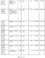
- FIG. 26 Borrelia genome survey for the three Borrelia species, B. burgdorferi, B. afzelii and B. garinii . Listed are the database accession numbers for the individual genomes and the known plasmids or variable plasmid segments of the three species. The specific strains of the individual species are denoted.
- FIG. 27 Complete list of all known and putative proteins encoded by the genome and plasmids of Borrelia burgdorferi (strain B31). The proteins are identified by their names or designations. Position of start and end of the gene, coding strand, amino acid length of the encoded protein, and the accession numbers and gene ID's are given.
- FIG. 28 Complete list of all known and putative proteins encoded by the genome and plasmids of Borrelia afzelii (strain PKo). The proteins are identified by their names or designations. Position of start and end of the gene, coding strand, amino acid length of the encoded protein, and the accession numbers and gene ID's are given.
- FIG. 29 Complete list of all known and putative proteins encoded by the genome, plasmids and known variable plasmid segments of Borrelia garinii (strain PBi). The proteins are identified by their names or designations. Position of start and end of the gene, coding strand, amino acid length of the encoded protein, and the accession numbers and gene ID's are given.
- FIG. 30 The amino acid sequences of Borrelia antigens of specific interest as sources for MHC binding peptides. Name and protein bank accession numbers are shown.
- FIG. 31 Borrelia MHC class 1 antigen peptides of 8-, 9-, 10- and 11 amino acids in length generated from the chosen protein sequences by random prediction software (see FIG. 2 ). Name and protein bank accession numbers are shown.
- FIG. 32 Borrelia MHC class 1 antigen peptides of 8-, 9-, 10- and 11 amino acids in length generated from the chosen protein sequences by use of software based on neural prediction of MHC binding peptides. Peptide sequences are grouped according to HLA binding alleles and sub-grouped according to peptide length.
- FIG. 33 Borrelia MHC class 2 antigen peptides of 13-, 14-, 15-, and 16 amino acids in length generated from the chosen protein sequences by random prediction software (see FIG. 2 ).
- FIG. 34 Borrelia MHC class 2 antigen peptides of 15 amino acids in length generated from the chosen protein sequences by use of software based on neural prediction of MHC binding peptides. Peptide sequences are grouped according to HLA binding alleles and sub-grouped according to peptide length. For each 15-mer binding peptide the essential 9-mer core sequence is given.
- FIG. 35 Summary flow chart ELISPOT. Summary flow chart showing measurement of antigen reactive T-Cells by IFN- ⁇ capture in blood samples by ELISPOT. See example 31 for more detailed information.
- FIG. 36 Prediction of cancer antigen BclX(L) specific MHC class 1, 8-, 9-, 10-, 11-mer peptide binders. Prediction of cancer antigen BclX(L) specific MHC class 1, 8-, 9, 10-,11-mer peptide binders for 24 MHC class 1 alleles (see FIG. 10 ) using the www.cbs.dtu.dk/services/NetMHC/ database. The MHC class 1 molecules for which no binders were found are not listed. The peptides listed in FIG. 36 correspond to SEQ ID NO 216470-217262 in the sequence listing.
- FIG. 37 Prediction of cancer antigen BclX(L) specific MHC class 2, 15-mer peptide binders. Prediction of cancer antigen BclX(L) specific MHC class 2, 15-mer peptide binders for 14 MHC class 2 alleles (see FIG. 10 ) using the www.cbs.dtu.dk/services/NetMHCII/ database. The MHC class 2 molecules for which no binders were found are not listed. The peptides listed in FIG. 37 correspond to SEQ ID NO 217263-217774 in the sequence listing.
- FIG. 38 The amino acid sequences of Borrelia antigens of specific interest as sources for MHC binding peptides. Name and protein bank accession numbers are shown.
- FIG. 39 Borrelia MHC class 1 and 2 antigen peptides of 8-, 9-, 10- and 11 amino acids and 13-, 14-, 15- and 16 amino acids in length generated from the chosen protein sequences by random prediction software. Name and protein bank accession numbers are shown.
- FIG. 40 NetMHC peptides. A) Borrelia burgdorferi MHC class 1 & 2 epitopes, B) Borrelia afzelii MHC class 1 & 2 epitopes, C) Borrelia garinii MHC class 1 & 2 epitopes.
- the NetMHC peptides are predicted by the www.cbs.dtu.dk/services/NetMHC/database.
- FIG. 41 NetMHC peptide sequences from FIG. 40 are grouped according to HLA binding alleles and sub-grouped according to peptide length.
- FIG. 42 Selected peptides.
- the amino acid sequence from one or more Borrelia strains were aligned using the protein alignment programs Vector NTI from Invitrogen, and a homologous sequence for all analyzed strains were be identified. This homologous sequence were run through the “intelligent” peptide epitope prediction program NetMHC as described elsewhere herein to identify epitopes able to bind HLA-A*02 or HLA-A*03. The identified epitopes are shown in this figure.
- FIG. 43 Borrelia MHC class 1 and 2 antigen peptides of 8-, 9-, 10- and 11 amino acids and 13-, 14-, 15- and 16 amino acids in length generated from the chosen protein sequences by random prediction software (SEQ ID NO 180858 to SEQ ID NO 215925). NetMHC peptides predicted by the www.cbs.dtu.dk/services/NetMHC/database (SEQ ID NO 215926 to SEQ ID NO216339). Name and protein bank accession numbers are shown.
- FIG. 44 Special selected HLA-A*0201 epitopes from Borrelia species. Some peptides may be identical to sequences in either Borrelia afzelii. B. garinii or B. burgdorferi but some will be optimized to show possible fit to more than one species ie. some amino acid position have been altered compared to the individual parent sequences.
- FIG. 45 Special selected HLA-A*03 epitopes from Borrelia species. Some peptides may be identical to sequences in either Borrelia afzelii. B. garinii or B. burgdorferi but some will be optimized to show possible fit to more than one species ie. some amino acid position have been altered compared to the individual parent sequences.
- the present invention in one aspect refers to a MHC monomer comprising a-b-P, or a MHC multimer comprising (a-b-P) n , wherein n>1,
- (a-b-P) is the MHC-peptide complex formed when the antigenic peptide P binds to the functional MHC protein
- each MHC peptide complex of a MHC multimer is associated with one or more multimerization domains.
- Another aspect of the present invention refers to an antigenic peptide P or an antigenic polypeptide featuring one or more antigenic peptides P.
- the antigenic peptide P is in one embodiment a Borrelia peptide such as e.g. a Borrelia burgdorferi B31 peptide, a Borreila afzelii PKo peptide or a Borrelia garinii PBi peptide.
- a Borrelia peptide such as e.g. a Borrelia burgdorferi B31 peptide, a Borreila afzelii PKo peptide or a Borrelia garinii PBi peptide.
- the antigenic peptide P can have a length of e.g. 8, 9, 10, 11, 12, 13, 14, 15, 16, 16-20, or 20-30 amino acid residues.
- the antigenic peptide P can be selected from the group consisting of sequences disclosed in the “Sequence Listing” and annotated consecutively (using integers) starting with SEQ ID NO:1 and ending with SEQ ID NO:217791 or any fragment thereof.
- the present invention is directed to a composition
- a composition comprising a plurality of MHC monomers and/or MHC multimers according to the present invention, wherein the MHC multimers are identical or different, and a carrier.
- the present invention further relates to a method for detection of antigen-specific T cells, said method comprising the steps of 1) providing the MHC multimer described above, 2) providing a population of antigen-specific T cells, and 3) detecting antigen-specific T cells specific for the peptide P of the MHC multimer.
- the present invention also relates to a method for detection of antigen-specific T cells, said method comprising the steps of 1) providing the antigenic peptid or antigenic polypeptide described above, 2) providing a population of antigen-specific T cells, and 3) detecting antigen-specific T cells specific for the antigenic peptide P in complex with MHC molecules.
- the present invention relates to a method for counting of antigen-specific T cells, said method comprising the steps of 1) providing the MHC multimer described above, 2) providing a population of antigen-specific T cells, and 3) counting antigen-specific T cells specific for the peptide P of the MHC multimer.
- the present invention also relates to a method for sorting of antigen-specific T cells, said method comprising the steps of 1) providing the MHC multimer described above, 2) providing a population of antigen-specific T cells, and 3) sorting antigen-specific T cells specific for the peptide P of the MHC multimer.
- the present invention relates to a method for isolation of antigen-specific T cells, said method comprising the steps of 1) providing the MHC multimer described above, 2) providing a population of antigen-specific T cells, and 3) isolating antigen-specific T cells specific for the peptide P of the MHC multimer.
- kits comprising an antigenic peptide, an antigenic polypeptide, a MHC monomer or a MHC multimer according to the present invention, or a composition according to the present invention, and at least one additional component, such as a positive control and/or instructions for use.
- a method for immune monitoring one or more diseases comprising monitoring of antigen-specific T cells, said method comprising the steps of
- the present invention makes it possible to pursue different immune monitoring methods using the MHC monomers, MHC multimers, antigenic peptides or antigenic polypeptides according to the present invention.
- the immune monitoring methods include e.g. flow cytometry, ELISPOT, LDA, Quantiferon and Quantiferon-like methods.
- the MHC monomers and/or the MHC multimers can be provided as a MHC peptide complex, or the antigenic peptide and the MHC monomer and/or multimer can be provided separately.
- recognition of TCR's can be achieved by direct or indirect detection, e.g. by using one or more of the following methods:
- ELISPOT technique using indirect detection, e.g. by adding the antigenic peptide optionally associated with a MHC monomer or MHC multimer, followed by measurement of INF-gamma secretion from a population of cells or from individual cells.
- Another technique involves a Quantiferon-like detection assay, e.g. by using indirect detection, e.g. by adding the antigenic peptide optionally associated with a MHC monomer or MHC multimer, followed by measurement of INF-gamma secretion from a population of cells or from individual cells.
- indirect detection e.g. by adding the antigenic peptide optionally associated with a MHC monomer or MHC multimer, followed by measurement of INF-gamma secretion from a population of cells or from individual cells.
- Flow cytometry offers another alternative for performing detection assays, e.g. by using direct detection (e.g. of MHC tetramers), e.g. by adding the antigenic peptide optionally associated with a MHC monomer or MHC multimer, followed by detection of a fluorescein label, thereby measuring the number of TCRs on specific T-cells.
- direct detection e.g. of MHC tetramers
- MHC monomer or MHC multimer e.g. by adding the antigenic peptide optionally associated with a MHC monomer or MHC multimer, followed by detection of a fluorescein label, thereby measuring the number of TCRs on specific T-cells.
- Flow cytometry can also be used for indirect detection, e.g. by adding the antigenic peptide optionally associated with a MHC monomer or MHC multimer, followed by addition of a “cell-permeabilizing factor,” and subsequent measurement of an intracellular component (e.g. INF-gamma mRNA), from individual cells or populations of cells.
- an intracellular component e.g. INF-gamma mRNA
- infectious diseases caused e.g. by mycobacetrium, Gram positive bacteria, Gram negative bacteria, Spirochetes, intracellular bacterium, extracellular bacterium, Borrelia , TB, CMV, HPV, Hepatitis, BK, fungal organisms and microorganisms.
- infectious diseases caused e.g. by mycobacetrium, Gram positive bacteria, Gram negative bacteria, Spirochetes, intracellular bacterium, extracellular bacterium, Borrelia , TB, CMV, HPV, Hepatitis, BK, fungal organisms and microorganisms.
- the diagnosis and/or monitoring of a particular disease can
- the present invention provides:
- a method for performing a control experiment comprising the step of counting of particles comprising the MHC multimer according to the present invention.
- a method for performing a control experiment comprising the step of sorting of particles comprising the MHC multimer according to the present invention.
- a method for performing a control experiment comprising the step of performing flow cytometry analysis of particles comprising the MHC multimer according to the present invention.
- a method for performing a control experiment comprising the step of performing a immunohistochemistry analysis comprising the MHC multimer according to the present invention.
- a method for performing a control experiment comprising the step of performing a immunocytochemistry analysis comprising the MHC multimer according to the present invention.
- a method for performing a control experiment comprising the step of performing an ELISA analysis comprising the MHC multimer according to the present invention.
- the method can also be performed by initially providing one or more antigenic peptide(s) P and optionally one or more functional MHC proteins to generate a MHC-peptide complex (a-b-P); subsequently or simultaneously providing one or more multimerisation domain(s); and reacting the one or more MHC-peptide complexes and the one or more multimerization domain(s) to generate a MHC multimer according to the present invention.
- the present invention is directed to novel MHC complexes optionally comprising a multimerization domain preferably comprising a carrier molecule and/or a scaffold.
- MHC multimer comprising 2 or more MHC-peptide complexes and a multimerization domain to which the 2 or more MHC-peptide complexes are associated.
- the MHC multimer can generally be formed by association of the 2 or more MHC-peptide complexes with the multimerization domain to which the 2 or more MHC-peptide complexes are capable of associating.
- the multimerization domain can be a scaffold associated with one or more MHC-peptide complexes, or a carrier associated with one or more, preferably more than one, MHC-peptide complex(es), or a carrier associated with a plurality of scaffolds each associated with one or more MHC-peptide complexes, such as 2 MHC-peptide complexes, 3 MHC-peptide complexes, 4 MHC-peptide complexes, 5 MHC-peptide complexes or more than 5 MHC-peptide complexes. Accordingly, multimerization domain collectively refers to each and every of the above. It will be clear from the detailed description of the invention provided herein below when the multimerization domain refers to a scaffold or a carrier or a carrier comprising one or more scaffolds.
- the MHC complexes can be associated with this domain either directly or via one or more binding entities.
- the association can be covalent or non-covalent.
- a MHC complex comprising one or more entities (a-b-P) n , wherein a and b together form a functional MHC protein capable of binding a peptide P, and wherein (a-b-P) is the MHC-peptide complex formed when the peptide P binds to the functional MHC protein, said MHC complex optionally further comprising a multimerization domain comprising a carrier molecule and/or a scaffold.
- MHC complex refers to any MHC complex, including MHC monomers in the form of a single MHC-peptide complex and MHC multimers comprising a multimerization domain to which more than one MHC peptide complex is associated.
- MHC multimer i.e. a plurality of MHC peptide complexes of the general composition (a-b-P) n associated with a multimerization domain
- n is by definition more than 1, i.e. at least 2 or more.
- MHC multimer is used herein specifically to indicate that more than one MHC-peptide complex is associated with a multimerization domain, such as a scaffold or carrier or carrier comprising one or more scaffolds.
- a single MHC-peptide complex can be associated with a scaffold or a carrier or a carrier comprising a scaffold and a MHC-multimer comprising 2 or more MHC-peptide complexes can be formed by association of the individual MHC-peptide complexes with a scaffold or a carrier or a carrier comprising one or more scaffolds each associated with one or more MHC-peptide complexes.
- the association can be a covalent linkage so that each or at least some of the n MHC-peptide complexes is covalently linked to the multimerization domain, or the association can be a non-covalent association so that each or at least some of the n MHC-peptide complexes are non-covalently associated with the multimerization domain.
- the MHC complexes of the invention may be provided in non-soluble or soluble form, depending on the intended application.
- MHC complexes of the present invention overcome low intrinsic affinities of monomer ligands and counter receptors.
- the MHC complexes have a large variety of applications that include targeting of high affinity receptors (e.g. hormone peptide receptors for insulin) on target cells. Taken together poly-ligand binding to target cells has numerous practical, clinical and scientifically uses.
- the present invention provides MHC complexes which present mono-valent or multi-valent binding sites for MHC-peptide recognising cells, such as MHC complexes optionally comprising a multimerization domain, such as a scaffold or a carrier molecule, which multimerization domain have attached thereto, directly or indirectly via one or more linkers, covalently or non-covalently, one or more MHC peptide complexes.
- MHC complexes optionally comprising a multimerization domain, such as a scaffold or a carrier molecule, which multimerization domain have attached thereto, directly or indirectly via one or more linkers, covalently or non-covalently, one or more MHC peptide complexes.
- a multimerization domain such as a scaffold or a carrier molecule
- the scaffold or carrier molecule may thus have attached thereto a MHC peptide complex or a plurality of such MHC peptide complexes, and/or a linker or a plurality of linkers.
- the product is a MHC monomer or a MHC multimer as described above.
- MHC multimers will be used interchangeably with the terms MHC′mers and MHCmers, and will include any number, (larger than one) of MHC-peptide complexes, held together in a large complex by covalent or non-covalent interactions between a multimerization domain and one or more MHC-peptide complexes, and will also include the monomeric form of the MHC-peptide complex, i.e. a MHC-peptide complex that is not attached to a multimerization domain.
- the multimerization domain consists of one or more carriers and/or one or more scaffolds while the MHC-peptide complex consists of MHC molecule and antigenic peptide.
- MHC-peptide complexes may be attached to the multimerization domain through one or more linkers.
- FIG. 1 A schematic representation of a MHC multimer is presented in FIG. 1 .
- the product is antigenic peptide or antigenic polypeptide containing one or more antigenic peptide sequences.
- antigenic peptide will be used interchangeably with the term binding peptide and refers to any peptide molecule that is bound or able to bind into the binding groove of either MHC class 1 or MHC class 2.
- antigenic polypeptides In the following the design and generation of antigenic peptides, antigenic polypeptides and the different components of MHC monomers and/or MHC multimers are described.
- Antigenic peptides of the present invention may be used in processes of the present invention either as part of MHC monomers, MHC multimers or antigenic polypeptides or used themselves as a product. Antigenic polypeptide and antigenic peptide products will later in the process they are used for, bind MHC molecules and thereby generate MHC monomers and/or MHC multimers, e.g. when used as a vaccine the antigenic peptides may bind MHC molecules on cells inside the body or when used for an immune monitoring process antigenic peptides binds MHC molecules present in the sample they are applied to.
- MHC class 1 protein typically binds octa-, nona-, deca- or ondecamer (8-, 9-, 10,- 11-mer) peptides in their peptide binding groove.
- the individual MHC class 1 alleles have individual preferences for the peptide length within the given range.
- MHC class 2 proteins typically bind peptides with a total length of 13-18 amino acids, comprising a 9′-mer core motif containing the important amino acid anchor residues. However the total length is not strictly defined, as opposed to most MHC class 1 molecules.
- a given peptide is a binder it is not necessarily a functional T-cell epitope. Functionality needs to be confirmed by a functional analysis e.g. ELISPOT, CTL killing assay or flow cytometry assay as described elsewhere herein.
- the binding affinity of the peptide for the MHC molecules can for some MHC molecules be predicted in databases such as www.syfpeithi.de; www-bimas.cit.nih.gov/molbio/hla_bind/; www.cbs.dtu.dk/services/NetMHC/; www.cbs.dtu.dk/services/NetMHCII/
- the first step in the design of binding peptides is obtaining the protein's amino acid sequence.
- the amino acid sequence of the protein from which antigenic peptides have to be identified from are known.
- the DNA sequence needs to be translated in all three reading frames in both directions leading to a total of six amino acid sequences for a given genome.
- binding peptides can then be identified as described below.
- the present approach must be modified accordingly, to identify peptide sequence motifs that are derived by combination of amino acid sequences derived partly from two separate introns.
- cDNA sequences can be translated into the actual amino acid sequences to allow peptide identification. In cases where the protein sequence is known, these can directly be used to predict peptide epitopes.
- Binding peptide sequences can be predicted from any protein sequence by either a total approach, generating binding peptide sequences for potentially any MHC allele, or by a directed approach, identifying a subset of binding peptides with certain preferred characteristics such as affinity for MHC protein, specificity for MHC protein, likelihood of being formed by proteolysis in the cell, and other important characteristics.
- MHC-peptide complex Many parameters influence the design of the individual binding peptide, as well as the choice of the set of binding peptides to be used in a particular application. Important characteristics of the MHC-peptide complex are physical and chemical (e.g. proteolytic) stability. The relevance of these parameters must be considered for the production of the antigenic peptides, the antigenic polypeptides, the MHC-peptide complexes and the MHC multimers, as well as for their use in a given application. As an example, the stability of the MHC-peptide complex in assay buffer (e.g. PBS), in blood, or in the body can be very important for a particular application.
- assay buffer e.g. PBS
- MHC-peptide complexes In the interaction of the MHC-peptide complex with the TCR, a number of additional characteristics must be considered, including binding affinity and specificity for the TCR, degree of cross-talk, undesired binding or interaction with other TCRs. Finally, a number of parameters must be considered for the interaction of MHC-peptide complexes, MHC multimers, antigenic peptides or antigenic polypeptides with the sample or individual it is being applied to. These include immunogenicity, allergenicity, as well as side effects resulting from un-desired interaction with “wrong” T cells, including cross-talk with e.g. autoimmune diseases and un-desired interaction with other cells than antigen-specific T cells.
- binding peptides of that antigen are included in the application (i.e. the “total approach” for the design of binding peptides described below).
- vaccines it may be adequate to include a few or just one binding peptide for each of the HLA-alleles included in the application (i.e. the “directed approach” whereby only the most potent binding peptides can be included).
- Personalized diagnostics, therapeutics and vaccines will often fall in-between these two extremes, as it will only be necessary to include a few or just one binding peptide in e.g.
- the MHC class 1 binding peptide prediction is done as follows using the total approach.
- the actual protein sequence is split up into 8-, 9-, 10-, and 11-mer peptide sequences. This is performed by starting at amino acid position 1 identifying the first 8-mer; then move the start position by one amino acid identifying the second 8-mer; then move the start position by one amino acid, identifying the third 8-mer. This procedure continues by moving start position by one amino acid for each round of peptide identification. Generated peptides will be amino acid position 1-8, 2-9, 3-10 etc. This procedure can be carried out manually or by means of a software program ( FIG. 2 ). This procedure is then repeated in an identical fashion for 9-, 10 and 11-mers, respectively.
- the directed approach identifies a preferred subset of binding peptides from the binding peptides generated in the total approach. This preferred subset is of particularly value in a given context.
- One way to select subsets of antigenic peptides is to use consensus sequences to choose a set of relevant binding peptides able to bind the individual MHC allele and that will suit the “average” individual.
- consensus sequences often solely consider the affinity of the binding peptide for the MHC protein; in other words, a subset of binding peptides is identified where the designed binding peptides have a high probability of forming stable MHC-peptide complexes, but where it is uncertain whether this MHC-peptide complex is of high relevance in a population, and more uncertain whether this MHC-peptide complex is of high relevance in a given individual.
- the consensus sequence for a binding peptide is generally given by the formula X1-X2-X3-X4- . . . —Xn, where n equals 8, 9, 10, or 11, and where X represents one of the twenty naturally occurring amino acids, optionally modified as described elsewhere in this application.
- X1-Xn can be further defined.
- certain positions in the consensus sequence are more likely to contribute to binding to a given MHC molecule than others.
- Antigenic peptide-binding by MHC I is accomplished by interaction of specific amino acid side chains of the antigenic peptide with discrete pockets within the peptide-binding groove of the MHC molecule.
- the peptide-binding groove is formed by the ⁇ 1 and ⁇ 2 domains of the MHC I heavy chain and contains six pockets denoted A, B, C, D, E, F.
- the main binding energy associating antigenic peptide to MHC I is provided by interaction of amino acids in position 2 and at the c-terminus of the antigenic peptide with the B and F binding pockets of the MHC I molecule.
- amino acids of the antigenic peptide being responsible for the main anchoring of the peptide to the MHC molecule are in the following called primary anchor amino acids and the motif they form for primary anchor motif.
- Other amino acid side chains of an antigenic peptide may also contribute to the anchoring of the antigenic peptide to the MHC molecule but to a lesser extent.
- Such amino acids are often referred to as secondary anchor amino acids and form a secondary anchor motif.
- HLA alleles have different amino acids lining the various pockets of the peptide-binding groove enabling the various alleles to bind unique repertoires of antigenic peptides with specific anchor amino acid motifs.
- certain positions are the socalled anchor positions and the selection of useful amino acids for these positions is limited to those able to fit into the corresponding binding pockets in the HLA molecule.
- X2 and X9 are primary anchor positions docking into the B and F pocket of the HLA molecule respectively, and useful amino acids at these two positions in the binding peptide are preferable limited to leucine or methionine for X2 and to valine or leucine at position X9.
- the primary anchor positions of peptides binding HLA-B*08 are X3, X5 and X9 and the corresponding preferred amino acids at these positions are lysine at position X3, lysine or arginine at position X5 and leucine at position X9.
- antigenic peptides can be selected on their ability to bind a given HLA molecule or a given HLA supertype on the basis of their amino acid sequence, e.g. the identity of the primary anchor motif.
- Antigenic peptide primary anchor motifs of special interest of the present invention are listed in table 6.
- HLA I supertype explore's and their antigenic peptide anchor motifs.
- Examples of usefull amino acids binding in pocket B and pocket F are shown as one letter code.
- Anchor motif Example B pocket aa B F pocket aa F Example of HLA Supertype specificity pocket specificity pocket allele's A01 Small and A,T, S, V, Aromatic and F, W, Y, A*0101, A*2601, aliphatic L, I, M, Q large L, I, M A*2602, A*2603, hydrophobic A*3002, A*3003, A*3004, A*3201 A01/A03 Small and A,T, S, V, Aromatic and Y, R, K A*3001, A*3201, aliphatic L, I, M, Q basic A*7401 A01/A24 Small, A, S, T, V, Aromatic and F, W, Y, A*2902 aliphatic and L, I, M, Q, large L, I, M aromatic F, W, Y hydropho
- Antigenic peptides able to bind a given MHC molecule do not necessarily have primary anchor amino acid residues compatible with both main anchoring pockets of the MHC molecule but may have one or no primary anchor amino acids suitable for binding the MHC molecule in question. However, having the preferred primary anchor motif for a given MHC allele increases the affinity of the antigenic peptide for that given allele and thereby the likelihood of making a stable and useful MHC-peptide molecule.
- antigenic peptides can be identified and selected on their ability to bind a given HLA or other MHC molecule based on what amino acids they have at primary anchor positions and/or secondary anchor positions.
- Another usefull parameter for prediction and selection of usefull antigenic peptides are the probability of the binding peptide in question to be generated in vivo by the proteolytic machinery inside cells. For example for a given antigen the combined action of endosolic, cytosolic and membrane bound protease activities as well as the TAP1 and TAP2 transporter specificities can be taken into consideration. However, the proteolytic activity varies a lot among individuals, and for personalized diagnostics, treatment or vaccination it may be desirable to disregard these general proteolytic data.
- An example of a program predicting the ability of antigenic peptides to be processed is www.cbs.dtu.dk/services/NetCTL/.
- individual peptides or a subset of peptides able to bind one or more types of MHC molecules and make stable MHC-peptide complexes can be identified.
- the identified peptides can then be tested for biological relevance in functional assays such as interferon gamma release assays (e.g. ELISPOT), cytotoxicity assays (e.g. CTL killing assays) or using other methods as described in the section “Detection” elsewhere herein.
- functional assays such as interferon gamma release assays (e.g. ELISPOT), cytotoxicity assays (e.g. CTL killing assays) or using other methods as described in the section “Detection” elsewhere herein.
- the ability of the identified antigenic peptides to bind selected MHC molecules may be determined in binding assays like Biacore measurement, competition assays or other assays usefull for measurement of binding of peptide to MHC molecules, known by persons skilled in the art.
- MHC II molecules bind antigenic peptides with a size of 12-24 amino acids or even longer peptides. From a given antigenic protein, MHC II molecules typically can bind sets of overlapping peptides that shares a common core sequence but differs in the overall peptide size and in positioning of the core sequence in the peptide.
- the core peptide sequence is typically 9 amino acids long but may also be shorter or longer.
- antigenic peptide sequences binding MHC II of the present invention are described by the central part of the peptide mainly the 9-mer core peptide.
- the core peptide sequence may be flanked with a few or several important amino acids, generating antigenic peptides with a length of 13-16 amino acids. In some cases the peptide may contain even more flanking residues resulting in binding peptides longer than 13-16 amino acids.
- antigenic peptides of special interest of the present invention are peptides consisting of or containing 9-mer core peptide sequences
- the antigenic peptide sequences may be selected using the total approach as described for MHC I antigenic peptides elsewhere herein, e.g. using the software program shown in FIG. 2 .
- a directed approach identifying a preferred subset of binding peptides from the binding peptides generated in the total approach can be used.
- MHC I one way to select subsets of antigenic peptides is to use consensus sequences to choose a set of relevant binding peptides able to bind the individual MHC allele and that will suit the “average” individual.
- Such consensus sequences often solely consider the affinity of the binding peptide for the MHC protein; in other words, a subset of binding peptides is identified where the designed binding peptides have a high probability of forming stable MHC-peptide complexes, but where it is uncertain whether this MHC-peptide complex is of high relevance in a population, and more uncertain whether this MHC-peptide complex is of high relevance in a given individual.
- the consensus sequence for the interacting core of a binding peptide is generally given by the formula X1-X2-X3-X4- . . . —Xn, where n equals 9, and where X represents one of the twenty naturally occurring amino acids, optionally modified as described elsewhere in this application.
- X1-Xn can be further defined.
- certain positions in the consensus sequence are the socalled anchor positions and the selection of useful amino acids for these positions is limited to those able to fit into the corresponding binding pockets in the HLA molecule.
- HLA-DRB1*1501 have X1, X4 and X7 as primary anchor positions where preferred amino acids at the three positions are as follows, X1: leucine, valine and isoleucine, X4: phenylalanine, tyrosine or isoleucine, X7: isoleucine, leucine, valine, methionine or phenylalanine.
- antigenic peptides can be identified and selected on their ability to bind a given HLA or other MHC molecule based on what amino acids they have at various anchor positions.
- MHC II binding peptides have much more varied anchor positions than MHC I binding peptides and the number of usefull amino acids at each anchor position is much higher. For some MHC II alleles no really consensus sequence has been identified. In general position 1, 4, 6 and 9 of the 9-mer core motif of MHC II antigenic peptides are important for anchoring of the antigenic peptide to the MHC II molecule.
- Table 7 shows examples of primary anchor positions and corresponding usefull amino acids for antigenic peptides binding various MHC II molecules.
- Another usefull parameter for prediction and selection of usefull antigenic peptides are the probability of the binding peptide in question to be generated in vivo or processed by the proteolytic machinery inside cells.
- the proteolytic activity varies a lot among individuals, and for personalized diagnostics, treatment or vaccination it may be desirable to disregard these general proteolytic data.
- individual peptides or one or more subsets of peptides able to bind one or more types of MHC molecules and make stable MHC-peptide complexes can be identified.
- the identified peptides can then be tested for biological relevance in functional assays such as interferon gamma release assays, ELISPOT, CTL killing assays or using other methods as described in the section “Detection” elsewhere herein.
- functional assays such as interferon gamma release assays, ELISPOT, CTL killing assays or using other methods as described in the section “Detection” elsewhere herein.
- the ability of the identified antigenic peptides to bind selected MHC molecules may be determined in binding assays like Biacore measurement, competition assays or other assays usefull for measurement of binding of peptide to MHC molecules, known by persons skilled in the art.
- binding peptides designed by the total approach and/or directed approach
- homologous peptides and peptides that have been modified in the amino acid side chains or in the backbone can be used as binding peptides.
- Homologues MHC peptide sequences may arise from the existence of multiple strongly homologous alleles, from small insertions, deletions, inversions or substitutions. If they are sufficiently homologous to peptides derived by the total approach, i.e. have an amino acid sequence identity greater than e.g. more than 90%, more than 80%, or more than 70%, or more than 60%, to one or two binding peptides derived by the total approach, they may be good candidates. Identity is often most important for the anchor residues.
- a MHC binding peptide may be of split- or combinatorial epitope origin i.e. formed by linkage of peptide fragments derived from two different peptide fragments and/or proteins.
- Such peptides can be the result of either genetic recombination on the DNA level or due to peptide fragment association during the complex break down of proteins during protein turnover. Possibly it could also be the result of faulty reactions during protein synthesis i.e. caused by some kind of mixed RNA handling.
- a kind of combinatorial peptide epitope can also be seen if a portion of a longer peptide make a loop out leaving only the terminal parts of the peptide bound in the groove.
- Peptides having un-common amino acids such as selenocysteine and pyrrolysine, may be bound in the MHC groove as well.
- Artificial amino acids e.g. having the isomeric D-form may also make up isomeric D-peptides that can bind in the binding groove of the MHC molecules.
- Bound peptides may also contain amino acids that are chemically modified or being linked to reactive groups that can be activated to induce changes in or disrupt the peptide. Example post-translational modifications are shown below. However, chemical modifications of amino acid side chains or the peptide backbone can also be performed.
- amino acids of the antigenic peptides can also be modified in various ways dependent on the amino acid in question, or the modification can affect the amino- or carboxy-terminal end of the peptide. See table 1. Such peptide modifications are occurring naturally as the result of post translational processing of the parental protein. A non-exhaustive description of the major post translational modifications is given below, divided into three main types.
- the peptide modifications can occur as modification of a single amino acid or more than one i.e. in combinations. Modifications can be present on any position within the peptide i.e. on position 1, 2, 3, 4, 5, etc. for the entire length of the peptide.
- binding peptides can be obtained from natural sources by enzymatic digestion or proteolysis of natural proteins or proteins derived by in vitro translation of mRNA. Binding peptides may also be eluted from the MHC binding groove.
- peptides can be produced recombinantly by transfected cells either as monomeric antigenic peptides or as multimeric (concatemeric) antigenic peptides.
- the Multimeric antigenic peptides are cleaved to form monomeric antigenic peptides before binding to MHC protein.
- Binding peptides may also constitute a part of a bigger recombinant protein e.g. consisting of,
- Peptide-linker- ⁇ 2m, ⁇ 2m being full length or truncated
- Peptide-linker-MHC class 1 heavy chain the heavy chain being full length or truncated. Most importantly the truncated class I heavy chain will consist of the extracellular part i.e the ⁇ 1, ⁇ ⁇ 2, and ⁇ domains.
- the heavy chain fragment may also only contain the ⁇ 1 and ⁇ 2 domains, or ⁇ 1 domain alone, or any fragment or full length ⁇ 2m or heavy chain attached to a designer domain(s) or protein fragment(s).
- Peptide-linker-MHC class 2 ⁇ -chain, full length or truncated
- Peptide-linker-MHC class 2 ⁇ -chain-linker-MHC class 2, ⁇ -chain, both chains can be full length or truncated, truncation may involve, omission of ⁇ - and/or ⁇ -chain intermembrane domain, or omission of ⁇ - and/or ⁇ -chain intermembrane plus cytoplasmic domains.
- MHC class 2 part of the construction may consist of fused domains from NH2-terminal, MHC class 2 ⁇ 1domain-MHC class 2 ⁇ 1domain-constant ⁇ 3 of MHC class 1, or alternatively of fused domains from NH2-terminal, MHC class 2 ⁇ 1domain-MHC class 2 ⁇ 1domain-constant ⁇ 3 of MHC class 1.
- ⁇ 2m will be associated non-covalently in the folded MHC complex.
- ⁇ 2m can also be covalently associated in the folded MHC class 2 complex if the following constructs are used from NH2 terminal, MHC class 2 ⁇ 1domain-MHC class 2 ⁇ 1domain-constant ⁇ 3 of MHC class 1-linker- ⁇ 2m, or alternatively of fused domains from NH2-terminal, MHC class 2 ⁇ 1domain-MHC class 2 ⁇ 1domain-constant ⁇ 3 of MHC class 1-linker- ⁇ 2m; the construct may also consist of any of the above MHC class 2 constructs with added designer domain(s) or sequence(s).
- MHC binding peptide may also be chemically synthesized by solid phase or fluid phase synthesis, according to standard protocols.
- the protocol for the synthesis of the full-length antigen on solid support is modified by adding a partial cleavage step after each coupling of an amino acid.
- the starting point for the synthesis is a solid support to which has been attached a cleavable linker.
- the first amino acid X1 (corresponding to the C-terminal end of the antigen) is added and a coupling reaction performed.
- the solid support now carries the molecule “linker-X1”.
- a fraction (e.g. 10%) of the cleavable linkers are now cleaved, to release into solution X1.
- the second amino acid X2 is added and coupled to X1 or the cleavable linker, to form on solid support the molecules “linker-X2” and “linker-X1-X2”.
- a fraction e.g. 10%
- the supernatant is collected into the collection container, which therefore now contains X1, X2, and X1-X2.
- Additional solid support carrying a cleavable linker is added, e.g. corresponding to 10% of the initial amount of solid support.
- the third amino acid X3 is added and coupled to X2 or the cleavable linker, to form on solid support the molecules “linker-X3”, “linker-X2-X3” and “linker-X1-X2-X3”.
- a fraction e.g. 10%
- the supernatant is collected into the collection container, which therefore now contains X1, X2, X3, X1-X2, X2-X3 and X1-X2-X3.
- Additional solid support carrying a cleavable linker is added, e.g. corresponding to 10% of the initial amount of solid support.
- This step-wise coupling and partial cleavage of the linker is continued until the N-terminal end of the antigen is reached.
- the collection container will now contain a large number of peptides of different length and sequence. In the present example where a 10% partial cleavage was employed, a large fraction of the peptides will be 8′-mers, 9′-mers, 10′-mers and 11′-mers, corresponding to class I antigenic peptides. As an example, for a 100 amino acid antigen the 8′-mers will consist of the sequences X1-X2-X3-X4-X5-X6-X7-X8, X2-X3-X4-X5-X6-X7-X8-X9, . . . , X93-X94-X95-X96-X97-X98-X99-X100.
- the used (inactivated) linkers on solid support can be regenerated, in order to maintain a high fraction of linkers available for synthesis.
- the collection of antigenic peptides can be used as a pool for e.g. the display by APCs to stimulate CTLs in ELISPOT assays, or the antigenic peptides may be mixed with one or more MHC alleles, to form a large number of different MHC-peptide complexes which can e.g. be used to form a large number of different MHC multimers which can e.g. be used in flow cytometry experiments.
- MHC Class I and MHC Class II molecules have different structures, as described above, and therefore have different restrictions on the size of the peptide which may be accommodated.
- MHC Class I molecules will accommodate peptides of from about 8 amino acids in length to about 11 amino acids.
- MHC Class II molecules will in general accommodate peptides of from about 13 amino acids in length to about 16 amino acids.
- antigenic peptide sequences derived from proteins of Borrelia bacteria are described in more detail.
- the various species of Borrelia are known to humans in the form of Lyme disease and recurring fever, transmitted through tick or flea bite.
- the cycle of Borrelia through animals is related to the tick's life cycle.
- the tick has four stages in its two-year life cycle, egg, larva, nymph and adult. Between each stage the tick needs a blood meal in order to mature.
- the tick usually acquires the spirochaete during its larval stage, when it feeds on small animals such as rodents or birds.
- small animals such as rodents or birds.
- the tick picks up Borrelia from the white-footed mouse, which is commonly infected.
- the tick then becomes the host for the spirochaete.
- the bacteria resides in the digestive tract of the host for its next nymph and adult stages during which it is passed on to other animals, and sometimes humans.
- Borrelia is a genus of bacteria of the spirochete class. It is a zoonotic, vector-borne disease transmitted primarily by ticks and some by lice, depending on the species. There are at least 37 known species of Borrelia . Different species of Borrelia results in different clinical symptoms. Of the 37 known species of Borrelia, 12 of these species are known to cause Lyme disease or borreliosis and are transmitted by ticks. The major Borrelia species causing Lyme disease are Borrelia burgdorferi, Borrelia afzelii, Borrelia garinii and Borrelia valaisiana.
- B. recurrentis Other Borrelia species cause relapsing fever such as Borrelia recurrentis , caused by the human body louse.
- tick-borne relapsing infections are acquired from other species, such as Borrelia hermsii or Borrelia parkeri , which can be spread from rodents, and serve as a reservoir for the infection, via a tick vector.
- Borrelia hermsii and Borrelia recurrentis cause very similar diseases although the disease associated with Borrelia hermsii has more relapses and is responsible for more fatalities, while the disease caused by B. recurrentis has longer febrile and afebrile intervals and a longer incubation period.
- the present invention relates in one embodiment to one or more antigenic peptides or one or more antigenic polypeptides or to MHC-peptide complexes or MHC multimers comprising one or more antigenic peptides comprising one or more sequences from one or more Borrelia species including the ones mentioned above.
- the invention relates to one or more antigenic peptides or antigenic polypeptides comprising one or more sequences from Borrelia burgdorferi such as Borrelia burgdorferi B31 or to one or more MHC-peptide complexes or MHC multimers comprising one or more such antigenic peptides.
- Borrelia burgdorferi is species of bacteria of the spirochete class of the genus Borrelia. B. burgdorferi is predominant in North America, but also exists in Europe, and is the agent of Lyme disease.
- B. burgdorferi is one of the few pathogenic bacteria that can survive without iron, having replaced all of its iron-sulphur cluster enzymes with enzymes that use manganese, thus avoiding the problem many pathogenic bacteria face in acquiring iron.
- B. burgdorferi infections have been linked to non-Hodgkin lymphomas.
- the B. burgdorferi genome (B31 strain) contains 910,725 base pairs and 853 genes.
- the invention relates to one or more antigenic peptides or one or more antigenic polypeptides comprising one or more sequences from Borrelia afzelii such as Borrelia afzelii Pko or one or more MHC-peptide complexes or one or more MHC multimers comprising one or more such antigenic peptides.
- Borrelia afzelii is considered a new species of the Genus Borrelia and considered homologous to Borrelia burgdorferi with regard to phenotypic, genetic, and immunological characteristics.
- Diseases linked to this species of Borrelia are Lyme disease and Acrodermatitis chronica atrophicans (ACA). Better understanding of the structure and function of this pathogen will create better methods of treatment to people with the diseases it causes.
- the invention relates to one or more antigenic peptides or one or more antigenic polypeptides comprising one or more sequences from Borrelia garinii such as garinii PBi or to one or more MHC-peptide complexes or MHC multimers comprising one or more such antigenic peptides.
- Borrelia garinii is one of two major strains found in Europe. It usually causes Lyme Disease symptoms of the neurological kind—such as extreme back- and leg-pains, meningitis and partial facial paralysis, Lyme arthritis due to B. garinii may be associated in susceptible hosts with amoxicillin resistance or treatment resistance.
- the present invention relates to or to one or more antigenic peptides or antigenic polypeptides, wherein the sequence of one or more antigenic peptides is from Borrelia anserine, or to one or more MHC-peptide complexes or MHC multimers, comprising such antigenic peptides.
- the present invention relates to one or more antigenic peptides or antigenic polypeptides, wherein the sequence of one or more antigenic peptides or antigenic polypeptides is from Borrelia barbouri , or to one or more MHC-peptide complexes or MHC multimers, comprising one or more such antigenic peptides.
- the present invention relates to one or more antigenic peptides or antigenic polypeptides, wherein the sequence of one or more antigenic peptides or antigenic polypeptides is from the Borrelia burgdorferi group such as burgdorferi B31, or to one or more MHC-peptide complexes or MHC multimers, comprising one or more such antigenic peptides.
- the present invention relates to one or more antigenic peptides or antigenic polypeptides, wherein the sequence of one or more antigenic peptides or antigenic polypeptides is from Borrelia afzelii such as Borrelia afzelii ACA-1, Borrelia afzelii K78 and/or Borrelia afzelii PKo, or to one or more MHC-peptide complexes or MHC multimers, comprising one or more such antigenic peptides.
- Borrelia afzelii such as Borrelia afzelii ACA-1, Borrelia afzelii K78 and/or Borrelia afzelii PKo
- MHC-peptide complexes or MHC multimers comprising one or more such antigenic peptides.
- the present invention relates to one or more antigenic peptides or antigenic polypeptides, wherein the sequence of one or more antigenic peptides or antigenic polypeptides is from afzelii PKo, or to one or more MHC-peptide complexes or MHC multimers, comprising one or more such antigenic peptides.
- the present invention relates to one or more antigenic peptides or antigenic polypeptides, wherein the sequence of one or more antigenic peptides or antigenic polypeptides is from Borrelia andersonii , or to one or more MHC-peptide complexes or MHC multimers, comprising one or more such antigenic peptides.
- the present invention relates to one or more antigenic peptides or antigenic polypeptides, wherein the sequence of one or more antigenic peptides or antigenic polypeptides is from Borrelia bissettii , or to one or more MHC-peptide complexes or MHC multimers, comprising one or more such antigenic peptides.
- the present invention relates to one or more Antigenic peptides or antigenic polypeptides, wherein the sequence of one or more antigenic peptides or antigenic polypeptides is from Borrelia burgdorferi (Lyme disease spirochete), or to one or more MHC-peptide complexes or MHC multimers, comprising one or more such antigenic peptides.
- the present invention relates to one or more Antigenic peptides or antigenic polypeptides, wherein the sequence of one or more antigenic peptides or antigenic polypeptides is from Borrelia burgdorferi 118a, or to one or more MHC-peptide complexes or MHC multimers, comprising one or more such antigenic peptides.
- the present invention relates to one or more antigenic peptides or antigenic polypeptides, wherein the sequence of one or more antigenic peptides or antigenic polypeptides is from Borrelia burgdorferi 156a, or to one or more MHC-peptide complexes or MHC multimers, comprising one or more such antigenic peptides.
- the present invention relates to one or more Antigenic peptides or antigenic polypeptides, wherein the sequence of one or more antigenic peptides or antigenic polypeptides is from Borrelia burgdorferi 29805, or to one or more MHC-peptide complexes or MHC multimers, comprising one or more such antigenic peptides.
- the present invention relates to one or more Antigenic peptides or antigenic polypeptides, wherein the sequence of one or more antigenic peptides or antigenic polypeptides is from Borrelia burgdorferi 64b, or to one or more MHC-peptide complexes or MHC multimers, comprising one or more such antigenic peptides.
- the present invention relates to one or more Antigenic peptides or antigenic polypeptides, wherein the sequence of one or more antigenic peptides or antigenic polypeptides is from Borrelia burgdorferi 72a, or to one or more MHC-peptide complexes or MHC multimers, comprising one or more such antigenic peptides.
- the present invention relates to one or more Antigenic peptides or antigenic polypeptides, wherein the sequence of one or more antigenic peptides or antigenic polypeptides is from Borrelia burgdorferi 80a, or to one or more MHC-peptide complexes or MHC multimers, comprising one or more such antigenic
- the present invention relates to one or more Antigenic peptides or antigenic polypeptides, wherein the sequence of one or more antigenic peptides or antigenic polypeptides is from Borrelia burgdorferi 94a, or to one or more MHC-peptide complexes or MHC multimers, comprising one or more such antigenic peptides.
- the present invention relates to one or more antigenic peptides or antigenic polypeptides, wherein the sequence of one or more antigenic peptides or antigenic polypeptides is from Borrelia burgdorferi B31, or to one or more MHC-peptide complexes or MHC multimers, comprising one or more such antigenic peptides.
- the present invention relates to one or more antigenic peptides or antigenic polypeptides, wherein the sequence of one or more antigenic peptides or antigenic polypeptides is from Borrelia burgdorferi Bol26, or to one or more MHC-peptide complexes or MHC multimers, comprising one or more such antigenic peptides.
- the present invention relates to one or more antigenic peptides or antigenic polypeptides, wherein the sequence of one or more antigenic peptides or antigenic polypeptides is from Borrelia burgdorferi CA-11.2a, or to one or more MHC-peptide complexes or MHC multimers, comprising one or more such antigenic peptides.
- the present invention relates to one or more antigenic peptides or antigenic polypeptides, wherein the sequence of one or more antigenic peptides or antigenic polypeptides is from Borrelia burgdorferi WI91-23, or to one or more MHC-peptide complexes or MHC multimers, comprising one or more such antigenic peptides.
- the present invention relates to one or more antigenic peptides or antigenic polypeptides, wherein the sequence of one or more antigenic peptides or antigenic polypeptides is from Borrelia burgdorferi ZS7, or to one or more MHC-peptide complexes or MHC multimers, comprising one or more such antigenic peptides.
- the present invention relates to one or more antigenic peptides or antigenic polypeptides, wherein the sequence of one or more antigenic peptides or antigenic polypeptides is from Borrelia californiensis , or to one or more MHC-peptide complexes or MHC multimers, comprising one or more such antigenic peptides.
- the present invention relates to one or more antigenic peptides or antigenic polypeptides, wherein the sequence of one or more antigenic peptides or antigenic polypeptides is from Borrelia garinii such as garinii PBi, or to one or more MHC-peptide complexes or MHC multimers, comprising one or more such antigenic peptides.
- the present invention relates to one or more antigenic peptides or antigenic polypeptides, wherein the sequence of one or more antigenic peptides or antigenic polypeptides is from Borrelia garinii PBr, or to one or more MHC-peptide complexes or MHC multimers, comprising one or more such antigenic peptides.
- the present invention relates to one or more antigenic peptides or antigenic polypeptides, wherein the sequence of one or more antigenic peptides or antigenic polypeptides is from Borrelia genomosp. 1, or to one or more MHC-peptide complexes or MHC multimers, comprising one or more such antigenic peptides.
- the present invention relates to one or more antigenic peptides or antigenic polypeptides, wherein the sequence of one or more antigenic peptides or antigenic polypeptides is from Borrelia genomosp. 2, or to one or more MHC-peptide complexes or MHC multimers, comprising one or more such antigenic peptides.
- the present invention relates to one or more antigenic peptides or antigenic polypeptides, wherein the sequence of one or more antigenic peptides or antigenic polypeptides is from Borrelia japonica , or to one or more MHC-peptide complexes or MHC multimers, comprising one or more such antigenic peptides.
- the present invention relates to one or more antigenic peptides or antigenic polypeptides, wherein the sequence of one or more antigenic peptides or antigenic polypeptides is from Borrelia lusitaniae , or to one or more MHC-peptide complexes of MHC multimers, comprising one or more such antigenic peptides.
- the present invention relates to one or more antigenic peptides or antigenic polypeptides, wherein the sequence of one or more antigenic peptides or antigenic polypeptides is from Borrelia spielmanii , or to one or more MHC-peptide complexes or MHC multimers, comprising one or more such antigenic peptides.
- the present invention relates to one or more antigenic peptides or antigenic polypeptides, wherein the sequence of one or more antigenic peptides or antigenic polypeptides is from Borrelia spielmanii A14S, or to one or more MHC-peptide complexes or MHC multimers, comprising one or more such antigenic peptides.
- the present invention relates to one or more antigenic peptides or antigenic polypeptides, wherein the sequence of one or more antigenic peptides or antigenic polypeptides is from Borrelia tanukii , or to one or more MHC-peptide complexes or MHC multimers, comprising one or more such antigenic peptides.
- the present invention relates to one or more antigenic peptides or antigenic polypeptides, wherein the sequence of one or more antigenic peptides or antigenic polypeptides is from Borrelia turdi , or to one or more MHC-peptide complexes or MHC multimers, comprising one or more such antigenic peptides.
- the present invention relates to one or more antigenic peptides or antigenic polypeptides, wherein the sequence of one or more antigenic peptides or antigenic polypeptides is from Borrelia valaisiana , or to one or more MHC-peptide complexes or MHC multimers, comprising one or more such antigenic peptides.
- the present invention relates to one or more antigenic peptides or antigenic polypeptides, wherein the sequence of one or more antigenic peptides or antigenic polypeptides is from Borrelia valaisiana VS116, or to one or more MHC-peptide complexes or MHC multimers, comprising one or more such antigenic peptides.
- the present invention relates to one or more antigenic peptides or antigenic polypeptides, wherein the sequence of one or more antigenic peptides or antigenic polypeptides is from Candidatus Borrelia texasensis , or to one or more MHC-peptide complexes or MHC multimers, comprising one or more such antigenic peptides.
- the present invention relates to one or more antigenic peptides or antigenic polypeptides, wherein the sequence of one or more antigenic peptides or antigenic polypeptides is from Borrelia sp. AA4Pool, or to one or more
- MHC-peptide complexes or MHC multimers comprising one or more such antigenic peptides.
- the present invention relates to one or more antigenic peptides or antigenic polypeptides, wherein the sequence of one or more antigenic peptides or antigenic polypeptides is from Borrelia sp. AI-1, or to one or more MHC-peptide complexes or MHC multimers, comprising one or more such antigenic peptides.
- the present invention relates to one or more antigenic peptides or antigenic polypeptides, wherein the sequence of one or more antigenic peptides or antigenic polypeptides is from Borrelia sp. B31, or to one or more MHC-peptide complexes or MHC multimers, comprising one or more such antigenic peptides.
- the present invention relates to one or more antigenic peptides or antigenic polypeptides, wherein the sequence of one or more antigenic peptides or antigenic polypeptides is from Borrelia sp. BC-1, or to one or more MHC-peptide complexes or MHC multimers, comprising one or more such antigenic peptides.
- the present invention relates to one or more antigenic peptides or antigenic polypeptides, wherein the sequence of one or more antigenic peptides or antigenic polypeptides is from Borrelia sp. CA1133, or to one or more MHC-peptide complexes or MHC multimers, comprising one or more such antigenic peptides.
- the present invention relates to one or more antigenic peptides or antigenic polypeptides, wherein the sequence of one or more antigenic peptides or antigenic polypeptides is from Borrelia sp. CA1176, or to one or more MHC-peptide complexes of MHC multimers, comprising one or more such antigenic peptides.
- the present invention relates to one or more antigenic peptides or antigenic polypeptides, wherein the sequence of one or more antigenic peptides or antigenic polypeptides is from Borrelia sp. CA128, or to one or more MHC-peptide complexes or MHC multimers, comprising one or more such antigenic peptides.
- the present invention relates to one or more antigenic peptides or antigenic polypeptides, wherein the sequence of one or more antigenic peptides or antigenic polypeptides is from Borrelia sp. CA13, or to one or more MHC-peptide complexes or MHC multimers, comprising one or more such antigenic peptides.
- the present invention relates to one or more antigenic peptides or antigenic polypeptides, wherein the sequence of one or more antigenic peptides or antigenic polypeptides is from Borrelia sp. CA134, or to one or more MHC-peptide complexes or MHC multimers, comprising one or more such antigenic peptides.
- the present invention relates to one or more antigenic peptides or antigenic polypeptides, wherein the sequence of one or more antigenic peptides or antigenic polypeptides is from Borrelia sp. CA142, or to one or more MHC-peptide complexes or MHC multimers, comprising one or more such antigenic peptides.
- the present invention relates to one or more antigenic peptides or antigenic polypeptides, wherein the sequence of one or more antigenic peptides or antigenic polypeptides is from Borrelia sp. CA20, or to one or more MHC-peptide complexes or MHC multimers, comprising one or more such antigenic peptides.
- the present invention relates to one or more antigenic peptides or antigenic polypeptides, wherein the sequence of one or more antigenic peptides or antigenic polypeptides is from Borrelia sp. CA22, or to one or more MHC-peptide complexes or MHC multimers, comprising one or more such antigenic peptides.
- the present invention relates to one or more antigenic peptides or antigenic polypeptides, wherein the sequence of one or more antigenic peptides or antigenic polypeptides is from Borrelia sp. CA27, or to one or more MHC-peptide complexes or MHC multimers, comprising one or more such antigenic peptides.
- the present invention relates to one or more antigenic peptides or antigenic polypeptides, wherein the sequence of one or more antigenic peptides or antigenic polypeptides is from Borrelia sp. CA28, or to one or more MHC-peptide complexes or MHC multimers, comprising one or more such antigenic peptides.
- the present invention relates to one or more antigenic peptides or antigenic polypeptides, wherein the sequence of one or more antigenic peptides or antigenic polypeptides is from Borrelia sp. CA29, or to one or more MHC-peptide complexes or MHC multimers, comprising one or more such antigenic peptides.
- the present invention relates to one or more antigenic peptides or antigenic polypeptides, wherein the sequence of one or more antigenic peptides or antigenic polypeptides is from Borrelia sp. CA31, or to one or more MHC-peptide complexes or MHC multimers, comprising one or more such antigenic peptides.
- the present invention relates to one or more antigenic peptides or antigenic polypeptides, wherein the sequence of one or more antigenic peptides or antigenic polypeptides is from Borrelia sp. CA33, or to one or more MHC-peptide complexes or MHC multimers, comprising one or more such antigenic peptides.
- the present invention relates to one or more antigenic peptides or antigenic polypeptides, wherein the sequence of one or more antigenic peptides or antigenic polypeptides is from Borrelia sp. CA370, or to one or more MHC-peptide complexes or MHC multimers, comprising one or more such antigenic peptides.
- the present invention relates to one or more antigenic peptides or antigenic polypeptides, wherein the sequence of one or more antigenic peptides or antigenic polypeptides is from Borrelia sp. CA372, or to one or more MHC-peptide complexes of MHC multimers, comprising one or more such antigenic peptides.
- the present invention relates to one or more antigenic peptides or antigenic polypeptides, wherein the sequence of one or more antigenic peptides or antigenic polypeptides is from Borrelia sp. CA378, or to one or more MHC-peptide complexes or MHC multimers, comprising one or more such antigenic peptides.
- the present invention relates to one or more antigenic peptides or antigenic polypeptides, wherein the sequence of one or more antigenic peptides or antigenic polypeptides is from Borrelia sp. CA388, or to one or more MHC-peptide complexes or MHC multimers, comprising one or more such antigenic peptides.
- the present invention relates to one or more antigenic peptides or antigenic polypeptides, wherein the sequence of one or more antigenic peptides or antigenic polypeptides is from Borrelia sp. CA393, or to one or more MHC-peptide complexes or MHC multimers, comprising one or more such antigenic peptides.
- the present invention relates to one or more antigenic peptides or antigenic polypeptides, wherein the sequence of one or more antigenic peptides or antigenic polypeptides is from Borrelia sp. CA394, or to one or more MHC-peptide complexes or MHC multimers, comprising one or more such antigenic peptides.
- the present invention relates to one or more antigenic peptides or antigenic polypeptides, wherein the sequence of one or more antigenic peptides or antigenic polypeptides is from Borrelia sp. CA395, or to one or more MHC-peptide complexes or MHC multimers, comprising one or more such antigenic peptides.
- the present invention relates to one or more antigenic peptides or antigenic polypeptides, wherein the sequence of one or more antigenic peptides or antigenic polypeptides is from Borrelia sp. CA399, or to one or more MHC-peptide complexes or MHC multimers, comprising one or more such antigenic peptides.
- the present invention relates to one or more antigenic peptides or antigenic polypeptides, wherein the sequence of one or more antigenic peptides or antigenic polypeptides is from Borrelia sp. CA400, or to one or more MHC-peptide complexes or MHC multimers, comprising one or more such antigenic peptides.
- the present invention relates to one or more antigenic peptides or antigenic polypeptides, wherein the sequence of one or more antigenic peptides or antigenic polypeptides is from Borrelia sp. CA401, or to one or more MHC-peptide complexes or MHC multimers, comprising one or more such antigenic peptides.
- the present invention relates to one or more antigenic peptides or antigenic polypeptides, wherein the sequence of one or more antigenic peptides or antigenic polypeptides is from Borrelia sp. CA402, or to one or more MHC-peptide complexes or MHC multimers, comprising one or more such antigenic peptides.
- the present invention relates to one or more antigenic peptides or antigenic polypeptides, wherein the sequence of one or more antigenic peptides or antigenic polypeptides is from Borrelia sp. CA404, or to one or more MHC-peptide complexes or MHC multimers, comprising one or more such antigenic peptides.
- the present invention relates to one or more antigenic peptides or antigenic polypeptides, wherein the sequence of one or more antigenic peptides or antigenic polypeptides is from Borrelia sp. CA411, or to one or more MHC-peptide complexes or MHC multimers, comprising one or more such antigenic peptides.
- the present invention relates to one or more antigenic peptides or antigenic polypeptides, wherein the sequence of one or more antigenic peptides or antigenic polypeptides is from Borrelia sp. CA426, or to one or more MHC-peptide complexes or MHC multimers, comprising one or more such antigenic peptides.
- the present invention relates to one or more antigenic peptides or antigenic polypeptides, wherein the sequence of one or more antigenic peptides or antigenic polypeptides is from Borrelia sp. CA443, or to one or more MHC-peptide complexes of MHC multimers, comprising one or more such antigenic peptides.
- the present invention relates to one or more antigenic peptides or antigenic polypeptides, wherein the sequence of one or more antigenic peptides or antigenic polypeptides is from Borrelia sp. CA446, or to one or more MHC-peptide complexes or MHC multimers, comprising one or more such antigenic peptides.
- the present invention relates to one or more antigenic peptides or antigenic polypeptides, wherein the sequence of one or more antigenic peptides or antigenic polypeptides is from Borrelia sp. CA448, or to one or more MHC-peptide complexes or MHC multimers, comprising one or more such antigenic peptides.
- the present invention relates to one or more antigenic peptides or antigenic polypeptides, wherein the sequence of one or more antigenic peptides or antigenic polypeptides is from Borrelia sp. CA462, or to one or more MHC-peptide complexes or MHC multimers, comprising one or more such antigenic peptides.
- the present invention relates to one or more antigenic peptides or antigenic polypeptides, wherein the sequence of one or more antigenic peptides or antigenic polypeptides is from Borrelia sp. CA468, or to one or more MHC-peptide complexes or MHC multimers, comprising one or more such antigenic peptides.
- the present invention relates to one or more antigenic peptides or antigenic polypeptides, wherein the sequence of one or more antigenic peptides or antigenic polypeptides is from Borrelia sp. CA502, or to one or more MHC-peptide complexes or MHC multimers, comprising one or more such antigenic peptides.
- the present invention relates to one or more antigenic peptides or antigenic polypeptides, wherein the sequence of one or more antigenic peptides or antigenic polypeptides is from Borrelia sp. CA504, or to one or more MHC-peptide complexes or MHC multimers, comprising one or more such antigenic peptides.
- the present invention relates to one or more antigenic peptides or antigenic polypeptides, wherein the sequence of one or more antigenic peptides or antigenic polypeptides is from Borrelia sp. CA507, or to one or more MHC-peptide complexes or MHC multimers, comprising one or more such antigenic peptides.
- the present invention relates to one or more antigenic peptides or antigenic polypeptides, wherein the sequence of one or more antigenic peptides or antigenic polypeptides is from Borrelia sp. CA547, or to one or more MHC-peptide complexes or MHC multimers, comprising one or more such antigenic peptides.
- the present invention relates to one or more antigenic peptides or antigenic polypeptides, wherein the sequence of one or more antigenic peptides or antigenic polypeptides is from Borrelia sp. CA552, or to one or more MHC-peptide complexes or MHC multimers, comprising one or more such antigenic peptides.
- the present invention relates to one or more antigenic peptides or antigenic polypeptides, wherein the sequence of one or more antigenic peptides or antigenic polypeptides is from Borrelia sp. CA8, or to one or more MHC-peptide complexes or MHC multimers, comprising one or more such antigenic peptides.
- the present invention relates to one or more antigenic peptides or antigenic polypeptides, wherein the sequence of one or more antigenic peptides or antigenic polypeptides is from Borrelia sp. D22, or to one or more MHC-peptide complexes or MHC multimers, comprising one or more such antigenic peptides.
- the present invention relates to one or more antigenic peptides or antigenic polypeptides, wherein the sequence of one or more antigenic peptides or antigenic polypeptides is from Borrelia sp. D35, or to one or more MHC-peptide complexes or MHC multimers, comprising one or more such antigenic peptides.
- the present invention relates to one or more antigenic peptides or antigenic polypeptides, wherein the sequence of one or more antigenic peptides or antigenic polypeptides is from Borrelia sp. FD-1, or to one or more MHC-peptide complexes of MHC multimers, comprising one or more such antigenic peptides.
- the present invention relates to one or more antigenic peptides or antigenic polypeptides, wherein the sequence of one or more antigenic peptides or antigenic polypeptides is from Borrelia sp. FL18, or to one or more MHC-peptide complexes or MHC multimers, comprising one or more such antigenic peptides.
- the present invention relates to one or more antigenic peptides or antigenic polypeptides, wherein the sequence of one or more antigenic peptides or antigenic polypeptides is from Borrelia sp. FL27, or to one or more MHC-peptide complexes or MHC multimers, comprising one or more such antigenic peptides.
- the present invention relates to one or more antigenic peptides or antigenic polypeptides, wherein the sequence of one or more antigenic peptides or antigenic polypeptides is from Borrelia sp. FL35, or to one or more MHC-peptide complexes or MHC multimers, comprising one or more such antigenic peptides.
- the present invention relates to one or more antigenic peptides or antigenic polypeptides, wherein the sequence of one or more antigenic peptides or antigenic polypeptides is from Borrelia sp. FL42, or to one or more MHC-peptide complexes or MHC multimers, comprising one or more such antigenic peptides.
- the present invention relates to one or more antigenic peptides or antigenic polypeptides, wherein the sequence of one or more antigenic peptides or antigenic polypeptides is from Borrelia sp. HN6, or to one or more MHC-peptide complexes or MHC multimers, comprising one or more such antigenic peptides.
- the present invention relates to one or more antigenic peptides or antigenic polypeptides, wherein the sequence of one or more antigenic peptides or antigenic polypeptides is from Borrelia sp. HN7, or to one or more MHC-peptide complexes or MHC multimers, comprising one or more such antigenic peptides.
- the present invention relates to one or more antigenic peptides or antigenic polypeptides, wherein the sequence of one or more antigenic peptides or antigenic polypeptides is from Borrelia sp. HN8, or to one or more MHC-peptide complexes or MHC multimers, comprising one or more such antigenic peptides.
- the present invention relates to one or more antigenic peptides or antigenic polypeptides, wherein the sequence of one or more antigenic peptides or antigenic polypeptides is from Borrelia sp. HNM13, or to one or more MHC-peptide complexes or MHC multimers, comprising one or more such antigenic peptides.
- the present invention relates to one or more antigenic peptides or antigenic polypeptides, wherein the sequence of one or more antigenic peptides or antigenic polypeptides is from Borrelia sp. HNM14, or to one or more MHC-peptide complexes or MHC multimers, comprising one or more such antigenic peptides.
- the present invention relates to one or more antigenic peptides or antigenic polypeptides, wherein the sequence of one or more antigenic peptides or antigenic polypeptides is from Borrelia sp. HNM19, or to one or more MHC-peptide complexes or MHC multimers, comprising one or more such antigenic peptides.
- the present invention relates to one or more antigenic peptides or antigenic polypeptides, wherein the sequence of one or more antigenic peptides or antigenic polypeptides is from Borrelia sp. IA1, or to one or more MHC-peptide complexes or MHC multimers, comprising one or more such antigenic peptides.
- the present invention relates to one or more antigenic peptides or antigenic polypeptides, wherein the sequence of one or more antigenic peptides or antigenic polypeptides is from Borrelia sp. Ir-3519, or to one or more MHC-peptide complexes or MHC multimers, comprising one or more such antigenic peptides.
- the present invention relates to one or more antigenic peptides or antigenic polypeptides, wherein the sequence of one or more antigenic peptides or antigenic polypeptides is from Borrelia sp. Ir-4721, or to one or more MHC-peptide complexes of MHC multimers, comprising one or more such antigenic peptides.
- the present invention relates to one or more antigenic peptides or antigenic polypeptides, wherein the sequence of one or more antigenic peptides or antigenic polypeptides is from Borrelia sp. Ir-4812, or to one or more MHC-peptide complexes or MHC multimers, comprising one or more such antigenic peptides.
- the present invention relates to one or more antigenic peptides or antigenic polypeptides, wherein the sequence of one or more antigenic peptides or antigenic polypeptides is from Borrelia sp. Ir-5215, or to one or more MHC-peptide complexes or MHC multimers, comprising one or more such antigenic peptides.
- the present invention relates to one or more antigenic peptides or antigenic polypeptides, wherein the sequence of one or more antigenic peptides or antigenic polypeptides is from Borrelia sp. LV5, or to one or more MHC-peptide complexes or MHC multimers, comprising one or more such antigenic peptides.
- the present invention relates to one or more antigenic peptides or antigenic polypeptides, wherein the sequence of one or more antigenic peptides or antigenic polypeptides is from Borrelia sp. MI-2, or to one or more MHC-peptide complexes or MHC multimers, comprising one or more such antigenic peptides.
- the present invention relates to one or more antigenic peptides or antigenic polypeptides, wherein the sequence of one or more antigenic peptides or antigenic polypeptides is from Borrelia sp. MI-5, or to one or more MHC-peptide complexes or MHC multimers, comprising one or more such antigenic peptides.
- the present invention relates to one or more antigenic peptides or antigenic polypeptides, wherein the sequence of one or more antigenic peptides or antigenic polypeptides is from Borrelia sp. MI-6, or to one or more MHC-peptide complexes or MHC multimers, comprising one or more such antigenic peptides.
- the present invention relates to one or more antigenic peptides or antigenic polypeptides, wherein the sequence of one or more antigenic peptides or antigenic polypeptides is from Borrelia sp. MI-8, or to one or more MHC-peptide complexes or MHC multimers, comprising one or more such antigenic peptides.
- the present invention relates to one or more antigenic peptides or antigenic polypeptides, wherein the sequence of one or more antigenic peptides or antigenic polypeptides is from Borrelia sp. MI-9, or to one or more MHC-peptide complexes or MHC multimers, comprising one or more such antigenic peptides.
- the present invention relates to one or more antigenic peptides or antigenic polypeptides, wherein the sequence of one or more antigenic peptides or antigenic polypeptides is from Borrelia sp. MOD-1, or to one or more MHC-peptide complexes or MHC multimers, comprising one or more such antigenic peptides.
- the present invention relates to one or more antigenic peptides or antigenic polypeptides, wherein the sequence of one or more antigenic peptides or antigenic polypeptides is from Borrelia sp. MOD-5, or to one or more MHC-peptide complexes or MHC multimers, comprising one or more such antigenic peptides.
- the present invention relates to one or more antigenic peptides or antigenic polypeptides, wherein the sequence of one or more antigenic peptides or antigenic polypeptides is from Borrelia sp. MOK-3a, or to one or more MHC-peptide complexes or MHC multimers, comprising one or more such antigenic peptides.
- the present invention relates to one or more antigenic peptides or antigenic polypeptides, wherein the sequence of one or more antigenic peptides or antigenic polypeptides is from Borrelia sp. MOS-1b, or to one or more MHC-peptide complexes or MHC multimers, comprising one or more such antigenic peptides.
- the present invention relates to one or more antigenic peptides or antigenic polypeptides, wherein the sequence of one or more antigenic peptides or antigenic polypeptides is from Borrelia sp. NE49, or to one or more MHC-peptide complexes of MHC multimers, comprising one or more such antigenic peptides.
- the present invention relates to one or more antigenic peptides or antigenic polypeptides, wherein the sequence of one or more antigenic peptides or antigenic polypeptides is from Borrelia sp. NE581, or to one or more MHC-peptide complexes or MHC multimers, comprising one or more such antigenic peptides.
- the present invention relates to one or more antigenic peptides or antigenic polypeptides, wherein the sequence of one or more antigenic peptides or antigenic polypeptides is from Borrelia sp. PHaP, or to one or more MHC-peptide complexes or MHC multimers, comprising one or more such antigenic peptides.
- the present invention relates to one or more antigenic peptides or antigenic polypeptides, wherein the sequence of one or more antigenic peptides or antigenic polypeptides is from Borrelia sp. PSigII, or to one or more MHC-peptide complexes or MHC multimers, comprising one or more such antigenic peptides.
- the present invention relates to one or more antigenic peptides or antigenic polypeptides, wherein the sequence of one or more antigenic peptides or antigenic polypeptides is from Borrelia sp. SCGT-10, or to one or more MHC-peptide complexes or MHC multimers, comprising one or more such antigenic peptides.
- the present invention relates to one or more antigenic peptides or antigenic polypeptides, wherein the sequence of one or more antigenic peptides or antigenic polypeptides is from Borrelia sp. SCGT-8a, or to one or more MHC-peptide complexes or MHC multimers, comprising one or more such antigenic peptides.
- the present invention relates to one or more antigenic peptides or antigenic polypeptides, wherein the sequence of one or more antigenic peptides or antigenic polypeptides is from Borrelia sp. SCI-2, or to one or more MHC-peptide complexes or MHC multimers, comprising one or more such antigenic peptides.
- the present invention relates to one or more antigenic peptides or antigenic polypeptides, wherein the sequence of one or more antigenic peptides or antigenic polypeptides is from Borrelia sp. SCW-30h, or to one or more MHC-peptide complexes or MHC multimers, comprising one or more such antigenic peptides.
- the present invention relates to one or more antigenic peptides or antigenic polypeptides, wherein the sequence of one or more antigenic peptides or antigenic polypeptides is from Borrelia sp. SI-1, or to one or more MHC-peptide complexes or MHC multimers, comprising one or more such antigenic peptides.
- the present invention relates to one or more antigenic peptides or antigenic polypeptides, wherein the sequence of one or more antigenic peptides or antigenic polypeptides is from Borrelia sp. SI-10, or to one or more MHC-peptide complexes or MHC multimers, comprising one or more such antigenic peptides.
- the present invention relates to one or more antigenic peptides or antigenic polypeptides, wherein the sequence of one or more antigenic peptides or antigenic polypeptides is from Borrelia sp. SM-1, or to one or more MHC-peptide complexes or MHC multimers, comprising one or more such antigenic peptides.
- the present invention relates to one or more antigenic peptides or antigenic polypeptides, wherein the sequence of one or more antigenic peptides or antigenic polypeptides is from Borrelia sp. SV1, or to one or more MHC-peptide complexes or MHC multimers, comprising one or more such antigenic peptides.
- the present invention relates to one or more antigenic peptides or antigenic polypeptides, wherein the sequence of one or more antigenic peptides or antigenic polypeptides is from Borrelia sp. W97F51, or to one or more MHC-peptide complexes or MHC multimers, comprising one or more such antigenic peptides.
- the present invention relates to one or more antigenic peptides or antigenic polypeptides, wherein the sequence of one or more antigenic peptides or antigenic polypeptides is from Borrelia sp. Z41293, or to one or more MHC-peptide complexes of MHC multimers, comprising one or more such antigenic peptides.
- the present invention relates to one or more antigenic peptides or antigenic polypeptides, wherein the sequence of one or more antigenic peptides or antigenic polypeptides is from Borrelia sp. Z41493, or to one or more MHC-peptide complexes or MHC multimers, comprising one or more such antigenic peptides.
- the present invention relates to one or more antigenic peptides or antigenic polypeptides, wherein the sequence of one or more antigenic peptides or antigenic polypeptides is from Borrelia coriaceae , or to one or more MHC-peptide complexes or MHC multimers, comprising one or more such antigenic peptides.
- the present invention relates to one or more antigenic peptides or antigenic polypeptides, wherein the sequence of one or more antigenic peptides or antigenic polypeptides is from Borrelia crocidurae , or to one or more MHC-peptide complexes or MHC multimers, comprising one or more such antigenic peptides.
- the present invention relates to one or more antigenic peptides or antigenic polypeptides, wherein the sequence of one or more antigenic peptides or antigenic polypeptides is from Borrelia duttonii , or to one or more MHC-peptide complexes or MHC multimers, comprising one or more such antigenic peptides.
- the present invention relates to one or more antigenic peptides or antigenic polypeptides, wherein the sequence of one or more antigenic peptides or antigenic polypeptides is from Borrelia duttonii Ly, or to one or more MHC-peptide complexes or MHC multimers, comprising one or more such antigenic peptides.
- the present invention relates to one or more antigenic peptides or antigenic polypeptides, wherein the sequence of one or more antigenic peptides or antigenic polypeptides is from Borrelia hermsii , or to one or more MHC-peptide complexes or MHC multimers, comprising one or more such antigenic peptides.
- the present invention relates to one or more antigenic peptides or antigenic polypeptides, wherein the sequence of one or more antigenic peptides or antigenic polypeptides is from Borrelia hermsii DAH, or to one or more MHC-peptide complexes or MHC multimers, comprising one or more such antigenic peptides.
- the present invention relates to one or more antigenic peptides or antigenic polypeptides, wherein the sequence of one or more antigenic peptides or antigenic polypeptides is from Borrelia hispanica , or to one or more MHC-peptide complexes or MHC multimers, comprising one or more such antigenic peptides.
- the present invention relates to one or more antigenic peptides or antigenic polypeptides, wherein the sequence of one or more antigenic peptides or antigenic polypeptides is from Borrelia lonestari , or to one or more MHC-peptide complexes or MHC multimers, comprising one or more such antigenic peptides.
- the present invention relates to one or more antigenic peptides or antigenic polypeptides, wherein the sequence of one or more antigenic peptides or antigenic polypeptides is from Borrelia miyamotoi , or to one or more MHC-peptide complexes or MHC multimers, comprising one or more such antigenic peptides.
- the present invention relates to one or more antigenic peptides or antigenic polypeptides, wherein the sequence of one or more antigenic peptides or antigenic polypeptides is from Borrelia parkeri , or to one or more MHC-peptide complexes or MHC multimers, comprising one or more such antigenic peptides.
- the present invention relates to one or more antigenic peptides or antigenic polypeptides, wherein the sequence of one or more antigenic peptides or antigenic polypeptides is from Borrelia persica , or to one or more MHC-peptide complexes or MHC multimers, comprising one or more such antigenic peptides.
- the present invention relates to one or more antigenic peptides or antigenic polypeptides, wherein the sequence of one or more antigenic peptides or antigenic polypeptides is from Borrelia recurrentis , or to one or more MHC-peptide complexes of MHC multimers, comprising one or more such antigenic peptides.
- the present invention relates to one or more antigenic peptides or antigenic polypeptides, wherein the sequence of one or more antigenic peptides or antigenic polypeptides is from Borrelia recurrentis A1, or to one or more MHC-peptide complexes or MHC multimers, comprising one or more such antigenic peptides.
- the present invention relates to one or more antigenic peptides or antigenic polypeptides, wherein the sequence of one or more antigenic peptides or antigenic polypeptides is from Borrelia sinica , or to one or more MHC-peptide complexes or MHC multimers, comprising one or more such antigenic peptides.
- the present invention relates to one or more antigenic peptides or antigenic polypeptides, wherein the sequence of one or more antigenic peptides or antigenic polypeptides is from Borrelia theileri , or to one or more MHC-peptide complexes or MHC multimers, comprising one or more such antigenic peptides.
- the present invention relates to one or more antigenic peptides or antigenic polypeptides, wherein the sequence of one or more antigenic peptides or antigenic polypeptides is from Borrelia turcica , or to one or more MHC-peptide complexes or MHC multimers, comprising one or more such antigenic peptides.
- the present invention relates to one or more antigenic peptides or antigenic polypeptides, wherein the sequence of one or more antigenic peptides or antigenic polypeptides is from Borrelia turicatae , or to one or more MHC-peptide complexes or MHC multimers, comprising one or more such antigenic peptides.
- the present invention relates to one or more antigenic peptides or antigenic polypeptides, wherein the sequence of one or more antigenic peptides or antigenic polypeptides is from Borrelia turicatae 91E135, or to one or more MHC-peptide complexes or MHC multimers, comprising one or more such antigenic peptides.
- the present invention relates to one or more antigenic peptides or antigenic polypeptides, wherein the sequence of one or more antigenic peptides or antigenic polypeptides is from Borrelia sp. ‘Lake Gaillard’, or to one or more MHC-peptide complexes or MHC multimers, comprising one or more such antigenic peptides.
- the present invention relates to one or more antigenic peptides or antigenic polypeptides, wherein the sequence of one or more antigenic peptides or antigenic polypeptides is from Borrelia sp. 000133, or to one or more MHC-peptide complexes or MHC multimers, comprising one or more such antigenic peptides.
- the present invention relates to one or more antigenic peptides or antigenic polypeptides, wherein the sequence of one or more antigenic peptides or antigenic polypeptides is from Borrelia sp. 010298, or to one or more MHC-peptide complexes or MHC multimers, comprising one or more such antigenic peptides.
- the present invention relates to one or more antigenic peptides or antigenic polypeptides, wherein the sequence of one or more antigenic peptides or antigenic polypeptides is from Borrelia sp. 10MT, or to one or more MHC-peptide complexes or MHC multimers, comprising one or more such antigenic peptides.
- the present invention relates to one or more antigenic peptides or antigenic polypeptides, wherein the sequence of one or more antigenic peptides or antigenic polypeptides is from Borrelia sp. 5145, or to one or more MHC-peptide complexes or MHC multimers, comprising one or more such antigenic peptides.
- the present invention relates to one or more antigenic peptides or antigenic polypeptides, wherein the sequence of one or more antigenic peptides or antigenic polypeptides is from Borrelia sp. 57Nsk, or to one or more MHC-peptide complexes or MHC multimers, comprising one or more such antigenic peptides.
- the present invention relates to one or more antigenic peptides or antigenic polypeptides, wherein the sequence of one or more antigenic peptides or antigenic polypeptides is from Borrelia sp. 5MT, or to one or more MHC-peptide complexes of MHC multimers, comprising one or more such antigenic peptides.
- the present invention relates to one or more antigenic peptides or antigenic polypeptides, wherein the sequence of one or more antigenic peptides or antigenic polypeptides is from Borrelia sp. 6T04-2, or to one or more MHC-peptide complexes or MHC multimers, comprising one or more such antigenic peptides.
- the present invention relates to one or more antigenic peptides or antigenic polypeptides, wherein the sequence of one or more antigenic peptides or antigenic polypeptides is from Borrelia sp. BR, or to one or more MHC-peptide complexes or MHC multimers, comprising one or more such antigenic peptides.
- the present invention relates to one or more antigenic peptides or antigenic polypeptides, wherein the sequence of one or more antigenic peptides or antigenic polypeptides is from Borrelia sp. BR 2007, or to one or more MHC-peptide complexes or MHC multimers, comprising one or more such antigenic peptides.
- the present invention relates to one or more antigenic peptides or antigenic polypeptides, wherein the sequence of one or more antigenic peptides or antigenic polypeptides is from Borrelia sp. C5-N52, or to one or more MHC-peptide complexes or MHC multimers, comprising one or more such antigenic peptides.
- the present invention relates to one or more antigenic peptides or antigenic polypeptides, wherein the sequence of one or more antigenic peptides or antigenic polypeptides is from Borrelia sp. CB-A1, or to one or more MHC-peptide complexes or MHC multimers, comprising one or more such antigenic peptides.
- the present invention relates to one or more antigenic peptides or antigenic polypeptides, wherein the sequence of one or more antigenic peptides or antigenic polypeptides is from Borrelia sp. CB-A11, or to one or more MHC-peptide complexes or MHC multimers, comprising one or more such antigenic peptides.
- the present invention relates to one or more antigenic peptides or antigenic polypeptides, wherein the sequence of one or more antigenic peptides or antigenic polypeptides is from Borrelia sp. CB-A3, or to one or more MHC-peptide complexes or MHC multimers, comprising one or more such antigenic peptides.
- the present invention relates to one or more antigenic peptides or antigenic polypeptides, wherein the sequence of one or more antigenic peptides or antigenic polypeptides is from Borrelia sp. EFL-S0100110, or to one or more MHC-peptide complexes or MHC multimers, comprising one or more such antigenic peptides.
- the present invention relates to one or more antigenic peptides or antigenic polypeptides, wherein the sequence of one or more antigenic peptides or antigenic polypeptides is from Borrelia sp. IK/23, or to one or more MHC-peptide complexes or MHC multimers, comprising one or more such antigenic peptides.
- the present invention relates to one or more antigenic peptides or antigenic polypeptides, wherein the sequence of one or more antigenic peptides or antigenic polypeptides is from Borrelia sp. IM/16, or to one or more MHC-peptide complexes or MHC multimers, comprising one or more such antigenic peptides.
- the present invention relates to one or more antigenic peptides or antigenic polypeptides, wherein the sequence of one or more antigenic peptides or antigenic polypeptides is from Borrelia sp. IM/19, or to one or more MHC-peptide complexes or MHC multimers, comprising one or more such antigenic peptides.
- the present invention relates to one or more antigenic peptides or antigenic polypeptides, wherein the sequence of one or more antigenic peptides or antigenic polypeptides is from Borrelia sp. KR1, or to one or more MHC-peptide complexes or MHC multimers, comprising one or more such antigenic peptides.
- the present invention relates to one or more antigenic peptides or antigenic polypeptides, wherein the sequence of one or more antigenic peptides or antigenic polypeptides is from Borrelia sp. KR3, or to one or more MHC-peptide complexes of MHC multimers, comprising one or more such antigenic peptides.
- the present invention relates to one or more antigenic peptides or antigenic polypeptides, wherein the sequence of one or more antigenic peptides or antigenic polypeptides is from Borrelia sp. LB-2001, or to one or more MHC-peptide complexes or MHC multimers, comprising one or more such antigenic peptides.
- the present invention relates to one or more antigenic peptides or antigenic polypeptides, wherein the sequence of one or more antigenic peptides or antigenic polypeptides is from Borrelia sp. LB-M56, or to one or more MHC-peptide complexes or MHC multimers, comprising one or more such antigenic peptides.
- the present invention relates to one or more antigenic peptides or antigenic polypeptides, wherein the sequence of one or more antigenic peptides or antigenic polypeptides is from Borrelia sp. LB-W100, or to one or more MHC-peptide complexes or MHC multimers, comprising one or more such antigenic peptides.
- the present invention relates to one or more antigenic peptides or antigenic polypeptides, wherein the sequence of one or more antigenic peptides or antigenic polypeptides is from Borrelia sp. MK-N61, or to one or more MHC-peptide complexes or MHC multimers, comprising one or more such antigenic peptides.
- the present invention relates to one or more antigenic peptides or antigenic polypeptides, wherein the sequence of one or more antigenic peptides or antigenic polypeptides is from Borrelia sp. NR-N8, or to one or more MHC-peptide complexes or MHC multimers, comprising one or more such antigenic peptides.
- the present invention relates to one or more antigenic peptides or antigenic polypeptides, wherein the sequence of one or more antigenic peptides or antigenic polypeptides is from Borrelia sp. OkME1, or to one or more MHC-peptide complexes or MHC multimers, comprising one or more such antigenic peptides.
- the present invention relates to one or more antigenic peptides or antigenic polypeptides, wherein the sequence of one or more antigenic peptides or antigenic polypeptides is from Borrelia sp. PAnz, or to one or more MHC-peptide complexes or MHC multimers, comprising one or more such antigenic peptides.
- the present invention relates to one or more antigenic peptides or antigenic polypeptides, wherein the sequence of one or more antigenic peptides or antigenic polypeptides is from Borrelia sp. PJes, or to one or more MHC-peptide complexes or MHC multimers, comprising one or more such antigenic peptides.
- the present invention relates to one or more antigenic peptides or antigenic polypeptides, wherein the sequence of one or more antigenic peptides or antigenic polypeptides is from Borrelia sp. PMai, or to one or more MHC-peptide complexes or MHC multimers, comprising one or more such antigenic peptides.
- the present invention relates to one or more antigenic peptides or antigenic polypeptides, wherein the sequence of one or more antigenic peptides or antigenic polypeptides is from Borrelia sp. PMew, or to one or more MHC-peptide complexes or MHC multimers, comprising one or more such antigenic peptides.
- the present invention relates to one or more antigenic peptides or antigenic polypeptides, wherein the sequence of one or more antigenic peptides or antigenic polypeptides is from Borrelia sp. R57, or to one or more MHC-peptide complexes or MHC multimers, comprising one or more such antigenic peptides.
- the present invention relates to one or more antigenic peptides or antigenic polypeptides, wherein the sequence of one or more antigenic peptides or antigenic polypeptides is from Borrelia sp. strain Spain, or to one or more MHC-peptide complexes or MHC multimers, comprising one or more such antigenic peptides.
- the present invention relates to one or more antigenic peptides or antigenic polypeptides, wherein the sequence of one or more antigenic peptides or antigenic polypeptides is from Borrelia sp. TA1, or to one or more MHC-peptide complexes of MHC multimers, comprising one or more such antigenic peptides.
- the present invention relates to one or more antigenic peptides or antigenic polypeptides, wherein the sequence of one or more antigenic peptides or antigenic polypeptides is from Borrelia sp. TM, or to one or more MHC-peptide complexes or MHC multimers, comprising one or more such antigenic peptides.
- the present invention relates to one or more antigenic peptides or antigenic polypeptides, wherein the sequence of one or more antigenic peptides or antigenic polypeptides is from Borrelia sp. TM1, or to one or more MHC-peptide complexes or MHC multimers, comprising one or more such antigenic peptides.
- the present invention relates to one or more antigenic peptides or antigenic polypeptides, wherein the sequence of one or more antigenic peptides or antigenic polypeptides is from Borrelia sp. TM2, or to one or more MHC-peptide complexes or MHC multimers, comprising one or more such antigenic peptides.
- the present invention in one embodiment relates to one or more antigenic peptides or antigenic polypeptides, wherein the one or more antigenic peptides or antigenic polypeptides are encoded by one or more of the Borrelia species Borrelia burgdorferi, Borrelia garinii and Borrelia afzelii , or to one or more MHC-peptide complexes or MHC multimers, comprising one or more such antigenic peptides.
- the invention relates to one or more antigenic peptides or antigenic polypeptides, wherein the one or more antigenic peptides or antigenic polypeptides are encoded by Borrelia burgdorferi and not by Borrelia garinii or Borrelia afzelii , or to one or more MHC-peptide complexes or MHC multimers, comprising one or more such antigenic peptides.
- the invention relates to one or more antigenic peptides or antigenic polypeptides, wherein the one or more antigenic peptides or antigenic polypeptides are encoded by Borrelia garinii and not by Borrelia burgdorferi or Borrelia afzelii , or to one or more MHC-peptide complexes or MHC multimers, comprising one or more such antigenic peptides.
- the invention relates to one or more antigenic peptides or antigenic polypeptides, wherein the one or more antigenic peptides or antigenic polypeptides are encoded by Borrelia afzelii and not by Borrelia garinii or Borrelia burgdorferi , or to one or more MHC-peptide complexes or MHC multimers, comprising one or more such antigenic peptides.
- the present invention relates in one embodiment to one or more antigenic peptides or antigenic polypeptides comprising a sequence encoded by a Borrelia genome and/or plasmid, or to one or more MHC-peptide complexes or MHC multimers, comprising one or more such antigenic peptides.
- the genomes and plasmids of Borrelia burgdoferi B31 , Borrelia afzelii Pko and Borrelia garinii PBi are listed in FIG. 26-29 .
- the peptides can e.g. be 8 mers, 9 mers, 10 mers, 11 mers, 12 mers, 13 mers, 14 mers, 15 mers or 16 mers.
- the present invention relates to one or more antigenic peptides or antigenic polypeptides comprising a sequence encoded by the genome (NC_001318) of Borrelia burgdorferi B31, or to one or more MHC-peptide complexes or MHC multimers, comprising one or more such antigenic peptides.
- the present invention relates to one or more antigenic peptides or antigenic polypeptides comprising a sequence encoded by the plasmid Ip5 (NC_000957) of Borrelia burgdorferi B31, or to one or more MHC-peptide complexes or MHC multimers, comprising one or more such antigenic peptides.
- the present invention relates to one or more antigenic peptides or antigenic polypeptides comprising a sequence encoded by the plasmid Ip 17 (NC_001849) of Borrelia burgdorferi B31, or to one or more MHC-peptide complexes or MHC multimers, comprising one or more such antigenic peptides.
- the present invention relates to one or more antigenic peptides or antigenic polypeptides comprising a sequence encoded by the plasmid Ip21 (NC_000955) of Borrelia burgdorferi B31, or to one or more MHC-peptide complexes or MHC multimers, comprising one or more such antigenic peptides.
- the present invention relates to one or more antigenic peptides or antigenic polypeptides comprising a sequence encoded by the plasmid Ip25 (NC_001850) of Borrelia burgdorferi B31, or to one or more MHC-peptide complexes or MHC multimers, comprising one or more such antigenic peptides.
- the present invention relates to one or more antigenic peptides or antigenic polypeptides comprising a sequence encoded by the plasmid Ip28-1 (NC_001851) of Borrelia burgdorferi B31, or to one or more MHC-peptide complexes or MHC multimers, comprising one or more such antigenic peptides.
- the present invention relates to one or more antigenic peptides or antigenic polypeptides comprising a sequence encoded by the plasmid Ip28-2 (NC_001852) of Borrelia burgdorferi B31, or to one or more MHC-peptide complexes or MHC multimers, comprising one or more such antigenic peptides.
- the present invention relates to one or more antigenic peptides or antigenic polypeptides comprising a sequence encoded by the plasmid Ip28-3 (NC_001853) of Borrelia burgdorferi B31, or to one or more MHC-peptide complexes or MHC multimers, comprising one or more such antigenic peptides.
- the present invention relates to one or more antigenic peptides or antigenic polypeptides comprising a sequence encoded by the plasmid Ip28-4 (NC_001854) of Borrelia burgdorferi B31, or to one or more MHC-peptide complexes or MHC multimers, comprising one or more such antigenic peptides.
- the present invention relates to one or more antigenic peptides or antigenic polypeptides comprising a sequence encoded by the plasmid Ip36 (NC_001855) of Borrelia burgdorferi B31, or to one or more MHC-peptide complexes or MHC multimers, comprising one or more such antigenic peptides.
- the present invention relates to one or more antigenic peptides or antigenic polypeptides comprising a sequence encoded by the plasmid Ip38 (NC_001856) of Borrelia burgdorferi B31, or to one or more MHC-peptide complexes or MHC multimers, comprising one or more such antigenic peptides.
- the present invention relates to one or more antigenic peptides or antigenic polypeptides comprising a sequence encoded by the plasmid Ip56 (NC_000956) of Borrelia burgdorferi B31, or to one or more MHC-peptide complexes or MHC multimers, comprising one or more such antigenic peptides.
- the present invention relates to one or more antigenic peptides or antigenic polypeptides comprising a sequence encoded by the plasmid cp9 (NC_001904) of Borrelia burgdorferi B31, or to one or more MHC-peptide complexes or MHC multimers, comprising one or more such antigenic peptides.
- the present invention relates to one or more antigenic peptides or antigenic polypeptides comprising a sequence encoded by the plasmid Ip28-4 (NC_001854) of Borrelia burgdorferi B31, or to one or more MHC-peptide complexes or MHC multimers, comprising one or more such antigenic peptides.
- the present invention relates to one or more antigenic peptides or antigenic polypeptides comprising a sequence encoded by the plasmid cp26 (NC_001903) of Borrelia burgdorferi B31, or to one or more MHC-peptide complexes or MHC multimers, comprising one or more such antigenic peptides.
- the present invention relates to one or more antigenic peptides or antigenic polypeptides comprising a sequence encoded by the plasmid cp32-1 (NC_000948) of Borrelia burgdorferi B31, or to one or more MHC-peptide complexes or MHC multimers, comprising one or more such antigenic peptides.
- the present invention relates to one or more antigenic peptides or antigenic polypeptides comprising a sequence encoded by the plasmid cp32-3 (NC_000949) of Borrelia burgdorferi B31, or to one or more MHC-peptide complexes or MHC multimers, comprising one or more such antigenic peptides.
- the present invention relates to one or more antigenic peptides or antigenic polypeptides comprising a sequence encoded by the plasmid cp32-4 (NC_000950) of Borrelia burgdorferi B31, or to one or more MHC-peptide complexes or MHC multimers, comprising one or more such antigenic peptides.
- the present invention relates to one or more antigenic peptides or antigenic polypeptides comprising a sequence encoded by the plasmid cp32-6 (NC_000951) of Borrelia burgdorferi B31, or to one or more MHC-peptide complexes or MHC multimers, comprising one or more such antigenic peptides.
- the present invention relates to one or more antigenic peptides or antigenic polypeptides comprising a sequence encoded by the plasmid cp32-7 (NC_000952) of Borrelia burgdorferi B31, or to one or more MHC-peptide complexes or MHC multimers, comprising one or more such antigenic peptides.
- the present invention relates to one or more antigenic peptides or antigenic polypeptides comprising a sequence encoded by the plasmid cp32-8 (NC_000953) of Borrelia burgdorferi B31, or to one or more MHC-peptide complexes or MHC multimers, comprising one or more such antigenic peptides.
- the present invention relates to one or more antigenic peptides or antigenic polypeptides comprising a sequence encoded by the plasmid cp32-9 (NC_000954) of Borrelia burgdorferi B31, or to one or more MHC-peptide complexes or MHC multimers, comprising one or more such antigenic peptides.
- the present invention relates to one or more antigenic peptides or antigenic polypeptides comprising a sequence encoded by the genome (NC_008277) of Borrelia afzelii PKo, or to one or more MHC-peptide complexes or MHC multimers, comprising one or more such antigenic peptides.
- the present invention relates to one or more antigenic peptides or antigenic polypeptides comprising a sequence encoded by the plasmid Ip25 (NC_008569) of Borrelia afzelii PKo, or to one or more MHC-peptide complexes or MHC multimers, comprising one or more such antigenic peptides.
- the present invention relates to one or more antigenic peptides or antigenic polypeptides comprising a sequence encoded by the plasmid Ip28 (NC_0085698) of Borrelia afzelii PKo, or to one or more MHC-peptide complexes or MHC multimers, comprising one or more such antigenic peptides.
- the present invention relates to one or more antigenic peptides or antigenic polypeptides comprising a sequence encoded by the plasmid Ip32 (NC_008567) of Borrelia afzelii PKo, or to one or more MHC-peptide complexes or MHC multimers, comprising one or more such antigenic peptides.
- the present invention relates to one or more antigenic peptides or antigenic polypeptides comprising a sequence encoded by the plasmid Ip34 (NC_008566) of Borrelia afzelii PKo, or to one or more MHC-peptide complexes or MHC multimers, comprising one or more such antigenic peptides.
- the present invention relates to one or more antigenic peptides or antigenic polypeptides comprising a sequence encoded by the plasmid Ip60 (NC_008564) of Borrelia afzelii PKo, or to one or more MHC-peptide complexes or MHC multimers, comprising one or more such antigenic peptides.
- the present invention relates to one or more antigenic peptides or antigenic polypeptides comprising a sequence encoded by the plasmid Ip60-2 (NC_008565) of Borrelia afzelii PKo, or to one or more MHC-peptide complexes or MHC multimers, comprising one or more such antigenic peptides.
- the present invention relates to one or more antigenic peptides or antigenic polypeptides comprising a sequence encoded by the plasmid cp27 (NC_008274) of Borrelia afzelii PKo, or to one or more MHC-peptide complexes or MHC multimers, comprising one or more such antigenic peptides.
- the present invention relates to one or more antigenic peptides or antigenic polypeptides comprising a sequence encoded by the plasmid cp30 (NC_008273) of Borrelia afzelii PKo, or to one or more MHC-peptide complexes or MHC multimers, comprising one or more such antigenic peptides.
- the present invention relates to one or more antigenic peptides or antigenic polypeptides comprising a sequence encoded by the genome (NC_006156) of Borrelia garinii PBi, or to one or more MHC-peptide complexes or MHC multimers, comprising one or more such antigenic peptides.
- the present invention relates to one or more antigenic peptides or antigenic polypeptides comprising a sequence encoded by the plasmid Ip54 (NC_006129) of Borrelia garinii PBi, or to one or more MHC-peptide complexes or MHC multimers, comprising one or more such antigenic peptides.
- the present invention relates to one or more antigenic peptides or antigenic polypeptides comprising a sequence encoded by the plasmid cp26 (NC_006128) of Borrelia garinii PBi, or to one or more MHC-peptide complexes or MHC multimers, comprising one or more such antigenic peptides.
- the present invention relates to one or more antigenic peptides or antigenic polypeptides comprising a sequence encoded by variable plasmid segments not finally assembled to whole plasmids of Borrelia garinii PBi, or to one or more MHC-peptide complexes or MHC multimers, comprising one or more such antigenic peptides.
- variable plasmid segments not finally assembled to whole plasmids can in one preferred embodiment be selected from the group consisting of NT_108239, NT_108263, NT_108262, NT_108261, NT_108260, NT_108259, NT_108258, NT_108257, NT_108256, NT_108255, NT_108254, NT_108253 NT_108252, NT_108251, NT_108250, NT_108249, NT_108248, NT_108247, NT_108246, NT_108245, NT_108244, NT_108243, NT_108242, NT_108241, NT_108240, NT_108238, NT_108237, NT_108236, NT_108235, NT_108234, NT_108233, NT_108232, NT_108231, NT_108230, NT_108229, NT_108228 and
- the present invention relates to one or more antigenic peptides or antigenic polypeptides, optionally comprised in one or more MHC-peptide complexes or MHC multimers, where the antigenic peptides or antigenic polypeptides comprise a sequence encoded by a Borrelia genome and/or plasmid from one or more of the Borrelia species or subspecies: Borrelia anserina, Borrelia barbouri, Borrelia afzelii, Borrelia afzelii ACA-1 , Borrelia afzelii K78, Borrelia afzelii PKo, Borrelia andersonii, Borrelia bissettii, Borrelia burgdorferi, Borrelia burgdorferi 118a, Borrelia burgdorferi 156a, Borrelia burgdorferi 29805, Borrelia burgdorferi 64b, Borrelia burgdorferi 72a, Borrelia burgdorferi 80a
- TM Borrelia sp. TM1 and Borrelia sp. TM2.
- These peptides can e.g. be 8 mers, 9 mers, 10 mers, 11 mers, 12 mers, 13 mers, 14 mers, 15 mers or 16 mers.
- the present invention relates to one or more antigenic peptides or antigenic polypeptides comprising a fragment of the sequence from one or more proteins from Borrelia bulgdorferi B31, or to one or more MHC-peptide complexes or MHC multimers, comprising one or more such antigenic peptides.
- the one or more proteins can be selected from the proteins listed in FIG. 27 and FIG. 38 .
- These fragments can e.g. be 8 mers, 9 mers, 10 mers, 11 mers, 12 mers, 13 mers, 14 mers, 15 mers or 16 mers.
- the present invention relates to one or more antigenic peptides or antigenic polypeptides comprising a fragment of the sequence from one or more proteins from Borrelia afzelii Pko, or to one or more MHC-peptide complexes or MHC multimers, comprising one or more such antigenic peptides.
- the one or more proteins can be selected from the proteins listed in FIG. 28 and FIG. 38 .
- These fragments can e.g. be 8 mers, 9 mers, 10 mers, 11 mers, 12 mers, 13 mers, 14 mers, 15 mers or 16 mers.
- the present invention relates to one or more antigenic peptides or antigenic polypeptides comprising a fragment of the sequence from one or more proteins from Borrelia garinii PBi, or to one or more MHC-peptide complexes or MHC multimers, comprising one or more such antigenic peptides.
- the one or more proteins can be selected from the proteins listed in FIG. 29 and FIG. 38 .
- These fragments can e.g. be 8 mers, 9 mers, 10 mers, 11 mers, 12 mers, 13 mers, 14 mers, 15 mers or 16 mers.
- the present invention relates to one or more antigenic peptides or antigenic polypeptides comprising a fragment of the sequence from one or more proteins listed in FIG. 30 and FIG. 38 , or to one or more MHC-peptide complexes or MHC multimers, comprising one or more such antigenic peptides.
- These fragments can e.g. be 8 mers, 9 mers, 10 mers, 11 mers, 12 mers, 13 mers, 14 mers, 15 mers or 16 mers.
- the present invention relates to one or more antigenic peptides or antigenic polypeptides or MHC-peptide complexes containing such antigenic peptides, where the antigenic peptides or antigenic polypeptides comprise a fragment of the sequence from one or more protein(s) from one or more of the Borrelia species or subspecies: Borrelia anserina, Borrelia barbouri, Borrelia afzelii, Borrelia afzelii ACA-1 , Borrelia afzelii K78, Borrelia afzelii PKo, Borrelia andersonii, Borrelia bissettii, Borrelia burgdorferi, Borrelia burgdorferi 118a, Borrelia burgdorferi 156a, Borrelia burgdorferi 29805, Borrelia burgdorferi 64b, Borrelia burgdorferi 72a, Borrelia burgdorferi 80a, Borrelia burgdorfer
- TM Borrelia sp. TM1 and Borrelia sp. TM2.
- These peptides can e.g. be 8 mers, 9 mers, 10 mers, 11 mers, 12 mers, 13 mers, 14 mers, 15 mers or 16 mers.
- the present invention relates to one or more MHC-peptide complexes or MHC multimers comprising one or more Borrelia class I antigen peptide fragments or to the Borrelia class I antigen peptide fragments themselves.
- the present invention relates to one or more MHC-peptide complexes or MHC multimers comprising one or more Borrelia class I antigen peptide fragments, wherein said peptide fragments are 8 mers, 9 mers, 10 mers and/or 11 mers.
- the present invention relates to one or more antigenic peptides or antigenic polypeptides being Borrelia class I antigen peptide fragments, wherein said peptide fragments are 8 mers, 9 mers, 10 mers and/or 11 mers.
- the present invention relates to one or more MHC-peptide complexes or MHC multimers comprising one or more Borrelia class I antigen peptide fragments or to the Borrelia class I antigenic peptide fragments themselves, wherein said peptide fragments are 8 mers, 9 mers, 10 mers and/or 11 mers of a protein from or encoded by Borrelia burgdorferi such as Borrelia burgdorferi B31.
- the present invention relates to one or more MHC-peptide complexes or MHC multimers comprising one or more Borrelia class I antigen peptide fragments or to the Borrelia class I antigenic peptide fragments themselves, wherein said peptide fragments are 8 mers, 9 mers, 10 mers and/or 11 mers of a protein from or encoded by Borrelia afzelii such as Borrelia afzelii PKo.
- the present invention relates to one or more MHC-peptide complexes or MHC multimers comprising one or more Borrelia class I antigen peptide fragments or to the Borrelia class I antigenic peptide fragments themselves, wherein said peptide fragments are 8 mers, 9 mers, 10 mers and/or 11 mers of a protein from or encoded by Borrelia garinii such as Borrelia garinii PBi.
- the present invention relates to one or more MHC-peptide complexes or MHC multimers comprising one or more Borrelia class I antigen peptide fragments or to the Borrelia class I antigenic peptide fragments themselves, wherein said peptide fragments are 8 mers, 9 mers, 10 mers and/or 11 mers of a protein from or encoded by Borrelia anserina, Borrelia barbouri, Borrelia afzelii, Borrelia afzelii ACA-1 , Borrelia afzelii K78, Borrelia afzelii PKo, Borrelia andersonii, Borrelia bissettii, Borrelia burgdorferi, Borrelia burgdorferi 118a, Borrelia burgdorferi 156 a, Borrelia burgdorferi 29805, Borrelia burgdorferi 64b, Borrelia burgdorferi 72a, Borrelia burgdorferi 80a, Borreli
- CA128 Borrelia sp. CA13 , Borrelia sp. CA134 , Borrelia sp. CA142 , Borrelia sp. CA20 , Borrelia sp. CA22 , Borrelia sp. CA27 , Borrelia sp. CA28 , Borrelia sp. CA29 , Borrelia sp. CA31 , Borrelia sp. CA33 , Borrelia sp. CA370 , Borrelia sp. CA372 , Borrelia sp. CA378 , Borrelia sp. CA388 , Borrelia sp. CA393 , Borrelia sp.
- CA394 Borrelia sp. CA395 , Borrelia sp. CA399 , Borrelia sp. CA400 , Borrelia sp. CA401 , Borrelia sp. CA402 , Borrelia sp. CA404 , Borrelia sp. CA411 , Borrelia sp. CA426 , Borrelia sp. CA443 , Borrelia sp. CA446 , Borrelia sp. CA448 , Borrelia sp. CA462 , Borrelia sp. CA468 , Borrelia sp. CA502 , Borrelia sp. CA504 , Borrelia sp.
- CA507 Borrelia sp. CA547 , Borrelia sp. CA552 , Borrelia sp. CA8 , Borrelia sp. D22 , Borrelia sp. D35 , Borrelia sp. FD-1 , Borrelia sp. FL18 , Borrelia sp. FL27 , Borrelia sp. FL35 , Borrelia sp. FL42 , Borrelia sp. HN6 , Borrelia sp. HN7 , Borrelia sp. HN8 , Borrelia sp. HNM13 , Borrelia sp. HNM14 , Borrelia sp.
- the present invention relates to one or more MHC-peptide complexes or MHC multimers comprising one or more Borrelia class I antigen peptide fragments or to the Borrelia class I antigenic peptide fragments themselves, where the peptide fragments are selected from the peptides listed in FIG. 31 , FIG. 32 , FIG. 39 , FIG. 40 , FIG. 41 , FIG. 42 , Table A, Table B and Table C.
- the present invention relates to one or more MHC-peptide complexes or MHC multimers comprising one or more Borrelia class II antigen peptide fragments, or to the Borrelia class II antigen peptide fragments themselves.
- the present invention relates to one or more MHC-peptide complexes or MHC multimers comprising one or more Borrelia class II antigen peptide fragments, or to the Borrelia class II antigen peptide fragments themselves, wherein said peptide fragments are 13 mers, 14 mers, 15 mers and/or 16 mers.
- the present invention relates to one or more MHC-peptide complexes or MHC multimers comprising one or more Borrelia class II antigen peptide fragments, or to the Borrelia class II antigen peptide fragments themselves, wherein said peptide fragments are 13 mers, 14 mers, 15 mers and/or 16 mers of a protein from or encoded by Borrelia burgdorferi such as Borrelia burgdorferi B31.
- the present invention relates to one or more MHC-peptide complexes or MHC multimers comprising one or more Borrelia class II antigen peptide fragments, or to the Borrelia class II antigen peptide fragments themselves, wherein said peptide fragments are 13 mers, 14 mers, 15 mers and/or 16 mers of a protein from or encoded by Borrelia afzelii such as Borrelia afzelii PKo.
- the present invention relates to one or more MHC-peptide complexes or MHC multimers comprising one or more Borrelia class II antigen peptide fragments, or to the Borrelia class II antigen peptide fragments themselves, wherein said peptide fragments are 13 mers, 14 mers, 15 mers and/or 16 mers of a protein from or encoded by Borrelia garinii such as Borrelia garinii PBi.
- the present invention relates to one or more MHC-peptide complexes or MHC multimers comprising one or more Borrelia class II antigen peptide fragments, or to the Borrelia class II antigen peptide fragments themselves, wherein said peptide fragments are 13 mers, 14 mers, 15 mers and/or 16 mers of a protein from or encoded by Borrelia anserina, Borrelia barbouri, Borrelia afzelii, Borrelia afzelii ACA-1 , Borrelia afzelii K78, Borrelia afzelii PKo, Borrelia andersonii, Borrelia bissettii, Borrelia burgdorferi, Borrelia burgdorferi 118a, Borrelia burgdorferi 156 a, Borrelia burgdorferi 29805, Borrelia burgdorferi 64b, Borrelia burgdorferi 72a, Borrelia burgdorferi 80a, Bor
- CA128 Borrelia sp. CA13 , Borrelia sp. CA134 , Borrelia sp. CA142 , Borrelia sp. CA20 , Borrelia sp. CA22 , Borrelia sp. CA27 , Borrelia sp. CA28 , Borrelia sp. CA29 , Borrelia sp. CA31 , Borrelia sp. CA33 , Borrelia sp. CA370 , Borrelia sp. CA372 , Borrelia sp. CA378 , Borrelia sp. CA388 , Borrelia sp. CA393 , Borrelia sp.
- CA394 Borrelia sp. CA395 , Borrelia sp. CA399 , Borrelia sp. CA400 , Borrelia sp. CA401 , Borrelia sp. CA402 , Borrelia sp. CA404 , Borrelia sp. CA411 , Borrelia sp. CA426 , Borrelia sp. CA443 , Borrelia sp. CA446 , Borrelia sp. CA448 , Borrelia sp. CA462 , Borrelia sp. CA468 , Borrelia sp. CA502 , Borrelia sp. CA504 , Borrelia sp.
- CA507 Borrelia sp. CA547 , Borrelia sp. CA552 , Borrelia sp. CA8 , Borrelia sp. D22 , Borrelia sp. D35 , Borrelia sp. FD-1 , Borrelia sp. FL18 , Borrelia sp. FL27 , Borrelia sp. FL35 , Borrelia sp. FL42 , Borrelia sp. HN6 , Borrelia sp. HN7 , Borrelia sp. HN8 , Borrelia sp. HNM13 , Borrelia sp. HNM14 , Borrelia sp.
- the present invention relates to one or more MHC-peptide complexes or MHC multimers comprising one or more Borrelia class II antigen peptide fragments selected from the peptides listed in FIG. 33 , FIG. 34 , FIG. 39 , FIG. 40 , FIG. 41 , FIG. 42 , Table A, Table B and Table C.
- the present invention relates to one or more Borrelia class II antigenic peptide fragments selected from the peptides listed in FIG. 33 , FIG. 34 , FIG. 39 , FIG. 40 , FIG. 41 , FIG. 42 , Table A, Table B and Table C.
- MHC class 2 proteins typically bind peptides with a total length of 13-18 amino acids, comprising a 9′-mer core motif containing the important amino acid anchor residues. However the total length is not strictly defined, as opposed to most MHC class 1 molecules.
- the putative binding peptide sequences only describe the central part of the peptide including the 9-mer core motif; in other words, the peptide sequences shown represent the core of the binding peptide with a few important flanking amino acids, which in some cases may be of considerably length generating binding peptides longer than the 13-16 amino acids.
- the present invention relates to one or more MHC-peptide complexes or MHC multimers comprising one or more Borrelia class II antigen peptide fragments comprising one or more of the 9 mer core peptides listed in FIG. 34 .
- the present invention relates to one or more Borrelia class II antigen peptide fragments comprising one or more of the 9 mer core peptides listed in FIG. 34 .
- the above mentioned 9 mer core peptides can be part of one or more 13 mers, 14 mers, 15 mers, 16 mers, 17 mers, 18 mers, 19 mers, 20 mers, 21 mers, 22 mers, 23, mers, 24 mers or 25 mer.
- the 9 mer core peptides can have different positions in the 13 mers, 14 mers, 15 mers, 16 mers, 17 mers, 18 mers, 19 mers, 20 mers, 21 mers, 22 mers, 23, mers, 24 mers or 25 mer.
- the 9 mer starts at position 1 from the N-terminus of the 13 mer.
- the 9 mer starts at position 2 from the N-terminus of the 13 mer.
- the 9 mer starts at position 3 from the N-terminus of the 13 mer.
- the 9 mer starts at position 4 from the N-terminus of the 13 mer.
- the 9 mer starts at position 5 from the N-terminus of the 13 mer.
- the 9 mer starts at position 1 from the N-terminus of the 14 mer.
- the 9 mer starts at position 2 from the N-terminus of the 14 mer.
- the 9 mer starts at position 3 from the N-terminus of the 14 mer.
- the 9 mer starts at position 4 from the N-terminus of the 14 mer.
- the 9 mer starts at position 5 from the N-terminus of the 14 mer.
- the 9 mer starts at position 6 from the N-terminus of the 14 mer.
- the 9 mer starts at position 1 from the N-terminus of the 15 mer.
- the 9 mer starts at position 2 from the N-terminus of the 15 mer.
- the 9 mer starts at position 3 from the N-terminus of the 15 mer.
- the 9 mer starts at position 4 from the N-terminus of the 15 mer.
- the 9 mer starts at position 5 from the N-terminus of the 15 mer.
- the 9 mer starts at position 6 from the N-terminus of the 15 mer.
- the 9 mer starts at position 7 from the N-terminus of the 15 mer.
- the 9 mer starts at position 1 from the N-terminus of the 16 mer.
- the 9 mer starts at position 2 from the N-terminus of the 16 mer.
- the 9 mer starts at position 3 from the N-terminus of the 16 mer.
- the 9 mer starts at position 4 from the N-terminus of the 16 mer.
- the 9 mer starts at position 5 from the N-terminus of the 16 mer.
- the 9 mer starts at position 6 from the N-terminus of the 16 mer.
- the 9 mer starts at position 7 from the N-terminus of the 16 mer.
- the 9 mer starts at position 8 from the N-terminus of the 16 mer.
- the 9 mer starts at position 1 from the N-terminus of the 17 mer.
- the 9 mer starts at position 2 from the N-terminus of the 17 mer.
- the 9 mer starts at position 3 from the N-terminus of the 17 mer.
- the 9 mer starts at position 4 from the N-terminus of the 17 mer.
- the 9 mer starts at position 5 from the N-terminus of the 17 mer.
- the 9 mer starts at position 6 from the N-terminus of the 17 mer.
- the 9 mer starts at position 7 from the N-terminus of the 17 mer.
- the 9 mer starts at position 8 from the N-terminus of the 17 mer.
- the 9 mer starts at position 9 from the N-terminus of the 17 mer.
- the 9 mer starts at position 1 from the N-terminus of the 18 mer.
- the 9 mer starts at position 2 from the N-terminus of the 18 mer.
- the 9 mer starts at position 3 from the N-terminus of the 18 mer.
- the 9 mer starts at position 4 from the N-terminus of the 18 mer.
- the 9 mer starts at position 5 from the N-terminus of the 18 mer.
- the 9 mer starts at position 6 from the N-terminus of the 18 mer.
- the 9 mer starts at position 7 from the N-terminus of the 18 mer.
- the 9 mer starts at position 8 from the N-terminus of the 18 mer.
- the 9 mer starts at position 9 from the N-terminus of the 18 mer.
- the 9 mer starts at position 10 from the N-terminus of the 18 mer.
- the 9 mer starts at position 1 from the N-terminus of the 19 mer.
- the 9 mer starts at position 2 from the N-terminus of the 19 mer.
- the 9 mer starts at position 3 from the N-terminus of the 19 mer.
- the 9 mer starts at position 4 from the N-terminus of the 19 mer.
- the 9 mer starts at position 5 from the N-terminus of the 19 mer.
- the 9 mer starts at position 6 from the N-terminus of the 19 mer.
- the 9 mer starts at position 7 from the N-terminus of the 19 mer.
- the 9 mer starts at position 8 from the N-terminus of the 19 mer.
- the 9 mer starts at position 9 from the N-terminus of the 19 mer.
- the 9 mer starts at position 10 from the N-terminus of the 19 mer.
- the 9 mer starts at position 11 from the N-terminus of the 19 mer.
- the 9 mer starts at position 1 from the N-terminus of the 20 mer.
- the 9 mer starts at position 2 from the N-terminus of the 20 mer.
- the 9 mer starts at position 3 from the N-terminus of the 20 mer.
- the 9 mer starts at position 4 from the N-terminus of the 20 mer.
- the 9 mer starts at position 5 from the N-terminus of the 20 mer.
- the 9 mer starts at position 6 from the N-terminus of the 20 mer.
- the 9 mer starts at position 7 from the N-terminus of the 20 mer.
- the 9 mer starts at position 8 from the N-terminus of the 20 mer.
- the 9 mer starts at position 9 from the N-terminus of the 20 mer.
- the 9 mer starts at position 10 from the N-terminus of the 20 mer.
- the 9 mer starts at position 11 from the N-terminus of the 20 mer.
- the 9 mer starts at position 12 from the N-terminus of the 20 mer.
- the 9 mer starts at position 1 from the N-terminus of the 21 mer, 22 mer, 23 mer, 24 mer or 25 mer.
- the 9 mer starts at position 2 from the N-terminus of the 21 mer, 22 mer, 23 mer, 24 mer or 25 mer.
- the 9 mer starts at position 3 from the N-terminus of the 21 mer, 22 mer, 23 mer, 24 mer or 25 mer.
- the 9 mer starts at position 4 from the N-terminus of the 21 mer, 22 mer, 23 mer, 24 mer or 25 mer.
- the 9 mer starts at position 5 from the N-terminus of the 21 mer, 22 mer, 23 mer, 24 mer or 25 mer.
- the 9 mer starts at position 6 from the N-terminus of the 21 mer, 22 mer, 23 mer, 24 mer or 25 mer.
- the 9 mer starts at position 7 from the N-terminus of the 21 mer, 22 mer, 23 mer, 24 mer or 25 mer.
- the 9 mer starts at position 8 from the N-terminus of the 21 mer, 22 mer, 23 mer, 24 mer or 25 mer.
- the 9 mer starts at position 9 from the N-terminus of the 21 mer, 22 mer, 23 mer, 24 mer or 25 mer.
- the 9 mer starts at position 10 from the N-terminus of the 21 mer, 22 mer, 23 mer, 24 mer or 25 mer.
- the 9 mer starts at position 11 from the N-terminus of the 21 mer, 22 mer, 23 mer, 24 mer or 25 mer.
- the 9 mer starts at position 12 from the N-terminus of the 21 mer, 22 mer, 23 mer, 24 mer or 25 mer.
- the 9 mer starts at position 13 from the N-terminus of the 21 mer, 22 mer, 23 mer, 24 mer or 25 mer.
- the 9 mer starts at position 14 from the N-terminus of the 22 mer, 23 mer, 24 mer or 25 mer.
- the 9 mer starts at position 15 from the N-terminus of the 23 mer, 24 mer or 25 mer.
- the 9 mer starts at position 16 from the N-terminus of the 24 mer or 25 mer.
- the 9 mer starts at position 17 from the N-terminus of the 25 mer.
- the amino acids surrounding the 9 mer core peptides in the 13 mers, 14 mers, 15 mers, 16 mers, 17 mers, 18 mers, 19 mers, 20 mers, 21 mers, 22 mers, 23 mers, 24 mers and/or 25 mers can be any amino acids.
- the present invention further relates to one or more antigenic borrelia peptides that have one or more amino acid substitutions such as 1, 2, 3, 4, 5, 6, 7, or 8.
- the present invention also relates to one or more MHC-peptide complexes or MHC multimers, wherein the one or more antigenic borrelia peptides have one or more amino acid substitutions such as 1, 2, 3, 4, 5, 6, 7, or 8.
- the one or more amino acid substitutions are within the amino acid anchor motif. In another embodiment the one or more amino acid substitutions are outside the amino acid anchor motif. In one embodiment the one or more amino acid substitutions are within the 9 mer core motif. In another embodiment the one or more amino acid substitutions are outside the 9 mer core motif.
- amino acid substitutions comprise substitution with an “equivalent amino acid residue”.
- An “equivalent amino acid residue” refers to an amino acid residue capable of replacing another amino acid residue in a polypeptide without substantially altering the structure and/or functionality of the polypeptide. Equivalent amino acids thus have similar properties such as bulkiness of the side-chain, side chain polarity (polar or non-polar), hydrophobicity (hydrophobic or hydrophilic), pH (acidic, neutral or basic) and side chain organization of carbon molecules (aromatic/aliphatic). As such, “equivalent amino acid residues” can be regarded as “conservative amino acid substitutions”.
- the classification of equivalent amino acids refers in one embodiment to the following classes: 1) HRK, 2) DENQ, 3) C, 4) STPAG, 5) MILV and 6) FYW.
- one amino acid may be substituted for another, in one embodiment, within the groups of amino acids indicated herein below:
- Amino acids having polar side chains (Asp, Glu, Lys, Arg, His, Asn, Gln, Ser, Thr, Tyr, and Cys,)
- Amino acids having non-polar side chains (Gly, Ala, Val, Leu, Ile, Phe, Trp, Pro, and Met)
- Amino acids having aliphatic side chains (Gly, Ala Val, Leu, Ile)
- Amino acids having acidic side chains (Asp, Glu)
- a Venn diagram is another method for grouping of amino acids according to their properties (Livingstone & Barton, CABIOS, 9, 745-756, 1993). In another preferred embodiment one or more amino acids may be substituted with another within the same Venn diagram group.
- these amino acid substitutions comprise substitution with a “non-equivalent amino acid residue”.
- Non-equivalent amino acid residues are amino acid residues with dissimilar properties to the properties of the amino acid they substitute according to the groupings described above.
- amino acid substitutions increases the affinity of the peptide for the MHC molecule and thereby increase the stability of the MHC-peptide complex.
- amino acid substitutions decreases the affinity of the peptide for the MHC molecule and thereby increase the stability of the MHC-peptide complex.
- amino acid substitutions increases the overall affinity of one or more T-cell receptors for the MHC-peptide complex containing the modified antigenic peptide.
- amino acid substitutions decreases the overall affinity of one or more T-cell receptors for the MHC-peptide complex containing the modified antigenic peptide.
- the present invention further relates to one or more MHC-peptide complexes or MHC multimers, wherein the one or more antigenic borrelia peptides are encoded by one or more gene variants.
- the present invention also relates to one or more antigenic borrelia peptides that are encoded by one or more gene variants.
- variant gene refers to nucleic acid molecules that encode a polypeptide having an amino acid sequence that is a modification of the polypeptides according to the present invention. Such variants include naturally-occurring polymorphisms of genes according to the present invention, as well as synthetic genes that contain conservative amino acid substitutions of the amino acid sequence of a polypeptide according to the present invention. Additional variant forms of genes are nucleic acid molecules that contain insertions or deletions of the nucleotide sequences described herein. A variant gene according to the present invention can be identified by determining whether the gene hybridizes with a nucleic acid molecule having the nucleotide sequence of a polypeptide according to the present invention, or its complement, under stringent conditions.
- the present invention relates in one embodiment to antigenic peptides derived from Borrelia antigens.
- the one or more antigenic peptides can in one embodiment comprise one or more fragments from one or more Borrelia antigens capable of interacting with one or more MHC class 1 molecules.
- the one or more antigenic peptides can in another embodiment comprise one or more fragments from one or more Borrelia antigens capable of interacting with one or more MHC class 2 molecules.
- the antigenic peptides can be generated from any Borrelia antigen such as the Borrelia antigens listed in this application including the Borrelia antigens listed in FIG. 27 , FIG. 28 , FIG. 29 , FIG. 30 , FIG. 38 and FIG. 43 .
- MHC Class I and MHC Class II molecules have different structures, as described above, and therefore have different restrictions on the size of the peptide which may be accommodated.
- MHC Class I molecules will accommodate peptides of from about 8 amino acids in length to about 11 amino acids.
- MHC Class II molecules will in general accommodate peptides of from about 13 amino acids in length to about 16 amino acids but may also accommodate longer peptides.
- the antigenic peptides can in one embodiment be generated by computational prediction e.g. using NetMHC (www.cbs.dtu.dk/services/NetMHC/) or by selection of specific 8, 9, 10, 11, 13, 14, 15 or 16 amino acid sequences.
- FIG. 31 , FIG. 32 , FIG. 33 , FIG. 34 , FIG. 36 , FIG. 37 , FIG. 39 , FIG. 40 , FIG. 41 , FIG. 42 , FIG. 43 , FIG. 44 , FIG. 45 , table A, table B and table C comprise antigen peptides including Borrelia antigenic peptides.
- the present invention relates to one or more antigenic peptides and/or one or more MHC multimers, MHC-peptide complexes and/or one or more antigenic polypeptides comprising one or more antigenic peptides such as the antigenic peptides listed in the figures and/or tables in this application (comprising SEQ ID NO 1 to SEQ ID NO 217791) and/or the antigenic peptides characterized by item 1 to 735 herein below.
- the one or more antigenic peptides can in one embodiment comprise or consist of a fragment of one or more antigenic peptides listed in the figures and tables of this application (SEQ ID NO 1 to SEQ ID NO 217791) and/or the antigenic peptides characterized by item 1 to 735 herein below, such as a fragment consisting of 1, 2, 3, 4, 5, 6, 7, 8, 9, 10, 11, 12, 13, 14, 15 or 16 amino acids.
- the antigenic peptide listed in figures and/or tables in this application can be a fragment or part of a larger antigenic polypeptide, wherein the larger antigenic polypeptide may be of a total length of 9, such as 10, for example 11, such as 12, for example 13, such as 14, for example 15, such as 16, for example 17, such as 18, for example 19, such as 20, for example 21, such as 22, for example 23, such as 24, for example 25, such as 26, for example 27, such as 28, for example 29, such as 30, for example 31, such as 32, for example 33, such as 34, for example 35, such as 36, for example 37, such as 38, for example 39, such as 40 amino acids, wherein 8 to 16 of said amino acids are defined in the items below.
- the larger antigenic polypeptide may be of a total length of 9, such as 10, for example 11, such as 12, for example 13, such as 14, for example 15, such as 16, for example 17, such as 18, for example 19, such as 20, for example 21, such as 22, for example 23, such as 24, for example 25, such as 26, for example 27, such as 28, for example 29, such as 30, for example
- the larger polypeptide may be of a total length of between 20 to 30, such as 30-40, for example 40-50, such as 50-60, for example 60-70, such as 70-80, for example 80-90, such as 90-100, for example 100-150, such as 150-200, for example 200-250, such as 250-300, for example 300-500, such as 500-1000, for example 1000-2000, such as 2000-3000, for example 3000-4000, such as 4000-5000, for example 5000-10,000, such as 10,000-20,000, for example 20,000-30,000, such as 30,000-40,000, for example 40,000-50,000, such as 50,000-75,000, for example 75,000-100,000, such as 100,000-250,000, for example 250,000-,500,000, such as 500,000-1,000,000 amino acids.
- the antigenic peptides listed in the figures and/or tables of this application are modified by one or more type(s) of post-translational modifications such as one or more of the post-translational modifications listed in the items (item 1 to 735) herein below.
- the same or different types of post-translational modification can occur on one or more amino acids in the antigenic peptide.
- any one amino acid may be modified once, twice or three times with the same or different types of modifications.
- said identical and/or different modification may be present on 1, 2, 3, 4, 5, 6, 7, 8, 9, 10, 11, 12, 13, 14, 15, 16, 17 or 18 of the amino acid residues of the peptide according to the present invention as defined in the items below.
- modifications may also be present on amino acid residues outside said 8 to 16 amino acids, in case the peptide is part of a larger protein.
- Preferred Borrelia afzelii fragments of Osp C capable of interacting with one or more MHC class 1 and/or MHC class 2 molecules are listed in Table A.
- Preferred Borrelia burgdorferi fragments of Osp A capable of interacting with one or more MHC molecules are listed in Table B.
- Preferred Borrelia garinii fragments of FlaB capable of interacting with one or more MHC molecules are listed in Table C.
- FIG. 3 presents an updated list of the HLA class 1 alleles.
- the frequency of the different HLA alleles varies considerably, also between different ethnic groups ( FIG. 4 ). Thus it is of outmost importance to carefully select the MHC alleles that corresponds to the population that one wish to study.
- binding peptides and which MHC alleles are available.
- MHC-peptide complex or MHC multimer.
- characteristics of binding peptides and MHC alleles are important when using the MHC-peptide complex or MHC-multimer for different purposes.
- a first preferred embodiment employs binding peptides of particularly high affinity for the MHC proteins. This may be done in order to increase the stability of the MHC-peptide complex. A higher affinity of the binding peptide for the MHC proteins may in some instances also result in increased rigidity of the MHC-peptide complex, which in turn often will result in higher affinity and/or specificity of the MHC-peptide complex for the T-cell receptor. A higher affinity and specificity will in turn have consequences for the immunogenicity and allergenicity, as well as possible side-effects of the MHC-peptide complex in e.g. the body.
- Binding peptides of particularly high affinity for the MHC proteins may be identified by several means, including the following.
- a second preferred embodiment employs binding peptides with medium affinity for the MHC molecule.
- a medium affinity of the peptide for the MHC protein will often lead to lower physical and chemical stability of the MHC-peptide complex, which can be an advantage for certain applications. As an example, it is often desirable to administer a drug on a daily basis due to convenience. An MHC-peptide complex-based drug with high stability in the body would not allow this.
- a binding peptide with medium or low affinity for the MHC protein can be an advantage for such applications, since these functional MHC-peptide molecules will be cleared more rapidly from the body due to their lower stability.
- MHC-peptide complexes containing binding peptides from different strains of a given species
- a medium or low affinity of the binding peptide for the MHC protein can be an advantage.
- these MHC-peptide complexes are often more structurally flexible, allowing the MHC-peptide complexes to interact with several structurally related TCRs.
- the affinity of a given peptide for a MHC protein should only be considered a guideline to its real affinity.
- the affinity can vary a lot depending on the conditions in the environment, e.g. the affinity in blood may be very different from the affinity in a biochemical assay.
- the flexibility of the MHC-peptide complex can sometimes be an important parameter for overall avidity.
- the affinity of the binding peptide for MHC protein is preferably high, medium, or low; the physical and/or chemical stability of the MHC-peptide complex is preferably high, medium or low; the binding peptide is preferably a very common or very rare epitope in a given population; etc.
- binding peptides generated by the total approach have important applications.
- relevant MHC multimers that suit the different applications with regard to e.g. personalized or general targeting, or with regard to affinity, avidity, specificity, immunogenicity, stimulatory efficiency, or stability, one must be able to choose from the whole set of binding peptides generated by the total approach
- MHC class 1 or class 2 can be performed in a number of ways depending on the source of the peptide and the MHC, and depending on the application.
- MHC class 2 molecules can in principle be loaded with peptides in similar ways as MHC class 1.
- due to complex instability the most successful approach have been to make the complexes recombinant in toto in eukaryotic cells from a gene construct encoding the following form (3 chain-flexible linker- ⁇ chain-flexible linker-antigenic peptide.
- the antigenic peptide may be added to the other peptide chain(s) at different times and in different forms, as follows.
- MHC class I molecules are most often loaded with peptide during assembly in vitro by the individual components in a folding reaction i.e. consisting of purified recombinant heavy chain ⁇ with the purified recombinant ⁇ 2 microglobulin and a peptide or a peptide mix.
- Antigenic Peptide is Part of a Recombinant Protein Construct
- the peptide to be folded into the binding groove can be encoded together with e.g. the ⁇ heavy chain or fragment hereof by a gene construct having the structure, heavy chain-flexible linker- peptide. This recombinant molecule is then folded in vitro with ⁇ 2-microglobulin.
- Loading of desired peptide can also be made by an in vitro exchange reaction where a peptide already in place in the binding groove are being exchanged by another peptide species.
- Peptide exchange reactions can also take place when the parent molecule is attached to other molecules, structures, surfaces, artificial or natural membranes and nano-particles.
- This method can be refined by making the parent construct with a peptide containing a meta-stable amino acid analog that is split by either light or chemically induction thereby leaving the parent structure free for access of the desired peptide in the binding groove.
- Loading of MHC class I and II molecules expressed on the cell surface with the desired peptides can be performed by an exchange reaction.
- cells can be transfected by the peptides themselves or by the mother proteins that are then being processed leading to an in vivo analogous situation where the peptides are bound in the groove during the natural cause of MHC expression by the transfected cells.
- professional antigen presenting cells e.g. dendritic cells, macrophages, Langerhans cells
- the proteins and peptides can be taken up by the cells themselves by phagocytosis and then bound to the MHC complexes the natural way and expressed on the cell surface in the correct MHC context.
- the MHC multimer is between 50,000 Da and 1,000,000 Da, such as from 50,000 Da to 980,000; for example from 50,000 Da to 960,000; such as from 50,000 Da to 940,000; for example from 50,000 Da to 920,000; such as from 50,000 Da to 900,000; for example from 50,000 Da to 880,000; such as from 50,000 Da to 860,000; for example from 50,000 Da to 840,000; such as from 50,000 Da to 820,000; for example from 50,000 Da to 800,000; such as from 50,000 Da to 780,000; for example from 50,000 Da to 760,000; such as from 50,000 Da to 740,000; for example from 50,000 Da to 720,000; such as from 50,000 Da to 700,000; for example from 50,000 Da to 680,000; such as from 50,000 Da to 660,000; for example from 50,000 Da to 640,000; such as from 50,000 Da to 620,000; for example from 50,000 Da to 600,000; such as from 50,000 Da to 580,000; for example from 50,000 Da to 560,000; such as from 50,000 Da to 540,000; for
- the MHC multimer is between 1,000,000 Da and 3,000,000 Da, such as from 1,000,000 Da to 2,800,000; for example from 1,000,000 Da to 2,600,000; such as from 1,000,000 Da to 2,400,000; for example from 1,000,000 Da to 2,200,000; such as from 1,000,000 Da to 2,000,000; for example from 1,000,000 Da to 1,800,000; such as from 1,000,000 Da to 1,600,000; for example from 1,000,000 Da to 1,400,000.
- n indicates the number of MHC complexes comprised in the multimer:
- MHC multimers thus include MHC-dimers, MHC-trimers, MHC-tetramers, MHC-pentamers, MHC-hexamers, as well as organic molecules, cells, membranes, polymers and particles that comprise two or more MHC-peptide complexes.
- Example organic molecule-based multimers include functionalized cyclic structures such as benzene rings where e.g. a benzene ring is functionalized and covalently linked to e.g.
- example cell-based MHC multimers include dendritic cells and antigen presenting cells (APCs);
- example membrane-based MHC multimers include liposomes and micelles carrying MHC-peptide complexes in their membranes;
- example polymer-based MHC multimers include MHC-dextramers (dextran to which a number of MHC-peptide complexes are covalently or non-covalently attached) and
- example particles include beads or other solid supports with MHC complexes immobilized on the surface.
- any kind of multimerization domain can be used, including any kind of cell, polymer, protein or other molecular structure, or particles and solid supports.
- Any of the three components of a MHC complex can be of any of the below mentioned origins.
- the list is non-exhaustive. A complete list would encompass all Chordate species.
- origin is meant that the sequence is identical or highly homologous to a naturally occurring sequence of the specific species.
- MHC multimers can be generated by first expressing and purifying the individual protein components of the MHC protein, and then combining the MHC protein components and the peptide, to form the MHC-peptide complex. Then an appropriate number of MHC-peptide complexes are linked together by covalent or non-covalent bonds to a multimerization domain.
- reactive groups of the multimerization domain e.g. vinyl sulfone functionalities on a dextran polymer
- reactive groups on the MHC protein e.g. amino groups on the protein surface
- non-covalent interaction between a part of the MHC protein e.g. a biotinylated peptide component
- the multimerization domain e.g. four binding sites for biotin on the strepavidin tetrameric protein.
- the MHC multimer can be formed by the non-covalent association of amino acid helices fused to one component of the MHC protein, to form a pentameric MHC multimer, held together by five helices in a coiled-coil structure making up the multimerization domain.
- Appropriate chemical reactions for the covalent coupling of MHC and the multimerization domain include nucleophilic substitution by activation of electrophiles (e.g. acylation such as amide formation, pyrazolone formation, isoxazolone formation; alkylation; vinylation; disulfide formation), addition to carbon-hetero multiple bonds (e.g. alkene formation by reaction of phosphonates with aldehydes or ketones; arylation; alkylation of arenes/hetarenes by reaction with alkyl boronates or enolethers), nucleophilic substitution using activation of nucleophiles (e.g. condensations; alkylation of aliphatic halides or tosylates with enolethers or enamines), and cycloadditions.
- electrophiles e.g. acylation such as amide formation, pyrazolone formation, isoxazolone formation; alkylation; vinylation; disulfide formation
- Appropriate molecules capable of providing non-covalent interactions between the multimerization domain and the MHC-peptide complex, involve the following molecule pairs and molecules: streptavidin/biotin, avidin/biotin, antibody/antigen, DNA/DNA, DNA/PNA, DNA/RNA, PNA/PNA, LNA/DNA, leucine zipper e.g.
- Fos/Jun IgG dimeric protein, IgM multivalent protein, acid/base coiled-coil helices, chelate/metal ion-bound chelate, streptavidin (SA) and avidin and derivatives thereof, biotin, immunoglobulins, antibodies (monoclonal, polyclonal, and recombinant), antibody fragments and derivatives thereof, leucine zipper domain of AP-1 (jun and fos), hexa-his (metal chelate moiety), hexa-hat GST (glutathione S-transferase) glutathione affinity, Calmodulin-binding peptide (CBP), Strep-tag, Cellulose Binding Domain, Maltose Binding Protein, S-Peptide Tag, Chitin Binding Tag, Immuno-reactive Epitopes, Epitope Tags, E2Tag, HA Epitope Tag, Myc Epitope, FLAG Epitope, AU1 and AU5 Epi
- binding entity can be an “anti-tag”.
- anti-tag is meant an antibody binding to the tag and any other molecule capable of binding to such tag.
- MHC multimers When employing MHC multimers for diagnostic purposes, it is preferable to use a MHC allele that corresponds to the tissue type of the person or animal to be diagnosed.
- a MHC allele Once the MHC allele has been chosen, a peptide derived from the antigenic protein may be chosen. The choice will depend on factors such as known or expected binding affinity of the MHC protein and the various possible peptide fragments that may be derived from the full sequence of the antigenic peptide, and will depend on the expected or known binding affinity and specificity of the MHC-peptide complex for the TCR.
- the affinity of the peptide for the MHC molecule, and the affinity and specificity of the MHC-peptide complex for the TCR should be high.
- modifications to the protein structure may be advantageous for some diagnostics purposes, because of e.g. increased stability, while in for vaccine purposes modifications to the MHC protein structure may induce undesired allergenic responses.
- MHC class I heavy chain (HC) and ⁇ 2-microglobulin ( ⁇ 2m) can be obtained from a variety of sources.
- MHC class 2 ⁇ - and ⁇ -chains can be obtained from a variety of sources:
- MHC I and MHC II complexes modified in any way as described above, can bind TCR. Modifications include mutations (substitutions, deletions or insertions of natural or non-natural amino acids, or any other organic molecule. The mutations are not limited to those that increase the stability of the MHC complex, and could be introduced anywhere in the MHC complex.
- One example of special interest is mutations introduced in the ⁇ 3 subunit of MHC I heavy chain. The ⁇ 3-subunit interacts with CD8 molecules on the surface of T cells. To minimize binding of MHC multimer to CD8 molecules on the surface of non-specific T cells, amino acids in ⁇ 3 domain involved in the interaction with CD8 can be mutated. Such a mutation can result in altered or abrogated binding of MHC to CD8 molecules.
- Another example of special interest is mutations in areas of the ⁇ 2-domain of MHC II molecules responsible for binding CD4 molecules.
- MHC complexes where the chemical modification could be introduced anywhere in the complex, e.g. a MHC complex where the peptide in the peptide-binding cleft has a dinitrophenyl group attached.
- Modified MHC complexes could also be MHC I or MHC II fusion proteins where the fusion protein is not necessarily more stable than the native protein.
- This biotinylation sequence could be fused with the COOH-terminal of ⁇ 2m or the heavy chain of MHC I molecules or the COOH-terminal of either the ⁇ -chain or ⁇ -chain of MHC II.
- other sequences capable of being enzymatically or chemically modified can be fused to the NH 2 or COOH-terminal ends of the MHC complex.
- Classical MHC complexes are in nature embedded in the membrane.
- a preferred embodiment includes multimers comprising a soluble form of MHC II or I where the transmembrane and cytosolic domains of the membrane-anchored MHC complexes are removed. The removal of the membrane-anchoring parts of the molecules can influence the stability of the MHC complexes. The stability of MHC complexes is an important parameter when generating and using MHC multimers.
- MHC I complexes consist of a single membrane-anchored heavy chain that contains the complete peptide binding groove and is stable in the soluble form when complexed with ⁇ 2m.
- the long-term stability is dependent on the binding of peptide in the peptide-binding groove. Without a peptide in the peptide binding groove the heavy chain and ⁇ 2m tend to dissociate.
- peptides with high affinity for binding in the peptide-binding groove will typically stabilize the soluble form of the MHC complex while peptides with low affinity for the peptide-binding groove will typically have a smaller stabilizing effect.
- MHC II complexes consist of two membrane-anchored chains of almost equal size. When not attached to the cell membrane the two chains tend to dissociate and are therefore not stable in the soluble form unless a high affinity peptide is bound in the peptide-binding groove or the two chains are held together in another way.
- MHC I molecules consist of a heavy chain combined with ⁇ 2m, and a peptide of typically 8-11 amino acids.
- MHC I molecules also include molecules consisting of a heavy chain and ⁇ 2m (empty MHC), or a heavy chain combined with a peptide or a truncated heavy chain comprising ⁇ 1 and ⁇ 2 subunits combined with a peptide, or a full-length or truncated heavy chain combined with a full-length or truncated ⁇ 2m chain.
- empty MHC empty MHC
- MHC I molecules can be produced in E. coli as recombinant proteins, purified and refolded in vitro (Garboczi et al., (1992), Proc. Natl. Acad. Sci.
- MHC II molecules as used herein are defined as classical MHC II molecule consisting of a ⁇ -chain and a ⁇ -chain combined with a peptide. It could also be a molecule only consisting of ⁇ -chain and ⁇ -chain ( ⁇ / ⁇ dimer or empty MHC II), a truncated ⁇ -chain (e.g. ⁇ 1 domain alone) combined with full-length ⁇ -chain either empty or loaded with a peptide, a truncated ⁇ -chain (e.g. ( ⁇ 1 domain alone) combined with a full-length ⁇ -chain either empty or loaded with a peptide or a truncated ⁇ -chain combined with a truncated ⁇ -chain (e.g. ⁇ 1 and ⁇ 1 domain) either empty or loaded with a peptide.
- a truncated ⁇ -chain e.g. ⁇ 1 and ⁇ 1 domain
- MHC II molecules are not easily refolded in vitro. Only some MHC II alleles may be produced in E. coli followed by refolding in vitro.
- MHC II molecules are eukaryotic systems where refolding after expression of protein is not necessary.
- Such expression systems could be stable Drosophila cell transfectants, baculovirus infected insect cells, CHO cells or other mammalian cell lines suitable for expression of proteins.
- Stabilization of soluble MHC II molecules is even more important than for MHC I molecules since both ⁇ - and ⁇ -chain are participants in formation of the peptide binding groove and tend to dissociate when not embedded in the cell membrane.
- dextran chemistry Another great advantage of this dextran chemistry is that it can be combined with fluorochrome labelling; i.e. the dextran is reacted both with one or several MHC-complexes and one or more fluorescent protein such as APC.
- MHC I and MHC II complexes bind to TCRs. However, other molecules also bind TCR. Some TCR-binding molecules are described in the following. MHC I and MHC II complexes binding to TCRs may be substituted with other molecules capable of binding TCR or molecules that have homology to the classical MHC molecules and therefore potentially could be TCR binding molecules. These other TCR binding or MHC like molecules include:
- Non-classical MHC complexes include protein products of MHC Ib and MHC IIb genes.
- MHC Ib genes encode ⁇ 2m-associated cell-surface molecules but show little polymorphism in contrast to classical MHC class I genes.
- Protein products of MHC class Ib genes include HLA-E, HLA-G, HLA-F, HLA-H, MIC A, MIC B, ULBP-1, ULBP-2, ULBP-3 in humans and H2-M, H2-Q, H2-T and Rae1 in mice.
- Non-classical MHC II molecules include HLA-DM, HLA-DO in humans and H2-DM and H2-DO in mice that are involved in regulation of peptide loading into MHC II molecules.
- MHC-like molecule CD1 Another MHC-like molecule of special interest is the MHC I-like molecule CD1.
- CD1 is similar to MHC I molecules in its organization of subunits and association with ⁇ 2m but presents glycolipids and lipids instead of peptides.
- Antibodies herein include full length antibodies of isotype IgG, IgM, IgE, IgA and truncated versions of these, antibody fragments like Fab fragments and scFv.
- Antibodies also include antibodies of antibody fragments displayed on various supramolecular structures or solid supports, including filamentous phages, yeast, mammalian cells, fungi, artificial cells or micelles, and beads with various surface chemistries.
- Peptides herein include peptides composed of natural, non-natural and/or chemically modified amino acids with a length of 8-20 amino acid.
- the peptides could also be longer than 20 amino acids or shorter than 8 amino acids.
- the peptides can or can not have a defined tertiary structure.
- Aptamers are another preferred group of TCR ligands.
- Aptamers are herein understood as natural nucleic acids (e.g. RNA and DNA) or unnatural nucleic acids (e.g. PNA, LNA, morpholinos) capable of binding TCR.
- the aptamer molecules consist of natural or modified nucleotides in various lengths.
- TCR-binding molecules can be ankyrin repeat proteins or other repeat proteins, Avimers, or small chemical molecules, as long as they are capable of binding TCR with a dissociation constant smaller than 10 ⁇ 3 M.
- fraction or amount of functional and/or correctly folded MHC can be tested in a number of different ways, including:
- a number of MHC complexes associate with a multimerization domain to form a MHC multimer.
- the size of the multimerization domain spans a wide range, from multimerisation domains based on small organic molecule scaffolds to large multimers based on a cellular structure or solid support.
- the multimerization domain may thus be based on different types of carriers or scaffolds, and likewise, the attachment of MHC complexes to the multimerization domain may involve covalent or non-covalent linkers. Characteristics of different kinds of multimerization domains are described below.
- multimerisation domains can comprise carrier molecules, scaffolds or combinations of the two.
- any kind of carrier or scaffold can be used as multimerization domain, including any kind of cell, polymer, protein or other molecular structure, or particles and solid supports. Below different types and specific examples of multimerization domains are listed.
- MHC multimers based on a molecule multimerization domain involve one or more of the abovementoned types of multimerization domains.
- MHC dextramers can be made by coupling MHC complexes to dextran via a streptavidin-biotin interaction.
- biotin-streptavdin can be replaced by any dimerization domain, where one half of the dimerization domain is coupled to the MHC-peptide complex and the other half is coupled to dextran.
- an acidic helix one half of a coiled-coil dimer
- a basic helix other half of a coiled-coil dimmer
- Antibodies can be used as scaffolds by using their capacity to bind to a carefully selected antigen found naturally or added as a tag to a part of the MHC molecule not involved in peptide binding. For example, IgG and IgE will be able to bind two MHC molecules, IgM having a pentameric structure will be able to bind 10 MHC molecules.
- the antibodies can be full-length or truncated; a standard antibody-fragment includes the Fab2 fragment.
- Peptides involved in coiled-coil structures can act as scaffold by making stable dimeric, trimeric, tetrameric and pentameric interactions.
- Examples hereof are the Fos-Jun heterodimeric coiled coil, the E. coli homo-trimeric coiled-coil domain Lpp-56, the engineered Trp-zipper protein forming a discrete, stable, ⁇ -helical pentamer in water at physiological pH.
- scaffolds, carriers and linkers are streptavidin (SA) and avidin and derivatives thereof, biotin, immunoglobulins, antibodies (monoclonal, polyclonal, and recombinant), antibody fragments and derivatives thereof, leucine zipper domain of AP-1 (jun and fos), hexa-his (metal chelate moiety), hexa-hat GST (glutathione S-tranferase), glutathione, Calmodulin-binding peptide (CBP), Strep-tag, Cellulose Binding Domain, Maltose Binding Protein, S-Peptide Tag, Chitin Binding Tag, Immuno-reactive Epitopes, Epitope Tags, E2Tag, HA Epitope Tag, Myc Epitope, FLAG Epitope, AU1 and AU5 Epitopes, Glu-Glu Epitope, KT3 Epitope, IRS Epitope, Btag Epitope, Protein Kinase-C
- binding entity can be an “anti-tag”.
- anti-tag is meant an antibody binding to the tag, or any other molecule capable of binding to such tag.
- MHC complexes can be multimerized by other means than coupling or binding to a multimerization domain.
- the multimerization domain may be formed during the multimerization of MHCs.
- One such method is to extend the bound antigenic peptide with dimerization domains.
- One end of the antigenic peptide is extended with dimerization domain A (e.g. acidic helix, half of a coiled-coil dimer) and the other end is extended with dimerization domain B (e.g. basic helix, other half of a coiled-coil dimer).
- dimerization domain A e.g. acidic helix, half of a coiled-coil dimer
- dimerization domain B e.g. basic helix, other half of a coiled-coil dimer
- the antigenic peptides in the mixture can either be identical or a mixture of peptides with comparable extended dimerization domains. Alternatively both ends of a peptide are extended with the same dimerization domain A and another peptide (same amino acid sequence or a different amino acid sequence) is extended with dimerization domain B.
- MHC and peptides are mixed the following structures are formed: A-MHC-AB-MHC-BA-MHC-AB-MHC-B etc.
- Multimerization of MHC complexes by extension of peptides are restricted to MHC II molecules since the peptide binding groove of MHC I molecules is typically closed in both ends thereby limiting the size of peptide that can be embedded in the groove, and therefore preventing the peptide from extending out of the groove.
- Another multimerization approach applicable to both MHC I and MHC II complexes is based on extension of N- and C-terminal of the MHC complex.
- the N-terminal of the MHC complex is extended with dimerization domain A and the C-terminal is extended with dimerization domain B.
- MHC complexes are incubated together they pair with each other and form multimers like: A-MHC-BA-MHC-BA-MHC-BA-MHC-B etc.
- the N-terminal and the C-terminal of a MHC complex are both extended with dimerization domain A and the N-terminal and C-terminal of another preparation of MHC complex (either the same or a different MHC) are extended with dimerization domain B.
- these two types of MHC complexes are incubated together multimers will be formed: A-MHC-AB-MHC-BA-MHC-AB-MHC-B etc.
- extension can be either chemically coupled to the peptide/MHC complex or introduced as extension by gene fusion.
- Dimerization domain AB can be any molecule pair able to bind to each other, such as acid/base coiled-coil helices, antibody-antigen, DNA-DNA, PNA-PNA, DNA-PNA, DNA-RNA, LNA-DNA, leucine zipper e.g. Fos/Jun, streptavidin-biotin and other molecule pairs as described elsewhere herein.
- MHC complexes associate with a multimerization domain to form a MHC multimer.
- the attachment of MHC complexes to the multimerization domain may involve covalent or non-covalent linkers, and may involve small reactive groups as well as large protein-protein interactions.
- the coupling of multimerization domains and MHC complexes involve the association of an entity X (attached to or part of the multimerization domain) and an entity Y (attached to or part of the MHC complex).
- entity X attached to or part of the multimerization domain
- entity Y attached to or part of the MHC complex
- the linker that connects the multimerization domain and the MHC complex comprises an XY portion.
- the multimerization domain is a bead.
- the bead is covalently or non-covalently coated with MHC multimers or single MHC complexes, through non-cleavable or cleavable linkers.
- the bead can be coated with streptavidin monomers, which in turn are associated with biotinylated MHC complexes; or the bead can be coated with streptavidin tetramers, each of which are associated with 0, 1, 2, 3, or 4 biotinylated MHC complexes; or the bead can be coated with MHC-dextramers where e.g. the reactive groups of the MHC-dextramer (e.g. the divinyl sulfone-activated dextran backbone) has reacted with nucleophilic groups on the bead, to form a covalent linkage between the dextran of the dextramer and the beads.
- the reactive groups of the MHC-dextramer e.g. the divinyl s
- the MHC multimers described above (e.g. where the multimerization domain is a bead) further contains a flexible or rigid, and water soluble, linker that allows for the immobilized MHC complexes to interact efficiently with cells, such as T-cells with affinity for the MHC complexes.
- the linker is cleavable, allowing for release of the MHC complexes from the bead. If T-cells have been immobilized, by binding to the MHC complexes, the T-cells can very gently be released by cleavage of this cleavable linker. Appropriate cleavable linkers are shown in FIG. 6 . Most preferably, the linker is cleaved at physiological conditions, allowing for the integrity of the isolated cells.
- CBP Calmodulin-binding peptide
- 6 ⁇ HIS Protein A
- Protein G Biotin
- Avidine Avidine
- Streptavidine Strep-tag
- Cellulose Binding Domain Maltose Binding Protein
- dimerization- and multimerization domains define alternative non-covalent linkers between the multimerization domain and the MHC complex.
- the abovementioned dimerization- and multimerization domains represent specific binding interactions.
- Another type of non-covalent interactions involves the non-specific adsorption of e.g. proteins onto surfaces.
- the non-covalent adsorption of proteins onto glass beads represents this class of XY interactions.
- the interaction of MHC complexes (comprising full-length polypeptide chains, including the transmembrane portion) with the cell membrane of for example dendritic cells is an example of a non-covalent, primarily non-specific XY interaction.
- multimerization domains e.g. streptavidin tetramers bound to biotinylated MHC complexes
- another multimerization domain e.g. the bead
- Additional components may be coupled to carrier or added as individual components not coupled to carrier.
- T-cells normally respond to a single signal stimulus by going into apoptosis.
- T cells needs a second signal in order to become activated and start development into a specific activation state e.g. become an active cytotoxic T cell, helper T cell or regulatory T cell.
- the MHC multimer of the invention may further comprise one or more additional substituents.
- additional substituents include one or more additional substituents.
- Such biologically active molecules may be attached to the construct in order to affect the characteristics of the constructs, e.g. with respect to binding properties, effects, MHC molecule specificities, solubility, stability, or detectability. For instance, spacing could be provided between the MHC complexes, one or both chromophores of a Fluorescence Resonance Energy Transfer (FRET) donor/acceptor pair could be inserted, functional groups could be attached, or groups having a biological activity could be attached.
- FRET Fluorescence Resonance Energy Transfer
- MHC multimers can be covalently or non-covalently associated with various molecules: having adjuvant effects; being immune targets e.g. antigens; having biological activity e.g. enzymes, regulators of receptor activity, receptor ligands, immune potentiators, drugs, toxins, co-receptors, proteins and peptides in general; sugar moieties; lipid groups; nucleic acids including siRNA; nano particles; small molecules. In the following these molecules are collectively called biologically active molecules.
- Such molecules can be attached to the MHC multimer using the same principles as those described for attachment of MHC complexes to multimerisation domains as described elsewhere herein.
- attachment can be done by chemical reactions between reactive groups on the biologically active molecule and reactive groups of the multimerisation domain and/or between reactive groups on the biologically active molecule and reactive groups of the MHC-peptide complex.
- attachment is done by non-covalent interaction between part of the multimerisation domain and part of the biological active molecule or between part of the MHC-peptide complex and part of the biological active molecule.
- a linker molecule can connect the two.
- the linker molecule can be covalent or non-covalent attached to both molecules. Examples of linker molecules are described elsewhere herein.
- Bioactive molecules can be attached repetitively aiding to recognition by and stimulation of the innate immune system via Toll or other receptors.
- MHC multimers carrying one or more additional groups can be used as therapeutic or vaccine reagents.
- the biologically active molecule may be selected from:
- Bioactive molecules as described above may also be attached to antigenic peptide products or antigenic polypeptide products using same principles for attachment.
- the product of the present invention may be used for immune monitoring, diagnosis, therapy and/or vaccination.
- the generation of product may follow some or all of the following general steps.
- epitopes that covers several species and/or strains.
- the protein Bap A from Borrelia bacteria is very heterologous among Borrelia strains meaning that the amino acid sequence of this protein isolated from Borrelia different Borrelia isolates are varies.
- the amino acid sequence from several strains can be aligned e.g. using protein alignment programs (e.g. Vector NTI from Invitrogen) and a homologous sequence for all strains can be identified.
- This homologous sequence can be run through the general peptide epitope generator program and/or the “intelligent” peptide epitope prediction programs as described above to identify epitopes able to bind selected MHC alleles.
- the identified epitope may not necessarily have 100% homology to amino acid stretches in any of the proteins selected for alignment.
- the selected epitopes can be used for generation of MHC multimer diagnostic, immune monitoring or therapeutic reagents.
- the present invention relates to methods for detecting the presence of MHC-peptide recognising cells in a sample comprising the steps of
- Binding indicates the presence of MHC-peptide recognising cells.
- Binding indicates the presence of MHC-peptide recognising cells.
- Such methods are a powerful tool in diagnosing various diseases. Establishing a diagnosis is important in several ways. A diagnosis provides information about the disease, thus the patient can be offered a suitable treatment regime. Also, establishing a more specific diagnosis may give important information about a subtype of a disease for which a particular treatment will be beneficial (i.e. various subtypes of diseases may involve display of different peptides which are recognised by MHC-peptide recognising cells, and thus treatment can be targeted effectively against a particular subtype). In this way, it may also be possible to gain information about aberrant cells, which emerge through the progress of the disease or condition, or to investigate whether and how T-cell specificity is affected. The binding of the MHC multimer makes possible these options, since the binding is indicative for the presence of the MHC-peptide recognising cells in the sample, and accordingly the presence of MHC multimers displaying the peptide.
- the present invention also relates to methods for monitoring MHC-peptide recognising cells comprising the steps of
- Such methods are a powerful tool in monitoring the progress of a disease, e.g. to closely follow the effect of a treatment.
- the method can i.a. be used to manage or control the disease in a better way, to ensure the patient receives the optimum treatment regime, to adjust the treatment, to confirm remission or recurrence, and to ensure the patient is not treated with a medicament which does not cure or alleviate the disease.
- it may also be possible to monitor aberrant cells, which emerge through the progress of the disease or condition, or to investigate whether and how T-cell specificity is affected during treatment.
- the binding of the MHC multimer makes possible these options, since the binding is indicative for the presence of the MHC-peptide recognising cells in the sample, and accordingly the presence of MHC multimers displaying the peptide.
- the present invention also relates to methods for establishing a prognosis of a disease involving MHC-peptide recognising cells comprising the steps of
- Such methods are a valuable tool in order to manage diseases, i.a. to ensure the patient is not treated without effect, to ensure the disease is treated in the optimum way, and to predict the chances of survival or cure. In this way, it may also be possible to gain information about aberrant cells, which emerge through the progress of the disease or condition, or to investigate whether and how T-cell specificity is affected, thereby being able to establish a prognosis.
- the binding of the MHC multimer makes possible these options, since the binding is indicative for the presence of the MHC-peptide recognising cells in the sample, and accordingly the presence of MHC-peptide complexes displaying the peptide.
- the present invention also relates to methods for determining the status of a disease involving MHC-peptide recognising cells comprising the steps of
- a disease could, e.g. change from one stage to another, and thus it is important to be able to determine the disease status. In this way, it may also be possible to gain information about aberrant cells which emerge through the progress of the disease or condition, or to investigate whether and how T-cell specificity is affected, thereby determining the status of a disease or condition.
- the binding of the MHC-peptide complex makes possible these options, since the binding is indicative for the presence of the MHC-peptide recognising cells in the sample, and accordingly the presence of MHC-peptide complexes displaying the peptide.
- the present invention also relates to methods for the diagnosis of a disease involving MHC-peptide recognising cells comprising the steps of
- Such diagnostic methods are a powerful tool in the diagnosis of various diseases. Establishing a diagnosis is important in several ways. A diagnosis gives information about the disease, thus the patient can be offered a suitable treatment regime. Also, establishing a more specific diagnosis may give important information about a subtype of a disease for which a particular treatment will be beneficial (i.e. various subtypes of diseases may involve display of different peptides which are recognised by MHC-peptide recognising cells, and thus treatment can be targeted effectively against a particular subtype). Valuable information may also be obtained about aberrant cells emerging through the progress of the disease or condition as well as whether and how T-cell specificity is affected. The binding of the MHC multimer makes possible these options, since the binding is indicative for the presence of the MHC-peptide recognising cells in the sample, and accordingly the presence of MHC multimers displaying the peptide.
- the present invention also relates to methods of correlating cellular morphology with the presence of MHC-peptide recognising cells in a sample comprising the steps of
- Such methods are especially valuable as applied in the field of histochemical methods, as the binding pattern and distribution of the MHC multimers can be observed directly.
- the sample is treated so as to preserve the morphology of the individual cells of the sample. The information gained is important i.a. in diagnostic procedures as sites affected can be observed directly.
Abstract
Novel compounds carrying ligands capable of binding to counter receptors on relevant target cells are disclosed. The compounds possess a number of advantageous features, rendering them very suitable for a wide range of applications, including use as detection systems, detection of relevant target cells as well as a number of other methods. In particular, novel MHC complexes comprising one or more MHC molecules containing one or more Borrelia derived peptides are disclosed. The possibility of presenting to the target cells a plurality of MHC-peptide complexes makes the MHC complexes according to the present invention an extremely powerful tool e.g. in the field of therapy and diagnosis. The invention generally relates to the sample-mounted use of MHC complexes and MHC multimers.
Also comprised by the invention is the field of therapy and vaccine, including therapeutic/vaccine methods and therapeutic/vaccine compositions.
Description
All patent and non-patent references cited in PA 2008 00295, PA 2008 01011 as well as in this application are hereby incorporated by reference in their entirety. PA 2008 00295 and PA 2008 01011 are hereby also incorporated herein by reference in its entirety.
The present invention relates to MHC-peptide complexes and uses thereof in the diagnosis, treatment and monitoring of treatment of a disease in an individual.
Biochemical interactions between peptide epitope specific membrane molecules encoded by the Major Histocompatibility Complex (MHC, in humans HLA) and T-cell receptors (TCR) are required to elicit specific immune responses. This requires activation of T-cells by presentation to the T-cells of peptides against which a T-cell response should be raised. The peptides are presented to the T-cells by the MHC complexes.
The Immune Response
The immune response is divided into two parts termed the innate immune response and the adaptive immune response. Both responses work together to eliminate pathogens (antigens). Innate immunity is present at all times and is the first line of defense against invading pathogens. The immediate response by means of pre-existing elements, i.e. various proteins and phagocytic cells that recognize conserved features on the pathogens, is important in clearing and control of spreading of pathogens. If a pathogen is persistent in the body and thus only partially cleared by the actions of the innate immune system, the adaptive immune system initiate a response against the pathogen. The adaptive immune system is capable of eliciting a response against virtually any type of pathogen and is unlike the innate immune system capable of establishing immunological memory.
The adaptive response is highly specific to the particular pathogen that activated it but it is not so quickly launched as the innate when first encountering a pathogen.
However, due to the generation of memory cells, a fast and more efficient response is generated upon repeated exposure to the same pathogen. The adaptive response is carried out by two distinct sets of lymphocytes, the B cells producing antibodies leading to the humoral or antibody mediated immune response, and the T cells leading to the cell mediated immune response.
T cells express a clonotypic T cell receptor (TCR) on the surface. This receptor enable the T cell to recognize peptide antigens bound to major histocompatibility complex (MHC) molecules, called human leukocyte antigens (HLA) in man. Depending on the type of pathogen, being intracellular or extracellular, the antigenic peptides are bound to MHC class I or MHC class II, respectively. The two classes of MHC complexes are recognized by different subsets of T cells; Cytotoxic CD8+ T cells recognizing MHC class I and CD4+ helper cells recognizing MHC class II. In general, TCR recognition of MHC-peptide complexes result in T cell activation, clonal expansion and differentiation of the T cells into effector, memory and regulatory T cells.
B cells express a membrane bound form of immunoglobulin (Ig) called the B cell receptor (BCR). The BCR recognizes an epitope that is part of an intact three dimensional antigenic molecule. Upon BCR recognition of an antigen the BCR:antigen complex is internalized and fragments from the internalized antigen is presented in the context of MHC class II on the surface of the B cell to CD4+ helper T-cells (Th). The specific Th cell will then activate the B cell leading to differentiation into an antibody producing plasma cell.
A very important feature of the adaptive immune system is its ability to distinguish between self and non-self antigens, and preferably respond against non-self. If the immune system fails to discriminate between the two, specific immune responses against self-antigens are generated. These autoimmune reactions can lead to damage of self-tissue.
The adaptive immune response is initiated when antigens are taken up by professional antigen presenting cells such as dendritic cells, Macrophages, Langerhans cells and B-cells. These cells present peptide fragments, resulting from the degradation of proteins, in the context of MHC class II proteins (Major Histocompatibility Complex) to helper T cells. The T helper cells then mediate help to B-cells and antigen-specific cytotoxic T cells, both of which have received primary activation signals via their BCR respective TCR. The help from the Th-cell is mediated by means of soluble mediators e.g. cytokines.
In general the interactions between the various cells of the cellular immune response is governed by receptor-ligand interactions directly between the cells and by production of various soluble reporter substances e.g. cytokines by activated cells.
MHC-Peptide Complexes
MHC complexes function as antigenic peptide receptors, collecting peptides inside the cell and transporting them to the cell surface, where the MHC-peptide complex can be recognized by T-lymphocytes. Two classes of classical MHC complexes exist, MHC class I and II. The most important difference between these two molecules lies in the protein source from which they obtain their associated peptides. MHC class I molecules present peptides derived from endogenous antigens degraded in the cytosol and are thus able to display fragments of viral proteins and unique proteins derived from cancerous cells. Almost all nucleated cells express MHC class I on their surface even though the expression level varies among different cell types. MHC class II molecules bind peptides derived from exogenous antigens. Exogenous proteins enter the cells by endocytosis or phagocytosis, and these proteins are degraded by proteases in acidified intracellular vesicles before presentation by MHC class II molecules. MHC class II molecules are only expressed on professional antigen presenting cells like B cells and macrophages.
The three-dimensional structure of MHC class I and II molecules are very similar but important differences exist. MHC class I molecules consist of two polypeptide chains, a heavy chain, α, spanning the membrane and a light chain, β2-microglobulin (β2m). The heavy chain is encoded in the gene complex termed the major histocompatibility complex (MHC), and its extracellular portion comprises three domains, α1, α2 and α3. The β2m chain is not encoded in the MHC gene and consists of a single domain, which together with the α3 domain of the heavy chain make up a folded structure that closely resembles that of the immunoglobulin. The α1 and α2 domains pair to form the peptide binding cleft, consisting of two segmented a helices lying on a sheet of eight β-strands. In humans as well as in mice three different types of MHC class I molecule exist. HLA-A, B, C are found in humans while MHC class I molecules in mice are designated H-2K, H-2D and H-2L.
The MHC class II molecule is composed of two membrane spanning polypeptide chains, α and β, of similar size (about 30000 Da). Genes located in the major histocompatibility complex encode both chains. Each chain consists of two domains, where α1 and β1 forms a 9-pocket peptide-binding cleft, where pocket 1, 4, 6 and 9 are considered as major peptide binding pockets. The α2 and β2, like the α2 and β2m in the MHC class I molecules, have amino acid sequence and structural similarities to immunoglobulin constant domains. In contrast to MHC class I complexes, where the ends of the antigenic peptide is buried, peptide-ends in MHC class II complexes are not. HLA-DR, DQ and DP are the human class II molecules, H-2A, M and E are those of the mice.
A remarkable feature of MHC genes is their polymorphism accomplished by multiple alleles at each gene. The polygenic and polymorphic nature of MHC genes is reflected in the peptide-binding cleft so that different MHC complexes bind different sets of peptides. The variable amino acids in the peptide binding cleft form pockets where the amino acid side chains of the bound peptide can be buried. This permits a specific variant of MHC to bind some peptides better than others.
MHC Multimers
Due to the short half-life of the peptide-MHC-T cell receptor ternary complex (typically between 10 and 25 seconds) it is difficult to label specific T cells with labelled MHC-peptide complexes, and like-wise, it is difficult to employ such monomers of MHC-peptide for therapeutic and vaccine purposes because of their weak binding. In order to circumvent this problem, MHC multimers have been developed. These are complexes that include multiple copies of MHC-peptide complexes, providing these complexes with an increased affinity and half-life of interaction, compared to that of the monomer MHC-peptide complex. The multiple copies of MHC-peptide complexes are attached, covalently or non-covalently, to a multimerization domain. Known examples of such MHC multimers include the following:
-
- MHC-dimers: Each MHC dimer contains two copies of MHC-peptide. IgG is used as multimerization domain, and one of the domains of the MHC protein is covalently linked to IgG.
- MHC-tetramers: Each MHC-tetramer contains four copies of MHC-peptide, each of which is biotinylated. The MHC complexes are held together in a complex by the streptavidin tetramer protein, providing a non-covalent linkage between a streptavidin monomer and the MHC protein. Tetramers are described in U.S. Pat. No. 5,635,363.
- MHC pentamers: Five copies of MHC-peptide complexes are multimerised by a self-assembling coiled-coil domain, to form a MHC pentamer. MHC pentamers are described in the US patent 2004209295
- MHC dextramers: A large number of MHC-peptide complexes, typically more than ten, are attached to a dextran polymer. MHC-dextramers are described in the patent application WO 02/072631 A2.
- MHC streptamers: 8-12 MHC-peptide complexes attached to Streptactin. MHC streptamers are described in Knabel M et al. Reversibel MHC multimer staining for functional isolation of T-cell populations and effective adoptive transfer.
Nature medicine 6. 631-637 (2002).
Use of MHC Multimers in Flow Cytometry and Related Techniques
The concentration of antigen-specific T-cells in samples from e.g. peripheral blood can be very low. Flow cytometry and related methods offer the ability to analyze a large number of cells and simultaneously identify the few of interest. MHC multimers have turned out to be very valuable reagents for detection and characterization of antigen-specific T-cells in flow cytometer experiments. The relative amount of antigen-specific T cells in a sample can be determined and also the affinity of the binding of MHC multimer to the T-cell receptor can be determined.
The basic function of a flow cytometer is its ability to analyse and identify fluorochrome labelled entities in a liquid sample, by means of its excitation, using a light source such as a laser beam and the light emission from the bound fluorochrome.
MHC multimers is used as detections molecule for identification of antigen-specific T-cells in flow cytometry, by labelling the MHC multimer with a specific fluorochrome, which is detectable, by the flow cytometer used.
In order to facilitate the identification of a small amount of cells, the cells can be sub-categorized using antibodies or other fluorochrome labelled detections molecules directed against surface markers other than the TCR on the specific T-cells population. Antibodies or other fluorochrome labelled detections molecules can also be used to identify cells known not to be antigen-specific T-cells. Both kinds of detections molecules are in the following referred to as gating reagents. Gating reagents, helps identify the “true” antigen-specific T cells bound by MHC multimers by identifying specific subpopulations in a sample, e.g. T cells and by excluding cells that for some reason bind MHC mulimers without being antigen-specific T-cells.
Other cytometry methods, e.g. fluorescence microscopy and IHC can like flow cytometry be employed in identification of antigen-specific T cells in a cell sample using MHC multimers.
Application of MHC Multimers in Immune Monitoring, Diagnostics, Prognostics, Therapy and Vaccines
T cells are pivotal for mounting an adaptive immune response. It is therefore of importance to be able to measure the number of specific T cells when performing a monitoring of a given immune response, for example in connection with vaccine development, autologous cancer therapy, transplantation, infectious diseases, toxicity studies etc.
Accordingly, the present invention further provides powerful tools in the fields of vaccines, therapy and diagnosis. One objective of the present invention is to provide methods for anti-bacterial and anti-virus immunotherapy by generating antigen-specific T-cells capable of inactivating or eliminating undesirable target cells. Another objective is to isolate antigen-specific T-cells and culture these in the presence of co-stimulatory molecules. Ex vivo priming and expansion of T-cell populations allows the T-cells to be used in immunotherapy of various types of infectious diseases. A third objective of the present invention is to identify and label specific subsets of cells with relevance for the development or treatment of diseases.
Measurement of antigen-specific T cells during an immune response are important parameters in vaccine development, therapy and infectious diseases, inflammation, autoimmunity, toxicity studies etc. MHC multimers are crucial reagents in monitoring of antigen-specific T cells. The present invention describes novel methods to generate MHC multimers and methods to improve existing and new MHC multimers. The invention also describes improved methods for the use of MHC multimers in analysis of T cells in samples including diagnostic and prognostic methods. Furthermore the use of MHC multimers in therapy are described, e.g. anti-bacteria therapy. The present invention also relates to MHC multimers comprising one or more Borrelia peptides. In one preferred embodiment the present invention relates to a Borrelia vaccine. In a Borrelia vaccine the peptides bound in the peptide binding cleft of MHC are derived from antigenic borrelia proteins. In another preferred embodiment the present invention relates to diagnosis and monitoring of Borrelia infection using MHC multimers with Borrelia derived peptides bound in the peptide binding cleft of the MHC molecules.
As used everywhere herein, the term“a”, “an” or “the” is meant to be one or more, i. e. at least one.
“8 mers” are peptides consisting of 8 amino acids.
“9 mers” are peptides consisting of 9 amino acids.
“10 mers” are peptides consisting of 10 amino acids.
“11 mers” are peptides consisting of 11 amino acids.
“13 mers” are peptides consisting of 13 amino acids.
“14 mers” are peptides consisting of 14 amino acids.
“15 mers” are peptides consisting of 15 amino acids.
“16 mers” are peptides consisting of 16 amino acids.
As used everywhere herein, the term “Borrelia garini” also relates to “Borrelia garinii”.
An “amino acid residue” can be a natural or non-natural amino acid residue linked peptide bonds or bonds different from peptide bonds. The amino acid residues can be in D-configuration or L-configuration. An amino acid residue comprises an amino terminal part (NH2) and a carboxy terminal part (COOH) separated by a central part comprising a carbon atom, or a chain of carbon atoms, at least one of which comprises at least one side chain or functional group. NH2 refers to the amino group present at the amino terminal end of an amino acid or peptide, and COOH refers to the carboxy group present at the carboxy terminal end of an amino acid or peptide. The generic term amino acid comprises both natural and non-natural amino acids. Natural amino acids of standard nomenclature as listed in J. Biol. Chem., 243:3552-59 (1969) and adopted in 37 C.F.R., section 1.822(b)(2) belong to the group of amino acids listed in the table herein below. Non-natural amino acids are those not listed in the Table below. Examples of non-natural amino acids are those listed e.g. in 37 C.F.R. section 1.822(b)(4), all of which are incorporated herein by reference. Also, non-natural amino acid residues include, but are not limited to, modified amino acid residues, L-amino acid residues, and stereoisomers of D-amino acid residues.
| Natural amino acids and their respective codes. |
| Symbols |
| 1-Letter | 3-Letter | Amino acid | ||
| Y | Tyr | tyrosine | ||
| G | Gly | glycine | ||
| F | Phe | phenylalanine | ||
| M | Met | methionine | ||
| A | Ala | alanine | ||
| S | Ser | serine | ||
| I | Ile | isoleucine | ||
| L | Leu | leucine | ||
| T | Thr | threonine | ||
| V | Val | valine | ||
| P | Pro | proline | ||
| K | Lys | lysine | ||
| H | His | histidine | ||
| Q | Gln | glutamine | ||
| E | Glu | glutamic acid | ||
| W | Trp | tryptophan | ||
| R | Arg | arginine | ||
| D | Asp | aspartic acid | ||
| N | Asn | asparagine | ||
| C | Cys | cysteine | ||
The terms “amino-terminal” and “carboxyl-terminal” are used herein to denote positions within polypeptides. Where the context allows, these terms are used with reference to a particular sequence or portion of a polypeptide to denote proximity or relative position. For example, a certain sequence positioned carboxyl-terminal to a reference sequence within a polypeptide is located proximal to the carboxyl terminus of the reference sequence, but is not necessarily at the carboxyl terminus of the complete polypeptide.
Adjuvant: adjuvants are drugs that have few or no pharmacological effects by themselves, but can increase the efficacy or potency of other drugs when given at the same time. In another embodiment, an adjuvant is an agent which, while not having any specific antigenic effect in itself, can stimulate the immune system, increasing the response to a vaccine.
Agonist: agonist as used herein is a substance that binds to a specific receptor and triggers a response in the cell. It mimics the action of an endogenous ligand that binds to the same receptor.
Anchor amino acid: Anchor amino acid is used interchangeably herein with anchor residue and is an amino acid of antigenic peptide having amino acid sidechains that bind into pockets lining the peptide-binding groove of MHC molecules thereby anchoring the peptide to the MHC molecule. Anchor residues being responsible for the main anchoring of peptide to MHC molecule are called primary anchor amino acids. Amino acids contributing to the binding of antigenic peptide to MHC molecule but in a lesser extend than primary anchor amino acids are called secondary anchor amino acids.
Anchor motif: The pattern of anchor residues in an antigenic peptide binding a certain MHC molecule. Peptides binding different MHC molecules have different anchor motifs defined by the patterns of anchor residues in the peptide sequence.
Anchor residue: Anchor residue is used interchangeably herein with anchor amino acid
Anchor position: The position of an anchor amino acid in antigenic peptide sequence. For MHC II the anchor positions is defined in the 9-mer core motif.
Antagonist: antagonist as used herein is a substance that binds to a specific receptor and blocks the response in the cell. It blocks the action of an endogenous ligand that binds to the same receptor.
Antibodies: As used herein, the term “antibody” means an isolated or recombinant binding agent that comprises the necessary variable region sequences to specifically bind an antigenic epitope. Therefore, an antibody is any form of antibody or fragment thereof that exhibits the desired biological activity, e.g., binding the specific target antigen. Antibodies can derive from multiple species. For example, antibodies include rodent (such as mouse and rat), rabbit, sheep, camel, and human antibodies. Antibodies can also include chimeric antibodies, which join variable regions from one species to constant regions from another species. Likewise, antibodies can be humanized, that is constructed by recombinant DNA technology to produce immunoglobulins which have human framework regions from one species combined with complementarity determining regions (CDR's) from a another species' immunoglobulin. The antibody can be monoclonal or polyclonal. Antibodies can be divided into isotypes (IgA, IgG, IgM, IgD, IgE, IgG1, IgG2, IgG3, IgG4, IgA1, IgA2, IgM1, IgM2)
Antibodies: In another embodiment the term “antibody” refers to an intact antibody, or a fragment of an antibody that competes with the intact antibody for antigen binding. In certain embodiments, antibody fragments are produced by recombinant DNA techniques. In certain embodiments, antibody fragments are produced by enzymatic or chemical cleavage of intact antibodies. Exemplary antibody fragments include, but are not limited to, Fab, Fab′, F(ab′)2, Fv, and scFv. Exemplary antibody fragments also include, but are not limited to, domain antibodies, nanobodies, minibodies ((scFv-C.sub.H3).sub.2), maxibodies ((scFv-C.sub.H2-C.sub.H3).sub.2), diabodies (noncovalent dimer of scFv).
Antigen presenting cell: An antigen-presenting cell (APC) as used herein is a cell that displays foreign antigen complexed with MHC on its surface.
Antigenic peptide: Used interchangeably with binding peptide. Any peptide molecule that is bound or able to bind into the binding groove of either MHC class 1 or MHC class 2 molecules.
Antigenic polypeptide: Polypeptide that contains one or more antigenic peptide sequences.
APC: Antigen presenting cell
Aptamer: the term aptamer as used herein is defined as oligonucleic acid or peptide molecules that bind a specific target molecule. Aptamers are usually created by selecting them from a large random sequence pool, but natural aptamers also exist. Aptamers can be divided into DNA aptamers, RNA aptamers and peptide aptamers.
Avidin: Avidin as used herein is a glycoprotein found in the egg white and tissues of birds, reptiles and amphibians. It contains four identical subunits having a combined mass of 67,000-68,000 daltons. Each subunit consists of 128 amino acids and binds one molecule of biotin.
Biologically active molecule: A biologically active molecule is a molecule having itself a biological activity/effect or is able to induce a biological activity/effect when administered to a biological system. Biologically active molecules include adjuvants, immune targets (e.g. antigens), enzymes, regulators of receptor activity, receptor ligands, immune potentiators, drugs, toxins, cytotoxic molecules, co-receptors, proteins and peptides in general, sugar moieties, lipid groups, nucleic acids including siRNA, nanoparticles, small molecules.
Bioluminescent: Bioluminescence, as used herein, is the production and emission of light by a living organism as the result of a chemical reaction during which chemical energy is converted to light energy.
Biotin: Biotin, as used herein, is also known as vitamin H or B7. Niotin has the chemical formula C10H16N2O3S.
Bispecific antibodies: The term bispecific antibodies as used herein is defined as antibodies that have binding specificities for at least two different antigens. The antibody can also be trispecific or multispecific.
Bispecific capture molecule: Molecule that have binding specificities for at least two different antigens. The molecule can also be trispecific or multispecific.
Carrier: A carrier as used herein can be any type of molecule that is directly or indirectly associated with the MHC peptide complex. In this invention, a carrier will typically refer to a functionalized polymer (e.g. dextran) that is capable of reacting with MHC-peptide complexes, thus covalently attaching the MHC-peptide complex to the carrier, or that is capable of reacting with scaffold molecules (e.g. streptavidin), thus covalently attaching streptavidin to the carrier; the streptavidin then may bind MHC-peptide complexes. Carrier and scaffold are used interchangeably herein where scaffold typically refers to smaller molecules of a multimerization domain and carrier typically refers to larger molecule and/or cell like structures.
Chelating chemical compound: Chelating chemical compound, as used herein, is the process of reversible binding of a ligand to a metal ion, forming a metal complex.
Chemiluminescent: Chemiluminescence, as used herein, is the emission of light (luminescence) without emission of heat as the result of a chemical reaction.
Chromophore: A chromophore, as used herein, is the part of a visibly coloured molecule responsible for light absorption over a range of wavelengths thus giving rise to the colour. By extension the term can be applied to uv or it absorbing parts of molecules.
Coiled-coil polypeptide: Used interchangeably with coiled-coil peptide and coiled-coil structure. The term coiled-coil polypeptide as used herein is a structural motif in proteins, in which 2-7 alpha-helices are coiled together like the strands of a rope
Complement protein: Protein of the complement system.
Counting beads: Beads countable in a flow cytometry experiment.
Covalent binding: The term covalent binding is used herein to describe a form of chemical bonding that is characterized by the sharing of pairs of electrons between atoms. Attraction-to-repulsion stability that forms between atoms when they share electrons is known as covalent bonding.
Crosslinking is the process of chemically joining two or more molecules by a covalent bond. Crosslinking reagents contain reactive ends to specific functional groups (primary amines, sulfhydryls, etc.) on proteins or other molecules.
CSF: Cerebrospinal fluid.
Diagnosis: The act or process of identifying or determining the nature and cause of a disease or injury through evaluation
Diabodies: The term “diabodies” refers to small antibody fragments with two antigen-binding sites, which fragments comprise a heavy-chain variable domain (VH) connected to a light-chain variable domain (VL) in the same polypeptide chain (VH-VL). By using a linker that is too short to allow pairing between the two domains on the same chain, the domains are forced to pair with the complementary domains of another chain and create two antigen-binding sites.
Dendritic cell: The term dendritic cell as used herein is a type of immune cells. Their main function is to process antigen material and present it on the surface to other cells of the immune system, thus functioning as antigen-presenting cells.
Detection: In this invention detection means any method capable of measuring one molecule bound to another molecule. The molecules are typically proteins but can be any type of molecule
Dextran: the term dextran as used herein is is a complex, branched polysaccharide made of many glucose molecules joined into chains of varying lengths. The straight chain consists of α1→6 glycosidic linkages between glucose molecules, while branches begin from α1→3 linkages (and in some cases, α1→2 and α1→4 linkages as well).
Direct detection of T cells: Direct detection of T cells is used herein interchangeably with direct detection of TCR and direct detection of T cell receptor. As used herein direct detection of T cells is detection directly of the binding interaction between a specific T cell receptor and a MHC multimer.
DNA: The term DNA (Deoxyribonucleic acid) duplex as used herein is a polymer of simple units called nucleotides, with a backbone made of sugars and phosphate atoms joined by ester bonds. Attached to each sugar is one of four types of molecules called bases.
DNA duplex: In living organisms, DNA does not usually exist as a single molecule, but instead as a tightly-associated pair of molecules. These two long strands entwine like vines, in the shape of a double helix.
Electrophilic: electrophile, as used herein, is a reagent attracted to electrons that participates in a chemical reaction by accepting an electron pair in order to bond to a nucleophile.
Enzyme label: enzyme labelling, as used herein, involves a detection method comprising a reaction catalysed by an enzyme.
Epitope-focused antibody: Antibodies also include epitope-focused antibodies, which have at least one minimal essential binding specificity determinant from a heavy chain or light chain CDR3 from a reference antibody, methods for making such epitope-focused antibodies are described in U.S. patent application Ser. No. 11/040,159, which is incorporated herein by reference in its entirety.
Flow cytometry: The analysis of single cells using a flow cytometer.
Flow cytometer: Instrument that measures cell size, granularity and fluorescence due to bound fluorescent marker molecules as single cells pass in a stream past photodetectors. A flow cytometer carry out the measurements and/or sorting of individual cells.
Fluorescent: the term fluorescent as used herein is to have the ability to emit light of a certain wavelength when activated by light of another wavelength.
Fluorochromes: fluorochrome, as used herein, is any fluorescent compound used as a dye to mark e.g. protein with a fluorescent label.
Fluorophore: A fluorophore, as used herein, is a component of a molecule which causes a molecule to be fluorescent.
Folding: In this invention folding means in vitro or in vivo folding of proteins in a tertiary structure.
Fusion antibody: As used herein, the term “fusion antibody” refers to a molecule in which an antibody is fused to a non-antibody polypeptide at the N- or C-terminus of the antibody polypeptide.
Glycosylated: Glycosylation, as used herein, is the process or result of addition of saccharides to proteins and lipids.
Hapten: A residue on a molecule for which there is a specific molecule that can bind, e.g. an antibody.
Heteroconjugate antibodies are composed of two covalently joined antibodies. Such antibodies have, for example, been proposed to target immune system cells to unwanted cells.
IgG: IgG as used herein is a monomeric immunoglobulin, built of two heavy chains and two light chains. Each molecule has two antigen binding sites.
Isolated antibody: The term “isolated” antibody as used herein is an antibody which has been identified and separated and/or recovered from a component of its natural environment.
Immunoconjugates: The invention also pertains to immunoconjugates comprising an antibody conjugated to a cytotoxic agent such as a chemotherapeutic agent, toxin (e.g., an enzymatically active toxin of bacterial, fungal, plant, or animal origin, or fragments thereof), or a radioactive isotope (i.e., a radioconjugate). Enzymatically active toxins and fragments thereof that can be used include diphtheria A chain, nonbinding active fragments of diphtheria toxin, exotoxin A chain (from Pseudomonas aeruginosa), ricin A chain, abrin A chain, modeccin A chain, alpha-sarcin, Aleurites fordii proteins, dianthin proteins, Phytolaca americana proteins (PAPI, PAPII, and PAP-S), Momordica charantia inhibitor, curcin, crotin, Sapaonaria officinalis inhibitor, gelonin, mitogellin, restrictocin, phenomycin, enomycin, and the tricothecenes. A variety of radionuclides are available for the production of radioconjugated antibodies. Conjugates of the antibody and cytotoxic agent are made using a variety of bifunctional protein-coupling agents such as N-succinimidyl-3-(2-pyridyldithiol) propionate (SPDP), iminothiolane (IT), bifunctional derivatives of imidoesters (such as dimethyl adipimidate HCL), active esters (such as disuccinimidyl suberate), aldehydes (such as glutaraldehyde), bis-azido compounds (such as bis(p-azidobenzoyl)hexanediamine), bis-diazonium derivatives (such as bis-(p-diazoniumbenzoyl)-ethylenediamine), diisocyanates (such as tolyene 2,6-diisocyanate), and bis-active fluorine compounds (such as 1,5-difluoro-2,4-dinitrobenzene).
Immune monitoring: Immune monitoring of the present invention refers to testing of immune status in the diagnosis and therapy of diseases like but not limited to cancer, immunoproliferative and immunodeficiency disorders, autoimmune abnormalities, and infectious disease. It also refers to testing of immune status before, during and after vaccination and transplantation procedures.
Immune monitoring process: a series of one or more immune monitoring analysis
Indirect detection of T cells: Indirect detection of T cells is used interchangeably herein with Indirect detection of TCR and indirect detection of T cell receptor. As used herein indirect detection of T cells is detection of the binding interaction between a specific T cell receptor and a MHC multimer by measurement of the effect of the binding interaction.
Ionophore: ionophore, as used herein, is a lipid-soluble molecule usually synthesized by microorganisms capable of transporting ions.
Label: Label herein is used interchangeable with labeling molecule. Label as described herein is an identifiable substance that is detectable in an assay and that can be attached to a molecule creating a labeled molecule. The behavior of the labeled molecule can then be studied.
Labelling: Labelling herein means attachment of a label to a molecule.
Lanthanide: lanthanide, as used herein, series comprises the 15 elements with atomic numbers 57 through 71, from lanthanum to lutetium.
Linker molecule: Linker molecule and linker is used interchangeable herein. A linker molecule is a molecule that covalently or non-covalently connects two or more molecules, thereby creating a larger complex consisting of all molecules including the linker molecule.
LDA: limiting dilution assay
Liposomes: The term liposomes as used herein is defined as a spherical vesicle with a membrane composed of a phospholipid and cholesterol bilayer. Liposomes, usually but not by definition, contain a core of aqueous solution; lipid spheres that contain no aqueous material are called micelles.
Immunoliposomes: The antibodies disclosed herein can also be formulated as immunoliposomes. Liposomes comprising the antibody are prepared by methods known in the art, such as described in Epstein et al., Proc. Natl. Acad. Sci. USA 82: 3688 (1985); Hwang et al., Proc. Natl. Acad. Sci. USA 77: 4030 (1980); and U.S. Pat. Nos. 4,485,045 and 4,544,545. Particularly useful liposomes can be generated by the reverse-phase evaporation method with a lipid composition comprising phosphatidylcholine, cholesterol, and PEG-derivatized phosphatidylethanolamine (PEG-PE).
Immuno profiling: Immuno profiling as used herein defines the profiling of an individual's antigen-specific T-cell repertoire Marker: Marker is used interchangeably with marker molecule herein. A marker is molecule that specifically associates covalently or non-covalently with a molecule belonging to or associated with an entity.
MHC: Denotes the major histocompatibility complex.
MHC I is used interchangeably herein with MHC class I and denotes the major histocompatibility complex class I.
MHC II is used interchangeably herein with MHC class II and denotes the major histocompatibility complex class I.
MHC molecule: a MHC molecule as used everywhere herein is defined as any MHC class I molecule or MHC class II molecule as defined herein.
A “MHC Class I molecule” as used everywhere herein is used interchangeably with MHC I molecule and is defined as a molecule which comprises 1-3 subunits, including a MHC I heavy chain, a MHC I heavy chain combined with a MHC I beta2microglobulin chain, a MHC I heavy chain combined with MHC I beta2microglobulin chain through a flexible linker, a MHC I heavy chain combined with an antigenic peptide, a MHC I heavy chain combined with an antigenic peptide through a linker, a MHC I heavy chain/MHC I beta2microglobulin dimer combined with an antigenic peptide, and a MHC I heavy chain/MHC I beta2microglobulin dimer combined with an antigenic peptide through a flexible linker to the heavy chain or beta2microglobulin. The MHC I molecule chains can be changed by substitution of single or by cohorts of native amino acids, or by inserts, or deletions to enhance or impair the functions attributed to said molecule. MHC complex: MHC complex is herein used interchangeably with MHC-peptide complex, and defines any MHC I and/or MHC II molecule combined with antigenic peptide unless it is specified that the MHC complex is empty, i.e. is not complexed with antigenic peptide
MHC Class I like molecules (including non-classical MHC Class I molecules) include CD1d, HLA E, HLA G, HLA F, HLA H, MICA, MIC B, ULBP-1, ULBP-2, and ULBP-3.
A “MHC Class II molecule” as used everywhere herein is used interchangeably with MHC II molecule and is defined as a molecule which comprises 2-3 subunits including a MHC II alpha-chain and a MHC II beta-chain (i.e. a MHC II alpha/beta-dimer), an MHC II alpha/beta dimer with an antigenic peptide, and an MHC II alpha/beta dimer combined with an antigenic peptide through a flexible linker to the MHC II alpha or MHC II beta chain, a MHC II alpha/beta dimer combined through an interaction by affinity tags e.g. jun-fos, a MHC II alpha/beta dimer combined through an interaction by affinity tags e.g. jun-fos and further combined with an antigenic peptide through a flexible linker to the MHC II alpha or MHC II beta chain. The MHC II molecule chains can be changed by substitution of single or by cohorts of native amino acids, or by inserts, or deletions to enhance or impair the functions attributed to said molecule. Under circumstances where the MHC II alpha-chain and MHC II beta-chain have been fused, to form one subunit, the “MHC Class II molecule” can comprise only 1 subunit or 2 subunits if antigenic peptide is also included.
MHC Class II like molecules (including non-classical MHC Class II molecules) include HLA DM, HLA DO, I-A beta2, and I-E beta2.
A “peptide free MHC Class I molecule” is used interchangeably herein with “peptide free MHC I molecule” and as used everywhere herein is meant to be a MHC Class I molecule as defined above with no peptide.
A “peptide free MHC Class II molecule” is used interchangeably herein with “peptide free MHC II molecule” and as used everywhere herein is meant to be a MHC Class II molecule as defined above with no peptide.
Such peptide free MHC Class I and II molecules are also called “empty” MHC Class I and II molecules.
The MHC molecule may suitably be a vertebrate MHC molecule such as a human, a mouse, a rat, a porcine, a bovine or an avian MHC molecule. Such MHC complexes from different species have different names. E.g. in humans, MHC complexes are denoted HLA. The person skilled in the art will readily know the name of the MHC complexes from various species.
In general, the term “MHC molecule” is intended to include all alleles. By way of example, in humans e.g. HLA A, HLA B, HLA C, HLA D, HLA E, HLA F, HLA G, HLA H, HLA DR, HLA DQ and HLA DP alleles are of interest shall be included, and in the mouse system, H-2 alleles are of interest shall be included. Likewise, in the rat system RT1-alleles, in the porcine system SLA-alleles, in the bovine system BoLA, in the avian system e.g. chicken-B alleles, are of interest shall be included.
“MHC complexes” and “MHC constructs” are used interchangeably herein.
By the terms “MHC complexes” and “MHC multimers” as used herein are meant such complexes and multimers thereof, which are capable of performing at least one of the functions attributed to said complex or multimer. The terms include both classical and non-classical MHC complexes. The meaning of “classical” and “non-classical” in connection with MHC complexes is well known to the person skilled in the art. Non-classical MHC complexes are subgroups of MHC-like complexes. The term “MHC complex” includes MHC Class I molecules, MHC Class II molecules, as well as MHC-like molecules (both Class I and Class II), including the subgroup non-classical MHC Class I and Class II molecules.
MHC multimer: The terms MHC multimer, MHC-multimer, MHCmer and MHC′mer herein are used interchangeably, to denote a complex comprising more than one MHC-peptide complexes, held together by covalent or non-covalent bonds.
Monoclonal antibodies: Monoclonal antibodies, as used herein, are antibodies that are identical because they were produced by one type of immune cell and are all clones of a single parent cell.
Monovalent antibodies: The antibodies in the present invention can be monovalent antibodies. Methods for preparing monovalent antibodies are well known in the art. For example, one method involves recombinant expression of immunoglobulin light chain and modified heavy chain. The heavy chain is truncated generally at any point in the Fc region so as to prevent heavy chain crosslinking. Alternatively, the relevant cysteine residues are substituted with another amino acid residue or are deleted so as to prevent crosslinking. In vitro methods are also suitable for preparing monovalent antibodies. Digestion of antibodies to produce fragments thereof, particularly, Fab fragments, can be accomplished using routine techniques known in the art.
Multimerization domain: A multimerization domain is a molecule, a complex of molecules, or a solid support, to which one or more MHC or MHC-peptide complexes can be attached. A multimerization domain consist of one or more carriers and/or one or more scaffolds and may also contain one or more linkers connecting carrier to scaffold, carrier to carrier, scaffold to scaffold. The multimerization domain may also contain one or more linkers that can be used for attachment of MHC complexes and/or other molecules to the multimerization domain.
Multimerization domains thus include IgG, streptavidin, streptactin, micelles, cells, polymers, beads and other types of solid support, and small organic molecules carrying reactive groups or carrying chemical motifs that can bind MHC complexes and other molecules.
Nanobodies: Nanobodies as used herein is a type of antibodies derived from camels, and are much smaller than traditional antibodies.
Neutralizing antibodies: neutralizing antibodies as used herein is an antibody which, on mixture with the homologous infectious agent, reduces the infectious titer.
NMR: NMR (Nuclear magnetic resonance), as used herein, is a physical phenomenon based upon the quantum mechanical magnetic properties of an atom's nucleus. NMR refers to a family of scientific methods that exploit nuclear magnetic resonance to study molecules.
Non-covalent: The term noncovalent bond as used herein is a type of chemical bond that does not involve the sharing of pairs of electrons, but rather involves more dispersed variations of electromagnetic interactions.
Nucleic acid duplex: A nucleic acid is a complex, high-molecular-weight biochemical macromolecule composed of nucleotide chains that convey genetic information. The most common nucleic acids are deoxyribonucleic acid (DNA) and ribonucleic acid (RNA).
Nucleophilic: a nucleophile, as used herein, is a reagent that forms a chemical bond to its reaction partner (the electrophile) by donating both bonding electrons.
“One or more” as used everywhere herein is intended to include one and a plurality.
A “peptide free MHC Class I molecule” as used everywhere herein is meant to be a MHC Class I molecule as defined above with no peptide.
A “peptide free MHC Class II molecule” as used everywhere herein is meant to be a MHC Class II molecule as defined above with no peptide.
Such peptide free MHC Class I and II molecules are also called “empty” MHC Class I and II molecules.
Pegylated: pegylated, as used herein, is conjugation of Polyethylene glycol (PEG) to proteins.
Peptide or protein: Any molecule composed of at least two amino acids. Peptide normally refers to smaller molecules of up to around 30 amino acids and protein to larger molecules containing more amino acids.
Phosphorylated; phosphorylated, as used herein, is is the addition of a phosphate (PO4) group to a protein molecule or a small molecule.
PNA: PNA (Peptide nucleic acid) as used herein is a chemical similar to DNA or RNA. PNA is not known to occur naturally in existing life on Earth but is artificially synthesized and used in some biological research and medical treatments. DNA and RNA have a deoxyribose and ribose sugar backbone, respectively, whereas PNA's backbone is composed of repeating N-(2-aminoethyl)-glycine units linked by peptide bonds. The various purine and pyrimidine bases are linked to the backbone by methylene carbonyl bonds. PNAs are depicted like peptides, with the N-terminus at the first (left) position and the C-terminus at the right.
“A plurality” as used everywhere herein should be interpreted as two or more.
This applies i.a. to the MHC peptide complex and the binding entity. When a plurality of MHC peptide complexes is attached to the multimerization domain, such as a scaffold or a carrier molecule, the number of MHC peptide complexes need only be limited by the capacity of the multimerization domain.
Polyclonal antibodies: a polyclonal antibody as used herein is an antibody that is derived from different B-cell lines. They are a mixture of immunoglobulin molecules secreted against a specific antigen, each recognising a different epitope.
Polymer: the term polymer as used herein is defined as a compound composed of repeating structural units, or monomers, connected by covalent chemical bonds.
Polypeptide: Peptides are the family of short molecules formed from the linking, in a defined order, of various a-amino acids. The link between one amino acid residue and the next is an amide bond and is sometimes referred to as a peptide bond. Longer peptides are referred to as proteins or polypeptide.
Polysaccharide: The term polysaccharide as used herein is defined as polymers made up of many monosaccharides joined together by glycosidic linkages.
Radicals: radicals, as used herein, are atomic or molecular species with unpaired electrons on an otherwise open shell configuration. These unpaired electrons are usually highly reactive, so radicals are likely to take part in chemical reactions.
Radioactivity: Radioactive decay is the process in which an unstable atomic nucleus loses energy by emitting radiation in the form of particles or electromagnetic waves. RNA:RNA (Ribonucleic acid) as used herein is a nucleic acid polymer consisting of nucleotide monomers that plays several important roles in the processes that translate genetic information from deoxyribonucleic acid (DNA) into protein products.
RNA:RNA (Ribonucleic acid) as used herein is a nucleic acid polymer consisting of nucleotide monomers that plays several important roles in the processes that translate genetic information from deoxyribonucleic acid (DNA) into protein products
Scaffold: A scaffold is typically an organic molecule carrying reactive groups, capable of reacting with reactive groups on a MHC-peptide complex. Particularly small organic molecules of cyclic structure (e.g. functionalized cycloalkanes or functionalized aromatic ring structures) are termed scaffolds. Scaffold and carrier are used interchangeably herein where scaffold typically refers to smaller molecules of a multimerization domain and carrier typically refers to larger molecule and/or cell like structures.
Staining: In this invention staining means specific or unspecific labelling of cells by binding labeled molecules to defined proteins or other structures on the surface of cells or inside cells. The cells are either in suspension or part of a tissue. The labeled molecules can be MHC multimers, antibodies or similar molecules capable of binding specific structures on the surface of cells.
Streptavidin: Streptavidin as used herein is a tetrameric protein purified from the bacterium Streptomyces avidinii. Streptavidin is widely use in molecular biology through its extraordinarily strong affinity for biotin.
Sugar: Sugars as used herein include monosaccharides, disaccharides, trisaccharides and the oligosaccharides—comprising 1, 2, 3, and 4 or more monosaccharide units respectively.
Therapy: Treatment of illness or disability
Vaccine: A vaccine is an antigenic preparation used to establish immunity to a disease or illness and thereby protect or cure the body from a specific disease or illness. Vaccines are either prophylactic and prevent disease or therapeutic and treat disease. Vaccines may contain more than one type of antigen and is then called a combined vaccine.
Vaccination: The introduction of vaccine into the body of human or animals for the purpose of inducing immunity.
‘B.L.’ is an abbreviation for Bind level.
‘Aff.’ is an abbreviation for affinity.
Lane 1: Benchmark protein-ladder
Lane 2: Folded HLA-A*0201-β2m-QLFEELQEL (SEQ ID NO 217775) peptide-complex
Lane 3: Folded HLA-A*0201-β2m-QLFEELQEL (SEQ ID NO 217775) peptide-complex incubated with molar excess Streptavidin.
(Reference: Sorensen R B, Hadrup S R, Kollgaard T, Svane I M, Thor Straten P, Andersen M H (2006) Efficient tumor cell lysis mediated by a Bcl-X(L) specific T cell clone isolated from a breast cancer patient. Cancer Immunol Immunother April; 56(4)527-33)
A) Histogram showing x-axis: Fluorescence intensity measured in the RPE channel (FL2), y-axis: events counted. Events measured in the Region R9 are regarded as negative, and events measured in Region R10 are regarded as positive.
B) Percentage of positively stained beads is shown for each preparation of beads. Negative control samples:
1) Beads coupled with 10 μg TCR stained with HLA-A*0201(ILKEPVHGV)/RPE (SEQ ID NO 217780)
2) Beads coupled with 0 μg TCR stained with HLA-A*0201(NLVPMVATV)/RPE (SEQ ID NO 217779)
Positive control samples:
3) Beads coupled with 2 μg TCR stained with HLA-A*0201(NLVPMVATV)/RPE (SEQ ID NO 217779)
4) Beads coupled with 5 μg TCR stained with HLA-A*0201(NLVPMVATV)/RPE (SEQ ID NO 217779)
5) Beads coupled with 10 μg TCR stained with HLA-A*0201(NLVPMVATV)/RPE (SEQ ID NO 217779)
6) Beads coupled with 20 μg TCR stained with HLA-A*0201(NLVPMVATV)/RPE (SEQ ID NO 217779)
TCR-beads were added into human peripheral whole blood (A) or HPBMC (B) and then the samples were analysed by flow cytometry. Region R1 represents TCR-beads; region R2 represents lymphocyte cell population of interest.
Mononuclear Cell samples. Purified HPBMC were stained with different MHC(peptide) molecules attached to APC labeled dextran270 multimerization domain and analyzed by flow cytometry. See example 58 for details on experimental procedures. 5 different MHC(peptide) molecules were investigated. Construct 1: HLA-A*0201(GLAGDVSAV) (SEQ ID NO 217778), construct 2: HLA-A*0201(ALIAPVHAV) (SEQ ID NO 217781), construct 3: HLA-A*0201(NLVPMVATV) (SEQ ID NO 217779), construct 4: HLA-A*0201(GLCTLVAML) (SEQ ID NO 217782) and construct 5: HLA-A*0201(ILKEPVHGV) (SEQ ID NO 217780). A positive staining is symbolized with a (+) and is here defined as the identification of a distinct CD8 positive and MHC (peptide) positive population when visualized in a dot plot (se FIG. 20 ). Negative staining is symbolized with a (−) and is defined as absence of a distinct CD8 positive and MHC (peptide) positive population when visualized in a dot plot. Nt means not determined. All samples have previously been analyzed for the presence of T-cells restricted by HLA-A*0201(NLVPMVATV) (SEQ ID NO 217779), HLA-A*0201(GLCTLVAML) (SEQ ID NO 217782) and HLA-A*0201(ILKEPVHGV) (SEQ ID NO 217780) and these results are shown in italics in the figure (column 2 and 3).
Concentration of HLA-A*0101(VTEHDTLLY) (SEQ ID NO 217783) specific T cells in the blood sample were determined as follows:
((count of MHC multimer+CD8+ cells in A×concentration of beads×dilution factor of beads)/counted beads C))−((counted MHC multimer+CD8+ cells in B×concentration of beads×dilution factor of beads)/counted beads D)=((1300 cells×1028 beads/μl×0.05)/67225 beads)−((2 cells×1028 beads/μl×0.05)/72623 beads)=0,9926 cells/μl=992.6 celler/ml
((count of MHC multimer+CD8+ cells in A×concentration of beads×dilution factor of beads)/counted beads C))−((counted MHC multimer+CD8+ cells in B×concentration of beads×dilution factor of beads)/counted beads D)=((1300 cells×1028 beads/μl×0.05)/67225 beads)−((2 cells×1028 beads/μl×0.05)/72623 beads)=0,9926 cells/μl=992.6 celler/ml
The present invention in one aspect refers to a MHC monomer comprising a-b-P, or a MHC multimer comprising (a-b-P)n, wherein n>1,
wherein a and b together form a functional MHC protein capable of binding the antigenic peptide P,
wherein (a-b-P) is the MHC-peptide complex formed when the antigenic peptide P binds to the functional MHC protein, and
wherein each MHC peptide complex of a MHC multimer is associated with one or more multimerization domains.
Another aspect of the present invention refers to an antigenic peptide P or an antigenic polypeptide featuring one or more antigenic peptides P.
The antigenic peptide P is in one embodiment a Borrelia peptide such as e.g. a Borrelia burgdorferi B31 peptide, a Borreila afzelii PKo peptide or a Borrelia garinii PBi peptide.
MHC monomers and MHC multimers comprising one or more MHC peptide complexes of class 1 or class 2 MHC are covered by the present invention. Accordingly, the antigenic peptide P can have a length of e.g. 8, 9, 10, 11, 12, 13, 14, 15, 16, 16-20, or 20-30 amino acid residues.
Examples of the antigenic peptide P is provided herein below. In one embodiment, the antigenic peptide P can be selected from the group consisting of sequences disclosed in the “Sequence Listing” and annotated consecutively (using integers) starting with SEQ ID NO:1 and ending with SEQ ID NO:217791 or any fragment thereof.
In another aspect the present invention is directed to a composition comprising a plurality of MHC monomers and/or MHC multimers according to the present invention, wherein the MHC multimers are identical or different, and a carrier.
The present invention further relates to a method for detection of antigen-specific T cells, said method comprising the steps of 1) providing the MHC multimer described above, 2) providing a population of antigen-specific T cells, and 3) detecting antigen-specific T cells specific for the peptide P of the MHC multimer.
The present invention also relates to a method for detection of antigen-specific T cells, said method comprising the steps of 1) providing the antigenic peptid or antigenic polypeptide described above, 2) providing a population of antigen-specific T cells, and 3) detecting antigen-specific T cells specific for the antigenic peptide P in complex with MHC molecules.
In a further embodiment the present invention relates to a method for counting of antigen-specific T cells, said method comprising the steps of 1) providing the MHC multimer described above, 2) providing a population of antigen-specific T cells, and 3) counting antigen-specific T cells specific for the peptide P of the MHC multimer.
The present invention also relates to a method for sorting of antigen-specific T cells, said method comprising the steps of 1) providing the MHC multimer described above, 2) providing a population of antigen-specific T cells, and 3) sorting antigen-specific T cells specific for the peptide P of the MHC multimer.
In yet another embodiment the present invention relates to a method for isolation of antigen-specific T cells, said method comprising the steps of 1) providing the MHC multimer described above, 2) providing a population of antigen-specific T cells, and 3) isolating antigen-specific T cells specific for the peptide P of the MHC multimer.
In yet another aspect there is provided a kit comprising an antigenic peptide, an antigenic polypeptide, a MHC monomer or a MHC multimer according to the present invention, or a composition according to the present invention, and at least one additional component, such as a positive control and/or instructions for use.
In a still further aspect there is provided a method for immune monitoring one or more diseases comprising monitoring of antigen-specific T cells, said method comprising the steps of
-
- i) providing the MHC monomer or MHC multimer or individual components thereof according to the present invention,
- ii) providing a population of antigen-specific T cells or individual antigen-specific T cells, and
- iii) measuring the number, activity or state and/or presence of antigen-specific of T cells specific for the peptide P of the said MHC monomer or MHC multimer, thereby immune monitoring said one or more diseases.
- Or i) providing the antigenic peptide or antigenic polypeptide according to the present invention,
- ii) providing a population of antigen-specific T cells or individual antigen-specific T cells, and providing antigen presenting cells expressing MHC molecules on their surface
- iii) measuring the number, activity or state and/or presence of antigen-specific of T cells specific for the antigenic peptide P in complex with one or more MHC molecules displayed on the surface of the antigen presenting cell, thereby immune monitoring said one or more diseases.
In yet another aspect there is provided a method for diagnosing one or more diseases comprising immune monitoring of antigen-specific T cells, said method comprising the following steps: of
-
- i) providing the MHC monomer or MHC multimer or individual components thereof according to the present invention, or individual components thereof,
- ii) providing a population of antigen-specific T cells or individual antigen-specific T cells, and
- iii) measuring the number, activity or state and/or presence of T cells specific for said MHC monomer or the peptide P of the MHC multimer, thereby diagnosing said one or more diseases.
- Or
- i) providing the antigenic peptide or antigenic polypeptide according to the present invention,
- ii) providing a population of antigen-specific T cells or individual antigen-specific T cells, and providing antigen presenting cells expressing MHC molecules on their surface
- iii) measuring the number, activity or state and/or presence of T cells specific for said antigenic peptide P in complex with one or more MHC molecules displayed on the surface of the antigen presenting cell, thereby diagnosing said one or more diseases.
There is also provided a method for isolation of one or more antigen-specific T cells, said method comprising the steps of
-
- i) providing the MHC monomer or MHC multimer or individual components thereof according to the present invention, or individual components thereof, and
- ii) providing a population of antigen-specific T cells or individual antigen-specific T cells, and
- iii) thereby isolating said T cells specific for the peptide P of the said MHC monomer or MHC multimer.
The present invention makes it possible to pursue different immune monitoring methods using the MHC monomers, MHC multimers, antigenic peptides or antigenic polypeptides according to the present invention. The immune monitoring methods include e.g. flow cytometry, ELISPOT, LDA, Quantiferon and Quantiferon-like methods. Using the above-cited methods, the MHC monomers and/or the MHC multimers can be provided as a MHC peptide complex, or the antigenic peptide and the MHC monomer and/or multimer can be provided separately.
Accordingly, recognition of TCR's can be achieved by direct or indirect detection, e.g. by using one or more of the following methods:
ELISPOT technique using indirect detection, e.g. by adding the antigenic peptide optionally associated with a MHC monomer or MHC multimer, followed by measurement of INF-gamma secretion from a population of cells or from individual cells.
Another technique involves a Quantiferon-like detection assay, e.g. by using indirect detection, e.g. by adding the antigenic peptide optionally associated with a MHC monomer or MHC multimer, followed by measurement of INF-gamma secretion from a population of cells or from individual cells.
Flow cytometry offers another alternative for performing detection assays, e.g. by using direct detection (e.g. of MHC tetramers), e.g. by adding the antigenic peptide optionally associated with a MHC monomer or MHC multimer, followed by detection of a fluorescein label, thereby measuring the number of TCRs on specific T-cells.
Flow cytometry can also be used for indirect detection, e.g. by adding the antigenic peptide optionally associated with a MHC monomer or MHC multimer, followed by addition of a “cell-permeabilizing factor,” and subsequent measurement of an intracellular component (e.g. INF-gamma mRNA), from individual cells or populations of cells.
By using the above-mentioned and other techniques, one can diagnose and/or monitor e.g. infectious diseases caused e.g. by mycobacetrium, Gram positive bacteria, Gram negative bacteria, Spirochetes, intracellular bacterium, extracelular bacterium, Borrelia, TB, CMV, HPV, Hepatitis, BK, fungal organisms and microorganisms. The diagnosis and/or monitoring of a particular disease can greatly aid in directing an optimal treatment of said disease in an individual.
In still further aspects of the present invention there is provided a method for performing a vaccination of an individual in need thereof, said method comprising the steps of
-
- providing an antigenic peptide, an antigenic polypeptide, a MHC monomer or a MHC multimer, according to the present invention, or the individual components thereof, and
- administering said antigenic peptide, antigenic polypeptide, MHC monomer or MHC multimer to said individual and obtaining a protective immune response, thereby performing a vaccination of the said individual.
In yet another embodiment there is provided a method for performing therapeutic treatment of an individual comprising the steps of
-
- Providing the MHC multimer according to the present invention, or individual components thereof, and
- Isolating or obtaining T-cells from a source, such as an individual or an ex-vivo library or cell bank, wherein said isolated or obtained T-cells are specific for said provided MHC multimer,
- Optionally manipulating said T-cells, and
- Introducing said isolated or obtained T-cells into an individual to be subjected to a therapeutic treatment, wherein the individual can be the same individual or a different individual from the source individual.
There is also provided a method comprising one or more steps for minimizing undesired binding of the MHC multimer according to the present invention. This method is disclosed herein below in more detail.
In further aspects the present invention provides:
A method for performing a control experiment comprising the step of counting of particles comprising the MHC multimer according to the present invention.
A method for performing a control experiment comprising the step of sorting of particles comprising the MHC multimer according to the present invention.
A method for performing a control experiment comprising the step of performing flow cytometry analysis of particles comprising the MHC multimer according to the present invention.
A method for performing a control experiment comprising the step of performing a immunohistochemistry analysis comprising the MHC multimer according to the present invention.
A method for performing a control experiment comprising the step of performing a immunocytochemistry analysis comprising the MHC multimer according to the present invention.
A method for performing a control experiment comprising the step of performing an ELISA analysis comprising the MHC multimer according to the present invention.
In a still further aspect of the present invention there is provided a method for generating MHC multimers according to the present invention, said method comprising the steps of
-
- i) providing one or more antigenic peptides P; and/or
- ii) providing one or more functional MHC proteins,
- iii) optionally providing one or more multimerization domains, and
- iv) contacting the one or more peptides P and the one or more functional MHC proteins and the one or more multimerization domains simultaneously or sequentially in any order, thereby obtaining MHC multimers according to the present invention.
The method can also be performed by initially providing one or more antigenic peptide(s) P and optionally one or more functional MHC proteins to generate a MHC-peptide complex (a-b-P); subsequently or simultaneously providing one or more multimerisation domain(s); and reacting the one or more MHC-peptide complexes and the one or more multimerization domain(s) to generate a MHC multimer according to the present invention.
In one aspect, the present invention is directed to novel MHC complexes optionally comprising a multimerization domain preferably comprising a carrier molecule and/or a scaffold.
There is also provided a MHC multimer comprising 2 or more MHC-peptide complexes and a multimerization domain to which the 2 or more MHC-peptide complexes are associated. The MHC multimer can generally be formed by association of the 2 or more MHC-peptide complexes with the multimerization domain to which the 2 or more MHC-peptide complexes are capable of associating.
The multimerization domain can be a scaffold associated with one or more MHC-peptide complexes, or a carrier associated with one or more, preferably more than one, MHC-peptide complex(es), or a carrier associated with a plurality of scaffolds each associated with one or more MHC-peptide complexes, such as 2 MHC-peptide complexes, 3 MHC-peptide complexes, 4 MHC-peptide complexes, 5 MHC-peptide complexes or more than 5 MHC-peptide complexes. Accordingly, multimerization domain collectively refers to each and every of the above. It will be clear from the detailed description of the invention provided herein below when the multimerization domain refers to a scaffold or a carrier or a carrier comprising one or more scaffolds.
Generally, when a multimerization domain comprising a carrier and/or a scaffold is present, the MHC complexes can be associated with this domain either directly or via one or more binding entities. The association can be covalent or non-covalent.
Accordingly, there is provided in one embodiment a MHC complex comprising one or more entities (a-b-P)n, wherein a and b together form a functional MHC protein capable of binding a peptide P, and wherein (a-b-P) is the MHC-peptide complex formed when the peptide P binds to the functional MHC protein, said MHC complex optionally further comprising a multimerization domain comprising a carrier molecule and/or a scaffold. “MHC complex” refers to any MHC complex, including MHC monomers in the form of a single MHC-peptide complex and MHC multimers comprising a multimerization domain to which more than one MHC peptide complex is associated.
When the invention is directed to complexes comprising a MHC multimer, i.e. a plurality of MHC peptide complexes of the general composition (a-b-P)n associated with a multimerization domain, n is by definition more than 1, i.e. at least 2 or more. Accordingly, the term “MHC multimer” is used herein specifically to indicate that more than one MHC-peptide complex is associated with a multimerization domain, such as a scaffold or carrier or carrier comprising one or more scaffolds. Accordingly, a single MHC-peptide complex can be associated with a scaffold or a carrier or a carrier comprising a scaffold and a MHC-multimer comprising 2 or more MHC-peptide complexes can be formed by association of the individual MHC-peptide complexes with a scaffold or a carrier or a carrier comprising one or more scaffolds each associated with one or more MHC-peptide complexes.
When the MHC complex comprises a multimerization domain to which the n MHC-peptide complexes are associated, the association can be a covalent linkage so that each or at least some of the n MHC-peptide complexes is covalently linked to the multimerization domain, or the association can be a non-covalent association so that each or at least some of the n MHC-peptide complexes are non-covalently associated with the multimerization domain.
The MHC complexes of the invention may be provided in non-soluble or soluble form, depending on the intended application.
Effective methods to produce a variety of MHC complexes comprising highly polymorphic human HLA encoded proteins makes it possible to perform advanced analyses of complex immune responses, which may comprise a variety of peptide epitope specific T-cell clones.
One of the benefits of the MHC complexes of the present invention is that the MHC complexes overcome low intrinsic affinities of monomer ligands and counter receptors. The MHC complexes have a large variety of applications that include targeting of high affinity receptors (e.g. hormone peptide receptors for insulin) on target cells. Taken together poly-ligand binding to target cells has numerous practical, clinical and scientifically uses.
Thus, the present invention provides MHC complexes which present mono-valent or multi-valent binding sites for MHC-peptide recognising cells, such as MHC complexes optionally comprising a multimerization domain, such as a scaffold or a carrier molecule, which multimerization domain have attached thereto, directly or indirectly via one or more linkers, covalently or non-covalently, one or more MHC peptide complexes. “One or more” as used herein is intended to include one as well as a plurality, such as at least 2. This applies i.a. to the MHC peptide complexes and to the binding entities of the multimerization domain. The scaffold or carrier molecule may thus have attached thereto a MHC peptide complex or a plurality of such MHC peptide complexes, and/or a linker or a plurality of linkers.
Product
In one embodiment of the present invention the product is a MHC monomer or a MHC multimer as described above. As used in the description of this invention, the term “MHC multimers” will be used interchangeably with the terms MHC′mers and MHCmers, and will include any number, (larger than one) of MHC-peptide complexes, held together in a large complex by covalent or non-covalent interactions between a multimerization domain and one or more MHC-peptide complexes, and will also include the monomeric form of the MHC-peptide complex, i.e. a MHC-peptide complex that is not attached to a multimerization domain. The multimerization domain consists of one or more carriers and/or one or more scaffolds while the MHC-peptide complex consists of MHC molecule and antigenic peptide. MHC-peptide complexes may be attached to the multimerization domain through one or more linkers. A schematic representation of a MHC multimer is presented in FIG. 1 .
In another embodiment of the present invention the product is antigenic peptide or antigenic polypeptide containing one or more antigenic peptide sequences. As used in the description of this invention the term antigenic peptide will be used interchangeably with the term binding peptide and refers to any peptide molecule that is bound or able to bind into the binding groove of either MHC class 1 or MHC class 2.
In the following the design and generation of antigenic peptides, antigenic polypeptides and the different components of MHC monomers and/or MHC multimers are described.
Design and Generation of Antigenic Peptides
Antigenic peptides of the present invention may be used in processes of the present invention either as part of MHC monomers, MHC multimers or antigenic polypeptides or used themselves as a product. Antigenic polypeptide and antigenic peptide products will later in the process they are used for, bind MHC molecules and thereby generate MHC monomers and/or MHC multimers, e.g. when used as a vaccine the antigenic peptides may bind MHC molecules on cells inside the body or when used for an immune monitoring process antigenic peptides binds MHC molecules present in the sample they are applied to.
Therefore the features of and principles for design and generation of antigenic peptides used themselves as a product or used in MHC monomers, MHC multimers or in antigenic polypeptides are identical and will be described in more detail in the following.
For some of the MHC alleles the optimal peptide length and the preferences for specific amino acid residues in the so called anchor positions are known.
To identify high-affinity binding peptides derived from a specific protein for a given MHC allele it is necessary to systematically work through the amino acid sequence of the protein to identify the putative high-affinity binding peptides. Although a given peptide is a binder it is not necessarily a functional T-cell epitope. Functionality needs to be confirmed by a functional analysis e.g. ELISPOT, CTL killing assay or flow cytometry assay as described elsewhere herein.
The binding affinity of the peptide for the MHC molecules can for some MHC molecules be predicted in databases such as www.syfpeithi.de; www-bimas.cit.nih.gov/molbio/hla_bind/; www.cbs.dtu.dk/services/NetMHC/; www.cbs.dtu.dk/services/NetMHCII/
Design of Binding Peptides
The first step in the design of binding peptides is obtaining the protein's amino acid sequence. In many cases the amino acid sequence of the protein from which antigenic peptides have to be identified from are known. However, when only the genomic DNA sequences are known, i.e. the reading frame and direction of transcription of the genes is unknown, the DNA sequence needs to be translated in all three reading frames in both directions leading to a total of six amino acid sequences for a given genome. From these amino acid sequences binding peptides can then be identified as described below. In organisms having intron/exon gene structure the present approach must be modified accordingly, to identify peptide sequence motifs that are derived by combination of amino acid sequences derived partly from two separate introns. cDNA sequences can be translated into the actual amino acid sequences to allow peptide identification. In cases where the protein sequence is known, these can directly be used to predict peptide epitopes.
Binding peptide sequences can be predicted from any protein sequence by either a total approach, generating binding peptide sequences for potentially any MHC allele, or by a directed approach, identifying a subset of binding peptides with certain preferred characteristics such as affinity for MHC protein, specificity for MHC protein, likelihood of being formed by proteolysis in the cell, and other important characteristics.
Design of MHC Class 1 Binding Peptide Sequence
Many parameters influence the design of the individual binding peptide, as well as the choice of the set of binding peptides to be used in a particular application. Important characteristics of the MHC-peptide complex are physical and chemical (e.g. proteolytic) stability. The relevance of these parameters must be considered for the production of the antigenic peptides, the antigenic polypeptides, the MHC-peptide complexes and the MHC multimers, as well as for their use in a given application. As an example, the stability of the MHC-peptide complex in assay buffer (e.g. PBS), in blood, or in the body can be very important for a particular application.
In the interaction of the MHC-peptide complex with the TCR, a number of additional characteristics must be considered, including binding affinity and specificity for the TCR, degree of cross-talk, undesired binding or interaction with other TCRs. Finally, a number of parameters must be considered for the interaction of MHC-peptide complexes, MHC multimers, antigenic peptides or antigenic polypeptides with the sample or individual it is being applied to. These include immunogenicity, allergenicity, as well as side effects resulting from un-desired interaction with “wrong” T cells, including cross-talk with e.g. autoimmune diseases and un-desired interaction with other cells than antigen-specific T cells.
For some applications, e.g. immuno profiling of an individual's immune response focused on one antigen, it is preferred that all possible binding peptides of that antigen are included in the application (i.e. the “total approach” for the design of binding peptides described below). For other applications, e.g vaccines it may be adequate to include a few or just one binding peptide for each of the HLA-alleles included in the application (i.e. the “directed approach” whereby only the most potent binding peptides can be included). Personalized diagnostics, therapeutics and vaccines will often fall in-between these two extremes, as it will only be necessary to include a few or just one binding peptide in e.g. a vaccine targeting a given individual, but the specific binding peptide may have to be picked from binding peptides designed by the total approach, and identified through the use of immuno profiling studies involving all possible binding peptides. The principles of immuno profiling is described elsewhere herein.
a) Total Approach
The MHC class 1 binding peptide prediction is done as follows using the total approach. The actual protein sequence is split up into 8-, 9-, 10-, and 11-mer peptide sequences. This is performed by starting at amino acid position 1 identifying the first 8-mer; then move the start position by one amino acid identifying the second 8-mer; then move the start position by one amino acid, identifying the third 8-mer. This procedure continues by moving start position by one amino acid for each round of peptide identification. Generated peptides will be amino acid position 1-8, 2-9, 3-10 etc. This procedure can be carried out manually or by means of a software program (FIG. 2 ). This procedure is then repeated in an identical fashion for 9-, 10 and 11-mers, respectively.
b) Directed Approach
The directed approach identifies a preferred subset of binding peptides from the binding peptides generated in the total approach. This preferred subset is of particularly value in a given context.
One way to select subsets of antigenic peptides is to use consensus sequences to choose a set of relevant binding peptides able to bind the individual MHC allele and that will suit the “average” individual. Such consensus sequences often solely consider the affinity of the binding peptide for the MHC protein; in other words, a subset of binding peptides is identified where the designed binding peptides have a high probability of forming stable MHC-peptide complexes, but where it is uncertain whether this MHC-peptide complex is of high relevance in a population, and more uncertain whether this MHC-peptide complex is of high relevance in a given individual. For class I MHC-alleles, the consensus sequence for a binding peptide is generally given by the formula
X1-X2-X3-X4- . . . —Xn,
where n equals 8, 9, 10, or 11, and where X represents one of the twenty naturally occurring amino acids, optionally modified as described elsewhere in this application. X1-Xn can be further defined. Thus certain positions in the consensus sequence are more likely to contribute to binding to a given MHC molecule than others.
Antigenic peptide-binding by MHC I is accomplished by interaction of specific amino acid side chains of the antigenic peptide with discrete pockets within the peptide-binding groove of the MHC molecule. The peptide-binding groove is formed by the α1 and α2 domains of the MHC I heavy chain and contains six pockets denoted A, B, C, D, E, F. For human HLA molecules the main binding energy associating antigenic peptide to MHC I is provided by interaction of amino acids in position 2 and at the c-terminus of the antigenic peptide with the B and F binding pockets of the MHC I molecule. The amino acids of the antigenic peptide being responsible for the main anchoring of the peptide to the MHC molecule are in the following called primary anchor amino acids and the motif they form for primary anchor motif. Other amino acid side chains of an antigenic peptide may also contribute to the anchoring of the antigenic peptide to the MHC molecule but to a lesser extent. Such amino acids are often referred to as secondary anchor amino acids and form a secondary anchor motif.
Different HLA alleles have different amino acids lining the various pockets of the peptide-binding groove enabling the various alleles to bind unique repertoires of antigenic peptides with specific anchor amino acid motifs. Thus for a selected consensus sequence certain positions are the socalled anchor positions and the selection of useful amino acids for these positions is limited to those able to fit into the corresponding binding pockets in the HLA molecule. For example for peptides binding HLA-A* 02, X2 and X9 are primary anchor positions docking into the B and F pocket of the HLA molecule respectively, and useful amino acids at these two positions in the binding peptide are preferable limited to leucine or methionine for X2 and to valine or leucine at position X9. In contrast the primary anchor positions of peptides binding HLA-B*08 are X3, X5 and X9 and the corresponding preferred amino acids at these positions are lysine at position X3, lysine or arginine at position X5 and leucine at position X9.
However, the different HLA alleles can be grouped into clusters or supertypes where the alleles of the supertype share peptide-binding pocket similarities in that they are able to recognize the same type of antigenic peptide primary anchor motif. Therefore antigenic peptides can be selected on their ability to bind a given HLA molecule or a given HLA supertype on the basis of their amino acid sequence, e.g. the identity of the primary anchor motif.
Antigenic peptide primary anchor motifs of special interest of the present invention are listed in table 6.
| TABLE 6 |
| HLA I supertype familie's and their antigenic peptide anchor motifs. Examples of |
| usefull amino acids binding in pocket B and pocket F are shown as one letter code. |
| Anchor motif |
| Example | Example | ||||
| B pocket | aa B | F pocket | aa F | Example of HLA | |
| Supertype | specificity | specificity | allele's | ||
| A01 | Small and | A,T, S, V, | Aromatic and | F, W, Y, | A*0101, A*2601, |
| aliphatic | L, I, M, Q | large | L, I, M | A*2602, A*2603, | |
| hydrophobic | A*3002, A*3003, | ||||
| A*3004, A*3201 | |||||
| A01/A03 | Small and | A,T, S, V, | Aromatic and | Y, R, K | A*3001, A*3201, |
| aliphatic | L, I, M, Q | basic | A*7401 | ||
| A01/A24 | Small, | A, S, T, V, | Aromatic and | F, W, Y, | A*2902 |
| aliphatic and | L, I, M, Q, | large | L, I, M | ||
| aromatic | F, W, Y | hydrophobic | |||
| A02 | Small and | A, T, S, V, | Aliphatic and | L, I, V, M, | A*0201, A*0202, |
| aliphatic | L, I, M, Q | small | Q, A | A*0203, A*0204, | |
| hydrophobic | A*0205, A*0206, | ||||
| A*0207, A*0214, | |||||
| A*0217, A*6802, | |||||
| A*6901 | |||||
| A03 | Small and | A, T, S, V, | Basic | R, H, K | A*0301, A*1101, |
| aliphatic | L, I, M, Q | A*3101, A*3301, | |||
| A*3303, A*6601, | |||||
| A*6801, A*7401 | |||||
| A24 | Aromatic and | F, W, Y, L, | Aromatic, | F, W, Y, | A*2301, A*2402 |
| aliphatic | I, V, M, Q | aliphatic and | L, I, V, M, | ||
| hydrophbic | Q, A | ||||
| B07 | Proline | P | Aromatic, | F, W, Y, | B*0702, B*0703, |
| aliphatic and | L, I, V, M, | B*0705, B*1508, | |||
| hydrophbic | Q, A | B*3501, B*3503, | |||
| B*4201, B*5101, | |||||
| B*5102, B*5103, | |||||
| B*5301, B*5401, | |||||
| B*5501, B*5502, | |||||
| B*5601, B*6701, | |||||
| B*7801 | |||||
| B08 | Undefined | Aromatic, | F, W, Y, | B*0801, B*0802 | |
| aliphatic and | L, I, V, M, | ||||
| hydrophbic | Q, A | ||||
| B27 | Basic | R, H, K | Aromatic, | F, W, Y, | B*1402, B*1503, |
| aliphatic, | L, I, V, M, | B*1509, B*1510, | |||
| basic and | Q, A, R, | B*1518, B*2702, | |||
| hydrophbic | H, K | B*2703, B*2704, | |||
| B*2705, B*2706, | |||||
| B*2707, B*2709, | |||||
| B*3801, B*3901, | |||||
| B*3902, B*3909, | |||||
| B*4801, B*7301 | |||||
| B44 | Acidic | D, E | Aromatic, | F, W, Y, | B*1801, B*3701, |
| aliphatic and | L, I, V, M, | B*4001, | |||
| hydrophbic | Q, A | B*4002,B*4006, | |||
| B*4402, B*4403, | |||||
| B*4501 | |||||
| B58 | Small | A, S, T | Aromatic, | F, W, Y, | B*1516, B*1517, |
| aliphatic and | L, I, V, M, | B*5701, B*5702, | |||
| hydrophbic | Q, A | B*5801, B*5802 | |||
| B62 | Aliphatic | L, I, V, M, | Aromatic, | F, W, Y, | B*1501, B*1502, |
| Q | aliphatic and | L, I, V, M, | B*1512, B*1513, | ||
| hydrophbic | Q, A | B*4501, B*4601, | |||
| B*5201 | |||||
Antigenic peptides able to bind a given MHC molecule do not necessarily have primary anchor amino acid residues compatible with both main anchoring pockets of the MHC molecule but may have one or no primary anchor amino acids suitable for binding the MHC molecule in question. However, having the preferred primary anchor motif for a given MHC allele increases the affinity of the antigenic peptide for that given allele and thereby the likelihood of making a stable and useful MHC-peptide molecule.
Therefore in one embodiment of the present invention antigenic peptides can be identified and selected on their ability to bind a given HLA or other MHC molecule based on what amino acids they have at primary anchor positions and/or secondary anchor positions.
Software programs are available that use neural networks or established binding preferences to predict the interaction of specific binding peptides with specific MHC class I alleles. Examples of such programs are www.syfpeithi.de; www.imtech.res.in/raghava/propred1/index.html; www.cbs.dtu.dk/services/NetMHC/.
Another usefull parameter for prediction and selection of usefull antigenic peptides are the probability of the binding peptide in question to be generated in vivo by the proteolytic machinery inside cells. For example for a given antigen the combined action of endosolic, cytosolic and membrane bound protease activities as well as the TAP1 and TAP2 transporter specificities can be taken into consideration. However, the proteolytic activity varies a lot among individuals, and for personalized diagnostics, treatment or vaccination it may be desirable to disregard these general proteolytic data. An example of a program predicting the ability of antigenic peptides to be processed is www.cbs.dtu.dk/services/NetCTL/.
Using the above described principles individual peptides or a subset of peptides able to bind one or more types of MHC molecules and make stable MHC-peptide complexes can be identified. The identified peptides can then be tested for biological relevance in functional assays such as interferon gamma release assays (e.g. ELISPOT), cytotoxicity assays (e.g. CTL killing assays) or using other methods as described in the section “Detection” elsewhere herein. Alternatively or complementary hereto the ability of the identified antigenic peptides to bind selected MHC molecules may be determined in binding assays like Biacore measurement, competition assays or other assays usefull for measurement of binding of peptide to MHC molecules, known by persons skilled in the art.
Design of MHC Class 2 Binding Peptide Sequence.
a) Total Approach and b) Directed Approach
The approach to predict antigenic peptide binders for MHC class 2 can be done in a similar way as described for MHC class 1 binding peptide prediction above. The change is the different size of the antigenic peptides binding MHC II compared to MHC I. MHC II molecules bind antigenic peptides with a size of 12-24 amino acids or even longer peptides. From a given antigenic protein, MHC II molecules typically can bind sets of overlapping peptides that shares a common core sequence but differs in the overall peptide size and in positioning of the core sequence in the peptide. The core peptide sequence is typically 9 amino acids long but may also be shorter or longer. Usefull antigenic peptide sequences binding MHC II of the present invention are described by the central part of the peptide mainly the 9-mer core peptide. The core peptide sequence may be flanked with a few or several important amino acids, generating antigenic peptides with a length of 13-16 amino acids. In some cases the peptide may contain even more flanking residues resulting in binding peptides longer than 13-16 amino acids. Thus, antigenic peptides of special interest of the present invention are peptides consisting of or containing 9-mer core peptide sequences The antigenic peptide sequences may be selected using the total approach as described for MHC I antigenic peptides elsewhere herein, e.g. using the software program shown in FIG. 2 .
Alternatively a directed approach identifying a preferred subset of binding peptides from the binding peptides generated in the total approach can be used. As for MHC I one way to select subsets of antigenic peptides is to use consensus sequences to choose a set of relevant binding peptides able to bind the individual MHC allele and that will suit the “average” individual. Such consensus sequences often solely consider the affinity of the binding peptide for the MHC protein; in other words, a subset of binding peptides is identified where the designed binding peptides have a high probability of forming stable MHC-peptide complexes, but where it is uncertain whether this MHC-peptide complex is of high relevance in a population, and more uncertain whether this MHC-peptide complex is of high relevance in a given individual.
For class II MHC-alleles, the consensus sequence for the interacting core of a binding peptide is generally given by the formula
X1-X2-X3-X4- . . . —Xn,
where n equals 9, and where X represents one of the twenty naturally occurring amino acids, optionally modified as described elsewhere in this application.
X1-X2-X3-X4- . . . —Xn,
where n equals 9, and where X represents one of the twenty naturally occurring amino acids, optionally modified as described elsewhere in this application.
X1-Xn can be further defined. Thus, certain positions in the consensus sequence are the socalled anchor positions and the selection of useful amino acids for these positions is limited to those able to fit into the corresponding binding pockets in the HLA molecule. For example HLA-DRB1*1501 have X1, X4 and X7 as primary anchor positions where preferred amino acids at the three positions are as follows, X1: leucine, valine and isoleucine, X4: phenylalanine, tyrosine or isoleucine, X7: isoleucine, leucine, valine, methionine or phenylalanine.
Therefore in one embodiment of the present invention antigenic peptides can be identified and selected on their ability to bind a given HLA or other MHC molecule based on what amino acids they have at various anchor positions.
In general, MHC II binding peptides have much more varied anchor positions than MHC I binding peptides and the number of usefull amino acids at each anchor position is much higher. For some MHC II alleles no really consensus sequence has been identified. In general position 1, 4, 6 and 9 of the 9-mer core motif of MHC II antigenic peptides are important for anchoring of the antigenic peptide to the MHC II molecule.
Table 7 shows examples of primary anchor positions and corresponding usefull amino acids for antigenic peptides binding various MHC II molecules.
| TABLE 7 |
| Examples of primary anchor positions and corresponding |
| usefull amino acids shown as one letter code. |
| MHC II | 1 | 2 | 3 | 4 | 5 | 6 | 7 | 8 | 9 |
| DQ2 | F, W, | D, E, | P, D, | D, E | F, W, | ||||
| Y, I, | L, V, | E, H, | Y, I, | ||||||
| L, V | I, H | P, A | L, V, | ||||||
| M | |||||||||
| DQA1*0101/DQB1*0501 | L | Y, F, W | |||||||
| DQA1*0102/DQB1*0602 | L, I, V | A, G, | |||||||
| S, T | |||||||||
| DR17 (DRB1*0301) | L, I, | D | K, R, | Y, L, | |||||
| F, M, V | E, Q, N | F | |||||||
| DR4 (DRB1*0401) | F, Y, | P, | N, S, | D, E, | E, H, | ||||
| W, I, | W, I, | T, Q, | H, K, | K, N, | |||||
| L, V, | L, V, | H, R | N, Q, | Q, R, | |||||
| M | A, D, | R, S, | S, T, | ||||||
| E | T, Y, | Y, A, | |||||||
| A, C, | C,I, | ||||||||
| I, L, | L, M, | ||||||||
| M, V | V | ||||||||
| DRB1*1101 | W, | L, V, | R, K, | A, G, | |||||
| Y, F | M, A, | H | S, P | ||||||
| F, Y | |||||||||
| DRB1*1301 | I, V, | Y, | R, K | Y, F, | |||||
| F, L | W, L, | A, S, | |||||||
| V, A, | T | ||||||||
| M | |||||||||
| DRB1*1302 | Y, F, | Y, | R, K | Y, F, | |||||
| V, A, | W, L, | A, S, | |||||||
| I | V, A, | T | |||||||
| M | |||||||||
Another usefull parameter for prediction and selection of usefull antigenic peptides are the probability of the binding peptide in question to be generated in vivo or processed by the proteolytic machinery inside cells. However, like for MHC I, the proteolytic activity varies a lot among individuals, and for personalized diagnostics, treatment or vaccination it may be desirable to disregard these general proteolytic data.
Using the above described principles individual peptides or one or more subsets of peptides able to bind one or more types of MHC molecules and make stable MHC-peptide complexes can be identified. The identified peptides can then be tested for biological relevance in functional assays such as interferon gamma release assays, ELISPOT, CTL killing assays or using other methods as described in the section “Detection” elsewhere herein. Alternatively or complementary hereto the ability of the identified antigenic peptides to bind selected MHC molecules may be determined in binding assays like Biacore measurement, competition assays or other assays usefull for measurement of binding of peptide to MHC molecules, known by persons skilled in the art.
Peptide Modifications
In addition to the binding peptides designed by the total approach and/or directed approach, homologous peptides and peptides that have been modified in the amino acid side chains or in the backbone can be used as binding peptides.
Homologous Peptides
Homologues MHC peptide sequences may arise from the existence of multiple strongly homologous alleles, from small insertions, deletions, inversions or substitutions. If they are sufficiently homologous to peptides derived by the total approach, i.e. have an amino acid sequence identity greater than e.g. more than 90%, more than 80%, or more than 70%, or more than 60%, to one or two binding peptides derived by the total approach, they may be good candidates. Identity is often most important for the anchor residues.
A MHC binding peptide may be of split- or combinatorial epitope origin i.e. formed by linkage of peptide fragments derived from two different peptide fragments and/or proteins. Such peptides can be the result of either genetic recombination on the DNA level or due to peptide fragment association during the complex break down of proteins during protein turnover. Possibly it could also be the result of faulty reactions during protein synthesis i.e. caused by some kind of mixed RNA handling. A kind of combinatorial peptide epitope can also be seen if a portion of a longer peptide make a loop out leaving only the terminal parts of the peptide bound in the groove.
Uncommon, Artificial and Chemically Modified Amino Acids.
Peptides having un-common amino acids, such as selenocysteine and pyrrolysine, may be bound in the MHC groove as well. Artificial amino acids e.g. having the isomeric D-form may also make up isomeric D-peptides that can bind in the binding groove of the MHC molecules. Bound peptides may also contain amino acids that are chemically modified or being linked to reactive groups that can be activated to induce changes in or disrupt the peptide. Example post-translational modifications are shown below. However, chemical modifications of amino acid side chains or the peptide backbone can also be performed.
Any of the modifications can be found individually or in combination at any position of the peptide, e.g. position 1, 2, 3, 4, 5, 6, etc. up to n.
| TABLE 1 |
| Post translational modification of peptides |
| Protein primary structure and posttranslational modifications |
| N-terminus | Acetylation, Formylation, Pyroglutamate, Methylation, |
| Glycation, Myristoylation (Gly), carbamylation | |
| C-terminus | Am idation, Glycosyl phosphatidylinositol (GPI), |
| O-methylation, Glypiation, Ubiquitination, Sumoylation | |
| Lysine | Methylation, Acetylation, Acylation, Hydroxylation, |
| Ubiquitination, SUMOylation, Desmosine formation, | |
| ADP-ribosylation, Deamination and Oxidation to | |
| aldehyde | |
| Cysteine | Disulfide bond, Prenylation, Palmitoylation |
| Serine/ | Phosphorylation, Glycosylation |
| Threonine | |
| Tyrosine | Phosphorylation, Sulfation, Porphyrin ring linkage, |
| Flavin linkage GFP prosthetic group (Thr-Tyr-Gly | |
| sequence) formation, Lysine tyrosine quinone (LTQ) | |
| formation, Topaquinone (TPQ) formation | |
| Asparagine | Deamidation, Glycosylation |
| Aspartate | Succinimide formation |
| Glutamine | Transglutamination |
| Glutamate | Carboxylation, Methylation, Polyglutamylation, |
| Polyglycylation | |
| Arginine | Citrullination, Methylation |
| Proline | Hydroxylation |
Post Translationally Modified Peptides
The amino acids of the antigenic peptides can also be modified in various ways dependent on the amino acid in question, or the modification can affect the amino- or carboxy-terminal end of the peptide. See table 1. Such peptide modifications are occurring naturally as the result of post translational processing of the parental protein. A non-exhaustive description of the major post translational modifications is given below, divided into three main types.
a) Modification that Adds a Chemical Moiety to the Binding Peptide.
-
- acetylation, the addition of an acetyl group, usually at the N-terminus of the protein
- alkylation, the addition of an alkyl group (e.g. methyl, ethyl). Methylation, the addition of a methyl group, usually at lysine or arginine residues is a type of alkylation. Demethylation involves the removal of a methyl-group.
- amidation at C-terminus
- biotinylation, acylation of conserved lysine residues with a biotin appendage
- formylation
- gamma-carboxylation dependent on Vitamin K
- glutamylation, covalent linkage of glutamic acid residues to tubulin and some other proteins by means of tubulin polyglutamylase
- glycosylation, the addition of a glycosyl group to either asparagine, hydroxylysine, serine, or threonine, resulting in a glycoprotein. Distinct from glycation, which is regarded as a nonenzymatic attachment of sugars.
- glycylation, covalent linkage of one to more than 40 glycine residues to the tubulin C-terminal tail
- heme moiety may be covalently attached
- hydroxylation, is any chemical process that introduces one or more hydroxyl groups (—OH) into a compound (or radical) thereby oxidizing it. The principal residue to be hydroxylated is Proline. The hydroxilation occurs at the CY atom, forming hydroxyproline (Hyp). In some cases, proline may be hydroxylated instead on its Cβ atom. Lysine may also be hydroxylated on its Cδ atom, forming hydroxylysine (Hyl).
- iodination
- isoprenylation, the addition of an isoprenoid group (e.g. farnesol and geranylgeraniol)
- lipoylation, attachment of a lipoate functionality, as in prenylation, GPI anchor formation, myristoylation, farnesylation, geranylation
- nucleotides or derivatives thereof may be covalently attached, as in ADP-ribosylation and flavin attachment
- oxidation, lysine can be oxidized to aldehyde
- pegylation, addition of poly-ethylen-glycol groups to a protein. Typical reactive amino acids include lysine, cysteine, histidine, arginine, aspartic acid, glutamic acid, serine, threonine, tyrosine. The N-terminal amino group and the C-terminal carboxylic acid can also be used
- phosphatidylinositol may be covalently attached
- phosphopantetheinylation, the addition of a 4′-phosphopantetheinyl moiety from coenzyme A, as in fatty acid, polyketide, non-ribosomal peptide and leucine biosynthesis
- phosphorylation, the addition of a phosphate group, usually to serine, tyrosine, threonine or histidine
- pyroglutamate formation as a result of N-terminal glutamine self-attack, resulting in formation of a cyclic pyroglutamate group.
- racemization of proline by prolyl isomerase
- tRNA-mediated addition of amino acids such as arginylation
- sulfation, the addition of a sulfate group to a tyrosine.
- Selenoylation (co-translational incorporation of selenium in selenoproteins)
b) Modification that Adds Protein or Peptide.
-
- ISGylation, the covalent linkage to the ISG15 protein (Interferon-Stimulated Gene 15)
- SUMOylation, the covalent linkage to the SUMO protein (Small Ubiquitin-related MOdifier)
- ubiquitination, the covalent linkage to the protein ubiquitin.
c) Modification that Converts One or More Amino Acids to Different Amino Acids.
-
- citrullination, or deimination the conversion of arginine to citrulline
- deamidation, the conversion of glutamine to glutamic acid or asparagine to aspartic acid
The peptide modifications can occur as modification of a single amino acid or more than one i.e. in combinations. Modifications can be present on any position within the peptide i.e. on position 1, 2, 3, 4, 5, etc. for the entire length of the peptide.
Sources of Binding Peptides
a) From Natural Sources
The binding peptides can be obtained from natural sources by enzymatic digestion or proteolysis of natural proteins or proteins derived by in vitro translation of mRNA. Binding peptides may also be eluted from the MHC binding groove.
b) From Recombinant Sources
1) As Monomeric or Multimeric Peptide
Alternatively peptides can be produced recombinantly by transfected cells either as monomeric antigenic peptides or as multimeric (concatemeric) antigenic peptides. Optionally, the Multimeric antigenic peptides are cleaved to form monomeric antigenic peptides before binding to MHC protein.
2) As Part of a Bigger Recombinant Protein
Binding peptides may also constitute a part of a bigger recombinant protein e.g. consisting of,
2a) for MHC Class 1 Binding Peptides,
Peptide-linker-β2m, β2m being full length or truncated;
Peptide-linker-MHC class 1 heavy chain, the heavy chain being full length or truncated. Most importantly the truncated class I heavy chain will consist of the extracellular part i.e the α1, □ α2, and α domains. The heavy chain fragment may also only contain the α1 and α2 domains, or α1 domain alone, or any fragment or full length β2m or heavy chain attached to a designer domain(s) or protein fragment(s).
2b) for MHC Class 2 Binding Peptides the Recombinant Construction can Consist of,
Peptide-linker-MHC class 2 α-chain, full length or truncated;
Peptide-linker-MHC class 2, β-chain, full length or truncated;
Peptide-linker-MHC class 2 α-chain-linker-MHC class 2, β-chain, both chains can be full length or truncated, truncation may involve, omission of α- and/or β-chain intermembrane domain, or omission of α- and/or β-chain intermembrane plus cytoplasmic domains. MHC class 2 part of the construction may consist of fused domains from NH2-terminal, MHC class 2 β1domain-MHC class 2 α1domain-constant α3 of MHC class 1, or alternatively of fused domains from NH2-terminal, MHC class 2 α1domain-MHC class 2 β1domain-constant α3 of MHC class 1. In both cases β2m will be associated non-covalently in the folded MHC complex. β2m can also be covalently associated in the folded MHC class 2 complex if the following constructs are used from NH2 terminal, MHC class 2 β1domain-MHC class 2 α1domain-constant α3 of MHC class 1-linker-β2m, or alternatively of fused domains from NH2-terminal, MHC class 2 α1domain-MHC class 2 β1domain-constant α3 of MHC class 1-linker-β2m; the construct may also consist of any of the above MHC class 2 constructs with added designer domain(s) or sequence(s).
c) From Chemical Synthesis
MHC binding peptide may also be chemically synthesized by solid phase or fluid phase synthesis, according to standard protocols.
Comprehensive collections of antigenic peptides, derived from one antigen, may be prepared by a modification of the solid phase synthesis protocol, as described in the following and exemplified in Example 24.
The protocol for the synthesis of the full-length antigen on solid support is modified by adding a partial cleavage step after each coupling of an amino acid. Thus, the starting point for the synthesis is a solid support to which has been attached a cleavable linker. Then the first amino acid X1 (corresponding to the C-terminal end of the antigen) is added and a coupling reaction performed. The solid support now carries the molecule “linker-X1”. After washing, a fraction (e.g. 10%) of the cleavable linkers are now cleaved, to release into solution X1. The supernatant is transferred to a collection container. Additional solid support carrying a cleavable linker is added, e.g. corresponding to 10% of the initial amount of solid support.
Then the second amino acid X2 is added and coupled to X1 or the cleavable linker, to form on solid support the molecules “linker-X2” and “linker-X1-X2”. After washing, a fraction (e.g. 10%) of the cleavable linker is cleaved, to release into solution X2 and X1-X2. The supernatant is collected into the collection container, which therefore now contains X1, X2, and X1-X2. Additional solid support carrying a cleavable linker is added, e.g. corresponding to 10% of the initial amount of solid support.
Then the third amino acid X3 is added and coupled to X2 or the cleavable linker, to form on solid support the molecules “linker-X3”, “linker-X2-X3” and “linker-X1-X2-X3”. After washing, a fraction (e.g. 10%) of the cleavable linker is cleaved, to release into solution X3, X2-X3 and X1-X2-X3. The supernatant is collected into the collection container, which therefore now contains X1, X2, X3, X1-X2, X2-X3 and X1-X2-X3. Additional solid support carrying a cleavable linker is added, e.g. corresponding to 10% of the initial amount of solid support.
This step-wise coupling and partial cleavage of the linker is continued until the N-terminal end of the antigen is reached. The collection container will now contain a large number of peptides of different length and sequence. In the present example where a 10% partial cleavage was employed, a large fraction of the peptides will be 8′-mers, 9′-mers, 10′-mers and 11′-mers, corresponding to class I antigenic peptides. As an example, for a 100 amino acid antigen the 8′-mers will consist of the sequences X1-X2-X3-X4-X5-X6-X7-X8, X2-X3-X4-X5-X6-X7-X8-X9, . . . , X93-X94-X95-X96-X97-X98-X99-X100.
Optionally, after a number of coupling and cleavage steps or after each coupling and cleavage step, the used (inactivated) linkers on solid support can be regenerated, in order to maintain a high fraction of linkers available for synthesis. The collection of antigenic peptides can be used as a pool for e.g. the display by APCs to stimulate CTLs in ELISPOT assays, or the antigenic peptides may be mixed with one or more MHC alleles, to form a large number of different MHC-peptide complexes which can e.g. be used to form a large number of different MHC multimers which can e.g. be used in flow cytometry experiments.
Sequences for Use in MHC Monomers, MHC Multimers, Antigenic Polypeptides and Antigenic Peptides
MHC Class I and MHC Class II molecules have different structures, as described above, and therefore have different restrictions on the size of the peptide which may be accommodated. In general, MHC Class I molecules will accommodate peptides of from about 8 amino acids in length to about 11 amino acids. MHC Class II molecules will in general accommodate peptides of from about 13 amino acids in length to about 16 amino acids.
Of special interest of the present invention is antigenic peptide sequences derived from proteins of Borrelia bacteria. In the following Borrelia bacteria and their genes and proteins are described in more detail.
Borrelia Bacteria
The various species of Borrelia are known to humans in the form of Lyme disease and recurring fever, transmitted through tick or flea bite. The cycle of Borrelia through animals is related to the tick's life cycle. The tick has four stages in its two-year life cycle, egg, larva, nymph and adult. Between each stage the tick needs a blood meal in order to mature. The tick usually acquires the spirochaete during its larval stage, when it feeds on small animals such as rodents or birds. Usually the tick picks up Borrelia from the white-footed mouse, which is commonly infected. The tick then becomes the host for the spirochaete. The bacteria resides in the digestive tract of the host for its next nymph and adult stages during which it is passed on to other animals, and sometimes humans.
Borrelia Species
Borrelia is a genus of bacteria of the spirochete class. It is a zoonotic, vector-borne disease transmitted primarily by ticks and some by lice, depending on the species. There are at least 37 known species of Borrelia. Different species of Borrelia results in different clinical symptoms. Of the 37 known species of Borrelia, 12 of these species are known to cause Lyme disease or borreliosis and are transmitted by ticks. The major Borrelia species causing Lyme disease are Borrelia burgdorferi, Borrelia afzelii, Borrelia garinii and Borrelia valaisiana.
Other Borrelia species cause relapsing fever such as Borrelia recurrentis, caused by the human body louse. No animal reservoir of B. recurrentis exists. Lice that feed on infected humans acquire the Borrelia organisms that then multiply in the gut of the louse. When an infected louse feeds on an uninfected human, the organism gains access when the victim crushes the louse or scratches the area where the louse is feeding. B. recurrentis infects the person via mucous membranes and then invades the bloodstream.
Other tick-borne relapsing infections are acquired from other species, such as Borrelia hermsii or Borrelia parkeri, which can be spread from rodents, and serve as a reservoir for the infection, via a tick vector. Borrelia hermsii and Borrelia recurrentis cause very similar diseases although the disease associated with Borrelia hermsii has more relapses and is responsible for more fatalities, while the disease caused by B. recurrentis has longer febrile and afebrile intervals and a longer incubation period. The present invention relates in one embodiment to one or more antigenic peptides or one or more antigenic polypeptides or to MHC-peptide complexes or MHC multimers comprising one or more antigenic peptides comprising one or more sequences from one or more Borrelia species including the ones mentioned above.
Borrelia burgdorferi
In one preferred embodiment the invention relates to one or more antigenic peptides or antigenic polypeptides comprising one or more sequences from Borrelia burgdorferi such as Borrelia burgdorferi B31 or to one or more MHC-peptide complexes or MHC multimers comprising one or more such antigenic peptides.
Borrelia burgdorferi is species of bacteria of the spirochete class of the genus Borrelia. B. burgdorferi is predominant in North America, but also exists in Europe, and is the agent of Lyme disease.
It is a zoonotic, vector-borne disease transmitted by ticks and is named after the researcher Willy Burgdorfer who first isolated the bacterium in 1982. B. burgdorferi is one of the few pathogenic bacteria that can survive without iron, having replaced all of its iron-sulphur cluster enzymes with enzymes that use manganese, thus avoiding the problem many pathogenic bacteria face in acquiring iron. B. burgdorferi infections have been linked to non-Hodgkin lymphomas. The B. burgdorferi genome (B31 strain) contains 910,725 base pairs and 853 genes.
Borrelia afzelii
In one preferred embodiment the invention relates to one or more antigenic peptides or one or more antigenic polypeptides comprising one or more sequences from Borrelia afzelii such as Borrelia afzelii Pko or one or more MHC-peptide complexes or one or more MHC multimers comprising one or more such antigenic peptides.
Borrelia afzelii is considered a new species of the Genus Borrelia and considered homologous to Borrelia burgdorferi with regard to phenotypic, genetic, and immunological characteristics. Diseases linked to this species of Borrelia are Lyme disease and Acrodermatitis chronica atrophicans (ACA). Better understanding of the structure and function of this pathogen will create better methods of treatment to people with the diseases it causes.
Borrelia garini
In one preferred embodiment the invention relates to one or more antigenic peptides or one or more antigenic polypeptides comprising one or more sequences from Borrelia garinii such as garinii PBi or to one or more MHC-peptide complexes or MHC multimers comprising one or more such antigenic peptides.
Borrelia garinii is one of two major strains found in Europe. It usually causes Lyme Disease symptoms of the neurological kind—such as extreme back- and leg-pains, meningitis and partial facial paralysis, Lyme arthritis due to B. garinii may be associated in susceptible hosts with amoxicillin resistance or treatment resistance.
Other Borrelia Species
In one preferred embodiment the present invention relates to or to one or more antigenic peptides or antigenic polypeptides, wherein the sequence of one or more antigenic peptides is from Borrelia anserine, or to one or more MHC-peptide complexes or MHC multimers, comprising such antigenic peptides.
In one preferred embodiment the present invention relates to one or more antigenic peptides or antigenic polypeptides, wherein the sequence of one or more antigenic peptides or antigenic polypeptides is from Borrelia barbouri, or to one or more MHC-peptide complexes or MHC multimers, comprising one or more such antigenic peptides.
In one preferred embodiment the present invention relates to one or more antigenic peptides or antigenic polypeptides, wherein the sequence of one or more antigenic peptides or antigenic polypeptides is from the Borrelia burgdorferi group such as burgdorferi B31, or to one or more MHC-peptide complexes or MHC multimers, comprising one or more such antigenic peptides.
In one preferred embodiment the present invention relates to one or more antigenic peptides or antigenic polypeptides, wherein the sequence of one or more antigenic peptides or antigenic polypeptides is from Borrelia afzelii such as Borrelia afzelii ACA-1, Borrelia afzelii K78 and/or Borrelia afzelii PKo, or to one or more MHC-peptide complexes or MHC multimers, comprising one or more such antigenic peptides.
In one preferred embodiment the present invention relates to one or more antigenic peptides or antigenic polypeptides, wherein the sequence of one or more antigenic peptides or antigenic polypeptides is from afzelii PKo, or to one or more MHC-peptide complexes or MHC multimers, comprising one or more such antigenic peptides.
In one preferred embodiment the present invention relates to one or more antigenic peptides or antigenic polypeptides, wherein the sequence of one or more antigenic peptides or antigenic polypeptides is from Borrelia andersonii, or to one or more MHC-peptide complexes or MHC multimers, comprising one or more such antigenic peptides.
In one preferred embodiment the present invention relates to one or more antigenic peptides or antigenic polypeptides, wherein the sequence of one or more antigenic peptides or antigenic polypeptides is from Borrelia bissettii, or to one or more MHC-peptide complexes or MHC multimers, comprising one or more such antigenic peptides.
In one preferred embodiment the present invention relates to one or more Antigenic peptides or antigenic polypeptides, wherein the sequence of one or more antigenic peptides or antigenic polypeptides is from Borrelia burgdorferi (Lyme disease spirochete), or to one or more MHC-peptide complexes or MHC multimers, comprising one or more such antigenic peptides.
In one preferred embodiment the present invention relates to one or more Antigenic peptides or antigenic polypeptides, wherein the sequence of one or more antigenic peptides or antigenic polypeptides is from Borrelia burgdorferi 118a, or to one or more MHC-peptide complexes or MHC multimers, comprising one or more such antigenic peptides.
In one preferred embodiment the present invention relates to one or more antigenic peptides or antigenic polypeptides, wherein the sequence of one or more antigenic peptides or antigenic polypeptides is from Borrelia burgdorferi 156a, or to one or more MHC-peptide complexes or MHC multimers, comprising one or more such antigenic peptides.
In one preferred embodiment the present invention relates to one or more Antigenic peptides or antigenic polypeptides, wherein the sequence of one or more antigenic peptides or antigenic polypeptides is from Borrelia burgdorferi 29805, or to one or more MHC-peptide complexes or MHC multimers, comprising one or more such antigenic peptides.
In one preferred embodiment the present invention relates to one or more Antigenic peptides or antigenic polypeptides, wherein the sequence of one or more antigenic peptides or antigenic polypeptides is from Borrelia burgdorferi 64b, or to one or more MHC-peptide complexes or MHC multimers, comprising one or more such antigenic peptides.
In one preferred embodiment the present invention relates to one or more Antigenic peptides or antigenic polypeptides, wherein the sequence of one or more antigenic peptides or antigenic polypeptides is from Borrelia burgdorferi 72a, or to one or more MHC-peptide complexes or MHC multimers, comprising one or more such antigenic peptides.
In one preferred embodiment the present invention relates to one or more Antigenic peptides or antigenic polypeptides, wherein the sequence of one or more antigenic peptides or antigenic polypeptides is from Borrelia burgdorferi 80a, or to one or more MHC-peptide complexes or MHC multimers, comprising one or more such antigenic
In one preferred embodiment the present invention relates to one or more Antigenic peptides or antigenic polypeptides, wherein the sequence of one or more antigenic peptides or antigenic polypeptides is from Borrelia burgdorferi 94a, or to one or more MHC-peptide complexes or MHC multimers, comprising one or more such antigenic peptides.
In one preferred embodiment the present invention relates to one or more antigenic peptides or antigenic polypeptides, wherein the sequence of one or more antigenic peptides or antigenic polypeptides is from Borrelia burgdorferi B31, or to one or more MHC-peptide complexes or MHC multimers, comprising one or more such antigenic peptides.
In one preferred embodiment the present invention relates to one or more antigenic peptides or antigenic polypeptides, wherein the sequence of one or more antigenic peptides or antigenic polypeptides is from Borrelia burgdorferi Bol26, or to one or more MHC-peptide complexes or MHC multimers, comprising one or more such antigenic peptides.
In one preferred embodiment the present invention relates to one or more antigenic peptides or antigenic polypeptides, wherein the sequence of one or more antigenic peptides or antigenic polypeptides is from Borrelia burgdorferi CA-11.2a, or to one or more MHC-peptide complexes or MHC multimers, comprising one or more such antigenic peptides.
In one preferred embodiment the present invention relates to one or more antigenic peptides or antigenic polypeptides, wherein the sequence of one or more antigenic peptides or antigenic polypeptides is from Borrelia burgdorferi WI91-23, or to one or more MHC-peptide complexes or MHC multimers, comprising one or more such antigenic peptides.
In one preferred embodiment the present invention relates to one or more antigenic peptides or antigenic polypeptides, wherein the sequence of one or more antigenic peptides or antigenic polypeptides is from Borrelia burgdorferi ZS7, or to one or more MHC-peptide complexes or MHC multimers, comprising one or more such antigenic peptides.
In one preferred embodiment the present invention relates to one or more antigenic peptides or antigenic polypeptides, wherein the sequence of one or more antigenic peptides or antigenic polypeptides is from Borrelia californiensis, or to one or more MHC-peptide complexes or MHC multimers, comprising one or more such antigenic peptides.
In one preferred embodiment the present invention relates to one or more antigenic peptides or antigenic polypeptides, wherein the sequence of one or more antigenic peptides or antigenic polypeptides is from Borrelia garinii such as garinii PBi, or to one or more MHC-peptide complexes or MHC multimers, comprising one or more such antigenic peptides.
In one preferred embodiment the present invention relates to one or more antigenic peptides or antigenic polypeptides, wherein the sequence of one or more antigenic peptides or antigenic polypeptides is from Borrelia garinii PBr, or to one or more MHC-peptide complexes or MHC multimers, comprising one or more such antigenic peptides.
In one preferred embodiment the present invention relates to one or more antigenic peptides or antigenic polypeptides, wherein the sequence of one or more antigenic peptides or antigenic polypeptides is from Borrelia genomosp. 1, or to one or more MHC-peptide complexes or MHC multimers, comprising one or more such antigenic peptides.
In one preferred embodiment the present invention relates to one or more antigenic peptides or antigenic polypeptides, wherein the sequence of one or more antigenic peptides or antigenic polypeptides is from Borrelia genomosp. 2, or to one or more MHC-peptide complexes or MHC multimers, comprising one or more such antigenic peptides.
In one preferred embodiment the present invention relates to one or more antigenic peptides or antigenic polypeptides, wherein the sequence of one or more antigenic peptides or antigenic polypeptides is from Borrelia japonica, or to one or more MHC-peptide complexes or MHC multimers, comprising one or more such antigenic peptides.
In one preferred embodiment the present invention relates to one or more antigenic peptides or antigenic polypeptides, wherein the sequence of one or more antigenic peptides or antigenic polypeptides is from Borrelia lusitaniae, or to one or more MHC-peptide complexes of MHC multimers, comprising one or more such antigenic peptides.
In one preferred embodiment the present invention relates to one or more antigenic peptides or antigenic polypeptides, wherein the sequence of one or more antigenic peptides or antigenic polypeptides is from Borrelia spielmanii, or to one or more MHC-peptide complexes or MHC multimers, comprising one or more such antigenic peptides.
In one preferred embodiment the present invention relates to one or more antigenic peptides or antigenic polypeptides, wherein the sequence of one or more antigenic peptides or antigenic polypeptides is from Borrelia spielmanii A14S, or to one or more MHC-peptide complexes or MHC multimers, comprising one or more such antigenic peptides.
In one preferred embodiment the present invention relates to one or more antigenic peptides or antigenic polypeptides, wherein the sequence of one or more antigenic peptides or antigenic polypeptides is from Borrelia tanukii, or to one or more MHC-peptide complexes or MHC multimers, comprising one or more such antigenic peptides.
In one preferred embodiment the present invention relates to one or more antigenic peptides or antigenic polypeptides, wherein the sequence of one or more antigenic peptides or antigenic polypeptides is from Borrelia turdi, or to one or more MHC-peptide complexes or MHC multimers, comprising one or more such antigenic peptides.
In one preferred embodiment the present invention relates to one or more antigenic peptides or antigenic polypeptides, wherein the sequence of one or more antigenic peptides or antigenic polypeptides is from Borrelia valaisiana, or to one or more MHC-peptide complexes or MHC multimers, comprising one or more such antigenic peptides.
In one preferred embodiment the present invention relates to one or more antigenic peptides or antigenic polypeptides, wherein the sequence of one or more antigenic peptides or antigenic polypeptides is from Borrelia valaisiana VS116, or to one or more MHC-peptide complexes or MHC multimers, comprising one or more such antigenic peptides.
In one preferred embodiment the present invention relates to one or more antigenic peptides or antigenic polypeptides, wherein the sequence of one or more antigenic peptides or antigenic polypeptides is from Candidatus Borrelia texasensis, or to one or more MHC-peptide complexes or MHC multimers, comprising one or more such antigenic peptides.
In one preferred embodiment the present invention relates to one or more antigenic peptides or antigenic polypeptides, wherein the sequence of one or more antigenic peptides or antigenic polypeptides is from Borrelia sp. AA4Pool, or to one or more
MHC-peptide complexes or MHC multimers, comprising one or more such antigenic peptides.
In one preferred embodiment the present invention relates to one or more antigenic peptides or antigenic polypeptides, wherein the sequence of one or more antigenic peptides or antigenic polypeptides is from Borrelia sp. AI-1, or to one or more MHC-peptide complexes or MHC multimers, comprising one or more such antigenic peptides.
In one preferred embodiment the present invention relates to one or more antigenic peptides or antigenic polypeptides, wherein the sequence of one or more antigenic peptides or antigenic polypeptides is from Borrelia sp. B31, or to one or more MHC-peptide complexes or MHC multimers, comprising one or more such antigenic peptides.
In one preferred embodiment the present invention relates to one or more antigenic peptides or antigenic polypeptides, wherein the sequence of one or more antigenic peptides or antigenic polypeptides is from Borrelia sp. BC-1, or to one or more MHC-peptide complexes or MHC multimers, comprising one or more such antigenic peptides.
In one preferred embodiment the present invention relates to one or more antigenic peptides or antigenic polypeptides, wherein the sequence of one or more antigenic peptides or antigenic polypeptides is from Borrelia sp. CA1133, or to one or more MHC-peptide complexes or MHC multimers, comprising one or more such antigenic peptides.
In one preferred embodiment the present invention relates to one or more antigenic peptides or antigenic polypeptides, wherein the sequence of one or more antigenic peptides or antigenic polypeptides is from Borrelia sp. CA1176, or to one or more MHC-peptide complexes of MHC multimers, comprising one or more such antigenic peptides.
In one preferred embodiment the present invention relates to one or more antigenic peptides or antigenic polypeptides, wherein the sequence of one or more antigenic peptides or antigenic polypeptides is from Borrelia sp. CA128, or to one or more MHC-peptide complexes or MHC multimers, comprising one or more such antigenic peptides.
In one preferred embodiment the present invention relates to one or more antigenic peptides or antigenic polypeptides, wherein the sequence of one or more antigenic peptides or antigenic polypeptides is from Borrelia sp. CA13, or to one or more MHC-peptide complexes or MHC multimers, comprising one or more such antigenic peptides.
In one preferred embodiment the present invention relates to one or more antigenic peptides or antigenic polypeptides, wherein the sequence of one or more antigenic peptides or antigenic polypeptides is from Borrelia sp. CA134, or to one or more MHC-peptide complexes or MHC multimers, comprising one or more such antigenic peptides.
In one preferred embodiment the present invention relates to one or more antigenic peptides or antigenic polypeptides, wherein the sequence of one or more antigenic peptides or antigenic polypeptides is from Borrelia sp. CA142, or to one or more MHC-peptide complexes or MHC multimers, comprising one or more such antigenic peptides.
In one preferred embodiment the present invention relates to one or more antigenic peptides or antigenic polypeptides, wherein the sequence of one or more antigenic peptides or antigenic polypeptides is from Borrelia sp. CA20, or to one or more MHC-peptide complexes or MHC multimers, comprising one or more such antigenic peptides.
In one preferred embodiment the present invention relates to one or more antigenic peptides or antigenic polypeptides, wherein the sequence of one or more antigenic peptides or antigenic polypeptides is from Borrelia sp. CA22, or to one or more MHC-peptide complexes or MHC multimers, comprising one or more such antigenic peptides.
In one preferred embodiment the present invention relates to one or more antigenic peptides or antigenic polypeptides, wherein the sequence of one or more antigenic peptides or antigenic polypeptides is from Borrelia sp. CA27, or to one or more MHC-peptide complexes or MHC multimers, comprising one or more such antigenic peptides.
In one preferred embodiment the present invention relates to one or more antigenic peptides or antigenic polypeptides, wherein the sequence of one or more antigenic peptides or antigenic polypeptides is from Borrelia sp. CA28, or to one or more MHC-peptide complexes or MHC multimers, comprising one or more such antigenic peptides.
In one preferred embodiment the present invention relates to one or more antigenic peptides or antigenic polypeptides, wherein the sequence of one or more antigenic peptides or antigenic polypeptides is from Borrelia sp. CA29, or to one or more MHC-peptide complexes or MHC multimers, comprising one or more such antigenic peptides.
In one preferred embodiment the present invention relates to one or more antigenic peptides or antigenic polypeptides, wherein the sequence of one or more antigenic peptides or antigenic polypeptides is from Borrelia sp. CA31, or to one or more MHC-peptide complexes or MHC multimers, comprising one or more such antigenic peptides.
In one preferred embodiment the present invention relates to one or more antigenic peptides or antigenic polypeptides, wherein the sequence of one or more antigenic peptides or antigenic polypeptides is from Borrelia sp. CA33, or to one or more MHC-peptide complexes or MHC multimers, comprising one or more such antigenic peptides.
In one preferred embodiment the present invention relates to one or more antigenic peptides or antigenic polypeptides, wherein the sequence of one or more antigenic peptides or antigenic polypeptides is from Borrelia sp. CA370, or to one or more MHC-peptide complexes or MHC multimers, comprising one or more such antigenic peptides.
In one preferred embodiment the present invention relates to one or more antigenic peptides or antigenic polypeptides, wherein the sequence of one or more antigenic peptides or antigenic polypeptides is from Borrelia sp. CA372, or to one or more MHC-peptide complexes of MHC multimers, comprising one or more such antigenic peptides.
In one preferred embodiment the present invention relates to one or more antigenic peptides or antigenic polypeptides, wherein the sequence of one or more antigenic peptides or antigenic polypeptides is from Borrelia sp. CA378, or to one or more MHC-peptide complexes or MHC multimers, comprising one or more such antigenic peptides.
In one preferred embodiment the present invention relates to one or more antigenic peptides or antigenic polypeptides, wherein the sequence of one or more antigenic peptides or antigenic polypeptides is from Borrelia sp. CA388, or to one or more MHC-peptide complexes or MHC multimers, comprising one or more such antigenic peptides.
In one preferred embodiment the present invention relates to one or more antigenic peptides or antigenic polypeptides, wherein the sequence of one or more antigenic peptides or antigenic polypeptides is from Borrelia sp. CA393, or to one or more MHC-peptide complexes or MHC multimers, comprising one or more such antigenic peptides.
In one preferred embodiment the present invention relates to one or more antigenic peptides or antigenic polypeptides, wherein the sequence of one or more antigenic peptides or antigenic polypeptides is from Borrelia sp. CA394, or to one or more MHC-peptide complexes or MHC multimers, comprising one or more such antigenic peptides.
In one preferred embodiment the present invention relates to one or more antigenic peptides or antigenic polypeptides, wherein the sequence of one or more antigenic peptides or antigenic polypeptides is from Borrelia sp. CA395, or to one or more MHC-peptide complexes or MHC multimers, comprising one or more such antigenic peptides.
In one preferred embodiment the present invention relates to one or more antigenic peptides or antigenic polypeptides, wherein the sequence of one or more antigenic peptides or antigenic polypeptides is from Borrelia sp. CA399, or to one or more MHC-peptide complexes or MHC multimers, comprising one or more such antigenic peptides.
In one preferred embodiment the present invention relates to one or more antigenic peptides or antigenic polypeptides, wherein the sequence of one or more antigenic peptides or antigenic polypeptides is from Borrelia sp. CA400, or to one or more MHC-peptide complexes or MHC multimers, comprising one or more such antigenic peptides.
In one preferred embodiment the present invention relates to one or more antigenic peptides or antigenic polypeptides, wherein the sequence of one or more antigenic peptides or antigenic polypeptides is from Borrelia sp. CA401, or to one or more MHC-peptide complexes or MHC multimers, comprising one or more such antigenic peptides.
In one preferred embodiment the present invention relates to one or more antigenic peptides or antigenic polypeptides, wherein the sequence of one or more antigenic peptides or antigenic polypeptides is from Borrelia sp. CA402, or to one or more MHC-peptide complexes or MHC multimers, comprising one or more such antigenic peptides.
In one preferred embodiment the present invention relates to one or more antigenic peptides or antigenic polypeptides, wherein the sequence of one or more antigenic peptides or antigenic polypeptides is from Borrelia sp. CA404, or to one or more MHC-peptide complexes or MHC multimers, comprising one or more such antigenic peptides.
In one preferred embodiment the present invention relates to one or more antigenic peptides or antigenic polypeptides, wherein the sequence of one or more antigenic peptides or antigenic polypeptides is from Borrelia sp. CA411, or to one or more MHC-peptide complexes or MHC multimers, comprising one or more such antigenic peptides.
In one preferred embodiment the present invention relates to one or more antigenic peptides or antigenic polypeptides, wherein the sequence of one or more antigenic peptides or antigenic polypeptides is from Borrelia sp. CA426, or to one or more MHC-peptide complexes or MHC multimers, comprising one or more such antigenic peptides.
In one preferred embodiment the present invention relates to one or more antigenic peptides or antigenic polypeptides, wherein the sequence of one or more antigenic peptides or antigenic polypeptides is from Borrelia sp. CA443, or to one or more MHC-peptide complexes of MHC multimers, comprising one or more such antigenic peptides.
In one preferred embodiment the present invention relates to one or more antigenic peptides or antigenic polypeptides, wherein the sequence of one or more antigenic peptides or antigenic polypeptides is from Borrelia sp. CA446, or to one or more MHC-peptide complexes or MHC multimers, comprising one or more such antigenic peptides.
In one preferred embodiment the present invention relates to one or more antigenic peptides or antigenic polypeptides, wherein the sequence of one or more antigenic peptides or antigenic polypeptides is from Borrelia sp. CA448, or to one or more MHC-peptide complexes or MHC multimers, comprising one or more such antigenic peptides.
In one preferred embodiment the present invention relates to one or more antigenic peptides or antigenic polypeptides, wherein the sequence of one or more antigenic peptides or antigenic polypeptides is from Borrelia sp. CA462, or to one or more MHC-peptide complexes or MHC multimers, comprising one or more such antigenic peptides.
In one preferred embodiment the present invention relates to one or more antigenic peptides or antigenic polypeptides, wherein the sequence of one or more antigenic peptides or antigenic polypeptides is from Borrelia sp. CA468, or to one or more MHC-peptide complexes or MHC multimers, comprising one or more such antigenic peptides.
In one preferred embodiment the present invention relates to one or more antigenic peptides or antigenic polypeptides, wherein the sequence of one or more antigenic peptides or antigenic polypeptides is from Borrelia sp. CA502, or to one or more MHC-peptide complexes or MHC multimers, comprising one or more such antigenic peptides.
In one preferred embodiment the present invention relates to one or more antigenic peptides or antigenic polypeptides, wherein the sequence of one or more antigenic peptides or antigenic polypeptides is from Borrelia sp. CA504, or to one or more MHC-peptide complexes or MHC multimers, comprising one or more such antigenic peptides.
In one preferred embodiment the present invention relates to one or more antigenic peptides or antigenic polypeptides, wherein the sequence of one or more antigenic peptides or antigenic polypeptides is from Borrelia sp. CA507, or to one or more MHC-peptide complexes or MHC multimers, comprising one or more such antigenic peptides.
In one preferred embodiment the present invention relates to one or more antigenic peptides or antigenic polypeptides, wherein the sequence of one or more antigenic peptides or antigenic polypeptides is from Borrelia sp. CA547, or to one or more MHC-peptide complexes or MHC multimers, comprising one or more such antigenic peptides.
In one preferred embodiment the present invention relates to one or more antigenic peptides or antigenic polypeptides, wherein the sequence of one or more antigenic peptides or antigenic polypeptides is from Borrelia sp. CA552, or to one or more MHC-peptide complexes or MHC multimers, comprising one or more such antigenic peptides.
In one preferred embodiment the present invention relates to one or more antigenic peptides or antigenic polypeptides, wherein the sequence of one or more antigenic peptides or antigenic polypeptides is from Borrelia sp. CA8, or to one or more MHC-peptide complexes or MHC multimers, comprising one or more such antigenic peptides.
In one preferred embodiment the present invention relates to one or more antigenic peptides or antigenic polypeptides, wherein the sequence of one or more antigenic peptides or antigenic polypeptides is from Borrelia sp. D22, or to one or more MHC-peptide complexes or MHC multimers, comprising one or more such antigenic peptides.
In one preferred embodiment the present invention relates to one or more antigenic peptides or antigenic polypeptides, wherein the sequence of one or more antigenic peptides or antigenic polypeptides is from Borrelia sp. D35, or to one or more MHC-peptide complexes or MHC multimers, comprising one or more such antigenic peptides.
In one preferred embodiment the present invention relates to one or more antigenic peptides or antigenic polypeptides, wherein the sequence of one or more antigenic peptides or antigenic polypeptides is from Borrelia sp. FD-1, or to one or more MHC-peptide complexes of MHC multimers, comprising one or more such antigenic peptides.
In one preferred embodiment the present invention relates to one or more antigenic peptides or antigenic polypeptides, wherein the sequence of one or more antigenic peptides or antigenic polypeptides is from Borrelia sp. FL18, or to one or more MHC-peptide complexes or MHC multimers, comprising one or more such antigenic peptides.
In one preferred embodiment the present invention relates to one or more antigenic peptides or antigenic polypeptides, wherein the sequence of one or more antigenic peptides or antigenic polypeptides is from Borrelia sp. FL27, or to one or more MHC-peptide complexes or MHC multimers, comprising one or more such antigenic peptides.
In one preferred embodiment the present invention relates to one or more antigenic peptides or antigenic polypeptides, wherein the sequence of one or more antigenic peptides or antigenic polypeptides is from Borrelia sp. FL35, or to one or more MHC-peptide complexes or MHC multimers, comprising one or more such antigenic peptides.
In one preferred embodiment the present invention relates to one or more antigenic peptides or antigenic polypeptides, wherein the sequence of one or more antigenic peptides or antigenic polypeptides is from Borrelia sp. FL42, or to one or more MHC-peptide complexes or MHC multimers, comprising one or more such antigenic peptides.
In one preferred embodiment the present invention relates to one or more antigenic peptides or antigenic polypeptides, wherein the sequence of one or more antigenic peptides or antigenic polypeptides is from Borrelia sp. HN6, or to one or more MHC-peptide complexes or MHC multimers, comprising one or more such antigenic peptides.
In one preferred embodiment the present invention relates to one or more antigenic peptides or antigenic polypeptides, wherein the sequence of one or more antigenic peptides or antigenic polypeptides is from Borrelia sp. HN7, or to one or more MHC-peptide complexes or MHC multimers, comprising one or more such antigenic peptides.
In one preferred embodiment the present invention relates to one or more antigenic peptides or antigenic polypeptides, wherein the sequence of one or more antigenic peptides or antigenic polypeptides is from Borrelia sp. HN8, or to one or more MHC-peptide complexes or MHC multimers, comprising one or more such antigenic peptides.
In one preferred embodiment the present invention relates to one or more antigenic peptides or antigenic polypeptides, wherein the sequence of one or more antigenic peptides or antigenic polypeptides is from Borrelia sp. HNM13, or to one or more MHC-peptide complexes or MHC multimers, comprising one or more such antigenic peptides.
In one preferred embodiment the present invention relates to one or more antigenic peptides or antigenic polypeptides, wherein the sequence of one or more antigenic peptides or antigenic polypeptides is from Borrelia sp. HNM14, or to one or more MHC-peptide complexes or MHC multimers, comprising one or more such antigenic peptides.
In one preferred embodiment the present invention relates to one or more antigenic peptides or antigenic polypeptides, wherein the sequence of one or more antigenic peptides or antigenic polypeptides is from Borrelia sp. HNM19, or to one or more MHC-peptide complexes or MHC multimers, comprising one or more such antigenic peptides.
In one preferred embodiment the present invention relates to one or more antigenic peptides or antigenic polypeptides, wherein the sequence of one or more antigenic peptides or antigenic polypeptides is from Borrelia sp. IA1, or to one or more MHC-peptide complexes or MHC multimers, comprising one or more such antigenic peptides.
In one preferred embodiment the present invention relates to one or more antigenic peptides or antigenic polypeptides, wherein the sequence of one or more antigenic peptides or antigenic polypeptides is from Borrelia sp. Ir-3519, or to one or more MHC-peptide complexes or MHC multimers, comprising one or more such antigenic peptides.
In one preferred embodiment the present invention relates to one or more antigenic peptides or antigenic polypeptides, wherein the sequence of one or more antigenic peptides or antigenic polypeptides is from Borrelia sp. Ir-4721, or to one or more MHC-peptide complexes of MHC multimers, comprising one or more such antigenic peptides.
In one preferred embodiment the present invention relates to one or more antigenic peptides or antigenic polypeptides, wherein the sequence of one or more antigenic peptides or antigenic polypeptides is from Borrelia sp. Ir-4812, or to one or more MHC-peptide complexes or MHC multimers, comprising one or more such antigenic peptides.
In one preferred embodiment the present invention relates to one or more antigenic peptides or antigenic polypeptides, wherein the sequence of one or more antigenic peptides or antigenic polypeptides is from Borrelia sp. Ir-5215, or to one or more MHC-peptide complexes or MHC multimers, comprising one or more such antigenic peptides.
In one preferred embodiment the present invention relates to one or more antigenic peptides or antigenic polypeptides, wherein the sequence of one or more antigenic peptides or antigenic polypeptides is from Borrelia sp. LV5, or to one or more MHC-peptide complexes or MHC multimers, comprising one or more such antigenic peptides.
In one preferred embodiment the present invention relates to one or more antigenic peptides or antigenic polypeptides, wherein the sequence of one or more antigenic peptides or antigenic polypeptides is from Borrelia sp. MI-2, or to one or more MHC-peptide complexes or MHC multimers, comprising one or more such antigenic peptides.
In one preferred embodiment the present invention relates to one or more antigenic peptides or antigenic polypeptides, wherein the sequence of one or more antigenic peptides or antigenic polypeptides is from Borrelia sp. MI-5, or to one or more MHC-peptide complexes or MHC multimers, comprising one or more such antigenic peptides.
In one preferred embodiment the present invention relates to one or more antigenic peptides or antigenic polypeptides, wherein the sequence of one or more antigenic peptides or antigenic polypeptides is from Borrelia sp. MI-6, or to one or more MHC-peptide complexes or MHC multimers, comprising one or more such antigenic peptides.
In one preferred embodiment the present invention relates to one or more antigenic peptides or antigenic polypeptides, wherein the sequence of one or more antigenic peptides or antigenic polypeptides is from Borrelia sp. MI-8, or to one or more MHC-peptide complexes or MHC multimers, comprising one or more such antigenic peptides.
In one preferred embodiment the present invention relates to one or more antigenic peptides or antigenic polypeptides, wherein the sequence of one or more antigenic peptides or antigenic polypeptides is from Borrelia sp. MI-9, or to one or more MHC-peptide complexes or MHC multimers, comprising one or more such antigenic peptides.
In one preferred embodiment the present invention relates to one or more antigenic peptides or antigenic polypeptides, wherein the sequence of one or more antigenic peptides or antigenic polypeptides is from Borrelia sp. MOD-1, or to one or more MHC-peptide complexes or MHC multimers, comprising one or more such antigenic peptides.
In one preferred embodiment the present invention relates to one or more antigenic peptides or antigenic polypeptides, wherein the sequence of one or more antigenic peptides or antigenic polypeptides is from Borrelia sp. MOD-5, or to one or more MHC-peptide complexes or MHC multimers, comprising one or more such antigenic peptides.
In one preferred embodiment the present invention relates to one or more antigenic peptides or antigenic polypeptides, wherein the sequence of one or more antigenic peptides or antigenic polypeptides is from Borrelia sp. MOK-3a, or to one or more MHC-peptide complexes or MHC multimers, comprising one or more such antigenic peptides.
In one preferred embodiment the present invention relates to one or more antigenic peptides or antigenic polypeptides, wherein the sequence of one or more antigenic peptides or antigenic polypeptides is from Borrelia sp. MOS-1b, or to one or more MHC-peptide complexes or MHC multimers, comprising one or more such antigenic peptides.
In one preferred embodiment the present invention relates to one or more antigenic peptides or antigenic polypeptides, wherein the sequence of one or more antigenic peptides or antigenic polypeptides is from Borrelia sp. NE49, or to one or more MHC-peptide complexes of MHC multimers, comprising one or more such antigenic peptides.
In one preferred embodiment the present invention relates to one or more antigenic peptides or antigenic polypeptides, wherein the sequence of one or more antigenic peptides or antigenic polypeptides is from Borrelia sp. NE581, or to one or more MHC-peptide complexes or MHC multimers, comprising one or more such antigenic peptides.
In one preferred embodiment the present invention relates to one or more antigenic peptides or antigenic polypeptides, wherein the sequence of one or more antigenic peptides or antigenic polypeptides is from Borrelia sp. PHaP, or to one or more MHC-peptide complexes or MHC multimers, comprising one or more such antigenic peptides.
In one preferred embodiment the present invention relates to one or more antigenic peptides or antigenic polypeptides, wherein the sequence of one or more antigenic peptides or antigenic polypeptides is from Borrelia sp. PSigII, or to one or more MHC-peptide complexes or MHC multimers, comprising one or more such antigenic peptides.
In one preferred embodiment the present invention relates to one or more antigenic peptides or antigenic polypeptides, wherein the sequence of one or more antigenic peptides or antigenic polypeptides is from Borrelia sp. SCGT-10, or to one or more MHC-peptide complexes or MHC multimers, comprising one or more such antigenic peptides.
In one preferred embodiment the present invention relates to one or more antigenic peptides or antigenic polypeptides, wherein the sequence of one or more antigenic peptides or antigenic polypeptides is from Borrelia sp. SCGT-8a, or to one or more MHC-peptide complexes or MHC multimers, comprising one or more such antigenic peptides.
In one preferred embodiment the present invention relates to one or more antigenic peptides or antigenic polypeptides, wherein the sequence of one or more antigenic peptides or antigenic polypeptides is from Borrelia sp. SCI-2, or to one or more MHC-peptide complexes or MHC multimers, comprising one or more such antigenic peptides.
In one preferred embodiment the present invention relates to one or more antigenic peptides or antigenic polypeptides, wherein the sequence of one or more antigenic peptides or antigenic polypeptides is from Borrelia sp. SCW-30h, or to one or more MHC-peptide complexes or MHC multimers, comprising one or more such antigenic peptides.
In one preferred embodiment the present invention relates to one or more antigenic peptides or antigenic polypeptides, wherein the sequence of one or more antigenic peptides or antigenic polypeptides is from Borrelia sp. SI-1, or to one or more MHC-peptide complexes or MHC multimers, comprising one or more such antigenic peptides.
In one preferred embodiment the present invention relates to one or more antigenic peptides or antigenic polypeptides, wherein the sequence of one or more antigenic peptides or antigenic polypeptides is from Borrelia sp. SI-10, or to one or more MHC-peptide complexes or MHC multimers, comprising one or more such antigenic peptides.
In one preferred embodiment the present invention relates to one or more antigenic peptides or antigenic polypeptides, wherein the sequence of one or more antigenic peptides or antigenic polypeptides is from Borrelia sp. SM-1, or to one or more MHC-peptide complexes or MHC multimers, comprising one or more such antigenic peptides.
In one preferred embodiment the present invention relates to one or more antigenic peptides or antigenic polypeptides, wherein the sequence of one or more antigenic peptides or antigenic polypeptides is from Borrelia sp. SV1, or to one or more MHC-peptide complexes or MHC multimers, comprising one or more such antigenic peptides.
In one preferred embodiment the present invention relates to one or more antigenic peptides or antigenic polypeptides, wherein the sequence of one or more antigenic peptides or antigenic polypeptides is from Borrelia sp. W97F51, or to one or more MHC-peptide complexes or MHC multimers, comprising one or more such antigenic peptides.
In one preferred embodiment the present invention relates to one or more antigenic peptides or antigenic polypeptides, wherein the sequence of one or more antigenic peptides or antigenic polypeptides is from Borrelia sp. Z41293, or to one or more MHC-peptide complexes of MHC multimers, comprising one or more such antigenic peptides.
In one preferred embodiment the present invention relates to one or more antigenic peptides or antigenic polypeptides, wherein the sequence of one or more antigenic peptides or antigenic polypeptides is from Borrelia sp. Z41493, or to one or more MHC-peptide complexes or MHC multimers, comprising one or more such antigenic peptides.
In one preferred embodiment the present invention relates to one or more antigenic peptides or antigenic polypeptides, wherein the sequence of one or more antigenic peptides or antigenic polypeptides is from Borrelia coriaceae, or to one or more MHC-peptide complexes or MHC multimers, comprising one or more such antigenic peptides.
In one preferred embodiment the present invention relates to one or more antigenic peptides or antigenic polypeptides, wherein the sequence of one or more antigenic peptides or antigenic polypeptides is from Borrelia crocidurae, or to one or more MHC-peptide complexes or MHC multimers, comprising one or more such antigenic peptides.
In one preferred embodiment the present invention relates to one or more antigenic peptides or antigenic polypeptides, wherein the sequence of one or more antigenic peptides or antigenic polypeptides is from Borrelia duttonii, or to one or more MHC-peptide complexes or MHC multimers, comprising one or more such antigenic peptides.
In one preferred embodiment the present invention relates to one or more antigenic peptides or antigenic polypeptides, wherein the sequence of one or more antigenic peptides or antigenic polypeptides is from Borrelia duttonii Ly, or to one or more MHC-peptide complexes or MHC multimers, comprising one or more such antigenic peptides.
In one preferred embodiment the present invention relates to one or more antigenic peptides or antigenic polypeptides, wherein the sequence of one or more antigenic peptides or antigenic polypeptides is from Borrelia hermsii, or to one or more MHC-peptide complexes or MHC multimers, comprising one or more such antigenic peptides.
In one preferred embodiment the present invention relates to one or more antigenic peptides or antigenic polypeptides, wherein the sequence of one or more antigenic peptides or antigenic polypeptides is from Borrelia hermsii DAH, or to one or more MHC-peptide complexes or MHC multimers, comprising one or more such antigenic peptides.
In one preferred embodiment the present invention relates to one or more antigenic peptides or antigenic polypeptides, wherein the sequence of one or more antigenic peptides or antigenic polypeptides is from Borrelia hispanica, or to one or more MHC-peptide complexes or MHC multimers, comprising one or more such antigenic peptides.
In one preferred embodiment the present invention relates to one or more antigenic peptides or antigenic polypeptides, wherein the sequence of one or more antigenic peptides or antigenic polypeptides is from Borrelia lonestari, or to one or more MHC-peptide complexes or MHC multimers, comprising one or more such antigenic peptides.
In one preferred embodiment the present invention relates to one or more antigenic peptides or antigenic polypeptides, wherein the sequence of one or more antigenic peptides or antigenic polypeptides is from Borrelia miyamotoi, or to one or more MHC-peptide complexes or MHC multimers, comprising one or more such antigenic peptides.
In one preferred embodiment the present invention relates to one or more antigenic peptides or antigenic polypeptides, wherein the sequence of one or more antigenic peptides or antigenic polypeptides is from Borrelia parkeri, or to one or more MHC-peptide complexes or MHC multimers, comprising one or more such antigenic peptides.
In one preferred embodiment the present invention relates to one or more antigenic peptides or antigenic polypeptides, wherein the sequence of one or more antigenic peptides or antigenic polypeptides is from Borrelia persica, or to one or more MHC-peptide complexes or MHC multimers, comprising one or more such antigenic peptides.
In one preferred embodiment the present invention relates to one or more antigenic peptides or antigenic polypeptides, wherein the sequence of one or more antigenic peptides or antigenic polypeptides is from Borrelia recurrentis, or to one or more MHC-peptide complexes of MHC multimers, comprising one or more such antigenic peptides.
In one preferred embodiment the present invention relates to one or more antigenic peptides or antigenic polypeptides, wherein the sequence of one or more antigenic peptides or antigenic polypeptides is from Borrelia recurrentis A1, or to one or more MHC-peptide complexes or MHC multimers, comprising one or more such antigenic peptides.
In one preferred embodiment the present invention relates to one or more antigenic peptides or antigenic polypeptides, wherein the sequence of one or more antigenic peptides or antigenic polypeptides is from Borrelia sinica, or to one or more MHC-peptide complexes or MHC multimers, comprising one or more such antigenic peptides.
In one preferred embodiment the present invention relates to one or more antigenic peptides or antigenic polypeptides, wherein the sequence of one or more antigenic peptides or antigenic polypeptides is from Borrelia theileri, or to one or more MHC-peptide complexes or MHC multimers, comprising one or more such antigenic peptides.
In one preferred embodiment the present invention relates to one or more antigenic peptides or antigenic polypeptides, wherein the sequence of one or more antigenic peptides or antigenic polypeptides is from Borrelia turcica, or to one or more MHC-peptide complexes or MHC multimers, comprising one or more such antigenic peptides.
In one preferred embodiment the present invention relates to one or more antigenic peptides or antigenic polypeptides, wherein the sequence of one or more antigenic peptides or antigenic polypeptides is from Borrelia turicatae, or to one or more MHC-peptide complexes or MHC multimers, comprising one or more such antigenic peptides.
In one preferred embodiment the present invention relates to one or more antigenic peptides or antigenic polypeptides, wherein the sequence of one or more antigenic peptides or antigenic polypeptides is from Borrelia turicatae 91E135, or to one or more MHC-peptide complexes or MHC multimers, comprising one or more such antigenic peptides.
In one preferred embodiment the present invention relates to one or more antigenic peptides or antigenic polypeptides, wherein the sequence of one or more antigenic peptides or antigenic polypeptides is from Borrelia sp. ‘Lake Gaillard’, or to one or more MHC-peptide complexes or MHC multimers, comprising one or more such antigenic peptides.
In one preferred embodiment the present invention relates to one or more antigenic peptides or antigenic polypeptides, wherein the sequence of one or more antigenic peptides or antigenic polypeptides is from Borrelia sp. 000133, or to one or more MHC-peptide complexes or MHC multimers, comprising one or more such antigenic peptides.
In one preferred embodiment the present invention relates to one or more antigenic peptides or antigenic polypeptides, wherein the sequence of one or more antigenic peptides or antigenic polypeptides is from Borrelia sp. 010298, or to one or more MHC-peptide complexes or MHC multimers, comprising one or more such antigenic peptides.
In one preferred embodiment the present invention relates to one or more antigenic peptides or antigenic polypeptides, wherein the sequence of one or more antigenic peptides or antigenic polypeptides is from Borrelia sp. 10MT, or to one or more MHC-peptide complexes or MHC multimers, comprising one or more such antigenic peptides.
In one preferred embodiment the present invention relates to one or more antigenic peptides or antigenic polypeptides, wherein the sequence of one or more antigenic peptides or antigenic polypeptides is from Borrelia sp. 5145, or to one or more MHC-peptide complexes or MHC multimers, comprising one or more such antigenic peptides.
In one preferred embodiment the present invention relates to one or more antigenic peptides or antigenic polypeptides, wherein the sequence of one or more antigenic peptides or antigenic polypeptides is from Borrelia sp. 57Nsk, or to one or more MHC-peptide complexes or MHC multimers, comprising one or more such antigenic peptides.
In one preferred embodiment the present invention relates to one or more antigenic peptides or antigenic polypeptides, wherein the sequence of one or more antigenic peptides or antigenic polypeptides is from Borrelia sp. 5MT, or to one or more MHC-peptide complexes of MHC multimers, comprising one or more such antigenic peptides.
In one preferred embodiment the present invention relates to one or more antigenic peptides or antigenic polypeptides, wherein the sequence of one or more antigenic peptides or antigenic polypeptides is from Borrelia sp. 6T04-2, or to one or more MHC-peptide complexes or MHC multimers, comprising one or more such antigenic peptides.
In one preferred embodiment the present invention relates to one or more antigenic peptides or antigenic polypeptides, wherein the sequence of one or more antigenic peptides or antigenic polypeptides is from Borrelia sp. BR, or to one or more MHC-peptide complexes or MHC multimers, comprising one or more such antigenic peptides.
In one preferred embodiment the present invention relates to one or more antigenic peptides or antigenic polypeptides, wherein the sequence of one or more antigenic peptides or antigenic polypeptides is from Borrelia sp. BR 2007, or to one or more MHC-peptide complexes or MHC multimers, comprising one or more such antigenic peptides.
In one preferred embodiment the present invention relates to one or more antigenic peptides or antigenic polypeptides, wherein the sequence of one or more antigenic peptides or antigenic polypeptides is from Borrelia sp. C5-N52, or to one or more MHC-peptide complexes or MHC multimers, comprising one or more such antigenic peptides.
In one preferred embodiment the present invention relates to one or more antigenic peptides or antigenic polypeptides, wherein the sequence of one or more antigenic peptides or antigenic polypeptides is from Borrelia sp. CB-A1, or to one or more MHC-peptide complexes or MHC multimers, comprising one or more such antigenic peptides.
In one preferred embodiment the present invention relates to one or more antigenic peptides or antigenic polypeptides, wherein the sequence of one or more antigenic peptides or antigenic polypeptides is from Borrelia sp. CB-A11, or to one or more MHC-peptide complexes or MHC multimers, comprising one or more such antigenic peptides.
In one preferred embodiment the present invention relates to one or more antigenic peptides or antigenic polypeptides, wherein the sequence of one or more antigenic peptides or antigenic polypeptides is from Borrelia sp. CB-A3, or to one or more MHC-peptide complexes or MHC multimers, comprising one or more such antigenic peptides.
In one preferred embodiment the present invention relates to one or more antigenic peptides or antigenic polypeptides, wherein the sequence of one or more antigenic peptides or antigenic polypeptides is from Borrelia sp. EFL-S0100110, or to one or more MHC-peptide complexes or MHC multimers, comprising one or more such antigenic peptides.
In one preferred embodiment the present invention relates to one or more antigenic peptides or antigenic polypeptides, wherein the sequence of one or more antigenic peptides or antigenic polypeptides is from Borrelia sp. IK/23, or to one or more MHC-peptide complexes or MHC multimers, comprising one or more such antigenic peptides.
In one preferred embodiment the present invention relates to one or more antigenic peptides or antigenic polypeptides, wherein the sequence of one or more antigenic peptides or antigenic polypeptides is from Borrelia sp. IM/16, or to one or more MHC-peptide complexes or MHC multimers, comprising one or more such antigenic peptides.
In one preferred embodiment the present invention relates to one or more antigenic peptides or antigenic polypeptides, wherein the sequence of one or more antigenic peptides or antigenic polypeptides is from Borrelia sp. IM/19, or to one or more MHC-peptide complexes or MHC multimers, comprising one or more such antigenic peptides.
In one preferred embodiment the present invention relates to one or more antigenic peptides or antigenic polypeptides, wherein the sequence of one or more antigenic peptides or antigenic polypeptides is from Borrelia sp. KR1, or to one or more MHC-peptide complexes or MHC multimers, comprising one or more such antigenic peptides.
In one preferred embodiment the present invention relates to one or more antigenic peptides or antigenic polypeptides, wherein the sequence of one or more antigenic peptides or antigenic polypeptides is from Borrelia sp. KR3, or to one or more MHC-peptide complexes of MHC multimers, comprising one or more such antigenic peptides.
In one preferred embodiment the present invention relates to one or more antigenic peptides or antigenic polypeptides, wherein the sequence of one or more antigenic peptides or antigenic polypeptides is from Borrelia sp. LB-2001, or to one or more MHC-peptide complexes or MHC multimers, comprising one or more such antigenic peptides.
In one preferred embodiment the present invention relates to one or more antigenic peptides or antigenic polypeptides, wherein the sequence of one or more antigenic peptides or antigenic polypeptides is from Borrelia sp. LB-M56, or to one or more MHC-peptide complexes or MHC multimers, comprising one or more such antigenic peptides.
In one preferred embodiment the present invention relates to one or more antigenic peptides or antigenic polypeptides, wherein the sequence of one or more antigenic peptides or antigenic polypeptides is from Borrelia sp. LB-W100, or to one or more MHC-peptide complexes or MHC multimers, comprising one or more such antigenic peptides.
In one preferred embodiment the present invention relates to one or more antigenic peptides or antigenic polypeptides, wherein the sequence of one or more antigenic peptides or antigenic polypeptides is from Borrelia sp. MK-N61, or to one or more MHC-peptide complexes or MHC multimers, comprising one or more such antigenic peptides.
In one preferred embodiment the present invention relates to one or more antigenic peptides or antigenic polypeptides, wherein the sequence of one or more antigenic peptides or antigenic polypeptides is from Borrelia sp. NR-N8, or to one or more MHC-peptide complexes or MHC multimers, comprising one or more such antigenic peptides.
In one preferred embodiment the present invention relates to one or more antigenic peptides or antigenic polypeptides, wherein the sequence of one or more antigenic peptides or antigenic polypeptides is from Borrelia sp. OkME1, or to one or more MHC-peptide complexes or MHC multimers, comprising one or more such antigenic peptides.
In one preferred embodiment the present invention relates to one or more antigenic peptides or antigenic polypeptides, wherein the sequence of one or more antigenic peptides or antigenic polypeptides is from Borrelia sp. PAnz, or to one or more MHC-peptide complexes or MHC multimers, comprising one or more such antigenic peptides.
In one preferred embodiment the present invention relates to one or more antigenic peptides or antigenic polypeptides, wherein the sequence of one or more antigenic peptides or antigenic polypeptides is from Borrelia sp. PJes, or to one or more MHC-peptide complexes or MHC multimers, comprising one or more such antigenic peptides.
In one preferred embodiment the present invention relates to one or more antigenic peptides or antigenic polypeptides, wherein the sequence of one or more antigenic peptides or antigenic polypeptides is from Borrelia sp. PMai, or to one or more MHC-peptide complexes or MHC multimers, comprising one or more such antigenic peptides.
In one preferred embodiment the present invention relates to one or more antigenic peptides or antigenic polypeptides, wherein the sequence of one or more antigenic peptides or antigenic polypeptides is from Borrelia sp. PMew, or to one or more MHC-peptide complexes or MHC multimers, comprising one or more such antigenic peptides.
In one preferred embodiment the present invention relates to one or more antigenic peptides or antigenic polypeptides, wherein the sequence of one or more antigenic peptides or antigenic polypeptides is from Borrelia sp. R57, or to one or more MHC-peptide complexes or MHC multimers, comprising one or more such antigenic peptides.
In one preferred embodiment the present invention relates to one or more antigenic peptides or antigenic polypeptides, wherein the sequence of one or more antigenic peptides or antigenic polypeptides is from Borrelia sp. strain Spain, or to one or more MHC-peptide complexes or MHC multimers, comprising one or more such antigenic peptides.
In one preferred embodiment the present invention relates to one or more antigenic peptides or antigenic polypeptides, wherein the sequence of one or more antigenic peptides or antigenic polypeptides is from Borrelia sp. TA1, or to one or more MHC-peptide complexes of MHC multimers, comprising one or more such antigenic peptides.
In one preferred embodiment the present invention relates to one or more antigenic peptides or antigenic polypeptides, wherein the sequence of one or more antigenic peptides or antigenic polypeptides is from Borrelia sp. TM, or to one or more MHC-peptide complexes or MHC multimers, comprising one or more such antigenic peptides.
In one preferred embodiment the present invention relates to one or more antigenic peptides or antigenic polypeptides, wherein the sequence of one or more antigenic peptides or antigenic polypeptides is from Borrelia sp. TM1, or to one or more MHC-peptide complexes or MHC multimers, comprising one or more such antigenic peptides.
In one preferred embodiment the present invention relates to one or more antigenic peptides or antigenic polypeptides, wherein the sequence of one or more antigenic peptides or antigenic polypeptides is from Borrelia sp. TM2, or to one or more MHC-peptide complexes or MHC multimers, comprising one or more such antigenic peptides.
Borrelia Genome
All species of Borrelia have linear chromosomes ranging in size from 900,000 to 920,000 base pairs, with an accompaniment of circular and linear plasmids (some species contain up to 20 different plasmids). Between the linear chromosome and array of plasmids there is a high degree of redundancy in the genetic sequence. Although it has not been determined yet, it seems likely that the origin of replication of the main chromosome in species of Borrelia is almost exactly centered on the linear chromosome.
Some of the protein sequences from Borrelia burgdorferi, Borrelia garinii and Borrelia afzelii are homolog protein sequences. This means that the present invention in one embodiment relates to one or more antigenic peptides or antigenic polypeptides, wherein the one or more antigenic peptides or antigenic polypeptides are encoded by one or more of the Borrelia species Borrelia burgdorferi, Borrelia garinii and Borrelia afzelii, or to one or more MHC-peptide complexes or MHC multimers, comprising one or more such antigenic peptides.
In another embodiment the invention relates to one or more antigenic peptides or antigenic polypeptides, wherein the one or more antigenic peptides or antigenic polypeptides are encoded by Borrelia burgdorferi and not by Borrelia garinii or Borrelia afzelii, or to one or more MHC-peptide complexes or MHC multimers, comprising one or more such antigenic peptides.
In another embodiment the invention relates to one or more antigenic peptides or antigenic polypeptides, wherein the one or more antigenic peptides or antigenic polypeptides are encoded by Borrelia garinii and not by Borrelia burgdorferi or Borrelia afzelii, or to one or more MHC-peptide complexes or MHC multimers, comprising one or more such antigenic peptides.
In another embodiment the invention relates to one or more antigenic peptides or antigenic polypeptides, wherein the one or more antigenic peptides or antigenic polypeptides are encoded by Borrelia afzelii and not by Borrelia garinii or Borrelia burgdorferi, or to one or more MHC-peptide complexes or MHC multimers, comprising one or more such antigenic peptides.
The present invention relates in one embodiment to one or more antigenic peptides or antigenic polypeptides comprising a sequence encoded by a Borrelia genome and/or plasmid, or to one or more MHC-peptide complexes or MHC multimers, comprising one or more such antigenic peptides. The genomes and plasmids of Borrelia burgdoferi B31, Borrelia afzelii Pko and Borrelia garinii PBi are listed in FIG. 26-29 . The peptides can e.g. be 8 mers, 9 mers, 10 mers, 11 mers, 12 mers, 13 mers, 14 mers, 15 mers or 16 mers.
In one preferred embodiment the present invention relates to one or more antigenic peptides or antigenic polypeptides comprising a sequence encoded by the genome (NC_001318) of Borrelia burgdorferi B31, or to one or more MHC-peptide complexes or MHC multimers, comprising one or more such antigenic peptides.
In one preferred embodiment the present invention relates to one or more antigenic peptides or antigenic polypeptides comprising a sequence encoded by the plasmid Ip5 (NC_000957) of Borrelia burgdorferi B31, or to one or more MHC-peptide complexes or MHC multimers, comprising one or more such antigenic peptides.
In one preferred embodiment the present invention relates to one or more antigenic peptides or antigenic polypeptides comprising a sequence encoded by the plasmid Ip 17 (NC_001849) of Borrelia burgdorferi B31, or to one or more MHC-peptide complexes or MHC multimers, comprising one or more such antigenic peptides.
In one preferred embodiment the present invention relates to one or more antigenic peptides or antigenic polypeptides comprising a sequence encoded by the plasmid Ip21 (NC_000955) of Borrelia burgdorferi B31, or to one or more MHC-peptide complexes or MHC multimers, comprising one or more such antigenic peptides.
In one preferred embodiment the present invention relates to one or more antigenic peptides or antigenic polypeptides comprising a sequence encoded by the plasmid Ip25 (NC_001850) of Borrelia burgdorferi B31, or to one or more MHC-peptide complexes or MHC multimers, comprising one or more such antigenic peptides.
In one preferred embodiment the present invention relates to one or more antigenic peptides or antigenic polypeptides comprising a sequence encoded by the plasmid Ip28-1 (NC_001851) of Borrelia burgdorferi B31, or to one or more MHC-peptide complexes or MHC multimers, comprising one or more such antigenic peptides.
In one preferred embodiment the present invention relates to one or more antigenic peptides or antigenic polypeptides comprising a sequence encoded by the plasmid Ip28-2 (NC_001852) of Borrelia burgdorferi B31, or to one or more MHC-peptide complexes or MHC multimers, comprising one or more such antigenic peptides.
In one preferred embodiment the present invention relates to one or more antigenic peptides or antigenic polypeptides comprising a sequence encoded by the plasmid Ip28-3 (NC_001853) of Borrelia burgdorferi B31, or to one or more MHC-peptide complexes or MHC multimers, comprising one or more such antigenic peptides.
In one preferred embodiment the present invention relates to one or more antigenic peptides or antigenic polypeptides comprising a sequence encoded by the plasmid Ip28-4 (NC_001854) of Borrelia burgdorferi B31, or to one or more MHC-peptide complexes or MHC multimers, comprising one or more such antigenic peptides.
In one preferred embodiment the present invention relates to one or more antigenic peptides or antigenic polypeptides comprising a sequence encoded by the plasmid Ip36 (NC_001855) of Borrelia burgdorferi B31, or to one or more MHC-peptide complexes or MHC multimers, comprising one or more such antigenic peptides.
In one preferred embodiment the present invention relates to one or more antigenic peptides or antigenic polypeptides comprising a sequence encoded by the plasmid Ip38 (NC_001856) of Borrelia burgdorferi B31, or to one or more MHC-peptide complexes or MHC multimers, comprising one or more such antigenic peptides.
In one preferred embodiment the present invention relates to one or more antigenic peptides or antigenic polypeptides comprising a sequence encoded by the plasmid Ip56 (NC_000956) of Borrelia burgdorferi B31, or to one or more MHC-peptide complexes or MHC multimers, comprising one or more such antigenic peptides.
In one preferred embodiment the present invention relates to one or more antigenic peptides or antigenic polypeptides comprising a sequence encoded by the plasmid cp9 (NC_001904) of Borrelia burgdorferi B31, or to one or more MHC-peptide complexes or MHC multimers, comprising one or more such antigenic peptides.
In one preferred embodiment the present invention relates to one or more antigenic peptides or antigenic polypeptides comprising a sequence encoded by the plasmid Ip28-4 (NC_001854) of Borrelia burgdorferi B31, or to one or more MHC-peptide complexes or MHC multimers, comprising one or more such antigenic peptides.
In one preferred embodiment the present invention relates to one or more antigenic peptides or antigenic polypeptides comprising a sequence encoded by the plasmid cp26 (NC_001903) of Borrelia burgdorferi B31, or to one or more MHC-peptide complexes or MHC multimers, comprising one or more such antigenic peptides.
In one preferred embodiment the present invention relates to one or more antigenic peptides or antigenic polypeptides comprising a sequence encoded by the plasmid cp32-1 (NC_000948) of Borrelia burgdorferi B31, or to one or more MHC-peptide complexes or MHC multimers, comprising one or more such antigenic peptides.
In one preferred embodiment the present invention relates to one or more antigenic peptides or antigenic polypeptides comprising a sequence encoded by the plasmid cp32-3 (NC_000949) of Borrelia burgdorferi B31, or to one or more MHC-peptide complexes or MHC multimers, comprising one or more such antigenic peptides.
In one preferred embodiment the present invention relates to one or more antigenic peptides or antigenic polypeptides comprising a sequence encoded by the plasmid cp32-4 (NC_000950) of Borrelia burgdorferi B31, or to one or more MHC-peptide complexes or MHC multimers, comprising one or more such antigenic peptides.
In one preferred embodiment the present invention relates to one or more antigenic peptides or antigenic polypeptides comprising a sequence encoded by the plasmid cp32-6 (NC_000951) of Borrelia burgdorferi B31, or to one or more MHC-peptide complexes or MHC multimers, comprising one or more such antigenic peptides.
In one preferred embodiment the present invention relates to one or more antigenic peptides or antigenic polypeptides comprising a sequence encoded by the plasmid cp32-7 (NC_000952) of Borrelia burgdorferi B31, or to one or more MHC-peptide complexes or MHC multimers, comprising one or more such antigenic peptides.
In one preferred embodiment the present invention relates to one or more antigenic peptides or antigenic polypeptides comprising a sequence encoded by the plasmid cp32-8 (NC_000953) of Borrelia burgdorferi B31, or to one or more MHC-peptide complexes or MHC multimers, comprising one or more such antigenic peptides.
In one preferred embodiment the present invention relates to one or more antigenic peptides or antigenic polypeptides comprising a sequence encoded by the plasmid cp32-9 (NC_000954) of Borrelia burgdorferi B31, or to one or more MHC-peptide complexes or MHC multimers, comprising one or more such antigenic peptides.
In one preferred embodiment the present invention relates to one or more antigenic peptides or antigenic polypeptides comprising a sequence encoded by the genome (NC_008277) of Borrelia afzelii PKo, or to one or more MHC-peptide complexes or MHC multimers, comprising one or more such antigenic peptides.
In one preferred embodiment the present invention relates to one or more antigenic peptides or antigenic polypeptides comprising a sequence encoded by the plasmid Ip25 (NC_008569) of Borrelia afzelii PKo, or to one or more MHC-peptide complexes or MHC multimers, comprising one or more such antigenic peptides.
In one preferred embodiment the present invention relates to one or more antigenic peptides or antigenic polypeptides comprising a sequence encoded by the plasmid Ip28 (NC_0085698) of Borrelia afzelii PKo, or to one or more MHC-peptide complexes or MHC multimers, comprising one or more such antigenic peptides.
In one preferred embodiment the present invention relates to one or more antigenic peptides or antigenic polypeptides comprising a sequence encoded by the plasmid Ip32 (NC_008567) of Borrelia afzelii PKo, or to one or more MHC-peptide complexes or MHC multimers, comprising one or more such antigenic peptides.
In one preferred embodiment the present invention relates to one or more antigenic peptides or antigenic polypeptides comprising a sequence encoded by the plasmid Ip34 (NC_008566) of Borrelia afzelii PKo, or to one or more MHC-peptide complexes or MHC multimers, comprising one or more such antigenic peptides.
In one preferred embodiment the present invention relates to one or more antigenic peptides or antigenic polypeptides comprising a sequence encoded by the plasmid Ip60 (NC_008564) of Borrelia afzelii PKo, or to one or more MHC-peptide complexes or MHC multimers, comprising one or more such antigenic peptides.
In one preferred embodiment the present invention relates to one or more antigenic peptides or antigenic polypeptides comprising a sequence encoded by the plasmid Ip60-2 (NC_008565) of Borrelia afzelii PKo, or to one or more MHC-peptide complexes or MHC multimers, comprising one or more such antigenic peptides.
In one preferred embodiment the present invention relates to one or more antigenic peptides or antigenic polypeptides comprising a sequence encoded by the plasmid cp27 (NC_008274) of Borrelia afzelii PKo, or to one or more MHC-peptide complexes or MHC multimers, comprising one or more such antigenic peptides.
In one preferred embodiment the present invention relates to one or more antigenic peptides or antigenic polypeptides comprising a sequence encoded by the plasmid cp30 (NC_008273) of Borrelia afzelii PKo, or to one or more MHC-peptide complexes or MHC multimers, comprising one or more such antigenic peptides.
In one preferred embodiment the present invention relates to one or more antigenic peptides or antigenic polypeptides comprising a sequence encoded by the genome (NC_006156) of Borrelia garinii PBi, or to one or more MHC-peptide complexes or MHC multimers, comprising one or more such antigenic peptides.
In one preferred embodiment the present invention relates to one or more antigenic peptides or antigenic polypeptides comprising a sequence encoded by the plasmid Ip54 (NC_006129) of Borrelia garinii PBi, or to one or more MHC-peptide complexes or MHC multimers, comprising one or more such antigenic peptides.
In one preferred embodiment the present invention relates to one or more antigenic peptides or antigenic polypeptides comprising a sequence encoded by the plasmid cp26 (NC_006128) of Borrelia garinii PBi, or to one or more MHC-peptide complexes or MHC multimers, comprising one or more such antigenic peptides.
In another preferred embodiment the present invention relates to one or more antigenic peptides or antigenic polypeptides comprising a sequence encoded by variable plasmid segments not finally assembled to whole plasmids of Borrelia garinii PBi, or to one or more MHC-peptide complexes or MHC multimers, comprising one or more such antigenic peptides. These variable plasmid segments not finally assembled to whole plasmids can in one preferred embodiment be selected from the group consisting of NT_108239, NT_108263, NT_108262, NT_108261, NT_108260, NT_108259, NT_108258, NT_108257, NT_108256, NT_108255, NT_108254, NT_108253 NT_108252, NT_108251, NT_108250, NT_108249, NT_108248, NT_108247, NT_108246, NT_108245, NT_108244, NT_108243, NT_108242, NT_108241, NT_108240, NT_108238, NT_108237, NT_108236, NT_108235, NT_108234, NT_108233, NT_108232, NT_108231, NT_108230, NT_108229, NT_108228 and NT_108227.
In yet another embodiment the present invention relates to one or more antigenic peptides or antigenic polypeptides, optionally comprised in one or more MHC-peptide complexes or MHC multimers, where the antigenic peptides or antigenic polypeptides comprise a sequence encoded by a Borrelia genome and/or plasmid from one or more of the Borrelia species or subspecies: Borrelia anserina, Borrelia barbouri, Borrelia afzelii, Borrelia afzelii ACA-1, Borrelia afzelii K78, Borrelia afzelii PKo, Borrelia andersonii, Borrelia bissettii, Borrelia burgdorferi, Borrelia burgdorferi 118a, Borrelia burgdorferi 156a, Borrelia burgdorferi 29805, Borrelia burgdorferi 64b, Borrelia burgdorferi 72a, Borrelia burgdorferi 80a, Borrelia burgdorferi 94a, Borrelia burgdorferi B31, Borrelia burgdorferi Bo126, Borrelia burgdorferi CA-11.2a, Borrelia burgdorferi WI91-23, Borrelia burgdorferi ZS7, Borrelia californiensis, Borrelia garinii, Borrelia garinii PBi, Borrelia garinii PBr, Borrelia genomosp. 1, Borrelia genomosp. 2, Borrelia japonica, Borrelia lusitaniae, Borrelia spielmanii, Borrelia spielmanii A14S, Borrelia tanukii, Borrelia turdi, Borrelia valaisiana, Borrelia valaisiana VS116, Candidatus Borrelia texasensis, Borrelia sp. AA4Pool, Borrelia sp. AI-1, Borrelia sp. B31, Borrelia sp. BC-1, Borrelia sp. CA1133, Borrelia sp. CA1176, Borrelia sp. CA128, Borrelia sp. CA13, Borrelia sp. CA134, Borrelia sp. CA142, Borrelia sp. CA20, Borrelia sp. CA22, Borrelia sp. CA27, Borrelia sp. CA28, Borrelia sp. CA29, Borrelia sp. CA31, Borrelia sp. CA33, Borrelia sp. CA370, Borrelia sp. CA372, Borrelia sp. CA378, Borrelia sp. CA388, Borrelia sp. CA393, Borrelia sp. CA394, Borrelia sp. CA395, Borrelia sp. CA399, Borrelia sp. CA400, Borrelia sp. CA401, Borrelia sp. CA402, Borrelia sp. CA404, Borrelia sp. CA411, Borrelia sp. CA426, Borrelia sp. CA443, Borrelia sp. CA446, Borrelia sp. CA448, Borrelia sp. CA462, Borrelia sp. CA468, Borrelia sp. CA502, Borrelia sp. CA504, Borrelia sp. CA507, Borrelia sp. CA547, Borrelia sp. CA552, Borrelia sp. CA8, Borrelia sp. D22, Borrelia sp. D35, Borrelia sp. FD-1, Borrelia sp. FL18, Borrelia sp. FL27, Borrelia sp. FL35, Borrelia sp. FL42, Borrelia sp. HN6, Borrelia sp. HN7, Borrelia sp. HN8, Borrelia sp. HNM13, Borrelia sp. HNM14, Borrelia sp. HNM19, Borrelia sp. IA1, Borrelia sp. Ir-3519, Borrelia sp. Ir-4721, Borrelia sp. Ir-4812, Borrelia sp. Ir-5215, Borrelia sp. LV5, Borrelia sp. MI-2, Borrelia sp. MI-5, Borrelia sp. MI-6, Borrelia sp. MI-8, Borrelia sp. MI-9, Borrelia sp. MOD-1, Borrelia sp. MOD-5, Borrelia sp. MOK-3a, Borrelia sp. MOS-1b, Borrelia sp. NE49, Borrelia sp. NE581, Borrelia sp. PHaP, Borrelia sp. PSigII, Borrelia sp. SCGT-10, Borrelia sp. SCGT-8a, Borrelia sp. SCI-2, Borrelia sp. SCW-30h, Borrelia sp. SI-1, Borrelia sp. SI-10, Borrelia sp. SM-1, Borrelia sp. SV1, Borrelia sp. W97F51, Borrelia sp. Z41293, Borrelia sp. Z41493, Borrelia coriaceae, Borrelia crocidurae, Borrelia duttonii, Borrelia duttonii Ly, Borrelia hermsii, Borrelia hermsii DAH, Borrelia hispanica, Borrelia lonestari, Borrelia miyamotoi, Borrelia parkeri, Borrelia persica, Borrelia recurrentis, Borrelia recurrentis A1, Borrelia sinica, Borrelia theileri, Borrelia turcica, Borrelia turicatae, Borrelia turicatae 91E135, Borrelia sp., Borrelia sp. ‘Lake Gaillard’, Borrelia sp. 000133, Borrelia sp. 010298, Borrelia sp. 10MT, Borrelia sp. 5145, Borrelia sp. 57Nsk, Borrelia sp. 5MT, Borrelia sp. 6T04-2, Borrelia sp. BR, Borrelia sp. BR 2007, Borrelia sp. C5-N52, Borrelia sp. CB-A1, Borrelia sp. CB-A11, Borrelia sp. CB-A3, Borrelia sp. EFL-S0100110, Borrelia sp. IK/23, Borrelia sp. IM/16, Borrelia sp. IM/19, Borrelia sp. KR1, Borrelia sp. KR3, Borrelia sp. LB-2001, Borrelia sp. LB-M56, Borrelia sp. LB-W100, Borrelia sp. MK-N61, Borrelia sp. NR-N8, Borrelia sp. OkME1, Borrelia sp. PAnz, Borrelia sp. PJes, Borrelia sp. PMai, Borrelia sp. PMew, Borrelia sp. R57, Borrelia sp. strain Spain, Borrelia sp. TA1, Borrelia sp. TM, Borrelia sp. TM1 and Borrelia sp. TM2. These peptides can e.g. be 8 mers, 9 mers, 10 mers, 11 mers, 12 mers, 13 mers, 14 mers, 15 mers or 16 mers.
Borrelia Proteome
In another preferred embodiment the present invention relates to one or more antigenic peptides or antigenic polypeptides comprising a fragment of the sequence from one or more proteins from Borrelia bulgdorferi B31, or to one or more MHC-peptide complexes or MHC multimers, comprising one or more such antigenic peptides. The one or more proteins can be selected from the proteins listed in FIG. 27 and FIG. 38 . These fragments can e.g. be 8 mers, 9 mers, 10 mers, 11 mers, 12 mers, 13 mers, 14 mers, 15 mers or 16 mers.
In another preferred embodiment the present invention relates to one or more antigenic peptides or antigenic polypeptides comprising a fragment of the sequence from one or more proteins from Borrelia afzelii Pko, or to one or more MHC-peptide complexes or MHC multimers, comprising one or more such antigenic peptides. The one or more proteins can be selected from the proteins listed in FIG. 28 and FIG. 38 . These fragments can e.g. be 8 mers, 9 mers, 10 mers, 11 mers, 12 mers, 13 mers, 14 mers, 15 mers or 16 mers.
In another preferred embodiment the present invention relates to one or more antigenic peptides or antigenic polypeptides comprising a fragment of the sequence from one or more proteins from Borrelia garinii PBi, or to one or more MHC-peptide complexes or MHC multimers, comprising one or more such antigenic peptides. The one or more proteins can be selected from the proteins listed in FIG. 29 and FIG. 38 . These fragments can e.g. be 8 mers, 9 mers, 10 mers, 11 mers, 12 mers, 13 mers, 14 mers, 15 mers or 16 mers.
In another preferred embodiment the present invention relates to one or more antigenic peptides or antigenic polypeptides comprising a fragment of the sequence from one or more proteins listed in FIG. 30 and FIG. 38 , or to one or more MHC-peptide complexes or MHC multimers, comprising one or more such antigenic peptides. These fragments can e.g. be 8 mers, 9 mers, 10 mers, 11 mers, 12 mers, 13 mers, 14 mers, 15 mers or 16 mers.
In yet another preferred embodiment the present invention relates to one or more antigenic peptides or antigenic polypeptides or MHC-peptide complexes containing such antigenic peptides, where the antigenic peptides or antigenic polypeptides comprise a fragment of the sequence from one or more protein(s) from one or more of the Borrelia species or subspecies: Borrelia anserina, Borrelia barbouri, Borrelia afzelii, Borrelia afzelii ACA-1, Borrelia afzelii K78, Borrelia afzelii PKo, Borrelia andersonii, Borrelia bissettii, Borrelia burgdorferi, Borrelia burgdorferi 118a, Borrelia burgdorferi 156a, Borrelia burgdorferi 29805, Borrelia burgdorferi 64b, Borrelia burgdorferi 72a, Borrelia burgdorferi 80a, Borrelia burgdorferi 94a, Borrelia burgdorferi B31, Borrelia burgdorferi Bo126, Borrelia burgdorferi CA-11.2a, Borrelia burgdorferi W191-23, Borrelia burgdorferi ZS7, Borrelia californiensis, Borrelia garinii, Borrelia garinii PBi, Borrelia garinii PBr, Borrelia genomosp. 1, Borrelia genomosp. 2, Borrelia japonica, Borrelia lusitaniae, Borrelia spielmanii, Borrelia spielmanii A14S, Borrelia tanukii, Borrelia turdi, Borrelia valaisiana, Borrelia valaisiana VS116, Candidatus Borrelia texasensis, Borrelia sp. AA4Pool, Borrelia sp. AI-1, Borrelia sp. B31, Borrelia sp. BC-1, Borrelia sp. CA1133, Borrelia sp. CA1176, Borrelia sp. CA128, Borrelia sp. CA13, Borrelia sp. CA134, Borrelia sp. CA142, Borrelia sp. CA20, Borrelia sp. CA22, Borrelia sp. CA27, Borrelia sp. CA28, Borrelia sp. CA29, Borrelia sp. CA31, Borrelia sp. CA33, Borrelia sp. CA370, Borrelia sp. CA372, Borrelia sp. CA378, Borrelia sp. CA388, Borrelia sp. CA393, Borrelia sp. CA394, Borrelia sp. CA395, Borrelia sp. CA399, Borrelia sp. CA400, Borrelia sp. CA401, Borrelia sp. CA402, Borrelia sp. CA404, Borrelia sp. CA411, Borrelia sp. CA426, Borrelia sp. CA443, Borrelia sp. CA446, Borrelia sp. CA448, Borrelia sp. CA462, Borrelia sp. CA468, Borrelia sp. CA502, Borrelia sp. CA504, Borrelia sp. CA507, Borrelia sp. CA547, Borrelia sp. CA552, Borrelia sp. CA8, Borrelia sp. D22, Borrelia sp. D35, Borrelia sp. FD-1, Borrelia sp. FL18, Borrelia sp. FL27, Borrelia sp. FL35, Borrelia sp. FL42, Borrelia sp. HN6, Borrelia sp. HN7, Borrelia sp. HN8, Borrelia sp. HNM13, Borrelia sp. HNM14, Borrelia sp. HNM19, Borrelia sp. IA1, Borrelia sp. Ir-3519, Borrelia sp. Ir-4721, Borrelia sp. Ir-4812, Borrelia sp. Ir-5215, Borrelia sp. LV5, Borrelia sp. MI-2, Borrelia sp. MI-5, Borrelia sp. MI-6, Borrelia sp. MI-8, Borrelia sp. MI-9, Borrelia sp. MOD-1, Borrelia sp. MOD-5, Borrelia sp. MOK-3a, Borrelia sp. MOS-1b, Borrelia sp. NE49, Borrelia sp. NE581, Borrelia sp. PHaP, Borrelia sp. PSigII, Borrelia sp. SCGT-10, Borrelia sp. SCGT-8a, Borrelia sp. SCI-2, Borrelia sp. SCW-30h, Borrelia sp. SI-1, Borrelia sp. SI-10, Borrelia sp. SM-1, Borrelia sp. SV1, Borrelia sp. W97F51, Borrelia sp. Z41293, Borrelia sp. Z41493, Borrelia coriaceae, Borrelia crocidurae, Borrelia duttonii, Borrelia duttonii Ly, Borrelia hermsii, Borrelia hermsii DAH, Borrelia hispanica, Borrelia lonestari, Borrelia miyamotoi, Borrelia parkeri, Borrelia persica, Borrelia recurrentis, Borrelia recurrentis A1, Borrelia sinica, Borrelia theileri, Borrelia turcica, Borrelia turicatae, Borrelia turicatae 91E135, Borrelia sp., Borrelia sp. ‘Lake Gaillard’, Borrelia sp. 000133, Borrelia sp. 010298, Borrelia sp. 10MT, Borrelia sp. 5145, Borrelia sp. 57Nsk, Borrelia sp. 5MT, Borrelia sp. 6T04-2, Borrelia sp. BR, Borrelia sp. BR 2007, Borrelia sp. C5-N52, Borrelia sp. CB-A1, Borrelia sp. CB-A11, Borrelia sp. CB-A3, Borrelia sp. EFL-S0100110, Borrelia sp. IK/23, Borrelia sp. IM/16, Borrelia sp. IM/19, Borrelia sp. KR1, Borrelia sp. KR3, Borrelia sp. LB-2001, Borrelia sp. LB-M56, Borrelia sp. LB-W100, Borrelia sp. MK-N61, Borrelia sp. NR-N8, Borrelia sp. OkME1, Borrelia sp. PAnz, Borrelia sp. PJes, Borrelia sp. PMai, Borrelia sp. PMew, Borrelia sp. R57, Borrelia sp. strain Spain, Borrelia sp. TA1, Borrelia sp. TM, Borrelia sp. TM1 and Borrelia sp. TM2. These peptides can e.g. be 8 mers, 9 mers, 10 mers, 11 mers, 12 mers, 13 mers, 14 mers, 15 mers or 16 mers.
Borrelia Class I Antigen Peptide Fragments
In one preferred embodiment the present invention relates to one or more MHC-peptide complexes or MHC multimers comprising one or more Borrelia class I antigen peptide fragments or to the Borrelia class I antigen peptide fragments themselves.
In one preferred embodiment the present invention relates to one or more MHC-peptide complexes or MHC multimers comprising one or more Borrelia class I antigen peptide fragments, wherein said peptide fragments are 8 mers, 9 mers, 10 mers and/or 11 mers.
In one preferred embodiment the present invention relates to one or more antigenic peptides or antigenic polypeptides being Borrelia class I antigen peptide fragments, wherein said peptide fragments are 8 mers, 9 mers, 10 mers and/or 11 mers.
In one preferred embodiment the present invention relates to one or more MHC-peptide complexes or MHC multimers comprising one or more Borrelia class I antigen peptide fragments or to the Borrelia class I antigenic peptide fragments themselves, wherein said peptide fragments are 8 mers, 9 mers, 10 mers and/or 11 mers of a protein from or encoded by Borrelia burgdorferi such as Borrelia burgdorferi B31.
In one preferred embodiment the present invention relates to one or more MHC-peptide complexes or MHC multimers comprising one or more Borrelia class I antigen peptide fragments or to the Borrelia class I antigenic peptide fragments themselves, wherein said peptide fragments are 8 mers, 9 mers, 10 mers and/or 11 mers of a protein from or encoded by Borrelia afzelii such as Borrelia afzelii PKo.
In one preferred embodiment the present invention relates to one or more MHC-peptide complexes or MHC multimers comprising one or more Borrelia class I antigen peptide fragments or to the Borrelia class I antigenic peptide fragments themselves, wherein said peptide fragments are 8 mers, 9 mers, 10 mers and/or 11 mers of a protein from or encoded by Borrelia garinii such as Borrelia garinii PBi.
In one preferred embodiment the present invention relates to one or more MHC-peptide complexes or MHC multimers comprising one or more Borrelia class I antigen peptide fragments or to the Borrelia class I antigenic peptide fragments themselves, wherein said peptide fragments are 8 mers, 9 mers, 10 mers and/or 11 mers of a protein from or encoded by Borrelia anserina, Borrelia barbouri, Borrelia afzelii, Borrelia afzelii ACA-1, Borrelia afzelii K78, Borrelia afzelii PKo, Borrelia andersonii, Borrelia bissettii, Borrelia burgdorferi, Borrelia burgdorferi 118a, Borrelia burgdorferi 156a, Borrelia burgdorferi 29805, Borrelia burgdorferi 64b, Borrelia burgdorferi 72a, Borrelia burgdorferi 80a, Borrelia burgdorferi 94a, Borrelia burgdorferi B31, Borrelia burgdorferi Bo126, Borrelia burgdorferi CA-11.2a, Borrelia burgdorferi W191-23, Borrelia burgdorferi ZS7, Borrelia californiensis, Borrelia garinii, Borrelia garinii PBi, Borrelia garinii PBr, Borrelia genomosp. 1, Borrelia genomosp. 2, Borrelia japonica, Borrelia lusitaniae, Borrelia spielmanii, Borrelia spielmanii A14S, Borrelia tanukii, Borrelia turdi, Borrelia valaisiana, Borrelia valaisiana VS116, Candidatus Borrelia texasensis, Borrelia sp. AA4Pool, Borrelia sp. AI-1, Borrelia sp. B31, Borrelia sp. BC-1, Borrelia sp. CA1133, Borrelia sp. CA1176, Borrelia sp. CA128, Borrelia sp. CA13, Borrelia sp. CA134, Borrelia sp. CA142, Borrelia sp. CA20, Borrelia sp. CA22, Borrelia sp. CA27, Borrelia sp. CA28, Borrelia sp. CA29, Borrelia sp. CA31, Borrelia sp. CA33, Borrelia sp. CA370, Borrelia sp. CA372, Borrelia sp. CA378, Borrelia sp. CA388, Borrelia sp. CA393, Borrelia sp. CA394, Borrelia sp. CA395, Borrelia sp. CA399, Borrelia sp. CA400, Borrelia sp. CA401, Borrelia sp. CA402, Borrelia sp. CA404, Borrelia sp. CA411, Borrelia sp. CA426, Borrelia sp. CA443, Borrelia sp. CA446, Borrelia sp. CA448, Borrelia sp. CA462, Borrelia sp. CA468, Borrelia sp. CA502, Borrelia sp. CA504, Borrelia sp. CA507, Borrelia sp. CA547, Borrelia sp. CA552, Borrelia sp. CA8, Borrelia sp. D22, Borrelia sp. D35, Borrelia sp. FD-1, Borrelia sp. FL18, Borrelia sp. FL27, Borrelia sp. FL35, Borrelia sp. FL42, Borrelia sp. HN6, Borrelia sp. HN7, Borrelia sp. HN8, Borrelia sp. HNM13, Borrelia sp. HNM14, Borrelia sp. HNM19, Borrelia sp. IA1, Borrelia sp. Ir-3519, Borrelia sp. Ir-4721, Borrelia sp. Ir-4812, Borrelia sp. Ir-5215, Borrelia sp. LV5, Borrelia sp. MI-2, Borrelia sp. MI-5, Borrelia sp. MI-6, Borrelia sp. MI-8, Borrelia sp. MI-9, Borrelia sp. MOD-1, Borrelia sp. MOD-5, Borrelia sp. MOK-3a, Borrelia sp. MOS-1b, Borrelia sp. NE49, Borrelia sp. NE581, Borrelia sp. PHaP, Borrelia sp. PSigII, Borrelia sp. SCGT-10, Borrelia sp. SCGT-8a, Borrelia sp. SCI-2, Borrelia sp. SCW-30h, Borrelia sp. SI-1, Borrelia sp. SI-10, Borrelia sp. SM-1, Borrelia sp. SV1, Borrelia sp. W97F51, Borrelia sp. Z41293, Borrelia sp. Z41493, Borrelia coriaceae, Borrelia crocidurae, Borrelia duttonii, Borrelia duttonii Ly, Borrelia hermsii, Borrelia hermsii DAH, Borrelia hispanica, Borrelia lonestari, Borrelia miyamotoi, Borrelia parkeri, Borrelia persica, Borrelia recurrentis, Borrelia recurrentis A1, Borrelia sinica, Borrelia theileri, Borrelia turcica, Borrelia turicatae, Borrelia turicatae 91E135, Borrelia sp., Borrelia sp. ‘Lake Gaillard’, Borrelia sp. 000133, Borrelia sp. 010298, Borrelia sp. 10MT, Borrelia sp. 5145, Borrelia sp. 57Nsk, Borrelia sp. 5MT, Borrelia sp. 6T04-2, Borrelia sp. BR, Borrelia sp. BR 2007, Borrelia sp. C5-N52, Borrelia sp. CB-A1, Borrelia sp. CB-A11, Borrelia sp. CB-A3, Borrelia sp. EFL-S0100110, Borrelia sp. IK/23, Borrelia sp. IM/16, Borrelia sp. IM/19, Borrelia sp. KR1, Borrelia sp. KR3, Borrelia sp. LB-2001, Borrelia sp. LB-M56, Borrelia sp. LB-W100, Borrelia sp. MK-N61, Borrelia sp. NR-N8, Borrelia sp. OkME1, Borrelia sp. PAnz, Borrelia sp. PJes, Borrelia sp. PMai, Borrelia sp. PMew, Borrelia sp. R57, Borrelia sp. strain Spain, Borrelia sp. TA1, Borrelia sp. TM, Borrelia sp. TM1 and/or Borrelia sp. TM2.
In another preferred embodiment the present invention relates to one or more MHC-peptide complexes or MHC multimers comprising one or more Borrelia class I antigen peptide fragments or to the Borrelia class I antigenic peptide fragments themselves, where the peptide fragments are selected from the peptides listed in FIG. 31 , FIG. 32 , FIG. 39 , FIG. 40 , FIG. 41 , FIG. 42 , Table A, Table B and Table C.
Borrelia Class II Antigen Peptide Fragments
In one preferred embodiment the present invention relates to one or more MHC-peptide complexes or MHC multimers comprising one or more Borrelia class II antigen peptide fragments, or to the Borrelia class II antigen peptide fragments themselves.
In one preferred embodiment the present invention relates to one or more MHC-peptide complexes or MHC multimers comprising one or more Borrelia class II antigen peptide fragments, or to the Borrelia class II antigen peptide fragments themselves, wherein said peptide fragments are 13 mers, 14 mers, 15 mers and/or 16 mers.
In one preferred embodiment the present invention relates to one or more MHC-peptide complexes or MHC multimers comprising one or more Borrelia class II antigen peptide fragments, or to the Borrelia class II antigen peptide fragments themselves, wherein said peptide fragments are 13 mers, 14 mers, 15 mers and/or 16 mers of a protein from or encoded by Borrelia burgdorferi such as Borrelia burgdorferi B31.
In one preferred embodiment the present invention relates to one or more MHC-peptide complexes or MHC multimers comprising one or more Borrelia class II antigen peptide fragments, or to the Borrelia class II antigen peptide fragments themselves, wherein said peptide fragments are 13 mers, 14 mers, 15 mers and/or 16 mers of a protein from or encoded by Borrelia afzelii such as Borrelia afzelii PKo.
In one preferred embodiment the present invention relates to one or more MHC-peptide complexes or MHC multimers comprising one or more Borrelia class II antigen peptide fragments, or to the Borrelia class II antigen peptide fragments themselves, wherein said peptide fragments are 13 mers, 14 mers, 15 mers and/or 16 mers of a protein from or encoded by Borrelia garinii such as Borrelia garinii PBi.
In one preferred embodiment the present invention relates to one or more MHC-peptide complexes or MHC multimers comprising one or more Borrelia class II antigen peptide fragments, or to the Borrelia class II antigen peptide fragments themselves, wherein said peptide fragments are 13 mers, 14 mers, 15 mers and/or 16 mers of a protein from or encoded by Borrelia anserina, Borrelia barbouri, Borrelia afzelii, Borrelia afzelii ACA-1, Borrelia afzelii K78, Borrelia afzelii PKo, Borrelia andersonii, Borrelia bissettii, Borrelia burgdorferi, Borrelia burgdorferi 118a, Borrelia burgdorferi 156a, Borrelia burgdorferi 29805, Borrelia burgdorferi 64b, Borrelia burgdorferi 72a, Borrelia burgdorferi 80a, Borrelia burgdorferi 94a, Borrelia burgdorferi B31, Borrelia burgdorferi Bo126, Borrelia burgdorferi CA-11.2a, Borrelia burgdorferi W191-23, Borrelia burgdorferi ZS7, Borrelia californiensis, Borrelia garinii, Borrelia garinii PBi, Borrelia garinii PBr, Borrelia genomosp. 1, Borrelia genomosp. 2, Borrelia japonica, Borrelia lusitaniae, Borrelia spielmanii, Borrelia spielmanii A14S, Borrelia tanukii, Borrelia turdi, Borrelia valaisiana, Borrelia valaisiana VS116, Candidatus Borrelia texasensis, Borrelia sp. AA4Pool, Borrelia sp. AI-1, Borrelia sp. B31, Borrelia sp. BC-1, Borrelia sp. CA1133, Borrelia sp. CA1176, Borrelia sp. CA128, Borrelia sp. CA13, Borrelia sp. CA134, Borrelia sp. CA142, Borrelia sp. CA20, Borrelia sp. CA22, Borrelia sp. CA27, Borrelia sp. CA28, Borrelia sp. CA29, Borrelia sp. CA31, Borrelia sp. CA33, Borrelia sp. CA370, Borrelia sp. CA372, Borrelia sp. CA378, Borrelia sp. CA388, Borrelia sp. CA393, Borrelia sp. CA394, Borrelia sp. CA395, Borrelia sp. CA399, Borrelia sp. CA400, Borrelia sp. CA401, Borrelia sp. CA402, Borrelia sp. CA404, Borrelia sp. CA411, Borrelia sp. CA426, Borrelia sp. CA443, Borrelia sp. CA446, Borrelia sp. CA448, Borrelia sp. CA462, Borrelia sp. CA468, Borrelia sp. CA502, Borrelia sp. CA504, Borrelia sp. CA507, Borrelia sp. CA547, Borrelia sp. CA552, Borrelia sp. CA8, Borrelia sp. D22, Borrelia sp. D35, Borrelia sp. FD-1, Borrelia sp. FL18, Borrelia sp. FL27, Borrelia sp. FL35, Borrelia sp. FL42, Borrelia sp. HN6, Borrelia sp. HN7, Borrelia sp. HN8, Borrelia sp. HNM13, Borrelia sp. HNM14, Borrelia sp. HNM19, Borrelia sp. IA1, Borrelia sp. Ir-3519, Borrelia sp. Ir-4721, Borrelia sp. Ir-4812, Borrelia sp. Ir-5215, Borrelia sp. LV5, Borrelia sp. MI-2, Borrelia sp. MI-5, Borrelia sp. MI-6, Borrelia sp. MI-8, Borrelia sp. MI-9, Borrelia sp. MOD-1, Borrelia sp. MOD-5, Borrelia sp. MOK-3a, Borrelia sp. MOS-1b, Borrelia sp. NE49, Borrelia sp. NE581, Borrelia sp. PHaP, Borrelia sp. PSigII, Borrelia sp. SCGT-10, Borrelia sp. SCGT-8a, Borrelia sp. SCI-2, Borrelia sp. SCW-30h, Borrelia sp. SI-1, Borrelia sp. SI-10, Borrelia sp. SM-1, Borrelia sp. SV1, Borrelia sp. W97F51, Borrelia sp. Z41293, Borrelia sp. Z41493, Borrelia coriaceae, Borrelia crocidurae, Borrelia duttonii, Borrelia duttonii Ly, Borrelia hermsii, Borrelia hermsii DAH, Borrelia hispanica, Borrelia lonestari, Borrelia miyamotoi, Borrelia parkeri, Borrelia persica, Borrelia recurrentis, Borrelia recurrentis A1, Borrelia sinica, Borrelia theileri, Borrelia turcica, Borrelia turicatae, Borrelia turicatae 91E135, Borrelia sp., Borrelia sp. ‘Lake Gaillard’, Borrelia sp. 000133, Borrelia sp. 010298, Borrelia sp. 10MT, Borrelia sp. 5145, Borrelia sp. 57Nsk, Borrelia sp. 5MT, Borrelia sp. 6T04-2, Borrelia sp. BR, Borrelia sp. BR 2007, Borrelia sp. C5-N52, Borrelia sp. CB-A1, Borrelia sp. CB-A11, Borrelia sp. CB-A3, Borrelia sp. EFL-S0100110, Borrelia sp. IK/23, Borrelia sp. IM/16, Borrelia sp. IM/19, Borrelia sp. KR1, Borrelia sp. KR3, Borrelia sp. LB-2001, Borrelia sp. LB-M56, Borrelia sp. LB-W100, Borrelia sp. MK-N61, Borrelia sp. NR-N8, Borrelia sp. OkME1, Borrelia sp. PAnz, Borrelia sp. PJes, Borrelia sp. PMai, Borrelia sp. PMew, Borrelia sp. R57, Borrelia sp. strain Spain, Borrelia sp. TA1, Borrelia sp. TM, Borrelia sp. TM1 and/or Borrelia sp. TM2.
In another preferred embodiment the present invention relates to one or more MHC-peptide complexes or MHC multimers comprising one or more Borrelia class II antigen peptide fragments selected from the peptides listed in FIG. 33 , FIG. 34 , FIG. 39 , FIG. 40 , FIG. 41 , FIG. 42 , Table A, Table B and Table C.
In another preferred embodiment the present invention relates to one or more Borrelia class II antigenic peptide fragments selected from the peptides listed in FIG. 33 , FIG. 34 , FIG. 39 , FIG. 40 , FIG. 41 , FIG. 42 , Table A, Table B and Table C.
Borrelia class II antigen peptide fragments comprising a conserved core peptide. MHC class 2 proteins typically bind peptides with a total length of 13-18 amino acids, comprising a 9′-mer core motif containing the important amino acid anchor residues. However the total length is not strictly defined, as opposed to most MHC class 1 molecules.
The putative binding peptide sequences only describe the central part of the peptide including the 9-mer core motif; in other words, the peptide sequences shown represent the core of the binding peptide with a few important flanking amino acids, which in some cases may be of considerably length generating binding peptides longer than the 13-16 amino acids.
In another preferred embodiment the present invention relates to one or more MHC-peptide complexes or MHC multimers comprising one or more Borrelia class II antigen peptide fragments comprising one or more of the 9 mer core peptides listed in FIG. 34 .
In another preferred embodiment the present invention relates to one or more Borrelia class II antigen peptide fragments comprising one or more of the 9 mer core peptides listed in FIG. 34 .
The above mentioned 9 mer core peptides can be part of one or more 13 mers, 14 mers, 15 mers, 16 mers, 17 mers, 18 mers, 19 mers, 20 mers, 21 mers, 22 mers, 23, mers, 24 mers or 25 mer.
The 9 mer core peptides can have different positions in the 13 mers, 14 mers, 15 mers, 16 mers, 17 mers, 18 mers, 19 mers, 20 mers, 21 mers, 22 mers, 23, mers, 24 mers or 25 mer.
In one preferred embodiment the 9 mer starts at position 1 from the N-terminus of the 13 mer.
In one preferred embodiment the 9 mer starts at position 2 from the N-terminus of the 13 mer.
In one preferred embodiment the 9 mer starts at position 3 from the N-terminus of the 13 mer.
In one preferred embodiment the 9 mer starts at position 4 from the N-terminus of the 13 mer.
In one preferred embodiment the 9 mer starts at position 5 from the N-terminus of the 13 mer.
In one preferred embodiment the 9 mer starts at position 1 from the N-terminus of the 14 mer.
In one preferred embodiment the 9 mer starts at position 2 from the N-terminus of the 14 mer.
In one preferred embodiment the 9 mer starts at position 3 from the N-terminus of the 14 mer.
In one preferred embodiment the 9 mer starts at position 4 from the N-terminus of the 14 mer.
In one preferred embodiment the 9 mer starts at position 5 from the N-terminus of the 14 mer.
In one preferred embodiment the 9 mer starts at position 6 from the N-terminus of the 14 mer.
In one preferred embodiment the 9 mer starts at position 1 from the N-terminus of the 15 mer.
In one preferred embodiment the 9 mer starts at position 2 from the N-terminus of the 15 mer.
In one preferred embodiment the 9 mer starts at position 3 from the N-terminus of the 15 mer.
In one preferred embodiment the 9 mer starts at position 4 from the N-terminus of the 15 mer.
In one preferred embodiment the 9 mer starts at position 5 from the N-terminus of the 15 mer.
In one preferred embodiment the 9 mer starts at position 6 from the N-terminus of the 15 mer.
In one preferred embodiment the 9 mer starts at position 7 from the N-terminus of the 15 mer.
In one preferred embodiment the 9 mer starts at position 1 from the N-terminus of the 16 mer.
In one preferred embodiment the 9 mer starts at position 2 from the N-terminus of the 16 mer.
In one preferred embodiment the 9 mer starts at position 3 from the N-terminus of the 16 mer.
In one preferred embodiment the 9 mer starts at position 4 from the N-terminus of the 16 mer.
In one preferred embodiment the 9 mer starts at position 5 from the N-terminus of the 16 mer.
In one preferred embodiment the 9 mer starts at position 6 from the N-terminus of the 16 mer.
In one preferred embodiment the 9 mer starts at position 7 from the N-terminus of the 16 mer.
In one preferred embodiment the 9 mer starts at position 8 from the N-terminus of the 16 mer.
In one preferred embodiment the 9 mer starts at position 1 from the N-terminus of the 17 mer.
In one preferred embodiment the 9 mer starts at position 2 from the N-terminus of the 17 mer.
In one preferred embodiment the 9 mer starts at position 3 from the N-terminus of the 17 mer.
In one preferred embodiment the 9 mer starts at position 4 from the N-terminus of the 17 mer.
In one preferred embodiment the 9 mer starts at position 5 from the N-terminus of the 17 mer.
In one preferred embodiment the 9 mer starts at position 6 from the N-terminus of the 17 mer.
In one preferred embodiment the 9 mer starts at position 7 from the N-terminus of the 17 mer.
In one preferred embodiment the 9 mer starts at position 8 from the N-terminus of the 17 mer.
In one preferred embodiment the 9 mer starts at position 9 from the N-terminus of the 17 mer.
In one preferred embodiment the 9 mer starts at position 1 from the N-terminus of the 18 mer.
In one preferred embodiment the 9 mer starts at position 2 from the N-terminus of the 18 mer.
In one preferred embodiment the 9 mer starts at position 3 from the N-terminus of the 18 mer.
In one preferred embodiment the 9 mer starts at position 4 from the N-terminus of the 18 mer.
In one preferred embodiment the 9 mer starts at position 5 from the N-terminus of the 18 mer.
In one preferred embodiment the 9 mer starts at position 6 from the N-terminus of the 18 mer.
In one preferred embodiment the 9 mer starts at position 7 from the N-terminus of the 18 mer.
In one preferred embodiment the 9 mer starts at position 8 from the N-terminus of the 18 mer.
In one preferred embodiment the 9 mer starts at position 9 from the N-terminus of the 18 mer.
In one preferred embodiment the 9 mer starts at position 10 from the N-terminus of the 18 mer.
In one preferred embodiment the 9 mer starts at position 1 from the N-terminus of the 19 mer.
In one preferred embodiment the 9 mer starts at position 2 from the N-terminus of the 19 mer.
In one preferred embodiment the 9 mer starts at position 3 from the N-terminus of the 19 mer.
In one preferred embodiment the 9 mer starts at position 4 from the N-terminus of the 19 mer.
In one preferred embodiment the 9 mer starts at position 5 from the N-terminus of the 19 mer.
In one preferred embodiment the 9 mer starts at position 6 from the N-terminus of the 19 mer.
In one preferred embodiment the 9 mer starts at position 7 from the N-terminus of the 19 mer.
In one preferred embodiment the 9 mer starts at position 8 from the N-terminus of the 19 mer.
In one preferred embodiment the 9 mer starts at position 9 from the N-terminus of the 19 mer.
In one preferred embodiment the 9 mer starts at position 10 from the N-terminus of the 19 mer.
In one preferred embodiment the 9 mer starts at position 11 from the N-terminus of the 19 mer.
In one preferred embodiment the 9 mer starts at position 1 from the N-terminus of the 20 mer.
In one preferred embodiment the 9 mer starts at position 2 from the N-terminus of the 20 mer.
In one preferred embodiment the 9 mer starts at position 3 from the N-terminus of the 20 mer.
In one preferred embodiment the 9 mer starts at position 4 from the N-terminus of the 20 mer.
In one preferred embodiment the 9 mer starts at position 5 from the N-terminus of the 20 mer.
In one preferred embodiment the 9 mer starts at position 6 from the N-terminus of the 20 mer.
In one preferred embodiment the 9 mer starts at position 7 from the N-terminus of the 20 mer.
In one preferred embodiment the 9 mer starts at position 8 from the N-terminus of the 20 mer.
In one preferred embodiment the 9 mer starts at position 9 from the N-terminus of the 20 mer.
In one preferred embodiment the 9 mer starts at position 10 from the N-terminus of the 20 mer.
In one preferred embodiment the 9 mer starts at position 11 from the N-terminus of the 20 mer.
In one preferred embodiment the 9 mer starts at position 12 from the N-terminus of the 20 mer.
In one preferred embodiment the 9 mer starts at position 1 from the N-terminus of the 21 mer, 22 mer, 23 mer, 24 mer or 25 mer.
In one preferred embodiment the 9 mer starts at position 2 from the N-terminus of the 21 mer, 22 mer, 23 mer, 24 mer or 25 mer.
In one preferred embodiment the 9 mer starts at position 3 from the N-terminus of the 21 mer, 22 mer, 23 mer, 24 mer or 25 mer.
In one preferred embodiment the 9 mer starts at position 4 from the N-terminus of the 21 mer, 22 mer, 23 mer, 24 mer or 25 mer.
In one preferred embodiment the 9 mer starts at position 5 from the N-terminus of the 21 mer, 22 mer, 23 mer, 24 mer or 25 mer.
In one preferred embodiment the 9 mer starts at position 6 from the N-terminus of the 21 mer, 22 mer, 23 mer, 24 mer or 25 mer.
In one preferred embodiment the 9 mer starts at position 7 from the N-terminus of the 21 mer, 22 mer, 23 mer, 24 mer or 25 mer.
In one preferred embodiment the 9 mer starts at position 8 from the N-terminus of the 21 mer, 22 mer, 23 mer, 24 mer or 25 mer.
In one preferred embodiment the 9 mer starts at position 9 from the N-terminus of the 21 mer, 22 mer, 23 mer, 24 mer or 25 mer.
In one preferred embodiment the 9 mer starts at position 10 from the N-terminus of the 21 mer, 22 mer, 23 mer, 24 mer or 25 mer.
In one preferred embodiment the 9 mer starts at position 11 from the N-terminus of the 21 mer, 22 mer, 23 mer, 24 mer or 25 mer.
In one preferred embodiment the 9 mer starts at position 12 from the N-terminus of the 21 mer, 22 mer, 23 mer, 24 mer or 25 mer.
In one preferred embodiment the 9 mer starts at position 13 from the N-terminus of the 21 mer, 22 mer, 23 mer, 24 mer or 25 mer.
In one preferred embodiment the 9 mer starts at position 14 from the N-terminus of the 22 mer, 23 mer, 24 mer or 25 mer.
In one preferred embodiment the 9 mer starts at position 15 from the N-terminus of the 23 mer, 24 mer or 25 mer.
In one preferred embodiment the 9 mer starts at position 16 from the N-terminus of the 24 mer or 25 mer.
In one preferred embodiment the 9 mer starts at position 17 from the N-terminus of the 25 mer.
The amino acids surrounding the 9 mer core peptides in the 13 mers, 14 mers, 15 mers, 16 mers, 17 mers, 18 mers, 19 mers, 20 mers, 21 mers, 22 mers, 23 mers, 24 mers and/or 25 mers can be any amino acids.
Peptide Fragments with Amino Acid Substitutions
The present invention further relates to one or more antigenic borrelia peptides that have one or more amino acid substitutions such as 1, 2, 3, 4, 5, 6, 7, or 8.
The present invention also relates to one or more MHC-peptide complexes or MHC multimers, wherein the one or more antigenic borrelia peptides have one or more amino acid substitutions such as 1, 2, 3, 4, 5, 6, 7, or 8.
In one embodiment the one or more amino acid substitutions are within the amino acid anchor motif. In another embodiment the one or more amino acid substitutions are outside the amino acid anchor motif. In one embodiment the one or more amino acid substitutions are within the 9 mer core motif. In another embodiment the one or more amino acid substitutions are outside the 9 mer core motif.
In a preferred embodiment these amino acid substitutions comprise substitution with an “equivalent amino acid residue”. An “equivalent amino acid residue” refers to an amino acid residue capable of replacing another amino acid residue in a polypeptide without substantially altering the structure and/or functionality of the polypeptide. Equivalent amino acids thus have similar properties such as bulkiness of the side-chain, side chain polarity (polar or non-polar), hydrophobicity (hydrophobic or hydrophilic), pH (acidic, neutral or basic) and side chain organization of carbon molecules (aromatic/aliphatic). As such, “equivalent amino acid residues” can be regarded as “conservative amino acid substitutions”.
The classification of equivalent amino acids refers in one embodiment to the following classes: 1) HRK, 2) DENQ, 3) C, 4) STPAG, 5) MILV and 6) FYW.
Within the meaning of the term “equivalent amino acid substitution” as applied herein, one amino acid may be substituted for another, in one embodiment, within the groups of amino acids indicated herein below:
Amino acids having polar side chains (Asp, Glu, Lys, Arg, His, Asn, Gln, Ser, Thr, Tyr, and Cys,)
Amino acids having non-polar side chains (Gly, Ala, Val, Leu, Ile, Phe, Trp, Pro, and Met)
Amino acids having aliphatic side chains (Gly, Ala Val, Leu, Ile)
Amino acids having cyclic side chains (Phe, Tyr, Trp, His, Pro)
Amino acids having aromatic side chains (Phe, Tyr, Trp)
Amino acids having acidic side chains (Asp, Glu)
Amino acids having basic side chains (Lys, Arg, His)
Amino acids having amide side chains (Asn, Gln)
Amino acids having hydroxy side chains (Ser, Thr)
Amino acids having sulphor-containing side chains (Cys, Met),
Neutral, weakly hydrophobic amino acids (Pro, Ala, Gly, Ser, Thr)
Hydrophilic, acidic amino acids (Gln, Asn, Glu, Asp), and
Hydrophobic amino acids (Leu, Ile, Val)
A Venn diagram is another method for grouping of amino acids according to their properties (Livingstone & Barton, CABIOS, 9, 745-756, 1993). In another preferred embodiment one or more amino acids may be substituted with another within the same Venn diagram group.
In another preferred embodiment these amino acid substitutions comprise substitution with a “non-equivalent amino acid residue”. Non-equivalent amino acid residues are amino acid residues with dissimilar properties to the properties of the amino acid they substitute according to the groupings described above.
In one preferred embodiment the amino acid substitutions increases the affinity of the peptide for the MHC molecule and thereby increase the stability of the MHC-peptide complex.
In another preferred embodiment the amino acid substitutions decreases the affinity of the peptide for the MHC molecule and thereby increase the stability of the MHC-peptide complex.
In one preferred embodiment the amino acid substitutions increases the overall affinity of one or more T-cell receptors for the MHC-peptide complex containing the modified antigenic peptide.
In another preferred embodiment the amino acid substitutions decreases the overall affinity of one or more T-cell receptors for the MHC-peptide complex containing the modified antigenic peptide.
Gene Variants
The present invention further relates to one or more MHC-peptide complexes or MHC multimers, wherein the one or more antigenic borrelia peptides are encoded by one or more gene variants.
The present invention also relates to one or more antigenic borrelia peptides that are encoded by one or more gene variants.
The term “variant gene” refers to nucleic acid molecules that encode a polypeptide having an amino acid sequence that is a modification of the polypeptides according to the present invention. Such variants include naturally-occurring polymorphisms of genes according to the present invention, as well as synthetic genes that contain conservative amino acid substitutions of the amino acid sequence of a polypeptide according to the present invention. Additional variant forms of genes are nucleic acid molecules that contain insertions or deletions of the nucleotide sequences described herein. A variant gene according to the present invention can be identified by determining whether the gene hybridizes with a nucleic acid molecule having the nucleotide sequence of a polypeptide according to the present invention, or its complement, under stringent conditions.
Antigenic Peptides
The present invention relates in one embodiment to antigenic peptides derived from Borrelia antigens. The one or more antigenic peptides can in one embodiment comprise one or more fragments from one or more Borrelia antigens capable of interacting with one or more MHC class 1 molecules. The one or more antigenic peptides can in another embodiment comprise one or more fragments from one or more Borrelia antigens capable of interacting with one or more MHC class 2 molecules.
The antigenic peptides can be generated from any Borrelia antigen such as the Borrelia antigens listed in this application including the Borrelia antigens listed in FIG. 27 , FIG. 28 , FIG. 29 , FIG. 30 , FIG. 38 and FIG. 43 .
MHC Class I and MHC Class II molecules have different structures, as described above, and therefore have different restrictions on the size of the peptide which may be accommodated. In general, MHC Class I molecules will accommodate peptides of from about 8 amino acids in length to about 11 amino acids. MHC Class II molecules will in general accommodate peptides of from about 13 amino acids in length to about 16 amino acids but may also accommodate longer peptides.
The antigenic peptides can in one embodiment be generated by computational prediction e.g. using NetMHC (www.cbs.dtu.dk/services/NetMHC/) or by selection of specific 8, 9, 10, 11, 13, 14, 15 or 16 amino acid sequences. FIG. 31 , FIG. 32 , FIG. 33 , FIG. 34 , FIG. 36 , FIG. 37 , FIG. 39 , FIG. 40 , FIG. 41 , FIG. 42 , FIG. 43 , FIG. 44 , FIG. 45 , table A, table B and table C comprise antigen peptides including Borrelia antigenic peptides.
The present invention relates to one or more antigenic peptides and/or one or more MHC multimers, MHC-peptide complexes and/or one or more antigenic polypeptides comprising one or more antigenic peptides such as the antigenic peptides listed in the figures and/or tables in this application (comprising SEQ ID NO 1 to SEQ ID NO 217791) and/or the antigenic peptides characterized by item 1 to 735 herein below.
The one or more antigenic peptides can in one embodiment comprise or consist of a fragment of one or more antigenic peptides listed in the figures and tables of this application (SEQ ID NO 1 to SEQ ID NO 217791) and/or the antigenic peptides characterized by item 1 to 735 herein below, such as a fragment consisting of 1, 2, 3, 4, 5, 6, 7, 8, 9, 10, 11, 12, 13, 14, 15 or 16 amino acids.
It is to be understood that said items are not meant to be limiting to the peptide according to the present invention in that said peptide may consist of more than said 8 to 16 amino acids, but at least comprising said 8 to 16 amino acids.
Thus, in another embodiment the antigenic peptide listed in figures and/or tables in this application (including SEQ ID NO 1 to SEQ ID NO 217791) and/or the antigenic peptides characterized by item 1 to 735 herein below can be a fragment or part of a larger antigenic polypeptide, wherein the larger antigenic polypeptide may be of a total length of 9, such as 10, for example 11, such as 12, for example 13, such as 14, for example 15, such as 16, for example 17, such as 18, for example 19, such as 20, for example 21, such as 22, for example 23, such as 24, for example 25, such as 26, for example 27, such as 28, for example 29, such as 30, for example 31, such as 32, for example 33, such as 34, for example 35, such as 36, for example 37, such as 38, for example 39, such as 40 amino acids, wherein 8 to 16 of said amino acids are defined in the items below. In another embodiment, the larger polypeptide may be of a total length of between 20 to 30, such as 30-40, for example 40-50, such as 50-60, for example 60-70, such as 70-80, for example 80-90, such as 90-100, for example 100-150, such as 150-200, for example 200-250, such as 250-300, for example 300-500, such as 500-1000, for example 1000-2000, such as 2000-3000, for example 3000-4000, such as 4000-5000, for example 5000-10,000, such as 10,000-20,000, for example 20,000-30,000, such as 30,000-40,000, for example 40,000-50,000, such as 50,000-75,000, for example 75,000-100,000, such as 100,000-250,000, for example 250,000-,500,000, such as 500,000-1,000,000 amino acids.
In one embodiment the antigenic peptides listed in the figures and/or tables of this application (SEQ ID NO 1 to SEQ ID NO 217791) are modified by one or more type(s) of post-translational modifications such as one or more of the post-translational modifications listed in the items (item 1 to 735) herein below. The same or different types of post-translational modification can occur on one or more amino acids in the antigenic peptide. Thus, in one embodiment, any one amino acid may be modified once, twice or three times with the same or different types of modifications. Furthermore, said identical and/or different modification may be present on 1, 2, 3, 4, 5, 6, 7, 8, 9, 10, 11, 12, 13, 14, 15, 16, 17 or 18 of the amino acid residues of the peptide according to the present invention as defined in the items below. In addition, modifications may also be present on amino acid residues outside said 8 to 16 amino acids, in case the peptide is part of a larger protein.
Preferred Borrelia afzelii fragments of Osp C capable of interacting with one or more MHC class 1 and/or MHC class 2 molecules are listed in Table A.
| TABLE A |
| Prediction of |
| OspC peptide binders. Prediction of 8-, 9-, 10-, 11-, |
| 13-, 14-, 15-, 16-mer peptides were generated using the |
| program displayed in FIG. 2. |
| SEQ ID NOS: 177539-179138 |
| 8 mers: |
| MKKNTLSA; KKNTLSAI; KNTLSAIL; NTLSAILM; TLSAILMT; LSAILMTL; |
| SAILMTLF; AILMTLFL; ILMTLFLF; LMTLFLFI; MTLFLFIS; TLFLFISC; |
| LFLFISCN; FLFISCNN; LFISCNNS; FISCNNSG; ISCNNSGK; SCNNSGKG; |
| CNNSGKGG; NNSGKGGD; NSGKGGDS; SGKGGDSA; GKGGDSAS; KGGDSAST; |
| GGDSASTN; GDSASTNP; DSASTNPA; SASTNPAD; ASTNPADE; STNPADES; |
| TNPADESA; ATADESAK; PADESAKG; ADESAKGP; DESAKGPN; ESAKGPNL; |
| SAKGPNLT; AKGPNLTE; KGPNLTEI; GPNLTEIS; PNLTEISK; NLTEISKK; |
| LTEISKKI; TEISKKIT; EISKKITD; ISKKITDS; SKKITDSN; KKITDSNA; |
| KITDSNAF; ITDSNAFV; TDSNAFVL; DSNAFVLA; SNAFVLAV; NAFVLAVK; |
| AFVLAVKE; FVLAVKEV; VLAVKEVE; LAVKEVET; AVKEVETL; VKEVETLV; |
| KEVETLVS; EVETLVSS; VETLVSSI; ETLVSSID; TLVSSIDE; LVSSIDEL; |
| VSSIDELA; SSIDELAN; SIDELANK; IDELANKA; DELANKAI; ELANKAIG; |
| LANKAIGK; ANKAIGKK; NKAIGKKI; KAIGKKIQ; AIGKKIQQ; IGKKIQQN; |
| GKKIQQNG; KKIQQNGL; KIQQNGLG; IQQNGLGA; QQNGLGAE; QNGLGAEA; |
| NGLGAEAN; GLGAEANR; LGAEANRN; GAEANRNE; AEANRNES; EANRNESL; |
| ANRNESLL; NRNESLLA; RNESLLAG; NESLLAGV; ESLLAGVH; SLLAGVHE; |
| LLAGVHEI; LAGVHEIS; AGVHEIST; GVHEISTL; VHEISTLI; HEISTLIT; |
| EISTLITE; ISTLITEK; STLITEKL; TLITEKLS; LITEKLSK; ITEKLSKL; |
| TEKLSKLK; EKLSKLKN; KLSKLKNS; LSKLKNSG; SKLKNSGE; KLKNSGEL; |
| LKNSGELK; KNSGELKA; NSGELKAK; SGELKAKI; GELKAKIE; ELKAKIED; |
| LKAKIEDA; KAKIEDAK; AKIEDAKK; KIEDAKKC; IEDAKKCS; EDAKKCSE; |
| DAKKCSEE; AKKCSEEF; KKCSEEFT; KCSEEFTN; CSEEFTNK; SEEFTNKL; |
| EEFTNKLR; EFTNKLRV; FTNKLRVS; TNKLRVSH; NKLRVSHA; KLRVSHAD; |
| LRVSHADL; RVSHADLG; VSHADLGK; SHADLGKQ; HADLGKQG; ADLGKQGV; |
| DLGKQGVN; LGKQGVND; GKQGVNDD; KQGVNDDD; QGVNDDDA; GVNDDDAK; |
| VNDDDAKK; NDDDAKKA; DDDAKKAI; DLAKKAIL; DAKKAILK; AKKAILKT; |
| KKAILKTN; KAILKTNA; AILKTNAD; ILKTNADK; LKTNADKT; KTNADKTK; |
| TNADKTKG; NADKTKGA; ADKTKGAE; DKTKGAEE; KTKGAEEL; TKGAEELG; |
| KGAEELGK; GAEELGKL; AEELGKLF; EELGKLFK; ELGKLFKS; LGKLFKSV; |
| GKLFKSVE; KLFKSVEG; LFKSVEGL; FKSVEGLV; KSVEGLVK; SVEGLVKA; |
| VEGLVKAA; EGLVKAAQ; GLVKAAQE; LVKAAQEA; VKAAQEAL; KAAQEALT; |
| AAQEALTN; AQEALTNS; QEALTNSV; EALTNSVK; ALTNSVKE; LTNSVHEL; |
| TNSVKELT; NSVHELTS; SVHELTSP; VKELTSPV; KELTSPVV; ELTSPVVA; |
| LTSPVVAE; TSPVVAES; SPVVAESP; PVVAESPK; VVAESPKK; VAESPKKP |
| 9 mers: |
| MKKNTLSAI; HKNTLSAIL; KNTLSAILM; NTLSAILMT; TLSAILMTL; LSAILMTLF; |
| SAILMTLFM; AILMTLFLF; ILMTLFLFT; LMTLFLFTS; MTLFLFISC; TLFLFISCN; |
| LFLFISCNN; FLFISCNNS; LFTSCNNSG; FISCNNSGK; ISCNNSGKG; SCNNSGKGG; |
| CNNSGKGGD; NNSGKGGDS; NSGKGGDSA; SGKGGDSAS; GKGGDSAST; KGGDSASTN; |
| GGDSASTNP; GDSASTNPA; DSASTNPAD; SASTNPADE; ASTNPADES; STNPADESA; |
| TNPADESAK; NPADESAKG; PADESAKGP; ADESAKGPN; DESAKGPNL; ESAKGPNLT; |
| SAKGPNLTE; AKGPNLTEI; KGPNLTEIS; GPNLTEISK; PNLTEISKK; NLTEISKKI; |
| LTEISKKIT; TEISKKITD; EISKKITDS; ISKKITDSN; SKKITDSNA; KKITDSNAF; |
| KITDSNAFV; ITDSNAFVL; TDSNAFVLA; DSATAFVLAV SNAFVLAVK; NAFVLAVKE; |
| AFVLAVKEV; FVLAVKEVE; VLAVKEVET; LAVKEVETL; AVKEVETLV; VKEVETLVS; |
| KEVETLVSS; EVETLVSSI; VETLVSSID; ETLVSSIDE; TLVSSIDEL; LVSSIDELA; |
| VSSIDELAN; SSIDELANK; SIDELANKA; IDELANKAI; DELANKAIG; ELANKAIGK; |
| LANKAIGKK; ANKAIGKKI; NKAIGKKIQ; KAIGKKIQQ; AIGKKIQQN; IGKKIQQNG; |
| GKKIQQNGL; KKIQQNGLG; KIQQNGLGA; IQQNGLGAE; QQNGLGAEA; QNGLGAEAN; |
| NGLGAEANR; GLGAEANRN; LGAEANRNE; GAEANRNES; AEANRNESL; EANRNESLL; |
| ANRNESLLA; NRNESLLAG; RNESLLAGV; NESLLAGVH; ESLLAGVHE; SLLAGVHEI; |
| LLAGVHEIS; LAGVHEIST; AGVHEISTL; GVHEISTLI; VHEISTLIT; HEISTLITE; |
| EISTLITEK; ISTLITEKL; STLITEKLS; TLITEKLSK; LITEKLSKL; ITEKLSKLK; |
| TEKLSKLKN; EKLSKLKNS; KLSKLKNSG; LSKLKNSGE; SKLKNSGEL; KLKNSGELK; |
| LKNSGELKA; KNSGELKAK; NSGELKAKI; SGELKAKIE; GELKAKIED; ELKAKIEDA; |
| LKAKIEDAK; KAKIEDAKK; AKIEDAKKC; KIEDAKKCS; IEDAKKCSE; EDAKKCSEE; |
| DAKKCSEEF; AKKCSEEFT; KKCSEEFTN; KCSEEFTNK; CSEEFTNKL; SEEFTNKLR; |
| EEFTNKLRV; EFTNKLRVS; FTNHLRVSH; TNKLRVSHA; NKLRVSHAD; KLRVSHADL; |
| LRVSHADLG; RVSHADLGK; VSHADLGKQ; SHADLGKQG; HADLGKQGV; ADLGKQGVN; |
| DLGKQGVND; LGKQGVNDD; GKQGVNDDD; KQGVNDDDA; QGVNDDDAK; GVNDDDAKK; |
| VNDDDAKKA; NDDDAKKAI; DDDAKKAIL; DDAKKAILK; DAKKAILKT; AKKAILKTN; |
| KKAILKTNA; KAILKTNAD; AILKTNADK; ILKTNADKT; LKTNADKTK; KTNADKTKG; |
| TNADKTKGA; NADKTKGAE; ADKTKGAEE; DKTKGAEEL; KTKGAEELG; TKGAEELGK; |
| KGAEELGKL; GAEELGKLF; AEELGKLFK; EELGKLFKS; ELGKLFKSV; LGKLFKSVE; |
| GKLFKSVEG; KLFKSVEGL; LFKSVEGLV; FKSVEGLVK; KSVEGLVKA; SVEGLVKAA; |
| VEGLVKAAQ; EGLVKAAQE; GLVKAAQEA; LVKAAQEAL; VKAAQEALT; KAAQEALTN; |
| AAQEALTNS; AQEALTNSV; QEALTNSVK; EALTNSVHE; ALTNSVKEL; LTNSVKELT; |
| TNSVKELTS; NSVKELTSP; SVHELTSPV; VKELTSPVV; KELTSPVVA; ELTSPVVAE; |
| LTSPVVAES; TSPVVAESP; SPVVAESPK; PVVAESPKK; VVAESPKKP |
| 10 mers: |
| MKKNTLSAIL; KKNTLSAILM; KNTLSAILMT; ATTLSAILMTL; TLSAILMTLF; |
| LSAILMTLFL; SAILMTLFLF; AILMTLFMFT; ILMTLFLFTS; LMTLFLFISC; |
| MTLFLFISCN; TLFLFISCNN; LFLFISCNNS; FLFISCNNSG; LFISCNNSGK; |
| FISCNNSGKG; ISCNNSGKGG; SCNNSGKGGD; CNNSGKGGDS; NNSGKGGDSA; |
| NSGKGGDSAS; SGKGGDSAST; GKGGDSASTN; KGGDSASTNP; GGDSASTNPA; |
| GDSASTNPAD; DSASTNPADE; SASTNPADES; ASTNPADESA; STNPADESAK; |
| TNPADESAKG; ATADESAKGP; PADESAKGPN; ADESAKGPNL; DESAKGPNLT; |
| ESAKGPNLTE; SAKGPNLTEI; AKGPNLTEIS; KGPNLTEISK; GPNLTEISKK; |
| PNLTEISKKI; NLTEISKKIT; LTEISKKITD; TEISKKITDS; EISKKITDSN; |
| ISKKITDSNA; SKKITDSNAF; KKITDSNAFV; KITDSNAIVI; ITDSNAFVLA; |
| TDSNAFVLAV; DSNAFVLAVK; SNAFVLAVHE; NAFVLAVKEV; AFVLAVKEVE; |
| FVLAVKEVET; VLAVKEVETL; LAVKEVETLV; AVHEVETLVS; VKEVETLVSS; |
| KEVETLVSSI; EVETLVSSID; VETLVSSIDE; ETLVSSIDEL; TLVSSIDELA; |
| LVSSIDELAN; VSSIDELANK; SSIDELANHA; SIDELANKAI; IDELANHAIG; |
| DELANHAIGK; ELANHAIGKK; LANHAIGKKI; ANKAIGKKIQ; NHAIGKKIQQ; |
| KAIGKKIQQN; AIGKKIQQNG; IGKKIQQNGL; GKKIQQNGLG; KKIQQNGLGA; |
| KIQQNGLGAE; IQQNGLGAEA; QQNGLGAEAN; QNGLGAEANR; NGLGAEANRN; |
| GLGAEARRNE; LGAEANRNES; GAEANRNESL; AEANRNESLL; EANRNESLLA; |
| ANRNESLLAG; NRNESLLAGV; RNESLLAGVH; NESLLAGVRE; ESLLAGVHEI; |
| SLLAGVHEIS; LLAGVHEIST; LAGVHEISTL; AGVHEISTLI; GVHEISTLIT; |
| VHEISTLITE; HEISTLITEK; EISTLITEKL; ISTLITEKLS; STLITEKLSK; |
| TLITEKLSKL; LITEKLSKLK; ITEKLSKLKN; TEKLSKLKNS; EKLSKLKNSG; |
| KLSKLKNSGE; LSKLKNSGEL; SKLKNSGELK; KLKNSGELKA; LKNSGELKAK; |
| KNSGELKAKI; NSGELKAKIE; SGELKAKIED; GELKAKIEDA; ELKAKIEDAK; |
| LKAKIEDAKK; KAKIEDAKKC; AKIEDAKKCS; KIEDAKKCSE; IEDAKKCSEE; |
| EDAKKCSEEF; DAKKCSEEFT; AKKCSEEFTN; KKCSEEFTNK; KCSEEFTWKL; |
| CSEEFTNKLR; SEEFTNHLRV; EEFTNHLRVS; EFTNKLRVSH; FTNKLRVSHA; |
| TNKLRVSHAD; NKLRVSHADL; KLRVSHADLG; LRVSHADLGK; RVSHADLGKQ; |
| VSHADLGKQG; SHADLGKQGV; HADLGKQGVN; ADLGKQGVND; DLGKQGVNDD; |
| LGKQGVNDDD; GKQGVNDDDA; KQGVNDDDAK; QGVNDDDAKK; GVNDDDAKKA; |
| VNDDDAKKAI; NDDDAKHAIL; DDDAKHAILK; DDAKKAILKT; DAKKAILKTN; |
| AKHAILKTMA; KHAILKTMAD; KAILKTNADK; AILKTMADKT; ILKTMADKTK; |
| LKTMADKTKG; KTMADKTKGA; TMADKTKGAE; NADKTKGAEE; ADKTKGAEEL; |
| DKTKGAEELG; KTKGAEELGK; TKGAEELGKL; KGAEELGKLF; GAEELGKLFK; |
| AEELGKLFKS; EELGKLFKSV; ELGKLFKSVE; LGKLFKSVEG; GKLFKSVEGL; |
| KLFKSVEGLV; LFKSVEGLVK; FKSVEGLVKA; KSVEGLVKAA; SVEGLVEAAQ; |
| VEGLVKAAQE; EGLVEAAQEA; GLVEAAQEAL; LVHAAQEALT; VKAAQEALTN; |
| KAAQEALTNS; AAQEALTNSV; AQEALTNSVK; QEALTNSVHE; EALTNSVHEL; |
| ALTNSVKELT; LTNSVHELTS; TNSVHELTSP; NSVHELTSPV; SVHELTSPVV; |
| VKELTSPVVA; KELTSPVVAE; ELTSPVVAES; LTSPVVAESP; TSPVVAESPK; |
| SPVVAESPKK; PVVAESPKKP |
| 11 mers: |
| MHKNTLSAILM; KKNTLSAILMT; KNTLSAILMTL; NTLSAILMTLF; TLSAILMTLFL; |
| LSAILMTLFLF; SAILMTLFLFI; AILMTLFLFIS; ILMTLFLFISC; LMTLFLFISCN; |
| MTLFLFISCNN; TLFLFISCNNS; LFLFISCNNSG; FLFTSCNNSGK; LFISCNNSGKG; |
| FISCNNSGKGG; ISCNNSGKGGD; SCNNSGKGGDS; CNNSGKGGDSA; NNSGKGGDSAS; |
| NSGKGGDSAST; SGKGGDSASTN; GKGGDSASTNP; KGGDSASTNPA; GGDSASTNPAD; |
| GDSASTNPADE; DSASTNPADES; SASTNPADESA; ASTNPADESAK; STNPADESAKG; |
| TNPADESAKGP; NPADESAKGPN; PADESAKGPNL; ADESAKGPNLT; DESAKGPNLTE; |
| ESAKGPNLTEI; SAKGPNLTEIS; AKGPNLTEISK; KGPNLTEISKK; GPNLTEISKKI; |
| PNLTEISKKIT; NLTEISKKITD; LTEISKKITDS; TEISKKITDSN; EISKKITDSNA; |
| ISKKITDSNAF; SKKITDSNAFV; KKITDSNAIVI; KITDSNAFVLA; ITDSNAFVLAV; |
| TDSNAFVLAVK; DSNAFVLAVHE; SNAFVLAVKEV; NAFVLAVKEVE; AFVLAVKEVET; |
| FVLAVKEVETL; VLAVKEVETLV; LAVKEVETLVS; AVEEVETLVSS; VKEVETLVSSI; |
| KEVETLVSSID; EVETLVSSIDE; VETLVSSIDEL; ETLVSSIDELA; TLVSSIDELAN; |
| LVSSIDELAMK; VSSIDELANHA; SSIDELANKAI; SIDELANHAIG; IDELANHAIGK; |
| DELANHAIGKK; ELANHAIGKKI; LANHAIGKKIQ; ANHAIGKKIQQ; NHAIGKKIQQN; |
| KAIGKKIQQNG; AIGKKIQQNGL; IGKKIQQNGLG; GKKIQQNGLGA; KKIQQNGLGAE; |
| KIQQNGLGAEA; IQQNGLGAEAN; QQNGLGAEANR; QNGLGAEANRN; NGLGAEANRNE; |
| GLGAEANRNES; LGAEANRNESL; GAEANRNESLL; AEANRNESLLA; EANRNESLLAG; |
| ANRNESLLAGV; NRNESLLAGVH; RNESLLAGVRE; NESLLAGVHEI; ESLLAGVHEIS; |
| SLLAGVHEIST; LLAGVHEISTL; LAGVHEISTLI; AGVHEISTLIT; GVHEISTLITE; |
| VHEISTLITEK; HEISTLITEKL; EISTLITEKLS; ISTLITEKLSK; STLITEKLSKL; |
| TLITEKLSKLK; LITEKLSKLKN; ITEKLSKLKNS; TEKLSKLKNSG; EKLSKLKNSGE; |
| KLSKLKNSGEL; LSKLKNSGELK; SKLKNSGELKA; KLKNSGELKAK; LKNSGELKAKI; |
| KNSGELKAKIE; NSGELKAKIED; SGELKAKIEDA; GELKAKIEDAK; ELKAKIEDAKK; |
| LKAKIEDAKKC; KAKIEDAKKCS; AKIEDAKKCSE; KIEDAKKCSEE; IEDAKKCSEEF; |
| EDAKKCSEEFT; DAKKCSEEFTN; AKKCSEEFTNK; KKCSEEFTNKL; KCSEEFTNHLR; |
| CSEEFTNKLRV; SEEFTNKLRVS; EEFTNHLRVSH; EFTNKLRVSHA; FTNKLRVSHAD; |
| TNKLRVSHADL; NHLRVSHADLG; KLRVSHADLGK; LRVSHADLGKQ; RVSHADLGKQG; |
| VSHADLGKQGV; SHADLGKQGVN; HADLGKQGVND; ADLGKQGVNDD; DLGKQGVNDDD; |
| LGKQGVNDDDA; GKQGVNDDDAK; KQGVNDDDAKK; QGVNDDDAKKA; GVNDDDAKKAI; |
| VNDDDAKKAIL; NDDLAKKAILK; DDDAKKAILKT; DDAKKAILKTN; DAKKAILKTNA; |
| AKKAILKTNAD; KKAILKTNADK; KAILKTNADKT; AILKTNADKTK; ILKTNADKTKG; |
| LKTNADKTKGA; KTNADKTKGAE; TNADKTKGAEE; NADKTKGAEEL; ADKTKGAEELG; |
| DKTKGAEELGK; KTKGAEELGKL; TKGAEIMGKLF; KGAEELGKLFK; GAEELGKLFKS; |
| AEELGKLFKSV; EELGKLFKSVE; ELGKLFKSVEG; LGKLFKSVEGL; GKLFKSVEGLV; |
| KLFKSVEGLVK; LFKSVEGLVKA; FKSVEGLVKAA; KSVEGLVKAAQ; SVEGLVKAAQE; |
| VEGLVKAAQEA; EGLVKAAQEAL; GLVKAAQEALT; LVKAAQEALTN; VKAAQEALTNS; |
| KAAQEALTNSV; AAQEALTNSVK; AQEALTNSVKE; QEALTNSVHEL; EALTNSVHELT; |
| ALTNSVKELTS; LTNSVKELTSP; TNSVHELTSPV; NSVKELTSPVV; SVKELTSPVVA; |
| VKELTSPVVAE; KELTSPVVAES; ELTSPVVAESP; LTSPVVAESPK; TSPVVAESPKK; |
| SPVVAESPKKP; |
| 13 mers: |
| MKKNTLSAILMTL; HKNTLSAILMTLF; KNTLSAILMTLFL; AUMSAILMTLFLF; |
| TLSAILMTLFLIT; LSAILMTLFLITS; SAILMTLFLFISC; AILMTLFLFISCN; |
| ILMTLFLFISCAMT LMTLFLFISCNNS; MTLFLFTSCNNSG; TLFLFISCNNSGK; |
| LFLFISCNNSGKG; FLFISCNNSGKGG; LFTSCNNSGKGGD; FISCNNSGKGGDS; |
| ISCNNSGKGGDSA; SCNNSGKGGDSAS; CNNSGKGGDSAST; NNSGKGGDSASTN; |
| NSGKGGDSASTNP; SGKGGDSASTNPA; GKGGDSASTNPAD; KGGDSASTNPADE; |
| GGDSASTNPADES; GDSASTNPADESA; DSASTNPADESAK; SASTNPADESAKG; |
| ASTNPADESAKGP; STNPADESAKGPN; TNPADESAKGPNL; NPADESAKGPNLT; |
| PADESAKGPNLTE; ADESAKGPNLTEI; DESAKGPNLTEIS; ESAKGPNLTEISK; |
| SAKGTAMTEISKK; AKGPNLTEISKKI; KGPNLTEISKKIT; GPNLTEISKKITD; |
| PNLTEISKKITDS; NLTEISKKITDSN; LTEISKKITDSNA; TEISKKITDSNAF; |
| EISKKITDSNAFV; ISKKITDSNAFVL; SKKITDSNAFVLA; KKITDSNAFVLAV; |
| KITDSNAFVLAVK; ITDSNAFVLAVHE; TDSNAFVLAVKEV; DSNAFVLAVKEVE; |
| SNAFVLAVKEVET; NAFVLAVKEVETL; AFVLAVKEVETLV; FVLAVKEVETLVS; |
| VLAVKEVETLVSS; LAVKEVETLVSSI; AVHEVETLVSSID; VKEVETLVSSIDE; |
| KEVETLVSSIDEL; EVETLVSSIDELA; VETLVSSIDELAN; ETLVSSIDELANK; |
| TLVSSIDELANKA; LVSSIDELANKAI; VSSIDELANKAIG; SSIDELANKAIGK; |
| SIDELANKAIGKK; IDELANKAIGKKI; DELANKAIGKKIQ; ELANKAIGKKIQQ; |
| LANKAIGKKIQQN; AAWAIGKKIQQNG; NKAIGKKIQQNGL; KAIGKKIQQNGLG; |
| AIGKKIQQNGLGA; IGKKIQQNGLGAE; GKKIQQNGLGAEA; KKIQQNGLGAEAN; |
| KIQQNGLGAEANR; IQQNGLGAEANRN; QQNGLGAEANRNE; QNGLGAEANRNES; |
| NGLGAEANRNESL; GLGAEANRNESLL; LGAEANRNESLLA; GAEANRNESLLAG; |
| AEANRNESLLAGV; EANRNESLLAGVH; AAMNESLLAGVHE; NRNESLLAGVHEI; |
| RNESLLAGVHEIS; NESLLAGVHEIST; ESLLAGVHEISTL; SLLAGVHEISTLI; |
| LLAGVHEISTLIT; LAGVHEISTLITE; AGVHEISTLITEK; GVHEISTLITEKL; |
| VHEISTLITEKLS; HEISTLITEKLSK; EISTLITEKLSKL; ISTLITEKLSKLK; |
| STLITEKLSKLKN; TLITEKLSKLKNS; LITEKLSKLKNSG; ITEKLSKLKNSGE; |
| TEKLSKLKNSGEL; EKLSKLKNSGELK; KLSKLKNSGELKA; LSKLKNSGELKAK; |
| SKLKNSGELKAKI; KLKNSGELKAKIE; LKNSGELKAKIED; KNSGELKAKIEDA; |
| NSGELKAKIEDAK; SGELKAKIEDAKK; GELKAKIEDAKKC; ELKAKIEDAKKCS; |
| LKAKIEDAKKCSE; KAKIEDAKKCSEE; AKIEDAKKCSEEF; KIEDAKKCSEEFT; |
| IEDAKKCSEEFTN; EDAKKCSEEFTNK; DAKKCSEEFTNKL; AKKCSEEFTNKLR; |
| KKCSEEFTNKLRV; KCSEEFTNKLRVS; CSEEFTNHLRVSH; SEEFTNKLRVSHA; |
| EEFTNKLRVSHAD; EFTNKLRVSHADL; FTNHLRVSHADLG; TAMMRVSHADLGK; |
| NKLRVSHADLGKQ; KLRVSHADLGKQG; LRVSHADLGKQGV; RVSHADLGKQGVN; |
| VSHADLGKQGVND; SHADLGKQGVNDD; HADLGKQGVNDDD; ADLGKQGVNDDDA; |
| DLGKQGVNDDDAK; LGKQGVNDDDAKK; GKQGVNDDDAKKA; KQGVNDDLAKKAI; |
| QGVNDDLAKKAIL; GVNDDDAKKAILK; VNDDDAKKAILKT; NDDLAKKAILKTN; |
| DDLAKKAILKTNA; DEAKKAILKTNAD; DAKKAILKTNADK; AKKAILKTNADKT; |
| KKAILKTNADKTK; KAILKTNADKTKG; AILKTNADKTKGA; ILKTNADKTKGAE; |
| LKTNADKTKGAEE; KTNADKTKGAEEL; TNADKTKGAEELG; NADKTKGAEELGK; |
| ADKTKGAEELGKL; DKTKGAEELGKLF; KTKGAEELGKLFK; TKGAEELGKLFKS; |
| KGAEELGKLFKSV; GAEELGHLFKSVE; AEELGKLFKSVEG; EELGKLFKSVEGL; |
| ELGKLFKSVEGLV; LGKLFKSVEGLVK; GKLFKSVEGLVKA; KLFKSVEGLVKAA; |
| LFKSVEGLVKAAQ; FKSVEGLVKAAQE; KSVEGLVKAAQEA; SVEGLVKAAQEAL; |
| VEGLVKAAQEALT; EGLVKAAQEALTN; GLVKAAQEALTNS; LVKAAQEALTNSV; |
| VKAAQEALTNSVK; KAAQEALTNSVHE; AAQEALTNSVHEL; AQEALTNSVKELT; |
| QEALTNSVKELTS; EALTNSVHELTSP; ALTNSVHELTSPV; LTNSVKELTSPVV; |
| TNSVKELTSPVVA; NSVKELTSPVVAE; SVHELTSPVVAES; VKELTSPVVAESP; |
| KELTSPVVAESPK; ELTSPVVAESPKK; LTSPVVAESPKKP |
| 14 mers: |
| MKKNTLSAILMTLF; KKNTLSAILMTLFM; KNTLSAILMTLFLF; AUMSAILMTLFLFT; |
| TLSAILMTLFLFIS; LSAILMTLFLFISC; SAILMTLFLFTSCN; AILMTLFLFISCNN; |
| ILMTLFLFISCNNS; LMTLFLFISCNNSG; MTLFLFTSCNNSGK; TLFLFISCNNSGKG; |
| LFLFISCNNSGKGG; FLFISCNNSGKGGD; LFTSCNNSGKGGDS; FISCNNSGKGGDSA; |
| ISCNNSGKGGDSAS; SCNNSGKGGDSAST; CNNSGKGGDSASTN; NNSGKGGDSASTNP; |
| NSGKGGDSASTNPA; SGKGGDSASTNPAD; GKGGDSASTNPADE; KGGDSASTNPADES; |
| GGDSASTNPADESA; GDSASTNPADESAK; DSASTNPADESAKG; SASTNPADESAKGP; |
| ASTNPADESAKGPN; STNPADESAKGPNL; TNPADESAKGPNLT; ATADESAKGPNLTE; |
| PADESAKGPNLTEI; ADESAKGPNLTEIS; DESAKGPNLTEISK; ESAKGPNLTEISKK; |
| SANGTNLTEISKKI; AKGPNLTEISKKIT; KGPNLTEISKKITD; GPNLTEISKKITDS; |
| PNLTEISKKITDSN; NLTEISKKITDSNA; LTEISKKITDSNAF; TEISKKITDSNAFV; |
| EISKKITDSNAFVL; ISKKITDSNAFVLA; SKKITDSNAITLAV; KKITDSNAFVLAVK; |
| KITDSNAFVLAVKE; ITDSNAFVLAVKEV; TDSNAFVLAVKEVE; DSNAFVLAVKEVET; |
| SNAFVLAVKEVETL; NAFVLAVKEVETLV; AFVLAVKEVETLVS; FVLAVKEVETLVSS; |
| VLAVKEVETLVSSI; LAVKEVETLVSSID; AVHEVETLVSSIDE; VKEVETLVSSIDEL; |
| KEVETLVSSIDELA; EVETLVSSIDELAN; VETLVSSIDELANK; ETLVSSIDELANKA; |
| TLVSSIDELANKAI; LVSSIDELANKAIG; VSSIDELANKAIGK; SSIDELANKAIGKK; |
| SIDELANKAIGKKI; IDELANKAIGKKIQ; DELANKAIGKKIQQ; ELANKAIGKKIQQN; |
| LANKAIGKKIQQNG; ANKAIGKKIQQNGL; NKAIGKKIQQNGLG; KAIGKKIQQNGLGA; |
| AIGKKIQQNGLGAE; IGKKIQQNGLGAEA; GKKIQQNGLGAEAN; KKIQQNGLGAEANR; |
| KIQQNGLGAEANRN; IQQNGLGAEANRNE; QQNGLGAEAAMNES; QNGLGAEANRNESL; |
| NGLGAEANRNESLL; GLGAEANRNESLLA; LGAEANRNESLLAG; GAEANRNESLLAGV; |
| AEANRNESLLAGVH; EANRNESLLAGVHE; AAMNESLLAGVHEI; NRNESLLAGVHEIS; |
| RNESLLAGVHEIST; NESLLAGVHEISTL; ESLLAGVHEISTLI; SLLAGVHEISTLIT; |
| LLAGVHEISTLITE; LAGVHEISTLITEK; AGVHEISTLITEKL; GVHEISTLITEKLS; |
| VHEISTLITEKLSK; HEISTLITEKLSKL; EISTLITEKLSKLK; ISTLITEKLSKIEN; |
| STLITEKLSKLKNS; TLITEKLSKLKNSG; LITEKLSKLKNSGE; ITEKLSKLKNSGEL; |
| TEKLSKLKNSGELK; EKLSKLKNSGELKA; KLSKLKNSGELKAK; LSKLKNSGELKAKI; |
| SKLKNSGELKAKIE; KLKNSGELKAKIED; LKNSGELKAKIEDA; KNSGELKAKIEDAK; |
| NSGELKAKIEDAKK; SGELKAKIEDAKKC; GELKAKIEDAKKCS; ELKAKIEDAKKCSE; |
| LKAKIEDAKKCSEE; KAKIEDAKKCSEEF; AKIEDAKKCSEEFT; KIEDAKKCSEEFTN; |
| IEDAKKCSEEFTNK; EDAKKCSEEFTNKL; DAKKCSEEFTNKLR; AKKCSEEFTNHLRV; |
| KKCSEEFTNKLRVS; KCSEEFTNKLRVSH; CSEEFTNHLRVSHA; SEEFTNHLRVSHAD; |
| EEFTNKLRVSHADL; EFTNKLRVSHADLG; FTNHLRVSHADLGK; TNKLRVSHADLGKQ; |
| NKLRVSHADLGKQG; KLRVSHADLGKQGV; LRVSHADLGKQGVN; RVSHADLGKQGVND; |
| VSHADLGKQGVNDD; SHALEGKQGVNEED; HADLGKQGVNDDDA; ADLGKQGVNDDDAK; |
| DLGKQGVNDDDAKK; LGKQGVNDDLAKKA; GKQGVNDDDAKKAI; KQGVNDDDAKKAIL; |
| QGVNDDLAKKAILK; GVNDDLAKKAILKT; VNDDDAKKAILKTN; NDDDAKKAILKTNA; |
| DDLAKKAILKTNAD; Dnomflummuirgc; DAKKAILKTNADKT; AKKAILKTNADKTK; |
| KKAILKTNADKTKG; KAILKTNADKTKGA; AILKTNADKTKGAE; ILKTNADKTKGAEE; |
| LKTNADKTKGAEEL; KTNADKTKGAEELG; TNADKTKGAEELGK; NADKTKGAEELGKL; |
| ADKTKGAEELGKLF; DKTKGAEELGKLFK; KTKGAEELGKLFKS; TKGAEELGKLFKSV; |
| KGAEELGKLFKSVE; GAEELGKLFKSVEG; AEELGKLFKSVEGL; EELGKLFKSVEGLV; |
| ELGKLFKSVEGLVK; LGKLFKSVEGLVKA; GKLFKSVEGLVKAA; KLFKSVEGLVKAAQ; |
| LFKSVEGLVKAAQE; FKSVEGLVKAAQEA; KSVEGLVKAAQEAL; SVEGLVKAAQEALT; |
| VEGLVKAAQEALTN; EGLVKAAQEALTNS; GLVKAAQEALTNSV; LVKAAQEALTNSVK; |
| VKAAQEALTNSVKE; KAAQEALTNSVKEL; AAQEALTNSVKELT; AQEALTNSVKELTS; |
| QEALTNSVKELTSP; EALTNSVKELTSPV; ALTNSVKELTSPVV; LTNSVKELTSPVVA; |
| TNSVKELTSPVVAE; NSVKELTSPVVAES; SVKELTSPVVAESP; VKELTSPVVAESPK; |
| KELTSPVVAESPKK; ELTSPVVAESPKKP |
| 15 mers: |
| MKKNTLSAILMTLFL; KKNTLSAILMTLFLF; KNTLSAILMTLFLFI; NTLSAILMTLFLFIS; |
| TLSAILMTLFLFISC; LSAILMTLFLFISCN; SAILMTLFLFISCNN; AILMTLFLFISCNNS; |
| ILMTLFLFISCNNSG; LMTLFLFISCNNSGK; MTLFLFISCNNSGKG; TLFLFISCNNSGKGG; |
| LFLFISCNNSGKGGD; FLFISCNNSGKGGDS; LFISCNNSGKGGDSA; FISCNNSGKGGDSAS; |
| ISCNNSGKGGDSAST; SCNNSGKGGDSASTN; CNNSGKGGDSASTNP; NNSGKGGDSASTNPA; |
| NSGKGGDSASTNPAD; SGKGGDSASTNPADE; GKGGDSASTNPADES; KGGDSASTNPADESA; |
| GGDSASTNPADESAK; GDSASTNPADESAKG; DSASTNPADESAKGP; SASTNPADESAKGPN; |
| ASTNPADESAKGPNL; STNPADESAKGPNLT; TNPADESAKGPNLTE; NPADESAKGPNLTEI; |
| PADESAKGPNLTEIS; ADESAKGPNLTEISK; DESAKGPNLTEISKK; ESAKGPNLTEISKKI; |
| SAKGPNLTEISKKIT; AKGPNLTEISKKITD; KGPNLTEISKKITDS; GPNLTEISKKITDSN; |
| PNLTEISKKITDSNA; NLTEISKKITDSNAF; LTEISKKITDSNAFV; TEISKKITDSNAFVL; |
| EISKKITDSNAFVLA; ISKKITDSNAFVLAV; SKKITDSNAFVLAVK; KKITDSNAFVLAVKE; |
| KITDSNAFVLAVKEV; ITDSNAFVLAVKEVE; TDSNAFVLAVKEVET; DSNAFVLAVKEVETL; |
| SNAFVLAVKEVETLV; NAFVLAVKEVETLVS; AFVLAVKEVETLVSS; FVLAVKEVETLVSSI; |
| VLAVKEVETLVSSID; LAVKEVETLVSSIDE; AVKEVETLVSSIDEL; VKEVETLVSSIDELA; |
| KEVETLVSSIDELAN; EVETLVSSIDELANK; VETLVSSIDELANKA; ETLVSSIDELANKAI; |
| TLVSSIDELANKAIG; LVSSIDELANKAIGK; VSSIDELANKAIGKK; SSIDELANKAIGKKI; |
| SIDELANKAIGKKIQ; IDELANKAIGKKIQQ; DELANKAIGKKIQQN; ELANKAIGKKIQQNG; |
| LANKAIGKKIQQNGL; ANKAIGKKIQQNGLG; NKAIGKKIQQNGLGA; KAIGKKIQQNGLGAE; |
| AIGKKIQQNGLGAEA; IGKKIQQNGLGAEAN; GKKIQQNGLGAEANR; KKIQQNGLGAEANRN; |
| KIQQNGLGAEANRNE; IQQNGLGAEANRNES; QQNGLGAEANRNESL; QNGLGAEANRNESLL; |
| NGLGAEANRNESLLA; GLGAEANRNESLLAG; LGAEANRNESLLAGV; GAEANRNESLLAGVH; |
| AEANRNESLLAGVHE; EANRNESLLAGVHEI; ANRNESLLAGVHEIS; NRNESLLAGVHEIST; |
| RNESLLAGVHEISTL; NESLLAGVHEISTLI; ESLLAGVHEISTLIT; SLLAGVHEISTLITE; |
| LLAGVHEISTLITEK; LAGVHEISTLITEKL; AGVHEISTLITEKLS; GVHEISTLITEKLSK; |
| VHEISTLITEKLSKL; HEISTLITEKLSKLK; EISTLITEKLSKLKN; ISTLITEKLSKLKNS; |
| STLITEKLSKLKNSG; TLITEKLSKLKNSGE; LITEKLSKLKNSGEL; ITEKLSKLKNSGELK; |
| TEKLSKLKNSGELKA; EKLSKLKNSGELKAK; KLSKLKNSGELKAKI; LSKLKNSGELKAKIE; |
| SKLKNSGELKAKIED; KLKNSGELKAKIEDA; LKNSGELKAKIEDAK; KNSGELKAKIEDAKK; |
| NSGELKAKIEDAKKC; SGELKAKIEDAKKCS; GELKAKIEDAKKCSE; ELKAKIEDAKKCSEE; |
| LKAKIEDAKKCSEEF; KAKIEDAKKCSEEFT; AKIEDAKKCSEEFTN; KIEDAKKCSEEFTNK; |
| IEDAKKCSEEFTNKL; EDAKKCSEEFTNKLR; DAKKCSEEFTNKLRV; AKKCSEEFTNKLRVS; |
| KKCSEEFTNKLRVSH; KCSEEFTNKLRVSHA; CSEEFTNKLRVSHAD; SEEFTNKLRVSHADL; |
| EEFTNKLRVSHADLG; EFTNKLRVSHADLGK; FTNKLRVSHADLGKQ; TNKLRVSHADLGKQG; |
| NKLRVSHADLGKQGV; KLRVSHADLGKQGVN; LRVSHADLGKQGVND; RVSHADLGKQGVNDD; |
| VSHADLGKQGVNDDD; SHADLGKQGVNDDDA; HADLGKQGVNDDDAK; ADLGKQGVNDDDAKK; |
| DLGKQGVNDDDAKKA; LGKQGVNDDDAKKAI; GKQGVNDDDAKKAIL; KQGVNDDDAKKAILK; |
| QGVNDDDAKKAILKT; GVNDDDAKKAILKTN; VNDDDAKKAILKTNA; NDDDAKKAILKTNAD; |
| DDDAKKAILKTNADK; DDAKKAILKTNADKT; DAKKAILKTNADKTK; AKKAILKTNADKTKG; |
| KKAILKTNADKTKGA; KAILKTNADKTKGAE; AILKTNADKTKGAEE; ILKTNADKTKGAEEL; |
| LKTNADKTKGAEELG; KTNADKTKGAEELGK; TNADKTKGAEELGKL; NADKTKGAEELGKLF; |
| ADKTKGAEELGKLFK; DKTKGAEELGKLFKS; KTKGAEELGKLFKSV; TKGAEELGKLFKSVE; |
| KGAEELGKLFKSVEG; GAEELGKLFKSVEGL; AEELGKLFKSVEGLV; EELGKLFKSVEGLVK; |
| ELGKLFKSVEGLVKA; LGKLFKSVEGLVKAA; GKLFKSVEGLVKAAQ; KLFKSVEGLVKAAQE; |
| LFKSVEGLVKAAQEA; FKSVEGLVKAAQEAL; KSVEGLVKAAQEALT; SVEGLVKAAQEALTN; |
| VEGLVKAAQEALTNS; EGLVKAAQEALTNSV; GLVKAAQEALTNSVK; LVKAAQEALTNSVKE; |
| VKAAQEALTNSVKEL; KAAQEALTNSVKELT; AAQEALTNSVKELTS; AQEALTNSVKELTSP; |
| QEALTNSVKELTSPV; EALTNSVKELTSPVV; ALTNSVKELTSPVVA; LTNSVKELTSPVVAE; |
| TNSVKELTSPVVAES; NSVKELTSPVVAESP; SVKELTSPVVAESPK; VKELTSPVVAESPKK; |
| KELTSPVVAESPKKP |
| 16 mers: |
| MKKNTLSAILMTLFLF; KKNTLSAILMTLFLFI; KNTLSAILMTLFLFIS; |
| NTLSAILMTLFLFISC; TLSAILMTLFLFISCN; LSAILMTLFLFISCNN; |
| SAILMTLFLFISCNNS; AILMTLFLFISCNNSG; ILMTLFLFISCNNSGK; |
| LMTLFLFISCNNSGKG; MTLFLFISCNNSGKGG; TLFLFISCNNSGKGGD; |
| LFLFISCNNSGKGGDS; FLFISCNNSGKGGDSA; LFISCNNSGKGGDSAS; |
| FISCNNSGKGGDSAST; ISCNNSGKGGDSASTN; SCNNSGKGGDSASTNP; |
| CNNSGKGGDSASTNPA; NNSGKGGDSASTNPAD; NSGKGGDSASTNPADE; |
| SGKGGDSASTNPADES; GKGGDSASTNPADESA; KGGDSASTNPADESAK; |
| GGDSASTNPADESAKG; GDSASTNPADESAKGP; DSASTNPADESAKGPN; |
| SASTNPADESAKGPNL; ASTNPADESAKGPNLT; STNPADESAKGPNLTE; |
| TNPADESAKGPNLTEI; NPADESAKGPNLTEIS; PADESAKGPNLTEISK; |
| ADESAKGPNLTEISKK; DESAKGPNLTEISKKI; ESAKGPNLTEISKKIT; |
| SAKGPNLTEISKKITD; AKGPNLTEISKKITDS; KGPNLTEISKKITDSN; |
| GPNLTEISKKITDSNA; PNLTEISKKITDSNAF; NLTEISKKITDSNAFV; |
| LTEISKKITDSNAFVL; TEISKKITDSNAFVLA; EISKKITDSNAFVLAV; |
| ISKKITDSNAFVLAVK; SKKITDSNAFVLAVKE; KKITDSNAFVLAVKEV; |
| KITDSNAFVLAVKEVE; ITDSNAFVLAVKEVET; TDSNAFVLAVKEVETL; |
| DSNAFVLAVKEVETLV; SNAFVLAVKEVETLVS; NAFVLAVKEVETLVSS; |
| AFVLAVKEVETLVSSI; FVLAVKEVETLVSSID; VLAVKEVETLVSSIDE; |
| LAVKEVETLVSSIDEL; AVKEVETLVSSIDELA; VKEVETLVSSIDELAN; |
| KEVETLVSSIDELANK; EVETLVSSIDELANKA; VETLVSSIDELANKAI; |
| ETLVSSIDELANKAIG; TLVSSIDELANKAIGK; LVSSIDELANKAIGKK; |
| VSSIDELANKAIGKKI; SSIDELANKAIGKKIQ; SIDELANKAIGKKIQQ; |
| IDELANKAIGKKIQQN; DELANKAIGKKIQQNG; ELANKAIGKKIQQNGL; |
| LANKAIGKKIQQNGLG; ANKAIGKKIQQNGLGA; NKAIGKKIQQNGLGAE; |
| KAIGKKIQQNGLGAEA; AIGKKIQQNGLGAEAN; IGKKIQQNGLGAEANR; |
| GKKIQQNGLGAEANRN; KKIQQNGLGAEANRNE; KIQQNGLGAEANRNES; |
| IQQNGLGAEANRNESL; QQNGLGAEANRNESLL; QNGLGAEANRNESLLA; |
| NGLGAEANRNESLLAG; GLGAEANRNESLLAGV; LGAEANRNESLLAGVH; |
| GAEANRNESLLAGVHE; AEANRNESLLAGVHEI; EANRNESLLAGVHEIS; |
| ANRNESLLAGVHEIST; NRNESLLAGVHEISTL; RNESLLAGVHEISTLI; |
| NESLLAGVHEISTLIT; ESLLAGVHEISTLITE; SLLAGVHEISTLITEK; |
| LLAGVHEISTLITEKL; LAGVHEISTLITEKLS; AGVHEISTLITEKLSK; |
| GVHEISTLITEKLSKL; VHEISTLITEKLSKLK; HEISTLITEKLSKLKN; |
| EISTLITEKLSKLKNS; ISTLITEKLSKLKNSG; STLITEKLSKLKNSGE; |
| TLITEKLSKLKNSGEL; LITEKLSKLKNSGELK; ITEKLSKLKNSGELKA; |
| TEKLSKLKNSGELKAK; EKLSKLKNSGELKAKI; KLSKLKNSGELKAKIE; |
| LSKLKNSGELKAKIED; SKLKNSGELKAKIEDA; KLKNSGELKAKIEDAK; |
| LKNSGELKAKIEDAKK; KNSGELKAKIEDAKKC; NSGELKAKIEDAKKCS; |
| SGELKAKIEDAKKCSE; GELKAKIEDAKKCSEE; ELKAKIEDAKKCSEEF; |
| LKAKIEDAKKCSEEFT; KAKIEDAKKCSEEFTN; AKIEDAKKCSEEFTNK; |
| KIEDAKKCSEEFTNKL; IEDAKKCSEEFTNKLR; EDAKKCSEEFTNKLRV; |
| DAKKCSEEFTNKLRVS; AKKCSEEFTNKLRVSH; KKCSEEFTNKLRVSHA; |
| KCSEEFTNKLRVSHAD; CSEEFTNKLRVSHADL; SEEFTNKLRVSHADLG; |
| EEFTNKLRVSHADLGK; EFTNKLRVSHADLGKQ; FTNKLRVSHADLGKQG; |
| TNKLRVSHADLGKQGV; NKLRVSHADLGKQGVN; KLRVSHADLGKQGVND; |
| LRVSHADLGKQGVNDD; RVSHADLGKQGVNDDD; VSHADLGKQGVNDDDA; |
| SHADLGKQGVNDDDAK; HADLGKQGVNDDDAKK; ADLGKQGVNDDDAKKA; |
| DLGKQGVNDDDAKKAI; LGKQGVNDDDAKKAIL; GKQGVNDDDAKKAILK; |
| KQGVNDDDAKKAILKT; QGVNDDDAKKAILKTN; GVNDDDAKKAILKTNA; |
| VNDDDAKKAILKTNAD; NDDDAKKAILKTNADK; DDDAKKAILKTNADKT; |
| DDAKKAILKTNADKTK; DAKKAILKTNADKTKG; AKKAILKTNADKTKGA; |
| KKAILKTNADKTKGAE; KAILKTNADKTKGAEE; AILKTNADKTKGAEEL; |
| ILKTNADKTKGAEELG; LKTNADKTKGAEELGK; KTNADKTKGAEELGKL; |
| TNADKTKGAEELGKLF; NADKTKGAEELGKLFK; ADKTKGAEELGKLFKS; |
| DKTKGAEELGKLFKSV; KTKGAEELGKLFKSVE; TKGAEELGKLFKSVEG; |
| KGAEELGKLFKSVEGL; GAEELGKLFKSVEGLV; AEELGKLFKSVEGLVK; |
| EELGKLFKSVEGLVKA; ELGKLFKSVEGLVKAA; LGKLFKSVEGLVKAAQ; |
| GKLFKSVEGLVKAAQE; KLFKSVEGLVKAAQEA; LFKSVEGLVKAAQEAL; |
| FKSVEGLVKAAQEALT; KSVEGLVKAAQEALTN; SVEGLVKAAQEALTNS; |
| VEGLVKAAQEALTNSV; EGLVKAAQEALTNSVK; GLVKAAQEALTNSVKE; |
| LVKAAQEALTNSVKEL; VKAAQEALTNSVKELT; KAAQEALTNSVKELTS; |
| AAQEALTNSVKELTSP; AQEALTNSVKELTSPV; QEALTNSVKELTSPVV; |
| EALTNSVKELTSPVVA; ALTNSVKELTSPVVAE; LTNSVKELTSPVVAES; |
| TNSVKELTSPVVAESP; NSVKELTSPVVAESPK; SVKELTSPVVAESPKK; |
| VKELTSPVVAESPKKP |
Preferred Borrelia burgdorferi fragments of Osp A capable of interacting with one or more MHC molecules are listed in Table B.
| TABLE B |
| Prediction of Borrelia burgdorferi OspA |
| protein |
| peptide binders for 24 |
| FIG. 10) using the www.cbs.dtu.dk/services/NetMHC/ |
| database. The |
| which no binders were found are not listed. |
| CAA44492.1| Outer surface | HLA- | |
| protein A [Borrelia | A0101 | |
| burgdorferi] | ||
| SEQ ID NO: 9 | HLA- | YLLGIGLIL |
| SEQ ID NOS: 179139-179392 | A0201 | FTLEGTLAA |
| KTSTLTISV | ||
| FTKEDTITV | ||
| ALIACKQNV | ||
| LLGIGLILA | ||
| SLEATVDKL | ||
| ILKSGEITV | ||
| KVTEGTVVL | ||
| STLDEKNSV | ||
| TLVSKKVTL | ||
| GIGLILALI | ||
| YLLGIGLILA | ||
| TLDEKNSVSV | ||
| ALDDSDTTQA | ||
| LLGIGLILAL | ||
| NILKSGEITV | ||
| TLAADGKTTL | ||
| VLKDFTLEGT | ||
| YSLEATVDKL | ||
| YLLGIGLILAL | ||
| LVFTKEDTITV | ||
| LLGIGLILALI | ||
| ILALIACKQNV | ||
| STLDEKNSVSV | ||
| ALDDSDTTQAT | ||
| VLKDFTLEGTL | ||
| SLEATVDKLEL | ||
| ILKSGEITVAL | ||
| ALIACKQNVST | ||
| TTLKVTEGTVV | ||
| LAADGKTTLKV | ||
| LIACKQNVSTL | ||
| HLA- | VVLSKNILK | |
| A0301 | KTKNLVFTK | |
| LVSKKVTLK | ||
| LILALIACK | ||
| LTISVNSQK | ||
| KAVEITTLK | ||
| AADGKTTLK | ||
| ISVNSQKTK | ||
| RANGTRLEY | ||
| SQTKFEIFK | ||
| KSDGSGKAK | ||
| TQATKKTGK | ||
| MTELVSKEK | ||
| TLVSKKVTLK | ||
| TLTISVNSQK | ||
| TVVLSKNILK | ||
| GLILALIACK | ||
| TLKELKNALK | ||
| LSQTKFEIFK | ||
| LAADGKTTLK | ||
| VTEGTVVLSK | ||
| KLELKGTSDK | ||
| TTQATKKTGK | ||
| GMTELVSKEK | ||
| KYSLEATVDK | ||
| KQNVSTLDEK | ||
| TIADDLSQTK | ||
| TISVNSQKTK | ||
| KEDAKTLVSK | ||
| GSGKAKEVLK | ||
| KTIVRANGTR | ||
| GTLEGEKTDK | ||
| GKAVEITTLK | ||
| GTRLEYTDIK | ||
| VSKKVTLKDK | ||
| IKSDGSGKAK | ||
| TLAADGKTTLK | ||
| STLTISVNSQK | ||
| KTLVSKKVTLK | ||
| KVTEGTVVLSK | ||
| TTLKELKNALK | ||
| LTISVNSQKTK | ||
| GTVVLSKNILK | ||
| FTKEDTITVQK | ||
| IVRANGTRLEY | ||
| TLKDKSSTEEK | ||
| LVSKKVTLKDK | ||
| KFNEKGETSEK | ||
| LTIADDLSQTK | ||
| SQKTKNLVFTK | ||
| KSSTEEKFNEK | ||
| ATKKTGKWDSK | ||
| HLA- | VVLSKNILK | |
| A1101 | KTKNLVFTK | |
| SQTKFEIFK | ||
| LTISVNSQK | ||
| ATVDKLELK | ||
| LILALTACK | ||
| STEEKFNEK | ||
| KAVEITTLK | ||
| LVSKKVTLK | ||
| MTELVSKEK | ||
| AADGKTTLK | ||
| SAGTNLEGK | ||
| YSLEATVDK | ||
| GSGTLEGEK | ||
| TQATKKTGK | ||
| RANGTRLEY | ||
| IADDLSQTK | ||
| ISVNSQKTK | ||
| KSDGSGKAK | ||
| GGMTELVSK | ||
| TEGTVVLSK | ||
| EITTLKELK | ||
| TIVRANGTR | ||
| TVVLSKNILK | ||
| TTQATKKTGK | ||
| TIADDLSQTK | ||
| VTEGTVVLSK | ||
| LSQTKFEIFK | ||
| SSTEEKFNEK | ||
| GLILALIACK | ||
| KQNVSTLDEK | ||
| TLTISVNSQK | ||
| TLVSKKVTLK | ||
| TISVNSQKTK | ||
| LAADGKTTLK | ||
| GMTELVSKEK | ||
| TLKELKNALK | ||
| KTIVRANGTR | ||
| GSGKAKEVLK | ||
| GTLEGEKTDK | ||
| VSKKVTLKDK | ||
| GTRLEYTDIK | ||
| KLELKGTSDK | ||
| DSAGTNLEGK | ||
| STLTISVNSQK | ||
| TTLKELKNALK | ||
| KVTEGTVVLSK | ||
| KTLVSKKVTLK | ||
| GTVVLSKNILK | ||
| LTIADDLSQTK | ||
| TLAADGKTTLK | ||
| AVEITTLKELK | ||
| LTISVNSQKTK | ||
| SQKTKNLVFTK | ||
| KSSTEEKFNEK | ||
| ATKKTGKWDSK | ||
| TLKDKSSTEEK | ||
| FTKEDTITVQK | ||
| TLEGTLAADGK | ||
| LVSKKVTLKDK | ||
| MTELVSKEKDK | ||
| DLSQTKFEIFK | ||
| YTDIKSDGSGK | ||
| LVSKEKDKDGK | ||
| KFNEKGETSEK | ||
| IGLILALIACK | ||
| HLA- | KYLLGIGLI | |
| A2402 | KWDSKTSTL | |
| KYDSAGTNL | ||
| KYLLGIGLIL | ||
| KWDSKTSTLTI | ||
| KYSLEATVDKL | ||
| HLA- | IVRANGTRLEY | |
| A2902 | TIADDLSQTKF | |
| HLA- | LTISVNSQK | |
| A6801 | ETSEKTIVR | |
| MTELVSKEK | ||
| EITTLKELK | ||
| TIVRANGTR | ||
| KAVEITTLK | ||
| DAKTLVSKK | ||
| KTKNLVFTK | ||
| STEEKFNEK | ||
| ATVDKLELK | ||
| LVSKKVTLK | ||
| SQTKFEIFK | ||
| YSLEATVDK | ||
| EGTLAADGK | ||
| ISVNSQKTK | ||
| LILALIACK | ||
| DIKSDGSGK | ||
| VVLSKNILK | ||
| EDAKTLVSK | ||
| DSDTTQATK | ||
| SAGTNLEGK | ||
| TVVLSKNILK | ||
| TLTISVNSQK | ||
| EATVDKLELK | ||
| TIADDLSQTK | ||
| DSAGTNLEGK | ||
| TTQATKKTGK | ||
| LAADGKTTLK | ||
| KTIVRANGTR | ||
| TISVNSQKTK | ||
| TLVSKKVTLK | ||
| SSTEEKFNEK | ||
| TLKELKNALK | ||
| LSQTKFEIFK | ||
| VTEGTVVLSK | ||
| EDAKTLVSKK | ||
| DSDTTQATKK | ||
| GLILALTACK | ||
| HLA- | IVRANGTRL | |
| B0702 | KVTEGTVVL | |
| LAADGKTTL | ||
| LPGGMTELV | ||
| TVVLSKNIL | ||
| KAVEITTLKEL | ||
| HLA- | TLKELKNAL | |
| B0801 | ILKSGEITVAL | |
| YLLGIGLILAL | ||
| TLKVTEGTVVL | ||
| HLA- | SQKTKNLVF | |
| B1501 | RANGTRLEY | |
| YLLGIGLIL | ||
| KVTEGTVVL | ||
| TLKELKNAL | ||
| KAKEVLKDF | ||
| IVRANGTRL | ||
| LGIGLILAL | ||
| TLAADGKTTL | ||
| IVRANGTRLEY | ||
| YLLGIGLILAL | ||
| VQKYDSAGTNL | ||
| ILKSGEITVAL | ||
| VSKEKDKDGKY | ||
| VLKDFTLEGTL | ||
| TLKVTEGTVVL | ||
| VNSQKTKNLVF | ||
| HLA- | KKYLLGIGL | |
| B2705 | VRANGTRLEY | |
| GKWDSKTSTL | ||
| KKYLLGIGLIL | ||
| HLA- | LAADGKTTL | |
| B3501 | RANGTRLEY | |
| FTLEGTLAA | ||
| TVVLSKNIL | ||
| VALDDSDTT | ||
| YLLGIGLIL | ||
| LPGGMTELV | ||
| IADDLSQTKE | ||
| LPGGMTELVS | ||
| LKVTEGTVVL | ||
| YSLEATVDKL | ||
| YLLGIGLILAL | ||
| NSVSVDLPGGM | ||
| IVRANGTRLEY | ||
| TIADDLSQTKF | ||
| KAVEITTLKEL | ||
| HLA- | KEVLKDFTL | |
| B4403 | GEITVALDD | |
| KEKDKDGKY | ||
| KEDAKTLVS | ||
| GEITVALDDS | ||
| KEDTITVQKY | ||
| GEITVALDDSD | ||
| KEVLKDFTLEG | ||
| HLA- | LPGGMTELV | |
| B5101 | ||
| HLA- | TTQATKKTGKW | |
| B5701 | ||
Preferred Borrelia garinii fragments of FlaB capable of interacting with one or more MHC molecules are listed in Table C.
| TABLE C |
| Prediction of Borrelia garinii FlaB protein |
| |
| binders for 14 |
| using thewww.cbs.dtu.dk/services/NetMHCII/ |
| database. The |
| no binders were found are not listed. |
| BAD18055.1 | HLA-A0101 | NQDEAIAVNIY |
| FlaB protein | ELAVQSGNGTY | |
| [Borrelia garinii] | HLA-A0201 | SQASWTLRV |
| SEQ ID NO: 15 | QLTDEINRI | |
| AQAAQTAPV | ||
| SEQ ID NOS:179393- | AIAVNIYAA | |
| 179552 | SLAKIENAI | |
| AVNIYAANV | ||
| AQYNQMHML | ||
| TTVDANTSL | ||
| SQGGVNSPV | ||
| QTAPVQEGV | ||
| NIYAANVANL | ||
| NLNEVEKVLV | ||
| VLVRMKELAV | ||
| QLTDEINRIA | ||
| NLFSGEGAQA | ||
| IAVNIYAANV | ||
| SLSGSQASWTL | ||
| MLSNKSASQNV | ||
| AIAVNIYAANV | ||
| SQASWTLRVHV | ||
| GMQPAKINTPA | ||
| KVLVRMKELAV | ||
| SLAKIENAIRM | ||
| NLFSGEGAQAA | ||
| HLA-A0301 | NQMHMLSNK | |
| GSQASWTLR | ||
| TVDANTSLAK | ||
| YNQMHMLSNK | ||
| LSGSQASWTLR | ||
| TTVDANTSLAK | ||
| LSNKSASQNVR | ||
| HLA-A1101 | NQMHMLSNK | |
| GSQASWTLR | ||
| AVQSGNGTY | ||
| TVDANTSLAK | ||
| YNQMHMLSNK | ||
| EVEKVLVRMK | ||
| TTVDANTSLAK | ||
| TTEGNLNEVEK | ||
| TAEELGMQPAK | ||
| TSLAKIENAIR | ||
| QYNQMHMLSNK | ||
| LSGSQASWTLR | ||
| LSNKSASQNVR | ||
| HLA-A2402 | IYAANVANL | |
| YAANVANLF | ||
| IYAANVANLF | ||
| TYSDADRGSI | ||
| IYAANVANLFS | ||
| HLA-A2902 | AVQSGNGTY | |
| YAANVANLF | ||
| ELAVQSGNGTY | ||
| EINRIADQAQY | ||
| HLA-A6801 | LAKIENAIR | |
| EGNLNEVEK | ||
| YAANVANLF | ||
| NGTYSDADR | ||
| GSQASWTLR | ||
| NQMHMLSNK | ||
| TSKAINFIQ | ||
| NKSASQNVR | ||
| EVEKVLVRMK | ||
| SLAKIENAIR | ||
| TVDANTSLAK | ||
| NTSKAINFIQ | ||
| SGSQASWTLR | ||
| TTVDANTSLA | ||
| TTVDANTSLAK | ||
| TTEGNLNEVEK | ||
| EIEQLTDEINR | ||
| LSNKSASQNVR | ||
| TSLAKIENAIR | ||
| EINRIADQAQY | ||
| NLNEVEKVLVR | ||
| LSGSQASWTLR | ||
| TAEELGMQPAK | ||
| QYNQMHMLSNK | ||
| NIYAANVANLF | ||
| QTAPVQEGVQQ | ||
| HLA-B0702 | SPVNVTTTV | |
| QPAKINTPA | ||
| QPAPATAPS | ||
| LVRMKELAV | ||
| TPASLSGSQ | ||
| APSQGGVNS | ||
| APATAPSQG | ||
| APVQEGVQQ | ||
| TPASLSGSQA | ||
| SPVNVTTTVD | ||
| APSQGGVNSP | ||
| QPAKINTPAS | ||
| APSQGGVNSPV | ||
| QPAKINTPASL | ||
| APATAPSQGGV | ||
| SPVNVTTTVDA | ||
| TPASLSGSQAS | ||
| HLA-B0801 | KVLVRMKEL | |
| VEKVLVRMKEL | ||
| HLA-B1501 | YAANVANLF | |
| AVQSGNGTY | ||
| AQAAQTAPV | ||
| AQYNQMHML | ||
| SQGGVNSPV | ||
| SLSGSQASW | ||
| SQASWTLRV | ||
| IQIEIEQLT | ||
| LAVQSGNGTY | ||
| SQASWTLRVH | ||
| SQNVRTAEEL | ||
| VQQEGAQQPA | ||
| INRIADQAQY | ||
| AQAAQTAPVQ | ||
| MQPAKINTPA | ||
| ELAVQSGNGTY | ||
| NIYAANVANLF | ||
| NQDEAIAVNIY | ||
| SLSGSQASWTL | ||
| HLA-B2705 | NRIADQAQY | |
| HLA-B3501 | YAANVANLF | |
| QPAPATAPS | ||
| SPVNVTTTV | ||
| DEAIAVNIY | ||
| TTVDANTSL | ||
| LAVQSGNGT | ||
| QPAKINTPA | ||
| AVQSGNGTY | ||
| QASWTLRVH | ||
| IAVNIYAAN | ||
| LAVQSGNGTY | ||
| IADQAQYNQM | ||
| QPAKINTPAS | ||
| LAKIENAIRM | ||
| QPAPATAPSQ | ||
| YAANVANLFS | ||
| TAEELGMQPA | ||
| EAIAVNIYAA | ||
| QAQYNQMHML | ||
| TPASLSGSQA | ||
| SPVNVTTTVD | ||
| TPASLSGSQAS | ||
| ELAVQSGNGTY | ||
| NQDEAIAVNIY | ||
| QPAKINTPASL | ||
| IAVNIYAANVA | ||
| NIYAANVANLF | ||
| SPVNVTTTVDA | ||
| EINRIADQAQY | ||
| IADQAQYNQMH | ||
| APSQGGVNSPV | ||
| LAVQSGNGTYS | ||
| HLA-B4403 | EELGMQPAKI | |
| NEVEKVLVRM | ||
| DEINRIADQA | ||
| DEINRIADQAQ | ||
| HLA-B5101 | SPVNVTTTV | |
| APATAPSQGGV | ||
| APSQGGVNSPV | ||
| HLA-B5701 | ASLSGSQASW | |
Choice of MHC Allele for Generation of MHC Monomers and MHC Multimers
More than 600 MHC alleles (class 1 and 2) are known in humans; for many of these, the peptide binding characteristics are known. FIG. 3 presents an updated list of the HLA class 1 alleles. The frequency of the different HLA alleles varies considerably, also between different ethnic groups (FIG. 4 ). Thus it is of outmost importance to carefully select the MHC alleles that corresponds to the population that one wish to study.
The Combined Choice of Peptide, MHC and Carrier.
Above it has been described how to generate binding peptides, and which MHC alleles are available. Below it is further described how one may modify the binding peptides in order to increase the stability, affinity, specificity and other features of the MHC-peptide complex or the MHC multimer. In the following it is described what characteristics of binding peptides and MHC alleles are important when using the MHC-peptide complex or MHC-multimer for different purposes.
A first preferred embodiment employs binding peptides of particularly high affinity for the MHC proteins. This may be done in order to increase the stability of the MHC-peptide complex. A higher affinity of the binding peptide for the MHC proteins may in some instances also result in increased rigidity of the MHC-peptide complex, which in turn often will result in higher affinity and/or specificity of the MHC-peptide complex for the T-cell receptor. A higher affinity and specificity will in turn have consequences for the immunogenicity and allergenicity, as well as possible side-effects of the MHC-peptide complex in e.g. the body.
Binding peptides of particularly high affinity for the MHC proteins may be identified by several means, including the following.
-
- Incubation of candidate binding peptides and MHC proteins, followed by analysis of the resulting complexes to identify those binding peptides that have most frequently been associated with MHC proteins. The binding peptides that have most frequently been associated with MHC proteins typically will represent high-affinity binding peptides. The identification of binding peptides with particularly high-affinity may involve enrichment of binding peptides, e.g. incubation of candidate peptides with immobilized MHC molecules, removal of non-binding peptides by e.g. washing, elution of binding peptides. This pool of peptides enriched for binding to the chosen MHC molecules may then be identified e.g. by mass spectrometry or HPLC and amino acid sequencing or the pool can be further enriched by another round of incubation with immobilized MHC.
- Candidate binding peptides may be compared to consensus sequences for the binding to a specific MHC allele. Thus, for a given
class 1 allele, theconsensus 8′mer sequence may be given by the sequence “X1-X2-X3-X4-X5-X6-X7-X8”, where each of the X1-X8 amino acids can be chosen from a specific subset of amino acids, as described above. - Those binding peptides that correlate the best with the consensus sequence are expected to have particularly high affinity for the MHC allele in question.
- Based on a large data set of affinities of binding peptides for specific MHC alleles, software programs (often involving neural networks) have been developed that allow a relatively accurate prediction of the affinity of a given candidate binding peptide for a given MHC allele. By examining candidate binding peptides using such software programs, one can identify binding peptides of expected high-affinity for the MHC molecule.
A second preferred embodiment employs binding peptides with medium affinity for the MHC molecule. A medium affinity of the peptide for the MHC protein will often lead to lower physical and chemical stability of the MHC-peptide complex, which can be an advantage for certain applications. As an example, it is often desirable to administer a drug on a daily basis due to convenience. An MHC-peptide complex-based drug with high stability in the body would not allow this. In contrast a binding peptide with medium or low affinity for the MHC protein can be an advantage for such applications, since these functional MHC-peptide molecules will be cleared more rapidly from the body due to their lower stability.
For some applications where some level of cross-talk is desired, e.g. in applications where the target is a number of T cell clones that interact with a number of structurally related MHC-peptide complexes, e.g. MHC-peptide complexes containing binding peptides from different strains of a given species, a medium or low affinity of the binding peptide for the MHC protein can be an advantage. Thus, these MHC-peptide complexes are often more structurally flexible, allowing the MHC-peptide complexes to interact with several structurally related TCRs.
The affinity of a given peptide for a MHC protein, predicted by a software program or by its similarity to a consensus sequence, should only be considered a guideline to its real affinity. Moreover, the affinity can vary a lot depending on the conditions in the environment, e.g. the affinity in blood may be very different from the affinity in a biochemical assay. Further, in the context of a MHC multimer, the flexibility of the MHC-peptide complex can sometimes be an important parameter for overall avidity.
In summary, a lot of factors must be considered for the choice of binding peptides in a certain application. Some applications benefit from the use of all possible binding peptides for an antigen (“total approach”), other applications benefit from the selective choice of just a few binding peptides. Depending on the application, the affinity of the binding peptide for MHC protein is preferably high, medium, or low; the physical and/or chemical stability of the MHC-peptide complex is preferably high, medium or low; the binding peptide is preferably a very common or very rare epitope in a given population; etc.
It is obvious from the above preferred embodiments that most or all of the binding peptides generated by the total approach have important applications. In other words, in order to make relevant MHC multimers that suit the different applications with regard to e.g. personalized or general targeting, or with regard to affinity, avidity, specificity, immunogenicity, stimulatory efficiency, or stability, one must be able to choose from the whole set of binding peptides generated by the total approach
Loading of the Peptide into the MHC Multimer
Loading of the peptides into the MHCmer being either MHC class 1 or class 2 can be performed in a number of ways depending on the source of the peptide and the MHC, and depending on the application. MHC class 2 molecules can in principle be loaded with peptides in similar ways as MHC class 1. However, due to complex instability the most successful approach have been to make the complexes recombinant in toto in eukaryotic cells from a gene construct encoding the following form (3 chain-flexible linker-α chain-flexible linker-antigenic peptide.
The antigenic peptide may be added to the other peptide chain(s) at different times and in different forms, as follows.
a) Loading of Antigenic Peptide During MHC Complex Folding
a1) Antigenic Peptide is Added as a Free Peptide
MHC class I molecules are most often loaded with peptide during assembly in vitro by the individual components in a folding reaction i.e. consisting of purified recombinant heavy chain α with the purified recombinant β2 microglobulin and a peptide or a peptide mix.
a2) Antigenic Peptide is Part of a Recombinant Protein Construct
Alternatively the peptide to be folded into the binding groove can be encoded together with e.g. the α heavy chain or fragment hereof by a gene construct having the structure, heavy chain-flexible linker- peptide. This recombinant molecule is then folded in vitro with β2-microglobulin.
b) Antigenic Peptide Replaces Another Antigenic Peptide by an Exchange Reaction.
b1) Exchange Reaction “in Solution”
Loading of desired peptide can also be made by an in vitro exchange reaction where a peptide already in place in the binding groove are being exchanged by another peptide species.
b2) Exchange Reaction “In Situ”
Peptide exchange reactions can also take place when the parent molecule is attached to other molecules, structures, surfaces, artificial or natural membranes and nano-particles.
b3) Aided Exchange Reaction.
This method can be refined by making the parent construct with a peptide containing a meta-stable amino acid analog that is split by either light or chemically induction thereby leaving the parent structure free for access of the desired peptide in the binding groove.
b4) Display by In Vivo Loading
Loading of MHC class I and II molecules expressed on the cell surface with the desired peptides can be performed by an exchange reaction. Alternatively cells can be transfected by the peptides themselves or by the mother proteins that are then being processed leading to an in vivo analogous situation where the peptides are bound in the groove during the natural cause of MHC expression by the transfected cells. In the case of professional antigen presenting cells e.g. dendritic cells, macrophages, Langerhans cells, the proteins and peptides can be taken up by the cells themselves by phagocytosis and then bound to the MHC complexes the natural way and expressed on the cell surface in the correct MHC context.
Other Features of Product
In one preferred embodiment the MHC multimer is between 50,000 Da and 1,000,000 Da, such as from 50,000 Da to 980,000; for example from 50,000 Da to 960,000; such as from 50,000 Da to 940,000; for example from 50,000 Da to 920,000; such as from 50,000 Da to 900,000; for example from 50,000 Da to 880,000; such as from 50,000 Da to 860,000; for example from 50,000 Da to 840,000; such as from 50,000 Da to 820,000; for example from 50,000 Da to 800,000; such as from 50,000 Da to 780,000; for example from 50,000 Da to 760,000; such as from 50,000 Da to 740,000; for example from 50,000 Da to 720,000; such as from 50,000 Da to 700,000; for example from 50,000 Da to 680,000; such as from 50,000 Da to 660,000; for example from 50,000 Da to 640,000; such as from 50,000 Da to 620,000; for example from 50,000 Da to 600,000; such as from 50,000 Da to 580,000; for example from 50,000 Da to 560,000; such as from 50,000 Da to 540,000; for example from 50,000 Da to 520,000; such as from 50,000 Da to 500,000; for example from 50,000 Da to 480,000; such as from 50,000 Da to 460,000; for example from 50,000 Da to 440,000; such as from 50,000 Da to 420,000; for example from 50,000 Da to 400,000; such as from 50,000 Da to 380,000; for example from 50,000 Da to 360,000; such as from 50,000 Da to 340,000; for example from 50,000 Da to 320,000; such as from 50,000 Da to 300,000; for example from 50,000 Da to 280,000; such as from 50,000 Da to 260,000; for example from 50,000 Da to 240,000; such as from 50,000 Da to 220,000; for example from 50,000 Da to 200,000; such as from 50,000 Da to 180,000; for example from 50,000 Da to 160,000; such as from 50,000 Da to 140,000; for example from 50,000 Da to 120,000; such as from 50,000 Da to 100,000; for example from 50,000 Da to 80,000; such as from 50,000 Da to 60,000; such as from 100,000 Da to 980,000; for example from 100,000 Da to 960,000; such as from 100,000 Da to 940,000; for example from 100,000 Da to 920,000; such as from 100,000 Da to 900,000; for example from 100,000 Da to 880,000; such as from 100,000 Da to 860,000; for example from 100,000 Da to 840,000; such as from 100,000 Da to 820,000; for example from 100,000 Da to 800,000; such as from 100,000 Da to 780,000; for example from 100,000 Da to 760,000; such as from 100,000 Da to 740,000; for example from 100,000 Da to 720,000; such as from 100,000 Da to 700,000; for example from 100,000 Da to 680,000; such as from 100,000 Da to 660,000; for example from 100,000 Da to 640,000; such as from 100,000 Da to 620,000; for example from 100,000 Da to 600,000; such as from 100,000 Da to 580,000; for example from 100,000 Da to 560,000; such as from 100,000 Da to 540,000; for example from 100,000 Da to 520,000; such as from 100,000 Da to 500,000; for example from 100,000 Da to 480,000; such as from 100,000 Da to 460,000; for example from 100,000 Da to 440,000; such as from 100,000 Da to 420,000; for example from 100,000 Da to 400,000; such as from 100,000 Da to 380,000; for example from 100,000 Da to 360,000; such as from 100,000 Da to 340,000; for example from 100,000 Da to 320,000; such as from 100,000 Da to 300,000; for example from 100,000 Da to 280,000; such as from 100,000 Da to 260,000; for example from 100,000 Da to 240,000; such as from 100,000 Da to 220,000; for example from 100,000 Da to 200,000; such as from 100,000 Da to 180,000; for example from 100,000 Da to 160,000; such as from 100,000 Da to 140,000; for example from 100,000 Da to 120,000; such as from 150,000 Da to 980,000; for example from 150,000 Da to 960,000; such as from 150,000 Da to 940,000; for example from 150,000 Da to 920,000; such as from 150,000 Da to 900,000; for example from 150,000 Da to 880,000; such as from 150,000 Da to 860,000; for example from 150,000 Da to 840,000; such as from 150,000 Da to 820,000; for example from 150,000 Da to 800,000; such as from 150,000 Da to 780,000; for example from 150,000 Da to 760,000; such as from 150,000 Da to 740,000; for example from 150,000 Da to 720,000; such as from 150,000 Da to 700,000; for example from 150,000 Da to 680,000; such as from 150,000 Da to 660,000; for example from 150,000 Da to 640,000; such as from 150,000 Da to 620,000; for example from 150,000 Da to 600,000; such as from 150,000 Da to 580,000; for example from 150,000 Da to 560,000; such as from 150,000 Da to 540,000; for example from 150,000 Da to 520,000; such as from 150,000 Da to 500,000; for example from 150,000 Da to 480,000; such as from 150,000 Da to 460,000; for example from 150,000 Da to 440,000; such as from 150,000 Da to 420,000; for example from 150,000 Da to 400,000; such as from 150,000 Da to 380,000; for example from 150,000 Da to 360,000; such as from 150,000 Da to 340,000; for example from 150,000 Da to 320,000; such as from 150,000 Da to 300,000; for example from 150,000 Da to 280,000; such as from 150,000 Da to 260,000; for example from 150,000 Da to 240,000; such as from 150,000 Da to 220,000; for example from 150,000 Da to 200,000; such as from 150,000 Da to 180,000; for example from 150,000 Da to 160,000.
In another preferred embodiment the MHC multimer is between 1,000,000 Da and 3,000,000 Da, such as from 1,000,000 Da to 2,800,000; for example from 1,000,000 Da to 2,600,000; such as from 1,000,000 Da to 2,400,000; for example from 1,000,000 Da to 2,200,000; such as from 1,000,000 Da to 2,000,000; for example from 1,000,000 Da to 1,800,000; such as from 1,000,000 Da to 1,600,000; for example from 1,000,000 Da to 1,400,000.
Above it was described how to design and produce the key components of the MHC multimers, i.e. the MHC-peptide complex. In the following it is described how to generate the MHC monomer or MHC multimer products of the present invention.
Number of MHC Complexes Pr Multimer
A non-exhaustive list of possible MHC mono- and multimers illustrates the possibilities. ‘n’ indicates the number of MHC complexes comprised in the multimer:
- a) n=1, Monomers
- b) n=2, Dimers, multimerization can be based on IgG scaffold, streptavidin with two MHC's, coiled-coil dimerization e.g. Fos.Jun dimerization
- c) n=3, Trimers, multimerization can be based on streptavidin as scaffold with three MHC's, TNFalpha-MHC hybrids, triplex DNA-MHC conjugates or other trimer structures
- d) n=4, Tetramers, multimerization can be based on streptavidin with all four binding sites occupied by MHC molecules or based on dimeric IgA
- e) n=5, Pentamers, multimerization can take place around a pentameric coil-coil structure
- f) n=6, Hexamers
- g) n=7, Heptamers
- h) n=8-12, Octa-dodecamers, multimerization can take place using Streptactin
- i) n=10, Decamers, multimerization can take place using IgM
- j) 1<n<100, Dextramers, as multimerization domain polymers such as polypeptide, polysaccharides and Dextrans can be used.
- k) 1<n<1000, Multimerization can make use of dendritic cells (DC), antigen-presenting cells (APC), micelles, liposomes, beads, surfaces e.g. microtiterplate, tubes, microarray devices, micro-fluidic systems
- l) 1<n, n in billions or trillions or higher, multimerization take place on beads, and surfaces e.g. microtiterplate, tubes, microarray devices, micro-fluidic systems
MHC multimers thus include MHC-dimers, MHC-trimers, MHC-tetramers, MHC-pentamers, MHC-hexamers, as well as organic molecules, cells, membranes, polymers and particles that comprise two or more MHC-peptide complexes. Example organic molecule-based multimers include functionalized cyclic structures such as benzene rings where e.g. a benzene ring is functionalized and covalently linked to e.g. three MHC complexes; example cell-based MHC multimers include dendritic cells and antigen presenting cells (APCs); example membrane-based MHC multimers include liposomes and micelles carrying MHC-peptide complexes in their membranes; example polymer-based MHC multimers include MHC-dextramers (dextran to which a number of MHC-peptide complexes are covalently or non-covalently attached) and example particles include beads or other solid supports with MHC complexes immobilized on the surface. Obviously, any kind of multimerization domain can be used, including any kind of cell, polymer, protein or other molecular structure, or particles and solid supports.
MHC Origin
Any of the three components of a MHC complex can be of any of the below mentioned origins. The list is non-exhaustive. A complete list would encompass all Chordate species. By origin is meant that the sequence is identical or highly homologous to a naturally occurring sequence of the specific species.
List of origins:
-
- Human
- Mouse
- Primate
- Chimpansee
- Gorilla
- Orang Utan
- Monkey
- Macaques
- Porcine (Swine/Pig)
- Bovine (Cattle/Antilopes)
- Equine (Horse)
- Camelides (Camels)
- Ruminants (Deears)
- Canine (Dog)
- Feline (Cat)
- Bird
- Chicken
- Turkey
- Fish
- Reptiles
- Amphibians
Generation of MHC Multimers
Different approaches to the generation of various types of MHC multimers are described in U.S. Pat. No. 5,635,363 (Altmann et al.), patent application WO 02/072631 A2 (Winther et al.), patent application WO 99/42597, US patent 2004209295, U.S. Pat. No. 5,635,363, and is described elsewhere in the present patent application as well. In brief, MHC multimers can be generated by first expressing and purifying the individual protein components of the MHC protein, and then combining the MHC protein components and the peptide, to form the MHC-peptide complex. Then an appropriate number of MHC-peptide complexes are linked together by covalent or non-covalent bonds to a multimerization domain. This can be done by chemical reactions between reactive groups of the multimerization domain (e.g. vinyl sulfone functionalities on a dextran polymer) and reactive groups on the MHC protein (e.g. amino groups on the protein surface), or by non-covalent interaction between a part of the MHC protein (e.g. a biotinylated peptide component) and the multimerization domain (e.g. four binding sites for biotin on the strepavidin tetrameric protein). As an alternative, the MHC multimer can be formed by the non-covalent association of amino acid helices fused to one component of the MHC protein, to form a pentameric MHC multimer, held together by five helices in a coiled-coil structure making up the multimerization domain.
Appropriate chemical reactions for the covalent coupling of MHC and the multimerization domain include nucleophilic substitution by activation of electrophiles (e.g. acylation such as amide formation, pyrazolone formation, isoxazolone formation; alkylation; vinylation; disulfide formation), addition to carbon-hetero multiple bonds (e.g. alkene formation by reaction of phosphonates with aldehydes or ketones; arylation; alkylation of arenes/hetarenes by reaction with alkyl boronates or enolethers), nucleophilic substitution using activation of nucleophiles (e.g. condensations; alkylation of aliphatic halides or tosylates with enolethers or enamines), and cycloadditions.
Appropriate molecules, capable of providing non-covalent interactions between the multimerization domain and the MHC-peptide complex, involve the following molecule pairs and molecules: streptavidin/biotin, avidin/biotin, antibody/antigen, DNA/DNA, DNA/PNA, DNA/RNA, PNA/PNA, LNA/DNA, leucine zipper e.g. Fos/Jun, IgG dimeric protein, IgM multivalent protein, acid/base coiled-coil helices, chelate/metal ion-bound chelate, streptavidin (SA) and avidin and derivatives thereof, biotin, immunoglobulins, antibodies (monoclonal, polyclonal, and recombinant), antibody fragments and derivatives thereof, leucine zipper domain of AP-1 (jun and fos), hexa-his (metal chelate moiety), hexa-hat GST (glutathione S-transferase) glutathione affinity, Calmodulin-binding peptide (CBP), Strep-tag, Cellulose Binding Domain, Maltose Binding Protein, S-Peptide Tag, Chitin Binding Tag, Immuno-reactive Epitopes, Epitope Tags, E2Tag, HA Epitope Tag, Myc Epitope, FLAG Epitope, AU1 and AU5 Epitopes, Glu-Glu Epitope, KT3 Epitope, IRS Epitope, Btag Epitope, Protein Kinase-C Epitope, VSV Epitope, lectins that mediate binding to a diversity of compounds, including carbohydrates, lipids and proteins, e.g. Con A (Canavalia ensiformis) or WGA (wheat germ agglutinin) and tetranectin or Protein A or G (antibody affinity). Combinations of such binding entities are also comprised. In particular, when the MHC complex is tagged, the binding entity can be an “anti-tag”. By “anti-tag” is meant an antibody binding to the tag and any other molecule capable of binding to such tag.
Generation of Components of MHC
When employing MHC multimers for diagnostic purposes, it is preferable to use a MHC allele that corresponds to the tissue type of the person or animal to be diagnosed. Once the MHC allele has been chosen, a peptide derived from the antigenic protein may be chosen. The choice will depend on factors such as known or expected binding affinity of the MHC protein and the various possible peptide fragments that may be derived from the full sequence of the antigenic peptide, and will depend on the expected or known binding affinity and specificity of the MHC-peptide complex for the TCR. Preferably, the affinity of the peptide for the MHC molecule, and the affinity and specificity of the MHC-peptide complex for the TCR, should be high.
Similar considerations apply to the choice of MHC allele and peptide for therapeutic and vaccine purposes. In addition, for some of these applications the effect of binding the MHC multimer to the TCR is also important. Thus, in these cases the effect on the T-cell's general state must be considered, e.g. it must be decided whether the desired end result is apoptosis or proliferation of the T-cell.
Likewise, it must be decided whether stability is important. For some applications low stability may be an advantage, e.g. when a short-term effect is desired; in other instances, a long-term effect is desired and MHC multimers of high stability is desired. Stabilities of the MHC protein and of the MHC-peptide complex may be modified as described elsewhere herein.
Finally, modifications to the protein structure may be advantageous for some diagnostics purposes, because of e.g. increased stability, while in for vaccine purposes modifications to the MHC protein structure may induce undesired allergenic responses.
Generation of Protein Chains of MHC
Generation of MHC Class I Heavy Chain and β2-Microglobulin
MHC class I heavy chain (HC) and β2-microglobulin (β2m) can be obtained from a variety of sources.
-
- a) Natural sources by means of purification from eukaryotic cells naturally expressing the
MHC class 1 or β2m molecules in question. - b) The molecules can be obtained by recombinant means e.g. using.
- a. in vitro translation of mRNA obtained from cells naturally expressing the MHC or β2m molecules in question
- b. by expression and purification of HC and/or β2m gene transfected cells of mammalian, yeast, bacterial or other origin. This last method will normally be the method of choice. The genetic material used for transfection/transformation can be:
- i. of natural origin isolated from cells, tissue or organisms
- ii. of synthetical origin i.e. synthetic genes identical to the natural DNA sequence or it could be modified to introduce molecular changes or to ease recombinant expression.
- The genetic material can encode all or only a fragment of β2m, all or only a fragment of
MHC class 1 heavy chain. Of special interest areMHC class 1 heavy chain fragments consisting of, the complete chain minus the intramembrane domain, a chain consisting of only the extracellular α1 andaλ class 1 heavy chain domains, or any of the mentioned β2m and heavy chain fragments containing modified or added designer domain(s) or sequence(s).
- The genetic material can encode all or only a fragment of β2m, all or only a fragment of
- a) Natural sources by means of purification from eukaryotic cells naturally expressing the
Generation of MHC Class 2 α- and β-Chains
-
- a) Natural sources by means of purification from eukaryotic cells naturally expressing the
MHC class 2 molecules in question. - b) By recombinant means e.g. using:
- a. in vitro translation of mRNA obtained from cells naturally expressing the
MHC class 2 molecules in question - b. By purification from
MHC class 2 gene transfected cells of mammalian, yeast, bacterial or other origin. This last method will normally be the method of choice. The genetic material used for transfection/transformation can be- i. of natural origin isolated from cells, tissue or organisms
- ii. of synthetical origin i.e. synthetic genes identical to the natural DNA sequence or it could be modified to introduce molecular changes or to ease recombinant expression.
- The genetic material can encode all or only a fragment of
MHC class 2 α- and β-chains. Of special interest areMHC class 2 α- and β-chain fragments consisting of, the complete α- and β-chains minus the intramembrane domains of either or both chains; and α- and β-chains consisting of only the extracellular domains of either or both, i.e α1 plus α2 and β1 plus β2 domains, respectively. The genetic material can be modified to encode theinteresting MHC class 2 molecule fragments consisting of domains starting from the amino terminal in consecutive order,MHC class 2 β1 plusMHC class 2 α1 plusMHC class 1 α3 domains or in alternative order,MHC class 2 α1 plusMHC class 2 β1 plusMHC class 1 α3 domains. - Lastly, the genetic material can encode any of the above mentioned
MHC class 2 α- and β-chain molecules or fragments containing modified or added designer domain(s) or sequence(s).
- The genetic material can encode all or only a fragment of
- a. in vitro translation of mRNA obtained from cells naturally expressing the
- c) The MHC material may also be of exclusively synthetic origin manufactured by solid phase protein synthesis. Any of the above mentioned molecules can be made this way.
- a) Natural sources by means of purification from eukaryotic cells naturally expressing the
Modified MHC I or MHC II Complexes
MHC I and MHC II complexes modified in any way as described above, can bind TCR. Modifications include mutations (substitutions, deletions or insertions of natural or non-natural amino acids, or any other organic molecule. The mutations are not limited to those that increase the stability of the MHC complex, and could be introduced anywhere in the MHC complex. One example of special interest is mutations introduced in the α3 subunit of MHC I heavy chain. The α3-subunit interacts with CD8 molecules on the surface of T cells. To minimize binding of MHC multimer to CD8 molecules on the surface of non-specific T cells, amino acids in α3 domain involved in the interaction with CD8 can be mutated. Such a mutation can result in altered or abrogated binding of MHC to CD8 molecules. Another example of special interest is mutations in areas of the β2-domain of MHC II molecules responsible for binding CD4 molecules.
Another embodiment is chemically modified MHC complexes where the chemical modification could be introduced anywhere in the complex, e.g. a MHC complex where the peptide in the peptide-binding cleft has a dinitrophenyl group attached. Modified MHC complexes could also be MHC I or MHC II fusion proteins where the fusion protein is not necessarily more stable than the native protein. Of special interest is MHC complexes fused with genes encoding an amino acid sequence capable of being biotinylated with a Bir A enzyme (Schatz, P. J.,(1993), Biotechnology 11(10):1138-1143). This biotinylation sequence could be fused with the COOH-terminal of β2m or the heavy chain of MHC I molecules or the COOH-terminal of either the α-chain or β-chain of MHC II. Similarly, other sequences capable of being enzymatically or chemically modified can be fused to the NH2 or COOH-terminal ends of the MHC complex.
Stabilization of Empty MHC Complexes and MHC-Peptide Complexes
Classical MHC complexes are in nature embedded in the membrane. A preferred embodiment includes multimers comprising a soluble form of MHC II or I where the transmembrane and cytosolic domains of the membrane-anchored MHC complexes are removed. The removal of the membrane-anchoring parts of the molecules can influence the stability of the MHC complexes. The stability of MHC complexes is an important parameter when generating and using MHC multimers.
MHC I complexes consist of a single membrane-anchored heavy chain that contains the complete peptide binding groove and is stable in the soluble form when complexed with β2m. The long-term stability is dependent on the binding of peptide in the peptide-binding groove. Without a peptide in the peptide binding groove the heavy chain and β2m tend to dissociate. Similarly, peptides with high affinity for binding in the peptide-binding groove will typically stabilize the soluble form of the MHC complex while peptides with low affinity for the peptide-binding groove will typically have a smaller stabilizing effect.
In contrast, MHC II complexes consist of two membrane-anchored chains of almost equal size. When not attached to the cell membrane the two chains tend to dissociate and are therefore not stable in the soluble form unless a high affinity peptide is bound in the peptide-binding groove or the two chains are held together in another way.
In nature MHC I molecules consist of a heavy chain combined with β2m, and a peptide of typically 8-11 amino acids. Herein, MHC I molecules also include molecules consisting of a heavy chain and β2m (empty MHC), or a heavy chain combined with a peptide or a truncated heavy chain comprising α1 and α2 subunits combined with a peptide, or a full-length or truncated heavy chain combined with a full-length or truncated β2m chain. These MHC I molecules can be produced in E. coli as recombinant proteins, purified and refolded in vitro (Garboczi et al., (1992), Proc. Natl. Acad. Sci. 89, 3429-33). Alternatively, insect cell systems or mammalian cell systems can be used. To produce stable MHC I complexes and thereby generate reliable MHC I multimers several strategies can be followed. Stabilization strategies for MHC I complexes are described in the following.
Stabilization Strategies for MHC I Complexes
Generation of Covalent Protein-Fusions
-
- MHC I molecules can be stabilized by introduction of one or more linkers between the individual components of the MHC I complex. This could be a complex consisting of a heavy chain fused with β2m through a linker and a soluble peptide, a heavy chain fused to β2m through a linker, a heavy chain/β2m dimer covalently linked to a peptide through a linker to either heavy chain or β2m, and where there can or can not be a linker between the heavy chain and β2m, a heavy chain fused to a peptide through a linker, or the α1 and α2 subunits of the heavy chain fused to a peptide through a linker. In all of these example protein-fusions, each of the heavy chain, β2m and the peptide can be truncated.
- The linker could be a flexible linker, e.g. made of glycine and serine and e.g. between 5-20 residues long. The linker could also be rigid with a defined structure, e.g. made of amino acids like glutamate, alanine, lysine, and leucine creating e.g. a more rigid structure.
- In heavy chain-β2m fusion proteins the COOH terminus of β2m can be covalently linked to the NH2 terminus of the heavy chain, or the NH2 terminus of β2m can be linked to the COOH terminus of the heavy chain. The fusion-protein can also comprise a β2m domain, or a truncated β2m domain, inserted into the heavy chain, to form a fusion-protein of the form “heavy chain (first part)-β2m-heavy chain (last part)”.
- Likewise, the fusion-protein can comprise a heavy chain domain, or a truncated heavy chain, inserted into the β2m chain, to form a fusion-protein of the form “β2m (first part)-heavy chain-β2m (last part)”.
- In peptide-β2m fusion proteins the COOH terminus of the peptide is preferable linked to the NH2 terminus of β2m but the peptide can also be linked to the COOH terminal of β2m via its NH2 terminus. In heavy chain-peptide fusion proteins it is preferred to fuse the NH2 terminus of the heavy chain to the COOH terminus of the peptide, but the fusion can also be between the COOH terminus of the heavy chain and the NH2 terminus of the peptide. In heavy chain-β2m-peptide fusion proteins the NH2 terminus of the heavy chain can be fused to the COOH terminus of β2m and the NH2 terminus of β2m can be fused to the COOH terminus of the peptide.
Non-Covalent Stabilization by Binding to an Unnatural Component
-
- Non-covalent binding of unnatural components to the MHC I complexes can lead to increased stability. The unnatural component can bind to both the heavy chain and the β2m, and in this way promote the assemble of the complex, and/or stabilize the formed complex. Alternatively, the unnatural component can bind to either β2m or heavy chain, and in this way stabilize the polypeptide in its correct conformation, and in this way increase the affinity of the heavy chain for β2m and/or peptide, or increase the affinity of β2m for peptide.
- Here, unnatural components mean antibodies, peptides, aptamers or any other molecule with the ability to bind peptides stretches of the MHC complex. Antibody is here to be understood as truncated or full-length antibodies (of isotype IgG, IgM, IgA, IgE), Fab, scFv or bi-Fab fragments or diabodies.
- An example of special interest is an antibody binding the MHC I molecule by interaction with the heavy chain as well as β2m. The antibody can be a bispecific antibody that binds with one arm to the heavy chain and the other arm to the β2m of the MHC complex. Alternatively the antibody can be monospecific, and bind at the interface between heavy chain and β2m.
- Another example of special interest is an antibody binding the heavy chain but only when the heavy chain is correct folded. Correct folded is here a conformation where the MHC complex is able to bind and present peptide in such a way that a restricted T cell can recognize the MHC-peptide complex and be activated. This type of antibody can be an antibody like the one produced by the clone W6/32 (M0736 from Dako, Denmark) that recognizes a conformational epitope on intact human and some monkey MHC complexes containing β2m, heavy chain and peptide.
Generation of Modified Proteins or Protein Components
-
- One way to improve stability of a MHC I complex am to increase the affinity of the binding peptide for the MHC complex. This can be done by mutation/substitution of amino acids at relevant positions in the peptide, by chemical modifications of amino acids at relevant positions in the peptide or introduction by synthesis of non-natural amino acids at relevant positions in the peptide. Alternatively, mutations, chemical modifications, insertion of natural or non-natural amino acids or deletions could be introduced in the peptide binding cleft, i.e. in the binding pockets that accommodate peptide side chains responsible for anchoring the peptide to the peptide binding cleft. Moreover, reactive groups can be introduced into the antigenic peptide; before, during or upon binding of the peptide, the reactive groups can react with amino acid residues of the peptide binding cleft, thus covalently linking the peptide to the binding pocket.
- Mutations/substitutions, chemical modifications, insertion of natural or non-natural amino acids or deletions could also be introduced in the heavy chain and/or β2m at positions outside the peptide-binding cleft. By example, it has been shown that substitution of XX with YY in position nn of human β2m enhance the biochemical stability of MHC Class I molecule complexes and thus may lead to more efficient antigen presentation of subdominant peptide epitopes.
- A preferred embodiment is removal of “unwanted cysteine residues” in the heavy chain by mutation, chemical modification, amino acid exchange or deletion. “Unwanted cysteine residues” is here to be understood as cysteines not involved in the correct folding of the final MHC I molecule. The presence of cysteine not directly involved in the formation of correctly folded MHC I molecules can lead to formation of intra molecular disulfide bridges resulting in a non correct folded MHC complex during in vitro refolding.
- Another method for covalent stabilization of MHC I complex am to covalently attach a linker between two of the subunits of the MHC complex. This can be a linker between peptide and heavy chain or between heavy chain and beta2microglobulin.
Stabilization Strategies for MHC II Complexes
MHC II molecules as used herein are defined as classical MHC II molecule consisting of a α-chain and a β-chain combined with a peptide. It could also be a molecule only consisting of α-chain and β-chain (α/β dimer or empty MHC II), a truncated α-chain (e.g. α1 domain alone) combined with full-length β-chain either empty or loaded with a peptide, a truncated β-chain (e.g. (β1 domain alone) combined with a full-length α-chain either empty or loaded with a peptide or a truncated α-chain combined with a truncated β-chain (e.g. α1 and β1 domain) either empty or loaded with a peptide.
In contrast to MHC I molecules MHC II molecules are not easily refolded in vitro. Only some MHC II alleles may be produced in E. coli followed by refolding in vitro.
Therefore preferred expression systems for production of MHC II molecules are eukaryotic systems where refolding after expression of protein is not necessary. Such expression systems could be stable Drosophila cell transfectants, baculovirus infected insect cells, CHO cells or other mammalian cell lines suitable for expression of proteins.
Stabilization of soluble MHC II molecules is even more important than for MHC I molecules since both α- and β-chain are participants in formation of the peptide binding groove and tend to dissociate when not embedded in the cell membrane.
Generation of Covalent Protein-Fusions.
-
- MHC II complexes can be stabilized by introduction of one or more linkers between the individual components of the MHC II complex. This can be a α/β dimer with a linker between α-chain and β-chain; a α/β dimer covalently linked to the peptide via a linker to either the α-chain or β-chain; a α/β dimer, covalently linked by a linker between the α-chain and β-chain, and where the dimer is covalently linked to the peptide; a α/β dimer with a linker between α-chain and β-chain, where the dimer is combined with a peptide covalently linked to either α-chain or β-chain.
- The linker can be a flexible linker, e.g. made of glycine and serine, and is typically between 5-20 residues long, but can be shorter or longer. The linker can also be more rigid with a more defined structure, e.g. made of amino acids like glutamate, alanine, lysine, and leucine.
- The peptides can be linked to the NH2— or COOH-terminus of either α-chain or β-chain. Of special interest are peptides linked to the NH2-terminus of the β-chain via their COOH-terminus, since the linker required is shorter than if the peptide is linked to the COOH-terminus of the β-chain.
- Linkage of α-chain to β-chain can be via the COOH-terminus of the β-chain to the NH2-terminus of the α-chain or from the COOH-terminus of the α-chain to the NH2-terminus of the β-chain.
- In a three-molecule fusion protein consisting of α-chain, β-chain and peptide a preferred construct is where one linker connect the COOH-terminus of the β-chain with the NH2-terminus of the α-chain and another linker connects the COOH-terminal of the peptide with the NH2-terminal of the β-chain. Alternatively one linker joins the COOH-terminus of the α-chain with the NH2-terminus of the β-chain and the second linker joins the NH2-terminus of the peptide with the COOH-terminus of the β-chain. The three peptides of the MHC complex can further be linked as described above for the three peptides of the MHC complex, including internal fusion points for the proteins.
Non-Covalent Stabilization by Binding Ligand.
-
- Non-covalent binding of ligands to the MHC II complex can promote assembly of α- and β-chain by bridging the two chains, or by binding to either of the α- or β-chains, and in this way stabilize the conformation of α or β, that binds β or α, respectively, and/or that binds the peptide. Ligands here mean antibodies, peptides, aptamers or any other molecules with the ability to bind proteins.
- A particular interesting example is an antibody binding the MHC complex distal to the interaction site with TCR, i.e. distal to the peptide-binding cleft. An antibody in this example can be any truncated or full length antibody of any isotype (e.g. IgG, IgM, IgA or IgE), a bi-Fab fragment or a diabody. The antibody could be bispecific with one arm binding to the α-chain and the other arm binding to the β-chain. Alternatively the antibody could be monospecific and directed to a sequence fused to the α-chain as well as to the β-chain.
- Another example of interest is an antibody binding more central in the MHC II molecule, but still interacting with both α- and β-chain. Preferable the antibody binds a conformational epitope, thereby forcing the MHC molecule into a correct folded configuration. The antibody can be bispecific binding with one arm to the α-chain and the other arm to the β-chain. Alternatively the antibody is monospecific and binds to a surface of the complex that involves both the α- and β-chain, e.g. both the α2- and β2-domain or both the α1- and β1-domain.
- The antibodies described above can be substituted with any other ligand that binds at the α-/β-chain interface, e.g. peptides and aptamers. The ligand can also bind the peptide, although, in this case it is important that the ligand does not interfere with the interaction of the peptide or binding cleft with the TCR.
Non-Covalent Stabilization by Induced Multimerization.
-
- In nature the anchoring of the α- and β-chains in the cell membrane stabilizes the MHC II complexes considerably. As mentioned above, a similar concept for stabilization of the α/β-dimer was employed by attachment of the MHC II chains to the Fc regions of an antibody, leading to a stable α/β-dimer, where α and β are held together by the tight interactions between two Fc domains of an antibody. Other dimerization domains can be used as well.
- In one other example of special interest MHC II molecules are incorporated into artificial membrane spheres like liposomes or lipospheres. MHC II molecules can be incorporated as monomers in the membrane or as dimers like the MHC II-antibody constructs describes above. In addition to stabilization of the MHC II complex an increased avidity is obtained. The stabilization of the dimer will in most cases also stabilize the trimeric MHC-peptide complex.
- Induced multimerization can also be achieved by biotinylation of α- as well as β-chain and the two chains brought together by binding to streptavidin. Long flexible linkers such as extended glycine-serine tracts can be used to extend both chains, and the chains can be biotinylated at the end of such extended linkers. Then streptavidin can be used as a scaffold to bring the chains together in the presence of the peptide, while the flexible linkers still allow the chains to orientate properly.
Generation of Modified Proteins or Protein Components
-
- Stability of MHC II complexes can be increased by covalent modifications of the protein. One method is to increase the affinity of the peptide for the MHC complex. This can be done by exchange of the natural amino acids with other natural or non-natural amino acids at relevant positions in the peptide or by chemical modifications of amino acids at relevant positions in the peptide. Alternatively, mutations, chemical modifications, insertion of natural or non-natural amino acids or deletions can be introduced in the peptide-binding cleft.
- Mutations, chemical modifications, insertion of natural or non-natural amino acids or deletions can alternatively be introduced in α- and/or β-chain at positions outside the peptide-binding cleft.
- In this respect a preferred embodiment is to replace the hydrophobic transmembrane regions of α-chain and β-chain by leucine zipper dimerisation domains (e.g. Fos-Jun leucine zipper; acid-base coiled-coil structure) to promote assembly of α-chain and β-chain.
- Another preferred embodiment is to introduce one or more cysteine residues by amino acid exchange at the COOH-terminal of both α-chain and β-chain, to create disulfide bridges between the two chains upon assembly of the MHC complex. Another embodiment is removal of “unwanted cysteine residues” in either of the chains by mutation, chemical modification, amino acid exchange or deletion. “Unwanted cysteine residues” is here to be understood as cysteines not involved in correct folding of the MHC II-peptide complex. The presence of cysteines not directly involved in the formation of correctly folded MHC II complexes can lead to formation of intra molecular disulfide bridges and incorrectly folded MHC complexes.
- MHC II complexes can also be stabilized by chemically linking together the subunits and the peptide. That can be a linker between peptide and α-chain, between peptide and β-chain, between α-chain and β-chain, and combination thereof.
- Such linkages can be introduced prior to folding by linking two of the complex constituents together, then folding this covalent hetero-dimer in the presence of the third constituent. An advantage of this method is that it only requires complex formation between two, rather than three species.
- Another possibility is to allow all three constituents to fold, and then to introduce covalent cross-links on the folded MHC-complex, stabilizing the structure. An advantage of this method is that the two chains and the peptide will be correctly positioned relatively to each other when the cross linkages are introduced.
Other Stabilization of MHC I and/or MHC II Complexes
Stabilization with Soluble Additives.
-
- The stability of proteins in aqueous solution depends on the composition of the solution. Addition of salts, detergents organic solvent, polymers etc. can influence the stability. Salts, detergents, organic solvent, polymers and any other soluble additives can be added to increase the stability of MHC complexes. Of special interest are additives that increase surface tension of the MHC molecule without binding the molecule. Examples are sucrose, mannose, glycine, betaine, alanine, glutamine, glutamic acid and ammoniumsulfate. Glycerol, mannitol and sorbitol are also included in this group even though they are able to bind polar regions.
- Another group of additives of special interest are able to increase surface tension of the MHC molecule and simultaneously interact with charged groups in the protein. Examples are MgSO4, NaCl, polyethylenglycol, 2-methyl-2,4-pentandiol and guanidiniumsulfate.
- Correct formation of MHC complexes is dependent on binding of peptide in the peptide-binding cleft; the bound peptide appears to stabilize the complex in its correct conformation. Addition of molar excess of peptide will force the equilibrium towards correctly folded MHC-peptide complexes. Likewise is excess Iβ2m also expected to drive the folding process in direction of correct folded MHC I complexes. Therefore peptide identical to the peptide bound in the peptide-binding cleft and/or β2m are included as stabilizing soluble additives.
- Other additives of special interest for stabilization of MHC II molecules are BSA, fetal and bovine calf serum or individual protein components in serum with a protein stabilizing effect.
- All of the above mentioned soluble additives can be added to any solution containing MHC complexes in order to increase the stability of the molecule. This can be during the refolding process, to the formed MHC complex, to the soluble MHC monomer, to a solution of MHC multimers comprising one or more MHC complexes or to solutions used during analysis of MHC specific T cells with MHC multimers.
- Other additives of special interest for stabilization of MHC molecules are BSA, fetal and bovine calf serum or individual protein components in serum with a protein stabilizing effect.
Chemically Modified MHC I and II Complexes
-
- There are a number of amino acids that are particularly reactive towards chemical cross linkers. In the following, chemical reactions are described that are particularly preferable for the cross-linking or modification of MHC I or MHC II complexes. The amino group at the N-terminal of both chains and of the peptide, as well as amino groups of lysine side chains, are nucleophilic and can be used in a number of chemical reactions, including nucleophilic substitution by activation of electrophiles (e.g. acylation such as amide formation, pyrazolone formation, isoxazolone formation; alkylation; vinylation; disulfide formation), addition to carbon-hetero multiple bonds (e.g. alkene formation by reaction of phosphonates with aldehydes or ketones; arylation; alkylation of arenes/hetarenes by reaction with alkyl boronates or enolethers), nucleophilic substitution using activation of nucleophiles (e.g. condensations; alkylation of aliphatic halides or tosylates with enolethers or enamines), and cycloadditions. Example reagents that can be used in a reaction with the amino groups are activated carboxylic acids such as NHS-ester, tetra and pentafluoro phenolic esters, anhydrides, acid chlorides and fluorides, to form stable amide bonds. Likewise, sulphonyl chlorides can react with these amino groups to form stable sulphone-amides. Iso-Cyanates can also react with amino groups to form stable ureas, and isothiocyanates can be used to introduce thio-urea linkages.
- Aldehydes, such as formaldehyde and glutardialdehyde will react with amino groups to form shiff's bases, than can be further reduced to secondary amines. The guanidino group on the side chain of arginine will undergo similar reactions with the same type of reagents.
- Another very useful amino acid is cysteine. The thiol on the side chain is readily alkylated by maleimides, vinyl sulphones and halides to form stable thioethers, and reaction with other thiols will give rise to disulphides.
- Carboxylic acids at the C-terminal of both chains and peptide, as well as on the side chains of glutamic and aspartic acid, can also be used to introduce cross-links. They will require activation with reagents such as carbodiimides, and can then react with amino groups to give stable amides.
- Thus, a large number of chemistries can be employed to form covalent cross-links. The crucial point is that the chemical reagents are bi-functional, being capable of reacting with two amino acid residues.
- They can be either homo bi-functional, possessing two identical reactive moieties, such as glutardialdehyde or can be hetero bi-functional with two different reactive moieties, such as GMBS (MaleimidoButyryloxy-Succinimide ester).
- Alternatively, two or more reagents can be used; i.e. GMBS can be used to introduce maleimides on the α-chain, and iminothiolane can be used to introduce thiols on the β-chain; the malemide and thiol can then form a thioether link between the two chains.
- For the present invention some types of cross-links are particularly useful. The folded MHC-complex can be reacted with dextrans possessing a large number (up to many hundreds) of vinyl sulphones. These can react with lysine residues on both the α and β chains as well as with lysine residues on the peptide protruding from the binding site, effectively cross linking the entire MHC-complex. Such cross linking is indeed a favored reaction because as the first lysine residue reacts with the dextran, the MHC-complex becomes anchored to the dextran favoring further reactions between the MHC complex and the dextran multimerization domain.
Another great advantage of this dextran chemistry is that it can be combined with fluorochrome labelling; i.e. the dextran is reacted both with one or several MHC-complexes and one or more fluorescent protein such as APC.
-
- Another valuable approach is to combine the molecular biological tools described above with chemical cross linkers. As an example, one or more lysine residues can be inserted into the α-chain, juxtaposed with glutamic acids in the β-chain, where after the introduced amino groups and carboxylic acids are reacted by addition of carbodiimide. Such reactions are usually not very effective in water, unless as in this case, the groups are well positioned towards reaction. This implies that one avoids excessive reactions that could otherwise end up denaturing or changing the conformation of the MHC-complex.
- Likewise a dextran multimerization domain can be cross-linked with appropriately modified MHC-complexes; i.e. one or both chains of the MHC complex can be enriched with lysine residues, increasing reactivity towards the vinylsulphone dextran. The lysine's can be inserted at positions opposite the peptide binding cleft, orienting the MHC-complexes favorably for T-cell recognition.
- Another valuable chemical tool is to use extended and flexible cross-linkers. An extended linker will allow the two chains to interact with little or no strain resulting from the linker that connects them, while keeping the chains in the vicinity of each other should the complex dissociate. An excess of peptide should further favor reformation of dissociated MHC-complex.
Other TCR Binding Molecules
MHC I and MHC II complexes bind to TCRs. However, other molecules also bind TCR. Some TCR-binding molecules are described in the following. MHC I and MHC II complexes binding to TCRs may be substituted with other molecules capable of binding TCR or molecules that have homology to the classical MHC molecules and therefore potentially could be TCR binding molecules. These other TCR binding or MHC like molecules include:
Non-Classical MHC Complexes and Other MHC-Like Molecules
Non-classical MHC complexes include protein products of MHC Ib and MHC IIb genes. MHC Ib genes encode β2m-associated cell-surface molecules but show little polymorphism in contrast to classical MHC class I genes. Protein products of MHC class Ib genes include HLA-E, HLA-G, HLA-F, HLA-H, MIC A, MIC B, ULBP-1, ULBP-2, ULBP-3 in humans and H2-M, H2-Q, H2-T and Rae1 in mice.
Non-classical MHC II molecules (protein products of MHC IIb genes) include HLA-DM, HLA-DO in humans and H2-DM and H2-DO in mice that are involved in regulation of peptide loading into MHC II molecules.
Another MHC-like molecule of special interest is the MHC I-like molecule CD1. CD1 is similar to MHC I molecules in its organization of subunits and association with β2m but presents glycolipids and lipids instead of peptides.
Artificial Molecules Capable of Binding Specific TCRs
Of special interest are antibodies that bind TCRs. Antibodies herein include full length antibodies of isotype IgG, IgM, IgE, IgA and truncated versions of these, antibody fragments like Fab fragments and scFv. Antibodies also include antibodies of antibody fragments displayed on various supramolecular structures or solid supports, including filamentous phages, yeast, mammalian cells, fungi, artificial cells or micelles, and beads with various surface chemistries.
Peptide Binding TCR
Another embodiment of special interest is peptides that bind TCRs. Peptides herein include peptides composed of natural, non-natural and/or chemically modified amino acids with a length of 8-20 amino acid. The peptides could also be longer than 20 amino acids or shorter than 8 amino acids. The peptides can or can not have a defined tertiary structure.
Aptamers
Aptamers are another preferred group of TCR ligands. Aptamers are herein understood as natural nucleic acids (e.g. RNA and DNA) or unnatural nucleic acids (e.g. PNA, LNA, morpholinos) capable of binding TCR. The aptamer molecules consist of natural or modified nucleotides in various lengths.
Other TCR-binding molecules can be ankyrin repeat proteins or other repeat proteins, Avimers, or small chemical molecules, as long as they are capable of binding TCR with a dissociation constant smaller than 10−3 M.
Verification of Correctly Folded MHC-Peptide Complexes
Quantitative ELISA and Other Techniques to Quantify Correctly Folded MHC Complexes
When producing MHC multimers, it is desirable to determine the degree of correctly folded MHC.
The fraction or amount of functional and/or correctly folded MHC can be tested in a number of different ways, including:
-
- Measurement of correctly folded MHC in a quantitative ELISA, e.g. where the MHC bind to immobilized molecules recognizing the correctly folded complex.
- Measurement of functional MHC in an assay where the total protein concentration is measured before functional MHC is captured, by binding to e.g. immobilized TCR, and the excess, non-bound protein are measured. If the dissociation constant for the interaction is known, the amount of total and the amount of non-bound protein can be determined. From these numbers, the fraction of functional MHC complex can be determined.
- Measurement of functional MHC complex by a non-denaturing gel-shift assay, where functional MHC complexes bind to TCR (or another molecule that recognize correctly folded MHC complex), and thereby shifts the TCR to another position in the gel.
Multimerization Domain
A number of MHC complexes associate with a multimerization domain to form a MHC multimer. The size of the multimerization domain spans a wide range, from multimerisation domains based on small organic molecule scaffolds to large multimers based on a cellular structure or solid support. The multimerization domain may thus be based on different types of carriers or scaffolds, and likewise, the attachment of MHC complexes to the multimerization domain may involve covalent or non-covalent linkers. Characteristics of different kinds of multimerization domains are described below.
Molecular Weight of Multimerization Domain.
-
- In one embodiment the multimerization domain(s) in the present invention is preferably less than 1,000 Da (small molecule scaffold). Examples include short peptides (e.g. comprising 10 amino acids), and various small molecule scaffolds (e.g. aromatic ring structures).
- In another embodiment the multimerization domain(s) is preferably between 1,000 Da and 10,000 Da (small molecule scaffold, small peptides, small polymers). Examples include polycyclic structures of both aliphatic and aromatic compounds, peptides comprising e.g. 10-100 amino acids, and other polymers such as dextran, polyethylenglycol, and polyureas.
- In another embodiment the multimerization domain(s) is between 10,000 Da and 100,000 Da (Small molecule scaffold, polymers e.g. dextran, streptavidin, IgG, pentamer structure). Examples include proteins and large polypeptides, small molecule scaffolds such as steroids, dextran, dimeric streptavidin, and multi-subunit proteins such as used in Pentamers.
- In another embodiment the multimerization domain(s) is preferably between 100,000 Da and 1,000,000 Da (Small molecule scaffold, polymers e.g. dextran, streptavidin, IgG, pentamer structure). Typical examples include larger polymers such as dextran (used in e.g. Dextramers), and streptavidin tetramers.
- In another embodiment the multimerization domain(s) is preferably larger than 1,000,000 Da (Small molecule scaffold, polymers e.g. dextran, streptavidin, IgG, pentamer structure, cells, liposomes, artificial lipid bilayers, polystyrene beads and other beads. Most examples of this size involve cells or cell-based structures such as micelles and liposomes, as well as beads and other solid supports.
As mentioned elsewhere herein multimerisation domains can comprise carrier molecules, scaffolds or combinations of the two.
Type of Multimerization Domain.
In principle any kind of carrier or scaffold can be used as multimerization domain, including any kind of cell, polymer, protein or other molecular structure, or particles and solid supports. Below different types and specific examples of multimerization domains are listed.
-
- Cell. Cells can be used as carriers. Cells can be either alive and mitotic active, alive and mitotic inactive as a result of irradiation or chemically treatment, or the cells may be dead. The MHC expression may be natural (i.e. not stimulated) or may be induced/stimulated by e.g. Inf-γ. Of special interest are natural antigen presenting cells (APCs) such as dendritic cells, macrophages, Kupfer cells, Langerhans cells, B-cells and any MHC expressing cell either naturally expressing, being transfected or being a hybridoma.
- Cell-like structures. Cell-like carriers include membrane-based structures carrying MHC-peptide complexes in their membranes such as micelles, liposomes, and other structures of membranes, and phages such as filamentous phages.
- Solid support. Solid support includes beads, particulate matters and other surfaces. A preferred embodiment include beads (magnetic or non-magnetic beads) that carry electrophilic groups e.g. divinyl sulfone activated polysaccharide, polystyrene beads that have been functionalized with tosyl-activated esters, magnetic polystyrene beads functionalized with tosyl-activated esters), and where MHC complexes may be covalently immobilized to these by reaction of nucleophiles comprised within the MHC complex with the electrophiles of the beads. Beads may be made of sepharose, sephacryl, polystyrene, agarose, polysaccharide, polycarbamate or any other kind of beads that can be suspended in aqueous buffer.
- Another embodiment includes surfaces, i.e. solid supports and particles carrying immobilized MHC complexes on the surface. Of special interest are wells of a microtiter plate or other plate formats, reagent tubes, glass slides or other supports for use in microarray analysis, tubings or channels of micro fluidic chambers or devices, Biacore chips and beads
- Molecule. Multimerization domains may also be molecules or complexes of molecules held together by non-covalent bonds. The molecules constituting the multimerization domain can be small organic molecules or large polymers, and may be flexible linear molecules or rigid, globular structures such as e.g. proteins. Different kinds of molecules used in multimerization domains are described below.
- Small organic molecules. Small organic molecules here includes steroids, peptides, linear or cyclic structures, and aromatic or aliphatic structures, and many others. The prototypical small organic scaffold is a functionalized benzene ring, i.e. a benzene ring functionalized with a number of reactive groups such as amines, to which a number of MHC molecules may be covalently linked. However, the types of reactive groups constituting the linker connecting the MHC complex and the multimerization domain, as well as the type of scaffold structure, can be chosen from a long list of chemical structures. A non-comprehensive list of scaffold structures are listed below.
- Typical scaffolds include aromatic structures, benzodiazepines, hydantoins, piperazines, indoles, furans, thiazoles, steroids, diketopiperazines, morpholines, tropanes, coumarines, qinolines, pyrroles, oxazoles, amino acid precursors, cyclic or aromatic ring structures, and many others.
- Typical carriers include linear and branched polymers such as peptides, polysaccharides, nucleic acids, and many others. Multimerization domains based on small organic or polymer molecules thus include a wealth of different structures, including small compact molecules, linear structures, polymers, polypeptides, polyureas, polycarbamates, cyclic structures, natural compound derivatives, alpha-, beta-, gamma-, and omega-peptides, mono-, di- and tri-substituted peptides, L- and D-form peptides, cyclohexane- and cyclopentane-backbone modified beta-peptides, vinylogous polypeptides, glycopolypeptides, polyamides, vinylogous sulfonamide peptide, Polysulfonamide-conjugated peptide (i.e., having prosthetic groups), Polyesters, Polysaccharides such as dextran and aminodextran, polycarbamates, polycarbonates, polyureas, poly-peptidylphosphonates, Azatides, peptoids (oligo N-substituted glycines), Polyethers, ethoxyformacetal oligomers, poly-thioethers, polyethylene, glycols (PEG), polyethylenes, polydisulfides, polyarylene sulfides, Polynucleotides, PNAs, LNAs, Morpholinos, oligo pyrrolinone, polyoximes, Polyimines, Polyethyleneimine, Polyacetates, Polystyrenes, Polyacetylene, Polyvinyl, Lipids, Phospholipids, Glycolipids, polycycles, (aliphatic), polycycles (aromatic), polyheterocycles, Proteoglycan, Polysiloxanes, Polyisocyanides, Polyisocyanates, polymethacrylates, Monofunctional, Difunctional, Trifunctional and Oligofunctional open-chain hydrocarbons, Monofunctional, Difunctional, Trifunctional and Oligofunctional Nonaromat Carbocycles, Monocyclic, Bicyclic, Tricyclic and Polycyclic Hydrocarbons, Bridged Polycyclic Hydrocarbones, Monofunctional, Difunctional, Trifunctional and Oligofunctional Nonaromatic, Heterocycles, Monocyclic, Bicyclic, Tricyclic and Polycyclic Heterocycles, bridged Polycyclic Heterocycles, Monofunctional, Difunctional, Trifunctional and Oligofunctional Aromatic Carbocycles, Monocyclic, Bicyclic, Tricyclic and Polycyclic Aromatic Carbocycles, Monofunctional, Difunctional, Trifunctional and Oligofunctional Aromatic Hetero-cycles. Monocyclic, Bicyclic, Tricyclic and Polycyclic Heterocycles. Chelates, fullerenes, and any combination of the above and many others.
- Biological polymers. Biological molecules here include peptides, proteins (including antibodies, coiled-coil helices, streptavidin and many others), nucleic acids such as DNA and RNA, and polysaccharides such as dextran. The biological polymers may be reacted with MHC complexes (e.g. a number of MHC complexes chemically coupled to e.g. the amino groups of a protein), or may be linked through e.g. DNA duplex formation between a carrier DNA molecule and a number of DNA oligonucleotides each coupled to a MHC complex. Another type of multimerization domain based on a biological polymer is the streptavidin-based tetramer, where a streptavidin binds up to four biotinylated MHC complexes, as described above (see Background of the invention).
- Self-assembling multimeric structures. Several examples of commercial MHC multimers exist where the multimer is formed through self-assembling. Thus, the Pentamers are formed through formation of a coiled-coil structure that holds together 5 MHC complexes in an apparently planar structure. In a similar way, the Streptamers are based on the Streptactin protein which oligomerizes to form a MHC multimer comprising several MHC complexes (see Background of the invention).
In the following, alternative ways to make MHC multimers based on a molecule multimerization domain are described. They involve one or more of the abovementoned types of multimerization domains.
MHC dextramers can be made by coupling MHC complexes to dextran via a streptavidin-biotin interaction. In principle, biotin-streptavdin can be replaced by any dimerization domain, where one half of the dimerization domain is coupled to the MHC-peptide complex and the other half is coupled to dextran. For example, an acidic helix (one half of a coiled-coil dimer) is coupled or fused to MHC, and a basic helix (other half of a coiled-coil dimmer) is coupled to dextran. Mixing the two results in MHC binding to dextran by forming the acid/base coiled-coil structure.
Antibodies can be used as scaffolds by using their capacity to bind to a carefully selected antigen found naturally or added as a tag to a part of the MHC molecule not involved in peptide binding. For example, IgG and IgE will be able to bind two MHC molecules, IgM having a pentameric structure will be able to bind 10 MHC molecules. The antibodies can be full-length or truncated; a standard antibody-fragment includes the Fab2 fragment.
Peptides involved in coiled-coil structures can act as scaffold by making stable dimeric, trimeric, tetrameric and pentameric interactions. Examples hereof are the Fos-Jun heterodimeric coiled coil, the E. coli homo-trimeric coiled-coil domain Lpp-56, the engineered Trp-zipper protein forming a discrete, stable, α-helical pentamer in water at physiological pH.
Further examples of suitable scaffolds, carriers and linkers are streptavidin (SA) and avidin and derivatives thereof, biotin, immunoglobulins, antibodies (monoclonal, polyclonal, and recombinant), antibody fragments and derivatives thereof, leucine zipper domain of AP-1 (jun and fos), hexa-his (metal chelate moiety), hexa-hat GST (glutathione S-tranferase), glutathione, Calmodulin-binding peptide (CBP), Strep-tag, Cellulose Binding Domain, Maltose Binding Protein, S-Peptide Tag, Chitin Binding Tag, Immuno-reactive Epitopes, Epitope Tags, E2Tag, HA Epitope Tag, Myc Epitope, FLAG Epitope, AU1 and AU5 Epitopes, Glu-Glu Epitope, KT3 Epitope, IRS Epitope, Btag Epitope, Protein Kinase-C Epitope, VSV Epitope, lectins that mediate binding to a diversity of compounds, including carbohydrates, lipids and proteins, e.g. Con A (Canavalia ensiformis) or WGA (wheat germ agglutinin) and tetranectin or Protein A or G (antibody affinity). Combinations of such binding entities are also comprised. Non-limiting examples are streptavidin-biotin and jun-fos. In particular, when the MHC molecule is tagged, the binding entity may be an “anti-tag”. By “anti-tag” is meant an antibody binding to the tag, or any other molecule capable of binding to such tag.
MHC complexes can be multimerized by other means than coupling or binding to a multimerization domain. Thus, the multimerization domain may be formed during the multimerization of MHCs. One such method is to extend the bound antigenic peptide with dimerization domains. One end of the antigenic peptide is extended with dimerization domain A (e.g. acidic helix, half of a coiled-coil dimer) and the other end is extended with dimerization domain B (e.g. basic helix, other half of a coiled-coil dimer). When MHC complexes are loaded/mixed with these extended peptides the following multimer structure will be formed: A-MHC-BA-MHC-BA-MHC-B etc. The antigenic peptides in the mixture can either be identical or a mixture of peptides with comparable extended dimerization domains. Alternatively both ends of a peptide are extended with the same dimerization domain A and another peptide (same amino acid sequence or a different amino acid sequence) is extended with dimerization domain B. When MHC and peptides are mixed the following structures are formed: A-MHC-AB-MHC-BA-MHC-AB-MHC-B etc. Multimerization of MHC complexes by extension of peptides are restricted to MHC II molecules since the peptide binding groove of MHC I molecules is typically closed in both ends thereby limiting the size of peptide that can be embedded in the groove, and therefore preventing the peptide from extending out of the groove.
Another multimerization approach applicable to both MHC I and MHC II complexes is based on extension of N- and C-terminal of the MHC complex. For example the N-terminal of the MHC complex is extended with dimerization domain A and the C-terminal is extended with dimerization domain B. When MHC complexes are incubated together they pair with each other and form multimers like: A-MHC-BA-MHC-BA-MHC-BA-MHC-B etc. Alternatively the N-terminal and the C-terminal of a MHC complex are both extended with dimerization domain A and the N-terminal and C-terminal of another preparation of MHC complex (either the same or a different MHC) are extended with dimerization domain B. When these two types of MHC complexes are incubated together multimers will be formed: A-MHC-AB-MHC-BA-MHC-AB-MHC-B etc.
In all the above-described examples the extension can be either chemically coupled to the peptide/MHC complex or introduced as extension by gene fusion.
Dimerization domain AB can be any molecule pair able to bind to each other, such as acid/base coiled-coil helices, antibody-antigen, DNA-DNA, PNA-PNA, DNA-PNA, DNA-RNA, LNA-DNA, leucine zipper e.g. Fos/Jun, streptavidin-biotin and other molecule pairs as described elsewhere herein.
Linker Molecules
A number of MHC complexes associate with a multimerization domain to form a MHC multimer. The attachment of MHC complexes to the multimerization domain may involve covalent or non-covalent linkers, and may involve small reactive groups as well as large protein-protein interactions.
The coupling of multimerization domains and MHC complexes involve the association of an entity X (attached to or part of the multimerization domain) and an entity Y (attached to or part of the MHC complex). Thus, the linker that connects the multimerization domain and the MHC complex comprises an XY portion.
-
- Covalent linker. The XY linkage can be covalent, in which case X and Y are reactive groups. In this case, X can be a nucleophilic group (such as —NH2, —OH, —SH, —NH—NH2), and Y an electrophilic group (such as CHO, COOH, CO) that react to form a covalent bond XY; or Y can be a nucleophilic group and X an electrophilic group that react to form a covalent bond XY. Other possibilities exist, e.g either of the reactive groups can be a radical, capable of reacting with the other reactive group. A number of reactive groups X and Y, and the bonds that are formed upon reaction of X and Y, are shown in
FIG. 5 . - X and Y can be reactive groups naturally comprised within the multimerization domain and/or the MHC complex, or they can be artificially added reactive groups. Thus, linkers containing reactive groups can be linked to either of the multimerization domain and MHC complex; subsequently the introduced reactive group(s) can be used to covalently link the multimerization domain and MHC complex.
- Example natural reactive groups of MHC complexes include amino acid side chains comprising —NH2, —OH, —SH, and —NH—. Example natural reactive groups of multimerization domains include hydroxyls of polysaccharides such as dextrans, but also include amino acid side chains comprising —NH2, —OH, —SH, and —NH— of polypeptides, when the polypeptide is used as a multimerization domain. In some MHC multimers, one of the polypeptides of the MHC complex (i.e. the β2M, heavy chain or the antigenic peptide) is linked by a protein fusion to the multimerization domain. Thus, during the translation of the fusion protein, an acyl group (reactive group X or Y) and an amino group (reactive group Y or X) react to form an amide bond. Example MHC multimers where the bond between the multimerization domain and the MHC complex is covalent and results from reaction between natural reactive groups, include MHC-pentamers (described in US patent 2004209295) and MHC-dimers, where the linkage between multimerization domain and MHC complex is in both cases generated during the translation of the fusion protein.
- Example artificial reactive groups include reactive groups that are attached to the multimerization domain or MHC complex, through association of a linker molecule comprising the reactive group. The activation of dextran by reaction of the dextran hydroxyls with divinyl sulfone, introduces a reactive vinyl group that can react with e.g. amines of the MHC complex, to form an amine that now links the multimerization domain (the dextran polymer) and the MHC complex. An alternative activation of the dextran multimerization domain involves a multistep reaction that results in the decoration of the dextran with maleimide groups, as described in the patent Siiman et al. U.S. Pat. No. 6,387,622. In this approach, the amino groups of MHC complexes are converted to —SH groups, capable of reacting with the maleimide groups of the activated dextran. Thus, in the latter example, both the reactive group of the multimerization domain (the maleimide) and the reactive group of the MHC complex (the thiol) are artificially introduced.
- Sometimes activating reagents are used in order to make the reactive groups more reactive. For example, acids such as glutamate or aspartate can be converted to activated esters by addition of e.g. carbodiimid and NHS or nitrophenol, or by converting the acid moiety to a tosyl-activated ester. The activated ester reacts efficiently with a nucleophile such as —NH2, —SH, —OH, etc.
- For the purpose of this invention, the multimerization domains (including small organic scaffold molecules, proteins, protein complexes, polymers, beads, liposomes, micelles, cells) that form a covalent bond with the MHC complexes can be divided into separate groups, depending on the nature of the reactive group that the multimerization domain contains. One group comprise multimerization domains that carry nucleophilic groups (e.g. —NH2, —OH, —SH, —CN, —NH—NH2), exemplified by polysaccharides, polypeptides containing e.g. lysine, serine, and cysteine; another group of multimerization domains carry electrophilic groups (e.g. —COOH, —CHO, —CO, NHS-ester, tosyl-activated ester, and other activated esters, acid-anhydrides), exemplified by polypeptides containing e.g. glutamate and aspartate, or vinyl sulfone activated dextran; yet another group of multimerization domains carry radicals or conjugated double bonds.
- The multimerization domains appropriate for this invention thus include those that contain any of the reactive groups shown in
FIG. 5 or that can react with other reactive groups to form the bonds shown inFIG. 5 . - Likewise, MHC complexes can be divided into separate groups, depending on the nature of the reactive group comprised within the MHC complex. One group comprise MHCs that carry nucleophilic groups (e.g. —NH2, —OH, —SH, —CN, —NH—NH2), e.g. lysine, serine, and cysteine; another group of MHCs carry electrophilic groups (e.g. —COOH, —CHO, —CO, NHS-ester, tosyl-activated ester, and other activated esters, acid-anhydrides), exemplified by e.g. glutamate and aspartate; yet another group of MHCs carry radicals or conjugated double bonds.
- The reactive groups of the MHC complex are either carried by the amino acids of the MHC-peptide complex (and may be comprised by any of the peptides of the MHC-peptide complex, including the antigenic peptide), or alternatively, the reactive group of the MHC complex has been introduced by covalent or non-covalent attachment of a molecule containing the appropriate reactive group.
- Preferred reactive groups in this regard include —CSO2OH, phenylchloride, —SH, —SS, aldehydes, hydroxyls, isocyanate, thiols, amines, esters, thioesters, carboxylic acids, triple bonds, double bonds, ethers, acid chlorides, phosphates, imidazoles, halogenated aromatic rings, any precursors thereof, or any protected reactive groups, and many others. Example pairs of reactive groups, and the resulting bonds formed, are shown in
FIG. 5 . - Reactions that may be employed include acylation (formation of amide, pyrazolone, isoxazolone, pyrimidine, comarine, quinolinon, phthalhydrazide, diketopiperazine, benzodiazepinone, and hydantoin), alkylation, vinylation, disulfide formation, Wittig reaction, Horner-Wittig-Emmans reaction, arylation (formation of biaryl or vinylarene), condensation reactions, cycloadditions ((2+4), (3+2)), addition to carbon-carbon multiple bonds, cycloaddition to multiple bonds, addition to carbon-hetero multiple bonds, nucleophilic aromatic substitution, transition metal catalyzed reactions, and may involve formation of ethers, thioethers, secondary amines, tertiary amines, beta-hydroxy ethers, beta-hydroxy thioethers, beta-hydroxy amines, beta-amino ethers, amides, thioamides, oximes, sulfonamides, di- and tri-functional compounds, substituted aromatic compounds, vinyl substituted aromatic compounds, alkyn substituted aromatic compounds, biaryl compounds, hydrazines, hydroxylamine ethers, substituted cycloalkenes, substituted cyclodienes, substituted 1, 2, 3 triazoles, substituted cycloalkenes, beta-hydroxy ketones, beta-hydroxy aldehydes, vinyl ketones, vinyl aldehydes, substituted alkenes, substituted alkenes, substituted amines, and many others.
- MHC dextramers can be made by covalent coupling of MHC complexes to the dextran backbone, e.g. by chemical coupling of MHC complexes to dextran backbones. The MHC complexes can be coupled through either heavy chain or β2-microglobulin if the MHC complexes are MHC I or through α-chain or β-chain if the MHC complexes are MHC II. MHC complexes can be coupled as folded complexes comprising heavy chain/beta2microglobulin or α-chain/β-chain or either combination together with peptide in the peptide-binding cleft. Alternatively either of the protein chains can be coupled to dextran and then folded in vitro together with the other chain of the MHC complex not coupled to dextran and together with peptide. Direct coupling of MHC complexes to dextran multimerization domain can be via an amino group or via a sulphide group. Either group can be a natural component of the MHC complex or attached to the MHC complex chemically. Alternatively, a cysteine may be introduced into the genes of either chain of the MHC complex.
- Another way to covalently link MHC complexes to dextran multimerization domains is to use the antigenic peptide as a linker between MHC and dextran. Linker containing antigenic peptide at one end is coupled to dextran. Antigenic peptide here means a peptide able to bind MHC complexes in the peptide-binding cleft. As an example, 10 or more antigenic peptides may be coupled to one dextran molecule. When MHC complexes are added to such peptide-dextran construct the MHC complexes will bind the antigenic peptides and thereby MHC-peptide complexes are displayed around the dextran multimerization domain. The antigenic peptides can be identical or different from each other. Similarly MHC complexes can be either identical or different from each other as long as they are capable of binding one or more of the peptides on the dextran multimerization domain.
- Non-covalent linker. The linker that connects the multimerization domain and the MHC complex comprises an XY portion. Above different kinds of covalent linkages XY were described. However, the XY linkage can also be non-covalent.
- Non-covalent XY linkages can comprise natural dimerization pairs such as antigen-antibody pairs, DNA-DNA interactions, or can include natural interactions between small molecules and proteins, e.g. between biotin and streptavidin. Artificial XY examples include XY pairs such as His6 tag (X) interacting with Ni-NTA (Y) and PNA-PNA interations.
- Protein-protein interactions. The non-covalent linker may comprise a complex of two or more polypeptides or proteins, held together by non-covalent interactions. Example polypeptides and proteins belonging to this group include Fos/Jun, Acid/Base coiled coil structure, antibody/antigen (where the antigen is a peptide), and many others.
- A preferred embodiment involving non-covalent interactions between polypeptides and/or proteins are represented by the Pentamer structure described in US patent 2004209295.
- Another preferred embodiment involves the use of antibodies, with affinity for the surface of MHC opposite to the peptide-binding groove. Thus, an anti-MHC antibody, with its two binding site, will bind two MHC complexes and in this way generate a bivalent MHC multimer. In addition, the antibody can stabilize the MHC complex through the binding interactions. This is particularly relevant for MHC class II complexes, as these are less stable than class I MHC complexes.
- Polynucleotide-polynucleotide interactions. The non-covalent linker may comprise nucleotides that interact non-covalently. Example interactions include PNA/PNA, DNA/DNA, RNA/RNA, LNA/DNA, and any other nucleic acid duplex structure, and any combination of such natural and unnatural polynucleotides such as DNA/PNA, RNA/DNA, and PNA/LNA.
- Protein-small molecule interactions. The non-covalent linker may comprise a macromolecule (e.g. protein, polynucleotide) and a small molecule ligand of the macromolecule. The interaction may be natural (i.e., found in Nature, such as the Streptavidin/biotin interaction) or non-natural (e.g. His-tag peptide/Ni-NTA interaction). Example interactions include Streptavidin/biotin and anti-biotin antibody/biotin.
- Combinations—non-covalent linker molecules. Other combinations of proteins, polynucleotides, small organic molecules, and other molecules, may be used to link the MHC to the multimerization domain. These other combinations include protein-DNA interactions (e.g. DNA binding protein such as the gene regulatory protein CRP interacting with its DNA recognition sequence), RNA aptamer-protein interactions (e.g. RNA aptamer specific for growth hormone interacting with growth hormone)
- Synthetic molecule-synthetic molecule interaction. The non-covalent linker may comprise a complex of two or more organic molecules, held together by non-covalent interactions. Example interactions are two chelate molecules binding to the same metal ion (e.g. EDTA-Ni++-NTA), or a short polyhistidine peptide (e.g. His6) bound to NTA-N++.
- Covalent linker. The XY linkage can be covalent, in which case X and Y are reactive groups. In this case, X can be a nucleophilic group (such as —NH2, —OH, —SH, —NH—NH2), and Y an electrophilic group (such as CHO, COOH, CO) that react to form a covalent bond XY; or Y can be a nucleophilic group and X an electrophilic group that react to form a covalent bond XY. Other possibilities exist, e.g either of the reactive groups can be a radical, capable of reacting with the other reactive group. A number of reactive groups X and Y, and the bonds that are formed upon reaction of X and Y, are shown in
In another preferred embodiment the multimerization domain is a bead. The bead is covalently or non-covalently coated with MHC multimers or single MHC complexes, through non-cleavable or cleavable linkers. As an example, the bead can be coated with streptavidin monomers, which in turn are associated with biotinylated MHC complexes; or the bead can be coated with streptavidin tetramers, each of which are associated with 0, 1, 2, 3, or 4 biotinylated MHC complexes; or the bead can be coated with MHC-dextramers where e.g. the reactive groups of the MHC-dextramer (e.g. the divinyl sulfone-activated dextran backbone) has reacted with nucleophilic groups on the bead, to form a covalent linkage between the dextran of the dextramer and the beads.
In another preferred embodiment, the MHC multimers described above (e.g. where the multimerization domain is a bead) further contains a flexible or rigid, and water soluble, linker that allows for the immobilized MHC complexes to interact efficiently with cells, such as T-cells with affinity for the MHC complexes. In yet another embodiment, the linker is cleavable, allowing for release of the MHC complexes from the bead. If T-cells have been immobilized, by binding to the MHC complexes, the T-cells can very gently be released by cleavage of this cleavable linker. Appropriate cleavable linkers are shown in FIG. 6 . Most preferably, the linker is cleaved at physiological conditions, allowing for the integrity of the isolated cells.
Further examples of linker molecules that may be employed in the present invention include Calmodulin-binding peptide (CBP), 6×HIS, Protein A, Protein G, biotin, Avidine, Streptavidine, Strep-tag, Cellulose Binding Domain, Maltose Binding Protein, S-Peptide Tag, Chitin Binding Tag, Immuno-reactive Epitopes, Epitope Tags, GST tagged proteins, E2Tag, HA Epitope Tag, Myc Epitope, FLAG Epitope, AU1 and AU5 Epitopes, Glu-Glu Epitope, KT3 Epitope, IRS Epitope, Btag Epitope, Protein Kinase-C Epitope, VSV Epitope.
The list of dimerization- and multimerization domains, described elsewhere in this document, define alternative non-covalent linkers between the multimerization domain and the MHC complex.
The abovementioned dimerization- and multimerization domains represent specific binding interactions. Another type of non-covalent interactions involves the non-specific adsorption of e.g. proteins onto surfaces. As an example, the non-covalent adsorption of proteins onto glass beads represents this class of XY interactions. Likewise, the interaction of MHC complexes (comprising full-length polypeptide chains, including the transmembrane portion) with the cell membrane of for example dendritic cells is an example of a non-covalent, primarily non-specific XY interaction.
In some of the abovementioned embodiments, several multimerization domains (e.g. streptavidin tetramers bound to biotinylated MHC complexes) are linked to another multimerization domain (e.g. the bead). For the purpose of this invention we shall call both the smaller and the bigger multimerization domain, as well as the combined multimerization domain, for multimerization domain.
Additional Features of MHC Multimer, Antigenic Peptide or Antigenic Polypeptide Product
Additional components may be coupled to carrier or added as individual components not coupled to carrier.
Attachment of Biologically Active Molecules to MHC Multimers
Engagement of MHC complex to the specific T cell receptor leads to a signaling cascade in the T cell. However, T-cells normally respond to a single signal stimulus by going into apoptosis. T cells needs a second signal in order to become activated and start development into a specific activation state e.g. become an active cytotoxic T cell, helper T cell or regulatory T cell.
It is to be understood that the MHC multimer of the invention may further comprise one or more additional substituents. The definition of the terms “one or more”, “a plurality”, “a”, “an”, and “the” also apply here. Such biologically active molecules may be attached to the construct in order to affect the characteristics of the constructs, e.g. with respect to binding properties, effects, MHC molecule specificities, solubility, stability, or detectability. For instance, spacing could be provided between the MHC complexes, one or both chromophores of a Fluorescence Resonance Energy Transfer (FRET) donor/acceptor pair could be inserted, functional groups could be attached, or groups having a biological activity could be attached.
MHC multimers can be covalently or non-covalently associated with various molecules: having adjuvant effects; being immune targets e.g. antigens; having biological activity e.g. enzymes, regulators of receptor activity, receptor ligands, immune potentiators, drugs, toxins, co-receptors, proteins and peptides in general; sugar moieties; lipid groups; nucleic acids including siRNA; nano particles; small molecules. In the following these molecules are collectively called biologically active molecules. Such molecules can be attached to the MHC multimer using the same principles as those described for attachment of MHC complexes to multimerisation domains as described elsewhere herein. In brief, attachment can be done by chemical reactions between reactive groups on the biologically active molecule and reactive groups of the multimerisation domain and/or between reactive groups on the biologically active molecule and reactive groups of the MHC-peptide complex. Alternatively, attachment is done by non-covalent interaction between part of the multimerisation domain and part of the biological active molecule or between part of the MHC-peptide complex and part of the biological active molecule. In both covalent and non-covalent attachment of the biologically molecule to the multimerisation domain a linker molecule can connect the two. The linker molecule can be covalent or non-covalent attached to both molecules. Examples of linker molecules are described elsewhere herein. Some of the MHCmer structures better allows these kind of modifications than others.
Biological active molecules can be attached repetitively aiding to recognition by and stimulation of the innate immune system via Toll or other receptors.
MHC multimers carrying one or more additional groups can be used as therapeutic or vaccine reagents.
In particular, the biologically active molecule may be selected from:
-
- proteins such as MHC Class I-like proteins like MIC A, MIC B, CD1d, HLA E, HLA F, HLA G, HLA H, ULBP-1, ULBP-2, and ULBP-3,
- co-stimulatory molecules such as CD2, CD3, CD4, CD5, CD8, CD9, CD27, CD28, CD30, CD69, CD134 (OX40), CD137 (4-1BB), CD147, CDw150 (SLAM), CD152 (CTLA-4), CD153 (CD30L), CD40L (CD154), NKG2D, ICOS, HVEM, HLA Class II, PD-1, Fas (CD95), FasL expressed on T and/or NK cells, CD40, CD48, CD58, CD70, CD72, 67.1 (CD80), B7.2 (CD86), B7RP-1, B7-H3, PD-L1, PD-L2, CD134L, CD137L, ICOSL, LIGHT expressed on APC and/or tumour cells,
- cell modulating molecules such as CD16, NKp30, NKp44, NKp46, NKp80, 2B4, KIR, LIR, CD94/NKG2A, CD94/NKG2C expressed on NK cells, IFN-alpha, IFN-beta, IFN-gamma, IL-1, IL-2, IL-3, IL-4, IL-6, IL-7, IL-8, IL-10, IL-11, IL-12, IL-15, CSFs (colony-stimulating factors), vitamin D3, IL-2 toxins, cyclosporin, FK-506, rapamycin, TGF-beta, clotrimazole, nitrendipine, and charybdotoxin,
- accessory molecules such as LFA-1, CD11a/18, CD54 (ICAM-1), CD106 (VCAM), and CD49a,b,c,d,e,f/CD29 (VLA-4),
- adhesion molecules such as ICAM-1, ICAM-2, GlyCAM-1, CD34, anti-LFA-1, anti-CD44, anti-beta7, chemokines, CXCR4, CCR5, anti-selectin L, anti-selectin E, and anti-selectin P,
- toxic molecules selected from toxins, enzymes, antibodies, radioisotopes, chemiluminescent substances, bioluminescent substances, polymers, metal particles, and haptens, such as cyclophosphamide, methrotrexate, Azathioprine, mizoribine, 15-deoxuspergualin, neomycin, staurosporine, genestein, herbimycin A, Pseudomonas exotoxin A, saporin, Rituxan, Ricin, gemtuzumab ozogamicin, Shiga toxin, heavy metals like inorganic and organic mercurials, and FN18-CRM9, radioisotopes such as incorporated isotopes of iodide, cobalt, selenium, tritium, and phosphor, and haptens such as DNP, and digoxiginin,
and combinations of any of the foregoing, as well as antibodies (monoclonal, polyclonal, and recombinant) to the foregoing, where relevant. Antibody derivatives or fragments thereof may also be used.
Biological active molecules as described above may also be attached to antigenic peptide products or antigenic polypeptide products using same principles for attachment.
Design and Generation of Product to be Used for Immune Monitoring, Diagnosis, Therapy or Vaccination
The product of the present invention may be used for immune monitoring, diagnosis, therapy and/or vaccination. The generation of product may follow some or all of the following general steps.
-
- 1. Design of antigenic peptides
- 2. Choice of MHC allele
- 3. Generation of product
- 4. Validation and optimization of product
Production of a MHC Multimer, Antigenic Peptide or Antigenic Polypeptide Diagnostic or Immune Monitoring Reagent May Follow Some or all of the Following Steps.
-
- 1. Identify disease of interest. Most relevant diseases in this regard are infectious-, auto immune-related diseases.
- 2. Identify relevant protein antigen(s). This may be individual proteins, a group of proteins from a given tissue or subgroups of proteins from an organism.
- 3. Identify the protein sequence. Amino acid sequences can be directly found in databases or deduced from gene- or mRNA sequence e.g. using the following link www.ncbi.nlm.nih.gov/Genbank/index.html. If not in databases relevant proteins or genes encoding relevant proteins may be isolated and sequenced. In some cases only DNA sequences will be available without knowing which part of the sequence is protein coding. Then the DNA sequence is translated into amino acid sequence in all reading frames.
- 4. Choose MHC allele(s). Decide on needed MHC allele population coverage. If a broad coverage of a given population is needed (i.e. when a generally applicable reagents are sought) the most frequently expressed MHC alleles by the population of interest may be chosen e.g. using the database www.allelefrequencies.net/test/default1.asp or epitope.liai.org:8080/tools/population/iedb_input.
- In case of personalized medicine the patient is tissue typed (HLA type) and then MHC alleles may be selected according to that.
- 5. Run the general peptide epitope generator program described elsewhere herein on all selected amino acid sequences from
step 3, thereby generating all possible epitopes of defined length (8′-, 9′-, 10′-, 11′-, 13′-, 14′-, 15′ and/or 16′-mers). This procedure is particularly useful when the amino acid sequence is derived from a DNA sequence not knowing the protein encoding areas. - 6. If searching for broadly applicable epitope sequences, a good alternative to step 5 is to run the “intelligent” peptide epitope prediction programs on the selected amino acid sequences of
step 3 using the selected MHC alleles fromstep 4 e.g. using epitope prediction programs like www.syfpeithi.de/, www.cbs.dtu.dk/services/NetMHC/, and www.cbs.dtu.dk/services/NetMHCII/. This step can also be used supplementary to step 5 by running selected or all epitopes from the general peptide epitope generator program through one or more of the intelligent peptide epitope prediction programs. - 7. If searching for broadly applicable epitope sequences, one may choose to select the epitopes with highest binding score, or the most likely proteolytic products of the species in question for the chosen MHC alleles and run them through the BLAST program (www.ncbi.nlm.nih.gov/blast/Blast.cgi) to validate the uniqueness of the peptides. If the peptide sequences are present in other species, evaluate the potential risk of disease states caused by the non-relevant species in relation to causing false positive results. If considered being a potential problem for evaluating the future analysis outcome, leave out the peptide. Preferably, choose unique peptide sequences only present in the selected protein.
- 8. Produce selected peptides as described elsewhere herein, e.g. by standard organic synthesis, and optionally test for binding to the desired MHC alleles by e.g in vitro folding, peptide exchange of already preloaded MHC complexes or another method able to test for peptide binding to MHC I or II molecules.
- 9. Generate desired MHC multimer by covalently or non-covalently attaching MHC-peptide complex(es) to multimerization domain, and optionally attach a fluorophore to the MHC multimer, as described elsewhere herein. Optionally, test efficacy in detecting specific T-cells using e.g. the methods described in the section “Detection”.
- The MHC multimer reagents may be used in a diagnostic procedure or kit for testing patient and control samples e.g. by flow cytometry, immune histochemistry, Elispot or other methods as described herein.
In some applications it is desirable to identify epitopes that covers several species and/or strains. E.g. the protein Bap A from Borrelia bacteria is very heterologous among Borrelia strains meaning that the amino acid sequence of this protein isolated from Borrelia different Borrelia isolates are varies. In such cases the amino acid sequence from several strains can be aligned e.g. using protein alignment programs (e.g. Vector NTI from Invitrogen) and a homologous sequence for all strains can be identified. This homologous sequence can be run through the general peptide epitope generator program and/or the “intelligent” peptide epitope prediction programs as described above to identify epitopes able to bind selected MHC alleles. The identified epitope may not necessarily have 100% homology to amino acid stretches in any of the proteins selected for alignment. The selected epitopes can be used for generation of MHC multimer diagnostic, immune monitoring or therapeutic reagents.
Production of a MHC Multimer Vaccine or Therapeutic Reagent May Follow Some or all of the Following Steps.
-
- 1. As step 1-8 above for diagnostic reagent.
- 9. Select additional molecules (e.g. biologically active molecules, toxins) to attach to the MHC multimer as described elsewhere herein. The additional molecules can have different functionalities as e.g. adjuvants, specific activators, toxins etc.
- 10. Test the therapeutic reagent following general guidelines
- 11. Use for therapy Processes Involving Mhc Multimers, Antigenic Peptides and/or Antigenic Polypeptides.
The present invention relates to methods for detecting the presence of MHC-peptide recognising cells in a sample comprising the steps of
(a) providing a sample suspected of comprising MHC-peptide recognising cells,
(b) contacting the sample with a MHC multimer as defined above, and
(c) determining any binding of the MHC multimer
Binding indicates the presence of MHC-peptide recognising cells.
-
- or
(a) providing a sample suspected of comprising MHC-peptide recognising cells,
(b) contacting the sample with an antigenic peptide or antigenic polypeptide, and
(c) determining any binding of the MHC multimer generated as a consequence of addition of antigenic peptide or antigenic polypeptide.
- or
Binding indicates the presence of MHC-peptide recognising cells.
Such methods are a powerful tool in diagnosing various diseases. Establishing a diagnosis is important in several ways. A diagnosis provides information about the disease, thus the patient can be offered a suitable treatment regime. Also, establishing a more specific diagnosis may give important information about a subtype of a disease for which a particular treatment will be beneficial (i.e. various subtypes of diseases may involve display of different peptides which are recognised by MHC-peptide recognising cells, and thus treatment can be targeted effectively against a particular subtype). In this way, it may also be possible to gain information about aberrant cells, which emerge through the progress of the disease or condition, or to investigate whether and how T-cell specificity is affected. The binding of the MHC multimer makes possible these options, since the binding is indicative for the presence of the MHC-peptide recognising cells in the sample, and accordingly the presence of MHC multimers displaying the peptide.
The present invention also relates to methods for monitoring MHC-peptide recognising cells comprising the steps of
(a) providing a sample suspected of comprising MHC-peptide recognising cells,
(b) contacting the sample with a MHC multimer as defined above, and
(c) determining any binding of the MHC multimer, thereby monitoring MHC-peptide recognising cells.
Or
(a) providing a sample suspected of comprising MHC-peptide recognising cells,
(b) contacting the sample with an antigenic peptide or antigenic polypeptide as defined above, and
(c) determining any binding of the MHC multimer generated as a consequence of contacting sample with antigenic peptide or antigenic polypeptide, thereby monitoring MHC-peptide recognising cells.
Such methods are a powerful tool in monitoring the progress of a disease, e.g. to closely follow the effect of a treatment. The method can i.a. be used to manage or control the disease in a better way, to ensure the patient receives the optimum treatment regime, to adjust the treatment, to confirm remission or recurrence, and to ensure the patient is not treated with a medicament which does not cure or alleviate the disease. In this way, it may also be possible to monitor aberrant cells, which emerge through the progress of the disease or condition, or to investigate whether and how T-cell specificity is affected during treatment. The binding of the MHC multimer makes possible these options, since the binding is indicative for the presence of the MHC-peptide recognising cells in the sample, and accordingly the presence of MHC multimers displaying the peptide.
The present invention also relates to methods for establishing a prognosis of a disease involving MHC-peptide recognising cells comprising the steps of
(a) providing a sample suspected of comprising MHC-peptide recognising cells,
(b) contacting the sample with a MHC multimer as defined above, and
(c) determining any binding of the MHC multimer, thereby establishing a prognosis of a disease involving MHC-peptide recognising cells.
Or
(a) providing a sample suspected of comprising MHC-peptide recognising cells,
(b) contacting the sample with an antigenic peptide or antigenic polypeptide as defined above, and
(c) determining any binding of the MHC multimer generated as a consequence of contacting sample with antigenic peptide or antigenic polypeptide, thereby establishing a prognosis of a disease involving MHC-peptide recognising cells.
Such methods are a valuable tool in order to manage diseases, i.a. to ensure the patient is not treated without effect, to ensure the disease is treated in the optimum way, and to predict the chances of survival or cure. In this way, it may also be possible to gain information about aberrant cells, which emerge through the progress of the disease or condition, or to investigate whether and how T-cell specificity is affected, thereby being able to establish a prognosis. The binding of the MHC multimer makes possible these options, since the binding is indicative for the presence of the MHC-peptide recognising cells in the sample, and accordingly the presence of MHC-peptide complexes displaying the peptide.
The present invention also relates to methods for determining the status of a disease involving MHC-peptide recognising cells comprising the steps of
(a) providing a sample suspected of comprising MHC-peptide recognising cells,
(b) contacting the sample with a MHC multimer as defined above, and
(c) determining any binding of the MHC multimer, thereby determining the status of a disease involving MHC-peptide recognising cells.
Or
(a) providing a sample suspected of comprising MHC-peptide recognising cells,
(b) contacting the sample with an antigenic peptide or antigenic polypeptide as defined above, and
(c) determining any binding of the MHC multimer generated as a consequence of contacting sample with antigenic peptide or antigenic polypeptide, thereby establishing a prognosis of a disease involving MHC-peptide recognising cells.
Such methods are a valuable tool in managing and controlling various diseases. A disease could, e.g. change from one stage to another, and thus it is important to be able to determine the disease status. In this way, it may also be possible to gain information about aberrant cells which emerge through the progress of the disease or condition, or to investigate whether and how T-cell specificity is affected, thereby determining the status of a disease or condition. The binding of the MHC-peptide complex makes possible these options, since the binding is indicative for the presence of the MHC-peptide recognising cells in the sample, and accordingly the presence of MHC-peptide complexes displaying the peptide.
The present invention also relates to methods for the diagnosis of a disease involving MHC-peptide recognising cells comprising the steps of
(a) providing a sample suspected of comprising MHC-peptide recognising cells,
(b) contacting the sample with a MHC multimer as defined above, and
(c) determining any binding of the MHC multimer, thereby diagnosing a disease involving MHC-peptide recognising cells.
Or
(a) providing a sample suspected of comprising MHC-peptide recognising cells,
(b) contacting the sample with an antigenic peptide or antigenic polypeptide as defined above, and
(c) determining any binding of the MHC multimer generated as a consequence of contacting sample with antigenic peptide or antigenic polypeptide, thereby establishing a prognosis of a disease involving MHC-peptide recognising cells.
Such diagnostic methods are a powerful tool in the diagnosis of various diseases. Establishing a diagnosis is important in several ways. A diagnosis gives information about the disease, thus the patient can be offered a suitable treatment regime. Also, establishing a more specific diagnosis may give important information about a subtype of a disease for which a particular treatment will be beneficial (i.e. various subtypes of diseases may involve display of different peptides which are recognised by MHC-peptide recognising cells, and thus treatment can be targeted effectively against a particular subtype). Valuable information may also be obtained about aberrant cells emerging through the progress of the disease or condition as well as whether and how T-cell specificity is affected. The binding of the MHC multimer makes possible these options, since the binding is indicative for the presence of the MHC-peptide recognising cells in the sample, and accordingly the presence of MHC multimers displaying the peptide.
The present invention also relates to methods of correlating cellular morphology with the presence of MHC-peptide recognising cells in a sample comprising the steps of
(a) providing a sample suspected of comprising MHC-peptide recognising cells,
(b) contacting the sample with a MHC multimer as defined above, and
(c) determining any binding of the MHC multimer, thereby correlating the binding of the MHC-peptide multimer with the cellular morphology.
Such methods are especially valuable as applied in the field of histochemical methods, as the binding pattern and distribution of the MHC multimers can be observed directly. In such methods, the sample is treated so as to preserve the morphology of the individual cells of the sample. The information gained is important i.a. in diagnostic procedures as sites affected can be observed directly.
The present invention also relates to methods for determining the effectiveness of a medicament against a disease involving MHC-peptide recognising cells comprising the steps of
(a) providing a sample from a subject receiving treatment with a medicament,
(b) contacting the sample with a MHC multimer as defined herein, and
(c) determining any binding of the MHC multimer, thereby determining the effectiveness of the medicament.
Or
(a) providing a sample from a subject receiving treatment with a medicament,
(b) contacting the sample with an antigenic peptide or antigenic polypeptide as defined herein, and
(c) determining any binding of the MHC multimer generated as a consequence of contacting sample with antigenic peptide, thereby determining the effectiveness of the medicament.
Such methods are a valuable tool in several ways. The methods may be used to determine whether a treatment is effectively combating the disease. The method may also provide information about aberrant cells which emerge through the progress of the disease or condition as well as whether and how T-cell specificity is affected, thereby providing information of the effectiveness of a medicament in question. The binding of the MHC multimer makes possible these options, since the binding is indicative for the presence of the MHC-peptide recognising cells in the sample, and accordingly the presence of MHC multimers displaying the peptide.
The present invention also relates to methods for manipulating MHC-peptide recognising cells populations comprising the steps of
(a) providing a sample comprising MHC-peptide recognising cells,
(b) contacting the sample with a MHC multimer immobilised onto a solid support as defined above,
(c) isolating the relevant MHC-peptide recognising cells, and
(d) expanding such cells to a clinically relevant number, with or without further manipulation.
Such ex vivo methods are a powerful tool to generate antigen-specific, long-lived human effector T-cell populations that, when re-introduced to the subject, enable killing of target cells and has a great potential for use in immunotherapy applications against various types of cancer and infectious diseases.
As used everywhere herein, the term “MHC-peptide recognising cells” are intended to mean such which are able to recognise and bind to MHC multimers. The intended meaning of “MHC multimers” is given above. Such MHC-peptide recognising cells may also be called MHC-peptide recognising cell clones, target cells, target MHC-peptide recognising cells, target MHC molecule recognising cells, MHC molecule receptors, MHC receptors, MHC peptide specific receptors, or peptide-specific cells. The term “MHC-peptide recognising cells” is intended to include all subsets of normal, abnormal and defect cells, which recognise and bind to the MHC molecule. Actually, it is the receptor on the MHC-peptide recognising cell that binds to the MHC molecule.
As described above, in diseases and various conditions, peptides are displayed by means of MHC multimers, which are recognised by the immune system, and cells targeting such MHC multimers are produced (MHC-peptide recognising cells). Thus, the presence of such MHC protein recognising cells is a direct indication of the presence of MHC multimers displaying the peptides recognised by the MHC protein recognising cells. The peptides displayed are indicative and may involved in various diseases and conditions.
For instance, such MHC-peptide recognising cells may be involved in diseases of inflammatory, auto-immune, allergic, viral, cancerous, infectious, allo- or xenogene (graft versus host and host versus graft) origin.
The MHC multimers, antigenic polypeptides and antigenic peptides of the present invention have numerous uses and are a valuable and powerful tools e.g. in the fields of therapy, diagnosis, prognosis, monitoring, stratification, and determining the status of diseases or conditions. Thus, the MHC multimers, antigenic polypeptides and/or antigenic peptides may be applied in the various methods involving the detection of MHC-peptide recognising cells.
Furthermore, the present invention relates to compositions comprising the MHC multimers, antigenic polypeptides and/or antigenic peptides in a solubilising medium. The present invention also relates to compositions comprising the MHC multimers, antigenic polypeptides and/or antigenic peptides immobilised onto a solid or semi-solid support.
The MHC multimers, antigenic polypeptides and/or antigenic peptides can be used in a number of applications, including analyses such as flow cytometry, immunohistochemistry (IHC), and ELISA-like analyses, and can be used for diagnostic, prognostic or therapeutic purposes including autologous cancer therapy or vaccines such as HIV vaccine or cancer vaccine.
The MHC multimers are very suitable as detection systems. Thus, the present invention relates to the use of the MHC multimers as defined herein as detection systems.
In another aspect, the present invention relates to the general use of antigenic, antigenic polypeptides peptides, MHC-peptide complexes and multimers of such MHC-peptide complexes in various methods. These methods include therapeutic methods, diagnostic methods, prognostic methods, methods for determining the progress and status of a disease or condition, and methods for the stratification of a patient.
The MHC multimers, antigenic polypeptides and/or antigenic peptides of the present invention are also of value in testing the expected efficacy of medicaments against or for the treatment of various diseases. Thus, the present invention relates to methods of testing the effect of medicaments or treatments, the methods comprising detecting the binding of the MHC multimers to MHC-peptide recognising cells and establishing the effectiveness of the medicament or the treatment in question based on the specificity of the MHC-peptide recognising cells.
As mentioned above, the present invention also relates generally to the field of therapy. Thus, the present invention relates per se to the antigenic peptides, antigenic polypeptides and/or the MHC multimer as defined herein for use as medicaments, and to the antigenic peptides, antigenic polypeptides and the MHC multimers for use in in vivo and ex vivo therapy.
The present invention relates to therapeutic compositions comprising as active ingredients the MHC multimers, antigenic polypeptides and/or the antigenic peptides as defined herein.
An important aspect of the present invention is therapeutic compositions comprising as active ingredients effective amounts of MHC-peptide recognising cells obtained using the MHC multimers as defined herein to isolate relevant MHC-peptide recognising cells, and expanding such cells to a clinically relevant number.
The present invention further relates to methods for treating, preventing or alleviating diseases, methods for inducing anergy of cells, as well as to methods for up-regulating, down-regulating, modulating, stimulating, inhibiting, restoring, enhancing and/or otherwise manipulating immune responses.
The invention also relates to methods for obtaining MHC-peptide recognising cells by using the MHC multimers as described herein.
Also encompassed by the present invention are methods for preparing the therapeutic compositions of the invention.
The present invention is also directed to generating MHC multimers for detecting and analysing receptors on MHC-peptide recognising cells, such as epitope specific T-cell clones or other immune competent effector cells.
It is a further object of the present invention to provide new and powerful strategies for the development of curative vaccines. This in turn will improve the possibilities for directed and efficient immune manipulations against diseases caused by tumour genesis or infection by pathogenic agent like viruses and bacteria. HIV is an important example. The ability to generate and optionally attach recombinant MHC multimers to multimerization domains, such as scaffolds and/or carrier molecules, will enable the development of a novel analytical and therapeutical tool for monitoring immune responses and contribute to a rational platform for novel therapy and “vaccine” applications.
Therapeutic compositions (e.g. “therapeutical vaccines”) that stimulate specific T-cell proliferation by peptide-specific stimulation is indeed a possibility within the present invention. Thus, quantitative analysis and ligand-based detection of specific T-cells that proliferate by the peptide specific stimulation should be performed simultaneously to monitoring the generated response.
Application of MHC Multimer, Antigenic Peptides and Antigenic Polypeptides in Immune Monitoring, Diagnostics, Therapy, Vaccine
MHC multimers, antigenic polypeptides and antigenic peptides as described herein can be used to identify and isolate specific T cells in a wide array of applications. In principle all kind of samples possessing T cells can be analyzed using MHC multimers, antigenic peptides and/or antigenic polypeptides creating one or more MHC multimers in sample.
MHC multimers detect antigen-specific T cells of the various T cell subsets. T cells are pivotal for mounting an adaptive immune response. It is therefore of importance to be able to measure the number of specific T cells when performing a monitoring of a given immune response. Typically, the adaptive immune response is monitored by measuring the specific antibody response, which is only one of the effector arms of the immune system. This can lead to miss-interpretation of the actual clinical immune status.
In many cases intruders of the organism can hide away inside the cells, which can not provoke a humoral response. In other cases, e.g. in the case of certain viruses the intruder mutates fast, particularly in the genes encoding the proteins that are targets for the humoral response. Examples include the influenza and HIV viruses. The high rate of mutagenesis renders the humoral response unable to cope with the infection. In these cases the immune system relies on the cellular immune response. When developing vaccines against such targets one needs to provoke the cellular response in order to get an efficient vaccine.
MHC multimers, antigenic peptides and/or antigenic polypeptides comprising antigenic peptides can be used for monitoring immune responses elicited by vaccines.
One preferred embodiment of the present invention is monitoring the effect of vaccines against infectious disease, e.g. vaccines against Lyme Borreliosis. Lyme Borreliosis is a dangerous, multisystem and multiorgan disease caused by infection with the bacterial spirochete Borrelia burgdorferi senso lato and therefore the possibilities of prophylaxis are of great importance. Many Borrelia vaccines aim at eliciting an antibody response, but since the Borrelia bacteria have proven to be able to go inside cells a vaccine eliciting a cellular cytotoxic response is desirable. MHC multimers can be used to monitor the effectiveness of such a vaccine, where MHC multimers are any MHC multimer that can be added to a sample or one or more MHC multimers generated in sample by addition of antigenic peptide or antigenic polypeptide.
MHC multimers, antigenic peptides and/or antigenic polypeptides themself can also be valuable as a Borrelia vaccine in order to elicit a cellular immune response.
In one embodiment of the present invention MHC multimers having many MHC molecules attached to the multimerisation domain are used as vaccine. Such MHC multimers are able to bind several TCR simultaneously thereby crosslinking the receptors resulting in activation of the T cell in question. Example MHC multimers usefull as vaccine are MHC dextramers where many MHC molecules are coupled to dextran. In principle any MHC multimer as described elsewhere herein that are able to bind several TCR's simultaneously can be used.
In another embodiment of the present invention antigenic peptides and/or antigenic polypeptides are used as a vaccine eliciting a T cell responses directed against the peptide(s). Following administration of such vaccine antigenic peptides and/or antigenic polypeptides are taken up by antigen presenting cells, processed inside cell and displayed as MHC-peptide complexes on the surface of the antigen presenting cells thereby generating cell-based MHC multimers in sample. Such antigen presenting cells displaying antigenic peptide by MHC molecules may then bind TCR on antigen specific T cells and elicit a specific T cell immune response against the antigenic peptides.
In a Borrelia vaccine the antigenic peptides used as vaccine themselves, comprised in antigenic polypeptides or bound in the peptide binding cleft of MHC molecules in MHC multimers are derived from antigenic Borrelia proteins. In vaccines against other infectious agents peptides derived from antigenic proteins of the relevant pathogen are used.
To further enhance the MHC-peptide specific stimulation of the T cell bound, T cell stimulatory molecules can be coupled to the multimerisation domain together with MHC or may be added as soluble adjuvant together with the MHC multimer, antigenic peptide and/or antigenic polypeptide. Example T cell stimulatory molecules include but are not limited to IL-2, CD80 (B7.1), CD86 (B7.2), anti-CD28 antibody, CD40, CD37ligand (4-1BBL), IL-6, IL-15,IL-21, IFN-γ, IFN-α, IFN-β, CD27 ligand, CD30 ligand, IL-23, IL-1α and IL-1β.
One or more T cell stimulatory molecules may be added together with or coupled to MHC multimer, antigenic peptide and/or antigenic polypeptide. Likewise other adjuvants or molecules enhancing or otherwise affecting the cellular, humoral or innate immune response may be coupled to or added together with the MHC multimer, antigenic peptide and/or antigenic polypeptide vaccine.
The number of antigen-specific cytotoxic T cells can be used as surrogate markers for the overall wellness of the immune system. The immune system can be compromised severely by natural causes such as HIV infections or big traumas or by immuno suppressive therapy in relation to transplantation. The efficacy of an anti HIV treatment can be evaluated by studying the number of common antigen-specific cytotoxic T cells, specific against for example Cytomegalovirus (CMV) and Epstein-Barr virus. In this case the measured T cells can be conceived as surrogate markers. The treatment can then be corrected accordingly and a prognosis can be made.
A similar situation is found for patients undergoing transplantation as they are severely immune compromised due to pharmaceutical immune suppression to avoid organ rejection. The suppression can lead to outbreak of opportunistic infections caused by reactivation of otherwise dormant viruses residing in the transplanted patients or the grafts. This can be the case for CMV and EBV viruses. Therefore measurement of the number of virus specific T cells can be used to give a prognosis for the outcome of the transplantation and adjustment of the immune suppressive treatment. Similarly, the BK virus has been implied as a causative reagent for kidney rejection. Therefore measurement of BK-virus specific T cells can have prognostic value. Measurement of Borrelia bacteria specific T cells or T cells specific for other latent bacterial infections can also have a prognostic value.
MHC multimers, antigenic peptide and/or antigenic polypeptide can be of importance in diagnosis of infections caused by bacteria, virus and parasites that hide away inside cells. Serum titers can be very low and direct measurement of the disease-causing organisms by PCR can be very difficult because the host cells are not identified or are inaccessible. Other clinical symptoms of a chronical infection can be unrecognizable in an otherwise healthy individuals, even though such persons still are disease-carriers and at risk of becoming spontaneously ill if being compromised by other diseases or stress.
MHC multimers, antigenic peptide and/or antigenic polypeptide can be of importance in diagnosis of infections caused by bacteria, virus and parasites especially those hiding inside cells. Clinical symptoms of a chronic infection can be unrecognizable in otherwise healthy individuals, even though such persons still are disease-carriers and at risk of becoming spontaneously ill if being compromised by other diseases or stress. In some acute infections the clinical symptoms are similar to clinical symptoms of completely unrelated diseases and therefore diagnosis of the disease is difficult based on clinical symptoms alone. Many infectious agents can be detected directly eg by measurement in serum. However, serum titers of the infectious agent can for other infectious agents be very low and therefore hard to measure. For intracellular pathogens direct measurement of the disease-causing organisms by e.g. PCR can be very difficult because the host cells are not identified or are inaccessible. Instead of detecting the infectious agent directly the immune response elicited by the infections agent may be measured.
Infections caused by extracellular bacteria and parasites often mediate a humoral immune response that can be monitored by measuring the specific antibody response. An alternative to measure antibodies could be measurement of CD4+ T cells which are important for establishing a humoral response.
Likewise infections caused by virus, intracellular bacteria or other infectious agents able to go inside cells can be detected by measurement of CD8+ T cells specific for the infectious agent.
An example of an infectious disease where MHC multimers, antigenic peptide and/or antigenic polypeptide could be usefull is in diagnosis of Borreliosis also called Lyme disease caused by Borrelia bacteria. Borrelia bacteria are very difficult to detect directly due to low serum titers and their ability to stay inside cells of unknown origin. Clinical symptoms caused by infection with Borrelia bacteria depend on the type of bacteria and are similar to clinical symptoms of other diseases. Borrelia specific CD4+ and CD8+ T cells can be measured in serum, cerebrospinal fluid and/or joint fluid from infected individuals. The presence of a Borrelia infection can be measured using MHC multimers able to detect Borrelia specific T cells in liquid samples, preferably blood, from patients suspected to have borreliosis. Such MHC multimers may be added directly to sample or alternatively antigenic peptide and/or antigenic polypeptide may be added to sample and then cell-based MHC multimer generated in sample as described elsewhere herein.
Other bacterial infections where MHC multimers could be usefull for diagnosis is infections caused by intracellular bacteria like Brucella, Bartonella, Mycobacterium, Listeria, Rickettsia, Chlamydia and Salmonella.
MHC multimers can in principle be applied to diagnosis of any infection caused by an infectious agent eliciting a cellular immune response by measurement of antigen-specific T cells or changes in the amount antigen-specific T cells in the circulation.
Antigen-specific T helper cells and regulatory T cells have been implicated in the development of autoimmune disorders. In most cases the timing of events leading to autoimmune disease is unknown and the exact role of the immune cells not clear. Use of MHC multimers to study these diseases will lead to greater understanding of the disease-causing scenario and make provisions for development of therapies and vaccines for these diseases.
Therapeutic use of MHC multimers, antigenic peptide and/or antigenic polypeptide is possible, either directly or as part of therapeutic vaccines. In therapies involving T cells, e.g. treatment with in vitro amplified antigen-specific effector T cells, the T cells often do not home effectively to the correct target sites but ends up in undesired parts of the body. If the molecules responsible for interaction with the correct homing receptor can be identified these can be added to the MHC multimer making a dual, triple or multiple molecular structure that are able to aid the antigen-specific T cells home to the correct target, as the MHC multimer will bind to the specific T cell and the additional molecules will mediate binding to the target cells.
In a preferable embodiment, MHC multimers bound to other functional molecules are employed to directly block, regulate or kill the targeted cells.
In a preferable embodiment, MHC multimers bound to other functional molecules are employed to directly block, regulate or kill these cells.
In another aspect of the present invention modulation of regulatory T cells could be part of a treatment. In diseases where the function of regulatory T cells is understood it may be possible to directly block, regulate or kill these regulatory cells by means of MHC multimers that besides MHC-peptide complexes also features other functional molecules. The MHC multimers specifically recognize the target regulatory T cells and direct the action of the other functional molecules to this target T cell.
Diseases
MHC multimers, antigenic peptides and/or antigenic polypeptides can be used in immune monitoring, diagnostics, prognostics, therapy and vaccines for many different diseases, including but not limited to the diseases listed in the following.
a) Infectious diseases caused by virus such as,
Adenovirus (subgropus A-F), BK-virus, CMV (Cytomegalo virus, HHV-5), EBV (Epstein Barr Virus, HHV-4), HBV (Hepatitis B Virus), HCV (Hepatitis C virus), HHV-6a and b (Human Herpes Virus-6a and b), HHV-7, HHV-8, HSV-1 (Herpes simplex virus-1, HHV-1), HSV-2 (HHV-2), JC-virus, SV-40 (Simian virus 40), VZV (Varizella-Zoster-Virus, HHV-3), Parvovirus B19, Haemophilus influenza, HIV-1 (Human immunodeficiency Virus-1), HTLV-1 (Human T-lymphotrophic virus-1), HPV (Human Papillomavirus giving rise to clinical manifestions such as Hepatitis, AIDS, Measles, Pox, Chicken pox, Rubella, Herpes and others.
b) Infectious diseases caused by bacteria such as,
Gram positive bacteria, gram negative bacteria, intracellular bacterium, extracellular bacterium, Mycobacterium tuberculosis, Mycobacterium bovis, Mycobacterium avium subsp. Paratuberculosis, Borrelia burgdorferi, Borrelia garinii, Borrelia afzelii, other spirochetes, Helicobacter pylori, Streptococcus pneumoniae, Listeria monocytogenes, Histoplasma capsulatum, Bartonella henselae, Bartonella quintana giving rise to clinical manifestations such as Tuberculosis, Pneumonia, Stomach ulcers, Paratuberculosis and others.
c) Infectious diseases caused by fungus such as,
Aspergillus fumigatus, Candida albicans, Cryptococcus neoformans, Pneumocystis carinii giving rise to clinical manifestations such as skin-, nail-, and mucosal infections, Meningitis, Sepsis and others.
d) Parasitic diseases caused by parasites such as,
Plasmodium falciparum, Plasmodium vivax, Plasmodium malariae, Schistosoma mansoni, Schistosoma japonicum, Schistosoma haematobium, Trypanosoma cruzi, Trypanosoma rhodesiense, Trypanosoma gambiense, Leishmania donovani, and Leishmania tropica.
e) Allergic diseases caused by allergens such as,
Birch, Hazel, Elm, Ragweed, Wormwood, Grass, Mould, Dust Mite giving rise to clinical manifestations such as Asthma.
f) Transplantation-related diseases caused by
reactions to minor histocompatibility antigens such as HA-1, HA-8, USP9Y, SMCY, TPR-protein, HB-1Y and other antigens in relation to, Graft-versus-host-related disease, allo- or xenogene reactions i.e. graft-versus-host and host-versus-graft disease.
g) Cancerous diseases associated with antigens such as
Survivin, Survivin-2B, Livin/ML-IAP, Bcl-2, Mcl-1, Bcl-X(L), Mucin-1, NY-ESO-1, Telomerase, CEA, MART-1, HER-2/neu, bcr-abl, PSA, PSCA, Tyrosinase, p53, hTRT, Leukocyte Proteinase-3, hTRT, gp100, MAGE antigens, GASC, JMJD2C, JARD2 (JMJ), JHDM3a, WT-1, CA 9, Protein kinases, where the cancerous diseases include malignant melanoma, renal carcinoma, breast cancer, lung cancer, cancer of the uterus, cervical cancer, prostatic cancer, pancreatic cancer, brain cancer, head and neck cancer, leukemia, cutaneous lymphoma, hepatic carcinoma, colorectal cancer, bladder cancer.
h) Autoimmune and inflammatory diseases, associated with antigens such as GAD64, Collagen, human cartilage glycoprotein 39, β-amyloid, Aβ42, APP, Presenilin 1, where the autoimmune and inflammatory diseases include Diabetes type 1, Rheumatoid arthritis, Alzheimer, chronic inflammatory bowel disease, Crohn's disease, ulcerative colitis uterosa, Multiple Sclerosis, Psoriasis
Approaches to the Analysis or Treatment of Diseases
For each application of a MHC multimer, antigenic peptide or antigenic polypeptide, a number of choices must be made. These include:
-
- A. Disease (to be e.g. treated, prevented, diagnosed, monitored).
- B. Application (e.g. analyze by flow cytometry, isolate specific cells, induce an immune response)
- C. Label (e.g. should the MHC multimer be labelled with a fluorophore or a chromophore)
- D. Biologically active molecule (e.g. should a biologically active molecule such as an interleukin be added or chemically linked to the complex)
- E. Peptide sequence (e.g. decide on the sequence of one or more antigenic peptide to be complexed with MHC, encoded in antigenic polypeptide or used as product itself)
- F. MHC (e.g. use a MHC allele that does not interfere with the patient's immune system in an undesired way. This step is directly relevant for use of MHC multimers but also indirectly applicable when using antigenic peptide or polypeptide since these will following addition to assay/individual bind MHC molecules in sample/individual generating MHC multimer in sample/individual as described elsewhere herein).
A number of diseases A1-An, relevant in connection with MHC multimers, antigenic peptide or antigenic polypeptide, have been described herein; a number of applications B1-Bn, relevant in connection with MHC multimers, antigenic peptide or antigenic polypeptide, have been described herein; a number of Labels C1-Cn, relevant in connection with MHC multimers, have been described herein; a number of biologically active molecules D1-Dn, relevant in connection with MHC multimers antigenic peptide or antigenic polypeptide, have been described herein; a number of peptide sequences E1-En, relevant in connection with MHC multimers, antigenic peptide or antigenic polypeptide, have been described herein; and a number of MHC molecules F1-Fn, relevant in connection with MHC multimers, have been described herein.
Thus, each approach involves a choice to be made regarding all or some of the parameters A-F. A given application and the choices it involves can thus be described as follows:
Ai×Bi×Ci×Di×Ei×Fi
Ai×Bi×Ci×Di×Ei×Fi
Where i specifies a number between 1 and n. n is different for different choices A, B, C, D, E, or F. Consequently, the present invention describes a large number of approaches to the diagnosis, monitoring, prognosis, therapeutic or vaccine treatment of diseases. The total number of approaches, as defined by these parameters, are
n(A)×n(B)×n(C)×n(D)×n(E)×n(F),
where n(A) describes the number of different diseases A described herein, n(B) describes the number of different applications B described herein, etc.
n(A)×n(B)×n(C)×n(D)×n(E)×n(F),
where n(A) describes the number of different diseases A described herein, n(B) describes the number of different applications B described herein, etc.
Detection
Diagnostic procedures, immune monitoring and some therapeutic processes of the present invention all involve identification and/or enumeration and/or isolation of antigen-specific T cells. Identification and enumeration of antigen-specific T cells may be done in a number of ways, and several assays are currently employed to provide this information.
In the following it is described how MHC multimers, antigenic peptides and/or antigenic polypeptides as described in the present invention can be used to detect specific T cell receptors (TCRs) and thereby antigen-specific T cells in a variety of methods and assays. In the present invention detection includes detection of the presence of antigen-specific TCR/T cells in a sample, detection of and isolation of cells or entities with antigen-specific TCR from a sample and detection and enrichment of cells or entities with antigen-specific TCR in a sample.
The sample may be a biological sample including solid tissue, solid tissue section and fluid samples such as, but not limited to, whole blood, serum, plasma, nasal secretions, sputum, urine, sweat, saliva, transdermal exudates, pharyngeal exudates, bronchoalveolar lavage, tracheal aspirations, cerebrospinal fluid, synovial fluid, fluid from joints, vitreous fluid, vaginal or urethral secretions, semen, or the like. Herein, disaggregated cellular tissues such as, for example, hair, skin, synovial tissue, tissue biopsies and nail scrapings are also considered as biological samples.
Many of the assays and methods described in the present invention are particularly useful for assaying T-cells in blood samples. Blood samples includes but is not limited to whole blood samples or blood processed to remove erythrocytes and platelets (e.g., by Ficoll density centrifugation or other such methods known to one of skill in the art) and the remaining PBMC sample, which includes the T-cells of interest, as well as B-cells, macrophages and dendritic cells, is used directly. Also included are blood samples processed in other ways e.g. isolating various subsets of blood cells by selecting or deselecting cells or entities in blood.
In order to be able to detect specific T cells by MHC multimers, labels and marker molecules can be used.
Marker Molecules
Marker molecules are molecules or complexes of molecules that bind to other molecules. Marker molecules thus may bind to molecules on entities, including the desired entities as well as undesired entities. Labeling molecules are molecules that may be detected in a certain analysis, i.e. the labeling molecules provide a signal detectable by the used method. Marker molecules, linked to labeling molecules, constitute detection molecules. Likewise labeling molecules linked to MHC multimers also constitute detection molecules but in contrast to detection molecules made up of marker and labelling molecule labeled MHC multimers are specific for TCR. Sometimes a marker molecule in itself provides a detectable signal, wherefore attachment to a labeling molecule is not necessary.
Marker molecules are typically antibodies or antibody fragments but can also be aptamers, proteins, peptides, small organic molecules, natural compounds (e.g. steroids), non-peptide polymers, or any other molecules that specifically and efficiently bind to other molecules are also marker molecules.
Labelling Molecules
Labelling molecules are molecules that can be detected in a certain analysis, i.e. the labelling molecules provide a signal detectable by the used method. The amount of labelling molecules can be quantified.
The labelling molecule is preferably such which is directly or indirectly detectable.
The labelling molecule may be any labelling molecule suitable for direct or indirect detection. By the term “direct” is meant that the labelling molecule can be detected per se without the need for a secondary molecule, i.e. is a “primary” labelling molecule. By the term “indirect” is meant that the labelling molecule can be detected by using one or more “secondary” molecules, i.e. the detection is performed by the detection of the binding of the secondary molecule(s) to the primary molecule.
The labelling molecule may further be attached via a suitable linker. Linkers suitable for attachment to labelling molecules would be readily known by the person skilled in the art and as described elsewhere herein for attachment of MHC molecules to multimerisation domains.
Examples of such suitable labelling compounds are fluorescent labels, enzyme labels, radioisotopes, chemiluminescent labels, bioluminescent labels, polymers, metal particles, haptens, antibodies, and dyes.
The labelling compound may suitably be selected:
from fluorescent labels such as 5-(and 6)-carboxyfluorescein, 5- or 6-carboxy-fluorescein, 6-(fluorescein)-5-(and 6)-carboxamido hexanoic acid, fluorescein isothio-cyanate (FITC), rhodamine, tetramethylrhodamine, and dyes such as Cy2, Cy3, and Cy5, optionally substituted coumarin including AMCA, PerCP, phycobiliproteins including R-phycoerythrin (RPE) and allophycoerythrin (APC), Texas Red, Princeston Red, Green fluorescent protein (GFP) and analogues thereof, and conjugates of R-phycoerythrin or allophycoerythrin and e.g. Cy5 or Texas Red, and inorganic fluorescent labels based on semiconductor nanocrystals (like quantum dot and Qdot™ nanocrystals), and time-resolved fluorescent labels based on lanthanides like Eu3+ and Sm3+,
from haptens such as DNP, biotin, and digoxiginin,
from enzymic labels such as horse radish peroxidase (HRP), alkaline phosphatase (AP), beta-galactosidase (GAL), glucose-6-phosphate dehydrogenase, beta-N-acetyl-glucosaminidase, R-glucuronidase, invertase, Xanthine Oxidase, firefly luciferase and glucose oxidase (GO),
from luminiscence labels such as luminol, isoluminol, acridinium esters, 1,2-dioxetanes and pyridopyridazines, and
from radioactivity labels such as incorporated isotopes of iodide, cobalt, selenium, tritium, and phosphor.
Radioactive labels may in particular be interesting in connection with labelling of the peptides harboured by the MHC multimers.
Different principles of labelling and detection exist, based on the specific property of the labelling molecule. Examples of different types of labelling are emission of radioactive radiation (radionuclide, isotopes), absorption of light (e.g. dyes, chromophores), emission of light after excitation (fluorescence from fluorochromes), NMR (nuclear magnetic resonance form paramagnetic molecules) and reflection of light (scatter from e.g. such as gold-, plastic- or glass-beads/particles of various sizes and shapes). Alternatively, the labelling molecules can have an enzymatic activity, by which they catalyze a reaction between chemicals in the near environment of the labelling molecules, producing a signal, which include production of light (chemi-luminescence), precipitation of chromophor dyes, or precipitates that can be detected by an additional layer of detection molecules. The enzymatic product can deposit at the location of the enzyme or, in a cell based analysis system, react with the membrane of the cell or diffuse into the cell to which it is attached. Examples of labelling molecules and associated detection principles are shown in table 2 below.
| TABLE 2 |
| Examples of labeling molecules and associated detection principles. |
| Labelling substance | Effect | Assay-principle |
| Fluorochromes | emission of light having a | ¤Photometry, Microscopy, |
| specific spectra | spectroscopy | |
| PMT, photographic film, | ||
| CCD's (Color-Capture Device | ||
| or Charge-coupled device). | ||
| Radionuclide | irradiation, α, β or gamma | Scintillation counting, GM- |
| □ rays | tube, photographic film, | |
| excitation of phosphor-imager | ||
| screen | ||
| Enzyme; | catalysis of H2O2 reduction | ¤Photometry, Microscopy, |
| HRP, (horse reddish | using luminol as Oxygen | spectroscopy |
| peroxidase), | acceptor, resulting in oxidized | PMT, photographic film, |
| peroxidases in general | luminal + light | CCD's (Colour-Capture |
| catalysis of H2O2 reduction | Device or Charge-coupled | |
| using a soluble dye, or | device), | |
| molecule containing a hapten, | Secondary label linked | |
| such as a biotin residue as | antibody | |
| Oxygen acceptor, resulting in | ||
| precipitation. The habten can | ||
| be recognized by a detection | ||
| molecule. | ||
| Particles; gold, polystyrene | Change of scatter, reflection | Microscopy, cytometry, |
| beads, pollen and other | and transparency of the | electron microscopy |
| particles | associated entity | PMT's, light detecting |
| devices, flowcytometry scatter | ||
| AP (Alkaline Phosphatase) | Catalyze a chemical | ¤Photometry, Microscopy, |
| conversion of a non- | spectroscopy | |
| detectable to a precipitated | Secondary label linked | |
| detectable molecule, such as | antibody | |
| a dye or a hapten | ||
| Ionophores or chelating | Change in absorption and | ¤Photometry, Cytometry, |
| chemical compounds binding | emission spectrums when | spectroscopy |
| to specific ions, e.g. Ca2+ | binding. | |
| Change in intensity | ||
| Lanthanides | Fluorescence | ¤photometry, cytometry, |
| Phosphorescence | spectroscopy | |
| Paramagnetic | NMR (Nuclear magnetic | |
| resonance) | ||
| DNA fluorescing stains | Propidium iodide | ¤Photometry, cytometry, |
| Hoechst stain | spectroscopy | |
| DAPI | ||
| AMC | ||
| DraQ5 ™ | ||
| Acridine orange | ||
| 7-AAD | ||
Labelling molecules can be used to label MHC multimers as well as other reagents used together with MHC multimers, e.g. antibodies, aptamers or other proteins or molecules able to bind specific structures in another protein, in sugars, in DNA or in other molecules. In the following molecules able to bind a specific structure in another molecule are named a marker.
Labelling molecules can be attached to a given MHC multimer or any other protein marker by covalent linkage as described for attachment of MHC multimers to multimerization domains elsewhere herein. The attachment can be directly between reactive groups in the labelling molecule and reactive groups in the marker molecule or the attachment can be through a linker covalently attached to labelling molecule and marker, both as described elsewhere herein. When labelling MHC multimers the label can be attached either to the MHC complex (heavy chain, (32 nn or peptide) or to the multimerization domain.
In particular,
one or more labelling molecules may be attached to the carrier molecule, or one or more labelling molecules may be attached to one or more of the scaffolds, or one or more labelling compounds may be attached to one or more of the MHC complexes, or one or more labelling compounds may be attached to the carrier molecule and/or one or more of the scaffolds and/or one or more of the MHC complexes, or one or more labelling compounds may be attached to the peptide harboured by the MHC molecule.
A single labelling molecule on a marker does not always generate sufficient signal intensity. The signal intensity can be improved by assembling single label molecules into large multi-labelling compounds, containing two or more label molecule residues. Generation of multi-label compounds can be achieved by covalent or non-covalent, association of labelling molecules with a major structural molecule. Examples of such structures are synthetic or natural polymers (e.g. dextramers), proteins (e.g. streptavidin), or polymers. The labelling molecules in a multi-labelling compound can all be of the same type or can be a mixture of different labelling molecules.
In some applications, it may be advantageous to apply different MHC complexes, either as a combination or in individual steps. Such different MHC multimers can be differently labelled (i.e. by labelling with different labelling compounds) enabling visualisation of different target MHC-peptide recognising cells. Thus, if several different MHC multimers with different labelling compounds are present, it is possible simultaneously to identify more than one specific receptor, if each of the MHC multimers presents a different peptide.
Detection principles, such as listed in Table 2, can be applied to flow cytometry, stationary cytometry, and batch-based analysis. Most batch-based approaches can use any of the labelling substances depending on the purpose of the assay. Flow cytometry primarily employs fluorescence, whereas stationary cytometry primarily employs light absorption, e.g. dyes or chromophore deposit from enzymatic activity. In the following section, principles involving fluorescence detection will be exemplified for flow cytometry, and principles involving chromophore detection will be exemplified in the context of stationary cytometry. However, the labelling molecules can be applied to any of the analyses described in this invention.
Labelling Molecules of Particular Utility in Flow Cytometry:
In flow cytometry the typical label is detected by its fluorescence. Most often a positive detection is based on the presents of light from a single fluorochrome, but in other techniques the signal is detected by a shift in wavelength of emitted light; as in FRET based techniques, where the exited fluorochrome transfer its energy to an adjacent bound fluorochrome that emits light, or when using Ca2+ chelating fluorescent props, which change the emission (and absorption) spectra upon binding to calcium. Preferably labelling molecules employed in flowcytometry are illustrated in Table 3 and 4 and described in the following.
Simple Fluorescent Labels:
-
- Fluor dyes, Pacific Blue™, Pacific Orange™, Cascade Yellow™,
- AlexaFluor®(AF);
- AF405, AF488,AF500, AF514, AF532, AF546, AF555, AF568, AF594, AF610, AF633, AF635, AF647, AF680, AF700, AF710, AF750, AF800
- Quantum Dot based dyes, QDot® Nanocrystals (Invitrogen, MolecularProbs)
-
Qdot® 525, Qdot®565,Qdot® 585, Qdot®605, Qdot®655, Qdot®705,Qdot® 800
-
- DyLight™ Dyes (Pierce) (DL);
- DL549, DL649, DL680, DL800
- Fluorescein (Flu) or any derivate of that, ex. FITC
- Cy-Dyes
- Cy2, Cy3, Cy3.5, Cy5, Cy5.5, Cy7
- Fluorescent Proteins;
- RPE, PerCp, APC
- Green fluorescent proteins;
- GFP and GFP-derived mutant proteins; BFP, CFP, YFP, DsRed, T1, Dimer2, mRFP1, MBanana, mOrange, dTomato, tdTomato, mTangerine, mStrawberry, mCherry
- Tandem dyes:
- RPE-Cy5, RPE-Cy5.5, RPE-Cy7, RPE-AlexaFluor® tandem conjugates; RPE-Alexa610, RPE-TxRed
- APC-Aleca600, APC-Alexa610, APC-Alexa750, APC-Cy5, APC-Cy5.5
- Ionophors; ion chelating fluorescent props
- Props that change wavelength when binding a specific ion, such as Calcium
- Props that change intensity when binding to a specific ion, such as Calcium
- Combinations of fluorochromes on the same marker. Thus, the marker is not identified by a single fluorochrome but by a code of identification being a specific combination of fluorochromes, as well as inter related ratio of intensities.
- Example: Antibody Ab1 and Ab2, are conjugated to both. FITC and BP but Ab1 have 1 FITC to 1 BP whereas Ab2 have 2 FITC to 1 BP. Each antibody may then be identified individually by the relative intensity of each fluorochrome. Any such combinations of n fluorochromes with m different ratios can be generated.
| TABLE 3 |
| Examples of preferable fluorochromes |
| Fluorofor/Fluorochrome | Excitation nm | Emission nm |
| 2-(4′-maleimidylanilino)naphthalene-6- | 322 | 417 |
| sulfonic acid, sodium salt | ||
| 5-((((2-iodoacetyl)amino)ethyl)amino) | 336 | 490 |
| naphthalene-1-sulfonic acid | ||
| Pyrene-1-butanoic acid | 340 | 376 |
| AlexaFluor 350 (7-amino-6-sulfonic | 346 | 442 |
| acid-4-methyl coumarin-3-acetic acid) | ||
| AMCA (7-amino-4-methyl coumarin-3- | 353 | 442 |
| acetic acid) | ||
| 7-hydroxy-4-methyl coumarin-3-acetic | 360 | 455 |
| acid | ||
| Marina Blue (6,8-difluoro-7-hydroxy-4- | 362 | 459 |
| methyl coumarin-3-acetic acid) | ||
| 7-dimethylamino-coumarin-4-acetic acid | 370 | 459 |
| Fluorescamin-N-butyl amine adduct | 380 | 464 |
| 7-hydroxy-coumarine-3-carboxylic acid | 386 | 448 |
| CascadeBlue (pyrene-trisulphonic acid | 396 | 410 |
| acetyl azide) | ||
| Cascade Yellow | 409 | 558 |
| Pacific Blue (6,8 difluoro-7-hydroxy | 416 | 451 |
| coumarin-3-carboxylic acid) | ||
| 7-diethylamino-coumarin-3-carboxylic | 420 | 468 |
| acid | ||
| N-(((4-azidobenzoyl)amino)ethyl)-4- | 426 | 534 |
| amino-3,6-disulfo-1,8-naphthalimide, | ||
| dipotassium salt | ||
| Alexa Fluor 430 | 434 | 539 |
| 3-perylenedodecanoic acid | 440 | 448 |
| 8-hydroxypyrene-1,3,6-trisulfonic acid, | 454 | 511 |
| trisodium salt | ||
| 12-(N-(7-nitrobenz-2-oxa-1,3-diazol-4- | 467 | 536 |
| yl)amino)dodecanoic acid | ||
| N, N′-dimethyl-N- (iodoacetyl)-N′-(7- | 478 | 541 |
| nitrobenz-2-oxa-1,3-diazol-4- | ||
| yl)ethylenediamine | ||
| Oregon Green 488 (difluoro carboxy | 488 | 518 |
| fluorescein) | ||
| 5-iodoacetamidofluorescein | 492 | 515 |
| Propidium iodide-DNA adduct | 493 | 636 |
| Carboxy fluorescein | 495 | 519 |
| TABLE 4 |
| Examples of preferable fluorochrome families |
| Fluorochrome | |
| family | Example fluorochrome |
| AlexaFluor® | AF®350, AF405, AF430, AF488, AF500, AF514, |
| (AF) | AF532, AF546, AF555, AF568, AF594, AF610, |
| AF633, AF635, AF647, AF680, AF700, AF710, | |
| AF750, AF800 | |
| Quantum Dot | Qdot®525, Qdot®565, Qdot®585, Qdot®605, |
| (Qdot®) based | Qdot®655, Qdot®705, Qdot®800 |
| dyes | |
| DyLight ™ | DL549, DL649, DL680, DL800 |
| Dyes (DL) | |
| Small | FITC, Pacific Blue ™, Pacific Orange ™, Cascade |
| fluorescing | Yellow ™, Marina blue ™, DSred, DSred-2, 7-AAD, |
| dyes | TO-Pro-3, |
| Cy-Dyes | Cy2, Cy3, Cy3.5, Cy5, Cy5.5, Cy7 |
| Phycobili | R-Phycoerythrin (RPE), PerCP, Allophycocyanin |
| Proteins: | (APC), B-Phycoerythrin, C-Phycocyanin |
| Fluorescent | (E)GFP and GFP ((enhanced) green fluorescent |
| Proteins | protein) derived mutant proteins; BFP, CFP, YFP, |
| DsRed, T1, Dimer2, mRFP1, MBanana, mOrange, | |
| dTomato, tdTomato, mTangerine, mStrawberry, | |
| mCherry | |
| Tandem dyes | RPE-Cy5, RPE-Cy5.5, RPE-Cy7, RPE-AlexaFluor® |
| with RPE | tandem conjugates; RPE-Alexa610, RPE-TxRed |
| Tandem dyes | APC-Aleca600, APC-Alexa610, APC-Alexa750, |
| with APC | APC-Cy5, APC-Cy5.5 |
| Calcium dyes | Indo-1-Ca2+ Indo-2-Ca2+ |
Preferably Labelling Molecules Employed in Stationary Cytometry and IHC
-
- Enzymatic labelling, as exemplified in Table 5:
- Horse radish peroxidase; reduces peroxides (H2O2), and the signal is generated by the Oxygen acceptor when being oxidized.
- Precipitating dyes; Dyes that when they are reduced they are soluble, and precipitate when oxidized, generating a coloured deposit at the site of the reaction.
- Precipitating agent, carrying a chemical residue, a hapten, for second layer binding of marker molecules, for amplification of the primary signal.
- Luminol reaction, generating a light signal at the site of reaction.
- Other enzymes, such as Alkaline Phosphatase, capable of converting a chemical compound from a non-detectable molecule to a precipitated detectable molecule, which can be coloured, or carries a hapten as described above.
- Horse radish peroxidase; reduces peroxides (H2O2), and the signal is generated by the Oxygen acceptor when being oxidized.
- Fluorescent labels, as exemplified in Table 3 and 4; as those described for Flow cytometry are likewise important for used in stationary cytometry, such as in fluorescent microscopy.
- Enzymatic labelling, as exemplified in Table 5:
| TABLE 5 |
| Examples of preferable labels for stationary cytometry |
| Enzyme substrate, | Precipitate or | ||
| Oxygen acceptor | Residue, hapten* for | ||
| Chromogen/ | secondary detection | Binding partner | |
| Label | precipitating agent | layer | to hapten |
| HRP | diaminobenzidine | Colored precipitate | — |
| (DAB) | |||
| HRP | 3-amino-9-ethyl- | Colored precipitate | — |
| carbazole (AEC+) | |||
| AP | Fast red dye | Red precipitate | — |
| HRP | biotinyl tyramide | Exposed Biotin | Streptavidin, |
| residue | avidine | ||
| HRP | fluorescein | Exposed Fluorescein | Anti-Fluorecein |
| tyramide | residue | Antibody | |
| “Enzyme” | Substrate that when | Primary label; being a | Secondary label |
| reacted precipitate | dye, | in case the | |
| chemiluminescence's, | primary label | ||
| or exposure of a | is a hapten | ||
| hapten | |||
Detection Methods and Principles
MHC multimers can be used to detect T-cell receptors (TCR) e.g. T-cells carrying specific TCR. For example, the MHC multimers can be labelled with fluorophores and used in flow cytometer, the MHC multimers can be labelled with chromophores, in order to specifically stain specific T-cells carrying TCRs that specifically bind the MHC multimer in question in e.g. sections of solid tissues using IHC.
ELISA and ELISA-like analyses can be performed with MHC multimers that are labelled with e.g. chromophores, fluorophores or enzymes.
MHC multimers can be used in a wide range os other methods using various principles. In the following methods and principles using MHC multimers for detection of TCR are outlined.
Detection of TCRs with MHC multimers may be direct or indirect.
Direct Detection
Direct detection of TCRs is detection directly of the binding interaction between the specific T cell receptor and the MHC multimer. The MHC multimer may be generated and then added to sample or alternatively MHC multimers are generated in sample by addition of antigenic peptide and MHC molecules attached to multimerization domain to sample.
Direct detection includes detection of TCR when TCR is attached to lipid bilayer (e.g. T cells), when TCR is attached to or in a solid medium or when TCR is in solution.
Direct Detection of TCR Attached to Lipid Bilayer
One type of TCRs to detect and measure are TCRs attached to lipid bilayer including but not limited to naturally occurring T cells (from blood, spleen, lymphnode, brain or any other tissue containing T cells), TCR transfected cells, T cell hybridomas, TCRs embedded in liposomes or any other membrane structure. In the following methods for direct detection of entities of TCRs attached to lipid bilayer will be described and any entity consisting of TCR attached to lipid bilayer will be referred to as T cells.
T cells can be directly detected either when in a fluid solution or when immobilized to a solid support.
Direct Detection of T Cells in Fluid Sample.
T cells can be detected in fluid samples using the methods described below including but not limited to detection of T cells in culture media, in buffers, in water or in other liquids and also suspensions of disrupted tissues e.g. homogenized tissue resuspended in the fluids described above. T cells in fluid samples can be detected individually or detected as populations of T cells. In the following different methods for direct detection of T cells in fluid samples are described.
Direct Detection of Individual T Cells
Direct Detection of Individual T Cells Using Flow Cytometry.
-
- An example of direct detection of individual T cells by flow cytometry is measurement of antigen specific T cells using MHC multimers like Tetramers, Pentamers, Dextramers or similar types of reagents.
- Briefly, a suspension of T cells are added MHC multimers, the sample washed and then the amount of MHC multimer bound to each cell are measured. Bound MHC multimers may be labelled directly or measured through addition and binding of labelled marker molecules. The sample is analyzed using a flow cytometer, able to detect and count individual cells passing in a stream through a laser beam. For identification of specific T cells using MHC multimers, cells are stained with fluorescently labeled MHC multimer by incubating cells with fluorochrome labelled MHC multimer and then forcing the cells with a large volume of liquid through a nozzle creating a stream of spaced cells. Each cell passes through a laser beam and any fluorochrome bound to the cell is excited and thereby fluoresces. Sensitive photomultipliers detect emitted fluorescence, providing information about the amount of MHC multimer bound to the cell. By this method MHC multimers can be used to identify specific T cell populations in liquid samples such as blood, CSF, synovial fluid, cell cultures or any other liquid sample containing T cells.
- When analyzing blood samples whole blood can be used with or without lysis of red blood cells. Alternatively lymphocytes can be purified from blood before flow cytometry analysis e.g. using a standard procedure like a Ficoll-Hypaque gradient. Another possibility is to isolate lymphocytes, subgroups of lymphocytes, T cells or subgroups of T cells from the blood sample for example by affinity purification like binding to antibody coated surfaces, followed by elution of bound cells. This purified lymphocyte or T cell population can then be used for flow cytometry analysis together with MHC multimers.
- Instead of actively isolating T cells or subgroups of lymphocytes unwanted cells like B cells, NK cells or any other unwanted cells or substances can be removed prior to the analysis. One way to do this is by affinity purification e.g. using columns or beads or other surfaces coated with antibodies specific for the unwanted cells. Alternatively, specific antibodies recognizing the unwanted cells can be added to the blood sample together with complement proteins, thereby killing cells recognized by the antibodies.
- Various gating reagents can be included in the analysis. Gating reagents here means labeled antibodies or other labeled marker molecules identifying subsets of cells by binding to unique surface proteins. Preferred gating reagents when using MHC multimers are antibodies or other marker molecules directed against CD3, CD4, and CD8 identifying major subsets of T cells. Other preferred gating reagents are antibodies or marker molecules specifically binding CD14, CD15, CD19, CD25, CD56, CD27, CD28, CD45, CD45RA, CD45RO, CCR7, CCR5, CD62L, Foxp3, CD95, CD127, CD7, CD57, CD154 or other specific proteins or molecules unique for different lymphocytes of the immune system.
- Following labelling with MHC multimers and before analysis on a flow cytometer stained cells can be treated with a fixation reagent e.g. formaldehyde to cross-link bound MHC multimer to the cell surface. Stained cells can also be analyzed directly without fixation.
- The number of cells in a sample can vary. When the target cells are rare, it is preferable to analyze large amounts of cells. In contrast, fewer cells are required when looking at T cell lines or samples containing many cells of the target cell type.
- The flow cytometer can be equipped to separate and collect particular types of cells. This is called cell sorting. MHC multimers in combination with sorting on a flowcytometer can be used to isolate specific T cell populations. Isolated specific T cell populations can then be further manipulated as described elsewhere herein, e.g. expanded in vitro. This can be useful in autologous cancer therapy.
- Amounts of MHC-peptide specific T cells in a blood sample can be determined by flow cytometry by calculating the amount of MHC multimer labeled cells in a given volumen of sample with a given cell density and then back calculate. Exact enumeration of specific T cells is better achieved by incubating sample with MHC multimers (and optionally gating reagents) together with an exact amount of counting beads followed by flow cytometry analysis. Counting beads is here to be understood as any fluorescent bead with a size that can be visualized in a sample containing T cells by flow cytometry. The beads could e.g. be made of polystyrene with a size of about 1-10 μm. They could also be made of agarose, polyacrylamide, silica, or any other material, and have any size between 0.1 μm and 100 μm. The counting beads are used as reference population to measure the exact volume of analyzed sample. The sample are analyzed on a flow cytometer and the amount of MHC-specific T cell detected can then be correlated with the amount of counting beads in the same volume of the sample and an exact number of MHC-peptide specific T cells determined using the following equation:
Concentration of MHC-specific T-cell in sample=(number of MHC-peptide specific T cells counted/number of counting beads counted)×concentration of counting beads in sample - Alternatively MHC multimers are added to one tube (below denoted tube 1) together with sample and counting beads are added to a separate tube (below denoted tube 2) containing the same sample but no MHC multimers. To both tubes one or more gating reagents are added able to identify other cell subsets in sample e.g. CD3+, CD4+, CD8+, CD19+, CD56+ cells. The exact amount of one of the cell subsets for which gating reagents are included are then calculated from the tube containing counting beads. For example if CD8+ cells are measured in both tubes the following equation can be used to determine the exact concentration of CD8+ cells in the sample:
(((number of CD8+ cells counted (tube 2))/(number of counting beads counted (tube 2)))×(concentration of counting beads in sample)=exact concentration of CD8+ cells in sample - The exact concentration of CD8+ cells in sample are then used to determine the exact concentration of MHC-specific T cells in sample using the equation:
(Calculated exact concentration of CD8+ cells in sample (calculated from tube 2))×(MHC-specific T cells counted as percentage of CD8+ events counted in sample (tube 1))=concentration of MHC-specific T-cell in sample
Direct Detection of Individual T Cells in Fluid Sample by Microscopy
-
- A suspension of T cells are added MHC multimers, the sample washed and then the amount of MHC multimer bound to each cell are measured. Bound MHC multimers may be labelled directly or labelled through addition of labelled marker molecules. The sample is then spread out on a slide or similar in a thin layer able to distinguish individual cells and labelled cells identified using a microscope. Depending on the type of label different types of microscopes may be used, e.g. if fluorescent labels are used a fluorescent microscope is used for the analysis. For example MHC multimers can be labeled with a flourochrome or bound MHC multimer detected with a fluorescent antibody. Cells with bound fluorescent MHC multimers can then be visualized using an immunofluorescence microscope or a confocal fluorescence microscope.
Direct Detection of Populations of T Cells
-
- Cell suspensions are added labeled MHC multimer, samples are washed and then total signal from label are measured. The MHC multimers may be labeled themselves or detected through a labeled marker molecule.
- Cell suspensions are added labeled MHC multimer, samples are washed and then signal from label are amplified and then total signal from label and/or amplifier are measured.
Direct Detection of Immobilized T Cells.
T cells may be immobilized and then detected directly. Immobilization can be on solid support, in solid tissue or in fixator (e.g. paraffin, a sugar matrix or another medium fixing the T cells).
Direct Detection of T Cells Immobilized on Solid Support.
In a number of applications, it may be advantageous to immobilize the T cell onto a solid or semi-solid support. Such support may be any which is suited for immobilisation, separation etc. Non-limiting examples include particles, beads, biodegradable particles, sheets, gels, filters, membranes (e. g. nylon membranes), fibres, capillaries, needles, microtitre strips, tubes, plates or wells, combs, pipette tips, micro arrays, chips, slides, or indeed any solid surface material. The solid or semi-solid support may be labelled, if this is desired. The support may also have scattering properties or sizes, which enable discrimination among supports of the same nature, e.g. particles of different sizes or scattering properties, colour or intensities.
Conveniently the support may be made of glass, silica, latex, plastic or any polymeric material. The support may also be made from a biodegradable material.
Generally speaking, the nature of the support is not critical and a variety of materials may be used. The surface of support may be hydrophobic or hydrophilic.
Preferred are materials presenting a high surface area for binding of the T cells. Such supports may for example be porous or particulate e.g. particles, beads, fibres, webs, sinters or sieves. Particulate materials like particles and beads are generally preferred due to their greater binding capacity. Particularly polymeric beads and particles may be of interest.
Conveniently, a particulate support (e.g. beads or particles) may be substantially spherical. The size of the particulate support is not critical, but it may for example have a diameter of at least 1 μm and preferably at least 2 μm, and have a maximum diameter of preferably not more than 10 μm and more preferably not more than 6 μm. For example, particulate supports having diameters of 2.8 μm and 4.5 μm will work well.
An example of a particulate support is monodisperse particles, i.e. such which are substantially uniform in size (e. g. size having a diameter standard deviation of less than 5%). Such have the advantage that they provide very uniform reproducibility of reaction. Monodisperse particles, e.g. made of a polymeric material, produced by the technique described in U.S. Pat. No. 4,336,173 (ref. 25) are especially suitable.
Non-magnetic polymer beads may also be applicable. Such are available from a wide range of manufactures, e.g. Dynal Particles AS, Qiagen, Amersham Biosciences, Serotec, Seradyne, Merck, Nippon Paint, Chemagen, Promega, Prolabo, Polysciences, Agowa, and Bangs Laboratories.
Another example of a suitable support is magnetic beads or particles. The term “magnetic” as used everywhere herein is intended to mean that the support is capable of having a magnetic moment imparted to it when placed in a magnetic field, and thus is displaceable under the action of that magnetic field. In other words, a support comprising magnetic beads or particles may readily be removed by magnetic aggregation, which provides a quick, simple and efficient way of separating out the beads or particles from a solution. Magnetic beads and particles may suitably be paramagnetic or superparamagnetic. Superparamagnetic beads and particles are e.g. described in EP 0 106 873 (Sintef, ref. 26). Magnetic beads and particles are available from several manufacturers, e.g. Dynal Biotech ASA (Oslo, Norway, previously Dynal AS, e.g. Dynabeads®).
The support may suitably have a functionalised surface. Different types of functionalisation include making the surface of the support positively or negatively charged, or hydrophilic or hydrophobic. This applies in particular to beads and particles. Various methods therefore are e.g. described in U.S. Pat. No. 4,336,173 (ref. 25), U.S. Pat. No. 4,459,378 (ref. 27) and U.S. Pat. No. 4,654,267 (ref. 28).
Immobilized T cells may be detected in several ways including:
Direct Detection of T Cells Directly Immobilized on Solid Support.
-
- T cells may be directly immobilized on solid support e.g. by non-specific adhesion. Then MHC multimers are added to the immobilized T cells thereby allowing specific T cells to bind the MHC multimers. Bound MHC multimer may be measured through label directly attached to the multimer or through labeled marker molecules. Individual T cells may be detected if the method for analysis is able to distinguish individual labelled cells, e.g. cells are immobilized in a monolayer on a cell culture well or a glass slide. Following staining with labelled multimer a digital picture is taken and labelled cells identified and counted. Alternatively a population of T cells is detected by measurement of total signal from all labelled T cells, e.g. cells are plated to wells of a microtiter plate, stained with labelled MHC multimer and total signal from each well are measured.
Direct Detection of T Cells Immobilized on Solid Support Through Linker Molecule
-
- T cells can also be immobilized to solid support through a linker molecule. The linker molecule can be an antibody specific for the T cell, a MHC multimer, or any molecule capable of binding T cells. In any case the linker may be attached directly to the solid support, to the solid support through another linker, or the linker molecule may be embedded in a matrix, e.g. a sugar matrix.
- Then MHC multimers are added to the immobilized T cells thereby allowing specific T cells to bind the MHC multimers. Bound MHC multimer may be measured through label directly attached to the multimer or through labeled marker molecules. Individual T cells may be detected if the method for analysis is able to distinguish individual labelled cells, e.g. a digital picture is taken and labelled cells identified and counted.
- By using a specific MHC multimer both for the immobilization of the T-cells and for the labelling of immobilized cells (e.g. by labelling immobilized cells with chromophore- or fluorophore-labelled MHC multimer), a very high analytical specificity may be achieved because of the low background noise that results.
- Alternatively a population of T cells is detected by measurement of total signal from all labeled T cells.
- T cells can also be immobilized to solid support through a linker molecule. The linker molecule can be an antibody specific for the T cell, a MHC multimer, or any molecule capable of binding T cells. In any case the linker may be attached directly to the solid support, to the solid support through another linker, or the linker molecule may be embedded in a matrix, e.g. a sugar matrix.
Immuno Profiling: Phenotyping T Cell Sample Using MHC Multimer Beads or Arrays.
-
- Different MHC multimers are immobilized to different beads with different characteristics (e.g. different size, different fluorophores or different fluorescence intensities) where each kind of bead has a specific type of MHC multimer molecule immobilized. The immobilization may be direct or through a linker molecule as described above. The amount of bound T cells to a specific population of beads can be analyzed, thereby phenotyping the sample. The TCR on the T cell is defined by the MHC multimer and hence the bead to which it binds.
- Likewise, MHC multimers can be immobilized in an array, e.g. on a glass plate or pin array so that the position in the array specifies the identity of the MHC multimer. Again, the immobilization may be direct or through a linker molecule as described above. After addition of T cells, the amount of bound T cells at a specified position in the array can be determined by addition of a label or labelled marker that binds to cells in general, or that binds specifically to the cells of interest. For example, the cells may be generally labelled by the addition of a labelled molecule that binds to all kinds of cells, or specific cell types, e.g. CD4+ T-cells, may be labelled with anti-CD4 antibodies that are labelled with e.g. a chromophore or fluorophore. Either of these approaches allow a phenotyping of the sample. An example for the use of immuno profiling is given below.
- Profiling of an individual's disease-specific T-cell repertoire.
- Mass profiling of the T-cells of an individual may be done by first immobilizing specific MHC multimers (e.g. 10-106 different MHC multimers, each comprising a specific MHC-peptide combination) in an array (e.g. a glass plate), adding e.g. a blood sample from the individual, and then after washing the unbound cells off, label the immobilized cells. Positions in the array of particularly high staining indicate MHC-peptide combinations that recognize specific T-cells of particularly high abundance or affinity. Thus, an immuno profiling of the individual with regard to the tested MHC-peptide combinations is achieved. A similar profiling of an individuals disease may be made using MHC multimers immobilized to different beads as described above.
- Whether the profiling is performed using beads or arrays, the profiling may entail a number of diseases, a specific disease, a set of specific antigens implicated in one or more diseases, or a specific antigen (e.g. implicated in a specific disease or set of diseases).
- In a preferred embodiment, an individual's immuno profile for a particular antigen is obtained. Thus, peptides corresponding to all possible 8′-, 9′- 10′- and 11′-mer peptide sequences derived from the peptide antigen sequence are generated, for example by standard organic synthesis or combinatorial chemistry, and the corresponding MHC multimers are produced, using one or more of the class I MHC-alleles of the individual in question. Further, peptides of e.g. 13, 14, 15, 16 and up to 25 amino acids length may be generated, for example by organic synthesis or combinatorial chemistry, corresponding to all 13′, 14′, 15′, 16′ and up to 25′-mers of the antigen, and the corresponding class II MHC multimers are produced, using one or more of the class II MHC-alleles of the individual in question. For a complete profiling for this particular antigen, all of the HLA-alleles of the individual in question should be used for the generation of the array; i.e., if the HLA class I haplotype of the individual is HLA-
A* 02, HLA-A* 03, HLA-B* 08 and HLA-B* 07, all these HLA class I alleles should be combined with every tested peptide and similarly for all HLA class II alleles of the given individual. - Based on the profile, a personalized drug, -vaccine or -diagnostic test may be produced.
- The principle described above may also be employed to distinguish between the immune response raised against a disease (e.g. an infection with a bacterium or the formation of a tumour), and the immune response raised against a vaccine for the same disease (in the example, a vaccine against the bacterium or the tumour). Most vaccines consists of subcomponents of the pathogen/tumour they are directed against and/or are designed to elicit an immune response different from the natural occurring immune response i.e. the T cell epitopes of the two immune responses differs. Thus, by establishing the immuno profile, using a comprehensive array (i.e. an array that comprises all possible epitopes from one or more antigen(s)) or a subset of these epitopes, it is possible to deduce whether the immune response has been generated against the disease or the vaccine, or against both the disease and the vaccine. If the vaccine raises a response against a particular epitope or a particular set of epitopes, the corresponding positions in the array will give rise to high signals (compared to the remaining positions). Similarly a natural generated immune response will be directed against other and/or more particular epitopes and therefore give rise to high signals in other positions and/or more positions in the array. When an individual is vaccinated the immuno profile will reflect the effect of the vaccination on the immune response, and even if the individual has encountered the disease before and has generated a general immune response towards this disease, it will still be possible to deduce from the profiling whether this individual also has generated a specific response against the vaccine.
- In another preferred embodiment, an individual's immuno profile for a set of antigens implicated in a specific disease is obtained. A subset of epitopes from a number of antigens is used. Thus, this is not a comprehensive profiling of this individual with regard to these antigens, but careful selection of the epitopes used may ensure that the profiling data can be used afterwards to choose between e.g. a limited set of vaccines available, or the data can be used to evaluate the immune response of the individual following an infection, where the epitopes used have been selected in order to avoid interference from related infectious diseases.
- As above, a personalized drug, -vaccine or -diagnostic test may be produced. based on the information obtained from the immuno profiling.
- In yet another preferred embodiment, the array comprising all possible 8′-, 9′-10′- and 11′-mer peptide sequences derived from a given peptide antigen, and all 13, 14, 15 and 16′-mers of the same antigen, are synthesized and assembled in MHC multimers, and immobilized in an array. Then, the ability of the individual peptide to form a complex with MHC is tested. As an example, one may add labelled W6/32 antibody, an antibody that binds correctly folded MHC I heavy chain, when this heavy chain is assembled together with antigenic peptide and beta2microglobulin, and which therefore can be used to detect formation of MHC-peptide complex, as binding of W6/32 antibody is usually considered a strong indication that the MHC-peptide complex has been formed. The ability of different peptides to enter into a MHC-peptide complex may also be promoted by the addition to the array of T-cells. Specific T-cells will drive the formation of the corresponding specific MHC-peptide complexes. Thus, after addition of T-cells to the array, the MHC-peptide complex integrity can be examined by addition of the labelled W6/32 antibody or other antibodies specific for correct conformation. Positions on the array that have strong signals indicate that the peptide that was added to MHC and immobilized at this position, was capable of forming the MHC-peptide complex in the presence of specific T-cells. Alternatively, the binding of the specific T-cells to the corresponding MHC-peptide complexes may be detected directly through a labelled antibody specific for the T cell.
Direct Detection of Immobilized T Cells Followed by Sorting
Specific T cells or specific T cell subsets can be isolated from a sample containing other T cells, T cell subsets and/or other cells by immobilization of the wanted specific T cells in sample to solid support as described above followed by washing and elution. For example, MHC multimers are immobilized to a support e.g. beads, immunotubes, wells of a microtiterplate, CD, microchip or similar as described elsewhere herein, then a suspension of T cells (the sample) are added allowing specific T cells to bind MHC multimer molecules. Following washing bound T cells are recovered/eluted (e.g. using acid or competition with one or more competitor molecules) and counted.
Specific T-cells can e.g. be isolated through the use of bead-based MHC multimers. Bead-based MHC multimers are beads whereto monomer MHC-peptide complexes or MHC multimers are immobilized.
The isolated T cells can following elution optionally be manipulated further before final use. For example the isolated cells can be activated (to differentiate or proliferate), they can undergo induced apoptosis, or undesired cells of the isolated cell population can be removed. Then, the manipulated cell population can be re-introduced into the patient from which the sample originate, or can be introduced into another patient. A typical cell sorting experiment, based on bead-based MHC multimers, would follow some of the steps of the general procedure outlined in general terms in the following:
Acquire the sample, e.g. a cell sample from the blood or bone marrow of a cancer patient.
Block the sample with a protein solution, e.g. BSA or skim milk.
Block the beads coated with MHC complexes or MHC multimers, with BSA or skim milk.
Mix MHC-coated beads and the cell sample, and incubate.
Wash the beads with washing buffer, to remove unbound cells and non-specifically bound cells.
Isolate the immobilized cells, by either cleavage of the linker that connects MHC complex/MHC multimer and bead; or alternatively, release the cells by a change in pH, salt-concentration addition of competitive binding molecule or the like. Preferably, the cells are released under conditions that do not disrupt the integrity of the cells. Manipulate the isolated cells (e.g. induce apoptosis, proliferation or differentiation)
Direct Detection of T Cells in Solid Tissue.
Direct Detection of T Cells in Solid Tissue In Vitro.
-
- Example direct detection of T cells in solid tissue in vitro include but is not limited til Immunohistochemistry (IHC). IHC is here referred to as the detection of antigens in solid tissue by antibodies or other marker molecules labelled with a labelling molecule as described elsewhere herein.
- For in vitro methods of the present invention solid tissue includes tissue, tissue biopsies, frozen tissue or tissue biopsies, paraffin embedded tissue or tissue biopsies and sections of either of the above mentioned. In a preferred method of this invention sections of fixed or frozen tissues are incubated with MHC multimer, allowing MHC multimer to bind to specific T cells in the tissue section. The MHC multimer may be labeled directly or through a labeled marker molecule. As an example, the MHC multimer can be labeled with a tag that can be recognized by e.g. a secondary antibody, optionally labeled with HRP or another label. The bound MHC multimer is then detected by its fluorescence or absorbance (for fluorophore or chromophore), or by addition of an enzyme-labeled antibody directed against this tag, or another component of the MHC multimer (e.g. one of the protein chains, a label on the multimerization domain). The enzyme can be Horse Raddish Peroxidase (HRP) or Alkaline Phosphatase (AP), both of which convert a colorless substrate into a colored reaction product in situ. This colored deposit identifies the binding site of the MHC multimer, and can be visualized under a light microscope. The MHC multimer can also be directly labeled with e.g. HRP or AP, and used in IHC without an additional antibody.
- The tissue sections may derive from blocks of tissue or tissue biopsies embedded in paraffin, and tissue sections from this paraffin-tissue block fixed in formalin before staining. This procedure may influence the structure of the TCR in the fixed T cells and thereby influence the ability to recognize specific MHC complexes. In this case, the native structure of TCR needs to be at least partly preserved in the fixed tissue. Fixation of tissue therefore should be gentle. Alternatively, the staining is performed on frozen tissue sections, and the fixation is done after MHC multimer staining.
Direct Detection of T Cells in Solid Tissue In Vivo
-
- For in vivo detection of T cells labeled MHC multimers are injected in to the body of the individual to be investigated. The MHC multimers may be labeled with e.g. a paramagnetic isotope. Using a magnetic resonance imaging (MRI) scanner or electron spin resonance (ESR) scanner MHC multimer binding T cells can then be measured and localized. In general, any conventional method for diagnostic imaging visualization can be utilized. Usually gamma and positron emitting radioisotopes are used for camera and paramagnetic isotopes for MRI.
The methods described above for direct detection of TCR embedded in lipid bilayers collectively called T cells using MHC multimers also applies to detection of TCR in solution and detection of TCR attached to or in a solid medium. Though detection of individual TCRs may not be possible when TCR is in solution.
Indirect Detection of TCR
Indirect detection of TCR is primarily usefull for detection of TCRs embedded in lipid bilayer, preferably natural occurring T cells, T cell hybridomas or transfected T cells. MHC multimers used for the indirect detection may be generated and then added to sample. Alternatively MHC multimers are generated in sample by addition of antigenic peptide and/or antigenic polypeptide to sample and optionally also addition of MHC molecules, components of MHC molecules or MHC molecules coupled to carrier. For example when antigenic peptide and/or antigenic polypeptide is added to a sample containing antigen presenting cells, the antigenic peptide and/or antigenic polypeptide are taken up by antigen presenting cells in sample, processed inside cells and displayed on their surface by binding MHC molecules, or the antigenic peptides added are bound directly to MHC molecules displayed on the surface of antigen presenting cells by exchange with peptide already present in the peptide binding cleft of the MHC molecules.
In indirect detection, the number or activity of T cells are measured, by detection of events that are the result of TCR-MHC-peptide complex interaction. Interaction between MHC multimer and T cell may stimulate the T cell resulting in activation of T cells, in cell division and proliferation of T cell populations or alternatively result in inactivation of T cells. All these mechanism can be measured using various detection methods.
Indirect Detection of T Cells by Measurement of Activation.
MHC multimers, e.g. antigen presenting cells, can stimulate T cells resulting in activation of the stimulated T cells. Activation of T cell can be detected by measurement of production of specific soluble factor from the stimulated T cell, e.g. production of cytokines like INFγ and IL2. Stimulation of T cells can also be detected by measurement of changes in expression of specific surface receptors, or by measurement of T cell effector functions.
Measurement of activation of T cells involves the following steps:
- a) Antigenic peptide is added to a sample of T cells containing antigen presenting cells, preferably a suspension of cells e.g. blood. The antigenic peptide have to be able to bind MHC I or MHC II molecules of one or more antigen presenting cells in the sample thereby generating one or more cell based MHC multimer(s) in sample. Alternatively, antigenic polypeptide protein or protein fragment containing one or more antigenic peptides sequences is added to such sample. The protein/protein fragments antigenic polypeptide is then taken up by antigen presenting cells in sample, processed into antigenic peptides and presented by MHC I or MHC II molecules on the surface of antigen presenting cells thereby creating cell based MHC multimers in the sample. Several different antigenic peptides or antigenic proteins polypeptides may be added to the sample. The peptide-loaded antigen presenting cells (the cell based MHC multimers) can then stimulate specific T cells in sample, and thereby induce the production of soluble factor, up- or down-regulation of surface receptors, or mediate other changes in the T cell, e.g. enhancing effector functions.
- Alternatively, one or more MHC multimer(s) containing one or more antigenic peptide(s) are added to a sample containing T cells, preferably a suspension of cells, to stimulate MHC multimer specific T cells in sample and thereby induce production of soluble factor, up- or down-regulation of surface receptor and/or other changes in the T cell.
- Following addition of antigenic peptide, antigenic protein antigenic polypeptide or MHC multimer to sample, a second soluble factor, e.g. cytokine and/or growth factor(s) may optionally be added to facilitate continued activation and expansion of antigen-specific T cells
- b) Detection of the presence of produced soluble factor, the presence/absence of surface receptor or detection of effector function.
- Correlate the measured result with presence of T cells. The measured signal/response indicates the presence of specific T cells that have been stimulated with particular MHC multimer. The signal/response of a T lymphocyte population is a measure of the overall response in sample.
- The frequency of specific T cells able to respond to a given MHC multimer can be determined by including a limiting-dilution culture in the assay also called a Limiting dilution assay.
- The limiting-dilution culture method involves the following steps:
- i. Sample of T cells in suspension are plated into culture wells at increasing dilutions.
- ii. Antigen presenting cells are provided into the sample if not already in sample and then antigenic peptide or protein polypeptide containing one or more antigenic peptide sequence(s) is added to the sample as described above thereby creating cell based MHC multimers in sample able to stimulate antigen-specific T cells in the sample. Alternatively, already generated MHC multimers are added to sample to stimulate specific T cells.
- Optionally growth factors, cytokines or other factors helping T cells to proliferate are added.
- iii. Cells are allowed to grow and proliferate (½—several days). Each well that initially contained a specific T cell will make a response to the MHC multimer and divide.
- iv. Wells are tested for a specific response e.g. production of soluble factors, cell proliferation, cytotoxicity or other effector functions.
- The assay is replicated with different numbers of T cells in the sample, and each well that originally contained a specific T cell will make a response to the MHC multimer. The frequency of specific T cells in the sample equals the reciprocal of the number of cells added to each well when 37% of the wells are negative, because due to Poisson distribution each well then on average contained one specific T cell at the beginning of the culture.
- Optionally step i) and ii) from above maybe reversed, e.g. adding sample containing T cells in various dilutions to wells or containers containing MHC multimer, antigenic peptide, antigenic peptide+antigen presenting cells, antigenic polypeptide or antigenic polypeptide+antigen presenting cells or MHC multimer.
In the following various methods to measure production of specific soluble factor, expression of surface receptors, effector functions or proliferation is described.
Indirect Detection of T Cells by Measurement of Production of Soluble Factors.
Indirect Detection of T Cells by Measurement of Secreted Soluble Factors.
Secreted soluble factors can be measured directly in fluid suspension or the soluble factor captured by immobilization on solid support and then detected or an effect of the secreted soluble factor can be detected.
Examples of such detection methods are interferon gamma release assays (IGRA's) like Quantiferon, enzyme-linked immunospot (ELISPOT) and cytokine flow cytometry (CFC), where INF-γ released from antigen stimulated T cells are measured. Principles of the various and alternative assays are described in more details below.
Indirect Detection of T Cells by Measurement of Secreted Soluble Factor Directly in Fluid Sample.
-
- A sample of T cells are added MHC multimer or antigenic peptide MHC multimer and/or antigenic peptide and/or antigenic polypeptide as described above to induce production and secretion of soluble factors from antigen-specific T cells. The secreted soluble factors can be measured directly in the supernatant using e.g. mass spectrometry.
Indirect Detection of T Cells by Capture of Secreted Soluble Factor on Solid Support.
-
- A sample of T cells are added MHC multimer and/or, antigenic peptide and/or antigenic protein antigenic polypeptide as described above to induce production and secretion of soluble factors from antigen-specific T cells. Secreted soluble factors in the supernatant are then immobilized on a solid support either directly or through a linker as described for immobilization of T cells elsewhere herein. Then immobilized soluble factors can be detected using labeled marker molecules.
- Soluble factors secreted from individual T cells can be detected using ELISPOT assays or related techniques. The principle is capturing of the secreted soluble factors locally by marker molecules, e.g antibodies specific for the soluble factor. Soluble factor recognised by marker molecules are immobilised on a solid support together with T cells and soluble factors secreted by individual T cells are thereby captured in the proximity of each T cell. Bound soluble factor can then be measured using labelled marker molecules specific for the captured soluble factor. The number of T cells that has given rise to labelled spots on solid support can then be enumerated and these spots indicate the presence of specific T cells that have been stimulated with particular MHC multimer and/or antigenic peptide and/or antigenic polypeptide.
- Soluble factors secreted from a population of T cells are detected by capture and detection of soluble factor secreted from the entire population of specific T cells. In this case soluble factor do not have to be captured locally close to each T cell but the secreted soluble factors my be captured and detected in the same well or container as where the T cells are, or supernatant containing secreted soluble factor transferred to another solid support with marker molecules for capture e.g. beads or wells of ELISA plate. An example of such an assay is QuantiFERON or QuantiFERON like assays measuring secretion of INF-γ from antigen stimulated T cells.
Indirect Detection of T Cells Immobilized to Solid Support in a Defined Pattern.
-
- Different MHC multimers, or MHC-peptide complexes are immobilized to a support to form a spatial array in a defined pattern, where the position specifies the identity of the MHC multimer/MHC-peptide complex immobilized at this position. Marker molecules able to bind T cell secreted soluble factors are co-spotted together with MHC multimer/MHC-peptide complex. Such marker molecules can e.g. be antibodies specific for cytokines like INFγ or IL-2. The immobilization may be direct or through a linker molecule as described above. Then a suspension of labeled T cells are added or passed over the array of MHC multimers/MHC-peptide complexes and specific T cells will bind to the immobilized MHC multimers/MHC-peptide complexes and upon binding be stimulated to secrete soluble factors e.g. cytokines like INFγ ord IL-2. Soluble factors secreted by individual T cells are then captured in the proximity of each T cell and bound soluble factor can be measured using labelled marker molecule specific for the soluble factor. The number and position of different specific T cells that has given rise to labelled spots on solid support can then be identified and enumerated. In this way T cells bound to defined areas of the support are analyzed, thereby, phenotyping the sample. Each individual T cell is defined by the TCR it expose and depending on these TCRs each entity will bind to different types of MHC multimers/MHC-peptide complexes immobilized at defined positions on the solid support.
- Alternatively to MHC multimers or MHC-peptide complexes antigenic peptides or antigenic polypeptides can be immobilized to a support to form a spatial array in a defined pattern, where the position specifies the identity of the antigenic peptide/antigenic polypeptide immobilized. As described above marker molecules able to bind T cell secreted soluble factors are co-spotted together with antigenic peptide/antigenic polypeptide. Before or together with addition of the suspension of T cells, MHC molecules, components of MHC molecules or MHC molecules attached to carrier are added to the array thereby generating MHC multimers. Then antigen specific T cells in sample are detected as described above.
Indirect Detection of T Cells by Measurement of Secreted Soluble Factor on Surface of T Cell
-
- An alternative way to detect secretion of soluble factor from individual cells is to use soluble factor capture on the surface of the T cell secreting the soluble factor. This can be done by using a bispecific capture molecule able to bind a component on the surface of the T cell with one part of the capture molecule and bind the secreted soluble factor by another part of the capture molecule. Example useful capture molecules are bispecific antibodies in which two different heavy- and light chain pairs from different antibodies are combined in one antibody resulting in an antibody molecule with the two antigen-binding sites recognizing different ligands.
- Activated T cells in a sample can then be detected by adding the bispecific capture molecules to the sample. These molecules will then bind all T cells with on part of the molecule. T cells secreting soluble factor (due to activation) will then capture the secreted soluble factor on their surface by the soluble factor binding part of the capture molecule. Bound soluble factor can then be detected by addition of a labelled marker molecule specific for the soluble factor in question.
Indirect Detection of T Cells by Measurement of Effect of Secreted Soluble Factor.
-
- Secreted soluble factors can be measured and quantified indirectly by measurement of the effect of the soluble factor on other cell systems. Briefly, a sample of T cells are added MHC multimer or antigenic peptide MHC multimer and/or antigenic peptide and/or antigenic polypeptide as described above to induce secretion of soluble factors from antigen-specific T cells. The supernatant containing secreted soluble factor are transferred to another cell system and the effect measured. The soluble factor may induce proliferation, secretion of other soluble factors, expression/downregulation of receptors, or the soluble factor may have cytotoxic effects on these other cells. All effects can be measured as described elsewhere herein.
Indirect Detection of T Cells by Measurement of Produced Soluble Factors Intracellularly
Production of soluble factors can be measured intracellularly using flow cytometry. Often cytokines are measured and the method is therefore referred to as cytokine flow cytometry (CFC). The principles are described below.
Soluble factor production by stimulated T cells can also be measured intracellular by e.g. flow cytometry. This can be done using block of secretion of soluble factor (e.g. by monensin), permeabilization of cell (by e.g. saponine) followed by immunofluorescent staining. The method involves the following steps: 1) Stimulation of T cells e.g. by binding specific MHC multimers: The MHC multimers may be generated and added to sample containing T cells or antigenic peptide or protein polypeptide containing antigenic peptide sequences can be added to sample and MHC multimers generated in sample as described elsewhere herein. An example of usefull MHC multimers for stimulation of specific T cells is antigen presenting cells displaying MHC molecules containing antigenic peptide. A reagent able to block extracellular secretion of cytokine is added during stimulation, e.g. monensin that interrupt intracellular transport processes leading to accumulation of produced soluble factor, e.g. cytokine in the Golgi complex. Other soluble factors may be added to the T cell sample during stimulation to enhance activation and/or expansion. This other soluble factor can be cytokine and or growth factors. 2) addition of one or more labelled marker molecules able to detect special surface receptors (e.g. marker molecules able to bind CD8, CD4, CD3, CD27, CD28, CD2). 3) Fixation of cell membrane using mild fixator followed by permeabilization of cell membrane e.g. by saponine. 4) Addition of labelled marker specific for the produced soluble factor to be determined, e.g. INFγ, IL-2, IL-4, IL-10. 5) Measurement of labelled cells using a flow cytometer.
An alternative to this procedure is to trap secreted soluble factors on the surface of the secreting T cell as described elsewhere herein or as described by Manz, R. et al., Proc. Natl. Acad. Sci. USA 92:1921 (1995).
Indirect Detection of T Cells by Measurement of Expression of Receptors
Activation of T cells can be detected by measurement of expression and/or down regulation of specific surface receptors. The method includes the following steps. A sample of T cells are added MHC multimer, antigenic peptide or protein containing antigenic peptide as described elsewhere herein antigenic polypeptide to stimulate T cell and thereby induce expression or downregulation of specific surface receptors on antigen-specific T cells. These receptors include but are not limited to CD28, CD27, CCR7, CD45RO, CD45RA, IL2-receptor, CD62L, CCR5. Their expression level can be detected by addition of labelled marker specific for the desired receptor and then measure the amount of labeled cells using flow cytometry, microscopy, immobilization of activated T cell on solid support or any other method like those described for direct detection of TCR.
Indirect Detection of T Cells by Measurement of Effector Function
Activation of T cells can be detected indirectly by measurement of effector functions. A sample of T cells are added MHC multimer, antigenic peptide or antigenic polypeptide protein containing antigenic peptide as described elsewhere herein to stimulate T cell and thereby induce one or more effector functions of the antigen-specific T cells. The one or more effector function(s) are then measured. For example activation of antigen-specific CD8 positive T cells can be determined by measurement of killing of target cells, i.e. cells displaying specific MHC-peptide complexes recognized by the activated antigen-specific CD8 positive T cell. This method is often referred to as cytotoxicity assays or CTL killing assays and involves the following steps:
1) Sample containing antigen-specific CD8 positive cells are stimulated by addition of MHC multimer, antigenic peptide or antigenic polypeptide protein containing antigenic peptide as described elsewhere herein. 2) Another sample containing live target cells displaying MHC I molecules containing specific antigenic peptide are added labelled molecules that can be taken up by live cells but that are not spontaneously released by the target cells following uptake e.g. radioactive labelled compounds. 3) Stimulated and activated T cells from step 1 are then added to target cells of step 2. target cells displaying the MHC complexes containing specific antigenic peptide(s) are then killed releasing labelled compound from the target cells and the presence of this labelled compound may be detected in the supernatant of mixtures of target and cytoxic cells. Alternatively, amount of labelled compound in cells that are not killed by the CD8 positive T cells are measured, by removing labelled compound released by killed target cells followed by measurement of label inside remaining cells either directly or by release of the labelled compound from these remaining cells.
Indirect Detection of T Cells by Measurement of Proliferation
T cells can be stimulated to proliferate upon binding specific MHC multimers. Proliferation of T cells can be measured several ways including but not limited to:
Detection of mRNA
-
- Proliferation of T cells can be detected by measurement of mRNA inside cell. Cell division and proliferation requires production of new protein in each cell which as an initial step requires production of mRNA encoding the proteins to be synthesized.
- A sample of T cells are added MHC multimer or antigenic peptide MHC multimer and/or antigenic peptide and/or antigenic polypeptide as described above to induce proliferation of antigen-specific T cells. Detection of levels of mRNA inside the proliferating T cells can be done by quantitative PCR and indirectly measure activation of a T cell population as a result of interaction with MHC multimer. An example is measurement of cytokine mRNA by in situ hybridization.
Detection of Incorporation of Thymidine
-
- The proliferative capacity of T cells in response to stimulation by MHC multimer can be determined by a radioactive assay based on incorporation of [3H]thymidine ([3H]TdR) into newly generated DNA followed by measurement of radioactive signal.
Detection of Incorporation of BrdU
-
- T cell proliferation can also be detected by of incorporation of bromo-2′-deoxyuridine (BrdU) followed by measurement of incorporated BrdU using a labeled anti-BrdU antibody in an ELISA based analysis.
Viability of cells may be measured by measurement ATP in a cell culture.
Indirect Detection of T Cells by Measurement of Inactivation
Not all MHC multimers will lead to activation of the T cells they bind. Under certain circumstances some MHC multimers may rather inactivate the T cells they bind to.
Indirect Detection of T Cells by Measurement of Effect of Blockade of TCR
Inactivation of T cells by MHC multimers may be measured be measuring the effect of blocking TCR on antigen-specific T cells. MHC multimers, e.g. MHC-peptide complexes coupled to IgG scaffold can block the TCR of an antigen-specific T cell by binding the TCR, thereby prevent the blocked T cell receptor interacting with e.g. antigen presenting cells. Blockade of TCRs of a T cell can be detected in any of the above described methods for detection of TCR by addition of an unlabeled blocking MHC multimer together with the labelled MHC multimer and then measuring the effect of the blockade on the readout.
Indirect Detection of T Cells by Measurement of Induction of Apoptosis
Inactivation of T cells by MHC multimers may be measured be measuring apoptosis of the antigen-specific T cell. Binding of some MHC multimers to specific T cells may lead to induction of apoptosis. Inactivation of T cells by binding MHC multimer may therefore be detected by measuring apoptosis in the T cell population. Methods to measure apoptosis in T cells include but are not limited to measurement of the following:
-
- DNA fragmentation
- Alterations in membrane asymmetry (phosphatidylserine translocation)
- Activation of apoptotic caspases
- Release of cytochrome C and AIF from mitochondria into the cytoplasm
Positive Control Experiments for the Use of MHC Multimers in Flow Cytometry and Related Techniques
When performing flow cytometry experiments, or when using similar technologies, it is important to include appropriate positive and negative controls. In addition to establishing proper conditions for the experiments, positive and negative control reagents can also be used to evaluate the quality (e.g. specificity and affinity) and stability (e.g. shelf life) of produced MHC multimers.
The quality and stability of a given MHC multimer can be tested in a number of different ways, including:
-
- Measurement of specific MHC multimer binding to beads, other types of solid support, or micelles and liposomes, to which TCR's have been immobilized. Other kinds of molecules that recognize specifically the MHC-peptide complex can be immobilized and used as well. Depending on the nature of the solid support or membrane structure to which the TCR is immobilized, the TCR can be full-length (i.e. comprise the intracellular- and intra-membrane domains), or can be truncated (e.g. only comprise the extracellular domains). Likewise, the TCR can be recombinant, and can be chemically or enzymatically modified.
- Measurement of MHC multimer binding to beads, other types of solid support, or micelles and liposomes, to which aptamers, antibodies or other kinds of molecules that recognize correctly folded MHC-peptide complexes have been immobilized.
- Measurement of specific MHC multimer binding to specific cell lines (e.g. T-cell lines) displaying MHC multimer-binding molecules, e.g. displaying TCRs with appropriate specificity and affinity for the MHC multimer in question.
- Measurement of specific MHC multimer binding to cells in blood samples, preparations of purified lymphocytes (HPBMCs), or other bodily fluids that contain cells carrying receptor molecules specific for the MHC multimer in question.
- Measurement of specific MHC multimer binding to soluble TCRs, aptamers, antibodies, or other soluble MHC-peptide complex-binding molecules, by density-gradient centrifugation (e.g. in CsCl) or by size exclusion chromatography, PAGE or other type of chromatographic method.
Measurement of specific MHC binding to TCRs, aptamers, antibodies, streptavidin, or other MHC-peptide complex-binding molecules immobilized on a solid surface (e.g. a microtiter plate). The degree of MHC multimer binding can be visualized with a secondary component that binds the MHC multimer, e.g. a biotinylated fluorophore in cases where the MHC multimer contains streptavidin proteins, not fully loaded with biotin. Alternatively, the secondary component is unlabelled, and a labelled second component-specific compound is employed (e.g. EnVision System, Dako) for visualization. This solid surface can be beads, immunotubes, microtiter plates act. The principle for purification are basically the same I.e. T cells are added to the solid with immobilized MHC′mer, non-binding T cells are washed away and MHC-peptide specific T cells can be retrieved by elution with mild acid or a competitive binding reagent.
-
- Measurement of specific MHC multimer binding to TCRs, aptamers, antibodies, streptavidin, or other MHC-peptide complex-binding molecules immobilized on a solid surface (e.g. a microtiter plate) visualized with a secondary component specific to MHC multimer (e.g. TCRs, aptamers, antibodies, streptavidin, or other MHC-peptide binding complex-binding molecules). Alternatively the secondary receptor is unlabelled, and a labelled second receptor-specific compound is employed (e.g. EnVision System, Dako) before visualization.
In the above mentioned approaches, positive control reagents include MHC multimers comprising correctly folded MHC, complexed with an appropriate peptide that allows the MHC multimer to interact specifically and efficiently with its cognate TCR. Negative control reagents include empty MHC multimers, or correctly folded MHC multimers complexed with so-called nonsense peptides that support a correct conformation of the MHC-peptide complex, but that do not efficiently bind TCRs through the peptide-binding site of the MHC complex.
Negative Control Reagents and Negative Control Experiments for the Use of MHC Multimers in Flow Cytometry and Related Techniques
Experiments with MHC multimers require a negative control in order to determine background staining with MHC multimer. Background staining can be due to unwanted binding of any of the individual components of the MHC multimer, e.g., MHC complex or individual components of the MHC complex, multimerization domain or label molecules. The unwanted binding can be to any surface or intracellular protein or other cellular structure of any cell in the test sample, e.g. undesired binding to B cells, NK cells or T cells. Unwanted binding to certain cells or certain components on cells can normally be corrected for during the analysis, by staining with antibodies that bind to unique surface markers of these specific cells, and thus identifies these as false positives, or alternatively, that bind to other components of the target cells, and thus identifies these cells as true positives. A negative control reagent can be used in any experiment involving MHC multimers, e.g. flow cytometry analysis, other cytometric methods, immunohistochemistry (IHC) and ELISA. Negative control reagents include the following:
-
- MHC complexes or MHC multimers comprising MHC complexes carrying nonsense peptides. A nonsense peptide is here to be understood as a peptide that binds the MHC protein efficiently, but that does not support binding of the resultant MHC-peptide complex to the desired TCR. An example nonsense peptide is a peptide with an amino acid sequence different from the linear sequence of any peptide derived from any known protein. When choosing an appropriate nonsense peptide the following points are taken into consideration. The peptide should ideally have appropriate amino acids at relevant positions that can anchor the peptide to the peptide-binding groove of the MHC. The remaining amino acids should ideally be chosen in such a way that possible binding to TCR (through interactions with the peptide or peptide-binding site of MHC) are minimized. The peptide should ideally be soluble in water to make proper folding with MHC alpha chain and β2m possible in aqueous buffer. The length of the peptide should ideally match the type and allele of MHC complex. The final peptide sequence should ideally be taken through a blast search or similar analysis, to ensure that it is not identical with any peptide sequence found in any known naturally occurring proteins.
- MHC complexes or MHC multimers comprising MHC complexes carrying a chemically modified peptide in the peptide-binding groove. The modification should ideally allow proper conformation of the MHC-peptide structure, yet should not allow efficient interaction of the peptide or peptide-binding site of MHC with the TCR.
- MHC complexes or MHC multimers comprising MHC complexes carrying a naturally occurring peptide different from the peptide used for analysis of specific T cells in the sample. When choosing the appropriate natural peptide the following should be taken into consideration. The peptide in complex with the MHC protein should ideally not be likely to bind a TCR of any T cell in the sample with such an affinity that it can be detected with the applied analysis method. The peptide should ideally be soluble in water to make proper folding with MHC alpha chain and β2m possible in aqueous buffer. The length of the peptide should match the type and allele of MHC complex.
- Empty MHC complexes or MHC multimers comprising empty MHC complexes, meaning any correctly folded MHC complex without a peptide in the peptide-binding groove.
- MHC heavy chain or MHC multimers comprising MHC heavy chain, where MHC heavy chain should be understood as full-length MHC I or MHC II heavy chain or any truncated version of MHC I or MHC II heavy chain. The MHC heavy chains can be either folded or unfolded. Of special interest is MHC I alpha chains containing the α3 domain that binds CD8 molecules on cytotoxic T cells. Another embodiment of special interest is MHC II β chains containing the β2 domain that binds CD4 on the surface of helper T cells.
- Beta2microglobulin or subunits of beta2microglobulin, or MHC multimers comprising Beta2microglobulin or subunits of beta2microglobulin, folded or unfolded.
- MHC-like complexes or MHC multimers comprising MHC-like complexes, folded or unfolded. An example could be CD1 molecules that are able to bind peptides in a peptide-binding groove that can be recognized by T cells (Russano et al. (2007). CD1-restricted recognition of exogenous and self-lipid antigens by duodenal gammadelta+ T lymphocytes. J Immunol. 178(6):3620-6)
- Multimerization domains without MHC or MHC-like molecules, e.g. dextran, streptavidin, IgG, coiled-coil-domain liposomes.
- Labels, e.g. FITC, PE, APC, pacific blue, cascade yellow, or any other label listed elsewhere herein.
Negative controls 1-4 can provide information about potentially undesired binding of the MHC multimer, through interaction of a surface of the MHC-peptide complex different from the peptide-binding groove and its surroundings. Negative control 5 and 6 can provide information about binding through interactions through the MHC I or MHC II proteins (in the absence of peptide). Negative control 7 can provide information about binding through surfaces of the MHC complex that is not unique to the MHC complex. Negative controls 8 and 9 provide information about potential undesired interactions between non-MHC-peptide complex components of the MHC multimer and cell constituents.
Minimization of Undesired Binding of the MHC Multimer
Identification of MHC-peptide specific T cells can give rise to background signals due to unwanted binding to cells that do not carry TCRs. This undesired binding can result from binding to cells or other material, by various components of the MHC multimer, e.g. the dextran in a MHC dextramer construct, the labelling molecule (e.g. FITC), or surface regions of the MHC-peptide complex that do not include the peptide and the peptide-binding cleft.
MHC-peptide complexes bind to specific T cells through interaction with at least two receptors in the cell membrane of the T-cell. These two receptors are the T-cell receptor (TCR) and CD8 for MHC I-peptide complexes and TCR and CD4 receptor protein for MHC II-peptide complexes. Therefore, a particularly interesting example of undesired binding of a MHC multimer is its binding to the CD8 or CD4 molecules of T cells that do not carry a TCR specific for the actual MHC-peptide complex. The interaction of CD8 or CD4 molecules with the MHC is not very strong; however, because of the avidity gained from the binding of several MHC complexes of a MHC multimer, the interaction between the MHC multimer and several CD8 or CD4 receptors potentially can result in undesired but efficient binding of the MHC multimer to these T cells. In an analytical experiment this would give rise to an unwanted background signal; in a cell sorting experiment undesired cells might become isolated. Other particular interesting examples of undesired binding is binding to lymphoid cells different from T cells, e.g. NK-cells, B-cells, monocytes, dendritic cells, and granulocytes like eosinophils, neutrophils and basophiles.
Apart from the MHC complex, other components in the MHC multimer can give rise to unspecific binding. Of special interest are the multimerization domain, multimerization domain molecules, and labelling molecules.
One way to overcome the problem with unwanted binding is to include negative controls in the experiment and subtract this signal from signals derived from the analyzed sample, as described elsewhere in the invention.
Alternatively, unwanted binding could be minimized or eliminated during the experiment. Methods to minimize or eliminate background signals include:
-
- Mutations in areas of the MHC complex responsible for binding to unwanted cells can be introduced. Mutations here mean substitution, insertion, or deletion of natural or non-natural amino acids. Sub-domains in the MHC complex can be responsible for unwanted binding of the MHC multimer to cells without a TCR specific for the MHC-peptide complex contained in the MHC multimer. One example of special interest is a small region in the α3-domain of the α-chain of MHC I molecules that is responsible for binding to CD8 on all cytotoxic T cells. Mutations in this area can alter or completely abolish the interaction between CD8 on cytotoxic T cells and MHC multimer (Neveu et al. (2006) Int Immunol. 18, 1139-45). Similarly a sub domain in the β2 domain of the β-chain of MHC II molecules is responsible for binding CD4 molecules on all CD4 positive T cells. Mutations in this sub domain can alter or completely abolish the interaction between MHC II and CD4.
- Another embodiment is to mutate other areas of MHC I/MHC II complexes that are involved in interactions with T cell surface receptors different from TCR, CD8 and CD4, or that bind surface receptors on B cells, NK cells, Eosiniophils, Neutrophils, Basophiles, Dendritic cells or monocytes.
- Chemical alterations in areas of the MHC complex responsible for binding to unwanted cells can be employed in order to minimize unwanted binding of MHC multimer to irrelevant cells. Chemical alteration here means any chemical modification of one or more amino acids. Regions in MHC complexes that are of special interest are as mentioned above the α3 domain of the α-chain in MHC I molecules and β2 domains in the β-chain of MHC II molecules. Other regions in MHC I/MHC II molecules that can be chemically modified to decrease the extent of undesired binding are regions involved in interaction with T cell surface receptors different from TCR, CD8 and CD4, or that bind surface receptors on B cells, NK cells, Eosiniophils, Neutrophils, Basophiles, Dendritic cells or monocytes.
- Another method to minimize undesired binding involves the addition of one or more components of a MHC multimer, predicted to be responsible for the unwanted binding. The added component is not labeled, or carries a label different from the label of the MHC multimer used for analysis. Of special interest is addition of MHC multimers that contain nonsense peptides, i.e. peptides that interact efficiently with the MHC protein, but that expectably do not support specific binding of the MHC multimer to the TCR in question. Another example of interest is addition of soluble MHC complexes not coupled to a multimerization domain, and with or without peptide bound in the peptide binding cleft. In another embodiment, individual components of the MHC complex can be added to the sample, e.g. I α-chain or subunits of MHC I α-chain either folded or unfolded, beta2microglobulin or subunits thereof either folded or unfolded, α/β-chain of MHC II or subunits thereof either folded or unfolded. Any of the above mentioned individual components can also be attached to a multimerization domain identical or different from the one used in the MHC multimer employed in the analysis.
- Of special interest is also addition of multimerization domain similar or identical to the multimerization domain used in the MHC multimer or individual components of the multimerization domain.
- Reagents able to identify specific cell types either by selection or exclusion can be included in the analysis to help identify the population of T cells of interest, and in this way deselect the signal arising from binding of the MHC multimer to undesired cells.
- Of special interest is the use of appropriate gating reagents in flow cytometry experiments. Thus, fluorescent antibodies directed against specific surface markers can be used for identification of specific subpopulations of cells, and in this way help to deselect signals resulting from MHC multimers binding to undesired cells. Gating reagents of special interest that helps identify the subset of T cells of interest when using MHC I multimers are reagents binding to CD3 and CD8 identifying all cytotoxic T cells. These reagents are preferably antibodies but can be any labeled molecule capable of binding CD3 or CD8. Gating reagents directed against CD3 and CD8 are preferably used together. As they stain overlapping cell populations they are preferably labeled with distinct fluorochromes. However, they can also be used individually in separate samples. In experiments with MHC II multimers reagents binding to CD3 and CD4 identifying T helper cells can be used. These reagents are preferably antibodies but can be any labeled molecule capable of binding CD3 or CD4. Gating reagents directed against CD3 and CD4 are preferable used together. As they stain overlapping cell populations they are preferably labeled with distinct fluorochromes. However, they can also be used individually in separate samples.
Other gating reagents of special interest in experiments with any MHC multimer, are reagents binding to the cell surface markers CD2, CD27, CD28, CD45RA, CD45RO, CD62L and CCR7. These surface markers are unique to T cells in various differentiation states. Co staining with either of these reagents or combinations thereof together with MHC multimers helps to select MHC multimer binding T cells expressing a correct TCR. These reagents can also be combined with reagents directed against CD3, CD4 and/or CD8.
Another flow cytometric method of special interest to remove signals from MHC multimer stained cells not expressing the specific TCR, is to introduce an exclusion gate. Antibodies or other reagents specific for surface markers unique to the unwanted cells are labeled with a fluorochrome and added to the test sample together with the MHC multimer. The number of antibodies or surface marker specific reagents are not limited to one but can be two, three, four, five, six, seven, eight, nine, ten or more individual reagents recognizing different surface markers, all of which are unique to the unwanted cells. During or after collection of data all events representing cells labeled with these antibodies are dumped in the same gate and removed from the dataset. This is possible because all the antibodies/reagents that bind to the wrong cells are labeled with the same fluorochrome.
Reagents of special interest that exclude irrelevant cells include reagents against CD45 expressed on red blood cells, CD19 expressed on B cells, CD56 expressed on NK cells, CD4 expressed on T helper cells and CD8 expressed on cytotoxic T cells, CD14 expressed on monocytes and CD15 expressed on granulocytes and monocytes.
Vaccine Treatment
For the purpose of making vaccines it can be desirable to employ MHC multimers that comprise a polymer such as dextran, or that are cell-based (e.g. specialized dendritic cells such as described by Banchereau and Palucka, Nature Reviews, Immunology, 2005, vol. 5, p. 296-306).
-
- Preventive vaccination leading to prophylaxis/sterile immunity by inducing memory in the immune system may be obtained by immunizing/vaccinating an individual or animal with MHC alone, or with MHC in combination with other molecules as mentioned elsewhere in the patent.
- Vaccine antigens can be administered alone
- Vaccine can be administered in combination with adjuvant(s).
- Adjuvant can be mixed with vaccine component or administered alone, simultaneously or in any order.
- Adjuvant can be administered by the same route as the other vaccine components
- Vaccine administered more than once may change composition from 1st administration to the 2nd, 3rd, etc.
- Vaccine administered more than once can be administered by alternating routes
- Vaccine components can be administered alone or in combinations by the same route or by alternating/mixed routes
- Vaccine can be administered by the following routes
- Cutaneously
- Subcutaneously (SC)
- Intramuscular (IM)
- Intravenous (IV)
- Per-oral (PO)
- Inter peritoneally
- Pulmonally
- Vaginally
- Rectally
- Therapeutic vaccination i.e. vaccination “teaching” the immune system to fight an existing infection or disease, may be obtained by immunizing/vaccinating an individual or animal with MHC alone, or with MHC in combination with other molecules as mentioned elsewhere in the patent.
- Vaccine antigens can be administered alone
- Vaccine can be administered in combination with adjuvant(s).
- Adjuvant can be mixed with vaccine component or administered alone, simultaneously or in any order.
- Adjuvant can be administered by the same route as the other vaccine components
- Vaccine administered more than once may change composition from 1st administration to the 2nd, 3rd, etc.
- Vaccine administered more than once can be administered by alternating routes
- Vaccine components can be administered alone or in combinations by the same route or by alternating/mixed routes
- Vaccine can be administered by the following routes
- Cutaneously
- Subcutaneously (SC)
- Intramuscular (IM)
- Intravenous (IV)
- Per-oral (PO)
- Inter peritoneally
- Pulmonally
- Vaginally
- Rectally
- Preventive vaccination leading to prophylaxis/sterile immunity by inducing memory in the immune system may be obtained by immunizing/vaccinating an individual or animal with MHC alone, or with MHC in combination with other molecules as mentioned elsewhere in the patent.
Therapeutic Treatment
-
- Therapeutic treatment includes the use of MHC molecules alone or in any molecular combination mentioned elsewhere in the patent application for the purpose of treating a disease in any state. Treatment may be in the form of
- Per-orally intake
- Pills
- Capsules
- Injections
- Systemic
- Local
- Jet-infusion (micro-drops, micro-spheres, micro-beads) through skin
- Drinking solution, suspension or gel
- Inhalation
- Nose-drops
- Eye-drops
- Ear-drops
- Skin application as ointment, gel or creme
- Vaginal application as ointment, gel, crème or washing
- Gastro-Intestinal flushing
- Rectal washings or by use of suppositories
- Per-orally intake
- Treatment can be performed as
- Single intake, injection, application, washing
- Multiple intake, injection, application, washing
- On single day basis
- Over prolonged time as days, month, years
- Treatment dose and regimen can be modified during the course
- Therapeutic treatment includes the use of MHC molecules alone or in any molecular combination mentioned elsewhere in the patent application for the purpose of treating a disease in any state. Treatment may be in the form of
The Variation in Peptide Epitope Usage Among Individuals Must be Considered when Developing Personalized Medicine Based on Antigenic Peptides and/or MHC Complexes.
The immune system is very complex. Each individual has a very large repertoire of specific T cells (on the order of 106-109 different T cell specificities, differing in the identity of the T cell receptor), which again is only a small subset of the total T cell repertoire of a population of individuals. It is estimated that the Caucasian population represents a T cell diversity of 1019-1012.
The T cell receptor recognizes MHC peptide complexes, embedded in the cell membrane. Each individual has between 3 and 6 MHC I alleles and 3 and 8 MHC II alleles. Each of these MHC alleles forms complexes with short antigenic peptides generated by proteolytic degradation and prematurely terminated protein synthesis. Individuals of a population differ in their pattern of peptide degradation. The MHC allele diversity described above combined with this variation among individuals' proteolytic metabolism further enhances the variation among different individuals' immune responses. As a result, each individual has its own characteristic immune response profile, comprising its unique set of alleles and peptide combinations.
This is important when designing an antigenic peptide-based or a MHC multimer-based immune monitoring reagent or immunotherapeutic agent. If an agent is sought that should be generally applicable to the majority of individuals in a population, one should try to identify peptide epitopes and MHC alleles that are common to the majority of individuals of a population. As described elsewhere in this application, such peptide epitopes can be identified through computerized search algorithms and/or experimental testing of a large set of individuals.
This approach will be advantageous in many cases, but because of the variability among immune response profiles of different individuals, is likely to be inefficient in certain individuals, because of these individuals' non-average profile. In these latter cases one may have to turn to personalized medicine. In the case of immune monitoring and immunotherapy, this may involve testing a large number of different epitopes from a given antigen, in order to find peptide epitopes that applies to the given individual.
When considering the patient population as a whole, a large fraction of the epitopes that theoretically may be generated from a given antigen, for use as a free antigenic peptide agent or to be included in a MHC I or MHC II multimer reagent, are therefore of relevance in personalized medicine. For the individual patient only a small fraction of these will be efficient; and in order to make generally applicable diagnostics, vaccines or therapeutics, even less epitopes are of relevance. Only in the case where one wants to generate a therapeutic agent or diagnostic reagent that is applicable to the majority of individuals of a population can the large majority of epitope sequences be said to be irrelevant, and those identified by computerized search algorithms and experimental testing be said to be the only epitopes of value. For the odd individual with the odd immune response these disregarded peptide epitopes may be the epitopes that provide an efficient diagnostic reagent or cures that individual from a deadly disease. In conclusion, a large fraction of the theoretical epitopes that can be generated from an antigen are of great practical value for use in personalized diagnostics, vaccines and therapeutics.
Items
-
- 1. A peptide of between 8 to 16 consecutive amino acids, comprising at least 8 of amino acid number X1-X2-X3-X4-X5-X6-X7-X8-X9-X10-X11-X12-X13-X14-X15-X16
- 2. The peptide according to
item 1, wherein X1 is alanine - 3. The peptide according to
item 1, wherein X1 is arginine - 4. The peptide according to
item 1, wherein X1 is asparagine - 5. The peptide according to
item 1, wherein X1 is aspartic acid - 6. The peptide according to
item 1, wherein X1 is cysteine - 7. The peptide according to
item 1, wherein X1 is glutamic acid - 8. The peptide according to
item 1, wherein X1 is glutamine - 9. The peptide according to
item 1, wherein X1 is glycine - 10. The peptide according to
item 1, wherein X1 is histidine - 11. The peptide according to
item 1, wherein X1 is isoleucine - 12. The peptide according to
item 1, wherein X1 is leucine - 13. The peptide according to
item 1, wherein X1 is lysine - 14. The peptide according to
item 1, wherein X1 is methionine - 15. The peptide according to
item 1, wherein X1 is phenylalanine - 16. The peptide according to
item 1, wherein X1 is proline - 17. The peptide according to
item 1, wherein X1 is serine - 18. The peptide according to
item 1, wherein X1 is threonine - 19. The peptide according to
item 1, wherein X1 is tryptophan - 20. The peptide according to
item 1, wherein X1 is tyrosine - 21. The peptide according to
item 1, wherein X1 is valine - 22. The peptide according to
item 1, wherein X2 is alanine - 23. The peptide according to
item 1, wherein X2 is arginine - 24. The peptide according to
item 1, wherein X2 is asparagine - 25. The peptide according to
item 1, wherein X2 is aspartic acid - 26. The peptide according to
item 1, wherein X2 is cysteine - 27. The peptide according to
item 1, wherein X2 is glutamic acid - 28. The peptide according to
item 1, wherein X2 is glutamine - 29. The peptide according to
item 1, wherein X2 is glycine - 30. The peptide according to
item 1, wherein X2 is histidine - 31. The peptide according to
item 1, wherein X2 is isoleucine - 32. The peptide according to
item 1, wherein X2 is leucine - 33. The peptide according to
item 1, wherein X2 is lysine - 34. The peptide according to
item 1, wherein X2 is methionine - 35. The peptide according to
item 1, wherein X2 is phenylalanine - 36. The peptide according to
item 1, wherein X2 is proline - 37. The peptide according to
item 1, wherein X2 is serine - 38. The peptide according to
item 1, wherein X2 is threonine - 39. The peptide according to
item 1, wherein X2 is tryptophan - 40. The peptide according to
item 1, wherein X2 is tyrosine - 41. The peptide according to
item 1, wherein X2 is valine - 42. The peptide according to
item 1, wherein X3 is alanine - 43. The peptide according to
item 1, wherein X3 is arginine - 44. The peptide according to
item 1, wherein X3 is asparagine - 45. The peptide according to
item 1, wherein X3 is aspartic acid - 46. The peptide according to
item 1, wherein X3 is cysteine - 47. The peptide according to
item 1, wherein X3 is glutamic acid - 48. The peptide according to
item 1, wherein X3 is glutamine - 49. The peptide according to
item 1, wherein X3 is glycine - 50. The peptide according to
item 1, wherein X3 is histidine - 51. The peptide according to
item 1, wherein X3 is isoleucine - 52. The peptide according to
item 1, wherein X3 is leucine - 53. The peptide according to
item 1, wherein X3 is lysine - 54. The peptide according to
item 1, wherein X3 is methionine - 55. The peptide according to
item 1, wherein X3 is phenylalanine - 56. The peptide according to
item 1, wherein X3 is proline - 57. The peptide according to
item 1, wherein X3 is serine - 58. The peptide according to
item 1, wherein X3 is threonine - 59. The peptide according to
item 1, wherein X3 is tryptophan - 60. The peptide according to
item 1, wherein X3 is tyrosine - 61. The peptide according to
item 1, wherein X3 is valine - 62. The peptide according to
item 1, wherein X4 is alanine - 63. The peptide according to
item 1, wherein X4 is arginine - 64. The peptide according to
item 1, wherein X4 is asparagine - 65. The peptide according to
item 1, wherein X4 is aspartic acid - 66. The peptide according to
item 1, wherein X4 is cysteine - 67. The peptide according to
item 1, wherein X4 is glutamic acid - 68. The peptide according to
item 1, wherein X4 is glutamine - 69. The peptide according to
item 1, wherein X4 is glycine - 70. The peptide according to
item 1, wherein X4 is histidine - 71. The peptide according to
item 1, wherein X4 is isoleucine - 72. The peptide according to
item 1, wherein X4 is leucine - 73. The peptide according to
item 1, wherein X4 is lysine - 74. The peptide according to
item 1, wherein X4 is methionine - 75. The peptide according to
item 1, wherein X4 is phenylalanine - 76. The peptide according to
item 1, wherein X4 is proline - 77. The peptide according to
item 1, wherein X4 is serine - 78. The peptide according to
item 1, wherein X4 is threonine - 79. The peptide according to
item 1, wherein X4 is tryptophan - 80. The peptide according to
item 1, wherein X4 is tyrosine - 81. The peptide according to
item 1, wherein X4 is valine - 82. The peptide according to
item 1, wherein X5 is alanine - 83. The peptide according to
item 1, wherein X5 is arginine - 84. The peptide according to
item 1, wherein X5 is asparagine - 85. The peptide according to
item 1, wherein X5 is aspartic acid - 86. The peptide according to
item 1, wherein X5 is cysteine - 87. The peptide according to
item 1, wherein X5 is glutamic acid - 88. The peptide according to
item 1, wherein X5 is glutamine - 89. The peptide according to
item 1, wherein X5 is glycine - 90. The peptide according to
item 1, wherein X5 is histidine - 91. The peptide according to
item 1, wherein X5 is isoleucine - 92. The peptide according to
item 1, wherein X5 is leucine - 93. The peptide according to
item 1, wherein X5 is lysine - 94. The peptide according to
item 1, wherein X5 is methionine - 95. The peptide according to
item 1, wherein X5 is phenylalanine - 96. The peptide according to
item 1, wherein X5 is proline - 97. The peptide according to
item 1, wherein X5 is serine - 98. The peptide according to
item 1, wherein X5 is threonine - 99. The peptide according to
item 1, wherein X5 is tryptophan - 100. The peptide according to
item 1, wherein X5 is tyrosine - 101. The peptide according to
item 1, wherein X5 is valine - 102. The peptide according to
item 1, wherein X6 is alanine - 103. The peptide according to
item 1, wherein X6 is arginine - 104. The peptide according to
item 1, wherein X6 is asparagine - 105. The peptide according to
item 1, wherein X6 is aspartic acid - 106. The peptide according to
item 1, wherein X6 is cysteine - 107. The peptide according to
item 1, wherein X6 is glutamic acid - 108. The peptide according to
item 1, wherein X6 is glutamine - 109. The peptide according to
item 1, wherein X6 is glycine - 110. The peptide according to
item 1, wherein X6 is histidine - 111. The peptide according to
item 1, wherein X6 is isoleucine - 112. The peptide according to
item 1, wherein X6 is leucine - 113. The peptide according to
item 1, wherein X6 is lysine - 114. The peptide according to
item 1, wherein X6 is methionine - 115. The peptide according to
item 1, wherein X6 is phenylalanine - 116. The peptide according to
item 1, wherein X6 is proline - 117. The peptide according to
item 1, wherein X6 is serine - 118. The peptide according to
item 1, wherein X6 is threonine - 119. The peptide according to
item 1, wherein X6 is tryptophan - 120. The peptide according to
item 1, wherein X6 is tyrosine - 121. The peptide according to
item 1, wherein X6 is valine - 122. The peptide according to
item 1, wherein X7 is alanine - 123. The peptide according to
item 1, wherein X7 is arginine - 124. The peptide according to
item 1, wherein X7 is asparagine - 125. The peptide according to
item 1, wherein X7 is aspartic acid - 126. The peptide according to
item 1, wherein X7 is cysteine - 127. The peptide according to
item 1, wherein X7 is glutamic acid - 128. The peptide according to
item 1, wherein X7 is glutamine - 129. The peptide according to
item 1, wherein X7 is glycine - 130. The peptide according to
item 1, wherein X7 is histidine - 131. The peptide according to
item 1, wherein X7 is isoleucine - 132. The peptide according to
item 1, wherein X7 is leucine - 133. The peptide according to
item 1, wherein X7 is lysine - 134. The peptide according to
item 1, wherein X7 is methionine - 135. The peptide according to
item 1, wherein X7 is phenylalanine - 136. The peptide according to
item 1, wherein X7 is proline - 137. The peptide according to
item 1, wherein X7 is serine - 138. The peptide according to
item 1, wherein X7 is threonine - 139. The peptide according to
item 1, wherein X7 is tryptophan - 140. The peptide according to
item 1, wherein X7 is tyrosine - 141. The peptide according to
item 1, wherein X7 is valine - 142. The peptide according to
item 1, wherein X8 is alanine - 143. The peptide according to
item 1, wherein X8 is arginine - 144. The peptide according to
item 1, wherein X8 is asparagine - 145. The peptide according to
item 1, wherein X8 is aspartic acid - 146. The peptide according to
item 1, wherein X8 is cysteine - 147. The peptide according to
item 1, wherein X8 is glutamic acid - 148. The peptide according to
item 1, wherein X8 is glutamine - 149. The peptide according to
item 1, wherein X8 is glycine - 150. The peptide according to
item 1, wherein X8 is an histidine - 151. The peptide according to
item 1, wherein X8 is isoleucine - 152. The peptide according to
item 1, wherein X8 is leucine - 153. The peptide according to
item 1, wherein X8 is lysine - 154. The peptide according to
item 1, wherein X8 is methionine - 155. The peptide according to
item 1, wherein X8 is phenylalanine - 156. The peptide according to
item 1, wherein X8 is proline - 157. The peptide according to
item 1, wherein X8 is serine - 158. The peptide according to
item 1, wherein X8 is threonine - 159. The peptide according to
item 1, wherein X8 is tryptophan - 160. The peptide according to
item 1, wherein X8 is tyrosine - 161. The peptide according to
item 1, wherein X8 is valine - 162. The peptide according to
item 1, wherein X9 is alanine - 163. The peptide according to
item 1, wherein X9 is arginine - 164. The peptide according to
item 1, wherein X9 is asparagine - 165. The peptide according to
item 1, wherein X9 is aspartic acid - 166. The peptide according to
item 1, wherein X9 is cysteine - 167. The peptide according to
item 1, wherein X9 is glutamic acid - 168. The peptide according to
item 1, wherein X9 is glutamine - 169. The peptide according to
item 1, wherein X9 is glycine - 170. The peptide according to
item 1, wherein X9 is an histidine - 171. The peptide according to
item 1, wherein X9 is isoleucine - 172. The peptide according to
item 1, wherein X9 is leucine - 173. The peptide according to
item 1, wherein X9 is lysine - 174. The peptide according to
item 1, wherein X9 is methionine - 175. The peptide according to
item 1, wherein X9 is phenylalanine - 176. The peptide according to
item 1, wherein X9 is proline - 177. The peptide according to
item 1, wherein X9 is serine - 178. The peptide according to
item 1, wherein X9 is threonine - 179. The peptide according to
item 1, wherein X9 is tryptophan - 180. The peptide according to
item 1, wherein X9 is tyrosine - 181. The peptide according to
item 1, wherein X9 is valine - 182. The peptide according to
item 1, wherein X9 is alanine - 183. The peptide according to
item 1, wherein X9 is arginine - 184. The peptide according to
item 1, wherein X9 is asparagine - 185. The peptide according to
item 1, wherein X9 is aspartic acid - 186. The peptide according to
item 1, wherein X9 is cysteine - 187. The peptide according to
item 1, wherein X9 is glutamic acid - 188. The peptide according to
item 1, wherein X9 is glutamine - 189. The peptide according to
item 1, wherein X9 is glycine - 190. The peptide according to
item 1, wherein X9 is an histidine - 191. The peptide according to
item 1, wherein X9 is isoleucine - 192. The peptide according to
item 1, wherein X9 is leucine - 193. The peptide according to
item 1, wherein X9 is lysine - 194. The peptide according to
item 1, wherein X9 is methionine - 195. The peptide according to
item 1, wherein X9 is phenylalanine - 196. The peptide according to
item 1, wherein X9 is proline - 197. The peptide according to
item 1, wherein X9 is serine - 198. The peptide according to
item 1, wherein X9 is threonine - 199. The peptide according to
item 1, wherein X9 is tryptophan - 200. The peptide according to
item 1, wherein X9 is tyrosine - 201. The peptide according to
item 1, wherein X9 is valine - 202. The peptide according to
item 1, wherein X10 is alanine - 203. The peptide according to
item 1, wherein X10 is arginine - 204. The peptide according to
item 1, wherein X10 is asparagine - 205. The peptide according to
item 1, wherein X10 is aspartic acid - 206. The peptide according to
item 1, wherein X10 is cysteine - 207. The peptide according to
item 1, wherein X10 is glutamic acid - 208. The peptide according to
item 1, wherein X10 is glutamine - 209. The peptide according to
item 1, wherein X10 is glycine - 210. The peptide according to
item 1, wherein X10 is an histidine - 211. The peptide according to
item 1, wherein X10 is isoleucine - 212. The peptide according to
item 1, wherein X10 is leucine - 213. The peptide according to
item 1, wherein X10 is lysine - 214. The peptide according to
item 1, wherein X10 is methionine - 215. The peptide according to
item 1, wherein X10 is phenylalanine - 216. The peptide according to
item 1, wherein X10 is proline - 217. The peptide according to
item 1, wherein X10 is serine - 218. The peptide according to
item 1, wherein X10 is threonine - 219. The peptide according to
item 1, wherein X10 is tryptophan - 220. The peptide according to
item 1, wherein X10 is tyrosine - 221. The peptide according to
item 1, wherein X10 is valine - 222. The peptide according to
item 1, wherein X11 is alanine - 223. The peptide according to
item 1, wherein X11 is arginine - 224. The peptide according to
item 1, wherein X11 is asparagine - 225. The peptide according to
item 1, wherein X11 is aspartic acid - 226. The peptide according to
item 1, wherein X11 is cysteine - 227. The peptide according to
item 1, wherein X11 is glutamic acid - 228. The peptide according to
item 1, wherein X11 is glutamine - 229. The peptide according to
item 1, wherein X11 is glycine - 230. The peptide according to
item 1, wherein X11 is an histidine - 231. The peptide according to
item 1, wherein X11 is isoleucine - 232. The peptide according to
item 1, wherein X11 is leucine - 233. The peptide according to
item 1, wherein X11 is lysine - 234. The peptide according to
item 1, wherein X11 is methionine - 235. The peptide according to
item 1, wherein X11 is phenylalanine - 236. The peptide according to
item 1, wherein X11 is proline - 237. The peptide according to
item 1, wherein X11 is serine - 238. The peptide according to
item 1, wherein X11 is threonine - 239. The peptide according to
item 1, wherein X11 is tryptophan - 240. The peptide according to
item 1, wherein X11 is tyrosine - 241. The peptide according to
item 1, wherein X11 is valine - 242. The peptide according to
item 1, wherein X12 is alanine - 243. The peptide according to
item 1, wherein X12 is arginine - 244. The peptide according to
item 1, wherein X12 is asparagine - 245. The peptide according to
item 1, wherein X12 is aspartic acid - 246. The peptide according to
item 1, wherein X12 is cysteine - 247. The peptide according to
item 1, wherein X12 is glutamic acid - 248. The peptide according to
item 1, wherein X12 is glutamine - 249. The peptide according to
item 1, wherein X12 is glycine - 250. The peptide according to
item 1, wherein X12 is histidine - 251. The peptide according to
item 1, wherein X12 is isoleucine - 252. The peptide according to
item 1, wherein X12 is leucine - 253. The peptide according to
item 1, wherein X12 is lysine - 254. The peptide according to
item 1, wherein X12 is methionine - 255. The peptide according to
item 1, wherein X12 is phenylalanine - 256. The peptide according to
item 1, wherein X12 is proline - 257. The peptide according to
item 1, wherein X12 is serine - 258. The peptide according to
item 1, wherein X12 is threonine - 259. The peptide according to
item 1, wherein X12 is tryptophan - 260. The peptide according to
item 1, wherein X12 is tyrosine - 261. The peptide according to
item 1, wherein X12 is valine - 262. The peptide according to
item 1, wherein X13 is alanine - 263. The peptide according to
item 1, wherein X13 is arginine - 264. The peptide according to
item 1, wherein X13 is asparagine - 265. The peptide according to
item 1, wherein X13 is aspartic acid - 266. The peptide according to
item 1, wherein X13 is cysteine - 267. The peptide according to
item 1, wherein X13 is glutamic acid - 268. The peptide according to
item 1, wherein X13 is glutamine - 269. The peptide according to
item 1, wherein X13 is glycine - 270. The peptide according to
item 1, wherein X13 is histidine - 271. The peptide according to
item 1, wherein X13 is isoleucine - 272. The peptide according to
item 1, wherein X13 is leucine - 273. The peptide according to
item 1, wherein X13 is lysine - 274. The peptide according to
item 1, wherein X13 is methionine - 275. The peptide according to
item 1, wherein X13 is phenylalanine - 276. The peptide according to
item 1, wherein X13 is proline - 277. The peptide according to
item 1, wherein X13 is serine - 278. The peptide according to
item 1, wherein X13 is threonine - 279. The peptide according to
item 1, wherein X13 is tryptophan - 280. The peptide according to
item 1, wherein X13 is tyrosine - 281. The peptide according to
item 1, wherein X13 is valine - 282. The peptide according to
item 1, wherein X14 is alanine - 283. The peptide according to
item 1, wherein X14 is arginine - 284. The peptide according to
item 1, wherein X14 is asparagine - 285. The peptide according to
item 1, wherein X14 is aspartic acid - 286. The peptide according to
item 1, wherein X14 is cysteine - 287. The peptide according to
item 1, wherein X14 is glutamic acid - 288. The peptide according to
item 1, wherein X14 is glutamine - 289. The peptide according to
item 1, wherein X14 is glycine - 290. The peptide according to
item 1, wherein X14 is histidine - 291. The peptide according to
item 1, wherein X14 is isoleucine - 292. The peptide according to
item 1, wherein X14 is leucine - 293. The peptide according to
item 1, wherein X14 is lysine - 294. The peptide according to
item 1, wherein X14 is methionine - 295. The peptide according to
item 1, wherein X14 is phenylalanine - 296. The peptide according to
item 1, wherein X14 is proline - 297. The peptide according to
item 1, wherein X14 is serine - 298. The peptide according to
item 1, wherein X14 is threonine - 299. The peptide according to
item 1, wherein X14 is tryptophan - 300. The peptide according to
item 1, wherein X14 is tyrosine - 301. The peptide according to
item 1, wherein X14 is valine - 302. The peptide according to
item 1, wherein X15 is alanine - 303. The peptide according to
item 1, wherein X15 is arginine - 304. The peptide according to
item 1, wherein X15 is asparagine - 305. The peptide according to
item 1, wherein X15 is aspartic acid - 306. The peptide according to
item 1, wherein X15 is cysteine - 307. The peptide according to
item 1, wherein X15 is glutamic acid - 308. The peptide according to
item 1, wherein X15 is glutamine - 309. The peptide according to
item 1, wherein X15 is glycine - 310. The peptide according to
item 1, wherein X15 is histidine - 311. The peptide according to
item 1, wherein X15 is isoleucine - 312. The peptide according to
item 1, wherein X15 is leucine - 313. The peptide according to
item 1, wherein X15 is lysine - 314. The peptide according to
item 1, wherein X15 is methionine - 315. The peptide according to
item 1, wherein X15 is phenylalanine - 316. The peptide according to
item 1, wherein X15 is proline - 317. The peptide according to
item 1, wherein X15 is serine - 318. The peptide according to
item 1, wherein X15 is threonine - 319. The peptide according to
item 1, wherein X15 is tryptophan - 320. The peptide according to
item 1, wherein X15 is tyrosine - 321. The peptide according to
item 1, wherein X15 is valine - 322. The peptide according to
item 1, wherein X16 is alanine - 323. The peptide according to
item 1, wherein X16 is arginine - 324. The peptide according to
item 1, wherein X16 is asparagine - 325. The peptide according to
item 1, wherein X16 is aspartic acid - 326. The peptide according to
item 1, wherein X16 is cysteine - 327. The peptide according to
item 1, wherein X16 is glutamic acid - 328. The peptide according to
item 1, wherein X16 is glutamine - 329. The peptide according to
item 1, wherein X16 is glycine - 330. The peptide according to
item 1, wherein X16 is histidine - 331. The peptide according to
item 1, wherein X16 is isoleucine - 332. The peptide according to
item 1, wherein X16 is leucine - 333. The peptide according to
item 1, wherein X16 is lysine - 334. The peptide according to
item 1, wherein X16 is methionine - 335. The peptide according to
item 1, wherein X16 is phenylalanine - 336. The peptide according to
item 1, wherein X16 is proline - 337. The peptide according to
item 1, wherein X16 is serine - 338. The peptide according to
item 1, wherein X16 is threonine - 339. The peptide according to
item 1, wherein X16 is tryptophan - 340. The peptide according to
item 1, wherein X16 is tyrosine - 341. The peptide according to
item 1, wherein X16 is valine - 342. The peptide according to any of
items - 343. The peptide according to any of
items - 344. The peptide according to any of
items - 345. The peptide according to any of
items - 346. The peptide according to any of
items - 347. The peptide according to any of
items - 348. The peptide according to any of
items - 349. The peptide according to any of
items - 350. The peptide according to any of
items - 351. The peptide according to any of
items - 352. The peptide according to any of
items - 353. The peptide according to any of
items - 354. The peptide according to any of
items - 355. The peptide according to any of
items - 356. The peptide according to any of
items - 357. The peptide according to any of
items - 358. The peptide according to any of
items - 359. The peptide according to any of
items - 360. The peptide according to any of
items - 361. The peptide according to any of
items - 362. The peptide according to any of
items - 363. The peptide according to any of
items - 364. The peptide according to any of
items - 365. The peptide according to any of
items - 366. The peptide according to any of
items - 367. The peptide according to any of
items - 368. The peptide according to any of
items - 369. The peptide according to any of
items - 370. The peptide according to any of
items - 371. The peptide according to any of
items - 372. The peptide according to any of
items - 373. The peptide according to any of
items - 374. The peptide according to any of
items - 375. The peptide according to any of
items - 376. The peptide according to any of
items - 377. The peptide according to any of
items - 378. The peptide according to any of
items - 379. The peptide according to any of
items - 380. The peptide according to any of
items - 381. The peptide according to any of
items - 382. The peptide according to
item 1 to 381, wherein one or more of said amino acid residues are modified, such as post-translationally modified or co-translationally modified - 383. The peptide according to
item 382, wherein said modification is acetylation of one or more amino acid residues - 384. The peptide according to
item 382, wherein said modification is phosphorylation of one or more amino acid residues - 385. The peptide according to
item 382, wherein said modification is glycosylation of one or more amino acid residues - 386. The peptide according to
item 382, wherein said modification is non-enzymatic glycosylation (or glycation) of one or more amino acid residues - 387. The peptide according to
item 382, wherein said modification is methylation of one or more amino acid residues - 388. The peptide according to
item 382, wherein said modification is amidation of one or more amino acid residues - 389. The peptide according to
item 382, wherein said modification is deamidation of one or more amino acid residues - 390. The peptide according to
item 382, wherein said modification is succinimide formation of one or more amino acid residues - 391. The peptide according to
item 382, wherein said modification is biotinylation of one or more amino acid residues - 392. The peptide according to
item 382, wherein said modification is formylation of one or more amino acid residues - 393. The peptide according to
item 382, wherein said modification is carboxylation of one or more amino acid residues - 394. The peptide according to
item 382, wherein said modification is carbamylation of one or more amino acid residues - 395. The peptide according to
item 382, wherein said modification is hydroxylation of one or more amino acid residues - 396. The peptide according to
item 382, wherein said modification is iodination of one or more amino acid residues - 397. The peptide according to
item 382, wherein said modification is isoprenylation (or prenylation or lipidation or lipoylation) of one or more amino acid residues - 398. The peptide according to
item 382, wherein said modification is GPI (glycosyl phosphatidylinositol) anchor formation of one or more amino acid residues - 399. The peptide according to
item 382, wherein said modification is myristoylation of one or more amino acid residues - 400. The peptide according to
item 382, wherein said modification is farnesylation of one or more amino acid residues - 401. The peptide according to
item 382, wherein said modification is geranylgeranylation of one or more amino acid residues - 402. The peptide according to
item 382, wherein said modification is covalent attachment of nucleotides or derivates thereof to one or more amino acid residues - 403. The peptide according to
item 382, wherein said modification is ADP-ribosylation of one or more amino acid residues - 404. The peptide according to
item 382, wherein said modification is flavin attachment to one or more amino acid residues - 405. The peptide according to
item 382, wherein said modification is oxidation of one or more amino acid residues - 406. The peptide according to
item 382, wherein said modification is oxidative deamination of one or more amino acid residues - 407. The peptide according to
item 382, wherein said modification is deamination of one or more amino acid residues - 408. The peptide according to
item 382, wherein said modification is palmitoylation of one or more amino acid residues - 409. The peptide according to
item 382, wherein said modification is pegylation of one or more amino acid residues - 410. The peptide according to
item 382, wherein said modification is attachment of phosphatidyl-inositol of one or more amino acid residues - 411. The peptide according to
item 382, wherein said modification is phosphopantetheinylation of one or more amino acid residues - 412. The peptide according to
item 382, wherein said modification is polysialylation of one or more amino acid residues - 413. The peptide according to
item 382, wherein said modification is sulfation of one or more amino acid residues - 414. The peptide according to
item 382, wherein said modification is selenoylation of one or more amino acid residues - 415. The peptide according to
item 382, wherein said modification is arginylation of one or more amino acid residues - 416. The peptide according to
item 382, wherein said modification is glutamylation or polyglutamylation of one or more amino acid residues - 417. The peptide according to
item 382, wherein said modification is glycylation or polyglycylation of one or more amino acid residues - 418. The peptide according to
item 382, wherein said modification is acylation (or alkanoylation) of one or more amino acid residues - 419. The peptide according to
item 382, wherein said modification is Methylidene-imidazolone (MIO) formation of one or more amino acid residues - 420. The peptide according to
item 382, wherein said modification is p-Hydroxybenzylidene-imidazolone formation of one or more amino acid residues - 421. The peptide according to
item 382, wherein said modification is Lysine tyrosyl quinone (LTQ) formation of one or more amino acid residues - 422. The peptide according to
item 382, wherein said modification is Topaquinone (TPQ) formation of one or more amino acid residues - 423. The peptide according to
item 382, wherein said modification is Porphyrin ring linkage of one or more amino acid residues - 424. The peptide according to
item 382, wherein said modification is glypiation (addition of glycosyl phosphatidyl inositol) of one or more amino acid residues - 425. The peptide according to
item 382, wherein said modification is addition of heme to one or more amino acid residues - 426. The peptide according to
item 382, wherein said modification is ubiquitination of one or more amino acid residues - 427. The peptide according to
item 382, wherein said modification is SUMOylation (Small Ubiquitin-like Modifier) of one or more amino acid residues - 428. The peptide according to
item 382, wherein said modification is ISGylation of one or more amino acid residues - 429. The peptide according to
item 382, wherein said modification is citrullination (or deimination) of one or more amino acid residues - 430. The peptide according to
item 382, wherein said modification is the formation of pyroglutamic acid (or pidolic acid) of one or more amino acid residues - 431. The peptide according to
item 382, wherein said modification is formation of disulfide bridges (or disulfide bond or SS-bond or persulfide connection) between two amino acid residues - 432. The peptide according to
item 382, wherein said modification is formation of a desmosine cross-link between two or more amino acid residues - 433. The peptide according to
item 382, wherein said modification is transglutamination between two or more amino acid residues - 434. The peptide according to
item 1, wherein any of X1, X2, X3, X4, X5, X6, X7, X8, X9, X10, X11, X12, X13, X14, X15 and/or X16 is an uncommon or modified amino acid - 435. The peptide according to
item 434, wherein said uncommon amino acid is acetylalanine - 436. The peptide according to
item 434, wherein said uncommon amino acid is acetylaspartic acid - 437. The peptide according to
item 434, wherein said uncommon amino acid is acetylcysteine - 438. The peptide according to
item 434, wherein said uncommon amino acid is acetylglutamic acid - 439. The peptide according to
item 434, wherein said uncommon amino acid is acetylglutamine - 440. The peptide according to
item 434, wherein said uncommon amino acid is acetylglycine - 441. The peptide according to
item 434, wherein said uncommon amino acid is acetylisoleucine - 442. The peptide according to
item 434, wherein said uncommon amino acid is acetyllysine - 443. The peptide according to
item 434, wherein said uncommon amino acid is acetylmethionine - 444. The peptide according to
item 434, wherein said uncommon amino acid is acetylproline - 445. The peptide according to
item 434, wherein said uncommon amino acid is acetylserine - 446. The peptide according to
item 434, wherein said uncommon amino acid is acetylthreonine - 447. The peptide according to
item 434, wherein said uncommon amino acid is acetyltyrosine - 448. The peptide according to
item 434, wherein said uncommon amino acid is acetylvaline - 449. The peptide according to
item 434, wherein said uncommon amino acid is acetyllysine - 450. The peptide according to
item 434, wherein said uncommon amino acid is acetylcysteine - 451. The peptide according to
item 434, wherein said uncommon amino acid is alanine amide - 452. The peptide according to
item 434, wherein said uncommon amino acid is arginine amide - 453. The peptide according to
item 434, wherein said uncommon amino acid is asparagine amide - 454. The peptide according to
item 434, wherein said uncommon amino acid is aspartic acid amide - 455. The peptide according to
item 434, wherein said uncommon amino acid is cysteine amide - 456. The peptide according to
item 434, wherein said uncommon amino acid is glutamine amide - 457. The peptide according to
item 434, wherein said uncommon amino acid is glutamic acid amide - 458. The peptide according to
item 434, wherein said uncommon amino acid is glycine amide - 459. The peptide according to
item 434, wherein said uncommon amino acid is histidine amide - 460. The peptide according to
item 434, wherein said uncommon amino acid is isoleucine amide - 461. The peptide according to
item 434, wherein said uncommon amino acid is leucine amide - 462. The peptide according to
item 434, wherein said uncommon amino acid is lysine amide - 463. The peptide according to
item 434, wherein said uncommon amino acid is methionine amide - 464. The peptide according to
item 434, wherein said uncommon amino acid is phenylalanine amide - 465. The peptide according to
item 434, wherein said uncommon amino acid is proline amide - 466. The peptide according to
item 434, wherein said uncommon amino acid is serine amide - 467. The peptide according to
item 434, wherein said uncommon amino acid is threonine amide - 468. The peptide according to
item 434, wherein said uncommon amino acid is tryptophan amide - 469. The peptide according to
item 434, wherein said uncommon amino acid is tyrosine amide - 470. The peptide according to
item 434, wherein said uncommon amino acid is valine amide - 471. The peptide according to
item 434, wherein said uncommon amino acid is an amino acid alcohol - 472. The peptide according to
item 434, wherein said uncommon amino acid is Aminobenzoic Acid - 473. The peptide according to
item 434, wherein said uncommon amino acid is Aminobutyric Acid - 474. The peptide according to
item 434, wherein said uncommon amino acid is Aminocyanobutyric acid - 475. The peptide according to
item 434, wherein said uncommon amino acid is Aminocyanopropionic acid - 476. The peptide according to
item 434, wherein said uncommon amino acid is Aminocyclohexanoic acid - 477. The peptide according to
item 434, wherein said uncommon amino acid is Aminocyclopropanoic acid - 478. The peptide according to
item 434, wherein said uncommon amino acid is Aminocylopentanoic acid - 479. The peptide according to
item 434, wherein said uncommon amino acid is Aminodecanoic acid - 480. The peptide according to
item 434, wherein said uncommon amino acid is Aminododecanoic acid - 481. The peptide according to
item 434, wherein said uncommon amino acid is Aminohexanoic acid - 482. The peptide according to
item 434, wherein said uncommon amino acid is Aminoisobutyric acid - 483. The peptide according to
item 434, wherein said uncommon amino acid is Aminomethylbenzoic acid - 484. The peptide according to
item 434, wherein said uncommon amino acid is Aminomethylcyclohexanoic acid - 485. The peptide according to
item 434, wherein said uncommon amino acid is Aminononanoic acid - 486. The peptide according to
item 434, wherein said uncommon amino acid is Aminooctanoic acid - 487. The peptide according to
item 434, wherein said uncommon amino acid is Aminophenylalanine - 488. The peptide according to
item 434, wherein said uncommon amino acid is Amino Salicylic acid - 489. The peptide according to
item 434, wherein said uncommon amino acid is 2-Amino-2-Thiazoline-4-carboxylic acid - 490. The peptide according to
item 434, wherein said uncommon amino acid is Aminoundecanoic acid - 491. The peptide according to
item 434, wherein said uncommon amino acid is Aminovaleric acid - 492. The peptide according to
item 434, wherein said uncommon amino acid is 4-Benzoylphenylalanine - 493. The peptide according to
item 434, wherein said uncommon amino acid is Biphenylalanine - 494. The peptide according to
item 434, wherein said uncommon amino acid is Bromophenylalanine - 495. The peptide according to
item 434, wherein said uncommon amino acid is gamma-Carboxyglutamic acid - 496. The peptide according to
item 434, wherein said uncommon amino acid is canavanine - 497. The peptide according to
item 434, wherein said uncommon amino acid is Carnitine - 498. The peptide according to
item 434, wherein said uncommon amino acid is Chlorophenylalanine - 499. The peptide according to
item 434, wherein said uncommon amino acid is Chlorotyrosine - 500. The peptide according to
item 434, wherein said uncommon amino acid is Cine - 501. The peptide according to
item 434, wherein said uncommon amino acid is Citrulline - 502. The peptide according to
item 434, wherein said uncommon amino acid is 4-Cyano-2-Aminobutyric acid - 503. The peptide according to
item 434, wherein said uncommon amino acid is Cyclohexylalanine - 504. The peptide according to
item 434, wherein said uncommon amino acid is Cyclohexylglycine - 505. The peptide according to
item 434, wherein said uncommon amino acid is Diaminobenzoic acid - 506. The peptide according to
item 434, wherein said uncommon amino acid is 2,4-Diaminobutyric acid - 507. The peptide according to
item 434, wherein said uncommon amino acid is 2,3-Diaminopropionic acid - 508. The peptide according to
item 434, wherein said uncommon amino acid is Dibutylglycine - 509. The peptide according to
item 434, wherein said uncommon amino acid is Diethylglycine - 510. The peptide according to
item 434, wherein said uncommon amino acid is Dihydrotryptophan - 511. The peptide according to
item 434, wherein said uncommon amino acid is Dipropylglycine - 512. The peptide according to
item 434, wherein said uncommon amino acid is Fluorophenylalanine - 513. The peptide according to
item 434, wherein said uncommon amino acid is formylmethionine - 514. The peptide according to
item 434, wherein said uncommon amino acid is formylglycine - 515. The peptide according to
item 434, wherein said uncommon amino acid is formyllysine - 516. The peptide according to
item 434, wherein said uncommon amino acid is farnesylcysteine - 517. The peptide according to
item 434, wherein said uncommon amino acid is hydroxyfarnesylcysteine - 518. The peptide according to
item 434, wherein said uncommon amino acid is Homoalanine - 519. The peptide according to
item 434, wherein said uncommon amino acid is Homoarginine - 520. The peptide according to
item 434, wherein said uncommon amino acid is Homoasparagine - 521. The peptide according to
item 434, wherein said uncommon amino acid is Homoaspartic acid - 522. The peptide according to
item 434, wherein said uncommon amino acid is Homoglutamic acid - 523. The peptide according to
item 434, wherein said uncommon amino acid is Homoglutamine - 524. The peptide according to
item 434, wherein said uncommon amino acid is Homoisoleucine - 525. The peptide according to
item 434, wherein said uncommon amino acid is Homophenylalanine - 526. The peptide according to
item 434, wherein said uncommon amino acid is Homoserine - 527. The peptide according to
item 434, wherein said uncommon amino acid is Homotyrosine - 528. The peptide according to
item 434, wherein said uncommon amino acid is Hydroxyproline - 529. The peptide according to
item 434, wherein said uncommon amino acid is Hydroxylysine - 530. The peptide according to
item 434, wherein said uncommon amino acid is 2-Indanylglycine - 531. The peptide according to
item 434, wherein said uncommon amino acid is 2-Indolecarboxylic acid - 532. The peptide according to
item 434, wherein said uncommon amino acid is Indoleglycine - 533. The peptide according to
item 434, wherein said uncommon amino acid is Iodophenylalanine - 534. The peptide according to
item 434, wherein said uncommon amino acid is Isonipecotic Acid - 535. The peptide according to
item 434, wherein said uncommon amino acid is Kynurenine - 536. The peptide according to
item 434, wherein said uncommon amino acid is β-(S-Benzyl)Mercapto-β,β-cyclopentamethylene propionic acid - 537. The peptide according to
item 434, wherein said uncommon amino acid is Methyltyrosine - 538. The peptide according to
item 434, wherein said uncommon amino acid is Methylphenylalanine - 539. The peptide according to
item 434, wherein said uncommon amino acid is methylalanine - 540. The peptide according to
item 434, wherein said uncommon amino acid is trimethylalanine - 541. The peptide according to
item 434, wherein said uncommon amino acid is methylglycine - 542. The peptide according to
item 434, wherein said uncommon amino acid is methylmethionine - 543. The peptide according to
item 434, wherein said uncommon amino acid is methylphenylalanine - 544. The peptide according to
item 434, wherein said uncommon amino acid is dimethylproline - 545. The peptide according to
item 434, wherein said uncommon amino acid is dimethylarginine - 546. The peptide according to
item 434, wherein said uncommon amino acid is methylarginine - 547. The peptide according to
item 434, wherein said uncommon amino acid is methylasparagine - 548. The peptide according to
item 434, wherein said uncommon amino acid is methylglutamine - 549. The peptide according to
item 434, wherein said uncommon amino acid is methylhistidine - 550. The peptide according to
item 434, wherein said uncommon amino acid is trimethyllysine - 551. The peptide according to
item 434, wherein said uncommon amino acid is dimethyllysine - 552. The peptide according to
item 434, wherein said uncommon amino acid is methyllysine - 553. The peptide according to
item 434, wherein said uncommon amino acid is methylcysteine - 554. The peptide according to
item 434, wherein said uncommon amino acid is glutamic acid 5-methyl ester - 555. The peptide according to
item 434, wherein said uncommon amino acid is Naphthylalanine - 556. The peptide according to
item 434, wherein said uncommon amino acid is Nipecotic acid - 557. The peptide according to
item 434, wherein said uncommon amino acid is Nitrophenylalanine - 558. The peptide according to
item 434, wherein said uncommon amino acid is Norleucine - 559. The peptide according to
item 434, wherein said uncommon amino acid is Norvaline - 560. The peptide according to
item 434, wherein said uncommon amino acid is Octahydroindolecarboxylic acid - 561. The peptide according to
item 434, wherein said uncommon amino acid is ornithine - 562. The peptide according to
item 434, wherein said uncommon amino acid is Penicillamine - 563. The peptide according to
item 434, wherein said uncommon amino acid is Phenylglycine - 564. The peptide according to
item 434, wherein said uncommon amino acid is phosphocysteine - 565. The peptide according to
item 434, wherein said uncommon amino acid is phosphohistidine - 566. The peptide according to
item 434, wherein said uncommon amino acid is phosphoserine - 567. The peptide according to
item 434, wherein said uncommon amino acid is phosphothreonine - 568. The peptide according to
item 434, wherein said uncommon amino acid is phosphotyrosine - 569. The peptide according to
item 434, wherein said uncommon amino acid is phosphoarginine - 570. The peptide according to
item 434, wherein said uncommon amino acid is (phospho-5′-adenosine)-tyrosine - 571. The peptide according to
item 434, wherein said uncommon amino acid is phosphopantetheine-serine - 572. The peptide according to
item 434, wherein said uncommon amino acid is (phospho-5′-RNA)-serine - 573. The peptide according to
item 434, wherein said uncommon amino acid is (phospho-5′-adenosine)-lysine - 574. The peptide according to
item 434, wherein said uncommon amino acid is (phospho-5′-guanosine)-lysine - 575. The peptide according to
item 434, wherein said uncommon amino acid is (phospho-5′-DNA)-serine - 576. The peptide according to
item 434, wherein said uncommon amino acid is (phospho-5′-RNA)-tyrosine - 577. The peptide according to
item 434, wherein said uncommon amino acid is (phospho-5′-adenosine)-threonine - 578. The peptide according to
item 434, wherein said uncommon amino acid is (phospho-5′-DNA)-tyrosine - 579. The peptide according to
item 434, wherein said uncommon amino acid is (phospho-5′-DNA)-threonine - 580. The peptide according to
item 434, wherein said uncommon amino acid is (phospho-5′-uridine)-tyrosine - 581. The peptide according to
item 434, wherein said uncommon amino acid is 4-Phosphonomethylphenylalanine - 582. The peptide according to
item 434, wherein said uncommon amino acid is palmitoylcysteine - 583. The peptide according to
item 434, wherein said uncommon amino acid is palmitoyllysine - 584. The peptide according to
item 434, wherein said uncommon amino acid is palmitoylthreonine - 585. The peptide according to
item 434, wherein said uncommon amino acid is palmitoylserine - 586. The peptide according to
item 434, wherein said uncommon amino acid is palmitoylcysteine - 587. The peptide according to
item 434, wherein said uncommon amino acid is phycoerythrobilin-bis-cysteine - 588. The peptide according to
item 434, wherein said uncommon amino acid is phycourobilin-bis-cysteine - 589. The peptide according to
item 434, wherein said uncommon amino acid is pyrrolidone-5-carboxylic acid - 590. The peptide according to
item 434, wherein said uncommon amino acid is Pipericolic Acid - 591. The peptide according to
item 434, wherein said uncommon amino acid is Propargylglycine - 592. The peptide according to
item 434, wherein said uncommon amino acid is Pyridinylalanine - 593. The peptide according to
item 434, wherein said uncommon amino acid is pyroglutamic acid - 594. The peptide according to
item 434, wherein said uncommon amino acid is Sarcosine - 595. The peptide according to
item 434, wherein said uncommon amino acid is Tert-Leucine - 596. The peptide according to
item 434, wherein said uncommon amino acid is Tetrahydoisoquinoline-3-carboxylic acid - 597. The peptide according to
item 434, wherein said uncommon amino acid is Thiazolidinecarboxylic acid - 598. The peptide according to
item 434, wherein said uncommon amino acid is Thyronine - 599. The peptide according to
item 434, wherein said uncommon amino acid is selenocysteine - 600. The peptide according to
item 434, wherein said uncommon amino acid is selenomethionine - 601. The peptide according to
item 434, wherein said uncommon amino acid is erythro-beta-hydroxyasparagine - 602. The peptide according to
item 434, wherein said uncommon amino acid is erythro-beta-hydroxyaspartic acid - 603. The peptide according to
item 434, wherein said uncommon amino acid is gamma-carboxyglutamic acid - 604. The peptide according to
item 434, wherein said uncommon amino acid is aspartic 4-phosphoric anhydride - 605. The peptide according to
item 434, wherein said uncommon amino acid is 2′[3-carboxamido-3-(trimethylammonio)propyl]histidine - 606. The peptide according to
item 434, wherein said uncommon amino acid is glucuronoylglycine - 607. The peptide according to
item 434, wherein said uncommon amino acid is geranylgeranylcysteine - 608. The peptide according to
item 434, wherein said uncommon amino acid is myristoylglycine - 609. The peptide according to
item 434, wherein said uncommon amino acid is myristoyllysine - 610. The peptide according to
item 434, wherein said uncommon amino acid is cysteine methyl disulfide - 611. The peptide according to
item 434, wherein said uncommon amino acid is diacylglycerolcysteine - 612. The peptide according to
item 434, wherein said uncommon amino acid is isoglutamylcysteine - 613. The peptide according to
item 434, wherein said uncommon amino acid is cysteinylhistidine - 614. The peptide according to
item 434, wherein said uncommon amino acid is lanthionine - 615. The peptide according to
item 434, wherein said uncommon amino acid is mesolanthionine - 616. The peptide according to
item 434, wherein said uncommon amino acid is methyllanthionine - 617. The peptide according to
item 434, wherein said uncommon amino acid is cysteinyltyrosine - 618. The peptide according to
item 434, wherein said uncommon amino acid is carboxylysine - 619. The peptide according to
item 434, wherein said uncommon amino acid is carboxyethyllysine - 620. The peptide according to
item 434, wherein said uncommon amino acid is (4-amino-2-hydroxybutyl)-lysine - 621. The peptide according to
item 434, wherein said uncommon amino acid is biotinyllysine - 622. The peptide according to
item 434, wherein said uncommon amino acid is lipoyllysine - 623. The peptide according to
item 434, wherein said uncommon amino acid is pyridoxal phosphate-lysine - 624. The peptide according to
item 434, wherein said uncommon amino acid is retinal-lysine - 625. The peptide according to
item 434, wherein said uncommon amino acid is allysine - 626. The peptide according to
item 434, wherein said uncommon amino acid is lysinoalanine - 627. The peptide according to
item 434, wherein said uncommon amino acid is isoglutamyllysine - 628. The peptide according to
item 434, wherein said uncommon amino acid is glycyllysine - 629. The peptide according to
item 434, wherein said uncommon amino acid is isoaspartylglycine - 630. The peptide according to
item 434, wherein said uncommon amino acid is pyruvic acid - 631. The peptide according to
item 434, wherein said uncommon amino acid is phenyllactic acid - 632. The peptide according to
item 434, wherein said uncommon amino acid is oxobutanoic acid - 633. The peptide according to
item 434, wherein said uncommon amino acid is succinyltryptophan - 634. The peptide according to
item 434, wherein said uncommon amino acid is phycocyanobilincysteine - 635. The peptide according to
item 434, wherein said uncommon amino acid is phycoerythrobilincysteine - 636. The peptide according to
item 434, wherein said uncommon amino acid is phytochromobilincysteine - 637. The peptide according to
item 434, wherein said uncommon amino acid is heme-bis-cysteine - 638. The peptide according to
item 434, wherein said uncommon amino acid is heme-cysteine - 639. The peptide according to
item 434, wherein said uncommon amino acid is tetrakis-cysteinyl iron - 640. The peptide according to
item 434, wherein said uncommon amino acid is tetrakis-cysteinyl diiron disulfide - 641. The peptide according to
item 434, wherein said uncommon amino acid is tris-cysteinyl triiron trisulfide - 642. The peptide according to
item 434, wherein said uncommon amino acid is tris-cysteinyl triiron tetrasulfide - 643. The peptide according to
item 434, wherein said uncommon amino acid is tetrakis-cysteinyl tetrairon tetrasulfide - 644. The peptide according to
item 434, wherein said uncommon amino acid is cysteinyl homocitryl molybdenum-heptairon-nonasulfide - 645. The peptide according to
item 434, wherein said uncommon amino acid is cysteinyl molybdopterin - 646. The peptide according to
item 434, wherein said uncommon amino acid is (8alpha-FAD)-cysteine - 647. The peptide according to
item 434, wherein said uncommon amino acid is (8alpha-FAD)-histidine - 648. The peptide according to
item 434, wherein said uncommon amino acid is (8alpha-FAD)-tyrosine - 649. The peptide according to
item 434, wherein said uncommon amino acid is dihydroxyphenylalanine - 650. The peptide according to
item 434, wherein said uncommon amino acid is topaquinone - 651. The peptide according to
item 434, wherein said uncommon amino acid is tryptophyl quinine - 652. The peptide according to
item 434, wherein said uncommon amino acid is (tryptophan)-tryptophyl quinone - 653. The peptide according to
item 434, wherein said uncommon amino acid is glycosylasparagine - 654. The peptide according to
item 434, wherein said uncommon amino acid is glycosylcysteine - 655. The peptide according to
item 434, wherein said uncommon amino acid is glycosylhydroxylysine - 656. The peptide according to
item 434, wherein said uncommon amino acid is glycosylserine - 657. The peptide according to
item 434, wherein said uncommon amino acid is glycosylthreonine - 658. The peptide according to
item 434, wherein said uncommon amino acid is glycosyltryptophan - 659. The peptide according to
item 434, wherein said uncommon amino acid is glycosyltyrosine - 660. The peptide according to
item 434, wherein said uncommon amino acid is asparaginyl-glycosylphosphatidylinositolethanolamine - 661. The peptide according to
item 434, wherein said uncommon amino acid is aspartyl-glycosylphosphatidylinositolethanolamine - 662. The peptide according to
item 434, wherein said uncommon amino acid is cysteinyl-glycosylphosphatidylinositolethanolamine - 663. The peptide according to
item 434, wherein said uncommon amino acid is glycyl-glycosylphosphatidylinositolethanolamine - 664. The peptide according to
item 434, wherein said uncommon amino acid is seryl-glycosylphosphatidylinositolethanolamine - 665. The peptide according to
item 434, wherein said uncommon amino acid is seryl-glycosylsphingolipidinositolethanolamine - 666. The peptide according to
item 434, wherein said uncommon amino acid is (phosphoribosyl dephospho-coenzyme A)-serine - 667. The peptide according to
item 434, wherein said uncommon amino acid is (ADP-ribosyl)-arginine - 668. The peptide according to
item 434, wherein said uncommon amino acid is (ADP-ribosyl)-cysteine - 669. The peptide according to
item 434, wherein said uncommon amino acid is glutamyl-glycerylphosphorylethanolamine - 670. The peptide according to
item 434, wherein said uncommon amino acid is sulfocysteine - 671. The peptide according to
item 434, wherein said uncommon amino acid is sulfotyrosine - 672. The peptide according to
item 434, wherein said uncommon amino acid is bromohistidine - 673. The peptide according to
item 434, wherein said uncommon amino acid is bromophenylalanine - 674. The peptide according to
item 434, wherein said uncommon amino acid is triiodothyronine - 675. The peptide according to
item 434, wherein said uncommon amino acid is thyroxine - 676. The peptide according to
item 434, wherein said uncommon amino acid is bromotryptophan - 677. The peptide according to
item 434, wherein said uncommon amino acid is dehydroalanine - 678. The peptide according to
item 434, wherein said uncommon amino acid is dehydrobutyrine - 679. The peptide according to
item 434, wherein said uncommon amino acid is dehydrotyrosine - 680. The peptide according to
item 434, wherein said uncommon amino acid is seryl-imidazolinone glycine - 681. The peptide according to
item 434, wherein said uncommon amino acid is oxoalanine - 682. The peptide according to
item 434, wherein said uncommon amino acid is alanyl-imidazolinone glycine - 683. The peptide according to
item 434, wherein said uncommon amino acid is allo-isoleucine - 684. The peptide according to
item 434, wherein said uncommon amino acid is isoglutamyl-polyglycine - 685. The peptide according to
item 434, wherein said uncommon amino acid is isoglutamyl-polyglutamic acid - 686. The peptide according to
item 434, wherein said uncommon amino acid is aminovinyl-cysteine - 687. The peptide according to
item 434, wherein said uncommon amino acid is (aminovinyl)-methyl-cysteine - 688. The peptide according to
item 434, wherein said uncommon amino acid is cysteine sulfenic acid - 689. The peptide according to
item 434, wherein said uncommon amino acid is glycyl-cysteine - 690. The peptide according to
item 434, wherein said uncommon amino acid is hydroxycinnamyl-cysteine - 691. The peptide according to
item 434, wherein said uncommon amino acid is chondroitin sulfate glucuronyl-galactosyl-galactosyl-xylosyl-serine - 692. The peptide according to
item 434, wherein said uncommon amino acid is dermatan sulfate glucuronyl-galactosyl-galactosyl-xylosyl-serine - 693. The peptide according to
item 434, wherein said uncommon amino acid is heparan sulfate glucuronyl-galactosyl-galactosyl-xylosyl-serine - 694. The peptide according to
item 434, wherein said uncommon amino acid is glycosyl-hydroxyproline - 695. The peptide according to
item 434, wherein said uncommon amino acid is hydroxy-arginine - 696. The peptide according to
item 434, wherein said uncommon amino acid is isoaspartyl-cysteine - 697. The peptide according to
item 434, wherein said uncommon amino acid is alpha-mannosyl-tryptophan - 698. The peptide according to
item 434, wherein said uncommon amino acid is mureinyl-lysine - 699. The peptide according to
item 434, wherein said uncommon amino acid is chondroitin sulfate-aspartic acid ester - 700. The peptide according to
item 434, wherein said uncommon amino acid is (6-FMN)-cysteine - 701. The peptide according to
item 434, wherein said uncommon amino acid is diphytanylglycerol diether-cysteine - 702. The peptide according to
item 434, wherein said uncommon amino acid is bis-cysteinyl bis-histidino diiron disulfide - 703. The peptide according to
item 434, wherein said uncommon amino acid is hexakis-cysteinyl hexairon hexasulfide - 704. The peptide according to
item 434, wherein said uncommon amino acid is cysteine glutathione disulfide - 705. The peptide according to
item 434, wherein said uncommon amino acid is nitrosyl-cysteine - 706. The peptide according to
item 434, wherein said uncommon amino acid is (ADP-ribosyl)-asparagine - 707. The peptide according to
item 434, wherein said uncommon amino acid is beta-methylthioaspartic acid - 708. The peptide according to
item 434, wherein said uncommon amino acid is (lysine)-topaquinone - 709. The peptide according to
item 434, wherein said uncommon amino acid is hydroxymethyl-asparagine - 710. The peptide according to
item 434, wherein said uncommon amino acid is (ADP-ribosyl)-serine - 711. The peptide according to
item 434, wherein said uncommon amino acid is cysteine oxazolecarboxylic acid - 712. The peptide according to
item 434, wherein said uncommon amino acid is cysteine oxazolinecarboxylic acid - 713. The peptide according to
item 434, wherein said uncommon amino acid is glycine oxazolecarboxylic acid - 714. The peptide according to
item 434, wherein said uncommon amino acid is glycine thiazolecarboxylic acid - 715. The peptide according to
item 434, wherein said uncommon amino acid is serine thiazolecarboxylic acid - 716. The peptide according to
item 434, wherein said uncommon amino acid is phenyalanine thiazolecarboxylic acid - 717. The peptide according to
item 434, wherein said uncommon amino acid is cysteine thiazolecarboxylic acid - 718. The peptide according to
item 434, wherein said uncommon amino acid is lysine thiazolecarboxylic acid - 719. The peptide according to
item 434, wherein said uncommon amino acid is keratan sulfate glucuronyl-galactosyl-galactosyl-xylosyl-threonine - 720. The peptide according to
item 434, wherein said uncommon amino acid is selenocysteinyl molybdopterin guanine dinucleotide - 721. The peptide according to
item 434, wherein said uncommon amino acid is histidyl-tyrosine - 722. The peptide according to
item 434, wherein said uncommon amino acid is methionine sulfone - 723. The peptide according to
item 434, wherein said uncommon amino acid is dipyrrolylmethanemethyl-cysteine - 724. The peptide according to
item 434, wherein said uncommon amino acid is glutamyl-tyrosine - 725. The peptide according to
item 434, wherein said uncommon amino acid is glutamyl-poly-glutamic acid - 726. The peptide according to
item 434, wherein said uncommon amino acid is cysteine sulfinic acid - 727. The peptide according to
item 434, wherein said uncommon amino acid is trihydroxyphenylalanine - 728. The peptide according to
item 434, wherein said uncommon amino acid is (sn-1-glycerophosphoryl)-serine - 729. The peptide according to
item 434, wherein said uncommon amino acid is thioglycine - 730. The peptide according to
item 434, wherein said uncommon amino acid is heme P460-bis-cysteine-tyrosine - 731. The peptide according to
item 434, wherein said uncommon amino acid is tris-cysteinyl-cysteine persulfido-bis-glutamato-histidino tetrairon disulfide trioxide - 732. The peptide according to
item 434, wherein said uncommon amino acid is cysteine persulfide 733. The peptide according toitem 434, wherein said uncommon amino acid is Lactic acid (2-hydroxypropanoic acid) - 734. The peptide according to any of
items 434 to 733, wherein said uncommon amino acid is the L-enantiomer - 735. The peptide according to any of
items 434 to 733, wherein said uncommon amino acid is the D-enantiomer
Index to Sequence List
The sequences in the figures and tables are included in the sequence list. The sequences have the SEQ ID NO's indicated below—the sequences in each figure or table are consecutively numbered.
SEQ ID NO 1-44
| FIG. 31: |
| Antigen designation in Table 19 | SEQ ID NO |
| NP_212517.1 Basic membrane protein A (bmpA) | 45-1366 |
| [Borrelia burgdorferi B31] | |
| NP_212516.1 Basic membrane protein B (bmpB) | 1367-2696 |
| [Borrelia burgdorferi B31 | |
| NP_212518.1 Basic membrane protein C (bmpC) | 2697-4074 |
| [Borrelia burgdorferi B31 | |
| NP_212519.1B Basic membrane protein D (bmpD) | 4075-5480 |
| [Borrelia burgdorferi B31 | |
| AAC70056.1 Decorin binding protein A; DbpA [Borrelia | 5481-6126 |
| afzelii | |
| AAC70057.1 Decorin binding protein A; DbpA [Borrelia | 6127-6836 |
| garinii] | |
| AAC70021.1 Decorin binding protein B; DbpB [Borrelia | 6837-7522 |
| burgdorferi] | |
| NP_212694.1 Heat shock protein 90 [Borrelia | 7523-10087 |
| burgdorferi B31] | |
| CAA44492.1| Outer surface protein A [Borrelia | 10088-11149 |
| burgdorferi] | |
| |BAA22351.1| Outer surface protein B [Borrelia garinii] | 11150-12307 |
| AAM22469.1| Outer surface protein C [Borrelia afzelii] | 12308-13117 |
| AAC62927.1| OspE-related lipoprotein [Borrelia garinii] | 13118-13799 |
| CAA57806.1| Outer surface protein G [Borrelia | 13800-14549 |
| burgdorferi] | |
| AAC44770.1| FlaA protein (Borrelia burgdorferi) | 14550-15879 |
| AAU07005.1| flagellar filament 41 kDa core protein | 15880-17189 |
| [Borrelia garinii PBi] | |
| 1L8W|A Chain A, Crystal Structure Of Lyme Disease | 17190-18547 |
| Variable Surface Antigen Vise Of Borrelia Burgdorferi | |
| CAA57807.1 | 18548-19189 |
| Associated protein A (BapA)[Borrelia burgdorferi] | |
| Seq19 | 19190-20455 |
| Seq 20 | 20456-22321 |
| Seq 21 | 22322-23355 |
| Seq 22 | 23356-24473 |
| Seq 23 | 24474-25783 |
| Seq 24 | 25784-27861 |
| Seq 25 | 27862-28855 |
| Seq 26 | 28856-31653 |
| Seq 27 | 31564-33623 |
| Seq 28 | 33624-35913 |
| Seq 29 | 35914-36939 |
| Seq 30 | 36940-38333 |
| Seq 31 | 38334-38919 |
| Seq 32 | 38920-39737 |
| Seq 33 | 39738-40611 |
| Seq 34 | 40612-42220 |
| Seq 35 | 42221-43880 |
| Seq 36 | 43881-44898 |
| Seq 37 | 44899-45904 |
| Seq 38 | 45905-46606 |
| Seq 39 | 46607-47212 |
| Seq 40 | 47213-48406 |
| Seq 41 | 48407-49644 |
| Seq 42 | 49645-51050 |
| Seq 43 | 51051-52424 |
| Seq 44 | 52425-54050 |
| FIG. 32 |
| Antigen designation | SEQ ID NO |
| NP_212517.1; Basic membrane protein A (bmpA) | 54051-54446 |
| [Borrelia burgdorferi B31; Seq1 | |
| NP_212516.1; Basic membrane protein B (bmpB) | 54447-54934 |
| [Borrelia burgdorferi B31; Seq2 | |
| NP_212518.1; Basic membrane protein C (bmpC) | 54935-55486 |
| [Borrelia burgdorferi B31; Seq3 | |
| NP_212519.1B; Basic membrane protein D (bmpD) | 55487-56052 |
| [Borrelia burgdorferi B31; Seq4 | |
| AAC70056.1; Decorin binding protein A; DbpA | 56053-56237 |
| [Borrelia afzelii; Seq5 | |
| AAC70057.1; Decorin binding protein A; DbpA | 56238-56436 |
| [Borrelia garinii] ; Seq6 | |
| AAC70021.1; Decorin binding protein B; DbpB | 56437-56639 |
| [Borrelia burgdorferi] ; Seq7 | |
| NP_212694.1; Heat shock protein 90 [Borrelia | 56640-57579 |
| burgdorferi B31] ; Seq8 | |
| CAA44492.1| Outer surface protein A [Borrelia | 57580-57833 |
| burgdorferi] ; Seq9 | |
| |BAA22351.1| Outer surface protein B [Borrelia garinii] ; | 57834-58137 |
| Seq 10 | |
| AAM22469.1| Outer surface protein C [Borrelia afzelii] ; | 58138-58345 |
| Seq 11 | |
| AAC62927.1| OspE-related lipoprotein [Borrelia | 58346-58523 |
| garinii] ; Seq12 | |
| CAA57806.1| Outer surface protein G [Borrelia | 58524-58669 |
| burgdorferi] ; Seq13 | |
| AAC44770.1| FlaA protein (Borrelia burgdorferi) ; | 58670-59232 |
| Seq14 | |
| BAD18055.1 ; FlaB protein [Borrelia garinii] ; Seq15 | 59233-59392 |
| AAU07005.1| flagellar filament 41 kDa core protein | 59393-59741 |
| [Borrelia garinii PBi] ; Seq16 | |
| 1L8W|A Chain A, Crystal Structure Of Lyme Disease | 59742-60008 |
| Variable Surface Antigen VlsE1 Of Borrelia | |
| Burgdorferi; Seq17 | |
| CAA57807.1; Associated protein A (BapA)[Borrelia | 60009-60203 |
| burgdorferi]; Seq18 | |
| AAL25643.1, P37-47 [Borrelia burgdorferi]; Seq19 | 60204-60500 |
| NP_212481.1| fibronectin/fibrinogen-binding protein, | 60501-61184 |
| putative [Borrelia burgdorferi B31]; Seq20 | |
| AAC44656.1| P30 Borrelia burgdorferi; Seq21 | 61185-61566 |
| NP_045619.1| immunogenic protein P37, putative | 61567-61910 |
| [Borrelia burgdorferi B31]; Seq22 | |
| NP_212281.1| flagellin [Borrelia burgdorferi B31]; | 61911-62261 |
| Seq23 | |
| NP_212463.1| oligopeptide ABC transporter, | 62262-63060 |
| periplasmic oligopeptide-binding protein (oppA-2) ; | |
| [Borrelia burgdorferi B31]; Seq24 | |
| AAC44381.1| outer membrane porin protein Oms28 | 63061-63322 |
| precursor; Seq25 | |
| CAA49829.1| p93 [Borrelia burgdorferi]; Seq26 | 63323-64135 |
| NP_212375.1| glycerol kinase (glpK) [Borrelia | 64136-64773 |
| burgdorferi B31]; Seq27 | |
| NP_212342.1| hypothetical protein BB0208 [Borrelia | 64774-65881 |
| burgdorferi B31]; Seq28 | |
| NP_045482.1| hypothetical protein BBG22 [Borrelia | 65882-66290 |
| burgdorferi B31]; Seq29 | |
| NP_212885.1| hypothetical protein BB0751 [Borrelia | 66291-66734 |
| burgdorferi B31]; Seq30 | |
| AAC67038.1| predicted coding region BB0689 | 66735-66934 |
| [Borrelia burgdorferi B31]; Seq31 | |
| NP_045709.1| lipoprotein BBA36[Borrelia burgdorferi | 66935-67186 |
| B31]; Seq32 | |
| AA193789.1| lipoprotein BBA36 (homolog | 67187-67422 |
| 67%)[Borrelia garinii PBi]; Seq33 | |
| NP_045739.1| BBA66 antigen, P35, putative [Borrelia | 67423-67916 |
| burgdorferi B31]; Seq34 | |
| AA193824.1|BBA66 antigen, P35, putative [Borrelia | 67917-68425 |
| garinii PBi]; Seq35 | |
| NP_045742.1| hypothetical protein BBA69 [Borrelia | 68426-68757 |
| burgdorferi B31]; Seq36 | |
| AA193826.1| conserved hypothetical protein | 68758-69026 |
| BBA69[Borrelia garinii PBi]; Seq37; | |
| NP_045573.1| hypothetical protein BBI42 [Borrelia | 69027-69299 |
| burgdorferi B31]; Seq38 | |
| NP_051318.1| revA protein[Borrelia burgdorferi B31]; | 69300-69486 |
| Seq39 | |
| NP_045737.1| BBA64 antigen, P35 [Borrelia | 69487-69835 |
| burgdorferi B31]; Seq40 | |
| AA193822.1| BBA64 antigen, P35 [Borrelia garinii | 69836-70189 |
| PBi]; Seq41 | |
| AAL84596.1| BBK32 [Borrelia burgdorferi]; Seq42 | 70190-70651 |
| AAL84590.1| BBK32 [Borrelia afzelii]; Seq43 | 70652-71087 |
| AAL84595.1| BBK32 [Borrelia garinii]; Seq44 | 71088-71575 |
| FIG. 33 |
| Antigen designation | SEQ ID NO |
| NP_212517.1 Basic membrane protein A (bmpA) | 71576-72877 |
| [Borrelia burgdorferi B31 | |
| NP_212516.1 Basic membrane protein B (bmpB) | 72848-74187 |
| [Borrelia burgdorferi B31 | |
| NP_212518.1 Basic membrane protein C (bmpC) | 74188-75545 |
| [Borrelia burgdorferi B31 | |
| NP_212519.1B Basic membrane protein D (bmpD) | 75546-76931 |
| [Borrelia burgdorferi B31 | |
| AAC70056.1 Decorin binding protein A; DbpA | 76932-77557 |
| [Borrelia afzelii | |
| AAC70057.1 Decorin binding protein A; DbpA | 77558-78247 |
| [Borrelia garinii] | |
| AAC70021.1 Decorin binding protein B; DbpB | 78248-78913 |
| [Borrelia burgdorferi] | |
| NP_212694.1 Heat shock protein 90 [Borrelia | 78914-81459 |
| burgdorferi B31] | |
| CAA44492.1| Outer surface protein A [Borrelia | 81460-82502 |
| burgdorferi] | |
| |BAA22351.1| Outer surface protein B [Borrelia | 82503-83640 |
| garinii] | |
| AAM22469.1| Outer surface protein C [Borrelia | 83641-84430 |
| afzelii] | |
| AA062927.1| OspE-related lipoprotein [Borrelia | 84431-85092 |
| garinii] | |
| CAA57806.1| Outer surface protein G [Borrelia | 85093-85822 |
| burgdorferi] | |
| AAC44770.1| FlaA protein (Borrelia burgdorferi) | 85823-87132 |
| AAU07005.1| flagellar filament 41 kDa core protein | 87133-88422 |
| [Borrelia garinii PBi] | |
| 1L8W|A Chain A, Crystal Structure Of Lyme | 88423-89760 |
| Disease Variable Surface Antigen Vise Of Borrelia | |
| Burgdorferi | |
| CAA57807.1 Associated protein A (BapA)[Borrelia | 89761-90382 |
| burgdorferi] | |
| AAL25643.1, P37-47 [Borrelia burgdorferi]; Seq19 | 90383-91628 |
| NP_212481.1| fibronectin/fibrinogen-binding protein, | 91629-93474 |
| putative [Borrelia burgdorferi B31]; Seq20 | |
| AAC44656.1| P30 Borrelia burgdorferi; Seq21 | 93475-94488 |
| NP_045619.1| immunogenic protein P37, putative | 94489-95586 |
| [Borrelia burgdorferi B31]; Seq22 | |
| NP_212281.1| flagellin [Borrelia burgdorferi B31]; | 95587-96876 |
| Seq23 | |
| NP_212463.1| oligopeptide ABC transporter, | 96877-98934 |
| periplasmic oligopeptide-binding protein (oppA-2) ; | |
| [Borrelia burgdorferi B31]; Seq24 | |
| AAC44381.1| outer membrane porin protein Oms28 | 98935-99908 |
| precursor; Seq25 | |
| CAA49829.1| p93 [Borrelia burgdorferi]; Seq26 | 99909-102686 |
| NP_212375.1| glycerol kinase (glpK) [Borrelia | 102687-104636 |
| burgdorferi B31]; Seq27 | |
| NP_212342.1| hypothetical protein BB0208 [Borrelia | 104637-106906 |
| burgdorferi B31]; Seq28 | |
| NP_045482.1| hypothetical protein BBG22 [Borrelia | 106907-107912 |
| burgdorferi B31]; Seq29 | |
| NP_212885.1| hypothetical protein BB0751 [Borrelia | 107913-109286 |
| burgdorferi B31]; Seq30 | |
| AAC67038.1| predicted coding region BB0689 | 109287-109852 |
| [Borrelia burgdorferi B31]; Seq31 | |
| NP_045709.1| lipoprotein BBA36[Borrelia | 109853-110650 |
| burgdorferi B31]; Seq32 | |
| AA193789.1| lipoprotein BBA36 (homolog | 110651-111504 |
| 67%)[Borrelia garinii PBi]; Seq33 | |
| NP_045739.1| BBA66 antigen, P35, putative [Borrelia | 111505-113094 |
| burgdorferi B31]; Seq34 | |
| AA193824.1| BBA66 antigen, P35, putative [Borrelia | 113095-114736 |
| garinii PBi]; Seq35 | |
| NP_045742.1| hypothetical protein BBA69 [Borrelia | 114737-116378 |
| burgdorferi B31]; Seq36 | |
| AA193826.1| conserved hypothetical protein | 116379-117364 |
| BBA69[Borrelia garinii PBi]; Seq37; | |
| NP_045573.1| hypothetical protein BBI42 [Borrelia | 117365-118046 |
| burgdorferi B31]; Seq38 | |
| NP_051318.1| revA protein[Borrelia burgdorferi | 118047-118632 |
| B31]; Seq39 | |
| NP_045737.1| BBA64 antigen, P35 [Borrelia | 118633-119806 |
| burgdorferi B31]; Seq40 | |
| AA193822.1| BBA64 antigen, P35 [Borrelia garinii | 119807-121024 |
| PBi]; Seq41 | |
| AAL84596.1| BBK32 [Borrelia burgdorferi]; Seq42 | 121025-122410 |
| AAL84590.1| BBK32 [Borrelia afzelii]; Seq43 | 122411-123764 |
| AAL84595.1| BBK32 [Borrelia garinii]; Seq44 | 123765-125370 |
| FIG. 34 |
| Antigen designation | SEQ ID NO |
| NP_212517.1 Basic membrane protein A (bmpA) | 125371-126024 |
| [Borrelia burgdorferi B31 Seq1 | |
| NP_212516.1 Basic membrane protein B (bmpB) | 126025-126584 |
| [Borrelia burgdorferi B31 Seq2 | |
| NP_212518.1 Basic membrane protein C (bmpC) | 126585-127288 |
| [Borrelia burgdorferi B31 Seq3 | |
| NP_212519.1B Basic membrane protein D (bmpD) | 127289-127904 |
| [Borrelia burgdorferi B31 Seq4 | |
| AAC70056.1 Decorin binding protein A DbpA | 127905-128186 |
| [Borrelia afzelii Seq5 | |
| AAC70057.1 Decorin binding protein A DbpA | 128187-128428 |
| [Borrelia garinii] Seq6 | |
| AAC70021.1 Decorin binding protein B DbpB | 128429-128684 |
| [Borrelia burgdorferi] Seq7 | |
| NP_212694.1 Heat shock protein 90 [Borrelia | 128685-129678 |
| burgdorferi B31] Seq8 | |
| CAA44492.1| Outer surface protein A [Borrelia | 129679-129984 |
| burgdorferi] Seq9 | |
| |BAA22351.1| Outer surface protein B [Borrelia | 129985-130282 |
| garinii] Seq 10 | |
| AAM22469.1| Outer surface protein C [Borrelia | 130283-130498 |
| afzelii] Seq 11 | |
| AAC62927.1| OspE-related lipoprotein [Borrelia | 130499-130870 |
| garinii] Seq12 | |
| CAA57806.1| Outer surface protein G [Borrelia | 130871-131040 |
| burgdorferi] Seq13 | |
| AAC44770.1| FlaA protein (Borrelia burgdorferi) | 131041-131966 |
| Seq14 | |
| BAD18055.1 FlaB protein [Borrelia garinii] Seq15 | 131967-132356 |
| AAU07005.1| flagellar filament 41 kDa core protein | 132357-132940 |
| [Borrelia garinii PBi] Seq16 | |
| 1L8W|A Chain A, Crystal Structure Of Lyme Disease | 132941-133116 |
| Variable Surface Antigen VlsE1 Of Borrelia | |
| Burgdorferi Seq17 | |
| CAA57807.1 Associated protein A (BapA)[Borrelia | 133117-133398 |
| burgdorferi] Seq18 | |
| AAL25643.1, P37-47 [Borrelia burgdorferi] Seq19 | 133399-133708 |
| NP_212481.1| fibronectin/fibrinogen-binding protein, | 133709-134764 |
| putative [Borrelia burgdorferi B31] Seq20 | |
| AAC44656.1| P30 Borrelia burgdorferi Seq21 | 134765-135226 |
| NP_045619.1| immunogenic protein P37, putative | 135227-135614 |
| [Borrelia burgdorferi B31] Seq22 | |
| NP_212281.1| flagellin [Borrelia burgdorferi B31] | 135615-136268 |
| Seq23 | |
| NP_212463.1| oligopeptide ABC transporter, | 136269-137092 |
| periplasmic oligopeptide-binding protein (oppA-2) | |
| [Borrelia burgdorferi B31] Seq24 | |
| AAC44381.1| outer membrane porin protein Oms28 | 137093-137570 |
| precursor Seq25 | |
| CAA49829.1| p93 [Borrelia burgdorferi] Seq26 | 137571-138312 |
| NP_212375.1| glycerol kinase (glpK) [Borrelia | 138313-139244 |
| burgdorferi B31] Seq27 | |
| NP_212342.1| hypothetical protein BB0208 [Borrelia | 139245-139616 |
| burgdorferi B31] Seq28 | |
| NP_045482.1| hypothetical protein BBG22 [Borrelia | 139617-140010 |
| burgdorferi B31] Seq29 | |
| NP_212885.1| hypothetical protein BB0751 [Borrelia | 140011-140476 |
| burgdorferi B31] Seq30 | |
| AAC67038.1| predicted coding region BB0689 | 140477-140768 |
| [Borrelia burgdorferi B31] Seq31 | |
| NP_045709.1| lipoprotein BBA36[Borrelia | 140769-141052 |
| burgdorferi B31]Seq32 | |
| AA193789.1| lipoprotein BBA36 (homolog | 141053-141368 |
| 67%)[Borrelia garinii PBi]Seq33 | |
| NP_045739.1| BBA66 antigen, P35, putative [Borrelia | 141369-142034 |
| burgdorferi B31] Seq34 | |
| AA193824.1| BBA66 antigen, P35, putative [Borrelia | 142035-142838 |
| garinii PBi] Seq35 | |
| NP_045742.1| hypothetical protein BBA69 [Borrelia | 142839-143178 |
| burgdorferi B31] Seq36 | |
| AA193826.1| conserved hypothetical protein | 143179-143468 |
| BBA69[Borrelia garinii PBi] Seq37 | |
| NP_045573.1| hypothetical protein BBI42 [Borrelia | 143469-143852 |
| burgdorferi B31] Seq38 | |
| NP_051318.1| revA protein[Borrelia burgdorferi B31] | 143853-144042 |
| Seq39 | |
| NP_045737.1| BBA64 antigen, P35 [Borrelia | 144043-144728 |
| burgdorferi B31] Seq40 | |
| AA193822.1| BBA64 antigen, P35 [Borrelia garinii | 144729-145414 |
| PBi] Seq41 | |
| AAL84596.1| BBK32 [Borrelia burgdorferi] Seq42 | 145415-145890 |
| AAL84590.1| BBK32 [Borrelia afzelii] Seq43 | 145891-146426 |
| AAL84595.1| BBK32 [Borrelia garinii] Seq44 | 146427-146962 |
| FIG. 38 |
| Antigen | SEQ ID NO |
| <ABX71745 CRASP-2,Borrelia Burgdorferi> | 146963 |
| <CAH10086 CRASP-1,Borrelia garinii> | 146964 |
| <AAU07022 4-alpha-glucanotransferase,Borrelia garinii | 146965 |
| PBi> | |
| <CAA59725 outer surface protein A,Borrelia afzelii> | 146966 |
| <CAA59727 outer surface protein A,Borrelia garinii> | 146967 |
| <YP_853838 | 146968 |
| outer surface protein B,Borrelia afzelii PKo> | |
| <NP_045689 outer surface protein B,Borrelia | 146969 |
| burgdorferi B31> | |
| AAN87995 OspC,Borrelia garinii> | 146970 |
| <NP_047005 outer surface protein C, Borrelia | 146971 |
| burgdorferi B31> | |
| <CAF34024 outer surface protein VlsE,Borrelia garinii> | 146972 |
| <CAF34027 outer surface protein VlsE,Borrelia afzelii | 146973 |
| PKo> | |
| FIG. 39 |
| Antigen | SEQ ID NO |
| <ABX71745 CRASP-2,Borrelia Burgdorferi> | 146974-148805 |
| <CAH10086 CRASP-1,Borrelia garinii> | 148806-150757 |
| <AAU07022 4-alpha- | 150758-154717 |
| glucanotransferase,Borrelia garinii PBi> | |
| <CAA59727 outer surface protein A,Borrelia | 154718-156821 |
| garinii> | |
| <CAA59725 outer surface protein A,Borrelia | 156822-158917 |
| afzelii> | |
| <YP_853838 | 158918-161221 |
| outer surface protein B,Borrelia afzelii PKo> | |
| <NP_045689 outer surface protein B,Borrelia | 161222-163501 |
| burgdorferi B31> | |
| AAN87995 OspC,Borrelia garinii> | 163502-165005 |
| <NP_047005 outer surface protein C, Borrelia | 165006-166597 |
| burgdorferi B31> | |
| <CAF34024 outer surface protein | 166598-168853 |
| VIsE,Borrelia garinii> | |
| <CAF34027 outer surface protein | 168854-171149 |
| VIsE,Borrelia afzelii PKo> | |
| FIG. 40 |
| Antigen | SEQ ID NO | ||
| <ABX71745 CRASP-2;Protein;Borrelia | 171150-171695 | ||
| burgdorferi> |
|||
| <NP_045689 outer surface protein | 171696-172134 | ||
| B;Protein;Borrelia burgdorferi B31> |
|||
| and 2 | |||
| <NP_047005 outer surface protein | 172135-172552 | ||
| C;Protein;Borrelia burgdorferi B31> |
|||
| and 2 | |||
| <CAA59725 outer surface protein | 172553-172996 | ||
| A;Protein;Borrelia afzelii> |
|||
| <YP_853838 outer surface protein | 172997-173522 | ||
| B;Protein;Borrelia afzelii PKo> | |||
| |
|||
| <CAF34027 outer surface protein | 173523-174053 | ||
| VlsE;Protein;Borrelia afzelii PKo> |
|||
| and 2 | |||
| <CAH10086 CRASP-1;Protein;Borrelia | 174054-174491 | ||
| garinii> | |||
| |
|||
| <AAU07022 4-alpha- | 174492-176082 | ||
| glucanotransferase;Protein;Borrelia garinii | |||
| PBi> |
|||
| <CAA59727 outer surface protein | 176083-176568 | ||
| A;Protein;Borrelia garinii> | |||
| |
|||
| <AAN87995 Outer Surface Protein | 176569-176943 | ||
| C;Protein;Borrelia garinii> | |||
| |
|||
| <CAF34024 outer surface protein | 176944-177458 | ||
| VlsE;Protein;Borrelia garinii> | |||
| |
|||
The peptides listed in FIG. 41 are identical to the ones listed in FIG. 40 .
| FIG. 42 |
| Peptide | SEQ ID NO |
| <OspA 7;Protein;Borrelia species>FTLEGTLAA | 177459 |
| <OspA 8;Protein;Borrelia species>TLVSKKVIL | 177460 |
| <OspB 8;Protein;Borrelia species>IMLEGNLV | 177461 |
| <OspB 9;Protein;Borrelia species>TMSITDDL | 177462 |
| <OspC 7;Protein;Borrelia species>LMTLFLFI | 177463 |
| <OspC 8;Protein;Borrelia species>LLAGAYAI | 177464 |
| <FlaB 6;Protein;Borrelia species>QASWILRV | 177465 |
| <FlaB 7;Protein;Borrelia species>IAVNIYAA | 177466 |
| <VlsE 8;Protein;Borrelia species>LSAIVTAA | 177467 |
| <VlsE 9;Protein;Borrelia species>ILSAIVTA | 177468 |
| <OspG 6;Protein;Borrelia species>IICAVFVL | 177469 |
| <FlaA 6;Protein;Borrelia species>IWSNPNYI | 177470 |
| <OspE 4;Protein;Borrelia species>IICAVFVL | 177471 |
| <OspE 5;Protein;Borrelia species>FSEFTVNI | 177472 |
| <BmpA 3;Protein;Borrelia species>MYSDGIDI | 177473 |
| <BmpA 4;Protein;Borrelia species>LAPNNVIT | 177474 |
| <BmpB 3;Protein;Borrelia species>LIGVVFRI | 177475 |
| <BmpB 4;Protein;Borrelia species>VGDALYLI | 177476 |
| <BmpC 3;Protein;Borrelia species>MTEDAYEV | 177477 |
| <BmpC 4;Protein;Borrelia species>LNQDQSYI | 177478 |
| <BmpD 2;Protein;Borrelia species>MYGYEAGA | 177479 |
| <BmpD 3;Protein;Borrelia species>LAPNNVLV | 177480 |
| <DbpA 3;Protein;Borrelia species>ILKAKIKA | 177481 |
| <DbpA 4;Protein;Borrelia species>TADGIIAI | 177482 |
| <DbpB 2;Protein;Borrelia species>LAACNFGL | 177483 |
| <DbpB 3;Protein;Borrelia species>LVACSIGL | 177484 |
| <BapA 4;Protein;Borrelia species>LFILSLSA | 177485 |
| <CRASP-1a;Protein;Borrelia species>KLNIIKLNI | 177486 |
| <CRASP-1b;Protein;Borrelia species>LNYEIEKI | 177487 |
| <CRASP-1c;Protein;Borrelia species>KLNILTTIL | 177488 |
| <CRASP-2a;Protein;Borrelia species>MLISISLL | 177489 |
| <CRASP-2b;Protein;Borrelia species>LIDDFAIEL | 177490 |
| <CRASP-2c;Protein;Borrelia species>LSCDVSRL | 177491 |
| <MalQ 4;Protein;Borrelia species>LLDFASFV | 177492 |
| <MalQ 5;Protein;Borrelia species>LNTNEDFV | 177493 |
| <MalQ 6;Protein;Borrelia species>IAYDSADV | 177494 |
| <BmpA 5;Protein;Borrelia species>IVFLSCSGK | 177495 |
| <BmpA 6;Protein;Borrelia species>FLTGYIAAK | 177496 |
| <BmpB 5;Protein;Borrelia species>EIFIKQILK | 177497 |
| <BmpB 6;Protein;Borrelia species>ALYLITGEY | 177498 |
| <BmpC 5;Protein;Borrelia species>KEMARFMYK | 177499 |
| <BmpC 6;Protein;Borrelia species>YIAAKMSRK | 177500 |
| <BmpD 5;Protein;Borrelia species>SLMYSLIKK | 177501 |
| <BmpD 6;Protein;Borrelia species>RSTASNMYR | 177502 |
| <DbpA 5;Protein;Borrelia species>IIAIVKVMK | 177503 |
| <DbpA 6;Protein;Borrelia species>FINTQTGSK | 177504 |
| <DbpB 5;Protein;Borrelia species>FTGLKTGSK | 177505 |
| <DbpB 6;Protein;Borrelia species>LFEAFTGLK | 177506 |
| <HSP90 6;Protein;Borrelia species>LLTSGMPSK | 177507 |
| <OspA 7;Protein;Borrelia species>LILALIACK | 177508 |
| <OspA 8;Protein;Borrelia species>KTKNLVFTK | 177509 |
| <OspA 9;Protein;Borrelia species>ISVNSQKTK | 177510 |
| <OspB 9;Protein;Borrelia species>VTLKKEIEK | 177511 |
| <OspB 10;Protein;Borrelia species>RINGTTLEY | 177512 |
| <OspB 11;Protein;Borrelia species>MTDADNATK | 177513 |
| <OspC 9;Protein;Borrelia species>LANKAIGKK | 177514 |
| <OspC 10;Protein;Borrelia species>ILMTLFLFI | 177515 |
| <OspC 11;Protein;Borrelia species>AISTLITEK | 177516 |
| <OspE 6;Protein;Borrelia species>GSFKTSLYY | 177517 |
| <OspE 7;Protein;Borrelia species>NLGTLVIRK | 177518 |
| <OspG 7;Protein;Borrelia species>VFVLIISCK | 177519 |
| <FlaA 7;Protein;Borrelia species>RVSKSHSSK | 177520 |
| <FlaB 8;Protein;Borrelia species>SINAANLSK | 177521 |
| <FlaB 9;Protein;Borrelia species>KINTPASLS | 177522 |
| <FlaB 10;Protein;Borrelia species>SQASRNTSK | 177523 |
| <FlaB 11;Protein;Borrelia species>FQNRLESIK | 177524 |
| <VIsE 10;Protein;Borrelia species>AVSGEQILK | 177525 |
| <VIsE 11;Protein;Borrelia species>AIVLRGLAK | 177526 |
| <BapA 5;Protein;Borrelia species>KQILADLPK | 177527 |
| <BapA 6;Protein;Borrelia species>QLNSDKIDY | 177528 |
| <CRASP-1d;Protein;Borrelia species>RIIYSSLNY | 177529 |
| <CRASP-1e;Protein;Borrelia species>SSLNYEIEK | 177530 |
| <CRASP-2d;Protein;Borrelia species>RSRYNNFYK | 177531 |
| <CRASP-2e;Protein;Borrelia species>ESFDVISSK | 177532 |
| <CRASP-2f;Protein;Borrelia species>LIALKCIVK | 177533 |
| <MalQ 1;Protein;Borrelia species>RSFEKFKKK | 177534 |
| <MalQ 2;Protein;Borrelia species>FLFASSQSY | 177535 |
| <MalQ 3;Protein;Borrelia species>RINLNLKRK | 177536 |
| <Artificial;Protein> | 177537 |
| <Artificial;Protein> | 177538 |
Table A: SEQ ID NO 177539-179138
Table B SEQ ID NO 179139-179392
Table C SEQ ID NO 179393-179552
This example describes how to make a MHC class I complex with a peptide in the peptide binding-groove using in vitro refolding. The MHC-complex in this example consisted of light chain β2m, the MHC class I Heavy Chain allele HLA-A*0201 (a truncated version in which the intracellular and transmembrane domains have been deleted) and the peptide QLFEELQEL (SEQ ID NO 217775).
MHC I-complexes consists of 3 components; Light Chain (β2m), Heavy Chain and a peptide of typically 8-10 amino acids. In this example MHC-complexes was generated by in vitro refolding of heavy chain, β2m and peptide in a buffer containing reduced and oxidized glutathione. By incubation in this buffer a non-covalent complex between Heavy Chain, β2m and peptide was formed. Heavy chain and β2m was expressed as inclusion bodies in E. coli prior to in vitro refolding following standard procedures as described in Garboczi et al., (1996), Nature 384, 134-141. Following refolding the MHC complexes was biotinylated using BirA enzyme able to biotinylate a specific amino acid residue in a recognition sequence fused to the C-terminal of the Heavy Chain by genetic fusion. Monomer MHC complexes was then purified by size exclusion chromatography.
- 1. 200 ml of refolding buffer (100 mM Tris, 400 mM L-arginin-HCL, 2 mM NaEDTA, 0.5 mM oxidized Gluthathione, 5 mM reduced Glutathione, pH 8.0) was supplied with protease inhibitors PMSF (phenylmethylsulphonyl fluoride), Pepstatin A and Leupeptin (to a final concentration of 1 mM, 1 mg/l and 1 mg/l, respectively). The refolding buffer was placed at 10° C. on a stirrer.
- 2. 12 mg of peptide QLFEELQEL (SEQ ID NO 217775) was dissolved in DMSO or another suitable solvent (300-500 μl), and added drop-wise to the refolding buffer at vigorous stirring.
- 3. 4.4 mg of human Light Chain β2m was added drop-wise to the refolding buffer at vigorous stirring.
- 4. 6.2 mg of Heavy Chain HLA-A*0201 (supplied with DTT to a concentration of 0.1 mM) was added drop-wise to the refolding buffer at vigorous stirring.
- 5. The folding reaction was placed at 10° C. at slow stirring for 4-8 hours.
- 6. After 4-8 hours,
step - 7.
Step
Optionally, steps 5-7 may be done in less time, e.g. a total of 0.5-5 hours.
- 8. After 6-8 hours the folding reaction was filtrated through a 0.2 μm filter to remove aggregates.
- 9. The folding reaction was concentrated O/N at 10° C. shaking gently in a suitable concentrator with a 5000 mw cut-off filter. The folding reaction was concentrated to approximately 5-10 ml. (Optionally the filtrate can be stored at 4° C. and reused for another folding with the same peptide and heavy chain.)
- 10. The concentrated folding reaction was buffer-exchanged at least 8 times, into a MHC-buffer (20 mM Tris-HCl, 50 mM NaCl, pH 8.0) and concentrated (at 10° C. in a suitable concentrator with a 5000 mw cut-off filter) down to approximately 1 ml.
- 11. The heavy chain part of the MHC-complex was biotinylated by mixing the following components: approximately 1000 μl folded MHC-complex, 100 μl each of Biomix-A, Biomix-B and d-Biotin (all 3 from Biotin Protein Ligase Kit from Avidity, 10 μl birA enzyme (3 mg/ml, from Biotin Protein Ligase Kit from Avidity, 0.5 μl Pepstatin A (2 mg/ml) and 0.5 μl Leupeptin (2 mg/ml). The above was gently mixed and incubated O/N at room temperature.
- 12. The biotinylated and folded MHC-complex solution was centrifuged for 5 min. at 1700×g, room temperature.
- 13. Correctly folded MHC-complex was separated and purified from excess biotin, excess β2m, excess heavy chain and aggregates thereof, by size exclusion chromatography on a column that separates proteins in the 10-100 kDa range. Correctly folded monomer MHC-complex was eluted with a MHC-buffer (20 mM Tris-HCl, 50 mM NaCl, pH 8.0). The elution profile consisted of 4 peaks, corresponding to aggregated Heavy Chain, correctly folded monomer MHC-complex, β2m and excess biotin and peptide (See
FIG. 7 ). - 14. Fractions containing the folded MHC-complex were pooled and concentrated to approximately 1 ml in a suitable concentrator with a 5000 mw cut-off filter. The protein-concentration was estimated from its absorption at 280 nm.
- 15. Folded MHC-complex can optionally be stored stored at −170° C. before further use.
- 16. The grade of biotinylation was analyzed by a SDS PAGE SHIFT-assay with Streptavidin (
FIG. 8 ) and correct folding was confirmed by ELISA, using the antibody W6/32 that recognizes correctly folded MHC-peptide complex. - 17.
The above procedure may be used for folding any MHC I complexes consisting of any β2m, any heavy chain and any peptide approx. 8-11 amino acids long. Either of the components can be truncated or otherwise modified. The above procedure can also be used for generation of “empty” MHC I complexes consisting of β2m and heavy chain without peptide.
This example describes how to generate soluble biotinylated MHC II complexes using a baculovirus expression system, where the MHC II complex was DR4 consisting of the α-chain DRA1*0101 and the β-chain DRB1*0401 as described by Svendsen et al., (2004), J. Immunol. 173(11):7037-45. Briefly, the hydrophobic transmembrane regions of the DRα and DRβ chains of DR4 were replaced by leucine zipper dimerization domains from the transcription factors Fos and Jun to promote DR α/β assembly. This was done by ligating cytoplasmic cDNA sequences of DRA1*0101 and DRB1*0401 to fos- and jun-encoding sequences. A birA site GLNDIFEAQKIEWH (SEQ ID NO 217788) was added to the 3′ end of the DRA1*0101-fos template. Covalently bound peptide AGFKGEQGPKGEP (SEQ ID NO 217789) derived from collagen II amino acid 261-273 were genetically attached by a flexible linker peptide to the N terminus of the DRβ-chain. Finally, the modified DRA1*0101 and DRB1*0401 inserts were cloned into the expression vector pAcAb3. The pAcAB3-DRA1*0101/DRB1*0401 plasmids were cotransfected with linearized baculovirus DNA (BD Pharmingen; BaculoGold kit) into Sf9 insect cells, according to the manufacturer's instructions. Following two rounds of plaque purification, clonal virus isolates were further amplified three times before preparation of high-titer virus (108-1010/ml). These stocks were used to infect High Five or serum-free Sf21 insect cells (Invitrogen Life Technologies, Carlsbad, Calif.) for protein production. Spinner cultures (2-3×10⁶ cells/ml) were infected at a multiplicity of infection of 1-3 in a volume of 150 ml per 2 L spinner flask. Supernatants were harvested and proteinase inhibitor tablets (Roche, Basel, Switzerland) were added before affinity purification on MiniLeak-Low columns (Kem-En-Tec) coupled with the anti-HLA-DR monoclonal antibody L243. HLA-DR4 complexes were eluted with diethylamine (pH 11) into neutralization buffer (2 M Tris, pH 6.5) and immediately buffer exchanged and concentrated in PBS, 0.01% NaN3, using Millipore (Bedford, Mass.) concentrators. The purity of protein was confirmed by SDS-PAGE. The purified DR4 complexes were biotinylated in vitro as described for MHC I complexes elsewhere herein. These complexes may now be used for coupling to any dimerization domain, e.g. divynylsulfone activated dextran 270 coupled with SA and a fluorochrome.
This example describes how to generate empty biotinylated MHC II complexes using a baculovirus expression system, where the MHC II complex consist of any α-chain and any β-chain, including truncated and otherwise modified versions of the two. Briefly, The hydrophobic transmembrane regions of the DRα and DRβ chains of MHC II are replaced by leucine zipper dimerization domains from the transcription factors Fos and Jun to promote DR α/β assembly. This is done by ligating cytoplasmic cDNA sequences of DRα and DRβ to fos- and jun-encoding sequences. A birA site GLNDIFEAQKIEWH (SEQ ID NO 217788) is added to the 3′ end of either the DRα-fos/DRα-jun or the DRβ-jun/DRβ-fos template. The modified DRα and DRβ inserts is cloned into the expression vector pAcAb3 and cotransfected with linearized baculovirus DNA into Sf9 insect cells, according to the manufacturer's instructions. Following rounds of plaque purification, clonal virus isolates is further amplified before preparation of high-titer virus. These stocks are used to infect High Five or serum-free Sf21 insect cells (Invitrogen Life Technologies, Carlsbad, Calif.) for protein production, e.g. as Spinner cultures. Supernatants are harvested and proteinase inhibitors added before affinity purification, e.g. using a MiniLeak-Low columns (Kem-En-Tec) coupled with anti-MHC II antibody. The purified MHC II complexes is biotinylated in vitro as described for MHC I complexes elsewhere herein. These biotinylated MHC II complexes may now be used for coupling to any dimerization domain, e.g. divynylsulfone activated dextran 270 coupled with SA and a fluorochrome.
This example describes how to generate biotinylated MHC II complexes using a cell based protein expression system, where the MHC II complex consist of any α-chain and any β-chain, including truncated and otherwise modified versions of the two. The MHC II complex may also have a peptide bound in the peptide binding cleft.
The hydrophobic transmembrane regions of the MHC II α-chain and MHC II β-chain are replaced by leucine zipper dimerization domains from the transcription factors Fos and Jun to promote α/β chain assembly. This is done by ligating cytoplasmic cDNA sequences of α-chain and β-chain to fos- and jun-encoding sequences. A birA site GLNDIFEAQKIEWH (SEQ ID NO 217788) is added to the 3′ end of the DRα-fos template. Optionally covalently bound peptide is genetically attached by a flexible linker peptide to the N terminus of the DRβ-chain. The modified DRα and DRβ inserts is cloned into a suitable expression vector and transfected into a cell line capable of protein expression, e.g. insect cells, CHO cells or similar. Transfected cells are grown in culture, supernatants are harvested and proteinase inhibitors added before affinity purification, e.g. using a MiniLeak-Low columns (Kem-En-Tec) coupled with anti-MHC II antibody. Alternatively the expressed MHC II complexes may be purified by anion- or cation-exchange chromatography. The purified MHC II complexes is biotinylated in vitro as described for MHC I complexes elsewhere herein. These biotinylated MHC II complexes may now be used for coupling to any dimerization domain, e.g. divynylsulfone activated dextran 270 coupled with SA and a fluorochrome.
This is an example of how to make a MHC multimer that is a tetramer and where the MHC are attached to the multimerization domain through a non-covalent interaction The multimerization domain consist of Streptavidin. The MHC molecule was biotinylated DR4 consisting of the α-chain DRA1*0101 and the β-chain DRB1*0401 and the peptide AGFKGEQGPKGEP (SEQ ID NO 217789) derived from collagen II amino acid 261-273. The biotinylated MHC-peptide complexes was generated as described in a previous example herein.
Fluorescent DR4-peptide tetramer complexes were assembled by addition of ultra-avidin-R-PE (Leinco Technologies, St. Louis, Mo.) at a final molar ratio of biotinylated to DR4-peptide ultra-avidin-R-PE of 6:1. The resulting DR4-peptide multimer complexes were subjected to size exclusion on a Superdex-200 column to separate the tetramer complexes from protein aggregates and lower molecular weight complexes and excess fre DR4-peptide. The tetramer complexes were concentrated using Centicon-30 concentrators and stored at 0.1-0.3 mg/ml in a mixture of protease inhibitors.
These complexes could be used to detect specific T cells in a flow cytometry assay as described by Svendsen et al. (2004) Tracking of Proinflammatory Collagen-Specific T cells in Early and Late Collagen-Induced Arthritis in Humanized mice. J. Immunol. 173:7037-7045.
This is and example of generation of a multimerization domain. The multimerization domain is an activated divinylsylfone-dextran (270 kDa) (VS-dex270) was coupled with streptavidin (SA) and labelled with Allophycocyanin (APC).
-
- 1. Streptavidin (approx. 100 mg SA/ml in 10 mM HEPES, 0.1M NaCl, pH 7.85) was dialysed with gentle stirring for 2 days against 10 mM HEPES, 0.1M NaCl, pH 7.85 (20 fold excess volume) at 2-8° C. with 1 buffer change/day.
- 2. 5 ml of APC from a homogen suspension (approx. 10 mg/ml) was centrifuged 40 min. at 3000 rpm. The supernatant was discharged and the precipitate dissolved in 5 ml of 10 mM HEPES, 0.1M NaCl, pH 7.85. This APC solution was dialysed with gentle stirring in the dark for 2 days against 10 mM HEPES, 0.1M NaCl, pH 7.85 (20 fold excess volume) at 2-8° C. with 1 buffer change/day.
- 3. The APC-solution was concentrated to 1 ml and the concentration measured to 47 g/L at UV 650 nm. The A650/A278-ratio was measured to 4.2.
- 4. The SA-solution was filtrated through a 0.45 μm filter and the protein concentration was measured to 61.8 g SNL at
UV 278 nm. - 5. Conjugation: The reagents was mixed to a total volume of 500 μl in the following order with 8.1 mol SA/mol Dex and 27 mol APC/mol Dex.:
- a) 90 μl water
- b) 160 μl activated VS-dex270
- c) 23 μl SA (61.8 g/L)˜8.1 equivalents,
- d) 177 μl APC (47 g/L)˜27 equivalents,
- e) 50 μl of 100 mM HEPES, 1M NaCl,
pH 8
The reaction was placed in a water bath with stirring at 30° C. in the dark for 18 hours.
- 6. The coupling was stopped by adding 50 μl 0.1M ethanolamine, pH 8.0.
- 7. The conjugate was purified on a Sephacryl S-200 column with 10 mM HEPES, 0.1M NaCl buffer, pH 7.2.
- 8. 3 peaks were collected (peak 1: APC-SA-dex270; peak 2: Free APC; peak 3: Free SA). Volume, UV A650 and UV A278 were measured.
- 9. The concentration of dextran270, APC/Dex and SA/Dex were calculated to 22.4×10⁻⁸ M; 3.48 and 9.54 respectively.
- 10. The conjugate were added NaN3 and BSA to a final concentration of 15 mM and 1% respectively. The volume was adjusted with 10 mM HEPES, 0.1M NaCl, pH 7.2 to a final concentration of 16×10−8M Dex270.
- 11. The conjugate were kept at 2-8° C. in dark until further use.
This is and example of generation of a multimerization domain. The multimerization domain is an activated divinylsylfone-dextran(270 kDa)(VS-dex270) was coupled with streptavidin (SA) and the label is R-phycoerythrin (RPE).
The coupling procedure described for coupling of SA and APC to VS-dex270 (as described elsewhere herein) were followed with the exception that APC were replaced with RPE
This is an example of how to couple MHC-peptide complexes to a multimerization domain, where the multimerization domain is dextran.
This example describes how to couple an empty MHC or a MHC-complex to a dextran multimerization domain through a non-covalent coupling, to generate a MHC-dextramer. The MHC-dextramer in this example consisted of APC-streptavidin (APC-SA)-conjugated 270 kDA dextran and a biotinylated, folded MHC-complex composed of β2m, HLA-A*0201 heavy chain and the peptide NLVPMVATV (SEQ ID NO 217779). The APC-SA conjugated 270 kDA dextran contained 3.7 molecules of SA per dextran (each SA can bind 3 MHC-complexes) and the concentration was 16×10−8 M. The concentration of the HLA-A*0201/NLVPMVATV-complex (SEQ ID NO 217779) was 4 mg/ml (1 μg=20,663 pmol). The molecular concentration of the MHC-complex was 8.27×10⁻⁵M.
The MHC-complex was attached to the dextran by a non-covalent Biotin-Streptavidin interaction between the biotinylated Heavy Chain part of the MHC-complex and the SA, conjugated to dextran.
Here follows a protocol for how to produce 1000 μl of a MHC-dextramer solution with a final concentration of approximately 32×10−9M:
- 1. 200
μL 270 kDA vinylsulfone-activated dextran, corresponding to 3.2×10⁻¹¹ mol, and 4 μl MHC-complex, corresponding to 3.55×10⁻¹° mol was mixed and incubated at room temperature in the dark for 30 min. - 2. A buffer of 0.05M Tris-HCl, 15 mM NaN3, 1% BSA, pH 7.2 was added to a total volume of 1000 μl.
- 3. The resulting MHC-dextramer preparation may now be used in flow cytometry experiments.
This is an example of how to make and use MHC multimers that are trimers consisting of a streptavidin multimerization domain with 3 biotinylated MHC complexes and 1 flourophore molecule attached to the biotin binding pockets of streptavidin. MHC complexes consisting of HLA-A*0201 heavy chain, beta2microglobulin and NLVPMVATV (SEQ ID NO 217779) peptide or the negative control peptide GLAGDVSAV (SEQ ID NO 217778) were generated as described elsewhere herein. The fluorophore in this example was Fluorescein-linker molecules as shown in FIG. 9 . Each of these molecules consist of a linker-biotin molecule mounted with 4 trippel fluorescein-linker molecules. The linker-biotin molecule was here H-L30-Lys(NH2)-L30-Lys(NH2)-L30-Lys(NH2)L300Lys(caproylamidobiotin)-NH2 where L30 was a 30 atom large linker and L300 was a 300 atom large linker. Both L30 and L300 was composed of multiple L15 linkers with the structure shown in FIG. 9B . Linker-biotin molecules were generated as follows: Downloaded Boc-L300-Lys(Fmoc) resin (100 mg) was deprotected and subjected to coupling with Boc-Lys(2ClZ)—OH, Boc-L30-OH, Boc-Lys(2ClZ)—OH, Boc-L30-OH, Boc-Lys(2ClZ)—OH then Boc-L30-OH. The resin was Fmoc deprotected and reacted twice (2×2 h) with caproylamido biotin NHS ester (25 mg in 0.5 mL NMP+25 microL DIPEA). The resin was washed with TFA and the product cleaved off with TFA:TFMSA:mCresol:thioanisol (6:2:1:1), 1 mL, precipitated with diethyl ether and purified by RP-HPLC. MS calculated for C300H544N64O137S is 7272.009 Da, found 7271.19 Da.
Alternatively linker-biotin molecule was H-L60-Lys(NH2)-L60-Lys(NH2)-L60-Lys(NH2)L300Lys(caproylamidobiotin)-NH2 and made from downloaded Boc-L300-Lys(Fmoc) resin (100 mg), and then prepared analogously to H-L30-Lys(NH2)-L30-Lys(NH2)-L30-Lys(NH2)L300Lys(caproylamidobiotin)-NH2. MS calculated for C360H652N76O167S is 8749.5848 Da and was found to be 7271.19 Da. Yield 3 mg. The trippel fluorescein-linker molecules was here betaalanin-L90-Lys(Flu)-L90-Lys(Flu)-L90-Lys(Flu)-NH2 where Lys=Lysine, Flu=Fluorescein and L90 is a 90 atom linker (se FIG. 9 for further details). The trippel-fluorescein-linker molecule was generated as follows: Downloaded Boc-Lys(Fmoc) resin, 2 g, was Boc deprotected and subjected to 3× coupling with Boc-L30-OH, Boc-Lys(Fmoc)-OH, 3×Boc-L30-OH, Boc-Lys(Fmoc)-OH, 3×Boc-L30-OH. The three Fmoc groups were removed and carboxyfluorescein, 301 mg, activated with HATU, 274 mg, and DIPEA, 139 μL, in 8 mL NMP, was added to the resin twice for 30 min. The resin was Boc deprotected and subjected to 2×30 min coupling with beta-alanine-N,N-diacetic acid benzyl ester, followed by 5 min treatment with 20% piperidine in NMP. The resin was washed with DCM, then TFA and the product was cleaved off the resin, precipitated with diethyl ether and purified by RP-HPLC. Yield was 621 mg. MS calculated for C268H402N44O116 is 6096.384 Da, while MS found was 6096 Da.
Biotin-linker molecule were coupled together with 4 trippel fluorescein-linker molecules as follows: (500 nmol) was dissolved in 88 microliter NMP+2 μl pyridine and activated for 10 min at room temperature (conversion to cyclic anhydride) by addition of 10 N,N′ diisopropylcarbodiimide. Following activation the trippel fluorescein-linker was precipitated with diethyl ether and redissolved in 100 microliter NMP containing 10 nmol biotin-linker. Once dissolved the coupling was initiated by addition of 5 diisopropyl ethyl amine, and was complete after 30 min.
Streptavidin and Fluorescein-linker molecules are then mixed in a molar ration of 1:1 and incubated for ½ hour. Then MHC complexes are added in 3-fold molar excess in respect to streptavidin and incubated for another ½ hour. Alternatively, MHC complexes are added first, then Fluorescein-linker molecules or MHC complexes are mixed with Fluorescein-linker molecules before addition to Streptavidin.
These MHC multimers are then used to stain CMV specific T cells in a flow Cytometry experiment. 1×10⁶ purified HPBMC from a donor with T cells specific for HLA-A*0201(NLVPMVATV) (SEQ ID NO 217779) are incubated with 10 μl of each of the two HLA-A*0201(peptide)/Fluorescein constructs described above for 10 minutes in the dark at room temperature with a cell concentration of 2×10⁷ cells/ml. 10 μl of mouse-anti-human CD8/PB (clone DK25 from Dako) are added and the incubation continued for another 20 minutes at 4° C. in the dark. The samples are then washed by adding 2 ml PBS; pH=7.2 followed by centrifugation for 5 minutes at 200×g and the supernatant removed. The cells are resuspended in 400-500 μl PBS; pH=7.2 and analyzed on a flowcytometer.
In the above described example the Fluorescein-linker is as shown in FIG. 9 but the linker molecule can be any linker molecule as described in patent application WO 2007/015168 A2 (Lohse (2007)) or alternatively chemical biotinylated fluorochrom can be used instead of Fluorescein-linker molecules. The MHC complexes described in this example is a MHC I molecule composed of HLA-A*0201 heavy chain, beta2microglobulin and NLVPMVATV (SEQ ID NO 217779) peptide but can in principle be any MHC complex or MHC like molecule as described elsewhere herein.
This is an example of how to make and use MHC multimers that are trimers. This is an example of how to make MHC multimers consisting of a streptavidin multimerization domain with 3 biotinylated MHC complexes attached to the biotin binding pockets of streptavidin and how to use such trimer MHC complexes to detect specific T cells by direct detection of individual cells in a flow cytometry experiment by addition of a biotinylated flourophore molecule. In this example the fluorophore is Fluorescein linker molecules constructed as described elsewhere herein.
MHC complexes consisting of HLA-A*0201 heavy chain, beta2microglobulin and peptide are generated as described elsewhere. MHC complexes are incubated with streptavidin in a molar ratio of 3:1 for 1/2 hour.
These trimer MHC multimers are then used to stain CMV specific T cells in a flow Cytometry experiment. 1×10⁶ purified HPBMC from a donor with T cells specific for HLA-A*0201(NLVPMVATV) (SEQ ID NO 217779) are incubated with 10 μl HLA-A*0201(peptide) multimer construct for 10 minutes in the dark at room temperature with a cell concentration of 2×10⁷ cells/ml. Then Fluorescein linker molecules (as described in Example 9) are added and incubation continued for 5 minutes. 10 μl mouse-anti-human CD8/PB antibody (clone DK25 from Dako) is added and the incubation continued for another 20 minutes at 4° C. in the dark. The samples are then washed by addition of 2 ml PBS; pH=7.2 followed by centrifugation for 5 minutes at 200×g and the supernatant removed. Cells are resuspended in 400-500 μl PBS; pH=7.2 and analyzed on a flowcytometer.
In this example the Fluorescein-linker is as shown in FIG. 9 but the linker molecule can be any linker molecule as described in Lohse, Jesper, (2007), WO 2007/015168 A2 or alternative chemically biotinylated fluorochrome may be used. The MHC complexes described in this example is a MHC I molecule composed of HLA-A*0201 heavy chain, beta2microglobulin and NLVPMVATV (SEQ ID NO 217779) peptide but can in principle be any MHC complex or MHC like molecule as described elsewhere herein.
This is an example of how to make MHC multimers where the multimerization domain is dextran and the MHC complexes are chemically conjugated to the dextran multimerization domain.
MHC complexes consisting of HLA-A*0201 heavy chain, beta2microglobulin and NLVPMVATV (SEQ ID NO 217779) peptide or the negative control peptide GLAGDVSAV (SEQ ID NO 217778) are generated as described elsewhere herein. Dextran with a molecular weight of 270 kDa is activated with divinylsulfone. Activated Dextran is then incubated with MHC and RPE in a 0.05 M NaCHO3 buffer; pH=9.5 with a molar ratio between MHC and Dextran of 30-60 and a molar ratio between RPE and dextran of 3-7:1 The mixture is placed in a water bath at 30° C. for 16 hours. Excess flourochrome, MHC and dextran are removed by FPLC using a sephacryl S-300 column.
These MHC/RPE dextramers are then used to stain CMV specific T cells in a flow Cytometry experiment. Briefly, 1×10⁶ purified HPBMC from a donor with T cells specific for HLA-A*0201(NLVPMVATV) (SEQ ID NO 217779) are incubated with 10 μl of each of the two HLA-A*0201(peptide)/RPE constructs described above for 10 minutes in the dark at room temperature with a cell concentration of 2×10⁷ cells/ml. 10 μl mouse-anti-human CD8/PB antibody (clone DK25 from Dako) are added and the incubation continued for another 20 minutes at 4° C. in the dark. The samples are then washed by adding 2 ml PBS; pH=7.2 followed by centrifugation for 5 minutes at 200×g and the supernatant removed. The cells are then resuspended in 400-500 μl PBS; pH=7.2 and analyzed on a flow cytometer.
This is an example of how to make MHC multimers where the multimerization domain is dextran and MHC complexes are MHC I molecules chemically conjugated to dextran multimerization domain and the dextran multimerization domain also have fluorochrome chemically coupled.
Human beta2microglobulin is coupled to dextran as follows. Dextran with a molecular weight of 270 kDa is activated with divinylsulfone. Activated dextran is incubated with human beta2microglobulin and RPE in a 0.05 M NaCHO3 buffer; pH=9.5 with a molar ratio between beta2microglobulin and Dextran of 30-60 and a molar ratio between RPE and dextran of 3-7:1. The molar ratio of the final product is preferable 4-6 RPE and 15-24 beta2microglobulin per dextran. The mixture is placed in a water bath at 30° C. for 16 hours. Excess flourochrome, beta2microglobulin and dextran are removed by FPLC using a sephacryl S-300 column. The beta2microglobulin-RPE-dextran construct is then refolded in vitro together with heavy chain and peptide using the following procedure. 200 ml refolding buffer (100 mM Tris, 400 mM L-arginin-HCL, 2 mM NaEDTA, 0.5 mM oxidized Gluthathione, 5 mM reduced Glutathione, pH 8.0) supplied with protease inhibitors PMSF, Pepstatin A and Leupeptin (to a final concentration of 1 mM, 1 mg/l and 1 mg/l, respectively) is made and cooled to 10° C. 12 mg NLVPMVATV (SEQ ID NO 217779) peptide is dissolved in DMSO and added to the refolding buffer together with 20-30 mg beta2microglobulin-RPE-dex and 6 mg HLA-A*0201 heavy chain. Incubation at 10° C. for 4-8 hours, then 20-30 mg beta2microglobulin-RPE-dex and 6 mg HLA-A*0201 heavy chain is added and incubation continued for 4-8 hours. Another 20-30 mg beta2microglobulin-RPE-dex and 6 mg HLA-A*0201 heavy chain is added and incubation continued for 6-8 hours. The folding reaction is filtrated through a 0.2 μm filter to remove larger aggregates and then buffer exchanged into a buffer containing 20 mM Tris-HCl, 50 nM NaCl; pH=8.0 followed by concentration to 1-2 ml sample. Dextran-RPE-MHC complexes are then separated from excess heavy chain and peptide by size exclusion chromatography using a sephacryl S-300, S-400 or sephacryl S-500 column.
These MHC/RPE dextramers may be used to stain CMV specific T cells in a flow Cytometry experiment. Briefly, 1×10⁶ purified HPBMC from a donor with T cells specific for HLA-A*0201(NLVPMVATV) (SEQ ID NO 217779) are incubated with 10 μl of each of the two HLA-A*0201(peptide)/RPE constructs described above for 10 minutes in the dark at room temperature with a cell concentration of 2×10⁷ cells/ml. 10 μl of mouse-anti-human CD8/PB antibody (clone DK25 from Dako) are added and the incubation continued for another 20 minutes at 4° C. in the dark. The samples are then washed by adding 2 ml PBS; pH=7.2 followed by centrifugation for 5 minutes at 200×g and the supernatant removed. The cells are then resuspended in 400-500 μl PBS; pH=7.2 and analyzed on a flowcytometer.
The preparation of a Pentamer is described in e.g. (United States Patent application 20040209295). Briefly, the following steps lead to a fluorescent Pentamer reagent:
The following is a detailed example for cloning, expressing, and purifying a pentameric class I MHC complex, which comprises a chimeric fusion of .beta.2m with COMP. The chimeric .beta.2m-COMP protein is expressed in insoluble inclusion bodies in E. coli and subsequently assembled as pentameric .beta.2m-COMP in vitro. The pentameric class I MHC peptide complex is then formed in a second refolding reaction by combining .beta.2m-COMP pentamers and the human MHC class I .alpha. molecule known as HLA-A*0201, in the presence of an appropriate synthetic binding peptide representing the T cell antigen. In this example, a well characterized antigen derived from Epstein-Barr virus BMLF1 protein, GLCTLVAML (SEQ ID NO 217782) (a.a. 289-297), is used. The resultant complex is labelled with a fluorescent entity and used as a staining reagent for detecting antigen-specific T cells from a mixed lymphocyte population, in a flow cytometry application.
The strategy involves the sequential cloning into pET-24c vector of .beta.2m, yielding a construct referred to as pETBMC01, followed by the insertion of the oligomerisation domain of cartilage oligomeric matrix protein (COMP) with a biotin acceptor sequence (BP) for site-specific biotinylation with the biotin-protein ligase BirA, yielding a construct referred to as pETBMC02. Thirdly a polyglycine linker is cloned in between .beta.2m and COMP, yielding a construct referred to as pETBMC03, and finally, a serine-residue is removed by site-directed mutagenesis, which serine residue precedes the poly-glycine linker, to give the final .beta.2m-COMP/pET-24c construct, referred to as pETBMC04 (see also FIG. 3 ). Removal of the serine residue is carried out to avoid steric hindrance when the .beta.2m molecule is associated with the MHC class I chain protein.
The extracellular portion of .beta.2m comprises of 99 amino acids (equivalent to Ile1-Met99 of the mature protein) encoded by 74 bp-370 bp of the DNA sequence. This region of the .beta.2m sequence is amplified from a normal human lymphocyte cDNA library, by polymerase chain reaction (PCR)
beta.2m PCR product is purified from the above reaction mix using a QIAquick® PCR purification kit according to the manufacturers instructions (Qiagen). 200 ng of purified PCR product and 1 .mu·g pET-24c vector (Novagen) are each digested with BamH I (10 U) and Nde I (10 U) restriction enzymes (New England Biolabs, NEB) for 4 h at 37.degree. C., in accordance with the manufacturers instructions, and purified.
The gel-purified insert and vector DNA are ligated at a 1:3 molar ratio (vector:insert, 50 ng: 7.5 ng) using T4 DNA ligase (5 U; Bioline), in T4 DNA ligase buffer (as supplied) for 16 hrs at 16.degree. C.
The ligation mixtures and appropriate controls are subsequently transformed into XL1-Blue strain competent E. coli cells, according to the manufacturers instructions
(Stratagene). Successful transformants are selected by plating the cells on Luria-Bertani (LB) agar plates containing 30 .mu·g/nnl kanamycin, and incubating overnight at 37.degree. C.
A selection of single colonies from the bacterial transformation plates are screened by PCR with T7 promoter [SEQ ID NO: 4] (1 .mu·M) and T7 terminator [SEQ ID NO: 5] (1 .mu·M) primers (Sigma Genosys, see Appendix I for primer sequences), which are complementary to regions of the pET vector flanking the cloning site. Amplification is carried out using Taq DNA polymerase (1 U, Bioline) in Taq reaction buffer (as supplied), supplemented with 2 mM MgSO.sub.4 and 0.2 mM dNTPs, using 25 thermal-cycling reactions as detailed above. Successful transformants generated a DNA fragment of approximately 500 bp, ascertained by 1.5% agarose gel electrophoresis.
Bacterial transformants that generated the correct size of PCR products are inoculated into 6 ml of sterile LB-kanamycin medium and incubated overnight at 37.degree. C. with 200 rpm shaking. pETBMC01 plasmid DNA is recovered from the bacterial cultures using a QIAprep® Spin Mini-prep kit according to the manufacturers instructions (Qiagen). The presence of the .beta.2m fragment in these plasmids is further verified by automated DNA sequencing.
The sequence of the oligomerisation domain of COMP is obtained from the Genbank database (accession #1705995) and a region encoding the coiled-coil domain (amino acids 21-85) is selected based on self-association experiments of COMP (Efinov et al., FEBS Letters 341:54-58 (1994)). A biotin acceptor sequence ‘BP’: SLNDIFEAQKIEWHE [SEQ ID NO: 6] is incorporated at the C terminus and an additional 14 amino acid linker, PQPQPKPQPKPEPET [SEQ ID NO:7] is included to provide a physical separation between the COMP oligomerising domain and BP.
The whole region is synthesized using the overlapping complementary oligonucleotides, and purified COMP-BP and 1 .mu·g pETBMC01 vector are digested for 4 hrs at 37.degree. C. using Hind III (10 U) and Xho I (10 U) restriction enzymes (NEB), as described in Section 1.1. The digestion products are purified, ligated, transformed and PCR screened as in Section 1.1. Plasmids positive from the screen are purified and sequenced as described in Section 1.1.
The poly-glycine linker is synthesized by annealing overlapping oligonucleotides. Since the nucleotide sequence of the polyGlycine linker only incorporates the 5′ overhang of the cut BamH I restriction site, and the 3′ overhang of the cut Hind III nucleotide recognition motifs, there is no need to digest the annealed product to produce the complementary single-stranded overhangs suitable for subsequent ligation. The oligonucleotides are phosphorylated and annealed as described in Section 1.2.
pETBMC02 is digested with BamH I (10 U) and Hind III (10 U). Ligation of the annealed poly-glycine linker into pETBMC02 was as described previously (Section 1.1), assuming 96 fmoles of annealed oligonucleotide/.mu·l. The transformation and PCR-screening reactions are as described in Section 1.1, but in addition, the presence of an inserted linker is verified by a restriction enzyme digestion of the PCR screen product to ascertain the presence or absence of a Sal I restriction site. Successful transformants are not susceptible to Sal I digestion, given the removal of the site from the plasmid vector backbone. Purification of pETBMC03 and automated sequencing is as described in Section 1.1.
Analysis of X-ray crystallography models of MHC class I molecules reveal that the C terminus of .beta.2m closely abuts the .alpha.3 domain of the .alpha. chain. It is therefore desirable to achieve maximum flexibility at the start of the poly-glycine linker.
The extracellular portion of HLA-A* 0201 .alpha. chain (EMBL M84379) comprises of 276 amino acids (equivalent to Gly1-Pro276 of the mature protein) encoded by bases 73-900 of the messenger RNA sequence. This region of the A*0201 sequence is amplified from a normal human lymphocyte cDNA library by PCR, using the primers A25#1 [SEQ ID NO: 20] and A25#2 [SEQ ID NO: 21] which incorporated NcoI and BamHI restriction sites respectively. The procedure for cloning the A*0201 insert into Nco I/BamH I-digested pET-11d vector (Novagen) is essentially as described for .beta.2m in Section 1.1.
An identical procedure is carried out to produce either .beta.2m-COMP or A*0201 .alpha. chain proteins. Plasmid DNA is transformed into an E. coli expression host strain in preparation for a large scale bacterial prep. Protein is produced as insoluble inclusion bodies within the bacterial cells, and is recovered by sonication. Purified inclusion bodies are solubilised in denaturing buffer and stored at −80.degree. C. until required.
Purified plasmid DNA is transformed into the BL21(DE3)pLysS E. coli strain, which carries a chromosomal copy of the T7 RNA polymerase required to drive protein expression from pET-based constructs. Transformations into BL21(DE3)pLysS competent cells (Stratagene) are carried out with appropriate controls.
A single bacterial transformant colony is innoculated into 60 ml sterile LB medium, containing appropriate antibiotics for selection, and left to stand overnight in a warm room (.about.24.degree. C.) The resulting overnight culture is added to 6 litres of LB and grown at 37.degree. C. with shaking (.about.240 rpm), up to mid-log phase (OD.sub.600=0.3-0.4). Protein expression is induced at this stage by addition of 1.0 ml of 1M IPTG to each flask. The cultures are left for a further 4 h at 37.degree. C. with shaking, after which the cells are harvested by centrifugation and the supernatant discarded.
The bacterial cell pellet is resuspended in ice-cold balanced salt solution and sonicated (XL series sonicator; Misonix Inc., USA) in a small glass beaker on ice in order to lyse the cells and release the protein inclusion bodies. Once the cells are completely lysed the inclusion bodies are spun down in 50 ml polycarbonate Oak Ridge centrifuge tubes in a Beckman high-speed centrifuge (J2 series) at 15,000 rpm for 10 min. The inclusion bodies are then washed three times in chilled Triton® wash This is followed by a final wash in detergent-free wash buffer.
The resultant purified protein preparation is solubilised in 20-50 ml of 8 M urea buffer, containing 50 mM MES, pH 6.5, 0.1 mM EDTA and 1 mM DTT, and left on an end-over-end rotator overnight at 4.degree. C. Insoluble particles are removed by centrifugation and the protein yield is determined using Bradford's protein assay reagent (Bio-Rad Laboratories) and by comparison with known standards. Urea-solubilised protein is dispensed in 10 mg aliquots and stored at −80.degree. C. for future use.
Assembly of .beta.2m-COMP from the urea-solubilised inclusion bodies is performed by diluting the protein into 20 mM CAPS buffer, pH 11.0, containing 0.2 M sodium chloride and 1 mM EDTA, to give a final protein concentration of 1.5 mg/ml. The protein is oxidised at room temperature by addition of oxidised and reduced glutathione to final concentrations of 20 mM and 2 mM, respectively. Following an overnight incubation, disulphide bond formation is analysed by non-reducing SDS-PAGE on 10% bis-tricine gels (Invitrogen).
The protein mixture is subsequently buffer exchanged into 20 mM Tris, pH 8.0, 50 mM sodium chloride (‘S200 buffer’), and concentrated to a final volume of 4.5 ml, in preparation for enzymatic biotinylation with BirA (Affinity, Denver, Colo.). 0.5 ml of 10.times. BirA reaction buffer (as supplied) is added, and recombinant BirA enzyme at 10 .mu·M final concentration, supplemented with 10 mM ATP, pH 7.0. A selection of protease inhibitors is also used to preserve the proteins: 0.2 mM PMSF, 2 .mu·g/ml pepstatin and 2 .mu·g/ml leupeptin. The reaction is left for 4 hours at room temperature.
Biotinylated .beta.2m-COMP is purified by size exclusion chromatography (SEC) on a Superdex® 200 HR 26/60 column (Amersham Biosciences), running S200 buffer.
500 ml of refolding buffer is prepared as follows: 100 mM Tris, pH 8.0, 400 mM Larginine hydrochloride, 2 mM EDTA, 5 mM reduced glutathione and 0.5 mM oxidised glutathione, dissolved in deionised water and left stirring at 4.degree. C. 15 mg of lyophilised synthetic peptide GLCTLVAML (SEQ ID NO 217782) is dissolved in 0.5 ml dimethylsulfoxide and added to the refolding buffer whilst stirring. 50 mg of biotinylated pentameric .beta.2m-COMP and 30 mg of A*0201 .alpha. chain is added sequentially, injected through a 23 gauge hypodermic needle directly into the vigorously-stirred buffer, to ensure adequate dispersion. The refolding mixture is then left stirring gently for 16 hours at 4.degree. C.
The protein refolding mixture is subsequently concentrated from 500 ml to 20 ml using a MiniKros hollow fibre ultrafiltration cartridge (Spectrum Labs, Rancho Dominguez, Calif.) with a 30 kD molecular weight cutoff. Further concentration of the complex from 20 ml to 5 ml is carried out in Centricon Plus-20 centrifugal concentrators (30 kD molecular weight cut-off) according to the manufacturers instructions, followed by buffer exchange into S200 buffer using disposable PD10 desalting columns (Amersham Biosciences), according to the manufacturers instructions. Final volume is 7.5 ml. The concentrated protein refold mixture is first purified by SEC on a Superdex® 200 HR 26/60 gel filtration chromatography column, as in Section 4.2. Fractions containing protein complexes in the region of 310 kD is collected.
Fractions collected from SEC are pooled and subjected to further purification by anion exchange chromatography on a MonoQ® HR 5/5 column (Amersham Biosciences), running a salt gradient from 0-0.5 M sodium chloride in 20 mM Tris over 15 column volumes. The dominant peak is collected. Protein recovery is determined using the Bradford assay.
Since each streptavidin molecule is able to bind up to 4 biotin entities, final labelling with phycoerythrin (PE)-conjugated streptavidin is carried out in a molar ratio of 1:0.8, streptavidin to biotinylated pentamer complex respectively, taking into account the initial biotinylation efficiency measurement made for .beta.2m-COMP in Section 4.2. The total required amount of pentamer complex is subdivided (e.g. into 5 equal amounts) and titrated successively into streptavidin-PE. The concentration of A*0201 pentamer-streptavidin complex is adjusted to 1 mg/ml with phosphate buffered saline (PBS), supplemented with 0.01% azide and 1% BSA.
This resultant fluorescent Pentamer reagent is stored at 4.degree. C.
Prediction of MHC Class 1 Peptide Binders for Human Cancer Protein BclX(L) Using Directed Approach
This example describes the directed approach, applied to a known protein sequence, the cancer protein BclX(L) encoded by the human genome. The purpose is to predict BclX(L) peptide sequences that binds to MHC class 1 molecules for use in construction of MHC′mers designed to be used for analytical, diagnostic, prognostic, therapeutic and vaccine purposes, through the interaction of the MHC′mers with human BclX(L) specific T-cells. Prediction is carried out using the known preferences of the 42 HLA class 1 alleles included in the www.cbs.dtu.dk/services/NetMHC/database (FIG. 10 ).
The result of the prediction software is used to find all strong and weak 8-, 9-, 10- and 11-mer peptide binders of the 42 HLA class 1 alleles. The result can be seen in FIG. 36 . The MHC class 1 alleles for whom no binders are predicted are omitted from the list. The listed peptides are ranked according to decreased binding affinity for the individual MHC alleles. Strong binders are defined as binders with an affinity value of less than 50 nM and weak binders with a value of less than 500 nM. Only peptides defined as weak or strong binders are shown.
Prediction of MHC Class 2 Peptide Binders for Human Cancer Protein BclX(L) Using Directed Approach
This example describes the directed approach, applied to a known protein sequence, the cancer protein BclX(L) encoded by the human genome. The purpose is to predict BclX(L) peptide sequences that binds to MHC class 2 molecules for use in construction of MHC′mers designed to be used for analytical, diagnostic, prognostic, therapeutic and vaccine purposes, through the interaction of the MHC′mers with human BclX(L) specific T-cells. Prediction is carried out using the known preferences of the 14 HLA class 2 alleles included in the www.cbs.dtu.dk/services/NetMHCII/database (FIG. 10 ).
The result of the prediction software is used to find all strong and weak 15-mer peptide binders of the 14 HLA class 2 alleles. It also finds the important central nonamer core peptide sequence of each binding peptide. The result can be seen in FIG. 37 . The MHC class 2 alleles for whom no binders are predicted are omitted from the list. The listed peptides are ranked according to decreased binding affinity for the individual MHC alleles. Strong binders are defined as binders with an affinity value of less than 50 nM and weak binders with a value of less than 500 nM. Only peptides defined as weak or strong binders are shown.
Prediction of MHC Class 1 and 2 Borrelia afzelii OspC Peptide Binders
This example describes the prediction of MHC class 1 and 2 Borrelia afzelii OspC peptide sequences for use in construction of MHC′mers designed to be used for analytical, diagnostic, prognostic, therapeutic and vaccine purposes, through the interaction of the MHC′mers with Borrelia afzelii OspC-specific T-cells. The amino acid sequence of Borrelia afzelii OspC protein was found and retrieved from NCBI protein database (www.ncbi.nlm.nih.gov). Prediction of the 8-, 9-, 10-, 11-, 13-, 14-, 15- and 16-mer peptide sequences are carried out using the peptide generation software program described in FIG. 2 . The outcome is shown in table A.
Prediction of MHC Class 1 Borrelia burgdorferi OspA Peptide Binders
This example describes the directed approach, applied to a known protein sequence, the Borrelia burgdorferi protein OspA encoded by the Borrelia genome. The purpose was to predict OspA peptide sequences that binds to MHC class 1 molecules for use in construction of MHC′mers designed to be used for analytical, diagnostic, prognostic, therapeutic and vaccine purposes, through the interaction of the MHC′mers with human OspA specific T-cells. The amino acid sequence of Borrelia burgdorferi OspA protein was found and retrieved from NCBI protein database (www.ncbi.nlm.nih.gov) Prediction was carried out using the known preferences of the 42 HLA class 1 alleles included in the www.cbs.dtu.dk/services/NetMHC/database (FIG. 10 ).
The result of the prediction software was used to find all strong and weak 8-, 9-, 10- and 11-mer peptide binders of the 42 HLA class 1 alleles. The result can be seen in table B. The MHC class 1 alleles for whom no binders are predicted are omitted from the list. The listed peptides are ranked according to decreased binding affinity for the individual MHC alleles. Strong binders are defined as binders with an affinity value of less than 50 nM and weak binders with a value of less than 500 nM. Only peptides defined as weak or strong binders are shown.
Prediction of MHC Class 1 Borrelia garinii FlaB Peptide Binders
This example describes the directed approach, applied to a known protein sequence, the Borrelia garinii protein FlaB encoded by the Borrelia genome. The purpose was to predict FlaB peptide sequences that binds to MHC class 1 molecules for use in construction of MHC′mers designed to be used for analytical, diagnostic, prognostic, therapeutic and vaccine purposes, through the interaction of the MHC′mers with human FlaB specific T-cells. The amino acid sequence of Borrelia burgdorferi OspA protein was found and retrieved from NCBI protein database (www.ncbi.nlm.nih.gov). Prediction was carried out using the known preferences of the 42 HLA class 1 alleles included in the www.cbs.dtu.dk/services/NetMHC/database (FIG. 10 ).
The result of the prediction software was used to find all strong and weak 8-, 9-, 10- and 11-mer peptide binders of the 42 HLA class 1 alleles. The result can be seen in table C. The MHC class 1 alleles for whom no binders are predicted are omitted from the list. The listed peptides are ranked according to decreased binding affinity for the individual MHC alleles. Strong binders are defined as binders with an affinity value of less than 50 nM and weak binders with a value of less than 500 nM. Only peptides defined as weak or strong binders are shown.
Test of Predicted BclX(L) 10-Mer Binding Peptide Functionality in ELISPOT
In example 14 the best binding BclX(L) 10-mer peptide for HLA-A*0201 was identified to be YLNDHLEPWI. This peptide has then been tested in ELISPOT to see if it were able to detect the presence Bcl-X(L)-specific, CD8 positive T cells in PBL (Peripheral Blood Lymphocytes) from a breast cancer patient. PBL from a breast cancer patient was analyzed by ELISPOT ex vivo either with or without the Bcl-X(L)173-182 peptide (YLNDHLEPWII), 106 PBL/well in doublets. The number of spots was counted using the Immunospot Series 2.0 Analyzer (CTL Analysers). The result is given as number of spots above the pictures of the result as shown in FIG. 11 and it clearly shows the presence of BclX(L) specific T-cells and thereby the functionality of the peptide as compared to the absence of added peptide.
This example is from Cancer Immunol Immunother April; 56(4)527-33.
Test of Predicted BclX(L) 10-Mer Binding Peptide Functionality in Flow Cytometry
In example 14 the best binding BclX(L) 10-mer peptide for HLA-A*0201 was identified to be YLNDHLEPWI. In the present example the functionality of the peptide is shown in a flow cytometric analysis of PBL from the patient was analyzed ex vivo by Flow cytometry to identify Bcl-X(L)173-182 specific CD8 T cells using the dextramer complex HLA-A2/Bcl-X(L)173-182-APC, 7-AAD-PerCP, CD3-FITC, and CD8-APC-Cy7. The dextramer complex HLA-A2/HIV-1 pol476-484-APC was used as negative control. The result (FIG. 12 ) clearly demonstrate that a MHC Dextramer HLA-A*0201/YLNRHLHTWI complex detects BclX(L) antigen-specific CD-8 cells in the patient sample at a level of 0.03% as compared with the negative control using HIV specific MHC Dextramer.
This example is from Cancer Immunol Immunother April; 56(4)527-33.
Use of BclX(L) Specific MHC Dextramer for Sorting of Antigen-Specific CD8 T Cells from Patient Sample
The antigen-specific CD8 positive T-cells of example 20 were sorted out during the flow cytometric analysis using the MHC Dextramer HLA-A*0201/YLNDHLEPWI. The detectable population of dextramer positive CD8 T cells was sorted as single cells into 96 well plates using the following protocol:
Small lymphocytes were gated by forward and side scatter profile, before cloning according to CD8/MHC-multimer double staining. CD8/MHC-multimer double-positive cells were sorted as single cells into 96 well plates (Nunc) already containing 105 cloning mix cells/well. The cloning mix was prepared containing 106 irradiated (20 Gy) lymphocytes from three healthy donors per ml in X-vivo with 5% heat-inactivated human serum, 25 mM HEPES buffer (GibcoBRL), 1 μg/ml phytohemagglutinin (PHA) (Peprotech) and 120 U/ml IL-2. The cloning mix was incubated for two hours at 37° C./5% CO2, prior to cloning. After cloning, the plates were incubated at 37° C./5% CO2. Every 3-4 days 50 μl fresh media were added containing IL-2 to a final concentration of 120 U/ml. Following 10-14 days of incubation, growing clones were further expanded using cloning mix cells. Consequently, each of the growing clones were transferred (split) into two or three wells (depending on the number of growing cells) of a new 96 well plate containing 5×10⁴ cloning mix cells/well. Clones that were not growing at this time were incubated for another week with IL-2, and then expanded. Subsequently, the specificity of the growing clones was tested in a 51Cr-release assay or by FACS.
Out of twenty isolated dextramer positive CD8 T cells, ten were able to be expanded into T-cell clones.
This example is from Cancer Immunol Immunother April; 56(4)527-33.
Demonstration of Specific Cytolytic Activity of Isolated BclX(L) Specific CD8 T-Cells.
This is an example of a CTL killing assay.
The ten expanded T cell clones isolated by Flow sorting as shown in example 21 were tested for their specificity by analysis in a standard 51-Cr release assay. For this purpose, T2 cells loaded with either Bcl-X(L)173-182 peptide or an irrelevant peptide (BA4697-105, GLQHWVPEL (SEQ ID NO 217777)) were used as target cells. Five CD8 T-cell clones ( Clone 8, 9, 10, 11, and 12) effectively lysed T2 cells pulsed with Bcl-X(L)173-182 without killing of T2 cells pulsed with an irrelevant peptide (FIG. 13 ). One of these BclX(L)173-182 specific CD8 T-cell clones [Clone 9] were expanded for further analyses. The remaining five expanded clones ( Clone 7, 13, 15, 17, and 18) did not show specific lysis against T2 cells pulsed with Bcl-X(L)173-182 peptide (FIG. 13 ).
This example is from Cancer Immunol Immunother April; 56(4)527-33.
Demonstration of the Cytotoxic Capacity of a BclX(L)173-182 Specific CD8 T Cell Clone Isolated by Flow Aided Sorting of Antigen (HLA-A*0201/YLNRHLHTWI) Specific T Cells.
The Bcl-X(L)173-182 specific clone 9 from example 22 was expanded for additional 2 weeks before the cytotoxic potential was examined further in 51Cr-release assays. Two assays were performed a Cell lysis of T2 cells pulsed with Bcl-X(L)173-182 peptide or an irrelevant peptide (BA4697-105, GLQHWVPEL (SEQ ID NO 217777)) in three E:T ratios. b Cell lysis of T2 cells pulsed with different concentrations of Bcl-X(L)173-182 peptide at the E:T ratio 1:1 The result is given in FIG. 14 . As can be seen the presence of the specific peptide is necessary to get killing of the target cell and the effect of the peptide is significant even at low concentrations.
This example is from Cancer Immunol Immunother April; 56(4)527-33.
Synthesis of a Comprehensive Library of Antigenic Peptides of Variable Size Derived from a Full-Length Antigen Sequence.
In this example it is described how virtually all of the possible 8′- to 20′-mer peptide epitopes of an antigen may be synthetically prepared by modification of the standard Fmoc peptide synthesis protocol.
N-(-amino acids are incorporated into a peptide of the desired sequence with one end of the sequence remaining attached to a solid support matrix. All soluble reagents can be removed from the peptide-solid support matrix by filtration and washed away at the end of each coupling step. After each of the coupling steps, and after the removal of reagents, a fraction of the generated peptides are removed and recovered from the polymeric support by cleavage of the cleavable linker that links the growing peptide to solid support.
The solid support can be a synthetic polymer that bears reactive groups such as —OH. These groups are made so that they can react easily with the carboxyl group of an N-(-protected amino acid, thereby covalently binding it to the polymer. The amino protecting group can then be removed and a second N-(-protected amino acid can be coupled to the attached amino acid. These steps are repeated until the desired sequence is obtained. At the end of the synthesis, a different reagent is applied to cleave the bond between the C-terminal amino acid and the polymer support; the peptide then goes into solution and can be obtained from the solution.
Initially, the first Fmoc amino acid (starting at the C-terminal end of the antigen sequence) is coupled to a precursor molecule on an insoluble support resin via an acid labile linker. Deprotection of Fmoc is accomplished by treatment of the amino acid with a base, usually piperidine. Before coupling the next amino acid, a fraction of the synthesized peptide (for example 0.1%) is detached from the solid support, and recovered. Then additional beads carrying only the precursor molecule including the linker (for example corresponding to 0.1% of the total amount of solid support in the reaction) is added. Then the next Fmoc amino acid is coupled utilizing a pre-activated species or in situ activation.
This cycle of amino acid coupling, removal of reagents, detachment of a small fraction of synthesized peptide and recovery of these, and activation of the immobilized peptide to prepare for the next round of coupling, goes on until the entire antigen sequence has been processed.
The recovered peptides thus represent different fragments of the antigen, with varying lengths. The peptide pool thus contains most or all of the possible peptide epitopes of the antigen, and may be used in the preparation of MHC multimers as a pool.
The entire process, including the detachment of a fraction of the peptides after each round of coupling, follows standard Fmoc peptide synthesis protocols, and involves weak acids such as TFA or TMSBr, typical scavengers such as thiol compounds, phenol and water, and involves standard protecting groups.
This is an example of how MHC multimers may be used for diagnosis of Lyme Disease in blood samples from humans infected with Borrelia bacteria.
In this example the MHC multimer used are MHC complexes coupled to fluorophor-labelled dextran (Dextramers). The dextramers are used for direct detection of TCR in flow Cytometry. The antigen origin is Borrelia, thus, immune monitoring of a Borrelia infection.
Lyme disease is caused by infection by Borrelia bacteria. During acute infection Borrelia specific activated T cells will be present in increased amounts in an activated state compared to healthy individuals. The presences of an increased amount of activated Borrelia specific T cells may thereby act as a surrogate marker for infection with Borrelia bacterium. MHC multimers carrying Borrelia specific antigenic peptides is in this example used to detect the presence of Borrelia specific T cells in the blood of patients infected with Borrelia.
Purified MHC-peptide complexes consisting of HLA-A*0201 heavy chain, human beta2microglobulin and peptide derived from regions in Outer surface protein A (Table B) or Flagellin B (Table C) conserved among the three species Borrelia burgdorferi, Borrelia garinii and Borrelia afzelii or a negative control peptide are generated by in vitro refolding, purified and biotinylated as described elsewhere herein. Biotinylated MHC-peptide complexes are then coupled to a 270 kDa dextran multimerization domain labelled with APC by interaction with streptavidin (SA) on the dextran multimerization domain. The dextran-APC-SA multimerization domain is generated as described elsewhere herein. MHC-peptide complexes are added in an amount corresponding to a ratio of three MHC-peptide molecules per SA molecule and each molecule dextran contains 3.7 SA molecule and 8.95 molecules APC. The final concentration of dextran is 3.8×10e-8 M. The following MHC(peptide)/APC dextran constructs are made:
-
- 1. APC-SA conjugated 270 kDa dextran coupled with HLA-A*0201 in complex with beta2microglobulin and the peptide ALIACKQNV derived from OspA.
- 2. APC-SA conjugated 270 kDa dextran coupled with HLA-A*0201 in complex with beta2microglobulin and the peptide FTKEDTIT derived from OspA.
- 3. APC-SA conjugated 270 kDa dextran coupled with HLA-A*0201 in complex with beta2microglobulin and the peptide SIQIEIEQL derived from Fla B
- 4. APC-SA conjugated 270 kDa dextran coupled with HLA-A*0201 in complex with beta2microglobulin and the peptide NLNEVEKVL derived from Fla B
- 5. APC-SA conjugated 270 kDa dextran coupled with HLA-A*0201 in complex with beta2microglobulin and the peptide SLAKIENAI derived from Fla B
- 6. APC-SA conjugated 270 kDa dextran coupled with HLA-A*0201 in complex with beta2microglobulin and the non-sense peptide GLAGDVSAV (SEQ ID NO 217778).
The binding of the above described MHC(peptide)/APC dextran is used to determine the presence of Osp A or Fla B specific T cells in the blood from Borrelia infected individuals by flow cytometry following a standard flow cytometry protocol. Blood from a patient with Lyme disease is isolated and 100 ul of this blood is incubated with 10 μl of each of the MHC(peptide)/APC dextran constructs described above for 10 minutes in the dark at room temperature. 5 μl of each of each of the antibodies mouse-anti-human CD3/PB (clone UCHT1 from Dako), mouse-anti-human CD4/FITC (clone MT310 from Dako) and mouse-anti-human CD8/PE (clone DK25 from Dako) are added and the incubation continues for another 20 minutes at 4° C. in the dark. The samples are then washed by adding 2 ml PBS; pH=7.2 followed by centrifugation for 5 minutes at 300×g and the supernatant removed. The washing step is repeated twice. The washed cells are resuspended in 400-500 μl PBS+1% BSA; pH=7.2 and analyzed on flowcytometer.
The presence of cells labeled with anti-CD3/PB, anti-CD8/PE and the MHC(peptide)/APC dextran constructs 1, 2, 3, 4 and 5 described above and thereby the presence of Borrelia specific T cells indicate that the patient are infected with Borrelia bacteria. Blood analysed with MHC(peptide)/APC dextran construct 6 show no staining of CD3 and CD8 positive cells with this MHC(peptide)/APC dextran construct. The result is shown in FIG. 15 .
The sensitivity of the above described diagnostic test may be enhanced by addition of labeled antibodies specific for activation markers expressed in or on the surface of the Borrelia specific T cells.
We conclude that the MHC(peptide)/APC dextran constructs can be used to detect the presence of Borrelia specific T cells in the blood of patients infected with Borrelia.
This is an example of how MHC multimers may be used for diagnosis of Lyme Disease in blood samples from humans infected with Borrelia bacteria.
In this example the MHC multimer used are MHC complexes coupled to the fluorophor-labelled multimerisation domain Streptavidin (SA), used for direct detection of TCR in flow Cytometry. The antigen origin is Borrelia, thus, immune monitoring of a Borrelia infection.
Lyme disease is caused by infection by Borrelia bacteria. During acute infection Borrelia specific activated T cells will be present in increased amounts in an activated state compared to healthy individuals. The presences of an increased amount of activated Borrelia specific T cells may thereby act as a surrogate marker for infection with Borrelia bacterium. MHC multimers carrying Borrelia specific antigenic peptides is in this example used to detect the presence of Borrelia specific T cells in the blood of patients infected with Borrelia.
Purified MHC-peptide complexes consisting of HLA-A*0201 heavy chain, human beta2microglobulin and peptide derived from regions in Outer surface protein A (Table B) or Flagellin B (Table C) conserved among the three species Borrelia burgdorferi, Borrelia garinii and Borrelia afzelii or a negative control peptide were generated by in vitro refolding, purified and biotinylated as described elsewhere herein. Biotinylated MHC-peptide complexes are then coupled SA labelled with APC. MHC-peptide complexes were added in an amount corresponding to a ratio of 5 MHC-peptide molecules per SA molecule. Then SA/APC carrying four MHC complexes were purified from free SA, free monomeric MHC complex, SA carrying three, two and one MHC complexes.
The following SA-MHC(peptide)/APC tetramers are made:
-
- 7. APC-SA coupled with HLA-A*0201 in complex with beta2microglobulin and the peptide ALIACKQNV derived from OspA.
- 8. APC-SA coupled with HLA-A*0201 in complex with beta2microglobulin and the peptide FTKEDTIT derived from OspA.
- 9. APC-SA coupled with HLA-A*0201 in complex with beta2microglobulin and the peptide SIQIEIEQL derived from Fla B.
- 10. APC-SA coupled with HLA-A*0201 in complex with beta2microglobulin and the peptide NLNEVEKVL derived from Fla B.
- 11. APC-SA coupled with HLA-A*0201 in complex with beta2microglobulin and the peptide SLAKIENAI derived from Fla B.
- 12. APC-SA coupled with HLA-A*0201 in complex with beta2microglobulin and the non-sense peptide GLAGDVSAV (SEQ ID NO 217778).
The binding of the above described MHC(peptide)/APC dextran can be used to determine the presence of Osp A or Fla B specific T cells in the blood from Borrelia infected individuals by flow cytometry following a standard flow cytometry protocol. Blood from a patient with Lyme disease is isolated and 100 ul of this blood is incubated with either of the four SA-MHC(peptide)/APC tetramers described above for 10 minutes in the dark at room temperature. 5 μl of each of each of the antibodies mouse-anti-human CD3/PB (clone UCHT1 from Dako) and mouse-anti-human CD8/PE (clone DK25 from Dako) are added and the incubation continued for another 20 minutes at 4° C. in the dark. The samples are then washed by adding 2 ml PBS; pH=7.2 followed by centrifugation for 5 minutes at 200×g and the supernatant removed. The washing step is repeated. The washed cells are resuspended in 400-500 μl PBS; pH=7.2 and analyzed on flowcytometer.
The presence of cells labeled with anti-CD3/PB, anti-CD8/PE and either of the SA-MHC(peptide)/ APC tetramers 7, 8, 9, 10 or 11 described above and thereby the presence of Borrelia specific T cells will indicate that the patient are infected with Borrelia bacteria. Blood analysed with SA-MHC(peptide)/APC tetramers 12 should show no staining of CD3 and CD8 positive cells with this SA-MHC(peptide)/APC tetramer.
The sensitivity of the above described diagnostic test may be enhanced by addition of labeled antibodies specific for activation markers expressed in or on the surface of the Borrelia specific T cells.
We conclude that the APC-SA coupled MHC(peptide) constructs may be used to detect the presence of Borrelia specific T cells in the blood of patients infected with Borrelia.
This is an example of how MHC multimers may be used for diagnosis of Lyme Disease in blood samples from humans infected with Borrelia bacteria.
In this example the MHC multimer used are MHC complexes coupled to any fluorophor-labelled multimerisation as described elsewhere herein. The MHC multimers are used for direct detection of TCR in flow cytometry. The antigen origin is Borrelia, thus, immune monitoring of a Borrelia infection.
Lyme disease is caused by infection by Borrelia bacteria. During acute infection Borrelia specific activated T cells will be present in increased amounts in an activated state compared to healthy individuals. The presences of an increased amount of activated Borrelia specific T cells may thereby act as a surrogate marker for infection with Borrelia bacterium. MHC multimers carrying Borrelia specific antigenic peptides is in this example used to detect the presence of Borrelia specific T cells in the blood of patients infected with Borrelia.
Purified MHC-peptide complexes consisting of HLA-A*0201 heavy chain, human beta2microglobulin and peptide derived from regions in Outer surface protein A (Table B) or Flagellin B (Table C) conserved among the three species Borrelia burgdorferi, Borrelia garinii and Borrelia afzelii or a negative control peptide were generated by in vitro refolding and purified or purified from antigen presenting cells. MHC-peptide complexes are then coupled to a multimerisation domain together with APC.
The following MHC(peptide)/APC multimers are made:
-
- 13. APC-multimerisation domain coupled with HLA-A*0201 in complex with beta2microglobulin and the peptide ALIACKQNV derived from OspA.
- 14. APC-multimerisation domain coupled coupled with HLA-A*0201 in complex with beta2microglobulin and the peptide FTKEDTIT derived from OspA.
- 15. APC-multimerisation domain coupled coupled with HLA-A*0201 in complex with beta2microglobulin and the peptide SIQIEIEQL derived from Fla B.
- 16. APC-multimerisation domain coupled coupled with HLA-A*0201 in complex with beta2microglobulin and the peptide NLNEVEKVL derived from Fla B.
- 17. APC-multimerisation domain coupled coupled with HLA-A*0201 in complex with beta2microglobulin and the peptide SLAKIENAI derived from Fla B.
- 18. APC-multimerisation domain coupled with HLA-A*0201 in complex with beta2microglobulin and the non-sense peptide GLAGDVSAV (SEQ ID NO 217778).
The binding of the above described MHC(peptide)/APC multimers can be used to determine the presence of Osp A or Fla B specific T cells in the blood from Borrelia infected individuals by flow cytometry following a standard flow cytometry protocol. Blood from a patient with Lyme disease is isolated and 100 ul of this blood is incubated with either of the four MHC(peptide)/APC multimers described above for 10 minutes in the dark at room temperature. 5 μl of each of each of the antibodies mouse-anti-human CD3/PB (clone UCHT1 from Dako) and mouse-anti-human CD8/PE (clone DK25 from Dako) are added and the incubation continued for another 20 minutes at 4° C. in the dark. The samples are then washed by adding 2 ml PBS; pH=7.2 followed by centrifugation for 5 minutes at 200×g and the supernatant removed. The washing step is repeated. The washed cells are resuspended in 400-500 μl PBS; pH=7.2 and analyzed on flowcytometer.
The presence of cells labeled with anti-CD3/PB, anti-CD8/PE and either of the MHC(peptide)/ APC multimers 13, 14, 15, 16 or 17 described above and thereby the presence of Borrelia specific T cells will indicate that the patient are infected with Borrelia bacteria. Blood analysed with MHC(peptide)/APC multimer 18 should show no staining of CD3 and CD8 positive cells with this SA-MHC(peptide)/APC multimer.
The sensitivity of the above described diagnostic test may be enhanced by addition of labeled antibodies specific for activation markers expressed in or on the surface of the Borrelia specific T cells.
We conclude that the APC-multimerisation domain coupled MHC(peptide) constructs may be used to detect the presence of Borrelia specific T cells in the blood of patients infected with Borrelia.
This is an example of how MHC multimers may be used for diagnosis of Lyme Disease in blood samples from humans infected with Borrelia bacteria.
In this example the MHC multimer used are MHC complexes coupled to fluorophor-labelled dextran (Dextramers). The dextramers are used for direct detection of TCR in flow Cytometry. The antigen origin is Borrelia, thus, immune monitoring of a Borrelia infection.
Lyme disease is caused by infection by Borrelia bacteria. During acute infection Borrelia specific activated T cells will be present in increased amounts in an activated state compared to healthy individuals. The presences of an increased amount of activated Borrelia specific T cells may thereby act as a surrogate marker for infection with Borrelia bacterium. MHC multimers carrying Borrelia specific antigenic peptides is in this example used to detect the presence of Borrelia specific T cells in the blood of patients infected with Borrelia.
Purified MHC-peptide complexes consisting of HLA-A*0301 heavy chain, human beta2microglobulin and peptide derived from regions in Outer surface protein C (Table A) conserved among the three species Borrelia burgdorferi, Borrelia garinii and Borrelia afzelii or a negative control peptide were generated by in vitro refolding, purified and biotinylated as described elsewhere herein. Biotinylated MHC-peptide complexes were then coupled to a 270 kDa dextran multimerization domain labelled with APC by interaction with streptavidin (SA) on the dextran multimerization domain. The dextran-APC-SA multimerization domain was generated as described elsewhere herein. MHC-peptide complexes were added in an amount corresponding to a ratio of three MHC-peptide molecules per SA molecule and each molecule dextran contained 3.7 SA molecule and 8.95 molecules APC. The final concentration of dextran was 3.8×10e-8 M. The following MHC(peptide)/APC dextran constructs were made:
-
- 19. APC-SA conjugated 270 kDa dextran coupled with HLA-A*0301 in complex with beta2microglobulin and the peptide TLITEKLSK derived from OspC.
- 20. APC-SA conjugated 270 kDa dextran coupled with HLA-A*0301 in complex with beta2microglobulin and the peptide ELANKAIGK derived from OspC.
- 21. APC-SA conjugated 270 kDa dextran coupled with HLA-A*0301 in complex with beta2microglobulin and the HIV peptide QVPLRPMTYK.
The binding of the above described MHC(peptide)/APC dextran can be used to determine the presence of Osp C specific T cells in the blood from Borrelia infected individuals by flow cytometry following a standard flow cytometry protocol.
Blood from a patient with Lyme disease is isolated and 100 ul of this blood is incubated with 10 μl of one of the MHC(peptide)/APC dextran constructs described above for 10 minutes in the dark at room temperature. 5 μl of each of each of the antibodies mouse-anti-human CD3/PB (clone UCHT1 from Dako) and mouse-anti-human CD8/PE (clone DK25 from Dako) are added and the incubation continued for another 20 minutes at 4° C. in the dark. The samples are then washed by adding 2 ml PBS; pH=7.2 followed by centrifugation for 5 minutes at 200×g and the supernatant removed. The washing step is repeated. The washed cells are resuspended in 400-500 μl PBS; pH=7.2 and analyzed on flowcytometer.
The presence of cells labeled with anti-CD3/PB, anti-CD8/PE and either of the MHC(peptide)/APC dextran constructs 19 or 20 described above and thereby the presence of Borrelia specific T cells will indicate that the patient are infected with Borrelia bacteria. Blood analysed with MHC(peptide)/APC dextran construct 21 should show no staining of CD3 and CD8 positive cells with this MHC(peptide)/APC dextran construct.
The sensitivity of the above described diagnostic test may be enhanced by addition of labeled antibodies specific for activation markers expressed in or on the surface of the Borrelia specific T cells.
We conclude that the APC-SA conjugated 270 kDa dextran coupled MHC(peptide) constructs may be used to detect the presence of Borrelia specific T cells in the blood of patients infected with Borrelia.
This is an example of how MHC multimers may be used for diagnosis of Lyme Disease in blood samples from humans infected with Borrelia bacteria.
In this example the MHC multimer used are MHC complexes coupled to the fluorophor-labelled multimerisation domain Streptavidin (SA), used for direct detection of TCR in flow Cytometry. The antigen origin is Borrelia, thus, immune monitoring of a Borrelia infection.
Lyme disease is caused by infection by Borrelia bacteria. During acute infection Borrelia specific activated T cells will be present in increased amounts in an activated state compared to healthy individuals. The presences of an increased amount of activated Borrelia specific T cells may thereby act as a surrogate marker for infection with Borrelia bacterium. MHC multimers carrying Borrelia specific antigenic peptides is in this example used to detect the presence of Borrelia specific T cells in the blood of patients infected with Borrelia.
Purified MHC-peptide complexes consisting of HLA-A*0301 heavy chain, human beta2microglobulin and peptide derived from regions in Outer surface protein C (Table A) conserved among the three species Borrelia burgdorferi, Borrelia garinii and Borrelia afzelii or a negative control peptide were generated by in vitro refolding, purified and biotinylated as described elsewhere herein. Biotinylated MHC-peptide complexes are then coupled SA labelled with APC. MHC-peptide complexes were added in an amount corresponding to a ratio of 5 MHC-peptide molecules per SA molecule. Then SA/APC carrying four MHC complexes were purified from free SA, free monomeric MHC complex, SA carrying three, two and one MHC complexes.
The following SA-MHC(peptide)/APC tetramers are made:
-
- 22. APC-SA coupled with HLA-A*0301 in complex with beta2microglobulin and the peptide TLITEKLSK derived from OspC.
- 23. APC-SA coupled with HLA-A*0301 in complex with beta2microglobulin and the peptide ELANKAIGK derived from OspC.
- 24. APC-SA coupled with HLA-A*0201 in complex with beta2microglobulin and the HIV peptide QVPLRPMTYK.
The binding of the above described MHC(peptide)/APC dextran can be used to determine the presence of Osp C specific T cells in the blood from Borrelia infected individuals by flow cytometry following a standard flow cytometry protocol.
Blood from a patient with Lyme disease is isolated and 100 ul of this blood is incubated with either of the four SA-MHC(peptide)/APC tetramers described above for 10 minutes in the dark at room temperature. 5 μl of each of each of the antibodies mouse-anti-human CD3/PB (clone UCHT1 from Dako) and mouse-anti-human CD8/PE (clone DK25 from Dako) are added and the incubation continued for another 20 minutes at 4° C. in the dark. The samples are then washed by adding 2 ml PBS; pH=7.2 followed by centrifugation for 5 minutes at 200×g and the supernatant removed. The washing step is repeated. The washed cells are resuspended in 400-500 μl PBS; pH=7.2 and analyzed on flowcytometer.
The presence of cells labeled with anti-CD3/PB, anti-CD8/PE and either of the SA-MHC(peptide)/ APC tetramers 22 or 23 described above and thereby the presence of Borrelia specific T cells will indicate that the patient are infected with Borrelia bacteria. Blood analysed with SA-MHC(peptide)/APC tetramers 24 should show no staining of CD3 and CD8 positive cells with this SA-MHC(peptide)/APC tetramer.
The sensitivity of the above described diagnostic test may be enhanced by addition of labeled antibodies specific for activation markers expressed in or on the surface of the Borrelia specific T cells.
We conclude that the APC-SA coupled MHC(peptide) constructs may be used to detect the presence of Borrelia specific T cells in the blood of patients infected with Borrelia.
This is an example of how MHC multimers may be used for diagnosis of Lyme Disease in blood samples from humans infected with Borrelia bacteria.
In this example the MHC multimer used are MHC complexes coupled to any fluorophor-labelled multimerisation as described elsewhere herein. The MHC multimers are used for direct detection of TCR in flow cytometry. The antigen origin is Borrelia, thus, immune monitoring of a Borrelia infection.
Lyme disease is caused by infection by Borrelia bacteria. During acute infection Borrelia specific activated T cells will be present in increased amounts in an activated state compared to healthy individuals. The presences of an increased amount of activated Borrelia specific T cells may thereby act as a surrogate marker for infection with Borrelia bacterium. MHC multimers carrying Borrelia specific antigenic peptides is in this example used to detect the presence of Borrelia specific T cells in the blood of patients infected with Borrelia.
Purified MHC-peptide complexes consisting of HLA-A*0201 heavy chain, human beta2microglobulin and peptide derived from regions in Outer surface protein C (Table A) conserved among the three species Borrelia burgdorferi, Borrelia garinii and Borrelia afzelii or a negative control peptide were generated by in vitro refolding and purified or purified from antigen presenting cells. MHC-peptide complexes are then coupled to a multimerisation domain together with APC.
The following MHC(peptide)/APC multimers are made:
-
- 25. APC-multimerisation domain coupled with HLA-A*0201 in complex with beta2microglobulin and the peptide TLITEKLSK derived from OspC.
- 26. APC-multimerisation domain coupled with HLA-A*0201 in complex with beta2microglobulin and the peptide ELANKAIG derived from OspC.
- 27. APC-multimerisation domain coupled with HLA-A*0201 in complex with beta2microglobulin and the HIV peptide QVPLRPMTYK.
The binding of the above described MHC(peptide)/APC multimers can be used to determine the presence of Osp C specific T cells in the blood from Borrelia infected individuals by flow cytometry following a standard flow cytometry protocol. Blood from a patient with Lyme disease is isolated and 100 ul of this blood is incubated with either of the four MHC(peptide)/APC multimers described above for 10 minutes in the dark at room temperature. 5 μl of each of each of the antibodies mouse-anti-human CD3/PB (clone UCHT1 from Dako) and mouse-anti-human CD8/PE (clone DK25 from Dako) are added and the incubation continued for another 20 minutes at 4° C. in the dark. The samples are then washed by adding 2 ml PBS; pH=7.2 followed by centrifugation for 5 minutes at 200×g and the supernatant removed. The washing step is repeated. The washed cells are resuspended in 400-500 μl PBS; pH=7.2 and analyzed on flowcytometer.
The presence of cells labeled with anti-CD3/PB, anti-CD8/PE and either of the MHC(peptide)/ APC multimers 25 or 26 described above and thereby the presence of Borrelia specific T cells will indicate that the patient are infected with Borrelia bacteria. Blood analysed with MHC(peptide)/APC multimer 27 should show no staining of CD3 and CD8 positive cells with this SA-MHC(peptide)/APC multimer.
The sensitivity of the above described diagnostic test may be enhanced by addition of labeled antibodies specific for activation markers expressed in or on the surface of the Borrelia specific T cells.
We conclude that the APC-multimerisation domain coupled MHC(peptide) constructs may be used to detect the presence of Borrelia specific T cells in the blood of patients infected with Borrelia.
This is an example of measurement of antigen reactive T-Cells by IFN-γ capture in blood samples by ELISPOT.
This is an example of indirect detection of TCR, where individual cells are immobilized and measured by a chromogen assay.
The example provides a sensitive assay for the detection of T-cells reactive to an antigen by detecting a soluble factor whose secretion is induced by stimulation of the T-cell by the antigen.
A summary flow chart of the method is shown in FIG. 35 . In brief, peripheral blood is diluted threefold in Dulbecco's phosphate buffered saline (DPBS), underlain with 15 ml of Ficoll (Pharmacia Ficoll-Paque #17-0840-02, Piscataway, N.J.) per 40 ml diluted blood in a 50 ml polypropylene centrifuge tube, and spun at 2000 RPM for 20 minutes in a Beckman CS-6R centrifuge (Beckman Inc., Palo Alto, Calif.). The buffy layer at the DPBS/Ficoll interface is removed, washed twice with DPBS and once with human tissue culture medium (hTCM: aMEM+5% heat inactivated human AB serum (Ultraserum, BioWhittaker, Walkersville, Md.), penicillin/streptomycin, 1-glutamine) at low RCF to remove platelets. Sixty percent of the PBMCs are resuspended in freezing medium (10% dimethyl sulfoxide (Sigma Chemical Co., St. Louis, Mo.), 90% fetal bovine serum to a concentration of 5×10⁶ cells/ml, frozen in a programmable Cryo-Med (New Baltimore, Mich.) cell freezer, and stored under liquid nitrogen until needed.
The purified PBMCs are plated at 2×10⁵ cells/well at a volume of 0.1 ml in 96 well Costar cell culture plates. An equal volume of antigen at 10 μg/ml is added to triplicate or sextuplet sets of wells and the plate is incubated in a 37° C., 5% CO2 incubator. On day five, 10 μl/well of 100 U/ml stock recombinant IL-2 (Advanced Biotechnologies Inc., Columbia, Md.) is added to each well. On day 8, frozen PBMCs are thawed, washed in DPBS+0.5% bovine serum albumin (BSA) to remove DMSO, resuspended to a concentration of 4×10⁶ cells/ml in hTCM, and γ-irradiated (3,000 RADS). Fifty microliters/well are dispensed along with 50 μl of the appropriate antigen at a stock concentration of 40 μl/ml to give a final antigen concentration of 10 μg/ml.
To prepare a capture plate, IFN-γ capture antibody (monoclonal mouse anti-human IFN-g, Endogen # M700A, Cambridge, Mass.) is diluted to 10 μg/ml in sterile 0.1 M Na(CO3)2 pH 8.2 buffer, aliquotted at 50 μl/well in flat bottomed 96 well sterile microtiter plates (Corning Costar Corp.), and incubated at 4° C. for a minimum of 24 hours. Prior to use, excess antibody is removed and wells are washed twice with dPBS+1% Tween 20 (PBST). To block further nonspecific protein binding, plates are incubated with 250 μl/well of PBS+5% BSA at room temperature for 1 hour. After discarding the blocking solution, wells are washed once with PBST (0.1% Tween), followed by hTCM in preparation for the antigen stimulated cells.
On day 9 of the assay, twenty four hours after the second antigen stimulation, the stimulation plate is spun for 5 minutes at 1500 RPM in a Beckman CS-6R centrifuge and 90 μl of supernatant is carefully removed from each well with a micropipette. The pelleted cells are resuspended in 100 μl of hTCM, pooled in sterile tubes (Corning Costar corp sterile ClusterTAb # 4411, Cambridge, Mass.), mixed and transferred into an equal number of wells of an anti IFN-γ capture plate. Capture plates are incubated undisturbed at 37° C. for 16-20 hours. At the end of the IFN-γ secretion phase, the cells are discarded and the plates are washed three times with 0.1% PBST. A final aliquot of PBST is added to the wells for ten minutes, removed, and 100 μl of a 1:500 dilution of rabbit anti-human IFN-γ polyclonal antibody (Endogen # P700, Cambridge, Mass.) in PBST+1% BSA is added to each well for 3.5 hours at room temperature with gentle rocking. Unbound anti-IFN-γ polyclonal antibody is removed by three washes with PBST, followed by a wash with 250 μl of 1×Tris-buffered saline+0.05% Tween 20 (TBST). Next, a 100 μl aliquot of 1:5000 alkaline phosphatase-conjugated mouse anti-rabbit polyclonal antibody (Jackson Immunological #211-055-109, West Grove, Pa.) diluted in TBST is added to each well and incubated at room temperature for 1.5-2 hours with gentle rocking. Excess enzyme-conjugated antibody is removed by three washes with PBST and two washes with alkaline phosphatase buffer (APB=0.1 M NaCl, 0.05 M MgCl.sub.2, 0.1 M Tris HCl, pH 9.5) followed by addition of the substrate mix of p-Toluidine salt and nitroblue tetrazolium chloride (BCIP/NBT, GIBCO BRL #18280-016, Gaithersburg, Md.). To stop the calorimetric reaction, plates were washed three times in dH2O, inverted to minimize deposition of dust in the wells, and dried overnight at 28° C. in a dust free drying oven.
Images of the spots corresponding to the lymphokine secreted by individual antigen-stimulated T cells are captured with a CCD video camera and the image is analyzed by NIH image software. Captured images are enhanced using the Look Up Table which contrasts the images. Thresholding is then applied to every image and a wand tool is used to highlight the border to effectively subtract the edge of the well so that background counts won't be high and artificial. Density slicing over a narrow range is then used to highlight the spots produced from secreting cells. Pixel limits are set to subtract out small debris and large particles, and the number of spots falling within the prescribed pixel range are counted by the software program. Totals from each well are then manually recorded for future analysis. Alternatively, spots can be counted by other commercially available or customized software applications, or may be quantitated manually by a technician using standard light microscopy. Spots can also be counted manually under a light microscope.
We conclude that the protocol detailed above can be used for the enumeration of single IFN-γ secreting T cells.
This is an example of measurement of antigen reactive T-Cells by IFN-γ capture in blood samples by ELISPOT.
This is an example of indirect detection of TCR, where individual cells are immobilized and measured by a chromogen assay. The antigenic peptide origin is a library of antigens.
The example provides a sensitive assay for the detection of T-cells reactive to the antigen of a library generated as described in example 24, by detecting a soluble factor whose secretion is induced by stimulation of the T-cell by the antigen.
This example is similar to the experiment above. PMBC are isolated, prepared and stored as described in the example above.
The purified PBMCs are plated at 2×10⁵ cells/well at a volume of 0.1 ml in 96 well Costar cell culture plates. An equal volume of antigens from the library, at 10 μg/ml is added to triplicate or sextuplet sets of wells and the plate is incubated in a 37° C., 5% CO2 incubator. On day five, 10 μl/well of 100 U/ml stock recombinant IL-2 is added to each well. On day 8, frozen PBMCs are thawed, washed in DPBS+0.5% BSA to remove DMSO, resuspended to a concentration of 4×10⁶ cells/ml in hTCM, and γ-irradiated (3,000 RADS). 50 μl/well are dispensed along with 50 μl of the appropriate antigen at a stock concentration of 40 μl/ml to give a final antigen concentration of 10 μg/ml.
A capture plate with IFN-γ antibody is prepared, washed and blocked as described in the example above.
On day 9 of the assay, twenty four hours after the second antigen stimulation, the stimulation plate is spun for 5 minutes at 1500 RPM and 90 μl of supernatant is carefully removed from each well with a micropipette. The pelleted cells are resuspended in 100 μl of hTCM, pooled in sterile tubes, mixed and transferred into an equal number of wells of an anti IFN-γ capture plate. Capture plates are incubated undisturbed at 37° C. for 16-20 hours. At the end of the IFN-γ secretion phase, the cells are discarded and the plates are washed three times with 0.1% PBST. A final aliquot of PBST is added to the wells for ten minutes, removed, and 100 μl of a 1:500 dilution of rabbit anti-human IFN-γ polyclonal antibody in PBST+1% BSA is added to each well for 3.5 hours at room temperature with gentle rocking. Unbound anti-IFN-γ polyclonal antibody is removed by three washes with PBST, followed by a wash with 250 μl of 1×Tris-buffered saline+0.05% Tween 20 (TBST). Next, a 100 μl aliquot of 1:5000 alkaline phosphatase-conjugated mouse anti-rabbit polyclonal antibody diluted in TBST is added to each well and incubated at room temperature for 1.5-2 hours with gentle rocking. Excess enzyme-conjugated antibody is removed by three washes with PBST and two washes with alkaline phosphatase followed by addition of the substrate mix of p-Toluidine salt and nitroblue tetrazolium chloride. To stop the calorimetric reaction, plates were washed three times in dH2O, inverted to minimize deposition of dust in the wells, and dried overnight at 28° C. in a dust free drying oven.
Images of the spots corresponding to the lymphokine secreted by individual antigen-stimulated T cells are captured with a CCD video camera and the image is analyzed as described in the example above
We conclude that the experiment detailed above can be used for the enumeration of single IFN-γ secreting T cells in blood.
This is an example of measurement of antigen reactive T-Cells by IFN-γ capture in blood samples from Borrelia patients by ELISPOT.
This is an example of indirect detection of TCR, where individual cells are immobilized and measured by a chromogen assay. The antigenic peptide origin is Borrelia, thus, immune monitoring of Borrelia.
The example provides a sensitive assay for the detection of T-cells reactive to the antigen OspC by detecting a soluble factor whose secretion is induced by stimulation of the T-cell by the antigen.
This example is similar to the experiment above. PBMCs from Borrelia patients are isolated, prepared and stored as described in the example above.
The purified PBMCs are plated at 2×10⁵ cells/well at a volume of 0.1 ml in 96 well Costar cell culture plates. An equal volume of a mix of antigenic peptides from OspC protein, at 10 μg/ml is added to triplicate or sextuplet sets of wells and the plate is incubated in a 37° C., 5% CO2 incubator. On day five, 10 μl/well of 100 U/ml stock recombinant IL-2 is added to each well. On day 8, frozen PBMCs are thawed, washed in DPBS+0.5% BSA to remove DMSO, resuspended to a concentration of 4×10⁶ cells/ml in hTCM, and γ-irradiated (3,000 RADS). 50 μl/well are dispensed along with 50 μl of the appropriate antigen at a stock concentration of 40 μl/ml to give a final antigen concentration of 10 μg/ml.
A capture plate with IFN-γ antibody is prepared, washed and blocked as described in the example above.
On day 9 of the assay, twenty four hours after the second antigen stimulation, the stimulation plate is spun for 5 minutes at 1500 RPM and 90 μl of supernatant is carefully removed from each well with a micropipette. The pelleted cells are resuspended in 100 μl of hTCM, pooled in sterile tubes, mixed and transferred into an equal number of wells of an anti IFN-γ capture plate. Capture plates are incubated undisturbed at 37° C. for 16-20 hours. At the end of the IFN-γ secretion phase, the cells are discarded and the plates are washed three times with 0.1% PBST. A final aliquot of PBST is added to the wells for ten minutes, removed, and 100 μl of a 1:500 dilution of rabbit anti-human IFN-γ polyclonal antibody in PBST+1% BSA is added to each well for 3.5 hours at room temperature with gentle rocking. Unbound anti-IFN-γ polyclonal antibody is removed by three washes with PBST, followed by a wash with 250 μl of 1×Tris-buffered saline+0.05% Tween 20 (TBST). Next, a 100 μl aliquot of 1:5000 alkaline phosphatase-conjugated mouse anti-rabbit polyclonal antibody diluted in TBST is added to each well and incubated at room temperature for 1.5-2 hours with gentle rocking. Excess enzyme-conjugated antibody is removed by three washes with PBST and two washes with alkaline phosphatase followed by addition of the substrate mix of p-Toluidine salt and nitroblue tetrazolium chloride. To stop the calorimetric reaction, plates were washed three times in dH2O, inverted to minimize deposition of dust in the wells, and dried overnight at 28° C. in a dust free drying oven.
Images of the spots corresponding to the lymphokine secreted by individual antigen-stimulated T cells are captured with a CCD video camera and the image is analyzed as described in the example above
We conclude that the experiment detailed above can be used for the enumeration of single IFN-γ secreting T cells in blood from Borrelia patients.
This is an example of how antigen-specific T-cells can be detected using a direct detection method detecting T cell immobilized in solid tissue. In this example MHC dextramers are used to detect antigen-specific T cells on frozen tissue sections using enzymatic chromogenic precipitation detection.
Equilibrate the cryosection tissue (e.g. section of spleen from transgenic mice) to −20° C. in the cryostate. Cut 5 μm sections and then dry sections on slides at room temperature. Store slides frozen until use at −20° C.
Equilibrate frozen sections to room temperature. Fix with acetone for 5 min. Immediately after fixation transfer slides to TBS buffer (50 mM Tris-HCL pH 7.6, 150 mM NaCl) for 10 min.
Incubate slides with FITC-conjugated MHC-dextramers at appropriate dilution (1:40-1:80) and incubate for 30 min at room temperature. Other dilution ranges, as well as incubation time and temperature, may be desirable.
Decant solution and gently tap slides against filter paper, submerge in TBS buffer. Decant and wash for 10 min in TBS buffer.
Incubate with rabbit polyclonal anti-FITC antibody (Dako P5100) at 1:100 dilution in TBS at room temperature for 30 min.
Incubate with Envision anti-Rabbit HRP (Dako K4003) at room temperature for 30 min. Other visualization systems may be used.
Develop with DAB+(Dako K3468) in fume hood for 10 min. Other substrates may be used
Rinse slides in tap-water for 5 min.
Counterstain with hematoxylin (Dako S3309) for 2 min.
The slides stained with MHC-Dextramers can now be evaluated by microscopy.
This is an example of how antigen-specific T-cells can be detected using a direct detection method detecting T cell immobilized in solid tissue. In this example MHC dextramers are used to detect antigen-specific T cells on paraffin embedded tissue sections using enzymatic chromogenic precipitation detection.
Formaldehyde fixed paraffin-embedded tissue are cut in section and mounted on the glass slice, for subsequent IHC staining with MHC-dextramers. Tissue fixed and prepared according to other protocols may be used as well. E.g. fresh tissue, lightly fixed tissue section (e.g. tissue fixed in 2% formaldehyde) or formalin-fixed, paraffin-embedded tissue section.
Optimal staining may require target retrieval treatment with enzymes as well as heating in a suitable buffer before incubation with antibodies and MHC-dextramer. The sample is stained for DNA using DAPI stain, followed by incubated with an antigen-specific MHCdex/FITC reagent, followed by addition of anti-FITC antibody labeled with HRP.
Then the substrate for HRP, “DAP” is added and the reaction allows to progress. The sample is analyzed by light microscopy for the present of a colored precipitate on the cells (DAPI stained nucleus) positive for the specific MHC/dex reagent.
A digital image of the stained sample is obtained, and this can be analyzed manually in the same way as by microscopy. However, a digital image may be used for automatic determination of where and how many cells that are positive, related to the total amount of cells, determined by the DAPI staining, or other criteria or stainings.
This example describes how the quality of a MHC multimer can be tested. The MHC multimer is in this example a MHC-dextramer, and the test involves specific binding of the MHC-dextramer to TCRs immobilized on beads.
Recombinant TCRs (CMV3 TCRs; Soluble CMVpp65(NLVPMVATV)-specific (SEQ ID NO 217779) TCR protein) specific for the MHC-peptide complex HLA-A*0201(NLVPMVATV) (SEQ ID NO 217779), where the letters in parenthesis denote the peptide complexed to the MHC-allel HLA-A*0201, were obtained from Altor Biosciences. The TCRs were dimers linked together via an IgG framework.
The purity of the TCRs was verified by SDS PAGE and was between 95-100% pure. The quality of the TCRs was verified by their ability to recognize the relevant MHC-dextramer and not irrelevant MHC dextramers in ELISA experiments (data not shown).
Carboxylate-modified beads were coupled with dimeric TCR (CMV3 TCRs; Soluble CMVpp65(NLVPMVATV)-specific TCR protein (SEQ ID NO 217779)), incubated with fluorescently labeled MHC-dextramers and the extend of cell staining analysed by flow cytometry, as follows:
Immobilization of TCR on Carboxylate Beads:
3×10⁹ Carboxylate-modified beads, Duke Scientific Corporation, XPR-1536, 4 μm, lot:4394 were washed in 2×500 μl Wash buffer 1 (0.05% Tetronic 1307, 0.1M MES-buffer (2-[N-morpholino]ethanesulfonic acid), pH 6.0), centrifuged 4 min at 15000 g, and the supernatant was discarded.
125 μl EDAC/Sulfo-NHS (50 mM EDAC (1-ethyl-3-(3-dimethylaminopropyl)carbodiimide), 50 mM Sulfo-NHS, in Wash buffer 1) was added to the beads, and the suspension incubated at room temperature for 20 min.
Beads were washed in 2×250 μl Wash buffer 1 and centrifuged 2 min at 15000 g, and the supernatant was discarded.
TCR was added in various concentrations from 0 μg to 20 μg, and incubated with slow shaking overnight at 4° C.
Beads were centrifuged 4 min at 15000 g, and the supernatant discarded.
Beads were washed in 2×500 μl Wash buffer 1 and centrifuged 4 min at 1500 g, and the supernatant was discarded.
125 μl 120 mM Glycin in Wash buffer 1 was added, and resuspended beads incubated for 1 hour at room temperature.
Beads were washed in 2×500 μl phosphate-buffered saline (PBS) pH 7.2, 0.5% Tetronic 1307, and centrifuged 2 min at 15000 g, and the supernatant was discarded. Beads were resuspended in 250 μl PBS pH 7.2, 0.05% Tetronic 1307.
Bead concentration after resuspension was 1.2×10⁷ beads/μl. Beads coated with TCR were stored at 2-8° C. until further use.
Flow Cytometry Analysis:
20 μl beads (1.2×10⁷ beads/pp coated with 0-20 μg TCRs, as described above were washed in 200 μl Wash buffer 2 (5% FCS, PBS, pH 7.4).
Beads were centrifuged 3 min at 12000 g, and the supernatant was discarded, and beads resuspended in 50 μl Wash buffer 2.
10 μl MHC-dextramers were added, and samples were incubated 15 min. at room temperature in the dark.
Samples were washed in 1 ml Wash buffer 2, centrifuged at 300 g for 5 min. The supernatant was discarded, and pellet resuspended in 0.4 ml PBS pH 7.4, and kept at 4° C. in the dark until analysis on flow cytometer.
Samples were analysed by flow cytometry on a CyAn instrument.
The results are shown in FIG. 18 . Beads coated with 2-20 μg TCR all showed positive staining with the specific HLA-A*0201(NLVPMVATV)/RPE (SEQ ID NO 217779) and not with an irrelevant HLA-A*0201(ILKEPVHGV)/RPE (SEQ ID NO 217780) dextramer. It can be concluded that carboxylate beads coated with dimeric TCRs can be used to test the quality of the MHC-dextramers.
This example describes how the quality of a MHC multimer can be tested. The MHC multimer was in this example a MHC-dextramer, and the test involved specific binding of the MHC-dextramer to monomeric and dimeric TCRs immobilized to different kind of beads.
Binding of MHC-dextramer to carboxylated beads coated with monomeric TCR: Recombinant monomeric TCRs (CMV3 TCRs; Soluble CMVpp65(NLVPMVATV)-specific TCR protein (SEQ ID NO 217779)) specific for the MHC-peptide complex HLA-A*0201(NLVPMVATV) (SEQ ID NO 217779), were obtained from Altor Biosciences. The purity of the TCRs were verified by SDS PAGE. The quality of the TCRs were verified by their ability to recognize the relevant MHC-dextramer and not irrelevant MHC dextramers in ELISA experiments (data not shown).
Carboxylate modified beads were coupled with monomeric TCR (CMV3 TCRs; Soluble CMVpp65(NLVPMVATV)-specific TCR protein (SEQ ID NO 217779)), incubated with fluorescently labeled MHC-dextramers and the extend of cell staining analysed by flow cytometry, as described in Example 8. 0-20 μg of monomeric TCRs were coupled to Carboxylate modified beads.
Flow cytometry analysis of beads coupled with 0-20 μg of monomeric TCRs showed a slightly stronger signal when stained with the relevant HLA-A*0201(NLVPMVATV) (SEQ ID NO 217779) dextramer than with an irrelevant MHC-dextramer (data not shown). It might be desirable to coat the beads with larger amounts of monomeric TCRs in order to increase the signal difference between relevant and irrelevant MHC-dextramer.
Beads couples with 0 μg of monomeric TCRs showed identical signal when stained with relevant and irrelevant MHC-dextramers.
We conclude that the monomeric TCRs coupled to Carboxylate modified beads can be used as positive control for the MHC-dextramer.
Binding of MHC-Dextramer to Streptavidin Beads Coupled with Biotinylated Monomeric TCR:
Recombinant monomeric, biotinylated TCRs (CMV3 TCRs; Soluble CMVpp65(NLVPMVATV)-specific TCR protein (SEQ ID NO 217779)) specific for the MHC-peptide complex HLA-A*0201(NLVPMVATV) (SEQ ID NO 217779), were obtained from Altor Biosciences.
The purity of the TCRs were verified by SDS PAGE. The quality of the TCRs were verified by their ability to recognize the relevant MHC-dextramer and not irrelevant MHC dextramers in ELISA experiments (data not shown).
Streptavidin beads were coupled with monomeric biotinylated TCR (CMV3 TCRs; Soluble CMVpp65(NLVPMVATV)-specific TCR protein (SEQ ID NO 217779)), incubated with fluorescently labeled MHC-dextramers and the extend of cell staining analysed by flow cytometry.
Immobilization of TCR on Streptavidin Beads:
2×10⁶ Streptavidin Coated Compel Magnetic beads, Bangs laboratories, CM01N, lot: 6998, were washed in 2×500 μl phosphate-buffered saline (PBS) pH 7.2 centrifuged 5 min at 15000 g, and then the supernatant was discarded.
Beads were resuspended in 50 μl PBS pH 7.2.
0-4 μg TCRs was added. Incubated at room temperature for 30 min.
Beads were washed in 2×500 μl PBS, pH 7.2. Centrifuged 5 min at 15000 g, and the supernatant was discarded.
Beads were resuspended in 100 μl PBS pH 7.2.
Bead concentration after resuspension was 2×10⁴ beads/μl. Beads coated with TCRs were stored at 2-8° C. until further use.
Flow Cytometry Analysis:
50 μl streptavidin beads (1×10⁶ beads) coated with 0-4 μg TCRs were added 10 μl
MHC-dextramers, and samples were incubated at 4° C. for 1 hour.
2×500 μl PBS pH 7.2 was added, and samples were centrifuged for 5 min at 15000 g, and supernatant discarded.
Beads were resuspended in 500 μl PBS pH 7.2 and kept at 4° C. in the dark until analysis on a flow cytometer.
Samples were analysed by flow cytometry on a CyAn instrument.
Beads were stained with HLA-A*0201(NLVPMVATV (SEQ ID NO 217779))-dextramers specific for the TCR and with irrelevant MHC-dextramers not able to bind the TCR. Flow cytometry analysis of beads coupled with >0 μg TCRs showed a slightly stronger signal when stained with the relevant MHC-dextramer than with the irrelevant MHC-dextramer (data not shown). The staining intensity was identical when beads conjugated with 0 μg TCR were stained with either specific or non-specific MHC-dextramers. We conclude that the monomeric biotinylated TCRs bound to streptavidin coated beads can be used as positive control for the MHC-dextramer.
Binding of MHC-Dextramers to Amine-Modified Beads Coupled with Monomeric or Dimeric TCRs:
Recombinant monomeric or dimeric TCRs (CMV3 TCRs; Soluble CMVpp65(NLVPMVATV (SEQ ID NO 217779))-specific TCR protein) specific for the MHC-peptide complex HLA-A*0201(NLVPMVATV(SEQ ID NO 217779)), were obtained from Altor Biosciences.
The purity of the TCRs were verified by SDS PAGE. The quality of the TCRs were verified by their ability to recognize the relevant MHC-dextramer and not irrelevant MHC dextramers in ELISA experiments (data not shown).
Amine-modified beads were coupled with dimeric TCR (CMV3 TCRs; Soluble CMVpp65(NLVPMVATV (SEQ ID NO 217779))-specific TCR protein).
Immobilization of TCR on Amine Modified Beads:
3×10⁹ Amine modified beads, Duke Scentific XPR-1536, 4 μm, lot:4393 were washed in 2×500 μl phosphate-buffered saline (PBS) pH 7.5 centrifuged 4 min at 15000 g, and then the supernatant was discarded.
Resuspend beads in 0.3 ml PBS pH 7.5
25 μl 20 mM solution of SPDP (N-Succinimidyl 3-(2-pyridyldithio) propionate) in DMSO was added to the beads, and the suspension incubated at room temperature for 30 min.
Beads were washed in 2×1 ml PBS containing 1 mM EDTA, pH 7.5. Centrifuged 4 min at 15000 g, and the supernatant was discarded.
Resuspend beads in 0.3 ml PBS containing 1 mM EDTA, pH 7.5.
10 mg dithiothritol (DTT) was added, and suspension incubated at room temperature for 30 min.
Beads were washed in 2×1 ml PBS, pH 7.5. Centrifuged 4 min at 15000 g, and the supernatant was discarded.
Sedimented beads were added a freshly prepared SPDP-derivatized protein prepared according to the following outlines (step 14-15):
Treat protein (in this example dimeric TCR) 2-5 mg/ml, in 0.1 ml carbonate buffer, pH 8 with 10 μl 20 mM solution of SPDP in DMSO at room temperature for 30 min.
Remove excess reagent by passing through a small desalting column in PBS pH 7.5, or by rapid dialyse against PBS pH 7.5.
Add >1 mg SPDS-protein per 109 beads, to the SPDP derivatized and reduced bead preparation from paragraph 13. 0-1 mg of SPDS-protein per 109 beads might be desirable.
Resuspend beads in the SPDP-protein solution. Incubate with slow shaking overnight at 4° C.
Beads were washed in 2×1 ml PBS pH 7.5. Centrifuged 4 min at 15000 g, and the supernatant was discarded.
Resuspend beads in 250 μl PBS pH 7.5.
Bead concentration after resuspension was 1.2×10⁷ beads/μl. Beads coated with TCRs were stored at 2-8° C. The quality of MHC-dextramer can now be analysed by examining the degree of binding of specific MHC-dextramer to the TCR-coated beads, versus the binding of irrelevant MHC-dextramer to the TCR coated beads.
Various reaction conditions (e.g various protein:bead ratios) and assay for optimal coupling yield can be explored. The latter can be done by using an ELISA technique (incubate protein/bead conjugated with an appropriate anti-protein enzyme conjugated (e.g. HRP Peroxidase) followed by washing and colour development with a suitable substrate (e.g. TMB/Peroxide)) or by flow cytometry (e.g. a fluorescence labelled anti-protein (in this example MHC multimer HLA-A*0201(NLVPMVATV (SEQ ID NO 217779))) to assess level of covalently bound protein to amine-modified beads.
We conclude that the TCRs coupled to amine-modified beads coupled can be used as positive controls for the MHC Dextramer as described in Example 38 and 39.
This example describes how TCR-coated beads can be used as internal, positive controls when analysing suspensions of Human Peripheral Blood Mononuclear Cells (HPBMCs), whole blood samples or any other cell sample of interest. The MHC multimer employed in this example is a MHC-dextramer.
In this example TCR-coated carboxylated beads generated as described in Example 8 were added to a sample containing either HPBMCs or whole peripheral blood.
HPBMCs and TCR-beads were incubated with fluorescently labelled MHC-dextramers and the extent of cell staining analysed by flow cytometry according to this general staining procedure:
Transfer 1-3×10⁶ lymphoid cells (PBMC or splenocytes) to a 12×75 mm polystyrene test tube. Other cells of interest can be used. Allocate only 2-5×10⁵ cells per tube when staining T-cell clones or cell lines due to the high frequency of antigen-specific T cells
Add 2 ml 0.01 mol/L PBS containing 5% fetal calf serum and centrifuge at 300×g for 5 minutes. Remove supernatant and resuspend cells in remaining liquid.
Add 10 μl of MHC Dextramer and mix gently with a vortex mixer. Incubate in the dark at room temperature for 10 minutes.
Add an optimally titrated amount of anti-CD8 antibody conjugated with a relevant flourochrome (e.g. Dako clone DK25 for human lymphocytes or clone YTS169.4/KT15 for mouse lymphocytes). Incubate in the dark at 2-8° C. for 20 min.
Add 2 ml of 0.01 mol/L PBS containing 5% fetal calf serum and centrifuge at 300×g for 5 minutes.
Resuspend pellet in an appropriate fluid for flow cytometry, e.g. 0.4 ml PBS. Analyse on a flow cytometer or store at 2-8° C. in the dark until analysis. Do not store longer than 2 hours before analysis.
Human peripheral whole blood and TCR-beads were incubated with fluorescently labelled MHC-dextramers and the extent of cell staining analysed by flow cytometry as follows:
Add 10 μl of MHC Dextramer and mix with a vortex mixer. Incubate in the dark at room temperature for 10 minutes.
Add an optimally titrated amount of anti-CD8 antibody (e.g. Dako clone DK25) conjugated with a relevant fluorochromes and mix well. Continue incubation at 2-8° C. in the dark for 20 minutes.
Add 2 mL EasyLyse™ working solution (Code No. S2364) and incubate for 10 minutes. Centrifuge for 5 minutes at 300×g and aspirate supernatant.
Add 2 mL 0.01 mol/L PBS and centrifuge for 5 minutes at 300×g and aspirate supernatant.
Resuspend pellet in an appropriate fluid for flow cytometry, e.g. 0.4 mL PBS, and analyze on a flow cytometer or store at 2-8° C. in the dark until analysis. Do not store longer than 2 hours before analysis.
The size and conditions of coating of beads might be optimized. The size of beads or labelling of beads (e.g. flourescent labelling) can be optimized to allow separation of cells of interest in the sample. In this example the forward- vs. side-scatter dot plot has been used for gating of cell populations of interest. Other parameters (e.g. fluorescence intensity) for cell populations of interest can be used.
Human peripheral whole blood and other cells (e.g. HPBMCs) can be stained with MHC Dextramers simultaneously with immuno-phenotyping of relevant antigens. The staining procedure describes the use of labelled CD8 antibody together with MHC dextramers; additional antibodies for detection of other extracellular antigens can be added. Likewise, detection of intracellular antigens can be performed simultaneously with MHC-detection (for protocol, see IntraStain procedure, cat no. K2311, Dako. Additional washing step prior to IntraStain Reagent A is essential for good results using MHC Dextramers together with this IntraStain procedure).
This example describes how TCR-coated beads can be used as internal, positive controls when analysing suspensions of Human Peripheral Blood Mononuclear Cells (HPBMCs), whole blood samples or any cell sample of interest. The MHC multimer employed in this example is a MHC-tetramer.
In this example TCR-coated beads as described in Example 37 and 38 are added to a sample containing either HPBMCs or whole peripheral blood.
HPBMCs/whole peripheral blood and TCR-beads are incubated with fluorescently labelled MHC-tetramers and the extent of cell staining is analysed by flow cytometry according to staining procedures as described for Tetramers in the product insert by Beckman Coulter.
This example describes how TCR-coated beads can be used as internal, positive controls when analysing suspensions of Human Peripheral Blood Mononuclear Cells (HPBMCs), blood samples (red blood cell depleted) or any cell sample of interest. The MHC multimer employed in this example is a MHC-pentamer.
In this example TCR-coated beads as described in Example 37 and 38 are added to a sample containing either HPBMCs or blood.
HPBMCs/blood sample and TCR-beads are incubated with fluorescently labelled MHC-pentamers and the extent of cell staining is analysed by flow cytometry according to staining procedures as described for Petramers in the product insert by ProImmune.
This example describes how it can be examined whether a MHC multimer is correctly folded. The MHC multimer is in this example a MHC-dextramer, and the test involves specific binding of the MHC-dextramer to antibodies immobilized on beads.
Beads were coated with the antibody clone W6/32. W6/32 is an antibody recognizing all human MHC I HLA-A, B and, C alleles but only when they are in the correct conformation and properly loaded with antigenic peptide. The protocol for immobilization of proteins on carboxylate beads described elsewhere herein was followed. In the following these W6/32 antibody coated carboxylated beads are referred to as W/32-beads.
W6/32-beads were incubated with fluorescently labeled MHC-dextramers and the extent of cell staining analyzed by flow cytometry. The staining procedure described in example 37 was followed.
W6/32-beads incubated with correctly folded MHC-dextramers showed efficient staining. Experiments with W6/32-beads incubated with unfolded heavy chain attached to fluorescently labelled dextran, or a fluorescently labelled dextran without MHC complex attached, showed less fluorescence intensity compared to W6/32-beads incubated with correct folded MHC-dextramer.
We conclude that beads coupled with the antibody clone W6/32 can be used as positive control for all MHC alleles recognized by this antibody.
Other antibodies, or other types of molecules such as DNA aptamers recognizing correctly folded MHCs or parts of MHC could be used in similar experiments.
This example describes how the quality of the MHC multimer can be tested. The example also describes how MHC multimers can be used for detection of TCR immobilized to solid support.
The MHC multimer in this example is a MHC-dextramer, and the test involves specific binding of the MHC-dextramer to TCRs or other MHC-recognizing molecules. Experiments can be performed with any kind of TCRs or MHC-recognizing molecules immobilized on a bead or other solid support.
Procedures as described in examples elsewhere herein can be used, depending on the chemistry of the MHC recognizing molecules and type of solid support. Procedures for coupling of molecules and type of solid support can be chosen and optimized according to the chemistry of the molecules and solid support.
Alternatively, the experiments could be performed without including solid supports, e.g. by performing immunoprecipitation of formed MHC multimer-TCR complexes.
This example describes how the quality of a MHC multimer can be tested. The example also describes how MHC multimers can be used for detection of a specific T-cell line.
The MHC multimer in this example is a MHC-dextramer, and the test involves specific binding of the MHC-dextramer to a cell line that expresses specific TCRs and displays these on the cell surface.
A transfected Jurkat T cell line (JT3A) from Altor Biosciences specific for the MHC complex HLA-A*0201(NLVPMVATV (SEQ ID NO 217779)) was evaluated as positive control for the MHC-dextramer HLA-A*0201(NLVPMVATV (SEQ ID NO 217779)). The cells were cultured and treated to express TCR just before evaluation. Under the conditions used, 20-50% of the cells were expected to express and display TCR. After stimulation the cells were incubated with fluorescently labeled MHC-dextramers and the extent of cell staining analyzed by flow cytometry, as follows:
JT3A cells growing in log phase were incubated at room temperature for 2-3 hours to express TCRs (The TCRs are not stable expressed at 37° C.).
After 3 hours cells were centrifuged for 5 min at 400 g, and the supernatant was discarded.
Cells were washed in PBS pH 7.4+5% FCS, and centrifuged for 5 min at 400 g. The supernatant was discarded, and cells resuspended in proper volume PBS pH 7.4+5% FCS for counting in a Bürker chamber.
1×10⁶ cells per sample in 100 μl PBS pH 7.4+5% FCS were added to each sample tube.
10 μl MHC-dextramers were added. Incubation for 30 min at 4° C. in the dark.
5 μl anti-CD3 was added to each sample. Further incubation for 30 min at 4° C. in the dark.
Samples were washed in 2 ml PBS, centrifuged for 5 min at 300 g. Supernatant discarded and sample resuspended in 0.4 ml PBS pH 7.4.
Samples were kept at 2-8° C. in the dark until analysis on flow cytometer.
Samples were analyzed by flow cytometry on a CyAn instrument.
Data were analyzed by the Summit software. Stimulated JT3A cells were stained with the specific MHC-dextramer HLA-A*0201(NLVPMVATV (SEQ ID NO 217779)) and anti-CD3. Another sample of cells were stained with the irrelevant MHC-dextramer HLA-A*0201(GILGFVFTL) and anti-CD3. The cells stained with HLA-A*0201(GILGFVFTL) had weak signals (low fluorescent intensity), and therefore regarded as the negative population. A boundary was introduced in the dot plot, to mark the negative population. Cells with fluorescence higher than the negative boundary were hereafter regarded positive. 19% and 0.25% of the cells were regarded positive when stained with the relevant and irrelevant MHC-dextramer, respectively. See table below.
| MHC-complex | Percentage of positive cells | ||
| HLA-A*0201(NLVPMVATV) | 19% | ||
| (SEQ ID NO 217779) | |||
| HLA-A*0201(GILGFVFTL) | 0.25% | ||
The results thus correlate well with the expected 20-50% HLA-A*0201(NLVPMVATV) (SEQ ID NO 217779) positive JT3A cells after stimulation. We conclude that the transfected Jurkat cell line (JT3A) can be used as positive control for the MHC-dextramer.
This example describes how the quality of a MHC multimer can be tested. The example also describes how MHC multimers can be used for detection of specific T-cell lines.
The MHC multimer in this example is a MHC-dextramer, and the test involves specific binding of the MHC-dextramer to cell preparations expressing TCRs.
Three different peptide specific T-cell preparations of Human cytotoxic T lymphocyte lines specific for viral peptides were incubated with fluorescently labeled MHC-dextramers and the extent of cell staining analyzed by flow cytometry. The following T-cell preparations were examined: (NLV) specific for MHC-dextramer HLA-A*0201(NLVPMVATV) (SEQ ID NO 217779), (IPSI) specific for MHC-dextramer B*3501(IPSINVHHY) and (GLC) specific for MHC-dextramer A*0201(GLCLVALM).
Cells were added 1 ml RPMI and then transfer to a tube with 9 ml RPMI. Cells were centrifuged for 5 min at 300 g, and the supernatant was discarded.
Cells were washed in 10 ml PBS pH 7.4+5% FCS, and centrifuged for 5 min at 300 g, and the supernatant was discarded.
1×10⁶ cells per sample in 100 μl PBS pH 7.4+5% FCS were added to sample tubes. 10 μl MHC Dextramers were added, and incubated at room temperature in the dark for 10 min.
5 μl anti-CD3 and anti-CD8 were added to each sample. Further incubation for 20 min at 4° C. in the dark.
Samples were washed in 2 ml PBS pH 7.4+5% FCS and centrifuged for 5 min at 300 g, and the supernatant was discarded.
Pellets were resuspended in 0.4 ml PBS pH 7.4.
Samples were kept in the dark at 2-8° C. until analysis on a flow cytometer.
Samples were analyzed by flow cytometry on a CyAn instrument.
Data were analyzed by the Summit software. The cell preparations were stained with anti-CD3, anti-CD8, the respective specific MHC-dextramer, or an irrelevant MHC-dextramer. Anti-CD3 positive cells were positively gated and anti-CD8 vs. MHC-dextramer were depicted in a dot plot. The main population of anti-CD8 positive cells stained with the irrelevant MHC-dextramer was regarded as negative, and a boundary was introduced in the dot plot to mark the negative population. Anti-CD8 positive cells with fluorescence higher than the negative boundary were regarded positive. In the NLV and IPSI cell preparations, approximately 95% of the CD8+ cells were positive for the relevant MHC dextramer. 45% of the CD8+ GLC cells were positive for relevant MHC Dextramers, see table below. Cell preparations were not stained by the irrelevant MHC-dextramer.
We conclude that the different peptide specific T-cell preparations can be used as positive controls for the relevant MHC-dextramer.
| Percentage | ||
| Cell | of positive | |
| preparation | MHC-complex | cells |
| NLV | HLA-A*0201(NLVPMVATV) (SEQ ID NO | 97% |
| 217779) | ||
| HLA-B*3501(IPSINVHHY) | 0.02% | |
| IPSI | HLA-B*3501(IPSINVHHY) | 95% |
| HLA-A*0201(NLVPMVATV) (SEQ ID NO | ||
| 217779) | 0.01% | |
| GLC | HLA-A*0201(GLCLVALM) | 45% |
| HLA-A*0201(ILKEPVHGV) (SEQ ID NO | 0.1% | |
| 217780) | ||
This example describes how the quality of the MHC multimer can be tested. The example also describes how MHC multimers can be used for detection of specific T-cell lines.
The MHC multimer in this example is a MHC-dextramer, and the test involves specific binding of the MHC-dextramer to cell lines or other preparations expressing TCRs or MHC-recognizing molecules.
Experiments can be performed with any kind of TCRs or MHC-recognizing molecules displayed on cells (e.g. a cell line or a cell preparation).
Procedures as described in Example 43 and 44 can be used with any kind of TCRs or MHC-recognizing molecules displayed on cells. Stimulation of cells has to be optimized for the specific experiments (e.g. stimulation with chemicals and/or temperature to express the TCR prior to analysis).
Results can be analyzed as in Example 43 and 44 and cell preparations stained with relevant MHC-dextramers will extent higher signal intensity than when stained with irrevant MHC-dextramers.
This example describes how the quality of the MHC multimer can be tested. The example also describes how MHC multimers can be used for detection of TCR immobilized to solid support or attached to other molecule.
The MHC multimer in this example is a MHC-dextramer, and the test involves specific binding of the MHC-dextramer to TCRs or MHC recognizing motifs immobilized or expressed on molecules detectable in flow cytometry.
The experiments can be performed with any kind of molecule detectable in flow cytometry. MHC-recognizing molecules can be displayed on beads, cells, or other entities that are amenable to flow cytometry analysis.
MHC-recognizing molecules can be displayed on entities amenable in flow cytometry either naturally or artificially (e.g. chemical coupling). Procedures as described in the above examples can be applied, depending on the nature of the molecules. Alternatively procedures optimized for the specific molecule can be applied.
Molecules can be stained with fluorescently labeled MHC-dextramers and the extent of cell staining can be analyzed by flow cytometry. Molecules stained with relevant MHC-dextramers will extent higher signal intensity than when stained with irrelevant MHC-dextramers.
This example describes how the quantity of correctly folded MHC can be determined by ELISA assay. The test involves specific binding of the MHC to anti-HLA-ABC antibody, clone W6/32. W6/32 is an antibody recognizing all human MHC I HLA-A, B and C alleles but only when they are in the correct conformation and properly loaded with antigenic peptide.
The ELISA can be carried out as follows. A preparation of MHC complexes is incubated in wells of a microtiter plate pre-coated with W6/32 antibody following standard ELISA procedure regarding washes, blocking etc. A secondary antibody recognizing MHC (e.g. anti-β2m) is used for visualization. The secondary antibody can be labeled, e.g. with Horse raddish Peroxidase or it can be unlabelled, and a labeled compound specific for the secondary antibody is then employed (e.g. EnVision System) before visualization.. A compound for visualization is added, e.g. TMB One-Substrate System when Peroxidase enzyme is the label. The chromogenic intensity is measured and the result from the ELISA assay is evaluated. In parallel a standard curve of is generated, e.g. consisting of various concentrations of correctly folded MHC complexes. The chromogenic intensity of the tested MHC sample can now be converted to actual concentrations of correctly folded MHC using the standard curve. We conclude that the ELISA assay together with a standard curve can be used to quantify correctly folded MHC.
This example describes how the quantity of correctly folded MHC can be determined by ELISA assay. The test involves specific binding of MHC complexes to a molecule recognizing correctly folded MHC (e.g. antibody, TCR, aptamers, or other MHC-peptide complex binding molecules).
As described elsewhere the quantity of correctly folded MHC can be measured in an ELISA assay using a molecule specific for correctly folded MHC complexes either for catching of the testet MHC complex preparation or for detection of it. The result obtained from bound MHC complex in the sample tested is correlated to a standard curve for determination of concentration. The ELISA assay can be conducted and optimized by various measures depending on the reagent and enzyme used.
This example describes how the quantity of correctly folded MHC can be determined by competitive ELISA assay. The example also describes how MHC multimers can be used for detection of TCR immobilized to solid support.
The test involves specific binding of MHC complexes to a molecule recognizing correctly folded MHC (e.g. antibody, TCR, aptamers, or other MHC-peptide complex binding molecules). The analysis is carried out as follows. A preparation of correctly folded MHC of known concentration is immobilized in wells of a microtiter plate. A mixture of a molecule recognizing correctly folded MHC (e.g. the antibody W6/32) and soluble MHC complex, containing a mixture of correctly and non-correctly folded MHC, is added to the microtiter plate. The molecules recognizing correctly folded MHC will now attach to either the immobilized MHC or the correctly folded MHC in the soluble fraction. The amount of molecules recognizing correctly folded MHC bound to the immobilized MHC in the microtiter plate is measured, e.g. by addition of a secondary labeled or unlabeled antibody specific for the molecule recognizing correctly folded MHC. Depending on the characteristic of the secondary antibody visualization is performed. The result of the ELISA assay is evaluated. The amount of bound molecules specific for correctly folded MHC complexes is inversely proportional to the quantity of correctly folded MHC in the soluble fraction. The quantity of correctly folded MHC in the soluble fraction can be measured using a standard curve, obtained using known amounts of a pure preparation of MHC complexes and constant amount of molecules recognizing correctly folded MHC.
We conclude that an indirect competitive ELISA can be used to quantify correctly folded MHC.
This example describes how the quantity of correctly folded MHC can be determined by competitive ELISA assay. The example also describes how MHC multimers can be used for detection of TCR immobilized to solid support.
The test involves specific binding of MHC complexes to a molecule recognizing correctly folded MHC (e.g. antibody, TCR, aptamers, or other MHC-peptide complex binding molecules).
The analysis is carried out as follows. Molecules recognizing correctly folded MHC are immobilized in wells of a microtiter plate. A preparation of in vitro folded MHC complexes is mixed with a known amount of labeled correctly folded MHC and the mixture added to the microtiter plate. The correctly folded MHC in the mixture will now compete for binding to the molecules recognizing correctly folded MHC immobilized in the microtiter plate. The amount of bound labeled MHC can now be visualized and measured. The result is inversely proportional to the quantity of correctly folded MHC in the mixture. The quantity of correctly folded MHC in the mixture can be measured using a standard curve, obtained using known amounts of a pure preparation of MHC mixed with a constant amount of labeled MHC.
This example describes how the quantity of correctly folded MHC can be determined by competitive ELISA assay. The example also describes how MHC multimers can be used for detection of TCR immobilized to solid support.
The test involves specific binding of the MHC to a molecule recognizing correctly folded MHC (e.g. antibody, TCR, aptamers, or other MHC-peptide complex binding molecules) or to molecules recognizing MHC, correctly as well as non-correctly folded (e.g. antibody, TCR, aptamers, or other MHC-peptide complex binding molecules).
A preparation of MHC complexes are immobilized in a microtiter plate and incubated with a labeled molecule recognizing MHC, correctly as well as non-correctly folded (in the following referred to as recognizing all MHC complexes).
Then an unlabeled molecule recognizing correctly folded MHC is added in major excess. These molecules will displace all the MHC recognizing molecules added in step 1 that binds correctly folded MHC complexes.
The amount of bound labeled molecule recognizing all MHC can now be visualized and measured.
The result is inversely proportional to the quantity of correctly folded MHC. The quantity of correctly folded MHC can be measured using a standard curve, obtained using known amounts of a MHC incubated with labeled molecule recognizing all MHC.
This example describes how the quantity of correctly folded MHC can be determined. The test involves specific binding of MHC complexes to anti-HLA-ABC antibody, clone W6/32. In this example W6/32 antibody is immobilized on magnetic beads.
The protein concentration in a purified preparation of MHC complexes is determined by spectofotometric measurement of OD280. Other techniques can be used as well, e.g. BCA (bicinchoninic acid) assay or SDS-PAGE gel.
To measure the proportion of correctly folded MHC, a sample of the MHC complex preparation is incubated with W6/32 antibody coated magnetic beads.
After incubation the beads are sedimented or captured by a magnet and excess supernatant is withdrawn.
The concentration of protein in the supernatant is measured as in step 1.
The proportion of correctly folded MHC complex is calculated as follows:
-
- X: Total protein concentration (step 1)
- Y: Protein concentration after capture of correctly folded MHC (step 4)
% of correct folded MHC=(1−(Y/X))*100%
Ex. X=10 mg/ml
-
- Y=2 mg/ml
% of correct folded MHC=(1−(2 mg/ml/10 mg/ml))*100%=80%
- Y=2 mg/ml
The proportion of correctly folded MHC can be used as quality parameter, to measure the variation from production to production of a specific MHC-peptide complex.
This example describes how the quantity of correctly folded MHC can be determined. The example also describes how MHC multimers can be used for detection of TCR immobilized to solid support.
The test involves specific binding of MHC complexes to a molecule recognizing correctly folded MHC (e.g. antibody, TCR, aptamers, or other MHC-peptide complex binding molecules). The MHC recognizing molecules can be immobilized to a solid support (e.g. beads, microtiter plate or gel material).
The protein concentration in a purified preparation of MHC complexes is determined by spectofotometric measurement of OD280. Other techniques can be used as well, e.g. BCA (bicinchoninic acid) assay or SDS-PAGE gel.
To measure the proportion of correctly folded MHC, a sample of the MHC complex preparation is incubated with a molecule recognizing correctly folded MHC complex immobilized to a solid support.
After incubation supernatant is withdrawn.
The concentration of protein in the supernatant is measured as in step 1.
The proportion of correctly folded MHC complex is calculated as follows:
-
- X: Total protein concentration (step 1)
- Y: Protein concentration after capture of correctly folded MHC (step 4)
% of correct folded MHC=(1−(Y/X))*100%
Ex. X=10 mg/ml
-
- Y=2 mg/ml
The proportion of correctly folded MHC can be used as quality parameter, to measure the variation from production to production of a specific MHC-peptide complex.
This example describes how the quantity of correctly folded MHC can be determined. The test involves specific binding of MHC complexes to anti-HLA-ABC antibody, clone W6/32. In this example W6/32 antibody is immobilized on magnetic beads.
A preparation of MHC complexes is incubated with W6/32 antibody coated magnetic beads and all correctly folded MHCs are thereby captured.
After incubation the beads are sedimented or captured by magnet, d the supernatant withdrawn and the amount of non-bound MHC complex (equal to not correctly folded MHC complex) is determined by ELISA.
To measure the quantity of MHC complexes in solution before and after incubation with W6/32 antibody coated magnetic beads an ELISA assay is applied. A microtiter plate is coated with an antibody recognizing all MHC complexes correct as well as non-correct folded.
Samples of MHC complex before and after incubation with W6/32 antibody coated magnetic beads are added to the coated microtiter plate.
A labeled secondary antibody recognizing MHC (e.g. anti-β2m) is used for visualization. Alternatively the secondary antibody is unlabelled, and a labeled molecule specific for the secondary antibody is employed (e.g. EnVision System) before visualization.
The proportion between results from the sample after incubation with W6/32 antibody coated magnetic beads and the sample before incubation with W6/32 antibody coated magnetic beads is a measure of the amount of non correct folded MHC complex and thereby an indirect measure of the amount of correct folded MHC complex. To obtain exact values for the amount of MHC complex in each sample a standard curve can be generated.
This example describes how the quantity of correctly folded MHC in a sample can be determined. The example also describes how MHC multimers can be used for detection of TCR immobilized to solid support.
The test involves specific binding of MHC complexes to a molecule recognizing correctly folded MHC complex (e.g. antibody, TCR, aptamers, or other MHC-peptide complex binding molecules). The MHC recognizing molecules can be immobilized to any solid support (e.g. beads, microtiter plate or gel material). Correctly folded MHC complex in a sample is captured by addition of the sample to the immobilized MHC-binding molecule, then non-bound MHC-complex (equal to not correctly folded MHC complex) is removed. The proportion of MHC complex in the two fractions (before and after incubation with immobilized MHC-specific molecule) can be evaluated by any method capable of determination of the total amount of MHC complex (folded or unfolded).
This example describes how the quantity of correctly folded MHC in a sample can be determined. The test involves specific binding of MHC complex to a molecule recognizing correctly folded MHC (e.g. antibody, TCR, aptamers, or other MHC-peptide complex binding molecules). The specific binding is evaluated by native protein gel electrophoresis.
A preparation of in vitro folded and purified MHC complex is incubated with a molecule recognizing correctly folded MHC, e.g. the antibody W6/32.
The product is analysed by native protein gel electrophoresis (lane 1) together with a sample of in vitro folded and purified MHC complex not incubated with MHC recognizing molecule (lane 2), and a sample of the molecule recognizing correctly folded MHC (lane 3). In the protein gel the different proteins are separated according to size. The various protein products are visualized in the gel e.g. by Comassie blue staining, isotope or chemiluminescence. In the sample with MHC complex and MHC-binding molecule correctly folded MHC complex will bind the MHC-binding molecule and appear in the protein gel as one or more large bands in the top of the gel. In contrast non-correctly folded MHC complex will appear as monomer lower in the gel. The fraction of not correctly folded MHC (lane 1) can now be compared with the total amount of MHC in the sample (lane 2) and the difference between the two correlates with the amount of correctly folded MHC complex in the sample. The results can be evaluated in different manners to determine the quantity of correctly folded MHC, e.g. comparison of intensity to the intensity of products of known concentration.
This example describes how to verify that a MHC-complex is correctly folded by a sandwich-ELISA assay. W6/32 mouse-anti-HLA-ABC antibody (Dako M0736), that recognizes a conformational epitope on correctly folded MHC-complex, was used as coating-antibody. HRP-conjugated rabbit anti-β2m (Dako PO174) was used for visualization.
Wells of a microtiter plate was pre-coated with W6/32 antibody (Dako M0736, 5 μg/ml in 0.1M NaHCO3, 1 mM MgCl2, pH 9.8, 50 μl/well) following a standard ELISA procedure regarding washes and blocking etc.
After addition of 50 μl of 0.5M Tris-HCl, 0.1 M NaCl, 0.1% Tween 20, 0.01% Bronidox, pH 7.2 to each well, 50 μl of a sample of purified folded MHC-complex (in a concentration of approx. 0.4 mg/ml) was added to two wells in to columns in the microtiter plate, diluted 2-fold down the column and incubated 2 hours at 4° C. Light chain β2m (0.15 mg/ml in 0.5M Tris-HCl, 0.1 M NaCl, 0.1% Tween 20, 0.01% Bronidox, pH 7.2) was used as a negative control and the cell-line KG-1a, expressing HLA-A* 30, HLA-A* 31 and HLA-B*35 heavy chains, was used as positive control (106 cells/well).
After a standard ELISA wash, 50 μl of the detecting antibody; HRP-conjugated rabbit anti-β2m (Dako PO174), diluted 1:2500 in 1% Skimmed Milk in 0.5M Tris-HCl, 0.1 M NaCl, 0.1% Tween 20, 0.01% Bronidox, pH 7.2 was added to each well. The plate was incubated 1 hour at 4° C.
After a standard ELISA wash, 50 μl of an amplifying antibody; HRP-Dextran500-conjugated goat anti-rabbit (Dako DM0106), diluted 1:2000 in 1% Skimmed Milk in 0.5M Tris-HCl, 0.1 M NaCl, 0.1% Tween 20, 0.01% Bronidox, 1% mouse serum (Dako X0190) pH 7.2 was added. The plate was incubated 30 min. at 20° C.
After a standard ELISA wash, 50 μl of Dako S1599 (TMB+Substrat Chromogen) was added to each well for visualization.
After 10 min. the visualization reaction was stopped with 50 μl 0.5M H2SO4/well. The chromogenic intensity was measured at OD450 and the result from the ELISA assay evaluated.
As shown in FIG. 17 the OD450 values from wells with MHC complex was more than 6 times higher than OD450 values from wells with the negative control β2m. This ELISA procedure can be used to verify the presence of correctly folded MHC-peptide complexes in a preparation of MHC complexes.
This example describes the generation and application of negative controls, where the MHC complex is HLA-A*0201 loaded with either of the nonsense peptides GLAGDVSAV (SEQ ID NO 217778) or ALIAPVHAV (SEQ ID NO 217781) and these MHC complexes are coupled to a 270 kDa dextran multimerization domain. The nonsense peptides have an amino acid sequence different from the linear sequence of any peptide derived from any known naturally occurring protein. This was analyzed by a blast search. The amino acids at position 2 and 9 can serve as anchor residues when binding to HLA-A*0201 molecules.
Purified MHC(peptide) molecules consisting of the allele HLA-A*0201, human beta2microglobulin and peptide was generated by in vitro refolding, purified and biotinylated as described elsewhere herein. Biotinylated HLA-A*0201(peptide) was mixed with APC-SA-conjugated 270 kDa dextran in an amount corresponding to a ratio of three biotinylated HLA-A*0201(peptide) molecules per SA molecule and incubated for 30 minutes in the dark at room temperature. The APC-SA-conjugated 270 kDa dextran contained 9 molecules APC and 3.7 molecules SA per dextran molecule. Following incubation the mixture was diluted into a buffer containing 0.05M Tris/HCl, 15 nM NaN3 and 1% BSA to a final concentration of 3.8×10⁻⁸ M dextran.
By this procedure the following MHC multimer constructs were made: A negative control construct comprising APC-SA-conjugated 270 kDa dextran and biotinylated HLA-A*0201 in complex with beta2microglobulin and the nonsense peptide GLAGDVSAV ((SEQ ID NO 217778) nonsense peptide 1).
A negative control construct comprising APC-SA-conjugated 270 kDa dextran and biotinylated HLA-A*0201 in complex with beta2microglobulin and the nonsense peptide ALIAPVHAV ((SEQ ID NO 217781) nonsense peptide 2).
A construct comprising APC-SA-conjugated 270 kDa dextran and biotinylated HLA-A*0201 in complex with beta2microglobulin and the peptide NLVPMVATV (SEQ ID NO 217779) derived from pp65 protein from human cytomegalovirus (HCMV).
A construct comprising APC-SA-conjugated 270 kDa dextran and biotinylated HLA-A*0201 in complex with beta2microglobulin and the peptide GLCTLVAML (SEQ ID NO 217782) derived from BMLF-1 protein from Epstein Barr virus (EBV).
A construct comprising APC-SA-conjugated 270 kDa dextran and biotinylated HLA-A*0201 in complex with beta2microglobulin and the peptide ILKEPVHGV (SEQ ID NO 217780) Reverse Transcriptase from Human Immunodeficiency Virus (HIV).
The binding of the HLA-A*0201(peptide)/APC dextran constructs to Human Peripheral Blood Mononuclear Cells (HPBMC) from various donors was analyzed by flow cytometry following a standard flow cytometry protocol. Briefly, HPBMC from the blood of 9 individual donors were isolated, by a standard protocol using Ficoll-Hypaque. 1×10⁶ purified HPBMC at a concentration of 2×10⁷ cells/ml were incubated with 10 μl of one of the HLA-A*0201(peptide)/APC dextran constructs described above for 10 minutes in the dark at room temperature. 10 μl of each of the antibodies mouse-anti-human CD3/PE (clone UCHT1 from Dako) and mouse-anti-human CD8/PB (clone DK25 from Dako) were added and the incubation continued for another 20 minutes at 4° C. in the dark. The samples were then washed by adding 2 ml PBS; pH=7.2 followed by centrifugation for 5 minutes at 200×g and the supernatant removed. The cells were then resuspended in 400-500 μl PBS; pH=7.2 and analyzed on a CYAN ADP flowcytometer.
Donor 1-5 were known to have detectable T cells specific for HLA-A*0201(NLVPMVATV) (SEQ ID NO 217779) and no detectable T cells specific for HLA-A*0201(ILKEPVHGV) (SEQ ID NO 217780) while donor 6 were known not to have detectable specific T cells for either HLA-A*0201(NLVPMVATV) (SEQ ID NO 217779) nor HLA-A*0201(ILKEPVHGV) (SEQ ID NO 217780). Lymphocytes from these 6 donors were stained with MHC multimer construct 1, 2, 3, and 5. Donor 1-5 showed positive staining with MHC multimer construct 3 as expected while no staining was observed with the either of the negative control MHC complex constructs 1 and 2 or with MHC complex construct 5. An example showing the staining patterns for donor 2 is shown in FIG. 20 . No specific staining was observed of lymphocytes from donor 6 with either of the MHC multimer constructs.
Donor 7-8 known to have detectable T cells specific for HLA-A*0201(GLCTLVAML) (SEQ ID NO 217782) and no detectable T cells recognizing HLA-A*0201(ILKEPVHGV) (SEQ ID NO 217780) and donor 9 having no detectable T cells specific for either HLA-A*0201(GLCTLVAML) (SEQ ID NO 217782) nor HLA-A*0201(ILKEPVHGV) (SEQ ID NO 217780) were all stained with MHC multimer construct 1, 2, 4, and 5. Donor 7 and 8 demonstrated efficient staining with MHC multimer construct 4 as expected while no staining was observed with the other MHC multimer constructs tested. No staining was observed of lymphocytes from donor 9 with either of the MHC multimer constructs tested. A summary of the results is shown in FIG. 21 .
In conclusion this experiment demonstrates that the negative MHC multimer constructs 1 and 2 did not stain any specific T cells in lymphocyte preparations from 10 different donors. Donors known to have specific T cells for either HLA-A*0201(GLCTLVAML) (SEQ ID NO 217782) or HLA-A*0201(NLVPMVATV) (SEQ ID NO 217779) also demonstrated positive staining with the corresponding MHC multimer constructs 3 and 4. None of the 10 donors were infected with HIV and as expected did not appear to have T cells specific for HLA-A*0201 in complex with the HIV derived peptide ILKEPVHGV (SEQ ID NO 217780), and as expected none of these donors showed staining with MHC multimere construct 5. MHC multimer construct 1 and 2 are therefore suitable negative controls when using HLA-A*0201(peptide) multimers for detection of specific T cells in Flow Cytometry.
This is an example of how to generate and use negative controls, where the MHC complex is any MHC I or MHC II complex of human, mouse, rabbit, rat, swine or monkey origin loaded with a nonsense peptide. A nonsense peptide is here to be understood as a peptide having an amino acid sequence different from any peptide derived from any known naturally occurring protein, and preferably is not recognized by any T cell when presented by a MHC complex. The nonsense peptide has amino acid residues at relevant positions that anchor the peptide to the peptide-binding groove of the MHC complex. The MHC(nonsense peptide) complex is coupled to a 270 kDa dextran multimerization domain.
Purified MHC(peptide) molecules consisting of the alpha chain, human beta2microglobulin and peptide is generated by in vitro refolding, purified and biotinylated as described elsewhere. Biotinylated MHC(peptide) is mixed with APC-SA-conjugated 270 kDa dextran in amounts corresponding to a ratio of three biotinylated MHC(peptide) molecules per SA molecule and incubated for 30 minutes in the dark at room temperature. The APC-SA-conjugated 270 kDa dextran contains 9 molecules APC and 3.7 molecules SA per dextran molecule. Following incubation the mixture is diluted into a buffer containing 0.05M Tris/HCl, 15 nM NaN3 and 1% BSA to a final concentration of 3.8×10⁻⁸ M dextran.
By this procedure the following MHC complex constructs are made:
A negative control construct comprising APC-SA-conjugated 270 kDa dextran and biotinylated alpha chain in complex with beta2microglobulin and a corresponding nonsense peptide. A nonsense peptide is here to be understood as a peptide with an amino acid sequence different from any peptide derived from any known naturally occurring protein and the nonsense peptide is not recognized by any T cell when presented by a MHC complex.
A construct comprising APC-SA-conjugated 270 kDa dextran and biotinylated alpha chain in complex with beta2microglobulin and a peptide derived from a known protein.
The binding of the MHC(peptide)/APC dextran constructs to Human Peripheral Blood Mononuclear Cells (HPBMC) from various donors is analyzed by flow cytometry following a standard flow cytometry protocol. Briefly, HPBMC from the blood of 9 individual donors are isolated by a standard protocol using Ficoll-Hypaque. 1×10⁶ purified HPBMC at a concentration of 2×10⁷ cells/ml is incubated with 10 μl of one of the MHC(peptide)/APC dextran constructs described above for 10 minutes in the dark at room temperature. 10 μl of each of each of the antibodies mouse-anti-human CD3/PE (clone UCHT1 from Dako) and mouse-anti-human CD8/PB (clone DK25 from Dako) are added and the incubation continued for another 20 minutes at 4° C. in the dark. The samples are then washed by adding 2 ml PBS; pH=7.2 followed by centrifugation for 5 minutes at 200×g and the supernatant removed. The washed cells are resuspended in 400-500 μl PBS; pH=7.2 and analyzed on a CYAN ADP flowcytometer.
The staining patterns of flow cytometry analysis with the two MHC(peptide)/APC constructs 1 and 2 are compared. There will be no staining observed with construct 1.
This is an example of how to generate negative controls, where the MHC complexes is any MHC I or MHC II molecule of human, mouse, rabbit, rat, swine, monkey or any other origin loaded with a nonsense peptide and where the MHC(nonsense peptide) complexes are coupled to any multimerization domain. A nonsense peptide is here to be understood as a peptide that have an amino acid sequence different from any peptide derived from any known naturally occurring protein and cannot be recognized by any T cell when presented by a MHC complex. The nonsense peptide carries amino acid residues at relevant positions that anchor the peptide to the peptide-binding groove of the MHC complex.
The MHC(nonsense peptide) complex can be made as described elsewhere herein, and can then be coupled to a relevant multimerization domain. The labelling of the multimerization domain can be optimized depending on later use of the negative control e.g. in flow cytometry analysis, IHC, ELISA or similar.
This example describes the generation of a negative control, where the MHC complex is HLA-A*0201 coupled to a 270 kDa dextran, and where the MHC is loaded with the peptide ILAKFLHWL that have pivaloyl coupled to Lysine at position 4. ILAKFLHWL is a peptide derived from telomerase and is known to bind HLA-A* 0201. Pivaloyl is a small molecule that confers high sterical hindrance. Because pivaloyl is placed at a central position in the peptide it is likely to inhibit or completely abrogate the interaction with a specific TCR, because TCR-recognition is normally directed to the middle of the peptide when bound in the peptide-binding cleft. In the following the pivaloyl-modified peptide will be designated ILAKpFLHWL.
Purified HLA-A*0201(ILAKpFLHWL) molecules consisting of the HLA-A*0201 heavy chain, human beta2microglobulin and ILAKFLHWL peptide is generated by in vitro refolding, purified and biotinylated as described elsewhere herein. Biotinylated HLA-A*0201(ILAKpFLHWL) molecules are mixed with flourochrome-SA-conjugated 270 kDa dextran molecules. The resulting HLA-A*0201(ILAKpFLHWL)/flourochrome-carrying dextran molecules can be used as negative controls in e.g. flow cytometric analysis.
This example describes the generation of a negative control, where the MHC complex is any MHC I or MHC II molecule of human, mouse, rabbit, rat, swine, monkey or any other origin loaded with the peptide ILAKFLHWL and coupled to any multimerization domain labeled with fluorochrome, HRP or any other label. Purified MHC(ILAKpFLHWL) complexes consisting of the heavy chain, human beta2microglobulin and ILAKFLHWL peptide is generated by in vitro refolding, purified and biotinylated as described elsewhere herein. Biotinylated MHC(ILAKpFLHWL) complexes are mixed with labeled multimerization domain, thereby generating MHC(ILAKpFLHWL) multimers. The MHC(ILAKpFLHWL) multimers may be used as negative controls in e.g. flow cytometric analysis, IHC, ELISA or similar.
This example describes the generation of a negative control where the MHC is HLA-A*0201 loaded with the peptide ILKEPVHGV (SEQ ID NO 217780) and coupled to a 270 kDa dextran multimerization domain. The peptide ILKEPVHGV (SEQ ID NO 217780) is derived from HIV. HLA-A*0201(ILKEPVHGV) (SEQ ID NO 217780) complexes are generated, biotinylated and coupled with a labeled dextran multimerization domain as described elsewhere herein. Then these HLA-A*0201(ILKEPVHGV) (SEQ ID NO 217780)-dextran conjugates are used as negative controls in experiments with samples from humans that are HLA-A*0201 positive and not infected by HIV, because it is not likely that they have T cell's with a TCR specific for HLA-A*0201(ILKEPVHGV) (SEQ ID NO 217780) in amounts that can be detected by MHC multimer reagents. The conjugates can also be used as negative control in samples from humans that are HLA-A*0201 negative or in samples from other species.
This example describes the generation of a negative control. CD8 molecules on the surface of cytotoxic T-cells bind MHC I complexes by interaction with amino acids in the α3 domain of the heavy chain. When using MHC multimers this interaction can result in MHC multimer binding to CD8 positive T-cells that are not restricted by the MHC(peptide) complex in the MHC multimer. In this example a proper negative control showing these false positive cells is described. Such a negative control can consist of MHC I heavy chain or truncated versions of MHC I heavy chain bound to any multimerization domain. The heavy chain can be folded or unfolded, and can have a peptide bound in the peptide binding groove but does not have to comprise a peptide. A MHC I heavy chain-multimerization domain can be made in a way similar to what is described for the generation of MHC I-multimerization domains. Briefly, purified MHC I heavy chain is expressed as inclusion bodies in E. coli and purified by general procedures. The heavy chain is refolded in vitro, purified and biotinylated, or the heavy chain can be biotinylated directly without refolding. Biotinylated heavy chain is coupled to any multimerization domain. The multimerization domain can be labeled with any suitable label or can be unlabeled depending on later use. Such a negative control can be used in experiments with MHC I multimers, e.g. in flow cytometry analysis, IHC, or ELISA.
This example describes how to identify specific T cells in a blood sample with MHC multimers using flow cytometry analysis without lysis of red blood cells and without washing the cells after staining. MHC complexes in this example consisted of HLA-A*0201 heavy chain, human beta2microglobulin and different peptides, and the MHC complexes were coupled to a 270 kDa dextran multimerization domain.
Purified MHC-peptide complexes consisting of human heavy chain, human beta2microglobulin and peptide were generated by in vitro refolding, purified and biotinylated as described elsewhere herein. Biotinylated MHC-peptide complexes were then coupled to a 270 kDa dextran multimerization domain labelled with PE by interaction with streptavidin (SA) on the dextran multimerization domain. The SA-PE-dextran was made as described elsewhere herein. MHC-peptide complexes was added in an amount corresponding to a ratio of three MHC-peptide molecules per SA molecule and each molecule dextran contained 6.1 SA molecule and 3.9 molecules PE. The final concentration of dextran was 3.8×10e-8 M. The following constructs were made:
PE conjugated 270 kDa dextran coupled with HLA-A*0101 in complex with beta2microglobulin and the peptide VTEHDTLLY (SEQ ID NO 217783) derived from Human Cytomegalo Virus (HCMV).
PE conjugated 270 kDa dextran coupled with HLA-A*0101 in complex with beta2microglobulin and the peptide IVDCLTEMY (SEQ ID NO 217784) derived from ubiquitin specific peptidase 9 (USP9).
PE conjugated 270 kDa dextran coupled with HLA-A*0201 in complex with beta2microglobulin and the peptide NLVPMVATV (SEQ ID NO 217779) derived from Human Cytomegalo Virus (HCMV).
PE conjugated 270 kDa dextran coupled with HLA-A*0201 in complex with beta2microglobulin and the peptide ILKEPVHGV (SEQ ID NO 217780) derived from Human Immunodeficiency Virus (HIV).
PE/SA conjugated 270 kDa dextran coupled with HLA-B*0207 in complex with beta2microglobulin and the peptide TPRVTGGGAM (SEQ ID NO 217785) derived from Human Cytomegalo Virus (HCMV).
PE conjugated 270 kDa dextran coupled with HLA-B*0207 in complex with beta2microglobulin and the peptide RPHERNGFTVL (SEQ ID NO 217786) derived from Human Cytomegalo Virus (HCMV).
PE conjugated 270 kDa dextran coupled with HLA-B*0207 in complex with beta2microglobulin and the peptide TPGPGVRYPL (SEQ ID NO 217787) derived from Human Immunodeficiency Virus (HIV).
These seven MHC multimer constructs were used for detection of specific T cells in flow cytometry analysis using a no-lyse no-wash procedure. Blood samples from three individual donors were analyzed. The donors had previously been screened for the presence of specific T cells using a general staining procedure including lysis and wash of the cell sample, and donor one turned out to be positive for HLA*0201 in complex with the peptide NLVPMVATV (SEQ ID NO 217779), donor two were positive for HLA*0101 in complex with the peptide VTEHDTLLY (SEQ ID NO 217783) and donor three were positive for HLA-B*0207 in complex with the peptides TPRVTGGGAM (SEQ ID NO 217785) and RPHERNGFTVL (SEQ ID NO 217786). In this experiment blood from each donor were analyzed with the MHC multimer construct they were supposed to have specific T cells restricted for and with MHC multimers of same haplotype but carrying a negative control peptide. The negative control peptides were either derived from HIV or the self-protein USP 9. Self-protein here means a naturally occurring protein in normal cells of a human individual. Normal healthy donors not infected with HIV are not expected to have specific T cells recognizing HIV derived peptides or peptides derived from self-proteins in complex with any HLA molecule in an amount detectable with this analysis method.
The blood was stained as follows:
100 μl EDTA stabilized blood were incubated with 5 μl MHC(peptide)/PE dextran for 5 minutes at room temperature. Anti-CD45/PB, anti-CD3/FITC and anti-CD8/APC antibody in an amount of 0.4-1.2 μg/sample was added to each tube and the incubation continued for another 15 minutes. 850 μl PBS; pH=7.2 was added and the sample analyzed on a CyAn ADP flowcytometry instrument with a speed of 150 μl/minute. A total of 20.000 CD8 positive cells were acquired. During analysis CD45/PB antibody was used to set a trigger discriminator to allow the flow cytometer to distinguish between red blood cells and stained white blood cells (see FIG. 22A ). Furthermore CD3/FITC antibody was used to select CD3 positive cells in a second gating strategy (see FIG. 22B ).
Blood from donor one showed specific staining with HLA-A*0201(NLVPMVATV) (SEQ ID NO 217779) multimer (construct 3) while no staining of specific T cells was observed with the negative control HLA-A*0201(ILKEPVHGV) (SEQ ID NO 217780) multimer (construct 4). Donor two showed specific staining with HLA-A*0101(VTEHDTLLY) (SEQ ID NO 217783) multimer (construct 1) and no staining was observed with the negative control HLA-A*0101(IVDCLTEMY) (SEQ ID NO 217784) multimer (construct 2). In blood from donor three a population of T cells were stained with HLA-B*0207(TPRVTGGGAM) (SEQ ID NO 217785) multimer (construct 5) and another population with HLA-B*0207(RPHERNGFTVL) (SEQ ID NO 217786) multimer (construct 6) while no specific staining was observed with the negative control HLA-B*0207(TPGPGVRYPL) (SEQ ID NO 217787) multimer (construct 7). The results are shown in FIG. 23 .
We have shown that MHC multimers of three different haplotypes can be used to identify specific T cells in blood samples from three different donors using an approach without lysing red blood cells and without wash following staining with MHC multimer. This method is simple, fast and interfere as little as possible with cells in the blood sample.
This example describes how to identify specific T cells in a blood sample with MHC multimers using flow cytometry analysis without lysis of red blood cells and without washing cells upon staining. The MHC complex is here any MHC I or MHC II molecule of human, rodent, bovine, monkey or any other origin loaded with any peptide able to bind the peptide-binding cleft of the MHC complex and where the MHC-peptide complexes are coupled to any multimerization domain.
Purified MHC-peptide complexes is generated as described elsewhere herein and coupled to any multimerization domain labelled with a fluorescent dye, preferable FITC, PE, APC, pacific blue, cascade yellow or any other flour chrome. These MHC multimers are used for detection of specific T cells by flow cytometry using the following procedure:
EDTA stabilized blood are incubated with MHC multimer at room temperature. The amount of blood analyzed is preferable 50-150 μl but could be any volume ranging from 1-1000 μl. The amount of MHC multimer depends on the multimer construct and the volume of blood and should be determined by titration prior to this type of experiment. The incubation time with MHC multimer is preferably 5-20 minutes but could be anything between 0 minutes and 1 hour. Then anti-CD45/CY, anti-CD3/APC and anti-CD8/PB antibody is added and the incubation continued. The incubation time is preferably 5-20 minutes but can be anything between 1 minute and 1 hour. The amount of antibody is preferable 0.4-1.2 μg/100 μl blood but these limits can be extended and should be determined by titration prior to this kind of experiments. The antibodies can be labelled with any fluorochrome as long as the fluorochrome is different from the fluorochrome on the MHC multimer. Next PBS; pH=7.0-8.0 is added and the sample analyzed by a flowcytometer. The amount of PBS added is preferable 500-1000 μl but can also be more than 1000 μl and less than 500 μl. During analysis anti-CD45 antibody is used to set a trigger discriminator to allow the flow cytometer to distinguish between red blood cells and stained white blood cells. Different gating strategies can then be applied to analyse data. Preferably cells are first gated on CD3 positive cells and then for CD8 positive cells, but can also be gated only for CD8 positive cells of only for CD3 positive cells. Alternatively “dump” gates can be applied excluding unwanted cells, e.g. B-cells, CD4-positive cells, NK-cells.
In the above example MHC multimers are added prior to antibodies but MHC multimers and antibodies can also be added simultaneously to the blood sample and incubated for preferably 5-30 minutes but the incubation time can be anything between 1 minute and 2 hours.
This method can be used to identify specific T cells in blood samples from different donors using an approach without lysing red blood cells and without wash following staining with MHC multimer. This method is simple, fast and interfere as little as possible with cells in the blood sample
This example illustrates how MHC multimers together with counting beads was used for exact numeration of MHC-peptide specific T cells in a flow cytometry analysis whit no lyses of red blood cells and no washing steps during or after staining. Counting beads in this example was CytoCount™, Count Control Beads from Dako that are polystyrene Fluorospheres with a diameter of 5.2 μm. The MHC multimer consisted of HLA-A*0101 heavy chain complexed with human beta2microgloblin and a peptide and the MHC-peptide complexes were coupled to a 270 kDa dextran multimerization domain labelled with PE. MHC multimers were generated as described elsewhere herein and the following two constructs were made:
PE conjugated 270 kDa dextran coupled with HLA-A*0101 in complex with beta2microglobulin and the peptide VTEHDTLLY (SEQ ID NO 217783) derived from Human Cytomegalo Virus (HCMV).
PE conjugated 270 kDa dextran coupled with HLA-A*0101 in complex with beta2microglobulin and the peptide IVDCLTEMY (SEQ ID NO 217784) derived from ubiquitin specific peptidase 9 (USP9).
The concentration of T cells specific for HLA-A*0101(VTEHDTLLY) (SEQ ID NO 217783) multimer (construct 1) in the blood sample were determined using the counting beads as an internal standard. Events obtained from staining with the negative control MHC multimer, construct 2, were defined as background signals and subtracted from the result obtained from staining with construct 1.
Concentration of HLA-A*0101(VTEHDTLLY) (SEQ ID NO 217783) specific T cells in the blood sample=((Count of MHC multimer+CD8+positive cells, construct 1×concentration of beads x dilution factor of beads)/counted beads))−((Counted MHC multimer+CD8+ cells, construct 2×concentration of beads×dilution factor of beads)/counted beads)=992.6 cells/ml
Concentration of HLA-A*0101(VTEHDTLLY) (SEQ ID NO 217783) specific T cells in the blood sample=((Count of MHC multimer+CD8+positive cells, construct 1×concentration of beads x dilution factor of beads)/counted beads))−((Counted MHC multimer+CD8+ cells, construct 2×concentration of beads×dilution factor of beads)/counted beads)=992.6 cells/ml
For details see FIG. 24 .
This experiment demonstrated how CytoCount™ counting beads together with MHC multimers could be used to determine the exact concentration of MHC-peptide specific T cells in a blood sample using a no-lyse no-wash method.
This example describes an analysis of specific T cells in blood using MHC multimers where MHC multimers together with antibodies are pre-mixed and embedded in a matrix material to retain and immobilize the reagents prior to use. In this example the matrix was composed of Trehalose and Fructose and the MHC complex consisted of HLA-A*0101 heavy chain complexed with human beta2microglobulin and peptide. The MHC-peptide complexes were coupled to a 270 kDa dextran multimerization domain.
Purified MHC-peptide complexes consisting of heavy chain, human beta2microglobulin and peptide were generated by in vitro refolding, purified and biotinylated as described elsewhere herein. Biotinylated MHC(peptide) complexes were coupled to a 270 kDa dextran multimerization domain labelled with PE, thereby generating PE labelled MHC multimers. The following MHC multimer constructs were made:
PE conjugated 270 kDa dextran coupled with HLA-A*0101 in complex with beta2microglobulin and the peptide VTEHDTLLY (SEQ ID NO 217783) derived from Human Cytomegalo Virus (HCMV).
PE conjugated 270 kDa dextran coupled with HLA-A*0101 in complex with beta2microglobulin and the negative control peptide IVDCLTEMY (SEQ ID NO 217784) derived from ubiquitin specific peptidase 9 (USP9).
Tubes with a matrix material to retain and immobilize the above described MHC multimer constructs together with antibodies relevant for later flow cytometer analysis were made. The matrix material was made to retain MHC multimer and antibody in the container when dry but release them into the sample medium when a sample containing cells of interest was added to the tube.
Experimentally, solutions of 20% Fructose in water and 20% Trehalose in water were made and mixed in a 1:1 ratio. 15 μl of this mixture were transferred to two 5 ml Falcon tubes. A premix of antibodies were made consisting of 40 μl anti-CD8 Alexa700 labelled antibody in a concentration of 25 μg/ml+40 μl anti-CD3 Pacific Blue labelled antibody in a concentration of 100 μg/ml+160 μl anti-CD45 Cascade Yellow labelled antibody in a concentration of 200 μg/ml. 12 μl of this mixture were added to each Falcon tube together with 3 μl of either of the two MHC multimer constructs. 100 μl butylated hydroxytoluen (BHT) with a concentration of 99 mg/L were added. The mixtures were dried under vacuum a 2-8° C. over night. 100 μl EDTA stabilized blood from a donor with T cells specific for HLA-A*0101 complexed with the peptide VTEHDTLLY (SEQ ID NO 217783) were added to each of the two tubes. As a control experiment 6 μl of the antibody premix described above were transferred to two empty 5 ml Falcon tubes together with 3 μl of either of the MHC multimer constructs and 100 μl blood from the same donor. All four tubes were incubated for 15 minutes at room temperature. Then 900 μl PBS; pH=7.2 was added and the sample analyzed on a CyAn ADP flowcytometer instrument.
A total of 20.000 CD8 positive cells were acquired for each sample. During analysis CD45/CY antibody was used to set a trigger discriminator to allow the flow cytometer to distinguish between red blood cells and stained white blood cells.
As expected and shown in FIG. 25 a population of CD8 positive and HLA-A*0101(VTEHDTLLY) (SEQ ID NO 217783) multimer positive cells were observed in the two samples stained with construct 1. The amount of specific T cells detected in the matrix sample was comparable to the amount of specific T cells detected in the control sample without matrix material. No HLA-A*0101(IVDCLTEMY) (SEQ ID NO 217784) multimer specific CD8 positive cells were observed in the two samples stained with the negative control MHC multimer construct 2.
This experiment demonstrates that the MHC multimer constructs used in this experiment can be embedded in a sugar matrix and later used for analysis of specific T cells in a blood sample and that this method gives results comparable to results obtained from a no-lyse no-wash staining procedure.
This example describes an analysis of specific T cells in blood or other samples with cells in solution using MHC multimers where MHC multimers together with antibodies are pre-mixed and embedded in a matrix material to retain and immobilize the reagents prior to use. In this example the matrix is composed of Trehalose and Fructose and the MHC multimer is any MHC I, MHC II or MHC like molecule.
Tubes with a matrix material to retain and immobilize MHC multimer constructs together with antibodies relevant for later flow cytometer analysis are made. The matrix material is made to retain MHC multimer and antibody in the container when dry but release them into the sample medium when a sample containing cells of interest is added to the tube. The matrix is preferable water-soluble sugar mixtures but can be any contiguous mass releasing its components upon addition of aqueous solution. The matrix embedding medium can comprise one or more compounds including carbohydrates, polymers, small proteins etc. Examples of carbohydrates for use in the matrix include saccharose, arabinose, ribulose, fructose, sorbose, glucose, mannose, gulose, galactose, sucrose, lactose, maltose, trehalose, raffinose and melizitose. Examples of suitable polymers for use in matrix include polyvinyl alcohols, polyethylene glycols, polyethylene imines, polyacryl amides, polyaziridines, glycols, polyacrylic acids, esters or derivatives thereof. Examples of small proteins include BSA, other albumins or protein fragments.
The matrix-embedding medium is transferred to tubes preferable 5 ml tubes or other tubes usable in flow cytometry. Fluorochrome antibodies are added and here means any antibody useable for gating when analysing samples with T lymphocytes. Preferable antibodies are directed against CD8, CD4, CD3, CD45, CD27, CD28, CD45RA, CD45R0 and CD62L. Then MHC multimer constructs are added but can also be added simultaneously with the antibodies or before the antibodies. Addition of MHC multimer is not restricted to one type of MHC multimer but several different MHC multimers can be added to the same tube and thereby embedded in the same matrix sample. Optionally scavengers for oxygen-derived radicals can be added. Examples of such radical scavengers are ascorbic acid, beta-carotene, bilirubin, butylated hydroxytoluene, butylated hydroxyanisol, tert-butylhydroquinone, d-alpha-tocopherol, trolox and hydroxyanisol. The matrix mixtures are then dried under vacuum a 2-8° C. over night.
Cell samples in solution are added to the dry or semidry matrix tubes. Cell samples here means any sample containing specific T cells. That is preferable whole blood, homogenized spleen, lymph nodes, tumors or similar or purified lymphocytes from any of the above. The samples are incubated at room temperature for 1 minute to 2 hours, preferable 10-30 minutes. They can also be incubated at 4° C. or 37° C. or any temperature in between those two. The samples are analyzed on a flowcytometry instrument.
As an alternative to the above described method MHC multimers can be added to the sample after addition of cell sample thereby only embedding antibodies and not MHC multimer in the matrix. Similarly only MHC multimer are embedded in the matrix and antibodies added after addition of cell sample.
For enumeration of specific T cells in the samples counting beads can be embedded in the matrix medium. The beads are then added before, together with or after adding antibodies and MHC multimer. Alternatively counting beads are added following incubation with cell sample. The exact amount of specific T cells are determined as described elsewhere herein.
This is an example of how to make and use MHC multimers where the multimerization domain is dextran and the MHC complexes are chemically conjugated to the dextran multimerization domain.
MHC complexes consisting of HLA-A*0201 heavy chain, beta2microglobulin and NLVPMVATV (SEQ ID NO 217779) peptide or the negative control peptide GLAGDVSAV (SEQ ID NO 217778) are generated as described elsewhere herein. Dextran with a molecular weight of 270 kDa is activated with divinylsulfone. Activated Dextran is then incubated with MHC and RPE in a 0.05 M NaCHO3 buffer; pH=9.5 with a molar ratio between MHC and Dextran of 30-60 and a molar ratio between RPE and dextran of 3-7:1 The mixture is placed in a water bath at 30° C. for 16 hours. Excess flourochrome, MHC and dextran are removed by FPLC using a sephacryl S-300 column.
These MHC/RPE dextramers are then used to stain CMV specific T cells in a flow Cytometry experiment. Briefly, 1×10⁶ purified HPBMC from a donor with T cells specific for HLA-A*0201(NLVPMVATV) (SEQ ID NO 217779) are incubated with 10 μl of each of the two HLA-A*0201(peptide)/RPE constructs described above for 10 minutes in the dark at room temperature with a cell concentration of 2×10⁷ cells/ml. 10 μl mouse-anti-human CD8/PB antibody (clone DK25 from Dako) are added and the incubation continued for another 20 minutes at 4° C. in the dark. The samples are then washed by adding 2 ml PBS; pH=7.2 followed by centrifugation for 5 minutes at 200×g and the supernatant removed. The cells are then resuspended in 400-500 μl PBS; pH=7.2 and analyzed on a flow cytometer.
This is an example of how to make and use MHC multimers where the multimerization domain is dextran and MHC complexes are MHC I molecules chemically conjugated to dextran multimerization domain and the dextran multimerization domain also have fluorochrome chemically coupled.
Human beta2microglobulin is coupled to dextran as follows. Dextran with a molecular weight of 270 kDa is activated with divinylsulfone. Activated dextran is incubated with human beta2microglobulin and RPE in a 0.05 M NaCHO3 buffer; pH=9.5 with a molar ratio between beta2microglobulin and Dextran of 30-60 and a molar ratio between RPE and dextran of 3-7:1. The molar ratio of the final product is preferable 4-6 RPE and 15-24 beta2microglobulin per dextran. The mixture is placed in a water bath at 30° C. for 16 hours. Excess flourochrome, beta2microglobulin and dextran are removed by FPLC using a sephacryl S-300 column. The beta2microglobulin-RPE-dextran construct is then refolded in vitro together with heavy chain and peptide using the following procedure. 200 ml refolding buffer (100 mM Tris, 400 mM L-arginin-HCL, 2 mM NaEDTA, 0.5 mM oxidized Gluthathione, 5 mM reduced Glutathione, pH 8.0) supplied with protease inhibitors PMSF, Pepstatin A and Leupeptin (to a final concentration of 1 mM, 1 mg/l and 1 mg/l, respectively) is made and cooled to 10° C. 12 mg NLVPMVATV (SEQ ID NO 217779) peptide is dissolved in DMSO and added to the refolding buffer together with 20-30 mg beta2microglobulin-RPE-dex and 6 mg HLA-A*0201 heavy chain. Incubation at 10° C. for 4-8 hours, then 20-30 mg beta2microglobulin-RPE-dex and 6 mg HLA-A*0201 heavy chain is added and incubation continued for 4-8 hours.
Another 20-30 mg beta2microglobulin-RPE-dex and 6 mg HLA-A*0201 heavy chain is added and incubation continued for 6-8 hours. The folding reaction is filtrated through a 0.2 μm filter to remove larger aggregates and then buffer exchanged into a buffer containing 20 mM Tris-HCl, 50 nM NaCl; pH=8.0 followed by concentration to 1-2 ml sample. Dextran-RPE-MHC complexes are then separated from excess heavy chain and peptide by size exclusion chromatography using a sephacryl S-300, S-400 or sephacryl S-500 column.
These MHC/RPE dextramers can be used to stain CMV specific T cells in a flow Cytometry experiment. Briefly, 1×10⁶ purified HPBMC from a donor with T cells specific for HLA-A*0201(NLVPMVATV (SEQ ID NO 217779)) are incubated with 10 μl of each of the two HLA-A*0201(peptide)/RPE constructs described above for 10 minutes in the dark at room temperature with a cell concentration of 2×10⁷ cells/ml. 10 μl of mouse-anti-human CD8/PB antibody (clone DK25 from Dako) are added and the incubation continued for another 20 minutes at 4° C. in the dark. The samples are then washed by adding 2 ml PBS; pH=7.2 followed by centrifugation for 5 minutes at 200×g and the supernatant removed. The cells are then resuspended in 400-500 μl PBS; pH=7.2 and analyzed on a flowcytometer.
This is an example of how MHC multimers may be used for diagnosis of Lyme Disease in blood samples from humans infected with Borrelia bacteria. The MHC multimer used are MHC complexes coupled to labeled dextran.
Lyme disease is caused by infection by Borrelia bacteria. During acute infection Borrelia specific activated T cells will be present in increased amounts in an activated state compared to healthy individuals. The presences of an increased amount of activated Borrelia specific T cells may thereby act as a surrogate marker for infection with Borrelia bacterium. MHC multimers carrying Borrelia specific antigenic peptides is in this example used to detect the presence of Borrelia specific T cells in the blood of a patient infected with Borrelia.
Purified MHC-peptide complexes consisting of HLA-A*0201 heavy chain, human beta2microglobulin and peptide derived from regions in Outer surface protein B conserved among the three species Borrelia burgdorferi, Borrelia garinii and Borrelia afzelii or a negative control peptide were generated by in vitro refolding, purified and biotinylated as described elsewhere herein. Biotinylated MHC-peptide complexes were then coupled to a 270 kDa dextran multimerization domain labelled with APC by interaction with streptavidin (SA) on the dextran multimerization domain. The dextran-APC-SA multimerization domain was generated as described elsewhere herein. MHC-peptide complexes were added in an amount corresponding to a ratio of three MHC-peptide molecules per SA molecule and each molecule dextran contained 3.7 SA molecule and 8.95 molecules APC. The final concentration of dextran was 3.8×10e-8 M. The following MHC(peptide)/APC dextran constructs were made:
APC-SA conjugated 270 kDa dextran coupled with HLA-A*0201 in complex with beta2microglobulin and the peptide FLTDGTITV derived from OspB.
APC-SA conjugated 270 kDa dextran coupled with HLA-A*0201 in complex with beta2microglobulin and the peptide SITDDLNTI derived from OspB.
APC-SA conjugated 270 kDa dextran coupled with HLA-A*0201 in complex with beta2microglobulin and the peptide KIKDFVFLT derived from OspB
APC-SA conjugated 270 kDa dextran coupled with HLA-A*0201 in complex with beta2microglobulin and the non-sense peptide GLAGDVSAV (SEQ ID NO 217778).
The binding of the above described MHC(peptide)/APC dextran can be used to determine the presence of Osp B specific T cells in the blood from Borrelia infected individuals by flow cytometry following a standard flow cytometry protocol.
Blood from a patient with Lyme disease is isolated and 100 ul of this blood is incubated with 10 μl of one of the MHC(peptide)/APC dextran constructs described above for 10 minutes in the dark at room temperature. 5 μl of each of each of the antibodies mouse-anti-human CD3/PB (clone UCHT1 from Dako) and mouse-anti-human CD8/PE (clone DK25 from Dako) are added and the incubation continued for another 20 minutes at 4° C. in the dark. The samples are then washed by adding 2 ml PBS; pH=7.2 followed by centrifugation for 5 minutes at 200×g and the supernatant removed. The washing step is repeated. The washed cells are resuspended in 400-500 μl PBS; pH=7.2 and analyzed on flowcytometer.
The presence of cells labeled with anti-CD3/PB, anti-CD8/PE and either of the MHC(peptide)/APC dextran constructs 1, 2 or 3 described above and thereby the presence of Borrelia specific T cells will indicate that the patient are infected with Borrelia bacteria. Blood analysed with MHC(peptide)/APC dextran construct 4 should show no staining of CD3 and CD8 positive cells with this MHC(peptide)/APC dextran construct.
The sensitivity of the above described diagnostic test may be enhanced by addition of labeled antibodies specific for activation markers expressed in or on the surface of the Borrelia specific T cells.
This is an example of how MHC multimers may be used for diagnosis of Lyme Disease in blood samples from humans infected with Borrelia bacteria. The MHC multimer used are MHC complexes coupled to the multimerisation domain Streptavidin (SA). Lyme disease is caused by infection by Borrelia bacteria. During acute infection Borrelia specific activated T cells will be present in increased amounts in an activated state compared to healthy individuals. The presences of an increased amount of activated Borrelia specific T cells may thereby act as a surrogate marker for infection with Borrelia bacterium. MHC multimers carrying Borrelia specific antigenic peptides is in this example used to detect the presence of Borrelia specific T cells in the blood of patients infected with Borrelia.
Purified MHC-peptide complexes consisting of HLA-A*0201 heavy chain, human beta2microglobulin and peptide derived from regions in Outer surface protein B conserved among the three species Borrelia burgdorferi, Borrelia garinii and Borrelia afzelii or a negative control peptide were generated by in vitro refolding, purified and biotinylated as described elsewhere herein. Biotinylated MHC-peptide complexes are then coupled SA labelled with APC. MHC-peptide complexes were added in an amount corresponding to a ratio of 5 MHC-peptide molecules per SA molecule. Then SA/APC carrying four MHC complexes were purified from free SA, free monomeric MHC complex, SA carrying three, two and one MHC complexes.
The following SA-MHC(peptide)/APC tetramers are made:
APC-SA coupled with HLA-A*0201 in complex with beta2microglobulin and the peptide FLTDGTITV derived from OspB.
APC-SA coupled with HLA-A*0201 in complex with beta2microglobulin and the peptide SITDDLNTI derived from OspB.
APC-SA coupled with HLA-A*0201 in complex with beta2microglobulin and the peptide KIKDFVFLT derived from OspB
APC-SA coupled with HLA-A*0201 in complex with beta2microglobulin and the non-sense peptide GLAGDVSAV (SEQ ID NO 217778).
The binding of the above described MHC(peptide)/APC dextran can be used to determine the presence of Osp B specific T cells in the blood from Borrelia infected individuals by flow cytometry following a standard flow cytometry protocol.
Blood from a patient with Lyme disease is isolated and 100 ul of this blood is incubated with either of the four SA-MHC(peptide)/APC tetramers described above for 10 minutes in the dark at room temperature. 5 μl of each of each of the antibodies mouse-anti-human CD3/PB (clone UCHT1 from Dako) and mouse-anti-human CD8/PE (clone DK25 from Dako) are added and the incubation continued for another 20 minutes at 4° C. in the dark. The samples are then washed by adding 2 ml PBS; pH=7.2 followed by centrifugation for 5 minutes at 200×g and the supernatant removed. The washing step is repeated. The washed cells are resuspended in 400-500 μl PBS; pH=7.2 and analyzed on flowcytometer.
The presence of cells labeled with anti-CD3/PB, anti-CD8/PE and either of the SA-MHC(peptide)/ APC tetramers 1, 2 or 3 described above and thereby the presence of Borrelia specific T cells will indicate that the patient are infected with Borrelia bacteria. Blood analysed with SA-MHC(peptide)/APC tetramers 4 should show no staining of CD3 and CD8 positive cells with this SA-MHC(peptide)/APC tetramer.
The sensitivity of the above described diagnostic test may be enhanced by addition of labeled antibodies specific for activation markers expressed in or on the surface of the Borrelia specific T cells.
This is an example of how MHC multimers may be used for diagnosis of Lyme Disease in blood samples from humans infected with Borrelia bacteria. The MHC multimer used MHC complexes coupled to any multimerisation as described elsewhere herein. Lyme disease is caused by infection by Borrelia bacteria. During acute infection Borrelia specific activated T cells will be present in increased amounts in an activated state compared to healthy individuals. The presences of an increased amount of activated Borrelia specific T cells may thereby act as a surrogate marker for infection with Borrelia bacterium. MHC multimers carrying Borrelia specific antigenic peptides is in this example used to detect the presence of Borrelia specific T cells in the blood of a patient infected with Borrelia.
Purified MHC-peptide complexes consisting of HLA-A*0201 heavy chain, human beta2microglobulin and peptide derived from regions in Outer surface protein B conserved among the three species Borrelia burgdorferi, Borrelia garinii and Borrelia afzelii or a negative control peptide were generated by in vitro refolding and purified or purified from antigen presenting cells. MHC-peptide complexes are then coupled to a multimerisation domain together with APC.
The following MHC(peptide)/APC multimers are made:
APC-multimerisation domain coupled with HLA-A*0201 in complex with beta2microglobulin and the peptide FLTDGTITV derived from OspB.
APC-multimerisation domain coupled coupled with HLA-A*0201 in complex with beta2microglobulin and the peptide SITDDLNTI derived from OspB.
APC-multimerisation domain coupled coupled with HLA-A*0201 in complex with beta2microglobulin and the peptide KIKDFVFLT derived from OspB
APC-multimerisation domain coupled coupled with HLA-A*0201 in complex with beta2microglobulin and the non-sense peptide GLAGDVSAV (SEQ ID NO 217778).
The binding of the above described MHC(peptide)/APC multimers can be used to determine the presence of Osp B specific T cells in the blood from Borrelia infected individuals by flow cytometry following a standard flow cytometry protocol. Blood from a patient with Lyme disease is isolated and 100 ul of this blood is incubated with either of the four MHC(peptide)/APC multimers described above for 10 minutes in the dark at room temperature. 5 μl of each of each of the antibodies mouse-anti-human CD3/PB (clone UCHT1 from Dako) and mouse-anti-human CD8/PE (clone DK25 from Dako) are added and the incubation continued for another 20 minutes at 4° C. in the dark. The samples are then washed by adding 2 ml PBS; pH=7.2 followed by centrifugation for 5 minutes at 200×g and the supernatant removed. The washing step is repeated. The washed cells are resuspended in 400-500 μl PBS; pH=7.2 and analyzed on flowcytometer.
The presence of cells labeled with anti-CD3/PB, anti-CD8/PE and either of the MHC(peptide)/ APC multimers 1, 2 or 3 described above and thereby the presence of Borrelia specific T cells will indicate that the patient are infected with Borrelia bacteria. Blood analysed with MHC(peptide)/APC multimer 4 should show no staining of CD3 and CD8 positive cells with this SA-MHC(peptide)/APC tetramer.
The sensitivity of the above described diagnostic test may be enhanced by addition of labeled antibodies specific for activation markers expressed in or on the surface of the Borrelia specific T cells.
This is an example of how MHC multimers may be used for diagnosis of Lyme Disease in blood samples from humans infected with Borrelia bacteria. This is an example of indirect detection of individual specific T cells by measurement of produced soluble factor.
The MHC multimer used are antigen presenting cells expressing MHC molecules complexed with peptides derived from Borrelia antigen.
Lyme disease is caused by infection by Borrelia bacteria. During acute infection Borrelia specific activated T cells will be present in increased amounts in an activated state compared to healthy individuals. The presences of an increased amount of activated Borrelia specific T cells may thereby act as a surrogate marker for infection with Borrelia bacterium. MHC multimers carrying Borrelia specific antigenic peptides is in this example used to detect the presence of Borrelia specific T cells in the blood of a patient infected with Borrelia.
Activated effector T cells can be detected directly, via their secretion of cytokines, when presented with specific antigens. In this example the activation of individual effector T cells is detected by measurement of cytokines secreted from each individual T cell by capturing of the secreted cytokines in the proximity of each cell. The effector T cells secrete specific cytokine in response to stimulation by peptides from the Borrelia antigen OspB when these peptides are presented by antigen presenting cells, briefly:
A patient's blood sample is collected ficoll purified using a Vacutainer Cell Preparation Tubes™, which are spun to separate white blood cells, known as peripheral blood mononuclear cells (PBMCs)
PBMCs are washed in culture media to remove any background interference, counted to correct for a patient's immune status, and added in even amounts to the wells of a 96-well microtiter plate. These plate wells are pre-coated with a monoclonal antibody specific for the cytokine INFγ released by effector T cells in response to contact with specific antigens presented on antigen presenting cells (e.g. B cells that are a population in the PBMC pool)
The peptides FLTDGTITV, SITDDLNTI and KIKDFVFLT all derived from OspB from Borrelia bacteria are added to the appropriate wells to stimulate cytokine release (positive and nil controls are used as internal assay controls). A negative control is a non-sense peptide unable to stimulate antigen-specific T cells.
The assay plate is placed in a CO2 incubator overnight to allow the effector T cells to encounter the antigen.
The plates are washed, removing both the T cells and the antigen from the wells and leaving any T cell secreted cytokine captured by the antibodies lining the wells.
An enzyme-conjugated secondary antibody that binds to another epitope on the captured cytokine is then added and incubated for 1 hour at room temperature.
The plates are washed to remove unbound secondary antibody.
Coloured spots are generated by the conjugated enzyme upon application of a colorimetric substrate. These spots reveal a footprint of the antigen-specific effector T cells in the sample.
The spots are counted to determine the number of T cells in the sample that reacted to the antigen relative to the negative control, thereby identifying infection.
This is an example of how MHC multimers may be used for diagnosis of Lyme Disease in blood samples from humans infected with Borrelia bacteria. This is an example of indirect detection of a population of specific T cells by measurement of produced soluble factor.
The MHC multimer used are antigen presenting cells expressing MHC molecules complexed with peptides derived from Borrelia antigen.
Lyme disease is caused by infection by Borrelia bacteria. During acute infection Borrelia specific activated T cells will be present in increased amounts in an activated state compared to healthy individuals. The presences of an increased amount of activated Borrelia specific T cells may thereby act as a surrogate marker for infection with Borrelia bacterium. MHC multimers carrying Borrelia specific antigenic peptides is in this example used to detect the presence of Borrelia specific T cells in the blood of a patient infected with Borrelia.
Activated effector T cells can be detected directly, via their secretion of cytokines, when presented with specific antigens. In this example the activation of a pool of effector T cells is detected by measurement of cytokines secreted from the activated T cells in the analysed sample. In this example the effector T cells secrete the cytokine INFγ in response to stimulation by peptides derived from the Borrelia antigen OspB when these peptides are presented by antigen presenting cells.
Briefly, the procedure is as follows: Blood is collected in a heparin-containing tube, then the sample is aliquotated into the wells of a microtiterplate. Appropriate wells are incubated with the peptides FLTDGTITV, SITDDLNTI and KIKDFVFLT all derived from OspB from Borrelia bacteria within 12 hours of blood collection. Mix on a shaker, incubate for a day-ish. The supernatant plasma, is transferred to wells of another microtiterplate in various dilutions. The wells of the microtiterplate are previously coated with antibody specific for INFγ. An IFN-gamma standard dilution series is included as positive control. Mix, incubate, wash six times. An enzyme-conjugated secondary antibody that binds to another epitope on the captured INF is then added and incubated for 1 hour at room temperature Add enzyme substrate solution, incubate, add enzyme stopping solution. The OD of each well is determined. The OD value may be correlated to the amount of INF-gamma in the sample which is a surrogate marker for the presence of activated Borrelia specific T cells in the sample. The presence of activated Borrelia specific T cells in the sample is a surrogate marker for infection
This is an example of how MHC multimers may be used for diagnosis of Lyme Disease in blood samples from humans infected with Borrelia bacteria. The MHC multimer used are MHC complexes coupled to labeled dextran, where the MHC molecules are complexed with peptides derived from Borrelia antigen.
Lyme disease is caused by infection by Borrelia bacteria. During acute infection Borrelia specific activated T cells will be present in increased amounts in an activated state compared to healthy individuals. The presences of an increased amount of activated Borrelia specific T cells may thereby act as a surrogate marker for infection with Borrelia bacterium. MHC multimers carrying Borrelia specific antigenic peptides is in this example used to detect the presence of Borrelia specific T cells in the blood of a patient infected with Borrelia.
Activated effector T cells can be detected directly, via their production of cytokines, when presented with specific antigens. In this example the activation of individual effector T cells is detected by measurement of the intracellular production of cytokines in individual T cell in response to stimulation by peptides from the Borrelia antigen OspB when these peptides are presented by antigen presenting cells.
Briefly, a patient's blood sample is collected ficoll purified using a Vacutainer Cell Preparation Tubes™, which are spun to separate white blood cells, known as peripheral blood mononuclear cells (PBMCs)
PBMCs are washed in culture media to remove any background interference, counted to correct for a patient's immune status, and added in even amounts to the wells of cell culture plate.
The peptides FLTDGTITV, SITDDLNTI and KIKDFVFLT all derived from OspB from Borrelia bacteria are added to the appropriate wells to stimulate cytokine release (positive and nil controls are used as internal assay controls). A negative control is a non-sense peptide unable to stimulate antigen-specific T cells.
The assay plate is placed in a CO2 incubator overnight to allow the effector T cells to encounter the antigen.
T cells from each well of the plate is transferred to 12×75 mm polystyrene test tubes and then analysed by flow cytometry using a procedure allowing for intracellular staining:
5 μl of each of each of the antibodies mouse-anti-human CD3/PB (clone UCHT1 from Dako) and mouse-anti-human CD8/PE (clone DK25 from Dako) are added to each sample and incubated for 20 minutes at room temperature in the dark. The samples are then washed by adding 2 ml PBS; pH=7.2 followed by centrifugation for 5 minutes at 200×g and the supernatant removed.
5 μl of each of each of the antibodies mouse-anti-human CD3/PB (clone UCHT1 from Dako) and mouse-anti-human CD8/PE (clone DK25 from Dako) are added to each sample and incubated for 20 minutes at room temperature in the dark.
The samples are then washed by adding 2 ml PBS; pH=7.2 followed by centrifugation for 5 minutes at 200×g and the supernatant removed.
Fixative is added. Mix gently with a vortex mixer to ensure that the cells are in suspension.
Incubate in the dark at room temperature for 15 minutes.
The samples are then washed by adding 2 ml PBS; pH=7.2 followed by centrifugation for 5 minutes at 200×g and then aspirate the supernatant, leaving approximately 50 μL of fluid. Mix thoroughly to ensure that the cells are in suspension.
Permeabilization reagent is added, to each test tube. Add an appropriate volume of fluorochrome-conjugated antibody specific for the intracellular antigen to be stained. Mix gently to ensure that the cells are in suspension.
Incubate in the dark at room temperature for 15 minutes.
The samples are then washed by adding 2 ml PBS; pH=7.2 followed by centrifugation for 5 minutes at 200×g and the supernatant removed.
Resuspend the pellet in an appropriate fluid for flow cytometric analysis.
Analyze on a flow cytometer
The presence of cells labeled with anti-CD3/PB, anti-CD8/PE and the antibody used for intracellular staining are cells that have been activated by Borrelia antigens and are thereby Borrelia specific T cells. The presence of activated Borrelia specific T cells in the sample is a surrogate marker for infection.
The sensitivity of the above described diagnostic test may be enhanced by addition of flourochrome labeled MHC multimers able to measure antigen-specific T cells and/or by addition of labeled antibodies specific for activation markers expressed in or on the surface of the Borrelia specific T cells.
This is an example of a CTL killing assay.
T cell clones can be tested for specificity for a given Borrelia derived antigenic peptide able to bind a given HLA molecule using a CTL killing assay. In this example expanded T cell clones are tested for specificity for the Borrelia derived Outer surface protein C (OspC) by analysis in a standard 51-Cr release assay. For this purpose, T2 cells expressing HLA-A*0201 molecules on their surface are loaded with either TLVSSIDEL peptide derived from OspC and able to bind HLA-A*0201 (se FIG. 32 ) or an irrelevant peptide GLAGDVSAV and used as target cells. CD8 T-cell clones that effectively lyse T2 cells pulsed with TLVSSIDEL without killing of T2 cells pulsed with the irrelevant peptide GLAGDVSAV are T cell clones specific for the OspC derived epitope TLVSSIDEL. Clones that do not show specific lysis against T2 cells pulsed with TLVSSIDEL peptide are not specific for OspC.
This is an example of a CTL killing assay.
T cell clones can be tested for specificity for a given Borrelia derived antigenic peptide able to bind a given HLA molecule using a CTL killing assay. In this example expanded T cell clones are tested for specificity for any Borrelia derived protein by analysis in a standard 51-Cr release assay. For this purpose, T2 cells expressing HLA-A*0201 or any other cell type expressing specific HLA molecules are loaded with either antigenic peptides derived from Borrelia proteins and able to bind HLA molecules in question or an irrelevant peptide and used as target cells. CD8 T-cell clones that effectively lyse T2 cells pulsed with antigenic peptides derived from Borrelia proteins without killing of T2 cells pulsed with the irrelevant peptide are T cell clones specific for the tested Borrelia protein. Clones that do not show specific lysis against T2 cells pulsed with Borrelia derived antigenic peptide are not specific for OspC.
This is an example of indirect detection of a population of T cells, where cells in suspension are induced to produce soluble factor. The soluble factor produced is a cytokine (IFN-γ) and is detected by a chromogen assay using anti-cytokine antibodies. The antigenic peptides origin is Borrelia bacteria.
Blood from patients suspected to have Borrelia infection are withdrawn and the presence of IFN-γ releasing T cells are detected as described in the following.
The procedure used in this example is a whole blood INF-γ assay (QuantiFERON [QFT]; Cellestis, Carnegie, Australia) and involves two stages: (1) overnight incubation of whole blood with antigens and (2) measurement of IFN-γ production in harvested plasma samples by ELISA.
Briefly, the procedure is as follows:
Within 12 hours of collection, 1-ml aliquots of blood samples are dispensed into 24-well tissue culture plates and antigens are added to appropriate wells. Three drops of saline (nil control) or phytohemagglutinin (5 μg/ml; mitogen-positive control), and 100 μl of a peptide cocktail, are added to separate wells to give a final peptide concentration of 1 μg/ml. The peptide cocktail contain 10 antigenic peptides selected from the Borrelia antigen OspB. The 10 peptides are able to bind different HLA class 2 molecules and have the following sequences:
KKYLLGFALVLALIA; DTVKLFNDTKIFISK; SKVFKKQGSLTEETE; TAVWSDTSNTLTVSA; ETYDSSNTKVASKVF; MKKYLLGFALVLALI; TKAVETLKNGIMLEG; KAVETLKNGIMLEGN; TLEYSDMTNDENATK and ITVQNYDTAGTKLAG (see FIG. 34 ).
Blood samples were incubated with antigens for 16 to 24 hours at 37° C. before harvesting about 300 μl of plasma from above the settled blood cells.
The concentration of IFN-γ produced in the four plasma samples from each subject, as a result of stimulation of specific T cells with antigen presenting cells displaying the above listed peptides, is determined by QuantiFERON-CMI ELISA or another IFN-γ measuring ELISA assay following the manufacturers instructions.
Samples from up to 16 subjects are tested in each ELISA run, which also included a set of standards that are measured in duplicate. For an ELISA run to be valid, strict performance criteria (coefficient of variation less than 15% and correlation coefficient for the standard curve greater than 0.98) had to be met. ELISA data for the Borrelia-specific antigen OspB and the nil and mitogen controls are converted to international units per milliliter on the basis of the IFN-γ standard curve generated for each ELISA plate. For an individual's test to be deemed valid, their response to at least one antigen (OspB or mitogen) has to be at least 0.25 IU of IFN-γ per milliliter above that of their nil control (five times the limit of detection for the ELISA). Results for OspB are expressed as the concentration of detected minus the concentration of in the respective nil control plasma.
The presence of IFN-γ in blood of the tested individual indicates the presence of activated T cells specific for one or more of the investigated peptide epitopes from the Borrelia antigen OspB tested and can be regarded as a surrogate marker for infection with Borrelia bacteria.
This is an example of indirect detection of a population of T cells, where cells in suspension are induced to produce soluble factor. The soluble factor produced is a cytokine (IFN-γ) and is detected by a chromogen assay using anti-cytokine antibodies. The antigenic peptides origin is Borrelia bacteria.
Blood from patients suspected to have Borrelia infection are withdrawn and the presence of IFN-γ releasing T cells are detected as described in the following.
The procedure used in this example is a whole blood IFN-γ assay (QuantiFERON [QFT]; Cellestis, Carnegie, Australia) and involves two stages: (1) overnight incubation of whole blood with antigens and (2) measurement of IFN-γ production in harvested plasma samples by ELISA.
Briefly, the procedure is as follows:
Within 12 hours of collection, 1-ml aliquots of blood samples are dispensed into 24-well tissue culture plates and antigens are added to appropriate wells. Three drops of saline (nil control) or phytohemagglutinin (5 μg/ml; mitogen-positive control), and 100 μl of a peptide cocktail, are added to separate wells to give a final peptide concentration of 1 μg/ml. The peptide cocktail contain 10-20 antigenic peptides selected from one or more Borrelia antigen(s). The 10-20 peptides are able to bind different HLA class 1 and/or 2 molecules and have sequences selected from the lists of Borrelia derived antigenic peptide sequences enclosed in this application.
Blood samples were incubated with antigens for 16 to 24 hours at 37° C. before harvesting about 300 μl of plasma from above the settled blood cells.
The concentration of IFN-γ produced in the four plasma samples from each subject, as a result of stimulation of specific T cells with antigen presenting cells displaying the above listed peptides, is determined by QuantiFERON-CMI ELISA or another IFN-γ measuring ELISA assay following the manufacturers instructions.
Samples from up to 16 subjects are tested in each ELISA run, which also included a set of standards that are measured in duplicate. For an ELISA run to be valid, strict performance criteria (coefficient of variation less than 15% and correlation coefficient for the standard curve greater than 0.98) had to be met. ELISA data for the Borrelia-specific antigen(s) and the nil and mitogen controls are converted to international units per milliliter on the basis of the IFN-γ (standard curve generated for each ELISA plate. For an individual's test to be deemed valid, their response to at least one antigen (Borrelia antigen or mitogen) has to be at least 0.25 IU of IFN-γ per milliliter above that of their nil control (five times the limit of detection for the ELISA). Results for Borrelia antigen(s) are expressed as the concentration of IFN-γ detected minus the concentration of IFN-γ in the respective nil control plasma.
The presence of IFN-γ in blood of the tested individual indicates the presence of activated T cells specific for one or more of the investigated antigenic peptide epitopes from the Borrelia antigen(s) tested and can be regarded as a surrogate marker for infection with Borrelia bacteria.
This is an example of indirect detection of a population of T cells, where cells in suspension are induced to produce soluble factor. The soluble factor produced is a cytokine (IFN-γ) and is detected by a chromogen assay using anti-cytokine antibodies. The antigenic peptides origin is Borrelia bacteria.
Blood from patients suspected to have Borrelia infection are withdrawn and the presence of IFN-γ releasing T cells are detected as described in the following.
The procedure used in this example is a whole blood IFN-γ assay (QuantiFERON [QFT]; Cellestis, Carnegie, Australia) and involves two stages: (1) overnight incubation of whole blood with antigens and (2) measurement of IFN-γ production in harvested plasma samples by ELISA.
Briefly, the procedure is as follows:
Within 12 hours of collection, 1-ml aliquots of blood samples are dispensed into 24-well tissue culture plates and antigens are added to appropriate wells. Three drops of saline (nil control) or phytohemagglutinin (5 μg/ml; mitogen-positive control), and 100 μl of a peptide cocktail, are added to separate wells to give a final peptide concentration of 1 μg/ml. The peptide cocktail contain 10-20 antigenic polypeptides selected from one or more Borrelia antigen(s). The 10-20 peptides comprises antigenic peptides able to bind different HLA class 1 and/or 2 molecules and these antigenic peptides have sequences selected from the lists of Borrelia derived antigenic peptide sequences enclosed in this application.
Blood samples were incubated with antigens for 16 to 24 hours at 37° C. before harvesting about 300 μl of plasma from above the settled blood cells.
The concentration of IFN-γ produced in the four plasma samples from each subject, as a result of stimulation of specific T cells with antigen presenting cells displaying the above listed peptides, is determined by QuantiFERON-CMI ELISA or another IFN-γ measuring ELISA assay following the manufacturers instructions.
Samples from up to 16 subjects are tested in each ELISA run, which also included a set of standards that are measured in duplicate. For an ELISA run to be valid, strict performance criteria (coefficient of variation less than 15% and correlation coefficient for the standard curve greater than 0.98) had to be met. ELISA data for the Borrelia-specific antigen(s) and the nil and mitogen controls are converted to international units per milliliter on the basis of the standard curve generated for each ELISA plate. For an individual's test to be deemed valid, their response to at least one antigen (Borrelia antigen or mitogen) has to be at least 0.25 IU of IFN-γ per milliliter above that of their nil control (five times the limit of detection for the ELISA). Results for Borrelia antigen(s) are expressed as the concentration of IFN-γ detected minus the concentration of IFN-γ in the respective nil control plasma.
The presence of IFN-γ in blood of the tested individual indicates the presence of activated T cells specific for one or more of the investigated antigenic polypeptide epitopes from the Borrelia antigen(s) tested and can be regarded as a surrogate marker for infection with Borrelia bacteria.
This is an example of how Borrelia derived antigenic polypeptides can be used for vaccination of Gerbils.
To ascertain whether cross-protection between different OspC families was possible, OspC proteins were purified from B. burgdorferi strain ZS7 (OspC family 1), B. afzelii strain PKO (OspC family 7) and B. garinii strain W (OspC family 10) and used as immunogens in the gerbil model of Lyme borreliosis against a challenge with B. afzelii strain Orth (OspC family 5). To test protection within a family, the OspC from strain H7
(OspC family 5) was used as an immunogen against strain Orth. The OspC from strain H7 belongs to the same serovar and RFLP-type as the OspC from strain Orth.
Gerbils were given either a single, subcutaneous immunization of purified OspC (20 .mu·g protein/200 .mu·l, adjuvanted with TiterMax # R-1 (CytRx), that is, were prepared as a water-in-oil (squalene) emulsion with a synthetic immunomodulator (copolymer CRL89-41) or two intraperitoneal injections of OspC (10 .mu·g protein/500 .mu·l) adjuvanted with aluminium hydroxide. The purified antigens were prepared from strains by methods described in U.S. patent application Ser. No. 07/903,580, the contents of which were previously incorporated above by reference. Three and a half weeks after the first immunization, blood samples were taken from the eye and plasma prepared so that the antibody response to the immunogen at the time of the challenge could be ascertained (unimmunized control animals were likewise treated). Four weeks after the first immunization, the animals were challenged intraperitoneally with 10.sup.4 cells (25-100 ID.sub.50) of strain Orth, as were a group of unimmunized control animals. The challenge suspension was also titrated in unimmunized gerbils to determine the dose required to infect 50% of the animals. After a further two weeks, the animals challenged with the 10.sup.4 dose were killed and the bladder, heart, kidneys and spleens cultured in BSK medium. Cultures were inspected for spirochetes at weekly intervals, from the second to the sixth week post-inoculation, by dark-field microscopy. Blood also was taken and the resultant plasma analyzed by western blotting for sero-conversion, i.e. the development of antibodies post-challenge to antigens from strain Orth other than the immunogen. There was good agreement between the cultural and serological tests used to ascertain which animals were infected. Only serological testing was used for the ID(50) determinations but in this instance the animals were bled three weeks post-challenge, the extra time being given to ensure that the antibody response in infected gerbils was sufficiently strong to be easily detected.
There were no signs of cross-protection between species i.e. OspC proteins from OspC families 1 (B. burgdorferi) and (B. garinii) were ineffective as immunogens against the challenge strain Orth (B. afzelii). Likewise there was no sign of cross-protection between different OspC families of the same species i.e. the OspC protein from an OspC family 7 isolate (B. afzelii strain PKO) was ineffective as an immunogen against a challenge with strain Orth which expresses an OspC protein from OspC family 5. By contrast, immunization with the OspC protein from strain H7 (OspC family 5) was effective against a challenge strain Orth (OspC family 5). These data indicate that cross-protection between the OspC families is unlikely and that protection within a family is possible. A multivalent vaccine comprising one or more types of OspC proteins from each of the OspC families should be sufficient to protect against most Lyme disease Borrelia strains.
This example is modified from patent WO94/25596.
This is an example of how a Borrelia derived antigenic polypeptide can be used for vaccination of Gerbils.
In this example the antigenic polypeptide is the first 86 amino acids of the Borrelia Outer surface protein G (OspG). The amino acids sequences used is MNKKMKNLIICAVFVLIISCKIDASSEDLKQNVKEKVEGFLDKELMQGDDPNNSLFNPP PVLPASSHDNTPVLKAVQAKDGGQQEG and is derived from Borrelia burgdorferi strain ZS7. Genes encoding the antigenic polypeptide with this sequence are synthesized e.g. using Gene Assembler™ gene synthesis platform (made by GeneArt) and cloned into a suitable expression vector. Then this plasmid is transformed into E. coli, expressed and purified using standard procedures known by persons skilled in the art.
This OspG(1-86) antigenic polypeptide are used as immunogens in the gerbil model of Lyme borreliosis against a challenge with Borrelia burgdorferi.
Gerbils were given either a single, subcutaneous immunization of purified OspG(1-86) (20 .mu·g protein/200 .mu·l, adjuvanted with TiterMax # R-1 (CytRx), that is, were prepared as a water-in-oil (squalene) emulsion with a synthetic immunomodulator (copolymer CRL89-41) or two intraperitoneal injections of OspG(1-86) (10 .mu·g protein/500 .mu·l) adjuvanted with aluminium hydroxide.
Three and a half weeks after the first immunization, blood samples are taken from the eye and plasma prepared so that the antibody response to the immunogen at the time of the challenge can be ascertained (unimmunized control animals were likewise treated). Four weeks after the first immunization, the animals are challenged intraperitoneally with 10.sup.4 cells (25-100 ID.sub.50) of a Borrelia burgdorferi strain (e.g. ZS7 or B31). The challenge suspension is also titrated in unimmunized gerbils to determine the dose required to infect 50% of the animals. After a further two weeks, the animals challenged with the 10.sup.4 dose are killed and the bladder, heart, kidneys and spleens cultured in BSK medium. Cultures are inspected for spirochetes at weekly intervals, from the second to the sixth week post-inoculation, by dark-field microscopy. Only serological testing was used for the ID(50) determinations but in this instance the animals are bled three weeks post-challenge, the extra time being given to ensure that the antibody response in infected gerbils is sufficiently strong to be easily detected.
It is expected that vaccination with the OspG(1-86) protein is effective against a challenge strain ZS7 and may also be effective against challenge with other strains since these strains have homologous sequences of the first 86 amino acids of the OspG proteins.
This is an example of how several Borrelia derived antigenic polypeptide can be used for vaccination of Gerbils.
In this example 10 different antigenic polypeptides with sequences derived from 1-10 different borrelia proteins are used to generate a vaccine against Lyme disease. Each sequence is selected to be homologous among many Borrelia species and strains. Genes encoding the antigenic polypeptides with these sequences are synthesized e.g. using Gene Assembler™ gene synthesis platform (made by GeneArt) and cloned into a suitable expression vectors. Then these plasmids are transformed into E. coli, expressed and purified using standard procedures known by persons skilled in the art. The antigenic polypeptides are used as immunogens in the gerbil model of Lyme borreliosis against a challenge with Borrelia burgdorferi, Borrelia garinii and/or Borrelia afzelii.
Gerbils were given either a single, subcutaneous immunization of purified antigenic polypeptides (each 20 .mu·g protein/200 .mu·l, adjuvanted with TiterMax # R-1 (CytRx), that is, were prepared as a water-in-oil (squalene) emulsion with a synthetic immunomodulator (copolymer CRL89-41) or two intraperitoneal injections of antigenic polypeptide (each 10 .mu·g protein/500 .mu·l) adjuvanted with aluminium hydroxide. Three and a half weeks after the first immunization, blood samples are taken from the eye and plasma prepared so that the antibody response to the immunogen at the time of the challenge can be ascertained (unimmunized control animals were likewise treated). Four weeks after the first immunization, the animals are challenged intraperitoneally with 10.sup.4 cells (25-100 ID.sub.50) of a Borrelia burgdorferi, Borrelia garinii and/or Borrelia afzelii strain(s). The challenge suspension is also titrated in unimmunized gerbils to determine the dose required to infect 50% of the animals. After a further two weeks, the animals challenged with the 10.sup.4 dose are killed and the bladder, heart, kidneys and spleens cultured in BSK medium. Cultures are inspected for spirochetes at weekly intervals, from the second to the sixth week post-inoculation, by dark-field microscopy.
Only serological testing was used for the ID(50) determinations but in this instance the animals are bled three weeks post-challenge, the extra time being given to ensure that the antibody response in infected gerbils is sufficiently strong to be easily detected.
It is expected that vaccination with these antigenic polypeptides are effective against a challenge a wide range of Borrelia species and strain since these strains have homologous sequences of the amino acid sequence of the selected antigenic polypeptides.
A multivalent vaccine comprising one or more types of Borrelia derived proteins s should be sufficient to protect against most Lyme disease Borrelia strains. In general antigenic polypeptides comprising one/or more antigenic peptides sequence(s) as described herein can be used.
This is an example of how several Borrelia derived antigenic polypeptide can be used for vaccination of humans against Lyme disease.
In this example 10-20 different antigenic polypeptides with sequences derived from 1-20 different Borrelia proteins are used to generate a vaccine against Lyme disease.
Each sequence is selected to be homologous among many Borrelia species and strains.
Genes encoding the antigenic polypeptides with these sequences are synthesized, expressed and produced.
The antigenic polypeptides are used as vaccine components in humans
To protect against Borrelia burgdorferi, Borrelia garinii and/or Borrelia afzelii strains.
The antigenic polypeptides are given in doses suitable for humans and together with an adjuvant usefull in humans. The vaccine is given either as a single dose or as multiple doses.
It is expected that vaccination with these antigenic polypeptides are effective against a challenge a wide range of Borrelia species and strain since these strains have homologous sequences of the amino acid sequence of the selected antigenic polypeptides.
A multivalent vaccine comprising one or more types of Borrelia derived proteins s should be sufficient to protect humans against most Lyme disease Borrelia strains. In general antigenic polypeptides comprising one/or more Borrelia derived antigenic peptide sequences as described in the present application can be used.
This is an example of how several Borrelia derived antigenic peptides can be used for vaccination of humans against Lyme disease.
In this example 10-20 different antigenic peptides with sequences derived from 1-20 different Borrelia proteins are used to generate a vaccine against Lyme disease. Each sequence is selected to be homologous among many Borrelia species and strains.
Genes encoding the antigenic peptides with these sequences are synthesized, expressed and produced.
The antigenic peptides are used as vaccine components in humans
To protect against Borrelia burgdorferi, Borrelia garinii and/or Borrelia afzelii strains.
The antigenic peptides are given in doses suitable for humans and together with an adjuvant usefull in humans. The vaccine is given either as a single dose or as multiple doses.
It is expected that vaccination with these antigenic peptides are effective against a challenge a wide range of Borrelia species and strain since these strains have homologous sequences of the amino acid sequence of the selected antigenic peptides. A multivalent vaccine comprising one or more types of Borrelia derived proteins s should be sufficient to protect humans against most Lyme disease Borrelia strains. In general any antigenic peptides comprising Borrelia derived amino acid sequences as described in the present application can be used.
This is an example of how MHC multimers can be used as a Borrelia vaccine for monkeys.
The vaccine is composed of a Dextran (270 kDa) carrier that has been derivatized by attaching Strepavidin (on average 8.6 Strepavidin molecules/Dextran molecule). Attached to the carrier are human HLA class 1 and 2 molecules containing antigenic peptides derived from the Borrelia antigen Basic membrane protein A (BmpA). The attached HLA-peptide complexes are:
HLA-A*0201(YLAPDNVIT), HLA-A*0101(YSDEIDIIH), HLA-B*0702(APDNVITST), HLA-DRB1*0401(IELVLKESSSNSYLS), HLA-BRB1*1501(MNKILLLILLESIVF) and HLA-DRB1*1101(SDLIWLIGYRFSDVA).
A single full dose of the final vaccine corresponds to, 100 μg of each MHC class 1; 113.5 □g of HLA class 2; 3×25.8 μg HSP70 and 3×162.2 μg dextran (corresponding to a total of approx 300 μg MHC multimer/kg body weight for a monkey of 3.5 kg) and consist of a 1:1:1 mixture of the following three MHC multimers:
MHC Multimer 1:
HLA-A*0101(YSDEIDIIH)+HLA-DR*0401(IELVLKESSSNSYLS)+HSP70+Dextran carrier, in the molar ratio 10/10/4/1
MHC Multimer 2:
HLA-A*0201(YLAPDNVIT)+HLA-DR*1501(MNKILLLILLESIVF)+HSP70+Dextran carrier, in the molar ratio 10/10/4/1
MHC Multimer 2:
HLA-B*0702(APDNVITST)+HLA-DRB1*1101(SDLIWLIGYRFSDVA)+HSP70+Dextran carrier, in the molar ratio 10/10/4/1
Administration:
The MHC multimer mix is emulsified with the adjuvant Titermax Gold as specified by the manufacturer and administered as intramuscular injections at time points 0, 4, 8, and 16 weeks such that a full dose is given at 0 weeks and half-full dose at 4, 8, and 16 5 weeks. This vaccine may give protection against infection with Borrelia bacteria.
This is an example of how MHC multimers can be used as a Borrelia vaccine for monkeys.
The vaccine is composed of an antigen presenting cell (APC) e.g. a Dendritic cell expressing the HLA class 1 molecules HLA-A*0101 and HLA-B*0702. The APC is loaded with antigenic peptides derived from the Borrelia protein Basic membrane protein A (BmpA) by incubation with the peptides for 2-5 hours at 37° C. The amino acid sequence of the peptides are YSDEIDIIH and APDNVITST (se FIG. 32 ).
Administration:
The peptide loaded APC are added the adjuvant Titermax Gold as specified by the manufacturer and administered as intramuscular injections at time points 0, 4, 8, and 16 weeks. This vaccine may give protection against infection with Borrelia bacteria.
Claims (12)
1. An isolated MHC multimer comprising (a-b-P)n, wherein n>1 and P is a peptide derived from a Borrelia Decorin Binding Protein A,
wherein a is at least the extracellular portion of an HLA*02 heavy chain and b is β2 microglobulin,
wherein a and b together form a functional MHC complex encoded by an HLA-A*02 allele, whereto the peptide P is bound, forming a MHC peptide complex,
wherein the HLA*02 allele is selected from the group consisting of HLA-A*0201, HLA-A*0202, HLA-A*0203, HLA-A*0211, and HLA-A*0216,
wherein each MHC peptide complex of said MHC multimer is associated with one or more multimerization domains, with the proviso that the one or more multimerization domains is not a cell, and
wherein in each MHC peptide complex, the sequence of P is selected from the group consisting of SEQ ID NOS: 5751 (GIYDLILNA), 5655 (TLLASLLAA), and 5767 (GMQGMKQAV).
2. A method for generating the isolated MHC multimer according to claim 1 , said method comprising the steps of
i) providing one or more peptides P;
ii) providing one or more functional MHC complexes,
iii) providing one or more multimerization domains, and
iv) contacting or reacting the one or more peptides P and the one or more functional MHC complexes and the one or more multimerization domains simultaneously or sequentially, in any order, thereby obtaining MHC multimers according to the present invention.
3. A composition comprising a plurality of isolated MHC multimers according to claim 1 , wherein the isolated MHC multimers are identical or different, and a carrier.
4. A kit comprising an isolated MHC multimer according to claim 1 , or the composition according to claim 3 , and at least one additional component.
5. A vaccine comprising one or more isolated MHC multimers according to claim 1 .
6. The kit according to claim 4 , wherein said at least one additional component is a positive control.
7. The kit according to claim 4 , wherein said at least one additional component is instructions for use.
8. The isolated MHC multimer according to claim 1 , wherein said one or more multimerization domains comprise a soluble polysaccharide carrier molecule.
9. The isolated MHC multimer according to claim 1 , wherein said MHC multimer is a soluble MHC multimer.
10. The isolated MHC multimer according to claim 1 , wherein the one or more multimerization domains comprise an isolated cell membrane or isolated lipid bilayer.
11. The isolated MHC multimer according to claim 1 , wherein each P is SEQ ID NO 5751.
12. The isolated MHC multimer according to claim 1 , wherein said one or more multimerization domains is selected from the group consisting of scaffolds, carriers, organic molecules and substituted organic molecules, membranes, liposomes or micelles, polymers, polysaccharides, dextran moieties, IgG domains, coiled-coil polypeptide structures, DNA duplexes, nucleic acid duplexes, PNA-PNA, PNA-DNA, DNA-RNA, avidins, streptavidins, antibodies, small organic molecules, proteins, a solid support, and biological polymers.
Priority Applications (1)
| Application Number | Priority Date | Filing Date | Title |
|---|---|---|---|
| US12/919,405 US10968269B1 (en) | 2008-02-28 | 2008-12-30 | MHC multimers in borrelia diagnostics and disease |
Applications Claiming Priority (11)
| Application Number | Priority Date | Filing Date | Title |
|---|---|---|---|
| US6783108P | 2008-02-28 | 2008-02-28 | |
| DKPA200800295 | 2008-02-28 | ||
| DKPA200800295 | 2008-02-28 | ||
| DKPA200801011 | 2008-07-17 | ||
| DKPA200801011 | 2008-07-17 | ||
| US8348108P | 2008-07-24 | 2008-07-24 | |
| US10193108P | 2008-10-01 | 2008-10-01 | |
| DKPA200801380 | 2008-10-01 | ||
| DKPA200801380 | 2008-10-01 | ||
| US12/919,405 US10968269B1 (en) | 2008-02-28 | 2008-12-30 | MHC multimers in borrelia diagnostics and disease |
| PCT/DK2008/000451 WO2009106073A2 (en) | 2008-02-28 | 2008-12-30 | Mhc multimers in borrelia diagnostics and disease |
Related Parent Applications (1)
| Application Number | Title | Priority Date | Filing Date |
|---|---|---|---|
| PCT/DK2008/000451 A-371-Of-International WO2009106073A2 (en) | 2008-02-28 | 2008-12-30 | Mhc multimers in borrelia diagnostics and disease |
Related Child Applications (1)
| Application Number | Title | Priority Date | Filing Date |
|---|---|---|---|
| US16/909,165 Continuation-In-Part US20210047383A1 (en) | 2008-02-28 | 2020-06-23 | MHC Multimers in Borrelia Diagnostics and Disease |
Publications (1)
| Publication Number | Publication Date |
|---|---|
| US10968269B1 true US10968269B1 (en) | 2021-04-06 |
Family
ID=67848651
Family Applications (1)
| Application Number | Title | Priority Date | Filing Date |
|---|---|---|---|
| US12/919,405 Active 2030-12-22 US10968269B1 (en) | 2008-02-28 | 2008-12-30 | MHC multimers in borrelia diagnostics and disease |
Country Status (2)
| Country | Link |
|---|---|
| US (1) | US10968269B1 (en) |
| DK (1) | DK2254592T3 (en) |
Citations (196)
| Publication number | Priority date | Publication date | Assignee | Title |
|---|---|---|---|---|
| US4281061A (en) | 1979-07-27 | 1981-07-28 | Syva Company | Double antibody for enhanced sensitivity in immunoassay |
| US4336173A (en) | 1978-02-21 | 1982-06-22 | Sintef | Process for preparing an aqueous emulsion or dispersion of a partly water-soluble material, and optionally further conversion of the prepared dispersion or emulsion to a polymer dispersion when the partly water-soluble material is a polymerizable monomer |
| US4387164A (en) | 1980-11-05 | 1983-06-07 | Fmc Corporation | Method and apparatus for chemical analysis using reactive reagents dispersed in soluble film |
| EP0106873A1 (en) | 1982-04-23 | 1984-05-02 | Sintef | Process for the production of magnetic polymer particles. |
| US4876190A (en) | 1987-10-21 | 1989-10-24 | Becton Dickinson & Company | Peridinin-chlorophyll complex as fluorescent label |
| WO1989012458A1 (en) | 1988-06-14 | 1989-12-28 | Cell Med, Inc. | Heterofunctional cellular immunological reagents, vaccines containing same and methods for the use of same |
| WO1989012459A1 (en) | 1988-06-23 | 1989-12-28 | Biospan Corporation | Mhc-mediated toxic conjugates useful in ameliorating autoimmunity |
| EP0352761A2 (en) | 1988-07-28 | 1990-01-31 | BEHRINGWERKE Aktiengesellschaft | Antigens composed of "major histocompatibility complex" class I antigens and specific carrier molecules, their production and use |
| WO1990004411A1 (en) | 1988-10-24 | 1990-05-03 | Symbicom Aktiebolag | Immunogenically active fractions of borrelia burgdorferi |
| WO1991009870A1 (en) | 1989-12-22 | 1991-07-11 | Mikrogen Molekularbiologische Entwicklungs-Gmbh | Immunologically active proteines from borrelia burgdorferi, related test kits and vaccine |
| US5039487A (en) | 1987-12-22 | 1991-08-13 | Board Of Regents, The University Of Texas System | Methods for quantifying components in liquid samples |
| WO1991015766A1 (en) | 1990-04-09 | 1991-10-17 | Dynal A.S | Antigen/anti-antigen cleavage |
| WO1992000055A1 (en) | 1990-06-15 | 1992-01-09 | Yale University | Compositions and methods for the prevention and diagnosis of lyme disease |
| WO1992008983A1 (en) | 1990-11-19 | 1992-05-29 | Queen's University At Kingston | Hiv marker/aids vaccine |
| US5130297A (en) | 1988-06-23 | 1992-07-14 | Anergen, Inc. | Conjugates useful in ameliorating autoimmunity MHC-II-peptide |
| WO1992018150A1 (en) | 1991-04-23 | 1992-10-29 | Anergen, Inc. | Mhc conjugates useful in ameliorating autoimmunity |
| EP0516953A1 (en) | 1991-04-19 | 1992-12-09 | Tanox Biosystems, Inc. | Conjugates of liposomes or micro-beads and antibodies specific for T lymphocytes |
| WO1992021972A1 (en) | 1991-06-06 | 1992-12-10 | Immulogic Pharmaceutical Corporation | Rapid screening assay for the identification of ligand-receptor interactions |
| WO1993001498A1 (en) | 1991-07-04 | 1993-01-21 | Immunodex K/S | Water-soluble, polymer-based reagents and conjugates comprising moieties derived from divinyl sulfone |
| WO1993004175A1 (en) | 1991-08-15 | 1993-03-04 | Smithkline Beecham Biologicals (S.A.) | Osp a proteins of borrelia burgdorferi subgroups, encoding genes and vaccines |
| WO1993008306A1 (en) | 1991-10-22 | 1993-04-29 | Symbicom Aktiebolag | Improvement in borrelia burgdorferi diagnosis and prophylaxis |
| WO1993010220A1 (en) | 1991-11-19 | 1993-05-27 | Anergen, Inc. | Soluble mhc molecules and their uses |
| US5312744A (en) | 1992-04-30 | 1994-05-17 | Olympus Optical Co., Ltd. | Method of immobilizing and preserving an immunologically reactive substance |
| WO1994011078A1 (en) | 1992-11-16 | 1994-05-26 | Immunivest Corporation | Magnetic immobilization and manipulation of biological entities |
| WO1994012196A1 (en) | 1992-11-25 | 1994-06-09 | Tanox Biosystems, Inc. | Conjugates and constructs including anti-cd28 and anti-cd3 binding molecules |
| EP0630255A1 (en) | 1991-04-23 | 1994-12-28 | Anergen, Inc. | Mhc conjugates useful in ameliorating autoimmunity |
| EP0633028A1 (en) | 1989-09-19 | 1995-01-11 | Max-Planck-Gesellschaft zur Förderung der Wissenschaften e.V. | Vaccine against lyme disease |
| EP0636696A1 (en) | 1992-10-15 | 1995-02-01 | Toray Industries, Inc. | Process for producing major histocompatibility antigen class ii protein and material having the same immobilized thereon |
| WO1995011998A1 (en) | 1993-10-26 | 1995-05-04 | United Biomedical, Inc. | Structured synthetic antigen libraries as diagnostics, vaccines and therapeutics |
| WO1995012676A1 (en) | 1993-11-01 | 1995-05-11 | Associated Universities, Inc. | Chimeric proteins comprising borrelia polypeptides: uses therefor |
| WO1995014781A2 (en) | 1993-11-24 | 1995-06-01 | The University Of Connecticut | Methods for diagnosing early lyme disease |
| EP0665289A2 (en) | 1994-01-20 | 1995-08-02 | Roche Diagnostics GmbH | Peptides from GAD65, provoking autoimmune reactions |
| US5468481A (en) | 1988-06-23 | 1995-11-21 | Amergen, Inc. | MHC class II-peptide conjugates useful in ameliorating autoimmunity |
| WO1996004314A1 (en) | 1994-07-29 | 1996-02-15 | Dade International, Inc. | Mhc complexes and uses thereof |
| WO1996026962A1 (en) | 1995-02-28 | 1996-09-06 | The Board Of Trustees Of The Leland Stanford Junior University | Mhc-antigen complexes for detecting and purifying antigen-specific t cells |
| US5583031A (en) | 1992-02-06 | 1996-12-10 | President And Fellows Of Harvard College | Empty major histocompatibility class II heterodimers |
| WO1997005239A1 (en) | 1995-07-25 | 1997-02-13 | Celltherapy, Inc. | Autologous immune cell therapy: cell compositions, methods and applications to treatment of human disease |
| US5627037A (en) | 1990-08-07 | 1997-05-06 | Becton Dickinson And Company | One step method for detection and enumeration of absolute counts of one more cell populations in a sample |
| WO1997028191A1 (en) | 1996-01-31 | 1997-08-07 | Sunol Molecular Corporation | Mhc complexes and uses thereof |
| WO1997035991A1 (en) | 1996-03-28 | 1997-10-02 | The Johns Hopkins University | Soluble divalent and multivalent heterodimeric analogs of proteins |
| WO1997042221A1 (en) | 1996-05-02 | 1997-11-13 | Statens Seruminstitut | Novel osp-c derived peptide fragments |
| WO1997044667A2 (en) | 1996-05-21 | 1997-11-27 | Institut Pasteur | Method for using peptide complexes/major histocompatibility complex to obtain or purify antigen-specific t cells and to stimulate t cells |
| WO1998003552A2 (en) | 1996-07-18 | 1998-01-29 | Children's Hospital Medical Center | A multivalent major histocompatibility complex peptide fusion protein for modulating specific t cell function |
| WO1998005684A2 (en) | 1996-08-05 | 1998-02-12 | President And Fellows Of Havard College | Mhc binding peptide oligomers and methods of use |
| WO1998005965A1 (en) | 1996-08-01 | 1998-02-12 | Societe De Recherche Et De Developpement En Activation Et Communication Cellulaire | Method for identifying eukaryotic cells by fixing said cells on a support and support for implementing said method |
| WO1998006749A2 (en) | 1996-08-16 | 1998-02-19 | President And Fellows Of Harvard College | Soluble monovalent and multivalent mhc class ii fusion proteins, and uses therefor |
| US5807552A (en) | 1995-08-04 | 1998-09-15 | Board Of Regents, The University Of Texas System | Compositions for conferring immunogenicity to a substance and uses thereof |
| WO1999002183A2 (en) | 1997-07-10 | 1999-01-21 | Ctl Immunotherapies Corporation | A method of inducing a ctl response |
| WO1999011775A1 (en) | 1997-08-29 | 1999-03-11 | President And Fellows Of Harvard College | Mutant mhc class i molecules |
| WO1999011661A1 (en) | 1997-08-29 | 1999-03-11 | Dynal As | Biomolecules comprising an elastomeric peptide |
| DE19740735A1 (en) | 1997-09-16 | 1999-03-18 | Max Planck Gesellschaft | Antibodies specific for Borrelia burgdorferi 24 kD antigen OspC |
| WO1999013095A2 (en) | 1997-09-11 | 1999-03-18 | The Johns Hopkins University School Of Medicine | Use of multivalent chimeric peptide-loaded, mhc/ig molecules to detect, activate or suppress antigen-specific t cell-dependent immune responses |
| WO1999014236A1 (en) | 1997-09-16 | 1999-03-25 | Oregon Health Sciences University | Recombinant mhc molecules useful for manipulation of antigen-specific t-cells |
| US5891741A (en) | 1997-05-16 | 1999-04-06 | Coulter International Corp. | Antibody-aminodextran-phycobiliprotein conjugates |
| WO1999021572A1 (en) | 1997-10-29 | 1999-05-06 | Sunol Molecular Corporation | Soluble mhc complexes and methods of use thereof |
| WO1999024577A1 (en) | 1997-11-10 | 1999-05-20 | Statens Serum Institut | NUCLEIC ACID FRAGMENTS AND POLYPEPTIDE FRAGMENTS DERIVED FROM $i(M. TUBERCULOSIS) |
| WO1999036568A2 (en) | 1998-01-19 | 1999-07-22 | Florian Kern | Method for identifying t-cell stimulating protein fragments |
| WO1999042597A1 (en) | 1998-02-19 | 1999-08-26 | President And Fellows Of Harvard College | Monovalent, multivalent, and multimeric mhc binding domain fusion proteins and conjugates, and uses therefor |
| US5955079A (en) | 1992-02-11 | 1999-09-21 | Henry Jackson Foundation For The Advancement Of Military Medicine | Dual carrier immunogenic construct |
| EP0946592A2 (en) | 1996-11-12 | 1999-10-06 | City Of Hope | Immuno-reactive peptide ctl epitopes of human cytomegalovirus |
| WO1999050637A2 (en) | 1998-03-27 | 1999-10-07 | Ludwig Institute For Cancer Research | Isolated multimeric complexes useful in analysis of t cells, peptides useful in making the complexes, and uses thereof |
| EP0949508A1 (en) | 1998-04-08 | 1999-10-13 | Dako A/S | Method, antigen complex and kit for diagnosing Lyme borreliosis |
| WO1999058557A2 (en) | 1998-05-11 | 1999-11-18 | Isis Innovation Limited | Dimers of the hla-b27 heavy chain extracellular domain and uses thereof |
| WO1999060119A2 (en) | 1998-05-19 | 1999-11-25 | Avidex, Ltd. | Multivalent t cell receptor complexes |
| US5994089A (en) | 1997-05-16 | 1999-11-30 | Coulter International Corp. | Simultaneous analyses of white blood cell subsets using multi-color, multi-intensity fluorescent markers in flow cytometry |
| US5998204A (en) | 1997-03-14 | 1999-12-07 | The Regents Of The University Of California | Fluorescent protein sensors for detection of analytes |
| WO2000006745A1 (en) | 1998-07-31 | 2000-02-10 | Gundersen Lutheran Medical Foundation, Inc. | Uses of the borreliacidal epitope(s) of borrelia burgdorferi outer surface protein c (ospc) as vaccine |
| WO2000015665A2 (en) | 1998-09-14 | 2000-03-23 | Pedersen Lars Oestergaard | A method of producing a functional immunoglobulin superfamily protein |
| WO2000021989A1 (en) | 1998-10-09 | 2000-04-20 | Medimmune, Inc. | Decorin binding proteins dbp a and b and genes encoding them |
| WO2000023053A2 (en) | 1998-10-20 | 2000-04-27 | Salvatore Albani | Artificial antigen-specific cells and related methods |
| US6074645A (en) | 1996-11-12 | 2000-06-13 | City Of Hope | Immuno-reactive peptide CTL epitopes of human cytomegalovirus |
| US6090587A (en) | 1993-10-25 | 2000-07-18 | Corixa Corporation | Prokaryotic expression of MHC proteins |
| EP1023319A1 (en) | 1997-10-14 | 2000-08-02 | City Of Hope | Immuno-reactive peptide ctl epitopes of human cytomegalovirus |
| US6106840A (en) | 1988-06-23 | 2000-08-22 | Anergen, Inc. | MHC conjugates useful in ameliorating autoimmunity |
| US6129916A (en) | 1991-04-19 | 2000-10-10 | Tanox, Inc. | Method of Increasing activation on proliferation of T cells using antibody-microbead conjugates |
| US6140113A (en) | 1996-03-28 | 2000-10-31 | The Johns Hopkins University | Polynucleotides encoding molecular complexes which modify immune responses |
| US6156514A (en) | 1998-12-03 | 2000-12-05 | Sunol Molecular Corporation | Methods for making recombinant cells |
| WO2000075180A2 (en) | 1999-06-04 | 2000-12-14 | Florian Kern | Peptides for vaccinating against human cmv |
| WO2000078966A1 (en) | 1999-06-18 | 2000-12-28 | Research Foundation Of State University Of New York | Groups of borrelia burgdorferi and borrelia afzelii that cause lyme disease in humans |
| US6197928B1 (en) | 1997-03-14 | 2001-03-06 | The Regents Of The University Of California | Fluorescent protein sensors for detection of analytes |
| WO2001063286A2 (en) | 2000-02-22 | 2001-08-30 | Florian Kern | Method for antigen-specific stimulation of t-lymphocytes with synthetic peptide libraries |
| WO2001070245A1 (en) | 2000-03-22 | 2001-09-27 | Corixa Corporation | Immune mediators and related methods |
| WO2001072782A2 (en) | 2000-03-27 | 2001-10-04 | City Of Hope | Immuno-reactive peptide ctl epitopes of human cytomegalovirus |
| WO2001073443A2 (en) | 2000-03-28 | 2001-10-04 | The Government Of The United State Of America, As Represented By The Secretary Of The Department Of Health And Human Services | Methods and compositions for the simultaneous detection of multiple analytes |
| WO2001080833A1 (en) | 2000-04-20 | 2001-11-01 | Salvatore Albani | Methods for isolation, quantification, characterization and modulation of antigen-specific t cells |
| WO2001090747A2 (en) | 2000-05-25 | 2001-11-29 | Sunol Molecular Corporation | Modulation of t-cell receptor interactions |
| WO2001090198A1 (en) | 2000-05-24 | 2001-11-29 | Ludwig Institute For Cancer Research | Multicomponent conjugates which bind to target molecules and stimulate cell lysis |
| US6335173B1 (en) | 1999-01-12 | 2002-01-01 | Verve, Ltd. C/O James Bell | Methods for detecting an analyte of interest using tyramide coating technology |
| WO2002016422A2 (en) | 2000-08-18 | 2002-02-28 | Research Foundation Of The State University Of New York | Recombinant constructs of borrelia burgdorferi |
| US20020082411A1 (en) | 2001-01-23 | 2002-06-27 | Darrick Carter | Immune mediators and related methods |
| WO2002054065A2 (en) | 2000-12-28 | 2002-07-11 | Iba Gmbh | Reversible mhc multimer staining for functional purification of antigen-specific t cells |
| WO2002055992A2 (en) | 2001-01-12 | 2002-07-18 | Becton Dickinson Co | Intrinsically fluorescent, self-multimerizing mhc fusion proteins and complexes thereof |
| US6451769B1 (en) | 1996-06-14 | 2002-09-17 | Pasteur Merieux Serums Et Vaccins | Compositions and methods for administering Borrelia DNA |
| WO2002072631A2 (en) | 2001-03-14 | 2002-09-19 | Dakocytomation Denmark A/S | Mhc molecule constructs and their usesfor diagnosis and therapy |
| US6458933B1 (en) | 1998-05-20 | 2002-10-01 | Immunomedics, Inc. | Therapeutic using a bispecific antibody |
| US6458354B1 (en) | 1996-03-28 | 2002-10-01 | The Johns Hopkins University | Molecular complexes which modify immune responses |
| WO2002083906A1 (en) | 2001-04-10 | 2002-10-24 | Gsf Forschungszentrum Umwelt | Mhc tetramers |
| US20020164340A1 (en) | 2001-05-07 | 2002-11-07 | Mount Sinai School Of Medicine Of The City Of New York | Multivalent MHC class II - peptide chimeras |
| US6486130B1 (en) | 1991-07-11 | 2002-11-26 | Baxter Vaccine Ag | Immunogenic formulation of OSPC antigen vaccines for the prevention and treatment of lyme disease and recombinant methods for the preparation of such antigens |
| US20020198144A1 (en) | 1994-07-29 | 2002-12-26 | Dade International, Inc. | MHC complexes and uses thereof |
| WO2003000720A1 (en) | 2001-06-26 | 2003-01-03 | The Council Of The Queensland Institute Of Medical Research | Novel human cytomegalovirus (hcmv) cytotoxic t cell epitopes, polyepitopes, compositions comprising same and diagnostic and prophylactic and therapeutic uses therefor |
| US20030017447A1 (en) | 2000-03-17 | 2003-01-23 | Barnardo Martin C | Method |
| US6517838B1 (en) * | 2000-06-16 | 2003-02-11 | The Texas A&M University System | Decorin binding protein essential peptides and methods of use |
| WO2003016905A2 (en) | 2001-08-16 | 2003-02-27 | Avidex Limited | Determination of t-cell/mayor histocompatibility complex (mhc) reactivity |
| US6534633B1 (en) | 1998-10-21 | 2003-03-18 | Altor Bioscience Corporation | Polyspecific binding molecules and uses thereof |
| US20030073102A1 (en) | 2001-04-05 | 2003-04-17 | Virginia Mason Research Center | Methods of MHC class II epitope mapping, detection of autoimmune T cells and antigens, and autoimmune treatment |
| US20030096432A1 (en) | 1999-09-21 | 2003-05-22 | Avidex Limited | Screening methods |
| US20030104635A1 (en) | 1999-09-21 | 2003-06-05 | Avidex Limited | Screening methods |
| US20030118594A1 (en) * | 1995-06-07 | 2003-06-26 | Bishwajit Nag | Stable formulations of mhc-peptide complexes |
| US6605711B1 (en) | 1999-11-15 | 2003-08-12 | Ludwig Institute For Cancer Research | NY-ESO-1 peptide derivatives, and uses thereof |
| WO2003073097A2 (en) | 2002-02-28 | 2003-09-04 | Intercell Ag | Methods for isolating ligands e.g. t cell epitopes |
| US20030171290A1 (en) | 2000-08-03 | 2003-09-11 | Carr Francis J. | Peptides presented by cells |
| US20030199438A1 (en) | 2001-04-09 | 2003-10-23 | Shaw Allan Christian | Method for identification of proteins from intracellular bacteria |
| US20030228258A1 (en) | 2002-05-30 | 2003-12-11 | David Scheinberg | Suicide tetramers and uses thereof |
| WO2004000873A2 (en) | 2002-06-25 | 2003-12-31 | City Of Hope | Adjuvant-free peptide vaccine |
| WO2004014957A1 (en) | 2002-08-12 | 2004-02-19 | The Council Of The Queensland Institute Of Medical Research | Novel immunogenic lipopeptides comprising t-helper and cytotoxic t lymphocyte (ctl) epitopes |
| WO2004018520A1 (en) | 2002-08-21 | 2004-03-04 | Proimmune Limited | Chimeric mhc protein and oligomer thereof |
| US20040068100A1 (en) | 2001-05-24 | 2004-04-08 | Jean-Pierre Mach | Multicomponent conjugates which bind to target molecules and stimulate cell lysis |
| US20040072262A1 (en) | 2002-10-11 | 2004-04-15 | Montero-Julian Felix A. | Methods and systems for detecting MHC class I binding peptides |
| DE10247014A1 (en) | 2002-10-09 | 2004-04-22 | Erfle, Volker, Prof. Priv.-Doz. Dr. | New multimer of major histocompatibility complex molecules, useful e.g. for sorting specific T cells for therapeutic application, includes a chromoprotein as multimerizing agent |
| US20040086520A1 (en) | 1999-10-20 | 2004-05-06 | City Of Hope | CTL epitope analogs |
| US20040143094A1 (en) | 2003-02-10 | 2004-07-22 | Alena Donda | Multicomponent conjugates which bind to target molecules and stimulate T cell lysis |
| US20040141958A1 (en) | 1998-10-05 | 2004-07-22 | M&E Biotech A/S | Novel methods for therapeutic vaccination |
| US20040209314A1 (en) | 1999-09-06 | 2004-10-21 | Institut National De La Sante Et De La Recherche Medicale France | Means for detection and purification of CD8+ T lymphocyte populations specific to peptides presented in the context of HLA |
| WO2004093905A1 (en) | 2003-04-16 | 2004-11-04 | City Of Hope | Human cytomegalovirus antigens expressed in mva and methods of use |
| US20040223977A1 (en) | 2003-02-03 | 2004-11-11 | City Of Hope | Fusion peptide HIV vaccines |
| US20050003431A1 (en) | 1996-08-16 | 2005-01-06 | Wucherpfennig Kai W. | Monovalent, multivalent, and multimeric MHC binding domain fusion proteins and conjugates, and uses therefor |
| WO2005003394A2 (en) | 2003-06-27 | 2005-01-13 | Nanosphere, Inc. | Bio-barcode based detection of target analytes |
| WO2005002621A2 (en) | 2003-06-17 | 2005-01-13 | Mannkind Corporation | Methods to elicit, enhance and sustain immune responses against mhc class i-restricted epitopes, for prophylactic or therapeutic purposes |
| WO2005007689A1 (en) | 2003-07-11 | 2005-01-27 | Alphavax, Inc. | Alphavirus-based cytomegalovirus vaccines |
| US20050074848A1 (en) | 2003-10-06 | 2005-04-07 | Schwabe Nikolai Franz Gregor | Chimeric MHC protein and oligomer thereof for specific targeting |
| US20050074822A1 (en) | 2003-07-29 | 2005-04-07 | Douglas Nixon | Method of detecting antigen-specific T lymphocytes |
| US20050095655A1 (en) | 2003-11-03 | 2005-05-05 | Montero-Julian Felix A. | Solution-based methods for detecting MHC-binding peptides |
| WO2005049073A2 (en) | 2003-11-19 | 2005-06-02 | Survac Aps | Proteins belonging to the bcl-2 family and fragments thereof, and their use in cancer patients |
| RU2260047C2 (en) | 2003-10-23 | 2005-09-10 | Государственное учреждение Научно-исследовательский институт биохимии Сибирского отделения Российской академии медицинских наук | Recombinant plasmid dna providing synthesis of borrelia garinii immunodominant protein for lyme-borreliosis diagnosis |
| US20050208529A1 (en) | 2003-12-01 | 2005-09-22 | Lars Winther | Methods and compositions for immuno-histochemical detection |
| US20050214284A1 (en) | 2003-11-10 | 2005-09-29 | Altor Bioscience Corporation | Soluble TCR molecules and methods of use |
| US20050214852A1 (en) * | 1995-09-20 | 2005-09-29 | Gaynor Bruce D | Peptides for the treatment and diagnosis of systemic lupus erythematosus |
| WO2005116051A2 (en) | 2004-05-25 | 2005-12-08 | Immatics Biotechnologies Gmbh | Tumor-associated peptides that bind to mhc-molecules |
| US20060018878A1 (en) | 2003-03-11 | 2006-01-26 | City Of Hope | Dual antigen specific T cells with trafficking ability |
| US20060018929A1 (en) | 2000-03-23 | 2006-01-26 | City Of Hope | Protein kinase deficient, immunologically active CMVpp65 mutants |
| WO2006009838A2 (en) | 2004-06-17 | 2006-01-26 | Beckman Coulter, Inc. | Mycobacterium tuberculosis epitopes and methods of use thereof |
| WO2006014292A2 (en) | 2004-07-02 | 2006-02-09 | Dattwyler Raymond J | Oral vaccine for borrelia |
| US20060073159A1 (en) | 2004-05-25 | 2006-04-06 | The Trustees Of The University Of Pennsylvania | Human anti-cancer immunotherapy |
| US20060078563A1 (en) | 2000-10-20 | 2006-04-13 | University Of Connecticut Health Center | Using heat shock proteins to increase immune response |
| US20060084116A1 (en) | 2004-09-13 | 2006-04-20 | Xencor, Inc. | Analysis of MHC-peptide binding interactions |
| WO2006056027A1 (en) | 2004-11-29 | 2006-06-01 | The Council Of The Queensland Institute Of Medical Research | Human cytomegalovirus immunotherapy |
| US20060141540A1 (en) | 1992-10-21 | 2006-06-29 | Stefan Miltenyi | Direct selection of cells by secretion product |
| WO2006071990A2 (en) | 2004-12-29 | 2006-07-06 | Mannkind Corporation | Methods to bypass cd+4 cells in the induction of an immune response |
| US20060166214A1 (en) | 2003-03-10 | 2006-07-27 | Yukio Kato | Marker for detecting mesenchymal stem cell and method of distinguishing mesenchymal stem cell using the marker |
| US20060166875A1 (en) | 2002-10-09 | 2006-07-27 | Avidex Limited | Single chain recombinant t cell receptors |
| WO2006081826A2 (en) | 2005-02-04 | 2006-08-10 | Survac Aps | Survivin peptide vaccine |
| WO2006082387A1 (en) | 2005-02-04 | 2006-08-10 | Proimmune Limited | Mhc oligomer and method of making the same |
| WO2006090283A2 (en) | 2005-02-25 | 2006-08-31 | Dako Denmark A/S | Cell counting |
| US7116407B2 (en) | 1998-12-15 | 2006-10-03 | Union Biometrica, Inc. | System for axial pattern analysis of multicellular organisms |
| US20060228758A1 (en) | 2004-09-13 | 2006-10-12 | Xencor, Inc. | Analysis of MHC-peptide binding interactions |
| US20060228759A1 (en) | 2004-09-13 | 2006-10-12 | Xencor, Inc. | Analysis of MHC-peptide binding interactions |
| US20060234309A1 (en) | 2002-05-08 | 2006-10-19 | Northwest Biotherapeutics, Inc | Quality assays for antigen presenting cells |
| US20060234310A1 (en) | 1999-04-16 | 2006-10-19 | Ortho Mcneil Pharmaceutical, Inc. | Enriched antigen-specific T-cells and related therapeutic and prophylactic compositions and methods |
| WO2006113622A2 (en) | 2005-04-15 | 2006-10-26 | University Of Maryland, Baltimore | Direct vaccination of the bone marrow |
| US20070026503A1 (en) | 2005-07-22 | 2007-02-01 | City Of Hope | Polyomavirus cellular epitopes and uses therefor |
| WO2007015168A2 (en) | 2005-07-01 | 2007-02-08 | Dako Denmark A/S | Monomeric and polymeric linkers useful for conjugating biological molecules and other substances |
| WO2007065098A2 (en) | 2005-11-29 | 2007-06-07 | Virginia Commonwealth University | Polyvalent chimeric ospc vaccinogen and diagnostic antigen |
| US20070134814A1 (en) | 2005-12-09 | 2007-06-14 | Kajander E O | Methods and compositions for the detection of calcifying nano-particles, identification and quantification of associated proteins thereon, and correlation to disease |
| US20070154953A1 (en) | 2003-03-05 | 2007-07-05 | Herwig Brunner | Identification of antigen epitopes |
| WO2007085266A1 (en) | 2006-01-30 | 2007-08-02 | Dako Denmark A/S | High-speed quantification of antigen specific t-cells in whole blood by flow cytometry |
| US20070178532A1 (en) | 2003-03-24 | 2007-08-02 | The Gov.Of The Us As Represented By The Secretary | Identification, quantification, and characterization of t cells and t cell antigens |
| US20070184022A1 (en) | 2003-08-01 | 2007-08-09 | Queen Mary & Westfield College | Microsome vaccine |
| WO2007136778A2 (en) | 2006-05-19 | 2007-11-29 | Teva Pharmaceutical Industries, Ltd. | Fusion proteins, uses thereof and processes for producing same |
| US20070280957A1 (en) | 2004-09-16 | 2007-12-06 | Kirsten Falk | Change of the Load State of Mhc Molecules |
| WO2008019366A2 (en) | 2006-08-07 | 2008-02-14 | Ludwig Institute For Cancer Research | Methods and compositions for increased priming of t-cells through cross-presentation of exogenous antigens |
| WO2008031133A2 (en) | 2006-09-15 | 2008-03-20 | Intercell Ag | Borrelia antigens |
| US20080219947A1 (en) | 2002-04-19 | 2008-09-11 | Linette Gerald P | Single chain trimers and uses therefor |
| WO2008116468A2 (en) | 2007-03-26 | 2008-10-02 | Dako Denmark A/S | Mhc peptide complexes and uses thereof in infectious diseases |
| US20090004213A1 (en) | 2007-03-26 | 2009-01-01 | Immatics Biotechnologies Gmbh | Combination therapy using active immunotherapy |
| WO2009003493A2 (en) | 2007-07-03 | 2009-01-08 | Dako Denmark A/S | Mhc multimers, methods for their generation, labeling and use |
| US7502580B2 (en) | 2005-11-30 | 2009-03-10 | Xerox Corporation | Two component development system using ion or electron charged toner |
| WO2009039854A2 (en) | 2007-09-27 | 2009-04-02 | Dako Denmark A/S | Mhc multimers in tuberculosis diagnostics, vaccine and therapeutics |
| US7519318B2 (en) | 2005-04-04 | 2009-04-14 | Samsung Electronics Co., Ltd. | Electrophotolithographic image forming device and image developing method |
| WO2009106073A2 (en) | 2008-02-28 | 2009-09-03 | Dako Denmark A/S | Mhc multimers in borrelia diagnostics and disease |
| WO2009114207A2 (en) | 2008-03-14 | 2009-09-17 | Sanofi Pasteur Biologics Co. | Replication-defective flavivirus vaccines and vaccine vectors |
| WO2009126828A2 (en) | 2008-04-09 | 2009-10-15 | California Institute Of Technology | Capture agents and related methods and systems for detecting and/or sorting targets |
| WO2009125231A2 (en) | 2008-04-10 | 2009-10-15 | Thrombosis Research Institute | Anti-atheroma vaccine |
| WO2009126816A1 (en) | 2008-04-09 | 2009-10-15 | Ventria Bioscience | Production of ospa for lyme disease control |
| WO2009155535A2 (en) | 2008-06-20 | 2009-12-23 | Duke University | Compositions, methods and kits for eliciting an immune response |
| US20090324630A1 (en) | 2008-04-21 | 2009-12-31 | Jensen Michael C | Fusion multiviral chimeric antigen |
| WO2010009735A2 (en) | 2008-07-23 | 2010-01-28 | Dako Denmark A/S | Combinatorial analysis and repair |
| WO2010032022A2 (en) | 2008-09-20 | 2010-03-25 | University College Cardiff Consultants Limited | Use of a protein kinase inhibitor to detect immune cells |
| WO2010037402A1 (en) | 2008-10-02 | 2010-04-08 | Dako Denmark A/S | Molecular vaccines for infectious disease |
| WO2010037395A2 (en) | 2008-10-01 | 2010-04-08 | Dako Denmark A/S | Mhc multimers in cancer vaccines and immune monitoring |
| WO2010037397A1 (en) | 2008-10-01 | 2010-04-08 | Dako Denmark A/S | Mhc multimers in cmv immune monitoring |
| US7706782B1 (en) | 2004-03-01 | 2010-04-27 | Adobe Systems Incorporated | System and method for developing information for a wireless information system |
| US20100159594A1 (en) | 2006-04-04 | 2010-06-24 | Washington University | Single chain trimers and uses therefor |
| US7902121B2 (en) | 2001-07-02 | 2011-03-08 | The Board Of Trustees Of The Leland Stanford Junior University | MHC-antigen arrays for detection and characterization of immune responses |
| US20120020998A1 (en) | 2008-05-16 | 2012-01-26 | Etablissement Francais Du Sang | Plasmacytoid dendritic cell line used in active or adoptive cell therapy |
| US8114669B2 (en) | 2002-10-03 | 2012-02-14 | Plasticell Limited | Cell culture |
-
2008
- 2008-12-30 US US12/919,405 patent/US10968269B1/en active Active
- 2008-12-30 DK DK08872852.2T patent/DK2254592T3/en active
Patent Citations (273)
| Publication number | Priority date | Publication date | Assignee | Title |
|---|---|---|---|---|
| US4336173A (en) | 1978-02-21 | 1982-06-22 | Sintef | Process for preparing an aqueous emulsion or dispersion of a partly water-soluble material, and optionally further conversion of the prepared dispersion or emulsion to a polymer dispersion when the partly water-soluble material is a polymerizable monomer |
| US4459378A (en) | 1978-02-21 | 1984-07-10 | Sintef | Monodisperse polymer particles and dispersions thereof |
| US4281061A (en) | 1979-07-27 | 1981-07-28 | Syva Company | Double antibody for enhanced sensitivity in immunoassay |
| US4387164A (en) | 1980-11-05 | 1983-06-07 | Fmc Corporation | Method and apparatus for chemical analysis using reactive reagents dispersed in soluble film |
| EP0106873A1 (en) | 1982-04-23 | 1984-05-02 | Sintef | Process for the production of magnetic polymer particles. |
| US4654267A (en) | 1982-04-23 | 1987-03-31 | Sintef | Magnetic polymer particles and process for the preparation thereof |
| US4876190A (en) | 1987-10-21 | 1989-10-24 | Becton Dickinson & Company | Peridinin-chlorophyll complex as fluorescent label |
| US5039487A (en) | 1987-12-22 | 1991-08-13 | Board Of Regents, The University Of Texas System | Methods for quantifying components in liquid samples |
| WO1989012458A1 (en) | 1988-06-14 | 1989-12-28 | Cell Med, Inc. | Heterofunctional cellular immunological reagents, vaccines containing same and methods for the use of same |
| US6096315A (en) | 1988-06-14 | 2000-08-01 | Cel Sci Corporation | Heterofunctional cellular immunological reagents, vaccines containing same and methods for the use of same |
| US5652342A (en) | 1988-06-14 | 1997-07-29 | Cel-Sci Corporation | Heterofunctional cellular immunological reagents, vaccines containing same and methods for the use of same |
| EP0420913A1 (en) | 1988-06-14 | 1991-04-10 | Cell Med Inc | Heterofunctional cellular immunological reagents, vaccines containing same and methods for the use of same. |
| US6451314B1 (en) | 1988-06-23 | 2002-09-17 | Anergen, Inc. | MHC conjugates useful in ameliorating autoimmunity |
| WO1989012459A1 (en) | 1988-06-23 | 1989-12-28 | Biospan Corporation | Mhc-mediated toxic conjugates useful in ameliorating autoimmunity |
| EP0423201A1 (en) | 1988-06-23 | 1991-04-24 | Biospan Corp | Mhc-mediated toxic conjugates useful in ameliorating autoimmunity. |
| EP0742014A1 (en) | 1988-06-23 | 1996-11-13 | Biospan Corporation | MHC-peptide labelled conjugates |
| US5284935A (en) | 1988-06-23 | 1994-02-08 | Anergen, Inc. | MHC-mediated toxic conjugates useful in ameliorating autoimmunity |
| US5260422A (en) | 1988-06-23 | 1993-11-09 | Anergen, Inc. | MHC conjugates useful in ameliorating autoimmunity |
| US5130297A (en) | 1988-06-23 | 1992-07-14 | Anergen, Inc. | Conjugates useful in ameliorating autoimmunity MHC-II-peptide |
| US6106840A (en) | 1988-06-23 | 2000-08-22 | Anergen, Inc. | MHC conjugates useful in ameliorating autoimmunity |
| US5194425A (en) | 1988-06-23 | 1993-03-16 | Anergen, Inc. | Mhc-mediated toxic conjugates useful in ameliorating autoimmunity |
| US5468481A (en) | 1988-06-23 | 1995-11-21 | Amergen, Inc. | MHC class II-peptide conjugates useful in ameliorating autoimmunity |
| EP0352761A2 (en) | 1988-07-28 | 1990-01-31 | BEHRINGWERKE Aktiengesellschaft | Antigens composed of "major histocompatibility complex" class I antigens and specific carrier molecules, their production and use |
| US6548067B1 (en) | 1988-07-28 | 2003-04-15 | Gerhard Seeman | Antigenic constructs of major histocompatibility complex class I antigens with specific carrier molecules, the preparation and use thereof |
| WO1990004411A1 (en) | 1988-10-24 | 1990-05-03 | Symbicom Aktiebolag | Immunogenically active fractions of borrelia burgdorferi |
| EP0633028A1 (en) | 1989-09-19 | 1995-01-11 | Max-Planck-Gesellschaft zur Förderung der Wissenschaften e.V. | Vaccine against lyme disease |
| WO1991009870A1 (en) | 1989-12-22 | 1991-07-11 | Mikrogen Molekularbiologische Entwicklungs-Gmbh | Immunologically active proteines from borrelia burgdorferi, related test kits and vaccine |
| WO1991015766A1 (en) | 1990-04-09 | 1991-10-17 | Dynal A.S | Antigen/anti-antigen cleavage |
| WO1992000055A1 (en) | 1990-06-15 | 1992-01-09 | Yale University | Compositions and methods for the prevention and diagnosis of lyme disease |
| US5627037A (en) | 1990-08-07 | 1997-05-06 | Becton Dickinson And Company | One step method for detection and enumeration of absolute counts of one more cell populations in a sample |
| WO1992008983A1 (en) | 1990-11-19 | 1992-05-29 | Queen's University At Kingston | Hiv marker/aids vaccine |
| US6129916A (en) | 1991-04-19 | 2000-10-10 | Tanox, Inc. | Method of Increasing activation on proliferation of T cells using antibody-microbead conjugates |
| EP0516953A1 (en) | 1991-04-19 | 1992-12-09 | Tanox Biosystems, Inc. | Conjugates of liposomes or micro-beads and antibodies specific for T lymphocytes |
| EP0630255A1 (en) | 1991-04-23 | 1994-12-28 | Anergen, Inc. | Mhc conjugates useful in ameliorating autoimmunity |
| WO1992018150A1 (en) | 1991-04-23 | 1992-10-29 | Anergen, Inc. | Mhc conjugates useful in ameliorating autoimmunity |
| WO1992021972A1 (en) | 1991-06-06 | 1992-12-10 | Immulogic Pharmaceutical Corporation | Rapid screening assay for the identification of ligand-receptor interactions |
| WO1993001498A1 (en) | 1991-07-04 | 1993-01-21 | Immunodex K/S | Water-soluble, polymer-based reagents and conjugates comprising moieties derived from divinyl sulfone |
| US6486130B1 (en) | 1991-07-11 | 2002-11-26 | Baxter Vaccine Ag | Immunogenic formulation of OSPC antigen vaccines for the prevention and treatment of lyme disease and recombinant methods for the preparation of such antigens |
| WO1993004175A1 (en) | 1991-08-15 | 1993-03-04 | Smithkline Beecham Biologicals (S.A.) | Osp a proteins of borrelia burgdorferi subgroups, encoding genes and vaccines |
| WO1993008306A1 (en) | 1991-10-22 | 1993-04-29 | Symbicom Aktiebolag | Improvement in borrelia burgdorferi diagnosis and prophylaxis |
| WO1993010220A1 (en) | 1991-11-19 | 1993-05-27 | Anergen, Inc. | Soluble mhc molecules and their uses |
| US5583031A (en) | 1992-02-06 | 1996-12-10 | President And Fellows Of Harvard College | Empty major histocompatibility class II heterodimers |
| US5955079A (en) | 1992-02-11 | 1999-09-21 | Henry Jackson Foundation For The Advancement Of Military Medicine | Dual carrier immunogenic construct |
| US5312744A (en) | 1992-04-30 | 1994-05-17 | Olympus Optical Co., Ltd. | Method of immobilizing and preserving an immunologically reactive substance |
| EP0636696A1 (en) | 1992-10-15 | 1995-02-01 | Toray Industries, Inc. | Process for producing major histocompatibility antigen class ii protein and material having the same immobilized thereon |
| US20060141540A1 (en) | 1992-10-21 | 2006-06-29 | Stefan Miltenyi | Direct selection of cells by secretion product |
| WO1994011078A1 (en) | 1992-11-16 | 1994-05-26 | Immunivest Corporation | Magnetic immobilization and manipulation of biological entities |
| WO1994012196A1 (en) | 1992-11-25 | 1994-06-09 | Tanox Biosystems, Inc. | Conjugates and constructs including anti-cd28 and anti-cd3 binding molecules |
| US6090587A (en) | 1993-10-25 | 2000-07-18 | Corixa Corporation | Prokaryotic expression of MHC proteins |
| WO1995011998A1 (en) | 1993-10-26 | 1995-05-04 | United Biomedical, Inc. | Structured synthetic antigen libraries as diagnostics, vaccines and therapeutics |
| WO1995012676A1 (en) | 1993-11-01 | 1995-05-11 | Associated Universities, Inc. | Chimeric proteins comprising borrelia polypeptides: uses therefor |
| WO1995014781A2 (en) | 1993-11-24 | 1995-06-01 | The University Of Connecticut | Methods for diagnosing early lyme disease |
| US7064190B1 (en) | 1994-01-20 | 2006-06-20 | Roche Diagnostics Gmbh | Antigen-specific, activated T lymphocytes, detection and use |
| EP0665289A2 (en) | 1994-01-20 | 1995-08-02 | Roche Diagnostics GmbH | Peptides from GAD65, provoking autoimmune reactions |
| US20060171954A1 (en) | 1994-01-20 | 2006-08-03 | Roche Diagnostics Gmbh | Antigen specific activated T-lymphocytes, detection and use |
| EP0997477A2 (en) | 1994-07-29 | 2000-05-03 | Sunol Molecular Corporation | MHC complexes and uses thereof |
| US7074904B2 (en) | 1994-07-29 | 2006-07-11 | Altor Bioscience Corporation | MHC complexes and uses thereof |
| EP0776339A1 (en) | 1994-07-29 | 1997-06-04 | Dade International Inc. | Mhc complexes and uses thereof |
| US20020198144A1 (en) | 1994-07-29 | 2002-12-26 | Dade International, Inc. | MHC complexes and uses thereof |
| WO1996004314A1 (en) | 1994-07-29 | 1996-02-15 | Dade International, Inc. | Mhc complexes and uses thereof |
| EP1437366A1 (en) | 1995-02-28 | 2004-07-14 | The Board Of Trustees Of The Leland Stanford Junior University | MHC antigen complexes for detecting and purifying antigen-specific T cells |
| US5635363A (en) | 1995-02-28 | 1997-06-03 | The Board Of Trustees Of The Leland Stanford Junior University | Compositions and methods for the detection, quantitation and purification of antigen-specific T cells |
| EP0812331A1 (en) | 1995-02-28 | 1997-12-17 | The Board Of Trustees Of The Leland Stanford Junior University | Mhc-antigen complexes for detecting and purifying antigen-specific t cells |
| WO1996026962A1 (en) | 1995-02-28 | 1996-09-06 | The Board Of Trustees Of The Leland Stanford Junior University | Mhc-antigen complexes for detecting and purifying antigen-specific t cells |
| US20030118594A1 (en) * | 1995-06-07 | 2003-06-26 | Bishwajit Nag | Stable formulations of mhc-peptide complexes |
| WO1997005239A1 (en) | 1995-07-25 | 1997-02-13 | Celltherapy, Inc. | Autologous immune cell therapy: cell compositions, methods and applications to treatment of human disease |
| US5807552A (en) | 1995-08-04 | 1998-09-15 | Board Of Regents, The University Of Texas System | Compositions for conferring immunogenicity to a substance and uses thereof |
| US20050214852A1 (en) * | 1995-09-20 | 2005-09-29 | Gaynor Bruce D | Peptides for the treatment and diagnosis of systemic lupus erythematosus |
| EP1526141A2 (en) | 1996-01-31 | 2005-04-27 | Altor BioScience Corporation | MHC complexes and uses thereof |
| US5869270A (en) | 1996-01-31 | 1999-02-09 | Sunol Molecular Corporation | Single chain MHC complexes and uses thereof |
| WO1997028191A1 (en) | 1996-01-31 | 1997-08-07 | Sunol Molecular Corporation | Mhc complexes and uses thereof |
| US20020034513A1 (en) | 1996-01-31 | 2002-03-21 | Sunol Molecular Corporation | MHC complexes and uses thereof |
| US6309645B1 (en) | 1996-01-31 | 2001-10-30 | Sunol Molecular Corporation | MHC molecules and uses thereof |
| US7141656B2 (en) | 1996-01-31 | 2006-11-28 | Altor Bioscience Corporation | MHC complexes and uses thereof |
| EP0877760A1 (en) | 1996-01-31 | 1998-11-18 | Sunol Molecular Corporation | Mhc complexes and uses thereof |
| US20020127231A1 (en) | 1996-03-28 | 2002-09-12 | Jonathan Schneck | Soluble divalent and multivalent heterodimeric analogs of proteins |
| US6140113A (en) | 1996-03-28 | 2000-10-31 | The Johns Hopkins University | Polynucleotides encoding molecular complexes which modify immune responses |
| WO1997035991A1 (en) | 1996-03-28 | 1997-10-02 | The Johns Hopkins University | Soluble divalent and multivalent heterodimeric analogs of proteins |
| US6458354B1 (en) | 1996-03-28 | 2002-10-01 | The Johns Hopkins University | Molecular complexes which modify immune responses |
| US6448071B1 (en) | 1996-03-28 | 2002-09-10 | The Johns Hopkins University | Soluble divalent and multivalent heterodimeric analogs of proteins |
| US6015884A (en) | 1996-03-28 | 2000-01-18 | The Johns Hopkins University | Soluble divalent and multivalent heterodimeric analogs of proteins |
| WO1997042221A1 (en) | 1996-05-02 | 1997-11-13 | Statens Seruminstitut | Novel osp-c derived peptide fragments |
| WO1997044667A2 (en) | 1996-05-21 | 1997-11-27 | Institut Pasteur | Method for using peptide complexes/major histocompatibility complex to obtain or purify antigen-specific t cells and to stimulate t cells |
| US6451769B1 (en) | 1996-06-14 | 2002-09-17 | Pasteur Merieux Serums Et Vaccins | Compositions and methods for administering Borrelia DNA |
| US6197302B1 (en) | 1996-07-18 | 2001-03-06 | Children's Hospital Medical Center | Method of stimulating T cells |
| WO1998003552A2 (en) | 1996-07-18 | 1998-01-29 | Children's Hospital Medical Center | A multivalent major histocompatibility complex peptide fusion protein for modulating specific t cell function |
| US6211342B1 (en) | 1996-07-18 | 2001-04-03 | Children's Hospital Medical Center | Multivalent MHC complex peptide fusion protein complex for stimulating specific T cell function |
| WO1998005965A1 (en) | 1996-08-01 | 1998-02-12 | Societe De Recherche Et De Developpement En Activation Et Communication Cellulaire | Method for identifying eukaryotic cells by fixing said cells on a support and support for implementing said method |
| WO1998005684A2 (en) | 1996-08-05 | 1998-02-12 | President And Fellows Of Havard College | Mhc binding peptide oligomers and methods of use |
| US20020058787A1 (en) | 1996-08-05 | 2002-05-16 | Jack L. Strominger | Mhc binding peptide oligomers and methods of use |
| US20050003431A1 (en) | 1996-08-16 | 2005-01-06 | Wucherpfennig Kai W. | Monovalent, multivalent, and multimeric MHC binding domain fusion proteins and conjugates, and uses therefor |
| EP0935607A2 (en) | 1996-08-16 | 1999-08-18 | The President And Fellows Of Harvard College | Soluble monovalent and multivalent mhc class ii fusion proteins, and uses therefor |
| WO1998006749A2 (en) | 1996-08-16 | 1998-02-19 | President And Fellows Of Harvard College | Soluble monovalent and multivalent mhc class ii fusion proteins, and uses therefor |
| US6074645A (en) | 1996-11-12 | 2000-06-13 | City Of Hope | Immuno-reactive peptide CTL epitopes of human cytomegalovirus |
| US6251399B1 (en) | 1996-11-12 | 2001-06-26 | Hope City | Immuno-reactive peptide CTL epitopes of human cytomegalovirus |
| US6156317A (en) | 1996-11-12 | 2000-12-05 | City Of Hope | Immuno-reactive peptide CTL epitopes of human cytomegalovirus |
| EP0946592A2 (en) | 1996-11-12 | 1999-10-06 | City Of Hope | Immuno-reactive peptide ctl epitopes of human cytomegalovirus |
| US20020165364A1 (en) | 1997-03-14 | 2002-11-07 | Tsien Roger Y. | Fluorescent protein sensors for detection of analytes |
| US5998204A (en) | 1997-03-14 | 1999-12-07 | The Regents Of The University Of California | Fluorescent protein sensors for detection of analytes |
| US7060869B2 (en) | 1997-03-14 | 2006-06-13 | The Regents Of The University Of California | Fluorescent protein sensors for detection of analytes |
| US20060112440A1 (en) | 1997-03-14 | 2006-05-25 | The Regents Of The University Of California | Fluorescent protein sensors for detection of analytes |
| US6197928B1 (en) | 1997-03-14 | 2001-03-06 | The Regents Of The University Of California | Fluorescent protein sensors for detection of analytes |
| US20020006903A1 (en) | 1997-03-28 | 2002-01-17 | Jonathan Schneck | Use of multivalent chimeric peptide-loaded, MHC/Ig molecules to detect, activate or suppress antigen-specific T cell-dependent immune responses |
| US6734013B2 (en) | 1997-03-28 | 2004-05-11 | The Johns Hopkins University | Use of multivalent chimeric peptide-loaded, MHC/Ig molecules to detect, activate or suppress antigen-specific T cell-dependent immune responses |
| US5891741A (en) | 1997-05-16 | 1999-04-06 | Coulter International Corp. | Antibody-aminodextran-phycobiliprotein conjugates |
| EP0981747A1 (en) | 1997-05-16 | 2000-03-01 | Coulter International Corp. | Ligand-aminodextran-marker conjugates and uses thereof |
| US6387622B1 (en) | 1997-05-16 | 2002-05-14 | Coulter International Corp. | Ligand-aminodextran-marker conjugates and uses thereof |
| US5994089A (en) | 1997-05-16 | 1999-11-30 | Coulter International Corp. | Simultaneous analyses of white blood cell subsets using multi-color, multi-intensity fluorescent markers in flow cytometry |
| WO1999002183A2 (en) | 1997-07-10 | 1999-01-21 | Ctl Immunotherapies Corporation | A method of inducing a ctl response |
| US6248564B1 (en) | 1997-08-29 | 2001-06-19 | Harvard University | Mutant MHC class I molecules |
| EP1017799A1 (en) | 1997-08-29 | 2000-07-12 | President And Fellows Of Harvard College | Mutant mhc class i molecules |
| WO1999011775A1 (en) | 1997-08-29 | 1999-03-11 | President And Fellows Of Harvard College | Mutant mhc class i molecules |
| WO1999011661A1 (en) | 1997-08-29 | 1999-03-11 | Dynal As | Biomolecules comprising an elastomeric peptide |
| US6268411B1 (en) | 1997-09-11 | 2001-07-31 | The Johns Hopkins University | Use of multivalent chimeric peptide-loaded, MHC/ig molecules to detect, activate or suppress antigen-specific T cell-dependent immune responses |
| US20040204565A1 (en) | 1997-09-11 | 2004-10-14 | The Johns Hopkins University | Use of multivalent chimeric peptide-loaded, MHC/IG molecules to detect, activate or suppress antigen-specific T cell-dependent immune responses |
| WO1999013095A2 (en) | 1997-09-11 | 1999-03-18 | The Johns Hopkins University School Of Medicine | Use of multivalent chimeric peptide-loaded, mhc/ig molecules to detect, activate or suppress antigen-specific t cell-dependent immune responses |
| EP1012320A2 (en) | 1997-09-11 | 2000-06-28 | The Johns Hopkins University School Of Medicine | Use of multivalent chimeric peptide-loaded, mhc/ig molecules to detect, activate or suppress antigen-specific t cell-dependent immune responses |
| DE19740735A1 (en) | 1997-09-16 | 1999-03-18 | Max Planck Gesellschaft | Antibodies specific for Borrelia burgdorferi 24 kD antigen OspC |
| WO1999014236A1 (en) | 1997-09-16 | 1999-03-25 | Oregon Health Sciences University | Recombinant mhc molecules useful for manipulation of antigen-specific t-cells |
| EP1023319A1 (en) | 1997-10-14 | 2000-08-02 | City Of Hope | Immuno-reactive peptide ctl epitopes of human cytomegalovirus |
| US20020091079A1 (en) | 1997-10-29 | 2002-07-11 | Sunol Molecular Corporation | Soluble MHC complexes and methods of use thereof |
| WO1999021572A1 (en) | 1997-10-29 | 1999-05-06 | Sunol Molecular Corporation | Soluble mhc complexes and methods of use thereof |
| US6232445B1 (en) | 1997-10-29 | 2001-05-15 | Sunol Molecular Corporation | Soluble MHC complexes and methods of use thereof |
| WO1999024577A1 (en) | 1997-11-10 | 1999-05-20 | Statens Serum Institut | NUCLEIC ACID FRAGMENTS AND POLYPEPTIDE FRAGMENTS DERIVED FROM $i(M. TUBERCULOSIS) |
| WO1999036568A2 (en) | 1998-01-19 | 1999-07-22 | Florian Kern | Method for identifying t-cell stimulating protein fragments |
| EP1051619A2 (en) | 1998-01-19 | 2000-11-15 | Florian Kern | Method for identifying t-cell stimulating protein fragments |
| WO1999042597A1 (en) | 1998-02-19 | 1999-08-26 | President And Fellows Of Harvard College | Monovalent, multivalent, and multimeric mhc binding domain fusion proteins and conjugates, and uses therefor |
| WO1999050637A2 (en) | 1998-03-27 | 1999-10-07 | Ludwig Institute For Cancer Research | Isolated multimeric complexes useful in analysis of t cells, peptides useful in making the complexes, and uses thereof |
| EP0949508A1 (en) | 1998-04-08 | 1999-10-13 | Dako A/S | Method, antigen complex and kit for diagnosing Lyme borreliosis |
| WO1999058557A2 (en) | 1998-05-11 | 1999-11-18 | Isis Innovation Limited | Dimers of the hla-b27 heavy chain extracellular domain and uses thereof |
| US20060155115A1 (en) | 1998-05-19 | 2006-07-13 | Avidex Limited | Multivalent T cell receptor complexes |
| US20020119149A1 (en) | 1998-05-19 | 2002-08-29 | Jakobsen Bent Karsten | Multivalent T cell receptor complexes |
| WO1999060119A2 (en) | 1998-05-19 | 1999-11-25 | Avidex, Ltd. | Multivalent t cell receptor complexes |
| US6458933B1 (en) | 1998-05-20 | 2002-10-01 | Immunomedics, Inc. | Therapeutic using a bispecific antibody |
| WO2000006745A1 (en) | 1998-07-31 | 2000-02-10 | Gundersen Lutheran Medical Foundation, Inc. | Uses of the borreliacidal epitope(s) of borrelia burgdorferi outer surface protein c (ospc) as vaccine |
| WO2000015665A2 (en) | 1998-09-14 | 2000-03-23 | Pedersen Lars Oestergaard | A method of producing a functional immunoglobulin superfamily protein |
| US20040141958A1 (en) | 1998-10-05 | 2004-07-22 | M&E Biotech A/S | Novel methods for therapeutic vaccination |
| WO2000021989A1 (en) | 1998-10-09 | 2000-04-20 | Medimmune, Inc. | Decorin binding proteins dbp a and b and genes encoding them |
| WO2000023053A2 (en) | 1998-10-20 | 2000-04-27 | Salvatore Albani | Artificial antigen-specific cells and related methods |
| US6534633B1 (en) | 1998-10-21 | 2003-03-18 | Altor Bioscience Corporation | Polyspecific binding molecules and uses thereof |
| US6156514A (en) | 1998-12-03 | 2000-12-05 | Sunol Molecular Corporation | Methods for making recombinant cells |
| US6306605B1 (en) | 1998-12-03 | 2001-10-23 | Sunol Molecular Corporation | Methods for making recombinant cells |
| US7116407B2 (en) | 1998-12-15 | 2006-10-03 | Union Biometrica, Inc. | System for axial pattern analysis of multicellular organisms |
| US6335173B1 (en) | 1999-01-12 | 2002-01-01 | Verve, Ltd. C/O James Bell | Methods for detecting an analyte of interest using tyramide coating technology |
| US20060234310A1 (en) | 1999-04-16 | 2006-10-19 | Ortho Mcneil Pharmaceutical, Inc. | Enriched antigen-specific T-cells and related therapeutic and prophylactic compositions and methods |
| WO2000075180A2 (en) | 1999-06-04 | 2000-12-14 | Florian Kern | Peptides for vaccinating against human cmv |
| EP1181313A2 (en) | 1999-06-04 | 2002-02-27 | Florian Kern | Peptides for vaccinating against human cmv |
| US7041442B1 (en) | 1999-06-04 | 2006-05-09 | Florian Kern | Peptides for vaccinating against human CMV |
| WO2000078966A1 (en) | 1999-06-18 | 2000-12-28 | Research Foundation Of State University Of New York | Groups of borrelia burgdorferi and borrelia afzelii that cause lyme disease in humans |
| US20040209314A1 (en) | 1999-09-06 | 2004-10-21 | Institut National De La Sante Et De La Recherche Medicale France | Means for detection and purification of CD8+ T lymphocyte populations specific to peptides presented in the context of HLA |
| US20030096432A1 (en) | 1999-09-21 | 2003-05-22 | Avidex Limited | Screening methods |
| US20030104635A1 (en) | 1999-09-21 | 2003-06-05 | Avidex Limited | Screening methods |
| US20040086520A1 (en) | 1999-10-20 | 2004-05-06 | City Of Hope | CTL epitope analogs |
| US6605711B1 (en) | 1999-11-15 | 2003-08-12 | Ludwig Institute For Cancer Research | NY-ESO-1 peptide derivatives, and uses thereof |
| WO2001063286A2 (en) | 2000-02-22 | 2001-08-30 | Florian Kern | Method for antigen-specific stimulation of t-lymphocytes with synthetic peptide libraries |
| US20030017447A1 (en) | 2000-03-17 | 2003-01-23 | Barnardo Martin C | Method |
| WO2001070245A1 (en) | 2000-03-22 | 2001-09-27 | Corixa Corporation | Immune mediators and related methods |
| US20060018929A1 (en) | 2000-03-23 | 2006-01-26 | City Of Hope | Protein kinase deficient, immunologically active CMVpp65 mutants |
| WO2001072782A2 (en) | 2000-03-27 | 2001-10-04 | City Of Hope | Immuno-reactive peptide ctl epitopes of human cytomegalovirus |
| WO2001073443A2 (en) | 2000-03-28 | 2001-10-04 | The Government Of The United State Of America, As Represented By The Secretary Of The Department Of Health And Human Services | Methods and compositions for the simultaneous detection of multiple analytes |
| WO2001080833A1 (en) | 2000-04-20 | 2001-11-01 | Salvatore Albani | Methods for isolation, quantification, characterization and modulation of antigen-specific t cells |
| WO2001090198A1 (en) | 2000-05-24 | 2001-11-29 | Ludwig Institute For Cancer Research | Multicomponent conjugates which bind to target molecules and stimulate cell lysis |
| US20040253632A1 (en) | 2000-05-25 | 2004-12-16 | Sunol Molecular Corporation | Modulation of T -cell receptor interactions |
| WO2001090747A2 (en) | 2000-05-25 | 2001-11-29 | Sunol Molecular Corporation | Modulation of t-cell receptor interactions |
| US6517838B1 (en) * | 2000-06-16 | 2003-02-11 | The Texas A&M University System | Decorin binding protein essential peptides and methods of use |
| US20030171290A1 (en) | 2000-08-03 | 2003-09-11 | Carr Francis J. | Peptides presented by cells |
| WO2002016422A2 (en) | 2000-08-18 | 2002-02-28 | Research Foundation Of The State University Of New York | Recombinant constructs of borrelia burgdorferi |
| US20060078563A1 (en) | 2000-10-20 | 2006-04-13 | University Of Connecticut Health Center | Using heat shock proteins to increase immune response |
| EP1227321A1 (en) | 2000-12-28 | 2002-07-31 | Institut für Bioanalytik GmbH | Reversible MHC multimer staining for functional purification of antigen-specific T cells |
| US20040082012A1 (en) | 2000-12-28 | 2004-04-29 | Busch Dirk H. | Reversible mhc multimer staining for functional purification of antigen-specific t cells |
| US8298782B2 (en) | 2000-12-28 | 2012-10-30 | Iba Gmbh | Reversible MHC multimer staining for functional purification of antigen-specific T cells |
| WO2002054065A2 (en) | 2000-12-28 | 2002-07-11 | Iba Gmbh | Reversible mhc multimer staining for functional purification of antigen-specific t cells |
| US7202349B2 (en) | 2001-01-12 | 2007-04-10 | Becton, Dickinson And Company | Intrinsically fluorescent, self-multimerizing MHC fusion proteins and complexes thereof |
| EP1349569A2 (en) | 2001-01-12 | 2003-10-08 | Becton Dickinson and Company | Intrinsically fluorescent, self-multimerizing mhc fusion proteins and complexes thereof |
| WO2002055992A2 (en) | 2001-01-12 | 2002-07-18 | Becton Dickinson Co | Intrinsically fluorescent, self-multimerizing mhc fusion proteins and complexes thereof |
| US20020082411A1 (en) | 2001-01-23 | 2002-06-27 | Darrick Carter | Immune mediators and related methods |
| WO2002072631A2 (en) | 2001-03-14 | 2002-09-19 | Dakocytomation Denmark A/S | Mhc molecule constructs and their usesfor diagnosis and therapy |
| US7094555B2 (en) | 2001-04-05 | 2006-08-22 | Benaroya Research Institute At Virginia Mason | Methods of MHC class II epitope mapping, detection of autoimmune T cells and antigens, and autoimmune treatment |
| US20030073102A1 (en) | 2001-04-05 | 2003-04-17 | Virginia Mason Research Center | Methods of MHC class II epitope mapping, detection of autoimmune T cells and antigens, and autoimmune treatment |
| US20060240482A1 (en) | 2001-04-05 | 2006-10-26 | Benaroya Research Institute At Virginia Mason | Methods of MHC class II epitope mapping, detection of autoimmune T cells and antigens, and autoimmune treatment |
| US20050239160A1 (en) | 2001-04-09 | 2005-10-27 | Shaw Allan C | Method for identification of proteins from intracellular bacteria |
| US20030199438A1 (en) | 2001-04-09 | 2003-10-23 | Shaw Allan Christian | Method for identification of proteins from intracellular bacteria |
| US20040137642A1 (en) | 2001-04-10 | 2004-07-15 | Volker Erfle | MHC tetramers |
| WO2002083906A1 (en) | 2001-04-10 | 2002-10-24 | Gsf Forschungszentrum Umwelt | Mhc tetramers |
| WO2002089837A1 (en) | 2001-05-07 | 2002-11-14 | Mount Sinai School Of Medicine Of New York University ; | Chimeric human leukocyte antigen and epitope-bearing molecules having immunosuppressant activity |
| US20020164340A1 (en) | 2001-05-07 | 2002-11-07 | Mount Sinai School Of Medicine Of The City Of New York | Multivalent MHC class II - peptide chimeras |
| US20040068100A1 (en) | 2001-05-24 | 2004-04-08 | Jean-Pierre Mach | Multicomponent conjugates which bind to target molecules and stimulate cell lysis |
| WO2003000720A1 (en) | 2001-06-26 | 2003-01-03 | The Council Of The Queensland Institute Of Medical Research | Novel human cytomegalovirus (hcmv) cytotoxic t cell epitopes, polyepitopes, compositions comprising same and diagnostic and prophylactic and therapeutic uses therefor |
| US7524503B2 (en) | 2001-06-26 | 2009-04-28 | The Council Of The Queensland Institute Of Medical Research | Human cytomegalovirus (HCMV) cytotoxic T cell epitopes, polyepitopes compositions comprising same and diagnostic and prophylactic and therapeutic uses therefor |
| US7902121B2 (en) | 2001-07-02 | 2011-03-08 | The Board Of Trustees Of The Leland Stanford Junior University | MHC-antigen arrays for detection and characterization of immune responses |
| WO2003016905A2 (en) | 2001-08-16 | 2003-02-27 | Avidex Limited | Determination of t-cell/mayor histocompatibility complex (mhc) reactivity |
| WO2003073097A2 (en) | 2002-02-28 | 2003-09-04 | Intercell Ag | Methods for isolating ligands e.g. t cell epitopes |
| US20080219947A1 (en) | 2002-04-19 | 2008-09-11 | Linette Gerald P | Single chain trimers and uses therefor |
| US20060234309A1 (en) | 2002-05-08 | 2006-10-19 | Northwest Biotherapeutics, Inc | Quality assays for antigen presenting cells |
| WO2003101473A1 (en) | 2002-05-30 | 2003-12-11 | Sloan Kettering Institute For Cancer Research | Suicide tetramers comprising mhc class i and uses thereof |
| US20030228258A1 (en) | 2002-05-30 | 2003-12-11 | David Scheinberg | Suicide tetramers and uses thereof |
| WO2004000873A2 (en) | 2002-06-25 | 2003-12-31 | City Of Hope | Adjuvant-free peptide vaccine |
| WO2004014957A1 (en) | 2002-08-12 | 2004-02-19 | The Council Of The Queensland Institute Of Medical Research | Novel immunogenic lipopeptides comprising t-helper and cytotoxic t lymphocyte (ctl) epitopes |
| WO2004018520A1 (en) | 2002-08-21 | 2004-03-04 | Proimmune Limited | Chimeric mhc protein and oligomer thereof |
| US20040209295A1 (en) | 2002-08-21 | 2004-10-21 | Nikolai Franz Gregor Schwabe | Chimeric MHC protein and oligomer thereof |
| US8114669B2 (en) | 2002-10-03 | 2012-02-14 | Plasticell Limited | Cell culture |
| DE10247014A1 (en) | 2002-10-09 | 2004-04-22 | Erfle, Volker, Prof. Priv.-Doz. Dr. | New multimer of major histocompatibility complex molecules, useful e.g. for sorting specific T cells for therapeutic application, includes a chromoprotein as multimerizing agent |
| WO2004033497A1 (en) | 2002-10-09 | 2004-04-22 | Volker Erfle | Mhc multimers |
| US20060166875A1 (en) | 2002-10-09 | 2006-07-27 | Avidex Limited | Single chain recombinant t cell receptors |
| US20040072262A1 (en) | 2002-10-11 | 2004-04-15 | Montero-Julian Felix A. | Methods and systems for detecting MHC class I binding peptides |
| US20040223977A1 (en) | 2003-02-03 | 2004-11-11 | City Of Hope | Fusion peptide HIV vaccines |
| US20040143094A1 (en) | 2003-02-10 | 2004-07-22 | Alena Donda | Multicomponent conjugates which bind to target molecules and stimulate T cell lysis |
| US20070154953A1 (en) | 2003-03-05 | 2007-07-05 | Herwig Brunner | Identification of antigen epitopes |
| US20060166214A1 (en) | 2003-03-10 | 2006-07-27 | Yukio Kato | Marker for detecting mesenchymal stem cell and method of distinguishing mesenchymal stem cell using the marker |
| US20060018878A1 (en) | 2003-03-11 | 2006-01-26 | City Of Hope | Dual antigen specific T cells with trafficking ability |
| US20070178532A1 (en) | 2003-03-24 | 2007-08-02 | The Gov.Of The Us As Represented By The Secretary | Identification, quantification, and characterization of t cells and t cell antigens |
| WO2004093905A1 (en) | 2003-04-16 | 2004-11-04 | City Of Hope | Human cytomegalovirus antigens expressed in mva and methods of use |
| WO2005002621A2 (en) | 2003-06-17 | 2005-01-13 | Mannkind Corporation | Methods to elicit, enhance and sustain immune responses against mhc class i-restricted epitopes, for prophylactic or therapeutic purposes |
| US20050079152A1 (en) | 2003-06-17 | 2005-04-14 | Bot Adrian Ian | Methods of elicit, enhance and sustain immune responses against MHC class I-restricted epitopes, for prophylactic or therapeutic purposes |
| WO2005003394A2 (en) | 2003-06-27 | 2005-01-13 | Nanosphere, Inc. | Bio-barcode based detection of target analytes |
| WO2005007689A1 (en) | 2003-07-11 | 2005-01-27 | Alphavax, Inc. | Alphavirus-based cytomegalovirus vaccines |
| US7364869B2 (en) | 2003-07-29 | 2008-04-29 | The J. David Gladstone Institutes | Method of detecting antigen-specific T lymphocytes |
| US20050074822A1 (en) | 2003-07-29 | 2005-04-07 | Douglas Nixon | Method of detecting antigen-specific T lymphocytes |
| US20070184022A1 (en) | 2003-08-01 | 2007-08-09 | Queen Mary & Westfield College | Microsome vaccine |
| US20090232766A1 (en) | 2003-08-01 | 2009-09-17 | Queen Mary & Westfield College | Compositions for Inducing Immune Response Comprising Inverted Microsomes |
| US20050074848A1 (en) | 2003-10-06 | 2005-04-07 | Schwabe Nikolai Franz Gregor | Chimeric MHC protein and oligomer thereof for specific targeting |
| WO2005035567A1 (en) | 2003-10-06 | 2005-04-21 | Proimmune Limited | Chimeric mhc protein and oligomer thereof for specific targeting |
| RU2260047C2 (en) | 2003-10-23 | 2005-09-10 | Государственное учреждение Научно-исследовательский институт биохимии Сибирского отделения Российской академии медицинских наук | Recombinant plasmid dna providing synthesis of borrelia garinii immunodominant protein for lyme-borreliosis diagnosis |
| US20050095655A1 (en) | 2003-11-03 | 2005-05-05 | Montero-Julian Felix A. | Solution-based methods for detecting MHC-binding peptides |
| US20050214284A1 (en) | 2003-11-10 | 2005-09-29 | Altor Bioscience Corporation | Soluble TCR molecules and methods of use |
| WO2005049073A2 (en) | 2003-11-19 | 2005-06-02 | Survac Aps | Proteins belonging to the bcl-2 family and fragments thereof, and their use in cancer patients |
| US20050208529A1 (en) | 2003-12-01 | 2005-09-22 | Lars Winther | Methods and compositions for immuno-histochemical detection |
| US7706782B1 (en) | 2004-03-01 | 2010-04-27 | Adobe Systems Incorporated | System and method for developing information for a wireless information system |
| WO2005116051A2 (en) | 2004-05-25 | 2005-12-08 | Immatics Biotechnologies Gmbh | Tumor-associated peptides that bind to mhc-molecules |
| US20060073159A1 (en) | 2004-05-25 | 2006-04-06 | The Trustees Of The University Of Pennsylvania | Human anti-cancer immunotherapy |
| WO2006009838A2 (en) | 2004-06-17 | 2006-01-26 | Beckman Coulter, Inc. | Mycobacterium tuberculosis epitopes and methods of use thereof |
| WO2006014292A2 (en) | 2004-07-02 | 2006-02-09 | Dattwyler Raymond J | Oral vaccine for borrelia |
| US20060084116A1 (en) | 2004-09-13 | 2006-04-20 | Xencor, Inc. | Analysis of MHC-peptide binding interactions |
| US20060228759A1 (en) | 2004-09-13 | 2006-10-12 | Xencor, Inc. | Analysis of MHC-peptide binding interactions |
| US20060228758A1 (en) | 2004-09-13 | 2006-10-12 | Xencor, Inc. | Analysis of MHC-peptide binding interactions |
| US20070280957A1 (en) | 2004-09-16 | 2007-12-06 | Kirsten Falk | Change of the Load State of Mhc Molecules |
| WO2006056027A1 (en) | 2004-11-29 | 2006-06-01 | The Council Of The Queensland Institute Of Medical Research | Human cytomegalovirus immunotherapy |
| WO2006071990A2 (en) | 2004-12-29 | 2006-07-06 | Mannkind Corporation | Methods to bypass cd+4 cells in the induction of an immune response |
| WO2006082387A1 (en) | 2005-02-04 | 2006-08-10 | Proimmune Limited | Mhc oligomer and method of making the same |
| WO2006081826A2 (en) | 2005-02-04 | 2006-08-10 | Survac Aps | Survivin peptide vaccine |
| WO2006090283A2 (en) | 2005-02-25 | 2006-08-31 | Dako Denmark A/S | Cell counting |
| US7519318B2 (en) | 2005-04-04 | 2009-04-14 | Samsung Electronics Co., Ltd. | Electrophotolithographic image forming device and image developing method |
| WO2006113622A2 (en) | 2005-04-15 | 2006-10-26 | University Of Maryland, Baltimore | Direct vaccination of the bone marrow |
| WO2007015168A2 (en) | 2005-07-01 | 2007-02-08 | Dako Denmark A/S | Monomeric and polymeric linkers useful for conjugating biological molecules and other substances |
| US20070026503A1 (en) | 2005-07-22 | 2007-02-01 | City Of Hope | Polyomavirus cellular epitopes and uses therefor |
| WO2007065098A2 (en) | 2005-11-29 | 2007-06-07 | Virginia Commonwealth University | Polyvalent chimeric ospc vaccinogen and diagnostic antigen |
| US7502580B2 (en) | 2005-11-30 | 2009-03-10 | Xerox Corporation | Two component development system using ion or electron charged toner |
| US20070134814A1 (en) | 2005-12-09 | 2007-06-14 | Kajander E O | Methods and compositions for the detection of calcifying nano-particles, identification and quantification of associated proteins thereon, and correlation to disease |
| WO2007085266A1 (en) | 2006-01-30 | 2007-08-02 | Dako Denmark A/S | High-speed quantification of antigen specific t-cells in whole blood by flow cytometry |
| US20090061478A1 (en) | 2006-01-30 | 2009-03-05 | Lene Have Poulsen | High-Speed Quantification of Antigen Specific T-Cells in Whole Blood by Flow Cytometry |
| US20100159594A1 (en) | 2006-04-04 | 2010-06-24 | Washington University | Single chain trimers and uses therefor |
| WO2007136778A2 (en) | 2006-05-19 | 2007-11-29 | Teva Pharmaceutical Industries, Ltd. | Fusion proteins, uses thereof and processes for producing same |
| WO2008019366A2 (en) | 2006-08-07 | 2008-02-14 | Ludwig Institute For Cancer Research | Methods and compositions for increased priming of t-cells through cross-presentation of exogenous antigens |
| WO2008031133A2 (en) | 2006-09-15 | 2008-03-20 | Intercell Ag | Borrelia antigens |
| US20090004213A1 (en) | 2007-03-26 | 2009-01-01 | Immatics Biotechnologies Gmbh | Combination therapy using active immunotherapy |
| WO2008116468A2 (en) | 2007-03-26 | 2008-10-02 | Dako Denmark A/S | Mhc peptide complexes and uses thereof in infectious diseases |
| US8268964B2 (en) | 2007-03-26 | 2012-09-18 | Dako Denmark A/S | MHC peptide complexes and uses thereof in infectious diseases |
| WO2009003493A2 (en) | 2007-07-03 | 2009-01-08 | Dako Denmark A/S | Mhc multimers, methods for their generation, labeling and use |
| WO2009003492A1 (en) | 2007-07-03 | 2009-01-08 | Dako Denmark A/S | Mhc multimers, methods for their generation, labeling and use |
| WO2009039854A2 (en) | 2007-09-27 | 2009-04-02 | Dako Denmark A/S | Mhc multimers in tuberculosis diagnostics, vaccine and therapeutics |
| WO2009106073A2 (en) | 2008-02-28 | 2009-09-03 | Dako Denmark A/S | Mhc multimers in borrelia diagnostics and disease |
| WO2009114207A2 (en) | 2008-03-14 | 2009-09-17 | Sanofi Pasteur Biologics Co. | Replication-defective flavivirus vaccines and vaccine vectors |
| WO2009126828A2 (en) | 2008-04-09 | 2009-10-15 | California Institute Of Technology | Capture agents and related methods and systems for detecting and/or sorting targets |
| WO2009126816A1 (en) | 2008-04-09 | 2009-10-15 | Ventria Bioscience | Production of ospa for lyme disease control |
| WO2009125231A2 (en) | 2008-04-10 | 2009-10-15 | Thrombosis Research Institute | Anti-atheroma vaccine |
| US20090324630A1 (en) | 2008-04-21 | 2009-12-31 | Jensen Michael C | Fusion multiviral chimeric antigen |
| US20120020998A1 (en) | 2008-05-16 | 2012-01-26 | Etablissement Francais Du Sang | Plasmacytoid dendritic cell line used in active or adoptive cell therapy |
| WO2009155535A2 (en) | 2008-06-20 | 2009-12-23 | Duke University | Compositions, methods and kits for eliciting an immune response |
| WO2010009735A2 (en) | 2008-07-23 | 2010-01-28 | Dako Denmark A/S | Combinatorial analysis and repair |
| WO2010032022A2 (en) | 2008-09-20 | 2010-03-25 | University College Cardiff Consultants Limited | Use of a protein kinase inhibitor to detect immune cells |
| WO2010037397A1 (en) | 2008-10-01 | 2010-04-08 | Dako Denmark A/S | Mhc multimers in cmv immune monitoring |
| WO2010037395A2 (en) | 2008-10-01 | 2010-04-08 | Dako Denmark A/S | Mhc multimers in cancer vaccines and immune monitoring |
| WO2010037402A1 (en) | 2008-10-02 | 2010-04-08 | Dako Denmark A/S | Molecular vaccines for infectious disease |
Non-Patent Citations (150)
Also Published As
| Publication number | Publication date |
|---|---|
| DK2254592T3 (en) | 2019-09-09 |
Similar Documents
| Publication | Publication Date | Title |
|---|---|---|
| EP2254592B1 (en) | Mhc multimers in borrelia diagnostics and disease | |
| US20200347114A1 (en) | MHC Peptide Complexes and Uses Thereof in Infectious Diseases | |
| US10611818B2 (en) | MHC multimers in tuberculosis diagnostics, vaccine and therapeutics | |
| US11668705B2 (en) | General detection and isolation of specific cells by binding of labeled molecules | |
| US20190085048A1 (en) | MHC Multimers, Methods for Their Generation, Labeling and Use | |
| US20110318380A1 (en) | MHC Multimers in Cancer Vaccines and Immune Monitoring | |
| WO2010037397A1 (en) | Mhc multimers in cmv immune monitoring | |
| US20180346606A1 (en) | Reversible protein multimers, methods for their production and use | |
| JP2008529994A (en) | MHC oligomer and method for producing the same | |
| US20120329080A1 (en) | Anti-hla assay and methods | |
| US20230265157A1 (en) | Mhc multimer expression constructs and uses thereof | |
| US20220033460A1 (en) | Identification and use of t cell epitopes in designing diagnostic and therapeutic approaches for covid-19 | |
| US20230287394A1 (en) | Barcodable exchangeable peptide-mhc multimer libraries | |
| US20220064226A1 (en) | Panel comprising Borrelia MHC multimers | |
| US20210047383A1 (en) | MHC Multimers in Borrelia Diagnostics and Disease | |
| US20200347103A1 (en) | Mhc multimers in tuberculosis diagnostics, vaccine and therapeutics | |
| US10968269B1 (en) | MHC multimers in borrelia diagnostics and disease | |
| WO2022087154A1 (en) | Mhc class ii peptide multimers and uses thereof | |
| WO2004029280A2 (en) | Anti-hla assay and methods |
Legal Events
| Date | Code | Title | Description |
|---|---|---|---|
| STCF | Information on status: patent grant |
Free format text: PATENTED CASE |