EP1040198B1 - Recombinant rhabdovirus containing a heterologous fusion protein - Google Patents
Recombinant rhabdovirus containing a heterologous fusion protein Download PDFInfo
- Publication number
- EP1040198B1 EP1040198B1 EP98963811A EP98963811A EP1040198B1 EP 1040198 B1 EP1040198 B1 EP 1040198B1 EP 98963811 A EP98963811 A EP 98963811A EP 98963811 A EP98963811 A EP 98963811A EP 1040198 B1 EP1040198 B1 EP 1040198B1
- Authority
- EP
- European Patent Office
- Prior art keywords
- protein
- rhabdovirus
- vsv
- recombinant
- cells
- Prior art date
- Legal status (The legal status is an assumption and is not a legal conclusion. Google has not performed a legal analysis and makes no representation as to the accuracy of the status listed.)
- Expired - Lifetime
Links
- 108020001507 fusion proteins Proteins 0.000 title claims description 61
- 102000037865 fusion proteins Human genes 0.000 title claims description 56
- 210000004027 cell Anatomy 0.000 claims description 310
- 108090000623 proteins and genes Proteins 0.000 claims description 214
- 241000711975 Vesicular stomatitis virus Species 0.000 claims description 181
- 241000700605 Viruses Species 0.000 claims description 173
- 102000004169 proteins and genes Human genes 0.000 claims description 139
- 108091006027 G proteins Proteins 0.000 claims description 89
- 108091000058 GTP-Binding Proteins 0.000 claims description 89
- 102000030782 GTP binding Human genes 0.000 claims description 79
- 238000000034 method Methods 0.000 claims description 56
- 108090000765 processed proteins & peptides Proteins 0.000 claims description 38
- 229920001184 polypeptide Polymers 0.000 claims description 37
- 102000004196 processed proteins & peptides Human genes 0.000 claims description 37
- 230000004927 fusion Effects 0.000 claims description 30
- 241001559185 Mammalian rubulavirus 5 Species 0.000 claims description 27
- 206010028980 Neoplasm Diseases 0.000 claims description 27
- 239000002299 complementary DNA Substances 0.000 claims description 27
- 150000001413 amino acids Chemical class 0.000 claims description 20
- 210000000170 cell membrane Anatomy 0.000 claims description 18
- 239000012634 fragment Substances 0.000 claims description 18
- 201000010099 disease Diseases 0.000 claims description 17
- 208000037265 diseases, disorders, signs and symptoms Diseases 0.000 claims description 17
- 101710085938 Matrix protein Proteins 0.000 claims description 15
- 101710127721 Membrane protein Proteins 0.000 claims description 15
- 230000019540 viral envelope fusion with host membrane Effects 0.000 claims description 13
- 230000003612 virological effect Effects 0.000 claims description 13
- 241000894006 Bacteria Species 0.000 claims description 11
- 230000010076 replication Effects 0.000 claims description 11
- 101001065501 Escherichia phage MS2 Lysis protein Proteins 0.000 claims description 10
- 101710181008 P protein Proteins 0.000 claims description 10
- 101710177166 Phosphoprotein Proteins 0.000 claims description 10
- 102000003886 Glycoproteins Human genes 0.000 claims description 9
- 108090000288 Glycoproteins Proteins 0.000 claims description 9
- 238000004519 manufacturing process Methods 0.000 claims description 9
- 244000045947 parasite Species 0.000 claims description 9
- 210000005220 cytoplasmic tail Anatomy 0.000 claims description 8
- 210000005170 neoplastic cell Anatomy 0.000 claims description 8
- 239000000427 antigen Substances 0.000 claims description 7
- 108091007433 antigens Proteins 0.000 claims description 7
- 102000036639 antigens Human genes 0.000 claims description 7
- 238000000338 in vitro Methods 0.000 claims description 6
- 239000012528 membrane Substances 0.000 claims description 6
- 230000003071 parasitic effect Effects 0.000 claims description 6
- 208000035143 Bacterial infection Diseases 0.000 claims description 5
- 102000008394 Immunoglobulin Fragments Human genes 0.000 claims description 5
- 108010021625 Immunoglobulin Fragments Proteins 0.000 claims description 5
- 208000022362 bacterial infectious disease Diseases 0.000 claims description 5
- 210000004899 c-terminal region Anatomy 0.000 claims description 5
- 238000012258 culturing Methods 0.000 claims description 5
- 208000002741 leukoplakia Diseases 0.000 claims description 5
- 239000000203 mixture Substances 0.000 claims description 5
- 201000011001 Ebola Hemorrhagic Fever Diseases 0.000 claims description 4
- 208000007256 Nevus Diseases 0.000 claims description 4
- 208000030852 Parasitic disease Diseases 0.000 claims description 4
- 208000036142 Viral infection Diseases 0.000 claims description 4
- 230000001855 preneoplastic effect Effects 0.000 claims description 4
- 238000002360 preparation method Methods 0.000 claims description 3
- 238000009007 Diagnostic Kit Methods 0.000 claims description 2
- 108010077805 Bacterial Proteins Proteins 0.000 claims 2
- 208000015181 infectious disease Diseases 0.000 description 81
- 239000002245 particle Substances 0.000 description 50
- 239000006228 supernatant Substances 0.000 description 41
- 230000002458 infectious effect Effects 0.000 description 39
- 108010068327 4-hydroxyphenylpyruvate dioxygenase Proteins 0.000 description 37
- 241000711798 Rabies lyssavirus Species 0.000 description 36
- 102100036011 T-cell surface glycoprotein CD4 Human genes 0.000 description 36
- 239000013612 plasmid Substances 0.000 description 30
- 241000713772 Human immunodeficiency virus 1 Species 0.000 description 29
- 239000005090 green fluorescent protein Substances 0.000 description 23
- 108010043121 Green Fluorescent Proteins Proteins 0.000 description 20
- 102000004144 Green Fluorescent Proteins Human genes 0.000 description 20
- 239000013598 vector Substances 0.000 description 20
- 210000002845 virion Anatomy 0.000 description 19
- 210000004779 membrane envelope Anatomy 0.000 description 18
- 102100027723 Endogenous retrovirus group K member 6 Rec protein Human genes 0.000 description 17
- 101710091045 Envelope protein Proteins 0.000 description 17
- 101710188315 Protein X Proteins 0.000 description 17
- 241000700618 Vaccinia virus Species 0.000 description 17
- 108010003533 Viral Envelope Proteins Proteins 0.000 description 17
- 210000001519 tissue Anatomy 0.000 description 17
- 101710141454 Nucleoprotein Proteins 0.000 description 16
- 102000005962 receptors Human genes 0.000 description 16
- 108020003175 receptors Proteins 0.000 description 16
- 102100031650 C-X-C chemokine receptor type 4 Human genes 0.000 description 15
- 108020004414 DNA Proteins 0.000 description 15
- 101000922348 Homo sapiens C-X-C chemokine receptor type 4 Proteins 0.000 description 15
- 108091032973 (ribonucleotides)n+m Proteins 0.000 description 14
- 108020004635 Complementary DNA Proteins 0.000 description 14
- 230000002159 abnormal effect Effects 0.000 description 14
- 201000011510 cancer Diseases 0.000 description 13
- 239000002054 inoculum Substances 0.000 description 13
- 238000001415 gene therapy Methods 0.000 description 12
- 239000002609 medium Substances 0.000 description 12
- 238000001890 transfection Methods 0.000 description 11
- 108091003079 Bovine Serum Albumin Proteins 0.000 description 10
- 102000007066 Prostate-Specific Antigen Human genes 0.000 description 10
- 108010072866 Prostate-Specific Antigen Proteins 0.000 description 10
- 239000012894 fetal calf serum Substances 0.000 description 10
- 210000002443 helper t lymphocyte Anatomy 0.000 description 10
- 230000008685 targeting Effects 0.000 description 9
- 241000725303 Human immunodeficiency virus Species 0.000 description 8
- 230000000120 cytopathologic effect Effects 0.000 description 8
- LOKCTEFSRHRXRJ-UHFFFAOYSA-I dipotassium trisodium dihydrogen phosphate hydrogen phosphate dichloride Chemical compound P(=O)(O)(O)[O-].[K+].P(=O)(O)([O-])[O-].[Na+].[Na+].[Cl-].[K+].[Cl-].[Na+] LOKCTEFSRHRXRJ-UHFFFAOYSA-I 0.000 description 8
- 230000000694 effects Effects 0.000 description 8
- 239000002953 phosphate buffered saline Substances 0.000 description 8
- 238000011084 recovery Methods 0.000 description 8
- 210000001744 T-lymphocyte Anatomy 0.000 description 7
- 238000010348 incorporation Methods 0.000 description 7
- 230000003362 replicative effect Effects 0.000 description 7
- 230000010415 tropism Effects 0.000 description 7
- 108091026890 Coding region Proteins 0.000 description 6
- 108090001074 Nucleocapsid Proteins Proteins 0.000 description 6
- 238000010521 absorption reaction Methods 0.000 description 6
- 238000011534 incubation Methods 0.000 description 6
- 238000013518 transcription Methods 0.000 description 6
- 230000035897 transcription Effects 0.000 description 6
- 102000019034 Chemokines Human genes 0.000 description 5
- 108010012236 Chemokines Proteins 0.000 description 5
- 101710133291 Hemagglutinin-neuraminidase Proteins 0.000 description 5
- 108700026244 Open Reading Frames Proteins 0.000 description 5
- 101710137500 T7 RNA polymerase Proteins 0.000 description 5
- 238000004458 analytical method Methods 0.000 description 5
- 239000003795 chemical substances by application Substances 0.000 description 5
- 230000001086 cytosolic effect Effects 0.000 description 5
- 238000010820 immunofluorescence microscopy Methods 0.000 description 5
- 239000012678 infectious agent Substances 0.000 description 5
- 108020004999 messenger RNA Proteins 0.000 description 5
- 239000011148 porous material Substances 0.000 description 5
- 239000000047 product Substances 0.000 description 5
- 241000699800 Cricetinae Species 0.000 description 4
- WSFSSNUMVMOOMR-UHFFFAOYSA-N Formaldehyde Chemical compound O=C WSFSSNUMVMOOMR-UHFFFAOYSA-N 0.000 description 4
- ZDXPYRJPNDTMRX-VKHMYHEASA-N L-glutamine Chemical compound OC(=O)[C@@H](N)CCC(N)=O ZDXPYRJPNDTMRX-VKHMYHEASA-N 0.000 description 4
- 229930182816 L-glutamine Natural products 0.000 description 4
- 241001465754 Metazoa Species 0.000 description 4
- 108091034117 Oligonucleotide Proteins 0.000 description 4
- 206010060862 Prostate cancer Diseases 0.000 description 4
- 208000000236 Prostatic Neoplasms Diseases 0.000 description 4
- 241000711931 Rhabdoviridae Species 0.000 description 4
- 206010046865 Vaccinia virus infection Diseases 0.000 description 4
- 238000003556 assay Methods 0.000 description 4
- 239000013553 cell monolayer Substances 0.000 description 4
- 230000001413 cellular effect Effects 0.000 description 4
- 238000001514 detection method Methods 0.000 description 4
- 230000002708 enhancing effect Effects 0.000 description 4
- 238000002474 experimental method Methods 0.000 description 4
- 238000000799 fluorescence microscopy Methods 0.000 description 4
- 238000010166 immunofluorescence Methods 0.000 description 4
- 238000003780 insertion Methods 0.000 description 4
- 230000037431 insertion Effects 0.000 description 4
- 210000002540 macrophage Anatomy 0.000 description 4
- 230000001404 mediated effect Effects 0.000 description 4
- 238000002415 sodium dodecyl sulfate polyacrylamide gel electrophoresis Methods 0.000 description 4
- 208000007089 vaccinia Diseases 0.000 description 4
- 239000013603 viral vector Substances 0.000 description 4
- QFVHZQCOUORWEI-UHFFFAOYSA-N 4-[(4-anilino-5-sulfonaphthalen-1-yl)diazenyl]-5-hydroxynaphthalene-2,7-disulfonic acid Chemical compound C=12C(O)=CC(S(O)(=O)=O)=CC2=CC(S(O)(=O)=O)=CC=1N=NC(C1=CC=CC(=C11)S(O)(=O)=O)=CC=C1NC1=CC=CC=C1 QFVHZQCOUORWEI-UHFFFAOYSA-N 0.000 description 3
- 206010006187 Breast cancer Diseases 0.000 description 3
- 208000026310 Breast neoplasm Diseases 0.000 description 3
- 108010022366 Carcinoembryonic Antigen Proteins 0.000 description 3
- 102100025475 Carcinoembryonic antigen-related cell adhesion molecule 5 Human genes 0.000 description 3
- 102000014914 Carrier Proteins Human genes 0.000 description 3
- 241000282693 Cercopithecidae Species 0.000 description 3
- 101100007328 Cocos nucifera COS-1 gene Proteins 0.000 description 3
- 241001115402 Ebolavirus Species 0.000 description 3
- LFQSCWFLJHTTHZ-UHFFFAOYSA-N Ethanol Chemical compound CCO LFQSCWFLJHTTHZ-UHFFFAOYSA-N 0.000 description 3
- 101150082239 G gene Proteins 0.000 description 3
- HVLSXIKZNLPZJJ-TXZCQADKSA-N HA peptide Chemical compound C([C@@H](C(=O)N[C@@H](CC(O)=O)C(=O)N[C@@H](C(C)C)C(=O)N1[C@@H](CCC1)C(=O)N[C@@H](CC(O)=O)C(=O)N[C@@H](CC=1C=CC(O)=CC=1)C(=O)N[C@@H](C)C(O)=O)NC(=O)[C@H]1N(CCC1)C(=O)[C@@H](N)CC=1C=CC(O)=CC=1)C1=CC=C(O)C=C1 HVLSXIKZNLPZJJ-TXZCQADKSA-N 0.000 description 3
- 241000711828 Lyssavirus Species 0.000 description 3
- 241000699666 Mus <mouse, genus> Species 0.000 description 3
- 108010089430 Phosphoproteins Proteins 0.000 description 3
- 102000007982 Phosphoproteins Human genes 0.000 description 3
- 108010076504 Protein Sorting Signals Proteins 0.000 description 3
- 241000712907 Retroviridae Species 0.000 description 3
- 229930006000 Sucrose Natural products 0.000 description 3
- CZMRCDWAGMRECN-UGDNZRGBSA-N Sucrose Chemical compound O[C@H]1[C@H](O)[C@@H](CO)O[C@@]1(CO)O[C@@H]1[C@H](O)[C@@H](O)[C@H](O)[C@@H](CO)O1 CZMRCDWAGMRECN-UGDNZRGBSA-N 0.000 description 3
- 241000711970 Vesiculovirus Species 0.000 description 3
- 108020000999 Viral RNA Proteins 0.000 description 3
- 230000003321 amplification Effects 0.000 description 3
- 239000003242 anti bacterial agent Substances 0.000 description 3
- 229940088710 antibiotic agent Drugs 0.000 description 3
- 230000003190 augmentative effect Effects 0.000 description 3
- 238000005119 centrifugation Methods 0.000 description 3
- 238000003776 cleavage reaction Methods 0.000 description 3
- 238000010276 construction Methods 0.000 description 3
- 230000001747 exhibiting effect Effects 0.000 description 3
- 102000034287 fluorescent proteins Human genes 0.000 description 3
- 108091006047 fluorescent proteins Proteins 0.000 description 3
- 239000001963 growth medium Substances 0.000 description 3
- 210000004408 hybridoma Anatomy 0.000 description 3
- 238000001727 in vivo Methods 0.000 description 3
- 239000002502 liposome Substances 0.000 description 3
- 230000003472 neutralizing effect Effects 0.000 description 3
- 238000003199 nucleic acid amplification method Methods 0.000 description 3
- 108020004707 nucleic acids Proteins 0.000 description 3
- 102000039446 nucleic acids Human genes 0.000 description 3
- 150000007523 nucleic acids Chemical class 0.000 description 3
- 230000008569 process Effects 0.000 description 3
- 238000011160 research Methods 0.000 description 3
- 230000007017 scission Effects 0.000 description 3
- 238000012163 sequencing technique Methods 0.000 description 3
- 210000002966 serum Anatomy 0.000 description 3
- 239000012679 serum free medium Substances 0.000 description 3
- 238000010561 standard procedure Methods 0.000 description 3
- 239000005720 sucrose Substances 0.000 description 3
- 230000007501 viral attachment Effects 0.000 description 3
- 238000001262 western blot Methods 0.000 description 3
- 208000030507 AIDS Diseases 0.000 description 2
- 241000701386 African swine fever virus Species 0.000 description 2
- 102100023635 Alpha-fetoprotein Human genes 0.000 description 2
- 241000712892 Arenaviridae Species 0.000 description 2
- 241000283690 Bos taurus Species 0.000 description 2
- 241000282472 Canis lupus familiaris Species 0.000 description 2
- 108020004705 Codon Proteins 0.000 description 2
- 241000711573 Coronaviridae Species 0.000 description 2
- UHDGCWIWMRVCDJ-CCXZUQQUSA-N Cytarabine Chemical compound O=C1N=C(N)C=CN1[C@H]1[C@@H](O)[C@H](O)[C@@H](CO)O1 UHDGCWIWMRVCDJ-CCXZUQQUSA-N 0.000 description 2
- 239000006144 Dulbecco’s modified Eagle's medium Substances 0.000 description 2
- 102000001301 EGF receptor Human genes 0.000 description 2
- 108060006698 EGF receptor Proteins 0.000 description 2
- 241000711950 Filoviridae Species 0.000 description 2
- 241000710781 Flaviviridae Species 0.000 description 2
- 241000287828 Gallus gallus Species 0.000 description 2
- BCCRXDTUTZHDEU-VKHMYHEASA-N Gly-Ser Chemical compound NCC(=O)N[C@@H](CO)C(O)=O BCCRXDTUTZHDEU-VKHMYHEASA-N 0.000 description 2
- 241000700739 Hepadnaviridae Species 0.000 description 2
- 241000700586 Herpesviridae Species 0.000 description 2
- 101150062031 L gene Proteins 0.000 description 2
- FFEARJCKVFRZRR-BYPYZUCNSA-N L-methionine Chemical compound CSCC[C@H](N)C(O)=O FFEARJCKVFRZRR-BYPYZUCNSA-N 0.000 description 2
- 229930040373 Paraformaldehyde Natural products 0.000 description 2
- 241000711504 Paramyxoviridae Species 0.000 description 2
- 241000150350 Peribunyaviridae Species 0.000 description 2
- 108700008625 Reporter Genes Proteins 0.000 description 2
- 108010067390 Viral Proteins Proteins 0.000 description 2
- 101150003160 X gene Proteins 0.000 description 2
- 239000000074 antisense oligonucleotide Substances 0.000 description 2
- 238000012230 antisense oligonucleotides Methods 0.000 description 2
- 108091008324 binding proteins Proteins 0.000 description 2
- 238000001574 biopsy Methods 0.000 description 2
- 230000005101 cell tropism Effects 0.000 description 2
- 235000013330 chicken meat Nutrition 0.000 description 2
- 238000010367 cloning Methods 0.000 description 2
- 230000001419 dependent effect Effects 0.000 description 2
- 238000003745 diagnosis Methods 0.000 description 2
- 238000010586 diagram Methods 0.000 description 2
- PSLWZOIUBRXAQW-UHFFFAOYSA-M dimethyl(dioctadecyl)azanium;bromide Chemical compound [Br-].CCCCCCCCCCCCCCCCCC[N+](C)(C)CCCCCCCCCCCCCCCCCC PSLWZOIUBRXAQW-UHFFFAOYSA-M 0.000 description 2
- 230000012202 endocytosis Effects 0.000 description 2
- 108010030074 endodeoxyribonuclease MluI Proteins 0.000 description 2
- 210000002472 endoplasmic reticulum Anatomy 0.000 description 2
- 238000010195 expression analysis Methods 0.000 description 2
- 239000013613 expression plasmid Substances 0.000 description 2
- 239000000706 filtrate Substances 0.000 description 2
- 239000012530 fluid Substances 0.000 description 2
- PEDCQBHIVMGVHV-UHFFFAOYSA-N glycerol Substances OCC(O)CO PEDCQBHIVMGVHV-UHFFFAOYSA-N 0.000 description 2
- 231100001261 hazardous Toxicity 0.000 description 2
- 208000006454 hepatitis Diseases 0.000 description 2
- 231100000283 hepatitis Toxicity 0.000 description 2
- 230000000977 initiatory effect Effects 0.000 description 2
- 238000002347 injection Methods 0.000 description 2
- 239000007924 injection Substances 0.000 description 2
- 230000003834 intracellular effect Effects 0.000 description 2
- 150000002632 lipids Chemical class 0.000 description 2
- 230000002101 lytic effect Effects 0.000 description 2
- 230000003211 malignant effect Effects 0.000 description 2
- 231100000350 mutagenesis Toxicity 0.000 description 2
- 238000002703 mutagenesis Methods 0.000 description 2
- 229920002866 paraformaldehyde Polymers 0.000 description 2
- 239000000546 pharmaceutical excipient Substances 0.000 description 2
- 210000002307 prostate Anatomy 0.000 description 2
- 230000001105 regulatory effect Effects 0.000 description 2
- 238000010839 reverse transcription Methods 0.000 description 2
- 241000894007 species Species 0.000 description 2
- 238000010186 staining Methods 0.000 description 2
- 238000007920 subcutaneous administration Methods 0.000 description 2
- 238000001356 surgical procedure Methods 0.000 description 2
- 239000000725 suspension Substances 0.000 description 2
- 238000002560 therapeutic procedure Methods 0.000 description 2
- 230000001052 transient effect Effects 0.000 description 2
- 238000005199 ultracentrifugation Methods 0.000 description 2
- 241001430294 unidentified retrovirus Species 0.000 description 2
- 102000040650 (ribonucleotides)n+m Human genes 0.000 description 1
- UHDGCWIWMRVCDJ-UHFFFAOYSA-N 1-beta-D-Xylofuranosyl-NH-Cytosine Natural products O=C1N=C(N)C=CN1C1C(O)C(O)C(CO)O1 UHDGCWIWMRVCDJ-UHFFFAOYSA-N 0.000 description 1
- 241000251468 Actinopterygii Species 0.000 description 1
- 241000701242 Adenoviridae Species 0.000 description 1
- 102000002260 Alkaline Phosphatase Human genes 0.000 description 1
- 108020004774 Alkaline Phosphatase Proteins 0.000 description 1
- 208000003829 American Hemorrhagic Fever Diseases 0.000 description 1
- 206010003571 Astrocytoma Diseases 0.000 description 1
- 108700020463 BRCA1 Proteins 0.000 description 1
- 102000036365 BRCA1 Human genes 0.000 description 1
- 101150072950 BRCA1 gene Proteins 0.000 description 1
- 102000052609 BRCA2 Human genes 0.000 description 1
- 108700020462 BRCA2 Proteins 0.000 description 1
- 102100026189 Beta-galactosidase Human genes 0.000 description 1
- 241000724653 Borna disease virus Species 0.000 description 1
- 241000712462 Bovine ephemeral fever virus Species 0.000 description 1
- 208000003174 Brain Neoplasms Diseases 0.000 description 1
- 101150008921 Brca2 gene Proteins 0.000 description 1
- 241000219076 Calchaqui virus Species 0.000 description 1
- 101710132601 Capsid protein Proteins 0.000 description 1
- 241000702749 Carajas virus Species 0.000 description 1
- 108010078791 Carrier Proteins Proteins 0.000 description 1
- 241000711969 Chandipura virus Species 0.000 description 1
- 241000288673 Chiroptera Species 0.000 description 1
- 241001185363 Chlamydiae Species 0.000 description 1
- 108010035563 Chloramphenicol O-acetyltransferase Proteins 0.000 description 1
- 108091062157 Cis-regulatory element Proteins 0.000 description 1
- 101710094648 Coat protein Proteins 0.000 description 1
- 241000501789 Cocal virus Species 0.000 description 1
- 241000223935 Cryptosporidium Species 0.000 description 1
- 241000256113 Culicidae Species 0.000 description 1
- 102000004594 DNA Polymerase I Human genes 0.000 description 1
- 108010017826 DNA Polymerase I Proteins 0.000 description 1
- 108090000626 DNA-directed RNA polymerases Proteins 0.000 description 1
- 102000004163 DNA-directed RNA polymerases Human genes 0.000 description 1
- 208000001490 Dengue Diseases 0.000 description 1
- 206010012310 Dengue fever Diseases 0.000 description 1
- 241000702421 Dependoparvovirus Species 0.000 description 1
- 102000016607 Diphtheria Toxin Human genes 0.000 description 1
- 108010053187 Diphtheria Toxin Proteins 0.000 description 1
- 241000255925 Diptera Species 0.000 description 1
- 206010061818 Disease progression Diseases 0.000 description 1
- 101100118548 Drosophila melanogaster Egfr gene Proteins 0.000 description 1
- 241001520695 Duvenhage lyssavirus Species 0.000 description 1
- 239000006145 Eagle's minimal essential medium Substances 0.000 description 1
- 101000686777 Escherichia phage T7 T7 RNA polymerase Proteins 0.000 description 1
- 102100021181 Golgi phosphoprotein 3 Human genes 0.000 description 1
- 241000150562 Hantaan orthohantavirus Species 0.000 description 1
- 208000032982 Hemorrhagic Fever with Renal Syndrome Diseases 0.000 description 1
- 208000005176 Hepatitis C Diseases 0.000 description 1
- 241000238631 Hexapoda Species 0.000 description 1
- 101100005713 Homo sapiens CD4 gene Proteins 0.000 description 1
- 101001133056 Homo sapiens Mucin-1 Proteins 0.000 description 1
- 241000598436 Human T-cell lymphotropic virus Species 0.000 description 1
- 241000714259 Human T-lymphotropic virus 2 Species 0.000 description 1
- 241000701044 Human gammaherpesvirus 4 Species 0.000 description 1
- 101900158831 Human immunodeficiency virus type 1 group M subtype B Envelope glycoprotein gp160 Proteins 0.000 description 1
- 101900082162 Human immunodeficiency virus type 1 group M subtype B Surface protein gp120 Proteins 0.000 description 1
- 206010061598 Immunodeficiency Diseases 0.000 description 1
- 208000029462 Immunodeficiency disease Diseases 0.000 description 1
- 241000711804 Infectious hematopoietic necrosis virus Species 0.000 description 1
- 241001109688 Isfahan virus Species 0.000 description 1
- 241001481498 Jurona vesiculovirus Species 0.000 description 1
- 208000016028 Korean hemorrhagic fever Diseases 0.000 description 1
- 241001331012 Kotonkan virus Species 0.000 description 1
- 241001520693 Lagos bat lyssavirus Species 0.000 description 1
- 206010023927 Lassa fever Diseases 0.000 description 1
- 108090001090 Lectins Proteins 0.000 description 1
- 102000004856 Lectins Human genes 0.000 description 1
- 241000589248 Legionella Species 0.000 description 1
- 241000222722 Leishmania <genus> Species 0.000 description 1
- 241000713666 Lentivirus Species 0.000 description 1
- 108060001084 Luciferase Proteins 0.000 description 1
- 239000005089 Luciferase Substances 0.000 description 1
- 108010010995 MART-1 Antigen Proteins 0.000 description 1
- 102000016200 MART-1 Antigen Human genes 0.000 description 1
- 101710125418 Major capsid protein Proteins 0.000 description 1
- 241001372913 Maraba virus Species 0.000 description 1
- 241001115401 Marburgvirus Species 0.000 description 1
- 206010027476 Metastases Diseases 0.000 description 1
- 241000725171 Mokola lyssavirus Species 0.000 description 1
- 102100034256 Mucin-1 Human genes 0.000 description 1
- 241001529936 Murinae Species 0.000 description 1
- 241000699670 Mus sp. Species 0.000 description 1
- 101710195254 Non-structural glycoprotein Proteins 0.000 description 1
- 241000468053 Obodhiang virus Species 0.000 description 1
- 241000712464 Orthomyxoviridae Species 0.000 description 1
- 102000016979 Other receptors Human genes 0.000 description 1
- 229910019142 PO4 Inorganic materials 0.000 description 1
- 241001631646 Papillomaviridae Species 0.000 description 1
- 241000701945 Parvoviridae Species 0.000 description 1
- 241001481499 Perinet vesiculovirus Species 0.000 description 1
- 206010057249 Phagocytosis Diseases 0.000 description 1
- 241000711965 Piry virus Species 0.000 description 1
- 241000224016 Plasmodium Species 0.000 description 1
- 241000223960 Plasmodium falciparum Species 0.000 description 1
- 241000223821 Plasmodium malariae Species 0.000 description 1
- 206010035501 Plasmodium malariae infection Diseases 0.000 description 1
- 241001505293 Plasmodium ovale Species 0.000 description 1
- 206010035502 Plasmodium ovale infection Diseases 0.000 description 1
- 241000223810 Plasmodium vivax Species 0.000 description 1
- 241000700625 Poxviridae Species 0.000 description 1
- 241000288906 Primates Species 0.000 description 1
- 101710083689 Probable capsid protein Proteins 0.000 description 1
- 101710146873 Receptor-binding protein Proteins 0.000 description 1
- 241000293871 Salmonella enterica subsp. enterica serovar Typhi Species 0.000 description 1
- 108091081021 Sense strand Proteins 0.000 description 1
- 241000150278 Seoul orthohantavirus Species 0.000 description 1
- MTCFGRXMJLQNBG-UHFFFAOYSA-N Serine Natural products OCC(N)C(O)=O MTCFGRXMJLQNBG-UHFFFAOYSA-N 0.000 description 1
- 241000607762 Shigella flexneri Species 0.000 description 1
- 241001516645 Simian hemorrhagic fever virus Species 0.000 description 1
- 241000700584 Simplexvirus Species 0.000 description 1
- 108091036066 Three prime untranslated region Proteins 0.000 description 1
- AYFVYJQAPQTCCC-UHFFFAOYSA-N Threonine Natural products CC(O)C(N)C(O)=O AYFVYJQAPQTCCC-UHFFFAOYSA-N 0.000 description 1
- 239000004473 Threonine Substances 0.000 description 1
- 241000223109 Trypanosoma cruzi Species 0.000 description 1
- 102100039094 Tyrosinase Human genes 0.000 description 1
- 108060008724 Tyrosinase Proteins 0.000 description 1
- 108010059722 Viral Fusion Proteins Proteins 0.000 description 1
- 108010046516 Wheat Germ Agglutinins Proteins 0.000 description 1
- 241001481505 Yug Bogdanovac vesiculovirus Species 0.000 description 1
- 230000001594 aberrant effect Effects 0.000 description 1
- 238000009825 accumulation Methods 0.000 description 1
- 239000002253 acid Substances 0.000 description 1
- 230000002378 acidificating effect Effects 0.000 description 1
- 230000009471 action Effects 0.000 description 1
- 230000004913 activation Effects 0.000 description 1
- 108700010877 adenoviridae proteins Proteins 0.000 description 1
- 230000001464 adherent effect Effects 0.000 description 1
- 230000002411 adverse Effects 0.000 description 1
- 108010026331 alpha-Fetoproteins Proteins 0.000 description 1
- 238000010171 animal model Methods 0.000 description 1
- 230000001580 bacterial effect Effects 0.000 description 1
- 108010005774 beta-Galactosidase Proteins 0.000 description 1
- 230000001588 bifunctional effect Effects 0.000 description 1
- 230000004071 biological effect Effects 0.000 description 1
- 230000015572 biosynthetic process Effects 0.000 description 1
- 210000000481 breast Anatomy 0.000 description 1
- 244000309466 calf Species 0.000 description 1
- 230000030833 cell death Effects 0.000 description 1
- 230000007910 cell fusion Effects 0.000 description 1
- 239000013592 cell lysate Substances 0.000 description 1
- 238000012512 characterization method Methods 0.000 description 1
- 238000006243 chemical reaction Methods 0.000 description 1
- ZPUCINDJVBIVPJ-LJISPDSOSA-N cocaine Chemical compound O([C@H]1C[C@@H]2CC[C@@H](N2C)[C@H]1C(=O)OC)C(=O)C1=CC=CC=C1 ZPUCINDJVBIVPJ-LJISPDSOSA-N 0.000 description 1
- 230000000295 complement effect Effects 0.000 description 1
- 239000012531 culture fluid Substances 0.000 description 1
- 229960000684 cytarabine Drugs 0.000 description 1
- 230000009089 cytolysis Effects 0.000 description 1
- 210000000805 cytoplasm Anatomy 0.000 description 1
- 230000003013 cytotoxicity Effects 0.000 description 1
- 231100000135 cytotoxicity Toxicity 0.000 description 1
- 238000012217 deletion Methods 0.000 description 1
- 230000037430 deletion Effects 0.000 description 1
- 208000025729 dengue disease Diseases 0.000 description 1
- 238000000326 densiometry Methods 0.000 description 1
- 238000001085 differential centrifugation Methods 0.000 description 1
- 230000005750 disease progression Effects 0.000 description 1
- 229940079593 drug Drugs 0.000 description 1
- 239000003814 drug Substances 0.000 description 1
- 238000004520 electroporation Methods 0.000 description 1
- 210000001163 endosome Anatomy 0.000 description 1
- 210000002919 epithelial cell Anatomy 0.000 description 1
- 210000000981 epithelium Anatomy 0.000 description 1
- 239000003797 essential amino acid Substances 0.000 description 1
- 235000020776 essential amino acid Nutrition 0.000 description 1
- 210000003527 eukaryotic cell Anatomy 0.000 description 1
- 238000011156 evaluation Methods 0.000 description 1
- 239000013604 expression vector Substances 0.000 description 1
- 238000011049 filling Methods 0.000 description 1
- MKXKFYHWDHIYRV-UHFFFAOYSA-N flutamide Chemical compound CC(C)C(=O)NC1=CC=C([N+]([O-])=O)C(C(F)(F)F)=C1 MKXKFYHWDHIYRV-UHFFFAOYSA-N 0.000 description 1
- 239000012737 fresh medium Substances 0.000 description 1
- 238000010230 functional analysis Methods 0.000 description 1
- 239000000499 gel Substances 0.000 description 1
- 230000002068 genetic effect Effects 0.000 description 1
- 238000010353 genetic engineering Methods 0.000 description 1
- 239000008103 glucose Substances 0.000 description 1
- 230000001456 gonadotroph Effects 0.000 description 1
- 208000002672 hepatitis B Diseases 0.000 description 1
- 239000005556 hormone Substances 0.000 description 1
- 229940088597 hormone Drugs 0.000 description 1
- 238000003384 imaging method Methods 0.000 description 1
- 238000003119 immunoblot Methods 0.000 description 1
- 230000007813 immunodeficiency Effects 0.000 description 1
- 230000002163 immunogen Effects 0.000 description 1
- 208000037797 influenza A Diseases 0.000 description 1
- 230000003993 interaction Effects 0.000 description 1
- 244000000056 intracellular parasite Species 0.000 description 1
- 238000007918 intramuscular administration Methods 0.000 description 1
- 238000002955 isolation Methods 0.000 description 1
- 239000002523 lectin Substances 0.000 description 1
- 230000003902 lesion Effects 0.000 description 1
- 239000003446 ligand Substances 0.000 description 1
- 230000000670 limiting effect Effects 0.000 description 1
- 238000001638 lipofection Methods 0.000 description 1
- 208000020816 lung neoplasm Diseases 0.000 description 1
- 239000006166 lysate Substances 0.000 description 1
- 230000002934 lysing effect Effects 0.000 description 1
- 210000003712 lysosome Anatomy 0.000 description 1
- 230000001868 lysosomic effect Effects 0.000 description 1
- 230000034701 macropinocytosis Effects 0.000 description 1
- 239000011159 matrix material Substances 0.000 description 1
- 201000001441 melanoma Diseases 0.000 description 1
- 230000009401 metastasis Effects 0.000 description 1
- 238000010172 mouse model Methods 0.000 description 1
- 231100000219 mutagenic Toxicity 0.000 description 1
- 230000003505 mutagenic effect Effects 0.000 description 1
- 230000035772 mutation Effects 0.000 description 1
- 239000013642 negative control Substances 0.000 description 1
- 230000009826 neoplastic cell growth Effects 0.000 description 1
- 230000007935 neutral effect Effects 0.000 description 1
- 238000010606 normalization Methods 0.000 description 1
- 239000002773 nucleotide Substances 0.000 description 1
- 125000003729 nucleotide group Chemical group 0.000 description 1
- 238000006384 oligomerization reaction Methods 0.000 description 1
- 238000004806 packaging method and process Methods 0.000 description 1
- 239000008188 pellet Substances 0.000 description 1
- 230000035515 penetration Effects 0.000 description 1
- 230000008782 phagocytosis Effects 0.000 description 1
- 238000002135 phase contrast microscopy Methods 0.000 description 1
- 239000010452 phosphate Substances 0.000 description 1
- 239000013641 positive control Substances 0.000 description 1
- 238000012809 post-inoculation Methods 0.000 description 1
- 239000002243 precursor Substances 0.000 description 1
- 230000001902 propagating effect Effects 0.000 description 1
- 238000000746 purification Methods 0.000 description 1
- 230000002829 reductive effect Effects 0.000 description 1
- 230000022532 regulation of transcription, DNA-dependent Effects 0.000 description 1
- 230000000717 retained effect Effects 0.000 description 1
- 238000003757 reverse transcription PCR Methods 0.000 description 1
- 238000004062 sedimentation Methods 0.000 description 1
- 238000000926 separation method Methods 0.000 description 1
- 210000004927 skin cell Anatomy 0.000 description 1
- 206010040882 skin lesion Diseases 0.000 description 1
- 239000000243 solution Substances 0.000 description 1
- 238000003786 synthesis reaction Methods 0.000 description 1
- 230000001225 therapeutic effect Effects 0.000 description 1
- 125000000341 threoninyl group Chemical group [H]OC([H])(C([H])([H])[H])C([H])(N([H])[H])C(*)=O 0.000 description 1
- 230000005100 tissue tropism Effects 0.000 description 1
- 238000004448 titration Methods 0.000 description 1
- 230000005026 transcription initiation Effects 0.000 description 1
- 230000005030 transcription termination Effects 0.000 description 1
- 230000002103 transcriptional effect Effects 0.000 description 1
- 239000012096 transfection reagent Substances 0.000 description 1
- 238000013519 translation Methods 0.000 description 1
- 230000032258 transport Effects 0.000 description 1
- 210000004881 tumor cell Anatomy 0.000 description 1
- 108010087967 type I signal peptidase Proteins 0.000 description 1
- 241000701161 unidentified adenovirus Species 0.000 description 1
- 241001515965 unidentified phage Species 0.000 description 1
- 238000011144 upstream manufacturing Methods 0.000 description 1
- 239000003981 vehicle Substances 0.000 description 1
- 210000003501 vero cell Anatomy 0.000 description 1
- 230000009385 viral infection Effects 0.000 description 1
- 230000010464 virion assembly Effects 0.000 description 1
- 239000011782 vitamin Substances 0.000 description 1
- 229940088594 vitamin Drugs 0.000 description 1
- 229930003231 vitamin Natural products 0.000 description 1
- 235000013343 vitamin Nutrition 0.000 description 1
- 150000003722 vitamin derivatives Chemical class 0.000 description 1
Images


Classifications
-
- C—CHEMISTRY; METALLURGY
- C12—BIOCHEMISTRY; BEER; SPIRITS; WINE; VINEGAR; MICROBIOLOGY; ENZYMOLOGY; MUTATION OR GENETIC ENGINEERING
- C12N—MICROORGANISMS OR ENZYMES; COMPOSITIONS THEREOF; PROPAGATING, PRESERVING, OR MAINTAINING MICROORGANISMS; MUTATION OR GENETIC ENGINEERING; CULTURE MEDIA
- C12N15/00—Mutation or genetic engineering; DNA or RNA concerning genetic engineering, vectors, e.g. plasmids, or their isolation, preparation or purification; Use of hosts therefor
- C12N15/09—Recombinant DNA-technology
- C12N15/63—Introduction of foreign genetic material using vectors; Vectors; Use of hosts therefor; Regulation of expression
- C12N15/79—Vectors or expression systems specially adapted for eukaryotic hosts
- C12N15/85—Vectors or expression systems specially adapted for eukaryotic hosts for animal cells
- C12N15/86—Viral vectors
-
- A—HUMAN NECESSITIES
- A61—MEDICAL OR VETERINARY SCIENCE; HYGIENE
- A61P—SPECIFIC THERAPEUTIC ACTIVITY OF CHEMICAL COMPOUNDS OR MEDICINAL PREPARATIONS
- A61P31/00—Antiinfectives, i.e. antibiotics, antiseptics, chemotherapeutics
-
- A—HUMAN NECESSITIES
- A61—MEDICAL OR VETERINARY SCIENCE; HYGIENE
- A61P—SPECIFIC THERAPEUTIC ACTIVITY OF CHEMICAL COMPOUNDS OR MEDICINAL PREPARATIONS
- A61P33/00—Antiparasitic agents
-
- A—HUMAN NECESSITIES
- A61—MEDICAL OR VETERINARY SCIENCE; HYGIENE
- A61P—SPECIFIC THERAPEUTIC ACTIVITY OF CHEMICAL COMPOUNDS OR MEDICINAL PREPARATIONS
- A61P35/00—Antineoplastic agents
-
- C—CHEMISTRY; METALLURGY
- C07—ORGANIC CHEMISTRY
- C07K—PEPTIDES
- C07K14/00—Peptides having more than 20 amino acids; Gastrins; Somatostatins; Melanotropins; Derivatives thereof
- C07K14/005—Peptides having more than 20 amino acids; Gastrins; Somatostatins; Melanotropins; Derivatives thereof from viruses
-
- C—CHEMISTRY; METALLURGY
- C07—ORGANIC CHEMISTRY
- C07K—PEPTIDES
- C07K2319/00—Fusion polypeptide
-
- C—CHEMISTRY; METALLURGY
- C12—BIOCHEMISTRY; BEER; SPIRITS; WINE; VINEGAR; MICROBIOLOGY; ENZYMOLOGY; MUTATION OR GENETIC ENGINEERING
- C12N—MICROORGANISMS OR ENZYMES; COMPOSITIONS THEREOF; PROPAGATING, PRESERVING, OR MAINTAINING MICROORGANISMS; MUTATION OR GENETIC ENGINEERING; CULTURE MEDIA
- C12N2760/00—MICROORGANISMS OR ENZYMES; COMPOSITIONS THEREOF; PROPAGATING, PRESERVING, OR MAINTAINING MICROORGANISMS; MUTATION OR GENETIC ENGINEERING; CULTURE MEDIA ssRNA viruses negative-sense
- C12N2760/00011—Details
- C12N2760/18011—Paramyxoviridae
- C12N2760/18022—New viral proteins or individual genes, new structural or functional aspects of known viral proteins or genes
-
- C—CHEMISTRY; METALLURGY
- C12—BIOCHEMISTRY; BEER; SPIRITS; WINE; VINEGAR; MICROBIOLOGY; ENZYMOLOGY; MUTATION OR GENETIC ENGINEERING
- C12N—MICROORGANISMS OR ENZYMES; COMPOSITIONS THEREOF; PROPAGATING, PRESERVING, OR MAINTAINING MICROORGANISMS; MUTATION OR GENETIC ENGINEERING; CULTURE MEDIA
- C12N2760/00—MICROORGANISMS OR ENZYMES; COMPOSITIONS THEREOF; PROPAGATING, PRESERVING, OR MAINTAINING MICROORGANISMS; MUTATION OR GENETIC ENGINEERING; CULTURE MEDIA ssRNA viruses negative-sense
- C12N2760/00011—Details
- C12N2760/20011—Rhabdoviridae
- C12N2760/20211—Vesiculovirus, e.g. vesicular stomatitis Indiana virus
- C12N2760/20222—New viral proteins or individual genes, new structural or functional aspects of known viral proteins or genes
-
- C—CHEMISTRY; METALLURGY
- C12—BIOCHEMISTRY; BEER; SPIRITS; WINE; VINEGAR; MICROBIOLOGY; ENZYMOLOGY; MUTATION OR GENETIC ENGINEERING
- C12N—MICROORGANISMS OR ENZYMES; COMPOSITIONS THEREOF; PROPAGATING, PRESERVING, OR MAINTAINING MICROORGANISMS; MUTATION OR GENETIC ENGINEERING; CULTURE MEDIA
- C12N2760/00—MICROORGANISMS OR ENZYMES; COMPOSITIONS THEREOF; PROPAGATING, PRESERVING, OR MAINTAINING MICROORGANISMS; MUTATION OR GENETIC ENGINEERING; CULTURE MEDIA ssRNA viruses negative-sense
- C12N2760/00011—Details
- C12N2760/20011—Rhabdoviridae
- C12N2760/20211—Vesiculovirus, e.g. vesicular stomatitis Indiana virus
- C12N2760/20241—Use of virus, viral particle or viral elements as a vector
- C12N2760/20243—Use of virus, viral particle or viral elements as a vector viral genome or elements thereof as genetic vector
-
- C—CHEMISTRY; METALLURGY
- C12—BIOCHEMISTRY; BEER; SPIRITS; WINE; VINEGAR; MICROBIOLOGY; ENZYMOLOGY; MUTATION OR GENETIC ENGINEERING
- C12N—MICROORGANISMS OR ENZYMES; COMPOSITIONS THEREOF; PROPAGATING, PRESERVING, OR MAINTAINING MICROORGANISMS; MUTATION OR GENETIC ENGINEERING; CULTURE MEDIA
- C12N2810/00—Vectors comprising a targeting moiety
- C12N2810/50—Vectors comprising as targeting moiety peptide derived from defined protein
- C12N2810/80—Vectors comprising as targeting moiety peptide derived from defined protein from vertebrates
- C12N2810/85—Vectors comprising as targeting moiety peptide derived from defined protein from vertebrates mammalian
- C12N2810/855—Vectors comprising as targeting moiety peptide derived from defined protein from vertebrates mammalian from receptors; from cell surface antigens; from cell surface determinants
-
- C—CHEMISTRY; METALLURGY
- C12—BIOCHEMISTRY; BEER; SPIRITS; WINE; VINEGAR; MICROBIOLOGY; ENZYMOLOGY; MUTATION OR GENETIC ENGINEERING
- C12N—MICROORGANISMS OR ENZYMES; COMPOSITIONS THEREOF; PROPAGATING, PRESERVING, OR MAINTAINING MICROORGANISMS; MUTATION OR GENETIC ENGINEERING; CULTURE MEDIA
- C12N2810/00—Vectors comprising a targeting moiety
- C12N2810/50—Vectors comprising as targeting moiety peptide derived from defined protein
- C12N2810/80—Vectors comprising as targeting moiety peptide derived from defined protein from vertebrates
- C12N2810/85—Vectors comprising as targeting moiety peptide derived from defined protein from vertebrates mammalian
- C12N2810/859—Vectors comprising as targeting moiety peptide derived from defined protein from vertebrates mammalian from immunoglobulins
-
- C—CHEMISTRY; METALLURGY
- C12—BIOCHEMISTRY; BEER; SPIRITS; WINE; VINEGAR; MICROBIOLOGY; ENZYMOLOGY; MUTATION OR GENETIC ENGINEERING
- C12N—MICROORGANISMS OR ENZYMES; COMPOSITIONS THEREOF; PROPAGATING, PRESERVING, OR MAINTAINING MICROORGANISMS; MUTATION OR GENETIC ENGINEERING; CULTURE MEDIA
- C12N2840/00—Vectors comprising a special translation-regulating system
- C12N2840/20—Vectors comprising a special translation-regulating system translation of more than one cistron
Definitions
- This invention relates to a recombinant Rhabdovirus that expresses at least a heterologous viral envelope fusion protein which facilitates fusion and entry of the recombinant Rhabdovirus into a cell target.
- This invention includes a recombinant Vesicular Stomatitis Virus (VSV) which expresses such a fusion protein on the surface of the VSV particle.
- VSV Vesicular Stomatitis Virus
- These recombinant Rhabdoviruses which express a fusion protein can be used to study function and specificity of proteins not naturally found on the Rhabdovirus being engineered, and used as a method of targeting abnormal and diseased cells (e.g., virus infected cells or cancer cells) for diagnostic and therapeutic purposes.
- This invention also discloses methods of producing such recombinant Rhabdoviruses.
- Viruses have been engineered in the last decade to target cells, mainly for purposes of gene therapy. Gene therapy involves the delivery of a gene, often a diseased cell, and usually involves insertion of the gene into the genome of the host cell. Viruses from the families of Adenoviridae, Parvoviridae and Retroviridae have successfully been engineered not only to insert genes to cell genomes, but also to deliver the gene to specific cells or tissue.
- the virus To deliver viruses to specific cells, the virus must be able to infect that cell type. Viruses cannot typically infect all cells or even all organisms. The ability of a virus to infect a cell is based on the "tropism" the virus has for the host organism and the cells of that organism. For a virus to be able to infect a cell, the cell must have a receptor for a virus protein which allows the virus to recognize and bind to the cellular receptor, whereupon it enters the cell either via endocytosis, phagocytosis or macropinocytosis. Upon entry into the cell, the virus begins replicating.
- the virus To infect cells for which the virus does not have a tissue tropism, the virus must be engineered to recognize and bind to a receptor on the cell or tissue of interest. Even then, a virus may still not be able to replicate, as it may require additional cellular factors not produced in that cell.
- Gene therapy viral vectors typically do not kill or lyse the cells they target.
- Viral vectors used for gene therapy are engineered to deliver therapeutically effective DNAs with relative safety, like a drug (see for example, D. T. Curiel et al., U.S. Patent No. 5,547,932 ). Some of these vectors are capable of replicating upon infection, but only in the targeted cells ( F. McCormick, U.S. Patent No. 5,677,178 ). Other gene therapy vectors are engineered such that they are unable to replicate.
- Non-replicating gene therapy vectors are usually produced using helper plasmids (see for example, G. Natsoulis, U.S. Patent No. 5,622,856 ; M. Mamounas, U.S. Patent No. 5,646,034 ) or packaging cells that confer genetic elements missing in the virus genome.
- Gene therapy vectors have also encountered problems with overcoming the wild-type tropisms natural to the viral vector being utilized. Pseudotype viruses were created to overcome this by engineering a virus genome to contain the DNA encoding an envelope protein from another virus, even from a different virus family or genus, that would be capable of infecting the tissue or cell target.
- many gene therapy patents have been issued describing adenovirus vectors ( M. Cotten et al., U.S. Patent No. 5,693,509 ); adeno-associated virus vectors ( J. S. Lebkowski et al., U.S. Patent No. 5,589,377 ); retrovirus vectors ( B. O.
- Virus delivery vehicles have also been created for transient gene therapy, wherein expression of the gene delivered to the cell is transient and not permanent ( I. H. Maxwell et al., U.S. Patent No. 5,585,254 ).
- Vectors have been created that selectively express certain toxin-encoding genes, such as the gene for diphtheria toxin ( U.S. Patent No. 5,585,254 .).
- Viral vectors also can be engineered to make the host cells they infect more immunogenic ( U.S. Patent No. 5,580,564 ).
- VSV Vesicular Stomatitis Virus
- RV Rabies Virus
- a drawback of this viral system was that effective incorporation of the non-RV proteins (e.g., CD4 and CXCR4) only occurred when at least the tail of the G protein (a 44 amino acid cytoplasmic domain) was expressed on the virion in the form of a chimera fused to either CD4 (RV-CD4) or CXCR4 (RV-CXCR4).
- a recombinant RV expressing only the RV-CD4 and the truncated G protein chimera cDNA did not contain detectable amounts of CD4 in the virion.
- a recombinant RV expressing both RV-CD4 and RV-CXCR4 yielded a virus particle with both the CD4 and CXCR4 proteins in the virus envelope (T.
- CD4-derived proteins are incorporated only in the form of a complex with a heterologous "carrier" protein.
- the carrier protein in the RV construct is the CXCR.4 co-receptor.
- CD4 also has been expressed in VSV particles along with all five VSV gene products: N, P, M, G and L ( Schnell et al., (1996) Proc. Nat'l Acad. Sci. USA 93: 11359-11365 ).
- a more recent publication by Schnell et al. demonstrated that both CD4 and a co-receptor protein, such as CXCR4, can be expressed in virus particles even if the entire gene encoding the G protein is deleted ( ⁇ G) ( Schnell et al., (1997) Cell 90: 849-857 ).
- This CD4/CXCR4 recombinant was produced by utilizing a complementing plasmid containing DNA encoding the G protein.
- the gene encoding CXCR4 was then placed downstream of the gene encoding CD4 in the ⁇ G recombinant VSV.
- Levels of the ⁇ G-CD4 virus were 25% of the levels reported for the CD4 construct which contained the G protein (Schnell et al., 1997).
- CD4 was incorporated in the recombinant virion with the same efficiency as other VSV proteins despite the absence of a G protein.
- the ⁇ G-CD4 construct When comparing the ability of the ⁇ G-CD4 construct to infect HIV-1 infected Jurkat cells to the VSV ⁇ G construct containing both CD4 and CXCR4 ( ⁇ G-CD4/CXCR4), the ⁇ G-CD4 construct infected the cells at 10% the rate of the ⁇ G-CD4/CXCR4 VSV recombinant. Moreover, the ⁇ G-CD4/CXCR4 was able to reduce the number of HIV-1 positive cells. These constructs were demonstrated to be capable of entering and propagating in cells infected with HIV-1 or that express the HIV-1 envelope protein (Schnell et al., 1997).
- VSV and RV are members of the same virus family, Rhabdoviridae, VSV can produce an infectious virus particle in the absence of any G protein.
- the RV recombinants could only function if the non-RV proteins were presented as a chimera containing the G tail fused to the non-VSV protein (Mebatsion et al., 1997).
- the ability to express a non-VSV protein in the lipid envelope of the VSV virion by itself and not as a chimera allows for the greater likelihood that the non-VSV protein, such as CD4, will form the same three-dimensional conformation that is found on the cell surface.
- a drawback of using the recombinant Rhabdoviruses as described by Mebatsion et al. (1997) and Schnell et al. (1997) is that in both cases they require a co-receptor protein, such as CXCR4 (derived from human T lymphocytes) for efficient infection into HIV-1 infected cells.
- CXCR4 derived from human T lymphocytes
- the Mebatsion et al. (1997) recombinant RV virus additionally requires the tail of the G protein.
- the penetration of an enveloped virus into a cell occurs as a consequence of fusion of the viral envelope with the plasma membrane or with an intracellular compartment such as the endosome or lysosome following endocytosis.
- the fusion (F) protein of the paramyxovirus simian virus 5 (SV5) mediates a fusion between the viral envelope and the cell membrane. It has been cloned and expressed in recombinant cells.
- Other virus envelope proteins including the G protein of VSV, require acid pH for fusion activity in vitro, and, in contrast, the Paramyxovirus SV5 F protein is capable of causing cell fusion at neutral pH.
- the wild-type virus fuses to its target cell through the combined action of the F protein and the associated HN protein of paramyxovirus.
- Cells that are transformed to express both the F protein and the HN protein can fuse with adjacent cells to form syncytia.
- vesicles created in vitro and comprising the F protein will not fuse with a target cell unless either the viral HN protein is also present or some other antireceptor molecule such as a lectin ( i.e ., wheat germ agglutinin) is present.
- a lectin i.e ., wheat germ agglutinin
- the present invention relates to recombinant Rhabdoviruses as claimed in the claims.
- Rhabdoviruses express a heterologous viral envelope fusion protein (as defined herein) to facilitate fusion of the lipid envelope of the recombinant virus to the cell membrane of a target cell.
- a heterologous viral envelope fusion protein as defined herein
- Such constructs overcome the limitations of systems known in the art that require specific coreceptors or exhibit specific target cell tropisms.
- a preferred embodiment of the invention uses the fusion protein of Paramyxovirus strain SV5 as the F Protein as defined herein.
- the preferred recombinant Rhabdovirus can be Vesicular Stomatitis Virus (VSV) or Rabies Virus (RV).
- VSV Vesicular Stomatitis Virus
- RV Rabies Virus
- the DNA encoding the Fusion Protein is preferably fused to a cDNA encoding a portion of the RV G protein, particularly the "tail" portion thereof.
- the recombinant Rhabdoviruses contemplated by this invention may further express a second heterologous (i.e, another non-Rhabdovirus, non-RV or non-VSV) protein.
- This heterologous protein may be used as an attachment protein or antireceptor to target the recombinant virus to a particular receptor present on the cell membrane of the target cell.
- Such heterologous attachment proteins preferably recognize and bind specifically to glycoproteins or proteins that are expressed on diseased or abnormal cells as part of the disease process.
- a preferred attachment protein is the CD4 protein, or derivatives thereof, that function to target a recombinant Rhabdovirus to cells infected with HIV that express GP120 proteins on their cell membranes.
- receptor proteins expressed on diseased or abnormal cells may result from conditions relating to a parasitic infection, a viral infection, a bacterial infection, neoplasia, preneoplasia, leukoplakia, polyps, dermatological conditions (e.g ., café au lait spots) and benign tumors.
- the present invention further relates to a method of producing a recombinant Rhabdovirus which expresses a fusion protein effective to facilitate fusion of the Rhabdovirus to a cell membrane as claimed in claim 7.
- the foregoing method may further comprise a means of expressing another non-Rhabdovirus protein or polypeptide fragment thereof.
- This additional heterologous protein will usually serve as an attachment protein and therefore should have the ability to recognize and bind to a receptor expressed on the cell membrane to which the engineered Rhabdovirus is to be targeted.
- One preferred embodiment of this method would be to express the F protein of paramyxovirus SV5 strain as the Fusion Protein of the engineered virus
- Another aspect of the present invention relates to a method of producing a recombinant Rhabdovirus as claimed in claim 12.
- Preferred recombinant Rhabdoviruses would be VSV and RV.
- Also disclosed is a method of targeting diseased cells in vitro with a recombinant Rhabdovirus expressing a fusion protein comprising the steps of contacting the target diseased or abnormal cell with the recombinant Rhabdovirus under conditions that would permit infection by the recombinant virus. This may require the additional expression of another non-Rhabdovirus protein on the surface of the virus particle to serve as an attachment protein or antireceptor. Said cell targeting by a recombinant Rhabdovirus may further consist of expression of a reporter protein or fluorescent protein upon infection of the targeted cell. The targeted cell may additionally be diseased or infected.
- One recombinant Rhabdovirus contemplated would be one which expresses the F protein of Paramyxovirus SV5 as mediating the step of fusion.
- recombinant Rhabdoyiruses as described above for treating a subject suffering from a viral, parasitic or bacterial infection comprising administering to a patient a therapeutically effective amount of the recombinant Rhabdovirus that expresses the fusion protein.
- This recombinant Rhabdovirus ideally would be able to recognize and differentiate the cells infected with a virus, bacteria or parasite from those that are uninfected.
- the non-Rhabdovirus (attachment) protein responsible for binding to the infected cells is operatively linked to the regulatory sequences of the recombinant Rhabdovirus genes.
- Said method is similarly contemplated for treating a subject suffering from a disease, wherein the non-Rhabdovirus protein expressed by the recombinant Rhabdovirus expressing a fusion protein is capable of recognizing and binding to the protein expressed on the surface of the diseased or abnormal cell.
- the diseased or abnormal cell or tissue contemplated for treatment includes neoplastic cells, pre-neoplastic cells, benign tumors, polyps, café au lait spots, leukoplakias, other skin moles or lesions or dermatologic conditions.
- a method of identifying Fusion Proteins includes: (A) inserting suitable cells with a polycistronic first cDNA containing at least the 3' and 5' Rhabdovirus leader and trailer regions containing the cis acting signals for Rhabdovirus replication, the Rhabdovirus genes encoding the N, P, and L proteins, a gene encoding an F Protein candidate and a non-Rhabdovirus protein; (B) infecting the cells with a minivirus containing cis acting signals for Rhabdovirus replication and a second cDNA encoding a reporter protein; (C) culturing the cells under conditions to permit replication of the first and the minivirus to produce a recombinant Rhabdovirus; (D) isolating said recombinant Rhabdovirus; (E) bringing the isolated recombinant Rhabdovirus in contact with uninfected cells under conditions permitting infection by said recombinant Rhabdovirus; and (F) determining whether the reporter protein
- the term "Fusion Protein” or "F Protein” means any protein (or fusion-facilitating polypeptide fragment thereof) that: (1) is derived from a virus characterized as having a lipid envelope; and (2) when expressed as a heterologous protein in a genetically engineered virus facilitates the fusion of the viral envelope to a cell membrane, where such fusion is mediated by the binding of an attachment protein (or "antireceptor”) on the viral envelope to a receptor on the cell membrane. It is thus contemplated that an F Protein according to the present invention can function in a non-specific fashion in facilitating the association of an attachment protein on the viral envelope other than the native viral attachment protein.
- F Protein As contemplated herein is the viral envelope fusion protein known in the literature as the "F protein” of the SV5 strain of Paramyxoviruses which specifically is referred to herein as the "F protein” rather than the more generic "F Protein” or "Fusion Protein.”
- an “antireceptor,” also referred to as an “attachment protein,” means the protein on a viral envelope that is responsible for the step of attaching a virus particle to the corresponding "receptor" on the cell membrane.
- the native anti receptor of the paramyxovirus SV5 F protein is the viral HN protein.
- Virus attachment means the step that occurs in the process of infecting when the antireceptor on the lipid envelope of the virus particle recognizes and binds a cell surface "receptor.” The skilled artisan will recognize that virus attachment is a prior step before the fusion of the viral envelope membrane with the target cell's plasma membrane occurs. Fusion is a prior step before the virus particle penetrates the cell.
- the "Rhabdoviruses” contemplated for use in this invention include viruses from the Vesiculovirus genus and the Lyssavirus genus.
- the Vesiculovirus genus includes: Vesicular Stomatitis Virus (VSV) New Jersey serotype (VSV NJ ), VSV-Indiana serotype (VSV Ind ), VSV-Alagoas strain, Cocal virus, Jurona virus, Carajas virus, Maraba virus, Piry virus, Calchaqui virus, Yug Bogdanovac virus, Isfahan virus, Chandipura virus, Perinet virus, and Porton-S virus ( R.R. Wagner et al., IN B. N. FIELDS' VIROLOGY 3RD ED.
- the Lyssavirus genus includes: Rabies virus (RV), Lagos bat virus, Mokola virus, Duvenhage virus, Obodhiang virus, and Kotonkan virus ( ID .).
- the preferred recombinant Rhabdoviruses would be VSV (any serotype or strain) and RV ( ID .).
- Rhabdoviruses contain five genes which encode the nucleocapsid (N) protein, Phosphoprotein (P, but was originally designated NS), matrix (M) protein, glycoprotein (G) and large protein (L) proteins. These genes are ordered on a genomic RNA that is negative sense and ordered as follows: 3'-N-P-M-G-(X)-L-5'. The order of the genes is important as it dictates the proportion of proteins synthesized. Any manipulation of a Rhabdovirus genome must maintain at least five transcription domains in order to maintain full capability to infect and replicate. Rhabdoviruses have an endogenous RNA polymerase for transcription of plus sense messenger RNA (mRNA).
- mRNA messenger RNA
- the X gene does not occur in all Rhabdoviruses.
- the X gene encodes a nonstructural protein found in the fish infectious hematopoietic necrosis virus, a nonstructural glycoprotein in the bovine ephemeral fever virus and a pseudogene in the rabies virus.
- the extra (X) gene has been found in different locations on the Rhabdovirus genome. Synthesis of the M protein in infected cells is cytopathic to the cell, and will eventually result in cell death.
- a “recombinant Rhabdovirus” means a virus that expresses proteins not native to the Rhabdovirus. This creates a “pseudotype” or “chimeric” virus.
- minivirus is meant an incomplete virus particle containing the polycistronic nucleic acid molecule encoding N-P-M-L, N-P-L, N-P-G-L, M-G, G only, M only or any combination of four or fewer VSV genes. This incomplete virus particle does not have the capacity to replicate.
- a "recombinant Rhabdovirus” or “recombinant VSV” or “recombinant RV” means any recombinant Rhabdovirus including VSV and RV produced by transfection of a cDNA or cDNAs that contains at least a fusion protein or polypeptide fragments thereof.
- Recombinant Rhabdoviruses produced by the transfection of cDNA or cDNAs may also be complemented in trans with a minivirus or additional cDNA with which to make an infectious virus particle.
- the proteins can be supplied using a helper cell which stably expresses the proteins needed to produce a functional virus particle.
- polypeptide fragment is meant a fragment of a protein, antibody, antireceptor, receptor, Rhabdovirus protein, VSV protein, etc., possessing biological activity, such as augmenting and/or enhancing virus particle fusion, augmenting and/or enhancing cell targeting, augmenting and/or enhancing virus titers and/or virus infectivity, etc.
- non-Rhabdovirus protein or "non-VSV protein” or “non-RV protein”means any protein or polypeptide fragment of that protein which is not naturally expressed in wild-type Rhabdovirus (or VSV or RV) which has been engineered to express the fusion protein.
- infectious recombinant means a recombinant Rhabdovirus produced by any of the described methods that after infection of a cell is capable of replicating new virus particles that in turn can infect other targeted cells.
- non-infectious recombinant is meant a recombinant Rhabdovirus produced by any of the described methods that after infection of the target cell cannot release or produce new virus particles.
- a "non-infectious recombinant" Rhabdovirus would comprise genes encoding the N, P and L proteins but lacking the genes encoding G and M proteins.
- the recombinant Rhabdoviruses and VSV constructs described comprise a VSV "G stem polypeptide” which is expressed on the envelope of the virus particle.
- G stem or “G stem polypeptide” is meant a polypeptide comprising the cytoplasmic tail domain, the transmembrane domain and 23 amino acids to 70 amino acids of the carboxy terminus of the membrane proximal ectodomain of a VSV G protein.
- G stem polypeptides are utilized to enhance recombinant virus titers and infectivity. Functionality of the G stem can be assessed by assaying envelope infectivity. Envelope infectivity is compared between a Rhabdovirus construct expressing an F Protein and an antireceptor and a Rhabdovirus construct expressing an F Protein and a G stem. The addition of a G stem can increase infectivity of the Rhabdovirus construct 100 to 10 8 fold.
- G stem polypeptides can be expressed with an F Protein and an antireceptor proteins in the following combinations: (1) a G stem polypeptide, an F Protein and an antireceptor protein or a polypeptide fragment thereof, each expressed separately; (2) a separately expressed G stem polypeptide and an F Protein expressed from a common encoding DNA in open reading frame with the antireceptor protein; or (3) a separately expressed F Protein and an G stem polypeptide expressed from a common encoding DNA in open reading frame with an antireceptor protein.
- VSV G stem polypeptide is the VSV G stem of VSV-Indiana strain (GenBank Accession No. 61834). G stem polypeptides starting after VSV Ind 336 N are contemplated herein. More preferred G stem polypeptides start after VSV Ind residue 392 T.
- G stem polypeptides can include from 404 Gly to 511 Arg ( 404 ghgmlds glhlsskaqv fehphiqdaa sqlpddeilf fgdtglsknp idfvegwfss wkssiasfff iigliiglfl vlrvgiylyi klkhtkkrqi ytdiemnrlg r 511 ).
- VSV Ind G stem includes the polypeptide comprising 434 Pro to 511 Arg ( 434 pddeilf fgdtglsknp idfvegwfss wkssiasfff iigliiglfl vlrvgiylyi klkhtkkrqi ytdiemnrlg r 511 ).
- Other contemplated VSV Ind G stems include G stems starting after 404 Gly of the G protein.
- VSV Ind G stems which start downstream from 440 Phe, because although these G stems possess a high assembly phenotype, they have lower specific infectivity than G stem constructs which begin upstream from 440 Phe.
- the VSV Ind G stems contemplated therefore comprise the entire cytoplasmic tail domain (the double underlined portion of the VSV-Indiana G protein) and all of the transmembrane domain (designated in capitalized, bold letters), as well as 23 to 70 amino acids of the carboxy terminus of the ectodomain , as described below:
- Other analogous G stem polypeptides from other serotypes or strains of VSV comprising the above described domains would be readily apparent to the skilled artisan.
- cDNA's necessary" to produce an infectious Rhabdovirus or a non-infectious Rhabdovirus means the nucleic acid molecules required to produce either infectious or non-infectious recombinant Rhabdovirus particles that express a fusion protein.
- These recombinant viruses can be produced (1) entirely using cDNAs or (2) a combination of cDNAs transfected into a helper cell, or (3) cDNAs transfected into a cell, which is further infected with a minivirus providing in trans the remaining components or activities needed to produce either an infectious or non-infectious recombinant Rhabdovirus.
- the minimum components required are an RNA molecule containing the cis-acting signals for (1) encapsidation of the genomic (or antigenomic) RNA by the Rhabdovirus N protein, and (2) replication of a genomic or antigenomic (replicative intermediate) RNA equivalent.
- a replicating element or replicon we mean a strand of RNA minimally containing at the 5' and 3' ends the leader sequence and the trailer sequence of a Rhabdovirus. In the genomic sense, the leader is at the 3' end and the trailer is at the 5' end. Any RNA placed between these two replication signals will in turn be replicated.
- the leader and trailer regions further must contain the minimal cis-acting elements for purposes of encapsidation by the N protein and for polymerase binding which are necessary to initiate transcription and replication.
- the minivirus containing the G gene would also contain a leader region, a trailer region and a G gene with the appropriate initiation and termination signals for producing a G protein mRNA. If the minivirus further comprises a M gene, the appropriate initiation and termination signals for producing the M protein mRNA must also present.
- the gene would be flanked by the appropriate transcription initiation and termination signals which will allow expression of those genes and production of the protein products.
- the engineered Rhabdovirus must have the minimal replicon elements and the N, P and L proteins and it must contain the M gene (one example is the ⁇ G construct described in the examples below). This produces virus particles that are budded from the cell, but are non-infectious particles. To produce "infectious” particles, the virus particles must additionally comprise proteins that can mediate virus particle binding and fusion, such as through the use of an attachment protein or antireceptor.
- the native antireceptor of Rhabdoviruses is the G protein.
- a “suitable cell” means any cell that would permit assembly of the recombinant Rhabdovirus for any one of the three methods disclosed below.
- an appropriate cell line e.g ., BHK cells
- vaccinia virus vTF7-3 T. R. Fuerst et al., (1986) Proc. Nat'l Acad. Sci. USA 3: 8122-26
- T7 RNA polymerase or other suitable bacteriophage polymerase such as the T3 or SP6 polymerases (see Usdin et al., (1993) BioTechniques 14:222-224 or Rodriguez et al. (1990) J. Virol. 64:4851-4857 ).
- the cells are then transfected with individual cDNA containing the genes encoding the G, N, P, L and M Rhabdovirus proteins. These cDNAs will provide the proteins for building the recombinant Rhabdovirus particle.
- Cells can be transfected by any method known in the art (e.g ., liposomes, electroporation, etc. ).
- the cell line is also transfected with a cDNA encoding a fusion protein, such as the F protein of paramyxovirus strain SV5.
- a gene encoding a fusion protein can be transfected into the cells by itself or fused to a DNA encoding portion of the G protein tail. A fusion of the F protein to the G protein tail described by Mebatsion et al., (1997) would be required when preparing infectious or non-infectious recombinant RV virion.
- a "polycistronic cDNA" containing the Rhabdovirus genomic RNA equivalent. If the infectious, recombinant Rhabdovirus particle is intended to be lytic in an infected cell, then the genes encoding for the N, P, M and L proteins must be present as well as the gene encoding the fusion protein or polypeptide fragments thereof. If the infectious, recombinant Rhabdovirus particle is not intended to be lytic, then the gene encoding the M protein is not included in the polycistronic DNA.
- polycistronic cDNA is meant a cDNA comprising at least transcription units containing the genes which encode the N, P and L proteins.
- the recombinant Rhabdovirus polycistronic DNA may also contain a gene encoding a fusion protein or polypeptide fragment thereof.
- the fusion protein or fragment thereof may be supplied in trans.
- Another embodiment contemplated is a polycistronic cDNA comprising a gene encoding a reporter protein or fluorescent protein (e.g ., green fluorescent protein and its derivatives, ⁇ -galactosidase, alkaline phosphatase, luciferase, chloramphenicol acetyltransferase, etc.), the N-P-L or N-P-L-M genes, and a fusion protein.
- a reporter protein or fluorescent protein e.g ., green fluorescent protein and its derivatives, ⁇ -galactosidase, alkaline phosphatase, luciferase, chloramphenicol acetyltransferase, etc.
- Another polycistronic DNA contemplated may contain a gene encoding an attachment protein, as well as a gene encoding a fusion protein, a gene encoding a reporter and either the N-P-L genes or the N-P-L-M genes.
- the genes encoding the fusion protein and the attachment protein would have to be fused to the G tail, as described by Mebatsion et al. (1997).
- the first step in generating a recombinant Rhabdovirus is expression of an RNA that is a genomic or antigenomic equivalent from a cDNA. Then that RNA is packaged by the N protein and then replicated by the P/L proteins. The virus thus produced can be recovered. If the G protein is absent from the recombinant RNA genome, then it must be supplied in trans. If both the G and the M proteins are absent, then both must be supplied in trans.
- non-infectious Rhabdovirus particles For preparing "non-infectious Rhabdovirus" particles, the procedure may be the same as above, except that the polycistronic cDNA transfected into the cells would contain the N, P and L genes of the Rhabdovirus only.
- the polycistronic cDNA of non-infectious Rhabdovirus particles may additionally contain a gene encoding a reporter protein.
- methods of producing a recombinant VSV which lacks the gene encoding the G protein, see A. Takada et al., (in press, 1997) Proc. Nat'l Acad. Sci. USA .
- Transfected cells are usually incubated for at least 24 hr at the desired temperature, usually about 37°C.
- the desired temperature usually about 37°C.
- the supernatant is collected and the virus particles isolated.
- infectious virus particles the supernatant containing virus is harvested and transferred to fresh cells. The fresh cells are incubated for approximately 48 hours, and the supernatant is collected.
- isolation or "isolating" a Rhabdovirus means the process of culturing and purifying the virus particles such that very little cellular debris remains.
- a Rhabdovirus means the process of culturing and purifying the virus particles such that very little cellular debris remains.
- One example would be to take the virion containing supernatant and pass them through a 0.1-0.2 ⁇ pore size filter (e.g ., Millex-GS, Millipore) to remove the vaccinia virus and cellular debris (See Example 1).
- virions can be purified using a gradient, such as a sucrose gradient. Recombinant Rhabdovirus particles can then be pelleted and resuspended in whatever excipient or carrier is desired.
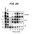
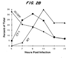
- Titers can be determined by indirect immunofluorescence using, for example, anti-M (23H12) or anti-N (10G4) protein specific antibodies ( L. Lefrancois et al., (1982) Virology 121: 157-67 ).
- miniviruses and “helper cells” (also known as “helper cell lines”) provide the same thing: to provide a source of Rhabdovirus proteins for Rhabdovirus virion assembly.
- a Rhabdovirus minivirus is the VSV minivirus which expresses only the G and M protein, as reported by E. A. Stillman et al., (1995) J. Virol. 69: 2946-53 .
- Helper viruses and miniviruses are used as methods of providing Rhabdovirus proteins that are not produced from transfected DNA encoding the genes for Rhabdovirus proteins.
- minivirus When using a minivirus, cells are infected with vaccinia virus as described above for purposes of providing T7 RNA polymerase.
- the desired polycistronic RNA, and plasmids containing the N, P and L genes are transfected into cells.
- the transfection mix is removed after approximately 3 hrs, and cells are infected with the minivirus at a m.o.i. of about 1.
- the minivirus supplies the missing G and/or M proteins (see Stillman et al., (1995) J. Virol. 69: 2946-53 .).
- the polycistronic RNA transfected into the cell will depend on whether an infectious or non-infectious recombinant Rhabdovirus is wanted.
- a minivirus could be used to provide the N, P and L genes.
- the minivirus could also be used to produce the M protein in addition to N, P and L.
- the minivirus also can produce the G protein.
- helper cell line When using a helper cell line, the genes encoding the missing Rhabdovirus proteins are produced by the helper cell line.
- the helper cell line has N, P, L and G proteins for production of recombinant Rhabdovirus particles which does not encode wild-type G protein.
- the proteins are expressed from genes or DNAs that are not part of the recombinant virus genome. These plasmids or other vector system is stably incorporated into the genome of the cell line.
- the proteins are then produced from the cell's genome and not from a replicon in the cytoplasm.
- the helper cell line can then be transfected with a polycistronic DNA and plasmid cDNAs containing the other Rhabdovirus genes not expressed by the helper virus.
- the polycistronic RNA used will depend on whether an infectious or non-infectious recombinant Rhabdovirus is desired. Otherwise, supply of missing gene products (e.g ., G and/
- the recombinant Rhabdoviruses produced as described above can be used (1) to target cells infected with infectious agents (e.g ., bacteria, parasites, or viruses); (2) to target diseased or abnormal cells; (3) to study the envelope proteins of other viruses for research purposes; (4) to treat a disease or infection and to remove abnormal cells; (5) to image specific cells or tissues for diagnostic purposes; and (6) to use as a kit for diagnoses and/or disease tracking.
- infectious agents e.g ., bacteria, parasites, or viruses
- Recombinant Rhabdoviruses which express a fusion protein can be further engineered to also express an attachment protein that recognizes a protein located on the surface of the cell that results from infection by some infectious agent (e.g ., bacteria, viruses, or parasites).
- some infectious agent e.g ., bacteria, viruses, or parasites.
- Such a recombinant Rhabdovirus could be utilized to (1) diagnose the presence of the infectious agent by producing a reporter protein if infection by the recombinant virus occurs or (2) to treat the infection.
- Rhabdovirus that expresses the CD4 protein in conjunction with the fusion protein.
- RV both the CD4 and fusion proteins would be created from plasmids, where the genes encoding these proteins are fused to the gene encoding the tail of the G protein as described by Mebatsion et al., 1997. The absence of the tail of the G protein in RV constructs would prevent CD4 and fusion protein expression on the surface of the recombinant RV construct.
- VSV the CD4 and the fusion proteins may be expressed alone without being fused to a portion of the G protein.
- the genes encoding the CD4 protein and the fusion protein would be placed downstream from the N and P genes, but before the L gene.
- the L gene is the last gene in these constructs. It is important that the ratios of the Rhabdovirus proteins remain substantially the same in all constructs for proper assembly to occur.
- VSV that expresses the F protein of paramyxovirus strain SV5 and CD4 would be capable of infecting both HIV-1 infected macrophages and T cells.
- Such a recombinant VSV can be further engineered to express a reporter protein upon entry into the HIV-1 infected cells. This construct is in contrast to those described by Schnell et al. (1997) and Mebatsion et al. (1997), which can only infect T-cells due to the presence of the co-receptor CXCR4.
- the isolated recombinant Rhabdovirus is incubated with the cells using techniques known in the art. Detection of infection by the recombinant Rhabdovirus could proceed by determining the presence of a reporter gene, such as a green fluorescent protein.
- the isolated recombinant Rhabdovirus could be administered by either intravascular (i.v.), intramuscular (i.m.), subcutaneous (s.c.), oral, or by any means in which the recombinant virus can be delivered to tissue (e.g ., needle or catheter).
- the recombinant Rhabdovirus could be administered topically for insertion into epithelial cells. Another method of administration would be as an aspirate.
- the recombinant virus would be given sufficient time to circulate, attach, infect, and produce the reporter protein. Reporter protein should be detectable within 24 hours and certainly within 36 hours.
- the viral agents infecting the cells to be targeted contemplated include members of the following virus families: Arenaviridae, Bunyaviridae, Coronaviridae, Filoviridae, Flaviviridae, Herpesviridae, Hepadnaviridae, Orthomyxoviridae, Paramyxoviridae, Poxyiridae, Retroviridae, and Rhabdoviridae.
- Additional viral agents for targeting by recombinant Rhabdoviruses considered include: African Swine Fever Virus, Borna Disease Virus, Hepatitis X, HIV-1, Human T Lymphocyte virus type-1 (HTLV-1), HTLV-2, lentiviruses, Epstein-Barr Virus, papilloma viruses, herpes simplex viruses, hepatitis B and hepatitis C.
- Proteins of bacterial agents capable of residing intracellularly in an infected host are also potential targets contemplated for infection by the recombinant Rhabdovirus.
- the intracellular bacteria contemplated include, amongst others: Shigella flexneri, Salmonella typhi, Legionella species, mycobacteria, and chlamydiae (See G. L. Mandell, "Introduction to Bacterial Disease" IN CECIL TEXTBOOK OF MEDICINE, (W.B. Saunders Co., 1996) 1556-7 ). These bacteria would be expected to express a bacteria-related protein on the surface of the infected cell to which the recombinant Rhabdovirus would attach.
- Proteins of parasitic agents which reside intracellularly, also are targets contemplated for infection by the recombinant Rhabdovirus.
- the intracellular parasites contemplated include, for example, Protozoa.
- Protozoa which infect cells include: parasites of the genus Plasmodium (e.g., Plasmodium falciparum, P. Vivax, P. ovale and P. malariae ), Trypanosoma cruzi, Leishmania, and Cryptosporidium.
- Diseased and/or abnormal cells would be targeted using the recombinant Rhabdoviruses previously described by the same administration methods as previously described.
- the diseased or abnormal cells contemplated include: neoplastic cells, pre-neoplastic cells, benign tumors, or polyps, café au lait spots, leukoplakia and other skin moles.
- the recombinant Rhabdoviruses will be targeted using an antireceptor that will recognize and bind to a receptor expressed on the diseased or abnormal cell. Fusion of the recombinant Rhabdovirus virion will be mediated by a fusion protein as previously described. Possible antireceptors used in the recombinant Rhabdovirus will include the natural antireceptor for any particular cell receptor.
- recombinant Rhabdoviruses can be engineered to express an antibody or polypeptide fragment thereof, a bifunctional antibody, Fab, F(ab') 2 , Fc, Fv, or single chain Fv (scFv) as their attachment protein.
- Fab fragment antigen
- Fc single chain Fv
- scFv single chain Fv
- These antibodies can be humanized, human, or chimeric antibodies (for discussion and additional references see S. L. Morrison "Antibody Molecules, Genetic Engineering of," in MOLECULAR BIOLOGY AND BIOTECHNOLOGY: A COMPREHENSIVE DESK REFERENCE 1995 ; S. D. Gillies et al., (1990) Hum. Antibod. Hybridomas 1(1): 47-54 ; E.
- TAA tumor associated antigens
- the genes of antibodies which recognize TAAs can be fused to either the F Protein, the complete G protein or the G stem or expressed in combination with the F Protein and G protein or G stem.
- antibodies include those antibodies which react with malignant prostatic epithelium but not with benign prostate tissue (e.g ., ATCC No. HB-9119; ATCC HB-9120; and ATCC No. HB-11430) or react with malignant breast cancer cells but not with normal breast tissue (e.g ., ATCC No. HB-8691; ATCC No. HB-10807; and HB-108011).
- Other antibodies or fragments thereof which react with diseased tissue and not with normal tissue would be apparent to the skilled artisan.
- the envelope proteins of viruses that express a lipid membrane envelope can be studied using a recombinant Rhabdovirus.
- a recombinant virus would allow study of (1) the tropism of a particular virus envelope protein; and (2) the cell receptors targeted by the virus envelope protein (antireceptor).
- experimentation can proceed without the usual need for high-level containment areas.
- virus envelope proteins of interest in such study are those of the following virus families, for example: Arenaviridae, Bunyaviridae, Coronaviridae, Filoviridae, Flaviviridae, Herpesviridae, Hepadnaviridae, Orthomyxoyviridae, Paramyxoviridae, Poxviridae, Retroviridae, and Rhabdoviridae.
- Additional viruses with protein-expressing lipid envelopes include: African Swine Fever Virus, Boma Disease Virus and Hepatitis X.
- the hazardous viruses contemplated for study using recombinant Rhabdoviruses include amongst others: Ebola virus (subtypes Zaire, Reston and Sudan), Marburg Virus, Lassa Fever, Dengue, Hantaan virus, Korean hemorrhagic fever, Kyasanur Forest virus, Puumula virus, Seoul virus, Simian hemorrhagic fever virus, HIV, HTLV and South American hemorrhagic fever virus ( B. N. FIELDS' VIROLOGY volumes 1 and 2, New York, NY 1996 ).
- Ebola virus subtypes Zaire, Reston and Sudan
- Marburg Virus Lassa Fever
- Dengue Hantaan virus
- Korean hemorrhagic fever Kyasanur Forest virus
- Puumula virus Seoul virus
- Simian hemorrhagic fever virus HIV
- HTLV South American hemorrhagic fever virus
- Example 6 describes a recombinant VSV that expresses the envelope protein of Ebola virus, Reston strain.
- diseased cell is meant a cell that is infected with a virus, bacteria or parasite, or a cell that is histologically or genetically aberrant for its tissue type.
- the recombinant Rhabdovirus particles described above can be used therapeutically to treat or ameliorate a condition resulting from a disease or an infection. If the diseased or abnormal cells are targeted and infected by a recombinant Rhabdovirus, and the M protein is expressed, the infected cell will eventually die as a result of M protein cytotoxicity. Recombinant VSV constructs that do not express a fusion protein have been demonstrated to kill and reduce the number of cells infected with HIV-1 for this reason (See Schnell et al., 1997).
- Recombinant Rhabdoviruses that express antibodies or antibody fragments which recognize, for example, a specific tumor associated antigen (TAA) could target certain cancer cells that express at least one TAA (H. I. Scher et al., IN V. T. DEVITA ET AL., CANCER: PRINCIPLES AND PRACTICE OF ONCOLOGY (1996) 1300-1320 ).
- TAA tumor associated antigen
- TAAs that can serve as the target of modified Rhabdoviruses include: ⁇ -human chorionic gonadotrophic hormone ( ⁇ -HCG) and ⁇ -fetoprotein (AFP) which are expressed in brain tumors; the products of BRCA1, BRCA2 and carcinoembryonic antigen (CEA), which are expressed in breast cancers; tyrosinase and the MART-1 antigens associated with melanomas; members of the c-erbB family; MUC1/REP expressed in breast cancer tissues; TuAg.1; and epidermal growth factor receptor (EGFR) which is expressed in lung cancers ( V.T.
- ⁇ -HCG ⁇ -human chorionic gonadotrophic hormone
- AFP ⁇ -fetoprotein
- CEA carcinoembryonic antigen
- tyrosinase and the MART-1 antigens associated with melanomas members of the c-erbB family
- MUC1/REP expressed in breast cancer tissues
- Rhabdovirus pseudotypes are contemplated for use in treating cancer by infecting individuals with a therapeutically effective amount of a recombinant Rhabdovirus that targets the cancer cells and after infection, lyses and kills the cancer cell.
- Other TAAs for expression on recombinant Rhabdovirus would be known to the skilled artisan.
- the recombinant Rhabdoviruses of the present invention could be engineered to be capable of replicating in the targeted cell after entry and reinfecting other target cells. These recombinant Rhabdoviruses would be unable to infect healthy cells.
- a recombinant Rhabdovirus can be constructed to express a reporter protein or fluorescent protein. These reporter proteins would be transcribed and translated only if the recombinant Rhabdovirus successfully infects the cell. The presence or absence of the reporter protein will determine whether or not the recombinant Rhabdovirus can infect the cell.
- recombinant Rhabdoviruses that express a reporter protein could be used when studying the tropism of virus envelope proteins or even other protein-protein ( e.g ., receptor-ligand) interactions. Detection of a reporter protein would indicate the presence of a disease. Examples 5 and 7 describe representative recombinant Rhabdoviruses used to image cells.
- kits can be prepared that comprises a recombinant Rhabdovirus capable of infecting a diseased or abnormal cell that upon successful infection would produce a reporter protein.
- the recombinant Rhabdovirus would be prepared to recognize a cell receptor associated with a disease.
- the attachment protein expressed by the recombinant Rhabdovirus could recognize a tumor associated antigen (TAA) and would be able to detect cancer or a particular type of cancer.
- TAA tumor associated antigen
- kits can be used in conjunction with existing histological staining techniques to determine more quickly, as well as more accurately, what disease is present.
- the kit could additionally be used to ascertain the stage of a disease (e.g ., cancer stage I versus cancer stage IV). This would be useful for purposes of diagnosis and determining what therapy or therapies may be appropriate in treating a particular subject's disease.
- the preferred kit would have the recombinant Rhabdovirus prepared for contact with the tissue sample from a biopsy, for example.
- the tissue sample or aspirated cells then would be cultured with the recombinant Rhabdovirus, as would be known in the art.
- the cells and/or tissue After incubation with kit Rhabdovirus, the cells and/or tissue would be examined for the presence or absence of a reporter protein.
- Kits could also be prepared as above for use in vivo.
- the pre-prepared, recombinant Rhabdoviruses could be administered to a subject via one of the previously discussed methods of administration.
- Expression of the reporter gene may be detected in skin cells or during a surgical procedure. For surgery, detection of diseased or abnormal cells versus healthy (or normal) cells would permit surgeons to determine how much diseased tissue needed to be resected by margins determined by examining reporter protein expression in cells infected by the Rhabdoviruses.
- VSV cDNAs A diagram of the method used, in this example, to construct a recombinant VSV that expresses the F protein of paramyxovirus SV5 is shown in Figure 5 .
- Full-length VSV cDNAs encoding G proteins with truncated cytoplasmic domains were generated by replacing the gene encoding the wild-type G protein in pVSVFL(+) I/NJG ( N. Lawson et al., Proc. Nat'l Acad. Sci. USA (1995) 92: 4477-4481 ).
- CT-1 CT-1 Triple Stop
- MW-36 sense-strand primer
- nucleotides 975-997 see the sequence for the complete genome of VSV-Indiana, GenBank # J02428, which are 5' to the Kpn I site in the G protein cDNA, and an antisense oligonucleotide (5'-TATAT GCTAGC TTATTATTA TCGGAGAACCAAGAATAGTCCAATG -3').
- the three stop codons that were introduced are indicated in boldface type, and the Nhe I site used for cloning is underlined.
- pVSV- ⁇ G A plasmid encoding the G protein deletion mutant (pVSV- ⁇ G) was generated by removing the MIu I to Nhe I fragment from pVSVFL(+)-2 ( Lawson et al., (1995) Proc. Nat'l Acad. Sci. USA 92: 4477-81 ), filling in with Klenow fragment, and subsequent re-legation of the plasmid. Therefore, pVSV- ⁇ G retained the transcriptional regulatory sequences and a portion of the 3' untranslated region of the G protein gene, however it lacked the open reading frame (ORF) of the G protein coding region.
- ORF open reading frame
- oligonucleotide-derived polylinker region (5' -Mlu I -Kpn I -Xho I -Sma I -Eag I -Sph I -Nhe I -3 ') was also introduced between the Mlu I and Nhe I sites to facilitate the insertion of heterologous genes into the G protein gene transcription unit.
- This plasmid (pVSV ⁇ G -PL) was used to produce the recombinant VSV cDNAs encoding either human CD4 (pVSV ⁇ G -CD4), or a chimeric CD4 protein in which the transmembrane and cytoplasmic domains of CD4 were replaced with the corresponding regions of the VSV G protein ( ⁇ G -CD4G).
- HAGS HA -tagged G - s tem
- the construct HAGS was generated by PCR-mediated mutagenesis using a primer that overlapped the Mlu I site in the 5' nontranslated region of the G protein gene of pVSVFL(+)-2 and an antisense oligonucleotide 5'-AGGATG TTCGAA AGCGTAATCTGGTACATCATACGGATACTT GCAATTCACCCCAATG-3', which overlapped the Nsp V site (underlined) at position 1,280 in the VSV G protein gene, and which fused sequences complimentary to a portion of the G protein signal peptide (in italics) to the HA epitope (in bold-face type) followed by the membrane-proximal "stem” region of the G protein ectodomain.
- Infectious viruses encoding mutant G proteins were recovered from plasmids by infecting approximately 3 x 10 6 BHK-21 cells with the vaccinia virus vTF7-3 ( T. R. Fuerst et al., (1986) Proc. Nat'l Acad. Sci. USA 83: 8122-8126 ), which encodes T7 RNA polymerase.
- VSV Ind Indiana serotype of VSV
- VSV- ⁇ G, ⁇ G -CD4/G and ⁇ G -CD4 was performed as described for the CT mutants except the supernatants from the primary transfections were transferred directly to dishes containing 2 x 10 6 BHK cells, which were pre-infected with vTF7-3 and transfected with 10 ⁇ g of the G protein expression plasmid pBS-G- ⁇ T. After a 24 hr incubation, the supernatants were collected, cells were fixed with 3% paraformaldehyde and then examined by indirect immunofluorescence using anti-M protein-specific (23H12) or N protein-specific (10G4) monoclonal antibodies ( L.
- VSV-HAGS recovery using a VSV Minivirus The recombinant VSV-HAGS was recovered by an alternative strategy in which a VSV minivirus (GMMG), which expresses only the VSV G and M proteins ( E. A. Stillman et al., J. Virology (1995) 69: 2946-2953 ), was used to provide the source of complementing G protein.
- GMMG VSV minivirus
- Approximately 10 6 vTF7-3 infected cells were transfected with 10 ⁇ g of pVSV-HAGS and 3 ⁇ g, 5 ⁇ g, and 1 ⁇ g of plasmids encoding the wild-type VSV Ind N, P and L proteins, respectively.
- the transfection mix was removed after 3 hr, and the cells were then superinfected with GMMG particles at a m.o.i. of one. Fresh medium was added directly to the cells after absorbing the GMMG minivirus for 1 hr. Supernatants were harvested 18 hr later and then filtered to remove the vaccinia virus.
- Example 2 In vitro Administration of recombinant VSV containing a fusion protein to cells.
- the filtrate was then applied to cells in 10 cm dishes. After 18 to 24 hr, the cells were examined for cytopathic effects (CPE). Supernatants from cultures exhibiting CPE were titered, and individual plaques were purified. Plaque purified isolates were used to infect BHK cells at a m.o.i. of one for the production of stocks of the tail mutant viruses. Direct sequencing of viral RNA isolated from each of the mutants was performed to ascertain whether the recovered virus contained the appropriate mutations.
- CPE cytopathic effects
- the lower fraction which usually contained approximately 300-fold more VSV-HAGS than GMMG, was recovered by side-puncture and the amount of VSV-HAGS to GMMG was determined by an immunofluorescence-based titering assay utilizing either an N protein-specific (for VSV-HAGS) or a G protein-specific monoclonal antibody.
- an immunofluorescence-based titering assay utilizing either an N protein-specific (for VSV-HAGS) or a G protein-specific monoclonal antibody.
- the partially purified VSV-HAGS was completed by expressing the G protein from the New Jersey serotype of VSV (G NJ ). Then, neutralizing antibody, which was specific for the Indiana serotype of VSV (VSV Ind ), was added to the culture medium.
- VSV-HAGS/GMMG coinfected cells The amount of Indiana-specific sera used completely neutralized all virus produced by the VSV-HAGS/GMMG coinfected cells. After three consecutive passages on cells expressing G NJ and in the presence of anti-VSV Ind neutralizing antibody, the titers of VSV-HAGS reached approximately 10 8 IU/ml, while 0.5 ml of the supernatant contained no detectable GMMG. High titer stocks of pure VSV-HAGS complemented with the VSV G Ind protein were then made as described above for VSV ⁇ G.
- Example 3 Administration of modified VSV to cells in vivo.
- Recombinant VSV particles prepared by either of the methods described above are inoculated into a subject infected with HIV-1.
- concentration of the virus to be administered to a subject may vary, but the titer of the virus to be administered preferably should be in the order of about 10 6 to 10 12 IU/ml.
- the amount of the virus used may be greater if the patient has a relatively high number of infected cells.
- the viruses harvested as described in Example 1 would be resuspended in an appropriate carrier or excipient for purposes of injection.
- Example 4 Analysis of Infectivity of Recombinant VSV Expressing Paramyxovirus SV5 F protein and the Envelope Protein of Ebola Virus.
- Plasmids Full-length cDNAs encoding the Ebola Reston virus glycoprotein (ResGP) ( A. Sanchez et al., (1996) Proc. Nat'l Acad. Sci. USA 93: 3602-3607 ), VSV G protein ( J.K. Rose et al., (1982) Cell 30: 753-762 ), and influenza A/turkey/Ireland/1378/83 ( H. Niwa et al., (1991) Gene 108: 193-200 ) are designated pCResGP, pCVSVG, and pCTH, respectively.
- ResGP Ebola Reston virus glycoprotein
- VSV- ⁇ G* The coding region of the G protein gene in a full-length cDNA clone of the VSV genome (Indiana serotype, VSV Ind ) (N.D. Lawson et al., 1995) was replaced with the coding region of a modified version of the green fluorescent protein (GFP) gene ( M. Chalfie et al., (1994) Science 263: 802-5 ), in which serine 65 was replaced with a threonine. This plasmid was designated pVSV- ⁇ G*. BHK cells in 60 mm dishes were infected at a multiplicity of 10 with the recombinant vaccinia virus vTF7-3 (T.R.
- the supernatant fluid was harvested at 48 hr post transfection, and one half of the supernatant was used to infect a second plate of cells that had been infected with vTF7-3 and transfected with 5 ⁇ g of plasmid encoding the G protein only. Cells were examined for GFP expression at 24 hr post infection by fluorescence microscopy. The presence of fluorescent cells indicated that VSV genome had been successfully recovered.
- VSV ⁇ G*-G Additional passages of supernatant containing VSV ⁇ G* complemented with G protein (VSV ⁇ G*-G) through cells expressing the G protein from plasmid DNA in the presence of 25 ⁇ g/ml of cytosine- ⁇ -D-arabinofuranoside were performed to obtain high titer stocks of VSV ⁇ G*-G.
- the virus stocks were filtered through a membrane with a 0.2 ⁇ M pore to remove the vaccinia virus.
- Virus titer in the supernatant fluid was then determined again by infecting BHK cells and quantifying the number of cells expressing GFP by fluorescence microscopy.
- VSV ⁇ G* Complemented with VSV G or ResGP or Without Complementation .
- 293T cells were transfected with pCVSVG, pCResGP or pCAGGS using Lipofectamine (Gibco).
- m.o.i. multiplicity of infection
- cells were infected with the VSV ⁇ G*-G described above, at a multiplicity of infection (m.o.i.) of about one for one hour at 37°C. They were then washed with PBS three times, and medium was added. After 24 hours of incubation at 37°C in a CO 2 incubator, culture fluid was collected and centrifuged to remove cells. Each virus stock was stored at -80°C until used.
- Example 5 Method of Determining Tropism of VSV-Ebola Reston Pseudotype and Analysis of Cells So Infected.
- the infectious units of the recombinant virus expressed in different cell lines were determined by counting the number of green fluorescent protein (GFP) expressing cells. See Figure 4 for an example of the cells successfully infected by the recombinant VSV constructs: VSV ⁇ G*-G, VSV ⁇ G*-ResGP and VSV ⁇ G*.
- GFP green fluorescent protein
- VSV ⁇ G*-G the infectivity of VSV ⁇ G*-ResGP virus stock was greatly dependent on the type of cell line.
- infectivity titers of VSV ⁇ G*-ResGP were 100-fold higher than those with cells from hamsters, dogs, cattle, mice, chickens or bats.
- VSV ⁇ G*-ResGP did not infect the C6/36 insect cells.
- FIG. 3 shows lysates of 293T cells, which were transfected with an expression vector plasmid containing the VSV G gene (lane 1), the ResGP gene (lane 2), or the vector plasmid only (lane 3).
- the 293T cells were labeled for 5 hours with [ 35 S]-methionine 30 hr post transfection with the recombinant virus. Proteins in cell lysates were precipitated using a monoclonal antibody specific to the VSV G protein ( Fig. 3 , lane 1) or ResGP (lanes 2 and 3).
- VSV ⁇ G* complemented with VSV G protein
- ResGP lane 5; VSV ⁇ G*-ResGP
- lacking complementation lane 6; VSV ⁇ G*
- Recovery of infectious virus from plasmids is accomplished by infecting 10 6 BHK-21 cells with vTF7-3 vaccinia for 60 minutes and then transfecting the cells with 10 ⁇ g of plasmid encoding the various full-length VSV constructs and 3 ⁇ g, 5 ⁇ g, and 1 ⁇ g of plasmids encoding the VSVInd nucleocapsid protein (N), phosphoprotein (P) and polymerase (L), respectively. Cells were incubated for two days to allow replication and assembly of viral particles. The supernatant from the cells was then plated directly onto fresh BHK cells, and the cells were incubated for 24 hours to permit virus amplification.
- N nucleocapsid protein
- P phosphoprotein
- L polymerase
- the supernatants from these cultures were then passed through the filter as described above.
- the filtrate was transferred onto fresh BHK-21 cells.
- the supernatant was replaced with DMEM containing FCS.
- the media of cultures exhibiting CPE of a VSV infection were then removed and the virus was plaque purified.
- Virus stocks were subsequently prepared by infection, for example, of 10 7 cells with approximately 10 5 plaque forming units (p.f.u.) of the plaque isolate.
- the supernatant from these cultures was then used for all subsequent infections. Direct sequencing of the viral RNA isolated from every recovered virus is performed to ensure that the recovered virus was derived from a plasmid.
- Example 7 Method of Treating Individuals Infected with a Virus, Bacteria or a Parasite.
- a subject is treated using a recombinant Rhabdovirus that has been engineered, as discussed above, to target cells infected with either a virus, parasite or bacterium that leads to the incorporation on the cell surface of a protein derived from the infectious agent.
- This protein acts as a receptor that is to be targeted by the recombinant antireceptor of the recombinant Rhabdovirus agent.
- the CD4 protein would be expressed on the surface of the recombinant Rhabdovirus along with a heterologous fusion protein.
- the recombinant Rhabdovirus has the ability to recognize, bind to and infect a cell which expresses the mature HIV-1 envelope protein, gp120.
- the recombinant Rhabdovirus would contain a polycistronic nucleic acid that comprises the gene encoding the M protein. This Rhabdovirus would be capable of lysing the cells that it infects.
- the number of infected HIV-1 cells such as macrophages and T cells, could be reduced in number, and the virus thereby kept in check.
- the amount of recombinant Rhabdovirus administered would be approximately similar to what is administered when using most gene therapy vectors.
- Example 8 Method of Treating Subjects with Cancer.
- a recombinant VSV is prepared as described above except that the recombinant expresses an attachment protein (antireceptor) that can identify a protein associated with some forms of cancer, such as CEA, prostate specific antigen (PSA) or PMA.
- an attachment protein antireceptor
- PSA prostate specific antigen
- Example 9 Creation of a Recombinant Rhabdovirus Containing a Polypeptide Fragment of a Fusion Protein.
- a recombinant Rhabdovirus such as VSV
- VSV is constructed to express a fusion protein, such as the SV5 F protein, or a fragment of the fusion protein joined to the stem region of VSV G protein encompassing the putative oligomerization domain of G protein, the transmembrane domain and the cytoplasmic tail of G protein.
- Example 10 A Method of Identifying Fusion Proteins Using Only cDNAs.
- a recombinant Rhabdovirus such as VSV
- the attachment protein could be CD4 and the cells targeted could be cells expressing the HIV-1 envelope protein gp120/gp160.
- the construct expressing the candidate fusion protein would be compared to constructs that express the fusion protein of Paramyxovirus SV5 and CD4 (a positive control), and VSV constructs that express CD4 and the VSV G protein only. The degree of infectivity by these constructs could be compared using a reporter protein, such as the green fluorescent protein used above and as described in Examples 5 and 6.
- constructs could be prepared using any of the methods described in the specification.
- Example 11 Infectivity of recombinant CD4-VSV pseudotyped with the SV5 F protein on cells expressing HIV gp160.
- CD4-VSV with Paramyxovirus SV5 F proteins BHK-21 cells expressing the SV5 F protein (using the vaccinia T7 expression system) were infected with CD4-VSV that had been previously pseudotyped with wild-type VSV G protein. The supernatant was harvested 18 hr post-infection and then filtered to remove the vaccinia virus. The resulting supernatant contained recombinant VSV that had both CD4 and SV5 protein in the viral envelope, but that lacked VSV G protein. Residual infectivity from the inoculum ( e.g ., from the G protein pseudotyped CD4 virus) was eliminated by addition of neutralizing antibody specific for VSV G protein.
- inoculum e.g ., from the G protein pseudotyped CD4 virus
- Figure 7 demonstrates that in cells transfected with increasing amounts of plasmid encoding gp160, there is a corresponding increase in the number of cells that become infected with the CD4-SV5 F pseudotyped ⁇ G VSV.
- Example 12 Generation of recombinant VSV encoding a PSA-specific ScFv for targeting prostate cancer.
- V--H and V--L domains from a hybridoma cell line that produces antibodies to prostate-specific antigen (PSA), or other tumor-specific antigens are cloned using standard methods for ScFv construction. Briefly, sequences for the variable domains are obtained following RT-PCR amplification of poly-A containing mRNA isolated from the appropriate hybridoma cells. The poly-A RNA is used as the template for the reverse transcription (RT) reaction, and the primers that are used are similar to those described previously ( R. Orlandi et al., (1989) Proc. Natl. Acad. Sci. USA, 86: 3833-3837 .).
- the amplified V domains are cloned into pBluescript, and the sequence of the amplified regions are determined by standard methods. The sequences are analyzed for open reading frames and will be compared to known V H (variable heavy chain) and V L (variable light chain) sequences. Once the sequences are confirmed, these clones are used to construct the ScFv gene.
- the sequences encoding the V H and V L domains are joined using an oligonucleotide linker.
- the linker encodes a short Gly-Ser peptide that connects the C-terminus of the first V region and the N-terminus of the second V (variable) region into a V L - (Gly-Ser)-V H polypeptide.
- the intact ScFv gene is then joined to sequences encoding the VSV G protein signal sequence, such that following expression in eukaryotic cells, the protein is targeted to the endoplasmic reticulum and secreted.
- Expression analysis is used to confirm that the ScFv is functional and is able to bind the appropriate antigen (e.g ., PSA).
- the ScFv coding region is joined to sequences encoding the membrane-anchored stem region of a VSV G protein.
- a unique restriction site introduced into the G stem sequence is used to join the two coding regions such that the reading frame remains intact.
- the resulting molecule consists of the ScFv domain fused to amino acid E422 of VSV G stem (ScFv-GS). Expression analysis is used to confirm that the molecule is expressed and is localized to the plasma membrane of the transfected cells.
- the ScFv-GS gene is cloned into the multiple cloning site of pVSV-G-F such that the gene is under the transcriptional control of VSV.
- the resulting construct is called pVSV-ScFv-F.
- Recombinant ScFv-F virus is recovered from the plasmid pVSV-ScFv-F using standard procedures. Pure VSV-ScFv-F is generated following passage in cells not expressing wild-type VSV G protein.
- the cell line LNCaP is a model cell line for prostate cancer. These cells are known to express PSA and should serve as a target for recombinant ScFv-F virus. LNCaP cells are grown under standard culture conditions and then supernatants containing recombinant ScFv-F virus are added. Because the cells are lightly adherent and the monolayers are easily disrupted, the inoculum is not removed. Infection is assessed at 15 hours post-inoculation using a typical immunofluorescence-based infectious center assay.
- Controls include infection with wild-type recombinant VSV, the non-infectious mutant VSV-HAGS, which contains only the membrane-anchored G stem, and a recombinant that encodes only the ScFv-GS polypeptide without the fusion (F) protein. Specificity is determined by assessing the infectivity of ScFv-F virus on normal cells that do not express PSA.
- VSV-ScFv-F is administered to animals bearing human prostate-specific tumors. The animals are monitored for several weeks to determine if there are any adverse effects resulting from administration of the recombinant virus. Animals are sacrificed and the tumor masses examined for changes in size, weight, vascularization and metastasis. Controls include infection with VSV-HAGS, which serves as the negative control. An additional control is the administration of a recombinant VSV-ScFv-GS that does not contain a fusion (F) protein. Like VSV-HAGS, this virus should be noninfectious.
- the VSV-HAGS construct allows the evaluation of effects associated with administration of a virus having the ScFv-GS polypeptide on the viral envelope.
- BHK-21 cells were infected with wild-type G protein pseudotyped recombinant ⁇ G-VSV encoding either CD4-G, CD4-GS, or both CD4-G and HAGS viruses.
- the pseudotyped viruses were absorbed for 60 min, the inoculum was removed, and then cell monolayers were washed extensively to remove any residual inoculum. Following infection, the cells were incubated in medium containing 5% FCS for 16 hours.
- the medium which contained pure recombinant VSV/CD4-G, CD4GS, or a mixture of CD4-G and HAGS viruses, was collected, and detached cells were removed by centrifugation at 2,500 rpm for 10 min. Aliquots of the supernatant were used to infect cells expressing the HXB subtype of the HIV-1 envelope protein. The supernatants were absorbed for 60 min in serum-free medium; the inoculum was removed and then replaced with serum-free medium. Infectivity was assayed by examining the number of cells expressing the VSV nucleocapsid protein by immunofluorescence microscopy.
- VSV/CD4-G 100 ⁇ l of the VSV/CD4-G supernatant was used for the infectivity assay, whereas only 10 ⁇ l of the CD4-GS or CD4G+HAGS supernatants were used. A total of 60 VSV/CD4-G infected cells were found on the entire dish. This result corresponds to a titer of ⁇ 600 infectious units per ml (IU/ml). In contrast, the titer of CD4-GS or CD4-G+HAGS was >10 8 IU/ml.
- the data shown in Figure 8 demonstrates that virus containing CD4-G, but lacking a human chemokine coreceptor in the viral envelope, has extremely low infectivity on cells expressing the T-tropic HIV-1 envelope protein (HXB).
- virus encoding CD4-GS (the CD4 ectodomain joined to amino acid E422 of the VSV G protein ectodomain, e.g ., the "G stem") has very high specific infectivity.
- the specific infectivity of CD4-GS is nearly equivalent to the specific infectivity observed with wild-type VSV for its wild-type host cell target.
- infection of HIV-1 envelope expressing cells by VSV/CD4-GS does not require a chemokine coreceptor.
- VSV/CD4-GS virions Analysis of purified VSV/CD4-GS virions has revealed that a proportion ( ⁇ 50%) of CD4-GS is cleaved at, or near the CD4/G-stem junction. This cleavage, which occurs during transport of the protein to the plasma membrane in infected cells, results in the release of virus particles that have both the membrane-anchored cleavage product (the G-stem) as well as full-length CD4-GS molecules ( Figure 9 ). Cleavage does not occur with CD4-G and consequently, the VSV envelope consists solely of CD4-G.
- the G-stem in this case an HA-epitope tagged version called HAGS
- HAGS HA-epitope tagged version
- Virus production and G-stem incorporation Virus was produced as described in the experiment demonstrated in Figure 8 . The supernatant was harvested at 16 hours post-infection. Virions were concentrated by ultracentrifugation, and the amount of virus and the protein content of the virions were determined using SDS-PAGE. Virions proteins were visualized by staining with Coomassie blue or by Western blot analysis using an antibody specific for the cytoplasmic domain of VSV G protein (see Figure 9 ).
- VSV/CD4-G infected cells produce approximately the same amount of virus as ⁇ G-VSV. This corresponds to ⁇ 10-fold less virus than wild-type VSV. In contrast, VSV/CD4GS produces nearly wild-type amounts of virus ( ⁇ 70-75% of wild-type).
- Immunoblot analysis ( Figure 9 ) of equivalent amounts of CD4-G and CD4-GS virus, with an antibody which recognizes and binds to the last 15 amino acids of the VSV-G cytoplasmicdomain, shows that CD4-GS is cleaved and the corresponding the G-stem fragment is incorporated into recombinant VSV virions together with the full-length CD4-GS molecule.
- VSV / CD4-GS Infectivity of VSV / CD4-GS produced from various cell types on cells. Virus was produced in the indicated cell types essentially as described in the experiment of Figure 8 . Supernatants were harvested at various times post-infection, which was dictated by differences in the susceptibility of the different cell types to lysis by VSV. A portion of the supernatant was used for the infectivity assays, and the remainder was subjected to ultracentrifugation to pellet virions. The amount of virus produced by each of the cell types was determined by densitometry of Coomassie blue stained gels following SDS-PAGE separation of the proteins. The relative infectivity displayed in Figure 10 represents the amount of infectivity after normalization to viral protein pelleted from the supernatant.
- VSV/CD4-Q427 construct for X4 (T cell tropic envelope) and R5 specific envelopes (macrophage tropic envelopes).
- BHK-21 cells were infected with wild-type G protein pseudotyped recombinant G-VSV encoding CD4-GS fused at Q427 of the 511 amino acid G protein.
- the pseudotyped virus was absorbed for 60 min, the inoculum was removed, and then the cell monolayer was washed extensively to remove any residual inoculum.
- the infected cultures were subsequently incubated in medium containing 5% FCS for 16 hours.
- the medium from the infected cultures which contained pure recombinant VSV/CD4-GS(Q427) virus, was collected and detached cells were removed by centrifugation at 2,500 rpm for 10 min. A 100 ⁇ l aliquot of the supernatant was used to infect HeLa cells expressing the indicated HIV-1 envelope proteins as follows.
- the HIV-1 envelope proteins were expressed using vaccinia virus vectors which were supplied from the NIH AIDS Research and Reference Reagent Program.
- HeLa cells were infected with the indicated vaccinia virus vector by absorption for 60 min in serum-free medium. The inoculum was removed and replaced with medium containing 5% FCS, and 100 ⁇ g/ml AraC.
- the cells were incubated for 30 min, then 100 ⁇ l of the VSV/CD4-GS(Q427) supernatant was added directly to the medium, and the cultures were incubated for 4 hours to allow expression of the HIV-1 envelope and absorption of the CD4-GS(Q427) virus. After 4 hours, the medium containing the inoculum was removed and replaced with medium containing serum and AraC. The cultures were incubated for an additional 10 hours, and then the cells were fixed with 3% paraformaldehyde. Infectivity was assayed by examining the number of cells expressing the VSV nucleocapsid (N) protein using an N-specific monoclonal antibody and detection by immunofluorescence microscopy.
- N VSV nucleocapsid
- VSV/CD4-GS can infect cells expressing both T- and M-tropic HIV-1 envelope proteins. This infectivity occurs in a chemokine coreceptor-independent manner, since the recombinant virus was produced in a hamster cell line (BHK-21), which does not express a human chemokine coreceptor. Infectivity is absolutely dependent on expression of the HIV-1 envelope protein. Cells infected with either a recombinant vaccinia virus encoding T7 RNA polymerase (vTF7-3) or normal HeLa cells (not infected with vaccinia virus, but which were grown in the presence of AraC) were not susceptible to VSV/CD4-GS infection.
- vTF7-3 recombinant vaccinia virus encoding T7 RNA polymerase
- AraC normal HeLa cells
- HIV-1 envelope protein e.g ., gp120-gp41
- cells expressing the uncleaved e.g ., gp160
- fusion-defective mutant of LAI IIIB expressed from vCB-16
- Vaccinia vectors are as described in C.C. Broder et al., (1995) Proc. Nat'l Acad. Sci. USA 92: 9004-9008 .
- the truncated version of the VSV G protein consists of the membrane-proximal 42 amino acids of the G protein ectodomain, the 20 amino acid transmembrane domain and the 29 amino acid cytoplasmic tail.
- the protein is generated from a precursor that contains the VSV-G protein signal sequence which is cleaved by signal peptidase following cotranslational insertion into the endoplasmic reticulum.
- the "maximum-length" stem that we have analyzed has 59 amino acids of the membrane-proximal ectodomain and the shortest version (which shows enhancement of fusion activity) has 29 amino acids of the ectodomain.
- the membrane-anchored G stem appears to enhance the fusion activity of a variety of heterologous viral fusion proteins. This includes the fusion activity of HIV-1 gp120-gp41 from both T- and M-tropic viruses, the F protein of the paramyxovirus SVS, and the New Jersey serotype glycoprotein of VSV. We postulate that the stein somehow stabilizes, thereby enhancing, the fusion pores formed by these fusion proteins. The presence of the fusion pores usually necessitates the presence of another cofactor to manifest the full fusion activity of these proteins. In the case of HIV-1 envelope proteins, the cofactor is the chemokine coreceptors, for the paramyxovirus F proteins it is their cognate HN proteins, and for VSV G protein it is activation by acidic pH.
Landscapes
- Health & Medical Sciences (AREA)
- Life Sciences & Earth Sciences (AREA)
- Chemical & Material Sciences (AREA)
- Organic Chemistry (AREA)
- Genetics & Genomics (AREA)
- General Health & Medical Sciences (AREA)
- Engineering & Computer Science (AREA)
- Medicinal Chemistry (AREA)
- Zoology (AREA)
- General Engineering & Computer Science (AREA)
- Bioinformatics & Cheminformatics (AREA)
- Molecular Biology (AREA)
- Veterinary Medicine (AREA)
- Biochemistry (AREA)
- Virology (AREA)
- Wood Science & Technology (AREA)
- Biotechnology (AREA)
- Biophysics (AREA)
- Biomedical Technology (AREA)
- General Chemical & Material Sciences (AREA)
- Chemical Kinetics & Catalysis (AREA)
- Nuclear Medicine, Radiotherapy & Molecular Imaging (AREA)
- Public Health (AREA)
- Animal Behavior & Ethology (AREA)
- Pharmacology & Pharmacy (AREA)
- Plant Pathology (AREA)
- Microbiology (AREA)
- Physics & Mathematics (AREA)
- Gastroenterology & Hepatology (AREA)
- Proteomics, Peptides & Aminoacids (AREA)
- Tropical Medicine & Parasitology (AREA)
- Communicable Diseases (AREA)
- Oncology (AREA)
- Micro-Organisms Or Cultivation Processes Thereof (AREA)
- Medicines Containing Material From Animals Or Micro-Organisms (AREA)
- Medicines That Contain Protein Lipid Enzymes And Other Medicines (AREA)
- Peptides Or Proteins (AREA)
Applications Claiming Priority (3)
| Application Number | Priority Date | Filing Date | Title |
|---|---|---|---|
| US6847297P | 1997-12-22 | 1997-12-22 | |
| US68472P | 1997-12-22 | ||
| PCT/US1998/026084 WO1999032648A1 (en) | 1997-12-22 | 1998-12-22 | Recombinant rhabdovirus containing a heterologous fusion protein |
Publications (2)
| Publication Number | Publication Date |
|---|---|
| EP1040198A1 EP1040198A1 (en) | 2000-10-04 |
| EP1040198B1 true EP1040198B1 (en) | 2009-12-09 |
Family
ID=22082797
Family Applications (1)
| Application Number | Title | Priority Date | Filing Date |
|---|---|---|---|
| EP98963811A Expired - Lifetime EP1040198B1 (en) | 1997-12-22 | 1998-12-22 | Recombinant rhabdovirus containing a heterologous fusion protein |
Country Status (14)
| Country | Link |
|---|---|
| US (2) | US6497873B1 (enExample) |
| EP (1) | EP1040198B1 (enExample) |
| JP (1) | JP4544497B2 (enExample) |
| AP (1) | AP1968A (enExample) |
| AT (1) | ATE451466T1 (enExample) |
| AU (2) | AU752004B2 (enExample) |
| BR (1) | BR9814370A (enExample) |
| CA (1) | CA2316033A1 (enExample) |
| DE (1) | DE69841372D1 (enExample) |
| IL (2) | IL136905A0 (enExample) |
| MX (1) | MXPA00006184A (enExample) |
| NZ (1) | NZ505312A (enExample) |
| OA (1) | OA11432A (enExample) |
| WO (1) | WO1999032648A1 (enExample) |
Families Citing this family (22)
| Publication number | Priority date | Publication date | Assignee | Title |
|---|---|---|---|---|
| EP1716858B9 (en) * | 1999-09-17 | 2009-08-12 | Wellstat Biologics Corporation | Oncolytic virus |
| AU2001281151A1 (en) * | 2000-08-24 | 2002-03-04 | Thomas Jefferson University | Rhabdovirus-based vectors to express high levels of functional human antibodies |
| CA2440433A1 (en) * | 2001-03-09 | 2002-09-19 | The Regents Of The University Of California | Cell culture system for synthesis of infectious hepatitis c virus |
| WO2003005964A2 (en) * | 2001-07-11 | 2003-01-23 | University Of Miami | Recombinant vsv for the treatment of tumor cells |
| US7820435B2 (en) * | 2002-02-06 | 2010-10-26 | Expression Technologies Inc. | De novo synthesized plasmid, methods of making and use thereof |
| CN1788088B (zh) | 2003-03-27 | 2012-09-26 | 渥太华健康研究学会 | 突变的水泡性口炎病毒及其用途 |
| US7731974B2 (en) | 2003-03-27 | 2010-06-08 | Ottawa Hospital Research Institute | Mutant vesicular stomatitis viruses and uses thereof |
| KR20060017635A (ko) * | 2003-06-05 | 2006-02-24 | 와이어쓰 홀딩스 코포레이션 | 베네쥬엘라 말 뇌염 바이러스 레플리콘 벡터 및파라믹소바이러스 단백질 항원을 포함하는 면역원성 조성물 |
| AU2004250129A1 (en) | 2003-06-09 | 2004-12-29 | Wyeth | Improved method for the recovery of non-segmented, negative-stranded RNA viruses from cDNA |
| CA2921063C (en) * | 2006-09-15 | 2020-01-28 | Ottawa Hospital Research Institute | Oncolytic rhabdovirus |
| US8961951B2 (en) * | 2007-07-06 | 2015-02-24 | Boyce Thompson Institute For Plant Research | Baculoviruses with enhanced virion production and a method for the production of baculoviruses |
| US8703469B2 (en) | 2007-07-06 | 2014-04-22 | Boyce Thompson Institute For Plant Research | Baculoviruses with enhanced virion production and a method for the production of baculoviruses |
| EP2398496B1 (en) * | 2009-02-20 | 2015-08-05 | International AIDS Vaccine Initiative | Vesicular stomatitis virus vectors encoding hiv env epitopes |
| AU2012216792A1 (en) | 2011-09-12 | 2013-03-28 | International Aids Vaccine Initiative | Immunoselection of recombinant vesicular stomatitis virus expressing HIV-1 proteins by broadly neutralizing antibodies |
| EP2586461A1 (en) | 2011-10-27 | 2013-05-01 | Christopher L. Parks | Viral particles derived from an enveloped virus |
| US9347065B2 (en) | 2012-03-29 | 2016-05-24 | International Aids Vaccine Initiative | Methods to improve vector expression and genetic stability |
| US9555067B2 (en) | 2012-04-18 | 2017-01-31 | Mayo Foundation For Medical Education And Research | Replication-competent vesicular stomatitis viruses |
| FR2996856B1 (fr) * | 2012-10-15 | 2015-06-05 | Agronomique Inst Nat Rech | Novirhabdovirus recombinant utilisable comme vecteur d'antigenes |
| JP2020518275A (ja) * | 2017-05-03 | 2020-06-25 | バイオマリン ファーマシューティカル インコーポレイテッド | 造血幹細胞の形質導入のための改善されたレンチウイルス |
| CN109414483B (zh) | 2017-05-10 | 2025-10-24 | 制药五有限责任公司 | 对补体灭活作用具有抗性的用于治疗癌症的包膜病毒 |
| CN115768784A (zh) * | 2020-02-06 | 2023-03-07 | 勃林格殷格翰动物保健有限公司 | 用于检测抗弹状病毒抗体的多肽 |
| CN116144608A (zh) * | 2021-09-29 | 2023-05-23 | 上海行深生物科技有限公司 | 病毒培养方法 |
-
1998
- 1998-12-22 AP APAP/P/2000/001852A patent/AP1968A/en active
- 1998-12-22 EP EP98963811A patent/EP1040198B1/en not_active Expired - Lifetime
- 1998-12-22 CA CA002316033A patent/CA2316033A1/en not_active Abandoned
- 1998-12-22 AU AU19056/99A patent/AU752004B2/en not_active Ceased
- 1998-12-22 IL IL13690598A patent/IL136905A0/xx active IP Right Grant
- 1998-12-22 NZ NZ505312A patent/NZ505312A/xx unknown
- 1998-12-22 AT AT98963811T patent/ATE451466T1/de not_active IP Right Cessation
- 1998-12-22 DE DE69841372T patent/DE69841372D1/de not_active Expired - Lifetime
- 1998-12-22 BR BR9814370-0A patent/BR9814370A/pt not_active Application Discontinuation
- 1998-12-22 JP JP2000525566A patent/JP4544497B2/ja not_active Expired - Fee Related
- 1998-12-22 MX MXPA00006184A patent/MXPA00006184A/es not_active IP Right Cessation
- 1998-12-22 US US09/217,967 patent/US6497873B1/en not_active Expired - Fee Related
- 1998-12-22 WO PCT/US1998/026084 patent/WO1999032648A1/en not_active Ceased
-
2000
- 2000-06-20 IL IL136905A patent/IL136905A/en not_active IP Right Cessation
- 2000-06-21 OA OA1200000184A patent/OA11432A/en unknown
-
2002
- 2002-07-19 AU AU2002300207A patent/AU2002300207B2/en not_active Ceased
- 2002-10-23 US US10/277,997 patent/US20030138457A1/en not_active Abandoned
Also Published As
| Publication number | Publication date |
|---|---|
| JP2001526901A (ja) | 2001-12-25 |
| US20030138457A1 (en) | 2003-07-24 |
| AP1968A (en) | 2009-04-30 |
| DE69841372D1 (de) | 2010-01-21 |
| NZ505312A (en) | 2003-01-31 |
| EP1040198A1 (en) | 2000-10-04 |
| IL136905A0 (en) | 2001-06-14 |
| AU752004B2 (en) | 2002-09-05 |
| ATE451466T1 (de) | 2009-12-15 |
| MXPA00006184A (es) | 2003-02-11 |
| IL136905A (en) | 2007-10-31 |
| AP2000001852A0 (en) | 2000-06-30 |
| US6497873B1 (en) | 2002-12-24 |
| WO1999032648A1 (en) | 1999-07-01 |
| CA2316033A1 (en) | 1999-07-01 |
| AU2002300207B2 (en) | 2006-07-13 |
| OA11432A (en) | 2004-04-26 |
| JP4544497B2 (ja) | 2010-09-15 |
| BR9814370A (pt) | 2000-10-10 |
| AU1905699A (en) | 1999-07-12 |
Similar Documents
| Publication | Publication Date | Title |
|---|---|---|
| EP1040198B1 (en) | Recombinant rhabdovirus containing a heterologous fusion protein | |
| Robison et al. | The membrane-proximal stem region of vesicular stomatitis virus G protein confers efficient virus assembly | |
| Johnson et al. | Specific targeting to CD4+ cells of recombinant vesicular stomatitis viruses encoding human immunodeficiency virus envelope proteins | |
| US20230190917A1 (en) | Viral vaccine vector for immunization against a betacoronavirus | |
| Cathomen et al. | Measles viruses with altered envelope protein cytoplasmic tails gain cell fusion competence | |
| JP4434478B2 (ja) | リエンジニアリングされた外被を有するラブドウイルス | |
| JP7237960B2 (ja) | 標的細胞の選択的形質導入のためのアダプターベースのレトロウイルスベクター系 | |
| US11028409B2 (en) | Replication-deficient RNA viruses as vaccines | |
| Mebatsion et al. | Specific infection of CD4+ target cells by recombinant rabies virus pseudotypes carrying the HIV-1 envelope spike protein. | |
| EA032403B1 (ru) | Лентивирусные векторы, псевдотипированные гликопротеином оболочки вируса синдбис | |
| AU2003268505A1 (en) | Recombinatant mutants of rhabdovirus and methods of use thereof | |
| US7041489B2 (en) | Recombinant respiratory syncytial viruses with deleted surface glycoprotein genes and uses thereof | |
| JPH10501403A (ja) | 抗体−エンベロープ融合タンパク質および野生型エンベロープ融合タンパク質を含むレトロウィルスベクターを用いる細胞型特異的遺伝子移入 | |
| WO2024000223A1 (zh) | 经改造的病毒包膜蛋白及其用途 | |
| AU2006225325B2 (en) | Recombinant Rhabdovirus containing a heterologous fusion protein | |
| Roberts et al. | Redesign and genetic dissection of the rhabdoviruses | |
| US7994295B2 (en) | Recombinant viruses comprising the membrane-proximal domain of VSV G protein | |
| US20250340899A1 (en) | Rna virus-derived chimeric envelope protein and rna virus vector having same | |
| WO2009081154A1 (en) | Targeted delivery of macromolecules | |
| Johnson | Assembly of foreign proteins into the envelope of vesicular stomatitis virus: Vaccine and targeting applications | |
| Chai | Identification of a receptor for an avian retrovirus and characterization of a novel gene delivery vector | |
| Bertrand | Understanding the mechanisms of entry of Jaagsiekte sheep retrovirus | |
| Robison IV | Assembly and fusion-potentiation domains of the vesicular stomatitis virus glycoprotein and their utility in the development of cell-type-targeted rhabdoviral vectors |
Legal Events
| Date | Code | Title | Description |
|---|---|---|---|
| PUAI | Public reference made under article 153(3) epc to a published international application that has entered the european phase |
Free format text: ORIGINAL CODE: 0009012 |
|
| 17P | Request for examination filed |
Effective date: 20000623 |
|
| AK | Designated contracting states |
Kind code of ref document: A1 Designated state(s): AT BE CH CY DE DK ES FI FR GB GR IE IT LI LU MC NL PT SE |
|
| AX | Request for extension of the european patent |
Free format text: AL PAYMENT 20000623;LT PAYMENT 20000623;LV PAYMENT 20000623;MK PAYMENT 20000623;RO PAYMENT 20000623;SI PAYMENT 20000623 |
|
| RIN1 | Information on inventor provided before grant (corrected) |
Inventor name: ROBINSON, CLINTON, S. Inventor name: WHITT, MICHAEL A. |
|
| 17Q | First examination report despatched |
Effective date: 20041025 |
|
| RIN1 | Information on inventor provided before grant (corrected) |
Inventor name: ROBISON, CLINTON, S. Inventor name: WHITT, MICHAEL A. |
|
| 17Q | First examination report despatched |
Effective date: 20041025 |
|
| GRAP | Despatch of communication of intention to grant a patent |
Free format text: ORIGINAL CODE: EPIDOSNIGR1 |
|
| GRAS | Grant fee paid |
Free format text: ORIGINAL CODE: EPIDOSNIGR3 |
|
| GRAA | (expected) grant |
Free format text: ORIGINAL CODE: 0009210 |
|
| AK | Designated contracting states |
Kind code of ref document: B1 Designated state(s): AT BE CH CY DE DK ES FI FR GB GR IE IT LI LU MC NL PT SE |
|
| AX | Request for extension of the european patent |
Extension state: AL LT LV MK RO SI |
|
| REG | Reference to a national code |
Ref country code: GB Ref legal event code: FG4D |
|
| REG | Reference to a national code |
Ref country code: CH Ref legal event code: EP |
|
| REG | Reference to a national code |
Ref country code: IE Ref legal event code: FG4D |
|
| REF | Corresponds to: |
Ref document number: 69841372 Country of ref document: DE Date of ref document: 20100121 Kind code of ref document: P |
|
| REG | Reference to a national code |
Ref country code: NL Ref legal event code: T3 |
|
| REG | Reference to a national code |
Ref country code: CH Ref legal event code: NV Representative=s name: DR. GRAF & PARTNER INTELLECTUAL PROPERTY |
|
| PG25 | Lapsed in a contracting state [announced via postgrant information from national office to epo] |
Ref country code: SE Free format text: LAPSE BECAUSE OF FAILURE TO SUBMIT A TRANSLATION OF THE DESCRIPTION OR TO PAY THE FEE WITHIN THE PRESCRIBED TIME-LIMIT Effective date: 20091209 Ref country code: FI Free format text: LAPSE BECAUSE OF FAILURE TO SUBMIT A TRANSLATION OF THE DESCRIPTION OR TO PAY THE FEE WITHIN THE PRESCRIBED TIME-LIMIT Effective date: 20091209 |
|
| LTIE | Lt: invalidation of european patent or patent extension |
Effective date: 20091209 |
|
| REG | Reference to a national code |
Ref country code: CH Ref legal event code: PFA Owner name: THE UNIVERSITY OF TENNESSEE RESEARCH CORPORATION Free format text: THE UNIVERSITY OF TENNESSEE RESEARCH CORPORATION#1534 WHITE AVENUE, SUITE 403#KNOXVILLE, TN 37996-1527 (US) -TRANSFER TO- THE UNIVERSITY OF TENNESSEE RESEARCH CORPORATION#1534 WHITE AVENUE, SUITE 403#KNOXVILLE, TN 37996-1527 (US) |
|
| PG25 | Lapsed in a contracting state [announced via postgrant information from national office to epo] |
Ref country code: AT Free format text: LAPSE BECAUSE OF FAILURE TO SUBMIT A TRANSLATION OF THE DESCRIPTION OR TO PAY THE FEE WITHIN THE PRESCRIBED TIME-LIMIT Effective date: 20091209 |
|
| PG25 | Lapsed in a contracting state [announced via postgrant information from national office to epo] |
Ref country code: PT Free format text: LAPSE BECAUSE OF FAILURE TO SUBMIT A TRANSLATION OF THE DESCRIPTION OR TO PAY THE FEE WITHIN THE PRESCRIBED TIME-LIMIT Effective date: 20100409 Ref country code: MC Free format text: LAPSE BECAUSE OF NON-PAYMENT OF DUE FEES Effective date: 20100701 Ref country code: ES Free format text: LAPSE BECAUSE OF FAILURE TO SUBMIT A TRANSLATION OF THE DESCRIPTION OR TO PAY THE FEE WITHIN THE PRESCRIBED TIME-LIMIT Effective date: 20100320 |
|
| REG | Reference to a national code |
Ref country code: CH Ref legal event code: PCOW Free format text: THE UNIVERSITY OF TENNESSEE RESEARCH CORPORATION;UT CONFERENCE CENTER SUITE 211, 600 HENLEY STREET;KNOXVILLE, TN 37996-4122 (US) |
|
| PLBE | No opposition filed within time limit |
Free format text: ORIGINAL CODE: 0009261 |
|
| STAA | Information on the status of an ep patent application or granted ep patent |
Free format text: STATUS: NO OPPOSITION FILED WITHIN TIME LIMIT |
|
| PG25 | Lapsed in a contracting state [announced via postgrant information from national office to epo] |
Ref country code: GR Free format text: LAPSE BECAUSE OF FAILURE TO SUBMIT A TRANSLATION OF THE DESCRIPTION OR TO PAY THE FEE WITHIN THE PRESCRIBED TIME-LIMIT Effective date: 20100310 Ref country code: CY Free format text: LAPSE BECAUSE OF FAILURE TO SUBMIT A TRANSLATION OF THE DESCRIPTION OR TO PAY THE FEE WITHIN THE PRESCRIBED TIME-LIMIT Effective date: 20091209 |
|
| 26N | No opposition filed |
Effective date: 20100910 |
|
| PG25 | Lapsed in a contracting state [announced via postgrant information from national office to epo] |
Ref country code: DK Free format text: LAPSE BECAUSE OF FAILURE TO SUBMIT A TRANSLATION OF THE DESCRIPTION OR TO PAY THE FEE WITHIN THE PRESCRIBED TIME-LIMIT Effective date: 20091209 |
|
| PGFP | Annual fee paid to national office [announced via postgrant information from national office to epo] |
Ref country code: NL Payment date: 20101124 Year of fee payment: 13 Ref country code: IE Payment date: 20101126 Year of fee payment: 13 Ref country code: FR Payment date: 20101203 Year of fee payment: 13 |
|
| PGFP | Annual fee paid to national office [announced via postgrant information from national office to epo] |
Ref country code: CH Payment date: 20101026 Year of fee payment: 13 |
|
| PGFP | Annual fee paid to national office [announced via postgrant information from national office to epo] |
Ref country code: IT Payment date: 20101216 Year of fee payment: 13 Ref country code: GB Payment date: 20101123 Year of fee payment: 13 |
|
| PG25 | Lapsed in a contracting state [announced via postgrant information from national office to epo] |
Ref country code: LU Free format text: LAPSE BECAUSE OF NON-PAYMENT OF DUE FEES Effective date: 20091222 |
|
| PGFP | Annual fee paid to national office [announced via postgrant information from national office to epo] |
Ref country code: DE Payment date: 20101230 Year of fee payment: 13 |
|
| PGFP | Annual fee paid to national office [announced via postgrant information from national office to epo] |
Ref country code: BE Payment date: 20101213 Year of fee payment: 13 |
|
| BERE | Be: lapsed |
Owner name: THE UNIVERSITY OF TENNESSEE RESEARCH CORP. Effective date: 20111231 |
|
| REG | Reference to a national code |
Ref country code: NL Ref legal event code: V1 Effective date: 20120701 |
|
| REG | Reference to a national code |
Ref country code: CH Ref legal event code: PL |
|
| GBPC | Gb: european patent ceased through non-payment of renewal fee |
Effective date: 20111222 |
|
| REG | Reference to a national code |
Ref country code: FR Ref legal event code: ST Effective date: 20120831 |
|
| REG | Reference to a national code |
Ref country code: IE Ref legal event code: MM4A |
|
| REG | Reference to a national code |
Ref country code: DE Ref legal event code: R119 Ref document number: 69841372 Country of ref document: DE Effective date: 20120703 |
|
| PG25 | Lapsed in a contracting state [announced via postgrant information from national office to epo] |
Ref country code: LI Free format text: LAPSE BECAUSE OF NON-PAYMENT OF DUE FEES Effective date: 20111231 Ref country code: DE Free format text: LAPSE BECAUSE OF NON-PAYMENT OF DUE FEES Effective date: 20120703 Ref country code: IE Free format text: LAPSE BECAUSE OF NON-PAYMENT OF DUE FEES Effective date: 20111222 Ref country code: CH Free format text: LAPSE BECAUSE OF NON-PAYMENT OF DUE FEES Effective date: 20111231 Ref country code: GB Free format text: LAPSE BECAUSE OF NON-PAYMENT OF DUE FEES Effective date: 20111222 Ref country code: BE Free format text: LAPSE BECAUSE OF NON-PAYMENT OF DUE FEES Effective date: 20111231 |
|
| PG25 | Lapsed in a contracting state [announced via postgrant information from national office to epo] |
Ref country code: IT Free format text: LAPSE BECAUSE OF NON-PAYMENT OF DUE FEES Effective date: 20111222 |
|
| PG25 | Lapsed in a contracting state [announced via postgrant information from national office to epo] |
Ref country code: NL Free format text: LAPSE BECAUSE OF NON-PAYMENT OF DUE FEES Effective date: 20120701 |
|
| PG25 | Lapsed in a contracting state [announced via postgrant information from national office to epo] |
Ref country code: FR Free format text: LAPSE BECAUSE OF NON-PAYMENT OF DUE FEES Effective date: 20120102 |