EP1001024A2 - Durch das trkB Proto-onkogen kodiertes Protein, das als Nachweissystem für die Neurotrophin-aktivität gebraucht wird - Google Patents
Durch das trkB Proto-onkogen kodiertes Protein, das als Nachweissystem für die Neurotrophin-aktivität gebraucht wird Download PDFInfo
- Publication number
- EP1001024A2 EP1001024A2 EP99203310A EP99203310A EP1001024A2 EP 1001024 A2 EP1001024 A2 EP 1001024A2 EP 99203310 A EP99203310 A EP 99203310A EP 99203310 A EP99203310 A EP 99203310A EP 1001024 A2 EP1001024 A2 EP 1001024A2
- Authority
- EP
- European Patent Office
- Prior art keywords
- trkb
- cells
- bdnf
- neurotrophin
- cell
- Prior art date
- Legal status (The legal status is an assumption and is not a legal conclusion. Google has not performed a legal analysis and makes no representation as to the accuracy of the status listed.)
- Granted
Links
Images


Classifications
-
- C—CHEMISTRY; METALLURGY
- C12—BIOCHEMISTRY; BEER; SPIRITS; WINE; VINEGAR; MICROBIOLOGY; ENZYMOLOGY; MUTATION OR GENETIC ENGINEERING
- C12N—MICROORGANISMS OR ENZYMES; COMPOSITIONS THEREOF; PROPAGATING, PRESERVING, OR MAINTAINING MICROORGANISMS; MUTATION OR GENETIC ENGINEERING; CULTURE MEDIA
- C12N9/00—Enzymes; Proenzymes; Compositions thereof; Processes for preparing, activating, inhibiting, separating or purifying enzymes
- C12N9/10—Transferases (2.)
- C12N9/12—Transferases (2.) transferring phosphorus containing groups, e.g. kinases (2.7)
- C12N9/1205—Phosphotransferases with an alcohol group as acceptor (2.7.1), e.g. protein kinases
-
- C—CHEMISTRY; METALLURGY
- C07—ORGANIC CHEMISTRY
- C07K—PEPTIDES
- C07K14/00—Peptides having more than 20 amino acids; Gastrins; Somatostatins; Melanotropins; Derivatives thereof
- C07K14/435—Peptides having more than 20 amino acids; Gastrins; Somatostatins; Melanotropins; Derivatives thereof from animals; from humans
- C07K14/705—Receptors; Cell surface antigens; Cell surface determinants
- C07K14/71—Receptors; Cell surface antigens; Cell surface determinants for growth factors; for growth regulators
-
- C—CHEMISTRY; METALLURGY
- C07—ORGANIC CHEMISTRY
- C07K—PEPTIDES
- C07K14/00—Peptides having more than 20 amino acids; Gastrins; Somatostatins; Melanotropins; Derivatives thereof
- C07K14/82—Translation products from oncogenes
-
- C—CHEMISTRY; METALLURGY
- C12—BIOCHEMISTRY; BEER; SPIRITS; WINE; VINEGAR; MICROBIOLOGY; ENZYMOLOGY; MUTATION OR GENETIC ENGINEERING
- C12Q—MEASURING OR TESTING PROCESSES INVOLVING ENZYMES, NUCLEIC ACIDS OR MICROORGANISMS; COMPOSITIONS OR TEST PAPERS THEREFOR; PROCESSES OF PREPARING SUCH COMPOSITIONS; CONDITION-RESPONSIVE CONTROL IN MICROBIOLOGICAL OR ENZYMOLOGICAL PROCESSES
- C12Q1/00—Measuring or testing processes involving enzymes, nucleic acids or microorganisms; Compositions therefor; Processes of preparing such compositions
- C12Q1/02—Measuring or testing processes involving enzymes, nucleic acids or microorganisms; Compositions therefor; Processes of preparing such compositions involving viable microorganisms
-
- G—PHYSICS
- G01—MEASURING; TESTING
- G01N—INVESTIGATING OR ANALYSING MATERIALS BY DETERMINING THEIR CHEMICAL OR PHYSICAL PROPERTIES
- G01N33/00—Investigating or analysing materials by specific methods not covered by groups G01N1/00 - G01N31/00
- G01N33/48—Biological material, e.g. blood, urine; Haemocytometers
- G01N33/50—Chemical analysis of biological material, e.g. blood, urine; Testing involving biospecific ligand binding methods; Immunological testing
- G01N33/53—Immunoassay; Biospecific binding assay; Materials therefor
- G01N33/573—Immunoassay; Biospecific binding assay; Materials therefor for enzymes or isoenzymes
-
- G—PHYSICS
- G01—MEASURING; TESTING
- G01N—INVESTIGATING OR ANALYSING MATERIALS BY DETERMINING THEIR CHEMICAL OR PHYSICAL PROPERTIES
- G01N33/00—Investigating or analysing materials by specific methods not covered by groups G01N1/00 - G01N31/00
- G01N33/48—Biological material, e.g. blood, urine; Haemocytometers
- G01N33/50—Chemical analysis of biological material, e.g. blood, urine; Testing involving biospecific ligand binding methods; Immunological testing
- G01N33/68—Chemical analysis of biological material, e.g. blood, urine; Testing involving biospecific ligand binding methods; Immunological testing involving proteins, peptides or amino acids
- G01N33/6872—Intracellular protein regulatory factors and their receptors, e.g. including ion channels
-
- G—PHYSICS
- G01—MEASURING; TESTING
- G01N—INVESTIGATING OR ANALYSING MATERIALS BY DETERMINING THEIR CHEMICAL OR PHYSICAL PROPERTIES
- G01N33/00—Investigating or analysing materials by specific methods not covered by groups G01N1/00 - G01N31/00
- G01N33/48—Biological material, e.g. blood, urine; Haemocytometers
- G01N33/50—Chemical analysis of biological material, e.g. blood, urine; Testing involving biospecific ligand binding methods; Immunological testing
- G01N33/68—Chemical analysis of biological material, e.g. blood, urine; Testing involving biospecific ligand binding methods; Immunological testing involving proteins, peptides or amino acids
- G01N33/6893—Chemical analysis of biological material, e.g. blood, urine; Testing involving biospecific ligand binding methods; Immunological testing involving proteins, peptides or amino acids related to diseases not provided for elsewhere
- G01N33/6896—Neurological disorders, e.g. Alzheimer's disease
-
- A—HUMAN NECESSITIES
- A61—MEDICAL OR VETERINARY SCIENCE; HYGIENE
- A61K—PREPARATIONS FOR MEDICAL, DENTAL OR TOILETRY PURPOSES
- A61K38/00—Medicinal preparations containing peptides
-
- G—PHYSICS
- G01—MEASURING; TESTING
- G01N—INVESTIGATING OR ANALYSING MATERIALS BY DETERMINING THEIR CHEMICAL OR PHYSICAL PROPERTIES
- G01N2333/00—Assays involving biological materials from specific organisms or of a specific nature
- G01N2333/435—Assays involving biological materials from specific organisms or of a specific nature from animals; from humans
- G01N2333/475—Assays involving growth factors
Definitions
- the present invention provides for assay systems that may be used to detect and/or measure neurotrophin activity or to identify agents that exhibit neurotrophin-like activity. It is based, at least in part, on the discovery that the trkB proto-oncogene encodes a tyrosine kinase receptor that may serve as a functional binding protein for BDNF and NT-3.
- the present invention also provides for diagnostic and therapeutic methods based on the interaction between BDNF and/or NT-3 and trkB, and for a number of orphan receptor molecules.
- neurotrophic factors originally defined by their ability to support the survival of neuronal populations (Snider and Johnson, 1989, Ann. Neurol. 26 :489).
- Neurotrophic factors have also been implicated in processes involving the proliferation and differentiation of neurons (Cattaneo and McKay, 1990, Nature 347 : 762-765; Lindsay and Harmar, 1989, Nature 337 : 362-364), and they may play additional, thus far unexplored, roles both within as well as outside of the nervous system.
- Brain-derived neurotrophic factor (BDNF) and neurotrophin-3 (NT-3) have recently been molecularly cloned and shown to be structurally related to the prototypical neuronal survival molecule, nerve growth factor (NGF; Leibrock et al., 1989, Nature 341 :149-152; Hohn et al., 1990, Nature 344 :339-341; Maisonpierre et al., 1990a, Science 247 :1446-1451; Rosenthal et al., 1990, Neuron 4 :767-773; Ernfors et al., 1990, Proc. Natl. Acad. Sci. U.S.A. 87 :5454-5458; Jones and Reichardt, 1990, Proc.
- Neurotrophins do not display any structural homology to a fourth neurotrophic factor, ciliary neurotrophic factor (CNTF; Lin et al., 1989, Science 246 :1023-1025; Stockli et al., 1989, Nature 342 :920-923).
- NGF neurotrophic factor
- PC12 pheochromocytoma cell line
- LNGFR transmembrane protein
- HNGFR high-affinity NGF receptor
- NGF-induced signal transduction Zimmerman et al., 1978, J. Supramol. Struc. 9 :351-361; Sutter et al., 1979 in Transmembrane Signalling (N.Y. Alan Liss) pp. 659-667; Bernd and Greene, 1984, J. Bio. Chem. 259 :15509-15516; Hempstead et al., 1989, Science 243 :373-375).
- This HNGFR is phosphorylated on tyrosine in response to NGF, and apparently contains intrinsic tyrosine kinase activity (Meakin and Shooter, 1991a, Neuron 6 :153-163).
- the ERK kinases also known as the MAP2 kinases
- early intermediates in tyrosine kinase activated signal cascades are rapidly activated and phosphorylated on tyrosine in response to NGF.
- NGF signal transduction may be initiated by the activation of a receptor-linked tyrosine kinase.
- BDNF appears to bind to the LNGFR with an affinity similar to that of NGF (Rodriguez-Tebar et al., 1990, Neuron 4 :487-492).
- both low and high affinity receptors for BDNF exist on neurons responsive to BDNF, the findings that BDNF and NGF act on different neurons and that NGF-responsive neurons do not express high-affinity BDNF receptors suggest that BDNF utilizes a different high affinity receptor than NGF (Rodriguez-Tebar and Barde, 1988, J. Neurosc. 8 :3337-3342).
- BDNF and NT-3 have their major effects on dorsal root ganglia and nodose ganglia, although NT-3 does seem to have minor effects on sympathetic ganglia (Maisonpierre et al. 1990a, Science 247 : 1446-1451). NGF, in contrast, predominantly affects dorsal root ganglia and sympathetic ganglia.
- trkB was found to be preferentially expressed in brain tissue, although significant levels of trkB RNAs were also observed in lung, muscle, and ovaries. Further, trkB transcripts were detected in mid and late gestation embryos. In situ hybridization analysis of 14 and 18 day old mouse embryos indicated that trkB transcripts were localized in the central and peripheral nervous systems, including brain, spinal cord, spinal and cranial ganglia, paravertebral trunk of the sympathetic nervous system and various innervation pathways, suggesting that the trkB gene product may be a receptor involved in neurogenesis and early neural development as well as playing a role in the adult nervous system.
- TrkB transcripts coding for this protein were observed in the cerebral cortex and the pyramidal cell layer of the hippocampus, whereas transcripts encoding gp95 trkB were found in the ependymal linings of the cerebral ventricles and in the choroid plexus. Further, Middlemas et al. (1991, Mol. Cell. Biol. 11 :143-153) reported the existence of two distinct C-terminally truncated receptors which share the complete extracellular region and transmembrane domain with gp145 trkB but which differ from gp145 trkB (hitherto referred to simply as trkB ) in their short cytoplasmic tails.
- the present invention provides for assay systems that may be used to detect and/or measure neurotrophin activity or to identify agents that exhibit neurotrophin-like activity, and for methods of using such assay systems. It is based, at least in part, on the discovery that the trkB proto-oncogene encodes a tyrosine kinase receptor that may serve as a functional binding protein for BDNF and NT-3. Such assay systems may be of particular value in identifying new neurotrophins or agents with neurotrophin-like activity.
- the assay systems and methods of the invention may be used to detect and/or measure the binding of neurotrophin to the trkB protein, either using direct binding studies or the detection of the secondary effects of trkB/neurotrophin binding.
- the present invention also provides for systems that may be used in both the assay of pre-defined agents, as well as the discovery of novel agents, that act on receptor tyrosine kinases.
- the same system can be used to discover unknown receptors that mediate responses to known factors.
- This invention is based, at least in part, on the discovery that the trkB proto-oncogene encodes a tyrosine kinase receptor that is able not only to mediate BDNF/NT-3 dependent neuronal survival and differentiation (and not proliferation) in the neuronal cells in which it is normally expressed, but also is able to confer BDNF/NT-3 dependent survival and proliferation when stably expressed in a particular clone of the NIH3T3 fibroblast cell line.
- the expression of receptor tyrosine kinases in fibroblasts allows for the use of these cells in survival/proliferation assays that may be used in both the assay of pre-defined agents, (such as the neurotrophins) as well as the discovery of novel agents, that act on these receptor tyrosine kinases; or other receptor tyrosine kinases for which no known ligand exists; these systems can be used even with receptor/ligand systems (such as the trk receptors and the neurotrophins) which may not normally act to mediate cellular proliferation.
- trkB and BDNF/NT-3 Once a particular receptor/ligand system is defined (as is done here with trkB and BDNF/NT-3), a variety of additional specific assay systems can be utilized.
- the present invention further provides for a number of orphan tyrosine kinase receptor-like molecules, including five such molecules that are homologous to trk receptor and the insulin receptor family, and another four molecules that are homologous to, respectively, CSF1R/PDGFR/kit; ret; eck (alpha); and eck (beta).
- the invention also provides for a method for identifying receptor molecules, which can be orphan receptor molecules, as well as for additional species of receptor identified by this method.
- the present invention also has diagnostic and therapeutic utilities.
- methods of detecting aberrancies in trkB function or expression may be used in the diagnosis of neurological disorders.
- manipulation of the trkB/neurotrophin interaction may be used in the treatment of neurological disorders, including Alzheimer's disease, Parkinson's disease, and amyotrophic lateral sclerosis (Lou Gehrig's disease).
- the present invention provides for assay systems and methods that may be used to detect and/or measure neurotrophin activity or to identify agents that exhibit neurotrophin-like activity.
- neurotrophin activity should be construed to refer to the activity of BDNF or NT-3, or of other, hitherto unidentified neurotrophic factors, or of non-neurotrophic factors (including peptide and nonpeptide molecules) which are capable of binding to trkB.
- Agents that exhibit neurotrophin activity include but are not limited to neurotrophic and non-neurotrophic factors, including peptide and non-peptide molecules, that have biological activity similar to BDNF and/or NT-3 with respect to immediate early gene induction, cell types affected, phenomena induced, etc.
- test agents Biological activities of BDNF and NT-3 are described, respectively, in PCT application numbers PCT/US90/04915 and PCT/US90/04916, which are incorporated by reference in their entirety herein. Henceforth, both neurotrophins and agents with neurotrophin activity will be collectively referred to as test agents.
- the present invention provides for a method of detecting or measuring neurotrophin activity comprising (i) exposing a cell that expresses trkB to a test agent; and (ii) detecting or measuring the specific binding of the test agent to trkB, in which specific binding to trkB positively correlates with neurotrophin activity.
- a cell that expresses trkB may either naturally express trkB or be genetically engineered to do so.
- trkB -encoding nucleic acid sequences obtained as described in section 6.1.2., infra may be introduced into a cell by transfection, transduction, microinjection, electroporation, via a transgenic animal, etc., using any method known in the art. See for example, the transfection of COS and PC12 cells as described in section 6, infra , and the description of assay systems provided in Section 5.1.2., infra .
- test agent to trkB may be measured in a number of ways.
- the actual binding of test agent to cells expressing trkB may be detected or measured, by detecting or measuring (i) test agent bound to the surface of intact cells; (ii) test agent cross-linked to trkB protein in cell lysates; or (iii) test agent bound to trkB in vitro .
- the specific interaction between test agent and trkB may be evaluated by using reagents that demonstrate the unique properties of that interaction. For example, it has been demonstrated, according to the present invention (see section 6) that BDNF and NT-3, but not NGF, bind to trkB. Therefore, the specific binding of test agent to trkB may be competitively inhibited by BDNF or NT-3, but not NGF.
- the methods of the invention may be used as follows.
- the neurotrophin level for instance, BDNF
- the amount of BDNF in the test sample may be evaluated by determining the amount of 125 I-labeled BDNF that binds to the controls and in each of the dilutions, and comparing the sample values to a standard curve. The more BDNF in the sample,the less 125 I-BDNF that will bind to trkB.
- the amount of 125 I-BDNF bound may be determined by measuring the amount of radioactivity per cell, or by cross-linking the BDNF to cell surface proteins using DSS, as described in Meakin and Shooter, 1991 , Neuron 6 :153-163, and detecting the amount of labeled protein in cell extracts, using, for example, SDS polyacrylamide gel electrophoresis, which may reveal a labeled protein having a size corresponding to BDNF-bound trkB.
- the specific test agent/trkB interaction may further be tested by adding various dilutions of unlabeled NGF to the assays; such unlabeled NGF should have no substantial affect on the competition between labeled BDNF and test agent for trkB binding.
- an agent known to be able to disrupt neurotrophin/trkB binding such as, but not limited to, unlabeled NT-3 or anti-trkB antibody, may be expected to interfere with the competition between 125 I-BDNF and test agent for trkB binding.
- Detectably labeled neurotrophin includes, but is not limited to, neurotrophin linked covalently or noncovalently to a radioactive substance, a fluorescent substance, a substance that has enzymatic activity, a substance that may serve as a substrate for an enzyme (enzymes and substrates associated with colorimetrically detectable reactions are preferred) or to a substance that can be recognized by an antibody molecule that is preferably a detectably labeled antibody molecule.
- the specific binding of test agent to trkB may be measured by evaluating the secondary biological effects of neurotrophin/trkB binding, including, but not limited to, the induction of neurite sprouting, immediate early gene expression or phosphorylation of trkB (see Figure 11).
- the ability of the test agent to induce neurite sprouting can be tested in cells that lack trkB and in comparable cells that express trkB ; neurite sprouting in trkB -expressing cells but not in comparable cells that lack trkB would be indicative of a specific test agent/trkB interaction.
- a similar analysis could be performed by detecting immediate early gene (e.g.
- PC12 cells a well characterized neuroblastoma cell line
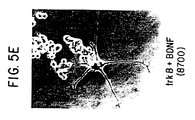
- PC12 cells transfected with trkB do sprout neurites in response to BDNF (see section 6, infra , and Figure 5). Therefore, normal PC12 cells ( trkB -minus cells) and PC12 cells transfected with trkB ( trkB -plus cells) may be exposed to the sample (the test agent) and the presence or absence of neurite sprouting may be evaluated microscopically.
- the amount of BDNF in the sample may be measured by determining the amount of neurite sprouting (or immediate early gene induction) and then comparing this value with a dose response curve for the particular neurotrophin being tested, here, BDNF.
- the present invention provides for a method of identifying an agent that has neurotrophin activity comprising (i) exposing a cell that expresses trkB to a test agent and (ii) detecting the specific binding of the test agent to trkB, in which specific binding to trkB positively correlates with neurotrophin-like activity.
- Specific binding may be detected by either assaying for direct binding or the secondary biological effects of binding, as discussed supra .
- Such a method may be particularly useful in identifying new members of the neurotrophin family or, in the pharmaceutical industry, in screening a large array of peptide and non-peptide agents (e.g., peptidomimetics) for neurotrophin-like activity.
- a large grid of culture wells may be prepared that contain, in alternate rows, PC12 (or fibroblasts, see infra ) cells that are either trkB -minus or engineered to be trkB -plus.
- PC12 or fibroblasts, see infra
- a variety of test agents may then be added such that each column of the grid, or a portion thereof, contains a different test agent.
- Each well could then be scored for the presence or absence of neurite sprouting.
- An extremely large number of test agents could be screened for neurotrophin activity in this manner.
- the invention provides for methods of detecting or measuring neurotrophin activity or identifying an agent as having neurotrophin activity comprising (i) exposing a test agent to a trkB protein in vitro under conditions that permit binding to occur and (ii) detecting binding of the test agent to the trkB protein, in which binding of test agent to trkB correlates with neurotrophin or neurotrophin-like activity.
- the trkB may or may not be substantially purified, may be affixed to a solid support (e.g. as an affinity column or as an ELISA assay), or may be incorporated into an artificial membrane. Binding of test agent to trkB may be evaluated by any method known in the art. In preferred embodiments, the binding of test agent may be detected or measured by evaluating its ability to compete with detectably labeled known trkB ligands for trkB binding.
- the present invention also provides for a method of detecting the ability of a test agent compound to function as an antagonist of neurotrophin activity comprising detecting the ability of the compound to inhibit an effect of neurotrophin binding to trkB on a cell that expresses trkB .
- Such an antagonist may or may not interfere with trkB/neurotrophin binding.
- Effects of neurotrophin binding to trkB are preferably biological or biochemical effects, including, but not limited to, neurite sprouting, cell survival or proliferation, cell transformation, immediate early gene induction, or trkB phosphorylation.
- PC12 cells (or fibroblasts, etc.) transfected with trkB may be exposed to effective amounts of either BDNF or BDNF plus a test agent suspected of being a BDNF antagonist.
- Neurite sprouting in these two groups of cells may be compared to sprouting in non-transfected cells exposed to BDNF, or BDNF plus the test agent, or NGF, or NGF plus the test agent.
- neurite sprouting should be inhibited only in trkB -plus cells treated with BDNF plus test agent compared to trkB -plus cells exposed to BDNF, and there should be little or no inhibition of sprouting of trkB -minus cells treated with NGF plus test agent relative to trkB -minus PC12 cells treated with NGF alone.
- the present invention also provides for assay systems that may be used according to the methods described supra .
- assay systems may comprise in vitro preparations of trkB, e.g. affixed to a solid support, or may, preferably, comprise cells that express trkB protein.
- Cells that express trkB protein may do so naturally or may be genetically engineered to produce trkB, as described supra , by transfection, transduction, electroporation, microinjection, via a transgenic animal, etc. of nucleic acid encoding trkB in a suitable expression vector.
- any of the methods known to one skilled in the art for the insertion of DNA fragments into a vector may be used to construct expression vectors encoding trkB containing a chimeric gene consisting of appropriate transcriptional/translational control signals and the protein coding sequences. These methods may include in vitro recombinant DNA and synthetic techniques and in vivo recombinations (genetic recombination). Expression of nucleic acid sequence encoding trkB protein or peptide fragment may be regulated by a second nucleic acid sequence so that trkB protein or peptide is expressed in a host transformed with the recombinant DNA molecule.
- trkB expression of trkB may be controlled by any promoter/enhancer element known in the art.
- Promoters which may be used to control trkB expression include, but are not limited to the long terminal repeat as described in Squinto et al., (1991, Cell 65 :1-20); the SV40 early promoter region (Bernoist and Chambon, 1981, Nature 290 :304-310), the CMV promoter, the M-MuLV 5' terminal repeat the promoter contained in the 3' long terminal repeat of Rous sarcoma virus (Yamamoto, et al., 1980, Cell 22 :787-797), the herpes thymidine kinase promoter (Wagner et al., 1981, Proc.
- mouse mammary tumor virus control region which is active in testicular, breast, lymphoid and mast cells (Leder et al., 1986, Cell 45 :485-495), albumin gene control region which is active in liver (Pinkert et al., 1987, Genes and Devel. 1 :268-276), alpha-fetoprotein gene control region which is active in liver (Krumlauf et al., 1985, Mol. Cell. Biol. 5 :1639-1648; Hammer et al., 1987, Science 235 :53-58); alpha 1-antitrypsin gene control region which is active in the liver (Kelsey et al, 1987, Genes and Devel.
- beta-globin gene control region which is active in myeloid cells (Mogram et al., 1985, Nature 315 :338-340; Kollias et al., 1986, Cell 46 :89-94); myelin basic protein gene control region which is active in oligodendrocyte cells in the brain (Readhead et al., 1987, Cell 48 :703-712); myosin light chain-2 gene control region which is active in skeletal muscle (Sani, 1985, Nature 314 :283-286), and gonadotropic releasing hormone gene control region which is active in the hypothalamus (Mason et al., 1986, Science 234 :1372-1378).
- Expression vectors containing trkB gene inserts can be identified by three general approaches: (a) DNA-DNA hybridization, (b) presence or absence of "marker" gene functions, and (c) expression of inserted sequences.
- first approach the presence of a foreign gene inserted in an expression vector can be detected by DNA-DNA hybridization using probes comprising sequences that are homologous to an inserted trkB gene.
- second approach the recombinant vector/host system can be identified and selected based upon the presence or absence of certain "marker" gene functions (e.g.
- recombinants containing the trkB insert can be identified by the absence of the marker gene function.
- recombinant expression vectors can be identified by assaying the foreign gene product expressed by the recombinant. Such assays can be based, for example, on the physical or functional properties of the trkB gene product, for example, by binding of the receptor to neurotrophic factor or to an antibody which directly recognizes the trkB.
- Cells of the present invention may transiently or, preferably, constitutively and permanently express trkB .
- the present invention provides for cells that express trkB and that also contain recombinant nucleic acid comprising an immediate early gene promoter (e.g. the fos or jun promoters (Gilman et al., 1986, Mol. Cell. Biol. 6 :4205-4316).
- an immediate early gene promoter e.g. the fos or jun promoters (Gilman et al., 1986, Mol. Cell. Biol. 6 :4205-4316).
- the neurotrophin may be expected to bind to trkB and secondarily induce transcription off the immediate early promoter.
- Such a cell may be used to detect neurotrophin/trkB binding by measuring the transcriptional activity of the immediate early gene promoter, for example, by nuclear run-off analysis, Northern blot analysis, or by measuring levels of a gene controlled by the promoter.
- the immediate early promoter may be used to control the expression of fos or jun or any detectable gene product, including, but not limited to, any of the known reporter genes, such as a gene that confers hygromycin resistance (Murphy and Efstratiadis, 1987, Proc. Natl. Acad. Sci. U.S.A. 84 :8277-8281) chloramphenicol acetyltransferase (CAT), neomycin phosphotransferase (neo), beta-galactosidase beta-glucuronidase, beta-galactosidase, etc.
- CAT chloramphenicol acetyltransferase
- neomycin phosphotransferase neo
- beta-galactosidase beta-glucuronidase beta-galactosidase
- neurotrophin/trkB binding in a cell that expresses trkB and contains the human growth hormone gene under the control of the fos gene promoter may be expected to produce recombinant human growth hormone, as measured by Seldon et al., 1986, Mol. Cell. Biol. 6 :3173-3179.
- trkB expression may also be used as a reporter gene and be placed under the control of an immediate early promoter in addition to constitutively expressed trkB to produce an amplified response to neurotrophin.
- Such trkB-expression reporter gene containing cell lines may provide an exceptionally sensitive and efficient method of detecting or measuring neurotrophin activity.
- the cells used in the assay systems of the invention may or may not be cells of the nervous system.
- growth-factor dependent fibroblasts may be used as the basis for a neurotrophin assay system. See Section 7, infra .
- a fibroblast cell line that is growth factor dependent in serum-free media e.g. as described in Zham and Goldfarb, 1986, Mol. Cell. Biol.
- trkB gene may be transfected with the trkB gene, for instance by using a CaPO 4 transfection protocol with 5 micrograms of DNA of CMV-promoter-based expression vector comprising the rat trkB gene and one microgram of hygromycin-resistance gene-containing expression vector. After about 48 hours, the cells may then be selected for hygromycin resistance to identify positive transfectants. The cells may then be cultured for about three weeks in the presence of hygromycin,and then resistant colonies may be pooled.
- These cells may then be plated on tissue culture plates coated with poly-D-lysine and human fibronectin, and allowed to grow in DMEM plus 10% bovine calf serum for about four hours to allow the cells to bind to the plates.
- the serum-containing media may then be aspirated and the cells may be washed about three times with PBS to remove any residual serum.
- the cells may then be taken up with either serum free defined media (A 3:1 mixture of DMEM and Hams F12, supplemented with 8 mM sodium bicarbonate, 15 mM HEPES, 4 x 10 -6 M MnCl 2 , 3 mM histidine, 10 -5 M ethanolamine, 10 -7 M sodium selenite, 5 mg transferrin per liter, 200 mg bovine serum albumin-linoleic acid complex per liter gentamicin, penicillin, and streptomycin, 20 mM L-glutamine).
- serum free defined media A 3:1 mixture of DMEM and Hams F12, supplemented with 8 mM sodium bicarbonate, 15 mM HEPES, 4 x 10 -6 M MnCl 2 , 3 mM histidine, 10 -5 M ethanolamine, 10 -7 M sodium selenite, 5 mg transferrin per liter, 200 mg bovine serum albumin-linoleic acid complex per liter gentamicin, pen
- 100 ng/ml NT-3 or BDNF may, after about 5 days in culture (replacing media and growth factors every 48 hours), be expected to be growing and proliferating; cells treated with NGF at 100 ng/ml or in serum free-medium should not, however, proliferate (see also Figure 7).
- NGF neurotrophic factor
- the present invention also provides for assay systems and methods utilizing the non-trkB receptor in a manner analogous to those utilizing the trkB receptor, as described herein.
- the present invention also provides for experimental model systems for studying the physiological role of the neurotrophin gene family.
- trkB protein, peptide fragment, or a derivative thereof may be either supplied to the system or produced within the system.
- Such model systems could be used to study the effects of neurotrophin excess or neurotrophin depletion.
- the experimental model systems may be used to study the effects of increased or decreased response to neurotrophin in cell or tissue cultures, in whole animals, in particular cells or tissues within whole animals or tissue culture systems, or over specified time intervals (including during embryogenesis) in embodiments in which trkB expression is controlled by an inducible or developmentally regulated promoter.
- the CMV promoter may be used to control expression of trkB in transgenic animals.
- transgenic animals refers to non-human transgenic animals, including transgenic mosaics, which carry a transgene in some or all of their cells, which include any non-human species, and which are produced by any method known in the art, including, but not limited to microinjection, cell fusion, transfection, electroporation, etc.
- the animals may be produced by a microinjection of zygotes by a method such as that set forth in "Brinster et al., 1989, Proc. Natl. Acad. Sci. U.S.A. 82 :4438-4442.
- the present invention also provides for model systems for autoimmune disease in which an autoimmune response is directed toward trkB.
- models comprise animals which have been immunized with immunogenic amounts of trkB and preferably found to produce anti-trkB antibodies and/or cell-mediated immunity.
- an immune adjuvant such as Bacille Calmette Guerin (BCG).
- an experimental model system may be created which may be used to study the effects of excess neurotrophin activity.
- the response to neurotrophin may be increased by engineering an increased number of trkB molecules on cells of the model system relative to cells which have not been so engineered. It may be preferable to provide an increased number of neurotrophins selectively on cells which normally express neurotrophins.
- Cells may be engineered to produce increased amounts of trkB protein by infection with a virus which carries a trkB gene of the invention.
- the trkB gene may be provided to the cells by transfection.
- a recombinant trkB gene may be introduced into the cells of the animal by infection with a virus which carries the trkB gene.
- a transgenic animal may be created which carries the trkB gene as a transgene.
- the trkB gene should be placed under the control of a suitable promoter sequence. It may be desirable to put the trkB gene under the control of a constitutive and/or tissue specific promoter, including but not limited to the CNS neuron specific enolase, neurofilament, and tyrosine hydroxylase promoter, an inducible promoter, such as the metallothionein promoter, the UV activated promoter in the human immunodeficiency virus long terminal repeat (Valeri et al., 1988, Nature 332 :78-81), or the CMV promoter (as contained in pCMX, infra ) or a developmentally regulated promoter.
- a constitutive and/or tissue specific promoter including but not limited to the CNS neuron specific enolase, neurofilament, and tyrosine hydroxylase promoter, an inducible promoter, such as the metallothionein promoter, the UV activated promoter in the human immunodeficiency virus long
- neurotrophin By increasing the number of cellular trkB molecules, the response to endogenous neurotrophin may be increased. If the model system contains little or no neurotrophin, neurotrophin may be added to the system. It may also be desirable to add additional neurotrophin to the model system in order to evaluate the effects of excess neurotrophin activity. Over expressing neurotrophin (or secreted neurotrophin) may be the preferable method for studying the effects of elevated levels of neurotrophin on cells already expressing trkB . More preferably would be to express trkB in all cells (general expression) and determine which cells are then endowed with functional responsiveness to neurotrophin, thus allowing the potential identification of a second receptor component, if one exists.
- an experimental model system may be created which may be used to study the effects of diminished neurotrophin activity.
- This system may permit identification of processes or neurons which require neurotrophin, and which may represent potential therapeutic targets.
- the response to neurotrophin may be decreased by providing recombinant trkB proteins which are not associated with a cell surface or which are engineered so as to be ineffective in transducing a response to neurotrophin.
- trkB protein, peptide, or derivative may be supplied to the system such that the supplied receptor may compete with endogenous trkB for neurotrophin binding, thereby diminishing the response to neurotrophin.
- the trkB may be a cell free receptor which is either added to the system or produced by the system.
- a trkB protein which lacks the transmembrane domain may be produced by cells within the system, such as an anchorless trkB that may be secreted from the producing cell.
- trkB protein, peptide or derivative may be added to an extracellular space within the system.
- a recombinant trkB gene may be used to inactivate or "knock out" the endogenous gene by homologous recombination, and thereby create a trkB deficient cell, tissue, or animal.
- a recombinant trkB gene may be engineered to contain an insertional mutation, for example the neo gene, which inactivates trkB.
- Such a construct under the control of a suitable promoter, may be introduced into a cell, such as an embryonic stem cell, by a technique such as transfection, transduction, injection, etc. Cells containing the construct may then be selected by G418 resistance.
- Cells which lack an intact trkB gene may then be identified, e.g. by Southern blotting or Northern blotting or assay of expression. Cells lacking an intact trkB gene may then be fused to early embryo cells to generate transgenic animals deficient in trkB. A comparison of such an animal with an animal not expressing endogenous neurotrophin would reveal that either the two phenotypes match completely or that they do not, implying the presence of additional neurotrophin-like factors or receptors.
- Such an animal may be used to define specific neuronal populations, or any other in vivo processes, normally dependent upon neurotrophin. Thus, these populations or processes may be expected to be affected if the animal did not express trkB and therefore could not respond to neurotrophin.
- a recombinant trkB protein, peptide, or derivative which competes with endogenous receptor for neurotrophin may be expressed on the surface of cells within the system, but may be engineered so as to fail to transduce a response to neurotrophin binding.
- the recombinant trkB proteins, peptides or derivatives described above may bind to neurotrophin with an affinity that is similar to or different from the affinity of endogenous trkB to neurotrophin.
- the trkB protein, peptide, or derivative may desirably bind to neurotrophin with a greater affinity than that exhibited by the native receptor.
- nucleic acid encoding the trkB protein, peptide, or derivative may be supplied to the system by infection, transduction, transfection, etc. or as a transgene.
- the trkB gene may be placed under the control of a suitable promoter, which may be, for example, a tissue-specific promoter or an inducible promoter or developmentally regulated promoter.
- the endogenous trkB gene of a cell may be replaced by a mutant trkB gene by homologous recombination.
- a test animal may be immunized against trkB.
- trkB expression may be reduced by providing trkB expressing cells with an amount of trkB anti-sense RNA or DNA effective to reduce expression of trkB protein.
- trkB probes may be used to identify cells and tissues which are responsive to neurotrophin in normal or diseased states.
- the present invention provides for a method of diagnosing a neurological disorder in a patient comprising comparing the levels of expression of trkB in a patient sample with the levels of expression of trkB in a comparable sample from a healthy person, in which a difference in the levels of expression of trkB in the patient compared to the healthy person indicates that a disorder in the patient may be primarily or secondarily related to trkB metabolism.
- a patient sample may be any cell, tissue, or body fluid but is preferably nervous system tissue or cerebral spinal fluid.
- the present invention provides for methods for identifying cells which are responsive to neurotrophin comprising detecting trkB expression in such cells.
- TrkB expression may be evidenced by transcription of trkB mRNA or production of trkB protein.
- TrkB expression may be detected using probes which identify trkB nucleic acid or protein.
- probe which may be used is anti-trkB antibody or fragments thereof containing the binding domain.
- trkB protein may be used as an immunogen to generate anti-trkB antibodies.
- trkB protein or fragments or derivatives thereof, may be used as an immunogen to generate anti-trkB antibodies.
- the amino acid sequence of trkB may be analyzed in order to identify portions of the molecule which may be associated with increased immunogenicity.
- the amino acid sequence may be subjected to computer analysis to identify surface epitopes which present computer-generated plots of hydrophilicity, surface probability, flexibility, antigenic index, amphiphilic helix, amphiphilic sheet, and secondary structure of trkB.
- the deduced amino acid sequences of trkB from different species could be compared, and relatively non-homologous regions identified; these non-homologous regions would be more likely to be immunogenic across various species.
- any technique which provides for the production of antibody molecules by continuous cell lines in culture may be used.
- the hybridoma technique originally developed by Kohler and Milstein (1975, Nature 256 :495-497), as well as the trioma technique, the human B-cell hybridoma technique (Kozbor et al., 1983, Immunology Today 4 :72), and the EBV-hybridoma technique to produce human monoclonal antibodies Colde et al., 1985, in "Monoclonal Antibodies and Cancer Therapy," Alan R. Liss, Inc. pp. 77-96) and the like are within the scope of the present invention.
- the monoclonal antibodies for therapeutic use may be human monoclonal antibodies or chimeric human-mouse (or other species) monoclonal antibodies.
- Human monoclonal antibodies may be made by any of numerous techniques known in the art (e.g. , Teng et al., 1983, Proc. Natl. Acad. Sci. U.S.A. 80:7308-7312; Kozbor et al., 1983, Immunology Today 4:72-79; Olsson et al., 1982, Meth. Enzymol. 92:3-16).
- Chimeric antibody molecules may be prepared containing a mouse antigen-binding domain with human constant regions (Morrison et al., 1984, Proc. Natl. Acad. Sci. U.S.A. 81:6851, Takeda et al., 1985, Nature 314:452).
- trkB polyclonal antibodies to epitopes of trkB.
- various host animals can be immunized by injection with trkB protein, or a fragment or derivative thereof, including but not limited to rabbits, mice, rats, etc.
- adjuvants may be used to increase the immunological response, depending on the host species, and including but not limited to Freund's (complete and incomplete), mineral gels such as aluminum hydroxide, surface active substances such as lysolecithin, pluronic polyols, polyanions, peptides, oil emulsions, keyhole limpet hemocyanins, dinitrophenol, and potentially useful human adjuvants such as BCG (Bacille Calmette-Guerin) and Corynebacterium parvum .
- BCG Bacille Calmette-Guerin
- a molecular clone of an antibody to a trkB epitope can be prepared by known techniques. Recombinant DNA methodology (see e.g., Maniatis et al., 1982, Molecular Cloning, A Laboratory Manual, Cold Spring Harbor Laboratory, Cold Spring Harbor, New York) may be used to construct nucleic acid sequences which encode a monoclonal antibody molecule, or antigen binding region thereof.
- Antibody molecules may be purified by known techniques, e.g. , immunoabsorption or immunoaffinity chromatography, chromatographic methods such as HPLC (high performance liquid chromatography), or a combination thereof, etc.
- the present invention provides for antibody molecules as well as fragments of such antibody molecules.
- Antibody fragments which contain the idiotype of the molecule can be generated by known techniques.
- such fragments include but are not limited to: the F(ab') 2 fragment which can be produced by pepsin digestion of the antibody molecule; the Fab' fragments which can be generated by reducing the disulfide bridges of the F(ab') 2 fragment, and the Fab fragments which can be generated by treating the antibody molecule with papain and a reducing agent.
- the abovementioned probes may be used experimentally to identify cells or tissues which hitherto had not been shown to express trkB. Furthermore, these methods may be used to identify the expression of trkB by aberrant tissues, such as malignancies. In additional embodiments, these methods may be used diagnostically to compare the expression of trkB in cells, fluids, or tissue from a patient suffering from a disorder with comparable cells, fluid, or tissue from a healthy person. Fluid is construed to refer to any body fluid, but particularly blood or cerebrospinal fluid. A difference in the levels of expression of trkB in the patient compared to a healthy person may indicate that the patient's disorder may be primarily or secondarily related to trkB metabolism.
- trkB neurotrophin levels
- trkB neurotrophin levels
- RNA level i.e. by measuring amounts of trkB protein or trkB RNA in a patient relative to those amounts in healthy persons.
- probes may also be used to select neurotrophin-responsive cells for use in assay systems, as described above, or in U. S. Application Serial No. 07/532,285 filed June 1, 1990 (incorporated by reference herein, or according to standard methods of cell selection or cell sorting.
- the present invention also provides for methods of treating a patient suffering from a neurological disorder comprising treating the patient with an effective amount of trkB protein, peptide fragment, or derivative thereof capable of binding to a neurotrophin.
- Therapeutic methods comprising administering trkB, trkB agonists, trkB antagonists (which compete with endogenous neurotrophin), or anti-trkB antibodies are within the scope of the present invention.
- the present invention also provides for pharmaceutical compositions comprising trkB protein, peptide fragment, or derivative in a suitable pharmacologic carrier.
- the trkB protein, peptide fragment, or derivative may be administered systemically or locally. Any appropriate mode of administration known in the art may be used, including, but not limited to, intravenous, intrathecal, intraarterial, intranasal, oral, subcutaneous, intraperitoneal, or by local injection or surgical implant. Sustained release formulations are also provided for.
- neurotrophin antagonists including, but not limited to, soluble forms of trkB which may compete with endogenous cellular receptor for neurotrophin binding. Under such circumstances, it may be desirable to provide neurotrophin antagonist locally at the injury site rather than systemically. Use of a trkB providing implant may be desirable.
- certain conditions may benefit from an increase in neurotrophin responsiveness. It may therefore be beneficial to increase the number or binding affinity of trkBs in patients suffering from such conditions. This could be achieved through gene therapy. Selective expression of recombinant trkB in appropriate cells could be achieved using trkB genes controlled by tissue specific or inducible promoters or by producing localized infection with replication defective viruses carrying a recombinant trkB gene.
- Conditions which may benefit from increased sensitivity to neurotrophin include particularly but are not limited to motorneuron disorders including amyotrophic lateral sclerosis, Werdnig-Hoffmann disease, chronic proximal spinal muscular atrophy, and Guillain-Barre syndrome. Such treatment may also be used for treatment of neurological disorders associated with diabetes, Parkinson's disease, Alzheimer's disease, and Huntington's chorea.
- the present invention also provides for systems that may be generally used in both the assay of pre-defined agents, as well as the discovery of novel agents, that act on receptor tyrosine kinases.
- the same system can be used to discover unknown receptors that mediate responses to known factors.
- the present invention reveals that a receptor tyrosine kinase, when introduced into cells that do not normally express this receptor, allows these cells to exhibit profound and easily distinguishable responses to a ligand which binds this receptor.
- the present invention reveals that the type of response elicited depends on the cell utilized, and not the specific receptor introduced into the cell.
- the trkB receptor in PC12 pheochromocytoma cells results in BDNF/NT-3 dependent differentiation, whereas the same receptor in fibroblasts mediates both survival and proliferation in response to either BDNF or NT-3.
- Agents refers to any molecule(s), including but not limited to peptide and non-peptide molecules, that will act in systems to be described in a receptor specific manner.
- One of the more useful systems to be exploited involves the introduction of the desired receptor (e.g.
- a fibroblast cell line e.g., the particular clone of NIH3T3 cells to be described below, section 7
- a fibroblast growth factor e.g. thymidine incorporation or other types of proliferation assays; see van Zoelen, 1990, "The Use of Biological Assays For Detection Of Polypeptide Growth Factors” in Progress Factor Research, Vol. 2, pp. 131-152; Zhan and M. Goldfarb, 1986, Mol. Cell. Biol., Vol. 6, pp.
- Such systems are not limited to the assay of known ligands for known receptors, but can also be utilized to identify novel agents that might act on these or (or any other) receptors.
- both the cell line bearing the introduced receptor as well as the parental cell line without the receptor can be exposed to any potential source of an agent that might work through the receptor; any specific effects (e.g. on cell survival or proliferation) on the cell line bearing the receptor can be used to identify sources of agents acting on that receptor, and to eventually purify such an agent.
- Receptors also need not be limited to those for which a known ligand exists. In fact, this system may allow for the identification of ligands for "orphan" receptors so named because they have no known ligand.
- fibroblasts expressing trkB could have been used in such systems in order to identify and eventually purify the ligands (e.g. BDNF and NT-3) that normally activate trkB; they can now be used to identify additional peptide ligands or other agents (e.g. non-peptide molecules) that could act on these receptors.
- Sources for "agents” could include extracts from a variety of tissues and organisms, or supernatants from cells transfected with genomic DNA or cDNA expression libraries.
- fibroblasts expressing an introduced receptor for which a ligand is desired could be transfected with cDNA expression libraries derived from a potential source of such a ligand; cells which survive and form colonies in defined media lacking fibroblast growth factors (Zhan and Goldfarb, 1986, Mol. Cell. Biol., Vol. 6, pp. 3541-3544) would presumably now be making a growth factor that overcomes their normal requirements via an autocrine loop.
- the present invention provides for methods for cloning at least a portion of tyrosine kinase receptor gene which may then be used in the identification of ligand/receptor pairs, as set forth supra .
- Such methods comprise (i) amplifying tyrosine kinase encoding nucleic acid sequences by polymerase chain reaction using a collection of cDNA molecules (such as a cDNA library) as template and using oligonucleotide primers that correspond to regions of known tyrosine kinase molecules, said regions being associated with tyrosine kinase activity; and (ii) cloning the amplified nucleic acid into an appropriate vector molecule, such as a plasmid, bacteriophage, etc.
- an appropriate vector molecule such as a plasmid, bacteriophage, etc.
- the present invention also provides for tyrosine kinase genes, and portions thereof, cloned by this method.
- the oligonucleotide primers utilized may be as set forth in Figure 12A or, alternatively, Table II, infra .
- DNA amplified and cloned by this method may then be sequenced using standard techniques.
- the resulting sequences may then be compared to the sequences of known tyrosine kinase molecules in order to identify clones of particular interest.
- the present invention further provides for the cloned nucleic acid sequences identified using this method, as set forth in Sections 9 and 10, infra , and for peptides and proteins encoded by cDNAs comprising these sequences.
- the present invention provides for substantially purified recombinant nucleic acid molecules comprising the nucleic acid sequences (i) substantially as set forth in Figure 12C for Rtk-1, Rtk-6, Rtk-7, Rtk-8 and Rtk-9; (ii) substantially as set forth in Figure 13B for Rtk-4 and Rtk-5; (iii) substantially as set forth in Figure 14 for Rtk-2; and (iv) substantially as set forth in Figure 15 for Rtk-3; or portions thereof comprising at least about ten nucleic acid residues.
- the present invention also provides for nucleic acids as contained in pBluescript SK-containing Rtk-2 and pBluescript SK-containing Rtk-3 deposited with the American Type Culture Collection and granted accession numbers and .
- the present invention further provides for substantially purified protein molecules comprising the amino acid sequences (i) substantially as set forth in Figure 12C for Rtk-1, Rtk-6, Rtk-7, Rtk-8 and Rtk-9; (ii) substantially as set forth in Figure 13B for Rtk-4 and Rtk-5; (iii) substantially as set forth in Figure 14 for Rtk-2; and (iv) substantially as set forth in Figure 15 for Rtk-3; or portions thereof comprising at least about six amino acid molecules, or functionally equivalent molecules.
- Functionally equivalent molecules include those in which amino acid residues are substituted for residues within the sequence resulting in a silent change.
- one or more amino acid residues within the sequence can be substituted by another amino acid of a similar polarity which acts as a functional equivalent, resulting in a silent alteration.
- Substitutes for an amino acid within the sequence may be selected from other members of the class to which the amino acid belongs.
- the nonpolar (hydrophobic) amino acids include alanine, leucine, isoleucine, valine, proline, phenylalanine, tryptophan and methionine.
- the polar neutral amino acids include glycine, serine, threonine, cysteine, tyrosine, asparagine, and glutamine.
- the positively charged (basic) amino acids include arginine, lysine and histidine.
- the negatively charged (acidic) amino acids include aspartic acid and glutamic acid.
- proteins or fragments or derivatives thereof which are differentially modified during or after translation, e.g. , by glycosylation, proteolytic cleavage, linkage to an antibody molecule or other cellular ligand, etc.
- the present invention further provides for cells and microorganisms that carry the recombinant nucleic acid molecules described above, including, but not limited to, Rtk-1, Rtk-6, Rtk-7, Rtk-8, Rtk-9, Rtk-2, Rtk-3, Rtk-4 and Rtk-5.
- the cell carrying the recombinant nucleic acid is a fibroblast.
- the cell is a bacterium.
- Amplified nucleic acid fragments may then be used to identify full-length cDNA clones.
- an amplified DNA fragment of interest is used to identify a tissue or cell line that expresses relatively abundant levels of a corresponding mRNA, for example, using Northern blot or dot-blot analysis.
- tissue or cell line may then be used to generate a cDNA library which may serve as a superior source of a full-length cDNA which comprises the sequence and the amplified fragment.
- a reciprocal approach could be used to molecularly clone a receptor for an "orphan" factor (for example, a neurotrophic protein for which no receptor has been isolated). Fibroblasts exposed to this factor normally would not respond, but if transfected with a cDNA expression library prepared from cells thought to be expressing this receptor, occasional transfectants would arise which now express this receptor and should now respond to this factor in an autocrine fashion. Powerful selection mechanisms, such as the ability to form colonies in defined media in the presence of the "orphan" factor, should identify transfectants that express the receptor of interest; the gene encoding this receptor could then be isolated by traditional means.
- an "orphan” factor for example, a neurotrophic protein for which no receptor has been isolated.
- COS-M5 cells were cultured in Dulbecco's modified Eagle's medium (DMEM) containing 10% fetal bovine serum (FBS), 1% each of penicillin and streptomycin (P/S) and 2 mM glutamine in an atmosphere of 5% CO 2 .
- PC12 cells were cultured in DMEM with 6% FBS and 6% horse serum, (P/S) and 2mM glutamine on Costar tissue culture plates in an atmosphere of 7.5% CO 2 .
- PC12 cells obtained from Dr. L.A. Greene's laboratory were utilized in experiments depicted in Figure 2
- PC12 cells obtained from Dr. E.M. Shooter's laboratory were utilized in experiments depicted in Figure 5.
- the human neuroblastoma cell line, SH-SY5Y (obtained from June Biedler, Sloan-Kettering) was cultured in Eagle's minimal essential medium (EMEM) with 10% FBS, (P/S) and 2 mM glutamine.
- EMEM Eagle's minimal essential medium
- Murine 2.5S NGF was obtained from Bioproducts for Science (Indianapolis, IN). Both human BDNF and NT-3 were produced in CHO cells and purified from CHO cell conditioned media to homogeneity as assessed by silver-stained polyacrylamide gels and amino acid sequence analysis. Purified neurotrophins (NGF, BDNF, and NT-3) were all iodinated using the lactoperoxidase method as described in Hempstead et al., 1989, Science, 243 : 373-375. Iodinated neurotrophins were separated from unincorporated 125 I by using a Centriflo CF50A filters (Amicon, Beverly, MA). Aggregates were removed using gel filtration (S200) column chromatography.
- rat trkB cDNA clone was obtained by screening a rat brain cDNA library in the lambda ZAP2 vector (Stratagene) with rat trkB -specific oligonucleotides corresponding to the most 5' and 3' coding regions of trkB . Both the human LNGFR (Johnson et al., 1986, Cell 47 : 545-554) and rat trkB cDNAs were subcloned into the mammalian expression vector, pCMX, to generate pCMX-LNGFR or pCMX- trkB respectively.
- pCMX mammalian expression vector
- pCMX- trkB (del) was generated by digesting the pCMX-trkB plasmid with Apal (which cuts just after the trkB transmembrane domain) and Not1 (which cuts just after the trkB coding region in vector sequences), blunting these ends, and religating the plasmid; the trkB coding region generated includes all of the extracellular and transmembrane domains of trkB , but is lacking the C-terminal 320 amino acids.
- COS-M5 cells were transiently transfected with either the pCMX-LNGFR, pCMX- trkB , or control vector (pCMX) by the DEAE-dextran transfection protocol. Briefly, COS-M5 cells were plated at a density of 1.5 x 10 6 cells per 100 mm plate 24 hours prior to transfection.
- the cells were cultured in serum-free DMEM containing 400 ⁇ g/ml of DEAE-dextran, 1 ⁇ M chloroquin, 2 mM glutamine, 20 ⁇ g/ml insulin, 5 ⁇ g/ml transferrin, 33 nM sodium selenite, and 5 ⁇ g of the appropriate DNA for 3 hours and 15 minutes at 37°C in an atmosphere of 5% CO2.
- the transfection media was aspirated and replaced with phosphate-buffered saline with 10% DMSO for 2 min. Following this DMSO "shock", the COS-M5 cells were placed into DMEM with 10% FBS, 1% each of penicillin and streptomycin, and 2 mM glutamine for 48 hours.
- PC12 cells were transiently transfected by electroporation. Briefly, the cells were rinsed prior to transfection in ice-cold Dulbecco's phosphate-buffered saline (calcium and magnesium-free) containing 2 mg/ml glucose and then resuspended in the same buffer at a density of 1.5 x 10 7 cells per ml containing 40 ⁇ g of the appropriate DNA.
- PC12 cells were transfected with either pCMX, pCMX- trkB or the pT24-ras plasmid (Yancopoulos et al., 1985, Proc. Natl. Acad. Sci. USA., 82 : 5455-5459).
- the cell mixture was incubated on ice for 10 min. and then quickly brought to room temperature and electroporated in a total volume of 1 ml at 1150 V/cm and 500 uF. Electroporated cells were incubated on ice for 30 minutes prior to plating in DMEM with 6% FBS and 6% horse serum with 1% each of penicillin and streptomycin and 2mM glutamine; cells were plated on Costar plastic in the absence of any pre-coating. 48 hours after transfection, the cells were treated with 100 ng/ml of neurotrophin or BSA (see Legend of Figure 5) and neurite outgrowth was scored 48 hours later.
- Cells were harvested in phosphate-buffered saline containing 1 mM EDTA, 1 mg/ml glucose and 25 mM HEPES (PBS-versene) and resuspended at an appropriate density (generally 1 x 10 6 cells per ml) in ice-cold binding buffer A (PBS containing 1 mg/ml each of BSA and glucose).
- PBS phosphate-buffered saline containing 1 mM EDTA, 1 mg/ml glucose and 25 mM HEPES (PBS-versene) and resuspended at an appropriate density (generally 1 x 10 6 cells per ml) in ice-cold binding buffer A (PBS containing 1 mg/ml each of BSA and glucose).
- pCMX- trkB or pCMX vector transfected COS-M5 cells (4 x 10 5 cells) were incubated on ice with 125 I-labeled neurotrophins (final concentration estimated to be between 0.1 and 0.25 nM) for 90 minutes in the absence or presence of unlabeled NGF, BDNF or NT-3 (see Figures 1 and 3).
- the chemical cross-linker DSS (Pierce, Rockford, IL) was used following conditions described in Meakin and Shooter, 1991, Neuron 6 : 153-163. The cross-linking reaction was terminated after 90 minutes and quenched with 12 ml of 50 mM Tris buffer containing 160 mM NaCl.
- Binding of 125 I-NT-3 to COS-M5 cells transfected with either pCMX-LNGFR, pCMX- trkB or control vector (pCMX) was assessed on cells in suspension.
- Cells were harvested in PBS-versene and then resuspended in binding buffer A as described above for chemical cross-linking.
- Cells were incubated with 125 I-NT-3 (estimated between 0.1 and 0.25 nM) in the absence or presence of increasing concentrations of unlabeled NT-3, BDNF, or NGF ranging from 0.3 to 30 nM for NGF and between 1 and 100 nM for BDNF and NT-3 (see Figure 4C and D).
- the binding reactions were carried out for 90 minutes on ice.
- Free 125 I was separated from bound 125 I by quickly centrifuging (30 second spin) the reaction mixture through a sucrose gradient formed in a long-tip microcentrifuge tube. The tubes were immediately frozen in a dry-ice/ethanol bath. The bottom of the reaction tube was cut and then counted in a gamma counter.
- Total cellular RNA isolated from SH-SY5Y and treated or untreated PC12 cells was fractionated on 1% formaldehyde agarose gels, transferred to nylon membranes and hybridized to a 32 P-labeled v-fos probe or a 32 P-labeled c-jun probe as previously described (Squinto et al., 1990, Neuron 5 , 757-766); probings for trkB expression were performed using a 32 P-labeled rat trkB probe spanning a region encoding the intra-cytoplasmic tyrosine kinase domain.
- NT-3 like NGF and BDNF (Rodriguez-Tebar, 1990, Neuron 4 : 487-492), could also bind to the LNGFR
- BDNF Rodriguez-Tebar, 1990, Neuron 4 : 487-492
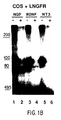
- Each of the three radiolabeled neurotrophins could be specifically cross-linked to a species of the molecular weight expected for the LNGFR ( Figure 1B cross-linked complex reported to be approximately 100 kb by Hosang and Shooter, J.Biol. Chem. 260 : 655-662).
- the cross-linked product was not observed on COS cells that were not expressing the LNGFR protein ( Figure 1A).
- the PC12 cell line which expresses both classes of NGF receptor, displays prominent responses including both neurite outgrowth and the transcriptional induction of a set of so-called "immediate early genes" in response to NGF (Greene and Tischler, 1976, Proc. Natl. Acad. Sci. U.S.A., 73 : 2424-2428; Greenberg et al., 1985, J.Biol. Chem. 260 : 14101-14110, see Figure 2A, B).
- TrkB is not expressed in PC12 cells (Kaplan et al., 1991, Nature 350 : 158-160); cell lines expressing trkB have not been described.
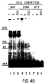
- a full-length trkB cDNA was isolated and transiently expressed in COS cells, on which cross-linking experiments were carried out with radioiodinated neurotrophins.
- both BDNF and NT-3, but not NGF could be cross-linked to a polypeptide of approximately the expected size for the trkB gene product; some heterogeneity in the size of this cross-linked species was observed, as has been previously reported for the HNGFR cross-linked to NGF (Meakin and Shooter, 1991, Neuron 6 : 153-163).
- radiolabeled ligand in this case NT-3 was cross-linked efficiently to a surface component of cells expressing the truncated trkB product; furthermore, the marked shift in mobility (corresponding to about 35 kD) observed between the cross-linked species obtained with full-length or truncated trkB proteins agreed well with the known size of the deletion.
- PC12 cells display a characteristic morphological response, neurite extension, indicative of differentiation to a more mature neuronal phenotype when exposed to NGF. As demonstrated above, these cells do not respond to either BDNF or NT-3.
- PC12 cells were transiently transfected with a trkB expression vector (pCMX-trkB) and incubated with each of the neurotrophins. In order to minimize background, the transfected cells were cultured on standard tissue culture plastic rather than either collagen-coated or Primaria surfaces; under these conditions which are suboptimal for neurite extension (Greene et al., 1987, Meth.
- the PC12 cells were transfected with an activated H-ras gene, which has been shown to induce ligand-independent differentiation of PC12 cells.
- the number of differentiated cells seen in the ras -transfected cultures indicates the number of transiently transfected PC12 cells in the cultures.
- trkB encodes an essential component of a functional receptor for BDNF and NT-3, but not for the third neurotrophin family member, NGF.
- trkA proto-oncogene the closest known relative of trkB
- similarly encodes an essential component for a high affinity receptor which binds NGF Kaplan et al., 1991, Nature, 350 : 158-160; Klein et al., 1991, EMBO J. 8 : 3701-3709.
- Our observations that normal PC12 cells do not respond to BDNF or NT-3 imply that trkA, which is expressed in PC12 cells, is uniquely activated by only one known member of the neurotrophin family, NGF.
- the LNGFR modulates the binding of each of the neurotrophins to its appropriate trk receptor.
- the LNGFR may mediate signal transduction via an independent pathway, or it may not be directly involved in initiating signal transduction. For example it may act to localize, concentrate or trap the neurotrophins on the surface of LNGFR-expressing cells.
- neuronal supporting cells such as Schwann cells
- that do not respond to NGF express the LNGFR and up-regulate it in response to injury (Johnson et al., 1988, TINS 11 : 299-304), perhaps providing a fixed matrix or path for the concentration and presentation of neurotrophins to regenerating neurons.
- the LNGFR may act as a "clearance" receptor that reduces free or circulating levels of the neurotrophins; the LNGFR is widely distributed both in the CNS and in the periphery (Maisonpierre et al., 1990, Neuron 5 : 501-509), and secreted forms of the LNGFR (DiStefano and Johnson, 1988, Proc. Natl. Acad., Sci. USA, 85 : 270-274) may aid in such clearance mechanisms In regard to non-signalling roles for the LNGFR, related mechanisms specific for BDNF and NT-3 can be proposed based on the presence of truncated forms of trkB.
- BDNF and NT-3 share a functional receptor, trkB
- trkB a functional receptor
- the expression of trkB remains relatively constant.
- the identification of a cell line which responds to BDNF but not NT-3 and does not detectably express trkB strongly suggests that these two neurotrophins are not entirely interchangeable, and that there may exist additional receptors or modulatory components, which allow for distinct responses to either BDNF or NT-3.
- Such modulation may explain, for example, the differing effects of NT-3 and BDNF on PC12 cells transfected with trkB (see above) or on sympathetic neurons (Maisonpeirre at al., 1990, Science, 247 : 1446-1451). Sympathetic neurons are known to express trkB , although the available in situ hybridization data do not distinguish between the presence of functional or non-functional trkB transcripts in these neurons (Klein at al., 1989, EMBO J. 8 : 3701-3709). An evolutionary comparison of BDNF and NT-3 lead us to predict that these two neurotrophins were strictly conserved to maintain their specific interactions with multiple receptors.
- NGF neurotrophin
- receptor tyrosine kinases The binding and activation of receptor tyrosine kinases by the neurotrophins reveals that these factors utilize signalling pathways very similar to those activated by mitogenic growth factors. This finding is consistent with recent data that neurotrophic factors can act as mitogens in certain contexts (e.g. Cattaneo and McKay, 1990, Nature, 347 : 762-765), but also indicates that signals which initiate the activation of receptor tyrosine kinases normally integrate into non-mitogenic transduction pathways in neurons. Despite differences in ultimate sequelae (i.e. mitogenesis vs.
- tyrosine kinase signalling cascades such as the ERK family of protein kinases
- ERK protein kinases
- Other examples in which activation of receptor-like tryosine kinases in neuronal cells leads to non-mitogenic sequelae include the Drosophila sevenless protein, where activation via a non-diffusable ligand is required for the differentiation of photoreceptor cells (Basler et al., 1991, Cell 64 : 1069-1081).
- NIH3T3 cells were cultured in Dulbecco's modified Eagle's medium (DMEM) containing 10% bovine calf serum, 1% each of penicillin and streptomycin (P/S) and 2 mM glutamine in an atmosphere of 5% CO 2 .
- DMEM Dulbecco's modified Eagle's medium
- P/S penicillin and streptomycin
- 2 mM glutamine 2 mM glutamine in an atmosphere of 5% CO 2 .
- defined media assays defined media was prepared as described in Zhan et al., (1987, Oncogene 1 :369-376). While we have demonstrated that our NIH3T3 cells lose viability in growth factor-deficient media (Zhan et al., Oncogen 1 :369-376; Zhan and Goldfarb, 1986, Mol. Cell. Biol.
- survival and proliferation assays were essentially performed as described in Zhan et al. (1987, Oncogene 1 :369-376; Zhan and Goldfarb, 1986, Mol. Cell. Biol. 6 :3541-3544). In brief, survival assays were performed by plating cells at 20% confluency on poly-D-lysine/fibronectin coated dishes and culturing in defined media (+/- factors) for 5 days; the degree of survival was scored microscopically and ranked from "-", for almost no survival, to "+++", for confluent survival.
- cells were plated at approximately 0.5% confluency on poly-D-lysine/fibronectin coated dishes, cultured for 8 days with or without added factors, and counted in a Coulter counter after trypsinization.
- DNA synthesis assays cells were plated in medium containing 3% calf serum, and after achieving quiescence were challenged with indicated factors for 14 hours; the media was then supplemented with 3 H-thymidine, at 1 microCurie per milliliter, for two hours. Radiolabel incorporation into TCA-insolulble material was subsequently determined as described in Cavalieri and Goldfarb (1987, Mol. Cell. Biol. 7 :3544-3560).
- RNA isolation and Northern blotting analysis was conducted as described in Maisonpierre et al. (1990, Neuron 5 :501-509).
- the lysates were precipitated with agarose-conjugated anti-phosphotyrosine monoclonal antibody, designation 4G10 (FOXNY Sp2 derivative myeloma x BALB/c spleen cells), obtained from Upstate Biotechnologies, Inc.; the immunoprecipitates were washed in lysis buffer, suspended by boiling in SDS-containing loading buffer, and run on 6% SDS-acrylamide gels and immunoblotted using the 4G10 anti-phosphotyrosine antibody.
- the total cell lysates were directly electrophoresed on 8% SDS-acrylamide gels prior to immunoblotting using the 4G10 anti-phosphotyrosine antibody. Immunoblots were performed using Immobilon-P membranes; following application of the primary antiphosphotyrosine antibody 4G10, an 125 I-labelled goat anti-mouse polyclonal antibody was used for detection.
- NIH3T3 cells did not express detectable LNGFR as assayed by direct cross-linking to radiolabelled NT-3 ( Figure 7A; compare LNGFR-expressing cells in lanes 1, 2 with NIH3T3 cells in lanes 3, 4) or by 125 I NGF binding studies performed on whole cells.
- Northern blot analysis also failed to detect LNGFR mRNA in NIH3T3 cells, although this mRNA was readily detectable in PC12 cells (estimated to have 100,000 LNGFRs/cell) or SH-SY5Y cells (which express barely detectable levels of the LNGFR; Chao et al., 1986, Science 232 :518-521; Sonnenfeld and Ishii, 1982, J. Neurosci. Res.
- the trkB -transfected cells were first selected in media containing 10% calf serum and hygromycin.
- hygromycin-selected cells expressed substantial amounts of trkB transcripts (Figure 9B, lower panel) and detectable trkB protein as judged by chemical cross-linking to radiolabelled NT-3 ( Figure 7A, lanes 5, 6); the number of trkB receptors in these cells is low in comparison to the number of LNGFR molecules expressed in COS cells transiently transfected with a LNGFR expression construct ( Figure 7A, compare lanes 1, 2 and 5, 6).
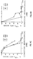
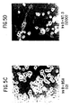
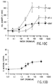
- the trkB -expressing cells were then tested in "survival assays" in which the cells were plated at relatively high density (20% confluency) in defined media alone or in defined media supplemented with 5 nM of bFGF or each of the neurotrophins.
- BDNF and/or NT-3 activation of trkB provides mitogenic as well as survival signals to NIH 3T3 cells
- BDNF and NT-3 for their ability to promote DNA systhesis in trkB-expressing cells by utilizing a thymidine incorporation assay.
- Cells were grown to confluency in 3% serum, at which point they appeared quiescent based on the absence of mitotic cells; no cell death was apparent under these conditions. The various factors were then directly added to this media, and 3 H-labeled thymidine incorporation was monitored 12 hours after factor addition.
- BDNF and NT-3 were also tested for their ability to promote long-term proliferation of trkB-expressing NIH 3T3 cells.
- factors were tested for their ability to enhance the growth rate of sparsely plated parental or trkB-expressing cells; in contrast to the cell death that follows the dense plating of these cells in mitogen-free defined medium, continued proliferation in the absence of notable cell death is seen in cells plated sparsely (at 0.5% confluency) in such medium.
- an enhanced growth rate of trkB-expressing NIH-3T3 cells was seen in response to both BDNF and NT-3 ( Figure 10C); the parental NIH 3T3 cells only responded to bFGF.
- the dose dependencies of BDNF and NT-3 for proliferation paralleled those seen for the survival and thymidine incorporation assays (compare Figures 9B, 10B and 10C).
- This dose-response curve on primary neurons is very similar to those described above for survival and proliferation in fibroblasts (compare Figure 10D to Figures 9B, 10B, and 10C).
- fibroblasts that express trkB in the absence of the LNGFR display a sensitivity to BDNF which is similar to that displayed by BDNF-dependent primary neurons.
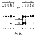
- tyrosine phosphorylation in trkB -expressing NIH3T3 cells following treatment with bFGF or the neurotrophins.
- Parental or trkB -expressing NIH3T3 cells were treated with factors for five minutes, lysates were prepared, and then immunoprecipitated using an anti-phosphotyrosine antibody.
- the precipitated proteins were separated by gel electrophoresis,and detected by immunoblotting with the anti-phosphotyrosine antibody.
- the trkB -expressing NIH3T3 cells treated with BDNF or NT-3 displayed a major tyrosine phosphorylation product of approximately 145 kD, corresponding to the known size of TrkB (Figure 11B); this product was not seen in parental cells exposed to any of the factors, nor was it seen in trkB -expressing cells exposed to either bFGF or NGF ( Figure 11A). These data suggest that the major 145 kD phosphorylation product induced by BDNF and NT-3 is trkB .
- BDNF and NT-3 treatment of trkB -expressing NIH3T3 cells also induced tyrosine-phosphorylation of a 41 kD protein (Figure 11B).
- bFGF also induced phosphorylation of a 41 kD protein in both parental and trkB -expressing NIH3T3 cells ( Figure 11B).
- this phosphoprotein may correspond to ERK2, a serine/threonine kinase which is activated and tyrosine phosphorylated in PC12 cells in response to NGF.
- trkB when expressed in LNGFR-positive PC12 cells, can mediate neurite outgrowth in response to both BDNF and NT-3 (Squinto et al., 1991, Cell 65 :1-20).
- BDNF and NT-3 act as survival and proliferative factors for NIH3T3 cells expressing trkB but no detectable LNGFR.
- neurotrophin receptors do not appear to be unusual among receptor tyrosine kinases in either their requirement for interactions with other receptor components, or in the types of signals they transduce. Therefore the apparently unique actions of the neurotrophins, in terms of promoting neuronal survival and differentiation as opposed to cell growth, can most probably be attributed to the relatively restricted distribution of the trk receptors in post-mitotic neurons rather than any unique features of the receptor system. Conversely, our data would also suggest that the apparent neurotrophic actions of traditional growth factors, including members of the FGF family, are mediated by signal transduction pathways which overlap those utilized by the neurotrophins.
- NIH3T3 cells provide the first example of an immortalized cell line which displays responses to the neurotrophins at physiologically appropriate levels; responses in PC12 cells require approximately 100-fold higher NGF levels than responses in primary neurons.
- survival and proliferation assays utilizing NIH3T3 cells expressing novel receptor tyrosine kinases may be used as a powerful system for the molecular cloning of the ligands for these orphan receptors.
- the NIH3T3 system may also be used to provide insight into how neurotrophic factors prevent neuronal cell death; it can now be determined whether the neurotrophins utilize the same or distinct pathways to prevent neuronal versus non-neuronal cell death.
- the LNGFR could play a signalling role in a pathway not involving the trks, although the apparent inability of BDNF or NT-3 to elicit responses from PC12 cells seems to make this a less likely possibility (Squinto et al., 1991, Cell 65 :1-20).
- the finding that the LNGFR is much more widely distributed than the trks (Bothwell, 1991 Curr. Top. Microbiol. Immunol. 165 :63-70), particularly within apparent targets of innervation that produce neurotrophins but which are not known to respond to them, seems to support more of an "immunobilization" or "clearance” role rather than a signalling role for the LNGFR.
- NT-3 and BDNF display dramatically reciprocal expression patterns during development (Maisonpierre et al., 1990a, Neuron 5 :501-509).
- the NT-3 gene is expressed at much higher levels during early development than is the BDNF gene; NT-3 expression then decreases while BDNF levels increase.
- This reciprocal relationship between NT-3 and BDNF expression is particularly noteworthy in light of our findings that trkB can act as a BDNF receptor, and somewhat less well as an NT-3 receptor.
- the apparently high levels of NT-3 early in development may allow it to act more generally on receptors such as trkB; when NT-3 expression decreases to low levels unidentified, NT-3 receptors.
- cDNA templates were generated by reverse transcription of adult and E13 total rat brain RNAs using oligo d(T) primers. Combinatorial PCR reactions with either of the two cDNA groups were done comparing the six combinations of downstream and upstream primers (see Table I below and Figure 12A) at primer annealing temperatures of 40°C versus 45°C. Aliquots of the PCR reactions were subjected to electrophoresis on an agarose gel, blotted to nylon and probed with "internal" radiolabeled degenerate oligonucleotides complimentary to the domain VII homology box (see above).
- PCR # t anneal cDNA source Primers 1 40°C E13 DLATRN-DVWSLG 2 40°C adult DLATRN-DVWSYG 3 40°C E13 DLATRN-DVWSYG 4 40°C adult DLAARN-DVWSLG 5 40°C E13 DLAARN-DVWSLG
- the eluted DNAs were cloned into compatible XhoI//SstI sites in the Bluescript II KS(-) plasmid, introduced into DH5 ⁇ E . coli by electroporation, followed plating of transformants on selective agar. Approximately 200 ampicillin-resistant bacterial colonies from each PCR transformation were inoculated into 96-well microtiter plates and have been stored as glycerol stocks at -80°C. Individual colonies from these five pools of PCR clones have been analyzed by sequencing of plasmid DNAs that have been purified by standard plasmid miniprep procedures.
- Oligonucleotide primers corresponding to conserved regions of known tyrosine kinase molecules were used to amplify and clone DNA sequences encoding novel orphan tyrosine kinase receptor molecules.
- the amino acid sequences of representatives from branches of the tyrosine kinase superfamily (Hanks et al., 1988, Science 241 :42-52) were aligned ( Figure 12E), and regions of homology within the catalytic domain of these proteins were identified and used to design degenerate oligonucleotide primers, depicted in Table I ( supra ) and Figure 12A.
- Figure 12B presents the amino acid sequences encoded by a number of these clones which are homologous to a number of known tyrosine kinase molecules.
- the five particular cloned molecules of Figure 12B bear homology to trk, CSF1R/PDGFT/kit, ret, eck (alpha ), and eck (beta) and are termed respectively, Rtk-1, Rtk-6, Rtk-7, Rtk-8, and Rtk-9.
- Figure 12C presents DNA sequences for each of these five tyrosine kinase clones, as well as information about the cDNA source and primers used to amplify each clone. Also shown are alignments of these DNA sequences (or their cognate amino acid sequence) with near relatives from the tyrosine kinase superfamily.
- Figure 12D shows the amino acid sequence of a rat brain cDNA that was obtained by probing with the Rtk-1 cDNA sequence shown in Figure 12C.
- the cDNA clone contains sequences identical to Rtk-1 and an additional 1440 amino acid segment of the gene, and based on alighment with trkA and trkB, may correspond to the carboxy terminus of the Rtk-1 gene product.
- PCR-mediated cloning was performed essentially as described in Section 9, supra , but using cDNA from the human neuroblastoma cell line SY5Y as template, and oligonucleotide primers shown in Table II, infra .
- oligodeoxynucleotide primers were designed which correspond to protein regions strongly conserved in all protein kinases, but which at the same time would strongly bias the amplification reaction towards trk -related sequences.
- trk-10r primer in Fig. 13A corresponds to the -PIRWMPPE- in which trk and trkB are identical,but even their closest known relatives, insulin and insulin-like growth factor receptors have two amino acid substitutions.
- Figure 13A shows alignment of tyrosine kinase domains of the members of insulin receptor subfamily with regions of sequence conservation identified earlier by others, and the location of all primers used in this work.
- PCRs polymerase chain reactions
- DNA products were digested with restriction enzymes which cut trk and trkB sequences, and the resistant material was re-amplified and cloned. Individual clones were then sequenced. Amino acid sequences deduced from these DNA sequences revealed the presence of fragments of four novel potential receptors in the pool.
- Rtk-2, Rtk-3, Rtk-4 and Rtk-5 are depicted in Figure 13B (Rtk-4 and Rtk-5), Figure 14 (Rtk-2), and Figure 15 (Rtk-3) and compared with the corresponding region of human trk in Figure 13C.
- Rtk-2 is shown alligned with human trk, rat trkB, insulin-related growth faction receptor, and insulin receptor in Figure 13D.
- Rtk-2 and Rtk-3 sequences are of particular interest.
- the deduced amino acid sequences were used to search GenBank, Release 67.0 (03/91) and EMBL (Modified) Release 26.0 (02/91) Databases with TFASTA, the highest homology scores were obtained for trkB and trk sequences.
- Rtk-2 and Rtk-3 contain the YxxDYY sequence characteristic for trk/insulin-R subfamily. Both sequences have an L for F substitution in the DFG motif which is very strongly conserved in all kinases.
- Rtk-2 and Rtk-3 probes were hybridized to Northern blots, no signal was obtained with SY5Y RNA.
- Rtk-2 hybridized to a 6-7 kb band in RNAs from other lines, in particular from CHP100, SKES, and LAN5 cells.
- Rtk-3 hybridized to a 5 kb band in RNAs from SKN-SH and LAN5 cells.
- Screening of 1.5 x 10 6 clones from SY5Y cDNA library in lambda ZPA II with Rtk-2 probe yielded 5 positive clones, with inserts in the range 1.4-3 kb.
- Sequence data for Rtk-2 indicates that nucleotides 801-875 appear to code for a transmembrane domain, nucleotides 1002-1850 appear to contain a tyrosine kinase domain; and 1-800 code for a continuous open reading frame comprising a ligand binding domain.
- a 180 amino acid stretch follows the tyrosine kinase domain, in contrast to trks, which terminate shortly following the tyrosine kinase domain.
- Dorsal root ganglion (DRG) from embryonic rat (E14) were dissected and collected in serum-containing medium (F14 medium supplemented with 5% horse serum), as described in Linsay et al. (1985, Dev. Biol. 112 : 319).
- serum-containing medium F14 medium supplemented with 5% horse serum
- ganglia were put onto 35 mm dishes precoated with polyornithine (1mg/ml) and laminin (50ug/ml). Ganglia were cultured for 24 hours in the presence or absence of neurotrophins at 37 °C in a humidified 3% CO2 atmosphere. The extent of neurite outgrowth was scored with a scale of 0-5.
- ganglia were rinsed with PBS, following which RNA was prepared using guanidinium thiocyanate, as described in Chomczynski and Sacchi (1987, Analytical Biochem. 162 : 156).
- RNA was prepared using guanidinium thiocyanate, as described in Chomczynski and Sacchi (1987, Analytical Biochem. 162 : 156).
- ganglia were incubated with 0.1% trypsin at 37°C for 30 minutes, after which they were triturated and preplated for 2.5 hours. Neurons were then collected and seeded onto 16mm wells precoated with 100ug/ml polyornithine and 50ug/ml laminin. Neurons were cultured for 24 hours in the presence or absence of neurotrophins, and cell counts were scored.
- RNA from tissues were prepared using standard techniques.
- mRNA levels for trkA, B, and C were examined in the ganglia cultured in the presence or absence of various neurotrophins.
- mRNA for trkA, B and C can be detected ( Figure 18 and 19).
- Figure 18A ganglia incubated in the presence of NGF (50ng/ml) had a significant trkA mRNA level, but undetectable trkB message ( Figure 18B).
- ganglia incubated in the presence of BDNF revealed significant trkB message (Figure 19B), but very low levels of trkA (Figure 19A) and no trkC ( Figure 19C).
- ganglia had significant trkC and trkA mRNA, but very low levels of trkB message.
- NT-3 Treatment of NT-3 appeared to result in survival of both trk-C and trk-A containing neurons.
- specific receptors for the respective neurotrophins are present on distinct cell types suggest that neurotrophins may be used to rescue distinct cell populations in the peripheral nervous system.
Landscapes
- Health & Medical Sciences (AREA)
- Life Sciences & Earth Sciences (AREA)
- Chemical & Material Sciences (AREA)
- Engineering & Computer Science (AREA)
- Molecular Biology (AREA)
- Immunology (AREA)
- Organic Chemistry (AREA)
- Biomedical Technology (AREA)
- Biochemistry (AREA)
- General Health & Medical Sciences (AREA)
- Proteomics, Peptides & Aminoacids (AREA)
- Hematology (AREA)
- Urology & Nephrology (AREA)
- Medicinal Chemistry (AREA)
- Zoology (AREA)
- Microbiology (AREA)
- Biotechnology (AREA)
- Cell Biology (AREA)
- Wood Science & Technology (AREA)
- Analytical Chemistry (AREA)
- Genetics & Genomics (AREA)
- Physics & Mathematics (AREA)
- General Physics & Mathematics (AREA)
- Food Science & Technology (AREA)
- Pathology (AREA)
- Biophysics (AREA)
- Bioinformatics & Cheminformatics (AREA)
- General Engineering & Computer Science (AREA)
- Gastroenterology & Hepatology (AREA)
- Oncology (AREA)
- Neurology (AREA)
- Neurosurgery (AREA)
- Toxicology (AREA)
- Measuring Or Testing Involving Enzymes Or Micro-Organisms (AREA)
- Micro-Organisms Or Cultivation Processes Thereof (AREA)
- Investigating Or Analysing Biological Materials (AREA)
- Medicines That Contain Protein Lipid Enzymes And Other Medicines (AREA)
- Peptides Or Proteins (AREA)
- Measuring Pulse, Heart Rate, Blood Pressure Or Blood Flow (AREA)
- Saccharide Compounds (AREA)
Applications Claiming Priority (5)
| Application Number | Priority Date | Filing Date | Title |
|---|---|---|---|
| US69019991A | 1991-04-23 | 1991-04-23 | |
| US690199 | 1991-04-23 | ||
| US73655991A | 1991-07-26 | 1991-07-26 | |
| US736559 | 1991-07-26 | ||
| EP92912036A EP0589961B1 (de) | 1991-04-23 | 1992-04-23 | Nachweissystem für die neurotrophin-aktivität |
Related Parent Applications (2)
| Application Number | Title | Priority Date | Filing Date |
|---|---|---|---|
| EP92912036A Division EP0589961B1 (de) | 1991-04-23 | 1992-04-23 | Nachweissystem für die neurotrophin-aktivität |
| EP92912036.8 Division | 1992-11-06 |
Publications (3)
| Publication Number | Publication Date |
|---|---|
| EP1001024A2 true EP1001024A2 (de) | 2000-05-17 |
| EP1001024A3 EP1001024A3 (de) | 2004-02-18 |
| EP1001024B1 EP1001024B1 (de) | 2006-08-16 |
Family
ID=27104557
Family Applications (2)
| Application Number | Title | Priority Date | Filing Date |
|---|---|---|---|
| EP92912036A Expired - Lifetime EP0589961B1 (de) | 1991-04-23 | 1992-04-23 | Nachweissystem für die neurotrophin-aktivität |
| EP99203310A Expired - Lifetime EP1001024B1 (de) | 1991-04-23 | 1992-04-23 | Durch das trkB Proto-onkogen kodiertes Protein, das als Nachweissystem für die Neurotrophin-aktivität gebraucht wird |
Family Applications Before (1)
| Application Number | Title | Priority Date | Filing Date |
|---|---|---|---|
| EP92912036A Expired - Lifetime EP0589961B1 (de) | 1991-04-23 | 1992-04-23 | Nachweissystem für die neurotrophin-aktivität |
Country Status (18)
| Country | Link |
|---|---|
| EP (2) | EP0589961B1 (de) |
| JP (1) | JPH06509413A (de) |
| AT (2) | ATE336576T1 (de) |
| AU (1) | AU676664B2 (de) |
| CA (1) | CA2109100A1 (de) |
| CZ (1) | CZ223793A3 (de) |
| DE (2) | DE69233649D1 (de) |
| DK (1) | DK0589961T3 (de) |
| ES (1) | ES2170056T3 (de) |
| FI (1) | FI934701L (de) |
| HU (1) | HUT66051A (de) |
| IE (1) | IE921316A1 (de) |
| IL (1) | IL101661A (de) |
| NO (1) | NO933819L (de) |
| NZ (1) | NZ242468A (de) |
| PT (1) | PT100423A (de) |
| SK (1) | SK116593A3 (de) |
| WO (1) | WO1992018149A1 (de) |
Families Citing this family (13)
| Publication number | Priority date | Publication date | Assignee | Title |
|---|---|---|---|---|
| CA2109598A1 (en) * | 1991-05-21 | 1992-11-26 | Finn Hallbook | Therapeutic and diagnostic methods based on neurotrophin-4- expression |
| WO1993025684A1 (en) * | 1992-06-12 | 1993-12-23 | Regeneron Pharmaceuticals, Inc. | Therapeutic and diagnostic methods based on neurotrophin-4 expression |
| JPH07246040A (ja) * | 1993-07-06 | 1995-09-26 | Takeda Chem Ind Ltd | ニューロトロフィン−3遺伝子が不活性化された胚幹細胞および該遺伝子発現不全動物 |
| US5625121A (en) * | 1993-09-02 | 1997-04-29 | Bristol-Myers Squibb Company | Mice deficient in nerve growth factor receptors encoded by trkB |
| US5753225A (en) * | 1993-12-03 | 1998-05-19 | The Regents Of The University Of California | Antibodies that mimic actions of neurotrophins |
| US6103689A (en) * | 1994-08-31 | 2000-08-15 | Trustees Of Boston University | Methods of inducing hair growth and coloration |
| US6867179B1 (en) | 1994-08-31 | 2005-03-15 | Trustees Of Boston University | Methods of inducing hair growth and coloration |
| US7196055B2 (en) | 1994-08-31 | 2007-03-27 | Trustees Of Boston University | Inhibition of apoptosis in keratinocytes by a ligand of p75 nerve growth factor receptor |
| CZ310798A3 (cs) * | 1996-03-28 | 1999-02-17 | Trustees Of Boston University | Způsob tlumení aktivace tyrosinasy, upravování pigmentace a identifikace látky, látka zesilující fosforylaci tyrosinasy, látka tlumící fosforylaci tyrosinasy, použití činidla tlumícího fosforylaci tyrosinasy, použití peptidu a použití DNA |
| WO1997037228A1 (en) | 1996-03-29 | 1997-10-09 | The Trustees Of Boston University | Methods for diagnosing and treating alzheimer's disease |
| GB9807781D0 (en) * | 1998-04-09 | 1998-06-10 | Univ Bristol | Therapeutic agent |
| EP2789630A1 (de) | 2013-04-09 | 2014-10-15 | EngMab AG | Bispezifische Antikörper gegen CD3e und ROR1 |
| US11952421B2 (en) | 2014-10-09 | 2024-04-09 | Bristol-Myers Squibb Company | Bispecific antibodies against CD3EPSILON and ROR1 |
Family Cites Families (4)
| Publication number | Priority date | Publication date | Assignee | Title |
|---|---|---|---|---|
| US4963481A (en) * | 1985-12-18 | 1990-10-16 | Genetics Institute Inc | Promoter system |
| US5231001A (en) * | 1991-03-14 | 1993-07-27 | The United States Of America As Represented By The Department Of Health And Human Services | Trk tyrosine kinase receptor is the physiological receptor for nerve growth factor |
| CA2062377A1 (en) * | 1991-03-21 | 1992-09-22 | Mariano Barbacid | Method for detection of neuroactive substances |
| US5843749A (en) * | 1991-07-26 | 1998-12-01 | Regeneron Pharmaceuticals, Inc. | Ehk and Ror tyrosine kinases |
-
1992
- 1992-04-21 IL IL101661A patent/IL101661A/en not_active IP Right Cessation
- 1992-04-23 CA CA002109100A patent/CA2109100A1/en not_active Abandoned
- 1992-04-23 PT PT100423A patent/PT100423A/pt not_active Application Discontinuation
- 1992-04-23 AT AT99203310T patent/ATE336576T1/de not_active IP Right Cessation
- 1992-04-23 FI FI934701A patent/FI934701L/fi not_active Application Discontinuation
- 1992-04-23 DE DE69233649T patent/DE69233649D1/de not_active Expired - Lifetime
- 1992-04-23 AT AT92912036T patent/ATE214733T1/de not_active IP Right Cessation
- 1992-04-23 JP JP4511770A patent/JPH06509413A/ja active Pending
- 1992-04-23 NZ NZ242468A patent/NZ242468A/en not_active IP Right Cessation
- 1992-04-23 EP EP92912036A patent/EP0589961B1/de not_active Expired - Lifetime
- 1992-04-23 DK DK92912036T patent/DK0589961T3/da active
- 1992-04-23 IE IE131692A patent/IE921316A1/en not_active Application Discontinuation
- 1992-04-23 WO PCT/US1992/003376 patent/WO1992018149A1/en not_active Ceased
- 1992-04-23 EP EP99203310A patent/EP1001024B1/de not_active Expired - Lifetime
- 1992-04-23 CZ CS932237A patent/CZ223793A3/cs unknown
- 1992-04-23 SK SK1165-93A patent/SK116593A3/sk unknown
- 1992-04-23 ES ES92912036T patent/ES2170056T3/es not_active Expired - Lifetime
- 1992-04-23 HU HU9303012A patent/HUT66051A/hu unknown
- 1992-04-23 AU AU20149/92A patent/AU676664B2/en not_active Ceased
- 1992-04-23 DE DE69232496T patent/DE69232496T2/de not_active Expired - Lifetime
-
1993
- 1993-10-22 NO NO933819A patent/NO933819L/no unknown
Also Published As
| Publication number | Publication date |
|---|---|
| IL101661A0 (en) | 1992-12-30 |
| IL101661A (en) | 1998-02-08 |
| HUT66051A (en) | 1994-09-28 |
| DK0589961T3 (da) | 2002-05-27 |
| SK116593A3 (en) | 1994-11-09 |
| IE921316A1 (en) | 1992-11-04 |
| EP1001024B1 (de) | 2006-08-16 |
| EP0589961A1 (de) | 1994-04-06 |
| EP1001024A3 (de) | 2004-02-18 |
| DE69232496D1 (de) | 2002-04-25 |
| FI934701A0 (fi) | 1993-10-25 |
| CA2109100A1 (en) | 1992-10-29 |
| EP0589961A4 (de) | 1994-11-02 |
| ATE214733T1 (de) | 2002-04-15 |
| FI934701A7 (fi) | 1993-12-22 |
| JPH06509413A (ja) | 1994-10-20 |
| NZ242468A (en) | 1994-12-22 |
| DE69232496T2 (de) | 2002-10-10 |
| DE69233649D1 (de) | 2006-09-28 |
| HU9303012D0 (en) | 1994-03-28 |
| AU676664B2 (en) | 1997-03-20 |
| EP0589961B1 (de) | 2002-03-20 |
| NO933819D0 (no) | 1993-10-22 |
| FI934701L (fi) | 1993-12-22 |
| NO933819L (no) | 1993-12-20 |
| WO1992018149A1 (en) | 1992-10-29 |
| CZ223793A3 (en) | 1994-08-17 |
| HK1024935A1 (en) | 2000-10-27 |
| ATE336576T1 (de) | 2006-09-15 |
| PT100423A (pt) | 1993-08-31 |
| ES2170056T3 (es) | 2002-08-01 |
| AU2014992A (en) | 1992-11-17 |
Similar Documents
| Publication | Publication Date | Title |
|---|---|---|
| US5843749A (en) | Ehk and Ror tyrosine kinases | |
| EP1001024B1 (de) | Durch das trkB Proto-onkogen kodiertes Protein, das als Nachweissystem für die Neurotrophin-aktivität gebraucht wird | |
| US5814478A (en) | Tyrosine kinase receptors and ligands | |
| US6413740B1 (en) | Tyrosine kinase receptors and ligands | |
| US5622862A (en) | Assay systems for trkB neurotrophin activity | |
| JP3686084B2 (ja) | 生物学上活性なeph族リガンド | |
| US5332672A (en) | Prevention of ES cell differentiation by ciliary neurotrophic factor | |
| US5426177A (en) | Ciliary neurotrophic factor receptor | |
| US5747033A (en) | Method of enhancing the biological activity of Eph family ligands | |
| US5656473A (en) | Human Dmk receptor | |
| IL98304A (en) | The ciliary neurotrophic factor receptor | |
| WO1996004389A1 (en) | Rtk/cytokine receptor chimeras | |
| HK1024935B (en) | Protein encoded by the trkb proto-oncogene, used in assay systems for neurothophin activity | |
| US6316206B1 (en) | Method for screening comprising cells expressing the CNTF receptor | |
| EP0767835B1 (de) | Denervierende muskelkinase (dmk), ein rezeptor der tyrosinkinase-superfamilie | |
| US5955290A (en) | Assay systems using the CNTF signal transduction pathway | |
| JP2004201693A (ja) | 除神経筋キナーゼ(dmk)、チロシンキナーゼスーパーファミリーのレセプター | |
| WO1995007467A1 (en) | Assay systems using the cntf signal transduction pathway | |
| WO1993011253A1 (en) | Cell-free ciliary neurotrophic factor/receptor complex |
Legal Events
| Date | Code | Title | Description |
|---|---|---|---|
| PUAI | Public reference made under article 153(3) epc to a published international application that has entered the european phase |
Free format text: ORIGINAL CODE: 0009012 |
|
| AC | Divisional application: reference to earlier application |
Ref document number: 589961 Country of ref document: EP |
|
| AK | Designated contracting states |
Kind code of ref document: A2 Designated state(s): AT BE CH DE DK ES FR GB GR IT LI LU MC NL PT SE |
|
| RIN1 | Information on inventor provided before grant (corrected) |
Inventor name: MAISONPIERRE, PETER C. Inventor name: YANCOPOULOS, GEORGE D. Inventor name: FURTH, MARC E. Inventor name: STITT, TREVOR Inventor name: DISTEPHANO, PETER Inventor name: ALDRICH, THOMAS H. Inventor name: GLASS, DAVID J. Inventor name: SQUINTO, STEPHEN P. |
|
| PUAL | Search report despatched |
Free format text: ORIGINAL CODE: 0009013 |
|
| AK | Designated contracting states |
Kind code of ref document: A3 Designated state(s): AT BE CH DE DK ES FR GB GR IT LI LU MC NL PT SE |
|
| RIC1 | Information provided on ipc code assigned before grant |
Ipc: 7G 01N 33/68 B Ipc: 7C 07K 14/00 B Ipc: 7C 12N 15/12 A |
|
| 17P | Request for examination filed |
Effective date: 20040818 |
|
| AKX | Designation fees paid |
Designated state(s): AT BE CH DE DK ES FR GB GR IT LI LU MC NL PT SE |
|
| 17Q | First examination report despatched |
Effective date: 20050503 |
|
| GRAP | Despatch of communication of intention to grant a patent |
Free format text: ORIGINAL CODE: EPIDOSNIGR1 |
|
| GRAS | Grant fee paid |
Free format text: ORIGINAL CODE: EPIDOSNIGR3 |
|
| GRAA | (expected) grant |
Free format text: ORIGINAL CODE: 0009210 |
|
| AC | Divisional application: reference to earlier application |
Ref document number: 0589961 Country of ref document: EP Kind code of ref document: P |
|
| AK | Designated contracting states |
Kind code of ref document: B1 Designated state(s): AT BE CH DE DK ES FR GB GR IT LI LU MC NL PT SE |
|
| PG25 | Lapsed in a contracting state [announced via postgrant information from national office to epo] |
Ref country code: NL Free format text: LAPSE BECAUSE OF FAILURE TO SUBMIT A TRANSLATION OF THE DESCRIPTION OR TO PAY THE FEE WITHIN THE PRESCRIBED TIME-LIMIT Effective date: 20060816 Ref country code: LI Free format text: LAPSE BECAUSE OF FAILURE TO SUBMIT A TRANSLATION OF THE DESCRIPTION OR TO PAY THE FEE WITHIN THE PRESCRIBED TIME-LIMIT Effective date: 20060816 Ref country code: IT Free format text: LAPSE BECAUSE OF FAILURE TO SUBMIT A TRANSLATION OF THE DESCRIPTION OR TO PAY THE FEE WITHIN THE PRESCRIBED TIME-LIMIT;WARNING: LAPSES OF ITALIAN PATENTS WITH EFFECTIVE DATE BEFORE 2007 MAY HAVE OCCURRED AT ANY TIME BEFORE 2007. THE CORRECT EFFECTIVE DATE MAY BE DIFFERENT FROM THE ONE RECORDED. Effective date: 20060816 Ref country code: CH Free format text: LAPSE BECAUSE OF FAILURE TO SUBMIT A TRANSLATION OF THE DESCRIPTION OR TO PAY THE FEE WITHIN THE PRESCRIBED TIME-LIMIT Effective date: 20060816 Ref country code: BE Free format text: LAPSE BECAUSE OF FAILURE TO SUBMIT A TRANSLATION OF THE DESCRIPTION OR TO PAY THE FEE WITHIN THE PRESCRIBED TIME-LIMIT Effective date: 20060816 Ref country code: AT Free format text: LAPSE BECAUSE OF FAILURE TO SUBMIT A TRANSLATION OF THE DESCRIPTION OR TO PAY THE FEE WITHIN THE PRESCRIBED TIME-LIMIT Effective date: 20060816 |
|
| REG | Reference to a national code |
Ref country code: GB Ref legal event code: FG4D |
|
| REG | Reference to a national code |
Ref country code: CH Ref legal event code: EP |
|
| REF | Corresponds to: |
Ref document number: 69233649 Country of ref document: DE Date of ref document: 20060928 Kind code of ref document: P |
|
| PG25 | Lapsed in a contracting state [announced via postgrant information from national office to epo] |
Ref country code: SE Free format text: LAPSE BECAUSE OF FAILURE TO SUBMIT A TRANSLATION OF THE DESCRIPTION OR TO PAY THE FEE WITHIN THE PRESCRIBED TIME-LIMIT Effective date: 20061116 Ref country code: DK Free format text: LAPSE BECAUSE OF FAILURE TO SUBMIT A TRANSLATION OF THE DESCRIPTION OR TO PAY THE FEE WITHIN THE PRESCRIBED TIME-LIMIT Effective date: 20061116 |
|
| PG25 | Lapsed in a contracting state [announced via postgrant information from national office to epo] |
Ref country code: DE Free format text: LAPSE BECAUSE OF FAILURE TO SUBMIT A TRANSLATION OF THE DESCRIPTION OR TO PAY THE FEE WITHIN THE PRESCRIBED TIME-LIMIT Effective date: 20061117 |
|
| REG | Reference to a national code |
Ref country code: HK Ref legal event code: GR Ref document number: 1024935 Country of ref document: HK |
|
| PG25 | Lapsed in a contracting state [announced via postgrant information from national office to epo] |
Ref country code: ES Free format text: LAPSE BECAUSE OF FAILURE TO SUBMIT A TRANSLATION OF THE DESCRIPTION OR TO PAY THE FEE WITHIN THE PRESCRIBED TIME-LIMIT Effective date: 20061127 |
|
| PG25 | Lapsed in a contracting state [announced via postgrant information from national office to epo] |
Ref country code: PT Free format text: LAPSE BECAUSE OF FAILURE TO SUBMIT A TRANSLATION OF THE DESCRIPTION OR TO PAY THE FEE WITHIN THE PRESCRIBED TIME-LIMIT Effective date: 20070116 |
|
| NLV1 | Nl: lapsed or annulled due to failure to fulfill the requirements of art. 29p and 29m of the patents act | ||
| REG | Reference to a national code |
Ref country code: CH Ref legal event code: PL |
|
| EN | Fr: translation not filed | ||
| PLBE | No opposition filed within time limit |
Free format text: ORIGINAL CODE: 0009261 |
|
| STAA | Information on the status of an ep patent application or granted ep patent |
Free format text: STATUS: NO OPPOSITION FILED WITHIN TIME LIMIT |
|
| 26N | No opposition filed |
Effective date: 20070518 |
|
| GBPC | Gb: european patent ceased through non-payment of renewal fee |
Effective date: 20070423 |
|
| PG25 | Lapsed in a contracting state [announced via postgrant information from national office to epo] |
Ref country code: GR Free format text: LAPSE BECAUSE OF FAILURE TO SUBMIT A TRANSLATION OF THE DESCRIPTION OR TO PAY THE FEE WITHIN THE PRESCRIBED TIME-LIMIT Effective date: 20061117 Ref country code: GB Free format text: LAPSE BECAUSE OF NON-PAYMENT OF DUE FEES Effective date: 20070423 Ref country code: FR Free format text: LAPSE BECAUSE OF FAILURE TO SUBMIT A TRANSLATION OF THE DESCRIPTION OR TO PAY THE FEE WITHIN THE PRESCRIBED TIME-LIMIT Effective date: 20070511 |
|
| PG25 | Lapsed in a contracting state [announced via postgrant information from national office to epo] |
Ref country code: FR Free format text: LAPSE BECAUSE OF FAILURE TO SUBMIT A TRANSLATION OF THE DESCRIPTION OR TO PAY THE FEE WITHIN THE PRESCRIBED TIME-LIMIT Effective date: 20060816 |
|
| PG25 | Lapsed in a contracting state [announced via postgrant information from national office to epo] |
Ref country code: MC Free format text: LAPSE BECAUSE OF NON-PAYMENT OF DUE FEES Effective date: 20070430 |
|
| PG25 | Lapsed in a contracting state [announced via postgrant information from national office to epo] |
Ref country code: LU Free format text: LAPSE BECAUSE OF NON-PAYMENT OF DUE FEES Effective date: 20070423 |