EP0988552B1 - Procede de detection precoce des maladies cardiaques - Google Patents
Procede de detection precoce des maladies cardiaques Download PDFInfo
- Publication number
- EP0988552B1 EP0988552B1 EP98923666A EP98923666A EP0988552B1 EP 0988552 B1 EP0988552 B1 EP 0988552B1 EP 98923666 A EP98923666 A EP 98923666A EP 98923666 A EP98923666 A EP 98923666A EP 0988552 B1 EP0988552 B1 EP 0988552B1
- Authority
- EP
- European Patent Office
- Prior art keywords
- sphingosine
- cardiac
- metabolite
- sph
- antibody
- Prior art date
- Legal status (The legal status is an assumption and is not a legal conclusion. Google has not performed a legal analysis and makes no representation as to the accuracy of the status listed.)
- Expired - Lifetime
Links
- 238000000034 method Methods 0.000 title claims abstract description 106
- 208000019622 heart disease Diseases 0.000 title claims description 13
- 238000001514 detection method Methods 0.000 title description 22
- 230000000747 cardiac effect Effects 0.000 claims abstract description 127
- 239000003550 marker Substances 0.000 claims abstract description 96
- 208000031225 myocardial ischemia Diseases 0.000 claims abstract description 66
- 206010021143 Hypoxia Diseases 0.000 claims abstract description 63
- 230000007954 hypoxia Effects 0.000 claims abstract description 51
- 238000012360 testing method Methods 0.000 claims abstract description 49
- 241000124008 Mammalia Species 0.000 claims abstract description 25
- WWUZIQQURGPMPG-KRWOKUGFSA-N sphingosine Chemical compound CCCCCCCCCCCCC\C=C\[C@@H](O)[C@@H](N)CO WWUZIQQURGPMPG-KRWOKUGFSA-N 0.000 claims description 222
- WWUZIQQURGPMPG-UHFFFAOYSA-N (-)-D-erythro-Sphingosine Natural products CCCCCCCCCCCCCC=CC(O)C(N)CO WWUZIQQURGPMPG-UHFFFAOYSA-N 0.000 claims description 215
- 210000002966 serum Anatomy 0.000 claims description 111
- 108060008682 Tumor Necrosis Factor Proteins 0.000 claims description 104
- 102000000852 Tumor Necrosis Factor-alpha Human genes 0.000 claims description 104
- DUYSYHSSBDVJSM-KRWOKUGFSA-N sphingosine 1-phosphate Chemical compound CCCCCCCCCCCCC\C=C\[C@@H](O)[C@@H](N)COP(O)(O)=O DUYSYHSSBDVJSM-KRWOKUGFSA-N 0.000 claims description 44
- 206010000891 acute myocardial infarction Diseases 0.000 claims description 42
- 210000004369 blood Anatomy 0.000 claims description 37
- 239000008280 blood Substances 0.000 claims description 37
- 150000003410 sphingosines Chemical class 0.000 claims description 31
- OTKJDMGTUTTYMP-UHFFFAOYSA-N dihydrosphingosine Natural products CCCCCCCCCCCCCCCC(O)C(N)CO OTKJDMGTUTTYMP-UHFFFAOYSA-N 0.000 claims description 27
- 210000001124 body fluid Anatomy 0.000 claims description 25
- 239000010839 body fluid Substances 0.000 claims description 24
- 238000002399 angioplasty Methods 0.000 claims description 17
- 102000008394 Immunoglobulin Fragments Human genes 0.000 claims description 16
- 108010021625 Immunoglobulin Fragments Proteins 0.000 claims description 16
- 230000027455 binding Effects 0.000 claims description 16
- 238000003018 immunoassay Methods 0.000 claims description 14
- 238000012544 monitoring process Methods 0.000 claims description 14
- 229940106189 ceramide Drugs 0.000 claims description 13
- YDNKGFDKKRUKPY-JHOUSYSJSA-N C16 ceramide Natural products CCCCCCCCCCCCCCCC(=O)N[C@@H](CO)[C@H](O)C=CCCCCCCCCCCCCC YDNKGFDKKRUKPY-JHOUSYSJSA-N 0.000 claims description 12
- ZVEQCJWYRWKARO-UHFFFAOYSA-N ceramide Natural products CCCCCCCCCCCCCCC(O)C(=O)NC(CO)C(O)C=CCCC=C(C)CCCCCCCCC ZVEQCJWYRWKARO-UHFFFAOYSA-N 0.000 claims description 12
- 238000004128 high performance liquid chromatography Methods 0.000 claims description 12
- 230000002107 myocardial effect Effects 0.000 claims description 12
- VVGIYYKRAMHVLU-UHFFFAOYSA-N newbouldiamide Natural products CCCCCCCCCCCCCCCCCCCC(O)C(O)C(O)C(CO)NC(=O)CCCCCCCCCCCCCCCCC VVGIYYKRAMHVLU-UHFFFAOYSA-N 0.000 claims description 12
- 239000007787 solid Substances 0.000 claims description 12
- CRJGESKKUOMBCT-VQTJNVASSA-N N-acetylsphinganine Chemical compound CCCCCCCCCCCCCCC[C@@H](O)[C@H](CO)NC(C)=O CRJGESKKUOMBCT-VQTJNVASSA-N 0.000 claims description 11
- 102000004127 Cytokines Human genes 0.000 claims description 9
- 108090000695 Cytokines Proteins 0.000 claims description 9
- 239000000427 antigen Substances 0.000 claims description 9
- 108091007433 antigens Proteins 0.000 claims description 9
- 102000036639 antigens Human genes 0.000 claims description 9
- YDNKGFDKKRUKPY-TURZORIXSA-N N-hexadecanoylsphingosine Chemical compound CCCCCCCCCCCCCCCC(=O)N[C@@H](CO)[C@H](O)\C=C\CCCCCCCCCCCCC YDNKGFDKKRUKPY-TURZORIXSA-N 0.000 claims description 7
- 238000004587 chromatography analysis Methods 0.000 claims description 7
- 238000004611 spectroscopical analysis Methods 0.000 claims description 6
- 238000001356 surgical procedure Methods 0.000 claims description 6
- 210000002700 urine Anatomy 0.000 claims description 6
- 102000000589 Interleukin-1 Human genes 0.000 claims description 5
- 108010002352 Interleukin-1 Proteins 0.000 claims description 5
- 238000007824 enzymatic assay Methods 0.000 claims description 5
- 238000004817 gas chromatography Methods 0.000 claims description 5
- 230000004217 heart function Effects 0.000 claims description 5
- 239000003446 ligand Substances 0.000 claims description 5
- 229920000642 polymer Polymers 0.000 claims description 5
- 230000000770 proinflammatory effect Effects 0.000 claims description 5
- 210000003296 saliva Anatomy 0.000 claims description 5
- 238000000870 ultraviolet spectroscopy Methods 0.000 claims description 5
- YBJHBAHKTGYVGT-ZKWXMUAHSA-N (+)-Biotin Chemical compound N1C(=O)N[C@@H]2[C@H](CCCCC(=O)O)SC[C@@H]21 YBJHBAHKTGYVGT-ZKWXMUAHSA-N 0.000 claims description 4
- 238000004566 IR spectroscopy Methods 0.000 claims description 4
- 238000005481 NMR spectroscopy Methods 0.000 claims description 4
- 239000003146 anticoagulant agent Substances 0.000 claims description 4
- 229940127219 anticoagulant drug Drugs 0.000 claims description 4
- 239000011521 glass Substances 0.000 claims description 4
- 238000000338 in vitro Methods 0.000 claims description 4
- 241000894007 species Species 0.000 claims description 4
- 102000000588 Interleukin-2 Human genes 0.000 claims description 3
- 108010002350 Interleukin-2 Proteins 0.000 claims description 3
- 108090001005 Interleukin-6 Proteins 0.000 claims description 3
- 239000004677 Nylon Substances 0.000 claims description 3
- 239000011324 bead Substances 0.000 claims description 3
- 210000002751 lymph Anatomy 0.000 claims description 3
- 229920001778 nylon Polymers 0.000 claims description 3
- 238000004393 prognosis Methods 0.000 claims description 3
- 210000004243 sweat Anatomy 0.000 claims description 3
- 108090001008 Avidin Proteins 0.000 claims description 2
- 108010074328 Interferon-gamma Proteins 0.000 claims description 2
- 102000008070 Interferon-gamma Human genes 0.000 claims description 2
- 239000000020 Nitrocellulose Substances 0.000 claims description 2
- 239000004793 Polystyrene Substances 0.000 claims description 2
- 210000000941 bile Anatomy 0.000 claims description 2
- 229960002685 biotin Drugs 0.000 claims description 2
- 235000020958 biotin Nutrition 0.000 claims description 2
- 239000011616 biotin Substances 0.000 claims description 2
- 229920002301 cellulose acetate Polymers 0.000 claims description 2
- 239000012634 fragment Substances 0.000 claims description 2
- 210000004051 gastric juice Anatomy 0.000 claims description 2
- 229960003130 interferon gamma Drugs 0.000 claims description 2
- 239000000463 material Substances 0.000 claims description 2
- 229920001220 nitrocellulos Polymers 0.000 claims description 2
- 229920002223 polystyrene Polymers 0.000 claims description 2
- OTKJDMGTUTTYMP-ROUUACIJSA-N Safingol ( L-threo-sphinganine) Chemical compound CCCCCCCCCCCCCCC[C@H](O)[C@@H](N)CO OTKJDMGTUTTYMP-ROUUACIJSA-N 0.000 claims 8
- JLVSPVFPBBFMBE-HXSWCURESA-O sphingosylphosphocholine acid Chemical compound CCCCCCCCCCCCC\C=C\[C@@H](O)[C@@H]([NH3+])COP([O-])(=O)OCC[N+](C)(C)C JLVSPVFPBBFMBE-HXSWCURESA-O 0.000 claims 8
- MZOFCQQQCNRIBI-VMXHOPILSA-N (3s)-4-[[(2s)-1-[[(2s)-1-[[(1s)-1-carboxy-2-hydroxyethyl]amino]-4-methyl-1-oxopentan-2-yl]amino]-5-(diaminomethylideneamino)-1-oxopentan-2-yl]amino]-3-[[2-[[(2s)-2,6-diaminohexanoyl]amino]acetyl]amino]-4-oxobutanoic acid Chemical compound OC[C@@H](C(O)=O)NC(=O)[C@H](CC(C)C)NC(=O)[C@H](CCCN=C(N)N)NC(=O)[C@H](CC(O)=O)NC(=O)CNC(=O)[C@@H](N)CCCCN MZOFCQQQCNRIBI-VMXHOPILSA-N 0.000 claims 2
- 102000004889 Interleukin-6 Human genes 0.000 claims 1
- 125000001549 ceramide group Chemical group 0.000 claims 1
- 229940100601 interleukin-6 Drugs 0.000 claims 1
- 206010019280 Heart failures Diseases 0.000 abstract description 36
- 239000000203 mixture Substances 0.000 abstract description 10
- 150000003408 sphingolipids Chemical class 0.000 description 46
- 208000028867 ischemia Diseases 0.000 description 45
- 230000000302 ischemic effect Effects 0.000 description 39
- JLVSPVFPBBFMBE-HXSWCURESA-N sphingosine-1-phosphocholine Chemical compound CCCCCCCCCCCCC\C=C\[C@@H](O)[C@@H](N)COP([O-])(=O)OCC[N+](C)(C)C JLVSPVFPBBFMBE-HXSWCURESA-N 0.000 description 28
- 210000002064 heart cell Anatomy 0.000 description 25
- 238000004519 manufacturing process Methods 0.000 description 25
- 239000000523 sample Substances 0.000 description 25
- 210000004027 cell Anatomy 0.000 description 24
- 239000002207 metabolite Substances 0.000 description 24
- 238000003556 assay Methods 0.000 description 20
- OTKJDMGTUTTYMP-ZWKOTPCHSA-N sphinganine Chemical compound CCCCCCCCCCCCCCC[C@@H](O)[C@@H](N)CO OTKJDMGTUTTYMP-ZWKOTPCHSA-N 0.000 description 20
- 102000004420 Creatine Kinase Human genes 0.000 description 16
- 108010042126 Creatine kinase Proteins 0.000 description 16
- OKKJLVBELUTLKV-UHFFFAOYSA-N Methanol Chemical compound OC OKKJLVBELUTLKV-UHFFFAOYSA-N 0.000 description 15
- 238000005259 measurement Methods 0.000 description 15
- 150000001875 compounds Chemical class 0.000 description 13
- 239000011575 calcium Substances 0.000 description 12
- 230000001146 hypoxic effect Effects 0.000 description 12
- 210000002027 skeletal muscle Anatomy 0.000 description 12
- OYPRJOBELJOOCE-UHFFFAOYSA-N Calcium Chemical compound [Ca] OYPRJOBELJOOCE-UHFFFAOYSA-N 0.000 description 11
- 229910052791 calcium Inorganic materials 0.000 description 11
- 210000004413 cardiac myocyte Anatomy 0.000 description 11
- 108020003175 receptors Proteins 0.000 description 11
- 102000005962 receptors Human genes 0.000 description 11
- 208000024891 symptom Diseases 0.000 description 11
- FAPWRFPIFSIZLT-UHFFFAOYSA-M Sodium chloride Chemical compound [Na+].[Cl-] FAPWRFPIFSIZLT-UHFFFAOYSA-M 0.000 description 10
- 230000006907 apoptotic process Effects 0.000 description 10
- 241000700159 Rattus Species 0.000 description 9
- 108010061312 Sphingomyelin Phosphodiesterase Proteins 0.000 description 9
- 230000030833 cell death Effects 0.000 description 9
- 239000000047 product Substances 0.000 description 9
- 108090000623 proteins and genes Proteins 0.000 description 9
- 210000001147 pulmonary artery Anatomy 0.000 description 9
- HEDRZPFGACZZDS-UHFFFAOYSA-N Chloroform Chemical compound ClC(Cl)Cl HEDRZPFGACZZDS-UHFFFAOYSA-N 0.000 description 8
- -1 e.g. Substances 0.000 description 8
- 238000012216 screening Methods 0.000 description 8
- 238000011282 treatment Methods 0.000 description 8
- 102000011971 Sphingomyelin Phosphodiesterase Human genes 0.000 description 7
- 238000007887 coronary angioplasty Methods 0.000 description 7
- 230000000694 effects Effects 0.000 description 7
- 239000002158 endotoxin Substances 0.000 description 7
- 238000005516 engineering process Methods 0.000 description 7
- 210000005003 heart tissue Anatomy 0.000 description 7
- 208000010125 myocardial infarction Diseases 0.000 description 7
- 102000004169 proteins and genes Human genes 0.000 description 7
- 239000000126 substance Substances 0.000 description 7
- 239000000758 substrate Substances 0.000 description 7
- 210000001519 tissue Anatomy 0.000 description 7
- ZDRVLAOYDGQLFI-UHFFFAOYSA-N 4-[[4-(4-chlorophenyl)-1,3-thiazol-2-yl]amino]phenol;hydrochloride Chemical compound Cl.C1=CC(O)=CC=C1NC1=NC(C=2C=CC(Cl)=CC=2)=CS1 ZDRVLAOYDGQLFI-UHFFFAOYSA-N 0.000 description 6
- 206010002383 Angina Pectoris Diseases 0.000 description 6
- 238000002965 ELISA Methods 0.000 description 6
- 102000004190 Enzymes Human genes 0.000 description 6
- 108090000790 Enzymes Proteins 0.000 description 6
- 108090000315 Protein Kinase C Proteins 0.000 description 6
- 102000003923 Protein Kinase C Human genes 0.000 description 6
- 230000002159 abnormal effect Effects 0.000 description 6
- 230000002378 acidificating effect Effects 0.000 description 6
- 230000001684 chronic effect Effects 0.000 description 6
- 229940088598 enzyme Drugs 0.000 description 6
- 230000036541 health Effects 0.000 description 6
- 210000004165 myocardium Anatomy 0.000 description 6
- 108020004707 nucleic acids Proteins 0.000 description 6
- 102000039446 nucleic acids Human genes 0.000 description 6
- 150000007523 nucleic acids Chemical class 0.000 description 6
- 230000003449 preventive effect Effects 0.000 description 6
- 108010035597 sphingosine kinase Proteins 0.000 description 6
- 208000010444 Acidosis Diseases 0.000 description 5
- 108091003079 Bovine Serum Albumin Proteins 0.000 description 5
- 230000007950 acidosis Effects 0.000 description 5
- 208000026545 acidosis disease Diseases 0.000 description 5
- 238000010171 animal model Methods 0.000 description 5
- 210000001772 blood platelet Anatomy 0.000 description 5
- 230000001419 dependent effect Effects 0.000 description 5
- 238000011156 evaluation Methods 0.000 description 5
- 150000002632 lipids Chemical class 0.000 description 5
- 229920006008 lipopolysaccharide Polymers 0.000 description 5
- 239000011780 sodium chloride Substances 0.000 description 5
- 238000010561 standard procedure Methods 0.000 description 5
- 239000006144 Dulbecco’s modified Eagle's medium Substances 0.000 description 4
- 206010061216 Infarction Diseases 0.000 description 4
- 241001465754 Metazoa Species 0.000 description 4
- LCTONWCANYUPML-UHFFFAOYSA-N Pyruvic acid Chemical compound CC(=O)C(O)=O LCTONWCANYUPML-UHFFFAOYSA-N 0.000 description 4
- QVGXLLKOCUKJST-UHFFFAOYSA-N atomic oxygen Chemical compound [O] QVGXLLKOCUKJST-UHFFFAOYSA-N 0.000 description 4
- 230000008901 benefit Effects 0.000 description 4
- 230000036765 blood level Effects 0.000 description 4
- 230000015556 catabolic process Effects 0.000 description 4
- 230000008859 change Effects 0.000 description 4
- 208000029078 coronary artery disease Diseases 0.000 description 4
- 238000005859 coupling reaction Methods 0.000 description 4
- 238000003745 diagnosis Methods 0.000 description 4
- 238000002565 electrocardiography Methods 0.000 description 4
- 239000000284 extract Substances 0.000 description 4
- 230000007574 infarction Effects 0.000 description 4
- 230000003834 intracellular effect Effects 0.000 description 4
- 238000012806 monitoring device Methods 0.000 description 4
- 229910052760 oxygen Inorganic materials 0.000 description 4
- 239000001301 oxygen Substances 0.000 description 4
- 210000002381 plasma Anatomy 0.000 description 4
- 239000000243 solution Substances 0.000 description 4
- 230000009870 specific binding Effects 0.000 description 4
- 230000000451 tissue damage Effects 0.000 description 4
- 231100000827 tissue damage Toxicity 0.000 description 4
- 241000282412 Homo Species 0.000 description 3
- 102000004016 L-Type Calcium Channels Human genes 0.000 description 3
- 108090000420 L-Type Calcium Channels Proteins 0.000 description 3
- 206010024229 Leprosy Diseases 0.000 description 3
- 206010028980 Neoplasm Diseases 0.000 description 3
- 241000283973 Oryctolagus cuniculus Species 0.000 description 3
- 239000002253 acid Substances 0.000 description 3
- 230000009471 action Effects 0.000 description 3
- 230000004913 activation Effects 0.000 description 3
- 239000012491 analyte Substances 0.000 description 3
- 239000000872 buffer Substances 0.000 description 3
- 201000011510 cancer Diseases 0.000 description 3
- 238000005119 centrifugation Methods 0.000 description 3
- 230000004087 circulation Effects 0.000 description 3
- 239000002299 complementary DNA Substances 0.000 description 3
- 230000006378 damage Effects 0.000 description 3
- 230000003247 decreasing effect Effects 0.000 description 3
- 230000003831 deregulation Effects 0.000 description 3
- 238000002405 diagnostic procedure Methods 0.000 description 3
- 208000037265 diseases, disorders, signs and symptoms Diseases 0.000 description 3
- 210000002889 endothelial cell Anatomy 0.000 description 3
- 238000000605 extraction Methods 0.000 description 3
- 239000012894 fetal calf serum Substances 0.000 description 3
- 230000007062 hydrolysis Effects 0.000 description 3
- 238000006460 hydrolysis reaction Methods 0.000 description 3
- 238000002372 labelling Methods 0.000 description 3
- 239000002609 medium Substances 0.000 description 3
- 239000012528 membrane Substances 0.000 description 3
- 238000002823 phage display Methods 0.000 description 3
- 230000002829 reductive effect Effects 0.000 description 3
- 230000001105 regulatory effect Effects 0.000 description 3
- 230000010410 reperfusion Effects 0.000 description 3
- 230000004044 response Effects 0.000 description 3
- 210000001908 sarcoplasmic reticulum Anatomy 0.000 description 3
- 238000009662 stress testing Methods 0.000 description 3
- 230000001225 therapeutic effect Effects 0.000 description 3
- WRGQSWVCFNIUNZ-GDCKJWNLSA-N 1-oleoyl-sn-glycerol 3-phosphate Chemical compound CCCCCCCC\C=C/CCCCCCCC(=O)OC[C@@H](O)COP(O)(O)=O WRGQSWVCFNIUNZ-GDCKJWNLSA-N 0.000 description 2
- OKTWQKXBJUBAKS-WQADZSDSSA-N 2-[[(e,2r,3s)-2-amino-3-hydroxyoctadec-4-enoxy]-hydroxyphosphoryl]oxyethyl-trimethylazanium;chloride Chemical compound [Cl-].CCCCCCCCCCCCC\C=C\[C@H](O)[C@H](N)COP(O)(=O)OCC[N+](C)(C)C OKTWQKXBJUBAKS-WQADZSDSSA-N 0.000 description 2
- 108090000312 Calcium Channels Proteins 0.000 description 2
- 102000003922 Calcium Channels Human genes 0.000 description 2
- 206010008479 Chest Pain Diseases 0.000 description 2
- 208000017667 Chronic Disease Diseases 0.000 description 2
- 102000029816 Collagenase Human genes 0.000 description 2
- 108060005980 Collagenase Proteins 0.000 description 2
- 206010009944 Colon cancer Diseases 0.000 description 2
- 208000001333 Colorectal Neoplasms Diseases 0.000 description 2
- WQZGKKKJIJFFOK-GASJEMHNSA-N Glucose Natural products OC[C@H]1OC(O)[C@H](O)[C@@H](O)[C@@H]1O WQZGKKKJIJFFOK-GASJEMHNSA-N 0.000 description 2
- 102000015696 Interleukins Human genes 0.000 description 2
- 108010063738 Interleukins Proteins 0.000 description 2
- JVTAAEKCZFNVCJ-UHFFFAOYSA-M Lactate Chemical compound CC(O)C([O-])=O JVTAAEKCZFNVCJ-UHFFFAOYSA-M 0.000 description 2
- TWRXJAOTZQYOKJ-UHFFFAOYSA-L Magnesium chloride Chemical compound [Mg+2].[Cl-].[Cl-] TWRXJAOTZQYOKJ-UHFFFAOYSA-L 0.000 description 2
- 241000699666 Mus <mouse, genus> Species 0.000 description 2
- 108091007491 NSP3 Papain-like protease domains Proteins 0.000 description 2
- 108020004711 Nucleic Acid Probes Proteins 0.000 description 2
- 229910019142 PO4 Inorganic materials 0.000 description 2
- 108010029485 Protein Isoforms Proteins 0.000 description 2
- 102000001708 Protein Isoforms Human genes 0.000 description 2
- 108020005115 Pyruvate Kinase Proteins 0.000 description 2
- 102000013009 Pyruvate Kinase Human genes 0.000 description 2
- 241000220317 Rosa Species 0.000 description 2
- 241001222774 Salmonella enterica subsp. enterica serovar Minnesota Species 0.000 description 2
- 102100026263 Sphingomyelin phosphodiesterase Human genes 0.000 description 2
- 102100024550 Sphingomyelin phosphodiesterase 2 Human genes 0.000 description 2
- 101710201923 Sphingomyelin phosphodiesterase 2 Proteins 0.000 description 2
- 102000004142 Trypsin Human genes 0.000 description 2
- 108090000631 Trypsin Proteins 0.000 description 2
- 230000002411 adverse Effects 0.000 description 2
- AWUCVROLDVIAJX-UHFFFAOYSA-N alpha-glycerophosphate Natural products OCC(O)COP(O)(O)=O AWUCVROLDVIAJX-UHFFFAOYSA-N 0.000 description 2
- 238000004458 analytical method Methods 0.000 description 2
- 210000001367 artery Anatomy 0.000 description 2
- 239000003150 biochemical marker Substances 0.000 description 2
- 230000015572 biosynthetic process Effects 0.000 description 2
- 230000036770 blood supply Effects 0.000 description 2
- 229940098773 bovine serum albumin Drugs 0.000 description 2
- 125000002091 cationic group Chemical group 0.000 description 2
- 239000003153 chemical reaction reagent Substances 0.000 description 2
- 229960002424 collagenase Drugs 0.000 description 2
- CVSVTCORWBXHQV-UHFFFAOYSA-N creatine Chemical compound NC(=[NH2+])N(C)CC([O-])=O CVSVTCORWBXHQV-UHFFFAOYSA-N 0.000 description 2
- 208000035475 disorder Diseases 0.000 description 2
- 239000003814 drug Substances 0.000 description 2
- 238000013399 early diagnosis Methods 0.000 description 2
- 230000002255 enzymatic effect Effects 0.000 description 2
- 238000001952 enzyme assay Methods 0.000 description 2
- 238000002474 experimental method Methods 0.000 description 2
- 210000003191 femoral vein Anatomy 0.000 description 2
- 210000002950 fibroblast Anatomy 0.000 description 2
- 108020001507 fusion proteins Proteins 0.000 description 2
- 102000037865 fusion proteins Human genes 0.000 description 2
- 150000002270 gangliosides Chemical class 0.000 description 2
- 239000008103 glucose Substances 0.000 description 2
- 150000002339 glycosphingolipids Chemical class 0.000 description 2
- PCHJSUWPFVWCPO-UHFFFAOYSA-N gold Chemical compound [Au] PCHJSUWPFVWCPO-UHFFFAOYSA-N 0.000 description 2
- 230000001900 immune effect Effects 0.000 description 2
- 230000001771 impaired effect Effects 0.000 description 2
- 238000001727 in vivo Methods 0.000 description 2
- 230000001939 inductive effect Effects 0.000 description 2
- 239000003112 inhibitor Substances 0.000 description 2
- 230000000977 initiatory effect Effects 0.000 description 2
- JVTAAEKCZFNVCJ-UHFFFAOYSA-N lactic acid Chemical compound CC(O)C(O)=O JVTAAEKCZFNVCJ-UHFFFAOYSA-N 0.000 description 2
- 230000000670 limiting effect Effects 0.000 description 2
- 239000011159 matrix material Substances 0.000 description 2
- 230000001404 mediated effect Effects 0.000 description 2
- 239000002853 nucleic acid probe Substances 0.000 description 2
- 230000007310 pathophysiology Effects 0.000 description 2
- 230000010412 perfusion Effects 0.000 description 2
- 230000000737 periodic effect Effects 0.000 description 2
- 239000012071 phase Substances 0.000 description 2
- NBIIXXVUZAFLBC-UHFFFAOYSA-K phosphate Chemical compound [O-]P([O-])([O-])=O NBIIXXVUZAFLBC-UHFFFAOYSA-K 0.000 description 2
- 239000010452 phosphate Substances 0.000 description 2
- 239000002243 precursor Substances 0.000 description 2
- 238000002360 preparation method Methods 0.000 description 2
- 108090000765 processed proteins & peptides Proteins 0.000 description 2
- 238000000746 purification Methods 0.000 description 2
- 229940107700 pyruvic acid Drugs 0.000 description 2
- 238000003127 radioimmunoassay Methods 0.000 description 2
- 210000001567 regular cardiac muscle cell of ventricle Anatomy 0.000 description 2
- 239000012146 running buffer Substances 0.000 description 2
- 230000000580 secretagogue effect Effects 0.000 description 2
- 230000003248 secreting effect Effects 0.000 description 2
- 210000000952 spleen Anatomy 0.000 description 2
- 238000012453 sprague-dawley rat model Methods 0.000 description 2
- 230000008093 supporting effect Effects 0.000 description 2
- 239000000725 suspension Substances 0.000 description 2
- 239000012588 trypsin Substances 0.000 description 2
- 239000003643 water by type Substances 0.000 description 2
- JKMHFZQWWAIEOD-UHFFFAOYSA-N 2-[4-(2-hydroxyethyl)piperazin-1-yl]ethanesulfonic acid Chemical compound OCC[NH+]1CCN(CCS([O-])(=O)=O)CC1 JKMHFZQWWAIEOD-UHFFFAOYSA-N 0.000 description 1
- NJNWCIAPVGRBHO-UHFFFAOYSA-N 2-hydroxyethyl-dimethyl-[(oxo-$l^{5}-phosphanylidyne)methyl]azanium Chemical group OCC[N+](C)(C)C#P=O NJNWCIAPVGRBHO-UHFFFAOYSA-N 0.000 description 1
- 206010002388 Angina unstable Diseases 0.000 description 1
- 201000001320 Atherosclerosis Diseases 0.000 description 1
- 241000894006 Bacteria Species 0.000 description 1
- 241000283690 Bos taurus Species 0.000 description 1
- 229940122642 Calcium channel agonist Drugs 0.000 description 1
- UXVMQQNJUSDDNG-UHFFFAOYSA-L Calcium chloride Chemical compound [Cl-].[Cl-].[Ca+2] UXVMQQNJUSDDNG-UHFFFAOYSA-L 0.000 description 1
- 241000282472 Canis lupus familiaris Species 0.000 description 1
- 241000283707 Capra Species 0.000 description 1
- OKTJSMMVPCPJKN-UHFFFAOYSA-N Carbon Chemical group [C] OKTJSMMVPCPJKN-UHFFFAOYSA-N 0.000 description 1
- 208000031229 Cardiomyopathies Diseases 0.000 description 1
- 241000282693 Cercopithecidae Species 0.000 description 1
- 108091006146 Channels Proteins 0.000 description 1
- 102000009016 Cholera Toxin Human genes 0.000 description 1
- 108010049048 Cholera Toxin Proteins 0.000 description 1
- 206010053567 Coagulopathies Diseases 0.000 description 1
- 101710088194 Dehydrogenase Proteins 0.000 description 1
- 229920002307 Dextran Polymers 0.000 description 1
- KCXVZYZYPLLWCC-UHFFFAOYSA-N EDTA Chemical compound OC(=O)CN(CC(O)=O)CCN(CC(O)=O)CC(O)=O KCXVZYZYPLLWCC-UHFFFAOYSA-N 0.000 description 1
- 241000283073 Equus caballus Species 0.000 description 1
- ULGZDMOVFRHVEP-RWJQBGPGSA-N Erythromycin Chemical compound O([C@@H]1[C@@H](C)C(=O)O[C@@H]([C@@]([C@H](O)[C@@H](C)C(=O)[C@H](C)C[C@@](C)(O)[C@H](O[C@H]2[C@@H]([C@H](C[C@@H](C)O2)N(C)C)O)[C@H]1C)(C)O)CC)[C@H]1C[C@@](C)(OC)[C@@H](O)[C@H](C)O1 ULGZDMOVFRHVEP-RWJQBGPGSA-N 0.000 description 1
- 241000282326 Felis catus Species 0.000 description 1
- 102000016359 Fibronectins Human genes 0.000 description 1
- 108010067306 Fibronectins Proteins 0.000 description 1
- 239000007995 HEPES buffer Substances 0.000 description 1
- 241001272567 Hominoidea Species 0.000 description 1
- 101001051777 Homo sapiens Protein kinase C alpha type Proteins 0.000 description 1
- 101000611183 Homo sapiens Tumor necrosis factor Proteins 0.000 description 1
- 206010020772 Hypertension Diseases 0.000 description 1
- 238000012404 In vitro experiment Methods 0.000 description 1
- 108060001084 Luciferase Proteins 0.000 description 1
- 239000005089 Luciferase Substances 0.000 description 1
- 102000018697 Membrane Proteins Human genes 0.000 description 1
- 108010052285 Membrane Proteins Proteins 0.000 description 1
- 241000699670 Mus sp. Species 0.000 description 1
- 208000009525 Myocarditis Diseases 0.000 description 1
- 102100030856 Myoglobin Human genes 0.000 description 1
- 108010062374 Myoglobin Proteins 0.000 description 1
- YRXOQXUDKDCXME-YIVRLKKSSA-N N,N-dimethylsphingosine Chemical compound CCCCCCCCCCCCC\C=C\[C@@H](O)[C@H](CO)N(C)C YRXOQXUDKDCXME-YIVRLKKSSA-N 0.000 description 1
- 206010028851 Necrosis Diseases 0.000 description 1
- 241001494479 Pecora Species 0.000 description 1
- 108091000080 Phosphotransferase Proteins 0.000 description 1
- 102100024924 Protein kinase C alpha type Human genes 0.000 description 1
- 206010063837 Reperfusion injury Diseases 0.000 description 1
- 229920002684 Sepharose Polymers 0.000 description 1
- 108010090804 Streptavidin Proteins 0.000 description 1
- 206010042434 Sudden death Diseases 0.000 description 1
- 241000282887 Suidae Species 0.000 description 1
- 241000244159 Taenia saginata Species 0.000 description 1
- 208000007536 Thrombosis Diseases 0.000 description 1
- 208000007814 Unstable Angina Diseases 0.000 description 1
- 208000027418 Wounds and injury Diseases 0.000 description 1
- 238000002835 absorbance Methods 0.000 description 1
- 230000000758 acidotic effect Effects 0.000 description 1
- 230000001154 acute effect Effects 0.000 description 1
- 239000002671 adjuvant Substances 0.000 description 1
- 238000001261 affinity purification Methods 0.000 description 1
- 150000001412 amines Chemical group 0.000 description 1
- 150000001414 amino alcohols Chemical class 0.000 description 1
- 125000003277 amino group Chemical group 0.000 description 1
- 230000000890 antigenic effect Effects 0.000 description 1
- 230000001640 apoptogenic effect Effects 0.000 description 1
- 238000003782 apoptosis assay Methods 0.000 description 1
- 239000008346 aqueous phase Substances 0.000 description 1
- 230000001580 bacterial effect Effects 0.000 description 1
- 230000006406 biphasic response Effects 0.000 description 1
- 230000017531 blood circulation Effects 0.000 description 1
- 238000009534 blood test Methods 0.000 description 1
- 230000037396 body weight Effects 0.000 description 1
- KGBXLFKZBHKPEV-UHFFFAOYSA-N boric acid Chemical compound OB(O)O KGBXLFKZBHKPEV-UHFFFAOYSA-N 0.000 description 1
- 239000004327 boric acid Substances 0.000 description 1
- 210000005013 brain tissue Anatomy 0.000 description 1
- 239000000801 calcium channel stimulating agent Substances 0.000 description 1
- 239000001110 calcium chloride Substances 0.000 description 1
- 229910001628 calcium chloride Inorganic materials 0.000 description 1
- 230000001964 calcium overload Effects 0.000 description 1
- 238000004364 calculation method Methods 0.000 description 1
- 244000309466 calf Species 0.000 description 1
- 230000003683 cardiac damage Effects 0.000 description 1
- 238000004113 cell culture Methods 0.000 description 1
- 230000007910 cell fusion Effects 0.000 description 1
- 210000000170 cell membrane Anatomy 0.000 description 1
- 230000001413 cellular effect Effects 0.000 description 1
- 210000003710 cerebral cortex Anatomy 0.000 description 1
- 229930183167 cerebroside Natural products 0.000 description 1
- 150000001784 cerebrosides Chemical class 0.000 description 1
- 239000003795 chemical substances by application Substances 0.000 description 1
- 210000000038 chest Anatomy 0.000 description 1
- HVYWMOMLDIMFJA-DPAQBDIFSA-N cholesterol Chemical compound C1C=C2C[C@@H](O)CC[C@]2(C)[C@@H]2[C@@H]1[C@@H]1CC[C@H]([C@H](C)CCCC(C)C)[C@@]1(C)CC2 HVYWMOMLDIMFJA-DPAQBDIFSA-N 0.000 description 1
- 238000010367 cloning Methods 0.000 description 1
- 230000035602 clotting Effects 0.000 description 1
- 230000024203 complement activation Effects 0.000 description 1
- 230000000295 complement effect Effects 0.000 description 1
- 230000001010 compromised effect Effects 0.000 description 1
- 239000004020 conductor Substances 0.000 description 1
- 210000003748 coronary sinus Anatomy 0.000 description 1
- 210000004351 coronary vessel Anatomy 0.000 description 1
- 230000002596 correlated effect Effects 0.000 description 1
- 230000008878 coupling Effects 0.000 description 1
- 238000010168 coupling process Methods 0.000 description 1
- 229960003624 creatine Drugs 0.000 description 1
- 239000006046 creatine Substances 0.000 description 1
- 210000004748 cultured cell Anatomy 0.000 description 1
- 230000001086 cytosolic effect Effects 0.000 description 1
- 231100000433 cytotoxic Toxicity 0.000 description 1
- 230000001472 cytotoxic effect Effects 0.000 description 1
- 230000034994 death Effects 0.000 description 1
- 238000006731 degradation reaction Methods 0.000 description 1
- 230000002074 deregulated effect Effects 0.000 description 1
- 235000014113 dietary fatty acids Nutrition 0.000 description 1
- 230000029087 digestion Effects 0.000 description 1
- 201000010099 disease Diseases 0.000 description 1
- 238000010494 dissociation reaction Methods 0.000 description 1
- 230000005593 dissociations Effects 0.000 description 1
- 229940079593 drug Drugs 0.000 description 1
- 230000009977 dual effect Effects 0.000 description 1
- 230000005284 excitation Effects 0.000 description 1
- 230000001747 exhibiting effect Effects 0.000 description 1
- 238000001400 expression cloning Methods 0.000 description 1
- 239000000194 fatty acid Substances 0.000 description 1
- 229930195729 fatty acid Natural products 0.000 description 1
- 150000004665 fatty acids Chemical class 0.000 description 1
- 239000003527 fibrinolytic agent Substances 0.000 description 1
- 239000000706 filtrate Substances 0.000 description 1
- 230000002068 genetic effect Effects 0.000 description 1
- 239000010931 gold Substances 0.000 description 1
- 229910052737 gold Inorganic materials 0.000 description 1
- 239000001963 growth medium Substances 0.000 description 1
- 102000057041 human TNF Human genes 0.000 description 1
- 238000009396 hybridization Methods 0.000 description 1
- 210000004408 hybridoma Anatomy 0.000 description 1
- XLYOFNOQVPJJNP-UHFFFAOYSA-M hydroxide Chemical compound [OH-] XLYOFNOQVPJJNP-UHFFFAOYSA-M 0.000 description 1
- 125000002887 hydroxy group Chemical group [H]O* 0.000 description 1
- CBOIHMRHGLHBPB-UHFFFAOYSA-N hydroxymethyl Chemical compound O[CH2] CBOIHMRHGLHBPB-UHFFFAOYSA-N 0.000 description 1
- 238000003384 imaging method Methods 0.000 description 1
- 230000028993 immune response Effects 0.000 description 1
- 230000003053 immunization Effects 0.000 description 1
- 238000002649 immunization Methods 0.000 description 1
- 230000002163 immunogen Effects 0.000 description 1
- 238000002847 impedance measurement Methods 0.000 description 1
- 230000006872 improvement Effects 0.000 description 1
- 230000002757 inflammatory effect Effects 0.000 description 1
- 238000002347 injection Methods 0.000 description 1
- 239000007924 injection Substances 0.000 description 1
- 208000014674 injury Diseases 0.000 description 1
- 230000000297 inotrophic effect Effects 0.000 description 1
- 239000004041 inotropic agent Substances 0.000 description 1
- 229940047122 interleukins Drugs 0.000 description 1
- 201000004332 intermediate coronary syndrome Diseases 0.000 description 1
- 230000037041 intracellular level Effects 0.000 description 1
- 238000002955 isolation Methods 0.000 description 1
- 239000004310 lactic acid Substances 0.000 description 1
- 235000014655 lactic acid Nutrition 0.000 description 1
- 239000004816 latex Substances 0.000 description 1
- 229920000126 latex Polymers 0.000 description 1
- 238000000670 ligand binding assay Methods 0.000 description 1
- 210000004698 lymphocyte Anatomy 0.000 description 1
- 230000002132 lysosomal effect Effects 0.000 description 1
- 210000003712 lysosome Anatomy 0.000 description 1
- 230000001868 lysosomic effect Effects 0.000 description 1
- 210000002540 macrophage Anatomy 0.000 description 1
- 229910001629 magnesium chloride Inorganic materials 0.000 description 1
- 230000014759 maintenance of location Effects 0.000 description 1
- 102000006240 membrane receptors Human genes 0.000 description 1
- 108020004084 membrane receptors Proteins 0.000 description 1
- 108020004999 messenger RNA Proteins 0.000 description 1
- 239000000693 micelle Substances 0.000 description 1
- 238000010369 molecular cloning Methods 0.000 description 1
- 210000003205 muscle Anatomy 0.000 description 1
- 210000000663 muscle cell Anatomy 0.000 description 1
- 230000003680 myocardial damage Effects 0.000 description 1
- 210000000107 myocyte Anatomy 0.000 description 1
- 230000017074 necrotic cell death Effects 0.000 description 1
- 230000001338 necrotic effect Effects 0.000 description 1
- 230000007935 neutral effect Effects 0.000 description 1
- 210000000440 neutrophil Anatomy 0.000 description 1
- 229930027945 nicotinamide-adenine dinucleotide Natural products 0.000 description 1
- BOPGDPNILDQYTO-NNYOXOHSSA-N nicotinamide-adenine dinucleotide Chemical compound C1=CCC(C(=O)N)=CN1[C@H]1[C@H](O)[C@H](O)[C@@H](COP(O)(=O)OP(O)(=O)OC[C@@H]2[C@H]([C@@H](O)[C@@H](O2)N2C3=NC=NC(N)=C3N=C2)O)O1 BOPGDPNILDQYTO-NNYOXOHSSA-N 0.000 description 1
- 239000002773 nucleotide Substances 0.000 description 1
- 125000003729 nucleotide group Chemical group 0.000 description 1
- 239000012074 organic phase Substances 0.000 description 1
- 239000003960 organic solvent Substances 0.000 description 1
- 230000036961 partial effect Effects 0.000 description 1
- 238000005192 partition Methods 0.000 description 1
- 230000004796 pathophysiological change Effects 0.000 description 1
- 230000001991 pathophysiological effect Effects 0.000 description 1
- 239000000825 pharmaceutical preparation Substances 0.000 description 1
- 229940127557 pharmaceutical product Drugs 0.000 description 1
- 238000005191 phase separation Methods 0.000 description 1
- 229930029653 phosphoenolpyruvate Natural products 0.000 description 1
- DTBNBXWJWCWCIK-UHFFFAOYSA-N phosphoenolpyruvic acid Chemical compound OC(=O)C(=C)OP(O)(O)=O DTBNBXWJWCWCIK-UHFFFAOYSA-N 0.000 description 1
- 150000003904 phospholipids Chemical class 0.000 description 1
- 102000020233 phosphotransferase Human genes 0.000 description 1
- 210000004623 platelet-rich plasma Anatomy 0.000 description 1
- 229920001184 polypeptide Polymers 0.000 description 1
- 239000011148 porous material Substances 0.000 description 1
- 239000008057 potassium phosphate buffer Substances 0.000 description 1
- 239000002244 precipitate Substances 0.000 description 1
- 238000009597 pregnancy test Methods 0.000 description 1
- 230000002265 prevention Effects 0.000 description 1
- 102000004196 processed proteins & peptides Human genes 0.000 description 1
- 230000005522 programmed cell death Effects 0.000 description 1
- 230000001737 promoting effect Effects 0.000 description 1
- 238000004445 quantitative analysis Methods 0.000 description 1
- 238000011084 recovery Methods 0.000 description 1
- 230000009467 reduction Effects 0.000 description 1
- 108091008146 restriction endonucleases Proteins 0.000 description 1
- 238000004366 reverse phase liquid chromatography Methods 0.000 description 1
- 230000002441 reversible effect Effects 0.000 description 1
- 239000012266 salt solution Substances 0.000 description 1
- 230000019491 signal transduction Effects 0.000 description 1
- 230000011664 signaling Effects 0.000 description 1
- 230000007781 signaling event Effects 0.000 description 1
- 210000000329 smooth muscle myocyte Anatomy 0.000 description 1
- AJPJDKMHJJGVTQ-UHFFFAOYSA-M sodium dihydrogen phosphate Chemical compound [Na+].OP(O)([O-])=O AJPJDKMHJJGVTQ-UHFFFAOYSA-M 0.000 description 1
- 229910000162 sodium phosphate Inorganic materials 0.000 description 1
- 230000006829 sphingolipid biosynthesis Effects 0.000 description 1
- 230000000638 stimulation Effects 0.000 description 1
- 239000011550 stock solution Substances 0.000 description 1
- 125000001424 substituent group Chemical group 0.000 description 1
- 239000006228 supernatant Substances 0.000 description 1
- 238000002198 surface plasmon resonance spectroscopy Methods 0.000 description 1
- 230000004083 survival effect Effects 0.000 description 1
- 229920001897 terpolymer Polymers 0.000 description 1
- 229960000103 thrombolytic agent Drugs 0.000 description 1
- 230000002537 thrombolytic effect Effects 0.000 description 1
- 230000036962 time dependent Effects 0.000 description 1
- 230000024033 toxin binding Effects 0.000 description 1
- 230000001052 transient effect Effects 0.000 description 1
- 238000001291 vacuum drying Methods 0.000 description 1
- DGVVWUTYPXICAM-UHFFFAOYSA-N β‐Mercaptoethanol Chemical compound OCCS DGVVWUTYPXICAM-UHFFFAOYSA-N 0.000 description 1
Images
Classifications
-
- G—PHYSICS
- G01—MEASURING; TESTING
- G01N—INVESTIGATING OR ANALYSING MATERIALS BY DETERMINING THEIR CHEMICAL OR PHYSICAL PROPERTIES
- G01N33/00—Investigating or analysing materials by specific methods not covered by groups G01N1/00 - G01N31/00
- G01N33/48—Biological material, e.g. blood, urine; Haemocytometers
- G01N33/50—Chemical analysis of biological material, e.g. blood, urine; Testing involving biospecific ligand binding methods; Immunological testing
- G01N33/68—Chemical analysis of biological material, e.g. blood, urine; Testing involving biospecific ligand binding methods; Immunological testing involving proteins, peptides or amino acids
- G01N33/6893—Chemical analysis of biological material, e.g. blood, urine; Testing involving biospecific ligand binding methods; Immunological testing involving proteins, peptides or amino acids related to diseases not provided for elsewhere
-
- G—PHYSICS
- G01—MEASURING; TESTING
- G01N—INVESTIGATING OR ANALYSING MATERIALS BY DETERMINING THEIR CHEMICAL OR PHYSICAL PROPERTIES
- G01N33/00—Investigating or analysing materials by specific methods not covered by groups G01N1/00 - G01N31/00
- G01N33/48—Biological material, e.g. blood, urine; Haemocytometers
- G01N33/50—Chemical analysis of biological material, e.g. blood, urine; Testing involving biospecific ligand binding methods; Immunological testing
- G01N33/92—Chemical analysis of biological material, e.g. blood, urine; Testing involving biospecific ligand binding methods; Immunological testing involving lipids, e.g. cholesterol, lipoproteins, or their receptors
-
- G—PHYSICS
- G01—MEASURING; TESTING
- G01N—INVESTIGATING OR ANALYSING MATERIALS BY DETERMINING THEIR CHEMICAL OR PHYSICAL PROPERTIES
- G01N2405/00—Assays, e.g. immunoassays or enzyme assays, involving lipids
- G01N2405/08—Sphingolipids
-
- G—PHYSICS
- G01—MEASURING; TESTING
- G01N—INVESTIGATING OR ANALYSING MATERIALS BY DETERMINING THEIR CHEMICAL OR PHYSICAL PROPERTIES
- G01N2800/00—Detection or diagnosis of diseases
- G01N2800/32—Cardiovascular disorders
-
- G—PHYSICS
- G01—MEASURING; TESTING
- G01N—INVESTIGATING OR ANALYSING MATERIALS BY DETERMINING THEIR CHEMICAL OR PHYSICAL PROPERTIES
- G01N2800/00—Detection or diagnosis of diseases
- G01N2800/32—Cardiovascular disorders
- G01N2800/325—Heart failure or cardiac arrest, e.g. cardiomyopathy, congestive heart failure
-
- Y—GENERAL TAGGING OF NEW TECHNOLOGICAL DEVELOPMENTS; GENERAL TAGGING OF CROSS-SECTIONAL TECHNOLOGIES SPANNING OVER SEVERAL SECTIONS OF THE IPC; TECHNICAL SUBJECTS COVERED BY FORMER USPC CROSS-REFERENCE ART COLLECTIONS [XRACs] AND DIGESTS
- Y10—TECHNICAL SUBJECTS COVERED BY FORMER USPC
- Y10S—TECHNICAL SUBJECTS COVERED BY FORMER USPC CROSS-REFERENCE ART COLLECTIONS [XRACs] AND DIGESTS
- Y10S435/00—Chemistry: molecular biology and microbiology
- Y10S435/81—Packaged device or kit
-
- Y—GENERAL TAGGING OF NEW TECHNOLOGICAL DEVELOPMENTS; GENERAL TAGGING OF CROSS-SECTIONAL TECHNOLOGIES SPANNING OVER SEVERAL SECTIONS OF THE IPC; TECHNICAL SUBJECTS COVERED BY FORMER USPC CROSS-REFERENCE ART COLLECTIONS [XRACs] AND DIGESTS
- Y10—TECHNICAL SUBJECTS COVERED BY FORMER USPC
- Y10S—TECHNICAL SUBJECTS COVERED BY FORMER USPC CROSS-REFERENCE ART COLLECTIONS [XRACs] AND DIGESTS
- Y10S435/00—Chemistry: molecular biology and microbiology
- Y10S435/967—Standards, controls, materials, e.g. validation studies, buffer systems
-
- Y—GENERAL TAGGING OF NEW TECHNOLOGICAL DEVELOPMENTS; GENERAL TAGGING OF CROSS-SECTIONAL TECHNOLOGIES SPANNING OVER SEVERAL SECTIONS OF THE IPC; TECHNICAL SUBJECTS COVERED BY FORMER USPC CROSS-REFERENCE ART COLLECTIONS [XRACs] AND DIGESTS
- Y10—TECHNICAL SUBJECTS COVERED BY FORMER USPC
- Y10S—TECHNICAL SUBJECTS COVERED BY FORMER USPC CROSS-REFERENCE ART COLLECTIONS [XRACs] AND DIGESTS
- Y10S435/00—Chemistry: molecular biology and microbiology
- Y10S435/975—Kit
-
- Y—GENERAL TAGGING OF NEW TECHNOLOGICAL DEVELOPMENTS; GENERAL TAGGING OF CROSS-SECTIONAL TECHNOLOGIES SPANNING OVER SEVERAL SECTIONS OF THE IPC; TECHNICAL SUBJECTS COVERED BY FORMER USPC CROSS-REFERENCE ART COLLECTIONS [XRACs] AND DIGESTS
- Y10—TECHNICAL SUBJECTS COVERED BY FORMER USPC
- Y10S—TECHNICAL SUBJECTS COVERED BY FORMER USPC CROSS-REFERENCE ART COLLECTIONS [XRACs] AND DIGESTS
- Y10S436/00—Chemistry: analytical and immunological testing
- Y10S436/807—Apparatus included in process claim, e.g. physical support structures
- Y10S436/808—Automated or kit
-
- Y—GENERAL TAGGING OF NEW TECHNOLOGICAL DEVELOPMENTS; GENERAL TAGGING OF CROSS-SECTIONAL TECHNOLOGIES SPANNING OVER SEVERAL SECTIONS OF THE IPC; TECHNICAL SUBJECTS COVERED BY FORMER USPC CROSS-REFERENCE ART COLLECTIONS [XRACs] AND DIGESTS
- Y10—TECHNICAL SUBJECTS COVERED BY FORMER USPC
- Y10S—TECHNICAL SUBJECTS COVERED BY FORMER USPC CROSS-REFERENCE ART COLLECTIONS [XRACs] AND DIGESTS
- Y10S436/00—Chemistry: analytical and immunological testing
- Y10S436/811—Test for named disease, body condition or organ function
-
- Y—GENERAL TAGGING OF NEW TECHNOLOGICAL DEVELOPMENTS; GENERAL TAGGING OF CROSS-SECTIONAL TECHNOLOGIES SPANNING OVER SEVERAL SECTIONS OF THE IPC; TECHNICAL SUBJECTS COVERED BY FORMER USPC CROSS-REFERENCE ART COLLECTIONS [XRACs] AND DIGESTS
- Y10—TECHNICAL SUBJECTS COVERED BY FORMER USPC
- Y10S—TECHNICAL SUBJECTS COVERED BY FORMER USPC CROSS-REFERENCE ART COLLECTIONS [XRACs] AND DIGESTS
- Y10S436/00—Chemistry: analytical and immunological testing
- Y10S436/815—Test for named compound or class of compounds
-
- Y—GENERAL TAGGING OF NEW TECHNOLOGICAL DEVELOPMENTS; GENERAL TAGGING OF CROSS-SECTIONAL TECHNOLOGIES SPANNING OVER SEVERAL SECTIONS OF THE IPC; TECHNICAL SUBJECTS COVERED BY FORMER USPC CROSS-REFERENCE ART COLLECTIONS [XRACs] AND DIGESTS
- Y10—TECHNICAL SUBJECTS COVERED BY FORMER USPC
- Y10T—TECHNICAL SUBJECTS COVERED BY FORMER US CLASSIFICATION
- Y10T436/00—Chemistry: analytical and immunological testing
- Y10T436/24—Nuclear magnetic resonance, electron spin resonance or other spin effects or mass spectrometry
Definitions
- This invention relates generally to the area of diagnosis of heart disease, and specifically relates to methods of diagnosis of heart failure, cardiac ischemia, or hypoxia by detecting the level, e . g ., concentration, of a sphingosine or sphingosine metabolite as an indicator of heart damage, particularly chronic underlying coronary artery disease, and for monitoring of therapeutic regimes designed to alleviate cardiac ischemia or hypoxia.
- Ischemic heart disease is the major form of heart failure. Heart failure affects millions of people worldwide and is the leading cause of death in the United States, The most common manifestation of cardiac ischemia is chest pain (angina pectoris) which can lead to heart attack (acute myocardial infarction or AMI) and sudden death. In addition to those who exhibit clinical symptoms of ischemic heart disease, many other individuals are at high risk of developing heart disease based on indicators such as hypertension conditions, high levels of serum cholesterol and/or family history.
- Myocardial ischemic disorders occur when cardiac blood flow is restricted (ischemia) and/or when the oxygen supply to heart muscle is compromised (hypoxia) such that the heart's demand for oxygen is not met.
- Atherosclerosis of the coronary artery is the most common cause of ischemia-associated symptoms such as angina pectoris. Ischemia and hypoxia can be transient and reversible, but can also lead to infarction. During infarction, cardiac tissue is damaged and the heart cells become permeabilized, releasing a portion of their contents to the surrounding milieu, including cardiac enzymes and other biochemical markers.
- CK creatine kinase
- LDH lactic acid dehydrogenase
- CKMB creatine kinase-MB
- I and T troponin
- myoglobin mass levels are then detectable in the serum.
- Electrocardiography and currently available diagnostic blood tests are generally not effective for early detection of myocardial ischemia that precedes the damage associated with AMI because the tests detect infarction-associated tissue damage. They are not effective in early detection of chronic underlying coronary artery disease and the resulting myocardial ischemia that precedes the damage associated with AMI.
- ECG monitoring during exercise stress e.g ., treadmill exercise
- Such stress testing is usually given after the patient has experienced symptoms and sought treatment (e . g ., at an emergency medical facility).
- stress testing is sometimes used to screen asymptomatic patients, testing is costly, time-consuming and generally not amenable to routine screening of large numbers of patients.
- exercise stress test evaluations result in about 15% false negatives.
- the present invention is an early detection assay for cardiac ischemia or hypoxia.
- the present invention provides a method of detecting or monitoring cardiac ischemia or cardiac hypoxia as sot forth in claim 1. Subsidiary features of the invention are set forth in dependent claims 2 to 26.
- the invention also provides for the use of an antibody or fragment thereof as set forth in claim 27. Subsidiary features of this aspect of the invention are set forth in dependent claims 28 to 47.
- the present invention provides diagnostic methods for the early detection of heart disease (e.g. heart failure) by detecting or monitoring cardiac ischemia or cardiac hypoxia in mammals, particularly humans, by monitoring serum or whole blood levels of sphingosine and/or its metabolites. For instance, an early event in the course of cardiac ischemia ( i .
- sphingosine SPH; D(+)-erythro-2-amino-4- trans -octadecene-1,3-diol or sphingenine
- cardiac markers such as, but not limited to, sphingosine (SPH; D(+)-erythro-2-amino-4- trans -octadecene-1,3-diol or sphingenine); its isomers, and metabolites; ceramide (Cer, n-acylsphingosine), sphingosine-1-phosphate (S1P), sphingosylphosphorylcholine (SPC, lysosphingomyelin), and glycosphinsolipids and lysophospholipids such as lysophosphatidic acid (LPA), and the metabolites of any of SPH; D(+)-erythro-2-amino-4- trans -octadecene-1,3-diol
- the present invention is based on the observation that SPH is increased in the serum and suggests that blood sphingolipid levels represent a new biochemical marker for cardiac ischemia.
- TNF ⁇ tumor necrosis factor alpha
- preferred embodiments of the invention provide that serum SPH levels, or levels of other related lipids having a sphingosine backbone, be used in combination with levels of a secondary marker, e . g ., serum TNF ⁇ , as an index of ischemia.
- a secondary marker e . g ., serum TNF ⁇
- other non-polypeptidic cardiac markers can also be used in conjunction with a secondary marker such as TNF ⁇ to calculate such an index.
- This dual analyte measure is referred to as Myocardial Risk Factor (MRF).
- MRF Myocardial Risk Factor
- Kits may provide cost-effective and rapid tests that can be used to identify and predict, among other cardiac conditions, acute myocardial infarction (AMI) and to confirm that angina pectoris results from cardiac ischemia.
- AMI acute myocardial infarction
- the present invention can be used for simple screenings of early ischemic or hypoxic events before symptoms are presented, e . g ., in persons with high risk for heart disease and for persons experiencing other forms of heart failure, including myocarditis, the cardiomyopathies, and congestive and idopathic heart failure.
- the methods and uses according to the invention can be used to monitor the effectiveness of therapeutic interventions designed to relieve the ischemia and heart failure.
- Ischemia means a condition where the cardiac muscle receives insufficient blood supply
- hypooxia means a condition where the cardiac muscle receives insufficient oxygen
- mice refers to such organisms as mice, rats, rabbits, goats, horse, sheep, cattle, cats, dogs, pigs, more preferably monkeys and apes, and most preferably humans.
- the subject of the methods of the invention is a human, and the test sample used is preferably a body fluid.
- the body fluid is preferably selected from the group consisting of blood, urine, lymph, and saliva, although any other body fluid, such as serum, gastric juices, and bile, may be used. Most preferably the body fluid is blood.
- non-polypeptidic cardiac marker means a compound that is not considered to be a peptide by those skilled in the art, even though it may contain a peptide bond or an amide bond, and is uniquely associated with the heart, such that the heart and cardiac functions are the source of the compound.
- Sphingosine means the compound of formula CH 3 (CH 2 ) 14 CH(OH)CH(NH 3 + )CH 2 OH, as shown in Figure 1 .
- the carbon chain may contain centers of unsaturation (i . e ., double bonds or triple bonds), or where hydroxide or the amine substituents are further substituted with organic substituents. Included is any isomer, e . g ., threo-sphingosine, erythro-sphingosine, and L and D isomers, as well as any metabolite of any of the foregoing.
- the cardiac marker is sphingosine or one of its metabolites.
- the metabolite is preferably selected from the group consisting of ceramide (Cer, n-acylsphingosine), sphingosine-1-phosphate (S1P), sphingosylphosphorylcholine (SPC), and dihydrosphingosine (DHSPH).
- ceramide Cer, n-acylsphingosine
- S1P sphingosine-1-phosphate
- SPC sphingosylphosphorylcholine
- DHSPH dihydrosphingosine
- the measuring step of the methods of the invention comprises measuring the marker level by a method selected from the group consisting of chromatography, immunoassay, enzymatic assay, and spectroscopy, where the cardiac marker is directly or indirectly detected.
- Marker level means the amount of the marker in the sample or in the mammal, and refers to units of concentration, mass, moles, volume, preferably concentration, or other measure indicating the amount of marker present in the sample.
- the chromatographic method is preferably high performance liquid chromatography (HPLC) or gas chromatography (GC).
- HPLC high performance liquid chromatography
- GC gas chromatography
- the spectroscopic method is preferably selected from the group consisting of ultraviolet spectroscopy (UV or UV/Vis spectroscopy), infrared spectroscopy (IR), and nuclear magnetic resonance spectroscopy (NMR).
- the immunoassay preferably detects a cardiac marker selected from the group consisting of Cer, SPH, S1P, DHSPH, and SPC.
- the immunoassay detects the cardiac marker in the test sample using anti-marker antibodies.
- antibody refers to a monoclonal or polyclonal antibody or antibody fragment having specific binding affinity to a cardiac marker of the invention as defined herein.
- binding affinity is meant that the antibody or antibody fragment binds to target compounds with greater affinity than it binds to other compounds under specified conditions.
- Antibodies or antibody fragments having specific binding affinity to a compound may be used in methods for detecting the presence and/or amount of the compound in a sample by contacting the sample with the antibody or antibody fragment under conditions such that an immunocomplex forms and detecting the presence and/or amount of the compound conjugated to the antibody or antibody fragment.
- polyclonal refers to antibodies that are heterogeneous populations of antibody molecules derived from the sera of animals immunized with an antigen or an antigenic functional derivative thereof.
- various host animals may be immunized by injection with the antigen.
- Various adjuvants may be used to increase the immunological response, depending on the host species.
- Monoclonal antibodies are substantially homogenous populations of antibodies to a particular antigen. They may be obtained by any technique which provides for the production of antibody molecules by continuous cell lines in culture. Monoclonal antibodies may be obtained by methods known to those skilled in the art. See, for example, Kohler, et al., Nature 256:495-497, 1975 , and U.S. Patent No. 4,376,110 .
- antibody fragment refers to a portion of an antibody, often the hypervariable region and portions of the surrounding heavy and light chains, that displays specific binding affinity for a particular molecules.
- a hypervariable region is a portion of an antibody that physically binds to the target compound.
- antibody fragment also includes single chain antibodies.
- the determination step of the method of invention is a comparison between the concentration of the cardiac marker sphingosine or sphingosine metabolite and a predetermined value for the marker.
- the predetermined value is indicative of a normal cardiac condition. This predetermined value can be determined using the methods of the present invention as described in Detailed Description of the Invention, below, and can be specific for a particular patient or generic for a given population.
- the predetermined value is preferably obtained from a mammal in the same species and approximately the same age as the mammal providing the test sample. In certain embodiments, the predetermined value may have been established by prior measurement of the particular patient's marker levels when the patient was healthy.
- the level, (e.g ., concentration) of the cardiac marker in the test sample is preferably higher than a predetermined value for that marker, which higher level correlates with or indicates ischemia, hypoxia, or another form of heart failure.
- the level of the marker in the test sample may be lower than the predetermined value in order to indicate ischemia, hypoxia, or another form of heart failure.
- the invention also provides a method as set form in claim 19,
- the second (secondary) cardiac marker(s) is(are) preferably a pro-inflammatory cytokine such as interleukin (IL-1, 2, or 6), interferon gamma (IFN ⁇ ), and particularly tumor necrosis factor alpha (TNF ⁇ ).
- TNF ⁇ has been implicated in the pathophysiology of ischemia and hypoxia.
- the instant methods and compositions may also include measurement of the levels of two (or) more cardiac markers as defined herein, alone or in conjunction with one or more secondary cardiac markers.
- a "secondary" cardiac marker is an intercellular or intracellular messenger which precipitates or contributes to the underlying cause of heart failure.
- the level of one or more "tertiary" cardiac markers can also be determined and used in conjunction with levels determined for the cardiac markers(s), or non-polypeptidic and secondary cardiac marker(s) tested.
- a "tertiary” marker is one associated with disruption of cardiac cells, and generally relates to proteins, polypeptides, and nucleic acids released from ruptured or lysed cardiac cells. Certain preferred examples of such markers include CK, LDH, CKMB, and troponin. Other preferred examples of such tertiary cardiac markers include nucleic acids specific for cardiac cells, particularly mRNA, expressed predominantly, and preferably only in cardiac cells.
- the methods of the invention may additionally include calculating a myocardial risk factor (MRF).
- MRF myocardial risk factor
- the MRF has a mathematical relation with the measured level, preferably concentration, of at least one cardiac marker and the measured level, preferably concentration, of a second cardiac marker, e.g ., TNF ⁇ .
- the mathematical relation is preferably a product of the measured level ( e . g ., concentration) of at least one cardiac marker, and the measured level ( e . g ., concentration) of the second marker, preferably TNF ⁇ .
- TNF ⁇ myocardial risk factor
- other mathematical relationships between different markers are also within the scope of the invention. For example, such relationship may involve two cardiac markers, a cardiac marker, a secondary cardiac marker, and a tertiary cardiac marker, or a cardiac marker and a tertiary marker.
- the severity of a subsequent acute myocardial infarction may be prevented or reduced by detecting cardiac ischemia or hypoxia, as described herein, and by taking a preventive measure.
- the preventive measure is preferably selected from the group consisting of coronary bypass surgery, preventive angioplasty, and/or administering therapeutically effective amounts of one or more anticoagulants, thrombolytics, or other pharmaceutical products intended to alleviate the ischemic or hypoxic condition.
- the methods of the invention allow a health care professional to determine the prognosis of a patient following a cardiac procedure by detecting cardiac ischemia or hypoxia.
- the cardiac procedure is preferably selected from the group consisting of coronary bypass surgery, preventive angioplasty, and administering one or more anticoagulant, although other cardiac procedures are also available.
- Kits for detecting cardiac ischemia or hypoxia, in a mammal may comprise a composition for detecting an abnormal level of at least one cardiac marker in a test sample obtained from a mammal.
- the composition enables measuring the abnormal level in a quantitative manner, although measuring the abnormal level can also be accomplished in a semi-quantitative manner (e . g ., is the level above or below a pre-determined threshold value).
- the composition may preferably comprise a substrate, which may preferably be an antibody which binds to a non-polypeptidic cardiac marker selected from the group consisting of Cer, SPH, S1P, DHSPH, and SPC.
- the composition may also include one or more other substrates, e.g ., an anti-TNF ⁇ antibody, to detect other cardiac-specific markers.
- the substrate may be affixed to a solid support for easy handling.
- solid support include, but are not limited to, plates, tubes, and beads, all of which could be made of glass or another suitable material, e.g ., polystyrene, nylon, cellulose acetate, nitrocellulose, and other polymers.
- the solid support can be in the form of a dipstick, flow-through device, or other suitable configuration.
- a “quantitative” measurement the step of measuring results in the production of a value which accurately shows the level of the cardiac marker in the test sample.
- a “semi-quantitative” measurement the step of measuring results in the indication of whether the level of the cardiac maker is within a particular range.
- Semi-quantitative methods include, for example, but are not limited to, color indicators or depiction of certain symbols, where each color or symbol represents a concentration range.
- the level of the cardiac marker(s) detected in the practice of this invention is(are) different than a standard or reference measure that indicates a normal cardiac condition. More preferably, the level of the cardiac marker detected is greater than the standard measure.
- the level of the cardiac marker(s) measured in accordance with the invention are preferably detected using a "non-invasive" method, i . e ., one which does not require piercing the skin of the subject mammal to obtain the test sample.
- Non-invasive methods include, but are not limited to, testing body fluids such as saliva, urine, and sweat, or using imaging techniques.
- the level of the cardiac marker is measured using a kit and the method of detection is selected from the group consisting of chromatography, immunoassay, enzymatic assay, and spectroscopy, where the marker is directly or indirectly detected.
- the chromatographic method is preferably high performance liquid chromatography (HPLC) or gas chromatography (GC).
- the spectroscopic method is preferably selected from the group consisting of ultraviolet spectroscopy, infrared spectroscopy, and nuclear magnetic resonance spectroscopy.
- An immunoassay is preferably used to detect Cer, SPH, S1P, DHSPH, or SPC.
- Devices for detecting cardiac ischemia or hypoxia in a mammal are described wherein the device informs the user of an abnormal level of at least one non-polypeptidic cardiac marker in a test sample obtained from a mammal.
- the informing step preferably includes the step of detecting said cardiac marker, which, in turn, is preferably performed by a non-invasive procedure.
- the informing step also preferably comprises the step of comparing the level of the marker with a predetermined value.
- the informing step preferably includes a step of alerting a user, who may or may not be the wearer of the device, as to the level of the marker.
- the device may display the level of the marker, sound an alarm when the level of the maker surpasses a pre-determined threshold, or inform emergency personnel, such as police, ambulance, or fire department.
- the mammal for whom the device is used is preferably a human.
- the device preferably tests a body fluid for the presence of a non-polypeptidic cardiac marker, which is sphingosine or a metabolite thereof.
- a non-polypeptidic cardiac marker which is sphingosine or a metabolite thereof.
- the sphingosine metabolite is preferably selected from the group consisting of Cer, S1P. SPC, and DHSPH.
- compositions for detecting an abnormal level e . g ., concentration
- a test sample preferably a body fluid obtained from a mammal, particularly a human.
- the level of the marker is measured quantitatively; in other embodiments, the measurement is semi-quantitative.
- the composition may comprise an antibody, antibody fragment, or antigen binding domain of an antibody, that specifically binds a cardiac marker which is sphingosine or a metabolite thereof, particularly Cer, SPH, S1P, DHSPH, and SPC.
- the antibody can be a polyclonal, and preferably a monoclonal antibody.
- Compositions may also comprise, in addition to a moiety capable of detecting the cardiac marker, a second moiety capable of detecting a secondary cardiac marker (e.g ., TNF ⁇ , IL-1, IL-2, IL-6, and IFN ⁇ ), and/or a third moiety capable of detecting a tertiary cardiac marker (e . g ., CK, CKMB, LPH, a troponin, and a nucleic acid, particularly a nucleic acid specific to cardiac cells).
- a secondary cardiac marker e.g ., TNF ⁇ , IL-1, IL-2, IL-6, and IFN ⁇
- a third moiety capable of detecting a tertiary cardiac marker (e . g ., CK, CKMB, LPH, a troponin, and a nucleic acid, particularly a nucleic acid specific to cardiac cells).
- a tertiary cardiac marker comprises a nucleic acid probe substantially complementary to at least
- Compositons may further comprise a solid support to which the moiety detecting the cardiac marker(s) is or can be attached.
- Attachment of the detecting moiety e . g ., an antibody or nucleic acid probe, is may be via a covalent linkage with the solid support. Attachment may alternatively be via a non-covalent linkage, for example, between members of a high affinity binding pair.
- high affinity binding pairs are known in the art, and include biotin/avidin, ligand/receptor, and antigen/antibody pairs.
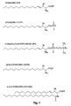
- Fig. 1 shows the chemical structure of sphingosine (SPH; D(+)-erythro-2-amino-4- trans -octadecene-1,3-diol or sphingenine), sphingosine-1-phosphate (S1P), sphingosylphosphorylcholine (SPC; lyso-sphingomyelin), ceramide (Cer, an n-acyl sphingosine) and dihydrosphingosine (DHSPH; sphinganine). All of these lipids share the sphingosine backbone containing a long-chain 18-C amino alcohol.
- sphingolipids include N, N-dimethyl-sphingosine, sphingomyelin (n-acylsphingosine-1-phosphocholine) and various glycosphingolipids (cerebrosides and gangliosides). Erythro, threo, D, L, and other isomers are also, described herein.
- the present invention concerns methods for early diagnosis of ischemic heart disease or other forms of heart failure by detecting levels of sphingosine (SPH) and/orits metabolites, alone or in conjunction with one or more other cardiac markers in a test sample from a mammal.
- SPH sphingosine
- the invention is based on the inventor's discovery that an early event in the course of heart failure, for example, that caused by cardiac ischemia, is excess production by the heart muscle of SPH and its metabolites, Cer, S1P, DHSPH, and SPC.
- Sphingolipids e . g ., SPH, S1P, DHSPH, or SPC
- SPH, S1P, DHSPH, or SPC can be extracted from the serum of patients with ischemic heart disease or controls without cardiac ischemic conditions and derivatized with a fluorescent marker (e.g., o-pthalaldehyde, OPA) for chromatographic detection.
- a fluorescent marker e.g., o-pthalaldehyde, OPA
- derivatized sphingolipids can then be detected and quantified by a variety of methodologies, including HPLC.
- TNF ⁇ inflammatory cytokines
- SPH oxidized glutathione
- SPH oxidized glutathione
- SPH oxidized glutathione
- Such cardiac hypoxia and ischemia result in a cycle whereby the acidic conditions of the ischemic heart stimulate excess SPH production which, in turn, inhibits the cell's ability to extrude protons. Increased intracellular acidic conditions further stimulate SPH production in a positive feedback manner to further increase intracellular levels of both protons and SPH.
- the inventor believes that decreased intracellular pH has profound adverse effects on the cell's contractile machinery, and that increased SPH levels cause the uncontrolled release of calcium from the sarcoplasmic reticulum membranes and the L-type calcium channel, thus preventing the cell from regulating its beat-to-beat contractile behavior.
- Sphingolipid-mediated acidosis and calcium deregulation activates apoptosis, leading to cell death and subsequent impaired cardiac function. SPH and its metabolites are useful as early indicators of heart failure because these compounds appear early in conditions such as cardiac ischemia and hypoxia, before biochemical compounds associated with cardiac cell death are released.
- This invention is based in part on the discovery that in ischemic patients, the levels of serum sphingolipids are significantly higher than those detected in non-ischemic controls. Based on the results obtained, levels of SPH that are diagnostic of heart failure associated with cardiac ischemia or hypoxia are generally greater than 100 pmol/mL. SPH levels diagnostic of cardiac ischemia or hypoxia are preferably in a range of about 200 pmol/mL to about 2,500 pmol/mL, more preferably in a range of about 300 pmol/mL to about 2,000 pmol/mL, and most preferably in a range of about 400 pmol/mL to about 1,500 pmol/mL.
- high serum (or other body fluid) levels are similarly diagnostic of cardiac ischemia or hypoxia.
- diagnostic levels are generally greater than 100 pmol/mL.
- S1P levels diagnostic of cardiac ischemia or hypoxia are preferably in a range of about 200 pmol/mL to about 2,500 pmol/mL, more preferably in a range of about 300 pmol/mL to about 2,000 pmol/mL, and most preferably in a range of about 400 pmol/mL to about 1,500 pmol/mL.
- diagnostic levels in serum are generally greater than 100 pmol/mL.
- SPC levels diagnostic of cardiac ischemia or hypoxia are preferably in a range of about 200 pmol/mL to about 2,500 pmol/mL, more preferably in a range of about 300 pmol/mL to about 2,000 pmol/mL, and most preferably in a range of about 400 pmol/mL to about 1,500 pmol/mL. Similar serum levels of DHSPH are diagnostic of cardiac ischemia.
- HPLC can be used to detect and quantify cardiac markers, including non-polypeptidic cardiac markers such as SPH in body fluids such as serum
- other methods of detecting such markers are also acceptable.
- enzymatic assays can be used to indirectly detect sphingolipids (or other non-polypeptidic cardiac markers) in test samples.
- assays include, for example, purification of sphingosine kinase from cultured cells which is used in a coupled assay employing pyruvate kinase and its substrate phosphoenolpyruvate to detect hydrolysis.
- the product of the coupled reaction is pyruvic acid, and the drop in pH resulting from this product is then detected by a variety of known methods such as detecting pH-dependent polymer breakdown that results in a measurable change in impedance.
- sphingosine kinase in blood or serum can be detected in a coupled assay employing luciferase to detect ATP hydrolysis.
- Such assays are suitable for indirectly detecting blood levels of SPH but not S1P or SPC.
- Immunodiagnostic assays using a variety of known methods, can also be used to detect cardiac markers, including non-polypeptidic cardiac markers such as sphingolipids and their metabolites, in body fluids, including blood or serum.
- cardiac markers including non-polypeptidic cardiac markers such as sphingolipids and their metabolites
- Antibodies and antibody fragments specific for Cer, SPH, DHSPH, S1P, and SPC and other such markers can be produced and used to quantitatively or semi-quantitatively detect the presence of one or more of such markers in whole blood, serum, or other body fluids using standard immunoassays.
- immunoassays that detect the presence of anti-sphingolipid (or other non-polypeptidic cardiac markers) antibodies in body fluids can be used to indirectly test for increased levels of such marker(s) in patients with chronic conditions associated with heart failure, including chronic ischemia and hypoxia.
- This assay is based on the assumption that patients experiencing such chronic conditions produce antibodies to these markers as a consequence of their elevated blood levels by analogy to the anti-lactosylsphingosine antibodies observed in patients with colorectal cancer ( Jozwiak W. & J. Koscielak, Eur. J. Cancer Clin. Oncol. 18:617-621, 1982 ) and the anti-galactocerebroside antibodies detected in the sera of leprosy patients ( Vemuri N. et al., Leprosy Rev. 67:95-103, 1996 ).
- Detection of one or more secondary markers such as TNF ⁇ can be combined with detection of one or more non-polypeptidic cardiac marker(s), such as SPH and/or its metabolites, as an early indicator of heart failure, such as may be caused by cardiac ischemia or hypoxia.
- production of secondary cardiac markers such as TNF ⁇ is also associated with heart failure (such as may be caused by cardiac ischemia) and may induce increased levels of non-polypeptidic cardiac markers such as SPH and its metabolites in body fluids ( e.g ., blood and serum)
- the diagnostic combination of the level of one or more secondary markers such as TNF ⁇ and levels of a non-polypeptidic cardiac marker such as a sphingolipid serve as a more sensitive indicator of heart failure.
- the product of the levels of the non-polypeptidic cardiac marker and the secondary marker(s) can be used to provide a quantitative measure of risk of ischemia or hypoxia referred to as the "Myocardial Risk Factor" (MRF).
- MRF
- Non-polypeptidic cardiac markers such as sphingolipids (including SPH and/or its metabolites) at levels characteristic of ischemia, hypoxia, or other conditions causally related to heart failure, preferably using a test kit, is useful for identifying these conditions in angina patients or individuals at risk for ischemic heart disease.
- the assay is also useful for diagnosis of AMI and other forms of heart failure.
- the present invention is useful for simple screening of persons at risk for heart disease for ischemic or hypoxic conditions before traditional symptoms are detected.
- the invention is also useful for following the progress of therapeutic regimes intended to treat myocardial ischemia, and thus will have important prognostic value.
- Methods and compositions of the invention can also be used for preventing the onset of AMI by allowing the patient or a health care professional to use the methods of the invention to detect the conditions that would result in AMI and taking preventive measures, such as angioplasty.
- Sphingosine (SPH; D(+)-erythro-2-amino-4- trans -octadecene-1,3-diol or sphingenine) is a lipid second messenger that the inventor has found to be endogenous to cardiac muscle tissue ( Dettbam et al., J. Mol. Cell. Biol. 26:229-242, 1994 ; Sabbadini et al., Biochem. Biophys. Res. Comm. 193:752-758, 1993 ).
- SPH is a negative inotropic agent and acts as a calcium channel agonist.
- the calcium deregulation, negative inotropy, and eventual calcium overload produced by SPH in experimental animal models resembles the pathophysiological changes that the heart experiences during ischemia or other forms of heart failure.
- the inventor has demonstrated that chronic treatment of neonatal and adult cardiac cells in culture with physiologically relevant levels of SPH and its immediate metabolite, S1P, results in the activation of cardiomyocyte cell death by apoptosis ( Krown et al., J. Clin. Invest. 98:2854-2865, 1996 ).
- Apoptosis is a form of programmed cell death, and determines the size of myocardial infarcts ( Kajstura et al., Lab. Invest. 74:86-107, 1996 ).
- Sphingosine production has been implicated as an early signaling event in apoptotic cell death in a variety of cell types ( Cuvlilier et al., Nature 381:800-803, 1996 ; Ohta et al., Cancer Res. 55:691-697, 1995 ; Ohta et al., FEBS Letters 355:267-270, 1994 ).
- Activation of the sphingomyelin signal transduction cascade is a key early event in the cytotoxic (apoptotic) effects of TNF ⁇ ( Zhang and Kolesnick, Endo.
- TNF ⁇ can cause significant apoptosis in cultured rat cardiomyocytes ( Krown et al., J. Clin. Invest. 98:2854-2865, 1996 ).
- SMase sphingomyelinase
- the lysosomal isoform of SMase could be activated by the acidic conditions of hypoxia and could complement activation of the plasma membrane isoform of SMase (neutral or nSMase).
- the nSMase of cardiomyocytes is likely activated by TNF ⁇ . TNF ⁇ is released from ischemic cardiac tissue and the TNF ⁇ -induced SPH production is an early event in cardiac ischemia.
- This invention is in part based on the belief that an early event in cardiac ischemia is TNF ⁇ -induced sphingolipid production followed by sphingolipid-dependent acidosis that results in additional sphingolipid synthesis by the acidic form of aSMase, whose source is the lysosome.
- Sphingosine is a well-known inhibitor of protein kinase C and the system of Na/H exchange which is activated by the kinase to extrude unwanted acid ( Lowe et al., J. Biol. Chem. 265:7188-7194, 1990 ).
- cardiac hypoxia, ischemia, and other conditions which cause heart failure can create a cycle whereby the acidic conditions of the ischemic, hypoxic, or otherwise failing heart stimulate excess sphingolipid production, leading to uncontrolled release of calcium from the sarcoplasmic reticulum membranes and the L-type calcium channel, thus preventing the cell from regulating its beat-to-beat contractile behavior.
- Deregulated heart calcium levels can also exacerbate the situation by promoting Na/Ca exchange and indirectly acidifying the cell by stimulation of the Na/H exchanger ( Gottling et al., Proc. Natl. Acad. Sci., USA 92:5965-68, 1995 ). Sphingolipid-mediated acidosis and subsequent calcium deregulation activate the cell death program and result in apoptosis. In the end, cardiac function suffers from the loss of cells by apoptosis as well as the negative inotropic effects of SPH and pH on surviving cardiomyocytes.
- the source of SPH for S1P formation by platelets was not discussed, nor was a potential role of SPH and/or S1P in cardiac ischemia.
- the present invention is based on the understanding that the SPH released from cardiac cells during the early stages of cardiac ischemia leaks is "secreted” or otherwise escapes into the blood from cells damaged by the hypoxic or ischemic conditions, and is acted upon by sphingosine kinase present in blood platelets.
- the S1P released from the platelets then stimulates thrombus formation.
- the SPH released from cardiac cells damaged by hypoxic or ischemic conditions eventually results in the production of a myocardial infarction.
- TNF ⁇ Tumor Necrosis Factor Alpha
- TNF ⁇ tumor necrosis factor alpha
- cytokines such as tumor necrosis factor alpha (TNF ⁇ ) have been implicated in the pathophysiology of ischemia and hypoxia. Elevated serum TNF ⁇ levels occur during hypoxic conditions associated with cardiac ischemia and reperfusion injury, and circulating TNF ⁇ levels are markedly increased after acute myocardial infarction ( Herskowitz A. et al., Am. J. Pathol. 146:419-428, 1995 ; Vaddi K. et al., Circ. 90:694-699, 1994 ; Lefer A.M. et al., Science 24:61-63, 1990 ; Maury C.P.J. & A.-M. Teppo. J. Intern.
- Pro-inflammatory cytokines such as TNF ⁇ , interleukins 1, 2, and 6 (IL-1, IL-2, and IL-6), are generally produced by myeloid-derived cells such as macrophages, neutrophils and lymphocytes ( Kelker H. et al., Int. J. Cancer 36(1):69073, 1985 ; Cuturi M. et al., J. Exp. Med. 165:1581-1594, 1987 ; Sung S. et al., J. Clin. Invest. 84(1):236-243, 1989 ; Liebermann A. et al., Proc. Natl. Acad. Sci. USA 86:6348-6352, 1989 ; Lindemann A. et al., J. Clin.
- Heart Cells are the Source of Serum TNF ⁇ and SPH
- TNF ⁇ levels in the pulmonary arteries of human subjects undergoing balloon angioplasty is greater than the serum levels of TNF ⁇ found in the femoral veins of the same patients, which data suggests that, during ischemia (induced by balloon inflation), the ischemic heart tissues produce TNF ⁇ which then is released into the general circulation from the coronary sinuses and the pulmonary artery.
- the TNF ⁇ in the pulmonary artery of coronary angioplasty patients correlates well with changes in pulmonary artery SPH levels. Based on these data, it is believed that the cardiac source of TNF ⁇ is a major stimulus for cardiac cell SPH production.
- Skeletal muscle mass represents 30-40% of total body weight and could represent a very large source of serum SPH.
- Skeletal muscle ischemia was induced by asking the subjects to exercise to exhaustion on treadmills placed in a 49 °C room, and was confirmed by measuring serum lactate.
- the level of the non-polypeptidic and/or secondary cardiac marker(s) or the MRF calculated for a test sample is compared with a predetermined value for that marker in order to determine if evidence of heart failure, such as may be induced by cardiac ischemia or hypoxia, exists.
- the predetermined value for one or more of such markers can be established by one of at least two ways. For example, it can be established by gathering data from the mammal (e.g ., a human) at risk of AMI prior to the onset of signs for heart disease, or by testing other healthy mammals in preferably the same species and age group as the patient.
- the physician treating the patient may determine that the patient, based on statistical, genetic, familial, or other factors generally known in the art of medicine, is at risk of an AMI.
- the physician can then determine the level of one or more non-polypeptidic cardiac markers or the MRF for the patient to establish a baseline.
- the physician may also determine the level of one or more secondary markers (e . g ., TNF ⁇ , IL-1, 2, 6, or another cytokine) to establish a baseline.
- the methods of the invention provide for the comparison of the level of the non-polypeptidic cardiac marker(s) and/or secondary marker(s) in the patient with this baseline in order to detect impending heart failure, such as may be caused by cardiac hypoxia or ischemia.
- the physician or other health care professional can determine the level of the cardiac marker(s) or the MRF in individuals determined to be healthy by a physician.
- the levels of the individuals in the same age group can be grouped together and their average and standard deviation determined. This value will represent the predetermined value to which the level of the cardiac marker in the patient will be compared in order to detect cardiac hypoxia or ischemia.
- the methods of the present invention allow for early detection of cardiac ischemia or hypoxia, i . e ., conditions that lead to AMI and other forms of heart failure.
- cardiac ischemia or hypoxia i . e .
- the patient can present himself or herself to an emergency medical facility, where measures can be taken in order to prevent heart failure from occurring.
- measures include, but are not limited to, angioplasty, coronary bypass surgery, or administration of one or more anticoagulant or thrombolytic drugs.
- preventive measures are to alleviate the ischemic or hypoxic conditions prior to the onset of AMI or other types of heart failure.
- the above measures are used after an AMI has occurred, or while the patient is experiencing heart failure.
- the present invention allows for early detection of conditions which cause heart failure, prior to the onset of the symptoms and the tissue damage associated therewith.
- the methods of the present invention allow physicians and other health care professionals to determine the success of a cardiac procedure immediately following the procedure. For instance, following angioplasty or stent placement in a cardiac artery, the level of one or more cardiac marker(s) or the MRF can be monitored as described herein to determine whether the ischemic or hypoxic conditions are being alleviated. Thus, the success of the operation can be immediately determined. If the procedure did not result In the desired results, as determined by the level of the cardiac marker(s) or the MRF measured, then further procedures can be employed prior to the patient suffering a complete or partial heart failure.
- D-Sphingosine [D(+)-erythro-2-amino-4-trans-octadecene-1,3-diol] from Matreya, Inc. (Pleasant Gap, PA); sphingosine-1-phosphate from Biomol (Plymouth Meeting, PA); o-Pthalaldehyde from ICN Biochemicals (Cleveland, OH) and HPLC grade methanol from Fisher Scientific (Tustin, CA).
- Other chemicals including sphingosylphosphorylcholine and DL-erythro-dihydrosphingosine, were obtained from Sigma Chemical Co. (St. Louis, MO).
- Stock solutions of sphingosine and other sphingolipids were prepared as complexes with fatty acid-free bovine serum albumin (BSA) to provide solutions of the compounds that are essentially free of micelles or organic solvents.
- BSA bovine serum albumin
- Sphingosine levels were determined by HPLC performed essentially as described previously (Sabbadini, et al., 1993). Briefly, fresh tissue was excised from the animal and homogenized at 5°C in the presence of four volumes of 50 mM potassium phosphate buffer, pH 7.0. Similarly, SPH can be extracted from whole blood or serum samples. Samples (300 ⁇ L) of the crude homogenates, blood or serum were added to 750 ⁇ L of a chloroform:methanol solution (1:2) and vortexed several times. Chloroform (about 500 ⁇ L) was then added, followed by 500 ⁇ L of 1 M NaCl. The extract was centrifuged briefly to promote phase separation and the upper aqueous phase was removed by aspiration.
- the NaCl extraction step was repeated twice and the organic phase was vacuum dried with centrifugation for 30 min.
- the extracts were derivatized for 10 min. with 50 ⁇ L of a solution of 0.5 mg/mL o-pthalaldehyde (OPA), 3% boric acid, pH 10.5, and 0.05% ⁇ -mercaptoethanol.
- OPA o-pthalaldehyde
- the sphingolipids were separated by reverse-phase chromatography on a 250 H 7 mm C18, 300 Angstrom pore Brownlee column (Applied Biosystems, Foster City, CA) fitted with an Aquapor C18 guard column. Samples were run isocratically at 1.25 mL/min using the running buffer, resulting in an efficiency of extraction of about 50% based on the recovery of SPH standards. The retention times for the standards were: about 15 min for SPH; about 5 min for S1P, and about 20 min for SPC.
- Example 1 Serum SPH Leves in Human Patients Experiencing Cardiac Ischemia
- Serum samples were taken from patients presenting themselves to an emergency medical facility, under a strict human subject protocol. Three patient groups were examined for serum levels of both SPH and TNF ⁇ : (1) patients suspected of AMI and subjected to exercise stress testing; (2) patients undergoing coronary angioplasty; and (3) patients in the early phases of acute myocardial infarction.
- Serum from three control groups not exhibiting any clinical symptoms of myocardial ischemic disorders was also tested. These control groups were: (1) age-matched subjects (47 to 79 yrs old) enrolled in an adult fitness program; (2) healthy military personnel at rest and exercising to exhaustion on treadmills at 49 °C (120 °F) ambient temperature; and (3) athletes at an Olympic Training Center at rest and exercising to exhaustion on treadmills at 49 °C ambient temperature.
- an average serum SPH level of 697 ⁇ 0.7 pmol/mL was obtained. This value was about 7-fold higher than the age-matched control group and about 160-fold higher than well-conditioned military personnel and athletes undergoing severe exercise stress.
- the levels of biochemical markers known to be associated with cardiac tissue damage were also determined.
- the increased SPH levels detected were consistent with high levels of CK (in the range of 17-810 U/mL) and high levels of CKMB (in the range of 0.62-33.2 mg/L).
- two patients (P2, P3) who had high CKMB mass levels did not have abnormally high levels of SPH.
- the other patient (P3) displayed a negligible level of serum SPH (18.1 pmol/mL), a normal CK level (161 U/L) and a high CKMB level (18.4 mg/L).
- the moderate-to-low SPH levels detected for these two patients represent a 11 % false negative rate (2 of 18), because both patients were considered ischemic based on other indicators.
- the inclusion of the SPH data for these two patients (P2 and P3) in the AMI group also accounts for the somewhat lower SPH levels in the AMI group (587 ⁇ 7 pmol SPH/mL), compared to the SPH levels for the ischemic patients undergoing coronary angioplasty (885 ⁇ 123 pmol/mL).
- Patient T2 was a 47 year-old female who presented herself to the emergency room of a hospital with complaints of angina. Blood samples were drawn and the patient was then referred to a hospital's exercise stress test facility. The patient passed the treadmill test, showing no evidence of cardiac ischemia or AMI as detected by an ECG administrated during the stress test and by serum enzyme levels analyzed during her period of evaluation in the hospital. The patient was discharged from the hospital, but returned three weeks later with evidence of an AMI as determined by high serum levels of CK and CKMB and other clinical symptoms.
- serum SPH level was an early indicator of ischemia and more predictive of her cardiac condition than the exercise stress test or the serum CK and CKMB analysis, both of which showed no evidence of cardiac ischemia or AMI on her first visit to the emergency room.
- Patient MI-12 was a male who was admitted to the hospital with a confusing clinical presentation. When admitted, his CK level (126 U/L) was normal, but his CKMB level (3.8 mg/L) was elevated. His serum SPH level at admission was also high (732 pmol/mL), indicating ischemia. At about five hours after admission, this patient had an AMI. This result shows that the serum SPH level, but not by the CK parameter, accurately detected his ischemic condition. Thus, the results of the SPH test, alone or combined with the CKMB parameter, were predictive of imminent AMI.
- This patient was a 58 year-old female who was admitted to the hospital for an initial evaluation of her heart condition.
- blood was drawn and the serum showed normal CKMB (0.632 mg/L) but moderately high SPH (300 pmol/mL).
- SPH 300 pmol/mL
- her serum SPH level was significantly reduced to 7.31 pmol/mL, whereas her serum CK (934 U/L) and CKMB (88.9 mg/L) levels remained high.
- SPH being an accurate indicator of ischemia
- CK and CKMB being indicators of myocardial damage (i.e ., large molecular weight cytoplasmic proteins that are released from necrotic myocardial cells).
- myocardial damage i.e ., large molecular weight cytoplasmic proteins that are released from necrotic myocardial cells.
- serum SPH is more predictive of the early stages of ischemia and imminent AMI than current methods generally used in diagnosis.
- serum SPH is quantitatively related to the early events that precede cardiac cell death, in contrast to other biochemical markers (e . g ., CK, CKMB and troponin) that are released after cell death.
- routine screening of patients for serum SPH can aid in the early diagnosis of coronary artery disease and identification of patients at high risk of heart disease who can be treated to prevent AMI.
- Quantitative detection of a single sphingolipid such as SPH may be subject to variability which can be minimized by combining the measurement with detection of serum TNF ⁇ a to provide a more general index of risk of cardiac ischemia or hypoxia.
- tissue levels of SPH were examined in adult rabbit hearts subjected to retrograde coronary perfusion with hypoxic (i.e ., low oxygen) conditions (95% CO 2 ; 5% O 2 ) or with normal Krebs buffers (containing 5% CO 2 ; 95% Q ). The hearts were removed, quickly homogenized, and sphingolipids were extracted and detected as described above.
- hypoxic i.e ., low oxygen
- normal Krebs buffers containing 5% CO 2 ; 95% Q
- HPLC analysis of the extracts revealed significant increases (20-fold) in tissue SPH levels for hearts perfused with buffer containing 95% CO 2 when compared to control conditions. Moreover, these increases occurred after only 5 min. of treatment.
- SPH is a cationic amphipathic lipid that can partition into whole blood and other body fluids. Therefore, the relative amounts of SPH were determined in whole blood and serum obtained from humans, rats, and rabbits. SPH was found predominantly in the serum, the preferred body fluid for measuring SPH levels for detecting cardiac ischemia or hypoxia.
- a human serum sample was obtained and allowed to sit at 22 °C for 5 hrs before sphingolipid and HPLC analysis. Serum samples were also spiked with commercially available SPH and similarly stored at room temperature. Neither the aged control serum samples nor the spiked samples showed appreciable differences in SPH levels compared to samples that were assayed immediately after collection or preparation of the spiked samples, indicating that SPH does not undergo degradation if serum samples were not assayed immediately after collection.
- Serum levels of TNF ⁇ also increase in cardiac ischemia and correlate with the serum SPH levels detected in angioplasty patients.
- the product of these two parameters e . g ., the levels of a non-polypeptidic cardiac marker and a secondary [or tertiary] cardiac marker
- MRF myocardial risk factor
- TNF ⁇ was measured using standard methods in an enhanced ELISA double antibody capture assay (using a Quantikine HS kit (Cat. No. HSTA50) kit, R+D Systems, Minneapolis, MN). Human serum samples (200 ⁇ L) were assayed and compared to recombinant human TNF ⁇ standards using the assay procedures provided by the manufacturer.
- Serum TNF ⁇ and SPH levels were determined from samples taken from the pulmonary artery of a patient (A5) experiencing the periodic ischemia (e.g ., during balloon inflation) and reperfusion (e.g ., when the artery is cleared) that occurs during successful angioplasty.
- Serum TNF ⁇ and SPH levels were both elevated prior to initiation of the angioplasty procedure (from -20 min to 0 min). Neither the TNF ⁇ nor the SPH increased during the ischemic period. They were already high and went down after reperfusion. Both TNF ⁇ and SPH parameters showed a biphasic response, with initial decrease after angioplasty followed by a steady rise back to pre-angioplasty levels. The time courses were similar with TNF ⁇ levels falling slightly ahead of SPH in time. This is consistent with TNF ⁇ being the trigger for SPH production.
- serum TNF ⁇ levels were assessed for 19 patients undergoing coronary angioplasty and for seven AMI patients.
- serum TNF ⁇ levels were determined for two control groups (age-matched controls and healthy military personnel). Serum TNF ⁇ levels were highest in the AMI patients (5.2 ⁇ 0.6 pg/mL), and somewhat lower in the pulmonary artery blood taken from the angioplasty group before the procedure was done (3.6 ⁇ 0.74 pg/mL). The patients had significantly higher serum TNF ⁇ levels (4.04 ⁇ 0.57 pg/mL) compared to those of the age-matched control group ("Controls") of 2.43 ⁇ 0.32 pg/mL, and the healthy military subjects ("Athletes”) of 1.22 ⁇ 0.29 pg/mL.
- serum TNF ⁇ levels showed the same trends as seen for the serum SPH levels in the three groups' tests. That is, the athletes had the lowest levels of SPH and TNF ⁇ , the age-matched controls had higher levels of SPH and TNF ⁇ , and the patients had the highest levels of SPH and TNF ⁇ . Therefore, a more accurate measure of on-going myocardial ischemia can be obtained by combining two or more parameters, e.g ., the levels of a non-polypeptidic and a secondary or tertiary cardiac marker trending in the same direction, to calculate a MRF.
- the MRF is the product obtained by multiplying the numerical value of the serum TNF ⁇ level and the numerical value of the serum SPH level.
- MRF data (using SPH and TNF ⁇ levels) for all ischemic patient groups in this experiment were combined and compared to age-matched and healthy military controls.
- the MRF of the ischemic patients (2820) was about 12-fold higher than that of the age-matched control group (238) and about 440-fold higher than that of the healthy military group (6.3).
- the MRF value distinguished ischemic patients from controls to a greater extent than did either serum SPH or serum TNF ⁇ measurement alone.
- the calculated MRF value reduced the occurrence of false negatives for either parameter alone.
- Table 1 shows the SPH/TNF ⁇ MRF value calculated for seven patients. The values were calculated by multiplying the SPH value (approximated to the nearest whole integer) and the TNF ⁇ value (approximated to the nearest one-tenth) to produce the MRF value (to the nearest whole integer).
- Six of the seven angioplasty patients had a MRF in the range of about 1,800 to about 3,000 range. The mean MRF value for these six patients was 2,326. Only one patient had a MRF outside of that range (patient A1 with MRF of about 16,000). The mean MRF for all seven patients is shown in Table 1.
- patient A1 appeared to have an atypically high level of ischemia, with the TNF ⁇ level being significantly higher than that of the other six patients. If the SPH level of patient A1 is multiplied by the mean TNF ⁇ value of the other six patients (2.9), the MRF value would be 3,254 which is close to the range seen for the other six patients.
- Serum SPH was predictive of the early stages of ischemia and imminent AMI more frequently than other currently used methods, although false negative results were detected with a few AMI patients.
- level of SPH detected provides a quantitative measurement of early events that precede cardiac cell death.
- other biochemical markers e.g ., CK and CKMB
- the assay is also useful for monitoring the efficacy of cardiac treatments (e.g ., bypass surgery or angioplasty). Testing of serum SPH levels is useful as a routine diagnostic of cardiac ischemia and hypoxia, permitting patients at risk of heart disease to be identified and treated. The method is also useful for monitoring patients during or after treatment for cardiac conditions to detect the level of cardiac ischemia or hypoxia as an indicator of the success of the treatment.
- kits that includes assays for TNF ⁇ and sphingolipids is used to provide a measure of the MRF.
- assays for TNF ⁇ and sphingolipids are used to provide a measure of the MRF.
- anti-TNF ⁇ and anti-SPH antibodies/or an enzyme assay for sphingolipids are combined in a kit to assess the MRF value.
- known biosensor technology can be used for the determination of two or more analytes in blood or serum.
- An algorithm may be used in the kit to calculate the MRF value, which may be particularly advantageous for detecting all three important sphingolipids, SPH, S1P and SPC, as well as TNF ⁇ , and determining a series of MRF values for all of the combinations of sphingolipid and TNF ⁇ , or for determining a single MRF value that is the product of all the measured sphingolipid levels and the TNF ⁇ level.
- Neonatal ventricular myocytes were dissociated from hearts obtained from one to four day old Sprague-Dawley rats essentially as described previously ( Shields et al., J. Biol. Chem. 263:12619-12618, 1988 ). Ventricles were finely minced and dissociated with 0.5 g/L trypsin, 0.2 g/L EDTA in a calcium-free and magnesium-free Hank's buffered salt solution (Sigma, St. Louis, MO). The tissues were agitated, pelleted gently by centrifugation and the trypsin digestions were repeated five times.
- the supernatants containing cells in suspension were combined with DMEM/F12 medium, filtered through 125 ⁇ m nylon mesh, and the filtrate was centrifuged.
- the pelleted cells were resuspended in DMEM/F12 medium and plated on fibronectin-coated glass coverslips in DMEM/F12 medium plus 10% fetal calf serum (FCS).
- FCS fetal calf serum
- Freshly dissociated adult ventricular myocytes were prepared from hearts of adult (200 to 350 g) Sprague-Dawley rats by enzymatic dissociation using a Langendorff retrograde aortic perfusion apparatus. After perfusing the hearts with collagenase (Type II, Worthington Biochemicals, Freehold, NJ), rat ventricles were diced and incubated for 30 min at 37 °C in 10 to 15 mL of 0.58 mg/mL collagenase in oxygenated Tyrode's solution (140 mM NaCl, 5.4 mM KCl, 5.0 mM MgCl 2 , 1.0 mM CaCl 2 , 10 mM HEPES, 0.25 mM NaH 2 PO 4 , pH 7.3).
- collagenase Type II, Worthington Biochemicals, Freehold, NJ
- the dissociated myocytes were plated on laminin-coated (50 ⁇ g/mL) culture dishes and cultured for 18 hr in DMEM/F12 plus 10% FCS, in the presence or absence of the test agent (e.g ., LPS or control buffer).
- the test agent e.g ., LPS or control buffer.
- the elevated serum SPH levels detected in the ischemic patients were not produced by skeletal muscle ischemia as demonstrated by assaying serum samples from healthy athletes and military personnel before and after inducing severe skeletal muscle ischemia.
- serum SPH Prior to the exercise regime, serum SPH averaged 4.18 ⁇ 4.5 pmol/mL, which slightly decreased to 4.02 ⁇ 3 pmol/mL after exercise. These serum. SPH values were substantially lower than those detected in ischemic patients.
- serum TNF ⁇ levels were not increased in these subjects undergoing severe skeletal muscle ischemia (for example, serum TNF ⁇ values for the military personnel were 1.22 ⁇ 0.49 pg/mL before exercise and rose insignificantly to 1.39 ⁇ 0.23 pg/mL after exercise).
- Example 5 Use of Anti-sphinaolipid Antibodies to Detect Sphingolipids in Whole Blood or Serum
- Anti-sphingolipid (or other non-polypeptidic cardiac markers) monoclonal antibodies are prepared by methods similar to those used in the preparation of anti-phospholipid antibodies. Briefly, one method of mAb production involves direct immunization of sphingolipid-coated, acid-treated Salmonella minnesota directly into a mouse spleen using known methods used to make anti-phospholipid mAbs ( Reza et al., FEBS Lett. 339:229-233, 1994 ; Umeda et al., J. Immunol. 143:2273-2279, 1989 ).
- the acid-treated S . minnesota is coated with the desired sphingolipid, e.g ., SPH, and injected into the mouse spleen prior to cell fusion to produce a hybridoma that secretes anti-SPH mAb. Similar methods are used to produce anti-S1P mAb and anti-SPC mAb. Alternately, fatty acid free BSA-sphingolipid conjugates can be used as the immunogen in order to present unique epitopes to the animal. Care must be taken to ensure that mAbs are not produced to oxidized lipid or protein-lipid adducts [see discussion by Witztum et al. in refs. ( Horkko et al., J. Clin. Invest. 98:815-825, 1996 ; Palinski et al., J. Clin. Invest. 98:800-814, 1996 )].
- Lateral flow formats involve double capture antibody technology where the analyte in the blood sample is captured by the first antibody tethered to the substrate. A strept-avidin system is then used to detect binding of the second antibody.
- Biosensor technology typically uses surface plasmon resonance to detect refractive index changes on the surface of a gold/glass matrix as the antigen (e . g ., the sphingolipid) binds to the tethered antibody (e.g ., anti-SPH) [ see for example, Kuziemko et al., Biochem. 35:6375-6384, 1996 , where cholera toxin binding to gangliosides, including lactosyl ceramides, was studied].
- Biosensors are preferred because they are very rapid (develop in minutes), quantitative, and are amenable to use with multiple ligands. Algorithms and digital readouts are possible with biosensors.
- Biosensor format-based immunoassays for detection of SPH, S1P, and SPC can be performed individually, to provide independent measurements of each of the sphingolipids as indicators of ischemic cardiac conditions.
- a single assay could include multiple mAbs to provide a single measurement of any combination of sphingolipids (e.g ., SPH and S1P; SPH and SPC; S1P and SPC; or SPH, S1P and SPC), alone or in combination with one or more other markers, e.g ., TNF ⁇ .
- This method involves purification of sphingosine kinase and its use in a coupled assay employing pyruvate kinase and its substrate phosphoenopyruvate to detect ATP hydrolysis.
- the product of the coupled reaction is pyruvic acid and the resulting change in pH is used in a kit that takes advantage of pH-dependent polymer breakdown technology (as described in Serres, A. et al., Pharmaceutical Res. 13(2):196-201, 1996 ) and the subsequent changes in impedance that are measured.
- Sphingosine kinase is isolated from Swiss 3T3 cells or other cells as previously described ( Olivera A., et al., Anal. Biochem. 223(2):306-312, 1994 ).
- the assay includes the following features:
- the substrate of the test strip is coated with a pH-sensitive linear terpolymer (e.g ., a derivative of poly(N-isopropylacrylamide-co-burylmethacrylate-co-acrylic acid).
- a blood or serum sample is dropped onto the test strip and the coupled reaction precedes.
- the pH drops by the coupled enzyme assay the decrease in pH is proportional to the amount of SPH in blood
- the polymer breaks down and exposes the conductor on the test strip.
- An impedance measurement is then made which is proportional to the amount of SPH in the blood or serum sample that was dropped onto the strip.
- This technique is used to screen phage which encode receptors which bind with high affinity to the markers, particularly the non-polypeptidic cardiac markers, of the invention, and express them on their surfaces. See U.S. Patent Nos. 5,223,409 and 5,403,484 for a detailed description of phage display technology. Bacteria, which express the receptor, are then cloned and used in a biosensor-based kit. This technique can be used to detect the markers used in the practice of this invention, including all important sphingolipids, including SPH, S1P, DHSPH, and SPC. Because the techniques used to isolate receptors for any marker are substantially the same, the techniques are described generically herein. This method is described in more detail in McGregor, D., Mol. Biotech. 6(2):155-162, 1996 .
- a cDNA library is provided (e.g ., a cDNA library that is available from a number of commercial sources, including Clontech, San Diego, CA) and then used to create fusion proteins with a membrane protein of a filamentous male phage such as M13.
- Phage clones expressing the fusion protein containing the receptor for the desired marker, e.g ., SPH are detected and isolated using standard ligand binding assays.
- the gene for the receptor is then excised from positive clones using standard endonuclease restriction enzymes and cloning methods ( see, e.g ., J. Sambrook, Molecular Cloning, A Laboratory Manual, 2nd Ed., CSH Lab. Press, 1989 ).
- the gene may also be expressed in a bacterial system using standard methods to yield unlimited quantities of the receptor.
- the receptor is purified using standard methods and is then used to detect the marker for which the receptor demonstrates specificity.
- the purified receptor is tethered to an ELISA plate, to a Biacore dextran surface, to a test strip of any of a number of detection kits, to biosensor detectors and the like, and used to measure the quantity of a sphingolipid in the blood or serum.
- Other applications taking advantage of marker binding to its membrane receptor are also envisioned.
- This method is based on the assumption that patients experiencing ischemia produce anti-sphingolipid antibodies as a consequence of elevated blood levels of sphingolipids such as sphingosine. It is also based on the findings that anti-lactosylsphingosine antibodies have been observed in patients with colorectal cancer (Jozwiak W. and J. Koscielak, 1982) and anti-galactocerebrosides were detected in the sera of leprosy patients (Vemuri N. et al., 1996).
- sphingosine and its metabolites are suggested by their structures as a cationic amphiphiles ( e . g ., see Fig. 1 ) and by the finding that antibodies can be generated against phospholipids (Umeda M. et al., 1989) and glycosphingolipids (Vemuri N. et al., 1996).
- anti-glycosphingolipid antibodies were detected in the serum of calves experimentally infected with T . saginata ( Baumeister S. et al., Parasitol. Res. 81:18-25, 1995 ).
- This technique can be applied to detecting any sphingolipid or sphingolipid metabolite, including SPH, DHSPH, S1P and SPC, as well as other non-polypeptidic cardiac markers used in the practice of this invention.
- Finger stab dipstick technology is widely used for blood glucose monitoring and this method can be adapted to the measurement of blood-bome cardiac markers (e.g ., non-polypeptidic, secondary, and tertiary markers) in a drop of blood, given the disclosure herein.
- blood-bome cardiac markers e.g ., non-polypeptidic, secondary, and tertiary markers
- the cardiac markers used in the practice of the invention including sphingosine and its derivatives, can also be measured in many tissues and body fluids besides blood, including saliva, sweat, and urine.
- Other home monitoring devices useful in the practice of this invention include those analogous to in-home pregnancy tests, where the level of a particular marker or set of markers is measured in urine.
- a portable electronic measurement device can also be applied in the practice of this invention.
- a wrist-wom device similar in size and shape to a wristwatch has been developed for monitoring blood sugar levels in diabetics.
- the Cygnus GlucoWatchTM platform allows an individual to continuously monitor glucose through intact skin for more accurate assessment of the analyte at all times and without the discomfort of the finger stab technique.
- real-time continuous or periodic monitoring of one or more cardiac markers in accordance with the instant methods could be accomplished by wristwatch platforms.
- the measurement of non-polypeptidic cardiac markers (and secondary and tertiary markers, if desired) according to the invention would occur directly through the skin, and would be compared with marker levels indicative of cardiac conditions associated with ischemia, hypoxia, and others correlating with various forms of heart failure. Downloading of stored data from such a device having data logging capability is also envisioned, and would provide the clinician with a record of the patient's recent history of marker level changes.
- Such a device, or other home monitoring device designed to monitor cardiac marker levels according to the invention can be used in conjunction with an alarm system that is activated when one or more cardiac marker level (or a calculated index such as MRF) exceeds a certain threshold.
- the alarm may inform the patient, i.e ., wearer, or a third party, e . g ., a friend or relative, a health care professional, or emergency response personnel, such as the police, paramedics, or the fire department of a change in the level of the marker(s) being monitored.
- Such an alarm when transmitted, may also include telemetry.
- a home monitoring device will preferably be accompanied by instructions for use.
- the device may also be accompanied with a notice in form prescribed by a governmental agency regulating the manufacture, use, or sale of medical devices, which notice is reflective of approval by the agency of the form of the device for home use.
- a notice for example, may be the labeling approved by the U.S. Food and Drug Administration, or the equivalent governmental agency in other countries, for medical devices, or the approved product insert.
- This embodiment of the invention employs a coupled assay to assess sphingosine levels in blood or other tissues or bodily fluids by taking advantage of the ability of sphingosine and other lysosphingolipids to inhibit protein kinase C (PKC) ( Hannun and Bell, Science 234:670-674, 1987 ; Hannun et al., J. Biol. Chem. 261:12604-12609, 1986 ).
- PKC protein kinase C
- the amount of sphingolipid in the blood sample is quantitatively related to the change in absorbance at 340 nm as a stoichiometric amount of NADH is oxidized by the coupling system as described by Sabbadini and Okamoto ( Sabbadini and Okamoto, Arch. Biochem. Biophys. 223:107-119, 1983 ).
- sphingosine inhibits PKC by preventing DAG binding to the enzyme ( Faucher et al., J. Biol. Chem. 263:5319-5327, 1988 ). Thus, sphingosine may bind directly to PKC via the DAG binding site.
- the sequence for PKC ⁇ and its consensus DAG binding site is known ( Hurley et al., Protein Science (6):477-80, 1997 ). Since SPH can also bind to putative sites on sphingosine kinase and other proteins with which is specifically interacts, it quite likely that several proteins have specific SPH binding sites.
Landscapes
- Life Sciences & Earth Sciences (AREA)
- Health & Medical Sciences (AREA)
- Engineering & Computer Science (AREA)
- Molecular Biology (AREA)
- Chemical & Material Sciences (AREA)
- Biomedical Technology (AREA)
- Urology & Nephrology (AREA)
- Hematology (AREA)
- Immunology (AREA)
- Cell Biology (AREA)
- Analytical Chemistry (AREA)
- Biotechnology (AREA)
- Pathology (AREA)
- Food Science & Technology (AREA)
- Medicinal Chemistry (AREA)
- Physics & Mathematics (AREA)
- Microbiology (AREA)
- Biochemistry (AREA)
- General Health & Medical Sciences (AREA)
- General Physics & Mathematics (AREA)
- Proteomics, Peptides & Aminoacids (AREA)
- Biophysics (AREA)
- Endocrinology (AREA)
- Investigating Or Analysing Biological Materials (AREA)
- Medicines Containing Antibodies Or Antigens For Use As Internal Diagnostic Agents (AREA)
Claims (47)
- Procédé de détection ou de surveillance de l'ischémie cardiaque ou de l'hypoxie cardiaque chez un mammifère, comprenant :a. mesurer le niveau de sphingosine ou d'un métabolite de sphingosine dans un échantillon provenant du mammifère ; etb. déterminer si le niveau de sphingosine ou d'un métabolite de sphingosine dans l'échantillon est en corrélation avec l'ischémie cardiaque ou l'hypoxie cardiaque.
- Procédé selon la revendication 1, dans lequel l'échantillon de test est un fluide corporel ; en option dans lequel le fluide corporel est sélectionné parmi le groupe constitué du sang, de l'urine, de la lymphe et de la salive ; de préférence dans lequel le fluide corporel est du sang.
- Procédé selon la revendication 1 ou la revendication 2, dans lequel le niveau de sphingosine ou d'un métabolite de sphingosine est une concentration de celui-ci.
- Procédé selon l'une quelconque des revendications précédentes, dans lequel le métabolite de sphingosine est sélectionné parmi le groupe constitué de céramide (Cer, n-acylsphingosine), sphingosine-1-phosphate (S1P), sphingosylphosphorylcholine (SPC) et dihydrosphingosine (DHSPH).
- Procédé selon l'une quelconque des revendications précédentes, dans lequel l'étape de détermination comprend la mesure du niveau de sphingosine ou d'un métabolite de sphingosine par un procédé sélectionné parmi le groupe constitué de la chromatographie, l'immunoessai, le dosage enzymatique et la spectroscopie.
- Procédé selon la revendication 5, dans lequel le niveau de sphingosine ou d'un métabolite de sphingosine est mesuré directement.
- Procédé selon la revendication 5, dans lequel le niveau de sphingosine ou d'un métabolite de sphingosine est mesuré indirectement.
- Procédé selon l'une quelconque des revendications précédentes, dans lequel l'étape de détermination est un procédé chromatographique sélectionné parmi le groupe constitué de la chromatographie liquide à haute performance et la chromatographie gazeuse.
- Procédé selon l'une quelconque des revendications précédentes, dans lequel l'étape de détermination est un procédé spectroscopique sélectionné parmi le groupe constitué de la spectroscopie par ultraviolet, la spectroscopie par infrarouge et la spectroscopie par résonance magnétique nucléaire.
- Procédé selon l'une quelconque des revendications 1 à 6, dans lequel l'étape de détermination est un immunoessai qui détecte la sphingosine ou un métabolite de sphingosine sélectionné parmi le groupe constitué de céramide (Cer, n-acylsphingosine), sphingosine (SPH), sphingosine-1-phosphate (S1P), dihydrosphingosine (DHSPH) et sphingosylphosphorylcholine (SPC).
- Procédé selon l'une quelconque des revendications 1 à 6 ou 10, dans lequel l'étape de détermination est un immunoessai qui détecte la sphingosine ou un métabolite de sphingosine dans l'échantillon de test en utilisant un anticorps anti-sphingosine ou un anticorps anti-métabolite de sphingosine.
- Procédé selon la revendication 11, dans lequel l'anticorps est sélectionné parmi un fragment d'anticorps, un anticorps à chaîne unique et un domaine de liaison à l'antigène d'un anticorps.
- Procédé selon l'une quelconque des revendications précédentes, dans lequel l'étape de détermination est une comparaison entre le niveau mesuré de la sphingosine ou d'un métabolite de sphingosine et une valeur prédéterminée.
- Procédé selon la revendication 13, dans lequel la valeur prédéterminée pour le niveau de la sphingosine ou d'un métabolite de sphingosine est indicatrice d'une fonction cardiaque normale.
- Procédé selon la revendication 14, dans lequel la valeur prédéterminée pour le niveau de la sphingosine ou d'un métabolite de sphingosine est obtenue à partir d'un mammifère de la même espèce et d'environ le même âge que le mammifère à partir duquel l'échantillon de test a été obtenu,
- Procédé selon l'une quelconque des revendications 13 à 15, dans lequel le niveau mesuré de sphingosine ou d'un métabolite de sphingosine diffère de la valeur prédéterminée pour celui-ci.
- Procédé selon la revendication 16, dans lequel le niveau mesuré de sphingosine ou d'un métabolite de sphingosine est supérieur à la valeur prédéterminée pour celui-ci.
- Procédé selon la revendication 16, dans lequel le niveau mesuré de sphingosine ou d'un métabolite de sphingosine est inférieur à la valeur prédéterminée pour celui-ci.
- Procédé selon l'une quelconque des revendications précédentes, comprenant en outre :(a) mesurer un niveau d'un second marqueur cardiaque dans l'échantillon ; et(b) déterminer si le niveau du second marqueur cardiaque est en corrélation avec l'ischémie cardiaque ou l'hypoxie cardiaque,
- Procédé selon la revendication 19, dans lequel le second marqueur est une cytokine pro-inflammatoire, de préférence TNF-alpha.
- Procédé selon la revendication 19 ou la revendication 20, comprenant en outre l'étape de calcul d'un facteur de risque myocardique, dans lequel le facteur de risque myocardique a un rapport mathématique avec un niveau mesuré de sphingosine ou d'un métabolite de sphingosine et le niveau mesuré du second marqueur cardiaque.
- Procédé selon la revendication 21, dans lequel le rapport mathématique est un produit des niveaux mesurés d'une sphingosine ou d'un métabolite de sphingosine et du niveau mesuré du second marqueur cardiaque.
- Procédé selon l'une quelconque des revendications précédentes, dans lequel le mammifère est l'homme.
- Procédé de diagnostic d'un patient à risque d'infarctus aigu du myocarde comprenant la détection d'ischémie ou d'hypoxie cardiaque selon un procédé selon l'une quelconque des revendications 1 à 23.
- Procédé de diagnostic et de détermination du pronostic d'un patient suite à une procédure cardiaque comprenant la détection d'ischémie ou d'hypoxie cardiaque selon un procédé selon l'une quelconque des revendications 1 à 23.
- Procédé selon la revendication 25, dans lequel la procédure cardiaque est sélectionnée parmi le groupe constitué de la chirurgie de pontage coronarien, l'angioplastie et l'administration d'un anticoagulant.
- Utilisation d'un anticorps ou fragment de celui-ci qui se fixe spécifiquement à la sphingosine ou un métabolite de sphingosine in vitro, pour la détection ou surveillance de l'ischémie cardiaque ou l'hypoxie cardiaque chez un mammifère, dans laquelle la sphingosine ou un métabolite de sphingosine est sélectionné parmi le groupe constitué de céramide (Cer, n-acylsphingosine), sphingosine (SPH), sphingosine-1-phosphate (S1P), dihydrosphingosine (DHSPH) et sphingosylphosphorylcholine (SPC).
- Utilisation selon la revendication 27, dans laquelle l'anticorps ou fragment d'anticorps est un anticorps monoclonal ou un anticorps polyclonal.
- Utilisation selon la revendication 27 ou la revendication 28, dans laquelle l'anticorps ou fragment d'anticorps est un anticorps à chaîne unique ou un domaine de liaison à l'antigène d'un anticorps.
- Utilisation selon l'une quelconque des revendications 27 à 29, comprenant en outre un support solide auquel l'anticorps ou fragment d'anticorps est fixé.
- Utilisation selon la revendication 30, dans laquelle le support solide est sous la forme d'une plaque, d'un tube ou d'une bille.
- Utilisation selon la revendication 30 ou la revendication 31, dans laquelle le support solide est composé d'un matériau sélectionné parmi le groupe constitué du verre, du polystyrène, du nylon, de l'acétate de cellulose, de la nitrocellulose et d'autres polymères.
- Utilisation selon l'une quelconque des revendications 30 à 32, dans laquelle l'anticorps ou fragment d'anticorps est fixé au support solide par le biais d'une liaison covalente.
- Utilisation selon l'une quelconque des revendications 30 à 32, dans laquelle l'anticorps ou fragment d'anticorps est fixé au support solide par le biais d'une liaison non covalente.
- Utilisation selon la revendication 34, dans laquelle la liaison non covalente se produit entre deux membres d'une paire de liaison à haute affinité, dans laquelle un membre de la paire de liaison à haute affinité est fixé au support solide et l'autre membre de la paire de liaison à haute affinité est fixé à l'anticorps ou fragment d'anticorps.
- Utilisation selon la revendication 35, dans laquelle la paire de liaison à haute affinité est sélectionnée parmi le groupe constitué d'une paire de biotine/avidine, d'une paire de ligand/récepteur et d'une paire d'antigène/anticorps.
- Utilisation selon l'une quelconque des revendications 30 à 36, dans laquelle le support solide est une bandelette réactive.
- Utilisation selon l'une quelconque des revendications 27 à 37, dans laquelle la sphingosine ou un métabolite de sphingosine devant être détecté se trouve dans un échantillon de test obtenu à partir d'un mammifère.
- Utilisation selon la revendication 38, dans laquelle le mammifère a ou est suspecté d'avoir une maladie cardiaque.
- Utilisation selon l'une quelconque des revendications 27 à 39, dans laquelle l'échantillon de test est un fluide corporel ; en option dans laquelle le fluide corporel est sélectionné parmi le groupe constitué du sang, du sérum, de l'urine, de la lymphe, des sucs gastriques, de la bile, de la sueur et de la salive ; de préférence dans laquelle le fluide corporel est du sang.
- Utilisation selon l'une quelconque des revendications 27 à 40, comprenant en outre l'utilisation d'un second motif qui se fixe spécifiquement à un marqueur secondaire dans un échantillon de test, dans laquelle le marqueur cardiaque secondaire est sélectionné parmi le groupe constitué du facteur de nécrose tumorale alpha, de l'interleukine-1, de l'interleukine-2, de l'interleukine-6 et de l'interféron-gamma.
- Utilisation selon la revendication 41, dans laquelle le second marqueur est une cytokine pro-inflammatoire, de préférence TNF-alpha.
- Utilisation selon l'une quelconque des revendications 27 à 42, dans laquelle la sphingosine ou un métabolite de sphingosine est le céramide (Cer, n-acylsphingosine).
- Utilisation selon l'une quelconque des revendications 27 à 42, dans laquelle la sphingosine ou un métabolite de sphingosine est la sphingosine (SPH).
- Utilisation selon l'une quelconque des revendications 27 à 42, dans laquelle la sphingosine ou un métabolite de sphingosine est le sphingosine-1-phosphate (S1P).
- Utilisation selon l'une quelconque des revendications 27 à 42, dans laquelle la sphingosine ou un métabolite de sphingosine est la dihydrosphingosine (DHSPH).
- Utilisation selon l'une quelconque des revendications 27 à 42, dans laquelle la sphingosine ou un métabolite de sphingosine est la sphingosylphosphorylcholine (SPC).
Applications Claiming Priority (3)
| Application Number | Priority Date | Filing Date | Title |
|---|---|---|---|
| US4927497P | 1997-06-10 | 1997-06-10 | |
| US49274P | 1997-06-10 | ||
| PCT/US1998/010486 WO1998057179A1 (fr) | 1997-06-10 | 1998-05-22 | Procede de detection precoce des maladies cardiaques |
Publications (2)
| Publication Number | Publication Date |
|---|---|
| EP0988552A1 EP0988552A1 (fr) | 2000-03-29 |
| EP0988552B1 true EP0988552B1 (fr) | 2010-11-24 |
Family
ID=21958982
Family Applications (1)
| Application Number | Title | Priority Date | Filing Date |
|---|---|---|---|
| EP98923666A Expired - Lifetime EP0988552B1 (fr) | 1997-06-10 | 1998-05-22 | Procede de detection precoce des maladies cardiaques |
Country Status (8)
| Country | Link |
|---|---|
| US (3) | US6210976B1 (fr) |
| EP (1) | EP0988552B1 (fr) |
| JP (1) | JP5264026B2 (fr) |
| AT (1) | ATE489633T1 (fr) |
| AU (1) | AU7590198A (fr) |
| CA (1) | CA2293718A1 (fr) |
| DE (1) | DE69842017D1 (fr) |
| WO (1) | WO1998057179A1 (fr) |
Families Citing this family (130)
| Publication number | Priority date | Publication date | Assignee | Title |
|---|---|---|---|---|
| US8974386B2 (en) | 1998-04-30 | 2015-03-10 | Abbott Diabetes Care Inc. | Analyte monitoring device and methods of use |
| US8480580B2 (en) | 1998-04-30 | 2013-07-09 | Abbott Diabetes Care Inc. | Analyte monitoring device and methods of use |
| US6949816B2 (en) | 2003-04-21 | 2005-09-27 | Motorola, Inc. | Semiconductor component having first surface area for electrically coupling to a semiconductor chip and second surface area for electrically coupling to a substrate, and method of manufacturing same |
| US9066695B2 (en) | 1998-04-30 | 2015-06-30 | Abbott Diabetes Care Inc. | Analyte monitoring device and methods of use |
| US8465425B2 (en) | 1998-04-30 | 2013-06-18 | Abbott Diabetes Care Inc. | Analyte monitoring device and methods of use |
| US8346337B2 (en) | 1998-04-30 | 2013-01-01 | Abbott Diabetes Care Inc. | Analyte monitoring device and methods of use |
| US6175752B1 (en) | 1998-04-30 | 2001-01-16 | Therasense, Inc. | Analyte monitoring device and methods of use |
| US8688188B2 (en) | 1998-04-30 | 2014-04-01 | Abbott Diabetes Care Inc. | Analyte monitoring device and methods of use |
| US6495538B2 (en) * | 1999-06-23 | 2002-12-17 | Zinc Therapeutics, Canada Inc. | Zinc ionophores as therapeutic agents |
| WO2001003739A1 (fr) * | 1999-07-12 | 2001-01-18 | Ono Pharmaceutical Co., Ltd. | Inhibiteurs de fibrose contenant comme ingredient actif l'agoniste du recepteur de sphingosine-1-phosphate ou la sphingosine-1-phosphate |
| AU2002218026A1 (en) | 2000-11-09 | 2002-05-21 | The Brigham And Women's Hospital, Inc. | Cardiovascular disease diagnostic and therapeutic targets |
| US7699790B2 (en) * | 2000-12-20 | 2010-04-20 | Ev3, Inc. | Debulking catheters and methods |
| US20100121360A9 (en) * | 2000-12-20 | 2010-05-13 | Fox Hollow Technologies, Inc | Testing a patient population having a cardiovascular condition for drug efficacy |
| US20050154407A1 (en) * | 2000-12-20 | 2005-07-14 | Fox Hollow Technologies, Inc. | Method of evaluating drug efficacy for treating atherosclerosis |
| CA2432978C (fr) | 2000-12-22 | 2012-08-28 | Medlyte, Inc. | Compositions et procedes destines au traitement et a la prevention de troubles et de maladies cardio-vasculaires, et a l'identification d'agents therapeutiques correspondants |
| US6560471B1 (en) | 2001-01-02 | 2003-05-06 | Therasense, Inc. | Analyte monitoring device and methods of use |
| JP2002243737A (ja) * | 2001-02-09 | 2002-08-28 | Azwell Inc | 抗スフィンゴ脂質モノクローナル抗体 |
| AU2002309528A1 (en) | 2001-04-02 | 2002-10-15 | Therasense, Inc. | Blood glucose tracking apparatus and methods |
| US20040142496A1 (en) * | 2001-04-23 | 2004-07-22 | Nicholson Jeremy Kirk | Methods for analysis of spectral data and their applications: atherosclerosis/coronary heart disease |
| US7262017B2 (en) | 2001-09-14 | 2007-08-28 | Torrey Pines Institute For Molecular Studies | Diagnostic markers for ischemia |
| US7202089B2 (en) * | 2001-09-14 | 2007-04-10 | Alan Kleinfeld | Early diagnosis of stroke |
| US7608258B2 (en) * | 2002-04-13 | 2009-10-27 | Allan Mishra | Method for treatment of tendinosis using platelet rich plasma |
| US6811777B2 (en) * | 2002-04-13 | 2004-11-02 | Allan Mishra | Compositions and minimally invasive methods for treating incomplete connective tissue repair |
| DK3072978T3 (en) | 2002-05-09 | 2018-09-17 | Brigham & Womens Hospital Inc | : 1L1RL-1 AS A CARDIOVASCULAR DISEASE MARKER |
| MXPA03004105A (es) * | 2002-05-14 | 2004-10-15 | Hoffmann La Roche | Elaboracion de una prognosis en casos de enfermedad cardiaca usando una combinacion de marcadores. |
| GB0216191D0 (en) * | 2002-07-11 | 2002-08-21 | Univ Leicester | Plasma urotensin in human heart failure |
| US20050014198A1 (en) * | 2002-07-11 | 2005-01-20 | Leong Ng | Assays and kits for detecting and monitoring heart disease |
| JP5179011B2 (ja) * | 2002-10-09 | 2013-04-10 | ディーエムアイ バイオサイエンシズ インコーポレイテッド | 胎盤虚血および多臓器不全のモニタリング |
| GB0224425D0 (en) * | 2002-10-21 | 2002-11-27 | Univ Leicester | Method for prediction of cardiac disease |
| WO2004046729A2 (fr) * | 2002-11-21 | 2004-06-03 | The University Of Leicester | Marqueurs a base de fluide corporel pour l'hypoxie tissulaire |
| US7285269B2 (en) * | 2002-12-02 | 2007-10-23 | Amgen Fremont, Inc. | Antibodies directed to tumor necrosis factor |
| US8771183B2 (en) | 2004-02-17 | 2014-07-08 | Abbott Diabetes Care Inc. | Method and system for providing data communication in continuous glucose monitoring and management system |
| AU2003303597A1 (en) | 2002-12-31 | 2004-07-29 | Therasense, Inc. | Continuous glucose monitoring system and methods of use |
| MXPA05008370A (es) * | 2003-01-06 | 2006-03-13 | Nutri Check Technologies Llc | Sistema basado en las necesidades de un individuo, para el suministro de complementos. |
| US7074194B2 (en) * | 2003-05-19 | 2006-07-11 | Ischemia Technologies, Inc. | Apparatus and method for risk stratification of patients with chest pain of suspected cardiac origin |
| US20040244062A1 (en) * | 2003-06-02 | 2004-12-02 | Crittenden Jill R. | Use of protein inhibitors as antithrombotic agents |
| US8066639B2 (en) | 2003-06-10 | 2011-11-29 | Abbott Diabetes Care Inc. | Glucose measuring device for use in personal area network |
| EP1522857A1 (fr) * | 2003-10-09 | 2005-04-13 | Universiteit Maastricht | Méthode pour identifier des individus qui risquent de développer une défaillance cardiaque par la détection du taux de galectine-3 ou thrombospondine-2 |
| DE10351238A1 (de) * | 2003-11-03 | 2005-06-02 | Roche Diagnostics Gmbh | Diagnosestellung von akuten myokardialen, ischämischen Erkrankungen durch Kombination von Markern |
| US20050100964A1 (en) * | 2003-11-11 | 2005-05-12 | George Jackowski | Diagnostic methods for congestive heart failure |
| US7608458B2 (en) * | 2004-02-05 | 2009-10-27 | Medtronic, Inc. | Identifying patients at risk for life threatening arrhythmias |
| CA2554836A1 (fr) * | 2004-02-05 | 2005-08-25 | Medtronic, Inc. | Methodes et appareil permettant d'identifier des patients presentant un risque de developper des arythmies mettant leur vie en danger |
| EP2385375A3 (fr) * | 2004-03-22 | 2012-02-22 | FFA Sciences, LLP | Développement et utilisation de sondes fluorescentes d'analytes non liés |
| US7794713B2 (en) * | 2004-04-07 | 2010-09-14 | Lpath, Inc. | Compositions and methods for the treatment and prevention of hyperproliferative diseases |
| US20050287574A1 (en) * | 2004-06-23 | 2005-12-29 | Medtronic, Inc. | Genetic diagnostic method for SCD risk stratification |
| US8335652B2 (en) * | 2004-06-23 | 2012-12-18 | Yougene Corp. | Self-improving identification method |
| US8027791B2 (en) * | 2004-06-23 | 2011-09-27 | Medtronic, Inc. | Self-improving classification system |
| US7462268B2 (en) * | 2004-08-20 | 2008-12-09 | Allan Mishra | Particle/cell separation device and compositions |
| EP1789806A2 (fr) * | 2004-09-13 | 2007-05-30 | Lipomics Technologies, Inc. | Marqueurs metabolites destines a la gestion ponderale |
| WO2006094185A2 (fr) * | 2005-03-03 | 2006-09-08 | Dmi Biosciences Inc. | Quantification de proteines |
| US7794413B2 (en) * | 2005-04-19 | 2010-09-14 | Ev3, Inc. | Libraries and data structures of materials removed by debulking catheters |
| US8112240B2 (en) | 2005-04-29 | 2012-02-07 | Abbott Diabetes Care Inc. | Method and apparatus for providing leak detection in data monitoring and management systems |
| WO2007081378A1 (fr) * | 2005-04-29 | 2007-07-19 | Byrne Norman R | Bloc de connexion simple face pour raccordement central |
| US20060246574A1 (en) * | 2005-04-29 | 2006-11-02 | Sarah Rosenstein | Dispenser for making a lateral flow device |
| US20060246599A1 (en) * | 2005-04-29 | 2006-11-02 | Sarah Rosenstein | Lateral flow device |
| US20060257938A1 (en) * | 2005-05-10 | 2006-11-16 | Kleinfeld Alan M | Method of screening for drugs that block ligand binding to a lipid binding protein |
| JP4850001B2 (ja) * | 2005-09-05 | 2012-01-11 | プリマハム株式会社 | 新規ストレスバイオマーカー及びその用途 |
| US20070065954A1 (en) * | 2005-09-15 | 2007-03-22 | Minoru Taya | Surface plasmon resonance biosensor system for detection of antigens and method for determining the presence of antigens |
| US20070077200A1 (en) * | 2005-09-30 | 2007-04-05 | Baker Clark R | Method and system for controlled maintenance of hypoxia for therapeutic or diagnostic purposes |
| US20080213274A1 (en) * | 2005-10-28 | 2008-09-04 | Sabbadini Roger A | Compositions and methods for the treatment and prevention of fibrotic, inflammatory, and neovascularization conditions of the eye |
| US20090074720A1 (en) * | 2005-10-28 | 2009-03-19 | Sabbadini Roger A | Methods for decreasing immune response and treating immune conditions |
| US7766829B2 (en) | 2005-11-04 | 2010-08-03 | Abbott Diabetes Care Inc. | Method and system for providing basal profile modification in analyte monitoring and management systems |
| US7878644B2 (en) | 2005-11-16 | 2011-02-01 | Gerber Scientific International, Inc. | Light cure of cationic ink on acidic substrates |
| US8226891B2 (en) | 2006-03-31 | 2012-07-24 | Abbott Diabetes Care Inc. | Analyte monitoring devices and methods therefor |
| US7620438B2 (en) | 2006-03-31 | 2009-11-17 | Abbott Diabetes Care Inc. | Method and system for powering an electronic device |
| DK2848938T3 (da) * | 2006-04-24 | 2017-11-13 | Critical Care Diagnostics Inc | Evaluering af effektiviteten af en behandling hos et individ baseret på st2- niveauer |
| EP2021799B1 (fr) * | 2006-04-27 | 2011-07-20 | Critical Care Diagnostics, Inc. | Interleukine-33 (il-33) utilisée pour le diagnostic et le pronostic des maladies cardiovasculaires |
| PT2021796E (pt) * | 2006-05-01 | 2012-04-19 | Critical Care Diagnostics Inc | Diagnóstico de doença cardiovascular |
| ES2723957T3 (es) * | 2006-05-02 | 2019-09-04 | Critical Care Diagnostics Inc | Procedimiento de selección de un tratamiento basado en el diagnóstico diferencial entre enfermedad pulmonar y cardiovascular |
| US9217749B2 (en) | 2006-05-31 | 2015-12-22 | Lpath, Inc. | Immune-derived moieties reactive against lysophosphatidic acid |
| US8796429B2 (en) | 2006-05-31 | 2014-08-05 | Lpath, Inc. | Bioactive lipid derivatives, and methods of making and using same |
| US9274129B2 (en) * | 2006-05-31 | 2016-03-01 | Lpath, Inc. | Methods and reagents for detecting bioactive lipids |
| US9274130B2 (en) | 2006-05-31 | 2016-03-01 | Lpath, Inc. | Prevention and treatment of pain using antibodies to lysophosphatidic acid |
| US7862812B2 (en) * | 2006-05-31 | 2011-01-04 | Lpath, Inc. | Methods for decreasing immune response and treating immune conditions |
| WO2007143225A2 (fr) | 2006-06-07 | 2007-12-13 | Abbott Diabetes Care, Inc. | Système et procédé de surveillance d'un analyte |
| US8614103B2 (en) * | 2006-10-27 | 2013-12-24 | Lpath, Inc. | Compositions and methods for treating sphingosine-1-phosphate (S1P) related ocular diseases and conditions |
| PL2087002T3 (pl) * | 2006-10-27 | 2015-02-27 | Lpath Inc | Kompozycje i sposoby do wiązania sfingozyno-1 fosforanu |
| WO2008060841A2 (fr) | 2006-10-27 | 2008-05-22 | Ffa Sciences, Llc | Utilisation de sondes pour métabolites non liés |
| US8930203B2 (en) | 2007-02-18 | 2015-01-06 | Abbott Diabetes Care Inc. | Multi-function analyte test device and methods therefor |
| US8732188B2 (en) | 2007-02-18 | 2014-05-20 | Abbott Diabetes Care Inc. | Method and system for providing contextual based medication dosage determination |
| US8123686B2 (en) | 2007-03-01 | 2012-02-28 | Abbott Diabetes Care Inc. | Method and apparatus for providing rolling data in communication systems |
| JP5105583B2 (ja) * | 2007-03-14 | 2012-12-26 | 国立大学法人信州大学 | 心血管系疾患既往区別方法 |
| US8461985B2 (en) | 2007-05-08 | 2013-06-11 | Abbott Diabetes Care Inc. | Analyte monitoring system and methods |
| US8456301B2 (en) | 2007-05-08 | 2013-06-04 | Abbott Diabetes Care Inc. | Analyte monitoring system and methods |
| US8665091B2 (en) | 2007-05-08 | 2014-03-04 | Abbott Diabetes Care Inc. | Method and device for determining elapsed sensor life |
| US7928850B2 (en) | 2007-05-08 | 2011-04-19 | Abbott Diabetes Care Inc. | Analyte monitoring system and methods |
| WO2008148857A1 (fr) * | 2007-06-07 | 2008-12-11 | INSERM (Institut National de la Santé et de la Recherche Médicale) | Procédé pour évaluer le risque d'une maladie cardiovasculaire et pour diagnostiquer une dyslipidémie |
| US8067730B2 (en) | 2007-07-20 | 2011-11-29 | The George Washington University | Laser ablation electrospray ionization (LAESI) for atmospheric pressure, In vivo, and imaging mass spectrometry |
| US20100285446A1 (en) * | 2007-07-20 | 2010-11-11 | Akos Vertes | Methods for Detecting Metabolic States by Laser Ablation Electrospray Ionization Mass Spectrometry |
| DE102007042986A1 (de) * | 2007-09-10 | 2009-04-23 | Siemens Ag | Medizinisches Gerät und Verfahren zum Betreiben eines medizinischen Geräts |
| AU2008302489A1 (en) * | 2007-09-17 | 2009-03-26 | Bg Medicine, Inc. | Assessing congestive heart risk in patients treated or potentially to be treated with a peroxisome-proliferator-activator-receptor-gamma agonist or a thiazolidinedione |
| US8361465B2 (en) * | 2007-10-26 | 2013-01-29 | Lpath, Inc. | Use of anti-sphingosine-1-phosphate antibodies in combination with chemotherapeutic agents |
| EP2222877A4 (fr) * | 2007-11-14 | 2011-10-12 | Medtronic Inc | Trousses de diagnostic pour choix d'une thérapie en cas de mort subite par arrêt cardiaque (scd) ou d'arrêt cardiaque subit (sca) |
| US20110143956A1 (en) * | 2007-11-14 | 2011-06-16 | Medtronic, Inc. | Diagnostic Kits and Methods for SCD or SCA Therapy Selection |
| WO2009070457A1 (fr) * | 2007-11-30 | 2009-06-04 | Waters Technologies Corporation | Procedes permettant de determiner la presence ou l'absence de mineralocorticoides dans des echantillons |
| JP5526122B2 (ja) | 2008-04-18 | 2014-06-18 | クリティカル ケア ダイアグノスティクス インコーポレイテッド | 主要有害心イベントのリスクを予測する方法 |
| US7811749B2 (en) * | 2008-04-21 | 2010-10-12 | Abbott Laboratories | Measuring free choline to determine suitability of erythrocytes for transfusion |
| WO2010042658A1 (fr) | 2008-10-07 | 2010-04-15 | Bioparadox, Llc | Utilisation d’une composition de plasma riche en plaquettes dans le traitement d’anomalies de conduction cardiaque |
| EP2337617A4 (fr) | 2008-10-09 | 2013-06-12 | Bioparadox Llc | Formulations riches en plasma pour des traitements cardiaques |
| US8871202B2 (en) | 2008-10-24 | 2014-10-28 | Lpath, Inc. | Prevention and treatment of pain using antibodies to sphingosine-1-phosphate |
| AU2009340423B2 (en) * | 2008-10-29 | 2014-07-24 | Bg Medicine, Inc. | Galectin-3 immunoassay |
| US8103456B2 (en) | 2009-01-29 | 2012-01-24 | Abbott Diabetes Care Inc. | Method and device for early signal attenuation detection using blood glucose measurements |
| WO2010127050A1 (fr) | 2009-04-28 | 2010-11-04 | Abbott Diabetes Care Inc. | Détection d'erreur dans des données de répétition critiques dans un système de capteur sans fil |
| WO2010129302A1 (fr) * | 2009-04-28 | 2010-11-11 | Innovative Laboratory Technologies, Inc. | Dispositifs de dosage immuno-chromatographique à écoulement latéral |
| EP2430184A2 (fr) * | 2009-05-12 | 2012-03-21 | Medtronic, Inc. | Stratification des risques d'arrêt cardiaque soudain (acs) par prédiction de la réponse du patient aux anti-arythmiques |
| WO2010138856A1 (fr) | 2009-05-29 | 2010-12-02 | Abbott Diabetes Care Inc. | Systèmes d'antenne de dispositif médical comportant des configurations d'antenne externe |
| CN102576028A (zh) * | 2009-08-25 | 2012-07-11 | 保函医药公司 | 半乳凝集素-3和心脏再同步疗法 |
| EP2473099A4 (fr) | 2009-08-31 | 2015-01-14 | Abbott Diabetes Care Inc | Système de surveillance de substance à analyser et procédés de gestion de l'énergie et du bruit |
| WO2011026147A1 (fr) | 2009-08-31 | 2011-03-03 | Abbott Diabetes Care Inc. | Dispositif et procédés de traitement de signal d'analyte |
| US9320461B2 (en) | 2009-09-29 | 2016-04-26 | Abbott Diabetes Care Inc. | Method and apparatus for providing notification function in analyte monitoring systems |
| EP2526426A1 (fr) | 2010-01-20 | 2012-11-28 | Universität Zürich | Méthode d'étude de maladies caractérisées par une dyslipidémie |
| EP2529235B1 (fr) * | 2010-01-29 | 2019-03-06 | Metanomics GmbH | Moyens et méthodes de diagnostic d'une insuffisance cardiaque chez un sujet |
| NO2385374T3 (fr) * | 2010-05-05 | 2014-06-07 | ||
| CN108445241B (zh) | 2010-06-20 | 2021-11-02 | 佐拉生物科学公司 | 用于鉴定高风险冠状动脉疾病患者的脂质组学标志 |
| JP2013536408A (ja) | 2010-07-02 | 2013-09-19 | ビージー メディシン, インコーポレイテッド | ガレクチン−3測定によりモニタリングされるスタチン療法 |
| CA2825954A1 (fr) | 2011-01-31 | 2012-08-09 | Bg Medicine, Inc. | Utilisation de galectine-3 pour detecter et pronostiquer une insuffisance cardiaque apres un syndrome coronarien aigu |
| US8728742B2 (en) | 2011-03-17 | 2014-05-20 | Critical Care Diagnostics, Inc. | Methods predicting risk of an adverse clinical outcome |
| JP2014524121A (ja) | 2011-07-14 | 2014-09-18 | ザ・ジョージ・ワシントン・ユニバーシティ | レーザアブレーション・エレクトロスプレイイオン化質量分析用のプルームコリメーション |
| EP3190418B1 (fr) * | 2011-07-28 | 2019-05-01 | metanomics GmbH | Utilisation de sm_sphingomyeline (d18:1, c16:0) comme marqueur de défaillance cardiaque |
| CA2840640C (fr) | 2011-11-07 | 2020-03-24 | Abbott Diabetes Care Inc. | Dispositif et procedes de controle de substance a analyser |
| EP2592423A1 (fr) * | 2011-11-08 | 2013-05-15 | Zora Biosciences OY | Biomarqueurs lipidomiques pour la prédiction des résultats cardiovasculaires chez des patients atteints de coronaropathie ne prenant pas un traitement par statine |
| EP2592422A1 (fr) * | 2011-11-08 | 2013-05-15 | Zora Biosciences OY | Biomarqueurs lipidomiques pour la prédiction des résultats cardiovasculaires chez des patients atteints de coronaropathie prenant un traitement par statine |
| CA2862830A1 (fr) | 2012-01-06 | 2013-07-11 | Bg Medicine, Inc. | Utilisation de la galectine-3 pour evaluer le risque et pour la detection de la preeclampsie et d'etats associes |
| JP6450679B2 (ja) | 2012-08-21 | 2019-01-09 | クリティカル ケア ダイアグノスティクス インコーポレイテッド | マルチマーカーリスク層別化 |
| US9968306B2 (en) | 2012-09-17 | 2018-05-15 | Abbott Diabetes Care Inc. | Methods and apparatuses for providing adverse condition notification with enhanced wireless communication range in analyte monitoring systems |
| US20140356893A1 (en) | 2013-06-04 | 2014-12-04 | Allan Mishra | Compositions and methods for using platelet-rich plasma for drug discovery, cell nuclear reprogramming, proliferation or differentiation |
| US10590459B2 (en) * | 2013-12-20 | 2020-03-17 | The Regents Of The University Of California | Biomimetic virus-based colorimetric sensors |
| KR101799985B1 (ko) | 2016-09-02 | 2017-11-21 | 아주대학교산학협력단 | S1p 및 스핑고신을 이용한 중증천식의 진단방법 |
| JP6883851B2 (ja) * | 2017-06-16 | 2021-06-09 | 国立大学法人 東京大学 | 炎症性疾患の診断方法およびそのためのキット、ならびに炎症疾患の治療のための薬物のスクリーニング方法 |
| US20250232875A1 (en) * | 2022-03-18 | 2025-07-17 | Roche Diagnostics Operations, Inc. | Cmybpc marker combinations for early discrimination of type 2 versus type 1 acute myocardial infarction |
Family Cites Families (9)
| Publication number | Priority date | Publication date | Assignee | Title |
|---|---|---|---|---|
| JPS5510590A (en) * | 1978-05-04 | 1980-01-25 | Wellcome Found | Enzyme immunity quantity analysis |
| US4376110A (en) | 1980-08-04 | 1983-03-08 | Hybritech, Incorporated | Immunometric assays using monoclonal antibodies |
| JPS6035263A (ja) * | 1983-08-05 | 1985-02-23 | Wako Pure Chem Ind Ltd | 不溶性担体に固定化された免疫活性物質の安定化法及び該物質を構成単位として含む生理活性物質測定用試薬 |
| US5223409A (en) | 1988-09-02 | 1993-06-29 | Protein Engineering Corp. | Directed evolution of novel binding proteins |
| JPH02128697A (ja) * | 1988-11-09 | 1990-05-17 | Hoechst Japan Ltd | 抗フコシルセラミドモノクローナル抗体 |
| US5710008B1 (en) | 1990-10-12 | 1999-09-07 | Spectral Diagnostics Inc | Method and device for diagnosing and distinguishing chest pain in early onset thereof |
| US5604105B1 (en) * | 1990-10-12 | 1999-08-24 | Spectral Diagnostics Inc | Method and device for diagnosingand distinguishing chest pain in early onset thereof |
| CA2027434C (fr) | 1990-10-12 | 1999-01-05 | George Jackowski | Trousse pour le diagnostic des douleurs thoraciques des leur apparition |
| RU2003979C1 (ru) * | 1991-06-28 | 1993-11-30 | Московский областной научно-исследовательский клинический институт им.М.Ф.Владимирского | Способ прогнозировани течени послеоперационного периода у больных врожденными пороками сердца |
-
1998
- 1998-05-22 JP JP50251099A patent/JP5264026B2/ja not_active Expired - Fee Related
- 1998-05-22 DE DE69842017T patent/DE69842017D1/de not_active Expired - Lifetime
- 1998-05-22 WO PCT/US1998/010486 patent/WO1998057179A1/fr not_active Ceased
- 1998-05-22 AT AT98923666T patent/ATE489633T1/de not_active IP Right Cessation
- 1998-05-22 EP EP98923666A patent/EP0988552B1/fr not_active Expired - Lifetime
- 1998-05-22 CA CA002293718A patent/CA2293718A1/fr not_active Abandoned
- 1998-05-22 US US09/084,069 patent/US6210976B1/en not_active Expired - Lifetime
- 1998-05-22 AU AU75901/98A patent/AU7590198A/en not_active Abandoned
-
2000
- 2000-01-21 US US09/489,158 patent/US6534322B1/en not_active Expired - Lifetime
- 2000-01-21 US US09/489,466 patent/US6534323B1/en not_active Expired - Lifetime
Also Published As
| Publication number | Publication date |
|---|---|
| US6210976B1 (en) | 2001-04-03 |
| WO1998057179A1 (fr) | 1998-12-17 |
| US6534322B1 (en) | 2003-03-18 |
| CA2293718A1 (fr) | 1998-12-17 |
| US6534323B1 (en) | 2003-03-18 |
| EP0988552A1 (fr) | 2000-03-29 |
| JP5264026B2 (ja) | 2013-08-14 |
| ATE489633T1 (de) | 2010-12-15 |
| AU7590198A (en) | 1998-12-30 |
| JP2002504999A (ja) | 2002-02-12 |
| DE69842017D1 (de) | 2011-01-05 |
Similar Documents
| Publication | Publication Date | Title |
|---|---|---|
| EP0988552B1 (fr) | Procede de detection precoce des maladies cardiaques | |
| US12055535B2 (en) | Trimethylamine compounds as risk predictors of cardiovascular disease | |
| Eggertsen et al. | Screening for thyroid disease in a primary care unit with a thyroid stimulating hormone assay with a low detection limit. | |
| Yetman et al. | Antiphospholipid antibody panels and recurrent pregnancy loss: prevalence of anticardiolipin antibodies compared with other antiphospholipid antibodies | |
| Simental-Mendía et al. | The product of fasting glucose and triglycerides as surrogate for identifying insulin resistance in apparently healthy subjects | |
| DE602005002500T2 (de) | Kombination von Markern für Diabetes Typ 1 und 2 | |
| Magill et al. | Relationships between the metabolism of high‐density and very‐low‐density lipoproteins in man: studies of apolipoprotein kinetics and adipose tissue lipoprotein lipase activity | |
| US7662577B2 (en) | Method of diagnosing cardiovascular disease | |
| Gylling et al. | Physiologic mechanisms for reduced apolipoprotein AI concentrations associated with low levels of high density lipoprotein cholesterol in patients with normal plasma lipids. | |
| DE602005001299T2 (de) | Kombinationen von Markern zum Nachweis von typ 1 und 2 Diabetes | |
| US7445886B2 (en) | Macrophage migration inhibitory factor as a marker for cardiovascular risk | |
| US20040054265A1 (en) | Psychological stress in humans | |
| Takezako et al. | Insulin resistance and angiographical characteristics of coronary atherosclerosis | |
| Saku et al. | Levels of soluble cell adhesion molecules in patients with angiographically defined coronary atherosclerosis | |
| Zellers et al. | Prevalence of significant congenital heart defects in children of parents with Fallot's tetralogy | |
| Catalano et al. | Lp (a) in hypertensive patients | |
| AU781171B2 (en) | Methods for early detection of heart disease | |
| Schmidt et al. | ApoB/apoA-I ratio is related to femoral artery plaques in 64-year-old women also in cases with low LDL cholesterol | |
| AU2005201211A1 (en) | Methods for early detection of heart disease | |
| US7906300B2 (en) | Methods for diagnosing an attention-deficit/Hyperactivity disorder | |
| Irish et al. | Lipoprotein (a) concentration in diabetes: relationship to proteinuria and diabetes control | |
| Capra et al. | Significantly higher levels of soluble interleukin 2 in patients with relapsing-remitting multiple sclerosis compared with healthy subjects | |
| Miller et al. | Differences in red blood cell choline and lipid-bound choline between patients with Alzheimer disease and control subjects | |
| Altinkaynak et al. | Investigation of serum paraoxonase 1 (PON1) activity in postprandial lipemia | |
| US7842463B2 (en) | Method of diagnosing early stage renal impairment |
Legal Events
| Date | Code | Title | Description |
|---|---|---|---|
| PUAI | Public reference made under article 153(3) epc to a published international application that has entered the european phase |
Free format text: ORIGINAL CODE: 0009012 |
|
| 17P | Request for examination filed |
Effective date: 19991210 |
|
| AK | Designated contracting states |
Kind code of ref document: A1 Designated state(s): AT BE CH CY DE DK ES FI FR GB GR IE IT LI LU MC NL PT SE |
|
| 17Q | First examination report despatched |
Effective date: 20030721 |
|
| 17Q | First examination report despatched |
Effective date: 20030721 |
|
| GRAP | Despatch of communication of intention to grant a patent |
Free format text: ORIGINAL CODE: EPIDOSNIGR1 |
|
| GRAS | Grant fee paid |
Free format text: ORIGINAL CODE: EPIDOSNIGR3 |
|
| GRAL | Information related to payment of fee for publishing/printing deleted |
Free format text: ORIGINAL CODE: EPIDOSDIGR3 |
|
| GRAS | Grant fee paid |
Free format text: ORIGINAL CODE: EPIDOSNIGR3 |
|
| GRAA | (expected) grant |
Free format text: ORIGINAL CODE: 0009210 |
|
| RAP1 | Party data changed (applicant data changed or rights of an application transferred) |
Owner name: LPATH, INC. |
|
| AK | Designated contracting states |
Kind code of ref document: B1 Designated state(s): AT BE CH CY DE DK ES FI FR GB GR IE IT LI LU MC NL PT SE |
|
| REG | Reference to a national code |
Ref country code: GB Ref legal event code: FG4D |
|
| REG | Reference to a national code |
Ref country code: CH Ref legal event code: EP |
|
| REG | Reference to a national code |
Ref country code: IE Ref legal event code: FG4D |
|
| REF | Corresponds to: |
Ref document number: 69842017 Country of ref document: DE Date of ref document: 20110105 Kind code of ref document: P |
|
| REG | Reference to a national code |
Ref country code: NL Ref legal event code: VDEP Effective date: 20101124 |
|
| PG25 | Lapsed in a contracting state [announced via postgrant information from national office to epo] |
Ref country code: SE Free format text: LAPSE BECAUSE OF FAILURE TO SUBMIT A TRANSLATION OF THE DESCRIPTION OR TO PAY THE FEE WITHIN THE PRESCRIBED TIME-LIMIT Effective date: 20101124 Ref country code: CY Free format text: LAPSE BECAUSE OF FAILURE TO SUBMIT A TRANSLATION OF THE DESCRIPTION OR TO PAY THE FEE WITHIN THE PRESCRIBED TIME-LIMIT Effective date: 20101124 Ref country code: PT Free format text: LAPSE BECAUSE OF FAILURE TO SUBMIT A TRANSLATION OF THE DESCRIPTION OR TO PAY THE FEE WITHIN THE PRESCRIBED TIME-LIMIT Effective date: 20110324 Ref country code: FI Free format text: LAPSE BECAUSE OF FAILURE TO SUBMIT A TRANSLATION OF THE DESCRIPTION OR TO PAY THE FEE WITHIN THE PRESCRIBED TIME-LIMIT Effective date: 20101124 Ref country code: AT Free format text: LAPSE BECAUSE OF FAILURE TO SUBMIT A TRANSLATION OF THE DESCRIPTION OR TO PAY THE FEE WITHIN THE PRESCRIBED TIME-LIMIT Effective date: 20101124 Ref country code: NL Free format text: LAPSE BECAUSE OF FAILURE TO SUBMIT A TRANSLATION OF THE DESCRIPTION OR TO PAY THE FEE WITHIN THE PRESCRIBED TIME-LIMIT Effective date: 20101124 |
|
| PG25 | Lapsed in a contracting state [announced via postgrant information from national office to epo] |
Ref country code: GR Free format text: LAPSE BECAUSE OF FAILURE TO SUBMIT A TRANSLATION OF THE DESCRIPTION OR TO PAY THE FEE WITHIN THE PRESCRIBED TIME-LIMIT Effective date: 20110225 |
|
| PG25 | Lapsed in a contracting state [announced via postgrant information from national office to epo] |
Ref country code: BE Free format text: LAPSE BECAUSE OF FAILURE TO SUBMIT A TRANSLATION OF THE DESCRIPTION OR TO PAY THE FEE WITHIN THE PRESCRIBED TIME-LIMIT Effective date: 20101124 Ref country code: ES Free format text: LAPSE BECAUSE OF FAILURE TO SUBMIT A TRANSLATION OF THE DESCRIPTION OR TO PAY THE FEE WITHIN THE PRESCRIBED TIME-LIMIT Effective date: 20110307 |
|
| PG25 | Lapsed in a contracting state [announced via postgrant information from national office to epo] |
Ref country code: DK Free format text: LAPSE BECAUSE OF FAILURE TO SUBMIT A TRANSLATION OF THE DESCRIPTION OR TO PAY THE FEE WITHIN THE PRESCRIBED TIME-LIMIT Effective date: 20101124 |
|
| PLBE | No opposition filed within time limit |
Free format text: ORIGINAL CODE: 0009261 |
|
| STAA | Information on the status of an ep patent application or granted ep patent |
Free format text: STATUS: NO OPPOSITION FILED WITHIN TIME LIMIT |
|
| 26N | No opposition filed |
Effective date: 20110825 |
|
| REG | Reference to a national code |
Ref country code: DE Ref legal event code: R097 Ref document number: 69842017 Country of ref document: DE Effective date: 20110825 |
|
| PG25 | Lapsed in a contracting state [announced via postgrant information from national office to epo] |
Ref country code: IT Free format text: LAPSE BECAUSE OF FAILURE TO SUBMIT A TRANSLATION OF THE DESCRIPTION OR TO PAY THE FEE WITHIN THE PRESCRIBED TIME-LIMIT Effective date: 20101124 Ref country code: MC Free format text: LAPSE BECAUSE OF NON-PAYMENT OF DUE FEES Effective date: 20110531 |
|
| REG | Reference to a national code |
Ref country code: CH Ref legal event code: PL |
|
| PG25 | Lapsed in a contracting state [announced via postgrant information from national office to epo] |
Ref country code: LI Free format text: LAPSE BECAUSE OF NON-PAYMENT OF DUE FEES Effective date: 20110531 Ref country code: CH Free format text: LAPSE BECAUSE OF NON-PAYMENT OF DUE FEES Effective date: 20110531 |
|
| REG | Reference to a national code |
Ref country code: IE Ref legal event code: MM4A |
|
| PG25 | Lapsed in a contracting state [announced via postgrant information from national office to epo] |
Ref country code: IE Free format text: LAPSE BECAUSE OF NON-PAYMENT OF DUE FEES Effective date: 20110522 |
|
| REG | Reference to a national code |
Ref country code: FR Ref legal event code: CA Effective date: 20130226 |
|
| PG25 | Lapsed in a contracting state [announced via postgrant information from national office to epo] |
Ref country code: LU Free format text: LAPSE BECAUSE OF NON-PAYMENT OF DUE FEES Effective date: 20110522 |
|
| REG | Reference to a national code |
Ref country code: FR Ref legal event code: PLFP Year of fee payment: 19 |
|
| PGFP | Annual fee paid to national office [announced via postgrant information from national office to epo] |
Ref country code: GB Payment date: 20160520 Year of fee payment: 19 Ref country code: DE Payment date: 20160520 Year of fee payment: 19 |
|
| PGFP | Annual fee paid to national office [announced via postgrant information from national office to epo] |
Ref country code: FR Payment date: 20160520 Year of fee payment: 19 |
|
| REG | Reference to a national code |
Ref country code: DE Ref legal event code: R119 Ref document number: 69842017 Country of ref document: DE |
|
| GBPC | Gb: european patent ceased through non-payment of renewal fee |
Effective date: 20170522 |
|
| REG | Reference to a national code |
Ref country code: FR Ref legal event code: ST Effective date: 20180131 |
|
| PG25 | Lapsed in a contracting state [announced via postgrant information from national office to epo] |
Ref country code: DE Free format text: LAPSE BECAUSE OF NON-PAYMENT OF DUE FEES Effective date: 20171201 Ref country code: GB Free format text: LAPSE BECAUSE OF NON-PAYMENT OF DUE FEES Effective date: 20170522 |
|
| PG25 | Lapsed in a contracting state [announced via postgrant information from national office to epo] |
Ref country code: FR Free format text: LAPSE BECAUSE OF NON-PAYMENT OF DUE FEES Effective date: 20170531 |