EP0986336B1 - Systeme permettant d'effectuer une ablation par frequences radioelectriques a l'aide de plaques - Google Patents
Systeme permettant d'effectuer une ablation par frequences radioelectriques a l'aide de plaques Download PDFInfo
- Publication number
- EP0986336B1 EP0986336B1 EP98926224A EP98926224A EP0986336B1 EP 0986336 B1 EP0986336 B1 EP 0986336B1 EP 98926224 A EP98926224 A EP 98926224A EP 98926224 A EP98926224 A EP 98926224A EP 0986336 B1 EP0986336 B1 EP 0986336B1
- Authority
- EP
- European Patent Office
- Prior art keywords
- tissue
- electrodes
- electrode
- plate electrode
- plate
- Prior art date
- Legal status (The legal status is an assumption and is not a legal conclusion. Google has not performed a legal analysis and makes no representation as to the accuracy of the status listed.)
- Expired - Lifetime
Links
Images

Classifications
-
- A—HUMAN NECESSITIES
- A61—MEDICAL OR VETERINARY SCIENCE; HYGIENE
- A61B—DIAGNOSIS; SURGERY; IDENTIFICATION
- A61B18/00—Surgical instruments, devices or methods for transferring non-mechanical forms of energy to or from the body
- A61B18/04—Surgical instruments, devices or methods for transferring non-mechanical forms of energy to or from the body by heating
- A61B18/12—Surgical instruments, devices or methods for transferring non-mechanical forms of energy to or from the body by heating by passing a current through the tissue to be heated, e.g. high-frequency current
- A61B18/14—Probes or electrodes therefor
- A61B18/1442—Probes having pivoting end effectors, e.g. forceps
- A61B18/1445—Probes having pivoting end effectors, e.g. forceps at the distal end of a shaft, e.g. forceps or scissors at the end of a rigid rod
-
- A—HUMAN NECESSITIES
- A61—MEDICAL OR VETERINARY SCIENCE; HYGIENE
- A61B—DIAGNOSIS; SURGERY; IDENTIFICATION
- A61B18/00—Surgical instruments, devices or methods for transferring non-mechanical forms of energy to or from the body
- A61B2018/00005—Cooling or heating of the probe or tissue immediately surrounding the probe
- A61B2018/00011—Cooling or heating of the probe or tissue immediately surrounding the probe with fluids
- A61B2018/00023—Cooling or heating of the probe or tissue immediately surrounding the probe with fluids closed, i.e. without wound contact by the fluid
-
- A—HUMAN NECESSITIES
- A61—MEDICAL OR VETERINARY SCIENCE; HYGIENE
- A61B—DIAGNOSIS; SURGERY; IDENTIFICATION
- A61B18/00—Surgical instruments, devices or methods for transferring non-mechanical forms of energy to or from the body
- A61B2018/00571—Surgical instruments, devices or methods for transferring non-mechanical forms of energy to or from the body for achieving a particular surgical effect
- A61B2018/00577—Ablation
-
- A—HUMAN NECESSITIES
- A61—MEDICAL OR VETERINARY SCIENCE; HYGIENE
- A61B—DIAGNOSIS; SURGERY; IDENTIFICATION
- A61B18/00—Surgical instruments, devices or methods for transferring non-mechanical forms of energy to or from the body
- A61B2018/00636—Sensing and controlling the application of energy
- A61B2018/00642—Sensing and controlling the application of energy with feedback, i.e. closed loop control
- A61B2018/00648—Sensing and controlling the application of energy with feedback, i.e. closed loop control using more than one sensed parameter
-
- A—HUMAN NECESSITIES
- A61—MEDICAL OR VETERINARY SCIENCE; HYGIENE
- A61B—DIAGNOSIS; SURGERY; IDENTIFICATION
- A61B18/00—Surgical instruments, devices or methods for transferring non-mechanical forms of energy to or from the body
- A61B2018/00636—Sensing and controlling the application of energy
- A61B2018/00696—Controlled or regulated parameters
- A61B2018/00702—Power or energy
-
- A—HUMAN NECESSITIES
- A61—MEDICAL OR VETERINARY SCIENCE; HYGIENE
- A61B—DIAGNOSIS; SURGERY; IDENTIFICATION
- A61B18/00—Surgical instruments, devices or methods for transferring non-mechanical forms of energy to or from the body
- A61B2018/00636—Sensing and controlling the application of energy
- A61B2018/00696—Controlled or regulated parameters
- A61B2018/00744—Fluid flow
-
- A—HUMAN NECESSITIES
- A61—MEDICAL OR VETERINARY SCIENCE; HYGIENE
- A61B—DIAGNOSIS; SURGERY; IDENTIFICATION
- A61B18/00—Surgical instruments, devices or methods for transferring non-mechanical forms of energy to or from the body
- A61B2018/00636—Sensing and controlling the application of energy
- A61B2018/00773—Sensed parameters
- A61B2018/00791—Temperature
-
- A—HUMAN NECESSITIES
- A61—MEDICAL OR VETERINARY SCIENCE; HYGIENE
- A61B—DIAGNOSIS; SURGERY; IDENTIFICATION
- A61B18/00—Surgical instruments, devices or methods for transferring non-mechanical forms of energy to or from the body
- A61B2018/00636—Sensing and controlling the application of energy
- A61B2018/00773—Sensed parameters
- A61B2018/00827—Current
-
- A—HUMAN NECESSITIES
- A61—MEDICAL OR VETERINARY SCIENCE; HYGIENE
- A61B—DIAGNOSIS; SURGERY; IDENTIFICATION
- A61B18/00—Surgical instruments, devices or methods for transferring non-mechanical forms of energy to or from the body
- A61B2018/00636—Sensing and controlling the application of energy
- A61B2018/00773—Sensed parameters
- A61B2018/00875—Resistance or impedance
-
- A—HUMAN NECESSITIES
- A61—MEDICAL OR VETERINARY SCIENCE; HYGIENE
- A61B—DIAGNOSIS; SURGERY; IDENTIFICATION
- A61B18/00—Surgical instruments, devices or methods for transferring non-mechanical forms of energy to or from the body
- A61B2018/00636—Sensing and controlling the application of energy
- A61B2018/00773—Sensed parameters
- A61B2018/00892—Voltage
-
- A—HUMAN NECESSITIES
- A61—MEDICAL OR VETERINARY SCIENCE; HYGIENE
- A61B—DIAGNOSIS; SURGERY; IDENTIFICATION
- A61B18/00—Surgical instruments, devices or methods for transferring non-mechanical forms of energy to or from the body
- A61B18/04—Surgical instruments, devices or methods for transferring non-mechanical forms of energy to or from the body by heating
- A61B18/12—Surgical instruments, devices or methods for transferring non-mechanical forms of energy to or from the body by heating by passing a current through the tissue to be heated, e.g. high-frequency current
- A61B18/14—Probes or electrodes therefor
- A61B2018/1405—Electrodes having a specific shape
- A61B2018/142—Electrodes having a specific shape at least partly surrounding the target, e.g. concave, curved or in the form of a cave
-
- A—HUMAN NECESSITIES
- A61—MEDICAL OR VETERINARY SCIENCE; HYGIENE
- A61B—DIAGNOSIS; SURGERY; IDENTIFICATION
- A61B90/00—Instruments, implements or accessories specially adapted for surgery or diagnosis and not covered by any of the groups A61B1/00 - A61B50/00, e.g. for luxation treatment or for protecting wound edges
- A61B90/36—Image-producing devices or illumination devices not otherwise provided for
- A61B90/37—Surgical systems with images on a monitor during operation
- A61B2090/378—Surgical systems with images on a monitor during operation using ultrasound
- A61B2090/3782—Surgical systems with images on a monitor during operation using ultrasound transmitter or receiver in catheter or minimal invasive instrument
Definitions
- This invention relates generally to advances in medical systems and procedures for prolonging and improving human life. More particularly, this invention relates to an improved system and method including radiofrequency applicators of area or array type configuration for performing ablation of volumes or masses of abnormal tissue, such as tumors.
- Electrodes for performing radiofrequency ablation on certain parts of a patient's body are well known.
- Conventional electrodes are typically elongated, cylindrical shafts with insulation over a portion of the shaft.
- Such electrodes typically have an exposed, conductive tip, which is used to contact body tissue in a region where a heat lesion or ablation zone is desired.
- reference electrodes are also commonly used to serve as reference electrodes.
- Such reference electrodes are placed external to a patient's body and never heated, but merely serve as a return path for the radiofrequency (rf) current circuit.
- These reference electrodes typically have a greater surface area than the surface area of radiofrequency (rf) ablation electrodes.
- reference electrodes spread or dissipate the radiofrequency current over a wide area of the tissue and consequently, prevent concentrated heating at any one point.
- Such reference electrodes are deliberately configured to remain cool as a safety precaution, to avoid burning surface tissue on a patient's body.
- radiofrequency (rf) ablation electrode and reference electrode are connected to a radiofrequency generator, which provides the recurring current and voltage to produce the heat ablation around the conductive tip of radiofrequency (rf) ablation electrode:
- a radiofrequency generator which provides the recurring current and voltage to produce the heat ablation around the conductive tip of radiofrequency (rf) ablation electrode:
- a generally thin cylindrical ablation electrode is inserted into the body, and heating is enabled near it.
- the reference electrode which is typically an area electrode, is placed on the patient's skin.
- the reference electrode specifically is much larger in surface area (for example, 150 square centimeters) than the thin cylindrical ablation electrode so that no substantial heating occurs near the reference electrode. Any such heating at the area electrode would cause skin bums, which is contrary to the radiofrequency technique described in the paper by Cosman, et al., referenced above.
- Cylindrical electrodes are also commonly used for some applications. Cylindrical electrodes are typically metal tubes of 1 to 3 millimeters in diameter and several centimeters in length.
- concentration of heat is maximum near the exposed conductive tip of the cylindrical electrode, with it progressively decreasing as the distance from the exposed tip increases.
- the degree of heat distribution depends on the radiofrequency current density in the tissue and electrical and thermal conductivities of the tissue near the electrode. Further details are discussed in the research paper by Cosman, et al., referenced above. Cooled radiofrequency electrodes can deposit heat at greater distances from the point at which the electrodes are placed. Yet, temperature inhomogeneities or hot spots can develop near the radiofrequency electrode, and this can lead to dangerous and uncontrolled boiling, charring, sticking, explosive steam formation, and hemorrhaging. This limits the amount of power that can be deposited into the tissue, limiting therefore, the volume of coagulated tissue.
- Open surgical resection of a large sector of the liver is done routinely to remove regions where cancerous tumors are believed to exist. Such a procedure is possible only after using imaging techniques to determine the exact locations where the cancerous tumors are believed to exist. Such operations are technically challenging, morbid, and dangerous, often resulting in fatalities. They require an expensive and time-consuming surgical procedure. For a person in frail health or with significant health problems, undergoing such major surgery can be prohibitive or lead to extended recovery periods, which are inconvenient and costly.
- bipolar electrocautery Another known form of electrosurgery is often referred to as bipolar electrocautery or bipolar coagulation.
- a surgeon typically uses bipolar forceps, which are similar to surgical forceps, except that each arm of the forceps is insulated from the other and connected to a high frequency power source.
- bipolar forceps and coagulators are available from Radionics, Inc., located in Burlington, Massachusetts.
- Such forceps typically have very small tips, which are conductive and therefore, serve as electrodes, contacting small volumes of tissue between them.
- Such tips typically have an area of no more than 4 to 6 square millimeters or 0.04 to 0.06 square centimeters.
- the purpose of these devices is to coagulate small volumes of tissue between the tips when the forceps are applied to the tissue and high frequency current is passed between the tips and through the tissue.
- a common application where such forceps may be used is for purposes of coagulating small blood vessels or to stop bleeding during surgery.
- tissue that is coagulated can boil and char because of the very focused heat, which is caused by the small area forceps tips and the high density of coagulating current running through the tissue between them.
- Such small area tips would not be adequate to coagulate larger volumes of tissue lying within an organ or limb.
- common dimensions for a tumor in the liver are typically between 1 centimeter and 6 centimeters or more.
- a tumor, whose size exceeds 1 centimeter would be too large to coagulate by using small area tip bipolar forceps of the type described above.
- a less invasive system or method of ablating large volumes of tissue having cancerous areas would be desirable.
- a method and system, which is minimally invasive in terms of penetrating large tissue volumes, either through intact skin or interoperatively, and which would avoid heat inhomogeneities and hot spots is also desirable.
- the present invention is directed to a system for using plate electrodes for effecting large volume, uniform, and extended ablation of the tissue areas proximate the plate electrodes.
- the plate electrodes are placed on the surface or boundary layers of body tissue, where the ablation is desired.
- the plate electrodes comprise plates configured to lie approximately parallel or opposing each other, such that they make a lesion by coagulating most of the body tissue volume between them.
- the plate radiofrequency electrodes can be simply placed on the surface of such an organ. A radiofrequency current is applied between the plates or through the tissue proximate the plates. This causes a large and relatively uniform distribution of heating within the tissue to ablate the tissue near and between the plates.
- a seat of nearly parallel radiofrequency (rf) plate electrodes are positioned on either side of a tumor volume, a tubular section of tissue volume between the plates can be ablated by radiofrequency heating.
- the extent of ablation can be increased, to easily encompass the entire tumor and kill it.
- the system and procedure in accordance with the present invention has many advantages, one of which is that the surgeon need not determine the precise position of the tumor. Also, as there is no need to penetrate the tissue with radiofrequency electrodes, any danger from haemorrhage, vessel puncture, and spread of cancer cells within the tissue is avoided.
- Soft tissue tumors such as sarcomas often occur in the limbs or torso, and it is desirable to be able to ablate them without inserting electrodes within the body and incurring the risk of hemorrhage.
- radiofrequency plate electrodes may be placed for example, on the surface of the limb near the sarcoma, to cause global heating within the tissue so as to engulf the sarcoma and destroy it.
- area radiofrequency electrodes that are cooled may be used to prevent the surface skin from being destroyed while producing heating deeper in the tissue in order to destroy the tumor volume.
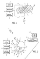
- the system S includes area-type electrodes 1 and 2, which are shown in contact with nearly opposing sides of a body organ OR within a desired operative field.
- the organ OR may be any internal organ in a patient's body such as the liver, pancreas, lungs, heart, etc.
- the organ OR may be an external organ in a patient's body, such as a limb, torso, head, neck, etc. (not shown). It should be recognized that the organ OR is shown within the patient's body only by way of example.
- a section of the organ OR, which falls between the area-type electrodes 1 and 2 is illustrated in sectional view by wide diagonally hatched lines.
- the area-type electrodes 1 and 2 are connected by wires, cables, or any other electrical connections 3 and 4, respectively, to a generator 5 yielding a high frequency output (voltage, current, or power).
- the generator 5 may provide some form of a display, indicated by reference numeral 6, to indicate readings corresponding to its electrical outputs.
- the display 5 may indicate a reading (one of R1 through RN) corresponding to levels of current, voltage, power, impedance, and/or temperature in the event the area-type electrodes 1 and 2 incorporate temperature sensors.
- the generator 5 also has a control mechanism indicated by reference numeral 7 to control the levels of current, voltage, power, impedance, and/or temperature.
- the control mechanism may take the form of controls, which may be manually maneuvered to control the levels of power current, voltage, temperature(s) at the electrodes or in the nearby tissue, impedance between the electrodes and so on.
- the various controls 7 may be automatically controlled. For example, they may be coupled to a feedback of temperature levels, in the event the temperature levels are being monitored at the area-type electrodes 1 and 2 or within the body organ OR.
- the generator 5, the display 6 and the control mechanism 7 are shown separately for illustration purposes only. The display 6 and the control mechanism 7 may be part of the generator or separate devices.
- the high frequency generator 5 When the high frequency generator 5 is turned on and a high frequency voltage is applied to the electrodes 1 and 2, the high frequency current passes between the electrodes 1 and 2, and through the organ OR. This causes heating by ionic friction or dielectric heating in the ionic medium of the organ OR.
- a high frequency generator 5 such as one available from Radionics, Inc., Burlington, Massachusetts may be used.
- a target tissue volume or area T which is shown by way of example may be a cancerous tumor or other abnormality which is to be destroyed.
- the plate-like electrodes 1 and 2 are disposed on either side of the tissue volume T. By heating the plate-like electrodes 1 and 2, the zone of heat between the electrodes 1 and 2 is created, which engulfs the target T.
- the dashed lines 8 and 9 provide a sectional representation of the boundaries of a heat lesion zone created between the plates. Between these perimeters indicated, the tissue may be heated to lethal levels. For example, sustaining tissue at approximately 50 degrees centigrade or higher temperatures for several minutes would kill the tissue.
- the heat lesion zone shown by cross-hatched lines in the cross-section of the organ OR in Figure 1, represents the region where the tissue is destroyed or ablated.
- organ OR in Figure 1
- the plate-like high frequency electrodes in accordance with the present invention across the liver, a large portion of the liver can be ablated, including that portion which includes the tumor T.
- the organ OR there is little or no open surgery performed on the organ OR, and the organ can be kept intact, thus, reducing the risk of hemorrhage and long convalescence.
- interoperative coagulation of internal organs can be performed, which may be exposed during surgery or visualized laproscopically, so as to ablate or to coagulate part or all of the organ without requiring a surgical incision within it.
- circular or square plate electrodes approximately 2 inches (5 centimeters) in diameter or side length referenced by numerals 1 and 2 are placed on opposite sides organ OR, which in this particular illustration represents a living liver.
- a radiofrequency generator with a radiofrequency output of about 500 KiloHertz is connected by electrical cables to each of the plate electrodes 1 and 2.
- a level of power output from the generator 5 of approximately 50 to 100 watts is applied to the tissue between the plates 1 and 2 for several minutes.
- a cylindrical, prismatic-shaped volume of tissue indicated between the boundaries 8 and 9 in Figure 1, is completely coagulated between the electrode plates 1 and 2, and all tissue within that volume is killed.
- the high frequency generator 5 may any suitable one, as for example, Model No. RFG-3D available from Radionics, Inc., Burlington, Massachusetts.
- the high frequency generator 5 can have a power range between zero and several hundred watts.
- the range of radiofrequency or high frequency can vary. Also, ranges from less than 100 kH to several tens or hundreds of Mega Hertz could be used.
- FIG 2 Another example of the present invention is shown.
- An external organ PB which is part of the patient's anatomy, is shown in sectional view. Inside the organ, bony structures B may exist as shown in Figure 2.
- the external organ PB could be a leg, arm, torso, pelvis, or neck.
- a target volume or area T within the organ PB that is to be ablated or destroyed.
- the surface structure of the organ PB may be skin, which one may not wish to destroy.
- the electrodes 1 and 2 are placed on the external surface of the organ and connected to the radiofrequency generator 5, in a manner similar to the configuration shown in Figure 1.
- the electrodes 1 and 2 may have cooling elements within them to prevent excessive heating at the point where the electrodes 1 and 2 contact the external surface of the organ PB.
- electrode 1 has an inflow tube 11 and outflow tube 12, which can circulate cooled fluid such as chilled saline, indicated by the flow arrows 15 and 16, respectively.
- the chilled fluid chills the electrode 1, and therefore, the skin surface of the organ PB just below it.
- Similar inflow and outflow channels 13 and 14 of electrode 2 exchange cooled fluid for the same purpose.
- the dashed line 10 illustrates in sectional perspective the ablation volume.
- This may correspond, for example, to the isotherm of temperatures around 50 to 60 degrees centigrade or greater, which may be sustained for several seconds to several minutes to cause tissue death of the ablation volume within line 10. This may be preplanned, calculated, or heuristically determined to engulf the volume T associated with the tumor.
- imaging of the patient anatomy relative to the positioning of electrodes 1 and 2 may be done with ultrasound, CT, MR, or interoperative ultrasonic imaging.
- Preplanned, three-dimensional treatment planning for such thermal radiofrequency surgery can be carried out in a computer workstation based on data taken from image scanners such as CT, MR, PET, X-ray, ultrasound, etc.
- Interactive imaging could be present, as indicated by the element 17, which could be a CT, MR, X-ray, PET, ultrasound, or other type of scanner.
- Its data processing unit could be built in or could be coupled to a computer workstation 23 with a display apparatus, such as a CRT, that can illustrate the sectional or volumetric rendering of the anatomy PB and also the position of the electrodes 1 and 2 and the isotherms such as dashed line 10.
- the scanner 17 could be positioned in a coordinated way with the position of the electrodes 1 and 2, and thereby to visualize the effect of the thermal lesion on the target volume T in a graphical and stereotactic fashion.
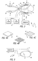
- FIG 3 shows a further embodiment of the present invention wherein the plates 1 and 2 are attached to an articulated forceps-like or tong-like device to grasp an external or internal organ.
- the arms 18 and 19 are coupled to the electrodes 1 and 2.
- Radiofrequency (rf) generator 5 which connects the radiofrequency output of the generator to the electrodes. That in turn causes the heating of the tissue as discussed above.
- a thermal sensor 29 might be embedded in one or both of the electrodes 1 and 2.
- a thermal-sensing, connecting cable 20 connects to the generator 5 so that readings R1 and RN can read out single or plural temperatures associated with one or both of the area electrodes 1 and 2.
- a plurality of such temperature sensors may be built in with the area electrodes to monitor surface tissue temperature nearby.
- temperature sensors may be placed in the tissue near the area electrodes 1 and 2 and monitored on the display 6, which may provide readings R1 through RN to indicate temperature levels.
- the readings R1 through RN may alternatively indicate other parameters.
- the hinged structure can be replaced by other articulations.
- the plates 1 and 2 can also have their own articulations, as for example, use of hinge joints 181 and 191 to connect to the arms 18 and 19, respectively, so as to better conform to the surface of the internal or external body part.
- Electrode type 30 could simply be a conductive plate made of metal, conductive plastic, or conductive rubber.
- Element 31 is shown as a mesh or matrix of conductive wires 32 so as to provide a conforming, lightweight area contact with tissue.
- the area-like electrodes may be flexible to conform to the curved shapes of organs.
- Element 34 is shown as a substrate with an array of conductive elements 35, which could be of any shape, geometry, or multiplicity and distribution on the surface plate 34. Tailored shapes (circles, ellipses, rectangles, oblong shapes, paddles, and the like) of area electrodes could be devised for a particular body part.
- Areas of area electrodes could be implemented, depending on the size of the body part, the size of the target area or volume T, the extent of heating, or the particular geometry and considerations of the clinical application. Areas of area electrodes of 100 or 500 square centimeters or larger may be useful for particular organs, tumors, or anatomical sites.
- Area electrodes of mesh or wire or discrete element types such as those illustrated by reference numerals 31 or 35 in Figure 4 may have conductors of a smaller actual conductive element area than the effective total area over which the area electrode contacts the tissue.
- the total contact area of the actual wire conductors 32 in the electrode 31 may be only a small percent of the entire area covered by the wire field.
- the total wire 31 field simulates an equipotential area over the tissue it contacts, it acts effectively like a large area continuous conductive electrode in terms of its heating affect. That is, it has the equivalent heating effect of a continuous metal plate electrode of area equal to the area of 31.
- FIG. 5 shows another embodiment of the present invention where different sized electrodes are used to alter the temperature distribution of the rf ablation.
- electrode 1 has a smaller area than electrode 2.
- the concentration of current density is higher near the smaller area electrode 1.
- the isotherm 8 which may represent, for example, the 50 degree centigrade or the lethal isotherm, is shifted more towards electrode 1 and less towards electrode 2.
- the area electrodes By placing the area electrodes in different positions on the body, either parallel, opposed, or adjacent to one another, in accordance with the above discussion can also vary the temperature distribution within the tissue.
- FIG. 6 a flow chart is shown to illustrate the procedure of applying area-type radiofrequency electrodes to a patient's anatomy and inducing desired ablations.
- the procedure starts at block 50, which represents the step of applying the area radiofrequency electrode to the desired surface of the desired organ or to the desired surface of a patient's anatomy.
- block 50 represents the step of applying the area radiofrequency electrode to the desired surface of the desired organ or to the desired surface of a patient's anatomy.
- a multiplicity of linear electrodes are inserted into the organ or anatomy, such a procedure is referred to as "interstitially" placing a virtual area-type electrode.
- the steps of inserting such electrodes or placing the electrodes relative to the internal anatomy could be imaged as indicated in block 51 by the imaging system or scanner 17 or monitor 23, such as a CT, MR, X-ray, ultrasound, or other type of imaging scanner.
- the position of a target volume or area such as a tumor may be imaged even before or after inserting the electrodes or placing them relative to the anatomy.
- imaging data may be taken during placement of the electrodes or when generating the desired radiofrequency levels to monitor the progress of coagulation.
- Scanners such as MR scanners, monitor the process of heating, and thus, may be used during the process of coagulation to observe the extent of ablation.
- Operation proceeds to the next step, represented by block 52, in accordance with which the generator 5 is connected to the electrodes 1 and 2 by connection cables.
- the power generated by the generator 5 is applied to the area electrodes 1 and 2.
- the radiofrequency ablation process or coagulation is initiated, as illustrated by block 52, to induce desired ablation or coagulation amounts.
- image scan data representations may also be acquired, as represented by block 51.
- the generator output parameters Before, during, and/or after the procedure, is may be desirable to monitor the generator output parameters, as illustrated by block 54. Also, it may be desirable to monitor temperature levels at the area electrodes or within the tissue to be ablated, in which case, temperature sensors may be included within the electrodes or placed "interstitially.”
- the generator parameters may be analyzed to determine if the desired amount of coagulation is accomplished. This can also be determined by monitoring the image representations, or by monitoring the temperature levels to determine if the appropriate coagulation is reached. At that point, the power from the generator 5 may be turned off and the ablation process terminated.
- FIG. 6 Another variation of the process in accordance with the present invention would be to use the ablation steps, as shown in Figure 6, to coagulate tissue and thereby to reduce blood circulation within the tissue preparatory to performing a surgical incision.
- area electrodes 1 and 2 can be placed on the liver, and a coagulation through the liver can be made according to the above description.
- a sectional volume through the liver is coagulated, including many of the arteries and vessels in the tissue.
- an incision can be made through the coagulated tissue with a reduced amount of bleeding in comparison with the state of tissue before coagulation was made. This is illustrated by step 55, as shown in Figure 6.
- system and method in accordance with the present invention can therefore be used as an adjunct to performing a surgical incision within a portion of the body to reduce problems of bleeding during surgery.
- the present invention is aimed at least one or two or more area-type or plate-type electrodes to induce ablative heating in a substantial volume proximate the area electrode(s) or in the tissue between the area electrodes
- Variations of the geometry, lesion parameters, frequency range, electrode characteristics, and materials can be made in various embodiments in accordance with the present invention.
- the electrodes can be of a widely varying shape, such as circular, square, or irregular shapes. For a smallest heating volume as near a skin cancer, tumor, or melanoma, an electrode of about 0.5 to 2 square centimeters may be appropriate.
- Electrodes of more than 1 and up to 50 square centimeters may be desirable.
- the one or more area electrodes with areas of 100 to 1000 square centimeters may be desirable.
- Electrodes can be fabricated from various conductive materials including metal, carbon structures, metal-impregnated plastics or rubbers, screens of stainless steel, titanium, platinum, or other conductive, bio-compatible materials.
- the area electrodes can be configured for one-time use or to be reused and resterilized. If they are configured for a single use, they may be easily disposed and there is no . need for sterilization before further use. This has the advantage of reducing the risk of transmission of diseases, such as AIDS and of reducing expenses with cleaning and resterilizing.
- the area electrodes may be for surface contact during surgery or external application, or they may be implanted within the body for a period of time for continuous re-application of heat.
- the area electrode can have a multiplicity of segmented conductive areas so as to provide a matrix or patchwork of conductive structures so that the generator can distribute the potential to each of these various structures in different ways during the procedure to achieve different heating patterns.
- Such heating patterns could be achieved by electrifying the elements 35 in Figure 4 in independent ways with multiplexing the output from a generator such as generator 5.
- MRI imaging such as image scanner 17 in Figure 2
- real-time visualization of the thermal distribution may be achieved on the display 23, and variations of the potential on the electrode or portions of the electrode may be made to achieve different temperature distributions under real-time visualization.
- Various high frequency generators can be used with a plurality of temperature monitors, measuring temperature at a multiplicity of points on the area electrode or within the bodily tissue itself with independent temperature sensors in a variety of displays of rf high frequency parameters.
- Cooling pumps can be connected cooperatively with the generator apparatus to cool the area electrodes as shown in Figure 2 in accordance with the temperature distribution or preplanning thereof
Claims (12)
- Système pour provoquer des ablations thermiques dans un champ opératoire au sein de l'organisme d'un patient, comprenant un tissu destiné à subir une ablation, comprenant :caractérisé en ce que la ou lesdites premières électrodes (1) et la ou lesdites deuxièmes électrodes (2) comprennent des électrodes à plaque qui sont adaptées et disposées pour délimiter le volume de tissu destiné à subir une ablation dans l'organisme du patient, les électrodes à plaque (1,2) étant reliées au générateur de haute fréquence (5) et disposées de telle façon que lorsque le générateur de haute fréquence (5) est mis en marche et qu'une tension à haute fréquence est appliquée aux électrodes à plaque (1,2), un courant à haute fréquence passe entre les électrodes (1,2) et à travers le tissu.au moins une première électrode (1) ayant une surface supérieure à 1 cm2, adaptée à venir en contact avec une première partie de tissu dans le champ opératoire ;au moins une deuxième électrode (2) ayant une surface supérieure à 1 cm2, adaptée à venir en contact avec une deuxième partie de tissu dans le champ opératoire ;un générateur de haute fréquence (5) générant un courant électrique de sortie, qui lorsqu'il est relié à la ou les premières et à la ou les deuxièmes électrodes, fournit de la chaleur à un volume de tissu pour provoquer une ablation d'au moins une partie du volume de tissu ; etau moins une connexion électrique (3,4) reliant le générateur de haute fréquence (5) à la ou à chaque première électrode(1) et à la ou à chaque deuxième électrode (2) ;
- Système selon la revendication 1, comprenant au moins une autre électrode à plaque.
- Système selon l'une quelconque des revendications précédentes, dans lequel la ou les premières électrodes à plaque (1) et la ou les deuxièmes électrodes à plaque (2) comprennent chacune une ou plusieurs plaques d'une matière conductrice.
- Système selon l'une quelconque des revendications précédentes, dans lequel la ou les premières électrodes à plaque (1) et la ou les deuxièmes électrodes à plaque (2) comprennent chacune une surface de matière conductrice.
- Système selon l'une quelconque des revendications précédentes, dans lequel la ou les premières électrodes à plaque (1) et la ou les deuxièmes électrodes à plaque (2) comprennent chacune un treillis conducteur de fils métalliques.
- Système selon l'une quelconque des revendications précédentes, dans lequel la ou les premières électrodes à plaque (1) et la ou les deuxièmes électrodes à plaque (2) comprennent chacune une surface de plusieurs éléments conducteurs.
- Système selon l'une quelconque des revendications précédentes, dans lequel la ou les premières électrodes à plaque (1) et la ou les deuxièmes électrodes à plaque (2) ont chacune une configuration appropriée pour adapter la ou chaque première électrode à plaque et la ou chaque deuxième électrode à plaque à la partie de tissu.
- Système selon l'une quelconque des revendications précédentes, dans lequel la ou au moins une des premières électrodes à plaque (1) et de la ou des deuxièmes électrodes à plaque (2), comprend un capteur de température (29) pour capter la température de la partie de tissu en contact.
- Système selon l'une quelconque des revendications précédentes, comprenant en outre :un tomodensitomètre à imagerie (17) pour fournir des représentations sous forme d'image du tissu dans le champ opératoire de façon à contrôler la position de la ou de chaque première électrode à plaque (1) et de la ou de chaque deuxième électrode à plaque (2) par rapport au champ opératoire.
- Système selon l'une quelconque des revendications précédentes, comprenant en outre :un dispositif pour serrer deux électrodes à plaque afin de guider les deux électrodes à plaque (1,2) dans des positions par rapport à la partie de tissu dans le champ opératoire, telles que les deux électrodes à plaque serrent la partie de tissu entre elles.
- Système selon l'une quelconque des revendications précédentes, dans lequel la ou au moins une des premières électrodes à plaque (1) et de la ou des deuxièmes électrodes à plaque (2), comprend un système de refroidissement à fluide (11,12) qui refroidit au moins une partie de l'électrode afin de prévenir un chauffage excessif de la partie de tissu dans le champ opératoire.
- Système selon l'une quelconque des revendications précédentes, dans lequel la ou au moins une des premières électrodes à plaque (1) et de la ou des deuxièmes électrodes à plaque (2), comprend plusieurs électrodes qui sont configurées pour permettre l'insertion dans la partie de tissu du champ opératoire.
Applications Claiming Priority (3)
| Application Number | Priority Date | Filing Date | Title |
|---|---|---|---|
| US866765 | 1992-04-09 | ||
| US08/866,765 US6312426B1 (en) | 1997-05-30 | 1997-05-30 | Method and system for performing plate type radiofrequency ablation |
| PCT/US1998/011372 WO1998053751A1 (fr) | 1997-05-30 | 1998-05-29 | Systeme et procede permettant d'effectuer une ablation par frequence radioelectriques a l'aide de plaques |
Publications (2)
| Publication Number | Publication Date |
|---|---|
| EP0986336A1 EP0986336A1 (fr) | 2000-03-22 |
| EP0986336B1 true EP0986336B1 (fr) | 2005-01-12 |
Family
ID=25348356
Family Applications (1)
| Application Number | Title | Priority Date | Filing Date |
|---|---|---|---|
| EP98926224A Expired - Lifetime EP0986336B1 (fr) | 1997-05-30 | 1998-05-29 | Systeme permettant d'effectuer une ablation par frequences radioelectriques a l'aide de plaques |
Country Status (5)
| Country | Link |
|---|---|
| US (2) | US6312426B1 (fr) |
| EP (1) | EP0986336B1 (fr) |
| AU (1) | AU7811198A (fr) |
| DE (1) | DE69828601T2 (fr) |
| WO (1) | WO1998053751A1 (fr) |
Families Citing this family (102)
| Publication number | Priority date | Publication date | Assignee | Title |
|---|---|---|---|---|
| US6096037A (en) * | 1997-07-29 | 2000-08-01 | Medtronic, Inc. | Tissue sealing electrosurgery device and methods of sealing tissue |
| US6692491B1 (en) | 2000-03-24 | 2004-02-17 | Scimed Life Systems, Inc. | Surgical methods and apparatus for positioning a diagnostic or therapeutic element around one or more pulmonary veins or other body structures |
| US6926712B2 (en) | 2000-03-24 | 2005-08-09 | Boston Scientific Scimed, Inc. | Clamp having at least one malleable clamp member and surgical method employing the same |
| US6471695B1 (en) | 2000-09-06 | 2002-10-29 | Radiotherapeutics, Inc. | Apparatus and method for shielding tissue during tumor ablation |
| US7008421B2 (en) * | 2002-08-21 | 2006-03-07 | Resect Medical, Inc. | Apparatus and method for tissue resection |
| US6796828B2 (en) | 2001-06-01 | 2004-09-28 | Sherwood Services Ag | Return pad cable connector |
| US7753908B2 (en) | 2002-02-19 | 2010-07-13 | Endoscopic Technologies, Inc. (Estech) | Apparatus for securing an electrophysiology probe to a clamp |
| US7785324B2 (en) | 2005-02-25 | 2010-08-31 | Endoscopic Technologies, Inc. (Estech) | Clamp based lesion formation apparatus and methods configured to protect non-target tissue |
| US20030158548A1 (en) * | 2002-02-19 | 2003-08-21 | Phan Huy D. | Surgical system including clamp and apparatus for securing an energy transmission device to the clamp and method of converting a clamp into an electrophysiology device |
| US6932816B2 (en) * | 2002-02-19 | 2005-08-23 | Boston Scientific Scimed, Inc. | Apparatus for converting a clamp into an electrophysiology device |
| US7367974B2 (en) * | 2004-09-20 | 2008-05-06 | Wisconsin Alumni Research Foundation | Electrode array for tissue ablation |
| US20040077951A1 (en) * | 2002-07-05 | 2004-04-22 | Wei-Chiang Lin | Apparatus and methods of detection of radiation injury using optical spectroscopy |
| US7223264B2 (en) * | 2002-08-21 | 2007-05-29 | Resect Medical, Inc. | Thermal coagulation of tissue during tissue resection |
| US8808284B2 (en) | 2008-09-26 | 2014-08-19 | Relievant Medsystems, Inc. | Systems for navigating an instrument through bone |
| US8361067B2 (en) | 2002-09-30 | 2013-01-29 | Relievant Medsystems, Inc. | Methods of therapeutically heating a vertebral body to treat back pain |
| US8613744B2 (en) | 2002-09-30 | 2013-12-24 | Relievant Medsystems, Inc. | Systems and methods for navigating an instrument through bone |
| US7258690B2 (en) | 2003-03-28 | 2007-08-21 | Relievant Medsystems, Inc. | Windowed thermal ablation probe |
| US6907884B2 (en) | 2002-09-30 | 2005-06-21 | Depay Acromed, Inc. | Method of straddling an intraosseous nerve |
| US20050015125A1 (en) * | 2003-03-14 | 2005-01-20 | Mioduski Paul C. | Hyperthermia treatment systems and methods |
| ITFI20030104A1 (it) * | 2003-04-10 | 2004-10-11 | Luciano Alcidi | Apparecchiatura per terapie di ipertemia non distruttiva |
| US20040254572A1 (en) * | 2003-04-25 | 2004-12-16 | Mcintyre Jon T. | Self anchoring radio frequency ablation array |
| WO2005032342A2 (fr) * | 2003-09-30 | 2005-04-14 | Vanderbilt University | Procedes et dispositifs de detection spectroscopique optique de la mort cellulaire et tissulaire |
| US20050190982A1 (en) * | 2003-11-28 | 2005-09-01 | Matsushita Electric Industrial Co., Ltd. | Image reducing device and image reducing method |
| US8002770B2 (en) | 2003-12-02 | 2011-08-23 | Endoscopic Technologies, Inc. (Estech) | Clamp based methods and apparatus for forming lesions in tissue and confirming whether a therapeutic lesion has been formed |
| US7481808B2 (en) * | 2004-06-30 | 2009-01-27 | Ethicon, Inc. | Flexible electrode device and surgical apparatus equipped with same |
| US20060025761A1 (en) * | 2004-07-29 | 2006-02-02 | Riley Lee B | Linear-array radio frequency resections |
| US7776033B2 (en) * | 2005-01-08 | 2010-08-17 | Boston Scientific Scimed, Inc. | Wettable structures including conductive fibers and apparatus including the same |
| US7862561B2 (en) * | 2005-01-08 | 2011-01-04 | Boston Scientific Scimed, Inc. | Clamp based lesion formation apparatus with variable spacing structures |
| US7727231B2 (en) | 2005-01-08 | 2010-06-01 | Boston Scientific Scimed, Inc. | Apparatus and methods for forming lesions in tissue and applying stimulation energy to tissue in which lesions are formed |
| US7862562B2 (en) * | 2005-02-25 | 2011-01-04 | Boston Scientific Scimed, Inc. | Wrap based lesion formation apparatus and methods configured to protect non-target tissue |
| ES2352144T3 (es) * | 2005-08-02 | 2011-02-16 | Neurotherm, Inc. | Aparato para diagnosticar y tratar disfunción neuronal. |
| EP1754512A3 (fr) * | 2005-08-18 | 2008-03-05 | Neurotherm, Inc. | Méthode et appareil pour diagnostiquer et traiter des dysfonctionnements neuronaux |
| US20070049914A1 (en) * | 2005-09-01 | 2007-03-01 | Sherwood Services Ag | Return electrode pad with conductive element grid and method |
| DE102005045363A1 (de) * | 2005-09-22 | 2007-04-05 | Siemens Ag | Medizinische Behandlungsvorrichtung und zugehöriges Betriebsverfahren |
| US20070100331A1 (en) * | 2005-10-27 | 2007-05-03 | Boston Scientific Scimed, Inc. | Systems and methods for organ tissue ablation |
| CN100463660C (zh) * | 2006-01-18 | 2009-02-25 | 重庆海扶(Hifu)技术有限公司 | 超声治疗钳 |
| US8155416B2 (en) * | 2008-02-04 | 2012-04-10 | INTIO, Inc. | Methods and apparatuses for planning, performing, monitoring and assessing thermal ablation |
| US20080033417A1 (en) * | 2006-08-04 | 2008-02-07 | Nields Morgan W | Apparatus for planning and performing thermal ablation |
| US7871406B2 (en) * | 2006-08-04 | 2011-01-18 | INTIO, Inc. | Methods for planning and performing thermal ablation |
| US8556888B2 (en) * | 2006-08-04 | 2013-10-15 | INTIO, Inc. | Methods and apparatuses for performing and monitoring thermal ablation |
| US20080033419A1 (en) * | 2006-08-04 | 2008-02-07 | Nields Morgan W | Method for planning, performing and monitoring thermal ablation |
| US20080033418A1 (en) * | 2006-08-04 | 2008-02-07 | Nields Morgan W | Methods for monitoring thermal ablation |
| FR2912817B1 (fr) * | 2007-02-21 | 2009-05-22 | Super Sonic Imagine Sa | Procede d'optimisation de la focalisation d'ondes au travers d'un element introducteur d'aberations. |
| US20080243121A1 (en) * | 2007-04-02 | 2008-10-02 | Tomoyuki Takashino | Curative treatment system, curative treatment device, and treatment method for living tissue using energy |
| US20080243213A1 (en) * | 2007-04-02 | 2008-10-02 | Tomoyuki Takashino | Curative treatment system, curative treatment device, and treatment method for living tissue using energy |
| US8231614B2 (en) | 2007-05-11 | 2012-07-31 | Tyco Healthcare Group Lp | Temperature monitoring return electrode |
| US8801703B2 (en) | 2007-08-01 | 2014-08-12 | Covidien Lp | System and method for return electrode monitoring |
| US20090112202A1 (en) * | 2007-10-31 | 2009-04-30 | Boston Scientific Scimed, Inc. | Dispersive electrode with thermochromatic properties |
| EP2881050B1 (fr) | 2008-03-31 | 2023-02-22 | Applied Medical Resources Corporation | Système électrochirurgical avec des moyens pour déterminer le terme d'un traitement sur la base d'un angle de phase |
| US10702326B2 (en) | 2011-07-15 | 2020-07-07 | Virginia Tech Intellectual Properties, Inc. | Device and method for electroporation based treatment of stenosis of a tubular body part |
| US9283051B2 (en) | 2008-04-29 | 2016-03-15 | Virginia Tech Intellectual Properties, Inc. | System and method for estimating a treatment volume for administering electrical-energy based therapies |
| US9598691B2 (en) | 2008-04-29 | 2017-03-21 | Virginia Tech Intellectual Properties, Inc. | Irreversible electroporation to create tissue scaffolds |
| US8992517B2 (en) | 2008-04-29 | 2015-03-31 | Virginia Tech Intellectual Properties Inc. | Irreversible electroporation to treat aberrant cell masses |
| US10272178B2 (en) | 2008-04-29 | 2019-04-30 | Virginia Tech Intellectual Properties Inc. | Methods for blood-brain barrier disruption using electrical energy |
| US11272979B2 (en) | 2008-04-29 | 2022-03-15 | Virginia Tech Intellectual Properties, Inc. | System and method for estimating tissue heating of a target ablation zone for electrical-energy based therapies |
| US9867652B2 (en) | 2008-04-29 | 2018-01-16 | Virginia Tech Intellectual Properties, Inc. | Irreversible electroporation using tissue vasculature to treat aberrant cell masses or create tissue scaffolds |
| US9198733B2 (en) | 2008-04-29 | 2015-12-01 | Virginia Tech Intellectual Properties, Inc. | Treatment planning for electroporation-based therapies |
| US10245098B2 (en) | 2008-04-29 | 2019-04-02 | Virginia Tech Intellectual Properties, Inc. | Acute blood-brain barrier disruption using electrical energy based therapy |
| US10238447B2 (en) | 2008-04-29 | 2019-03-26 | Virginia Tech Intellectual Properties, Inc. | System and method for ablating a tissue site by electroporation with real-time monitoring of treatment progress |
| US10117707B2 (en) | 2008-04-29 | 2018-11-06 | Virginia Tech Intellectual Properties, Inc. | System and method for estimating tissue heating of a target ablation zone for electrical-energy based therapies |
| US11254926B2 (en) | 2008-04-29 | 2022-02-22 | Virginia Tech Intellectual Properties, Inc. | Devices and methods for high frequency electroporation |
| US20090287140A1 (en) * | 2008-05-16 | 2009-11-19 | Rittman Iii William J | Electrical stimulation and infusion introducer assembly |
| EP3406210A1 (fr) | 2008-09-26 | 2018-11-28 | Relievant Medsystems, Inc. | Systèmes pour piloter un instrument à travers un os |
| US10028753B2 (en) | 2008-09-26 | 2018-07-24 | Relievant Medsystems, Inc. | Spine treatment kits |
| CN102421386A (zh) * | 2009-03-31 | 2012-04-18 | 安吉戴尼克公司 | 用于估计医疗装置的治疗区和互动式地计划患者治疗的系统和方法 |
| US11382681B2 (en) | 2009-04-09 | 2022-07-12 | Virginia Tech Intellectual Properties, Inc. | Device and methods for delivery of high frequency electrical pulses for non-thermal ablation |
| US11638603B2 (en) | 2009-04-09 | 2023-05-02 | Virginia Tech Intellectual Properties, Inc. | Selective modulation of intracellular effects of cells using pulsed electric fields |
| US8903488B2 (en) | 2009-05-28 | 2014-12-02 | Angiodynamics, Inc. | System and method for synchronizing energy delivery to the cardiac rhythm |
| US9895189B2 (en) | 2009-06-19 | 2018-02-20 | Angiodynamics, Inc. | Methods of sterilization and treating infection using irreversible electroporation |
| US20110213356A1 (en) | 2009-11-05 | 2011-09-01 | Wright Robert E | Methods and systems for spinal radio frequency neurotomy |
| US8879995B2 (en) * | 2009-12-23 | 2014-11-04 | Viconics Electronics Inc. | Wireless power transmission using phased array antennae |
| US8617157B2 (en) * | 2010-01-26 | 2013-12-31 | Covidien Lp | Hernia repair system |
| AU2011256709B2 (en) | 2010-05-21 | 2013-10-24 | Stratus Medical, LLC | Systems and methods for tissue ablation |
| ES2912092T3 (es) | 2010-10-01 | 2022-05-24 | Applied Med Resources | Instrumentos electroquirúrgicos y conexiones a los mismos |
| EP2627274B1 (fr) | 2010-10-13 | 2022-12-14 | AngioDynamics, Inc. | Système d'ablation électrique des tissus chez un patient |
| WO2012088149A2 (fr) | 2010-12-20 | 2012-06-28 | Virginia Tech Intellectual Properties, Inc. | Électroporation à haute fréquence pour thérapie anticancéreuse |
| US9078665B2 (en) | 2011-09-28 | 2015-07-14 | Angiodynamics, Inc. | Multiple treatment zone ablation probe |
| US10390877B2 (en) | 2011-12-30 | 2019-08-27 | Relievant Medsystems, Inc. | Systems and methods for treating back pain |
| US10588691B2 (en) | 2012-09-12 | 2020-03-17 | Relievant Medsystems, Inc. | Radiofrequency ablation of tissue within a vertebral body |
| WO2014071161A1 (fr) | 2012-11-05 | 2014-05-08 | Relievant Medsystems, Inc. | Système et méthodes de création de chemins incurvés à travers un os et de modulation des nerfs au sein de l'os |
| US9888956B2 (en) | 2013-01-22 | 2018-02-13 | Angiodynamics, Inc. | Integrated pump and generator device and method of use |
| US9724151B2 (en) | 2013-08-08 | 2017-08-08 | Relievant Medsystems, Inc. | Modulating nerves within bone using bone fasteners |
| US20150148795A1 (en) * | 2013-11-26 | 2015-05-28 | Boston Scientific Scimed, Inc. | Radio frequency ablation coil |
| JP6594901B2 (ja) | 2014-05-12 | 2019-10-23 | バージニア テック インテレクチュアル プロパティース インコーポレイテッド | パルス電界を使用した細胞の細胞内効果の選択的調節 |
| KR20230076143A (ko) | 2014-05-16 | 2023-05-31 | 어플라이드 메디컬 리소시스 코포레이션 | 전기수술용 시스템 |
| EP3148465B1 (fr) | 2014-05-30 | 2018-05-16 | Applied Medical Resources Corporation | Système électrochirurgical avec un instrument comprenant une mâchoire avec une plaquette isolante centrale |
| WO2016100325A1 (fr) | 2014-12-15 | 2016-06-23 | Virginia Tech Intellectual Properties, Inc. | Dispositifs, systèmes et méthodes de surveillance en temps réel d'effets électrophysiologiques au cours d'un traitement tissulaire |
| KR20230093365A (ko) | 2014-12-23 | 2023-06-27 | 어플라이드 메디컬 리소시스 코포레이션 | 바이폴라 전기수술용 밀봉기 및 디바이더 |
| USD748259S1 (en) | 2014-12-29 | 2016-01-26 | Applied Medical Resources Corporation | Electrosurgical instrument |
| EP3463141B1 (fr) * | 2016-05-25 | 2021-07-07 | Ikomed Technologies Inc. | Système de traitement de tissus non souhaités |
| EP3522807A1 (fr) | 2016-10-04 | 2019-08-14 | Avent, Inc. | Sondes rf refroidies |
| US10905492B2 (en) | 2016-11-17 | 2021-02-02 | Angiodynamics, Inc. | Techniques for irreversible electroporation using a single-pole tine-style internal device communicating with an external surface electrode |
| WO2018167877A1 (fr) * | 2017-03-15 | 2018-09-20 | オリンパス株式会社 | Dispositif de source d'énergie |
| EP3644883A4 (fr) * | 2017-06-30 | 2021-04-07 | Intuitive Surgical Operations, Inc. | Instrument électrochirurgical avec électrode élastomère souple |
| WO2019071269A2 (fr) | 2017-10-06 | 2019-04-11 | Powell Charles Lee | Système et procédé pour traiter une apnée du sommeil obstructive |
| US11607537B2 (en) | 2017-12-05 | 2023-03-21 | Virginia Tech Intellectual Properties, Inc. | Method for treating neurological disorders, including tumors, with electroporation |
| US11311329B2 (en) | 2018-03-13 | 2022-04-26 | Virginia Tech Intellectual Properties, Inc. | Treatment planning for immunotherapy based treatments using non-thermal ablation techniques |
| US11925405B2 (en) | 2018-03-13 | 2024-03-12 | Virginia Tech Intellectual Properties, Inc. | Treatment planning system for immunotherapy enhancement via non-thermal ablation |
| US11864812B2 (en) | 2018-09-05 | 2024-01-09 | Applied Medical Resources Corporation | Electrosurgical generator control system |
| CA3120182A1 (fr) | 2018-11-16 | 2020-05-22 | Applied Medical Resources Corporation | Systeme electrochirurgical |
| US11950835B2 (en) | 2019-06-28 | 2024-04-09 | Virginia Tech Intellectual Properties, Inc. | Cycled pulsing to mitigate thermal damage for multi-electrode irreversible electroporation therapy |
| WO2021050767A1 (fr) | 2019-09-12 | 2021-03-18 | Relievant Medsystems, Inc. | Systèmes et méthodes de modulation de tissu |
Family Cites Families (157)
| Publication number | Priority date | Publication date | Assignee | Title |
|---|---|---|---|---|
| US2047535A (en) | 1932-10-07 | 1936-07-14 | Frederick C Wappler | Surgical electrodes |
| US2828747A (en) | 1952-12-06 | 1958-04-01 | Birtcher Corp | Gas-blanketed electro-surgical device |
| US3598108A (en) | 1969-02-28 | 1971-08-10 | Khosrow Jamshidi | Biopsy technique and biopsy device |
| US3685518A (en) | 1970-07-29 | 1972-08-22 | Aesculap Werke Ag | Surgical instrument for high-frequency surgery |
| US3698394A (en) | 1971-06-14 | 1972-10-17 | William S Piper | Electrically heated hypodermic needle |
| US3980861A (en) | 1973-03-26 | 1976-09-14 | Akio Fukunaga | Electrically heated miniature thermal implement |
| US3828780A (en) | 1973-03-26 | 1974-08-13 | Valleylab Inc | Combined electrocoagulator-suction instrument |
| US3845771A (en) * | 1973-04-24 | 1974-11-05 | W Vise | Electrosurgical glove |
| DE2324415C2 (de) | 1973-05-15 | 1975-06-05 | Aesculap-Werke Ag Vormals Jetter & Scheerer, 7200 Tuttlingen | Chirurgisches Sauginstrument |
| DE2324658B2 (de) | 1973-05-16 | 1977-06-30 | Richard Wolf Gmbh, 7134 Knittlingen | Sonde zum koagulieren von koerpergewebe |
| DK131541B (da) | 1973-09-03 | 1975-08-04 | Akad Tekn Videnskaber | Prostataresectoskop. |
| DE2407559C3 (de) | 1974-02-16 | 1982-01-21 | Dornier System Gmbh, 7990 Friedrichshafen | Wärmesonde |
| US3890977A (en) | 1974-03-01 | 1975-06-24 | Bruce C Wilson | Kinetic memory electrodes, catheters and cannulae |
| US3943932A (en) | 1975-01-17 | 1976-03-16 | Yen Kong Woo | Acupuncture needles and holder |
| US3982542A (en) | 1975-03-12 | 1976-09-28 | Ford John L | Electroresectroscope and method of laparoscopic tubal sterilization |
| US4184492A (en) | 1975-08-07 | 1980-01-22 | Karl Storz Endoscopy-America, Inc. | Safety circuitry for high frequency cutting and coagulating devices |
| US4051855A (en) | 1976-02-06 | 1977-10-04 | Ipco Hospital Supply Corporation, Whaledent International Division | Electrosurgical unit |
| US4074718A (en) | 1976-03-17 | 1978-02-21 | Valleylab, Inc. | Electrosurgical instrument |
| US4140130A (en) * | 1977-05-31 | 1979-02-20 | Storm Iii Frederick K | Electrode structure for radio frequency localized heating of tumor bearing tissue |
| US4204549A (en) | 1977-12-12 | 1980-05-27 | Rca Corporation | Coaxial applicator for microwave hyperthermia |
| US4196734A (en) | 1978-02-16 | 1980-04-08 | Valleylab, Inc. | Combined electrosurgery/cautery system and method |
| JPS5552748A (en) | 1978-10-12 | 1980-04-17 | Olympus Optical Co | Highhfrequency incising tool |
| US4608977A (en) | 1979-08-29 | 1986-09-02 | Brown Russell A | System using computed tomography as for selective body treatment |
| US4301802A (en) | 1980-03-17 | 1981-11-24 | Stanley Poler | Cauterizing tool for ophthalmological surgery |
| US4375220A (en) * | 1980-05-09 | 1983-03-01 | Matvias Fredrick M | Microwave applicator with cooling mechanism for intracavitary treatment of cancer |
| WO1981003271A1 (fr) | 1980-05-13 | 1981-11-26 | American Hospital Supply Corp | Dispositif electro-chirurgical multipolaire |
| EP0040658A3 (fr) * | 1980-05-28 | 1981-12-09 | Drg (Uk) Limited | Support de patient pour un appareil de diathermie et appareil de diathermie équipé d'un tel support |
| US4565200A (en) | 1980-09-24 | 1986-01-21 | Cosman Eric R | Universal lesion and recording electrode system |
| US4411266A (en) | 1980-09-24 | 1983-10-25 | Cosman Eric R | Thermocouple radio frequency lesion electrode |
| US4397314A (en) | 1981-08-03 | 1983-08-09 | Clini-Therm Corporation | Method and apparatus for controlling and optimizing the heating pattern for a hyperthermia system |
| DE3207152A1 (de) * | 1982-02-27 | 1983-09-22 | Ernst Leitz Wetzlar Gmbh, 6330 Wetzlar | Automatische fokussiereinrichtung fuer diaprojektoren |
| JPS58173540A (ja) | 1982-04-03 | 1983-10-12 | 銭谷 利男 | マイクロ波手術装置 |
| US5370675A (en) | 1992-08-12 | 1994-12-06 | Vidamed, Inc. | Medical probe device and method |
| US5421819A (en) | 1992-08-12 | 1995-06-06 | Vidamed, Inc. | Medical probe device |
| US5542915A (en) | 1992-08-12 | 1996-08-06 | Vidamed, Inc. | Thermal mapping catheter with ultrasound probe |
| JPS5957650A (ja) | 1982-09-27 | 1984-04-03 | 呉羽化学工業株式会社 | 腔内加熱用プロ−ブ |
| US4644955A (en) | 1982-12-27 | 1987-02-24 | Rdm International, Inc. | Circuit apparatus and method for electrothermal treatment of cancer eye |
| US4961422A (en) | 1983-01-21 | 1990-10-09 | Marchosky J Alexander | Method and apparatus for volumetric interstitial conductive hyperthermia |
| US4524770A (en) | 1983-01-25 | 1985-06-25 | Ahmad Orandi | Endoscope injection needle |
| US4662368A (en) | 1983-06-13 | 1987-05-05 | Trimedyne Laser Systems, Inc. | Localized heat applying medical device |
| US4796640A (en) | 1984-01-13 | 1989-01-10 | American Hospital Supply Corporation | Apparatus with fast response thermistor |
| GB2161082B (en) * | 1984-01-30 | 1986-12-03 | Kh Nii Obschei Neot Khirurg | Bipolar electric surgical instrument |
| US4682596A (en) | 1984-05-22 | 1987-07-28 | Cordis Corporation | Electrosurgical catheter and method for vascular applications |
| US4712559A (en) | 1985-06-28 | 1987-12-15 | Bsd Medical Corporation | Local current capacitive field applicator for interstitial array |
| DE3544443C2 (de) * | 1985-12-16 | 1994-02-17 | Siemens Ag | HF-Chirurgiegerät |
| SE455920B (sv) | 1986-01-29 | 1988-08-22 | Hans Wiksell | Anordning for hypertermibehandling av tumorer |
| FR2597744A1 (fr) | 1986-04-29 | 1987-10-30 | Boussignac Georges | Catheter cardio-vasculaire pour tir au rayon laser |
| SU1391626A1 (ru) | 1986-10-23 | 1988-04-30 | Институт сердечно-сосудистой хирургии им.А.Н.Бакулева | Устройство дл проводки хирургического инструмента |
| JPH0511882Y2 (fr) | 1987-01-06 | 1993-03-25 | ||
| US4823791A (en) | 1987-05-08 | 1989-04-25 | Circon Acmi Division Of Circon Corporation | Electrosurgical probe apparatus |
| US4989608A (en) | 1987-07-02 | 1991-02-05 | Ratner Adam V | Device construction and method facilitating magnetic resonance imaging of foreign objects in a body |
| JPH0636834Y2 (ja) | 1987-10-28 | 1994-09-28 | オリンパス光学工業株式会社 | 高周波誘電加温用電極 |
| JP2614887B2 (ja) * | 1988-02-18 | 1997-05-28 | 甚一 松田 | 局所加温装置及び局所加温用の立体共振器 |
| US4907589A (en) | 1988-04-29 | 1990-03-13 | Cosman Eric R | Automatic over-temperature control apparatus for a therapeutic heating device |
| US5249585A (en) | 1988-07-28 | 1993-10-05 | Bsd Medical Corporation | Urethral inserted applicator for prostate hyperthermia |
| US5220927A (en) | 1988-07-28 | 1993-06-22 | Bsd Medical Corporation | Urethral inserted applicator for prostate hyperthermia |
| US5191883A (en) | 1988-10-28 | 1993-03-09 | Prutech Research And Development Partnership Ii | Device for heating tissue in a patient's body |
| US4966597A (en) | 1988-11-04 | 1990-10-30 | Cosman Eric R | Thermometric cardiac tissue ablation electrode with ultra-sensitive temperature detection |
| US4927420A (en) | 1988-11-14 | 1990-05-22 | Colorado Biomedical, Inc. | Ultra-sharp tungsten needle for electrosurgical knife |
| FR2639238B1 (fr) | 1988-11-21 | 1991-02-22 | Technomed Int Sa | Appareil de traitement chirurgical de tissus par hyperthermie, de preference la prostate, comprenant des moyens de protection thermique comprenant de preference des moyens formant ecran radioreflechissant |
| US5099846A (en) | 1988-12-23 | 1992-03-31 | Hardy Tyrone L | Method and apparatus for video presentation from a variety of scanner imaging sources |
| US5151102A (en) | 1989-05-31 | 1992-09-29 | Kyocera Corporation | Blood vessel coagulation/stanching device |
| US5029588A (en) | 1989-06-15 | 1991-07-09 | Cardiovascular Imaging Systems, Inc. | Laser catheter with imaging capability |
| US5284144A (en) | 1989-11-22 | 1994-02-08 | The United States Of America As Represented By The Secretary Of The Dept. Of Health & Human Services | Apparatus for hyperthermia treatment of cancer |
| US5217030A (en) * | 1989-12-05 | 1993-06-08 | Inbae Yoon | Multi-functional instruments and stretchable ligating and occluding devices |
| EP0511955B1 (fr) * | 1990-01-22 | 1995-09-06 | PHILLIPS, Arnold G. | Applicateur de cire osseuse pour traiter les tissus osseux |
| US5263931A (en) | 1990-02-14 | 1993-11-23 | Advanced Cardiovascular Systems, Inc. | Balloon catheter for dilating a prostatic urethra |
| SE465500B (sv) | 1990-02-15 | 1991-09-23 | Dmitry Nikolaevich Kolmakov | Utstraalare av elektromagnetisk energi foer lokal hypertermi i maenniskokroppen |
| SE465754B (sv) | 1990-03-05 | 1991-10-28 | Dmitry Nikolaevich Kolmakov | Applikator foer lokal hypertermi innefattande en mikrovaagsstraalare |
| GB2242132A (en) | 1990-03-21 | 1991-09-25 | Minta Instrumentation Limited | A lesion probe |
| US5097835A (en) | 1990-04-09 | 1992-03-24 | Ad-Tech Medical Instrument Corporation | Subdural electrode with improved lead connection |
| US5482054A (en) * | 1990-05-10 | 1996-01-09 | Symbiosis Corporation | Edoscopic biopsy forceps devices with selective bipolar cautery |
| US5103804A (en) | 1990-07-03 | 1992-04-14 | Boston Scientific Corporation | Expandable tip hemostatic probes and the like |
| US5282799A (en) * | 1990-08-24 | 1994-02-01 | Everest Medical Corporation | Bipolar electrosurgical scalpel with paired loop electrodes |
| US5116333A (en) | 1990-11-02 | 1992-05-26 | Kirwan Surgical Products, Inc. | Bipolar handswitch adapter |
| US5409453A (en) | 1992-08-12 | 1995-04-25 | Vidamed, Inc. | Steerable medical probe with stylets |
| US5300087A (en) * | 1991-03-22 | 1994-04-05 | Knoepfler Dennis J | Multiple purpose forceps |
| US5396900A (en) * | 1991-04-04 | 1995-03-14 | Symbiosis Corporation | Endoscopic end effectors constructed from a combination of conductive and non-conductive materials and useful for selective endoscopic cautery |
| FR2675371A1 (fr) | 1991-04-22 | 1992-10-23 | Technomed Int Sa | Dispositif de traitement thermique de tissus par groupe de sequence d'impulsions. |
| US5484436A (en) * | 1991-06-07 | 1996-01-16 | Hemostatic Surgery Corporation | Bi-polar electrosurgical instruments and methods of making |
| US5330471A (en) * | 1991-06-07 | 1994-07-19 | Hemostatic Surgery Corporation | Bi-polar electrosurgical endoscopic instruments and methods of use |
| US5391166A (en) * | 1991-06-07 | 1995-02-21 | Hemostatic Surgery Corporation | Bi-polar electrosurgical endoscopic instruments having a detachable working end |
| US5383917A (en) | 1991-07-05 | 1995-01-24 | Jawahar M. Desai | Device and method for multi-phase radio-frequency ablation |
| DE4130064A1 (de) * | 1991-09-11 | 1993-03-18 | Wolf Gmbh Richard | Endoskopische koagulationsfasszange |
| US5196009A (en) | 1991-09-11 | 1993-03-23 | Kirwan Jr Lawrence T | Non-sticking electrosurgical device having nickel tips |
| US5300080A (en) | 1991-11-01 | 1994-04-05 | David Clayman | Stereotactic instrument guided placement |
| US5207691A (en) * | 1991-11-01 | 1993-05-04 | Medical Scientific, Inc. | Electrosurgical clip applicator |
| US5713896A (en) * | 1991-11-01 | 1998-02-03 | Medical Scientific, Inc. | Impedance feedback electrosurgical system |
| US5323778A (en) | 1991-11-05 | 1994-06-28 | Brigham & Women's Hospital | Method and apparatus for magnetic resonance imaging and heating tissues |
| US5395362A (en) | 1992-01-14 | 1995-03-07 | Summit Technology | Methods and apparatus for distributing laser radiation |
| US5484435A (en) * | 1992-01-15 | 1996-01-16 | Conmed Corporation | Bipolar electrosurgical instrument for use in minimally invasive internal surgical procedures |
| US5267994A (en) | 1992-02-10 | 1993-12-07 | Conmed Corporation | Electrosurgical probe |
| US5413588A (en) | 1992-03-06 | 1995-05-09 | Urologix, Inc. | Device and method for asymmetrical thermal therapy with helical dipole microwave antenna |
| US5330518A (en) | 1992-03-06 | 1994-07-19 | Urologix, Inc. | Method for treating interstitial tissue associated with microwave thermal therapy |
| US5281216A (en) * | 1992-03-31 | 1994-01-25 | Valleylab, Inc. | Electrosurgical bipolar treating apparatus |
| US5281215A (en) | 1992-04-16 | 1994-01-25 | Implemed, Inc. | Cryogenic catheter |
| US5281213A (en) | 1992-04-16 | 1994-01-25 | Implemed, Inc. | Catheter for ice mapping and ablation |
| US5277201A (en) * | 1992-05-01 | 1994-01-11 | Vesta Medical, Inc. | Endometrial ablation apparatus and method |
| US5443463A (en) * | 1992-05-01 | 1995-08-22 | Vesta Medical, Inc. | Coagulating forceps |
| US5293863A (en) * | 1992-05-08 | 1994-03-15 | Loma Linda University Medical Center | Bladed endoscopic retractor |
| US5281218A (en) | 1992-06-05 | 1994-01-25 | Cardiac Pathways Corporation | Catheter having needle electrode for radiofrequency ablation |
| US5486161A (en) | 1993-02-02 | 1996-01-23 | Zomed International | Medical probe device and method |
| US5295990A (en) * | 1992-09-11 | 1994-03-22 | Levin John M | Tissue sampling and removal device |
| EP0669840A4 (fr) | 1992-09-11 | 1995-11-15 | Advanced Surgical Inc | Catheter a perfusion a insertion autonome. |
| US5647361A (en) | 1992-09-28 | 1997-07-15 | Fonar Corporation | Magnetic resonance imaging method and apparatus for guiding invasive therapy |
| US5334193A (en) | 1992-11-13 | 1994-08-02 | American Cardiac Ablation Co., Inc. | Fluid cooled ablation catheter |
| WO1994010924A1 (fr) | 1992-11-13 | 1994-05-26 | American Cardiac Ablation Co., Inc. | Sonde electrochirurgicale refroidie par liquide |
| US5342357A (en) | 1992-11-13 | 1994-08-30 | American Cardiac Ablation Co., Inc. | Fluid cooled electrosurgical cauterization system |
| US5545161A (en) | 1992-12-01 | 1996-08-13 | Cardiac Pathways Corporation | Catheter for RF ablation having cooled electrode with electrically insulated sleeve |
| US5348554A (en) | 1992-12-01 | 1994-09-20 | Cardiac Pathways Corporation | Catheter for RF ablation with cooled electrode |
| DE4240722C2 (de) | 1992-12-03 | 1996-08-29 | Siemens Ag | Gerät für die Behandlung von pathologischem Gewebe |
| US5403312A (en) * | 1993-07-22 | 1995-04-04 | Ethicon, Inc. | Electrosurgical hemostatic device |
| US5514134A (en) * | 1993-02-05 | 1996-05-07 | Everest Medical Corporation | Bipolar electrosurgical scissors |
| US5307812A (en) | 1993-03-26 | 1994-05-03 | General Electric Company | Heat surgery system monitored by real-time magnetic resonance profiling |
| US5403311A (en) | 1993-03-29 | 1995-04-04 | Boston Scientific Corporation | Electro-coagulation and ablation and other electrotherapeutic treatments of body tissue |
| US5417687A (en) * | 1993-04-30 | 1995-05-23 | Medical Scientific, Inc. | Bipolar electrosurgical trocar |
| GB9309142D0 (en) * | 1993-05-04 | 1993-06-16 | Gyrus Medical Ltd | Laparoscopic instrument |
| ATE284650T1 (de) | 1993-06-10 | 2005-01-15 | Mir A Imran | Urethrales gerät zur ablation mittels hochfrequenz |
| US5395369A (en) * | 1993-06-10 | 1995-03-07 | Symbiosis Corporation | Endoscopic bipolar electrocautery instruments |
| US5569243A (en) * | 1993-07-13 | 1996-10-29 | Symbiosis Corporation | Double acting endoscopic scissors with bipolar cautery capability |
| GR940100335A (el) * | 1993-07-22 | 1996-05-22 | Ethicon Inc. | Ηλεκτροχειρουργικη συσκευη τοποθετησης συρραπτικων αγκυλων. |
| US5709680A (en) * | 1993-07-22 | 1998-01-20 | Ethicon Endo-Surgery, Inc. | Electrosurgical hemostatic device |
| US5807395A (en) | 1993-08-27 | 1998-09-15 | Medtronic, Inc. | Method and apparatus for RF ablation and hyperthermia |
| US5409000A (en) | 1993-09-14 | 1995-04-25 | Cardiac Pathways Corporation | Endocardial mapping and ablation system utilizing separately controlled steerable ablation catheter with ultrasonic imaging capabilities and method |
| US5718703A (en) * | 1993-09-17 | 1998-02-17 | Origin Medsystems, Inc. | Method and apparatus for small needle electrocautery |
| US5496312A (en) * | 1993-10-07 | 1996-03-05 | Valleylab Inc. | Impedance and temperature generator control |
| US5433739A (en) | 1993-11-02 | 1995-07-18 | Sluijter; Menno E. | Method and apparatus for heating an intervertebral disc for relief of back pain |
| US5571147A (en) | 1993-11-02 | 1996-11-05 | Sluijter; Menno E. | Thermal denervation of an intervertebral disc for relief of back pain |
| US5599345A (en) | 1993-11-08 | 1997-02-04 | Zomed International, Inc. | RF treatment apparatus |
| US5458597A (en) | 1993-11-08 | 1995-10-17 | Zomed International | Device for treating cancer and non-malignant tumors and methods |
| US5462521A (en) | 1993-12-21 | 1995-10-31 | Angeion Corporation | Fluid cooled and perfused tip for a catheter |
| US5503150A (en) | 1994-03-10 | 1996-04-02 | Westinghouse Electric Corp. | Apparatus and method for noninvasive microwave heating of tissue |
| US5458596A (en) | 1994-05-06 | 1995-10-17 | Dorsal Orthopedic Corporation | Method and apparatus for controlled contraction of soft tissue |
| DE4420608A1 (de) | 1994-06-13 | 1995-12-14 | Delma Elektro Med App | Medizinisches Multifunktionsinstrument zum endoskopischen Operieren |
| US5479732A (en) * | 1994-06-21 | 1996-01-02 | Ronald P. Burtch & Associates Limited | Erectable periscoping display device |
| US5540684A (en) * | 1994-07-28 | 1996-07-30 | Hassler, Jr.; William L. | Method and apparatus for electrosurgically treating tissue |
| US5630813A (en) * | 1994-12-08 | 1997-05-20 | Kieturakis; Maciej J. | Electro-cauterizing dissector and method for facilitating breast implant procedure |
| DE19500691A1 (de) | 1994-12-13 | 1996-06-20 | Guenter Prof Dr Fuhr | Elektrische Wanderwellenvorrichtung |
| US5611803A (en) * | 1994-12-22 | 1997-03-18 | Urohealth Systems, Inc. | Tissue segmentation device |
| US5603711A (en) * | 1995-01-20 | 1997-02-18 | Everest Medical Corp. | Endoscopic bipolar biopsy forceps |
| US5637110A (en) * | 1995-01-31 | 1997-06-10 | Stryker Corporation | Electrocautery surgical tool with relatively pivoted tissue engaging jaws |
| US5647871A (en) * | 1995-03-10 | 1997-07-15 | Microsurge, Inc. | Electrosurgery with cooled electrodes |
| US5868740A (en) | 1995-03-24 | 1999-02-09 | Board Of Regents-Univ Of Nebraska | Method for volumetric tissue ablation |
| US5599350A (en) * | 1995-04-03 | 1997-02-04 | Ethicon Endo-Surgery, Inc. | Electrosurgical clamping device with coagulation feedback |
| US5624452A (en) * | 1995-04-07 | 1997-04-29 | Ethicon Endo-Surgery, Inc. | Hemostatic surgical cutting or stapling instrument |
| US5707369A (en) * | 1995-04-24 | 1998-01-13 | Ethicon Endo-Surgery, Inc. | Temperature feedback monitor for hemostatic surgical instrument |
| US5628770A (en) | 1995-06-06 | 1997-05-13 | Urologix, Inc. | Devices for transurethral thermal therapy |
| US5849011A (en) | 1995-06-19 | 1998-12-15 | Vidamed, Inc. | Medical device with trigger actuation assembly |
| US5735847A (en) | 1995-08-15 | 1998-04-07 | Zomed International, Inc. | Multiple antenna ablation apparatus and method with cooling element |
| US5672173A (en) | 1995-08-15 | 1997-09-30 | Rita Medical Systems, Inc. | Multiple antenna ablation apparatus and method |
| US5733316A (en) | 1995-10-27 | 1998-03-31 | Dornier Medical Systems, Inc. | Organ separation for thermal therapy |
| US5817092A (en) | 1995-11-09 | 1998-10-06 | Radio Therapeutics Corporation | Apparatus, system and method for delivering radio frequency energy to a treatment site |
| US5755717A (en) * | 1996-01-16 | 1998-05-26 | Ethicon Endo-Surgery, Inc. | Electrosurgical clamping device with improved coagulation feedback |
| US5733283A (en) * | 1996-06-05 | 1998-03-31 | Malis; Jerry L. | Flat loop bipolar electrode tips for electrosurgical instrument |
| US5735289A (en) * | 1996-08-08 | 1998-04-07 | Pfeffer; Herbert G. | Method and apparatus for organic specimen retrieval |
| US5735849A (en) * | 1996-11-07 | 1998-04-07 | Everest Medical Corporation | Endoscopic forceps with thumb-slide lock release mechanism |
| US5873877A (en) | 1997-04-11 | 1999-02-23 | Vidamed, Inc. | Medical probe device with transparent distal extremity |
-
1997
- 1997-05-30 US US08/866,765 patent/US6312426B1/en not_active Expired - Lifetime
-
1998
- 1998-05-29 AU AU78111/98A patent/AU7811198A/en not_active Abandoned
- 1998-05-29 EP EP98926224A patent/EP0986336B1/fr not_active Expired - Lifetime
- 1998-05-29 DE DE69828601T patent/DE69828601T2/de not_active Expired - Lifetime
- 1998-05-29 WO PCT/US1998/011372 patent/WO1998053751A1/fr active IP Right Grant
-
2001
- 2001-09-25 US US09/962,186 patent/US20020052601A1/en not_active Abandoned
Also Published As
| Publication number | Publication date |
|---|---|
| US20020052601A1 (en) | 2002-05-02 |
| AU7811198A (en) | 1998-12-30 |
| US6312426B1 (en) | 2001-11-06 |
| DE69828601D1 (de) | 2005-02-17 |
| EP0986336A1 (fr) | 2000-03-22 |
| WO1998053751A1 (fr) | 1998-12-03 |
| DE69828601T2 (de) | 2006-05-04 |
Similar Documents
| Publication | Publication Date | Title |
|---|---|---|
| EP0986336B1 (fr) | Systeme permettant d'effectuer une ablation par frequences radioelectriques a l'aide de plaques | |
| EP1932487B1 (fr) | Système électrochirurgical utilisant plusieurs électrodes | |
| AU752140B2 (en) | Cluster ablation electrode system | |
| US6530922B2 (en) | Cluster ablation electrode system | |
| AU2005220217B2 (en) | Cool-tip combined electrode introducer | |
| EP2221018A2 (fr) | Système électrochirurgical avec une pluralité des électrodes | |
| WO1996034571A1 (fr) | Systeme de chirurgie par voie thermique avec electrode a pointe froide | |
| EP3752084B1 (fr) | Dispositif de fourniture d'énergie | |
| AU2014213577A1 (en) | Adaptive electrode for bi-polar ablation | |
| JP2007089691A (ja) | 冷却rfアブレーションニードル | |
| AU2003249061B2 (en) | Apparatus and method for tissue resection | |
| CA2848436A1 (fr) | Aiguille d'ablation rf refroidie | |
| WO2013076439A1 (fr) | Appareil pour détruire des tumeurs solides in situ | |
| CA2522245C (fr) | Systeme electrochirurgical faisant appel a plusieurs electrodes et methode connexe | |
| Trujillo et al. | Radiofrequency ablation | |
| WO2013076440A1 (fr) | Sonde chirurgicale à radiofréquence | |
| AU2004257746A1 (en) | Thermal coagulation of tissue during tissue resection |
Legal Events
| Date | Code | Title | Description |
|---|---|---|---|
| PUAI | Public reference made under article 153(3) epc to a published international application that has entered the european phase |
Free format text: ORIGINAL CODE: 0009012 |
|
| 17P | Request for examination filed |
Effective date: 19991222 |
|
| AK | Designated contracting states |
Kind code of ref document: A1 Designated state(s): DE FR GB |
|
| RAP1 | Party data changed (applicant data changed or rights of an application transferred) |
Owner name: THE GENERAL HOSPITAL CORPORATION DOING BUSINESS A Owner name: SHERWOOD SERVICES AG |
|
| RIN1 | Information on inventor provided before grant (corrected) |
Inventor name: COSMAN, ERIC R. Inventor name: RITTMAN, WILLIAM J., III. Inventor name: TANABE, KENNETH K. Inventor name: GOLDBERG, S. NAHUM |
|
| 17Q | First examination report despatched |
Effective date: 20030627 |
|
| GRAP | Despatch of communication of intention to grant a patent |
Free format text: ORIGINAL CODE: EPIDOSNIGR1 |
|
| RIC1 | Information provided on ipc code assigned before grant |
Ipc: 7A 61B 18/00 A |
|
| RTI1 | Title (correction) |
Free format text: SYSTEM FOR PERFORMING PLATE TYPE RADIOFREQUENCY ABLATION |
|
| GRAS | Grant fee paid |
Free format text: ORIGINAL CODE: EPIDOSNIGR3 |
|
| GRAA | (expected) grant |
Free format text: ORIGINAL CODE: 0009210 |
|
| AK | Designated contracting states |
Kind code of ref document: B1 Designated state(s): DE FR GB |
|
| REG | Reference to a national code |
Ref country code: GB Ref legal event code: FG4D |
|
| REF | Corresponds to: |
Ref document number: 69828601 Country of ref document: DE Date of ref document: 20050217 Kind code of ref document: P |
|
| PLBE | No opposition filed within time limit |
Free format text: ORIGINAL CODE: 0009261 |
|
| STAA | Information on the status of an ep patent application or granted ep patent |
Free format text: STATUS: NO OPPOSITION FILED WITHIN TIME LIMIT |
|
| ET | Fr: translation filed | ||
| 26N | No opposition filed |
Effective date: 20051013 |
|
| REG | Reference to a national code |
Ref country code: FR Ref legal event code: CD Ref country code: FR Ref legal event code: CA |
|
| PGFP | Annual fee paid to national office [announced via postgrant information from national office to epo] |
Ref country code: GB Payment date: 20140527 Year of fee payment: 17 |
|
| PGFP | Annual fee paid to national office [announced via postgrant information from national office to epo] |
Ref country code: FR Payment date: 20140519 Year of fee payment: 17 Ref country code: DE Payment date: 20140529 Year of fee payment: 17 |
|
| REG | Reference to a national code |
Ref country code: DE Ref legal event code: R082 Ref document number: 69828601 Country of ref document: DE Representative=s name: GRAMM, LINS & PARTNER PATENT- UND RECHTSANWAEL, DE |
|
| REG | Reference to a national code |
Ref country code: DE Ref legal event code: R119 Ref document number: 69828601 Country of ref document: DE |
|
| GBPC | Gb: european patent ceased through non-payment of renewal fee |
Effective date: 20150529 |
|
| REG | Reference to a national code |
Ref country code: FR Ref legal event code: ST Effective date: 20160129 |
|
| PG25 | Lapsed in a contracting state [announced via postgrant information from national office to epo] |
Ref country code: DE Free format text: LAPSE BECAUSE OF NON-PAYMENT OF DUE FEES Effective date: 20151201 Ref country code: GB Free format text: LAPSE BECAUSE OF NON-PAYMENT OF DUE FEES Effective date: 20150529 |
|
| PG25 | Lapsed in a contracting state [announced via postgrant information from national office to epo] |
Ref country code: FR Free format text: LAPSE BECAUSE OF NON-PAYMENT OF DUE FEES Effective date: 20150601 |