EP0973048B1 - Procédé et appareil pour la réduction l'artefact spectrale dans un systeme tomographique à ordinateur - Google Patents
Procédé et appareil pour la réduction l'artefact spectrale dans un systeme tomographique à ordinateur Download PDFInfo
- Publication number
- EP0973048B1 EP0973048B1 EP99305495A EP99305495A EP0973048B1 EP 0973048 B1 EP0973048 B1 EP 0973048B1 EP 99305495 A EP99305495 A EP 99305495A EP 99305495 A EP99305495 A EP 99305495A EP 0973048 B1 EP0973048 B1 EP 0973048B1
- Authority
- EP
- European Patent Office
- Prior art keywords
- scintillator
- collimator
- gap
- detector
- array
- Prior art date
- Legal status (The legal status is an assumption and is not a legal conclusion. Google has not performed a legal analysis and makes no representation as to the accuracy of the status listed.)
- Expired - Lifetime
Links
Images

Classifications
-
- A—HUMAN NECESSITIES
- A61—MEDICAL OR VETERINARY SCIENCE; HYGIENE
- A61B—DIAGNOSIS; SURGERY; IDENTIFICATION
- A61B6/00—Apparatus for radiation diagnosis, e.g. combined with radiation therapy equipment
- A61B6/02—Devices for diagnosis sequentially in different planes; Stereoscopic radiation diagnosis
- A61B6/03—Computerised tomographs
- A61B6/032—Transmission computed tomography [CT]
-
- A—HUMAN NECESSITIES
- A61—MEDICAL OR VETERINARY SCIENCE; HYGIENE
- A61B—DIAGNOSIS; SURGERY; IDENTIFICATION
- A61B6/00—Apparatus for radiation diagnosis, e.g. combined with radiation therapy equipment
- A61B6/06—Diaphragms
-
- A—HUMAN NECESSITIES
- A61—MEDICAL OR VETERINARY SCIENCE; HYGIENE
- A61B—DIAGNOSIS; SURGERY; IDENTIFICATION
- A61B6/00—Apparatus for radiation diagnosis, e.g. combined with radiation therapy equipment
- A61B6/40—Apparatus for radiation diagnosis, e.g. combined with radiation therapy equipment with arrangements for generating radiation specially adapted for radiation diagnosis
- A61B6/4064—Apparatus for radiation diagnosis, e.g. combined with radiation therapy equipment with arrangements for generating radiation specially adapted for radiation diagnosis specially adapted for producing a particular type of beam
- A61B6/4085—Cone-beams
-
- A—HUMAN NECESSITIES
- A61—MEDICAL OR VETERINARY SCIENCE; HYGIENE
- A61B—DIAGNOSIS; SURGERY; IDENTIFICATION
- A61B6/00—Apparatus for radiation diagnosis, e.g. combined with radiation therapy equipment
- A61B6/44—Constructional features of apparatus for radiation diagnosis
- A61B6/4411—Constructional features of apparatus for radiation diagnosis the apparatus being modular
-
- G—PHYSICS
- G01—MEASURING; TESTING
- G01T—MEASUREMENT OF NUCLEAR OR X-RADIATION
- G01T1/00—Measuring X-radiation, gamma radiation, corpuscular radiation, or cosmic radiation
- G01T1/29—Measurement performed on radiation beams, e.g. position or section of the beam; Measurement of spatial distribution of radiation
- G01T1/2914—Measurement of spatial distribution of radiation
- G01T1/2985—In depth localisation, e.g. using positron emitters; Tomographic imaging (longitudinal and transverse section imaging; apparatus for radiation diagnosis sequentially in different planes, steroscopic radiation diagnosis)
-
- A—HUMAN NECESSITIES
- A61—MEDICAL OR VETERINARY SCIENCE; HYGIENE
- A61B—DIAGNOSIS; SURGERY; IDENTIFICATION
- A61B6/00—Apparatus for radiation diagnosis, e.g. combined with radiation therapy equipment
- A61B6/40—Apparatus for radiation diagnosis, e.g. combined with radiation therapy equipment with arrangements for generating radiation specially adapted for radiation diagnosis
- A61B6/4021—Apparatus for radiation diagnosis, e.g. combined with radiation therapy equipment with arrangements for generating radiation specially adapted for radiation diagnosis involving movement of the focal spot
Landscapes
- Health & Medical Sciences (AREA)
- Life Sciences & Earth Sciences (AREA)
- Engineering & Computer Science (AREA)
- Medical Informatics (AREA)
- Physics & Mathematics (AREA)
- Molecular Biology (AREA)
- High Energy & Nuclear Physics (AREA)
- Heart & Thoracic Surgery (AREA)
- General Health & Medical Sciences (AREA)
- Pathology (AREA)
- Radiology & Medical Imaging (AREA)
- Biomedical Technology (AREA)
- Nuclear Medicine, Radiotherapy & Molecular Imaging (AREA)
- Biophysics (AREA)
- Surgery (AREA)
- Animal Behavior & Ethology (AREA)
- Optics & Photonics (AREA)
- Public Health (AREA)
- Veterinary Medicine (AREA)
- Pulmonology (AREA)
- Theoretical Computer Science (AREA)
- General Physics & Mathematics (AREA)
- Spectroscopy & Molecular Physics (AREA)
- Apparatus For Radiation Diagnosis (AREA)
Claims (7)
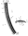
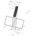
- Détecteur (18) pour un système de tomodensitométrie multitranche (10) conçu pour émettre de manière radiale des faisceaux de rayons X, ledit détecteur (18) comprenant :une matrice (56) de cellules de scintillateur comprenant au moins une première et une seconde cellules séparées l'une de l'autre par un espace, la matrice (56) de cellules de scintillateur étant plate ;un collimateur post-patient (62) placé en face de la matrice (56) de cellules de scintillateur, ledit collimateur post-patient (62) comportant au moins une plaque (64) de collimateur ;caractérisé en ce que ledit collimateur post-patient (62) est conçu pour être placé, par rapport à la matrice (56) de cellules de scintillateur, de telle manière qu'un axe géométrique central de ladite plaque (64) de collimateur est décalé par rapport à un axe géométrique central dudit espace de la matrice de cellules de scintillateur de façon qu'une ombre de rayons X soit centrée sur chaque espace de la matrice de cellules de scintillateur ; et
la/les plaques (64) de collimateur étant plus larges que l'espace entre la première et la seconde cellules, adjacentes, de la matrice (56) de cellules de scintillateur. - Détecteur (18) selon la revendication 1, dans lequel ladite matrice (56) comporte au moins un espace (70) de scintillateur sur l'axe X dans une première direction et au moins un espace de scintillateur sur l'axe Z dans une direction orthogonale à la première direction.
- Détecteur (18) selon la revendication 2, dans lequel ledit axe géométrique central de plaque de collimateur est décalé par rapport à l'axe géométrique central dudit espace (70) de scintillateur dans une première direction sur l'axe X.
- Détecteur (18) selon la revendication 3, dans lequel la largeur de ladite plaque (64) de collimateur est plus grande que la largeur dudit espace (70) de scintillateur dans une première direction sur l'axe X.
- Détecteur (18) selon l'une quelconque des revendications précédentes, dans lequel ledit collimateur (62) comprend une pluralité de plaques (64) de collimateur.
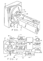
- Système de tomodensitométrie multitranche (10) pour produire des images d'un objet, ledit système (10) comprenant:une source (14) de rayons X servant à émettre des faisceaux de rayons X (16), lesdits faisceaux (16) étant émis depuis un foyer de la source ; etun détecteur (18) selon l'une quelconque des revendications précédentes.
- Procédé de réduction des artefacts spectraux dans un système de tomodensitométrie multitranche, le système comprenant une source de rayons X servant à émettre radialement des faisceaux de rayons X, au moins un détecteur multitranche comportant une matrice plate de cellules de scintillateur comportant au moins une première et une deuxième cellules séparées l'une de l'autre par un espace, et un collimateur post-patient placé en face de la matrice de cellules de scintillateur comportant au moins une plaque de collimateur, ledit procédé étant caractérisé par l'étape consistant à placer la plaque de collimateur post-patient au-dessus de l'espace de scintillateur, l'axe géométrique central de la plaque de collimateur étant décalé par rapport à un axe géométrique central de l'espace du scintillateur de sorte que, lorsque des faisceaux de rayons X sont émis depuis la source de rayons X, une ombre de la plaque de collimateur soit centrée sur chaque espace de scintillateur, et en ce qu'au moins une plaque de collimateur est plus large que l'espace entre la première et la seconde cellules adjacentes de la matrice de cellules de scintillateur.
Applications Claiming Priority (2)
| Application Number | Priority Date | Filing Date | Title |
|---|---|---|---|
| US09/118,397 US6266434B1 (en) | 1998-07-17 | 1998-07-17 | Methods and apparatus for reducing spectral artifacts in a computed tomograph system |
| US118397 | 1998-07-17 |
Publications (3)
| Publication Number | Publication Date |
|---|---|
| EP0973048A2 EP0973048A2 (fr) | 2000-01-19 |
| EP0973048A3 EP0973048A3 (fr) | 2002-07-31 |
| EP0973048B1 true EP0973048B1 (fr) | 2007-06-13 |
Family
ID=22378327
Family Applications (1)
| Application Number | Title | Priority Date | Filing Date |
|---|---|---|---|
| EP99305495A Expired - Lifetime EP0973048B1 (fr) | 1998-07-17 | 1999-07-12 | Procédé et appareil pour la réduction l'artefact spectrale dans un systeme tomographique à ordinateur |
Country Status (5)
| Country | Link |
|---|---|
| US (1) | US6266434B1 (fr) |
| EP (1) | EP0973048B1 (fr) |
| JP (1) | JP4502422B2 (fr) |
| DE (1) | DE69936278T2 (fr) |
| IL (1) | IL130935A (fr) |
Families Citing this family (5)
| Publication number | Priority date | Publication date | Assignee | Title |
|---|---|---|---|---|
| JP3961468B2 (ja) | 2003-09-19 | 2007-08-22 | ジーイー・メディカル・システムズ・グローバル・テクノロジー・カンパニー・エルエルシー | 放射線計算断層画像装置およびそれに用いる放射線検出器 |
| US7086780B2 (en) * | 2004-05-20 | 2006-08-08 | General Electric Company | Methods for spectrally calibrating CT imaging apparatus detectors |
| US7391844B2 (en) * | 2005-01-14 | 2008-06-24 | General Electric Company | Method and apparatus for correcting for beam hardening in CT images |
| US20120056095A1 (en) * | 2010-09-03 | 2012-03-08 | Scott Metzler | Collimation apparatus for high resolution imaging |
| US10610191B2 (en) * | 2017-07-06 | 2020-04-07 | Prismatic Sensors Ab | Managing geometric misalignment in x-ray imaging systems |
Family Cites Families (18)
| Publication number | Priority date | Publication date | Assignee | Title |
|---|---|---|---|---|
| US5093850A (en) * | 1976-04-19 | 1992-03-03 | General Electric Company | Tomographic scanning apparatus |
| US4180737A (en) * | 1978-02-06 | 1979-12-25 | General Electric Company | X-ray detector |
| US4392057A (en) * | 1980-02-22 | 1983-07-05 | National Research Development Corporation | Position-sensitive radiation detector |
| DE3669928D1 (de) * | 1985-07-12 | 1990-05-03 | Siemens Ag | Roentgendetektorsystem. |
| JPH01305930A (ja) * | 1988-06-04 | 1989-12-11 | Toshiba Corp | Ctスキヤナ用放射線検出器 |
| EP0489904B1 (fr) * | 1990-07-02 | 1998-03-04 | Varian Associates, Inc. | Simulateur de therapie a rayons x |
| US5168532A (en) * | 1990-07-02 | 1992-12-01 | Varian Associates, Inc. | Method for improving the dynamic range of an imaging system |
| JPH05256950A (ja) * | 1992-03-13 | 1993-10-08 | Toshiba Corp | X線コンピュータトモグラフィ装置用固体検出器 |
| US5335255A (en) * | 1992-03-24 | 1994-08-02 | Seppi Edward J | X-ray scanner with a source emitting plurality of fan beams |
| US5684855A (en) * | 1995-02-16 | 1997-11-04 | Kabushiki Kaisha Toshiba | X-ray CT scanner |
| JP3549169B2 (ja) * | 1995-03-10 | 2004-08-04 | 株式会社日立メディコ | X線ct装置 |
| US5961457A (en) * | 1996-05-03 | 1999-10-05 | The Regents Of The University Of Michigan | Method and apparatus for radiopharmaceutical-guided biopsy |
| JPH10104359A (ja) * | 1996-09-30 | 1998-04-24 | Shimadzu Corp | X線ct装置用検出器の製造方法 |
| US5812629A (en) * | 1997-04-30 | 1998-09-22 | Clauser; John F. | Ultrahigh resolution interferometric x-ray imaging |
| US5903008A (en) * | 1997-07-02 | 1999-05-11 | General Electric Company | Scatter correction methods and systems in single photon emission computed tomography |
| US5970113A (en) * | 1997-10-10 | 1999-10-19 | Analogic Corporation | Computed tomography scanning apparatus and method with temperature compensation for dark current offsets |
| US6115448A (en) | 1997-11-26 | 2000-09-05 | General Electric Company | Photodiode array for a scalable multislice scanning computed tomography system |
| JPH11295432A (ja) * | 1998-04-15 | 1999-10-29 | Shimadzu Corp | Ct用固体検出器 |
-
1998
- 1998-07-17 US US09/118,397 patent/US6266434B1/en not_active Expired - Lifetime
-
1999
- 1999-07-12 EP EP99305495A patent/EP0973048B1/fr not_active Expired - Lifetime
- 1999-07-12 DE DE69936278T patent/DE69936278T2/de not_active Expired - Lifetime
- 1999-07-14 IL IL13093599A patent/IL130935A/en not_active IP Right Cessation
- 1999-07-15 JP JP20109599A patent/JP4502422B2/ja not_active Expired - Lifetime
Non-Patent Citations (1)
| Title |
|---|
| None * |
Also Published As
| Publication number | Publication date |
|---|---|
| EP0973048A3 (fr) | 2002-07-31 |
| DE69936278T2 (de) | 2008-02-14 |
| JP2000037377A (ja) | 2000-02-08 |
| IL130935A0 (en) | 2001-01-28 |
| DE69936278D1 (de) | 2007-07-26 |
| US6266434B1 (en) | 2001-07-24 |
| EP0973048A2 (fr) | 2000-01-19 |
| IL130935A (en) | 2004-08-31 |
| JP4502422B2 (ja) | 2010-07-14 |
Similar Documents
| Publication | Publication Date | Title |
|---|---|---|
| US5982846A (en) | Methods and apparatus for dose reduction in a computed tomograph | |
| US6934354B2 (en) | Collimator assembly having multi-piece components | |
| US7177387B2 (en) | Self-aligning scintillator-collimator assembly | |
| US6173039B1 (en) | Variable aperture z-axis tracking collimator for computed tomograph system | |
| JP4070283B2 (ja) | コリメータ、コリメータ用のコーミング用具及びx線を検出する装置 | |
| US6061419A (en) | Methods and apparatus for noise compensation in an imaging system | |
| US7492857B2 (en) | Self-aligning scintillator-collimator assembly | |
| JP3270060B2 (ja) | 実質的に連続した放射線検出ゾーンを備えたエックス線断層撮影システム | |
| US6947517B2 (en) | Scintillator array having a reflector with integrated air gaps | |
| EP0981999A2 (fr) | Filtrage de rayons x pour la réalisation d'une qualité de faisceau dépendante de la position | |
| US6173031B1 (en) | Detector modules for computed tomograph system | |
| JP2012081265A (ja) | X線用ハイブリッド型コリメータ及びその製造方法 | |
| US6137857A (en) | Scalable detector for computed tomograph system | |
| US6118840A (en) | Methods and apparatus to desensitize incident angle errors on a multi-slice computed tomograph detector | |
| US7286639B2 (en) | Focal spot sensing device and method in an imaging system | |
| EP0982000B1 (fr) | Appareil de vérification de dosage dans un système d'imagerie | |
| US7655915B2 (en) | Collimator assembly for computed tomography system | |
| US6424697B1 (en) | Directed energy beam welded CT detector collimators | |
| US6343110B1 (en) | Methods and apparatus for submillimeter CT slices with increased coverage | |
| EP0973048B1 (fr) | Procédé et appareil pour la réduction l'artefact spectrale dans un systeme tomographique à ordinateur | |
| US6304625B1 (en) | Dose instrumentation methods and apparatus for collimated CT imaging systems | |
| US6252925B1 (en) | System and method for performing computed tomography with fiber waveguides |
Legal Events
| Date | Code | Title | Description |
|---|---|---|---|
| PUAI | Public reference made under article 153(3) epc to a published international application that has entered the european phase |
Free format text: ORIGINAL CODE: 0009012 |
|
| AK | Designated contracting states |
Kind code of ref document: A2 Designated state(s): AT BE CH CY DE DK ES FI FR GB GR IE IT LI LU MC NL PT SE |
|
| AX | Request for extension of the european patent |
Free format text: AL;LT;LV;MK;RO;SI |
|
| PUAL | Search report despatched |
Free format text: ORIGINAL CODE: 0009013 |
|
| AK | Designated contracting states |
Kind code of ref document: A3 Designated state(s): AT BE CH CY DE DK ES FI FR GB GR IE IT LI LU MC NL PT SE |
|
| AX | Request for extension of the european patent |
Free format text: AL;LT;LV;MK;RO;SI |
|
| RIC1 | Information provided on ipc code assigned before grant |
Free format text: 7G 01T 1/29 A, 7G 01T 1/00 B, 7G 01T 1/164 B |
|
| 17P | Request for examination filed |
Effective date: 20030131 |
|
| AKX | Designation fees paid |
Designated state(s): DE NL |
|
| 17Q | First examination report despatched |
Effective date: 20040623 |
|
| GRAP | Despatch of communication of intention to grant a patent |
Free format text: ORIGINAL CODE: EPIDOSNIGR1 |
|
| GRAS | Grant fee paid |
Free format text: ORIGINAL CODE: EPIDOSNIGR3 |
|
| GRAA | (expected) grant |
Free format text: ORIGINAL CODE: 0009210 |
|
| AK | Designated contracting states |
Kind code of ref document: B1 Designated state(s): DE NL |
|
| REF | Corresponds to: |
Ref document number: 69936278 Country of ref document: DE Date of ref document: 20070726 Kind code of ref document: P |
|
| PLBE | No opposition filed within time limit |
Free format text: ORIGINAL CODE: 0009261 |
|
| STAA | Information on the status of an ep patent application or granted ep patent |
Free format text: STATUS: NO OPPOSITION FILED WITHIN TIME LIMIT |
|
| 26N | No opposition filed |
Effective date: 20080314 |
|
| PGFP | Annual fee paid to national office [announced via postgrant information from national office to epo] |
Ref country code: NL Payment date: 20150726 Year of fee payment: 17 |
|
| REG | Reference to a national code |
Ref country code: NL Ref legal event code: MM Effective date: 20160801 |
|
| PG25 | Lapsed in a contracting state [announced via postgrant information from national office to epo] |
Ref country code: NL Free format text: LAPSE BECAUSE OF NON-PAYMENT OF DUE FEES Effective date: 20160801 |
|
| PGFP | Annual fee paid to national office [announced via postgrant information from national office to epo] |
Ref country code: DE Payment date: 20170727 Year of fee payment: 19 |
|
| REG | Reference to a national code |
Ref country code: DE Ref legal event code: R119 Ref document number: 69936278 Country of ref document: DE |
|
| PG25 | Lapsed in a contracting state [announced via postgrant information from national office to epo] |
Ref country code: DE Free format text: LAPSE BECAUSE OF NON-PAYMENT OF DUE FEES Effective date: 20190201 |