EP0928598A2 - Vorrichtung zur Befestigung eines Sensors in einem Körperlumen - Google Patents
Vorrichtung zur Befestigung eines Sensors in einem Körperlumen Download PDFInfo
- Publication number
- EP0928598A2 EP0928598A2 EP98124408A EP98124408A EP0928598A2 EP 0928598 A2 EP0928598 A2 EP 0928598A2 EP 98124408 A EP98124408 A EP 98124408A EP 98124408 A EP98124408 A EP 98124408A EP 0928598 A2 EP0928598 A2 EP 0928598A2
- Authority
- EP
- European Patent Office
- Prior art keywords
- sensor
- fixation device
- bodily lumen
- fixation
- sensor support
- Prior art date
- Legal status (The legal status is an assumption and is not a legal conclusion. Google has not performed a legal analysis and makes no representation as to the accuracy of the status listed.)
- Withdrawn
Links
- 238000000034 method Methods 0.000 claims abstract description 71
- 238000004873 anchoring Methods 0.000 claims abstract description 29
- 238000003780 insertion Methods 0.000 claims abstract description 18
- 230000037431 insertion Effects 0.000 claims abstract description 18
- 230000006378 damage Effects 0.000 claims abstract description 8
- 238000013461 design Methods 0.000 claims description 31
- 239000000463 material Substances 0.000 claims description 20
- 239000011241 protective layer Substances 0.000 claims description 16
- 239000011248 coating agent Substances 0.000 claims description 14
- 238000000576 coating method Methods 0.000 claims description 14
- 238000005530 etching Methods 0.000 claims description 12
- 239000002184 metal Substances 0.000 claims description 12
- 238000003466 welding Methods 0.000 claims description 10
- WQZGKKKJIJFFOK-GASJEMHNSA-N Glucose Natural products OC[C@H]1OC(O)[C@H](O)[C@@H](O)[C@@H]1O WQZGKKKJIJFFOK-GASJEMHNSA-N 0.000 claims description 8
- CZMRCDWAGMRECN-UGDNZRGBSA-N Sucrose Chemical compound O[C@H]1[C@H](O)[C@@H](CO)O[C@@]1(CO)O[C@@H]1[C@H](O)[C@@H](O)[C@H](O)[C@@H](CO)O1 CZMRCDWAGMRECN-UGDNZRGBSA-N 0.000 claims description 8
- 229930006000 Sucrose Natural products 0.000 claims description 8
- 239000008103 glucose Substances 0.000 claims description 8
- 239000005720 sucrose Substances 0.000 claims description 8
- WQZGKKKJIJFFOK-VFUOTHLCSA-N beta-D-glucose Chemical compound OC[C@H]1O[C@@H](O)[C@H](O)[C@@H](O)[C@@H]1O WQZGKKKJIJFFOK-VFUOTHLCSA-N 0.000 claims description 7
- 238000012544 monitoring process Methods 0.000 claims description 5
- 229920002153 Hydroxypropyl cellulose Polymers 0.000 claims description 4
- VYPSYNLAJGMNEJ-UHFFFAOYSA-N Silicium dioxide Chemical compound O=[Si]=O VYPSYNLAJGMNEJ-UHFFFAOYSA-N 0.000 claims description 4
- 239000001863 hydroxypropyl cellulose Substances 0.000 claims description 4
- 235000010977 hydroxypropyl cellulose Nutrition 0.000 claims description 4
- 239000001866 hydroxypropyl methyl cellulose Substances 0.000 claims description 4
- 235000010979 hydroxypropyl methyl cellulose Nutrition 0.000 claims description 4
- 229920003088 hydroxypropyl methyl cellulose Polymers 0.000 claims description 4
- UFVKGYZPFZQRLF-UHFFFAOYSA-N hydroxypropyl methyl cellulose Chemical compound OC1C(O)C(OC)OC(CO)C1OC1C(O)C(O)C(OC2C(C(O)C(OC3C(C(O)C(O)C(CO)O3)O)C(CO)O2)O)C(CO)O1 UFVKGYZPFZQRLF-UHFFFAOYSA-N 0.000 claims description 4
- 229940071676 hydroxypropylcellulose Drugs 0.000 claims description 4
- XLYOFNOQVPJJNP-UHFFFAOYSA-N water Substances O XLYOFNOQVPJJNP-UHFFFAOYSA-N 0.000 claims description 4
- 238000004026 adhesive bonding Methods 0.000 claims description 3
- 239000007864 aqueous solution Substances 0.000 claims description 3
- 238000005520 cutting process Methods 0.000 claims description 3
- 238000007598 dipping method Methods 0.000 claims description 2
- 238000010422 painting Methods 0.000 claims description 2
- 238000005507 spraying Methods 0.000 claims description 2
- 229910001220 stainless steel Inorganic materials 0.000 claims description 2
- 239000010935 stainless steel Substances 0.000 claims description 2
- 230000008878 coupling Effects 0.000 claims 9
- 238000010168 coupling process Methods 0.000 claims 9
- 238000005859 coupling reaction Methods 0.000 claims 9
- 239000000853 adhesive Substances 0.000 claims 1
- 230000001070 adhesive effect Effects 0.000 claims 1
- 230000000737 periodic effect Effects 0.000 claims 1
- 239000011253 protective coating Substances 0.000 abstract description 3
- 210000000709 aorta Anatomy 0.000 description 4
- 239000012528 membrane Substances 0.000 description 4
- 229910003460 diamond Inorganic materials 0.000 description 3
- 239000010432 diamond Substances 0.000 description 3
- 239000000126 substance Substances 0.000 description 3
- KKJUPNGICOCCDW-UHFFFAOYSA-N 7-N,N-Dimethylamino-1,2,3,4,5-pentathiocyclooctane Chemical compound CN(C)C1CSSSSSC1 KKJUPNGICOCCDW-UHFFFAOYSA-N 0.000 description 2
- 210000004351 coronary vessel Anatomy 0.000 description 2
- 239000000203 mixture Substances 0.000 description 2
- 238000001356 surgical procedure Methods 0.000 description 2
- 239000003106 tissue adhesive Substances 0.000 description 2
- 239000000969 carrier Substances 0.000 description 1
- 238000012937 correction Methods 0.000 description 1
- 230000006866 deterioration Effects 0.000 description 1
- 201000010099 disease Diseases 0.000 description 1
- 208000037265 diseases, disorders, signs and symptoms Diseases 0.000 description 1
- 238000004090 dissolution Methods 0.000 description 1
- 230000009977 dual effect Effects 0.000 description 1
- 230000000694 effects Effects 0.000 description 1
- 239000012530 fluid Substances 0.000 description 1
- 239000007789 gas Substances 0.000 description 1
- 238000002513 implantation Methods 0.000 description 1
- 238000003698 laser cutting Methods 0.000 description 1
- 230000003902 lesion Effects 0.000 description 1
- 239000007788 liquid Substances 0.000 description 1
- 230000007774 longterm Effects 0.000 description 1
- 230000035479 physiological effects, processes and functions Effects 0.000 description 1
- 239000006187 pill Substances 0.000 description 1
- 230000035945 sensitivity Effects 0.000 description 1
- 235000020374 simple syrup Nutrition 0.000 description 1
- 238000005476 soldering Methods 0.000 description 1
- 239000007787 solid Substances 0.000 description 1
- 238000011477 surgical intervention Methods 0.000 description 1
- 208000024891 symptom Diseases 0.000 description 1
Images


Classifications
-
- A—HUMAN NECESSITIES
- A61—MEDICAL OR VETERINARY SCIENCE; HYGIENE
- A61F—FILTERS IMPLANTABLE INTO BLOOD VESSELS; PROSTHESES; DEVICES PROVIDING PATENCY TO, OR PREVENTING COLLAPSING OF, TUBULAR STRUCTURES OF THE BODY, e.g. STENTS; ORTHOPAEDIC, NURSING OR CONTRACEPTIVE DEVICES; FOMENTATION; TREATMENT OR PROTECTION OF EYES OR EARS; BANDAGES, DRESSINGS OR ABSORBENT PADS; FIRST-AID KITS
- A61F2/00—Filters implantable into blood vessels; Prostheses, i.e. artificial substitutes or replacements for parts of the body; Appliances for connecting them with the body; Devices providing patency to, or preventing collapsing of, tubular structures of the body, e.g. stents
- A61F2/82—Devices providing patency to, or preventing collapsing of, tubular structures of the body, e.g. stents
-
- A—HUMAN NECESSITIES
- A61—MEDICAL OR VETERINARY SCIENCE; HYGIENE
- A61B—DIAGNOSIS; SURGERY; IDENTIFICATION
- A61B17/00—Surgical instruments, devices or methods
- A61B17/12—Surgical instruments, devices or methods for ligaturing or otherwise compressing tubular parts of the body, e.g. blood vessels or umbilical cord
- A61B17/12022—Occluding by internal devices, e.g. balloons or releasable wires
- A61B17/12131—Occluding by internal devices, e.g. balloons or releasable wires characterised by the type of occluding device
- A61B17/12168—Occluding by internal devices, e.g. balloons or releasable wires characterised by the type of occluding device having a mesh structure
- A61B17/12172—Occluding by internal devices, e.g. balloons or releasable wires characterised by the type of occluding device having a mesh structure having a pre-set deployed three-dimensional shape
-
- A—HUMAN NECESSITIES
- A61—MEDICAL OR VETERINARY SCIENCE; HYGIENE
- A61B—DIAGNOSIS; SURGERY; IDENTIFICATION
- A61B5/00—Measuring for diagnostic purposes; Identification of persons
- A61B5/68—Arrangements of detecting, measuring or recording means, e.g. sensors, in relation to patient
- A61B5/6846—Arrangements of detecting, measuring or recording means, e.g. sensors, in relation to patient specially adapted to be brought in contact with an internal body part, i.e. invasive
- A61B5/6847—Arrangements of detecting, measuring or recording means, e.g. sensors, in relation to patient specially adapted to be brought in contact with an internal body part, i.e. invasive mounted on an invasive device
- A61B5/6862—Stents
-
- A—HUMAN NECESSITIES
- A61—MEDICAL OR VETERINARY SCIENCE; HYGIENE
- A61B—DIAGNOSIS; SURGERY; IDENTIFICATION
- A61B5/00—Measuring for diagnostic purposes; Identification of persons
- A61B5/68—Arrangements of detecting, measuring or recording means, e.g. sensors, in relation to patient
- A61B5/6846—Arrangements of detecting, measuring or recording means, e.g. sensors, in relation to patient specially adapted to be brought in contact with an internal body part, i.e. invasive
- A61B5/6867—Arrangements of detecting, measuring or recording means, e.g. sensors, in relation to patient specially adapted to be brought in contact with an internal body part, i.e. invasive specially adapted to be attached or implanted in a specific body part
- A61B5/6876—Blood vessel
-
- A—HUMAN NECESSITIES
- A61—MEDICAL OR VETERINARY SCIENCE; HYGIENE
- A61B—DIAGNOSIS; SURGERY; IDENTIFICATION
- A61B5/00—Measuring for diagnostic purposes; Identification of persons
- A61B5/68—Arrangements of detecting, measuring or recording means, e.g. sensors, in relation to patient
- A61B5/6846—Arrangements of detecting, measuring or recording means, e.g. sensors, in relation to patient specially adapted to be brought in contact with an internal body part, i.e. invasive
- A61B5/6879—Means for maintaining contact with the body
- A61B5/6882—Anchoring means
-
- A—HUMAN NECESSITIES
- A61—MEDICAL OR VETERINARY SCIENCE; HYGIENE
- A61F—FILTERS IMPLANTABLE INTO BLOOD VESSELS; PROSTHESES; DEVICES PROVIDING PATENCY TO, OR PREVENTING COLLAPSING OF, TUBULAR STRUCTURES OF THE BODY, e.g. STENTS; ORTHOPAEDIC, NURSING OR CONTRACEPTIVE DEVICES; FOMENTATION; TREATMENT OR PROTECTION OF EYES OR EARS; BANDAGES, DRESSINGS OR ABSORBENT PADS; FIRST-AID KITS
- A61F2/00—Filters implantable into blood vessels; Prostheses, i.e. artificial substitutes or replacements for parts of the body; Appliances for connecting them with the body; Devices providing patency to, or preventing collapsing of, tubular structures of the body, e.g. stents
- A61F2/82—Devices providing patency to, or preventing collapsing of, tubular structures of the body, e.g. stents
- A61F2/86—Stents in a form characterised by the wire-like elements; Stents in the form characterised by a net-like or mesh-like structure
- A61F2/90—Stents in a form characterised by the wire-like elements; Stents in the form characterised by a net-like or mesh-like structure characterised by a net-like or mesh-like structure
- A61F2/91—Stents in a form characterised by the wire-like elements; Stents in the form characterised by a net-like or mesh-like structure characterised by a net-like or mesh-like structure made from perforated sheets or tubes, e.g. perforated by laser cuts or etched holes
- A61F2/915—Stents in a form characterised by the wire-like elements; Stents in the form characterised by a net-like or mesh-like structure characterised by a net-like or mesh-like structure made from perforated sheets or tubes, e.g. perforated by laser cuts or etched holes with bands having a meander structure, adjacent bands being connected to each other
-
- A—HUMAN NECESSITIES
- A61—MEDICAL OR VETERINARY SCIENCE; HYGIENE
- A61B—DIAGNOSIS; SURGERY; IDENTIFICATION
- A61B17/00—Surgical instruments, devices or methods
- A61B17/12—Surgical instruments, devices or methods for ligaturing or otherwise compressing tubular parts of the body, e.g. blood vessels or umbilical cord
- A61B17/12022—Occluding by internal devices, e.g. balloons or releasable wires
-
- A—HUMAN NECESSITIES
- A61—MEDICAL OR VETERINARY SCIENCE; HYGIENE
- A61B—DIAGNOSIS; SURGERY; IDENTIFICATION
- A61B5/00—Measuring for diagnostic purposes; Identification of persons
- A61B5/0002—Remote monitoring of patients using telemetry, e.g. transmission of vital signals via a communication network
- A61B5/0031—Implanted circuitry
-
- A—HUMAN NECESSITIES
- A61—MEDICAL OR VETERINARY SCIENCE; HYGIENE
- A61F—FILTERS IMPLANTABLE INTO BLOOD VESSELS; PROSTHESES; DEVICES PROVIDING PATENCY TO, OR PREVENTING COLLAPSING OF, TUBULAR STRUCTURES OF THE BODY, e.g. STENTS; ORTHOPAEDIC, NURSING OR CONTRACEPTIVE DEVICES; FOMENTATION; TREATMENT OR PROTECTION OF EYES OR EARS; BANDAGES, DRESSINGS OR ABSORBENT PADS; FIRST-AID KITS
- A61F2/00—Filters implantable into blood vessels; Prostheses, i.e. artificial substitutes or replacements for parts of the body; Appliances for connecting them with the body; Devices providing patency to, or preventing collapsing of, tubular structures of the body, e.g. stents
- A61F2/82—Devices providing patency to, or preventing collapsing of, tubular structures of the body, e.g. stents
- A61F2/86—Stents in a form characterised by the wire-like elements; Stents in the form characterised by a net-like or mesh-like structure
- A61F2/90—Stents in a form characterised by the wire-like elements; Stents in the form characterised by a net-like or mesh-like structure characterised by a net-like or mesh-like structure
- A61F2/91—Stents in a form characterised by the wire-like elements; Stents in the form characterised by a net-like or mesh-like structure characterised by a net-like or mesh-like structure made from perforated sheets or tubes, e.g. perforated by laser cuts or etched holes
- A61F2/915—Stents in a form characterised by the wire-like elements; Stents in the form characterised by a net-like or mesh-like structure characterised by a net-like or mesh-like structure made from perforated sheets or tubes, e.g. perforated by laser cuts or etched holes with bands having a meander structure, adjacent bands being connected to each other
- A61F2002/91525—Stents in a form characterised by the wire-like elements; Stents in the form characterised by a net-like or mesh-like structure characterised by a net-like or mesh-like structure made from perforated sheets or tubes, e.g. perforated by laser cuts or etched holes with bands having a meander structure, adjacent bands being connected to each other within the whole structure different bands showing different meander characteristics, e.g. frequency or amplitude
-
- A—HUMAN NECESSITIES
- A61—MEDICAL OR VETERINARY SCIENCE; HYGIENE
- A61F—FILTERS IMPLANTABLE INTO BLOOD VESSELS; PROSTHESES; DEVICES PROVIDING PATENCY TO, OR PREVENTING COLLAPSING OF, TUBULAR STRUCTURES OF THE BODY, e.g. STENTS; ORTHOPAEDIC, NURSING OR CONTRACEPTIVE DEVICES; FOMENTATION; TREATMENT OR PROTECTION OF EYES OR EARS; BANDAGES, DRESSINGS OR ABSORBENT PADS; FIRST-AID KITS
- A61F2/00—Filters implantable into blood vessels; Prostheses, i.e. artificial substitutes or replacements for parts of the body; Appliances for connecting them with the body; Devices providing patency to, or preventing collapsing of, tubular structures of the body, e.g. stents
- A61F2/82—Devices providing patency to, or preventing collapsing of, tubular structures of the body, e.g. stents
- A61F2/86—Stents in a form characterised by the wire-like elements; Stents in the form characterised by a net-like or mesh-like structure
- A61F2/90—Stents in a form characterised by the wire-like elements; Stents in the form characterised by a net-like or mesh-like structure characterised by a net-like or mesh-like structure
- A61F2/91—Stents in a form characterised by the wire-like elements; Stents in the form characterised by a net-like or mesh-like structure characterised by a net-like or mesh-like structure made from perforated sheets or tubes, e.g. perforated by laser cuts or etched holes
- A61F2/915—Stents in a form characterised by the wire-like elements; Stents in the form characterised by a net-like or mesh-like structure characterised by a net-like or mesh-like structure made from perforated sheets or tubes, e.g. perforated by laser cuts or etched holes with bands having a meander structure, adjacent bands being connected to each other
- A61F2002/91533—Stents in a form characterised by the wire-like elements; Stents in the form characterised by a net-like or mesh-like structure characterised by a net-like or mesh-like structure made from perforated sheets or tubes, e.g. perforated by laser cuts or etched holes with bands having a meander structure, adjacent bands being connected to each other characterised by the phase between adjacent bands
- A61F2002/91541—Adjacent bands are arranged out of phase
-
- A—HUMAN NECESSITIES
- A61—MEDICAL OR VETERINARY SCIENCE; HYGIENE
- A61F—FILTERS IMPLANTABLE INTO BLOOD VESSELS; PROSTHESES; DEVICES PROVIDING PATENCY TO, OR PREVENTING COLLAPSING OF, TUBULAR STRUCTURES OF THE BODY, e.g. STENTS; ORTHOPAEDIC, NURSING OR CONTRACEPTIVE DEVICES; FOMENTATION; TREATMENT OR PROTECTION OF EYES OR EARS; BANDAGES, DRESSINGS OR ABSORBENT PADS; FIRST-AID KITS
- A61F2/00—Filters implantable into blood vessels; Prostheses, i.e. artificial substitutes or replacements for parts of the body; Appliances for connecting them with the body; Devices providing patency to, or preventing collapsing of, tubular structures of the body, e.g. stents
- A61F2/82—Devices providing patency to, or preventing collapsing of, tubular structures of the body, e.g. stents
- A61F2/86—Stents in a form characterised by the wire-like elements; Stents in the form characterised by a net-like or mesh-like structure
- A61F2/90—Stents in a form characterised by the wire-like elements; Stents in the form characterised by a net-like or mesh-like structure characterised by a net-like or mesh-like structure
- A61F2/91—Stents in a form characterised by the wire-like elements; Stents in the form characterised by a net-like or mesh-like structure characterised by a net-like or mesh-like structure made from perforated sheets or tubes, e.g. perforated by laser cuts or etched holes
- A61F2/915—Stents in a form characterised by the wire-like elements; Stents in the form characterised by a net-like or mesh-like structure characterised by a net-like or mesh-like structure made from perforated sheets or tubes, e.g. perforated by laser cuts or etched holes with bands having a meander structure, adjacent bands being connected to each other
- A61F2002/9155—Adjacent bands being connected to each other
- A61F2002/91558—Adjacent bands being connected to each other connected peak to peak
-
- A—HUMAN NECESSITIES
- A61—MEDICAL OR VETERINARY SCIENCE; HYGIENE
- A61F—FILTERS IMPLANTABLE INTO BLOOD VESSELS; PROSTHESES; DEVICES PROVIDING PATENCY TO, OR PREVENTING COLLAPSING OF, TUBULAR STRUCTURES OF THE BODY, e.g. STENTS; ORTHOPAEDIC, NURSING OR CONTRACEPTIVE DEVICES; FOMENTATION; TREATMENT OR PROTECTION OF EYES OR EARS; BANDAGES, DRESSINGS OR ABSORBENT PADS; FIRST-AID KITS
- A61F2220/00—Fixations or connections for prostheses classified in groups A61F2/00 - A61F2/26 or A61F2/82 or A61F9/00 or A61F11/00 or subgroups thereof
- A61F2220/0008—Fixation appliances for connecting prostheses to the body
-
- A—HUMAN NECESSITIES
- A61—MEDICAL OR VETERINARY SCIENCE; HYGIENE
- A61F—FILTERS IMPLANTABLE INTO BLOOD VESSELS; PROSTHESES; DEVICES PROVIDING PATENCY TO, OR PREVENTING COLLAPSING OF, TUBULAR STRUCTURES OF THE BODY, e.g. STENTS; ORTHOPAEDIC, NURSING OR CONTRACEPTIVE DEVICES; FOMENTATION; TREATMENT OR PROTECTION OF EYES OR EARS; BANDAGES, DRESSINGS OR ABSORBENT PADS; FIRST-AID KITS
- A61F2250/00—Special features of prostheses classified in groups A61F2/00 - A61F2/26 or A61F2/82 or A61F9/00 or A61F11/00 or subgroups thereof
- A61F2250/0001—Means for transferring electromagnetic energy to implants
- A61F2250/0002—Means for transferring electromagnetic energy to implants for data transfer
Definitions
- the present invention relates generally to a method and device for fixation of a sensor in a bodily lumen and for protection of the sensor during insertion into a bodily lumen.
- U.S. Patent No. 4,485,813 describes a sensor that may be permanently implanted in a specific location within the human body in an implantable medical device such as a pacemaker. This sensor is used to monitor certain physical and/or physiological parameters of the subject in which it has been implanted. This sensor can be maintained in the subject for extended periods of time to continuously monitor information about the subject.
- Sensors used to monitor parameters within lumens are made of very thin membranes that are highly sensitive to mechanical pressure. As a result there is a great risk of the sensor being damaged during insertion and or positioning. Damage to the sensor could result in poor performance or non-operability of the sensor. For example, should the membrane of a sensor break during insertion, the sensor would be rendered inoperable. Due to the risks associated with the procedures for the insertion of sensors, there would be great costs and risks involved should a sensor be damaged or destroyed during insertion. Thus, there is also a need for a device and method of protecting sensors during insertion and fixation.
- an object of the present invention to provide a method and device for fixation of a sensor in a bodily lumen.
- remotely interrogated sensors may be fixed within bodily lumens.
- Such sensors may be used to record and/or monitor parameters such as, for example, physiological parameters, e.g ., pressure and velocity of flow, and biochemical parameters, e.g ., level of gases and biochemical substances in the fluid contained in the lumen.
- the present invention therefore, provides a sensor device which may be implanted, either temporarily or permanently, in a lumen and interrogated from an exterior position, for example, the surface of the body, at any time without any intervention or physical intrusion.
- the present invention provides a method and device for the fixation of such sensors in specific desired locations and/or preferred positions in the lumen.
- Such fixation of the sensors may be achieved at the time of any required surgical intervention or independently by catheterization.
- the sensor may be connected to the repair device, e.g ., the stitches of a bypass, an aneurismal repair device, a stent, etc., or mounted on its own dedicated fixation device.
- a sensor may be fixed inside a lumen by any number of means, including directly attaching the sensor in place, for example, by including holes in the sensor, e.g. , around its periphery, and attaching the sensor to the stitches of a bypass during surgery, or through the use of a surgical adhesive.
- a sensor may also be positioned inside a lumen using a carrier or support (of any shape and size) which may be part of, or coupled to a repair device, e.g ., a stent or aneurismal correction device which holds the sensor in place adjacent to or near the repair device.
- a sensor may be positioned inside a lumen using a dedicated device, e.g ., an anchoring ring, which is held within a lumen and fixed in place, for example, by expansion with a catheter balloon.
- the anchoring ring does not necessarily have to be circular in shape, but may instead be oval or any other shape best suited for the location where placed.
- the anchoring ring may have a separate carrier or support to hold the sensor.
- the carrier or support may be any shape or size, including, for example, circular, square, rectangular, diamond shaped, linear with or without a bent or curved end, etc, and it may be constructed as only a border or as a solid piece of material.
- Multiple sensors may be attached to a carrier or carriers, for example, two sensors with one placed on each side of a stent, or two sensors attached at both connections of a bypass section, e.g ., one sensor at the entrance to an aneurismal sleeve and one at the outside of the sleeve to monitor for a possible leak around the sleeve.
- a sensor may have multiple repair devices or dedicated devices supporting it within a lumen, either with or without a carrier, for example, a sensor supported between two anchoring rings
- a sensor may be supported by or connected to a carrier, for example, by providing a groove-like depression(s) or notch-like depression(s) in the sensor into which a portion(s) of the carrier may be inserted, or the sensor may be configured such that a portion(s) of the sensor, for example, a lip-like extension(s) or protrusion(s), may extend beyond the dimensions of the carrier to be supported thereby. Additionally, the sensor may be attached to the carrier, for example, by welding and/or glueing or any combinations of the above.
- a sensor After a sensor is fixed within a lumen, for example, during an intervention procedure such as aneurismal device implantation, PTCA, coronary bypass surgery, etc., it may thereafter be monitored periodically to track any of a variety of parameters or to assess the effectiveness of the procedure that was performed. For example, the sensor may be monitored periodically to assess the long term progress or deterioration of the corrective effect, and the progress of relevant symptoms of a disease.
- Multiple sensors may be implanted and may be monitored individually or simultaneously to derive gradients along a lumen and across a repair device or section. Such sensors may be fixed in any number of positions within a lumen, for example, on both sides of a lesion treated by PTCA with or without a stent, on both sides of a bypass section, and before, after and around an aneurismal repair device, etc.
- the present invention also provides for a device and method for the protection of sensors during insertion.
- sensors may be coated with a protective layer which is soluble in an aqueous solution, and which disappears immediately or soon alter deployment of the sensor in the body.
- the material used for, thickness of, and hardness of the coating may vary, for example, depending on the location of the sensor, the type of sensor, protection level sought, and rate of dissolution desired.
- the fixation device may be constructed by first creating a flat version of the desired pattern for the fixation device, for example, from a piece of thin stainless steel sheet metal or some other material, e.g. , any metal, non-metallic or bioabsorbable material.
- the flat pattern can be produced through any suitable technique, such as etching the design into the sheet metal, by cutting with a very fine laser, or by any other technique.
- the material is deformed so as to cause its edges to meet.
- the flat metal is rolled until the edges meet.
- the portion which holds the sensor may be located along the circumference of the fixation device, may extend perpendicular to the cross-section of the ring formed or may extend in some other manner from the ring formed by the fixation device.
- the locations where edges meet are joined together, such as by spot welding.
- the fixation device is polished, either mechanically or electrochemically.
- FIG. 1A and 1B illustrate a first fixation device for a sensor and a first method for fixation of a sensor within a lumen, respectively, according to a preferred embodiment of the present invention.
- a sensor 1 having two holes 3 in its periphery for attachment to sutures within a lumen.
- a coronary artery 5 starting at the Aorta 7 and having an occlusion 9.
- a bypass 11 is connected between the Aorta at point 13 and at point 15 beyond the occlusion 9.
- Sensor 1 is placed either at the proximal ostium 17 or at the proximal part of the bypass 19.
- sensor 1 may be placed at the distal ostium 21, at the distal part before the distal ostium 23, or at the distal part after the distal ostium 25. Any number of sensors may be used, and they may be placed in any combination of the above positions or any other position desired.
- the sensor 1 is fixed in place using the two holes 3 for attachment to the sutures.
- the sensor 1 may be fixed in place using surgical adhesive or a surgical staple(s).
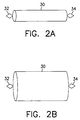
- FIGs. 2A and 2B there are shown a second fixation device for a sensor before expansion and after expansion, respectively, according to a preferred embodiment of the present invention.
- a stent 30 in a non-expanded state with a first sensor support 32 and a second sensor support 34.
- the stent 30 may include only one or more than two sensor supports.
- a third sensor support may be located opposite the first sensor support 32.
- the stent 30 from Fig. 2A is shown in its expanded state. Expansion may be accomplished, for example, by balloon catheterization or some other procedure. To fix a sensor within a lumen, the stent 30 is positioned as it normally would be during any medical procedure in which a stent is used.
- a sensor Prior to expansion, and either prior to or after insertion of the stent 30 into the lumen, a sensor is placed in, placed on or attached to the first sensor support 32 and/or the second sensor support 34. The stent 30 is then either expanded, or inserted into the lumen and then expanded. The same procedure may be used to fix any number of sensors within a lumen, with the additional step of placing each sensor either in or on, or attaching each sensor to its corresponding sensor support.
- FIGs. 3A and 3B there are shown a third fixation device for a sensor before expansion and after expansion, respectively, according to a preferred embodiment of the present invention.
- a fixation device 40 in the form of an anchoring ring 42, in a non-expanded state coupled to a sensor support 44.
- the fixation device 40 may be formed of any malleable material which does not revert automatically to its original shape after being expanded.
- the anchoring ring 42 is made up of a plurality of elliptical sections 46 connected one to the other at the middle of each of their long portions 48 to form a ring.
- the sensor support 44 is connected to one of the elliptical sections 46 at a short portion 49, and perpendicular to a cross-section of the anchoring ring 42 forming a circular plane.
- the sensor support 44 is formed in the shape of a diamond, but can be any shape desired. Additionally, there may be multiple sensor supports attached to the anchoring ring 42.
- the anchoring ring 42 may be made of a single sinusoidal ring, with one or more sensor supports attached to the peaks, since it does not serve any support function for the lumen.
- Fig. 3B shows the fixation device 40 of Fig. 3A in an expanded state.
- the fixation device 40 is positioned within the lumen, for example, during an intervention procedure, and expanded, for example, by balloon catheterization or some other procedure.
- the sensor Prior to expansion, and either prior to or after insertion of the fixation device 40 into the lumen, the sensor is placed in, placed on or attached to the sensor support 42.
- the fixation device 40 is then either expanded, or inserted into the lumen and then expanded.
- the same procedure may be used to fix multiple sensors within a lumen, with the additional step of placing each sensor either in or on, or attaching each sensor to a corresponding sensor support.
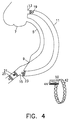
- FIG. 4 illustrates a second method for fixation of a sensor within a lumen using the third fixation device shown in Figs. 3A and 3B, according to a preferred embodiment of the present invention.
- a coronary artery 5 starting at the Aorta 7 and having an occlusion 9 is fitted with a bypass 11 which is connected between the Aorta at point 13 and at point 15 beyond the occlusion 9.
- Sensor 50 which is carried by the sensor support 44 coupled to the anchoring ring 42 of Figs.
- 3A and 3B is placed either at the proximal part of the bypass 19, at the distal ostium 21, at the distal part before the distal ostium 23, or at the distal part after the distal ostium 25.
- Any number of sensors may be used, and they may be placed in any combination of the above positions or any other position desired in which an anchoring ring can be used.
- the sensor 50 is fixed in place by expansion using balloon catheterization.
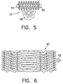
- FIG. 5 there is shown an illustration of a mask for etching of a flat design of the fixation device of Figs. 3A and 3B, according to a preferred embodiment of the present invention.
- a mask 52 is created for etching a flat design of a fixation device.
- the flat design is then etched onto a piece of thin sheet metal or some other malleable material.
- the flat design is next cut from the sheet metal using, for example, a fine laser.
- the cut flat design is then polished and bent into a circular (or other) shape.
- Points 54 and 56 show the locations where the flat design is coupled, for example, by welding after it is bent. The welding creates an anchoring ring.
- Sensor support 58 is positioned approximately at the midpoint of the mask 52, but may alternatively be located at any other position. Additionally, there may be multiple sensor supports, for example, located at both sides of the fixation device design.
- FIG. 6 there is shown an illustration of a mask for etching of a flat design of the fixation device of Figs. 2A and 2B, according to a preferred embodiment of the present invention.
- a mask 60 is created for etching a flat design of a stent.
- the flat design is then etched onto a piece of thin sheet metal or some other malleable material.
- the flat design is next cut from the sheet metal using, for example, a fine laser.
- the cut flat design is then polished and bent into a circular (or other) shape and coupled, for example, by welding after it is bent.
- Sensor support 62 is positioned approximately at the midpoint of the mask 60, but may alternatively be located at any other position. Additionally, there may be multiple sensor supports, for example, located at both sides of the stent design.
- a fixation device 70 in the form of a dual anchoring ring comprises a first ring 72 and a second ring 74, in a non-expanded state, with a sensor support 76 positioned between the two rings 72, 74.
- the fixation device 70 may be formed of any malleable material which does not revert automatically to its original shape after being expanded.
- the fixation device 70 is made up of a plurality of sections 78 connected one to the other to form two anchoring rings 72, 74.
- a sensor support 76 is connected to one of the sections 78 of each anchoring ring 72, 74 perpendicular to a cross-section of each of the rings 72, 74 forming a circular plane, and is positioned between the two rings 72, 74.
- the sensor support is formed in the shape of a diamond, but can be any shape desired. Additionally, there may be multiple sensor supports attached to the fixation device 70. Alternatively, the fixation device 70 may be made of two single sinusoidal rings, with one or more sensor supports attached to the peaks, since it does not serve any support function for the lumen.
- the fixation device 70 may alternatively be made of two stents, one on each side of a sensor support, or having multiple sensor supports attached thereto.
- FIG. 8 there is shown an illustration of a mask for etching of a flat design of the fixation device of Fig. 7, according to a preferred embodiment of the present invention.
- a mask 80 is created for etching a flat design of a fixation device.
- the flat design is then etched onto a piece of thin sheet metal or some other malleable material.
- the flat design is next cut from the sheet metal using, for example, a fine laser.
- the cut flat design is then polished and bent into a circular (or other) shape.
- Points 82 and 83, and points 84 and 85 show the respective locations where the flat design is coupled, for example, by welding after it is bent. The welding creates two anchoring rings.
- Sensor support 87 is positioned approximately at the midpoint of the mask 80, but may alternatively be located at any other position. Additionally, there may be multiple sensor supports, for example, located at both sides of the fixation device design.
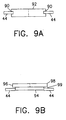
- a groove 90 is formed in two portions of the periphery of sensor 92, for example, by cutting with a wire saw, by etching, by laser cutting, etc., and the sensor 92 is then inserted into the sensor support 44 such that two portions of the sensor support 44 are positioned within the groove 90 providing support for the sensor 92.
- two notches may be formed in the periphery of the sensor 92 in which the two portions of the sensor support 44 may be positioned.
- sensor 94 is formed with a lip 96 around its upper edge 98.
- Sensor 94 may instead be formed with one or more protrusions along its upper edge 98.
- the lip or protrusion(s) may be located on the bottom or at any other position on the sensor.
- the sensor 94 is coupled to the sensor support 44, for example, by glueing, welding, soldering, etc., the lip 96 or protrusion(s) to an edge or portion 99 of the sensor support.
- the sensor 94 may be placed on the sensor support 44 and supported by the lip 96 or by the protrusion(s).
- a coating may be placed on the sensors to protect them from damage and/or destruction during deployment.
- the coating may be made from a material that is soluble in an aqueous solution, and should dissolve immediately or soon after deployment of the sensor.
- the material used, the thickness of the coating and the hardness of the coating will depend to a large extent on the location of the sensor, the type of sensor, and a variety of other factors including the physiology involved, the parameters being measured, and the desired speed of deployment.
- a first example of a coating is a composition comprising solidified sugar syrup made of approximately equal amounts of glucose and sucrose.
- the proportions of glucose and sucrose may be varied, however, depending on the application.
- a second example of a coating is a composition comprising Hydroxy Propyl Methyl Cellulose, Hydroxy Propyl Cellulose and Colloidal Silicone Dioxide, all finely ground and mixed in water, which is used for coating pills and is commercially available as Opadry-Oy-34817 from Colorcon Ltd., Italy.
- the protective coating may be made from any other substance which is hard or thick enough to protect the sensor from damage during insertion, dissolves immediately or soon after insertion and is biocompatible in the intended location of deployment in the body.
- a sensor may be coated by any available method for coating objects including, for example, spraying the coating on the sensor, dipping the sensor in a liquid bath, pouring or dripping the coating onto the sensor, painting the coating onto the sensor, etc. Additionally, the coating may cover only the membrane of the sensor or it may cover a larger portion of the sensor or the entire sensor.
Landscapes
- Health & Medical Sciences (AREA)
- Life Sciences & Earth Sciences (AREA)
- Engineering & Computer Science (AREA)
- Biomedical Technology (AREA)
- Animal Behavior & Ethology (AREA)
- Veterinary Medicine (AREA)
- Heart & Thoracic Surgery (AREA)
- Public Health (AREA)
- General Health & Medical Sciences (AREA)
- Vascular Medicine (AREA)
- Physics & Mathematics (AREA)
- Surgery (AREA)
- Molecular Biology (AREA)
- Medical Informatics (AREA)
- Transplantation (AREA)
- Biophysics (AREA)
- Pathology (AREA)
- Cardiology (AREA)
- Oral & Maxillofacial Surgery (AREA)
- Optics & Photonics (AREA)
- Reproductive Health (AREA)
- Nuclear Medicine, Radiotherapy & Molecular Imaging (AREA)
- Prostheses (AREA)
- Measuring And Recording Apparatus For Diagnosis (AREA)
- Measurement Of The Respiration, Hearing Ability, Form, And Blood Characteristics Of Living Organisms (AREA)
- Endoscopes (AREA)
- Electrotherapy Devices (AREA)
Applications Claiming Priority (2)
| Application Number | Priority Date | Filing Date | Title |
|---|---|---|---|
| US442098A | 1998-01-08 | 1998-01-08 | |
| US4420 | 1998-01-08 |
Publications (2)
| Publication Number | Publication Date |
|---|---|
| EP0928598A2 true EP0928598A2 (de) | 1999-07-14 |
| EP0928598A3 EP0928598A3 (de) | 2000-08-02 |
Family
ID=21710712
Family Applications (1)
| Application Number | Title | Priority Date | Filing Date |
|---|---|---|---|
| EP98124408A Withdrawn EP0928598A3 (de) | 1998-01-08 | 1998-12-21 | Vorrichtung zur Befestigung eines Sensors in einem Körperlumen |
Country Status (16)
| Country | Link |
|---|---|
| EP (1) | EP0928598A3 (de) |
| JP (1) | JPH11285538A (de) |
| AR (1) | AR014271A1 (de) |
| AU (2) | AU9704698A (de) |
| BR (1) | BR9805889A (de) |
| CA (1) | CA2256291A1 (de) |
| DE (1) | DE19900363A1 (de) |
| EE (2) | EE03799B1 (de) |
| GB (5) | GB2379882B (de) |
| IL (1) | IL127940A0 (de) |
| NO (1) | NO990060L (de) |
| NZ (5) | NZ505201A (de) |
| PL (1) | PL330343A1 (de) |
| SG (1) | SG71881A1 (de) |
| SK (1) | SK1699A3 (de) |
| WO (1) | WO1999034731A1 (de) |
Cited By (31)
| Publication number | Priority date | Publication date | Assignee | Title |
|---|---|---|---|---|
| EP1068836A3 (de) * | 1999-07-16 | 2002-01-02 | Microsense Cardiovascular, Systems (1996) Ltd. | Deckschicht für einen Körpersensor |
| US6416474B1 (en) * | 2000-03-10 | 2002-07-09 | Ramon Medical Technologies Ltd. | Systems and methods for deploying a biosensor in conjunction with a prosthesis |
| WO2002026168A3 (en) * | 2000-09-29 | 2002-08-15 | Tricardia Llc | Venous valvuloplasty device |
| WO2006094273A2 (en) | 2005-03-03 | 2006-09-08 | Cardiomems, Inc. | Apparatus and method for sensor deployment and fixation |
| WO2007057739A1 (en) * | 2005-11-15 | 2007-05-24 | Remon Medical Technologies Ltd | Implant device for fixing a sensor in a body lumen |
| US7617001B2 (en) | 2000-10-16 | 2009-11-10 | Remon Medical Technologies, Ltd | Systems and method for communicating with implantable devices |
| US7650185B2 (en) | 2006-04-25 | 2010-01-19 | Cardiac Pacemakers, Inc. | System and method for walking an implantable medical device from a sleep state |
| US7930031B2 (en) | 2000-10-16 | 2011-04-19 | Remon Medical Technologies, Ltd. | Acoustically powered implantable stimulating device |
| USRE42378E1 (en) | 2000-10-16 | 2011-05-17 | Remon Medical Technologies, Ltd. | Implantable pressure sensors and methods for making and using them |
| US8078278B2 (en) | 2006-01-10 | 2011-12-13 | Remon Medical Technologies Ltd. | Body attachable unit in wireless communication with implantable devices |
| US8340776B2 (en) | 2007-03-26 | 2012-12-25 | Cardiac Pacemakers, Inc. | Biased acoustic switch for implantable medical device |
| US8374693B2 (en) | 2004-12-03 | 2013-02-12 | Cardiac Pacemakers, Inc. | Systems and methods for timing-based communication between implantable medical devices |
| US8475372B2 (en) | 2010-10-29 | 2013-07-02 | Medtronic Vascular, Inc. | Implantable medical sensor and fixation system |
| EP2175770B1 (de) * | 2007-07-23 | 2013-09-11 | Cardiac Pacemakers, Inc. | Implantierbare viskositätsüberwachungsvorrichtung und verfahren dafür |
| US8593107B2 (en) | 2008-10-27 | 2013-11-26 | Cardiac Pacemakers, Inc. | Methods and systems for recharging an implanted device by delivering a section of a charging device adjacent the implanted device within a body |
| US8591423B2 (en) | 2008-10-10 | 2013-11-26 | Cardiac Pacemakers, Inc. | Systems and methods for determining cardiac output using pulmonary artery pressure measurements |
| US8632470B2 (en) | 2008-11-19 | 2014-01-21 | Cardiac Pacemakers, Inc. | Assessment of pulmonary vascular resistance via pulmonary artery pressure |
| US8676349B2 (en) | 2006-09-15 | 2014-03-18 | Cardiac Pacemakers, Inc. | Mechanism for releasably engaging an implantable medical device for implantation |
| US8694129B2 (en) | 2009-02-13 | 2014-04-08 | Cardiac Pacemakers, Inc. | Deployable sensor platform on the lead system of an implantable device |
| US8725260B2 (en) | 2008-02-11 | 2014-05-13 | Cardiac Pacemakers, Inc | Methods of monitoring hemodynamic status for rhythm discrimination within the heart |
| US8727996B2 (en) | 2011-04-20 | 2014-05-20 | Medtronic Vascular, Inc. | Delivery system for implantable medical device |
| US8798761B2 (en) | 2008-06-27 | 2014-08-05 | Cardiac Pacemakers, Inc. | Systems and methods of monitoring the acoustic coupling of medical devices |
| US8852099B2 (en) | 2004-09-17 | 2014-10-07 | Cardiac Pacemakers, Inc. | Systems and methods for deriving relative physiologic measurements |
| US8864676B2 (en) | 2010-10-29 | 2014-10-21 | Medtronic Vascular, Inc. | Implantable medical sensor and fixation system |
| US8934987B2 (en) | 2008-07-15 | 2015-01-13 | Cardiac Pacemakers, Inc. | Implant assist apparatus for acoustically enabled implantable medical device |
| US9149193B2 (en) | 2004-01-13 | 2015-10-06 | Remon Medical Technologies Ltd | Devices for fixing a sensor in a lumen |
| US9351648B2 (en) | 2012-08-24 | 2016-05-31 | Medtronic, Inc. | Implantable medical device electrode assembly |
| RU2616131C2 (ru) * | 2012-07-05 | 2017-04-12 | Микротек Медикал Текнолоджис Лтд. | Система и способ прямого введения устройства для контроля физиологических условий |
| US9731141B2 (en) | 2007-06-14 | 2017-08-15 | Cardiac Pacemakers, Inc. | Multi-element acoustic recharging system |
| US9757574B2 (en) | 2015-05-11 | 2017-09-12 | Rainbow Medical Ltd. | Dual chamber transvenous pacemaker |
| US10390714B2 (en) | 2005-01-12 | 2019-08-27 | Remon Medical Technologies, Ltd. | Devices for fixing a sensor in a lumen |
Families Citing this family (46)
| Publication number | Priority date | Publication date | Assignee | Title |
|---|---|---|---|---|
| US20030036746A1 (en) | 2001-08-16 | 2003-02-20 | Avi Penner | Devices for intrabody delivery of molecules and systems and methods utilizing same |
| GB2344053A (en) * | 1998-11-30 | 2000-05-31 | Imperial College | Stents for blood vessels |
| US7558616B2 (en) | 1999-03-11 | 2009-07-07 | Biosense, Inc. | Guidance of invasive medical procedures using implantable tags |
| US7575550B1 (en) | 1999-03-11 | 2009-08-18 | Biosense, Inc. | Position sensing based on ultrasound emission |
| US7590441B2 (en) | 1999-03-11 | 2009-09-15 | Biosense, Inc. | Invasive medical device with position sensing and display |
| US7549960B2 (en) | 1999-03-11 | 2009-06-23 | Biosense, Inc. | Implantable and insertable passive tags |
| US7174201B2 (en) | 1999-03-11 | 2007-02-06 | Biosense, Inc. | Position sensing system with integral location pad and position display |
| US20050055082A1 (en) | 2001-10-04 | 2005-03-10 | Shmuel Ben Muvhar | Flow reducing implant |
| US6953476B1 (en) | 2000-03-27 | 2005-10-11 | Neovasc Medical Ltd. | Device and method for treating ischemic heart disease |
| US6442413B1 (en) | 2000-05-15 | 2002-08-27 | James H. Silver | Implantable sensor |
| US7181261B2 (en) | 2000-05-15 | 2007-02-20 | Silver James H | Implantable, retrievable, thrombus minimizing sensors |
| US7769420B2 (en) | 2000-05-15 | 2010-08-03 | Silver James H | Sensors for detecting substances indicative of stroke, ischemia, or myocardial infarction |
| US7006858B2 (en) | 2000-05-15 | 2006-02-28 | Silver James H | Implantable, retrievable sensors and immunosensors |
| US7198603B2 (en) | 2003-04-14 | 2007-04-03 | Remon Medical Technologies, Inc. | Apparatus and methods using acoustic telemetry for intrabody communications |
| US8372139B2 (en) | 2001-02-14 | 2013-02-12 | Advanced Bio Prosthetic Surfaces, Ltd. | In vivo sensor and method of making same |
| US6783499B2 (en) | 2000-12-18 | 2004-08-31 | Biosense, Inc. | Anchoring mechanism for implantable telemetric medical sensor |
| US6746404B2 (en) | 2000-12-18 | 2004-06-08 | Biosense, Inc. | Method for anchoring a medical device between tissue |
| US6855115B2 (en) | 2002-01-22 | 2005-02-15 | Cardiomems, Inc. | Implantable wireless sensor for pressure measurement within the heart |
| US7399313B2 (en) * | 2002-06-07 | 2008-07-15 | Brown Peter S | Endovascular graft with separable sensors |
| US7060075B2 (en) | 2002-07-18 | 2006-06-13 | Biosense, Inc. | Distal targeting of locking screws in intramedullary nails |
| US7147604B1 (en) | 2002-08-07 | 2006-12-12 | Cardiomems, Inc. | High Q factor sensor |
| US7617007B2 (en) | 2003-06-04 | 2009-11-10 | Synecor Llc | Method and apparatus for retaining medical implants within body vessels |
| JP4616252B2 (ja) | 2003-06-04 | 2011-01-19 | シネコー・エルエルシー | 脈管内電気生理システム及び方法 |
| US7466120B2 (en) | 2004-11-01 | 2008-12-16 | Cardiomems, Inc. | Communicating with an implanted wireless sensor |
| US7245117B1 (en) | 2004-11-01 | 2007-07-17 | Cardiomems, Inc. | Communicating with implanted wireless sensor |
| US8026729B2 (en) | 2003-09-16 | 2011-09-27 | Cardiomems, Inc. | System and apparatus for in-vivo assessment of relative position of an implant |
| US20050187482A1 (en) | 2003-09-16 | 2005-08-25 | O'brien David | Implantable wireless sensor |
| US7813808B1 (en) | 2004-11-24 | 2010-10-12 | Remon Medical Technologies Ltd | Implanted sensor system with optimized operational and sensing parameters |
| US7621036B2 (en) | 2005-06-21 | 2009-11-24 | Cardiomems, Inc. | Method of manufacturing implantable wireless sensor for in vivo pressure measurement |
| AU2006262287A1 (en) | 2005-06-21 | 2007-01-04 | Cardiomems, Inc. | Method of manufacturing implantable wireless sensor for in vivo pressure measurement |
| US7742815B2 (en) | 2005-09-09 | 2010-06-22 | Cardiac Pacemakers, Inc. | Using implanted sensors for feedback control of implanted medical devices |
| US8060214B2 (en) | 2006-01-05 | 2011-11-15 | Cardiac Pacemakers, Inc. | Implantable medical device with inductive coil configurable for mechanical fixation |
| US7908334B2 (en) | 2006-07-21 | 2011-03-15 | Cardiac Pacemakers, Inc. | System and method for addressing implantable devices |
| US7955268B2 (en) | 2006-07-21 | 2011-06-07 | Cardiac Pacemakers, Inc. | Multiple sensor deployment |
| JP5156749B2 (ja) | 2006-09-15 | 2013-03-06 | カーディアック ペースメイカーズ, インコーポレイテッド | 植え込み型センサ用アンカー |
| WO2008089282A2 (en) | 2007-01-16 | 2008-07-24 | Silver James H | Sensors for detecting subtances indicative of stroke, ischemia, infection or inflammation |
| US8204599B2 (en) | 2007-05-02 | 2012-06-19 | Cardiac Pacemakers, Inc. | System for anchoring an implantable sensor in a vessel |
| US8369960B2 (en) | 2008-02-12 | 2013-02-05 | Cardiac Pacemakers, Inc. | Systems and methods for controlling wireless signal transfers between ultrasound-enabled medical devices |
| DE102008045876A1 (de) * | 2008-09-06 | 2010-03-11 | Robert Prof. Bauernschmitt | Implantierbare medizinische Vorrichtung mit einer Gitterstruktur sowie Verfahren zum Herstellen und Verwenden derselben |
| DE102008054403A1 (de) | 2008-12-09 | 2010-06-10 | Robert Bosch Gmbh | Implantat mit einer Oberflächenstruktur und Verfahren zur Herstellung eines solchen Implantats |
| US9949692B2 (en) | 2012-12-21 | 2018-04-24 | Canary Medical Inc. | Stent graft monitoring assembly and method of use thereof |
| SG10201707624TA (en) * | 2013-03-15 | 2017-11-29 | William L Hunter | Stent monitoring assembly and method of use thereof |
| WO2015200707A1 (en) | 2014-06-25 | 2015-12-30 | Hunter William L | Devices, systems and methods for using and monitoring heart valves |
| CA2992263A1 (en) | 2014-06-25 | 2015-12-30 | Canary Medical Inc. | Devices, systems and methods for using and monitoring tubes in body passageways |
| CN108066042A (zh) * | 2016-11-09 | 2018-05-25 | 上海微创医疗器械(集团)有限公司 | 支架、医疗装置以及植入物 |
| CN113891686B (zh) | 2019-01-23 | 2024-12-27 | 冲击波医疗公司 | 具有覆盖物的流改变装置 |
Citations (1)
| Publication number | Priority date | Publication date | Assignee | Title |
|---|---|---|---|---|
| US4485813A (en) | 1981-11-19 | 1984-12-04 | Medtronic, Inc. | Implantable dynamic pressure transducer system |
Family Cites Families (19)
| Publication number | Priority date | Publication date | Assignee | Title |
|---|---|---|---|---|
| GR77132B (de) * | 1982-03-25 | 1984-09-07 | Coats Ltd J & P | |
| US4881939A (en) * | 1985-02-19 | 1989-11-21 | The Johns Hopkins University | Implantable helical cuff |
| FR2585944B1 (fr) * | 1985-08-12 | 1988-07-08 | Alvar Electronic Sa | Sonde a ultrasons implantable et son pro cede de fabrication |
| DE69030811T2 (de) * | 1989-01-27 | 1997-10-02 | Au Membrane & Biotech Res Inst | Rezeptormembranen und selektive steuerung des ionenflusses durch ionophoren |
| US5441508A (en) * | 1989-04-27 | 1995-08-15 | Gazielly; Dominique | Reinforcement and supporting device for the rotator cuff of a shoulder joint of a person |
| DE3932718A1 (de) * | 1989-09-30 | 1991-04-18 | Preussner Paul Rolf Dipl Phys | Vorrichtung zum messung der fuellung der menschlichen harnblase |
| WO1991015583A1 (en) * | 1990-04-05 | 1991-10-17 | The American National Red Cross | A protein family related to immediate-early protein expressed by human endothelial cells during differentiation |
| US5205292A (en) * | 1991-06-03 | 1993-04-27 | Applied Biometric, Inc. | Removable implanted device |
| US5342387A (en) * | 1992-06-18 | 1994-08-30 | American Biomed, Inc. | Artificial support for a blood vessel |
| US5306294A (en) * | 1992-08-05 | 1994-04-26 | Ultrasonic Sensing And Monitoring Systems, Inc. | Stent construction of rolled configuration |
| US5353800A (en) * | 1992-12-11 | 1994-10-11 | Medtronic, Inc. | Implantable pressure sensor lead |
| NL9401690A (nl) * | 1994-10-13 | 1996-05-01 | Industrial Res Bv | In een lichaamsvat implanteerbare stent. |
| IL115756A0 (en) * | 1994-10-27 | 1996-01-19 | Medinol Ltd | Stent fabrication method |
| US5836964A (en) * | 1996-10-30 | 1998-11-17 | Medinol Ltd. | Stent fabrication method |
| FR2728156B1 (fr) * | 1994-12-16 | 1997-05-30 | Fouere Alain | Manchon extensible interne a usage chirurgical pour dilatation de conduits physiologiques |
| US5564434A (en) * | 1995-02-27 | 1996-10-15 | Medtronic, Inc. | Implantable capacitive absolute pressure and temperature sensor |
| CA2171896C (en) * | 1995-03-17 | 2007-05-15 | Scott C. Anderson | Multi-anchor stent |
| US5665103A (en) * | 1996-03-07 | 1997-09-09 | Scimed Life Systems, Inc. | Stent locating device |
| CA2247943C (en) * | 1997-01-03 | 2008-04-29 | Biosense, Inc. | Pressure-sensing stent |
-
1998
- 1998-12-10 SG SG1998005456A patent/SG71881A1/en unknown
- 1998-12-11 AU AU97046/98A patent/AU9704698A/en not_active Abandoned
- 1998-12-16 NZ NZ505201A patent/NZ505201A/en unknown
- 1998-12-16 NZ NZ505202A patent/NZ505202A/xx unknown
- 1998-12-16 NZ NZ505204A patent/NZ505204A/en unknown
- 1998-12-16 PL PL98330343A patent/PL330343A1/xx unknown
- 1998-12-16 NZ NZ505203A patent/NZ505203A/en unknown
- 1998-12-16 NZ NZ333395A patent/NZ333395A/xx unknown
- 1998-12-18 GB GB0228502A patent/GB2379882B/en not_active Expired - Fee Related
- 1998-12-18 GB GB0228499A patent/GB2379881B/en not_active Expired - Fee Related
- 1998-12-18 CA CA002256291A patent/CA2256291A1/en not_active Abandoned
- 1998-12-18 GB GB0228495A patent/GB2379880B/en not_active Expired - Fee Related
- 1998-12-18 GB GB0228504A patent/GB2379883B/en not_active Expired - Fee Related
- 1998-12-18 GB GB9828049A patent/GB2333044B/en not_active Expired - Fee Related
- 1998-12-21 EP EP98124408A patent/EP0928598A3/de not_active Withdrawn
- 1998-12-29 BR BR9805889-4A patent/BR9805889A/pt not_active Application Discontinuation
-
1999
- 1999-01-06 AU AU18884/99A patent/AU1888499A/en not_active Abandoned
- 1999-01-06 IL IL12794099A patent/IL127940A0/xx unknown
- 1999-01-06 WO PCT/IL1999/000014 patent/WO1999034731A1/en not_active Ceased
- 1999-01-07 SK SK16-99A patent/SK1699A3/sk unknown
- 1999-01-07 AR ARP990100049A patent/AR014271A1/es unknown
- 1999-01-07 NO NO990060A patent/NO990060L/no not_active Application Discontinuation
- 1999-01-07 DE DE19900363A patent/DE19900363A1/de not_active Withdrawn
- 1999-01-08 EE EEP199900003A patent/EE03799B1/xx not_active IP Right Cessation
- 1999-01-08 EE EEP200200291A patent/EE200200291A/xx unknown
- 1999-01-08 JP JP11003391A patent/JPH11285538A/ja active Pending
Patent Citations (1)
| Publication number | Priority date | Publication date | Assignee | Title |
|---|---|---|---|---|
| US4485813A (en) | 1981-11-19 | 1984-12-04 | Medtronic, Inc. | Implantable dynamic pressure transducer system |
Cited By (40)
| Publication number | Priority date | Publication date | Assignee | Title |
|---|---|---|---|---|
| EP1068836A3 (de) * | 1999-07-16 | 2002-01-02 | Microsense Cardiovascular, Systems (1996) Ltd. | Deckschicht für einen Körpersensor |
| US6416474B1 (en) * | 2000-03-10 | 2002-07-09 | Ramon Medical Technologies Ltd. | Systems and methods for deploying a biosensor in conjunction with a prosthesis |
| US6743173B2 (en) | 2000-03-10 | 2004-06-01 | Remon Medical Technologies Ltd | Systems and methods for deploying a biosensor in conjunction with a prosthesis |
| WO2002026168A3 (en) * | 2000-09-29 | 2002-08-15 | Tricardia Llc | Venous valvuloplasty device |
| US6932838B2 (en) | 2000-09-29 | 2005-08-23 | Tricardia, Llc | Venous valvuloplasty device and method |
| US7756587B2 (en) | 2000-10-16 | 2010-07-13 | Cardiac Pacemakers, Inc. | Systems and methods for communicating with implantable devices |
| US7617001B2 (en) | 2000-10-16 | 2009-11-10 | Remon Medical Technologies, Ltd | Systems and method for communicating with implantable devices |
| US8934972B2 (en) | 2000-10-16 | 2015-01-13 | Remon Medical Technologies, Ltd. | Acoustically powered implantable stimulating device |
| US7930031B2 (en) | 2000-10-16 | 2011-04-19 | Remon Medical Technologies, Ltd. | Acoustically powered implantable stimulating device |
| USRE42378E1 (en) | 2000-10-16 | 2011-05-17 | Remon Medical Technologies, Ltd. | Implantable pressure sensors and methods for making and using them |
| US8577460B2 (en) | 2000-10-16 | 2013-11-05 | Remon Medical Technologies, Ltd | Acoustically powered implantable stimulating device |
| US9149193B2 (en) | 2004-01-13 | 2015-10-06 | Remon Medical Technologies Ltd | Devices for fixing a sensor in a lumen |
| US8852099B2 (en) | 2004-09-17 | 2014-10-07 | Cardiac Pacemakers, Inc. | Systems and methods for deriving relative physiologic measurements |
| US8374693B2 (en) | 2004-12-03 | 2013-02-12 | Cardiac Pacemakers, Inc. | Systems and methods for timing-based communication between implantable medical devices |
| US10390714B2 (en) | 2005-01-12 | 2019-08-27 | Remon Medical Technologies, Ltd. | Devices for fixing a sensor in a lumen |
| EP3884858A1 (de) | 2005-03-03 | 2021-09-29 | CardioMems, Inc. | Vorrichtung und verfahren zum einsetzen und fixieren eines sensors |
| WO2006094273A2 (en) | 2005-03-03 | 2006-09-08 | Cardiomems, Inc. | Apparatus and method for sensor deployment and fixation |
| WO2007057739A1 (en) * | 2005-11-15 | 2007-05-24 | Remon Medical Technologies Ltd | Implant device for fixing a sensor in a body lumen |
| US8078278B2 (en) | 2006-01-10 | 2011-12-13 | Remon Medical Technologies Ltd. | Body attachable unit in wireless communication with implantable devices |
| US7650185B2 (en) | 2006-04-25 | 2010-01-19 | Cardiac Pacemakers, Inc. | System and method for walking an implantable medical device from a sleep state |
| US8676349B2 (en) | 2006-09-15 | 2014-03-18 | Cardiac Pacemakers, Inc. | Mechanism for releasably engaging an implantable medical device for implantation |
| US9713427B2 (en) | 2006-09-15 | 2017-07-25 | Cardiac Pacemakers, Inc. | Mechanism for releasably engaging an implantable medical device for implantation |
| US9026229B2 (en) | 2006-09-15 | 2015-05-05 | Cardiac Pacemakers, Inc. | Mechanism for releasably engaging an implantable medical device for implantation |
| US8340776B2 (en) | 2007-03-26 | 2012-12-25 | Cardiac Pacemakers, Inc. | Biased acoustic switch for implantable medical device |
| US9731141B2 (en) | 2007-06-14 | 2017-08-15 | Cardiac Pacemakers, Inc. | Multi-element acoustic recharging system |
| EP2175770B1 (de) * | 2007-07-23 | 2013-09-11 | Cardiac Pacemakers, Inc. | Implantierbare viskositätsüberwachungsvorrichtung und verfahren dafür |
| US8725260B2 (en) | 2008-02-11 | 2014-05-13 | Cardiac Pacemakers, Inc | Methods of monitoring hemodynamic status for rhythm discrimination within the heart |
| US8798761B2 (en) | 2008-06-27 | 2014-08-05 | Cardiac Pacemakers, Inc. | Systems and methods of monitoring the acoustic coupling of medical devices |
| US8934987B2 (en) | 2008-07-15 | 2015-01-13 | Cardiac Pacemakers, Inc. | Implant assist apparatus for acoustically enabled implantable medical device |
| US8591423B2 (en) | 2008-10-10 | 2013-11-26 | Cardiac Pacemakers, Inc. | Systems and methods for determining cardiac output using pulmonary artery pressure measurements |
| US9024582B2 (en) | 2008-10-27 | 2015-05-05 | Cardiac Pacemakers, Inc. | Methods and systems for recharging an implanted device by delivering a section of a charging device adjacent the implanted device within a body |
| US8593107B2 (en) | 2008-10-27 | 2013-11-26 | Cardiac Pacemakers, Inc. | Methods and systems for recharging an implanted device by delivering a section of a charging device adjacent the implanted device within a body |
| US8632470B2 (en) | 2008-11-19 | 2014-01-21 | Cardiac Pacemakers, Inc. | Assessment of pulmonary vascular resistance via pulmonary artery pressure |
| US8694129B2 (en) | 2009-02-13 | 2014-04-08 | Cardiac Pacemakers, Inc. | Deployable sensor platform on the lead system of an implantable device |
| US8864676B2 (en) | 2010-10-29 | 2014-10-21 | Medtronic Vascular, Inc. | Implantable medical sensor and fixation system |
| US8475372B2 (en) | 2010-10-29 | 2013-07-02 | Medtronic Vascular, Inc. | Implantable medical sensor and fixation system |
| US8727996B2 (en) | 2011-04-20 | 2014-05-20 | Medtronic Vascular, Inc. | Delivery system for implantable medical device |
| RU2616131C2 (ru) * | 2012-07-05 | 2017-04-12 | Микротек Медикал Текнолоджис Лтд. | Система и способ прямого введения устройства для контроля физиологических условий |
| US9351648B2 (en) | 2012-08-24 | 2016-05-31 | Medtronic, Inc. | Implantable medical device electrode assembly |
| US9757574B2 (en) | 2015-05-11 | 2017-09-12 | Rainbow Medical Ltd. | Dual chamber transvenous pacemaker |
Also Published As
Similar Documents
| Publication | Publication Date | Title |
|---|---|---|
| EP0928598A2 (de) | Vorrichtung zur Befestigung eines Sensors in einem Körperlumen | |
| US20020188207A1 (en) | Anchor for sensor implanted in a bodily lumen | |
| ES2277926T3 (es) | Procedimientos y dispositivos para fabricar un stent intravascular. | |
| US11160506B2 (en) | Implantable device with bridge | |
| JP2825452B2 (ja) | ラジオパク・ステント・マーカ | |
| US10426642B2 (en) | Membrane for covering a peripheral surface of a stent | |
| WO2001005301A2 (en) | Protective coating for bodily sensor | |
| US10736759B2 (en) | Medical stent | |
| CA2402046A1 (en) | Selective radiopaque plating of a stent | |
| KR100319980B1 (ko) | 인체내관강에서센서의고정을위한장치및방법 | |
| CA2471652A1 (en) | Anchor for a sensor implanted in a bodily lumen | |
| WO2003092492A1 (en) | Anchor for a sensor implanted in a bodily lumen | |
| MXPA99000384A (en) | Method and device for fixing a sensor in a lumen corpo | |
| CZ9900005A3 (cs) | Zařízení a způsob fixace sensoru v tělesném lumen | |
| HK1020855A (en) | A device and method for fixation of a sensor in a bodily lumen | |
| HK1212578B (en) | Implantable device with bridge and methods of manufacturing thereof |
Legal Events
| Date | Code | Title | Description |
|---|---|---|---|
| PUAI | Public reference made under article 153(3) epc to a published international application that has entered the european phase |
Free format text: ORIGINAL CODE: 0009012 |
|
| AK | Designated contracting states |
Kind code of ref document: A2 Designated state(s): AT BE CH CY DE DK ES FI FR GB GR IE IT LI LU MC NL PT SE |
|
| AX | Request for extension of the european patent |
Free format text: AL;LT;LV;MK;RO;SI |
|
| PUAL | Search report despatched |
Free format text: ORIGINAL CODE: 0009013 |
|
| AK | Designated contracting states |
Kind code of ref document: A3 Designated state(s): AT BE CH CY DE DK ES FI FR GB GR IE IT LI LU MC NL PT SE |
|
| AX | Request for extension of the european patent |
Free format text: AL;LT;LV;MK;RO;SI |
|
| 17P | Request for examination filed |
Effective date: 20000919 |
|
| AKX | Designation fees paid |
Free format text: AT BE CH CY DE DK ES FI FR GB GR IE IT LI LU MC NL PT SE |
|
| AXX | Extension fees paid |
Free format text: AL PAYMENT 20000919;LT PAYMENT 20000919;LV PAYMENT 20000919;MK PAYMENT 20000919;RO PAYMENT 20000919;SI PAYMENT 20000919 |
|
| 17Q | First examination report despatched |
Effective date: 20040105 |
|
| STAA | Information on the status of an ep patent application or granted ep patent |
Free format text: STATUS: THE APPLICATION IS DEEMED TO BE WITHDRAWN |
|
| 18D | Application deemed to be withdrawn |
Effective date: 20050427 |