EP0914472B1 - Procede de typage et de detection du virus de l'hepatite b (hbv) - Google Patents
Procede de typage et de detection du virus de l'hepatite b (hbv) Download PDFInfo
- Publication number
- EP0914472B1 EP0914472B1 EP97921677A EP97921677A EP0914472B1 EP 0914472 B1 EP0914472 B1 EP 0914472B1 EP 97921677 A EP97921677 A EP 97921677A EP 97921677 A EP97921677 A EP 97921677A EP 0914472 B1 EP0914472 B1 EP 0914472B1
- Authority
- EP
- European Patent Office
- Prior art keywords
- hbv
- seq
- probes
- genotype
- hbsag
- Prior art date
- Legal status (The legal status is an assumption and is not a legal conclusion. Google has not performed a legal analysis and makes no representation as to the accuracy of the status listed.)
- Expired - Lifetime
Links
Images











Classifications
-
- C—CHEMISTRY; METALLURGY
- C12—BIOCHEMISTRY; BEER; SPIRITS; WINE; VINEGAR; MICROBIOLOGY; ENZYMOLOGY; MUTATION OR GENETIC ENGINEERING
- C12Q—MEASURING OR TESTING PROCESSES INVOLVING ENZYMES, NUCLEIC ACIDS OR MICROORGANISMS; COMPOSITIONS OR TEST PAPERS THEREFOR; PROCESSES OF PREPARING SUCH COMPOSITIONS; CONDITION-RESPONSIVE CONTROL IN MICROBIOLOGICAL OR ENZYMOLOGICAL PROCESSES
- C12Q1/00—Measuring or testing processes involving enzymes, nucleic acids or microorganisms; Compositions therefor; Processes of preparing such compositions
- C12Q1/70—Measuring or testing processes involving enzymes, nucleic acids or microorganisms; Compositions therefor; Processes of preparing such compositions involving virus or bacteriophage
- C12Q1/701—Specific hybridization probes
- C12Q1/706—Specific hybridization probes for hepatitis
Definitions
- the present invention relates to the field of Hepatitis B virus (HBV) diagnosis. More particularly, the present invention relates to the field of HBV genotyping and determination of the presence of HBV mutants in test samples.
- HBV Hepatitis B virus
- the present invention relates particularly to a method for the rapid and reliable detection of HBV mutants and genotypes occuring in a test sample using specific sets of probes optimized to function together in a reverse-hybridisation assay.
- HBeAg An important protein marker in inducing immune tolerance, virus elimination, and chronic infection, is HBeAg.
- HBeAg An important protein marker in inducing immune tolerance, virus elimination, and chronic infection.
- the expression of this protein is strictly controled both at the transcriptional and translational level (Li et al., 1993; Okamoto et al., 1990; Yuan et al., 1995; Sato et al., 1995) . Therefore, in the natural course of HBV infection, a well, characterized stage of the disease is indicated as HBe-negative chronic hepatitis B (reviewed by Hadziyannis S.J., 1995).
- This phase is mostly due to the appearance of preCore translational stop codon mutations.
- the overal genetic variability determines the frequency and physical location on the viral genome where these translational stop-codon mutations appear.
- the transcriptional regulation was proposed to be the mechanism for genotype A (and possibly also F), whereas the translational control was more likely to be found in the other genotypes (Li et al.; 1993; Sato et al., 1995). Contradictory to the translational regulation, it was shown that the transcriptional regulation was unable to block the HBeAg expression completely and was therefore proposed to categorize the phenotype of this mutant as HBe-suppressed, rather than as HBe-negative (Takahashi et al., 1995).
- these preCore mutants would lead to a destruction of the pre-existing balance between HBeAg in circulation and the HBc-derived peptides presented by class I HLA molecules on the surface of infected hepatocytes, thereby diminishing the supressive effect of HBeAg on T cells, finally resulting in partial liberation of core-specific CTLs and leading to apoptosis of the infected hepatocytes.
- HBe-minus variants the course of the viral infection is characterized by the progression of chronic hepatitis, which may lead to the development of cirrhosis and hepatocellular carcinoma (Hadziyannis, 1995).
- HBsAg HB surface antigen
- Lamivudine is a (-) enantiomer of 3' thiacytidine, a 2'3'-dideoxynucleoside analogue, and is known to be a potent inhibitor of HBV replication through inhibition of the reverse transcriptase (RT) activity of the HBV polymerase.
- Lamivudine treatment can result in histological improvements in chronic hepatitis patients, and when given pre- and post-liver transplantation, it can prevent graft reinfection (Honkoop et al., 1995; Naoumov et al., 1995). However, after treatment, a hepatitis flare-up can be observed in most patients, with ALT elevations and HBV DNA that becomes detectable again.
- This YMDD motif is also present in the RT part of the HBV polymerase, and lamivudine-selected mutations in HBV have been found in this region (Tipples et al., 1996), as well as in other regions of the RT part of the polymerase (Ling et al., 1996).
- Penciclovir is another drug that has been shown to inhibit the reverse transcriptase activity of the HBV polymerise (Shaw et al., 1996), and mutations in the HBV polymerase may also be detected upon treatment with this drug.
- a method for the detection of HBV in a sample in a solution phase sandwich hybridization assay has been described in the international patent application WO 93/13120 .
- WO 95/02690 a composition of probes is provided of HBV and the determination thereof present in the test sample.
- EP patent 229701 a general PCR process has been described for detecting the presence or absence of a nucleic acid in a virus.
- the present invention also aims at diagnostic kits comprising said probes useful for developing such a genotyping assay and assays for detecting, monitoring or following-up HBV infection and assays for detecting HBV mutations.
- the present invention provides an elegant way to tackle problems of such complexity which involves residing to a reverse hybridization approach (particularly on Line Probe Assays strips, as described by Stuyver et al., 1993). Using this technology it is possible to conveniently obtain all essential data in one test run. To achieve this goal, a set of probes needs to be designed and assembled which can detect all relevant polymorphisms in the HBV gene regions of interest.
- the present invention thus particularly relates to a method for the detection of both HBV genotypes of strains and drug resistant HBV mutants in a biological sample, comprising:
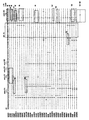
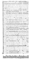
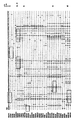
- the genotype specific target sequences can be any nucleotide variation appearing upon alignment of the different HBV genomes that permits classification of a certain HBV isolate as a certain genotype (see Figure 1 ).
- relevant part of a suitable HBV gene refers to the part of the HBV gene, encompassing the HBV genotype specific target sequence chosen from Figure 1 to be detected.
- step (iii) is performed using probes all meticulously designed such that they show the desired hybridization results, when used in a reverse hybridisation assay format, more particularly under the same hybridization and wash conditions implying that each of said probes is able to form a complex upon hybridisation with its target sequence present in the polynucleic acids of the sample as obtained after step (i) or (ii).
- HBV gene encoded amino acids and nucleotides are as generally accepted in literature.
- a set of probes allowing the detection of a genotype specific variation, including probes allowing the detection of a wild-type sequence, a polymorphic or a mutated sequence at any one of the nucleotide positions showing a sequence diversity upon alignment of all known or yet to be discovered HBV sequences as represented in Figure 1 for all complete HBV genomes found in the EMBL/NCBI/DDBJ/Genbank.
- the sets of probes according to the present invention have as a common characteristic that all the probes in said set are designed so that they can be used together in a reverse-hybridization assay, more particularly under similar or identical hybridization and wash conditions as indicated above and below.
- Selected sets of probes include probes which allow to differentiate any of the HBV genotype specific nucleotide changes as represented in Figure 1 , preferably in the HBsAg region of HBV. Said probes being characterized in that they can function in a method as set out above.
- the present invention provides an elegant way to tackle this problem which involves residing to a reverse hybridisation approach (particularly on Line Probe Assays strips, as described by Stuyver et al., 1993). Using this technology it is possible to conveniently obtain all essential data in one test run. To achieve this goal, a set of probes needs to be designed and assembled which can detect all relevant mutations and possibly also wild-type sequences or polymorphisms in the HBV gene regions of interest.
- Also disclosed herein is a method for determining the presence or absence of one or more HBV mutants in a biological sample, comprising:
- mutant target sequence not only covers the sequence containing a mutation, but also the corresponding wild-type sequence.
- the HBV mutant target sequence disclosed herein can be any sequence including a HBV mutated codon known in the art or yet to be discovered. Particularly preferred HBV mutant target regions are set out below.
- the present invention provides a particularly elegant way to obtain such complex information.
- a set of probes selected as defined above may be used, wherein said set of probes is characterized as being chosen such that for a given HBV mutation, the following probes are included in said set :
- At least one probe characterizing the presence of a mutation in the RT pol gene region of HBV, that gives rise to resistance to drugs such as lamivudine and penciclovir, for instance mutation of M to V or to I at position 552 (in the YMDD motif), mutation of V to I at position 555, mutation of F to L at position 514, mutation of V to L at position 521, mutation of P to L at position 525 and mutation of L to M at position 528 (see Figure 1 ).
- drugs such as lamivudine and penciclovir
- oligonucleotide probes hybridizes specifically to at least two of the following groups of target sequences:
- hybridizing with at least one nucleotide probe hybridizing specifically to a genotype specific target sequence chosen from Figure 1 and at least one nucleotide probe hybridizing specifically to a HBV mutant target sequence chosen from Figure 1 is disclosed herein.
- hybridizing with at least one nucleotide probe hybridizing specifically to a genotype specific target sequence chosen from Figure 1 and at least one nucleotide probe hybridizing specifically to a HBV vaccine escape mutant target sequence within the HBsAg region as represented in Figure 1 .
- hybridizing with at least one probe hybridizing specifically to a mutant target sequence from the HBV RT pol gene region of HBV, and at least one probe hybridizing specifically to a mutant target sequence from the HBsAg region of HBV, and at least one probe hybridizing specifically to a genotype-specific target sequence from the HBsAg region of HBV.
- the relevant part of the HBV genome can be amplified by use of one primer pair, for instance HBPr 75 and HBPr 94.
- a set of probes as set out above comprising at least one, preferably at least two, at least three, at least four or more probe(s) for targeting one, preferably two, three or more nucleotide changes appearing in the alignment of HBV genomes as chosen from the HBV RT pol gene and represented in Figure 1 .
- Selected sets of probes include probes derived from two of the same or different regions of HBV bearing HBV mutated nucleotides, or in addition also a third (set of) probe(s) characterizing the presence of a third HBV mutation at any of the positions shown in Figure 1 , or particular combinations thereof.
- a set of probes which allows simultaneous detection of HBV mutations at codons 15, 28 and 29 in the preCore region, in combination with mutations in the preCore promoter regions, in combination with mutations at codons 122, 126, 141, 143, 144, 145 in the HBsAg region, possibly also in combination with mutations in the HBV pol gene at codons 514, 521, 525, 528, 552 or 555.
- HBV genomes of Figure 1 In the instances where the alignment of HBV genomes of Figure 1 is referred to herein, it should be construed as referring to an alignment of all existing and future HBV genomes.
- the existing HBV genome sequences can be deduced from any database, such as the EMBL/NCBI/DDBJ/GENBANK database.
- a set of preCore, preS1, HBsAg and RT pol gene probes are the probes with SEQ ID NO 1 to 278 of Table 1 (see also Figure 1 ).
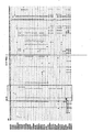
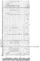
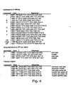
- probes in this respect are shown in Figure 2 and in Figure 4 .
- the probes in Figure 2 and in Figure 4 were withheld after a first selection for preCore, preS1, HBsAg and RT pol probes.
- the probes of the invention are designed for obtaining optimal performance under the same hybridization conditions so that they can be used in sets of at least 2 probes for simultaneous hybridization. This highly increases the usefulness of these probes and results in a significant gain in time and labour.
- all probes should be adapted accordingly by adding or deleting a number of nucleotides at their extremities. It should be understood that these concomitant adaptations should give rise to essentially the same result, namely that the respective probes still hybridize specifically with the defined target. Such adaptations might also be necessary if the amplified material should be RNA in nature and not DNA as in the case for the NASBA system.
- the selection of the preferred probes of the present invention is based on a reverse hybridization assay format using immobilized oligonucleotide probes present at distinct locations on a solid support. More particularly the selection of preferred probes of the present invention is based on the use of the Line Probe Assay (LiPA) principle which is a reverse hybridization assay using oligonucleotide probes immobilized as parallel lines on a solid support strip (Stuyver et al. 1993; international application WO 94/12670 ). This approach is particularly advantageous since it is fast and simple to perform.
- the reverse hybridization format and more particularly the LiPA approach has many practical advantages as compared to other DNA techniques or hybridization formats, especially when the use of a combination of probes is preferable or unavoidable to obtain the relevant information sought.
- the reverse hybridization approach implies that the probes are immobilized to certain locations on a solid support and that the target DNA is labelled in order to enable the detection of the hybrids formed.
- genetic analysis refers to the study of the nucleotide sequence of the genome of HBV by any appropriate technique.
- HBV mutant refers to any HBV strain harbouring genomic variations with serological, genetical or clinical consequences.
- vaccine escape mutant is reviewed in the introduction section and in Example 7. The most important region lies between amino acid 124 and 147 of the HBsAg region, but can be extended from amino acid 114 to 150.
- mutant resistant to drugs such as lamivudine and penciclovir is reviewed in the introduction section and in Example 8.
- HBV genotype refers to HBV strains with an intergenotype variation of 8% or more based on a comparison of complete genomes.
- the target material in the samples to be analyzed may either be DNA or RNA, e.g. genomic DNA, messenger RNA, viral RNA or amplified versions thereof. These molecules are also termed polynucleic acids.
- probe refers to single stranded sequence-specific oligonucleotides which have a sequence which is complementary to the target sequence to be detected.
- target sequence describes the nucleotide sequence of a part of wild-type, polymorphic or mutant HBV gene sequence to be specifically detected by a probe according to the present invention.
- the polymorphic sequence may encompass one or more polymorphic nucleotides; the mutant sequence may encompass one or more nucleotides that are different from the wild-type sequence.
- mutant target sequence not only covers the sequence containing a mutation, but also the corresponding wild-type sequence.
- Target sequences may generally refer to single nucleotide positions, codon positions, nucleotides encoding amino acids or to sequences spanning any of the foregoing positions. In the present invention said target sequence often includes one, two or more variable nucleotide positions.
- polynucleic acids detected by the probes of the invention will comprise the target sequence against which the probe is detected.
- target sequence is also a suitable target sequence in some cases.
- the target sequences as defined in the present invention provide sequences which should at least be complementary to the central part of the probe which is designed to hybridize specifically to said target region. In most cases the target sequence is completely complementary to the sequence of the probe.
- sequence of the single stranded probe is exactly the (inverse) complement of the sequence of the single-stranded target, with the target being further defined as the sequence where the mutation to be detected is located.
- the probes of the invention are about 5 to 50 nucleotides long, more preferably from about 10 to 25 nucleotides. Particularly preferred lengths of probes include 10, 11, 12, 13, 14, 15, 16, 17, 18, 19, 20, 21, 22, 23, 24 or 25 nucleotides.
- the nucleotides as used in the present invention may be ribonucleotides, deoxyribonucleotides and modified nucleotides such as inosine or nucleotides containing modified groups which do not essentially alter their hybridisation characteristics.
- Probe sequences are represented throughout the specification as single stranded DNA oligonucleotides from the 5' to the 3' end. It is obvious to the man skilled in the art that any of the below-specified probes can be used as such, or in their complementary form, or in their RNA form (wherein T is replaced by U).
- the probes according to the invention can be prepared by cloning of recombinant plasmids containing inserts including the corresponding nucleotide sequences, if need be by cleaving the latter out from the cloned plasmids upon using the adequate nucleases and recovering them, e.g. by fractionation according to molecular weight.
- the probes according to the present invention can also be synthesized chemically, for instance by the conventional phospho-triester method.
- solid support can refer to any substrate to which an oligonucleotide probe can be coupled, provided that it retains its hybridization characteristics and provided that the background level of hybridization remains low.
- the solid substrate will be a microtiter plate, a membrane (e.g. nylon or nitrocellulose) or a microsphere (bead) or a chip.
- a membrane e.g. nylon or nitrocellulose
- a microsphere bead
- a chip Prior to application to the membrane or fixation it may be convenient to modify the nucleic acid probe in order to facilitate fixation or improve the hybridization efficiency. Such modifications may encompass homopolymer tailing, coupling with different reactive groups such as aliphatic groups, NH 2 groups, SH groups, carboxylic groups, or coupling with biotin, haptens or proteins.
- labelled refers to the use of labelled nucleic acids. Labelling may be carried out by the use of labelled nucleotides incorporated during the polymerase step of the amplification such as illustrated by Saiki et al. (1988) or Bej et al. (1990) or labelled primers, or by any other method known to the person skilled in the art.
- the nature of the label may be isotopic ( 32 P, 35 S, etc.) or non-isotopic (biotin, digoxigenin, etc.).
- primer refers to a single stranded oligonucleotide sequence capable of acting as a point of initiation for synthesis of a primer extension product which is complementary to the nucleic acid strand to be copied.
- the length and the sequence of the primer must be such that they allow to prime the synthesis of the extension products.
- the primer is about 5-50 nucleotides long. Specific length and sequence will depend on the complexity of the required DNA or RNA targets, as well as on the conditions of primer use such as temperature and ionic strength.
- suitable primer pair in this invention refers to a pair of primers allowing the amplification of part or all of the HBV gene for which probes are immobilized.
- the amplification method used can be either polymerase chain reaction (PCR; Saiki et al., 1988), ligase chain reaction (LCR; Landgren et al., 1988; Wu & Wallace, 1989; Barany, 1991), nucleic acid sequence-based amplification (NASBA; Guatelli et al., 1990; Compton, 1991), transcription-based amplification system (TAS; Kwoh et al., 1989), strand displacement amplification (SDA; Duck, 1990; Walker et al., 1992) or amplification by means of QB replicase (Lizardi et al., 1988: Lomeli et al., 1989) or any other suitable method to amplify nucleic acid molecules known in the art.
- PCR polymerase chain reaction
- LCR Landgren et al., 1988; Wu & Wallace, 1989
- NASBA nucleic acid sequence-based amplification
- TAS transcription-based amplification system
- SDA strand displacement
- oligonucleotides used as primers or probes may also comprise nucleotide analogues such as phosphorothiates (Matsukura et al., 1987), alkylphosphorothiates (Miller et al., 1979) or peptide nucleic acids (Nielsen et al., 1991; Nielsen et al., 1993) or may contain intercalating agents (Asseline et al., 1984).
- nucleotide analogues such as phosphorothiates (Matsukura et al., 1987), alkylphosphorothiates (Miller et al., 1979) or peptide nucleic acids (Nielsen et al., 1991; Nielsen et al., 1993) or may contain intercalating agents (Asseline et al., 1984).
- sample may be any biological material taken either directly from the infected human being (or animal), or after culturing (enrichment).
- Biological material may be e.g. expectorations of any kind, broncheolavages, blood, skin tissue, biopsies, sperm, lymphocyte blood culture material, colonies, liquid cultures, faecal samples, urine etc.
- the sets of probes will include at least 2, 3, 4, 5, 6, 7, 8, 9, 10, 11, 12, 13, 14, 15, 16, 17, 18, 19, 20, 21, 22, 23, 24, 25, 26, 27, 28, 29, 30 or more probes.
- Said probes may be applied in two or more (possibly as many as there are probes) distinct and known positions on a solid substrate. Often it is preferable to apply two or more probes together in one and the same position of said solid support.
- the stability of the [probe : target] nucleic acid hybrid should be chosen to be compatible with the assay conditions. This may be accomplished by avoiding long AT-rich sequences, by terminating the hybrids with G:C base pairs, and by designing the probe with an appropriate Tm. The beginning and end points of the probe should be chosen so that the length and %GC result in a Tm about 2-10°C higher than the temperature at which the final assay will be performed.
- the base composition of the probe is significant because G-C base pairs exhibit greater thermal stability as compared to A-T base pairs due to additional hydrogen bonding. Thus, hybridization involving complementary nucleic acids of higher G-C content will be stable at higher temperatures.
- Conditions such as ionic strength and incubation temperature under which a probe will be used should also be taken into account when designing a probe. It is known that hybridization will increase as the ionic strength of the reaction mixture increases, and that the thermal stability of the hybrids will increase with increasing ionic strength. On the other hand, chemical reagents, such as formamide, urea, DMSO and alcohols, which disrupt hydrogen bonds, will increase the stringency of hybridization. Destabilization of the hydrogen bonds by such reagents can greatly reduce the Tm. In general, optimal hybridization for synthetic oligonucleotide probes of about 10-50 bases in length occurs approximately 5°C below the melting temperature for a given duplex. Incubation at temperatures below the optimum may allow mismatched base sequences to hybridize and can therefore result in reduced specificity.
- the stringency of the assay conditions determines the amount of complementarity needed between two nucleic acid strands forming a hybrid.
- the degree of stringency is chosen such as to maximize the difference in stability between the hybrid formed with the target and the nontarget nucleic acid. In the present case, single base pair changes need to be detected, which requires conditions of very high stringency.
- the length of the target nucleic acid sequence and, accordingly, the length of the probe sequence can also be important. In some cases, there may be several sequences from a particular region, varying in location and length, which will yield probes with the desired hybridization characteristics. In other cases, one sequence may be significantly better than another which differs merely by a single base. While it is possible for nucleic acids that are not perfectly complementary to hybridize, the longest stretch of perfectly complementary base sequence will normally primarily determine hybrid stability. While oligonucleotide probes of different lengths and base composition may be used, preferred oligonucleotide probes of this invention are between about 5 to 50 (more particularly 10-25) bases in length and have a sufficient stretch in the sequence which is perfectly complementary to the target nucleic acid sequence.
- Regions in the target DNA or RNA which are known to form strong internal structures inhibitory to hybridization are less preferred.
- probes with extensive self-complementarity should be avoided.
- hybridization is the association of two single strands of complementary nucleic acids to form a hydrogen bonded double strand. It is implicit that if one of the two strands is wholly or partially involved in a hybrid that it will be less able to participate in formation of a new hybrid. There can be intramolecular and intermolecular hybrids formed within the molecules of one type of probe if there is sufficient self complementarity. Such structures can be avoided through careful probe design.
- Standard hybridization and wash conditions are disclosed in the Materials & Methods section of the Examples. Other conditions are for instance 3X SSC (Sodium Saline Citrate), 20% deionized FA (Formamide) at 50°C.
- SSPE Sodium saline phosphate EDTA
- TMACI Tetramethyl ammonium Chloride
- temperatures can also be used provided that the specificity and sensitivity of the probes is maintained. If need be, slight modifications of the probes in length or in sequence have to be carried out to maintain the specificity and sensitivity required under the given circumstances.
- the above-mentioned polynucleic acids from step (i) or (ii) are hybridized with at least two, three, four, five, six, seven, eight, nine, ten, eleven, twelve, thirteen, fourteen, fifteen, sixteen, seventeen, eighteen, nineteen, twenty, or more of the above-mentioned target region specific probes, preferably with 5 or 6 probes, which, taken together, cover the "mutation region" of the relevant HBV gene.
- mutant region means the region in the relevant HBV gene sequence where at least one mutation encoding a HBV mutant is located in a preferred part of this mutation region, represented in figure 1 .
- HBV wild-type or mutant genomes may also show polymorphic nucleotide variations at positions other than those referred to as genotype specific or mutant specific variated positions as shown in Figure 1 .
- any hybridization method known in the art can be used (conventional dot-blot, Southern blot, sandwich, etc.).
- the selected set of probes are immobilized to a solid support in known distinct locations (dots, lines or other figures). In another preferred embodiment the selected set of probes are immobilized to a membrane strip in a line fashion. Said probes may be immobilized individually or as mixtures to delineated locations on the solid support.
- a specific and very user-friendly embodiment of the above-mentioned preferential method is the LiPA method, where the above-mentioned set of probes is immobilized in parallel lines on a membrane, as further described in the examples.
- the invention also provides for a set of primers allowing amplification of the region of the respective HBV gene to be detected by means of probes. Examples of such primers of the invention are given in Table 1 and Figure 1 .
- Primers may be labelled with a label of choice (e.g. biotine).
- a label of choice e.g. biotine
- Different primer-based target amplification systems may be used, and preferably PCR-amplification, as set out in the examples.
- Single-round or nested PCR may be used.
- the invention also provides a assay kit according to claim 3.
- hybridization buffer means a buffer enabling a hybridization reaction to occur between the probes and the polynucleic acids present in the sample, or the amplified products, under the appropriate stringency conditions.
- wash solution means a solution enabling washing of the hybrids formed under the appropriate stringency conditions.
- a line probe assay (LiPA) was designed for screening for HBV genotypes and/or HBV mutants.
- the principle of the assay is based on reverse hybridization of an amplified polynucleic acid fragment such as a biotinylated PCR fragment of the HBV gene onto short oligonucleotides.
- the latter hybrid can then, via a biotine-streptavidine coupling, be detected with a non-radioactive colour developing system.
- Primers from the PreS1 region include 1, 106, 2 (sense primers) and 4, 107 and 3 (antisense primers).
- Primers from the HBsAg region include 75 and 104 (sense primers) and 76, 94 and 105 (antisense primers).
- Primers from the PreCore region include 5, 6, 69, 70, 84, 86, 87 and 108 (sense primers) and 7, 8, 85 and 109 (antisense primers).
- the remaining oligonucleotides are probes from the PreCore, PreS1, HBsAg and RT pol gene regions of HBV as indicated.
- the YMDDV motif and its mutants consist of amino acids 551 to 555 of the RT pol protein; the sequence MGVGL and its mutant consist of amino acids 519 to 523 of the RT pol protein; the sequence SPFLL and its mutants and genotypic variants consist of amino acids 524 to 528 of the RT pol protein.
- Example 1 HBV DNA preparation and PCR amplification
- Serum samples were collected from HBsAg-positive individuals and stored at minus 20°C until use in 0.5 ml aliquots.
- 18 ⁇ l serum was mixed with 2 ⁇ l 1N NaOH and incubated at 37°C for 60 minutes. The denaturation was stopped and neutralized by adding 20 ⁇ l of 0.1 N HCl. After a 15 minutes centrifugation step, the supernatant was collected and the pellet discarded.
- PCR was carried out on this lysate as follows: 32 ⁇ l H 2 O was mixed with 5 ⁇ l of 10x PCR buffer, 1 ⁇ l 10 mM dXTPs, 1 ⁇ l of each biotinylated primer (10 pmol/ ⁇ l), 10 ⁇ l of serum lysate, and 2 U Taq enzyme.
- the amplification scheme contained 40 cycles of 95°C 1 min, annealing at 45°C for 1 min, and extension at 72°C for 1 min. Amplification products were visualized on 3% agarose gel.
- the outer primer set for preS1 has the following sequence:
- the outer primer set for preCore has the following sequence:
- the outer primer set for HBsAg has the following sequence:
- Samples that were negative in the first round PCR were retested in a nested reaction composed of the following: ⁇ l H 2 O, 5 ⁇ l 10x Taq buffer, 1 ⁇ l 10 mM dXTPs, 1 ⁇ l of each nested primer (10 pmol/ ⁇ l), 1 ⁇ l of the first round PCR product, and 2 U Taq polymerase.
- the amplification scheme was identical as for the first round PCR.
- the sequence of the nested primers were as follows, for the preS1 region:
- the HBsAg region can be amplified in a similar protocol by using the following primers: HBPr 75: 5'-bio-CAAGGTATGTTGCCCGTTTGTCC-3' in combination with either HBPr 76: 5'-bio-CCAAACAGTGGGGGAAAGCCC-3'; or with HBPr 94: 5'-bio-GGTA(A/T)AAAGGGACTCA(C/A)GATG-3'.
- Probes were designed to cover the universal, genotypic and mutant motifs. In principle only probes that discriminate between one single nucleotide variation were retained. However, for certain polymorphisms at the extreme ends of the probe, cross-reactivity was tolerated. Specificity was reached experimentally for each probe individually after considering the % (G + C), the probe length, the final concentration, and hybridization temperature. Optimized probes were provided enzymatically with a poly-T-tail using the TdT (Pharmacia) in a standard reaction condition.
- probe 400 pmol probe was incubated at 37°C in a 30 ⁇ l reaction mix containing 5.3 mM dTTP, 25 mM Tris.HCL pH 7.5, 0.1 M sodium cacodylate, 1 mM CoCl 2 , 0.1 M DTT and 170 U terminal deoxynucleotidyl transferase (Pharmacia). After one hour incubation, the reaction was stopped and the tailed probes were precipitated and washed with ice-cold ethanol. Probes were dissolved in 6x SSC at their respectively specific concentrations and applied as horizontal lines on membrane strips in concentrations between 0.2 and 2.5 pM/ml.
- Biotinylated DNA was applied alongside as positive control (LiPA line 1).
- the oligonucleotides were fixed to the membrane by baking at 80°C for 12 hours.
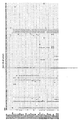
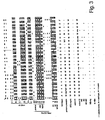
- the membrane was than sliced into 4 mm strips. The design of this strip is indicated in Figure 2 .
- Equal volumes (10 ⁇ l each) of the biotinylated PCR fragment and of the denaturation solution (DS; 400 mM NaOH/10 mM EDTA) were mixed in test troughs and incubated at room temperature for 5 minutes. Then, 2 ml of the 37°C prewarmed hybridization solution (HS, 3x SSC/0.1 % SDS) was added, followed by the addition of one strip per test trough. Hybridisation occured for 1 hour at 50 ⁇ 0.5°C in a closed shaking water bath. The strips were washed twice with 2 ml of stringent wash solution (3x SSC/0.1 % SDS) at room temperature for 20 seconds, and once at 50°C for 30 minutes.
- strips were rinsed two times with 2 ml of the Innogenetics standard Rinse Solution (RS). Strips were incubated on a rotating platform with the alkaline phosphatase-labelled streptavidin conjugate, diluted in standard Conjugate Solution for 30 minutes at room temperature (20 to 25°C). Strips were than washed twice with 2 ml of RS and once with standard Substrate Buffer (SB), and the colour reaction was started by adding BCIP and NBT to the SB. After maximum 30 minutes at room temperature, the colour reaction was stopped by replacing the colour compounds by distilled water. Immediately after drying, the strips were interpreted. Reactivities were considered positive whenever the reactivity was stronger than the reaction on the negative control. Strips can be stored on a dry dark place. The complete procedure described above can also be replaced by the standardized Inno-LiPA automation device (auto-LiPA).
- auto-LiPA Inno-LiPA automation device
- PCR fragments were prepared, derived from members of the different genotypes, the different preCore wild type and mutant sequences, drug resistant motifs and vaccine escape mutants.
- the PCR fragments were amplified with primers lacking the biotine group at their 5'-end and cloned into the pretreated EcoRV site of the pGEMT vector (Promega).
- Recombinant clones were selected after ⁇ -complementation and restriction fragment length analysis, and sequenced with plasmid primers.
- Other biotinylated fragments were directly sequenced with a dye-terminator protocol (Applied Biosystems) using the amplification primers.
- nested PCR was carried out with analogs of the primers, in which the biotine group was replaced with the T7- and SP6-primer sequence, respectively. These amplicons were than sequenced with an SP6- and T7-dye-primer procedure. By doing so, a reference panel of recombinant clones was prepared, which is necessary for optimizing LiPA probes.
- Example 5 Genotyping HBV-infected serum samples.
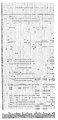
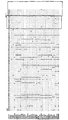
- Figure 3 shows the reactivity of a selection of samples on these probes. Genotyping of these samples is straightforward, with samples 2 to 8 belonging to genotype A, samples 9 and 10 belonging to genotype B, samples 11 and 12 belonging to genotype C, samples 13 to 19 belonging to genotype D, samples 20 to 23 belonging to genotype E, and sample 24 belonging to genotype F.
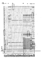
- Genotyping can also be performed in the HBsAg region. Again, probes were designed to cover most of the variable regions shown in Fig. 1 . Only a limited selection of probes were retained. These probes are boxed in Fig.1 and are listed in Figure 4 . A LiPA strip was prepared carrying these probes and samples belonging to the different genotypes were characterized, as shown in Fig. 5 .
- Example 6 Scanning the preCore region for mutations.
- HBeAg expression can be regulated at the transcriptional and translational level. It is postulated that a transcriptional regulation exists due to the presence of a dinucleotide variation in the promoter region of the preCore mRNA. Probes covering the wild type (e.g. probe HBPr 88) and the mutant (e.g. HBPr 89) motif were selected and their positions are indicated in the alignment shown in Figure 1 , and applied on the LiPA strip as line 15 and 16 ( Figure 2 ).
- any mutations at codon 1 (ATG) destroying translation initiation codon 2 (CAA to TAA), codon 7 (TGC to TGA), codon 12 (TGT to TGA), codon 13 in genotype B, C, D, E, F (TCA to TGA or TAA), codon 14 (TGT to TGA), codon 18 (CAA to TAA), codon 21 (AAG to TAG), codon 23 (TGC to TGA), codon 26 (TGG to TAG or TGA), codon 28 (TGG to TAG or TGA).
- Figure 3 shows the reactivity of the selected genotyped samples on the probes for the preCore region. Samples were previously tested for the presence of HBeAg or for anti-HBe. The interpretation of the reactivity on the LiPA probes for each sample is indicated below each strip. This approach allowed for the simultaneous screening of a sample for preCore mutations and the characterization of the viral genotype.
- Figure 6 also shows a panel of samples with mutations in the preCore region, as well as wild type samples.
- the probes used in this assay are listed in Figure 4 .
- This assay includes a codon 29 mutant (M4 motif), which was not present in the experiment in Figure 3 .
- Example 7 Detection of mutants in the HBsAg region.
- Vaccine escape mutants have been described. The most commonly found mutant is the variation at codon 145 of HBsAg (G145R or GGA to AGA).
- LiPA probes are designed to detect wild type and mutant probes. Genotypic variations are present in the vicinity of codon 145. Therefore, genotype A is covered by probe 77, genotype B by probe 78, genotype C by probe 79, and genotype D/E by probe 80. Hence, in principle, it is possible to genotype and detect the wild type strains of the virus in one single experiment. Mutant target sequences are covered by probe 81 and 82 for genotype A and D, respectively. Probe 83 can be used as a positive control in these experiments.
- probes are designed to cover the wild type sequence of the different genotypes over the HBsAg epitope region and applied in a LiPA format. Again here, absence of staining at one of these probes is indicative for the presence of a mutant strain. The exact nature of this variant is then determined by sequencing analysis.
- Example 8 Detection of HBV strains resistant to lamivudine.
- Probes covering the wild type YMDD motif and YVDD mutant motif are indicated in Figure 1 , respectively probes 95 to 100 and 101 to 103, as well as probes 115, 116, 127 and 132, the latter probes yielding the best results in the LiPA assay.
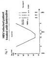
- Such an assay was used to determine the presence of mutations in the YMDD motif in serum of a HBV-infected patient during treatment with lamivudine.
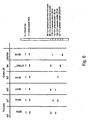
- Fig. 7 shows that in the first phase of the treatment (May 1995) no mutations were detected. During the treatment, the viral load decreased, reaching a level of approximately 10 4 during November and December 1995, whereafter a breakthrough was observed, resulting in a level as high as during the first months of the treatment by June 1996.
Landscapes
- Life Sciences & Earth Sciences (AREA)
- Chemical & Material Sciences (AREA)
- Health & Medical Sciences (AREA)
- Organic Chemistry (AREA)
- Proteomics, Peptides & Aminoacids (AREA)
- Zoology (AREA)
- Immunology (AREA)
- Wood Science & Technology (AREA)
- Engineering & Computer Science (AREA)
- Analytical Chemistry (AREA)
- General Health & Medical Sciences (AREA)
- Biotechnology (AREA)
- Microbiology (AREA)
- Molecular Biology (AREA)
- Physics & Mathematics (AREA)
- Virology (AREA)
- Communicable Diseases (AREA)
- Biochemistry (AREA)
- Bioinformatics & Cheminformatics (AREA)
- General Engineering & Computer Science (AREA)
- Biophysics (AREA)
- Genetics & Genomics (AREA)
- Measuring Or Testing Involving Enzymes Or Micro-Organisms (AREA)
- Vending Machines For Individual Products (AREA)
- General Induction Heating (AREA)
- Coupling Device And Connection With Printed Circuit (AREA)
- Superconductors And Manufacturing Methods Therefor (AREA)
- Magnetic Record Carriers (AREA)
- Control Of High-Frequency Heating Circuits (AREA)
- Medicines That Contain Protein Lipid Enzymes And Other Medicines (AREA)
Claims (5)
- Procédé de détection, dans un échantillon biologique, de deux génotypes de souches de HBV et de mutants de HBV ayant une résistance médicamenteuse, comprenant les étapes consistant à :(i) si besoin est, libérer, isoler ou concentrer les acides polynucléiques présents dans ledit échantillon ;(ii) si besoin est, amplifier la partie pertinente d'un gène de HBV approprié présent dans ledit échantillon avec au moins une paire appropriée d'amorces ;(iii) hybrider les acides polynucléiques de l'étape (i) ou de la (ii) avec une combinaison d'au moins une sonde provenant de chacun des groupes suivants :- SEQ ID 77, 81, 139-141, 190-194- SEQ ID 78, 142-147, 149- SEQ ID 79, 150-161, 204- SEQ ID 82, 162-169, 208- SEQ ID 170-176, 213- SEQ ID 178-189, 214-219- facultativement une ou plusieurs des SEQ ID 80, 148 et 177 et
au moins une sonde provenant de chacun des groupes suivants :- SEQ ID 114, 115, 127-129, 227-234, 243, 249, 250, 261- SEQ ID 116, 117, 130-133, 244-248, 251-260, 262-266 et/ou au moins une sonde parmi les : SEQ ID 267 et 269, chacune avec la SEQ ID 268,(iv) détecter les hybrides formés à l'étape (iii) ;(v) en déduire, à partir du signal (des signaux) différentiel(s) d'hybridation obtenu(s) à l'étape (iv), les génotypes de HBV et les mutants de HBV ayant une résistance médicamenteuse qui sont présents dans ledit échantillon. - Procédé selon la revendication 1, dans lequel les sondes d'oligonucléotides de l'étape (iii) sont caractérisées en ce qu'elles détectent spécifiquement des mutations de résistance médicamenteuse dans le motif YMDD, avec lesdites sondes étant les :- SEQ ID 77, 140 et 193- SEQ ID 78,- SEQ ID 153, 154 et 204- SEQ ID 165 et 208- SEQ ID 172 et 213- SEQ ID 186, 216 et 219- SEQ ID 80 et 177- SEQ ID 115 et 127- SEQ ID 116 et 132.
- Kit de dosage pour détecter, dans un échantillon biologique, deux génotypes de souches de HBV et des mutants du HBV ayant une résistance médicamenteuse, selon le procédé de la revendication 1, comprenant les composants suivants :(i) quand cela est approprié, un moyen pour libérer, isoler ou concentrer les acides polynucléiques présents dans ledit échantillon ;(ii) quand cela est approprié, au moins une paire appropriée d'amorces ;(iii) au moins une sonde provenant de chacun des groupes suivants :- SEQ ID 77, 81, 139-141, 190-194- SEQ ID 78, 142-147, 149- SEQ ID 79, 150-161, 204- SEQ ID 82, 162-169, 208- SEQ ID 170-176, 213- SEQ ID 178-189, 214-219- facultativement une ou plusieurs des SEQ ID 80, 148 et 177 et
au moins une sonde provenant de chacun des groupes suivants :- SEQ ID 114, 115, 127-129, 227-234, 243, 249, 250, 261- SEQ ID 116, 117, 130-133, 244-248, 251-260, 262-266 et/ou au moins une sonde parmi les : SEQ ID 267 et 269, chacune avec la SEQ ID 268, possiblement fixée à un support solide ;(iv) un tampon d'hybridation ou des composants nécessaires à produire ledit tampon ;(v) une solution de lavage ou des composants nécessaires à produire ladite solution ;(vi) quand cela est approprié, un moyen de détection des hybrides qui résultent de la précédente hybridation ;(vii) quand cela est nécessaire, un moyen pour fixer ladite sonde à un emplacement connu sur un support solide. - Mutant de HBV comprenant une séquence nucléotidique qui contient une mutation de résistance médicamenteuse dans le gène pol de RT de HBV, dans lequel la mutation conduit à un changement d'acide aminé dans le motif YMDD de la polymérase de HBV, dans lequel le changement d'acide aminé est d'un M en un V à la position 552 comme indiqué dans la figure 1.
- Procédé d'identification, dans un échantillon, d'un mutant de HBV, tel que défini dans la revendication 4, comprenant les étapes consistant à :(i) libérer, isoler ou concentrer les acides polynucléiques présents dans ledit échantillon ;(ii) facultativement, amplifier au moins une partie du gène de HBV présent dans ledit échantillon avec au moins une paire appropriée d'amorces ;(iii)déterminer la présence ou l'absence d'une mutation dans le gène pol de RT de HBV, conduisant à un changement d'acide aminé dans le motif YMDD de la polymérase de HBV, dans lequel le changement d'acide aminé est d'un M en un V à la position 552 comme indiqué dans la figure 1, et en identifiant de ce fait l'existence dans ledit échantillon d'une souche de HBV ayant une résistance médicamenteuse.
Priority Applications (3)
| Application Number | Priority Date | Filing Date | Title |
|---|---|---|---|
| EP10181900.1A EP2322669B1 (fr) | 1996-04-19 | 1997-04-21 | Procédé de typage et de détection du virus de l'hépatite B (HBV) |
| EP97921677A EP0914472B1 (fr) | 1996-04-19 | 1997-04-21 | Procede de typage et de detection du virus de l'hepatite b (hbv) |
| EP10182184A EP2322670A3 (fr) | 1996-04-19 | 1997-04-21 | Procédé de typage et de detection du virus de l'hépatite B (HBV) |
Applications Claiming Priority (4)
| Application Number | Priority Date | Filing Date | Title |
|---|---|---|---|
| EP96870053 | 1996-04-19 | ||
| EP96870053 | 1996-04-19 | ||
| PCT/EP1997/002002 WO1997040193A2 (fr) | 1996-04-19 | 1997-04-21 | Procede de typage et de detection du virus de l'hepatite b (hbv) |
| EP97921677A EP0914472B1 (fr) | 1996-04-19 | 1997-04-21 | Procede de typage et de detection du virus de l'hepatite b (hbv) |
Related Child Applications (4)
| Application Number | Title | Priority Date | Filing Date |
|---|---|---|---|
| EP10181900.1A Division EP2322669B1 (fr) | 1996-04-19 | 1997-04-21 | Procédé de typage et de détection du virus de l'hépatite B (HBV) |
| EP10181900.1A Previously-Filed-Application EP2322669B1 (fr) | 1996-04-19 | 1997-04-21 | Procédé de typage et de détection du virus de l'hépatite B (HBV) |
| EP10182184.1 Division-Into | 2010-09-29 | ||
| EP10181900.1 Division-Into | 2010-09-29 |
Publications (2)
| Publication Number | Publication Date |
|---|---|
| EP0914472A2 EP0914472A2 (fr) | 1999-05-12 |
| EP0914472B1 true EP0914472B1 (fr) | 2010-11-03 |
Family
ID=8226137
Family Applications (3)
| Application Number | Title | Priority Date | Filing Date |
|---|---|---|---|
| EP10181900.1A Expired - Lifetime EP2322669B1 (fr) | 1996-04-19 | 1997-04-21 | Procédé de typage et de détection du virus de l'hépatite B (HBV) |
| EP10182184A Withdrawn EP2322670A3 (fr) | 1996-04-19 | 1997-04-21 | Procédé de typage et de detection du virus de l'hépatite B (HBV) |
| EP97921677A Expired - Lifetime EP0914472B1 (fr) | 1996-04-19 | 1997-04-21 | Procede de typage et de detection du virus de l'hepatite b (hbv) |
Family Applications Before (2)
| Application Number | Title | Priority Date | Filing Date |
|---|---|---|---|
| EP10181900.1A Expired - Lifetime EP2322669B1 (fr) | 1996-04-19 | 1997-04-21 | Procédé de typage et de détection du virus de l'hépatite B (HBV) |
| EP10182184A Withdrawn EP2322670A3 (fr) | 1996-04-19 | 1997-04-21 | Procédé de typage et de detection du virus de l'hépatite B (HBV) |
Country Status (10)
| Country | Link |
|---|---|
| US (4) | US6709812B1 (fr) |
| EP (3) | EP2322669B1 (fr) |
| AT (1) | ATE486965T1 (fr) |
| AU (1) | AU2766297A (fr) |
| DE (1) | DE69740039D1 (fr) |
| DK (1) | DK0914472T3 (fr) |
| ES (2) | ES2354937T3 (fr) |
| PT (1) | PT914472E (fr) |
| WO (1) | WO1997040193A2 (fr) |
| ZA (1) | ZA973367B (fr) |
Families Citing this family (38)
| Publication number | Priority date | Publication date | Assignee | Title |
|---|---|---|---|---|
| ZA973367B (en) | 1996-04-19 | 1997-11-18 | Innogenetics Nv | Method for typing and detecting HBV. |
| USRE40233E1 (en) | 1996-11-08 | 2008-04-08 | Melbourne Health | Viral variants and methods for detecting same |
| AUPO351996A0 (en) * | 1996-11-08 | 1996-12-05 | Western Health Care Network | Viral variants and methods for detecting same |
| IT1297096B1 (it) * | 1997-12-03 | 1999-08-03 | Sorin Diagnostics S R L | Metodo per la rivelazione di analiti di dna a singola elica |
| WO1999051776A1 (fr) * | 1998-04-07 | 1999-10-14 | Southern Research Institute | Procede a haut rendement de criblage d'activite antivirale |
| DE19835856A1 (de) * | 1998-08-07 | 2000-02-17 | W Kurt Roth | Verfahren zum Nachweis von HBV |
| AR028149A1 (es) * | 1999-07-08 | 2003-04-30 | Innogenetics Nv | Deteccion de la resistencia a los farmacos contra la hepatitis b |
| US6759193B2 (en) * | 1999-07-28 | 2004-07-06 | The Government Of The Republic Of Singapore | Detection of human hepatitis B virus surface antigen mutants by specific amplification and its application on gene chip |
| SG90149A1 (en) * | 2000-07-18 | 2002-07-23 | Government Of The Republic Of | Diagnostic assay |
| JP2002355098A (ja) * | 2000-08-14 | 2002-12-10 | Genome Science Laboratories Co Ltd | B型肝炎ウイルスの遺伝子型分類方法並びにそのためのプライマー及びプローブ |
| GR1003878B (el) * | 2001-03-13 | 2002-04-24 | Γεωργακοπουλος Ελευθεριος Δρ. | Νεα μεθοδος ανιχνευσης και ταυτοποιησης σημειακων μεταλλαξεων στελεχων του ιου της ηπατιτιδος β (hbv) σε ορο αιματος ασθενων |
| US20030143527A1 (en) * | 2001-10-09 | 2003-07-31 | Venkatakrishna Shyamala | Identification of oligonucleotides for the capture, detection and quantitation of hepatitis B viral DNA |
| WO2003083094A1 (fr) | 2002-03-29 | 2003-10-09 | Innogenetics N.V. | Methodes de detection de resistance medicamenteuse au hbv |
| AU2014203075B2 (en) * | 2002-06-14 | 2014-11-27 | Gen-Probe Incorporated | Compositions and methods for detecting hepatitis B virus |
| AU2003253651C1 (en) * | 2002-06-14 | 2010-06-03 | Gen-Probe Incorporated | Compositions and methods for detecting hepatitis B virus |
| AU2012202286B2 (en) * | 2002-06-14 | 2014-04-03 | Gen-Probe Incorporated | Compositions and methods for detecting hepatitis B virus |
| US20040170963A1 (en) * | 2002-12-03 | 2004-09-02 | Ih-Jen Su | Antivirus RNA |
| KR100650162B1 (ko) * | 2003-08-05 | 2006-11-27 | 주식회사 진인 | 품질 관리 프로브 및 음성 조절 프로브를 함유하는 약제내성 b형 간염 바이러스 검출용 마이크로어레이 및 이를이용한 약제 내성 b형 간염 바이러스의 검출 방법 |
| DE10339927A1 (de) * | 2003-08-29 | 2005-03-24 | Rhein Biotech Gesellschaft für neue Biotechnologische Prozesse und Produkte mbH | Zusammensetzung zur Prophylaxe/Therapie von HBV-Infektionen und HBV-vermittelten Erkrankungen |
| CN100422344C (zh) * | 2004-08-27 | 2008-10-01 | 广州华银基因科技有限公司 | 乙型肝炎病毒基因分型的荧光pcr检测方法及其试剂盒 |
| US20060057670A1 (en) * | 2004-09-10 | 2006-03-16 | The Chinese University Of Hong Kong | Genomic markers of hepatitis B virus associated with hepatocellular carcinomas |
| CA2597319C (fr) * | 2005-02-18 | 2014-09-30 | Gen-Probe Incorporated | Procede de preparation d'echantillons faisant appel a un choc alcalin |
| CN101142316A (zh) * | 2005-03-09 | 2008-03-12 | 牧岩生命工学研究所 | 小干扰rna和包含它的用于治疗乙型肝炎的药物组合物 |
| PL1858916T4 (pl) | 2005-03-15 | 2015-04-30 | Fujirebio Europe N V | Warianty wirusa zapalenia wątroby typu b z obniżoną wrażliwością na analogi nukleozydów i ich zastosowanie |
| ES2470318T3 (es) | 2005-03-15 | 2014-06-23 | Rheinische Friedrich-Wilhelms-Universit�T Bonn | Variantes del virus de la hepatitis B resistentes a ciertos análogos nucle�sidos, pero sensibles a otros, y usos de las mismas |
| EP1948800B1 (fr) * | 2005-10-21 | 2015-12-23 | Melbourne Health | Mutants ayant une résistance aux agents antiviraux et applications de ceux-ci |
| JP2009213465A (ja) * | 2007-10-30 | 2009-09-24 | Toshiba Corp | B型肝炎ウイルス薬剤耐性株検出用核酸プライマーセット、アッセイキットおよびb型肝炎ウィルスの薬剤耐性株の検出方法 |
| EP2205617A4 (fr) * | 2007-11-06 | 2013-10-16 | Siemens Healthcare Diagnostics | Séquences oligonucléotidiques spécifiques du virus de l'hépatite b (vhb) |
| WO2010016071A2 (fr) * | 2008-08-05 | 2010-02-11 | Swati Subodh | Identification d'une signature génomique pour différencier des variants de séquence hautement similaires d'un organisme |
| US10058837B2 (en) * | 2009-08-28 | 2018-08-28 | The Trustees Of Columbia University In The City Of New York | Systems, methods, and devices for production of gas-filled microbubbles |
| US8642752B2 (en) | 2011-04-21 | 2014-02-04 | Isis Pharmaceuticals, Inc. | Modulation of Hepatitis B virus (HBV) expression |
| US9045803B2 (en) | 2012-02-29 | 2015-06-02 | Abbott Molecular Inc. | Hepatitis B virus typing and resistance assay |
| KR101402608B1 (ko) | 2012-04-05 | 2014-06-03 | (주)진매트릭스 | 만성 b형 간염 치료제의 약제내성 예측용 조성물 및 그 예측 방법 |
| US10344285B2 (en) | 2014-04-09 | 2019-07-09 | Dna2.0, Inc. | DNA vectors, transposons and transposases for eukaryotic genome modification |
| WO2015157579A2 (fr) | 2014-04-09 | 2015-10-15 | Dna2.0, Inc. | Constructions d'acides nucléiques améliorées pour l'expression de gènes eucaryotes |
| HUE056009T2 (hu) | 2015-10-08 | 2022-01-28 | Dna Twopointo Inc | DNS vektorok, transzpozonok és transzpozázok eukarióta genom módosítására |
| EP4458975A3 (fr) | 2019-09-30 | 2025-02-12 | Gilead Sciences, Inc. | Vaccins contre le vhb et méthodes de traitement du vhb |
| CN111172293A (zh) * | 2020-02-19 | 2020-05-19 | 武汉大学 | Sanger法检测乙肝病毒耐药基因突变位点的试剂盒及检测方法 |
Family Cites Families (15)
| Publication number | Priority date | Publication date | Assignee | Title |
|---|---|---|---|---|
| AU606043B2 (en) * | 1985-03-28 | 1991-01-31 | F. Hoffmann-La Roche Ag | Detection of viruses by amplification and hybridization |
| US4683202A (en) * | 1985-03-28 | 1987-07-28 | Cetus Corporation | Process for amplifying nucleic acid sequences |
| US5124246A (en) | 1987-10-15 | 1992-06-23 | Chiron Corporation | Nucleic acid multimers and amplified nucleic acid hybridization assays using same |
| US5780219A (en) * | 1989-07-11 | 1998-07-14 | Gen-Probe Incorporated | Nucleic acid amplification oligonucleotides and probes to human hepatitis B virus |
| JPH05503422A (ja) * | 1990-01-10 | 1993-06-10 | バイエル コーポレイション | 核酸ハイブリダイゼーションアッセイでの信号増幅用レポーター分子としてのdna依存性rnaポリメラーゼ転写産物 |
| DE69231693T2 (de) * | 1991-12-23 | 2001-05-31 | Bayer Corp., East Walpole | Hbv amplifizierungssonden zur verwendung in hybridisierungssandwichassay in der lösungsphase |
| CA2135074A1 (fr) * | 1992-05-06 | 1993-11-11 | Sherrol H. Mcdonough | Oligonucleotides pour amplification des acides nucleiques et sondes pour virus de l'hepatite b chez l'homme |
| ES2133529T3 (es) | 1992-11-27 | 1999-09-16 | Innogenetics Nv | Procedimiento para tipificar aislamientos de hcv. |
| US5595739A (en) | 1993-05-07 | 1997-01-21 | Abbott Laboratories | Hepatitis B virus mutants, reagents and methods for detection |
| JPH09501829A (ja) * | 1993-07-13 | 1997-02-25 | アボツト・ラボラトリーズ | 肝炎b型ウイルスのヌクレオチド配列及びdnaの増幅及び検出のための方法 |
| DE69433180T2 (de) * | 1993-10-26 | 2004-06-24 | Affymetrix, Inc., Santa Clara | Felder von nukleinsaeuresonden auf biologischen chips |
| US5646262A (en) * | 1994-07-28 | 1997-07-08 | Georgetown University | Antisense oligonucleotides against hepatitis B viral replication |
| WO1997029212A1 (fr) * | 1996-02-08 | 1997-08-14 | Affymetrix, Inc. | Speciation de micro-organismes a partir de microplaquettes et caracterisation des phenotypes de ceux-ci |
| ZA973367B (en) | 1996-04-19 | 1997-11-18 | Innogenetics Nv | Method for typing and detecting HBV. |
| AUPO351996A0 (en) * | 1996-11-08 | 1996-12-05 | Western Health Care Network | Viral variants and methods for detecting same |
-
1997
- 1997-04-18 ZA ZA9703367A patent/ZA973367B/xx unknown
- 1997-04-21 WO PCT/EP1997/002002 patent/WO1997040193A2/fr not_active Ceased
- 1997-04-21 EP EP10181900.1A patent/EP2322669B1/fr not_active Expired - Lifetime
- 1997-04-21 ES ES97921677T patent/ES2354937T3/es not_active Expired - Lifetime
- 1997-04-21 AU AU27662/97A patent/AU2766297A/en not_active Abandoned
- 1997-04-21 PT PT97921677T patent/PT914472E/pt unknown
- 1997-04-21 US US09/155,885 patent/US6709812B1/en not_active Expired - Lifetime
- 1997-04-21 EP EP10182184A patent/EP2322670A3/fr not_active Withdrawn
- 1997-04-21 EP EP97921677A patent/EP0914472B1/fr not_active Expired - Lifetime
- 1997-04-21 AT AT97921677T patent/ATE486965T1/de active
- 1997-04-21 ES ES10181900.1T patent/ES2574252T3/es not_active Expired - Lifetime
- 1997-04-21 DK DK97921677.7T patent/DK0914472T3/da active
- 1997-04-21 DE DE69740039T patent/DE69740039D1/de not_active Expired - Lifetime
-
2003
- 2003-06-04 US US10/453,792 patent/US7313357B2/en not_active Expired - Fee Related
- 2003-06-27 US US10/606,879 patent/US8859203B2/en not_active Expired - Fee Related
-
2007
- 2007-05-22 US US11/802,328 patent/US8835106B2/en not_active Expired - Fee Related
Also Published As
| Publication number | Publication date |
|---|---|
| ES2354937T3 (es) | 2011-03-21 |
| ES2574252T3 (es) | 2016-06-16 |
| US8835106B2 (en) | 2014-09-16 |
| EP2322670A2 (fr) | 2011-05-18 |
| US7313357B2 (en) | 2007-12-25 |
| US20090197244A1 (en) | 2009-08-06 |
| PT914472E (pt) | 2011-01-24 |
| AU2766297A (en) | 1997-11-12 |
| WO1997040193A3 (fr) | 1998-05-07 |
| ZA973367B (en) | 1997-11-18 |
| US6709812B1 (en) | 2004-03-23 |
| EP2322670A3 (fr) | 2012-03-28 |
| EP0914472A2 (fr) | 1999-05-12 |
| DK0914472T3 (da) | 2011-05-16 |
| EP2322669A3 (fr) | 2012-03-28 |
| US20050175990A1 (en) | 2005-08-11 |
| ATE486965T1 (de) | 2010-11-15 |
| DE69740039D1 (de) | 2010-12-16 |
| EP2322669B1 (fr) | 2016-04-06 |
| US8859203B2 (en) | 2014-10-14 |
| EP2322669A2 (fr) | 2011-05-18 |
| WO1997040193A2 (fr) | 1997-10-30 |
| HK1020759A1 (en) | 2000-05-19 |
| US20040029110A1 (en) | 2004-02-12 |
Similar Documents
| Publication | Publication Date | Title |
|---|---|---|
| US8835106B2 (en) | Method for typing and detecting HBV | |
| EP1197568B1 (fr) | Détection et typage du virus de l'hépatite C à l'aide de séquences d'acides nucléiques 5'UTR et NS5 | |
| Kato et al. | Frequent coinfection with hepatitis B virus strains of distinct genotypes detected by hybridization with type-specific probes immobilized on a solid-phase support | |
| WO2001040279A2 (fr) | Nouvelles sequences hbv | |
| WO2001004358A2 (fr) | Detection de la resistance medicamenteuse anti-hepatite b | |
| US9879330B2 (en) | Hepatitis B variants with reduced sensitivity to therapeutic compounds, their detection and uses thereof | |
| HK1020759B (en) | Method for typing and detecting hbv | |
| HK1157409A (en) | Method for typing and detecting hbv | |
| HK1157408A (en) | Method for typing and detecting hbv | |
| US8101351B2 (en) | Process for typing of HCV isolates | |
| Conzelmann et al. | Complete hepatitis B virus genome analysis in chronically infected children before and during lamivudine treatment | |
| HK1020497A (en) | Method for detecting nucleic acid sequences based on the use of solid phase immobilised nucleotide probes (line probe assay) |
Legal Events
| Date | Code | Title | Description |
|---|---|---|---|
| PUAI | Public reference made under article 153(3) epc to a published international application that has entered the european phase |
Free format text: ORIGINAL CODE: 0009012 |
|
| 17P | Request for examination filed |
Effective date: 19981118 |
|
| AK | Designated contracting states |
Kind code of ref document: A2 Designated state(s): AT BE CH DE DK ES FI FR GB GR IE IT LI LU MC NL PT SE |
|
| 17Q | First examination report despatched |
Effective date: 20061113 |
|
| GRAP | Despatch of communication of intention to grant a patent |
Free format text: ORIGINAL CODE: EPIDOSNIGR1 |
|
| DAX | Request for extension of the european patent (deleted) | ||
| GRAS | Grant fee paid |
Free format text: ORIGINAL CODE: EPIDOSNIGR3 |
|
| GRAA | (expected) grant |
Free format text: ORIGINAL CODE: 0009210 |
|
| AK | Designated contracting states |
Kind code of ref document: B1 Designated state(s): AT BE CH DE DK ES FI FR GB GR IE IT LI LU MC NL PT SE |
|
| REG | Reference to a national code |
Ref country code: GB Ref legal event code: FG4D |
|
| REG | Reference to a national code |
Ref country code: CH Ref legal event code: EP |
|
| REG | Reference to a national code |
Ref country code: IE Ref legal event code: FG4D |
|
| REF | Corresponds to: |
Ref document number: 69740039 Country of ref document: DE Date of ref document: 20101216 Kind code of ref document: P |
|
| REG | Reference to a national code |
Ref country code: PT Ref legal event code: SC4A Free format text: AVAILABILITY OF NATIONAL TRANSLATION Effective date: 20110117 |
|
| REG | Reference to a national code |
Ref country code: NL Ref legal event code: T3 |
|
| REG | Reference to a national code |
Ref country code: SE Ref legal event code: TRGR |
|
| REG | Reference to a national code |
Ref country code: ES Ref legal event code: FG2A Effective date: 20110309 |
|
| REG | Reference to a national code |
Ref country code: GR Ref legal event code: EP Ref document number: 20110400257 Country of ref document: GR Effective date: 20110317 |
|
| REG | Reference to a national code |
Ref country code: DK Ref legal event code: T3 |
|
| REG | Reference to a national code |
Ref country code: HK Ref legal event code: GR Ref document number: 1020759 Country of ref document: HK |
|
| PLBE | No opposition filed within time limit |
Free format text: ORIGINAL CODE: 0009261 |
|
| STAA | Information on the status of an ep patent application or granted ep patent |
Free format text: STATUS: NO OPPOSITION FILED WITHIN TIME LIMIT |
|
| 26N | No opposition filed |
Effective date: 20110804 |
|
| REG | Reference to a national code |
Ref country code: DE Ref legal event code: R097 Ref document number: 69740039 Country of ref document: DE Effective date: 20110804 |
|
| REG | Reference to a national code |
Ref country code: FR Ref legal event code: PLFP Year of fee payment: 20 |
|
| PGFP | Annual fee paid to national office [announced via postgrant information from national office to epo] |
Ref country code: LU Payment date: 20160422 Year of fee payment: 20 |
|
| PGFP | Annual fee paid to national office [announced via postgrant information from national office to epo] |
Ref country code: NL Payment date: 20160420 Year of fee payment: 20 |
|
| PGFP | Annual fee paid to national office [announced via postgrant information from national office to epo] |
Ref country code: IE Payment date: 20160421 Year of fee payment: 20 Ref country code: ES Payment date: 20160413 Year of fee payment: 20 Ref country code: DE Payment date: 20160421 Year of fee payment: 20 Ref country code: GR Payment date: 20160414 Year of fee payment: 20 Ref country code: CH Payment date: 20160420 Year of fee payment: 20 Ref country code: GB Payment date: 20160421 Year of fee payment: 20 Ref country code: FI Payment date: 20160413 Year of fee payment: 20 Ref country code: MC Payment date: 20160414 Year of fee payment: 20 |
|
| PGFP | Annual fee paid to national office [announced via postgrant information from national office to epo] |
Ref country code: DK Payment date: 20160420 Year of fee payment: 20 Ref country code: SE Payment date: 20160420 Year of fee payment: 20 Ref country code: PT Payment date: 20160419 Year of fee payment: 20 Ref country code: IT Payment date: 20160427 Year of fee payment: 20 Ref country code: AT Payment date: 20160421 Year of fee payment: 20 Ref country code: BE Payment date: 20160420 Year of fee payment: 20 Ref country code: FR Payment date: 20160421 Year of fee payment: 20 |
|
| REG | Reference to a national code |
Ref country code: DE Ref legal event code: R071 Ref document number: 69740039 Country of ref document: DE |
|
| REG | Reference to a national code |
Ref country code: DK Ref legal event code: EUP Effective date: 20170421 |
|
| REG | Reference to a national code |
Ref country code: NL Ref legal event code: MK Effective date: 20170420 |
|
| REG | Reference to a national code |
Ref country code: CH Ref legal event code: PL |
|
| REG | Reference to a national code |
Ref country code: GB Ref legal event code: PE20 Expiry date: 20170420 |
|
| REG | Reference to a national code |
Ref country code: IE Ref legal event code: MK9A |
|
| REG | Reference to a national code |
Ref country code: AT Ref legal event code: MK07 Ref document number: 486965 Country of ref document: AT Kind code of ref document: T Effective date: 20170421 |
|
| PG25 | Lapsed in a contracting state [announced via postgrant information from national office to epo] |
Ref country code: GB Free format text: LAPSE BECAUSE OF EXPIRATION OF PROTECTION Effective date: 20170420 Ref country code: IE Free format text: LAPSE BECAUSE OF EXPIRATION OF PROTECTION Effective date: 20170421 |
|
| PG25 | Lapsed in a contracting state [announced via postgrant information from national office to epo] |
Ref country code: PT Free format text: LAPSE BECAUSE OF EXPIRATION OF PROTECTION Effective date: 20170503 |
|
| REG | Reference to a national code |
Ref country code: ES Ref legal event code: FD2A Effective date: 20180508 |
|
| PG25 | Lapsed in a contracting state [announced via postgrant information from national office to epo] |
Ref country code: ES Free format text: LAPSE BECAUSE OF EXPIRATION OF PROTECTION Effective date: 20170422 |