EP0893965B1 - Monitoring of myocardial revascularization - Google Patents
Monitoring of myocardial revascularization Download PDFInfo
- Publication number
- EP0893965B1 EP0893965B1 EP97940316A EP97940316A EP0893965B1 EP 0893965 B1 EP0893965 B1 EP 0893965B1 EP 97940316 A EP97940316 A EP 97940316A EP 97940316 A EP97940316 A EP 97940316A EP 0893965 B1 EP0893965 B1 EP 0893965B1
- Authority
- EP
- European Patent Office
- Prior art keywords
- sensor
- pmr
- catheter
- tissue
- heart tissue
- Prior art date
- Legal status (The legal status is an assumption and is not a legal conclusion. Google has not performed a legal analysis and makes no representation as to the accuracy of the status listed.)
- Expired - Lifetime
Links
- 230000000250 revascularization Effects 0.000 title claims abstract description 19
- 230000002107 myocardial effect Effects 0.000 title claims description 13
- 238000012544 monitoring process Methods 0.000 title description 3
- 210000005003 heart tissue Anatomy 0.000 claims abstract description 22
- 239000000523 sample Substances 0.000 claims abstract description 11
- 230000005855 radiation Effects 0.000 claims description 21
- 210000001519 tissue Anatomy 0.000 claims description 18
- 230000003287 optical effect Effects 0.000 claims description 13
- 238000012545 processing Methods 0.000 claims description 7
- 230000010412 perfusion Effects 0.000 abstract description 3
- 230000002708 enhancing effect Effects 0.000 abstract 1
- 238000000034 method Methods 0.000 description 32
- 210000002216 heart Anatomy 0.000 description 23
- 210000004165 myocardium Anatomy 0.000 description 13
- 238000005553 drilling Methods 0.000 description 12
- 230000008081 blood perfusion Effects 0.000 description 7
- 230000000694 effects Effects 0.000 description 6
- 210000001174 endocardium Anatomy 0.000 description 6
- 230000017531 blood circulation Effects 0.000 description 5
- 238000002604 ultrasonography Methods 0.000 description 5
- 230000008859 change Effects 0.000 description 4
- 241000282472 Canis lupus familiaris Species 0.000 description 3
- 206010015856 Extrasystoles Diseases 0.000 description 3
- 208000000418 Premature Cardiac Complexes Diseases 0.000 description 3
- 208000027418 Wounds and injury Diseases 0.000 description 3
- 239000008280 blood Substances 0.000 description 3
- 210000004369 blood Anatomy 0.000 description 3
- 230000036770 blood supply Effects 0.000 description 3
- 239000002872 contrast media Substances 0.000 description 3
- 230000006378 damage Effects 0.000 description 3
- 238000010304 firing Methods 0.000 description 3
- MOFVSTNWEDAEEK-UHFFFAOYSA-M indocyanine green Chemical compound [Na+].[O-]S(=O)(=O)CCCCN1C2=CC=C3C=CC=CC3=C2C(C)(C)C1=CC=CC=CC=CC1=[N+](CCCCS([O-])(=O)=O)C2=CC=C(C=CC=C3)C3=C2C1(C)C MOFVSTNWEDAEEK-UHFFFAOYSA-M 0.000 description 3
- 229960004657 indocyanine green Drugs 0.000 description 3
- 208000014674 injury Diseases 0.000 description 3
- 230000000302 ischemic effect Effects 0.000 description 3
- 238000005259 measurement Methods 0.000 description 3
- 230000033764 rhythmic process Effects 0.000 description 3
- 230000002861 ventricular Effects 0.000 description 3
- 238000004458 analytical method Methods 0.000 description 2
- 238000002583 angiography Methods 0.000 description 2
- 206010003119 arrhythmia Diseases 0.000 description 2
- 230000006793 arrhythmia Effects 0.000 description 2
- 210000005242 cardiac chamber Anatomy 0.000 description 2
- 238000001514 detection method Methods 0.000 description 2
- 230000001771 impaired effect Effects 0.000 description 2
- 210000005240 left ventricle Anatomy 0.000 description 2
- 230000008338 local blood flow Effects 0.000 description 2
- 230000007257 malfunction Effects 0.000 description 2
- 238000010297 mechanical methods and process Methods 0.000 description 2
- 230000004089 microcirculation Effects 0.000 description 2
- BOPGDPNILDQYTO-NNYOXOHSSA-N nicotinamide-adenine dinucleotide Chemical compound C1=CCC(C(=O)N)=CN1[C@H]1[C@H](O)[C@H](O)[C@@H](COP(O)(=O)OP(O)(=O)OC[C@@H]2[C@H]([C@@H](O)[C@@H](O2)N2C3=NC=NC(N)=C3N=C2)O)O1 BOPGDPNILDQYTO-NNYOXOHSSA-N 0.000 description 2
- 229930027945 nicotinamide-adenine dinucleotide Natural products 0.000 description 2
- 238000012634 optical imaging Methods 0.000 description 2
- 238000004611 spectroscopical analysis Methods 0.000 description 2
- 230000001502 supplementing effect Effects 0.000 description 2
- 230000000287 tissue oxygenation Effects 0.000 description 2
- 208000007536 Thrombosis Diseases 0.000 description 1
- 230000033115 angiogenesis Effects 0.000 description 1
- 210000000709 aorta Anatomy 0.000 description 1
- 210000004204 blood vessel Anatomy 0.000 description 1
- 238000007675 cardiac surgery Methods 0.000 description 1
- 230000002596 correlated effect Effects 0.000 description 1
- 238000013461 design Methods 0.000 description 1
- 238000005516 engineering process Methods 0.000 description 1
- 238000002474 experimental method Methods 0.000 description 1
- 210000001105 femoral artery Anatomy 0.000 description 1
- 239000012530 fluid Substances 0.000 description 1
- GNBHRKFJIUUOQI-UHFFFAOYSA-N fluorescein Chemical compound O1C(=O)C2=CC=CC=C2C21C1=CC=C(O)C=C1OC1=CC(O)=CC=C21 GNBHRKFJIUUOQI-UHFFFAOYSA-N 0.000 description 1
- 238000011010 flushing procedure Methods 0.000 description 1
- 230000006870 function Effects 0.000 description 1
- 230000004217 heart function Effects 0.000 description 1
- 238000003384 imaging method Methods 0.000 description 1
- 238000003780 insertion Methods 0.000 description 1
- 230000037431 insertion Effects 0.000 description 1
- 208000028867 ischemia Diseases 0.000 description 1
- 238000000608 laser ablation Methods 0.000 description 1
- 238000001499 laser induced fluorescence spectroscopy Methods 0.000 description 1
- 239000003550 marker Substances 0.000 description 1
- 230000007246 mechanism Effects 0.000 description 1
- 230000000149 penetrating effect Effects 0.000 description 1
- 210000003516 pericardium Anatomy 0.000 description 1
- 230000008569 process Effects 0.000 description 1
- 230000010410 reperfusion Effects 0.000 description 1
- 230000004044 response Effects 0.000 description 1
- 239000000126 substance Substances 0.000 description 1
- 230000001225 therapeutic effect Effects 0.000 description 1
- 231100000732 tissue residue Toxicity 0.000 description 1
- 230000001052 transient effect Effects 0.000 description 1
- 230000002792 vascular Effects 0.000 description 1
- 230000000007 visual effect Effects 0.000 description 1
Images


Classifications
-
- A—HUMAN NECESSITIES
- A61—MEDICAL OR VETERINARY SCIENCE; HYGIENE
- A61N—ELECTROTHERAPY; MAGNETOTHERAPY; RADIATION THERAPY; ULTRASOUND THERAPY
- A61N5/00—Radiation therapy
- A61N5/06—Radiation therapy using light
- A61N5/0601—Apparatus for use inside the body
-
- A—HUMAN NECESSITIES
- A61—MEDICAL OR VETERINARY SCIENCE; HYGIENE
- A61B—DIAGNOSIS; SURGERY; IDENTIFICATION
- A61B18/00—Surgical instruments, devices or methods for transferring non-mechanical forms of energy to or from the body
-
- A—HUMAN NECESSITIES
- A61—MEDICAL OR VETERINARY SCIENCE; HYGIENE
- A61B—DIAGNOSIS; SURGERY; IDENTIFICATION
- A61B18/00—Surgical instruments, devices or methods for transferring non-mechanical forms of energy to or from the body
- A61B18/18—Surgical instruments, devices or methods for transferring non-mechanical forms of energy to or from the body by applying electromagnetic radiation, e.g. microwaves
- A61B18/20—Surgical instruments, devices or methods for transferring non-mechanical forms of energy to or from the body by applying electromagnetic radiation, e.g. microwaves using laser
- A61B18/22—Surgical instruments, devices or methods for transferring non-mechanical forms of energy to or from the body by applying electromagnetic radiation, e.g. microwaves using laser the beam being directed along or through a flexible conduit, e.g. an optical fibre; Couplings or hand-pieces therefor
- A61B18/24—Surgical instruments, devices or methods for transferring non-mechanical forms of energy to or from the body by applying electromagnetic radiation, e.g. microwaves using laser the beam being directed along or through a flexible conduit, e.g. an optical fibre; Couplings or hand-pieces therefor with a catheter
-
- A—HUMAN NECESSITIES
- A61—MEDICAL OR VETERINARY SCIENCE; HYGIENE
- A61B—DIAGNOSIS; SURGERY; IDENTIFICATION
- A61B17/00—Surgical instruments, devices or methods
- A61B17/00234—Surgical instruments, devices or methods for minimally invasive surgery
- A61B2017/00238—Type of minimally invasive operation
- A61B2017/00243—Type of minimally invasive operation cardiac
- A61B2017/00247—Making holes in the wall of the heart, e.g. laser Myocardial revascularization
-
- A—HUMAN NECESSITIES
- A61—MEDICAL OR VETERINARY SCIENCE; HYGIENE
- A61B—DIAGNOSIS; SURGERY; IDENTIFICATION
- A61B18/00—Surgical instruments, devices or methods for transferring non-mechanical forms of energy to or from the body
- A61B2018/00315—Surgical instruments, devices or methods for transferring non-mechanical forms of energy to or from the body for treatment of particular body parts
- A61B2018/00345—Vascular system
- A61B2018/00351—Heart
- A61B2018/00392—Transmyocardial revascularisation
-
- A—HUMAN NECESSITIES
- A61—MEDICAL OR VETERINARY SCIENCE; HYGIENE
- A61B—DIAGNOSIS; SURGERY; IDENTIFICATION
- A61B18/00—Surgical instruments, devices or methods for transferring non-mechanical forms of energy to or from the body
- A61B18/18—Surgical instruments, devices or methods for transferring non-mechanical forms of energy to or from the body by applying electromagnetic radiation, e.g. microwaves
- A61B18/20—Surgical instruments, devices or methods for transferring non-mechanical forms of energy to or from the body by applying electromagnetic radiation, e.g. microwaves using laser
- A61B18/22—Surgical instruments, devices or methods for transferring non-mechanical forms of energy to or from the body by applying electromagnetic radiation, e.g. microwaves using laser the beam being directed along or through a flexible conduit, e.g. an optical fibre; Couplings or hand-pieces therefor
- A61B2018/2255—Optical elements at the distal end of probe tips
- A61B2018/2272—Optical elements at the distal end of probe tips with reflective or refractive surfaces for deflecting the beam
-
- A—HUMAN NECESSITIES
- A61—MEDICAL OR VETERINARY SCIENCE; HYGIENE
- A61B—DIAGNOSIS; SURGERY; IDENTIFICATION
- A61B5/00—Measuring for diagnostic purposes; Identification of persons
- A61B5/0059—Measuring for diagnostic purposes; Identification of persons using light, e.g. diagnosis by transillumination, diascopy, fluorescence
- A61B5/0071—Measuring for diagnostic purposes; Identification of persons using light, e.g. diagnosis by transillumination, diascopy, fluorescence by measuring fluorescence emission
-
- A—HUMAN NECESSITIES
- A61—MEDICAL OR VETERINARY SCIENCE; HYGIENE
- A61B—DIAGNOSIS; SURGERY; IDENTIFICATION
- A61B5/00—Measuring for diagnostic purposes; Identification of persons
- A61B5/0059—Measuring for diagnostic purposes; Identification of persons using light, e.g. diagnosis by transillumination, diascopy, fluorescence
- A61B5/0082—Measuring for diagnostic purposes; Identification of persons using light, e.g. diagnosis by transillumination, diascopy, fluorescence adapted for particular medical purposes
- A61B5/0084—Measuring for diagnostic purposes; Identification of persons using light, e.g. diagnosis by transillumination, diascopy, fluorescence adapted for particular medical purposes for introduction into the body, e.g. by catheters
-
- A—HUMAN NECESSITIES
- A61—MEDICAL OR VETERINARY SCIENCE; HYGIENE
- A61B—DIAGNOSIS; SURGERY; IDENTIFICATION
- A61B5/00—Measuring for diagnostic purposes; Identification of persons
- A61B5/06—Devices, other than using radiation, for detecting or locating foreign bodies ; Determining position of diagnostic devices within or on the body of the patient
-
- A—HUMAN NECESSITIES
- A61—MEDICAL OR VETERINARY SCIENCE; HYGIENE
- A61B—DIAGNOSIS; SURGERY; IDENTIFICATION
- A61B8/00—Diagnosis using ultrasonic, sonic or infrasonic waves
- A61B8/12—Diagnosis using ultrasonic, sonic or infrasonic waves in body cavities or body tracts, e.g. by using catheters
-
- A—HUMAN NECESSITIES
- A61—MEDICAL OR VETERINARY SCIENCE; HYGIENE
- A61B—DIAGNOSIS; SURGERY; IDENTIFICATION
- A61B8/00—Diagnosis using ultrasonic, sonic or infrasonic waves
- A61B8/44—Constructional features of the ultrasonic, sonic or infrasonic diagnostic device
- A61B8/4483—Constructional features of the ultrasonic, sonic or infrasonic diagnostic device characterised by features of the ultrasound transducer
- A61B8/4488—Constructional features of the ultrasonic, sonic or infrasonic diagnostic device characterised by features of the ultrasound transducer the transducer being a phased array
Definitions
- the present invention relates generally to methods and devices for cardiac surgery, and specifically to an apparatus for myocardial revascularization.
- Myocardial revascularization is a technique, known in the art, for creating channels in ischemic heart tissue to improve the blood supply to ischemic myocardium. It may be performed by various techniques, the best-known of which is laser myocardial revascularization, which employs laser radiation for generating such channels.
- transmyocardial revascularization a computer-controlled laser is used to drill penetrating holes about 1 mm in diameter in the myocardium by delivering laser energy to the epicardium through an incision in the chest and the pericardium. Blood at the outer, epicardial openings of the channels typically clots after a few minutes, but the inner portions of the channels, communicating with the ventricle, remain patent. It is hypothesized that during systole, blood flows through these channels into naturally-existing myocardial sinusoids, supplementing the impaired arterial blood supply.
- the local injury caused to the myocardium by various forms of energy stimulates local angiogenesis, eventually supplementing the impaired arterial blood supply.
- various forms of energy e.g., laser radiation, as described above, or alternatively, RF radiation, or ultrasonic or mechanical energy
- U.S. patent 5,389,096, to Aita, et al. describes methods and apparatus for percutaneous myocardial revascularization (PMR).
- PMR percutaneous myocardial revascularization
- a deflectable, elongated lasing apparatus is guided to an area within the patient's heart, and the distal end of the apparatus is directed to an area of interest in the inner wall of the heart.
- the wall is irradiated with laser energy to form channels therein, preferably without perforating the epicardium.
- PMR may be carried out by applying other energy forms, as described above, from inside the art.
- the channels are created through the myocardium from the outside in, and the transient blood stream ensuing upon channel completion constitutes an intrinsic indication of successful drilling.
- the channel is generated from inside the heart chamber and, preferably, does not penetrate the myocardium. Consequently there is no direct indication of successful generation of the channel.
- a PMR procedure may fail due to a multiplicity of reasons.
- the catheter inserted into the heart may be incorrectly oriented, so that the energy does not impact and penetrate the endocardium, or does not penetrate to a significant depth.
- the distal end of the catheter may be obstructed, for example, by a thrombus and/or ablated tissue residues. Because systems for PMR known in the art do not give any indication of whether the energy pulse has successfully generated a channel in the myocardium, it is difficult or impossible for an interventional cardiologist to detect and correct such a failure during the procedure.
- US 5,350,375 discloses a method in which laser induced fluorescence intensity and/or spectroscopy is monitored in order to determine when a laser ablation device has crossed an occlusion or to determine the effectiveness of fluid flushing in a blood vessel.
- the method involves the use of a device comprising an elongate probe having a distal end for engaging tissue and comprising a revascularization device for imparting energy to the tissue.
- the device includes a sensor for sensing the fluorescence intensity and/or spectroscopy and further includes signal processing circuitry which is coupled to and analyses the signals to provide and indication of the efficacy of the treatment.
- the apparatus of the present invention is defined in claim 1.
- PMR percutaneous myocardial revascularization treatment
- laser RF
- ultrasound ultrasonic RF
- mechanical methods but not limited thereto. Accordingly, while preferred embodiments of the present invention are described herein largely in terms of creating channels in the myocardium using laser irradiation, those skilled in the art will understand that the principles of the present invention are similarly applicable to other PMR techniques.
- Some aspects of the present invention are based on the finding by the inventors that when an energy pulse is incident on the myocardium in such a manner as to create a channel therein, it causes detectable variations in the heart's electrical activity, both local and global. In particular, the applicants have observed such variations when a laser beam creates a channel in the myocardium.
- the local variation is expressed in the form of an elevated ST segment in the locally-measured electrogram.
- the elevated ST is characteristic of injuries to the heart, and is observed to last for at least several minutes after generating the channel. It is a distinctly local effect, and is not observed outside a diameter of several millimeters (typically 3 mm) from the point at which the channel is generated.
- the global variation is observed in the form of disturbance of the heart's sinus rhythm, typically in one or more ventricular premature beats (VPB's) immediately following the laser pulse.
- VPB's ventricular premature beats
- the VPB's are observed both in electrogram signals recorded within the heart chamber and in ECG signals recorded on the body surface.
- the catheter comprises a waveguide, for conveying energy to the endocardium, preferably laser energy, and has at least one sensor at its distal tip.
- the sensor may comprise one or more electrophysiological sensing electrodes, position sensors, ultrasound transducers, or other sensors known in the art.
- the senor comprises an electrode, which receives electrical signals from the heart indicative of the efficacy of local PMR treatment, i.e., whether an energy pulse or series of pulses has actually succeeded in generating a channel of substantial depth in the myocardium.
- the catheter is coupled to signal processing circuitry, which processes the signals received by the electrode and provides an indication to a user of the catheter, typically an interventional cardiologist, as to whether the channel has been generated.
- the indication is typically based on elevation of the ST segment and/or VPB's in the local electrogram during at least several minutes after the channel has been generated. Failure to sense such a change after one or several energy pulses is taken to be an indication of an error or malfunction, requiring the cardiologist's intervention.
- the catheter is held in place at a candidate site for a period both before and after channel generation, long enough to gather pre- and post-PMR electrograms, which are compared to ascertain the efficacy of the local treatment.
- the elevated ST effect which is of a highly localized nature and significantly long duration, also provides an indication to the user during subsequent PMR channel generation as to whether a channel preexists in a new candidate area.
- the senor at the distal end of the catheter comprising ultrasonic transducer.
- the transducer generates signals responsive to the changes induced in the myocardial tissue by the channel generation operation.
- the signals are used to detect successful generation of the channel, alone or in conjunction with internal or external ECG readings.
- the senor at the distal end of the catheter comprises an optical sensor, which receives light emitted by endocardial tissue.
- Light is transmitted from a radiation source, optionally via the waveguide in the catheter, as described above, to the myocardial tissue.
- the radiation is tuned to be absorbed by substances in the tissue related to local blood perfusion and stimulate them to fluoresce (i.e., autofluorescence).
- the emitted auto fluorescent radiation is received by the optical imaging sensor and is measured to detect successful channel generation.
- the sensor may be used to detect local NADH levels, which are correlated with ischemia, as described in a series of publications, including Kedem et al., Q.J. Exp. Physiol. 66:501-514, 1981; Furman et al., Cardiovasc. Res. 10:606-612, 1985; and Duboc et al., Lancet, Aug. 30 1986, p. 522.
- fluorescing contrast agents known in the art, such as fluorescein or indocyanine green (ICG) may be injected into the blood stream to facilitate photo-detection of local blood perfusion by angiography.
- fluorescing contrast agents known in the art, such as fluorescein or indocyanine green (ICG)
- ICG indocyanine green
- the catheter may comprise a device for imparting to the heart energy forms other than laser radiation, for example. RF, ultrasonic or mechanical energy.
- apparatus for PMR treatment including:
- the senor receives signals generated by the body of the subject responsive to the treatment.
- the senor includes an electrode, which is positioned on the probe adjacent the distal end thereof.
- the electrode may be placed on the subject's body independently of the probe.
- the senor comprises on optical sensor.
- the apparatus includes a waveguide, for transmitting fluorescence-stimulating radiation to the myocardial tissue, wherein the sensor is adapted to receive fluorescence emitted from the tissue and generates signals responsive thereto.
- the circuitry detects by the circuitry of a change in tissue characteristics adjacent to the .distal end of the probe.
- the change includes a change in tissue density, or, alternatively or additionally, an increase in blood perfusion adjacent to the distal end of the probe.
- the revascularization device is adapted to apply laser radiation to the heart tissue.
- the revascularization device applies RF energy, high-intensity ultrasonic radiation, and/or mechanical energy to the heart tissue.
- apparatus for PMR treatment comprising:
- Figs. 1A and 1B are graphs schematically depicting signals received from the body of a dog undergoing an experimental PMR treatment, using a laboratory system similar to that which is shown schematically in Fig. 2A below and described with reference thereto.
- Figs. 1A and 1B represent ECG signals 10 received from body surface electrodes, intracardiac electrogram signals 20 received from an electrode on a PMR catheter, as described below, and a trigger pulse 30 applied to a laser source used in performing the PMR treatment.
- Fig. 1B shows a portion 11 of the traces of Fig. 1A on an expanded time scale.
- the global variation is observed as a disturbance of the heart's normal sinus rhythm, typically in the form of one or more ventricular premature beats (VPB's) 14 in ECG trace 10 and electrogram trace 20, immediately following the laser pulse.
- VPB's ventricular premature beats
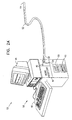
- Figs. 2A and 2B schematically illustrate a system 50 for PMR, including a catheter 52 for insertion into the body of a subject, in accordance with a preferred embodiment of the present invention.
- Catheter 52 comprises an optical waveguide 54, as is known in the art, for transmitting laser energy from the laser source to the heart tissue.
- a focusing lens 62 at distal end 64 of catheter 52 focuses the laser radiation from waveguide 54 into heart tissue.
- Catheter 52 is connected at its proximal end 56 to a console 58, which includes a laser source 60 optically coupled to waveguide 54.
- the laser is activated to generate PMR channels into the heart tissue.
- console 58 includes an optical radiation source 61, which is used in conjunction with a catheter comprising an optical sensor for measuring local blood perfusion (as shown in detail in Fig. 7B and described with reference thereto)
- console 58 also includes signal processing circuitry 44, as well as a display 46 and user controls 48.
- intracardiac electrogram trace 10 the skin ECG trace 20 and/or the laser trigger signal 30 are monitored and displayed on display 46 during the PMR treatment. As described above, these traces provide a real-time visual indication to the user of the catheter, typically an interventional cardiologist, as to whether the channel has been generated.
- the signal processing circuitry analyzes the data and gives the user a "go/no go” indication as to whether the channel has been successfully generated
- Catheter 52 preferably also includes a position sensor 66, fixed in a known position adjacent distal end 64, for use in navigating and positioning the catheter within the heart, as described more fully in patent application EP-A-0 888 081.
- catheter 52 includes a sensor unit 42 at its distal end 64.
- the sensor unit 42 comprises an electrode 43 for sensing electrical potentials in heart tissue adjacent to distal end 64.
- Local electrogram signals from electrode 43 are conveyed by wires 40 to circuitry 44.
- these signals are used to monitor the changes in the electrogram signals due to the PMR drilling, as described above, thus indicating successful channel drilling.
- the electrogram signals may also be used to trigger laser source 60, as disclosed in patent apptication EP-A-0 888 081, mentioned above.
- sensor unit 42 may include other sensors and other types of elements.
- additional electrodes may be placed at or adjacent to distal end 64, either on catheter 52 itself or on a structure fixed to the catheter, as described in patent application EP-A-0 888 082.
- Fig. 3A is a schematic, sectional illustration showing catheter 52 inserted into heart 70 of a subject, in accordance with a preferred embodiment of the present invention.
- Catheter 52 is fed percutaneously into the subject's vascular system, for example, through the femoral artery, and is passed through aorta 72 into left ventricle 74 of heart 70.
- Distal end 64 is positioned against endocardium 76 in a desired position and orientation and drills channels therein, preferably, as described in the above-mentioned PCT patent application no. PCT/IL97/00011,
- Fig. 3B is a schematic, sectional illustration showing details of catheter 52 drilling a channel 88 in myocardium 86 of heart 70, in accordance with the present invention, electrode 43 measures the local electrical signals prior to, during and after the drilling to assess successful drilling, as described above.
- Fig. 4 is a schematic illustration showing details of another catheter 53 for use in PMR, in accordance with alternative preferred embodiments of the present invention.
- Catheter 53 includes waveguide 54, lens 62 and position sensor 66, and is coupled to console 58, substantially as described above with reference to catheter 52.
- sensor unit 42 of catheter 53 includes an ultrasound transducer 41.
- transducer 41 comprises a transducer array, as is known in the art, which emits a beam 67 that may be steered over a range of angles within an area distal to distal end 64 of catheter 53.
- Transducer 41 is coupled via wires 40 to signal processing circuitry 44.
- Catheter 53 is preferably brought into contact with endocardium 76, as shown in Fig. 4
- signals received by circuitry 44 from transducer 41 are used to map the designated channel location prior to and after the PMR procedure to determine, by means of comparison, the dimensions, location and orientation of channel 88, thus indicating its successful generation.
- the ultrasonic readings may be used for dynamic monitoring of channel parameters.
- the transducer signals are used to measure the depth and direction of channel 88 and determine whether the optimal, desired depth has been reached and whether catheter 53 is properly aimed.
- transducer 41 and electrode 43 are used in tandem for assessing successful completion of the PMR procedure, by combining data regarding variations in the electrogram signals following PMR drilling with quantitative measurement of dimensional parameters of channel 88.
- catheters 52 and 53 include various sensors and optical elements in certain preferred combinations and configurations, it will be appreciated that in other preferred embodiments of the present invention, PMR catheters may include some or all of these sensors and elements in other combinations and in the same or other configurations.
- Such catheters may also include other types of sensors known in the art, for example, temperature or pressure sensors, useful in diagnosing other aspects of cardiac function. They may further include blood flow sensors for measuring the local microcirculation flow rate, or optical sensors for visualizing local blood perfusion by tissue autofluorescence or angiography enhanced by fluorescing contrast agents.
- Fig. 5 is a schematic illustration showing the use of skin electrodes 45 placed on a subject's body 71 to record ECG signals therefrom during a PMR procedure, in accordance with a preferred embodiment of the present invention.
- electrodes 45 record the skin ECG signals prior to and for several minutes after laser firing to assess successful drilling, primarily by observing VPB's, as described above with reference to Figs 1A and 1B.
- the global changes sensed by skin electrodes 45 may serve as the sole indication of successful drifting.
- the global variations monitored in the ECG signals are used in conjunction with local variations in the electrical signals sensed by electrode 43.
- the signals measured by electrodes 45 may be used in conjunction with measurements from ultrasonic transducer 41, as described above with reference to Fig. 4.
- Fig. 6 is a flow chart that summarizes the key steps in a method for monitored PMR, in accordance with preferred embodiments of the present invention. The method is described below with reference to catheter 52, shown in Figs. 2A and 2B, but it will be understood that the principles of this method may be applied using other suitable catheters, as described hereinabove.
- At least one candidate area for the procedure is identified within heart 70, preferably as described in the above-mentioned patent application EP-A-0 888 081.
- Catheter 52 is then navigated to the candidate area.
- the position and orientation of distal end 64 of the catheter are preferably ascertained and controlled by receiving signals from position sensor 66, and are compared with a stored map of the heart, although such position and orientation sensing are not a necessary part of the present invention.
- intracardiac electrogram signals are received and stored by console 48.
- Laser source 60 is fired to drill a channel in the heart tissue, as described above.
- post-PMR readings are taken by electrode 43 and analyzed, preferably by comparing them with the pre-PMR signals, for indication of successful drilling.
- the position of the channel is marked on the map, and catheter 52 is then repositioned to drill the next channel. This procedure is preferably repeated until channels have been drilled to a desired density over the entire candidate area.
- the method of monitored PMR shown in Fig. 6 may similarly be implemented by monitoring the skin surface ECG or by using ultrasound or other sensing modalities.
- the PMR procedure may be carried out using other methods of PMR, such as RF or mechanical methods, mentioned above, in place of the laser.
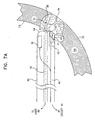
- Fig. 7A is a schematic illustration showing details of a catheter 90 for use in monitored PMR, in accordance with an alternative preferred embodiment of the present invention.
- Catheter 90 includes waveguide 54, lens 62 and position sensor 66, and is coupled to console 58, substantially as described above with reference to catheter 52. Additionally, sensor unit 42 of catheter 90 includes a blood flow sensor 92, which senses signals responsive to blood flow within microvasculature 94 in a vicinity of channel 88, generated by the catheter.
- Sensor 92 preferably comprises an optical detector, which senses microperfusion and/or tissue oxygenation based on light reflected from the heart tissue.
- the sensor may be used to detect NADH activity, as described in the above-mentioned articles by Kedem, Furman and Duboc, or to detect the concentration of a contrast agent or fluorescent marker.
- sensor 92 may comprise an ultrasound transducer. Sensor 92 is coupled via wires 40 to circuitry 44.
- sensor 92 When catheter 90 is brought into contact with endocardium 76, sensor 92 receives signals from the vicinity of channel 88. Signals prior to and after the PMR procedure are compared, so as to detect changes in local blood flow in the vicinity.
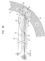
- Fig. 7B schematically illustrates a catheter 96, similar in design and function to catheter 90 described above, in accordance with another preferred embodiment of the present invention.
- Sensor unit 42 of catheter 96 includes an optical sensor assembly 102, comprising a waveguide 98, which is connected to radiation source 61 (shown in Fig. 2A) and transmits fluorescence-stimulating radiation to the myocardial tissue through a lens 100.
- Assembly 102 further comprises a light detector 104, connected via wires 40 to circuitry 44. Detector 104 receives fluorescent radiation emitted from the tissue and generates signals in response thereto.
- the detector may detect near-IR fluorescence of ICG injected into the patient's bloodstream and conveyed thereby to microvasculature 94, as described in the above-mentioned article by May.
- detector 104 includes an optical filter, as is known in the art, so that the detector receives radiation only in a wavelength band of interest.
- sensor assembly 102 When catheter 96 is brought into contact with the endocardium, sensor assembly 102 receives signals in the vicinity of channel 88 prior and after the PMR procedure to determine changes in local perfusion, as explained above. Increased perfusion generally indicates a successful PMR treatment.
- channels 88 may be drilled using a laser source, as described above, or alternatively, using drills of other suitable types known in the art, for example, a high-speed roto-ablator drill head.
- the channels may be produced using a focused, high-intensity beam of ultrasonic radiation, or by applying RF energy to the tissue.
- catheters 52, 53, 90 and 96 are used to produce channels in the wall of left ventricle 74, it will also be understood that the principles of the present invention may be applied to assess the efficacy of PMEt procedures applied to other parts of the heart.
Landscapes
- Health & Medical Sciences (AREA)
- Life Sciences & Earth Sciences (AREA)
- Biomedical Technology (AREA)
- Engineering & Computer Science (AREA)
- Animal Behavior & Ethology (AREA)
- Nuclear Medicine, Radiotherapy & Molecular Imaging (AREA)
- Veterinary Medicine (AREA)
- Public Health (AREA)
- General Health & Medical Sciences (AREA)
- Surgery (AREA)
- Molecular Biology (AREA)
- Medical Informatics (AREA)
- Heart & Thoracic Surgery (AREA)
- Otolaryngology (AREA)
- Pathology (AREA)
- Radiology & Medical Imaging (AREA)
- Laser Surgery Devices (AREA)
- Media Introduction/Drainage Providing Device (AREA)
- Investigating, Analyzing Materials By Fluorescence Or Luminescence (AREA)
- Radiation-Therapy Devices (AREA)
- Medicines Containing Antibodies Or Antigens For Use As Internal Diagnostic Agents (AREA)
- Measurement Of The Respiration, Hearing Ability, Form, And Blood Characteristics Of Living Organisms (AREA)
Applications Claiming Priority (3)
| Application Number | Priority Date | Filing Date | Title |
|---|---|---|---|
| WOPCT/IL97/00011 | 1997-01-08 | ||
| PCT/IL1997/000011 WO1997025101A2 (en) | 1996-01-08 | 1997-01-08 | Methods and apparatus for myocardial revascularization |
| PCT/IL1997/000307 WO1998030144A1 (en) | 1997-01-08 | 1997-09-15 | Monitoring of myocardial revascularization |
Publications (3)
| Publication Number | Publication Date |
|---|---|
| EP0893965A1 EP0893965A1 (en) | 1999-02-03 |
| EP0893965A4 EP0893965A4 (en) | 1999-04-07 |
| EP0893965B1 true EP0893965B1 (en) | 2005-03-09 |
Family
ID=11061976
Family Applications (1)
| Application Number | Title | Priority Date | Filing Date |
|---|---|---|---|
| EP97940316A Expired - Lifetime EP0893965B1 (en) | 1997-01-08 | 1997-09-15 | Monitoring of myocardial revascularization |
Country Status (8)
Families Citing this family (80)
| Publication number | Priority date | Publication date | Assignee | Title |
|---|---|---|---|---|
| US5697882A (en) | 1992-01-07 | 1997-12-16 | Arthrocare Corporation | System and method for electrosurgical cutting and ablation |
| US5683366A (en) | 1992-01-07 | 1997-11-04 | Arthrocare Corporation | System and method for electrosurgical tissue canalization |
| US6277112B1 (en) | 1996-07-16 | 2001-08-21 | Arthrocare Corporation | Methods for electrosurgical spine surgery |
| US6179824B1 (en) | 1993-05-10 | 2001-01-30 | Arthrocare Corporation | System and methods for electrosurgical restenosis of body lumens |
| US6915806B2 (en) | 1993-05-10 | 2005-07-12 | Arthrocare Corporation | Method for harvesting graft vessel |
| US6749604B1 (en) | 1993-05-10 | 2004-06-15 | Arthrocare Corporation | Electrosurgical instrument with axially-spaced electrodes |
| US20080154257A1 (en) * | 2006-12-22 | 2008-06-26 | Shiva Sharareh | Real-time optoacoustic monitoring with electophysiologic catheters |
| US6669685B1 (en) * | 1997-11-06 | 2003-12-30 | Biolase Technology, Inc. | Tissue remover and method |
| US6086534A (en) * | 1997-03-07 | 2000-07-11 | Cardiogenesis Corporation | Apparatus and method of myocardial revascularization using ultrasonic pulse-echo distance ranging |
| US6024703A (en) | 1997-05-07 | 2000-02-15 | Eclipse Surgical Technologies, Inc. | Ultrasound device for axial ranging |
| US6855143B2 (en) | 1997-06-13 | 2005-02-15 | Arthrocare Corporation | Electrosurgical systems and methods for recanalization of occluded body lumens |
| US6083166A (en) * | 1997-12-02 | 2000-07-04 | Situs Corporation | Method and apparatus for determining a measure of tissue manipulation |
| US7435247B2 (en) | 1998-08-11 | 2008-10-14 | Arthrocare Corporation | Systems and methods for electrosurgical tissue treatment |
| WO2000009003A1 (de) * | 1998-08-13 | 2000-02-24 | Dornier Medtech Holding International Gmbh | Vorrichtung und verfahren zur darstellung von gewebeveränderungen |
| US6261304B1 (en) | 1998-09-10 | 2001-07-17 | Percardia, Inc. | Delivery methods for left ventricular conduit |
| US6217518B1 (en) | 1998-10-01 | 2001-04-17 | Situs Corporation | Medical instrument sheath comprising a flexible ultrasound transducer |
| US6468271B1 (en) | 1999-02-24 | 2002-10-22 | Scimed Life Systems, Inc. | Device and method for percutaneous myocardial revascularization |
| US6217575B1 (en) | 1999-02-24 | 2001-04-17 | Scimed Life Systems, Inc. | PMR catheter |
| US6638237B1 (en) * | 1999-08-04 | 2003-10-28 | Percardia, Inc. | Left ventricular conduits and methods for delivery |
| US6253768B1 (en) | 1999-08-04 | 2001-07-03 | Percardia, Inc. | Vascular graft bypass |
| US20050182434A1 (en) | 2000-08-11 | 2005-08-18 | National Research Council Of Canada | Method and apparatus for performing intra-operative angiography |
| CN101406392B (zh) * | 1999-09-24 | 2011-05-18 | 加拿大国家研究委员会 | 用于手术中血管造影的装置 |
| US6915154B1 (en) | 1999-09-24 | 2005-07-05 | National Research Council Of Canada | Method and apparatus for performing intra-operative angiography |
| US6500119B1 (en) * | 1999-12-01 | 2002-12-31 | Medical Tactile, Inc. | Obtaining images of structures in bodily tissue |
| AU5113401A (en) * | 2000-03-31 | 2001-10-15 | Rita Medical Systems Inc | Tissue biopsy and treatment apparatus and method |
| US6854467B2 (en) | 2000-05-04 | 2005-02-15 | Percardia, Inc. | Methods and devices for delivering a ventricular stent |
| US6546276B1 (en) | 2000-09-12 | 2003-04-08 | Claudio I. Zanelli | Ultrasonic based detection of interventional medical device contact and alignment |
| US6436059B1 (en) | 2000-09-12 | 2002-08-20 | Claudio I. Zanelli | Detection of imd contact and alignment based on changes in frequency response characteristics |
| US6725085B2 (en) | 2000-09-22 | 2004-04-20 | Armin Schwartzman | Method and apparatus for characterizing cardiac tissue from local electrograms |
| US6533779B2 (en) | 2001-01-16 | 2003-03-18 | Scimed Life Systems, Inc. | PMR catheter and associated methods |
| US6544220B2 (en) | 2001-02-14 | 2003-04-08 | Scimed Life Systems, Inc. | Fluid jet PMR |
| US6666863B2 (en) * | 2001-03-01 | 2003-12-23 | Scimed Life Systems, Inc. | Device and method for percutaneous myocardial revascularization |
| US6666862B2 (en) * | 2001-03-01 | 2003-12-23 | Cardiac Pacemakers, Inc. | Radio frequency ablation system and method linking energy delivery with fluid flow |
| US6508783B2 (en) | 2001-03-14 | 2003-01-21 | Scimed Life Systems, Inc. | Ultrasound method for revascularization and drug delivery |
| US20040152974A1 (en) * | 2001-04-06 | 2004-08-05 | Stephen Solomon | Cardiology mapping and navigation system |
| AU2002307150A1 (en) * | 2001-04-06 | 2002-10-21 | Steven Solomon | Cardiological mapping and navigation system |
| AU2003238656A1 (en) * | 2002-06-25 | 2004-01-06 | Glucon Inc. | Method and apparatus for performing myocardial revascularization |
| US20040158235A1 (en) * | 2003-02-11 | 2004-08-12 | Robert Rudko | TMR system and handpiece with ECG electrodes |
| US7232437B2 (en) * | 2003-10-30 | 2007-06-19 | Medical Cv, Inc. | Assessment of lesion transmurality |
| WO2005044124A1 (en) * | 2003-10-30 | 2005-05-19 | Medical Cv, Inc. | Apparatus and method for laser treatment |
| US7238179B2 (en) * | 2003-10-30 | 2007-07-03 | Medical Cv, Inc. | Apparatus and method for guided ablation treatment |
| US7238180B2 (en) * | 2003-10-30 | 2007-07-03 | Medicalcv Inc. | Guided ablation with end-fire fiber |
| DE10355275B4 (de) * | 2003-11-26 | 2009-03-05 | Siemens Ag | Kathedereinrichtung |
| US7640046B2 (en) * | 2004-06-18 | 2009-12-29 | Cardiac Pacemakers, Inc. | Methods and apparatuses for localizing myocardial infarction during catheterization |
| DE202005003411U1 (de) * | 2005-02-24 | 2006-07-06 | Karl Storz Gmbh & Co. Kg | Multifunktionales Fluoreszenzdiagnosesystem |
| US20060239921A1 (en) | 2005-04-26 | 2006-10-26 | Novadaq Technologies Inc. | Real time vascular imaging during solid organ transplant |
| US9861836B2 (en) * | 2005-06-16 | 2018-01-09 | Biosense Webster, Inc. | Less invasive methods for ablation of fat pads |
| US20070122344A1 (en) | 2005-09-02 | 2007-05-31 | University Of Rochester Medical Center Office Of Technology Transfer | Intraoperative determination of nerve location |
| US20070073281A1 (en) * | 2005-09-16 | 2007-03-29 | Medicalcv, Inc. | Guided ablation with motion control |
| US20070073280A1 (en) * | 2005-09-16 | 2007-03-29 | Medicalcv, Inc. | End-fire guided ablation |
| US8709056B2 (en) * | 2006-04-10 | 2014-04-29 | Bwt Property Inc | Phototherapy apparatus with built-in ultrasonic image module |
| US20080161744A1 (en) | 2006-09-07 | 2008-07-03 | University Of Rochester Medical Center | Pre-And Intra-Operative Localization of Penile Sentinel Nodes |
| US8155730B2 (en) | 2006-10-24 | 2012-04-10 | The Research Foundation Of State University Of New York | Composition, method, system, and kit for optical electrophysiology |
| US7717855B2 (en) * | 2006-12-06 | 2010-05-18 | The Hospital For Sick Children | System for performing remote ischemic preconditioning |
| US8406860B2 (en) | 2008-01-25 | 2013-03-26 | Novadaq Technologies Inc. | Method for evaluating blush in myocardial tissue |
| US10219742B2 (en) | 2008-04-14 | 2019-03-05 | Novadaq Technologies ULC | Locating and analyzing perforator flaps for plastic and reconstructive surgery |
| ES2671710T3 (es) | 2008-05-02 | 2018-06-08 | Novadaq Technologies ULC | Métodos para la producción y uso de eritrocitos cargados con sustancias para la observación y el tratamiento de la hemodinámica microvascular |
| US8355799B2 (en) | 2008-12-12 | 2013-01-15 | Arthrocare Corporation | Systems and methods for limiting joint temperature |
| US10492671B2 (en) | 2009-05-08 | 2019-12-03 | Novadaq Technologies ULC | Near infra red fluorescence imaging for visualization of blood vessels during endoscopic harvest |
| DE102009034249A1 (de) * | 2009-07-22 | 2011-03-24 | Siemens Aktiengesellschaft | Verfahren und Vorrichtung zur Regelung der Ablationsenergie zur Durchführung einer elektrophysiologischen Katheteranwendung |
| JP5635282B2 (ja) * | 2010-03-15 | 2014-12-03 | ソニー株式会社 | 判別装置 |
| EP2552331B1 (en) | 2010-03-31 | 2020-01-08 | The Hospital For Sick Children | Use of remote ischemic conditioning to improve outcome after myocardial infarction |
| RU2012147442A (ru) | 2010-04-08 | 2014-05-20 | Дзе Хоспитал Фор Сик Чилдрен | Применение дистантного ишемического кондиционирования при травматическом повреждении |
| BR112013008821A2 (pt) * | 2010-10-14 | 2016-06-28 | Koninkl Philips Electronics Nv | aparelho de determinação de propriedades para determinar propriedades de objetos, método de determinação de propriedades para determinar propriedades de objetos e programa de computador de determinação de propriedades para determinar propriedade de objeto |
| US8764789B2 (en) | 2011-04-15 | 2014-07-01 | CellAegis Devices Inc. | System for performing remote ischemic conditioning |
| US10278585B2 (en) | 2012-06-21 | 2019-05-07 | Novadaq Technologies ULC | Quantification and analysis of angiography and perfusion |
| CN103035774B (zh) * | 2012-12-31 | 2015-07-01 | 东南大学 | 一种单光源植入式神经多点同步交互芯片及其制备方法 |
| WO2014140832A2 (en) | 2013-03-15 | 2014-09-18 | The Hospital For Sick Children | Treatment of erectile dysfunction using remote ischemic conditioning |
| WO2014199239A2 (en) | 2013-03-15 | 2014-12-18 | The Hospital For Sick Children | Methods relating to the use of remote ischemic conditioning |
| AU2013203746B2 (en) | 2013-03-15 | 2015-05-07 | Cellaegis Devices, Inc. | Gas Powered System for Performing Remote Ischemic Conditioning |
| WO2014167423A2 (en) | 2013-03-15 | 2014-10-16 | The Hospital For Sick Children | Methods for modulating autophagy using remote ischemic conditioning |
| US9526556B2 (en) | 2014-02-28 | 2016-12-27 | Arthrocare Corporation | Systems and methods systems related to electrosurgical wands with screen electrodes |
| US9597142B2 (en) | 2014-07-24 | 2017-03-21 | Arthrocare Corporation | Method and system related to electrosurgical procedures |
| US9649148B2 (en) | 2014-07-24 | 2017-05-16 | Arthrocare Corporation | Electrosurgical system and method having enhanced arc prevention |
| KR20170067803A (ko) | 2014-09-29 | 2017-06-16 | 노바다크 테크놀러지즈 인코포레이티드 | 자가형광이 존재하는 생물학적 물질에서 타겟 형광체의 이미징 |
| EP3915467A1 (en) | 2014-10-09 | 2021-12-01 | Novadaq Technologies ULC | Quantification of absolute blood flow in tissue using fluorescence-mediated photoplethysmography |
| CN104720752B (zh) * | 2015-02-13 | 2017-09-15 | 亚太仿生学有限公司 | 一种用于空腔结构内部热成像的探测器及系统装置 |
| EP3580609B1 (en) | 2017-02-10 | 2023-05-24 | Stryker European Operations Limited | Open-field handheld fluorescence imaging systems and methods |
| EP3773301B1 (en) | 2018-04-13 | 2024-03-06 | Karl Storz SE & Co. KG | Guidance system and associated computer program |
| US11751938B2 (en) * | 2019-03-22 | 2023-09-12 | Boston Scientific Scimed, Inc. | Ablation catheter with blood perfusion sensor |
Family Cites Families (43)
| Publication number | Priority date | Publication date | Assignee | Title |
|---|---|---|---|---|
| US4658817A (en) | 1985-04-01 | 1987-04-21 | Children's Hospital Medical Center | Method and apparatus for transmyocardial revascularization using a laser |
| US4788975B1 (en) | 1987-11-05 | 1999-03-02 | Trimedyne Inc | Control system and method for improved laser angioplasty |
| JPH04250169A (ja) * | 1990-07-20 | 1992-09-07 | Telectronics Nv | 患者治療装置及び方法 |
| US5125924A (en) | 1990-09-24 | 1992-06-30 | Laser Engineering, Inc. | Heart-synchronized vacuum-assisted pulsed laser system and method |
| US5125926A (en) | 1990-09-24 | 1992-06-30 | Laser Engineering, Inc. | Heart-synchronized pulsed laser system |
| US5389096A (en) * | 1990-12-18 | 1995-02-14 | Advanced Cardiovascular Systems | System and method for percutaneous myocardial revascularization |
| US5885272A (en) | 1990-10-30 | 1999-03-23 | Aita; Michael | System and method for percutaneous myocardial revascularization |
| US5380316A (en) | 1990-12-18 | 1995-01-10 | Advanced Cardiovascular Systems, Inc. | Method for intra-operative myocardial device revascularization |
| US5188111A (en) | 1991-01-18 | 1993-02-23 | Catheter Research, Inc. | Device for seeking an area of interest within a body |
| NZ242509A (en) * | 1991-05-01 | 1996-03-26 | Univ Columbia | Myocardial revascularisation using laser |
| US5683366A (en) * | 1992-01-07 | 1997-11-04 | Arthrocare Corporation | System and method for electrosurgical tissue canalization |
| EP0661948B1 (en) | 1992-09-23 | 1997-11-19 | Endocardial Solutions, Inc. | Endocardial mapping system |
| US5385146A (en) | 1993-01-08 | 1995-01-31 | Goldreyer; Bruce N. | Orthogonal sensing for use in clinical electrophysiology |
| US5433198A (en) | 1993-03-11 | 1995-07-18 | Desai; Jawahar M. | Apparatus and method for cardiac ablation |
| US5350375A (en) * | 1993-03-15 | 1994-09-27 | Yale University | Methods for laser induced fluorescence intensity feedback control during laser angioplasty |
| US5403311A (en) | 1993-03-29 | 1995-04-04 | Boston Scientific Corporation | Electro-coagulation and ablation and other electrotherapeutic treatments of body tissue |
| US5403356A (en) | 1993-04-28 | 1995-04-04 | Medtronic, Inc. | Method and apparatus for prevention of atrial tachy arrhythmias |
| US5462544A (en) * | 1993-05-05 | 1995-10-31 | Energy Life System Corporation | Continuous heart tissue mapping and lasing catheter |
| US5840031A (en) | 1993-07-01 | 1998-11-24 | Boston Scientific Corporation | Catheters for imaging, sensing electrical potentials and ablating tissue |
| US5738096A (en) | 1993-07-20 | 1998-04-14 | Biosense, Inc. | Cardiac electromechanics |
| IL116699A (en) | 1996-01-08 | 2001-09-13 | Biosense Ltd | Method of building a heart map |
| US5391199A (en) * | 1993-07-20 | 1995-02-21 | Biosense, Inc. | Apparatus and method for treating cardiac arrhythmias |
| US5431168A (en) | 1993-08-23 | 1995-07-11 | Cordis-Webster, Inc. | Steerable open-lumen catheter |
| US5409000A (en) | 1993-09-14 | 1995-04-25 | Cardiac Pathways Corporation | Endocardial mapping and ablation system utilizing separately controlled steerable ablation catheter with ultrasonic imaging capabilities and method |
| US5651786A (en) * | 1993-09-20 | 1997-07-29 | Abela Laser Systems, Inc. | Mapping catheter and method |
| US5487391A (en) | 1994-01-28 | 1996-01-30 | Ep Technologies, Inc. | Systems and methods for deriving and displaying the propagation velocities of electrical events in the heart |
| JPH0838460A (ja) | 1994-08-03 | 1996-02-13 | Mitsubishi Electric Corp | 脳活動計測装置 |
| CA2607769C (en) | 1994-08-19 | 2012-04-24 | Biosense, Inc. | Medical diagnosis, treatment and imaging systems |
| US5620439A (en) * | 1995-06-06 | 1997-04-15 | George S. Abela | Catheter and technique for endovascular myocardial revascularization |
| JP3231221B2 (ja) * | 1995-06-22 | 2001-11-19 | テルモ株式会社 | ガイドワイヤ |
| US6023638A (en) * | 1995-07-28 | 2000-02-08 | Scimed Life Systems, Inc. | System and method for conducting electrophysiological testing using high-voltage energy pulses to stun tissue |
| US5824005A (en) * | 1995-08-22 | 1998-10-20 | Board Of Regents, The University Of Texas System | Maneuverable electrophysiology catheter for percutaneous or intraoperative ablation of cardiac arrhythmias |
| CA2242356C (en) | 1996-01-08 | 2005-08-23 | Biosense, Inc. | Methods and apparatus for myocardial revascularization |
| US5769843A (en) | 1996-02-20 | 1998-06-23 | Cormedica | Percutaneous endomyocardial revascularization |
| US5725523A (en) * | 1996-03-29 | 1998-03-10 | Mueller; Richard L. | Lateral-and posterior-aspect method and apparatus for laser-assisted transmyocardial revascularization and other surgical applications |
| US5891133A (en) * | 1996-03-29 | 1999-04-06 | Eclipse Surgical Technologies, Inc. | Apparatus for laser-assisted intra-coronary transmyocardial revascularization and other applications |
| US5871495A (en) * | 1996-09-13 | 1999-02-16 | Eclipse Surgical Technologies, Inc. | Method and apparatus for mechanical transmyocardial revascularization of the heart |
| US5893848A (en) * | 1996-10-24 | 1999-04-13 | Plc Medical Systems, Inc. | Gauging system for monitoring channel depth in percutaneous endocardial revascularization |
| US5724975A (en) * | 1996-12-12 | 1998-03-10 | Plc Medical Systems, Inc. | Ultrasonic detection system for transmyocardial revascularization |
| CA2228584A1 (en) * | 1997-02-03 | 1998-08-03 | Eclipse Surgical Technologies, Inc. | Revascularization with heart pacing |
| US6024739A (en) | 1997-09-05 | 2000-02-15 | Cordis Webster, Inc. | Method for detecting and revascularizing ischemic myocardial tissue |
| US5964757A (en) | 1997-09-05 | 1999-10-12 | Cordis Webster, Inc. | Steerable direct myocardial revascularization catheter |
| US6027473A (en) | 1997-09-05 | 2000-02-22 | Cordis Webster, Inc. | Handle for steerable DMR catheter |
-
1997
- 1997-09-15 EP EP97940316A patent/EP0893965B1/en not_active Expired - Lifetime
- 1997-09-15 AU AU42182/97A patent/AU741217B2/en not_active Expired
- 1997-09-15 CA CA002248223A patent/CA2248223C/en not_active Expired - Lifetime
- 1997-09-15 US US09/142,696 patent/US6200310B1/en not_active Expired - Lifetime
- 1997-09-15 DE DE69732696T patent/DE69732696T2/de not_active Expired - Lifetime
- 1997-09-15 WO PCT/IL1997/000307 patent/WO1998030144A1/en active IP Right Grant
- 1997-09-15 ES ES97940316T patent/ES2237802T3/es not_active Expired - Lifetime
- 1997-09-15 JP JP53069698A patent/JP4236014B2/ja not_active Expired - Lifetime
-
2000
- 2000-10-12 US US09/689,257 patent/US6436095B1/en not_active Expired - Fee Related
Non-Patent Citations (1)
| Title |
|---|
| DENES P. ET AL: "Prognostic significance of signal-averaged electrocardiogram after thrombolytic therapy and/or angioplasty during acute myocardial infarction (CAST substudy)", AM.J.CARDIOL., vol. 74, no. 3, 1994, pages 216 - 220, XP023278416, DOI: doi:10.1016/0002-9149(94)90359-X * |
Also Published As
| Publication number | Publication date |
|---|---|
| EP0893965A1 (en) | 1999-02-03 |
| US6200310B1 (en) | 2001-03-13 |
| DE69732696D1 (de) | 2005-04-14 |
| JP2001505472A (ja) | 2001-04-24 |
| CA2248223C (en) | 2007-01-16 |
| WO1998030144A1 (en) | 1998-07-16 |
| ES2237802T3 (es) | 2005-08-01 |
| JP4236014B2 (ja) | 2009-03-11 |
| AU4218297A (en) | 1998-08-03 |
| DE69732696T2 (de) | 2006-04-13 |
| EP0893965A4 (en) | 1999-04-07 |
| AU741217B2 (en) | 2001-11-29 |
| US6436095B1 (en) | 2002-08-20 |
| CA2248223A1 (en) | 1998-07-16 |
Similar Documents
| Publication | Publication Date | Title |
|---|---|---|
| EP0893965B1 (en) | Monitoring of myocardial revascularization | |
| US6447504B1 (en) | System for treatment of heart tissue using viability map | |
| US6171303B1 (en) | Methods and apparatus for myocardial revascularization | |
| US11596472B2 (en) | Systems and methods for assessment of contact quality | |
| US6660001B2 (en) | Myocardial revascularization-optical reflectance catheter and method | |
| US10499984B2 (en) | Apparatus and method for assessing tissue treatment | |
| US9332893B2 (en) | Delivery of biological compounds to ischemic and/or infarcted tissue | |
| EP3150115A1 (en) | Catheter apparatus with a diagnostic assembly including an optical emitting element and an optical receiving element | |
| US20060122583A1 (en) | Method and apparatus for performing myocardial revascularization | |
| US20080125634A1 (en) | Method and apparatus for identifying and treating myocardial infarction | |
| US10881459B2 (en) | Apparatus and method for assessing tissue treatment | |
| JP2023510326A (ja) | アブレーション焼灼巣の光学的探索のためのシステム及び方法 | |
| JP4518339B2 (ja) | 心筋の脈管再生方法および装置 |
Legal Events
| Date | Code | Title | Description |
|---|---|---|---|
| PUAI | Public reference made under article 153(3) epc to a published international application that has entered the european phase |
Free format text: ORIGINAL CODE: 0009012 |
|
| 17P | Request for examination filed |
Effective date: 19981007 |
|
| AK | Designated contracting states |
Kind code of ref document: A1 Designated state(s): DE ES FR GB IT NL |
|
| A4 | Supplementary search report drawn up and despatched |
Effective date: 19990219 |
|
| AK | Designated contracting states |
Kind code of ref document: A4 Designated state(s): DE ES FR GB IT NL |
|
| R17P | Request for examination filed (corrected) |
Effective date: 19981008 |
|
| 17Q | First examination report despatched |
Effective date: 20030116 |
|
| GRAP | Despatch of communication of intention to grant a patent |
Free format text: ORIGINAL CODE: EPIDOSNIGR1 |
|
| RIC1 | Information provided on ipc code assigned before grant |
Ipc: 7A 61F 7/00 B Ipc: 7A 61F 2/00 B Ipc: 7A 61B 18/00 B Ipc: 7A 61B 5/04 A |
|
| RIC1 | Information provided on ipc code assigned before grant |
Ipc: 7A 61F 7/00 B Ipc: 7A 61F 2/00 B Ipc: 7A 61B 18/00 B Ipc: 7A 61B 5/04 A |
|
| GRAS | Grant fee paid |
Free format text: ORIGINAL CODE: EPIDOSNIGR3 |
|
| RAP1 | Party data changed (applicant data changed or rights of an application transferred) |
Owner name: BIOSENSE WEBMASTER, INC |
|
| RAP1 | Party data changed (applicant data changed or rights of an application transferred) |
Owner name: BIOSENSE WEBSTER, INC. |
|
| GRAA | (expected) grant |
Free format text: ORIGINAL CODE: 0009210 |
|
| AK | Designated contracting states |
Kind code of ref document: B1 Designated state(s): DE ES FR GB IT NL |
|
| REG | Reference to a national code |
Ref country code: GB Ref legal event code: FG4D |
|
| REF | Corresponds to: |
Ref document number: 69732696 Country of ref document: DE Date of ref document: 20050414 Kind code of ref document: P |
|
| REG | Reference to a national code |
Ref country code: ES Ref legal event code: FG2A Ref document number: 2237802 Country of ref document: ES Kind code of ref document: T3 |
|
| PLBE | No opposition filed within time limit |
Free format text: ORIGINAL CODE: 0009261 |
|
| STAA | Information on the status of an ep patent application or granted ep patent |
Free format text: STATUS: NO OPPOSITION FILED WITHIN TIME LIMIT |
|
| ET | Fr: translation filed | ||
| 26N | No opposition filed |
Effective date: 20051212 |
|
| PGFP | Annual fee paid to national office [announced via postgrant information from national office to epo] |
Ref country code: ES Payment date: 20111017 Year of fee payment: 15 |
|
| REG | Reference to a national code |
Ref country code: ES Ref legal event code: FD2A Effective date: 20131018 |
|
| PG25 | Lapsed in a contracting state [announced via postgrant information from national office to epo] |
Ref country code: ES Free format text: LAPSE BECAUSE OF NON-PAYMENT OF DUE FEES Effective date: 20120916 |
|
| REG | Reference to a national code |
Ref country code: FR Ref legal event code: PLFP Year of fee payment: 20 |
|
| PGFP | Annual fee paid to national office [announced via postgrant information from national office to epo] |
Ref country code: GB Payment date: 20160914 Year of fee payment: 20 Ref country code: NL Payment date: 20160913 Year of fee payment: 20 Ref country code: DE Payment date: 20160907 Year of fee payment: 20 Ref country code: IT Payment date: 20160921 Year of fee payment: 20 |
|
| PGFP | Annual fee paid to national office [announced via postgrant information from national office to epo] |
Ref country code: FR Payment date: 20160816 Year of fee payment: 20 |
|
| REG | Reference to a national code |
Ref country code: DE Ref legal event code: R071 Ref document number: 69732696 Country of ref document: DE |
|
| REG | Reference to a national code |
Ref country code: NL Ref legal event code: MK Effective date: 20170914 |
|
| REG | Reference to a national code |
Ref country code: GB Ref legal event code: PE20 Expiry date: 20170914 |
|
| PG25 | Lapsed in a contracting state [announced via postgrant information from national office to epo] |
Ref country code: GB Free format text: LAPSE BECAUSE OF EXPIRATION OF PROTECTION Effective date: 20170914 |