EP0893816B1 - Appareil à faisceau corpusculaire - Google Patents
Appareil à faisceau corpusculaire Download PDFInfo
- Publication number
- EP0893816B1 EP0893816B1 EP98111715A EP98111715A EP0893816B1 EP 0893816 B1 EP0893816 B1 EP 0893816B1 EP 98111715 A EP98111715 A EP 98111715A EP 98111715 A EP98111715 A EP 98111715A EP 0893816 B1 EP0893816 B1 EP 0893816B1
- Authority
- EP
- European Patent Office
- Prior art keywords
- potential
- anode
- cathode
- voltage source
- corpuscles
- Prior art date
- Legal status (The legal status is an assumption and is not a legal conclusion. Google has not performed a legal analysis and makes no representation as to the accuracy of the status listed.)
- Expired - Lifetime
Links
Images
Classifications
-
- H—ELECTRICITY
- H01—ELECTRIC ELEMENTS
- H01J—ELECTRIC DISCHARGE TUBES OR DISCHARGE LAMPS
- H01J37/00—Discharge tubes with provision for introducing objects or material to be exposed to the discharge, e.g. for the purpose of examination or processing thereof
- H01J37/02—Details
- H01J37/04—Arrangements of electrodes and associated parts for generating or controlling the discharge, e.g. electron-optical arrangement, ion-optical arrangement
- H01J37/10—Lenses
- H01J37/145—Combinations of electrostatic and magnetic lenses
-
- H—ELECTRICITY
- H01—ELECTRIC ELEMENTS
- H01J—ELECTRIC DISCHARGE TUBES OR DISCHARGE LAMPS
- H01J37/00—Discharge tubes with provision for introducing objects or material to be exposed to the discharge, e.g. for the purpose of examination or processing thereof
- H01J37/02—Details
- H01J37/04—Arrangements of electrodes and associated parts for generating or controlling the discharge, e.g. electron-optical arrangement, ion-optical arrangement
-
- H—ELECTRICITY
- H01—ELECTRIC ELEMENTS
- H01J—ELECTRIC DISCHARGE TUBES OR DISCHARGE LAMPS
- H01J37/00—Discharge tubes with provision for introducing objects or material to be exposed to the discharge, e.g. for the purpose of examination or processing thereof
- H01J37/26—Electron or ion microscopes; Electron or ion diffraction tubes
- H01J37/28—Electron or ion microscopes; Electron or ion diffraction tubes with scanning beams
-
- H—ELECTRICITY
- H01—ELECTRIC ELEMENTS
- H01J—ELECTRIC DISCHARGE TUBES OR DISCHARGE LAMPS
- H01J2237/00—Discharge tubes exposing object to beam, e.g. for analysis treatment, etching, imaging
- H01J2237/04—Means for controlling the discharge
- H01J2237/047—Changing particle velocity
- H01J2237/0475—Changing particle velocity decelerating
- H01J2237/04756—Changing particle velocity decelerating with electrostatic means
-
- H—ELECTRICITY
- H01—ELECTRIC ELEMENTS
- H01J—ELECTRIC DISCHARGE TUBES OR DISCHARGE LAMPS
- H01J2237/00—Discharge tubes exposing object to beam, e.g. for analysis treatment, etching, imaging
- H01J2237/244—Detection characterized by the detecting means
- H01J2237/2443—Scintillation detectors
Definitions
- the invention relates to a particle beam device in which an object is irradiated with a focused beam of electrically charged particles, such as electrons, positrons or ions, and wherein the particle energy is adjustable to values below 1 keV.
- a particle beam device in which an object is irradiated with a focused beam of electrically charged particles, such as electrons, positrons or ions, and wherein the particle energy is adjustable to values below 1 keV.
- Such devices are used in the form of scanning electron microscopes, in particular for the inspection of semiconductor wafers and for the imaging and analysis of objects with low conductivity.
- the energy when hitting the object must not exceed certain limit energies that lie between 10 eV and 5 keV.
- the resolution defined by the focus diameter of the particle beam is significantly worse at low particle energies than at higher particle energies.
- a delay device usually consists of two or more electrodes to which an electrostatic retarding field is applied. The deceleration of the particles can be done either in the back of the lens or after exiting the lens between the lens and the object.
- the beam guiding tube is at ground potential from the anode to at least the pole piece gap of the objective, and the object is at a high voltage potential having the same sign as the cathode potential.
- Such arrangements are in the article by KJ Polasko et al. "Low energy electron beam lithography" in OPTICAL ENGINEERING, March / April 1983, pages 195 ff , in US-A 5,389,787 and in EP-A2 0 769 799 described.
- the sample must be placed on a correspondingly high potential of 9-19 kV.
- the object and the object-side electrode of the delay device is at ground potential and the beam guide tube from the anode to the first electrode of the Abbrems worn at a high anode potential, the sign is chosen so that upon exit from the beam guide tube, a delay of the particles occurs.
- Such systems are for example in the DE 29 22 325 C2 and the EP 0 180 723 B1 described.
- the adjustment of the target energy by a variation of the potential of the beam guiding tube.
- This variable anode potential is superimposed on a fixed but opposite potential between the cathode and the anode.
- the entire beam guidance tube from the anode to the objective is then at a potential which, in terms of magnitude, is only 0.5-1 keV less than the potential difference between the cathode and the anode. Since the insulation to the environment be designed accordingly must, this arrangement can only be used at low maximum energies. At target energies below 0.2 keV, the emission becomes increasingly unstable with decreasing target energy because the cathode potential is close to the ground potential and thus any stray fields have an influence on the emission. For target energies below 0.2 keV, this arrangement is therefore less suitable.
- the following invention is intended to indicate an arrangement in which a stable and constant operation can be ensured even at lowest target energies, which may also be less than 0.2 keV, without the object having to be set to a potential that is high in relation to the ground potential.
- a good detectability of the secondary or backscatter particles formed during the interaction of the corpuscles with the object should be ensured even with short distances between the objective and the object.
- the particle beam device has a substantially known construction of a particle source with a cathode and an anode for emission and acceleration of the particles, a beam guidance tube, at least one objective for focusing the particle beam on an object and a delay device.
- the anode and the beam guide tube are on the same, compared to the ground potential high high voltage potential.
- the last electrode of the delay device and the sample likewise lie on a common potential which, at least at the lowest target energies of the particles, deviates from the ground potential and has a sign opposite to the anode potential.
- the invention thus leaves the previous design ways in which at least either the steel guide tube or the object are kept at ground potential.
- the known solutions are combined with one another in such a way that the specific advantages of the known solutions are achieved without at the same time having to take the specific disadvantages of the individual solutions into account.
- the cathode potential can be kept at a value of also 0.2-5 kV with respect to the ground potential, even at lowest target energies, at which the emission is stable.
- the energy setting is achieved by increasing the magnitude of the cathode potential, wherein the object is preferably at ground potential.
- the cathode potential is greater than 0.2 kV, preferably greater than 0.5 kV.
- the sign of the cathode potential is selected as a function of the sign of the corpus cell charges so that the corpuscles have a higher potential energy at the cathode potential compared to the ground potential.
- the anode potential amounts to more than 5 kV in magnitude and is chosen such that the corpuscles have lower potential energy relative to the ground potential at the anode potential.
- the corpuscles emerging from the cathode are thereby accelerated to an energy which corresponds to the potential difference between cathode and anode and should be at least 5-10 kV.
- the corpuscles pass through the beam guiding tube and are braked to the target energy after emerging from the beam guiding tube.
- the setting of the target energy takes place by adjusting the potential at the object and the delay device relative to the ground potential.
- the energy within the beam guide tube thus remains constant.
- This eliminates a fine adjustment of the corpuscular optical column, which greatly simplifies the operation of the system and in particular with the additional use of correction elements (eg multipole correctors) for reducing the aberrations of the lenses represents a significant advantage.
- the sample potential required for adjusting the target energy is at most as large as the cathode potential and is at most 5 kV, so that no voltage flashovers, insulation problems and / or sample damage occur when the sample potential is switched on and off.
- the setting of higher target energies takes place in the particle beam device according to the invention by an increase in the magnitude of the cathode potential.
- the anode potential remains constant, so that a good detection of secondary or backscattered electrons is always guaranteed.
- the sample and the delay device are thus at a potential having the same sign as the cathode potential in the particle beam device according to the invention.
- the anode potential has an opposite sign.
- controllable high voltage sources are respectively provided for the potentials of the delay device, the anode and the cathode of the particle beam device according to the invention.
- Optimal potential adjustment may be performed by a controller depending on the target energy values and the maximum allowable sample potential as parameters to be entered by the user.
- the beam-guiding tube has a widened region in a region between the particle gun and the objective, in which a detector lying at the potential of the beam-guiding tube is arranged for detecting the secondary electrons emanating from the object.
- the secondary electrons or backscatter electrons emerging from the object are accelerated in the rearward direction of the primary beam by the delay device to the difference between the potential of the sample and the potential of the beam guiding tube and have sufficient energy to be well detectable.
- the particle beam device can be formed only with a lens as a single imaging element or as at least two-lens system of lens and condenser.
- a two-lens imaging system of condenser and lens is advantageously used in combination with a source from which the corpuscles emerge relatively undirected, since the usable aperture of the out of the cathode emerging corpuscles in two-stage systems is greater than in single-stage systems. With weak excitation of the condenser, the lens-side aperture of the particle beam is reduced and can thus be optimally adapted to the requirements.
- the delay device may be formed in the particle beam device according to the invention by the rear end of the beam guide tube and one or more further electrodes.
- the rear end of the beam guide tube is advantageously formed as a tube electrode.
- the further electrode of the delay device is preferably accommodated on an insulating body of the objective.
- the objective can basically be designed as a magnetic objective or as an electrostatic single lens.
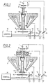
- the particle beam generator in the particle beam device according to the invention consists of a cathode (1) emitting the particles, an extraction electrode (2) and an anode (4). If the particle beam device according to the invention is designed as a scanning electron microscope, the cathode (1) is preferably a thermal field emitter. Depending on the design of the cathode (1), the extraction electrode (2) is at a potential of 2-5 kV with respect to the cathode (1), so that the particles are sucked out of the extraction electrode (2) from the cathode (1).
- U K is a first controllable high voltage source (14) and for the generation of the fixed potential difference between the cathode (1) and extraction electrode (2) is a fixed voltage source (3) is provided.
- the anode (4) simultaneously forms the source-side end of the beam guide tube (5).
- This beam guide tube (5) made of electrically conductive material is guided through a hole through the pole pieces (6) of a magnetic lens acting as an objective and is thickened on the object-side end (8) as a tube lens.
- This thickened object-side end of the beam guide tube (5) ends only behind the Polschuhspalt the magnetic lens whose coils are denoted by (7).
- Downstream of the beam guide tube is a single electrode (9), which forms an electrostatic deceleration device together with the tube electrode (8) of the beam guide tube (5).
- the tube electrode (8) lies together with the entire beam guide tube (5) on the anode potential U A , which is generated by a second controllable high voltage source (13) and the individual electrode (9) and the stage (10) are at a common potential Up, which is generated by a third controllable high voltage source.
- the tube electrode (8) and the single electrode (9) act simultaneously as an electrostatic lens (immersion lens) through which the Corpuscular beam is focused in addition to the effect of the lens on the object.
- the corpuscles emerging from the cathode are accelerated to the anode by one of the potential difference between cathode (1) and anode (4) corresponding energy and subsequently pass through the beam guide tube (5) with this constant energy.
- the particles After emerging from the beam-guiding tube between the tube electrode (8) and the individual electrode (9), the particles are then delayed to a target energy corresponding to the potential difference between the cathode potential U K and the sample potential Up.
- the setting of this target energy is carried out at low target energies via a change of the sample potential Up at each fixed anode potential U A and cathode potential U K. Low energies are energies that are smaller in magnitude than the body energy at cathode potential.
- the sample potential Up is zero and the constant energy of the particle within the beam guide tube is carried out by coordinated change of the cathode potential U K and the anode potential U A.
- Exemplary potential combinations for target energies within the beam guide tube of 10 and 15 keV in the case of electrons are given in Tables 1 and 2.
- the associated other potential values can be easily supplemented.
- the energy U S in the beam-guiding tube can be kept constant in the corpuscular beam device according to the invention.
- a controller (15) can be provided which selects the suitable potential combinations as a function of the target energy, the target energy in the beam guide tube and the maximum allowable sample potential Up as user-definable parameters.
- Table 1 ⁇ / b> Target energy / keV U K / kV U A / kV U P / kV U S / keV 0 -2 8th -2 10 0.5 -2 8th -1.5 10 1 -2 8th -1 10 2 -2 8th 0 10 3 -3 8th 0 11 5 -5 8th 0 13 10 -10 8th 0 18 15 15 8th 0 23 20 20 8th 0 28 30 30 8th 0 38 Target energy / keV U K / kV U A / kV U P / kV U S / keV 0 -2 13 -2 15 0.5 -2 13 -1.5 15 1 -2 13 -1 15 Second -2 13 0 15 3 -3 13 0
- a deflection system (11) For the scanning of a larger object area a deflection system (11) is provided, which is arranged at the height of the Polschuhspaltes outside the beam guide tube.
- the arrangement of the deflection (11) at the height of the Polschuhspaltes ensures that the deflection for the scanning of the object in the main plane of the lens takes place.
- the beam guidance tube In the source-side region between the particle beam generator and the objective or in the source-side region of the objective, the beam guidance tube is widened. In this extended region, an annular detector (16) arranged concentrically to the optical axis is arranged for secondary or backscattered electrons.
- Electrons exiting the object are accelerated in inverse direction to the primary beam through the delay field between the individual electrode (9) and the tube electrode (8) to the differential potential U A - U K and thus have sufficient energy to be detectable by means of a semiconductor detector or scintillation detector ,
- the emission of the electrons from the object need not necessarily be stimulated by a primary electron beam, but it is also possible that the stimulation is done by means of UV light or gamma radiation, what then the device has additional UV light sources and / or gamma radiation sources.
- FIG. 2 In the embodiment in FIG. 2 are the to the embodiment of FIG. 1 identical components provided with identical reference numerals.
- the only difference between the two embodiments is that between the anode (4) and the lens (6, 7) in addition a condenser (17) is provided as an additional imaging stage.
- the condenser (17) generates a real intermediate image of the emitting surface of the cathode (1), which is subsequently imaged by the lens (6, 7) on the object.
- the yield of the particles emitted by the cathode which are focused on the object, slightly larger.
- stochastic interactions between the particles occur in the intermediate image of the cathode (1), ie in the so-called crossover, whereby the chromatic aberrations of the entire imaging system are increased.
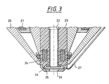
- the pole pieces of the objective are denoted by (20) and the objective coils by (21).
- the beam guidance tube (22) is received over an insulator (23) which surrounds the beam guide tube in an annular manner.
- the deflection system (24) is arranged at the height of the Polschuhspaltes the pole piece (20) the deflection system (24) is arranged.
- the object-side end of the beam guide tube (22) in turn has an annular broadening, so that the object-side end of the beam guide tube (22) acts as a tube electrode.
- the second electrode (26) is received, which forms the Abbrems worn together with the tube electrode (25) of the beam guiding tube (22).
- the beam guide tube (22) is continued so far that the tube electrode (25) - in the direction of the primary electron beam - is arranged behind the Polschuhspalt in an area in which the emerging from the Polschuhspalt magnetic field is almost completely subsided.
- the deceleration of the particles to the target energy thus takes place immediately in front of the object, whereby interference by external electric or magnetic fields are reduced.
- the Abbremselektrode (26) is designed as a centrically flattened conical ring with a central bore (29).
- the center bore (29) is centered on the bore of the pole piece (20) and the beam guide tube (22).
- the conical formation of the electrode (26) avoids collisions between the electrode (26) and the object during object tilting.
- the deceleration of the corpuscles does not necessarily have to be done behind the lens, even if this is preferred, because thereby the corpuscles are kept as long as possible at high kinetic energy.
- the invention has been explained for the case that the corpuscles are electrons.
- the invention When applying the invention to positively charged particles, only the polarity of cathode potential, anode potential and sample potential need to be inverted, i. a positive potential is replaced by a negative potential and a negative potential by a positive potential.
- the objective does not necessarily have to be designed as a magnetic lens. It is also conceivable and, in particular when using heavy corpuscles such as ions, to use an electrostatic single lens for focusing the corpuscles.
- an electrostatic single lens can be constructed in known manner from three successive electrodes, of which the two external electrodes are at the potential of the beam guiding tube; the middle electrode is then at a potential which corresponds to the cathode potential with respect to the polarity, but is somewhat smaller in magnitude.
- the beam-guiding tube may have a cuboid-shaped widened region in which the center electrode is arranged.
- the two edge electrodes are then formed by the rectangular surfaces perpendicular to the tube axis. In contrast to the electrostatic immersion lens, such a single lens does not change the particle energy.
Claims (8)
- Appareil à rayonnement corpusculaire comprenant un générateur de rayon corpusculaire qui présente une cathode (1), une anode (4) et une deuxième source de haute tension (13) associée à l'anode, laquelle génère un potentiel d'anode pour accélérer au potentiel d'anode les corpuscules qui sortent de la cathode (1), un tube de focalisation du rayon (5), un objectif (6, 7) pour focaliser le rayon corpusculaire sur un objet, un dispositif de retard (8, 9), une troisième source de haute tension (12) associée au dispositif de retard et un plateau porte-objet (10), l'anode (4) et le tube de focalisation du rayon (5) étant configurés de telle sorte qu'ils se trouvent au même potentiel de haute tension de la deuxième source de haute tension (13) par rapport au potentiel de masse et la troisième source de haute tension (12) étant reliée avec le dispositif de retard (8, 9) et le plateau porte-objet (10) de telle sorte que le dispositif de retard (8, 9) et l'objet (10) se trouvent au même potentiel, et la troisième source de haute tension, avec une faible énergie de cible, génère un potentiel qui est différent du potentiel de masse et à l'opposé du potentiel d'anode.
- Appareil à rayonnement corpusculaire selon la revendication 1, une première source de haute tension (14) étant présente pour générer un potentiel de cathode (UK) dont la valeur est toujours supérieure à 0,2 kV et le signal du potentiel de cathode étant choisi en fonction du signal des charges des corpuscules de telle sorte que les corpuscules présentent une énergie potentielle supérieure par rapport au potentiel de masse et le signe du potentiel d'anode (UA) étant choisi de telle sorte que les corpuscules au potentiel d'anode présentent une énergie potentielle plus faible par rapport au potentiel de masse.
- Appareil à rayonnement corpusculaire selon la revendication 1 ou 2, le potentiel de la troisième source de haute tension (12) pouvant varier par rapport au potentiel de masse pour modifier l'énergie de cible des corpuscules sur l'objet.
- Appareil à rayonnement corpusculaire selon l'une des revendications 1 à 3, le plateau porte-objet et le dispositif de retard (8, 9), en présence d'une faible énergie de cible des corpuscules, se trouvant par rapport au potentiel de masse à un potentiel ayant le même signe que le potentiel de cathode (UK).
- Appareil à rayonnement corpusculaire selon l'une des revendications 2 à 4, le potentiel de la troisième source de haute tension (12) étant réglable entre la valeur du potentiel de masse et le potentiel de cathode (UK) servant de valeurs limites.
- Appareil à rayonnement corpusculaire selon l'une des revendications 1 à 5, le tube de focalisation du rayon (5) étant élargi dans une zone entre le générateur de rayon et la lentille d'objectif (6, 7) et présentant un détecteur (16) qui se trouve au potentiel du tube de focalisation du rayon (5) pour détecter les électrons secondaires ou rétrodiffusés.
- Appareil à rayonnement corpusculaire selon l'une des revendications 1 à 6, le dispositif de retard se composant d'une électronique unique (9) qui interagit avec l'extrémité côté objet (8) du tube de focalisation du rayon (5).
- Appareil à rayonnement corpusculaire selon la revendication 7, l'extrémité côté objet du tube de focalisation du rayon (5) étant réalisée sous la forme d'une électrode tubulaire (8) et l'électrode unique (9) étant logée sur un corps isolant (23) sur l'objectif (20, 21).
Applications Claiming Priority (2)
| Application Number | Priority Date | Filing Date | Title |
|---|---|---|---|
| DE19732093A DE19732093B4 (de) | 1997-07-25 | 1997-07-25 | Korpuskularstrahlgerät |
| DE19732093 | 1997-07-25 |
Publications (3)
| Publication Number | Publication Date |
|---|---|
| EP0893816A2 EP0893816A2 (fr) | 1999-01-27 |
| EP0893816A3 EP0893816A3 (fr) | 2003-01-29 |
| EP0893816B1 true EP0893816B1 (fr) | 2009-10-21 |
Family
ID=7836916
Family Applications (1)
| Application Number | Title | Priority Date | Filing Date |
|---|---|---|---|
| EP98111715A Expired - Lifetime EP0893816B1 (fr) | 1997-07-25 | 1998-06-25 | Appareil à faisceau corpusculaire |
Country Status (4)
| Country | Link |
|---|---|
| US (1) | US6194729B1 (fr) |
| EP (1) | EP0893816B1 (fr) |
| JP (1) | JP4037533B2 (fr) |
| DE (2) | DE19732093B4 (fr) |
Families Citing this family (28)
| Publication number | Priority date | Publication date | Assignee | Title |
|---|---|---|---|---|
| JP4236742B2 (ja) * | 1998-10-29 | 2009-03-11 | 株式会社日立製作所 | 走査形電子顕微鏡 |
| EP1022766B1 (fr) * | 1998-11-30 | 2004-02-04 | Advantest Corporation | Appareil à faisceau de particules |
| GB2348048A (en) * | 1999-03-19 | 2000-09-20 | Shimadzu Research Lab | Magnetic immersion lenses |
| US6960766B2 (en) * | 2000-02-25 | 2005-11-01 | Hermes-Microvision, Inc. | Swinging objective retarding immersion lens electron optics focusing, deflection and signal collection system and method |
| US6392231B1 (en) * | 2000-02-25 | 2002-05-21 | Hermes-Microvision, Inc. | Swinging objective retarding immersion lens electron optics focusing, deflection and signal collection system and method |
| US6674075B2 (en) * | 2002-05-13 | 2004-01-06 | Applied Materials, Inc. | Charged particle beam apparatus and method for inspecting samples |
| DE10233002B4 (de) * | 2002-07-19 | 2006-05-04 | Leo Elektronenmikroskopie Gmbh | Objektivlinse für ein Elektronenmikroskopiesystem und Elektronenmikroskopiesystem |
| GB2393571B (en) | 2002-09-26 | 2007-03-21 | Leo Electron Microscopy Ltd | Improvements in and relating to the control of instruments |
| KR101041661B1 (ko) * | 2003-07-30 | 2011-06-14 | 어플라이드 머티리얼즈 이스라엘 리미티드 | 다중 검출기들을 갖는 스캐닝 전자 현미경 및 다중 검출기기반 이미징을 위한 방법 |
| US7842933B2 (en) * | 2003-10-22 | 2010-11-30 | Applied Materials Israel, Ltd. | System and method for measuring overlay errors |
| US7112803B2 (en) * | 2004-07-23 | 2006-09-26 | Applied Materials, Israel, Ltd. | Beam directing system and method for use in a charged particle beam column |
| DE102004037781A1 (de) * | 2004-08-03 | 2006-02-23 | Carl Zeiss Nts Gmbh | Elektronenstrahlgerät |
| US20070090288A1 (en) * | 2005-10-20 | 2007-04-26 | Dror Shemesh | Method and system for enhancing resolution of a scanning electron microscope |
| CN102103967B (zh) * | 2005-11-28 | 2013-02-06 | 卡尔蔡司Smt有限责任公司 | 粒子光学组件 |
| US7872236B2 (en) * | 2007-01-30 | 2011-01-18 | Hermes Microvision, Inc. | Charged particle detection devices |
| US20090246171A1 (en) * | 2008-03-27 | 2009-10-01 | Van Antwerp William P | Automatic system for dose control in treating hepatitis c using infusion pumps |
| US7960697B2 (en) | 2008-10-23 | 2011-06-14 | Hermes-Microvision, Inc. | Electron beam apparatus |
| US7919760B2 (en) * | 2008-12-09 | 2011-04-05 | Hermes-Microvision, Inc. | Operation stage for wafer edge inspection and review |
| US8094924B2 (en) * | 2008-12-15 | 2012-01-10 | Hermes-Microvision, Inc. | E-beam defect review system |
| US8319192B2 (en) | 2010-08-24 | 2012-11-27 | Hermes Microvision Inc. | Charged particle apparatus |
| EP2518755B1 (fr) * | 2011-04-26 | 2014-10-15 | FEI Company | Détecteur "in-column" pour colonne d'optique corpusculaire |
| US9046475B2 (en) | 2011-05-19 | 2015-06-02 | Applied Materials Israel, Ltd. | High electron energy based overlay error measurement methods and systems |
| EP2665082A1 (fr) * | 2012-05-16 | 2013-11-20 | ICT Integrated Circuit Testing Gesellschaft für Halbleiterprüftechnik mbH | Élément de déflexion rapide de faisceau magnétique |
| JP5667618B2 (ja) * | 2012-12-14 | 2015-02-12 | 株式会社アドバンテスト | 電磁レンズ及び電子ビーム露光装置 |
| EP3454357B1 (fr) | 2013-09-30 | 2020-08-12 | Carl Zeiss Microscopy GmbH | Système à faisceau de particules chargées et son procédé de fonctionnement |
| US10008360B2 (en) | 2015-01-26 | 2018-06-26 | Hermes Microvision Inc. | Objective lens system for fast scanning large FOV |
| JP6913344B2 (ja) * | 2017-03-27 | 2021-08-04 | 株式会社日立ハイテクサイエンス | 荷電粒子ビーム装置 |
| WO2019100600A1 (fr) * | 2017-11-21 | 2019-05-31 | Focus-Ebeam Technology (Beijing) Co., Ltd. | Microscope électronique à balayage à basse tension et procédé d'observation d'échantillon |
Family Cites Families (10)
| Publication number | Priority date | Publication date | Assignee | Title |
|---|---|---|---|---|
| US3896331A (en) * | 1973-06-28 | 1975-07-22 | Varian Associates | Electron optical system |
| DE2922325A1 (de) * | 1979-06-01 | 1980-12-11 | Philips Patentverwaltung | Rasterelektronenmikroskop |
| US4713543A (en) * | 1984-08-13 | 1987-12-15 | Siemens Aktiengesellschaft | Scanning particle microscope |
| ATE91822T1 (de) * | 1986-04-24 | 1993-08-15 | Integrated Circuit Testing | Elektrostatisch-magnetische-linse fuer korpuskularstrahlgeraete. |
| US5146090A (en) * | 1990-06-11 | 1992-09-08 | Siemens Aktiengesellschaft | Particle beam apparatus having an immersion lens arranged in an intermediate image of the beam |
| JP3148353B2 (ja) * | 1991-05-30 | 2001-03-19 | ケーエルエー・インストルメンツ・コーポレーション | 電子ビーム検査方法とそのシステム |
| EP0548573B1 (fr) * | 1991-11-27 | 1998-02-25 | Hitachi, Ltd. | Appareil à faisceau d'électrons |
| JP2919170B2 (ja) * | 1992-03-19 | 1999-07-12 | 株式会社日立製作所 | 走査電子顕微鏡 |
| JP2927627B2 (ja) * | 1992-10-20 | 1999-07-28 | 株式会社日立製作所 | 走査電子顕微鏡 |
| EP0769799B1 (fr) * | 1995-10-19 | 2010-02-17 | Hitachi, Ltd. | Microscope életronique à balayage |
-
1997
- 1997-07-25 DE DE19732093A patent/DE19732093B4/de not_active Expired - Fee Related
-
1998
- 1998-06-25 EP EP98111715A patent/EP0893816B1/fr not_active Expired - Lifetime
- 1998-06-25 DE DE59814405T patent/DE59814405D1/de not_active Expired - Lifetime
- 1998-07-26 US US09/123,017 patent/US6194729B1/en not_active Expired - Lifetime
- 1998-07-27 JP JP21075198A patent/JP4037533B2/ja not_active Expired - Lifetime
Also Published As
| Publication number | Publication date |
|---|---|
| JPH1196957A (ja) | 1999-04-09 |
| US6194729B1 (en) | 2001-02-27 |
| DE19732093A1 (de) | 1999-01-28 |
| DE19732093B4 (de) | 2008-09-25 |
| DE59814405D1 (de) | 2009-12-03 |
| JP4037533B2 (ja) | 2008-01-23 |
| EP0893816A2 (fr) | 1999-01-27 |
| EP0893816A3 (fr) | 2003-01-29 |
Similar Documents
| Publication | Publication Date | Title |
|---|---|---|
| EP0893816B1 (fr) | Appareil à faisceau corpusculaire | |
| DE102018007652B4 (de) | Teilchenstrahl-System sowie Verfahren zur Stromregulierung von Einzel-Teilchenstrahlen | |
| EP0461442B1 (fr) | Appareil à faisceau de particules | |
| DE112014002951B4 (de) | Rasterelektronenmikroskop | |
| EP0333018B1 (fr) | Lentille d'objectif pour la focalisation de particules chargées | |
| DE69822139T2 (de) | Korrekturvorrichtung zur linsenfehlerkorrektur in ladungsträger-optischen geräten | |
| EP0218829B1 (fr) | Dispositif détecteur d'électrons secondaires et/ou d'électrons de dispersion en retour dans un appareil à faisceau électronique | |
| EP0205184B1 (fr) | Objectif de faible aberration comportant un spectromètre de haute acceptance en électrons secondaires | |
| EP0180723B1 (fr) | Dispositif corpusculaire à rayonnement | |
| DE69821467T2 (de) | Rasterelektronenmikroskop unter kontrollierter umgebung mit einem magnetfeld zur erhöhten sekundärelektronenerfassung | |
| EP0267555A2 (fr) | Objectif de spectromètre pour appareils de mesure par faisceau corpusculaire et procédé pour l'examen d'échantillons. | |
| DE69920182T2 (de) | Korpuskularstrahloptisches gerät mit auger-elektronendetektion | |
| DE19549022C2 (de) | Rasterelektronenmikroskop und Probenbetrachtungsverfahren mittels eines solchen | |
| DE4216730A1 (de) | Rasterelektronenstrahlgerät | |
| DE60105199T2 (de) | Sem mit einem sekundärelektronendetektor mit einer zentralelektrode | |
| EP0194570A2 (fr) | Microscope corpusculaire à balayage à effet Boersch réduit | |
| EP0205185B1 (fr) | Objectif comportant un spectromètre en technique de mesure par faisceau d'électrons | |
| EP0236807A2 (fr) | Objectif de spectromètre en technique de mesure par faisceau corpusculaire | |
| DE69815498T2 (de) | Rasterelektronenmikroskop unter kontrollierter umgebung mit mehrpolfelder zur erhöter sekundärelektronenerfassung | |
| EP0348785A2 (fr) | Appareil de mesure par faisceau d'électrons | |
| DE102018207645B9 (de) | Verfahren zum Betrieb eines Teilchenstrahlerzeugers für ein Teilchenstrahlgerät, Computerprogrammprodukt und Teilchenstrahlgerät mit einem Teilchenstrahlerzeuger | |
| EP0910108B1 (fr) | Lentille pour faisceau d'électrons | |
| DE2608958A1 (de) | Vorrichtung zum erzeugen von strahlen aus geladenen teilchen | |
| EP1774560A1 (fr) | Appareil a faisceau electronique | |
| EP0086431A2 (fr) | Système générateur de faisceau de particules et méthode d'utilisation |
Legal Events
| Date | Code | Title | Description |
|---|---|---|---|
| PUAI | Public reference made under article 153(3) epc to a published international application that has entered the european phase |
Free format text: ORIGINAL CODE: 0009012 |
|
| AK | Designated contracting states |
Kind code of ref document: A2 Designated state(s): AT BE CH CY DE DK ES FI FR GB GR IE IT LI LU MC NL PT SE |
|
| AX | Request for extension of the european patent |
Free format text: AL;LT;LV;MK;RO;SI |
|
| PUAL | Search report despatched |
Free format text: ORIGINAL CODE: 0009013 |
|
| AK | Designated contracting states |
Designated state(s): AT BE CH CY DE DK ES FI FR GB GR IE IT LI LU MC NL PT SE |
|
| AX | Request for extension of the european patent |
Extension state: AL LT LV MK RO SI |
|
| 17P | Request for examination filed |
Effective date: 20030625 |
|
| AKX | Designation fees paid |
Designated state(s): DE GB NL |
|
| 17Q | First examination report despatched |
Effective date: 20070905 |
|
| RAP1 | Party data changed (applicant data changed or rights of an application transferred) |
Owner name: CARL ZEISS NTS GMBH |
|
| GRAP | Despatch of communication of intention to grant a patent |
Free format text: ORIGINAL CODE: EPIDOSNIGR1 |
|
| GRAS | Grant fee paid |
Free format text: ORIGINAL CODE: EPIDOSNIGR3 |
|
| GRAA | (expected) grant |
Free format text: ORIGINAL CODE: 0009210 |
|
| AK | Designated contracting states |
Kind code of ref document: B1 Designated state(s): DE GB NL |
|
| REG | Reference to a national code |
Ref country code: GB Ref legal event code: FG4D Free format text: NOT ENGLISH |
|
| REF | Corresponds to: |
Ref document number: 59814405 Country of ref document: DE Date of ref document: 20091203 Kind code of ref document: P |
|
| NLV1 | Nl: lapsed or annulled due to failure to fulfill the requirements of art. 29p and 29m of the patents act | ||
| PLBE | No opposition filed within time limit |
Free format text: ORIGINAL CODE: 0009261 |
|
| STAA | Information on the status of an ep patent application or granted ep patent |
Free format text: STATUS: NO OPPOSITION FILED WITHIN TIME LIMIT |
|
| 26N | No opposition filed |
Effective date: 20100722 |
|
| GBPC | Gb: european patent ceased through non-payment of renewal fee |
Effective date: 20100625 |
|
| PG25 | Lapsed in a contracting state [announced via postgrant information from national office to epo] |
Ref country code: GB Free format text: LAPSE BECAUSE OF NON-PAYMENT OF DUE FEES Effective date: 20100625 |
|
| PG25 | Lapsed in a contracting state [announced via postgrant information from national office to epo] |
Ref country code: NL Free format text: LAPSE BECAUSE OF FAILURE TO SUBMIT A TRANSLATION OF THE DESCRIPTION OR TO PAY THE FEE WITHIN THE PRESCRIBED TIME-LIMIT Effective date: 20091021 |
|
| REG | Reference to a national code |
Ref country code: DE Ref legal event code: R081 Ref document number: 59814405 Country of ref document: DE Owner name: CARL ZEISS MICROSCOPY GMBH, DE Free format text: FORMER OWNER: CARL ZEISS NTS GMBH, 73447 OBERKOCHEN, DE Effective date: 20130319 |
|
| PGFP | Annual fee paid to national office [announced via postgrant information from national office to epo] |
Ref country code: DE Payment date: 20140619 Year of fee payment: 17 |
|
| REG | Reference to a national code |
Ref country code: DE Ref legal event code: R119 Ref document number: 59814405 Country of ref document: DE |
|
| PG25 | Lapsed in a contracting state [announced via postgrant information from national office to epo] |
Ref country code: DE Free format text: LAPSE BECAUSE OF NON-PAYMENT OF DUE FEES Effective date: 20160101 |