EP0803065B1 - A-protein als diagnostikum für krebs - Google Patents
A-protein als diagnostikum für krebs Download PDFInfo
- Publication number
- EP0803065B1 EP0803065B1 EP96902593A EP96902593A EP0803065B1 EP 0803065 B1 EP0803065 B1 EP 0803065B1 EP 96902593 A EP96902593 A EP 96902593A EP 96902593 A EP96902593 A EP 96902593A EP 0803065 B1 EP0803065 B1 EP 0803065B1
- Authority
- EP
- European Patent Office
- Prior art keywords
- protein
- cancer
- antibody
- amino acids
- amino acid
- Prior art date
- Legal status (The legal status is an assumption and is not a legal conclusion. Google has not performed a legal analysis and makes no representation as to the accuracy of the status listed.)
- Expired - Lifetime
Links
Images

Classifications
-
- G—PHYSICS
- G01—MEASURING; TESTING
- G01N—INVESTIGATING OR ANALYSING MATERIALS BY DETERMINING THEIR CHEMICAL OR PHYSICAL PROPERTIES
- G01N33/00—Investigating or analysing materials by specific methods not covered by groups G01N1/00 - G01N31/00
- G01N33/48—Biological material, e.g. blood, urine; Haemocytometers
- G01N33/50—Chemical analysis of biological material, e.g. blood, urine; Testing involving biospecific ligand binding methods; Immunological testing
- G01N33/53—Immunoassay; Biospecific binding assay; Materials therefor
- G01N33/575—Immunoassay; Biospecific binding assay; Materials therefor for cancer
- G01N33/5758—Immunoassay; Biospecific binding assay; Materials therefor for cancer involving compounds serving as markers for tumours, cancers or neoplasias, e.g. cellular determinants, receptors, heat shock/stress proteins, A-protein, oligosaccharides or metabolites
-
- C—CHEMISTRY; METALLURGY
- C07—ORGANIC CHEMISTRY
- C07K—PEPTIDES
- C07K16/00—Immunoglobulins [IG], e.g. monoclonal or polyclonal antibodies
- C07K16/18—Immunoglobulins [IG], e.g. monoclonal or polyclonal antibodies against material from animals or humans
- C07K16/28—Immunoglobulins [IG], e.g. monoclonal or polyclonal antibodies against material from animals or humans against receptors, cell surface antigens or cell surface determinants
- C07K16/30—Immunoglobulins [IG], e.g. monoclonal or polyclonal antibodies against material from animals or humans against receptors, cell surface antigens or cell surface determinants from tumour cells
-
- C—CHEMISTRY; METALLURGY
- C07—ORGANIC CHEMISTRY
- C07K—PEPTIDES
- C07K2317/00—Immunoglobulins specific features
- C07K2317/30—Immunoglobulins specific features characterized by aspects of specificity or valency
- C07K2317/34—Identification of a linear epitope shorter than 20 amino acid residues or of a conformational epitope defined by amino acid residues
Definitions
- CCA Carcinoembryonic Antigen
- AFP Alpha Fetoprotein
- PSA Prostate Specific Antigen
- the assays using these markers have not, to date, been markedly predictive of the presence of cancer in these individuals, as verified by other clinical diagnoses.
- the sensitivity and specificity of these assays has been disappointingly low.
- Time-consuming and labor-intensive clinical assessments e.g. palpations, x-rays, mammograms, biopsies
- This invention pertains to methods of detecting cancer in an individual from the results of an assay of a biological sample, such as a blood sample, from the individual.
- a biological sample such as a blood sample
- the sample is incubated with at least one antibody that is immunoreactive with a cancer-diagnostic protein that may be present in the biological sample.
- the immunoconjugates that are formed in the cancer-diagnostic protein:antibody reaction are detected. The presence of an abnormally high concentration of these immunoconjugates indicates that the individual from whom the sample was taken has primary or metastatic cancer.
- the cancer-diagnostic protein is A-protein and the sample is incubated in a sandwich assay for A-protein.
- An antibody that is immunoreactive with A-protein is attached to a solid support.
- the sample is allowed to immunoreact with the attached antibody and with a second antibody that is immunoreactive with another region of A-protein (i.e., a region other than the region immunoreactive with the solid support-attached antibody).
- the resultant two antibodies-A-protein complex thereby forms a sandwich.
- the amount of bound second antibody is detected. This amount of detected second antibody is directly proportional to the amount of attached A-protein.
- An abnormally large amount of detected second antibody is indicative of the presence of primary or metastatic cancer that is being detected by the assay of the biological sample.
- Another embodiment of this invention is a test kit that contains one or more antibodies to be used in the assay for the cancer-diagnostic protein (e.g. A-protein).
- One of the antibodies is immunoreactive with one epitopic region of the cancer-diagnostic protein and, if a second antibody is included, the second antibody is immunoreactive with an epitopic region of the cancer-diagnostic protein separate from the epitopic region that is immunoreactive with the first antibody.
- One of the antibodies is attached to a solid support, such as the walls and bottoms of wells of a microtiter plate. The other antibody has a detection label bound to it.
- Yet another embodiment of this invention is one or more antibodies that immunoreact with a cancer-diagnostic protein, i.e. A-protein. These antibodies are used to detect or assess the presence of the cancer-diagnostic protein. The assessment can be performed using biological samples such as blood.
- the present invention pertains to methods and compositions for diagnosing the presence of primary or metastatic cancer in individuals.
- Primary cancer is cancerous growth that is confined to a particular anatomical region of the body or is composed of cells that formed the initial cancerous lesion.
- Metastatic cancer is cancerous growth that has spread from an original primary site in the body through either the blood or the lymphatic system, or both, and is growing at a site removed from the primary tumor; it is also recurrent disease spread secondary to the treatment of a primary tumor.
- the primary or metastatic cancer that can be diagnosed by employing the methods and compositions of this invention are any one of a large variety of cancers. Included in this list of detectable cancers, by the methods and compositions of this invention, are breast cancer, prostate cancer, liver cancer, lung cancer, colorectal cancer, gastric tissue cancer, pancreatic cancer, bladder cancer, head and neck cancer, endometrial cancer, parotid cancer, cholangio cancer, kidney or renal cancer, cervical cancer, thyroid cancer, brain cancer, mouth cancer, uterine cancer, abdominal cancer, tongue cancer, lip cancer, anal cancer, pelvic cancer, inguinal cancer, penile cancer, chest wall cancer, fallopian tube cancer, POEMS, lymphoma, leukemia, multiple myeloma, melanoma and various sarcomas.
- cancers and most likely other types of cancer, can be detected in various stages of their progression including stage 1 and stage 2.
- stage 1 and stage 2 The detection of the presence of these cancers can be distinguished from the presence of benign tumors or the absence of cancer in individuals by using the methods and compositions of this invention.
- the methods of this invention involve the use of antibodies that are immunoreactive with a specified sequence of 10-18 amino acids of proteins selected from the group consisting of 14 amino acid or 16 amino acid carboxy terminus of A protein; the peptide containing amino acids 60-71 of A protein; the peptide containing amino acids 142-158 of A protein; and the peptide containing amino-acids 158-170 of A protein.
- a specified sequence of 10-18 amino acids of proteins selected from the group consisting of 14 amino acid or 16 amino acid carboxy terminus of A protein; the peptide containing amino acids 60-71 of A protein; the peptide containing amino acids 142-158 of A protein; and the peptide containing amino-acids 158-170 of A protein.
- Blood is an often used and preferred biological sample.
- the preferred blood constituent that is analyzed is either plasma or serum, more preferably serum.
- the immunoreactive cancer-diagnostic protein e.g. A-protein
- the methods of this invention do not rely on an analysis of localized, e.g. biopsy, material but, rather, can be employed with a readily obtainable biological sample such as blood serum or plasma. Even when such blood sampling is employed, the variety of cancers that can be detected is quite extensive.
- the presence of a concentration of A-protein in a biological sample from a particular individual that is more than twice the concentration of A-protein found in similar samples from individuals without cancer is indicative of the presence of cancer in that particular individual.
- biological samples of the same size are taken from individuals who are free of cancer and the average (mean) amount of A-protein in these samples is determined, an individual is predicted to have cancer if a biological sample of the same size from that individual has more than twice the amount of A-protein in it than was determined as the average amount of A-protein for the cancer-free individuals.
- the factor of 2.0 is considered to be a threshold value.
- the threshold value of 2.0 is a particularly preferred value. Other threshold values can be established for predicting the presence of cancer in an individual. These threshold values will depend on the accuracy and reproducibility of the particular cancer diagnostic assay as well as the predictive reliability that is sought for the assay.
- the actual amount or concentration of A-protein in a biological sample from an individual can have diagnostic or prognostic value.
- determining the quantity of A-protein over a period of time for an individual it is possible to monitor the progressiveness of the cancer (as the quantity of A-protein increases) or the regression of the cancer, e.g. as a result of therapeutic treatment (as the quantity of A-protein decreases).
- the quantity of A-protein that is detected can be meaningful as an indicator of the presence and vigor of cancer growth.
- the A-protein quantity is monitored in relation to a baseline value for that individual or in comparison to the average amount of A-protein in similar biological samples from cancer-free individuals.
- the individual When the A-protein quantity is greater than a particular value, established by the monitor of the assay, the individual is considered to have cancer.
- the actual A-protein quantities in excess of this established value indicate the severity of the cancer and lesser A-protein quantities, with or without therapeutic treatments, indicate a diminution of cancer severity.
- the presence of more than about 10 ng of A-protein per ml of blood is predictive of metastatic cancer.
- assays for metastatic cancer are also based on measurements of the amount of A-protein in biological samples (e.g., fluid samples such as blood plasma or serum, urine, etc.) from an individual. Again, the amount of A-protein in the biological sample is indicative of the presence or absence of metastatic cancer in the individual from whom the sample was obtained. An abnormally large amount of A-protein indicates the presence of metastatic cancer; a normal amount of A-protein indicates that the individual does not have metastatic cancer.
- a variety of techniques are known and available to the artisan for assaying for the amount of A-protein in a biological sample. These techniques include isolating and quantifying the A-protein in the sample by solvation and centrifugation procedures, by column chromatography or gel electrophoretic separation procedures, by filtration procedures, by enzymatic recognition procedures, or by binding procedures with molecules that recognize and bind A-protein with specificity such as affinity chromatography or immunoassay. Such specific binding processes can also be used to detect and quantify the A-protein in situ in biological samples by assessing the volume or area distribution of the specific binding molecule in the sample.
- a technique of the invention for assaying for the amount of A-protein in a biological sample is immunological recognition with antibodies that specifically bind particular epitopes of A-protein.
- Such antibodies are compositions of the present invention. These antibodies selectively recognize A-protein determinants and bind to these determinants with high affinity.
- the antibodies can be used singly as affinity immobilization or as tagging binding moieties in procedures such as Western blot or electrophoretic pattern analyses, or multiply to bind to different A-protein determinants or epitopes such as in sandwich assays. These antibodies can have substances that act as labels attached to them for ease of identification following binding of the antibody to A-protein.
- the antibodies of this invention bind to A-protein with specificity so specific epitopes of A-protein can be detected with particularity.
- Antibodies which can be used within this invention are reactive with particular A-protein epitopes.
- the term antibody is also intended to encompass both polyclonal and monoclonal antibodies.
- the term antibody is intended to encompass mixtures of more than one antibody reactive with A-protein (e.g., a cocktail of different types of monoclonal antibodies reactive with A-protein).
- the term antibody is further intended to encompass whole antibodies, biologically functional fragments thereof, single chains or single chain fragments with A-protein binding properties, and chimeric antibodies comprising portions from more than one species, bifunctional antibodies, etc.
- Biologically functional antibody fragments which can be used are those fragments sufficient for binding of the antibody fragment to A-protein.
- the chimeric antibodies can comprise portions derived from two different species (e.g., human constant region and murine variable or binding region).
- the portions derived from two different species can be joined together chemically by conventional techniques or can be prepared as fusion proteins using genetic engineering techniques.
- DNA encoding the proteins of both the light chain and heavy chain portions of the chimeric antibody can be expressed together as fusion proteins.
- A-proteins are intimately involved in the regulation of the inositol-related signal transducing system.
- WO 93/20439 disclosed that A-proteins and antibodies thereto which can be used in ELISA assays, where the A-proteins are involved in the regulation of inositol metabolism.
- A-proteins function by activating phospholipase C (PL-C) to generate the second messengers inositol-1,4,5-trisphosphate (IP 3 ) and diacylglycerol (DG).
- PL-C phospholipase C
- IP 3 inositol-1,4,5-trisphosphate
- DG diacylglycerol
- Upon stimulation of a membrane bound receptor an A-protein binds with GTP to form an intermediate which functions to activate PL-C.
- GTP of the intermediate is hydrolyzed to GDP, PL-C activation terminates.
- A-proteins are accordingly important G-type signal transducing molecules critical
- signal transducing molecules are useful in producing the antibodies that are used in the foregoing methods of the present invention.
- the signal transducing molecules that are used to produce the antibodies are isolated and purified A-proteins and epitopic fragments of A-protein.
- the cancer-diagnostic proteins of the invention can also be useful in immunotherapy.
- these proteins can stimulate the immune system to respond, i.e. to recognize the presence of cancer in the host and to respond to it.
- the immune system can be stimulated to generate immunosuppressive activity to eradicate the cancer.
- the antibodies of this invention can have therapeutic value.
- they can immunoreact with the cancer-diagnostic protein, e.g. A-protein, and thereby initiate further immune system response, e.g. by stimulating the production of T-cells, to eliminate the cancer cells present in the individual.
- the cancer-diagnostic protein e.g. A-protein
- A-protein was originally used to describe a rod photoreceptor protein of approximately 20 kilodaltons molecular weight based on the electrophoretic mobility of this protein through a gel under reducing conditions. From, an analysis of the protein as an expressed product of the known A-protein gene, the molecular weight of the A-protein is 26 kilodaltons. Improved extraction and separation methods combined with preliminary sequence data on the separated forms indicates that the entity referred to previously as A-protein may consist of at least two structurally and functionally related proteins; one membrane-bound and one soluble.
- A-proteins include the amino acid sequences for the N-terminal regions set forth in the Sequence Listing as: and
- A-protein related molecules comprise a single polypeptide chain with a significantly hydrophobic region. These molecules also have the ability to bind and hydrolyze the nucleotides adenosine and guanosine triphosphate, and have the ability to activate phospholipase C, phospholipase D, and possibly also phospholipase A 2 , in the presence of GTP.
- A-proteins from rod outer segments are involved in the ADP-ribosylation of GTP-binding (schmidt et al, J. Biol. Chem. 262: 1433-14336 (1987)
- A-proteins can be isolated from mammalian (bovine) and amphibian (frog) rod outer segments (ROS) of photoreceptor cells of the eye by extraction, centrifugation, chromatography and other protein purification techniques known to those skilled in the art.
- Other proteins with similar or identical physical and functional characteristics as the A-proteins have been isolated from various other tissues from vertebrates and invertebrates.
- A-proteins, various truncated or mutein analogs thereof, and fused proteins comprising an A-protein and other protein domains can be produced by various synthetic and biosynthetic means.
- an appropriate host cell such as a microorganism, yeast, or eucaryotic cell culture can be genetically engineered to express an A-protein, or a portion or analog thereof. This may be accomplished by now well-established recombinant DNA technologies known to those skilled in the art.
- the recombinant procedure may include the isolation or synthesis of the gene encoding an A-protein, a portion, or analog thereof, and the integration of that gene into a plasmid.
- the amino acid sequences of the A-proteins may be established readily.
- Sequence Listing sets forth the N-terminus amino acid sequence of two forms of A-protein (A m and A s ) as SEQ ID NO:1 and SEQ ID NO:2, respectively.
- Gene synthesis from synthetic oligonucleotides and known mutagenesis techniques provide the technologies to prepare an array of analogs, truncated A-protein forms, and fused proteins comprising A-protein or an antibody binding domain thereof.
- Production of such materials further may include the transformation of an appropriate host cell with a vector harboring the recombinant DNA that includes the gene encoding A-protein or a portion or analog thereof, culturing that transformed host cell, and isolation of the expressed protein. Given the availability of A-protein-rich samples producible as disclosed herein, the recombinant production of the native form and various portions and analogs thereof is well within the current skill in the art.
- At least portions of the protein can be produced synthetically by chemically joining amino acids in the correct sequence.
- the isolated A-proteins, or portion or analog thereof, can be used as antigens to produce antibodies that are useful to detect particular A-protein epitopes in fluid samples from individuals, thereby assaying for the presence of primary or metastatic cancer.
- the antibodies can be part of a polyclonal antisera, or the binding portions thereof of these antibodies, raised against A-protein, and shown to react with A-protein or with its analogs, fragments or to a particular epitope on A m or A s .
- the antibodies can be polyclonal or monoclonal antibodies produced by methods known per se.
- the antibodies preferably are selected so as not to cross-react with other cellular components.
- the antibodies can be of any class and subclass as determined by the Ouchterlony double diffusion test. Antibodies of the IgG class are preferred.
- antibodies which recognize A-protein can be synthesized by biosynthetic or recombinant means, either in whole or in part.
- the antibodies can be linked to other functional molecules such as toxins, fluorescent or absorption dyes, enzymes, or radioactive markers.
- the antibodies are linked to biotin molecules which have a particularly strong avidity for avidin or streptavidin which, in turn, can be linked to fluorescent, absorption or radioactive markers.
- the linked antibodies can be used to detect A-protein from biological samples and thereby assay for primary or metastatic cancer.
- the antibody-marker complex can be prepared by chemical linkage or by recombinant DNA techniques if the marker is proteinaceous.
- the antibodies can also be labelled with a reagent which enables the monitoring or imaging of the antibody immediately after its administration to a patient.
- the label can be, for example, a radioisotope such as 125 I or 99m Tc, both of which can be imaged extracorporeally by radiation detection means such as a gamma scintillation camera.
- the antibody can be labelled with a non-radioactive, paramagnetic contrast agent capable of detection in MRI systems. In such systems, a strong magnetic field is used to align the nuclear spin vectors of the atoms in a patient's body. The field is then disturbed and an image of the patient is read as the nuclei return to their equilibrium alignments.
- antibodies can be linked to paramagnetic materials such as gadolinium, cobalt, nickel, manganese or iron complexes, to form conjugate diagnostic contrast reagents that are imaged extracorporeally with an MRI system.
- conjugates of antibody-A-protein can still be detected by using standard biochemical techniques to recover immunoprecipitates, such as centrifugation.
- Anti-A-protein monoclonal antibodies can be obtained from hybridoma cell lines formed upon the fusion of mouse myeloma cells with spleen cells of mice previously immunized with A-protein that has been purified, for example, from bovine ROS.
- the immunogen alternatively can be a derivative of A-protein, or an analog or portion thereof, produced in vitro according to known mechanical or manual procedures of peptide synthesis.
- the immunogen (A-protein) can be synthesized by biosynthetic means using recombinant DNA technologies known to those skilled in the art.
- the mice whose spleen cells are chosen for fusion are preferably from a genetically defined lineage such as Balb/C.
- the myeloma cells used in the fusion are from a mammalian, antibody-producing cell line, but most preferably are from a mouse cell line such as, e.g., NS-1.
- the monoclonal antibodies can be obtained from ascites fluid of mice injected with the fusion product.
- an immunogen for the production of polyclonal or monoclonal antibodies is either the 14 amino acid or 16 amino acid carboxyl terminus of A-protein, the peptide containing amino acids 60-71 of A-protein, the peptide containing amino acids 142 to 158 of A-protein or the peptide containing amino acids 158-170 of A-protein.
- a second immunogen from A-protein is chosen other than the approximately 16 amino acid peptide of the first immunogen.
- Another technique for obtaining antibodies that immunoreact with the second immunogen of A-protein is to immunize an animal with intact A-protein and select antibodies that immunoreact with an epitope of A-protein other than that of the first immunogen.
- the production of two sets of antibodies with the properties of immunoreacting with different epitopes ensures that sandwich assays using these antibodies will not be impeded by competition of the antibodies for the same antigenic site. This feature increases the sensitivity of the assay for detecting the actual amount of A-protein present in the sample.
- polyclonal or monoclonal antibodies so produced by known procedures are specific for A-protein, and therefore are particularly useful for assaying for A-proteins.
- either intact A-proteins or detectable fragments of A-proteins can be assayed by the disclosed methods.
- the essential feature of the intact protein or detectable fragment thereof is the ability of these proteins or peptides to be detectable, i.e. to immunoreact with immobilization or detection antibodies. Formation of the protein or fragment complexes with the antibodies and detection of these complexes are all that is required. Such detectable complexes can be formed with A-protein fragments.
- This invention includes a particular method for detecting the amount of A-protein in a biological sample such as blood serum or plasma.
- a biological sample such as blood serum or plasma.
- a first antibody which binds to a first defined epitope on A-protein is adhered to a solid support.
- the adhered first antibody is contacted with the biological sample to be tested.
- a labeled second antibody which binds a second defined epitope on A-protein is added to the first antibody-biological sample mixture, thereby forming a first antibody:A-protein:second antibody immunocomplex attached to the solid support.
- This second antibody is attached to a detectable marker.
- Any unbound second antibody is removed, and the presence and, if desired, the amount of the marker is then detected, its presence and amount being indicative of the presence and the amount of A-protein in the sample.
- An abnormally large amount of detected A-protein indicates that the individual from whom the biological sample was obtained has a high probability of having primary or metastatic cancer. In other words, this assay is diagnostic for primary or metastatic cancer.
- Test kits are also embodiments of this invention. These test kits contain one or more of the polyclonal or monoclonal antibodies that are needed to perform assays for A-protein, that may be present in the biological samples, such as blood, obtained from individuals. These test kits can also contain the solid supports, such as microtiter trays, for performing the assays. Instructions for performing the assays for A-protein can also be included in the kits. If desired, an identification label can be attached to an antibody of the test kits. In preferred embodiments of the test kits, antibodies are provided that allow sandwich assays to be performed. In particularly preferred embodiments of the invention, one of the sandwich antibodies is unlabeled and adhered to a solid support. The other antibody has a label bound to it for detection purposes.
- A-proteins were isolated from the retinas of cow eyes essentially as described by Schmidt et al. ( J. Biol. Chem, 262:14333-14336 (1987)). Bovine (cow or calf) eyes were obtained from a local abattoir within 2 hours of killing. Bovine eyes were kept on ice in the dark for 30 to 60 minutes. Retinas were dissected out and placed in buffer A (130 mM NaCl, 20 mM Tris-HCl, pH 7.0; 1 ml per calf retina or 2 ml per cow retina). Gentle, repeated inversions of the container liberated large numbers of ROS into the buffer. The mixture was poured through a Buchler funnel to remove the retinas.
- the filtrate was allowed to settle in a conical-bottomed tube on ice for 5 minutes, allowing gross particulate matter to settle out of the ROS suspension.
- the supernatant was found, by means of microscopic examination on a hemocytometer, to consist of greater than 95% ROS.
- the ROS were disrupted with shear which was created by repeatedly drawing the suspension into the syringe and forcing it out against the wall of the container.
- the suspension was centrifuged at 10,000 to 12,000 x g for 20 minutes at 4°C.
- the pellet containing ROS membranes was washed once with a volume of buffer A equal to that of the removed supernatant.
- the resulting pellet was resuspended in 3 to 6 ml of buffer T ((0.05%) Tween 20/80 (1:1) in double-distilled water) and spun at 15,000 x g for 45 minutes.
- buffer T ((0.05%) Tween 20/80 (1:1) in double-distilled water)
- Both A-protein solutions were filtered through Centricon 30 microconcentrators (molecular weight cut-off of 30 kD, Amicon Corporation), with centrifugation at 5,000 x g in a refrigerated centrifuge. The ultrafiltrates were then concentrated and dialyzed in Centricon 10's (molecular weight cut-off of 10 kD) at 5,000 x g.
- the retentates contained proteins of 10 kD to 30 kD, with average concentrations of 100 to 200 ⁇ g/ml for soluble A-protein (A S ) and 30 to 100 ⁇ g/ml for membrane-bound (A m ), reduced to a volume of 0.5 to 1 ml.
- a S soluble A-protein
- a m membrane-bound
- the purification of A S resulted in a 320-fold enrichment and A m was purified 20-fold. If the soluble A-protein solution was to be kept overnight before concentration and use, buffer A with 0.05% Tween 20/80TM (1:1) was added 1:5 to minimize aggregation of proteins in the concentrated solution.
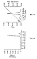
- A-proteins purified by ultrafiltration as described above were further purified for sequence analysis by HPLC on a Bio-Sil SEC-125 column in buffer A (A s ) or buffer T (A m ). Elution was isocratic. Pooled peaks were concentrated, dialyzed against water and lyophylized prior to analysis.
- a s purified by ultrafiltration as described, was run on a HPLC size exclusion column (TSK-2000, Bio-Rad) in buffer A (Fig. 1A), and then rechromatographed in 0.1 M potassium phosphate buffer on a DEAE anion-exchange column. Protein was eluted with a 0 to 200 mM NaCl gradient (Fig. 1B).
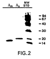
- a s as described above results in preparations of greater than 95% purity. Any protein contaminant of the purified A s preparations has been shown not to interact with guanosine or adenosine nucleotides under any of the conditions tested. Since the extractions are sequential, A m is purified to essential homogeneity by the procedure described with no detectable contaminants, as shown in the SDS gel described below in Example 2 and pictured in Fig. 2, lane 1.
- a m is metastable in the purified state and retains most of its functional properties for several days at 4°C. A m can also withstand freezing and thawing in detergent without losing more than 15 to 20% of its original activity.
- a S is labile under a variety of conditions and no satisfactory methods of treatment have been found to prevent greater than 80% activity loss over a 48 hour period of 4°C.
- Polyacrylamide gel electrophoresis was performed according to a modification of the methods of O'Farrell ( J . Biol. Chem ., 250 :4007 (1975)) in the presence of 0.1% SDS in a pore gradient gel (10 to 20%). Samples were applied in a sample buffer of 0.33 mM DTT, 7% SDS, 17% glycerol and 0.5 M Tris-HCl, pH 6.8, and run to equilibrium. Samples were not heat-denatured, in order to avoid the appearance of additional bands caused by the formation of stable polymers. Proteins were visualized with the Bio-Rad silver stain kit. Gels were calibrated using pre-stained molecular weight standards from Bio-Rad (range 17 to 94 kD).
- A-protein includes A m , a membrane bound form having a molecular weight of about 20 kD and A s , a soluble form having a molecular weight of about 19 kD when the gels are run under reducing conditions.
- the soluble protein resolves into two closely spaced bands on gels.
- the membrane-bound form migrates as a single band.
- mice (6-8 weeks old; The Jackson Laboratory, Bar Harbor, ME) were immunized with four injections of A-protein. The injections were performed one week apart and 50 ⁇ g A-protein (either A s , A m , or both) was injected on each occasion. The first three injections were given intraperitoneally, and the fourth intravenously. A-protein was injected with complete Freunds adjuvant on the first occasion, incomplete adjuvant on the second and third occasions, and without adjuvant on the last occasion. Serum withdrawn prior to the last injection showed prominent binding to purified A-protein using a solid phase microtiter plate enzyme-linked immunoassay. The mouse with the best immune response was sacrificed three days after the last injection.
- Lymphocytes from this animal were maintained as a polyclonal hybridoma by subcloning them as antibody producer cells in a Cellco bioreactor. These producer cells were maintained in liquid nitrogen cryostorage. The murine polyclonal antibodies from these producer cells displayed specificity for A-protein.
- Monoclonal hybridomas were produced by fusion of spleen cells from the sacrificed mouse with NS-1 (P3NS-1/1-Ag4-1) myeloma cells (American Type Culture Collection, Rockville, MD; Accession No. TIB18).
- NS-1 P3NS-1/1-Ag4-1) myeloma cells
- the method of Nadakavukaren was employed to perform the fusions.
- Resultant clones were tested for binding to A-protein. Subcloning by serial dilution was carried out on one clone.
- the most productive subclones were injected into the peritoneal cavity of Balb/c mice to produce ascites fluid containing monoclonal antibody. The ascites fluid which was obtained was centrifuged, tested for activity, and then stored at -70°C until required.
- the resulting 18 anti-A-protein antibodies were screened for antibody isotype by the Ouchterlony double diffusion test in agar plates against anti-IgM, anti-IgG, anti-IgG1, anti-IgG2a, anti-IgG2b, and anti-IgG3 antibodies (Cappell). The results are shown in TABLE 1.
- the rate of hydrolysis of GTP or ATP by both soluble and membrane-bound A-protein was assayed in a total volume of 200 ⁇ l. 1.0 to 10.0 ⁇ g A s or A m , 5 x 10 -5 M GTP or ATP, 67 to 335 nM [ ⁇ - 32 P] GTP (2.8 Ci/mmol) or 88.8 to 177.5 nM [ ⁇ - 32 P] ATP (2.8 Ci/mmol) and 20 ⁇ l of stripped ROS membranes (in the case of A m ) were mixed in buffer J (20 mM Tris-HCl, pH 7.0, 0.1 mM EDTA).
- the stripped ROS membranes (containing rhodopsin as the receptor) were prepared by washing ROS membranes 3 times in buffer C(100 mM NaCl, 20 mM Tris-HCl, pH 7.0, 1 mM MgCl 2 ) and 3 times in water containing 0.01% polyoxyethylene 23 lauryl ether (Brij 35TM nonionic detergent, Sigma Chemical Co.).
- the stripped membranes were resuspended in buffer D (10 mM Tris-HCl, pH 7.0, 0.1 mM EGTA) prior to use.
- the GTPase activity of A m was enhanced in the presence of the activated receptor. In contrast, no effect on the hydrolysis rate was observed when unphotolyzed rhodopsin was added to the incubation in the dark. In the absence of rhodopsin or the presence of unbleached rhodopsin, A m had negligible GTPase activity.
- the rate of GTP hydrolysis by A m (0.458 GTP sec -1 /A m ) is comparable to that of transducin (0.512 GTP sec -1 /T ⁇ ) when both are measured at submaximal velocity in the presence of photoactivated rhodopsin.
- the GTPase rates for A m and transducin are additive when the two purified proteins are present in approximately equimolar concentrations.
- a m and A s were found to have ATPase activity that was not receptor coupled.
- the K m values for both A m and A s ATPases are given in TABLE 2, above. Comparison of the rate constants of A m indicates that its affinity for ATP is approximately an order of magnitude greater than that of A S .
- the relative K m values for GTPase and ATPase activity of both A m and A S indicate that GTP is the preferred substrate for binding and hydrolysis.
- the samples were vortexed and incubated at 25°C for 30 minutes, quenched with 200 ⁇ l ice-cold buffer (0.5 M NaCl, 0.1 M Tris-HCl, 0.1% Tween 80TM), and kept on ice for 30 minutes.
- the samples were placed onto nitrocellulose filters that had been previously washed with 2 ml of the same buffer. The filters were rinsed 5 times with 2 ml ice-cold buffer and assayed for radioactivity.
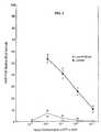
- adenosine nucleotides The effect of adenosine nucleotides on the binding of Gpp(NH)p to a mixture of A m and A s was examined because of the ability of the A-proteins to bind and hydrolyze ATP.
- Purified A m and A s were mixed (1:2), preincubated with ATP or ADP, and assayed for Gpp(NH)p binding after a brief incubation by the rapid filtration method. As shown in Fig. 3, ATP was an effective inhibitor of binding at all concentrations tested.
- ADP was inhibitory in a concentration-dependent manner at higher concentrations.
- Protein concentrations were determined according to Bradford ( Anal. Biochem., 72 :248 (1976)), using the Bio-Rad Microassay (Coomassie Brilliant Blue G-250). Bovine serum albumin was used as a standard.
- the complement of A m relative to A s is 0.47.
- the ratio of the amounts of the separated soluble species recovered from high pressure columns (as estimated by optical density at 280 nm) and slab gels (as estimated by densitometry) is approximately unity. This indicates that all forms of A-protein are extracted in equivalent amounts from rods if the two soluble forms are the products of separate genes. If not, the membrane-bound form would thus be present at half the concentration of the soluble.
- the antibodies of this invention produced against A-protein were used to create an assay for the detection of that antigen in the serum of humans.
- the procedure employs an enzyme-linked immunosorbent assay (ELISA).
- ELISA enzyme-linked immunosorbent assay
- ELISA type of ELISA being used in this context was of the "sandwich” variety. This assay requires two different antibodies that are specific for A-protein, where each antibody recognizes and binds to different epitopes on the protein.
- One of the antibodies serves as the "capture” agent and was used unmodified to coat the bottom of a chamber in a standard 96-well microtiter plate. The unused portion of this antibody was then removed from the well and a blocking agent (1% bovine serum albumin (BSA)) was then placed in the chamber to block non-specific binding sites. Serum from the propositus was then incubated in the prepared well for from 1 to 12 hours, and then removed. The well was washed with a detergent solution, and the second antibody in solution was added to the well.
- BSA bovine serum albumin
- the second antibody used in this procedure is physically linked to a labeling substance such as an enzyme; for example, the enzyme could be horseradish peroxidase (HRP).
- a labeling substance such as an enzyme
- the enzyme could be horseradish peroxidase (HRP).
- HRP horseradish peroxidase
- the amount of color development was directly proportional to the amount of enzyme-linked antibody bound in the well, which is proportional to the amount of antigen bound in the well by the "capture" antibody.
- the results were quantitated by a spectophotometric determination of the amount of color produced over a predetermined period of time (typically 30 to 120 minutes).
- the ELISA described above has been used to test human serum from normal healthy volunteers, and cancer patients, for the presence of the A-protein antigen. The normal population was used to determine the threshold of positivity in this assay.
- the sensitivity of the assay extends at least to the single ng/ml range as determined by the construction of standard sensitivity curves using known amounts of purified antigen.
- a positive reaction in the test was obtained from patients diagnosed with lung, lymphoma, stomach, colon, rectal, and breast cancer when compared to normal subjects.
- the carboxyl terminus 14 amino acid of the published sequence, QFEPQKVKEKMKNA (SEQ ID NO: 3), of human A-protein was synthesized.
- a mixture of this synthetic peptide in Freunds adjuvant was injected subcutaneously at 3-4 separate sites in each rabbit. The amount of synthesized peptide in each injection was 50 ⁇ g.
- boost injections of 50 ⁇ g synthesized peptide in Freunds adjuvant were given at 3-4 separate sites in each of the previously inoculated animals. After two weeks, test bleeds were made to assess blood antibody titers with the synthesized peptide. Further boost injections were made as needed to maintain adequate blood antibody titers.
- Sera from rabbits with adequate blood antibody titers were collected and the desired antibodies were obtained as a purified fraction by affinity chromatography techniques using the synthesized carboxyl terminus peptide immobilized on a solid matrix.
- the purified antibodies were precipitated by dialysis in water and stored as a dry powder for subsequent use.
- These rabbit polyclonal antibodies were reactive with the peptide derived from the carboxyl terminal region of A-protein and were designated as CY2A antibodies.
- a peptide sequence of approximately 16 amino acids at the carboxyl terminus of A-protein was synthetically produced. This peptide was then conjugated to the potent immunostimulator (adjuvant) molecule:keyhole limpet hemacyanin (KLH). The peptide-KLH conjugate was then injected into rabbits by standard procedures used to generate antibodies. The rabbits were subsequently bled and the sera were tested and shown to be positive for the A-protein carboxyl terminus peptide used in the immunogen. These rabbit polyclonal antibodies were designated as CPDD antibodies.
- the biotinylated antibody was first allowed to bind to its target antigen. Simultaneously, a second antibody immobilized on a solid phase support (microtiter plate) captured the same antigen. After removal of any unbound antibody by washing, the antibody:antigen:biotinylated antibody complex was reacted with streptavidin conjugated to a reporter molecule, usually an enzyme such as horse radish peroxidase. Another wash step was performed to remove unbound streptavidin:enzyme. A substrate specific for the enzyme labeled streptavidin was then allowed to react with the remaining streptavidin:enzyme complex. The amount of substrate hydrolyzed into the chromogenic product was thus directly proportional to the amount of antigen present in the sample.
- biotin/streptavidin detection system is that the antibody can be gently labeled with multiple biotin molecules without loss of antibody activity.
- more than one biotin is present on each antibody molecule allows the signal to be amplified through the subsequent binding of multiple streptavidin molecules.
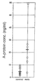
- Blood samples from several individuals were collected. Included within the group of individuals were persons diagnosed as having metastatic breast cancer, persons from a control population with no signs of cancer, and persons that had previously been diagnosed as having had cancer but, at the time of blood sampling, were diagnosed as having their breast cancer in a state of remission.
- the diagnosis labeled as NED refers to no evidence of disease recurrence.
- the results of this assay are also displayed in Figure 1.
- the average assay result for the combination of individuals identified as controls and persons whose cancer was in a state of remission is 4.5 ng A-protein/ml blood.
- the average assay result for the individuals with metastatic breast cancer is 23.4 ng A-protein/ml blood.
- all assay results could be clearly divided into two distinct groups: those individuals who had no sign of cancer and those individuals who had metastatic breast cancer. Such a demarcation is unusual.
- the statistical significance of these results was p ⁇ 0.001 by a two-tailed t-test.
- Example 6B blood plasma samples from over eight hundred human patients were subjected to the assay protocol of Example 6B.
- the patient population included individuals with no cancer (controls), individuals with various stages of primary cancers, individuals with metastatic cancer and individuals with benign tumors.
- the screening procedure was conducted in a blinded fashion and the A-protein assay results were subsequently aligned with independent clinical diagnoses for purposes of assay verification.
- the results of these assays demonstrate that the assay procedure has marked predictive ability to discern individuals with cancer while eliminating from consideration those individuals who do not have cancer.
- the cancers detected by this assay include primary as well as metastatic cancers. Cancer in various stages, including stage 1 and stage 2, are detected. In particular, breast cancer, prostate cancer, primary liver cancer, lymphoma, pancreatic cancer, lung cancer, colon cancer, bladder cancer, endometrial cancer, and multiple myeloma are readily detectable with the present assay.
Landscapes
- Health & Medical Sciences (AREA)
- Life Sciences & Earth Sciences (AREA)
- Chemical & Material Sciences (AREA)
- Immunology (AREA)
- Molecular Biology (AREA)
- Engineering & Computer Science (AREA)
- Organic Chemistry (AREA)
- Cell Biology (AREA)
- Hematology (AREA)
- General Health & Medical Sciences (AREA)
- Biochemistry (AREA)
- Urology & Nephrology (AREA)
- Biomedical Technology (AREA)
- Medicinal Chemistry (AREA)
- Food Science & Technology (AREA)
- Analytical Chemistry (AREA)
- Physics & Mathematics (AREA)
- Microbiology (AREA)
- General Physics & Mathematics (AREA)
- Pathology (AREA)
- Biophysics (AREA)
- Genetics & Genomics (AREA)
- Proteomics, Peptides & Aminoacids (AREA)
- Biotechnology (AREA)
- Peptides Or Proteins (AREA)
- Preparation Of Compounds By Using Micro-Organisms (AREA)
- Medicines Containing Antibodies Or Antigens For Use As Internal Diagnostic Agents (AREA)
Claims (20)
- Ein Verfahren zur Detektion des Vorliegens von Krebs in einem Individuum umfassend:(a) Gewinnen einer Probe von besagtem Individuum;(b) Inkubieren besagter Probe mit mindestens einem Antikörper, der immunreaktiv ist mit einer spezifizierten Sequenz aus 10 bis 18 Aminosäuren von Proteinen, die aus der Gruppe ausgewählt ist, bestehend aus 14 Aminosäuren oder 16 Aminosäuren des Carboxy-Terminus vom A-Protein; wobei das Peptid die Aminosäuren 60 bis 71 vom A-Protein enthält; wobei das Peptid die Aminosäuren 142 bis 158 vom A-Protein enthält; und wobei das Peptid die Aminosäuren 158 bis 170 vom A-Protein enthält;(c) Detektieren der Immunkonjugate, die sich als Folge der Inkubation von Schritt (b) bilden; und(d) Herstellen einer Beziehung zwischen der Menge an Immunkonjugaten von Schritt (c) mit dem Vorliegen von Krebs, worin Krebs vorliegt, wenn besagte Menge größer als ein Schwellenwert ist, gegebenenfalls worin der Krebs ein metastasischer Krebs ist.
- Das Verfahren nach Anspruch 1 umfassend:(a) Gewinnen einer Flüssigprobe, bevorzugt Blut, aus besagtem Individuum;(b) Unterziehen besagter Flüssigprobe einem Antikörper-Sandwich-Assay, worin einer der beiden Antikörper der besagte mindestens eine Antikörper ist und der andere Antikörper immunreaktiv ist mit einem anderen Teil vom A-Protein als besagtem Peptid mit der spezifizierten 10 bis 18 Aminosäure-Sequenz vom A-Protein;(c) Detektieren der Menge an Antikörper / A-Protein-Sandwich, die in Schritt (b) gebildet ist; und(d) Herstellen einer Beziehung zwischen besagter Menge und dem Vorliegen eines Krebsstadiums in besagtem Individuum, worin besagtes Krebsstadium vorliegt, wenn besagte Menge größer als ein Schwellenwert ist.
- Das Verfahren nach Anspruch 2, worin mindestens einer von besagten beiden Antikörpern ein monoklonaler Antikörper ist.
- Das Verfahren nach Anspruch 3, worin der monoklonale Antikörper der Antikörper ist, der mit einem anderen Teil vom A-Protein als besagtem Peptid mit der spezifizierten 10 bis 18 Aminosäure-Sequenz vom A-Protein immunreaktiv ist.
- Das Verfahren nach Anspruch 4, worin der Antikörper, der mit einem Peptid immunreaktiv ist, das eine spezifizierte Sequenz aus 10 bis 18 Aminosäuren von der A-Protein Aminosäure-Sequenz besitzt, ein polyklonaler Antikörper mit einem gebundenen Detektionsmarker ist.
- Das Verfahren nach Anspruch 5, worin der monoklonale Antikörper an einer festen Oberfläche haftet.
- Das Verfahren nach Anspruch 6, worin besagter gebundener Detektionsmarker Biotin ist.
- Das Verfahren nach Anspruch 6, worin besagter Schwellenwert das 2,0-Fache des Durchschnitts des negativen Kontroll-Werts beträgt.
- Das Verfahren nach Anspruch 6, worin besagte spezifizierte Sequenz aus 10 bis 18 Aminosäuren von der A-Protein Aminosäure-Sequenz entweder die Aminosäuren 142 bis 158 vom A-Protein oder die 16 Aminosäuren des Carboxyl-Terminus vom A-Protein ist.
- Das Verfahren nach einem der vorausgehenden Ansprüche, worin besagtes Krebsstadium aus der Gruppe ausgewählt ist bestehend aus Brustkrebs, Prostatakrebs, primärem Leberkrebs, Lymphom, Pankreaskrebs, Lungenkrebs, Darmkrebs, Blasenkrebs, Endometriumkarzinom und multiplem Myelom.
- Ein Verfahren zur Detektion eines Krebsdiagnose-Proteins, dessen erhöhter Spiegel im Blutkreislauf eines Individuums die Prognose erlaubt, dass das Individuum Krebs hat, umfassend die Schritte von einem der vorausgehenden Ansprüche außer Schritt (d).
- Das Verfahren nach Anspruch 11, worin entweder(i) besagte Flüssigprobe mit beiden besagten Antikörpern gleichzeitig inkubiert wird; oder(ii) besagte Flüssigprobe zuerst mit besagtem Antikörper inkubiert wird, der mit einem anderen Teil von besagtem Krebsdiagnose-Protein als besagtem Peptid mit der 10 bis 18 Aminosäure-Sequenz vom Krebsdiagnose-Protein immunreagiert, und dann mit besagtem Antikörper inkubiert ist, der mit einem Peptid immunreagiert, das eine spezifizierte Sequenz aus 10 bis 18 Aminosäuren von der Aminosäure-Sequenz von besagtem Krebsdiagnose-Protein besitzt.
- Das Verfahren nach Anspruch 12 (ii), worin(i) besagter Antikörper, der mit einem anderen Teil von besagtem Krebsdiagnose-Protein als besagtem Peptid mit der spezifizierten 10 bis 18 Aminosäure-Sequenz vom Krebsdiagnose-Protein immunreagiert, ein an einer festen Oberfläche haftender monoklonaler Antikörper ist; und(ii) besagter Antikörper, der mit einem Peptid immunreagiert, das eine spezifizierte Sequenz aus 10 bis 18 Aminosäuren von der Aminosäure-Sequenz von besagtem Krebsdiagnose-Protein hat, ein polyklonaler Antikörper mit einem gebundenen Detektionsmarker ist.
- Ein Antikörper, der an eine spezifizierte Sequenz aus 10 bis 18 Aminosäuren vom A-Protein bindet, um Immunkonjugate zu bilden, deren Vorliegen in einer größeren Menge als ein Schwellenwert in einer biologischen Probe, z.B. Blut, eine Prognose des Vorliegens von Krebs in dem Individuum erlaubt, von dem besagte biologische Probe gewonnen wurde, wobei die 10 bis 18 Aminosäuren vom A-Protein aus der Gruppe ausgewählt sind, bestehend aus 14 Aminosäuren oder 16 Aminosäuren des Carboxy-Terminus vom A-Protein; wobei das Peptid die Aminosäuren 60 bis 71 vom A-Protein enthält; wobei das Peptid die Aminosäuren 142 bis 158 vom A-Protein enthält; und wobei das Peptid die Aminosäuren 158 bis 170 vom A-Protein enthält;
- Ein Test-Set zur Detektion des Vorliegens von Krebs in einem Individuum umfassend:(a) einen ersten Antikörper, der mit einem Peptid immunreagiert, das eine spezifizierte Sequenz aus 10 bis 18 Aminosäuren von der Aminosäure-Sequenz vom A-Protein besitzt, die aus der Gruppe ausgewählt ist, bestehend aus 14 Aminosäuren oder 16 Aminosäuren des Carboxy-Terminus vom A-Protein; wobei das Peptid die Aminosäuren 60 bis 71 vom A-Protein enthält; wobei das Peptid die Aminosäuren 142 bis 158 vom A-Protein enthält; und wobei das Peptid die Aminosäuren 158 bis 170 vom A-Protein enthält;(b) einen zweiten Antikörper, der mit einem anderen Teil von besagtem Krebsdiagnose-Protein als besagtem Peptid mit der spezifizierten 10 bis 18 Aminosäure-Sequenz vom Krebsdiagnose-Protein immunreagiert.
- Das Test-Set nach Anspruch 15, worin entweder besagter erster Antikörper oder besagter zweiter Antikörper einen gebundenen Identifizierungsmarker hat.
- Das Test-Set nach Anspruch 15, worin mindestens einer von besagtem ersten Antikörper und besagtem zweiten Antikörper ein monoklonaler Antikörper ist.
- Das Test-Set nach Anspruch 17, worin besagter erster Antikörper ein polyklonaler Antikörper mit einem gebundenen Identifizierungsmarker ist und besagter zweiter Antikörper ein an einer festen Oberfläche haftender monoklonaler Antikörper ist.
- Das Test-Set nach Anspruch 18, worin besagter gebundener Identifizierungsmarker Biotin ist.
- Das Test-Set nach einem der Ansprüche 15 bis 19, worin besagte spezifizierte Sequenz aus 10 bis 18 Aminosäuren entweder die Aminosäuren 142 bis 158 von besagtem Krebsdiagnose-Protein oder die 16 Aminosäuren des Carboxyl-Terminus von besagtem Krebsdiagnose-Protein ist.
Applications Claiming Priority (3)
| Application Number | Priority Date | Filing Date | Title |
|---|---|---|---|
| US37096995A | 1995-01-10 | 1995-01-10 | |
| US370969 | 1995-01-10 | ||
| PCT/US1996/000098 WO1996021862A1 (en) | 1995-01-10 | 1996-01-11 | A-protein as a diagnostic of cancer |
Publications (2)
| Publication Number | Publication Date |
|---|---|
| EP0803065A1 EP0803065A1 (de) | 1997-10-29 |
| EP0803065B1 true EP0803065B1 (de) | 2001-10-31 |
Family
ID=23461946
Family Applications (1)
| Application Number | Title | Priority Date | Filing Date |
|---|---|---|---|
| EP96902593A Expired - Lifetime EP0803065B1 (de) | 1995-01-10 | 1996-01-11 | A-protein als diagnostikum für krebs |
Country Status (8)
| Country | Link |
|---|---|
| EP (1) | EP0803065B1 (de) |
| JP (1) | JPH10512369A (de) |
| CN (1) | CN1168176A (de) |
| AT (1) | ATE208041T1 (de) |
| AU (1) | AU4694096A (de) |
| CA (1) | CA2209823A1 (de) |
| DE (1) | DE69616509D1 (de) |
| WO (1) | WO1996021862A1 (de) |
Families Citing this family (2)
| Publication number | Priority date | Publication date | Assignee | Title |
|---|---|---|---|---|
| US6187549B1 (en) * | 1988-03-21 | 2001-02-13 | Cytra Corporation | Protein as a diagnostic of cancer |
| JP2003194775A (ja) * | 2001-12-28 | 2003-07-09 | Japan Science & Technology Corp | タンパク質の電気泳動法 |
Family Cites Families (2)
| Publication number | Priority date | Publication date | Assignee | Title |
|---|---|---|---|---|
| US5405749A (en) * | 1991-12-06 | 1995-04-11 | Polans; Arthur S. | Method for identifying and purifying a cancer associated retinopathy autoantigen, and testing patient serum for the autoantibody to the autoantigen |
| JPH07507537A (ja) * | 1992-03-27 | 1995-08-24 | ザ シェペンズ アイ リサーチ インスティチュート | 細胞シグナル変換系調節のための組成物と方法 |
-
1996
- 1996-01-11 AU AU46940/96A patent/AU4694096A/en not_active Abandoned
- 1996-01-11 CA CA002209823A patent/CA2209823A1/en not_active Abandoned
- 1996-01-11 JP JP8521734A patent/JPH10512369A/ja active Pending
- 1996-01-11 AT AT96902593T patent/ATE208041T1/de not_active IP Right Cessation
- 1996-01-11 EP EP96902593A patent/EP0803065B1/de not_active Expired - Lifetime
- 1996-01-11 DE DE69616509T patent/DE69616509D1/de not_active Expired - Lifetime
- 1996-01-11 CN CN96191397A patent/CN1168176A/zh active Pending
- 1996-01-11 WO PCT/US1996/000098 patent/WO1996021862A1/en not_active Ceased
Also Published As
| Publication number | Publication date |
|---|---|
| CA2209823A1 (en) | 1996-07-18 |
| MX9705161A (es) | 1998-10-31 |
| EP0803065A1 (de) | 1997-10-29 |
| JPH10512369A (ja) | 1998-11-24 |
| DE69616509D1 (de) | 2001-12-06 |
| ATE208041T1 (de) | 2001-11-15 |
| CN1168176A (zh) | 1997-12-17 |
| WO1996021862A1 (en) | 1996-07-18 |
| AU4694096A (en) | 1996-07-31 |
Similar Documents
| Publication | Publication Date | Title |
|---|---|---|
| US20240230652A9 (en) | Use of he4 and other biochemical markers for assessment of ovarian cancers | |
| US7087727B2 (en) | Periostin-based diagnostic assays | |
| JPH10500408A (ja) | サイトケラチン18のフラグメントおよび対応する抗体を伴う癌の検出法 | |
| JPS61204135A (ja) | 発癌遺伝子生成物阻害剤 | |
| US20080206140A1 (en) | Cspcna Isoform Antibodies and Uses Thereof | |
| EP2449126B1 (de) | Neue antikörper und ihre verwendung in therapie- und diagnoseverfahren | |
| AU632535B2 (en) | Cancer related haptoglobin (hpr) | |
| EP0421392B1 (de) | Monoklonaler Antikörper gegen Kern-hCG-beta, seine Herstellung und Verwendung | |
| KR101777259B1 (ko) | En2 단백질을 특이적으로 인식하는 단클론 항체 또는 이를 함유하는 전립선암 진단용 조성물 | |
| US6187549B1 (en) | Protein as a diagnostic of cancer | |
| EP1271152A1 (de) | Verfahren zum erkennen von krebs durch messen von autoantikörpern gegen mdm2 und reagenzien dafür | |
| JPS60500427A (ja) | 特異性ceaファミリ−抗原、それに対し特異性の抗体およびそれらの使用方法 | |
| US20060063214A1 (en) | Methods and compositions for diagnosing neoplastic disease | |
| KR101777254B1 (ko) | En2 단백질을 특이적으로 인식하는 특정 항원으로부터 얻어진 단클론 항체 또는 이를 함유하는 전립선암 진단용 조성물 | |
| JP2555028B2 (ja) | ヒト非−小細胞肺癌に関するモノクロ−ナル抗体 | |
| EP0803065B1 (de) | A-protein als diagnostikum für krebs | |
| CA2682132A1 (en) | Use of he4 for assessment of breast cancers | |
| JP2915530B2 (ja) | ラミニン フラグメント | |
| JP4451784B2 (ja) | 前立腺癌腫瘍マーカー | |
| MXPA97005161A (es) | La proteina"a"como diagnostico de cancer | |
| JP2878317B2 (ja) | ラミニン測定試薬 | |
| KR900008014B1 (ko) | 펩타이드 항체의 제조방법 | |
| JP3591306B2 (ja) | モノクローナル抗体、ハイブリドーマ及び免疫測定方法 | |
| US7141435B2 (en) | Monoclonal antibodies against human apoptosis inhibitory protein NAIP and method for assaying the NAIP | |
| JPH08208698A (ja) | モノクローナル抗体 |
Legal Events
| Date | Code | Title | Description |
|---|---|---|---|
| PUAI | Public reference made under article 153(3) epc to a published international application that has entered the european phase |
Free format text: ORIGINAL CODE: 0009012 |
|
| 17P | Request for examination filed |
Effective date: 19970729 |
|
| AK | Designated contracting states |
Kind code of ref document: A1 Designated state(s): AT BE CH DE DK ES FR GB GR IE IT LI LU MC NL PT SE |
|
| 17Q | First examination report despatched |
Effective date: 19990531 |
|
| GRAG | Despatch of communication of intention to grant |
Free format text: ORIGINAL CODE: EPIDOS AGRA |
|
| GRAG | Despatch of communication of intention to grant |
Free format text: ORIGINAL CODE: EPIDOS AGRA |
|
| GRAH | Despatch of communication of intention to grant a patent |
Free format text: ORIGINAL CODE: EPIDOS IGRA |
|
| GRAH | Despatch of communication of intention to grant a patent |
Free format text: ORIGINAL CODE: EPIDOS IGRA |
|
| GRAA | (expected) grant |
Free format text: ORIGINAL CODE: 0009210 |
|
| AK | Designated contracting states |
Kind code of ref document: B1 Designated state(s): AT BE CH DE DK ES FR GB GR IE IT LI LU MC NL PT SE |
|
| PG25 | Lapsed in a contracting state [announced via postgrant information from national office to epo] |
Ref country code: NL Free format text: LAPSE BECAUSE OF FAILURE TO SUBMIT A TRANSLATION OF THE DESCRIPTION OR TO PAY THE FEE WITHIN THE PRESCRIBED TIME-LIMIT Effective date: 20011031 Ref country code: LI Free format text: LAPSE BECAUSE OF FAILURE TO SUBMIT A TRANSLATION OF THE DESCRIPTION OR TO PAY THE FEE WITHIN THE PRESCRIBED TIME-LIMIT Effective date: 20011031 Ref country code: IT Free format text: LAPSE BECAUSE OF FAILURE TO SUBMIT A TRANSLATION OF THE DESCRIPTION OR TO PAY THE FEE WITHIN THE PRESCRIBED TIME-LIMIT;WARNING: LAPSES OF ITALIAN PATENTS WITH EFFECTIVE DATE BEFORE 2007 MAY HAVE OCCURRED AT ANY TIME BEFORE 2007. THE CORRECT EFFECTIVE DATE MAY BE DIFFERENT FROM THE ONE RECORDED. Effective date: 20011031 Ref country code: FR Free format text: LAPSE BECAUSE OF FAILURE TO SUBMIT A TRANSLATION OF THE DESCRIPTION OR TO PAY THE FEE WITHIN THE PRESCRIBED TIME-LIMIT Effective date: 20011031 Ref country code: CH Free format text: LAPSE BECAUSE OF FAILURE TO SUBMIT A TRANSLATION OF THE DESCRIPTION OR TO PAY THE FEE WITHIN THE PRESCRIBED TIME-LIMIT Effective date: 20011031 Ref country code: BE Free format text: LAPSE BECAUSE OF FAILURE TO SUBMIT A TRANSLATION OF THE DESCRIPTION OR TO PAY THE FEE WITHIN THE PRESCRIBED TIME-LIMIT Effective date: 20011031 Ref country code: AT Free format text: LAPSE BECAUSE OF FAILURE TO SUBMIT A TRANSLATION OF THE DESCRIPTION OR TO PAY THE FEE WITHIN THE PRESCRIBED TIME-LIMIT Effective date: 20011031 |
|
| REF | Corresponds to: |
Ref document number: 208041 Country of ref document: AT Date of ref document: 20011115 Kind code of ref document: T |
|
| REG | Reference to a national code |
Ref country code: CH Ref legal event code: EP |
|
| REF | Corresponds to: |
Ref document number: 69616509 Country of ref document: DE Date of ref document: 20011206 |
|
| REG | Reference to a national code |
Ref country code: IE Ref legal event code: FG4D |
|
| REG | Reference to a national code |
Ref country code: GB Ref legal event code: IF02 |
|
| PG25 | Lapsed in a contracting state [announced via postgrant information from national office to epo] |
Ref country code: LU Free format text: LAPSE BECAUSE OF NON-PAYMENT OF DUE FEES Effective date: 20020111 Ref country code: IE Free format text: LAPSE BECAUSE OF NON-PAYMENT OF DUE FEES Effective date: 20020111 |
|
| PG25 | Lapsed in a contracting state [announced via postgrant information from national office to epo] |
Ref country code: SE Free format text: LAPSE BECAUSE OF FAILURE TO SUBMIT A TRANSLATION OF THE DESCRIPTION OR TO PAY THE FEE WITHIN THE PRESCRIBED TIME-LIMIT Effective date: 20020131 Ref country code: PT Free format text: LAPSE BECAUSE OF FAILURE TO SUBMIT A TRANSLATION OF THE DESCRIPTION OR TO PAY THE FEE WITHIN THE PRESCRIBED TIME-LIMIT Effective date: 20020131 Ref country code: GB Free format text: LAPSE BECAUSE OF NON-PAYMENT OF DUE FEES Effective date: 20020131 Ref country code: DK Free format text: LAPSE BECAUSE OF FAILURE TO SUBMIT A TRANSLATION OF THE DESCRIPTION OR TO PAY THE FEE WITHIN THE PRESCRIBED TIME-LIMIT Effective date: 20020131 |
|
| PG25 | Lapsed in a contracting state [announced via postgrant information from national office to epo] |
Ref country code: GR Free format text: LAPSE BECAUSE OF FAILURE TO SUBMIT A TRANSLATION OF THE DESCRIPTION OR TO PAY THE FEE WITHIN THE PRESCRIBED TIME-LIMIT Effective date: 20020201 Ref country code: DE Free format text: LAPSE BECAUSE OF FAILURE TO SUBMIT A TRANSLATION OF THE DESCRIPTION OR TO PAY THE FEE WITHIN THE PRESCRIBED TIME-LIMIT Effective date: 20020201 |
|
| NLV1 | Nl: lapsed or annulled due to failure to fulfill the requirements of art. 29p and 29m of the patents act | ||
| PG25 | Lapsed in a contracting state [announced via postgrant information from national office to epo] |
Ref country code: ES Free format text: LAPSE BECAUSE OF FAILURE TO SUBMIT A TRANSLATION OF THE DESCRIPTION OR TO PAY THE FEE WITHIN THE PRESCRIBED TIME-LIMIT Effective date: 20020430 |
|
| PG25 | Lapsed in a contracting state [announced via postgrant information from national office to epo] |
Ref country code: MC Free format text: LAPSE BECAUSE OF NON-PAYMENT OF DUE FEES Effective date: 20020801 |
|
| PLBE | No opposition filed within time limit |
Free format text: ORIGINAL CODE: 0009261 |
|
| STAA | Information on the status of an ep patent application or granted ep patent |
Free format text: STATUS: NO OPPOSITION FILED WITHIN TIME LIMIT |
|
| GBPC | Gb: european patent ceased through non-payment of renewal fee |
Effective date: 20020131 |
|
| 26N | No opposition filed | ||
| REG | Reference to a national code |
Ref country code: IE Ref legal event code: MM4A |