EP0795129B1 - Vorrichtung zur analyse von blut und anderen proben - Google Patents
Vorrichtung zur analyse von blut und anderen proben Download PDFInfo
- Publication number
- EP0795129B1 EP0795129B1 EP95938498A EP95938498A EP0795129B1 EP 0795129 B1 EP0795129 B1 EP 0795129B1 EP 95938498 A EP95938498 A EP 95938498A EP 95938498 A EP95938498 A EP 95938498A EP 0795129 B1 EP0795129 B1 EP 0795129B1
- Authority
- EP
- European Patent Office
- Prior art keywords
- sample
- light source
- light
- detector
- rotor
- Prior art date
- Legal status (The legal status is an assumption and is not a legal conclusion. Google has not performed a legal analysis and makes no representation as to the accuracy of the status listed.)
- Expired - Lifetime
Links
Images


Classifications
-
- G—PHYSICS
- G01—MEASURING; TESTING
- G01N—INVESTIGATING OR ANALYSING MATERIALS BY DETERMINING THEIR CHEMICAL OR PHYSICAL PROPERTIES
- G01N33/00—Investigating or analysing materials by specific methods not covered by groups G01N1/00 - G01N31/00
- G01N33/48—Biological material, e.g. blood, urine; Haemocytometers
- G01N33/483—Physical analysis of biological material
- G01N33/487—Physical analysis of biological material of liquid biological material
- G01N33/49—Blood
- G01N33/491—Blood by separating the blood components
-
- G—PHYSICS
- G01—MEASURING; TESTING
- G01N—INVESTIGATING OR ANALYSING MATERIALS BY DETERMINING THEIR CHEMICAL OR PHYSICAL PROPERTIES
- G01N15/00—Investigating characteristics of particles; Investigating permeability, pore-volume or surface-area of porous materials
- G01N15/04—Investigating sedimentation of particle suspensions
- G01N15/042—Investigating sedimentation of particle suspensions by centrifuging and investigating centrifugates
-
- G—PHYSICS
- G01—MEASURING; TESTING
- G01N—INVESTIGATING OR ANALYSING MATERIALS BY DETERMINING THEIR CHEMICAL OR PHYSICAL PROPERTIES
- G01N21/00—Investigating or analysing materials by the use of optical means, i.e. using sub-millimetre waves, infrared, visible or ultraviolet light
- G01N21/17—Systems in which incident light is modified in accordance with the properties of the material investigated
- G01N21/25—Colour; Spectral properties, i.e. comparison of effect of material on the light at two or more different wavelengths or wavelength bands
- G01N21/31—Investigating relative effect of material at wavelengths characteristic of specific elements or molecules, e.g. atomic absorption spectrometry
-
- G—PHYSICS
- G01—MEASURING; TESTING
- G01N—INVESTIGATING OR ANALYSING MATERIALS BY DETERMINING THEIR CHEMICAL OR PHYSICAL PROPERTIES
- G01N15/00—Investigating characteristics of particles; Investigating permeability, pore-volume or surface-area of porous materials
- G01N15/04—Investigating sedimentation of particle suspensions
- G01N15/042—Investigating sedimentation of particle suspensions by centrifuging and investigating centrifugates
- G01N2015/045—Investigating sedimentation of particle suspensions by centrifuging and investigating centrifugates by optical analysis
Definitions
- This invention relates to an apparatus for analysing blood and other samples by means of automated sample analysis during centrifugation.
- a rotor disc adapted to receive a number of capillary haematocrit tubes each in a corresponding slit.
- An arm pivoted to a point clear of the rotor disc carries a light source above the rotor disc and an aligned light sensor below the rotor disc.
- the present invention is concerned, in one aspect, with an improved apparatus of the same general nature.
- Hb haemoglobin
- the present invention in one aspect, provides an analysis apparatus which includes a centrifuge rotor adapted to receive at least one sample in a transparent holder, the rotor being apertured to allow light to be transmitted through the sample and the rotor, and a scanning arm arranged to traverse the rotor carrying light source means and light detector means on respective sides of the rotor; and in which the light source means comprises a first light source for detection of sample component interfaces and a second light source for colourimetric inspection of at least one sample component.
- the first light source is an infrared light source and the second light source is a visible light source.
- the infrared light source is preferably an infrared laser.
- the sample is of blood
- the visible light source is used for examining blood plasma
- the visible light source comprises a plurality of substantially monochromatic light sources, most preferably sources of red, green and blue light at wavelengths of, for example, 625, 567 and 470 nm.
- the visible light source may be provided by a single, multi-colour LED with the colours switched sequentially during the scanning process.
- the apparatus includes a reference sample container on the rotor containing a reference material (for example, water) of known optical qualities, whereby signals representing light transmission through the sample(s) may be calibrated against signals representing light transmission through the reference.
- a reference material for example, water
- the present invention provides a haemoglobin photometer comprising means for receiving a transparent cell containing a blood sample mixed with a haemoglobin reagent, illumination means for directing substantially monochromatic light at the sample cell, and detector means arranged to receive light passing through the sample cell to generate a signal representing the optical density of the cell contents.
- the illumination means comprises a LED operating at a peak wavelength of, preferably, about 560 nm.
- the illumination means preferably further comprises collimation means causing the light directed at the sample cell to be substantially parallel.
- the detector means may suitably comprise a converging lens directing the transmitted light onto a photodiode.
- the illumination means and detector means preferably include optical stops dimensioned and positioned such that light emitted from the whole emitting area of the LED is received within the active surface of the photodiode.
- the photometer includes calibration means operating to calibrate a detected optical density to a predetermined standard.
- the calibration means operates by comparing the output of the detector means with and without a sample cell in position.
- the invention provides a blood analyzer comprising a centrifuge for separating a blood sample by centrifugation into packed red cells, packed white cells, and plasma; first optical means for measuring the proportions of red cells, white cells and plasma in the sample; photometer means for measuring haemoglobin concentration in a sample of the same blood; and computer means for deriving a number of parameters of interest from said measurements.
- Said parameters may suitably be:
- the analyzer further comprises second optical means for inspecting the plasma component to measure one or more parameters of interest therein.
- the second optical means is arranged to measure optical density at each of a plurality of visible wavelengths, suitably wavelengths of approximately 625, 570 and 470 nm.
- the parameters of interest are preferably icterus, lipaemia and haemolysis.
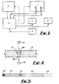
- the preferred form of the invention is a blood analyser comprising a centrifuge 1 and a photometer 2.
- the apparatus is controlled by a microprocessor 3 with user input via a keyboard 4 and data output by display 5 and/or printer 6.
- the photometer 2 provides an output signal to the microprocessor 3 which is representative of haemoglobin concentration.
- the centrifuge 1 includes two optical arrangements, as will be described, providing respective output signals to the microprocessor 3 from which signals a number of blood-related parameters can be derived.
- a centrifuge rotor in the form of a disc 10 receives four haematocrit tubes 12, 14, 16 and 18 in radial alignment.
- the rotor is rotated by a drive motor 20 at a suitable speed to centrifuge the samples within the tubes 12-18.
- the samples are human or animal blood
- the centrifugation results in separation into packed red blood cells 22, packed white blood cells 24, and plasma 26.
- the outer end of the tube is closed with a bung 28, and the inner end contains a quantity of air at 30.
- centrifugation will take place at 11,000 rpm for 3 to 10 minutes.
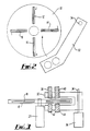
- the scanning arm 32 is of generally U-shape, having an upper part mounting an infrared (IR) light source 38 and a visible light source 40, and a lower part mounting an IR detector 42 and a visible light detector 44.
- IR infrared
- each sample tube 12 etc is located in a slit 46 formed in the rotor 10, the slit 46 having a relatively wide top portion 46a and a relatively narrow under portion 46b joined by a shoulder on which the tube rests.
- the slit 46 thus allows light to pass from the source 38 or 42 to the associated detector 40 or 44.
- the scanning process thus makes it possible to build up a curve defining the light absorption, at a given wavelength, along the length of each tube.
- One typical curve is shown in Fig. 6A.
- a curve of this nature can provide further information.
- Fig. 6B the interface between white and red blood cell portions is poorly defined, which indicates the presence of nucleated red cells and reticulocytes.
- a particular feature of the present invention is that scanning is carried out with both the IR source 38, suitably an IR laser, and with the visible light source described in more detail below.
- the IR source 38 is used to detect the interfaces between the various fractions shown in Fig. 5. By detecting the relative positions of the interfaces, packed cell volume and white blood cell volume can be expressed as percentage of total volume. Blood cells and plasma are scattering media and are optically dense. We have established that a satisfactory resolution can be attained by using as a light source an IR laser with a wavelength of 785 mm and power of 1.5 mw.
- Plasma may contain a number of components the presence and concentrations of which can provide crucial, early diagnostic information. Quantitative measurement of these components can enable the clinician quickly to plan further tests, and to avoid the adverse effects on certain tests arising from the presence of high concentrations of these components.
- the plasma components normally of interest are bilirubin (icterus), triglycerides (lipaemia) and haemoglobin (haemolysis). In the past, these have generally been assessed by visual inspection, with unsatisfactory results. We have determined experimentally that the optimum wavelengths for detecting these three components are 625, 567 and 470 nm.
- the visible light source 42 is arranged to emit at these three frequencies sequentially.
- a suitable light source for doing so is a "Rainbow” LED by Ledtronics, ref. DIS-10024-002. This device emits light at three preselected frequencies, each in response to a respective signal.
- the plasma components of the samples are scanned for the three components of interest. It is not uncommon for all three to be present in the plasma of unwell patients.
- the present system allows all three to be measured at one time.
- the optical densities measured at each wavelength are converted to concentration units via calibration curves held in software.
- the column of plasma is scanned along its length, rather than measured at a point. This makes it possible to measure component gradient, which may be of particular interest in lipaemia by providing information on the density of lipids present.
- the apparatus further includes a haemoglobin (Hb) photometer, which will now be described.
- Hb haemoglobin
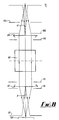
- a sample is placed in a transparent cuvette 100 and is exposed to monochromatic light from LED 102.
- the transmitted light is detected by a photocell 104 to derive a measure of optical density.
- the optical density measurement is linearly related to Hb concentration over a range of interest.
- a 20 ml sample is mixed with 3 ml of reagent and exposed to green light with a peak wavelength of about 560 nm.
- a suitable light source is a LED by Radiospares, catalogue no. 590-496, 563 nm peak, 250 mcd. This allows measurement of Hb concentration from 4 g/dl to a maximum of about 25 g/dl at which the optical density is about 0.8.
- light emitted by the LED 102 is passed through fixed stops 105 and 106 and collimated by lens 108.
- a further fixed stop 110 sets the area of the light beam applied to the cuvette 100.
- Light transmitted through the cuvette 100 is gathered by a collecting lens 112 mounted between equal aperture stops 114, 116 and passes to the photodetector 104 via a small diameter stop 118.
- Fig. 8 The geometry of this arrangement is shown schematically in Fig. 8.
- the use of an approximately parallel beam of light to illuminate the cuvette 100 means that small lateral and longitudinal misalignments of the cuvette have no significant detriment on accuracy.
- This arrangement also minimises the effects of non-uniformity of the sample itself or of the cuvette (for example, produced by fingerprints).
- the stop 106 which forms the light source exit window is chosen to be less than the aperture of the detector lens 112 as determined by the stops 114, 116.
- the fact that the LED source 102 has a finite source size is unimportant provided that all the transmitted light arrives within the sensitive area of the detector 104; this is achieved by setting the geometry such that the angle ⁇ defined by the collecting lens 112 and the detector stop 118 is greater than the angle a defined by the collimating lens 108 and the emitting area of the LED 102.
- a reference cuvette could be held permanently in the instrument and could, for example, be spring biased into the optical path so as to be displaced by insertion of a sample cuvette. It might be necessary to use repeated calibration of this nature to minimise drift effects, such as the non-uniformity of LED output intensity caused by ambient temperature changes.
- the photometer with a multiwavelength capability.
- the 563 nm LED described with reference to Fig. 7 could be replaced by a Ledtronics "Rainbow" LED of the type described with reference to the centrifuge. This could be used to take measurements sequentially at 625 nm, 567 nm and 470 nm. Alternatively, it could be used, by controlling the currents to the red, blue and green emitting regions simultaneously, to produce a desired wavelength for a given test. Such polychromatic capability would be useful for a number of biochemical analyses other than blood.
- MCHC mean corpuscular haemoglobin concentration
Landscapes
- Health & Medical Sciences (AREA)
- Physics & Mathematics (AREA)
- Life Sciences & Earth Sciences (AREA)
- Chemical & Material Sciences (AREA)
- Biochemistry (AREA)
- Analytical Chemistry (AREA)
- Engineering & Computer Science (AREA)
- General Health & Medical Sciences (AREA)
- General Physics & Mathematics (AREA)
- Immunology (AREA)
- Pathology (AREA)
- Biomedical Technology (AREA)
- Spectroscopy & Molecular Physics (AREA)
- Hematology (AREA)
- Dispersion Chemistry (AREA)
- Biophysics (AREA)
- Molecular Biology (AREA)
- Urology & Nephrology (AREA)
- Food Science & Technology (AREA)
- Medicinal Chemistry (AREA)
- Ecology (AREA)
- Investigating Or Analysing Materials By Optical Means (AREA)
- Investigating Or Analysing Biological Materials (AREA)
- Optical Measuring Cells (AREA)
- Spectrometry And Color Measurement (AREA)
- Investigating Or Analysing Materials By The Use Of Chemical Reactions (AREA)
- Automatic Analysis And Handling Materials Therefor (AREA)
Claims (15)
- Eine Analysevorrichtung, bestehend aus einem Zentrifugenrotor (10), der ausgeführt ist, um zumindest eine Probe in einem transparenten Behälter (12) aufzunehmen, wobei der Rotor mit Öffnungen versehen ist, damit Licht durch die Probe und den Rotor übertragen werden kann, und einem Abtastarm (32), der angeordnet ist, um den Rotor (10) und ein tragendes Lichtquellenmittel (38, 40) und ein Lichtdetektormittel (42, 44) auf den jeweiligen Seiten des Rotors (10) zu durchqueren;
dadurch gekennzeichnet, daß das Lichtquellenmittel eine erste Lichtquelle (38) zur Detektion von Probenkomponentenzwischenflächen und eine zweite Lichtquelle (40), anliegend an die erste Lichtquelle (38), zur kolorimetrischen Inspektion von zumindest einer Probenkomponente umfaßt. - Vorrichtung gemäß Anspruch 1, wobei die erste Lichtquelle (38) eine Infrarotlichtquelle ist und die zweite Lichtquelle (40) eine sichtbare Lichtquelle ist.
- Vorrichtung gemäß Anspruch 1 oder Anspruch 2, wobei die Infrarotlichtquelle (38) ein Infrarotlaser ist.
- Vorrichtung gemäß einem der vorhergehenden Ansprüche, wobei die Probe aus Blut besteht, wobei die sichtbare Lichtquelle (40) verwendet wird, um Blutplasma (26) zu untersuchen, und die sichtbare Lichtquelle (40) drei im wesentlichen monochromatische Lichtquellen aus rotem, grünem bzw. blauem Licht umfaßt.
- Vorrichtung gemäß Anspruch 4, wobei die sichtbare Lichtquelle (40) eine einzige, mehrfarbige Leuchtdiode ist, wobei die Farben während des Abtastverfahrens aufeinanderfolgend gewechselt werden.
- Vorrichtung gemäß einem der vorhergehenden Ansprüche, die weiters einen Referenzprobenbehälter auf dem Rotor umfaßt, der ein Referenzmaterial mit bekannten optischen Qualitäten enthält, wodurch Signale, welche die Lichtübertragung durch die Probe darstellen, mit Signalen, welche die Lichtübertragung durch das Referenzmaterial darstellen, kalibriert werden.
- Vorrichtung gemäß einem der vorhergehenden Ansprüche, weiters bestehend aus einem Hämoglobinphotometer, der ein Mittel zum Aufnehmen einer transparenten Zelle, die eine Blutprobe enthält, welche mit einem Hämoglobinreagens gemischt ist, einem Beleuchtungsmittel (102) zum Richten von im wesentlichen monochromatischem Licht auf die Probezelle (100) und einem Detektormittel (104), das so angeordnet ist, um durch die Probezelle hindurchgehendes Licht aufzunehmen, damit ein Signal erzeugt wird, das die optische Dichte der Zelleninhalte darstellt, umfaßt.
- Vorrichtung gemäß Anspruch 7, wobei das Beleuchtungsmittel (102) eine Leuchtdiode umfaßt, die auf einer Höchstwellenlänge von ungefähr 560 nm läuft.
- Vorrichtung gemäß Anspruch 7 oder Anspruch 8, wobei das Beleuchtungsmittel (102) weiters ein Kollimationsmittel (108) umfaßt, das bewirkt, daß das auf die Probezelle gerichtete Licht im wesentlichen parallel verläuft.
- Vorrichtung gemäß einem der Ansprüche 7 bis 9, wobei das Detektormittel (104) eine Sammellinse umfaßt, die das übertragene Licht auf eine Photodiode richtet.
- Vorrichtung gemäß einem der Ansprüche 7 bis 10, wobei das Beleuchtungsmittel (102) und das Detektormittel (104) optische Blenden (114, 116, 118) umfaßt, die so dimensioniert und angeordnet sind, daß das von der gesamten emittierenden Fläche der Leuchtdiode emittierte Licht innerhalb der aktiven Oberfläche der Photodiode aufgenommen wird.
- Vorrichtung gemäß einem der Ansprüche 7 bis 11, weiters bestehend aus einem Kalibriermittel, das so funktioniert, daß es eine ermittelte optische Dichte auf einen vorbestimmten Standard kalibriert.
- Vorrichtung gemäß Anspruch 12, wobei das Kalibriermittel so funktioniert, indem es die abgegebene Leistung des Detektormittels mit einer positionierten Probezelle und ohne eine solche vergleicht.
- Ein Blutanalysiergerät, bestehend aus einer Zentrifuge (1) zur Teilung einer Blutprobe durch Zentrifugieren in gepackte rote Zellen (22), gepackte weiße Zellen (24) und Plasma (26); einem ersten optischen Mittel (38) zur Messung der Verhältnisse von roten Zellen, weißen Zellen und Plasma in der Probe; einem zweiten optischen Mittel (40), das so angeordnet ist, daß es die optische Dichte der Plasmakomponente bei jeder der Vielzahl von sichtbaren Wellenlängen misst; einem Photometermittel (2) zur Messung der Hämoglobinkonzentration in einer Probe desselben Blutes; und einem Rechnermittel (3) zur Ableitung einer Anzahl wichtiger Parameter von diesen Messungen.
- Ein Verfahren zur Analyse einer Blutprobe unter Verwendung eines Analysiergerätes gemäß Anspruch 2, wobei das Verfahren folgende Schritte umfaßt:Eingeben der Probe in ein Hämatokritröhrchen (12, 14, 16) auf dem Zentrifugenrotor (10);Stellen eines mit Wasser gefüllten Hämatokritröhrchens (18) auf den Zentrifugenrotor (10);Schleudern des Zentrifugenrotors (10) bei einer ersten Geschwindigkeit von 11000 Ulmin, um die Fraktionen in der Probe zu teilen;Bewegen des Abtastarms (32) über den Rotor (10), während der Zentrifugenrotor (10) bei einer zweiten Geschwindigkeit von 1000 U/min geschleudert wird;Leiten von Licht von der ersten Infrarotlichtquelle (38) zu einem ersten Detektor (42), um die Schnittflächen zwischen den Fraktionen in der Probe entlang der Länge des Röhrchens (12, 14, 16) durch Kalibrieren des Signals, das beim ersten Detektor durch die Probe erhalten wurde, mit dem Signal, das am ersten Detektor durch das Wasser (18) erhalten wurde, zu ermitteln;Leiten von Licht von der zweiten sichtbaren Lichtquelle (40) zu einem zweiten Detektor (44), um die Lichtabsorption der Probe bei einer Vielzahl von sichtbaren Lichtwellenlängen entlang der Länge des Röhrchens (12, 14, 16) durch Kalibrieren des Signals, das am zweiten Detektor durch die Probe erhalten wurde, mit dem Signal, das am zweiten Detektor durch das Wasser (18) erhalten wurde, zu messen.
Applications Claiming Priority (3)
| Application Number | Priority Date | Filing Date | Title |
|---|---|---|---|
| GB9424218A GB9424218D0 (en) | 1994-11-30 | 1994-11-30 | Apparatus for analysing blood and other samples |
| GB9424218 | 1994-11-30 | ||
| PCT/GB1995/002786 WO1996017243A1 (en) | 1994-11-30 | 1995-11-30 | Apparatus for analysing blood and other samples |
Publications (2)
| Publication Number | Publication Date |
|---|---|
| EP0795129A1 EP0795129A1 (de) | 1997-09-17 |
| EP0795129B1 true EP0795129B1 (de) | 2001-01-24 |
Family
ID=10765237
Family Applications (1)
| Application Number | Title | Priority Date | Filing Date |
|---|---|---|---|
| EP95938498A Expired - Lifetime EP0795129B1 (de) | 1994-11-30 | 1995-11-30 | Vorrichtung zur analyse von blut und anderen proben |
Country Status (7)
| Country | Link |
|---|---|
| EP (1) | EP0795129B1 (de) |
| JP (1) | JPH10510362A (de) |
| AU (1) | AU3986895A (de) |
| CA (1) | CA2205484A1 (de) |
| DE (1) | DE69520001T2 (de) |
| GB (1) | GB9424218D0 (de) |
| WO (1) | WO1996017243A1 (de) |
Cited By (4)
| Publication number | Priority date | Publication date | Assignee | Title |
|---|---|---|---|---|
| US9921141B2 (en) | 2012-08-08 | 2018-03-20 | Koninklijke Philips N.V. | Centrifugal microfluidic device and methods of use |
| US11372006B2 (en) | 2018-04-24 | 2022-06-28 | Entia Ltd. | Method and apparatus for determining haemoglobin concentration |
| EP4220131A1 (de) * | 2018-10-02 | 2023-08-02 | Instrumentation Laboratory Company | Einweghämolysesensor |
| DE102023200265A1 (de) * | 2023-01-13 | 2024-07-18 | T&O Labsystems Gmbh & Co. Kg | Erkennungssystem für Blutproben |
Families Citing this family (16)
| Publication number | Priority date | Publication date | Assignee | Title |
|---|---|---|---|---|
| GB9606559D0 (en) * | 1996-03-28 | 1996-06-05 | Zynocyte Ltd | Apparatus and method for analysing blood samples |
| US6002474A (en) * | 1998-03-02 | 1999-12-14 | Becton Dickinson And Company | Method for using blood centrifugation device with movable optical reader |
| US6285450B1 (en) * | 1998-03-02 | 2001-09-04 | Bradley S. Thomas | Blood centrifugation device with movable optical reader |
| GB9804560D0 (en) * | 1998-03-05 | 1998-04-29 | Zynocyte Ltd | Method of measuring erthrocyte sedimentation rate (esr),plasma viscosity and plasma fibrinogen of a blood sample |
| DE10316686A1 (de) * | 2003-04-10 | 2004-10-28 | Endress + Hauser Conducta Gesellschaft für Mess- und Regeltechnik mbH + Co.KG | Küvette für ein Photometer oder ein Spektrometer |
| DE10316685A1 (de) * | 2003-04-10 | 2004-10-28 | Endress + Hauser Conducta Gesellschaft für Mess- und Regeltechnik mbH + Co. KG | Vorichtung zur photometrischen Messung der Konzentration einer chemischen Substanz in einer Meßlösung |
| JP4665902B2 (ja) * | 2004-07-29 | 2011-04-06 | パナソニック株式会社 | 分析装置、分析用ディスクおよびそれらを備えた分析システム |
| FI20085934A0 (fi) * | 2008-10-03 | 2008-10-03 | Wallac Oy | Menetelmä ja laitteisto näytteiden eluoinnin havaitsemiseksi |
| DE112009003827T5 (de) * | 2008-12-24 | 2012-06-06 | Hitachi High-Technologies Corp. | Fotometer und damit ausgestattetes analysesystem |
| JP2011237384A (ja) * | 2010-05-13 | 2011-11-24 | Hitachi High-Technologies Corp | 分析用光学系及びその光学系を用いた分析装置 |
| US20140231365A1 (en) * | 2012-07-30 | 2014-08-21 | Fenwal, Inc. | Optical Detection Of Lipids |
| JP5964388B2 (ja) * | 2014-10-23 | 2016-08-03 | シャープ株式会社 | 試料分析装置 |
| WO2016115014A1 (en) | 2015-01-12 | 2016-07-21 | Instrumentation Laboratory Company | Spatial separation of particles in a particle containing solution for biomedical sensing and detection |
| GB2555402B (en) * | 2016-10-24 | 2019-10-23 | Entia Ltd | A system and method for fluid analysis |
| SE545603C2 (en) * | 2019-08-22 | 2023-11-07 | Grimaldi Dev Ab | Separating particles through centrifugal sedimentation |
| EP4265337A1 (de) * | 2022-04-18 | 2023-10-25 | Fenwal, Inc. | Schnittstellendetektion und steuerung mit einer photodetektoranordnung |
Family Cites Families (5)
| Publication number | Priority date | Publication date | Assignee | Title |
|---|---|---|---|---|
| DK282085D0 (da) * | 1985-06-21 | 1985-06-21 | Radiometer As | Fremgangsmaade og apparat til bestemmelse af blodkomponenter |
| DE3908114C1 (de) * | 1988-10-07 | 1990-02-15 | Fraunhofer-Gesellschaft Zur Foerderung Der Angewandten Forschung Ev, 8000 Muenchen, De | |
| JPH02269938A (ja) * | 1989-04-11 | 1990-11-05 | Idemitsu Petrochem Co Ltd | 分析装置 |
| JPH062838A (ja) * | 1992-06-18 | 1994-01-11 | Tokyo Gas Co Ltd | 過熱防止装置付ガステーブル |
| GB9302673D0 (en) * | 1993-02-11 | 1993-03-24 | Haematest Limited | Apparatus for analysing blood and other samples |
-
1994
- 1994-11-30 GB GB9424218A patent/GB9424218D0/en active Pending
-
1995
- 1995-11-30 EP EP95938498A patent/EP0795129B1/de not_active Expired - Lifetime
- 1995-11-30 WO PCT/GB1995/002786 patent/WO1996017243A1/en not_active Ceased
- 1995-11-30 CA CA 2205484 patent/CA2205484A1/en not_active Abandoned
- 1995-11-30 JP JP8518430A patent/JPH10510362A/ja active Pending
- 1995-11-30 DE DE69520001T patent/DE69520001T2/de not_active Expired - Fee Related
- 1995-11-30 AU AU39868/95A patent/AU3986895A/en not_active Abandoned
Cited By (4)
| Publication number | Priority date | Publication date | Assignee | Title |
|---|---|---|---|---|
| US9921141B2 (en) | 2012-08-08 | 2018-03-20 | Koninklijke Philips N.V. | Centrifugal microfluidic device and methods of use |
| US11372006B2 (en) | 2018-04-24 | 2022-06-28 | Entia Ltd. | Method and apparatus for determining haemoglobin concentration |
| EP4220131A1 (de) * | 2018-10-02 | 2023-08-02 | Instrumentation Laboratory Company | Einweghämolysesensor |
| DE102023200265A1 (de) * | 2023-01-13 | 2024-07-18 | T&O Labsystems Gmbh & Co. Kg | Erkennungssystem für Blutproben |
Also Published As
| Publication number | Publication date |
|---|---|
| JPH10510362A (ja) | 1998-10-06 |
| WO1996017243A1 (en) | 1996-06-06 |
| CA2205484A1 (en) | 1996-06-06 |
| EP0795129A1 (de) | 1997-09-17 |
| GB9424218D0 (en) | 1995-01-18 |
| DE69520001T2 (de) | 2001-09-13 |
| DE69520001D1 (de) | 2001-03-01 |
| AU3986895A (en) | 1996-06-19 |
Similar Documents
| Publication | Publication Date | Title |
|---|---|---|
| EP0795129B1 (de) | Vorrichtung zur analyse von blut und anderen proben | |
| US6195158B1 (en) | Apparatus and method for rapid spectrophotometric pre-test screen of specimen for a blood analyzer | |
| EP0967954B1 (de) | VORRICHTUNG zur Bestimmung VON STÖRENDEN SUBSTANZEN im PLASMA | |
| US5478750A (en) | Methods for photometric analysis | |
| US6522398B2 (en) | Apparatus for measuring hematocrit | |
| EP2016390B1 (de) | Verfahren und Vorrichtung zur quantitativen Hämoglobinbestimmung | |
| US6353471B1 (en) | Method and apparatus for non-destructive screening of specimen integrity | |
| US5773301A (en) | Method for optically determining total hemoglobin concentration | |
| JP2879141B2 (ja) | 濃度測定装置およびその方法 | |
| EP1840555A1 (de) | Probenanalysegerät und Probenanalyseverfahren | |
| EP1191326A1 (de) | Verfahren und vorrichtung zum nachweis von mastitis mittels sichtbarem und/oder nahinfrarot-licht | |
| CN1146725C (zh) | 用于测定液体样品光学特性的仪器 | |
| JP3524419B2 (ja) | 吸光度測定装置 | |
| AU4113701A (en) | Method and apparatus for detecting mastitis by using visible light and/or near infrared light | |
| US7576855B2 (en) | Spectrophotometric method and apparatus | |
| US20070190637A1 (en) | Apparatus for handling fluids | |
| US20020186363A1 (en) | Method and apparatus for screening plasma for interferents in plasma from donor blood bags | |
| US6995835B2 (en) | Method and apparatus for measuring analytes in blood bags | |
| CA2323442C (en) | Method and apparatus for measuring proteins | |
| US5120979A (en) | Apparatus and method for analysis of a sample medium in a gap between a tube and a float | |
| JP2003021594A (ja) | 検体検査装置 | |
| CN112014343A (zh) | 一种多合一光源的分光光度检测系统 | |
| US20020110487A1 (en) | Apparatus and method for handling fluids | |
| JP2013024746A (ja) | 自動分析装置 | |
| JP2002181702A (ja) | 検体検査装置 |
Legal Events
| Date | Code | Title | Description |
|---|---|---|---|
| PUAI | Public reference made under article 153(3) epc to a published international application that has entered the european phase |
Free format text: ORIGINAL CODE: 0009012 |
|
| 17P | Request for examination filed |
Effective date: 19970515 |
|
| AK | Designated contracting states |
Kind code of ref document: A1 Designated state(s): DE FR GB |
|
| 17Q | First examination report despatched |
Effective date: 19981117 |
|
| RAP1 | Party data changed (applicant data changed or rights of an application transferred) |
Owner name: ZYNOCYTE LIMITED |
|
| RAP1 | Party data changed (applicant data changed or rights of an application transferred) |
Owner name: 3I GROUP PLC |
|
| GRAG | Despatch of communication of intention to grant |
Free format text: ORIGINAL CODE: EPIDOS AGRA |
|
| RAP1 | Party data changed (applicant data changed or rights of an application transferred) |
Owner name: ZYNOCYTE LIMITED |
|
| GRAG | Despatch of communication of intention to grant |
Free format text: ORIGINAL CODE: EPIDOS AGRA |
|
| GRAH | Despatch of communication of intention to grant a patent |
Free format text: ORIGINAL CODE: EPIDOS IGRA |
|
| GRAH | Despatch of communication of intention to grant a patent |
Free format text: ORIGINAL CODE: EPIDOS IGRA |
|
| GRAA | (expected) grant |
Free format text: ORIGINAL CODE: 0009210 |
|
| AK | Designated contracting states |
Kind code of ref document: B1 Designated state(s): DE FR GB |
|
| REF | Corresponds to: |
Ref document number: 69520001 Country of ref document: DE Date of ref document: 20010301 |
|
| ET | Fr: translation filed | ||
| PLBE | No opposition filed within time limit |
Free format text: ORIGINAL CODE: 0009261 |
|
| STAA | Information on the status of an ep patent application or granted ep patent |
Free format text: STATUS: NO OPPOSITION FILED WITHIN TIME LIMIT |
|
| REG | Reference to a national code |
Ref country code: GB Ref legal event code: IF02 |
|
| 26N | No opposition filed | ||
| PGFP | Annual fee paid to national office [announced via postgrant information from national office to epo] |
Ref country code: FR Payment date: 20021108 Year of fee payment: 8 |
|
| PG25 | Lapsed in a contracting state [announced via postgrant information from national office to epo] |
Ref country code: FR Free format text: LAPSE BECAUSE OF NON-PAYMENT OF DUE FEES Effective date: 20040730 |
|
| REG | Reference to a national code |
Ref country code: FR Ref legal event code: ST |
|
| PGFP | Annual fee paid to national office [announced via postgrant information from national office to epo] |
Ref country code: GB Payment date: 20041022 Year of fee payment: 10 |
|
| PGFP | Annual fee paid to national office [announced via postgrant information from national office to epo] |
Ref country code: DE Payment date: 20041130 Year of fee payment: 10 |
|
| PG25 | Lapsed in a contracting state [announced via postgrant information from national office to epo] |
Ref country code: GB Free format text: LAPSE BECAUSE OF NON-PAYMENT OF DUE FEES Effective date: 20051130 |
|
| PG25 | Lapsed in a contracting state [announced via postgrant information from national office to epo] |
Ref country code: DE Free format text: LAPSE BECAUSE OF NON-PAYMENT OF DUE FEES Effective date: 20060601 |
|
| GBPC | Gb: european patent ceased through non-payment of renewal fee |
Effective date: 20051130 |