EP0775750A2 - Herstellung von Proteinen aus Pro-Proteinen durch Fusionsproteine abgeleitet von Furin oder Furinanalogen - Google Patents
Herstellung von Proteinen aus Pro-Proteinen durch Fusionsproteine abgeleitet von Furin oder Furinanalogen Download PDFInfo
- Publication number
- EP0775750A2 EP0775750A2 EP96890171A EP96890171A EP0775750A2 EP 0775750 A2 EP0775750 A2 EP 0775750A2 EP 96890171 A EP96890171 A EP 96890171A EP 96890171 A EP96890171 A EP 96890171A EP 0775750 A2 EP0775750 A2 EP 0775750A2
- Authority
- EP
- European Patent Office
- Prior art keywords
- pro
- protein
- factor
- fusion protein
- furin
- Prior art date
- Legal status (The legal status is an assumption and is not a legal conclusion. Google has not performed a legal analysis and makes no representation as to the accuracy of the status listed.)
- Granted
Links
- 108090001126 Furin Proteins 0.000 title claims abstract description 193
- 102000004961 Furin Human genes 0.000 title claims abstract description 184
- 108020001507 fusion proteins Proteins 0.000 title claims abstract description 157
- 102000037865 fusion proteins Human genes 0.000 title claims abstract description 157
- 108090000623 proteins and genes Proteins 0.000 title claims description 167
- 102000004169 proteins and genes Human genes 0.000 title claims description 163
- 238000002360 preparation method Methods 0.000 title description 3
- 239000007787 solid Substances 0.000 claims abstract description 29
- 239000013604 expression vector Substances 0.000 claims abstract description 16
- 108091028043 Nucleic acid sequence Proteins 0.000 claims abstract description 5
- 108010047303 von Willebrand Factor Proteins 0.000 claims description 103
- 102100036537 von Willebrand factor Human genes 0.000 claims description 103
- 229960001134 von willebrand factor Drugs 0.000 claims description 103
- 210000004027 cell Anatomy 0.000 claims description 97
- 238000000034 method Methods 0.000 claims description 77
- 238000004113 cell culture Methods 0.000 claims description 57
- 239000012228 culture supernatant Substances 0.000 claims description 49
- 238000000338 in vitro Methods 0.000 claims description 36
- 108090000765 processed proteins & peptides Proteins 0.000 claims description 34
- 238000004519 manufacturing process Methods 0.000 claims description 21
- 230000008569 process Effects 0.000 claims description 21
- 102100022641 Coagulation factor IX Human genes 0.000 claims description 15
- 108010076282 Factor IX Proteins 0.000 claims description 15
- 230000027455 binding Effects 0.000 claims description 15
- 229960004222 factor ix Drugs 0.000 claims description 15
- 125000000487 histidyl group Chemical group [H]N([H])C(C(=O)O*)C([H])([H])C1=C([H])N([H])C([H])=N1 0.000 claims description 14
- 230000001086 cytosolic effect Effects 0.000 claims description 13
- 125000006850 spacer group Chemical group 0.000 claims description 13
- 238000012217 deletion Methods 0.000 claims description 12
- 230000037430 deletion Effects 0.000 claims description 12
- 102000004196 processed proteins & peptides Human genes 0.000 claims description 12
- 108010014173 Factor X Proteins 0.000 claims description 11
- 210000004899 c-terminal region Anatomy 0.000 claims description 11
- 102100023804 Coagulation factor VII Human genes 0.000 claims description 10
- 108010023321 Factor VII Proteins 0.000 claims description 10
- 229940012413 factor vii Drugs 0.000 claims description 10
- 239000002243 precursor Substances 0.000 claims description 10
- 108010014172 Factor V Proteins 0.000 claims description 9
- 108010074864 Factor XI Proteins 0.000 claims description 9
- 101800004937 Protein C Proteins 0.000 claims description 9
- 102000017975 Protein C Human genes 0.000 claims description 9
- 101800001700 Saposin-D Proteins 0.000 claims description 9
- 229920001184 polypeptide Polymers 0.000 claims description 9
- 229960000856 protein c Drugs 0.000 claims description 9
- 239000000969 carrier Substances 0.000 claims description 8
- 229910021645 metal ion Inorganic materials 0.000 claims description 8
- 239000000376 reactant Substances 0.000 claims description 7
- 108010088751 Albumins Proteins 0.000 claims description 6
- 102000009027 Albumins Human genes 0.000 claims description 6
- 238000001727 in vivo Methods 0.000 claims description 6
- 108010088109 von Willebrand factor propolypeptide Proteins 0.000 claims description 6
- 102000004506 Blood Proteins Human genes 0.000 claims description 5
- 108010017384 Blood Proteins Proteins 0.000 claims description 5
- 230000004186 co-expression Effects 0.000 claims description 5
- 102000007584 Prealbumin Human genes 0.000 claims description 4
- 108010071690 Prealbumin Proteins 0.000 claims description 4
- 108010067390 Viral Proteins Proteins 0.000 claims description 4
- 229940050528 albumin Drugs 0.000 claims description 4
- 239000000047 product Substances 0.000 claims description 4
- 241000712461 unidentified influenza virus Species 0.000 claims description 4
- 102000005720 Glutathione transferase Human genes 0.000 claims description 3
- 108010070675 Glutathione transferase Proteins 0.000 claims description 3
- 229940012426 factor x Drugs 0.000 claims description 3
- 229910001385 heavy metal Inorganic materials 0.000 claims description 3
- 108090001008 Avidin Proteins 0.000 claims description 2
- 102100026189 Beta-galactosidase Human genes 0.000 claims description 2
- KDXKERNSBIXSRK-UHFFFAOYSA-N Lysine Natural products NCCCCC(N)C(O)=O KDXKERNSBIXSRK-UHFFFAOYSA-N 0.000 claims description 2
- 239000004472 Lysine Substances 0.000 claims description 2
- 102100038895 Myc proto-oncogene protein Human genes 0.000 claims description 2
- 101710135898 Myc proto-oncogene protein Proteins 0.000 claims description 2
- 102100038124 Plasminogen Human genes 0.000 claims description 2
- 108010051456 Plasminogen Proteins 0.000 claims description 2
- 108010066124 Protein S Proteins 0.000 claims description 2
- 229940096437 Protein S Drugs 0.000 claims description 2
- 102000029301 Protein S Human genes 0.000 claims description 2
- 101710150448 Transcriptional regulator Myc Proteins 0.000 claims description 2
- 108010005774 beta-Galactosidase Proteins 0.000 claims description 2
- DLZKEQQWXODGGZ-KCJUWKMLSA-N 2-[[(2r)-2-[[(2s)-2-amino-3-(4-hydroxyphenyl)propanoyl]amino]propanoyl]amino]acetic acid Chemical compound OC(=O)CNC(=O)[C@@H](C)NC(=O)[C@@H](N)CC1=CC=C(O)C=C1 DLZKEQQWXODGGZ-KCJUWKMLSA-N 0.000 claims 1
- 239000008194 pharmaceutical composition Substances 0.000 claims 1
- 102100035233 Furin Human genes 0.000 abstract description 9
- 235000018102 proteins Nutrition 0.000 description 136
- RAXXELZNTBOGNW-UHFFFAOYSA-N imidazole Natural products C1=CNC=N1 RAXXELZNTBOGNW-UHFFFAOYSA-N 0.000 description 60
- 239000006228 supernatant Substances 0.000 description 52
- 239000011159 matrix material Substances 0.000 description 48
- 150000001413 amino acids Chemical group 0.000 description 35
- 230000014509 gene expression Effects 0.000 description 34
- 238000012545 processing Methods 0.000 description 32
- 239000013612 plasmid Substances 0.000 description 31
- 239000000243 solution Substances 0.000 description 29
- 235000001014 amino acid Nutrition 0.000 description 27
- 230000000694 effects Effects 0.000 description 27
- 238000001262 western blot Methods 0.000 description 27
- 238000003776 cleavage reaction Methods 0.000 description 23
- 230000007017 scission Effects 0.000 description 23
- 239000012634 fragment Substances 0.000 description 21
- 239000002299 complementary DNA Substances 0.000 description 19
- 239000000758 substrate Substances 0.000 description 18
- 210000004978 chinese hamster ovary cell Anatomy 0.000 description 17
- 108091034117 Oligonucleotide Proteins 0.000 description 14
- 230000001965 increasing effect Effects 0.000 description 12
- 239000002609 medium Substances 0.000 description 12
- 238000012360 testing method Methods 0.000 description 12
- 108020004414 DNA Proteins 0.000 description 11
- 238000001514 detection method Methods 0.000 description 11
- 239000002773 nucleotide Substances 0.000 description 11
- 125000003729 nucleotide group Chemical group 0.000 description 11
- 238000003752 polymerase chain reaction Methods 0.000 description 11
- 102000035195 Peptidases Human genes 0.000 description 10
- 108091005804 Peptidases Proteins 0.000 description 10
- 239000004365 Protease Substances 0.000 description 10
- 238000006243 chemical reaction Methods 0.000 description 10
- 239000008280 blood Substances 0.000 description 9
- 210000004369 blood Anatomy 0.000 description 9
- 230000003197 catalytic effect Effects 0.000 description 9
- 230000002255 enzymatic effect Effects 0.000 description 9
- 239000007850 fluorescent dye Substances 0.000 description 9
- 238000011534 incubation Methods 0.000 description 9
- UPSFMJHZUCSEHU-JYGUBCOQSA-N n-[(2s,3r,4r,5s,6r)-2-[(2r,3s,4r,5r,6s)-5-acetamido-4-hydroxy-2-(hydroxymethyl)-6-(4-methyl-2-oxochromen-7-yl)oxyoxan-3-yl]oxy-4,5-dihydroxy-6-(hydroxymethyl)oxan-3-yl]acetamide Chemical compound CC(=O)N[C@@H]1[C@@H](O)[C@H](O)[C@@H](CO)O[C@H]1O[C@H]1[C@H](O)[C@@H](NC(C)=O)[C@H](OC=2C=C3OC(=O)C=C(C)C3=CC=2)O[C@@H]1CO UPSFMJHZUCSEHU-JYGUBCOQSA-N 0.000 description 9
- 230000002797 proteolythic effect Effects 0.000 description 9
- 238000013459 approach Methods 0.000 description 8
- 235000018417 cysteine Nutrition 0.000 description 8
- XUJNEKJLAYXESH-UHFFFAOYSA-N cysteine Natural products SCC(N)C(O)=O XUJNEKJLAYXESH-UHFFFAOYSA-N 0.000 description 8
- 150000007523 nucleic acids Chemical class 0.000 description 8
- 108010022394 Threonine synthase Proteins 0.000 description 7
- 239000000872 buffer Substances 0.000 description 7
- 230000010261 cell growth Effects 0.000 description 7
- 102000004419 dihydrofolate reductase Human genes 0.000 description 7
- 238000002474 experimental method Methods 0.000 description 7
- 238000000746 purification Methods 0.000 description 7
- JLCPHMBAVCMARE-UHFFFAOYSA-N [3-[[3-[[3-[[3-[[3-[[3-[[3-[[3-[[3-[[3-[[3-[[5-(2-amino-6-oxo-1H-purin-9-yl)-3-[[3-[[3-[[3-[[3-[[3-[[5-(2-amino-6-oxo-1H-purin-9-yl)-3-[[5-(2-amino-6-oxo-1H-purin-9-yl)-3-hydroxyoxolan-2-yl]methoxy-hydroxyphosphoryl]oxyoxolan-2-yl]methoxy-hydroxyphosphoryl]oxy-5-(5-methyl-2,4-dioxopyrimidin-1-yl)oxolan-2-yl]methoxy-hydroxyphosphoryl]oxy-5-(6-aminopurin-9-yl)oxolan-2-yl]methoxy-hydroxyphosphoryl]oxy-5-(6-aminopurin-9-yl)oxolan-2-yl]methoxy-hydroxyphosphoryl]oxy-5-(6-aminopurin-9-yl)oxolan-2-yl]methoxy-hydroxyphosphoryl]oxy-5-(6-aminopurin-9-yl)oxolan-2-yl]methoxy-hydroxyphosphoryl]oxyoxolan-2-yl]methoxy-hydroxyphosphoryl]oxy-5-(5-methyl-2,4-dioxopyrimidin-1-yl)oxolan-2-yl]methoxy-hydroxyphosphoryl]oxy-5-(4-amino-2-oxopyrimidin-1-yl)oxolan-2-yl]methoxy-hydroxyphosphoryl]oxy-5-(5-methyl-2,4-dioxopyrimidin-1-yl)oxolan-2-yl]methoxy-hydroxyphosphoryl]oxy-5-(5-methyl-2,4-dioxopyrimidin-1-yl)oxolan-2-yl]methoxy-hydroxyphosphoryl]oxy-5-(6-aminopurin-9-yl)oxolan-2-yl]methoxy-hydroxyphosphoryl]oxy-5-(6-aminopurin-9-yl)oxolan-2-yl]methoxy-hydroxyphosphoryl]oxy-5-(4-amino-2-oxopyrimidin-1-yl)oxolan-2-yl]methoxy-hydroxyphosphoryl]oxy-5-(4-amino-2-oxopyrimidin-1-yl)oxolan-2-yl]methoxy-hydroxyphosphoryl]oxy-5-(4-amino-2-oxopyrimidin-1-yl)oxolan-2-yl]methoxy-hydroxyphosphoryl]oxy-5-(6-aminopurin-9-yl)oxolan-2-yl]methoxy-hydroxyphosphoryl]oxy-5-(4-amino-2-oxopyrimidin-1-yl)oxolan-2-yl]methyl [5-(6-aminopurin-9-yl)-2-(hydroxymethyl)oxolan-3-yl] hydrogen phosphate Polymers Cc1cn(C2CC(OP(O)(=O)OCC3OC(CC3OP(O)(=O)OCC3OC(CC3O)n3cnc4c3nc(N)[nH]c4=O)n3cnc4c3nc(N)[nH]c4=O)C(COP(O)(=O)OC3CC(OC3COP(O)(=O)OC3CC(OC3COP(O)(=O)OC3CC(OC3COP(O)(=O)OC3CC(OC3COP(O)(=O)OC3CC(OC3COP(O)(=O)OC3CC(OC3COP(O)(=O)OC3CC(OC3COP(O)(=O)OC3CC(OC3COP(O)(=O)OC3CC(OC3COP(O)(=O)OC3CC(OC3COP(O)(=O)OC3CC(OC3COP(O)(=O)OC3CC(OC3COP(O)(=O)OC3CC(OC3COP(O)(=O)OC3CC(OC3COP(O)(=O)OC3CC(OC3COP(O)(=O)OC3CC(OC3COP(O)(=O)OC3CC(OC3CO)n3cnc4c(N)ncnc34)n3ccc(N)nc3=O)n3cnc4c(N)ncnc34)n3ccc(N)nc3=O)n3ccc(N)nc3=O)n3ccc(N)nc3=O)n3cnc4c(N)ncnc34)n3cnc4c(N)ncnc34)n3cc(C)c(=O)[nH]c3=O)n3cc(C)c(=O)[nH]c3=O)n3ccc(N)nc3=O)n3cc(C)c(=O)[nH]c3=O)n3cnc4c3nc(N)[nH]c4=O)n3cnc4c(N)ncnc34)n3cnc4c(N)ncnc34)n3cnc4c(N)ncnc34)n3cnc4c(N)ncnc34)O2)c(=O)[nH]c1=O JLCPHMBAVCMARE-UHFFFAOYSA-N 0.000 description 6
- GLNDAGDHSLMOKX-UHFFFAOYSA-N coumarin 120 Chemical compound C1=C(N)C=CC2=C1OC(=O)C=C2C GLNDAGDHSLMOKX-UHFFFAOYSA-N 0.000 description 6
- 230000006870 function Effects 0.000 description 6
- 239000003550 marker Substances 0.000 description 6
- 108020004707 nucleic acids Proteins 0.000 description 6
- 102000039446 nucleic acids Human genes 0.000 description 6
- 229920000936 Agarose Polymers 0.000 description 5
- 230000004913 activation Effects 0.000 description 5
- 230000008901 benefit Effects 0.000 description 5
- 239000006143 cell culture medium Substances 0.000 description 5
- 238000004587 chromatography analysis Methods 0.000 description 5
- 238000010367 cloning Methods 0.000 description 5
- 239000000499 gel Substances 0.000 description 5
- 150000002500 ions Chemical class 0.000 description 5
- 239000000463 material Substances 0.000 description 5
- 239000000825 pharmaceutical preparation Substances 0.000 description 5
- 230000006337 proteolytic cleavage Effects 0.000 description 5
- 239000011541 reaction mixture Substances 0.000 description 5
- 230000009870 specific binding Effects 0.000 description 5
- DGVVWUTYPXICAM-UHFFFAOYSA-N β‐Mercaptoethanol Chemical compound OCCS DGVVWUTYPXICAM-UHFFFAOYSA-N 0.000 description 5
- JKMHFZQWWAIEOD-UHFFFAOYSA-N 2-[4-(2-hydroxyethyl)piperazin-1-yl]ethanesulfonic acid Chemical compound OCC[NH+]1CCN(CCS([O-])(=O)=O)CC1 JKMHFZQWWAIEOD-UHFFFAOYSA-N 0.000 description 4
- 108091026890 Coding region Proteins 0.000 description 4
- 108010025815 Kanamycin Kinase Proteins 0.000 description 4
- 239000003636 conditioned culture medium Substances 0.000 description 4
- 238000000502 dialysis Methods 0.000 description 4
- 230000004927 fusion Effects 0.000 description 4
- 238000010353 genetic engineering Methods 0.000 description 4
- 238000003259 recombinant expression Methods 0.000 description 4
- 238000002415 sodium dodecyl sulfate polyacrylamide gel electrophoresis Methods 0.000 description 4
- 239000013598 vector Substances 0.000 description 4
- 102000007469 Actins Human genes 0.000 description 3
- 108010085238 Actins Proteins 0.000 description 3
- 102000004190 Enzymes Human genes 0.000 description 3
- 108090000790 Enzymes Proteins 0.000 description 3
- 108010054218 Factor VIII Proteins 0.000 description 3
- 102000001690 Factor VIII Human genes 0.000 description 3
- PEDCQBHIVMGVHV-UHFFFAOYSA-N Glycerine Chemical compound OCC(O)CO PEDCQBHIVMGVHV-UHFFFAOYSA-N 0.000 description 3
- FBOZXECLQNJBKD-ZDUSSCGKSA-N L-methotrexate Chemical compound C=1N=C2N=C(N)N=C(N)C2=NC=1CN(C)C1=CC=C(C(=O)N[C@@H](CCC(O)=O)C(O)=O)C=C1 FBOZXECLQNJBKD-ZDUSSCGKSA-N 0.000 description 3
- 241001465754 Metazoa Species 0.000 description 3
- 241001529936 Murinae Species 0.000 description 3
- 102100038946 Proprotein convertase subtilisin/kexin type 6 Human genes 0.000 description 3
- 108010008281 Recombinant Fusion Proteins Proteins 0.000 description 3
- 102000007056 Recombinant Fusion Proteins Human genes 0.000 description 3
- 238000009825 accumulation Methods 0.000 description 3
- 239000000427 antigen Substances 0.000 description 3
- 102000036639 antigens Human genes 0.000 description 3
- 108091007433 antigens Proteins 0.000 description 3
- 238000010276 construction Methods 0.000 description 3
- 230000008878 coupling Effects 0.000 description 3
- 238000010168 coupling process Methods 0.000 description 3
- 238000005859 coupling reaction Methods 0.000 description 3
- 230000002950 deficient Effects 0.000 description 3
- 238000010828 elution Methods 0.000 description 3
- 229940088598 enzyme Drugs 0.000 description 3
- 239000013613 expression plasmid Substances 0.000 description 3
- 229960000301 factor viii Drugs 0.000 description 3
- 210000002288 golgi apparatus Anatomy 0.000 description 3
- 230000012010 growth Effects 0.000 description 3
- 210000004408 hybridoma Anatomy 0.000 description 3
- 230000003993 interaction Effects 0.000 description 3
- 238000002955 isolation Methods 0.000 description 3
- 239000013642 negative control Substances 0.000 description 3
- 238000003199 nucleic acid amplification method Methods 0.000 description 3
- 230000008488 polyadenylation Effects 0.000 description 3
- 210000002966 serum Anatomy 0.000 description 3
- 238000001179 sorption measurement Methods 0.000 description 3
- 231100000331 toxic Toxicity 0.000 description 3
- 230000002588 toxic effect Effects 0.000 description 3
- PXFBZOLANLWPMH-UHFFFAOYSA-N 16-Epiaffinine Natural products C1C(C2=CC=CC=C2N2)=C2C(=O)CC2C(=CC)CN(C)C1C2CO PXFBZOLANLWPMH-UHFFFAOYSA-N 0.000 description 2
- 108020003589 5' Untranslated Regions Proteins 0.000 description 2
- 208000002109 Argyria Diseases 0.000 description 2
- 241000193738 Bacillus anthracis Species 0.000 description 2
- 101150074155 DHFR gene Proteins 0.000 description 2
- 102000016607 Diphtheria Toxin Human genes 0.000 description 2
- 108010053187 Diphtheria Toxin Proteins 0.000 description 2
- 238000002965 ELISA Methods 0.000 description 2
- 108010062466 Enzyme Precursors Proteins 0.000 description 2
- 102000010911 Enzyme Precursors Human genes 0.000 description 2
- DHMQDGOQFOQNFH-UHFFFAOYSA-N Glycine Chemical compound NCC(O)=O DHMQDGOQFOQNFH-UHFFFAOYSA-N 0.000 description 2
- 101000756632 Homo sapiens Actin, cytoplasmic 1 Proteins 0.000 description 2
- 101001098833 Homo sapiens Proprotein convertase subtilisin/kexin type 6 Proteins 0.000 description 2
- 241000725303 Human immunodeficiency virus Species 0.000 description 2
- FAPWRFPIFSIZLT-UHFFFAOYSA-M Sodium chloride Chemical compound [Na+].[Cl-] FAPWRFPIFSIZLT-UHFFFAOYSA-M 0.000 description 2
- 108091036066 Three prime untranslated region Proteins 0.000 description 2
- 241000700605 Viruses Species 0.000 description 2
- 230000009471 action Effects 0.000 description 2
- 230000003321 amplification Effects 0.000 description 2
- 230000004071 biological effect Effects 0.000 description 2
- 229960000182 blood factors Drugs 0.000 description 2
- 230000008859 change Effects 0.000 description 2
- 230000000295 complement effect Effects 0.000 description 2
- 238000011109 contamination Methods 0.000 description 2
- 230000029087 digestion Effects 0.000 description 2
- 210000003527 eukaryotic cell Anatomy 0.000 description 2
- 125000000524 functional group Chemical group 0.000 description 2
- 210000004907 gland Anatomy 0.000 description 2
- 108091006058 immobilized fusion proteins Proteins 0.000 description 2
- 210000003292 kidney cell Anatomy 0.000 description 2
- 210000005229 liver cell Anatomy 0.000 description 2
- 239000012528 membrane Substances 0.000 description 2
- 229960000485 methotrexate Drugs 0.000 description 2
- 238000002156 mixing Methods 0.000 description 2
- 239000000203 mixture Substances 0.000 description 2
- 230000002018 overexpression Effects 0.000 description 2
- 230000004952 protein activity Effects 0.000 description 2
- 238000003908 quality control method Methods 0.000 description 2
- 108020003175 receptors Proteins 0.000 description 2
- 102000005962 receptors Human genes 0.000 description 2
- 238000000926 separation method Methods 0.000 description 2
- 238000012163 sequencing technique Methods 0.000 description 2
- 239000008279 sol Substances 0.000 description 2
- 230000006641 stabilisation Effects 0.000 description 2
- 238000011105 stabilization Methods 0.000 description 2
- 239000000126 substance Substances 0.000 description 2
- 239000013589 supplement Substances 0.000 description 2
- 239000003053 toxin Substances 0.000 description 2
- 231100000765 toxin Toxicity 0.000 description 2
- 230000009261 transgenic effect Effects 0.000 description 2
- 238000013519 translation Methods 0.000 description 2
- 241000701447 unidentified baculovirus Species 0.000 description 2
- 230000003612 virological effect Effects 0.000 description 2
- 238000005406 washing Methods 0.000 description 2
- DIGQNXIGRZPYDK-WKSCXVIASA-N (2R)-6-amino-2-[[2-[[(2S)-2-[[2-[[(2R)-2-[[(2S)-2-[[(2R,3S)-2-[[2-[[(2S)-2-[[2-[[(2S)-2-[[(2S)-2-[[(2R)-2-[[(2S,3S)-2-[[(2R)-2-[[(2S)-2-[[(2S)-2-[[(2S)-2-[[2-[[(2S)-2-[[(2R)-2-[[2-[[2-[[2-[(2-amino-1-hydroxyethylidene)amino]-3-carboxy-1-hydroxypropylidene]amino]-1-hydroxy-3-sulfanylpropylidene]amino]-1-hydroxyethylidene]amino]-1-hydroxy-3-sulfanylpropylidene]amino]-1,3-dihydroxypropylidene]amino]-1-hydroxyethylidene]amino]-1-hydroxypropylidene]amino]-1,3-dihydroxypropylidene]amino]-1,3-dihydroxypropylidene]amino]-1-hydroxy-3-sulfanylpropylidene]amino]-1,3-dihydroxybutylidene]amino]-1-hydroxy-3-sulfanylpropylidene]amino]-1-hydroxypropylidene]amino]-1,3-dihydroxypropylidene]amino]-1-hydroxyethylidene]amino]-1,5-dihydroxy-5-iminopentylidene]amino]-1-hydroxy-3-sulfanylpropylidene]amino]-1,3-dihydroxybutylidene]amino]-1-hydroxy-3-sulfanylpropylidene]amino]-1,3-dihydroxypropylidene]amino]-1-hydroxyethylidene]amino]-1-hydroxy-3-sulfanylpropylidene]amino]-1-hydroxyethylidene]amino]hexanoic acid Chemical compound C[C@@H]([C@@H](C(=N[C@@H](CS)C(=N[C@@H](C)C(=N[C@@H](CO)C(=NCC(=N[C@@H](CCC(=N)O)C(=NC(CS)C(=N[C@H]([C@H](C)O)C(=N[C@H](CS)C(=N[C@H](CO)C(=NCC(=N[C@H](CS)C(=NCC(=N[C@H](CCCCN)C(=O)O)O)O)O)O)O)O)O)O)O)O)O)O)O)N=C([C@H](CS)N=C([C@H](CO)N=C([C@H](CO)N=C([C@H](C)N=C(CN=C([C@H](CO)N=C([C@H](CS)N=C(CN=C(C(CS)N=C(C(CC(=O)O)N=C(CN)O)O)O)O)O)O)O)O)O)O)O)O DIGQNXIGRZPYDK-WKSCXVIASA-N 0.000 description 1
- TYMLOMAKGOJONV-UHFFFAOYSA-N 4-nitroaniline Chemical compound NC1=CC=C([N+]([O-])=O)C=C1 TYMLOMAKGOJONV-UHFFFAOYSA-N 0.000 description 1
- NIXOWILDQLNWCW-UHFFFAOYSA-M Acrylate Chemical compound [O-]C(=O)C=C NIXOWILDQLNWCW-UHFFFAOYSA-M 0.000 description 1
- 102000002260 Alkaline Phosphatase Human genes 0.000 description 1
- 108020004774 Alkaline Phosphatase Proteins 0.000 description 1
- 241000024188 Andala Species 0.000 description 1
- OMLWNBVRVJYMBQ-YUMQZZPRSA-N Arg-Arg Chemical compound NC(N)=NCCC[C@H](N)C(=O)N[C@@H](CCCN=C(N)N)C(O)=O OMLWNBVRVJYMBQ-YUMQZZPRSA-N 0.000 description 1
- 102100022717 Atypical chemokine receptor 1 Human genes 0.000 description 1
- 235000014469 Bacillus subtilis Nutrition 0.000 description 1
- 108010077805 Bacterial Proteins Proteins 0.000 description 1
- 101001012262 Bos taurus Enteropeptidase Proteins 0.000 description 1
- 241000283707 Capra Species 0.000 description 1
- 102000008186 Collagen Human genes 0.000 description 1
- 108010035532 Collagen Proteins 0.000 description 1
- 102000016918 Complement C3 Human genes 0.000 description 1
- 108010028780 Complement C3 Proteins 0.000 description 1
- 108091035707 Consensus sequence Proteins 0.000 description 1
- 101001030665 Dictyostelium discoideum GDP-L-fucose synthase Proteins 0.000 description 1
- 102000004533 Endonucleases Human genes 0.000 description 1
- 108010042407 Endonucleases Proteins 0.000 description 1
- 241000588724 Escherichia coli Species 0.000 description 1
- 241000206602 Eukaryota Species 0.000 description 1
- 201000003542 Factor VIII deficiency Diseases 0.000 description 1
- 101800000969 Factor X light chain Proteins 0.000 description 1
- 102400000515 Factor X light chain Human genes 0.000 description 1
- 108010074860 Factor Xa Proteins 0.000 description 1
- 108010010803 Gelatin Proteins 0.000 description 1
- 239000004471 Glycine Substances 0.000 description 1
- 239000007995 HEPES buffer Substances 0.000 description 1
- 101710154606 Hemagglutinin Proteins 0.000 description 1
- HTTJABKRGRZYRN-UHFFFAOYSA-N Heparin Chemical compound OC1C(NC(=O)C)C(O)OC(COS(O)(=O)=O)C1OC1C(OS(O)(=O)=O)C(O)C(OC2C(C(OS(O)(=O)=O)C(OC3C(C(O)C(O)C(O3)C(O)=O)OS(O)(=O)=O)C(CO)O2)NS(O)(=O)=O)C(C(O)=O)O1 HTTJABKRGRZYRN-UHFFFAOYSA-N 0.000 description 1
- 108090000100 Hepatocyte Growth Factor Proteins 0.000 description 1
- 102100021866 Hepatocyte growth factor Human genes 0.000 description 1
- 101000678879 Homo sapiens Atypical chemokine receptor 1 Proteins 0.000 description 1
- 101001022148 Homo sapiens Furin Proteins 0.000 description 1
- 241000713772 Human immunodeficiency virus 1 Species 0.000 description 1
- QNAYBMKLOCPYGJ-REOHCLBHSA-N L-alanine Chemical compound C[C@H](N)C(O)=O QNAYBMKLOCPYGJ-REOHCLBHSA-N 0.000 description 1
- 108091026898 Leader sequence (mRNA) Proteins 0.000 description 1
- 102000003792 Metallothionein Human genes 0.000 description 1
- 108090000157 Metallothionein Proteins 0.000 description 1
- 239000000020 Nitrocellulose Substances 0.000 description 1
- 241000283973 Oryctolagus cuniculus Species 0.000 description 1
- 101710093908 Outer capsid protein VP4 Proteins 0.000 description 1
- 101710135467 Outer capsid protein sigma-1 Proteins 0.000 description 1
- 241001631646 Papillomaviridae Species 0.000 description 1
- 102000004160 Phosphoric Monoester Hydrolases Human genes 0.000 description 1
- 108090000608 Phosphoric Monoester Hydrolases Proteins 0.000 description 1
- 241000241413 Propolis Species 0.000 description 1
- 101710176177 Protein A56 Proteins 0.000 description 1
- 101710130181 Protochlorophyllide reductase A, chloroplastic Proteins 0.000 description 1
- 108020004511 Recombinant DNA Proteins 0.000 description 1
- 108010081391 Ristocetin Proteins 0.000 description 1
- 240000004808 Saccharomyces cerevisiae Species 0.000 description 1
- 229920002684 Sepharose Polymers 0.000 description 1
- BQCADISMDOOEFD-UHFFFAOYSA-N Silver Chemical compound [Ag] BQCADISMDOOEFD-UHFFFAOYSA-N 0.000 description 1
- 101710135785 Subtilisin-like protease Proteins 0.000 description 1
- 101710174704 Subtilisin-like serine protease Proteins 0.000 description 1
- 241000700618 Vaccinia virus Species 0.000 description 1
- 229930003448 Vitamin K Natural products 0.000 description 1
- 238000001042 affinity chromatography Methods 0.000 description 1
- 235000004279 alanine Nutrition 0.000 description 1
- 238000012870 ammonium sulfate precipitation Methods 0.000 description 1
- 238000004458 analytical method Methods 0.000 description 1
- 108010068380 arginylarginine Proteins 0.000 description 1
- 239000007853 buffer solution Substances 0.000 description 1
- 238000006473 carboxylation reaction Methods 0.000 description 1
- 239000012876 carrier material Substances 0.000 description 1
- 230000006037 cell lysis Effects 0.000 description 1
- 230000001413 cellular effect Effects 0.000 description 1
- 238000005119 centrifugation Methods 0.000 description 1
- 239000013522 chelant Substances 0.000 description 1
- BGTFCAQCKWKTRL-YDEUACAXSA-N chembl1095986 Chemical compound C1[C@@H](N)[C@@H](O)[C@H](C)O[C@H]1O[C@@H]([C@H]1C(N[C@H](C2=CC(O)=CC(O[C@@H]3[C@H]([C@@H](O)[C@H](O)[C@@H](CO)O3)O)=C2C=2C(O)=CC=C(C=2)[C@@H](NC(=O)[C@@H]2NC(=O)[C@@H]3C=4C=C(C(=C(O)C=4)C)OC=4C(O)=CC=C(C=4)[C@@H](N)C(=O)N[C@@H](C(=O)N3)[C@H](O)C=3C=CC(O4)=CC=3)C(=O)N1)C(O)=O)=O)C(C=C1)=CC=C1OC1=C(O[C@@H]3[C@H]([C@H](O)[C@@H](O)[C@H](CO[C@@H]5[C@H]([C@@H](O)[C@H](O)[C@@H](C)O5)O)O3)O[C@@H]3[C@H]([C@@H](O)[C@H](O)[C@@H](CO)O3)O[C@@H]3[C@H]([C@H](O)[C@@H](CO)O3)O)C4=CC2=C1 BGTFCAQCKWKTRL-YDEUACAXSA-N 0.000 description 1
- 239000003593 chromogenic compound Substances 0.000 description 1
- 210000000349 chromosome Anatomy 0.000 description 1
- 238000012761 co-transfection Methods 0.000 description 1
- 229920001436 collagen Polymers 0.000 description 1
- 239000000356 contaminant Substances 0.000 description 1
- 238000011437 continuous method Methods 0.000 description 1
- 238000010924 continuous production Methods 0.000 description 1
- 238000007796 conventional method Methods 0.000 description 1
- 230000002596 correlated effect Effects 0.000 description 1
- 230000000875 corresponding effect Effects 0.000 description 1
- 238000005520 cutting process Methods 0.000 description 1
- 230000001419 dependent effect Effects 0.000 description 1
- 238000011161 development Methods 0.000 description 1
- 238000010790 dilution Methods 0.000 description 1
- 239000012895 dilution Substances 0.000 description 1
- 201000010099 disease Diseases 0.000 description 1
- 208000037265 diseases, disorders, signs and symptoms Diseases 0.000 description 1
- 239000012149 elution buffer Substances 0.000 description 1
- 230000003241 endoproteolytic effect Effects 0.000 description 1
- 239000003623 enhancer Substances 0.000 description 1
- 238000011067 equilibration Methods 0.000 description 1
- DNJIEGIFACGWOD-UHFFFAOYSA-N ethyl mercaptane Natural products CCS DNJIEGIFACGWOD-UHFFFAOYSA-N 0.000 description 1
- 238000011049 filling Methods 0.000 description 1
- 238000001914 filtration Methods 0.000 description 1
- 230000005714 functional activity Effects 0.000 description 1
- 101150046339 fur gene Proteins 0.000 description 1
- 238000001641 gel filtration chromatography Methods 0.000 description 1
- 239000008273 gelatin Substances 0.000 description 1
- 229920000159 gelatin Polymers 0.000 description 1
- 235000019322 gelatine Nutrition 0.000 description 1
- 235000011852 gelatine desserts Nutrition 0.000 description 1
- 230000000762 glandular Effects 0.000 description 1
- 125000000291 glutamic acid group Chemical group N[C@@H](CCC(O)=O)C(=O)* 0.000 description 1
- 230000013595 glycosylation Effects 0.000 description 1
- 238000006206 glycosylation reaction Methods 0.000 description 1
- 125000003630 glycyl group Chemical group [H]N([H])C([H])([H])C(*)=O 0.000 description 1
- 239000003102 growth factor Substances 0.000 description 1
- 239000000122 growth hormone Substances 0.000 description 1
- 239000001963 growth medium Substances 0.000 description 1
- 239000000185 hemagglutinin Substances 0.000 description 1
- 229960002897 heparin Drugs 0.000 description 1
- 229920000669 heparin Polymers 0.000 description 1
- 231100000086 high toxicity Toxicity 0.000 description 1
- 239000005556 hormone Substances 0.000 description 1
- 229940088597 hormone Drugs 0.000 description 1
- 238000003119 immunoblot Methods 0.000 description 1
- 230000005847 immunogenicity Effects 0.000 description 1
- 230000001771 impaired effect Effects 0.000 description 1
- 230000001939 inductive effect Effects 0.000 description 1
- 230000005764 inhibitory process Effects 0.000 description 1
- 108010007066 insulin proreceptor Proteins 0.000 description 1
- 230000010354 integration Effects 0.000 description 1
- 230000002452 interceptive effect Effects 0.000 description 1
- 238000011835 investigation Methods 0.000 description 1
- 238000004255 ion exchange chromatography Methods 0.000 description 1
- 101150066555 lacZ gene Proteins 0.000 description 1
- 238000011031 large-scale manufacturing process Methods 0.000 description 1
- 239000003446 ligand Substances 0.000 description 1
- 210000004185 liver Anatomy 0.000 description 1
- 210000004962 mammalian cell Anatomy 0.000 description 1
- 230000035800 maturation Effects 0.000 description 1
- 238000010369 molecular cloning Methods 0.000 description 1
- 229940126619 mouse monoclonal antibody Drugs 0.000 description 1
- 229920001220 nitrocellulos Polymers 0.000 description 1
- 230000037361 pathway Effects 0.000 description 1
- 239000008188 pellet Substances 0.000 description 1
- SHUZOJHMOBOZST-UHFFFAOYSA-N phylloquinone Natural products CC(C)CCCCC(C)CCC(C)CCCC(=CCC1=C(C)C(=O)c2ccccc2C1=O)C SHUZOJHMOBOZST-UHFFFAOYSA-N 0.000 description 1
- 239000013641 positive control Substances 0.000 description 1
- 210000001236 prokaryotic cell Anatomy 0.000 description 1
- 230000009465 prokaryotic expression Effects 0.000 description 1
- 229940069949 propolis Drugs 0.000 description 1
- 230000007026 protein scission Effects 0.000 description 1
- 239000010453 quartz Substances 0.000 description 1
- 238000004064 recycling Methods 0.000 description 1
- 230000022532 regulation of transcription, DNA-dependent Effects 0.000 description 1
- 230000001177 retroviral effect Effects 0.000 description 1
- 229950004257 ristocetin Drugs 0.000 description 1
- 150000003839 salts Chemical class 0.000 description 1
- 230000003248 secreting effect Effects 0.000 description 1
- 239000004017 serum-free culture medium Substances 0.000 description 1
- VYPSYNLAJGMNEJ-UHFFFAOYSA-N silicon dioxide Inorganic materials O=[Si]=O VYPSYNLAJGMNEJ-UHFFFAOYSA-N 0.000 description 1
- 229910052709 silver Inorganic materials 0.000 description 1
- 239000004332 silver Substances 0.000 description 1
- 239000003998 snake venom Substances 0.000 description 1
- 239000011780 sodium chloride Substances 0.000 description 1
- -1 spacer amino acids Chemical class 0.000 description 1
- 241000894007 species Species 0.000 description 1
- 239000000725 suspension Substances 0.000 description 1
- 238000003786 synthesis reaction Methods 0.000 description 1
- 238000002560 therapeutic procedure Methods 0.000 description 1
- 231100000419 toxicity Toxicity 0.000 description 1
- 230000001988 toxicity Effects 0.000 description 1
- 238000013518 transcription Methods 0.000 description 1
- 230000035897 transcription Effects 0.000 description 1
- 238000001890 transfection Methods 0.000 description 1
- 230000010474 transient expression Effects 0.000 description 1
- 238000000108 ultra-filtration Methods 0.000 description 1
- 230000035899 viability Effects 0.000 description 1
- 238000012800 visualization Methods 0.000 description 1
- 235000019168 vitamin K Nutrition 0.000 description 1
- 239000011712 vitamin K Substances 0.000 description 1
- 150000003721 vitamin K derivatives Chemical class 0.000 description 1
- 229940046010 vitamin k Drugs 0.000 description 1
Images








Classifications
-
- C—CHEMISTRY; METALLURGY
- C07—ORGANIC CHEMISTRY
- C07K—PEPTIDES
- C07K14/00—Peptides having more than 20 amino acids; Gastrins; Somatostatins; Melanotropins; Derivatives thereof
- C07K14/435—Peptides having more than 20 amino acids; Gastrins; Somatostatins; Melanotropins; Derivatives thereof from animals; from humans
- C07K14/745—Blood coagulation or fibrinolysis factors
- C07K14/755—Factors VIII, e.g. factor VIII C (AHF), factor VIII Ag (VWF)
-
- C—CHEMISTRY; METALLURGY
- C12—BIOCHEMISTRY; BEER; SPIRITS; WINE; VINEGAR; MICROBIOLOGY; ENZYMOLOGY; MUTATION OR GENETIC ENGINEERING
- C12N—MICROORGANISMS OR ENZYMES; COMPOSITIONS THEREOF; PROPAGATING, PRESERVING, OR MAINTAINING MICROORGANISMS; MUTATION OR GENETIC ENGINEERING; CULTURE MEDIA
- C12N9/00—Enzymes; Proenzymes; Compositions thereof; Processes for preparing, activating, inhibiting, separating or purifying enzymes
- C12N9/14—Hydrolases (3)
- C12N9/48—Hydrolases (3) acting on peptide bonds (3.4)
- C12N9/50—Proteinases, e.g. Endopeptidases (3.4.21-3.4.25)
- C12N9/64—Proteinases, e.g. Endopeptidases (3.4.21-3.4.25) derived from animal tissue
- C12N9/6421—Proteinases, e.g. Endopeptidases (3.4.21-3.4.25) derived from animal tissue from mammals
- C12N9/6424—Serine endopeptidases (3.4.21)
- C12N9/6454—Dibasic site splicing serine proteases, e.g. kexin (3.4.21.61); furin (3.4.21.75) and other proprotein convertases
-
- A—HUMAN NECESSITIES
- A61—MEDICAL OR VETERINARY SCIENCE; HYGIENE
- A61K—PREPARATIONS FOR MEDICAL, DENTAL OR TOILETRY PURPOSES
- A61K38/00—Medicinal preparations containing peptides
-
- C—CHEMISTRY; METALLURGY
- C07—ORGANIC CHEMISTRY
- C07K—PEPTIDES
- C07K2319/00—Fusion polypeptide
Definitions
- the invention relates to a new fusion protein, derived from furin or a furin analog, and to a method for producing proteins from pro-proteins by means of the fusion protein, in particular from von Willebrand factor from pro-von Willebrand factor.
- Furin also called PACE, belongs alongside PACE4, PC1 / PC3, PC2, PC4 and PC5 / PC6 to the group of subtilisin-like serine proteases, which play an important role in the cleavage of pro-proteins, especially in the secretory synthetic pathway ( Van de Ven et al., Crit. Rev. Oncogen., 4: 115-136, 1993).
- Pro proteins are processed post-translationally, intracellularly in the Golgi apparatus by the endogenous protease into its mature form.
- the protease cleavage site has a recognition sequence which is characterized by the amino acid sequence Arg-X-Lys / Arg-Arg.
- the protease furin cleaves pro-proteins specifically according to this consensus sequence (Hosaka et al., J.Biol.Chem. 266: 12127-12130, 1991).
- the human fur gene codes for a protein consisting of 794 amino acids, whereby individual, characteristic areas can be assigned specific functions: a catalytic center, a middle domain, a cysteine-rich region, a transmembrane and a cytoplasmic domain (Van de Ven et al ., Crit. Rev. Oncogen., 4: 115-136, 1993).
- Intact furin is built into the membrane system of the Golgi apparatus and is functionally active there (Bresnahan et al., J. Cell Biol. 111: 2851-2859, 1990).
- a truncated form of the overexpressed native furin of 75-80 kD could be detected in the cell supernatant as a secreted protein (Wise et al., Proc. Natl. Acad. Sci. USA 87: 9378-9382, 1990).
- This naturally secreted furin is known as "shed furin” (Vidricaire et al., Biochem. Biophys. Res. Comm. 195: 1011-1018, 1993) and is cleaved at the N-terminal of the transmembrane part (Vey et al. J. Cell Biol. 127: 1829-1842, 1994).
- Furin mutants which additionally have a deletion of the cysteine-rich region have also been described (Hatsuzawa et al. 1992. J. Biochem. 101: 296-301, Creemers et al. 1993. J. Biol. Chem. 268: 21826-21834).
- Pro-vWF pro-von Willebrand factor
- the other pro proteins that are cleaved by furin or by subtilisin-like enzymes include a number of hormones and growth factors (e.g. proactive A, hepatocyte growth factor), plasma proteins (albumin, factor VII, factor IX, factor X ), Receptors (insulin proreceptor), viral proteins (e.g. HIV-1 gp160, influenza virus hemagglutinin) and bacterial proteins (diphtheria toxin, anthrax toxin) (Decroly et al., J. Biol. Chem. 269 : 12240-12247, 1994, Stieneke-Groeber et al., EMBO J.
- hormones and growth factors e.g. proactive A, hepatocyte growth factor
- plasma proteins albumin, factor VII, factor IX, factor X
- Receptors insulin proreceptor
- viral proteins e.g. HIV-1 gp160, influenza virus hemagglutinin
- bacterial proteins dip
- WO 91/06314 discloses the recombinant expression of furin in prokaryotic and eukaryotic cells, the production of furin fusion proteins, deletion mutants and fragments, the purification of recombinantly produced furin, and the possible use of purified furin for the processing of pro-proteins in vitro in general.
- WO 92/09698 describes the expression of PACE (furin), the co-expression with inactive precursors of proteins such as e.g. pro-vWF, as well as the production of fusion proteins.
- PACE protein
- inactive precursors of proteins such as e.g. pro-vWF
- furin or furin derivatives could only be detected immunologically in a Western blot (Molloy et al. 1994. EMBO J. 13: 18-33). Attempts to express furin or furin derivatives to a high level have so far only been achieved in the baculovirus expression system, with 20-30 times higher expression than in transfected mammalian cells being postulated (Bravo et al., 1994, J. Biol. Chem. 269: 25830-25837 ). However, despite the relatively high yield of furin compared to other cell systems, the growth of these virus-infected cells is limited due to cell lysis as a result of the virus multiplication.
- furin or furin derivatives A good expression, as well as the isolation and purification of furin or furin derivatives, is of great importance and necessity for the ever broader field of application of furin or furin derivatives, for example in the recombinant production of furin-processed proteins from pro-proteins.
- the aim of the present invention is therefore to provide a process for the preparation of furin or a furin derivative from a continuously growing, recombinant cell culture while maintaining the enzymatic activity of the furin or furin derivative without the cell culture being significantly damaged by the increased proteolytic activity.
- Another object of the present invention is to provide an improved method for furin-dependent proteolytic cleavage of pro-proteins to proteins, in particular a new method for processing pro-von Willebrand factor to mature, active von Willebrand factor put.
- new fusion proteins which consist of a furin derivative or a derivative of a furin analog, fused with a heterologous sequence which enables the furin to be adsorbed on a solid support, in which the C-terminal region may be deleted is removed and replaced by a heterologous sequence.
- the furin or furin analogs according to the invention comprise a heterologous sequence, such as a heterologous protein, polypeptide or functionally active peptide, in particular an affinity peptide.
- a heterologous sequence is selected so that it has a high affinity or a specific binding property for a functional group of a carrier.
- the heterologous protein or polypeptide should be an immunologically well characterized protein against which, for example, antibodies are available for coupling to a solid support. According to the invention, adsorption to the solid support can take place, for example, by covalent binding or by affinity.
- the proteins or polypeptides can be derived from, for example, ⁇ -galactosidase, c-myc product, glutathione S-transferase, avidin and the lysine-binding curl domain from plasma proteins, such as from plasminogen (Evan et al. Mol. Cell. Biol. 5: 3610-3616, 1985; Duijhoven et al., Hybridoma 11: 71-86, 1992).
- the functionally active peptides which are in the heterologous sequence can consist of a series of several, identical or different amino acids.
- a preferred embodiment of the present invention relates to a fusion protein whose heterologous sequence portion is a peptide which can covalently bind to a solid support or a poly-histidine which has a high affinity, in particular for heavy metal ions or specific anti-poly-histidine antibodies , includes.
- the C-terminal deletion of the cytoplasmic and transmembrane region expresses soluble furin or furin analogues which are secreted from the recombinant cells. If necessary, the cysteine-rich region can also be deleted in the furin derivative or the derivative of the analog. According to the invention, the enzymatic activity of the deleted protein is essentially unchanged compared to the complete protein.
- a derivative of a furin analog is understood to mean any furin-like protein which has the same or similar biological activity as furin or has sequence homology to furin. This applies primarily to PACE, which is identical to furin, but PACE4, PC1 / PC3, PC2 and PC4 are also included in the present invention.
- Fusion proteins consisting of a deleted furin portion and a heterologous sequence have been described in the prior art, but these known fusion proteins are not suitable for achieving the object according to the invention.
- So Duijhoven et al. Hybridoma 11: 71-86, 1992 N-terminal furin deletion mutants fused with glutathione S-transferase.
- the pre-pro sequence of PACE was fused to the N-terminus of the light chain of bovine enterokinase (LaVallie et al. J. Biol. Chem. 268: 23311-23317, 1993).
- furin derivatives were used for the detection of furin during its maturation and processing in the Golgi apparatus or for the detection of furin-containing cell culture supernatants by means of immunoblotting and are for biotechnological use for the purposes of the present invention, in particular for the cleavage of pro-vWF in vWF, in vitro not suitable. Such furin derivatives should therefore not fall within the scope of the present invention.
- the fusion protein consists of a furin derivative, the C-terminal cytoplasmic and transmembrane domain and optionally the cysteine-rich region have been deleted and replaced by an affinity peptide.
- a furin derivative or a derivative of a furin analog was fused with a functional peptide, in particular from several histidine residues, preferably from 3 to 20 histidine residues, particularly preferably from 6 to 15 successive histidine residues.
- a furin derivative with a deleted cytoplasmic and transmembrane region was fused with a peptide from several histidine residues.
- furin is modified such that the sequences coding for the transmembrane and cytoplasmic part (amino acids 708 to 794) are deleted (rFurin ⁇ TM) and the coding sequence for six histidine residues is added after amino acid 707.
- the fusion protein thus obtained was called rFurin ⁇ TM-His.
- a further embodiment of the invention relates to a fusion protein in which a furin derivative or derivative of a furin analogue with the deleted transmembrane and cytoplasmic part and the cysteine-rich region was fused with an affinity peptide from several histidine residues.
- a furin derivative in which, in addition to the transmembrane and cytoplasmic domain, the cysteine-rich region after amino acid 585 was deleted, was fused with six histidine residues.
- This fusion protein was named rFurin ⁇ Cys-His.
- the fusion of a furin derivative or a derivative of a furin analog with a heterologous sequence should be carried out according to the present invention in such a way that the catalytic function of the furin or furin analog is essentially not impaired.
- a short peptide spacer is inserted between the sequence of the furin derivative or a derivative of a furin analog and the heterologous sequence, around the catalytic center of the furin derivative not hinder sterically.
- This short peptide spacer which preferably consists of 5 to 15 amino acids, is composed in particular of small, flexible amino acids, such as alanine or glycine.
- a spacer consisting of Ala-Ala-Gly-Gly-Ala-Ala is inserted between the furin-coding sequence and the heterologous sequence of 6 histidine residues. The resulting fusion proteins were called rFurin ⁇ TM-Spacer-His and rFurin ⁇ Cys-Spacer-His.
- the fusion protein according to the invention has specific binding properties to a solid carrier via its protein, polypeptide or peptide content.
- Carriers with (heavy) metal ions such as Ni 2+ , Co 2+ , Mg 2+ , Li 2+ or with antibodies can be used as solid supports.
- the fusion protein is immobilized by its binding to the solid support.
- the fusion protein according to the invention is preferably bound with a heterologous sequence portion consisting of several histidine residues on account of its affinity for (heavy) metal ions, in particular for Ni 2+ , or for specific anti-poly-histidine antibodies.
- the constructs rFurin ⁇ TM-His, rFurin ⁇ Cys-His, rFurin ⁇ TM-Spacer-His and rFurin ⁇ Cys-Spacer-His are bound to the support via their affinity for Ni 2+ ions or an antibody.
- the solid support can be provided as a matrix according to the present invention. The binding to the matrix takes place via the affine groups of the solid support, so that the fusion protein is freely accessible. This is particularly advantageous if the fusion protein immobilized on the carrier for proteolytic cleavage of pro-proteins is used and this process is bound to a matrix.
- Natural and synthetic matrices such as Sepharose, Agarose, Gelatin, Acrylate etc. can be used as the matrix on which the affinity carrier adsorbs.
- the solid matrix carries a functional group that can specifically bind the carrier.
- Another aspect of the present invention relates to the recombinant DNA coding for the fusion proteins according to the invention.
- the coding nucleotide sequence of furin or a furin analogue is modified such that the coding sequence is deleted for the cytoplasmic and transmembrane region and, if appropriate, for the cysteine-rich region (Van der Ven et al. 1993. Crit.Rev. Oncogen 4: 115-136). This is done by means of genetic engineering methods known from the prior art, such as specific restriction digestion with endonucleases, ligation or PCR. The deletion mutants thus produced are then fused with a heterologous sequence, likewise using known techniques.
- the fusion proteins according to the invention can also be produced by chemical synthesis.
- the fusion proteins are preferably produced by recombinant expression.
- Genetic engineering can be carried out using all common eukaryotic expression systems, such as, for example, permanent cell lines or viral expression systems.
- the permanent cell lines are produced by stable integration of the foreign DNA into the host cell chromosome, for example Vero, MRC5, CHO, BHK, 293, Sk-Hepl, in particular liver and kidney cells, or by an episomal vector derived from, for example, papilloma virus.
- Viral expression systems such as vaccinia virus, baculovirus or retroviral systems can also be used.
- Vero, MRC5, CHO, BHK, 293, Sk-Hepl, glandular, liver and kidney cells are generally used as cell lines.
- a eukaryotic Expression systems can also be used for yeast, endogenous glands (eg glands of transgenic animals) and other cell types that express endogenously furin or furin analogs.
- transgenic animals can also be used to express furin or derivatives thereof.
- CHO-DUXS B11 cells have proven their worth (Urlaub et al., Proc. Natl. Acad. Sci. USA 77: 4216-4220, 1980).
- prokaryotic expression systems can also be used. Systems that allow expression in E. coli or B. subtilis are particularly suitable for this.
- the fusion proteins are expressed in the corresponding expression systems under the control of a suitable promoter.
- a suitable promoter In the case of expression in eukaryotes, all known promoters such as SV40, CMV, RSV, HSV, EBV, ⁇ -actin, hGH or inducible promoters such as e.g. hsp or metallothionein promoter.
- the fusion proteins are preferably expressed in CHO-DUXS B11 cells under the control of the ⁇ -actin promoter.
- a fusion protein complex comprising a fusion protein according to the invention and a solid carrier.
- Carriers with metal ions such as Ni 2+ , Co 2+ , Mg 2+ , Li 2+ or antibodies can be used as solid supports.
- the fusion protein forms a stable complex with the carrier, which complex can be removed from a solution by binding to a matrix according to a further aspect of the invention. The binding to the matrix takes place selectively, whereby no further components of the solution are bound to the carrier material.
- the fusion protein complex is obtained in that a solution containing fusion protein, preferably a cell culture supernatant, is brought into contact with a solid carrier, optionally bound to a matrix, whereby the fusion protein is specific to the carrier adsorbed.
- the fusion protein can thus be selectively removed from the solution and the fusion protein-free medium can be returned to the cell culture. This is particularly advantageous because important growth hormones secreted by the cells during cell growth are made available to the cell culture system again and are not thinned out by changing the medium.
- the fusion protein interfering with cell growth is selectively removed from the medium and can therefore no longer negatively influence cell growth.
- the fusion protein complex obtained in this way can be used for the specific in vitro cleavage from pro-protein to protein or else detached from the carrier and worked up separately.
- Another aspect of the present invention relates to a method for producing proteins from pro-proteins, in which a pro-protein is proteolytically cleaved by a fusion protein or a fusion protein complex according to the invention.
- Pro-proteins are understood here to mean all precursors of proteins which can be converted into functional proteins by suitable proteolytic treatment.
- pro-proteins can be pro-enzymes, pre-pro-enzymes or other (inactive) precursors of biochemically (physiologically) or biotechnologically usable proteins or enzymes.
- the production of proteins from pro-proteins according to the invention can take place on the one hand in a manner known per se by co-expression of the complete coding sequences of the pro-protein with the fusion protein according to the invention in a cell. Since the fusion protein according to the invention is secreted from the cells as a soluble protein, in particular due to the possibly missing cytoplasmic and transmembrane region in the furin portion or its analogs, it can carry out its enzymatic activity after expression both in the cell and in the cell supernatant. This ensures that any unprocessed pro-protein that may be secreted into the supernatant is cleaved by the soluble fusion protein and the pro-protein is thus completely converted into its mature form.
- the proteolytic cleavage takes place both in vivo, i.e. in the cells, as well as in vitro.
- the cleavage in vitro ie outside the cells, represents an additional process for the cleavage of unprocessed pro-protein secreted in the supernatant due to the soluble properties of the fusion protein.
- the pro-protein is cleaved into the mature protein in vitro by the fusion protein according to the invention.
- the in vitro cleavage in contrast to the previously described in vivo cleavage, no living cells are directly or indirectly involved.
- both reactants, the pro-protein and the fusion protein are in solution.
- the solution can be a cell-free culture supernatant in which the pro-protein and the fusion protein are co-expressed, but the pro-protein is only completely split into its mature form in the cell supernatant.
- the solution can also be a cell culture supernatant from cells in which cells are transfected with recombinant fusion protein or recombinant pro-protein and the expressed proteins (with in vitro use: after separation of the cell material) react with one another in the cell culture supernatant.
- the fusion protein and the pro-protein are expressed in separate cell culture systems, optionally purified, and mixed with one another.
- this embodiment permits a higher expression for the pro-protein in comparison to co-expression or cocultivation, since the negative effect of the protease is eliminated during cell growth, and on the other hand a higher yield of processed material after the in vitro processing.
- the fusion protein from the cell culture supernatant regardless of whether it is produced by coexpression or separate expression, away. Since soluble furin is also proteolytically active in the cell culture supernatant (Wasley et al., 1993, J. Biol. Chem. 268: 8458-8465), as already mentioned, the presence of the protease restricts the efficient growth of continuously growing cell lines. According to the present invention, the fusion proteins according to the invention have essentially the same proteolytic activity as furin or furin analogs and therefore also interfere with cell growth. The protease effect on the cells can, however, according to the invention be minimized or largely prevented by removing the protease from the cell culture supernatant, as a result of which the cells can grow normally.
- the fusion protein can be removed from the solution containing the fusion protein by contacting it with a solid carrier.
- the cell culture supernatant containing the fusion protein is pumped over a solid matrix to which the affine carrier is specifically bound.
- the fusion protein-free medium is then returned to the cell culture.
- the fusion protein bound to the solid support is preferably removed from the cell culture supernatant in a continuous process.
- a continuous method is particularly preferred in which the cell supernatant flows through a matrix at predetermined time intervals. This ensures that, with constant expression, the fusion protein is bound to the solid support in a complex, and the fusion protein is continuously removed from the solution. This greatly reduces the toxic effect of the protease on cell growth and the cells can grow to a higher density, which in turn increases the yield of expression product.
- the described method achieves an enrichment of the fusion protein on a matrix, which also ensures a high purity of the fusion protein.
- a particular advantage of this method is thus the guarantee of improved cell growth combined with an enrichment of pure fusion protein on a matrix.
- the matrix is preferably in the form of a column matrix in front. The column matrix produced in this way can be used directly for the activation of pro-proteins to proteins.
- the method described above can also be carried out with "shed furin” furin or furin analogs, the furin or the furin analog being bound via an antibody which does not impair the proteolytic activity.
- the fusion protein can, as mentioned above, be removed from the solution by binding to a first solid support and specifically isolated.
- the already processed protein can also be isolated from the solution by adsorption on a second carrier different from the first.
- the second carrier is selected so that it has specific binding properties to the processed protein and does not bind unprocessed pro-protein.
- Carriers with antibodies, peptides and proteins with high affinity for the active protein are used as preferred carriers.
- the carrier is bound to a solid matrix, which is preferably in the form of a column matrix. The order of the carriers bound to a column matrix is variable according to the described method.
- the coupling of the columns is preferably carried out sequentially, the sequence of the fusion protein-binding column ⁇ protein-binding column being particularly preferred.
- This method offers the advantage that when the solution containing fusion protein, pro-protein and protein is run through, a specific binding to the respective carrier takes place and a solution free of protein and fusion protein is returned. With this method, higher cell densities can thus also be achieved in the coexpression and cocultivation of cells expressing fusion protein. At the same time, an enrichment and isolation of two different, important proteins is achieved in a single process step. It should be emphasized that this method can be carried out not only with furin fusion proteins, but also with wild-type furin or known furin mutants which have furin activity by binding these proteins to a solid matrix.
- the proteolytic cleavage of the pro-protein to the protein is carried out in vitro in such a way that one of the reactants, the pro-protein or the fusion protein, is immobilized.
- the pro protein can be immobilized on a solid support and a solution containing the fusion protein can be brought into contact with the pro protein.
- solutions containing purified fusion protein are used for the method according to the invention.
- the proteins are expressed separately in transfected cells using genetic engineering methods, the proteins are purified from the supernatant, dissolved in buffer and then the protein-containing buffer solutions are brought into contact with one another.
- the proteins are purified using methods generally known from the prior art, such as gel filtration, ion exchange or affinity chromatography.
- the fusion protein is immobilized on a solid support and the pro-protein is in solution.
- a fusion protein complex adsorbed on a column matrix, containing a fusion protein bound to a carrier is brought into contact with a solution containing pro protein in vitro.
- inactive precursors of a protein which are converted into its mature or active form by the activity of furin or a furin-like protein are suitable for carrying out the method according to the invention.
- inactive precursors of blood factors or of viral proteins as pro-proteins are therefore included in the present invention, but there is no restriction to these.
- the plasma proteins are selected in particular from factor IX, von Willebrand factor, factor VII, factor X, factor XI, factor V, protein C, protein S and albumin or derivatives thereof.
- Possible viral proteins or polypeptides are those of CMV, HDV HCV, HSV, HIV such as gp160 or influenza virus such as HA protein (Klenk et al., 1994, Cellular Receptors for Animal Viruses.CSH Laboratory Press. 241-280).
- the proteins are preferably produced by genetic engineering methods. However, each precursor of a polypeptide with at least one dibasic cleavage site is a candidate for the present method.
- the in vitro contact time between pro-protein and fusion protein is between a few seconds and several days.
- the optimal contact depends on the fusion protein or fusion protein derivative used and the inactive pro-protein precursor.
- any person skilled in the art can determine the optimum contact time at which pro-protein is completely split into protein by means of simple experiments.
- Incubation usually takes place at a temperature between 4 ° C and 42 ° C, preferably between 20 ° C and 38 ° C.
- the reaction takes place at a pH of 5.0 to 8.0, preferably at a pH of 6.5 to 7.9, and in particular at a pH of 7.1. Because of the Ca 2+ dependency of the furin or furin analog activity, buffers which contain Ca 2+ ions are usually used to carry out the method.
- reaction conditions for the activation of pro-protein by the fusion protein can, provided one of the reaction partners is immobilized, be easily optimized by the person skilled in the art, depending on the experimental arrangement, within the given framework.
- the flow rate of the reactant present in solution is of particular importance for the contact duration as a variable. This should be between 0.05 ml / min and 1 ml / min. Temperature, pH and salt concentration are also important parameters.
- several columns can be connected in series according to the present method, with either pro-protein or fusion protein being immobilized on a carrier. After each run, the already processed protein can be separated from its propeptide and further purified using selective chromatography.
- Carrying out the method according to the invention with an A reaction partner bound to a carrier is therefore particularly advantageous since the reaction arrangement enables an additional purification step by using a carrier, preferably a chromatography column.
- the active protein processed according to the present method is purified from the reaction mixture and its activity is determined using methods known from the prior art.
- isolated and purified processed protein is subjected to the usual quality controls and brought into a form that can be administered therapeutically.
- the invention therefore also relates to a pharmaceutical preparation containing a protein produced according to the method according to the invention.
- pro-vWF as pro-protein is brought into contact with the fusion protein according to the invention, preferably Furin ⁇ TM-Spacer-His.
- pro-vWF and fusion protein are brought into contact in solution.
- the solutions are preferably cell culture supernatants from recombinant cell lines or solutions which contain purified proteins.
- cell culture supernatants from transfected cells which express pro-vWF or the fusion protein according to the invention are mixed.
- the cell culture supernatants can optionally be used "raw", this means that the transfected cells are not separated from the supernatant and the respective proteins are not purified.
- the cell supernatants are cleaned before being brought into contact in such a way that the cells or cell fragments are separated from the supernatant by centrifugation and, if necessary, the proteins are roughly purified and concentrated. This can be done by methods generally known from the prior art, such as, for example, ultrafiltration, ammonium sulfate precipitation and subsequent dialysis or filtration.
- solutions containing purified pro-vWF and fusion protein are used for the method according to the invention.
- Another aspect of the method according to the invention is the cocultivation of cells which express pro-vWF on the one hand and fusion protein on the other hand.
- pro-vWF in the cell culture supernatant is split into its active form in vitro from the fusion protein also present in the cell culture supernatant, and processed vWF and fusion protein are then isolated and purified from the reaction mixture, as described above.
- All common expression systems can be used for the cocultivation and different systems for the expression of pro-vWF and fusion protein can be combined with one another.
- an expression system is preferably used in which both pro-vWF and fusion protein are expressed in different cell lines of the same origin.
- CHO cells are preferably used.
- one of the reactants, pro-vWF or fusion protein is immobilized on a carrier.
- pro-vWF is immobilized on a support and the fusion protein is in solution.
- Carriers which are either pro-vWF via antibodies or via specific ligands, such as collagen or platelet-protein-gpIb, gpIIb / IIIa complex, are particularly suitable for pro-vWF Bind VIII fragments, heparin, ristocetin or botrocetin.
- the antibodies used can be polyclonal or monoclonal and can either be directed against the propeptide of the vWF or against the mature form of the vWF. If the antibody is directed against the vWF propeptide, processed vWF is eluted from the carrier after contact with the fusion protein.
- the propeptide is cleaved off by contact with the fusion protein and removed from the reaction mixture.
- the processed vWF bound to the support can then be eluted from the column using known methods.
- the elution step additionally achieves additional purification and enrichment of the vWF.
- the fusion protein is bound to a matrix on a support with antibodies or heavy metal ions.
- the antibodies can be polyclonal or monoclonal. As a rule, only those antibodies are used which do not impair the proteolytic activity of the fusion protein.
- the fusion protein is bound to the matrix via a carrier with metal ions.
- Ni 2+ , Co 2+ , Mg 2+ or Li 2+ can be used as metal ions.
- the immobilization of the pro-vWF or the furin or its derivatives is carried out using methods common in protein chemistry.
- the processed, mature vWF obtained by the process according to the invention is purified from the reaction mixture and its activity is determined using methods known from the prior art (Baruch et al., 1989, Bailliere's Clinical Haematology 2: 627-672).
- isolated vWF Before processing into a pharmaceutical preparation, isolated vWF is subjected to the usual quality controls, concentrated and brought into a therapeutically administrable form.
- the invention relates to an rvWF which is produced by the process according to the invention and a pharmaceutical preparation which contains rvWF and one or more physiologically acceptable carriers. It was found that the pro-peptide-free rvWF produced by the process according to the invention is distinguished by a particularly high stability and structural integrity of the rvWF multimers and has no satellite bands. This rvWF is therefore suitable for the stabilization of factor VIII, recombinant factor VIII or functional deletion mutants of factor VIII both in vitro and in vivo.
- the pharmaceutical preparation containing pro-peptide-free rvWF has high stability and structural integrity of the rvWF multimers and is therefore particularly suitable for the treatment of haemophilia A and various forms of vWF disease.
- a particular advantage of the method according to the invention is that pro-vWF is almost completely split into its mature form.
- in vitro processed vWF is obtained in a purity from 80% to 100%, preferably from 90% to 100%, particularly preferably 95% to 100%. Contamination of vWF by pro-vWF should be avoided, especially with regard to use in therapy, since rvWF contaminated with pro-vWF could have a lower specific activity or an increased immunogenicity.
- the process according to the invention has the additional advantage that the protein-chemical separation of pro-vWF from vWF is facilitated, in particular in the case of immobilization of a reaction partner on a chromatography column.
- the furin has a negative effect on the cell.
- This effect can be minimized by continuously removing the furin or fusion protein produced from the cell supernatant. This can be done, for example, by furin-specific chromatography either in a batch or in a column process.
- the furin or fusion protein secreted in the supernatant is bound to a chromatographic support and the support with immobilized furin can then optionally be used directly for the pro-protein cleavage.
- furin or furin derivative is to be obtained, another way of minimizing the toxicity of furin on the expressing cells is to use a natural (pro-protein) or a synthetic (peptide) substrate with furin or fusion protein to co-express or add the cell culture supernatant as a supplement.
- a substrate can also be a peptide or protein which reversibly binds to furin or the fusion protein, but cannot be cleaved therefrom, and which reduces or prevents the catalytic activity of the furin or fusion protein as long as it interacts with furin or the fusion protein.
- the additional pro-protein or synthetic substrate can trap excess furin or fusion protein, which is endospoteolytically active inside or outside the cell, and can no longer damage the cells.
- the furin or fusion protein synthesized by the high expression is secreted into the cell supernatant with high efficiency and can then be purified from the cell supernatant.
- CHO cells were co-transfected with a plasmid encoding pro vWF and with a plasmid encoding dihydrofolate reductase (DHFR).
- the DHFR-encoding plasmid served as a marker plasmid for the selection of positive clones (CHO-vWF).
- the resulting clone, CHO-vWF was then used for coexpression with furin with plasmids, which carry the furin cDNA or cDNA coding for fusion proteins and the neomycin phosphotransferase gene, co-transfected.
- the neomycin phosphotransferase gene also served as a marker gene for the selection of positive clones (CHO-vWF / furin).
- pro-vWF / furin-coexpressing cells Another approach to establishing pro-vWF / furin-coexpressing cells was the simultaneous co-transfection of 3 plasmids, each containing pro-vWF cDNA, DHFR cDNA and furin or fusion protein cDNA. This approach enabled a co-amplification of pro-vWF and furin cDNA in order to achieve the highest possible yield of fully processed vWF under the given circumstances.
- cells were co-transfected with plasmids which either contain the coding sequence of pro-vWF and DHFR, or for furin or fusion protein and DHFR. The differently transfected cells were then cultured together.
- Example 1 describes the preparation of pro-vWF and furin-expressing vectors
- Example 2 describes the establishment of stable cell lines and shows the in vitro cleavage of pro-vWF by furin
- Example 3 outlines the cloning of furin mutants and fusion proteins
- Example 4 describes the detection of the enzymatic activity of furin and fusion protein rFurin ⁇ TM-His
- Example 5 describes the immobilization of a fusion protein on a support
- Example 6 describes the activation of pro-vWF on immobilized fusion protein.
- the expression vectors were prepared using standard cloning methods (Maniatis et al., "Molecular Cloning” - A Laboratory Manual, Cold Spring Harbor Laboratory, Cold Spring Harbor, New York, USA, 1983).
- the production of DNA fragments by means of polymerase chain reaction (PCR) was carried out by generally known methods (Clackson et al., 1991, PCR A practical approach. Ed. McPherson, Quirke, Taylor, pp. 187-214).
- Plasmid phAct-vWF contains the complete coding cDNA for human pro-vWF under transcriptional control of the ⁇ -actin promoter. Plasmid pSV-rdhfr, which codes for the murine DHFR cDNA, was used to select positive clones (FIG. 1).
- Plasmid pSV-MCS VII comprises the promoter / enhancer of the "early genes" of SV40 and 50bp of the 5'-UTR, as well as the SV40 16S / 19S intron and a "multiple cloning site" (MCS), followed by the SV40 polyadenylation site.
- MCS multiple cloning site
- the XbaI / HindIII fragment located on the 3 'side of the SV40 polyadenylation site was removed, the protruding ends were filled in with Klenow polymerase and the plasmid was religated.
- a synthetic MCS was cloned into the singular NotI restriction site by means of NotI compatible ends and the NotI site was thereby destroyed.
- the MCS to be inserted was determined by the two synthetic, complementary oligonucleotides # 256 (5'-GGCCATCGAT TGAATTCCCC GGGGTCCTCT AGAGTCGACC TGCAGAAGCT TAGTACTAGT AGGCCTAGGG CCCTA-3 ') (SEQ.ID.NO GTCGACTCTA GAGGACCCCG GGGAATTCAA TCGAT-3 ') (SEQ.ID.NO. 2) constituted.
- Plasmid pUCSV-neo was prepared by inserting the SV40-neo expression cassette from pMAMneo (Lee et al., 1981, Nature 294: 228-232) as a BamHI fragment into the BamHI restriction site of pUC19 (Yanisch-Perron et al., 1985, Gene 33: 103-119).
- the expression plasmid for rvWF, phAct-vWF (FIG. 1) was co-transfected with the selection marker plasmid pSV-rdhfr (FIG. 1) into dhfr-deficient CHO cells, under selection conditions the efficiently pro-rvWF-expressing clone CHO-rvWF was selected and this clone subcloned to stability (Fischer et al. 1994. Febs Lett. 351: 345-349).
- the cells were first washed several times with PBS and then, unless otherwise described, incubated in selection medium without serum with regular 24-hour media changes.
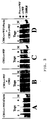
- the rFurin expression vector pSV-rFurink was co-transfected with the selection marker plasmid pUCSV-neo in the CHO-rvWF cell clone. Under selection conditions (500 ⁇ g G418 / ml), clones were identified which, in addition to rvWF, also express rFurin (CHO-rvWF / rFurin; Figure 2 I A, B).
- RFurin was also detected in the cell culture supernatant by means of Western blot analysis (FIG. 2 IB and II B). RFurin was visualized using the anti-hFurin mouse monoclonal antibody MON 148 (van Duijnhoven et al., Hybridoma 11: 71-86, 1992) and as a second antibody anti-mouse IgG-alkaline phosphatase-conjugated goat serum (Sigma A 4656 ).
- pro-vWF and furin coexpressed more frequently within 24 hours (every 8 hours), significant amounts of pro-rvWF could also be detected in cell culture supernatants from serum-free CHO-rvWF / rFurin cells ( Figure 2 IA ; "8 hours”).
- RFurin was detectable in the supernatants of the CHO-rvWF / rFurin cells (FIG. 2 IB); the greater the detectable amount of rFurin in the supernatant, the lower the amount of pro-vWF (FIG. 2 IA).
- rFurin was clearly detected in the supernatant of CHO-rvWF / rFurin cells with the aid of Western blot analysis and anti-hFurin monoclonal antibody (FIG. 2 IB).
- the degree of processing of pro-rvWF correlated with the detectable amount of rFurin in the supernatant: the more rFurin was detectable in the supernatant, the less pro-rvWF was present (see FIG. 2 I B with 2 I A).
- rFurin processing activity for recombinant pro-vWF in vitro, serum-free rFurin and pro-vWF-containing cell culture supernatants were mixed and incubated, and appropriate controls were carried out. Since reasonable expression yield of rwt-furin in CHO cells expressing only rFurin is not possible due to the interference of increased rFurin concentration with the viability of the cells, rFurin / vWF cells were used as the source for rFurin. Detectable amounts of rFurin could be achieved by coexpression.
- CHO-rvWF, CHO-rvWF / rFurin, CHO-rvWF and CHO in a 1: 1 ratio
- CHO-rvWF and CHO-rvWF / rFurin in a 1: 1 ratio
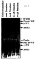
- FIG. 3 shows a complete processing of pro-rvWF only in those test batches that had either co-expressed rvWF / rFurin or that contained CHO-rvWF / rFurin and CHO-rvWF (FIG. 3A, lane 0-4, FIG. 3D, lane 2 and 4). While pro-rvWF in the 24-hour CHO-rvWF / rFurinatant was completely processed a priori to rvWF (FIG. 3A, lane 0), in CHO-rvWF cell culture supernatants 50% unprocessed vWF was still present in the cell culture supernatant (FIG. 3 B and C).
- Plasmid phAct- ⁇ EcoRIupstream was cloned by partial EcoRI digestion of plasmid phAct (Fischer et al., FEBS. Lett. 1994, 351: 345-348), filling in the protruding ends with Klenow enzyme, and religation. Plasmid phAct- ⁇ EcoRIupstream differs from phAct in that there is no EcoRI site on the 5 'side of the promoter; the EcoRI interface 3 'of the promoter or the intron, however, is still present in the MCS.
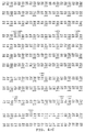
- the rFurin cDNA was deleted according to position 2127 of the nucleic acid sequence (FIG. 4) and thus at amino acid 709.
- the synthetic oligonucleotides # 2325 (5'-GATAAGCTT G TCGAC CATGG AGCTGAGGCC CTG-3 ') (SEQ.ID.NO. 3) and # 2819 (5'-AAGTCAT GAA TTC TTAC AGCAGCCCTG CGCGCAG-3') (SEQ. ID.NO.
- a DNA fragment was generated using the "template” pSV-rFurink (Example 1) with the polymerase chain reaction (PCR); this fragment contained the base pairs analogous to the first 709 amino acids of furin, followed by a translation termination triplet.
- the resulting rFurin- ⁇ TM fragment was cut at the flanking SalI and EcoRI restriction sites and inserted into the SalI / EcoRI cut expression vector phAct- ⁇ EcoRIupstream.
- the resulting plasmid is called prFurin- ⁇ TM.
- the nucleotide and amino acid sequence of rFurin- ⁇ TM is as SEQ.ID.NO. 5 and SEQ.ID.NO 6 reproduced.
- furin ⁇ TM fusion protein with a 3'-side fused affinity peptide six histidine residues (His tag) were added to the C-terminal end of rFurin ⁇ TM at amino acid 707 (base pair 2121): With the help of synthetic oligonucleotides # 2325 (SEQ.ID.NO. 3) and # 2823 (5'-CTA GAATTC A AT GATGATGATG ATGATG CCCT GCGCGCAGCC GTTGCCCC-3 ') (SEQ.ID.NO.
- the "template "pSV-rFurink (Example 1) by means of PCR a DNA fragment containing the base pairs analogous to the first 707 amino acids of furin, followed by six histidine residues and a translation termination triplet. After cutting the flanking SalI / EcoRI restriction sites, this fragment was inserted into the SalI / EcoRI sites of the vector phAct- ⁇ EcoRIupstream described above and plasmid prFurin ⁇ TM-His was obtained in this way.
- the nucleotide and amino acid sequence of rFurin ⁇ TM-His is SEQ.ID.NO. 8 and SEQ.ID.NO. 9 reproduced.
- a spacer consisting of Ala-Ala-Gly-Gly-Ala-Ala was inserted between the coding region of the deleted rFurin ⁇ TM 707 and the His tag sequence in order to prevent steric hindrance of the catalytic domain of furin and to increase mobility and To ensure functionality of the fusion protein when coupled to a column matrix.
- the spacer sequence was inserted into prFurin ⁇ TM-His by means of PCR with oligonucleotides # 2325 (SEQ.ID.NO. 3) and # 2820 (5'-CTA GAATTC A AT GATGATGATG ATGATG TGCAGCTCC ACCAGCTGCC CCTGCGCGCA GCCGTTGCCC C-3 ') SEQ.ID.NO. 10).
- prFurin ⁇ TM-His Analogously to the construction of prFurin ⁇ TM-His, this fragment was inserted into the SalI / EcoRI site of plasmid phAct- ⁇ EcoRIupstream. The resulting fusion protein was named prFurin ⁇ TM Spacer-His.
- the nucleotide and amino acid sequence of rFurin- ⁇ TM-Spacer-His is as SEQ.ID.NO. 11 and SEQ.ID.NO. 12 reproduced.
- a construct was produced in which, in addition to the transmembrane domain, the Cys-rich domain was also deleted. For this purpose, a termination triplet was inserted into the furin-coding sequence after nucleotide (amino acid 585).
- a DNA fragment with oligonucleotide # 2325 (SEQ.ID.NO. 3) as the 5 'primer and oligonucleotide # 2821 (5'-CTA GAATTCTAA CTGCTTTCTG GAGGTACGGG CAG-3') (SEQ.ID .NO, 15) generated as a 3 'primer and analogous to the construction of Furin ⁇ TM in phAct- ⁇ EcoRIupstream.
- the nucleotide and amino acid sequence of rFurin- ⁇ Cys is as SEQ.ID.NO. 13 and SEQ.ID.NO. 14 reproduced.
- rFurin ⁇ Cys fusion proteins with a His tag sequence according to amino acid 585 were produced.
- a PCR DNA fragment with oligonucleotide # 2325 (SEQ.ID.NO 3) as 5 'primer and oligonucleotide # 2810 (5'-CTA GAATTCTTAG TGGTGATGGT GATGATGACT GCTTTCTGGA GGTACGGGCA G-3') (SEQ.ID.NO. 16) generated as a 3 'primer and inserted into the plasmid pAct- ⁇ EcoRIupstream via the SalI / EcoRI interface.
- the resulting plasmid was named prFurin ⁇ Cys-His.
- the nucleotide and amino acid sequence of rFurin- ⁇ Cys-His is SEQ.ID.NO. 17 and SEQ.ID.NO.18.
- rFurin ⁇ Cys-Spacer-His was carried out by means of PCR with oligonucleotide # 2325 (SEQ.ID.NO. 3) as a 5 'primer and oligonucleotide # 2822 (5'-CTA GAATTCTTAG TGGTGATGGT GATGATGTGC AGCTCCACCA GCTGCACTGC TTCACGGQQG TTCACGGQQG TTCACGGGQC TTCGGGQQC TTCGGGQQTTCGGGQQTTCGGGQG T TTACGGGQC TTCGGGGQ TTCACGGGQCTTCGGGQQTTCGGGQQTTCGGGQG T TTACGGGQC TTCGGGGQ TTCACGGGQC TTCGGGQQTTCGGGQQTTCGGGQG T TTACGGGQTTCACGGGQC TTCGGGQQTTCGGGQQTTCGGGQG T TTACGGGQQTTCGGGQG .ID.NO.
- prFurin ⁇ cys-Spacer-10xHis was first partially cut with SauI and then completely with MamI. A DNA fragment was regenerated into the cleavage from the aged oligonucleotides # 3787 (5'-GGACCCCTCT GGCGAGTGGG TCCTCGAGAT TGAAAACACC AGCGAAGCCA ACAACTATGG GACGCT-3 ') (SEQ. ID. NO.
- the resulting plasmid was named prFurin ⁇ cys-Spacer-10xHis. It codes for a C-terminally deleted furin which contains the first 563 amino acids of furin, followed by a spacer consisting of a glutamic acid residue, five glycine residues and ten histidine residues.
- the supernatant transiently transfected 293 HEK cells with this construct showed processing activity in contrast to non-transfected cells in the fluorogenic substrate test.
- Furin or fusion protein activity was demonstrated using a low molecular weight peptide substrate Boc-Arg-Val-Arg-Arg-AMC.
- the action of furin removes AMC (7-amino-4-methylcoumarin) from the substrate.
- Soluble AMC has fluorescent properties compared to the peptide AMC, which can be used to determine the activity of furin.
- the fluorescence spectroscopic detection of the furin or fusion protein activity was carried out at 30 ° C. in stirred quartz cuvettes in a test volume of 2 ml.
- Permanent CHO cells transfected with the expression vector prFurin ⁇ TM-His constructed according to Example 3, were grown in roller bottles in the medium. The cell culture medium was removed, the cells were washed carefully with PBS and in serum-free Selection medium incubated further. The cell culture supernatant was then withdrawn every 24 hours and replaced with new medium. The supernatants were collected and pooled.
- 1 l cell culture supernatant was mixed with imidazole to a final concentration of 2 mM, 1 ml of Ni 2+ -NTA agarose was added and the suspension was incubated at 4 ° C with gentle shaking.
- the carrier- and matrix-bound fusion protein was separated from the supernatant by simply sitting down or centrifuging.
- the column matrix with bound rFurin ⁇ TM-His was resuspended in 10 ml buffer A (300mM NaCl, 10% glycerol, 1mM ⁇ -mercaptoethanol, 2mM imidazole, 5mM Hepes pH 7.0, 2mM CaCl 2 , 2mM MgCl 2 ) and then Centrifuged at 600 g for 10 min. The pellet was resuspended in the same buffer volume and loaded onto a chromatography column. The column was then washed with 5 ml of buffer A containing 2 mM imidazole and equilibrated with 5 ml of serum-free selection medium.
- RFurin ⁇ TM-His was eluted with buffer A containing 200 mM imidazole.
- the enzymatic activity of rFurin ⁇ TM-His was carried out analogously to Example 4. The result is summarized in Table 2. It was shown that rFurin ⁇ TM-His binds to the column matrix and can be eluted from the column again as an active molecule.
- Table 2 The activity of rFurin ⁇ TM-His before binding to the Ni 2+ -NTA matrix and after elution from the matrix
- the rFurin ⁇ TM-His-Ni 2+ column produced according to Example 5 was used to process rpro-vWF to rvWF.
- 20 ml of serum-free cell culture supernatant from CHO-rvWF cells adjusted to 2 mM imidazole were passed over the column.
- Aliquots of the eluate were analyzed by Western blot (according to Example 2).
- Samples of the cell culture supernatant that were not subjected to protease treatment served as controls.
- Samples of cell culture supernatants from CHO-rvWF showed an approximately 40% unprocessed pro-vWF, whereas only completely processed vWF was detected in samples of the column eluate.
- Stable CHO cell clones which secrete rFurin ⁇ cys-Spacer-10xHis were produced in accordance with Example 2 and a Ni 2+ -NTA matrix equilibrated with fresh, serum-free culture medium was loaded with conditioned medium of such clones.
- Adsorbed proteins were eluted from the column with elution buffer containing increasing concentrations of imidazole and aliquots of the individual fractions were separated in SDS-PAGE. The proteins were then stained with silver or detected by Western blot analysis (FIG. 6). The silver staining showed that a large number of proteins contained in the conditioned medium can bind to the matrix, but can be eluted again even at a low imidazole concentration.
- Contaminants were eluted from the column at an imidazole concentration of up to approximately 100 mM, whereas fractions which were eluted at a higher imidazole concentration contained only one protein. This protein was identified in a Western blot with anti-furin antibodies as rFurin ⁇ Cys-Spacer-10xHis.
- Imidazole-containing rFurin derivative fractions were therefore dialyzed against 20mM Hepes pH 7.0, 1mM CaCl 2 , 1mM ⁇ -mercaptoethanol at 4 ° C overnight.
- a comparison of the samples before and after dialysis shows that the activity of rFurin ⁇ Cys-Spacer-10xHis after removal of the imidazole by dialysis from 408 to> 1000 units (fraction eluted with 333 mM imidazole) or from 24 to> 1000 units ( Fraction eluted with 780 mM imidazole) could be restored.
- the rapid and very clean purification of the histidine-tagged rFurin derivatives using the described method thus enables industrial process development, for example purified Pro-rvWF molecules (or other target proteins) to be processed in solution by epitope-tagged rFurin derivatives.
- the two reactants can be selectively separated from one another by means of a Ni 2+ -NTA matrix, the completely processed substrate being removed in the flow and the r-furin derivative being bound to the matrix.
- the rFurin derivative is available for a new processing reaction. In this way it is possible to process a comparatively large amount of Pro-rvWF using repeated such rFurin derivative "recycling" steps with relatively little rFurin derivative in vitro.
- Ni 2+ -NTA matrix (Quiagen) equilibrated in serum-free cell culture medium at 4 ° C. with 10 ml of imidazole-free CHO-rFurin ⁇ Cys-Spacer-10xHis cell culture supernatant, or as a negative control with conditioned medium. manipulated CHO cells, loaded. Then the matrix was washed three times at room temperature with serum-free cell culture medium and the fluorogenic substrate was then brought into contact with immobilized rFurin ⁇ Cys-Spacer-10xHis at 30 ° C. As a positive control, CHO-rFurin ⁇ Cys-Spacer-10xHis cell culture supernatant was tested for its ability to convert fluorogenic substrate into solution.
- Ni 2+ NTA matrix rFurin ⁇ Cys spacer 10xHis transposed more than 1000 units of fluorogenic substrate and thus comparable to the reaction of the fluorogenic substrate by non-immobilized rFurin derivative.
- Column matrix "loaded" with conditioned medium of unmanipulated CHO cells did not cleave the fluorogenic substrate.
- rFurin ⁇ Cys-Spacer-10xHis mediates proteolytic activity not only in solution but also in the immobilized state.
- the substrate e.g. of rFIX, rFX, rvWF, rPC, rPS, etc.
- column matrices can be used in which the bound rFurin derivative is connected to the matrix via a longer column arm, such as for example with tentacle gels (see Example 10).
- pro-rvWF precursor protein was purified to homogeneity (with an rvWF antigen: total protein ratio of 2) and 10 ⁇ g pro-rvWF with 1340 U purified rFurin ⁇ Cys-Spacer-10xHis (see Example 7) in buffer (0.87mM CaCl 2 , 17mM Hepes pH 7.0, 0.87mM mercaptoethanol) at 37 ° C.
- buffer 0.87mM CaCl 2 , 17mM Hepes pH 7.0, 0.87mM mercaptoethanol
- the negative control an unmanipulated CHO cell culture supernatant (without rFurin ⁇ Cys-Spacer-10xHis) was prepared in an analogous manner on the Ni 2+ NTA matrix.
- FIG. 7 shows the processing of purified pro-rvWF by purified rFurin ⁇ Cys-Spacer-10xHis under defined conditions. While in the presence of the rFurin derivative pro-vWF was completely processed after four hours (FIG. 7, top), no processing was observed in the absence of the rFurin derivative (FIG. 7, bottom). When incubated with the rFurin derivative for a longer time, processed rvWF molecules were no longer proteolytically degraded and the molecular integrity of the mature rvWF molecules remained stable over the entire period of rFurin exposure (FIG. 7, top).
- the Fractogel EMD® tentacle gel was loaded with Ni 2+ ions according to the manufacturer's instructions and equilibrated with fresh serum-free cell culture medium.
- the column was then loaded with serum-free CHO-rFurin ⁇ Cys-Spacer-10xHis supernatant. Washing steps were carried out using serum-free cell culture medium containing increasing concentrations of imidazole up to 40mM.
- the pro-protein substrate was then passed over the column as a serum-free CHO supernatant. The processing of pro-protein was increased by means of Western blot analysis with specific vWF antiserum Protein detected in the flow through the column.
- oligonucleotide # 2912 (5'-ATTACAATTGCTGCAGGGATCCAC-3') (SEQ .NO.27) as a 3 'primer, a DNA fragment is amplified, which contains the 1,467 kB FX-coding sequence and 39 bp of the 3' untranslated region, flanked by an XhoI site, at the 5 'end and an MfeI -Interface at the 3 'end contains.
- primer # 2911 installed the ACC sequence in front of the ATG of the FX, so that an optimal Kozak transcription sequence is created.
- This PCR product was then cloned as an XhoI / MfeI fragment into the expression vector phAct cut with SalI and EcoRI. The resulting expression plasmid was designated phAct-rFX ( Figure 8).
- the expression vector phAct comprises the human beta-actin promoter, 78bp 5'UTR and the intron, a multiple cloning interface and the SV40 polyadenylation site.
- dhfr-deficient CHO cells were co-transfected with the expression plasmid phAct-rFX and the selection marker plasmid pSV-dhfr, as described in Example 2.
- the cell cultures were treated with serum-free selection medium in the presence of 10 ⁇ g / ml vitamin K for 24 hours incubated for a long time.
- the expression of rFX in the resulting cell clones was detected on the basis of the amount of antigen (ELISA) and the recombinant protein was subsequently characterized using SDS-PAGE (as described in Example 2) (FIGS. 9 A and B).
- the recombinant FX protein is in the form of a light chain (LC) of 22 kD and a heavy chain (HC) of 45 kD, which is identical to that are plasmatic factor X protein.
- LC light chain
- HC heavy chain
- a protein band at 75 kD can be seen which corresponds to the single-chain (SC) molecule and whose presence in FX-transfected CHO cells (Wolf et al. J. Biol. Chem. 266: 13726-13730, 1991) and in human plasma (Fair et al. Blood 64: 194-204, 1984).
- the initial clones were amplified with increasing amounts of methotrexate and then subcloned until stabilization.
- the expression could be increased from approx. 200-500 ng / 10E6 cells or 1 ⁇ g / ml to 30-50 ⁇ g / 10E6 cells or 100 ⁇ g / ml per 24 hours.
- Western blot analysis of these highly expressing cell clone supernatants shows an enrichment of the single-chain rFX molecule and the presence of additional forms of the light chain.
- the 22kD form of the light chain which corresponds to the plasmatic form (completely carboxylated without propeptide)
- the heterogeneity of the light chain in these clones could be attributed to incomplete cleavage of the propeptide (approx. 50% of the rFX material) and to under-carboxylation (approx. 50% of the rFX) by means of N-terminal sequencing of the recombinant material.
- the 21kD protein is an undercarboxylated propeptide-containing form and the 20kD protein is a undercarboxylated propeptide-free form of the light chain.
- the CHO-rFX / CHO-rFurin ⁇ TM-His exhibits almost complete processing after only two hours of incubation at 37 ° C. (FIG. 10 A, lane 7; FIG. 10 B, lane 8) in front.
- Single-chain rFX is largely implemented in the light and heavy chain shape. In the light chain area, only the processed pro-peptide-free forms of 22kD (carboxylated form) and 20kD (under-carboxylated form) were found in a ratio of approx. 50:50.
- the amount of rFXa and the color intensity are proportional to each other, the amount of rFX / ml cell culture supernatant that can be activated to rFXa can be determined using a calibration line interpolated from values from a plasma dilution series. With these results and the known amount of rFX antigen (ELISA data), the percentage of rFactor X activated in factor Xa can be calculated in%. The results are shown in Table 4.
- CHO-rFX incubated with CHO supernatants (without rFurin) as a control showed no significant increase in rFXa activity even after 6 hours, which was between 150-200mU / ml and corresponded to 19-25% functional rFX due to the experimental fluctuations.
- CHO-rFX was incubated with CHO-rFurin ⁇ TM-His after two hours there was a significant increase in rFX activity, which reached 344mU / ml or 43% functional part of CHO-rFX after 6 hours.
Landscapes
- Health & Medical Sciences (AREA)
- Chemical & Material Sciences (AREA)
- Life Sciences & Earth Sciences (AREA)
- Organic Chemistry (AREA)
- Genetics & Genomics (AREA)
- Engineering & Computer Science (AREA)
- Zoology (AREA)
- Molecular Biology (AREA)
- Biomedical Technology (AREA)
- Bioinformatics & Cheminformatics (AREA)
- General Health & Medical Sciences (AREA)
- Wood Science & Technology (AREA)
- Medicinal Chemistry (AREA)
- Biochemistry (AREA)
- General Engineering & Computer Science (AREA)
- Microbiology (AREA)
- Biotechnology (AREA)
- Hematology (AREA)
- Toxicology (AREA)
- Gastroenterology & Hepatology (AREA)
- Biophysics (AREA)
- Proteomics, Peptides & Aminoacids (AREA)
- Peptides Or Proteins (AREA)
- Preparation Of Compounds By Using Micro-Organisms (AREA)
- Micro-Organisms Or Cultivation Processes Thereof (AREA)
- Medicines That Contain Protein Lipid Enzymes And Other Medicines (AREA)
Abstract
Description
- Die Erfindung betrifft ein neues Fusionsprotein, abgeleitet von Furin oder einem Furinanalogon, sowie ein Verfahren zur Herstellung von Proteinen aus Pro-Proteinen durch das Fusionsprotein, insbesondere von von Willebrand-Faktor aus pro-von Willebrand-Faktor.
- Furin, auch PACE genannt, gehört neben PACE4, PC1/PC3, PC2, PC4 und PC5/PC6 zur Gruppe der Subtilisin-ähnlichen Serin-Proteasen, die eine wichtige Rolle bei der Spaltung von Pro-Proteinen, speziell im sekretorischen Syntheseweg, spielen (Van de Ven et al., Crit. Rev. Oncogen., 4:115-136, 1993). Pro-Proteine werden post-translatorisch, intrazellulär im Golgi-Apparat durch die endogene Protease in ihre reife Form prozessiert. Die Protease-Spaltstelle weist eine Erkennungssequenz auf, die durch die Aminosäuresequenz Arg-X-Lys/Arg-Arg gekennzeichnet ist. Die Protease Furin spaltet Pro-Proteine spezifisch nach dieser Konsensus-Sequenz (Hosaka et al., J.Biol.Chem. 266:12127-12130, 1991).
- Die DNA- und Aminosäuresequenz des humanen und des murinen Furins, sowie weitere Proteine mit Subtilisin-ähnlicher Proteasefunktion sind aufgeklärt (Roebroek et al., Mol. Biol. Rep. 11: 117-125, 1986, Roebroek et al., EMBO J. 5:2197-2202, 1986, Barr et al., DNA Cell Biol. 10:319-328,1991, Van den Ouweland et al., Nucleic Acids Res. 17:7101-7102, 1989, Van den Ouweland et al., Nucleic Acids Res. 18:664, 1990, Smeekens et al. 1990, J. Biol. Chem. 265:2997-3000; Smeekens et al 1991, Proc. Natl. Acad. Sci. USA. 88; 340-344; Kiefer et al 1991, DNA Cell. Bio. 10: 757; Nakayama et al 1992, J. Bio. Chem. 267:5897-5900, Hatsuzawa et al. 1990. J. Biol. Chem. 265:22075-22078). Das humane fur-Gen kodiert für ein Protein bestehend aus 794 Aminosäuren, wobei einzelnen, charakteristischen Bereichen bestimmte Funktionen zugeordnet werden können: ein katalytisches Zentrum, eine Mitteldomäne, eine Cystein-reiche Region, eine transmembrane und eine cytoplasmatische Domäne (Van de Ven et al., Crit. Rev. Oncogen., 4:115-136, 1993).
- Intaktes Furin wird in das Membransystem des Golgi-Apparates eingebaut und ist dort funktionell aktiv (Bresnahan et al., J. Cell Biol. 111:2851-2859, 1990). Eine trunkierte Form des überexprimierten nativen Furins von 75-80 kD konnte im Zellüberstand als sezerniertes Protein detektiert werden (Wise et al., Proc. Natl. Acad. Sci. USA 87:9378-9382, 1990). Dieses natürlich sekretierte Furin ist als "shed furin" bekannt (Vidricaire et al., Biochem. Biophys. Res. Comm. 195:1011-1018, 1993) und wird N-terminal des transmembranen Teils gespalten (Vey et al. J. Cell Biol. 127:1829-1842, 1994).
- Gentechnisch verkürztes Furin, bei dem der kodierende Teil der transmembranen und cytoplasmatischen Domäne deletiert ist, kann ebenfalls exprimiert und entsprechend sezerniert werden. Solche N-terminalen Deletionen wurden für Aminosäuren Δ714-794 (Leduc et al. J. Biol. Chem. 267:14304-14308, 1992, Molloy et al. J. Biol. Chem. 267:16396-16402, 1992) und für Aminosäuren Δ716-794 ("Sol-PACE") erstellt (Wasley et al. 1993. J. Biol. Chem. 268: 8458-8465, Rehemtulla et al. Blood 79:2349-2355, 1992) und für Aminosäure Δ705-794 (Hatsuzawa et al. 1992. J. Biol. Chem. 267: 16094-16099) beschrieben.
- Furinmutanten, die zusätzlich eine Deletion der cysteinreichen Region aufweisen wurden ebenfalls beschrieben (Hatsuzawa et al. 1992. J. Biochem. 101:296-301, Creemers et al. 1993. J. Biol. Chem. 268:21826-21834).
- Die endoproteolytische Aktivität von Furin und seine Selektivität für basische Aminosäuren wurde erstmals in Experimenten mit pro-von Willebrand-Faktor (pro-vWF) festgestellt. Pro-vWF besteht aus einem Propolypeptid mit 741 Aminosäuren und maturem von Willebrand-Faktor (vWF) mit 2050 Aminosäuren (Verweij et al., EMBO J. 5:1839-1847, 1986). Die Freisetzung von maturem vWF aus pro-vWF resultiert aus einer proteolytischen Spaltung nach Arg763. Transfektion von pro-vWF cDNA in eukaryotischen Expressionsvektoren führt zur Produktion von äquimolaren Mengen des 360 kD pro-vWF und des 260 kD reifen vWF im Zellkulturüberstand. vWF wird in seine reife Form in transfizierten Zellen vermutlich durch endogen vorkommendes Furin prozessiert (Wise et al., Proc. Natl. Acad. Sci. USA 87:9378-9382, 1990, Van de Ven et al., Mol. Biol. Rep. 14:265-275, 1990).
- Zu den weiteren Pro-Proteinen, die von Furin, bzw. von Subtilisin-ähnlichen Enzymen gespalten werden, gehören eine Reihe von Hormonen und Wachstumsfaktoren (z.B. Proaktivin A, Hepatozyten-Wachstumsfaktor), Plasmaproteinen (Albumin, Faktor VII, Faktor IX, Faktor X), Rezeptoren (Insulin-Prorezeptor), viralen Proteinen (z. B. HIV-1 gp160, Influenza Virus Hämagglutinin) sowie bakteriellen Proteinen (Diphterie-Toxin, Anthrax-Toxin) (Decroly et al., J. Biol. Chem. 269:12240-12247, 1994, Stieneke-Gröber et al., EMBO J. 11:2407-2414, 1992, Barr, Cell 66:1-3, 1991, Wasley et al., J. Biol. Chem. 268:8458-8465, 1993, Klimpel et al., Proc. Natl. Acad. Sci. USA 89:10277-10281, 1992, Tsuneoka et al., J. Biol. Chem. 268:26461-26465, 1993, Bresnahan et al., J. Cell Biol. 111:2851-2859, 1990, Hosaka et al., J. Biol. Chem. 266:12127-12130, 1991, Vey et al. J. Cell. Biol. 127: 1829-1842, 1994).
- Durch Koexpression der für intaktes Furin und für ein Pro-Protein kodierenden Nukleinsäuresequenzen in eukaryotischen Zellkulturen wurde eine erhöhte Prozessierung der Pro-Proteine in vivo erreicht. Dies wurde zum Beispiel für pro-Faktor IX (Wasley et al., J. Biol. Chem. 268:8458-8465, 1993) und pro-vWF (WO 91/06314, Van de Ven et al. Mol. Bio. Rep. 14:265-275, 1990, Rehemtulla et al., Blood 79:2349-2355, 1992) gezeigt.
- Neben der Koexpression von intaktem Furin mit Pro-Proteinen gibt es ebenfalls Ansätze, trunkiertes Furin mit Pro-Proteinen gemeinsam zu exprimieren. Deletiertes Furin ist bei Koexpression in vivo enzymatisch aktiv und wird sezerniert; die enzymatische Aktivität solcher Deletionsmutanten konnte unter anderem bei der Prozessierung von pro-Faktor IX (Wasley et al., J. Biol. Chem. 268:8458-8465, 1993) und pro-vWF (Rehemtulla et al., Blood 79: 2349-2355, 1992) nachgewiesen werden. Koexpressionsexperimente mit Furin-Deletionsmutanten zeigten, daß der transmembrane und der cytoplasmatische Teil des Proteins für die katalytische Funktion nicht wesentlich sind (Rehemtulla et al., Proc. Natl. Acad. Sci. USA 89: 8235-8239, 1992).
- WO 91/06314 offenbart die rekombinante Expression von Furin in prokaryotischen und eukaryotischen Zellen, die Herstellung von Furin-Fusionsproteinen, -Deletionsmutanten und -Fragmenten, die Reinigung von rekombinant hergestelltem Furin, sowie die mögliche Verwendung von gereinigtem Furin für die Prozessierung von Pro-Proteinen in vitro im allgemeinen.
- WO 92/09698 beschreibt die Expression von PACE (Furin), die Koexpression mit inaktiven Vorstufen von Proteinen wie z.B. pro-vWF, sowie die Herstellung von Fusionsproteinen. Zur Anreicherung von PACE wird dabei vorgeschlagen, sekretionsfähiges PACE über konventionelle Methoden zu isolieren.
- Stieneke-Gröber et al. (EMBO J. 11:2407-2414, 1992) beschreiben die in vitro-Spaltung von Influenza-Virus-HA-Protein durch gereinigtes Furin. Decroly et al. (J. Biol. Chem. 269: 12240-12247, 1994) beschreiben die in vitro-Spaltung von HIV gp160 durch Furin.
- Bei Versuchen mit C-terminal verkürztem Furin konnte in vitro die Spaltung von Proalbumin und Komplement Pro-C3 (Oda et al., Biochem. Biophys. Res. Commun. 189:1353-1361, 1992), Anthrax-Toxin (Klimpel et al. Proc. Natl. Acad. Sci. USA 89:10277-10281, 1992), Diphtherie-Toxin (Tsuneoka et al., J. Biol. Chem. 268: 26461-26465, 1993) und pro-Faktor IX (Wasley et al. J. Biol. Chem. 268: 8458-8468, 1993, Bristol et al., Biochemistry 33: 14136-14143, 1994) erfolgreich durchgeführt werden.
- In vitro-Prozessierung von pro-vWF durch Furin konnte bisher nicht gezeigt werden. Rehemtulla et al. (Blood 79:2349-2355, 1992 und Proc. Natl. Acad. Sci. USA 89:8235-8239, 1992) beschreiben, daß durch Mischen von Überständen von Zellen, transfiziert mit pro-vWF bzw. deletiertem Furin ("PACE SOL"), pro-vWF nicht zu vWF prozessiert wird. Im Gegensatz dazu konnten mittels gereinigtem "PACE SOL" in vitro sowohl synthetische Substrate (Rehemtulla et al., Proc. Natl. Acad. Sci. USA 89:8235-8239, 1992), als auch pro-Faktor IX (Bristol et al., Biochemistry 33:14136-14143, 1994) gespalten werden. Für pro-vWF wurde weiters wiederholt postuliert, daß er in vitro durch trunkiertes Furin nicht in seine reife Form prozessiert wird (Rehemtulla et al., Proc. Natl. Acad. Sci. USA 89:8235-8239, 1992 und Blood 79:2349-2355, 1992), während unter analogen Bedingungen Faktor IX gespalten wird (Wasley et al. 1993. J. Biol. Chem. 268:8458-8465).
- Um bei der rekombinanten Herstellung von Proteinen aus Pro-Proteinen hohe Ausbeuten an vollständig prozessierten Proteinen zu erhalten, wurde es gemäß dem Stand der Technik als notwendig angesehen, eine genügend große Menge an Furin zu exprimieren und zu isolieren, oder das Pro-Protein und Furin zu koexprimieren.
- Bei der rekombinanten Expression von Furin alleine, aber auch bei der Koexpression von Furin mit einem Pro-Protein im großtechnischen Ansatz in Zellkultur tritt allerdings das Problem auf, daß eine hohe Expression der Protease toxisch für die Zellen ist (Creemers 1994), wodurch nur eine geringe Ausbeute an Furin und an reifem Protein möglich wird. So konnte in Koexpressionsstudien gezeigt werden, daß Zellklone, die Furin hoch exprimieren und das Pro-Protein effizient prozessieren, zu weitaus geringeren Zelldichten wachsen im Vergleich zu Zellen, die kein Furin exprimieren. Daraus resultiert eine schlechtere Gesamtausbeute an prozessiertem Protein. Um eine hohe Ausbeute an reifem Protein zu erhalten, muß daher eine sehr lange Kultivierungszeit in Kauf genommen werden; der Bedarf an Kultivierungsgefäßen und -geräten ist dadurch groß, was in der Folge auch erhöhte Kontaminationsprobleme mit sich bringen kann.
- Bisher konnten nach rekombinanter Expression Furin oder Furinderivate nur immunologisch im Westernblot detektiert werden (Molloy et al. 1994. EMBO J. 13:18-33). Versuche, Furin oder Furinderivate hoch zu exprimieren, gelangen bisher nur im Baculovirus-Expressionssystem, wobei eine 20-30fach höhere Expression als in transfizierten Säugerzellen postuliert wurde (Bravo et al., 1994,. J. Biol. Chem. 269:25830-25837). Allerdings ist, trotz der relativ hohen Ausbeute an Furin im Vergleich zu anderen Zellsystemen, das Wachstum dieser virusinfizierten Zellen aufgrund der Zellyse als Folge der Virusvermehrung beschränkt. Eine gute Expression, sowie die Isolierung und Reinigung von Furin oder Furinderivaten, stellt für den immer breiter werdenden Anwendungsbereich von Furin bzw. von Furinderivaten z.B. bei der rekombinanten Herstellung von furin-prozessierten Proteinen aus Pro-Proteinen, eine große Bedeutung und Notwendigkeit dar.
- Da eine Überexpression der Protease das Wachstum von kontinuierlich wachsenden Zellkulturen negativ beeinflußt, wurden Lösungsansätze gesucht, um den toxischen Einfluß von Furin auf die Zellen zu reduzieren. Weiter besteht ein Bedarf nach einem verbesserten Verfahren zur Prozessierung von furin-aktivierten Proteinen aus Pro-Proteinen, insbesondere für die großtechnische Herstellung von rekombinanten Blutfaktoren, wie etwa pro-vWF.
- Das Ziel der vorliegenden Erfindung ist daher ein Verfahren zur Herstellung von Furin oder einem Furinderivat aus einer kontinuierlich wachsenden, rekombinanten Zellkultur unter Aufrechterhaltung der enzymatischen Aktivität des Furins oder Furinderivats zur Verfügung zu stellen, ohne daß die Zellkultur wesentlichen Schaden durch die erhöhte proteolytische Aktivität nimmt.
- Ein weiteres Ziel der vorliegenden Erfindung besteht darin, ein verbessertes Verfahren zur furin-abhängigen proteolytischen Spaltung von Pro-Proteinen zu Proteinen, insbesondere ein neues Verfahren zur Prozessierung von pro-von Willebrand-Faktor zu reifem, aktivem von Willebrand-Faktor, zur Verfügung zu stellen.
- Diese Aufgaben werden erfindungsgemäß gelöst durch Zurverfügungstellung von neuen Fusionsproteinen, die aus einem Furinderivat oder einem Derivat eines Furinanalogen, fusioniert mit einer heterologen Sequenz, welche zur Adsorption des Furins an einen festen Träger befähigt, bestehen, bei dem gegebenenfalls der C-terminale Bereich durch Deletion entfernt ist und durch eine heterologe Sequenz ersetzt ist.
- Die erfindungsgemäßen Furin- oder Furinanalogen umfassen eine heterologe Sequenz, wie etwa ein heterologes Protein, Polypeptid oder funktionell aktives Peptid, insbesondere ein Affinitätspeptid. Erfindungsgemäß ist die heterologe Sequenz so ausgewählt, daß sie eine hohe Affinität oder eine spezifische Bindungseigenschaft für eine funktionelle Gruppe eines Trägers besitzt. Das heterologe Protein oder Polypeptid sollte dabei ein immunologisch gut charakterisiertes Protein sein, gegen das beispielsweise Antikörper zur Kopplung an einen festen Träger zur Verfügung stehen. Erfindungsgemäß kann die Adsorption an den festen Träger z.B. durch kovalente Bindung oder über Affinität erfolgen. Die Proteine oder Polypeptide können dabei abgeleitet sein von z.B. ß-Galaktosidase, c-myc-Produkt, Glutathion S-Transferase, Avidin und die Lysin-bindende Kringeldomäne von Plasmaproteinen, wie z.B. von Plasminogen (Evan et al. Mol. Cell. Biol. 5: 3610-3616, 1985; Duijhoven et al., Hybridoma 11:71-86, 1992). Die funktionell aktiven Peptide, welche in der heterologen Sequenz liegen, können aus einer Anreihung von mehreren, gleichen oder verschiedenen Aminosäuren bestehen.
- Eine bevorzugte Ausführungsform der vorliegenden Erfindung betrifft ein Fusionsprotein, dessen heterologer Sequenzanteil ein Peptid, das mit einem festen Träger eine kovalente Bindung eingehen kann, oder ein Poly-Histidin, das eine hohe Affinität insbesondere zu Schwermetallionen oder spezifischen anti-Poly-Histidin-Antikörpern besitzt, umfaßt.
- Durch die C-terminale Deletion der cytoplasmatischen und transmembranen Region wird lösliches Furin oder Furinanaloges exprimiert, das aus den rekombinanten Zellen sekretiert wird. Gegebenenfalls kann bei dem Furinderivat oder dem Derivat des Analogons zusätzlich die Cystein-reiche Region deletiert sein. Die enzymatische Aktivität des deletierten Proteins ist erfindungsgemäß im Vergleich zum kompletten Protein im wesentlichen unverändert.
- Unter einem Derivat eines Furinanalogen wird im Rahmen der vorliegenden Erfindung jedes Furin-ähnliche Protein verstanden, das gleiche oder ähnliche biologische Aktivität wie Furin aufweist oder Sequenzhomologie zu Furin besitzt. Dies gilt in erster Linie für das mit Furin idente PACE, aber auch PACE4, PC1/PC3, PC2 und PC4 sind in die vorliegende Erfindung miteingeschlossen.
- Es sollen aber auch alle weiteren durch Insertion, Deletion oder Austausch von Aminosäuren bzw. Nukleotiden aus Furin bzw. dem Furinanalogen generierten Proteine bzw. Nukleinsäuren, welche Furin-ähnliche biologische Aktivität haben, in den Rahmen der vorliegenden Erfindung fallen.
- Fusionsproteine, bestehend aus einem deletierten Furinanteil und einer heterologen Sequenz, sind zwar im Stand der Technik beschrieben, diese bekannten Fusionsproteine eignen sich jedoch nicht zur Lösung der erfindungsgemäßen Aufgabe. So offenbaren Duijhoven et al. (Hybridoma 11:71-86, 1992) N-terminale Furin-deletionsmutanten fusioniert mit Glutathion S-Transferase. Ebenso wurde die Pre-Pro-Sequenz von PACE(Furin) an den N-Terminus der leichten Kette der bovinen Enterokinase fusioniert (LaVallie et al. J. Biol. Chem. 268:23311-23317, 1993). Fusionsproteine enthaltend ein sogenanntes FLAG Epitop-"Tag", inseriert an das N-terminale Ende des katalytischen Zentrums des Furins nach Aminosäure Arg107, sowie murine Furinmutanten, bei denen der transmembrane und cytoplasmatische Bereich C-terminal nach Aminosäure 704 deletiert und durch ein Antikörper-Epitop des HSV Glykoproteins D ersetzt wurde, wurden ebenfalls beschrieben (Molloy et al. EMBO J. 13: 18-33, 1994; Matthews et al. Protein Science 3: 1197-1205, 1994). Diese Furinderivate wurden für die Detektion von Furin während seiner Reifung und Prozessierung im Golgi-Apparat oder zur Detektion von furinhaltigen Zellkulturüberständen mittels Immunblot eingesetzt und sind zur biotechnologischen Anwendung für die Zwecke der vorliegenden Erfindung, insbesondere zur Spaltung von pro-vWF in vWF, in vitro ungeeignet. Solche Furinderivate sollen daher nicht in den Rahmen der vorliegenden Erfindung fallen.
- Gemäß einem besonderen Aspekt der vorliegenden Erfindung besteht das Fusionsprotein aus einem Furin-Derivat, dessen C-terminale cytoplasmatische und transmembrane Domäne und gegebenenfalls Cystein-reiche Region deletiert und durch ein Affinitätspeptid ersetzt worden ist. Dabei wurde ein Furinderivat oder ein Derivat eines Furinanalogons mit einem funktionellen Peptid, insbesondere aus mehreren Histidin-Resten, vorzugsweise aus 3 bis 20 Histidin-Resten, besonders bevorzugt aus 6 bis 15 aufeinander folgenden Histidin-Resten, fusioniert. Die Verwendung von C-terminal an ein Protein fusionierten Affinitätspeptiden in Form von Poly-Histidin-Resten (sogenanntes "His-Tag") zur Reinigung und/ oder zu Funktionsstudien von Proteinen wurde beschrieben (Janknecht et al., Proc. Natl. Acad. Sci. USA 88:8972-8976, 1991, Hoffmann et al., Nucleic Acids Res 19:6337-6338, 1991, EP 0 282 042).
- In einer bevorzugten Ausführungsform der vorliegenden Erfindung wurde ein Furinderivat mit deletierter cytoplasmatischer und transmembraner Region mit einem Peptid aus mehreren Histidin-Resten fusioniert. Gemäß einer besonderen Ausführungsform ist Furin derart modifiziert, daß die für den transmembranen und cytoplasmatischen Teil kodierenden Sequenzen (Aminosäure 708 bis 794) deletiert (rFurinΔTM) und nach der Aminosäure 707 die kodierende Sequenz für sechs Histidin-Reste angehängt werden. Das so erhaltene Fusionsprotein wurde mit rFurinΔTM-His bezeichnet.
- Eine weitere Ausführungsform der Erfindung betrifft ein Fusionsprotein, bei dem ein Furinderivat oder Derivat eines Furinanalogons mit deletiertem transmembranen und cytoplasmatischen Teil sowie der Cystein-reichen Region mit einem Affinitätspeptid aus mehreren Histidin-Resten fusioniert wurde.
- In einer bevorzugten Ausführungsform wurde ein Furinderivat, bei dem neben der transmembranen und cytoplasmatischen Domäne die Cystein-reiche Region nach Aminosäure 585 deletiert war, mit sechs Histidin-Resten fusioniert. Dieses Fusionsprotein wurde mit rFurinΔCys-His bezeichnet.
- Die Fusion eines Furinderivats oder eines Derivats eines Furinanalogons mit einer heterologen Sequenz soll gemäß der vorliegenden Erfindung so ausgeführt sein, daß die katalytische Funktion des Furins oder Furinanalogen im wesentlichen nicht beeinträchtigt wird.
- Gemäß einem besonderen Aspekt der vorliegenden Erfindung wird daher zwischen die Sequenz des Furinderivats oder eines Derivat eines Furinanalogons und die heterologe Sequenz ein kurzer Peptid-Spacer inseriert, um das katalytische Zentrum des Furinderivats sterisch nicht zu behindern.
- Dies ist insbesondere dann von Vorteil, wenn eine direkte Fusion der Cystein-reichen Region des Furinderivats mit einem Peptid die enzymatische Aktivität des Furinderivats beeinträchtigt, die Kopplung an den Träger durch chemische oder sterische Wechselwirkungen behindert oder mit einer effizienten Prozessierung des Pro-Proteins interferiert. Dieser kurze Peptid-Spacer, der vorzugsweise aus 5 bis 15 Aminosäuren besteht, ist im speziellen aus kleinen, flexiblen Aminosäuren, wie Alanin oder Glycin, zusammengesetzt. In einer besonderen Ausführungsform wird zwischen die Furin-kodierende Sequenz und die heterologe Sequenz von 6 Histidin-Resten ein Spacer bestehend aus Ala-Ala-Gly-Gly-Ala-Ala inseriert. Die so entstandenen Fusionsproteine wurden mit rFurinΔTM-Spacer-His und rFurinΔCys-Spacer-His bezeichnet.
- Das erfindungsgemäße Fusionsprotein weist über seinen Protein-, Polypeptid- oder Peptidanteil spezifische Bindungseigenschaften zu einem festen Träger auf. Als feste Träger können dabei Träger mit (Schwer)Metallionen, wie etwa Ni2+, Co2+,Mg2+, Li2+ oder mit Antikörpern eingesetzt werden.
- Gemäß einem besonderen Aspekt der vorliegenden Erfindung wird das Fusionsprotein durch seine Bindung an den festen Träger immobilisiert. Bevorzugterweise wird das erfindungsgemäße Fusionsprotein mit einem heterologen Sequenzanteil bestehend aus mehreren Histidin-Resten aufgrund dessen Affinität zu (Schwer)-Metallionen, insbesondere zu Ni2+, oder zu spezifischen anti-Poly-Histidin-Antikörpern, gebunden.
- In einer besonderen Ausführungsform werden die Konstrukte rFurinΔTM-His, rFurinΔCys-His, rFurinΔTM-Spacer-His und rFurinΔCys-Spacer-His über ihre Affinität zu Ni2+-Ionen, oder zu einem Antikörper an den Träger gebunden. Der feste Träger kann gemäß der vorliegenden Erfindung als Matrix zur Verfügung gestellt werden. Die Bindung an die Matrix erfolgt dabei über die affinen Gruppen des festen Trägers, so daß das Fusionsprotein frei zugänglich ist. Dies ist insbesondere dann von Vorteil, wenn das an den Träger immobilisierte Fusionsprotein für die proteolytische Spaltung von Pro-Proteinen eingesetzt wird und dieser Prozeß gebunden an einer Matrix erfolgt.
- Als Matrix, an der der Affinitätsträger adsorbiert, können natürliche und synthetische Matrices, wie Sepharose, Agarose, Gelatine, Acrylate etc. verwendet werden. Die feste Matrix trägt je nach Versuchsansatz eine funktionelle Gruppe, die den Träger spezifisch binden kann.
- Ein weiterer Aspekt der vorliegenden Erfindung betrifft die rekombinante DNA kodierend für die erfindungsgemäßen Fusionsproteine.
- Zur Konstrukion der Fusionsproteine wird die kodierende Nukleotidsequenz von Furin oder einem Furinanalogen derart modifiziert, daß die kodierende Sequenz für den cytoplasmatischen und transmembranen Bereich, und gegegebenenfalls für die Cystein-reiche Region deletiert wird (Van der Ven et al. 1993. Crit.Rev. Oncogen 4:115-136). Dies erfolgt durch aus dem Stand der Technik bekannte gentechnische Methoden, wie spezifischen Restriktionsverdau mit Endonukleasen, Ligation oder PCR. Die so hergestellten Deletionsmutanten werden dann mit einer heterologen Sequenz ebenfalls über bekannte Techniken, fusioniert.
- Die erfindungsgemäßen Fusionsproteine können ebenfalls durch chemische Synthese hergestellt werden.
- Die Fusionsproteine werden vorzugsweise durch rekombinante Expression hergestellt. Die gentechnische Herstellung kann mit allen gängigen eukaryontischen Expressionsystemen, wie z.B. permanenten Zellinien oder viralen Expressionssystemen, erfolgen. Die permanenten Zellinien werden hergestellt durch stabile Integration der Fremd-DNA in das Wirtszellchromosom z.B. Vero, MRC5, CHO, BHK, 293, Sk-Hepl, insbesondere Leber- und Nierenzellen, oder durch einen episomalen Vektor, abgeleitet von z.B. Papilloma Virus. Virale Expressionssysteme, wie Vaccinia Virus, Baculovirus oder retrovirale Systeme können ebenfalls eingesetzt werden. Als Zellinien werden allgemein Vero, MRC5, CHO, BHK, 293, Sk-Hepl, Drüsen-, Leber- und Nierenzellen eingesetzt. Als eukaryotische Expressionssysteme können auch Hefen, endogene Drüsen (z.B. Drüsen transgener Tiere) und andere Zelltypen, die endogen Furin oder Furinanaloge exprimieren, verwendet werden. Natürlich können auch transgene Tiere zur Expression von Furin oder Derivaten davon verwendet werden. Zur Expression der rekombinanten Proteine haben sich im speziellen CHO-DUXS B11 Zellen bewährt (Urlaub et al., Proc. Natl. Acad. Sci. USA 77:4216-4220, 1980).
- Zur rekombinanten Herstellung der erfindungsgemäßen Fusionsproteine können auch prokaryontische Expressionssysteme eingesetzt werden. Hierzu eignen sich insbesondere Systeme, die eine Expression in E. coli oder B. subtilis erlauben.
- Die Fusionsproteine werden in den entsprechenden Expressionssystemen unter der Kontrolle eines geeigneten Promotors exprimiert. Im Fall der Expression in Eukaryonten eignen sich dazu alle bekannten Promotoren, wie SV40-, CMV-, RSV-, HSV-, EBV-, β-Actin-, hGH oder induzierbare Promotoren wie z.B. hsp- oder Metallothionein-Promotor. Vorzugsweise werden die Fusionsproteine unter Kontrolle des β-Actin-Promotors in CHO-DUXS B11-Zellen exprimiert.
- Gemäß einem weiteren Aspekt der vorliegenden Erfindung wird ein Fusionsprotein-Komplex enthaltend ein erfindungsgemäßes Fusionsprotein und einen festen Träger zur Verfügung gestellt. Als feste Träger können dabei Träger mit Metallionen, wie etwa Ni2+, Co2+,Mg2+, Li2+ oder Antikörper eingesetzt werden. Das Fusionsprotein bildet dabei mit dem Träger einen stabilen Komplex, wobei dieser Komplex gemäß einem weiteren Aspekt der Erfindung aus einer Lösung durch Bindung an eine Matrix entfernt werden kann. Die Bindung an die Matrix erfolgt dabei selektiv, wodurch keine weiteren Bestandteile der Lösung an das Trägermaterial gebunden werden.
- In einer besonderen Ausführungsform wird der Fusionsprotein-Komplex dadurch erhalten, daß eine Fusionsprotein-haltige Lösung, vorzugsweise ein Zellkulturüberstand, mit einem festen Träger, gegebenenfalls gebunden an eine Matrix, in Kontakt gebracht wird, wodurch das Fusionsprotein spezifisch an den Träger adsorbierte. Das Fusionsprotein kann so selektiv aus der Lösung entfernt werden und das Fusionsprotein-freie Medium wieder zur Zellkultur zurückgeführt werden. Dies ist insbesondere deshalb von Vorteil, da während des Zellwachstums von den Zellen ausgeschiedene, wichtige Wachstumshormone dem Zellkultursystem wieder zur Verfügung gestellt und nicht durch Mediumwechsel ausverdünnt werden. Gleichzeitig wird das mit dem Zellwachstum interferierende Fusionsprotein selektiv aus dem Medium entfernt und kann so das Zellwachstum nicht mehr negativ beeinflussen. Der so gewonnene Fusionsprotein-Komplex kann zur spezifischen in vitro-Spaltung von Pro-Protein zu Protein eingesetzt werden oder aber vom Träger wieder losgelöst und separat aufgearbeitet werden.
- Ein anderer Aspekt der vorliegenden Erfindung betrifft ein Verfahren zur Herstellung von Proteinen aus Pro-Proteinen, bei dem ein Pro-Protein durch ein erfindungsgemäßes Fusionsprotein oder einen Fusionsprotein-Komplex proteolytisch gespalten wird.
- Unter Pro-Proteinen sind hierbei sämtliche Vorstufen von Proteinen verstanden, welche durch geeignete proteolytische Behandlung in funktionelle Proteine umgewandelt werden können. Insbesondere können Pro-Proteine Pro-Enzyme, Prä-Pro-Enzyme oder andere (inaktive) Vorstufen von biochemisch (physiologisch) oder biotechnologisch verwendbaren Proteinen oder Enzymen sein.
- Die erfindungsgemäße Herstellung von Proteinen aus Pro-Proteinen kann einerseits in an sich bekannter Weise durch Koexpression der kompletten kodierenden Sequenzen des Pro-Proteins mit dem erfindungsgemäßen Fusionsprotein in einer Zelle erfolgen. Da das erfindungsgemäße Fusionsprotein insbesondere aufgrund der gegebenenfalls fehlenden cytoplasmatischen und transmembranen Region im Furinanteil oder seinen Analogen als lösliches Protein aus der Zellen sezerniert wird, kann es nach Expression seine enzymatische Aktivität sowohl in der Zelle als auch im Zellüberstand ausführen. Damit ist gewährleistet, daß auch möglicherweise in den Überstand sezerniertes, unprozessiertes Pro-Protein durch das lösliche Fusionsprotein gespalten wird und somit das Pro-Protein vollständig in seine mature Form überführt wird. Bei diesem Verfahren erfolgt die proteolytische Spaltung sowohl in vivo, also in den Zellen, als auch in vitro. Insbesondere die Spaltung in vitro, also außerhalb der Zellen, stellt aufgrund der löslichen Eigenschaften des Fusionsproteins einen zusätzlichen Prozeß zur Spaltung von in den Überstand sezerniertem, unprozessiertem Pro-Protein dar.
- Gemäß einem weiteren Aspekt der vorliegenden Erfindung wird das Pro-Protein durch das erfindungsgemäße Fusionsprotein in vitro in das mature Protein gespalten. Bei der in vitro-Spaltung sind, im Gegensatz zur zuvor dargelegten in vivo-Spaltung keine lebenden Zellen mehr direkt oder indirekt involviert.
- Gemäß einer besonderen Ausführungsform der vorliegenden Erfindung liegen beide Reaktionspartner, das Pro-Protein und das Fusionsprotein in Lösung vor. Die Lösung kann dabei ein zellfreier Kulturüberstand sein, bei denen das Pro-Protein und das Fusionsprotein zwar koexprimiert werden, das Pro-Protein jedoch erst im Zellüberstand vollständig in seine mature Form gespalten wird. Die Lösung kann jedoch auch ein Zellkulturüberstand von Zellen sein, bei denen Zellen transfiziert mit rekombinantem Fusionsprotein bzw. rekombinantem Pro-Protein kokultiviert werden und die exprimierten Proteine (bei in vitro-Anwendung: nach Abtrennung des Zellmaterials) im Zellkulturüberstand miteinander reagieren.
- Gemäß einer weiteren Ausführungsform werden das Fusionsprotein und das Pro-Protein in separaten Zellkultursystemen exprimiert, gegebenenfalls gereinigt, und miteinander gemischt. Diese Ausführungsform erlaubt zum einen für das Pro-Protein eine höhere Expression im Vergleich zur Koexpression oder Kokultivierung, da der negative Effekt der Protease während des Zellwachstum entfällt, zum anderen eine höhere Ausbeute an prozessiertem Material nach der in vitro-Prozessierung.
- Gemäß einem besonderen Aspekt der vorliegenden Erfindung wird das Fusionsprotein aus dem Zellkulturüberstand, unabhängig, ob es durch Koexpression oder separate Expression hergestellt wird, entfernt. Da lösliches Furin auch im Zellkulturüberstand proteolytisch aktiv ist (Wasley et al.-, 1993, J. Biol. Chem. 268:8458-8465), schränkt, wie schon erwähnt, die Anwesenheit der Protease das effiziente Wachstum von kontinuierlich wachsenden Zellinien ein. Die erfindungsgemäßen Fusionsproteine besitzen gemäß der vorliegenden Erfindung im wesentlichen die gleiche proteolytische Aktivität wie Furin oder Furinanaloge und interferieren daher ebenfalls mit dem Zellwachstum. Die Proteaseeinwirkung auf die Zellen kann jedoch erfindungsgemäß durch Entfernen der Protease aus dem Zellkulturüberstand minimiert bzw. weitgehend verhindert werden, wodurch die Zellen normal wachsen können.
- Aufgrund der spezifischen Bindungseigenschaften der erfindungsgemäßen Fusionsproteine mit einem festen Träger kann das Fusionsprotein durch In-Kontakt-bringen mit einem festen Träger aus der Fusionsprotein-haltigen Lösung entfernt werden.
- Gemäß einem besonderen Aspekt der Erfindung wird der Zellkulturüberstand enthaltend das Fusionsprotein über eine feste Matrix gepumpt, an die der affine Träger spezifisch gebunden ist. Das Fusionsprotein-freie Medium wird anschließend wieder zur Zellkultur zurückgeführt. Das an den festen Träger gebundene Fusionsprotein wird dabei vorzugsweise in einem kontinuierlichen Prozeß aus dem Zellkulturüberstand entfernt. Besonders bevorzugt ist dabei ein kontinuierliches Verfahren, bei dem in vorgegebenen Zeitabständen ein Durchfluß des Zellüberstandes über eine Matrix erfolgt. Dadurch wird gewährleistet, daß bei ständiger Expression das Fusionsprotein in einem Komplex an den festen Träger gebunden wird, und das Fusionsprotein kontinuierlich aus der Lösung entfernt wird. Der toxische Effekt der Protease auf das Zellwachstum wird dadurch stark reduziert und die Zellen können zu größerer Dichte wachsen, was wiederum die Ausbeute an Expressionsprodukt erhöht. Gleichzeitig wird durch das beschriebene Verfahren eine Anreicherung des Fusionsproteins an einer Matrix erreicht, wodurch auch eine hohe Reinheit des Fusionsproteins gewährleistet ist. Ein besonderer Vorteil dieses Verfahrens ist damit die Gewährleistung von verbessertem Zellwachstum verbunden mit einer Anreicherung von reinem Fusionsprotein an einer Matrix. Die Matrix liegt vorzugsweise als Säulenmatrix vor. Die so hergestellte Säulenmatrix kann direkt für die Aktivierung von Pro-Proteinen zu Proteinen eingesetzt werden.
- Prinzipiell kann das oben beschriebene Verfahren auch mit "shed furin"-Furin oder Furinanalogen durchgeführt werden, wobei das Furin oder das Furinanaloge über einen Antikörper gebunden wird, der die proteolytische Aktivität nicht beeinträchtigt.
- Bei Zellkulturüberständen, die sowohl Fusionsprotein als auch vollständig prozessiertes Protein enthalten, wie etwa bei Koexpression oder Kokultivierung, kann, wie oben erwähnt, das Fusionsprotein durch Bindung an einen ersten festen Träger aus der Lösung entfernt und spezifisch isoliert werden. In einem zusätzlichen Schritt kann das schon prozessierte Protein durch Adsorption an einen zweiten, vom ersten verschiedenen Träger ebenfalls aus der Lösung isoliert werden. Der zweite Träger ist dabei so ausgewählt, daß er spezifische Bindungseigenschaften zum prozessierten Protein besitzt und unprozessiertes Pro-Protein nicht bindet. Als bevorzugte Träger werden Träger mit Antikörpern, Peptiden und Proteinen mit hoher Affinität zum aktiven Protein eingesetzt. Der Träger ist erfindungsgemäß an eine feste Matrix gebunden, die vorzugsweise als Säulenmatrix vorliegt. Die Reihenfolge der an eine Säulenmatrix gebundenen Träger ist gemäß dem beschriebenen Verfahren variabel. Die Kopplung der Säulen erfolgt vorzugsweise sequentiell, wobei die Reihung Fusionsprotein-bindende Säule → Protein-bindende Säule besonders bevorzugt ist. Dieses Verfahren bietet den Vorteil, daß beim Durchlauf der Fusionsprotein-, Pro-Protein-und Protein-haltigen Lösung eine spezifische Bindung an den jeweiligen Träger erfolgt und eine Protein- und Fusionsprotein-freie Lösung zurückgeführt wird. Durch dieses Verfahren können damit auch bei der Koexpression und Kokultivierung von Fusionsprotein-exprimierenden Zellen höhere Zelldichten erreicht werden. Gleichzeitig wird eine Anreicherung sowie Isolierung von zwei verschiedenen, wichtigen Proteinen in einem einzigen Verfahrensschritt erreicht. Es ist zu betonen, daß dieses Verfahren nicht nur mit Furin-Fusions-proteinen durchführbar ist, sondern auch mit Wildtyp-Furin oder bekannten Furin-Mutanten, welche Furin-Aktivität aufweisen, indem diese Proteine an eine feste Matrix gebunden werden.
- Gemäß einer bevorzugten Ausführungsform der vorliegenden Erfindung erfolgt die Durchführung der proteolytischen Spaltung des Pro-Proteins zum Protein in vitro derart, daß einer der Reaktionspartner, das Pro-Protein oder das Fusionsprotein, immobilisiert ist.
- Dabei kann das Pro-Protein an einem festen Träger immobilisiert sein und eine Lösung enthaltend das Fusionsprotein mit dem Pro-Protein in Kontakt gebracht werden. Gemäß einer Ausführungsform werden für das erfindungsgemäße Verfahren Lösungen enthaltend gereinigtes Fusionsprotein eingesetzt. Dazu werden die Proteine mittels gentechnischer Methoden in transfizierten Zellen separat exprimiert, die Proteine aus dem Überstand gereinigt, in Puffer gelöst und anschließend die proteinhaltigen Pufferlösungen miteinander in Kontakt gebracht. Die Reinigung der Proteine erfolgt dabei mit aus dem Stand der Technik allgemein bekannten Methoden wie Gelfiltrations-, Ionenaustauscher- oder Affinitätschromatographie.
- Gemäß einer besonders bevorzugten Ausführungsform der vorliegenden Erfindung ist das Fusionsprotein an einem festen Träger immobilisiert und das Pro-Protein liegt in Lösung vor. Bei diesem Verfahren wird ein an eine Säulenmatrix adsorbierter Fusionsprotein-Komplex, enthaltend ein Fusionsprotein gebunden an einen Träger, mit einer Pro-Protein-haltigen Lösung, in vitro in Kontakt gebracht.
- Zur Durchführung des erfindungsgemäßen Verfahrens eignen sich alle inaktiven Vorstufen eines Proteins, das durch die Aktivität von Furin oder eines Furin-ähnlichen Proteins in seine reife oder aktive Form überführt wird. In die vorliegende Erfindung sind daher insbesondere inaktive Vorstufen von Blutfaktoren oder von viralen Proteinen als Pro-Protein eingeschlossen, wobei jedoch keine Einschränkung auf diese erfolgt. Die Plasmaproteine sind insbesondere ausgewählt aus Faktor IX, von Willebrand-Faktor, Faktor VII, Faktor X, Faktor XI, Faktor V, Protein C, Protein S und Albumin oder Derivate davon. Mögliche virale Proteine oder Polypeptide sind solche von CMV, HDV HCV, HSV, HIV wie gp160 oder Influenza-Virus wie HA-Protein (Klenk et al., 1994, Cellular Receptors for Animal Viruses.CSH Laboratory Press. 241-280). Die Proteine sind vorzugsweise durch gentechnische Verfahren hergestellt. Jede Vorstufe eines Polypeptides mit mindestens einer dibasischen Spaltstelle ist jedoch ein Kandidat für das vorliegende Verfahren.
- Beim erfindungsgemäßen Verfahren liegt die in vitro-Kontaktdauer zwischen Pro-Protein und Fusionsprotein zwischen einigen Sekunden und mehreren Tagen. Der optimale Kontakt ist abhängig vom verwendeten Fusionsprotein oder Fusionsproteinderivat und der inaktiven Pro-Protein-Vorstufe. Die Bestimmung der optimalen Kontaktdauer, bei der Pro-Protein vollständig zu Protein gespalten wird, kann jedoch von jedem Fachmann mittels einfacher Versuche erfolgen. Die Inkubation erfolgt meist bei einer Temperatur zwischen 4°C und 42°C, vorzugsweise zwischen 20°C und 38°C. Die Reaktion erfolgt bei einem pH-Wert von 5,0 bis 8,0, vorzugsweise bei einem pH-Wert von 6,5 bis 7,9, und insbesondere bei einem pH-Wert von 7,1. Aufgrund der Ca2+-Abhängigkeit der Furin-oder Furinanalogen-Aktivität werden üblicherweise zur Durchführung des Verfahrens solche Puffer eingesetzt, die Ca2+-Ionen beinhalten.
- Die Reaktionsbedingungen für die Aktivierung von Pro-Protein durch das Fusionsprotein können, sofern einer der Reaktionspartner immobilisiert ist, ohne weiteres vom Fachmann je nach Versuchsanordnung innerhalb der gegebenen Rahmenbedingungen optimiert werden. Dabei ist für die Kontaktdauer als Variable die Fließgeschwindigkeit des in Lösung vorliegenden Reaktanden von besonderer Bedeutung. Diese sollte zwischen 0,05 ml/min und 1 ml/min liegen. Als weitere Parameter sind Temperatur, pH-Wert und Salzkonzentration von Bedeutung. Zur vollständigen Aktivierung von Pro-Protein können gemäß dem vorliegenden Verfahren mehrere Säulen hintereinander geschaltet, wobei entweder Pro-Protein oder Fusionsprotein an einen Träger immobilisiert sind. Nach jedem Durchlauf kann das bereits prozessierte Protein von seinem Propeptid abgetrennt und über selektive Chromatographie weiter gereinigt werden.
- Die Durchführung des erfindungsgemäßen Verfahrens mit einem an einen Träger gebundenen Reaktionspartner ist deshalb von besonderem Vorteil, da die Reaktionsanordnung durch Verwendung eines Trägers, vorzugsweise einer Chromatographiesäule, einen zusätzlichen Reinigungsschritt ermöglicht.
- Das gemäß dem vorliegenden Verfahren prozessierte, aktive Protein wird aus dem Reaktionsgemisch gereinigt und seine Aktivität mit aus dem Stand der Technik bekannten Methoden bestimmt.
- Vor der Aufbereitung in eine pharmazeutische Präparation wird isoliertes und gereinigtes prozessiertes Protein den üblichen Qualitätskontrollen unterzogen und in eine therapeutisch verabreichbare Form gebracht.
- Daher betrifft die Erfindung auch eine pharmazeutische Präparation enthaltend ein gemäß dem erfindungsgemäßen Verfahren hergestelltes Protein.
- In der Fachwelt galt es bisher als unmöglich pro-vWF in vitro mit Furin zu aktivem vWF zu prozessieren (Rehemtulla et al., 1992, Blood 79:2349-2355, Rehemtulla et al., 1992, Proc. Natl. Acad. Sci., USA 89:8235-8239).
- Es hat sich überraschenderweise herausgestellt, daß pro-vWF-entgegen der Lehrmeinung- unter bestimmten Versuchsbedingungen durch Furin in vitro zu vWF prozessiert wird.
- Ein besonderer Aspekt der vorliegenden Erfindung ist daher, daß pro-vWF als Pro-Protein mit dem erfindungsgemäßen Fusionsprotein, vorzugsweise FurinΔTM-Spacer-His, in Kontakt gebracht wird. In einem Aspekt werden pro-vWF und Fusionsprotein in Lösung in Kontakt gebracht. Die Lösungen sind vorzugsweise Zellkulturüberstände von rekombinanten Zellinien oder Lösungen, die gereinigte Proteine enthalten.
- Gemäß einer Ausführungsform der vorliegenden Erfindung werden Zellkulturüberstände von transfizierten Zellen, die pro-vWF bzw. das erfindungsgemäße Fusionsprotein exprimieren, gemischt. Die Zellkulturüberstände können gegebenenfalls "roh" verwendet werden, das heißt, daß die transfizierten Zellen nicht vom Überstand abgetrennt und die jeweiligen Proteine nicht gereinigt werden. Vorzugsweise werden die Zellüberstände jedoch vor dem In-Kontakt-bringen derart aufgereinigt, daß die Zellen bzw. Zellfragmente durch Zentrifugation vom Überstand abgetrennt werden und gegebenenfalls die Proteine grob gereinigt und aufkonzentriert werden. Dies kann durch allgemein aus dem Stand der Technik bekannten Methoden, wie z.B. Ultrafiltration, Ammoniumsulfatfällung und anschließende Dialyse oder Filtration, erfolgen.
- Gemäß einer weiteren Ausführungsform werden für das erfindungsgemäße Verfahren Lösungen enthaltend gereinigten pro-vWF und Fusionsprotein eingesetzt.
- Ein weiterer Aspekt des erfindungsgemäßen Verfahrens ist die Kokultivierung von Zellen, die pro-vWF einerseits und Fusionsprotein andererseits exprimieren. Bei diesem Verfahren wird pro-vWF im Zellkulturüberstand von dem ebenfalls im Zellkulturüberstand anwesenden Fusionsprotein in vitro in seine aktive Form gespalten und prozessierter vWF und Fusionsprotein werden anschließend, wie oben beschrieben, aus dem Reaktionsgemisch isoliert und gereinigt. Für die Kokultivierung können alle gängigen Expressionsysteme eingesetzt und verschiedene Systeme zur Expression von pro-vWF und Fusionsprotein miteinander kombiniert werden. Bevorzugt wird allerdings ein Expressionssystem eingesetzt, bei dem sowohl pro-vWF als auch Fusionsprotein in verschiedenen Zellinien desselben Ursprungs exprimiert werden. Dabei werden vorzugsweise CHO-Zellen eingesetzt.
- In einem weiteren Aspekt der Erfindung ist einer der Reaktionspartner, pro-vWF oder Fusionsprotein an einem Träger immobilisiert.
- In einer bevorzugten Ausführungsform ist pro-vWF an einem Träger immobilisiert und das Fusionsprotein liegt in Lösung vor. Für pro-vWF eignen sich in besonderer Weise Träger, die entweder pro-vWF über Antikörper oder über spezifische Liganden, wie z.B. Kollagen oder platelet-protein-gpIb, gpIIb/IIIa-Komplex, Faktor VIII-Fragmente, Heparin, Ristocetin oder Botrocetin binden. Die eingesetzten Antikörper können polyklonal oder monoklonal sein und entweder gegen das Propeptid des vWF oder gegen die reife Form des vWF gerichtet sein. Ist der Antikörper gegen das vWF-Propeptid gerichtet, so wird prozessierter vWF nach Kontakt mit dem Fusionsprotein vom Träger eluiert. Ist der Antikörper gegen maturen vWF gerichtet, so wird durch Kontakt mit dem Fusionsprotein das Propeptid abgespalten und aus dem Reaktionsgemisch entfernt. Der am Träger gebundene, prozessierte vWF kann anschließend mit bekannten Methoden von der Säule eluiert werden. Bei dieser Variante des erfindungsgemäßen Verfahrens erreicht man durch den Elutionsschritt zudem eine zusätzliche Reinigung und Anreicherung des vWF.
- In einem weiteren Aspekt der vorliegenden Erfindung wird das Fusionsprotein an eine Matrix auf einem Träger mit Antikörpern oder Schwermetallionen gebunden. Die Antikörper können polyklonal oder monoklonal sein. Dabei werden in der Regel nur solche Antikörper eingesetzt, die die proteolytische Aktivität des Fusionsproteins nicht beeinträchtigen.
- In einer besonderen Ausführungsform ist das Fusionsprotein über einen Träger mit Metallionen an die Matrix gebunden. Als Metallionen können dabei etwa Ni2+, Co2+, Mg2+ oder Li2+ eingesetzt werden.
- Die Immobilisierung des pro-vWF oder des Furins oder seiner Derivate erfolgt mit in der Proteinchemie gängigen Verfahren.
- Der nach dem erfindungsgemäßen Verfahren erhaltene prozessierte, mature vWF wird aus dem Reaktionsgemisch gereinigt und seine Aktivität mit aus dem Stand der Technik bekannten Methoden bestimmt (Baruch et al., 1989, Bailliere's Clinical Haematology 2:627-672).
- Vor der Aufbereitung in eine pharmazeutische Präparation wird isolierter vWF den üblichen Qualitätskontrollen unterzogen, aufkonzentriert und in eine therapeutisch verabreichbare Form gebracht.
- Desweiteren betrifft die Erfindung einen rvWF, der nach dem erfindungsgemäßen Verfahren hergestellt wird und eine pharmazeutische Präparation, die rvWF und einen oder mehrere physiologisch akzeptable Träger enthält. Es wurde gefunden, daß der nach dem erfindungsgemäßen Verfahren hergestellte, pro-Peptid-freie rvWF sich durch eine besonders hohe Stabilität und strukturelle Integrität der rvWF-Multimere auszeichnet und keine Satellitenbanden aufweist. Dieser rvWF eignet sich daher für die Stabilisierung von Faktor VIII, rekombinantem Faktor VIII oder funktionellen Deletionsmutanten von Faktor VIII sowohl in vitro als auch in vivo. Die pharmazeutische Präparation enthaltend pro-Peptid-freien rvWF besitzt hohe Stabilität sowie strukturelle Integrität der rvWF-Multimere und eignet sich daher besonders für die Behandlung von Hämophilie A und verschiedener Formen der vWF-Disease.
- Ein besonderer Vorteil des erfindungsgemäßen Verfahrens ist, daß pro-vWF fast vollständig in seine reife Form gespalten wird. Erfindungsgemäß wird in vitro-prozessierter vWF in einer Reinheit von 80% bis 100%, vorzugsweise von 90% bis 100%, besonders bevorzugt 95% bis 100% erhalten. Eine Verunreinigung von vWF durch pro-vWF ist vor allem in Hinblick auf den Einsatz in der Therapie zu vermeiden, da ein mit pro-vWF verunreinigter rvWF, eine geringere spezifische Aktivität bzw. eine erhöhte Immunogenität aufweisen könnte. Das erfindungsgemäße Verfahren hat den zusätzlichen Vorteil, daß die proteinchemische Abtrennung von pro-vWF von vWF, insbesondere im Fall der Immobilisierung eines Reaktionspartners an eine Chromatographiesäule, erleichtert wird.
- Bei hoher Amplifikation oder zu hoher Expression übt das Furin einen negativen Effekt auf die Zelle aus. Dieser Effekt kann minimiert werden, indem das produzierte Furin oder Fusionsprotein kontinuierlich aus dem Zellüberstand entfernt wird. Dies kann z.B. durch Furin-spezifische Chromatographie entweder im Batch-oder im Säulen-Verfahren erfolgen. Das in den Überstand sekretierte Furin oder Fusionsprotein wird dabei an einen chromatographischen Träger gebunden und der Träger mit immobilisiertem Furin kann dann gegebenenfalls direkt zur Pro-Protein-Spaltung eingesetzt werden.
- Wenn eine große Menge von sekretiertem Furin oder Furinderivat gewonnen werden soll, besteht eine weitere Möglichkeit, die Toxizität von Furin auf die exprimierenden Zellen zu minimieren, darin, ein natürliches (Pro-Protein) oder ein synthetisches (Peptid-) Substrat mit Furin oder Fusionsprotein zu koexprimieren oder dem Zellkulturüberstand als Supplement zuzugeben. Ein Substrat kann dabei auch ein an Furin oder das Fusionsprotein reversibel bindendes, jedoch davon nicht spaltbares Peptid oder Protein sein, das die katalytische Aktivität des Furins oder Fusionsproteins verringert oder unterbindet, solange es mit Furin oder Fusionsprotein interagiert. Durch das zusätzliche Pro-Protein bzw. synthetische Substrat kann überschüssiges Furin bzw. Fusionsprotein, das unspezifisch innerhalb oder außerhalb der Zelle endoproteolytisch aktiv ist, abgefangen werden, und die Zellen nicht mehr schädigen. Das durch die hohe Expression synthetisierte Furin oder Fusionsprotein wird mit hoher Effizienz in den Zellüberstand sekretiert und kann anschließend aus dem Zellüberstand gereinigt werden.
- Es ist daher ein besonderer Vorteil des Verfahrens, daß durch verschiedene, oben erwähnte Maßnahmen, die für die Zelle hohe Toxizität bedingt durch große Mengen an exprimiertem Furin oder Fusionsprotein reduziert wird, wodurch der Produktionsablauf bei Herstellung von großen Mengen an Furin oder Fusionsprotein und Pro-Protein wesentlich erleichtert bzw. effizienter gestaltet wird, da erstens eine höhere Zelldichte erreicht wird, zweitens in einer geringeren Zeitspanne eine höhere Ausbeute an sekretiertem Furin erreicht wird und drittens eine vollständige Prozessierung von Pro-Protein gewährleistet ist.
- Die Durchführung des erfindungsgemäßen Verfahrens wird im folgenden näher beschrieben:
- Zur Durchführung der Experimente wurden CHO-Zellen mit einem pro-vWF-kodierenden Plasmid und mit einem Dihydrofolat-Reduktase (DHFR)-cDNA-kodierenden Plasmid kotransfiziert. Das DHFR-kodierende Plasmid diente dabei als Markerplasmid zur Selektion positiver Klone (CHO-vWF). Der daraus resultierende Klon, CHO-vWF, wurde für die Koexpression mit Furin anschließend mit Plasmiden, die die Furin-cDNA bzw. cDNA kodierend für Fusionsproteine und das Neomycin-Phosphotransferase-Gen tragen, kotransfiziert. Das Neomycin-Phosphotransferase-Gen diente ebenfalls als Markergen zur Selektion positiver Klone (CHO-vWF/Furin). Zur Expression von Furin oder Fusionsprotein alleine wurden Zellen mit Plasmiden enthaltend komplette Furin-cDNA oder cDNA der Fusionsproteine (FurinΔTM-His-cDNA, FurinΔTM-Spacer-His-cDNA, FurinΔCys-His-cDNA, FurinΔCys-Spacer-His-cDNA) transfiziert.
- Ein anderer Ansatz zum Etablieren pro-vWF/Furin-koexprimierender Zellen war die gleichzeitige Kotransfektion von 3 Plasmiden, die respektive pro-vWF-cDNA, DHFR-cDNA und Furin- oder Fusionsprotein-cDNA enthalten. Durch diesen Ansatz wurde eine Koamplifikation von pro-vWF- und Furin-cDNA ermöglicht, um eine unter den gegebenen Umständen möglichst hohe Ausbeute an vollständig prozessiertem vWF zu erreichen.
- Für die Kokultivierung wurden Zellen mit Plasmiden, die entweder die kodierende Sequenz von pro-vWF und DHFR, oder für Furin bzw. Fusionsprotein und DHFR enthalten, kotransfiziert. Die unterschiedlich transfizierten Zellen wurden anschließend gemeinsam kultiviert.
- Das erfindungsgemäße Verfahren wird in folgenden Beispielen sowie in den Zeichnungsfiguren weiter erläutert. Die Erfindung soll jedoch in keiner Weise darauf beschränkt sein. Beispiel 1 beschreibt die Herstellung von pro-vWF- und Furin-exprimierenden Vektoren; Beispiel 2 beschreibt die Etablierung von stabilen Zellinien und zeigt die in vitro-Spaltung von pro-vWF durch Furin; Beispiel 3 skizziert die Klonierung von Furinmutanten und Fusionsproteinen; Beispiel 4 beschreibt den Nachweis der enzymatischen Aktivität von Furin und Fusionsprotein rFurinΔTM-His; Beispiel 5 beschreibt die Immobilisierung eines Fusionsprotein an einem Träger; und Beispiel 6 beschreibt die Aktivierung von pro-vWF an immobilisiertem Fusionsprotein.
- Es zeigen:
- Figur 1:
- Figur 1: Schematische Darstellung der Expressionskassette von pro-vWF, Furin und der verwendeten Selektionsmarker Dihydrofolat-Reduktase und Neomycin-Phosphotransferase.
- Figur 2:
- Western-Blot-Analyse von prozessiertem vWF und Furin in Zellkulturüberständen nach Koexpression von pro-vWF und Furin.
- Figur 3:
- Western-Blot-Analyse von Zellkulturüberständen auf rvWF und Nachweis der in vitro-Spaltung von pro-vWF durch Furin.
- Figur 4 :
- Nukleotid- und Aminosäuresequenz von Furin.
- Figur 5:
- Western-Blot-Analyse von rFurinΔCys-Spacer-10XHis mit anti-Furin monoklonalen Antikörpern.
- Figur 6:
- Silberfärbung und Western-Blot-Analyse von gereinigtem rFurin-Fusionsprotein.
- Figur 7:
- Western-Blot-Analyse von mittels gereinigtem rFurin-Fusionsprotein prozessiertem vWF.
- Figur 8:
- Schematische Zeichnung des Expressionsvektors phAct-rFX.
- Figur 9:
- Western-Blot-Analyse von rFaktor X exprimiert in CHO-Zellen vor und nach Amplifikation mit Methotrexat und
- Figur 10:
- Western-Blot-Analyse von rFaktor X nach in vitro-Spaltung durch rFurin-Fusionsproteine.
- Die Expressionsvektoren wurden mittels Standard-Klonierungs-Methoden (Maniatis et al., "Molecular Cloning" - A Laboratory Manual, Cold Spring Harbor Laboratory, Cold Spring Harbor, New York, USA, 1983) hergestellt. Die Herstellung von DNA-Fragmenten mittels Polymerase-Ketten-Reaktion (PCR) erfolgte durch allgemein bekannte Methoden (Clackson et al., 1991, PCR A practical approach. Ed. McPherson, Quirke, Taylor, S.187-214).
- Zur Herstellung von rekombinantem pro-vWF (rpro-vWF) wurde Plasmid phAct-vWF wie in Fischer et. al., (FEBS Lett. 351: 345-348, 1994) beschrieben, konstruiert: Plasmid phAct-vWF enthält die komplette kodierende cDNA für humanen pro-vWF unter transkriptioneller Kontrolle des β-Actin-Promotors. Zur Selektion von positiven Klonen wurde Plasmid pSV-rdhfr, das für die murine DHFR-cDNA kodiert, eingesetzt (Figur 1).
- Zur Herstellung von Furin-kodierenden Vektoren wurde die komplette cDNA von humanem Furin (Figur 4), (Van den Ouweland et al., Nucleic Acids Res. 18:664, 1990) als SmaI/AvrII-Fragment isoliert. Dieses Fragment umfaßt die 2,4 kb Furin-kodierende Region, sowie 0,05 kb der 5' nicht-translatierten und 0,4 kb der 3' nicht-translatierten Region und wurde anschließend in den mit SmaI und AvrII geschnittenen Expressionsvektor pSV-MCS VII kloniert. Das resultierende Plasmid wurde mit pSV-rFurink bezeichnet (Figur 1).
- Plasmid pSV-MCS VII umfaßt den Promotor/Enhancer der "frühen Gene" von SV40 und 50bp der 5'-UTR, sowie das SV40 16S/19S Intron und eine "Multiple Cloning Site" (MCS), gefolgt von der SV40-Polyadenylierungsstelle. Zur Herstellung von Plasmid pSV-MCS VII wurde Plasmid pSVβ (MacGregor et al., Nucleic Acids Res. 17: 2365, 1989) mit NotI geschnitten und die lacZ-Gensequenz als NotI-Fragment entfernt. Das 3'-seitig der SV40-Polyadenylierungsstelle befindliche XbaI/HindIII-Fragment wurde entfernt, die überstehenden Enden mit Klenow-Polymerase aufgefüllt und das Plasmid religiert. In die singuläre NotI-Restriktionsschnittstelle wurde eine synthetische MCS mittels NotI-kompatibler Enden kloniert und dadurch die NotI-Stelle zerstört. Die zu inserierende MCS wurde durch die beiden synthetischen, komplementären Oligonukleotide #256 (5'-GGCCATCGAT TGAATTCCCC GGGGTCCTCT AGAGTCGACC TGCAGAAGCT TAGTACTAGT AGGCCTAGGG CCCTA-3') (SEQ.ID. NO. 1) und #257 (5'-GGCCTAGGGC CCTAGGCCTA CTAGTACTAA GCTTCTGCAG GTCGACTCTA GAGGACCCCG GGGAATTCAA TCGAT-3') (SEQ.ID.NO. 2) konstituiert.
- Plasmid pUCSV-neo wurde hergestellt, indem die SV40-neo-Expressionskassette aus pMAMneo (Lee et al., 1981, Nature 294:228-232) als BamHI-Fragment in die BamHI-Restriktionsstelle von pUC19 (Yanisch-Perron et al., 1985, Gene 33:103-119) inseriert wurde.
- Das Expressionsplasmid für rvWF, phAct-vWF (Figur 1), wurde mit dem Selektionsmarkerplasmid pSV-rdhfr (Figur 1) in dhfr-defiziente CHO-Zellen cotransfiziert, unter Selektionsbedingungen der effizient pro-rvWF-exprimierende Klon CHO-rvWF ausgewählt und dieser Klon bis zur Stabilität subkloniert (Fischer et al. 1994. Febs Lett.351:345-349). Zur weiteren Analyse und für rvWF-Expressions- und Funktionsstudien wurden die Zellen zunächst mehrmals mit PBS gewaschen, und anschließend, wenn nicht anders beschrieben, bei regelmässigen 24-Stunden-Medienwechseln in Selektionsmedium ohne Serum inkubiert.
- In den Zellüberstand von CHO-rvWF sekretierter rvWF war zu etwa 40% unprozessiert (Figur 2 II A). Um die Effizienz der Propeptid-Abspaltung zu erhöhen, wurde im folgenden der rFurin-Expressionsvektor pSV-rFurink mit dem Selektionsmarker-Plasmid pUCSV-neo in den CHO-rvWF-Zellklon cotransfiziert. Unter Selektionsbedingungen (500µg G418/ml) wurden Klone identifiziert, die neben rvWF auch rFurin exprimieren (CHO-rvWF/rFurin; Figur 2 I A, B).
- Der Nachweis von rvWF im Zellkulturüberstand erfolgte mittels Western-Blot Analyse (Figur 2 I A und II A). Dazu wurden 10µl reduzierter Zellkulturüberstand mittels SDS-PAGE (Lämmli, Nature 227:680-685, 1970) aufgetrennt, und die Proteine anschließend mit dem BioRad Mini Trans-Blot System (BioRad Laboratories, Richmond, CA, USA) auf Nitrozellulose-Membranen transferiert. Zur Visualisierung von in den Zellkulturüberstand sekretiertem rvWF wurde das Protoblot-System der Fa. Promega (Madison, WIS, USA) verwendet. Als Antikörper zur vWF-Bindung wurde Kaninchen Anti-vWF Serum (Best. No. A 082) der Fa. Dakopatts (Glostrup, Dänemark) eingesetzt.
- Der Nachweis von rFurin im Zellkulturüberstand erfolgte ebenfalls mittels Western-Blot Analyse (Figur 2 I B und II B). Die Visualisierung von rFurin erfolgte unter Verwendung des anti-hFurin Maus monoklonalen Antikörpers MON 148 (van Duijnhoven et al., Hybridoma 11: 71-86, 1992) und als zweitem Antikörper anti-Maus IgG-alkalische Phosphatase-konjugiertes Ziegenserum (Sigma A 4656).
- In 24-Stunden-Zellkulturüberständen von CHO-rvWF-Zellen waren noch etwa 40% des sekretierten rvWF Propeptid-haltiger pro-vWF (Figur 2 II A), wogegen unter identischen Bedingungen in 24 Stunden-Überständen des CHO-rvWF/rFurin-Klons kein pro-vWF, sondern nur vollständig prozessierter rvWF detektiert wurde (Figur 2 I A). Hieraus folgt somit, daß in serumfreien 24-Stunden-Zellkulturüberständen kein pro-rvWF mehr nachgewiesen werden konnte, wenn die Zellen ausreichende Mengen rFurin exprimieren (Figur 2 I A, 1, 3, 4, 5). Durch die zusätzliche Expression von rFurin in CHO-rvWF konnte die Prozessierung von pro-rvWF zu vWF deutlich verbessert werden (Figur 2 I A). Dieser Effekt wurde bisher ausschließlich auf die intra-zelluläre Aktion des durch Koexpression hergestellten rFurins zurückgeführt (Wise et al., Proc. Natl. Acad. Sci., USA 87:9378-9382, 1990; Van de Ven et al. Mol. Bio. Rep. 14:265-275, 1990; Rehemtulla et al., Blood 79:2349-2355, 1992).
- Erfolgte bei Koexpression von pro-vWF und Furin ein häufigerer Mediumwechsel innerhalb von 24 Stunden (alle 8 Stunden), so konnten jedoch auch in Zellkulturüberständen von serumfrei gezüchteten CHO-rvWF/rFurin-Zellen signifikante Mengen von pro-rvWF nachgewiesen werden (Figur 2 I A; "8 Stunden"). In den Überständen der CHO-rvWF/rFurin-Zellen war rFurin detektierbar (Figur 2 I B); je größer die detektierbare rFurin-Menge im Überstand war, umso geringer war die Menge an pro-vWF (Figur 2 I A). Eine Erklärungsmöglichkeit - die jedoch im krassen Gegensatz zur vorherrschenden Lehrmeinung (Rehemtulla et al., 1992, Blood 79: 2349-2355, Rehemtulla et al., 1992, Proc. Natl. Acad. Sci. USA 89:8235-8239) steht - ist, daß durch Überexpression in den Zellkulturüberstand gelangtes rFurin erst dort, d.h. in vitro, sekretierten pro-rvWF bis zur Vollständigkeit spaltet. Die durch den 8stündigen Medienwechsel kürzere Expositionszeit von pro-rvWF im Überstand zum ebenfalls in den Überstand sekretierten rFurin war daher nicht ausreichend, um alle im Überstand vorhandenen pro-vWF-Moleküle zu prozessieren; dagegen konnte bei längeren Verweilzeiten (24 Stunden) sekretiertes rFurin im Überstand akkumulieren, so daß im Laufe der rFurin-Akkumulation immer mehr der zunächst im Überstand angesammelten pro-rvWF Moleküle prozessiert wurden. In der Folge wurde dann durch die Akkumulation immer größerer Mengen von rFurin weiterhin in den Überstand gelangter pro-rvWF dort sofort nach Ausschleusen aus der Zelle prozessiert. Während bei 8 Stunden-Überständen das Furin:pro-vWF-Verhältnis noch stärker auf Seiten des pro-vWF liegt, hat sich die Situation bei den 24 Stunden-Überständen aufgrund der Akkumulation von rFurin umgekehrt.
- Gemäß dem vorliegenden Beispiel wurde rFurin im Überstand von CHO-rvWF/rFurin-Zellen mit Hilfe von Western Blot-Analyse und anti-hFurin monoklonalem Antikörper eindeutig nachgewiesen (Figur 2 I B). Dabei korrelierte der Prozessierungsgrad von pro-rvWF mit der detektierbaren Menge an rFurin im Überstand: je mehr rFurin im Überstand nachweisbar war, umso weniger pro-rvWF war vorhanden (vgl. Figur 2 I B mit 2 I A).
- Zum direkten Nachweis der rFurin-Prozessierungsaktivität für rekombinanten pro-vWF in vitro wurden serumfreie rFurin und pro-vWF-enthaltende Zellkulturüberstände gemischt und inkubiert, sowie entsprechende Kontrollen gemacht. Da vernünftige Expressionsausbeute von rwt-Furin in ausschließlich rFurin exprimierenden CHO-Zellen durch die Interferenz erhöhter rFurin-Konzentration mit der Lebensfähigkeit der Zellen nicht möglich ist, wurde auf rFurin/ vWF-Zellen als Quelle für rFurin zurückgegriffen. Durch die Koexpression konnten detektierbare rFurin-Mengen erzielt werden. Entsprechend wurden CHO-rvWF, CHO-rvWF/rFurin, CHO-rvWF und CHO (im Verhältnis 1:1), sowie CHO-rvWF und CHO-rvWF/rFurin (im Verhältnis 1:1) gemischt und bei 37°C inkubiert.
- Aliquote der Reaktionsansätze wurden direkt vor der Inkubation bzw. nach dem Mischen der Zellkulturüberstände mittels Western Blot-Analyse auf prozessierten vWF getestet; weitere Aliquote wurden in Zeitintervallen von jeweils 24 Stunden entnommen und untersucht.
- Figur 3 zeigt eine vollständige Prozessierung von pro-rvWF nur in jenen Testansätzen, die entweder rvWF/rFurin koexprimiert hatten oder die CHO-rvWF/rFurin und CHO-rvWF enthielten (Figur 3 A, Spur 0-4, Figur 3 D, Spur 2 und 4). Während pro-rvWF im 24 Stunden-CHO-rvWF/rFurin-Überstand a priori vollständig zu rvWF prozessiert war (Figur 3 A, Spur 0), war in CHO-rvWF-Zellkulturüberständen noch zu 50% unprozessierter vWF im Zellkulturüberstand vorhanden (Figur 3 B und C). In Testansätzen, bei denen die Zellkulturüberstände von CHO-rvWF/rFurin und CHO-rvWF gemischt worden waren, war nach 24 Stunden nur noch ein geringer Anteil an unprozessiertem pro-vWF nachweisbar (Figur 3 D, Spur 1). Eine Verlängerung der Inkubationsdauer auf 48 h und 96 h (Figur 3 D, Spur 2+4) führte zu vollständig prozessiertem vWF. Damit wurde der Nachweis erbracht, daß in den Überstand von CHO-rvWF/rFurin-Zellen sekretiertes rFurin biologisch aktiv ist und pro-rvWF aus CHO-rvWF-Überständen in vitro vollständig prozessiert.
- Zur Herstellung eines sekretionsfähigen rFurins wurden verschiedene C-terminal-trunkierte Furin-Deletionsmutanten konstruiert und diese zur späteren leichteren Isolierung aus dem Kulturmedium mit zusätzlichen heterologen Sequenzen fusioniert.
- Alle rFurin-Mutanten wurden in den Expressionsvektor phAct-ΔEcoRIupstream, der den humanen ß-Actin-Promotor enthält, kloniert und, wie in Beispiel 2 beschrieben, stabil in CHO-Zellen exprimiert.
- Die Klonierung von Plasmid phAct-ΔEcoRIupstream erfolgte durch partialen EcoRI-Verdau von Plasmid phAct (Fischer et al., FEBS. Lett. 1994, 351:345-348), Auffüllen der überstehenden Enden mit Klenow-Enzym, und Religation. Plasmid phAct-ΔEcoRIupstream unterscheidet sich von phAct durch das Fehlen der EcoRI-Schnittstelle 5'-seitig des Promotors; die EcoRI-Schnittstelle 3' des Promotors, bzw. des Introns, in der MCS ist dagegen noch vorhanden.
- Zur Deletion der C-terminalen transmembranen Domäne wurde die rFurin-cDNA nach Position 2127 der Nukleinsäuresequenz (Figur 4) und damit bei Aminosäure 709 deletiert. Mit Hilfe der synthetischen Oligonukleotide #2325 (5'-GATAAGCTTG TCGACCATGG AGCTGAGGCC CTG-3') (SEQ.ID.NO. 3) und #2819 (5'-AAGTCATGAA TTCTTAC AGCAGCCCTG CGCGCAG-3') (SEQ.ID.NO. 4) wurde anhand des "Templates" pSV-rFurink (Beispiel 1) mit der Polymerase-Ketten-Reaktion (PCR) ein DNA-Fragment generiert; dieses Fragment enthielt die Basenpaare analog der ersten 709 Aminosäuren des Furins, gefolgt von einem Translations-Terminations-Triplett. Das entstandene rFurin-ΔTM-Fragment wurde an den flankierenden SalI- und EcoRI-Restriktionsstellen geschnitten und in den SalI/EcoRI geschnittenen Expressionsvektor phAct-ΔEcoRIupstream inseriert. Das entstandene Plasmid wird mit prFurin-ΔTM bezeichnet. Die Nukleotid-und Aminosäuresequenz von rFurin-ΔTM ist als SEQ.ID.NO. 5 und SEQ.ID.NO 6 wiedergegeben.
- Zur Herstellung von FurinΔTM-Fusionsprotein mit einem 3'-seitig fusionierten Affinitätspeptid wurden an das C-terminale Ende von rFurinΔTM an Aminosäure 707(Basenpaar 2121) sechs Histidin-Reste (His-Tag) angehängt:
Mit Hilfe der synthetischen Oligonukleotide #2325 (SEQ.ID.NO. 3) und #2823 (5'-CTAGAATTCAAT GATGATGATG ATGATGCCCT GCGCGCAGCC GTTGCCCC-3') (SEQ.ID.NO. 7) wurde anhand des "Templates" pSV-rFurink (Beispiel 1) mittels PCR ein DNA-Fragment enthaltend die Basenpaare analog der ersten 707 Aminosäuren des Furins, gefolgt von sechs Histidin-Resten und einem Translations-Terminations-Triplett hergestellt. Nach Schneiden der flankierenden SalI/ EcoRI-Restriktionsschnittstellen wurde dieses Fragment in die SalI/EcoRI-Schnittstellen des oben beschriebenen Vektors phAct-ΔEcoRIupstream inseriert und so Plasmid prFurinΔTM-His erhalten. Die Nukleotid- und Aminosäuresequenz von rFurinΔTM-His ist als SEQ.ID.NO. 8 und SEQ.ID.NO. 9 wiedergegeben. - Zwischen die kodierende Region des deletierten rFurinΔTM 707 und die His-Tag-Sequenz wurde ein Spacer, bestehend aus Ala-Ala-Gly-Gly-Ala-Ala, inseriert, um eine sterische Behinderung der katalytischen Domäne des Furins zu verhindern und grössere Beweglichkeit und Funktionalität des Fusionsproteins bei Kopplung an eine Säulenmatrix zu gewährleisten. Die Inserierung der Spacer-Sequenz in prFurinΔTM-His erfolgte mittels PCR mit den Oligonukleotiden # 2325 (SEQ.ID.NO. 3) und #2820 (5'-CTAGAATTCAAT GATGATGATG ATGATGTGCAGCTCC ACCAGCTGCC CCTGCGCGCA GCCGTTGCCC C-3') (SEQ.ID.NO. 10). Analog zur Konstruktion von prFurinΔTM-His wurde dieses Fragment in die SalI/EcoRI-Stelle von Plasmid phAct-ΔEcoRIupstream inseriert. Das entstandene Fusionsprotein wurde prFurinΔTM-Spacer-His genannt. Die Nukleotid- und Aminosäuresequenz von rFurin-ΔTM-Spacer-His ist als SEQ.ID.NO. 11 und SEQ.ID.NO. 12 wiedergegeben.
- Da das katalytische Zentrum von Furin am N-terminalen Ende des Moleküls lokalisiert ist, kann ein noch grösserer Bereich des C-Terminus, die Cys-reiche Region, ohne signifikante Einbussen der katalytischen Funktion deletiert werden.
- Es wurde ein Konstrukt hergestellt, bei dem zusätzlich zur transmembranen Domäne auch die Cys-reiche Domäne deletiert ist. Dazu wurde in die Furin kodierende Sequenz nach Nukleotid-(Aminosäure 585) ein Terminations-Triplett inseriert. Zur Konstruktion von prFurinΔCys wurde mittels PCR ein DNA-Fragment mit Oligonukleotid #2325 (SEQ.ID.NO. 3) als 5'-Primer und Oligonukleotid #2821 (5'-CTA GAATTCTAA CTGCTTTCTG GAGGTACGGG CAG-3') (SEQ.ID.NO, 15) als 3'-Primer generiert und analog zur Konstruktion von FurinΔTM in phAct-ΔEcoRIupstream inseriert. Die Nukleotid- und Aminosäuresequenz von rFurin-ΔCys ist als SEQ.ID.NO. 13 und SEQ.ID.NO. 14 wiedergegeben.
- Analog zu den FurinΔTM Fusionsprotein-Konstrukten wurden rFurinΔCys-Fusionsproteine mit einer His-Tag-Sequenz nach Aminosäure 585 hergestellt. Dazu wurde ein PCR DNA-Fragment mit Oligonukleotid # 2325 (SEQ.ID.NO 3) als 5'-Primer und Oligonukleotid # 2810 (5'-CTA GAATTCTTAG TGGTGATGGT GATGATGACT GCTTTCTGGA GGTACGGGCA G-3') (SEQ.ID.NO. 16) als 3'-Primer generiert und via SalI/EcoRI-Schnittstelle in Plasmid pAct-ΔEcoRIupstream inseriert. Das entstandene Plasmid wurde prFurinΔCys-His genannt. Die Nukleotid- und Aminosäuresequenz von rFurin-ΔCys-His ist als SEQ.ID.NO. 17 und SEQ.ID.NO.18 wiedergegeben.
- Die Konstruktion von rFurinΔCys-Spacer-His erfolgte mittels PCR mit Oligonukleotid # 2325 (SEQ.ID.NO. 3) als 5' Primer und Oligonukleotid # 2822 (5'-CTA GAATTCTTAG TGGTGATGGT GATGATGTGC AGCTCCACCA GCTGCACTGC TTTCTGGAGG TACGGGCAG-3') (SEQ.ID.NO. 19) als 3'-Primer. Das entstandene PCR-Fragment wurde via SalI/ EcoRI-Schnittstelle in Plasmid pAct-ΔEcoRIupstream inseriert und prFurinΔCys-Spacer-His genannt. Die Nukleotid- und Aminosäuresequenz von rFurin-ΔCys-Spacer-His ist als SEQ.ID.NO. 20 und SEQ.ID.NO. 21 wiedergegeben.
- Es wurde im Rahmen der Untersuchungen gefunden, daß ein Teil der Furin-Moleküle endogen einige, wenige Aminosäuren vor Aminosäure 707 gespalten wird, was zu einem löslichen sogenannten "shed"-Furin führt.
- Um ausschließlich Histidin-getagte rFurin-Molekül-Spezies im Überstand stabiler CHO-Zellen zu erhalten, die Ausbeute an trägeraffinem rFurin zu erhöhen, und schließlich bessere und stärkere Interaktion des "His-Tags" mit der Ni2+-NTA-Matrix und verbesserte sterische Bewegungsfreiheit des rFurin-Derivates an der Matrix zu ermöglichen, wurde ein verkürztes rFurin-Derivat konstruiert, das C-terminal der Mitteldomäne, d.h.nach Aminosäure 576 deletiert und mit einem Spacer und einem 10xHis-Rest fusioniert wurde. Dazu wurde prFurinΔTM-His partiell mit SauI (an Nukleotid Position 1723-1739 in SEQ.ID.NO. 8) und vollständig mit EcoRI geschnitten und die Spacer-lOxHis-Sequenz inseriert, die mittels der annealten synthetischen Oligonukleotide 5'-TGAGGGAGGT GGGGGAGGTC ATCACCACCA TCACCATCAT CATCACCATT-3' (SEQ.ID.NO. 22) und 5'-AATTAATGGTGA TGATGATGGT GATGGTGGTG ATGACCTCCC CCACCTCCC-3' (SEQ.ID.NO. 23) regeneriert wurde. Das resultierende Plasmid wurde prFurinΔCys-Spacer-10xHis genannt.
- Transiente Expression von rFurinΔcys-Spacer-10xHis in 293 HEK-Zellen (ATCC CRL 1573) zeigt, daß nur eine einzige mit anti-Furin monoklonalen Antikörper reaktive Protein-Bande, und diese in der erwarteten Molekülgröße von etwa 60kD (unter Berücksichtigung der Glycosylierung) zu finden ist. Mit an Ni2+ NTA gekoppelter alkalischer Phosphatase wurde zudem die Bindungsfähigkeit der Histidin-getagten Moleküle nachgewiesen (Fig. 5).
- Um den C-terminalen Furin-Anteil weiter zu verkürzen, wurde prFurinΔcys-Spacer-10xHis zunächst partiell mit SauI und anschließend vollständig mit MamI geschnitten. In die Schnittstelle wurde ein DNA-Fragment regeneriert aus den annealten Oligonukleotiden #3787 (5'-GGACCCCTCT GGCGAGTGGG TCCTCGAGAT TGAAAACACC AGCGAAGCCA ACAACTATGG GACGCT-3') (SEQ. ID. NO. 24) und #3788 (5'-TCAAGCGTCC CATAGTTGTT GGCTTCGCTG GTGTTTTCAA TCTCGAGGAC CCACTCGCCA GAGGGGTCC-3') (SEQ. ID. NO. 25) inseriert. Das resultierende Plasmid wurde prFurinΔΔcys-Spacer-10xHis genannt. Es kodiert für ein C-terminal deletiertes Furin, das die ersten 563 Aminosäuren von Furin enthält, gefolgt von einem Spacer, bestehend aus einem Glutaminsäure-Rest, fünf Glycin-Resten und zehn Histidin-Resten.
- Der Überstand transient mit diesem Konstrukt transfizierter 293 HEK-Zellen wies im Gegensatz zu nicht-transfizierten Zellen im fluorogenen Substrat-Test Prozessierungsaktivität auf.
- Der Nachweis der Furin- oder Fusionsprotein-Aktivität erfolgte mittels niedermolekularem Peptidsubstrat Boc-Arg-Val-Arg-Arg-AMC. Durch Einwirkung von Furin wird vom Substrat AMC (7-Amino-4-Methylcoumarin) abgespalten. Lösliches AMC besitzt dabei gegenüber dem Peptid-AMC fluoreszierende Eigenschaften, die zur Aktivitätsbestimmung von Furin benutzt werden können. Der fluoreszenzspektroskopische Nachweis der Furin- oder Fusionsprotein-Aktivität erfolgte bei 30°C in gerührten Quarzküvetten in einem Testvolumen von 2 ml. 1,7 ml 100mM HEPES-Puffer (pH 7,4, 1 mM CaCl2 und 1 mM 2-Mercaptoethanol) wurden mit 0,1 ml Substratlösung (Substrat Endkonzentration im Test: 0,1 mM) und 0,2 ml Probe vermischt. Die Fluoreszenz wurde bei einer Wellenlänge von 380 nm angeregt. Nach 2 Stunden Inkubation wurde die Fluoreszenz-Emission bei 438 nm gemessen (Tabelle 1).
- Die Überstände verschiedener permanenter CHO-rFurinΔTM-His-Zellklone und als Kontrolle von CHO-rvWF/rFurin- und CHO-Zellen wurden dem AMC-Peptid-Substrat-Test unterzogen. Die Intensität der Fluoreszenz-Emission bei 438 nm gibt die Aktivität wieder.
Tabelle 1 ZELLKLONE INTENSITÄT CHO-rFurinΔTM-His 2524 CHO-rvWF/rFurin 444 CHO 166 - Permanente CHO-Zellen, transfiziert mit gemäß Beispiel 3 konstruiertem Expressionsvektor prFurinΔTM-His, wurden in Rollerflaschen im Medium angezüchtet. Das Zellkulturmedium wurde abgezogen, die Zellen sorgfältig mit PBS gewaschen und in serumfreiem Selektionsmedium weiterinkubiert. Der Zellkulturüberstand wurde anschließend alle 24 Stunden abgezogen und durch neues Medium ersetzt. Die Überstände wurde gesammelt und vereinigt. Zur Adsorption von sekretiertem rFurinΔTM-His an Ni2+-NTA Agarose wurde 1 l Zellkulturüberstand mit Imidazol bis zu einer Endkonzentration von 2 mM versetzt, 1 ml Ni2+-NTA Agarose zugegeben und die Suspension unter vorsichtigem Schütteln bei 4°C inkubiert. Das träger- und matrixgebundene Fusionsprotein wurde durch einfaches Absitzen oder Zenrifugation vom Überstand getrennt. Die Säulenmatrix mit gebundenem rFurinΔTM-His wurde in 10 ml Puffer A (300mM NaCl, 10% Glycerin, 1mM β-Mercaptoethanol, 2mM Imidazol, 5 mM Hepes pH 7,0, 2 mM CaCl2, 2 mM MgCl2) resuspendiert und anschließend für 10 min bei 600 g zentrifugiert. Das Pellet wurde im gleichen Puffervolumen resuspendiert und eine Chromatographie-Säule damit beladen. Die Säule wurde anschließend mit 5 ml Puffer A, enthaltend 2 mM Imidazol, gewaschen und mit 5 ml serumfreiem Selektionsmedium equilibriert.
- Die Elution von rFurinΔTM-His erfolgte mit Puffer A enthaltend 200 mM Imidazol. Die enzymatische Aktivität von rFurinΔTM-His erfolgte analog zu Beispiel 4. Das Ergebnis ist in Tabelle 2 zusammengefaßt. Es zeigte sich, daß rFurinΔTM-His an die Säulen-matrix bindet und als aktives Molekül wieder von der Säule eluierbar ist.
-
Tabelle 2 ÜBERSTÄNDE INTENSITÄT Rollerüberstand CHO-rFurinΔTM-His 2664 Equilibrierung - 1.Waschen 340 2.Waschen 280 3.Waschen 121 Eluat 2820 - Die Aktivität von CHO-rFurinΔTM-His-Zellen aus Zellkulturüberständen vor der Bindung an die Säulenmatrix, der einzelnen Waschfraktionen sowie des Eluats wurden mittels AMC-Peptid-Substrat-Test bestimmt.
- Die gemäß Beispiel 5 hergestellte rFurinΔTM-His-Ni2+-Säule wurde zur Prozessierung von rpro-vWF zu rvWF eingesetzt. Dazu wurden über die Säule 20 ml auf 2 mM Imidazol eingestellter, serumfreier Zellkulturüberstand von CHO-rvWF-Zellen geleitet. Aliquote des Eluats wurden über Western Blot (gemäß Beispiel 2) analysiert. Proben des Zellkulturüberstandes, die keiner Proteasebehandlung unterzogen wurden, dienten als Kontrolle. Proben von Zellkulturüberständen von CHO-rvWF zeigten einen zu etwa 40% unprozessierten pro-vWF, wogegen in Proben des Säuleneluats nur vollständig prozessierten vWF nachgewiesen wurde.
- Stabile CHO-Zellklone, die rFurinΔcys-Spacer-10xHis sezernieren, wurden gemäß Beispiel 2 hergestellt und eine mit frischem, serum-freien Kulturmedium äquilibrierte Ni2+-NTA-Matrix mit konditioniertem Medium solcher Klone beladen. Adsorbierte Proteine wurden mit Elutionspuffer, enthaltend steigende Konzentrationen an Imidazol, von der Säule eluiert und Aliquote der einzelnen Fraktionen im SDS-PAGE aufgetrennt. Die Proteine wurden anschließend mit Silber gefärbt bzw. mittels Western-Blot-Analyse detektiert (Fig. 6). Die Silberfärbung zeigte, daß eine Vielzahl von Proteinen, enthalten im konditionierten Medium an die Matrix binden können, jedoch bereits bei niedriger Imidazol-Konzentration wieder eluiert werden. Bei einer Imidazol-Konzentration bis etwa 100mM wurden Kontaminanten von der Säule eluiert, wogegen Fraktionen, die bei höherer Imidazol-Konzentration eluiert wurden, nur noch ein Protein enthielten. Dieses Protein wurde im Western-Blot mit anti-Furin-Antikörpern als rFurinΔCys-Spacer-10xHis identifiziert.
- Der Einfluß von Imidazol auf die Furin-Aktivität wurde bestimmt. Dazu wurde ein rFurinΔCys-Spacer-10xHis-haltiger CHO-Überstand mit steigenden Mengen von Imidazol versetzt und die Proben anschließend einem fluorogenen Substrat-Test (gemäß Beispiel 4) unterzogen. Tabelle 3 zeigt, daß mit steigender Imidazol-Konzentration die Fähigkeit identischer rFurin-Derivat-Mengen zur Umsetzung des fluorogenen Substrats sukzessive reduziert wird und die Anwesenheit von Imidazol die Furin-Aktivität somit inhibiert.
TABELLE 3: Inhibition der Furin-Aktivität in Anwesenheit von Imidazol rFurin haltiger Überstand oder Medium Imidazol Konzentration in Probe (mM) Furin-Aktivität (Gemessene Fluoreszenz Units) CHO-rFurin 0 >1000 50 818 100 587 200 469 500 24 CHO (ohne rFurin) 0 43 Medium (ohne CHO) 0 32 - Imidazol-haltige rFurin-Derivat-Fraktionen wurden daher gegen 20mM Hepes pH 7,0, 1mM CaCl2, 1mM β-Mercaptoethanol über Nacht bei 4°C dialysiert. Bei einem Vergleich der Proben vor und nach Dialyse zeigt sich, daß die Aktivität von rFurinΔCys-Spacer-10xHis nach Entfernen des Imidazols durch Dialyse von 408 auf >1000 Units (Fraktion mit 333 mM Imidazol eluiert) bzw. von 24 auf >1000 Units (Fraktion mit 780 mM Imidazol eluiert) wiederhergestellt werden konnte.
- Die schnelle und sehr saubere Reinigung der Histidin-getagten rFurin-Derivate mittels der beschriebenen Methode ermöglicht somit eine großtechnische Prozeßentwicklung, z.B. gereinigte Pro-rvWF Moleküle (oder andere Target-Proteine) durch Epitop-getagte rFurin-Derivate in Lösung zu prozessieren. Nach vollständiger Prozessierung können die beiden Reaktionspartner mittels Ni2+-NTA-Matrix selektiv voneinander getrennt werden, wobei das vollständig prozessierte Substrat im Durchfluß entfernt und das rFurin-Derivat an die Matrix gebunden wird. Nach Elution von der Säulenmatrix und Dialyse steht das rFurin-Derivat für eine neuerliche Prozessierungsreaktion zur Verfügung. Auf diese Weise ist es möglich, mittels wiederholter solcher rFurin-Derivat "Recycling"-Schritte mit verhältnismäßig wenig rFurin-Derivat in vitro, eine vergleichsweise große Menge von Pro-rvWF zu prozessieren.
- Der Nachweis der funktionellen Aktivität von immobilisiertem rFurinΔCys-Spacer-10xHis erfolgte durch Prozessierung des Furinspezifischen, fluorogenen Boc-Arg-Val-Arg-Arg-AMC-Substrats (gemäss Beispiel 4).
- Dazu wurde 1 ml in Serum-freiem Zellkulturmedium äquilibrierte Ni2+-NTA-Matrix (Quiagen) bei 4°C mit 10ml Imidazol-freiem CHO-rFurinΔCys-Spacer-10xHis-Zellkulturüberstand, bzw. als Negativ-Kontrolle mit konditioniertem Medium nicht-manipulierter CHO-Zellen, beladen. Dann wurde die Matrix bei Raumtemperatur dreimal mit Serum-freiem Zellkulturmedium gewaschen und bei 30°C anschließend das fluorogene Substrat mit immobilisiertem rFurinΔCys-Spacer-10xHis in Kontakt gebracht. Als Positiv-Kontrolle wurde CHO-rFurinΔCys-Spacer-10xHis-Zellkulturüberstand auf seine Fähigkeit getestet, fluorogenes Substrat in Lösung umzusetzen.
- Äquivalente Mengen (200µl) des Ausgangs-Zellkulturüberstandes, der Durchfluß-Fraktionen und der Waschschritte wurden, wie in Beispiel 4 beschrieben, auf ihren rFurin-Derivat-Inhalt mittels fluorogenem Substrat-Test untersucht. Die ermittelten Substrat-Spaltungskapazitäts-Werte wurden mit den Werten jener Substrathaltigen Lösungen verglichen, die mit der rFurin-Derivat gekoppelten Säulenmatrix exponiert waren.
- Es zeigte sich, daß bei entsprechender Exposition Ni2+NTA-Matrix gebundenes rFurinΔCys-Spacer-10xHis mehr als 1000 Einheiten fluorogenen Substrats umsetzte und damit vergleichbar der Umsetzung des fluorogenen Substrats durch nicht-immobilisiertes rFurin-Derivat war. Mit konditioniertem Medium unmanipulierter CHO-Zellen "beladene" Säulenmatrix führte zu keiner Spaltung des fluorogenen Substrates.
- Dies zeigte, daß rFurinΔCys-Spacer-10xHis nicht nur in Lösung, sondern auch im immobilisierten Zustand proteolytische Aktivität vermittelt. Abhängig vom Substrat (z.B. von Proproteinen rFIX, rFX, rvWF, rPC, rPS, etc.) ist es unter Umständen sinnvoll, die sterische Bindung des rFurin-Derivats an die Matrix durch entsprechende Variation des Spacers (z.B. Verlängerung des Spacers und/oder Wahl der Spacer-Aminosäuren, um z.B. starres Abstehen von der Matrix oder größere Beweglichkeit zu erwirken) zu optimieren und so die Prozessierung noch zu verbessern bzw. für ein gegebenes Substrat zu optimieren. Alternativ können Säulenmatrices verwendet werden, bei denen das gebundene rFurin-Derivat über einen längeren Säulenarm mit der Matrix verbunden ist, wie beispielsweise bei Tentakelgelen (siehe Beispiel 10).
- pro-rvWF-Vorläuferprotein wurde bis zur Homogenität gereinigt (mit einem rvWF-Antigen:Gesamt-Protein-Verhältnis von 2) und 10µg pro-rvWF mit 1340 U gereinigtem rFurinΔCys-Spacer-10xHis (siehe Beispiel 7) in Puffer (0,87mM CaCl2, 17mM Hepes pH 7,0, 0,87mM Mercaptoethanol) bei 37°C inkubiert. Die Negativ-Kontrolle, ein unmanipulierter CHO-Zellkulturüberstand (ohne rFurinΔCys-Spacer-10xHis) wurde auf der Ni2+ NTA-Matrix in analoger Weise aufbereitet.
- Aliquote der Reaktionsgemische wurden zu Testbeginn als auch zu unterschiedlichen Zeitpunkten entnommen und weggefroren. Nach Beendigung der Inkubation wurden diese Aliquote unter reduzierten und denaturierten Bedingungen im SDS-PAGE aufgetrennt und im Western-Blot rvWF-reaktives Material (gemäß Beispiel 1) visualisiert.
- Figur 7 zeigt die Prozessierung von gereinigtem pro-rvWF durch gereinigtes rFurinΔCys-Spacer-10xHis unter definierten Bedingungen. Während in Anwesenheit des rFurin-Derivats pro-vWF bereits nach vier Stunden vollständig prozessiert war (Figur 7, oben), wurde bei Abwesenheit des rFurin-Derivats keine Prozessierung beobachtet (Figur 7, unten). Bei längerer Inkubation mit dem rFurin-Derivat wurden prozessierte rvWF-Moleküle nicht weiter proteolytisch abgebaut und die molekulare Integrität der reifen rvWF-Moleküle blieb über den gesamten Zeitraum der rFurin-Exposition stabil (Figur 7, oben).
- Um die Interaktion von zu spaltendem Substrat mit Säulen-gebundenem rFurin-Derivat gegebenenfalls zu verbessern, wurde untersucht, ob als Säulen-Matrix statt Ni2+-NTA-Agarose in einem experimentellen Ansatz Fractogel EMD®-Tentakelgel (Fa. Merck) verwendet werden kann. Da die Metallionen hierbei im Vergleich zur Ni2+-NTA-Agarose räumlich weiter von der eigentlichen Säulen-Matrix entfernt sind, könnte eine verbesserte sterische Zugänglichkeit des gebundenen rFurin-Derivats ermöglicht werden. In dem vorliegenden Ansatz wurde Pro-Protein (pro-vWF) durch Tentakelgel gebundenes rFurinΔCys-Spacer-10xHis prozessiert:
- Das Fractogel EMD®-Tentakelgel wurde nach Herstellervorschrift mit Ni2+-Ionen beladen und mit frischem Serum-freien Zellkulturmedium äquilibriert. Anschließend wurde die Säule mit Serum-freiem CHO-rFurinΔCys-Spacer-10xHis-Überstand beladen. Waschschritte erfolgten durch Serum-freies Zellkulturmedium, enthaltend steigende Imidazol-Konzentrationen bis 40mM. Anschließend wurde dasPro-Protein-Substrat als Serum-freier CHO-Überstand über die Säule geleitet. Mittels Western-Blot-Analyse mit spezifischem vWF-Antiserum wurde die Prozessierung von Pro-Protein zu Protein im Durchfluß der Säule nachgewiesen.
- Zur Herstellung von rekombinantem FX (rFX) wurde die cDNA von FX aus einer humanen Leber Lambda-cDNA-Bank, wie von Messier et al. beschrieben (1991, Gene 99:291-294), isoliert. Aus einem positiven Klon wurde mittels PCR mit Oligonukleotid #2911 (5'-ATTACTCGAGAAGCTTACCATGGGGCGCCCACTG-3')(SEQ.ID. NO. 26) als 5-Primer und Oligonukleotid #2912 (5'-ATTACAATTGCTGCAGGGATCCAC-3') (SEQ.ID.NO.27) als 3'-Primer ein DNA-Fragment amplifiziert, das die 1,467kB FX-kodierende Sequenz sowie 39bp der 3'-nicht-translatierten Region, flankiert von einer XhoI-Schnittstelle, am 5'-Ende und einer MfeI-Schnittstelle am 3'-Ende enthält. Zusätzlich wurde durch den Primer #2911 die Sequenz ACC vor das ATG des FX eingebaut, so daß eine optimale Kozak-Transkriptions-Sequenz ensteht. Anschließend wurde dieses PCR-Produkt als XhoI/MfeI-Fragment in den mit SalI und EcoRI geschnittenen Expressionsvektor phAct kloniert. Das resultierende Expressionsplasmid wurde mit phAct-rFX bezeichnet (Figur 8).
- Der Expressionsvektor phAct umfaßt den humanen beta-Actin-Promotor, 78bp 5'UTR sowie das Intron, eine multiple Klonierungsschnittstelle und die SV40-Polyadenylierungstelle.
- Zur Etablierung einer stabilen rFX-exprimierenden Zellinie wurden, wie in Beispiel 2 beschrieben, dhfr-defiziente CHO-Zellen mit dem Expressionsplasmid phAct-rFX und dem Selektionsmarker-plasmid pSV-dhfr co-transfiziert. Für alle weiteren Expressions-und Funktionsanalysen wurden die Zellkulturen mit serumfreiem Selektionsmedium in Anwesenheit von 10 µg/ml Vitamin K 24 Stunden lang inkubiert. Die Expression von rFX in den resultierenden Zellklonen wurde anhand der Antigenmenge (ELISA) nachgewiesen und das rekombinante Protein anschließend mit SDS-PAGE (wie in Beispiel 2 beschrieben) charakterisiert (Figur 9 A und B). In den Initialklonen und Subklonen liegt, wie im Western-Blot erkennbar (Figur 9 A), das rekombinante FX-Protein in der Form einer leichten Kette (LC) von 22kD und einer schweren Kette (HC) von 45kD vor, die identisch mit dem plasmatischen Faktor X-Protein sind. Zusätzlich ist eine Proteinbande bei 75kD zu erkennen, die dem einzelkettigen (SC) Molekül entspricht und deren Präsenz in FX-transfizierten CHO-Zellen (Wolf et al. J. Biol. Chem. 266:13726-13730, 1991) sowie in humanem Plasma (Fair et al. Blood 64:194-204, 1984) beschrieben wurde. Zur Herstellung von hochexprimierenden Klonen wurden die Initialklone mit steigenden Mengen Methotrexat amplifiziert und anschließend bis zur Stabilisierung subkloniert. Die Expression konnte von ca. 200-500 ng/10E6-Zellen bzw. 1µg/ml auf 30-50µg/10E6-Zellen bzw. 100µg/ml pro 24 Stunden gesteigert werden. Die Western-Blot-Analyse dieser hochexprimierenden Zellklonüberstände (Figur 9 B und Figur 9 A Spur 2) zeigt eine Anreicherung des einzelkettigen rFX-Moleküls sowie die Anwesenheit zusätzlicher Formen der leichten Kette. Neben der 22kD-Form der leichten Kette, die der plasmatischen Form entspricht (vollständig carboxyliert ohne Propeptid) liegen zumindest zwei weitere Varianten der leichten Kette mit ca. 21kD und 20kD vor. Die Heterogenität der leichten Kette in diesen Klonen konnte mittels einer N-terminalen Sequenzierung des rekombinanten Materials auf eine unvollständige Abspaltung des Propeptids (ca. 50% des rFX-Materials) sowie auf Untercarboxylierung (ca. 50% des rFX) zurückgeführt werden. Das 21kD-Protein ist eine untercarboxylierte propeptid-haltige und das 20kD-Protein eine untercarboxylierte pro-peptid-freie Form der leichten Kette.
- Aufgrund der Sequenzhomologie der Spaltstellen zwischen Faktor X Propeptid/N-Terminus der leichten Kette (RVTR/A) und zwischen leichter/schwerer Kette (RRKR/S) mit der Konsensus-Furin-Erkennungsequenz (RXK/RR/X) bestand die Möglichkeit, die Prozessierung sowohl einzelkettiger als auch Propeptid-haltiger rFX-Moleküle durch rFurin in vitro zu verbessern. Zellkulturüberstände von CHO-rFX und CHO-rFurinΔTM-His (beschrieben in Beispiel 3) sowie CHO-rFX und CHO (als Negativkontrolle) wurden im Verhältnis 1:1 gemischt und bei 37°C inkubiert. Aliquote der Reaktionsansätze wurden vor Inkubation (t=0) und nach verschiedenen Inkubationszeiten (t=2, 4, 6 Stunden) mittels Western-Blot-Analyse auf prozessierten rFX getestet (Figur 10). Der Nachweis von rFX in den Zellkulturüberständen erfolgte mittels eines anti-humanen FX-Antiserums (Figur 10 A) und eines monoklonalen Antikörpers spezifisch für die leichte Kette des FX (Figur 10 B).
- Im Gegensatz zu dem CHO-rFX/CHO-Gemisch weist das CHO-rFX/CHO-rFurinΔTM-His schon nach zwei Stunden Inkubation bei 37°C (Figur 10 A, Spur 7; Figur 10 B, Spur 8) eine fast vollständige Prozessierung vor. Einzelkettiger rFX ist zum Großteil in die leichte und schwere Kettenform umgesetzt. Im Bereich der leichten Kette wurden nur noch die prozessierten Pro-Peptid-freien Formen von 22kD (carboxylierte Form) und 20kD (untercarboxylierte Form) in einem Verhältnis von ca. 50:50 gefunden. Die korrekte Abspaltung der Pro-Sequenz zwischen Arg-1 und Ala+1 und die Homogenität des N-Terminus der leichten Kette wurde mittels N-terminaler Sequenzierung festgestellt. Im Kontrollexperiment, in dem CHO-rFX mit CHO-Überständen gemischt wurde, ist auch nach einer 6-stündigen Inkubation keine Veränderung des rFX-Bandenmusters zu erkennen. Damit wurde nachgewiesen, daß rFurinΔTM-His im Überstand von CHO-Zellen biologisch aktiv ist und sowohl die Prozessierung des Pro-Peptids als auch der schweren/leichten Kette von rFX durchführen kann. Die Prozessierung von rFX wurde auch mit CHO-rFurinΔCys-Spacer-His-Konstrukten nachgewiesen.
- Die Überstände aus dem unter c.) angeführten Experiment wurden anschließend mittels FX-Coatest-Kit (Fa. Chromogenix) auf FX-Aktivität getestet. Dazu wurden 50µl von jedem Überstand mit 50µl FX-defizientem, humanen Plasma versetzt und laut Protokoll des Herstellers rFX mit Schlangengift (RVV) in Anwesenheit von CaCl2 zu rFXa umgesetzt; rFXa hydrolysiert anschließend das chromogene Substrat (S-2337) und führt zur Freisetzung des gelbfarbigen Paranitroanilin. Da die Menge an rFXa und die Farbintensität proportional zueinander sind, kann anhand einer Eichgerade, interpoliert aus Werten einer Plasma-Verdünnungsreihe, die Menge zu rFXa aktivierbarem rFX/ml Zellkulturüberstand bestimmt werden. Mit diesen Ergebnissen und der bekannten rFX-Antigenmenge (ELISA-Daten) kann der Anteil von dem in Faktor Xa aktiviertem rFaktor X in % ausgerechnet werden. Die Ergebnisse sind in Tabelle 4 dargestellt.

- Um unspezifische, proteolytische Aktivität in CHO- und CHO-rFurinΔTM-His-Überständen auszuschließen, wurde das Gemisch dieser beiden Zellkulturüberstände ebenso untersucht. Die geringen OD-Werte (weniger als 7% der proteolytischen Aktivität), die in diesen Überständen gefunden wurden, sind photometrische Schwankungen und liegen innerhalb der Standardabweichung. Signifikante unspezifische proteolytische Aktivität in CHO-Überständen, die den Test beeinflussen könnte, wurde somit ausgeschlossen.
- CHO-rFX inkubiert mit CHO-Überständen (ohne rFurin) als Kontrolle zeigte auch nach 6 Stunden keinen wesentlichen Anstieg der rFXa-Aktivität, die aufgrund der experimentellen Schwankungen zwischen 150-200mU/ml lag und 19-25% funktionellem rFX entsprach. Wurde im Vergleich dazu CHO-rFX mit CHO-rFurinΔTM-His inkubiert, so entstand schon nach zwei Stunden eine signifikante Steigerung der rFX-Aktivität, die nach 6 Stunden 344mU/ml bzw. 43% funktionellen Anteil des CHO-rFX erreichte. Diese Daten korrelieren sehr gut mit der Anwesenheit von 50% der aktiven bzw. carboxylierten leichten Kette mit 22kD und 50% der inaktiven bzw. untercarboxylierten leichten Kette mit 20kD in diesen behandelten Überständen (Figur 10).
- Damit wurde nachgewiesen, daß durch in vitro-Prozessierung von CHO-rFX aus hochexprimierenden Klonen mittels rFurin-Derivat der Anteil von zu funktionellem rFXa aktivierbaren rFX wesentlich verbessert wird.


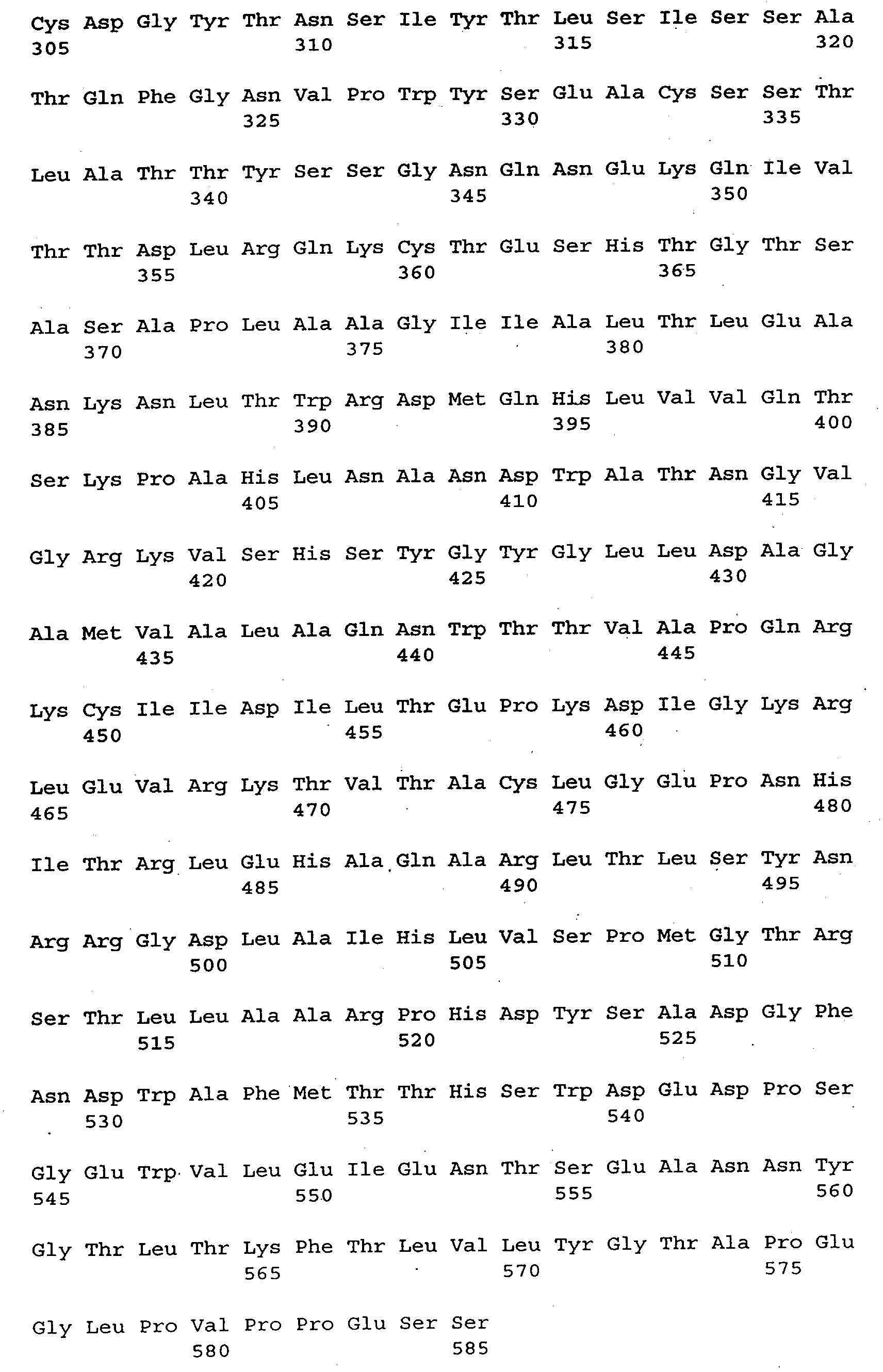


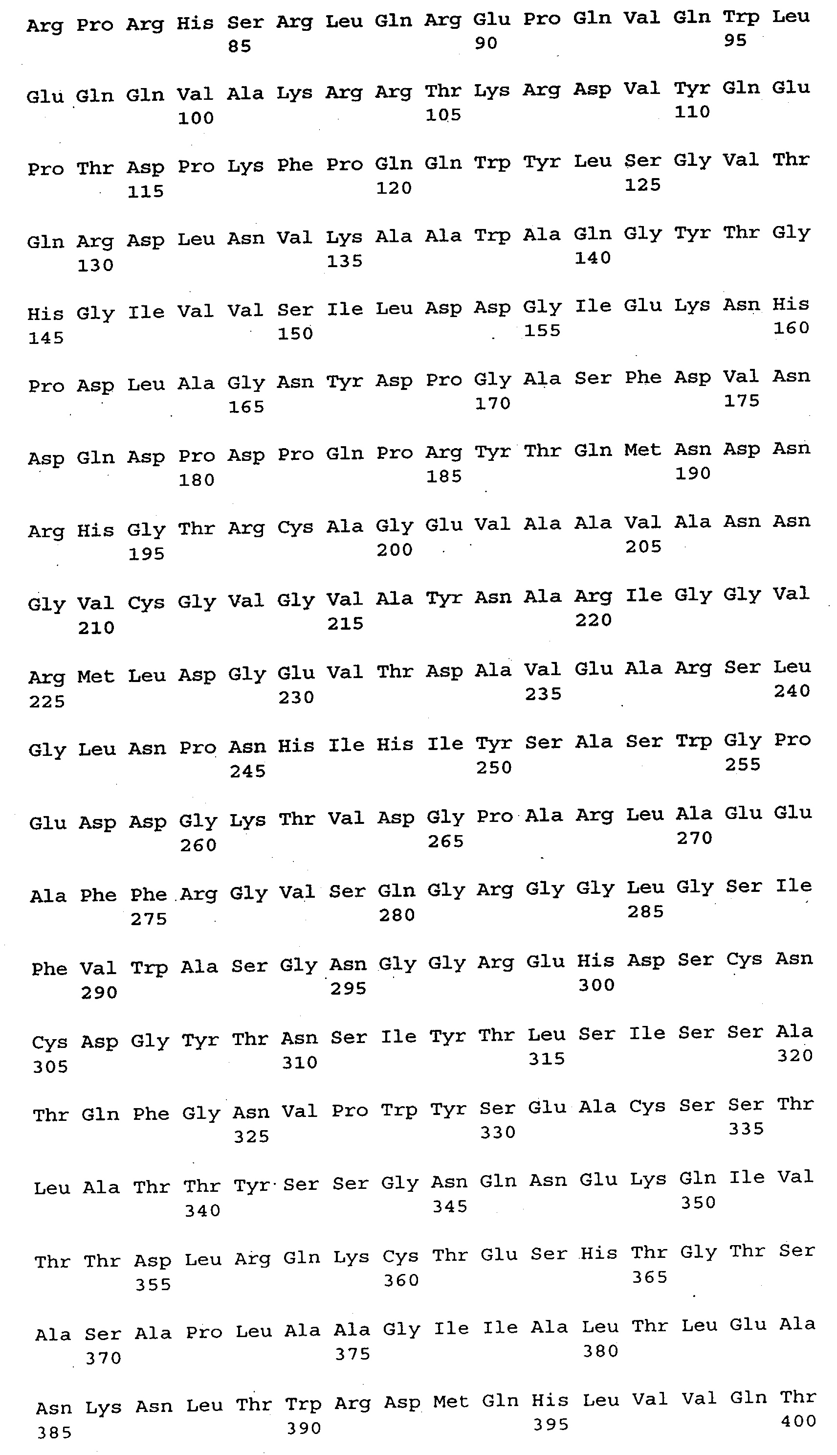


Claims (43)
- Fusionsprotein, dadurch gekennzeichnet, daß es ein gegebenenfalls C-terminal deletiertes Furinderivat oder ein Derivat eines Furinanalogen fusioniert mit einer heterologen Sequenz umfaßt.
- Fusionsprotein nach Anspruch 1, dadurch gekennzeichnet, daß die C-terminale Deletion die cytoplasmatische und transmembrane Region, und gegebenenfalls die Cys-reiche Region umfaßt.
- Fusionsprotein nach Anspruch 2, dadurch gekennzeichnet, daß die heterologe Sequenz ein Protein, ein Polypeptid oder ein Affinitätspeptid ist.
- Fusionsprotein nach einem der Ansprüche 1 bis 3, dadurch gekennzeichnet, daß das Protein oder Polypeptid abgeleitet ist von β-Galaktosidase, Glutathion-S-transferase, c-myc-Produkt, Avidin oder Lysin-bindende Kringeldomäne von Plasmaproteinen, wie Plasminogen.
- Fusionsprotein nach Anspruch 3, dadurch gekennzeichnet, daß das Affinitätspeptid ein Tag-Peptid ist, vorzugsweise ein His-Tag.
- Fusionsprotein nach Anspruch 5, dadurch gekennzeichnet, daß das Affinitätspeptid aus mehreren, vorzugsweise 3 bis 20, besonders bevorzugt 6 bis 15 aufeinanderfolgenden Histidin-Resten besteht.
- Fusionsprotein nach Anspruch 6, dadurch gekennzeichnet, daß es FurinΔTM-His oder FurinΔCys-His, vorzugsweise ein FurinΔTM-His gemäß Seq.ID 9 oder ein FurinΔCys-His gemäß Seq.ID No. 18 ist.
- Fusionsprotein nach einem der Ansprüche 1 bis 7, dadurch gekennzeichnet, daß zwischen der Furin-Sequenz und der heterologen Sequenz ein Spacer inseriert ist.
- Fusionsprotein nach Anspruch 8, dadurch gekennzeichnet, daß es FurinΔTM-Spacer-His oder FurinΔCys-Spacer-His ist.
- Fusionsprotein nach Anspruch 9, dadurch gekennzeichnet, daß es ein FurinΔTM-Spacer-His gemäß Seq.ID No.12 oder FurinΔCys-Spacer-His gemäß Seq. ID. No. 21 ist.
- Fusionsprotein nach einem der Ansprüche 1 bis 10, dadurch gekennzeichnet, daß es Affinität zu einem festen Träger besitzt.
- Fusionsprotein nach einem der Ansprüche 1 bis 11, dadurch gekennzeichnet, daß es an einen festen Träger immobilisiert ist.
- Fusionsprotein nach Anspruch 11, dadurch gekennzeichnet, daß der Träger einen Antikörper oder ein (Schwer-)Metallion umfaßt.
- DNA-Sequenz kodierend für ein Fusionsprotein nach einem der Ansprüche 1 bis 10.
- DNA-Sequenz nach Anspruch 14, dadurch gekennzeichnet, daß es eine Sequenz gemäß Seq.ID. No. 8, Seq.ID. No. 11, Seq.ID. No. 17, Seq.ID. No. 20 ist.
- Expressionsvektor enthaltend eine DNA-Sequenz nach Anspruch 14 oder 15.
- Transformierte Zellen enthaltend einen Expressionsvektor nach Anspruch 16.
- Fusionsprotein-Komplex enthaltend ein Fusionsprotein nach einem der Ansprüche 1 bis 13 adsorbiert an einen festen Träger.
- Verfahren zur Herstellung von Proteinen aus Pro-Proteinen, dadurch gekennzeichnet, daß ein Pro-Protein durch ein Fusionsprotein nach einem der Ansprüche 1 bis 13 proteolytisch gespalten wird.
- Verfahren nach Anspruch 19, dadurch gekennzeichnet, daß ein Pro-Protein durch rekombinante Coexpression mit einem Fusionsprotein nach einem der Ansprüche 1 bis 13 in vivo gespalten wird.
- Verfahren nach Anspruch 19, dadurch gekennzeichnet, daß das Pro-Protein durch ein Fusionsprotein nach einem der Ansprüche 1 bis 13 in vitro gespalten wird.
- Verfahren nach Anspruch 21, dadurch gekennzeichnet, daß beide Reaktionspartner in Lösung vorliegen.
- Verfahren nach Anspruch 22, dadurch gekennzeichnet, daß ein Pro-Protein durch Kokultivierung von rekombinanten Zellinien, die ein Pro-Protein oder ein Fusionsprotein nach einem der Ansprüche 1 bis 13 exprimieren, gespalten wird.
- Verfahren nach Anspruch 22 oder 23, dadurch gekennzeichnet, daß die Lösung ein Zellkulturüberstand von rekombinanten Zelllinien ist.
- Verfahren nach Anspruch 22, dadurch gekennzeichnet, daß die Lösung gereinigte Proteine enthält.
- Verfahren nach einem der Ansprüche 20 bis 25, dadurch gekennzeichnet, daß einer der Reaktionspartner immobilisiert ist.
- Verfahren nach Anspruch 26, dadurch gekennzeichnet, daß das Pro-Protein immobilisiert ist und das Fusionsprotein in Lösung vorliegt.
- Verfahren nach Anspruch 27, dadurch gekennzeichnet, daß das Pro-Protein an einen Antikörper immobilisiert ist.
- Verfahren nach Anspruch 26, dadurch gekennzeichnet, daß das Fusionsprotein immobilisiert ist und das Pro-Protein in Lösung vorliegt.
- Verfahren nach Anspruch 29, dadurch gekennzeichnet, daß das Fusionsprotein an einem Affinitätsträger immobilisiert ist.
- Verfahren nach Anspruch 30, dadurch gekennzeichnet, daß der Affintätsträger ein Schwermetall oder ein Antikörper ist.
- Verfahren nach einem der Ansprüche 19 bis 31, dadurch geennzeichnet, daß das Pro-Protein eine inaktive Vorstufe eines Plasmaproteins, vorzugsweise Faktor IX, von Willebrand-Faktor, Faktor VII, Faktor X, Faktor XI, Faktor V, Protein C, Protein S, Albumin oder eines viralen Proteins, vorzugsweise HIV gp160 und Influenzavirus HA, ist.
- Verfahren zum Herstellen eines Fusionsprotein-Komplexes nach Anspruch 18, dadurch gekennzeichnet, daß ein Fusionsprotein gemäß einem der Ansprüche 1 bis 13 mit einem festen Träger, welcher das Fusionsprotein binden kann, in Kontakt gebracht wird.
- Verfahren nach Anspruch 33, dadurch gekennzeichnet, daß eine Lösung enthaltend ein Fusionsprotein mit einem Träger in Kontakt gebracht wird.
- Verfahren nach Anspruch 34, dadurch gekennzeichnet, daß die Lösung ein Kulturüberstand von rekombinanten Zellinien ist.
- Verfahren nach Anspruch 34, dadurch gekennzeichnet, daß die Lösung gereinigtes Fusionsprotein enthält.
- Verfahren nach Anspruch 33 bis 36, dadurch gekennzeichnet, daß der feste Träger ausgewählt ist aus einem immobilisierten (Schwer-)Metallion, einem immobilisierten Antikörper oder einem immobilisierten Peptid oder Polypeptid.
- Verwendung eines Fusionsproteins nach einem der Ansprüche 1 bis 13 für ein Verfahren nach einem der Ansprüche 19 bis 32.
- Verwendung eines Fusionsprotein-Komplexes nach Anspruch 18 für ein Verfahren nach einem der Ansprüche 19 bis 32.
- Verwendung eines Fusionsproteins nach einem der Ansprüche 1 bis 13 zur Herstellung eines Proteins aus Pro-Protein, insbesondere von Faktor IX aus pro-Faktor IX, von Willebrand-Faktor aus pro-von Willebrand-Faktor, Faktor X aus pro-Faktor X, Faktor XI aus pro-Faktor XI, Faktor VII aus pro-Faktor VII, Faktor V aus pro-Faktor V, Protein C aus pro-Protein C und Albumin aus pro-Albumin.
- Verwendung eines Fusionsprotein-Komplexes nach Anspruch 18 zur Herstellung eines Proteins aus Pro-Protein, insbesondere von Faktor IX aus pro-Faktor IX, von Willebrand-Faktor aus pro-von Willebrand-Faktor, Faktor X aus pro-Faktor X, Faktor XI aus pro-Faktor XI, Faktor VII aus pro-Faktor VII, Faktor V aus pro-Faktor V, Protein C aus pro-Protein C und Albumin aus pro-Albumin.
- Verwendung eines Verfahrens nach einem der Ansprüche 19 bis 32 zur Herstellung eines Protein aus Pro-Protein, insbesondere von Faktor IX aus pro-Faktor IX, von Willebrand-Faktor aus pro-von Willebrand-Faktor, Faktor X aus pro-Faktor X, Faktor XI aus pro-Faktor XI, Faktor VII aus pro-Faktor VII, Faktor V aus pro-Faktor V, Protein C aus pro-Protein C und Albumin aus pro-Albumin.
- Pharmazeutische Zusammensetzung enthaltend ein gemäß einem Verfahren nach einem der Ansprüche 19 bis 32 hergestelltes Protein, insbesondere Faktor IX, Faktor X, Faktor XI, von Willebrand-Faktor, Faktor V, Faktor VII, Protein C, Albumin und einen oder mehrere physiologisch akzeptable Träger.
Applications Claiming Priority (2)
| Application Number | Priority Date | Filing Date | Title |
|---|---|---|---|
| AT0192895A AT404838B (de) | 1995-11-24 | 1995-11-24 | Herstellung von proteinen aus pro-proteinen durch fusionsproteine abgeleitet von furin oder furinanalogen |
| AT1928/95 | 1995-11-24 |
Publications (3)
| Publication Number | Publication Date |
|---|---|
| EP0775750A2 true EP0775750A2 (de) | 1997-05-28 |
| EP0775750A3 EP0775750A3 (de) | 1999-03-24 |
| EP0775750B1 EP0775750B1 (de) | 2007-06-13 |
Family
ID=3524052
Family Applications (1)
| Application Number | Title | Priority Date | Filing Date |
|---|---|---|---|
| EP96890171A Expired - Lifetime EP0775750B1 (de) | 1995-11-24 | 1996-11-19 | Herstellung von Proteinen aus Pro-Proteinen durch Fusionsproteine abgeleitet von Furin oder Furinanalogen |
Country Status (9)
| Country | Link |
|---|---|
| US (1) | US6210929B1 (de) |
| EP (1) | EP0775750B1 (de) |
| JP (1) | JPH09183800A (de) |
| AT (2) | AT404838B (de) |
| CA (1) | CA2191053C (de) |
| DE (1) | DE59611434D1 (de) |
| DK (1) | DK0775750T3 (de) |
| ES (1) | ES2289750T3 (de) |
| NO (1) | NO964963L (de) |
Cited By (8)
| Publication number | Priority date | Publication date | Assignee | Title |
|---|---|---|---|---|
| WO2000049047A1 (de) * | 1999-02-19 | 2000-08-24 | Baxter Aktiengesellschaft | VERFAHREN ZUR HERSTELLUNG EINER vWF-PRÄPARATION |
| WO2001036643A1 (en) * | 1999-11-19 | 2001-05-25 | Transkaryotic Therapies, Inc. | Nucleic acid construct for optimized production of products |
| US6562598B1 (en) | 1997-02-27 | 2003-05-13 | Baxter Aktiengesellschaft | Factor X deletion mutants and analogues thereof |
| US6573071B1 (en) | 1997-02-27 | 2003-06-03 | Baxter Aktiengesellschaft | Factor X analogues with a modified protease cleavage site |
| WO2002055110A3 (en) * | 2000-10-25 | 2004-01-08 | Genzyme Corporation | Methods for treating blood coagulation disorders |
| WO2008143977A1 (en) * | 2007-05-18 | 2008-11-27 | Baxter International Inc. | Method for producing mature vwf from vwf pro-peptide |
| US9127264B2 (en) | 2007-12-31 | 2015-09-08 | Baxalta Incorporated | Substantially animal protein-free recombinant furin and methods for producing the same |
| CN107002041A (zh) * | 2014-08-12 | 2017-08-01 | 百特股份有限公司 | 在分泌弗林蛋白酶的哺乳动物表达系统中产生被完全加工且功能性的因子x |
Families Citing this family (14)
| Publication number | Priority date | Publication date | Assignee | Title |
|---|---|---|---|---|
| AT410216B (de) | 1999-08-10 | 2003-03-25 | Baxter Ag | Faktor x-analogon mit verbesserter aktivierbarkeit |
| US6596526B1 (en) * | 2000-06-09 | 2003-07-22 | Baxter Aktiengesellschaft | Furin polypeptides with improved characteristics |
| US6821519B2 (en) * | 2000-06-29 | 2004-11-23 | Corixa Corporation | Compositions and methods for the diagnosis and treatment of herpes simplex virus infection |
| FI116198B (fi) * | 2001-07-16 | 2005-10-14 | Valtion Teknillinen | Polypeptidien immobilisointimenetelmä |
| US20050130132A1 (en) * | 2002-09-06 | 2005-06-16 | Day Craig H. | Compositions and methods for the diagnosis and treatment of herpes simplex virus infection |
| US20040096914A1 (en) * | 2002-11-20 | 2004-05-20 | Ye Fang | Substrates with stable surface chemistry for biological membrane arrays and methods for fabricating thereof |
| EP1799244A4 (de) * | 2004-07-23 | 2010-06-09 | Smith Judith | Furinhemmer |
| JP2008512098A (ja) | 2004-09-07 | 2008-04-24 | アーケミックス コーポレイション | フォン・ビルブラント因子に対するアプタマー、および血栓性疾患の処置剤としてのその使用 |
| US7566701B2 (en) * | 2004-09-07 | 2009-07-28 | Archemix Corp. | Aptamers to von Willebrand Factor and their use as thrombotic disease therapeutics |
| AU2005282380A1 (en) * | 2004-09-07 | 2006-03-16 | Archemix Corp. | Aptamer medicinal chemistry |
| CA2684885C (en) * | 2007-05-22 | 2018-09-11 | Baxter International Inc. | Preparative purification process for human furin |
| WO2008150495A2 (en) * | 2007-06-01 | 2008-12-11 | Archemix Corp. | Vwf aptamer formulations and methods for use |
| US8241623B1 (en) | 2009-02-09 | 2012-08-14 | David Bermudes | Protease sensitivity expression system |
| RU2610436C2 (ru) | 2011-06-02 | 2017-02-10 | Баксалта Инкорпорейтед | Составы рекомбинантного фурина |
Citations (2)
| Publication number | Priority date | Publication date | Assignee | Title |
|---|---|---|---|---|
| WO1991006314A2 (en) | 1989-10-25 | 1991-05-16 | H.B.T. Holland Biotechnology B.V. | Pharmaceutical composition having an endoproteolytic activity; a process for endoproteolytically processing (precursor) proteins and for the (micro)biological production of proteins |
| WO1992009698A1 (en) | 1990-11-26 | 1992-06-11 | Genetics Institute, Inc. | Expression of pace in host cells and methods of use thereof |
Family Cites Families (12)
| Publication number | Priority date | Publication date | Assignee | Title |
|---|---|---|---|---|
| CA1340522C (en) * | 1987-03-10 | 1999-05-04 | Heinz Dobeli | Fusion proteins containing neighbouring histidines for improved purification |
| US5571697A (en) * | 1989-05-05 | 1996-11-05 | Baylor College Of Medicine Texas Medical Center | Expression of processed recombinant lactoferrin and lactoferrin polypeptide fragments from a fusion product in Aspergillus |
| US5108910A (en) * | 1989-08-22 | 1992-04-28 | Immunex Corporation | DNA sequences encoding fusion proteins comprising GM-CSF and IL-3 |
| AU6215290A (en) * | 1989-09-05 | 1991-03-14 | Eli Lilly And Company | Method for activating protein c |
| NZ236819A (en) * | 1990-02-03 | 1993-07-27 | Max Planck Gesellschaft | Enzymatic cleavage of fusion proteins; fusion proteins; recombinant dna and pharmaceutical compositions |
| ATE157703T1 (de) * | 1991-12-16 | 1997-09-15 | Ciba Geigy Ag | Endoplasmatisches retikulum-ständige rekombinante dibasische endoprotease und deren verwendungen |
| AT397390B (de) * | 1992-04-06 | 1994-03-25 | Immuno Ag | Verfahren zur spaltung von proteinen |
| US5814603A (en) * | 1992-06-12 | 1998-09-29 | Affymax Technologies N.V. | Compounds with PTH activity |
| US5683695A (en) * | 1993-02-10 | 1997-11-04 | United Biomedical, Inc. | Production of recombinant proteins containing multiple antigenic determinants linked by flexible hinge domains |
| US5532142A (en) * | 1993-02-12 | 1996-07-02 | Board Of Regents, The University Of Texas System | Method of isolation and purification of fusion polypeptides |
| US5496934A (en) * | 1993-04-14 | 1996-03-05 | Yissum Research Development Company Of The Hebrew University Of Jerusalem | Nucleic acids encoding a cellulose binding domain |
| US5714371A (en) * | 1995-05-12 | 1998-02-03 | Schering Corporation | Method for refolding insoluble aggregates of hepatitis C virus protease |
-
1995
- 1995-11-24 AT AT0192895A patent/AT404838B/de not_active IP Right Cessation
-
1996
- 1996-11-19 DE DE59611434T patent/DE59611434D1/de not_active Expired - Lifetime
- 1996-11-19 ES ES96890171T patent/ES2289750T3/es not_active Expired - Lifetime
- 1996-11-19 AT AT96890171T patent/ATE364703T1/de active
- 1996-11-19 DK DK96890171T patent/DK0775750T3/da active
- 1996-11-19 EP EP96890171A patent/EP0775750B1/de not_active Expired - Lifetime
- 1996-11-22 CA CA2191053A patent/CA2191053C/en not_active Expired - Lifetime
- 1996-11-22 US US08/753,247 patent/US6210929B1/en not_active Expired - Lifetime
- 1996-11-22 NO NO964963A patent/NO964963L/no not_active Application Discontinuation
- 1996-11-25 JP JP8353126A patent/JPH09183800A/ja not_active Withdrawn
Patent Citations (2)
| Publication number | Priority date | Publication date | Assignee | Title |
|---|---|---|---|---|
| WO1991006314A2 (en) | 1989-10-25 | 1991-05-16 | H.B.T. Holland Biotechnology B.V. | Pharmaceutical composition having an endoproteolytic activity; a process for endoproteolytically processing (precursor) proteins and for the (micro)biological production of proteins |
| WO1992009698A1 (en) | 1990-11-26 | 1992-06-11 | Genetics Institute, Inc. | Expression of pace in host cells and methods of use thereof |
Non-Patent Citations (40)
| Title |
|---|
| APPLICATION CONTAINS STILL MORE REFERENCES |
| BARR ET AL., DNA CELL BIOL., vol. 10, 1991, pages 319 - 328 |
| BARR, CELL, vol. 66, 1991, pages 1 - 3 |
| BRAVO ET AL., J. BIOL. CHEM., vol. 269, 1994, pages 25830 - 25837 |
| BRESNAHAN ET AL., J. CELL BIOL., vol. 111, 1990, pages 2851 - 2859 |
| BRISTOL ET AL., BIOCHEMISTRY, vol. 33, 1994, pages 14136 - 14143 |
| CREEMERS ET AL., J. BIOL. CHEM., vol. 268, 1993, pages 21826 - 21834 |
| DECROLY ET AL., J. BIOL. CHEM., vol. 269, 1994, pages 12240 - 12247 |
| DUIJHOVEN ET AL., HYBRIDOMA, vol. 11, 1992, pages 71 - 86 |
| EVAN ET AL., MOL. CELL. BIOL., vol. 5, 1985, pages 3610 - 3616 |
| HATSUZAWA ET AL., J. BIO. CHEM., vol. 265, 1990, pages 22075 - 22078 |
| HATSUZAWA ET AL., J. BIOL. CHEM., vol. 101, 1992, pages 296 - 301 |
| HATSUZAWA ET AL., J. BIOL. CHEM., vol. 267, 1992, pages 16094 - 16099 |
| HOSAKA ET AL., J. BIOL. CHEM., vol. 266, 1991, pages 12127 - 12130 |
| KIEFER ET AL., DNA CELL. BIO., vol. 10, 1991, pages 757 |
| KLIMPEL ET AL., PROC. NATL. ACAD. SCI. USA, vol. 89, 1992, pages 10277 - 10281 |
| LAVALLIE ET AL., J. BIOL. CHEM., vol. 268, 1993, pages 23311 - 23317 |
| LEDUC ET AL., J. BIOL. CHEM., vol. 267, 1992, pages 14304 - 14308 |
| MATTHEWS ET AL., PROTEIN SCIENCE, vol. 3, 1994, pages 1197 - 1205 |
| MOLLOY ET AL., EMBO J., vol. 13, 1994, pages 18 - 33 |
| MOLLOY ET AL., J. BIOL. CHEM., vol. 267, 1992, pages 16396 - 16402 |
| NAKAYAMA ET AL., J. BIO. CHEM., vol. 267, 1992, pages 5897 - 5900 |
| ODA ET AL., BIOCHEM. BIOPHYS. RES. COMMUN., vol. 189, 1992, pages 1353 - 1361 |
| REHEMTULLA ET AL., BLOOD, vol. 79, 1992, pages 2349 - 2355 |
| REHEMTULLA ET AL., PROC. NATL. ACAD. SCI. USA, vol. 89, 1992, pages 8235 - 8239 |
| ROEBROEK ET AL., EMBO J., vol. 5, 1986, pages 2197 - 2202 |
| ROEBROEK ET AL., MOL. BIOL. REP., vol. 11, 1986, pages 117 - 125 |
| SMEEKENS ET AL., J. BIOL. CHEM., vol. 265, 1990, pages 2997 - 3000 |
| SMEEKENS ET AL., PROC. NATL. ACAD. SCI. USA, vol. 88, 1991, pages 340 - 344 |
| STIENEKE-GROEBER ET AL., EMBO J., vol. 11, 1992, pages 2407 - 2414 |
| TSUNEOKA ET AL., J. BIOL. CHEM., vol. 268, 1993, pages 26461 - 26465 |
| VAN DE VEN ET AL., CRIT. REV. ONCOGEN, vol. 4, 1993, pages 115 - 136 |
| VAN DE VEN ET AL., MOL. BIOL. REP., vol. 14, 1990, pages 265 - 275 |
| VAN DEN OUWELAND ET AL., NUCLEIC ACIDS RES., vol. 17, 1989, pages 7101 - 7102 |
| VAN DEN OUWELAND ET AL., NUCLEIC ACIDS RES., vol. 18, 1990, pages 664 |
| VERWEIJ ET AL., EMBO J., vol. 5, 1986, pages 1839 - 1847 |
| VEY ET AL., J. CELL. BIOL., vol. 127, 1994, pages 1829 - 1842 |
| VIDRICAIRE ET AL., BIOCHEM. BIOPHYS. RES. COMM., vol. 195, 1993, pages 1011 - 1018 |
| WASLEY ET AL., J. BIOL. CHEM., vol. 268, 1993, pages 8458 - 8465 |
| WISE ET AL., PROC. NATL. ACAD. SCI. USA, vol. 87, 1990, pages 9378 - 9382 |
Cited By (15)
| Publication number | Priority date | Publication date | Assignee | Title |
|---|---|---|---|---|
| US7220569B2 (en) | 1997-02-27 | 2007-05-22 | Baxter Aktiengesellschaft | Nucleic acids encoding factor X analogues having a modified protease cleavage site |
| US6562598B1 (en) | 1997-02-27 | 2003-05-13 | Baxter Aktiengesellschaft | Factor X deletion mutants and analogues thereof |
| US6573071B1 (en) | 1997-02-27 | 2003-06-03 | Baxter Aktiengesellschaft | Factor X analogues with a modified protease cleavage site |
| WO2000049047A1 (de) * | 1999-02-19 | 2000-08-24 | Baxter Aktiengesellschaft | VERFAHREN ZUR HERSTELLUNG EINER vWF-PRÄPARATION |
| US7229793B1 (en) | 1999-11-19 | 2007-06-12 | Shire Human Genetic Therapies, Inc. | Constructs and cells for production of small peptides |
| WO2001036643A1 (en) * | 1999-11-19 | 2001-05-25 | Transkaryotic Therapies, Inc. | Nucleic acid construct for optimized production of products |
| WO2002055110A3 (en) * | 2000-10-25 | 2004-01-08 | Genzyme Corporation | Methods for treating blood coagulation disorders |
| US7615537B2 (en) | 2000-10-25 | 2009-11-10 | Genzyme Corporation | Methods for treating blood coagulation disorders |
| WO2008143977A1 (en) * | 2007-05-18 | 2008-11-27 | Baxter International Inc. | Method for producing mature vwf from vwf pro-peptide |
| US8058411B2 (en) | 2007-05-18 | 2011-11-15 | Baxter International Inc. | Method for producing mature VWF from VWF pro-peptide |
| KR101156263B1 (ko) * | 2007-05-18 | 2012-06-13 | 박스터 헬쓰케어 에스.에이. | 폰 빌리브란트 인자 프로펩티드로부터 성숙한 폰 빌리브란트 인자를 제조하는 방법 |
| US9127264B2 (en) | 2007-12-31 | 2015-09-08 | Baxalta Incorporated | Substantially animal protein-free recombinant furin and methods for producing the same |
| EP3521422A1 (de) | 2007-12-31 | 2019-08-07 | Baxalta GmbH | Weitgehend tierproteinfreies rekombinantes furin und verfahren zur herstellung davon |
| CN107002041A (zh) * | 2014-08-12 | 2017-08-01 | 百特股份有限公司 | 在分泌弗林蛋白酶的哺乳动物表达系统中产生被完全加工且功能性的因子x |
| CN107002041B (zh) * | 2014-08-12 | 2021-06-29 | 百特股份有限公司 | 在分泌弗林蛋白酶的哺乳动物表达系统中产生被完全加工且功能性的因子x |
Also Published As
| Publication number | Publication date |
|---|---|
| EP0775750B1 (de) | 2007-06-13 |
| DK0775750T3 (da) | 2007-10-08 |
| NO964963D0 (no) | 1996-11-22 |
| EP0775750A3 (de) | 1999-03-24 |
| JPH09183800A (ja) | 1997-07-15 |
| US6210929B1 (en) | 2001-04-03 |
| ATE364703T1 (de) | 2007-07-15 |
| NO964963L (no) | 1997-05-26 |
| ATA192895A (de) | 1998-07-15 |
| AT404838B (de) | 1999-03-25 |
| CA2191053C (en) | 2011-01-18 |
| CA2191053A1 (en) | 1997-05-25 |
| ES2289750T3 (es) | 2008-02-01 |
| DE59611434D1 (de) | 2007-07-26 |
Similar Documents
| Publication | Publication Date | Title |
|---|---|---|
| AT404838B (de) | Herstellung von proteinen aus pro-proteinen durch fusionsproteine abgeleitet von furin oder furinanalogen | |
| AT405516B (de) | Faktor x-analoge mit modifizierter proteasespaltstelle | |
| DE69127829T2 (de) | Expression von pace in wirtszellen und verfahren zu dessen verwendung | |
| EP1012303B1 (de) | Faktor x-deletionsmutanten und analoge davon | |
| DE3885419T2 (de) | Für die direkte Expression von aktiviertem menschlichen Protein-C verwendbare Vektoren und Zusammensetzungen. | |
| DE3687584T2 (de) | Vektoren und verfahren zur expression einer die aktivitaet des menschlichen proteins-c enthaltenden substanz. | |
| DE60115613T2 (de) | Herstellung von rekombinanten muteine des blutgerinnungsfaktors viii in humanen zellinien | |
| DE3871798T2 (de) | Modifizierter faktor vii/viia. | |
| AT410216B (de) | Faktor x-analogon mit verbesserter aktivierbarkeit | |
| DE69636739T2 (de) | Verfahren zur rückfaltung von menschlichem activin a | |
| EP1399568A2 (de) | Verfahren zur herstellung von rekombinantem trypsin | |
| DE3786317T2 (de) | Proteinanaloge des gewebs-plasminogenaktivators. | |
| DE3523701A1 (de) | Verfahren zur herstellung von proteinen und polypeptiden | |
| DE69029663T3 (de) | Verfahren zur endoproteolytischen bearbeitung von (vorläufer)proteinen und zur (mikro)biologischen herstellung | |
| EP1012307B1 (de) | Autokatalytisch aktivierbare zymogene vorstufen von proteasen und deren verwendung | |
| DE69101486T2 (de) | Verfahren zur reinigung rekombinanter polypeptide. | |
| DE69318495T2 (de) | Verfahren zur Reinigung des Big-Endothelin Proteins | |
| DE3486023T2 (de) | Verfahren zur herstellung von urokinase zymogen. | |
| EP1169461B1 (de) | Verwendung von pankreatischer Procarboxypeptidase B zur Herstellung von Insulin | |
| DE69430898T2 (de) | Enzymatische Umsetzung von Proteinen unter Verwendung immobilisierter Dipeptidylaminopeptidase aus Dictyostelium | |
| DE69004964T2 (de) | Zellenzuchtverfahren für die produktion von aktiviertem protein-c. | |
| EP0367161B1 (de) | Verfahren zur selektiven Spaltung von Fusionsproteinen | |
| WO2000049047A1 (de) | VERFAHREN ZUR HERSTELLUNG EINER vWF-PRÄPARATION | |
| JPH022338A (ja) | 真核細肪中での同時発現 | |
| EP0968280A1 (de) | Aktivierter vitamin k-abhängiger blutfaktor und verfahren zu dessen herstellung |
Legal Events
| Date | Code | Title | Description |
|---|---|---|---|
| PUAI | Public reference made under article 153(3) epc to a published international application that has entered the european phase |
Free format text: ORIGINAL CODE: 0009012 |
|
| AK | Designated contracting states |
Kind code of ref document: A2 Designated state(s): AT BE CH DE DK ES FI FR GB IT LI NL SE |
|
| RBV | Designated contracting states (corrected) |
Designated state(s): AT BE CH DE DK ES FI FR GB IT LI NL SE |
|
| PUAL | Search report despatched |
Free format text: ORIGINAL CODE: 0009013 |
|
| AK | Designated contracting states |
Kind code of ref document: A3 Designated state(s): AT BE CH DE DK ES FI FR GB IT LI NL SE |
|
| 17P | Request for examination filed |
Effective date: 19990521 |
|
| RAP1 | Party data changed (applicant data changed or rights of an application transferred) |
Owner name: BAXTER AKTIENGESELLSCHAFT |
|
| RAP1 | Party data changed (applicant data changed or rights of an application transferred) |
Owner name: BAXTER AKTIENGESELLSCHAFT |
|
| 17Q | First examination report despatched |
Effective date: 20020226 |
|
| RTI1 | Title (correction) |
Free format text: PREPARATION OF PROTEINS FROM PRO-PROTEINS BY FUSION PROTEINS DERIVED FROM FURIN OR FURIN ANALOGS |
|
| GRAP | Despatch of communication of intention to grant a patent |
Free format text: ORIGINAL CODE: EPIDOSNIGR1 |
|
| GRAS | Grant fee paid |
Free format text: ORIGINAL CODE: EPIDOSNIGR3 |
|
| RIN1 | Information on inventor provided before grant (corrected) |
Inventor name: EIBL, JOHANN, DR. Inventor name: DORNER, FRIEDRICH, PROF. Inventor name: FALKNER, FALKO-GUENTER, DR. Inventor name: FISCHER, BERNHARD, DOZ. Inventor name: SCHLOKAT, UWE, DR. |
|
| GRAA | (expected) grant |
Free format text: ORIGINAL CODE: 0009210 |
|
| AK | Designated contracting states |
Kind code of ref document: B1 Designated state(s): AT BE CH DE DK ES FI FR GB IT LI NL SE |
|
| REG | Reference to a national code |
Ref country code: GB Ref legal event code: FG4D Free format text: NOT ENGLISH |
|
| REG | Reference to a national code |
Ref country code: CH Ref legal event code: NV Representative=s name: KIRKER & CIE S.A. Ref country code: CH Ref legal event code: EP |
|
| REF | Corresponds to: |
Ref document number: 59611434 Country of ref document: DE Date of ref document: 20070726 Kind code of ref document: P |
|
| REG | Reference to a national code |
Ref country code: SE Ref legal event code: TRGR |
|
| GBT | Gb: translation of ep patent filed (gb section 77(6)(a)/1977) |
Effective date: 20070905 |
|
| REG | Reference to a national code |
Ref country code: DK Ref legal event code: T3 |
|
| ET | Fr: translation filed | ||
| REG | Reference to a national code |
Ref country code: ES Ref legal event code: FG2A Ref document number: 2289750 Country of ref document: ES Kind code of ref document: T3 |
|
| PLBE | No opposition filed within time limit |
Free format text: ORIGINAL CODE: 0009261 |
|
| STAA | Information on the status of an ep patent application or granted ep patent |
Free format text: STATUS: NO OPPOSITION FILED WITHIN TIME LIMIT |
|
| 26N | No opposition filed |
Effective date: 20080314 |
|
| REG | Reference to a national code |
Ref country code: FR Ref legal event code: PLFP Year of fee payment: 20 |
|
| PGFP | Annual fee paid to national office [announced via postgrant information from national office to epo] |
Ref country code: FI Payment date: 20151127 Year of fee payment: 20 Ref country code: DK Payment date: 20151125 Year of fee payment: 20 Ref country code: IT Payment date: 20151124 Year of fee payment: 20 Ref country code: CH Payment date: 20151127 Year of fee payment: 20 Ref country code: GB Payment date: 20151127 Year of fee payment: 20 Ref country code: DE Payment date: 20151127 Year of fee payment: 20 |
|
| PGFP | Annual fee paid to national office [announced via postgrant information from national office to epo] |
Ref country code: FR Payment date: 20151117 Year of fee payment: 20 Ref country code: AT Payment date: 20151103 Year of fee payment: 20 Ref country code: BE Payment date: 20151130 Year of fee payment: 20 Ref country code: NL Payment date: 20151126 Year of fee payment: 20 Ref country code: SE Payment date: 20151127 Year of fee payment: 20 Ref country code: ES Payment date: 20151126 Year of fee payment: 20 |
|
| REG | Reference to a national code |
Ref country code: DE Ref legal event code: R082 Ref document number: 59611434 Country of ref document: DE Representative=s name: HOFFMANN - EITLE PATENT- UND RECHTSANWAELTE PA, DE Ref country code: DE Ref legal event code: R081 Ref document number: 59611434 Country of ref document: DE Owner name: BAXALTA GMBH, GLATTPARK, CH Free format text: FORMER OWNER: BAXTER AG, WIEN, AT Ref country code: DE Ref legal event code: R081 Ref document number: 59611434 Country of ref document: DE Owner name: BAXALTA INCORPORATED, BANNOCKBURN, US Free format text: FORMER OWNER: BAXTER AG, WIEN, AT |
|
| REG | Reference to a national code |
Ref country code: NL Ref legal event code: PD Owner name: BAXTER EASTERN EUROPE VERTRIEBS GMBH; AT Free format text: DETAILS ASSIGNMENT: VERANDERING VAN EIGENAAR(S), VERANDERING VAN DE JURIDISCHE ENTITEIT; FORMER OWNER NAME: BAXTER AKTIENGESELLSCHAFT Effective date: 20160502 Ref country code: NL Ref legal event code: HC Owner name: BAXALTA INNOVATIONS GMBH; AT Free format text: DETAILS ASSIGNMENT: VERANDERING VAN EIGENAAR(S), VERANDERING VAN NAAM VAN DE EIGENAAR(S); FORMER OWNER NAME: BAXTER TRADING GMBH Effective date: 20160502 |
|
| REG | Reference to a national code |
Ref country code: NL Ref legal event code: PD Owner name: BAXALTA INCORPORATED; US Free format text: DETAILS ASSIGNMENT: VERANDERING VAN EIGENAAR(S), OVERDRACHT; FORMER OWNER NAME: BAXTER AKTIENGESELLSCHAFT Effective date: 20160524 |
|
| REG | Reference to a national code |
Ref country code: GB Ref legal event code: 732E Free format text: REGISTERED BETWEEN 20160519 AND 20160525 |
|
| REG | Reference to a national code |
Ref country code: AT Ref legal event code: PC Ref document number: 364703 Country of ref document: AT Kind code of ref document: T Owner name: BAXALTA GMBH, CH Effective date: 20160623 Ref country code: AT Ref legal event code: PC Ref document number: 364703 Country of ref document: AT Kind code of ref document: T Owner name: BAXALTA INCORPORATED, US Effective date: 20160623 |
|
| REG | Reference to a national code |
Ref country code: ES Ref legal event code: PC2A Owner name: BAXALTA GMBH Effective date: 20160823 |
|
| REG | Reference to a national code |
Ref country code: CH Ref legal event code: PFA Owner name: BAXTER INNOVATIONS GMBH, AT Free format text: FORMER OWNER: BAXTER TRADING GMBH, AT Ref country code: CH Ref legal event code: PFA Owner name: BAXTER EASTERN EUROPE VERTRIEBS GMBH, AT Free format text: FORMER OWNER: BAXTER AKTIENGESELLSCHAFT, AT Ref country code: CH Ref legal event code: PFA Owner name: BAXALTA INNOVATIONS GMBH, AT Free format text: FORMER OWNER: BAXTER INNOVATIONS GMBH, AT Ref country code: CH Ref legal event code: PFA Owner name: BAXTER TRADING GMBH, AT Free format text: FORMER OWNER: BAXTER EASTERN EUROPE VERTRIEBS GMBH, AT |
|
| REG | Reference to a national code |
Ref country code: CH Ref legal event code: PUE Owner name: BAXALTA INCORPORATED, CH Free format text: FORMER OWNER: BAXALTA INNOVATIONS GMBH, AT Ref country code: CH Ref legal event code: NV Representative=s name: KELLER AND PARTNER PATENTANWAELTE AG, CH |
|
| REG | Reference to a national code |
Ref country code: DE Ref legal event code: R071 Ref document number: 59611434 Country of ref document: DE |
|
| REG | Reference to a national code |
Ref country code: DK Ref legal event code: EUP Effective date: 20161119 |
|
| REG | Reference to a national code |
Ref country code: NL Ref legal event code: MK Effective date: 20161118 |
|
| REG | Reference to a national code |
Ref country code: CH Ref legal event code: PL |
|
| REG | Reference to a national code |
Ref country code: GB Ref legal event code: PE20 Expiry date: 20161118 |
|
| REG | Reference to a national code |
Ref country code: AT Ref legal event code: MK07 Ref document number: 364703 Country of ref document: AT Kind code of ref document: T Effective date: 20161119 |
|
| PG25 | Lapsed in a contracting state [announced via postgrant information from national office to epo] |
Ref country code: GB Free format text: LAPSE BECAUSE OF EXPIRATION OF PROTECTION Effective date: 20161118 |
|
| REG | Reference to a national code |
Ref country code: ES Ref legal event code: FD2A Effective date: 20170224 |
|
| PG25 | Lapsed in a contracting state [announced via postgrant information from national office to epo] |
Ref country code: ES Free format text: LAPSE BECAUSE OF EXPIRATION OF PROTECTION Effective date: 20161120 |