EP0714510B1 - Für natürliche killerzellen spezifische antigene und sie identifizierende antikörper - Google Patents
Für natürliche killerzellen spezifische antigene und sie identifizierende antikörper Download PDFInfo
- Publication number
- EP0714510B1 EP0714510B1 EP94927259A EP94927259A EP0714510B1 EP 0714510 B1 EP0714510 B1 EP 0714510B1 EP 94927259 A EP94927259 A EP 94927259A EP 94927259 A EP94927259 A EP 94927259A EP 0714510 B1 EP0714510 B1 EP 0714510B1
- Authority
- EP
- European Patent Office
- Prior art keywords
- cells
- antibody
- pen5
- pens
- natural killer
- Prior art date
- Legal status (The legal status is an assumption and is not a legal conclusion. Google has not performed a legal analysis and makes no representation as to the accuracy of the status listed.)
- Expired - Lifetime
Links
Images


Classifications
-
- G—PHYSICS
- G01—MEASURING; TESTING
- G01N—INVESTIGATING OR ANALYSING MATERIALS BY DETERMINING THEIR CHEMICAL OR PHYSICAL PROPERTIES
- G01N33/00—Investigating or analysing materials by specific methods not covered by groups G01N1/00 - G01N31/00
- G01N33/48—Biological material, e.g. blood, urine; Haemocytometers
- G01N33/50—Chemical analysis of biological material, e.g. blood, urine; Testing involving biospecific ligand binding methods; Immunological testing
- G01N33/53—Immunoassay; Biospecific binding assay; Materials therefor
- G01N33/569—Immunoassay; Biospecific binding assay; Materials therefor for microorganisms, e.g. protozoa, bacteria, viruses
- G01N33/56966—Animal cells
- G01N33/56972—White blood cells
-
- C—CHEMISTRY; METALLURGY
- C07—ORGANIC CHEMISTRY
- C07K—PEPTIDES
- C07K14/00—Peptides having more than 20 amino acids; Gastrins; Somatostatins; Melanotropins; Derivatives thereof
- C07K14/435—Peptides having more than 20 amino acids; Gastrins; Somatostatins; Melanotropins; Derivatives thereof from animals; from humans
- C07K14/705—Receptors; Cell surface antigens; Cell surface determinants
-
- C—CHEMISTRY; METALLURGY
- C07—ORGANIC CHEMISTRY
- C07K—PEPTIDES
- C07K16/00—Immunoglobulins [IGs], e.g. monoclonal or polyclonal antibodies
- C07K16/18—Immunoglobulins [IGs], e.g. monoclonal or polyclonal antibodies against material from animals or humans
- C07K16/28—Immunoglobulins [IGs], e.g. monoclonal or polyclonal antibodies against material from animals or humans against receptors, cell surface antigens or cell surface determinants
-
- A—HUMAN NECESSITIES
- A61—MEDICAL OR VETERINARY SCIENCE; HYGIENE
- A61K—PREPARATIONS FOR MEDICAL, DENTAL OR TOILETRY PURPOSES
- A61K38/00—Medicinal preparations containing peptides
Definitions
- the invention relates to novel cell surface structures that are selectively expressed on a subpopulation of natural killer cells and to antibodies that bind to unique epitopes on these structures.
- NK cells Natural killer cells
- LGLs large granular lymphocytes
- NK cells do not rearrange or express either of the known T cell receptor complexes, they can recognize and kill certain virus-infected and transformed cells in a non-MHC-restricted fashion, without prior sensitization.
- CD16 an Fc receptor for immunoglobulin that recognizes antibody-coated target cells
- the NK cell surface receptors responsible for target cell recognition have not been identified. The lack of a defining surface receptor requires NK cells to be identified by a combination of phenotypic and functional characteristics.
- NK cells are CD3:TCR-, CD16+, CD56+ LGLs
- CD56 bright NK cells are largely CD16 + , agranular lymphocytes deficient in cytolytic effector function that proliferate vigorously in response to exogenous IL-2.
- CD56 dim NK cells are CD16 + LGLs possessing potent cytolytic effector function that do not proliferate in response to IL-2. Because some T cells express both CD16 and CD56, these molecules, by themselves, cannot define the NK cell population. (Trinchieri, 1989).
- CD56 on the functionally differentiated population of NK cells is low, monoclonal antibodies reactive with CD56 cannot be used to reliably distinguish this subpopulation of NK cells from other cells in a sample.
- EP-A-0160486 discloses methods for detecting and lysing NK cells.
- a monoclonal antibody according to the present invention forms an immune complex with the glycoprotein PENS, which is present on the surface of a natural killer cell, and said antibody is unreactive with antigens present on T cells, B cells, monocytes, granulocytes, red blood cells and platelets, wherein the glycoprotein PEN5 is present on the surface of a natural killer cell and comprises a N-linked glycoprotein PEN5 ⁇ having an average molecular weight of 227 ⁇ 4 kDa and an O-linked glycoprotein PEN5 ⁇ having an average molecular weight of 140 ⁇ 3 kDa.
- Antibodies of the invention bind to unique epitopes present on a cell surface structure selectively expressed on functionally differentiated NK cells.
- the novel NK cell-specific molecule of the invention consists essentially of a pair of polydispersed glycoproteins, designated herein as PEN5 ⁇ and PEN5 ⁇ , having apparent molecular weights of 120 - 150 and 210 - 245 kdal, respectively, as determined by SDS polyacrylamide gel electrophoresis on a 6% polyacrylamide gel under non-reducing conditions.
- the unique epitopes of the PEN5 ⁇ /PEN5 ⁇ glycoprotein pair are preferentially expressed on the subpopulation of peripheral blood NK cells which are of the phenotype CD16 + CD56 dim relative to their expression on peripheral blood NK cells having the phenotype CD16 + CD56 bright and are not present on CD3 + T lymphocytes or CD20 + B lymphocytes.
- the antibody is unreactive with peripheral blood T cells, activated T cells, thymocytes, peripheral blood B cells, splenic B cells, activated B cells, monocytes, granulocytes, platelets, and red blood cells.
- the antibodies of the invention are preferably monoclonal antibodies and in particularly preferred embodiments, are of mouse or human origin, or they are chimeric antibodies having at least the constant region thereof of human origin.
- the monoclonal antibodies are produced by hybrid cell lines using conventional hybridization and screening techniques, such as those described in Anderson et al, J. Immunol. , 143:1899 (1989). As is well known in the monoclonal antibody art, independently produced hybrid cell lines that produce monoclonal antibodies specific for a given antigenic determinant are typically distinct from one another, as is each of the monoclonal antibodies so produced.
- the epitope recognized by the antibodies of the invention is a sulfated polylactosamine carbohydrate related to keratan sulfate glycosaminoglycan.
- the antibodies have the characteristics of the monoclonal antibody, alternatively referred to herein as either anti-PENS or mAb 5H10, secreted by a hybridoma identified by ATCC Accession No. HB11441.
- the antibodies and/or immunoreactive fragments or derivatives of the invention can be labeled, e.g. with a radioactive, enzymatic, or fluorescent label and used to detect, enumerate, and/or purify functionally differentiated NK cells in a mixed population of cells and to distinguish these cells from non-NK cells and NK cells that are not functionally differentiated.
- Identification of the functionally differentiated subpopulation of NK cells involves (a) contacting a suitable sample that contains a mixed population of cells, which can be, for example, peripheral blood, bone marrow aspirate, or lymphoid tissue, with an antibody of the invention or an immunoreactive fragment or derivative thereof, and (b) detecting immune complex formation. Immune complex formation can be detected by any of the techniques that are conventional and well known in the art.
- the antibodies of the invention can also be used to selectively eliminate functionally differentiated NK cells that are of the phenotype CD16 + CD56 dim in a sample comprising a mixed population of cells.
- a suitable sample preferably a biological sample
- methods for selectively eliminating or removing functionally differentiated natural killer cells from a suitable sample preferably a biological sample, which involve (a) contacting the sample with an antibody of the invention or an immunoreactive fragment or derivative thereof, which is optionally linked to a radionucleotide or a toxin, and (b) removing from the sample the cells that bind to the antibody, fragment or derivative.
- the biological sample is bone marrow aspirate.
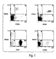
- Figure 1 comprises four two-color flow cytometry histograms, which collectively show that the PENS epitope is expressed selectively on CD56 + CD16 + peripheral blood lymphocytes ("PBL").
- PBL peripheral blood lymphocytes
- PBL were stained by 2-color flow cytometry using rhodamine-conjugated anti-CD56 mAb, rhodamine-conjugated anti-CD3 mAb, rhodamine-conjugated anti-CD20 mAb, FITC-conjugated anti-CD16 mAb or biotinylated anti-PENS mAb.
- the binding of biotinylated anti-PEN5 mAb was revealed using APC-conjugated avidin. Numbers in each quadrants indicate the percent of positive stained cells.
- Figure 2 comprises three sets of histograms that collectively show the expression of the PENS epitope on distinct NK cell subsets.
- purified NK cells were either unsorted, or sorted into CD56 dim and CD56 bright NK cell subsets using rhodamine-conjugated anti-CD56 mAb and flow cytometry.
- Unsorted NK cells, CD56 dim and CD56 bright NK cells were further analyzed for the expression of the PENS epitope using biotinylated anti-PEN5 mAb and FITC-conjugated avidin. Controls were performed using mouse isotype matched control mAb. The numbers in each histograms indicate the percent of positive stained cells.
- Figure 3 comprises a series of flow cytometry histograms illustrating the kinetics of PENS expression on activated NK cells. Sorted CD56 dim and CD56 bright NK cells were activated for 20 days with ionomycin and lymphocyte conditioned medium as described in the examples. At the indicated period of time, (i.e, 0, 6, 8, 10, 14, and 20 days of culture) aliquots of the activated NK cell populations were analyzed for their cell surface phenotype by flow cytometry using isotype matched control mAb, anti-CD56 and anti-PEN5 mAb. Results indicate the percent of positively stained cells (%) ; the total mean fluorescence intensity is indicated below.
- Figure 4 is a series of flow cytometry histograms that illustrate the cell surface expression of the PENS epitope on leukemic NK cells.
- Peripheral blood NK cells, as well as peripheral blood mononuclear cells (PBMC) isolated from three patients undergoing granular lymphocyte proliferative disorder "GLPD" blast crisis (GLPD1-3) were analyzed for the cell surface expression of CD56 and PENS using indirect immunofluorescence and flow cytometry. The numbers in each histogram indicate the percent of positive stained cells.
- PBMC peripheral blood mononuclear cells
- Figure 5 is a reproduction of an SDS gel from an immunoprecipitation of PENS glycoproteins.
- Detergent lysates prepared from radioiodinated NK cells were immunoprecipitated using 5H10 mAb or mouse IgM control. Samples were then separated under non-reducing conditions on a 6% SDS-polyacrylamide gel.
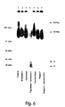
- Figure 6 is a reproduction of an SDS gel from immunoprecipitations performed after enzymatic deglycosylation of PENS glycoproteins.
- Detergent lysates prepared from radioiodinated NK cells were immunoprecipitated using 5H10 mAb.
- Affinity-purified PEN5 ⁇ and PEN5 ⁇ glycoproteins were eluted from the antibody-coated sepharose beads using 0.15M NH 4 OH, pH 10.5.
- Figures 7A through 7C illustrate the reactivity of anti-PENS mAb with keratan sulfate glycosaminoglycans.
- I 125 -labeled 5H10 mAb (1 x 10 6 cpm/sample) was preincubated for 20 min at 4°C in PBS in the presence of the indicated concentrations of bovine cornea keratan sulfate (BC). The mixture was then added to NK cells for another 20 min incubation at 4°C, prior to three washes in PBS-1%BSA. Samples were counted in a ⁇ -counter, and results are expressed as mean cpm of duplicate samples (SD ⁇ 10%).
- chondroitin sulfate B When used in incubation with NK cells or anti-PEN5 mAb, the following carbohydrates used at 10 mg/ml were without any effect on anti-PENS binding to the NK cell surface: chondroitin sulfate B, heparin, heparan sulfate, dextran sulfate, GlcNAc, mannose 6-phosphate, lactose, galactose-6-phosphate, fucose, glucose 6-phosphate, glucose and galactose.
- peripheral blood NK cells were incubated in PBS-1%BSA for 3 hr with glycosidases (0.025 U/ml) or 45 min with proteases (5 mg/ml) at 37°C, respectively.
- Cell surface expression of the PENS epitope was then analyzed by flow cytometry using anti-PENS mAb. Percent modulation was calculated as the ratio of the total linear mean fluorescence intensity of the treated cells over that of untreated control cells.
- CD1 embryonic chick cartilage aggrecan
- BNC bovine nasal cartilage aggrecan
- RC swarm rat chondrosarcoma aggrecan
- SHK shart cranial cartilage aggrecan.
- Figures 8A through 8C illustrate the results of immunogold staining of the PENS epitope on NK cells.
- the three photomicrographs 8A-8C represent different views of the same stained cell.
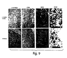
- Figure 9 illustrates a comparative histochemical staining of normal adult lymph node and tonsil. Magnification of PENS staining shown in the far right panels is 40X. Magnification in all other panels is 10X. Monoclonal antibodies used to stain tissue sections, and specific methods are described in the Materials and Methods found at Example 6.
- Figure 10 illustrates comparative histochemical staining of normal adult and fetal thymus. Magnification of adult thymus stained with control and PENS specific antibodies is 20X. Magnification of all other panels is 10X. Monoclonal antibodies used to stain tissue sections, and specific methods are described in the Materials and Methods found at Example 6.
- Figure 11 illustrates comparative histochemical staining of normal adult and fetal liver. Magnification of adult liver stained with anti-PENS is 20X (left panel) and 40X (right panel). Magnification of fetal liver stained with PENS is 10X (left panel) and 60X (right panel). Magnification of all other panels is 10X. Monoclonal antibodies used to stain tissue sections, and specific methods are described in the Materials and Methods found at Example 6.
- Figure 12 illustrates the comparative histochemical staining of normal adult lung and colon. Magnification of lung stained with anti-CD56 and anti-PENS is 20X. Magnification of all other sections is 10X.
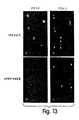
- FIGS 13A through 13D illustrate dual labeling of tissue infiltrating lymphocytes.
- Tissue sections from adult spleen (panels A and B) or adult appendix (panels C and D) were double labeled with fluorescein tagged anti-PEN5 (panels A and C) and rhodamine tagged anti-TIA-1 (panels B and D), prior to examination by fluorescent microscopy.
- Black arrowheads show the location of PEN5 + cells in panels A and C.
- White arrowheads show the location of both PEN5 + cells and TIA-1 + cells in panels B and D.
- Natural killer cells are CD3:TCR - , CD16 + , CD56 + large granular lymphocytes.
- Two functionally distinct populations of peripheral blood NK cells can be differentiated by their surface expression of an isoform of the neural cell adhesion molecule, NCAM (also known as CD56).
- NCAM neural cell adhesion molecule
- CD56 bright NK cells have the attributes of an undifferentiated cell in that they proliferate vigorously in response to exogenous cytokines, but largely lack cytolytic activity.
- CD56 dim NK cells have the attributes of a more differentiated cell, in that they proliferate poorly in response to exogenous cytokines, but are potent cytolytic effector cells.
- monoclonal antibodies that recognize human CD56 are available commercially, for example from Coulter Corp. (Hialeah, Florida) and AMAC, Inc. (Westbrook, Maine).
- NK cells are capable of mediating two types of cytotoxic effector function: natural cytotoxicity and antibody-dependent cellular cytotoxicity ("ADCC").
- ADCC antibody-dependent cellular cytotoxicity
- NK cells play an important role in host defense against viral infection, and in immune surveillance against the establishment of transformed cells. More recent results indicate that NK cells can effect a primitive form of allorecognition which can contribute to graft rejection during allogeneic transplantation and also to graft-versus-host disease. For these reasons, the reliable identification of NK cells within the mononuclear cell population is of great importance.
- the present invention relates to the identification and molecular characterization of a novel sulfated polylactosamine epitope whose expression is largely restricted to the functionally differentiated population of LGLs previously characterized as CD16 + CD56 dim , cytolytic effectors and to antibodies that are capable of recognizing the same. Because this epitope is not expressed on resting or activated T cells, resting or activated B cells, monocytes, granulocytes, platelets or red blood cells, antibodies that bind to unique epitopes on the PENS glycoprotein pair can be used to directly identify this important population of mononuclear cells.
- the epitope is preferentially expressed on the functionally differentiated subpopulation of NK cells relative to CD56 bright NK cells that have the attributes of an undifferentiated cell, antibodies that bind to the epitope can be used to distinguish these two subpopulations of NK cells from one another.
- an antibody or fragment or derivative thereof that recognizes a unique epitope of the PENS glycoprotein pair.
- unique epitope means any epitope on the PEN5 ⁇ glycoprotein and/or the PEN5 ⁇ glycoprotein, which like the novel sulfated polylactosamine epitope identified herein, is present on a high percentage, e.g., at least about 70%, of the population of LGLs previously characterized as CD56 dim , CD16 + natural killer cells and on a significantly lower percentage of the population of NK cells that are phenotypically CD16 + CD56 bright , but not on CD3 + T cells, or CD20 + B cells.
- the "unique epitope” may be present on a glycosylated form of the PENS glycoprotein pair as it is ordinarily expressed on the cell surface of CD56 dim , CD16 + natural killer cells as previously described, or, the "unique epitope” may be present on an unglycosylated or deglycosylated form of the PENS glycoprotein pair.
- the term "unglycosylated” means a PENS molecule where both the PEN5 ⁇ and the PEN5 ⁇ glycoproteins are free of any covalently attached carbohydrate moieties.
- the term "deglycosylated” means a PEN5 molecule where either or both of the PEN5 ⁇ glycoprotein or the PEN5 ⁇ glycoprotein is partially glycosylated but does not contain the same full contingent of carbohydrate moieties as the PEN5 ⁇ glycoprotein or the PEN5 ⁇ glycoprotein as it is ordinarily expressed on the cell surface of CD56 dim , CD16 + natural killer cells.
- the antibodies, fragments, and derivatives of the invention are useful as research reagents, to unambiguously identify, quantify and/or purify natural killer cells in a mixed population of cells and in isolating these natural killer cells therefrom.
- the antibodies, fragments and derivatives of the invention may also be useful therapeutically, either alone, in combination with complement, or conjugated to a radioactive material or a toxin to treat disorders of the immune system where NK cells are implicated as mediators of disease, especially graft-versus-host disease and solid organ and allogenic bone marrow transplant rejection.
- Monoclonal antibodies, and chimeric and humanized antibodies are preferred for detection and therapy, respectively.
- Antibodies, fragments and derivatives thereof recognizing a unique epitope on a deglycosylated or unglycosylated form of the PENS molecule may be useful in the diagnosis and treatment of immune disorders associated with NK cells expressing PENS exhibiting an aberrant glycosylation pattern compared to PENS normally expressed on NK cells.
- Monoclonal antibodies of the invention can be prepared using any technique that provides for the production of antibody molecules by continuous cell lines in culture. These include, but are not limited to, the original techniques of Röhler and Milstein, Nature, 265:495-497 (1975), modified as described in Anderson et al, J. Immunol. , 143:1899 (1989), and the more recent human B cell hybridoma technique and EBV-hybridoma technique well known to persons skilled in the art.
- various host animals including but not limited to rabbits, mice, hamsters, and rats can be immunized by injection with NK cells that express the PENS glycoprotein and, after a sufficient time, the animal is sacrificed and spleen or other immune cells obtained.
- the preferred immunogen to be used in the immunization protocol is a preparation of freshly isolated NK cells, purified from peripheral blood lymphocytes by negative selection.
- Other immunogens that alternatively could be used include partially purified preparations of the PENS molecule, the PEN5 ⁇ glycoprotein or the PEN5 ⁇ glycoprotein, including the glycosylated, deglycosylated or unglycosylated forms thereof and derivatives and fragments thereof.
- a partially purified preparation of the PENS molecule can be prepared from permeabilized NK cells following immunoprecipitation and SDS gel electrophoresis using 6% polyacrylamide gel as hereinafter described using techniques well known to persons skilled in the art.
- any suitable method for partially purifying the PENS molecule or the PEN5 ⁇ or the PEN5 ⁇ glycoprotein as described above can be satisfactorily employed and alternative methods of partial purification will be readily apparent to those persons skilled in this area of technology.
- recombinantly produced molecules can also be used as an immunogen.
- the spleen or other immune cells obtained from the animal are immortalized by fusing the spleen cells with an immortalized cell line, generally in the presence of a fusion enhancing reagent, for example, polyethylene glycol.
- the resulting cells which include the fused hybridomas, are then allowed to grow in a selective medium, such as HAT medium, and the surviving cells are grown in such medium using limiting dilution conditions.
- the cells are grown in a suitable container, e.g., microtiter wells, and the supernatant is screened for monoclonal antibodies having the desired specificity.
- the monoclonal antibodies of the present invention are prepared as described in the Examples.
- ELISAS enzyme-linked immunoadsorbent assays
- FACS fluorescent activated cell sorting
- Initial screening is preferably conducted by screening hybridoma supernatants by flow cytometry for their reactivity with NK cells, but not with T cells, B cells, and monocytes. Further characterization of the hybridomas for those that produce monoclonal antibodies that are preferentially expressed on NK cells that are phenotypically CD16 + CD56 dim relative to NK cells that are phenotypically CD16 + CD56 bright can be conducted by testing on purified populations of lymphoid and non-lymphoid cells by indirect immunofluorescence assays and flow cytometry, substantially as described in the Examples herein.
- Monoclonal antibodies that recognize a PENS epitope that is preferentially expressed on functionally differentiated NK cells will react with an epitope that is present on a high percentage NK cells that phenotypically are CD56 dim CD16 + cells, e.g., at least about 70 - 90%, preferably about 80%, of such cells, and with a much lower percentage of NK cells that are phenotypically CD16 + CD56 bright (e.g., about 10 to 35%), but will not react with CD3 + T cells or CD20 + B cells .
- the antibody will also be unreactive with monocytes, granulocytes, platelets, and red blood cells.
- Monoclonal antibodies that compete with the 5H10 antibody in competition assays well known to persons skilled in the art are likely to recognize essentially the same epitope as mAb 5H10, while monoclonal antibodies that fail to compete with mAb 5H10 but nevertheless meet the criteria of being unique to the CD16 + , CD56 dim subpopulation of NK cells are likely to recognize a different epitope on the PEN5 glycoprotein pair. Both classes of antibodies are considered within the scope of the present invention.
- the resultant antibody may be produced in one of two major ways.
- the purest monoclonal antibody is produced by in vitro culturing of the desired hybridoma in a suitable medium for a suitable length of time, followed by the recovery of the desired antibody from the supernatant.
- the length of time and medium are known or can readily be determined.
- This in vitro technique produces essentially monospecific monoclonal antibody, essentially free from other species of anti-human immunoglobulin.
- the in vitro method may not produce a sufficient quantity or concentration of antibody for some purposes, since the quantity of antibody generated is only about 50 ⁇ g/ml.
- the desired hybridoma may be injected into an animal, such as a mouse.
- the mice are syngeneic or semi-syngeneic to the strain from which the monoclonal-antibody producing hybridomas were obtained.
- Injection of the hybridoma causes formation of antibody producing tumors after a suitable incubation time, which will result in a high concentration of the desired antibody (about 5 - 20 mg/ml) in the ascites of the host animal.
- Antibody molecules can be purified by known techniques, e.g. by immunoabsorption or immunoaffinity chromatography, chromatographic methods such as high performance liquid chromatography or a combination thereof.
- any person skilled in this area of technology can readily isolate hybridomas that produce monoclonal antibodies exhibiting specificity for a unique epitope on functionally differentiated natural killer cells.
- a single hybridoma producing a monoclonal antibody (5H10) against the human PEN5 epitope is exemplified by way of working example, it is contemplated that the present invention encompasses all monoclonal antibodies exhibiting the characteristics of mAb 5H10 as herein described.
- the subject monoclonal antibody 5H10 belongs to the class IgM.
- a monoclonal antibody exhibiting the characteristic described herein may be of class IgG, subclass IgG 1 , IgG 2 ⁇ , IgG 2 ⁇ , or IgG 3 , or of classes IgM, IgA, or other known Ig classes. The differences among these classes or subclasses will not affect the selectivity of the reaction pattern of the antibody, but may affect the further reaction of the antibody with other materials, such as (for example) complement or anti-mouse antibodies.
- the subject antibody is specifically IgM, it is contemplated that antibodies having the patterns of reactivity illustrated herein are included within the subject invention regardless of the immunoglobulin class or subclass to which they belong.
- novel antibody of the present invention is from a murine source, this is not meant to be a limitation.
- the above antibody and those antibodies having the characteristics of the mAb 5H10, whether from a mouse source, other mammalian source including human, rat, or other sources, or combinations thereof, are included within the scope of this invention, as set forth above.
- the antibodies may be used for the detection and enumeration by indirect staining of CD16 + , CD56 dim subpopulation of NK cells in normal individuals or in disease states, for example by fluorescence microscopy, flow cytometry, immunoperoxidase, or other indirect methodologies. Panning techniques are also possible.
- the antibodies may also be used for purification of human natural killer cells which are CD16 + , CD56 dim .
- antibody fragments and derivatives which comprise at least the functional portion of the antigen binding domain of an anti-PENS antibody molecule.
- Antibody fragments which contain the binding domain of the molecule can be generated by known techniques.
- such fragments include, but are not limited to: the F(ab') 2 fragment which can be produced by pepsin digestion of the antibody molecule; the Fab' fragments which can be generated by reducing the disulfide bridges of the F(ab') 2 fragment, and the Fab fragments which can be generated by treating the antibody molecule with papain and a reducing agent.
- the F(ab') 2 fragment which can be produced by pepsin digestion of the antibody molecule
- the Fab' fragments which can be generated by reducing the disulfide bridges of the F(ab') 2 fragment
- the Fab fragments which can be generated by treating the antibody molecule with papain and a reducing agent See, e.g., National Institutes of Health, 1 Current Protocols In Immunology, Coligan et al., ed. ⁇ 2.8, 2.10 (Wiley Interscience, 1991).
- Antibody fragments also include Fv fragments, i.e., antibody products in which there are no constant region amino acid residues. Such fragments can be produced, for example as described in WO 92/04381 or U.S. Patent No. 4,642,334.
- chimeric antibody derivatives i.e. antibody molecules that combine a non-human animal variable region and a human constant region.
- Chimeric antibody molecules can include, for example, the antigen binding domain from an antibody of a mouse, rat, or other species, with human constant regions.
- a variety of approaches for making chimeric antibodies have been described and can be used to make chimeric antibodies containing the immunoglobulin variable region which recognize a unique epitope on the PENS antigen. See, for example, Morrison et al., Proc. Natl.
- the monoclonal or chimeric antibodies of the invention can be further humanized by producing human constant region chimeras, in which even parts of the variable regions, especially the conserved or framework regions of the antigen-binding domain, are of human origin and only the hypervariable regions are of non-human origin.
- Such altered immunoglobulin molecules may be made by any of several techniques known in the art, ( e.g. , Teng et al., Proc. Natl. Acad. Sci. U.S.A., 80:7308-7312 (1983); Kozbor et al., Immunology Today, 4:7279 (1983); Olsson et al., Meth.
- humanized antibodies are preferable for immunotherapy in that they minimize the effects of an immune response. This in turn leads to a lowering of any concomitant immunosuppression and to include increased long term effectiveness in, for instance, chronic disease situations or situations requiring repeated antibody treatments.
- antibody derivatives or immunoconjugates consisting of an antibody molecule or binding region thereof bound to a label such as a radioisotope, fluorescent tag (e.g., fluorescein isothiocyanate, phycoerythrin, phycoerythrin Cy5, or rhodamine), enzyme (e.g., biotin), or other tracer molecule
- a label such as a radioisotope, fluorescent tag (e.g., fluorescein isothiocyanate, phycoerythrin, phycoerythrin Cy5, or rhodamine), enzyme (e.g., biotin), or other tracer molecule
- a label such as a radioisotope, fluorescent tag (e.g., fluorescein isothiocyanate, phycoerythrin, phycoerythrin Cy5, or rhodamine), enzyme (e.g., biotin), or other tracer molecule
- a cytotoxic compound can be conjugated to an antibody of the invention which is specific for NK cells which are the causative agents of an immune disorder, for example, bone marrow graft rejection.
- the cytotoxic compound which can be for example, a radionucleotide or a toxin, such as diphtheria toxin, in conjugated form is thus targeted to the implicated NK cells.
- the antibodies of the invention and the fragments and derivatives thereof containing the binding region can be used in various immunoassays.
- immunoassays include, but are not limited to, competitive and non-competitive assay systems using techniques such as radioimmunoassays, ELISA (enzyme linked immunosorbent assay), "sandwich” immunoassays, precipitin reactions, gel diffusion precipitin reactions, immunodiffusion assays, agglutination assays, complement fixation assays, immunoradiometric assays, fluorescent immunoassays, protein A immunoassays, and immunoelectrophoresis assays, to name but a few.
- Differentiated NK cells can be detected in a biological sample including a mixed population of cells, for example, hematopoietic and lymphoid cells, using the antibodies, fragments or derivatives.
- Suitable biological samples include peripheral blood, bone marrow aspirate and lymphoid tissue.
- the antibody is typically labeled so that its binding with the relevant NK cell subpopulation can be detected. Any suitable label well known to persons skilled in the art, including but not limited to fluorescent dyes, radioactive isotopes, enzymes which catalyze a reaction producing detectable products, biotin, or metal ions detectable by nuclear magnetic resonance can be employed.
- Bone marrow transplantation is increasingly used for the treatment of disorders of the immune system, aplastic anemia, and especially hematopoietic malignancies, such as acute lymphocytic leukemia.
- graft-versus-host disease GVHD
- T cells contained within the bone marrow inoculum are effectors of GVHD
- many bone marrow transplant programs resorted to using T-cell depleted bone marrow cells. This procedure has somewhat successfully reduced the incidence and severity of GVHD.
- T cell depletion including an increased incidence of bone marrow graft rejection.
- GVHD continues to be a serious problem in many bone marrow transplantation recipients, especially in non-T cell depleted transplants.
- NK cells may play a deleterious role in graft-versus-host disease ("GVHD") following solid organ or tissue transplants.
- GVHD graft-versus-host disease
- the antibodies of the invention can be used to target the functionally differentiated subpopulation of NK cells specifically, the invention may also be useful prophylactically and therapeutically, in the prevention and treatment of graft rejection in solid organ and bone marrow transplantation, and in graft-versus-host disease, by modulating the function and number of cytolytic effector NK cells in vivo.
- the anti-NK cell-specific antibodies and fragments and derivatives thereof will have applicability for animal subjects in addition to human beings, such as domesticated animals, the therapeutic aspects of the invention are of the greatest value in the treatment of disorders in humans.
- the antibodies, fragments and derivatives of the invention can be used to remove the CD16 + CD56 dim cytolytic effector population of cells from bone marrow aspirates ex vivo, prior to transplantation of the marrow into the marrow recipient. Removal of these natural killer cells from the bone marrow aspirate can be accomplished by conventional methods, such as those used in immunological T cell depletion.
- Antibodies that exhibit the ability to lyse NK cells in the presence of complement can be used in combination with complement to treat the bone marrow ex vivo prior to transplantation, to kill the NK cells that might otherwise contribute to the etiology of graft-versus-host disease in the recipient.
- the anti-PENS antibody might be linked to a toxin as described, to kill the cytolytic effector NK cells.
- the antibodies, fragments or derivatives of the invention could also be administered to the bone marrow recipient in vivo prior to the transplantation procedure.
- NK cells may also prove useful in the treatment of autoimmune diseases such as SLE, which are in part mediated by NK cells.
- the antibodies, fragments, or derivatives of the invention may also be useful in the prophylaxis and/or treatment of solid organ graft rejection and bone marrow rejection, especially in allogeneic bone marrow transplant recipients where T cell depletion has been employed.
- the NK-specific antibodies, fragments or derivatives of the invention may be useful in unmodified form for modulating the number and function of the cytolytic effector population of NK cells, or they can be conjugated to radionucleotides or toxins by means well known in the art and used to deliver the conjugated substance to deleterious NK cells for negative modulation.
- radionucleotides which can be conjugated to antibodies and administered include 212 Bi, 131 I, 186 Re, and 90 Y. These elements exert their effect by locally irradiating the cells, leading to various intracellular lesions, well known to persons skilled in the art of radiotherapy.
- Cytotoxic drugs that can be conjugated to antibodies and administered for in vivo therapy include, but are not limited to, daunorubicin, doxorubicin, methotrexate, and mytomycin C.
- daunorubicin doxorubicin
- methotrexate doxorubicin
- mytomycin C mytomycin C
- an anti-PENS monoclonal antibody can be combined with diphtheria toxin, by the method of Bumol, Proc. Natl. Acad. Sci., 80:529 (1983). Briefly, monoclonal antibodies reactive with an NK cell specific epitope are prepared as described by Bumol. The antibodies are purified and combined with excess (6 mol/mol) N-succinimydyl 3-(2-pyridyldithio) propionate (Pharmacia, Uppsala, Sweden) in PBS. After 30 minutes incubation at room temperature, the solution is dialyzed against PBS.
- the modified antibodies are conjugated with an appropriate toxin, such as diphtheria toxin A chain.
- an appropriate toxin such as diphtheria toxin A chain.
- Other toxins such as ricin A can also be employed.
- the diphtheria toxin A chain is isolated as detailed in Bumol, supra.
- the modified antibodies are mixed with excess (3 mol/mol) reduced diphtheria toxin A chain (10% of the total volume), allowed to react for 36 hours at 4°C, and concentrated by chromatography on Sephadex G-2000.
- the product is applied to a Sephadex G200 column (1.0 x 100 cm), allowed to equilibrate and eluted with PBS.
- the effector population of natural killer cells can be selectively eliminated in the transplant recipient.
- the route of administration for the in vivo therapeutic modalities may include intradermal, intramuscular, intraperitoneal, intravenous, or subcutaneous injection, intranasal routes and slow release forms, such as those delivered in transplantable forms, on patches or in other colloidal forms.
- the antibody can be encapsulated in liposomes.
- the effective dose of the therapeutic reagent will be a function of the particular reagent employed, the presence and nature of conjugated therapeutic reagent, the patient, and his or her clinical condition.
- Effective doses of the antibodies, fragments, or derivatives of the invention for use in preventing, suppressing, or treating an immune-related disease are in the range of about 1 ng to 100 mg/kg body weight.
- a preferred dosage range is between about 10 ng and 10 mg/kg, and a more preferred dosage range is between 100 ng and 1 mg/kg.
- compositions may be utilized in order to deliver the antibodies, or fragments or derivatives thereof, according to the invention.
- Any suitable pharmaceutical agent with desirable solubility characteristics and chemical properties may be used, including but not limited to, where appropriate, saline or dextrose solutions.
- the reagent itself must be properly formulated, for example, as a humanized or chimeric antibody combined with various buffers, sugars, or stabilizing compounds that increase the stability or half life of the antibody. To extend the half-life, the reagent can first be modified to increase or decrease the amount of carbohydrate complexed to it, or alternatively, can be complexed with a reagent such as polyethylene glycol. Finally, pharmaceutical compositions comprising the therapeutic reagent in the appropriate buffers, salts, and pH are required.
- Therapeutic kits can comprise the therapeutic compositions of the invention in one or more containers.
- the invention also provides partially purified preparations of the NK cell-specific molecule, called PEN5 ⁇ /PEN5 ⁇ , that is preferentially expressed on the subpopulation of NK cells previously characterized as CD16 + , CD56 dim NK cells.
- the molecule consists essentially of two membrane bound glycoproteins.
- the term "partially purified preparation" with respect to the PENS means the PENS molecule, consisting essentially of the PEN5 ⁇ and PEN5 ⁇ glycoprotein pair as herein described, which has been purified from permeabilized NK cells following immunoprecipitation and SDS gel electrophoresis using 6% polyacrylamide gel as hereinafter described. After the glycoproteins are fractionated on a gel, they can be recovered and renatured in accordance with known and established techniques.
- the molecular weight range of the polydispersed PEN5 ⁇ species was 210 ⁇ 3 kDa to 245 ⁇ 5 kDa.
- the average molecular weight of the smaller species, PEN5 ⁇ was 140 ⁇ 3 kDa, with a range of 123 ⁇ 3 kDa to 170 ⁇ 4 kDa.
- the migration of both PEN5 ⁇ and PEN5 ⁇ as polydispersed bands suggests that both species are highly glycosylated.
- Enzymatic deglycosylation indicates that both PEN5 ⁇ and PEN5 ⁇ are 80-90% carbohydrate by weight. This result raised the possibility that these proteins are either proteoglycans or mucin-type glycoproteins.
- Proteoglycans are high molecular weight glycoproteins in which specific glycosaminoglycans are bound to proteins via Gal-xylose-Ser linkages [Bhavanandan, Glycobiology, (1991)], or in the case of keratan sulfate chains, terminal galactosamine linkages to serine or threonine.
- Gal-xylose-Ser linkages Gal-xylose-Ser linkages
- the anti-PENS mAbs are reactive with sulfated polylactosamine carbohydrates present on keratan sulfate glycosaminoglycans, which raised the possibility that PEN5 ⁇ and/or PEN5 ⁇ glycoproteins may be cell surface-associated keratan sulfate proteoglycans.
- cartilage-type keratan sulfate proteoglycans are O-linked glycoproteins
- cornea-type keratan sulfate proteoglycans are N-linked glycoproteins.
- PEN5 ⁇ is an N-linked glycoprotein, it is possible that PEN5 ⁇ is an unusual cell surface cornea-type keratan sulfate proteoglycan.
- PEN5 ⁇ is an O-linked glycoprotein sensitive to keratanase treatment, and may be a cartilage-type keratan sulfate proteoglycan.
- Mucin-type glycoproteins secreted by cultured hamster tracheal epithelial cells are sensitive to keratanase I treatment and contain polylactosamine carbohydrates [Wu, Biochem J., 277:713 (1991)].
- Mucin-type glycoproteins are highly glycosylated proteins containing a majority of O-linked oligosaccharides, and are associated with the cell membrane in a number of cell types [Carraway, Glycobiology 1:131 (1991); Strous, Rev. Biochem Mol. Bio. 27:57 (1992); Devine, 35 (1992). Classification of PEN5 ⁇ as an NK cell specific membrane-bound mucin-type glycoprotein is most consistent with our data.
- PEN5 ⁇ :PEN5 ⁇ complex appears to be analogous to the ASGP-1:ASGP-2 complex derived from ascitic mammary adenocarcinoma cells [Sherblom, J. Biol. Chem., 225:12051 (1980) in which only one component (ASGP-1) of the complex is a mucin-type glycoprotein.
- ASGP-1:ASGP-2 complex derived from ascitic mammary adenocarcinoma cells [Sherblom, J. Biol. Chem., 225:12051 (1980) in which only one component (ASGP-1) of the complex is a mucin-type glycoprotein.
- ASGP-1 ascitic mammary adenocarcinoma cells
- PEN5 The biochemical features of PEN5 molecules points the way for future research.
- carbohydrates are major mediators of cell-cell interactions [Jessel, Annu. Rev. NeuroSci., 13:227 (1990)].
- ligands for E- and P-selectins have been shown to contain either sialyl-CD15 or CD57 polylactosamine epitopes [Philips, Science, 250:1130 (1990); Larsen, Cell, 63:467 (1990); Needham, PNAS, 90:927 (1993)], and GlyCAM-1 a membrane bound mucin glycoprotein is the ligand for L-selectin [Lasky, 1992 #1853].
- PEN5 glycoproteins contribute to NK cell specific adhesion.
- the PEN5 glycoproteins resemble epithelial cell mucins.
- the mucin-type glycoproteins serve a protective role on the epithelial cell surface, and have been shown to protect cells from attack by cytotoxic lymphocytes.
- the PEN5 glycoproteins may therefore protect NK cells from their own cytolytic machinery.
- the PENS antigen can be used in preparing and/or purifying the antibodies of the invention and should also be useful in identifying the natural counter-receptor for the PENS antigen on target cells.
- Amino acid sequence information obtained from the PENS glycoprotein pair can also be used to clone the PEN5 ⁇ and PEN5 ⁇ glycoprotein chains in accordance with established techniques.
- BCK bovine cornea keratan sulfate
- BNC bovine nasal cartilage aggrecan
- CD1 embryonic chick cartilage aggrecan
- LCM leucocyte-conditioned medium
- GLPD granular lymphocyte proliferative disorder
- RC Swarm rat chondrosarcoma aggrecan
- SHK shark cranial cartilage aggrecan.
- Peptide-N-glycosidase (PNgase F) and Endo-a- N -acetylgalactosaminidase (O-glycanase) were used in the buffer provided by the manufacturer (Oxford Glycosystems).
- Keratanase I Keratan sulfate 1,4 b-D-galactanohydrolase; ICN Biomedicals, (Costa Mesa, CA), keratanase II which attacks oversulfated forms of (keratan sulfate 1,4 b-D-galactanohydrolase; ICN Biomedicals, (Costa Mesa, CA), keratanase II which attacks oversulfated forms of keratan sulfate resistant to keratanase I (Seikagaku America, Rockville, MD) and neuraminidase (Calbiochem) were used in either PBS, PNgase F or O-glycanase buffers.
- Chondroitinase ABC (ICN Biomedicals,) was used in sodium acetate 0.05M pH 7.4.
- Trypsin, chymotrypsin and pronase E were also obtained from Sigma.
- FITC- and PE-conjugated avidin were obtained from Becton-Dickinson, (Paramus, NJ).
- Mouse monoclonal antibodies (mAb) reactive with CD2 (T11.1, IgG1), CD3 (RW24B6, IgG2b), CD56 (N901, IgG1), CD20 (B1, IgG1) were obtained from Coulter Corp., as well as isotype matched control mouse mAb (IgG and IgM). Radioiodination of PENS mAb was performed using Iodobeads (Pierce) as previously described [Vivier, J. Immunol. 132:1410 (1991)]. The characterization of the anti-CD16 mAb (3G8, IgG1), and the anti-keratan sulfate mAb 5D4 (IgM) was reported elsewhere [Perussia, J.
- NK cells and T cells were isolated from peripheral blood mononuclear cells (PBMC) obtained from healthy volunteers by negative selection using immuno-magnetic bead depletion [Vivier, Int. Immunol., 4:1313-1323 (1992)].
- PBMC peripheral blood mononuclear cells
- NK cells and NK cell subsets were further purified by flow cytometric sorting on an Epics V flow cytometer (Coulter Electronics) after staining with anti-CD56 mAb.
- Activation of NK cells was performed using ionomycin (1 ⁇ M) and 20% lymphocyte-conditioned medium (LCM) as described previously [Robertson, J. Immunol., 150:1705 (1993)].
- PBMC from three patients with a CD3:TCR - , CD16 + , CD56 + granular lymphocyte proliferative disorder (GLPD) [Oshimi, Leukemia, 2:617 (1988)] were isolated by Ficoll-Hypaque gradient centrifugation.
- radioiodinated lysates were diluted in 1 ml lysis buffer and precleaned three times with 3 ⁇ l of affinity-purified rabbit anti-mouse IgM or IgG (RAM, Jackson Immunoresearch Laboratories, (West Grove, PA) and 50 ml of a 50% solution of protein A-sepharose beads (Pharmacia, Milwaukee, WI).
- the immunoprecipitations were performed using 3 ml of the indicated mAb, 3 ⁇ l of RAM and 50 ⁇ l of protein A-Sepharose beads at 50%.
- Sepharose-bound immune complexes were washed four times in lysis buffer, and eluted either directly into sample buffer (2% SDS, 10% glycerol, 0.1 M Tris-HCl, pH 6.8, 0.02% bromophenol blue) prior to electrophoretic separation, or in elution buffer (0.15 M NH 4 OH, pH 10.5) prior to deglycosylation experiments.
- the following enzymes were used alone or in combination: PNgase F (310 U/ml), O-glycanase (0.06 U/ml), keratanase I (0.25 U/ml) and neuraminidase (0.2 U/ml).
- Peripheral blood NK cells were first stained using 5H10 (anti-PENS) and colloidal gold-labeled goat anti-mouse IgM (Amersham). After fixation using % glutaraldehyde, the stained cells were examined by transmission electron microscopy.
- NK cells In order to identify novel cell surface structures selectively expressed on NK cells, we generated a panel of mouse mAb (anti-PEN mAb) that recognized NK cells but not T cells. These antibodies were produced by immunizing BALB/c mice with digitonin permeabilized peripheral blood NK cells as previously described [Anderson, J. Immunol. 143:1889 (1989)]. Briefly, mononuclear cells were isolated from leukopheresis residues (obtained from normal blood donors at the Dana-Farber Cancer Institute Blood Bank) by centrifugation over ficoll. These cells were cultured in plastic flasks in RPMI media containing 10% fetal calf serum for six to twelve hours to allow the adherence of monocytes.
- Nonadherent cells were incubated with monoclonal antibodies reactive with CD5 (24T6G12, IgG2A), CD3 (RW24B6, IgG1), CD20 (B1H299, IGG2A), CD24 (MY4322A-1, IgG2B) at optimal concentrations for thirty minutes, then washed extensively. Following the addition of magnetic beads coupled to goat and anti-mouse Ig (Advanced Magnetics, Inc., Cambridge, MA) these populations were depleted of T cells, B cells, monocytes by negative selection using a magnet.
- CD5 24T6G12, IgG2A
- CD3 RW24B6, IgG1
- CD20 B1H299, IGG2A
- CD24 MY4322A-1, IgG2B
- NK cells The remaining cells which were enriched for NK cells were phenotypically less than 5% CD3 + , 75-95% CD56 + , and 65-80% CD16 + as determined by flow cytometry using an Epics profile (Coulter Electronics, Hialeah, FL). These cells were then permeabilized with digitonin as described in Anderson, J. Immunol. 143:1889 (1989). Permeabilized NK cells (50 x 10 6 cells per ml PBS), were injected into a five week old Balb/c mouse at three week intervals for a total of four immunizations. Three days after the last immunization, the immunized mouse was sacrificed and splenocytes prepared using standard methods.
- Immune splenocytes were fused to the NS1 hybridoma cell line at a 1:1 ratio using polyethylene glycol as described in Anderson, J. Immunol. 143:1889 (1989). Following fusion, cells were cultured at limiting dilution in a 96-well plate in the presence of RPMI media containing 10% fetal calf serum and HAT selection medium. Individual supernatants were screened for their reactivity with permeabilized and unpermeabilized NK cells, T cells, B cells, monocytes. Monoclonal antibody 5H10 (anti-PEN5) was selected as an antibody which reacted specifically with peripheral blood NK cells.
- the reactivity pattern of 5H10 was first determined by testing purified peripheral blood lymphocytes obtained from healthy volunteers using an Epics V flow cytometer (Coulter Electronics, Hialeah, Florida).
- the binding of biotinylated anti-PEN5 mAb was revealed using APC-conjugated avidin.
- peripheral blood T cells isolated as described were activated with optimal mitogenic concentrations of PHA and Con A.
- Splenic B cells were activated with optimal concentrations of Staphylococcus aureas Cowan strain I in accordance with standard laboratory protocols. Immunofluorescence staining was performed at days 2, 4, and 6 after activation.
- T cell activation induced by mitogenic concentrations of PHA or Con A in the presence or absence of PMA
- B cell activation induced by Staphylococcus aureus Cowan strain I for 1 to 6 days induced the cell surface expression of the PEN5 epitope (See Table 1 below).
- allogeneic T cell clones did not express the PEN5 epitope (See Table 2).
- peripheral blood NK cells required a more careful comparison of the relative expression of PEN5 and CD56.
- the two-color flow cytometric comparison shown in Figure 1 suggested that PEN5 was preferentially expressed on the CD56 dim population. This was confirmed by comparing the expression of PEN5 on sorted populations of CD56dim and CD56bright NK cells, as shown in Figure 2.
- NK cells were sorted into CD56 dim and CD56 bright NK cell subsets using rhodamine-conjugated anti-CD56 mAb and flow cytometry. Unsorted NK cells, CD56 dim and CD56 bright NK cells were further analyzed for the expression of 5H10 using biotinylated anti-PENS mAb and FITC-conjugated avidin. Controls were performed using mouse isotype matched control IgM mAb. The results of this experiment are illustrated in Figure 2, in which the numbers in each histogram indicate the percentage of positively stained cells.
- PEN5 expression is down-regulated by NK cell activation.
- CD56 dim and CD56 bright NK cells strongly differ in their response to proliferative stimuli. Although CD56 dim NK cells do not proliferate in response to either IL-2 or the combination of ionomycin and PMA, CD56 bright NK cells proliferate in response to either stimulus.
- CD56 dim NK cells can be induced to proliferate in response to a combination of LCM and ionomycin to correlate PENS expression with the NK cell proliferative state. Briefly, sorted CD56 dim and CD56 bright NK cells were activated for 20 days with ionomycin and LCM as described in the Materials and Methods.
- Radioiodinated lysates prepared from resting NK cells were immunoprecipitated using the 5H10 (anti-PEN5) mAb or an isotype matched mouse IgM control mAb. Immunoprecipitates were then separated under non-reducing conditions on SDS-polyacrylamide gels (6% SDS). The results are shown in Figure 5.
- the m.w. range of the polydispersed PENS species was 210 ⁇ 3 kDa to 245 ⁇ 5 kDa.
- the average m.w. of the smaller species, PEN5 ⁇ was 140 ⁇ 3 kDa, with a range of 123 ⁇ 3 kDa to 170 ⁇ 4 kDa.
- the migration of both PEN5 ⁇ and ⁇ molecules as polydispersed bands suggested that they were highly glycosylated.
- PEN5 ⁇ and ⁇ are Carbohydrates With Keratanase I-Sensitive Chains
- PNgase F treatment induced the disappearance of PEN5 ⁇ from the 210-245 kDa m.w. range, and the appearance of a deglycosylated form of PEN5 ⁇ migrating at 20-25 kDa (c2).
- the apparent mobility of PEN5 ⁇ was reduced by only -20 kDa after PNgase F incubation.
- Treatment of PENS glycoproteins with O-glycanase ( Figure 6, lane 3) did not significantly affect their SDS-PAGE migration pattern.
- PEN5 ⁇ Contains 80% N-Linked Keratanase-Sensitive Carbohydrates, Whereas PEN5 ⁇ Contains 80% O-Linked Keratanase-Sensitive Carbohydrates
- PEN5 ⁇ The extensive N-linked glycosylation of PEN5 ⁇ suggested that it might be a member of one of the two major groups of glycoproteins characterized by such high carbohydrate content (50 to 90%), i.e: proteoglycans and mucin-type glycoproteins. Chondroitinase ABC, heparitinase and heparinase did not affect the migration pattern of PEN5 ⁇ or PEN5 ⁇ (data not shown). By contrast, incubation of PENS molecules with keratanase I reduced the apparent m.w. of PEN5 ⁇ from 210-245 kDa to 35-40 kDa (Fig. 6, lane 2; cl).
- PEN5 ⁇ contains ⁇ 80% N-linked keratanase I-sensitive carbohydrates
- PEN5 ⁇ contains -80% O-linked keratanase I-sensitive carbohydrates.
- treatment with neuraminidase induced a slight reduction in the polydispersity, as well as a shift in the apparent m.w. of both PEN5 ⁇ and ⁇ , indicating that sialic acid residues are also present on both glycoproteins (Fig. 6, lane 5).
- Treatment of PENS glycoproteins with a combination of PNGase F and neuraminidase Fig.
- Radioiodinated 5H10 (anti-PEN5) mAb was combined with various concentrations of bovine cornea keratan sulfate proteoglycan (BC), and the mixture was then incubated with NK cells.
- NK cells or anti-PENS mAb When used in incubation with NK cells or anti-PENS mAb, the following carbohydrates used at 10 mg/ml were without any effect on 5H10 binding to NK cell surface: chondroitin sulfate B, heparin, heparan sulfate, dextran sulfate, GlcNAc, mannose 6-phosphate, lactose, galactose-6-phosphate, fucose, glucose 6-phosphate, glucose and galactose.
- carbohydrates used at 10 mg/ml were without any effect on 5H10 binding to NK cell surface: chondroitin sulfate B, heparin, heparan sulfate, dextran sulfate, GlcNAc, mannose 6-phosphate, lactose, galactose-6-phosphate, fucose, glucose 6-phosphate, glucose and galactose.
- Fig. 7A the binding of radiolabeled 5H10 mAb to NK cells was inhibited in a dose-dependent manner in the presence of BC proteoglycan.
- Preincubation of NK cells with the same concentrations of BC proteoglycan did not affect the binding of 5H10 mAb (data not shown), indicating that the anti-PEN5 mAb reacted with carbohydrate determinants present on keratan sulfate glycosaminoglycans.
- Incubation of anti-PEN5 mAb with simple sugars or other glycosaminoglycans was without any effect (see Brief Description of Figure 7A).
- NK cells peripheral blood NK cells were incubated in PBS-1%BSA for 3 hr or 45 min at 37°C with glycosidases (0.025 U/ml) or proteases (5 mg/ml) respectively. Cell surface expression of PENS epitope was then analyzed by flow cytometry using 5H10 mAb. Percent modulation was calculated as the ratio of the total linear mean fluorescence intensity of the treated cells over that of untreated control cells.
- 5H10 anti-PEN5
- 5D4 anti-keratan sulfate
- the antigenicity of 5H10 mAb for aggrecan proteoglycans was analyzed by ELISA as described in Materials and Methods.
- the anti-keratan sulfate mAb 5D4 was used as a positive control.
- Chondroitinase ABC was used at 0.04 U/ml
- keratanase I was used at 0.05 U/ml
- keratanase II was used at 0.004 U/ml, for 1 hr at 37°C.
- the 5H10 mAb (cross-hatched) recognized aggrecan-type proteoglycans derived from embryonic chick cartilage (CD1, upper panel) and from bovine nasal cartilage (BNC, middle panel).
- CD1 and BNC embryonic chick cartilage
- BNC bovine nasal cartilage
- the anti-keratan sulfate mAb 5D4 open bars
- CD1 and BNC treated with either keratanase I or II, reduced 5H10 reactivity.
- Treatment of CD1 and BNC with chondroitinase ABC is known to increase the expression of keratan sulfate epitopes.
- the epitope expressed on the PEN5 molecule on NK cells is not simply a keratan sulfate chain since flow cytometric analysis using 6 distinct anti-keratan sulfate mAbs 1B4, 2D3, 3D2, 4D1, 8C2, and 5D4 did not detect binding to NK cells (data not shown).
- PEN5 glycoproteins are expressed at the NK cell surface as extended rod-like structures.
- the PENS epitope is in part a carbohydrate determinant that can be expressed on keratan sulfate chains.
- keratan sulfate glycosaminoglycans selectively compete with PENS molecules for binding to the 5H10 (anti-PEN5) mAb.
- treatment of NK cells with keratanase I down-regulates the cell surface expression of the PEN5 epitope.
- the 5H10 mAb recognizes two distinct aggrecan-type keratan sulfate proteoglycans.
- Keratan sulfates are glycosaminoglycans consisting of repeated Galbl-4(sulfated)GlcNac disaccharides. Within this constraint, differential branching of the disaccharide subunits, differential sulfation of GlcNAc, and differential fucoslyation and/or sialylation of the Galb1-4(sulfacted)GlcNAc can lead to heterogeneity in individual keratan sulfate chains.
- the lack of reactivity of anti-PENS mAb with the keratan-sulfate proteoglycan SHK isolated from shark cranial cartilage suggests that the standard lactosaminoglycan repeat sequence is not the epitope recognized by 5H10. Rather, our data indicate that 5H10 recognizes an unusual sulfated polylactosamine epitope present on some but not all keratan sulfate glycosaminoglycans.
- Tissues Histologically normal fetal (20 week gestation) and adult human tissues were obtained from surgical and autopsy specimens. Frozen tissues embedded in OCT compound (Baxter Corp., McGaw Park, IL) were stored at -70°C until needed. All tissues were used as frozen tissue sections and were adequately preserved histologically.
- the panel of normal tissues that were screened included adrenal, brain, breast, cervix, colon, esophagus, heart, kidney, liver, lung, lymph node, ovary, peripheral nerve, pancreas, skeletal muscle, skin, small intestine, spleen, stomach, testis, thyroid, tonsils, thymus, and uterus.
- Anti-5H10 was used at a dilution of 1:400 (2.5 mg/ml) in phosphate buffered saline (PBS) containing 0.06% crystalline bovine serum albumin (BSA) and 0.1% sodium azide.
- PBS phosphate buffered saline
- BSA crystalline bovine serum albumin
- Purified mouse IgM (Coulter Immunology, Hialeah, FL) served as the negative control. For use it was diluted to the same concentration, with the same buffer solution as the test antibody.
- N901 a murine monoclonal antibody of the IgGl subclass, binds to the NKH1 antigen (CD56) expressed on NK cells.
- the antibody was used at a dilution of 1:664 (2.5 mg/ml) in PBS containing 0.06% crystalline bovine serum albumin (BSA) and 0.1% sodium azide.
- BSA crystalline bovine serum albumin
- Biotinylated affinity purified goat anti-mouse IgM (m chain specific) and horse anti-mouse IgG (heavy + light chain specific) antibodies (Vector Laboratories, Inc,. Burlingame, CA) were utilized as secondary antibodies at a dilution of 1:150 in PBS containing 2% human AB + serum and 0.1% sodium azide.
- Avidin-biotin-peroxidase complexes were used as the labeling reagent at a dilution of 1:1:80 in PBS.
- Immunohistochemistry Immunohistochemical studies were performed using the avidin-biotin immunoperoxidase technique [Rice, et al., Am. J. Path., 138:385, (1991)]. To assure that tissue sections adhered, slides were coated with poly-L-lysine (Sigma Chemical Co., St. Louis, MO) reconstituted in purified water. Frozen sections were cryostat cut (6-8 mm thick), collected onto coated slides, air dried and fixed in 2% neutral buffered paraformaldehyde at 4°C for 20 minutes, followed by several washes with PBS.
- poly-L-lysine Sigma Chemical Co., St. Louis, MO
- Sections were then washed with PBS, incubated with either biotinylated goat anti-mouse IgM or horse anti-mouse IgG antibodies for 30 minutes, washed in PBS, incubated with avidin-biotin-peroxidase complexes for 45 minutes, and then washed again with PBS. After incubating the slides for 5 minutes in Tris-Imidazole/HCL buffer, the peroxidase reaction was initiated by incubating for 5 minutes with 3,3-diaminobenzidine (DAB) (Sigma Chemical Co.) dissolved in Tris-Imidazole/HCL buffer containing 0.11% hydrogen peroxide.
- DAB 3,3-diaminobenzidine
- Tissue sections were washed in water, counterstained with Harris hematoxylin, and dehydrated through graded alcohols and xylenes. Coverslips were then mounted on slides with E-Z-Mount mounting media (Shandon Inc., Pittsburgh, PA).
- CD56 is expressed on the surface of most peripheral blood NK cells, its density of expression on most NK cells is quite low. Because of this, antibodies reactive with CD56 may not be ideal reagents for the identification of tissue infiltrating NK cells.
- the relative inability of anti-CD56 to detect NK cells in lymphoid tissues is demonstrated in Figures 9 and 10, in which CD56 + cells are rarely detected in lymph node, tonsil, or thymus. In contrast, antibodies reactive with PENS identified lymphocytes infiltrating each of these tissues. Whereas PEN5 + cells were scattered throughout the lymph node, they tended to be concentrated in the parafollicular areas of the tonsil. At higher magnification, PEN5 + cells were observed to be round or oval or occasionally elongated.
- PEN5 + and CD56 + lymphocytes were not easily detected in either fetal or adult thymus.
- scattered lymphocytes expressing low levels of CD56 could be detected at high magnification, suggesting that CD56 + cells are present, but difficult to detect using this histochemical method. This might result from lability of the antigen under these fixation conditions, or the low level of CD56 expression, since Sanchez, et al [ J. Exp. Med.
- PEN5 expression in liver infiltrating lymphocytes appeared, at least in part, cytoplasmic.
- mucin-like glycoproteins can be identified in the trans-Golgi reticulum and in cytoplasmic vesicles that eventually fuse with the plasma membrane [Watkins, et al., Carbohydrate Res. 213:185 (1991)]. It is possible that liver infiltrating NK cells express PEN5 primarily in these intracellular compartments.
- PEN5 + lymphocytes were particularly prevalent in fetal liver and fetal thymus.
- NK cells and T cells arise from a common bone marrow-derived progenitor cell [Sanchez, et al., J. Exp. Med. 178:1857 (1993); Lanier, et al., Immunol. Today 13:392 (1992); Rodewald, et al., Cell 69:139 (1992); and Koyasu, et al., J. Exp. Med. 179:1957 (1994)].
- PEN5 + lymphocytes are present in fetal liver and thymus provides unbiased evidence for the differentiation of NK cells in these tissues. Although relatively few CD56 + cells were identified at these sites using histochemical analysis, this result might reflect the low density of expression of this NK marker. CD56 + lymphocytes have been detected in both fetal liver and fetal thymus using flow cytometric analysis [Sanchez, et al., J. Exp. Med. 178:1857 (1993)]. Our results suggest that PENS expression can be expected to be a more sensitive marker of tissue infiltrating NK cells than CD56 expression.
- Antibodies reactive with PENS also recognized some non-leukocytic cells. These were generally epithelial cells found in the esophagus, cervix, endometrium, trachea, bile ducts, colon and pancreas. The most dramatic example of this non-lymphoid staining was seen in the lung and colon, where anti-PEN5 strongly stained the mucous layer lining bronchial and colonic epithelial cells ( Figure 12). The specificity of this staining was confirmed by the inability of either isotype matched control antibody or anti-CD56 to stain epithelial mucosa.
- PEN5 + tissue infiltrating lymphocytes are NK cells.
- TIA-1 2G9, IgG1
- a cytotoxic lymphocyte-restricted granule protein a cytotoxic lymphocyte-restricted granule protein
- PEN5 + cells were identified in spleen and appendiceal lymphoid tissue using FITC-tagged anti-5H10. These same sections were also labeled using phycoerythrin-tagged anti-2G9.
- PEN5 + lymphocytes scattered throughout the spleen were also TIA-1 + . Consistent with the localization of these antigens, PENS staining is largely confined to the cell surface, whereas TIA-1 staining is cytoplasmic, and granular. Some PEN5-cells expressed TIA-1. These cells are likely to be cytotoxic T cells which express TIA-1, but not PENS. In the appendix, only one out of four PEN5 + lymphocytes co-expressed TIA-1. This result suggests that in some tissues, PENS might identify less differentiated NK cells that do not possess defined cytotoxic granules.
- TIA-1 that are related to NK cell differentiation.
- Table III tabulates the percentage of PEN5 + tissue infiltrating lymphocytes expressing TIA-1 in several tissues. As summarized below, whereas the majority of PEN5 + lymphocytes co-express TIA-1 in spleen and liver, this is not the case in tonsil or appendix, where most PEN5 + lymphocytes do not express TIA-1. Whether these tissue specific differences reflect different stages of NK cell differentiation, or different types of tissue infiltrating lymphocyte remains to be elucidated.
- Examples 6A through 6D illustrate a number of important findings.
- a monoclonal antibody reactive with a sulfated poly-N-lactosamine epitope expressed on the NK cell restricted glycoprotein PENS to survey the presence of tissue-infiltrating NK cells in lymphoid and non-lymphoid tissues.
- antibodies reactive with CD56 were unable to efficiently detect all tissue infiltrating NK cells
- PEN5 + lymphocytes were readily identified in multiple tissues.
- PENS is expressed similarly on both tissue infiltrating and circulating lymphoid cells, these results suggest that NK cells can infiltrate multiple lymphoid and non-lymphoid tissues to mediate their immune functions.
- PEN5 is selectively expressed on large granular lymphocytes possessing cytotoxic effector function. These cells express low levels of CD56, which might account for the inability of antibodies reactive with CD56 to recognize these cells in tissues.
- Double staining with the cytotoxic granule marker, TIA-1 supports the conclusion that PEN5 + lymphocytes infiltrating some tissues (e.g. spleen and liver) contain cytotoxic granules.
- PEN5 + cells infiltrating other tissues e.g. tonsil and appendix
- PENS might be expressed on agranular lymphocytes.
- the PEN5 epitope recognized by monoclonal antibody 5H10 is related to keratan sulfate, which is itself a member of the polylactosamine family of sugars.
- the two isoforms of PENS thus resemble a keratan sulfate proteoglycan (PEN5 ⁇ ) and a keratan sulfated mucin (PEN5 ⁇ ).
- Secreted mucins derivatized with keratan sulfate have been identified in the tracheal mucosa [Kim, et al., Exp. Lung Res. 17:533 (1991)].
- anti-5H10 can recognize keratan sulfate-bearing proteoglycans derived from several tissues, including embryonic chick cartilage and bovine nasal aggrecan [Vivier, et al., J. Exp. Med. 178:2023 (1993)]. In epithelial cells, mucins are secreted to provide protection against environmental toxins [Strous, et al., Critical Rev. in Biochem. and Molec. Biol. 27:57 (1992)].
- PENS is expressed on differentiated large granular NK cells to protect them against their own cytotoxic effector molecules.
- the extended, rod-like structure of PENS demonstrated by transmission electron microscopy could facilitate such a functional role.
- Cell surface mucins have also been identified as ligands for lymphocyte adhesion molecules involved in tissue homing [Lasky, et al., Cell 69:927 (1992)]. It is therefore possible that the expression of PEN5 on terminally differentiated NK cells allows its subsequent infiltration into the various tissues in which these cells are found.
Landscapes
- Health & Medical Sciences (AREA)
- Life Sciences & Earth Sciences (AREA)
- Chemical & Material Sciences (AREA)
- Immunology (AREA)
- Cell Biology (AREA)
- Organic Chemistry (AREA)
- Molecular Biology (AREA)
- Medicinal Chemistry (AREA)
- Hematology (AREA)
- Engineering & Computer Science (AREA)
- General Health & Medical Sciences (AREA)
- Biochemistry (AREA)
- Biophysics (AREA)
- Urology & Nephrology (AREA)
- Biomedical Technology (AREA)
- Proteomics, Peptides & Aminoacids (AREA)
- Zoology (AREA)
- Genetics & Genomics (AREA)
- Pathology (AREA)
- Food Science & Technology (AREA)
- Biotechnology (AREA)
- General Physics & Mathematics (AREA)
- Virology (AREA)
- Analytical Chemistry (AREA)
- Physics & Mathematics (AREA)
- Microbiology (AREA)
- Tropical Medicine & Parasitology (AREA)
- Toxicology (AREA)
- Gastroenterology & Hepatology (AREA)
- Peptides Or Proteins (AREA)
- Preparation Of Compounds By Using Micro-Organisms (AREA)
- Micro-Organisms Or Cultivation Processes Thereof (AREA)
Claims (21)
- Monoklonaler Antikörper oder ein immunreaktives Fragment oder Derivat davon, welcher einen Immunkomplex mit dem Glykoprotein PEN5 bildet, welches auf der Oberfläche von natürlichen Killerzellen vorhanden ist, und der Antikörper nicht mit den Antigenen, welche auf T-Zellen, B-Zellen, Monozyten, Granulozyten, roten Blutkörperchenzellen und Thrombozyten vorhanden sind, reagiert, wobei das Glykoprotein PEN5 auf der Oberfläche einer natürlichen Killerzelle vorliegt und ein N-verknüpftes Glykoprotein PEN5α mit einem durchschnittlichen Molekulargewicht von 227±4 kDa und ein O-verknüpftes Glykoprotein PEN5β mit einem durchschnittlichen Molekulargewicht von 140±3 kDa umfaßt.
- Antikörper nach Anspruch 1, welcher ein Epitop auf dem PEN5-Glykoprotein, umfassend ein Polylactosamin-Kohlenhydratmolekül, erkennt.
- Antikörper nach Anspruch 2, wobei das Epitop auf der Oberfläche von etwa 70 bis 90% der funktionell differenzierten natürlichen Killerzellen vorhanden ist und nicht auf CD3+ T-Zellen oder CD20+ B-Zellen vorhanden ist.
- Antikörper nach Anspruch 1, welcher einen Immunkomplex mit dem gleichen Epitop wie der monoklonale Antikörper, hergestellt durch das Hybridom, gekennzeichnet durch die ATCC-Zugangsnr. HB11441, bildet.
- Antikörper nach Anspruch 1, welcher durch das Hybridom, gekennzeichnet durch die ATCC-Zugangsnr. HB11441, hergestellt wird.
- Antikörper nach Anspruch 1 oder ein immunreaktives Fragment oder Derivat davon, verknüpft mit einem Marker, Toxin oder festem Träger, wobei der Marker ein fluoreszierender, radioaktiver oder enzymatischer Marker ist.
- Hybridom, welches einen monoklonalen Antikörper erzeugt, welcher einen Immunkomplex mit dem Glykoprotein PEN5, wie in Anspruch 1 definiert, bildet.
- Hybridom nach Anspruch 7, welches durch die ATCC-Zugangsnr. HB11441 gekennzeichnet ist.
- Teilweise gereinigte Aufarbeitung von einem natürlichen Killerzellenspezifischen Protein, wobei das Protein PEN5, wie in Anspruch 1 definiert, deglykolisiertes PEN5, PEN5α, deglykolisiertes PEN5α, PEN5β oder deglykolisiertes PEN5β ist.
- Verfahren zum Nachweis des Vorhandenseins von natürlichen Killerzellen in einer biologischen Probe, umfassend die Schritte(a) des Kontaktierens der biologischen Probe mit einem monoklonalen Antikörper, wie in Anspruch 1 definiert, oder einem immunreaktiven Fragment oder Derivat davon, welcher einen Immunkomplex mit dem natürlichen Kilierzell-Oberflächenglykoprotein PEN5 unter Bedingungen bildet, welche ausreichend sind, die Bildung eines Immunkomplex zu ermöglichen, und(b) des Nachweisens des Immunkomplex als einen Indikator für das Vorhandensein von natürlichen Killerzellen in der biologischen Probe.
- Verfahren nach Anspruch 10, wobei die biologische Probe eine Gewebeprobe, ausgewählt aus der Gruppe, bestehend aus Lymphe, Nebenniere, Gehirn, Brust, Gebärmutterhals, Dickdarm, Speiseröhre, Herz, Niere, Leber, Lunge, Lymphknoten, Eierstock, peripherer Nerv, Bauchspeicheldrüse, Skelettmuskel, Haut, Dünndarm, Milz, Magen, Hoden, Schilddrüse, Mandeln, Thymus und Gebärmutter, ist.
- Verfahren nach Anspruch 10, wobei der monoklonale Antikörper mit einem nachweisbaren Marker, ausgewählt aus der Gruppe, bestehend aus einem Radioisotop, einem Fluoreszenzmarker oder einem Enzym, verknüpft ist.
- Verfahren nach Anspruch 10, wobei die biologische Probe eine Probe aus dem peripheren Blut oder Knochenmark ist.
- Verfahren zur selektiven Entfernung natürlicher Killerzellen aus einer biologischen Probe, umfassend die Schritte(a) des Kontaktierens der biologischen Probe mit einem monoklonalen Antikörper, wie in Anspruch 1 definiert, oder einem immunreaktiven Fragment oder Derivat davon, welcher einen Immunkomplex mit dem natürlichen Killerzellen-Oberflächenglykoprotein PEN5, unter Bedingungen, welche ausreichend sind, die Bildung eines Immunkomplex zu ermöglichen, bildet, und(b) des Entfernens der Zellen, umfassend den Immunkomplex aus der biologischen Probe.
- Verfahren nach Anspruch 14, wobei die biologische Probe peripheres Blut oder eine Knochenmarkspunktion ist.
- Verfahren nach Anspruch 15, wobei die Probe mit dem Antikörper oder immunreaktiven Fragment davon in Anwesenheit von Komplement kontaktiert wird.
- Verfahren nach Anspruch 15, wobei der monoklonale Antikörper an ein Toxin konjugiert ist.
- Kit zum Nachweis oder zum Entfernen natürlicher Killerzellen aus einer biologischen Probe, umfassend einen monoklonalen Antikörper, wie in Anspruch 1 definiert, oder ein immunreaktives Fragment oder Derivat davon, welcher einen Immunkomplex mit dem natürlichen Killerzellen-Oberflächenprotein PEN5 bildet.
- Kit nach Anspruch 18, wobei der monoklonale Antikörper an das gleiche Epitop, wie der Antikörper, sekretiert von dem Hybridom, gekennzeichnet durch die ATCC-Nr. HB11441, bindet.
- Kit nach Anspruch 18, wobei der monoklonale Antikörper mit einem Marker, ausgewählt aus der Gruppe, bestehend aus einem Radioisotop, einem Fluoreszenzmarker oder einem Enzym, verknüpft ist.
- Kit nach Anspruch 18, weiter umfassend einen Antikörper, welcher ein T-Zellen-Oberflächenantigen, CD20, erkennt.
Applications Claiming Priority (3)
| Application Number | Priority Date | Filing Date | Title |
|---|---|---|---|
| US11317093A | 1993-08-27 | 1993-08-27 | |
| US113170 | 1993-08-27 | ||
| PCT/US1994/009714 WO1995006247A1 (en) | 1993-08-27 | 1994-08-26 | Natural killer cell-specific antigen and antibodies that identify the same |
Publications (3)
| Publication Number | Publication Date |
|---|---|
| EP0714510A1 EP0714510A1 (de) | 1996-06-05 |
| EP0714510A4 EP0714510A4 (de) | 1999-06-09 |
| EP0714510B1 true EP0714510B1 (de) | 2002-06-19 |
Family
ID=22347947
Family Applications (1)
| Application Number | Title | Priority Date | Filing Date |
|---|---|---|---|
| EP94927259A Expired - Lifetime EP0714510B1 (de) | 1993-08-27 | 1994-08-26 | Für natürliche killerzellen spezifische antigene und sie identifizierende antikörper |
Country Status (6)
| Country | Link |
|---|---|
| US (2) | US5786160A (de) |
| EP (1) | EP0714510B1 (de) |
| JP (1) | JPH09502089A (de) |
| CA (1) | CA2170352A1 (de) |
| DE (1) | DE69430846T2 (de) |
| WO (1) | WO1995006247A1 (de) |
Families Citing this family (10)
| Publication number | Priority date | Publication date | Assignee | Title |
|---|---|---|---|---|
| WO1995006247A1 (en) * | 1993-08-27 | 1995-03-02 | Dana-Farber Cancer Institute, Inc. | Natural killer cell-specific antigen and antibodies that identify the same |
| AU1533099A (en) * | 1998-08-25 | 2000-03-14 | Immune Response Corporation, The | Methods for evaluating immune function |
| FR2801056B1 (fr) | 1999-11-12 | 2003-03-28 | Commissariat Energie Atomique | Proteine presente a la surface des cellules souches hematopoietiques de la lignee lymphoide et des cellules nk, et ses applications |
| US6979546B2 (en) | 1999-11-15 | 2005-12-27 | Universita Di Genova | Triggering receptor involved in natural cytotoxicity mediated by human natural killer cells and antibodies that identify the same |
| AU783899B2 (en) * | 1999-11-15 | 2005-12-22 | Innate Pharma S.A.S. | Novel triggering receptor involved in natural cytotoxicity mediated by human natural killer cells, and antibodies that identify the same |
| WO2001088099A1 (en) * | 2000-05-16 | 2001-11-22 | Alison Davies | Cells, culture methods and their uses |
| FR2810045B1 (fr) * | 2000-06-07 | 2004-09-03 | Assist Publ Hopitaux De Paris | Procede d'obtention de population cellulaires caracterisees d'origine musculaire et utilisations |
| AU2004225941A1 (en) * | 2003-03-28 | 2004-10-14 | Elusys Therapeutics, Inc. | Method and compositions for conversion of antibody activity |
| EP1626059A1 (de) | 2004-08-09 | 2006-02-15 | Institut National De La Sante Et De La Recherche Medicale (Inserm) | Angiogenische und immunologische Verwendungen von anti-CD160 spezifischen Stoffen, erhältlich vom monoklonalen Antiköper CL1-R2 |
| CN101522219A (zh) * | 2006-10-12 | 2009-09-02 | 惠氏公司 | 改变抗体溶液中离子强度以减少乳光/聚集物 |
Citations (1)
| Publication number | Priority date | Publication date | Assignee | Title |
|---|---|---|---|---|
| US4831122A (en) * | 1986-01-09 | 1989-05-16 | Regents Of The University Of Minnesota | Radioimmunotoxins |
Family Cites Families (7)
| Publication number | Priority date | Publication date | Assignee | Title |
|---|---|---|---|---|
| US4797475A (en) * | 1983-05-20 | 1989-01-10 | The Regents Of The University Of California | Method and composition for isolating white cell elements |
| US4772552A (en) * | 1984-04-23 | 1988-09-20 | Dana-Farber Cancer Institute, Inc. | Monoclonal antibody which recognizes a 200-220 KD antigen on natural killer cells |
| US5169939A (en) * | 1985-05-21 | 1992-12-08 | Massachusetts Institute Of Technology & Pres. & Fellows Of Harvard College | Chimeric antibodies |
| US5068223A (en) * | 1985-09-09 | 1991-11-26 | Board Of Regents, University Of Texas System | Hydrophobic peptide esters and amides |
| US5215927A (en) * | 1986-01-30 | 1993-06-01 | Fred Hutchinson Cancer Research Center | Method for immunoselection of cells using avidin and biotin |
| US5340935A (en) * | 1990-01-05 | 1994-08-23 | Dana-Farber Cancer Institute, Inc. | DNAS encoding proteins active in lymphocyte-medicated cytotoxicity |
| WO1995006247A1 (en) * | 1993-08-27 | 1995-03-02 | Dana-Farber Cancer Institute, Inc. | Natural killer cell-specific antigen and antibodies that identify the same |
-
1994
- 1994-08-26 WO PCT/US1994/009714 patent/WO1995006247A1/en active IP Right Grant
- 1994-08-26 JP JP7507781A patent/JPH09502089A/ja not_active Ceased
- 1994-08-26 CA CA002170352A patent/CA2170352A1/en not_active Abandoned
- 1994-08-26 EP EP94927259A patent/EP0714510B1/de not_active Expired - Lifetime
- 1994-08-26 DE DE69430846T patent/DE69430846T2/de not_active Expired - Lifetime
-
1995
- 1995-06-07 US US08/484,748 patent/US5786160A/en not_active Expired - Lifetime
-
1998
- 1998-07-28 US US09/123,446 patent/US6194549B1/en not_active Expired - Fee Related
Patent Citations (1)
| Publication number | Priority date | Publication date | Assignee | Title |
|---|---|---|---|---|
| US4831122A (en) * | 1986-01-09 | 1989-05-16 | Regents Of The University Of Minnesota | Radioimmunotoxins |
Non-Patent Citations (1)
| Title |
|---|
| VIVIER E. ET AL.: "Development Regulation of a Mucinlike Glycoprotein selectively expressed on natural killer cells", J. EXP. MED., vol. 178, December 1993 (1993-12-01), pages 2023 - 2033 * |
Also Published As
| Publication number | Publication date |
|---|---|
| CA2170352A1 (en) | 1995-03-02 |
| WO1995006247A1 (en) | 1995-03-02 |
| EP0714510A1 (de) | 1996-06-05 |
| EP0714510A4 (de) | 1999-06-09 |
| US5786160A (en) | 1998-07-28 |
| DE69430846D1 (de) | 2002-07-25 |
| JPH09502089A (ja) | 1997-03-04 |
| US6194549B1 (en) | 2001-02-27 |
| DE69430846T2 (de) | 2003-01-16 |
Similar Documents
| Publication | Publication Date | Title |
|---|---|---|
| EP0018795B2 (de) | Einen monoklonalen Antikörper produzierende Hybridzellinie, Antikörper und Verfahren zu seiner Herstellung, diesen Antikörper enthaltende therapeutische Zusammensetzung und deren diagnostische und therapeutische Verwendungen | |
| US4515893A (en) | Hybrid cell line for producing complement-fixing monoclonal antibody to human T cells | |
| CA1171006A (en) | Hybrid cell line for producing monoclonal antibody to a human t cell antigen, antibody, and methods | |
| EP0018794B1 (de) | Monoklonaler Antikörper gegen menschliche Helfer-T-Zellen, Verfahren zu seiner Herstellung, ihn produzierende Hybridzellinie, seine diagnostischen und therapeutischen Verwendungen und diesen Antikörper enthaltende diagnostische und therapeutische Zusammensetzungen | |
| US4658019A (en) | Complement-fixing monoclonal antibody to human T cells | |
| US4657760A (en) | Methods and compositions using monoclonal antibody to human T cells | |
| Sutherland et al. | Structural and partial amino acid sequence analysis of the human hemopoietic progenitor cell antigen CD34 | |
| US4363799A (en) | Monoclonal antibody to human T cells, and methods for preparing same | |
| US4364936A (en) | Monoclonal antibody to a human monocyte antigen and methods of preparing same | |
| JPH0159870B2 (de) | ||
| CA1175367A (en) | Hubrid cell line for producing monoclonal antibody to a human early thymocyte antigen, antibody, and methods | |
| US4654210A (en) | Methods and compositions using complement fixing monoclonal antibody to human T cells | |
| IE60286B1 (en) | Monoclonal antibodies and antigen for human non-small cell lung carcinoma and certain other human carcinomas | |
| EP0714510B1 (de) | Für natürliche killerzellen spezifische antigene und sie identifizierende antikörper | |
| US4658020A (en) | Monoclonal antibody to human helper T cells | |
| US4652447A (en) | Methods and compositions using monoclonal antibody to human helper T cells | |
| AU8071187A (en) | Monoclonal paratopic molecule directed to human ganglioside gd2 | |
| US4515894A (en) | Hybrid cell line for producing monoclonal antibody to human T cells | |
| Koch et al. | Monoclonal antibodies defining shared human macrophage-endothelial antigens | |
| FI75433C (fi) | Diagnostiskt foerfarande, i vilket anvaends en specifik monoklonal antikropp till maensklig protymocyt antigen och i foerfarandet anvaendbar komposition. | |
| WO1994003629A9 (en) | Monoclonal antibodies specific for 3',6'-isold1 ganglioside | |
| HRP940823A2 (en) | Hybrid cell line for producing monoclonal antibody to a human early thymocite antigen, antibody and method of preparation of this antibody | |
| HRP940809A2 (en) | Hybrid cell line for producing complement-fixing monoclonal antibody to human t cells, antibody and methods | |
| HRP940824A2 (en) | Hybrid cell line for producing monoclonal antibody to a human prothymocyte antigen, antibody, and methods |
Legal Events
| Date | Code | Title | Description |
|---|---|---|---|
| PUAI | Public reference made under article 153(3) epc to a published international application that has entered the european phase |
Free format text: ORIGINAL CODE: 0009012 |
|
| 17P | Request for examination filed |
Effective date: 19960227 |
|
| AK | Designated contracting states |
Kind code of ref document: A1 Designated state(s): DE DK FR GB IT SE |
|
| A4 | Supplementary search report drawn up and despatched |
Effective date: 19990427 |
|
| AK | Designated contracting states |
Kind code of ref document: A4 Designated state(s): DE DK FR GB IT SE |
|
| 17Q | First examination report despatched |
Effective date: 19991124 |
|
| GRAG | Despatch of communication of intention to grant |
Free format text: ORIGINAL CODE: EPIDOS AGRA |
|
| GRAG | Despatch of communication of intention to grant |
Free format text: ORIGINAL CODE: EPIDOS AGRA |
|
| GRAH | Despatch of communication of intention to grant a patent |
Free format text: ORIGINAL CODE: EPIDOS IGRA |
|
| GRAH | Despatch of communication of intention to grant a patent |
Free format text: ORIGINAL CODE: EPIDOS IGRA |
|
| GRAA | (expected) grant |
Free format text: ORIGINAL CODE: 0009210 |
|
| AK | Designated contracting states |
Kind code of ref document: B1 Designated state(s): DE DK FR GB IT SE |
|
| PG25 | Lapsed in a contracting state [announced via postgrant information from national office to epo] |
Ref country code: IT Free format text: LAPSE BECAUSE OF FAILURE TO SUBMIT A TRANSLATION OF THE DESCRIPTION OR TO PAY THE FEE WITHIN THE PRESCRIBED TIME-LIMIT;WARNING: LAPSES OF ITALIAN PATENTS WITH EFFECTIVE DATE BEFORE 2007 MAY HAVE OCCURRED AT ANY TIME BEFORE 2007. THE CORRECT EFFECTIVE DATE MAY BE DIFFERENT FROM THE ONE RECORDED. Effective date: 20020619 |
|
| REG | Reference to a national code |
Ref country code: GB Ref legal event code: FG4D |
|
| REF | Corresponds to: |
Ref document number: 69430846 Country of ref document: DE Date of ref document: 20020725 |
|
| PG25 | Lapsed in a contracting state [announced via postgrant information from national office to epo] |
Ref country code: SE Free format text: LAPSE BECAUSE OF FAILURE TO SUBMIT A TRANSLATION OF THE DESCRIPTION OR TO PAY THE FEE WITHIN THE PRESCRIBED TIME-LIMIT Effective date: 20020919 Ref country code: DK Free format text: LAPSE BECAUSE OF FAILURE TO SUBMIT A TRANSLATION OF THE DESCRIPTION OR TO PAY THE FEE WITHIN THE PRESCRIBED TIME-LIMIT Effective date: 20020919 |
|
| ET | Fr: translation filed | ||
| PLBE | No opposition filed within time limit |
Free format text: ORIGINAL CODE: 0009261 |
|
| STAA | Information on the status of an ep patent application or granted ep patent |
Free format text: STATUS: NO OPPOSITION FILED WITHIN TIME LIMIT |
|
| 26N | No opposition filed |
Effective date: 20030320 |
|
| PGFP | Annual fee paid to national office [announced via postgrant information from national office to epo] |
Ref country code: DE Payment date: 20130828 Year of fee payment: 20 |
|
| PGFP | Annual fee paid to national office [announced via postgrant information from national office to epo] |
Ref country code: GB Payment date: 20130827 Year of fee payment: 20 Ref country code: FR Payment date: 20130819 Year of fee payment: 20 |
|
| REG | Reference to a national code |
Ref country code: DE Ref legal event code: R071 Ref document number: 69430846 Country of ref document: DE |
|
| REG | Reference to a national code |
Ref country code: GB Ref legal event code: PE20 Expiry date: 20140825 |
|
| PG25 | Lapsed in a contracting state [announced via postgrant information from national office to epo] |
Ref country code: DE Free format text: LAPSE BECAUSE OF EXPIRATION OF PROTECTION Effective date: 20140827 |
|
| PG25 | Lapsed in a contracting state [announced via postgrant information from national office to epo] |
Ref country code: GB Free format text: LAPSE BECAUSE OF EXPIRATION OF PROTECTION Effective date: 20140825 |