EP0597932B1 - Nachweis von molekularen veränderungen der augenlinse - Google Patents
Nachweis von molekularen veränderungen der augenlinse Download PDFInfo
- Publication number
- EP0597932B1 EP0597932B1 EP92916422A EP92916422A EP0597932B1 EP 0597932 B1 EP0597932 B1 EP 0597932B1 EP 92916422 A EP92916422 A EP 92916422A EP 92916422 A EP92916422 A EP 92916422A EP 0597932 B1 EP0597932 B1 EP 0597932B1
- Authority
- EP
- European Patent Office
- Prior art keywords
- light
- radiation
- tissue
- components
- approximately
- Prior art date
- Legal status (The legal status is an assumption and is not a legal conclusion. Google has not performed a legal analysis and makes no representation as to the accuracy of the status listed.)
- Expired - Lifetime
Links
Images

Classifications
-
- A—HUMAN NECESSITIES
- A61—MEDICAL OR VETERINARY SCIENCE; HYGIENE
- A61B—DIAGNOSIS; SURGERY; IDENTIFICATION
- A61B5/00—Measuring for diagnostic purposes; Identification of persons
- A61B5/145—Measuring characteristics of blood in vivo, e.g. gas concentration, pH value; Measuring characteristics of body fluids or tissues, e.g. interstitial fluid, cerebral tissue
- A61B5/1455—Measuring characteristics of blood in vivo, e.g. gas concentration, pH value; Measuring characteristics of body fluids or tissues, e.g. interstitial fluid, cerebral tissue using optical sensors, e.g. spectral photometrical oximeters
-
- A—HUMAN NECESSITIES
- A61—MEDICAL OR VETERINARY SCIENCE; HYGIENE
- A61B—DIAGNOSIS; SURGERY; IDENTIFICATION
- A61B3/00—Apparatus for testing the eyes; Instruments for examining the eyes
- A61B3/10—Objective types, i.e. instruments for examining the eyes independent of the patients' perceptions or reactions
- A61B3/117—Objective types, i.e. instruments for examining the eyes independent of the patients' perceptions or reactions for examining the anterior chamber or the anterior chamber angle, e.g. gonioscopes
- A61B3/1173—Objective types, i.e. instruments for examining the eyes independent of the patients' perceptions or reactions for examining the anterior chamber or the anterior chamber angle, e.g. gonioscopes for examining the eye lens
-
- A—HUMAN NECESSITIES
- A61—MEDICAL OR VETERINARY SCIENCE; HYGIENE
- A61B—DIAGNOSIS; SURGERY; IDENTIFICATION
- A61B5/00—Measuring for diagnostic purposes; Identification of persons
- A61B5/145—Measuring characteristics of blood in vivo, e.g. gas concentration, pH value; Measuring characteristics of body fluids or tissues, e.g. interstitial fluid, cerebral tissue
- A61B5/14532—Measuring characteristics of blood in vivo, e.g. gas concentration, pH value; Measuring characteristics of body fluids or tissues, e.g. interstitial fluid, cerebral tissue for measuring glucose, e.g. by tissue impedance measurement
Definitions
- This invention relates to evaluating changes in biological tissues and more specifically to apparatus and methods for quantitatively measuring molecular changes in the lens of the eye.
- a second diagnostic method may be used to predict those patients at risk for type I diabetes and can predate the onset of debilitating clinical symptoms by as much as five years.
- the ICA test is not typically utilized, however, because of its complexity, expense, and lack of specificity and because of a lack of standardization among evaluating laboratories. Furthermore, the test is useful only for detecting type I diabetes, which strikes only approximately ten percent of the entire diabetic patient population. By contrast, patients suspected of having the prediabetic condition for type II diabetes currently have no confirming diagnostic procedure.
- the lens of the eye can be made to fluoresce intensely when illuminated with radiation having a wavelength between approximately 350 nm and 550 nm. Utilizing radiation of a wavelength less than approximately 400 nm typically is avoided (unless power levels and exposure times are restricted), however, since this higher frequency radiation is known to cause damage to ocular tissue.
- the presence of certain diseases in the human body cause chemical changes in the lens of the eye, altering the amount of the fluorescent response to an illumination of the lens.
- the lenses of cataract patients for example, become opaque due to lipid peroxidation, protein glycosylation, and the conversion of sulfhydryl (-SH) bonds to disulfide bonds (-SS).
- -SH sulfhydryl
- -SS disulfide bonds
- glucose and galactose are converted to sorbitol and dulcitol, respectively. Accumulation of these compounds results in a high osmotic gradient within the lenticular cells.
- Prolonged therapy with drugs such as corticosteroids and chlorpromazine also causes opacities of the human lens.
- U.S. Patent Nos. 4,895,159 and 4,883,351 to Weiss disclose methods for detecting the existence of diabetes using light scattered from lenticular cells.
- the backscattered light from a patient suspected of having diabetes is used to calculate a diffusion coefficient for that patient.
- a second determination of diffusion coefficients is made for a control group of nondiabetic patients, and the diffusion coefficient of the suspected diabetic is compared with those of nondiabetic, control group patients of the same age.
- the excitation wavelengths are selected from the ranges 320-340 nm, 380-390 nm, and 430-450 nm, while the intensity of fluorescence peaks is measured within wavelength ranges of 410-440 nm, 450-460 nm, and 500-520 nm.
- the Lohmann patent measures the magnitude of fluorescence intensity at a single wavelength created by light of one excitation wavelength and compares this intensity to known intensities at the given wavelengths in order to determine the degree of eye lens cloudiness. Neither of these patents, however, teaches or suggests detection of diabetes or the prediabetic condition.
- DD-A-261957 also discloses a diagnostic apparatus in which backscattered fluorescent radiation from illuminated ocular tissue is used in a diagnostic method.
- DD-A-261957 also fails to dislcose an apparatus capable of detecting diabetes or the prediabetic condition.
- the present invention provides apparatus and methods for noninvasively diagnosing selected diseases, including diabetes and the prediabetic condition and various diseases affecting metabolism, in tissues of humans or other animals.
- a narrow-band light source of wavelength typically between 400-1500 nm (and, preferably, approximately 406.7 nm) from a laser or similar device and a confocal lens system
- the present invention illuminates the ocular lens (or other) tissue and determines the intensity of the backscattered radiation at both the peak of the fluorescent response (typically at approximately 490 nm within the range 460-1800 nm) and the peak of the Rayleigh component (at the excitation wavelength).
- the detected radiation subsequently is transmitted to a spectrometer to be divided into its various components (e.g.
- the intensity of the fluorescent component is then normalized to the intensity of the Rayleigh component by forming the ratio of the fluorescent intensity to the Rayleigh intensity.
- the relative amounts of the backscattered fluorescent and Rayleigh radiation provide a reliable indicator of the onset and progression of diseases such as (but not necessarily limited to) diabetes mellitus, the prediabetic condition, and cataracts in the human or other body.
- the present invention essentially eliminates the age-dependent measurement variations previously shown to be present.
- the precise amount of illumination energy delivered to the subject lens tissue area relative to the amount of fluorescence signal generated by the tissue can be determined.
- This approach reduces complications associated with variances in lens opacity which can alter, in an unknown fashion, the level of illumination delivered to the subject area.
- the technique permits establishment of a clear threshold--independent of age--separating the diabetic and prediabetic patients from those without the disease.
- the invention also neither requires use of a coherent light source nor suffers from the lack of specificity (existing in, e.g., the Weiss techniques) in discriminating the ultimate cause of the effect being measured.
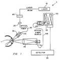
- FIG. 1 is a schematic representation of an apparatus of the present invention.
- FIG. 2 is a schematic representation of an alternate embodiment of the apparatus of FIG. 1.
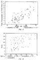
- FIG. 3 is a graphical representation of the fluorescent signal intensity as a function of age of both diabetic and nondiabetic patients obtained using the apparatus of FIG. 1 as described in the EXAMPLE herein.
- FIG. 4 is a graphical representation of the ratio of the fluorescent to Rayleigh signal intensities as a function of age of both diabetic and nondiabetic patients obtained using the apparatus of FIG. 1 as described in the EXAMPLE herein.
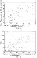
- FIG. 5 is a graphical representation of the fluorescent signal intensity as a function of age of both diabetic and nondiabetic patients obtained using the apparatus of FIG. 1 for an illumination radiation wavelength outside a preferred range of that used in connection with the present invention.
- FIG. 6 is a graphical representation of the ratio of the fluorescent to Rayleigh signal intensities as a function of age of both diabetic and nondiabetic patients obtained using the apparatus of FIG. 1 for an illumination radiation wavelength outside a preferred range of that used in connection with the present invention.
- FIG. 1 illustrates an optical system 5 of the present invention.
- Optical system 5 includes a light source 15, lens 25, a confocal lens system 35, collector 45, and a spectrometer 55.
- Source 15, which provides narrow-band illumination typically may be a low power krypton laser tuned to produce radiation having a wavelength between approximately 400-1500 nm. In one embodiment of optical system 5, source 15 provides radiation at a wavelength of 406.7 nm.
- ocular lens tissue L, attenuator 65, eyepiece 75, detection and processing assembly 85, and fiber optic waveguides 95 and 105 are also shown in FIG. 1 ocular lens tissue L, attenuator 65, eyepiece 75, detection and processing assembly 85, and fiber optic waveguides 95 and 105.
- Attenuator 65 used to reduce the power level of the transmitted radiation, receives radiation from source 15 and forwards it to lens 25.
- Lens 25, which may be a 40X microscope objective or other similar device, then focuses the (attenuated) radiation onto the end of waveguide 95, which in turn transmits the radiation to confocal lens system 35.
- Lens system 35 subsequently delivers the radiation to a selected volume of ocular lens tissue L (typically approximately 200 cubic micrometers).
- a modified slit lamp base may be used to house and position lens system 35 for easy access to lens tissue L, while lens system 35 itself is designed to permit the same volume of lens tissue L to be held in the focal point of collector 45.
- the aperture 115 of lens system 35 at its focus is greater than approximately fifteen micrometers, ensuring that the excitation radiation diverges rapidly after passing through the focal point of lens system 35 and thereby reducing the spot intensity of the radiation should it encounter any other portions of the ocular tissue.
- Collector 45 receives the radiation backscattered from lens (or other) tissue L as a result of it being illuminated by radiation from source 15. From collector 45, the backscattered radiation is directed into waveguide 105 and transmitted to the entrance slit 125 of the monochromator 135 forming spectrometer 55. If desired, collector 45 also may direct a portion of the backscattered radiation to eyepiece 75, permitting an operator to view the exact location of the selected volume of lens tissue L.
- spectrometer 55 Division and processing of the backscattered radiation occurs in spectrometer 55 and detection and processing assembly 85. Radiation transmitted to spectrometer 55 initially is separated into its Rayleigh and florescence components. The two components subsequently are directed, respectively and as necessary, to amplifiers forming part of assembly 85, for determination of the intensities of each. Assembly 85 also may include a digital computer or similar computing device for forming the ratio of the fluorescent and Rayleigh components of the backscattered radiation, thereby normalizing the peak intensity of the fluorescent component.
- light source 20 which may be a laser diode, produces radiation of wavelength approximately 813.4 nm (within the range of approximately 800-860 nm) and is coupled to a nonlinear frequency doubling device 30 to produce the desired wavelength output of 406.7 nm (within the range 400-1500 nm).
- Light source 20 alternatively may be a laser, light emitting diode, or other narrow-band light source (including broadband sources coupled to optical filters).
- the radiation subsequently is directed through an optical delivery system 40 into the eye 50 of a patient.
- alternate embodiment 10 includes an optical collector 60 confocal to the delivery system 40 to collect the backscattered radiation from the lens of eye 50.
- the backscattered radiation collected includes both a fluorescence signal (typically approximately 490-500 nm within the range 460-1800 nm) and an intense Rayleigh component at the illumination wavelength.
- FIG. 2 additionally discloses means for separating the components of interest of the backscattered radiation, including dichroic beam splitters 70 and 90, and for detecting the intensity of the components simultaneously using single chip silicon detectors 100 and 120 or similar devices.
- component separation may be accomplished using beam splitters in conjunction with optical bandpass filters or dispersive elements such as diffraction gratings.
- Hybrid detector/filter assemblies also may be used.
- Electronic circuitry 130 such as but neither limited to nor necessarily requiring analog amplifiers, analog to digital (A/D) converters, and a digital computer, processes the data detected by detectors 100 and 120, calculates the normalized fluorescent/Rayleigh component ratio, and, if desired, makes the result available to an operator through a digital display or other suitable means.
- Eyepiece 80 may be used by the operator to view the location of the excitation focal point in eye 50.
- FIGS. 3-6 illustrate data obtained from clinical trials conducted using sixty-nine (69) human patients aged twelve (12) to sixty-five (65). Forty-eight (48) of the patients had previously been diagnosed as having diabetes, while the remaining twenty-one (21) had not.
- FIGS. 3 shows the total fluorescence signal obtained for each patient (expressed in "Counts x 10 5 ,” where the number of Counts is a function of the number of emitted photons per unit time) using an illumination wavelength of 406.7 nm.
- FIG. 4 details the results when those same fluorescence signals are normalized by the Rayleigh component of the backscattered radiation in accordance with the present invention. As illustrated in FIG.
- FIGS. 5-6 which correspond, respectively, to FIGS. 3-4, show (in FIG. 6) less of a distinction between the normalized signals for the diabetic as opposed to nondiabetic patients. Furthermore, those patients who tested ICA positive are shown to have fluorescent/Rayleigh ratios within the range of nondiabetic patient values. As a result, no clearly established threshold is available for diagnostic purposes.
Claims (22)
- Eine Vorrichtung (5;10) zum Messen bzw. Nachweisen von molekularen Veränderungen bei einem Patienten mit das Auge betreffendem oder anderem Gewebe, das bei Beleuchtung Strahlung einschließlich fluoreszierender und Rayleigh-Komponenten bestimmbarer Intensitäten rückstreuen kann, mit:a. einer Einrichtung (15;20) zum Beleuchten des Gewebes mit einem Licht einer ausgewählten Wellenlänge, um dadurch das Gewebe zum Rückstreuen von Strahlung als Reaktion auf die Beleuchtung zu veranlassen; undb. einer auf die rückgestreute Strahlung reagierende Einrichtung (45;60) zum Sammeln der rückgestreuten Strahlung; gekennzeichnet dadurch, daß die Vorrichtung weiterhin aufweist:c. eine mit der Sammeleinrichtung verbundene Einrichtung (55;70,90) zum Trennen der eingesammelten Strahlung in eine Vielzahl von Komponenten undd. eine mit der Trenneinrichtung verbundene Einrichtung (85;100,120,130) zum (i) Messen der Intensität jeder der aufgetrennten Vielzahl von Komponenten und (ii) zum Bestimmen einer mathematischen Beziehung zwischen der aufgetrennten Vielzahl der Komponenten, um dadurch eine Messung bzw. einen Nachweis der molekularen Veränderungen in dem Gewebe herzustellen.
- Eine Vorrichtung gemäß Anspruch 1, bei der die eingesammelte Strahlung in ihre fluoreszierenden und Rayleigh-Komponenten getrennt wird.
- Eine Vorrichtung gemäß Anspruch 1, bei der die Beleuchtungseinrichtung umfaßt:a. eine Lichtquelle (15;20) aus der Gruppe bestehend aus Lasern (15;20), mit nicht linearen Frequenzverdopplungseinrichtungen gekoppelten Laserdioden, lichtemittierende Dioden (LED's) und mit optischen Filtern gekoppelten Breitbandquellen,b. eine optisch auf das Licht der Lichtquelle (15) reagierende Linse (25) zum Fokussieren des Lichts undc. ein optisch auf das fokussierte Licht reagierendes Linsensystem (35), das einen Fokus bzw. Brennpunkt aufweist und am Fokus eine Apertur festlegt, die größer ist als etwa 15 µm.
- Eine Vorrichtung gemäß einem der vorstehenden Ansprüche, bei der die Meß- bzw. Nachweis- und Bestimmungseinrichtung mindestens einen Silizium-Einzelchipdetektor (100,120) aufweist und die Wellenlänge der fluoreszierenden Komponente der rückgestreuten Strahlung zwischen etwa 460 und 1500 nm beträgt.
- Eine Vorrichtung gemäß Anspruch 4, bei der die Meß- bzw. Nachweis- und Bestimmungseinrichtung weiterhin einen Verstärker umfaßt.
- Eine Vorrichtung gemäß einem der vorstehenden Ansprüche, bei der die Beleuchtungseinrichtung weiterhin eine Einrichtung (65) zum Einstellen des Energieniveaus des Beleuchtungslichtes aufweist.
- Eine Vorrichtung gemäß Anspruch 1, bei der die Beleuchtungseinrichtung einen Laser (15) zum Liefern von Licht mit einem ausgewählten Energieniveau und einer ausgewählten Wellenlänge umfaßt.
- Eine Vorrichtung gemäß Anspruch 7, weiterhin umfassend eine optisch auf das gelieferte Licht reagierende Einrichtung (65) zum Einstellen des Energieniveaus des Lichtes.
- Eine Vorrichtung gemäß Anspruch 8, weiterhin umfassend eine optisch mit der Einstelleinrichtung verbundene Linse (25) zum Fokussieren des Lichtes.
- Eine Vorrichtung gemäß Anspruch 9, weiterhin umfassend einen optisch mit der Linse verbundenen ersten optischen Wellenleiter (95) zum Empfangen des fokussierten Lichtes.
- Eine Vorrichtung gemäß Anspruch 10, weiterhin umfassend ein optisch mit der ersten optischen Faser (95) verbundenes Linsensystem, das eine Apertur (115) mit einem Fokus definiert, der größer ist als etwa 15 µm, zum Leiten des fokussierten Lichtes auf einen ausgewählten, etwa 200 µm des Volumens des Gewebes betragenden Teil desselben, um dadurch die Okularlinse zum Rückstreuen von Strahlung in Reaktion auf das zugeführte Licht anzuregen bzw. zu veranlassen.
- Eine Vorrichtung gemäß Anspruch 11, weiterhin umfassend einen Kollektor (45), der (a) einen das gewählte Volumen des Gewebes, dem das fokussierte Licht zugeführt wird, einschließenden Brennpunkt aufweist und der (b) in Reaktion auf die rückgestreute Strahlung diese einsammeln kann.
- Eine Vorrichtung gemäß Anspruch 12, weiterhin umfassend einen optisch mit dem Kollektor (45) verbundenen zweiten optischen Wellenleiter (105) zum Empfangen der gesammelten Strahlung.
- Eine Vorrichtung gemäß einem der vorstehenden Ansprüche, bei der die Wellenlänge etwa 406,7 nm beträgt, die Trenneinrichtung ein Spektrometer (55) ist und die Detektier- und Normiereinrichtung (85) einen Computer umfaßt.
- Eine Vorrichtung gemäß einem der Ansprüche 1 bis 13, bei der die Trenneinrichtung mindestens einen dichroitischen Strahlenteiler (70,90) umfaßt.
- Eine Vorrichtung gemäß einem der vorstehenden Ansprüche, weiterhin umfassend ein auf die rückgestreute Strahlung reagierendes Okular (75;80), das ein Betrachten des ausgewählten Volumens des Gewebes durch eine Bedienperson zuläßt und bei dem die gemessenen bzw. nachgewiesenen molekularen Veränderungen eine Diagnose von Zuständen aus der Gruppe bestehend aus Diabetes, frühdiabetischer Zustand und Katarakt bzw. Linsentrübung oder grauer Star unterstützt.
- Ein nicht-invasives Verfahren zum Messen bzw. Nachweisen von molekularen Veränderungen bei einem Patienten mit das Auge betreffendem oder anderem Gewebe, das bei Beleuchtung rückstreuen kann, mit den Schritten:a. Beleuchten des Gewebes mit Licht einer ausgewählten Wellenlänge, um dadurch das Gewebe zum Rückstreuen von Strahlung in Reaktion auf die Beleuchtung anzuregen bzw. zu veranlassen, wobei das Verfahren durch Einschluß der folgenden Schritte gekennzeichnet ist:b. Auftrennen der rückgestreuten Strahlung in eine Vielzahl von Komponenten,c. Ermitteln der Intensität jeder der aufgetrennten Komponenten undd. Bestimmen einer mathematischen Beziehung zwischen der aufgetrennten Vielzahl von Komponenten, um dadurch eine Messung bzw. einen Nachweis der molekularen Veränderungen in dem Gewebe herzustellen.
- Ein Verfahren gemäß Anspruch 17, bei dem die Vielzahl von Komponenten die fluoreszierenden und Rayleigh-Komponenten der rückgestreuten Strahlung umfassen.
- Ein Verfahren gemäß Anspruch 18, bei dem der Bestimmungsschritt weiterhin den Schritt des Normierens der ermittelten Intensität der fluoreszierenden Komponente mit der ermittelten Intensität der Rayleigh-Komponente umfaßt.
- Verfahren gemäß einem der Ansprüche 17 bis 19, bei dem der Beleuchtungsschritt die Schritte umfaßt:a. Bereitstellen einer Lichtquelle (15;20) aus der Gruppe bestehend aus Lasern, mit nichtlinearen Frequenzverdopplungseinrichtungen gekoppelten Laserdioden, lichtemittierende Dioden (LED's) und mit optischen Filtern gekoppelten Breitbandquellen, zum Emittieren des Lichts einer Wellenlänge von etwa 406,7 nm undb. Fokussieren des Lichts unter Verwendung eines Linsensystems (25), das einen Fokus besitzt und eine Apertur (115) an dessen Fokus festlegt, die größer ist als etwa 15 µm und
mit dem weiteren Schritt des Vergleichens des Verhältnisses der ermittelten Intensitäten mit mindestens einem vorgewählten Wert zur Unterstützung bei der Diagnose von Zuständen aus der Gruppe bestehend aus Diabetes und frühdiabetischem Zustand. - Ein Verfahren gemäß einem der Ansprüche 17 bis 20, bei dem der Ermittlungsschritt den Schritt des Ermitteins der aufgetrennten fiuoreszierenden Komponente bei einer Wellenlänge zwischen etwa 460 und 1800 nm aufweist und bei dem der Vergleichsschritt den Schritt des Vergleichens des Verhältnisses der ermittelten Intensitäten mit zwei vorgewählten Werten, 13 und 15, umfaßt, derart, daß bei einem Verhältnis von weniger als 13 für den Patienten die Diagnose getroffen wird, daß es unwahrscheinlich ist, daß er den ausgewählten Zustand aufweist, und daß dann, wenn das Verhältnis größer als 15 ist, für den Patienten die Diagnose getroffen wird, daß er wahrscheinlich den ausgewählten Zustand aufweist.
- Ein Verfahren gemäß einem der Ansprüche 17 bis 21, bei dem der Trennschritt den Schritt des Trennens der rückgestreuten Strahlung unter Verwendung mindestens eines dichroitischen Strahlenteilers (70,90) aufweist.
Applications Claiming Priority (3)
| Application Number | Priority Date | Filing Date | Title |
|---|---|---|---|
| US07/731,533 US5203328A (en) | 1991-07-17 | 1991-07-17 | Apparatus and methods for quantitatively measuring molecular changes in the ocular lens |
| US731533 | 1991-07-17 | ||
| PCT/US1992/005941 WO1993001745A1 (en) | 1991-07-17 | 1992-07-16 | Measuring molecular change in the ocular lens |
Publications (3)
| Publication Number | Publication Date |
|---|---|
| EP0597932A1 EP0597932A1 (de) | 1994-05-25 |
| EP0597932A4 EP0597932A4 (de) | 1994-12-07 |
| EP0597932B1 true EP0597932B1 (de) | 1997-10-01 |
Family
ID=24939928
Family Applications (1)
| Application Number | Title | Priority Date | Filing Date |
|---|---|---|---|
| EP92916422A Expired - Lifetime EP0597932B1 (de) | 1991-07-17 | 1992-07-16 | Nachweis von molekularen veränderungen der augenlinse |
Country Status (9)
| Country | Link |
|---|---|
| US (2) | US5203328A (de) |
| EP (1) | EP0597932B1 (de) |
| JP (1) | JPH07500030A (de) |
| AT (1) | ATE158704T1 (de) |
| AU (1) | AU661026B2 (de) |
| CA (1) | CA2113268C (de) |
| DE (1) | DE69222535T2 (de) |
| ES (1) | ES2110007T3 (de) |
| WO (1) | WO1993001745A1 (de) |
Families Citing this family (161)
| Publication number | Priority date | Publication date | Assignee | Title |
|---|---|---|---|---|
| US5203328A (en) * | 1991-07-17 | 1993-04-20 | Georgia Tech Research Corporation | Apparatus and methods for quantitatively measuring molecular changes in the ocular lens |
| DE4243142A1 (de) * | 1992-12-19 | 1994-06-23 | Boehringer Mannheim Gmbh | Vorrichtung zur in-vivo-Bestimmung einer optischen Eigenschaft des Kammerwassers des Auges |
| US5341805A (en) * | 1993-04-06 | 1994-08-30 | Cedars-Sinai Medical Center | Glucose fluorescence monitor and method |
| US5427094A (en) * | 1993-11-08 | 1995-06-27 | Oculon Corporation | Method and apparatus for detecting cataractogenesis |
| US5427095A (en) * | 1993-11-09 | 1995-06-27 | Massachusetts Institute of Technology Oculon Corporation | Method and apparatus for detecting cataractogenesis |
| US5560356A (en) * | 1994-02-23 | 1996-10-01 | Vitrophage, Inc. | Diagnostic system and method using an implanted reflective device |
| US5685313A (en) * | 1994-05-31 | 1997-11-11 | Brain Monitor Ltd. | Tissue monitor |
| US5701902A (en) * | 1994-09-14 | 1997-12-30 | Cedars-Sinai Medical Center | Spectroscopic burn injury evaluation apparatus and method |
| WO1996029000A1 (en) * | 1995-03-23 | 1996-09-26 | Philips Electronics N.V. | Device for carrying out optical measurements in turbid media |
| US7328059B2 (en) | 1996-08-23 | 2008-02-05 | The Texas A & M University System | Imaging of light scattering tissues with fluorescent contrast agents |
| DE19538372A1 (de) * | 1995-10-14 | 1997-04-17 | Laser & Med Tech Gmbh | Nicht invasive Glukosemessung |
| US6011984A (en) * | 1995-11-22 | 2000-01-04 | Minimed Inc. | Detection of biological molecules using chemical amplification and optical sensors |
| US6766183B2 (en) | 1995-11-22 | 2004-07-20 | Medtronic Minimed, Inc. | Long wave fluorophore sensor compounds and other fluorescent sensor compounds in polymers |
| US6002954A (en) * | 1995-11-22 | 1999-12-14 | The Regents Of The University Of California | Detection of biological molecules using boronate-based chemical amplification and optical sensors |
| US6232609B1 (en) | 1995-12-01 | 2001-05-15 | Cedars-Sinai Medical Center | Glucose monitoring apparatus and method using laser-induced emission spectroscopy |
| US5882301A (en) | 1995-12-13 | 1999-03-16 | Yoshida; Akitoshi | Measuring apparatus for intraocular substance employing light from eyeball |
| KR19990076724A (ko) * | 1996-01-26 | 1999-10-15 | 로셰 디아그노스틱스 게엠베하 | 산란 매트릭스내에 함유된 분석물을 측정하는 방법 및 장치 |
| US6544193B2 (en) | 1996-09-04 | 2003-04-08 | Marcio Marc Abreu | Noninvasive measurement of chemical substances |
| WO1998022820A1 (en) | 1996-11-21 | 1998-05-28 | Lawrence Livermore National Laboratory | Detection of biological molecules using boronate-based chemical amplification and optical sensors |
| CA2192036A1 (en) * | 1996-12-04 | 1998-06-04 | Harvey Lui | Fluorescence scope system for dermatologic diagnosis |
| US6826422B1 (en) * | 1997-01-13 | 2004-11-30 | Medispectra, Inc. | Spectral volume microprobe arrays |
| US6847490B1 (en) | 1997-01-13 | 2005-01-25 | Medispectra, Inc. | Optical probe accessory device for use in vivo diagnostic procedures |
| US7865230B1 (en) | 1997-02-07 | 2011-01-04 | Texas A&M University System | Method and system for detecting sentinel lymph nodes |
| US5880812A (en) * | 1997-03-13 | 1999-03-09 | Ramot-University Authority For Applied Research And Industrial Development, Ltd. | Method and apparatus for evaluating and mapping visual field |
| AU6569198A (en) * | 1997-03-19 | 1998-10-12 | Lucid Technologies, Inc. | Cellular surgery utilizing confocal microscopy |
| US6091984A (en) | 1997-10-10 | 2000-07-18 | Massachusetts Institute Of Technology | Measuring tissue morphology |
| EP1037553B1 (de) * | 1997-11-12 | 2007-01-24 | Lightouch Medical, Inc. | Verfahren zur nicht invasiven analytenmessung |
| US6070093A (en) | 1997-12-02 | 2000-05-30 | Abbott Laboratories | Multiplex sensor and method of use |
| US6055451A (en) | 1997-12-12 | 2000-04-25 | Spectrx, Inc. | Apparatus and method for determining tissue characteristics |
| DE19808779C1 (de) * | 1998-03-03 | 1999-10-28 | Paul Dobrinski | Meßvorrichtung zur objektiven Beurteilung des Zustandes eines grauen Stars (einer Katarakt) durch den Augenarzt |
| US6174291B1 (en) | 1998-03-09 | 2001-01-16 | Spectrascience, Inc. | Optical biopsy system and methods for tissue diagnosis |
| US6560478B1 (en) * | 1998-03-16 | 2003-05-06 | The Research Foundation Of City University Of New York | Method and system for examining biological materials using low power CW excitation Raman spectroscopy |
| US5919132A (en) * | 1998-03-26 | 1999-07-06 | Universite De Montreal | On-line and real-time spectroreflectometry measurement of oxygenation in a patient's eye |
| US6149589A (en) * | 1998-03-26 | 2000-11-21 | Universite De Montreal | On-line and real-time spectroreflectometry measurement of oxygenation in a patient's eye |
| AU4970499A (en) | 1998-07-07 | 2000-01-24 | Lightouch Medical, Inc. | Tissue modulation process for quantitative noninvasive in vivo spectroscopic analysis of tissues |
| CA2343401C (en) * | 1998-09-11 | 2009-01-27 | Spectrx, Inc. | Multi-modal optical tissue diagnostic system |
| US6148232A (en) | 1998-11-09 | 2000-11-14 | Elecsys Ltd. | Transdermal drug delivery and analyte extraction |
| US6708060B1 (en) | 1998-11-09 | 2004-03-16 | Transpharma Ltd. | Handheld apparatus and method for transdermal drug delivery and analyte extraction |
| US6611706B2 (en) | 1998-11-09 | 2003-08-26 | Transpharma Ltd. | Monopolar and bipolar current application for transdermal drug delivery and analyte extraction |
| US6597946B2 (en) * | 1998-11-09 | 2003-07-22 | Transpharma Ltd. | Electronic card for transdermal drug delivery and analyte extraction |
| US6721583B1 (en) * | 1998-11-19 | 2004-04-13 | The United States Of America | Method for non-invasive identification of individuals at risk for diabetes |
| WO2000037917A2 (en) * | 1998-12-23 | 2000-06-29 | Medispectra, Inc. | Systems and methods for optical examination of samples |
| JP2002532181A (ja) * | 1998-12-23 | 2002-10-02 | メディスペクトラ, インコーポレイテッド | 頚部のスクリーニングのための光学的方法およびシステム |
| US6404497B1 (en) | 1999-01-25 | 2002-06-11 | Massachusetts Institute Of Technology | Polarized light scattering spectroscopy of tissue |
| US6665556B1 (en) * | 1999-01-29 | 2003-12-16 | Robert R. Alfano | Method and apparatus for examining a tissue using the spectral wing emission therefrom induced by visible to infrared photoexcitation |
| US6088606A (en) * | 1999-03-22 | 2000-07-11 | Spectrx, Inc. | Method and apparatus for determining a duration of a medical condition |
| US6219566B1 (en) * | 1999-07-13 | 2001-04-17 | Photonics Research Ontario | Method of measuring concentration of luminescent materials in turbid media |
| BR0013609B1 (pt) * | 1999-08-26 | 2009-01-13 | lente oftÁlmica para detectar um analito em um lÍquido ocular, e sistema sensor de analito. | |
| US6682938B1 (en) | 1999-09-15 | 2004-01-27 | The Regents Of The University Of California | Glucose sensing molecules having selected fluorescent properties |
| US6673625B2 (en) | 1999-09-15 | 2004-01-06 | The Regents Of The University Of California | Saccharide sensing molecules having enhanced fluorescent properties |
| US6494576B1 (en) * | 1999-09-30 | 2002-12-17 | L'esperance, Jr. Francis A. | Method and apparatus for spectrophotometry of the eye |
| US7054002B1 (en) | 1999-10-08 | 2006-05-30 | The Texas A&M University System | Characterization of luminescence in a scattering medium |
| BR0015285A (pt) * | 1999-11-05 | 2002-06-18 | Spectrx Inc | Método para diagnosticar uma condição de um tecido alvo, método para diagnosticar tecidos displásticos, e sistema para determinar uma condição de um tecido biológico alvo |
| US7187810B2 (en) * | 1999-12-15 | 2007-03-06 | Medispectra, Inc. | Methods and systems for correcting image misalignment |
| US6902935B2 (en) * | 1999-12-15 | 2005-06-07 | Medispectra, Inc. | Methods of monitoring effects of chemical agents on a sample |
| US7260248B2 (en) * | 1999-12-15 | 2007-08-21 | Medispectra, Inc. | Image processing using measures of similarity |
| US20040044287A1 (en) * | 2000-03-31 | 2004-03-04 | Wei-Chiang Lin | Identification of human tissue using optical spectroscopy |
| US6377841B1 (en) * | 2000-03-31 | 2002-04-23 | Vanderbilt University | Tumor demarcation using optical spectroscopy |
| DE60137046D1 (de) * | 2000-07-13 | 2009-01-29 | Univ Virginia Commonwealth | Verwendung von ultraviolett, nahultraviolett und nahinfrarot-resonanzrmanspektroskopie und fluoresenzspektoskopie zur gewebeuntersuchung auf schockzustand, kritische krankheiten oder andere krankheitszustände |
| US6549861B1 (en) | 2000-08-10 | 2003-04-15 | Euro-Celtique, S.A. | Automated system and method for spectroscopic analysis |
| AU2001288292A1 (en) | 2000-08-21 | 2002-03-04 | Euroceltique S.A. | Near infrared blood glucose monitoring system |
| EP1339323A4 (de) | 2000-11-16 | 2004-04-14 | Chameleon Medical Innovation L | Diagnosesystem für das ohr |
| US6839661B2 (en) * | 2000-12-15 | 2005-01-04 | Medispectra, Inc. | System for normalizing spectra |
| US6697652B2 (en) * | 2001-01-19 | 2004-02-24 | Massachusetts Institute Of Technology | Fluorescence, reflectance and light scattering spectroscopy for measuring tissue |
| US6927246B2 (en) * | 2001-02-15 | 2005-08-09 | Medtronic Minimed, Inc. | Polymers functionalized with fluorescent boronate motifs and methods for making them |
| US20070276199A1 (en) * | 2002-04-04 | 2007-11-29 | Ediger Marwood N | Determination of a Measure of a Glycation End-Product or Disease State Using Tissue Fluorescence |
| US7043288B2 (en) * | 2002-04-04 | 2006-05-09 | Inlight Solutions, Inc. | Apparatus and method for spectroscopic analysis of tissue to detect diabetes in an individual |
| US7139598B2 (en) * | 2002-04-04 | 2006-11-21 | Veralight, Inc. | Determination of a measure of a glycation end-product or disease state using tissue fluorescence |
| EP1385423B1 (de) * | 2001-04-27 | 2007-11-21 | EyeSense AG | Kit zur messung von blutzuckerkonzentrationen |
| US7904176B2 (en) * | 2006-09-07 | 2011-03-08 | Bio Control Medical (B.C.M.) Ltd. | Techniques for reducing pain associated with nerve stimulation |
| US7045361B2 (en) | 2001-09-12 | 2006-05-16 | Medtronic Minimed, Inc. | Analyte sensing via acridine-based boronate biosensors |
| US6650915B2 (en) | 2001-09-13 | 2003-11-18 | Fovioptics, Inc. | Non-invasive measurement of blood analytes using photodynamics |
| US8308797B2 (en) | 2002-01-04 | 2012-11-13 | Colibri Heart Valve, LLC | Percutaneously implantable replacement heart valve device and method of making same |
| US20120078075A1 (en) * | 2002-04-04 | 2012-03-29 | Maynard John D | Determination of a measure of a glycation end-product or disease state using tissue fluorescence in combination with one or more other tests |
| US8140147B2 (en) * | 2002-04-04 | 2012-03-20 | Veralight, Inc. | Determination of a measure of a glycation end-product or disease state using a flexible probe to determine tissue fluorescence of various sites |
| US7725144B2 (en) * | 2002-04-04 | 2010-05-25 | Veralight, Inc. | Determination of disease state using raman spectroscopy of tissue |
| US8131332B2 (en) * | 2002-04-04 | 2012-03-06 | Veralight, Inc. | Determination of a measure of a glycation end-product or disease state using tissue fluorescence of various sites |
| WO2003089043A2 (en) * | 2002-04-19 | 2003-10-30 | Transpharma Medical Ltd. | Handheld transdermal drug delivery and analyte extraction |
| US8849379B2 (en) | 2002-04-22 | 2014-09-30 | Geelux Holdings, Ltd. | Apparatus and method for measuring biologic parameters |
| IL164685A0 (en) | 2002-04-22 | 2005-12-18 | Marcio Marc Aurelio Martins Ab | Apparatus and method for measuring biologic parameters |
| US9848815B2 (en) | 2002-04-22 | 2017-12-26 | Geelux Holdings, Ltd. | Apparatus and method for measuring biologic parameters |
| US8328420B2 (en) | 2003-04-22 | 2012-12-11 | Marcio Marc Abreu | Apparatus and method for measuring biologic parameters |
| US20040208385A1 (en) * | 2003-04-18 | 2004-10-21 | Medispectra, Inc. | Methods and apparatus for visually enhancing images |
| US6933154B2 (en) * | 2002-07-09 | 2005-08-23 | Medispectra, Inc. | Optimal windows for obtaining optical data for characterization of tissue samples |
| US6818903B2 (en) * | 2002-07-09 | 2004-11-16 | Medispectra, Inc. | Method and apparatus for identifying spectral artifacts |
| US20040208390A1 (en) * | 2003-04-18 | 2004-10-21 | Medispectra, Inc. | Methods and apparatus for processing image data for use in tissue characterization |
| US7459696B2 (en) | 2003-04-18 | 2008-12-02 | Schomacker Kevin T | Methods and apparatus for calibrating spectral data |
| US20040209237A1 (en) * | 2003-04-18 | 2004-10-21 | Medispectra, Inc. | Methods and apparatus for characterization of tissue samples |
| US7309867B2 (en) | 2003-04-18 | 2007-12-18 | Medispectra, Inc. | Methods and apparatus for characterization of tissue samples |
| US7469160B2 (en) * | 2003-04-18 | 2008-12-23 | Banks Perry S | Methods and apparatus for evaluating image focus |
| US7136518B2 (en) * | 2003-04-18 | 2006-11-14 | Medispectra, Inc. | Methods and apparatus for displaying diagnostic data |
| US7282723B2 (en) * | 2002-07-09 | 2007-10-16 | Medispectra, Inc. | Methods and apparatus for processing spectral data for use in tissue characterization |
| US7103401B2 (en) | 2002-07-10 | 2006-09-05 | Medispectra, Inc. | Colonic polyp discrimination by tissue fluorescence and fiberoptic probe |
| US6768918B2 (en) | 2002-07-10 | 2004-07-27 | Medispectra, Inc. | Fluorescent fiberoptic probe for tissue health discrimination and method of use thereof |
| US6895264B2 (en) | 2002-08-26 | 2005-05-17 | Fovioptics Inc. | Non-invasive psychophysical measurement of glucose using photodynamics |
| WO2004060154A1 (en) * | 2003-01-07 | 2004-07-22 | Intelligent Photonics Control Corp. | Non-invasive blood monitor |
| EP1654531A1 (de) * | 2003-06-20 | 2006-05-10 | The Texas A & M University System | Verfahren und system zum fluoreszenz-kontrastverstärkten abbilden im nahinfrarotbereich mit flächiger beleuchtung und flächiger detektion |
| US7356365B2 (en) * | 2003-07-09 | 2008-04-08 | Glucolight Corporation | Method and apparatus for tissue oximetry |
| US20050090723A1 (en) * | 2003-10-23 | 2005-04-28 | Nassar Saeed | Method and apparatus for non-invasive measuring of physiological glucose concentration in bodies of humans or animals |
| GB2407378B (en) | 2003-10-24 | 2006-09-06 | Lein Applied Diagnostics Ltd | Ocular property measuring apparatus and method therefor |
| CN100998499B (zh) * | 2003-10-28 | 2013-07-24 | 薇拉莱特公司 | 使用组织荧光确定某一糖化终产物或疾病状态 |
| WO2005050156A2 (en) * | 2003-11-18 | 2005-06-02 | Chameleon Medical Innovation Ltd. | Measurement system and method for use in determining the patient's condition |
| GB2409033C (en) * | 2003-12-12 | 2006-05-24 | Lein Applied Diagnostics Ltd | Extended focal region measuring apparatus and method |
| US7510849B2 (en) * | 2004-01-29 | 2009-03-31 | Glucolight Corporation | OCT based method for diagnosis and therapy |
| US10227063B2 (en) | 2004-02-26 | 2019-03-12 | Geelux Holdings, Ltd. | Method and apparatus for biological evaluation |
| US7254429B2 (en) | 2004-08-11 | 2007-08-07 | Glucolight Corporation | Method and apparatus for monitoring glucose levels in a biological tissue |
| US8036727B2 (en) | 2004-08-11 | 2011-10-11 | Glt Acquisition Corp. | Methods for noninvasively measuring analyte levels in a subject |
| EP1874178A4 (de) | 2005-04-13 | 2009-12-09 | Glucolight Corp | Verfahren zur datenreduzierung und kalibrierung eines blutzuckermonitors auf oct-basis |
| US7330747B2 (en) * | 2005-06-07 | 2008-02-12 | Chemimage Corporation | Invasive chemometry |
| US7330746B2 (en) * | 2005-06-07 | 2008-02-12 | Chem Image Corporation | Non-invasive biochemical analysis |
| EP1907825A2 (de) * | 2005-07-25 | 2008-04-09 | Duke University | Verfahren, systeme und computerprogramme zur optimierung von sonden zur spektroskopischen messung in trüben medien |
| US20070167835A1 (en) * | 2005-07-25 | 2007-07-19 | Massachusetts Institute Of Technology | Tri modal spectroscopic imaging |
| US20070173736A1 (en) * | 2005-10-07 | 2007-07-26 | Femspec Llc | Apparatus and methods for endometrial biopsies |
| ES2399872T3 (es) | 2005-10-24 | 2013-04-04 | Marcio Marc Aurelio Martins Abreu | Aparato para medir parámetros biológicos |
| US8062287B2 (en) * | 2005-12-22 | 2011-11-22 | Koninklijke Philips Electronics N V | Device for controlled release of chemical molecules |
| US7818154B2 (en) * | 2006-03-17 | 2010-10-19 | Duke University | Monte Carlo based model of fluorescence in turbid media and methods and systems for using same to determine intrinsic fluorescence of turbid media |
| US7751039B2 (en) * | 2006-03-30 | 2010-07-06 | Duke University | Optical assay system for intraoperative assessment of tumor margins |
| EP2076172A4 (de) * | 2006-10-27 | 2012-03-21 | Aretais Inc | Verwendung kohärenter raman-verfahren für medizinische diagnostische und therapeutische zwecke sowie kalibrierverfahren dafür |
| US8498695B2 (en) | 2006-12-22 | 2013-07-30 | Novadaq Technologies Inc. | Imaging system with a single color image sensor for simultaneous fluorescence and color video endoscopy |
| JP2008197088A (ja) * | 2007-01-19 | 2008-08-28 | Shimadzu Corp | 蛍光検出器 |
| WO2008103486A1 (en) * | 2007-02-23 | 2008-08-28 | Duke University | Scaling method for fast monte carlo simulation of diffuse reflectance spectra |
| GB2451443B (en) | 2007-07-30 | 2012-12-26 | Lein Applied Diagnostics Ltd | Optical measurement apparatus and method therefor |
| GB2451442B (en) * | 2007-07-30 | 2013-03-06 | Lein Applied Diagnostics Ltd | Optical measurement apparatus and method therefor |
| WO2009043050A2 (en) * | 2007-09-27 | 2009-04-02 | Duke University | Optical assay system with a multi-probe imaging array |
| US9820655B2 (en) * | 2007-09-28 | 2017-11-21 | Duke University | Systems and methods for spectral analysis of a tissue mass using an instrument, an optical probe, and a Monte Carlo or a diffusion algorithm |
| EP2057941A1 (de) * | 2007-11-07 | 2009-05-13 | Koninklijke Philips Electronics N.V. | Verfahren und Vorrichtung zur Abbildung des Inneren eines optisch trüben Mediums |
| DE102007061987A1 (de) * | 2007-12-21 | 2009-06-25 | Carl Zeiss Meditec Ag | Vorrichtung und Verfahren zum Nachweisen von Molekülen im Auge |
| US8986253B2 (en) | 2008-01-25 | 2015-03-24 | Tandem Diabetes Care, Inc. | Two chamber pumps and related methods |
| GB2457302B (en) | 2008-02-11 | 2013-04-10 | Lein Applied Diagnostics Ltd | Measurement apparatus and method therefor |
| WO2009111542A2 (en) | 2008-03-04 | 2009-09-11 | Glucolight Corporation | Methods and systems for analyte level estimation in optical coherence tomography |
| JP5094484B2 (ja) * | 2008-03-11 | 2012-12-12 | 富士フイルム株式会社 | 蛍光検出方法および蛍光検出装置 |
| JP5097590B2 (ja) * | 2008-03-26 | 2012-12-12 | 富士フイルム株式会社 | ラマン信号測定方法およびラマン信号測定装置 |
| US20110105865A1 (en) * | 2008-04-24 | 2011-05-05 | Duke University | Diffuse reflectance spectroscopy device for quantifying tissue absorption and scattering |
| US8408421B2 (en) | 2008-09-16 | 2013-04-02 | Tandem Diabetes Care, Inc. | Flow regulating stopcocks and related methods |
| AU2009293019A1 (en) | 2008-09-19 | 2010-03-25 | Tandem Diabetes Care Inc. | Solute concentration measurement device and related methods |
| US20100249607A1 (en) * | 2008-09-26 | 2010-09-30 | Massachusetts Institute Of Technology | Quantitative spectroscopic imaging |
| US8606366B2 (en) | 2009-02-18 | 2013-12-10 | Syneron Medical Ltd. | Skin treatment apparatus for personal use and method for using same |
| GB0903274D0 (en) | 2009-02-26 | 2009-04-08 | Edinburgh Instr | Fluoreence method and system |
| US7896498B2 (en) * | 2009-03-30 | 2011-03-01 | Ottawa Hospital Research Institute | Apparatus and method for optical measurements |
| CA2769030C (en) | 2009-07-30 | 2016-05-10 | Tandem Diabetes Care, Inc. | Infusion pump system with disposable cartridge having pressure venting and pressure feedback |
| CN102473238B (zh) | 2009-08-20 | 2014-08-06 | 皇家飞利浦电子股份有限公司 | 用于图像分析的系统和方法 |
| EP2485639B1 (de) * | 2009-10-06 | 2019-08-14 | Koninklijke Philips N.V. | Verfahren und system zur durchführung einer photoplethysmographie |
| US9091637B2 (en) | 2009-12-04 | 2015-07-28 | Duke University | Smart fiber optic sensors systems and methods for quantitative optical spectroscopy |
| WO2011109450A2 (en) | 2010-03-01 | 2011-09-09 | Colibri Heart Valve Llc | Percutaneously deliverable heart valve and methods associated therewith |
| CA2806544C (en) | 2010-06-28 | 2016-08-23 | Colibri Heart Valve Llc | Method and apparatus for the endoluminal delivery of intravascular devices |
| WO2012061835A2 (en) | 2010-11-05 | 2012-05-10 | Freedom Meditech, Inc. | Apparatus and method for non-invasively detecting diseases that affect structural properties in biological tissues |
| AU2015202762B2 (en) * | 2010-11-05 | 2017-04-20 | Sinocare Meditech, Inc. | Improved algorithm for detection of diabetes |
| SG10201601962WA (en) | 2010-12-14 | 2016-04-28 | Colibri Heart Valve Llc | Percutaneously deliverable heart valve including folded membrane cusps with integral leaflets |
| US9180242B2 (en) | 2012-05-17 | 2015-11-10 | Tandem Diabetes Care, Inc. | Methods and devices for multiple fluid transfer |
| US9173998B2 (en) | 2013-03-14 | 2015-11-03 | Tandem Diabetes Care, Inc. | System and method for detecting occlusions in an infusion pump |
| CN105814419A (zh) | 2013-10-11 | 2016-07-27 | 马尔西奥·马克·阿布雷乌 | 用于生物学评估的方法和设备 |
| JP2017501844A (ja) | 2014-01-10 | 2017-01-19 | マーシオ マーク アブリュー | Abreu脳熱トンネルの赤外線出力を測定するためのデバイス |
| JP2017505657A (ja) | 2014-01-10 | 2017-02-23 | マーシオ マーク アブリュー | Abreu脳トンネルでモニタして治療を提供するデバイス |
| AU2015209304A1 (en) | 2014-01-22 | 2016-07-21 | Marcio Marc Abreu | Devices configured to provide treatment at an Abreu brain thermal tunnel |
| US9459201B2 (en) | 2014-09-29 | 2016-10-04 | Zyomed Corp. | Systems and methods for noninvasive blood glucose and other analyte detection and measurement using collision computing |
| EP3267877A1 (de) | 2015-03-10 | 2018-01-17 | Marcio Marc Abreu | Vorrichtungen, einrichtungen, systeme und verfahren zur messung der temperatur eines abtt-terminus |
| AU2016351730B2 (en) * | 2015-11-13 | 2019-07-11 | Novadaq Technologies Inc. | Systems and methods for illumination and imaging of a target |
| US9554738B1 (en) | 2016-03-30 | 2017-01-31 | Zyomed Corp. | Spectroscopic tomography systems and methods for noninvasive detection and measurement of analytes using collision computing |
| EP3469420A4 (de) | 2016-06-14 | 2020-02-12 | Novadaq Technologies ULC | Verfahren und systeme zur adaptiven bildgebung zur verstärkung eines schwachen lichtsignals bei einer medizinischen visualisierung |
| WO2018003906A1 (ja) * | 2016-06-30 | 2018-01-04 | 興和株式会社 | 眼科測定装置 |
| WO2019051476A1 (en) | 2017-09-11 | 2019-03-14 | Incubar, LLC | SEALING DEVICE FOR USE AS A VASCULAR DUCT IMPLANT FOR REDUCING ENDOFUCTION |
Family Cites Families (61)
| Publication number | Priority date | Publication date | Assignee | Title |
|---|---|---|---|---|
| FR1531145A (fr) * | 1966-07-13 | 1968-06-28 | Ici Ltd | Polyamides |
| US3948248A (en) * | 1974-09-05 | 1976-04-06 | Zuckerman Joel L | Method of measuring ocular pulse |
| US4014321A (en) * | 1974-11-25 | 1977-03-29 | March Wayne F | Non-invasive glucose sensor system |
| US3958560A (en) * | 1974-11-25 | 1976-05-25 | Wayne Front March | Non-invasive automatic glucose sensor system |
| DE2619571A1 (de) * | 1976-05-04 | 1977-11-17 | Dynamit Nobel Ag | Verfahren zum fixieren loeslicher acylasen an anorganisches traegermaterial |
| US4350163A (en) * | 1980-05-29 | 1982-09-21 | Ford Jr Norman C | Method and apparatus for analyzing contaminants in aqueous humor |
| US4412543A (en) * | 1981-04-09 | 1983-11-01 | Xanar, Inc. | Apparatus for determining the concentration of a fluorescent material in an eye |
| SE8102772L (sv) * | 1981-05-04 | 1982-11-05 | Sven Staffan Folestad | Forfarande for laserinducerad fluoriscensdetektering och anordning for genomforande av forfarandet |
| US4592361A (en) * | 1982-06-28 | 1986-06-03 | The Johns Hopkins University | Electro-optical device and method for monitoring instantaneous singlet oxygen concentration produced during photoradiation using pulsed excitation and time domain signal processing |
| US4883351A (en) * | 1982-09-10 | 1989-11-28 | Weiss Jeffrey N | Apparatus for the detection of diabetes and other abnormalities affecting the lens of the eye |
| US5025785A (en) * | 1982-09-10 | 1991-06-25 | Weiss Jeffrey N | Diabetes detection method |
| US4895159A (en) * | 1982-09-10 | 1990-01-23 | Weiss Jeffrey N | Diabetes detection method |
| US4573778A (en) * | 1983-03-16 | 1986-03-04 | Boston University | Aqueous fluorophotometer |
| DE3331586A1 (de) * | 1983-09-01 | 1985-03-28 | Fa. Carl Zeiss, 7920 Heidenheim | Ophthalmologisches kombinationsgeraet fuer diagnose und therapie |
| DE3346338A1 (de) * | 1983-12-22 | 1985-07-11 | Pka Pyrolyse Kraftanlagen Gmbh, 7080 Aalen | Rotierende schweltrommel zum verschwelen von abfallstoffen |
| US4711540A (en) * | 1984-01-04 | 1987-12-08 | Tokyo Kogaku Kikai Kabushiki Kaisha | Eye disease inspecting instrument |
| JPS60148537A (ja) * | 1984-01-12 | 1985-08-05 | 興和株式会社 | レ−ザ−光を利用した眼科測定装置 |
| US4711541A (en) * | 1984-02-02 | 1987-12-08 | Tokyo Kogaku Kikai Kabushiki Kaisha | Slit lamp and accessory device thereof |
| JPS61243827A (ja) * | 1985-04-22 | 1986-10-30 | Toray Ind Inc | 高速製糸用ナイロン66ポリマの製造方法 |
| JPS61268229A (ja) * | 1985-05-22 | 1986-11-27 | 興和株式会社 | 眼科測定装置 |
| JPS61268230A (ja) * | 1985-05-22 | 1986-11-27 | 興和株式会社 | 眼科測定装置 |
| US4758081A (en) * | 1985-07-18 | 1988-07-19 | Bausch & Lomb Incorporated | Control of laser photocoagulation using Raman radiation |
| US4675300A (en) * | 1985-09-18 | 1987-06-23 | The Board Of Trustees Of The Leland Stanford Junior University | Laser-excitation fluorescence detection electrokinetic separation |
| US4702576A (en) * | 1985-09-27 | 1987-10-27 | Cambridge Instruments Inc. | Ocular scattering analyzer |
| US5042494A (en) * | 1985-11-13 | 1991-08-27 | Alfano Robert R | Method and apparatus for detecting cancerous tissue using luminescence excitation spectra |
| DE3542167A1 (de) * | 1985-11-29 | 1987-06-04 | Wolfgang Prof Dr Lohmann | Verfahren zur messung der augen-linsen-truebung und anordnung zur durchfuehrung des verfahrens |
| CH671329A5 (de) * | 1986-01-21 | 1989-08-31 | Interzeag Ag | |
| JPS62266032A (ja) * | 1986-05-12 | 1987-11-18 | 興和株式会社 | 眼底検査装置 |
| JPS6399836A (ja) * | 1986-05-19 | 1988-05-02 | 興和株式会社 | 眼科測定装置 |
| JPS63135128A (ja) * | 1986-11-27 | 1988-06-07 | 興和株式会社 | 眼科測定装置 |
| JPH06100B2 (ja) * | 1987-03-31 | 1994-01-05 | 興和株式会社 | 眼科診断装置 |
| JP2520418B2 (ja) * | 1987-04-09 | 1996-07-31 | 興和株式会社 | 眼科測定装置 |
| US4877322A (en) * | 1987-04-30 | 1989-10-31 | Eyedentify, Inc. | Method and apparatus for measuring blood oxygen levels in selected areas of the eye fundus |
| DE3885341T2 (de) * | 1987-05-20 | 1994-02-24 | Kowa Co | Gerät zur Diagnostik von Augenleiden. |
| US4842401A (en) * | 1987-06-15 | 1989-06-27 | The Board Of Trustees Of The Leland Stanford Jr. University | Eye diagnosis process |
| JP2516631B2 (ja) * | 1987-06-18 | 1996-07-24 | 興和株式会社 | 眼科測定装置 |
| EP0296769B1 (de) * | 1987-06-25 | 1994-08-24 | Kowa Company, Ltd. | Verfahren und Gerät zur Diagnostik von Augenleiden |
| DD261957A1 (de) * | 1987-07-13 | 1988-11-16 | Friedrich Schiller Uni Jena Bf | Anordnung zur kataraktfruehdiagnostik |
| JPS6417623A (en) * | 1987-07-14 | 1989-01-20 | Kowa Co | Alignment apparatus in opthalmic apparatus |
| JP2520426B2 (ja) * | 1987-07-15 | 1996-07-31 | 興和株式会社 | 眼科測定装置 |
| US4957113A (en) * | 1987-09-01 | 1990-09-18 | Massachusetts Institute Of Technology | Method for detecting cataractogenesis using quasi-elastic light scattering |
| US4993827A (en) * | 1987-09-01 | 1991-02-19 | Massachusetts Institute Of Technology | Method for detecting cataractogenesis |
| US5072731A (en) * | 1987-09-01 | 1991-12-17 | Massachusetts Institute Of Technology | Apparatus for detecting cataractogenesis using quasielastic light scattering |
| US4832483A (en) * | 1987-09-03 | 1989-05-23 | New England Medical Center Hospitals, Inc. | Method of using resonance raman spectroscopy for detection of malignancy disease |
| US4836207A (en) * | 1987-09-09 | 1989-06-06 | The Beth Israel Hospital Association | Method and apparatus to monitor cholesterol levels with photon correlation spectroscopy |
| JPS6469925A (en) * | 1987-09-11 | 1989-03-15 | Hitachi Cable | Optical fiber type temperature distribution measuring apparatus |
| US4882492A (en) * | 1988-01-19 | 1989-11-21 | Biotronics Associates, Inc. | Non-invasive near infrared measurement of blood analyte concentrations |
| US5137355A (en) * | 1988-06-08 | 1992-08-11 | The Research Foundation Of State University Of New York | Method of imaging a random medium |
| JPH02119837A (ja) * | 1988-10-28 | 1990-05-07 | Kowa Co | 眼科測定方法および装置 |
| DD298677A5 (de) * | 1989-11-16 | 1992-03-05 | ��������`������������@�������@�������@M�������]k�� | Verfahren zur bestimmung des volumenflusses |
| JP2994703B2 (ja) * | 1990-07-23 | 1999-12-27 | 興和株式会社 | 眼科測定装置 |
| US5186173A (en) * | 1990-08-14 | 1993-02-16 | Drexel University | Method for in vivo measurement of oxygen concentration levels |
| US5139022A (en) * | 1990-10-26 | 1992-08-18 | Philip Lempert | Method and apparatus for imaging and analysis of ocular tissue |
| US5243983A (en) * | 1990-12-14 | 1993-09-14 | Georgia Tech Research Corporation | Non-invasive blood glucose measurement system and method using stimulated raman spectroscopy |
| US5279296A (en) * | 1991-01-04 | 1994-01-18 | Oculon Corporation | Method and apparatus for detecting cataractogenesis |
| US5219400A (en) * | 1991-06-11 | 1993-06-15 | The United States Of America As Represented By The Secretary Of The Army | Noninvasive method for quantitation of oxyhemoglobin saturation by near-infrared reflectance spectrophotometry |
| US5203328A (en) * | 1991-07-17 | 1993-04-20 | Georgia Tech Research Corporation | Apparatus and methods for quantitatively measuring molecular changes in the ocular lens |
| JPH07508426A (ja) * | 1991-10-17 | 1995-09-21 | サイエンティフィック ジェネリクス リミテッド | 血液検体測定装置及びその方法 |
| US5348018A (en) * | 1991-11-25 | 1994-09-20 | Alfano Robert R | Method for determining if tissue is malignant as opposed to non-malignant using time-resolved fluorescence spectroscopy |
| US5284149A (en) * | 1992-01-23 | 1994-02-08 | Dhadwal Harbans S | Method and apparatus for determining the physical characteristics of ocular tissue |
| US5340991A (en) * | 1993-05-21 | 1994-08-23 | The Board Of Regents Of The University Of Oklahoma | Fluorokinetic analysis of diffusion from a vessel |
-
1991
- 1991-07-17 US US07/731,533 patent/US5203328A/en not_active Expired - Lifetime
-
1992
- 1992-07-16 AU AU23733/92A patent/AU661026B2/en not_active Ceased
- 1992-07-16 CA CA002113268A patent/CA2113268C/en not_active Expired - Lifetime
- 1992-07-16 JP JP5502944A patent/JPH07500030A/ja not_active Expired - Lifetime
- 1992-07-16 ES ES92916422T patent/ES2110007T3/es not_active Expired - Lifetime
- 1992-07-16 WO PCT/US1992/005941 patent/WO1993001745A1/en active IP Right Grant
- 1992-07-16 EP EP92916422A patent/EP0597932B1/de not_active Expired - Lifetime
- 1992-07-16 DE DE69222535T patent/DE69222535T2/de not_active Expired - Lifetime
- 1992-07-16 AT AT92916422T patent/ATE158704T1/de not_active IP Right Cessation
-
1993
- 1993-01-22 US US08/007,584 patent/US5582168A/en not_active Expired - Lifetime
Also Published As
| Publication number | Publication date |
|---|---|
| CA2113268C (en) | 1999-01-12 |
| US5582168A (en) | 1996-12-10 |
| EP0597932A4 (de) | 1994-12-07 |
| ATE158704T1 (de) | 1997-10-15 |
| AU661026B2 (en) | 1995-07-13 |
| DE69222535T2 (de) | 1998-03-26 |
| WO1993001745A1 (en) | 1993-02-04 |
| EP0597932A1 (de) | 1994-05-25 |
| ES2110007T3 (es) | 1998-02-01 |
| US5203328A (en) | 1993-04-20 |
| CA2113268A1 (en) | 1993-02-04 |
| AU2373392A (en) | 1993-02-23 |
| JPH07500030A (ja) | 1995-01-05 |
| DE69222535D1 (de) | 1997-11-06 |
Similar Documents
| Publication | Publication Date | Title |
|---|---|---|
| EP0597932B1 (de) | Nachweis von molekularen veränderungen der augenlinse | |
| JP6188225B2 (ja) | 疾患状態を決定するために予測値と測定値とが比較されるように予測値および測定値を表示する方法 | |
| US4998533A (en) | Apparatus and method for in vivo analysis of red and white blood cell indices | |
| US5042494A (en) | Method and apparatus for detecting cancerous tissue using luminescence excitation spectra | |
| US4883351A (en) | Apparatus for the detection of diabetes and other abnormalities affecting the lens of the eye | |
| JP3643842B2 (ja) | グルコース濃度検査装置 | |
| US6088606A (en) | Method and apparatus for determining a duration of a medical condition | |
| US5072731A (en) | Apparatus for detecting cataractogenesis using quasielastic light scattering | |
| US5540226A (en) | Apparatus for detecting cataractogenesis | |
| JPS60236631A (ja) | グルコースの測光検出装置 | |
| US4993827A (en) | Method for detecting cataractogenesis | |
| JP2005529669A (ja) | ラインスキャン検眼鏡 | |
| US4922919A (en) | Method for measuring ocular oxidative metabolism | |
| Rovati et al. | Autofluorescence methods in ophthalmology | |
| US4957113A (en) | Method for detecting cataractogenesis using quasi-elastic light scattering | |
| US4895159A (en) | Diabetes detection method | |
| Weiss et al. | Laser light scattering spectroscopy of in vivo human lenses. | |
| Yu et al. | Development of a noninvasive diabetes screening device using the ratio of fluorescence to Rayleigh scattered light | |
| Eppstein et al. | Noninvasive detection of diabetes mellitus | |
| Weiss | Patents: Apparatus for the Detection of Diabetes and Other Abnormalities Affecting the Lens of the Eye | |
| Weiss | Patents: Diabetes Detection Method# 1 | |
| EP0380555B1 (de) | Verfahren und vorrichtung zum nachweis von kataraktogenesen | |
| AU2015202762B2 (en) | Improved algorithm for detection of diabetes |
Legal Events
| Date | Code | Title | Description |
|---|---|---|---|
| PUAI | Public reference made under article 153(3) epc to a published international application that has entered the european phase |
Free format text: ORIGINAL CODE: 0009012 |
|
| 17P | Request for examination filed |
Effective date: 19940210 |
|
| AK | Designated contracting states |
Kind code of ref document: A1 Designated state(s): AT BE CH DE DK ES FR GB GR IT LI LU MC NL SE |
|
| RHK1 | Main classification (correction) |
Ipc: A61B 5/00 |
|
| A4 | Supplementary search report drawn up and despatched |
Effective date: 19941021 |
|
| AK | Designated contracting states |
Kind code of ref document: A4 Designated state(s): AT BE CH DE DK ES FR GB GR IT LI LU MC NL SE |
|
| 17Q | First examination report despatched |
Effective date: 19960110 |
|
| GRAG | Despatch of communication of intention to grant |
Free format text: ORIGINAL CODE: EPIDOS AGRA |
|
| RAP1 | Party data changed (applicant data changed or rights of an application transferred) |
Owner name: GEORGIA TECH RESEARCH CORPORATION |
|
| GRAH | Despatch of communication of intention to grant a patent |
Free format text: ORIGINAL CODE: EPIDOS IGRA |
|
| GRAH | Despatch of communication of intention to grant a patent |
Free format text: ORIGINAL CODE: EPIDOS IGRA |
|
| GRAA | (expected) grant |
Free format text: ORIGINAL CODE: 0009210 |
|
| AK | Designated contracting states |
Kind code of ref document: B1 Designated state(s): AT BE CH DE DK ES FR GB GR IT LI LU MC NL SE |
|
| PG25 | Lapsed in a contracting state [announced via postgrant information from national office to epo] |
Ref country code: NL Free format text: LAPSE BECAUSE OF FAILURE TO SUBMIT A TRANSLATION OF THE DESCRIPTION OR TO PAY THE FEE WITHIN THE PRESCRIBED TIME-LIMIT Effective date: 19971001 Ref country code: GR Free format text: LAPSE BECAUSE OF FAILURE TO SUBMIT A TRANSLATION OF THE DESCRIPTION OR TO PAY THE FEE WITHIN THE PRESCRIBED TIME-LIMIT Effective date: 19971001 Ref country code: DK Free format text: LAPSE BECAUSE OF NON-PAYMENT OF DUE FEES Effective date: 19971001 Ref country code: AT Free format text: LAPSE BECAUSE OF FAILURE TO SUBMIT A TRANSLATION OF THE DESCRIPTION OR TO PAY THE FEE WITHIN THE PRESCRIBED TIME-LIMIT Effective date: 19971001 |
|
| RAP1 | Party data changed (applicant data changed or rights of an application transferred) |
Owner name: GEORGIA TECH RESEARCH CORPORATION |
|
| REF | Corresponds to: |
Ref document number: 158704 Country of ref document: AT Date of ref document: 19971015 Kind code of ref document: T |
|
| REG | Reference to a national code |
Ref country code: CH Ref legal event code: EP |
|
| REF | Corresponds to: |
Ref document number: 69222535 Country of ref document: DE Date of ref document: 19971106 |
|
| ITF | It: translation for a ep patent filed |
Owner name: NOTARBARTOLO & GERVASI S.P.A. |
|
| PG25 | Lapsed in a contracting state [announced via postgrant information from national office to epo] |
Ref country code: SE Effective date: 19980101 |
|
| ET | Fr: translation filed | ||
| REG | Reference to a national code |
Ref country code: ES Ref legal event code: FG2A Ref document number: 2110007 Country of ref document: ES Kind code of ref document: T3 |
|
| NLV1 | Nl: lapsed or annulled due to failure to fulfill the requirements of art. 29p and 29m of the patents act | ||
| REG | Reference to a national code |
Ref country code: CH Ref legal event code: NV Representative=s name: KIRKER & CIE SA |
|
| PG25 | Lapsed in a contracting state [announced via postgrant information from national office to epo] |
Ref country code: LU Free format text: LAPSE BECAUSE OF NON-PAYMENT OF DUE FEES Effective date: 19980716 |
|
| PLBE | No opposition filed within time limit |
Free format text: ORIGINAL CODE: 0009261 |
|
| STAA | Information on the status of an ep patent application or granted ep patent |
Free format text: STATUS: NO OPPOSITION FILED WITHIN TIME LIMIT |
|
| 26N | No opposition filed | ||
| PG25 | Lapsed in a contracting state [announced via postgrant information from national office to epo] |
Ref country code: MC Free format text: LAPSE BECAUSE OF NON-PAYMENT OF DUE FEES Effective date: 19990131 |
|
| REG | Reference to a national code |
Ref country code: GB Ref legal event code: IF02 |
|
| PGFP | Annual fee paid to national office [announced via postgrant information from national office to epo] |
Ref country code: GB Payment date: 20040615 Year of fee payment: 13 |
|
| PGFP | Annual fee paid to national office [announced via postgrant information from national office to epo] |
Ref country code: ES Payment date: 20040715 Year of fee payment: 13 |
|
| PGFP | Annual fee paid to national office [announced via postgrant information from national office to epo] |
Ref country code: BE Payment date: 20040728 Year of fee payment: 13 |
|
| PGFP | Annual fee paid to national office [announced via postgrant information from national office to epo] |
Ref country code: CH Payment date: 20040914 Year of fee payment: 13 |
|
| PG25 | Lapsed in a contracting state [announced via postgrant information from national office to epo] |
Ref country code: IT Free format text: LAPSE BECAUSE OF NON-PAYMENT OF DUE FEES;WARNING: LAPSES OF ITALIAN PATENTS WITH EFFECTIVE DATE BEFORE 2007 MAY HAVE OCCURRED AT ANY TIME BEFORE 2007. THE CORRECT EFFECTIVE DATE MAY BE DIFFERENT FROM THE ONE RECORDED. Effective date: 20050716 Ref country code: GB Free format text: LAPSE BECAUSE OF NON-PAYMENT OF DUE FEES Effective date: 20050716 |
|
| PG25 | Lapsed in a contracting state [announced via postgrant information from national office to epo] |
Ref country code: ES Free format text: LAPSE BECAUSE OF NON-PAYMENT OF DUE FEES Effective date: 20050718 |
|
| PG25 | Lapsed in a contracting state [announced via postgrant information from national office to epo] |
Ref country code: LI Free format text: LAPSE BECAUSE OF NON-PAYMENT OF DUE FEES Effective date: 20050731 Ref country code: CH Free format text: LAPSE BECAUSE OF NON-PAYMENT OF DUE FEES Effective date: 20050731 Ref country code: BE Free format text: LAPSE BECAUSE OF NON-PAYMENT OF DUE FEES Effective date: 20050731 |
|
| REG | Reference to a national code |
Ref country code: CH Ref legal event code: PL |
|
| GBPC | Gb: european patent ceased through non-payment of renewal fee |
Effective date: 20050716 |
|
| REG | Reference to a national code |
Ref country code: ES Ref legal event code: FD2A Effective date: 20050718 |
|
| BERE | Be: lapsed |
Owner name: *GEORGIA TECH RESEARCH CORP. Effective date: 20050731 |
|
| PGFP | Annual fee paid to national office [announced via postgrant information from national office to epo] |
Ref country code: FR Payment date: 20110727 Year of fee payment: 20 |
|
| PGFP | Annual fee paid to national office [announced via postgrant information from national office to epo] |
Ref country code: DE Payment date: 20110729 Year of fee payment: 20 |
|
| REG | Reference to a national code |
Ref country code: DE Ref legal event code: R071 Ref document number: 69222535 Country of ref document: DE |
|
| REG | Reference to a national code |
Ref country code: DE Ref legal event code: R071 Ref document number: 69222535 Country of ref document: DE |
|
| PG25 | Lapsed in a contracting state [announced via postgrant information from national office to epo] |
Ref country code: DE Free format text: LAPSE BECAUSE OF EXPIRATION OF PROTECTION Effective date: 20120717 |