EP0591462B1 - Cellules donneuses universelles - Google Patents
Cellules donneuses universelles Download PDFInfo
- Publication number
- EP0591462B1 EP0591462B1 EP92915715A EP92915715A EP0591462B1 EP 0591462 B1 EP0591462 B1 EP 0591462B1 EP 92915715 A EP92915715 A EP 92915715A EP 92915715 A EP92915715 A EP 92915715A EP 0591462 B1 EP0591462 B1 EP 0591462B1
- Authority
- EP
- European Patent Office
- Prior art keywords
- cell
- cells
- human
- gene
- class
- Prior art date
- Legal status (The legal status is an assumption and is not a legal conclusion. Google has not performed a legal analysis and makes no representation as to the accuracy of the status listed.)
- Expired - Lifetime
Links
- 210000004027 cell Anatomy 0.000 claims abstract description 344
- 108090000623 proteins and genes Proteins 0.000 claims abstract description 137
- 230000000295 complement effect Effects 0.000 claims abstract description 78
- 102000004169 proteins and genes Human genes 0.000 claims abstract description 66
- 230000002401 inhibitory effect Effects 0.000 claims abstract description 43
- 210000001744 T-lymphocyte Anatomy 0.000 claims abstract description 33
- 239000003814 drug Substances 0.000 claims abstract description 13
- 230000002792 vascular Effects 0.000 claims abstract description 9
- 229940124597 therapeutic agent Drugs 0.000 claims abstract description 5
- 241000282414 Homo sapiens Species 0.000 claims description 92
- 210000002889 endothelial cell Anatomy 0.000 claims description 85
- 238000000034 method Methods 0.000 claims description 45
- 230000001404 mediated effect Effects 0.000 claims description 42
- 230000014509 gene expression Effects 0.000 claims description 40
- 108010028930 invariant chain Proteins 0.000 claims description 32
- 108700018351 Major Histocompatibility Complex Proteins 0.000 claims description 23
- 241000894007 species Species 0.000 claims description 22
- 101000961414 Homo sapiens Membrane cofactor protein Proteins 0.000 claims description 20
- 101000897400 Homo sapiens CD59 glycoprotein Proteins 0.000 claims description 18
- 210000000056 organ Anatomy 0.000 claims description 18
- 102100039373 Membrane cofactor protein Human genes 0.000 claims description 17
- 102000001324 CD59 Antigens Human genes 0.000 claims description 16
- 108010055167 CD59 Antigens Proteins 0.000 claims description 16
- 102100025680 Complement decay-accelerating factor Human genes 0.000 claims description 16
- 101000856022 Homo sapiens Complement decay-accelerating factor Proteins 0.000 claims description 16
- 238000002054 transplantation Methods 0.000 claims description 15
- 241001465754 Metazoa Species 0.000 claims description 13
- 210000000170 cell membrane Anatomy 0.000 claims description 12
- 108010052285 Membrane Proteins Proteins 0.000 claims description 10
- 210000004204 blood vessel Anatomy 0.000 claims description 10
- 210000004962 mammalian cell Anatomy 0.000 claims description 9
- 125000003729 nucleotide group Chemical group 0.000 claims description 9
- 239000002773 nucleotide Substances 0.000 claims description 8
- 210000000329 smooth muscle myocyte Anatomy 0.000 claims description 8
- 238000011830 transgenic mouse model Methods 0.000 claims description 7
- 210000000601 blood cell Anatomy 0.000 claims description 6
- 238000006243 chemical reaction Methods 0.000 claims description 6
- 102000000311 Cytosine Deaminase Human genes 0.000 claims description 5
- 108010080611 Cytosine Deaminase Proteins 0.000 claims description 5
- 108010088652 Histocompatibility Antigens Class I Proteins 0.000 claims description 5
- 102000018697 Membrane Proteins Human genes 0.000 claims description 5
- 230000001580 bacterial effect Effects 0.000 claims description 5
- 238000004519 manufacturing process Methods 0.000 claims description 5
- 102000015789 HLA-DP Antigens Human genes 0.000 claims description 4
- 108010010378 HLA-DP Antigens Proteins 0.000 claims description 4
- 102000008949 Histocompatibility Antigens Class I Human genes 0.000 claims description 4
- 108010027412 Histocompatibility Antigens Class II Proteins 0.000 claims description 4
- 102000018713 Histocompatibility Antigens Class II Human genes 0.000 claims description 4
- 108010081355 beta 2-Microglobulin Proteins 0.000 claims description 4
- 230000030833 cell death Effects 0.000 claims description 4
- 239000003795 chemical substances by application Substances 0.000 claims description 4
- 210000002950 fibroblast Anatomy 0.000 claims description 4
- 210000003958 hematopoietic stem cell Anatomy 0.000 claims description 4
- 238000002513 implantation Methods 0.000 claims description 4
- 102100022002 CD59 glycoprotein Human genes 0.000 claims description 3
- 210000002919 epithelial cell Anatomy 0.000 claims description 3
- 210000004925 microvascular endothelial cell Anatomy 0.000 claims description 3
- 241000283690 Bos taurus Species 0.000 claims description 2
- 108700005089 MHC Class I Genes Proteins 0.000 claims description 2
- 108700005092 MHC Class II Genes Proteins 0.000 claims description 2
- 210000001130 astrocyte Anatomy 0.000 claims description 2
- 210000002798 bone marrow cell Anatomy 0.000 claims description 2
- 230000000747 cardiac effect Effects 0.000 claims description 2
- 210000004413 cardiac myocyte Anatomy 0.000 claims description 2
- 150000001875 compounds Chemical class 0.000 claims description 2
- 210000003494 hepatocyte Anatomy 0.000 claims description 2
- 210000004153 islets of langerhan Anatomy 0.000 claims description 2
- 210000004116 schwann cell Anatomy 0.000 claims description 2
- 210000002363 skeletal muscle cell Anatomy 0.000 claims description 2
- 108090000144 Human Proteins Proteins 0.000 claims 2
- 102000003839 Human Proteins Human genes 0.000 claims 2
- 229940121649 protein inhibitor Drugs 0.000 claims 1
- 239000012268 protein inhibitor Substances 0.000 claims 1
- 230000009089 cytolysis Effects 0.000 abstract description 24
- 108091028043 Nucleic acid sequence Proteins 0.000 abstract description 8
- 230000007246 mechanism Effects 0.000 abstract description 8
- 108010034753 Complement Membrane Attack Complex Proteins 0.000 description 66
- 235000018102 proteins Nutrition 0.000 description 53
- 239000013598 vector Substances 0.000 description 44
- 210000004978 chinese hamster ovary cell Anatomy 0.000 description 35
- 239000002299 complementary DNA Substances 0.000 description 33
- 108090000765 processed proteins & peptides Proteins 0.000 description 23
- 210000001519 tissue Anatomy 0.000 description 22
- FBOZXECLQNJBKD-ZDUSSCGKSA-N L-methotrexate Chemical compound C=1N=C2N=C(N)N=C(N)C2=NC=1CN(C)C1=CC=C(C(=O)N[C@@H](CCC(O)=O)C(O)=O)C=C1 FBOZXECLQNJBKD-ZDUSSCGKSA-N 0.000 description 21
- 239000000427 antigen Substances 0.000 description 21
- 108091007433 antigens Proteins 0.000 description 21
- 102000036639 antigens Human genes 0.000 description 21
- 229960000485 methotrexate Drugs 0.000 description 21
- 108020004414 DNA Proteins 0.000 description 20
- 239000003112 inhibitor Substances 0.000 description 20
- 210000003743 erythrocyte Anatomy 0.000 description 19
- 210000002966 serum Anatomy 0.000 description 19
- 241000699666 Mus <mouse, genus> Species 0.000 description 18
- 239000012634 fragment Substances 0.000 description 17
- 239000012528 membrane Substances 0.000 description 17
- 230000020382 suppression by virus of host antigen processing and presentation of peptide antigen via MHC class I Effects 0.000 description 17
- 210000001772 blood platelet Anatomy 0.000 description 16
- 230000000694 effects Effects 0.000 description 16
- 230000000937 inactivator Effects 0.000 description 16
- 238000011534 incubation Methods 0.000 description 16
- 102000051442 human CD59 Human genes 0.000 description 15
- 102000004196 processed proteins & peptides Human genes 0.000 description 15
- 108091003079 Bovine Serum Albumin Proteins 0.000 description 14
- 108010009575 CD55 Antigens Proteins 0.000 description 14
- 229930193140 Neomycin Natural products 0.000 description 14
- 230000004913 activation Effects 0.000 description 14
- 239000000203 mixture Substances 0.000 description 14
- 229960004927 neomycin Drugs 0.000 description 14
- 229920001184 polypeptide Polymers 0.000 description 14
- 230000001177 retroviral effect Effects 0.000 description 14
- 241001430294 unidentified retrovirus Species 0.000 description 14
- LOKCTEFSRHRXRJ-UHFFFAOYSA-I dipotassium trisodium dihydrogen phosphate hydrogen phosphate dichloride Chemical compound P(=O)(O)(O)[O-].[K+].P(=O)(O)([O-])[O-].[Na+].[Na+].[Cl-].[K+].[Cl-].[Na+] LOKCTEFSRHRXRJ-UHFFFAOYSA-I 0.000 description 13
- 238000002744 homologous recombination Methods 0.000 description 13
- 230000006801 homologous recombination Effects 0.000 description 13
- 239000002953 phosphate buffered saline Substances 0.000 description 13
- 238000001890 transfection Methods 0.000 description 13
- KCXVZYZYPLLWCC-UHFFFAOYSA-N EDTA Chemical compound OC(=O)CN(CC(O)=O)CCN(CC(O)=O)CC(O)=O KCXVZYZYPLLWCC-UHFFFAOYSA-N 0.000 description 12
- 239000000872 buffer Substances 0.000 description 12
- 239000002609 medium Substances 0.000 description 12
- 239000000243 solution Substances 0.000 description 12
- 238000010363 gene targeting Methods 0.000 description 11
- 230000004044 response Effects 0.000 description 11
- JKMHFZQWWAIEOD-UHFFFAOYSA-N 2-[4-(2-hydroxyethyl)piperazin-1-yl]ethanesulfonic acid Chemical compound OCC[NH+]1CCN(CCS([O-])(=O)=O)CC1 JKMHFZQWWAIEOD-UHFFFAOYSA-N 0.000 description 10
- IJGRMHOSHXDMSA-UHFFFAOYSA-N Atomic nitrogen Chemical compound N#N IJGRMHOSHXDMSA-UHFFFAOYSA-N 0.000 description 10
- 239000000975 dye Substances 0.000 description 10
- 230000006870 function Effects 0.000 description 10
- 239000013612 plasmid Substances 0.000 description 10
- UCSJYZPVAKXKNQ-HZYVHMACSA-N streptomycin Chemical compound CN[C@H]1[C@H](O)[C@@H](O)[C@H](CO)O[C@H]1O[C@@H]1[C@](C=O)(O)[C@H](C)O[C@H]1O[C@@H]1[C@@H](NC(N)=N)[C@H](O)[C@@H](NC(N)=N)[C@H](O)[C@H]1O UCSJYZPVAKXKNQ-HZYVHMACSA-N 0.000 description 10
- 230000027455 binding Effects 0.000 description 9
- 239000008280 blood Substances 0.000 description 9
- 230000004154 complement system Effects 0.000 description 9
- 208000037265 diseases, disorders, signs and symptoms Diseases 0.000 description 9
- 210000001671 embryonic stem cell Anatomy 0.000 description 9
- 230000001965 increasing effect Effects 0.000 description 9
- 239000000463 material Substances 0.000 description 9
- 238000003860 storage Methods 0.000 description 9
- 238000001356 surgical procedure Methods 0.000 description 9
- 239000007995 HEPES buffer Substances 0.000 description 8
- 239000012981 Hank's balanced salt solution Substances 0.000 description 8
- 102000050019 Membrane Cofactor Human genes 0.000 description 8
- 101710146216 Membrane cofactor protein Proteins 0.000 description 8
- 241000283973 Oryctolagus cuniculus Species 0.000 description 8
- 210000004369 blood Anatomy 0.000 description 8
- 239000004074 complement inhibitor Substances 0.000 description 8
- 239000012091 fetal bovine serum Substances 0.000 description 8
- MHMNJMPURVTYEJ-UHFFFAOYSA-N fluorescein-5-isothiocyanate Chemical compound O1C(=O)C2=CC(N=C=S)=CC=C2C21C1=CC=C(O)C=C1OC1=CC(O)=CC=C21 MHMNJMPURVTYEJ-UHFFFAOYSA-N 0.000 description 8
- 238000002399 angioplasty Methods 0.000 description 7
- 230000000903 blocking effect Effects 0.000 description 7
- 230000024203 complement activation Effects 0.000 description 7
- 230000002950 deficient Effects 0.000 description 7
- 238000010353 genetic engineering Methods 0.000 description 7
- 230000012010 growth Effects 0.000 description 7
- 230000028993 immune response Effects 0.000 description 7
- 238000000338 in vitro Methods 0.000 description 7
- 208000015181 infectious disease Diseases 0.000 description 7
- 230000005012 migration Effects 0.000 description 7
- 238000013508 migration Methods 0.000 description 7
- 238000002360 preparation method Methods 0.000 description 7
- 230000035945 sensitivity Effects 0.000 description 7
- 102100022133 Complement C3 Human genes 0.000 description 6
- 101000901154 Homo sapiens Complement C3 Proteins 0.000 description 6
- 102000012750 Membrane Glycoproteins Human genes 0.000 description 6
- 108010090054 Membrane Glycoproteins Proteins 0.000 description 6
- 241001529936 Murinae Species 0.000 description 6
- 241000700605 Viruses Species 0.000 description 6
- 239000003146 anticoagulant agent Substances 0.000 description 6
- 230000006037 cell lysis Effects 0.000 description 6
- 210000004351 coronary vessel Anatomy 0.000 description 6
- 230000001461 cytolytic effect Effects 0.000 description 6
- 230000007812 deficiency Effects 0.000 description 6
- 208000035475 disorder Diseases 0.000 description 6
- 238000004520 electroporation Methods 0.000 description 6
- 210000005260 human cell Anatomy 0.000 description 6
- 210000000987 immune system Anatomy 0.000 description 6
- 208000026278 immune system disease Diseases 0.000 description 6
- 238000001727 in vivo Methods 0.000 description 6
- 150000007523 nucleic acids Chemical group 0.000 description 6
- 230000002093 peripheral effect Effects 0.000 description 6
- YBYRMVIVWMBXKQ-UHFFFAOYSA-N phenylmethanesulfonyl fluoride Chemical compound FS(=O)(=O)CC1=CC=CC=C1 YBYRMVIVWMBXKQ-UHFFFAOYSA-N 0.000 description 6
- 210000004623 platelet-rich plasma Anatomy 0.000 description 6
- 238000010186 staining Methods 0.000 description 6
- 238000005406 washing Methods 0.000 description 6
- 241000700199 Cavia porcellus Species 0.000 description 5
- 239000003155 DNA primer Substances 0.000 description 5
- 239000006144 Dulbecco’s modified Eagle's medium Substances 0.000 description 5
- 102000004190 Enzymes Human genes 0.000 description 5
- 108090000790 Enzymes Proteins 0.000 description 5
- 241000699660 Mus musculus Species 0.000 description 5
- 241000699670 Mus sp. Species 0.000 description 5
- 208000000733 Paroxysmal Hemoglobinuria Diseases 0.000 description 5
- 229930182555 Penicillin Natural products 0.000 description 5
- JGSARLDLIJGVTE-MBNYWOFBSA-N Penicillin G Chemical compound N([C@H]1[C@H]2SC([C@@H](N2C1=O)C(O)=O)(C)C)C(=O)CC1=CC=CC=C1 JGSARLDLIJGVTE-MBNYWOFBSA-N 0.000 description 5
- 102000007079 Peptide Fragments Human genes 0.000 description 5
- 108010033276 Peptide Fragments Proteins 0.000 description 5
- 102100036050 Phosphatidylinositol N-acetylglucosaminyltransferase subunit A Human genes 0.000 description 5
- FAPWRFPIFSIZLT-UHFFFAOYSA-M Sodium chloride Chemical compound [Na+].[Cl-] FAPWRFPIFSIZLT-UHFFFAOYSA-M 0.000 description 5
- 108091008874 T cell receptors Proteins 0.000 description 5
- 102000016266 T-Cell Antigen Receptors Human genes 0.000 description 5
- 150000001413 amino acids Chemical class 0.000 description 5
- 230000000890 antigenic effect Effects 0.000 description 5
- 230000008901 benefit Effects 0.000 description 5
- 230000015572 biosynthetic process Effects 0.000 description 5
- 229940098773 bovine serum albumin Drugs 0.000 description 5
- 230000029087 digestion Effects 0.000 description 5
- 210000003038 endothelium Anatomy 0.000 description 5
- 229940088598 enzyme Drugs 0.000 description 5
- 239000013604 expression vector Substances 0.000 description 5
- 239000001963 growth medium Substances 0.000 description 5
- 238000010348 incorporation Methods 0.000 description 5
- 238000003780 insertion Methods 0.000 description 5
- 230000037431 insertion Effects 0.000 description 5
- 239000007788 liquid Substances 0.000 description 5
- 210000004698 lymphocyte Anatomy 0.000 description 5
- 229910052757 nitrogen Inorganic materials 0.000 description 5
- 238000010899 nucleation Methods 0.000 description 5
- 201000003045 paroxysmal nocturnal hemoglobinuria Diseases 0.000 description 5
- 229940049954 penicillin Drugs 0.000 description 5
- 239000003805 procoagulant Substances 0.000 description 5
- 230000001105 regulatory effect Effects 0.000 description 5
- 108091008146 restriction endonucleases Proteins 0.000 description 5
- 239000000523 sample Substances 0.000 description 5
- 210000003752 saphenous vein Anatomy 0.000 description 5
- 229960005322 streptomycin Drugs 0.000 description 5
- 230000001225 therapeutic effect Effects 0.000 description 5
- 238000012546 transfer Methods 0.000 description 5
- 102000012422 Collagen Type I Human genes 0.000 description 4
- 108010022452 Collagen Type I Proteins 0.000 description 4
- 102000000989 Complement System Proteins Human genes 0.000 description 4
- 108010069112 Complement System Proteins Proteins 0.000 description 4
- 102000004127 Cytokines Human genes 0.000 description 4
- 108090000695 Cytokines Proteins 0.000 description 4
- ZDXPYRJPNDTMRX-VKHMYHEASA-N L-glutamine Chemical compound OC(=O)[C@@H](N)CCC(N)=O ZDXPYRJPNDTMRX-VKHMYHEASA-N 0.000 description 4
- 102000006486 Phosphoinositide Phospholipase C Human genes 0.000 description 4
- 108010044302 Phosphoinositide phospholipase C Proteins 0.000 description 4
- 241000288906 Primates Species 0.000 description 4
- DBMJMQXJHONAFJ-UHFFFAOYSA-M Sodium laurylsulphate Chemical compound [Na+].CCCCCCCCCCCCOS([O-])(=O)=O DBMJMQXJHONAFJ-UHFFFAOYSA-M 0.000 description 4
- 238000002105 Southern blotting Methods 0.000 description 4
- 208000007536 Thrombosis Diseases 0.000 description 4
- OIRDTQYFTABQOQ-KQYNXXCUSA-N adenosine Chemical compound C1=NC=2C(N)=NC=NC=2N1[C@@H]1O[C@H](CO)[C@@H](O)[C@H]1O OIRDTQYFTABQOQ-KQYNXXCUSA-N 0.000 description 4
- 230000000692 anti-sense effect Effects 0.000 description 4
- 210000000612 antigen-presenting cell Anatomy 0.000 description 4
- 210000000709 aorta Anatomy 0.000 description 4
- 239000012298 atmosphere Substances 0.000 description 4
- 238000005119 centrifugation Methods 0.000 description 4
- 239000003153 chemical reaction reagent Substances 0.000 description 4
- 210000000349 chromosome Anatomy 0.000 description 4
- 239000011248 coating agent Substances 0.000 description 4
- 238000000576 coating method Methods 0.000 description 4
- 210000001151 cytotoxic T lymphocyte Anatomy 0.000 description 4
- 230000006378 damage Effects 0.000 description 4
- 229940079593 drug Drugs 0.000 description 4
- 230000003511 endothelial effect Effects 0.000 description 4
- 210000003617 erythrocyte membrane Anatomy 0.000 description 4
- 239000000499 gel Substances 0.000 description 4
- 230000002068 genetic effect Effects 0.000 description 4
- 230000003993 interaction Effects 0.000 description 4
- 210000001161 mammalian embryo Anatomy 0.000 description 4
- 108020004707 nucleic acids Proteins 0.000 description 4
- 102000039446 nucleic acids Human genes 0.000 description 4
- 229940083575 sodium dodecyl sulfate Drugs 0.000 description 4
- 238000002415 sodium dodecyl sulfate polyacrylamide gel electrophoresis Methods 0.000 description 4
- 235000019333 sodium laurylsulphate Nutrition 0.000 description 4
- 230000009870 specific binding Effects 0.000 description 4
- 230000004083 survival effect Effects 0.000 description 4
- 230000008685 targeting Effects 0.000 description 4
- 208000032116 Autoimmune Experimental Encephalomyelitis Diseases 0.000 description 3
- 210000001266 CD8-positive T-lymphocyte Anatomy 0.000 description 3
- 102000016574 Complement C3-C5 Convertases Human genes 0.000 description 3
- 108010067641 Complement C3-C5 Convertases Proteins 0.000 description 3
- 108020004635 Complementary DNA Proteins 0.000 description 3
- 206010059866 Drug resistance Diseases 0.000 description 3
- 229920000209 Hexadimethrine bromide Polymers 0.000 description 3
- 241000282412 Homo Species 0.000 description 3
- 101000749897 Homo sapiens Complement component C8 gamma chain Proteins 0.000 description 3
- 102000001706 Immunoglobulin Fab Fragments Human genes 0.000 description 3
- 108010054477 Immunoglobulin Fab Fragments Proteins 0.000 description 3
- GHAZCVNUKKZTLG-UHFFFAOYSA-N N-ethyl-succinimide Natural products CCN1C(=O)CCC1=O GHAZCVNUKKZTLG-UHFFFAOYSA-N 0.000 description 3
- HDFGOPSGAURCEO-UHFFFAOYSA-N N-ethylmaleimide Chemical compound CCN1C(=O)C=CC1=O HDFGOPSGAURCEO-UHFFFAOYSA-N 0.000 description 3
- 101000911993 Ovis aries CD59 glycoprotein Proteins 0.000 description 3
- 229930040373 Paraformaldehyde Natural products 0.000 description 3
- 102000000447 Peptide-N4-(N-acetyl-beta-glucosaminyl) Asparagine Amidase Human genes 0.000 description 3
- 108010055817 Peptide-N4-(N-acetyl-beta-glucosaminyl) Asparagine Amidase Proteins 0.000 description 3
- 206010052779 Transplant rejections Diseases 0.000 description 3
- 229940024606 amino acid Drugs 0.000 description 3
- 235000001014 amino acid Nutrition 0.000 description 3
- 238000004458 analytical method Methods 0.000 description 3
- 210000002403 aortic endothelial cell Anatomy 0.000 description 3
- 238000013459 approach Methods 0.000 description 3
- 210000004618 arterial endothelial cell Anatomy 0.000 description 3
- 210000001367 artery Anatomy 0.000 description 3
- 210000003719 b-lymphocyte Anatomy 0.000 description 3
- PXXJHWLDUBFPOL-UHFFFAOYSA-N benzamidine Chemical compound NC(=N)C1=CC=CC=C1 PXXJHWLDUBFPOL-UHFFFAOYSA-N 0.000 description 3
- 150000001720 carbohydrates Chemical class 0.000 description 3
- 238000004113 cell culture Methods 0.000 description 3
- 230000001413 cellular effect Effects 0.000 description 3
- 230000004186 co-expression Effects 0.000 description 3
- 208000029078 coronary artery disease Diseases 0.000 description 3
- 230000001419 dependent effect Effects 0.000 description 3
- 238000011161 development Methods 0.000 description 3
- 201000010099 disease Diseases 0.000 description 3
- 210000002257 embryonic structure Anatomy 0.000 description 3
- 238000000684 flow cytometry Methods 0.000 description 3
- XRECTZIEBJDKEO-UHFFFAOYSA-N flucytosine Chemical compound NC1=NC(=O)NC=C1F XRECTZIEBJDKEO-UHFFFAOYSA-N 0.000 description 3
- 229960004413 flucytosine Drugs 0.000 description 3
- 239000012530 fluid Substances 0.000 description 3
- 102000034356 gene-regulatory proteins Human genes 0.000 description 3
- 108091006104 gene-regulatory proteins Proteins 0.000 description 3
- ZDXPYRJPNDTMRX-UHFFFAOYSA-N glutamine Natural products OC(=O)C(N)CCC(N)=O ZDXPYRJPNDTMRX-UHFFFAOYSA-N 0.000 description 3
- 102000044017 human C8G Human genes 0.000 description 3
- 102000044446 human CD46 Human genes 0.000 description 3
- 238000009396 hybridization Methods 0.000 description 3
- 230000008105 immune reaction Effects 0.000 description 3
- 238000003119 immunoblot Methods 0.000 description 3
- 230000005764 inhibitory process Effects 0.000 description 3
- 238000002347 injection Methods 0.000 description 3
- 239000007924 injection Substances 0.000 description 3
- 210000003734 kidney Anatomy 0.000 description 3
- 231100000518 lethal Toxicity 0.000 description 3
- 230000001665 lethal effect Effects 0.000 description 3
- 210000000265 leukocyte Anatomy 0.000 description 3
- 239000003446 ligand Substances 0.000 description 3
- 239000003550 marker Substances 0.000 description 3
- 210000001616 monocyte Anatomy 0.000 description 3
- 238000004806 packaging method and process Methods 0.000 description 3
- 229920002866 paraformaldehyde Polymers 0.000 description 3
- 239000008188 pellet Substances 0.000 description 3
- 239000013600 plasmid vector Substances 0.000 description 3
- 239000000047 product Substances 0.000 description 3
- 230000001681 protective effect Effects 0.000 description 3
- 230000003331 prothrombotic effect Effects 0.000 description 3
- 102000005962 receptors Human genes 0.000 description 3
- 108020003175 receptors Proteins 0.000 description 3
- 208000037803 restenosis Diseases 0.000 description 3
- 230000028327 secretion Effects 0.000 description 3
- 239000011780 sodium chloride Substances 0.000 description 3
- 210000000130 stem cell Anatomy 0.000 description 3
- 238000013518 transcription Methods 0.000 description 3
- 230000035897 transcription Effects 0.000 description 3
- 230000009261 transgenic effect Effects 0.000 description 3
- 101710194912 18 kDa protein Proteins 0.000 description 2
- 200000000007 Arterial disease Diseases 0.000 description 2
- 241000894006 Bacteria Species 0.000 description 2
- 210000004366 CD4-positive T-lymphocyte Anatomy 0.000 description 2
- 241000283707 Capra Species 0.000 description 2
- 102000008186 Collagen Human genes 0.000 description 2
- 108010035532 Collagen Proteins 0.000 description 2
- 229940124073 Complement inhibitor Drugs 0.000 description 2
- 101710124086 Envelope protein UL45 Proteins 0.000 description 2
- 241000701959 Escherichia virus Lambda Species 0.000 description 2
- 238000012413 Fluorescence activated cell sorting analysis Methods 0.000 description 2
- 108010010803 Gelatin Proteins 0.000 description 2
- WQZGKKKJIJFFOK-GASJEMHNSA-N Glucose Natural products OC[C@H]1OC(O)[C@H](O)[C@@H](O)[C@@H]1O WQZGKKKJIJFFOK-GASJEMHNSA-N 0.000 description 2
- DHMQDGOQFOQNFH-UHFFFAOYSA-N Glycine Chemical compound NCC(O)=O DHMQDGOQFOQNFH-UHFFFAOYSA-N 0.000 description 2
- 206010018910 Haemolysis Diseases 0.000 description 2
- HTTJABKRGRZYRN-UHFFFAOYSA-N Heparin Chemical compound OC1C(NC(=O)C)C(O)OC(COS(O)(=O)=O)C1OC1C(OS(O)(=O)=O)C(O)C(OC2C(C(OS(O)(=O)=O)C(OC3C(C(O)C(O)C(O3)C(O)=O)OS(O)(=O)=O)C(CO)O2)NS(O)(=O)=O)C(C(O)=O)O1 HTTJABKRGRZYRN-UHFFFAOYSA-N 0.000 description 2
- 241000251188 Holocephali Species 0.000 description 2
- 101100167771 Homo sapiens C9 gene Proteins 0.000 description 2
- 241000725303 Human immunodeficiency virus Species 0.000 description 2
- 102000000588 Interleukin-2 Human genes 0.000 description 2
- 108010002350 Interleukin-2 Proteins 0.000 description 2
- 206010022822 Intravascular haemolysis Diseases 0.000 description 2
- 102000043129 MHC class I family Human genes 0.000 description 2
- 108091054437 MHC class I family Proteins 0.000 description 2
- TWRXJAOTZQYOKJ-UHFFFAOYSA-L Magnesium chloride Chemical compound [Mg+2].[Cl-].[Cl-] TWRXJAOTZQYOKJ-UHFFFAOYSA-L 0.000 description 2
- 208000034827 Neointima Diseases 0.000 description 2
- MUBZPKHOEPUJKR-UHFFFAOYSA-N Oxalic acid Chemical compound OC(=O)C(O)=O MUBZPKHOEPUJKR-UHFFFAOYSA-N 0.000 description 2
- 238000012408 PCR amplification Methods 0.000 description 2
- LCTONWCANYUPML-UHFFFAOYSA-M Pyruvate Chemical compound CC(=O)C([O-])=O LCTONWCANYUPML-UHFFFAOYSA-M 0.000 description 2
- 101000868151 Rattus norvegicus Somatotropin Proteins 0.000 description 2
- 206010038563 Reocclusion Diseases 0.000 description 2
- CDBYLPFSWZWCQE-UHFFFAOYSA-L Sodium Carbonate Chemical compound [Na+].[Na+].[O-]C([O-])=O CDBYLPFSWZWCQE-UHFFFAOYSA-L 0.000 description 2
- PXIPVTKHYLBLMZ-UHFFFAOYSA-N Sodium azide Chemical compound [Na+].[N-]=[N+]=[N-] PXIPVTKHYLBLMZ-UHFFFAOYSA-N 0.000 description 2
- UIIMBOGNXHQVGW-UHFFFAOYSA-M Sodium bicarbonate Chemical compound [Na+].OC([O-])=O UIIMBOGNXHQVGW-UHFFFAOYSA-M 0.000 description 2
- 241000282887 Suidae Species 0.000 description 2
- 108020004440 Thymidine kinase Proteins 0.000 description 2
- 239000007983 Tris buffer Substances 0.000 description 2
- 229920004890 Triton X-100 Polymers 0.000 description 2
- 108090000631 Trypsin Proteins 0.000 description 2
- 102000004142 Trypsin Human genes 0.000 description 2
- 206010047115 Vasculitis Diseases 0.000 description 2
- 208000027418 Wounds and injury Diseases 0.000 description 2
- 239000002253 acid Substances 0.000 description 2
- 230000001154 acute effect Effects 0.000 description 2
- 210000000577 adipose tissue Anatomy 0.000 description 2
- 229940126575 aminoglycoside Drugs 0.000 description 2
- 230000003321 amplification Effects 0.000 description 2
- 239000005557 antagonist Substances 0.000 description 2
- 230000002785 anti-thrombosis Effects 0.000 description 2
- 229940127219 anticoagulant drug Drugs 0.000 description 2
- 208000028922 artery disease Diseases 0.000 description 2
- 230000009286 beneficial effect Effects 0.000 description 2
- 230000004071 biological effect Effects 0.000 description 2
- 230000008033 biological extinction Effects 0.000 description 2
- 230000017531 blood circulation Effects 0.000 description 2
- 244000309466 calf Species 0.000 description 2
- 210000001043 capillary endothelial cell Anatomy 0.000 description 2
- 230000020411 cell activation Effects 0.000 description 2
- 239000006285 cell suspension Substances 0.000 description 2
- 230000036755 cellular response Effects 0.000 description 2
- HVYWMOMLDIMFJA-DPAQBDIFSA-N cholesterol Chemical compound C1C=C2C[C@@H](O)CC[C@]2(C)[C@@H]2[C@@H]1[C@@H]1CC[C@H]([C@H](C)CCCC(C)C)[C@@]1(C)CC2 HVYWMOMLDIMFJA-DPAQBDIFSA-N 0.000 description 2
- 239000012504 chromatography matrix Substances 0.000 description 2
- 229920001436 collagen Polymers 0.000 description 2
- 238000007887 coronary angioplasty Methods 0.000 description 2
- 231100000433 cytotoxic Toxicity 0.000 description 2
- 230000001472 cytotoxic effect Effects 0.000 description 2
- 239000010432 diamond Substances 0.000 description 2
- 239000003937 drug carrier Substances 0.000 description 2
- 238000012377 drug delivery Methods 0.000 description 2
- 230000008030 elimination Effects 0.000 description 2
- 238000003379 elimination reaction Methods 0.000 description 2
- 201000010063 epididymitis Diseases 0.000 description 2
- 238000002474 experimental method Methods 0.000 description 2
- 230000005714 functional activity Effects 0.000 description 2
- 239000008273 gelatin Substances 0.000 description 2
- 229920000159 gelatin Polymers 0.000 description 2
- 235000019322 gelatine Nutrition 0.000 description 2
- 235000011852 gelatine desserts Nutrition 0.000 description 2
- 210000004602 germ cell Anatomy 0.000 description 2
- 239000008103 glucose Substances 0.000 description 2
- 238000003306 harvesting Methods 0.000 description 2
- 210000002216 heart Anatomy 0.000 description 2
- 210000002443 helper t lymphocyte Anatomy 0.000 description 2
- 230000002439 hemostatic effect Effects 0.000 description 2
- 229960002897 heparin Drugs 0.000 description 2
- 229920000669 heparin Polymers 0.000 description 2
- 230000001976 improved effect Effects 0.000 description 2
- 238000001802 infusion Methods 0.000 description 2
- 230000000977 initiatory effect Effects 0.000 description 2
- 230000010354 integration Effects 0.000 description 2
- 238000011068 loading method Methods 0.000 description 2
- 206010025135 lupus erythematosus Diseases 0.000 description 2
- 230000002101 lytic effect Effects 0.000 description 2
- 238000000520 microinjection Methods 0.000 description 2
- 230000004048 modification Effects 0.000 description 2
- 238000012986 modification Methods 0.000 description 2
- 210000003643 myeloid progenitor cell Anatomy 0.000 description 2
- 230000008692 neointimal formation Effects 0.000 description 2
- 230000009871 nonspecific binding Effects 0.000 description 2
- 238000003199 nucleic acid amplification method Methods 0.000 description 2
- 230000036961 partial effect Effects 0.000 description 2
- 230000001575 pathological effect Effects 0.000 description 2
- 230000010412 perfusion Effects 0.000 description 2
- 230000010118 platelet activation Effects 0.000 description 2
- 239000011148 porous material Substances 0.000 description 2
- 239000002243 precursor Substances 0.000 description 2
- 230000008569 process Effects 0.000 description 2
- 238000012545 processing Methods 0.000 description 2
- 230000006798 recombination Effects 0.000 description 2
- 238000005215 recombination Methods 0.000 description 2
- 230000000250 revascularization Effects 0.000 description 2
- 206010039073 rheumatoid arthritis Diseases 0.000 description 2
- 238000009738 saturating Methods 0.000 description 2
- 238000012216 screening Methods 0.000 description 2
- 239000007787 solid Substances 0.000 description 2
- 229910001220 stainless steel Inorganic materials 0.000 description 2
- 239000010935 stainless steel Substances 0.000 description 2
- 230000004936 stimulating effect Effects 0.000 description 2
- 239000006228 supernatant Substances 0.000 description 2
- 239000000725 suspension Substances 0.000 description 2
- 208000001608 teratocarcinoma Diseases 0.000 description 2
- 238000012360 testing method Methods 0.000 description 2
- 238000010257 thawing Methods 0.000 description 2
- 210000000115 thoracic cavity Anatomy 0.000 description 2
- 230000002537 thrombolytic effect Effects 0.000 description 2
- 230000001732 thrombotic effect Effects 0.000 description 2
- LENZDBCJOHFCAS-UHFFFAOYSA-N tris Chemical compound OCC(N)(CO)CO LENZDBCJOHFCAS-UHFFFAOYSA-N 0.000 description 2
- 239000012588 trypsin Substances 0.000 description 2
- 210000005166 vasculature Anatomy 0.000 description 2
- 238000001262 western blot Methods 0.000 description 2
- DGVVWUTYPXICAM-UHFFFAOYSA-N β‐Mercaptoethanol Chemical compound OCCS DGVVWUTYPXICAM-UHFFFAOYSA-N 0.000 description 2
- 108091032973 (ribonucleotides)n+m Proteins 0.000 description 1
- INGWEZCOABYORO-UHFFFAOYSA-N 2-(furan-2-yl)-7-methyl-1h-1,8-naphthyridin-4-one Chemical compound N=1C2=NC(C)=CC=C2C(O)=CC=1C1=CC=CO1 INGWEZCOABYORO-UHFFFAOYSA-N 0.000 description 1
- KISWVXRQTGLFGD-UHFFFAOYSA-N 2-[[2-[[6-amino-2-[[2-[[2-[[5-amino-2-[[2-[[1-[2-[[6-amino-2-[(2,5-diamino-5-oxopentanoyl)amino]hexanoyl]amino]-5-(diaminomethylideneamino)pentanoyl]pyrrolidine-2-carbonyl]amino]-3-hydroxypropanoyl]amino]-5-oxopentanoyl]amino]-5-(diaminomethylideneamino)p Chemical compound C1CCN(C(=O)C(CCCN=C(N)N)NC(=O)C(CCCCN)NC(=O)C(N)CCC(N)=O)C1C(=O)NC(CO)C(=O)NC(CCC(N)=O)C(=O)NC(CCCN=C(N)N)C(=O)NC(CO)C(=O)NC(CCCCN)C(=O)NC(C(=O)NC(CC(C)C)C(O)=O)CC1=CC=C(O)C=C1 KISWVXRQTGLFGD-UHFFFAOYSA-N 0.000 description 1
- HVCOBJNICQPDBP-UHFFFAOYSA-N 3-[3-[3,5-dihydroxy-6-methyl-4-(3,4,5-trihydroxy-6-methyloxan-2-yl)oxyoxan-2-yl]oxydecanoyloxy]decanoic acid;hydrate Chemical compound O.OC1C(OC(CC(=O)OC(CCCCCCC)CC(O)=O)CCCCCCC)OC(C)C(O)C1OC1C(O)C(O)C(O)C(C)O1 HVCOBJNICQPDBP-UHFFFAOYSA-N 0.000 description 1
- 101710169336 5'-deoxyadenosine deaminase Proteins 0.000 description 1
- HFEKDTCAMMOLQP-DSYKOEDSSA-N 5-fluoro-2'-deoxyuridine-5'-monophosphate Chemical compound O1[C@@H](COP(O)(O)=O)[C@H](O)C[C@@H]1N1C(=O)NC(=O)C(F)=C1 HFEKDTCAMMOLQP-DSYKOEDSSA-N 0.000 description 1
- 102000055025 Adenosine deaminases Human genes 0.000 description 1
- 102000002260 Alkaline Phosphatase Human genes 0.000 description 1
- 108020004774 Alkaline Phosphatase Proteins 0.000 description 1
- 108010083590 Apoproteins Proteins 0.000 description 1
- 102000006410 Apoproteins Human genes 0.000 description 1
- 101100383179 Arabidopsis thaliana CDS5 gene Proteins 0.000 description 1
- 208000002109 Argyria Diseases 0.000 description 1
- DCXYFEDJOCDNAF-UHFFFAOYSA-N Asparagine Natural products OC(=O)C(N)CC(N)=O DCXYFEDJOCDNAF-UHFFFAOYSA-N 0.000 description 1
- 208000037260 Atherosclerotic Plaque Diseases 0.000 description 1
- 208000023275 Autoimmune disease Diseases 0.000 description 1
- 108010077805 Bacterial Proteins Proteins 0.000 description 1
- 102000004506 Blood Proteins Human genes 0.000 description 1
- 108010017384 Blood Proteins Proteins 0.000 description 1
- 239000002126 C01EB10 - Adenosine Substances 0.000 description 1
- 101150114997 CD59 gene Proteins 0.000 description 1
- OYPRJOBELJOOCE-UHFFFAOYSA-N Calcium Chemical compound [Ca] OYPRJOBELJOOCE-UHFFFAOYSA-N 0.000 description 1
- UXVMQQNJUSDDNG-UHFFFAOYSA-L Calcium chloride Chemical compound [Cl-].[Cl-].[Ca+2] UXVMQQNJUSDDNG-UHFFFAOYSA-L 0.000 description 1
- 101710132601 Capsid protein Proteins 0.000 description 1
- BVKZGUZCCUSVTD-UHFFFAOYSA-L Carbonate Chemical compound [O-]C([O-])=O BVKZGUZCCUSVTD-UHFFFAOYSA-L 0.000 description 1
- 206010057248 Cell death Diseases 0.000 description 1
- KRKNYBCHXYNGOX-UHFFFAOYSA-K Citrate Chemical compound [O-]C(=O)CC(O)(CC([O-])=O)C([O-])=O KRKNYBCHXYNGOX-UHFFFAOYSA-K 0.000 description 1
- 108091026890 Coding region Proteins 0.000 description 1
- 102000029816 Collagenase Human genes 0.000 description 1
- 108060005980 Collagenase Proteins 0.000 description 1
- 206010010144 Completed suicide Diseases 0.000 description 1
- 206010011091 Coronary artery thrombosis Diseases 0.000 description 1
- 241000699802 Cricetulus griseus Species 0.000 description 1
- 101150074155 DHFR gene Proteins 0.000 description 1
- 102000053602 DNA Human genes 0.000 description 1
- 108020003215 DNA Probes Proteins 0.000 description 1
- 230000004544 DNA amplification Effects 0.000 description 1
- 239000003298 DNA probe Substances 0.000 description 1
- 230000006820 DNA synthesis Effects 0.000 description 1
- 229920004934 Dacron® Polymers 0.000 description 1
- 241000588724 Escherichia coli Species 0.000 description 1
- 102000016359 Fibronectins Human genes 0.000 description 1
- 108010067306 Fibronectins Proteins 0.000 description 1
- GHASVSINZRGABV-UHFFFAOYSA-N Fluorouracil Chemical compound FC1=CNC(=O)NC1=O GHASVSINZRGABV-UHFFFAOYSA-N 0.000 description 1
- SXRSQZLOMIGNAQ-UHFFFAOYSA-N Glutaraldehyde Chemical compound O=CCCCC=O SXRSQZLOMIGNAQ-UHFFFAOYSA-N 0.000 description 1
- 239000004471 Glycine Substances 0.000 description 1
- 229930186217 Glycolipid Natural products 0.000 description 1
- 102000003886 Glycoproteins Human genes 0.000 description 1
- 108090000288 Glycoproteins Proteins 0.000 description 1
- 102000015779 HDL Lipoproteins Human genes 0.000 description 1
- 108010010234 HDL Lipoproteins Proteins 0.000 description 1
- 102100028972 HLA class I histocompatibility antigen, A alpha chain Human genes 0.000 description 1
- 108010075704 HLA-A Antigens Proteins 0.000 description 1
- 102000001554 Hemoglobins Human genes 0.000 description 1
- 108010054147 Hemoglobins Proteins 0.000 description 1
- 208000009889 Herpes Simplex Diseases 0.000 description 1
- 101000929495 Homo sapiens Adenosine deaminase Proteins 0.000 description 1
- 101100386242 Homo sapiens CD55 gene Proteins 0.000 description 1
- 101100495234 Homo sapiens CD59 gene Proteins 0.000 description 1
- 101000629635 Homo sapiens Signal recognition particle receptor subunit alpha Proteins 0.000 description 1
- ZQISRDCJNBUVMM-UHFFFAOYSA-N L-Histidinol Natural products OCC(N)CC1=CN=CN1 ZQISRDCJNBUVMM-UHFFFAOYSA-N 0.000 description 1
- DCXYFEDJOCDNAF-REOHCLBHSA-N L-asparagine Chemical compound OC(=O)[C@@H](N)CC(N)=O DCXYFEDJOCDNAF-REOHCLBHSA-N 0.000 description 1
- 229930182816 L-glutamine Natural products 0.000 description 1
- ZQISRDCJNBUVMM-YFKPBYRVSA-N L-histidinol Chemical compound OC[C@@H](N)CC1=CNC=N1 ZQISRDCJNBUVMM-YFKPBYRVSA-N 0.000 description 1
- 108090000542 Lymphotoxin-alpha Proteins 0.000 description 1
- 102000004083 Lymphotoxin-alpha Human genes 0.000 description 1
- 102000043131 MHC class II family Human genes 0.000 description 1
- 108091054438 MHC class II family Proteins 0.000 description 1
- 101150076359 Mhc gene Proteins 0.000 description 1
- 241000713869 Moloney murine leukemia virus Species 0.000 description 1
- 108700005084 Multigene Family Proteins 0.000 description 1
- 102000047918 Myelin Basic Human genes 0.000 description 1
- 101710107068 Myelin basic protein Proteins 0.000 description 1
- 230000004988 N-glycosylation Effects 0.000 description 1
- 206010028851 Necrosis Diseases 0.000 description 1
- 239000000020 Nitrocellulose Substances 0.000 description 1
- 239000004677 Nylon Substances 0.000 description 1
- 102000008212 P-Selectin Human genes 0.000 description 1
- 108010035766 P-Selectin Proteins 0.000 description 1
- 241000609499 Palicourea Species 0.000 description 1
- 108091005804 Peptidases Proteins 0.000 description 1
- 108010001014 Plasminogen Activators Proteins 0.000 description 1
- 102000001938 Plasminogen Activators Human genes 0.000 description 1
- 239000004365 Protease Substances 0.000 description 1
- 206010037549 Purpura Diseases 0.000 description 1
- 241001672981 Purpura Species 0.000 description 1
- 230000006819 RNA synthesis Effects 0.000 description 1
- 102000007056 Recombinant Fusion Proteins Human genes 0.000 description 1
- 108010008281 Recombinant Fusion Proteins Proteins 0.000 description 1
- 206010038997 Retroviral infections Diseases 0.000 description 1
- 102100037486 Reverse transcriptase/ribonuclease H Human genes 0.000 description 1
- 206010039710 Scleroderma Diseases 0.000 description 1
- 229920002684 Sepharose Polymers 0.000 description 1
- 102100026900 Signal recognition particle receptor subunit alpha Human genes 0.000 description 1
- 241000700584 Simplexvirus Species 0.000 description 1
- 108091081024 Start codon Proteins 0.000 description 1
- 206010042573 Superovulation Diseases 0.000 description 1
- 238000009171 T-cell vaccination Methods 0.000 description 1
- 108700026226 TATA Box Proteins 0.000 description 1
- 108010022394 Threonine synthase Proteins 0.000 description 1
- 108090000190 Thrombin Proteins 0.000 description 1
- 239000013504 Triton X-100 Substances 0.000 description 1
- GLNADSQYFUSGOU-GPTZEZBUSA-J Trypan blue Chemical compound [Na+].[Na+].[Na+].[Na+].C1=C(S([O-])(=O)=O)C=C2C=C(S([O-])(=O)=O)C(/N=N/C3=CC=C(C=C3C)C=3C=C(C(=CC=3)\N=N\C=3C(=CC4=CC(=CC(N)=C4C=3O)S([O-])(=O)=O)S([O-])(=O)=O)C)=C(O)C2=C1N GLNADSQYFUSGOU-GPTZEZBUSA-J 0.000 description 1
- 102000014384 Type C Phospholipases Human genes 0.000 description 1
- 108010079194 Type C Phospholipases Proteins 0.000 description 1
- 206010053613 Type IV hypersensitivity reaction Diseases 0.000 description 1
- 206010053648 Vascular occlusion Diseases 0.000 description 1
- 206010047249 Venous thrombosis Diseases 0.000 description 1
- DGEZNRSVGBDHLK-UHFFFAOYSA-N [1,10]phenanthroline Chemical compound C1=CN=C2C3=NC=CC=C3C=CC2=C1 DGEZNRSVGBDHLK-UHFFFAOYSA-N 0.000 description 1
- HMNZFMSWFCAGGW-XPWSMXQVSA-N [3-[hydroxy(2-hydroxyethoxy)phosphoryl]oxy-2-[(e)-octadec-9-enoyl]oxypropyl] (e)-octadec-9-enoate Chemical compound CCCCCCCC\C=C\CCCCCCCC(=O)OCC(COP(O)(=O)OCCO)OC(=O)CCCCCCC\C=C\CCCCCCCC HMNZFMSWFCAGGW-XPWSMXQVSA-N 0.000 description 1
- AREUQFTVCMGENT-UAKXSSHOSA-N [[(2r,3s,4r,5r)-5-(5-fluoro-2,4-dioxopyrimidin-1-yl)-3,4-dihydroxyoxolan-2-yl]methoxy-hydroxyphosphoryl] phosphono hydrogen phosphate Chemical compound O1[C@H](COP(O)(=O)OP(O)(=O)OP(O)(O)=O)[C@@H](O)[C@@H](O)[C@@H]1N1C(=O)NC(=O)C(F)=C1 AREUQFTVCMGENT-UAKXSSHOSA-N 0.000 description 1
- 230000002159 abnormal effect Effects 0.000 description 1
- 238000009825 accumulation Methods 0.000 description 1
- 230000009471 action Effects 0.000 description 1
- 230000003213 activating effect Effects 0.000 description 1
- 229960005305 adenosine Drugs 0.000 description 1
- 210000001789 adipocyte Anatomy 0.000 description 1
- 238000001261 affinity purification Methods 0.000 description 1
- 238000013019 agitation Methods 0.000 description 1
- 230000004075 alteration Effects 0.000 description 1
- 210000003425 amniotic epithelial cell Anatomy 0.000 description 1
- APKFDSVGJQXUKY-INPOYWNPSA-N amphotericin B Chemical compound O[C@H]1[C@@H](N)[C@H](O)[C@@H](C)O[C@H]1O[C@H]1/C=C/C=C/C=C/C=C/C=C/C=C/C=C/[C@H](C)[C@@H](O)[C@@H](C)[C@H](C)OC(=O)C[C@H](O)C[C@H](O)CC[C@@H](O)[C@H](O)C[C@H](O)C[C@](O)(C[C@H](O)[C@H]2C(O)=O)O[C@H]2C1 APKFDSVGJQXUKY-INPOYWNPSA-N 0.000 description 1
- 229960000723 ampicillin Drugs 0.000 description 1
- AVKUERGKIZMTKX-NJBDSQKTSA-N ampicillin Chemical compound C1([C@@H](N)C(=O)N[C@H]2[C@H]3SC([C@@H](N3C2=O)C(O)=O)(C)C)=CC=CC=C1 AVKUERGKIZMTKX-NJBDSQKTSA-N 0.000 description 1
- 229940127090 anticoagulant agent Drugs 0.000 description 1
- 210000002376 aorta thoracic Anatomy 0.000 description 1
- 229960001230 asparagine Drugs 0.000 description 1
- 235000009582 asparagine Nutrition 0.000 description 1
- 238000003556 assay Methods 0.000 description 1
- QVGXLLKOCUKJST-UHFFFAOYSA-N atomic oxygen Chemical compound [O] QVGXLLKOCUKJST-UHFFFAOYSA-N 0.000 description 1
- 230000003190 augmentative effect Effects 0.000 description 1
- 230000000721 bacterilogical effect Effects 0.000 description 1
- 230000004888 barrier function Effects 0.000 description 1
- 210000002960 bfu-e Anatomy 0.000 description 1
- 230000003115 biocidal effect Effects 0.000 description 1
- 230000004791 biological behavior Effects 0.000 description 1
- 230000008512 biological response Effects 0.000 description 1
- 230000005540 biological transmission Effects 0.000 description 1
- 210000002459 blastocyst Anatomy 0.000 description 1
- 230000023555 blood coagulation Effects 0.000 description 1
- 239000003114 blood coagulation factor Substances 0.000 description 1
- 210000001185 bone marrow Anatomy 0.000 description 1
- 238000010322 bone marrow transplantation Methods 0.000 description 1
- 239000007853 buffer solution Substances 0.000 description 1
- 239000011575 calcium Substances 0.000 description 1
- 229910052791 calcium Inorganic materials 0.000 description 1
- 239000001110 calcium chloride Substances 0.000 description 1
- 229910001628 calcium chloride Inorganic materials 0.000 description 1
- 239000001506 calcium phosphate Substances 0.000 description 1
- 229910000389 calcium phosphate Inorganic materials 0.000 description 1
- 235000011010 calcium phosphates Nutrition 0.000 description 1
- 230000021164 cell adhesion Effects 0.000 description 1
- 239000006143 cell culture medium Substances 0.000 description 1
- 230000005779 cell damage Effects 0.000 description 1
- 230000003915 cell function Effects 0.000 description 1
- 230000006727 cell loss Effects 0.000 description 1
- 230000005859 cell recognition Effects 0.000 description 1
- 229940030156 cell vaccine Drugs 0.000 description 1
- 238000012512 characterization method Methods 0.000 description 1
- 235000019365 chlortetracycline Nutrition 0.000 description 1
- 235000012000 cholesterol Nutrition 0.000 description 1
- 238000004587 chromatography analysis Methods 0.000 description 1
- 230000002759 chromosomal effect Effects 0.000 description 1
- 230000014107 chromosome localization Effects 0.000 description 1
- 230000004087 circulation Effects 0.000 description 1
- 238000010367 cloning Methods 0.000 description 1
- 230000035602 clotting Effects 0.000 description 1
- 230000015271 coagulation Effects 0.000 description 1
- 238000005345 coagulation Methods 0.000 description 1
- 229960002424 collagenase Drugs 0.000 description 1
- 230000001143 conditioned effect Effects 0.000 description 1
- 210000002808 connective tissue Anatomy 0.000 description 1
- 239000000470 constituent Substances 0.000 description 1
- 208000002528 coronary thrombosis Diseases 0.000 description 1
- 210000000028 corpus adiposum pararenale Anatomy 0.000 description 1
- 238000012937 correction Methods 0.000 description 1
- 238000012258 culturing Methods 0.000 description 1
- 101150018737 cyd gene Proteins 0.000 description 1
- 230000034994 death Effects 0.000 description 1
- 230000003247 decreasing effect Effects 0.000 description 1
- 230000007547 defect Effects 0.000 description 1
- 230000007123 defense Effects 0.000 description 1
- 238000012217 deletion Methods 0.000 description 1
- 230000037430 deletion Effects 0.000 description 1
- 238000004925 denaturation Methods 0.000 description 1
- 230000036425 denaturation Effects 0.000 description 1
- 210000004443 dendritic cell Anatomy 0.000 description 1
- 230000008021 deposition Effects 0.000 description 1
- 210000004207 dermis Anatomy 0.000 description 1
- 239000003599 detergent Substances 0.000 description 1
- 238000000502 dialysis Methods 0.000 description 1
- 238000009792 diffusion process Methods 0.000 description 1
- 102000004419 dihydrofolate reductase Human genes 0.000 description 1
- 239000003085 diluting agent Substances 0.000 description 1
- 238000010790 dilution Methods 0.000 description 1
- 239000012895 dilution Substances 0.000 description 1
- 238000010494 dissociation reaction Methods 0.000 description 1
- 230000005593 dissociations Effects 0.000 description 1
- 238000001647 drug administration Methods 0.000 description 1
- 239000012636 effector Substances 0.000 description 1
- 238000001962 electrophoresis Methods 0.000 description 1
- 230000010595 endothelial cell migration Effects 0.000 description 1
- 210000003989 endothelium vascular Anatomy 0.000 description 1
- 238000005516 engineering process Methods 0.000 description 1
- 230000002708 enhancing effect Effects 0.000 description 1
- 210000003527 eukaryotic cell Anatomy 0.000 description 1
- 238000011156 evaluation Methods 0.000 description 1
- 230000001747 exhibiting effect Effects 0.000 description 1
- 208000012997 experimental autoimmune encephalomyelitis Diseases 0.000 description 1
- 239000000284 extract Substances 0.000 description 1
- 239000012894 fetal calf serum Substances 0.000 description 1
- 230000001605 fetal effect Effects 0.000 description 1
- 239000000834 fixative Substances 0.000 description 1
- 238000001943 fluorescence-activated cell sorting Methods 0.000 description 1
- 239000007850 fluorescent dye Substances 0.000 description 1
- 238000001215 fluorescent labelling Methods 0.000 description 1
- 229960002949 fluorouracil Drugs 0.000 description 1
- 230000004907 flux Effects 0.000 description 1
- 238000009472 formulation Methods 0.000 description 1
- 238000001879 gelation Methods 0.000 description 1
- 238000002523 gelfiltration Methods 0.000 description 1
- 238000012239 gene modification Methods 0.000 description 1
- 230000005017 genetic modification Effects 0.000 description 1
- 235000013617 genetically modified food Nutrition 0.000 description 1
- 230000013595 glycosylation Effects 0.000 description 1
- 238000006206 glycosylation reaction Methods 0.000 description 1
- 239000008187 granular material Substances 0.000 description 1
- 210000003714 granulocyte Anatomy 0.000 description 1
- 239000003102 growth factor Substances 0.000 description 1
- 230000036541 health Effects 0.000 description 1
- 230000023597 hemostasis Effects 0.000 description 1
- 239000005556 hormone Substances 0.000 description 1
- 229940088597 hormone Drugs 0.000 description 1
- 230000009215 host defense mechanism Effects 0.000 description 1
- 230000008348 humoral response Effects 0.000 description 1
- 210000004408 hybridoma Anatomy 0.000 description 1
- 230000007062 hydrolysis Effects 0.000 description 1
- 238000006460 hydrolysis reaction Methods 0.000 description 1
- 230000002209 hydrophobic effect Effects 0.000 description 1
- 230000005934 immune activation Effects 0.000 description 1
- 230000008004 immune attack Effects 0.000 description 1
- 230000007124 immune defense Effects 0.000 description 1
- 230000001900 immune effect Effects 0.000 description 1
- 230000008076 immune mechanism Effects 0.000 description 1
- 230000002163 immunogen Effects 0.000 description 1
- 230000002779 inactivation Effects 0.000 description 1
- 230000006698 induction Effects 0.000 description 1
- 230000002458 infectious effect Effects 0.000 description 1
- 108091006086 inhibitor proteins Proteins 0.000 description 1
- 208000014674 injury Diseases 0.000 description 1
- 239000002198 insoluble material Substances 0.000 description 1
- 238000009434 installation Methods 0.000 description 1
- 230000002452 interceptive effect Effects 0.000 description 1
- 150000002500 ions Chemical class 0.000 description 1
- 238000002955 isolation Methods 0.000 description 1
- 230000002147 killing effect Effects 0.000 description 1
- 238000002372 labelling Methods 0.000 description 1
- 238000002647 laser therapy Methods 0.000 description 1
- 230000003902 lesion Effects 0.000 description 1
- 230000000670 limiting effect Effects 0.000 description 1
- 230000007774 longterm Effects 0.000 description 1
- 210000002540 macrophage Anatomy 0.000 description 1
- 229910001629 magnesium chloride Inorganic materials 0.000 description 1
- 239000011159 matrix material Substances 0.000 description 1
- 238000005259 measurement Methods 0.000 description 1
- 108020004999 messenger RNA Proteins 0.000 description 1
- 238000001000 micrograph Methods 0.000 description 1
- 238000010369 molecular cloning Methods 0.000 description 1
- 239000003068 molecular probe Substances 0.000 description 1
- 208000010125 myocardial infarction Diseases 0.000 description 1
- 210000004165 myocardium Anatomy 0.000 description 1
- 229930014626 natural product Natural products 0.000 description 1
- 230000017074 necrotic cell death Effects 0.000 description 1
- 238000006386 neutralization reaction Methods 0.000 description 1
- 229920001220 nitrocellulos Polymers 0.000 description 1
- 230000000422 nocturnal effect Effects 0.000 description 1
- 231100000252 nontoxic Toxicity 0.000 description 1
- 230000003000 nontoxic effect Effects 0.000 description 1
- 235000015097 nutrients Nutrition 0.000 description 1
- 229920001778 nylon Polymers 0.000 description 1
- 210000001672 ovary Anatomy 0.000 description 1
- 239000001301 oxygen Substances 0.000 description 1
- 229910052760 oxygen Inorganic materials 0.000 description 1
- 230000001314 paroxysmal effect Effects 0.000 description 1
- 230000001991 pathophysiological effect Effects 0.000 description 1
- 230000037361 pathway Effects 0.000 description 1
- 230000000149 penetrating effect Effects 0.000 description 1
- 230000035699 permeability Effects 0.000 description 1
- 239000000825 pharmaceutical preparation Substances 0.000 description 1
- 150000003906 phosphoinositides Chemical class 0.000 description 1
- 230000004962 physiological condition Effects 0.000 description 1
- 230000035479 physiological effects, processes and functions Effects 0.000 description 1
- 230000035790 physiological processes and functions Effects 0.000 description 1
- 210000002381 plasma Anatomy 0.000 description 1
- 229940012957 plasmin Drugs 0.000 description 1
- 229940127126 plasminogen activator Drugs 0.000 description 1
- 238000007747 plating Methods 0.000 description 1
- 239000005020 polyethylene terephthalate Substances 0.000 description 1
- 230000003389 potentiating effect Effects 0.000 description 1
- 238000001556 precipitation Methods 0.000 description 1
- 238000003825 pressing Methods 0.000 description 1
- 230000002947 procoagulating effect Effects 0.000 description 1
- 230000035755 proliferation Effects 0.000 description 1
- 230000002035 prolonged effect Effects 0.000 description 1
- 230000000644 propagated effect Effects 0.000 description 1
- 238000000159 protein binding assay Methods 0.000 description 1
- 230000004853 protein function Effects 0.000 description 1
- 238000000746 purification Methods 0.000 description 1
- 238000000163 radioactive labelling Methods 0.000 description 1
- 238000010188 recombinant method Methods 0.000 description 1
- 230000007115 recruitment Effects 0.000 description 1
- 238000011160 research Methods 0.000 description 1
- 238000012552 review Methods 0.000 description 1
- 239000012266 salt solution Substances 0.000 description 1
- 238000001878 scanning electron micrograph Methods 0.000 description 1
- 238000004626 scanning electron microscopy Methods 0.000 description 1
- 238000007790 scraping Methods 0.000 description 1
- 230000003248 secreting effect Effects 0.000 description 1
- 229910000030 sodium bicarbonate Inorganic materials 0.000 description 1
- 229910000029 sodium carbonate Inorganic materials 0.000 description 1
- 239000001488 sodium phosphate Substances 0.000 description 1
- 229910000162 sodium phosphate Inorganic materials 0.000 description 1
- 230000010473 stable expression Effects 0.000 description 1
- 230000000638 stimulation Effects 0.000 description 1
- 239000012536 storage buffer Substances 0.000 description 1
- 239000000126 substance Substances 0.000 description 1
- 230000008093 supporting effect Effects 0.000 description 1
- 230000001629 suppression Effects 0.000 description 1
- 238000004114 suspension culture Methods 0.000 description 1
- 208000011580 syndromic disease Diseases 0.000 description 1
- 230000009885 systemic effect Effects 0.000 description 1
- 229960004072 thrombin Drugs 0.000 description 1
- 239000003634 thrombocyte concentrate Substances 0.000 description 1
- 229960000103 thrombolytic agent Drugs 0.000 description 1
- -1 thymidme Chemical compound 0.000 description 1
- 231100000331 toxic Toxicity 0.000 description 1
- 230000002588 toxic effect Effects 0.000 description 1
- 230000002103 transcriptional effect Effects 0.000 description 1
- 238000013519 translation Methods 0.000 description 1
- 230000014621 translational initiation Effects 0.000 description 1
- 230000005945 translocation Effects 0.000 description 1
- 230000032258 transport Effects 0.000 description 1
- QORWJWZARLRLPR-UHFFFAOYSA-H tricalcium bis(phosphate) Chemical compound [Ca+2].[Ca+2].[Ca+2].[O-]P([O-])([O-])=O.[O-]P([O-])([O-])=O QORWJWZARLRLPR-UHFFFAOYSA-H 0.000 description 1
- RYFMWSXOAZQYPI-UHFFFAOYSA-K trisodium phosphate Chemical compound [Na+].[Na+].[Na+].[O-]P([O-])([O-])=O RYFMWSXOAZQYPI-UHFFFAOYSA-K 0.000 description 1
- 210000002993 trophoblast Anatomy 0.000 description 1
- 208000021331 vascular occlusion disease Diseases 0.000 description 1
- 239000003981 vehicle Substances 0.000 description 1
- 230000003612 virological effect Effects 0.000 description 1
- 108010047303 von Willebrand Factor Proteins 0.000 description 1
- 102100036537 von Willebrand factor Human genes 0.000 description 1
- 229960001134 von willebrand factor Drugs 0.000 description 1
- 238000002689 xenotransplantation Methods 0.000 description 1
Images


Classifications
-
- C—CHEMISTRY; METALLURGY
- C12—BIOCHEMISTRY; BEER; SPIRITS; WINE; VINEGAR; MICROBIOLOGY; ENZYMOLOGY; MUTATION OR GENETIC ENGINEERING
- C12N—MICROORGANISMS OR ENZYMES; COMPOSITIONS THEREOF; PROPAGATING, PRESERVING, OR MAINTAINING MICROORGANISMS; MUTATION OR GENETIC ENGINEERING; CULTURE MEDIA
- C12N15/00—Mutation or genetic engineering; DNA or RNA concerning genetic engineering, vectors, e.g. plasmids, or their isolation, preparation or purification; Use of hosts therefor
- C12N15/09—Recombinant DNA-technology
- C12N15/63—Introduction of foreign genetic material using vectors; Vectors; Use of hosts therefor; Regulation of expression
- C12N15/79—Vectors or expression systems specially adapted for eukaryotic hosts
- C12N15/85—Vectors or expression systems specially adapted for eukaryotic hosts for animal cells
- C12N15/8509—Vectors or expression systems specially adapted for eukaryotic hosts for animal cells for producing genetically modified animals, e.g. transgenic
-
- A—HUMAN NECESSITIES
- A01—AGRICULTURE; FORESTRY; ANIMAL HUSBANDRY; HUNTING; TRAPPING; FISHING
- A01K—ANIMAL HUSBANDRY; AVICULTURE; APICULTURE; PISCICULTURE; FISHING; REARING OR BREEDING ANIMALS, NOT OTHERWISE PROVIDED FOR; NEW BREEDS OF ANIMALS
- A01K67/00—Rearing or breeding animals, not otherwise provided for; New or modified breeds of animals
- A01K67/027—New or modified breeds of vertebrates
- A01K67/0271—Chimeric vertebrates, e.g. comprising exogenous cells
-
- A—HUMAN NECESSITIES
- A61—MEDICAL OR VETERINARY SCIENCE; HYGIENE
- A61L—METHODS OR APPARATUS FOR STERILISING MATERIALS OR OBJECTS IN GENERAL; DISINFECTION, STERILISATION OR DEODORISATION OF AIR; CHEMICAL ASPECTS OF BANDAGES, DRESSINGS, ABSORBENT PADS OR SURGICAL ARTICLES; MATERIALS FOR BANDAGES, DRESSINGS, ABSORBENT PADS OR SURGICAL ARTICLES
- A61L27/00—Materials for grafts or prostheses or for coating grafts or prostheses
- A61L27/50—Materials characterised by their function or physical properties, e.g. injectable or lubricating compositions, shape-memory materials, surface modified materials
- A61L27/507—Materials characterised by their function or physical properties, e.g. injectable or lubricating compositions, shape-memory materials, surface modified materials for artificial blood vessels
-
- A—HUMAN NECESSITIES
- A61—MEDICAL OR VETERINARY SCIENCE; HYGIENE
- A61P—SPECIFIC THERAPEUTIC ACTIVITY OF CHEMICAL COMPOUNDS OR MEDICINAL PREPARATIONS
- A61P43/00—Drugs for specific purposes, not provided for in groups A61P1/00-A61P41/00
-
- A—HUMAN NECESSITIES
- A61—MEDICAL OR VETERINARY SCIENCE; HYGIENE
- A61P—SPECIFIC THERAPEUTIC ACTIVITY OF CHEMICAL COMPOUNDS OR MEDICINAL PREPARATIONS
- A61P9/00—Drugs for disorders of the cardiovascular system
-
- C—CHEMISTRY; METALLURGY
- C07—ORGANIC CHEMISTRY
- C07K—PEPTIDES
- C07K14/00—Peptides having more than 20 amino acids; Gastrins; Somatostatins; Melanotropins; Derivatives thereof
- C07K14/435—Peptides having more than 20 amino acids; Gastrins; Somatostatins; Melanotropins; Derivatives thereof from animals; from humans
- C07K14/705—Receptors; Cell surface antigens; Cell surface determinants
- C07K14/70503—Immunoglobulin superfamily
- C07K14/70539—MHC-molecules, e.g. HLA-molecules
-
- C—CHEMISTRY; METALLURGY
- C07—ORGANIC CHEMISTRY
- C07K—PEPTIDES
- C07K14/00—Peptides having more than 20 amino acids; Gastrins; Somatostatins; Melanotropins; Derivatives thereof
- C07K14/435—Peptides having more than 20 amino acids; Gastrins; Somatostatins; Melanotropins; Derivatives thereof from animals; from humans
- C07K14/705—Receptors; Cell surface antigens; Cell surface determinants
- C07K14/70596—Molecules with a "CD"-designation not provided for elsewhere
-
- A—HUMAN NECESSITIES
- A01—AGRICULTURE; FORESTRY; ANIMAL HUSBANDRY; HUNTING; TRAPPING; FISHING
- A01K—ANIMAL HUSBANDRY; AVICULTURE; APICULTURE; PISCICULTURE; FISHING; REARING OR BREEDING ANIMALS, NOT OTHERWISE PROVIDED FOR; NEW BREEDS OF ANIMALS
- A01K2217/00—Genetically modified animals
- A01K2217/05—Animals comprising random inserted nucleic acids (transgenic)
-
- A—HUMAN NECESSITIES
- A01—AGRICULTURE; FORESTRY; ANIMAL HUSBANDRY; HUNTING; TRAPPING; FISHING
- A01K—ANIMAL HUSBANDRY; AVICULTURE; APICULTURE; PISCICULTURE; FISHING; REARING OR BREEDING ANIMALS, NOT OTHERWISE PROVIDED FOR; NEW BREEDS OF ANIMALS
- A01K2217/00—Genetically modified animals
- A01K2217/07—Animals genetically altered by homologous recombination
- A01K2217/075—Animals genetically altered by homologous recombination inducing loss of function, i.e. knock out
-
- A—HUMAN NECESSITIES
- A01—AGRICULTURE; FORESTRY; ANIMAL HUSBANDRY; HUNTING; TRAPPING; FISHING
- A01K—ANIMAL HUSBANDRY; AVICULTURE; APICULTURE; PISCICULTURE; FISHING; REARING OR BREEDING ANIMALS, NOT OTHERWISE PROVIDED FOR; NEW BREEDS OF ANIMALS
- A01K2267/00—Animals characterised by purpose
- A01K2267/03—Animal model, e.g. for test or diseases
-
- A—HUMAN NECESSITIES
- A61—MEDICAL OR VETERINARY SCIENCE; HYGIENE
- A61K—PREPARATIONS FOR MEDICAL, DENTAL OR TOILETRY PURPOSES
- A61K35/00—Medicinal preparations containing materials or reaction products thereof with undetermined constitution
- A61K35/12—Materials from mammals; Compositions comprising non-specified tissues or cells; Compositions comprising non-embryonic stem cells; Genetically modified cells
- A61K2035/122—Materials from mammals; Compositions comprising non-specified tissues or cells; Compositions comprising non-embryonic stem cells; Genetically modified cells for inducing tolerance or supression of immune responses
-
- A—HUMAN NECESSITIES
- A61—MEDICAL OR VETERINARY SCIENCE; HYGIENE
- A61K—PREPARATIONS FOR MEDICAL, DENTAL OR TOILETRY PURPOSES
- A61K38/00—Medicinal preparations containing peptides
-
- A—HUMAN NECESSITIES
- A61—MEDICAL OR VETERINARY SCIENCE; HYGIENE
- A61K—PREPARATIONS FOR MEDICAL, DENTAL OR TOILETRY PURPOSES
- A61K48/00—Medicinal preparations containing genetic material which is inserted into cells of the living body to treat genetic diseases; Gene therapy
-
- C—CHEMISTRY; METALLURGY
- C12—BIOCHEMISTRY; BEER; SPIRITS; WINE; VINEGAR; MICROBIOLOGY; ENZYMOLOGY; MUTATION OR GENETIC ENGINEERING
- C12N—MICROORGANISMS OR ENZYMES; COMPOSITIONS THEREOF; PROPAGATING, PRESERVING, OR MAINTAINING MICROORGANISMS; MUTATION OR GENETIC ENGINEERING; CULTURE MEDIA
- C12N2517/00—Cells related to new breeds of animals
- C12N2517/02—Cells from transgenic animals
Definitions
- This invention relates to genetically engineered endothelial cells and, in particular, to endothelial cells which have been modified to resist lysis by complement and evade the host's immune mechanisms for removing foreign cells, when inserted into a non-autologous host.
- Endothelial cells are specialized cells which form the lining of the heart and the blood vessels. Because of their direct contact with the circulating blood, a number of proposals have been made to genetically engineer these cells and use them as "in vivo " drug delivery systems, for example, by Culliton, B, J. 1989.
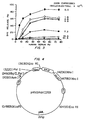
- Natural endothelial cells play important roles in normal physiology.
- these cells constitute the interface between the blood and the vessel wall and the organs of the body.
- endothelial cells secrete various natural products directly into the blood stream, maintain an antithrombotic surface on the inside of the vessel, restrict leukocytes from penetrating the vessel wall, regulate various of the biological properties of smooth muscle cells, and participate in the control of vessel wall tone. Therefore, loss of endothelial cells results in the loss of these normal physiological processes and ultimately leads to pathological conditions such as coronary artery disease, organ transplant rejection and vasculitis.
- endothelial cells for either administration of therapeutics or cell replacement have generally been limited to autologous cells, i.e., cells derived from the organism undergoing treatment.
- autologous cells i.e., cells derived from the organism undergoing treatment.
- the patient must be immunosuppressed, which is costly and leaves the patient vulnerable to infection.
- the cell doubling time for endothelial cells is on the order of at least 24 to 48 hours, leading to time periods on the order of a week or more before sufficient quantities of endothelial cells are available for genetic engineering or cell replacement.
- the cell doubling time for natural endothelial cells in vivo is also prolonged, making naturally occurring cell replacement in vivo following endothelial cell loss or damage highly inefficient.
- transplant rejection When a foreign cell is transplanted into a host, the immune system of the host rapidly mobilizes to destroy the cell and thereby protect the host.
- the immune system attack on the foreign cell is referred to as transplant rejection.
- the organism's first line of defense is through either lyric destruction or the activation of procoagulant and prothrombotic properties of the donor endothelial cell that may result from activation of the host's complement system and is generally known as the "hyperacute rejection response" or simply the "hyperacute response.”
- the complement system is a complex interaction of plasma proteins and membrane cofactors which act in a multistep, multi-protein cascade sequence in conjunction with other immunological systems of the host organism.
- the classic complement pathway involves an initial antibody recognition of, and binding to, an antigenic site on a target cell. This surface bound antibody subsequently reacts with the first component of complement, Clq, forming a Cl-antibody complex with Ca2+, Clr, and Cls which is proteolyticaity active. Cls cleaves C2 and C4 into active components, C2a and C4a.
- the C4b,2a complex is an active protease called C3 convertase, and acts to cleave C3 into C3a and C3b.
- C3b forns a complex with C4b,2a to produce C4b,2a,3b, which cleaves C5 into C5a and C5b.
- C5b forms a complex with C6 and this complex interacts with C7 in the fluid phase thereby exposing hydrophobic domains within C5b and C6 that stabilize the C5b,6,7 ternary complex in the cell membrane.
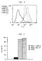
- Upon binding of C9 to C8 in the C5b-8 membrane complex lysis of foreign cells is rapidly accelerated.
- the homologous complement inhibitory activity of CD59 resides in its species-specific interaction with the terminal complement components C8 and C9, as further reported by Rollins and Sims, 1990 "The complement inhibitory activity of CD59 resides in its capacity to block incorporation of C9 into membrane C5b-9" J. Immunol. 144:3478-3483.
- CD59 does successfully address the problem of hyperacute rejection as a result of complement attack, it does not protect the cell against the overall immune attack of the host organism against foreign endothelial cells.
- T lymphocytes In stimulating immune responses, antigens elicit many molecular and cellular changes. Lymphocytes recognize antigens as foreign and are responsible for initiating both cellular and humoral responses against the presenting antigen. B lymphocyte cells respond to antigen by the production of antibodies against the presenting antigen; T lymphocytes respond by initiating a cellular response to the presenting antigen.
- T H cells involved in processing of antigen for presentation to B cells, characterized by the presence of a cell-surface glycoprotein called CD4, and cytolytic T lymphocytes (CTLs), involved in recognition of antigen on cell surfaces and lysis of cells recognized as foreign, characterized by the presence of a cell-surface glycoprotein called CD8.
- CD4 cytolytic T lymphocytes
- T cells recognize peptide fragments in conjunction with one of the two main classes of cell-surface glycoproteins of the major histocompatibility complex (MHC): either class I (MHC-I) or class II (MHC-II) proteins.
- MHC major histocompatibility complex
- CD8+ T cells recognize antigens in conjunction with MHC-I
- CD4+ T cells recognize them in conjunction with MHC-II.
- the MHC contain three major classes of genes.
- Class I genes encode the principal subunits of MHC-I glycoproteins, called human leukocyte antigens in humans, the principle ones being HLA-A, B, and C. These are present on virtually all cells and play a major role in rejection of allografts. They also form complexes with peptide fragments of viral antigens on virus-infected cells: recognition of the complexes by CD8+ CTLs results in destruction of virus infected cells. Recognition of the complexes is by a single receptor on the T cells which recognizes antigen in combination with MHC.
- Class II genes the major classes in humans being known as DP, DQ (subclasses ⁇ 2, ⁇ 2, and ⁇ 1 ⁇ 1) and DR (subclasses ⁇ 1, ⁇ 2, ⁇ 3 and ⁇ 1), encode cell-surface glycoproteins that are expressed by antigen-presenting cells, principally B cells, macrophages and dendritic cells. Together with peptide fragments of antigens, the class II proteins form the epitopes that are recognized by T helper cells (CD4+).
- Class III genes encode at least three proteins of the complement cascade and two cytotoxic proteins, tissue necrosis factor and lymphotoxin, which are involved in diverse immune reactions that destroy cells.
- T-cell mediated immune reactions can be organized into three sequential activation steps.
- CD4+ and CD8+ T lymphocytes recognize the presence of non-autologous MHC class II and class I proteins, respectively, on the surface of the foreign cell.
- the T-cells are activated by interaction of a ligand with the T cell receptors and other accessory stimulatory molecules, so that activation depends upon a variety of variables including humoral signals such as cytokines received by protein receptors on the surface of the cells. Most important is the interaction between the antigen specific T cell receptor and ligand, a complex of MHC and antigenic peptide on the antigen presenting cell (APC). Other receptors present on the T cell must also be contacted by their ligands on APC to insure activation. Once activated, the T-cells synthesize and secrete interleukin-2 (IL-2) and other cytokines.
- IL-2 interleukin-2
- the cytokines secreted by the activated T-cells lead to the third, or effector, phase of the immune response which involves recruitment and activation of lymphocytes, monocytes, and other leukocytes which together lead to cell lysis, as reviewed, for example, by Pober et al., 1990 "The potential roles of vascular endothelium in immune reactions" Human Immunol. 28:258-262.
- a genetically engineered mammalian cell for transplantation into a human or animal wherein the cell does not express on its surface proteins encoded by either the class I major histocompatibility complex genes or the class II major histocompatability complex genes which elicit a T lymphocyte mediated reaction against the cell and the cell expresses a stably incorporated nucleotide molecule which codes for a protein inhibiting complement mediated attack of the engineered cell when introduced into an animal of another species or another individual.
- genetically engineered cells which include a DNA sequence which is expressed by the cell and which codes for a protein having complement inhibitory activity that is not normally expressed in the cell.
- These cells may also be engineered so that they do not express on their cell surfaces functional proteins encoded by the class II major histocompatibility complex (MHC) genes, the HLA DP, DQ, and DR genes in human cells, or their equivalent in cells of a different species.
- MHC major histocompatibility complex
- the genetically engineered cells may not express on their cell surfaces the proteins encoded by the class I MHC genes, the HLA A, B and C genes in human cells, or their equivalent in cells of a different species.
- the cells include a genetic (DNA) sequence which is expressed by the cell and which codes for a protein which in the presence of a selected agent results in death of the cell.
- the genetic sequence which codes for a protein which has complement regulatory activity protects the cell from hyperacute rejection through attack and lysis resulting from activation of the complement system.
- the removal of the cell surface proteins encoded by the class I (for example, HLA A, B and C) and class II (for example, HLA DP, DQ, and DR) MHC genes makes the cells substantially unrecognizable by the host's CD8+ and CD4+ T-lymphocytes, respectively.
- the genetic sequence which codes for a protein which can produce cell death provides a mechanism for eliminating the genetically engineered cells from the host when their presence is no longer desired.
- the cells are modified in culture using standard in vitro transfection techniques, or can be derived from transgenic mice modified as embryos. These modified cells can serve as universal donor cells for administering therapeutic agents to the host or as replacements for natural cells which have been damaged or lost. In the most preferred embodiment, the cells are dissociated endothelial cells.
- MAC membrane attack complex
- protection against the pore-forming activity of the C5b-9 complex can be conferred on non-primate cells by txansfection of such cells with a cDNA encoding the human complement regulatory protein CD59.
- the capacity to stably express CD59 in Chinese hamster ovary (CHO) cells has enabled direct evaluation of the C5b-9 inhibitory activity conferred when CD59 is selectively expressed in mammalian cells that normally express neither CD59 nor HRF.
- the results demonstrate that the inhibitory activity of human blood cells toward the membrane attack complex of human serum complement can be transferred to a non-human mammalian cell by transfection with the CD59 cDNA and demonstrate that the C5b-9 inhibitory function of this protein correlates with the amount of newly expressed surface CD59 antigen.
- the survival and hemostatic efficacy of platelets can be increased by addition of the C5b-9 inhibitor to the storage buffer or perfusate and/or by the introduction and expression of the gene encoding CD59 in the cells to be protected.
- the C5b-9 inhibitory function observed for recombinant CD59 expressed in CHO cells exhibits specificity for human C8 and C9 (within C5b-9), analogous to that observed for the human erythrocyte membrane and for purified erythrocyte CD59 antigen.
- This capacity to confer species-selective protection against the human C5b-9 proteins by transfection of a non-human cell with cDNA encoding the CD59 sequence establishes unequivocally that this 18-21 kD protein functions as a homologous complement restriction factor on human blood cells and is consistent with the observation that the syndrome of paroxysmal nocturnal hemoglobinuria can be associated with an isolated deficiency of erythrocyte CD59.
- the complement inhibitory activity of recombinant CD59 was found to saturate when the expression of surface antigen was amplified to greater than or equal to 1.3 X 10 6 molecules/CHO cell. Assuming a spherical diameter of approximately 25 ⁇ m for the CHO cell, this is equivalent to greater than or equal to 600 molecules of CD59 antigen/ ⁇ m 2 of plasma membrane surface.
- human erythrocytes which are highly resistant to activation and lysis by human complement, express approximately 2.5 X 10 4 molecules of CD59 antigen/cell, which is equivalent to approximately 200 molecules/ ⁇ m 2 of membrane surface.
- 1 x 10 3 molecules CD59/cell or greater than or equal to 1 molecule of CD59 antigen/ ⁇ m 2 of plasma membrane surface should be effective in inhibiting complement mediated activation and lysis.
- CD59 expressed in CHO cells exhibits the species-selective recognition of human C5b-9 characteristic of CD59 in human erythrocytes despite apparent differences in N -linked glycosylation.
- species selectivity exhibited by CD59 which includes recognition for human C8 (within C5b-8) and human C9 (within C5b-9), is conferred by the core protein, independent of its carbohydrate, or that the relevant carbohydrate structures are conserved in the recombinant protein when expressed in CHO cells.
- C5b-9 inactivator refers to any CD59 molecule, including the 18 kDa protein on erythrocyte membranes, peptide fragments thereof having C5b-9 inhibitory activity, preferably containing a membrane binding domain, whether isolated from naturally produced materials or recombinantly engineered sequences.
- the term also includes cells infected or transfected with, and expressing, the gene for CD59 or a biologically functional portion thereof, as well as cells in transgenic mice in which the gene in combination with a promoter such as the murine K d MHC class I promoter has been stably introduced into an embryo of the animal using a technique such as microinjection. All molecular weights are determined by SDS-PAGE under non-reducing conditions.
- CD46, CD55, and CD59 The relative contributions of CD46, CD55, and CD59 in providing protection from complement-mediated lysis has been assessed in human amnionc epithelial cells (HAEC) by the use of specific blocking antibodies, as reported by Rooney et al., 1990 "Protection of human amniotic epithelial cells (HAEC) from complement-mediated lysis: expression on the cells of three complement inhibitory membrane proteins.” Immunology 71:308-311. The results demonstrated that CD59 provides the most protection against complement attack, as compared with CD46 and CD55.
- Cells suitable for transplantation into a foreign host are protected from complement-mediated lysis by introducing into the cell DNA encoding a protein, or combination of proteins, inhibiting complement-mediated lysis, for example, CD59, CD55, CD46 and/or other inhibitors of C8 or C9.
- CD59 is the preferred inhibitor, introduced into the cells by transfection or infection with a vector encoding the CD59 protein, and expressed on the surface of the transfected/infected cells.
- the inhibitor is preferably of the same species of origin as the host into which the cells are to be transplanted.
- the gene encoding the complement inhibitor can be introduced into a cell of a different species of origin, for example, a human CD59 gene can be introduced into a porcine cell so that the cell resists attack when transplanted into a human, or the gene can be introduced into a cell of the same species of origin so that increased amounts of the protein are expressed on the surface of the cell.
- the gene can be placed under the control of a promoter enhancing expression of the gene which is then inserted by homologous recombination into the host cell chromosome at the site where the gene is normally located, but under the control of the promoter which enhances expression, or can be inserted into the chromosome at another locus on the chromosome.
- amino acid sequence for the protein is:
- a cDNA sequence encoding the CD59 protein is (Sequence I.D. No. 4):
- Matching oligonucleotide primers can be readily designed and then used to obtain full length cDNA sequences for these proteins by performing a polymerase chain reaction amplification on human cDNA.
- the oligonucleotide primers are preferably designed with specific restriction enzyme sites so that the full length cDNA sequences can be readily subcloned into vectors for use in transfecting/infecting the target donor cells.
- DNA encoding the complement inhibitors can be introduced into the cells in culture using transfection or into embryos for production of transgenic animals expressing the complement inhibitors on the surface of their cells.
- transfection can be accomplished by electroporation, calcium phosphate precipitation, a lipofectin-based procedure, or micromjection or through use of a "gene gun".
- CDNA for the inhibitory protein, such as CD59 is subcloned into a plasmid-based vector which encodes elements for efficient expression in the genetically engineered, cell.
- the plasmid-based vector preferably contains a marker such as the neomycin gene for selection of stable transfectants with the cytotoxic aminoglycoside G418 in eukaryotic cells and an ampicillin gene for plasmid selection in bacteria.
- Infection which for endothelial cells is preferred, is accomplished by incorporating the genetic sequence for the inhibitory protein into a retroviral vector.
- Various procedures are known in the art for such incorporation.
- One such procedure which has been widely used in the art employs a defective murine retrovirus, Psi-2 cells for packaging the retrovirus, and the amphotropic packaging cell line Psi-AM to prepare infectious amphotropic virus for use in infecting the target donor cells, as described by Kohn et al., 1987 "Retroviral-mediated gene transfer into mammalian cells” Blood Cells 13:285-298.
- a retrovirus of the self-inactivating and double-copy type can be used, such as that described by Hantzopoulos et al., 1989 "Improved gene expression upon transfer of the adenosine deaminase minigene outside the transcriptional unit of a retroviral vector" Proc. Natl. Acad. Sci. USA 86:3519-3523.
- transgenic animals expressing a complement inhibitory protein on the surface of the cells for use as a source of modified cells for transplantation.
- particularly useful animals include transgenic mice.
- the most well known method for making a transgenic mice is by superovulation of a donor female, surgical removal of the egg and injection of the genetic material in the pronuclei of the embryo, as taught by U.S. Patent No. 4,873,191 to Wagner, the teachings of which are incorporated herein.
- Another commonly used technique involves the genetic manipulation of embryonic stem cells (ES cells), as specifically described below in Example 3.
- ES cells embryonic stem cells
- ES cells are grown as described, for example, in Robertson, E.J. "Embryo-derived stem cell lines" in: Teratocarcinomas and embryonic stem cells: A practical approach. E.J. Robertson, ed. 71-112 (Oxford-Washington, D.C.: IRL Press, 1987). Genetic material is introduced into the embryonic stem cells, for example, by electroporation according to the method of McMahon, A.P;, and Bradley, A. Cell 62, 1073-1085 (1991). Colonies are picked from day 6 to day 9 of selection into 96 or 24 well dishes (Costar) and expanded and used to isolate DNA for Southern blot analysis.
- Chimeric mice are generated as described in Bradley, "Production and analysis of chimaeric mice” in Teratocarcinomas and embryonic stem cells: A practical approach E.J. Robertson, ed. pp. 113-151 (Oxford, Washington, D.C. IRL Press 1987), the teachings of which are incorporated herein. Genetic material is injected into blastocysts. From those implanted females that become pregnant, chimaeras are selected from the offspring and bred to produce germline chimaeras for use as donor animals.
- the donor endothelial cells are genetically engineered to not express on their surface proteins encoded by either the class I major histocompatibility complex genes or the class II major histocompatability complex genes which elicit a T-lymphocyte mediated reaction against the cell. More preferably, the cells are engineered to not express substantially all cell surface class I and class II MHC molecules.
- the term "not express" may mean either that an insufficient amount is expressed on the surface of the cell to elicit a response or that the protein that is expressed is deficient and therefore does not elicit a response.
- the MHC molecules are referred to as HLA molecules, specifically of classes HLA A, B and C, and class II HLA DP, DQ, and DR, and their subclasses.
- HLA molecules specifically of classes HLA A, B and C, and class II HLA DP, DQ, and DR, and their subclasses.
- This terminology is generally construed as specific to the human MHC, but is intended herein to include the equivalent MHC genes from the donor cell species, for example, if the cells are of porcine origin, the term HLA would refer to the equivalent porcine MHC molecules, whether MHC I or II.
- CD4+ T - cells do not recognize the genetically engineered endothelial cells; when both the class I and class II MHC molecules are removed neither CD4+ nor CD8+ cells recognize the modified cells.
- HIV AIDS virus
- mice inheriting this genotype remain healthy and are capable of resisting infection by foreign organisms such as viruses, as reported by Zijlstra et al., 1989 "Germ-line transmission of a disrupted ⁇ 2-microglobulin gene produced by homologous recombination in embryonic stem cells” Nature 342:435438; and Koller et al., 1990 "Normal development of mice deficient in ⁇ 2M, MHC class I proteins, and CD8+ T cells” Science 248:1227-1230.
- the preferred genetic modification performed on the endothelial cells includes 1) disrupting the endogenous invariant chain gene which functions in the assembly and transport of class II MHC molecules to the cell surface and loading of antigenic peptide, and 2) disrupting the endogenous ⁇ 2 -microglobulin gene ( ⁇ 2 M gene) which codes for a protein required for the cell surface expression of all class I MHC molecules.
- Invariant chain is believed to be required for the insertion of andgienic peptide fragments into the MHC class II molecule.
- the antigenic peptide and MHC is recognized by T cells. In the absence of antigenic peptide, T cell recognition is not normally obtained, nor is the MHC class II molecule folded properly.
- presentation of peptide will be abrogated and even if minuscule amounts of cell surface MHC are obtained, they may be devoid of peptide and therefore, non-immunogenic.
- the disruption of these genes is accomplished by means of a homologous recombination gene targeting technique, as described by Zijlstra et al., 1989; Koller et al., 1990; and Example 2 below showing disruption of the invariant chain gene.
- the technique is applied to suppress expression of the class I MHC proteins on the cell surface as follows.
- the complete ⁇ 2 M gene for the target donor endothelial cell is cloned, e.g., for porcine endothelial cells the porcine ⁇ 2M gene is cloned.
- This is done by first obtaining cDNA for a homologous ⁇ 2 M gene, such as the mouse ⁇ 2M gene.
- DNA sequence information for the mouse ⁇ 2 M cDNA has been reported by Panics et al., 1983 Nature 302:449-452.
- Matching oligonucleotide primers are readily designed to hybridize by complementary base pairing to the extreme 5' and 3' ends of the mouse ⁇ 2 M cDNA.
- oligonucleotide primers are then used to obtain full-length cDNA sequences for the mouse ⁇ 2 M protein by performing a polymerase chain reaction amplification on mouse cDNA.
- the oligonucleotide primers are preferentially designed to encode specific restriction sites at their ends so that full-length cDNA sequences can be readily subcloned into plasmids.
- the full-length mouse ⁇ 2 M cDNA can then be used as a radiolabeled hybridization probe to screen cDNA libraries prepared from the source of the target donor endothelial cells, e.g., for porcine endothelial cells the mouse ⁇ 2 M cDNA is used as a hybridization probe to screen a porcine cDNA library which has been cloned into a lambda phage vector. Positive hybridizing clones are selected, purified, subcloned into plasmid vectors and then sequenced using methods known in the art.
- the complete porcine ⁇ 2 M gene can then be cloned by screening a porcine genomic DNA library cloned into a lambda phage vector with radiolabeled porcine ⁇ 2 M cDNA as a hybridization probe. Positive clones are selected, purified, subcloned into plasmid vectors and sequenced using methods known in the art.
- the ⁇ 2 M gene is subcloned into a plasmidbased or preferentially a retroviral-based vector (the "gene targeting vector") such that the reading frame of the ⁇ 2 M gene is disrupted by insertion of a short DNA sequence which allows for positive selection of recombination in the endothelial cells, for example, a neomycin resistance gene (hereinafter referred to as the "positive selection gene").
- the positive selection gene a neomycin resistance gene
- the gene targeting vector also carries an additional selection gene (the "negative selection gene"), outside of the disrupted ⁇ 2 m gene region which allows for selection against non-homologous recombination, i.e., for selection against incorporation of the entire plasmid into the genetic information of the cell rather than just the portion of the plasmid carrying the disrupted ⁇ 2 M gene.
- the negative selection gene can be, for example, a herpes simplex thymidine kinase gene.
- the gene targeting vector is then transfected/infected into the cells as described above and homologous recombination events are selected by screening for clones which express the positive selection gene but not the negative selection gene.
- cells which have been genetically engineered can be transplanted into a host to allow them to both resist and evade the immune system of a host.
- the host will normally be a human or a domesticated farm animal.
- endothelial cells especially dissociated endothelial cells for implantation or injection into a host
- the methods and compositions described herein are not limited to endothelial cells.
- Other cell types can be similarly modified for transplantation. Examples of other cell types include fibroblasts, epithelial cells, skeletal, cardiac and smooth muscle cells, hepatocytes, pancreatic islet cells, bone marrow cells, astrocytes, Schwann cells, and other cell types, dissociated or used as tissue (i.e., organs).
- endothelial cells will be construed to encompass modification of these other cell types unless otherwise specified or described specifically in the examples.
- the cells can come from a variety of sources.
- the cells are of non-human origin because of the ready availability of such cells in large quantities and at low cost.
- the cells can be of porcine or bovine origin. Cells from primates, including humans, can be used if desired. Even if human cells are used, protection from hyperacute rejection will in general still be required since complement-mediated cell attack can also occur even following allotypic transplantation.
- the genetically engineered cells are normally derived from a single clone or, for some applications, a group of individually selected clones. In this way, the characteristics of the final pharmaceutical preparation can be accurately controlled both in terms of the overall properties of the cells and their genetic make-up. Such control is of importance in evaluating the effectiveness of particular treatment protocols and in obtaining regulatory approval for such protocols.
- the cells are genetically engineered so that they express a complement inhibitory protein or proteins on their cell surface.
- the cells are also genetically engineered so that they do not express on their cell surface proteins encoded by either the class I major histocompatibility complex genes or the class II major histocompatability complex genes which elicit a T-lymphocyte mediated reaction against the cell.
- class I major histocompatibility complex genes or the class II major histocompatability complex genes which elicit a T-lymphocyte mediated reaction against the cell.
- the endothelial cells are obtained from the lining of a portion of the vascular system, e.g., a blood vessel or capillary, and are grown and maintained in a tissue culture or other suitable biological medium.
- porcine large vessel endothelial cells are isolated from the thoracic aortae of male pigs.
- the cells After being genetically engineered in the manner described below, the cells are normally stored in liquid nitrogen tanks until needed for the treatment of a particular patient.
- the ability to prepare the donor cells in advance and store them until needed is an important advantage.
- Cells are then seeded onto a matrix for implantation.
- dissociated endothelial cells are prepared for seeding onto the interior of GortexTM as follows.
- a C5b-9 inactivator can be administered in solution to cells or to a patient to inhibit complement-mediated attack.
- Administration or expression of the inhibitor, or a polypeptide representing its functional domain and possessing C5b-9 inhibitory activity produced from the isolated naturally produced inhibitor or from genetically engineered cells expressing (or more preferably, secreting) inhibitor, to block platelet or endothelial cell activation in a patient in need of such treatment, should thereby protect the patient from C5b-9 mediated procoagulant and prothrombotic responses.
- Platelets obtained from patients with the acquired stem cell disorder Paroxysmal Nocturnal Hemoglobinuria (PNH) have been shown to exhibit abnormal sensitivity to fluid phase complement activation, as characterized by an unusually high risk of venous thrombosis. This same finding is equally applicable to other types of complement mediated disorders, particularly in view of the discovery that the inhibitor is also found on the surface of endothelial cells.
- administration of the inhibitor protein whether purified from cells or expressed from cells engineered using recombinant techniques, or portions of the peptide having the same measurable activity, can be administered to these patients to alleviate the severity of the disorder.
- Treatment of patients with immune disorders and diseases such as immunovasculitis, rheumatoid arthritis, scleroderma, disseminated inuavascular coagulation, lupus, paroxysmal nocturnal hemoglobins, thrombotic thrombolytic purpura, vascular occlusion, reocclusion after surgery, coronary thrombosis, and myocardial infarction, is accomplished by administering an effective amount of a composition containing a C5b-9 inactivator as defined above such that procoagulant processes are suppressed.
- progenitor hematopoietic stem cells derived from or contained in bone marrow used for transplantation, or transplanted organs or tissue, the purified membrane inhibitor of C5b-9, or the functionally equivalent polypeptide or antibody, is first coated on the cell surface, or the gene introduced into the precursor cells as described above before transplantation or transfusion into the recipient.
- the precursor cells could be derived from the same species of origin as the recipient or from transgenic animals of a different species wherein the gene for CD59 for the recipient species is introduced into an embryo using techniques known to those skilled in the art such as microinjection.
- the amount of composition that must be administered to a patient in need of such treatment will vary depending on the particular disorder or disease and the severity of affliction.
- Treatment dosages will also vary with the individual patient depending upon response to treatment, genetic variability, and effect of co-administered drugs.
- the compositions disclosed herein are administered intravenously at a dosage of approximately nanograms of inhibitory protein or peptide per milliliter, or gene expression used to effect surface expression of at least 1 x 10 3 molecules/cell or 1 molecule CD59/ ⁇ m 2 .
- Treatment can take the form of a single administration of the composition or can be administered periodically or continuously over an extended period of time, as required.
- the C5b-9 inactivator is administered intravenously in a pharmaceutically acceptable carrier such as saline or a physiologically acceptable buffer.
- Isolated, functionally active polypeptides having the appropriate tertiary structure to inhibit C5b-9 have utility for increasing the hemostatic efficacy and extending the in vitro storage time of blood and platelet preparations.
- Platelet-containing solutions, particularly platelet-rich plasma (PRP) are in tremendous demand medically for transfusions.
- the current shelf life of platelet preparations is approximately 72 hours. An increase in the useful lifetime of such preparations represents a significant advancement in the state of the art and answers a pressing human and medical need.
- the C5b-9 inactivators can be added to the pcrfusate or storage medium to protect the vascular lining cells from ongoing complement activation during in vitro storage. Additionally, by coating these endothelial cells with a membrane-anchored C5b-9 inactivator or inserting into the cells the gene for expressing the C5b-9 inactivator, as described in more detail below, the organ or tissue would be protected from the cytolytic and thrombotic effects arising from complement activation initiated upon transplantation, thereby circumventing complement mediated acute rejection.
- the C5b-9 inactivator is administered in combination with anticoagulant, such as ACD, CPD, heparin, or oxalate, such that the concentration in the platelets or PRP is approximately nanograms inactivator/ml, or expressed at a concentration of at least 1 x 10 3 inhibitor/ml.
- anticoagulant such as ACD, CPD, heparin, or oxalate
- the C5b-9 inactivator is in combination with perfusate or storage solutions, or culture medium, such that the concentration is approximately nanograms inactivator/ml.
- compositions useful for extending the shelf life of platelet preparations stored in vitro contain C5b-9 inhibitor in an amount sufficient to inhibit C5b-9 mediated platelet activation.
- Ki concentration of half maximal inhibition
- the therapeutically effective dosage will be less than 1 ⁇ g inactivator/ml, or at least 1 x 10 3 molecules inactivator/platelet or other cell.
- Useful compositions may also contain additional anticoagulant agents such as oxalate, citrate, and heparin.
- the C5b-9 inhibitor containing compositions can be added to whole blood as it is collected or to platelet preparations after processing of the blood into isolated platelet concentrates.
- the cells are protected from activated complement C5b-9 after infusion or tissue/organ transplantation.
- the engineered cells resist attack by the complement system and evade the T-cell system, the cells and their progeny in theory can exist essentially indefinitely within the host organism. Since occasions may arise when it is desirable to remove these cells from the host, further genetic engineering is preferably performed wherein the cells are provided with an in "self-destruct" or "suicide” mechanism.
- such a mechanism involves including in the cell a gene which expresses a protein, usually an enzyme, which confers lethal sensitivity of the cell to a specific reagent not normally present in the cell's environment.
- the bacterial enzyme cytosine deaminase converts the non-toxic drug 5-fluorocytosine to 5-fluorouracil which in turn is converted within the cell to 5-fluorouridine 5'-triphosphate and 5fluoro-2'-deoxyuridine 5'-monophosphate which inhibit both RNA and DNA synthesis and thereby result in cell death, as reported by Mullin, et al., 1992 "Transfer of the bacterial gene for cytosine deaminase to mammalian cells confers lethal sensitivity to 5--fluorocytosine: a negative selection system" Proc. Natl. Acad. Sci. USA 89:33-37.
- cell death can be accomplished at any desired time by simply administering 5-fluorocytosine to the host organism.
- the sequence of the bacterial CyD gene is known and thus incorporation of the gene into the donor endothelial cells can be preformed in a manner similar to that used to insert the CD59 gene.
- the engineered cells can be used for cell replacement and for drug administration.
- coronary artery disease is caused by a blockage inside blood vessels, reducing the delivery of oxygen and nutrients to the heart.
- the current treatment for coronary artery blockade is either to mechanically dilate the blocked vessel from the inside with an angioplasty balloon or to use a replacement vessel, e.g., a synthetic graft or a section of the saphenous vein, to bypass or form a new channel around the blockage.
- Coronary angioplasty involves the insertion of a catheter from the leg vessel to the coronary artery and inflation of a balloon at the tip of the catheter to dilate the atherosclerotic plaque. This balloon inflation unfortunately has the undesired side effect of removing endothelial cells from the inner lining of the blood vessel.
- restenosis In terms of clinical practice, reocclusion of the treated vessel following coronary angioplasty, i.e., restenosis, is a significant medical problem since it occurs within six months following 30-50% of the procedures performed and is associated with substantial patient morbidity and health care expenditures.
- the principal reasons for the restenosis are acute thrombus formation due to loss of the antithrombotic surface provided by the endothelial cells and neointima formation due to unchecked smooth muscle cell stimulation by blood-borne cells, again due to the loss of the protective endothelial cell layer.
- Coronary bypass graft surgery does not involve removing the blockage to blood flow in the coronary artery, using instead a bypass to detour blood flow around the blocked vessel to supply the remainder of the heart muscle.
- a bypass to detour blood flow around the blocked vessel to supply the remainder of the heart muscle.
- the loss of the endothelial lining results in the loss of several critical endothelial properties including loss of the anticoagulant surface, loss of important smooth muscle cell regulatory force, and the loss of the protective vessel wall covering which shields smooth muscle cells from platelets, monocytes, and lymphocytes.
- the subsequent response of the blood vessel to this pathologic injury is two-fold: 1) the physiological and beneficial migration of endothelial cells from the edge of the wound to restore luminal integrity and 2) the pathophysiological migration of smooth muscle cells from the interior of the blood vessel wall toward the lumen resulting in the neointima formation and postintervention occlusion.
- Occlusion of peripheral arterial and coronary artery bypass grafts is a frequent and important clinical finding. Two-thirds of the saphenous vein coronary bypass grafts are either severely diseased or entirely occluded by six to eleven years following bypass surgery. Peripheral arterial bypass grafts generally suffer occlusion within two to five years.
- Synthetic grafts also exhibit high rates of occlusion. Initially, grafts of this type are not endothelialized. This results in a substantial incidence of early occlusion due to thrombosis. With time, the grafts become partially re-endothelialized by migration of arterial endothelial cells from the proximal and distal anastomotic sites or from ingrowth of capillary endothelial cells through the porous synthetic graft onto the luminal surface. However, the process of endothelial cell migration is normally slow and does not permit total coverage of the graft by arterial endothelial cells.
- capillary endothelial cells are less capable of inhibiting clot formation than arterial endothelial cells.
- Attempts to reseed peripheral grafts with autologous endothelial cells have demonstrated that incomplete coverage of the graft at the time of seeding results in graft closure and lack of clinical benefit of the seeding procedure.
- the genetically engineered cells described herein provide an important mechanism for addressing these critical problems in revascularization. These cells can be used to re-endothelialize denuded vessels or grafts without significant rejection by the patient's immune system. Moreover, since the cells can be grown in large numbers before the surgical procedure, adequate supplies are available for coverage of large areas of denuded vessel or naked graft. In this connection, further genetic engineering of the endothelial cells can be performed in accordance with copending application Serial No.
- a typical procedure for implanting universal donor endothelial cells in a patient's coronary artery is as follows:
- vascular graft or stent can be coated with genetically engineered endothelial cells and then implanted in a patient by:
- the genetically engineered endothelial cells provide an excellent mechanism for the administration of therapeutic agents either locally at the site of cell implantation or systemically. These cells might also secrete PDGF or FGF antagonists, thrombolytics, or thrombin antagonists, so as to inhibit restenosis in a vessel or graft wall.
- Systemic drug delivery via universal donor endothelial cells might be most effectively accomplished by the use of genetically engineered microvascular (capillary) endothelial cells which offer several advantages including a relatively large surface area to volume ratio, especially when the cells are seeded into a capillary network as described below, and direct secretion of therapeutic protein products without any barrier to diffusion.
- agents which can be administered in this way include blood clotting factors, clot dissolving factors, hormones, growth factors, cytokines, enzymes, and cholesterol binding or removing proteins.
- an appropriate gene or combination of genes is inserted into the genome of the donor endothelial cells prior to transplantation.
- a typical procedure for isolating cells of this type, for example, from a porcine source, is as follows:
- the engineered donor endothelial cells avoid graft rejection normally associated with the transplantation of non-autologous cells and thus can be used to administer their encoded therapeutic agent for substantial periods of time until, for example, removed from the host by a self-destruction mechanism of the type described above.
- Example 1 Methods for expression of human complement inhibiting proteins in mammalian cells, specifically human CD59 or functionally equivalent polypeptides.
- Human complement proteins C5b6, C7, C8 and C9 were purified and analyzed for functional activity according to methods described by Wiedmer and Sims, J. Biol.Chem, 260, 8014-8019 (1985).
- Human serum deficient in complement protein C8 (C*D) and the human complement proteins C8 and C9 were prepared and assayed according to Sims, P.J. Biochemistry 23,3248-3260 (1984), and Cheng, K., et al., J. Immunol . 135,459-464 (1985).
- Methotrexate was purchased from Lederle Laboratories (Carolina, Puerto Rico).
- BCECF/AM was from Molecular Probes (Eugene, OR).
- N -Glycanase was from Genzyme (Cambridge, MA). All other chemicals were of reagent or analytical grade.
- HBSS Hanks' balanced salt solution
- Monoclonal antibody against CD59 (1F1) was obtained from Dr. Motowo Tomita (Showa University, Tokyo).
- Fab fragments of monospecific rabbit antibody against human erythrocyte CD59 were prepared as described by Sims, P.J., Rollins, S.A., and Wiedmer, T. (1989).
- Rabbit antiserum reactive with CHO membranes was prepared by repeated injection of plasma membranes derived from cultured CHO cells, and the IgG fraction (anti-CHO) was prepared by affinity purification using protein A-Sepharose (Sigma).
- Rabbit anti-human erythrocyte was purchased from Cappel (Cochranville, PA).
- CD59 The 18 kDa human erythrocyte protein inhibitory for C5b-9 mediated activation and lysis, CD59, was isolated by modification of methods described by Sugita, et al. (1988). Additional purification was obtained by Mono-QTM FPLC (Pharmacia). CD59 polypeptides having inhibitory activity can also be affinity purified using inhibitor specific antibodies. Antibodies, such as ⁇ -P18, which bind the C5b-9 inhibitor polypeptide, are immobilized on chromatographic matrix material by techniques well known to those skilled in the art, the material containing the 18 kDa protein passed over the chromatographic matrix, non-binding material removed by washing, then the bound material removed with a higher salt solution or similar technique.
- Nucleic acid sequences encoding CD59 or active fragments thereof are isolated from a human cDNA library, or, preferably, the clone described herein.
- human DNA is isolated and digested with restriction enzymes to create fragments of appropriate size and with appropriate cohesive ends to be ligated into any of the known and commercially available expression vectors (e.g. Promega's lambda gt11 vector system).
- the isolated DNA is sheared and the appropriate linkers are ligated onto the resulting fragments which are then inserted into the expression vector of choice.
- Vectors containing human DNA fragments are next transformed into the appropriate bacterial strain, normally a strain of E. coli that is included in the expression vector kit, to generate the DNA gene bank or library. Plating out the vector containing bacteria of the library on appropriate media results in expression of the inserted human DNA fragment. The colonies are screened for the presence of DNA encoding and expressing the C5b-9 inhibitory polypeptide using specific antibodies such as ⁇ -P18. Positive colonies are isolated and used for the large scale expression of recombinanuy produced inhibitory protein.
- inhibitory protein can be made recombinantly as well as modified polypeptides and functional fragments and derivatives thereof.
- Functional polypeptides possessing the tertiary structure and ability to inhibit C5b-9 can be produced by any of the above discussed method or by other techniques commonly known to those of ordinary skill in the art.
- isolated and purified polypeptides can be further mixed with pharmaceutically acceptable carriers to form compositions for use in prolonging cell storage or in treatment of immune disorders or diseases.
- the following methods are useful in detecting and quantitating C5b-9 inhibitory activity of CD59 or fragments thereof.
- FITC fluorescein isothiocyanate
- Monoclonal antibody 1F1 was radiolabeled with IODO-GENTM (Pierce Chemical Co.) to a specific activity of 6221 cpm/ng.
- Concentrations of unlabeled proteins are estimated assuming the following extinction coefficients (E 1% 280 ): murine IgG (15), C8 (15.1), and C9 (9.6).
- concentrations of FITC-labeled proteins are determined by dye binding assay (BioRad), using the respective unlabeled protein as standard. FITC concentration is determined assuming a molar extinction (492 nm) of 68,000.
- amino acid sequence for the protein encoded by this insert is:
- the cDNA sequence encoding the CD59 protein is:
- the FRSV.CD59 vector was linearized with Sal I (20 ⁇ g) and introduced into 10 7 CHO cells by electroporation (2 kV, 25 microfarads). The cells were plated in minimum Eagle's medium (GIBCO) containing 10 mg/ml adenosine, thymidme, and deoxyadenosine and maintained for 1 to 2 days. The medium was then replaced with minimum Eagle's medium lacking deoxynucleosides but containing 0.09 ⁇ g/ml methotrexate and 10% dialyzed fetal calf serum (GIBCO). After 2 to 3 weeks, individual clones were isolated, expanded, and selected at increasing levels of methotrexate.
- GEBCO minimum Eagle's medium
- GIBCO dialyzed fetal calf serum
- transfectants were subcloned and selected for growth in medium that is supplemented with methotrexate and then amplified by continuous culture in incremental concentrations of methotrexate ranging up to 1 mg/ml, as shown in Figure 1.
- Immunoblotting of CD59 expressed by transfected CHO cells and human erythrocytes was performed. Immunoblots were developed with monoclonal antibody 1F1 (10 ⁇ g/ml) and rabbit anti-CD59 IgG (10 ⁇ g/ml). The transfected CHO cell-derived protein had a greater molecular weight than the native molecule.
- cell-surface CD59 was increased from 0 (in nontransfected CHO controls) to approximately 4.2 x 10 6 molecules/cell (for those CD59 transfectants maintained in 1 mg/ml methotrexate).
- the specific binding of 125 I-labeled 1F1 was utilized to quantitate the level of CD59 antigen expressed by CD59-transfected CHO cells.
- the CHO cells (CD59 transfectants and controls) were grown to confluence in 48-wall tissue culture plates, washed in HBSS, and then fixed with 1% paraformaldehyde (10 min, 23°C). After washing to remove fixative, the cells were incubated with a saturating concentration of antibodies, 10 ⁇ g/ml 125 I-1F1, for 30 min at 23°C. The cells were then washed six times in ice-cold HBSS, and cell-associated antibody was eluted with 4% sodium dodecylsulfate (SDS). Radioactivity was measured by photon counting and corrected for nonspecific binding, measured in the presence of a 20-fold excess of unlabeled antibody. Data for CD59 transfectants was expressed as increase in surface antigen relative to nontransfected CHO cell controls.
- the vector pFRSV has been used successfully to increase expression after gene amplification by selection in methotrexate containing medium.
- the plasmid vector pFRSV-SR ⁇ has a much stronger promoter driving expression of the introduced cDNA.
- the SalI-EcoRI fragment (800 bp) from pcDL-SR ⁇ 296 (Takebe, et al., Molec. Cell Biol. 8:466-472 (1988)) was inserted into the Hind III- Eco RI site of pFRSV, described by Slanetz and Bothwell, (1991). This plasmid expresses much higher basal levels of CD59 and other inserted cDNAs than the pFRSV vector.
- Retroviruses that bear different drug resistance markers for selection provides a means for introducing multiple cDNAs for expression in endothelial cells. Retroviruses currently under development for this purpose include the use of the neo, hygrogycin and histidinol as selectable markers. All of these resistance genes are available on Bam HI fragments and can be easily inserted into retroviral vectors to alter the resistance of a given vector. The DHFR gene is also available in retrovirus vectors. Retroviruses that express CD59 driven by the SR ⁇ promoter or the retroviral LTR (long terminal repeat) promoter can be utilized.
- retroviruses that bear distinct drug resistance markers can facilitate the co-expression of CD59 with either DAF(CD55) or MCP(CD46). Co-expression with CD59 may be more effective than CD59 alone in minimizing endothelial cell activation. Although desirable, it is not essential for the retrovirus vector to bear a drug resistance marker. Retroviruses may be developed that express both CD59 and CD55 or CD46 from a single virus. It is also possible to utilize vectors bearing different drug markers for expression of all three complement regulatory proteins CD59, CD55 and CD46.
- Membrane proteins from CD59-transfected CHO cells were extracted with 2% Triton X100, 20 mM Tris, 10 mM EDTA, 50 mM benzamidine, 200 mM N -ethylmaleimide, 1 mM phenylmethylsulfonyl fluoride, pH 7.4. After removal of insoluble material by centrifugation at 11,000 x g, 5 min, the detergent extracts were diluted 10-fold, and CD59 antigen was purified by immunoaffinity chromatography using antibody immobilized on Affi-Gel 10 (Bio-Rad).
- Antigen was eluted with 1 M glycine, 0.2% Triton X-100, 10 mM EDTA, 50 mM benzamidine, 200 mM N -ethylmaleimide, 1 mM phenylmethylsulfonyl fluoride, pH 3.0; dialyzed against 200 mM sodium phosphate, 10 mM EDTA, 50 mM benzamidine, 200 mM N -ethylmaleimide, 1 mM phenylmethylsulfonyl fluoride, pH 8.6; and concentrated to 200 ⁇ g/ml.
- CD59 expressed on the surface of these cells was susceptible to removal by phospharidylinositol-specific phospholipase C digestion, consistent with its attachment to the membrane via a glycolipid anchor, as shown in Figure 2.
- Confluent monolayers of CD59-transfected CHO cells maintained in 1.0 mg/ml methotrexate were released from T-25 tissue culture flasks with Versene/EDTA (Whittaker M.A. Bioproducts).
- FITC anti-mouse IgG was added (final concentration, 87 ⁇ g IgG/ml), and cells were incubated for an additional 15 min at 23°C.
- Surface CD59 was quantitated by specific binding of monoclonal antibody 1F1, measured by a FACSCAN (Becton, Dickinson & Co.) flow cytometer with the FL1 fluorescence channel (520 nm) set at logarithmic gain.
- CD59 expressed by the transfected CHO cells exhibited a distinctly slower migration on SDS-PAGE (apparent molecular mass of 21-24 kDa) than CD59 present in human erythrocytes (apparent molecular mass of 18-21 kDa).
- SDS-PAGE apparent molecular mass of 21-24 kDa
- CD59 present in human erythrocytes apparent molecular mass of 18-21 kDa.
- the functional activity of recombinant CD59 expressed in the transfected CHO cells was evaluated by assaying complement-mediated dye release using the intracelhilar fluorescent dye indicator BCECF/AM.
- BCECF/AM intracelhilar fluorescent dye indicator
- the CD59 transfected cells were tested for sensitivity of human serum complement using a BCECF/AM dye release assay. After incubation with BCECF/AM (15 ⁇ M) and washing, confluent monolayers were incubated with 5 mg/ml rabbit anti-CHO IgG and 25% C8D to deposit C5b-67 on the plasma membrane. Then, human serum containing 10 mM EDTA was added as the source of C8 and C9. Dye release into the supernatant was determined after 15 min at 37°C, with correction for nonspecific release observed for matched controls, omitting incubation in C8D.
- CD59 expression of transfected CHO cells was amplified by growth in 50 ⁇ g/ml methotrexate: the cells were loaded with dye by incubation in BCECF/AM; and C5b-67 was deposited as described for Figure 3. After washing, the cells were incubated (4°C, 30 min) with either 0 mg/ml or 0.5 mg/ml functionally inhibitory antibody (Fab fragments) to CD59. Unbound antibody was removed; C8 (1 ⁇ g/ml) and varying amounts of C9) were added; and dye release was measured after 15 min at 37°C.
- methotrexate the cells were loaded with dye by incubation in BCECF/AM; and C5b-67 was deposited as described for Figure 3. After washing, the cells were incubated (4°C, 30 min) with either 0 mg/ml or 0.5 mg/ml functionally inhibitory antibody (Fab fragments) to CD59. Unbound antibody was removed; C8 (1 ⁇ g/ml) and varying amounts of C9) were added;
- CD59-transfected CHO cells are selectively protected from the effects of the human C5b-9 proteins in a manner analogous to the species-selective resistance to lysis observed for human erythrocytes and for membranes reconstituted with purified CD59 antigen from human erythrocytes.
- anti-CHO IgG and human C8D were used to deposit the human C5b-67 complex on the CHO cell plasma membrane, before incubation in either human or guinea pig serum containing EDTA, as the source of the C8 and C9 components of the C5b-9 complex.
- CD59 expression by transfected CHO cells was amplified by growth at various methotrexate concentrations; the confluent monolayers were loaded with BCECF/AM; and human C5b-67 was deposited as described above. After washing, the C5b67 cells were made C5b-9 by incubation (15 min, 37°C) with either 10% (v/v) human serum (closed symbols) or 10% v/v) guinea pig serum (open symbols) in the presence of 10 mM EDTA.
- CD59 expressed by transfected CHO cells protected these cells from pore formation by human C5b-9, but not when the C8 and C9 components of this complex were replaced by the guinea pig proteins.
- Human C5b-67 was deposited on K562 cells by successive incubation with anti-human erythrocyte antiserum (1.5% (v/v, containing 10 mM EDTA) and 60% (v/v) human C8-depleted serum (diluted in HBSS). After loading with BCECF/AM cells were incubated (4°C, 30 min) with either 0 mg/ml or 1 mg/ml of functionally- blocking antibody to CD59. After removal of unbound antibody, the cells were made C5b-9 by incubation in either human (circles) or guinea pig (triangles) serum containing 10 mM EDTA.
- the effective potency of CD59 and other inhibitors of the C5b-9 complex depends on the number of C5b-9 complexes generated per unit area of plasma membrane. Therefore, the inhibitory effect of CD59 on the cytolytic and cell-stimulatory activity of C5b-9 can be overcome by increasing the input of the activated complement components that are required for assembly of the C5b-9 complex.
- the protective effects of CD59 can be augmented by providing CD59 in conjunction with CD46 and/or CD55. In this formulation, CD59 is expressed in conjunction with CD46 and/or CD55 by transfection or infection with a vector containing the gene for each protein.
- genes will serve to limit the amount of C5b-9 that can be generated at the cell surface (through the inhibitory effects of CD46 and/or CD55 on the conversion of C5 to C5b by the complement enzymes) and to protect from the cytolytic and cell-stimulatory effects of the residual C5b-9 that is formed through conversion of C5 to C5b by plasmin and other enzymes that are not inhibited by the action of CD46 and/or CD55.
- Example 2 Expression of Human CD59 in Porcine Endothelial Cells Protects them from Hyperacute Rejection by Human Complement.
- PAEC PAEC
- DMEM fetal bovine serum
- P/S penicillin and streptomycin
- the cells Prior to retroviral infection, the cells were grown to 50% confluence.
- Subconfluent PAEC were infected by using the amphotropic helper-free retroviral vector pRNSRalphaCD59+. The structure of this retroviral construct is shown in Figure 4.
- PAEC were also infected with a control retroviral vector containing the drug selection marker gene neomycin or were uninfected.
- amphotropic retroviral stocks were added to subconfluent cells growing in a T-25 tissue culture flask in a total volume of 3 ml. Polybrene was added to the flasks and the cultures were incubated at 37°C for 2 to 5 hours. The cell culture media was then removed, monolayers were rinsed two times in 5 ml of media and then 5 ml of media was added to the cells which were incubated at 37°C in 8% C02.
- Neomycin resistant colonies were assayed for the cell surface expression of human CD59 by flow cytometric techniques (FACS analysis).
- FACS analysis flow cytometric techniques
- the cells were then rinsed two times in staining buffer and then incubated for 30 minutes at 23°C with an FTTC-conjugated goat anti-rabbit IgG or an anti-mouse IgG diluted 1:50 in staining buffer.
- the cells were rinsed two times in staining buffer, once in PBS and then resuspended in 1% paraformaldehyde in PBS and analyzed by FACS. Positive cell surface expression of human CD59 (as measured by fluorescence intensity on the x axis) is demonstrated in Figure 5 for cell clones 1, 2 and 9 but not for control PAEC infected with control containing only the neomycin resistance gene.
- CD59-infected PAEC were not different from either uninfected PAEC or PAEC infected with control vector. For example, they maintained proliferation rates identical to uninfected cells and they did not overgrow monolayers or proliferate in suspension, and were contact inhibited. Additionally, CD59-infected porcine endothelial cells were capable of attaching to a synthetic GortexTM graft, as demonstrated in the scanning micrograph shown in Figure 6. Two centimeters square of synthetic GortexTM sheets were steam-sterilized, placed in sterile 35 mm bacteriological petri dishes and overlaid with sterile stainless steel fences having a one centimeter square well.
- CD59-infected PAEC were then seeded into the center wells of the fences at a density of 1 x 10 5 cells in a volume of 0.5 ml of culture media as described above and incubated at 37°C in 5% CO 2 . After two days, the cultures were refed with media and after an additional two days the media was aspirated off and the cultures were washed with PBS and then fixed with buffered 2% glutaraldehyde, 4% paraformaldehyde for one hour. The fences were then removed and the GortexTM was processed for scanning electron microscopy.
- Figure 6 demonstrates that PAEC expressing cell surface human CD59 attach as well to synthetic GortexTM grafts as normal endothelial cells.
- CD59-infected PAEC were assayed for their sensitivity to cytolysis by complement in human serum.
- CD59-infected PAEC, control PAEC infected with vector alone, and uninfected PAEC were plated into 48-well tissue culture plates at a density of 1.25 x 10 5 cells/well in DMEM with 10% FBS, 2 mM glutamine and P/S. The culture media was removed and the cells were washed three times with media without FBS. Next, human serum diluted in DMEM at various concentrations was added to the cultures for 2 hours at 37°C. The percentage of viable cells remaining in the cultures was assessed by staining the cells with 0.1% trypan blue.
- Figure 7 demonstrates that greater than 80% of uninfected or control (vector alone infected) PAEC were killed by human serum whereas less than 10% of CD59-infected PAEC were killed.
- FIG. 8B A partial restriction enzyme map for the mouse invariant chain gene is shown in Figure 8B. Digestion of mouse genomic DNA with the restriction enzyme Dra III should generate an invariant chain gene fragment of approximately 8.7 kb when this DNA is probed by Southern blotting with a radiolabeled probe specific for the mouse invariant chain gene. This result is obtained and demonstrated in Figure 9 (lane identified as parental cells).
- a gene targeting vector was constructed, so as to replace a sequence of the invariant chain gene between nucleotides 661 and 1064 with the neomycin gene.
- This genetic engineering leads to the elimination of most of exon 1 including the translation initiation codon ATG, and a large portion of the promoter including the TATA box and CART box which function as regulatory elements required for accurate and efficient transcription of the invariant chain gene, as reported by Zhu and Jones, 1989 "Complete sequence of the murine invariant chain (Ii) gene" Nucleic Acids Res. 17:447-448.
- This gene targeting vector is shown in Figure 8A as a general example of the disruption strategy. By deleting this region of the invariant chain gene, all expression of this gene including transcription and translation will be eliminated. It follows, therefore, that the expression of class II MHC gene products will be disrupted.
- Embryonic stem cells were routinely passaged every other day in ES growth media containing DMEM (high glucose) with 15% FBS and 0.1 mM 2-mercaptoethanol. The ES cells were maintained on a confluent layer of primary embryonic fibroblasts. Two days prior to the transfection of the ES cells with the gene targeting vector the cells were expanded in culture. To transfect these cells, 25 ⁇ g of DNA corresponding to the invariant chain targeting vector were introduced into 1 x 10 7 ES cells by electroporation using a BioRad electroporator set at 250 ⁇ F and 0.32 kV.
- ES cells were then seeded onto 10 x 100 mm NuncTM tissue culture plates and stable transfectants were selected for chromosomal integration by way of neomycin resistance in G418 (170 ⁇ g/ml) and/or gangcyclovir in some experiments where the herpes-simplex virus thymidine kinase gene was included in the targeting vector.
- Genomic DNA was isolated from 55 individual stable transfectants and digested overnight with either the Eco RI or Dra III restriction endonucleases. Digested DNA was resolved by electrophoresis, blotted to GeneScreen+TM nylon membranes and then hybridized with a radiolabeled DNA probe specific for the mouse invariant chain gene.
Landscapes
- Health & Medical Sciences (AREA)
- Life Sciences & Earth Sciences (AREA)
- Chemical & Material Sciences (AREA)
- Organic Chemistry (AREA)
- Genetics & Genomics (AREA)
- General Health & Medical Sciences (AREA)
- Medicinal Chemistry (AREA)
- Zoology (AREA)
- Engineering & Computer Science (AREA)
- Molecular Biology (AREA)
- Biochemistry (AREA)
- Animal Behavior & Ethology (AREA)
- Bioinformatics & Cheminformatics (AREA)
- Veterinary Medicine (AREA)
- Immunology (AREA)
- Cell Biology (AREA)
- Biophysics (AREA)
- Proteomics, Peptides & Aminoacids (AREA)
- Gastroenterology & Hepatology (AREA)
- Toxicology (AREA)
- Public Health (AREA)
- Wood Science & Technology (AREA)
- Biomedical Technology (AREA)
- General Engineering & Computer Science (AREA)
- Biotechnology (AREA)
- Environmental Sciences (AREA)
- Chemical Kinetics & Catalysis (AREA)
- General Chemical & Material Sciences (AREA)
- Nuclear Medicine, Radiotherapy & Molecular Imaging (AREA)
- Pharmacology & Pharmacy (AREA)
- Microbiology (AREA)
- Transplantation (AREA)
- Animal Husbandry (AREA)
- Physics & Mathematics (AREA)
- Epidemiology (AREA)
- Dermatology (AREA)
- Oral & Maxillofacial Surgery (AREA)
- Cardiology (AREA)
- Vascular Medicine (AREA)
- Biodiversity & Conservation Biology (AREA)
Claims (26)
- Cellule de mammifère modifiée génétiquement pour la transplantation dans l'homme ou un animal,
dans laquelle la cellule n'exprime pas sur sa surface des protéines codées par les gènes du complexe majeur d'histocompatibilité de catégorie I ou les gènes du complexe majeur d'histocompatibilité de catégorie II qui engendrent une réponse à médiation par les lymphocytes T contre la cellule, et
la cellule exprime une molécule nucléotidique incorporée de manière stable qui code pour une protéine inhibant l'attaque, à médiation par le complément, de la cellule modifiée génétiquement lors de l'introduction dans un animal d'une autre espèce ou un autre individu. - Cellule suivant la revendication 1, dans laquelle la molécule nucléotidique code pour une protéine inhibant l'attaque, à médiation par le complément, dans la même espèce que l'espèce dont la cellule est dérivée, et la molécule nucléotidique est située à un locus distinct de celui du gène naturel codant pour la même molécule dans la cellule.
- Cellule suivant la revendication 1, dans laquelle le promoteur du gène codant pour un inhibiteur protéique de l'attaque, à médiation par le complément, sur la cellule hôte contenant le gène est modifié de manière à accroítre l'expression du gène dans la cellule hôte.
- Cellule suivant la revendication 1, dans laquelle la protéine inhibant l'attaque, à médiation par le complément, est choisie dans le groupe consistant en CD59, CD46 et CD55.
- Cellule suivant la revendication 1, choisie dans le groupe consistant en des cellules endothéliales, des fibroblastes, des cellules épithéliales, des cellules des muscles squelettiques, du muscle cardiaque et des muscles lisses, des hépatocytes, des cellules des ílots pancréatiques, des cellules de la moelle osseuse, des astrocytes et des cellules de Schwann.
- Cellule suivant la revendication 1, dans laquelle la cellule n'est pas d'origine humaine, la protéine inhibant le complément est une protéine humaine et les gènes du CMH de catégorie II sont choisis dans le groupe consistant en l'équivalent de HLA DP, DR et DQ dans l'espèce de cellule.
- Cellule suivant la revendication 1, dans laquelle la cellule n'est pas d'origine humaine, la protéine inhibant l'attaque, à médiation par le complément, est une protéine humaine et les gènes du CMH de catégorie I sont choisis dans le groupe consistant en l'équivalent de HLA A, B et C dans l'espèce de cellule.
- Cellule suivant la revendication 1, dans laquelle la cellule n'exprime pas les protéines du CMH de catégorie II en résultat de la rupture du gène de chaíne invariante des gènes du CMH de catégorie II.
- Cellule suivant la revendication 1, dans laquelle la cellule n'exprime pas les protéines du CMH de catégorie I en résultat de la rupture du gène de β2-microglobuline.
- Cellule suivant la revendication 1, comprenant en outre une molécule nucléotidique qui est exprimée par la cellule et qui code pour une protéine qui, en présence d'un composé choisi, a pour résultat la mort cellulaire.
- Cellule suivant la revendication 10, dans laquelle la molécule nucléotidique code pour une cytosine-désaminase bactérienne.
- Cellule suivant la revendication 1, dans laquelle la cellule est choisie dans le groupe consistant en des cellules d'origine humaine, des cellules d'origine bovine et des cellules d'origine porcine.
- Cellule suivant l'une quelconque des revendications 1-4 ou 6-12, choisie dans le groupe consistant en des cellules souches hématopoïétiques, la descendance de cellules souches hématopoïétiques et les cellules sanguines matures.
- Cellule suivant l'une quelconque des revendications 1 à 13, dans laquelle la cellule exprime une molécule nucléotidique qui code pour un agent thérapeutique, qui est sécrété par la cellule et la cellule n'exprime pas normalement la molécule, exprime la molécule à partir du même promoteur ou bien exprime la molécule à partir de locus multiples.
- Cellule suivant l'une quelconque des revendications 1 à 14, exprimant sur sa surface une quantité supérieure ou égale à 103 molécules CD59/cellule ou supérieure ou égale à 1 molécule d'antigène CD59/µm2 de surface de membrane plasmatique.
- Cellule suivant la revendication 14, dans laquelle la cellule est une cellule endothéliale microvasculaire.
- Cellule suivant la revendication 1, qui est une cellule de souris.
- Cellule suivant la revendication 17, dans laquelle la cellule n'exprime pas les protéines du CMH de catégorie II en résultat d'une rupture du gène de chaíne invariante des gènes du CMH de catégorie II.
- Cellule suivant la revendication 17, dans laquelle la cellule n'exprime pas les protéines du CMH de catégorie I en résultat d'une rupture du gène de β2-microglobuline.
- Souris transgénique contenant n'importe laquelle des cellules suivant les revendications 17 à 19.
- Organe provenant d'une souris transgénique, organe qui est formé de cellules suivant l'une quelconque des revendications 17 à 19.
- Prothèse pour l'implantation dans un animal ou patient humain, comprenant des cellules suivant l'une quelconque des revendications 1 à 16 fixées à celle-ci.
- Prothèse suivant la revendication 22, qui est une greffe vasculaire, les cellules étant des cellules endothéliales microvasculaires.
- Procédé pour la ré-endothélialisation ex vivo d'un vaisseau sanguin dénudé, comprenant l'application au vaisseau de cellules suivant la revendication 16 ou de la prothèse suivant la revendication 23.
- Cellule suivant l'une quelconque des revendications 1 à 16 ou organe suivant la revendication 21 ou bien prothèse suivant la revendication 22 ou 23, destiné à être utilisé en médecine.
- Utilisation des cellules suivant la revendication 16 ou de la prothèse suivant la revendication 22 dans la production d'un agent destiné à la ré-endothélialisation d'un vaisseau sanguin dénudé.
Priority Applications (1)
| Application Number | Priority Date | Filing Date | Title |
|---|---|---|---|
| EP00114262A EP1041142A3 (fr) | 1991-07-15 | 1992-07-14 | Cellules donneuses universelles |
Applications Claiming Priority (5)
| Application Number | Priority Date | Filing Date | Title |
|---|---|---|---|
| US72992691A | 1991-07-15 | 1991-07-15 | |
| US729926 | 1991-07-15 | ||
| US90639492A | 1992-06-29 | 1992-06-29 | |
| US906394 | 1992-06-29 | ||
| PCT/US1992/005920 WO1993002188A1 (fr) | 1991-07-15 | 1992-07-14 | Cellules donneuses universelles |
Related Child Applications (1)
| Application Number | Title | Priority Date | Filing Date |
|---|---|---|---|
| EP00114262A Division EP1041142A3 (fr) | 1991-07-15 | 1992-07-14 | Cellules donneuses universelles |
Publications (2)
| Publication Number | Publication Date |
|---|---|
| EP0591462A1 EP0591462A1 (fr) | 1994-04-13 |
| EP0591462B1 true EP0591462B1 (fr) | 2003-04-16 |
Family
ID=27111956
Family Applications (2)
| Application Number | Title | Priority Date | Filing Date |
|---|---|---|---|
| EP92915715A Expired - Lifetime EP0591462B1 (fr) | 1991-07-15 | 1992-07-14 | Cellules donneuses universelles |
| EP00114262A Withdrawn EP1041142A3 (fr) | 1991-07-15 | 1992-07-14 | Cellules donneuses universelles |
Family Applications After (1)
| Application Number | Title | Priority Date | Filing Date |
|---|---|---|---|
| EP00114262A Withdrawn EP1041142A3 (fr) | 1991-07-15 | 1992-07-14 | Cellules donneuses universelles |
Country Status (6)
| Country | Link |
|---|---|
| EP (2) | EP0591462B1 (fr) |
| JP (2) | JPH06506604A (fr) |
| AT (1) | ATE237682T1 (fr) |
| CA (1) | CA2113089C (fr) |
| DE (1) | DE69233010T2 (fr) |
| WO (1) | WO1993002188A1 (fr) |
Families Citing this family (27)
| Publication number | Priority date | Publication date | Assignee | Title |
|---|---|---|---|---|
| GB9100481D0 (en) * | 1991-01-10 | 1991-02-20 | Inst Nat Sante Rech Med | Genetically engineered mice |
| ATE194494T1 (de) * | 1991-10-21 | 2000-07-15 | Cell Genesys Inc | Kombinierte zelluläre und immunsuppressive therapien |
| WO1994016080A1 (fr) * | 1993-01-08 | 1994-07-21 | Exemplar Corporation | Procede in vitro/in vivo d'identification de medicaments anti-neoplasiques |
| EP0771320A4 (fr) | 1993-07-29 | 1998-11-25 | Univ California | Leurres polynucleotidiques inhibant l'expression du complexe d'histocompatibilite majeure ii, et applications |
| US5859226A (en) * | 1993-07-29 | 1999-01-12 | Regents Of The University Of California, The | Polynucleotide decoys that inhibit MHC-II expression and uses thereof |
| WO1995004756A1 (fr) * | 1993-08-11 | 1995-02-16 | Alexion Pharmaceuticals, Inc. | Proteines inhibitrices complementaires de primates non humains |
| AU1931495A (en) * | 1994-03-14 | 1995-10-03 | Cryolife, Inc. | Treated tissue for implantation and preparation methods |
| US6156306A (en) * | 1994-08-17 | 2000-12-05 | Albert Einstein College Of Medicine Of Yeshiva University | Pancreatic β-cells for allogeneic transplantation without immunosuppression |
| US5629194A (en) * | 1994-10-21 | 1997-05-13 | Diacrin, Inc. | Isolated porcine pancreatic cells for use in treatment of diseases characterized by insufficient insulin activity |
| EP0832193A1 (fr) * | 1995-05-26 | 1998-04-01 | Diacrin, Inc. | Hepatocytes porcins a utiliser dans le traitement de troubles caracterises par une fonction hepatique insuffisante |
| US5766584A (en) * | 1995-06-02 | 1998-06-16 | Massachusetts Institute Of Technology | Inhibition of vascular smooth muscle cell proliferation with implanted matrix containing vascular endothelial cells |
| WO1997004087A1 (fr) * | 1995-07-18 | 1997-02-06 | Guido Krupp | Ribozymes pour l'inhibition selective de l'expression de genes d'alleles du complexe majeur d'histocompatibilite(cmh), et medicaments les contenant |
| AU723003C (en) | 1995-08-04 | 2004-01-29 | General Hospital Corporation, The | Transgenic swine and swine cells having human HLA genes |
| US6166288A (en) * | 1995-09-27 | 2000-12-26 | Nextran Inc. | Method of producing transgenic animals for xenotransplantation expressing both an enzyme masking or reducing the level of the gal epitope and a complement inhibitor |
| AU1933897A (en) * | 1996-03-22 | 1997-10-17 | Imutran Limited | Surfaces which prevent or reduce complement activation |
| US6022863A (en) * | 1996-05-21 | 2000-02-08 | Yale University | Regulation of gene expression |
| US6825395B1 (en) | 1997-07-14 | 2004-11-30 | Nippon Meat Packers, Inc. | Transgenic non-human mammals expressing the human complement inhibitor (DAF/CD55) |
| CN101268183B (zh) | 2005-04-21 | 2011-11-30 | 麻省理工学院 | 改变对外源和内源免疫原(包括同系和非同系细胞、组织或器官)的免疫应答的材料和方法 |
| EP2361591A3 (fr) | 2005-06-21 | 2011-12-21 | Pervasis Therapeutics, Inc. | Procédés et compositions d'amélioration d'accès vasculaire |
| ATE534727T1 (de) * | 2006-09-21 | 2011-12-15 | Tissue Genesis | Zellfreisetzungsmatrizen |
| JP2011069570A (ja) * | 2009-09-28 | 2011-04-07 | Fujitsu General Ltd | ヒートポンプサイクル装置 |
| PT3116902T (pt) * | 2014-03-11 | 2020-04-03 | Cellectis | Método para gerar células t compatíveis para transplante alogénico |
| MA45235A (fr) | 2016-06-14 | 2019-04-17 | Regeneron Pharma | Anticorps anti-c5 et leurs utilisations |
| US11834680B2 (en) | 2017-06-28 | 2023-12-05 | Rutgers, The State University Of New Jersey | Single kidney cell-derived organoids |
| EP3644965A4 (fr) * | 2017-06-28 | 2021-05-05 | Rutgers, The State University of New Jersey | Organoïdes dérivés d'une cellule cérébrale unique |
| EP3724226A1 (fr) | 2017-12-13 | 2020-10-21 | Regeneron Pharmaceuticals, Inc. | Associations d'anticorps anti-c5 et utilisations associées |
| US20220025332A1 (en) * | 2018-12-16 | 2022-01-27 | Figene, Llc | Therapeutic uses of gene edited fibroblasts |
Family Cites Families (8)
| Publication number | Priority date | Publication date | Assignee | Title |
|---|---|---|---|---|
| IL82390A0 (en) * | 1986-05-02 | 1987-10-30 | Genentech Inc | Nucleic acid and methods for the synthesis of novel daf compositions |
| US4883755A (en) * | 1987-10-28 | 1989-11-28 | Thomas Jefferson University | Method of reendothelializing vascular linings |
| IT1230047B (it) * | 1989-07-04 | 1991-09-27 | Giovanni Brotzu | Protesi vascolare contenente nella parete microcapsule inglobanti cellule produttrici di ormoni. |
| EP0483247A4 (en) * | 1989-07-21 | 1992-12-09 | Washington University | Recombinantly produced human membrane cofactor protein (mcp) |
| DE69033493D1 (de) * | 1989-07-25 | 2004-08-12 | Cell Genesys Inc | Homologe rekombination für universelle donorzellen und chimerische säugetierzellen |
| DK0495852T3 (da) * | 1989-10-12 | 1996-07-15 | Imutran Ltd | Modificeret biologisk materiale |
| CA2082277A1 (fr) * | 1990-05-11 | 1991-11-12 | Damien F. J. Purcell | Variants de cd46 |
| AU9076191A (en) * | 1990-11-21 | 1992-06-25 | Massachusetts Institute Of Technology | Mice having beta 2 microglobulin gene disruption |
-
1992
- 1992-07-14 CA CA002113089A patent/CA2113089C/fr not_active Expired - Fee Related
- 1992-07-14 EP EP92915715A patent/EP0591462B1/fr not_active Expired - Lifetime
- 1992-07-14 JP JP5502933A patent/JPH06506604A/ja not_active Withdrawn
- 1992-07-14 AT AT92915715T patent/ATE237682T1/de not_active IP Right Cessation
- 1992-07-14 DE DE69233010T patent/DE69233010T2/de not_active Expired - Fee Related
- 1992-07-14 WO PCT/US1992/005920 patent/WO1993002188A1/fr active IP Right Grant
- 1992-07-14 EP EP00114262A patent/EP1041142A3/fr not_active Withdrawn
-
1997
- 1997-06-18 JP JP9161718A patent/JPH1057054A/ja active Pending
Non-Patent Citations (6)
| Title |
|---|
| "The Trials of Xenotransplantation", NATURE, vol. 406, no. 6797, - 17 August 2000 (2000-08-17), pages 661 * |
| ARTIP J H ET AL: "Cardiac Xenotransplantation", CURRENT OPINION IN CARDIOLOGY, vol. 12, - 1997, pages 172 - 178 * |
| LAI L ET AL:" Production of alpha-1,3-Galactosyl transferase Knockout Pigs by Nuclear Transfer Cloning" , Sciencexpress, published online 03.01.2002 * |
| SANDRIN M S ETAL: "Deduction of the Major Porcine Xenoantigen ...", XENOTRANSPLANTATION, vol. 3, - 1996, pages 134 - 140- * |
| Süddeutsche Zeitung, 08.01.2002, No. 6, page V2/7: "Neun Schweinchen namens Hope" * |
| VANHOVE B ET AL: "Intracellular Expression in Pig Cells...", TRANSPLANTATION, vol. 66, no. 11, - 15 December 1998 (1998-12-15), pages 1477 - 148- * |
Also Published As
| Publication number | Publication date |
|---|---|
| WO1993002188A1 (fr) | 1993-02-04 |
| CA2113089A1 (fr) | 1993-02-04 |
| EP1041142A3 (fr) | 2007-10-24 |
| DE69233010D1 (de) | 2003-05-22 |
| EP1041142A2 (fr) | 2000-10-04 |
| JPH1057054A (ja) | 1998-03-03 |
| ATE237682T1 (de) | 2003-05-15 |
| CA2113089C (fr) | 2007-11-06 |
| EP0591462A1 (fr) | 1994-04-13 |
| DE69233010T2 (de) | 2004-02-19 |
| JPH06506604A (ja) | 1994-07-28 |
Similar Documents
| Publication | Publication Date | Title |
|---|---|---|
| EP0591462B1 (fr) | Cellules donneuses universelles | |
| US6100443A (en) | Universal donor cells | |
| AU727546B2 (en) | Transgenic animals for xenotransplantation with reduced antibody-mediated rejection | |
| AU673987B2 (en) | Specific tolerance in transplantation | |
| HUT53672A (en) | Quick immunoselective cloning process | |
| JP2002513568A (ja) | T細胞同時刺激シグナル2(b7/cd28相互作用)を遮断することによる免疫抑制 | |
| RO109863B1 (ro) | Gena care codifica cel putin un factor de restrictie complementar, animal transgenic si metode de transplantare a tesuturilor sau organelor animale | |
| US6916654B1 (en) | Universal donor cells | |
| US20040192599A1 (en) | Gene therapy for hemophilia a | |
| US5573940A (en) | Cells expressing high levels of CD59 | |
| EP0815252A1 (fr) | Therapie genique utile lors de transplantations et contre les inflammations ou les thromboses | |
| WO1994000560A1 (fr) | Matrice de cellules endotheliales microvasculaires et donneuses universelles | |
| EP0781333A1 (fr) | Therapie genique destinee a la transplantation et a des etats inflammatoires ou thrombotiques | |
| US20030086940A1 (en) | Engineered recombinant molecule that regulates humoral and cellular effector functions of the immune system | |
| AU671158B2 (en) | Delivery of proteins by intermembrane transfer for preaccommodation of xenogeneic organ transplants and other purposes | |
| WO1994006903A9 (fr) | Apport de proteines par transfert intermembranaire pour la preaccommodation de transplants d'organes xenogeniques et a d'autres fins | |
| WO2003066848A1 (fr) | Fgl-2 porcin | |
| RU2252533C2 (ru) | Способ получения трансгенного животного | |
| JP4064424B6 (ja) | 移植および炎症または血栓症状態の遺伝子治療 | |
| JP4064424B2 (ja) | 移植および炎症または血栓症状態の遺伝子治療 | |
| AU771025B2 (en) | Specific tolerance in transplantation | |
| JP2007259857A6 (ja) | 移植および炎症または血栓症状態の遺伝子治療 | |
| JP2006187291A6 (ja) | 移植および炎症または血栓症状態の遺伝子治療 | |
| JPH09500616A (ja) | 同種及び異種間移植 | |
| AU2002317065A1 (en) | Gene therapy for hemophilia A |
Legal Events
| Date | Code | Title | Description |
|---|---|---|---|
| PUAI | Public reference made under article 153(3) epc to a published international application that has entered the european phase |
Free format text: ORIGINAL CODE: 0009012 |
|
| AK | Designated contracting states |
Kind code of ref document: A1 Designated state(s): AT BE CH DE DK ES FR GB GR IT LI LU MC NL SE |
|
| 17P | Request for examination filed |
Effective date: 19940215 |
|
| 17Q | First examination report despatched |
Effective date: 19960704 |
|
| GRAG | Despatch of communication of intention to grant |
Free format text: ORIGINAL CODE: EPIDOS AGRA |
|
| RAP1 | Party data changed (applicant data changed or rights of an application transferred) |
Owner name: YALE UNIVERSITY Owner name: OKLAHOMA MEDICAL RESEARCH FOUNDATION |
|
| GRAG | Despatch of communication of intention to grant |
Free format text: ORIGINAL CODE: EPIDOS AGRA |
|
| GRAH | Despatch of communication of intention to grant a patent |
Free format text: ORIGINAL CODE: EPIDOS IGRA |
|
| GRAH | Despatch of communication of intention to grant a patent |
Free format text: ORIGINAL CODE: EPIDOS IGRA |
|
| RIN1 | Information on inventor provided before grant (corrected) |
Inventor name: SQUINTO, STEPHEN Inventor name: BELL, LEONARD Inventor name: ROLLINS, SCOTT Inventor name: MADRI, JOSEPH Inventor name: FLAVELL, RICHARD, A. Inventor name: ELLIOT, EILEEN, A. Inventor name: BOTHWELL, ALFRED, L., M. Inventor name: SIMS, PETER, J. |
|
| GRAA | (expected) grant |
Free format text: ORIGINAL CODE: 0009210 |
|
| RIN1 | Information on inventor provided before grant (corrected) |
Inventor name: SQUINTO, STEPHEN Inventor name: BELL, LEONARD Inventor name: ROLLINS, SCOTT Inventor name: MADRI, JOSEPH Inventor name: FLAVELL, RICHARD, A. Inventor name: ELLIOT, EILEEN, A. Inventor name: BOTHWELL, ALFRED, L., M. Inventor name: SIMMS, PETER, J. |
|
| RIC1 | Information provided on ipc code assigned before grant |
Ipc: 7C 07K 2/00 B Ipc: 7A 61L 27/00 B Ipc: 7C 12N 15/87 B Ipc: 7C 12N 5/22 B Ipc: 7C 12N 5/16 B Ipc: 7A 01K 67/027 B Ipc: 7C 12N 15/12 B Ipc: 7C 12N 15/00 A |
|
| AK | Designated contracting states |
Designated state(s): AT BE CH DE DK ES FR GB GR IT LI LU MC NL SE |
|
| PG25 | Lapsed in a contracting state [announced via postgrant information from national office to epo] |
Ref country code: NL Free format text: LAPSE BECAUSE OF FAILURE TO SUBMIT A TRANSLATION OF THE DESCRIPTION OR TO PAY THE FEE WITHIN THE PRESCRIBED TIME-LIMIT Effective date: 20030416 Ref country code: LI Free format text: LAPSE BECAUSE OF FAILURE TO SUBMIT A TRANSLATION OF THE DESCRIPTION OR TO PAY THE FEE WITHIN THE PRESCRIBED TIME-LIMIT Effective date: 20030416 Ref country code: IT Free format text: LAPSE BECAUSE OF FAILURE TO SUBMIT A TRANSLATION OF THE DESCRIPTION OR TO PAY THE FEE WITHIN THE PRESCRIBED TIME-LIMIT;WARNING: LAPSES OF ITALIAN PATENTS WITH EFFECTIVE DATE BEFORE 2007 MAY HAVE OCCURRED AT ANY TIME BEFORE 2007. THE CORRECT EFFECTIVE DATE MAY BE DIFFERENT FROM THE ONE RECORDED. Effective date: 20030416 Ref country code: CH Free format text: LAPSE BECAUSE OF FAILURE TO SUBMIT A TRANSLATION OF THE DESCRIPTION OR TO PAY THE FEE WITHIN THE PRESCRIBED TIME-LIMIT Effective date: 20030416 Ref country code: BE Free format text: LAPSE BECAUSE OF FAILURE TO SUBMIT A TRANSLATION OF THE DESCRIPTION OR TO PAY THE FEE WITHIN THE PRESCRIBED TIME-LIMIT Effective date: 20030416 Ref country code: AT Free format text: LAPSE BECAUSE OF FAILURE TO SUBMIT A TRANSLATION OF THE DESCRIPTION OR TO PAY THE FEE WITHIN THE PRESCRIBED TIME-LIMIT Effective date: 20030416 |
|
| REG | Reference to a national code |
Ref country code: GB Ref legal event code: FG4D |
|
| REG | Reference to a national code |
Ref country code: CH Ref legal event code: EP |
|
| RIN2 | Information on inventor provided after grant (corrected) |
Inventor name: SQUINTO, STEPHEN Inventor name: BELL, LEONARD Inventor name: ROLLINS, SCOTT Inventor name: MADRI, JOSEPH Inventor name: FLAVELL, RICHARD, A. Inventor name: ELLIOT, EILEEN, A. Inventor name: BOTHWELL, ALFRED, L., M. Inventor name: SIMMS, PETER, J. |
|
| REF | Corresponds to: |
Ref document number: 69233010 Country of ref document: DE Date of ref document: 20030522 Kind code of ref document: P |
|
| PG25 | Lapsed in a contracting state [announced via postgrant information from national office to epo] |
Ref country code: LU Free format text: LAPSE BECAUSE OF NON-PAYMENT OF DUE FEES Effective date: 20030714 |
|
| PG25 | Lapsed in a contracting state [announced via postgrant information from national office to epo] |
Ref country code: SE Free format text: LAPSE BECAUSE OF FAILURE TO SUBMIT A TRANSLATION OF THE DESCRIPTION OR TO PAY THE FEE WITHIN THE PRESCRIBED TIME-LIMIT Effective date: 20030716 Ref country code: GR Free format text: LAPSE BECAUSE OF FAILURE TO SUBMIT A TRANSLATION OF THE DESCRIPTION OR TO PAY THE FEE WITHIN THE PRESCRIBED TIME-LIMIT Effective date: 20030716 Ref country code: DK Free format text: LAPSE BECAUSE OF FAILURE TO SUBMIT A TRANSLATION OF THE DESCRIPTION OR TO PAY THE FEE WITHIN THE PRESCRIBED TIME-LIMIT Effective date: 20030716 |
|
| PG25 | Lapsed in a contracting state [announced via postgrant information from national office to epo] |
Ref country code: MC Free format text: LAPSE BECAUSE OF NON-PAYMENT OF DUE FEES Effective date: 20030731 |
|
| NLV1 | Nl: lapsed or annulled due to failure to fulfill the requirements of art. 29p and 29m of the patents act | ||
| PG25 | Lapsed in a contracting state [announced via postgrant information from national office to epo] |
Ref country code: ES Free format text: LAPSE BECAUSE OF FAILURE TO SUBMIT A TRANSLATION OF THE DESCRIPTION OR TO PAY THE FEE WITHIN THE PRESCRIBED TIME-LIMIT Effective date: 20031030 |
|
| REG | Reference to a national code |
Ref country code: CH Ref legal event code: PL |
|
| ET | Fr: translation filed | ||
| PLBE | No opposition filed within time limit |
Free format text: ORIGINAL CODE: 0009261 |
|
| STAA | Information on the status of an ep patent application or granted ep patent |
Free format text: STATUS: NO OPPOSITION FILED WITHIN TIME LIMIT |
|
| 26N | No opposition filed |
Effective date: 20040119 |
|
| PGFP | Annual fee paid to national office [announced via postgrant information from national office to epo] |
Ref country code: DE Payment date: 20070712 Year of fee payment: 16 |
|
| PGFP | Annual fee paid to national office [announced via postgrant information from national office to epo] |
Ref country code: GB Payment date: 20070711 Year of fee payment: 16 |
|
| PGFP | Annual fee paid to national office [announced via postgrant information from national office to epo] |
Ref country code: FR Payment date: 20070710 Year of fee payment: 16 |
|
| GBPC | Gb: european patent ceased through non-payment of renewal fee |
Effective date: 20080714 |
|
| PG25 | Lapsed in a contracting state [announced via postgrant information from national office to epo] |
Ref country code: DE Free format text: LAPSE BECAUSE OF NON-PAYMENT OF DUE FEES Effective date: 20090203 |
|
| REG | Reference to a national code |
Ref country code: FR Ref legal event code: ST Effective date: 20090331 |
|
| PG25 | Lapsed in a contracting state [announced via postgrant information from national office to epo] |
Ref country code: GB Free format text: LAPSE BECAUSE OF NON-PAYMENT OF DUE FEES Effective date: 20080714 |
|
| PG25 | Lapsed in a contracting state [announced via postgrant information from national office to epo] |
Ref country code: FR Free format text: LAPSE BECAUSE OF NON-PAYMENT OF DUE FEES Effective date: 20080731 |