EP0432631A2 - Verfahren zur Bestimmung eines Analyten - Google Patents
Verfahren zur Bestimmung eines Analyten Download PDFInfo
- Publication number
- EP0432631A2 EP0432631A2 EP90123380A EP90123380A EP0432631A2 EP 0432631 A2 EP0432631 A2 EP 0432631A2 EP 90123380 A EP90123380 A EP 90123380A EP 90123380 A EP90123380 A EP 90123380A EP 0432631 A2 EP0432631 A2 EP 0432631A2
- Authority
- EP
- European Patent Office
- Prior art keywords
- enzyme
- liquid
- analyte
- solid phase
- matrix
- Prior art date
- Legal status (The legal status is an assumption and is not a legal conclusion. Google has not performed a legal analysis and makes no representation as to the accuracy of the status listed.)
- Granted
Links
Images

Classifications
-
- G—PHYSICS
- G01—MEASURING; TESTING
- G01N—INVESTIGATING OR ANALYSING MATERIALS BY DETERMINING THEIR CHEMICAL OR PHYSICAL PROPERTIES
- G01N33/00—Investigating or analysing materials by specific methods not covered by groups G01N1/00 - G01N31/00
- G01N33/48—Biological material, e.g. blood, urine; Haemocytometers
- G01N33/50—Chemical analysis of biological material, e.g. blood, urine; Testing involving biospecific ligand binding methods; Immunological testing
- G01N33/53—Immunoassay; Biospecific binding assay; Materials therefor
- G01N33/543—Immunoassay; Biospecific binding assay; Materials therefor with an insoluble carrier for immobilising immunochemicals
- G01N33/54366—Apparatus specially adapted for solid-phase testing
- G01N33/54386—Analytical elements
-
- G—PHYSICS
- G01—MEASURING; TESTING
- G01N—INVESTIGATING OR ANALYSING MATERIALS BY DETERMINING THEIR CHEMICAL OR PHYSICAL PROPERTIES
- G01N33/00—Investigating or analysing materials by specific methods not covered by groups G01N1/00 - G01N31/00
- G01N33/48—Biological material, e.g. blood, urine; Haemocytometers
- G01N33/50—Chemical analysis of biological material, e.g. blood, urine; Testing involving biospecific ligand binding methods; Immunological testing
- G01N33/53—Immunoassay; Biospecific binding assay; Materials therefor
- G01N33/543—Immunoassay; Biospecific binding assay; Materials therefor with an insoluble carrier for immobilising immunochemicals
- G01N33/54366—Apparatus specially adapted for solid-phase testing
-
- Y—GENERAL TAGGING OF NEW TECHNOLOGICAL DEVELOPMENTS; GENERAL TAGGING OF CROSS-SECTIONAL TECHNOLOGIES SPANNING OVER SEVERAL SECTIONS OF THE IPC; TECHNICAL SUBJECTS COVERED BY FORMER USPC CROSS-REFERENCE ART COLLECTIONS [XRACs] AND DIGESTS
- Y10—TECHNICAL SUBJECTS COVERED BY FORMER USPC
- Y10S—TECHNICAL SUBJECTS COVERED BY FORMER USPC CROSS-REFERENCE ART COLLECTIONS [XRACs] AND DIGESTS
- Y10S435/00—Chemistry: molecular biology and microbiology
- Y10S435/97—Test strip or test slide
-
- Y—GENERAL TAGGING OF NEW TECHNOLOGICAL DEVELOPMENTS; GENERAL TAGGING OF CROSS-SECTIONAL TECHNOLOGIES SPANNING OVER SEVERAL SECTIONS OF THE IPC; TECHNICAL SUBJECTS COVERED BY FORMER USPC CROSS-REFERENCE ART COLLECTIONS [XRACs] AND DIGESTS
- Y10—TECHNICAL SUBJECTS COVERED BY FORMER USPC
- Y10S—TECHNICAL SUBJECTS COVERED BY FORMER USPC CROSS-REFERENCE ART COLLECTIONS [XRACs] AND DIGESTS
- Y10S436/00—Chemistry: analytical and immunological testing
- Y10S436/807—Apparatus included in process claim, e.g. physical support structures
- Y10S436/81—Tube, bottle, or dipstick
Definitions
- the invention relates to a method for the detection of an analyte in a sample liquid by means of a heterogeneous enzyme immunoassay and a test means suitable therefor.
- Enzyme immunoassays are increasingly replacing the previously common radioimmunoassays.
- the use of enzymes for labeling immunologically reactive compounds in immunoassays has particular safety-related advantages.
- Such an enzyme immunoassay is described, for example, in US Pat. No. 4,446,232.
- This method is based on the fact that the analyte contained in a sample releases an enzyme-bound antibody from a first zone and the enzyme label is made visible in a second zone by reaction with a suitable enzyme substrate.
- the first and second zones are made of a porous material.
- this method has the disadvantage that the resulting color cannot be measured by transmission photometry, since the porous material of the second zone is hardly translucent.
- EP-A-0185372 describes an enzyme immunoassay in which, after the immunological reaction, the liquid is removed from the first zone by centrifugal forces and fed to a cuvette, in which the color reaction is then measured.
- the complete removal of the liquid from the porous material of the first zone has the advantage that this solid phase does not interfere with the detection, but the additional step of completely separating the liquid from the solid phase is disadvantageous.
- Heterogeneous enzyme immunoassays are often carried out in so-called tubes.
- the smooth inside of the tubes then serves as a solid phase.
- These tube tests have the disadvantage that only a little immunologically active substance, via which the analyte could be immobilized, is immobilized on the solid phase.
- the incubation times for such tests are very long.
- These tube tests also have the disadvantage that the incubation solution must be removed as completely as possible from the tube after incubation, since residues remaining in the tube influence the measurement.
- the object of the present invention was to avoid the disadvantages of the prior art and in particular to provide simpler, faster or more sensitive enzyme immunoassays.
- the invention relates to a method for the detection of an analyte in a sample liquid by an enzyme immunoassay, in which a distribution of an enzyme-labeled compound between a solid and a liquid phase is carried out and the amount of enzyme labeling in the liquid phase outside the solid phase as a measure of the concentration of the analyte is determined, characterized in that the measurement is carried out in a non-porous molded part and the liquid phase therein is in contact with the solid phase.
- the method according to the invention is an improved method based on the so-called heterogeneous enzyme immunoassays known to date. Such immunoassays are described, for example, in messages from the German Society for Clinical Chemistry , 5, page 291-302 (1986). All of these enzyme immunoassays are based on the use of enzyme-labeled immunologically active compounds and a solid and a liquid phase. Immunologically active compounds are immobilized on the solid phase, which react directly or indirectly, for example via the analyte to be determined, with the enzyme-labeled compound and can immobilize it.
- the immobilized immunologically active compound is a component of an immunological reaction with the analyte or an analyte analog or the analyte.
- a suitable reaction procedure ensures that only a part of the enzyme-labeled compound is immobilized or remains, so that a certain amount of the enzyme-labeled compound remains in the liquid phase and either the amount of enzyme labeling on the solid or in the liquid phase is a measure of the concentration of the analyte. In the method according to the invention, the amount of enzyme labeling in the liquid phase is measured.
- immunologically active compounds can be used as analytes. Such compounds are components of an immunological pair or complex, in particular haptens, antigens or antibodies.
- a sample fluid is understood to mean, in particular, body fluids or fluid derived therefrom.
- Body fluids include blood or urine, for example.
- Sample liquids derived from these liquids are, for example, those which can be obtained by diluting or concentrating these liquids or by adding or removing individual components of the liquid, for example serum or plasma.
- the solid phase all materials with a large effective surface are particularly suitable as the solid phase.
- Nonwovens and fabrics are particularly suitable.
- Suitable materials for this solid phase are known to the person skilled in the art, for example from US-A-4,446,232.
- this solid phase contains an immunologically active compound suitable for carrying out the enzyme immunoassay, which is involved in the immobilization of the enzyme-labeled compound. It has been found that tests with such solid phases are particularly well suited for rapid immunoassays.
- An enzyme-labeled compound is a chemical compound from a component of an immunological reaction with an enzyme.
- the component of the immunological reaction is selected from the group of haptens, antigens or antibodies and depends on the type of analyte and the type of test. Enzymes which catalyze hydrolases and redox reactions in particular are possible, for which substrates are available, which allow a measurement of the enzyme activity. ⁇ -galactosidase and peroxidase have proven to be particularly suitable.
- the amount of enzyme labeling is measured after the enzyme-labeled compound has been distributed between the solid and the liquid phase in a non-porous vessel.
- at least part of the liquid phase is removed from the pores of the solid phase and brought to the surface thereof.
- the liquid phase is still in contact with the solid phase.
- the sample liquid is preferably displaced from the pores of the solid phase by a further liquid.
- the amount of enzyme label is measured by monitoring a reaction of the enzyme with an appropriate substrate.
- the advantage of enzyme labeling and reaction with a suitable substrate is the high sensitivity of the resulting tests.
- the substrate is converted into a detectable compound or a detectable compound is released from it, or a compound results, the amount of which can be made detectable in a subsequent reaction.
- the enzyme is a hydrolase, it is possible, for example, to use chromogenic or fluorogenic substrates, as are known from EP-A-0156347.
- the enzyme substrate can be added to the liquid phase, for example in the non-porous molded part. However, the substrate is preferably contained in the liquid which displaces the sample liquid from the porous material.
- the molded part can be, for example, an interior of a vessel, such as a cuvette, or a flat one Space between two walls.
- the determination can be carried out by transmission photometry.
- the measurement in a non-porous molded part has the advantage that it can be considerably more sensitive.
- the method according to the invention is particularly suitable for the detection of an analyte in a cuvette, as shown in FIG. 1, or a test strip, as in FIG. 2 (longitudinal section).
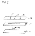
- FIG. 3 shows a method for producing a test strip according to FIG. 2.
- the device 1 consists of a cuvette 2 with an upper region 2a and a lower part 2b, in which a porous material 3 (matrix) is fixed.
- the matrix can be fixed by gluing the matrix to the cuvette or the matrix can be clamped in the cuvette.
- the cuvette is transparent to electromagnetic radiation, in particular light of the wavelength required to determine the marking.
- the sample and any auxiliary reagents are pipetted onto the porous material 3. The amount of the pipetted liquid should preferably not exceed the saturation volume of the porous material.
- the device After an incubation period, during which the distribution of the enzyme-labeled compound between the porous matrix and the liquid phase takes place, the device is passed through, for example with the aid of a needle 5, which is placed on the matrix Matrix filled through.
- the liquid phase contained in the pores of the matrix is almost completely displaced from the matrix by the solution.
- the displacing solution contains a substrate for enzyme labeling.
- the color development in the supernatant 4 (in the area 2a) is measured.
- the color development in the supernatant can also be directly measured kinetically without a second incubation period.
- the method according to the invention is carried out in a test strip according to FIG. 2.
- the test strip 10 contains a porous matrix 12 on a base film 11, which is at least partially covered by a film 13.
- This space preferably contains an air outlet opening 14.
- the inert plastic films known for test strips are suitable as the film.
- the sample liquid and any other reagents required for enzyme immunoassays are applied to the porous matrix 12. This can be done, for example, through a sample application opening 16.
- the vessel 15 is filled with a displacement liquid through the matrix 12.
- the displacement liquid can be filled in via the filling opening 17.
- the displacement liquid can already contain an enzyme substrate.
- the displacement liquid displaces the liquid phase from the matrix.
- the amount of enzyme labeling can be determined by measuring the color development in the cuvette-like space 15.
- one of the foils 13 and 11 is transparent and the other reflects light. Both foils are preferably transparent for a transmission photometric measurement. A measurement perpendicular to the drawing plane is also possible.
- a test strip according to FIG. 2 can be produced by gluing three foils and the solid matrix 12 together as shown in FIG. 3.
- the base film 11 is reflective or transparent
- film 18 has a punched-out part and is preferably thicker than films 11 and 13, preferably approximately as thick as the solid support 12.
- Film 13 has a sample application opening 16, a filling opening 17 and a vent opening 14 .
- the sample and the displacement liquid can be transported under pressure (tilting or pipetting) or by capillarity.
- the cuvette should have a layer thickness of less than 1 mm between 11 and 13.
- the embodiment of the method according to the invention in a test strip has the advantage that the reactions can be started by simple pipetting (also manually) and can also be evaluated visually.
- the reagents required to carry out enzyme immunoassays are known and can be used analogously in the method according to the invention.
- the enzyme label can also be replaced by a so-called direct label.
- Direct labels include, for example, dyes, fluorescent dyes, solid particles, in particular metal sols made of gold or non-metals or their oxides, such as selenium or tellurium.
- Such direct labels and the production of compounds labeled with them are known to the person skilled in the art. Even with these, it is not necessary to completely separate the liquid phase from the solid phase, and the tests using direct labels are more sensitive than when measured in a fleece, but the tests open up the possibility of even higher sensitivities when using enzyme labels.
- Fig. 1 shows a cross section through the center of a cuvette according to the invention.
- Fig. 2 shows a longitudinal section through the center of a test strip according to the invention.
- FIG. 3 illustrates the individual construction elements of a test strip according to the invention according to FIG. 2.
- a device according to FIG. 1 was used to carry out this test.
- the cuvette 2 had a volume of 1 ml.
- a fleece (80% polyester, 20% sulfite pulp, height 0.8 mm, length x width 1 cm x 0.5 cm, absorbency 800 ml / m2) served as the porous matrix Albumin was fixed according to DE-A-38 42 700. 200 mU of a conjugate of an antibody against albumin with ß-galactosidase are bound to the fleece via immune complex to the fixed albumin. 20 / ul albumin-containing sample liquid is pipetted onto the nonwoven material. After 5 min.
- the device is filled over the nonwoven material with a solution of chlorophenol red- ⁇ -D-galactoside (concentration 3 mmol, Hepes 50 mmol pH 7.5, amount 1 ml). After another 5 min. the absorbance at 570 nm in the supernatant is measured in a conventional photometer.
- the albumin concentration in urine samples of unknown albumin content can be determined.
- a device is used to carry out this test.
- T4 according to EP-A-0185372 is bound to a non-woven material (80% polyester, 20% sulfite pulp consolidated with etadurin).
- 5 / ul sample are pipetted with 15 / ul solution of a conjugate of AK against T4 and ß-galactosidase (3 U / ml) after preincubation of at least 5 min.
- the device is filled with a solution of chlorphenol red- ⁇ -D-galactoside (3 mM, 50 mmol Hepes pH 7.5) through the fleece by means of a needle placed on the fleece.
- the absorbance of the supernatant is after 5 min. measured to determine a calibration curve at various T4 concentrations. The results are shown in Table 2.
- This calibration curve can be used to determine the T4 content in serum or plasma.
- the test was carried out in a device according to FIG. 1.
- An antibody against mouse Fc is fixed to the nonwoven material (80% polyester, 20% sulfite pulp, etadurine) analogously to US-A-4803171.
- 425 / ul standard (composition analogous to the TSH concentrations of Table 3) or sample, 80 / ul antibody (mouse) against TSH (AK 1, 150 / ug per ml) and 80 / ul conjugate of sheep antibodies against TSH with ß-galactosidase (75 mU per ml) are 120 in a vessel min. pre-incubated for a long time. 20 / ul of this solution are pipetted onto the nonwoven material. After 5 min.
- the device is filled with a solution of chlorophenol red- ⁇ -D-galactoside (concentration 3 mM, 50 mM Hepes pH 7.5) through the fleece by means of a needle placed on the nonwoven material.
- the absorbance of the supernatant in area 2a is measured after 5 min at various TSH concentrations.
- the calibration curve in Table 3 was obtained from this.
- An unknown concentration of TSH in serum or plasma can be determined using the calibration curve.
Abstract
Description
- Gegenstand der Erfindung ist ein Verfahren zum Nachweis eines Analyten in einer Probenflüssigkeit durch einen heterogenen Enzymimmuntest und ein dafür geeignetes Testmittel.
- Enzymimmuntests ersetzen immer mehr die bisher üblichen Radioimmuno-Assays. Die Verwendung von Enzymen zur Markierung immunologisch reaktiver Verbindungen in Immuntests hat insbesondere sicherheitstechnische Vorteile. Ein solcher Enzymimmuntest ist beispielsweise in der US-A-4,446,232 beschrieben. Dieses Verfahren beruht darauf, daß durch den in einer Probe enthaltenen Analyten ein enzymgebundener Antikörper aus einer ersten Zone freigesetzt wird und die Enzymmarkierung in einer zweiten Zone durch Reaktion mit einem geeigneten Enzymsubstrat sichtbar gemacht wird. Die erste und die zweite Zone bestehen aus einem porösen Material. Es hat sich jedoch herausgestellt, daß dieses Verfahren den Nachteil hat, daß die entstandene Farbe nicht transmissionsphotometrisch gemessen werden kann, da das poröse Material der zweiten Zone kaum lichtdurchlässig ist.
- Aus der EP-A-0185372 ist ein Enzymimmuntest beschrieben, bei dem nach der immunologischen Reaktion die Flüssigkeit durch Zentrifugalkräfte aus der ersten Zone entfernt und einer Küvette zugeführt wird, in der dann die Farbreaktion gemessen wird. Die vollständige Entfernung der Flüssigkeit aus dem porösen Material der ersten Zone hat zwar den Vorteil, daß diese feste Phase dann die Detektion nicht stört, der zusätzlich durchzuführende Schritt der vollständigen Trennung der flüssigen von der festen Phase ist jedoch von Nachteil.
- Oft werden heterogene Emzymimmuntests auch in sogenannten Tubes durchgeführt. Die glatte Innenseite der Tubes dient dann als feste Phase. Diese Tube-Tests haben den Nachteil, daß nur wenig immunologisch aktive Substanz, über die der Analyt immobilisiert werden könnte, an der festen Phase immobilisiert ist. Außerdem sind die Inkubationszeiten für solche Tests sehr lang. Diese Tube-Tests haben auch den Nachteil, daß die Inkubationslösung nach Inkubation möglichst vollständig aus dem Tube entfernt werden muß, da im Tube verbleibende Reste die Messung beeinflussen.
- Aufgabe der vorliegenden Erfindung war es, die Nachteile des Standes der Technik zu vermeiden und insbesondere einfachere, schnellere bzw. empfindlichere Enzymimmuntests bereitzustellen.
- Diese Aufgabe wird durch die im Folgenden geschilderte Erfindung gelöst.
- Gegenstand der Erfindung ist ein Verfahren zum Nachweis eines Analyten in einer Probenflüssigkeit durch einen Enzymimmuntest, in dem eine Verteilung einer enzymmarkierten Verbindung zwischen einer festen und einer flüssigen Phase vorgenommen wird und die Menge an Enzymmarkierung in der flüssigen Phase außerhalb der festen Phase als ein Maß für die Konzentration des Analyten bestimmt wird, dadurch gekennzeichnet, daß die Messung in einem nicht porösen Formteil vorgenommen wird und die darin befindliche flüssige Phase mit der festen Phase in Kontakt steht.
- Das erfindungsgemäße Verfahren ist ein verbessertes Verfahren aufbauend auf den bislang bekannten sogenannten heterogenen Enzymimmuntests. Solche Immuntests sind beispielsweise in Mitteilungen der Deutschen Gesellschaft Für Klinische Chemie, 5, Seite 291-302 (1986) beschrieben. Grundlage all dieser Enzymimmuntests ist der Einsatz enzymmarkierter immunologisch aktiver Verbindungen und einer festen und einer flüssigen Phase. An der festen Phase sind immunologisch aktive Verbindungen immobilisiert, die direkt oder indirekt, beispielsweise über den zu bestimmenden Analyten, mit der enzymmarkierten Verbindung reagieren und diese immobilisieren können. Je nach Testführung ist die immobilisierte immunologisch aktive Verbindung eine Komponente einer immunologischen Reaktion mit dem Analyten oder eine Analytanaloges oder der Analyt. Durch geeignete Reaktionsführung wird erreicht, daß nur ein Teil der enzymmarkierten Verbindung immobilisiert wird oder bleibt, sodaß eine bestimmte Menge der enzymmarkierten Verbindung in der flüssigen Phase verbleibt und entweder die an der festen oder in der flüssigen Phase befindliche Menge an Enzymmarkierung ein Maß für die Konzentration des Analyten ist. Im erfindungsgemäßen Verfahren wird die Menge an Enzymmarkierung in der flüssigen Phase gemessen.
- Als Analyt kommen alle immunologisch aktiven Verbindungen in Frage. Solche Verbindungen sind Komponenten eines immunologischen Paares bzw. Komplexes, insbesondere Haptene, Antigene oder Antikörper.
- Unter einer Probenflüssigkeit werden insbesondere Körperflüssigkeiten oder davon abgeleitete Flüssigkeit verstanden. Zu Körperflüssigkeiten gehören beispielsweise Blut oder Harn. Von diesen Flüssigkeiten abgeleitete Probenflüssigkeiten sind beispielsweise solche, die durch Verdünnung oder Konzentrierung dieser Flüssigkeiten oder durch Zugabe oder Entfernung einzelner Komponenten der Flüssigkeit erhalten werden können, beispielsweise Serum oder Plasma.
- Als feste Phase sind im Sinne der Erfindung besonders alle Materialien mit einer großen wirksamen Oberfläche geeignet. Dazu gehören alle porösen Feststoffe, wenn sie gegenüber den oben genannten Flüssigkeiten saugfähig und von ihnen durchströmbar sind. Besonders geeignet sind Vliese und Gewebe. Geeignete Materialien für diese feste Phase sind dem Fachmann bekannt, beispielsweise aus der US-A-4,446,232. Es gehören dazu auch Zellulose und Gemische von Zellulose mit geeigneten Kunststoffen. Außerdem enthält diese feste Phase immobilisiert eine für die Durchführung des Enzymimmuntests geeignete immunologisch aktive Verbindung, die an der Immobilisierung der enzymmarkierten Verbindung beteiligt ist. Es hat sich herausgestellt, daß Tests mit derartigen festen Phasen für schnelle Immuntests besonders gut geeignet sind.
- Eine enzymmarkierte Verbindung ist eine chemische Verbindung aus einer Komponente einer immunologischen Reaktion mit einem Enzym. Die Komponente der immunologischen Reaktion wird ausgewählt aus der Gruppe Haptene, Antigene oder Antikörper und richtet sich nach der Art des Analyten und der Art der Testführung. Als Enzym kommen insbesondere Hydrolasen und Redoxreaktionen katalysierende Enzyme in Frage, für welche Substrate bereitstehen, die eine Messung der Enzymaktivität erlauben. Als besonders geeignet haben sich ß-Galaktosidase und Peroxidase erwiesen.
- Die Messung der Menge an Enzymmarkierung wird nach Verteilung der enzymmarkierten Verbindung zwischen der festen und der flüssigen Phase in einem nicht porösen Gefäß vorgenommen. Dazu wird mindestens ein Teil der flüssigen Phase aus den Poren der festen Phase entnommen und an deren Oberfläche gebracht. Die flüssige Phase steht dann immer noch in Kontakt mit der festen Phase. Bevorzugt wird die Probenflüssigkeit von einer weiteren Flüssigkeit aus den Poren der festen Phase verdrängt.
- Die Menge an Enzymmarkierung wird durch Verfolgung einer Reaktion des Enzyms mit einem geeigneten Substrat gemessen. Vorteil einer Enzymmarkierung und Durchführung einer Reaktion mit einem geeigneten Substrat ist die hohe Empfindlichkeit der resultierenden Tests. Durch die Enzymreaktion wird das Substrat in eine detektierbare Verbindung umgewandelt oder aus ihm eine detektierbare Verbindung freigesetzt oder es resultiert eine Verbindung, deren Menge in einer nachgeschalteten Reaktion detektierbar gemacht werden kann. Ist das Enzym eine Hydrolase, so können beispielsweise chromogene oder fluorogene Substrate verwendet werden, wie sie aus der EP-A-0156347 bekannt sind. Das Enzymsubstrat kann der flüssigen Phase beispielsweise in dem nicht porösen Formteil zugesetzt werden. Bevorzugt ist jedoch das Substrat in der Flüssigkeit enthalten, welche die Probenflüssigkeit aus dem porösen Material verdrängt. In diesem Fall wird mindestens soviel verdrängende Flüssigkeit verwendet, daß noch ein Teil der Flüssigkeit in dem nicht porösen Formteil austritt. Das Formteil kann beispielsweise ein Innenraum eines Gefäßes, wie einer Küvette, oder ein flacher Raum zwischen zwei Wänden sein. Ein Vorteil der Verwendung eines nicht porösen Formteils ist, daß die Empfindlichkeit deutlich erhöht ist. Dies gilt sowohl für den Fall einer reflexionsphotometrischen als auch einer transmissionsphotometrischen Messung.
- Es ist außerordentlich überraschend, daß das festphasengebundene Enzymlabel, welches in Flüssigkontakt zu einem nicht porösen Formteil steht, die Messung in diesem Formteil nicht stört, da ja auch das gebundene Enzym ständig detektierbares Material freisetzen kann, welches in den Formteil diffundieren kann.
- Dadurch, daß die Messung in einem nicht porösen Formteil vorgenommen wird, kann die Bestimmung transmissionsphotometrisch durchgeführt werden. Gegenüber den Teststreifen des Standes der Technik hat die Messung in einem nicht porösen Formteil den Vorteil, daß sie erheblich empfindlicher sein kann.
- Das erfindungsgemäße Verfahren ist insbesondere zum Nachweis eines Analyten in einer Küvette, wie in Fig. 1 gezeigt, oder einem Teststreifen, wie in Fig. 2 (Längsschnitt), geeignet. Fig. 3 zeigt ein Verfahren zur Herstellung eines Teststreifens gemäß Fig. 2.
- Anhand von Fig. 1 wird eine bevorzugte Ausführungsform beschrieben. Die Vorrichtung 1 besteht aus einer Küvette 2 mit einem oberen Bereich 2a und einem unteren Teil 2b, in dem ein poröses Material 3 (Matrix) fixiert ist. Die Fixierung kann über Kleben der Matrix an die Küvette erfolgen oder die Matrix kann in die Küvette eingeklemmt werden. Die Küvette ist mindestens im Teil 2a durchlässig für elektromagnetische Strahlung, insbesondere Licht der Wellenlänge, die zur Bestimmung der Markierung erforderlich ist. Die Probe und ggf. Hilfsreagenzien werden auf das poröse Material 3 aufpipettiert. Die Menge der aufpipettierten Flüssigkeit sollte bevorzugt das Sättigungsvolumen des porösen Materials nicht überschreiten.
- Nach einer Inkubationszeit, während der die Verteilung der enzymmarkierten Verbindung zwischen der porösen Matrix und der flüssigen Phase abläuft, wird die Vorrichtung beispielsweise mit Hilfe einer Nadel 5, die auf die Matrix aufgesetzt wird, durch die Matrix hindurch gefüllt. Die in den Poren der Matrix enthaltene flüssige Phase wird dabei praktisch vollständig aus der Matrix durch die Lösung verdrängt. Die verdrängende Lösung enthält ein Substrat für die Enzymmarkierung.
- Nach einer zweiten Inkubationszeit wird die Farbentwicklung im Überstand 4 (im Bereich 2a) gemessen. Die Farbentwicklung im Überstand kann jedoch auch ohne zweite Inkubationszeit direkt kinetisch gemessen werden.
- Ebenso möglich ist das teilweise Füllen der Vorrichtung durch das poröse Material hindurch mit einer Pufferlösung ohne Substrat unter anschließender Zugabe von Substratlösung in den Überstand.
- Gemäß einer weiteren Ausführungsform wird das erfindungsgemäße Verfahren in einem Teststreifen gemäß Fig. 2 durchgeführt. Der Teststreifen 10 enthält auf einer Grundfolie 11 eine poröse Matrix 12, die zumindest teilweise von einer Folie 13 überdeckt wird. Die Folie 13 bildet zusammen mit der Grundfolie 11 anschließend an die poröse Matrix 12 einen nicht porösen Raum 15. Dieser Raum enthält bevorzugt eine Luftaustrittsöffnung 14. Als Folie sind die für Teststreifen bekannten inerten Kunststoffolien geeignet.
- Zur Durchführung des erfindungsgemäßen Verfahrens mittels des Teststreifens 10 wird die Probenflüssigkeit, sowie ggf. für Enzymimmuntests erforderliche weitere Reagenzien auf die poröse Matrix 12 aufgegeben. Dies kann beispielsweise durch eine Probenaufgabeöffnung 16 geschehen. Nach einer Inkubationszeit wird das Gefäß 15 durch die Matrix 12 mit einer Verdrängungsflüssigkeit gefüllt. Dazu kann die Verdrängungsflüssigkeit über die Füllöffnung 17 eingefüllt werden. Die Verdrängungsflüssigkeit kann schon ein Enzymsubstrat enthalten. Die Verdrängungsflüssigkeit verdrängt die flüssige Phase aus der Matrix. Durch Messung der Farbentwicklung in dem küvettenartigen Raum 15 kann die Menge an Enzymmarkierung bestimmt werden. Für eine remissionsphotometrische Messung ist einer der Folien 13 und 11 transparent und die andere reflektiert Licht. Für eine transmissionsphotometrische Messung sind bevorzugt beide Folien transparent. Auch eine Messung senkrecht zur Zeichenebene ist möglich.
- Ein Teststreifen gemäß Figur 2 kann hergestellt werden, indem drei Folien und die feste Matrix 12 miteinander gemäß Fig. 3 verklebt werden.
- Hierbei ist die Grundfolie 11 reflektierend oder transparent, Folie 18 hat ein ausgestanztes Teil und ist bevorzugt dicker als die Folien 11 und 13, bevorzugt etwa so dick wie der feste Träger 12. Folie 13 hat eine Probenaufgabeöffnung 16, eine Füllöffnung 17 und eine Entlüftungsöffnung 14.
- Der Transport der Probe und der Verdrängungsflüssigkeit kann unter Druck (Kippen oder Pipettieren) oder mittels Kapillarität erfolgen. Die Küvette sollte im letzterwähnten Fall zwischen 11 und 13 eine Schichtdicke von kleiner als 1 mm aufweisen. Die Ausführungsform des erfindungsgemäßen Verfahrens in einem Teststreifen hat den Vorteil, daß die Reaktionen durch einfaches Pipettieren (auch manuell) gestartet werden können und auch visuell auswertbar sind.
- Die oben genannten Enzymimmuntests können folgendermaßen zur Durchführung des erfindungsgemäßen Verfahrens angepaßt werden:
- 1. Verdrängungstest
Ein Konjugat aus einem Enzym und einem Immunpartner des Analyten ist über einen Immunkomplex mit einem an das poröse Material immobilisierten Analyten oder Analytanalogen an das poröse Material gebunden. Die Inkubation der porösen Matrix mit der Probe führt zur teilweisen Ablösung des Konjugats vom porösen Material in die flüssige Phase und Bindung an den frei beweglichen Analyten. Nach Verdrängung wird die Menge des Konjugat-Analyt-Komplexes in der flüssigen Phase gemessen. - 2. IEMA
Die den Analyten enthaltende Probe und ein gegen den Analyten gerichtetes Enzymkonjugat werden nach eventueller Vorinkubation auf die poröse Matrix aufpipettiert. Die Matrix enthält Bindungsstellen für das freie Konjugat. Nach der Verdrängung der flüssigen Phase wird der Komplex aus Analyt und Konjugat in der flüssigen Phase vermessen. - 3. Kompetitiver Test
An das poröse Material ist ein Immunpartner des Analyten im Unterschuß fixiert. Die den Analyten enthaltende Probe und ein Konjugat aus Analyt oder Analytanalogem und einem Enzym in bekannter Konzentration werden auf das poröse Material aufpipettiert. Analyt und Konjugat konkurrieren um die Bindungsstellen des porösen Materials. Nach der Verdrängung wird die Menge an Konjugat in der flüssigen Phase gemessen.
In einer weiteren Ausführungsform eines kompetitiven Testes sind an das poröse Material Antikörper gegen den Proteinteil eines Polyhaptens fixiert. Ein Polyhapten ist beispielsweise ein Konjugat aus T₄ und IgG. Das Polyhapten (mit gebundener Probe und in bekannter Konzentration), die Probe und das Konjugat aus Antikörper (AK) und Enzym (bekannte Konzentration) werden auf das poröse Material pipettiert. Polyhapten und Probe konkurrieren um das Konjugat. Polyhapten und Polyhaptenkonjugatkomplex werden an das poröse Material gebunden. Der Komplex Probe/Konjugat in der flüssigen Phase wird nach Verdrängung gemessen. - 4. Sandwich-Test
An das poröse Material sind Antikörper (AK 1) gegen den Analyten gebunden. Die den Analyten enthaltende Probe und ein Konjugat aus Enzym und einem Antikörper gegen den Analyten (bekannte Konzentration) werden auf das poröse Material pipettiert. Nach der Inkubationszeit und Verdrängung wird das nicht gebundene Konjugat in der flüssigen Phase bestimmt. Auf dem porösen Material kann ebenso ein Antikörper AK 2 gegen einen Antikörper AK 1 gebunden sein. Inkubiert wird dann mit der Probe, dem Konjugat und AK 1. - Die zur Durchführung von Enzymimmuntests erforderlichen Reagenzien sind bekannt und können im erfindungsgemäßen Verfahren analog eingesetzt werden.
- Insbesondere bei Durchführung eines Immuntests auf einem oben beschriebenen Teststreifen kann die Enzymmarkierung auch durch einen sogenannten Direktlabel ersetzt werden. Zu den Direktlabeln gehören beispielsweise Farbstoffe, Fluoreszenzfarbstoffe, Feststoffpartikel, insbesondere Metallsole aus Gold oder aus Nichtmetallen oder deren Oxide, wie Selen oder Tellur. Solche Direktlabel und die Herstellung damit markierter Verbindungen sind dem Fachmann bekannt. Zwar ist es auch bei diesen nicht erforderlich, die flüssige Phase von der festen Phase vollständig abzutrennen, und sind die Tests mittels Direktlabeln empfindlicher als bei Messung in einem Vlies, jedoch eröffnen die Tests bei Verwendung von Enzymlabeln die Möglichkeit noch höherer Empfindlichkeiten.
- Fig. 1 zeigt einen Querschnitt durch die Mitte einer erfindungsgemäßen Küvette.
- Fig. 2 zeigt einen Längsschnitt durch die Mitte eines erfindungsgemäßen Teststreifens.
- Fig. 3 verdeutlicht die einzelnen Konstruktionselemente eines erfindungsgemäßen Teststreifens gemäß Fig. 2.
- Die Erfindung wird durch die folgenden Beispiele weiter erläutert:
- Zur Durchführung dieses Tests wurde eine Vorrichtung gemäß Fig. 1 benutzt. Die Küvette 2 hatte ein Volumen von 1 ml. Als poröse Matrix diente ein Vlies (80% Polyester, 20% Sulfitzellstoff,Höhe 0,8 mm, Länge x Breite 1 cm x 0,5 cm, Saugfähigkeit 800 ml/m²) an welchem Albumin gemäß DE-A-38 42 700 fixiert wurde. 200 mU eines Konjugates eines Antikörpers gegen Albumin mit ß-Galaktosidase sind an das Vlies über Immunkomplex an das fixierte Albumin gebunden. 20 /ul albuminhaltige Probenflüssigkeit wird auf das Vliesmaterial pipettiert. Nach 5 min. wird die Vorrichtung über das Vliesmaterial mit einer Lösung von Chlorphenolrot-ß-D-Galaktosid (Konzentration 3 mmol, Hepes 50 mmol pH 7,5, Menge 1 ml) gefüllt. Nach weiteren 5 min. wird die Extinktion bei 570 nm im Überstand in einem herkömmlichen Photometer gemessen.
- Zur quantitativen Bestimmung von Albumin in Harn wird eine Eichkurve mit Probenflüssigkeiten bekannten Albumingehalts aufgenommen. Das Ergebnis zeigt Tabelle 1.

- Mit Hilfe dieser Eichkurve kann die Albuminkonzentration in Harn-Proben unbekannten Albumingehalts ermittelt werden.
- Zur Durchführung dieses Tests wird eine Vorrichtung gemäß Fig. 1 benutzt. An ein Vliesmaterial (80% Polyester, 20% Sulfitzellstoff mit Etadurin verfestigt) ist T4 gemäß EP-A-0185372 gebunden. 5 /ul Probe werden mit 15 /ul Lösung eines Konjugats aus AK gegen T4 und ß-Galaktosidase (3 U/ml) nach Vorinkubation von mindestens 5 Min. auf das Vlies pipettiert. Nach 5 min. wir die Vorrichtung mittels einer auf das Vlies aufgesetzten Nadel durch das Vlies mit einer Lösung von Chlorphenolrot-ß-D-Galaktosid (3 mM, 50 mmol Hepes pH 7,5) gefüllt. Die Extinktion des Überstands wird nach 5 min. zur Ermittlung einer Eichkurve bei verschiedenen T4-Konzentrationen gemessen. Die Ergebnisse sind in Tabelle 2 enthalten.

- Mit Hilfe dieser Eichkurve kann der T4-Gehalt in Serum oder Plasma bestimmt werden.
- Der Test wurde in einer Vorrichtung gemäß Fig. 1 durchgeführt. An das Vliesmaterial (80% Polyester, 20% Sulfitzellstoff, Etadurin) ist ein Antikörper gegen Maus Fc analog US-A-4803171 fixiert. 425 /ul Standard (Zusammensetzung analog der TSH Konzentrationen der Tabelle 3) bzw. Probe, 80 /ul Antikörper (Maus) gegen TSH (AK 1, 150 /ug pro ml) und 80 /ul Konjugat von Schafantikörper gegen TSH mit ß-Galaktosidase (75 mU pro ml) werden in einem Gefäß 120 min. lang vorinkubiert. 20 /ul dieser Lösung werden auf das Vliesmaterial pipettiert. Nach 5 min. wird die Vorrichtung mittels einer auf das Vliesmaterial aufgesetzten Nadel durch das Vlies mit einer Lösung von Chlorphenolrot-ß-D-Galaktosid (Konzentration 3 mM, 50 mM Hepes pH 7,5) gefüllt. Die Extinktion des Überstands im Bereich 2a wird nach 5 Min. bei verschiedenen TSH-Konzentrationen gemessen. Daraus wurde die Eichkurve der Tabelle 3 erhalten.

- Mit Hilfe der Eichkurve kann eine unbekannte Konzentration an TSH in Serum oder Plasma bestimmt werden.
Claims (10)
- Verfahren zum Nachweis eines Analyten in einer Probenflüssigkeit durch einen Enzymimmuntest, in dem eine Verteilung einer enzymmarkierten Verbindung zwischen einer festen und einer flüssigen Phase vorgenommen und die Menge an Enzymmarkierung in der flüssigen Phase außerhalb der festen Phase als Maß für die Konzentration des Analyten bestimmt wird, dadurch gekennzeichnet, daß die Messung in einem nicht porösen Formteil vorgenommen wird und die darin befindliche flüssige Phase mit der festen Phase in Kontakt steht.
- Verfahren gemäß Anspruch 1, dadurch gekennzeichnet, daß die feste Phase ein Material mit einer großen Oberfläche ist.
- Verfahren gemäß einem der Ansprüche 1 und 2, dadurch gekennzeichnet, daß die feste Phase porös ist.
- Verfahren gemäß einem der Ansprüche 1-3, dadurch gekennzeichnet, daß die Probenflüssigkeit die feste Phase durchströmt.
- Verfahren gemäß Anspruch 4, dadurch gekennzeichnet, daß die Probenflüssigkeit von einer Flüssigkeit verdrängt wird, die ein für das Enzym geeignetes Substrat enthält.
- Verfahren gemäß einem der Ansprüche 1 und 2, dadurch gekennzeichnet, daß die feste Phase aus kleinen Partikeln besteht.
- Verfahren gemäß einem der Ansprüche 1-6, dadurch gekennzeichnet, daß die Menge an Enzymmarkierung über die Menge an durch das Enzym umgesetztem Enzymsubstrat in einer an die feste Phase direkt anschließenden Küvette bestimmt wird.
- Verfahren gemäß Anspruch 7, dadurch gekennzeichnet, daß die Bestimmung transmissionsphotometrisch geführt wird.
- Küvette zur Durchführung des Verfahrens gemäß einem der Ansprüche l bis 8, dadurch gekennzeichnet, daß sie am Boden eine poröse durchströmbare Matrix aufweist.
- Teststreifen, enthaltend auf einer Grundfolie eine durchströmbare, poröse Matrix, mindestens einen Streifenteil, durch den eine Flüssigkeit auf die Matrix aufgegeben werden kann und einen nicht porösen Formteil, dessen Raum mit der Matrix in Kontakt steht, wobei der Streifenteil, die Matrix und der nicht poröse Formteil so auf der Grundfolie angeordnet sind, daß sie in dieser Reihenfolge zunächst von der Probe und anschließend von einer Verdrängungsflüssigkeit durchströmt werden können.
Applications Claiming Priority (2)
| Application Number | Priority Date | Filing Date | Title |
|---|---|---|---|
| DE3941150 | 1989-12-13 | ||
| DE3941150A DE3941150A1 (de) | 1989-12-13 | 1989-12-13 | Verfahren zur bestimmung eines analyten |
Publications (3)
| Publication Number | Publication Date |
|---|---|
| EP0432631A2 true EP0432631A2 (de) | 1991-06-19 |
| EP0432631A3 EP0432631A3 (en) | 1992-03-04 |
| EP0432631B1 EP0432631B1 (de) | 1995-08-30 |
Family
ID=6395380
Family Applications (1)
| Application Number | Title | Priority Date | Filing Date |
|---|---|---|---|
| EP90123380A Expired - Lifetime EP0432631B1 (de) | 1989-12-13 | 1990-12-06 | Verfahren zur Bestimmung eines Analyten |
Country Status (6)
| Country | Link |
|---|---|
| US (1) | US5296356A (de) |
| EP (1) | EP0432631B1 (de) |
| JP (1) | JPH03279861A (de) |
| AT (1) | ATE127230T1 (de) |
| DE (2) | DE3941150A1 (de) |
| ES (1) | ES2078933T3 (de) |
Cited By (1)
| Publication number | Priority date | Publication date | Assignee | Title |
|---|---|---|---|---|
| WO1994002850A1 (en) * | 1992-07-21 | 1994-02-03 | Medix Biotech, Inc. | Transparent assay test devices and methods |
Families Citing this family (2)
| Publication number | Priority date | Publication date | Assignee | Title |
|---|---|---|---|---|
| FI92882C (fi) * | 1992-12-29 | 1995-01-10 | Medix Biochemica Ab Oy | Kertakäyttöinen testiliuska ja menetelmä sen valmistamiseksi |
| US5580790A (en) * | 1994-10-21 | 1996-12-03 | Chiron Corporation | Method and apparatus for processing fluids |
Citations (5)
| Publication number | Priority date | Publication date | Assignee | Title |
|---|---|---|---|---|
| EP0054675A1 (de) * | 1980-12-23 | 1982-06-30 | Roche Diagnostics GmbH | Verfahren zur Enzymimmunobestimmung in heterogener Phase |
| FR2514511A1 (fr) * | 1981-10-13 | 1983-04-15 | Liotta Lance | Dispositif et procede pour determiner la presence d'antigenes |
| JPS5956166A (ja) * | 1982-09-27 | 1984-03-31 | Konishiroku Photo Ind Co Ltd | 分析容器 |
| EP0286371A2 (de) * | 1987-04-07 | 1988-10-12 | Syntex (U.S.A.) Inc. | Einrichtung für Immunoassay |
| US4786594A (en) * | 1986-05-14 | 1988-11-22 | Syntex (U.S.A.) Inc. | Enzyme immunoassay |
Family Cites Families (5)
| Publication number | Priority date | Publication date | Assignee | Title |
|---|---|---|---|---|
| US4426451A (en) * | 1981-01-28 | 1984-01-17 | Eastman Kodak Company | Multi-zoned reaction vessel having pressure-actuatable control means between zones |
| SE8305704D0 (sv) * | 1983-10-18 | 1983-10-18 | Leo Ab | Cuvette |
| US4729875A (en) * | 1986-06-26 | 1988-03-08 | Allelix Inc. | Device for performing immunochemical assays |
| ATE225509T1 (de) * | 1987-04-27 | 2002-10-15 | Inverness Medical Switzerland | Testgerät zur durchführung von spezifischen bindungsprüfungen |
| US4978504A (en) * | 1988-02-09 | 1990-12-18 | Nason Frederic L | Specimen test unit |
-
1989
- 1989-12-13 DE DE3941150A patent/DE3941150A1/de not_active Withdrawn
-
1990
- 1990-12-06 DE DE59009576T patent/DE59009576D1/de not_active Expired - Fee Related
- 1990-12-06 ES ES90123380T patent/ES2078933T3/es not_active Expired - Lifetime
- 1990-12-06 AT AT90123380T patent/ATE127230T1/de not_active IP Right Cessation
- 1990-12-06 EP EP90123380A patent/EP0432631B1/de not_active Expired - Lifetime
- 1990-12-12 US US07/626,257 patent/US5296356A/en not_active Expired - Fee Related
- 1990-12-13 JP JP2410718A patent/JPH03279861A/ja active Pending
Patent Citations (5)
| Publication number | Priority date | Publication date | Assignee | Title |
|---|---|---|---|---|
| EP0054675A1 (de) * | 1980-12-23 | 1982-06-30 | Roche Diagnostics GmbH | Verfahren zur Enzymimmunobestimmung in heterogener Phase |
| FR2514511A1 (fr) * | 1981-10-13 | 1983-04-15 | Liotta Lance | Dispositif et procede pour determiner la presence d'antigenes |
| JPS5956166A (ja) * | 1982-09-27 | 1984-03-31 | Konishiroku Photo Ind Co Ltd | 分析容器 |
| US4786594A (en) * | 1986-05-14 | 1988-11-22 | Syntex (U.S.A.) Inc. | Enzyme immunoassay |
| EP0286371A2 (de) * | 1987-04-07 | 1988-10-12 | Syntex (U.S.A.) Inc. | Einrichtung für Immunoassay |
Non-Patent Citations (1)
| Title |
|---|
| PATENT ABSTRACTS OF JAPAN Band 8, Nr. 159 (P-289)(1596), 24 Juli 1984; & JP - A - 59056166 (KONISHIROKU SHASHIN KOGOYO K.K.) 31.03.1984 * |
Cited By (1)
| Publication number | Priority date | Publication date | Assignee | Title |
|---|---|---|---|---|
| WO1994002850A1 (en) * | 1992-07-21 | 1994-02-03 | Medix Biotech, Inc. | Transparent assay test devices and methods |
Also Published As
| Publication number | Publication date |
|---|---|
| DE59009576D1 (de) | 1995-10-05 |
| US5296356A (en) | 1994-03-22 |
| EP0432631A3 (en) | 1992-03-04 |
| EP0432631B1 (de) | 1995-08-30 |
| JPH03279861A (ja) | 1991-12-11 |
| ATE127230T1 (de) | 1995-09-15 |
| ES2078933T3 (es) | 1996-01-01 |
| DE3941150A1 (de) | 1991-06-20 |
Similar Documents
| Publication | Publication Date | Title |
|---|---|---|
| DE602004013147T2 (de) | Nativer analyt als referenz in lateralfluss-assays | |
| DE69128618T3 (de) | Vorrichtung und verfahren zur ausführung von immunoassays | |
| DE69630295T2 (de) | Diagnostische vorrichtung und verfahren | |
| DE69126248T3 (de) | Immunchromatographischer assay und verfahren zur nutzung desselben | |
| DE3237046C2 (de) | ||
| DE69233290T2 (de) | Testvorrichtung die einander entgegenstellbare Elemente enthält | |
| EP1759209B1 (de) | Verfahren zur erhöhung des dynamischen messbereichs von auf spezifischen bindereaktionen basierenden, insbesondere immunologischen testelementen | |
| DE69823014T2 (de) | Biochemisches und immunochemisches testgerät | |
| EP1808696B1 (de) | Immunologisches Testelement mit verbesserter Kontrollzone | |
| DE60037268T2 (de) | Verfahren zur durchführung magnetischer chromatographischer assays | |
| DE69837257T2 (de) | Analytische versuchsanordnung für auf membranen basierende versuche | |
| DE60024448T2 (de) | System zur elektrochemischen quantitativen analyse von analyten in einer festphase | |
| DE69830675T2 (de) | Chromatographische Vorrichtung und Verfahren zum Nachweis eines Analyts in einer Vollblutprobe | |
| DE4229591C1 (de) | Immunologisches Verfahren zur Bestimmung eines Analyten | |
| EP0407904B1 (de) | Verfahren zur Bestimmung eines Analyten | |
| EP0290921B1 (de) | Testträger für die Analyse einer Probenflüssigkeit und Verfahren zu seiner Herstellung | |
| DE60117699T2 (de) | Analyseverfahren durch spezifisches binden und vorrichtung, die das verfahren einsetzt | |
| EP0553770A2 (de) | Analyseelement für Immunoassays | |
| EP0267519A2 (de) | Testträger zur analytischen Bestimmung eines Bestandteils einer Körperflüssigkeit | |
| WO1999005525A1 (de) | Verwendung von kontrollflächen zur detektion von störproben in einem nachweisverfahren | |
| DE212013000068U1 (de) | Vorrichtung zur Bestimmung wenigstens eines Analyten, der in einer flüssigen Probe enthalten sein kann | |
| EP0268978B1 (de) | Verfahren und Testträger zur Bestimmung eines Analyten | |
| WO2000042434A1 (de) | Verfahren und vorrichtung zur bestimmung eines analyten | |
| EP0296366B1 (de) | Heterogenes Immunoassay | |
| DE69819833T2 (de) | Verfahren und Teststreife zur Veringerung des Harnstoffeinflusses bei immunochromatographischen Messungen unter Verwendung von Urinproben |
Legal Events
| Date | Code | Title | Description |
|---|---|---|---|
| PUAI | Public reference made under article 153(3) epc to a published international application that has entered the european phase |
Free format text: ORIGINAL CODE: 0009012 |
|
| 17P | Request for examination filed |
Effective date: 19901206 |
|
| AK | Designated contracting states |
Kind code of ref document: A2 Designated state(s): AT BE CH DE DK ES FR GB GR IT LI LU NL SE |
|
| PUAL | Search report despatched |
Free format text: ORIGINAL CODE: 0009013 |
|
| AK | Designated contracting states |
Kind code of ref document: A3 Designated state(s): AT BE CH DE DK ES FR GB GR IT LI LU NL SE |
|
| 17Q | First examination report despatched |
Effective date: 19940308 |
|
| GRAA | (expected) grant |
Free format text: ORIGINAL CODE: 0009210 |
|
| AK | Designated contracting states |
Kind code of ref document: B1 Designated state(s): AT BE CH DE DK ES FR GB GR IT LI LU NL SE |
|
| PG25 | Lapsed in a contracting state [announced via postgrant information from national office to epo] |
Ref country code: NL Free format text: LAPSE BECAUSE OF NON-PAYMENT OF DUE FEES Effective date: 19950830 Ref country code: GR Free format text: LAPSE BECAUSE OF FAILURE TO SUBMIT A TRANSLATION OF THE DESCRIPTION OR TO PAY THE FEE WITHIN THE PRESCRIBED TIME-LIMIT Effective date: 19950830 Ref country code: DK Effective date: 19950830 Ref country code: BE Effective date: 19950830 |
|
| REF | Corresponds to: |
Ref document number: 127230 Country of ref document: AT Date of ref document: 19950915 Kind code of ref document: T |
|
| REF | Corresponds to: |
Ref document number: 59009576 Country of ref document: DE Date of ref document: 19951005 |
|
| ET | Fr: translation filed | ||
| GBT | Gb: translation of ep patent filed (gb section 77(6)(a)/1977) |
Effective date: 19951002 |
|
| ITF | It: translation for a ep patent filed |
Owner name: STUDIO CONS. BREVETTUALE S.R.L. |
|
| PG25 | Lapsed in a contracting state [announced via postgrant information from national office to epo] |
Ref country code: SE Effective date: 19951130 |
|
| PG25 | Lapsed in a contracting state [announced via postgrant information from national office to epo] |
Ref country code: AT Effective date: 19951206 |
|
| PG25 | Lapsed in a contracting state [announced via postgrant information from national office to epo] |
Ref country code: LU Free format text: LAPSE BECAUSE OF NON-PAYMENT OF DUE FEES Effective date: 19951231 |
|
| REG | Reference to a national code |
Ref country code: ES Ref legal event code: FG2A Ref document number: 2078933 Country of ref document: ES Kind code of ref document: T3 |
|
| NLV1 | Nl: lapsed or annulled due to failure to fulfill the requirements of art. 29p and 29m of the patents act | ||
| PLBE | No opposition filed within time limit |
Free format text: ORIGINAL CODE: 0009261 |
|
| STAA | Information on the status of an ep patent application or granted ep patent |
Free format text: STATUS: NO OPPOSITION FILED WITHIN TIME LIMIT |
|
| 26N | No opposition filed | ||
| PGFP | Annual fee paid to national office [announced via postgrant information from national office to epo] |
Ref country code: GB Payment date: 19991201 Year of fee payment: 10 |
|
| PGFP | Annual fee paid to national office [announced via postgrant information from national office to epo] |
Ref country code: DE Payment date: 19991207 Year of fee payment: 10 |
|
| PGFP | Annual fee paid to national office [announced via postgrant information from national office to epo] |
Ref country code: FR Payment date: 19991208 Year of fee payment: 10 |
|
| PGFP | Annual fee paid to national office [announced via postgrant information from national office to epo] |
Ref country code: CH Payment date: 19991210 Year of fee payment: 10 |
|
| PGFP | Annual fee paid to national office [announced via postgrant information from national office to epo] |
Ref country code: ES Payment date: 19991221 Year of fee payment: 10 |
|
| PG25 | Lapsed in a contracting state [announced via postgrant information from national office to epo] |
Ref country code: GB Free format text: LAPSE BECAUSE OF NON-PAYMENT OF DUE FEES Effective date: 20001206 |
|
| PG25 | Lapsed in a contracting state [announced via postgrant information from national office to epo] |
Ref country code: LI Free format text: LAPSE BECAUSE OF NON-PAYMENT OF DUE FEES Effective date: 20001231 Ref country code: CH Free format text: LAPSE BECAUSE OF NON-PAYMENT OF DUE FEES Effective date: 20001231 |
|
| GBPC | Gb: european patent ceased through non-payment of renewal fee |
Effective date: 20001206 |
|
| REG | Reference to a national code |
Ref country code: CH Ref legal event code: PL |
|
| PG25 | Lapsed in a contracting state [announced via postgrant information from national office to epo] |
Ref country code: FR Free format text: LAPSE BECAUSE OF NON-PAYMENT OF DUE FEES Effective date: 20010831 |
|
| REG | Reference to a national code |
Ref country code: FR Ref legal event code: ST |
|
| PG25 | Lapsed in a contracting state [announced via postgrant information from national office to epo] |
Ref country code: DE Free format text: LAPSE BECAUSE OF NON-PAYMENT OF DUE FEES Effective date: 20011002 |
|
| PG25 | Lapsed in a contracting state [announced via postgrant information from national office to epo] |
Ref country code: ES Free format text: LAPSE BECAUSE OF NON-PAYMENT OF DUE FEES Effective date: 20011207 |
|
| REG | Reference to a national code |
Ref country code: ES Ref legal event code: FD2A Effective date: 20020112 |
|
| PG25 | Lapsed in a contracting state [announced via postgrant information from national office to epo] |
Ref country code: IT Free format text: LAPSE BECAUSE OF NON-PAYMENT OF DUE FEES;WARNING: LAPSES OF ITALIAN PATENTS WITH EFFECTIVE DATE BEFORE 2007 MAY HAVE OCCURRED AT ANY TIME BEFORE 2007. THE CORRECT EFFECTIVE DATE MAY BE DIFFERENT FROM THE ONE RECORDED. Effective date: 20051206 |