EP0365837A2 - Molécules d'adhésion intercellulaire et leurs ligands de liaison - Google Patents
Molécules d'adhésion intercellulaire et leurs ligands de liaison Download PDFInfo
- Publication number
- EP0365837A2 EP0365837A2 EP89117620A EP89117620A EP0365837A2 EP 0365837 A2 EP0365837 A2 EP 0365837A2 EP 89117620 A EP89117620 A EP 89117620A EP 89117620 A EP89117620 A EP 89117620A EP 0365837 A2 EP0365837 A2 EP 0365837A2
- Authority
- EP
- European Patent Office
- Prior art keywords
- icam
- cells
- lfa
- binding
- antibody
- Prior art date
- Legal status (The legal status is an assumption and is not a legal conclusion. Google has not performed a legal analysis and makes no representation as to the accuracy of the status listed.)
- Granted
Links
- 238000009739 binding Methods 0.000 title claims abstract description 153
- 230000027455 binding Effects 0.000 title claims abstract description 152
- 239000003446 ligand Substances 0.000 title claims description 36
- 102000016289 Cell Adhesion Molecules Human genes 0.000 title abstract description 6
- 108010067225 Cell Adhesion Molecules Proteins 0.000 title abstract description 6
- 238000000034 method Methods 0.000 claims abstract description 93
- 206010061218 Inflammation Diseases 0.000 claims abstract description 48
- 230000004054 inflammatory process Effects 0.000 claims abstract description 48
- 108010064593 Intercellular Adhesion Molecule-1 Proteins 0.000 claims description 641
- 210000004027 cell Anatomy 0.000 claims description 355
- 108010064548 Lymphocyte Function-Associated Antigen-1 Proteins 0.000 claims description 178
- 239000012634 fragment Substances 0.000 claims description 60
- 108060003951 Immunoglobulin Proteins 0.000 claims description 33
- 102000018358 immunoglobulin Human genes 0.000 claims description 33
- 239000005557 antagonist Substances 0.000 claims description 26
- 238000002360 preparation method Methods 0.000 claims description 25
- 230000004044 response Effects 0.000 claims description 22
- 229940121363 anti-inflammatory agent Drugs 0.000 claims description 21
- 239000002260 anti-inflammatory agent Substances 0.000 claims description 21
- PMATZTZNYRCHOR-CGLBZJNRSA-N Cyclosporin A Chemical compound CC[C@@H]1NC(=O)[C@H]([C@H](O)[C@H](C)C\C=C\C)N(C)C(=O)[C@H](C(C)C)N(C)C(=O)[C@H](CC(C)C)N(C)C(=O)[C@H](CC(C)C)N(C)C(=O)[C@@H](C)NC(=O)[C@H](C)NC(=O)[C@H](CC(C)C)N(C)C(=O)[C@H](C(C)C)NC(=O)[C@H](CC(C)C)N(C)C(=O)CN(C)C1=O PMATZTZNYRCHOR-CGLBZJNRSA-N 0.000 claims description 20
- 239000003795 chemical substances by application Substances 0.000 claims description 20
- 239000000203 mixture Substances 0.000 claims description 18
- 229930105110 Cyclosporin A Natural products 0.000 claims description 17
- 108010036949 Cyclosporine Proteins 0.000 claims description 17
- 229960001265 ciclosporin Drugs 0.000 claims description 17
- 210000004881 tumor cell Anatomy 0.000 claims description 16
- LMEKQMALGUDUQG-UHFFFAOYSA-N azathioprine Chemical compound CN1C=NC([N+]([O-])=O)=C1SC1=NC=NC2=C1NC=N2 LMEKQMALGUDUQG-UHFFFAOYSA-N 0.000 claims description 12
- 230000001086 cytosolic effect Effects 0.000 claims description 12
- 239000001963 growth medium Substances 0.000 claims description 11
- 230000035886 specific defense system Effects 0.000 claims description 11
- 239000003018 immunosuppressive agent Substances 0.000 claims description 10
- 229960002170 azathioprine Drugs 0.000 claims description 8
- 229940125721 immunosuppressive agent Drugs 0.000 claims description 8
- 239000013598 vector Substances 0.000 claims description 8
- 239000000126 substance Substances 0.000 claims description 7
- 206010027476 Metastases Diseases 0.000 claims description 6
- UREBDLICKHMUKA-CXSFZGCWSA-N dexamethasone Chemical compound C1CC2=CC(=O)C=C[C@]2(C)[C@]2(F)[C@@H]1[C@@H]1C[C@@H](C)[C@@](C(=O)CO)(O)[C@@]1(C)C[C@@H]2O UREBDLICKHMUKA-CXSFZGCWSA-N 0.000 claims description 6
- 229960003957 dexamethasone Drugs 0.000 claims description 6
- 230000009401 metastasis Effects 0.000 claims description 6
- 108020004511 Recombinant DNA Proteins 0.000 claims description 5
- 230000012010 growth Effects 0.000 claims description 5
- 230000035781 nonspecific defense system Effects 0.000 claims description 5
- 102000053602 DNA Human genes 0.000 claims description 4
- 230000005012 migration Effects 0.000 claims description 4
- 238000013508 migration Methods 0.000 claims description 4
- 208000019691 hematopoietic and lymphoid cell neoplasm Diseases 0.000 claims description 3
- 102100037877 Intercellular adhesion molecule 1 Human genes 0.000 claims 33
- 102100022339 Integrin alpha-L Human genes 0.000 claims 7
- 239000008194 pharmaceutical composition Substances 0.000 claims 7
- 230000003042 antagnostic effect Effects 0.000 claims 1
- 239000012050 conventional carrier Substances 0.000 claims 1
- 229940124589 immunosuppressive drug Drugs 0.000 claims 1
- 244000005700 microbiome Species 0.000 claims 1
- 239000000546 pharmaceutical excipient Substances 0.000 claims 1
- 210000004698 lymphocyte Anatomy 0.000 abstract description 38
- 230000001413 cellular effect Effects 0.000 abstract description 31
- 230000008569 process Effects 0.000 abstract description 13
- 239000000758 substrate Substances 0.000 abstract description 7
- 238000007423 screening assay Methods 0.000 abstract description 5
- 102000015271 Intercellular Adhesion Molecule-1 Human genes 0.000 description 587
- 230000014509 gene expression Effects 0.000 description 79
- 238000004220 aggregation Methods 0.000 description 76
- 230000002776 aggregation Effects 0.000 description 73
- 210000002510 keratinocyte Anatomy 0.000 description 62
- 230000000694 effects Effects 0.000 description 52
- 108090000623 proteins and genes Proteins 0.000 description 50
- 239000002299 complementary DNA Substances 0.000 description 41
- 238000003556 assay Methods 0.000 description 38
- PHEDXBVPIONUQT-RGYGYFBISA-N phorbol 13-acetate 12-myristate Chemical compound C([C@]1(O)C(=O)C(C)=C[C@H]1[C@@]1(O)[C@H](C)[C@H]2OC(=O)CCCCCCCCCCCCC)C(CO)=C[C@H]1[C@H]1[C@]2(OC(C)=O)C1(C)C PHEDXBVPIONUQT-RGYGYFBISA-N 0.000 description 37
- 210000001519 tissue Anatomy 0.000 description 37
- 210000002889 endothelial cell Anatomy 0.000 description 36
- 238000011282 treatment Methods 0.000 description 36
- 108020004414 DNA Proteins 0.000 description 34
- 108091034117 Oligonucleotide Proteins 0.000 description 34
- 239000000427 antigen Substances 0.000 description 34
- 108091007433 antigens Proteins 0.000 description 34
- 102000036639 antigens Human genes 0.000 description 34
- 102000006354 HLA-DR Antigens Human genes 0.000 description 31
- 108010058597 HLA-DR Antigens Proteins 0.000 description 31
- 210000004408 hybridoma Anatomy 0.000 description 31
- 108090000765 processed proteins & peptides Proteins 0.000 description 31
- 230000001419 dependent effect Effects 0.000 description 30
- 238000010186 staining Methods 0.000 description 28
- 239000012528 membrane Substances 0.000 description 27
- 235000018102 proteins Nutrition 0.000 description 27
- 102000004169 proteins and genes Human genes 0.000 description 27
- 210000004379 membrane Anatomy 0.000 description 26
- 230000003993 interaction Effects 0.000 description 24
- 238000001574 biopsy Methods 0.000 description 23
- 238000002474 experimental method Methods 0.000 description 22
- 238000007799 mixed lymphocyte reaction assay Methods 0.000 description 22
- 230000001225 therapeutic effect Effects 0.000 description 22
- 238000007792 addition Methods 0.000 description 21
- 230000002401 inhibitory effect Effects 0.000 description 21
- 239000002609 medium Substances 0.000 description 21
- 108091003079 Bovine Serum Albumin Proteins 0.000 description 20
- 241001465754 Metazoa Species 0.000 description 20
- 239000013604 expression vector Substances 0.000 description 20
- 230000003902 lesion Effects 0.000 description 20
- 210000000265 leukocyte Anatomy 0.000 description 20
- 210000000056 organ Anatomy 0.000 description 19
- 102000004196 processed proteins & peptides Human genes 0.000 description 19
- 101000616778 Homo sapiens Myelin-associated glycoprotein Proteins 0.000 description 18
- FAPWRFPIFSIZLT-UHFFFAOYSA-M Sodium chloride Chemical compound [Na+].[Cl-] FAPWRFPIFSIZLT-UHFFFAOYSA-M 0.000 description 18
- 125000003275 alpha amino acid group Chemical group 0.000 description 18
- 235000001014 amino acid Nutrition 0.000 description 18
- 210000003819 peripheral blood mononuclear cell Anatomy 0.000 description 18
- 238000012360 testing method Methods 0.000 description 18
- 108010074328 Interferon-gamma Proteins 0.000 description 17
- 102100021831 Myelin-associated glycoprotein Human genes 0.000 description 17
- 229920002684 Sepharose Polymers 0.000 description 17
- 230000015572 biosynthetic process Effects 0.000 description 17
- 238000006243 chemical reaction Methods 0.000 description 17
- 239000002158 endotoxin Substances 0.000 description 17
- 210000002950 fibroblast Anatomy 0.000 description 17
- 230000006870 function Effects 0.000 description 17
- 102000008070 Interferon-gamma Human genes 0.000 description 16
- 102100027347 Neural cell adhesion molecule 1 Human genes 0.000 description 16
- 210000001744 T-lymphocyte Anatomy 0.000 description 16
- 239000013504 Triton X-100 Substances 0.000 description 16
- 229920004890 Triton X-100 Polymers 0.000 description 16
- JLCPHMBAVCMARE-UHFFFAOYSA-N [3-[[3-[[3-[[3-[[3-[[3-[[3-[[3-[[3-[[3-[[3-[[5-(2-amino-6-oxo-1H-purin-9-yl)-3-[[3-[[3-[[3-[[3-[[3-[[5-(2-amino-6-oxo-1H-purin-9-yl)-3-[[5-(2-amino-6-oxo-1H-purin-9-yl)-3-hydroxyoxolan-2-yl]methoxy-hydroxyphosphoryl]oxyoxolan-2-yl]methoxy-hydroxyphosphoryl]oxy-5-(5-methyl-2,4-dioxopyrimidin-1-yl)oxolan-2-yl]methoxy-hydroxyphosphoryl]oxy-5-(6-aminopurin-9-yl)oxolan-2-yl]methoxy-hydroxyphosphoryl]oxy-5-(6-aminopurin-9-yl)oxolan-2-yl]methoxy-hydroxyphosphoryl]oxy-5-(6-aminopurin-9-yl)oxolan-2-yl]methoxy-hydroxyphosphoryl]oxy-5-(6-aminopurin-9-yl)oxolan-2-yl]methoxy-hydroxyphosphoryl]oxyoxolan-2-yl]methoxy-hydroxyphosphoryl]oxy-5-(5-methyl-2,4-dioxopyrimidin-1-yl)oxolan-2-yl]methoxy-hydroxyphosphoryl]oxy-5-(4-amino-2-oxopyrimidin-1-yl)oxolan-2-yl]methoxy-hydroxyphosphoryl]oxy-5-(5-methyl-2,4-dioxopyrimidin-1-yl)oxolan-2-yl]methoxy-hydroxyphosphoryl]oxy-5-(5-methyl-2,4-dioxopyrimidin-1-yl)oxolan-2-yl]methoxy-hydroxyphosphoryl]oxy-5-(6-aminopurin-9-yl)oxolan-2-yl]methoxy-hydroxyphosphoryl]oxy-5-(6-aminopurin-9-yl)oxolan-2-yl]methoxy-hydroxyphosphoryl]oxy-5-(4-amino-2-oxopyrimidin-1-yl)oxolan-2-yl]methoxy-hydroxyphosphoryl]oxy-5-(4-amino-2-oxopyrimidin-1-yl)oxolan-2-yl]methoxy-hydroxyphosphoryl]oxy-5-(4-amino-2-oxopyrimidin-1-yl)oxolan-2-yl]methoxy-hydroxyphosphoryl]oxy-5-(6-aminopurin-9-yl)oxolan-2-yl]methoxy-hydroxyphosphoryl]oxy-5-(4-amino-2-oxopyrimidin-1-yl)oxolan-2-yl]methyl [5-(6-aminopurin-9-yl)-2-(hydroxymethyl)oxolan-3-yl] hydrogen phosphate Polymers Cc1cn(C2CC(OP(O)(=O)OCC3OC(CC3OP(O)(=O)OCC3OC(CC3O)n3cnc4c3nc(N)[nH]c4=O)n3cnc4c3nc(N)[nH]c4=O)C(COP(O)(=O)OC3CC(OC3COP(O)(=O)OC3CC(OC3COP(O)(=O)OC3CC(OC3COP(O)(=O)OC3CC(OC3COP(O)(=O)OC3CC(OC3COP(O)(=O)OC3CC(OC3COP(O)(=O)OC3CC(OC3COP(O)(=O)OC3CC(OC3COP(O)(=O)OC3CC(OC3COP(O)(=O)OC3CC(OC3COP(O)(=O)OC3CC(OC3COP(O)(=O)OC3CC(OC3COP(O)(=O)OC3CC(OC3COP(O)(=O)OC3CC(OC3COP(O)(=O)OC3CC(OC3COP(O)(=O)OC3CC(OC3COP(O)(=O)OC3CC(OC3CO)n3cnc4c(N)ncnc34)n3ccc(N)nc3=O)n3cnc4c(N)ncnc34)n3ccc(N)nc3=O)n3ccc(N)nc3=O)n3ccc(N)nc3=O)n3cnc4c(N)ncnc34)n3cnc4c(N)ncnc34)n3cc(C)c(=O)[nH]c3=O)n3cc(C)c(=O)[nH]c3=O)n3ccc(N)nc3=O)n3cc(C)c(=O)[nH]c3=O)n3cnc4c3nc(N)[nH]c4=O)n3cnc4c(N)ncnc34)n3cnc4c(N)ncnc34)n3cnc4c(N)ncnc34)n3cnc4c(N)ncnc34)O2)c(=O)[nH]c1=O JLCPHMBAVCMARE-UHFFFAOYSA-N 0.000 description 16
- 229940024606 amino acid Drugs 0.000 description 16
- 150000001413 amino acids Chemical class 0.000 description 16
- 239000003599 detergent Substances 0.000 description 16
- 230000005764 inhibitory process Effects 0.000 description 16
- 239000011780 sodium chloride Substances 0.000 description 16
- 108010069196 Neural Cell Adhesion Molecules Proteins 0.000 description 15
- 125000000539 amino acid group Chemical group 0.000 description 15
- 229940044627 gamma-interferon Drugs 0.000 description 15
- 239000011159 matrix material Substances 0.000 description 15
- 239000012980 RPMI-1640 medium Substances 0.000 description 14
- 230000000172 allergic effect Effects 0.000 description 14
- 208000010668 atopic eczema Diseases 0.000 description 14
- DDRJAANPRJIHGJ-UHFFFAOYSA-N creatinine Chemical compound CN1CC(=O)NC1=N DDRJAANPRJIHGJ-UHFFFAOYSA-N 0.000 description 14
- 230000002500 effect on skin Effects 0.000 description 14
- 239000012894 fetal calf serum Substances 0.000 description 14
- 210000005087 mononuclear cell Anatomy 0.000 description 14
- 229920003023 plastic Polymers 0.000 description 14
- 239000004033 plastic Substances 0.000 description 14
- 239000006228 supernatant Substances 0.000 description 14
- 108010062580 Concanavalin A Proteins 0.000 description 13
- 238000012217 deletion Methods 0.000 description 13
- 230000037430 deletion Effects 0.000 description 13
- 230000001965 increasing effect Effects 0.000 description 13
- 102000000589 Interleukin-1 Human genes 0.000 description 12
- 108010002352 Interleukin-1 Proteins 0.000 description 12
- HEMHJVSKTPXQMS-UHFFFAOYSA-M Sodium hydroxide Chemical compound [OH-].[Na+] HEMHJVSKTPXQMS-UHFFFAOYSA-M 0.000 description 12
- RJURFGZVJUQBHK-UHFFFAOYSA-N actinomycin D Natural products CC1OC(=O)C(C(C)C)N(C)C(=O)CN(C)C(=O)C2CCCN2C(=O)C(C(C)C)NC(=O)C1NC(=O)C1=C(N)C(=O)C(C)=C2OC(C(C)=CC=C3C(=O)NC4C(=O)NC(C(N5CCCC5C(=O)N(C)CC(=O)N(C)C(C(C)C)C(=O)OC4C)=O)C(C)C)=C3N=C21 RJURFGZVJUQBHK-UHFFFAOYSA-N 0.000 description 12
- 150000001875 compounds Chemical class 0.000 description 12
- 230000002950 deficient Effects 0.000 description 12
- 239000002644 phorbol ester Substances 0.000 description 12
- 108091032973 (ribonucleotides)n+m Proteins 0.000 description 11
- 238000010367 cloning Methods 0.000 description 11
- YPHMISFOHDHNIV-FSZOTQKASA-N cycloheximide Chemical compound C1[C@@H](C)C[C@H](C)C(=O)[C@@H]1[C@H](O)CC1CC(=O)NC(=O)C1 YPHMISFOHDHNIV-FSZOTQKASA-N 0.000 description 11
- 230000006698 induction Effects 0.000 description 11
- 210000003734 kidney Anatomy 0.000 description 11
- 239000006166 lysate Substances 0.000 description 11
- 239000002773 nucleotide Substances 0.000 description 11
- 125000003729 nucleotide group Chemical group 0.000 description 11
- 239000000523 sample Substances 0.000 description 11
- 210000004989 spleen cell Anatomy 0.000 description 11
- 238000002560 therapeutic procedure Methods 0.000 description 11
- 231100000331 toxic Toxicity 0.000 description 11
- 230000002588 toxic effect Effects 0.000 description 11
- 239000003053 toxin Substances 0.000 description 11
- 231100000765 toxin Toxicity 0.000 description 11
- 108700012359 toxins Proteins 0.000 description 11
- 241000699670 Mus sp. Species 0.000 description 10
- 230000000295 complement effect Effects 0.000 description 10
- 230000007423 decrease Effects 0.000 description 10
- 208000037265 diseases, disorders, signs and symptoms Diseases 0.000 description 10
- 108010044426 integrins Proteins 0.000 description 10
- 102000006495 integrins Human genes 0.000 description 10
- 239000003226 mitogen Substances 0.000 description 10
- 230000037311 normal skin Effects 0.000 description 10
- YBYRMVIVWMBXKQ-UHFFFAOYSA-N phenylmethanesulfonyl fluoride Chemical compound FS(=O)(=O)CC1=CC=CC=C1 YBYRMVIVWMBXKQ-UHFFFAOYSA-N 0.000 description 10
- CSCPPACGZOOCGX-UHFFFAOYSA-N Acetone Chemical compound CC(C)=O CSCPPACGZOOCGX-UHFFFAOYSA-N 0.000 description 9
- 239000012981 Hank's balanced salt solution Substances 0.000 description 9
- 241000699666 Mus <mouse, genus> Species 0.000 description 9
- ZMANZCXQSJIPKH-UHFFFAOYSA-N Triethylamine Chemical compound CCN(CC)CC ZMANZCXQSJIPKH-UHFFFAOYSA-N 0.000 description 9
- 239000000872 buffer Substances 0.000 description 9
- 238000011534 incubation Methods 0.000 description 9
- 239000000463 material Substances 0.000 description 9
- 230000001404 mediated effect Effects 0.000 description 9
- HEGSGKPQLMEBJL-RKQHYHRCSA-N octyl beta-D-glucopyranoside Chemical compound CCCCCCCCO[C@@H]1O[C@H](CO)[C@@H](O)[C@H](O)[C@H]1O HEGSGKPQLMEBJL-RKQHYHRCSA-N 0.000 description 9
- 102000003886 Glycoproteins Human genes 0.000 description 8
- 108090000288 Glycoproteins Proteins 0.000 description 8
- TWRXJAOTZQYOKJ-UHFFFAOYSA-L Magnesium chloride Chemical compound [Mg+2].[Cl-].[Cl-] TWRXJAOTZQYOKJ-UHFFFAOYSA-L 0.000 description 8
- 108010047620 Phytohemagglutinins Proteins 0.000 description 8
- 239000007983 Tris buffer Substances 0.000 description 8
- 238000004458 analytical method Methods 0.000 description 8
- 229910001424 calcium ion Inorganic materials 0.000 description 8
- 239000003112 inhibitor Substances 0.000 description 8
- 230000035772 mutation Effects 0.000 description 8
- 230000001885 phytohemagglutinin Effects 0.000 description 8
- 231100000444 skin lesion Toxicity 0.000 description 8
- 206010040882 skin lesion Diseases 0.000 description 8
- 238000002415 sodium dodecyl sulfate polyacrylamide gel electrophoresis Methods 0.000 description 8
- 230000004083 survival effect Effects 0.000 description 8
- LENZDBCJOHFCAS-UHFFFAOYSA-N tris Chemical compound OCC(N)(CO)CO LENZDBCJOHFCAS-UHFFFAOYSA-N 0.000 description 8
- 210000003606 umbilical vein Anatomy 0.000 description 8
- BHPQYMZQTOCNFJ-UHFFFAOYSA-N Calcium cation Chemical compound [Ca+2] BHPQYMZQTOCNFJ-UHFFFAOYSA-N 0.000 description 7
- 241000282693 Cercopithecidae Species 0.000 description 7
- 108020004705 Codon Proteins 0.000 description 7
- CEAZRRDELHUEMR-URQXQFDESA-N Gentamicin Chemical compound O1[C@H](C(C)NC)CC[C@@H](N)[C@H]1O[C@H]1[C@H](O)[C@@H](O[C@@H]2[C@@H]([C@@H](NC)[C@@](C)(O)CO2)O)[C@H](N)C[C@@H]1N CEAZRRDELHUEMR-URQXQFDESA-N 0.000 description 7
- 229930182566 Gentamicin Natural products 0.000 description 7
- JLVVSXFLKOJNIY-UHFFFAOYSA-N Magnesium ion Chemical compound [Mg+2] JLVVSXFLKOJNIY-UHFFFAOYSA-N 0.000 description 7
- 101100341510 Mus musculus Itgal gene Proteins 0.000 description 7
- 102000007079 Peptide Fragments Human genes 0.000 description 7
- 108010033276 Peptide Fragments Proteins 0.000 description 7
- 206010035226 Plasma cell myeloma Diseases 0.000 description 7
- 230000001154 acute effect Effects 0.000 description 7
- 238000005119 centrifugation Methods 0.000 description 7
- 229940109239 creatinine Drugs 0.000 description 7
- 201000010099 disease Diseases 0.000 description 7
- 210000003714 granulocyte Anatomy 0.000 description 7
- 238000009396 hybridization Methods 0.000 description 7
- 230000028993 immune response Effects 0.000 description 7
- 238000010348 incorporation Methods 0.000 description 7
- 108010045069 keyhole-limpet hemocyanin Proteins 0.000 description 7
- 210000001165 lymph node Anatomy 0.000 description 7
- 229910001425 magnesium ion Inorganic materials 0.000 description 7
- 238000004519 manufacturing process Methods 0.000 description 7
- 201000000050 myeloid neoplasm Diseases 0.000 description 7
- 210000002966 serum Anatomy 0.000 description 7
- 210000000952 spleen Anatomy 0.000 description 7
- 239000000725 suspension Substances 0.000 description 7
- 229960000814 tetanus toxoid Drugs 0.000 description 7
- 238000001890 transfection Methods 0.000 description 7
- 102000002110 C2 domains Human genes 0.000 description 6
- 108050009459 C2 domains Proteins 0.000 description 6
- 101100289995 Caenorhabditis elegans mac-1 gene Proteins 0.000 description 6
- 108010092160 Dactinomycin Proteins 0.000 description 6
- LFQSCWFLJHTTHZ-UHFFFAOYSA-N Ethanol Chemical compound CCO LFQSCWFLJHTTHZ-UHFFFAOYSA-N 0.000 description 6
- 101000611183 Homo sapiens Tumor necrosis factor Proteins 0.000 description 6
- ZDXPYRJPNDTMRX-VKHMYHEASA-N L-glutamine Chemical compound OC(=O)[C@@H](N)CCC(N)=O ZDXPYRJPNDTMRX-VKHMYHEASA-N 0.000 description 6
- 201000001779 Leukocyte adhesion deficiency Diseases 0.000 description 6
- 108010093965 Polymyxin B Proteins 0.000 description 6
- 201000004681 Psoriasis Diseases 0.000 description 6
- 108091034057 RNA (poly(A)) Proteins 0.000 description 6
- 206010052779 Transplant rejections Diseases 0.000 description 6
- 102100040247 Tumor necrosis factor Human genes 0.000 description 6
- YJQCOFNZVFGCAF-UHFFFAOYSA-N Tunicamycin II Natural products O1C(CC(O)C2C(C(O)C(O2)N2C(NC(=O)C=C2)=O)O)C(O)C(O)C(NC(=O)C=CCCCCCCCCC(C)C)C1OC1OC(CO)C(O)C(O)C1NC(C)=O YJQCOFNZVFGCAF-UHFFFAOYSA-N 0.000 description 6
- RJURFGZVJUQBHK-IIXSONLDSA-N actinomycin D Chemical compound C[C@H]1OC(=O)[C@H](C(C)C)N(C)C(=O)CN(C)C(=O)[C@@H]2CCCN2C(=O)[C@@H](C(C)C)NC(=O)[C@H]1NC(=O)C1=C(N)C(=O)C(C)=C2OC(C(C)=CC=C3C(=O)N[C@@H]4C(=O)N[C@@H](C(N5CCC[C@H]5C(=O)N(C)CC(=O)N(C)[C@@H](C(C)C)C(=O)O[C@@H]4C)=O)C(C)C)=C3N=C21 RJURFGZVJUQBHK-IIXSONLDSA-N 0.000 description 6
- 125000003178 carboxy group Chemical group [H]OC(*)=O 0.000 description 6
- 150000001768 cations Chemical class 0.000 description 6
- 229960000640 dactinomycin Drugs 0.000 description 6
- 238000010790 dilution Methods 0.000 description 6
- 239000012895 dilution Substances 0.000 description 6
- LOKCTEFSRHRXRJ-UHFFFAOYSA-I dipotassium trisodium dihydrogen phosphate hydrogen phosphate dichloride Chemical compound P(=O)(O)(O)[O-].[K+].P(=O)(O)([O-])[O-].[Na+].[Na+].[Cl-].[K+].[Cl-].[Na+] LOKCTEFSRHRXRJ-UHFFFAOYSA-I 0.000 description 6
- 238000005516 engineering process Methods 0.000 description 6
- 230000004927 fusion Effects 0.000 description 6
- 230000013595 glycosylation Effects 0.000 description 6
- 238000006206 glycosylation reaction Methods 0.000 description 6
- 230000003053 immunization Effects 0.000 description 6
- 238000001114 immunoprecipitation Methods 0.000 description 6
- 230000000977 initiatory effect Effects 0.000 description 6
- 150000002632 lipids Chemical class 0.000 description 6
- 239000012139 lysis buffer Substances 0.000 description 6
- 210000002540 macrophage Anatomy 0.000 description 6
- 108020004999 messenger RNA Proteins 0.000 description 6
- 210000001616 monocyte Anatomy 0.000 description 6
- 210000002741 palatine tonsil Anatomy 0.000 description 6
- 239000002953 phosphate buffered saline Substances 0.000 description 6
- 229920000024 polymyxin B Polymers 0.000 description 6
- 229960005266 polymyxin b Drugs 0.000 description 6
- 229920001184 polypeptide Polymers 0.000 description 6
- 230000035755 proliferation Effects 0.000 description 6
- 238000000746 purification Methods 0.000 description 6
- 238000007390 skin biopsy Methods 0.000 description 6
- 208000017520 skin disease Diseases 0.000 description 6
- 238000003786 synthesis reaction Methods 0.000 description 6
- 238000002054 transplantation Methods 0.000 description 6
- ZHSGGJXRNHWHRS-VIDYELAYSA-N tunicamycin Chemical compound O([C@H]1[C@@H]([C@H]([C@@H](O)[C@@H](CC(O)[C@@H]2[C@H]([C@@H](O)[C@@H](O2)N2C(NC(=O)C=C2)=O)O)O1)O)NC(=O)/C=C/CC(C)C)[C@H]1O[C@H](CO)[C@@H](O)[C@H](O)[C@H]1NC(C)=O ZHSGGJXRNHWHRS-VIDYELAYSA-N 0.000 description 6
- MEYZYGMYMLNUHJ-UHFFFAOYSA-N tunicamycin Natural products CC(C)CCCCCCCCCC=CC(=O)NC1C(O)C(O)C(CC(O)C2OC(C(O)C2O)N3C=CC(=O)NC3=O)OC1OC4OC(CO)C(O)C(O)C4NC(=O)C MEYZYGMYMLNUHJ-UHFFFAOYSA-N 0.000 description 6
- DHMQDGOQFOQNFH-UHFFFAOYSA-N Glycine Chemical compound NCC(O)=O DHMQDGOQFOQNFH-UHFFFAOYSA-N 0.000 description 5
- 241000701044 Human gammaherpesvirus 4 Species 0.000 description 5
- 102000000588 Interleukin-2 Human genes 0.000 description 5
- 108010002350 Interleukin-2 Proteins 0.000 description 5
- 230000004988 N-glycosylation Effects 0.000 description 5
- 108020005187 Oligonucleotide Probes Proteins 0.000 description 5
- IQFYYKKMVGJFEH-XLPZGREQSA-N Thymidine Chemical compound O=C1NC(=O)C(C)=CN1[C@@H]1O[C@H](CO)[C@@H](O)C1 IQFYYKKMVGJFEH-XLPZGREQSA-N 0.000 description 5
- 238000001042 affinity chromatography Methods 0.000 description 5
- 239000000823 artificial membrane Substances 0.000 description 5
- 210000003719 b-lymphocyte Anatomy 0.000 description 5
- 210000004369 blood Anatomy 0.000 description 5
- 239000008280 blood Substances 0.000 description 5
- 239000000356 contaminant Substances 0.000 description 5
- 230000008878 coupling Effects 0.000 description 5
- 238000010168 coupling process Methods 0.000 description 5
- 238000005859 coupling reaction Methods 0.000 description 5
- 230000003247 decreasing effect Effects 0.000 description 5
- 239000003814 drug Substances 0.000 description 5
- 238000001962 electrophoresis Methods 0.000 description 5
- 210000002919 epithelial cell Anatomy 0.000 description 5
- 239000000499 gel Substances 0.000 description 5
- 229960002518 gentamicin Drugs 0.000 description 5
- 230000001939 inductive effect Effects 0.000 description 5
- 230000028709 inflammatory response Effects 0.000 description 5
- 230000004048 modification Effects 0.000 description 5
- 238000012986 modification Methods 0.000 description 5
- 239000002751 oligonucleotide probe Substances 0.000 description 5
- 125000002924 primary amino group Chemical group [H]N([H])* 0.000 description 5
- 208000025638 primary cutaneous T-cell non-Hodgkin lymphoma Diseases 0.000 description 5
- 239000000047 product Substances 0.000 description 5
- 238000000159 protein binding assay Methods 0.000 description 5
- 230000001185 psoriatic effect Effects 0.000 description 5
- 238000012216 screening Methods 0.000 description 5
- 238000002741 site-directed mutagenesis Methods 0.000 description 5
- 238000006467 substitution reaction Methods 0.000 description 5
- 229940124597 therapeutic agent Drugs 0.000 description 5
- 230000001988 toxicity Effects 0.000 description 5
- 231100000419 toxicity Toxicity 0.000 description 5
- 238000005406 washing Methods 0.000 description 5
- XLYOFNOQVPJJNP-UHFFFAOYSA-N water Chemical compound O XLYOFNOQVPJJNP-UHFFFAOYSA-N 0.000 description 5
- 206010002091 Anaesthesia Diseases 0.000 description 4
- 108010039627 Aprotinin Proteins 0.000 description 4
- 208000002109 Argyria Diseases 0.000 description 4
- 241000894006 Bacteria Species 0.000 description 4
- 108010084313 CD58 Antigens Proteins 0.000 description 4
- HEDRZPFGACZZDS-UHFFFAOYSA-N Chloroform Chemical compound ClC(Cl)Cl HEDRZPFGACZZDS-UHFFFAOYSA-N 0.000 description 4
- WSFSSNUMVMOOMR-UHFFFAOYSA-N Formaldehyde Chemical compound O=C WSFSSNUMVMOOMR-UHFFFAOYSA-N 0.000 description 4
- 101100456320 Homo sapiens NR3C2 gene Proteins 0.000 description 4
- 206010025323 Lymphomas Diseases 0.000 description 4
- 241000282567 Macaca fascicularis Species 0.000 description 4
- 102000012750 Membrane Glycoproteins Human genes 0.000 description 4
- 108010090054 Membrane Glycoproteins Proteins 0.000 description 4
- 108010052285 Membrane Proteins Proteins 0.000 description 4
- 239000006146 Roswell Park Memorial Institute medium Substances 0.000 description 4
- 239000004098 Tetracycline Substances 0.000 description 4
- 108090000631 Trypsin Proteins 0.000 description 4
- 102000004142 Trypsin Human genes 0.000 description 4
- ISAKRJDGNUQOIC-UHFFFAOYSA-N Uracil Chemical compound O=C1C=CNC(=O)N1 ISAKRJDGNUQOIC-UHFFFAOYSA-N 0.000 description 4
- 230000000996 additive effect Effects 0.000 description 4
- 230000000735 allogeneic effect Effects 0.000 description 4
- 229960000723 ampicillin Drugs 0.000 description 4
- AVKUERGKIZMTKX-NJBDSQKTSA-N ampicillin Chemical compound C1([C@@H](N)C(=O)N[C@H]2[C@H]3SC([C@@H](N3C2=O)C(O)=O)(C)C)=CC=CC=C1 AVKUERGKIZMTKX-NJBDSQKTSA-N 0.000 description 4
- 230000037005 anaesthesia Effects 0.000 description 4
- 229960004405 aprotinin Drugs 0.000 description 4
- 239000012148 binding buffer Substances 0.000 description 4
- 210000004204 blood vessel Anatomy 0.000 description 4
- 230000008614 cellular interaction Effects 0.000 description 4
- 238000004587 chromatography analysis Methods 0.000 description 4
- 208000037976 chronic inflammation Diseases 0.000 description 4
- ATDGTVJJHBUTRL-UHFFFAOYSA-N cyanogen bromide Chemical compound BrC#N ATDGTVJJHBUTRL-UHFFFAOYSA-N 0.000 description 4
- 235000018417 cysteine Nutrition 0.000 description 4
- 125000000151 cysteine group Chemical group N[C@@H](CS)C(=O)* 0.000 description 4
- 210000004443 dendritic cell Anatomy 0.000 description 4
- 239000000032 diagnostic agent Substances 0.000 description 4
- 229940039227 diagnostic agent Drugs 0.000 description 4
- 230000029087 digestion Effects 0.000 description 4
- 238000009826 distribution Methods 0.000 description 4
- 230000003511 endothelial effect Effects 0.000 description 4
- 239000011521 glass Substances 0.000 description 4
- ZDXPYRJPNDTMRX-UHFFFAOYSA-N glutamine Natural products OC(=O)C(N)CCC(N)=O ZDXPYRJPNDTMRX-UHFFFAOYSA-N 0.000 description 4
- 210000003958 hematopoietic stem cell Anatomy 0.000 description 4
- 210000005260 human cell Anatomy 0.000 description 4
- 238000002649 immunization Methods 0.000 description 4
- 230000001506 immunosuppresive effect Effects 0.000 description 4
- 208000015181 infectious disease Diseases 0.000 description 4
- 230000002757 inflammatory effect Effects 0.000 description 4
- ZPNFWUPYTFPOJU-LPYSRVMUSA-N iniprol Chemical compound C([C@H]1C(=O)NCC(=O)NCC(=O)N[C@H]2CSSC[C@H]3C(=O)N[C@@H](CCCCN)C(=O)N[C@@H](C)C(=O)N[C@@H](CCCNC(N)=N)C(=O)N[C@H](C(N[C@H](C(=O)N[C@@H](CCCNC(N)=N)C(=O)N[C@@H](CC=4C=CC(O)=CC=4)C(=O)N[C@@H](CC=4C=CC=CC=4)C(=O)N[C@@H](CC=4C=CC(O)=CC=4)C(=O)N[C@@H](CC(N)=O)C(=O)N[C@@H](C)C(=O)N[C@@H](CCCCN)C(=O)N[C@@H](C)C(=O)NCC(=O)N[C@@H](CC(C)C)C(=O)N[C@@H](CSSC[C@H](NC(=O)[C@H](CC(O)=O)NC(=O)[C@H](CCC(O)=O)NC(=O)[C@H](C)NC(=O)[C@H](CO)NC(=O)[C@H](CCCCN)NC(=O)[C@H](CC=4C=CC=CC=4)NC(=O)[C@H](CC(N)=O)NC(=O)[C@H](CC(N)=O)NC(=O)[C@H](CCCNC(N)=N)NC(=O)[C@H](CCCCN)NC(=O)[C@H](C)NC(=O)[C@H](CCCNC(N)=N)NC2=O)C(=O)N[C@@H](CCSC)C(=O)N[C@@H](CCCNC(N)=N)C(=O)N[C@@H]([C@@H](C)O)C(=O)N[C@@H](CSSC[C@H](NC(=O)[C@H](CC=2C=CC=CC=2)NC(=O)[C@H](CC(O)=O)NC(=O)[C@H]2N(CCC2)C(=O)[C@@H](N)CCCNC(N)=N)C(=O)N[C@@H](CC(C)C)C(=O)N[C@@H](CCC(O)=O)C(=O)N2[C@@H](CCC2)C(=O)N2[C@@H](CCC2)C(=O)N[C@@H](CC=2C=CC(O)=CC=2)C(=O)N[C@@H]([C@@H](C)O)C(=O)NCC(=O)N2[C@@H](CCC2)C(=O)N3)C(=O)NCC(=O)NCC(=O)N[C@@H](C)C(O)=O)C(=O)N[C@@H](CCC(N)=O)C(=O)N[C@H](C(=O)N[C@@H](CC=2C=CC=CC=2)C(=O)N[C@H](C(=O)N1)C(C)C)[C@@H](C)O)[C@@H](C)CC)=O)[C@@H](C)CC)C1=CC=C(O)C=C1 ZPNFWUPYTFPOJU-LPYSRVMUSA-N 0.000 description 4
- 201000011486 lichen planus Diseases 0.000 description 4
- 229910001629 magnesium chloride Inorganic materials 0.000 description 4
- 230000003211 malignant effect Effects 0.000 description 4
- 238000010369 molecular cloning Methods 0.000 description 4
- 239000002243 precursor Substances 0.000 description 4
- 102000005962 receptors Human genes 0.000 description 4
- 108020003175 receptors Proteins 0.000 description 4
- 230000002829 reductive effect Effects 0.000 description 4
- 230000001105 regulatory effect Effects 0.000 description 4
- 239000000243 solution Substances 0.000 description 4
- 208000024891 symptom Diseases 0.000 description 4
- 229960002180 tetracycline Drugs 0.000 description 4
- 229930101283 tetracycline Natural products 0.000 description 4
- 235000019364 tetracycline Nutrition 0.000 description 4
- 150000003522 tetracyclines Chemical class 0.000 description 4
- 239000012588 trypsin Substances 0.000 description 4
- 208000027930 type IV hypersensitivity disease Diseases 0.000 description 4
- 210000003556 vascular endothelial cell Anatomy 0.000 description 4
- JKMHFZQWWAIEOD-UHFFFAOYSA-N 2-[4-(2-hydroxyethyl)piperazin-1-yl]ethanesulfonic acid Chemical compound OCC[NH+]1CCN(CCS([O-])(=O)=O)CC1 JKMHFZQWWAIEOD-UHFFFAOYSA-N 0.000 description 3
- WEVYAHXRMPXWCK-UHFFFAOYSA-N Acetonitrile Chemical compound CC#N WEVYAHXRMPXWCK-UHFFFAOYSA-N 0.000 description 3
- IJGRMHOSHXDMSA-UHFFFAOYSA-N Atomic nitrogen Chemical compound N#N IJGRMHOSHXDMSA-UHFFFAOYSA-N 0.000 description 3
- 208000023275 Autoimmune disease Diseases 0.000 description 3
- 108010078015 Complement C3b Proteins 0.000 description 3
- 108020004635 Complementary DNA Proteins 0.000 description 3
- IAZDPXIOMUYVGZ-UHFFFAOYSA-N Dimethylsulphoxide Chemical compound CS(C)=O IAZDPXIOMUYVGZ-UHFFFAOYSA-N 0.000 description 3
- KCXVZYZYPLLWCC-UHFFFAOYSA-N EDTA Chemical compound OC(=O)CN(CC(O)=O)CCN(CC(O)=O)CC(O)=O KCXVZYZYPLLWCC-UHFFFAOYSA-N 0.000 description 3
- 108010042407 Endonucleases Proteins 0.000 description 3
- 241000588724 Escherichia coli Species 0.000 description 3
- 208000010201 Exanthema Diseases 0.000 description 3
- 108010067306 Fibronectins Proteins 0.000 description 3
- 102000016359 Fibronectins Human genes 0.000 description 3
- 102000028180 Glycophorins Human genes 0.000 description 3
- 108091005250 Glycophorins Proteins 0.000 description 3
- 102000011786 HLA-A Antigens Human genes 0.000 description 3
- 108010075704 HLA-A Antigens Proteins 0.000 description 3
- 108091092195 Intron Proteins 0.000 description 3
- 239000006137 Luria-Bertani broth Substances 0.000 description 3
- 102000018697 Membrane Proteins Human genes 0.000 description 3
- OKKJLVBELUTLKV-UHFFFAOYSA-N Methanol Chemical compound OC OKKJLVBELUTLKV-UHFFFAOYSA-N 0.000 description 3
- 108091028043 Nucleic acid sequence Proteins 0.000 description 3
- DBMJMQXJHONAFJ-UHFFFAOYSA-M Sodium laurylsulphate Chemical compound [Na+].CCCCCCCCCCCCOS([O-])(=O)=O DBMJMQXJHONAFJ-UHFFFAOYSA-M 0.000 description 3
- 238000002105 Southern blotting Methods 0.000 description 3
- 108091008874 T cell receptors Proteins 0.000 description 3
- 102000016266 T-Cell Antigen Receptors Human genes 0.000 description 3
- 206010053613 Type IV hypersensitivity reaction Diseases 0.000 description 3
- 208000024780 Urticaria Diseases 0.000 description 3
- 230000000890 antigenic effect Effects 0.000 description 3
- 238000013459 approach Methods 0.000 description 3
- 239000012298 atmosphere Substances 0.000 description 3
- 230000004071 biological effect Effects 0.000 description 3
- 229940098773 bovine serum albumin Drugs 0.000 description 3
- 230000021164 cell adhesion Effects 0.000 description 3
- 239000013592 cell lysate Substances 0.000 description 3
- 210000000170 cell membrane Anatomy 0.000 description 3
- 230000004663 cell proliferation Effects 0.000 description 3
- 230000008859 change Effects 0.000 description 3
- 239000003153 chemical reaction reagent Substances 0.000 description 3
- 208000037893 chronic inflammatory disorder Diseases 0.000 description 3
- 238000001514 detection method Methods 0.000 description 3
- 238000003745 diagnosis Methods 0.000 description 3
- 230000004069 differentiation Effects 0.000 description 3
- 208000035475 disorder Diseases 0.000 description 3
- -1 e.g. Proteins 0.000 description 3
- 210000003527 eukaryotic cell Anatomy 0.000 description 3
- 201000005884 exanthem Diseases 0.000 description 3
- 239000012091 fetal bovine serum Substances 0.000 description 3
- 230000005714 functional activity Effects 0.000 description 3
- 230000002068 genetic effect Effects 0.000 description 3
- 239000000833 heterodimer Substances 0.000 description 3
- 229940072221 immunoglobulins Drugs 0.000 description 3
- 238000000338 in vitro Methods 0.000 description 3
- 230000008595 infiltration Effects 0.000 description 3
- 238000001764 infiltration Methods 0.000 description 3
- 230000005732 intercellular adhesion Effects 0.000 description 3
- 230000003907 kidney function Effects 0.000 description 3
- 230000000670 limiting effect Effects 0.000 description 3
- 239000007788 liquid Substances 0.000 description 3
- 210000004185 liver Anatomy 0.000 description 3
- 210000004962 mammalian cell Anatomy 0.000 description 3
- 239000003094 microcapsule Substances 0.000 description 3
- 238000012544 monitoring process Methods 0.000 description 3
- 239000013642 negative control Substances 0.000 description 3
- 238000013059 nephrectomy Methods 0.000 description 3
- 229910052757 nitrogen Inorganic materials 0.000 description 3
- 210000004967 non-hematopoietic stem cell Anatomy 0.000 description 3
- 239000008188 pellet Substances 0.000 description 3
- 230000002093 peripheral effect Effects 0.000 description 3
- 102000013415 peroxidase activity proteins Human genes 0.000 description 3
- 108040007629 peroxidase activity proteins Proteins 0.000 description 3
- 210000001986 peyer's patch Anatomy 0.000 description 3
- 150000004633 phorbol derivatives Chemical class 0.000 description 3
- 229920002401 polyacrylamide Polymers 0.000 description 3
- 229920000136 polysorbate Polymers 0.000 description 3
- 230000003389 potentiating effect Effects 0.000 description 3
- 230000000069 prophylactic effect Effects 0.000 description 3
- 206010037844 rash Diseases 0.000 description 3
- 230000009257 reactivity Effects 0.000 description 3
- 230000000717 retained effect Effects 0.000 description 3
- 210000003752 saphenous vein Anatomy 0.000 description 3
- 238000012163 sequencing technique Methods 0.000 description 3
- 210000003491 skin Anatomy 0.000 description 3
- 235000019333 sodium laurylsulphate Nutrition 0.000 description 3
- CMZUMMUJMWNLFH-UHFFFAOYSA-N sodium metavanadate Chemical compound [Na+].[O-][V](=O)=O CMZUMMUJMWNLFH-UHFFFAOYSA-N 0.000 description 3
- 239000007787 solid Substances 0.000 description 3
- 238000005063 solubilization Methods 0.000 description 3
- 230000007928 solubilization Effects 0.000 description 3
- 230000002269 spontaneous effect Effects 0.000 description 3
- 208000011580 syndromic disease Diseases 0.000 description 3
- 230000002195 synergetic effect Effects 0.000 description 3
- 230000009466 transformation Effects 0.000 description 3
- 230000005951 type IV hypersensitivity Effects 0.000 description 3
- 230000003827 upregulation Effects 0.000 description 3
- 239000003981 vehicle Substances 0.000 description 3
- 230000003442 weekly effect Effects 0.000 description 3
- 229910000166 zirconium phosphate Inorganic materials 0.000 description 3
- YBJHBAHKTGYVGT-ZKWXMUAHSA-N (+)-Biotin Chemical compound N1C(=O)N[C@@H]2[C@H](CCCCC(=O)O)SC[C@@H]21 YBJHBAHKTGYVGT-ZKWXMUAHSA-N 0.000 description 2
- QGVLYPPODPLXMB-UBTYZVCOSA-N (1aR,1bS,4aR,7aS,7bS,8R,9R,9aS)-4a,7b,9,9a-tetrahydroxy-3-(hydroxymethyl)-1,1,6,8-tetramethyl-1,1a,1b,4,4a,7a,7b,8,9,9a-decahydro-5H-cyclopropa[3,4]benzo[1,2-e]azulen-5-one Chemical compound C1=C(CO)C[C@]2(O)C(=O)C(C)=C[C@H]2[C@@]2(O)[C@H](C)[C@@H](O)[C@@]3(O)C(C)(C)[C@H]3[C@@H]21 QGVLYPPODPLXMB-UBTYZVCOSA-N 0.000 description 2
- FJQZXCPWAGYPSD-UHFFFAOYSA-N 1,3,4,6-tetrachloro-3a,6a-diphenylimidazo[4,5-d]imidazole-2,5-dione Chemical compound ClN1C(=O)N(Cl)C2(C=3C=CC=CC=3)N(Cl)C(=O)N(Cl)C12C1=CC=CC=C1 FJQZXCPWAGYPSD-UHFFFAOYSA-N 0.000 description 2
- IQFYYKKMVGJFEH-OFKYTIFKSA-N 1-[(2r,4s,5r)-4-hydroxy-5-(tritiooxymethyl)oxolan-2-yl]-5-methylpyrimidine-2,4-dione Chemical compound C1[C@H](O)[C@@H](CO[3H])O[C@H]1N1C(=O)NC(=O)C(C)=C1 IQFYYKKMVGJFEH-OFKYTIFKSA-N 0.000 description 2
- NHBKXEKEPDILRR-UHFFFAOYSA-N 2,3-bis(butanoylsulfanyl)propyl butanoate Chemical compound CCCC(=O)OCC(SC(=O)CCC)CSC(=O)CCC NHBKXEKEPDILRR-UHFFFAOYSA-N 0.000 description 2
- BZTDTCNHAFUJOG-UHFFFAOYSA-N 6-carboxyfluorescein Chemical compound C12=CC=C(O)C=C2OC2=CC(O)=CC=C2C11OC(=O)C2=CC=C(C(=O)O)C=C21 BZTDTCNHAFUJOG-UHFFFAOYSA-N 0.000 description 2
- 206010001052 Acute respiratory distress syndrome Diseases 0.000 description 2
- 206010003445 Ascites Diseases 0.000 description 2
- DWRXFEITVBNRMK-UHFFFAOYSA-N Beta-D-1-Arabinofuranosylthymine Natural products O=C1NC(=O)C(C)=CN1C1C(O)C(O)C(CO)O1 DWRXFEITVBNRMK-UHFFFAOYSA-N 0.000 description 2
- 241000283707 Capra Species 0.000 description 2
- 102000000844 Cell Surface Receptors Human genes 0.000 description 2
- 108010001857 Cell Surface Receptors Proteins 0.000 description 2
- 108091026890 Coding region Proteins 0.000 description 2
- 108700010070 Codon Usage Proteins 0.000 description 2
- 102000004127 Cytokines Human genes 0.000 description 2
- 108090000695 Cytokines Proteins 0.000 description 2
- WQZGKKKJIJFFOK-QTVWNMPRSA-N D-mannopyranose Chemical compound OC[C@H]1OC(O)[C@@H](O)[C@@H](O)[C@@H]1O WQZGKKKJIJFFOK-QTVWNMPRSA-N 0.000 description 2
- 201000004624 Dermatitis Diseases 0.000 description 2
- 206010012434 Dermatitis allergic Diseases 0.000 description 2
- 206010012442 Dermatitis contact Diseases 0.000 description 2
- 102100031780 Endonuclease Human genes 0.000 description 2
- 108010087819 Fc receptors Proteins 0.000 description 2
- 102000009109 Fc receptors Human genes 0.000 description 2
- 241000233866 Fungi Species 0.000 description 2
- 239000004471 Glycine Substances 0.000 description 2
- 239000007995 HEPES buffer Substances 0.000 description 2
- WZUVPPKBWHMQCE-UHFFFAOYSA-N Haematoxylin Chemical compound C12=CC(O)=C(O)C=C2CC2(O)C1C1=CC=C(O)C(O)=C1OC2 WZUVPPKBWHMQCE-UHFFFAOYSA-N 0.000 description 2
- 102000018713 Histocompatibility Antigens Class II Human genes 0.000 description 2
- 108010027412 Histocompatibility Antigens Class II Proteins 0.000 description 2
- 101000876511 Homo sapiens General transcription and DNA repair factor IIH helicase subunit XPD Proteins 0.000 description 2
- 101000740205 Homo sapiens Sal-like protein 1 Proteins 0.000 description 2
- 108010019476 Immunoglobulin Heavy Chains Proteins 0.000 description 2
- 102000016844 Immunoglobulin-like domains Human genes 0.000 description 2
- 108050006430 Immunoglobulin-like domains Proteins 0.000 description 2
- 102100034343 Integrase Human genes 0.000 description 2
- 102000003996 Interferon-beta Human genes 0.000 description 2
- 102100020881 Interleukin-1 alpha Human genes 0.000 description 2
- 102000003777 Interleukin-1 beta Human genes 0.000 description 2
- 108090000193 Interleukin-1 beta Proteins 0.000 description 2
- 108010082786 Interleukin-1alpha Proteins 0.000 description 2
- YQEZLKZALYSWHR-UHFFFAOYSA-N Ketamine Chemical compound C=1C=CC=C(Cl)C=1C1(NC)CCCCC1=O YQEZLKZALYSWHR-UHFFFAOYSA-N 0.000 description 2
- 229930182816 L-glutamine Natural products 0.000 description 2
- FYYHWMGAXLPEAU-UHFFFAOYSA-N Magnesium Chemical compound [Mg] FYYHWMGAXLPEAU-UHFFFAOYSA-N 0.000 description 2
- 108700018351 Major Histocompatibility Complex Proteins 0.000 description 2
- 206010028980 Neoplasm Diseases 0.000 description 2
- 206010029155 Nephropathy toxic Diseases 0.000 description 2
- 238000000636 Northern blotting Methods 0.000 description 2
- 206010034277 Pemphigoid Diseases 0.000 description 2
- 241000721454 Pemphigus Species 0.000 description 2
- 239000004365 Protease Substances 0.000 description 2
- 108010076504 Protein Sorting Signals Proteins 0.000 description 2
- 239000012979 RPMI medium Substances 0.000 description 2
- 208000013616 Respiratory Distress Syndrome Diseases 0.000 description 2
- 102100037204 Sal-like protein 1 Human genes 0.000 description 2
- 238000012300 Sequence Analysis Methods 0.000 description 2
- PXIPVTKHYLBLMZ-UHFFFAOYSA-N Sodium azide Chemical compound [Na+].[N-]=[N+]=[N-] PXIPVTKHYLBLMZ-UHFFFAOYSA-N 0.000 description 2
- UIIMBOGNXHQVGW-UHFFFAOYSA-M Sodium bicarbonate Chemical compound [Na+].OC([O-])=O UIIMBOGNXHQVGW-UHFFFAOYSA-M 0.000 description 2
- 208000031673 T-Cell Cutaneous Lymphoma Diseases 0.000 description 2
- 239000004809 Teflon Substances 0.000 description 2
- 229920006362 Teflon® Polymers 0.000 description 2
- 108090000373 Tissue Plasminogen Activator Proteins 0.000 description 2
- 102000003978 Tissue Plasminogen Activator Human genes 0.000 description 2
- DTQVDTLACAAQTR-UHFFFAOYSA-N Trifluoroacetic acid Chemical compound OC(=O)C(F)(F)F DTQVDTLACAAQTR-UHFFFAOYSA-N 0.000 description 2
- 241000700605 Viruses Species 0.000 description 2
- 239000002253 acid Substances 0.000 description 2
- 230000009471 action Effects 0.000 description 2
- 239000000654 additive Substances 0.000 description 2
- 208000011341 adult acute respiratory distress syndrome Diseases 0.000 description 2
- 201000000028 adult respiratory distress syndrome Diseases 0.000 description 2
- 239000011543 agarose gel Substances 0.000 description 2
- 230000004931 aggregating effect Effects 0.000 description 2
- 210000004102 animal cell Anatomy 0.000 description 2
- 230000008485 antagonism Effects 0.000 description 2
- 150000001540 azides Chemical class 0.000 description 2
- 239000003855 balanced salt solution Substances 0.000 description 2
- 230000008901 benefit Effects 0.000 description 2
- IQFYYKKMVGJFEH-UHFFFAOYSA-N beta-L-thymidine Natural products O=C1NC(=O)C(C)=CN1C1OC(CO)C(O)C1 IQFYYKKMVGJFEH-UHFFFAOYSA-N 0.000 description 2
- 229960002685 biotin Drugs 0.000 description 2
- 239000011616 biotin Substances 0.000 description 2
- 230000000903 blocking effect Effects 0.000 description 2
- 230000037396 body weight Effects 0.000 description 2
- 210000004899 c-terminal region Anatomy 0.000 description 2
- AIYUHDOJVYHVIT-UHFFFAOYSA-M caesium chloride Chemical compound [Cl-].[Cs+] AIYUHDOJVYHVIT-UHFFFAOYSA-M 0.000 description 2
- 150000001720 carbohydrates Chemical class 0.000 description 2
- 238000003352 cell adhesion assay Methods 0.000 description 2
- 230000010261 cell growth Effects 0.000 description 2
- 239000006285 cell suspension Substances 0.000 description 2
- 230000017455 cell-cell adhesion Effects 0.000 description 2
- HVYWMOMLDIMFJA-DPAQBDIFSA-N cholesterol Chemical compound C1C=C2C[C@@H](O)CC[C@]2(C)[C@@H]2[C@@H]1[C@@H]1CC[C@H]([C@H](C)CCCC(C)C)[C@@]1(C)CC2 HVYWMOMLDIMFJA-DPAQBDIFSA-N 0.000 description 2
- 239000003636 conditioned culture medium Substances 0.000 description 2
- 208000010247 contact dermatitis Diseases 0.000 description 2
- 230000002596 correlated effect Effects 0.000 description 2
- 230000000875 corresponding effect Effects 0.000 description 2
- 238000012258 culturing Methods 0.000 description 2
- 230000009089 cytolysis Effects 0.000 description 2
- 210000001151 cytotoxic T lymphocyte Anatomy 0.000 description 2
- 230000034994 death Effects 0.000 description 2
- 238000000502 dialysis Methods 0.000 description 2
- AAOVKJBEBIDNHE-UHFFFAOYSA-N diazepam Chemical compound N=1CC(=O)N(C)C2=CC=C(Cl)C=C2C=1C1=CC=CC=C1 AAOVKJBEBIDNHE-UHFFFAOYSA-N 0.000 description 2
- 239000000539 dimer Substances 0.000 description 2
- 239000012153 distilled water Substances 0.000 description 2
- 231100000673 dose–response relationship Toxicity 0.000 description 2
- 239000012636 effector Substances 0.000 description 2
- 210000003386 epithelial cell of thymus gland Anatomy 0.000 description 2
- 238000000605 extraction Methods 0.000 description 2
- 230000001605 fetal effect Effects 0.000 description 2
- 238000001914 filtration Methods 0.000 description 2
- 125000000291 glutamic acid group Chemical group N[C@@H](CCC(O)=O)C(=O)* 0.000 description 2
- 210000003494 hepatocyte Anatomy 0.000 description 2
- 230000002209 hydrophobic effect Effects 0.000 description 2
- 238000003384 imaging method Methods 0.000 description 2
- 210000000987 immune system Anatomy 0.000 description 2
- 238000003018 immunoassay Methods 0.000 description 2
- 238000010166 immunofluorescence Methods 0.000 description 2
- 239000012133 immunoprecipitate Substances 0.000 description 2
- 230000001976 improved effect Effects 0.000 description 2
- 208000014674 injury Diseases 0.000 description 2
- 238000007689 inspection Methods 0.000 description 2
- 238000007912 intraperitoneal administration Methods 0.000 description 2
- PGLTVOMIXTUURA-UHFFFAOYSA-N iodoacetamide Chemical compound NC(=O)CI PGLTVOMIXTUURA-UHFFFAOYSA-N 0.000 description 2
- 150000002500 ions Chemical class 0.000 description 2
- 229960003299 ketamine Drugs 0.000 description 2
- 238000002372 labelling Methods 0.000 description 2
- 239000002502 liposome Substances 0.000 description 2
- 230000033001 locomotion Effects 0.000 description 2
- 210000005004 lymphoid follicle Anatomy 0.000 description 2
- 229920002521 macromolecule Polymers 0.000 description 2
- 239000011777 magnesium Substances 0.000 description 2
- 229910052749 magnesium Inorganic materials 0.000 description 2
- 238000013507 mapping Methods 0.000 description 2
- MYWUZJCMWCOHBA-VIFPVBQESA-N methamphetamine Chemical compound CN[C@@H](C)CC1=CC=CC=C1 MYWUZJCMWCOHBA-VIFPVBQESA-N 0.000 description 2
- 210000004877 mucosa Anatomy 0.000 description 2
- 201000005962 mycosis fungoides Diseases 0.000 description 2
- 230000023105 myelination Effects 0.000 description 2
- 231100000417 nephrotoxicity Toxicity 0.000 description 2
- 230000007694 nephrotoxicity Effects 0.000 description 2
- 238000007899 nucleic acid hybridization Methods 0.000 description 2
- 239000002245 particle Substances 0.000 description 2
- 210000005259 peripheral blood Anatomy 0.000 description 2
- 239000011886 peripheral blood Substances 0.000 description 2
- 210000005105 peripheral blood lymphocyte Anatomy 0.000 description 2
- 239000012071 phase Substances 0.000 description 2
- QGVLYPPODPLXMB-QXYKVGAMSA-N phorbol Natural products C[C@@H]1[C@@H](O)[C@]2(O)[C@H]([C@H]3C=C(CO)C[C@@]4(O)[C@H](C=C(C)C4=O)[C@@]13O)C2(C)C QGVLYPPODPLXMB-QXYKVGAMSA-N 0.000 description 2
- WTJKGGKOPKCXLL-RRHRGVEJSA-N phosphatidylcholine Chemical compound CCCCCCCCCCCCCCCC(=O)OC[C@H](COP([O-])(=O)OCC[N+](C)(C)C)OC(=O)CCCCCCCC=CCCCCCCCC WTJKGGKOPKCXLL-RRHRGVEJSA-N 0.000 description 2
- 239000013612 plasmid Substances 0.000 description 2
- 239000013600 plasmid vector Substances 0.000 description 2
- 229920001308 poly(aminoacid) Polymers 0.000 description 2
- 229920001200 poly(ethylene-vinyl acetate) Polymers 0.000 description 2
- 238000002264 polyacrylamide gel electrophoresis Methods 0.000 description 2
- 230000008488 polyadenylation Effects 0.000 description 2
- 229920000728 polyester Polymers 0.000 description 2
- 239000013641 positive control Substances 0.000 description 2
- 230000037452 priming Effects 0.000 description 2
- 210000001236 prokaryotic cell Anatomy 0.000 description 2
- 230000009696 proliferative response Effects 0.000 description 2
- 210000001243 pseudopodia Anatomy 0.000 description 2
- 230000009467 reduction Effects 0.000 description 2
- 238000011160 research Methods 0.000 description 2
- 230000000284 resting effect Effects 0.000 description 2
- 108091008146 restriction endonucleases Proteins 0.000 description 2
- 238000012552 review Methods 0.000 description 2
- 206010039073 rheumatoid arthritis Diseases 0.000 description 2
- 239000002356 single layer Substances 0.000 description 2
- 238000010561 standard procedure Methods 0.000 description 2
- 150000003431 steroids Chemical class 0.000 description 2
- 238000003756 stirring Methods 0.000 description 2
- 230000001629 suppression Effects 0.000 description 2
- 230000020382 suppression by virus of host antigen processing and presentation of peptide antigen via MHC class I Effects 0.000 description 2
- 229940104230 thymidine Drugs 0.000 description 2
- 210000004926 tubular epithelial cell Anatomy 0.000 description 2
- 229940035893 uracil Drugs 0.000 description 2
- 210000000626 ureter Anatomy 0.000 description 2
- 229940072690 valium Drugs 0.000 description 2
- 239000011534 wash buffer Substances 0.000 description 2
- DGVVWUTYPXICAM-UHFFFAOYSA-N β‐Mercaptoethanol Chemical compound OCCS DGVVWUTYPXICAM-UHFFFAOYSA-N 0.000 description 2
- LNAZSHAWQACDHT-XIYTZBAFSA-N (2r,3r,4s,5r,6s)-4,5-dimethoxy-2-(methoxymethyl)-3-[(2s,3r,4s,5r,6r)-3,4,5-trimethoxy-6-(methoxymethyl)oxan-2-yl]oxy-6-[(2r,3r,4s,5r,6r)-4,5,6-trimethoxy-2-(methoxymethyl)oxan-3-yl]oxyoxane Chemical compound CO[C@@H]1[C@@H](OC)[C@H](OC)[C@@H](COC)O[C@H]1O[C@H]1[C@H](OC)[C@@H](OC)[C@H](O[C@H]2[C@@H]([C@@H](OC)[C@H](OC)O[C@@H]2COC)OC)O[C@@H]1COC LNAZSHAWQACDHT-XIYTZBAFSA-N 0.000 description 1
- WHTVZRBIWZFKQO-AWEZNQCLSA-N (S)-chloroquine Chemical compound ClC1=CC=C2C(N[C@@H](C)CCCN(CC)CC)=CC=NC2=C1 WHTVZRBIWZFKQO-AWEZNQCLSA-N 0.000 description 1
- KZMAWJRXKGLWGS-UHFFFAOYSA-N 2-chloro-n-[4-(4-methoxyphenyl)-1,3-thiazol-2-yl]-n-(3-methoxypropyl)acetamide Chemical compound S1C(N(C(=O)CCl)CCCOC)=NC(C=2C=CC(OC)=CC=2)=C1 KZMAWJRXKGLWGS-UHFFFAOYSA-N 0.000 description 1
- OXEUETBFKVCRNP-UHFFFAOYSA-N 9-ethyl-3-carbazolamine Chemical compound NC1=CC=C2N(CC)C3=CC=CC=C3C2=C1 OXEUETBFKVCRNP-UHFFFAOYSA-N 0.000 description 1
- QTBSBXVTEAMEQO-UHFFFAOYSA-M Acetate Chemical compound CC([O-])=O QTBSBXVTEAMEQO-UHFFFAOYSA-M 0.000 description 1
- 208000009304 Acute Kidney Injury Diseases 0.000 description 1
- 206010059837 Adhesion Diseases 0.000 description 1
- 108010088751 Albumins Proteins 0.000 description 1
- 102000009027 Albumins Human genes 0.000 description 1
- 102000005369 Aldehyde Dehydrogenase Human genes 0.000 description 1
- 108020002663 Aldehyde Dehydrogenase Proteins 0.000 description 1
- 102100024321 Alkaline phosphatase, placental type Human genes 0.000 description 1
- 102100033326 Alpha-1B-glycoprotein Human genes 0.000 description 1
- 101710104910 Alpha-1B-glycoprotein Proteins 0.000 description 1
- 108020000948 Antisense Oligonucleotides Proteins 0.000 description 1
- 206010003267 Arthritis reactive Diseases 0.000 description 1
- DCXYFEDJOCDNAF-UHFFFAOYSA-N Asparagine Natural products OC(=O)C(N)CC(N)=O DCXYFEDJOCDNAF-UHFFFAOYSA-N 0.000 description 1
- 241000972773 Aulopiformes Species 0.000 description 1
- 208000032116 Autoimmune Experimental Encephalomyelitis Diseases 0.000 description 1
- 241000713838 Avian myeloblastosis virus Species 0.000 description 1
- 108090001008 Avidin Proteins 0.000 description 1
- 241000283690 Bos taurus Species 0.000 description 1
- 101001011741 Bos taurus Insulin Proteins 0.000 description 1
- 208000011691 Burkitt lymphomas Diseases 0.000 description 1
- 101100508406 Caenorhabditis elegans ifa-1 gene Proteins 0.000 description 1
- OYPRJOBELJOOCE-UHFFFAOYSA-N Calcium Chemical compound [Ca] OYPRJOBELJOOCE-UHFFFAOYSA-N 0.000 description 1
- UXVMQQNJUSDDNG-UHFFFAOYSA-L Calcium chloride Chemical compound [Cl-].[Cl-].[Ca+2] UXVMQQNJUSDDNG-UHFFFAOYSA-L 0.000 description 1
- BVKZGUZCCUSVTD-UHFFFAOYSA-L Carbonate Chemical compound [O-]C([O-])=O BVKZGUZCCUSVTD-UHFFFAOYSA-L 0.000 description 1
- 229920002134 Carboxymethyl cellulose Polymers 0.000 description 1
- 208000025985 Central nervous system inflammatory disease Diseases 0.000 description 1
- 108090000317 Chymotrypsin Proteins 0.000 description 1
- PHEDXBVPIONUQT-UHFFFAOYSA-N Cocarcinogen A1 Natural products CCCCCCCCCCCCCC(=O)OC1C(C)C2(O)C3C=C(C)C(=O)C3(O)CC(CO)=CC2C2C1(OC(C)=O)C2(C)C PHEDXBVPIONUQT-UHFFFAOYSA-N 0.000 description 1
- 206010009900 Colitis ulcerative Diseases 0.000 description 1
- 108010035532 Collagen Proteins 0.000 description 1
- 102000008186 Collagen Human genes 0.000 description 1
- 208000011231 Crohn disease Diseases 0.000 description 1
- 108010017826 DNA Polymerase I Proteins 0.000 description 1
- 102000004594 DNA Polymerase I Human genes 0.000 description 1
- 229920002307 Dextran Polymers 0.000 description 1
- 108010016626 Dipeptides Proteins 0.000 description 1
- 102000016607 Diphtheria Toxin Human genes 0.000 description 1
- 108010053187 Diphtheria Toxin Proteins 0.000 description 1
- 241000196324 Embryophyta Species 0.000 description 1
- 102000004533 Endonucleases Human genes 0.000 description 1
- 241000709661 Enterovirus Species 0.000 description 1
- 206010015150 Erythema Diseases 0.000 description 1
- 102000008946 Fibrinogen Human genes 0.000 description 1
- 108010049003 Fibrinogen Proteins 0.000 description 1
- 229920001917 Ficoll Polymers 0.000 description 1
- 206010018366 Glomerulonephritis acute Diseases 0.000 description 1
- WHUUTDBJXJRKMK-UHFFFAOYSA-N Glutamic acid Natural products OC(=O)C(N)CCC(O)=O WHUUTDBJXJRKMK-UHFFFAOYSA-N 0.000 description 1
- 208000030836 Hashimoto thyroiditis Diseases 0.000 description 1
- 108091027305 Heteroduplex Proteins 0.000 description 1
- 241000282412 Homo Species 0.000 description 1
- 101000882584 Homo sapiens Estrogen receptor Proteins 0.000 description 1
- 101000840258 Homo sapiens Immunoglobulin J chain Proteins 0.000 description 1
- 101000935040 Homo sapiens Integrin beta-2 Proteins 0.000 description 1
- 101000934346 Homo sapiens T-cell surface antigen CD2 Proteins 0.000 description 1
- 108090000144 Human Proteins Proteins 0.000 description 1
- 102000003839 Human Proteins Human genes 0.000 description 1
- 108091006905 Human Serum Albumin Proteins 0.000 description 1
- 102000008100 Human Serum Albumin Human genes 0.000 description 1
- 206010020751 Hypersensitivity Diseases 0.000 description 1
- 102000001706 Immunoglobulin Fab Fragments Human genes 0.000 description 1
- 108010054477 Immunoglobulin Fab Fragments Proteins 0.000 description 1
- 108700005091 Immunoglobulin Genes Proteins 0.000 description 1
- 102000006496 Immunoglobulin Heavy Chains Human genes 0.000 description 1
- 206010062016 Immunosuppression Diseases 0.000 description 1
- 206010061216 Infarction Diseases 0.000 description 1
- 101710203526 Integrase Proteins 0.000 description 1
- 102100025390 Integrin beta-2 Human genes 0.000 description 1
- 238000012695 Interfacial polymerization Methods 0.000 description 1
- 102100037850 Interferon gamma Human genes 0.000 description 1
- 108090000467 Interferon-beta Proteins 0.000 description 1
- 102000014150 Interferons Human genes 0.000 description 1
- 108010050904 Interferons Proteins 0.000 description 1
- DCXYFEDJOCDNAF-REOHCLBHSA-N L-asparagine Chemical compound OC(=O)[C@@H](N)CC(N)=O DCXYFEDJOCDNAF-REOHCLBHSA-N 0.000 description 1
- ROHFNLRQFUQHCH-YFKPBYRVSA-N L-leucine Chemical compound CC(C)C[C@H](N)C(O)=O ROHFNLRQFUQHCH-YFKPBYRVSA-N 0.000 description 1
- COLNVLDHVKWLRT-QMMMGPOBSA-N L-phenylalanine Chemical compound OC(=O)[C@@H](N)CC1=CC=CC=C1 COLNVLDHVKWLRT-QMMMGPOBSA-N 0.000 description 1
- OUYCCCASQSFEME-QMMMGPOBSA-N L-tyrosine Chemical compound OC(=O)[C@@H](N)CC1=CC=C(O)C=C1 OUYCCCASQSFEME-QMMMGPOBSA-N 0.000 description 1
- 201000005099 Langerhans cell histiocytosis Diseases 0.000 description 1
- 108091026898 Leader sequence (mRNA) Proteins 0.000 description 1
- 108090001090 Lectins Proteins 0.000 description 1
- 102000004856 Lectins Human genes 0.000 description 1
- ROHFNLRQFUQHCH-UHFFFAOYSA-N Leucine Natural products CC(C)CC(N)C(O)=O ROHFNLRQFUQHCH-UHFFFAOYSA-N 0.000 description 1
- 206010053180 Leukaemia cutis Diseases 0.000 description 1
- 239000006142 Luria-Bertani Agar Substances 0.000 description 1
- 239000004907 Macro-emulsion Substances 0.000 description 1
- 201000009906 Meningitis Diseases 0.000 description 1
- 241001529936 Murinae Species 0.000 description 1
- 108010021466 Mutant Proteins Proteins 0.000 description 1
- 102000008300 Mutant Proteins Human genes 0.000 description 1
- BACYUWVYYTXETD-UHFFFAOYSA-N N-Lauroylsarcosine Chemical compound CCCCCCCCCCCC(=O)N(C)CC(O)=O BACYUWVYYTXETD-UHFFFAOYSA-N 0.000 description 1
- 125000000729 N-terminal amino-acid group Chemical group 0.000 description 1
- 101150114886 NECTIN1 gene Proteins 0.000 description 1
- 206010051606 Necrotising colitis Diseases 0.000 description 1
- 102100023064 Nectin-1 Human genes 0.000 description 1
- 108050003738 Neural cell adhesion molecule 1 Proteins 0.000 description 1
- 239000000020 Nitrocellulose Substances 0.000 description 1
- 239000004677 Nylon Substances 0.000 description 1
- 108700026244 Open Reading Frames Proteins 0.000 description 1
- 241000283973 Oryctolagus cuniculus Species 0.000 description 1
- 108090000526 Papain Proteins 0.000 description 1
- 102000035195 Peptidases Human genes 0.000 description 1
- 108091005804 Peptidases Proteins 0.000 description 1
- 108010021757 Polynucleotide 5'-Hydroxyl-Kinase Proteins 0.000 description 1
- 102000008422 Polynucleotide 5'-hydroxyl-kinase Human genes 0.000 description 1
- 239000004743 Polypropylene Substances 0.000 description 1
- 239000004793 Polystyrene Substances 0.000 description 1
- 239000004792 Prolene Substances 0.000 description 1
- 102000007327 Protamines Human genes 0.000 description 1
- 108010007568 Protamines Proteins 0.000 description 1
- 108010092799 RNA-directed DNA polymerase Proteins 0.000 description 1
- 102000007056 Recombinant Fusion Proteins Human genes 0.000 description 1
- 108010008281 Recombinant Fusion Proteins Proteins 0.000 description 1
- 208000006265 Renal cell carcinoma Diseases 0.000 description 1
- 208000033626 Renal failure acute Diseases 0.000 description 1
- 206010063837 Reperfusion injury Diseases 0.000 description 1
- 108010039491 Ricin Proteins 0.000 description 1
- 241000710799 Rubella virus Species 0.000 description 1
- 239000012722 SDS sample buffer Substances 0.000 description 1
- 240000004808 Saccharomyces cerevisiae Species 0.000 description 1
- 206010040047 Sepsis Diseases 0.000 description 1
- BQCADISMDOOEFD-UHFFFAOYSA-N Silver Chemical compound [Ag] BQCADISMDOOEFD-UHFFFAOYSA-N 0.000 description 1
- QAOWNCQODCNURD-UHFFFAOYSA-L Sulfate Chemical compound [O-]S([O-])(=O)=O QAOWNCQODCNURD-UHFFFAOYSA-L 0.000 description 1
- 241000282898 Sus scrofa Species 0.000 description 1
- 241000473945 Theria <moth genus> Species 0.000 description 1
- 108091036066 Three prime untranslated region Proteins 0.000 description 1
- 241000159241 Toxicodendron Species 0.000 description 1
- 241000159243 Toxicodendron radicans Species 0.000 description 1
- 101710162629 Trypsin inhibitor Proteins 0.000 description 1
- 229940122618 Trypsin inhibitor Drugs 0.000 description 1
- 108060008682 Tumor Necrosis Factor Proteins 0.000 description 1
- 201000006704 Ulcerative Colitis Diseases 0.000 description 1
- 108010031318 Vitronectin Proteins 0.000 description 1
- 102100035140 Vitronectin Human genes 0.000 description 1
- 208000027418 Wounds and injury Diseases 0.000 description 1
- BHATUINFZWUDIX-UHFFFAOYSA-N Zwittergent 3-14 Chemical compound CCCCCCCCCCCCCC[N+](C)(C)CCCS([O-])(=O)=O BHATUINFZWUDIX-UHFFFAOYSA-N 0.000 description 1
- SXEHKFHPFVVDIR-UHFFFAOYSA-N [4-(4-hydrazinylphenyl)phenyl]hydrazine Chemical compound C1=CC(NN)=CC=C1C1=CC=C(NN)C=C1 SXEHKFHPFVVDIR-UHFFFAOYSA-N 0.000 description 1
- GPKUGWDQUVWHIC-UHFFFAOYSA-N [4-(4-hydrazinylphenyl)phenyl]hydrazine tetrahydrochloride Chemical compound Cl.Cl.Cl.Cl.NNC1=CC=C(C=C1)C1=CC=C(NN)C=C1 GPKUGWDQUVWHIC-UHFFFAOYSA-N 0.000 description 1
- ZKHQWZAMYRWXGA-KNYAHOBESA-N [[(2r,3s,4r,5r)-5-(6-aminopurin-9-yl)-3,4-dihydroxyoxolan-2-yl]methoxy-hydroxyphosphoryl] dihydroxyphosphoryl hydrogen phosphate Chemical compound C1=NC=2C(N)=NC=NC=2N1[C@@H]1O[C@H](COP(O)(=O)OP(O)(=O)O[32P](O)(O)=O)[C@@H](O)[C@H]1O ZKHQWZAMYRWXGA-KNYAHOBESA-N 0.000 description 1
- 230000002159 abnormal effect Effects 0.000 description 1
- 238000002835 absorbance Methods 0.000 description 1
- 238000010521 absorption reaction Methods 0.000 description 1
- 231100000851 acute glomerulonephritis Toxicity 0.000 description 1
- 208000038016 acute inflammation Diseases 0.000 description 1
- 230000006022 acute inflammation Effects 0.000 description 1
- 201000011040 acute kidney failure Diseases 0.000 description 1
- 208000012998 acute renal failure Diseases 0.000 description 1
- 230000006978 adaptation Effects 0.000 description 1
- 230000002730 additional effect Effects 0.000 description 1
- 230000001464 adherent effect Effects 0.000 description 1
- 239000007801 affinity label Substances 0.000 description 1
- 238000001261 affinity purification Methods 0.000 description 1
- 230000004520 agglutination Effects 0.000 description 1
- 238000013019 agitation Methods 0.000 description 1
- OFHCOWSQAMBJIW-AVJTYSNKSA-N alfacalcidol Chemical group C1(/[C@@H]2CC[C@@H]([C@]2(CCC1)C)[C@H](C)CCCC(C)C)=C\C=C1\C[C@@H](O)C[C@H](O)C1=C OFHCOWSQAMBJIW-AVJTYSNKSA-N 0.000 description 1
- 230000029936 alkylation Effects 0.000 description 1
- 238000005804 alkylation reaction Methods 0.000 description 1
- 208000026935 allergic disease Diseases 0.000 description 1
- 230000007815 allergy Effects 0.000 description 1
- XAGFODPZIPBFFR-UHFFFAOYSA-N aluminium Chemical compound [Al] XAGFODPZIPBFFR-UHFFFAOYSA-N 0.000 description 1
- 229910052782 aluminium Inorganic materials 0.000 description 1
- 125000003277 amino group Chemical group 0.000 description 1
- 238000012870 ammonium sulfate precipitation Methods 0.000 description 1
- 230000003872 anastomosis Effects 0.000 description 1
- 230000002744 anti-aggregatory effect Effects 0.000 description 1
- 230000003466 anti-cipated effect Effects 0.000 description 1
- 238000011861 anti-inflammatory therapy Methods 0.000 description 1
- 230000005875 antibody response Effects 0.000 description 1
- 230000010056 antibody-dependent cellular cytotoxicity Effects 0.000 description 1
- 210000000612 antigen-presenting cell Anatomy 0.000 description 1
- 239000000074 antisense oligonucleotide Substances 0.000 description 1
- 238000012230 antisense oligonucleotides Methods 0.000 description 1
- 210000000709 aorta Anatomy 0.000 description 1
- 210000001367 artery Anatomy 0.000 description 1
- 235000009582 asparagine Nutrition 0.000 description 1
- 229960001230 asparagine Drugs 0.000 description 1
- 239000012131 assay buffer Substances 0.000 description 1
- 208000006673 asthma Diseases 0.000 description 1
- 230000003190 augmentative effect Effects 0.000 description 1
- 230000001363 autoimmune Effects 0.000 description 1
- 230000006472 autoimmune response Effects 0.000 description 1
- 238000011888 autopsy Methods 0.000 description 1
- 210000003050 axon Anatomy 0.000 description 1
- 230000001580 bacterial effect Effects 0.000 description 1
- 230000000721 bacterilogical effect Effects 0.000 description 1
- 108010058966 bacteriophage T7 induced DNA polymerase Proteins 0.000 description 1
- 230000002146 bilateral effect Effects 0.000 description 1
- 210000000013 bile duct Anatomy 0.000 description 1
- 230000003115 biocidal effect Effects 0.000 description 1
- 230000033228 biological regulation Effects 0.000 description 1
- 235000020958 biotin Nutrition 0.000 description 1
- 210000003969 blast cell Anatomy 0.000 description 1
- IXIBAKNTJSCKJM-BUBXBXGNSA-N bovine insulin Chemical compound C([C@@H](C(=O)N[C@@H](CC(C)C)C(=O)N[C@H]1CSSC[C@H]2C(=O)N[C@@H](C)C(=O)N[C@@H](CO)C(=O)N[C@H](C(=O)N[C@H](C(N[C@@H](CO)C(=O)N[C@@H](CC(C)C)C(=O)N[C@@H](CC=3C=CC(O)=CC=3)C(=O)N[C@@H](CCC(N)=O)C(=O)N[C@@H](CC(C)C)C(=O)N[C@@H](CCC(O)=O)C(=O)N[C@@H](CC(N)=O)C(=O)N[C@@H](CC=3C=CC(O)=CC=3)C(=O)N[C@@H](CSSC[C@H](NC(=O)[C@H](C(C)C)NC(=O)[C@H](CC(C)C)NC(=O)[C@H](CC=3C=CC(O)=CC=3)NC(=O)[C@H](CC(C)C)NC(=O)[C@H](C)NC(=O)[C@H](CCC(O)=O)NC(=O)[C@H](C(C)C)NC(=O)[C@H](CC(C)C)NC(=O)[C@H](CC=3NC=NC=3)NC(=O)[C@H](CO)NC(=O)CNC1=O)C(=O)NCC(=O)N[C@@H](CCC(O)=O)C(=O)N[C@@H](CCCNC(N)=N)C(=O)NCC(=O)N[C@@H](CC=1C=CC=CC=1)C(=O)N[C@@H](CC=1C=CC=CC=1)C(=O)N[C@@H](CC=1C=CC(O)=CC=1)C(=O)N[C@@H]([C@@H](C)O)C(=O)N1[C@@H](CCC1)C(=O)N[C@@H](CCCCN)C(=O)N[C@@H](C)C(O)=O)C(=O)N[C@@H](CC(N)=O)C(O)=O)=O)CSSC[C@@H](C(N2)=O)NC(=O)[C@H](CCC(N)=O)NC(=O)[C@H](CCC(O)=O)NC(=O)[C@H](C(C)C)NC(=O)[C@@H](NC(=O)CN)[C@@H](C)CC)C(C)C)NC(=O)[C@H](CCC(N)=O)NC(=O)[C@H](CC(N)=O)NC(=O)[C@@H](NC(=O)[C@@H](N)CC=1C=CC=CC=1)C(C)C)C1=CN=CN1 IXIBAKNTJSCKJM-BUBXBXGNSA-N 0.000 description 1
- DQXBYHZEEUGOBF-UHFFFAOYSA-N but-3-enoic acid;ethene Chemical compound C=C.OC(=O)CC=C DQXBYHZEEUGOBF-UHFFFAOYSA-N 0.000 description 1
- 238000010804 cDNA synthesis Methods 0.000 description 1
- 239000011575 calcium Substances 0.000 description 1
- 239000001110 calcium chloride Substances 0.000 description 1
- 235000011148 calcium chloride Nutrition 0.000 description 1
- 229910001628 calcium chloride Inorganic materials 0.000 description 1
- 244000309466 calf Species 0.000 description 1
- 201000011510 cancer Diseases 0.000 description 1
- 239000001768 carboxy methyl cellulose Substances 0.000 description 1
- 235000010948 carboxy methyl cellulose Nutrition 0.000 description 1
- 239000008112 carboxymethyl-cellulose Substances 0.000 description 1
- 238000004113 cell culture Methods 0.000 description 1
- 230000022131 cell cycle Effects 0.000 description 1
- 230000030833 cell death Effects 0.000 description 1
- 230000008619 cell matrix interaction Effects 0.000 description 1
- 230000009087 cell motility Effects 0.000 description 1
- 239000001913 cellulose Substances 0.000 description 1
- 229920002678 cellulose Polymers 0.000 description 1
- 229960003677 chloroquine Drugs 0.000 description 1
- WHTVZRBIWZFKQO-UHFFFAOYSA-N chloroquine Natural products ClC1=CC=C2C(NC(C)CCCN(CC)CC)=CC=NC2=C1 WHTVZRBIWZFKQO-UHFFFAOYSA-N 0.000 description 1
- 235000012000 cholesterol Nutrition 0.000 description 1
- 230000001684 chronic effect Effects 0.000 description 1
- 230000006020 chronic inflammation Effects 0.000 description 1
- 229960002376 chymotrypsin Drugs 0.000 description 1
- 230000004186 co-expression Effects 0.000 description 1
- 238000005354 coacervation Methods 0.000 description 1
- 229920001436 collagen Polymers 0.000 description 1
- 238000010276 construction Methods 0.000 description 1
- 229940117173 croton oil Drugs 0.000 description 1
- 210000004748 cultured cell Anatomy 0.000 description 1
- 201000007241 cutaneous T cell lymphoma Diseases 0.000 description 1
- 230000006378 damage Effects 0.000 description 1
- 230000007547 defect Effects 0.000 description 1
- 230000007123 defense Effects 0.000 description 1
- 230000022811 deglycosylation Effects 0.000 description 1
- 239000003405 delayed action preparation Substances 0.000 description 1
- 230000002939 deleterious effect Effects 0.000 description 1
- 210000004207 dermis Anatomy 0.000 description 1
- 229960002086 dextran Drugs 0.000 description 1
- 229960000633 dextran sulfate Drugs 0.000 description 1
- 206010012601 diabetes mellitus Diseases 0.000 description 1
- 238000002059 diagnostic imaging Methods 0.000 description 1
- 238000002405 diagnostic procedure Methods 0.000 description 1
- 239000005546 dideoxynucleotide Substances 0.000 description 1
- 238000007865 diluting Methods 0.000 description 1
- 230000003292 diminished effect Effects 0.000 description 1
- 229910001873 dinitrogen Inorganic materials 0.000 description 1
- 210000001840 diploid cell Anatomy 0.000 description 1
- ZPWVASYFFYYZEW-UHFFFAOYSA-L dipotassium hydrogen phosphate Chemical compound [K+].[K+].OP([O-])([O-])=O ZPWVASYFFYYZEW-UHFFFAOYSA-L 0.000 description 1
- 235000019797 dipotassium phosphate Nutrition 0.000 description 1
- 229910000396 dipotassium phosphate Inorganic materials 0.000 description 1
- 150000002019 disulfides Chemical class 0.000 description 1
- VHJLVAABSRFDPM-QWWZWVQMSA-N dithiothreitol Chemical compound SC[C@@H](O)[C@H](O)CS VHJLVAABSRFDPM-QWWZWVQMSA-N 0.000 description 1
- 239000003937 drug carrier Substances 0.000 description 1
- 238000012377 drug delivery Methods 0.000 description 1
- 238000004520 electroporation Methods 0.000 description 1
- 230000008030 elimination Effects 0.000 description 1
- 238000003379 elimination reaction Methods 0.000 description 1
- 238000010828 elution Methods 0.000 description 1
- 239000012149 elution buffer Substances 0.000 description 1
- 201000002491 encephalomyelitis Diseases 0.000 description 1
- 210000003038 endothelium Anatomy 0.000 description 1
- 210000002615 epidermis Anatomy 0.000 description 1
- 238000011067 equilibration Methods 0.000 description 1
- 231100000321 erythema Toxicity 0.000 description 1
- 239000005038 ethylene vinyl acetate Substances 0.000 description 1
- 108010052305 exodeoxyribonuclease III Proteins 0.000 description 1
- 229940012952 fibrinogen Drugs 0.000 description 1
- 239000012997 ficoll-paque Substances 0.000 description 1
- 238000011049 filling Methods 0.000 description 1
- 239000010408 film Substances 0.000 description 1
- 238000000684 flow cytometry Methods 0.000 description 1
- 239000012530 fluid Substances 0.000 description 1
- GNBHRKFJIUUOQI-UHFFFAOYSA-N fluorescein Chemical compound O1C(=O)C2=CC=CC=C2C21C1=CC=C(O)C=C1OC1=CC(O)=CC=C21 GNBHRKFJIUUOQI-UHFFFAOYSA-N 0.000 description 1
- 239000011888 foil Substances 0.000 description 1
- 238000009472 formulation Methods 0.000 description 1
- 239000012737 fresh medium Substances 0.000 description 1
- 239000007789 gas Substances 0.000 description 1
- 210000001280 germinal center Anatomy 0.000 description 1
- 210000001102 germinal center b cell Anatomy 0.000 description 1
- 239000003365 glass fiber Substances 0.000 description 1
- 235000013922 glutamic acid Nutrition 0.000 description 1
- 239000004220 glutamic acid Substances 0.000 description 1
- 108010053299 glycyl-arginyl-glycyl-aspartyl-seryl-proline Proteins 0.000 description 1
- 108010044896 glycyl-arginyl-glycyl-glutamyl-seryl-proline Proteins 0.000 description 1
- PCHJSUWPFVWCPO-UHFFFAOYSA-N gold Chemical compound [Au] PCHJSUWPFVWCPO-UHFFFAOYSA-N 0.000 description 1
- 229910052737 gold Inorganic materials 0.000 description 1
- 239000010931 gold Substances 0.000 description 1
- ZJYYHGLJYGJLLN-UHFFFAOYSA-N guanidinium thiocyanate Chemical compound SC#N.NC(N)=N ZJYYHGLJYGJLLN-UHFFFAOYSA-N 0.000 description 1
- 238000001631 haemodialysis Methods 0.000 description 1
- 201000009277 hairy cell leukemia Diseases 0.000 description 1
- 238000003306 harvesting Methods 0.000 description 1
- 230000036541 health Effects 0.000 description 1
- 230000000322 hemodialysis Effects 0.000 description 1
- 230000001744 histochemical effect Effects 0.000 description 1
- 230000003118 histopathologic effect Effects 0.000 description 1
- 210000005104 human peripheral blood lymphocyte Anatomy 0.000 description 1
- 239000000017 hydrogel Substances 0.000 description 1
- 229920003063 hydroxymethyl cellulose Polymers 0.000 description 1
- 229940031574 hydroxymethyl cellulose Drugs 0.000 description 1
- 206010020718 hyperplasia Diseases 0.000 description 1
- 230000001900 immune effect Effects 0.000 description 1
- 230000008105 immune reaction Effects 0.000 description 1
- 230000005847 immunogenicity Effects 0.000 description 1
- 238000011501 immunologic monitoring Methods 0.000 description 1
- 230000006054 immunological memory Effects 0.000 description 1
- 229960003444 immunosuppressant agent Drugs 0.000 description 1
- 230000006872 improvement Effects 0.000 description 1
- 238000001727 in vivo Methods 0.000 description 1
- 239000000411 inducer Substances 0.000 description 1
- 230000007574 infarction Effects 0.000 description 1
- 208000027866 inflammatory disease Diseases 0.000 description 1
- 238000001802 infusion Methods 0.000 description 1
- 239000007924 injection Substances 0.000 description 1
- 238000002347 injection Methods 0.000 description 1
- 239000002198 insoluble material Substances 0.000 description 1
- 230000008611 intercellular interaction Effects 0.000 description 1
- 229940079322 interferon Drugs 0.000 description 1
- 229960003130 interferon gamma Drugs 0.000 description 1
- 210000004347 intestinal mucosa Anatomy 0.000 description 1
- 210000000936 intestine Anatomy 0.000 description 1
- 230000003834 intracellular effect Effects 0.000 description 1
- 239000007928 intraperitoneal injection Substances 0.000 description 1
- JDNTWHVOXJZDSN-UHFFFAOYSA-N iodoacetic acid Chemical compound OC(=O)CI JDNTWHVOXJZDSN-UHFFFAOYSA-N 0.000 description 1
- 238000002955 isolation Methods 0.000 description 1
- 238000011862 kidney biopsy Methods 0.000 description 1
- 239000010410 layer Substances 0.000 description 1
- 239000002523 lectin Substances 0.000 description 1
- 208000032839 leukemia Diseases 0.000 description 1
- 238000011068 loading method Methods 0.000 description 1
- 230000005923 long-lasting effect Effects 0.000 description 1
- 206010025135 lupus erythematosus Diseases 0.000 description 1
- 210000003563 lymphoid tissue Anatomy 0.000 description 1
- 230000002934 lysing effect Effects 0.000 description 1
- 230000002101 lytic effect Effects 0.000 description 1
- 230000005291 magnetic effect Effects 0.000 description 1
- 238000012423 maintenance Methods 0.000 description 1
- 201000004792 malaria Diseases 0.000 description 1
- 230000007246 mechanism Effects 0.000 description 1
- MIKKOBKEXMRYFQ-WZTVWXICSA-N meglumine amidotrizoate Chemical compound C[NH2+]C[C@H](O)[C@@H](O)[C@H](O)[C@H](O)CO.CC(=O)NC1=C(I)C(NC(C)=O)=C(I)C(C([O-])=O)=C1I MIKKOBKEXMRYFQ-WZTVWXICSA-N 0.000 description 1
- 238000002844 melting Methods 0.000 description 1
- 230000008018 melting Effects 0.000 description 1
- 230000008384 membrane barrier Effects 0.000 description 1
- HOVAGTYPODGVJG-VEIUFWFVSA-N methyl alpha-D-mannoside Chemical compound CO[C@H]1O[C@H](CO)[C@@H](O)[C@H](O)[C@@H]1O HOVAGTYPODGVJG-VEIUFWFVSA-N 0.000 description 1
- 229920000609 methyl cellulose Polymers 0.000 description 1
- 239000001923 methylcellulose Substances 0.000 description 1
- 239000000693 micelle Substances 0.000 description 1
- 239000004530 micro-emulsion Substances 0.000 description 1
- 239000004005 microsphere Substances 0.000 description 1
- 210000004400 mucous membrane Anatomy 0.000 description 1
- 208000037890 multiple organ injury Diseases 0.000 description 1
- 201000006417 multiple sclerosis Diseases 0.000 description 1
- 210000003007 myelin sheath Anatomy 0.000 description 1
- 230000002107 myocardial effect Effects 0.000 description 1
- HEGSGKPQLMEBJL-UHFFFAOYSA-N n-octyl beta-D-glucopyranoside Natural products CCCCCCCCOC1OC(CO)C(O)C(O)C1O HEGSGKPQLMEBJL-UHFFFAOYSA-N 0.000 description 1
- 239000002088 nanocapsule Substances 0.000 description 1
- 239000002105 nanoparticle Substances 0.000 description 1
- 208000004995 necrotizing enterocolitis Diseases 0.000 description 1
- 230000010807 negative regulation of binding Effects 0.000 description 1
- 210000000653 nervous system Anatomy 0.000 description 1
- 210000002241 neurite Anatomy 0.000 description 1
- 230000002232 neuromuscular Effects 0.000 description 1
- 230000007935 neutral effect Effects 0.000 description 1
- 230000003472 neutralizing effect Effects 0.000 description 1
- 229920001220 nitrocellulos Polymers 0.000 description 1
- 230000009871 nonspecific binding Effects 0.000 description 1
- 108020004707 nucleic acids Proteins 0.000 description 1
- 102000039446 nucleic acids Human genes 0.000 description 1
- 150000007523 nucleic acids Chemical class 0.000 description 1
- 229920001778 nylon Polymers 0.000 description 1
- 229940055729 papain Drugs 0.000 description 1
- 235000019834 papain Nutrition 0.000 description 1
- 230000005298 paramagnetic effect Effects 0.000 description 1
- 201000006195 perinatal necrotizing enterocolitis Diseases 0.000 description 1
- COLNVLDHVKWLRT-UHFFFAOYSA-N phenylalanine Natural products OC(=O)C(N)CC1=CC=CC=C1 COLNVLDHVKWLRT-UHFFFAOYSA-N 0.000 description 1
- XLCISDOVNFLSGO-VONOSFMSSA-N phorbol-12-myristate Chemical compound C([C@]1(O)C(=O)C(C)=C[C@H]1[C@@]1(O)[C@H](C)[C@H]2OC(=O)CCCCCCCCCCCCC)C(CO)=C[C@H]1[C@H]1[C@]2(O)C1(C)C XLCISDOVNFLSGO-VONOSFMSSA-N 0.000 description 1
- 230000035479 physiological effects, processes and functions Effects 0.000 description 1
- 108010086652 phytohemagglutinin-P Proteins 0.000 description 1
- 108010031345 placental alkaline phosphatase Proteins 0.000 description 1
- 229920000747 poly(lactic acid) Polymers 0.000 description 1
- 229920000642 polymer Polymers 0.000 description 1
- 239000000249 polyoxyethylene sorbitan monopalmitate Substances 0.000 description 1
- 235000010483 polyoxyethylene sorbitan monopalmitate Nutrition 0.000 description 1
- 229920001155 polypropylene Polymers 0.000 description 1
- 229920002223 polystyrene Polymers 0.000 description 1
- 210000003240 portal vein Anatomy 0.000 description 1
- 239000002244 precipitate Substances 0.000 description 1
- 238000001556 precipitation Methods 0.000 description 1
- 229960004618 prednisone Drugs 0.000 description 1
- XOFYZVNMUHMLCC-ZPOLXVRWSA-N prednisone Chemical compound O=C1C=C[C@]2(C)[C@H]3C(=O)C[C@](C)([C@@](CC4)(O)C(=O)CO)[C@@H]4[C@@H]3CCC2=C1 XOFYZVNMUHMLCC-ZPOLXVRWSA-N 0.000 description 1
- 238000012545 processing Methods 0.000 description 1
- 238000004393 prognosis Methods 0.000 description 1
- 230000009465 prokaryotic expression Effects 0.000 description 1
- 230000000644 propagated effect Effects 0.000 description 1
- 229940048914 protamine Drugs 0.000 description 1
- 229940076372 protein antagonist Drugs 0.000 description 1
- 238000002731 protein assay Methods 0.000 description 1
- 108020001580 protein domains Proteins 0.000 description 1
- 238000001742 protein purification Methods 0.000 description 1
- 238000001243 protein synthesis Methods 0.000 description 1
- 230000017854 proteolysis Effects 0.000 description 1
- 210000001938 protoplast Anatomy 0.000 description 1
- 230000003353 pseudopodial effect Effects 0.000 description 1
- HNJBEVLQSNELDL-UHFFFAOYSA-N pyrrolidin-2-one Chemical compound O=C1CCCN1 HNJBEVLQSNELDL-UHFFFAOYSA-N 0.000 description 1
- 238000012205 qualitative assay Methods 0.000 description 1
- 238000012207 quantitative assay Methods 0.000 description 1
- 238000011536 re-plating Methods 0.000 description 1
- 208000002574 reactive arthritis Diseases 0.000 description 1
- 210000005084 renal tissue Anatomy 0.000 description 1
- 210000004176 reticulum cell Anatomy 0.000 description 1
- 238000004007 reversed phase HPLC Methods 0.000 description 1
- 230000002441 reversible effect Effects 0.000 description 1
- 230000022932 ruffle assembly Effects 0.000 description 1
- 235000019515 salmon Nutrition 0.000 description 1
- 238000009118 salvage therapy Methods 0.000 description 1
- 239000012723 sample buffer Substances 0.000 description 1
- 108700004121 sarkosyl Proteins 0.000 description 1
- 229920006395 saturated elastomer Polymers 0.000 description 1
- 238000009738 saturating Methods 0.000 description 1
- 210000004116 schwann cell Anatomy 0.000 description 1
- 230000003248 secreting effect Effects 0.000 description 1
- 230000028327 secretion Effects 0.000 description 1
- 230000035945 sensitivity Effects 0.000 description 1
- 208000013223 septicemia Diseases 0.000 description 1
- 206010040400 serum sickness Diseases 0.000 description 1
- 238000010008 shearing Methods 0.000 description 1
- 239000013605 shuttle vector Substances 0.000 description 1
- 229910052709 silver Inorganic materials 0.000 description 1
- 239000004332 silver Substances 0.000 description 1
- 210000000813 small intestine Anatomy 0.000 description 1
- 210000002460 smooth muscle Anatomy 0.000 description 1
- 238000002791 soaking Methods 0.000 description 1
- 235000017557 sodium bicarbonate Nutrition 0.000 description 1
- 229910000030 sodium bicarbonate Inorganic materials 0.000 description 1
- 239000007790 solid phase Substances 0.000 description 1
- 230000003381 solubilizing effect Effects 0.000 description 1
- 241000894007 species Species 0.000 description 1
- 230000009870 specific binding Effects 0.000 description 1
- 230000003393 splenic effect Effects 0.000 description 1
- 210000004988 splenocyte Anatomy 0.000 description 1
- 210000000434 stratum corneum Anatomy 0.000 description 1
- 230000002459 sustained effect Effects 0.000 description 1
- 230000002194 synthesizing effect Effects 0.000 description 1
- 238000010189 synthetic method Methods 0.000 description 1
- 239000010409 thin film Substances 0.000 description 1
- 230000002992 thymic effect Effects 0.000 description 1
- 210000001541 thymus gland Anatomy 0.000 description 1
- 238000004448 titration Methods 0.000 description 1
- 230000002103 transcriptional effect Effects 0.000 description 1
- 230000014616 translation Effects 0.000 description 1
- 230000008733 trauma Effects 0.000 description 1
- XREXPQGDOPQPAH-QKUPJAQQSA-K trisodium;[(z)-18-[1,3-bis[[(z)-12-sulfonatooxyoctadec-9-enoyl]oxy]propan-2-yloxy]-18-oxooctadec-9-en-7-yl] sulfate Chemical compound [Na+].[Na+].[Na+].CCCCCCC(OS([O-])(=O)=O)C\C=C/CCCCCCCC(=O)OCC(OC(=O)CCCCCCC\C=C/CC(CCCCCC)OS([O-])(=O)=O)COC(=O)CCCCCCC\C=C/CC(CCCCCC)OS([O-])(=O)=O XREXPQGDOPQPAH-QKUPJAQQSA-K 0.000 description 1
- 229960001322 trypsin Drugs 0.000 description 1
- 239000002753 trypsin inhibitor Substances 0.000 description 1
- 230000005748 tumor development Effects 0.000 description 1
- 102000003390 tumor necrosis factor Human genes 0.000 description 1
- OUYCCCASQSFEME-UHFFFAOYSA-N tyrosine Natural products OC(=O)C(N)CC1=CC=C(O)C=C1 OUYCCCASQSFEME-UHFFFAOYSA-N 0.000 description 1
- 238000005199 ultracentrifugation Methods 0.000 description 1
- 125000002987 valine group Chemical group [H]N([H])C([H])(C(*)=O)C([H])(C([H])([H])[H])C([H])([H])[H] 0.000 description 1
- 230000002792 vascular Effects 0.000 description 1
- 210000005167 vascular cell Anatomy 0.000 description 1
- 210000003462 vein Anatomy 0.000 description 1
- 230000035899 viability Effects 0.000 description 1
- 108700026220 vif Genes Proteins 0.000 description 1
- 229920002554 vinyl polymer Polymers 0.000 description 1
- 239000013603 viral vector Substances 0.000 description 1
Images


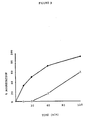
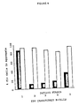
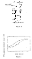


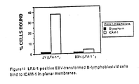


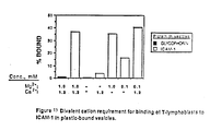


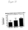

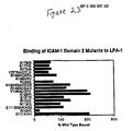


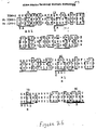
Classifications
-
- C—CHEMISTRY; METALLURGY
- C07—ORGANIC CHEMISTRY
- C07K—PEPTIDES
- C07K16/00—Immunoglobulins [IGs], e.g. monoclonal or polyclonal antibodies
- C07K16/18—Immunoglobulins [IGs], e.g. monoclonal or polyclonal antibodies against material from animals or humans
- C07K16/28—Immunoglobulins [IGs], e.g. monoclonal or polyclonal antibodies against material from animals or humans against receptors, cell surface antigens or cell surface determinants
- C07K16/2839—Immunoglobulins [IGs], e.g. monoclonal or polyclonal antibodies against material from animals or humans against receptors, cell surface antigens or cell surface determinants against the integrin superfamily
- C07K16/2845—Immunoglobulins [IGs], e.g. monoclonal or polyclonal antibodies against material from animals or humans against receptors, cell surface antigens or cell surface determinants against the integrin superfamily against integrin beta2-subunit-containing molecules, e.g. CD11, CD18
-
- A—HUMAN NECESSITIES
- A61—MEDICAL OR VETERINARY SCIENCE; HYGIENE
- A61P—SPECIFIC THERAPEUTIC ACTIVITY OF CHEMICAL COMPOUNDS OR MEDICINAL PREPARATIONS
- A61P35/00—Antineoplastic agents
-
- C—CHEMISTRY; METALLURGY
- C07—ORGANIC CHEMISTRY
- C07K—PEPTIDES
- C07K14/00—Peptides having more than 20 amino acids; Gastrins; Somatostatins; Melanotropins; Derivatives thereof
- C07K14/435—Peptides having more than 20 amino acids; Gastrins; Somatostatins; Melanotropins; Derivatives thereof from animals; from humans
- C07K14/705—Receptors; Cell surface antigens; Cell surface determinants
- C07K14/70503—Immunoglobulin superfamily
- C07K14/70525—ICAM molecules, e.g. CD50, CD54, CD102
-
- C—CHEMISTRY; METALLURGY
- C07—ORGANIC CHEMISTRY
- C07K—PEPTIDES
- C07K16/00—Immunoglobulins [IGs], e.g. monoclonal or polyclonal antibodies
- C07K16/18—Immunoglobulins [IGs], e.g. monoclonal or polyclonal antibodies against material from animals or humans
- C07K16/28—Immunoglobulins [IGs], e.g. monoclonal or polyclonal antibodies against material from animals or humans against receptors, cell surface antigens or cell surface determinants
- C07K16/2803—Immunoglobulins [IGs], e.g. monoclonal or polyclonal antibodies against material from animals or humans against receptors, cell surface antigens or cell surface determinants against the immunoglobulin superfamily
- C07K16/2821—Immunoglobulins [IGs], e.g. monoclonal or polyclonal antibodies against material from animals or humans against receptors, cell surface antigens or cell surface determinants against the immunoglobulin superfamily against ICAM molecules, e.g. CD50, CD54, CD102
-
- A—HUMAN NECESSITIES
- A61—MEDICAL OR VETERINARY SCIENCE; HYGIENE
- A61K—PREPARATIONS FOR MEDICAL, DENTAL OR TOILETRY PURPOSES
- A61K38/00—Medicinal preparations containing peptides
-
- A—HUMAN NECESSITIES
- A61—MEDICAL OR VETERINARY SCIENCE; HYGIENE
- A61K—PREPARATIONS FOR MEDICAL, DENTAL OR TOILETRY PURPOSES
- A61K39/00—Medicinal preparations containing antigens or antibodies
-
- C—CHEMISTRY; METALLURGY
- C07—ORGANIC CHEMISTRY
- C07K—PEPTIDES
- C07K2317/00—Immunoglobulins specific features
- C07K2317/30—Immunoglobulins specific features characterized by aspects of specificity or valency
- C07K2317/34—Identification of a linear epitope shorter than 20 amino acid residues or of a conformational epitope defined by amino acid residues
-
- C—CHEMISTRY; METALLURGY
- C07—ORGANIC CHEMISTRY
- C07K—PEPTIDES
- C07K2317/00—Immunoglobulins specific features
- C07K2317/70—Immunoglobulins specific features characterized by effect upon binding to a cell or to an antigen
- C07K2317/73—Inducing cell death, e.g. apoptosis, necrosis or inhibition of cell proliferation
Definitions
- the present invention relates to intercellular adhesion molecules such as ICAM-1 which are involved in the process through which populations of lymphocytes recognize and adhere to cellular substrates so that they may migrate to sites of inflammation and interact with cells during inflammatory reactions.
- the present invention additionally relates to ligand molecules capable of binding to such intercellular adhesion molecules, to a screening assay for these ligands, and to uses for the intercellular adhesion molecule, the ligand molecules, and the screening assay.
- Leukocytes must be able to attach to cellular substrates in order to properly defend the host against foreign invaders such as bacteria or viruses.
- An excellent review of the defense system is provided by Eisen, H.W., ( In: Microbiology , 3rd Ed., Harper & Row, Philadelphia, PA (1980), pp. 290-295 and 381-418). They must be able to attach to endothelial cells so that they can migrate from circulation to sites of ongoing inflammation. Furthermore, they must attach to antigen-presenting cells so that a normal specific immune response can occur, and finally, they must attach to appropriate target cells so that lysis of virally-infected or tumor cells can occur.
- Mac-1 is a heterodimer found on macrophages, granulocytes and large granular lymphocytes.
- LFA-1 is a heterodimer found on most lymphocytes (Springer, T.A., et al . Immunol. Rev. 68 :111-135 (1982)).
- p150,95 which has a tissue distribution similar to Mac-1 play a role in cellular adhesion (Keizer, G. et al ., Eur. J. Immunol. 15 :1142-1147 (1985)).
- leukocyte molecules were found to be members of a related family of glycoproteins (Sanchez-Madrid, F. et al ., J. Exper. Med. 158 :1785-1803 (1983); Keizer, G.D. et al ., Eur. J. Immunol. 15 :1142-1147 (1985)).
- This glycoprotein family is composed of heterodimers having one alpha chain and one beta chain. Although the alpha chain of each of the antigens differed from one another, the beta chain was found to be highly conserved (Sanchez-Madrid, F. et al ., J. Exper. Med. 158 :1785-1803 (1983)).
- the beta chain of the glycoprotein family (sometimes referred to as "CD18") was found to have a molecular weight of 95 kd whereas the alpha chains were found to vary from 150 kd to 180 kd (Springer, T., Fed. Proc. 44 :2660-2663 (1985)).
- CD18 The beta chain of the glycoprotein family
- the alpha subunits of the membrane proteins do not share the extensive homology shared by the beta subunits, close analysis of the alpha subunits of the glycoproteins has revealed that there are substantial similarities between them. Reviews of the similarities between the alpha and beta subunits of the LFA-1 related glycoproteins are provided by Sanchez-Madrid, F. et al ., ( J. Exper. Med. 158 :586-602 (1983); J. Exper. Med. 158 :1785-1803 (1983)).
- a group of individuals has been identified who are unable to express normal amounts of any member of this adhesion protein family on their leukocyte cell surface (Anderson, D.C., et al ., Fed. Proc. 44 :2671-2677 (1985); Anderson, D.C., et al ., J. Infect. Dis. 152 :668-689 (1985)). Lymphocytes from these patients displayed in vitro defects similar to normal counterparts whose LFA-1 family of molecules had been antagonized by antibodies. Furthermore, these individuals were unable to mount a normal immune response due to an inability of their cells to adhere to cellular substrates (Anderson, D.C., et al ., Fed. Proc.
- lymphocytes to maintain the health and viability of an animal requires that they be capable of adhering to other cells (such as endothelial cells).
- This adherence has been found to require cell-cell contacts which involve specific receptor molecules present on the cell surface of the lymphocytes. These receptors enable a lymphocyte to adhere to other lymphocytes or to endothelial, and other non-vascular cells.
- the cell surface receptor molecules have been found to be highly related to one another. Humans whose lymphocytes lack these cell surface receptor molecules exhibit chronic and recurring infections, as well as other clinical symptoms including defective antibody responses.
- lymphocyte adhesion is involved in the process through which foreign tissue is identified and rejected, an understanding of this process is of significant value in the fields of organ transplantation, tissue grafting, allergy and oncology.
- the present invention relates to Intercellular Adhesion Molecule-1 (ICAM-1) as well as to its functional derivatives.
- the invention additionally pertains to antibodies and fragments of antibodies capable of inhibiting the function of ICAM-1, and to other inhibitors of ICAM-1 function; and to assays capable of identifying such inhibitors.
- the invention additionally includes diagnostic and therapeutic uses for all of the above-described molecules.
- the invention includes the intercellular adhesion molecule ICAM-1 or its functional derivatives, which are substantially free of natural contaminants.
- the invention further pertains to such molecules which are additionally capable of binding to a molecule present on the surface of a lymphocyte.
- the invention further pertains to the intercellular adhesion molecule ICAM-1, and its derivatives which are detectably labeled.
- the invention additionally includes a recombinant DNA molecule capable of expressing ICAM-1 or a functional derivative thereof.
- the invention also includes a method for recovering ICAM-1 in substantially pure form which comprises the steps:
- the invention additionally includes an antibody capable of binding to a molecule selected from the group consisting of ICAM-1 and a functional derivative of ICAM-1.
- the invention also includes a hybridoma cell capable of producing such an antibody.
- the invention further includes a hybridoma cell capable of producing the monoclonal antibody R6-5-D6.
- the invention further includes a method for producing a desired hybridoma cell that produces an antibody which is capable of binding to ICAM-1, which comprises:
- the invention includes as well the hybridoma cell, and the antibody produced by the hybridoma cell, obtained by the above method.
- the invention is also directed to a method of identifying a non-immunoglobulin antagonist of intercellular adhesion which comprises:
- the invention is also directed toward a method for treating inflammation resulting from a response of the specific defense system in a mammalian subject which comprises providing to a subject in need of such treatment an amount of an anti-inflammatory agent sufficient to suppress the inflammation; wherein the anti-inflammatory agent is selected from the group consisting of: an antibody capable of binding to ICAM-1; a fragment of an antibody, the fragment being capable of binding to ICAM-1; ICAM-1; a functional derivative of ICAM-1; and a non-immunoglobulin antagonist of ICAM-1.
- the invention further includes the above-described method of treating inflammation wherein the non-immunoglobulin antagonist of ICAM-1 is a non-immunoglobulin antagonist of ICAM-1 other than LFA-1.
- the invention is also directed to a method of suppressing the metastasis of a hematopoietic tumor cell, the cell requiring a functional member of the LFA-1 family for migration, which method comprises providing to a patient in need of such treatment an amount of an anti-inflammatory agent sufficient to suppress the metastasis; wherein the anti-inflammatory agent is selected from the group consisting of: an antibody capable of binding to ICAM-1; a fragment of an antibody, the fragment being capable of binding to ICAM-1; ICAM-1; ICAM-1; a functional derivative of ICAM-1; and a non-immunoglobulin antagonist of ICAM-1.
- the invention further includes the above-described method of suppressing the metastasis of a hematopoietic tumor cell, wherein the non-immunoglobulin antagonist of ICAM-1 is a non-immunoglobulin antagonist of ICAM-1 other than LFA-1.
- the invention also includes a method of suppressing the growth of an ICAM-1-expressing tumor cell which comprises providing to a patient in need of such treatment an amount of a toxin sufficient to suppress the growth, the toxin being selected from the group consisting of a toxin-derivatized antibody capable of binding to ICAM-1; a toxin-derivatized fragment of an antibody, the fragment being capable of binding to ICAM-1; a toxin-derivatized member of the LFA-1 family of molecules; and a toxin-derivatized functional derivative of a member of the LFA-1 family of molecules.
- the invention is also directed to a method of suppressing the growth of an LFA-1-expressing tumor cell which comprises providing to a patient in need of such treatment an amount of toxin sufficient to suppress such growth, the toxin being selected from the group consisting of a toxin-derivatized ICAM-1; and a toxin-derivatized functional derivative of ICAM-1.
- the invention is further directed toward a method of diagnosing the presence and location of an inflammation resulting from a response of the specific defense system in a mammalian subject suspected of having the inflammation which comprises:
- the invention additionally provides a method of diagnosing the presence and location of an inflammation resulting from a response of the specific defense system in a mammalian subject suspected of having the inflammation which comprises:
- the invention also pertains to a method of diagnosing the presence and location of an ICAM-1-expressing tumor cell in a mammalian subject suspected of having such a cell, which comprises:
- the invention also pertains to a method of diagnosing the presence and location of an ICAM-1-expressing tumor cell in a mammalian subject suspected of having such a cell, which comprises:
- the invention also pertains to a method of diagnosing the presence and location of a tumor cell which expresses a member of the LFA-1 family of molecules in a subject suspected of having such a cell, which comprises:
- the invention also pertains to a method of diagnosing the presence and location of a tumor cell which expresses a member of the LFA-1 family of molecules in a subject suspected of having such a cell, which comprises:
- the invention additionally includes a phamaceutical composition comprising:
- One aspect of the present invention relates to the discovery of a natural binding ligand to LFA-1.
- Molecules such as those of LFA-1 family, which are involved in the process of cellular adhesion are referred to as "adhesion molecules.”
- the natural binding ligand of the present invention is designated "Intercellular Aadhesion Molecule-1" or "ICAM-1.”
- ICAM-1 is a 76-97 Kd glycoprotein. ICAM-1 is not a heterodimer.
- the present invention is directed toward ICAM-1 and its "functional derivatives.”
- a “functional derivative” of ICAM-1 is a compound which posesses a biological activity (either functional or structural) that is substantially similar to a biological activity of ICAM-1.
- the term “functional derivatives” is intended to include the “fragments,” “variants,” “analogs,” or “chemical derivatives” of a molecule.
- a “fragment” of a molecule such as ICAM-1 is meant to refer to any polypeptide subset of the molecule.
- Fragments of ICAM-1 which have ICAM-1 activity and which are soluble (i.e not membrane bound) are especially preferred.
- a "variant" of a molecule such as ICAM-1 is meant to refer to a molecule substantially similar in structure and function to either the entire molecule, or to a fragment thereof.
- a molecule is said to be "substantially similar” to another molecule if both molecules have substantially similar structures or if both molecules possess a similar biological activity. Thus, provided that two molecules possess a similar activity, they are considered variants as that term is used herein even if the structure of one of the molecules not found in the other, or if the sequence of amino acid residues is not identical.
- an “analog” of a molecule such as ICAM-1 is meant to refer to a molecule substantially similar in function to either the entire molecule or to a fragment thereof.
- a molecule is said to be a "chemical derivative" of another molecule when it contains additional chemical moieties not normally a part of the molecule. Such moieties may improve the molecule's solubility, absorption, biological half life, etc. The moieties may alternatively decrease the toxicity of the molecule, eliminate or attenuate any undesirable side effect of the molecule, etc. Moieties capable of mediating such effects are disclosed in Remington's Pharmaceutical Sciences (1980).
- Toxin-derivatized molecules constitute a special class of "chemical derivatives.”
- a "toxin-derivatized” molecule is a molecule (such as ICAM-1 or an antibody) which contains a toxin moiety. The binding of such a molecule to a cell brings the toxin moiety into close proximity with the cell and thereby promotes cell death.
- Any suitable toxin moiety may be employed; however, it is preferable to employ toxins such as, for example, the ricin toxin, the diphtheria toxin, radioisotopic toxins, membrane-channel-forming toxins, etc. Procedures for coupling such moieties to a molecule are well known in the art.
- an antigenic molecule such as ICAM-1, or members of the LFA-1 family of molecules are naturally expressed on the surfaces of lymphocytes.
- the introduction of such cells into an appropriate animal, as by intraperitoneal injection, etc. will result in the production of antibodies capable of binding to ICAM-1 or members of the LFA-1 family of molecules.
- the serum of such an animal may be removed and used as a source of polyclonal antibodies capable of binding these molecules. It is, however, preferable to remove splenocytes from such animals, to fuse such spleen cells with a myeloma cell line and to permit such fusion cells to form a hybridoma cell which secretes monoclonal antibodies capable of binding ICAM-1 or members of the LFA-1 family of molecules.
- the hybridoma cells obtained in the manner described above may be screened by a variety of methods to identify desired hybridoma cells that secrete antibody capable of binding either to ICAM-1 or to members of the LFA-1 family of molecules.
- such molecules are identified by their ability to inhibit the aggregation of Epstein-Barr virus-transformed cells.
- Antibodies capable of inhibiting such aggregation are then further screened to determine whether they inhibit such aggregation by binding to ICAM-1, or to a member of the LFA-1 family of molecules. Any means capable of distinguishing ICAM-1 from the LFA-1 family of molecules may be employed in such a screen.
- the antigen bound by the antibody may be analyzed as by immunoprecipitation and polyacrylamide gel electrophoresis. If the bound antigen is a member of the LFA-1 family of molecules then the immunoprecipitated antigen will be found to be a dimer, whereas if the bound antigen is ICAM-1 a single molecular weight species will have been immunoprecipitated. Alternatively, it is possible to distinguish between those antibodies which bind to members of the LFA-1 family of molecules from those which bind ICAM-1 by screening for the ability of the antibody to bind to cells such as granulocytes, which express IFA-1, but not ICAM-1.
- an antibody (known to inhibit cellular aggregation) to bind to granulocytes indicates that the antibody is capable of binding LFA-1. The absence of such binding is indicative of an antibody capable of recognizing ICAM-1.
- the ability of an antibody to bind to a cell such as a granulocyte may be detected by means commonly employed by those of ordinary skill. Such means include immunoassays, cellular agglutination, filter binding studies, antibody precipitation, etc.
- the anti-aggregation antibodies of the present invention may alternatively be identified by measuring their ability to differentially bind to cells which express ICAM-1 (such as activated endothelial cells), and their inability to bind to cells which fail to express ICAM-1.
- ICAM-1 such as activated endothelial cells
- the above assays may be modified, or performed in a different sequential order to provide a variety of potential screening assays, each of which is capable of identifying and discriminating between antibodies capable of binding to ICAM-1 versus members of the LFA-1 family of molecules.
- the anti-inflammatory agents of the present invention may be obtained by natural processes (such as, for example, by inducing an animal, plant, fungi, bacteria, etc., to produce a non-immunoglobulin antagonist of ICAM-1, or by inducing an animal to produce polyclonal antibodies capable of binding to ICAM-1); by synthetic methods (such as, for example, by using the Merrifield method for synthesizing polypeptides to synthesize ICAM-1, functional derivatives of ICAM-1, or protein antagonists of ICAM-1 (either immunoglobulin or non-immunoglobulin)); by hybridoma technology (such as, for example, to produce monoclonal antibodies capable of binding to ICAM-1); or by recombinant technology (such as, for example, to produce the anti-inflammatory agents of the present invention in diverse hosts (i.e., yeast, bacteria, fungi, cultured mammalian cells, etc.), or from recombinant plasmids or viral vectors).
- natural processes
- Epstein-Barr virus-transformed cells exhibit aggregation. This aggregation can be enhanced in the presence of phorbol esters. Such homotypic aggregation (i.e., aggregation involving only one cell type) was found to be blocked by anti-LFA-1 antibodies (Rothlein, R. et al ., J. Exper. Med. 163 :1132-1149 (1986)), which reference is incorporated herein by reference). Thus, the extent of LFA-1-dependent binding may be determined by assessing the extent of spontaneous or phorbol ester-dependent aggregate formation.
- Epstein-Barr virus-transformed cells may be employed in such an assay as long as the cells are capable of expressing the LFA-1 receptor molecule.
- Such cells may be prepared according to the technique of Springer, T.A. et al ., J. Exper. Med. 160 :1901-1918 (1984), which reference is herein incorporated by reference.
- any such cell may be employed in the LFA-1 dependent binding assay of the present invention, it is preferable to employ cells of the JY cell line (Terhost, C.T. et al ., Proc. Natl. Acad. Sci. USA 73 :910 (1976)).
- the cells may be cultivated in any suitable culture medium; however, it is most preferable to culture the cells in RMPI 1640 culture medium supplemented with 10% fetal calf serum and 50 ⁇ g/ml gentamycin (Gibco Laboratories, NY).
- the cells should be cultured under conditions suitable for mammalian cell proliferation (i.e., at a temperature of generally 37°C, in an atmosphere of 5% CO2, at a relative humidity of 95%, etc.).
- the novel intercellular adhesion molecule ICAM-1 was first identified and partially characterized according to the procedure of Rothlein, R. et al . ( J. Immunol. 137 :1270-1274 (1986)), which reference is herein incorporated by reference.
- monoclonal antibodies were prepared from spleen cells of mice immunized with cells from individuals genetically deficient in LFA-1 expression. Resultant antibodies were screened for their ability to inhibit the aggregation of LFA-1-expressing cells ( Figure 2).
- the ICAM-1 molecule mice were immunized with EBV-transformed B cells from LAD patients which do not express the LFA-1 antigen.
- the spleen cells from these animals were subsequently removed, fused with myeloma cells, and allowed to become monoclonal antibody producing hybridoma cells.
- EBV-transformed B cells from normal individuals which express LFA-1 were then incubated in the presence of the monoclonal antibody of the hybridoma cell in order to identify any monoclonal antibody which was capable of inhibiting the phorbol ester mediated, LFA-1 dependent, spontaneous aggregation of the EBV-transformed B cells. Since the hybridoma cells were derived from cells which had never encountered the LFA-1 antigen no monoclonal antibody to LFA-1 was produced.
- any antibody found to inhibit aggregation must be capable of binding to an antigen that, although not LFA-1, participated in the LFA-1 adhesion process.
- any method of obtaining such monoclonal antibodies may be employed, it is preferable to obtain ICAM-1-binding monoclonal antibodies by immunizing BALB/C mice using the routes and schedules described by Rothlein, R. et al . ( J. Immunol. 137 :1270-1274 (1986)) with Epstein-Barr virus-transformed peripheral blood mononuclear cells from an LFA-1-deficient individuals. Such cells are disclosed by Springer, T.A., et al ,. ( J. Exper. Med. 160 :1901-1918 (1984)).
- mice are immunized with either EBV-transformed B cells which express both ICAM-1 and LFA-1 or more preferably with TNF-activated endothelial cells which express ICAM-1 but not LFA-1.
- EBV-transformed B cells which express both ICAM-1 and LFA-1 or more preferably with TNF-activated endothelial cells which express ICAM-1 but not LFA-1.
- TNF-activated endothelial cells which express ICAM-1 but not LFA-1.
- a Balb/C mouse was sequentially immunized with JY cells and with differentiated U937 cells (ATCC CRL-1593). The spleen cells from such animals are removed, fused with myeloma cells and permitted to develop into antibody-producing hybridoma cells.
- the antibodies are screened for their ability to inhibit the LFA-1 dependent, phorbol ester induced aggregation of an EBV transformed cell line, such as JY cells, that expresses both the LFA-1 receptor and ICAM-1.
- an EBV transformed cell line such as JY cells
- antibodies capable of inhibiting such aggregation are then tested for their ability to inhibit the phorbol ester induced aggregation of a cell line, such as SKW3 (Dustin, M., et al ., J. Exper. Med.
- antibodies that are capable of binding to ICAM-1 may be identified by screening for antibodies which are capable of inhibiting the LFA-1 dependent aggregation of LFA-expression cells (such as JY cells) but are incapable of binding to cells that express LFA-1 but little or no ICAM-1 (such as normal granulocytes) or are capable of binding to cells that express ICAM-1 but not LFA-1 (such as TNF-activated endothelial cells).
- LFA-expression cells such as JY cells
- ICAM-1 such as normal granulocytes
- Another alternative is to immunoprecipitate from cells expressing ICAM-1, LFA-1, or both, using antibodies that inhibit the LFA-1 dependent aggregation of cells, such as JY cells, and through SDS-PAGE or an equivalent method determine some molecular characteristic of the molecule precipitated with the antibody. If the characteristic is the same as that of ICAM-1 then the antibody can be assumed to be an anti-ICAM-1 antibody.
- the ICAM-1 cell surface molecule was purified, and characterized.
- ICAM-1 was purified from human cells or tissue using monoclonal antibody affinity chromatography.
- a monoclonal antibody reactive with ICAM-1 is coupled to an inert column matrix. Any method of accomplishing such coupling may be employed; however, it is preferable to use the method of Oettgen, H.C. et al ., J. Biol. Chem. 259 :12034 (1984)).
- Oettgen H.C. et al ., J. Biol. Chem. 259 :12034 (1984)
- the bound ICAM-1 molecules may be eluted from the column.
- any suitable matrix can be employed, it is preferable to employ sepharose (Pharmacia) as the matrix material.
- sepharose Pharmacia
- the formation of column matrices, and their use in protein purification are well known in the art.
- the above-described assays may be used to identify compounds capable of attenuating or inhibiting the rate or extent of cellular adhesion.
- ICAM-1 is a cell surface glycoprotein expressed on non-hematopoietic cells such as vascular endothelial cells, thymic epithelial cells, certain other epithelial cells, and fibroblasts, and on hematopoietic cells such as tissue macrophages, mitogen-stimulated T lymphocyte blasts, and germinal centered B cells and dendritic cells in tonsils, lymph nodes, and Peyer's patches.
- ICAM-1 is highly expressed on vascular endothelial cells in T cell areas in lymph nodes and tonsils showing reactive hyperplasia. ICAM-1 is expressed in low amounts on peripheral blood lymphocytes.
- Phorbol ester-stimulated differentiation of some myelomonocytic cell lines greatly increases ICAM-1 expression.
- ICAM-1 is preferentially expressed at sites of inflammation, and is not generally expressed by quiescent cells.
- ICAM-1 expression on dermal fibroblasts is increased threefold to fivefold by either interleukin 1 or gamma interferon at levels of 10 U/ml over a period of 4 or 10 hours, respectively. The induction is dependent on protein and mRNA synthesis and is reversible.
- ICAM-1 displays molecular weight heterogeneity in different cell types with a molecular weight of 97 kd on fibroblasts, 114 kd on the myelomonocytic cell line U937, and 90 kd on the B lymphoblastoid cell JY.
- ICAM-1 biosynthesis has been found to involve an approximately 73 kd intracellular precursor.
- the non-N-glycosylated form resulting from tunicamycin treatment (which inhibits glycosylation) has a molecular weight of 55 kd.
- ICAM-1 isolated from phorbol ester stimulated U937 cells or from fibroblast cells yields an identical major product having a molecular weight of 60 kd after chemical deglycosylation.
- ICAM-1 monoclonal antibodies interfere with the adhesion of phytohemagglutinin blasts to LFA-1 deficient cell lines.
- Pretreatment of fibroblasts, but not lymphocytes, with monoclonal antibodies capable of binding ICAM-1 inhibits lymphocyte-fibroblast adhesion.
- Pretreatment of lymphocytes, but not fibroblasts, with antibodies against LFA-1 has also been found to inhibit lymphocyte-fibroblast adhesion.
- ICAM-1 is, thus, the binding ligand of the CD 18 complex on leukocytes. It is inducible on fibroblasts and endothelial cells in vitro by inflammatory mediators such as IL-1, gamma interferon and tumor necrosis factor in a time frame consistent with the infiltration of lymphocytes into inflammatory lesions in vivo (Dustin, M.L., et. al. , J. Immunol 137 :245-254, (1986); Prober, J.S., et. al. , J. Immunol 137 :1893-1896, (1986)).
- inflammatory mediators such as IL-1, gamma interferon and tumor necrosis factor
- ICAM-1 is expressed on non-hematopoietic cells such as vascular endothelial cells, thymic epithelial cells, other epithelial cells, and fibroblasts and on hematopoietic cells such as tissue macophages, mitogen-stimulated T lymphocyte blasts, and germinal center B-cells and dendritic cells in tonsils, lymph nodes and Peyer's patches (Dustin, M.L., et. al. J. Immunol 137 :245-254, (1986)).
- ICAM-1 is expressed on keratinocytes in benign inflammatory lesions such as allergic eczema, lichen planus, exanthema, urticaria and bullous diseases.
- ICAM-1 is present on keratinocytes from biopsies of skin lesions from various dermatological disorders and ICAM-1 expression is induced on lesions from allergic patch tests while keratinocytes from toxic patch test lesions failed to express ICAM-1.
- ICAM-1 is, therefore, a cellular substrate to which lymphocytes can attach, so that the lymphocytes may migrate to sites of inflammation and/or carry out various effector functions contributing to this inflammation. Such functions include the production of antibody, lysis of virally infected target cells, etc.
- the term "inflammation,” as used herein, is meant to include reactions of the specific defense systems.
- the term "specific defense system” is intended to refer to that component of the immune system that reacts to the presence of specific antigens. Inflammation is said to result from a response of the specific defense system if the inflammation is caused by, mediated by, or associated with a reaction of the specific defense system.
- inflammation resulting from a response of the specific defense system examples include the response to antigens such as rubella virus, autoimmune diseases, delayed type hypersensitivity response mediated by T-cells (as seen, for example in individuals who test "positive” in the Mantaux test), etc.
- antigens such as rubella virus, autoimmune diseases, delayed type hypersensitivity response mediated by T-cells (as seen, for example in individuals who test "positive” in the Mantaux test), etc.
- a "non-specific defence system reaction” is a response mediated by leukocytes incapable of immunological memory. Such cells include granulocytes and macrophages. As used herein, inflammation is said to result from a response of the non-specific defense system, if the inflammation is caused by, mediated by, or associated with a reaction of the non-specific defense system.
- inflammation which result, at least in part, from a reaction of the non-specific defense system include inflammation associated with conditions such as: asthma; adult respiratory distress syndrome (ARDS) or multiple organ injury syndromes secondary to septicemia or trauma; reperfusion injury of myocardial or other tissues; acute glomerulonephritis; reactive arthritis; dermatoses with acute inflammatory components; acute purulent meningitis or other central nervous system inflammatory disorders; thermal injury; hemodialysis; leukapheresis; ulcerative colitis; Crohn's disease; necrotizing enterocolitis; granulocyte transfusion associated syndromes; and cytokine-induced toxicity.
- ARDS adult respiratory distress syndrome
- multiple organ injury syndromes secondary to septicemia or trauma reperfusion injury of myocardial or other tissues
- acute glomerulonephritis reactive arthritis
- dermatoses with acute inflammatory components acute purulent meningitis or other central nervous system inflammatory disorders
- thermal injury hemodialysis
- leukapheresis ulcer
- ICAM-1 functional derivatives and especially such derivatives which comprise fragments or mutant variants of ICAM-1 which possess domains 1, 2 and 3 can be used in the treatment or therapy of such reactions of the non-specific defense system. More preferred for such treatment or therapy are ICAM-1 fragments or mutant variants which contain domain 2 of ICAM-1. Most preferred for such treatment or therapy are ICAM-1 fragments or mutant variants which contain domain 1 of ICAM-1.
- ICAM-1 gene Any of a variety of procedures may be used to clone the ICAM-1 gene.
- One such method entails analyzing a shuttle vector library of cDNA inserts (derived from an ICAM-1 expressing cell) for the presence of an insert which contains the ICAM-1 gene. Such an analysis may be conducted by transfecting cells with the vector and then assaying for ICAM-1 expression.
- the preferred method for cloning this gene entails determining the amino acid sequence of the ICAM-1 molecule.
- ICAM-1 protein may be purified and analyzed by automated sequenators. Alternatively, the molecule may be fragmented as with cyanogen bromide, or with proteases such as papain, chymotrypsin or trypsin (Oike, Y.
- sequence of amino acid residues in a peptide is designated herein either through the use of their commonly employed 3-letter designations or by their single-letter designations. A listing of these 3-letter and 1-letter designations may be found in textbooks such as Biochemistry , Lehninger, A., Worth Publishers, New York, NY (1970). When such a sequence is listed vertically, the amino terminal residue is intended to be at the top of the list, and the carboxy terminal residue of the peptide is intended to be at the bottom of the list. Similarly, when listed horizontally, the amino terminus is intended to be on the left end whereas the carboxy terminus is intended to be at the right end. The residues of amino acids in a peptide may be separated by hyphens.
- amino acid sequence designated: -Gly-Ala-Ser-Phe- indicates that an Ala residue is linked to the carboxy group of Gly, and that a Ser residue is linked to the carboxy group of the Ala residue and to the amino group of a Phe residue.
- the designation further indicates that the amino acid sequence contains the tetrapeptide Gly-Ala-Ser-Phe.
- the designation is not intended to limit the amino acid sequence to this one tetrapeptide, but is intended to include (1) the tetrapeptide having one or more amino acid residues linked to either its amino or carboxy end, (2) the tetrapeptide having one or more amino acid residues linked to both its amino and its carboxy ends, (3) the tetrapeptide having no additional amino acid residues.
- the DNA sequences capable of encoding them are examined. Because the genetic code is degenerate, more than one codon may be used to encode a particular amino acid (Watson, J.D., In: Molecular Biology of the Gene , 3rd Ed., W.A. Benjamin, Inc., Menlo Park, CA (1977), pp. 356-357).
- the peptide fragments are analyzed to identify sequences of amino acids which may be encoded by oligonucleotides having the lowest degree of degeneracy. This is preferably accomplished by identifying sequences that contain amino acids which are encoded by only a single codon.
- amino acid sequences may be encoded by only a single oligonucleotide, frequently the amino acid sequence can be encoded by any of a set of similar oligonucleotides.
- all of the members of the set contain oligonucleotides which are capable of encoding the peptide fragment and, thus, potentially contain the same nucleotide sequence as the gene which encodes the peptide fragment, only one member of the set contains a nucleotide sequence that is identical to the nucleotide sequence of this gene.
- this member is present within the set, and is capable of hybridizing to DNA even in the presence of the other members of the set, it is possible to employ the unfractionated set of oligonucleotides in the same manner in which one would employ a single oligonucleotide to clone the gene that encodes the peptide.
- oligonucleotide or set of oligonucleotides which have a nucleotide sequence that is complementary to the oligonucleotide sequence or set of sequences that is capable of encoding the peptide fragment.
- a suitable oligonucleotide, or set of oligonucleotides which is capable of encoding a fragment of the ICAM-1 gene (or which is complementary to such an oligonucleotide, or set of oligonucleotides) is identified (using the above-described procedure), synthesized, and hybridized, by means well known in the art, against a DNA or, more preferably, a cDNA preparation derived from human cells which are capable of expressing ICAM-1 gene sequences.
- Techniques of nucleic acid hybridization are disclosed by Maniatis, T. et al ., In: Molecular Cloning, a Laboratory Manual , Coldspring Harbor, NY (1982), and by Haymes, B.D.
- the source of DNA or cDNA used will preferably have been enriched for ICAM-1 sequences. Such enrichment can most easily be obtained from cDNA obtained by extracting RNA from cells cultured under conditions which induce ICAM-1 synthesis (such as U937 grown in the presence of phorbol esters, etc.).
- a library of expression vectors is prepared by cloning DNA or, more preferably cDNA, from a cell capable of expressing ICAM-1 into an expression vector.
- the library is then screened for members capable of expressing a protein which binds to anti-ICAM-1 antibody, and which has a nucleotide sequence that is capable of encoding polypeptides that have the same amino acid sequence as ICAM-1 or fragments of ICAM-1.
- the cloned ICAM-1 gene obtained through the methods described above, may be operably linked to an expression vector, and introduced into bacterial, or eukaryotic cells to produce ICAM-1 protein. Techniques for such manipulations are disclosed by Maniatis, T. et al ., supra , and are well known in the art.
- the above-described assay capable of measuring LFA-1 dependent aggregation, may be employed to identify agents which act as antagonists to inhibit the extent of LFA-1 dependent aggregation.
- agents which act as antagonists to inhibit the extent of LFA-1 dependent aggregation.
- Such antagonists may act by impairing the ability of LFA-1 or of ICAM-1 to mediate aggregation.
- agents include immunoglobulins such as an antibody capable of binding to either LFA-1 or ICAM-1.
- non-immunoglobulin (i.e., chemical) agents may be examined, using the above-described assay, to determine whether they are antagonists of LFA-1 aggregation.
- Monoclonal antibodies to members of the CD 18 complex inhibit many adhesion dependent functions of leukocytes including binding to endothelium (Haskard, D., et al. , J. Immunol. 137 :2901-2906 (1986)), homotypic adhesions (Rothlein, R., et al. , J. Exp. Med. 163 :1132-1149 (1986)), antigen and mitogen induced proliferation of lymphocytes (Davignon, D., et al. , Proc. Natl. Acad. Sci., USA 78 :4535-4539 (1981)), antibody formation (Fischer, A., et al. , J. Immunol.
- lymphocytes are capable of continually monitoring an animal for the presence of foreign antigens. Although these processes are normally desirable, they are also the cause of organ transplant rejection, tissue graft rejection and many autoimmune diseases. Hence, any means capable of attenuating or inhibiting cellular adhesion would be highly desirable in recipients of organ transplants, tissue grafts or autoimmune patients.
- Monoclonal antibodies capable of binding to ICAM-1 are highly suitable as anti-inflammatory agents in a mammalian subject. Significantly, such agents differ from general anti-inflammatory agents in that they are capable of selectively inhibiting adhesion, and do not offer other side effects such as nephrotoxicity which are found with conventional agents. Monoclonal antibodies capable of binding to ICAM-1 can therefore be used to prevent organ or tissue rejection, or modify autoimmune responses without the fear of such side effects, in the mammalian subject.
- monoclonal antibodies capable of recognizing ICAM-1 may permit one to perform organ transplants even between individuals having HLA mismatch.
- a composition containing a monoclonal antibody against ICAM-1 may be administered to a patient experiencing delayed type hypersensitivity reaction.
- a compositions might be provided to a individual who had been in contact with antigens such as poison ivy, poison oak, etc.
- the monoclonal antibody capable of binding to ICAM-1 is administered to a patient in conjunction with an antigen in order to prevent a subsequent inflammatory reaction.
- the additional administration of an antigen with an ICAM-1-binding monoclonal antibody may temporarily tolerize an individual to subsequent presentation of that antigen.
- LFA-1 LFA-1's natural ligand
- ICAM-1 antagonism of LFA-1's natural ligand, ICAM-1
- the ability of antibodies against ICAM-1 to inhibit inflammation provides the basis for their therapeutic use in the treatment of chronic inflammatory diseases and autoimmune diseases such as lupus erythematosus, autoimmune thyroiditis, experimental allergic encephalomyelitis (EAE), multiple sclerosis, some forms of diabetes Reynaud's syndrome, rheumatoid arthritis, etc.
- EAE experimental allergic encephalomyelitis
- Such antibodies may also be employed as a therapy in the treatment of psoriasis.
- the monoclonal antibodies capable of binding to ICAM-1 may be employed in the treatment of those diseases currently treatable through steroid therapy.
- monoclonal antibodies capable of binding to ICAM-1 may be employed as a means of imaging or visualizing the sites of infection and inflammation in a patient.
- the monoclonal antibodies are detectably labeled, through the use of radioisotopes, affinity labels (such as biotin, avidin, etc.) fluorescent labels, paramagnetic atoms, etc. Procedures for accomplishing such labeling are well known to the art.
- Clinical application of antibodies in diagnostic imaging are reviewed by Grossman, H.B., Uro. Clin. North Amer. 13 :465-474 (1986)), Unger, E.C. et al ., Invest. Radiol. 20 :693-700 (1985)), and Khaw, B.A. et al ., Science 209 :295-297 (1980)).
- the presence of inflammation may also be detected through the use of binding ligands, such as mRNA, cDNA, or DNA which bind to ICAM-1 gene sequences, or to ICAM-1 mRNA sequences, of cells which express ICAM-1.
- binding ligands such as mRNA, cDNA, or DNA which bind to ICAM-1 gene sequences, or to ICAM-1 mRNA sequences, of cells which express ICAM-1.
- the detection of foci of such detectably labeled antibodies is indicative of a site of inflammation or tumor development.
- this examination for inflammation is done by removing samples of tissue or blood and incubating such samples in the presence of the detectably labeled antibodies.
- this technique is done in a non-invasive manner through the use of magnetic imaging, fluorography, etc.
- Such a diagnostic test may be employed in monitoring organ transplant recipients for early signs of potential tissue rejection.
- Such assays may also be conducted in efforts to determine an individual's predilection to rheumatoid arthritis or other chronic inflammatory diseases.
- Immune responses to therapeutic or diagnostic agents such as, for example, bovine insulin, interferon, tissue-type plasminogen activator or murine monoclonal antibodies substantially impair the therapeutic or diagnostic value of such agents, and can, in fact, causes diseases such as serum sickness.
- therapeutic or diagnostic agents such as, for example, bovine insulin, interferon, tissue-type plasminogen activator or murine monoclonal antibodies
- Such a situation can be remedied through the use of the antibodies of the present invention.
- such antibodies would be administered in combination with the therapeutic or diagnostic agent.
- the addition of the antibodies prevents the recipient from recognizing the agent, and therefore prevents the recipient from initiating an immune response against it.
- the absence of such an immune response results in the ability of the patient to receive additional administrations of the therapeutic or diagnostic agent.
- ICAM-1 is a binding partner of LFA-1.
- ICAM-1 or its functional derivatives may be employed interchangeably with antibodies capable of binding to LFA-1 in the treatment of disease.
- such molecules may be employed to inhibit inflammation, organ rejection, graft rejection, etc.
- ICAM-1, or its functional derivatives may be used in the same manner as anti-ICAM antibodies to decrease the immunogenicity of therapeutic or diagnostic agents.
- ICAM-1 may be used to block the metastasis or proliferation of tumor cells which express either ICAM-1 or LFA-1 on their surfaces.
- a variety of methods may be used to accomplish such a goal. For example, the migration of hematopoietic cells requires LFA-1-ICAM-1 binding. Antagonists of such binding therefore suppress this migration and block the metastasis of tumor cells of leukocyte lineage.
- toxin-derivatized molecules capable of binding either ICAM-1 or a member of the LFA-1 family of molecules, may be administered to a patient. When such toxin-derivatized molecules bind to tumor cells that express ICAM-1 or a member of the LFA-1 family of molecules, the presence of the toxin kills the tumor cell thereby inhibiting the proliferation of the tumor.
- ICAM-1-dependent adhesion can be inhibited by non-immunoglobulin antagonists which are capable of binding to either ICAM-1 or to LFA-1.
- a non-immunoglobulin antagonist of ICAM-1 is LFA-1.
- An example of a non-immunoglobulin antagonist which binds to LFA-1 is ICAM-1.
- the therapeutic effects of ICAM-1 may be obtained by providing to a patient the entire ICAM-1 molecule, or any therapeutically active peptide fragments thereof.
- ICAM-1 and its functional derivatives may be obtained either synthetically, through the use of recombinant DNA technology, or by proteolysis.
- the therapeutic advantages of ICAM-1 may be augmented through the use of functional derivatives of ICAM-1 possessing additional amino acid residues added to enhance coupling to carrier or to enhance the activity of the ICAM-1.
- the scope of the present invention is further intended to include functional derivatives of ICAM-1 which lack certain amino acid residues, or which contain altered amino acid residues, so long as such derivatives exhibit the capacity to affect cellular adhesion.
- Both the antibodies of the present invention and the ICAM-1 molecule disclosed herein are said to be "substantially free of natural contaminants” if preparations which contain them are substantially free of materials with which these products are normally and naturally found.
- the present invention extends to antibodies, and biologically active fragments thereof, (whether polyclonal or monoclonal) which are capable of binding to ICAM-1.
- Such antibodies may be produced either by an animal, or by tissue culture, or recombinant DNA means.
- the dosage of administered agent will vary depending upon such factors as the patient's age, weight, height, sex, general medical condition, previous medical history, etc. In general, it is desirable to provide the recipient with a dosage of antibody which is in the range of from about 1 pg/kg to 10 mg/kg (body weight of patient, although a lower or higher dosage may be administered.
- ICAM-1 molecules or their functional derivatives When providing ICAM-1 molecules or their functional derivatives to a patient, it is preferable to administer such molecules in a dosage which also ranges from about 1 pg/kg to 10 mg/kg (body weight of patient) although a lower or higher dosage may also be administered.
- the therapeutically effective dose can be lowered if the anti-ICAM-1 antibody is additionally administered with an anti-LFA-1 antibody.
- one compound is said to be additionally administered with a second compound when the administration of the two compounds is in such proximity of time that both compounds can be detected at the same time in the patient's serum.
- Both the antibody capable of binding to ICAM-1 and ICAM-1 itself may be administered to patients intravenously, intramuscularly, subcutaneously, enterally, or parenterally.
- the administration may be by continuous infusion, or by single or multiple boluses.
- the anti-inflammatory agents of the present invention are intended to be provided to recipient subjects in an amount sufficient to suppress inflammation.
- An amount is said to be sufficient to "suppress" inflammation if the dosage, route of administration, etc. of the agent are sufficient to attenuate or prevent inflammation.
- Anti-ICAM-1 antibody, or a fragment thereof may be administered either alone or in combination with one or more additional immunosuppressive agents (especially to a recipient of an organ or tissue transplant).
- the administration of such compound(s) may be for either a "prophylactic” or “therapeutic” purpose.
- the immunosuppressive compound(s) are provided in advance of any inflammatory response or symptom (for example, prior to, at, or shortly after) the time of an organ or tissue transplant but in advance of any symptoms of organ rejection).
- the prophylactic administration of the compound(s) serves to prevent or attenuate any subsequent inflammatory response (such as, for example, rejection of a transplanted organ or tissue, etc.).
- the immunosuppressive compound(s) When provided therapeutically, the immunosuppressive compound(s) is provided at (or shortly after) the onset of a symptom of actual inflammation (such as, for example, organ or tissue rejection).
- the therapeutic administration of the compound(s) serves to attenuate any actual inflammation (such as, for example, the rejection of a transplanted organ or tissue).
- the anti-inflammatory agents of the present invention may, thus, be provided either prior to the onset of inflammation (so as to suppress an anticipated inflammation) or after the initiation of inflammation.
- a composition is said to be "pharmacologically acceptable” if its administration can be tolerated by a recipient patient.
- Such an agent is said to be administered in a "therapeutically effective amount” if the amount administered is physiologically significant.
- An agent is physiologically significant if its presence results in a detectable change in the physiology of a recipient patient.
- the antibody and ICAM-1 molecules of the present invention can be formulated according to known methods to prepare pharmaceutically useful compositions, whereby these materials, or their functional derivatives, are combined in admixture with a pharmaceutically acceptable carrier vehicle.
- a pharmaceutically acceptable carrier vehicle e.g., a pharmaceutically acceptable carrier vehicle.
- suitable vehicles and their formulation, inclusive of other human proteins, e.g., human serum albumin are described, for example, in Remington's Pharmaceutical Sciences (16th ed., Osol, A., Ed., Mack, Easton PA (1980)).
- compositions will contain an effective amount of anti-ICAM antibody or ICAM-1 molecule, or their functional derivatives, together with a suitable amount of carrier vehicle.
- Control release preparations may be achieved through the use of polymers to complex or absorb anti-ICAM-1 antibody or ICAM-1, or their functional derivatives.
- the controlled delivery may be exercised by selecting appropriate macromolecules (for example polyesters, polyamino acids, polyvinyl, pyrrolidone, ethylenevinylacetate, methylcellulose, carboxymethylcellulose, or protamine, sulfate) and the concentration of macromolecules as well as the methods of incorporation in order to control release.
- Another possible method to control the duration of action by controlled release preparations is to incorporate anti-ICAM-1 antibody or ICAM-1 molecules, or their functional derivatives, into particles of a polymeric material such as polyesters, polyamino acids, hydrogels, poly(lactic acid) or ethylene vinylacetate copolymers.
- a polymeric material such as polyesters, polyamino acids, hydrogels, poly(lactic acid) or ethylene vinylacetate copolymers.
- microcapsules prepared, for example, by coacervation techniques or by interfacial polymerization, for example, hydroxymethylcellulose or gelatine-microcapsules and poly(methylmethacylate) microcapsules, respectively, or in colloidal drug delivery systems, for example, liposomes, albumin microspheres, microemulsions, nanoparticles, and nanocapsules or in macroemulsions.
- colloidal drug delivery systems for example, liposomes, albumin microspheres, microemulsions, nanoparticles, and nanocapsules or in macroemulsions.
- the EBV-transformed and hybridoma cells of the present invention were maintained in RMPI 1640 culture medium, supplemented with 20 mM L-glutamine, 50 ⁇ g/ml gentamicin, and 10% fetal bovine (or fetal calf) sera.
- Cells were cultured at 37°C in a 5% CO2, 95% air humidity atmosphere.
- Epstein-Barr virus (EBV) transformants 106 T cell depleted peripheral blood mononuclear cells/ml in RPMI 1640 medium supplemented with 20% fetal calf serum (FCS), and 50 ⁇ g/ml gentamicin were incubated for 16 hours with EBV-containing supernatant of B95-8 cells (Thorley-Lawson, D.A. et al ., J. Exper. Med. 146 :495 (1977)). Cells in 0.2 ml aliquot were placed in 10 microtiter wells. Medium was replaced with RPMI 1640 medium (supplemented with 20% fetal calf serum and 50 ⁇ g/ml gentamicin) until cell growth was noted.
- FCS fetal calf serum
- PHA Phytohemagglutinin
- aggregation assays were employed. Cell lines used in such assays were washed two times with RPMI 1640 medium containing 5 mM Hepes buffer (Sigma Chemical Co., St. Louis) and resuspended to a concentration of 2 x 10⁶ cells/ml. Added to flat-bottomed, 96-well microtiter plates (No.
- 3596; Costar, Cambridge, MA were 50 ⁇ l of appropriate monoclonal antibody supernatant or 50 ⁇ l of complete medium with or without purified monoclonal antibodies, 50 ⁇ l of complete medium containing 200 ng/ml of the phorbol ester phorbol myristate acetate (PMA) and 100 ⁇ l of cells at a concentration of 2 x 10⁶ cells/ml in complete medium. This yielded a final concentration of 50 ng/ml PMA and 2 x 10⁵ cells/well. Cells were allowed to settle spontaneously, and the degree of aggregation was scored at various time points.
- PMA phorbol ester phorbol myristate acetate
- Scores ranged from 0 to 5+, where 0 indicated that essentially no cells were in clusters; 1+ indicated that less than 10% of the cells were in aggregates; 2+ indicated that less than 50% of the cells were aggregated; 3+ indicated that up to 100% of the cells were in small, loose clusters; 4+ indicated that up to 100% of the cells were aggregated in larger clusters; and 5+ indicated that 100% of the cells were in large, very compact aggregates.
- reagents and cells were added to 5 ml polystyrene tubes in the same order as above. Tubes were placed in a rack on a gyratory shaker at 37°C.
- Percent aggregation was determined by the following equation: The number of input cells in the above formula is the number of cells per ml in a control tube containing only cells and complete medium that had not been incubated. The number of free cells in the above equation equals the number of non-aggregated cells per ml from experimental tubes. The above procedures were described by Rothlein, R., et al ., J. Exper. Med. 163 :1132-1149 (1986).
- Example 2 The qualitative aggregation assay described in Example 2 was performed using the Epstein-Barr transformed cell line JY. Upon addition of PMA to the culture medium in the microtiter plates, aggregation of cells was observed. Time lapse video recordings showed that the JY cells on the bottom of the microtiter wells were motile and exhibited active membrane ruffling and pseudopodia movement. Contact between the pseudopodia of neighboring cells often resulted in cell-cell adherence. If adherence was sustained, the region of cell contact moved to the uropod. Contact could be maintained despite vigorous cell movements and the tugging of the cells in opposite directions. The primary difference between PMA-treated and untreated cells appeared to be in the stability of these contacts once they were formed. With PMA, clusters of cells developed, growing in size as additional cells adhered at their periphery.
- EBV-transformed lymphoblastoid cells were prepared from patients in the manner described in Example 1. Such cells were screened against monoclonal antibodies capable of recognizing LFA-1 and the cells were found to be LFA-1 deficient.
- Example 2 The qualitative aggregation assay described in Example 2 was employed, using the LFA-1 deficient cells described above. Such cells failed to spontaneously aggregate, even in the presence of PMA.
- the LFA-1 deficient cells of Example 5 were labeled with carboxyfluorescein diacetate (Patarroyo, M. et al ., Cell. Immunol. 63 :237-248 (1981)).
- the labeled cells were mixed in a ratio of 1:10 with autologous or JY cells and the percentage of fluorescein-labeled cells in aggregates was determined according to the procedure of Rothlein, R. et al. , J. Exper. Med. 163 :1132-1149 (1986).
- the LFA-1 deficient cells were found to be capable of coaggregating with LFA-1 expressing cells ( Figure 4).
- Monoclonal antibodies capable of binding to ICAM-1 were isolated according to the method of Rothlein, R. et al. , J. Immunol. 137 :1270-1274 (1986), which reference has been incorporated by reference herein.
- 3 BALB/C mice were immunized intraperitoneally with EBV-transformed peripheral blood mononuclear cells from an LFA-1-deficient individual (Springer, T.A. et al ., J. Exper. Med. 160 :1901 (1984)). Approximately 107 cells in 1 ml RPMI 1640 medium was used for each immunization.
- mice were given an additional 107 cells in 0.15 ml medium (intravenously).
- Isolated spleen cells from the above-described animals were fused with P3X73Ag8.653 myeloma cells at a ratio of 4:1 according to the protocol of Galfre, G. et al ., Nature 266 :550 (1977). Aliquots of the resulting hybridoma cells were introduced into 96-well microtiter plates. The hybridoma supernatants were screened for inhibition of aggregation, and one inhibitory hybridoma (of over 600 wells tested) was cloned and subcloned by limiting dilution. This subclone was designated RR1/1.1.1 (hereinafter designated "RR1/1").
- Monoclonal antibody RR1/1 was consistently found to inhibit PMA-stimulated aggregation of the LFA-1 expressing cell line JY.
- the RR1/1 monoclonal antibody inhibited aggregation equivalently, or slightly less than some monoclonal antibodies to the LFA-1 alpha or beta subunits.
- control monoclonal antibody against HLA which is abundantly expressed on JY cells, did not inhibit aggregation.
- the antigen bound by monoclonal antibody RR1/1 is defined as the intercellular adhesion molecule-1 (ICAM-1).
- IAM-1 intercellular adhesion molecule-1
- Lysates were centrifuged at 10,000 x g for 10 minutes and precleared with 50 ⁇ l of a 50% suspension of CNBr-activated, glycine-quenched Sepharose Cl-4B for 1 hour at 4°C.
- One milliliter of lysate was immunoprecipitated with 20 ⁇ l of a 50% suspension of monoclonal antibody RR1/1 coupled to Sepharose Cl-4B (1 mg/ml) overnight at 4°C (Springer, T.A. et al ., J. Exper. Med. 160 :1901 (1984)).
- Sepharose-bound monoclonal antibody was prepared using CNBr-activation of Sepharose CL-4B in carbonate buffer according to the method of March, S.
- Example 2 In order to determine the effect of monoclonal antibody RR1/1 on PHA-lymphoblast aggregation, the quantitative aggregation assay described in Example 2 was employed. Thus, T cell blast cells were stimulated for 4 days with PHA, thoroughly washed, then cultured for 6 days in the presence of IL-2 conditioned medium. PHA was found to be internalized during this 6-day culture, and did not contribute to the aggregation assay. In three different assays with different T cell blast preparations, ICAM-1 monoclonal antibodies consistently inhibited aggregation (Table 2). TABLE 2 Inhibition of PMA-Stimulated PHA-Lymphoblast Aggregation by RR1/1 Monoclonal Antibody a Expt.
- b Percent inhibition relative to cells treated with PMA and X63 monoclonal antibody.
- c Aggregation was measured 1 hr after the simultaneous addition of monoclonal antibody and PMA. Cells were shaken at 175 rpm.
- d A negative number indicates percent enhancement of aggregation.
- e Aggregation was measured 1 hr after the simultaneous addition of monoclonal antibody and PMA. Cells were pelleted at 200 x G for 1 min. incubated at 37°C for 15 min. gently resuspended, and shaken for 45 min. at 100 rpm.
- f Cells were pretreated with PMA for 4 hr at 37°C. After monoclonal antibody was added, the tubes were incubated at 37°C stationary for 20 min. and shaken at 75 rpm for 100 min.
- LFA-1 monoclonal antibodies were consistently more inhibitory than ICAM-1 monoclonal antibodies, whereas HLA-A, B and LFA-3 monoclonal antibodies were without effect. These results indicate that of the monoclonal antibodies tested, only those capable of binding to LFA-1 or ICAM-1 were capable of inhibiting cellular adhesion.
- mice were immunized intraperitoneally (i.p.) with 0.5 mls of 2 x 10⁷ JY cells in RPMI medium 103 days and 24 days prior to fusion. On day 4 and 3 prior to fusion, mice were immunized i.p. with 107 cells of PMA differentiated U937 cells in 0.5 ml of RPMI medium.
- U937 cells (ATCC CRL-1593) were differentiated by incubating them at 5 x 10⁵/ml in RPMI with 10% Fetal Bovine Serum, 1% glutamine and 50 ⁇ g/ml gentamyin (complete medium) containing 2 ng/ml phorbol-12-myristate acetate (PMA) in a sterile polypropylene container. On the third day of this incubation, one-half of the volume of medium was withdrawn and replaced with fresh complete medium containing PMA. On day 4, cells were removed, washed and prepared for immunization.
- PMA phorbol-12-myristate acetate
- Spleen cells from the immunized mice were fused with P3x63 Ag8 ⁇ 653 myeloma cells at a 4:1 ratio according to Galfre et al ., ( Nature 266 :550 (1977)). After the fusion, cells were plated in a 96 well flat bottomed microtiter plates at 105 spleen cells/well.
- a radioimmune assay was developed.
- purified RR1/1 was iodinated using iodogen to a specific activity of 10 ⁇ Ci/ ⁇ g.
- Endothelial cells were grown in 96 well plates and treated as described for each experiment. The plates were cooled at 4°C by placing in a cold room for 0.5-1 hr, not immediately on ice. The monolayers were washed 3x with cold complete media and then incubated 30 m at 4°C with 125I RR1/1. The monolayers were then washed 3x with complete media. The bound 125I was released using 0.1 N NaOH and counted.
- the specific activity of the 125I RR1/1 was adjusted using unlabeled RR1/1 to obtain a linear signal over the range of antigen densities encountered in this study. Non-specific binding was determined in the presence of a thousand fold excess of unlabeled RR1/1 and was subtracted from total binding to yield the specific binding.
- ICAM-1 expression measured using the above described radioimmune assay, is increased on human umbilical vein endothelial cells (HUVEC) and human saphenous vein endothelial cells (HSVEC) by IL-1, TNF, LPS and IFN gamma (Table 3).
- Saphenous vein endothelial cells were used in this study to confirm the results from umbilical vein endothelial cells in cultured large vein endothelial cells derived from adult tissue.
- the basal expression of ICAM-1 is 2 fold higher on saphenous vein endothelial cells than on umbilical vein endothelial cells.
- IL-1 alpha, TNF and LPS were the most potent inducers and IL-1 was less potent on a weight basis and also at saturating concentrations for the response (Table 3).
- IL-1 beta at 100 ng/ml increased ICAM-1 expression by 9 fold on HUVEC and 7.3 fld on HSVEC with half-maximal increase occuring at 15 ng/ml.
- rTNF at 50 ng/ml increased ICAM-1 expression 16 fold on HUVEC and 11 fold on HSVEC with half maximal effects at 0.5 ng/ml.
- Interferon-gamma produced a significant increase in ICAM-1 expression of 5.2 fold on HUVEC or 3.5 fold on HSVEC at 10,000 U/ml.
- the effect of LPS at 10 ⁇ g/ml was similar in magnitude to that of rTNF. Pairwise combinations of these mediators resulted in additive or slightly less than additive effects on ICAM-1 expression (Table 3).
- Cross-titration of rTNF with rIL-1 beta or rIFN gamma showed no synergism between these at suboptimal or optimal concentrations.
- the kinetics of interleukin 1 and gamma interferon effects on ICAM-1 expression on dermal fibroblasts were determined using the 125I goat anti-mouse IgG binding assay of Dustin, M.L. et al . ( J. Immunol. 137 :245-254 (1986); which reference is herein incorporated by reference).
- human dermal fibroblasts were grown in a 96-well microtiter plate to a density of 2-8 x 10⁴ cells/well (0.32 cm2). The cells were washed twice with RPMI 1640 medium supplemented as described in Example 1.
- the cells were additionally washed once with Hanks Balanced Salt Solution (HBSS), 10 mM HEPES, 0.05% NaN3 and 10% heat-inactivated fetal bovine serum. Washing with this binding buffer was done at 4°C. To each well was added 50 ⁇ l of the above-described binding buffer and 50 ⁇ l of the appropriate hybridoma supernatant with X63 and W6/32 as the negative and positive controls, respectively. After incubation for 30 minutes at 4°C, with gentle agitation, the wells were washed twice with binding buffer, and the second antibody 125I-goat anti-mouse IgG, was added at 50 nCi in 100 ⁇ l.
- HBSS Hanks Balanced Salt Solution
- 10 mM HEPES 10 mM HEPES
- NaN3 10% heat-inactivated fetal bovine serum
- the 125I-goat anti-mouse antibody was prepared by using Iodogen (Pierce) according to the method of Fraker, P.J. et al . ( Biochem. Biophys. Res. Commun. 80 :849 (1978)). After 30 minutes at 4°C, the wells were washed twice with 200 ⁇ l of binding buffer and the cell layer was solubilized by adding 100 ⁇ l of 0.1 N NaOH. This and a 100 ⁇ l wash were counted in a Beckman 5500 gamma counter. The specific counts per minute bound was calculated as [cpm with monoclonal antibody]-[cpm with X63]. All steps, including induction with specific reagents, were carried out in quadruplicate.
- the dose response curves for induction of ICAM-1 by recombinant mouse and human interleukin 1, and for recombinant human gamma interferon, are shown in Figure 7.
- Gamma interferon and interleukin 1 were found to have similar concentration dependencies with nearly identical effects at 1 ng/ml.
- the human and mouse recombinant interleukin 1 also have similar curves, but are much less effective than human interleukin 1 preparations in inducing ICAM-1 expression.
- Cyclohexamide an inhibitor of protein synthesis, and actinomycin D, an inhibitor of mRNA synthesis, abolish the effects of both interleukin 1 and gamma interferon on ICAM-1 expression on fibroblasts (Table 4). Furthermore, tunicamycin, an inhibitor of N-linked glycosylation, only inhibited the interleukin 1 effect by 43%. These results indicate that protein and mRNA synthesis, but not N-linked glycosylation, are required for interleukin 1 and gamma interferon-stimulated increases in ICAM-1 expression.
- Treatments were carried out in a final volume of 50 ⁇ l containing the indicated reagents. Cycloheximide, actinomycin D, and tunicamycin were added at 20 ⁇ g/ml, 10 ⁇ M, and 2 ⁇ g/ml, respectively, at the same time as the cytokines. All points are means of quadruplicate wells ⁇ SD.
- the sections were sequentially incubated with biotinylated horse anti-mouse IgG and avidin-biotinylated peroxidase complexes.
- the sections were finally dipped in a solution containing 3-amino-9-ethyl-carbazole (Aldrich Chemical Co., Inc., Milwaukee, WI) to develop a color reaction.
- the sections were then fixed in 4% formaldehyde for 5 minutes and were counterstained with hematoxylin. Controls included sections incubated with unrelated monoclonal antibodies instead of the RR1/1 antibody.
- ICAM-1 was found to have a distribution most similar to that of the major histocompatibility complex (MHC) Class II antigens.
- MHC major histocompatibility complex
- the vascular endothelial staining was more intense in the interfollicular (paracortical) areas in lymph nodes, tonsils, and Peyer's patches as compared with vessels in kidney, liver, and normal skin. In the liver, the staining was mostly restricted to sinusoidal lining cells; the hepatocytes and the endothelial cells lining most of the portal veins and arteries were not stained.
- the staining pattern was focal and predominantly dendritic. Thymocytes were not stained. In the peripheral lymphoid tissue, the germinal center cells of the secondary lymphoid follicles were intensely stained. In some lymphoid follicles, the staining pattern was mostly dendritic, with no recognizable staining of lymphocytes. Faint staining of cells in the mantle zone was also observed. In addition, dendritic cells with cytoplasmic extensions (interdigitating reticulum cells) and a small number of lymphocytes in the interfollicular or paracortical areas stained with the ICAM-1 binding antibody.
- cytoplasmic extensions interdigitating reticulum cells
- epithelial cells The staining of epithelial cells was consistently seen in the mucosa of the tonsils. Although hepatocytes, bile duct epithelium, intestinal epithelial cells, and tubular epithelial cells in kidney did not stain in most circumstances, sections of normal kidney tissue obtained from a nephrectomy specimen with renal cell carcinoma showed staining of many proximal tubular cells for ICAM-1. These tubular epithelial cells also stained with an anti-HLA-DR binding antibody.
- ICAM-1 is expressed on non-hematopoietic cells such as vascular endothelial cells and on hematopoietic cells such as tissue macrophages and mitogen-stimulated T lymphocyte blasts. ICAM-1 was found to be expressed in low amounts on peripheral blood lymphocytes.
- ICAM-1 was purified from human cells or tissue using monoclonal antibody affinity chromatography. Monoclonal antibody, RR1/1, reactive with ICAM-1 was first purified, and coupled to an inert column matrix. This antibody is described by Rothlein, R. et al . J. Immunol. 137 :1270-1274 (1986), and Dustin, M.L. et al . ( J. Immunol. 137 :245 (1986). ICAM-1 was solubilized from cell membranes by lysing the cells in a non-ionic detergent, Triton X-100, at a near neutral pH.
- Triton X-100 Triton X-100
- the cell lysate containing solubilized ICAM-1 was then passed through pre-columns designed to remove materials that bind nonspecifically to the column matrix material, and then through the monoclonal antibody column matrix, allowing the ICAM-1 to bind to the antibody.
- the antibody column was then washed with a series of detergent wash buffers of increasing pH up to pH 11.0. During these washes ICAM-1 remained bound to the antibody matrix, while non-binding and weakly binding contaminants were removed.
- the bound ICAM-1 was then specifically eluted from the column by applying a detergent buffer of pH 12.5.
- the anti-ICAM-1 monoclonal antibody RR1/1 was purified from the ascites fluid of hybridoma-bearing mice, or from hybridoma culture supernates by standard techniques of ammonium sulfate precipitation and protein A affinity chromatography (Ey et al ., Immunochem. 15 :429 (1978)).
- the purified IgG, or rat IgG was covalently coupled to Sepharose CL-4B (Pharmacia, Upsala, Sweden) using a modification of the method of March et al . ( Anal. Biochem. 60 :149 (1974)).
- Sepharose CL-4B was washed in distilled water, activated with 40 mg/ml CNBr in 5 M K2HPO4 (pH approximately 12) for 5 minutes, and then washed extensively with 0.1 mM HCl at 4°C.
- the filtered, activated Sepharose was resuspended with an equal volume of purified antibody (2-10 mg/ml in 0.1 M NaHCO3, 0.1 M NaCl).
- the suspension was incubated for 18 hours at 4°C with gentle end-over-end rotation. The supernatant was then monitored for unbound antibody by absorbance at 280 nm, and remaining reactive sites on the activated Sepharose were saturated by adding glycine to 0.05 M. Coupling efficiency was usually greater than 90%.
- Tween 40 polyoxyethylene sorbitan monopalmitate
- the homogenate was extracted using three strokes of a Dounce or, more preferably, a Teflon Potter Elvejhem homogenizer, and then centrifuged at 1000 x g for 15 minutes. The supernatant was retained and the pellet was re-extracted with 200 ml of 2.5% Tween 40 in Tris-saline. After centrifugation at 1000 x g for 15 minutes, the supernatants from both extractions were combined and centrifuged at 150,000 x g for 1 hour to pellet the membranes. The membranes were washed by resuspending in 200 ml Tris-saline, centrifuged at 150,000 x g for 1 hour.
- the membrane pellet was resuspended in 200 ml Tris-saline and was homogenized with a motorized homogenizer and Teflon pestle until the suspension was uniformly turbid. The volume was then brought up to 900 ml with Tris-saline, and N-lauroyl sarcosine was added to a final concentration of 1%. After stirring at 4°C for 30 minutes, insoluble material in the detergent lysate was removed by centrifugation at 150,000 x g for 1 hour. Triton X-100 was then added to the supernatant to a final concentration of 2%, and the lysate was stirred at 4°C for 1 hour.
- the EBV-transformed B-lymphoblastoid cell line JY was grown in RPMI-1640 containing 10% fetal calf serum (FCS) and 10 mM HEPES to an approximate density of 0.8 - 1.0 x 10⁶ cells/ml.
- FCS fetal calf serum
- HEPES fetal calf serum
- PMA phorbol 12-myristate 13-acetate
- Sodium vanadate (50 ⁇ M) was also added to the cultures during this time.
- the cells were pelleted by centrifugation at 500 x g for 10 minutes, and washed twice in Hank's Balanced Salt Solution (HBSS) by resuspension and centrifugation.
- HBSS Hank's Balanced Salt Solution
- the cells (approximately 5 g per 5 liters of culture) were lysed in 50 ml of lysis buffer (0.14 M NaCl, 50 mM Tris pH 8.0, 1% Triton X-100, 0.2 U/ml aprotinin, 1 mM PMSF, 50 ⁇ M sodium vanadate) by stirring at 4°C for 30 minutes. Unlysed nuclei and insoluble debris were removed by centrifugation at 10,000 x g for 15 minutes, followed by centrifugation of the supernatant at 150,000 x g for 1 hour, and filtration of the supernatant through Whatman 3mm filter paper.
- the column of RR1/1-Sepharose and bound ICAM-1 was washed sequentially at a flow rate of 1 ml/minute with a minimum of 5 column volumes each of the following: 1) lysis buffer, 2) 20 mM Tris pH 8.0/0.14 M NaCl/0.1% Triton X-100, 3) 20 mM glycine pH 10.0/0.1% Triton X-100, and 4) 50 mM triethylamine pH 11.0/0.1% Triton X-100. All wash buffers contained 1 mM PMSF and 0.2 U/ml aprotinin.
- ICAM-1 was eluted with 5 column volumes of elution buffer (50 mM triethylamine/0.1% Triton X-100/pH 12.5 at 4°C) at a flow rate of 1 ml/3 minutes.
- the eluted ICAM-1 was collected in 1 ml fractions and immediately neutralized by the addition of 0.1 ml of 1 M Tris, pH 6.7.
- Fractions containing ICAM-1 were identified by SDS-polyacrylamide electrophoresis of 10 ⁇ l aliquots (Springer et al ., J. Exp. Med. 160 :1901 (1984)), followed by silver staining (Morrissey, J.H., Anal. Biochem.
- the bulk of the ICAM-1 eluted in approximately 1 column volume and was greater than 90% pure as judged from silver-stained electropherograms (a small amount of IgG, leeched from the affinity matrix, was the major contaminant).
- the fractions containing ICAM-1 were pooled and concentrated approximately 20-fold using Centricon-30 microconcentrators (Amicon, Danvers, MA).
- the purified ICAM-1 was quantitated by Lowry protein assay of an ethanol-precipitated aliquot of the pool: approximately 500 ⁇ g of pure ICAM-1 were produced from the 200 g of human spleen.
- ICAM-1 Approximately 200 ⁇ g of purified ICAM-1 was subjected to a second stage purification by preparative SDS-polyacrylamide gel electrophoresis. The band representing ICAM-1 was visualized by soaking the gel in 1 M KCl. The gel region which contained ICAM-1 was then excised and electroeluted according to the method of Hunkapiller et al ., Meth. Enzymol. 91 :227-236 (1983). The purified protein was greater than 98% pure as judged by SDS-PAGE and silver staining.
- ICAM-1 for use in functional studies was purified from detergent lysates of JY cells as described above, but on a smaller scale (a 1 ml column of RR1/1-Sepharose), and with the following modifications. All solutions contained 50 ⁇ M sodium vanadate. After washing the column with pH 11.0 buffer containing 0.1% Triton X-100, the column was washed again with five column volumes of the same buffer containing 1% n-octyl-beta-D-glucopyranoside (octylglucoside) in place of 0.1% Triton X-100.
- the octylglucoside detergent displaces the Triton X-100 bound to the ICAM-1, and unlike Triton X-100, can be subsequently removed by dialysis.
- the ICAM-1 was then eluted with pH 12.5 buffer containing 1% octylglucoside in place of 0.1% Triton X-100, and was analyzed and concentrated as described above.
- ICAM-1 purified from human spleen migrates in SDS-polyacrylamide gels as a broad band of M r of 72,000 to 91,000.
- ICAM-1 purified from JY cells also migrates as a broad band of M r of 76,500 to 97,000.
- the non-glycosylated precursor has a M r of 55,000 (Dustin et al .).
- the protein purified from either JY cells or human spleen retains its antigenic activity as evidenced by its ability to re-bind to the original affinity column, and by immunoprecipitation with RR1/1-Sepharose and SDS-polyacrylamide electrophoresis.
- peptide fragments of ICAM-1 approximately 200 ⁇ g was reduced with 2 mM dithiothreitol/2% SDS, followed by alkylation with 5 mM iodoacetic acid.
- the protein was precipitated with ethanol, and redissolved in 0.1 M NH4CO3/0.1 mM CaCl2/0.1% zwittergent 3-14 (Calbiochem), and digested with 1% w/w trypsin at 37°C for 4 hours, followed by an additional digestion with 1% trypsin for 12 hours at 37°C.
- the tryptic peptides were purified by reverse-phase HPLC using a 0.4 x 15 cm C4 column (Vydac).
- the peptides were eluted with a linear gradient of 0% to 60% acetonitrile in 0.1% trifluoroacetic acid. Selected peptides were subjected to sequence analysis on a gas phase microsequenator (Applied Biosystems). The sequence information obtained from this study is shown in Table 5.
- the gene for ICAM-1 may be cloned using any of a variety of procedures. For example, the amino acid sequence information obtained through the sequencing of the tryptic fragments of ICAM-1 (Table 5) can be used to identify an oligonucleotide sequence which would correspond to the ICAM-1 gene. Alternatively, the ICAM-1 gene can be cloned using anti-ICAM-1 antibody to detect clones which produce ICAM-1.
- oligonucleotides can be identified, each of which would be capable of encoding the ICAM-1 tryptic peptides.
- the probability that a particular oligonucleotide will, in fact, constitute the actual ICAM-1 encoding sequence can be estimated by considering abnormal base pairing relationships and the frequency with which a particular codon is actually used (to encode a particular amino acid) in eukaryotic cells.
- Such "codon usage rules" are disclosed by Lathe, R., et al ., J. Molec. Biol.
- the oligonucleotide, or set of oligonucleotides, containing the theoretical "most probable" sequence capable of encoding the ICAM-1 fragments is used to identify the sequence of a complementary oligonucleotide or set of oligonucleotides which is capable of hybridizing to the "most probably" sequence, or set of sequences.
- An oligonucleotide containing such a complementary sequence can be employed as a probe to identify and isolate the ICAM-1 gene (Maniatis, T., et al ., Molecular Cloning A Laboratory Manual , Cold Spring Harbor Press, Cold Spring Harbor, NY (1982).
- ICAM-1 gene As described in Section C, supra , it is possible to clone the ICAM-1 gene from eukaryotic DNA preparations suspected of containing this gene. To identify and clone the gene which encodes the ICAM-1 protein, a DNA library is screened for its ability to hybridize with the oligonucleotide probes described above. Because it is likely that there will be only two copies of the gene for ICAM-1 in a normal diploid cell, and because it is possible that the ICAM-1 gene may have large non-transcribed intervening sequences (introns) whose cloning is not desired, it is preferable to isolate ICAM-1-encoding sequences from a cDNA library prepared from the mRNA of an ICAM-1-producing cell, rather than from genomic DNA.
- Suitable DNA, or cDNA preparations are enzymatically cleaved, or randomly sheared, and ligated into recombinant vectors. The ability of these recombinant vectors to hybridize to the above-described oligonucleotide probes is then measured. Procedures for hybridization are disclosed, for example, in Maniatis, T., Molecular Cloning A Laboratory Manual , Cold Spring Harbor Press, Cold Spring Harbor, NY (1982) or in Haymes, B.T., et al. , Nucleic Acid Hybridization a Practical Approach , IRL Press, Oxford, England (1985). Vectors found capable of such hybridization are then analyzed to determine the extent and nature of the ICAM-1 sequences which they contain. Based purely on statistical considerations, a gene such as that which encodes the ICAM-1 molecule could be unambiguously identified (via hybridization screening) using an oligonucleotide probe having only 18 nucleotides.
- the actual identification of ICAM-1 peptide sequences permits the identification of a theoretical "most probable" DNA sequence, or a set of such sequences, capable of encoding such a peptide.
- a DNA molecule or set of DNA molecules, capable of functioning as a probe to identify and isolate the ICAM-1 gene.
- ICAM-1 peptide sequences of Table 5 the sequence of the "most probable" sequence of an oligonucleotide capable of encoding the AA and J peptides was determined (Tables 6 and 7, respectively). Oligonucleotides complementary to these sequences were synthesized and purified for use as probes to isolate ICAM-1 gene sequences. Suitable size-selected cDNA libraries were generated from the poly(A)+ RNA of both PMA-induced HL-60 cells and from PS-stimulated umbilical vein endothelial cells. A size-selected cDNA library was prepared using poly(A)+ RNA from PMA-induced HL-60 cells according to the method of Gubler, U., et al. (( Gene 25 :263-269 (1983)) and Corbi, A., et al . ( EMBO J. 6 :4023-4028 (1987)), which references are herein incorporated by reference.
- a size-selected cDNA library was prepared using poly(A)+ RNA from umbilical vein endothelial cells which had been stimulated for 4 hours with PS 5 ⁇ g/ml.
- the RNA was extracted by homogenizing the cells in 4 M guanidinium isothiocyanate and subjecting the supernatant to ultracentrifugation through a CsCl gradient (Chirgwin, J.M., et al. , Biochem. 18 :5294-5299 (1979)).
- Poly(A)+ RNA was isolated from the mixture of total RNA species through the use of oligo (dT)-cellulose chromatography (Type 3, Collaborative Research) (Aviv, H., et al. , Proc. Natl. Acad. Sci. (USA) 69 :1408-1412 (1972).
- First strand cDNA was synthesized using 8 ⁇ g of poly(A)+ RNA, avian myeloblastosis virus reverse transcriptase (Life Sciences) and an oligo(dT) primer.
- the DNA-RNA hybrid was digested with RNase H (BRL) and the second strand was synthesized using DNA polymerase I (New England Biolabs).
- the product was methylated with Eco R1 methylase (New England Biolabs), blunt end ligated to Eco R1 linkers (New England Biolabs), digested with Eco R1 and size selected on a low melting point agarose gel.
- cDNA greater than 500bp were ligated to ⁇ gt10 which had previously been Eco R1 digested and dephosphorylated (Stratagene) The product of the ligation was then packaged (Stratagene gold).
- the umbilical vein endothelial cell and HL-60 cDNA libraries were then plated at 20,000 PFU/150mm plate. Recombinant DNA was transferred in duplicate to nitrocellulose filters, denatured in 0.5 M NaOH/1.5M NaCl, neutralized in 1M Tris, pH7.5/1.5M NaCl and baked at 80°C for 2 hours (Benton, W.D., et al. , Science 196 :180-182 (1977)). Filters were prehybridized and hybridized in 5X SSC containing 5X Denhardt's solution, 50 mM NaPO4 and 1 ⁇ g/ml salmon sperm DNA. Prehybridization was carried out at 45° for 1 hour.
- Hybridization was carried out using 32bp (′5-TTGGGCTGGTCACAGGAGGTGGAGCAGGTGAC) or 47bp (5′-GAGGTGTTCTCAAACAGCTCCAGGCCCTGGGGCCGCAGGTCCAGCTC) anti-sense oligonucleotides based, in the manner discussed above, on the ICAM-1 tryptic peptides J and AA, respectively (Table 6 and 7) (Lathe, R., J. Molec. Biol. , 183 :1-12 (1985)). Oligonucleotides were end labeled with ⁇ -(32P)ATP using T4 polynucleotide kinase and conditions recommended by the manufacturer (New England Biolabs). Following overnight hybridization the filters were washed twice with 2X SSC/0.1% SDS for 30 minutes at 45°C. Phages were isolated from those plaques which exhibited hybridization, and were purified by successive replating and rescreening.
- the gene for ICAM-1 may alternatively be cloned through the use of anti-ICAM-1 antibody.
- DNA, or more preferably cDNA is extracted and purified from a cell which is capable of expressing ICAM-1.
- the purified cDNA is fragmentized (by shearing, endonuclease digestion, etc.) to prduct a pool of DNA or cDNA fragments.
- DNA or cDNA fragments from this pool are then cloned into an expression vector in order to produce a genomic library of expression vectors whose members each contain a unique cloned DNA or cDNA fragment.
- Phage DNA from positive clones were digested with Eco R1 and examined by Southern analysis using a cDNA from one clone as a probe. Maximal size cDNA inserts which cross-hybridized were subcloned into the Eco R1 site of plasmid vector pGEM 4Z (Promega). HL-60 subclones containing the cDNA in both orientations were deleted by exonuclease III digestion (Henikoff, S., Gene 28 :351-359 (1984)) according to the manufacturers recommendations (Erase-a-Base, Promega). Progressively deleted cDNAs were then cloned and subjected to dideoxynucleotide chain termination sequencing (Sanger, F. et al.
- HL-60 cDNA 5′ and coding regions were sequenced completely on both strands and the 3′ region was sequenced approximately 70% on both strands.
- a representative endothelial cDNA was sequenced over most of its length by shotgun cloning of 4bp-recognition restriction enzyme fragments.
- the cDNA sequence of one HL-60 and one endothelial cell cDNA was established (Figure 8).
- the 3023 bp sequence contains a short 5′ untranslated region and a 1.3 kb 3′ untranslated region with a consensus polyadenylation signal at position 2966.
- the longest open reading frame begins with the first ATG at position 58 and ends with a TGA terminating triplet at position 1653.
- Identity between the translated amino acid sequence and sequences determined from 8 different tryptic peptides totaling 91 amino acids confirmed that authentic ICAM-1 cDNA clones had been isolated.
- the amino acid sequences of ICAM-1 tryptic peptides are shown in Table 8.
- Hydrophobicity analysis suggests the presence of a 27 residue signal sequence.
- the assignment of the +1 glutamine is consistent with our inability to obtain N-terminal sequence on 3 different ICAM-1 protein preparations; glutamine may cyclize to pyroglumatic acid, resulting in a blocked N-terminus.
- the translated sequence from 1 to 453 is predominantly hydrophilic followed by a 24 residue hydrophobic putative transmembrane domain. The transmembrane domain is immediately followed by several charged residues contained within a 27 residue putative cytoplasmic domain.
- the predicted size of the mature polypeptide chain is 55,219 daltons, in excellent agreement with the observed size of 55,000 for deglycosylated ICAM-1 (Dustin, M.L., et al. , J. Immunol. 137 :245-254 (1986)).
- Eight N-linked glycosylation sites are predicted. Absence of asparagine in the tryptic peptide sequences of 2 of these sites confirm their glycosylation and their extracellular orientation. Assuming 2,500 daltons per high mannose N-linked carbohydrate, a size of 75,000 daltons is predicted for the ICAM-1 precursor, compared to the observed six of 73,000 daltons (Dustin, M.L., et al. , J.
- ICAM-1 is a heavily glycosylated but otherwise typical integral membrane protein.
- ICAM-1 is an Integrin-Binding Member of the Immunoglobulin Supergene family
- ICAM-1 is a ligand of an integrin, it was unexpected that it would be a member of the immunoglobulin supergene family. However, inspection of the ICAM-1 sequence shows that it fulfills all criteria proposed for membership in the immunoglobulin supergene family. These criteria are discussed in turn below.
- ICAM-1 The entire extracellular domain of ICAM-1 is constructed from 5 homologous immunoglobuln-like domains which are shown aligned in Figure 9A. Domains 1-4 are 88, 97, 99, and 99 residues long, respectively and thus are of typical Ig domain size; domain 5 is truncated within 68 residues. Searches of the NBRF data base using the FASTP program revealed significant homologies with members of the immunoglobulin supergene family including IgM and IgG C domains, T cell receptor ⁇ subunit variable domain, and alpha 1 beta glycoprotein (Fig. 9B-D).
- the amino acid sequence of ICAM-1 was compared with the amino acid sequences of other members of the immununoglobulin supergene family.
- V and C domains Three types of Ig superfamily domains, V, C1, and C2 have been differentiated. Both V and C domains are constructed from 2 ⁇ -sheets linked together by the intradomain disulfide bond; V domains contain 9 anti-parallel ⁇ -strands while C domains have 7. Constant domains were divided into the C1- and C2- sets based on characteristic residues shown in Figure 9A.
- the C1-set includes proteins involved in antigen recognition.
- the C2-set includes several Fc receptors and proteins involved in call adhesion including CD2, LFA-3, MAG, and NCAM.
- ICAM-1 domains were found to be most strongly homologous with domains of the C2-set placing ICAM-1 in this set; this is reflected in stronger similarity to conserved residues in C2 than C1 domains as shown for ⁇ -strands B-F in Figure 9. Also, ICAM-1 domains aligned much better with ⁇ -strands A and G of C2 domains than with these strands in V and C1 domains, allowing good alignments across the entire C2 domain strength. Alignments with C2 domains from NCAM, MAG, and alpha 1- ⁇ glycoprotein are shown in Figures 9B and 9C; identity ranged from 28 to 33%. Alignments with a T cell receptor V ⁇ 27% identity and IgM C domain 3 34% identity are also shown ( Figures 9B, 9D).
- cysteines One of the most important characteristics of immunoglobulin domains is the disulfide-bonded cysteines bridging the B and F ⁇ strands which stabilizes the ⁇ sheet sandwich; in ICAM-1 the cysteines are conserved in all cases except in strand f of domain 4 where a leucine is found which may face into the sandwich and stabilize the contact as proposed for some other V- and C2-sets domains.
- the distance between the cysteines (43, 50, 52, and 37 residues) is as described for the C2-set.
- endothelial cell ICAM-1 was subjected to SDS-PAGE under reducing and non-reducing conditions. Endothelial cell ICAM-1 was used because it shows less glycosylation heterogeneity than JY or hairy cell splenic ICAM-1 and allows greater sensitivity to shifts in M r . ICAM-1 was, therefore, purified from 16 hour LPS (5 ⁇ g/ml) stimulated umbilical vein endothelial cell cultures by immunoaffinity chromatography as described above.
- Acetone precipitated ICAM-1 was resuspended in sample buffer (Laemmli, U.K., Nature 227 :680-685 (1970)) with 0.25% 2-mercaptoethanol or 25 mM iodoacetamide and brought to 100°C for 5 min. The samples were subjected to SDS-PAGE 4670 and silver staining 4613. Endothelial cell ICAM-1 had an apparent M r of 100 Kd under reducing conditions and 96 Kd under non-reducing conditions strongly suggesting the presence of intrachain disulfides in native ICAM-1.
- ICAM-1 was found to be most strongly homologous with the NCAM and MAG glycoproteins of the C2 set. This is of particular interest since both NCAM and MAG mediate cell-cell adhesion.
- NCAM is important in neuron-neuron and neuro-muscular interactions (Cunningham, B.A., et al. , Science 236 :799-806 (1987)), while MAG is important in neuron-olidendrocyte and oligodendrocyte-oligodendrocyte interactions during myelination (Poltorak, M., et al. , J. Cell Biol. 105 :1893-1899 (1987)).
- NCAM and MAG The cell surface expression of NCAM and MAG is developmentally regulated during nervous system formation and myelination, respectively, in analogy to the regulated induction of ICAM-1 in inflammation (Springer, T.A., et al. , Ann. Rev. Immunol. 5 :223-252 (1987)). ICAM-1, NCAM (Cunningham, B.A., et al. , Science 236 :799-806 (1987)), and MAG (Salzer, J.L., et al. , J. Cell. Biol.
- ICAM-1 aligns over its entire length including the transmembrane and cytoplasmic domains with MAG with 21% identity; the same % identity is found comparing the 5 domains of ICAM-1 and NCAM-1.
- Figure 10 A diagrammatic comparison of the secondary structures of ICAM-1 and MAG is shown in Figure 10. Domain by domain comparisons show that the level of homology between domains within the ICAM-1 and NCAM molecules (x ⁇ s.d.
- ICAM-1 functions as a ligand for LFA-1 in lymphocyte interactions with a number of different cell types. Lymphocytes bind to ICAM-1 incorporated in artificial membrane bilayers, and this requires LFA-1 on the lymphocyte, directly demonstrating LFA-1 interaction with ICAM-1 (Marlin, S.D., et al. , cell 51 :813-819 (1987)). LFA-1 is a leukocyte integrin and has no immunoglobin-like features. Leukocyte integrins comprise one integrin subfamily.
- the other two subfamilies mediate cell-matrix interactions and recognize the sequence RGD within their ligands which include fibronectin, vitronectin, collagen, and fibrinogen (Hynes, R.O., Cell 48 :549-554 (1987); Ruoslahti, E., et al. , Science 238 :491-497 (1987)).
- the leukocyte integrins are only expressed on leukocytes, are involved in cell-cell interactions, and the only known ligands are ICAM-1 and iC3b, a fragment of the complement component C3 which shows no immunoglobulin-like features and is recognized by Mac-1 (Kishimoto, T.K., et al.
- ICAM-1 is the first example of a member of the immunoglobulin supergene family which binds to an integrin. Although both of these families play an important role in cell adhesion, interaction between them had not previously been expected. In contrast, interactions within the immunoglobulin gene superfamily are quite common. It is quite possible that further examples of interactions between the integrin and immunoglobulin families will be found.
- LFA-1 recognizes a ligand distinct from ICAM-1 (Springer, T.A., et al. , Ann. Rev. Immunol. 5 :223-252 (1987)), and the leukocyte integrin Mac-1 recognizes a ligand distinct from C3bi in neutrophil-neutrophil adhesion (Anderson, D.C., et al.
- MAG-containing vesicles bind to neurites which are MAG, and thus MAG must be capable of heterophilic interaction with a distinct receptor (Poltorak, M., et al. , J. Cell Biol. 105 :1893-1899 (1987)).
- NCAM's role in neural-neural and neural-muscular cell interactions has been suggested to be due to homophilic NCAM-NCAM interactions (Cunningham, B.A., et al. , Science 236 :799-806 (1987)).
- the important role of MAG in interactions between adjacent turning loops of Schwann cells enveloping axons during myelin sheath formation might be due to interaction with a distinct receptor, or due to homophilic MAG-MAG interactions.
- the homology with NCAM and the frequent occurrence of domain-domain interactions within the immunoglobulin supergene family raises the possibility that ICAM-1 could engage in homophilic interactions as well as ICAM-1-LFA-1 heterophilic interactions.
- homologous MAG molecule contains an RGD sequence between domains 1 and 2 (Poltorak, M., et al. , J. Cell Biol. 105 :1893-1899 (1987); Salzer, J.L., et al. , J. Cell. Biol. 104 :957-965 (1987)).
- Southern blots were performed using a 5 ⁇ g of genomic DNA extracted from three cell lines: BL2, a Burkitt lymphoma cell line (a gift from Dr. Gilbert Lenoir); JY and Er-LCL, EBV transformed B-lymphoblastoid cell lines.
- the DNAs were digested with 5X the manufacturers recommended quantity of Bam H1 and Eco R1 endonucleases (New England Biolabs). Following electrophoresis through a 0.8% agarose gel, the DNAs were transferred to a nylon membrane (Zeta Probe, BioRad). The filter was prehybridized and hybridized following standard procedures using ICAM cDNA from HL-60 labeled with ⁇ -(32P)d XTP's by random priming (Boehringer Mannheim). Northern blots were performed using 20 ⁇ g of total RNA or 6 ⁇ g of poly(A)+ RNA.
- an "expression vector” is a vector which (due to the presence of appropriate transcriptional and/or translational control sequences) is capable of expressing a DNA (or cDNA) molecule which has been cloned into the vector and of thereby producing a polypeptide or protein. Expression of the cloned sequences occurs when the expression vector is introduced into an appropriate host cell. If a prokaryotic expression vector is employed, then the appropriate host cell would be any prokaryotic cell capable of expressing the cloned sequences. Similarly, if a eukaryotic expression vector is employed, then the appropriate host cell would be any eukaryotic cell capable of expressing the cloned sequences.
- cDNA from a cell which is capable of expressing ICAM-1 in order to produce a prokaryotic genomic expression vector library.
- Procedures for preparing cDNA and for producing a genomic library are disclosed by Maniatis, T., et al. ( Molecular Cloning: A Laboratory Manual , Cold Spring Harbor Press, Cold Spring Harbor, NY (1982)).
- the above-described expression vector genomic library is used to create a bank of host cells (each of which contains one member of the library).
- the expression vector may be introduced into the host cell by any of a variety of means (i.e., transformation, transfection, protoplast fusion, electroporation, etc.).
- the bank of expression vector-containing cells is clonally propagated, and its members are individually assayed (using an immunoassay) to determine whether they produce a protein capable of binding to anti-ICAM-1 antibody.
- the expression vectors of those cells which produce a protein capable of binding to anti-ICAM-1 antibody are then further analyzed to determine whether they express (and thus contain) the entire ICAM-1 gene, whether they express (and contain) only a fragment of the ICAM-1 gene, or whether they express (and contain) a gene whose product, though immunologically related to ICAM-1, is not ICAM-1.
- an analysis may be performed by any convenient means, it is preferable to determine the nucleotide sequence of the DNA or cDNA fragment which had been cloned into the expression vector. Such nucleotide sequences are then examined to determine whether they are capable of encoding polypeptides having the same amino acid sequence as the tryptic digestion fragments of ICAM-1 (Table 5).
- An expression vector which contains a DNA or cDNA molecule which encodes the ICAM-1 gene may, thus, be recognized by: (i) the ability to direct the expression of a protein which is capable of binding to anti-ICAM-1 antibody; and (ii) the presence of a nucleotide sequence which is capable of encoding each of the tryptic fragments of ICAM-1.
- the cloned DNA molecule of such an expression vector may be removed from the expression vector and isolated in pure form.
- ICAM-1 In cells, ICAM-1 normally functions as a surface protein associated with the cell membrane. Therefore, the function of purified ICAM-1 was tested after the molecule was reconstituted into artificial lipid membranes (liposomes, or vesicles) by dissolving the protein in detergent-solubilized lipids, followed by the removal of the detergent by dialysis. ICAM-1 purified from JY cells and eluted in the detergent octylglucoside as described above was reconstituted into vesicles, and the ICAM-1 containing vesicles were fused to glass coverslips or plastic culture wells to allow the detection of cells binding to the protein.
- Vesicles were prepared by the method of Gay et al . ( J. Immunol. 136 :2026 (1986)). Briefly, egg phosphatidylcholine and cholesterol were dissolved in chloroform and mixed in a molar ratio of 7:2. The lipid mixture was dried to a thin film while rotating under a stream of nitrogen gas, and was then lyophilized for 1 hour to remove all traces of chloroform. The lipid film was then dissolved in 1% octylglucoside/0.14 M NaCl/20 mM Tris (pH 7.2) to a final concentration of phosphatidylcholine of 0.1 mM.
- Planar membranes were prepared by the method of Brian et al ., Proc. Natl. Acad. Sci. 81 :6159 (1984). Glass coverslips (11 mm in diameter) were boiled for 15 minutes in a 1:6 dilution of 7X detergent (Linbro), washed overnight in distilled water, soaked in 70% ethanol, and air dried. An 80 ⁇ l drop of vesicle suspension containing either ICAM-1 or glycophorin was placed in the bottom of wells in a 24-well cluster plate, and the prepared glass coverslips were gently floated on top. After 20-30 minutes incubation at room temperature, the wells were filled with HBSS, and the coverslips were inverted to bring the planar membrane face up. The wells were then washed extensively with HBSS to remove unbound vesicles. The planar membrane surface was never exposed to air.
- vesicles containing ICAM-1 were found to bind directly to the plastic surface of multi-well tissue culture plates, and retain functional activity as evidenced by specific cell binding. Such vesicles are hereinafter referred to as "plastic-bound vesicles" (PBV) since the nature of the lipid vesicles bound to the plastic has not been determined.
- Plastic-bound vesicles were prepared by adding 30 ⁇ l of vesicle suspension directly to the bottom of wells in 96-well tissue culture trays (Falcon), followed by incubation and washing as described for planar membranes.
- 51Cr-labeled cells (5 x 10⁵ EBV-transformants in planar membrane assays; 1 x 10⁵ EBV-transformants or SKW-3 cells, 2 x 10⁵ Con-A blasts in PBV assays) were centrifuged for 2 minutes at 25 x g onto planar membranes or PBV, followed by incubation at 4°C, 22°C, or 37°C for one hour. After incubation, unbound cells were removed by eight cycles of filling and aspiration with buffer pre-equilibrated to the appropriate temperature. Bound cells were quantitated by solubilization of well contents with 0.1 N NaOH/1% Triton X-100 and counting in a gamma counter. Percent cell binding was determined by dividing cpm from bound cells by input cell-associated cpm. In planar membrane assays, input cpm were corrected for the ratio of the surface area of coverslips compared to the surface area of the culture wells.
- the binding was specific since the cells bound very poorly to control planar membranes or vesicles containing equivalent amounts of another human cell surface glycoprotein, glycophorin.
- LFA-1 positive EBV-transformants and Con-A blasts bound, while their LFA-1 negative counterparts failed to bind to any significant extent, demonstrating that the binding was dependent on the presence of LFA-1 on the cells.
- the binding of cells to ICAM-1 in artificial membranes also displayed two other characteristics of the LFA-1 dependent adhesion system: temperature dependence and a requirement for divalent cations.
- Con-A blasts bound to ICAM-1 in PBV most effectively at 37°C, partially at 22°C, and very poorly at 4°C.
- the binding was completely dependent on the presence of divalent cations.
- Mg2+ alone was sufficient for maximal cell binding, while Ca2+ alone produced very low levels of binding.
- Mg2+ at one-tenth of the normal concentration combined with Ca2+ was synergistic and produced maximal binding.
- ICAM-1 is a specific ligand for the LFA-1-dependent adhesion system.
- the staining pattern for ICAM-1 on keratinocytes from biopsies taken four hours after application of the hapten was usually in small clusters.
- ICAM-1 was expressed on the surface of the majority of the keratinocytes, no difference being seen between the center and periphery of the lesion.
- the intensity of the staining decreased as the keratinocytes approached the stratum corneum. This was found in biopsies taken from both the center and the periphery of the lesions.
- the patch test was positive (infiltration, erythema and vesicles). No difference in ICAM-1 expression was observed when different haptens were applied on sensitive individuals.
- ICAM-1 was also expressed on some mononuclear cells and endothelial cells at the site of the lesion.
- HLA-DR The expression of HLA-DR on keratinocytes in the allergic skin lesions was less frequent than that of ICAM-1. None of the subjects studied had lesions with keratinocytes that stained positively for HLA-DR up to 24 hours after the application of the hapten. In fact, only four biopsy samples had keratinocytes that expressed HLA-DR and no biopsy had keratinocytes that was positive for HLA-DR and not ICAM-1 (Table 10).
- the toxic patch test lesion induced with croton oil or sodium lauryl sulfate had keratinocytes that displayed little if any ICAM-1 on their surfaces at all time points tested (Table 11).
- the toxic patch test lesion induced with croton oil or sodium lauryl sulfate had keratinocytes that displayed little if any ICAM-1 on their surfaces at all time points tested (Table 11).
- the optimum time point for ICAM-1 expression in the allergic patch test subjects only one of the 14 toxic patch test subjects had keratinocytes expressing ICAM-1 in their lesions.
- ICAM-1 is expressed in immune-based inflammation and not in toxic based inflammation, and thus the expression of ICAM-1 may be used to distinguish between immuno based and toxic based inflammation, such as acute renal failure in kidney transplant patients where it is difficult to determine whether failure is due to rejection or nephrotoxicity of the immuno-suppressive therapeutic agent. Renal biopsy and assessment of upregulation of ICAM-1 expression will allow differentiation of immune based rejection and non-immune based toxicity reaction.
- ICAM-1 The expression of ICAM-1 on keratinocytes from skin biopsies of patients with exanthema and urticaria was less pronounced. Only four out of the seven patients tested with these diseases had keratinocytes that expressed ICAM-1 at the site of the lesion. HLA-DR expression was only found on one patient and this was in conjunction with ICAM-1.
- ICAM-1 ICAM-1 in skin biopsies from 5 patients with psoriasis were studied before the initiation and periodically during a course of PUVA treatment. Biopsies were obtained from 5 patients with classical psoriasis confirmed by histology. Biopsies were taken sequentially before and during indicated time of PUVA treatment. PUVA was given 3 to 4 times weekly. Biopsies were taken from the periphery of the psoriatic plaques in five patients and, in addition biopsies were taken from clinically normal skin in four of the patients.
- Fresh skin biopsy specimens were frozen and stored in liquid nitrogen. Six micron cryostat sections were air dried overnight at room temperature, fixed in acetone for 10 minutes and either stained immediately or wrapped in aluminum foil and stored at -80°C until staining.
- Staining was accomplished in the following manner. Sections were incubated with monoclonal antibodies and stained by a three stage immunoperoxidase method (Stein, H., et. al. , Adv. Cancer Res 42 :67-147, (1984)), using a diaminobenzidine H2O2, substrate. Tonsils and lymph nodes were used as positive control for anti-ICAM-1 and HLA-DR staining. Tissue stained in the absence of primary antibody were negative controls.
- the monoclonal antibodies against HLA-DR were purchased from Becton Dickinson (Mountainview, California).
- the monoclonal anti-ICAM-1 antibody was R6-5-D6.
- Peroxidase-conjugated rabbit anti-mouse Ig and peroxidase-conjugated swine anti-rabbit Ig were purchased from DAKAPATTS, Copenhagen, Denmark.
- Diaminobenzidine-tetrahydrochloride were obtained from Sigma (St. Louis, Mo.).
- the density of the mononuclear cell infiltrate correlated with the amount of ICAM-1 expression on keratinocytes. This pertained to both a decreased number of mononuclear cells in lesions during PUVA treatment when ICAM-1 expression also waned and an increased number of mononuclear cells during PUVA treatment when ICAM-1 expression on keratinocytes was more prominent. Endothelial cells and dermal mononuclear cells are also ICAM-1-positive. In clinically normal skin, ICAM-1 expression was confined to endothelial cells with no labelling of keratinocytes.
- HLA-DR HLA-DR on keratinocytes was variable. There was no HLA-DR positive biopsy that was not also ICAM-1 positive.
- ICAM-1 on keratinocytes corresponded to the density of the mononuclear cellular infiltrate of the dermis.
- ICAM-1 on keratinocytes of psoriatic lesions correlates with the clinical severity of the lesion as well as with the size of the dermal infiltrate.
- ICAM-1 plays a central role in psoriasis and inhibition of its expression and/or inhibition of its interaction with the CD 18 complex on mononuclear cells will be an effective treatment of the disease.
- monitoring ICAM-1 expression on keratinocytes will be an effective tool for diagnosis and prognosis, as well as evaluating the course of therapy of psoriasis.
- PS Psoriatic Skin Lesions
- N Clinically Normal Skin
- ICAM-1 Unlike lesions from benign cutaneous conditions, the expression of ICAM-1 on keratinocytes from malignant skin lesions was much more variable (Table 14). Of the 23 cutaneous T-cell lymphomas studied, ICAM-1 positive keratinocytes were identified in only 14 cases. There was a tendency for keratinocytes from biopsies of mycosis fungoides lesions to lose their ICAM-1 expression with progression of the disease to more advanced stages. However, ICAM-1 expression was observed on a varying proportion of the mononuclear cell infiltrate from most of the cutaneous T cell lymphoma lesions. Among the remaining lymphomas studied, four of eight had keratinocytes that expressed ICAM-1.
- Human peripheral blood mononuclear cells are induced to proliferate by the presence and recognition of antigens or mitogens. Certain molecules, such as the mitogen, concanavalin A, or the T-cell-binding antibody OKT3, cause a non-specific proliferation of peripheral blood mononuclear cells to occur.
- Human peripheral blood mononuclear cells are heterogeneous in that they are composed of subpopulations of cells which are capable of recognizing specific antigens. When a peripheral blood mononuclear cell which is capable of recognizing a particular specific antigen, encounters the antigen, the proliferation of that subpopulation of mononuclear cell is induced. Tetanus toxoid and keyhole limpet hemocyanin are examples of antigens which are recognized by subpopulations of peripheral mononuclear cells but are not recognized by all peripheral mononuclear cells in sensitized individuals.
- anti-ICAM-1 monoclonal antibody R6-5-D6 to inhibit proliferative responses of human peripheral blood mononuclear cells in systems known to require cell-cell adhesions was tested.
- Peripheral blood mononuclear cells were purified on Ficoll-Paque (Pharmacia) gradients as per the manufacturer's instructions. Following collection of the interface, the cells were washed three times with RPMI 1640 medium, and cultured in flat-bottomed 96-well microtiter plates at a concentration of 106 cells/ml in RPMI 1640 medium supplemented with 10% fetal bovine serum, 2mM glutamine, and gentamicin (50 ⁇ g/ml).
- Antigen either the T-cell mitogen, concanavalin A (0.25 ⁇ g/ml); the T-cell-binding antibody, OKT3 (0.001 ⁇ g/ml); keyhole limpet hemocyanin (10 g/ml) or tetanus toxoid (1:100 dilution from source) were added to cells which were cultured as described above in either the presence or absence of anti-ICAM antibody (R6-5-D6; final concentration of 5 g/ml). Cells were cultured for 3.5 days (concanavalin A experiment), 2.5 days (OKT3 experiment), or 5.5 days (keyhole limpet hemocyanin and tetanus toxoid experiments) before the assays were terminated.
- anti-ICAM-1 antibody inhibits proliferative responses to the non-specific T-cell mitogen, ConA; the non-specific T-cell associated antigen, OKT-3; and the specific antigens, keyhole limpet hemocyanin and tetanus toxoid, in mononuclear cells.
- the inhibition by anti-ICAM-1 antibody was comparable to that of anti-LFA-1 antibody suggesting that ICAM-1 is a functional ligand of LFA-1 and that antagonism of ICAM-1 will inhibit specific defense system responses.
- ICAM-1 is necessary for effective cellular interactions during an immune response mediated through LFA-1-dependent cell adhesion.
- the induction of ICAM-1 during immune responses or inflammatory disease allows for the interaction of leukocytes with each other and with endothelial cells.
- lymphocytes from two unrelated indivduals are cultured in each others presence, blast transformation and cell proliferation of the lymphocytes are observed.
- This response, of one population of lymphocytes to the presence of a second population of lymphocytes is known as a mixed lymphocyte reaction (MLR), and is analogous to the response of lymphocytes to the addition of mitogens ( Immunology The Science of Self-Nonself Discrimination , Klein, J., John Wiley & Sons, NY (1982), pp 453-458).
- RPMI-1640 culture medium containing 0.5% of gentamicin, 1 mM L-glutamine (GIBCO) and 5% heat inactivated (56°C, 30 min.) human AB sera (Flow Laboratories) (hereafter referred to as RPMI-culture medium).
- Mouse anti-ICAM-1 (R6-5-D6) was used in these experiments. All monoclonal antibodies (prepared from ascites by Jackson ImmunoResearch Laboratories, Boston, MA) were used as purified IgG preparations.
- PBMC Peripheral blood mononuclear cells
- Stimulator cells from a separate donor were irradiated at 1000 R and cultured with the responder cells at the same concentration. The total volume per culture was 0.2 ml. Controls included responder cells alone as well as stimulator cells alone.
- the culture plates were incubated at 37°C in a 5% CO2-humidified air atmosphere for 5 days. The wells were pulsed with 0.5 ⁇ Ci of tritiated thymidine (3HT) (New England Nuclear) for the last 18 hours of culture. In some cases a two-way MLR was performed. The protocol was the same except that the second donor's cells were not inactivated by irradiation.
- 3HT tritiated thymidine
- the cells were harvested onto glass fiber filters using an automated multiple sample harvester (Skatron, Norway), rinsing with water and methanol.
- the filters were oven dried and counted in Aquasol in a Beckman (LS-3801) liquid scintillation counter. Results are reported as the Mean CPM ⁇ standard error of 6 individual cultures.
- Table 15 shows that purified anti-ICAM-1 monoclonal antibodies inhibited the MLR in a dose dependent manner with significant suppression apparent at 20 ng/ml. Purified mouse IgG had little or no suppressive effect. Suppression of the MLR by the anti-ICAM-1 monoclonal antibody occurs when the antibody is added within the first 24 hours of cultures (Table 16).
- Responder cells (6.25 X 10⁵/ml)
- Stimulator Cells (6.25 x 10⁵/ml, irradiated at 1000R)
- c. Purified Monoclonal Antibody to ICAM-1 (R6-5-D6) or purified mouse IgG (mIgG) at final concentrations ( ⁇ g/ml).
- d. Mean ⁇ S.E.
- ICAM-1 monoclonal antibodies have therapeutic utility in acute graft rejection.
- ICAM-1 monoclonal antibodies also have therapeutic utility in related immune mediated disorders dependent on LFA-1/ICAM-1 regulated cell to cell interactions.
- ICAM-1 inhibit the mixed lymphocyte reaction (MLR) when added during the first 24 hours of the reaction. Furthermore, ICAM-1 becomes upregulated on human peripheral blood monocytes during in vitro culture.
- MLR mixed lymphocyte reaction
- ICAM-1 is not expressed on resting human peripheral blood lymphocytes or monocytes.
- ICAM-1 is up regulated on the monocytes of cultured cells alone or cells co-cultured with unrelated donor cells in a mixed lymphocyte reaction using conventional flow cytometric analyses. This up regulation of ICAM-1 on monocytes can be used as an indicator of inflammation, particularly if ICAM-1 is expressed on fresh monocytes of individuals with acute or chronic inflammation.
- the MLR is inhibited by anti-ICAM-1 antibody.
- the MLR can also be inhibited by the anti-LFA-1 antibody.
- an MLR assay (performed as described in Example 27) was conducted in the presence of various concentrations of the two antibodies.
- the MLR is inhibited by combinations of anti-ICAM-1 and anti-LFA-1 antibodies.
- anti-ICAM-1 and anti-LFA-1 antibodies In order to determine whether the combined administration of anti-ICAM-1 and other immunosuppressive agents (such as dexamethasone, azathioprine, cyclosporin A or steroids (such as, for example, prednisone, etc.) would also have enhanced effects, MLR assays were performed using sub-optimal concentrations (i.e concentrations which would be lower than the optimal concentration at which the agent alone would be provided to a subject) of R6-5-D6 in conjunction with other immunosuppressive agents as per the protocol in Example 27.
- sub-optimal concentrations i.e concentrations which would be lower than the optimal concentration at which the agent alone would be provided to a subject
- kidney transplantation was performed essentially as follows. Heterotropic renal allografts were performed in 3-5 kg Cynomolgus monkeys, essentially as described by Marquet (Marquet et al ., Medical Primatology , Part II, Basel, Karger, p. 125 (1972)) after induction of anesthesia with valium and ketamine. End-to-side anastomoses of donor renal vessels on a patch of aorta or vena cava were constructed using 7-0 Prolene suture. The donor ureter was spatulated and implanted into the bladder by the extravesical approach (Taguchi, Y., et al ., in Dausset et al.
- Renal function was evaluated by weekly or biweekly serum creatinine determinations.
- frequent allograft biopsies were obtained for histopathologic examination and complete autopsies were performed on all nonsurviving recipients.
- bilateral nephrectomy was performed at the time of transplantation and subsequent uremic death was considered the end point of allograft survival.
- unilateral native nephrectomy and contralateral ureteral ligation were performed at the time of transplantation.
- Monoclonal antibody R6-5-D6 was administered daily for 12 days starting two days prior to transplant at a dose of 1-2 mg/kg/day. Serum levels of creatinine were periodically tested to monitor rejection. The effect of anti-ICAM-1 antibody on the immune system's rejection of the allogeneic kidneys is shown in Table 21.
- R6-5-D6 was also tested in a therapeutic or acute kidney rejection model.
- monkey kidneys were transplanted (using the protocol described in Example 30) and given perioperatively 15 mg/kg cyclosporin A (CyA) i.m. until stable renal function was achieved.
- CyA cyclosporin A
- the dose of CyA was then reduced biweekly in 2.5 mg/kg increments until rejection occurred as indicated by a rise in blood creatinine levels.
- R6-5-D6 was administered for 10 days and the length of survival was monitored. It is important to note that in this protocol, the dose of CyA remains suboptimal since it does not change once the acute rejection episode occurs.
- Days of survival/post treatment represents days of survival once creatinine levels started to rise. d Died while under anesthesia. Creatinine levels were low. e Died of CyA toxicity. Creatinine levels were low. f Still living as of August 15, 1988.
- ICAM-1 is a cell membrane-bound protein containing an extracellular region of 5 immunoglobulin-like domains, a transmembrane domain, and a cytoplasmic domain. It was desirable to construct functional derivatives of ICAM-1 lacking the transmembrane domain and/or the cytoplasmic domain in that a soluble, secreted form of ICAM-1 could be generated. These functional derivatives were constructed by oligonucleotide-directed mutagenesis of the ICAM-1 gene, followed by expression in monkey cells after transfection with the mutant gene.
- ICAM-1 cDNA prepared as described above was digested with restriction endonucleases Sal 1 and Kpn 1, and the resulting 1.8 kb DNA fragment was subcloned into the plasmid vector CDM8 (Seed, B. et al. , Proc. Natl. Acad. Sci. (U.S.A.) 84 :3365-3369 (1987)).
- CDM8 Seed, B. et al. , Proc. Natl. Acad. Sci. (U.S.A.) 84 :3365-3369 (1987)).
- coli BW313/P3 was then transformed with this construct, designated pCD1.8C.
- a single-strand uracil-containing template was rescued from the transformants by infection with the helper phage R408 (Stratagene R ).
- Mutant ICAM-1 cDNAs were then generated by priming a second strand synthesis with an oligonucleotide possessing mismatched bases, and subsequent transformation of a ung + host (MC1061/P3) with the resulting heteroduplex. Mutants were isolated by screening for newly created endonuclease restriction sites introduced by the mutant oligonucleotide.
- the mutant ICAM-1 protein was expressed by transfection of Cos-7 cells with the mutant DNA in the eukaryotic expression vector CDMB using standard DEAE-Dextran procedures (Selden, R.F. et al. , In: Current Protocols in Molecular Biology (Ausubel, F.M. et al. , eds.) pages 9.2.1-9.2.6 (1987)).
- a truncated functional derivative of ICAM-1 lacking the transmembrane and cytoplasmic domains, but containing the extracellular region possessing all 5 immunoglobulin-like domains was prepared.
- a 30 bp mutant oligonucleotide (CTC TCC CCC CGG TTC TAG ATT GTC ATC) was used to transform the codons for amino acids tyrosine (Y) and glutamic acid (E) at positions 452 and 453, respectively, to a phenylalanine (F) and a translational stop codon (TAG).
- the mutant was isolated by its unique Xba 1 restriction site, and was designated Y452E/F,TAG.
- ICAM-1 was not immunoprecipitated from control culture supernates of cells transfected with native ICAM-1, demonstrating that the membrane form of ICAM-1 is not shed from Cos cells. Futhermore, no ICAM-1 was immunoprecipitated from either culture supernates or cell lysates from negative control mock-transfected cells.
- the truncated ICAM-1 secreted from transfected cells was purified by immunoaffinity chromatography with an ICAM-1 specific antibody (R6-5-D6) and tested for functional activity in a cell binding assay. After purification in the presence of the detergent octylglucoside, preparations containing native ICAM-1 or the truncated, secreted form were diluted to a final concentration of 0.25% octylglucoside (a concentration below the critical micelle concentration of the detergent). These preparations of ICAM-1 were allowed to bind to the surfaces of plastic 96-well plates (Nunc), to produce ICAM-1 bound to a solid-phase.
- a functional derivative of ICAM-1 lacking only the cytoplasmic domain was prepared by similar methods.
- a 25 bp oligonucleotide (TC AGC ACG TAC CTC TAG AAC CGC CA) was used to alter the codon for amino acid 476 (Y) to a TAG translational stop codon.
- the mutant was designated Y476/TAG.
- Immunoprecipitation analysis and SDS-PAGE of Cos cells transfected with the mutant detected a membrane bound form of ICAM-1 with a molecular weight approximately 3 kd less than native ICAM-1.
- Indirect immunofluorescence of the mutant-transfected Cos cells demonstrated a punctate staining pattern similar to naive ICAM-1 expressed on LPS-stimulated human endothelial cells.
- ICAM-1 possesses 7 domains. Five of these domains are extracellular (domain 5 being closest to the cell surface, domain 1 being furthest from the cell surface), one domain is a transmembrane domain, and one domain is cytoplasmic (i.e. lies within the cell).
- epitope mapping studies may be used. To conduct such studies, different deletion mutants are prepared and characterized for their capacity to bind to LFA-1. Alternatively, the studies may be accomplished using anti-ICAM antibody known to interfere with the capacity of ICAM-1 to bind LFA-1. Examples of such suitable antibody include RR1/1 (Rothlein, R. et al. , J.
- Deletion mutants of ICAM-1 can be created by any of a variety of means. It is, however, preferable to produce such mutants via site directed mutagenesis, or by other recombinant means (such as by constructing ICAM-1 expressing gene sequences in which sequences that encode particular protein regions have been deleted. Procedures which may be adapted to produce such mutants are well known in the art. Using such procedures, three ICAM-1 deletion mutants were prepared. The first mutant lacks amino acid residues F185 through P284 (i.e. deletion of domain 3). The second mutant lacks amino acid residues P284 through R451 (i.e. deletion of domains 4 and 5). The third mutant lacks amino acid residues after Y476 (i.e. deletion of cytoplasmic domain). The results of such studies indicate that domains 1, 2, and 3 are predominantly involved in ICAM-1 interactions with anti-ICAM-1 antibody and LFA-1.
- ICAM-1 amino acid residues which are present in domains 1 of the ICAM-1 molecule are present in domains 1 of the ICAM-1 molecule ( Figures 8, 9 and 10). Such interactions are assisted, however, by contributions from amino acids present in domains 2 and 3 of ICAM-1.
- soluble fragments of the ICAM-1 molecule which contain domains 1, 2, and 3 of ICAM-1. More preferred are soluble fragments of the ICAM-1 molecule which contain domains 1 and 2 of ICAM-1. Most preferred are soluble fragments of the ICAM-1 molecule which contain domain 1 of ICAM-1.
- Several amino acid residues within the first ICAM-1 domain are involved in the interaction of ICAM-1 and LFA-1.
- Glycosylation sites in the second domain are also involved in LFA-1 binding ( Figure 23).
- replacement of the glycosylation site N175 with A did not appear to substantially effect the capacity of the mutant ICAM-1 to bind LFA-1.
- Chimeric molecules are constructed in which domains 1 and 2 of ICAM-1 are attached to the hinge region of the immunoglobulin heavy chain.
- Preferred constructs attach the C-terminus of ICAM-1 domain 2 to a segment of the immunoglobulin heavy chain gene just N-terminal to the hinge region, allowing the segmental flexibility conferred by the hinge region.
- the ICAM-1 domains 1 and 2 will thus replace the Fab fragment of an antibody. Attachment to heavy chains of the IgG class and production of animal cells will result in the production of a chimeric molecule. Production of molecules containing heavy chains derived from IgA or IgM will result in production of molecules of higher multimericy containing from 2 to 12 ICAM-1 molecules.
- IgA and IgM multimers resulting predominantly in IgA molecules containing 4 to 6 ICAM-1 molecules and in the case of IgM containing approximately 10 ICAM-1 molecules.
- Ig molecules may have several advantages. First, Ig molecules are designed to be long lasting in the circulation and this may improve biological half-life.
- IgA and IgM are highly glycosylated molecules normally present in secretions in mucosal sites as in the nose. Their highly hydrophilic nature helps to keep bacteria and viruses to which they bind out in the mucosa, preventing attachment to cells and preventing crossing of the epithelial cell membrane barrier. Thus, they may have increased therapeutic efficacy.
- IgM and in particularly IgA are stable in mucosal environments and they may increase the stability of the ICAM-1 constructs.
- ICAM-1 functional derivative may also increase biological half-life. IgA does not fix complement and thus would be ideal for applications in which this would be deleterious. If IgG H chain chimerics are desired, it would be possible to mutate regions involved in attachment to complement as well as in interactions with Fc receptors.
- the coding region of an ICAM-1 DNA in a 1.8 kb sal1-kpn1 fragment was subcloned into the expression vector CDMB (Seed, B. et al. , Proc. Natl. Acad. Sci. (U.S.A.) 84 :3365-3369 (1987)). Based on the method of Kunkel, T., ( Proc. Natl. Acad. Sci. (U.S.A.) 82 :488-492 (1985)) and modifications of Staunton D. et al. (Staunton, D.E. et al. , Cell 52 :925-933 (1988)), this construct (pCD1.8) was used to generate a single strand uracil containing template to be used in oligonucleotide-directed mutagenesis.
- CDMB Seed, B. et al. , Proc. Natl. Acad. Sci. (U.S.A.) 84 :3365-
- E. coli strain XS127 was transformed with pCD1.8. Single colonies were grown in one ml of Luria Broth (LB) medium (Difco) with 13 ⁇ g/ml ampicillin and 8 ⁇ g/ml tetracycline until near saturation. 100 ⁇ l of the culture was infected with R408 helper phage (Strategene) at a multiplicity of infection (M0I) of 10, and 10 ml of LB medium with ampicillin and tetracycline was added for a 16 hr culture at 37°C. Following centrifugation at 10,000 rpm for one minute, and 0.22 ⁇ m filtration of the supernatant, the phage suspension was used to infect E.
- LB Luria Broth
- M0I multiplicity of infection
- coli BW313/P3 which was then plated on LB agar (Difco) plates supplemented with ampicillin and tetracycline. Colonies were picked, grown in 1 ml LB medium with ampicillin and tetracycline to near saturation and infected with helper phage at MOI of 10. Culture volume was then increased to 250 ml and the cells were cultured overnight. Single strand DNA was isolated by standard phage extraction.
- Mutant oligonucleotides were phosphorylated and utilized with the pCD1.8 template in a second strand synthesis reaction (Staunton, D.E. et al. , Cell 52: 925-933 (1988)).
- COS cells were seeded into 10 cm tissue culture plates such that they would be 50% confluent by 16-24 hrs. COS cells were then washed once with TBS and incubated for 4 hrs with 4 ml RPMI containing 10% Nu sera (Collaborative) 5 ⁇ g/ml chloroquine, 3 ⁇ g of mutant plasmid and 200 ⁇ g/ml DEAE-dextran sulfate. Cells were then washed wit 10% DMSO/PBS followed by PBS and cultured 16 hrs in culture media.
- OS cells were suspended by trypsin/EDTA (Gibco) treatment and divided into 2, 10 cm plates as well as 24 well tissue culture plates for HRV binding. At 72 hrs cells were harvested from 10 cm plates with 5 mM EDTA/HBSS and processed for adhesion to LFA-1 coated plastic and immunofluorescence.
- LFA-1 was purified from SKW-3 lysates by immunoaffinity chromatography on TS2/4 LFA-1 mAb Sepahrose and eluted at pH 11.5 in the presence of 2 mM MgCl2 and 1% octylgucoside.
- LFA-1 (10 ⁇ g per 200 ⁇ l per 6-cm plate) was bound to bacteriological Petri dishes by diluting octylglucoside to 0.1% in PBS (phosphate buffered saline) with 2 mM MgCl2 and overnight incubation at 4°C. Plates were blocked with 1% BSA (bovine serum albumin) and stored in PBS containing 2mM MgCl2, 0.2% BSA, 0.025% azide, and 50 ⁇ g/ml gentamycin.
- BSA bovine serum albumin
- 51Cr-labelled COS cells in PBS containing 5% FCS (fetal calf serum), 2 mM MgCl2, 0.025% azide (buffer) were incubated with or without 5 ⁇ g/ml RRI/1 and R6.5 in LFA-1 coated microtiter plates at 25°C for 1 hour. Non-adherent cells were removed by 3 washed with buffer. Adherent cells were eluted by the addition of EDTA to 10 mM and ⁇ -counted.
- Anti-ICAM-1 antibodies such as RR1/1, R6.5, LB-2, or CL203 have been identified. If these antibodies are capable of inhibiting ICAM-1 function, they must be capable of binding to a particular site in the ICAM-1 molecule which is also important to the ICAM-1 function. Thus, by preparing the above-described deletion mutants of ICAM-1, and determining the extent to which the anti-ICAM-1 antibodies can bind to the deletion, it is possible to determine whether the deleted domains are important for function.
- ICAM-1 is an integral membrane protein, of which the extracellular domain is predicted to be composed of 5 Ig-like C-domains.
- domain 3 and domains 4 and 5 were deleted by oligonucleotide-directed mutagenesis and tested functionally following expression in COS cells.
- the entire cytoplasmic domain was deleted to ascertain its potential influence on ICAM-1 interactions.
- Y476/* demonstrated no loss of RR1/1, R6.5, LB-2 and CL203 reactivity whereas, deletion of domain 3, F185-R451, resulted in a decrease and loss of CL203 reactivity, respectively ( Figure 20).
- the CL203 epitope appears to be located in domain 4 whereas RR1/1, R6.5 and LB-2 appear to be located in the 2 amino-terminal domains.
- LFA-1 and HRV binding appears to be a function of the amino terminal Ig-like domain of ICAM-1.
- Figure 26 shows an alignment of ICAM amino terminal domains.
Landscapes
- Health & Medical Sciences (AREA)
- Chemical & Material Sciences (AREA)
- Immunology (AREA)
- Organic Chemistry (AREA)
- Life Sciences & Earth Sciences (AREA)
- Medicinal Chemistry (AREA)
- General Health & Medical Sciences (AREA)
- Proteomics, Peptides & Aminoacids (AREA)
- Molecular Biology (AREA)
- Genetics & Genomics (AREA)
- Biophysics (AREA)
- Biochemistry (AREA)
- Pharmacology & Pharmacy (AREA)
- General Chemical & Material Sciences (AREA)
- Cell Biology (AREA)
- Toxicology (AREA)
- Zoology (AREA)
- Gastroenterology & Hepatology (AREA)
- Public Health (AREA)
- Animal Behavior & Ethology (AREA)
- Nuclear Medicine, Radiotherapy & Molecular Imaging (AREA)
- Veterinary Medicine (AREA)
- Chemical Kinetics & Catalysis (AREA)
- Peptides Or Proteins (AREA)
- Preparation Of Compounds By Using Micro-Organisms (AREA)
- Medicines Containing Antibodies Or Antigens For Use As Internal Diagnostic Agents (AREA)
- Medicines That Contain Protein Lipid Enzymes And Other Medicines (AREA)
- Measuring Or Testing Involving Enzymes Or Micro-Organisms (AREA)
- Investigating Or Analysing Biological Materials (AREA)
Priority Applications (1)
| Application Number | Priority Date | Filing Date | Title |
|---|---|---|---|
| EP93104664A EP0606518B1 (fr) | 1988-09-28 | 1989-09-23 | Molécules d'adhésion intercellulaire et leurs ligands de liaison |
Applications Claiming Priority (6)
| Application Number | Priority Date | Filing Date | Title |
|---|---|---|---|
| US25044688A | 1988-09-28 | 1988-09-28 | |
| US250446 | 1988-09-28 | ||
| US32448189A | 1989-03-16 | 1989-03-16 | |
| US324481 | 1989-03-16 | ||
| US37388289A | 1989-06-30 | 1989-06-30 | |
| US373882 | 1989-06-30 |
Related Child Applications (2)
| Application Number | Title | Priority Date | Filing Date |
|---|---|---|---|
| EP93104664A Division EP0606518B1 (fr) | 1988-09-28 | 1989-09-23 | Molécules d'adhésion intercellulaire et leurs ligands de liaison |
| EP93104664.3 Division-Into | 1993-03-22 |
Publications (3)
| Publication Number | Publication Date |
|---|---|
| EP0365837A2 true EP0365837A2 (fr) | 1990-05-02 |
| EP0365837A3 EP0365837A3 (fr) | 1991-01-09 |
| EP0365837B1 EP0365837B1 (fr) | 1995-08-02 |
Family
ID=27400322
Family Applications (2)
| Application Number | Title | Priority Date | Filing Date |
|---|---|---|---|
| EP93104664A Expired - Lifetime EP0606518B1 (fr) | 1988-09-28 | 1989-09-23 | Molécules d'adhésion intercellulaire et leurs ligands de liaison |
| EP89117620A Expired - Lifetime EP0365837B1 (fr) | 1988-09-28 | 1989-09-23 | Molécules d'adhésion intercellulaire et leurs ligands de liaison |
Family Applications Before (1)
| Application Number | Title | Priority Date | Filing Date |
|---|---|---|---|
| EP93104664A Expired - Lifetime EP0606518B1 (fr) | 1988-09-28 | 1989-09-23 | Molécules d'adhésion intercellulaire et leurs ligands de liaison |
Country Status (11)
| Country | Link |
|---|---|
| EP (2) | EP0606518B1 (fr) |
| AT (2) | ATE125708T1 (fr) |
| CA (1) | CA1341185C (fr) |
| DE (2) | DE68929477T2 (fr) |
| ES (2) | ES2201052T3 (fr) |
| GR (1) | GR3017959T3 (fr) |
| HK (1) | HK1003105A1 (fr) |
| IE (1) | IE69975B1 (fr) |
| MX (1) | MX9203212A (fr) |
| NZ (1) | NZ230775A (fr) |
| PT (2) | PT91834B (fr) |
Cited By (19)
| Publication number | Priority date | Publication date | Assignee | Title |
|---|---|---|---|---|
| EP0362531A1 (fr) | 1988-09-01 | 1990-04-11 | Bayer Corporation | Protéine du récepteur du rhinovirus humain, qui inhibe le caractère infectieux du virus |
| EP0432249A1 (fr) * | 1989-06-02 | 1991-06-19 | Univ Johns Hopkins Med | ANTICORPS MONOCLONAUX CONTRE LA CHAINE -g(b) DE RECEPTEURS D'ADHESION DE LEUCOCYTES, PROCEDE DE PRODUCTION DE CES ANTICORPS ET UTILISATION. |
| EP0468257A1 (fr) * | 1990-07-20 | 1992-01-29 | Bayer Corporation | Formes multimériques du récepteur du rhinovirus humain |
| EP0488061A2 (fr) * | 1990-11-28 | 1992-06-03 | Center For Blood Research Laboratories, Inc. | Le site de liaison d'ICAM-1 pour Mac-1 |
| WO1993006864A1 (fr) * | 1991-10-01 | 1993-04-15 | The General Hospital Corporation | Prevention du rejet d'une allogreffe a l'aide d'anticorps contre des molecules d'adhesion |
| WO1993013210A1 (fr) * | 1991-12-20 | 1993-07-08 | Bristol-Myers Squibb Company | Proteines de fusion solubles a region constante d'immunoglobuline et a molecules d'adhesion intercellulaire (icam) |
| EP0568645A1 (fr) * | 1991-01-24 | 1993-11-10 | Bristol-Myers Squibb Company | Proteines de fusion a specificite mixte |
| EP0604624A1 (fr) * | 1992-06-22 | 1994-07-06 | Bayer Corporation | Formes multimeres de la proteine propre au recepteur du rhinovirus humain |
| US5589453A (en) * | 1988-09-01 | 1996-12-31 | Molecular Therapeutics, Inc. | Human rhinovirus receptor protein (ICAM-1) that inhibits rhinovirus attachment and infectivity |
| US5674982A (en) * | 1990-07-20 | 1997-10-07 | Bayer Corporation | Multimeric form of human rhinovirus receptor protein |
| US5679346A (en) * | 1989-02-21 | 1997-10-21 | Dana-Farber Cancer Institute | Methods of blocking adhesion with anti-lami-3 antibody |
| US5686582A (en) * | 1990-07-20 | 1997-11-11 | Bayer Corporation | Multimeric forms of human rhinovirus receptor protein |
| FR2756735A1 (fr) * | 1996-12-11 | 1998-06-12 | Thorel Jean Noel | Agent d'inhibition de l'expression de la proteine d'adherence icam-1 et compositions cosmetiques et pharmaceutiques le contenant |
| US5849699A (en) * | 1989-01-24 | 1998-12-15 | Bayer Corporation | Soluble molecule related to but distinct from ICAM-1 |
| US6051231A (en) * | 1988-09-01 | 2000-04-18 | Bayer Corporation | Antiviral methods and prepations |
| US6107461A (en) * | 1990-07-20 | 2000-08-22 | Bayer Corporation | Multimeric forms of human rhinovirus receptor and fragments thereof, and method of use |
| US6143298A (en) * | 1988-09-01 | 2000-11-07 | Bayer Corporation | Soluble truncated forms of ICAM-1 |
| US6326004B1 (en) | 1988-09-01 | 2001-12-04 | Bayer Corporation | Antiviral methods using fragments of human rhinovirus receptor (ICAM-1) |
| US6649850B2 (en) | 2000-09-29 | 2003-11-18 | Pesola Prazisionswaagen Ag | Spring scale |
Families Citing this family (3)
| Publication number | Priority date | Publication date | Assignee | Title |
|---|---|---|---|---|
| WO1999022765A1 (fr) * | 1997-10-30 | 1999-05-14 | Brigham And Women's Hospital | Controle de la localisation des lymphocytes par l'activite des leep-cam |
| US20030035798A1 (en) | 2000-08-16 | 2003-02-20 | Fang Fang | Humanized antibodies |
| AU2002365196C1 (en) | 2001-07-19 | 2009-08-13 | Perlan Therapeutics, Inc. | Multimeric proteins and methods of making and using same |
Citations (2)
| Publication number | Priority date | Publication date | Assignee | Title |
|---|---|---|---|---|
| EP0289949A2 (fr) * | 1987-05-04 | 1988-11-09 | Dana Farber Cancer Institute | Molécules d'adhésion intercellulaire et leurs ligands de liaison |
| EP0319815B1 (fr) * | 1987-12-08 | 1994-08-31 | Miles Inc. | Lignées de cellules transfectées exprimant le récepteur principal du rhinovirus humain |
-
1989
- 1989-09-23 DE DE68929477T patent/DE68929477T2/de not_active Expired - Lifetime
- 1989-09-23 DE DE68923675T patent/DE68923675T2/de not_active Expired - Lifetime
- 1989-09-23 ES ES93104664T patent/ES2201052T3/es not_active Expired - Lifetime
- 1989-09-23 AT AT89117620T patent/ATE125708T1/de not_active IP Right Cessation
- 1989-09-23 ES ES89117620T patent/ES2077572T3/es not_active Expired - Lifetime
- 1989-09-23 AT AT93104664T patent/ATE246245T1/de not_active IP Right Cessation
- 1989-09-23 EP EP93104664A patent/EP0606518B1/fr not_active Expired - Lifetime
- 1989-09-23 EP EP89117620A patent/EP0365837B1/fr not_active Expired - Lifetime
- 1989-09-26 NZ NZ230775A patent/NZ230775A/en unknown
- 1989-09-26 CA CA000613303A patent/CA1341185C/fr not_active Expired - Lifetime
- 1989-09-27 IE IE308989A patent/IE69975B1/en not_active IP Right Cessation
- 1989-09-28 PT PT91834A patent/PT91834B/pt not_active IP Right Cessation
-
1992
- 1992-06-24 MX MX9203212A patent/MX9203212A/es unknown
-
1995
- 1995-06-27 PT PT101731A patent/PT101731B/pt not_active IP Right Cessation
- 1995-11-01 GR GR950403070T patent/GR3017959T3/el unknown
-
1998
- 1998-03-16 HK HK98102151A patent/HK1003105A1/xx not_active IP Right Cessation
Patent Citations (2)
| Publication number | Priority date | Publication date | Assignee | Title |
|---|---|---|---|---|
| EP0289949A2 (fr) * | 1987-05-04 | 1988-11-09 | Dana Farber Cancer Institute | Molécules d'adhésion intercellulaire et leurs ligands de liaison |
| EP0319815B1 (fr) * | 1987-12-08 | 1994-08-31 | Miles Inc. | Lignées de cellules transfectées exprimant le récepteur principal du rhinovirus humain |
Cited By (35)
| Publication number | Priority date | Publication date | Assignee | Title |
|---|---|---|---|---|
| US6051231A (en) * | 1988-09-01 | 2000-04-18 | Bayer Corporation | Antiviral methods and prepations |
| US6514936B1 (en) | 1988-09-01 | 2003-02-04 | Bayer Corporation | Antiviral methods using human rhinovirus receptor (ICAM-1) |
| EP0362531A1 (fr) | 1988-09-01 | 1990-04-11 | Bayer Corporation | Protéine du récepteur du rhinovirus humain, qui inhibe le caractère infectieux du virus |
| US5589453A (en) * | 1988-09-01 | 1996-12-31 | Molecular Therapeutics, Inc. | Human rhinovirus receptor protein (ICAM-1) that inhibits rhinovirus attachment and infectivity |
| US7132395B1 (en) | 1988-09-01 | 2006-11-07 | Bayer Pharmaceuticals Corporation | Antiviral methods using human rhinovirus receptor (ICAM-1) |
| US6143298A (en) * | 1988-09-01 | 2000-11-07 | Bayer Corporation | Soluble truncated forms of ICAM-1 |
| US6326004B1 (en) | 1988-09-01 | 2001-12-04 | Bayer Corporation | Antiviral methods using fragments of human rhinovirus receptor (ICAM-1) |
| US5849699A (en) * | 1989-01-24 | 1998-12-15 | Bayer Corporation | Soluble molecule related to but distinct from ICAM-1 |
| US5679346A (en) * | 1989-02-21 | 1997-10-21 | Dana-Farber Cancer Institute | Methods of blocking adhesion with anti-lami-3 antibody |
| US5776707A (en) * | 1989-02-21 | 1998-07-07 | Dana-Farber Cancer Institute | Method for identifying and isolating cells expressing leukocyte adhesion molecule-1 |
| EP0432249A1 (fr) * | 1989-06-02 | 1991-06-19 | Univ Johns Hopkins Med | ANTICORPS MONOCLONAUX CONTRE LA CHAINE -g(b) DE RECEPTEURS D'ADHESION DE LEUCOCYTES, PROCEDE DE PRODUCTION DE CES ANTICORPS ET UTILISATION. |
| US6921533B2 (en) | 1989-06-02 | 2005-07-26 | The Johns Hopkins University School Of Medicine | Method of using monoclonal antibodies against leukocyte adhesion receptor β-chain |
| US7368109B2 (en) | 1989-06-02 | 2008-05-06 | The Johns Hopkins University School Of Medicine | Monoclonal antibodies against leukocyte adhesion receptor β-chain, methods of producing these antibodies and use therefore |
| EP0432249A4 (en) * | 1989-06-02 | 1992-01-02 | The Johns Hopkins University School Of Medicine (A Maryland Corporation) | Monoclonal antibodies against leukocyte adhesion receptor -g(b)-chain, methods of producing these antibodies and use therefore |
| US5686582A (en) * | 1990-07-20 | 1997-11-11 | Bayer Corporation | Multimeric forms of human rhinovirus receptor protein |
| EP0987329A2 (fr) * | 1990-07-20 | 2000-03-22 | Bayer Corporation | Formes multimériques du récepteur du rhinovirus humain |
| US6107461A (en) * | 1990-07-20 | 2000-08-22 | Bayer Corporation | Multimeric forms of human rhinovirus receptor and fragments thereof, and method of use |
| US5686581A (en) * | 1990-07-20 | 1997-11-11 | Bayer Corporation | Multimeric form of human rhinovirus receptor protein |
| US6096862A (en) * | 1990-07-20 | 2000-08-01 | Bayer Corporation | Multimeric antiviral agent |
| EP0468257A1 (fr) * | 1990-07-20 | 1992-01-29 | Bayer Corporation | Formes multimériques du récepteur du rhinovirus humain |
| US5674982A (en) * | 1990-07-20 | 1997-10-07 | Bayer Corporation | Multimeric form of human rhinovirus receptor protein |
| EP0987329A3 (fr) * | 1990-07-20 | 2004-03-03 | Bayer Corporation | Formes multimériques du récepteur du rhinovirus humain |
| US5871733A (en) * | 1990-07-20 | 1999-02-16 | Bayer Corporation | Multimeric forms of human rhinovirus receptor protein |
| EP0488061A2 (fr) * | 1990-11-28 | 1992-06-03 | Center For Blood Research Laboratories, Inc. | Le site de liaison d'ICAM-1 pour Mac-1 |
| EP0488061A3 (en) * | 1990-11-28 | 1992-08-26 | Center For Blood Research Laboratories, Inc. | The mac-1 binding site of icam-1 |
| EP0568645A4 (en) * | 1991-01-24 | 1994-09-28 | Bristol Myers Squibb Co | Mixed specificity fusion proteins |
| US5709859A (en) * | 1991-01-24 | 1998-01-20 | Bristol-Myers Squibb Company | Mixed specificity fusion proteins |
| EP0568645A1 (fr) * | 1991-01-24 | 1993-11-10 | Bristol-Myers Squibb Company | Proteines de fusion a specificite mixte |
| WO1993006864A1 (fr) * | 1991-10-01 | 1993-04-15 | The General Hospital Corporation | Prevention du rejet d'une allogreffe a l'aide d'anticorps contre des molecules d'adhesion |
| AU678144B2 (en) * | 1991-12-20 | 1997-05-22 | Bristol-Myers Squibb Company | ICAMs immunoglobulin constant region soluble fusion proteins |
| WO1993013210A1 (fr) * | 1991-12-20 | 1993-07-08 | Bristol-Myers Squibb Company | Proteines de fusion solubles a region constante d'immunoglobuline et a molecules d'adhesion intercellulaire (icam) |
| EP0604624A4 (fr) * | 1992-06-22 | 1997-03-12 | Miles Inc | Formes multimeres de la proteine propre au recepteur du rhinovirus humain. |
| EP0604624A1 (fr) * | 1992-06-22 | 1994-07-06 | Bayer Corporation | Formes multimeres de la proteine propre au recepteur du rhinovirus humain |
| FR2756735A1 (fr) * | 1996-12-11 | 1998-06-12 | Thorel Jean Noel | Agent d'inhibition de l'expression de la proteine d'adherence icam-1 et compositions cosmetiques et pharmaceutiques le contenant |
| US6649850B2 (en) | 2000-09-29 | 2003-11-18 | Pesola Prazisionswaagen Ag | Spring scale |
Also Published As
| Publication number | Publication date |
|---|---|
| PT91834A (pt) | 1990-03-30 |
| DE68923675T2 (de) | 1996-02-15 |
| ATE125708T1 (de) | 1995-08-15 |
| HK1003105A1 (en) | 1998-10-09 |
| PT91834B (pt) | 1996-08-30 |
| EP0365837B1 (fr) | 1995-08-02 |
| IE69975B1 (en) | 1996-10-16 |
| IE893089L (en) | 1990-03-28 |
| DE68923675D1 (de) | 1995-09-07 |
| DE68929477D1 (de) | 2003-09-04 |
| EP0606518B1 (fr) | 2003-07-30 |
| NZ230775A (en) | 1995-03-28 |
| PT101731B (pt) | 1997-04-30 |
| EP0606518A1 (fr) | 1994-07-20 |
| MX9203212A (es) | 1992-07-01 |
| PT101731A (pt) | 1996-04-30 |
| ATE246245T1 (de) | 2003-08-15 |
| EP0365837A3 (fr) | 1991-01-09 |
| DE68929477T2 (de) | 2004-06-03 |
| CA1341185C (fr) | 2001-02-20 |
| ES2201052T3 (es) | 2004-03-16 |
| GR3017959T3 (en) | 1996-02-29 |
| ES2077572T3 (es) | 1995-12-01 |
Similar Documents
| Publication | Publication Date | Title |
|---|---|---|
| EP0289949B1 (fr) | Molécules d'adhésion intercellulaire et leurs ligands de liaison | |
| US5475091A (en) | R6-5-D6, an antibody which binds intercellular adhesion molecule-1 | |
| AU679506B2 (en) | Intercellular adhesion molecules, and their binding ligands | |
| EP0365837B1 (fr) | Molécules d'adhésion intercellulaire et leurs ligands de liaison | |
| RU2130782C1 (ru) | Рекомбинантная молекула днк, кодирующая молекулу iсам-3, молекула адгезии iсам-3, антитело, способное связываться с такой молекулой, фармацевтическая композиция | |
| US5831036A (en) | Soluble fragments of human intercellular adhesion molecule-1 | |
| AU629189B2 (en) | Intercellular adhesion molecules and their binding ligands | |
| JP3778922B2 (ja) | 細胞間粘着分子およびその結合性リガンド | |
| US20090035321A1 (en) | Intercellular adhesion molecules and their binding ligands | |
| DK176020B1 (da) | Monoklonale antistoffer og fragmenter deraf, hybridomaceller til fremstilling af monoklonale antistoffer, fremgangsmåder til diagnosticering af tumorceller, anvendelse af antistoffer samt farmaceutiske præparater indeholdende samme | |
| IE19960275A1 (en) | Intercellular adhesion molecules, and their binding ligands | |
| IE83840B1 (en) | Intercellular adhesion molecules, and their binding ligands | |
| NZ244853A (en) | Antibodies and fragments to icam-1, pharmaceutical composition | |
| NO320241B1 (no) | Fragment av ICAM-1 og anvendelse derav i diagnose in vitro |
Legal Events
| Date | Code | Title | Description |
|---|---|---|---|
| PUAI | Public reference made under article 153(3) epc to a published international application that has entered the european phase |
Free format text: ORIGINAL CODE: 0009012 |
|
| AK | Designated contracting states |
Kind code of ref document: A2 Designated state(s): AT BE CH DE ES FR GB GR IT LI LU NL SE |
|
| PUAL | Search report despatched |
Free format text: ORIGINAL CODE: 0009013 |
|
| AK | Designated contracting states |
Kind code of ref document: A3 Designated state(s): AT BE CH DE ES FR GB GR IT LI LU NL SE |
|
| 17P | Request for examination filed |
Effective date: 19901207 |
|
| 17Q | First examination report despatched |
Effective date: 19930222 |
|
| RIN1 | Information on inventor provided before grant (corrected) |
Inventor name: DUSTIN, MICHAEL LORAN Inventor name: ROTHLEIN, ROBERT Inventor name: MARLIN, STEVEN DEAN Inventor name: SPRINGER, TIMOTHY ALAN |
|
| GRAA | (expected) grant |
Free format text: ORIGINAL CODE: 0009210 |
|
| AK | Designated contracting states |
Kind code of ref document: B1 Designated state(s): AT BE CH DE ES FR GB GR IT LI LU NL SE |
|
| REF | Corresponds to: |
Ref document number: 125708 Country of ref document: AT Date of ref document: 19950815 Kind code of ref document: T |
|
| XX | Miscellaneous (additional remarks) |
Free format text: TEILANMELDUNG 93104664.3 EINGEREICHT AM 23/09/89. |
|
| REF | Corresponds to: |
Ref document number: 68923675 Country of ref document: DE Date of ref document: 19950907 |
|
| ITF | It: translation for a ep patent filed |
Owner name: PROROGA CONCESSA IN DATA: 22.12.95;STUDIO CONS. BR |
|
| ET | Fr: translation filed | ||
| REG | Reference to a national code |
Ref country code: ES Ref legal event code: FG2A Ref document number: 2077572 Country of ref document: ES Kind code of ref document: T3 |
|
| REG | Reference to a national code |
Ref country code: GR Ref legal event code: FG4A Free format text: 3017959 |
|
| PLBE | No opposition filed within time limit |
Free format text: ORIGINAL CODE: 0009261 |
|
| STAA | Information on the status of an ep patent application or granted ep patent |
Free format text: STATUS: NO OPPOSITION FILED WITHIN TIME LIMIT |
|
| 26N | No opposition filed | ||
| REG | Reference to a national code |
Ref country code: GB Ref legal event code: IF02 |
|
| PGFP | Annual fee paid to national office [announced via postgrant information from national office to epo] |
Ref country code: CH Payment date: 20080915 Year of fee payment: 20 Ref country code: LU Payment date: 20080924 Year of fee payment: 20 |
|
| PGFP | Annual fee paid to national office [announced via postgrant information from national office to epo] |
Ref country code: IT Payment date: 20080925 Year of fee payment: 20 Ref country code: NL Payment date: 20080915 Year of fee payment: 20 Ref country code: AT Payment date: 20080915 Year of fee payment: 20 Ref country code: FR Payment date: 20080912 Year of fee payment: 20 |
|
| PGFP | Annual fee paid to national office [announced via postgrant information from national office to epo] |
Ref country code: GB Payment date: 20080918 Year of fee payment: 20 |
|
| PGFP | Annual fee paid to national office [announced via postgrant information from national office to epo] |
Ref country code: DE Payment date: 20080919 Year of fee payment: 20 |
|
| PGFP | Annual fee paid to national office [announced via postgrant information from national office to epo] |
Ref country code: SE Payment date: 20080912 Year of fee payment: 20 Ref country code: ES Payment date: 20080929 Year of fee payment: 20 |
|
| PGFP | Annual fee paid to national office [announced via postgrant information from national office to epo] |
Ref country code: BE Payment date: 20081001 Year of fee payment: 20 |
|
| PGFP | Annual fee paid to national office [announced via postgrant information from national office to epo] |
Ref country code: GR Payment date: 20080926 Year of fee payment: 20 |
|
| BE20 | Be: patent expired |
Owner name: *DANA FARBER CANCER INSTITUTE Effective date: 20090923 |
|
| REG | Reference to a national code |
Ref country code: CH Ref legal event code: PL |
|
| REG | Reference to a national code |
Ref country code: GB Ref legal event code: PE20 Expiry date: 20090922 |
|
| EUG | Se: european patent has lapsed | ||
| REG | Reference to a national code |
Ref country code: ES Ref legal event code: FD2A Effective date: 20090924 |
|
| PG25 | Lapsed in a contracting state [announced via postgrant information from national office to epo] |
Ref country code: NL Free format text: LAPSE BECAUSE OF EXPIRATION OF PROTECTION Effective date: 20090923 Ref country code: GB Free format text: LAPSE BECAUSE OF EXPIRATION OF PROTECTION Effective date: 20090922 |
|
| NLV7 | Nl: ceased due to reaching the maximum lifetime of a patent | ||
| PG25 | Lapsed in a contracting state [announced via postgrant information from national office to epo] |
Ref country code: ES Free format text: LAPSE BECAUSE OF EXPIRATION OF PROTECTION Effective date: 20090924 |