EP0309969B1 - Méthode de mise en carte de gènes - Google Patents
Méthode de mise en carte de gènes Download PDFInfo
- Publication number
- EP0309969B1 EP0309969B1 EP88115842A EP88115842A EP0309969B1 EP 0309969 B1 EP0309969 B1 EP 0309969B1 EP 88115842 A EP88115842 A EP 88115842A EP 88115842 A EP88115842 A EP 88115842A EP 0309969 B1 EP0309969 B1 EP 0309969B1
- Authority
- EP
- European Patent Office
- Prior art keywords
- fragments
- dna
- restriction enzyme
- labeled
- reporter
- Prior art date
- Legal status (The legal status is an assumption and is not a legal conclusion. Google has not performed a legal analysis and makes no representation as to the accuracy of the status listed.)
- Expired - Lifetime
Links
- 0 CCC(CC1)(CCC2)C12C(C)CC(CCC1)CC1C(C)C(C1)C1C(CC1)CC1C(C)CCC(CCC1)CCC1C(C)[C@](C)*C1CCCCCC1 Chemical compound CCC(CC1)(CCC2)C12C(C)CC(CCC1)CC1C(C)C(C1)C1C(CC1)CC1C(C)CCC(CCC1)CCC1C(C)[C@](C)*C1CCCCCC1 0.000 description 2
- QXYDWRKJYPDWHW-MQFIMWEHSA-N CCN[C@H](CCC[C@H](CC[C@@H]([C@@H](CC[C@H](C1)O)[C@@H]1O1)C(CC2)[C@@H]1OCC2O)O)O Chemical compound CCN[C@H](CCC[C@H](CC[C@@H]([C@@H](CC[C@H](C1)O)[C@@H]1O1)C(CC2)[C@@H]1OCC2O)O)O QXYDWRKJYPDWHW-MQFIMWEHSA-N 0.000 description 1
Images
Classifications
-
- C—CHEMISTRY; METALLURGY
- C12—BIOCHEMISTRY; BEER; SPIRITS; WINE; VINEGAR; MICROBIOLOGY; ENZYMOLOGY; MUTATION OR GENETIC ENGINEERING
- C12Q—MEASURING OR TESTING PROCESSES INVOLVING ENZYMES, NUCLEIC ACIDS OR MICROORGANISMS; COMPOSITIONS OR TEST PAPERS THEREFOR; PROCESSES OF PREPARING SUCH COMPOSITIONS; CONDITION-RESPONSIVE CONTROL IN MICROBIOLOGICAL OR ENZYMOLOGICAL PROCESSES
- C12Q1/00—Measuring or testing processes involving enzymes, nucleic acids or microorganisms; Compositions therefor; Processes of preparing such compositions
- C12Q1/68—Measuring or testing processes involving enzymes, nucleic acids or microorganisms; Compositions therefor; Processes of preparing such compositions involving nucleic acids
- C12Q1/6869—Methods for sequencing
-
- C—CHEMISTRY; METALLURGY
- C12—BIOCHEMISTRY; BEER; SPIRITS; WINE; VINEGAR; MICROBIOLOGY; ENZYMOLOGY; MUTATION OR GENETIC ENGINEERING
- C12Q—MEASURING OR TESTING PROCESSES INVOLVING ENZYMES, NUCLEIC ACIDS OR MICROORGANISMS; COMPOSITIONS OR TEST PAPERS THEREFOR; PROCESSES OF PREPARING SUCH COMPOSITIONS; CONDITION-RESPONSIVE CONTROL IN MICROBIOLOGICAL OR ENZYMOLOGICAL PROCESSES
- C12Q1/00—Measuring or testing processes involving enzymes, nucleic acids or microorganisms; Compositions therefor; Processes of preparing such compositions
- C12Q1/68—Measuring or testing processes involving enzymes, nucleic acids or microorganisms; Compositions therefor; Processes of preparing such compositions involving nucleic acids
- C12Q1/6813—Hybridisation assays
- C12Q1/6827—Hybridisation assays for detection of mutation or polymorphism
- C12Q1/683—Hybridisation assays for detection of mutation or polymorphism involving restriction enzymes, e.g. restriction fragment length polymorphism [RFLP]
-
- Y—GENERAL TAGGING OF NEW TECHNOLOGICAL DEVELOPMENTS; GENERAL TAGGING OF CROSS-SECTIONAL TECHNOLOGIES SPANNING OVER SEVERAL SECTIONS OF THE IPC; TECHNICAL SUBJECTS COVERED BY FORMER USPC CROSS-REFERENCE ART COLLECTIONS [XRACs] AND DIGESTS
- Y10—TECHNICAL SUBJECTS COVERED BY FORMER USPC
- Y10T—TECHNICAL SUBJECTS COVERED BY FORMER US CLASSIFICATION
- Y10T436/00—Chemistry: analytical and immunological testing
- Y10T436/14—Heterocyclic carbon compound [i.e., O, S, N, Se, Te, as only ring hetero atom]
- Y10T436/142222—Hetero-O [e.g., ascorbic acid, etc.]
- Y10T436/143333—Saccharide [e.g., DNA, etc.]
Definitions
- This invention relates to a method for relating DNA segments to each other by comparing a limited number of polynucleotide fragments which make up each segment.
- Restriction enzymes provide a tool to rapidly analyze DNA segments to obtain a limited amount of sequence information.
- Each restriction enzyme recognizes a specific sequence of DNA, normally four to eight nucleotide pairs in length, and cleaves DNA at or near this recognition sequence. Digestion of a DNA segment with a particular restriction enzymes thus generates a characteristic array of fragments. Typically, these fragments are separated according to length by electrophoresis through an appropriate gel matrix. The sizes of the fragments are dependent on the exact sequence recognized by the restriction enzyme and the spatial distribution of the recognition sequence within the DNA segment.
- cleavage of a DNA segment with a restriction enzyme indicates that a particular short recognition sequence is present; the number of fragments produced indicates how many times the recognition sequence occurs; and the sizes of the fragments indicate the distance, in nucleotides, between adjacent recognition sites.
- restriction-fragment analysis a routine method for characterizing and comparing DNA segments. If two segments of DNA have restriction fragments of the same length, then there is an increased likelihood that the segments are similar in sequence or overlapping. The greater the number of restriction fragments in common, the higher the probability that any two DNA segments are related.
- Two procedures have been described that demonstrate the utility of using restriction-fragment comparisons to determine the relatedness of a large number (5000-10,000) of DNA segments. These two procedures are the global mapping method described by Olson et al. [Proc. Natl. Acad. Sci. USA 83:7826-7830 (1986)] and the fingerprint mapping method described by Coulson et al. [Proc. Natl. Acad. Sci. USA 83:7821-7825 (1986)].
- the first step in the global mapping method is to digest each DNA segment with a restriction enzyme or combination of restriction enzymes to generate a collection of restriction fragments.
- each DNA segment was digested with a combination of HindIII and EcoRI to generate fragments with an average size of 1200 bp.
- Each restriction digest is electrophoresed in a separate lane through an agarose gel in order to separate fragments according to length.
- the DNA restriction fragments are visualized by staining each gel with ethidium bromide and photographing the gel using ultra violet illumination.
- the size of each restriction fragment is determined by comparing its electrophoretic mobility with the mobilities of known size standards that were electrophoresed in parallel lane of each gel.
- each DNA segment is characterized by a list of restriction fragment sizes.
- a data base is constructed that contains fragment-size lists for all the DNA segments being compared. With the aid of a computer program, the fragment-size lists are compared in a pairwise manner in order to determine the number of fragments of common size. DNA segments with a significant number of overlaps are considered to be related. In this manner related DNA segments spanning regions greater than 100,000 bp can be identified.
- the Olson et al. procedure is referred to as a global mapping method because almost all the fragments produced in the restriction digest are used in the construction of the fragment-size lists.
- the inclusion of nearly all fragments requires the use of a separation method that can resolve fairly large fragments, such as electrophoresis through an agarose gel.
- an agarose gel allows analysis of large fragments, the ability to discriminate and accurately size closely-spaced fragments on an agarose gel is somewhat limited. This problem is addressed in the fingerprint mapping method of Coulson et al. by reducing the size of the fragments being analyzed to approximately 1000 nucleotides or smaller. Fragments of this size can be resolved with single base resolution on a denaturing acrylamide gel.
- each DNA segment is first cleaved with a restriction enzyme that leaves a 5′ overhang. The ends of these fragments are labeled by incubation with a DNA polymerase in the presence of a radioactive nucleotide. These radioactively-labeled fragments are then digested with a second restriction enzyme that cleaves quite frequently to generate fragments that are now fairly short in length (average size approximately 200 bp). Each collection of DNA fragments is then separated according to length by electrophoresis through a denaturing polyacrylamide gel. Although each sample may contain a large number of different fragments, only these fragments that have an end generated by cleavage with the first restriction enzyme are radioactively labeled.
- the locations of these labeled fragments on the gel are detected by autoradiography.
- the sizes of the detected fragments are determined by comparison to the mobilities of known size standards.
- fragment-size lists are compared in order to determine which DNA segments are related. Coulson et al. were able to identify clusters of related DNA segments that spanned regions 35,000 to 350,000 bp in size.
- the global mapping method, the fingerprint mapping method, and other similar methods use a fragment-size list to characterize the identity of each DNA segment being examined.
- Each fragment in the fragment-size list represents one bit of information that can be used in comparing the relatedness of DNA segments.
- One disadvantage of these methods is that the amount of information about each DNA segment is limited to the number of fragments in the fragment-size list. If the fragments could be differentiated in some other way besides just size, more information would be available for making comparisons. Increasing the information content of each fragment in the fragment-size list provides better discrimination in deciding which overlaps between DNA segments are significant.
- Another disadvantage of both the global and fingerprint mapping methods is that a number of steps are required after electrophoresis in order to obtain digital information that can be used in making comparisons.
- gels In the global mapping method gels must be stained with ethidium bromide and photographed in order to record the location of each DNA fragment in the gel.
- fingerprint mapping method gels In the fingerprint mapping method gels must be exposed to X-ray film and the X-ray film must be developed in order to obtain a record of the mobility of each DNA fragment. In both cases the photographs or autoradiograms must be analyzed in order to digitize the mobility information. These manual manipulations increase the time and effort required to perform the mapping procedures.
- a method for mapping a DNA segment made up of duplex strands of nucleotides by cleaving the segment with a first restriction enzyme to produce fragments of DNA, each having a cleaved end, attaching a reporter being a labeled terminator specific to a cleaved end nucleotide in each fragment, cleaving the DNA fragments with a second restriction enzyme to produce short fragments, separating the short fragments according to size, and analyzing the short, separated fragments for the presence of reporters, the size and reporter identity being indicative of the character of each DNA fragment, wherein the first restriction enzyme is an abiguous cut restriction enzyme provided that the second restriction enzyme is an arbitrary restriction enzyme; and a method for mapping DNA segments made up of duplex strands of nucleotides using specific binding pairs, one member of the pair being immobilized on a solid support, by the steps of:
- the advantage of differential labeling is that it eliminates the reliance on fragment size as the sole criterion for rapidly classifying restriction fragments. Differential labeling is achieved by first cleaving a DNA segment made up of duplex strands of nucelotides with a first restriction enzyme or enzymes to produce fragments with an overhang of nucleotides at the cleaved ends.
- the restriction enzyme is one of a group of ambiguous-end restriction enzymes that generate DNA molecules with a 5' overhang strand and a 3' recessed strand.
- a reporter specific for each nucleotide in each overhang is attached to the 3' recessed strand of the fragment ends.
- the short fragments are separated according to size and analyzed for the presence of reporters.
- each labeled short fragment is characterized not only by its size, but also by the type of reporter attached.
- the primary restriction enzyme or enzymes may be from a group of restriction enzymes that generate DNA molecules with a 3' overhang strand (and a 5' recessed strand) or from a group of restriction enzymes which generate DNA molecules with blunt ends.
- exact end restriction enzymes may be used, in each case the use of ambiguous-end restriction enzymes is preferred.
- the DNA polymerase attaches to each 3′ end a reporter complementary to each nucleotide in the newly created 5′ overhang.
- 5′, 3′ and blunt end restriction enzymes either ambiguous or exact end, may be used in the gene mapping methods of this invention.
- nucleotide-specific reporters allows the nucleotide sequence at the labeled end of each restriction fragment to be determined.
- This terminal sequence provides another criterion for comparing DNA fragments other than size.
- knowledge of each terminal sequence is advantageous because it provides information about the order of restriction fragments within the parent DNA segment. Cleavage of DNA with an ambiguous-end restriction enzyme produces ends with overhangs of complementary sequence. Thus, if two fragments have a complementary terminal sequence, they are likely adjacent; and, conversely, if two fragments do not have complementary terminal sequences, they cannot be adjacent. This rudimentary order information is useful in comparing the relatedness of different DNA segments and in mapping the location of restriction sites in a single DNA segment.
- a DNA sequencer using suitable nucleotide reporters preferably the DNA sequencer Genesis 2000TM sold by E. I. du Pont de Nemours and Company, Wilmington, Delaware 19898, an instrument capable of detecting DNA fragments having fluorescent reporters, makes it possible readily to add sequence information to the size information being compared in the fingerprint mapping method.
- the use of the labeled dideoxynucleotides in this invention takes advantage of certain restriction enzymes that cleave DNA leaving ends or overhangs containing bases which are not included in the restriction enzyme recognition specificity.
- the versatility of the above described methods are greatly enhanced by attaching the nucleotide specific reporters to the secondary cleaved end after the second cleavage.
- larger DNA molecules typically those 50 kilobases and larger
- the problem occurring with large DNA molecules is that it is necessary to separate the subset of labeled fragments from the remainder of the DNA to prevent distortion of the DNA fragment pattern caused by overloading the size fractionating gel.
- specific binding substances e.g., one member a ligand and the other member a receptor
- One member the ligand
- the DNA fragments are cleaved a second time using a different restriction enzyme to provide shorter fragments.
- the shorter fragments with an anchor end are separated from the remainder of DNA fragments. This may be accomplished by the use of a solid support coated with or secured on the outside with the other member (receptor).
- the shorter fragment anchor ends bind to the other member of the binding pair on the solid support.
- the solid support is separated from the unbound DNA fragments and reporter labeled nucleotide(s) are attached to the free end of each of the separated shorter fragments.
- the labeled shorter fragments are removed from the solid support and fractionated. Each fragment size and reporter identity are recorded for use in the mapping procedure.
- the method of this invention involves mapping DNA in a manner which provides information not only as to the size of DNA fragments but also information as to which nucleotides are on the ends of each fragment. It provides a means to rapidly characterize DNA segments by attaching distinguishable nucleotide-specific reporters to restriction fragment ends. These characterizations can be used to compare different DNA segments for relatedness and to help determine restriction maps, that is, the spatial distribution of restriction sites along a DNA segment.
- restriction enzymes are used to cleave the phosphodiester backbone in each strand of double-stranded DNA. Depending on the particular restriction enzyme, this cleavage results in a pair of complementary cleaved ends in which, for each cleaved end, one strand of DNA has a 5′ overhang and the other DNA strand has a 3′ recess (also it can result in a blunt end, or a 3′ overhang).
- An example of a 5′ overhang is shown by the structure: The number and identity of the nucleotides in the 5′ overhang depends on the particular restriction enzyme. For the restriction enzymes discovered so far, the number of nucleotides in the 5′ overhang ranges from one to five.
- the 5′ overhangs of one DNA strand generated by many restriction enzymes are suitable substrates for DNA polymerases.
- a DNA polymerase will repeatedly attach nucleotides to the recessed 3′ hydroxyl group of the other strand until it fills in the length of the 5′ overhang, resulting in a blunt-ended fragment.
- the nucleotides added to the 3′ recessed end are complementary to the nucleotides in the 5′ overhang.
- a DNA polymerase will attach these reporters to the free 3′ hydroxyl group, resulting in an end-labeled restriction fragment. If the reporters are nucleotide-specific, then restriction fragments can be differentially labeled with reporters complementary to the nucleotides in the 5′ overhang.
- the restriction enzymes that generate 5′ overhangs can be grouped in two subclasses.
- the nucleotides in the 5′ overhang are exactly defined. These will be referred to as 5′-exact-end restriction enzymes.
- the 5′ overhang can contain several possible nucleotide combinations. These will be referred to as 5′-indeterminate-end or 5′-ambiguous-end restriction enzymes.
- the preferred approach is to use 5′-ambiguous-end restriction enzymes.
- N denotes that any base (A,C,G, or T) can be present. In some cases, more than one enzyme will produce the same type of end. These are known as isoschizomers. Only one member of each isoschizomer family is listed.
- the labeled restriction fragments are digested a second time with a different restriction enzyme to generate still shorter fragments.
- the shorter fragments are then size separated, typically by gel electrophoresis, and the reporters on the separated fragments detected. This provides both size and nucleotide sequence information for each separated fragment which greatly facilitate mapping and comparison of fragments for similarities as described.
- Step 1 - DNA segments are cleaved to generate restriction fragments with 5′ overhangs by incubating each DNA sample under the appropriate conditions with a restriction enzyme or combination of restriction enzymes, preferably one or more of the 5′-ambiguous-end restriction enzymes. (At this point, the restriction fragments may be purified by ethanol precipitation or some other means, but this is generally not necessary.)
- Step 2- Nucleotides with nucleotide specific reporters, complementary to the nucleotide or nucleotides in each 5′ overhang, are attached to the recessed 3′ ends of the restriction fragments While any reporters can be used, such as those sold by Applied Biosystems, Inc., Foster City, California, fluorescent reporters of the type sold for use in the Du Pont GenesisTM DNA sequencer are preferred. These reporters have similar stabilities and do not affect the electrophoretic separation characteristic of the nucleotides they are attached to.
- the preferred reporters are one of a set of chain terminators, more specifically the fluorescence-labeled 2′,3′-dideoxynucleoside triphosphates (F-ddNTP's): 7-(SF505-Sar-AP3)ddc7GTP [15], 7-(SF512-Sar-AP3)ddc7ATP [14], 5-(SF519-Sar-AP3)ddCTP [13], and 5-(SF526-Sar-AP3)ddTTP [12].
- F-ddNTP's fluorescence-labeled 2′,3′-dideoxynucleoside triphosphates
- the preferred method for attaching reporters is to incubate the collection of restriction fragments under the appropriate conditions with a DNA polymerase and a mixture of F-ddNTP's and unlabeled dNTP's. At each 3′ restriction fragment end, this incubation results in the attachment of a distinctive fluorescence-labeled dideoxynucleotide complementary to each nucleotide in the 5′ overhang.
- the selection of DNA polymerase depends upon the reporter substrate being attached.
- reverse transcriptase and phage T7 DNA polymerase are appropriate DNA polymerases.
- the relative concentrations of F-ddNTP's and unlabeled dNTP's depends upon the number of reporters to be attached to each restriction fragment end.
- F-ddNTP's are included in the incubation. If it is desired to attach only one reporter per end, then only F-ddNTP's are included in the incubation. If the 5′ overhangs consist of more than one nucleotide and it is desired to attach more than one reporter at the 3′ recessed strand end of each, then unlabeled dNPT's are also included. At each possible addition point on the recessed strand, either a F-ddNTP is added and the chain is terminated and labeled, or a dNTP is added and the chain is now a substrate for further addition. The F-ddNTP and dNTP concentrations are adjusted to give a suitable distribution of labeled fragments, differing in length by one nucleotide. After the labeling incubation, the DNA polymerase is inactivated by incubation at elevated temperature or some other means. (The labeled fragments can be purified by ethanol precipitation or some other means).
- Step 3 The labeled fragments are next digested with a second restriction enzyme or combination of restriction enzymes in order to generate shorter fragments.
- Any restriction enzymes can be used at this point as long as they are different than the restriction enzymes used in the primary cleavage.
- This secondary cleavage serves two purposes. First, after the labeling reaction, both ends of each DNA fragment are labeled and this double-labeling would interfere with effective discrimination of the nucleotide-specific reporters. Thus, it is desirable to use a secondary cleavage to generate shorter fragments so that, in general, each labeled end is on a separate fragment of distinct size. Second, the generation of shorter fragments allows the use of separation procedures that can achieve single-base resolution, such as electrophoresis through denaturing polyacrylamide gels. (Again, the DNA fragments can be purified by ethanol precipitation or some other means.)
- Step 4 - The DNA fragments present after the secondary cleavage reaction are separated according to size and analyzed for the presence and identity of nucleotide-specific reporters.
- the preferred method is to use the Du Pont Genesis 2000TM gel electrophoresis and detection system. Other fluorescence detector and nucleotide-specific reporters may be used as desired. The reporters need not be fluorescent. With the Du Pont system, the time it takes for a labeled fragment to reach the detection zone is a measure of that fragment's mobility through the gel. By comparing this mobility data to the mobility data of known size standards, the size of each labeled fragment can be determined. Within the detection zone, the DNA fragments are irradiated by a laser beam and excitation/emission of the fluorescent reporters occurs as the fragments move through the zone. Using appropriate filters and a dual-detector system, each of the four nucleotide-specific reporters can be identified on the basis of their distinctive emission spectra.
- step 2 of the above-identified method may be modified so that the labeling can occur also with DNA fragments generated by the 3′ overhang or blunt end restriction enzymes.
- the restriction enzymes may be exact end or preferably ambiguous end.
- the invention exploits the 3′-exonuclease activity inherent in some DNA polymerases, such as, the Klenow fragment of DNA polymerase I or T7 DNA polymerase or T4 DNA polymerase. When presented with a blunt end or a 3′ overhang, these enzymes will remove 3′ nucleotides until a 5′ overhang is generated.
- the enzyme In the absence of deoxynucleoside triphosphates, the enzyme will continue to remove 3′ nucleotides creating longer and longer 5′ overhangs. In the presence of deoxynucleoside triphosphates, the enzymes adds nucleotides back to the 3′ end to generate blunt-ended fragments. The nucleotides added are complementary to the bases in the opposite strand. Thus, blunt ends and 3′ overhang ends can be labeled with nucleotide-specific reporters by using the appropriate DNA polymerase and tagged or reporter labeled nucleotides that are accepted by the enzyme.
- mapping procedure This means that, in the mapping procedure presented here, restriction enzymes that generate blunt ends or 3′ overhang ends can be used to cleave the DNA to produce the ends that will be labeled in a nucleotide-specific manner.
- the mapping procedure is more informative if there is some sequence ambiguity at the ends that are labeled.
- the following ambiguous-blunt-end and 3′-ambiguous-end restriction enzymes are especially useful for this mapping procedure:
- nucleotide specific reporters are attached to the cleaved ends produced by the secondary cleavage. This is true whether 5′, 3′ or blunt end, ambiguous or exact-end, restriction enzymes are used. This has the advantage that virtually any restriction enzyme may be used for the primary cleavage. It also facilitates the use of this method with larger DNA molecules, i.e., specifically those greater than 50 kilobases. The problem occurring with large DNA molecules is that it is necessary to separate the subset of labeled fragments from the remainder of the DNA to prevent distortion of the DNA fragment pattern caused by overloading the size fractionating gel.
- one member of a specific binding pair is attached to the primary cleaved DNA fragments.
- the cleaved ends to which the one member is attached will be referred to as the "anchor ends”.
- the fragments are cleaved a second time to provide shorter fragments using a different restriction enzyme.
- the shorter fragments with the anchor ends are separated from the remainder of the DNA fragments by the use of a solid support having the other member (receptor) of the specific binding pair attached to or bound to a solid support, preferably a bead, as will be described hereinafter.
- a reporter is attached to the free (unattached) end of each shorter separated fragment, separated from its solid support, and finally fractionated according to size with the size and reporter identity being recorded for each shorter fragment.
- beads are preferable, can be used in this invention. If beads are used, they must be water insoluble and stable to the physical and chemical conditions to which they are subjected during linking of one member (receptor) of the specific binding substance (e.g., avidin) and during elution of the labeled DNA. They must also be capable of being covalently linked to the specific binding substance in a manner which is stable to the elution conditions. It is desirable that the beads exhibit low nonspecific adsorption of nucleic acids under the binding conditions. The beads must be capable of being separated readily from the aqueous medium following binding of DNA fragments, e.g., by settling, centrifugation or application of magnetic field.
- receptor e.g., avidin
- Beads in the size range 1-300 microns are satisfactory for separation by settling or centrifugation. Beads in the size range 10-100 microns are preferred.
- beads which have been used heretofore in affinity purifications by hybridization of desired nucleic acids as described in Moss et al., J. Biol. Chem., 256:12655-8 (1981), Langdale et al., Gene 36:201-210 (1985), Bünemann et al., Nucleic Acids Research , 10:7163-7180 (1982), and Bünemann et al., Nucleic Acids Research 10:7181-7196 (1985), can be used in this invention.
- a preferred class of beads are composed of organic polymers having terminal amine groups, such as P-100 Bio-Gel aminoethyl polyacrylamide beads of Bio-Rad Co.
- Other beads which can be used include Sephadex®G-25, Sephacryl®S-500, Sephacryl® S-1000, and Sepharose®CL-2B, CL-4B and CL-6B, all products of Pharmacia Fine Chemicals, Upsala, Sweden.
- Sephadex® is a bead-formed dextran gel prepared by crosslinking selected dextran fractions with epichlorohydrin.
- Sephacryl® beads are formed by crosslinking allyl dextran with N,N′-methylene bisacrylamide.
- Sepharose® CL beads are agarose crosslinked with 2,3-dibromopropanol.
- Cellex®410 a dry cellulose powder available from Bio-Rad Laboratories.
- Magnetic beads can also be used, such as the particles described in Hersh, U.S. 3,933,997, Ithakissios U.S. 4,115,534, Forrest et al. U.S. 4,141,687, Mansfield et al., U.S. 4,197,337 and Chagnon, Danish application DK 2374/84.
- the latter are commercially available under the trade name Biomag® from Advanced Magnetics.
- Preferred magnetic beads are the coated CrO2 particles described U.S. Patent No. 4,661,408 issued to Lau et al.
- Magnetic particles having an outer layer of a silane compound with functional groups, such as 3-aminopropyl-triethyoxysilane can be utilized for covalent attachment of specific binding substance, either directly or through linear compounds, as taught in the above-cited references.
- mapping procedure depends upon efficient separation of fragments with an anchor end from other fragments.
- the basis of this separation is the strong affinity between the ligand attached to the anchor end and the receptor attached to the solid support.
- Preferred specific binding pairs are biotin and avidin or streptavidin.
- Other pairs which can be used include antibodies and their antigens or haptens; intrinsic factor and vitamin B12; folate binding protein and folic acid; thyroxine binding globulin and thyroxine; sugar and plant lectin, sulfur and mercury, substrate and enzyme and many others.
- biotin-streptavidin combination There are at least two reasons for preferring the biotin-streptavidin combination.
- the dissociation constant for the intraction between biotin and avidin or streptavidin is approximately 10 ⁇ 15, which makes this one of the tightest binding interactions known for this type of ligand-receptor combination. This extremely high affinity means that binding occurs very rapidly, that binding can occur at dilute concentrations, and that the bound complex can be rigorously washed to eliminate non-specific binding.
- nucleotides with biotin covalently attached are commercially available.
- Specific binding substances can be covalently attached to Sepharose®CL-2B and CL-6B, and Sephacryl®-S-500 and S-1000 via cyanogen bromide activation, as taught in the Bünemann et al. and Bünemann references, supra.
- Specific binding substances can be coupled to Cellex®410 via epoxy activation as taught in the Moss et al. reference, supra.
- Enzymatic addition of the ligand can be accomplished by using nucleotides or nucleotide analogs with the ligand covalently attached. These derivatized nucleotides can be incorporated into DNA using enzymes such as DNA polymerases, terminal transferase, or DNA ligases. Restriction enzymes that generate 5′-overhangs create DNA fragment ends that can serve directly as substrate for DNA polymerases. The DNA polymerase will add ligand-bearing nucleotides to the recessed 3′ strands in a base-complementary manner. Restriction fragments with blunt ends or 3′ overhangs can be used as substrates by using a DNA polymerase with 3′-exonuclease activity.
- the DNA polymerase will remove nucleotides from each 3′ end, converting the blunt or 3′-overhang ends into 3′-recessed ends. Then, when ligand-bearing nucleotides are added, the DNA polymerase will attach these nucleotides to the now available 3′ ends. Terminal transferase does not require a template strand and therefore can be used to attach ligand-bearing nucleotides directly to any 3′ end. In order to use DNA ligases, the ligand-bearing nucleotide must first be incorporated into an oligonucleotide. DNA ligase can then be used to covalently attach the oligonucleotide to any DNA fragment end.
- One method for eluting the labeled fragments from the solid support takes advantage of the double-stranded nature of DNA.
- the ligands are attached only to the 3′ strand of each anchor end.
- the opposite strand remains attached to the ligand-bearing strand because of the base-pairing of DNA.
- the labeled reporter is added only to the 3′ strand of each free end. This means that the binding ligand is on one strand and the reporter is on the opposite strand.
- the labeled strand can be eluted by any treatment that denatures the DNA double helix without regard to the ligand-receptor interaction.
- incubation in formamide at 65-70°C is a mild treatment that will denature DNA and elute the labeled strands.
- Another method for eluting the labeled fragments would be to incubate under conditions that destabilize the ligand-receptor binding interaction.
- steps (3), (4), and (5) could be varied and still result in the same collection of labeled fragments.
- the attachment of reporters could be performed before the derivatized fragments are bound to the solid support. In this case, all of the ends generated by the second cleavage are labeled with reporters. Although they are reporter-labeled, fragments that do not have at least one anchor end will not interfere with subsequent analysis because they can be washed away when fragments with an anchor end are bound to the solid support.
- the derivatized fragments could be bound to the solid support before the secondary cleavage. In this case, all the DNA fragments bind to the solid support.
- Secondary cleavage to produce shorter fragments can be accomplished by incubating the bound DNA with a second restriction enzyme or enzymes. Only short fragments that retain at least one anchor end remain attached to the solid support during washing steps. Thus, the same collection of DNA fragments is available for reporter labeling as in the summary procedure outlined above.
- the classical Sanger method uses a primer, DNA template, DNA polymerase I (Klenow fragment), three unlabeled deoxynucleotides and one radiolabeled deoxynucleotide in four reaction vessels that each contain one of four 2′,3′-dideoxynucleotides, which correspond to the four DNA bases (A,C,T,G).
- Appropriate reaction conditions are created which allow the polymerase to copy the template by adding nucleotides to the 3′ end of the primer.
- a multitude of reactions occur simultaneously on many primer copies to produce DNA fragments of varying length which all contain the radiolabel at appropriate nucleotides in each fragment, and which also irreversibly terminate in one of the four dideoxynucleotides.
- This set of fragments is typically separated on a polyacrylamide slab electrophoresis gel in four lanes, one lane corresponding to each of the four dideoxynucleotide reaction mixtures. After the fragments have been separated, a photosensitive film is placed on the gel, exposed under appropriate conditions, and a DNA sequence is inferred from reading the pattern of bands on the film in order of their appearance in the four lanes from the bottom of the gel.
- the fluorescent detector includes a laser whose light is directed at the excitation wavelength of the fluorophores to a specific region of the electrophoresis gel. As the DNA fragments move through the specific region, their reporter is excited. The light beam from the laser is passed through an excitation filter and focussing lens prior to striking the gel. The light emitted by the fluorescent species is collected as described in a copending application by Robinson et al., Serial No. 060,874, filed June 12, 1987, whose disclosure is incorporated herein by reference, by a pair of modules positioned above and below a plane in which the reporter exciting beam may scan multiple lanes on a gel.
- Each detection module comprises a photomultiplier tube having a wide entrance area on a separate wavelength selective filter positioned between its PMT and the fluorescent species in the gel.
- These filters are interferences filters having complimentary transmission band characteristics which simulate the dichroic filter action.
- the filters permit the PMT's to generate signals that vary in amplitude in different senses as a function of the nature of the species.
- One filter largely passes the lower emission wavelengths and rejects the high emission wavelengths while the other filter does precisely the reverse.
- Transmission filters may be used with each interference filter to reject light from off-axis angles greater than a predetermined angle.
- the wavelength filters have roughly complementary transmission vs. wavelength characteristics in the emission region of the four dyes, with the transition wavelengths occurring near the center of the species radiant energy.
- the electrical signals from the detectors are then passed via respective preamplifiers to a A/D converters and thence to a computer which calculates the ratio of the two signal functions.
- the wavelength filters modulate the intensity of the signals representing the fluorophores in each of the different wavelength bands according to wavelength.
- the magnitude of the ratio signal is indicative of the identity of the species and tend to fall into groupings or clusters which are uniquely indicative of each reporter. This identity is true whether the above described Genesis 2000TM detector or any other fluorescence detection system is used.
- reporter-labeled chain terminators can be used as well, but will not be discussed since the particular reporter used is immaterial to the subject invention.
- the information obtained using the fluorescent tag described above, or any other reporter is that of the time required for the reagents to reach the detection zone and the identity of the labeled nucleotides complementary to those in the 5′ overhang. This information is obtained for each fragment and, as described above, greatly facilitates the task of ascertaining the relateness of different DNA segments. To delineate the structural scope and rationale of the reporter-labeled chain terminators used herein, it is useful to break the structure down into five components.
- a fluorescence-labeled chain terminator for example, contains (i) a triphosphate part (ii) a "sugar” part, (iii) a heterocyclic base part, (iv) a linker part, and (v) a reporter part, where the reporter is a fluorescent compound.
- chain terminator is generic to the process of DNA sequencing with the Sanger methodology.
- the improved process of Prober et al. utilizes more specialized varieties of chain terminators which advantageously also have a reporter attached to them.
- These novel compounds can be differentiated from generic chain terminators in that the latter compounds typically contain only the triphosphate (i), "sugar” (ii), and heterocyclic base (iii) parts outlined above.
- the chain terminators of Prober et al. will be termed “reporter-labeled chain terminators,” and typically contain all five parts described hereinafter.
- the triphosphate, sugar, and heterocyclic base parts are well known and need not be discussed further.
- the linker which couples the reporter to the base may be simply an amino group alone or a chain with a backbone containing such atoms as carbon, nitrogen, oxygen or sulfur.
- the linker is preferably an alkynylamino group in which one end of the triple bond is attached to an amine through a substituted or unsubstituted diradical moiety, R1, of 1-20 atoms; the other end of the triple bond is covalently attached to the heterocyclic base at the 5-position for pyrimidines or the 7-position (purine numbering) for the 7-deazapurines.
- the amine nitrogen of the alkynylamino group is attached to a reactive functional group (e.g., carbonyl) on the fluorescent label.
- the linker must not significantly interfere with binding to or incorporation by the DNA polymerase.
- the diradical moiety can be straight-chained alkylene, C1-C20, optionally containing within the chain double bonds, triple bonds, aryl groups or heteratoms such a N, O or S.
- the heteratoms can be part of such functional groups as ethers, thioethers, esters, amines or amides.
- Substituents on the diradical moiety can include C1-C20; most preferably the diradical is -CH2-.
- the preceding disclosure emphasizes the utility of a detection means which is particularly adapted to measurement of a closely spaced spectra preferably of a set of fluorescent reporters as the emitting species.
- a detection means which is particularly adapted to measurement of a closely spaced spectra preferably of a set of fluorescent reporters as the emitting species.
- other species which emit radiation with closely spaced spectra can also be used to label DNA fragments.
- Several criteria can be identified for selection of appropriate reporter species to perform the methods of this invention.
- Appropriate reporter species may be found in several categories of materials which can function with the above-mentioned properties. Among them are chromophores, fluorophores, chemiluminescers, spin labels, and electron dense materials. Detection of each of these species of materials can also be accomplished by a variety of means. For example fluorescent species emissions can be detected as discussed previously in a manner that differentiates spectral distributions. In alternate fluorescent detection systems, additional species properties such as polarization and differential time-resolution can be employed to uniquely identify fragments having labeled DNA chain terminators corresponding to each base. The detection means selected can be optimized by known methods to maximize the signal-to-noise ratio and achieve acceptable sensitivity by minimizing background or extraneous signals. The unique properties and advantages of Prober et al. are achieved by coupling an appropriate detection means with the reporter-labeled chain terminator in sequencing DNA.
- chromophores can be selected, which may also possess fluorescent properties, to be incorporated on chain terminators to introduce reporters detectable by a number of means, including absorption and photon counting spectrophotometry.
- a typical example of chromophores which may be useful are 2,4-dinitrophenol and its derivatives. Appropriate substitutions can result in different emission characteristics under a given set of conditions that are similar, which allows their detection by the apparatus previously described with little modification.
- Luminescent reporters are differentiated from fluorescent reporters in the period of time required to re-emit incident radiation. Fluorescent reporters generally re-emit absorbed incident energy on the order of 10-8 to 10-3 seconds.
- the term "phosphorescent” is also often used to refer to compounds which are luminescent and the terms are generally used interchangeably. These compounds take longer to re-emit incident absorbed energy than fluorescent compounds.
- Typical luminescent reporters are derivatives of 2,2′-dihydroxybiphenyl-5,-5′-diacetic acid, for 2,2′-dihydroxy-3,3′-dimethoxybiphenyl-5,5′-diacetic acid, 2,2′-dihydroxybiphenyl-5,5′-dialanine, 2,2′-dihydroxybiphenyl-5,5′-diethylamine, etc.
- Additional reporter species can be covalently attached to chain terminators that serve as electron dense reagents, such as colloidal gold particles. These material can be used in an imaging system capable of detecting small changes in transmissive properties of light incident on an electrophoresis gel lane. Spin labels may also be used with appropriate detectors to uniquely label each chain terminator to make base assignments. The complexity of detection means in these instances may require the simplification of maintaining separate samples for each reporter-labeled chain terminator, rather than combining them into one sample before subjecting the sample(s) to a separation means.
- the fluorescent part provides detectable, emitted radiation following excitation by absorption of energy from an appropriate source such an argon ion laser. It is desirable to have a unique fluorescent reporter for each DNA base encountered in sequencing applications and a set of four distinguishable fluorescent reporters is generally adequate.
- a family of reporters which are useful in the DNA sequencing methods of Prober et al. was deviced especially for this purpose and is based on the known dye 9-carboxyethyl-6-hydroxy-3-oxo-3H-xanthene. Other dyes are known which are also derived from this parent compound.
- S. Biggs et al., J. Chem. Soc. , 123, 2934-2943 (1923) disclosed the preparation of several succinylfluorescein derivatives presumed to have bromine substitutions at either two or four (succinyleosin) positions in the resorcinol ring structure. Additonal derivatives bearing dinitro and tertanitro substituents on succinylfluorescein were also prepared.
- This family of fluorescent reporters found useful for DNA sequencing by the methods of Prober et al. has the general structure where n is 2 or 3 and R1 and R2 are H, lower alkyl, lower alkoxy, halo,and cyano. These materials are easily prepared by condensing either succinic or glutaric anhydrided with the appropriate substituted resorcinol in methanesulfonic acid. This is a modification of the procedure reported by Biggs et al. for the preparation of the parent compound.
- the actual fluorescent species is the dianion 2 , formally derived from the quino- form. This species will generally predominate in aqueous solution above pH 7.
- this species shows an absorption maximum at 487 nm with an absorption coefficient at about 72,600.
- the species emits at 505 nm with an efficiency comparable to that of fluorescein (quantum yield about 0.9).
- a set of four distinguishable fluorescent dyes can be generated by including small changes in the emission maximum of the parent chromophore through changes in the nature of the substituents R1 and R2.
- the correspondingly small differences in the absorption spectra maintain efficient excitation with an argon ion laser operated at 486 nm.
- the dye with the longest wavelength absorption maxima shows an excitation efficiency of about 50%.
- Covalent attachment of these xanthene dyes is made through the carboxylic acid functionality, via an amide bond with a linker amine group. It is useful to introduce chemical protecting groups to lock the dyes into a suitable form and minimize side-reactions during coupling.
- the protected dye (3) which bears acyloxy groups at the 3- and 6-positions and alkoxy group (derived from the alcohol) at the 9-position.
- Compounds where the acyl group is acetyl and the alkoxy group is ethoxy are easy to prepare and show good stability, crystallinity, and organic solubility.
- Brief treatment with concentrated aqueous ammonia regenerates the free dye.
- the protected dyes (3) may be coupled to amines via the carboxyl group using any one of a number of standard procedures.
- the active species in these procedures is generally an intermediate of structure 4 where -X is a good leaving group. These active species are usually generated and coupled in situ but in some cases the intermediates can be isolated and purified.
- One particularly useful class of isolable, activated intermediates is the NHS esters 5 .
- the compounds are easily prepared by treating the protected dyes 3 with an appropriate carbodiimide, such as N,N-dicyclohexylcarbodiimide, or preferably 1-(3-dimethylaminopropyl)-3-ethylcarbodiimide hydrochloride, in the presence of N-hydroxysuccinimide.
- the primary and secondary amines are contributed by the material of interest to be analyzed in the system of this invention. These materials are typically dideoxynucleotides or their analogs containing the desired deazapurine and pyrimidine bases useful in the modified Sanger DNA chain extension protocol.
- the NHS esters 5 may be used directly for coupling to a wide variety of second amines Deprotection of the product with aqueous ammonia affords a dye-labeled amine derivative 6 which shows full fluorescence intensity. Coupling of an NHS ester 5 to primary amines is rapid and clean but deprotection generally affords a labeled amine which displays reduced fluorescence intensity. This is attributable to a partial equilibration of the fluorescent product 7a to the non-fluorescent spirolactam form 7b . The degree of equilibration is solvent, pH, and amine dependent. This problem can be alleviated by inserting a spacer between the dye and the amine.
- the spacer can be selected from diamines, diacids, or from molecules bearing secondary amines and carboxylic acids. Preferred spacers contain reactive amines which can form amide bonds with dye carboxyl groups.
- the spacer is associated primarily with the reporters, particularly the fluorescent dyes, and it functions to move the reactive amine away from the dye in order to prevent cyclization to the spirolactam form. It is also consistent with observation that the spacer functions to extend the dye farther from the DNA polymerase active site This extension may improve the incorporation of reporter-labeled chain terminators into DNA fragments.
- an NHS ester 5 to a preferred secondary amine affords a species which does not show appreciable cyclization to the spirolactam form but which carries a carboxylic acid for activation and coupling to the amine of interest.
- a simple and effective spacer can be constructed from the amino acid sarcosine.
- Coupling of an NHS ester ( 5 ) to sarcosine benzyl ester followed by removal of the benzyl ester affords a carboxylic acid of the structure 8 .
- these carboxylic acids ( 8 ) can be coupled to amines using any one of a number of standard methods.
- NHS esters of the structure 9 are isolable and particularly useful in this context.
- a representative fluorescence-labeled-chain-terminator is 11 .
- This material can be constructed via a convergent route.
- 2′,3′-Dideoxyuridine is prepared from commercially available 2′-deoxyuridine in 5 steps [K. E. Pfitzner et al., J. Org. Chem. , 29, 1508-1511 (1964)].
- the 5′-triphosphate is prepared directly adapting the one-vessel procedure of J. L. Ruth et al., Mol. Pharmacol. , 20, 415-422 (1981).
- a 3-amino-1-propen-1-yl linker is appended to the 5-position on the heterocyclie-base through an adaptation of the sequence of reactions described by P. Langer et al., Proc.
- Preparation of the three analogs to substitute for the remaining non-labeled chain terminators of the modified Sanger protocol involves replacement of the heterocyclic base part of 11 (uracil) by cytosine (for ddCTP), 7-deazaadenine (for ddATP), and 7-deazaguanine (for ddGTP).
- the fluorescent part can be altered by changing the aromatic substituents R1 and R2 from both H (in 11 ) to, respectively, CH3 and H, H and CH3 and both CH3.
- the compounds are prepared through routes similar to that described for 11 .
- the preferred reporter-labeled chain terminators for use in the Sanger chain extension method as modified by this invention are 12 , 13 , 14 , and 15 .
- a fluorescent dye is covalently coupled to a deazapurine or pyrimidine base through a linker and optional spacer, that its nominal emission maximum will shift toward a somewhat longer wavelength. This effect depends to an extent upon the nature of the base and upon the conditions of measurement such as pH, ionic strength, solvent, separation medium etc.
- one factor which does not appear to influence the emission characteristics of the fluorophores is the nature of the adjacent nucleosides in the DNA fragment containing the fluorescently-labeled chain terminator.
- Emissions of a given fluorophore appear to remain constant when its chain terminator is enzymatically coupled next to any of the pyrimidines and purines.
- the reporter-labeled chain terminators disclosed above have fluorophores which emit maximally at 515 nm ( 12 ), 524 nm ( 14 ), 530 nm ( 13 ), and 536 nm ( 15 ), after 488 nm excitation.
- these fluorophores in the uncoupled state free in solution had nominal emission maxima of 505 nm, 512 nm, 519 nm, and 526 nm, respectively. These shifts in emission maxima are easily measured and within routine experimentation to determine. Characterization of this distribution of emission maxima, in turn, allows one to select the desired reflection/transmission characteristic of the dichroic filter or its equivalent in the system of this invention.
- Resorcinol 33.0 g, 0.300 mol
- succinic anhydride 30.0 g, 0.300 mol
- Methanesulfonic acid 150 mL was added and the solution was stirred at 65°C for 2 hours under an atmosphere of nitrogen.
- the reaction mixture was added dropwise to rapidly stirred, ice-cooled water (1 L) with simultaneous addition of 50% aqueous sodium hydroxide to maintain pH 2.5 +/0.5.
- the product which appeared as a granular precipitate was collected by filtration and rinsed with water (3 x 100 mL) then acetone (3 x 100 mL).
- the product was air-dried then vacuum-dried (vacuum oven) at 110°C for 18 hours to afford a dark red powder (37.7 g, 88%).
- SF-505 (29.3 g, 103 mmol) was added to ice-cold acetic anhydride (500 mL) followed by pyridine (100 mL). The mixture was stirred in ice for 20 minutes then added over 20 minutes to rapidly stirred, ice-cold water (7 L). After stirring for an additional 30 minutes, the intermediate product was filtered and resuspended in water (4 L) and stirred for another 30 minutes. The solid was collected by filtration, dissolved in absolute ethanol (1 L), and refluxed for 45 minutes. The solution was concentrated on a rotary evaporator to 200 mL which resulted in crystallization. The product was collected by filtration, air-dried, then vacuum-dried to afford pale-orange microcrystals (21.9 g, 51%).
- 2-Methylresorcinol (37.2 g, 0.300 mol) and succinic anhydride (30.0 g, 0.300 mol) were placed in a round bottomed flask and purged with nitrogen.
- Methanesulfonic acid 150 mL was added and the solution was stirred at 65°C for 4 hours under an atmosphere of nitrogen.
- the reaction mixture was added dropwise to rapidly stirred, ice-cooled water (1 L) with simultaneous addition of 50% aqueous sodium hydroxide to maintain pH 6.0 +/- 0.5.
- the finely divided solid was collected by centrifugation and rinsed with water (4 x 250 mL), each time resuspending, spinning down, and discarding the supernatant.
- SF-519 (15.0 g, 48.0 mmol) was added to acetic anhydride (250 mL) and the solid was pulverized. (Sonication is useful to disperse the highly insoluble SF-519 .)
- the suspension was ice-cooled, pyridine (50 mL) was added, and the mixture stirred for 20 minutes.
- the solution was filtered and added in a slow but steady stream to rapidly stirred ice-cooled water (4 L). After stirring for an additional 20 minutes, the intermediate product was filtered, resuspended in water (3 L), and stirred for another 25 minutes.
- the solid was collected by filtration and air-dried.
- the dried intermediate was dissolved in absolute ethanol (600 mL) and refluxed for 1 hour. The solution was concentrated on a rotary evaporator to 200 mL which resulted in crystallization.
- the product was collected by filtration, air-dried, then vacuum-dried to afford colorless microcrystals (12.13
- Phosphorus oxychloride 80 mL, 0.86 mol was added to a stirred mixture of N-methylformanilide (102 mL, 0.82 mol) in ether (250 mL). The mixture was stirred for 1 hour at room temperature and then cooled in ice. 2-Methyl resorcinol (Aldrich, 100 g, 0.81 mol) was added and the mixture was allowed to warm to room temperature while stirring overnight. The precipitated intermediate product was collected by filtration and rinsed with ether (3x). The intermediate was hydrolyzed by dissolving in a mixture of acetone (250 mL) and water (250 mL) and stirring for 30 minutes.
- 2,4-Dimethylresorcinol 28.4 g, 0.205 mol
- succinic anhydride 20.0 g, 0.200 mol
- Methanesulfonic acid 231 mL was added and the solution was stirred at 70°C for 20 hours under an atmosphere of nitrogen.
- the reaction mixture was added dropwise to a rapidly stirred mixture of aqueous sodium hydroxide (95 g in 150 mL water) and ice (3 L). Sufficient methanesulfonic acid was added to bring the final pH from 4.7 to 1.5.
- the resulting solid was collected by centrifugation and washed by suspending, spinning down, and decanting from water (5 x 1.2 L). The final suspension was collected by filtration, air-dried, then oven-dried at 110°C for 6 hours to afford a brick-red solid (30.6 g, 44%).
- SF-526 (25.2 g, 74 mmol) was added to ice-cold acetic anhydride (450 mL) followed by pyridine (100 mL) and the mixture was stirred with ice-cooling for 150 minutes.
- the reaction mixture was filtered then added in a slow, steady stream to rapidly stirred, ice-cold water (7 L). After stirring for an additional 30 minutes, the intermediate product was filtered, washed with water, resuspended in water (4 L) and stirred for another 30 minutes. The solid was collected by filtration and air-dried to afford the spirolactone intermediate (28.9 g). A portion of this intermediate (18.6 g) was dissolved in absolute ethanol (1 L), and refluxed for 90 minutes.
- the solution was concentrated on a rotary evaporator to 300 mL which resulted in crystallization.
- the product was collected by filtration, rinsed with ethanol, air-dried, then vacuum-dried to afford colorless microcrystals (11.6 g, 52% based on amount of intermediate used).
- Propargylamine (24.79 g, 0.450 mole; Aldrich, 99%) was added dropwise over 1 hour to methyl trifluoroacetate (69.19 g, 0.540 mole, 1.2 eq, Aldrich) at 0°. After stirring an additional hour at 0°, distillation through a 15 cm Vigreaux column afforded 62.12 g (91%) of trifluoroacetamide 18 as a colorless liquid (bp 68.5-69.5° at 11 torr). This material was homogeneous by NMR and GC and was used interchangeably with spectroscopically-identical material prepared by acylating propargylamine with trifluoroacetic acid anhydride.
- the reaction was filtered through filter aid and the filter aid was washed with 1:1 methanol-dichloromethane.
- the filtrate was evaporated onto silica gel (10 g) and the loaded silica gel was placed on top of a 150 g silica gel column. Elution with 5%, 10% and 20% methanol in dichloromethane afforded 2.79 g (83%) of iodide 19 as a colorless crystalline solid.
- Two recrystallizations from boiling water afforded, after vacuum-drying at 50°, large, analytically-pure prisms (mp: d 178°).
- N-Propargyltrifluoroacetamide (0.70 mL, 6.00 mmol, 3.0 eq) and triethylamine (0.56 mL, 4.00 mmol, 2.0 eq, stored over molecular sieves) were added via syringe.
- Tetrakis(triphenylphosphine)palladium(0) (231 mg, 0.20 mmol, 0.10 eq) was weighed into a vial in a dry box and added to the reaction mixture. The cuprous iodide dissolved, affording a yellow solution which gradually darkened over several hours. The reaction was allowed to proceed until TLC indicated that the starting material was completely consumed.
- reaction was diluted with 20 mL of 1:1 methanol-dichloromethane and the bicarbonate form of a strongly basic anion exchange resin (Bio-Rad AG1 X8, 2.0 g, ca. 6 eq) was added. After stirring for about 15 mintues, evolution of gas ceased. After 30 minutes, the reaction mixture was filtered and the resin was washed with 1:1 dichloromethane-methanol. The combined filtrates were rapidly concentrated with a rotary evaporator. (Removal of dimethylformamide required about 10 minutes at 45° and 2 torr.) The residue was immediately purified by chromatography on 150 g of silica gel using 10%, 15% and 20% methanol in dichloromethane.
- a strongly basic anion exchange resin Bio-Rad AG1 X8, 2.0 g, ca. 6 eq
- Tetrasodium pyrophosphate decahydrate (4.46 g, 10 mmol) was dissolved in the minimum amount of water (about 50 mL) and passed through a column of AG50W X8 resin (100-200 mesh, 4 x 10 cm bed) poured in water. The column was eluted with water and the eluent was collected in an ice-cooled flask until pH of the eluent approached neutrality. Tri-n-butylamine (Aldrich Gold Label, 7.1 mL, 30 mmol) was added to the eluent and the two phases were stirred vigorously until all of the amine dissolved. The resulting solution was lyophilized.
- the residue was co-evaporated twice with dry pyridine and once with dry dimethylformamide.
- the residue was dissolved in dry dimethylformamide (10 mL) and the resulting 1.0 M solution was stored (for as long as one month) at 0° under argon until used.
- Alkynylamino nucleoside 20 (361 mg, 1.00 mmol) was dissolved in trimethyl phosphate (2.0 mL, Aldrich Gold Label) while stirring under argon in an oven-dried flask. The solution was cooled to -10° and phosphorus oxychloride (0.093 mL, 1.00 mmol, Aldrich Gold Label) was added by syringe. After stirring the reaction mixture at -10° for 30 minutes, a second aliquot of phosphorus oxychloride (0.093 mL, 1.00 mmol) was added and the solution was allowed to warm slowly to 25° while stirring. Aliquots from the reaction mixture were quenched with 1 N aqueous hydroxide and analyzed by HPLC.
- the reaction mixture was added dropwise to a precooled (-10°) solution of tris(tri-n-butylammonium) pyrophosphate (6.0 mL of the above 1.0 M solution in dry dimethylformamide). The solution was allowed to warm slowly to 25° while stirring under argon. After 100 minutes, the reaction solution was added slowly to a precooled (0°) solution of triethylamine (1.4 mL) in water (20 mL). The solution was stirred with ice-cooling for 15 minutes and then allowed to stand overnight at about 2°.
- the elution was monitored by absorbance at 270 nm (40 AUFS).
- the desired material eluted as a well-separated, major band near the end of the gradient (Fractions 73-80).
- the product-containing fractions were pooled, concentrated (at below 30°), and co-evaporated twice with absolute ethanol. The residue was taken up in water (20.4 mL) and lyophilized.
- the intermediate product was taken up in water (12.5 mL) and concentrated ammonium hydroxide (12.5 mL) was added. After stirring for 3.5 hours, the solution was stirred under aspirator vacuum for 2 hours to remove the excess ammonia gas and then lyophilized. The residue was taken up in pH 7.6 0.1 M aqueous TEAB (10 mL) and applied to a column of DEAE-Sephadex A-25-120 (1.6 x 55 cm bed) that had been prepared as described above. The column was eluted while collecting 6 mL fractions with a linear gradient of TEAB from 0.1 M (280 mL) to 1.0 M (280 mL). The product eluted as a single major peak.
- Iodouridine 21 was coupled for 3 hours to N-propargyltrifluoroacetamide following the general method given in Example 5C. Chromatography with a 0-5% methanol in dichloromethane gradient afforded material which was homogeneous by TLC, but which was difficult to dry. After co-evaporating the chromatographed product several times with chloroform and vacuum-drying, 536.5 mg of alkynylamino nucleoside 22 was obtained as a white foam. This material was homogeneous by TLC and was pure by NMR except for a small amount (39 mole%; corrected yield 66%) of chloroform.
- Alkynylamino nucleoside 22 (0.30 mmol) was converted to the corresponding triphosphate and its trifluoroacetyl group was removed following the general procedure given in Example 5E. After addition of the second aliquot of phosphorus oxychloride, phosphorylation was allowed to proceed for 210 minutes. Assuming an absorption coefficient for the product equal to that of the starting material (13,000), the yield of triphosphate 23 , based on its UV absorption at 291.5 nm, was 18%.
- 6-Methoxy-2-methylthio-7-deazapurine (9.2 g, prepared following the procedure of F. Seela and R. Richter, Chem. Ber., 111 , 2925 (1978)) was azeotropically dried by dissolving in 150 mL of dry pyridine and evaporating to dryness at 30-35°. This material was suspended in 450 mL of dry acetonitrile at room temperature under nitrogen and sodium hydride (2.16 g of a 60% suspension in oil) was added with stirring. After 45 minutes, 1-chloro-2-deoxy-3,5-di-0-p-toluoyl- ⁇ -D-ribofuranose (18.6 g, prepared following the procedure of M. Hoffer, Chem.
- Diol 25 (7.2 g) was azeotropically dried by dissolving in dry pyridine and evaporating the solution to dryness at 35°. The residue was dissolved in dry pyridine (100 mL) and triphenylmethyl chloride (8.0 g), triethylamine (4.0 mL), and 4-(dimethylamino)pyridine (300 mg) were added. After heating the reaction mixture at 65° under nitrogen for 30 minutes, a second addition of triphenylmethyl chloride (1.0 g) was made and heating was continued for 16.5 hours. After cooling, the reaction mixture was concentrated and the residue was partitioned between dichloromethane and water.
- the resulting crude thionocarbonate was dissolved in dry toluene (350 mL) and azoisobisbutyronitrile (350 mg) and tri- n -butyltin hydride (10 mL) were added. The resulting solution was heated at 100-105° for 10 minutes. After cooling, the solution was diluted with a little ether and was shaken with 10% aqueous potassium fluoride (350 mL). The two layers were filtered through a pad of filter aid (to remove a dark sludge) and separated. The organic layer was washed with 0.75 N potassium hydroxide and brine, dried over sodium sulfate and concentrated.
- N-Iodosuccinimide (10.0 g) was added to a solution of deazapurine 27 (9.9 g) in dry dimethylformamide (550 mL). After stirring in the dark under nitrogen for 16 hours, 10% aqueous sodium bicarbonate (2.5 mL) was added and the reaction mixture was concentrated in vacuo at 50° to a volume of 100 mL. This solution was partitioned between water and ethyl acetate. The organic layer was washed with 5% aqueous sodium hydrosulfite and brine, dried over sodium sulfate, and concentrated.
- Sodium thiocresolate was prepared by adding sodium methoxide (1 eq) to a solution of thiocresol in methanol and then evaporating to dryness.
- a mixture of methyl ether 28 (4.0 g), sodium thiocresolate (4.0 g), and hexamethylphosphoramide (10 mL) in dry toluene (150 mL) was refluxed under nitrogen for 4.5 hours. After cooling, the mixture was partitioned between ethyl acetate and water. The organic layer was washed with water and brine, dried over sodium sulfate, and evaporated to dryness.
- Meta-chloroperoxybenzoic acid (1.23 g, 85%, Aldrich) was added to a stirred solution of methylthio ether 29 (3.6 g) in dry dichloromethane (150 mL) at 0° under nitrogen. After 15 minutes, the cooling bath was removed and stirring was continued at 25° for 40 minutes. This solution was washed with aqueous sodium bicarbonate and brine and dried over sodium sulfate. Methanol (two percent by volume) was added and the resulting solution was passed through a short plug of silica gel to remove polar impurities. The resulting crude sulfoxide (3.07 g) was dissolved in dioxane (40 mL) and placed in a glass-lined bomb.
- Iodide 31 (376 mg, 1.00 mmol) was coupled for 2.25 hours to N-propargyltrifluoroacetamide by the general method given in Example 5C.
- Product and starting material were indistiguishable by TLC, so the reaction was monitored by reverse phase HPLC (10 cm ODS, 1 mL/minute, gradient from 100% water to 100% methanol over 5 minutes, then 100% methanol, with UV detection at 280 nm: starting iodide 31 , 5.49 minutes; product 32 , 5.75 minutes; intermediate, 6.58 minutes).
- the crude product was poorly soluble in dichloromethane, so it was concentrated from a dichloromethane-methanol solution onto 5 g of silica gel before being loaded onto the chromatography column.
- Alkynylamino nucleoside 32 (0.90 mmol) was converted to the corresponding 5′-triphosphate and the trifluoroacetyl protecting group was subsequently removed following the general procedure given in Example 5F. After the second addition of phosphorus oxychloride, the reaction was stirred for an additional 165 minutes. Assuming an absorption coefficient for the product equal to that of the starting material (11,900), the yield of 7-AP3-ddc7GTP, based on its absorption at 272.5 nm, was 18%.
- Zinc-copper couple was freshly prepared by rapidly (total elapsed time of about 10 minutes) washing zinc dust (20 g, Mallinkrodt) with 1 N hydrochloric acid (3 x 50 mL), water (2 x 50 mL), 2% cupric sulfate (2 x 50 mL), water (4 x 50 mL), ethanol (3 x 50 mL) and ether (2 x 50 mL). During each wash, the zinc dust was stirred in a fritted funnel until it was suspended and the wash was removed by suction while minimizing exposure of the zinc to air. The couple was vacuum-dried for 30 minutes.
- reaction was complete in less than 15 minutes.
- saturated aqueous sodium bicarbonate 75 mL was added carefully over 10 minutes to the reaction mixture.
- the reaction mixture was filtered through a filter aid and the filter aid was washed with methanol (2 x 50 mL).
- the combined filtrates were evaporated to dryness and the residue was partitioned between water (150 mL) and ethyl acetate (150 mL).
- the aqueous layer was extracted with ethyl acetate (2 x 100 mL) and the combined organic extracts were dried over magnesium sulfate, concentrated, and vacuum dried for 1 hour.
- silica gel (50 g) was added to the filtrate and hydrogen sulfide was bubbled in for 10 minutes. The solvent was removed from this mixture with a rotary evaporator and the silica gel was "dried" by co-evaporating with chloroform (200 mL). This silica gel was rapidly loaded onto a silica gel column (500 g) which had been degassed with a stream of nitrogen. Elution under nitrogen with 5% (6 L) and 10% (4L) methanol in dichloromethane afforded 2.92 g (64%) of iodide 36 as a white powder and 456 mg (7.5%) of less polar 7,8-diiodo-2′,3′-dideoxy-7-deazaadenosine.
- Alkynylamino nucleoside 37 (1.00 mmol) was converted to the corresponding 5′-triphosphate and the trifluoroacetyl group was removed following the general procedure described in Example 5E. After addition of the second aliquot of phosphorus oxychloride, the solution was stirred for 120 minutes. Assuming an absorption coefficient for the product equal to that of the starting material (12,700), the yield of 7-AP3-ddc7ATP, based on the absorption at 279.5 nm, was 40%.
- the amine 5-(AP3)ddUTP (60 micromole) from Example 6C was taken up in water (0.300 mL) and diluted with DMF (0.600 mL).
- a solution of dye-labeling reagent Ac2EtSF-505-Sar-NHS (72 mg, 126 micromole) from Example 1E in DMF (0.600 mL) was added and the mixture was stirred at 50°C for 4 hours.
- Concentrated aqueous ammonia 1.5 mL was added, the flask was tightly stoppered, and heating was continued at 50°C for 20 minutes.
- the resulting red solution was diluted to 60 mL with water and applied to column of DEAE-Sephadex A-25-120 (1 x 35 cm bed) that had been equilibrated with 2.0 M pH 7.7 aqueous TEAB (50 mL) and then 0.2 M pH 7.7 aqueous TEAB (50 mL).
- the column was eluted with a linear gradient of pH 7.7 aqueous TEAB: 0.2 M (150 mL) ⁇ 2.0 M (150 mL).
- the column was driven at 100 mL/hour collecting fractions every 3 minutes.
- the eluent was monitored by absorbance at 510 nm (40 AUFS).
- Additional fluroescent-labeled chain terminator compounds were prepared according to the general procedure disclosed.
- the nomenclature in Table 1 represents the fluorescent-labeled spacer (e.g. SF-512-Sar) and dideoxynucleotide-linker (AP3-ddNTP) material which were prepared in the preceding examples and combined according to the general procedure to prepare new compositions useful in sequencing DNA.
- fluorescent-labeled spacer e.g. SF-512-Sar
- AP3-ddNTP dideoxynucleotide-linker
- the reaction was incubated at 42 degrees Centrigrade for 30 minutes.
- the DNA was precipitated by adding 10 »L 5 M ammonium acetate plus 50 »L ethanol, mixing, and placing on dry ice for 10 minutes.
- the DNA was recovered by centrifuging at 14,000 rpm for 15 minutes. After discarding the supernatant, the DNA pellet was rinsed with 500 »L 70% (v/v) ethanol, dried under vacuum for 10 minutes, then dissolved in 16 »L TE.
- the reaction was passed through a G-25 Select-D spin column (5 Prime-3 Prime; Paoli, PA) which had been prewashed with sterile distilled water, to separate unincorporated fluorescent-labeled chain terminators from fluorescent-labeled DNA fragments in the reaction mixture.
- the column was washed with 50 »L of sterile distilled water.
- the combined column effluents were collected and vacuum dried.
- a 0.5 mL quantity of 70% ethanol was added and the tube vortexed for 5 seconds. The tube was then spun for 10 minutes in a microcentrifuge, the DNA pellet vacuum dried and resuspended in 10 »L 95% formamide-25 mM EDTA.
- the tube was heated at 68 degrees Centigrade for 10 minutes and 3 »L of the DNA sample micropipetted onto a preconditioned 6% polyacrylamide:bis (19:1) gel (15 cm x 40 cm x 0.35 mm) containing 8.0 M urea and 1X TBE buffer.
- the sample was electrophoresed at 27 watts.
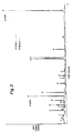
- the techniques of Prober et al. as exemplified by the Genesis 2000TM of using gel electrophoresis, the irradiation of the gel by an argon ion laser, and the detection of fluorescent emissions were used to identify the reporters on each DNA fragment.
- the sizes of the labeled fragments were determined by comparison to the mobilities of known size standards run in a parallel lane of the gel.
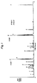
- Table I shows a representative list of detected fragments indicating the size of each fragment, the ratio of the signals from the two detectors, and the 3′ terminal base deduced from the ratio.
- the output appears as quartets of peaks because FokI generates a four-base 5′ overhang.
- the distribution of fragments and the identities of the 3′ terminal bases are exactly as predicted from the known sequence of phiX174 DNA.
- each fragment is identified not only by size but also by the identity of the labeled 3′ nucleotides.
- Phage ⁇ DNA double digested with the restriction enzymes BstEII and BamHI was prepared in the following manner. A quantity of 20 »g phage ⁇ DNA digested with the restriction enzyme BstEII (New England BioLabs; 0.4 »g/mL) was dispensed into a 1.5 mL Eppendorf tube. Following the addition of 20 »L 10X BamHI reaction buffer (1.5 M NaCl; 60 mM Tris-HCl, pH 8.0; 60 mM MgCl2; 1 mg/ml bovine serum albumin), 120 »L H2O, and 10 »L restriction enzyme BamHI (New England BiLabs; 16 units/»L), the reaction was incubated at 37°C for 2 hours.
- 20 »L 10X BamHI reaction buffer 1.5 M NaCl; 60 mM Tris-HCl, pH 8.0; 60 mM MgCl2; 1 mg/ml bovine serum albumin
- the DNA was precipitated by adding 200 »L 5 M ammonium acetate plus 1 mL ethanol, mixing, and placing on dry ice for 20 minutes. The DNA was recovered by centrifuging at 14,000 rpm for 15 minutes. After discarding the supernatant, the DNA pellet was dried under vacuum for 10 minutes, then dissolved in 79 »L TE (10 mM Tris-HCl, pH 7.4; 1 mM EDTA). The combination of BstEII and BamHI cleaves each phage ⁇ DNA molecule into 19 fragments.
- the next step is to incorporate biotin at the ends of the fragments produced by cleavage of phage ⁇ DNA with BstEII and BamHI. This was accomplished by taking the 79 »L sample containing 20 »g phage ⁇ DNA digested with BstEII and BamHI and making the following additions: 10 »L 10X Klenow buffer (0.5 M NaCl; 0.1 M Tris-HCl, pH 7.5; 0.1 M MgCl2; 0.01 M dithiothreitol), 5 »L 0.4 mM biotin-11-dUTP (Bethesda Research Laboratories), 5 »L of a solution containing 1 mM dATP, 1 mM dCTP, and 1 mM dGTP, and 1 »L of a solution containing the large fragment (Klenow fragment) of DNA polymerase I (Bethesda Research Laboratories; 6 units/»L).
- 10 »L 10X Klenow buffer 0.5 M NaCl
- the next step is to perform the secondary cleavage reaction using the restriction enzyme FokI.
- the following additions were made to the 125 »L phage ⁇ BstEII-BamHI-biotin DNA sample: 20 »L 10X FokI reaction buffer (0.2 M KCl; 0.1 M Tris-HCl, pH 7.5; 0.1 M MgCl2; 0.06 M 2-mercaptoethanol; 1 mg/ml bovine serum albumin), 35 »L H2O, and 20 »L restriction enzyme FokI (New England BiLabs; 3 units/»L). The reaction was incubated at 37°C for 3 hours.
- the DNA was precipitated by adding 200 »L 5 M ammonium acetate plus 1 mL ethanol, mixing, and storing at -20°C.
- the DNA was recovered by centrifuging at 14,000 rpm for 15 minutes. After discarding the supernatant, the DNA pellet was dried under vacuum for 10 minutes, dissolve in 200 »L TE (10 mM Tris-HCl, pH 7.4; 1 mM EDTA), and stored at 4°C.
- 200 »L TE (10 mM Tris-HCl, pH 7.4; 1 mM EDTA)
- This provides a stock solution at a DNA concentration of 0.1 »g/»L that will be referred to as phage ⁇ BstEII-BamHI-biotin-FokI.
- the next step is to use incubation with streptavidin-CrO2 particles to separate the relatively few fragments that contain biotin from the bulk of the fragments, followed by the labeling of the free ends with nucleotide-specific fluorescent reporters.
- Streptavidin was covalently attached to aminosilane coated CrO2 magnetic particles by standard crosslinking with glutaraldehyde as described in Lau et al. U.S. Patent 4,661,408.
- the DNA sample was diluted by mixing 10 »L (1 »g) of the phage ⁇ BstEII-BamHI-biotin-FokI DNA with 90 »L 100 mM NaCl; 10 mM Tris-HCl, pH 7.4; 1 mM EDTA; 100 »g/ml bovine serum albumin in a siliconized 1.5 mL Eppendorf tube.
- a suspension of streptavidin-CrO2 particles (10 »g in 10 »L) and the resulting suspension was incubated at room temperature for 15 minutes with shaking followed by 15 minutes at 37°C.
- the streptavidin-CrO2 particles bearing the biotin-containing DNA fragments were immobilized on the side of the tube by placing the tube in a magnetic rack (MAGIC® Magnetic Separation Unit, Corning Glass Works Magnetic Immunochemistries). After discarding the supernatant, the particles were washed by suspending in 100 »L 100 mM NaCl; 10 mM Tris-HCl, pH 7.4; 1 mM EDTA; 100 »g/ml bovine serum albumin, placing in the magnetic rack, and discarding the supernatant. The washing step was repeated. In order to label the DNA fragments with the fluorescent reporters, the particles were first suspended in 7 »L TE.
- the labeled fragments were eluted from the particles by suspending the particles in 5 »L 95% (v/v) formamide; 12.5 mM EDTA, incubating at 70°C for 10 minutes, placing the sample tube in the magnetic rack, and transferring the supernatant containing the labeled fragments to a new tube.
- the electrophoresis and detection conditions outlined in Example 10 were used to analyze the size and reporter identity for the labeled fragments.
- Table II shows a representative list of detected fragments indicating the size of each fragment, the ratio of the signals from the two detectors, and the 3′ terminal base deduced from the ratio. The distribution of fragments and the identities of the 3′ terminal base are as predicted from the known sequence of phage ⁇ DNA.
- This reaction was incubated at 20°C for 30 minutes.
- the DNA was precipitated by adding 100 »L 5 M ammonium acetate plus 50 »L ethanol, mixing, and storing at -20°C.
- the DNA was recovered by centrifuging at 14,000 rpm for 15 minutes. After discarding the supernatant, the DNA pellet was dried under vacuum for 10 minutes, and dissolved in 85 »L TE (10 mM Tris-HCl, pH 7.4; 1 mM EDTA).
- the DNA pellet was dried under vacuum for 10 minutes, dissolved in 100 »L TE (10 mM Tris-HCl, pH 7.4; 1 mM EDTA), and stored at 4°C. This provides a stock solution at a DNA concentration of 0.1 »g/»L that will be referred to as phage ⁇ BstEII-biotin-AluI.
- the particles were suspended in 11 »L TE and the following additions were made: 4 »L 5X Sequenase buffer (0.2 M Tris-HCl, pH 7.5; 0.05 M MgCl2; 0.25 M NaCl), 4 »L of a solution containing 100 »M 7-(SF505-Sar-AP3)ddC7GTP; 100 »M 7-(SF512-Sar-AP3)ddC7ATP; 100 »M 5-(SF519-Sar-AP3)ddCTP; and 100 »M 5-(SF526-Sar-AP3)ddTTP, and 1 »L (2 units) Sequenase (modified T7 DNA polymerase from U.S. Biochemical Corp.).
- 4 »L 5X Sequenase buffer 0.2 M Tris-HCl, pH 7.5; 0.05 M MgCl2; 0.25 M NaCl
- 4 »L of a solution containing 100 »M 7-(SF505-Sar-AP3)ddC7GTP 100 »
- Example 11 The reaction was incubated at 37°C for 1 hour.
- the sample tube was placed in the magnetic rack to immobilize the particles and the supernatant was discarded.
- the particles were washed three times with 100 »L 0.17% (w/v) Triton® X-100; 100 mM NaCl; 10 mM Tris-HCl, pH 7.4; 1 mM EDTA and the labeled fragments were eluted in 5 »L 95% (v/v) formamide; 12.5 mM EDTA.
- the electrophoresis and detection conditions outlined in Example 10 were used to analyze the size and reporter identity for the labeled fragments. Table III presents the fluorescent fragments detected in the size range of 50-320 nucleotides.
- HphI Secondary cleavage with HphI was accomplished by adding 20 »L 10X HphI reaction buffer (0.06 M KCl; 0.1 M Tris-HCl, pH 7.4; 0.1 M MgCl2; 0.01 M dithiothreitol; 2 mg/ml bovine serum albumin), 38 »L H2O, and 20 »L restriction enzyme HphI (New England BioLabs; 4 units/»L).
- the reaction was incubated at 37°C for 2 hours.
- the DNA was precipitated by adding 200 »L 5 M ammonium acetate plus 1 mL ethanol, mixing, and storing at -20°C.
- the DNA was recovered by centrifuging at 14,000 rpm for 15 minutes.
- the DNA pellet was dried under vacuum for 10 minutes, dissolved in 100 »L TE, and stored at 4°C. This provides a stock solution at a DNA concentration of 0.1 »g/»L that will be referred to as phage ⁇ BstEII-biotin-HphI.
- DNA was prepared from the Bartha strain and the Becker strain of pseudorabies virus, a herpes virus that infects pigs.
- Each type of DNA was digested with the restriction enzyme SalI by incubating 5 »g DNA in a 200-»L reaction mixture containing 150 mM NaCl, 10 mM Tris-HCl, pH 8.0, 10 mM MgCl2, 10 mM ⁇ -mercaptoethanol, 100 »g/mL bovine serum albumin, and 160 units SalI (New England BioLabs). Each reaction was incubated at 37°C for 2 hours.
- Each DNA sample was precipitated by adding 200 »L 5 M ammonium acetate plus 1 mL ethanol, mixing, and placing on dry ice for 20 minutes. The DNA was recovered by centrifuging at 14,000 rpm for 15 minutes. After discarding the supernatant, each DNA pellet was dried under vacuum for 10 minutes, then dissolved in 79 »L TE (10 mM Tris-HCl, pH 7.4; 1 mM EDTA).
- Each DNA sample was precipitated by adding 100 »L 5 M ammonium acetate plus 500 »L ethanol, mixing, and storing at -20°C.
- the DNA was recovered by centrifuging at 14,000 rpm for 15 minutes. After discarding the supernatant, each DNA pellet was dried under vacuum for 10 minutes, dissolved in 100 »L TE (10 mM Tris-HCl, pH 7.4; 1 mM EDTA), and passed through a G-50 Select-D spin column (5 Prime-3 Prime, Inc,; Paoli, PA). Each excluded volume (approx. 125 »L) was incubated at 70°C for 10 minutes, then stored at 4°C.
- each DNA pellet was dried under vacuum for 10 minutes, dissolved in 50 »L TE (10 mM Tris-HCl, pH 7.4; 1 mM EDTA), and stored at 4°C. This provides stock solutions each at a DNA concentration of 0.1 »g/»L that will be referred to as Bartha SalI-biotin-FokI and Becker SalI-biotin-FokI.
- each reaction was incubated at 42°C for 1 hour.
- this labeling protocol that contains no deoxynucleotides, only the first position in each FokI four-base overhang is labeled. This means that each labeled FokI end generates a single peak rather than the quartet of peaks observed in Examples 10 and 11.
- each sample of particles was washed three times, and the labeled fragments were eluted, then subjected to electrophoresis and fluorescence detection. Fluorescent fragments detected in the size range 50-240 nucleotides are shown in Figure 1 for the Becker sample and in Figure 2 for the Bartha sample. Comparison of these two figures shows that the two strains have 15 fragments in common.
- the Bartha sample has 2 fragments not present in the Becker sample.
- the Becker sample has 3 fragments not present in the Bartha sample.
Claims (24)
- Procédé de cartographie d'un segment d'ADN constitué de brins de nucléotides en duplex par
clivage du segment avec une première enzyme de restriction pour produire des fragments d'ADN, chacun comportant une extrémité clivée,
liaison d'un marqueur correspondant à un terminateur marqué spécifique d'un nucléotide de l'extrémité clivée dans chaque fragment,
clivage des fragments d'ADN avec une seconde enzyme de restriction pour produire des fragments courts,
séparation des fragments courts en fonction de leur taille, et
analyse des fragments courts séparés pour déterminer la présence de marqueurs, la taille et l'identité du marqueur étant une indication du caractère de chaque fragment d'ADN,
dans lequel la première enzyme de restriction est une enzyme de restriction conduisant à une coupure ambiguë à condition que la seconde enzyme de restriction soit une enzyme de restriction arbitraire. - Le procédé selon la revendication 1, dans lequel le clivage du segment produit des fragments d'ADN avec chaque extrémité clivée comportant un brin avec un résidu de nucléotides et un brin tronqué correspondant et inclut l'étape de liaison d'un nucléotide pourvu d'un marqueur au brin tronqué pour former une paire de base avec au moins un résidu de nucléotides, le marqueur étant spécifique de chaque type de nucléotide.
- Le procédé selon les revendications 1 et 2, dans lequel une troizième enzyme est utilisée en conjonction avec la première enzyme pour cliver des fragments de différentes longueurs.
- Le procédé selon la revendication 3, dans lequel l'analyse est accomplie en temps réel.
- Le procédé selon l'une quelconque des revendications 1 à 4, dans lequel l'étape de clivage du segment produit des fragments d'ADN présentant des extrémités franches.
- Le procédé selon l'une quelconque des revendications 1 à 4, dans lequel l'étape de clivage du segment produit des fragments d'ADN présentant une extrémité nucléotidique protubérante en 5'.
- Le procédé selon l'une quelconque des revendications 1 à 4, dans lequel l'étape de clivage du segment produit des fragments d'ADN présentant une extrémité nucléotidique protubérante en 3'.
- Le procédé selon la revendication 7, dans lequel la liaison du marqueur est réalisée par utilisation de polymérase présentant une activité 3'-exonucléase.
- Le procédé selon la revendication 8, dans lequel la polymérase est sélectionnée dans le groupe consistant en ADN polymérase I, ADN polymérase T7 et ADN polymérase T4.
- Le procédé selon l'une quelconque des revendications 1 à 9 comprenant: la réaction d'un mélange de segment de type ADN
et d'une première enzyme de restriction qui clive l'ADN pour fournir des fragments d'ADN présentant chacun des brins protubérants en 3' ou en 5' et un brin tronqué correspondant en 3' ou en 5', ou des extrémités franches,
l'incubation des fragments d'ADN avec un ADN polymérase et un mélange de désoxynucléotides non marqués et de didésoxynucléotides pourvus d'un marqueur spécifique pour ajouter des nucléotides aux brins tronqués complémentaires aux nucléotides présents dans les brins protubérants, la réaction des fragments d'ADN incubés avec une seconde enzyme de restriction pour produire des fragments d'ADN courts dont certains incluent lesdits didésoxynucléotides pourvus d'un marqueur et sont donc marqués,
la séparation des fragments d'ADN courts selon leur taille, et
la détection d'un marqueur pour chaque fragment marqué de façon à identifier les didésoxynucléotides terminaux ajoutés en 3',
dans lequel la première enzyme de restriction est une enzyme de restriction conduisant à une coupure ambiguë à condition que la seconde enzyme de restriction soit une enzyme de restriction arbitraire. - Le procédé selon la revendication 10, qui inclut l'étape de
corrélation de la taille et de l'identification du marqueur de chaque fragment d'ADN court marqué du segment d'ADN. - Le procédé selon les revendications 10 et 11, dans lequel on fait varier inversement le rapport didésoxynucléotides marqués aux désoxynucléotides non marqués par rapport au nombre de nucléotides dans les brins protubérants.
- Le procédé selon les revendications 10 et 11, dans lequel les fragments d'ADN réagissent seulement avec les didésoxynucléotides pourvus d'un marqueur.
- Le procédé selon l'une quelconque des revendications 10-13, dans lequel la première enzyme de restriction est une enzyme de restriction conduisant à des extrémités ambiguës.
- Le procédé selon l'une quelconque des revendications 10-13, dans lequel la première enzyme de restriction est une enzyme de restriction conduisant à une extrémité exacte.
- Un procédé de cartographie de segments d'ADN constitués de brins de nucléotides en duplex conduisant à des paires de liaison spécifiques, un membre de la paire étant immobilisé sur un support solide, par les étapes de:(a) clivage de chaque segment avec une première enzyme de restriction pour produire des fragments d'ADN comportant chacun une extrémité clivée,(b) substitution des nucléotides à l'extrémité clivée dans chaque fragment avec l'autre membre de la paire de liaison spécifique,(c) clivage des fragments d'ADN substitués avec une seconde enzyme de restriction pour produire des fragments d'ADN courts,(d) liaison des fragments courts au membre immobilisé, laissant un nucléotide terminal libre sur chaque fragment substitué,(e) séparation des fragments substitués des fragments non-substitués,(f) liaison d'un marqueur spécifique d'un nucléotide correspondant à un terminateur marqué à l'un des nucléotides terminaux libres dans chaque fragment plus court substitué,(g) séparation des fragments séparés pourvus d'un marqueur du support solide, et(h) fractionnement des fragments pourvus d'un marqueur en fonction de leur taille,dans lequel la seconde enzyme de restriction est une enzyme de restriction conduisant à une coupure ambiguë à condition que la première enzyme de restriction soit une enzyme de restriction arbitraire.
- Le procédé selon la revendication 16, qui inclut l'étape de : analyse des fragments fractionnés pour déterminer la présence de fragments marqués en fonction de leur taille.
- Le procédé selon les revendications 16 et 17, dans lequel le support solide correspond à des billes qui sont insolubles dans l'eau et stables aux conditions physiques et chimiques des étapes d, e, f et g de la revendication 16.
- Le procédé selon la revendication 18, dans lequel les billes sont du dioxyde de chrome.
- Le procédé selon la revendication 19, dans lequel les billes sont séparées de leur milieu environnant par un champ magnétique.
- Le procédé selon l'une quelconque des revendications 19-20, dans lequel l'étape de mise en contact des fragments courts avec le membre immobilisé pour lier les fragments courts substitués aux billes inclut l'étape de séparation des billes comportant les fragments courts substitués liés des fragments non-substitués.
- Le procédé selon la revendication 16, dans lequel le nucléotide terminal libre est présent sur le brin 3' de l'ADN.
- Un procédé selon l'une quelconque des revendications 16-22, comprenant les étapes de:(a) clivage de chaque segment avec une première enzyme de restriction pour produire des fragments d'ADN comportant chacun une extrémité clivée,(b) substitution des nucléotides à l'extrémité clivée dans chaque fragment avec l'autre membre de la paire de liaison spécifique,(c) clivage des fragments d'ADN substitués avec une seconde enzyme de restriction pour produire des fragments d'ADN courts, les secondes extrémités clivées étant non-substituées,(d) liaison d'un marqueur à chacune des extrémités non-substituées des fragments courts,(e) liaison des fragments courts avec des extrémités substituées au premier membre, (f) séparation des fragments substitués liés dontcertains sont pourvus d'un marqueur à une extrémité, des fragments qui sont non-substitués aux deux extrémités,(g) séparation des fragments pourvus d'un marqueur liés du support solide, et(h) fractionnement des fragments pourvus d'un marqueur en fonction de leur taille,dans lequel la seconde enzyme de restriction est une enzyme de restriction conduisant à une coupure ambiguë à condition que la première enzyme de restriction soit une enzyme de restriction arbitraire.
- Un procédé selon l'une quelconque des revendications 16-22, comprenant les étapes de:(a) clivage de chaque segment avec une première enzyme de restriction pour produire des fragments d'ADN comportant chacun une extrémité clivée,(b) substitution des nucléotides à l'extrémité clivée dans chaque fragment avec l'autre membre de la paire de liaison spécifique,(c) clivage des fragments d'ADN substitués avec une seconde enzyme de restriction pour produire des fragments d'ADN courts, liant les fragments substitués au premier membre,(d) clivage des fragments d'ADN liés avec une seconde enzyme de restriction pour produire des fragments d'ADN courts, dont certains comportent une extrémité substituée liée et une extrémité libre, et dont certains sont non-substitués aux deux extrémités,(e) séparation des fragments courts liés des fragments courts non liés,(f) liaison d'un marqueur à l'un des nucléotides de l'extrémité libre dans chaque fragment court lié,(g) séparation des fragments liés pourvus d'un marqueur lié du support solide,(h) fractionnement des fragments pourvus d'un marqueur en fonction de leur taille,dans lequel la seconde enzyme de restriction est une enzyme de restriction conduisant à une coupure ambiguë à condition que la première enzyme de restriction soit une enzyme de restriction arbitraire.
Applications Claiming Priority (4)
| Application Number | Priority Date | Filing Date | Title |
|---|---|---|---|
| US10310587A | 1987-09-28 | 1987-09-28 | |
| US103105 | 1987-09-28 | ||
| US07/185,741 US5102785A (en) | 1987-09-28 | 1988-04-25 | Method of gene mapping |
| US185741 | 1988-04-25 |
Publications (3)
| Publication Number | Publication Date |
|---|---|
| EP0309969A2 EP0309969A2 (fr) | 1989-04-05 |
| EP0309969A3 EP0309969A3 (fr) | 1991-03-06 |
| EP0309969B1 true EP0309969B1 (fr) | 1995-07-19 |
Family
ID=26800094
Family Applications (1)
| Application Number | Title | Priority Date | Filing Date |
|---|---|---|---|
| EP88115842A Expired - Lifetime EP0309969B1 (fr) | 1987-09-28 | 1988-09-27 | Méthode de mise en carte de gènes |
Country Status (8)
| Country | Link |
|---|---|
| US (1) | US5102785A (fr) |
| EP (1) | EP0309969B1 (fr) |
| JP (1) | JP2653684B2 (fr) |
| CA (1) | CA1322942C (fr) |
| DE (1) | DE3854176T2 (fr) |
| DK (1) | DK536388A (fr) |
| GR (1) | GR3017307T3 (fr) |
| IE (1) | IE69577B1 (fr) |
Families Citing this family (39)
| Publication number | Priority date | Publication date | Assignee | Title |
|---|---|---|---|---|
| FI95398C (fi) * | 1989-06-24 | 1996-01-25 | Boehringer Ingelheim Int | Menetelmä ihmisen nuhaviruksen serotyypittämiseksi |
| FR2650840B1 (fr) * | 1989-08-11 | 1991-11-29 | Bertin & Cie | Procede rapide de detection et/ou d'identification d'une seule base sur une sequence d'acide nucleique, et ses applications |
| US6074818A (en) * | 1990-08-24 | 2000-06-13 | The University Of Tennessee Research Corporation | Fingerprinting of nucleic acids, products and methods |
| EP0561796B1 (fr) * | 1990-08-24 | 1997-12-29 | The University Of Tennessee Research Corporation | Technique de l'emprunte digitale des adn utilisee en vue de leur amplification |
| US5686065A (en) * | 1991-03-27 | 1997-11-11 | Special Advanced Biomaterials, Inc. | Topical siloxane sunscreen compositions having enhanced performance and safety |
| DE4214112A1 (de) * | 1991-08-02 | 1993-02-04 | Europ Lab Molekularbiolog | Neues verfahren zur sequenzierung von nukleinsaeuren |
| GB2264496B (en) * | 1992-02-25 | 1995-10-25 | Us Energy | Sizing of fragments from a nucleic acid sequence |
| US5650274A (en) * | 1993-06-25 | 1997-07-22 | Hitachi, Ltd. | DNA analyzing method |
| US5547859A (en) * | 1993-08-02 | 1996-08-20 | Goodman; Myron F. | Chain-terminating nucleotides for DNA sequencing methods |
| GB9401200D0 (en) * | 1994-01-21 | 1994-03-16 | Medical Res Council | Sequencing of nucleic acids |
| US5714330A (en) * | 1994-04-04 | 1998-02-03 | Lynx Therapeutics, Inc. | DNA sequencing by stepwise ligation and cleavage |
| US5552278A (en) * | 1994-04-04 | 1996-09-03 | Spectragen, Inc. | DNA sequencing by stepwise ligation and cleavage |
| US5831065A (en) * | 1994-04-04 | 1998-11-03 | Lynx Therapeutics, Inc. | Kits for DNA sequencing by stepwise ligation and cleavage |
| USRE43097E1 (en) | 1994-10-13 | 2012-01-10 | Illumina, Inc. | Massively parallel signature sequencing by ligation of encoded adaptors |
| US6013445A (en) * | 1996-06-06 | 2000-01-11 | Lynx Therapeutics, Inc. | Massively parallel signature sequencing by ligation of encoded adaptors |
| US5604100A (en) * | 1995-07-19 | 1997-02-18 | Perlin; Mark W. | Method and system for sequencing genomes |
| US5763175A (en) * | 1995-11-17 | 1998-06-09 | Lynx Therapeutics, Inc. | Simultaneous sequencing of tagged polynucleotides |
| US5780231A (en) * | 1995-11-17 | 1998-07-14 | Lynx Therapeutics, Inc. | DNA extension and analysis with rolling primers |
| WO1997029211A1 (fr) * | 1996-02-09 | 1997-08-14 | The Government Of The United States Of America, Represented By The Secretary, Department Of Health And Human Services | VISUALISATION PAR RESTRICTION (RD-PCR) DES ARNm EXPRIMES DE MANIERE DIFFERENTIELLE |
| PL331513A1 (en) * | 1996-06-06 | 1999-07-19 | Lynx Therapeutics | Method of sequencing, by a ligand effect, specific encoded adapters and composition containing double-string oligonucleotidic adapters |
| GB9620769D0 (en) * | 1996-10-04 | 1996-11-20 | Brax Genomics Ltd | Nucleic acid sequencing |
| US5858671A (en) | 1996-11-01 | 1999-01-12 | The University Of Iowa Research Foundation | Iterative and regenerative DNA sequencing method |
| US6258533B1 (en) * | 1996-11-01 | 2001-07-10 | The University Of Iowa Research Foundation | Iterative and regenerative DNA sequencing method |
| WO1999000519A1 (fr) * | 1997-06-27 | 1999-01-07 | Lynx Therapeutics, Inc. | Methode d'etablissement de la cartographie des sites de restriction dans les polynucleotides |
| US6607878B2 (en) | 1997-10-06 | 2003-08-19 | Stratagene | Collections of uniquely tagged molecules |
| US6136537A (en) * | 1998-02-23 | 2000-10-24 | Macevicz; Stephen C. | Gene expression analysis |
| US6054276A (en) | 1998-02-23 | 2000-04-25 | Macevicz; Stephen C. | DNA restriction site mapping |
| AU6076900A (en) * | 1999-07-20 | 2001-02-05 | Pioneer Hi-Bred International, Inc. | Restriction site profiling |
| US20030228611A1 (en) * | 2002-05-01 | 2003-12-11 | President And Fellows Of Harvard College | Nucleic acid memory device |
| WO2005026686A2 (fr) * | 2003-09-09 | 2005-03-24 | Compass Genetics, Llc | Plate-forme analytique multiplexee |
| JP2007521833A (ja) | 2004-02-12 | 2007-08-09 | コンパス ジェネティクス エルエルシー | 配列特異的分類による遺伝的分析 |
| US7407757B2 (en) * | 2005-02-10 | 2008-08-05 | Population Genetics Technologies | Genetic analysis by sequence-specific sorting |
| EP1856293A2 (fr) * | 2005-03-16 | 2007-11-21 | Compass Genetics, Llc | Procédés et compositions pour relèves de mesures sur de multiples plate-formes analytiques |
| US9388457B2 (en) * | 2007-09-14 | 2016-07-12 | Affymetrix, Inc. | Locus specific amplification using array probes |
| US9074244B2 (en) | 2008-03-11 | 2015-07-07 | Affymetrix, Inc. | Array-based translocation and rearrangement assays |
| CA2817960C (fr) * | 2010-11-17 | 2020-06-09 | Isis Pharmaceuticals, Inc. | Modulation de l'expression de l'alpha synucleine |
| AU2017425028B2 (en) | 2017-07-28 | 2020-11-26 | Wright Medical Technology, Inc. | Joint osteotomy system and method |
| TWI809004B (zh) | 2017-11-09 | 2023-07-21 | 美商Ionis製藥公司 | 用於降低snca表現之化合物及方法 |
| MX2020007433A (es) | 2018-01-12 | 2020-09-14 | Bristol Myers Squibb Co | Oligonucleotidos antisentido que actuan sobre alfa-sinucleina, y usos de estos. |
Citations (1)
| Publication number | Priority date | Publication date | Assignee | Title |
|---|---|---|---|---|
| EP0252683A2 (fr) * | 1986-07-02 | 1988-01-13 | E.I. Du Pont De Nemours And Company | Procédé et réactifs pour la détermination de séquences ADN |
Family Cites Families (8)
| Publication number | Priority date | Publication date | Assignee | Title |
|---|---|---|---|---|
| US4672040A (en) * | 1983-05-12 | 1987-06-09 | Advanced Magnetics, Inc. | Magnetic particles for use in separations |
| CA1258611A (fr) * | 1984-01-16 | 1989-08-22 | Lloyd M. Smith | Methode de sequencage de l'adn |
| US4849077A (en) * | 1984-08-06 | 1989-07-18 | Akademie Der Wissenschaften Der Ddr | Process for solid phase-sequencing of nucleic acid fragments |
| JPS61278453A (ja) * | 1985-06-05 | 1986-12-09 | Morita Tokushu Kiko Kk | 定置式車両傾動装置 |
| ES8707343A1 (es) * | 1985-06-13 | 1987-07-16 | Amgen | Un metodo para aislar una secuencia de acido nucleico objetivo seleccionada |
| US4855225A (en) * | 1986-02-07 | 1989-08-08 | Applied Biosystems, Inc. | Method of detecting electrophoretically separated oligonucleotides |
| US4661408A (en) * | 1986-03-18 | 1987-04-28 | E.I. Du Pont De Nemours And Company | Coated chromium dioxide particles |
| US4851331A (en) * | 1986-05-16 | 1989-07-25 | Allied Corporation | Method and kit for polynucleotide assay including primer-dependant DNA polymerase |
-
1988
- 1988-04-25 US US07/185,741 patent/US5102785A/en not_active Expired - Lifetime
- 1988-09-22 CA CA000578124A patent/CA1322942C/fr not_active Expired - Fee Related
- 1988-09-27 EP EP88115842A patent/EP0309969B1/fr not_active Expired - Lifetime
- 1988-09-27 DE DE3854176T patent/DE3854176T2/de not_active Expired - Fee Related
- 1988-09-27 IE IE292088A patent/IE69577B1/en not_active IP Right Cessation
- 1988-09-27 DK DK536388A patent/DK536388A/da not_active Application Discontinuation
- 1988-09-27 JP JP63239879A patent/JP2653684B2/ja not_active Expired - Lifetime
-
1995
- 1995-09-06 GR GR950402435T patent/GR3017307T3/el unknown
Patent Citations (1)
| Publication number | Priority date | Publication date | Assignee | Title |
|---|---|---|---|---|
| EP0252683A2 (fr) * | 1986-07-02 | 1988-01-13 | E.I. Du Pont De Nemours And Company | Procédé et réactifs pour la détermination de séquences ADN |
Also Published As
| Publication number | Publication date |
|---|---|
| DK536388D0 (da) | 1988-09-27 |
| DE3854176D1 (de) | 1995-08-24 |
| GR3017307T3 (en) | 1995-12-31 |
| JPH01137983A (ja) | 1989-05-30 |
| JP2653684B2 (ja) | 1997-09-17 |
| DK536388A (da) | 1989-03-29 |
| IE882920L (en) | 1989-03-28 |
| EP0309969A3 (fr) | 1991-03-06 |
| CA1322942C (fr) | 1993-10-12 |
| IE69577B1 (en) | 1996-10-02 |
| US5102785A (en) | 1992-04-07 |
| EP0309969A2 (fr) | 1989-04-05 |
| DE3854176T2 (de) | 1996-02-15 |
Similar Documents
| Publication | Publication Date | Title |
|---|---|---|
| EP0309969B1 (fr) | Méthode de mise en carte de gènes | |
| US5625081A (en) | Fluorescent dye intermediates | |
| EP0252683B1 (fr) | Procédé et réactifs pour la détermination de séquences ADN | |
| US5151507A (en) | Alkynylamino-nucleotides | |
| US6232103B1 (en) | Methods useful for nucleic acid sequencing using modified nucleotides comprising phenylboronic acid | |
| US6573374B1 (en) | Nucleotides labelled with an infrared dye and their use in nucleic acid detection | |
| US5047519A (en) | Alkynylamino-nucleotides | |
| JP4891073B2 (ja) | 2’−ターミネーターヌクレオチドの合成及び組成物 | |
| JP2006518587A (ja) | 分子タグを使用する多重分析プラットフォーム | |
| AU4497200A (en) | Detection using degradation of a tagged sequence | |
| US20060223107A1 (en) | Detection of nucleic acid sequences by cleavage and separation of tag-containing structures | |
| US6682887B1 (en) | Detection using degradation of a tagged sequence | |
| Tong et al. | Combinatorial fluorescence energy transfer tags: New molecular tools for genomics applications |
Legal Events
| Date | Code | Title | Description |
|---|---|---|---|
| PUAI | Public reference made under article 153(3) epc to a published international application that has entered the european phase |
Free format text: ORIGINAL CODE: 0009012 |
|
| AK | Designated contracting states |
Kind code of ref document: A2 Designated state(s): BE DE FR GB GR IT LU NL |
|
| 17P | Request for examination filed |
Effective date: 19901116 |
|
| PUAL | Search report despatched |
Free format text: ORIGINAL CODE: 0009013 |
|
| AK | Designated contracting states |
Kind code of ref document: A3 Designated state(s): BE DE FR GB GR IT LU NL |
|
| 17Q | First examination report despatched |
Effective date: 19921008 |
|
| GRAA | (expected) grant |
Free format text: ORIGINAL CODE: 0009210 |
|
| AK | Designated contracting states |
Kind code of ref document: B1 Designated state(s): BE DE FR GB GR IT LU NL |
|
| ET | Fr: translation filed | ||
| REF | Corresponds to: |
Ref document number: 3854176 Country of ref document: DE Date of ref document: 19950824 |
|
| ITF | It: translation for a ep patent filed |
Owner name: ING. C. GREGORJ S.P.A. |
|
| REG | Reference to a national code |
Ref country code: GR Ref legal event code: FG4A Free format text: 3017307 |
|
| PLBE | No opposition filed within time limit |
Free format text: ORIGINAL CODE: 0009261 |
|
| STAA | Information on the status of an ep patent application or granted ep patent |
Free format text: STATUS: NO OPPOSITION FILED WITHIN TIME LIMIT |
|
| 26N | No opposition filed | ||
| REG | Reference to a national code |
Ref country code: GB Ref legal event code: 732E |
|
| PGFP | Annual fee paid to national office [announced via postgrant information from national office to epo] |
Ref country code: FR Payment date: 20000912 Year of fee payment: 13 |
|
| PGFP | Annual fee paid to national office [announced via postgrant information from national office to epo] |
Ref country code: DE Payment date: 20000918 Year of fee payment: 13 |
|
| PGFP | Annual fee paid to national office [announced via postgrant information from national office to epo] |
Ref country code: LU Payment date: 20000925 Year of fee payment: 13 |
|
| PGFP | Annual fee paid to national office [announced via postgrant information from national office to epo] |
Ref country code: GB Payment date: 20000927 Year of fee payment: 13 |
|
| PGFP | Annual fee paid to national office [announced via postgrant information from national office to epo] |
Ref country code: NL Payment date: 20000928 Year of fee payment: 13 Ref country code: GR Payment date: 20000928 Year of fee payment: 13 |
|
| PGFP | Annual fee paid to national office [announced via postgrant information from national office to epo] |
Ref country code: BE Payment date: 20001117 Year of fee payment: 13 |
|
| PG25 | Lapsed in a contracting state [announced via postgrant information from national office to epo] |
Ref country code: LU Free format text: LAPSE BECAUSE OF NON-PAYMENT OF DUE FEES Effective date: 20010927 Ref country code: GB Free format text: LAPSE BECAUSE OF NON-PAYMENT OF DUE FEES Effective date: 20010927 |
|
| PG25 | Lapsed in a contracting state [announced via postgrant information from national office to epo] |
Ref country code: GR Free format text: LAPSE BECAUSE OF NON-PAYMENT OF DUE FEES Effective date: 20010930 Ref country code: BE Free format text: LAPSE BECAUSE OF NON-PAYMENT OF DUE FEES Effective date: 20010930 |
|
| REG | Reference to a national code |
Ref country code: GB Ref legal event code: IF02 |
|
| NLS | Nl: assignments of ep-patents |
Owner name: NEN LIFE SCIENCE PRODUCTS, INC. |
|
| REG | Reference to a national code |
Ref country code: FR Ref legal event code: TP |
|
| BERE | Be: lapsed |
Owner name: NEN LIFE SCIENCE PRODUCTS INC. UNE SOC. DE L'ETAT Effective date: 20010930 |
|
| PG25 | Lapsed in a contracting state [announced via postgrant information from national office to epo] |
Ref country code: NL Free format text: LAPSE BECAUSE OF NON-PAYMENT OF DUE FEES Effective date: 20020401 |
|
| PG25 | Lapsed in a contracting state [announced via postgrant information from national office to epo] |
Ref country code: DE Free format text: LAPSE BECAUSE OF NON-PAYMENT OF DUE FEES Effective date: 20020501 |
|
| GBPC | Gb: european patent ceased through non-payment of renewal fee |
Effective date: 20010927 |
|
| PG25 | Lapsed in a contracting state [announced via postgrant information from national office to epo] |
Ref country code: FR Free format text: LAPSE BECAUSE OF NON-PAYMENT OF DUE FEES Effective date: 20020531 |
|
| NLV4 | Nl: lapsed or anulled due to non-payment of the annual fee |
Effective date: 20020401 |
|
| REG | Reference to a national code |
Ref country code: FR Ref legal event code: ST |
|
| NLV4 | Nl: lapsed or anulled due to non-payment of the annual fee |
Effective date: 20020401 |
|
| PG25 | Lapsed in a contracting state [announced via postgrant information from national office to epo] |
Ref country code: IT Free format text: LAPSE BECAUSE OF NON-PAYMENT OF DUE FEES;WARNING: LAPSES OF ITALIAN PATENTS WITH EFFECTIVE DATE BEFORE 2007 MAY HAVE OCCURRED AT ANY TIME BEFORE 2007. THE CORRECT EFFECTIVE DATE MAY BE DIFFERENT FROM THE ONE RECORDED. Effective date: 20050927 |