EP0249955B1 - Kit zur Verwendung in Sandwich-Immunoassays - Google Patents
Kit zur Verwendung in Sandwich-Immunoassays Download PDFInfo
- Publication number
- EP0249955B1 EP0249955B1 EP87108681A EP87108681A EP0249955B1 EP 0249955 B1 EP0249955 B1 EP 0249955B1 EP 87108681 A EP87108681 A EP 87108681A EP 87108681 A EP87108681 A EP 87108681A EP 0249955 B1 EP0249955 B1 EP 0249955B1
- Authority
- EP
- European Patent Office
- Prior art keywords
- guinea pig
- igg
- solid
- god
- phase
- Prior art date
- Legal status (The legal status is an assumption and is not a legal conclusion. Google has not performed a legal analysis and makes no representation as to the accuracy of the status listed.)
- Expired - Lifetime
Links
- 238000003018 immunoassay Methods 0.000 title claims description 49
- 241000700199 Cavia porcellus Species 0.000 claims description 181
- 229940027941 immunoglobulin g Drugs 0.000 claims description 126
- 239000007790 solid phase Substances 0.000 claims description 103
- 238000006243 chemical reaction Methods 0.000 claims description 97
- 210000002966 serum Anatomy 0.000 claims description 70
- 102000004190 Enzymes Human genes 0.000 claims description 42
- 108090000790 Enzymes Proteins 0.000 claims description 42
- 230000009871 nonspecific binding Effects 0.000 claims description 40
- 239000000427 antigen Substances 0.000 claims description 36
- 102000036639 antigens Human genes 0.000 claims description 36
- 108091007433 antigens Proteins 0.000 claims description 36
- 239000012634 fragment Substances 0.000 claims description 6
- 239000000243 solution Substances 0.000 description 124
- 241000283973 Oryctolagus cuniculus Species 0.000 description 63
- 239000004793 Polystyrene Substances 0.000 description 56
- 229920002223 polystyrene Polymers 0.000 description 56
- 230000000052 comparative effect Effects 0.000 description 47
- 230000002494 anti-cea effect Effects 0.000 description 43
- 229940088598 enzyme Drugs 0.000 description 41
- 238000003556 assay Methods 0.000 description 37
- 241000283707 Capra Species 0.000 description 36
- 239000012064 sodium phosphate buffer Substances 0.000 description 19
- -1 potassium ferricyanide Chemical compound 0.000 description 17
- HWYHZTIRURJOHG-UHFFFAOYSA-N luminol Chemical compound O=C1NNC(=O)C2=C1C(N)=CC=C2 HWYHZTIRURJOHG-UHFFFAOYSA-N 0.000 description 15
- 230000035945 sensitivity Effects 0.000 description 15
- 102000011022 Chorionic Gonadotropin Human genes 0.000 description 14
- 108010062540 Chorionic Gonadotropin Proteins 0.000 description 14
- KCXVZYZYPLLWCC-UHFFFAOYSA-N EDTA Chemical compound OC(=O)CN(CC(O)=O)CCN(CC(O)=O)CC(O)=O KCXVZYZYPLLWCC-UHFFFAOYSA-N 0.000 description 14
- 229940084986 human chorionic gonadotropin Drugs 0.000 description 14
- 108010015776 Glucose oxidase Proteins 0.000 description 13
- 239000004366 Glucose oxidase Substances 0.000 description 13
- 229940116332 glucose oxidase Drugs 0.000 description 13
- 235000019420 glucose oxidase Nutrition 0.000 description 13
- 108091003079 Bovine Serum Albumin Proteins 0.000 description 12
- MHAJPDPJQMAIIY-UHFFFAOYSA-N Hydrogen peroxide Chemical compound OO MHAJPDPJQMAIIY-UHFFFAOYSA-N 0.000 description 12
- FAPWRFPIFSIZLT-UHFFFAOYSA-M Sodium chloride Chemical compound [Na+].[Cl-] FAPWRFPIFSIZLT-UHFFFAOYSA-M 0.000 description 12
- 229940098773 bovine serum albumin Drugs 0.000 description 12
- 238000000034 method Methods 0.000 description 12
- PXIPVTKHYLBLMZ-UHFFFAOYSA-N Sodium azide Chemical compound [Na+].[N-]=[N+]=[N-] PXIPVTKHYLBLMZ-UHFFFAOYSA-N 0.000 description 10
- 102100023635 Alpha-fetoprotein Human genes 0.000 description 9
- 230000027455 binding Effects 0.000 description 8
- 238000001514 detection method Methods 0.000 description 8
- 241001465754 Metazoa Species 0.000 description 7
- 239000000872 buffer Substances 0.000 description 7
- 239000008351 acetate buffer Substances 0.000 description 6
- 238000005119 centrifugation Methods 0.000 description 6
- 238000002967 competitive immunoassay Methods 0.000 description 6
- 230000000694 effects Effects 0.000 description 6
- 239000012847 fine chemical Substances 0.000 description 6
- 239000011780 sodium chloride Substances 0.000 description 6
- BVKZGUZCCUSVTD-UHFFFAOYSA-L Carbonate Chemical compound [O-]C([O-])=O BVKZGUZCCUSVTD-UHFFFAOYSA-L 0.000 description 5
- WQZGKKKJIJFFOK-GASJEMHNSA-N Glucose Natural products OC[C@H]1OC(O)[C@H](O)[C@@H](O)[C@@H]1O WQZGKKKJIJFFOK-GASJEMHNSA-N 0.000 description 5
- PEEHTFAAVSWFBL-UHFFFAOYSA-N Maleimide Chemical compound O=C1NC(=O)C=C1 PEEHTFAAVSWFBL-UHFFFAOYSA-N 0.000 description 5
- 239000008103 glucose Substances 0.000 description 5
- 229940125396 insulin Drugs 0.000 description 5
- NOESYZHRGYRDHS-UHFFFAOYSA-N insulin Substances N1C(=O)C(NC(=O)C(CCC(N)=O)NC(=O)C(CCC(O)=O)NC(=O)C(C(C)C)NC(=O)C(NC(=O)CN)C(C)CC)CSSCC(C(NC(CO)C(=O)NC(CC(C)C)C(=O)NC(CC=2C=CC(O)=CC=2)C(=O)NC(CCC(N)=O)C(=O)NC(CC(C)C)C(=O)NC(CCC(O)=O)C(=O)NC(CC(N)=O)C(=O)NC(CC=2C=CC(O)=CC=2)C(=O)NC(CSSCC(NC(=O)C(C(C)C)NC(=O)C(CC(C)C)NC(=O)C(CC=2C=CC(O)=CC=2)NC(=O)C(CC(C)C)NC(=O)C(C)NC(=O)C(CCC(O)=O)NC(=O)C(C(C)C)NC(=O)C(CC(C)C)NC(=O)C(CC=2NC=NC=2)NC(=O)C(CO)NC(=O)CNC2=O)C(=O)NCC(=O)NC(CCC(O)=O)C(=O)NC(CCCNC(N)=N)C(=O)NCC(=O)NC(CC=3C=CC=CC=3)C(=O)NC(CC=3C=CC=CC=3)C(=O)NC(CC=3C=CC(O)=CC=3)C(=O)NC(C(C)O)C(=O)N3C(CCC3)C(=O)NC(CCCCN)C(=O)NC(C)C(O)=O)C(=O)NC(CC(N)=O)C(O)=O)=O)NC(=O)C(C(C)CC)NC(=O)C(CO)NC(=O)C(C(C)O)NC(=O)C1CSSCC2NC(=O)C(CC(C)C)NC(=O)C(NC(=O)C(CCC(N)=O)NC(=O)C(CC(N)=O)NC(=O)C(NC(=O)C(N)CC=1C=CC=CC=1)C(C)C)CC1=CN=CN1 NOESYZHRGYRDHS-UHFFFAOYSA-N 0.000 description 5
- 102000018071 Immunoglobulin Fc Fragments Human genes 0.000 description 4
- 108010091135 Immunoglobulin Fc Fragments Proteins 0.000 description 4
- ZMXDDKWLCZADIW-UHFFFAOYSA-N N,N-Dimethylformamide Chemical compound CN(C)C=O ZMXDDKWLCZADIW-UHFFFAOYSA-N 0.000 description 4
- 239000012506 Sephacryl® Substances 0.000 description 4
- 239000000203 mixture Substances 0.000 description 4
- 229920005654 Sephadex Polymers 0.000 description 3
- 239000012507 Sephadex™ Substances 0.000 description 3
- 210000004369 blood Anatomy 0.000 description 3
- 239000008280 blood Substances 0.000 description 3
- 239000004615 ingredient Substances 0.000 description 3
- 238000005259 measurement Methods 0.000 description 3
- 102000002260 Alkaline Phosphatase Human genes 0.000 description 2
- 108020004774 Alkaline Phosphatase Proteins 0.000 description 2
- WTDHULULXKLSOZ-UHFFFAOYSA-N Hydroxylamine hydrochloride Chemical compound Cl.ON WTDHULULXKLSOZ-UHFFFAOYSA-N 0.000 description 2
- 102000057297 Pepsin A Human genes 0.000 description 2
- 108090000284 Pepsin A Proteins 0.000 description 2
- 102000003992 Peroxidases Human genes 0.000 description 2
- BFNBIHQBYMNNAN-UHFFFAOYSA-N ammonium sulfate Chemical class N.N.OS(O)(=O)=O BFNBIHQBYMNNAN-UHFFFAOYSA-N 0.000 description 2
- 102000005936 beta-Galactosidase Human genes 0.000 description 2
- 108010005774 beta-Galactosidase Proteins 0.000 description 2
- 230000002860 competitive effect Effects 0.000 description 2
- UFULAYFCSOUIOV-UHFFFAOYSA-N cysteamine Chemical compound NCCS UFULAYFCSOUIOV-UHFFFAOYSA-N 0.000 description 2
- 239000012153 distilled water Substances 0.000 description 2
- 230000036046 immunoreaction Effects 0.000 description 2
- 239000000463 material Substances 0.000 description 2
- 229960003151 mercaptamine Drugs 0.000 description 2
- 230000036963 noncompetitive effect Effects 0.000 description 2
- 229940111202 pepsin Drugs 0.000 description 2
- 108040007629 peroxidase activity proteins Proteins 0.000 description 2
- 239000002244 precipitate Substances 0.000 description 2
- 239000012086 standard solution Substances 0.000 description 2
- XLYOFNOQVPJJNP-UHFFFAOYSA-N water Chemical compound O XLYOFNOQVPJJNP-UHFFFAOYSA-N 0.000 description 2
- HCILGDWKPMTTSN-UHFFFAOYSA-N 2-(2,5-dioxopyrrolidin-1-yl)-4-(2,5-dioxopyrrol-1-yl)butanoic acid Chemical compound O=C1CCC(=O)N1C(C(=O)O)CCN1C(=O)C=CC1=O HCILGDWKPMTTSN-UHFFFAOYSA-N 0.000 description 1
- OBYNJKLOYWCXEP-UHFFFAOYSA-N 2-[3-(dimethylamino)-6-dimethylazaniumylidenexanthen-9-yl]-4-isothiocyanatobenzoate Chemical compound C=12C=CC(=[N+](C)C)C=C2OC2=CC(N(C)C)=CC=C2C=1C1=CC(N=C=S)=CC=C1C([O-])=O OBYNJKLOYWCXEP-UHFFFAOYSA-N 0.000 description 1
- QKNYBSVHEMOAJP-UHFFFAOYSA-N 2-amino-2-(hydroxymethyl)propane-1,3-diol;hydron;chloride Chemical compound Cl.OCC(N)(CO)CO QKNYBSVHEMOAJP-UHFFFAOYSA-N 0.000 description 1
- BTBUEUYNUDRHOZ-UHFFFAOYSA-N Borate Chemical compound [O-]B([O-])[O-] BTBUEUYNUDRHOZ-UHFFFAOYSA-N 0.000 description 1
- DGAQECJNVWCQMB-PUAWFVPOSA-M Ilexoside XXIX Chemical compound C[C@@H]1CC[C@@]2(CC[C@@]3(C(=CC[C@H]4[C@]3(CC[C@@H]5[C@@]4(CC[C@@H](C5(C)C)OS(=O)(=O)[O-])C)C)[C@@H]2[C@]1(C)O)C)C(=O)O[C@H]6[C@@H]([C@H]([C@@H]([C@H](O6)CO)O)O)O.[Na+] DGAQECJNVWCQMB-PUAWFVPOSA-M 0.000 description 1
- 238000010521 absorption reaction Methods 0.000 description 1
- 108010026331 alpha-Fetoproteins Proteins 0.000 description 1
- 150000008064 anhydrides Chemical class 0.000 description 1
- 230000000593 degrading effect Effects 0.000 description 1
- 230000001419 dependent effect Effects 0.000 description 1
- 238000010586 diagram Methods 0.000 description 1
- YAGKRVSRTSUGEY-UHFFFAOYSA-N ferricyanide Chemical compound [Fe+3].N#[C-].N#[C-].N#[C-].N#[C-].N#[C-].N#[C-] YAGKRVSRTSUGEY-UHFFFAOYSA-N 0.000 description 1
- MHMNJMPURVTYEJ-UHFFFAOYSA-N fluorescein-5-isothiocyanate Chemical compound O1C(=O)C2=CC(N=C=S)=CC=C2C21C1=CC=C(O)C=C1OC1=CC(O)=CC=C21 MHMNJMPURVTYEJ-UHFFFAOYSA-N 0.000 description 1
- 230000003993 interaction Effects 0.000 description 1
- 238000004445 quantitative analysis Methods 0.000 description 1
- 230000002285 radioactive effect Effects 0.000 description 1
- 238000011946 reduction process Methods 0.000 description 1
- 238000000926 separation method Methods 0.000 description 1
- 238000002791 soaking Methods 0.000 description 1
- 229910052708 sodium Inorganic materials 0.000 description 1
- 239000011734 sodium Substances 0.000 description 1
- 239000007787 solid Substances 0.000 description 1
- 238000001179 sorption measurement Methods 0.000 description 1
Images

Classifications
-
- G—PHYSICS
- G01—MEASURING; TESTING
- G01N—INVESTIGATING OR ANALYSING MATERIALS BY DETERMINING THEIR CHEMICAL OR PHYSICAL PROPERTIES
- G01N33/00—Investigating or analysing materials by specific methods not covered by groups G01N1/00 - G01N31/00
- G01N33/48—Biological material, e.g. blood, urine; Haemocytometers
- G01N33/50—Chemical analysis of biological material, e.g. blood, urine; Testing involving biospecific ligand binding methods; Immunological testing
- G01N33/53—Immunoassay; Biospecific binding assay; Materials therefor
- G01N33/543—Immunoassay; Biospecific binding assay; Materials therefor with an insoluble carrier for immobilising immunochemicals
- G01N33/54393—Improving reaction conditions or stability, e.g. by coating or irradiation of surface, by reduction of non-specific binding, by promotion of specific binding
-
- G—PHYSICS
- G01—MEASURING; TESTING
- G01N—INVESTIGATING OR ANALYSING MATERIALS BY DETERMINING THEIR CHEMICAL OR PHYSICAL PROPERTIES
- G01N33/00—Investigating or analysing materials by specific methods not covered by groups G01N1/00 - G01N31/00
- G01N33/48—Biological material, e.g. blood, urine; Haemocytometers
- G01N33/50—Chemical analysis of biological material, e.g. blood, urine; Testing involving biospecific ligand binding methods; Immunological testing
- G01N33/53—Immunoassay; Biospecific binding assay; Materials therefor
- G01N33/563—Immunoassay; Biospecific binding assay; Materials therefor involving antibody fragments
Definitions
- This invention relates to sandwich immunoassays utilizing immunoreaction for measuring an extremely small quantity of an ingredient included in blood or other organic samples and, more particularly, to an improved kit for use in such sandwich immunoassays to increase the accuracy of measurement of the ingredient.
- Immunoassays have been used widely for measuring an extremely small quantity of an ingredient included in blood or other organic samples by utilizing immunoreaction or antigen-antibody reaction. Such immunoassays are generally of the two types; the “competitive immunoassay” and the “non-competitive immunoassay”.
- the competitive immunoassay employs a predetermined quantity of labeled antigen which binds to a predetermined quantity of antibody in a competitive manner with target antigen. The quantity of the target antigen is measured from the quantity of the labeled antigen binding to the antibody or the quantity of the labeled antigen which does not bind to the antibody.
- the labeled antigen is required to have a high concentration for its reaction to the antibody.
- the competitive immunoassay exhibits a poor measurement sensitivity as low as about a twentieth of the quantity of the labeled antigen.
- the non-competitive immunoassay is represented by a sandwich immunoassay which employs a solid phase coated on its surface with an excessive quantity of insolubilized antibody to trap a target antigen to be measured.
- the solid phase is washed and then incubated with an excessive amount of labeled antibody so that the labeled antibody can react with the trapped target antigen.
- the excessive labeled antibody is washed out away from the solid phase.
- the quantity of the target antigen is measured from the quantity of the label provided on the labeled antibody binding to the antigen.
- the sandwich immunoassay can provide a high measurement sensitivity, there is a tendency of non-specific binding of the labeled antibody to the solid phase, causing a considerable reduction in the sensitivity and measurable range of the sandwich immunoassay.
- a sandwich enzyme immunoassay employing solid-phase and labeled antibodies originated from guinea pig is disclosed in J. Immunol. Methods 83/2, 1985, 327 - 336.
- EP-A-O 125 893 describes a method for the quantitative analysis of antigen by an enzyme-antibody bridge method.
- a second antibody in a solution which can contain normal animal serum either is added to an insoluble carrier bound to a first antibody and to an antigen or is added simultaneously with the antigen to the insoluble carrier bound to the first antibody.
- US-A-4 414 324 concerns an immunoassay sample determination process wherein one of the components of an antigen-antibody reaction is insolubilized on the walls of a vessel, the other component is labeled and the sample being determined is added together with the labeled component to the vessel.
- the labeled component binds in a competitive manner with the sample to the insolubilized component.
- a site-deactivating medium e.g. an animal-derived serum, is covalently bound to the walls of the vessel and then the insolubilized component is covalently attached to said medium.
- kits for use in a sandwich immunoassay employing labeled and solid-phase antibodies reactive with an antigen in the presence of a reaction solution to bind the antigen between the labeled and solid-phase antibodies, said kit containing a solid-phase antibody having the antibody bound to a solid-phase, a labeled antibody having a label enzyme bound to the antibody at least one of the labeled and solid-phase antibodies being originated from guinea pig, and a reaction solution containing normal guinea pig serum for reducing the degree of non-specific binding of the labeled antibody to the solid phase.
- a target antigen is assayed by a sandwich enzyme immunoassay (EIA) technique employing first and second antibodies reactive in a specific manner with the target antigen in the presence of a reaction solution to trap the target antigen between the first and second antibodies.
- the antibodies are obtained from serum drawn from a selected animal into which the target antigen is injected.
- the first antibody is referred to as a solid-phase antibody which is coated on a solid phase such is polystyrene ball.
- the second antibody is referred to as a labeled antibody which has a label enzyme bound thereon.
- the label enzyme may be glucose oxidase.
- reaction solution A is 0.056 mol/l sodium phosphate buffer (pH 6.3) containing 0.1% sodium aside, 0.2% bovine serum albumin (BSA) and 0.337% NaCl. This reaction solution is referred to as reaction solution A.
- Guinea pig anti-hCG immunoglobulin G (IgG) is purified from the drawn serum in the following manner. First of all. 2 ml saturated ammonium sulfate is dripped into 2 ml of serum. The resulting solution is agitated slowly at a temperature of 4 o C for two or three hours. The resulting cloudy solution is then placed in a centrifugation tube. Centrifugation is performed at a temperature of 4 o C with a contrifugal separator rotating at a speed of 3000 rpm for 15 minutes.
- the supernatuant is removed from the centrifugation tube, whereas the precipitate is dissolved with 2 ml of 0.1 mol/l sodium phosphate buffer (pH 6.0) containing 5 mmol/l ethylenediaminetetraacetic acid (EDTA).
- the resulting solution is eluted at a flow rate of 6 ml/hour on Sephacryl S-300 (a trademark of Pharmacia Fine Chemicals, Sweden) column (1 x 90 cm) equilibrated with 0.1 mol/l sodium phosphate buffer (pH 6.0) containing 5 mmol/l ethylenediaminetetraacetic acid (EDTA).
- the eluted solution is divided into 1 ml of samples.
- the guinea pig anti-hCG IgG fractions, which absorb light at 280 nm, are gathered from the respective samples.
- the resulting guinea pig anti-hCG IgG is coated through physical absorption on a solid phase by soaking a polystyrene ball (6.5 mm in diameter) available from Ichiko Co., Japan at a temperature of 4 o C in 0.1 mol/l sodium phosphate buffer (pH 7.5) containing 0.1 mg/ml guinea pig anti-hCG IgG all the night through.
- the anti-hOC Igc-coated polystyrene ball is washed three times in 0.1 mol/l sodium phosphate buffer (pH 7.5) and then washed three times with 0.01 mol/l sodium phosphate buffer (pH 7.0) containing 0.1 mol/l NaCl, 0.1% bovine serum albumin (BSA) and 0.1% NaN3.
- the washed polystyrene ball is stored at a temperature of 4 o C in 0.01 mol/l sodium phosphate buffer (pH 7.0) containing 0.1 mol/l NaCl, 0.1% bovine serum albumin (BSA) and 0.1% NaN3.
- the labeled antibody, guinea pig anti-hCG IgG-GOD is prepared through the following three steps:
- the labeled antibody (guinea pig anti-hCG IgG-GOD) is bound through the antigen (hCG) to the solid-phase antibody (guinea pig anti-hCG IgG) in the presence of the reaction solution A (0.056 mol/l sodium phosphate buffer (pH 6.3) containing 0.1% sodium azide. 0.2% bovine serum albumin (BSA) and 0.337% NaCl) in the following manner.
- the anti-hCG IgG-coated polystyrene ball is incubated at room temperature with hCG (antigen) diluted with the reaction solution A. The polystylene ball is then washed three times with distilled water.
- the washed polystyrene ball is incubated at room temperature with anti-hCG IgG-GOD (labeled antibody) in the reaction solution A.
- the polystyrene ball is then washed again with distilled water and transferred into another clean assay tube.
- a chemiluminescent technique is used to determine the quantity of the label enzyme; that is, the quantity of the antigen.
- 0.3 ml of 0.01 mol/l acetate buffer (pH 5.1) containing 0.5 mol/l glucose is added to the assay tube.
- the assay tube is held stationary at a temperature of 37 o C for two hours.
- the resulting solution is taken to prepare a 0.1 ml sample.
- the sample is added with 0.5 ml of 0.2 mol/l carbonate buffer (pH 9.8) containing 2 x 10 ⁇ 7 mol/l luminol and then 0.5 ml of 6 x 10 ⁇ 3 mol/l potassium ferricyanide.
- the concentration of the resulting hydrogen peroxide is measured from the light emitted upon chemiluminescent reaction of the luminol and potassium ferricyanide and the hydrogen peroxide by a Luminometer UPD-8000 (a trademark of Meidensha Electric Mfg., Co., Japan) which starts its operation 15 second after the luminol and potassium ferricyanide are added to the assay tube and counts the light emitted for 30 seconds.
- a Luminometer UPD-8000 (a trademark of Meidensha Electric Mfg., Co., Japan) which starts its operation 15 second after the luminol and potassium ferricyanide are added to the assay tube and counts the light emitted for 30 seconds.
- antigen has been described in connection with human chorionic gonadotropin (hCG), it is to be noted that it may be human chorionic gonadotropin ⁇ (hCG ⁇ ), ⁇ -fetoprotein (AFP), anti-carcinoembryonic antigen (CEA) or the like.
- Anti-AFp IgG is purified from serum drawn from selected one of animals including guinea pig, rabbit, goat and the like into which AFP is injected six times at intervals of one week and a booster is injected on the seventh week.
- Anti-CEA IgG is purified from serum drawn from selected one of animals including guinea pig, rabbit, goat and the like into which CEA is injected six times at intervals of one week and a booster is injected on the seventh week,
- EIA sandwich enzyme immunoassay
- the degree of non-specific binding of the labeled antibody to the solid phase was determined.
- an IgG-coated polystyrene ball was incubated at room temperature with IgG-GOD diluted hundredfold with 0.3 ml of reaction solution A in an assay tube all the night through.
- the polystyrene ball was washed three times with the reaction solution A and then transferred into another clean assay tube.
- 0.3 ml of 0.01 mol/l acetate buffer (pH 5.1) containing 0.5 mol/l glucose was added to the assay tube.
- the assay tube was held stationary at a temperature of 37 o C far two hours.
- the resulting solution was taken to prepare a 0.1 ml sample.
- the sample was added with 0.5 ml of 0.2 mol/l carbonate buffer (pH 9.8) containing 2x10 ⁇ 7 mol/l luminol and then with 0.5 ml of 6x10 ⁇ 3 mol/l potassium ferricyanide.
- the light emitted upon chemiluminescent reaction of the luminol and potassium ferricyanide and the resulting hydrogen peroxide was measured by a Luminometer UPD-8000 which started its operation 15 seconds after the luminol and pltassium ferricyanide were added to the assay tube and counted the light emitted for 30 seconds.
- the degree of non-specific binding of the labeled antibody to the solid phase was represented by the non-specific bonding ratio given as C1/C2 x 100 (%) where C1 is the number of counts measured for the labeled antibody binding to the polystyrene ball and C2 is the number of counts corresponding to the whole amount of the labeled antibody used in the assay tube.
- the solid-phase antibody used was originated from guinea pig and the labeled antibody used was originated from guinea pig.
- Example 1 The labeled antibody used was normal guinea pig IgG-GOD and the solid-phase antibody used was normal guinea pig IgG.
- the normal guinea pig IgG was Miles Co,, Lot No. 29.
- a normal Guinea pig IgG-coated polystyrene ball was incubated with the normal guinea pig IgG-GOD in the presence of the reaction solution A mixed with guinea pig serum.
- Example 2 The labeled antibody used was guinea pig anti-hCG IgG-GOD and the solid-phase antibody used was guinea pig anti-hCG IgG.
- a guinea pig anti-hCG IgG-coated polystyrene ball was incubated with the guinea pig anti-hCG IgG-GOD in the presence of the reaction solution A mixed with guinea pig serum.
- Example 3 The labeled antibody used was guinea pig anti-AFP IgG-GOD and the solid-phase antibody used was guinea pig anti-AFP IgG.
- a guinea pig anti-AFP IgG-coated polystyrene ball was incubated with the guinea pig anti-AFP IgG-GOD in the presence of a reaction solution.
- Example 4 The labeled antibody used was guinea pig anti-CEA IgG-GOD and the solid-phase antibody used was guinea pig anti-CEA IgG.
- a guinea pig anti-CEA IgG-coated polystyrene ball was incubated with the guinea pig anti-CEA IgG-GOD in the presence of the reaction solution A mixed with guinea pig serum.
- Example 5 The labeled antibody used was guinea pig anti-insulin IgG-GOD and the solid-phase antibody was guinea pig anti-insulin IgG.
- the guinea pig anti-insulin IgG was Milea Co., Lot No. 23.
- a guinea pig anti-insulin IgG-coated polystyrene ball was incubated with the guinea pig anti-insulin IgG-GOD in the presence of the reaction solution A mixed with guinea pig serum.
- Comparative Example 1 The labeled antibody used was normal rabbit IgG-GOD and the solid-phase antibody used was normal rabbit IgG.
- the normal rabbit IgG was Miles Co., Lot No. 0019.
- a normal rabbit IgG-coated polystyrene ball was incubated with the normal rabbit IgG-GOD in the presence of the reaction solution A mixed with guinea pig serum.
- Comparative Example 2 The labeled antibody used was rabbit anti-hCG IgG-GOD and the solid-phase antibody used was rabbit anti-hCG IgG.
- the rabbit anti-hCG IgG was EY Co., Lot No. 021304.
- a rabbit anti-hCG IgG-coated polystyrene ball was incubated with the rabbit anti-hCG IgG-GOD in the presence of the reaction solution A mixed with guinea pig serum.
- Comparative Example 3 The labeled antibody used was rabbit anti-AFP IgG-GOD and the solid-phase antibody used was rabbit anti-AFP IgG.
- the rabbit anti-AFP IgG was Nordic Co., Lot No. 16-184.
- a rabbit anti-AFP IgG-coated polystyrene ball was incubated with the rabbit anti-AFP IgG-GOD in the presence of the reaction solution A mixed with guinea pig serum.
- Comparative Example 4 The labeled antibody used was rabbit anti-CEA IgG-GOD and the solid-phase antibody used was rabbit anti-CEA IgG.
- the rabbit anti-CEA IgG was Zymed Co.. Lot No. 40925.
- a rabbit anti-CEA IgG-coated polystyrene ball was incubated with the rabbit anti-CEA IgG-GOD in the presence of the reaction solution A mixed with guinea pig serum.
- the solid-phase antibody used was originated from guinea pig and the labeled antibody used was originated from rabbit or goat.
- Example 6 The labeled antibody used was rabbit anti-hCG IgG-GOD and the solid-phase antibody used was guinea pig anti-hCG IgG.
- the rabbit anti-hCG IgG was EY Co., Lot No. 021304.
- a guinea pig anti-hCG IgG-coated polystyrene ball was incubated with the rabbit anti-hCG IgG-GOD in the presence of the reaction solution A mixed with guinea pig serum.
- Example 7 The labeled antibody used was rabbit anti-AFP IgG-GOD and the solid-phase antibody used was guinea pig anti-AFP IgG.
- the rabbit anti-AFP IgG was Nordic Co., Lot No. 16-184.
- a guinea pig anti-AFP IgG-coated polystyrene ball was incubated with the rabbit anti-AFP IgG-GOD in the presence of the reaction solution A mixed with guinea pig serum.
- Example 8 The labeled antibody used was goat anti-AFP IgG-GOD and the solid-phase antibody used was guinea pig anti-AFP IgG.
- the goat anti-AFP IgG was Cappel Co., Lot No. 20176.
- a guinea pig anti-AFP IgG-coated polystyrene ball was incubated with the goat anti-AFP IgG-GOD in the presence of the reaction solution A mixed with guinea pig serum.
- Example 9 The labeled antibody used was goat anti-hCG IgG-GOD and the solid-phase antibody used was guinea pig anti-hCG IgG.
- a guinea pig anti-hCG IgG-coated polystyrene ball was incubated with the goat anti-hCG IgG-GOD in the present of the reaction solution A mixed with guinea pig serum.
- the solid-phase antibody used was originated from rabbit or goat and the labeled antibody used was originated from guinea pig.
- Example 10 The labeled antibody used was guinea pig anti-hCG IgG-GOD and the solid-phase antibody used was rabbit anti-hCG IgG.
- the rabbit anti-hCG IgG was EY Co., Lot No. 021304.
- a rabbit anti-hCG IgG-coated polystyrene ball was incubated with the guinea pig anti-hCG IgG-GOD in the presence of the reaction solution A mixed with guinea pig serum.
- Example 11 The labeled antibody used was guinea pig anti-AFP IgG-GOD and the solid-phase antibody used was rabbit anti-AFP IgG.
- the rabbit anti-AFP IgG was Nordic Co., Lot No. 16-134.
- a rabbit anti-AFP IgG-coated polystyrene ball was incubated with the guinea pig anti-AFP IgG-GOD in the presence of the reaction solution A mixed with guinea pig serum.
- Example 12 The labeled antibody used was guinea pig anti-APP IgG-GOD and the solid-phase antibody used was goat anti-AFP IgG.
- the goat anti-AFP IgG was Cappel Co., Lot No. 20176.
- a goat anti-APP IgG-coated polystyrene ball was incubated with the guinea pig anti-AFP IgG-GOD in the presence of the reaction solution A mixed with guinea pig serum.
- Comparative Example 5 The labeled antibody used was rabbit anti-hCG IgG-GOD and the solid-phase antibody used was rabbit anti-hCG IgG. A rabbit anti-hCG IgG-coated polystyrene ball was incubated with the rabbit anti-hCG IgG-GOD in the presence of the reaction solution A mixed with guinea pig serum.
- Comparative Example 6 The labeled antibody used was rabbit anti-AFP IgG-GOD and the solid-phase antibody used was rabbit anti-AFP IgG. A rabbit anti-AFP IgG-coated Polystyrene ball was incubated with the rabbit anti-AFP IgG-GOD in the presence of the reaction solution A mixed with guinea pig serum.
- Comparative Example 7 The labeled antibody used was goat anti-AFP IgG-GOD and the solid-phase antibody used was goat anti-AFP IgG. A goat anti-AFP IgG-coated polystyrene ball was incubated with the goat anti-AFP IgG-GOD in the presence of the reaction solution A mixed with guinea pig serum.
- the non-specific binding percentage ranges from 0.078% to 0.190% for combinations where neither the solid-phase antibody nor the labeled antibody is originated from guinea pig.
- the non-specific binding percentage range from 0.0089% to 0.023% for combinations where at least one of the solid-phase antibody and the labeled antibody is originated from guinea pig. It can be seen from the test results that the degree of non-specific binding of the labeled antibody to the solid phase can be reduced to a remarkable extent when at least one of the solid-phase antibody and the labeled antibody is originated from guinea pig.
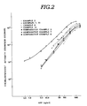
- the sensitivity and measurable range of such sandwich enzyme immunoassay was determined.
- an anti-hCG IgG-coated polystyrene ball was incubated at room temperature with 0.1 ml of hCG standard solution diluted with 0.2 ml of reaction solution A in an assay tube for six hours.
- the polystyrene ball was washed three times with the reaction solution A.
- the washed polystyrene ball was incubated with 0.1 ml of anti-hCG IgG-GOD labelled antibody diluted at a proper ratio with the reaction solution A.
- the assay tube was held stationary at room teperature all the night through.
- the polystyrene ball was then washed three times with the reaction solution A and transferred into another clean assay tube. Following this, 0.3 ml of 0.01 mol/l acetate buffer (pH 5.1) containing 0.5 mol/l glucose was added to the assay tube. The assay tube was held stationary at a temperature of 37°C for two hours. 0.1 ml of the reaction solution was pipetted into another assay tube. The tube was added with 0.5 ml of 0.2 mol/l carbonate buffer (pH 9.8) containing 2x10 ⁇ 7 mol/l luminol and then with 0.5 ml of 6x10 ⁇ 3 mol/l potassium ferricyanide.
- the light emitted upon chemiluminescent reaction of the luminol and the resulting hydrogen peroxide was measured by a Luminometer UPD-800 which started its operation 15 seconds after the luminol and potassium ferricyanide were added to the assay tube and counted the light emitted for 30 seconds. Similar procedures are repeated for anti-AFP IgG antibodies and anti-CEA IgG antibodies.
- Figs. 1 and 2 The results of the sandwich enzyme immunoassay sensitivity and measurable range are illustrated in Figs. 1 and 2.
- Fig. 1 relates to Examples 2, 6, 9 and 10 and Comparative Examples 2 and 5 where the antigen used was hCG.
- Fig. 2 relates to Examples 3, 7. 8. 11 and 12 and Comparative Examples 3, 6 and 7 where the antigen used was AFP. It can be seen from these test results that the sensitivity and measurable range of the sandwich enzyme immunoassay are superior when at least one of the solid-phase antibody and the labeled antibody is originated from guinea pig to those obtained when neither the solid-phase antibody nor the labeled antibody is originated from guinea pig.
- Sandwich enzyme immunoassay was repeated ten times within a day (within assay) using the antibodies in Examples 2, 6, 9 and 10 and Comparative Examples 2 and 5. The results are shown in Tables 2 and 3. Similarly, sandwich enzyme immunoassay was repeated six times on different six days (between assay) using the antibodies in Examples 2, 6, 9 and 10 and comparative Examples 2 and 5. The results are shown in Tables 4 and 5.
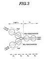
- the IgG structure can be divided into an antigen-binding F(ab')2 fragment and a crystallizable (Fc) fragment when it is held at a temperature of 37 o C for 18 hours in the presence of pepsin.
- the Fc fragment has a portion P4 for adsorption to a cell.
- the Fc fragment has no ability of binding to antigen and is crystallized at low temperature.
- the F(ab')2 fragment can be divided under a reduction process, for example, in the presence of mercaptoethylamine, into two Fab' fragments having a portion P1 capable of binding to antigen.
- the Fab' fragment is prepared as the result of separation of the Fc fragment from IgG in the following manner: First of all, 2 ml saturated ammonium sulfate is dripped into 2 ml of the serum drawn from a selected animal as described previously. The resulting solution is agitated slowly at a temperature of 4 o C for two or three hours. The resulting cloudy solution is then placed in a centrifugation tube.
- Centrifugation is performed at a temperature of 4 o C with a contrifugal separator rotating at a speed of 3000 rpm for 15 minutes.
- the supernatuant is removed from the centrifugation tube, whereas the precipitate is dissolved with 2 ml of 0.1 mol/l sodium phosphate buffer (pH 6.0) containing 5 mmol/l ethylenediaminetetraacetic acid (EDTA).
- EDTA ethylenediaminetetraacetic acid
- the resulting solution is eluted at a flow rate of 6 ml/hour on a Sephacryl S-300 (a trademark of Pharmacia Fine Cehmicals, Sweden) column (1 x 90 cm) equilibrated with 0.1 mol/l acetate buffer (pH 4.5) containing 0.1 mmol/l NaCl.
- the eluted solution is divided into 1 ml of samples.
- the IgG fractions, which absorb light at 280 nm, are gathered from the respective samples.
- 0.1 ml of 3 mg/ml pepsin solution is added to the gathered IgG.
- the resulting solution is incubated at a temperature of 37 o C for 18 hours.
- the resulting solution is eluted at a flow rate of 6 ml/hour on a Sephacryl S-200 (a trademark of Pharmacia Fine Chemicals, Sweden) column (1 x 90 cm) equilibrated with 0.1 mol/l sodium phosphate buffer (pH 7.5).
- the eluted solution is divided into 1 ml of samples.
- the F(ab')2 fractions which absorb light at 280 nm, are gathered from the respective samples.
- the gathered F(ab')2 is added with 0.05 ml of 0.1 mol/l mercaptoethylamine and incubated at a temperature of 37 o C for 90 minutes.
- the resulting solution is desalted at a flow rate of 6 ml/hour on a Sephadex G-25 (a trademark of Pharmacia Fine Chemicals. Sweden) column (1 x 30 cm) equilibrated with 0.1 mol/l sodium phosphate buffer (pH 6.0).
- the desalted solution is divided into 1 ml of Fab' samples.
- Example 13 The labeled antibody used was guinea pig anti-AFP Fab'-GOD and the solid-phase antibody used was guinea pig anti-AFP IgG.
- a guinea pig anti-AFP IgG-coated polystyrene ball was incubated with the guinea pig anti-AFP Fab'-GOD in the presence of the reaction solution A mixed with guinea pig serum.
- Example 14 The labeled antibody used was guinea pig anti-AFP Fab'-GOD and the solid-phase antibody used was rabbit anti-AFP IgG. A rabbit anti-AFP IgG-coated polystyrene ball was incubated with the guinea pig anti-AFP Fab'-GOD in the presence of the reaction solution A mixed with guinea pig serum.
- Example 15 The labeled antibody used was guinea pig anti-AFP Fab'-GOD and the solid-phase antibody used was goat anti-AFP IgG. A goat anti-AFP IgG-coated polystyrene ball was incubated with the guinea pig anti-AFP Fab'-GOD in the presence of the reaction solution A mixed with guinea pig serum.
- Example 16 The labeled antibody used was rabbit anti-AFP Fab'-GOD and the solid-phase antibody used was guinea pig anti-AFP IgG. A guinea pig anti-AFP IgG-coated polystyrene ball was incubated with the rabbit anti-AFP Fab'-GOD in the presence of the reaction solution A mixed with guinea pig serum.
- Example 17 The labeled antibody used was goat anti-AFP Fab'-GOD and the solid-phase antibody used was guinea pig anti-AFP IgG. A guinea pig anti-AFP IgG-coated polystyrene ball was incubated with the goat anti-AFP Fab'-GOD in the presence of the reaction solution A mixed with guinea pig serum.
- Example 18 The labeled antibody used was guinea pig anti-CEA Fab'-GOD and the solid-phase antibody used was guinea pig anti-CEA IgG.
- a guinea pig anti-CEA IgG-coated polystyrene ball was incubated with the guinea pig anti-CEA Feb'-GOD in the presence of the reaction solution A mixed with guinea pig serum.
- Example 19 The labeled antibody used was guinea pig anti-CEA Fab'-GOD and the solid-phase antibody used was rabbit anti-CEA IgG. A rabbit anti-CEA IgG-coated polystyrene ball was incubated with the guinea pig anti-CEA Fab'-GOD in the presence of the reaction solution A mixed with guinea pig serum.
- Example. 20 The labeled antibody used was guinea pig anti-CEA Fab'-GOD and the solid-phase antibody used was goat anti-CEA IgG. A goat anti-CEA IgG-coated polystyrene ball was incubated with the guinea pig anti-CEA Fab'-GOD in the presence of the reaction solution A mixed with guinea pig serum.
- Example 21 The labeled antibody used was rabbit anti-CEA Fab'-GOD and the solid-phase antibody used was guinea pig anti-CEA IgG. A guinea pig anti-CEA IgG-coated polystyrene ball was incubated with the rabbit anti-CEA Fab'-GOD in the presence of the reaction solution A mixed with guinea pig serum.
- Example 22 The labeled antibody used was goat anti-CEA Fab'-GOD and the solid-phase antibody used was guinea pig anti-CEA IgG. A guinea pig anti-CEA IgG-coated polystyrene ball was incubated with the goat anti-CEA Feb'-GOD in the presence of the reaction solution A mixed with guinea pig serum.
- comparative Example 8 The labeled antibody used was guinea pig anti-AFP IgG-GOD and the solid-phase antibody used was guinea pig anti-AFP IgG.
- a guinea pig anti-AFP IgG-coated polystyrene ball was incubated with the guinea pig anti-AFP IgG-GOD in the presence of the reaction solution A mixed with guinea pig serum.
- Comparative Example 9 The labeled antibody used was rabbit anti-AFP Fab'-GOD and the solid-phase antibody used was rabbit anti-AFP IgG. A rabbit anti-AFP IgG-coated polystyrene ball was incubated with the rabbit anti-AFP Fab'-GOD in the presence of the reaction solution A mixed with guinea pig serum.
- Comparative Example 10 The labeled antibody used was goat anti-AFP Fab'-GOD and the solid-phase antibody used was goat anti-AFP IgG. A goat anti-AFP IgG-coated polystyrene ball was incubated with the goat anti-AFP Fab'-GOD in the presence of a reaction solution.
- Comparative Example 11 The labeled antibody used was guinea pig anti-CEA IgG-GOD and the solid-phase antibody used was guinea pig anti-CEA IgG.
- a guinea pig anti-CEA IgG-coated polystyrene ball was incubated with the guinea pig anti-CEA IgG-GOD in the presence of the reaction solution A mixed with guinea pig serum.
- Comparative Example 12 The labeled antibody used was rabbit anti-CEA Fab'-GOD and the solid-phase antibody used was rabbit anti-CEA IgG. A rabbit anti-CEA IgG-coated polystyrene ball was incubated with the rabbit anti-CEA Fab'-GOD in the presence of the reaction solution A mixed with guinea pig serum.
- Comparative Example 13 The labeled antibody used was goat anti-CEA Fab'-GOD and the solid-phase antibody used was goat anti-CEA IgG. A goat anti-CEA IgG-coated polystyrene ball was incubated with the goat anti-CEA Fab'-GOD in the presence of the reaction solution A mixed with guinea pig serum.
- the non-specific binding percentages of the Comparative Examples 9, 10, 12 and 13 where the labeled antibody was Fab'-GOD are substantially the same as those of the Comparative Examples 8 and 11 where both of the IgG solid-phase antibody and the IgG-GOD labeled antibody were originated from guinea pig. It can be seen from the test results related to the Examples 13-22 that the degree of non-specific binding of the labeled antibody to the solid phase can be reduced to a remarkable extent when at least one of the Fab'-GOD labeled antibody and the IgG solid-phase antibody are originated from guinea pig.
- Measurable Range CEA (ng/ml) 18 0.1 - 100 11 2.5 - 250 19 0.1 - 100 12 1.0 - 250 20 0.1 - 100 13 5.0 - 250 21 0.1 - 100 22 0.1 - 100 TABLE 9
- the measurable range and detection limit of sandwich enzyme immunoassay are superior when the labeled antibody used is Fab'-GOD to those obtained when the labeled antibody used is IgG-GOD.
- the measurable range and detection limit of sandwich enzyme immunoassay are superior when at least one of the labeled antibody (Fab'-GOD) and the solid-phase antibody (IgG) is originated from guinea pig to those obtained when the labeled antibody is Fab'-GOD originated from rabbit and the solid-phase antibody (IgG) is originated from rabbit or when the labeled antibody (Fab'-GOD) is originated from goat and the solid-phase antibody (IgG) is originated from goat.
- Sandwich enzyme immunoassay was repeated ten times within a day (within assay) using the antibodies in Examples 13-22 and Comparative Examples 8-13. The results are shown in Tables 11-14. Similarly, sandwich enzyme immunoassay was repeated six times on different six days (between assay) using the antibodies in Examples 13-22 and Comparative Examples 8-13. The results are shown in Tables 15-18.
- the inventors also found that the degree of non-specific binding of the labeled antibody to the solid phase can be reduced by adding normal guinea pig serum to the reaction solution A in the present of which the labeled antibody binds to the solid-phase antibody through the antigen. A number of tests were conducted to provide the advantageous effect on the degree of non-specific binding of the labeled antibody to the solid phase.
- Example 23 The labeled antibody used was guinea pig anti-hCG IgG-GOD and the solid-phase antibody used was guinea pig anti-hCG IgG.
- the reaction solution was a mixture of the reaction solution A and normal guinea pig serum.
- a guinea pig anti-hCG IgG-coated polystyrene ball was incubated at room temperature with guinea pig anti-hCG IgG dilluted with 0.3 ml of a reaction solution mixture X containing the reaction solution A (0.056 mol/l sodium phosphate buffer (pH 6.3) containing 0.1% sodium azide, 0.2% bovine serum albumin (BSA) and 0.337% NaCl) mixed at a volume ratio with normal guinea pig serum in an assay tube all the night through.
- reaction solution A 0.056 mol/l sodium phosphate buffer (pH 6.3) containing 0.1% sodium azide, 0.2% bovine serum albumin (BSA) and 0.337% NaCl
- the polystyrene ball was washed three times with the reaction solution A and then transferred into another clean assay tube. Following this, 0.3 ml of 0.01 mol/l acetate buffer (pH 5.1) containing 0.5 mol/l glucose was added to the assay tube. The assay tube was held stationary at a temperature of 37 o C for two hours. The resulting solution was taken to prepare a 0.1 ml sample. The sample was added with 0.5 ml of 0.2 mol/l carbonate buffer (pH 9.8) containing 2 x 10 ⁇ 7 mol/l luminol and then with 0.5 ml of 6 x 10 ⁇ 3 mol/l potassium ferricyanide.
- the light emitted upon chemiluminenscent reaction of the luminol and potassium ferricyanide the resulting hydrogen peroxide was measured by a Luminometer UPD-8000 (a trademark of Meidensha Electric Mfg., Co., Japan) which starts its counting operation 15 second after the luminol and potassium ferricyanide were added to the assay tube and counted the light emitted for 30 seconds.
- a Luminometer UPD-8000 (a trademark of Meidensha Electric Mfg., Co., Japan) which starts its counting operation 15 second after the luminol and potassium ferricyanide were added to the assay tube and counted the light emitted for 30 seconds.
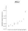
- the degree of non-specific binding of the labeled antibody to the solid phase was represented by the non-specific binding ratio given as C1/C2 x 100 (%) where C1 is the number of counts measured for the labeled antibody binding to the polystyrene ball and C2 is the number of counts corresponding to the whole amount of the labeled antibody used in the assay tube.
- Fig. 4 illustrates the results of a series of non-specific binding tests conducted for different volume ratios. It can be seen from the test results that the non-specific binding percentage increases from 0.005% to 0.05% as the volume ratio of the normal guinea pig serum to the reaction solution A increases from 1:0 to 1:20.
- Comparative Example 14 The labeled antibody used was guinea pig anti-hCG IgG-GOD and the solid-phase antibody used was guinea pig anti-hCG IgG.
- the reaction solution used was the reaction solution A mixed with no normal guinea pig serum.
- the non-specific binding ratio was 0.4%.
- Example 24 The labeled antibody used was guinea pig anti-hCG IgG-GOD and the solid-phase antibody used was guinea pig anti-hCG IgG.
- the reaction solution used was a mixture of the raction solution A and normal guinea pig serum.
- the volume ratio of the normal guinea pig serum to the reaction solution A was 1:5.
- An anti-hCG IgG-coated polystyrene ball was incubated at room temperature with hCG standard solution diluted with 0.3 ml of reaction solution A in an assay tube for six hours. The polystyrene ball was washed three times with the reaction solution A. The washed polystyrene ball was incubated with 0.1 ml of anti-hCG IgG-GOD labeled antibody diluted at a proper ratio with a mixture of the reaction solution A mixed with normal guinea pig serum. The volume ratio of the normal guinea pig serum to the reaction solution A was 1:5. The assay tube was held stationary at room temperature all the night through.
- the polystyrene ball was then washed three times with the reaction solution A and transferred into another clean assay tube. Following this. 0.3 ml of 0.01 mol/l acetate buffer (pH 5.1) containing 0.5 mol/l glucose was added to the assay tube. The assay tube was held stationary at a temperature of 37 o C for two hours. The resulting solution was taken to prepare a 0.1 ml sample. The sample was added with 0.5 ml of 0.2 mol/l carbonate buffer (pH 9.8) containing 2 x 10 ⁇ 7 mol/l luminol and then with 0.5 ml of 6 x 10 ⁇ 3 mol/l potassium ferricyanide.
- the light emitted upon chemiluminescent reaction of the luminol and potassium ferricyanide and the resulting hydrogen peroxide was measured by a Luminometer UPD-8000 which started its operation 15 seconds after the luminol and potassium ferricyanide were added to the assay tube and counted the light emitted for 30 seconds.
- Comparative Example 15 The labeled antibody used was guinea pig anti-hCG IgG-GOD and the solid-phase antibody used was guinea pig anti-hCG IgG.
- the reaction solution used was the reaction solution A mixed with no normal guinea pig serum.
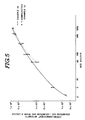
- Fig. 5 illustrates the results.
- the sandwich enzyme immunoassay measurable range and sensitivity obtained for Example 24 were 2.5-1000 mIU/ml and 2.5 mIU/ml, respectively.
- the sandwich enzyme immunoassay measurable range and sensitivity were 50-500 mIU/ml and 50 mIU/ml, respectively, for Comparative Example 15. It is, therefore, apparent that the sandwich enzyme immunoassay sensitivity and measurable range are improved when normal guinea pig serum is added to the reaction solution A.
- Sandwich enzyme immunoassay was repeated ten times within a day (within assay) for Example 24 and Comparative Example 15. The results are shown in Tables 19 and 20. Similarly, sandwich enzyme immunoassay was repeated on different days (between assay) for Example 24 and Comparative Example 15. The results are shown in Tabels 21 and 22.
- Comparative Example 16 The labeled antibody used was guinea pig anti-hCG IgG-GOD and the solid-phase antibody used was guinea pig anti-hCG IgG. Normal rabbit serum was added to the reaction solution A. The volume ratio of the normal rabit serum to the reaction solution A was 1:5.
- Comparative Example 17 The labeled antibody used was guinea pig anti-hCG IgG-GOD and the solid-phase antibody used was guinea pig anti-hCG IgG.
- Normal goat serum was added to the reaction solution A.
- the volume ratio of the normal goat serum to the reaction solution A was 1:5.
- Comparative Example 18 The labeled antibody used was guinea pig anti-hCG IgG-GOD and the solid-phase antibody used was guinea pig anti-hCG IgG. Normal horse serum was added to the reaction solution A. The volume ratio of the normal horse serum to the reaction solution A was 1:5.
- Comparative Example 19 The labeled antibody used was guinea pig anti-hCG IgG-GOD and the solid-phase antibody used was guinea pig anti-hCG IgG. No serum was added to the reaction solution A.
- the results of the non-specific binding tests conducted substantially in the same manner as described previously are illustrated In Fig. 6.
- the non-specific binding percentage was as small as 0.022% for Example 24.
- the non-specific binding percentage ranged from 0.07% to 0.3%. It is, therefore, apparent that the degree of non-specific binding of the labeled antibody to the solid phase can be reduced to a remarkable extent by adding normal guinea pig serum to the, reaction solution.
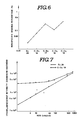
- the results of the sandwich enzyme immunoassay sensitivity and sensitive range tests conducted substantially in the same manner as described previously are illustrated in Fig. 7.
- the sandwich enzyme immunoassay sensitive range and sensitivity were 1-1000 mIU/ml and 1 mU/ml for Example 24.
- the sandwich enzyme immunoassay sensitive range and sensitivity were 50-1000 mIU/ml and 50 mIU/ml. It is, therefore, apparent that the sandwich enzyme immunoassay sensitive range and sensitivity can be improved by adding normal guinea pig serum to the reaction solution.
- Sandwich enzyme immunoassay was repeated ten times within a day (within assay) for Example 24 and Comparative Example 19. The results are shown in Tables 23 and 24. Similarly, sandwich enzyme immunoassay was repeated on different days (between assay) for Example 24 and Comparative Example 19. The results are shown in Tables 25 and 26.
- reaction solution has been described as containing sodium phosphate buffer (pH 6.3), it is to be noted that it may be replaced with sodium phosphate buffer (pH 6.0-7.5), borate buffer (pH 8.0-8.5) or the like.
- labeled antibody has been describing as containing glucose oxidase (GOD) label enzyme, it is to be noted that the label enzyme may be peroxidase, alkaline phosphatase, ⁇ -D-galactosidase, or the like.
- the label may be a fluorescent material (fluorescein isothiocyanate, tetramethylrhodamine isothiocyanate, or the like), a chemiluminescent material (aminoethylethyl isoluminol, aminobutylethyl isoluminol, aminopentylethyl isoluminol, aminohexylethyl isoluminol, or the like), or a radioactive isotope. (3H, 14C, 92P, 125I, 131I, or the like).
Landscapes
- Health & Medical Sciences (AREA)
- Immunology (AREA)
- Life Sciences & Earth Sciences (AREA)
- Engineering & Computer Science (AREA)
- Chemical & Material Sciences (AREA)
- Molecular Biology (AREA)
- Biomedical Technology (AREA)
- Hematology (AREA)
- Urology & Nephrology (AREA)
- Biotechnology (AREA)
- Microbiology (AREA)
- Cell Biology (AREA)
- Food Science & Technology (AREA)
- Medicinal Chemistry (AREA)
- Physics & Mathematics (AREA)
- Analytical Chemistry (AREA)
- Biochemistry (AREA)
- General Health & Medical Sciences (AREA)
- General Physics & Mathematics (AREA)
- Pathology (AREA)
- Chemical Kinetics & Catalysis (AREA)
- Medicines Containing Antibodies Or Antigens For Use As Internal Diagnostic Agents (AREA)
Claims (6)
- Kit zur Verwendung in einem Sandwich-Immunoassay, das markierte und Festphasen-Antikörper verwendet, die mit einem Antigen in Gegenwart einer Reaktionslösung reaktiv sind, um das Antigen zwischen dem markierten Antikörper und dem Fstphasen-Antikörper zu binden, wobei das Kit einen Festphasen-Antikörper, bei dem der Antikörper an eine feste Phase gebunden ist, einen markierten Antikörper, bei dem ein Markierungsenzym an den Antikörper gebunden ist, wobei wenigstens einer des markierten Antikörpers und des Festphasen-Antikörpers von einem Meerschweinchen stammt, und eine Reaktionslösung enthält, dadurch gekennzeichnet, daß die Reaktionslösung normales Meerschweinchenserum enthält, um das Ausmaß des nicht-spezifischen Bindens des markierten Antikörpers an die feste Phase zu erniedrigen.
- Kit nach Anspruch 1, in dem der markierte Antikörper Immunglobulin G umfaßt.
- Kit nach Anspruch 2, in dem der markierte Antikörper ein Fab' Fragment umfaßt, das aus Immunglobulin G hergestellt ist.
- Kit nach Anspruch 1, in dem der markierte Antikörper von einem Meerschweinchen stammt.
- Kit nach Anspruch 1, in dem der Festphasen-Antikörper von einem Meerschweinchen stammt.
- Kit nach Anspruch 1, in dem der markierte Antikörper von einem Meerschweinchen stammt, und der Festphasen-Antikörper von einem Meerschweinchen stammt.
Applications Claiming Priority (8)
| Application Number | Priority Date | Filing Date | Title |
|---|---|---|---|
| JP141375/86 | 1986-06-19 | ||
| JP61141375A JPS6394161A (ja) | 1986-06-19 | 1986-06-19 | 酵素検出用抗体ならびにこれを使用する酵素検出方法 |
| JP14497386 | 1986-06-23 | ||
| JP144973/86 | 1986-06-23 | ||
| JP82512/87 | 1987-04-03 | ||
| JP62082512A JPS63145960A (ja) | 1986-06-23 | 1987-04-03 | 標識抗体の非特異的吸着抑制薬 |
| JP62149325A JPS63313068A (ja) | 1987-06-16 | 1987-06-16 | 抗原の測定試薬 |
| JP149325/87 | 1987-06-16 |
Publications (3)
| Publication Number | Publication Date |
|---|---|
| EP0249955A2 EP0249955A2 (de) | 1987-12-23 |
| EP0249955A3 EP0249955A3 (en) | 1989-10-25 |
| EP0249955B1 true EP0249955B1 (de) | 1993-03-10 |
Family
ID=27466709
Family Applications (1)
| Application Number | Title | Priority Date | Filing Date |
|---|---|---|---|
| EP87108681A Expired - Lifetime EP0249955B1 (de) | 1986-06-19 | 1987-06-16 | Kit zur Verwendung in Sandwich-Immunoassays |
Country Status (2)
| Country | Link |
|---|---|
| EP (1) | EP0249955B1 (de) |
| DE (1) | DE3784559T2 (de) |
Families Citing this family (1)
| Publication number | Priority date | Publication date | Assignee | Title |
|---|---|---|---|---|
| WO1989010974A1 (en) * | 1988-05-11 | 1989-11-16 | Trustees Of The Sisters Of Charity Of Australia | Enzyme immunoassay system |
Family Cites Families (3)
| Publication number | Priority date | Publication date | Assignee | Title |
|---|---|---|---|---|
| US4414324A (en) * | 1979-05-21 | 1983-11-08 | Bma Laboratory Services, Inc. | Immunoassay method and apparatus |
| EP0125893A3 (de) * | 1983-05-12 | 1986-10-15 | Sumitomo Chemical Company, Limited | Quantitative Bestimmung von Antigenen durch Enzym-Antikörper-Brückenverfahren |
| US4595661A (en) * | 1983-11-18 | 1986-06-17 | Beckman Instruments, Inc. | Immunoassays and kits for use therein which include low affinity antibodies for reducing the hook effect |
-
1987
- 1987-06-16 EP EP87108681A patent/EP0249955B1/de not_active Expired - Lifetime
- 1987-06-16 DE DE8787108681T patent/DE3784559T2/de not_active Expired - Fee Related
Non-Patent Citations (1)
| Title |
|---|
| CHEMICAL ABSTRACTS, vol. 90, no. 3, 05 January 1979, Columbus, Ohio, US; H.G.KOPP et al.: "Problems connected with the production of highly specific antisera against prostaglandin E2 (PGE2) and prostaglandin A2 (PGA2) for radioimmunoassay" p. 270, abstract no. 18602r * |
Also Published As
| Publication number | Publication date |
|---|---|
| DE3784559D1 (de) | 1993-04-15 |
| EP0249955A2 (de) | 1987-12-23 |
| DE3784559T2 (de) | 1993-06-24 |
| EP0249955A3 (en) | 1989-10-25 |
Similar Documents
| Publication | Publication Date | Title |
|---|---|---|
| EP0365685B1 (de) | Gefriergetrocknete Zusammensetzung, die ein mit Meerrettichperoxydase markiertes Fab'-Fragment eines gegen menschliches Beta-Interferon gerichteten Antikörpers und Trehalose enthält; EIA Kit, der diese Zusammensetzung enthält | |
| US5876935A (en) | Luminescent specific binding assay | |
| US4375972A (en) | Heterogeneous chemiluminescent immunoassays utilizing metallo porphyrin tag | |
| US5236849A (en) | Method of high sensitivity immunoassay | |
| EP0303229B1 (de) | Hoch empfindlicher Immunoassay | |
| KR890002941B1 (ko) | 면역 반응 성분의 정량 방법 및 제제 | |
| CA1135620A (en) | Immunological compound attached to metallo-porphyrin tag | |
| EP0095089B1 (de) | Homogene Bindungstestverfahren und Reagenzsystem, Testsatz und Testgerät dafür | |
| EP0249955B1 (de) | Kit zur Verwendung in Sandwich-Immunoassays | |
| WO1999060401A1 (en) | Immunoassay reagents and immunoassay method | |
| EP0389301A2 (de) | Reagenzkomplex für Immunoessay | |
| EP0353895A1 (de) | Immunoassay-Methode | |
| US5128241A (en) | Microcapsule immunoassay and reagents therefor | |
| EP0249983B2 (de) | Reagenz-Kit zur Verwendung in Sandwich-Immunotesten | |
| KR960016337B1 (ko) | 샌드위치식 면역분석용 키트 | |
| EP0061071B1 (de) | Aktivierte Apoglucoseoxydase, Verfahren zu ihrer Herstellung, ihre Verwendung in spezifisch bindenden Testmethoden und dieselbe enthaltende Reagenzmittel und Testsatz | |
| JPS58149700A (ja) | ペルオキシダ−ゼ含有複合体,その製造法および試薬 | |
| KR960016338B1 (ko) | 샌드위치식 면역분석용 키트 | |
| Aoyagi et al. | The reduction of nonspecific binding in chemiluminescent sandwich enzyme immunoassays | |
| JP3174402B2 (ja) | 競合法による免疫測定法および測定用キット | |
| JPH10132818A (ja) | 免疫学的分析方法 | |
| Ishikawa et al. | Potential of the immune complex transfer enzyme immunoassay for antigens and antibodies to improve the sensitivity and its limitations | |
| JP2520465B2 (ja) | 多標識抗体 | |
| JPH0949840A (ja) | 免疫測定用試薬及び免疫測定法 | |
| Kohno et al. | Sensitive time‐resolved fluorimetric immune‐complex‐transfer immunoassay for antithyroglobulin IgG in serum |
Legal Events
| Date | Code | Title | Description |
|---|---|---|---|
| PUAI | Public reference made under article 153(3) epc to a published international application that has entered the european phase |
Free format text: ORIGINAL CODE: 0009012 |
|
| AK | Designated contracting states |
Kind code of ref document: A2 Designated state(s): DE FR GB |
|
| PUAL | Search report despatched |
Free format text: ORIGINAL CODE: 0009013 |
|
| AK | Designated contracting states |
Kind code of ref document: A3 Designated state(s): DE FR GB |
|
| 17P | Request for examination filed |
Effective date: 19900423 |
|
| 17Q | First examination report despatched |
Effective date: 19910919 |
|
| GRAA | (expected) grant |
Free format text: ORIGINAL CODE: 0009210 |
|
| AK | Designated contracting states |
Kind code of ref document: B1 Designated state(s): DE FR GB |
|
| REF | Corresponds to: |
Ref document number: 3784559 Country of ref document: DE Date of ref document: 19930415 |
|
| ET | Fr: translation filed | ||
| PLBE | No opposition filed within time limit |
Free format text: ORIGINAL CODE: 0009261 |
|
| STAA | Information on the status of an ep patent application or granted ep patent |
Free format text: STATUS: NO OPPOSITION FILED WITHIN TIME LIMIT |
|
| 26N | No opposition filed | ||
| PGFP | Annual fee paid to national office [announced via postgrant information from national office to epo] |
Ref country code: GB Payment date: 19990525 Year of fee payment: 13 |
|
| PGFP | Annual fee paid to national office [announced via postgrant information from national office to epo] |
Ref country code: FR Payment date: 19990616 Year of fee payment: 13 |
|
| PGFP | Annual fee paid to national office [announced via postgrant information from national office to epo] |
Ref country code: DE Payment date: 19990811 Year of fee payment: 13 |
|
| PG25 | Lapsed in a contracting state [announced via postgrant information from national office to epo] |
Ref country code: GB Free format text: LAPSE BECAUSE OF NON-PAYMENT OF DUE FEES Effective date: 20000616 |
|
| GBPC | Gb: european patent ceased through non-payment of renewal fee |
Effective date: 20000616 |
|
| PG25 | Lapsed in a contracting state [announced via postgrant information from national office to epo] |
Ref country code: FR Free format text: LAPSE BECAUSE OF NON-PAYMENT OF DUE FEES Effective date: 20010228 |
|
| REG | Reference to a national code |
Ref country code: FR Ref legal event code: ST |
|
| PG25 | Lapsed in a contracting state [announced via postgrant information from national office to epo] |
Ref country code: DE Free format text: LAPSE BECAUSE OF NON-PAYMENT OF DUE FEES Effective date: 20010403 |