EP0241811A2 - Monoclonale Antikörper auf human-Interleukin-2-Rezeptor - Google Patents
Monoclonale Antikörper auf human-Interleukin-2-Rezeptor Download PDFInfo
- Publication number
- EP0241811A2 EP0241811A2 EP87104868A EP87104868A EP0241811A2 EP 0241811 A2 EP0241811 A2 EP 0241811A2 EP 87104868 A EP87104868 A EP 87104868A EP 87104868 A EP87104868 A EP 87104868A EP 0241811 A2 EP0241811 A2 EP 0241811A2
- Authority
- EP
- European Patent Office
- Prior art keywords
- receptor
- human
- interleukin
- monoclonal antibodies
- antibody
- Prior art date
- Legal status (The legal status is an assumption and is not a legal conclusion. Google has not performed a legal analysis and makes no representation as to the accuracy of the status listed.)
- Granted
Links
Images


Classifications
-
- C—CHEMISTRY; METALLURGY
- C07—ORGANIC CHEMISTRY
- C07K—PEPTIDES
- C07K16/00—Immunoglobulins [IGs], e.g. monoclonal or polyclonal antibodies
- C07K16/18—Immunoglobulins [IGs], e.g. monoclonal or polyclonal antibodies against material from animals or humans
- C07K16/28—Immunoglobulins [IGs], e.g. monoclonal or polyclonal antibodies against material from animals or humans against receptors, cell surface antigens or cell surface determinants
- C07K16/2866—Immunoglobulins [IGs], e.g. monoclonal or polyclonal antibodies against material from animals or humans against receptors, cell surface antigens or cell surface determinants against receptors for cytokines, lymphokines, interferons
-
- C—CHEMISTRY; METALLURGY
- C07—ORGANIC CHEMISTRY
- C07K—PEPTIDES
- C07K16/00—Immunoglobulins [IGs], e.g. monoclonal or polyclonal antibodies
- C07K16/46—Hybrid immunoglobulins
- C07K16/468—Immunoglobulins having two or more different antigen binding sites, e.g. multifunctional antibodies
-
- A—HUMAN NECESSITIES
- A61—MEDICAL OR VETERINARY SCIENCE; HYGIENE
- A61K—PREPARATIONS FOR MEDICAL, DENTAL OR TOILETRY PURPOSES
- A61K38/00—Medicinal preparations containing peptides
-
- C—CHEMISTRY; METALLURGY
- C07—ORGANIC CHEMISTRY
- C07K—PEPTIDES
- C07K2317/00—Immunoglobulins specific features
- C07K2317/70—Immunoglobulins specific features characterized by effect upon binding to a cell or to an antigen
- C07K2317/73—Inducing cell death, e.g. apoptosis, necrosis or inhibition of cell proliferation
Definitions
- the invention relates generally to new hybrid cell lines, and in particular to hybrid cell lines for the production of monoclonal antibodies against an antigen which has been found on activated human lymphocytes, the interleukin-2 receptor, the antibodies produced therewith, and therapeutic and diagnostic methods and preparations under Use of the antibodies.
- the primary trigger of proliferation appears to be the interaction of growth factors with the growth factor receptor of the cell surface. Activation of the growth factor receptor in turn triggers previously unexplained cytoplasmic signaling systems.
- Dormant T lymphocytes are long-lived cells in the G o phase of the cell cycle. These enter the proliferation cycle only after antigen stimulation in the presence of a T cell growth factor, interleukin-2 (IL-2). Receptors for IL-2 are not detectable on the surface of resting T cells.
- IL-2R interleukin-2 receptors
- IL-2R The Expression of IL-2 receptors (IL-2R) is the result of the interaction of antigen presenting cells with the antigen receptor. It has recently been shown that IL-2 receptor expression is a transition stage and that repeated restimulation by lectins (Cantrell, PA, and KA Smith, (1984) Science (Wash.DC) 224: 1312); (Osawa, H., and Diamantstein, T. (1984), J.Immunol.
- IL-2-R are only expressed on activated lymphocytes
- monoclonal antibodies (mAb) that react with IL-2-R can be suitable as a specific and selective immunosuppressive agent.
- mAb monoclonal antibodies
- such antibodies can serve as diagnostic reagents to detect qualitatively and quantitatively activated lymphocytes as well as neoplastic cells which express IL-2-R.
- the object of the present invention is to provide a preparation of at least two monoclonal antibodies which recognize the human interleukin-2 receptor and are able to inhibit interleukin-2-induced lymphocyte proliferation.
- one or more of the antibodies belong to class IgG 1.
- the antibody preparation is able to inhibit interleukin-2 binding to the receptor.
- the present invention provides class IgG 1 monoclonal antibodies which recognize human interleukin-2 receptor and are capable of inhibiting interleukin-2 binding to the receptor and thus are capable of interleukin-2 -inhibit dependent lymphocyte proliferation.
- the antibodies according to the present invention are suitable for the construction of chimeric animal-human antibodies against the human interleukin-2 receptor, the constant Fc region of the immunoglobulin being of human origin and the variable Fab range being of animal origin.
- the Fab region is preferably obtained from mice.
- the present invention further provides hybridoma cell lines which are characterized by the production / secretion of monoclonal antibodies of class IgG 1 which recognize the human interleukin-2 receptor.
- the hybridoma cell lines of the NTCC deposit numbers ECACC 86 04 1801 and ECACC 86 04 1802 (PHLS Center of Applied Microbiology & Research, Porton Down, Salisbury, Wiltshire SP4 OJG, Great Britain) are particularly preferred.
- Human T-lymphoblasts expressing IL-2-R were produced by methods known per se (Osawa, H., and Diamantstein, T. (1983) J. Immunol. 130: 51) and were used to control monoclonal mice -Antibodies against IL-2-R according to the method of Köhler and Milstein (Köhler, G., and C. Milstein, (1975) Nature 256: 495).
- the fusion resulted in two hybrid clones, AHT-54 and AHT-107, which produce anti-IL-2-R antibodies of the IgG subclass.
- the hybrid clones which secrete anti-IL-2-R antibodies were selected as the preferred embodiment according to the invention.
- the competitive binding of AHT-54 and AHT-107 showed that they recognize different epitopes of the IL-2-R molecule.
- AHT-107 differs from Anti-Tac (Uchiyama, T. et al., (1981) J. Immunol. 126: 1398) in that competitive inhibition studies showed that they recognized two different epitopes of the IL-2-R molecule; AHT-107 also differs from 7G7 B6, a recently described anti-human IL-2-R mAb (A. Rubin, C. Kurman, E. Biddison, D. Goldman and L. Nelson (1985) Hybridoma Vol. 4 : 91), since in contrast to 7G7 B6 AHT-107 inhibits the binding of I L -2 to the IL-2-R as well as the IL-2-dependent proliferation of lymphocytes.
- T - ell-mediated Z autoimmune reaction such as acute autoimmune encephalomyelitis and adjuvant arthritis, which were induced by T-cell transfer (Wekerle, H. and T. diamond stone, (1986) Autoimmunity : Experimental and Clinical Aspects, Hsg: RS Schwarz, NR Rose, Ann.New York Acad.Sci., In press).
- the monoclonal anti-IL-2-R antibodies according to the invention are also suitable as therapeutic agents for clinical syndromes which are associated with the pathological proliferation of IL-2-dependent cells.
- Hyperimmune syndromes such as host-against-graft (HvG), graft-against-host (GvH) diseases and autoimmune diseases (e.g. multiplis sclerosis, autoimmune diabetes, diseases according to Crohn) are treated.
- monoclonal anti-IL-2-R antibodies are used directly as therapeutic agents, without further modification thereof.
- the invention further comprises a preparation of chimeric anti-IL-2-R antibodies using the heavy human chain of different classes and subclasses in combination with the variable region of the AHT-54 and AHT-107 mAb in order to optimize the therapeutic application .
- the antibodies can be coupled to drugs, including cytotoxic agents.
- the monoclonal antibodies according to the invention are suitable for recognizing antigen-activated cells which express IL-2 receptors, inhibiting their function and selectively eliminating them.
- the monoclonal antibodies of the present invention are also useful as diagnostic reagents on cells that contain IL-2-R either on the cell surface or within the cells or in body fluids.
- IL-2-R an enzyme or a chromophore or a radioactive substance (ELISA, RIA).
- Cells expressing IL-2-R were produced using human T lymphoblasts as described. Mixed peripheral human blood lymphocytes were stimulated with 3 mg / ml Concanavalin A (Con A) for 3 days. The converted cells were treated with alpha-methylmannoside (20 mg per ml), washed and used as an immunogen in culture medium. The cultures were grown in Click's RPMI medium (Seromed GmbH, Kunststoff, FRG), which with 2 x 10 -3 M L-glutamine, 5 x 10 -5 M 2-mercaptoethanol, 100 U / ml penicillin, 100 / ug / ml streptomycin, and 5 to 10% (V / V) fetal Calf serum (FCS; Lot No. 104; Seromed GmbH) was supplemented.
- FCS Click's RPMI medium
- mice 10 weeks old were primed with 2 x 10 7 T lymphoblasts.
- the cells were injected subcutaneously in 0.1 ml portions (10 6 cells) into the foot pad and neck of the mice as well as iv (10 7 cells in 0.5 ml). 4 weeks later, the mice received 10 7 T lymphoblasts iv. 3 days later, spleen cells of the immunized mice were fused with X63-Ag8.653 mouse myeloma cells in the presence of polyethylene glycol (Köhler and Milstein, (1975), Nature 256: 495, modified by Lemke H., GJ Hämmerling, C. Höhmann and K Rajewsky, (1978) Nature 271: 249).
- Fused cells suspended in HAT medium were distributed into each well of 10 tissue culture plates with 24 wells each (1 to 2 x 10 6 spleen cells / well).
- the supernatants in the wells were examined for their capacity for a) human T-lymphoblasts, b) mouse T-lymphoblasts and c) human thymocytes adhered to the surface of the wells of microtiter plates.
- Cell-bound immunoglobin was then detected using an enzyme immunosorbent assay (ELISA) as described (Kincade, PW, G. Lee, L. Sun, and T. Watanabe, (1981), J. Immunol.
- the hybridoma cells grown in HAT or RPMI medium and constantly producing antibodies that specifically bound to human T lymphoblasts were selected.
- the supernatants of the growing hybridoma cells were repeatedly tested and selected for hybridoma cells which produced supernatants which were used in the functional assay (inhibition of the T-lymphoblast response to IL-2) as well as in the absorption assay (inhibition of the ability of the T-lymphoblasts) To absorb IL-2 after preincubation) were active.
- Positive hybridomas were cloned by limited dilution with mouse thymocytes used as the feeder layer. The clones were retested and streaked. The supernatants from the relevant clones were used to isolate and purify mAb.
- Protein A-Sepharose Purification was accomplished by successive binding / elution of Protein A-Sepharose (Pharmacia Fihe Chemicals) according to the method of Ey et al (Ey, PL, SJ Prowse and CR Jenkin, (1978) Immunochemistry 15: 429). Approx. 600 ml of the culture supernatants, which had been brought to pH 8.0, were passed through a 5 ml protein A-Sepharose column, which was equilibrated with 0.1 M sodium phosphate buffer (pH 8.0). IgG 1 was eluted from the column using 0.1 M sodium citrate buffer (pH 6.0).
- the purified antibody was then dialyzed against a buffer containing 0.01 M HEPES (pH 7.4) and 0.9% NaCl.
- the purity of mAb was confirmed by sodium dodecyl sulfate (SDS) polyacrylamide gel electrophoresis, which was carried out under reducing conditions as described in the literature (Laemmli, GB 1970, Cleavage of structural proteins during the assembly of the head of bacteriophage T 4 , Nature 227: 429).
- SDS sodium dodecyl sulfate
- the protein concentration of the purified IgG 1 was determined by absorbing ultraviolet light at 280 nm, whereby an extinction coefficient (1% w / v: 1 cm) of 14 was assumed and according to the method of Lowry et al., 1951 (Lowry, OH, NJ Rosebrogh , AL Farr, and RJ Randall (1951)) using bovine serum albumin (BSA) as the standard.
- BSA bovine serum albumin
- Recombinant interleukin-2 was obtained from Sanolez Mena. 1-labeled recombinant IL-2 was purchased from NEN.
- MoAb were labeled with 125 I according to MacConahey and Dixon (McConahey, PJ, and FJ Dixon, (1980) Methods Enzymol, 70: 210).
- 20 ⁇ g IgG 1 dissolved in 60 ⁇ l Na 125 J (100 mCi per ml, carrier-free; Amersham Buchler).
- 10 ⁇ l chloramine-T 2.5 mg / ml in 0.05 M Na-P was added to the mixture.
- 20 / ul Na 2 S 2 0 5 3 mg / ml in 0.05 Na-P
- the mixture was immediately applied to a 15 ml Sephadex G-75 column (pre-washed with 0.05 M Na-P containing 4% BSA and washed successively with 0.05 M Na-P until the eluate was protein free ) and the radiolabel was collected in the excluded fraction.
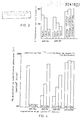
- Fig. 1 Inhibition of IL-2-dependent human T lymphoblast proliferation by different mAbs
- T-lymphoblasts were cultured with 20U / ml r-IL-2 for three days (see Fig. 1 for details) in the presence of either AHT-54 or AHT-107 mAb or a combination of both mAbs.
- the mixture was incubated at 4 ° C for 1 h.
- the relative amount of 125 I-labeled mAb (cpm) bound to the pelleted cells was measured after two washes with the binding buffer using a gamma-ray counter.
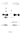
- mAbs used were mouse UPC-10 ascites as controls (lanes 1 & 7), anti-Tac ascites (lanes 2 & 8), AHT-54 ascites (lanes 4 and 10), AHT-107 ascites ( Lanes 5 & 11), and AHT-107 culture supernatant (lanes 6 & 12).
- HPBL Human peripheral blood cells
- AHT-54 or AHT-107 mAb 1: 1000 ascites- Liquid
- TSH TSH
- FIGS. 7 and 8 show that AHT-54 (FIG. 7) and AHT-107 (FIG. 8) are able to bind to lymphoblasts.
- Figures 9 to 11 relate to the same experiment, with the difference that the lymphoblasts were exchanged for HPBL. No reaction occurs with any of the three antibodies.
Landscapes
- Health & Medical Sciences (AREA)
- Chemical & Material Sciences (AREA)
- Immunology (AREA)
- Organic Chemistry (AREA)
- Medicinal Chemistry (AREA)
- Biophysics (AREA)
- General Health & Medical Sciences (AREA)
- Genetics & Genomics (AREA)
- Biochemistry (AREA)
- Molecular Biology (AREA)
- Proteomics, Peptides & Aminoacids (AREA)
- Life Sciences & Earth Sciences (AREA)
- Preparation Of Compounds By Using Micro-Organisms (AREA)
- Medicines Containing Antibodies Or Antigens For Use As Internal Diagnostic Agents (AREA)
- Peptides Or Proteins (AREA)
- Micro-Organisms Or Cultivation Processes Thereof (AREA)
Abstract
Description
- Die Erfindung betrifft allgemein neue Hybrid-Zelllinien, und insbesondere Hybrid-Zellinien zur Herstellung monoclonaler Antikörper gegen ein Antigen, das auf aktivierten human-Lymphozyten gefunden wurde, den Interleukin-2-Rezeptor, die damit hergestellten Antikörper sowie therapeutische und diagnostische Methoden und Zubereitungen unter Verwendung der Antikörper.
- In vielen Fällen, wenn nicht in allen, scheint der primäre Auslöser der Proliferation die Wechselwirkung von Wachstumsfaktoren mit dem Wachstumsfaktor-Rezeptor der Zelloberfläche zu sein. Die Aktivierung des Rezeptors des Wachstumsfaktors löst wiederum bisher ungeklärte zytoplasmische Signalsysteme aus.
- Ruhende T-Lymphozyten stellen langlebige Zellen in der Go-Phase des Zellzyklus dar. Diese treten in den Proliferationszyklus erst nach Antigen-Stimulierung in Gegenwart eines T-Zellen-Wachstumsfaktors, Interleukin-2 (IL-2) ein. Rezeptoren für IL-2 sind auf der Oberfläche ruhender T-Zellen nicht nachweisbar. Die Expression von IL-2-Rezeptoren (IL-2R) ist die Folge der Wechselwirkung von Antigen-präsentierenden Zellen mit dem Antigen-Rezeptor. Es konnte kürzlich gezeigt werden, dass die IL-2-Rezeptor-Expression ein Übergangsstadium darstellt, und daß eine wiederholte Restimulierung durch Lectine (Cantrell, P.A., und K.A. Smith, (1984), Science (Wash.D.C) 224:1312); (Osawa, H., und Diamantstein, T. (1984), J.Immunol. 132:2445) oder Antigen (Reske-Kunz, A.B., D.v. Steldern, E. Rüde, H. Osawa und T. Diamantstein, (1984), J. Immunol. 133:1356) zur kontinuierlichen IL-2-Rezeptor-Expression und folglich für ein langlebiges Zellwachstum erforderlich ist.
- Da IL-2-R ausschliesslich auf aktivierten Lymphozyten exprimiert werden, können monoclonale Antikörper (mAb), die mit IL-2-R reagieren, als spezifisches und selektives immunsuppressives Agens geeignet sein. Ausserdem können solche Antikörper als diagnostische Reagenzien dienen, um qualitativ und quantitativ aktivierte Lymphozyten als auch neoplastische Zellen, die IL-2-R exprimieren, nachzuweisen.
- Aufgabe der vorliegenden Erfindung ist es, eine Zubereitung aus mindestens zwei monoclonalen Antikörpern zur Verfügung zu stellen, die den human-Interleukin-2-Rezeptor erkennen und in der Lage sind, die Interleukin-2-induzierte Lymphozyten-Proliferation zu inhibieren. Nach einer bevorzugten Ausführungsform gehören einer oder mehrere der Antikörper der Klasse IgG 1 an. Nach einer weiteren bevorzugten Ausführungsform ist die Antikörper-Zubereitung in der Lage, die Interleukin-2-Bindung an den Rezeptor zu inhibieren. Die vorliegende Erfindung stellt monoclonale Antikörper der Klasse IgG1 zur Verfügung, die human-Interleukin-2-Rezeptor erkennen, und in der Lage sind, die Interleukin-2-Bindung an den Rezeptor zu inhibieren und damit in der Lage sind, die Interleukin-2-abhängige Lymphozyten-proliferation zu inhibieren. Die Antikörper gemäß der vorliegenden Erfindung eignen sich zur Konstruktion chimärer tierisch-menschlicher Antikörper gegen den menschlichen-Interleukin-2-Rezeptor, wobei die konstante Fc-Region des Immunoglobulins menschlichen Ursprungs und der variable Fab-Bereich tierischen Ursprungs ist. Bevorzugt wird der Fab-Bereich aus Mäusen erhalten.
- Die vorliegende Erfindung stellt im weiteren Hybridoma-Zellinien zur Verfügung, die durch die Produktion/ Sekretion monoclonaler Antikörper der Klasse IgG1, welche den human-Interleukin-2-Rezeptor erkennen, charakterisiert sind. Besonders bevorzugt sind die Hybridoma-Zellinien der NTCC-Hinterlegungsnummer ECACC 86 04 1801 und ECACC 86 04 1802 (PHLS Centre of Applied Microbiology & Research, Porton Down, Salisbury, Wiltshire SP4 OJG, Great Britain).
- Human-T-Lymphoblasten, welche IL-2-R exprimieren, wurden nach an sich bekannten Methoden hergestellt (Osawa, H., und Diamantstein, T. (1983) J. Immunol. 130:51) und wurden eingesetzt, um monoclonalen Maus-Antikörper gegen IL-2-R nach dem Verfahren von Köhler und Milstein (Köhler, G., und C. Milstein, (1975) Nature 256:495) herzustellen. Die Fusion ergab zwei Hybrid-Clone, AHT-54 und AHT-107, welche Anti-IL-2-R-Antikörper der Unterklasse IgG, produzieren. Die Hybridclone, die Anti-IL-2-R-Antikörper sekretieren, wurden als bevorzugte Ausführungsform gemäß der Erfindung selektiert.
- Beide mAb i) inhibieren die Bindung von 1-markiertem IL-2 an IL-2-R-positive human-Lymphozyten, ii) inhibieren die IL-2-abhängige Proliferation in vitro, und iii) präzipitieren das identische Zelloberflächenmolekül von 55 KD, das IL-2-Bindungsprotein. Die kompetitive Bindung von AHT-54 und AHT-107 zeigte, dass sie unterschiedliche Epitope des IL-2-R-Moleküls erkennen.
- AHT-107 unterscheidet sich von Anti-Tac (Uchiyama, T. et al., (1981) J. Immunol. 126:1398), da kompetitive Inhibierungsstudien zeigten, dass sie zwei unterschiedliche Epitope des IL-2-R-Moleküls erkennen; AHT-107 unterscheidet sich auch von 7G7 B6, einem kürzlich beschriebener Anti-human-IL-2-R mAb (A. Rubin, C. Kurman, E. Biddison, D. Goldman und L. Nelson (1985) Hybridoma Vol. 4:91), da im Gegensatz zu 7G7 B6 AHT-107 die Bindung von IL-2 an den IL-2-R wie auch die IL-2-abhängige Proliferation von Lymphozyten inhibiert.
- Beide mAb reagieren spezifisch mit aktivierten Lymphozyten (T und B), jedoch nicht mit ruhenden Lymphozyten oder anderen nicht-lymphoiden Zellen. Diese Behauptung basiert auf FACS-Analysedaten (Fig. 6 bis 11).
- Gemäss früheren Untersuchungen an Tiermodellen wurde gezeigt, dass solche Ab, welche mit Ratten (ART-18) und mit Mäuse-IL-2-R (AMT-13 und M7/20)reagieren, selektiv und spezifisch inhibieren: i) die lokale GVH-Reaktion (Diamantstein, T. und H. Osawa, (1986), Immune Rev. 92, in Druck), ii) die Cardio-Allotransplantat-Abstossung (L. Kirkman, E. Kelley, A. Koltun, J. Schoen, A. Ythier und B. Strom, (1985), Transplantation 40:719), (L. Kirkman, L.V. Barett, N. Gaulton, E. Kelley, A. Ythier und B. Strom, (1985), J. Exp. Med. 162; 358), und iii) die T-Zell-vermittelte Autoimmunreaktion, wie z.B. akute Autoimmun-Encephalomyelitis und Adjuvans-Arthritis, welche durch T-Zell-Übertragung induziert wurden (Wekerle, H. und T. Diamantstein, (1986), Autoimmunity: Experimental and Clinical Aspects, Hsg: R.S. Schwarz, N.R. Rose, Ann. New York Acad. Sci., in Druck).
- Die monoclonalen Anti-IL-2-R-Antikörper gemäss der Erfindung eignen sich auch als therapeutische Agenzien für klinische Syndrome, die im Zusammenhang mit der pathologischen Proliferation von IL-2-abhängigen Zellen stehen. So können z.B. Hyperimmun-Syndrome, wie Wirt-gegen-Transplantat (HvG)-, Transplantat-gegen-Wirt (GvH)-Erkrankungen und Autoimmun-Erkrankungen (z.B. Multiplis Sclerose, Autoimmun-Diabetes, Erkrankungen nach Crohn) behandelt werden. Nach einer bevorzugten Ausführungsform gemäss der vorliegenden Erfindung werden monoclonale Anti-IL-2-R-Antikörper direkt als therapeutische Agenzien, ohne weitere Modifizierung derselben, verwendet. Ausserdem umfaßt die Erfindung eine Zubereitung von chimären Anti-IL-2-R-Antikörpern unter Verwendung der schweren human-Kette verschiedener Klassen und Unterklassen in Kombination mit der variablen Region des AHT-54 und AHT-107 mAb, um die therapeutische Anwendung zu optimieren.
- Alternativ können die Antikörper an Arzneimittel, einschliesslich cytotoxischer Agenzien, gekoppelt werden. Die monoclonalen Antikörper gemäss der Erfindung sind geeignet, Antigen-aktivierte Zellen, die IL-2-Rezeptoren exprimieren, zu erkennen, deren Funktion zu inhibieren und diese selektiv zu eliminieren.
- Die monoclonalen Antikörper der vorliegenden Erfindung eignen sich auch als diagnostische Reagenzien auf Zellen, die IL-2-R entweder auf der Zelloberfläche oder innerhalb der Zellen oder in Körperflüssigkeiten enthalten. Somit ist es mit Hilfe der Erfindung möglich, Zellen, die IL-2-R enthalten, in Proben mit verschiedenen Zellarten zu identifizieren. Die Lokalisierung von IL-2-haltigen Zellen ist in den Kulturzellkolonien oder in Gewebeproben möglich. Bei einer derartigen Anwendung werden die monoclonalen Antikörper vorzugsweise an fluoreszierende, farbbildende Substanzen, wie z.B. ein Enzym oder ein Chromophor oder eine radioaktive Substanz (ELISA, RIA) gekoppelt.
- Die folgende Beschreibung soll die Erfindung näher erläutern, ohne dass diese in irgendeiner Weise im Hinblick auf im wesentlichen funktionelle Äquivalente von Hybridom-Zellen und monoclonalen Antikörpern, wie sie hier beschrieben sind, eingeschränkt sein soll.
- IL-2-R-exprimierende Zellen wurden, wie beschrieben, unter Verwendung von human-T-Lymphoblasten hergestellt. Gemischte periphere human Blut-Lymphozyten wurden mit 3 mg/ml Concanavalin A (Con A) 3 Tage lang stimuliert. Die konvertierten Zellen wurden mit alpha-Methylmannosid (20 mg pro ml) behandelt, gewaschen und als Immunogen in Kulturmedium verwendet. Die Kulturen wurden in Click's RPMI-Medium (Seromed GmbH, München, BRD) angesetzt, welches mit 2 x 10⁻³M L-Glutamin, 5 x 10⁻⁵ M 2-Mercaptoethanol, 100 E/ml Penicillin, 100/ug/ml Streptomycin, und 5 bis 10 % (V/V) Fötal-Kälberserum (FCS; Charge Nr. 104; Seromed GmbH) ergänzt war.
- 10 Wochen alte BALB/c-Mäuse wurden mit 2 x 10⁷ T-Lymphoblasten geprimt. Die Zellen wurden in 0,1 ml Anteilen (106 Zellen) subkutan in das Fusspolster und den Nacken der Mäuse wie auch i.v. (107 Zellen in 0,5 ml) injiziert. 4 Wochen darauf erhielten die Mäuse i.v. 107 T-Lymphoblasten. 3 Tage darauf wurden Milzzellen der immunisierten Mäuse mit X63-Ag8.653 Maus-Myelomazellen in Gegenwart von Polyethylenglykol fusioniert (Köhler und Milstein, (1975), Nature 256:495, modifiziert durch Lemke H., G.J. Hämmerling, C. Höhmann und K. Rajewsky, (1978), Nature 271:249). Fusionierte, in HAT-Medium suspendierte Zellen wurden in jede Vertiefung von 10 Gewebekulturplatten mit jeweils 24 Vertiefungen verteilt (1 bis 2 x 10⁶ Milzzellen/Vertiefung). Die Überstände in den Vertiefungen, in denen nach 3 bis 4 Wochen ein kräftiges Wachstum beobachtet wurde, wurden untersucht im Hinblick auf ihre Kapazität an a) human-T-Lymphoblasten, b) Maus-T-Lymphoblasten und c) human-Thymozyten, die an der Oberfläche der Vertiefungen von Mikrotiter-Platten hafteten, zu binden. Zell-gebundenes Immunoglobin wurde dann mit Hilfe eines Enzym-Immunosorbent-Assays (ELISA), wie beschrieben, nachgewiesen (Kincade, P.W., G. Lee, L. Sun, und T. Watanabe, (1981), J. Immunol. Methods 42:17) unter Verwendung von β-Galactosidasegekoppeltem Schaf-F(ab')2-anti-Maus Immunoglobulin (New England Nuclear, Dreieich, BRD) als zweiten Antikörper. Die Hybridomzellen, die in HAT- oder RPMI-Medium gezüchtet wurden und konstant Antikörper produzierten, die spezifisch an human-T-Lymphoblasten banden, wurden selektiert. Die Überstände der wachsenden Hybridomazellen wurden wiederholt getestet und im Hinblick auf Hybridomazellen selektiert, die Überstände produzierten, welche im funktionellen Assay (Inhibierung der T-Lymphoblasten-Antwort auf IL-2) wie auch beim Absorptions-Assay (Inhibierung der Fähigkeit der T-Lymphoblasten, IL-2 nach Preinkubation zu absorbieren) aktiv waren. Positive Hybridome wurden mittels beschränkter Verdünnung mit Maus-Thymozyten, die als Feeder-Schicht verwendet wurden, geclont. Die Clone wurden erneut getestet und ausgestrichen. Die Überstände der relevanten Clone wurden zur Isolierung und Reinigung von mAb verwendet.
- Tests, die mittels der0uchterlony-Doppel-Immunodiffu- sions-Methode mit Kaninchen-anti-Maus-IgM-, IgA-, IgG1-, IgG 2a-, IgG2b- und IgG3-Seren (Miles Laboratories, Ltd., Slough, England) durchgeführt wurden, zeigten, daß die Hybridoma AHT-54 und AHT-107 IgG1-Antikörper sezernieren. Ausgenommen der ursprünglichen Screening-Experimente, in denen nicht-gereinigte Kultur-Überstände verwendet wurden, wurden die folgenden Experimente mit gereinigtem IgGl durchgeführt. Die Reinigung wurde durch sukzessive Bindung/Elution von Protein A-Sepharose (Pharmacia Fihe Chemicals) gemäss dem Verfahren von Ey et al ( Ey, P.L., S.J. Prowse und C.R. Jenkin, (1978), Immunochemistry 15:429) erzielt. Ca. 600 ml der Kultur-Überstände, die auf pH 8,0 gebracht worden waren, wurden über eine 5m1 Protein-A-Sepharose-Säule gegeben, welche mit 0,1 M Natriumphosphatpuffer (pH 8,0) equilibriert war. IgGl wurde von der Säule mit Hilfe von 0,1 M Natriumzitratpuffer (pH 6,0) eluiert. Der gereinigte Antikörper wurde dann gegen einen Puffer dialysiert, der 0,01 M HEPES (pH 7,4) und 0,9 % NaCl enthielt. Die Reinheit von mAb wurde mittels Natriumdodecylsulfat (SDS)-Polyacrylamid-Gel- elektrophorese bestätigt, welche unter reduzierenden Bedingungen durchgeführt wurde, wie dies in der Literatur beschrieben ist (Laemmli, GB 1970, Cleavage of structural proteins during the assembly of the head of bacteriophage T4, Nature 227:429). Die Proteinkonzentraton des gereinigten IgG1 wurde durch Absorption von ultraviolettem Licht bei 280 nm bestimmt, wobei ein Extinktionskoeffizient (1% G/V:lcm) von 14 angenommen wurde und nach dem Verfahren von Lowry et al, 1951 (Lowry, O.H., N.J. Rosebrogh, A.L. Farr, und R.J. Randall (1951)), wobei Rinderserumalbumin (BSA) als Standard verwendet wurde.
- Rekombinantes Interleukin-2 wurde von Sanolez Mena erhalten. 1-markiertes rekombinantes IL-2 wurde von NEN bezogen.
- MoAb wurden mit 125I gemäß MacConahey und Dixon (McConahey, P.J., und F.J. Dixon, (1980) Methods Enzymol, 70:210) markiert. Kurzgesagt: 20µg IgG1, aufgelöst in 60µl Na125J (100 mCi pro ml, trägerfrei; Amersham Buchler). 10µl Chloramin-T (2,5 mg/ml in 0,05 M Na-P) wurden zu dem Gemisch zugegeben. Nach 45Sekunden Inkubation bei Raumtemperatur wurden der Lösung 20/ul Na2S205 (3mg/ml in 0,05 Na-P) einzugeben. Das Gemisch wurde sofort auf eine 15 ml Sephadex-G-75-Säule aufgetragen (vorgewaschen mit 0,05 M Na-P, welcher 4 % BSA enthielt, und nacheinander gewaschen mit 0,05 M Na-P, bis das Eluat proteinfrei war) und die Radiomarkierung in der ausgeschlossenen Fraktion gesammelt.
- 2 x 10⁶ human-T-Lymphoblasten wurden 3 Tage lang in 0,2 ml Medium inkubiert, welches die angegebenen Mengen an rekombinantem IL-2 (Fig. la, 20 E/ml; Fig. lb, 4E/ml) in Abwesenheit oder Gegenwart verschiedener mAbs, anti-Tac (-o-), AHT-54(-Δ-), AHT-107 (-*-) und einem Kontroll-mAblanti-human-TSH (-o-),enthielt. Die Zellen wurden mit 3H-Thymidin während der letzten 4 h der Inkubationsdauer pulsiert. Die Inkorporierung von 3H-Thymidin wurde nach dem Standard-Verfahren gemessen (Diamantstein et al, Mol. Immunol., (1984), 21:1229).
- T-Lymphoblasten wurden mit 20E/ml r-IL-2 drei Tage lang (wegen Details siehe Fig. l) in Gegenwart von entweder AHT-54 oder AHT-107 mAb oder in Kombination beider mAbs kultiviert.
- 2 x 10⁶ human-T-Blasten wurden zunächst in 100/ul eines Bindungspuffers suspendiert (PBS=0,5% BSA/10 mM NaN3), welcher verschiedene Verdünnungen der mAbsanti-Tac (-o-), AHT-54 (-Δ-) und AHT-107 (-*-) enthielt. Die Suspensionen wurden mit 100/ul einer 1:40-Verdünnung des 125I-markierten mAb gemischt.
- Das Gemisch wurde 1 h bei 4°C inkubiert. Die relative Menge an 125I-markiertem mAb (cpm), welche an die pelletierten Zellen gebunden war, wurde nach zwei Waschgängen mit dem Bindungspuffer unter Verwendung eines Gamma-Strahlen-Zählers genessen.
- 2 x 10⁶ human-T-Blasten wurden zunächst 30 Minuten bei 37°C 0,25 ml eines Puffers inkubiert (RPMI/Hepes/BSA/NaN3), welcher die angegebenen Mengen verschiedener mAbs enthielt. Die Inkubation wurde in Gegenwart von 125 I-IL-2 bei 37°C fortgesetzt. Nach 40 Minuten wurde das Inkubationsgemisch zentrifugiert, um die Zellen zu pelletieren; die pelletierten Zellen wurden in 100µl des Puffers aufgenommen und einer Ölphase überlagert, die aus Dibutylphthalat/Olivenöl (10+3) bestand. Nach Zentrifugation wurden die Reagenzglasspitzen, die die Zellpellets enthielten, abgetrennt und in einem gamma-Strahlen-Zähler ausgezählt.
- 2 x 10⁷ human-T-Blasten wurden mit 0,5 mCi Na[125I] oberflächenjodiert und in 0,5 ml des Lysepuffers lysiert. Das Lysat wurde zentrifugiert und mit 1/5 Volumen an Protein-A-Sepharose-Kügelchen (10µl) über Kaninchen-anti-Maus-IgG als Brückenantikörper preabsorbiert. Nach 1 h bei 4°C wurden die Kügelchen dreimal mit Puffer gewaschen, welcher 50 mM tris-HCl, pH 8,3, 450 mM NaCl, 5 mM KI, 0,02 % NaN3 und 0,5 Nonidet P-40 enthielt, und mit 100,ul des Probepuffers extrahiert. 50,ul aliquote Teile des Extraktes wurden einer SDS-PAGE-Analyse unterworfen, und zwar entweder unter nicht-reduzierenden (Bahnen 1 bis 6) oder reduzierenden (Bahnen 7 bis 12) Bedingungen. Die verwendeten mAbs waren Maus-UPC-10-Ascites als Kontrolle (Bahnen 1 & 7), anti-Tac-Ascites (Bahnen 2 & 8), AHT-54-Ascites (Bahnen 4 und 10), AHT-107-Ascites (Bahnen 5 & 11), und AHT-107-Kulturüberstand (Bahnen 6 & 12).
- Humane periphere Blutzellen (HPBL) und aktivierte T-Lymphoblasten, die von HPBL abgeleitet waren, wurden bei 4°C in Gegenwart von 0,1 % NaN3 30 Minuten mit AHT-54 oder AHT-107 mAb inkubiert (1:1000 Ascites-Flüssigkeit) und als negataive Kontrolle mit einem TSH, gewaschen und angefärbt unter Verwendung einer sättigenden Menge an Ziegen-anti-Maus-IgG, markiert mit FITC. Fluoreszenz-aktivierte Zell Sorter-Analysen wurden mit einem Epics V vorgenommen.
- Fig. 6 zeigt die negative Kontrolle. Der alpha-TSH-Antikörper band nicht an die T-Lymphoblasten. In Fig. 7 und 8 wird gezeigt, dass AHT-54 (Fig. 7) und AHT-107 (Fig. 8) in der Lage sind, an Lymphoblasten zu binden. Fig. 9 bis 11 beziehen sich auf das gleiche Experinent, mit dem Unterschied, daß die Lymphoblasten gegen HPBL ausgetauscht wurden. Bei keinem der drei Antikörper tritt eine Reaktion auf.
Claims (10)
Priority Applications (1)
| Application Number | Priority Date | Filing Date | Title |
|---|---|---|---|
| AT87104868T ATE97816T1 (de) | 1986-04-14 | 1987-04-02 | Monoclonale antikoerper auf human-interleukin-2rezeptor. |
Applications Claiming Priority (2)
| Application Number | Priority Date | Filing Date | Title |
|---|---|---|---|
| GB8609058A GB2188941B (en) | 1986-04-14 | 1986-04-14 | Monoclonal antibodies recognizing human interleukin-2-receptor |
| GB8609058 | 1986-04-14 |
Publications (3)
| Publication Number | Publication Date |
|---|---|
| EP0241811A2 true EP0241811A2 (de) | 1987-10-21 |
| EP0241811A3 EP0241811A3 (en) | 1989-07-26 |
| EP0241811B1 EP0241811B1 (de) | 1993-12-01 |
Family
ID=10596154
Family Applications (1)
| Application Number | Title | Priority Date | Filing Date |
|---|---|---|---|
| EP87104868A Revoked EP0241811B1 (de) | 1986-04-14 | 1987-04-02 | Monoclonale Antikörper auf human-Interleukin-2-Rezeptor |
Country Status (8)
| Country | Link |
|---|---|
| US (1) | US5631349A (de) |
| EP (1) | EP0241811B1 (de) |
| JP (1) | JPS6312297A (de) |
| AT (1) | ATE97816T1 (de) |
| DE (1) | DE3788296D1 (de) |
| DK (1) | DK190287A (de) |
| ES (1) | ES2060581T3 (de) |
| GB (1) | GB2188941B (de) |
Cited By (7)
| Publication number | Priority date | Publication date | Assignee | Title |
|---|---|---|---|---|
| EP0340604A2 (de) * | 1988-05-06 | 1989-11-08 | Innothérapie S.A. | Monoklonaler Antikörper und seine Verwendung |
| EP0380542A1 (de) * | 1987-08-17 | 1990-08-08 | Us Commerce | Verfahren zur behandlung der bösartigen und autoimmunen krankheiten beim menschen. |
| FR2649488A1 (fr) * | 1989-07-07 | 1991-01-11 | Inst Nat Sante Rech Med | Fragments d'anticorps monoclonaux specifiques de la presence de leucocytes actives - leur procede d'obtention et leur application dans le cas de rejet de greffe |
| EP0421876A1 (de) * | 1989-10-06 | 1991-04-10 | PASTEUR MERIEUX SERUMS ET VACCINS, Société Anonyme : | Verwendung von Wirkstoffen zur Herstellung eines Arzneimittels von Lymphomen oder ähnlichen Krankheiten |
| FR2652747A1 (fr) * | 1989-10-06 | 1991-04-12 | Merieux Inst | Application d'agents actifs pour la preparation d'un medicament destine au traitement de maladies auto-immunes. |
| WO1992004051A1 (de) * | 1990-09-12 | 1992-03-19 | Boehringer Mannheim Gmbh | Monoklonale antikörper gegen den interleukin 2-rezeptor |
| FR2672291A1 (fr) * | 1991-01-31 | 1992-08-07 | Inst Nat Sante Rech Med | Composition d'anticorps diriges contre le recepteur de l'interleukine-2 humaine ou animale. |
Families Citing this family (10)
| Publication number | Priority date | Publication date | Assignee | Title |
|---|---|---|---|---|
| JPH02503867A (ja) * | 1988-04-15 | 1990-11-15 | プロテイン デザイン ラブズ インコーポレーテッド | Il‐2レセプター特異的キメラ抗体 |
| IL162181A (en) * | 1988-12-28 | 2006-04-10 | Pdl Biopharma Inc | A method of producing humanized immunoglubulin, and polynucleotides encoding the same |
| US5530101A (en) | 1988-12-28 | 1996-06-25 | Protein Design Labs, Inc. | Humanized immunoglobulins |
| PT92900A (pt) * | 1989-01-24 | 1990-07-31 | Sistema de vectores de expressao para a producao de anticorpos monoclonais quimericos | |
| HUT60768A (en) | 1990-03-16 | 1992-10-28 | Sandoz Ag | Process for producing cd25 fixing molecules |
| CA2103059C (en) | 1991-06-14 | 2005-03-22 | Paul J. Carter | Method for making humanized antibodies |
| US6800738B1 (en) | 1991-06-14 | 2004-10-05 | Genentech, Inc. | Method for making humanized antibodies |
| WO1994004679A1 (en) * | 1991-06-14 | 1994-03-03 | Genentech, Inc. | Method for making humanized antibodies |
| ES2314067T3 (es) * | 2001-04-06 | 2009-03-16 | University Of Bristol | Uso de moleculas de enlace cd 25 en pacientes resistentes a los esteroides. |
| US7575742B2 (en) | 2002-06-28 | 2009-08-18 | The United States Of America As Represented By The Secretary Of The Department Of Health And Human Services | Method of treating autoimmune diseases with interferon-beta and IL-2R antagonist |
Citations (3)
| Publication number | Priority date | Publication date | Assignee | Title |
|---|---|---|---|---|
| EP0226062A1 (de) * | 1985-12-03 | 1987-06-24 | Bayer Ag | Gelöste Interleukin-2-Rezeptoren als Indikator für Immunreaktionen und Neoplasien |
| EP0235805A2 (de) * | 1986-03-04 | 1987-09-09 | The Royal Free Hospital School Of Medicine | Immunsuppression |
| EP0171496B1 (de) * | 1984-08-15 | 1993-05-26 | Research Development Corporation of Japan | Verfahren zur Herstellung von chimärischen monoklonalen Antikörpern |
Family Cites Families (5)
| Publication number | Priority date | Publication date | Assignee | Title |
|---|---|---|---|---|
| US4411993A (en) * | 1981-04-29 | 1983-10-25 | Steven Gillis | Hybridoma antibody which inhibits interleukin 2 activity |
| US4578335A (en) * | 1984-05-21 | 1986-03-25 | Immunex Corporation | Interleukin 2 receptor |
| US4845198A (en) * | 1984-05-21 | 1989-07-04 | Immunex Corporation | Hybridoma antibody which binds IL-2 receptor |
| GB8422238D0 (en) * | 1984-09-03 | 1984-10-10 | Neuberger M S | Chimeric proteins |
| US4707443A (en) * | 1985-04-19 | 1987-11-17 | The United States Of America As Represented By The Secretary Of The Department Of Health And Human Services | Soluble interleukin-2 receptor as a disease indicator and a method of assaying the same |
-
1986
- 1986-04-14 GB GB8609058A patent/GB2188941B/en not_active Expired - Lifetime
-
1987
- 1987-04-02 AT AT87104868T patent/ATE97816T1/de not_active IP Right Cessation
- 1987-04-02 DE DE87104868T patent/DE3788296D1/de not_active Revoked
- 1987-04-02 ES ES87104868T patent/ES2060581T3/es not_active Expired - Lifetime
- 1987-04-02 EP EP87104868A patent/EP0241811B1/de not_active Revoked
- 1987-04-13 DK DK190287A patent/DK190287A/da not_active Application Discontinuation
- 1987-04-14 JP JP62090009A patent/JPS6312297A/ja active Pending
-
1993
- 1993-11-15 US US08/152,782 patent/US5631349A/en not_active Expired - Fee Related
Patent Citations (3)
| Publication number | Priority date | Publication date | Assignee | Title |
|---|---|---|---|---|
| EP0171496B1 (de) * | 1984-08-15 | 1993-05-26 | Research Development Corporation of Japan | Verfahren zur Herstellung von chimärischen monoklonalen Antikörpern |
| EP0226062A1 (de) * | 1985-12-03 | 1987-06-24 | Bayer Ag | Gelöste Interleukin-2-Rezeptoren als Indikator für Immunreaktionen und Neoplasien |
| EP0235805A2 (de) * | 1986-03-04 | 1987-09-09 | The Royal Free Hospital School Of Medicine | Immunsuppression |
Non-Patent Citations (4)
| Title |
|---|
| IMMUNOLOGICAL REVIEWS, Nr. 92, 1986, Seiten 5-27, Munksgaard, Copenhagen, DK; T. DIAMANTSTEIN et al.: "The interleukin-2 receptor, its physiology and a new approach to a selective immunosuppressive therapy by anti-interleukin-2 receptor monoclonal antibodies" * |
| JOURNAL OF EXPERIMENTAL MEDICINE, Band 162, Juli 1985, Seiten 358-362, The Rockefeller University Press; R.L. KIRKMAN et al.: "Administration of an anti-interleukin 2 receptor monoclonal antibody prolongs cardiac allograft survival in mice" * |
| MICROBIOL. IMMUNOL., Band 29, Nr. 10, 1985; Seiten 959-972# * |
| THE JOURNAL OF IMMUNOLOGY, Band 130, Nr. 1, Januar 1983, Sei ten 51-55, The American Association of Immunologists; H. OSAWA et al.: "The characteristics of a monoclonal antibody that binds specifically to rat T lymphoblasts and inhibits IL 2 receptor functions" * |
Cited By (13)
| Publication number | Priority date | Publication date | Assignee | Title |
|---|---|---|---|---|
| EP0380542A1 (de) * | 1987-08-17 | 1990-08-08 | Us Commerce | Verfahren zur behandlung der bösartigen und autoimmunen krankheiten beim menschen. |
| EP0380542A4 (en) * | 1987-08-17 | 1990-12-05 | The United States Of America Represented By The Secretary The United States Department Of Commerce | Method for treating malignancy and autoimmune disorders in humans |
| EP0340604A2 (de) * | 1988-05-06 | 1989-11-08 | Innothérapie S.A. | Monoklonaler Antikörper und seine Verwendung |
| DE3815472A1 (de) * | 1988-05-06 | 1989-11-16 | Centre Regional De Transfusion | Monoklonaler antikoerper und seine verwendung |
| EP0340604A3 (en) * | 1988-05-06 | 1990-05-09 | Centre Regional De Transfusion Sanguine | Monoclonal antibody and its use |
| WO1991000921A1 (fr) * | 1989-07-07 | 1991-01-24 | Institut National De La Sante Et De La Recherche Medicale | Fragments d'anticorps monoclonaux specifiques de la presence de leucocytes actives - leur procede d'obtention et leur application dans le cas de rejet de greffe |
| FR2649488A1 (fr) * | 1989-07-07 | 1991-01-11 | Inst Nat Sante Rech Med | Fragments d'anticorps monoclonaux specifiques de la presence de leucocytes actives - leur procede d'obtention et leur application dans le cas de rejet de greffe |
| EP0421876A1 (de) * | 1989-10-06 | 1991-04-10 | PASTEUR MERIEUX SERUMS ET VACCINS, Société Anonyme : | Verwendung von Wirkstoffen zur Herstellung eines Arzneimittels von Lymphomen oder ähnlichen Krankheiten |
| FR2652747A1 (fr) * | 1989-10-06 | 1991-04-12 | Merieux Inst | Application d'agents actifs pour la preparation d'un medicament destine au traitement de maladies auto-immunes. |
| FR2652746A1 (fr) * | 1989-10-06 | 1991-04-12 | Merieux Inst | Application d'agents actifs pour la preparation d'un medicament destine au traitement de lymphomes ou assimiles. |
| WO1992004051A1 (de) * | 1990-09-12 | 1992-03-19 | Boehringer Mannheim Gmbh | Monoklonale antikörper gegen den interleukin 2-rezeptor |
| FR2672291A1 (fr) * | 1991-01-31 | 1992-08-07 | Inst Nat Sante Rech Med | Composition d'anticorps diriges contre le recepteur de l'interleukine-2 humaine ou animale. |
| WO1992013886A1 (fr) * | 1991-01-31 | 1992-08-20 | Institut National De La Sante Et De La Recherche Medicale (Inserm) | Composition d'anticorps diriges contre le recepteur de l'interleukine-2 humaine ou animale |
Also Published As
| Publication number | Publication date |
|---|---|
| EP0241811B1 (de) | 1993-12-01 |
| DE3788296D1 (de) | 1994-01-13 |
| DK190287D0 (da) | 1987-04-13 |
| GB8609058D0 (en) | 1986-05-21 |
| EP0241811A3 (en) | 1989-07-26 |
| GB2188941B (en) | 1990-06-06 |
| GB2188941A (en) | 1987-10-14 |
| ES2060581T3 (es) | 1994-12-01 |
| ATE97816T1 (de) | 1993-12-15 |
| US5631349A (en) | 1997-05-20 |
| DK190287A (da) | 1987-10-15 |
| JPS6312297A (ja) | 1988-01-19 |
Similar Documents
| Publication | Publication Date | Title |
|---|---|---|
| EP0241811B1 (de) | Monoclonale Antikörper auf human-Interleukin-2-Rezeptor | |
| Rao et al. | Characterization of a monoclonal antibody directed against the murine B lymphocyte receptor for IgE. | |
| EP0763128B1 (de) | Verfahren zur herstellung von heterologen bispezifischen antikörpern | |
| Wong et al. | Bi-specific monoclonal antibodies: selective binding and complement fixation to cells that express two different surface antigens. | |
| DE69128774T3 (de) | GEREINIGTE IgG ANTIKÖRPER | |
| DE69330523T3 (de) | Immunoglobuline ohne leichte ketten | |
| US5948893A (en) | Murine hybridoma and antibody binding to CD28 receptor secreted by the hybridoma and method of using the antibody | |
| EP0657533A1 (de) | Monoklonale Antikörper mit hoher Zytotoxizität gegen humanes CD16-Antigen, sowie bispezifische monoklonale Antikörper unter Verwendung derartiger monoklonaler Antikörper und des CD-30-HRS-3-Antikorpers | |
| DE69333039T2 (de) | Monoklonaler antikörper gegen ein humanes mdr1-gen produkt, das eine resistenz gegen verschiedene medikamente erzeugt und seine verwendung | |
| EP0499176B1 (de) | Monoklonale Antikörper gegen humane Pankreas-Inselzellen | |
| EP0430193A1 (de) | Monoklonale Antikörper und ihre Verwendung | |
| DE69028713T3 (de) | Schimäre immunoglobuline für cd4-rezeptoren | |
| Howard et al. | Isolation of six monoclonal alloantibodies against rat histocompatibility antigens: clonal competition. | |
| DE3719398C2 (de) | Liganden und deren Verwendung | |
| EP1025854A1 (de) | Verwendung depletorisch oder mitogen wirkender Antikörper gegen CD3/TCR in der Immuntherapie | |
| DD272473B3 (de) | Verfahren zur herstellung monoklonaler antikoerper gegen die epsilon-kettedes cd 3-antigens humaner t-lymphozyten | |
| DE19529026C2 (de) | Monoklonale Antikörper gegen humanes Interleukin-10 | |
| DD272471A1 (de) | Verfahren zur herstellung monoklonaler antikoerper gegen den il-2-rezeptor humaner t-lymphozyten | |
| DD230876A1 (de) | Verfahren zur gewinnung monoklonaler antikoerper gegen human-mif | |
| DE180486T1 (de) | Hybridzellen die antiidiotypischen antikoerpern sekretieren, monoklonale antikoerper durch diese hybridzellen sekretiert, ihre herstellung und ihre verwendung. | |
| AT400956B (de) | Verfahren zur gewinnung eines liganden und verfahren zur unterdrückung der proliferation von bp50 exprimierenden zellen | |
| WO1992004463A1 (de) | Cd58 spezifischer monoklonaler antikörper und dessen verwendung | |
| DE3634017A1 (de) | Verfahren zur aktivierung und vermehrung von t-zellen, insbesondere von deren subpopulationen | |
| DD250333B1 (de) | Verfahren zur herstellung monoklonaler anti-human-t-lymphozytenrezeptor-antikoerper | |
| DD243184A3 (de) | Verfahren zur Herstellung monoklonaler Antikörper für L-Ketten |
Legal Events
| Date | Code | Title | Description |
|---|---|---|---|
| PUAI | Public reference made under article 153(3) epc to a published international application that has entered the european phase |
Free format text: ORIGINAL CODE: 0009012 |
|
| 17P | Request for examination filed |
Effective date: 19870402 |
|
| AK | Designated contracting states |
Kind code of ref document: A2 Designated state(s): AT BE CH DE ES FR GB IT LI NL SE |
|
| PUAL | Search report despatched |
Free format text: ORIGINAL CODE: 0009013 |
|
| AK | Designated contracting states |
Kind code of ref document: A3 Designated state(s): AT BE CH DE ES FR GB IT LI NL SE |
|
| 17Q | First examination report despatched |
Effective date: 19910620 |
|
| GRAA | (expected) grant |
Free format text: ORIGINAL CODE: 0009210 |
|
| AK | Designated contracting states |
Kind code of ref document: B1 Designated state(s): AT BE CH DE ES FR GB IT LI NL SE |
|
| REF | Corresponds to: |
Ref document number: 97816 Country of ref document: AT Date of ref document: 19931215 Kind code of ref document: T |
|
| GBT | Gb: translation of ep patent filed (gb section 77(6)(a)/1977) |
Effective date: 19931215 |
|
| REF | Corresponds to: |
Ref document number: 3788296 Country of ref document: DE Date of ref document: 19940113 |
|
| ITF | It: translation for a ep patent filed |
Owner name: ING. C. GREGORJ S.P.A. |
|
| ET | Fr: translation filed | ||
| PLBI | Opposition filed |
Free format text: ORIGINAL CODE: 0009260 |
|
| 26 | Opposition filed |
Opponent name: SANDOZ AG PATENT- UND MARKENABTEILUNG Effective date: 19940901 Opponent name: F. HOFFMANN-LA ROCHE & CO. AKTIENGESELLSCHAFT Effective date: 19940830 |
|
| REG | Reference to a national code |
Ref country code: ES Ref legal event code: FG2A Ref document number: 2060581 Country of ref document: ES Kind code of ref document: T3 |
|
| NLR1 | Nl: opposition has been filed with the epo |
Opponent name: SANDOZ AG PATENT- UND MARKENABTEILUNG Opponent name: F. HAFFMANN LA ROCHE & CO. AKTIENGESELLSCHAFT |
|
| EAL | Se: european patent in force in sweden |
Ref document number: 87104868.2 |
|
| PLAB | Opposition data, opponent's data or that of the opponent's representative modified |
Free format text: ORIGINAL CODE: 0009299OPPO |
|
| R26 | Opposition filed (corrected) |
Opponent name: F. HOFFMANN-LA ROCHE & CO. AKTIENGESELLSCHAFT * 94 Effective date: 19940830 |
|
| NLR1 | Nl: opposition has been filed with the epo |
Opponent name: SANDOZ AG PATENT- UND MARKENABTEILUNG Opponent name: F. HOFFMANN-LA ROCHE & CO. AKTIENGESELLSCHAFT |
|
| PGFP | Annual fee paid to national office [announced via postgrant information from national office to epo] |
Ref country code: DE Payment date: 19970213 Year of fee payment: 11 |
|
| PLBQ | Unpublished change to opponent data |
Free format text: ORIGINAL CODE: EPIDOS OPPO |
|
| PGFP | Annual fee paid to national office [announced via postgrant information from national office to epo] |
Ref country code: SE Payment date: 19970314 Year of fee payment: 11 |
|
| PLAB | Opposition data, opponent's data or that of the opponent's representative modified |
Free format text: ORIGINAL CODE: 0009299OPPO |
|
| PGFP | Annual fee paid to national office [announced via postgrant information from national office to epo] |
Ref country code: GB Payment date: 19970324 Year of fee payment: 11 |
|
| PGFP | Annual fee paid to national office [announced via postgrant information from national office to epo] |
Ref country code: FR Payment date: 19970327 Year of fee payment: 11 |
|
| PGFP | Annual fee paid to national office [announced via postgrant information from national office to epo] |
Ref country code: ES Payment date: 19970408 Year of fee payment: 11 |
|
| PGFP | Annual fee paid to national office [announced via postgrant information from national office to epo] |
Ref country code: AT Payment date: 19970414 Year of fee payment: 11 |
|
| PGFP | Annual fee paid to national office [announced via postgrant information from national office to epo] |
Ref country code: BE Payment date: 19970423 Year of fee payment: 11 |
|
| PGFP | Annual fee paid to national office [announced via postgrant information from national office to epo] |
Ref country code: CH Payment date: 19970425 Year of fee payment: 11 |
|
| PGFP | Annual fee paid to national office [announced via postgrant information from national office to epo] |
Ref country code: NL Payment date: 19970430 Year of fee payment: 11 |
|
| R26 | Opposition filed (corrected) |
Opponent name: F. HOFFMANN-LA ROCHE & CO. AKTIENGESELLSCHAFT * 94 Effective date: 19940830 |
|
| RDAH | Patent revoked |
Free format text: ORIGINAL CODE: EPIDOS REVO |
|
| NLR1 | Nl: opposition has been filed with the epo |
Opponent name: NOVARTIS AG Opponent name: F. HOFFMANN-LA ROCHE & CO. AKTIENGESELLSCHAFT |
|
| RDAG | Patent revoked |
Free format text: ORIGINAL CODE: 0009271 |
|
| STAA | Information on the status of an ep patent application or granted ep patent |
Free format text: STATUS: PATENT REVOKED |
|
| REG | Reference to a national code |
Ref country code: CH Ref legal event code: PL |
|
| 27W | Patent revoked |
Effective date: 19970519 |
|
| GBPR | Gb: patent revoked under art. 102 of the ep convention designating the uk as contracting state |
Free format text: 970519 |
|
| NLR2 | Nl: decision of opposition |