EP0206533B1 - Fragment d'anticorps monoclonal inhibant les plaquettes - Google Patents
Fragment d'anticorps monoclonal inhibant les plaquettes Download PDFInfo
- Publication number
- EP0206533B1 EP0206533B1 EP86303955A EP86303955A EP0206533B1 EP 0206533 B1 EP0206533 B1 EP 0206533B1 EP 86303955 A EP86303955 A EP 86303955A EP 86303955 A EP86303955 A EP 86303955A EP 0206533 B1 EP0206533 B1 EP 0206533B1
- Authority
- EP
- European Patent Office
- Prior art keywords
- platelets
- antibody
- fragment
- cells
- human
- Prior art date
- Legal status (The legal status is an assumption and is not a legal conclusion. Google has not performed a legal analysis and makes no representation as to the accuracy of the status listed.)
- Expired - Lifetime
Links
Images


Classifications
-
- C—CHEMISTRY; METALLURGY
- C07—ORGANIC CHEMISTRY
- C07K—PEPTIDES
- C07K16/00—Immunoglobulins [IG], e.g. monoclonal or polyclonal antibodies
- C07K16/18—Immunoglobulins [IG], e.g. monoclonal or polyclonal antibodies against material from animals or humans
- C07K16/28—Immunoglobulins [IG], e.g. monoclonal or polyclonal antibodies against material from animals or humans against receptors, cell surface antigens or cell surface determinants
-
- A—HUMAN NECESSITIES
- A61—MEDICAL OR VETERINARY SCIENCE; HYGIENE
- A61P—SPECIFIC THERAPEUTIC ACTIVITY OF CHEMICAL COMPOUNDS OR MEDICINAL PREPARATIONS
- A61P7/00—Drugs for disorders of the blood or the extracellular fluid
- A61P7/02—Antithrombotic agents; Anticoagulants; Platelet aggregation inhibitors
-
- A—HUMAN NECESSITIES
- A61—MEDICAL OR VETERINARY SCIENCE; HYGIENE
- A61K—PREPARATIONS FOR MEDICAL, DENTAL OR TOILETRY PURPOSES
- A61K38/00—Medicinal preparations containing peptides
Definitions
- Blood platelets also known as thrombocytes, function in the human body to provide for the coagulation of blood when the blood supply is exposed to potential loss, i.e., trauma, abrasions, and other hemorrhaging injuries. Platelets rapidly interact with damaged blood vessels and with each other to prevent the escape of blood cells and also facilitate the formation of a blood clot in which they become enmeshed so as to plug up any "leaks" in the vascular system. However, in certain disease states, such as thrombosis, it is desirable and even necessary, to inhibit blood platelet function.
- the thrombus can either occlude the blood vessel and cause damage to the tissue being supplied with blood from that vessel (for example, myocardial infarction, stroke after vascular surgery, etc) or because the platelets and other thrombotic material can break off from the thrombus and lodge down-stream in blood vessels supplying other parts of the body (for example, venous thrombosis with pulmonary embolism, transient ischemic attacks, amaurosis fugax, strokes from the artificial hearts, etc).
- the present invention concerns a monoclonal antibody fragment which inhibits platelet function.
- the fragment was produced by the proteolytic digestion of the 7E3 monoclonal antibody, suitably with papain or pepsin.
- the 7E3 monoclonal antibody was produced in the following manner. Human blood platelets were injected into mice. The mouse spleen was removed and fused with a mouse myeloma by a modification of the technique of Levy et al., (Curr. Top. Microbiol. Immunol., 81 , 164 (1978)). The fused cells were incubated and then further incubated in HAT medium (in which non-fused cells will not grow).
- the cells were then diluted out and screened in a screening assay for antifibrinogen receptor activity.
- 7E3 of the class IgG 1 which reacts with normal human blood platelets and with dog blood platelets, fails to react with thrombasthenic platelets or human platelets whose GPIIb/IIIa complex was disassociated with EDTA, reacts slowly with unactivated human platelets and more rapidly with ADP activated human platelets and completely blocks the interaction of fibrinogen with platelets induced by ADP.
- the Balb/c mouse myeloma cell line X63-Ag 8.653 is described in Kearney J.F. et al J. Immunol. 1979 Vol.123 p.1548-1550.
- MA123 monoclonal antibody MA123 which inhibits the binding of fibrinogen to ADP-stimulated human platelets. However MA123 does not react with dog blood platelets.
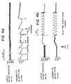
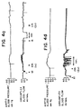
- Figure IV (a-d) is a continuous set of plots for one test dog of aortic blood pressure and circumflex blood flow against time after administration of the F(ab') 2 fragments.
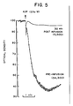
- Figure V is an optical density/time plot of ADP response pre and post F(ab') 2 infusion.
- the mice Three days later the mouse was killed, the spleen removed, the cells separated and fused with a BALB/c mouse myeloma line in accordance with the method of Levy et al., (supra).
- the spleen cells and the myeloma cells in a ratio of 3.9:1 were pelletted together, the pellet suspended in polyethylene glycol (35%) in RPMI 1640 medium whereupon the cells were immediately centrifuged at low velocity.
- the solution was then diluted to about 25% of its previous concentration with RPMI 1640, the cells resuspended, recentrifuged and the supernatant removed.
- the supernatant was then incubated in a 5% CO 2 95% air atmosphere in RPMI 1640 medium supplemented with fetal calf serum and thereafter selection made in the usual manner by adding HAT medium and aliquoting into microtiter wells. After two weeks, the supernatant of the wells that showed growth were screened for antifibrinogen receptor activity.
- the clones obtained by this method were selected for various qualities; one in particular, 7E3 was selected for certain qualities.
- the antibody was isolated from the supernatant in the wells or flasks.
- the hybridomas were injected intraperitoneally into Pristane R pretreated BALB/c rats and the antibodies isolated from the ascitic fluid.
- the antibody was purified by precipitation with 50% saturated ammonium sulfate, resuspended in between 5 and 10% of the original volume in sodium phosphate buffer and dialyzed against the same buffer. Chromatography on protein A-Sepharose CL-4B equilibrated with phosphate buffer was carried out, elution was with phosphate buffer followed by decreasing pH 0.1M citrate buffers. 7E3 antibody was eluted after the pH decreased to about 6.0. Protein elution was as monitored by ultra-violet spectroscopy at 280 nm.
- Purified antibody was then dialyzed overnight at at reduced temperatures, suitably between about 0 and 10°C preferrably at 4°C against a slightly acidic saline buffer of pH 3.5-6.5, suitably about pH 4.0, after which the freshly prepared pepsin was added in an amount equal to approximately 2% of the antibody's weight.
- the resulting solution was then incubated at about 37°C for 12 to 24 hours. Digestion was stopped by dialyzing the solution against PBS, pH 7.4. Analysis by polyacrylamide gel electrophoresis indicated that the digestion was essentially complete.
- an Fab fragment can be prepared by digestion with papain, another proteolytic agent, namely, .1M acetate, 2mM EDTA, 1mM cysteine and including 1% w/w of papain for 6-8 hours at 37° at a pH of 4.5-6, suitably 5.5.
- the resulting F(ab') 2 fragment of the 7E3 monoclonal antibody can then be further purified by chromatography on a material such as protein A Sepharose CL-4B or DE-52 in order to be certain that any remaining traces of the whole 7E3 monoclonal antibody are removed.
- a BALB/c mouse (Jackson Laboratories, Bar Harbor, Me.) was injected intraperitoneally with six weekly 0.2 ml injections of 3 x 10 8 washed platelets (citrated PRP washed twice in 0.15 M NaCl, 10 mM Tris/Cl, 10 mM EDTA, pH 7.4 [TS-E]), resuspended in 1/10 to 1/20 of their original volume in TS-E, and mixed 1:1 with complete Freund's adjuvant.
- the seventh weekly injection was given intravenously into the tail vein and consisted of 0.3 ml. containing 5 x 10 8 washed platelets resuspended in T-S without EDTA.
- Each of the seven platelet suspensions was obtained from a different donor. Three days after the last injection, the mouse was killed by cervical dislocation and the spleen removed. A suspension of spleen cells in RPMI 1640 was prepared by teasing the spleen apart.
- erythrocytes were lysed with ammonium chloride, the spleen cells were fused with a nonsecretory BALB/c mouse myeloma cel line (X63-Ag 8.653) that had been kept frozen in 10% DMSO, 90% fetal calf serum until one week before fusion, when it was thawed and maintained in the culture medium routinely used (RPMI 1640 supplemental with 10% fetal calf serum and 1,000 U of penicillin and 100 ug of streptomycin/ml). Fusion was carried out according to a modification of the method of Levy et al. ( supra ).
- 2.7 x 10 8 spleen cells and 7 x 10 7 myeloma cells were pelleted together, the pellet was gently suspended in 2 ml of 35% polyethylene glycol in RPMI 1640 medium and the cells immediately centrifuged at 500 g at 22°C for 6 minutes. The solution was then diluted with RPMI 1640 to 9% polyethylene glycol, the cells resuspended and immediately centrifuged at 230 g. for 6 minutes at 22°C. The supernatant fluid was then aspirated and the fused cells suspended in RPMI 1640 medium and supplemented with 20% fetal calf serum and 10% 109 medium (National Collection of Type Cultures).
- the cells were placed in a flask and incubated overnight at 37°C in a 5% CO 2 , 95% air atmosphere. The following day, the medium was made selective for successfully hybridized cells by adding hypoxanthine (10 -4 M), aminopterin (4 x 10 -7 M), and thymidine (1.6 x 10 -5 M), after which the cells were aliquoted into 960 microtiter wells (Costar, Data Packaging, Cambridge, Ma.). Two weeks later, 574 wells showed growth and the supernatant fluids from 59 wells were positive in a screening assay for antifibrinogen receptor activity (see below).
- the positive clones were transferred to 24-well microtiter dishes (Costar) and fed with the same medium as above, but without the aminopterin.
- the clones were expanded and the cells that continued to produce antifibrinogen receptor antibody were suspended in 90% fetal calf serum-10% DMSO and frozen in liquid nitrogen.
- the clones were subcloned by both limiting dilution technique and growth in soft agar to insure monoclonality.
- Ascitic fluid rich in 7E3 antibody was prepared by intraperitoneal injection of Pristane-pretreated BALB/c mice with 5 x 10 6 hybrid cells that had been washed twice in 0.15 M NaCl, 10 mM sodium phosphate, pH 7.4 (PBS).
- 35 ul of PRP platelet rich plasma
- 35 ul. of the supernatant culture medium (or ascitic fluid) to be assayed were incubated together for 2-60 minutes in a well of a round-bottomed microtiter plate (Linbro Chemical Co., Hamden, Ct).
- 5 ul of the fibrinogen-coated bead suspension was then added and the plate was mixed on a rotator (Tekator V, American Scientific Products, Edison, N.J.) for 5 minutes at 280 rpm.
- Culture supernatants were precipitated at 4°C with 50% saturated ammonium sulfate and resuspended to between 1/20 and 1/10 of their original volume in 0.1 M sodium phosphate buffer, pH 8.0. After dialysis against the same buffer, the samples were applied to a 0.8 x 15.9 cm column of protein A Sepharose CL-4B that had been equilibrated with the phosphate buffer (after having been washed with both the phosphate buffer and a 0.1 M citrate buffer, pH 3.0).
- Purified antibody was then dialyzed overnight at approximately 4°C against 0.2M sodium chloride, 0.2M acetate, pH 4.0, after which freshly prepared pepsin (1 mg/ml) was added in an amount equal to approximately 2% of the antibody's weight. The resulting solution was then incubated at about 37°C for 12 to 24 hours. Digestion was stopped by dialyzing the solution against PBS, pH 7.4. Analysis by polyacrylamide gel electrophoresis indicates that the digestion was essentially complete.
- the resulting F(ab') 2 fragment of the 7E3 monoclonal antibody can then be further purified by chromatography on a material such as protein A Sepharose CL-4B or DE-52 in order to be certain that any remaining traces of the whole 7E3 monoclonal antibody are removed.
- the experimental protocol consisted of first obtaining samples of blood anticoagulated with either 0.01 volume 40% sodium citrate (for platelet aggregation and 125 I-7E3 binding), or 0.037 volume 269 mM EDTA (for platelet counts), and then performing the surgery. After cyclical flow reductions were established, another set of blood samples was obtained and the antibody (0.7-0.8 mg/kg) was infused intravenously as a bolus. A final set of samples was obtained 30 min. after infusing the antibody.
- Platelet counts on the platelet-rich plasma were performed by microscopy after dilution and lysis of erythrocytes (Unopette, Becton-Dickinson), and whole blood platelet counts were performed by an electronic resistive particle counter (S+IV, Coulter Electronics, Hialeah, FL.).
- Cyclical reductions in blood flow were obtained in 4 dogs and 4 monkeys. In all 4 dogs and 4 monkeys, infustion of the F(ab') 2 fragments resulted in complete cessation of the cyclical flow reductions and restoration of the control flow rate in 10 min. or less ( Figure IV). In the dogs, restoration of coronary flow correlated with the disappearance of ST segment deviations on the electrocardiograms, indicating the reversal of myocardial ischemia. The cyclical flow reductions could not be restored with epinephrine infusions of 0.5 or 1 ug/kg/min.
Landscapes
- Health & Medical Sciences (AREA)
- Chemical & Material Sciences (AREA)
- Organic Chemistry (AREA)
- Medicinal Chemistry (AREA)
- Immunology (AREA)
- Life Sciences & Earth Sciences (AREA)
- General Health & Medical Sciences (AREA)
- Engineering & Computer Science (AREA)
- Chemical Kinetics & Catalysis (AREA)
- Molecular Biology (AREA)
- Proteomics, Peptides & Aminoacids (AREA)
- Biophysics (AREA)
- Biochemistry (AREA)
- Bioinformatics & Cheminformatics (AREA)
- Diabetes (AREA)
- Hematology (AREA)
- Genetics & Genomics (AREA)
- General Chemical & Material Sciences (AREA)
- Nuclear Medicine, Radiotherapy & Molecular Imaging (AREA)
- Pharmacology & Pharmacy (AREA)
- Animal Behavior & Ethology (AREA)
- Public Health (AREA)
- Veterinary Medicine (AREA)
- Preparation Of Compounds By Using Micro-Organisms (AREA)
- Medicines Containing Antibodies Or Antigens For Use As Internal Diagnostic Agents (AREA)
- Micro-Organisms Or Cultivation Processes Thereof (AREA)
- Peptides Or Proteins (AREA)
Claims (8)
- Fragment d'anticorps monoclonal qui a un site de liaison d'antigène et est dérivable d'un anticorps monoclonal de la classe IgG1 qui peut être obtenu à partir d'un hybrodome de cellules provenant d'une ligne de myélomes de souris et de cellules de rate provenant d'une souris antérieurement immuniséé avec des plaquettes de sang humain, lequel fragment d'anticorps(a) inhibe la fonction des plaquettes ;(b) inhibe la thrombose ;(c) réagit avec les plaquettes humaines normales et avec les plaquettes du sang de chien ;(d) échoue à réagir avec les plaquettes humaines thrombasthéniques ou les plaquettes humaines dont le complexe GPIIb/IIIa est dissoccié avec l'EDTA ;(e) réagit lentement avec les plaquettes inactivées et plus rapidement avec les plaquettes activées par ADP, et(f) bloque l'intéraction des fibrinogènes avec les plaquettes induites par l'ADP.
- Fragment selon la revendication 1 dans laquelle l'hybridome peut être obtenu par fusion de cellules de myélome murine X63-Ag 8./653 Balb/c et de cellules de rate provenant d'une souris Balb/c.
- Fragment selon la revendication 1 ou 2 qui est un fragment F(ab')2 ou un fragment Fab.
- Fragment selon la revendication 3 qui est un fragment F(ab')2.
- Fragment selon l'une quelconque des revendications précédentes dans laquelle l'hybridome est désigné 7E3 ayant un numéro de dépôt ATCC HB8832.
- Composition pharmaceutique qui comprend une quantité thérapeutiquement efficace d'un fragment selon l'une quelconque des revendications précédentes.
- Méthode qui comprend l'utilisation d'un fragment selon l'une quelconque des revendications 1 à 5 pour préparer un médicament pour la traitement ou la prévention de l'aggrégation des plaquettes.
- Méthode de préparation d'un fragment selon l'une quelconque des revendications 1 à 5 qui comprend les étapes séquentielles de :i) immunisation de souris avec des plaquettes de sang humain,ii) élimination des rates desdites souris et fabrication d'une suspension desdites cellules de rate,iii) fusion desdites cellules de rate avec les cellules de myélome de souris en présence d'un promoteur de fusion,iv) dilution et culture des cellules fusionnées dans des puits séparés dans un milieu qui ne supportera pas les cellules de myélome non fusionnées,v) évaluation du surnageant dans chaque puits contenant un hybridome quant à la présence d'un anticorps au récepteur fibrinogène,vi) sélection et clonage d'un hybridome produisant l'anticorps qui :(a) inhibe la fonction des plaquettes ;(b) inhibe le thrombose ;(c) réagit avec les plaquettes de sang normal et avec les plaquettes de sang de chien ;(d) ne réagissent pas avec les plaquettes humaines thrombasthéniques ou les plaquettes humaines dont le complexe GPIIb/IIIa est dissocié avec l'EDTA,(e) réagit lentement avec les plaquettes non activées et plus rapidement avec les plaquettes activées par l'ADP, et(f) bloque l'intéraction de fibrinogène avec les plaquettes induites par l'ADP.vii) récupération de l'anticorps surnageant et/ou transfert desdits clones de façon intrapéritonéalle dans des souris et récolte des ascites malins ou du sérum provenant desdites souris, lesquels ascites ou sérum contiennent les anticorps désirés,viii) digestion dudit anticorps avec un agent protéolytique choisi parmi le groupe consistant de la papaïne et de la pepsine,ix) récupération dudit fragment du mélange de digestion.
Priority Applications (1)
| Application Number | Priority Date | Filing Date | Title |
|---|---|---|---|
| AT86303955T ATE104316T1 (de) | 1985-06-14 | 1986-05-23 | Plaettcheninhibierender teil eines monoklonalen antikoerpers. |
Applications Claiming Priority (4)
| Application Number | Priority Date | Filing Date | Title |
|---|---|---|---|
| US74541585A | 1985-06-14 | 1985-06-14 | |
| US85171186A | 1986-04-14 | 1986-04-14 | |
| US851711 | 1986-04-14 | ||
| US745415 | 1986-04-14 |
Publications (3)
| Publication Number | Publication Date |
|---|---|
| EP0206533A2 EP0206533A2 (fr) | 1986-12-30 |
| EP0206533A3 EP0206533A3 (en) | 1987-04-08 |
| EP0206533B1 true EP0206533B1 (fr) | 1994-04-13 |
Family
ID=27114455
Family Applications (1)
| Application Number | Title | Priority Date | Filing Date |
|---|---|---|---|
| EP86303955A Expired - Lifetime EP0206533B1 (fr) | 1985-06-14 | 1986-05-23 | Fragment d'anticorps monoclonal inhibant les plaquettes |
Country Status (5)
| Country | Link |
|---|---|
| EP (1) | EP0206533B1 (fr) |
| JP (1) | JP2524978B2 (fr) |
| CA (1) | CA1297816C (fr) |
| DE (1) | DE3689779T2 (fr) |
| HK (1) | HK1007746A1 (fr) |
Families Citing this family (12)
| Publication number | Priority date | Publication date | Assignee | Title |
|---|---|---|---|---|
| US5275812A (en) * | 1985-06-07 | 1994-01-04 | The General Hospital Corporation | Method of treatment for myocardial infarction |
| US5114842A (en) * | 1987-07-08 | 1992-05-19 | The Scripps Research Institute | Peptides and antibodies that inhibit platelet adhesion |
| US5770198A (en) * | 1988-05-18 | 1998-06-23 | The Research Foundation Of The State Of New York | Platelet-specific chimeric 7E3 immunoglobulin |
| KR940009084B1 (ko) * | 1988-05-18 | 1994-09-29 | 체크 포인트 시스템스, 인코오퍼레이티드 | 자기 및 공명회로 검출용 안테나 시스템 |
| EP0447489A1 (fr) * | 1988-12-01 | 1991-09-25 | Centocor, Inc. | Anticorps humains specifiques de plaquettes |
| JP2686135B2 (ja) * | 1989-03-28 | 1997-12-08 | 松下電工株式会社 | 定電流電源回路 |
| WO1991001380A1 (fr) * | 1989-07-25 | 1991-02-07 | Institut National De La Sante Et De La Recherche Medicale | Anticorps monoclonaux diriges contre des proteines impliquees dans les fonctions plaquettaires, leur application en tant qu'agent diagnostique et therapeutique |
| FR2650186B1 (fr) * | 1989-07-25 | 1992-02-28 | Inst Nat Sante Rech Med | Anticorps monoclonaux diriges contre des proteines impliquees dans les fonctions plaquettaires. leur application en tant qu'agent diagnostique et therapeutique |
| AU9143991A (en) * | 1990-12-12 | 1992-07-08 | Blood Center Research Foundation, The | Monoclonal antibody op-g2 and method of use |
| US5256538A (en) * | 1991-03-08 | 1993-10-26 | Board Of Regents, The University Of Texas System | Detection of early platelet activation and prediagnosis of thrombotic events |
| ZA932522B (en) * | 1992-04-10 | 1993-12-20 | Res Dev Foundation | Immunotoxins directed against c-erbB-2(HER/neu) related surface antigens |
| CN1140295C (zh) * | 1993-11-05 | 2004-03-03 | 森特克公司 | 血小板特异性的嵌合性免疫球蛋白及其使用方法 |
Family Cites Families (1)
| Publication number | Priority date | Publication date | Assignee | Title |
|---|---|---|---|---|
| US4489710A (en) * | 1981-06-23 | 1984-12-25 | Xoma Corporation | Composition and method for transplantation therapy |
-
1986
- 1986-05-15 CA CA000509243A patent/CA1297816C/fr not_active Expired - Lifetime
- 1986-05-23 EP EP86303955A patent/EP0206533B1/fr not_active Expired - Lifetime
- 1986-05-23 DE DE3689779T patent/DE3689779T2/de not_active Expired - Lifetime
- 1986-06-02 JP JP61125962A patent/JP2524978B2/ja not_active Expired - Lifetime
-
1998
- 1998-06-26 HK HK98106881A patent/HK1007746A1/en not_active IP Right Cessation
Non-Patent Citations (2)
| Title |
|---|
| Coller et al. (1983) J. Clin. Invest. 72:325-338 * |
| Yasuda et al. (1988) J. Clin. Invest. 81:1284-1291 * |
Also Published As
| Publication number | Publication date |
|---|---|
| DE3689779T2 (de) | 1994-09-29 |
| HK1007746A1 (en) | 1999-04-23 |
| EP0206533A2 (fr) | 1986-12-30 |
| EP0206533A3 (en) | 1987-04-08 |
| JPS6229995A (ja) | 1987-02-07 |
| DE3689779D1 (de) | 1994-05-19 |
| CA1297816C (fr) | 1992-03-24 |
| JP2524978B2 (ja) | 1996-08-14 |
Similar Documents
| Publication | Publication Date | Title |
|---|---|---|
| US5856297A (en) | Human C3b/C4b receptor (CR1) | |
| EP0182634B1 (fr) | Méthodes diagnostiques utilisant les anticorps | |
| US5470738A (en) | Antibodies that bind to a ligand-induced binding site on GPIIIa | |
| US6280731B1 (en) | Antithrombotic agent and anti-von willebrand factor monoclonal antibody | |
| US5212071A (en) | Nucleic acids encoding a human C3b/C4b receptor (CR1) | |
| CA1340866C (fr) | Recepteur complementaire humain (cr1) vis-a-vis du c3b/c4b | |
| US5440020A (en) | Platelet function inhibiting monoclonal antibody fragment | |
| EP0206533B1 (fr) | Fragment d'anticorps monoclonal inhibant les plaquettes | |
| HK1007746B (en) | Platelet inhibiting monoclonal antibody fragment | |
| CA1341357C (fr) | Immunoglobuline chimerique specifique aux plaquettes sanguines | |
| KR100240159B1 (ko) | 당단백질 gpiib/iiia와 반응성인 사람화된 면역글로불린 | |
| WO1993006863A1 (fr) | Inhibition du retrecissement vasculaire par l'utilisation d'anticorps anti-padgem | |
| US5231025A (en) | Anti-platelet monoclonal antibody | |
| US5275812A (en) | Method of treatment for myocardial infarction | |
| EP0206532A2 (fr) | Anticorps monoclonal bloquant le fibrinogène | |
| JPH06303990A (ja) | モノクローナル抗体、それを産生するハイブリドーマおよび該抗体の製造方法 | |
| JP3483556B2 (ja) | 細胞接着阻害抗体およびこれを利用する細胞接着阻害剤 | |
| CA2052501A1 (fr) | Anticorps monoclonaux diriges contre des complexes formes par la thrombine et des inhibiteurs de la thrombine | |
| EP0117705A2 (fr) | Anticorps monoclonal spécifique des monocytes et des cellules blastiques | |
| AU647371C (en) | The human C3b/C4b receptor (CR1) |
Legal Events
| Date | Code | Title | Description |
|---|---|---|---|
| PUAI | Public reference made under article 153(3) epc to a published international application that has entered the european phase |
Free format text: ORIGINAL CODE: 0009012 |
|
| AK | Designated contracting states |
Kind code of ref document: A2 Designated state(s): AT BE CH DE FR GB IT LI LU NL SE |
|
| PUAL | Search report despatched |
Free format text: ORIGINAL CODE: 0009013 |
|
| AK | Designated contracting states |
Kind code of ref document: A3 Designated state(s): AT BE CH DE FR GB IT LI LU NL SE |
|
| 17P | Request for examination filed |
Effective date: 19870727 |
|
| 17Q | First examination report despatched |
Effective date: 19890330 |
|
| GRAA | (expected) grant |
Free format text: ORIGINAL CODE: 0009210 |
|
| AK | Designated contracting states |
Kind code of ref document: B1 Designated state(s): AT BE CH DE FR GB IT LI LU NL SE |
|
| REF | Corresponds to: |
Ref document number: 104316 Country of ref document: AT Date of ref document: 19940415 Kind code of ref document: T |
|
| REF | Corresponds to: |
Ref document number: 3689779 Country of ref document: DE Date of ref document: 19940519 |
|
| ITF | It: translation for a ep patent filed | ||
| EPTA | Lu: last paid annual fee | ||
| ET | Fr: translation filed | ||
| EAL | Se: european patent in force in sweden |
Ref document number: 86303955.8 |
|
| PLBE | No opposition filed within time limit |
Free format text: ORIGINAL CODE: 0009261 |
|
| STAA | Information on the status of an ep patent application or granted ep patent |
Free format text: STATUS: NO OPPOSITION FILED WITHIN TIME LIMIT |
|
| 26N | No opposition filed | ||
| K2C3 | Correction of patent specification (complete document) published |
Effective date: 19940413 |
|
| REG | Reference to a national code |
Ref country code: GB Ref legal event code: IF02 |
|
| PGFP | Annual fee paid to national office [announced via postgrant information from national office to epo] |
Ref country code: NL Payment date: 20050503 Year of fee payment: 20 |
|
| PGFP | Annual fee paid to national office [announced via postgrant information from national office to epo] |
Ref country code: SE Payment date: 20050506 Year of fee payment: 20 |
|
| PGFP | Annual fee paid to national office [announced via postgrant information from national office to epo] |
Ref country code: FR Payment date: 20050511 Year of fee payment: 20 Ref country code: AT Payment date: 20050511 Year of fee payment: 20 |
|
| PGFP | Annual fee paid to national office [announced via postgrant information from national office to epo] |
Ref country code: GB Payment date: 20050518 Year of fee payment: 20 |
|
| PGFP | Annual fee paid to national office [announced via postgrant information from national office to epo] |
Ref country code: DE Payment date: 20050519 Year of fee payment: 20 |
|
| PGFP | Annual fee paid to national office [announced via postgrant information from national office to epo] |
Ref country code: CH Payment date: 20050527 Year of fee payment: 20 |
|
| PGFP | Annual fee paid to national office [announced via postgrant information from national office to epo] |
Ref country code: LU Payment date: 20050531 Year of fee payment: 20 Ref country code: IT Payment date: 20050531 Year of fee payment: 20 |
|
| PGFP | Annual fee paid to national office [announced via postgrant information from national office to epo] |
Ref country code: BE Payment date: 20050713 Year of fee payment: 20 |
|
| PG25 | Lapsed in a contracting state [announced via postgrant information from national office to epo] |
Ref country code: GB Free format text: LAPSE BECAUSE OF EXPIRATION OF PROTECTION Effective date: 20060522 |
|
| PG25 | Lapsed in a contracting state [announced via postgrant information from national office to epo] |
Ref country code: NL Free format text: LAPSE BECAUSE OF EXPIRATION OF PROTECTION Effective date: 20060523 |
|
| REG | Reference to a national code |
Ref country code: GB Ref legal event code: PE20 |
|
| REG | Reference to a national code |
Ref country code: CH Ref legal event code: PL |
|
| EUG | Se: european patent has lapsed | ||
| NLV7 | Nl: ceased due to reaching the maximum lifetime of a patent |
Effective date: 20060523 |
|
| BE20 | Be: patent expired |
Owner name: THE *RESEARCH FOUNDATION OF STATE UNIVERSITY OF NE Effective date: 20060523 |