EP0136573B1 - Novel double-stranded dna - Google Patents
Novel double-stranded dna Download PDFInfo
- Publication number
- EP0136573B1 EP0136573B1 EP84110455A EP84110455A EP0136573B1 EP 0136573 B1 EP0136573 B1 EP 0136573B1 EP 84110455 A EP84110455 A EP 84110455A EP 84110455 A EP84110455 A EP 84110455A EP 0136573 B1 EP0136573 B1 EP 0136573B1
- Authority
- EP
- European Patent Office
- Prior art keywords
- dna
- kbp
- dnas
- dsdna
- hybrid
- Prior art date
- Legal status (The legal status is an assumption and is not a legal conclusion. Google has not performed a legal analysis and makes no representation as to the accuracy of the status listed.)
- Expired
Links
- 108020004414 DNA Proteins 0.000 title claims description 500
- 102000053602 DNA Human genes 0.000 title claims description 125
- 241001467086 Mungbean yellow mosaic virus Species 0.000 claims description 63
- 239000013598 vector Substances 0.000 claims description 55
- 108091008146 restriction endonucleases Proteins 0.000 claims description 45
- 108020004682 Single-Stranded DNA Proteins 0.000 claims description 42
- 241000588724 Escherichia coli Species 0.000 claims description 29
- 238000003776 cleavage reaction Methods 0.000 claims description 27
- 230000007017 scission Effects 0.000 claims description 27
- 240000004808 Saccharomyces cerevisiae Species 0.000 claims description 11
- 235000014680 Saccharomyces cerevisiae Nutrition 0.000 claims description 11
- 230000000295 complement effect Effects 0.000 claims description 8
- 235000014469 Bacillus subtilis Nutrition 0.000 claims description 6
- 244000063299 Bacillus subtilis Species 0.000 claims description 5
- 239000012634 fragment Substances 0.000 description 110
- 238000002474 experimental method Methods 0.000 description 74
- 239000000047 product Substances 0.000 description 74
- 238000000034 method Methods 0.000 description 59
- 230000029087 digestion Effects 0.000 description 58
- 239000013612 plasmid Substances 0.000 description 55
- 239000000203 mixture Substances 0.000 description 54
- 239000000243 solution Substances 0.000 description 46
- LFQSCWFLJHTTHZ-UHFFFAOYSA-N Ethanol Chemical compound CCO LFQSCWFLJHTTHZ-UHFFFAOYSA-N 0.000 description 42
- 102000002260 Alkaline Phosphatase Human genes 0.000 description 33
- 108020004774 Alkaline Phosphatase Proteins 0.000 description 33
- 210000004027 cell Anatomy 0.000 description 29
- 238000002360 preparation method Methods 0.000 description 29
- ISWSIDIOOBJBQZ-UHFFFAOYSA-N Phenol Chemical compound OC1=CC=CC=C1 ISWSIDIOOBJBQZ-UHFFFAOYSA-N 0.000 description 26
- 241000196324 Embryophyta Species 0.000 description 25
- XLYOFNOQVPJJNP-UHFFFAOYSA-N water Substances O XLYOFNOQVPJJNP-UHFFFAOYSA-N 0.000 description 25
- 241000700605 Viruses Species 0.000 description 24
- 108090000623 proteins and genes Proteins 0.000 description 24
- 239000007864 aqueous solution Substances 0.000 description 22
- 102000012410 DNA Ligases Human genes 0.000 description 21
- 108010061982 DNA Ligases Proteins 0.000 description 21
- 238000005215 recombination Methods 0.000 description 20
- 102000004190 Enzymes Human genes 0.000 description 19
- 108090000790 Enzymes Proteins 0.000 description 19
- 241001131785 Escherichia coli HB101 Species 0.000 description 19
- 239000000872 buffer Substances 0.000 description 17
- 238000006243 chemical reaction Methods 0.000 description 17
- 230000006798 recombination Effects 0.000 description 17
- 230000003362 replicative effect Effects 0.000 description 17
- 108700001094 Plant Genes Proteins 0.000 description 16
- 239000000499 gel Substances 0.000 description 16
- 239000008188 pellet Substances 0.000 description 15
- RTZKZFJDLAIYFH-UHFFFAOYSA-N Diethyl ether Chemical compound CCOCC RTZKZFJDLAIYFH-UHFFFAOYSA-N 0.000 description 14
- 238000000246 agarose gel electrophoresis Methods 0.000 description 13
- 239000006228 supernatant Substances 0.000 description 13
- 230000009089 cytolysis Effects 0.000 description 12
- 241000702463 Geminiviridae Species 0.000 description 11
- 108091034117 Oligonucleotide Proteins 0.000 description 11
- JLCPHMBAVCMARE-UHFFFAOYSA-N [3-[[3-[[3-[[3-[[3-[[3-[[3-[[3-[[3-[[3-[[3-[[5-(2-amino-6-oxo-1H-purin-9-yl)-3-[[3-[[3-[[3-[[3-[[3-[[5-(2-amino-6-oxo-1H-purin-9-yl)-3-[[5-(2-amino-6-oxo-1H-purin-9-yl)-3-hydroxyoxolan-2-yl]methoxy-hydroxyphosphoryl]oxyoxolan-2-yl]methoxy-hydroxyphosphoryl]oxy-5-(5-methyl-2,4-dioxopyrimidin-1-yl)oxolan-2-yl]methoxy-hydroxyphosphoryl]oxy-5-(6-aminopurin-9-yl)oxolan-2-yl]methoxy-hydroxyphosphoryl]oxy-5-(6-aminopurin-9-yl)oxolan-2-yl]methoxy-hydroxyphosphoryl]oxy-5-(6-aminopurin-9-yl)oxolan-2-yl]methoxy-hydroxyphosphoryl]oxy-5-(6-aminopurin-9-yl)oxolan-2-yl]methoxy-hydroxyphosphoryl]oxyoxolan-2-yl]methoxy-hydroxyphosphoryl]oxy-5-(5-methyl-2,4-dioxopyrimidin-1-yl)oxolan-2-yl]methoxy-hydroxyphosphoryl]oxy-5-(4-amino-2-oxopyrimidin-1-yl)oxolan-2-yl]methoxy-hydroxyphosphoryl]oxy-5-(5-methyl-2,4-dioxopyrimidin-1-yl)oxolan-2-yl]methoxy-hydroxyphosphoryl]oxy-5-(5-methyl-2,4-dioxopyrimidin-1-yl)oxolan-2-yl]methoxy-hydroxyphosphoryl]oxy-5-(6-aminopurin-9-yl)oxolan-2-yl]methoxy-hydroxyphosphoryl]oxy-5-(6-aminopurin-9-yl)oxolan-2-yl]methoxy-hydroxyphosphoryl]oxy-5-(4-amino-2-oxopyrimidin-1-yl)oxolan-2-yl]methoxy-hydroxyphosphoryl]oxy-5-(4-amino-2-oxopyrimidin-1-yl)oxolan-2-yl]methoxy-hydroxyphosphoryl]oxy-5-(4-amino-2-oxopyrimidin-1-yl)oxolan-2-yl]methoxy-hydroxyphosphoryl]oxy-5-(6-aminopurin-9-yl)oxolan-2-yl]methoxy-hydroxyphosphoryl]oxy-5-(4-amino-2-oxopyrimidin-1-yl)oxolan-2-yl]methyl [5-(6-aminopurin-9-yl)-2-(hydroxymethyl)oxolan-3-yl] hydrogen phosphate Polymers Cc1cn(C2CC(OP(O)(=O)OCC3OC(CC3OP(O)(=O)OCC3OC(CC3O)n3cnc4c3nc(N)[nH]c4=O)n3cnc4c3nc(N)[nH]c4=O)C(COP(O)(=O)OC3CC(OC3COP(O)(=O)OC3CC(OC3COP(O)(=O)OC3CC(OC3COP(O)(=O)OC3CC(OC3COP(O)(=O)OC3CC(OC3COP(O)(=O)OC3CC(OC3COP(O)(=O)OC3CC(OC3COP(O)(=O)OC3CC(OC3COP(O)(=O)OC3CC(OC3COP(O)(=O)OC3CC(OC3COP(O)(=O)OC3CC(OC3COP(O)(=O)OC3CC(OC3COP(O)(=O)OC3CC(OC3COP(O)(=O)OC3CC(OC3COP(O)(=O)OC3CC(OC3COP(O)(=O)OC3CC(OC3COP(O)(=O)OC3CC(OC3CO)n3cnc4c(N)ncnc34)n3ccc(N)nc3=O)n3cnc4c(N)ncnc34)n3ccc(N)nc3=O)n3ccc(N)nc3=O)n3ccc(N)nc3=O)n3cnc4c(N)ncnc34)n3cnc4c(N)ncnc34)n3cc(C)c(=O)[nH]c3=O)n3cc(C)c(=O)[nH]c3=O)n3ccc(N)nc3=O)n3cc(C)c(=O)[nH]c3=O)n3cnc4c3nc(N)[nH]c4=O)n3cnc4c(N)ncnc34)n3cnc4c(N)ncnc34)n3cnc4c(N)ncnc34)n3cnc4c(N)ncnc34)O2)c(=O)[nH]c1=O JLCPHMBAVCMARE-UHFFFAOYSA-N 0.000 description 11
- 150000003839 salts Chemical class 0.000 description 11
- TWRXJAOTZQYOKJ-UHFFFAOYSA-L Magnesium chloride Chemical compound [Mg+2].[Cl-].[Cl-] TWRXJAOTZQYOKJ-UHFFFAOYSA-L 0.000 description 10
- 238000010367 cloning Methods 0.000 description 10
- 238000001962 electrophoresis Methods 0.000 description 10
- 238000003780 insertion Methods 0.000 description 10
- 230000037431 insertion Effects 0.000 description 10
- 239000011541 reaction mixture Substances 0.000 description 10
- KCXVZYZYPLLWCC-UHFFFAOYSA-N EDTA Chemical compound OC(=O)CN(CC(O)=O)CCN(CC(O)=O)CC(O)=O KCXVZYZYPLLWCC-UHFFFAOYSA-N 0.000 description 9
- 229960000723 ampicillin Drugs 0.000 description 9
- AVKUERGKIZMTKX-NJBDSQKTSA-N ampicillin Chemical compound C1([C@@H](N)C(=O)N[C@H]2[C@H]3SC([C@@H](N3C2=O)C(O)=O)(C)C)=CC=CC=C1 AVKUERGKIZMTKX-NJBDSQKTSA-N 0.000 description 9
- FAPWRFPIFSIZLT-UHFFFAOYSA-M Sodium chloride Chemical compound [Na+].[Cl-] FAPWRFPIFSIZLT-UHFFFAOYSA-M 0.000 description 8
- 244000309466 calf Species 0.000 description 8
- 238000002523 gelfiltration Methods 0.000 description 8
- 210000001541 thymus gland Anatomy 0.000 description 8
- 239000000020 Nitrocellulose Substances 0.000 description 7
- 239000011543 agarose gel Substances 0.000 description 7
- 238000000376 autoradiography Methods 0.000 description 7
- 238000009835 boiling Methods 0.000 description 7
- 229920001220 nitrocellulos Polymers 0.000 description 7
- 150000002989 phenols Chemical class 0.000 description 7
- 229920001817 Agar Polymers 0.000 description 6
- HEDRZPFGACZZDS-UHFFFAOYSA-N Chloroform Chemical compound ClC(Cl)Cl HEDRZPFGACZZDS-UHFFFAOYSA-N 0.000 description 6
- 108010054576 Deoxyribonuclease EcoRI Proteins 0.000 description 6
- 102100031780 Endonuclease Human genes 0.000 description 6
- PEDCQBHIVMGVHV-UHFFFAOYSA-N Glycerine Chemical compound OCC(O)CO PEDCQBHIVMGVHV-UHFFFAOYSA-N 0.000 description 6
- DBMJMQXJHONAFJ-UHFFFAOYSA-M Sodium laurylsulphate Chemical compound [Na+].CCCCCCCCCCCCOS([O-])(=O)=O DBMJMQXJHONAFJ-UHFFFAOYSA-M 0.000 description 6
- 239000008272 agar Substances 0.000 description 6
- 230000001580 bacterial effect Effects 0.000 description 6
- 239000012153 distilled water Substances 0.000 description 6
- 239000000523 sample Substances 0.000 description 6
- 238000000926 separation method Methods 0.000 description 6
- 229940083575 sodium dodecyl sulfate Drugs 0.000 description 6
- 235000019333 sodium laurylsulphate Nutrition 0.000 description 6
- 241000713838 Avian myeloblastosis virus Species 0.000 description 5
- 241000701489 Cauliflower mosaic virus Species 0.000 description 5
- VMHLLURERBWHNL-UHFFFAOYSA-M Sodium acetate Chemical compound [Na+].CC([O-])=O VMHLLURERBWHNL-UHFFFAOYSA-M 0.000 description 5
- 239000004098 Tetracycline Substances 0.000 description 5
- 241000030538 Thecla Species 0.000 description 5
- RGWHQCVHVJXOKC-SHYZEUOFSA-N dCTP Chemical compound O=C1N=C(N)C=CN1[C@@H]1O[C@H](CO[P@](O)(=O)O[P@](O)(=O)OP(O)(O)=O)[C@@H](O)C1 RGWHQCVHVJXOKC-SHYZEUOFSA-N 0.000 description 5
- 238000000338 in vitro Methods 0.000 description 5
- 229910001629 magnesium chloride Inorganic materials 0.000 description 5
- 239000002244 precipitate Substances 0.000 description 5
- 235000018102 proteins Nutrition 0.000 description 5
- 102000004169 proteins and genes Human genes 0.000 description 5
- 230000010076 replication Effects 0.000 description 5
- 239000001632 sodium acetate Substances 0.000 description 5
- 235000017281 sodium acetate Nutrition 0.000 description 5
- 239000012064 sodium phosphate buffer Substances 0.000 description 5
- 229960002180 tetracycline Drugs 0.000 description 5
- 229930101283 tetracycline Natural products 0.000 description 5
- 235000019364 tetracycline Nutrition 0.000 description 5
- 150000003522 tetracyclines Chemical class 0.000 description 5
- 101100246536 Caenorhabditis elegans pyc-1 gene Proteins 0.000 description 4
- KFZMGEQAYNKOFK-UHFFFAOYSA-N Isopropanol Chemical compound CC(C)O KFZMGEQAYNKOFK-UHFFFAOYSA-N 0.000 description 4
- 108010092799 RNA-directed DNA polymerase Proteins 0.000 description 4
- 229920005654 Sephadex Polymers 0.000 description 4
- 239000012507 Sephadex™ Substances 0.000 description 4
- 238000004458 analytical method Methods 0.000 description 4
- 238000006911 enzymatic reaction Methods 0.000 description 4
- 239000012847 fine chemical Substances 0.000 description 4
- 239000003550 marker Substances 0.000 description 4
- 239000013600 plasmid vector Substances 0.000 description 4
- 230000000644 propagated effect Effects 0.000 description 4
- 239000011535 reaction buffer Substances 0.000 description 4
- 239000011780 sodium chloride Substances 0.000 description 4
- QTBSBXVTEAMEQO-UHFFFAOYSA-N Acetic acid Chemical compound CC(O)=O QTBSBXVTEAMEQO-UHFFFAOYSA-N 0.000 description 3
- 102000004594 DNA Polymerase I Human genes 0.000 description 3
- 108010017826 DNA Polymerase I Proteins 0.000 description 3
- 238000007900 DNA-DNA hybridization Methods 0.000 description 3
- 229930193140 Neomycin Natural products 0.000 description 3
- 239000002202 Polyethylene glycol Substances 0.000 description 3
- HEMHJVSKTPXQMS-UHFFFAOYSA-M Sodium hydroxide Chemical compound [OH-].[Na+] HEMHJVSKTPXQMS-UHFFFAOYSA-M 0.000 description 3
- 239000002585 base Substances 0.000 description 3
- 239000003637 basic solution Substances 0.000 description 3
- SUYVUBYJARFZHO-RRKCRQDMSA-N dATP Chemical compound C1=NC=2C(N)=NC=NC=2N1[C@H]1C[C@H](O)[C@@H](COP(O)(=O)OP(O)(=O)OP(O)(O)=O)O1 SUYVUBYJARFZHO-RRKCRQDMSA-N 0.000 description 3
- HAAZLUGHYHWQIW-KVQBGUIXSA-N dGTP Chemical compound C1=NC=2C(=O)NC(N)=NC=2N1[C@H]1C[C@H](O)[C@@H](COP(O)(=O)OP(O)(=O)OP(O)(O)=O)O1 HAAZLUGHYHWQIW-KVQBGUIXSA-N 0.000 description 3
- NHVNXKFIZYSCEB-XLPZGREQSA-N dTTP Chemical compound O=C1NC(=O)C(C)=CN1[C@@H]1O[C@H](COP(O)(=O)OP(O)(=O)OP(O)(O)=O)[C@@H](O)C1 NHVNXKFIZYSCEB-XLPZGREQSA-N 0.000 description 3
- 238000000432 density-gradient centrifugation Methods 0.000 description 3
- 238000000605 extraction Methods 0.000 description 3
- 238000009396 hybridization Methods 0.000 description 3
- 230000007062 hydrolysis Effects 0.000 description 3
- 238000006460 hydrolysis reaction Methods 0.000 description 3
- 238000002156 mixing Methods 0.000 description 3
- 229960004927 neomycin Drugs 0.000 description 3
- 229920001223 polyethylene glycol Polymers 0.000 description 3
- 230000001902 propagating effect Effects 0.000 description 3
- 238000000746 purification Methods 0.000 description 3
- 239000012086 standard solution Substances 0.000 description 3
- DGVVWUTYPXICAM-UHFFFAOYSA-N β‐Mercaptoethanol Chemical compound OCCS DGVVWUTYPXICAM-UHFFFAOYSA-N 0.000 description 3
- 108091003079 Bovine Serum Albumin Proteins 0.000 description 2
- 0 CC[C@]1(*)C(C)(CCCCC*CO)C**1 Chemical compound CC[C@]1(*)C(C)(CCCCC*CO)C**1 0.000 description 2
- 101100085217 Caenorhabditis elegans ptp-4 gene Proteins 0.000 description 2
- 108020004638 Circular DNA Proteins 0.000 description 2
- 108020003215 DNA Probes Proteins 0.000 description 2
- 239000003298 DNA probe Substances 0.000 description 2
- 102000007260 Deoxyribonuclease I Human genes 0.000 description 2
- 108010008532 Deoxyribonuclease I Proteins 0.000 description 2
- 108010042407 Endonucleases Proteins 0.000 description 2
- 108010067770 Endopeptidase K Proteins 0.000 description 2
- 229920001917 Ficoll Polymers 0.000 description 2
- CSNNHWWHGAXBCP-UHFFFAOYSA-L Magnesium sulfate Chemical compound [Mg+2].[O-][S+2]([O-])([O-])[O-] CSNNHWWHGAXBCP-UHFFFAOYSA-L 0.000 description 2
- 229930006000 Sucrose Natural products 0.000 description 2
- CZMRCDWAGMRECN-UGDNZRGBSA-N Sucrose Chemical compound O[C@H]1[C@H](O)[C@@H](CO)O[C@@]1(CO)O[C@@H]1[C@H](O)[C@@H](O)[C@H](O)[C@@H](CO)O1 CZMRCDWAGMRECN-UGDNZRGBSA-N 0.000 description 2
- 230000003115 biocidal effect Effects 0.000 description 2
- 229940098773 bovine serum albumin Drugs 0.000 description 2
- AIYUHDOJVYHVIT-UHFFFAOYSA-M caesium chloride Chemical compound [Cl-].[Cs+] AIYUHDOJVYHVIT-UHFFFAOYSA-M 0.000 description 2
- 238000005119 centrifugation Methods 0.000 description 2
- 239000003795 chemical substances by application Substances 0.000 description 2
- 210000000805 cytoplasm Anatomy 0.000 description 2
- 201000010099 disease Diseases 0.000 description 2
- 208000037265 diseases, disorders, signs and symptoms Diseases 0.000 description 2
- 239000003480 eluent Substances 0.000 description 2
- ZMMJGEGLRURXTF-UHFFFAOYSA-N ethidium bromide Chemical compound [Br-].C12=CC(N)=CC=C2C2=CC=C(N)C=C2[N+](CC)=C1C1=CC=CC=C1 ZMMJGEGLRURXTF-UHFFFAOYSA-N 0.000 description 2
- 229960005542 ethidium bromide Drugs 0.000 description 2
- 239000000706 filtrate Substances 0.000 description 2
- 238000011835 investigation Methods 0.000 description 2
- 238000005259 measurement Methods 0.000 description 2
- 239000000178 monomer Substances 0.000 description 2
- 239000002245 particle Substances 0.000 description 2
- 238000011160 research Methods 0.000 description 2
- 239000011734 sodium Substances 0.000 description 2
- 239000001509 sodium citrate Substances 0.000 description 2
- NLJMYIDDQXHKNR-UHFFFAOYSA-K sodium citrate Chemical compound O.O.[Na+].[Na+].[Na+].[O-]C(=O)CC(O)(CC([O-])=O)C([O-])=O NLJMYIDDQXHKNR-UHFFFAOYSA-K 0.000 description 2
- PFNFFQXMRSDOHW-UHFFFAOYSA-N spermine Chemical compound NCCCNCCCCNCCCN PFNFFQXMRSDOHW-UHFFFAOYSA-N 0.000 description 2
- 239000003381 stabilizer Substances 0.000 description 2
- 239000007858 starting material Substances 0.000 description 2
- 239000005720 sucrose Substances 0.000 description 2
- 230000009466 transformation Effects 0.000 description 2
- 238000010396 two-hybrid screening Methods 0.000 description 2
- 210000002845 virion Anatomy 0.000 description 2
- GHKCSRZBNZQHKW-UHFFFAOYSA-N 1-sulfanylethanol Chemical compound CC(O)S GHKCSRZBNZQHKW-UHFFFAOYSA-N 0.000 description 1
- CKTSBUTUHBMZGZ-SHYZEUOFSA-N 2'‐deoxycytidine Chemical compound O=C1N=C(N)C=CN1[C@@H]1O[C@H](CO)[C@@H](O)C1 CKTSBUTUHBMZGZ-SHYZEUOFSA-N 0.000 description 1
- WFIYPADYPQQLNN-UHFFFAOYSA-N 2-[2-(4-bromopyrazol-1-yl)ethyl]isoindole-1,3-dione Chemical compound C1=C(Br)C=NN1CCN1C(=O)C2=CC=CC=C2C1=O WFIYPADYPQQLNN-UHFFFAOYSA-N 0.000 description 1
- 229920000936 Agarose Polymers 0.000 description 1
- 241000589155 Agrobacterium tumefaciens Species 0.000 description 1
- 241000702286 Bean golden mosaic virus Species 0.000 description 1
- 241000908115 Bolivar Species 0.000 description 1
- 101001047514 Bos taurus Lethal(2) giant larvae protein homolog 1 Proteins 0.000 description 1
- 240000007124 Brassica oleracea Species 0.000 description 1
- 235000003899 Brassica oleracea var acephala Nutrition 0.000 description 1
- 235000011301 Brassica oleracea var capitata Nutrition 0.000 description 1
- 235000001169 Brassica oleracea var oleracea Nutrition 0.000 description 1
- 235000010149 Brassica rapa subsp chinensis Nutrition 0.000 description 1
- 235000000536 Brassica rapa subsp pekinensis Nutrition 0.000 description 1
- 241000499436 Brassica rapa subsp. pekinensis Species 0.000 description 1
- 238000007400 DNA extraction Methods 0.000 description 1
- CKTSBUTUHBMZGZ-UHFFFAOYSA-N Deoxycytidine Natural products O=C1N=C(N)C=CN1C1OC(CO)C(O)C1 CKTSBUTUHBMZGZ-UHFFFAOYSA-N 0.000 description 1
- DGAQECJNVWCQMB-PUAWFVPOSA-M Ilexoside XXIX Chemical compound C[C@@H]1CC[C@@]2(CC[C@@]3(C(=CC[C@H]4[C@]3(CC[C@@H]5[C@@]4(CC[C@@H](C5(C)C)OS(=O)(=O)[O-])C)C)[C@@H]2[C@]1(C)O)C)C(=O)O[C@H]6[C@@H]([C@H]([C@@H]([C@H](O6)CO)O)O)O.[Na+] DGAQECJNVWCQMB-PUAWFVPOSA-M 0.000 description 1
- 239000007836 KH2PO4 Substances 0.000 description 1
- 235000007688 Lycopersicon esculentum Nutrition 0.000 description 1
- 241001465754 Metazoa Species 0.000 description 1
- WHNWPMSKXPGLAX-UHFFFAOYSA-N N-Vinyl-2-pyrrolidone Chemical compound C=CN1CCCC1=O WHNWPMSKXPGLAX-UHFFFAOYSA-N 0.000 description 1
- 206010028980 Neoplasm Diseases 0.000 description 1
- 244000061176 Nicotiana tabacum Species 0.000 description 1
- 235000002637 Nicotiana tabacum Nutrition 0.000 description 1
- 241001631646 Papillomaviridae Species 0.000 description 1
- 244000046052 Phaseolus vulgaris Species 0.000 description 1
- 235000010627 Phaseolus vulgaris Nutrition 0.000 description 1
- 102000004160 Phosphoric Monoester Hydrolases Human genes 0.000 description 1
- 108090000608 Phosphoric Monoester Hydrolases Proteins 0.000 description 1
- 229920003072 Plasdone™ povidone Polymers 0.000 description 1
- 241001505332 Polyomavirus sp. Species 0.000 description 1
- 108020004511 Recombinant DNA Proteins 0.000 description 1
- 240000003768 Solanum lycopersicum Species 0.000 description 1
- 235000021307 Triticum Nutrition 0.000 description 1
- 244000098338 Triticum aestivum Species 0.000 description 1
- 229920004890 Triton X-100 Polymers 0.000 description 1
- 239000013504 Triton X-100 Substances 0.000 description 1
- 240000004922 Vigna radiata Species 0.000 description 1
- 235000010721 Vigna radiata var radiata Nutrition 0.000 description 1
- 235000011469 Vigna radiata var sublobata Nutrition 0.000 description 1
- 240000008042 Zea mays Species 0.000 description 1
- 235000005824 Zea mays ssp. parviglumis Nutrition 0.000 description 1
- 235000002017 Zea mays subsp mays Nutrition 0.000 description 1
- 238000002835 absorbance Methods 0.000 description 1
- 239000002253 acid Substances 0.000 description 1
- 150000007513 acids Chemical class 0.000 description 1
- XAGFODPZIPBFFR-UHFFFAOYSA-N aluminium Chemical compound [Al] XAGFODPZIPBFFR-UHFFFAOYSA-N 0.000 description 1
- 229910052782 aluminium Inorganic materials 0.000 description 1
- 230000003321 amplification Effects 0.000 description 1
- 239000003242 anti bacterial agent Substances 0.000 description 1
- 230000015572 biosynthetic process Effects 0.000 description 1
- 239000012888 bovine serum Substances 0.000 description 1
- 238000009395 breeding Methods 0.000 description 1
- 230000001488 breeding effect Effects 0.000 description 1
- UDSAIICHUKSCKT-UHFFFAOYSA-N bromophenol blue Chemical compound C1=C(Br)C(O)=C(Br)C=C1C1(C=2C=C(Br)C(O)=C(Br)C=2)C2=CC=CC=C2S(=O)(=O)O1 UDSAIICHUKSCKT-UHFFFAOYSA-N 0.000 description 1
- 235000013339 cereals Nutrition 0.000 description 1
- 238000012512 characterization method Methods 0.000 description 1
- 239000007795 chemical reaction product Substances 0.000 description 1
- 230000000052 comparative effect Effects 0.000 description 1
- 238000001816 cooling Methods 0.000 description 1
- 235000005822 corn Nutrition 0.000 description 1
- 239000013601 cosmid vector Substances 0.000 description 1
- 239000012531 culture fluid Substances 0.000 description 1
- 238000005520 cutting process Methods 0.000 description 1
- XUJNEKJLAYXESH-UHFFFAOYSA-N cysteine Natural products SCC(N)C(O)=O XUJNEKJLAYXESH-UHFFFAOYSA-N 0.000 description 1
- 235000018417 cysteine Nutrition 0.000 description 1
- 230000003247 decreasing effect Effects 0.000 description 1
- 238000011161 development Methods 0.000 description 1
- 239000000539 dimer Substances 0.000 description 1
- 239000006185 dispersion Substances 0.000 description 1
- 238000001035 drying Methods 0.000 description 1
- 230000006862 enzymatic digestion Effects 0.000 description 1
- 238000012869 ethanol precipitation Methods 0.000 description 1
- 238000001502 gel electrophoresis Methods 0.000 description 1
- 238000010438 heat treatment Methods 0.000 description 1
- 230000002363 herbicidal effect Effects 0.000 description 1
- 239000004009 herbicide Substances 0.000 description 1
- 229960000318 kanamycin Drugs 0.000 description 1
- 229930027917 kanamycin Natural products 0.000 description 1
- SBUJHOSQTJFQJX-NOAMYHISSA-N kanamycin Chemical compound O[C@@H]1[C@@H](O)[C@H](O)[C@@H](CN)O[C@@H]1O[C@H]1[C@H](O)[C@@H](O[C@@H]2[C@@H]([C@@H](N)[C@H](O)[C@@H](CO)O2)O)[C@H](N)C[C@@H]1N SBUJHOSQTJFQJX-NOAMYHISSA-N 0.000 description 1
- 229930182823 kanamycin A Natural products 0.000 description 1
- 229910052943 magnesium sulfate Inorganic materials 0.000 description 1
- 235000019341 magnesium sulphate Nutrition 0.000 description 1
- 238000004519 manufacturing process Methods 0.000 description 1
- 244000005700 microbiome Species 0.000 description 1
- 238000000520 microinjection Methods 0.000 description 1
- 238000010369 molecular cloning Methods 0.000 description 1
- 238000012544 monitoring process Methods 0.000 description 1
- 229910000402 monopotassium phosphate Inorganic materials 0.000 description 1
- 239000004570 mortar (masonry) Substances 0.000 description 1
- 238000003199 nucleic acid amplification method Methods 0.000 description 1
- 230000003287 optical effect Effects 0.000 description 1
- 230000008659 phytopathology Effects 0.000 description 1
- 229920000036 polyvinylpyrrolidone Polymers 0.000 description 1
- 239000001267 polyvinylpyrrolidone Substances 0.000 description 1
- 235000013855 polyvinylpyrrolidone Nutrition 0.000 description 1
- GNSKLFRGEWLPPA-UHFFFAOYSA-M potassium dihydrogen phosphate Chemical compound [K+].OP(O)([O-])=O GNSKLFRGEWLPPA-UHFFFAOYSA-M 0.000 description 1
- 238000000751 protein extraction Methods 0.000 description 1
- 210000001938 protoplast Anatomy 0.000 description 1
- 230000035484 reaction time Effects 0.000 description 1
- 229920006395 saturated elastomer Polymers 0.000 description 1
- 239000013049 sediment Substances 0.000 description 1
- 229910052708 sodium Inorganic materials 0.000 description 1
- 239000007974 sodium acetate buffer Substances 0.000 description 1
- 229940063675 spermine Drugs 0.000 description 1
- 239000000126 substance Substances 0.000 description 1
- 230000001131 transforming effect Effects 0.000 description 1
- 239000013638 trimer Substances 0.000 description 1
- 239000001226 triphosphate Substances 0.000 description 1
- 235000011178 triphosphate Nutrition 0.000 description 1
- UNXRWKVEANCORM-UHFFFAOYSA-N triphosphoric acid Chemical compound OP(O)(=O)OP(O)(=O)OP(O)(O)=O UNXRWKVEANCORM-UHFFFAOYSA-N 0.000 description 1
- PIEPQKCYPFFYMG-UHFFFAOYSA-N tris acetate Chemical compound CC(O)=O.OCC(N)(CO)CO PIEPQKCYPFFYMG-UHFFFAOYSA-N 0.000 description 1
- 241000701161 unidentified adenovirus Species 0.000 description 1
- 239000011800 void material Substances 0.000 description 1
- 238000005406 washing Methods 0.000 description 1
Images
Classifications
-
- C—CHEMISTRY; METALLURGY
- C12—BIOCHEMISTRY; BEER; SPIRITS; WINE; VINEGAR; MICROBIOLOGY; ENZYMOLOGY; MUTATION OR GENETIC ENGINEERING
- C12N—MICROORGANISMS OR ENZYMES; COMPOSITIONS THEREOF; PROPAGATING, PRESERVING, OR MAINTAINING MICROORGANISMS; MUTATION OR GENETIC ENGINEERING; CULTURE MEDIA
- C12N15/00—Mutation or genetic engineering; DNA or RNA concerning genetic engineering, vectors, e.g. plasmids, or their isolation, preparation or purification; Use of hosts therefor
- C12N15/09—Recombinant DNA-technology
- C12N15/63—Introduction of foreign genetic material using vectors; Vectors; Use of hosts therefor; Regulation of expression
- C12N15/74—Vectors or expression systems specially adapted for prokaryotic hosts other than E. coli, e.g. Lactobacillus, Micromonospora
- C12N15/75—Vectors or expression systems specially adapted for prokaryotic hosts other than E. coli, e.g. Lactobacillus, Micromonospora for Bacillus
-
- C—CHEMISTRY; METALLURGY
- C12—BIOCHEMISTRY; BEER; SPIRITS; WINE; VINEGAR; MICROBIOLOGY; ENZYMOLOGY; MUTATION OR GENETIC ENGINEERING
- C12N—MICROORGANISMS OR ENZYMES; COMPOSITIONS THEREOF; PROPAGATING, PRESERVING, OR MAINTAINING MICROORGANISMS; MUTATION OR GENETIC ENGINEERING; CULTURE MEDIA
- C12N15/00—Mutation or genetic engineering; DNA or RNA concerning genetic engineering, vectors, e.g. plasmids, or their isolation, preparation or purification; Use of hosts therefor
- C12N15/09—Recombinant DNA-technology
- C12N15/63—Introduction of foreign genetic material using vectors; Vectors; Use of hosts therefor; Regulation of expression
- C12N15/79—Vectors or expression systems specially adapted for eukaryotic hosts
- C12N15/80—Vectors or expression systems specially adapted for eukaryotic hosts for fungi
- C12N15/81—Vectors or expression systems specially adapted for eukaryotic hosts for fungi for yeasts
-
- C—CHEMISTRY; METALLURGY
- C12—BIOCHEMISTRY; BEER; SPIRITS; WINE; VINEGAR; MICROBIOLOGY; ENZYMOLOGY; MUTATION OR GENETIC ENGINEERING
- C12N—MICROORGANISMS OR ENZYMES; COMPOSITIONS THEREOF; PROPAGATING, PRESERVING, OR MAINTAINING MICROORGANISMS; MUTATION OR GENETIC ENGINEERING; CULTURE MEDIA
- C12N15/00—Mutation or genetic engineering; DNA or RNA concerning genetic engineering, vectors, e.g. plasmids, or their isolation, preparation or purification; Use of hosts therefor
- C12N15/09—Recombinant DNA-technology
- C12N15/63—Introduction of foreign genetic material using vectors; Vectors; Use of hosts therefor; Regulation of expression
- C12N15/79—Vectors or expression systems specially adapted for eukaryotic hosts
- C12N15/82—Vectors or expression systems specially adapted for eukaryotic hosts for plant cells, e.g. plant artificial chromosomes (PACs)
- C12N15/8201—Methods for introducing genetic material into plant cells, e.g. DNA, RNA, stable or transient incorporation, tissue culture methods adapted for transformation
- C12N15/8202—Methods for introducing genetic material into plant cells, e.g. DNA, RNA, stable or transient incorporation, tissue culture methods adapted for transformation by biological means, e.g. cell mediated or natural vector
- C12N15/8203—Virus mediated transformation
Definitions
- This invention relates to novel double-stranded derivatives of single-stranded DNAs derived from mungbean yellow mosaic virus (to be abbreviated as "MYMV”) and to hybrid DNAs having the double-stranded derivatives inserted thereinto.
- MYMV mungbean yellow mosaic virus
- viruses DNA genes from viruses have widely been developed and utilized as vectors in the gene recombination technology.
- Known viruses which give such vectors include, for example, papovaviruses such as simian virus (SV) 40 or polyoma virus, papilloma virus, and adenovirus. Since, however, these known vectors were discovered as animal vectors and do not replicate in plant cells, they cannot be utilized for gene recombination of plants.
- SV simian virus
- the Ti-plasmid of an extranuclear gene possessed by Agrobacterium tumefaciens which forms tumors in dicotyledonous plants such as tomato and tobacco, and the DNA gene from cauliflower mosaic virus which causes diseases to cabbage or Chinese cabbage are the only vectors which have so far been known and have possible utilizability in plant gene recombination. No other suitable vector for plant gene recombination has yet been developed. It can be said furthermore that the above cauliflower mosaic virus is the only known plant virus having DNA genes which may possibly have utility as a vector for plant gene recombination.
- BGMV a tropical plant virus
- the Ti-plasmid and the cauliflower mosaic virus previously proposed in regard to vectors for use in plant gene recombination are limited to dicotyledonous plants as host plants to be infected.
- the host range of the geminiviruses includes not only dicotyledonous plants but also monocotyledonous plants such as wheat and corn which are important cereals for man. Accordingly, it would seem very significant to use DNAs of these geminiviruses as vectors for plant gene recombination.
- the cauliflower mosaic virus propagates in the cytoplasm of plant cells, whereas the geminiviruses do both in the cytoplasm and the nucleus. This suggests the high possibility that vectors of geminiviruses will be able to modify nuclear genes themselves of plants. If, therefore, the single-stranded DNA of the geminivirus can be used as a vector by converting it into a double-stranded DNA which is easy to handle technically in the gene recombination technology, it would be industrially valuable.
- MYMV mungbean yellow mosaic virus
- Mungbean yellow mosaic virus is a kind of geminivirus discovered in Thailand. This virus causes a mosaic disease to mungbean or Top Crop (Phaseo/us vulgaris L. "Top Crop”). It is deposited in Institute for Plant Virus Research of the Ministry of Agriculture and Fishery, Japan and is available from the depository. This virus can also be easily purified from an MYMV-infected plant in accordance with the same method as described in Phytopathology, 67, (No. 1), 37 (1977). From the purified virus, single-stranded DNAs can be separated and purified and converted to double-stranded DNAs by the following procedures.
- the purified virus is shaken for 2 minutes with 30 mM Tris-HCI (pH 7.6); 1 % SDS and 10 micrograms/ml of proteinase K.
- the proteins are extracted three times with phenol.
- the dissolved phenol is removed from the aqueous layer by ether extraction. Then, by ethanol precipitation, the single-stranded DNAs of the virus are separated and purified.
- the single-stranded DNAs obtained as in (1) above are then converted to double-stranded DNAs in vitro by a modified version of a method known per se.
- a primer as a starting site for double-stranding is bonded to the single-stranded DNAs.
- this primer can be used in an amount of 0.002 to 2,000 micrograms per microgram of the single-stranded DNAs.
- oligonucleotides obtained by decomposing calf thymus DNA with DNase I in accordance with the method described in J. M. Taylor et al., Biochimica et Biophysica Acta, 442, 325 (1976).
- the oligonucleotides so obtained are in the double-stranded state, but can be easily single-stranded by heat-treating them under the conditions described below and can be bonded to a complementary site of the single-stranded DNAs of MYMV.
- the single stranded DNAs of MYMV isolated as above are dispersed in water or a buffer such as an aqueous solution of Tris-HCI (pH 7.6-8.4) having a molarity of 30 to 300 mM, preferably 70 to 200 mM.
- the oligonucleotides are added to the dispersion.
- the mixture is then heated to a temperature of generally 50 to 80°C, preferably 60 to 70°C and maintained at this temperature for about 1 to about 10 minutes, preferably about 3 to about 7 minutes. Then, as required, it is rapidly cooled to about 0°C or below.
- the single-stranded DNAs of MYMV having the primer (oligonucleotides) bonded thereto are obtained.
- oligonucleotides When used as the primer, their suitable amount is 50 to 400 micrograms, preferably 100 to 300 micrograms, per microgram of the single-stranded DNAs.
- Water or the buffer is suitably used in an amount of generally 1 to 1000 microliters, preferably 10 to 500 microliters, per microgram of the single-stranded DNAs of MYMV.
- the primer may also be a purified single-stranded DNA fragment containing at least 10 bases and having sequences quite complementary to the single-stranded DNA of MYMV.
- the amount of such a single-stranded DNA fragment used as a primer may generally be 0.002 to 1 microgram, preferably 0.004 to 0.02 microgram, per microgram of the single-stranded DNAs.
- the heat-treatment of a mixture of the single-stranded DNAs of MYMV and the primer is desirably carried out at a temperature of generally 40 to 70°C, preferably 55 to 65°C, for a period of about 5 to about 120 minutes, preferably about 20 to about 80 minutes.
- deoxynucleotides namely deoxyadenosine triphosphate (dATP), deoxycytidine triphosphate (dCTP), deoxyguanosine triphosphate (dGTP) and deoxythymidine triphosphate (dTTP), and they are reacted in the presence of an enzyme for double-stranding.
- deoxynucleotides may be added to the single-stranded DNAs of MYMV before or after bonding the primer thereto either together with the primer or successively.
- the time of addition is not critical.
- the amount of each of these four deoxynucleotides may generally be 10 to 300 micro-M, preferably 50 to 150 micro-M, as the final concentration in the reaction solution.
- alpha- 32 P-deoxycytidine triphosphate alpha- 32 P-dCTP
- Mg ++ such as magnesium chloride or magnesium sulfate
- Examples of the enzyme for double-stranding are a reverse transcriptase of avian myeloblastosis virus (AMV), T4-DNA polymerase, E. coli DNA polymerase I, and DNA polymerase I large fragment (Klenow Enzyme). Of these, the reverse transcriptase of AMV and DNA polymerase I large fragment are preferred.
- AMV avian myeloblastosis virus
- T4-DNA polymerase T4-DNA polymerase
- E. coli DNA polymerase I E. coli DNA polymerase I
- DNA polymerase I large fragment Klenow Enzyme
- the amount of the double-stranding enzyme depends mainly upon its type.
- the effective amount of the reverse transcriptase of AMV is 1 to 20 units, preferably 5 to 10 units, per microgram of the single-stranded DNAs. If it is less than 1 unit, double-stranding is sometimes difficult to perform sufficiently.
- the double-stranding reaction can be advantageously carried out by performing the reaction in the presence of the double-stranding enzyme first at a temperature of 10 to 30°C, preferably 15 to 25°C, and then at a temperature of 30 to 45°C, preferably 35 to 40°C, for 30 to 180 minutes, preferably 50 to 120 minutes.
- the pH of the reaction mixture during the reaction is maintained generally at 7.2 to 9.0, preferably at 7.6 to 8.4.
- the ordinary enzyme stabilizers are effective.
- examples are alpha-mercaptoethanol and dithiothreytol (DTT).
- the stabilizers may be used in a concentration of 0.1 to 5% by weight, preferably 0.5 to 2% by weight, in the reaction mixture.
- a stopping agent such as water-saturated phenol or an aqueous solution of EDTA (pH 8.0) may be added to the reaction mixture.
- Water-saturated phenol is added in an amount 1/10 to 2 times the volume of the reaction mixture.
- the mixture is shaken and then only the aqueous layer is separated by centrifugation.
- the aqueous EDTA solution is used in an amount sufficient to capture Mg ++ present as a reaction promoter in the reaction mixture.
- the double-stranded DNAs are separated by a method known per se, such as gel filtration, from the reaction mixture which has been subjected to the reaction stopping treatment described above.
- a method known per se such as gel filtration
- the aqueous solution containing the double-stranded DNAs is fractionated using a gel-filtration agent such as Sephadex G-10 (a product of Pharmacia Fine Chemicals) or Biogel P30 (a product of Bio-Rad Laboratories), and the intensities of 32p in the individual fractions are measured. Fractions containing high-molecular-weight DNAs having a high 32p intensity which come out first in the individual fractions are collected, and precipitated in ethanol or isopropanol. As a result, double-stranded DNAs are obtained.
- the separated double-stranded DNAs sometimes contain a considerable amount of the primer DNAs, and this tendency is remarkable when the oligonucleotides from calf thymus DNA are used as the primer.
- the double-stranded DNAs are again purified in this case in order to increase their purity.
- This purification can be carried out, for example, by the gel filtration procedure described above.
- the separated double-stranded DNAs are dissolved in 100 to 500 microliters of water and subjected to gel filtration on a column having increased separability which is filled with Sephadex G-50 or Biogel P-30 equilibrated with 10 mM Tris-HCI (pH 7.4) and 100 mM NaCI.
- 10 mM Tris-HCI (pH 7.4) and 100 mM NaCl are used as an eluent. Fractions having a high 32 P count are collected from the void volume, and precipitated in ethanol or isopropanol to give highly pure double-stranded DNAs derived from MYMV.
- the geminivirus, MYMV contains two kinds of single-stranded DNAs (to be referred to as "ssDNA-a” and “ssDNA-b"), and there are also two kinds of double-stranded DNAs obtained as shown above.
- the two double-stranded DNAs (to be referred to as “dsDNA-a” and “dsDNA-b” respectively) can be separated into the individual dsDNAs. They can be separated after cloning. Or it is possible to separate them before double-stranding (namely, while they are still single-stranded) and to double-strand them independently.
- a mixture of single-stranded DNAs from MYMV can be separated into ssDNA-a and ssDNA-b by a method called "strand separation" (for example, the method described at pages 180 to 185 of Maniatis et al. "Molecular Cloning, A Laboratory Manual"), namely, gel electrophoresis, or a method which comprises adding a DNA fragment complementary to one of ssDNA-a and ssDNA-b to a cesium chloride solution of the DNA mixture, and separate it into the individual DNAs by equilibrium density gradient centrifugation utilizing the difference in density between the DNA which remains single-stranded and the partially double-stranded DNA.
- strand separation for example, the method described at pages 180 to 185 of Maniatis et al. "Molecular Cloning, A Laboratory Manual”
- gel electrophoresis or a method which comprises adding a DNA fragment complementary to one of ssDNA-a and ssDNA-b to a cesium chloride solution of the DNA mixture, and
- the two DNAs derived from MYMV are easier to separate after the mixture of single-stranded DNAs is double-stranded as described above and then the mixture of double-stranded DNAs is cloned, for example, in plasmid pBR322. This method of separation will be described in detail hereinafter.
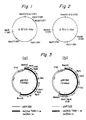
- the two DNAs obtained by double-stranding DNAs from MYMV have a molecular size of 2.67 ⁇ 0.1 kilobase pairs (to be abbreviated as "Kbp") and 2.70 ⁇ 0.1 Kbp and give the restriction endonuclease cleavage maps shown in Figures 1 and 2 respectively of the accompanying drawings.
- DNA fragments shown in the above table may vary over a range of 0.05 Kbp, particularly ⁇ 0.1 Kbp.
- Digestion with restriction enzymes such as Cla I, Sac I, Hpa I, Nar I, Nru I and Mbo I was carried out in a basic solution consisting of 10 mM Tris-HCI (pH 7.9), 7 mM MgCl 2 , 7 mM beta-mercaptoethanol, and 0.01% bovine serum albumin.
- Digestion with Hind III, Ava I, Kpn I, Pst I, Pvu II, Xmn I, Ban II, Sph I, Bgl I, Bcl and Apa I was carried out in a mixture of the above-mentioned basic solution and 50 mM of NaCI.
- Digestion with Sal I, Bam HI, Xba I, Xho I, Nco I, Eco RV, Stu I, Pvu I and Dpn I was carried out in a mixture of the basic solution and 150 mM of NaCI.
- Hind III, Sal I, Bam HI, Kpn I, Pst I, Pvu II, Xho I, Hpa I and Stu I used in this invention were those manufactured by Takara Shuzo Co., Ltd.
- Ava I, Xba I and Dpn I were those manufactured by Bethesda Research Laboratories.
- Nco I and Ban II were those manufactured by Toyobo Co., Ltd.
- Nar I, Naru I and Xmn I were those manufactured by Nippon Gene Co.
- Sph I, Pvu I, Eco RV and Bcl I were manufactured by Boehringer Mannheim Co. Ltd.
- all of the restriction enzymes for cleavage of DNA are used in an amount of at least 4 units per microgram of DNA, and the cleavage is carried out at 37°C for at least 4 hours.
- DNA is to be digested with two restriction enzymes, it is first digested with an enzyme for lower salt concentrations at 37°C for at least 2 hours, and then with an enzyme for higher salt concentrations at 37°C for at least 2 hours.
- Fragment DNAs formed as a result of enzymatic digestion are analyzed by electrophoresis using 1.5% agarose gel containing 0.5 microgram/ml of ethidium bromide.
- zigda-DNA digested with Eco RI/Hind III and plasmid pBR322 digested with Taq I were used as size markers of DNA.
- the dsDNA-a and -b produced as above are substantially entirely double-stranded. Depending upon the manufacturing conditions, these DNAs may partly remain single-stranded. It should be understood that the double-stranded DNAs of this invention include those which partly remain single-stranded as above. Such DNAs can also be used in cloning or propagation as described hereinafter.
- the double-stranded DNAs in accordance with this invention are used, for example, as vectors for plant gene recombination.
- the DNAs are cleaved with certain enzymes. If at least those parts which are to be cleaved are double-stranded, those sites which are not double-stranded are repaired and almost completely double-stranded in the subsequent cloning or propagation. Accordingly, the double-stranded DNAs to be cloned or propagated may be double-stranded at least partly. Desirably, at least 80%, preferably at least 90%, of the single-stranded DNAs may be double-stranded.
- the ratio between dsDNA-a and dsDNA-b differs depending upon the process of propagation of MYMV used as a starting material. Generally, the mole ratio of dsDNA-a to dsDNA-b is 2 or more. Accordingly, the assignment of the individual restriction fragments can be easily determined even by the action of a restriction enzyme on a mixture of dsDNA-a and dsDNA-b.
- the dsDNA-a or -b produced as above is digested with a restriction enzyme capable of cleaving it preferably at a certain site, such as Hind III, Cla I, Bam HI, Bgl II or Pst I inserted into a host vector DNA cleaved with the same restriction enzyme.
- a restriction enzyme capable of cleaving it preferably at a certain site, such as Hind III, Cla I, Bam HI, Bgl II or Pst I inserted into a host vector DNA cleaved with the same restriction enzyme.
- host vector DNAs that can be used in this case include plasmid vectors (such as pBR322, PBR328 and pBR325), cosmid vectors (such as pKY2662) and phage vectors (such as Sharon 10) for E. coli, plasmid vectors (such as pUB110, pUB112, pSA0501 and pTP4) for B. subtilis, and vectors for S
- host vector DNA denotes a vector DNA that can have dsDNA-a or -b inserted thereinto to obtain a recombinant DNA which can be propagated in a host and from which dsDNA TMM-1 or TMM-2 can be reconstructed.
- a vector DNA which has only one site (recognition site) capable of being cleaved with a restriction enzyme capable of cleaving dsDNA-a or -b only at one site.
- a host DNA which when recombined with dsDNA-a or -b, gives a hybrid DNA that self-propagates in a host microorganism such as Escherichia coli, Saccharomyces cerevisiae or Bacillus subtilis.
- host DNAs include pBR322 [F. Bolivar et al. ; Gene 2, 95 (1977)], pBR325 [F. Bolivar, Gene 4,121 (1978)], pBR328 [X. Sokeron et al., Gene, 9, (1980)], pMB9 [Rodriguez et al., Mol. Cell Biol., V. 471, Academic Press, New York (176)], pKY 2662 [L.
- telomere sequence there is only one site of cleavage with a restriction endonuclease both in dsDNA-a or dsDNA-b and the host vector DNA.
- host vector DNAs are pBR322, pBR325, pBR328, YRp7, Ylp32, pYC1, pTA1060, pC194, pC221, pC223, pUB110, pUB112 and pSA0501.
- pBR322, pB325, pBR328, YRp7, Ylp32, pUB110 and pUB112 are especially preferred.
- dsDNA-a or -b is inserted into the plasmid vector to form a hybrid DNA.
- a hybrid-DNA of dsDNA-a and pBR322 can be obtained by completely digesting dsDNA-a or a mixture of dsDNA-a and -b with one cut enzyme for dsDNA-a, for example, Bam HI, mixing the digestion product with the Bam HI-digestion product of a host vector DNA such as E. coli plasmid pBR322, and ligating them with a DNA ligase (such as T4-DNA ligase).
- a DNA ligase such as T4-DNA ligase
- a hybrid DNA of dsDNA-b and pBR322 can be obtained by completely digesting dsDNA-b or a mixture of dsDNA-a and -b with one cut enzyme for dsDNA-b, for example, Hind III, mixing the digestion product with the Hind III-digestion product of pBR322, and ligating them with a DNA ligase.
- one cut enzyme for dsDNA-b for example, Hind III
- the two DNAs to be ligated are mixed in nearly equimolar proportions because this can more efficiently lead to the desired hybrid DNA.
- the host vector DNA after cleavage with a . restriction enzyme, is treated with an alkaline phosphatase to dephosphorlyate the 5' terminal of DNA because this can almost completely prevent the self-ligation of the host vector DNA during the preparation of the hybrid DNA.
- Escherichia coli HB101 is transformed in accordance with the method of M. Mandel and A. Higa [J. Mol. Biol., 53,154 (1970)].
- the transformants are cultivated on an agar (L-Agar) plate containing 50 micrograms/ml of an appropriate antibiotic such as ampicillin or tetracycline.
- the resulting colonies are subjected to colony hybridization in accordance with the method of Grunstein and Hogness (Proc. Natl. Acad. Sci., 72, 3961) using the 32 P-labelled DNA of MYMV as a probe, and those colonies which have DNAs capable of being hybridized with the 32 P-labelled DNA of MYMV are selected.
- the plasmids which the selected colonies have are screened.
- the plasmid DNAs are separated from 1 ml of the colony overnight culture by performing "mini-preparation" by the so-called alkaline lysis method or the boiling lysis method.
- the resulting plasmid DNAs are completely digested with one cut enzyme for dsDNA-a or dsDNA-b, for example, Bam HI or Hind III to select colonies having hybrid DNA containing DNA fragments with a size of about 2.67 Kbp or about 2.70 Kbp.
- one cut enzyme for dsDNA-a or dsDNA-b for example, Bam HI or Hind III to select colonies having hybrid DNA containing DNA fragments with a size of about 2.67 Kbp or about 2.70 Kbp.
- the hybrid DNAs can be separated by a known method.
- the separated hybrid DNAs are almost completely double-stranded. They can be used to transform plant cells after introducing a foreign DNA into their specific site. They can be propagated within bacterial cells such as Escherichia coli cells.
- the desired hybrid DNA can be prepared in large quantities by propagating the bacterial strain containing the hybrid DNA, as required performing an operation of amplifying only the hybrid DNA, subjecting the cells to lysis, and separating the hybrid DNA from an aqueous solution containing the hybrid DNA.
- Only a covalently closed circular (to be referred to as "ccc") hybrid DNA can be isolated by, for example, separating the hybrid DNA from the isolated E. coli transformants containing the hybrid DNA by using the methods of amplification and lysis of a hybrid DNA described at pages 88 to 94 of "Molecular Cloning-A Laboratory Manual" of Maniatis et al. (Cold Spring Harbor Laboratory 1982), and then subjecting it to equilibrium density gradient centrifugation with cesium chloride.
- the isolated hybrid DNA is digested with a restriction enzyme, Bam HI or Hind III to separate it into DNA fragments derived from dsDNA-a or -b and vector DNA fragments, and then isolating the DNA fragments derived from dsDNA-a or -b.
- a restriction enzyme Bam HI or Hind III
- the digestion product is subjected to agarose gel electrophoresis, for example, and a slice of gel containing the DNA band of which DNA size is about 2.67 Kbp is cut off.
- DNA fragments can be separated from the gel and purified.
- the desired cloned dsDNA-a can be obtained by ligating the DNA fragments with, for example, T4-DNA ligase by a method known per se.
- This cloned dsDNA-a forms a band having an apparent molecular size of about 3.3 Kbp by the agarose gel electrophoresis.
- the cloned dsDNA-a can further be purified.
- a cloned dsDNA-b can be obtained by digesting the isolated hybrid DNA with Hind III, subjecting the digestion product to agarose gel electrophoresis in the same way as above, cutting off a slice of gel which contains_about 2.70 Kbp sized DNA and treating the gel in the same way as above.
- the cloned dsDNA-a and -b prepared by the above cloning operation are essentially the same as dsDNAs prepared in vitro from the ssDNAs of MYMV as stated in paragraph (2) above and the replicative form DNA of MYMV to be described hereinbelow in that they show the restriction endonuclease cleavages maps given in Figures 1 and 2 and the cleavage patterns given in Table 1. It has been found however that as shown in Table 2 they exhibit slightly different cleavage patterns with respect to restriction enzymes Dpn I and Mbo I.
- the cloned dsDNA-a and -b are referred to herein as dsDNA-a-1 and -b-1, and the dsDNA-a and -b prepared in vitro or being in the replicative form, as dsDNA-a-2 and -b-2. It should be understood that the dsDNA-a and dsDNA-b of this invention include dsDNA-a-1 and-2 and dsDNA-b-1 and -2.
- the dsDNA-a and -b provided by this invention can be used either singly or in combination as a vector for plant gene recombination. Furthermore, a circular DNA having a foreign DNA at a specified site of dsDNA-a or -b can be formed by cleaving a specified restriction site of dsDNA-a or -b with a restriction enzyme to form a linear DNA, mixing this DNA with a foreign DNA cut out by using the same restriction enzyme in nearly equimolar proportions, and ligating them with a DNA ligase (such as T4-DNA ligase) by a known method.
- a DNA ligase such as T4-DNA ligase
- plant cells can be transformed into kanamycin- or neomycin-resistant cells by using a DNA obtained by inserting the foreign DNA into dsDNA-r or -b.
- the transformation may be effected, for example, by the action of the DNA having the foreign DNA inserted thereinto upon the protoplasts of plant cells at a high pH in the presence of Ca ++ and polyethylene glycol or in the presence of Ca ++ ; or by mechanically introducing the aforesaid DNA into the plant cells (micro-injection).
- the dsDNA-a and -b provided by the present invention can be digested with a restriction enzyme preferably capable of cleaving one site of these DNAs, such as Hind III, Cla I, Bam HI, Bgl II, Bgl I or Pst I, and inserted into a host vector DNA (as exemplified below) cleaved with the same restriction enzyme.
- a restriction enzyme preferably capable of cleaving one site of these DNAs, such as Hind III, Cla I, Bam HI, Bgl II, Bgl I or Pst I
- the host vector DNA used at this time may be those previously known, for example pBR322, pBR325, pBR328, pMB9, pKY2662, and pKC7 of Escherichia coli; YEp13, YRp7, Ylp32, pYC1 and pYC2 of Saccharomyces cerevisiae; and pTA1060, pTA1020, pC194, pC221, pC223, pUB110, pUB112, and pSA0501, and pE194 of Bacillus subtilis.
- Preferred host vector DNAs are those which have only one cleavage site with an endonuclease as does the dsDNA-a or -b of the invention with the same endonuclease.
- Examples are pBR322, pBR325, pBR328, YRp7, pYC1, Ylp32, pC194, pC221, pC223, pUB110 and pSA0501. Of these, pBR322, pBR325, pBR328, YRp7, Ylp32, pUB110 and pUB112 are especially preferred.
- a hybrid DNA can be prepared from the host vector DNA and dsDNA-a or -b by the same method as described before in cloning dsDNA-a or -b into pBR322.
- the hybrid DNA so obtained may be utilized as a vector for various plants.
- Other foreign DNAs may further be inserted into the hybrid DNAs to utilize the latter more advantageously as vectors.
- they can be used for transforming plant cells by inserting other DNAs (such as kanamycin- or neomycin-resistant genes, herbicide-resistant genes, etc.) for imparting useful properties as plants.
- a mixture of dsDNA-a-2 and dsDNA-b-2 is digested with a restriction enzyme, Bam HI (the digestion product is referred to as "dsDNA(a+b)/Bam HI").
- Bam HI the digestion product is referred to as "dsDNA(a+b)/Bam HI"
- YRp7 is digested with Bam HI, and then treated with an alkaline phosphatase to dephosphorylate its 5' terminal (the product is referred to as YRp7/Bam HI").
- the resulting dsDNA(a+b)/Bam HI and YRp7/Bam HI are ligated with T4-DNA ligase.
- E. coli HB101 is transformed with the resulting hybrid DNA, and transformants resistant to ampicillin are selected. From the ampicillin-resistant transformants, those which are both ampicillin-resistant and tetracycline-sensitive are selected.
- the plasmid DNAs of the selected transformants are separated by the mini-preparation technique described above and completely digested with Bam HI.
- the resulting DNA fragments having a size of about 2.67 Kbp are self-ligated with T4-DNA ligase to obtain circular dsDNA-a-1.
- dsDNA-a-2 and -b-2 are digested with a restriction enzyme, Hind III (the digestion product is referred to as "dsDNA(a+b)/Hind III").
- dsDNA(a+b)/Hind III a restriction enzyme
- Ylp32 is digested with Hind III, and its 5' terminal is treated with an alkaline phosphatase (the product is referred to as "YRp7/Hind III").
- the resulting dsDNA (a+b)/Hind III and Ylp32/Hind III are ligated with T4-DNA ligase.
- E. coli HB101 is transformed with the resulting hybrid DNA, and those transformants which are resistant to ampicillin are selected. Then, colonies having DNAs capable of being hybridized with the 32P-labelled DNA of MYMV are selected by the aforesaid colony hybridization method using the 32 P-labelled DNA of MYMV as a probe.
- the hybrid DNA was separated from these selected colonies, and completely digested with Hind III.
- the resulting DNA fragments having a size of about 2.70 Kbp are self-ligated with T4-DNA ligase to obtain circular dsDNA-b-1.
- the replicative DNA of MYMV can be isolated from leaves infected with growing MYMV, for example the infected leaves of Top Crop (such as Top Crop of Takii Seed and Seedling Co., Ltd.) by using the method of D. O. Hamilton et al. [see Nucl. Acids Res. 10, 4902 (1982)] or the whole DNA extraction method described at pages 86 to 88 of Atsushi Hirai et al., "Introduction to Plant Cell Breeding" (a Japanese-language publication) published by Gakkai Shuppan Center.
- Top Crop such as Top Crop of Takii Seed and Seedling Co., Ltd.
- DNA is transferred to a nitrocellulose filter by the Southern method [see E. M. Southern, J. Mol. Biol., 98, 503-517 (1975)]. Then, by the same procedure as described at pages 387 to 389 of "Molecular Cloning-A Laboratory Manual" of Maniatis et al.
- DNA-DNA hybridization is carried out, followed by autoradiography by the same technique as the method described at pages 470 to 471 of the same book.
- the DNAs which are hybridized with the 32 P-labelled DNA of MYMV appear as black bands on an X-ray film by autoradiography. These bands are therefore shown to be DNAs derived from MYMV. There are about 9 such bands observed, and they have an apparent size of >20 Kbp, 10 Kbp, 6.8 Kbp, 5.4 Kbp, 4.0 Kbp, 3.2 Kbp, 2.7 Kbp, 1.6 Kbp and 0.89 Kbp.
- DNAs are taken out from the agarose gel, and isolated from the gel by the same procedure as described hereinabove. When they are digested with any of Bam HI, Bgl II and Bgl IIIPst I, the same DNA fragments shown in Table 1 except a DNA corresponding to 0.89 Kbp can be obtained. Thus, the DNAs which give eight types of bands excepting the DNA corresponding to 0.89 Kbp are replicative DNAs or MYMV based on the DNAs shown in Table 1.
- replicative DNAs are in various forms, such as a covalently closed circle (c.c.c.), an open circle (o.c.), or a linear monomer, dimer or trimer.
- c.c.c. covalently closed circle
- o.c. open circle
- trimer linear monomer, dimer or trimer.
- band of 0.89 Kbp agrees with the single-stranded DNA of MYMV, but the others all correspond to double-stranded DNAs which are the replicative DNAs of MYMV.
- DNA corresponding to 1.6 Kbp, DNA corresponding to 3.2 Kbp, and DNA corresponding to 10.0 Kbp are present in relatively large amounts and can be easily utilized.
- a hybrid DNA containing dsDNA-a-1 can be obtained.
- a hybrid DNA containing dsDNA-b-1 can be obtained by digesting DNA corresponding to 1.6 Kbp, 3.2 Kbp, or 10.0 Kbp with Hind III and cloning it into the Hind III cleavage site of pBR322.
- a hybrid DNA containing dsDNA-a-1 can be obtained by digesting DNA corresponding to 1.6 Kbp, 3.2 Kbp or 10.0 Kbp with Bam HI, cloning it into the Bam HI cleavage site of a S. cerevisiae vector such as YRp7, propagating the clone in E. coli (such as E. coli HB101 strain), and treating the culture by a specified procedure.
- a hybrid DNA containing dsDNA-b-1 can be obtained by digesting DNA corresponding to 1.6 Kbp, 3.2 Kbp or 10.0 Kbp with Hind III, cloning it into the Hind III cleavage site of Ylp32, propagating the clone in E. coli (such as E. coli HB101 strain), and treating the culture by a specified procedure.
- the resulting hybrid DNAs can be used as vectors for various plants, or can be used as advantageous vectors by further inserting other foreign DNAs into them.
- the dsDNA-a-1 and/or dsDNA-b-1 can be isolated from the resulting hybrid DNAs.
- the dsDNA-a-1 and/or dsDNA-b-1 has the same infectivity as MYMV.
- Top Crop Phaseolus vulgaris L. "Top Crop" (53 g) infected with MYMV were ground in 250 ml of 0.1 M sodium phosphate-10 mM EDTA buffer (pH 7.8) (containing 1.3 g of cysteine), and filtered through a double-layered gauze. The filtrate was centrifuged at 10000G for 40 minutes at 4°C to obtain 205 ml of a supernatant. Sodium chloride (2.4 g) and then 8.2 g of polyethylene glycol (having a weight average molecular weight of 7800 to 9000) were added to the supernatant.
- the mixture was stirred at 4°C for 1 hour, and then centrifuged at 10000G and 4°C for 25 minutes. The supernatant was removed, and 10 ml of 0.1 M sodium phosphate buffer (pH 7.8) was added to the precipitated polyethylene glycol pellet portion. The mixture was homogenized and centrifuged at 10000G and 4°C for 30 minutes. The supernatant was centrifuged at 30,000 rpm in a Beckman SW 40.1 rotor to obtain a crude virus as a precipitate.
- the crude virus was homogenized in 0.5 ml of 0.1 M sodium phosphate buffer (pH 7.8), and the mixture was subjected to 10-40% linear sucrose density gradient centrifugation at 32,000 rpm and 4°C for 3 hours using the same SW 40.1 rotor. After the centrifugation, 0.6 ml fractions were separated from the bottom of the centrifugal tube. The absorbances A 160 of the fractions were measured. A peak assigned to the virus was observed in fractions Nos. 9 to 16. These fractions were collected, and 0.1 M sodium phosphate buffer (pH 7.8) was added in an amount twice their amount.
- 0.1M sodium phosphate buffer pH 7.8
- the aqueous layer was subjected to the same phenol extracting operation twice to obtain 950 microliters of the aqueous layer. Then, 700 microliters of chloroform was added to the aqueous layer, and the mixture was shaken for 2 minutes. The aqueous layer was taken out, and 700 microliters of ether was added to the aqueous layer to extract phenol. The extraction was repeated three times. To 1000 microliters of the aqueous layer were added 100 microliters of 3M sodium acetate buffer (pH 4.8) and 2.5 ml of ethanol, and the mixture was maintained for one day at -20°C.
- 3M sodium acetate buffer pH 4.8
- the mixture was then centrifuged at 35,000 rpm for 20 minutes by a Beckman ultracentrifugal SW 50.1 rotor to precipitate the DNA.
- the DNA was dissolved in 400 microliters of 10 mM Tris-HCI/0.1 mM EDTA aqueous solution to form an aqueous solution of a single-stranded DNA of MYMV in a concentration of 0.1 microgram/microliter.
- pB10205 a product of Amersham Japan Company
- AMV reverse transcriptase 5 units/microliter; Code 120248 of Seikagaku Kogyo Co., Ltd.
- the gel filtration was carried out by using a column having a diameter of 6 mm filled with 5 ml of Sephadex G-75 (a product of Pharmacia Fine Chemicals) equilibrated with 10 mM Tris-HCI (pH 7.4)-0.1 M NaCI aqueous solution.
- the aqueous layer was passed through the column and four-drop fractions were collected.
- the 32p intensity of each of the fractions was measured.
- the first peak appeared in fractions Nos. 6 to 12, and in fraction No. 14 and subsequent fractions, a peak assigned to the unreacted alpha - 32 P - deoxycytidine triphosphate was observed.
- the resulting DNA was dissolved in 100 microliters of 1 mM Tris-HCI (pH 7.4)/0.1 mM EDTA aqueous solution, and a DNA double-stranded in vitro was obtained (its 32 P intensity was 32x10 4 cpm).
- DNA (66 mg) of calf thymus was dissolved in 6.6 ml of a mixture of 0.1M NaCI, 10 mM MgCI 2 and 10 mM Tris-HCI (pH 7.4), and 460 micrograms of DNase I (a product of Millipore Corporation; 2182 units/g) was added and incubated at 37°C for 3 hours. The reaction was stopped by adding 0.66 ml of 0.2M EDTA. The reaction mixture was subjected to protein extraction with 7 ml of water-saturated phenol three times, and then the aqueous layer was extracted with three 7 ml portions of ether to remove phenol. Ethanol (20 ml) was added to the resulting aqueous layer.
- the mixture was left to stand at -20°C for 2 hours, and centrifuged at 4000G for 4 minutes to obtain DNA pellets.
- the DNA pellets were dissolved in 0.5 ml of 0.1 M NaCI and 10 mM Tris-HCI (pH 7.4), and subjected to gel filtration on a column (length 42 cm, diameter 0.5 cm) of Sephadex G-75 (a product of Pharmacia Fine Chemicals) equilibrated with 0.1M NaCl and 10 mM Tris-HCI (pH 7.4). About 1.2 ml fractions were collected. A large peak was observed in fractions Nos. 10 to 32. Fractions Nos.
- Double stranding was carried out in the same way as in experiment Ic above except that 40 microliters of a primer mixture (DNA concentration 5 microgram/microliter) from calf thymus DNA prepared by the method of J. M. Taylor et al. [Biochimica et Biophysica Acta, 442, 325 (1976)] was used instead of 12 microliters of the oligonucleotides (16.6 microgram/microliter) in experiment Ic, and sterilized water was used in an amount of 55 microliters.
- the polymeric DNA portion had a 32p intensity of 13x10 4 cpm.
- the double-stranded DNA obtained above was digested with each of restriction enzymes Bam HI, Hind III, Xba I and Bgl II and then subjected to autoradiography under the same conditions as in experiment Id described hereinbelow.
- the Hind III-digested DNA did not at all move from the well position of the agarose gel. Some DNAs digested with the other restriction enzymes stayed at the well position. The DNA which migrated considerably smeared the gel, and the band was not as clear as in experiment ld.
- Electrophoresis was carried out on a 5 mm-thick horizontal gel at a voltage of 1.5 V/cm for 11 to 15 hours. At the time of electrophoresis, a product obtained by completely digesting 0.5 microgram of lambda-DNA with Eco RI and Hind III and a product obtained by completely digesting 0.2 microgram of pBR322 DNA with Taq I were used as size markers for DNA fragments.
- the agarose gel was withdrawn, dried on a gel drying plate, and subjected to autoradiography by the procedure described at pages 470 to 472 of the Manual of Maniatis et al. described hereinabove.
- the sizes of the resulting DNA fragments are as shown in Table 1 given hereinabove.
- DNA fragments which appeared high in density for their sizes when observed after autoradiography are classified as dsDNA-a and those which appeared low in density for their sizes, as dsDNA-b.
- the parenthesized Figure 3.3 shows that the DNA is open circular, and has an apparent molecular size of 3.3 Kbp with respect to the linear DNA size marker. It is presumed from this table that DNA which forms the fragments of dsDNA-a was open-circular and had a size of about 2.67 Kbp before digestion with restriction enzymes. The correct size can be known by determining the base sequences. But by methods available at present, a measuring error of 0.05 Kbp at the least cannot be avoided. The size of DNA which forms the fragments of dsDNA-a can be regarded as about 2.67 ⁇ 0.1 Kbp. From a similar analysis, DNA which produces the fragments of dsDNA-b can be regarded as being open-circular and having a size of 2.70 ⁇ 0.1 Kbp before digestion with restriction enzymes.
- the supernatant was discarded, and 5 ml of 75% ethanol kept at -20°C was freshly added.
- the mixture was centrifuged at 20,000 rpm for 2 minutes.
- the supernatant was discarded to form DNA pellets.
- the DNA pellets were dried in vacuum at 2 mmHg for 2 minutes, and dissolved in 50 microliters of distilled water.
- To 50 microliters of this DNA solution were added 6 microliters of a buffer for Bam HI (described in experiment lc) and 6 microliters of Bam HI.
- the reaction was carried out at 37°C for 4 hours.
- 10 microliters of a solution of yeast tRNA (2 microgram/microliter) and 70 microliters of distilled water were added.
- 100 microliters of water-saturated phenol was added.
- the mixture was shaken and centrifuged.
- the aqueous layer was taken out, and extracted with ether three times to remove the phenol.
- a solution (DNA 1 microgram/microliter) of the Bam HI-digested/alkaline phosphatase-treated product of pBR322, 2 microliters of a DNA ligation buffer [an aqueous solution containing 650 mM Tris-HCI (pH 7.4), 65 mM MgCl 2 , 10 mM DTT, 5 mM ATP and 40 mM Spermine) and 0.5 microliter of T4-DNA ligase (1.2 units/microliter; Code No. 2010B of Takara Shuzo Co., Ltd.). The reaction was carried out at 14°C for 22 hours. Then, the reaction mixture was treated at 65°C for 5 minutes to obtain a hybrid DNA.
- a DNA ligation buffer an aqueous solution containing 650 mM Tris-HCI (pH 7.4), 65 mM MgCl 2 , 10 mM DTT, 5 mM ATP and 40 mM Spermine
- the aqueous layer were added 30 microliters of 3M sodium acetate and 1100 microliters of ethanol. The mixture was left to stand at -20°C for 2 hours, and centrifuged at a high speed. The resulting DNA pellets were dried, and dissolved in 50 microliters of distilled water. The solution was the solution (DNA 1 microgram/microliter) of the Bam HI-digested/alkaline phosphatase-treated product of pBR322.
- a single colony of E. coli HB101 was transferred to 5 ml of L-broth medium, and cultivated with shaking at 37°C for 11 hours. Two milliliters of the culture broth was inoculated in 200 ml of fresh L-broth, and cultivated with shaking at 37°C for 2 hours and 20 minutes. When the OD 600 of the culture broth became 0.40, the culture broth was cooled to 0°C and centrifuged at 5,000 rpm for 5 minutes by a Tomy cooling high-speed centrifuge (No. 9 rotor). The supernatant was discarded. The precipitated E. coli was suspended in 50 ml of 10 mM auqeous NaCI solution, and again centrifuged at a high speed (No.
- the resuspended solution was centrifuged at a high speed (No. 4 rotor; 4,000 rpm for 5 minutes at 4°C). The supernatant was discarded, and 10 ml of an aqueous solution of 30 mM CaC1 2 and 15% glycerol was added to the bacterial pellets. The entire mixture was gently resuspended, and poured into 1.5 ml Eppendorf tubes as 200 microliter portions. They were stored in the frozen state at -80°C. The E. coli HB 101 cells treated with CaC1 2 were called competent cells.
- the competent cells were transformed. Specifically, the competent cells were brought to a temperature of 0°C, and about 10 minutes later, the aqueous solution of the hybrid DNA prepared above was added. The mixture was maintained at 0°C for 40 minutes and then heated at 42°C for 2 minutes. Then, 1.2 ml of L-broth was added, and the mixture was maintained at 37°C for 1 hour. The culture fluid was spread over eight L-agar plates (diameter 9 cm) containing 50 micrograms/ml of ampicillin at a rate of about 200 microliters per plate. The plates were maintained at 37°C for 16 hours to obtain 86 colonies of transformed HB101.
- Plasmid DNAs containing DNAs having a size of 2.67 Kbp, 2.02 Kbp and 0.67 Kbp were obtained.
- the fragment having a size of 2.67 Kbp was obtained by cleavage of dsDNA-a with Bam HI, and the fragments having sizes of 2.02 and 0.67 Kbp were presumably derived from two kinds of fragments generated by the digestion of dsDNA-b with Bam HI.
- a hybrid DNA of the fragment having a size of 2.67 Kbp and pBR322 is designated as pMYB4; a hybrid DNA of the fragment having a size of 2.02 Kbp and pBR322, as pMYB5; and a hybrid DNA of the fragment having a size of 0.67 Kbp and pBR322, as pMYB3:
- the DNAs of pMYB4, pMYB3 and pMYB5 were obtained by replication and propagation in the host E. coli of pBR322 and were in the completely double-stranded state.
- the hybrid D NA p MYB4 was completely digested with Bam HI, Bam HI/Bgl II, Bam HI/Bgl II/Cla I, Bam HI/Xba I, and Bam HI/Xba I/Bgl II, and subjected to 1.2% agarose gel electrophoresis (the digestion product of lambda-DNA with Eco RI/Hind III and the digestion product of pBR322 with Taq I were also subjected to electrophoresis as size markers). The fragment sizes of DNA were analyzed, and the results shown in Table 3 were obtained.
- the hybrid DNA pMYB5 was digested with Bam HI, Bam HI/Hind III/Bgl II and Bam HI/Hind III/Pst I, and the hybrid DNA pMYB3 was digested with Bam HI and Bam HI/Hind III. The same analysis as above was carried out. The results shown in Table 3 were obtained.
- the parenthesized figures are for DNAs derived from pBR322.
- the sizes of the fragments formed by the digestion of pMYB4 with the restriction enzymes shown in Table 3 contained measurement errors of a maximum of about ⁇ 0.1 Kbp.
- pMYB2 Another kind of a hybrid DNA having the Bam HI fragment with a size of 2.67 Kbp at the Bam HI site of pBR322 is designated as pMYB2.
- Digestion of pMYB2 with Cla I gave fragments having a size of 5.4 and 1.6 Kbp.
- Digestion of pMYB4 with Cla I gave fragments having a size of 5.2 and 1.8 Kbp.
- the Bam HI fragments having a size of 2.67 Kbp were taken out from pMYB4 and pMYB2 by the method described hereinbelow. Digestion of the fragments with Bgl II gave three fragments having a size of 1.67, 0.74 and 0.3 Kbp from each of the 2.67 Kbp fragments. Digestion of the 2.67 Kbp fragments with Cla I gave fragments having a size of 1.42 and 1.24 Kbp, respectively.
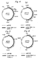
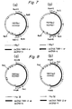
- pMYB4 and pMYB2 are hybrid DNAs which were formed by the insertion of the Bam HI cleavage product of dsDNA TMM-1 into the Bam HI site of pBR322 in opposite directions as shown in Figures 3-(a) and 3-(b).
- the hybrid DNA pMYH3 was digested with Hind III, Hind III/Bam HI, Hind III/Pst I and Hind III/Bam HI/Bgl II, and the resulting fragments were analyzed in the same way as in experiment le. The sizes of the fragments obtained are shown in Table 4.
- the parenthesized figures are for DNAs derived from pBR322.
- Hind III-digested fragments having a size of 2.70 Kbp were taken out from pMYH3 and pMYH14 by the method described hereinbelow, and digested with Bam HI. Fragments having a size of 1.10, 0.96 and 0.67 Kbp were formed from each of the 2.70 Kbp fragments. Digestion with Bgl gave fragments having a size of 1.92 and 0.78 Kbp from each of the 2.70 Kbp fragments.
- pMYH3 and pMYH14 are hybrid DNAs formed by the insertion of Hind III-cleaved dsDNA TMM-2 into the Hind III site of pBR322 in opposite directions, as shown in Figures 4-(a) and -(b).
- hybrid DNA pMYB2, pMYB4, pMYH3 and pMYH14 obtained in the experiments le and If are hybrid DNAs of dsDNA TMM-1 and dsDNA TMM-2 and pBR322. They can also be vectors for plant gene recombination.
- DNAs were extracted from them by the method described at pages 164 to 165 of the aforesaid Manual of Maniatis et al., and then purified by the method described at page 166 of the same Manual.
- the purified DNAs were each dissolved in 50 microliters of sterilized water (DNA about 0.1 microgram/ microliter).
- To 20 microliters of each DNA aqueous solution were added 5 microliters of the aforesaid DNA ligation buffer, 25 microliters of sterilized water and 0.5 microliter of T4-DNA ligase, and they were reacted at 12°C for 19 hours.
- the product was subjected to 0.8% agarose gel electrophoresis in the same way as above.
- the gel slice having a DNA band portion with an apparent size of 3.3 Kbp was cut off, and the DNA was extracted and purified in the same way as above. It is assumed that the DNAs so purified were circular dsDNA TMM-1 and TMM-2.
- Sterilized water (10 microliters) was added to each of the DNAs, and a 2 microliter portion of the solution was subjected to partial digestion with Bam HI or Hind III.
- the partial digestion was carried out by adding 5 microliters of the aforesaid restriction enzyme reaction buffer, 43 microliters of sterilized water and 0.5 unit of Bam HI (for dsDNA TMM-1) or 0.5 unit of Hind III (for dsDNA TMM-2) to 2 microliters of the DNA solution, and reacting them at 37°C for 5, 10 and 30 minutes respectively.
- Each of the reaction products was subjected to 0.8% agarose gel electrohporesis and changes in size were examined.
- the dsDNA TMM-1 and dsDNA TMM-2 so obtained were each digested with the enzymes indicated in Tables 5 and 6. Digestion was carried out at 37°C for 2 to 4 hours using 0.4 microgram of each DNA, the restriction enzyme reaction buffer and 4 to 8 units of each restriction enzyme. After complete digestion, the product was subjected to 1.5% agarose gel electrophoresis in the same way as in ld. The gel was stained with ethidium bromide and irradiated with UV 254 nm to detect DNA bands. The DNA fragments formed by digestion had the sizes shown in Tables 5 and 6.
- fragment sizes with Dpn I were those of fragments which could be detected. Not all fragments with Dpn I could be detected since they were large in number and small in size.
- the mixture was left to stand at -20°C for 5 hours, and then centrifuged at a high speed to sediment DNA pellets.
- To the DNA pellets was added 500 microliters of sterilized water (DNA concentration 0.47 microgram/microliter).
- DNA concentration 0.47 microgram/microliter 500 microliters of sterilized water.
- One hundred microliter of the resulting DNA solution was subjected to 0.8% agarose gel electrophoresis by the same technique as in experiemnt Ig.
- DNA corresponding to a band with an apparent size of 1.6 Kbp, DNA corresponding to a band with an apparent size of 3.2 Kbp, DNA corresponding to a band with an apparent size of 5.4 Kbp and DNA corresponding to a band with an apparent size of 10 Kbp which were hybridized with a 32 P-labelled DNA probe (the 32 P-labelled probe is described in experiment li) of MYMV were taken out from the gel and purified by the same technique as in lg. (Portions of the gel corresponding to bands with an apparent size of 5.4 Kbp and 10 Kbp were cut off by using the Eco RI/Hind III digestion product of lambda-DNA as a size marker).
- the four DNAs were each digested with Bam HI, Bgl and Bgl II/Pst I, and then subjected to agarose gel electrophoresis.
- the digestion patterns were anlyzed.
- the digested DNAs were transferred to a nitrocellulose filter after agarose gel electrophoresis, and by using the 32 P-labelled DNA of MYMV, DNA-DNa hybridization and autoradiography were carried out. Then, the digestion patterns of the DNAs were analyzed.
- the digestion patterns were 2.66, 2.01 and 0.62 Kbp by digestion with Bam HI, 2.71,1.64 and 1.04 Kbp by digestion with Bgl II, and 2.27, 1.67, 1.02 and 0.44 Kbp by double digestion with Bgl II/Pst I. These are the same as the digestion patterns of dsDNA-a and dsDNA-b with these restriction enzymes. This means that these replicative DNAs contain DNA-a or DNA-b and can be used as vectors for plant gene recombination.
- a nitrocellulose filter (BA85 manufactured by Schleicher und Schiill Company; this is the same as the nitrocellulose filter as described hereinabove) was placed on an L-agar plate in which the colonies of the transformants formed. Then, the nitrocellulose filter was removed, and dipped for 1 minute in 0.5M sodium hydroxide and 1.5M sodium chloride and then in 1M Tris-HCI (pH 7.0). The nitrocellulose filter was then heat-treated at 80°C and 1 mmHg for 2 hours.
- the heat-treated filter was dipped in a prehybridization solution [consisting of 0.9M NaCI, 0.09M sodium citrate, 0.02% polyvinyl pyrrolidone (Plasdone NP K-30, a product of Wako Pure Chemicals, Co., Ltd.), 0.02% Ficoll (Ficoll 400 prepared by Pharmacia Fine Chemicals), and 0.02% aqueous solution of bovine serum aluminum] and pre-treated at 63°C for 30 minutes.
- a prehybridization solution consisting of 0.9M NaCI, 0.09M sodium citrate, 0.02% polyvinyl pyrrolidone (Plasdone NP K-30, a product of Wako Pure Chemicals, Co., Ltd.), 0.02% Ficoll (Ficoll 400 prepared by Pharmacia Fine Chemicals), and 0.02% aqueous solution of bovine serum aluminum
- the prehybridization solution was removed, and a hybridization solution prepared by adding SDS (final 2%) and yeast tRNA (final 40 micrograms/ml) to the prehybridization solution and 200x10 4 cpm of 32 P probe of MYMV described below were added.
- SDS final 2%)
- yeast tRNA final 40 micrograms/ml
- the nitrocellulose filter treated as above was put in this solution, and DNA-DNA hybridization was carried out at 63°C for 24 hours.
- the filter was then taken out, and shaken slowly at 55°C for 20 minutes in large amounts of 0.45M NaCI and 0.045M sodium citrate. This washing was carried out three times.
- the filter was then dried, and autoradiography was carried out by the same technique as described at pages 470 to 472 of the aforesaid manual of Maniatis et al. About 8 colonies were observed which were hybridized with 32 P-labelled DNA of MYMV.
- a mini-preparation of a plasmid DNA was carried out by the boiling lysis method described above, and the plasmid DNA was digested with Hind III. There were obtained hybrid plasmids containing a fragment with a size of 2.70 Kbp from six colonies. One of these hybrid DNAs was designated as pMYRFHI.
- DNA extracted from a band portion having an apparent size of 3.2 Kbp in experiment Ih was digested with Bam HI, and treated with the same Bam HI-digested/alkaline phosphatase-treated product of pBR322 as in experiment le to obtain a hybrid DNA.
- E. coli was transformed with the resulting hybrid DNA. Plasmids were extracted from the transformants obtained in the same way as in experiment le, and analyzed in the same way as in experiment le.
- hybrid DNAs, pMYRFB1, pMYRFB3 and pMYRFB4, containing Bam HI-digested fragments having a size of 2.68,2.03 and 0.68 Kbp at the Bam HI site of pBR322 were obtained as in experiment le.
- pMYRFB1 When pMYRFB1 was digested with Bam HI/Bgl II and Bam HI/Xba I, the same DNA fragments as in Table 3 were formed. It can be concluded from the results of experiments li and Ij that the present replicative DNAs contain dsDNA-a and -b. Further it can be concluded that pMYRFH1 is a hybrid DNA from dsDNA-b and pBR322, and pMYRFB1, from dsDNA-a and pBR322. By using the technique of this experiment, dsDNA-a and dsDNA-b can be individually separated, and propagated.
- the double-stranded DNA of MYMV was prepared in the same way as in experiment lc.
- coli HB101 were transformed by the same operation as in experiment le to obtain 22 transformants.
- a mini-preparation of plasmid DNA was carried out on the 22 colonies by the alkaline lysis method described at pages p368 to p369 of the aforesaid manual of Maniatis et al., and the plasmid DNA was digested with Bam HI. Plasmids containing a fragment with a size of 2.67 Kbp were obtained (two of the hybrid plasmid DNAs were designated as pMYB81 and pMYB82). The hybrid DNA pMYB81 and pMYB82 were obtained by replication and propagation in E. coli and were in the completely double-stranded state.
- Bam HI-digested fragments having a size of 2.67 Kbp were taken out from pMYB81 and pMYB82 by the aforesaid method, and digested with Cla I. Fragments having a size of 1.43 and 1.24 Kbp were formed from each of the 2.67 Kbp fragments.
- pMYB81 and pMYB82 are hybrid DNAs formed by the insertion of Bam HI-cleaved dsDNA TMM-1 into the Bam HI site of pBR328 in opposite directions.
- the hybrid DNA pMYB81 is shown in Figure 5.
- the hybrid DNAs pMYB81 and pMYB82 obtained in this experiment are hybrid DNAs of dsDNATMM-1 and pBR328. They can also be vectors for plant gene recombination.
- the double-stranded DNA of MYMV was prepared as in experiment lc.
- competent cells of E. coli HB101 were transformed by the same operation as in experiment le except that 25 micrograms/ml of tetracycline was used instead of 50 micrograms/ml of ampicillin. Thus, 51 transformants were obtained.
- a mini-preparation of plasmid DNA was carried out on the 51 colonies by the alkaline lysis method as in experiment lk, and the plasmid DNA was digested with Pst I. Eleven plasmids containing fragments with a size of 2.70 Kbp were obtained (two of the hybrid plasmid DNAs were designated as pMYP81 and pMYP82).
- pMYP81 and pMYP82 were hybrid DNAs containing the whole of dsDNA TMM-2.
- the hybrid DNAs pMYP81 and pMYP82 were obtained by replication and propagation in E. coli and were in the completely double-stranded state.
- a Pst I-digested fragment having a size of 2.70 Kbp was taken out from each of pMYP81 and pMYP82 by the aforesaid method, and digested with Bam HI. Fragments having a size of 1.30, 0.73 and 0.67 Kbp were formed.
- pMYP81 and pMYP82 are hybrid DNAs formed by the insertion of Pst 1-cleaved dsDNA TMM-1 into the Pst I site of pBR328 in opposite directions.
- the hybrid DNA pMYP81 is shown in Figure 6.
- the hybrid DNAs pMYP81 and pMYP82 obtained in this experiment are hybrid DNAs of dsDNA TMM-2 and pBR328. They can also be vectors for plant gene recombination.
- DNA extracted from a band portion having an apparent size of 3.2 Kbp in experiment Ih was digested with Bam HI, and treated with the same Bam HI-digested/alkaline phosphatase-treated product of pBR325 (to be described below) as in experiment Ik to obtain a hybrid DNA.
- E. coli was transformed with the resulting hybrid DNA. Plasmids were extracted from 32 transformants obtained in the same way as in experiment Ik, and analyzed in the same way as in experiment le. In this manner, hybrid DNAs, pMYRFB51, pMYRFB52, pMYRFB53 and pMYRFB54, containing Bam HI-digested fragments having a size of 2.67, 2.67, 2.03 and 0.68 Kbp respectively at the Bam HI site of pBR325 were obtained as in experiment le.
- both of the plasmids gave the same DNA fragments having a size of (5.6), (0.3), 1.4 and 1.24 Kbp. (The parenthesized fragments are assigned to pBR325).
- pMYRFB51 and pMYRFB52 are hybrid DNAs formed by the insertion of Bam HI-cleaved dsDNA-a into the Bam HI site of pBR325 in opposite directions.
- These two hybrid DNAs can also be vectors for plant gene recombination.
- Replicative DNA (same as in experiment Im; apparent size 3.2 Kbp) was digested with Pst I and ligated with the Pst I-digested/alkaline phosphatase-treated product of pBR325 (to be described below) as in experiment II to obtain a hybrid DNA.
- E. coli HB101 was transformed with the hybrid DNAs. Plasmids were extracted from 17 transformants obtained as in experiment II, and analyzed in the same way as in experiment le. In this manner, hybrid DNAs pMYRFP51 and pMYRFP52 having a size of 2.70 Kbp at the Pst I site of pBR325 were obtained.
- both of the plasmids gave the same DNA fragments having a size of (3.2), (2.7), 1.30, 0.73 and 0.67 Kbp. (The parenthesized fragments are assigned to pBR325).
- pMYP51 and pMYP52 are hybrid DNAs formed by the insertion of Pst I-cleaved dsDNA-b into the Pst I site of pBR325 in opposite directions.
- These two hybrid DNAs can also be vectors for plant gene recombination.
- the double-stranded DNA of MYMV was prepared as in experiment Ic.
- the hybrid DNa, pMYC1 and pMYC2 were obtained by replication and propagation in E. coli and were in the completely double-stranded state.
- a Cla I-digested fragment having a size of 2.67 Kbp was taken out from each of pMYC1 and pMYC2 by the aforesaid method, and digested with Bam HI. Fragments having a size of 1.43 and 1.24 Kbp were formed. Digestion with Bgl II gave fragments having a size of 1.04, 0.97 and 0.68 Kbp.
- pMYC1 and pMYC2 are hybrid DNAs formed by the insertion of Cia I-cleaved dsDNA TMM-1 into the Cla I site of pBR322 in opposite directions.
- the hybrid DNA pMYC1 and pMYC2 obtained in this experiment are hybrid DNAs of dsDNA TMM-1 and pBR322. They can also be vectors for plant gene recombination.
- the double-stranded DNA of MYMV was prepared as in experiment lc.
- the double-stranded DNA was digested similarly with 3 microliters of Bam HI, and 1 microliter of a solution of the Bam HI-digested/alkaline phosphatase-treated product of YRp7 (DNA 1 microgram/microliter) was used instead of 1 microliter of the solution of the Bam HI-digested/alkaline phosphatase-treated product of pBR322 (DNA 1 microgram/microliter). Otherwise, a hybrid DNA was constructed and the competent cells of E. coli HB101 were transformed with it in the same way as in experiment le.
- the solution of the Bam HI-digested/alkaline phosphatase-treated product of YRp7 was prepared as in experiment le by using YRp7 DNa instead of pBR322 DNA. Then, by the same operation as in experiment le, the Bam HI-digested product was treated with BAP.
- the competent cells of E. coli HB101 were transformed by the same procedure as in experiment le using the hybrid DNA obtained. Thus, 12 transformants were obtained. A mini-preparation of a plasmid DNA was carried out on the 12 transformants by the same boiling lysis method, and the plasmid DNAs were digested with Bam HI. Four hybrid plasmids containing a fragment with a size of 2.67 Kbp were obtained. Two of them were designated as YMYBp1 and YMYBp2. When YMYBp1 was digested with Bgl II, fragments with a size of 5.1, 1.77 and 1.65 Kbp were formed.
- YMYBp1 and YMYBp2 are hybrid DNAs formed by the insertion of the Bam HI-cleaved DNA into the Bam HI site of YRp7 in opposite directions.
- the double-stranded DNA of MYMV was digested with 3 microliters of Hind III, and 1 microliter (DNA 1 microgram/microliter) of a solution of the Hind III-digested/alkaline phosphatase-treated product of Ylp32 (to be described below) was used instead of 1 microliter of the solution of the Bam HI digested/alkaline phosphatase-treated product of pBR322 (DNA 1 microgram/microliter).
- a hybrid DNA was constructed and the competent cells of E. coli HB101 were transformed.
- the solution of the Hind III-digested/alkaline phosphatase-treated product of Ylp32 was prepared in the same way as in experiment If except that Ylp32 was used instead of pBR322.
- the Ylp32 DNA was digested with Hind III and then treated with BAP.
- Hind III fragments having a size of 2.7 Kbp were taken out from YMYHp1 and YMYHp2, and digested with Pst I, fragments having a size of 2.4 and 0.34 Kbp were formed from each of the 2.7 Kbp fragments.
- YMYHp1 and YMYHp2 are hybrid DNAs formed by the insertion of the Hind III-digested DNA of dsDNA TMM-2 into the Hind III site of Ylp32 in opposite directions, as shown in Figures 8-(a) and -(b).
Landscapes
- Health & Medical Sciences (AREA)
- Life Sciences & Earth Sciences (AREA)
- Genetics & Genomics (AREA)
- Engineering & Computer Science (AREA)
- Biomedical Technology (AREA)
- Biotechnology (AREA)
- Organic Chemistry (AREA)
- Wood Science & Technology (AREA)
- General Engineering & Computer Science (AREA)
- Chemical & Material Sciences (AREA)
- Bioinformatics & Cheminformatics (AREA)
- Zoology (AREA)
- Molecular Biology (AREA)
- Biophysics (AREA)
- Microbiology (AREA)
- Plant Pathology (AREA)
- Biochemistry (AREA)
- General Health & Medical Sciences (AREA)
- Physics & Mathematics (AREA)
- Mycology (AREA)
- Virology (AREA)
- Cell Biology (AREA)
- Saccharide Compounds (AREA)
- Micro-Organisms Or Cultivation Processes Thereof (AREA)
Applications Claiming Priority (2)
| Application Number | Priority Date | Filing Date | Title |
|---|---|---|---|
| JP58161945A JPS6054683A (ja) | 1983-09-05 | 1983-09-05 | 新規dνa及びハイブリツドdνa |
| JP161945/83 | 1983-09-05 |
Publications (2)
| Publication Number | Publication Date |
|---|---|
| EP0136573A1 EP0136573A1 (en) | 1985-04-10 |
| EP0136573B1 true EP0136573B1 (en) | 1990-01-31 |
Family
ID=15745026
Family Applications (1)
| Application Number | Title | Priority Date | Filing Date |
|---|---|---|---|
| EP84110455A Expired EP0136573B1 (en) | 1983-09-05 | 1984-09-03 | Novel double-stranded dna |
Country Status (4)
| Country | Link |
|---|---|
| US (1) | US4789636A (cg-RX-API-DMAC10.html) |
| EP (1) | EP0136573B1 (cg-RX-API-DMAC10.html) |
| JP (1) | JPS6054683A (cg-RX-API-DMAC10.html) |
| DE (1) | DE3481200D1 (cg-RX-API-DMAC10.html) |
Families Citing this family (3)
| Publication number | Priority date | Publication date | Assignee | Title |
|---|---|---|---|---|
| JPS61202692A (ja) * | 1985-03-06 | 1986-09-08 | Teijin Ltd | 新規なdna及びハイブリツドdna |
| JPS62126982A (ja) * | 1985-11-28 | 1987-06-09 | Teijin Ltd | 新規なdna及びハイブリツドdna |
| CN101918561A (zh) * | 2007-12-07 | 2010-12-15 | 国立植物基因组研究所 | 绿豆黄色花叶病印度病毒(mymiv)的嵌合构建体和其应用 |
Family Cites Families (1)
| Publication number | Priority date | Publication date | Assignee | Title |
|---|---|---|---|---|
| US4237224A (en) * | 1974-11-04 | 1980-12-02 | Board Of Trustees Of The Leland Stanford Jr. University | Process for producing biologically functional molecular chimeras |
-
1983
- 1983-09-05 JP JP58161945A patent/JPS6054683A/ja active Granted
-
1984
- 1984-09-03 DE DE8484110455T patent/DE3481200D1/de not_active Expired - Lifetime
- 1984-09-03 EP EP84110455A patent/EP0136573B1/en not_active Expired
- 1984-09-05 US US06/647,829 patent/US4789636A/en not_active Expired - Fee Related
Also Published As
| Publication number | Publication date |
|---|---|
| US4789636A (en) | 1988-12-06 |
| JPS6054683A (ja) | 1985-03-29 |
| EP0136573A1 (en) | 1985-04-10 |
| JPH0258915B2 (cg-RX-API-DMAC10.html) | 1990-12-11 |
| DE3481200D1 (de) | 1990-03-08 |
Similar Documents
| Publication | Publication Date | Title |
|---|---|---|
| EP0136521B1 (en) | Novel, double-stranded dna | |
| EP0571741A2 (en) | An expression plasmid for seeds | |
| JP2690295B2 (ja) | 植物遺伝子の発現 | |
| DE3889546T2 (de) | Transformation von keimenden pflanzensamen mit hilfe von agrobacterium. | |
| Ream et al. | Multiple mutations in the T region of the Agrobacterium tumefaciens tumor-inducing plasmid. | |
| Takenaka et al. | Direct transformation and plant regeneration of the haploid liverwort Marchantia polymorpha L. | |
| DE3751393T2 (de) | Pflanzen, resistent gegen den Anti-sens RNS aufweisenden Virus. | |
| WO1985004899A1 (en) | Methods and vectors for transformation of plant cells | |
| US5073675A (en) | Method of introducing spectinomycin resistance into plants | |
| Haymes et al. | Agrobacterium-mediated transformation ofAlpine'Fragaria vesca, and transmission of transgenes to R1 progeny | |
| CN110317828A (zh) | 修饰水稻OsSWEET基因启动子培育广谱抗白叶枯病水稻的方法 | |
| JPWO2001025459A1 (ja) | 植物の形質転換用ベクター | |
| KR102675540B1 (ko) | 유전자 교정을 이용한 병 저항성이 조절된 토마토 식물체의 제조방법 및 상기 제조방법에 의해 제조된 토마토 식물체 | |
| Suma et al. | Agrobacterium tumefaciens mediated transformation and regeneration of ginger (Zingiber officinale Rosc.) | |
| EP0899340A2 (de) | Virus/Herbizidresistenz-Gene, Verfahren zu ihrer Herstellung und ihre Verwendung | |
| US5217902A (en) | Method of introducing spectinomycin resistance into plants | |
| DE3751099T2 (de) | Gegen Viren resistente Pflanzen mit einem viralen Überzugsprotein. | |
| CN119913200B (zh) | 一种负义rna病毒植物基因组编辑系统及其应用 | |
| US4789636A (en) | Double-stranded DNA having sequences complementary to a single-stranded DNA and derived from mungbean yellow mosaic virus | |
| Sam et al. | DESIGN AND TRANSFER OF OsSWEET14-EDITING T-DNA CONSTRUCT TO BAC THOM 7 RICE CULTIVAR. | |
| Senior et al. | Transformation of Antirrhinum majus using Agrobacterium rhizogenes | |
| Hood et al. | T-DNA presence and opine production in tumors of Picea abies (L.) Karst induced by Agrobacterium tumefaciens A281 | |
| CN119193687A (zh) | 一种利用农杆菌介导法提高小麦遗传转化效率的方法 | |
| EP0841402A2 (en) | High capacity binary shuttle vector | |
| CN116926109A (zh) | 一种植物程序化花粉自清除CRISPR/Cas基因编辑方法 |
Legal Events
| Date | Code | Title | Description |
|---|---|---|---|
| PUAI | Public reference made under article 153(3) epc to a published international application that has entered the european phase |
Free format text: ORIGINAL CODE: 0009012 |
|
| AK | Designated contracting states |
Designated state(s): DE FR GB |
|
| 17P | Request for examination filed |
Effective date: 19850814 |
|
| 17Q | First examination report despatched |
Effective date: 19871117 |
|
| GRAA | (expected) grant |
Free format text: ORIGINAL CODE: 0009210 |
|
| AK | Designated contracting states |
Kind code of ref document: B1 Designated state(s): DE FR GB |
|
| REF | Corresponds to: |
Ref document number: 3481200 Country of ref document: DE Date of ref document: 19900308 |
|
| ET | Fr: translation filed | ||
| PLBE | No opposition filed within time limit |
Free format text: ORIGINAL CODE: 0009261 |
|
| STAA | Information on the status of an ep patent application or granted ep patent |
Free format text: STATUS: NO OPPOSITION FILED WITHIN TIME LIMIT |
|
| 26N | No opposition filed | ||
| PGFP | Annual fee paid to national office [announced via postgrant information from national office to epo] |
Ref country code: FR Payment date: 19930608 Year of fee payment: 10 |
|
| PGFP | Annual fee paid to national office [announced via postgrant information from national office to epo] |
Ref country code: GB Payment date: 19930621 Year of fee payment: 10 |
|
| PGFP | Annual fee paid to national office [announced via postgrant information from national office to epo] |
Ref country code: DE Payment date: 19930930 Year of fee payment: 10 |
|
| PG25 | Lapsed in a contracting state [announced via postgrant information from national office to epo] |
Ref country code: GB Effective date: 19940903 |
|
| GBPC | Gb: european patent ceased through non-payment of renewal fee |
Effective date: 19940903 |
|
| PG25 | Lapsed in a contracting state [announced via postgrant information from national office to epo] |
Ref country code: FR Effective date: 19950531 |
|
| PG25 | Lapsed in a contracting state [announced via postgrant information from national office to epo] |
Ref country code: DE Effective date: 19950601 |
|
| REG | Reference to a national code |
Ref country code: FR Ref legal event code: ST |