EP0094797A2 - Klonierungsvektoren für die Polypeptideexpression in mikrobiellen Wirten - Google Patents
Klonierungsvektoren für die Polypeptideexpression in mikrobiellen Wirten Download PDFInfo
- Publication number
- EP0094797A2 EP0094797A2 EP83302699A EP83302699A EP0094797A2 EP 0094797 A2 EP0094797 A2 EP 0094797A2 EP 83302699 A EP83302699 A EP 83302699A EP 83302699 A EP83302699 A EP 83302699A EP 0094797 A2 EP0094797 A2 EP 0094797A2
- Authority
- EP
- European Patent Office
- Prior art keywords
- plasmid
- dna
- dna sequence
- gene
- eco
- Prior art date
- Legal status (The legal status is an assumption and is not a legal conclusion. Google has not performed a legal analysis and makes no representation as to the accuracy of the status listed.)
- Ceased
Links
Images


Classifications
-
- C—CHEMISTRY; METALLURGY
- C12—BIOCHEMISTRY; BEER; SPIRITS; WINE; VINEGAR; MICROBIOLOGY; ENZYMOLOGY; MUTATION OR GENETIC ENGINEERING
- C12N—MICROORGANISMS OR ENZYMES; COMPOSITIONS THEREOF; PROPAGATING, PRESERVING, OR MAINTAINING MICROORGANISMS; MUTATION OR GENETIC ENGINEERING; CULTURE MEDIA
- C12N15/00—Mutation or genetic engineering; DNA or RNA concerning genetic engineering, vectors, e.g. plasmids, or their isolation, preparation or purification; Use of hosts therefor
- C12N15/09—Recombinant DNA-technology
- C12N15/11—DNA or RNA fragments; Modified forms thereof; Non-coding nucleic acids having a biological activity
- C12N15/62—DNA sequences coding for fusion proteins
-
- C—CHEMISTRY; METALLURGY
- C12—BIOCHEMISTRY; BEER; SPIRITS; WINE; VINEGAR; MICROBIOLOGY; ENZYMOLOGY; MUTATION OR GENETIC ENGINEERING
- C12N—MICROORGANISMS OR ENZYMES; COMPOSITIONS THEREOF; PROPAGATING, PRESERVING, OR MAINTAINING MICROORGANISMS; MUTATION OR GENETIC ENGINEERING; CULTURE MEDIA
- C12N15/00—Mutation or genetic engineering; DNA or RNA concerning genetic engineering, vectors, e.g. plasmids, or their isolation, preparation or purification; Use of hosts therefor
- C12N15/09—Recombinant DNA-technology
- C12N15/63—Introduction of foreign genetic material using vectors; Vectors; Use of hosts therefor; Regulation of expression
- C12N15/70—Vectors or expression systems specially adapted for E. coli
Definitions
- This invention relates generally to the field of recombinant genetics, and specifically to a class of plasmid cloning vehicles with which exogenous genes may be expressed in transformed bacterial hosts.
- the plasmid cloning vehicles to which the invention relates are recombinant plasmids which comprise a first DNA sequence coding for at least one functional fragment derived from an outer membrane protein gene of a gram-negative bacterium, linked in reading phase with
- the recombinant plasmids to which the invention relates are made by linking a first DNA sequence coding for at least one functional fragment derived from an outer membrane protein gene of a gram-negative bacterium in reading phase with
- the invention further relates to a method for producing a polypeptide in a transformed bacterial host comprising the steps of (a) transforming said bacterial host with a recombinant plasmid which includes a first DNA sequence coding for at least one functional fragment derived from an outer membrane protein gene of a gram-negative bacterium, linked in reading phase with (i) a second DNA sequence coding for an inducible promoter signal, and (ii) a third DNA sequence coding for the amino acid sequence of said polypeptide, said plasmid further including a fourth DNA sequence coding for the amino acid sequence of a repressor protein capable of binding to said inducible promoter signal to selectively prevent transcription therefrom, (b) isolating and culturing said bacterial host to produce a large population of said bacterial host, (c) adding to said population an inducer capable of binding with the repressor protein to remove said repressor protein from said inducible promoter signal, and (d) producing said polypeptide from said population.
- the invention also relates to transformants comprising a recombinant plasmid.
- DNA double-stranded deoxyribonucleic acid .
- genes genetic information is encoded on double-stranded deoxyribonucleic acid .("DNA) molecules ("genes") according to the sequence in which the DNA coding strand presents the characteristic bases of its repeating nucleotide components.
- the four nitrogenous bases that characterize the two strands of DNA nucleotides are linked complementary pairs by hydrogen bonds to form the double helix of DNA: adenine (A) is linked to thymine (T) and guanine (G) to cytosine (C).
- “Expression" of the encoded information involves a two-part process.
- RNA polymerase messenger ribonucleic acid
- the DNA coding strand typically includes signals, which can be recognized by RNA polymerase, for both initiation and termination of transcription.
- the cell's ribosomes in conjunction with transfer-RNA, convert the RNA "message” into proteins or "polypeptides,” which determine cell form and function.
- signals for the initiation and termination of ribosomal translation as well as signals specifying the identity and sequence of the amino acids which make up the polypeptide.
- the DNA coding strand comprises long sequences of nucleotide triplets called “codons” in which the characteristic bases of the nucleotides in each triplet or codon encode specific bits of information. For example, three nucleotides read as ATG (adenine- thymine-guanine) result in an mRNA signal which is interpreted as “start translation,” while termination codons TAG, TAA and TGA are interpreted as “stop translation.” Between the initiation codon and the termination codon lies the so-called “structural gene,” the codons of which define the amino acid sequence ultimately translated. That definition proceeds according to the well-established “genetic code” (e.g., Watson, J.D., Molecular Biology Of The Gene, 3rd ed.
- ATG adenine- thymine-guanine
- promoter The DNA sequence within the control region of a gene which mediates the initiation of transcription is termed the "promoter” of the gene, while the specific signal encoded in the DNA following the structural gene at which transcription ends is defined as the “transcription termination site.”
- transcription termination site the DNA sequence within the control region of a gene which mediates the initiation of transcription.
- the promoter provides the site at which RNA polymerase must bind in order to initiate transcription, and that the effectiveness or "strength" of a particular promoter or terminator signal is determined by the efficiency with which RNA polymerase can recognize and interact with these signals. This in turn depends in large part upon the particular base sequence of the DNA at or near these sites (see, e.g., Rosenberg, M., et al., Ann. Rev. Genet., 1979 13, 319-353).
- control regions of some genes may also include DNA sequences which can be recognized by certain effector molecules, the action of which can positively or negatively influence the interaction between RNA polymerase and DNA and thereby further regulate gene expression at the level of transcription.
- the expression of genetic information by such genes may, for example, be inhibited in the absence of a given substance, and is therefore termed "inducible.”
- genes such as the lipoprotein gene of the gram-negative bacterium Escherichia coli ["E. coli"]
- genes The expression of genetic information by such genes is continuous during the lifetime of the cell, and is termed "constitutive.”
- the control regions of such genes are generally comprised solely of a promoter signal and a terminator signal which immediately precede and follow, respectively, the DNA sequence to be transcribed.
- the control regions cause mRNA synthesis to begin at a "transcription initiation site" located at or near the promoter, and to proceed until the transcription termination site is reached, producing an mRNA molecule of predetermined length with a base sequence complementary to the base sequence of the transcribed DNA.
- the DNA sequence between these two points defines not only the structural gene, the codons of which are ultimately translated for polypeptide expression, but also an "untranslated" region on either side of the structural gene.
- the untranslated region which precedes the structural sequence is known as the "5'-untranslated region,” while the region which follows the structural signals is known as the “3 1- untranslated region.”
- the DNA coding sequences for both of these untranslated regions, as well as the DNA coding sequences embodying the promoter signal and the terminator signal of certain genes, all of which may be referred to individually or collectively herein as "functional fragments" of those genes, may be effectively used in the creation of the novel cloning vehicles of the present invention.
- plasmid a non-chromosomal double-stranded DNA molecule in "plasmid” form which can be replicated after being placed within a unicellular organism by a process called “transformation.” An organism so transformed is called a “transformant.”
- a "plasmid” is a circular non-chromosomal double-stranded DNA molecule derived from viruses or bacteria, the latter being termed “bacterial plasmids.”
- plasmids are made to contain exogenous DNA.
- a recombinant plasmid may include DNA that codes for polypeptides not ordinarily produced by the organism susceptible to transformation by the recombinant plasmid, and the exogenous DNA may in some cases comprise human genetic material.
- plasmids are cleaved to provide linear DNA having ligatable termini. These are bound to an exogenous gene having ligatable termini to provide a biologically functional moiety with a desired phenotypical property.
- the recombinant moiety is inserted into a micro-organism by transformation and transformants are isolated and cloned, with the object of obtaining large populations capable of expressing the new genetic information.
- Methods and means of forming recombinant cloning vehicles and transforming organisms with them have been widely reported in the literature, and generalized discussions of the subject appear in Cohen, S., Scientific American 233, 24-33 (July 1975), and in Gilbert, W., Et al., Scientific American 242, 74-94 (April, 1980).
- DNA recombination A variety of techniques are available for DNA recombination, according to which adjoining ends of separate DNA fragments are tailored in one way or another to facilitate ligation.
- the latter term refers to the formation of phosphodiester bonds between adjoining nucleotides, through the agency of a catalytic enzyme such as T4 DNA ligase.
- DNA fragments with "blunt ends may be directly ligated.
- fragments containing, complementary single strands at their adjoining ends are advamtaged by hydrogen bonding which positions the respective ends for subsequent ligation.
- Such single strands may be formed by the addition of nucleotides to blunt ends using terminal transferase, or sometimes simply by “chewing back" one strand of a blunt end with an enzyme such as X-exonuclease.
- an enzyme such as X-exonuclease.
- such single strands may be formed by restriction endonucleases (also called restriction enzymes), which cleave the phosphodiester bonds in and around unique sequences of nucleotides of about 4-6 base pairs in length.
- restriction endonucleases also called restriction enzymes
- Many restriction endonucleases and their recognition sequences are known, the so-called Eco RI endonuclease being one of the most widely employed.
- Restriction endonucleases which cleave double-stranded DNA at unique sequences (e.g., at rotationally symmetric "palindromes") may leave cohesive termini.
- a plasmid or other cloning vehicle may be cleaved, leaving termini each comprising half of the restriction endonuclease recognition site.
- a cleavage product of exogenous DNA obtained with the same restriction endonuclease will have ends complementary to those of the plasmid termini.
- synthetic DNA comprising cohesive termini may be provided for insertion into the cleaved vehicle.
- the termini can be digested with alkaline phosphatase, providing molecular selection for closure incorporating the exogenous fragment. Incorporation of a fragment in the proper orientation relative to other aspects of the vehicle may be enhanced when the fragment supplants vehicle DNA excised by two different restriction endonucleases, and when the fragment itself comprises termini respectively constituting half the recognition sequence of the same two different endonucleases.
- the prior art includes a number of successful and commercially viable schemes to express funtional polypeptide products such as insulin, somatostatin and human and animal growth hormone.
- the present invention relates to an improvement of one of those schemes.
- a class of recombinant bacterial plasmid cloning vehicles for expression of exogenous genes in transformed bacterial hosts comprising a DNA insert fragment coding for the desired polypeptide, linked in reading phase with one or more functional fragments derived from an outer membrane protein gene of any gram-negative bacterium.
- the exogenous DNA codes for mammalian hormones, enzymes or immunogenic proteins (or intermediates therefor), the functional fragments are derived from the lipoprotein gene of E. coli, and the desired polypeptide is expressed in E. coli transformants.
- the DNA sequence coding for the desired protein is linked with and is expressed in conjunction with four specific functional fragments associated with the E. coli lipoprotein gene, namely, the promoter, the 5'-untranslated region, the 3'-untranslated region and the transcription termination site of that gene.
- These expression plasmids may also include a second promoter, preferably an inducible promoter and most preferably the E. coli a-galactosidase or "lac" promoter, which is inserted immediately downstream of the liproprotein promoter so that the exogenous DNA is expressed only in the presence of a "lactose inducer.”
- a second promoter preferably an inducible promoter and most preferably the E. coli a-galactosidase or "lac” promoter, which is inserted immediately downstream of the liproprotein promoter so that the exogenous DNA is expressed only in the presence of a "lactose inducer.”
- lactose inducer When induced, the DNA coding for the desired polypeptide is transcribed from both promoters, thereby increasing the yield of the desired product. Accordingly, both constitutive and inducible gene expressions may be achieved.
- E. coli strains are preferred for use as transformants, specifically, those which can overproduce the lactose repressor molecule.
- the lactose repressor molecule In the wild-type E. coli cell, only about 10 copies of the lactose repressor molecule are maintained in the cell at any one time, which is just enough to repress (i.e., inhibit the expression of) the one lac2 gene normally contained in the cell. This is insufficient, however, to block the expression of the exogenous DNA cloned in an inducible expression plasmid since 10 to 20 copies of the cloning vehicle, each containing an active lac promoter, may exist in each cell at a given time.
- the strain used for transformation is preferably a special E. coli strain JA221/F' lacI q lac pro + , which carries the mutant lacI q gene.
- the lacI q gene is a mutant of lacI, the "normal" gene coding for the lactose repressor. The mutant gene overproduces the lactose repressor, providing about 100-150 molecules/cell at any given time.
- the lacI q gene is carried on the plasmid F-prime in this E. coli strain.
- the F-prime plasmid is a sex factor which causes E. coli cells to conjugate, resulting in transfer of the F-prime plasmid from one cell to another.
- the use of E. coli strains carrying this factor for eukaryotic gene cloning is complicated, thereby reducing still further the applicability of the scheme.
- the purpose of the present invention is to provide a new class of plasmid cloning vehicles with which these disadvantages may be overcome.
- each plasmid comprising a DNA insert fragment coding for the desired polypeptide, linked with one or more functional fragments derived from an outer membrane protein gene of a gram-negative bacterium and also linked in reading phase with an inducible promoter fragment.
- Each plasmid also includes a DNA sequence coding for a protein capable of binding with and thereby repressing transcription from the inducible promoter fragment.
- the functional fragments are derived from the lipoprotein gene of E. coli
- the inducible promoter fragment is the E. coli lac promoter
- the DNA sequence for the repressor comprises an intact, functional E. coli lacI gene
- the desired polypeptide is expressed in E. coli transformants.
- the members of each sub-class containing one of three alternative insertion sites.
- the selection of a particular plasmid or a particular sub-class of plasmids for gene expression can influence the ultimate location at which the expression product can be found and collected.
- the desired polypeptide can be expressed with a leader sequence located at the amino terminal which comprises the signal peptide of the E. coli lipoprotein, such that the desired product may be secreted through the cytoplasmic membrane and the signal peptide removed in vivo by processes native to the transformant, to yield the exogenous gene product.
- the expression product can be expected to be found either in the cytoplasm of the cell, or in the cell wall.
- each sub-class comprises three plasmids, whose reading frames in effect differ by one base pair, enabling the selection of any desired reading frame for each insertion site and thereby facilitating the use of the present invention with a wide variety of DNA insert fragments without the necessity of any direct modification of the reading frames of those fragments.
- the exogenous DNA coding for the desired polypeptide is expressed in the plasmids of the present invention only in the presence of a lactose inducer. However, in the absence of a lactose inducer, the transcription of the cloned gene is completely repressed, due to the presence of a lacI gene on each copy of the expression plasmid existing in the host cell. Accordingly, inducible gene expression may be achieved using the cloning vehicles to which the present invention relates, without the necessity of utilizing transformants carrying the F-prime factor. Since the expression of genetic information in the cloning vehicles of the present invention is regulated from within each plasmid, the gene expression is term "auto-regulated.”
- E. coli lipoprotein which is one of the most extensively investigated membrane proteins, is also the most abundant protein in the cell in terms of numbers of molecules, there being approximately 700,000 - 750,000 lipoprotein molecules per cell. Since it has also been shown that there is only one structural gene for the lipoprotein of E. coli, extremely efficient machinery for lipoprotein gene expression, at the levels of both transcription and translation, is indicated. It is believed that the lipoprotein gene may be expressed at least ten times more efficiently than genes for ribosomal proteins. The presence of comparable quantities of other major outer membrane proteins in E.
- the lipoprotein promoter region shows a most striking feature, namely, an extremely high A-T content, which is believed likely to be essential for highly efficient transcription of the lipoprotein gene.
- the segment of 261 base pairs (“bp") preceding the transcription initiation site (from position -261 through position -1 as shown in FIG. 1) has a very high A-T content of 70%, in contrast with 53% for the transcribed region (or mRNA region) of 322 base pairs (positions +1 to +322), 44% for a segment of 126 bp after the transcription termination site (positions +323 to +449), and 49% for the average A-T content of the E.
- the A-T content of the segment from position-45 to position -1,, within which the nucleotide sequence of the lipoprotein ("lpp") promoter appears to reside, is especially high (80%), and appears to be the highest among the E. coli promoter regions thus far sequenced.
- the A-T richness of the promoter sequence is considered to destabilize the helix structure of the DNA and thereby facilitate the RNA polymerase-mediated strand unwinding necessary for the initiation of transcription.
- the lpp promoter Apart from its A-T content, the lpp promoter also appears to contain a heptanucleotide sequence at positions -15 thorugh -9 (only eight base pairs distal to the transcription initiation site) which is homologous to the generalized "Pribnow box,” as well as a dodecanucleotide sequence at positions -38 through -27 which is homologous to the generalized "RNA polymerase recognition site.”
- the homology of these sequences is striking, in that the Pribnow box sequence of the lpp promoter has only one base mismatching with the generalized sequence, while the recognition site sequence shows a mismatch of only 5 out of 12 bases of the generalized sequence.
- the importance of the specific base sequences at these sites for efficient transcription is well-documented, in that mutants with enhanced promoter efficiency show increased homology of these regions with the generalized sequences.
- the lipoprotein gene also has an oligo-T transcription termination signal, located between positions +316 and +322, which is at least as efficient as all other E. coli trnascription termination sites that have been studied. In is believed that this factor contributes to the overall efficiency of transcription by hastening the rate of mRNA production, and by limiting the size of the mRNA molecule which is transcribed from the DNA.
- the complete nucleotide sequence of the E. coli lipoprotein mRNA has also been determined, revealing that the mRNA has several unique features in its structure which appear to be important for efficient translation of the mRNA transcript.
- the mRNA consists of 322 nucleotides, 38 of which are in the 5'-untranslated region and 50 of which are in the 3'-untranslated region, leaving 234 nucleotides in the translated region which code for the lipoprotein precursor, or prolipoprotein.
- the mRNA sequence of FIG. 2 is complementary to the DNA sequence of FIG. 1, with the exception of the nucleotide at position 313 which is shown as C in FIG. 2 as determined by RNA sequencing, rather than A as determined by the DNA sequencing shown in FIG. 1. The reason for this difference is not known at present.
- the lipoprotein mRNA has been shown to be unusually stable, and it has been proposed that this stability is probably attributable to the formation of extensive secondary structures within the molecule. As shown in FIG. 3, the mRNA can form nine stable "hairpin" stem-and-loop structures (designated by Roman numerals I-IX), the most stable of which (I) is in the 3'-untranslated region. These secondary structures may be responsible for the longer functional half-life which has been observed for the lipoprotein mRNA in comparison with other E. coli mRNAs, and may thereby increase the availability of this molecule for ribosomal translation.
- the lipoprotein of E. coli is a "secretory" protein, i.e., it is produced from a precursor, which is then secreted across the cytoplasmic membrane and processed to the lipoprotein.
- the lipoprotein mRNA transcript actually yields this precursor, called the prolipoprotein, which has a peptide extension or signal peptide at its amino terminus, consisting of 20 amino acid residues whose sequence has been determined, followed by the known 58 amino acid sequence of the lipoprotein.
- the signal peptide is considered to direct the translocation in vivo of the prolipoprotein across the cytoplasmic membrane, in the process of which the peptide extension itself is removed, yielding mature lipoprotein.
- the resultant alterations in the amino acid sequence do not change the basic properties of the signal peptide as proposed for the E. coli prolipoprotein and for other bacterial secretory proteins.
- the lipoprotein mRNA of S. marcescens seems capable of forming seven stable hair- pain stem-and-loop structures.
- the existence of the lipoprotein in many different genera of gram-negative bacteria has now been confirmed, and it has been found that the E. coli lipoprotein mRNA hybridizes with DNAs from at least the following seven bacterial species (besides S.
- marcescens in the family Enterobacteriaceae: S higella dysenteriae, Salmonella typhimurium, Citrobacter freundii, Klebsiella aerogenes, Enterobacter aerogenes, Edwardsiella tarda, and Erwinia amylovora, thereby confirming a degree of homology of the lipoprotein gene between E. coli and other gram-negative bacteria.
- the extension of the present invention to recombinant plasmid cloning vehicles utilizing analogous and highly efficient machinery for gene expression derived from any gram-negative bacterium is believed justified by all of these as well as other findings.
- the efficiency of transcription is believed to be further enhanced, avoiding transcriptional "read-through” (the synthesis of an unnecessarily long 3'-untranslated region in the mRNA) and more importantly, facilitating the rate of mRNA production.
- the stability of the mRNA molecule is also augmented by the formation of secondary structure in the 3'-untranslated region.
- the secretory nature of the lipoprotein can be utilized to control yet another aspect of the expression of a eukaryotic protein or other desired polypeptide, namely, the location at which the expression product can be expected to be found.
- the expression product can be expected to accumulate either within the cytoplasm of the transformant cell, within the periplasmic space, or in the cell's outer membrane.
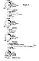
- FIG. 4 schematically illustrates a process wherein a transformant organism expresses a natural eukaryotic protein in accordance with the foregoing scheme.
- the structural gene for the eukaryotic protein is inserted within the signal peptide of the lpp gene, several base pairs after the translation initiation codon and downstream of certain functional fragments (namely, the promoter and the 5'-untranslated region) normally associated with the lipoprotein gene.
- the orientation of these functional fragments is identical to the natural orientation of these elements in the lipoprotein gene, while the exogenous DNA insert fragment supplants most of the signal peptide as well as a portion of the structural region of the lipoprotein gene.
- line b the foreign gene is linked at its 3' end to an extra translation termination codon, which is in turn fused to the remainder of the lipoprotein structural gene. This is linked still further downstream in the normal manner to the 3'- untranslated region of the Ipp gene, which ends with the trnascription termination site.
- the functional fragments which follow the DNA insert fragment are essentially identical to those which are present normally in the lipoprotein gene.
- the 3'-untranslated region derived from the lpp gene codes for an mRNA sequence capable of forming the stem-and-loop structure designated by the numeral I in FIG. 3, which, as discussed previously, is the most stable secondary structure in the lipoprotein mRNA.
- the recombinant DNA sequence depicted schematically in FIG. 4, line b also includes a terminal portion of the lipoprotein structural gene consisting of 105 base pairs starting with position +168 (this position is designated by the arrow (A) in FIG. 3.
- This region is chosen so that the stability of the mRNA transcript can be further enhanced by including four additional stem-and-loop structures (designated by the numerals II, III, IV and V in FIG. 3) without unduly increasing the size of the mRNA molecule produced. However, as set forth below, this region is not ultimately translated.
- FIG. 4 Transcription of the recombinant DNA sequence illustrated in FIG. 4, line b, yields an mRNA sequence which is illustrated schematically in FIG. 4, line c. It will be seen that this sequence contains the 5'- untranslated region and the 3'-untranslated region, both of which are normally associated with the production of the lipoprotein. However, the mRNA also incorporates a region coding for the eukaryotic protein, preceded by a region which codes for a short segment of the signal peptide of the prolipoprotein, and followed by another region which codes for a segment of the lipoprotein. The latter region ultimately will not be translated, however, due to the insertion of a termination codon (designated by an arrow (A) in FIG.
- A termination codon
- the DNA sequence coding for the extraneous amino acids can be excised from the expression plasmid in a known manner prior to transformation of the bacterial host, such that the expression product corresponds exactly with the desired foreign protein and may be purified by known techniques.
- the same functional fragments are used, but the DNA sequence coding for the desired polypeptide is inserted further downstream, following the last codon of the signal peptide (i.e., at or near the signal peptide cleavage site).
- the orientation of the functional fragments is once again identical to the natural orientation of these elements in the lipoprotein gene, allowing full advantage to be taken of the efficiencies of transcription and translation associated therewith, including the enhanced stability of the mRNA transcript attributable to the incorporation of four additional stem-and-loop structures, as described hereinabove.
- the accumulation of a large amount of the expression product inside the cell is less likely to interfere with cell growth, because the eukaryotic protein is linked with a signal peptide which is natural to the cell. Furthermore, the presence of the signal peptide may protect the foreign protein from possible degradative action inside the cell, which could otherwise lower the protein yield and could also cause contamination of the foreign protein by heterogenous degradative products, resulting in purification difficulties.
- the same functional fragments are again used, but the DNA sequence coding for the desired polypeptide is inserted still further downstream, for example, as illustrated herein, following the codon for the eighth amino acid residue after the signal peptide cleavage site.
- the orientation of the functional fragments is once again identical to the natural orientation of these elements in the lipoprotein gene, allowing full advantage to be taken of the efficiencies of transcription and translation associated therewith, including the enhanced stability of the mRNA transcript attributable to the incorportion of four additional stem-and-loop structures, as described hereinabove.
- a polypeptide comprising a signal peptide of 20 amino acid residues, corresponding to the signal peptide of the prolipoprotein, followed by eight amino acid residues corresponding to the first eight amino acid residues of the mature lipoprotein, followed by the amino acid sequence of the desired eukaryotic protein.
- this precursor product may be translocated naturally acorss the cytoplasmic membrane, in the process of which the signal peptide can be recognized and removed.
- the product may not accumulate in the periplasmic space; instead, the eight amino acids corresponding to the lipoprotein can be recognized, and the expression product may then be processed further and inserted into the outer membrane of the cell in a manner analogous to the normal insertion of the lipoprotein into the outer membrane. If, as expected, only the first eight amino acid residues of the expression product corresponding to the lipoprotein are actually bound into the outer membrane, then the remainder of the expression product, consisting of the amino acid sequence of the eukaryotic protein or other desired polypeptide, will protrude from the outer membrane.
- a short polynucleotide sequence containing the recognition sites for the Eco Ri, Hind III and Barn HI restriction enzymes can be incorporated at the insertion site in each expression plasmid. This allows additional flexibility, in that six different types of restriction fragments can be inserted into each plasmid according to the straightforward and well-known techniques described hereinabove.
- DNA insert framents tailored to have any one of the following pairs of cohesive termini can be readily used with the present invention: ECO RI-ECO RI, Hind III-Hind III, Bam Hi-Bam HI, Eco RI-Hind III, Eco RI-Bam HI and Hind III-Bam HI.
- inducible gene expression is exemplified in nature by the E. coli lac promoter-operator, which controls the production of a-galactosidase, an important enzyme in lactose digestion. Normally, the expression of this gene is "switched off” by the presence of a lactose repressor, which binds to the lac promoter-operator, preventing interaction between RNA polymerase and the promoter sequence and thereby inhibiting transcription (and subsequent translation) of the a-galactosidase structural gene.
- the repressor molecule In the presence of lactose, however, the repressor molecule is removed from the DNA and the gene is "switched on,” allowing transcription to proceed until a sufficient quantity of the ⁇ -galactosidase enzyme is produced to digest the lactose, after which the repressor again "switches off" the gene.
- the constitutive lpp gene cloning vehicles described hereinabove can be made inducible by inserting the lac promoter downstream of the lpp promoter, but upstream of the exogenous DNA insert fragment.
- transcription of the foreign DNA from either promoter is blocked by the repressor molecule and cannot proceed in the absence of a substance, termed a "lactose inducer," which for present purposes is a molecule that reacts with and alters the lactose repressor molecule such that the repressor molecule can no longer bind to the lac promoter-operator.
- lactose inducer a substance that reacts with and alters the lactose repressor molecule such that the repressor molecule can no longer bind to the lac promoter-operator.
- the inducible lpp gene cloning vehicles can, in turn, be modified for auto-regulation by inserting within each plasmid a functional E. coli lacI gene.
- a functional E. coli lacI gene a functional E. coli lacI gene.
- lactose repressor genes and lac promoters which is normally present in wild-type E. coli cells, can be maintained in transformants chosen for expression of the desired polypeptide. Accordingly, such transformants need not carry and need not be provided with the F-prime factor thought necessary, but found to be unsatisfactory to repress the expression of the desired product by microorganisms transformed with the expression plasmids which do no comprise the E. coli lacI gene.
- any structural gene coding for a desired polypeptide including mammalian and human hormones, enzymes and immunogenic proteins (or intermediates therefor), may be expressed using the recombinant plasmids of the present invention.
- proteins include A-chain insulin, B-chain insulin, proinsulin, growth hormone, somatostatin, interferon and trypanosome antigen, but the invention is not confined to these exemplary products.
- the auto-regulated inducible recombinant cloning vehicles incorporating the gene for the desired eukaryotic protein or other polypeptide are preferably used to transform special E. coli strains as hosts for cloning and for subsequent production of the protein.
- the host cell strains used will be chosen to have a "deletion mutant" in the lpp gene, so that the host cells cannot produce the lipoprotein.
- the use of a deletion mutant strain as the transformant is thought to stimulate the production of the foreign protein.
- secretion of the foreign protein across the cytoplasmic membrane is facilitated in lpp-defective host cells, since the secretion sites in the membrane which are intended to be used for lipoprotein secretion are instead available for secretion of the foreign protein.
- the use of the lpp-defective cells is especially beneficial when the gene coding for the foreign protein is inserted at or near the lipoprotein signal peptide cleavage site. This is because such cells are known to be "leaky", i.e., proteins secreted across the cytoplasmic membrane of such cells ultimately "leak” out into the culture medium through the outer membrane of the cell. This is believed to be desirable not only because release of the desired foreign protein into the culture medium may in some cases allow easier isolation and purification of the foreign protein than would be possible if the foreign protein remained inside the cell, but also because the foreign protein would otherwise accumulate in the periplasmic space, perhaps leading to undesirable interference with normal cellular activities or cell growth. Secretion of the desired eukaryotic gene product outside the cell may also avoid degradation of that product into smaller fragments by proteolytic enzymes which are normally present within the cell.
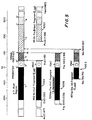
- the insertion site located within the DNA sequence coding for the prolipoprotein signal peptide will be designated the "A” site, while the insertion site located immediately after the last codon of the signal peptide will be labelled the "B” site, and the insertion site located after the codon for the eight amino acid residue of the mature lipoprotein will be referred to as the "C” site (see FIG. 5).
- A the insertion site located immediately after the last codon of the signal peptide
- the insertion site located after the codon for the eight amino acid residue of the mature lipoprotein will be referred to as the "C” site (see FIG. 5).
- three plasmids can be prepared (one corresponding to each of the three possible reading frames), yielding a total of nine expression plasmids in each series which are labelled A-1, A-2, A-3, B-1, B-2, B-3, and C-l, C-2, C-3.
- restriction enzymes used herein were obtained from New England Biolabs and Bethesda Research Laboratories. T4 DNA ligase was obtained from Bethesda Research Laboratories (unless otherwise indicated), and Sl Nuclease was obtained from Miles Laboratories.
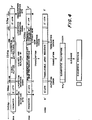
- FIGS. 6-15 schematically depict the manner in which recombinant plasmids incorporating the A insertion site were constructed, and may be referred to in connection with the following more particularized discussion.
- the first step in the construction of the A site lpp gene cloning vehicles was to construct a plasmid to serve as a source of Ipp gene components in subsequent steps of the procedure.
- the plasmid chosen to receive the E. coli lpp gene for this purpose was pSC101, a small (molecular wt. approximately 5.8 megadaltons) plasmid carrying a gene conferring resistance to the antibiotic tetracycline (Tc) (Cohen, S.N., et al., J. Bacteriol. 132: 734-737 [1977]).
- Tc antibiotic tetracycline
- pSC101 includes a cleavage site for the restriction endonuclease Eco RI located at the 5' end of the tetracycline resistance gene.
- the plasmid pSC101 was obtained from Dr. E. Ohtsubo at the Department of Microbiology, State University of New York at Stony Brook.
- BAP bacterial alkaline phosphatase
- a 2.8 kilobase (“Kb”) DNA fragment containing the E. coli lpp gene was separately derived, as shown at 102 in FIG. 6, from a hybrid ⁇ phage carrying the E. coli lpp gene (designated ⁇ lppEc-1).
- the lpp gene had previously been cloned into a A phage vector, ⁇ 540 (Murray and Murray, J. Mol. Biol. 98: 551-564 [1975]), as follows: Total DNA (200 micrograms) isolated from an E. coli K-12 strain merodiploid for the lpp gene (JE5519/F506 [Movva, N.R., et al., J. Bacteriol.
- Two hundred micrograms of ⁇ lppEc-1 DNA were then digested completely with 200 units of the restriction enzyme Hae III in 500 microliters of a reaction mixture containing 6mM Tris:HCl (pH 7.5), 6mM MgCl 2 , 6mM NaCl, 6mM a-mercaptoethanol and 100 micrograms/ml BSA (the foregoing reaction mixture will hereinafter be referred to as a "Hae III buffer”) at 37°C for 2 hours, and the 2.8 Kb Hae III fragment carrying the E.
- a reaction mixture containing 6mM Tris:HCl (pH 7.5), 6mM MgCl 2 , 6mM NaCl, 6mM a-mercaptoethanol and 100 micrograms/ml BSA (the foregoing reaction mixture will hereinafter be referred to as a "Hae III buffer") at 37°C for 2 hours, and the 2.8 Kb Hae III fragment carrying the E.
- coli l EE gene was purified by fractionation on a 5% polyacrylamide gel according to the following procedure: The reaction mixture was first extracted with phenol, and the DNA fragments were then precipitated with 2.5 volumes of ethanol, dried under vacuum, dissolved in 200 microliters of a buffer comprising 5% glycerol, 20mM EDTA, 0.05% bromophenol blue and 0.05% xylen cyanol (this mixture will hereinafter be referred to as a "gel buffer”) and thereafter fractionated on a 5% polyacrylamide gel. The DNA band which had migrated to a 2.8 Kb position was excised from the gel, and the DNA fragments were eluted from the gel by electrophoresis.
- a buffer comprising 5% glycerol, 20mM EDTA, 0.05% bromophenol blue and 0.05% xylen cyanol
- Ethidium bromide dye used to locate the DNA band in the gel, was removed from the DNA fragments by phenol extraction.
- the DNA fragments were precipitated with 2.5 volumes of ethanol, centrifuged, dissolved in 200 microliters of 0.3M Na-acetate, re-precipitated with 0.5 ml of ehtanol and dried again under vacuum. Approximately 10 micrograms of a purified 2.8 Kb Hae III fragment were recovered.
- Biochemicals with ATP in 50 microliters of a reaction mixture containing 3 moles of the linker, 66mM Tris:HCl (pH 7.5), 10mM MgCl 2 , 10mM ⁇ -mercaptoethanol, 60 ⁇ M ATP and 10 units of T4 polynucleotide kinase.
- the mixture was incubated at 37°C for 30 minutes, it was heated at 60°C for 10 minutes, and cooled to 37°C.
- Five microliters of 0.1M ⁇ -mercaptoethanol and 10 units of T4 polynucleotide kinase were added to the mixture, and the reaction was continued at 37°C for 30 minutes. The reaction was terminated by freezing the mixture in a dry ice-ethanol bath.
- the 2.8 Kb Hae III fragment (2 micrograms) was mixed with 150 pmoles of phosphorylated Eco RI linker and was treated with 4 units of T4 DNA ligase in 12.5 microliters of a reaction mixture containing,66mM Tris:HCl (pH 7.5), 10mM MgCl 2 , lOmM dithiothreitol (the foregoing reaction mixture will hereinafter be referred to as a "ligase buffer") and 0.6mM ATP at 12.5°C for 15 hours.
- the reaction was terminated by diluting the mixture twenty-fold with Eco RI buffer and by heating the mixture at 60°C for 10 minutes. Thirty units of the restriction enzyme Eco RI were added, and the mixture was incubated at 37°C for one hour to create Eco RI cohesive termini. The reaction was terminated by heating at 60°C for 10 minutes.
- the mixture thus obtained was added to 2 micrograms of the previously-linearized plasmid pSC101 NDA, and phenol extraction was performed. After extraction with ether, the DNAs were precipitated with ethanol, dried under vacuum, and dissolved in 100 microliters of ligase buffer. The mixture was heated at 37°C for 5 minutes, and the Eco RI cohesive termini were annealed by incubating at 4°C for 16 hours and then at 0°C for one hour. After adding ATP (0.4mM final) and 1 unit of T4 DNA ligase, the mixture was incubated at 12.5°C for 7 hours.
- E. coli lpp deletion mutant strain JE5527, NRRL B-15012 F , man, lpp-2, pps, thi, his, rpsL, gyrA, recAl [Hirota, Y., et al., Proc. Natl. Acad. Sci. U.S.A. 74: 1417-1420 (1977)], obtained from Dr. Y. Hirota, National Institute of Genetics, Mishima, Japan).
- This strain is available to the public from the permanent collection of the Northern Regional Research Laboratory, U.S. Department of Agriculture, Peoria, Illinois, U.S.A.
- a 0.95 Kb Msp I fragment of ⁇ lppEc-1 containing the lpp gene was nick-translated with [ ⁇ - 32 P]dATP and [a- 32 P]dCT P, as described in Maniatis, T., et al., Proc. Natl. Acad. Sci. U.S.A. 72: 1184-1188 (1975), and was used as a 32 P-probe.
- One of the transformants which gave positive hybridization was shown to contain the plasmid with the structure illustrated at 104 in FIG. 6, and this plasmid was designated pKENlll. This plasmid is obtainable from E.
- plasmid can be obtained from NRRL B-15011 by conventional means.
- the parental plasmid chosen for construction of the 1 2 p gene expression plasmids of the present invention was pBR322 (molecular wt. approximately 2.6 megadaltons), carrying genes conferring resistance to the antibiotics ampicillin (Amp) and tetracycline (Tc) (Bolivar, F., et al., Gene 2: 95-113 [1977]).
- pBR322 includes an Eco RI cleavage site located at the 5' end of the tetracycline resistance gene, as well as a Hind III cleavage site located within the promoter of the tetracycline resistance gene and a Pvu I cleavage site located within the ampicillin resistance gene.
- the plasmid pBR322 was obtained from Dr. N. Arnheim of the Department of Biochemistry, State University of New York at Stony Brook, and is available commercially from Bethesda Research Laboratories.
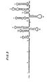
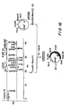
- FIG. 5 illustrates schematically the various components of the lpp gene, each of which is identified by a symbol or shading.
- the shaded segment indicated by the letter “a” identifies the A-T rich region of approximately 260 base pairs preceding the transcription initiation site and containing the lpp promoter.
- the 5'-untranslated region is identified by the segment containing the circular device and marked with the letter "b”.
- the signal peptide region of the prolipoprotein is identified by the diagonally hatched and shaded segment "c”.
- the structural region of the Ipp gene is identified by the diagonally hatched segment labelled with the letter "d", while the speckled segment "e” identified the 3'-untranslated region and the transcription termination site.
- FIG. 7 illustrates the strategy used for inserting a fragment carrying the promoter and the 5'-untranslated region of the lpp gene into pBR322.
- the fragment chosen for this purpose was a 462 bp Alu I fragment of pKENlll which, as shown schematically at 105A in FIG. 5, contains not only the promoter sequence and the S'-untranslated region (positions -45 to -1 and +1 to +39, respectively) of the lpp gene, but also the entire extremely A-T rich segment preceding the promoter sequence.
- the DNA was treated with 1.5 microliters of Sl Nuclease (Miles Laboratories) in a final volume of 300 microliters of a buffer containing 30mM Na-acetate (pH 4.25), 0.3M NaCl and 4mM ZnSO 4 (hereinafter referred to as an "Sl buffer”) at 20°C for one hour.
- Sl buffer a buffer containing 30mM Na-acetate (pH 4.25), 0.3M NaCl and 4mM ZnSO 4 (hereinafter referred to as an "Sl buffer") at 20°C for one hour.
- the reaction was terminated by adding 30 microliters 500 mM Tris:HCl (pH 8.0) and 30 microliters 250mM EDTA, following which phenol extraction was performed.
- Phosphorylated Eco RI linker (200 pmoles) was then added and the mixture was treated with 4 units of T4 DNA ligase in 12.5 microliters of ligase buffer containing 0.6mM ATP at 12.5°C for 16 hours. Eco RI cohesive termini were created by addition of 30 units of Eco RI restriction enzyme in 75 microliters of Eco RI buffer at 37°C for 2 hours. The reaction was terminated by phenol extraction and the DNAs were recovered by ethanol precipitation.
- Plasmid DNAs were isolated from 0.5 ml of the cultures by the rapid alkaline-denaturation method described by Birnboim, H.C. and Doly, J., Nucleic Acids Res. 7: 1513 (1979), and analyzed by restriction enzyme mapping.
- One of the plasmids had the structure shown at 107 in FIG. 7, and was designated pKEN005.
- the 462 bp Alu I fragment containing the lpp promoter was derived as follows: 100 micrograms of pKENlll plasmid DNA were digested with Msp I restriction enzyme in 600 microliters of a buffer containing 10mM Tris:HCl (pH 7.5), 10mM MgCl 2 , 6mM KC1, 1mM dithiothreitol, and 100 micrograms/ml BSA (this mixture will hereinafter be referred to as an "Hpa I buffer”) at 37°C for 3 hours. (Although pKENlll contains numerous Msp I cleavage sites, only the two of interest are illustrated at 109 in FIG.
- DNA fragments were precipitated with 2.5 volumes of ethanol, dried under vacuum, dissolved in 100 microliters of gel buffer, and fractionated on a 5% polyacrylamide gel. Approximately 6 micrograms of a purified 0.95 Kb Msp I fragment was subsequently digested with Alu I restriction endonuclease in 400 microliters of Hind III buffer at 37°C for 2 hours, yielding a 462 bp Alu I fragment which was purified by gel electrophoresis.
- One microgram of the 462 bp Alu I fragment was then mixed with 150 pmoles of phosphorylated Eco RI linker and treated with 4 units of T4 DNA ligase in 10 microliters of ligase buffer containing 0.6mM ATP at 12.5°C for 16 hours.
- the ligated DNA was digested with 40 units of Eco RI restriciton enzyme in 100 microliters of Eco RI buffer at 37°C for one hour to create Eco RI cohesive termini. The digestion was terminated by heating the mixture at 60°C for 10 minutes, and 0.6 micrograms of Eco RI-digested pKEN005 plasmid DNA were added to the mixture and phenol extraction was performed.
- the DNAs were recovered by ehtanol precipitation, and the Eco RI cohesive termini were joined by treating with 0.4 units of T4 DNA ligase in 20 microliters of ligase buffer containing 0.4mM ATP at 12.5°C for 7 hours.
- Ligated DNAs were used to transform E. coli strain JE5519, NRRL B-15013, and transformants were selected for tetracycline resistance on an L broth plate containing 12.5 micrograms/ml of tetracycline.
- FIG. 8 illustrates schematically the strategy for removing the Eco RI site distal to the lpp gene promoter.
- coli strain JE5519, NRRL B-15013, and one of the transformants was shown to contain the plasmid with the structure illustrated at 112 in FIG. 8.
- This plasmid was designated pKEN002, and after digestion of 25 micrograms of pKEN002 plasmid DNA with Pvu I and Xba I restriction enzymes in 500 microliters of a buffer comprising 6mM Tris:HCl (pH 7.9), 6mM MgCl 2 , 150mM NaCl, 6mM a-mercaptoethanol and 100 micrograms/ml BSA (the foregoing mixture will hereinafter be referred to as a "Bam HI buffer") at 37°C for one hour, a 1.04 Kb Pvu I-Xba I DNA fragment (illustrated at 113 in FIG. 8) was purified by gel electrophoresis.
- a 24 bp Xba I-Eco RI DNA fragment was derived from pKEN008 as follows: 25 micrograms of pKEN008 plasmid DNA was digested with Eco RI restriction enzyme, and a 470 bp Eco RI fragment was purified by gel electrophoresis.
- One microgram of the 470 bp Eco RI fragment was then digested with Xba I restriction enzyme, and was mixed with one microgram of the 1.04 Kb Pvu I-Xba I DNA fragment obtained previously, as well as with 0.75 micrograms of pKEN005 plasmid DNA previously digested with Pvu I and Eco RI restriction enzymes (as shown at 115 in FIG. 8).
- the DNA mixture was treated with 0.8 units of T4 DNA ligase in 50 microliters of ligase buffer containing 0.4mM ATP at 12.5°C for 7 hours. One-half of the ligated DNA was used to transform E.
- FIG. 9 illustrates the strategy used for cloning a DNA fragment carrying the 3'-untranslated region and the transcription termination site of the 2p 2 gene.
- the fragment chosen for this purpose was a 0.95 Kb Pvu II-Hpa I fragment of pKENlll, shown schematically at 105D in FIG. 5. Since the Pvu II restriction enzyme cleaves the lpp gene sequence between positions +167 and +168, this fragment contains approximately the latter half of the lpp gene (see FIGS. 1 and 5). In order to insert this fragment into the cloning vehicle in the same orientation as the promoter fragment, Bam HI linker and Sal I linker were attached to the Pvu II and Hpa I cleavage sites, respectively.
- a 2.8 Kb Eco RI fragment was obtained from pKENlll plasmid DNA by digestion with Eco RI restriction enzyme and fractionation on a polyacrylamide gel, and 10 micrograms of this purified fragment were digested completely with Pvu II restriction endonuclease in 500 microliters of Hae III buffer at 37°C for one hour. The reaction was terminated by phenol extraction, and the mixture was extracted with ether. The DNA fragments were precipitated with 2.5 volumes of ethanol, centrifuged, re-dissolved in 200 microliters of 0.3M Na-acetate and re-precipitated with 0.5 ml of ethanol.
- the DNA fragments were digested completely with Hpa I restriction enzyme in 400 microliters of Hpa I buffer at 37°C for 2 hours.
- the reaction mixture was extracted with phenol and the DNA fragments were precipitated with ethanol, dried under vacuum, dissolved in 100 microliters of gel buffer and fractionated on a 5% polyacrylamide gel.
- the DNA band which had migrated to a 0.95 KB position was excised from the gel, and the DNA fragments were eluted from the gel by electrophoresis.
- DNA fragments were precipitated with 2.5 volumes of ethanol, centrifuged, dissolved in 200 microliters of 0.3M Na-acetate, re-precipitated with 0.5 ml of ethanol and again dried under vacuum. Approximately one microgram of a purified 0.95 KB Hae III-Hpa I fragment (illustrated at 118 in FIG. 9) was recovered.
- GGTCGACC phosphorylated S al I linker
- Bam HI and Sal I restriction enzymes were added and the mixture was incubated at 37°C for 2 hours to create cohesive termini by cleaving the Bam HI and Sal I linkers attached to the Pvu II and Hpa I termini, respectively, resulting in a 0.95 KB Bam HI-Sal I fragment (illustrated at 119 in FIG. 9).
- the restriction endonuclease digestion was terminated by heating at 60°C for 10 minutes.
- deletion of this fragment was accomplished, as shown schematically at 121 in FIG. 9, by Hind III digestion, followed by Sl Nuclease treatment for one hour at 20°C, Bam HI linker attachment, Bam HI complete digestion, re-circularization by T4 DNA ligase, and selection of tetracycline-sensitive transformants.
- the mixture of linearized pKEN014 plasmid DNA and 0.95 Kb Bam HI-Sal I fragments was extracted with phenol, and the DNAs were precipitated with 2.5 volumes of ethanol, centrifuged and dissolved in 200 microliters of 0.3M Na-acetate. The DNAs were re-precipitated with 0.5 ml of ethanol, centrifuged and dried under vacuum. Cohesive termini of the DNA fragments were annealed with 0.4 units of T4 DNA ligase in 60 microliters of ligase buffer containing 0.4mM ATP at 12.5°C for 7 hours. Twelve microliters of the ligated mixture were then used to transform E.
- Plasmid DNAs were isolated from 0.5 ml of the cultures by the rapid alkaline-denaturation method and analyzed by agarose gel electrophoresis. Five of the plasmid DNA 4 were found to carry the 0.95 Kb Barn HI-Sal I fragment, and one of these plasmids was designated pKEN018. DNA sequencing of the pKEN018 plasmid DNA indicated the structure shown at 122 in FIG. 9, and specifically showed that the Bam HI linker was attached at the Pvu II site within the lpp gene at the correct position.
- the next step in the construction of the A site lpp gene cloning vehicles was to combine the Ipp promoter fragment with the transcription terminator fragment in the same orientation. This step was carried out by replacing a 630 bp Pvu I-Eco RI fragment of pKEN018 with a 1.1 Kb Pvu I-Eco RI fragment of pKEN010, as illustrated schematically in FIG. 10.
- the DNA fragments were precipitated with 2.5 volumes of ethanol, dried under vacuum, dissolved in 100 microliters of gel buffer, and fractionated on a 5% polyacrylamide gel. Four micrograms of a purified 1.1 Kb Pvu I-Eco RI fragment were obtained after elution of the separated DNA fragments from the gel.
- the purified fragment (0.75 micrograms) was then mixed with 0.6 micrograms of pKEN018 plasmid DNA which had previously been double-digested with Pvu I and Eco RI restriction enzymes and then treated with BAP (as shown at 124 in FIG. 10).
- the Pvu I and the Eco RI cohesive termini were ligated by treating with 0.4 units of T4 DNA ligase in 50 microliters of ligase buffer containing 0.4mM ATP at 12.5°C for 7 hours. Twenty-five microliters of the ligated mixture were used to transform E. coli strain JE5519, NRRL B-15013, and transformants were selected for ampicillin resistance.
- Plasmid DNAs were isolated from ampicillin-resistant transformants and analyzed by agarose gel electrophoresis. Restriction enzyme mapping indicated that one of the plasmids had the structure shown at 125 in FIG. 10, and this plasmid was designated pKEN021.
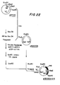
- FIG. 11 illustrates the final step in the construction of the first A site lpp gene expression plasmid.
- pKEN021 carries both the lpp promoter fragment and the lpp transcription terminator fragment, separated by a 32 bp fragment derived from pBR322. By deleting the latter fragment and inserting a DNA sequence coding for a desired polypeptide, a functional moiety for expression of the desired polypeptide is provided.
- the reaction was terminated by adding 20 microliters of 0.5M tris:HCl (pH 8.0) and 20 microliters of 0.25M EDTA.
- the mixture was extracted with phenol and dialyzed for four hours against 0.01 X SSC.
- the DNAs were precipitated with 2.5 volumes of ethanol, centrifuged and resuspended in 100 microliters of 0.3M Na-acetate.
- the DNAs were re-precipitated with 250 microliters of ethanol, centrifuged, and dried under vacuum.
- One microgram of the Sl-treated DNA was then mixed with 70 pmoles of phosphorylated Hind III linker ( 5' CCAAGCTTGG 3' ; obtained from Collaborative Research and phosphorylated according to the same procedure as described hereinabove) and blunt-end ligated with 4 units of T4 DNA ligase in 20 microliters of ligase buffer containing 0.6mM ATP at 12.5°C for 16 hours.
- the mixture was then diluted to 100 microliters with Hind III buffer and heated at 60°C for 10 minutes. Twenty units of Hind III restriction endonuclease were added, and the mixture was incubated at 37°C for one hour to remove superfluous linker molecules and to create Hind III cohesive termini.
- Plasmid DNAs (0.5 micrograms) were re-circularized by treating with 0.8 units of T4 DNA ligase in 15 microliters of ligase buffer containing 0.4mM ATP at 12.5°C for 7 hours. Eight microliters of the ligated mixture were used to transform E. coli strain JA221, NRRL B-15014 (recA-, hr-, h + , ⁇ trpE5, thr, leu, thi, lacY-; obtained from Dr. J. Carbon, Dept. of Biological Sciences, University of California, Santa Barbara). This strain is available to the public from the permanent collection of the Northern Regional Research Laboratory, U.S.
- plasmid DNAs which were purified from the ampicillin-resistant transformants was one that had the structure shown at 127 in FIG. 11, and this plasmid was designated pKEN030.
- the DNA sequence of plasmid pKEN033 was modified to create a Hind III cleavage site between the Eco RI and Bam HI sites, as follows: 5 micrograms of pKEN033 plasmid DNA (having the DNA sequence of interest shown in FIG. 12, line a) were digested with 10 units of Bam HI restriction endonuclease in 50 microliters of Barn HI buffer at 37°C for one hour.
- the DNAs (3.6 micrograms) were treated with three units of T4 DNA polymerase (obtained from Bethesda Research Laboratories) in 20 microliters of a reaction mixture containing 50mM Tris:HCl (pH 8.0), 100mM KC1, 6mM MgCl 2 , and 6mM dithiothreitol (this reaction mixture will hereinafter be referred to as a "polymerase buffer") in the presence of 0.lmM each of dATP, dGTP, dCTP and dTTP at 12.5°C for 45 minutes.
- the Bam HI and the Eco RI "sticky ends" were filled in completely, as shown in FIG. 12, line c.
- FIG. 13, line a, and FIG. 14, line a both illustrate the DNA sequence surrounding the translation initiation site of the prolipoprotein in pKENlll. As shown, this sequence includes an Alu I cleavage site between positions +45 and +46.
- an Eco RI linker was attached to the Alu I terminus, resulting in the DNA sequence shown in FIG. 13, line b, and in FIG.
- the remaining two single-strand A residues were removed by treating with Sl Nuclease in 200 microliters of Sl buffer at 20°C for one hour.
- the reaction was terminated by adding 20 microliters of 0.5M tris:HCl (pH 8.0) and 20 microliters of 0.25M EDTA.
- the mixture was extracted with phenol and dialyzed overnight against 0.01 x SSC.
- the DNAs were precipitated with 2.5 volumes of ethanol, centrifuged and re-suspended in 100 microliters of 0.3M Na-acetate.
- the DNAs were re-precipitated with 250 microliters of ethanol, centrifuged, and dried under vacuum.
- one microgram of the Sl-treated DNA was first mixed with 70 pmoles of phosphorylated Eco RI linker and blunt-end ligated with 3.2 units of T4 DNA ligase in 11 microliters of ligase buffer containing 0.6mM ATP at 12.5°C for 16 hours. The mixture was then diluted to 50 microliters with Eco RI buffer and heated at 60°C for 10 minutes. Twenty units of Eco RI restriction endonuclease were added, and the mixture was incubated at 37°C for one hour to remove superfluous linker molecules and to create Eco RI cohesive termini. The reaction mixture was then extracted with phenol, and the DNAs were precipitated with ethanol.
- Plasmid DNAs (0.5 micrograms) were re-circularized by treating with 0.8 units of T4 DNA ligase in 15 microliters of ligase buffer containing 0.4mM ATP at 12.5°C for 7 hours. Eight microliters of the ligated mixture were used to transform E. coli strain JA221, NRRL B-15014. Plasmid DNAs were purified from 3 ampicillin-resistant transformants, which had been grown overnight in 100 ml of L broth containing 50 micrograms/ml of ampicillin, and the DNA sequences at their Eco RI cleavage sites were determined. One of them was found to have the sequence shown in FIG. 13, line c, and was designated pKEN024 (A-2).
- the DNAs were precipitated with 2.5 volumes of ethanol, centrifuged and re-suspended in 100 microliters in 0.3 Na-acetate.
- the DNAs were re-precipitated with 250 microliters of ethanol, centrifuged, and dried under vacuum.
- one microgram of the Sl-treated DNA was first mixed with 240 pmoles of phosphorylated Eco RI linker and blunt-end ligated with 4 units of T4 DNA ligase in 15 microliters of ligase buffer containing 0.6mM ATP at 12.5°C for 16 hours. The mixture was then diluted to 250 microliters with Eco RI buffer and heated at 60°C for 10 minutes. One hundred units of Eco RI restriction endonuclease were added, and the mixture was incubated at 37°C for one hour to remove superfluous linker molecules and to create Eco RI cohesive termini. The reaction mixture was then extracted with phenol, and the DNAs were precipitated with ethanol.
- Plasmid DNAs (0.3 micrograms) were re-circularized by treating with 0.8 units of T4 DNA ligase in 15 microliters of ligase buffer containing 0.4mM ATP at 12.5°C for 7 hours. Eight microliters of the ligated mixture were used to transform E. coli strain JA221, NRRL B-15014. Plasmid DNAs were purified from 3 ampicillin-resistant transformants, which had been grown overnight in one hundred ml of L broth containing 50 micrograms/ml of ampicillin, and the DNA sequences at their Eco RI cleavage sites were determined. One of them was found to have the sequence shown in FIG. 14, line c, and was designated pKEN036 (A-3).
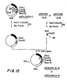
- the dried DNA fragments were dissolved in 20 microliters of water, and one microliter aliquots of this pKEN037 DNA fragment mixture were combined with 0.1 micrograms of each of the smaller Xba I-Eco RI restriction fragments (illustrated at 132 in FIG. 15) previously obtained from pKEN024 or pKEN036 by double-digestion of each plasmid with Xba I and Eco RI restriction enzymes followed by gel purification (as shown at 133 in FIG. 15).
- the "sticky ends" of the Xba I-Eco RI fragments were joined by treatment with 0.2 units of T4 DNA ligase in 20 microliters of ligase buffer containing 0.4mM ATP at 12.5°C for 7 hours, following which a portion of the ligated mixture was used to transform E. coli strain JA221, NRRL B-15014.
- plasmid DNAs having the A-2 and A-3 reading frames were obtained, and these were designated pKEN039 and pKEN040, respectively, each having the structure shown at 134 in FIG. 15.
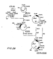
- FIGS. 16-21 schematically illustrate the manner in which recombinant plasmids incorporating the B insertion site were constructed, and may be referred to in connection with the following more particularized discussion.
- the first step in the construction of the B site expression plasmids was to construct a plasmid to serve as a source of 1p E gene fragments having a restriction enzyme cleavage site at or near the signal peptide cleavage site.
- the gene chosen codes for the lipoprotein of S. marcescens, and has a Fnu4H-I restriction endonuclease recognition sequence at the 3' end of the signal peptide.
- the plasmid chosen to receive the S. marcescens lpp gene was pBR322.
- DNA fragments were separated on a preparative agarose gel, and fractions of DNA fragments of approximately 8.5 Kb which showed positive hybridization with 5'32P-lipoprotein mRNA were collected, using the Southern hybridization technique.
- a mixture of 8.5 Kb Eco RI fragments (enriched approximately twenty-fold) and Eco RI-cleaved Charon 14 vector DNA was reacted with T4 DNA ligase.
- Ligated DNA was used to transfect E. coliK802, NRRL B-15016.
- Recombinant phages carrying the lpp gene were screened by the plaque hybridization technique of Benton and Davis using 5'- 32 P-lipoprotein mRNA.
- One of the plaques examined which gave positive hybridization was designated ⁇ lppSm-1.
- a 0.95 Kb Msp I fragment of ⁇ lppEc-1 containing the lpp gene was nick-translated with [a-32p]dATP and [a-32p]dCTP, as described in Maniatis, T., et al., Proc. Natl. Acad. Sci. USA 72: 1184-1188 (1975), and was used as a 32 P-probe.
- One of the transformants which gave positive hybridization was shown to contain the plasmid with the structure illustrated at 137 in FIG. 16, and this plasmid was designated pKEN221.
- a 329 bp Fnu4H-I fragment containing the Ipp promoter and 5'-untranslated region, as well as the signal peptide region of the S. marcescens lpp gene (this fragment is shown schematically at 105B in FIG. 5) was first cloned into pKEN005, as illustrated at 138 in FIG.
- the DNA fragments were mixed with 400 pmole of phosphorylated Eco RI linker and treated with 4 units of T4 DNA ligase in 20 microliters of ligase buffer containing 0.6mM ATP at 12.5°C for 16 hours. The mixture was diluted to 300 microliters with Eco RI buffer and digested with 150 units of Eco RI restriction enzyme to create Eco RI cohesive termini.
- One microgram of the Eco RI-digested fragments was then mixed with 0.5 micrograms of Eco RI-digested pKEN005 plasmid DNA, and treated with 0.4 units of T4 DNA ligase in 40 microliters of ligase buffer containing 0.6mM ATP at 12.5°C for 16 hours. Twenty microliters of the ligated mixture was used to transform E. coli strain JE5519, NRRL B-15013.
- FIG. 18 depicts schematically the strategy for removing the Eco RI site located upstream of the lpp promoter. This procedure involved transferring an 80 bp Xba I-Eco RI fragment (containing the signal peptide and a portion of the 5'-untranslated region of the S. marcescens lpp gene) from pKEN009 into the Xba I-Eco RI sites of pKEN010.
- 5 micrograms of pKENO10 plasmid DNA were first digested with 5 units of Xba I restriction endonuclease in 50 microliters of Barn HI buffer, followed by digestion with. 5 units of Eco RI restriction enzyme in 100 microliters of Eco RI buffer. The linearized DNA was then treated with 5 microliters of BAP in 100 microliters of 10mM Tris:HCl (pH 8.0) and 0.lmM EDTA at 37°C for 30 minutes.
- Plasmid DNAs were extracted with phenol and precipitated with ethanol, and 0.5 micrograms of the DNA were mixed with 0.2 micrograms of an 80 bp Xba I-Eco RI fragment, which had previously been obtained by digestion of 50 micrograms of pKEN009 plasmid DNA by Eco RI and Xba I restriciton enzymes, followed by polyacrylamide gel electrophoresis.
- the DNA mixture was treated with 0.4 units of T4 DNA ligase in 40 microliters of ligase buffer containing 0.4mM ATP at 12.5°C for 16 hours. Twenty microliters of the ligated mixture were used to transform E. coli strain JE5519, NRRL B-15013.
- one plasmid was found to contain the desired 80 bp Xba I-Eco RI fragment carrying the signal peptide region of the S. marcescens l EE gene in the B-1 reading frame, as shown at 140 in FIG. 18, and that plasmid was designated pKEN017.
- the reading frame at the B insertion site in pKEN017 was then modified to yield plasmids corresponding to the B-2 and B-3 reading frames, according to the methods previously described for changing the A-1 reading frame into the A-2 or A-3 reading frames, respectively. These procedures are illustrated schematically at 141 and 142 in FIG. 18, and the corresponding modifications of the DNA sequence around the Eco RI cleavage site are shown in FIGS. 19 and 20. It will be understood that the same procedures used to derive plasmids pKEN024 (A-2) and pKEN036 (A-3) from plasmid pKEN030 (A-1), described hereinabove in connection with FIGS. 13 and 14, can be used to derive plasmids pKEN026 (B-3) and pKEN027 (B-2) from plasmid pKEN017 (B-1).
- FIG. 21 illustrates schematically the last step in the construction of the B site cloning vehicles, which was to replace the Xba I-Eco RI A site fragment of pKEN037 with each of the three different Xba I-Eco RI B site fragments of pKEN017, pKEN026 and pKEN027. This was necessary in order to provide the B site plasmids with the same sequence of Eco RI, Hind III and Bam HI restriction enzyme recognition sequences at the exogenous DNA insertion site as contained in the A site plasmids. As shown schematically at 143 in FIG.
- each of the three B site fragments derived from pKEN017, pKEN026 and pKEN027 contains the DNA sequence including the signal peptide obtained from the Fnu4H-I fragment of the S. marcescens 1 2 p gene.
- Each DNA mixture was treated with 0.2 units of T4 DNA ligase in 20 microliters of ligase buffer containing 0.4mM ATP at 12.5°C for 16 hours. Ten microliters of each of the ligated mixtures were used to transform E. coli strain JA221, NRRL B-15014.
- plasmid DNAs having the B-1, B-2 and B-3 reading frames were purified, and these were designated pKEN041, pKEN047 and pKEN048, respectively, each having the structure shown at 144 in FIG. 21.
- FIGS. 22-26 schematically illustrate the manner in which recombinant plasmids incorporating the C insertion site were constructed, and may be referred to in connection with the following more particularized discussion.
- a 193 bp Sau 3A fragment containing the lpp promoter and 5'-untranslated region, as well as the signal peptide region and the first eight structural codons of the E. coli lpp gene was first cloned into pKEN005, as illustrated at 145 in FIG. 22, as follows: 200 micrograms of pKENlll plasmid DNA, which can be obtained by conventional means from E.
- coli CC620/pKENlll, NRRL B-15011 were digested to completion with 200 units of Sau 3A restriction endonuclease in 400 microliters of a reaction mixture comprising 10mM Tris:HCl (pH 7.5), 10mM MgCl 2 , 60mM NaCl, and 100 micrograms/ml BSA at 37°C for one hour. After digestion was completed, phenol extraction was performed, the DNAs were recovered by ethanol precipitation, and a 193 bp Sau 3A fragment was purified by acrylamide gel electrophoresis.
- the DNA fragments were mixed with 400 pmoles of phosphorylated Eco RI linker and treated with 4 units of T4 DNA ligase in 20 microliters of ligase buffer containing 0.6mM ATP at 12.5°C for 16 hours. The mixture was diluted to 300 microliters with Eco RI buffer and digested with 150 units of Eco RI restriction enzyme to create Eco RI cohesive termini.
- One microgram of the Eco RI-digested fragments was then mixed with 0.5 micrograms of Eco RI-digested pKEN005 plasmid DNA, and treated with 0.4 units of T4 DNA ligase in 40 microliters of ligase buffer containing 0.6mM ATP at 12.5°C for 16 hours. Twenty microliters of the ligated mixture were used to transform E. coli strain JE5519, NRRL B-15013.
- FIG. 23 depicts schematically the strategy for removing the Eco RI site located upstream of the lpp promoter. This procedure involved transferring a 106 bp Xba I-Eco RI fragment (containing the signal peptide, a portion of the 5'- untranslated region and a portion of the structural sequence of the E. coli Ipp gene) from pKEN006 into the Xba I-Eco RI sites of pKENO10.
- pKEN010 plasmid DNA 5 micrograms of pKEN010 plasmid DNA were first digested with 5 units of Xba I restriction endonuclease in 50 microliters of Bam HI buffer, followed by digestion with 5 units of Eco RI restriction enzyme in 100 microliters of Eco RI buffer. The linearized DNA was then treated with 5 microliters of BAP in 100 microliters of lOmM Tris:HCl (pH 8.0) and O.lmM EDTA at 37°C for 30 minutes.
- Plasmid DNAs were extracted with phenol and precipitated with ethanol, and 0.5 micrograms of the DNA were mixed with 0.2 micrograms of a 106 bp Xba I-Eco RI fragment, which had previously been obtained by digestion of 50 micrograms of pKEN006 plasmid DNA by Eco RI and Xba I restriction enzymes, followed by polyacrylamide gel electrophoresis.
- the DNA mixture was treated with 0.4 units of T4 DNA ligase in 40 microliters of ligase buffer containing 0.4mM ATP at 12.5°C for 7 hours. Twenty microliters of the ligated mixture were used to transform E. coli strain JE5519, NRRL B-15013.
- one plasmid was found to contain the desired 106 bp Xba I-Eco RI fragment carrying the signal peptide region of the E. coli Ipp gene in the C-1 reading frame, as shown at 147 in FIG. 23, and that plasmid was designated pKEN007.
- the reading frame at the C insertion site in pKEN007 was then modified to yield plasmids corresponding to the C-2 and C-3 reading frames, according to the methods prevously described for changing the A-1 reading frame into the A-2 or A-3 reading frames, respectively.
- These procedrues are illustrated schematically at 148 and 149 in FIG. 23, and the corresponding modificaitons of the DNA sequence around the Eco RI cleavage site are shown in FIGS. 24 and 25.
- each of the three C site fragments derived from pKEN007, pKEN046 and pKEN019 contains the DNA sequence including the signal peptide obtained from the Sau 3A fragment of the E. coli lpp gene.
- Each DNA mixture was treated with 0.2 units of T4 DNA ligase in 20 microliters of ligase buffer containing 0.4mM ATP at 12.5°C for 16 hours. Ten microliters of each of the ligated mixtures were used to transform E. coli strain JA221, NRRL B-15014.
- plasmid DNAs having the C-l, C-2 and C-3 reading frames were purified, and these were designated pKEN042, pKEN043 and pKEN044, respectively, each having the structure shown at 151 in FIG. 26.
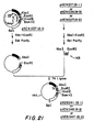
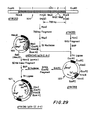
- FIG. 27 schematically depicts the manner in which an inducible plasmid cloning vehicle incorporating the A insertion site in the A-1 reading frame (and corresponding to the constitutive plasmid pKEN037) was constructed.
- the lac UV5 promoter-operator derived from plasmid pOP203-3 (obtained from Dr. F. Fuller, Dept. of Biochemistry and Molecular Biology, Harvard University).
- the lacUV5 promoter-operator is obtainable from E. coli 4288 recA/pKM006, NRRL B-15017, which is available to the public from the permanent collection of the Northern Regional Research Laboratory, U.S. Department of Agriculture, Peoria, Illinois, U .S.A.
- the lac UV5 promoter-operator is carried on a 95 bp Xba I fragment of plasmid pKM006.
- the plasmid can be obtained from NRRL B-15017 by conventional means, and the 95 bp Xba I fragment can thereafter be isolated using known techniques.
- the lac UV5 promoter-operator was inserted at the Xba I cleavage site of pKen037 (within the 5'-untranslated region of the Ipp gene) according to the following procedure: 200 micrograms of pOP203-3 plasmid DNA were digested to completion with 200 units of Alu I restriction enzyme in 400 microliters of Hind III buffer, and a 95 bp Alu I fragment carrying lac UV5 promoter and operator region (illustrated schematically by the diagonally cross-hatched segment at 152 in FIG. 27) was purified by polyacrylamide gel electrophoresis.
- One microgram of the 95 bp Alu I fragment was mixed with 400 pmoles of phosphorylated Xba I linker (5'CTCTAGAG 3' ; obtained from Collaborative Research and phosphorylated in the same manner as described hereinabove), and blunt-end ligated with 5 units of T4 DNA ligase in 20 microliters of ligase buffer containing 0.6mM ATP at 12.5°C for 16 hours.
- the ligated mixture was diluted to 300 microliters with Bam HI buffer and heated at 60°C for 10 minutes.
- the mixture was then treated with 100 units of Xba I restriction enzyme at 37°C for one hour to create Xba I cohesive termini.
- the mixture was extracted with phenol, ethanol precipitated and lyophilized.
- the DNA fragments were then dissolved in 10 microliters of water, and 0.3 micrograms of the lac fragment thus obtained were mixed with 0.5 micrograms of pKEN037 plasmid DNA, which had previously been digested with Xba I restriction enzyme.
- the mixture was treated with 0.4 units of T4 DNA ligase in 20 microliters of ligase buffer containing 0.4mM ATP at 12.5°C for 16 hours to anneal the Xba I cohesive termini, thereby re-circularizing the plasmid.
- Ten microliters of the ligated mixture were used to transform E.
- E. coli JA22l/F'lacI Q NRRL B-15015, which was constructed by transferring the F' factor from ⁇ 90/F'lacI q lac+ pro+ (obtained from Dr. J. Beckwith, Dept. of Biochemistry and Molecular Biology, Harvard University) into E. coli strain JA221, NRRL B-15014.
- E. coli strain JA221/F 2 lacI q is available to the public from the permanent collection of the Northern Regional Research Laboratory, U.S. Department of Agriculture, Peoria, Illinois, USA.
- the recognition sequence of the Sst I restriction enzyme overlaps with that of the Xba I enzyme.
- the deletion of the 4-base "sticky end" of the Sst I cleavage site using Sl nuclease should result in the deletion of part of the Xba I recognition sequence as well, effectively eliminating the Xba I cleavage site.
- a plasmid having the structure shown schematically at 156 in FIG. 27 was isolated after restriction enzyme analysis of plasmid DNAs obtained from ampicillin-resistant transformants by the rapid alkaline denaturation method, and that plasmid was designated pKEN045 (pIN-II A-1).
- An alternative and preferable method merely requires transferring the smaller Xba I-Eco RI fragments of pKEN039 and pKEN040 into the Xba I-Eco RI site of pKEN045, in a manner analogous to that described hereinabove in connection with FIG. 15, to yield plasmids pKEN049 (pIN-II, A-2) and pKEN050 (pIN-II, A-3).
- inducible plasmids pKEN049 (A-2) and pKEN050 (A-3) can be derived directly from plasmid pKEN045 (A-1).
- the DNA sequence in the vicinity of the Eco RI cleavage site of pKEN045 can itself be modified according to the scheme shown in FIG. 13, lines b and c, or the scheme illustrated in FIG. 14, lines b and c, to yield directly the structure of plasmids pKEN049 (A-2) or pKEN050 (A-3), respectively. This is the most preferred method of constructing these plasmids, since it does not require as a condition precedent the construction of the corresponding constitutive plasmids.
- pIN-II plasmids incorporating the B and C insertion sites.
- each could be modified to insert the lac promoter-operator fragment, according to the procedure of FIG. 27, or more preferably, the smaller Xba I-Eco RI fragment of pKEN045 (pIN-II, A-1) could be replaced successively with the smaller Xba I-Eco RI fragments from each of the constitutive B site and C site plasmids, yielding, in either case, pIN-II plasmids according to Table I.
- plasmid pKEN051 (pIN-II, B-1) can be derived from plasmid pKEN221 by first digesting pKEN221 plasmid DNA with Fnu4H-I restriction enzyme and then attaching Eco RI cohesive termini to the ends of the resulting fragment, according to the procedure described hereinabove and illustrated schematically at 138 in FIG. 17.
- the Eco RI fragment thus obtained can then be digested by Xba I restriction enzyme, splitting the fragment in two at the Xba I cleavage site located within the 5 1- untranslated region.
- the B-1 inducible cloning vehicle can be obtained.
- the resulting plasmid, pKEN051 (pIN-II, B-l) can then be further modified according to the scheme illustrated schematically at 141 in FIG. 18 and FIG. 19, or according to the scheme shown schematically at 142 in FIG. 18 and in FIG. 20, to yield the pIN-II plasmids corresponding to the B-2 and B-3 reading frames, pKEN052 and pKEN053, respectively.
- the Xba I RI fragment carrying the signal peptide region can then be inserted into the Xba I-Eco R I site of pKEN045, resulting in the plasmid pKEN054 (pIN-II, C-l). Further modification of the pKEN054 DNA according to the procedure shown schematically at 148 in FIG. 23 and in FIG. 24, or according to the procedure illustrated schematically at 149 in FIG. 23 and in FIG. 25, yields the pIN-II plasmids corresponding to the C-2 and C-3 reading frames, pKEN055 and pKEN056, respectively.
- FIGS. 28 and 29 schematically illustrate the manner in which the lacI gene was added to the inducible A-1 plasmid cloning vehicle of the pIN-II series, yielding the corresponding auto-regulated inducible expression plasmid of the pIN-III series. The specific steps in this procedure are described in detail hereinbelow.
- the first step in the construction of the A-1 expression plasmid of the pIN-III series was to clone the lacI gene into pBR322.
- a 5.1 Kb DNA fragment containing the lacI gene was first derived from plasmid pFB140 (obtained from Monica Riley of the Department of Biochemistry, State University of New York at Stony Brook) as follows: 15 micrograms of pFB140 plasmid DNA were digested with 80 units Eco RI restriction enzyme in 200 microliters of Eco RI buffer at 37°C for 2 hours. The reaction mixture was extracted with phenol and the DNA fragments were precipitated with 2.5 volumes of ethanol and dried under vacuum.
- the smaller DNA fragment lying between the Pse I and Eco RI cleavage sites of pBR322 was first deleted, as shown schematically at 158 in FIG. 28, using the following procedure: 10 micrograms of pBR322 DNA were digested with 2 units of Pst I restriction enzyme in 100 microliters of Pst I buffer at 37°C for 3 hours. After phenol extraction and ethanol precipitation, the DNAs were dried under vacuum, and then digested with 80 units of Eco RI restriction enzyme in a total volume of 200 microliters of Eco RI buffer at 37°C for 2 hours. The larger Pst I-Eco fragments, consisting of approximately 3.7 Kb, were then purified by agarose gel electrophoresis.
- the purified fragments (0.07 micrograms) were then mixed with 0.1 micrograms of the previously- obtained 5.1 Kb pFB140 fragments, and the Pst I and Eco R I cohesive termini were ligated by treating with 20 units of T4 DNA ligase (obtained from New England Biolabs) in 25 microliters of ligase buffer containing 0.48mM ATP at 12.5°C for 16 hours. Fifteen microliters of the ligated mixture were used to transform E. coli strain W620 recA, NRRL B-15024 (F-, thi-1, pyrD36, gltA6, galK30, strA129X-, supE44).
- This strain is available to the public from the permanent collection of the Northern Regional Research Laboratory, U.S. Department of Agriculture, Peoria, Illinois, USA., and was derived from E. coli strain W620, obtained from the Department of Human Genetics, Yale University, School of Medicine.
- E. coli strain W620 obtained from the Department of Human Genetics, Yale University, School of Medicine.
- One of the plasmid DNAs purified from tetracycline-resistant transformants had the structure shown at 150 in FIG. 28.
- This plasmid was designated pYM051, NRRL B-15025, which is available to the public from the permanent collection of the Northern Regional Research Laboratory, U.S. Department of Agriculture, Peoria, Illinois, USA.
- the plasmid can be obtained from NRRL B-15025 by conventional means.
- Plasmid pYM051 carries a 5.1 Kb Pst I-Eco RI DNA fragment containing not only the lacI gene, but also a substantial portion of the lacZ gene. As shown in 160 in FIG. 28, this fragment contains three Hinc II cleavage sites in the vicinity of the lacI gene, two of which surround or "bracket" the lacI gene, and one of which falls within the laci gene itself. In order to shorten this 5.1 Kb DNA fragment and at the same time retain the lacI gene intact for later use, the following procedure was used: 5 micrograms of pYM051 plasmid DNA were partially digested with 0.32 units of Hinc II restriction enzyme in 75 microliters of Hind III buffer at 37°C for 1 hour.
- plasmid designated pYMlll was constructed.
- This plasmid is obtainable from E. coli JA221/F'lacI q /pYMlll, NRRL B-15038, which is available to the public from the permanent collection of the Northern Regional Research Laboratory, U.S. Department of Agriculture, Peoria, Illinois, USA.
- the plasmid can be obtained from NRRL B-15038 by conventional means.
- pYMlll includes a Hpa I cleavage site surrounded in relatively close proximity by two Eco RI cleavage sites.
- This plasmid is an acceptable recipient for the laci gene fragment because it is a member of the class of plasmids having the following characteristics: (1) it contains a unique restriction enzyme cleavage site (i.e., a site occurring only once within the plasmid) which preferably yields blunt end termini, such as Hpa I, Hinc II or Pvu II; (2) it is derived from pBR322, and the unique cleavage site is not located within the DNA sequence responsible for the replication of the plasmid itself; (3) it also contains two cleavage sites which surround the unique cleavage site and are located within approximately 400-700 base pairs of the unique cleavage site, thet two surrounding cleavage sites preferably being recognizable by the same readily-available restriction enzyme, such as Eco RI, Hind III, Bam HI or Ps
- Plasmid pYMlll is suitable because it contains only one Hpa I cleavage site surrounded within 400-700 base pairs by two Eco RI cleavage sites, and because the lacI gene does not itself contain any Eco RI cleavage sites. It is to be understood, however, that any suitable plasmid having the foregoing characteristics can be used to receive the lacI gene fragment and to serve as a source of that fragment in subsequent steps of the procedure.
- the DNA fragment carrying the lacI gene was inserted in plasmid pYMlll, as shown schematically at 162 in FIG. 28, according to the following procedure: four micrograms of pYMlll plasmid DNA were digested with 4 units of Hpa I restriction endonuclease in a total volume of 50 microliters of Hpa I buffer at 37°C for 1 hour. Phenol extraction was performed, after which the DNAs were recovered by ethanol precipitation and dried under vacuum.
- the dried DNAs were treated with 0.15 units of BAP in a total volume of 100 microliters of a reaction mixture comprising lOmM Tris:HCl (pH 8.0) and 0.1mM EDTA (this reaction mixture will hereinafter be referred to as a "BAP buffer”) at 37°C for 45 minutes. Phenol extraction was performed three times to remove the BAP completely, after which the DNAs were recovered by ethanol precipitation and then dried under vacuum.
- 0.1 micrograms of the Hpa I-treated pYMlll plasmid DNA were mixed with 0.275 micrograms of the DNA fragments obtained from the Hinc II partial digestion of pYM051, and the DNAs were ligated with 320 units of T4 DNA ligase (obtained from New England Biolabs) in ligase buffer (total volume 20 microliters) containing 0.6mM ATP at 12.5 for 16 hours. Half of the ligation mixture was used to transform E.
- coli strain W620 recA, NRRL B-15024, and transformants were placed on the surface of an L-plate containing 50 micrograms/ml of ampicillin and 40 micrograms/ml of 5-bromo-4-chloro-3-indolyl-S-D-galactoside (hereinafter referred to as "X-gal").
- the transformants yielding white colonies were selected, indicating insertion of the 1.7 Kb Hinc II fragment carrying the intact lacI gene into plasmid pYMlll. Since this transformation results in plasmids carrying the lacI gene in two different orientations, the resulting plasmids were designated pYM052 and pYM053. The structure of both of these plasmids is depicted at 163 in FIG. 28.
- FIG. 29 depicts schematically the strategy used to remove this fragment from plasmid pYM053.
- the illustration at 164 in FIG. 29 is a partial restriction enzyme cleavage map of the region between the two Eco RI cleavage sites of plasmid pYM053, containing the lacl gene.
- This region includes the three Hinc II cleavage sites discussed hereinabove in connection with FIG. 28 and shown at 157 and at 160 in FIG. 28. As shown in FIG. 29, two of these Hinc II cleavage sites are also recognized by the Hpa I restriction endonuclease. Moreover, the greater detail of FIG.
- pYM053 plasmid DNA 100 micrograms of pYM053 plasmid DNA were digested with 60 units of Hpa I restriction enzyme in 800 microliters of Hpa I buffer at 37°C for 2 hours.
- a 789 bp fragment was purified.by 5% polyacrylamide gel electrophoresis: the DNA fragments in the polyacrylamide gel were stained with ethidium bromide (one microgram/ml), and the band corresponding to the 789 bp fragment was cut out. The DNA fragments in this band were eluted from the gel using electrophoresis. Ethidium bromide was removed from the DNA fragments by phenol extraction, and the DNAs were recovered by ethanol precipitation.
- the purified 789 bp DNA fragments (1.1 micrograms) were then digested with 12 units of Msp I restriction enzyme in 75 microliters of Hpa I buffer at 37°C for one hour. Phenol extraction was performed, after which the DNAs were recovered by ethanol precipitation and dried under vacuum. The DNA fragments were then treated with 2,000 units of Sl nuclease in a total volume of 150 microliters of Sl buffer at 20°C for 1 hour. The reaction was terminated by adding 15 microliters of 500mM Tris:HCl (pH 8.0) and 15 microliters of 250mM EDTA, following which phenol extraction was performed. To remove phenol and zinc ions, the mixture was extracted with ether and dialyzed against 0.01 x SSC at 4°C for 1.5 hours twice, and the DNAs were then recovered by ethanol precipitation.
- coli strain W620 recA, NRRL B-15024, and the transformants were placed on the surface of an L-plate containing 50 micrograms/ml of ampicillin and 40 micrograms/ml of X-gal.
- the ampicillin-resistant transformants yielding white colonies were selected, indicating that an intact lacI gene had been reconstructed.
- One of the plasmid DNAs purified from transformants selected had the structure shown at 165 in FIG. 29, and this plasmid was designated pYM058.
- the final step in the construction of the first auto-regulated inducible expression plasmid of the pIN-III series was to insert the lacI gene into pKEN045 (pIN-II, A-1).
- the following procedure was used: 30 micrograms of pYM058 plasmid DNA were digested with 100 units of Eco RI restriction enzyme in 250 microliters of Eco RI buffer at 37°C for 1.5 hours. A 2.4 Kb Eco RI fragment was purified by agarose gel electrophoresis in the same manner as described hereinabove. The DNA fragments (2.0 micrograsm) were then treated with 600 units of Sl nuclease in a total volume of 150 microliters of Sl buffer at 20°C for 1 hour.
- the reaction was terminated by adding 15 microliters of 500mM Tris:HCl (pH 8.0) and 15 microliters of 250mM EDTA, following which phenol extraction was performed. To remove phenol and zinc ions, the mixture was extracted with ether and dialyzed against 0.01 x SSC at 4°C for 1.5 hours twice, and the DNAs were then recovered by ethanol precipitation.
- pKEN045 plasmid DNA Ten micrograms of pKEN045 plasmid DNA were partially digested with 1 unit of Hinc II restriction enzyme in 751 microliters of Hind III buffer at 37°C for 30 minutes. As shown schematically at 166 in FIG. 29, pKEN045 includes two Hinc II cleavage sites, one of which is also a Sal I cleavage site. Partial digestion with Hinc II restriction enzyme yielded a mixture of linear DNA fragments, the longest of which had been cleaved at only one of the Hinc II sites. These fragments (each about 5.0 Kb long) were isolated by 0.7% agarose gel electrophoresis in the same manner as described hereinabove.
- the linearized DNAs (2.5 micrograms) were then treated with 0.15 units of BAP in 50 microliters of BAP buffer at 37°C for 1 hour to prevent self-ligation of the Hinc II blunt ends. Phenol extraction was performed three times to remove the BAP completely. The DNAs were recovered by ethanol precipitation and then dried under vacuum.
- the 2.4 Kb Eco RI fragments (0.15 micrograms) derived from pYM058 were then mixed with 0.3 micrograms of the 5.0 Kb pKEN045 fragments and treated with 400 units of T4 DNA ligase (obtained from New Endland . Biolabs) in 15 microliters of ligase buffer containing 0.8mM ATP at 12.5°C for 16 hours.
- Ten microliters of the ligation mixture were used to transform E. coli strain W620 recA, NRRL B-15024, and ampicillin-resistant transformants were selected using X-gal as previously described.
- the white colonies confirmed insertion of the lacI gene at the Hinc II site downstream from the Amp r gene.
- plasmids carrying the lacI gene in two different orientations the structure of one of which is illustrated schematically at 167 in FIG. 29.
- the plasmids having this structure were designated pYM061 (pIN-III, A-1).
- An alternative and preferably method merely requires substituting the smaller Xba I-Eco RI fragments of pKEN049 and pKEN050 (or pKEN039 (pIN-I, A-2) and pKEN040 (pIN-I, A-3)) for the smaller Xba I-Eco RI fragment of pYM061, in a manner analogous to that described hereinabove in connection with FIG. 15, to yield the A-2 and A-3 plasmids of the pIN-III series.
- auto-regulated inducible A-2 and A-3 plasmids can be derived directly from plasmid pYM061 (A-1).
- the DNA sequence in the vicinity of the Eco RI cleavage site of pYM061 can itself be modified according to the scheme shown in FIG. 13, lines b and c, or the scheme illustrated in FIG. 14, lines b and c, to yield directly the structure of the A-2 and A-3 plasmids, respectively, of pIN-III. This is the most preferred method of constructing these plasmids, since it does not require as a condition precedent the construction of the corresponding pIN-I and pIN-II plasmids.
- pIN-III plasmids incorporating the B and C insertion sites.
- each could be modified to insert the lacI gene fragment, according to the procedure of FIG. 29, or more preferably, the smaller Xba I-Eco RI fragment of pYM061 (pIN-III, A-1) could be replaced successively with the smaller Xba I-Eco RI fragments from each of the constitutive or inducible B site and C site plasmids, yielding, in either case, pIN-III B site and C site plasmids.
- the pIN-III expression plasmids are constructed without first making the corresponding pIN-I or pIN-II plasmids.
- the B-1 pIN-III plasmid can be derived from plasmid pKEN221 by first digesting pKEN221 plasmid DNA with Fnu4H-l restriction enzyme and then attaching Eco RI cohesive termini to the ends of the resulting fragment, according to the procedure described hereinabove and illustrated schematically at 138 in FIG. 17.
- the Eco RI fragment thus obtained can then be digested by Xba I restriction enzyme, splitting the fragment in two at the Xba I cleavage site located within the 5'-untranslated region.
- the B-1 auto-regulated inducible cloning vehicle can be obtained.
- the resulting plasmid can then be further modified according to the scheme illustrated schematically at 141 in FIG. 18 and in FIG. 19, or according to the scheme shown schematically at 142 in FIG. 18 and in FIG. 20, to yield the pIN-III plasmids corresponding to the B-2 and B-3 reading frames, respectively.
- the Xba I-Eco RI fragment carrying the signal peptide region can then be inserted into the Xba I-Eco RI site of pYM061, resulting in the C-1 plasmid of pIN-III. Further modification of the plasmid DNA according to the procedure shown schematically at 148 in FIG. 23 and in FIG. 24, or according to the procedure illustrated schematically at 149 in FIG. 23 and in FIG. 25, yields the pIN-III plasmids corresponding to the C-2 and C-3 reading frames, respectively.
- a structural gene coding for a human hormone or other desired polypeptide can be expressed in transformed bacterial hosts using a recombinant plasmid cloning vehicle constructed in accordance with the present invention, and significant quantities of the desired polypeptide can be produced thereby.
- a recombinant plasmid cloning vehicle constructed in accordance with the present invention can be expressed in transformed bacterial hosts using a recombinant plasmid cloning vehicle constructed in accordance with the present invention, and significant quantities of the desired polypeptide can be produced thereby.
Landscapes
- Genetics & Genomics (AREA)
- Health & Medical Sciences (AREA)
- Life Sciences & Earth Sciences (AREA)
- Engineering & Computer Science (AREA)
- Biomedical Technology (AREA)
- Chemical & Material Sciences (AREA)
- Organic Chemistry (AREA)
- Biotechnology (AREA)
- General Engineering & Computer Science (AREA)
- Zoology (AREA)
- Wood Science & Technology (AREA)
- Bioinformatics & Cheminformatics (AREA)
- Molecular Biology (AREA)
- Microbiology (AREA)
- Plant Pathology (AREA)
- Physics & Mathematics (AREA)
- Biochemistry (AREA)
- General Health & Medical Sciences (AREA)
- Biophysics (AREA)
- Preparation Of Compounds By Using Micro-Organisms (AREA)
- Micro-Organisms Or Cultivation Processes Thereof (AREA)
- Enzymes And Modification Thereof (AREA)
Applications Claiming Priority (2)
| Application Number | Priority Date | Filing Date | Title |
|---|---|---|---|
| US378481 | 1982-05-14 | ||
| US06/378,481 US4863855A (en) | 1982-05-14 | 1982-05-14 | Novel cloning vehicles for polypeptide expression in microbial hosts |
Publications (2)
| Publication Number | Publication Date |
|---|---|
| EP0094797A2 true EP0094797A2 (de) | 1983-11-23 |
| EP0094797A3 EP0094797A3 (de) | 1985-09-11 |
Family
ID=23493285
Family Applications (1)
| Application Number | Title | Priority Date | Filing Date |
|---|---|---|---|
| EP83302699A Ceased EP0094797A3 (de) | 1982-05-14 | 1983-05-12 | Klonierungsvektoren für die Polypeptideexpression in mikrobiellen Wirten |
Country Status (13)
| Country | Link |
|---|---|
| US (1) | US4863855A (de) |
| EP (1) | EP0094797A3 (de) |
| JP (1) | JPS5951793A (de) |
| AU (1) | AU555226B2 (de) |
| CA (1) | CA1205764A (de) |
| DD (1) | DD210466A5 (de) |
| DK (1) | DK215083A (de) |
| GB (1) | GB2120255B (de) |
| GR (1) | GR77485B (de) |
| HU (1) | HU197937B (de) |
| IE (1) | IE54976B1 (de) |
| IL (1) | IL68627A0 (de) |
| SU (1) | SU1479004A3 (de) |
Cited By (6)
| Publication number | Priority date | Publication date | Assignee | Title |
|---|---|---|---|---|
| GB2143535A (en) * | 1983-07-19 | 1985-02-13 | Suntory Ltd | Improved plasmid vector and use thereof |
| WO1987006590A1 (en) * | 1986-04-21 | 1987-11-05 | Bioenterprises Pty. Ltd. | Immunopotentiation |
| EP0182636A3 (de) * | 1984-11-16 | 1988-01-07 | Genex Corporation | Vektoren zur Expression von Genen mit hoher Ausbeute |
| EP0183272A3 (en) * | 1984-11-30 | 1988-01-20 | Suntory Limited | Vector capable of efficient gene expression and utilization of such vector |
| FR2663341A1 (fr) * | 1990-06-14 | 1991-12-20 | Pasteur Institut | Systeme pour l'expression d'une sequence polypeptidique et pour l'exposition de cette sequence a la surface d'un hote cellulaire. |
| US5976839A (en) * | 1987-03-13 | 1999-11-02 | Bioenterprises Pty Limited | Immunopotentiation through covalent linkage between immunogen and immunopotentiating molecules |
Families Citing this family (4)
| Publication number | Priority date | Publication date | Assignee | Title |
|---|---|---|---|---|
| GB2183655A (en) * | 1985-11-22 | 1987-06-10 | Christopher Francis Higgins | Method of increasing the half-life of an mRNA sequence |
| US5795776A (en) * | 1994-03-22 | 1998-08-18 | Bio-Technology General Corp. | Expression plasmids regulated by an OSMB promoter |
| US6596509B1 (en) | 1998-07-10 | 2003-07-22 | Cornell Research Foundation, Inc. | Recombinant constructs and systems for secretion of proteins via type III secretion systems |
| US8309324B2 (en) * | 2004-11-10 | 2012-11-13 | University Of Rochester | Promoters and proteins from Clostridium thermocellum and uses thereof |
Family Cites Families (10)
| Publication number | Priority date | Publication date | Assignee | Title |
|---|---|---|---|---|
| US4237224A (en) * | 1974-11-04 | 1980-12-02 | Board Of Trustees Of The Leland Stanford Jr. University | Process for producing biologically functional molecular chimeras |
| AT373279B (de) * | 1977-11-08 | 1984-01-10 | Genentech Inc | Verfahren zur herstellung eines rekombinationsclonbildungstraegers |
| US4411994A (en) * | 1978-06-08 | 1983-10-25 | The President And Fellows Of Harvard College | Protein synthesis |
| FR2442271A1 (fr) * | 1978-11-27 | 1980-06-20 | Pasteur Institut | Vecteur permettant l'insertion d'un gene procaryote ou eucaryote, et l'excretion de la proteine exprimee |
| ZA811368B (en) * | 1980-03-24 | 1982-04-28 | Genentech Inc | Bacterial polypedtide expression employing tryptophan promoter-operator |
| US4506013A (en) * | 1980-10-03 | 1985-03-19 | Eli Lilly And Company | Stabilizing and selecting recombinant DNA host cells |
| GR76959B (de) * | 1981-01-02 | 1984-09-04 | Univ New York State Res Found | |
| US4666836A (en) * | 1981-01-02 | 1987-05-19 | The Research Foundation Of State University Of New York | Novel cloning vehicles for polypeptide expression in microbial hosts |
| US4643969A (en) * | 1983-07-25 | 1987-02-17 | The Research Foundation Of State University Of New York | Novel cloning vehicles for polypeptide expression in microbial hosts |
| US4757013A (en) * | 1983-07-25 | 1988-07-12 | The Research Foundation Of State University Of New York | Cloning vehicles for polypeptide expression in microbial hosts |
-
1982
- 1982-05-14 US US06/378,481 patent/US4863855A/en not_active Expired - Fee Related
-
1983
- 1983-05-09 IL IL68627A patent/IL68627A0/xx unknown
- 1983-05-12 EP EP83302699A patent/EP0094797A3/de not_active Ceased
- 1983-05-12 SU SU833597101A patent/SU1479004A3/ru active
- 1983-05-12 IE IE1099/83A patent/IE54976B1/en unknown
- 1983-05-12 GB GB08313115A patent/GB2120255B/en not_active Expired
- 1983-05-12 AU AU14506/83A patent/AU555226B2/en not_active Ceased
- 1983-05-13 DD DD83250915A patent/DD210466A5/de unknown
- 1983-05-13 CA CA000428163A patent/CA1205764A/en not_active Expired
- 1983-05-13 DK DK215083A patent/DK215083A/da not_active Application Discontinuation
- 1983-05-13 JP JP58084880A patent/JPS5951793A/ja active Pending
- 1983-05-13 GR GR71351A patent/GR77485B/el unknown
- 1983-05-13 HU HU831675A patent/HU197937B/hu unknown
Cited By (10)
| Publication number | Priority date | Publication date | Assignee | Title |
|---|---|---|---|---|
| GB2143535A (en) * | 1983-07-19 | 1985-02-13 | Suntory Ltd | Improved plasmid vector and use thereof |
| EP0134673A1 (de) * | 1983-07-19 | 1985-03-20 | SUNTORY KABUSHIKI KAISHA, also known as SUNTORY LTD. | Plasmidvektor und seine Verwendung |
| US5955307A (en) * | 1983-07-19 | 1999-09-21 | Suntory Limited | Plasmid vector and use thereof for the production of interferon |
| EP0182636A3 (de) * | 1984-11-16 | 1988-01-07 | Genex Corporation | Vektoren zur Expression von Genen mit hoher Ausbeute |
| EP0183272A3 (en) * | 1984-11-30 | 1988-01-20 | Suntory Limited | Vector capable of efficient gene expression and utilization of such vector |
| WO1987006590A1 (en) * | 1986-04-21 | 1987-11-05 | Bioenterprises Pty. Ltd. | Immunopotentiation |
| US5874083A (en) * | 1986-04-21 | 1999-02-23 | Bioenterprises Pty Limited | Immunopotentiating complexes comprising TraT proteins |
| US5976839A (en) * | 1987-03-13 | 1999-11-02 | Bioenterprises Pty Limited | Immunopotentiation through covalent linkage between immunogen and immunopotentiating molecules |
| FR2663341A1 (fr) * | 1990-06-14 | 1991-12-20 | Pasteur Institut | Systeme pour l'expression d'une sequence polypeptidique et pour l'exposition de cette sequence a la surface d'un hote cellulaire. |
| WO1991019804A1 (fr) * | 1990-06-14 | 1991-12-26 | Institut Pasteur | Systeme pour l'expression d'une sequence polypeptidique et pour l'exposition de cette sequence a la surface d'un hote cellulaire |
Also Published As
| Publication number | Publication date |
|---|---|
| GB8313115D0 (en) | 1983-06-15 |
| HU197937B (en) | 1989-06-28 |
| GB2120255A (en) | 1983-11-30 |
| DK215083D0 (da) | 1983-05-13 |
| DD210466A5 (de) | 1984-06-13 |
| GB2120255B (en) | 1986-12-17 |
| AU1450683A (en) | 1983-11-17 |
| DK215083A (da) | 1983-11-15 |
| IE54976B1 (en) | 1990-04-11 |
| EP0094797A3 (de) | 1985-09-11 |
| CA1205764A (en) | 1986-06-10 |
| GR77485B (de) | 1984-09-24 |
| US4863855A (en) | 1989-09-05 |
| IL68627A0 (en) | 1983-09-30 |
| JPS5951793A (ja) | 1984-03-26 |
| IE831099L (en) | 1983-11-14 |
| AU555226B2 (en) | 1986-09-18 |
| SU1479004A3 (ru) | 1989-05-07 |
Similar Documents
| Publication | Publication Date | Title |
|---|---|---|
| US4757013A (en) | Cloning vehicles for polypeptide expression in microbial hosts | |
| US4624926A (en) | Novel cloning vehicles for polypeptide expression in microbial hosts | |
| US4643969A (en) | Novel cloning vehicles for polypeptide expression in microbial hosts | |
| EP0055942A2 (de) | Plasmide Klonierungsvektoren | |
| EP0075444B1 (de) | Verfahren und Produkte zur leichten mikrobiologischen Expression von DNS-Sequenzen | |
| JP2622479B2 (ja) | ヒト成長ホルモンの遺伝子を発現する複製可能な細菌プラスミド | |
| US5888808A (en) | Bacterial polypeptide expression employing tryptophan promoter-operator | |
| US4551433A (en) | Microbial hybrid promoters | |
| EP0128733B1 (de) | Menschlicher, insulinähnlicher Wachstumsfaktor (IGF)hergestellt aus einem rekombinanten Wirt, Verfahren, Expressionsvektor und rekombinanter Wirt hierfür und IGF enthaltende pharmazeutische Zusammensetzung | |
| EP0080848B1 (de) | Stabilisieren und Selektieren von Zellen | |
| EP0211047B1 (de) | Arab-promoter und verfahren zur herstellung von polypeptiden einschliesslich cecropinen mittels mikrobiologischer verfahren | |
| GB2084584A (en) | Method for stabilizing and selecting host cells containing recombinant dna | |
| JPH0759193B2 (ja) | 外来性蛋白質発現用クロ−ニングベクタ− | |
| EP0067540B1 (de) | Mikrobielle Genen promotor Operator | |
| US5232840A (en) | Enhanced protein production in bacteria by employing a novel ribosome binding site | |
| US4863855A (en) | Novel cloning vehicles for polypeptide expression in microbial hosts | |
| EP0105608A1 (de) | Verfahren zum Schützen von Bakterien | |
| US4666836A (en) | Novel cloning vehicles for polypeptide expression in microbial hosts | |
| CA1340091C (en) | Enhanced protein production in bacteria by employing novel ribosome binding site | |
| US5254463A (en) | Method for expression of bovine growth hormone | |
| EP0063494B1 (de) | Verfahren zum Produzieren von Protein aus einem Mikroorganismus, Mikroorganismen zur Verwendung in solchem Verfahren und ihre Erzeugung, Vektoren zur Verwendung in dieser Erzeugung und Protein produziert durch diesen Mikroorganismus und von diesen Mikroorganismen abgeleitete transformierte Kulturen | |
| US4880910A (en) | Terminal methionyl bovine growth hormone and its use | |
| EP0153961B1 (de) | Rekombinante materialien und verfahren zur herstellung von menschliche konnektive gewebe aktivierende peptide iii und deren analoge | |
| US4732859A (en) | Method for conferring bacteriophage resistance to bacteria | |
| JPS63501053A (ja) | 異種タンパク質の生産方法 |
Legal Events
| Date | Code | Title | Description |
|---|---|---|---|
| PUAI | Public reference made under article 153(3) epc to a published international application that has entered the european phase |
Free format text: ORIGINAL CODE: 0009012 |
|
| 17P | Request for examination filed |
Effective date: 19830516 |
|
| AK | Designated contracting states |
Designated state(s): BE CH DE FR GB IT LI LU NL SE |
|
| PUAL | Search report despatched |
Free format text: ORIGINAL CODE: 0009013 |
|
| AK | Designated contracting states |
Designated state(s): BE CH DE FR GB IT LI LU NL SE |
|
| 17Q | First examination report despatched |
Effective date: 19871210 |
|
| STAA | Information on the status of an ep patent application or granted ep patent |
Free format text: STATUS: THE APPLICATION HAS BEEN REFUSED |
|
| 18R | Application refused |
Effective date: 19890330 |
|
| RIN1 | Information on inventor provided before grant (corrected) |
Inventor name: INOUYE, MASAYORI Inventor name: MASUI, YOSHIHIRO Inventor name: NAKAMURA, KENZO |