EP0055942A2 - Plasmide Klonierungsvektoren - Google Patents
Plasmide Klonierungsvektoren Download PDFInfo
- Publication number
- EP0055942A2 EP0055942A2 EP81306190A EP81306190A EP0055942A2 EP 0055942 A2 EP0055942 A2 EP 0055942A2 EP 81306190 A EP81306190 A EP 81306190A EP 81306190 A EP81306190 A EP 81306190A EP 0055942 A2 EP0055942 A2 EP 0055942A2
- Authority
- EP
- European Patent Office
- Prior art keywords
- plasmid
- dna
- dna sequence
- eco
- gene
- Prior art date
- Legal status (The legal status is an assumption and is not a legal conclusion. Google has not performed a legal analysis and makes no representation as to the accuracy of the status listed.)
- Granted
Links
Images


Classifications
-
- C—CHEMISTRY; METALLURGY
- C12—BIOCHEMISTRY; BEER; SPIRITS; WINE; VINEGAR; MICROBIOLOGY; ENZYMOLOGY; MUTATION OR GENETIC ENGINEERING
- C12N—MICROORGANISMS OR ENZYMES; COMPOSITIONS THEREOF; PROPAGATING, PRESERVING, OR MAINTAINING MICROORGANISMS; MUTATION OR GENETIC ENGINEERING; CULTURE MEDIA
- C12N15/00—Mutation or genetic engineering; DNA or RNA concerning genetic engineering, vectors, e.g. plasmids, or their isolation, preparation or purification; Use of hosts therefor
- C12N15/09—Recombinant DNA-technology
- C12N15/11—DNA or RNA fragments; Modified forms thereof; Non-coding nucleic acids having a biological activity
- C12N15/62—DNA sequences coding for fusion proteins
-
- C—CHEMISTRY; METALLURGY
- C07—ORGANIC CHEMISTRY
- C07K—PEPTIDES
- C07K14/00—Peptides having more than 20 amino acids; Gastrins; Somatostatins; Melanotropins; Derivatives thereof
- C07K14/435—Peptides having more than 20 amino acids; Gastrins; Somatostatins; Melanotropins; Derivatives thereof from animals; from humans
- C07K14/575—Hormones
- C07K14/62—Insulins
-
- C—CHEMISTRY; METALLURGY
- C07—ORGANIC CHEMISTRY
- C07K—PEPTIDES
- C07K2319/00—Fusion polypeptide
- C07K2319/01—Fusion polypeptide containing a localisation/targetting motif
- C07K2319/02—Fusion polypeptide containing a localisation/targetting motif containing a signal sequence
Definitions
- This invention relates generally to the field of recombinant genetics, and specifically to a novel class of plasmid cloning vehicles with which exogenous genes may be expressed in transformed bacterial. hosts.
- DNA double-stranded deoxyribonucleic acid
- genes genetic information is encoded on double-stranded deoxyribonucleic acid (“DNA”) molecules (“genes”) according to the sequence in which the DNA coding strand presents the characteristic bases of its repeating nucleotide components.
- the four nitrogenous bases that characterize the two strands of DNA nucleotides are linked in complementary pairs by hydrogen bonds to form the double helix of DNA: adenine (A) is linked to thymine (T) and guanine (G) to cytosine (C).
- “Expression" of the encoded information to form polypeptides involves a two-part process.
- RNA polymerase messenger ribonucleic acid
- the DNA coding strand typically includes signals, which can be recognized by RNA polymerase, for both initiation and termination of transcription.
- the cell's ribosomes in conjunction with transfer-RNA, convert the RNA "message” into proteins or "po.lypeptides", which determine cell form and function.
- signals for the initiation and termination of ribosomal translation as well as signals specifying the identity and sequence of the amino acids which make up the polypeptide.
- the DNA coding strand comprises long sequences of nucleotide triplets called “codons” in which the characteristic bases of the nucleotides in each triplet or codon encode specific bits of information. For example, three nucleotides read as ATG (adenine- thymine-guanine) result in an mRNA signal which is interpreted as “start translation,” while termination codons TAG, TAA and TGA are interpreted as “stop translation.” Between the initiation codon and the termination codon lies the so-called “structural gene,” the codons of which define the amino acid sequence ultimately translated. That definition proceeds according to the well-established “genetic code” (e.g., Watson, J.D., Molecular Biology Of The Gene, 3rd ed.
- ATG adenine- thymine-guanine
- promoter The DNA sequence within the control region of a gene which mediates the initiation of transcription is termed the "promoter” of the gene, while the specific signal encoded in the DNA following the structural gene at which transcription ends is defined as the “transcription termination site.”
- transcription termination site the DNA sequence within the control region of a gene which mediates the initiation of transcription.
- the promoter provides the site at which RNA polymerase must bind in order to initiate transcription, and that the effectiveness or "strength" of a particular promoter or terminator signal is determined by the efficiency with which RNA polymerase can recognize and interact with these signals. This in turn depends in large part upon the particular base sequence of the DNA at or near these sites (see, e.g., Rosenberg, M., et. al., Ann. Rev. Genet., 1979 13, 319-353).
- control regions of some genes may also include DNA sequences which can be recognized by certain effector molecules, the action of which can positively or negatively influence the interaction between RNA polymerase and DNA and thereby further regulate gene expression at the level of transcription
- genes such as the lipoprotein gene of the gram-negative bacterium Escherichia coli ["E. coli"]
- the control regions of such genes are generally comprised solely of a promoter signal and a terminator signal which immediately precede and follow, respectively, the DNA sequence to be transcribed.
- the control regions cause mRNA synthesis to begin at a "transcription initiation site" located at or near the promoter, and to.proceed until the transcription termination site is reached, producing an mRNA molecule of predetermined length with a base sequence complementary to the base sequence of the transcribed DNA'.
- the DNA sequence between these two points defines not only the structural gene, the codons of which are ultimately translated for polypeptide expression, but also an "untranslated" region on either side of the structural gene.
- the untranslated region which precedes the structural sequence is known as the "5'-untranslated region," while the region which follows the structural signals is known as the "3'-untranslated region.”
- the DNA coding sequences for both of these untranslated regions, as well as the DNA coding sequences embodying the promoter signal and the terminator signal of certain genes, all of which may be referred to individually or collectively herein as "functional fragments" of those genes, may be effectively used in the creation of the novel cloning vehicles of the present invention.
- plasmid a non-chromosomal double-stranded DNA molecule in "plasmid” form which can be replicated when placed within a unicellular organism by a process called “transformation.” An organism so transformed is called a “transformant.”
- a "plasmid” is a circular non-chromosomal double-stranded DNA molecule derived from viruses or bacteria, the latter being termed “bacterial plasmids.”
- plasmids are made to contain exogenous DNA.
- a recombinant plasmid may include DNA that codes for polypeptides not ordinarily produced by the organism susceptible to transformation by the recombinant plasmid, and the exogenous DNA may in some cases comprise human genetic material.
- plasmids are cleaved to provide linear DNA having ligatable termini. These are bound to an exogenous gene having ligatable termini to provide a biologically functional moeity with a desired phenotypical property.
- the recombinant moiety is inserted into a micro-organism by transformation and transformants are isolated and cloned, wi:.h the object of obtaining large populations capable of expressing the new genetic information.
- Methods an means of forming recombinant cloning vehicles and transforming organisms with them have been widely reported in the literature, and generalized discussior 3 of the subject appear in Cohen, S., Scientific American 233, 24-33 (July 1975), and in Gilbert, W., et al., Scientific American 242, 74-94 (April, 1980).
- Such single strands may be formed by the addition of nucleotides to blunt ends using terminal transferase, or sometimes simply by “chewing back" one strand of a blunt end with an enzyme such as X-exonuclease.
- an enzyme such as X-exonuclease.
- such single strands may be formed by restriction endonucleases (also called restriction enzymes), which cleave the phosphodiester bonds in and around unique sequences of nucleotides of about 4-6 base pairs in length.
- restriction endonucleases also called restriction enzymes
- Many restriction endonucleases and their recognition sequences are known, the so-called Eco RI endonuclease being one of the most widely employed.
- Restriction endonucleases which cleave doublestranded DNA at unique sequences (e.g., at rotationally symmetric "palindromes") may leave cohesive termini.
- a plasmid or Other cloning vehicle may be cleaved, leaving termini each comprising half of the restriction endonuclease recognition site.
- a cleavage product of exogenous DNA obtained with the same restriction endonuclease will have ends complementary to those of the plasmid termini.
- synthetic DNA comprising cohesive termini may be provided for insertion into the cleaved vehicle.
- the termini can be digested with alkaline phosphatase, providing molecular selection for closure incorporating the exogenous fragment. Incorporation of a fragment in the proper orientation relative to other aspects of the vehicle may be enhanced when the fragment supplants vehicle D N A excised by two different restriction endonucleases, and when the fragment itself comprises termini respectively constituting half the recognition sequence of the same two different endonucleases.
- the prior art includes a number of successful and commercially viable schemes to express functional polypeptide products such as insulin, somatostatin and human and animal growth hormone, but these schemes generally utilize only one or two functional fragments of certain genes to control polypeptide expression.
- coli a-galactosidase structural gene, bringing the expression of the desired polypeptide under the control of the ⁇ -galactosidase or "lac" promoter. Since some of the codons for the ⁇ -galactosidase enzyme are thereby positioned to be translated in advance of the structural gene of the desired product, expression of this fused DNA sequence in an E. coli transformant is said to result in a precursor protein comprising a somatostatin (or other) polypeptide preceded by a superfluous protein fragment consisting of either a sequence of several amino acid residues derived from the a-galactosidase structural gene or virtually the entire ⁇ -galactosidase amino acid sequence, depending upon which insertion site is chosen.
- the efficiency of this system may be further reduced because the mRNA molecule transcribed from the DNA of these prior art cloning vehicles is unnecessarily long (extending in some cases far "upstream” and in other cases far “downstream” of the desired polypeptide through the remainder of the S-galactosidase structural gene), and may lack certain features, called “secondary structures” or “stem-and-loop structures,” which are believed to confer greater stability upon the mRNA molecule, thereby increasing its availability for ribosomal translation.
- the fused precursor- protein expression product accumulates inside the transformant cell, perhaps interfering with normal cellular activities and certainly requiring cell disruption in the harvesting process.
- a second class of prior art cloning vehicles involves the expression of similar "hybrid” proteins.
- the structural gene for the desired polypeptide is inserted within the structural gene for the 0-lactamase (or penicillinase) enzyme of E. coli.
- this protein is normally synthesized with a short leader sequence (also called a "signal peptide"), which is located at the amino terminus of the protein and is thought to direct secretion of the protein across the cytoplasmic membrane, the expected fused precursor- protein expression product of such a recombinant vehicle (containing the DNA sequence for rat growth hormone) in an E. coli transformant was not actually detected outside the cell, despite a reasonable expectation that a a-lactamase-rat growth hormone conjugate would be secreted into the periplasmic space.
- a third class of prior art cloning vehicles is represented by those described in Tacon, W., et al., M olec. gen. Genet. 177, 427-438 (1980) and in Halle- well, R.A., and Emtage, S., Gene 9, 27-47 (1980), wherein the use of the E. coli tryptophan gene is described.
- three plasmids were constructed from pBR322 which contained the E. coli tryptophan promoter-operator, and nucleotides specifying the leader sequence and first seven amino acids of the trpE gene.
- plasmids had a Hind III cloning site situated downstream from the translation initiation codon (ATG) of the trpE gene, with the cloning site in each plasmid differing in its translation phase relative to the initiation codon.
- ATG translation initiation codon
- tryptophan promoter-operator, the trpE gene and 15% of the trpD gene were used for the expression of foreign genes.
- the prior art has failed to develop cloning vehicles, which are suited for the efficient expression of prokaryotic and eukaryotic gene products in bacterial transformants, and which at the same time utilize functional fragments such as the 3'-untranslated region or the transcription termination site of any gene for expression of an exogenous DNA insert fragment. It is therefore the principle object fo the present invention to provide a new class of plasmid cloning vehicles incorporating not only the promoter and 5'-untranslated region, but also the 3'-untranslated region and the transcription termination site, derived from a particular class of bacterial genes.
- a class of recombinant bacterial plasmid cloning vehicles for expression of exogenous genes in transformed bacterial hosts comprising a DNA insert fragment coding for the desired polypeptide, linked in reading phase with one or more functional fragments derived from an outer membrane protein gene of any gram-negative bacterium.
- the exogenous DNA codes for mammalian hormones, enzymes or immunogenic proteins (or intermediates therefor), the functional fragments are derived from the lipoprotein gene of E. coli, and the desired polypeptide is expressed in E. coli transformants.
- the DNA sequence coding for the desired protein is linked with and is expressed in conjunction with four specific functional fragments associated with the E. coli liporpotein gene, namely, the promoter, the 5'-untranslated region, the 3'- untranslated region and the transcription termination site of the gene.
- Three alternative insertion sites for the exogenous DNA are provided, the selection of which can influence the ultimate location at which the expression product can be found and collected.
- the desired polypeptide ean thus be expressed with a leader sequence located at the amino terminal which comprises the signal peptide of the E. coli lipoprotein, such that the desired product may be secreted through the cytoplasmic membrane and the signal peptide removed in vivo by processes native to the transformant, yielding the exogenous gene product.
- E. coli lipoprotein which is one of the most extensively investigated membrane proteins, is also the most abundant protein in the cell in terms of numbers of molecules, there being approximately 700,000-750,000 lipoprotein of E. coli, extremely efficient machinery for lipoprotein gene expression, at the levels of both transcription and translation, is indicated. It is believed,that the lipoprotein gene may be expressed at least ten times more efficiently than genes for ribosomal proteins.
- the lipoprotein promoter region shows a most striking feature, namely, an extremely high A-T content, which is believed likely to be essential for highly efficient transcription of the lipoprotein gene.
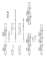
- the segment of 261 base pairs (“bp") preceding the transcription initiation site (from position -261 through position -1 as shown in FIG. 1) has a very high A-T content of 70%, in contrast with 53% for the transcribed region (or mRNA region) of 322 base pairs (positions +1 to +322), 44% for a segment of 126 bp after the transcription termination site (positions +323 to +449), and 49% for the average A-T content of the E.
- the A-T content of the segment from position -45 to position -1, within which the nucleotide sequence of the lipoprotein ("lpp") promoter appears to reside, is especially high (80%), and appears to be the highest among the E. coli promoter regions thus far sequenced.
- the A-T richness of the promoter sequence is considered to destabilize the helix structure of the DNA and thereby facilitate the RNA polymerase-mediated strand unwinding necessary for the initiation of transcription.
- the 1pp promoter also appears to contain a heptanucleotide sequence at positions -15 through -9 (only eight base pairs distal to the transcription initiation site) which is homologous to the generalized "Pribnow box,” as well as a dodecanucleotide sequence at positions -38 through -27 which is homologous to the generalized "RNA polymerase recognition site.”
- the homology of these sequences is striking, in that the Pribnow box sequence of the 1pp promoter has only one base mismatching with the generalized sequence, while the recognition site sequence shows a mismatch of only 5 out of 12 bases of the generalized sequence.
- the importance of the specific base sequences at these sites for efficient transcription is well-documented, in that mutants with enhanced promoter efficiency show increased homology of these regions with the generalized sequences.
- the lipoprotein gene also has an oligo-T transcription termination signal, located between positions +316 +322, which is at least as efficient as all other E. coli transcription termination sites that have been studied. It is believed that this factor contributes to the overall efficiency of transcription by hastening the rate of mRNA production, and by limiting the size of the mRNA molecule which is transcribed from the DNA.
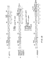
- the complete nucleotide sequence of the E. coli lipoprotein mRNA has also been determined, revealing that the mRNA has several unique features in its structure which appear to be important for efficient translation of the mRNA transcript.
- the mRNA consists of 322 nucleotides, 38 of which are in the 5'-untranslated region and 50 of which are in the 3'-untranslated region, leaving 234 nucleotides in the translated region which code for the lipoprotein precursor, or prolipoprotein.
- the mRNA sequence of - FIG. 2 is complementary to the DNA sequence of FIG. 1, with the exception of the nucleotide at position 313 which is shown as C in FIG. 2 as determined by RNA sequencing, rather than A as determined by the DNA sequencing shown in FIG. 1. The reason for this difference is not known at present.
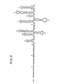
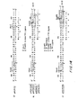
- the lipoprotein mRNA has been shown to be unusually stable, and it has been proposed that this stability is probably attributable to the formation of extensive secondary structures within the molecule. As shown in FIG. 3, the mRNA can form nine stable "hairpin" stem-and-loop structures (designated by Roman numerals I-IX), the most stable of which (I0 is in the 3'-untranslated region. These secondary structures may be responsible for the longer functional half-life which has been observed for the lipoprotein mRNA in comparison with other E. coli mRNAs, and may thereby increase the availability of this molecule for ribosomal translation.
- the lipoprotein of E. coli Apart from the efficiency of its expression, another important aspect of the lipoprotein of E. coli is that it is. a "secretory" protein, i.e., it is produced from a precursor, which.is then secreted across the cytoplasmic membrane and processed to the lipoprotein.
- the lipoprotein mRNA transcript -actually yields this precursor, called the prolipoprotein, which has a peptide extension or signal peptide at its amino terminus, consisting of 20 amino acid residues whose sequence has been determined, followed by the known 58 amino acid sequence of the lipoprotein.
- the signal peptide is considered to direct the translocation in vivo of the prolipoprotein across the cytoplasmic membrane, in the process of which the peptide extension itself is removed, yielding mature lipoprotein.
- the resultant alterations in the amino acid sequence do not change the basic properties of the signal peptide as proposed for the E. coli prolipoprotein and for other bacterial secretory proteins.
- the lipoprotein mRNA of S. marcescens seems capable of forming seven stable hairpin stem-and-loop structures. The existence of the lipoprotein in many different genera of gram-negative bacteria has been confirmed, and it has been found that the E. coli lipoprotein mRNA hybridizes with DNAs from at least the following seven bacterial species (besides S.
- marcescens in the family Enterobacteriaceae: Shigella dysenteriae, Salmonella typhimurium, Citro- bacter freundii, Klebsiella aerogenes, Enterobacter aerogenes, Edwardsiella tarda, and Erwinia amylovora, thereby confirming a degree of homology of the lipoprotein gene between E. coli and other gram-negative bacteria.
- the extension of the present invention to recombinant plasmid cloning vehicles utilizing analogous and highly efficient machinery for gene expression derived from any gram-negative bacterium is believed justified by all of these as well as other findings.
- a.structural gene for a eukaryotic protein or other desired polypeptide in an expression plasmid containing various combinations of the foregoing functional fragments, and by transforming a bacterial host with such a plasmid, the transcription and subsequent translation of the structural gene can be made to take place under the control of those functional fragments.
- the efficiency of transcription is believed to be further enhanced, avoiding transcriptional "read-through” (the synthesis of an unnecessarily long 3'-untranslated region in the mRNA) and more importantly, facilitating the rate of mRNA production.
- the stability of the mRNA molecule is also augmented by the formation of secondary structure in the 3'-untranslated region.
- the secretory nature of the lipoprotein can be utilized to control yet another aspect of the expression of a eukaryotic protein or other desired polypeptide, namely, the location at which at which the expression product can be expected to be found.
- the expression product can be expected to accumulate either within the cytoplasm of the transformant cell, within the periplasmic space, or in the cell's outer membrane.
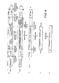
- FIG. 4 schematically illustrates a process wherein a transformant organism expresses a natural eukaryotic protein in accordance with the foregoing scheme.
- the structural gene for the eukaryotic protein is inserted within the signal peptide of the Ipp gene, several base pairs after the translation initiation codon and downstream of certain functional fragments (namely, the promoter and the 5'-untranslated region) normally associated with the lipoprotein gene.
- the orientation of these functional fragments is identical to the natural orientation of these elements in the lipoprotein gene, while the exogenous DNA insert fragment supplants most of the signal peptide as well as a portion of the structural region of the lipoprotein gene.
- line b the foreign gene is linked at its 3' end to an extra translation termination codon, which is in turn fused to the remainder of the lipoprotein structural gene. This is linked still further downstream in the normal manner to the 3'- untranslated region of the 1pp gnee, which ends with the transcription termination site.
- the functional fragments which follow the DNA insert fragment are essentially identical to those which are present normally in the lopoprotein gene.
- the 3'-untranslated region derived from the 1pp gene codes for an mRNA sequence capable of forming the stem-and-loop structure designated by the numeral I in FIG. 3 which, as discussed previously, is the most stable secondary structure in the lipoprotein mRNA.
- the recombinant DNA sequene depicted schematically in FIG. 4, line b also includes a terminal portion of the lipoprotein structural gene consisting of 105 base pairs starting with position +168 (this position is designated by the arrow (A) in FIG. 3).
- This region is chosen so that the stability of the mRNA transcript can be further enhanced by including four additional stem-and-loop structures (designated by the numerals II, III, IV and V in FIG. 3), without unduly increasing the size of the mRNA molecule produced. However, as set forth below, this region is not ultimately translated.
- mRNA sequence which is illustrated schematically in FIG. 4, line c. It will be seen that this sequence contains the 5'- untranslated region-and the 3' untranslated region, both of which are normally associated with the production of the lipoprotein.
- the mRNA also incorporates a region coding for the eukaryotic protein, preceded by a region which codes for a short segment of the signal peptide of the prolipoprotein, and followed by another region which codes codes for a short segment of the signal peptide of the prolipoprotein, and followed by another region which codes for a segment of the lipoprotein.
- the DNA sequence coding for the extraneous amino acids can be excised from the expression plasmid in a known manner prior to transformation of the bacterial host, such that the expression product corresponds exactly with the desired foreign protein and may be purified by known techniques.
- the same functional fragments are used, but the DNA sequence coding for the desired polypeptide is inserted further downstream, following the last codon of the signal peptide (i.e., at the signal peptide cleavage site).
- the orientation of the functional fragments is once again identical to the natural orientation of these elements in the lipoprotein gene, allowing full advantage to be taken of the efficiencies of transcription and translation associated therewith, including the enhanced stability of the mRNA transcript attributable to the incorporation of four additional stem-and-loop structures, as described hereinabove.
- coli transformant cell yielding a protein product which consists of the natural eukaryotic protein with several extraneous amino acid residues at the amino terminus, which can be removed as discussed hereinabove.
- This product accumulates initially in the periplasmic space, and may ultimately pass through the cell's outer membrane and into the culture medium provided that certain E. coli transformant strains are used, as set forth in more detail hereinbelow.
- the accumulation of a large amount of the expression product inside the cell is less likely to interfere with cell growth, because the eukaryotic protein is linked with a signal peptide which is natural to the cell. Furthermore, the presence of the signal peptide may protect the foreign protein from possible degradative action inside the cell, which could otherwise lower the protein yield and cause contamination of the foreign protein by heter- . ogenous segradative products, resulting in purification difficulties.
- the same functional fragments are again used, but the DNA sequence coding for the desired polypeptide is inserted still further downstream, following the codon for the eighth amino acid residue after the signal peptide cleavage site.
- the orientation of the functional fragments is once again identical to the natural orientation of these elements in the lipoprotein gene, allowing full advantage to be taken of the efficiencies of transcription and translation associated therewith, including the enhanced stability of the mRNA transcript attributable to the incorporation of four additional stem-and-loop structures, as described hereinabove.
- a polypeptide comprising a signal peptide of 20 amino acid residues, corresponding to the signal peptide of the prolipoprotein, followed by eight amino acid residues corresponding to the first eight amino acid residues of the mature lipoprotein, followed by the amino acid sequence of the desired eukaryotic protein.
- this precursor product may be translocated naturally across the cytoplasmic membrane, in the process of which the signal peptide can be recognized and removed.
- the product may not accumulate in the periplasmic space; instead, the eight amino acids corresponding to the lipoprotein can be recognized, and the expression product may then be processed further and inserted into the outer membrane of the cell in a manner analogous to the normal insertion of the lipoprotein into the outer membrane. If, as expected, only the first eight amino acid residues of the expression product corresponding to the lipoprotein are actually bound into the outer membrane, then the remainder of the expression product, consisting of the amino acid sequence of the eukaryotic protein or other desired polypeptide, will protrude from the outer membrane, such that, for certain proteins, the membrane may be isolated and the desired protein purified from the membrane easily.
- a plasmid cloning vehicle according to the present invention with one or another of the three insertion sites described above, and by using.such a plasmid to express an exogenous gene product, the location of that product can be predicted with a reasonable degree of certainty, and the appropriate methods for isolating and purifying that product will thereby be suggested.
- the choice of insertion site will often be dictated by the identity and structure of the desired polypeptide itself, especially if the method of purification most appropriate for that product is known.
- a short polynucleotide sequence containing the recognition sites for the Eco RI, Hind III and Barn HI restriction enzymes can be incorporated at the insertion site in each expression plasmid. This allows additional flexibility, in that six different types of restriction fragments can be inserted into each plasmid according to the straightforward and well-known techniques described hereinabove.
- DNA insert fragments tailored to have any one of the following pairs of cohesive termini can be readily used with the present invention: Eco RI-Eco RI, Hind III-Hind III, Bam HI-Bam HI, Eco RI-Hind III, Eco RI-Bam HI and Hind III-Bam HI.
- any structural gene coding for a desired polypeptide including mammalian and human hormones, enzymes and immunogenic proteins (or intermediates therefor), may be expressed using the recombinant plasmids of the present invention.
- proteins include A-chain insulin, B-chain insulin, proinsulin, growth hormone, somatostatin, interferon and trypanosome antigen, but the invention is not confined th these exemplary products.
- the recombinant cloning vehicles incorporating the gene for the desired eukaryotic protein or other polypeptide are used to transform special E. coli strains as hosts for cloning and for subsequent production of the protein.
- the host cell strains used will be chosen to have a "deletion mutant" in the ip 2 gene, so that the host cells cannot produce the lipoprotein.
- the use of a deletion mutant strain as the transformant is thought to stimulate the production of a large amount of the foreign protein, since the entire capacity of the host cells to produce the lipoprotein is thereby channelled towards production of the foreign protein.
- secretion.of the foreign protein across the cytoplasmic membrane is facilitated in lpp-defective host cells, since the secretion sites in the membrane which are intended to be used for lipoprotein secretion are instead available for secretion of the foreign protein.
- the use of the lpp-defective cells is especially beneficial when the gene coding for the foreign protein is inserted at the lipoprotein signal peptide cleavage site. This is because such cells are known to be "leaky", i.e., proteins secreted across the cytoplasmic membrane of such cells ultimately "leak” out into the culture medium through the outer membrane of the cell. This is believed to be desirable not only because release of the desired foreign protein into the culture medium may in some cases allow easier isolation and purification of the foreign protein than would be possible if the foreign protein remained inside the cells, but also because the foreign protein would otherwise accumulate in the periplasmic space, perhaps leading to undesirable interference with normal cellular activities or cell growth. Secretion of the desired eukaryotic gene product outside the cell may also avoid degradation of that product into smaller fragments by proteolytic enzymes which are normally present within the cell.
- the insertion site located within the DNA sequence coding for the prolipoprotein signal peptide will be designated the "A” site, while the insertion site located after the last codon of the signal peptide will be labelled the "B” site, and the insertion site located after the codon for the eighth amino acid residue of the mature lipoprotein will be referred to as the "C” site (see FIG. 5).
- A the insertion site located after the last codon of the signal peptide
- the insertion site located after the codon for the eighth amino acid residue of the mature lipoprotein will be referred to as the "C” site (see FIG. 5).
- three plasmids were prepared (one corresponding to each of the three possible reading frames), yielding a total of nine expression plasmids which were labelled A-1, A-2, A-3, B-1, B-2, B-3, and C-1, C-2, C-3.
- restriction enzymes used herein were obtained from New England Biolabs and Bethesda Research Laboratories. T4 DNA ligase was obtained from Bethesda Research Laboratories, and Sl Nuclease was obtained from Miles Laboratories.
- FIGS. 6-15 schematically depict the manner in which recombinant plasmids incorporating the A insertion site were constructed, and may be referred to in connection with the following more particularized discussion.
- the first step in the construction of the A site lpp gene cloning vehicles was to construct a plasmid to serve as a source of lpp gene components in subsequent steps of the procedure.
- the plasmid chosen to receive the E. coli lpp. gene for this purpose was pSC101, a small (molecular wt. approximately 5.8 megadaltons) plasmid carrying a gene conferring resistance to the antibiotic tetracycline (Tc) (Cohen, S.N., et al., J. Bacteriol. 132: 734-737 [1977]).
- Tc antibiotic tetracycline
- pSC101 includes a cleavage site for the restriction endonuclease Eco RI located at the 5' end of the tetracycline resistance gene.
- the plasmid pSC101 was obtained from Dr. E. Ohtsubo at the Department of Microbiology, State University of New York at Stony Brook.
- BAP bacterial alkaline phosphatase
- a 2.8 kilobase (“KB") DNA fragment containing the E. coli lpp gene was separately derived, as shown at 102 in FIG. 6, from a hybrid ⁇ phage carrying the E. coli lpp gene (designated ⁇ lppEc-1.
- the lpp gene had previously been cloned into a ⁇ phage vector, ⁇ 540 (Murray and Murray, J. Mol. Biol. 98: 551-564 [1975]), as follows: Total DNA (200 micrograms) isolated from an E. coli K-12 strain merodiploid for the lpp gene (JE5519/F506 [Movva, N.R., et al., J. Bacteriol.
- Two hundred micrograms of ⁇ lppEc-1 DNA were then digested completely with 200 units of the restriction enzyme Hae III in 500 microliters of a reaction mixture containgin 6mM Tris:HCl (pH 7.5), 6mM MgCl 2 , 6mM NaCl, 6mM 5-mercaptoethanol and 100 micrograms/ml BSA (the foregoing reaction mixture will hereinafter be referred to as a "Hae III buffer”) at 37°C for 2 hours, and the 2.8 Kb Hae III fragment carrying the E.
- coli lp 2 gene was purified by fractionation on a 5% polyacrylamide gel according to the following procedure: The reaction mixture was first extracted with phenol, and the DNA fragments were then precipitated with 2.5 volumes of ethanol, dried under vacuum, dissolved in 200 microliters of a buffer comprising 5% glycerol, 20mM EDTA, 0.05% bromophenol blue and 0.05% xylen cyanol (this mixture will hereinafter be referred to as a "gel buffer”) and thereafter fractionated on a 5% polyacrylamide gel. The DNA band which had migrated to a 2.8 Kb position was excised from the gel, and the DNA fragments were eluted from the gel by electrophoresis.
- a buffer comprising 5% glycerol, 20mM EDTA, 0.05% bromophenol blue and 0.05% xylen cyanol
- Ethidium bromide dye used to locate the DNA band in the gel, was removed from the DNA fragments by phenol extraction.
- the DNA fragments were precipitated with 2.5 volumes of ehtanol, centrifuged, dissolved in 200 microliters of 0.3M Na-acetate, re-precipitated with 0.5 ml of ethanol and dried again under vacuum. Approximately 10 micrograms of a purified 2.8 Kb Hae III fragment were recovered.
- Biochemicals with ATP in 50 microliters of a reaction mixture containing 3 moles of the linker, 66mM Tris:HCl (pH 7.5), 10mM MgCl 2 , 10mM ⁇ -mercaptoethanol, 60 ⁇ M ATP and 10 units of T4 polynucleotide kinase. After the mixture was incubated at 37°C for 30 minutes, it was heated at 60°C for 10 minutes, and cooled to 37°C. Five microliters of O.1M ⁇ -mercaptoethanol and 10 units of T4 polynucleotide kinase were added to the mixture, and the reaction was continued at 37°C for 30 minutes. The reaction was terminated by freezing the mixture in a dry ice ethanol bath.
- the 2.8 Kb Hae III fragment (2 micrograms) was mixed with 150 pmoles of phosphorylated Eco RI linker and was treated with 4 units of T4 DNA ligase in 12.5 microliters of a reaction mixture containing 66mM Tris:HCl (pH 7.5), lOmM MgCl 2 , lOmM dithiothreitol (the foregoing reaction mixture will hereinafter be referred to as a "ligase buffer") and 0.6mM ATP at 12.5°C for 15 hours.
- the reaction was terminated by diluting the mixture twenty-fold with Eco RI buffer and by heating the mixture at 60°C for 10 minutes. Thirty units of the restriction enzyme Eco RI were added, and the mixture was incubated at 37°C for one hour to create Eco RI cohesive termini. The reaction was terminated by heating at 60°C for 10 minutes.
- the mixture thus obtained was added to 2 micrograms of the previously-linearized plasmid pSC101 DNA, and phenol extraction was performed. After extraction with ether, the DNAs were precipitated with ethanol, dried under vacuum, and dissolved in 100 microliters of ligase buffer. The mixture was heated at 37°C for 5 minutes, and the Eco RI cohesive termini were annealed by incubating at 4°C for 16 hours and then at 0°C for one hour. After adding ATP (0.4mM final) and 1 unit of T4 DNA ligase, the mixture was incubated at 12.5°C for 7 hours.
- E. coli lpp deletion mutant strain JE5527 F - man, lpp-2, pps, thi, his, rpsl, gyr A, recAl [Hirota, Y., et al., Proc. Natl. Acad. Sci. U.S.A. 74: 1417-1420 (1977)], obtained from Dr. Y. Hirota, National Institute of Genetics, Mishima, Japan). Transformation was carried out as described in Cohen, S.N., et al., Proc. Natl. Acad. Sci. U.S.A.
- the parental plasmid chosen for construction of the l p p gene expression plasmids of the present invention was pBR322 (molecular wt. approximately 2.6 megadaltons), carrying genes conferring resistance to the antibiotics ampicillin (Amp) and tetracycline (Tc) (Bolivar, F., et al., Gene 2: 95-113 [1977]).
- pBR322 includes an Eco RI cleavage site located at the 5' end of the tetracycline resistance gene, as well as a Hind III cleavage site located within the promoter of the tetracycline resistance gene and a Pvu I cleavage site located within the ampicillin resistance gene.
- the plasmid pBR322 was obtained from Dr. N. Arnheim of the Department of Biochemistry, State University of New York at Stony Brook.
- FIG. 5 illustrates schematically the various components of the lpp gene, each of which is identified by a symbol or shading.
- the shaded segment indicated by the letter “a” identifies the A-T rich region of approximately 260 base pairs preceding the transcription initiation site and containing the lpp promoter.
- the 5'-untranslated region is identified by the segment containing the circular device and marked with the letter "b”.
- the signal peptide region of the prolipoprotein is identified by the diagonally hatched and shaded segment "c”.
- the structural region of the lpp gene is identified by the diagonally hatched segment labelled with the letter "d”, while the speckled segment "e” identifies the 3'-untranslated region and the transcription termination site.
- F I G. 7 illustrates the strategy used for inserting a fragment carrying the promoter and the 5'-untranslated region of the lpp gene into pBR322.
- the fragment chosen for this purpose was a 462 bp Alu I fragment of pKENlll which, as shown schematically at 105A in FIG. 5, contains not only the promoter sequence and the 5'-untranslated region (positions -45 to -1 and +1 to +39, respectively) of the l p 2 gene, but also the entire extremely A-T rich segment preceding the promoter sequence.
- the DNA was treated with 1.5 microliters of Sl Nuclease (Miles Laboratories) in a final volume of 300 microliters of a buffer containing 30mM Na-acetate (pH 4.25), 0.3M NaCl and 4mM ZnSO 4 (hereinafter referred to as an "Sl buffer”) at 20°C for one hour.
- Sl buffer a buffer containing 30mM Na-acetate (pH 4.25), 0.3M NaCl and 4mM ZnSO 4 (hereinafter referred to as an "Sl buffer") at 20°C for one hour.
- the reaction was terminated by adding 30 microliters 500mM Tris:HCl (pH 8.0) and 30 microliters 250mM EDTA, following which phenol extraction was performed.
- Phosphorylated Eco RI linker (200 pmoles) was then added and the mixture was treated with 4 units of T4 DNA ligase in 12.5 microliters of ligase buffer containing 0.6mM ATP at 12.5°C for 16 hours. Eco RI cohesive termini were created by addition of 30 units of Eco RI restriction enzyme in 75 microliters of Eco RI buffer at 37°C for 2 hours. The reaction was terminated by phenol extraction and the DNAs were recovered by ethanol precipitation.
- Plasmid DNAs were isolated from 0.5 ml of the cultures by the rapid alkaline-denaturation method described by Bi.rnboim, H. C. and Doly, J., Nucleic Acids Res. 7: 1513 (1979), and analyzed by restriction enzyme mapping.
- One of the plasmids had the structure shown at 107 in FIG. 7, and was designated pKEN005.
- the 462 bp Alu L fragment containing the lpp promoter was derived as follows: 100 micrograms of pKENIll plasmid DNA were digested with Msp I restriction enzyme in 600 microliters of a buffer containing 10mM Tris:HCl (pH 7.5), lOmM MgCl 2 , 6mM KCl, ImM dithiothreitol, and 100 micrograms/ml BSA (this mixture will hereinafter be referred to as an "Hpa I buffer”) at 37°C for 3 hours.
- Hpa I buffer this mixture will hereinafter be referred to as an "Hpa I buffer
- DNA fragments were precipitated with 2.5 volumes of ethanol, dried under vacuum, dissolved in 100 microliters of gel buffer, and fractionated on a 5% polyacrylamide gel. Approximately 6 micrograms of a purified 0.95 Kb Msp I fragment were recovered after elution of the separated DNA fragments from the gel. The purified 0.95 Kb Msp I fragment was subsequently digested with Alu I restriction endonuclease in 400 microliters of Hind III buffer at 37°C for 2 hours, yielding a 470 bp Alu I fragment which was purified by gel electrophoresis.
- One microgram of the 462 bp Alu I fragment was then mixed with 150 pmoles fo phosphorylated Eco RI linker and treated with 4 units of T4 DNA ligase in 10 microliters of ligase buffer containing 0.6mM ATP at 12.5°C for 16 hours.
- the ligated DNA was digested with 40 units of Eco RI restriction enzyme in 100 microliters of Eco RI buffer at 37°C for one hour to create Eco R I cohesive termini. The digestion was terminated by heating the mixture at 60°C for 10 minutes, and 0.6 micrograms. of Eco RI-digested pKEN005 plasmid DNA were added to the mixture and phenol extraction was performed.
- the DNAs were recovered by ethanol precipitation, and the Eco RI cohesive termini were joined by treating with 0.4 units of T4 DNA ligase in 20 microliters of ligase buffer containing 0.4mM ATP at 12.5°C for 7 hours.
- Ligated DNAs were used to transform E. coli strain JE5519, and transformants were selected for tetracycline resistance on an L broth plate containing 12.5 micrograms/ml of tetracycline.
- Anslysis of the plasmid DNAs isolated from the tetracycline-resistant transformants by the rapid alkaline-denaturation method showed insertion of the 462 bp Alu I fragment at the Eco RI site of pKEN005 as depicted at 110 in FIG. 7, and one of the plasmids thus obtained was designated pKEN008.
- FIG. 8 illustrates schematically the strategy for removing the Eco RI site distal to the lpp gene promoter.
- coli strain JE5519 and one of the transformants was shown to contain the plasmid with the structure illustrated at 112 in FIG. 8.
- This plasmid was designated pKEN002, and after digestion of 25 micrograms of pKEN002 plasmid DNA with Pvu I and Xba I restriction enzymes in 500 microliters of a buffer comprising 6mM Tris:HCl (pH 7.9), 6mM MgCl 2 , 150mM NaCl, 6mM ⁇ -mercaptoethanol and 100 micrograms/ml BSA (the foregoing mixture will hereinafter be referred to as a "Bam HI buffer") at 37°C for one hour, a 1.04 Kb Pvu I-Xba I DNA fragment (illustrated at 113 in FIG. 8) was purified by gel electrophoresis.
- a buffer comprising 6mM Tris:HCl (pH 7.9), 6mM MgCl 2 , 150mM NaCl,
- a 24 bp Xba I-Eco RI DNA fragment was derived from pKEN008 as follows: 25 micrograms of pKEN008 plasmid DNA was digested with Eco RI restriction enzyme, and a 470 bp Eco RI fragment was purified by gel electrophoresis.
- One microgram of the 470 bp Eco RI fragment was then digested with Xba I restriction enzyme, and was mixed with one microgram of the 1.04 Kb Pvu I-Iba I DNA fragment obtained previously, as well as with 0.75 micrograms of pKEN005 plasmid DNA previously digested with Pvu I and Eco RI restriction enzymes (as shown at 115 in FIG. 8).
- the DNA mixture was treated with 0.8 units of T4 DNA ligase in 50 microliters of ligase buffer containing 0.4mM ATP at 12.5°C for 7 hours.
- One-half of the ligated DNA was used to transform E. coli strain JE5519, and transformants were selected for tetracycline resistance.
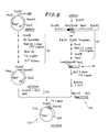
- FIG. 9 illustrates the strategy used for cloning a DNA fragment carrying the 3'-untranslated region and the transcription termination site of the lpp gene.
- the fragment chosen for this purpose was a 0.95 Kb Pvu II-Hpa I fragment of pKENlll, shown schematically at 105D in FIG. 5. Since the Pvu II restriction enzyme cleaves the lppgene sequence between positions +167 and +168, this fragment contains approximately the latter half of the lpp gene (see FIGS. I and 5). In order to insert this fragment into the cloning vehicle in the same orientation as the promoter fragment, Barn HI linker and Sal I linker were attached to the Pvu II and Hpa I cleavage sites, respectively.
- a 2.8 Kb Eco RI fragment was obtained from pKENlll plasmid DNA by digestion with Eco RI restriction enzyme and fractionation on a polyacrylamide gel, and 10 micrograms of this purified fragment were digested completely with Pvu II restriction endonuclease in 500 microliters of Hae III buffer at 37°C for one hour. The reaction was terminated by phenol extraction, and the mixture was extracted with ether. The DNA fragments were precipitated with 2.5 volumes of ethanol, centrifuged, re-dissolved in 200 microliters of 0.3M Na-acetate and re-precipitated with 0.5 ml of ethanol.
- the DNA fragments were digested completely with Hpa I restriction enzyme in 400 microliters of Hpa I buffer at 37°C for 2 hours.
- the reaction mixture was extracted with phenol and the DNA fragments were precipitated with ethanol, dried under vacuum, dissolved in 100 microliters of gel buffer and fractionated on a 5% polyacrylamide gel.
- the DNA bank which had migrated to a 0.95 Kb position was excised from the gel, and the DNA fragments were eluted from the gel by electrophoresis.
- DNA fragments were precipitated with 2.5 volumes of ethanol, centrifuged, dissolved in 200 microliters of 0.3M Na-acetate, re-precipitated with 0.5 ml of ethanol and again dried under vacuum. Approximately one microgram of a purified 0.95 Kb Hae III-Hpa I fragment (illustrated at 118 in FIG. 9) was recovered.
- Bam HI and Sal I restriction enzymes were added and the mixture was incubated at 37°C for 2 hours to create cohesive termini by cleaving the Bam HI and Sal I linkers attached to the Pvu II and Hpa I termini, respectively, resulting in a 0.95 Kb Bam HI-Sal I fragment (illustrated at 119 in FIG. 9).
- the restriction endonuclease digestion was terminated by heating at 60°C for 10 minutes.
- This fragment was accomplished, as shown schematically at 121 in FIG. 9, by Hind III digestion, followed by Sl Nuclease treatment, Barn HI linker attachment, Bam HI complete digestion, recircularization by T4 DNA ligase, and selection of tetracycline-sehsitive transformants.
- the mixture of linearized pKEN014 plasmid DNA and 0.95 Kb Bam HI-Sal I fragments was extracted with phenol, and the DNAs were precipitated with 2.5 volumes of ethanol, centrifuged and dissolved in 200 microliters of 0.3M Na-acetate. The DNAs were re-precipitated with 0.5 ml of ethanol, centrifuged and dried under vacuum. Cohesive termini of the DNA fragments were annealed with 0.4 units of T4 DNA ligase in 60 microliters of ligase buffer containing 0.4mM ATP at 12.5°C for 7 hours. Twelve microliters of the ligated mixture were then used to transform E.
- Plasmid DNAs were isolated from 0.5 ml of the cultures by the rapid alkaline-denaturation method and analyzed by agarose gel electrophoresis. Five of the plasmid DNAs were found to carry the 0.95 Kb Bam HI-Sal I fragment, and one of these plasmids was designated pKEN018. DNA sequencing of the pKEN018 plasmid DNA indicated the structure shown at 122 in FIG. 9, and specifically showed that the Bam HI linker was attached at the Pvu II site within the lpp gene at the correct position.
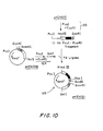
- the next step in the construction of the A site lpp gene cloning vehicles was to combine the lpp promoter fragment with the transcription terminator fragment in the same orientation. This step was carried out by replacing a 630 bp Pvu I-Eco RI fragment of pKEN018 with a 1.1 Kb Pvu I-Eco RI fragment of pKEN010, as illustrated schematically in FIG. 10.
- the DNA fragments were precipitated with 2.5 volumes of ethanol, dried under vacuum,dissolved in 100 microliters of gel buffer, and fractionated on a 5% polyacrylamide gel. Four micrograms of a purified 1.1 Kb Pvu I-Eco RI fragment were obtained after elution of the separated DNA fragments from the gel.
- the purified fragment (0.75 micrograms) was then mixed with 0.6 micrograms of pKEN018 plasmid DNA which had previously been double-digested with Pvu I and Eco RI restriction enzymes and then treated with BAP (as shown at 124 in FIG. 10).
- the Pvu I and the Eco RI cohesive.termini were ligated by treating with 0.4 units of T4 DNA ligase in 50 microliters of ligase buffer containing 0.4mM ATP at 12.5°C for 7 hours. Twenty-five microliters of the ligated mixture were used to transform E. coli strain JE5519, and transformants were selected for ampicillin resistance.
- Plasmid DNAs were isolated from ampicillin-resistant transformants and analyzed by agarose gel electrophoresis. Restriction enzyme mapping indicated that one of the plasmids had the structure shown at 125 in FIG. 10, and this plasmid was designated pKEN021.
- FIG. 11 illustrates the final step in the construction of the first A site lpp gene expression plasmid.
- pKEN021 carries both the lpp promoter fragment and the lpp transcription terminator fragment, separated by a 32 bp fragment derived from pBR322. By deleting the latter fragment and inserting a DNA sequence coding for a desired polypeptide, a functional moiety for expression of the desired polypeptide is provided.
- the reaction was terminated by adding 20 microliters of 0.5M Tris:HCl (pH 8.0) and 20 microliters of 0.25M EDTA.
- the mixture was extracted with phenol and dialyzed for four hours against 0.01 X SSC.
- the DNAs were precipitated with 2.5 volumes of ethanol, centrifuged and resuspended in 100 microliters of 0.3M Na-acetate.
- the DNAs were re-precipitated with 250 microliters of ethanol, centrifuged, and dried under vacuum.
- One microgram of the Sl-treated DNA was then mixed with 70 pmoles of phosphorylated Hind III linker ( CCAAGCTTGG ; obtained from Collaborative Research and phosphorylated according to the same procedure as described hereinabove) and blunt-end ligated with 4 units of T4 DNA ligase in 20 microliters of ligase buffer containing 0.6mM ATP at 12.5°C for 16 hours.
- the mixture was then diluted to 100 microliters with Hind III buffer and heated at 60°C for 10 minutes. Twenty units of Hind III restriction endonuclease were added, and the mixture was incubated at 37°C for one hour to remove superfluous linker molecules and to create Hind III cohesive termini.
- Plasmid DNAs (0.5 micrograms) were re-circularized by treating with 0.8 units of T4 DNA ligase in 15 microliters of ligase buffer containing 0.4mM ATP at 12.5°C for 7 hours. Eight microliters of the ligated mixture were used to transform E. coli strain JA221 (recA-, hr-, hm-, XtrpE5, thr, leu, thi, lacY-; obtained from Dr. J. Carbon, Dept. of Biological Sciences, University of California, Santa Barbara). Among the plasmid DNAs which were purified from the ampicillin-resistant transformants was one that had the structure shown at 127 in FIG. ll, and this plasmid was designated pKEN030.
- the DNA sequence of plasmid pKEN033 was modified to create a Hind III cleavage site between the Eco RI ' and Bam HI sites, as follows: 5 micrograms of pKEN033 plasmid DNA (having the DNA sequence of interest shown in FIG. 12, line a) were digested with 10 units of Bam HI restriction endonuclease in 50 microliters of Ban HI buffer at 37°C for one hour.
- the DNAs (3.6 micrograms) were treated with three units of T4 DNA polymerase (obtained from Bethesda Research Laboratories) in 20 microliters of a reaction mixture containing 50mM Tris:HCl (pH 8.0), 100m M KCl, 6mM MgCl 2 , and 6mM dithiothreitol (this reaction mixture will hereinafter be referred to as a "polymerase buffer”) in the presence of 0.lmM each of dATP, dGTP, dCTP and dTTP at 12.5°C for 45 minutes.
- the Bam HI and the Eco RI "sticky ends" were filled in completely, as shown in FIG. 12, line c.
- FIG. 13, line a, and FIG. 14, line a both illustrate the DNA sequence surrounding the translation initiation site of the prolipoprotein in pKEN111. As shown, this sequence includes an Alu I cleavage site between positions +45 and +46.
- an Eco RI linker was attached to the Alu I terminus, resulting in the DNA sequence shown in FIG. 13, line b, and in FIG.
- the remaining two single-strand A residues were removed by treating with Sl Nuclease in 200 microliters of Sl buffer at 20°C for one hour.
- the reaction was terminated by adding 20 microliters of 0.5M Tris:HCl (pH 8.0) and 20 microliters of 0.25M EDTA.
- the mixture was extracted with phenol and dialyzed overnight against 0.01 x SSC.
- the DNAs were precipitated with 2.5 volumes of ethanol, centrifuged and re-suspended in 100 microliters of 0.3M Na-acetate.
- the DNAs were re-precipitated with 250 microliters of ethanol, centrifuged, and dried under vacuum.
- one microgram of the Sl-treated DNA was first mixed with 70 pmoles of phosphorylated Eco RI linker and blunt-end ligated with 3.2 units of T4 DNA ligase in 11 microliters of ligase buffer containing 0.6mM ATP at 12.5°C for 16 hours. The mixture was then diluted to 50 microliters with Eco RI buffer and heated at 60°C for 10 minutes. Twenty units of Eco RI restriction endonuclease were added, and the mixture was incubated at 37°C for one hour to remove superfluous linker, molecules and to create Eco RI cohesive termini. The reaction mixture was then extracted with phenol, and the DNAs were precipitated with ethanol.
- Plasmid DNAs (0.5 micrograms) were re-circularized by treating with 0.8 units of T4 DNA ligase in 15 microliters of ligase buffer containing 0.4mM ATP at 12.5°C for 7 hours. Eight microliters of the ligated mixture were used to transform E. coli strain JA221. Plasmid DNAs were purified from 3 ampicillin-resistant transformants, which had been grown overnight in one hundred ml of L broth containing 50 micrograms/ml of ampicillin, and the DNA sequences at their Eco RI cleavage sites were determined. One of them was found to have the sequence shown in FIG. 13, line c, and was designated pKEN024 (A-2).
- the DNAs were precipitated with 2.5 volumes of ethanol, centrifuged and re-suspended in 100 microliters in 0.3 Na-acetate.
- the DNAs were re-precipitated with 250 microliters of ehtanol, centrifuged, and dried under vacuum.
- one microgram of the Sl-treated DNA was first mixed with 240 pmoles of phosphorylated Eco RI linker and blunt-end ligated with 4 units of T4 DNA ligase in 15 microliters of ligase buffer containing 0.6mM ATP at 12.5°C for 16 hours. The mixture was then diluted to 250 microliters with Eco RI buffer and heated at 60°C for 10 minutes. One hundred units of Eco RI restriction endonuclease were added, and the mixture was incubated at 37°C for one hour to remove superfluous linker molecules and to create Eco RI cohesive termini. The reaction mixture was then extracted with phenol, and the DNAs were precipitated with ethanol.
- Plasmid DNAs (0.3 micrograms) were re-circularized by treating with 0.8 units of T4 DNA ligase in 15 microliters of ligase buffer containing 0.4mM ATP at 12.5°C for 7 hours. Eight microliters of the ligated mixture were used to transform E. coli strain JA221. Plasmid DNAs were purified from 3 ampicillin-resistant transformants, which had been grown overnight in one hundred ml of L broth containing 50 micrograms/ml of ampicillin, and the DNA sequences at their Eco RI cleavage sites were determined. One of them was found to have the sequence shown in FIG. 14, line c, and was designed pKEN036 (A-3).
- the dried DNA fragments were dissolved in 20 microliters of water, and one microliter aliquots of this pKEN037 DNA fragment mixture were combined with 0.1 micrograms of each of the smaller Xba I-Eco RI restriction fragments (illustrated at 132 in FIG. 15) previously obtained from either pKEN024 or pKEN036 by double-digestion of either plasmid with Xba I and Eco RI restriction enzymes followed by gel purification (as shown at 133 in FIG. 15).
- the "sticky ends" of the Xba I-Eco RI fragments were joined by treatment with 0.2 units of T4 DNA ligase in 20 microliters of ligase buffer containing 0.4mM ATP at 12.5°C for 7 hours, following which a portion of the ligated mixture was used to transform E. coli strain JA221.
- plasmid DNAs having the A-2 and A-3 reading frames were obtained, and these were designated pKEN0 . 39 and pKEN040, respectively, each having the structure shown at 134 in FIG. 15.
- FIGS. 16-21 schematically illustrate the manner in which recombinant plasmids incorporating the B insertion site were constructed, and may be referred to in connection with the following more particularized discussion.
- the furst step in the construction of the B site'expression plasmids was to construct a plasmid to serve as a source of Ipp gene fragments having a restriction enzyme cleavage site at or near the signal peptide cleavage site.
- the gene chosen codes for the lipoprotein of S. marcescens, and has a Fnu4H-I restriction endonuclease recognition sequence at the 3' end of the signal peptide.
- the plasmid chosen to receive the S. marcescens lpp gene was pBR322.
- 2 micrograms of plasmid pBR322 DNA were digested to completion with two units of the restriction endonuclease Bam HI in 50 microliters of Barn HI buffer at 37°C for 60 minutes. After inactivation of Barn HI enzyme by heating at 60°C for 10 minutes, 2 units of Eco RI and 100 microliters of Eco RI buffer were added. The mixture was further incubated at 37°C for 60 minutes, and the reaction was then terminated by phenol extraction, after which the linearized DNA fragments were recovered by ethanol precipitation.
- DNA fragments were separated on a preparative agarose gel, and fractions of DNA fragments of approximately 8.5 Kb which showed positive hydridization with 5' 32 P-lipoprotein mRNA were collected, using the Southern hybridization technique.
- a mixture of 8.5 Kb Eco RI fragments (enriched approximately twenty-fold) and Eco RI-cleaved Charon 14 vector DNA was reacted with 14 DNA ligase.
- Ligated DNA was used to transfect E. coli K802.
- Recombinant phages carrying the lpp gene were screened by the plaque hybridization technique of Benton and Davis using 5'- 32 P-lipoprotein mRNA.
- One of the plaques examined which gave positive hybridization was designated ⁇ lppSm-1.
- a 0.95 Kb Msp I fragment of ⁇ lppEc-1 containing the lpp gene was nick-translated with [ ⁇ - 32 P]dATP and [ ⁇ - 32 P]-dCTP, as described in Maniatis, T., et al., Proc. Natl. Acad. Sci. U.S.A. 72: 1184-1188 (1975), and was used as a 32P-probe.
- One of the transformants which gave positive hybridization was shown to contain the plasmid with the structure illustrated at 137 in FIG. 16, and this plasmid was designated pKEN221.
- the DNA fragments were mixed with 400 pmoles of phosphorylated Eco RI linker and treated with 4 units of T4 DNA ligase in 20 microliters of ligase buffer containing 0.6mM ATP at 12.5°C for 16 hours. The mixture was diluted to 300 microliters with Eco RI buffer and digested with 150 units of Eco RI restriction enzyme to create Eco RI cohesive termini.
- One microgram of the Eco RI-digested fragments was then mixed with 0.5 micrograms of Eco RI-digested pKEN005 plasmid DNA, and treated with 0.4 units of T4 DNA ligase in 40 microliters of ligase buffer containing 0.6mM ATP - at 12.5°C for 16 hours. Twenty microliters of the ligated mixture was used to transform E. coli strain JE5519.
- FIG. 18 depicts schematically the strategy for removing the Eco RI site located upstream of the lpp promoter. This procedure involved transferring an 80 bp Xba I-Eco RI fragment (containing the signal peptide and a portion of the 5'-untranslated region of the S. marcescens lpp gene) from pKEN009 into the Xba I-Eco RI sites of pKEN010.
- pKEN010 plasmid DNA 5 micrograms of pKEN010 plasmid DNA were first digested with 5 units of Xba I restriction endonuclease in 50 microliters of Barn HI buffer, followed by digestion with 5 units of Eco RI restriction enzyme in 100 microliters of Eco RI buffer. The linearized DNA was then treated with 5 microliters of BAP in 100 microliters of 10mM Tris:HCl (pH 8.0) and 0.lmM EDTA at 37°C for 30 minutes.
- Plasmid DNAs were extracted with phendl and precipitated with ethanol, and 0.5 micrograms of the DNA were mixed with 0.2 micrograms of an 80 bp Xba I-Eco RI fragment, which had previously been obtained by digestion of 50 micrograms of pKEN009 plasmid DNA by Eco RI and Xba I restriction enzymes, followed by polyacrylamide gel electrophoresis.
- the DNA mixture was treated with 0.4 units of T4 DNA ligase in 40 microliters of ligase buffer containing 0.4mM ATP at 12.5°C for 16 hours. Twenty microliters of the ligated mixture were used to transform E. coli strain JE5519.
- one plasmid was found to contain the desired 80 bp Xba I-Eco RI fragment carrying the signal peptide region of the S. marcescens lpp gene in the B-1 reading frame, as shown at 140 in FIG. 18, and that plasmid was designated pKEN017.
- the reading frame at the B insertion site in pKEN017 was then modified to yield plasmids corresponding to the B-2 and B-3 reading frames, according to the methods previously described for changing the A-1 reading frame into the A-2 or A-3 reading frames, respectively. These procedures are illustrated schematically at 141 and 142 in FIG. 18, and the corresponding modifications of the DNA sequence around the E co RI cleavage site are shown in FIGS. 19 and 20. It will be understood that the same procedures used to derive plasmids pKEN024 (A-2) and pKEN036 (A-3) from plasmid pKEN030 (A-I), described hereinabove in connection with FIGA. 13 and 14, can be used to derive plasmids pKEN026 (B-3) and pKEN027 (B-2) from plasmid pKEN017 (B-1).
- FIG. 21 illustrates schematically the last step in the construction of the B site cloning vehicles, which was to replace the Xba I-Eco RI A site fragment of pKEN037 with each of the three different Xba I-Eco RI B site fragments of pKEN017, pKEN026 and pKEN027. This was necessary in order to provide the B site plasmids with the same sequence of Eco RI, Hind III and Bam HI restriciton enzyme recognition sequences at the exogenous DNA insertion site as contained in the A site plasmids. As shown schematically at 143 in FIG.
- each of the three B site fragments derived from pKEN017, pKEN026 and pKEN027 contains the DNA sequence including the signal peptide obtained from the Fnu4H-I fragment of the S. marcescens lpp gene.
- Each DNA mixture was treated with 0.2 units of T4 DNA ligase in 20 microliters of ligase buffer containing 0.4mM ATP at 12.5°C for 16 hours. Ten microliters of each of the ligated mixtures were used to transform E. coli strain JA221. Among the ampicillin-resistant transformants, plasmid DNAs having the B-1, B-2 and B-3 reading frames were purified, and these were designated pKEN041, pKEN047 and pKEN048, respectively, each having the structure shown at 144 in FIG. 21.
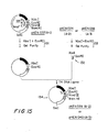
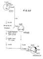
- FIGS. 22-26 schematically illustrate the manner in which recombinant plasmids incorporating the C insertion site were constructed, and may be referred to in connection with the following more particularized discussion.
- 200 micrograms of pKENlll plasmid DNA were digested to completion with 200 units of Sau 3A restriction endonuclease in 400 microliters of a reaction mixture comprising 10mM Tris:HCl (pH 7.5), 10mM MgCl2, 60mM NaCl, and 100 micrograms/ml BSA at 37°C for one hour. After digestion was completed, phenol extraction was performed, the DNAs were recovered by ethanol precipitation, and a 193 bp Sau 3A fragment was purified by acrylamide gel electrophoresis.
- the DNA fragments were mixed with 400 pmoles of phosphorylated Eco RI linker and treated with 4 units of T4 DNA ligase in 20 microliters of ligase buffer containing 0.6mM ATP at 12.5°C for 16 hours. The mixture was diluted to 300 microliters with Eco RI buffer and digested with 150 units of Eco RI restriction enzyme to create Eco RI cohesive termini.
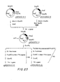
- FIG. 23 depicts schematically the strategy for removing the Eco RI site located upstream of the lpp promoter. This procedure involved transferring a 106 bp Xba I-Eco RI fragment (containing the signal peptide, a portion of the 5'-untranslated region and a portion of the structural sequence of the E. coli lpp gene) from pKEN006 into the Xba I-Eco RI sites of pKEN010.
- pKEN010 plasmid DNA 5 micrograms of pKEN010 plasmid DNA were first digested with 5 units of Xba I restriction endonuclease in 50 microliters of Bam HI buffer, followed by digestion with 5 units of Eco RI restriction enzyme in 100 microliters of Eco RI buffer. The linearized DNA was then treated with 5 microliters of BAP in 100 microliters of lOmM Tris:HCl (pH 8.0) and O.lmM EDTA at 37°C for 30 minutes.
- Plasmid DNAs were extracted with phenol and precipitated with ethanol, and 0.5 micrograms of the DNA were mixed with 0.2 micrograms of a 106 bp Xba I-Eco R I fragment, which had previously been obtained by digestion of 50 micrograms of pKEN006 plasmid DNA by Eco RI and Xba I restriction enzymes, followed by polyacrylamide gel electrophoresis.
- the DNA mixture was treated with 0.4 units of T4 DNA ligase in 40 microliters of ligase buffer containing 0.4mM ATP at 12.5°C for 7 hours. Twenty microliters of the ligated mixture were used to transform E. coli strain JE5519.
- one plasmid was found to contain the desired 106 bp Xba I-Eco fragment carrying the signal peptide region of the E. coli lpp gene in the C-1 reading frame, as shown at 147 in FIG. 23, and that plasmid was designated pKEN007.
- the reading frame at the C insertion site in pKEN007 was then modified to yield plasmids corresponding to the C-2 and C-3 reading frames, according to the methods previously described for changing the A-1 reading frame into the A-2 or A-3 reading frames, respectively. These procedures are illustrated schematically at 148 and 149 in FIG. 23, and the corresponding modifications of the DNA sequence around the Eco RI cleavage site are shown in FIGS. 24 and 25. It will be understood that the same procedures used to derive plasmids pKEN024 (A-2) and pKEN036 (A-3) from plasmid pKEN030 (A-1), described hereinabove in connection with FIGS. 13 and 14, can be used to derive plasmids pKEN046 (C-2) and pKEN019 (C-3) from plasmid pKEN007 (C-l).
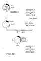
- each of the three C site fragments derived from pKEN007, pKEN046 and pKEN019 contains the DNA sequence including the signal peptide obtained from the Sau 3A fragment of the E. col lpp gene.
- microliters of each of the ligated mixtures were used to transform E. coli strain JA221.
- plasmid DNAs having the C-l, C-2 and C-3 reading frames were purified, and these were designated pKEN042, pKEN043 and pKEN044, respectively, each having the structure shown at 151 in FIG. 26.
- the structural gene for human proinsulin was chosen to demonstrate the expression of an exogenous gene in a transformed bacterial host in accordance with the invention.
- the proinsulin gene chosen for insertion into the cloning vehicle has an Eco RI cohesive terminus at its 5' end, as shown in FIG. 27.
- the reading frame of the proinsulin gene differs from the reading frame of plasmid pKEN021 by one base. Therefore, pKEN021 was modified at the Eco RI site, as shown in FIG. 13, line c, to make its reading frame compatible with that of the proinsulin gene.
- the reaction was terminated by adding EDTA to a final concentration of 25mM, followed by phenol extraction. By this procedure, half of the 4-base Eco RI "sticky end" was filled in with two A residues. The remaining two single-strand A residues were removed by treating with Sl Nuclease in 200 microliters of Sl buffer at 20°C for 1 hour. The reaction was terminated by adding 20 microliters of 0.5M Tris:HCl (pH 8.0) and 20 microliters of 0.25M EDTA. The mixture was extracted with phenol and dialyzed overnight against 0.01 x SCC. The DNAs were precipitated with 2.5 volumes of ethanol, centrifuged and re-suspended in 100 microliters of 0.3M N a-acetate. The DNAs were re-precipitated with 250 microliters of ethanol, centrifuged, and dried under vacuum.
- one microgram of Sl-treated DNA was first mixed with 70 pmoles of phosphorylated Eco RI linker and blunt-end ligated with 3.2 units of T4 DNA ligase in 11 ⁇ liters of ligase buffer containing 0.6mM ATP at 12.5°C for 16 hours. The mixture was then diluted to 50 microliters with Eco RI buffer and heated at 60°C for 10 minutes. Twenty units of Eco RI restriction endonuclease were added, and the mixture was incubated at 37°C for I hour to remove superfluous linker molecules and to create Eco RI cohesive termini. The reaction mixture was then extracted with phenol, and the DNAs were precipitated with ethanol.
- Plasmid DNAs (0.5 micrograms) were re-circularized by treating with 0.8 units of T4 DNA ligase in 15 microliters of ligase buffer containing 0.4mM ATP at 12.5°C for 7 hours. Eight microliters of the ligated mixture were used to transform E. coli strain JE5519. Plasmid DNAs were purified from 3 ampicillin-resistant transformants, whcih had been grown overnight in one ml of L broth containing 50 pg/ml of ampicillin, and the DNA sequences at their Eco RI cleavage sites were determined. One of them was found to have the sequence shown in FIG. 13, line c, and was designated pKEN024.
- FIG. 28 illustrates the strategy used to insert a gene coding for human proinsulin into plasmid -pKEN024.
- plasmid pNM431 illustrated at 151 in FIG. 28 was used as a source of the human proinsulin gene sequence.
- the human proinsulin gene sequence carried on plasmid pNM431 includes the proinsulin coding sequence, as well as the 3'-untranslated region of the insulin mRNA. These DNA nucleotide sequences, which are carried on a 469 bp Eco RI-Hind III fragment (as shown at 152 and 153 in FIG. 28), were inserted between the Eco RI site and the Hind III site of plasmid pBR322, yielding plasmid pNM431.
- the DNA nucleotide sequence of the human proinsulin gene carried on plasmid pNM431 is shown in FIG. 27.
- the DNA nucleotide sequence at the 5' end of this gene is such that when the gene is inserted at the Eco RI cleavage site of the expression plasmid pKEN024, the reading frame of the human proinsulin coding sequence will dovetail with the reading frame of pKEN024, as shown at 154 in FIG. 28.
- an 815 bp Eco RI-Bam HI fragment (illustrated at 155 in FIG. 28) of pNM431, comprising a 469 bp sequence from the Eco RI site to the Hind III site carrying the human proinsulin gene sequence, plus a 346 bp sequence from the Hind III site to the Bam HI site derived from plasmid pBR322, was cloned between the Eco RI site and the Barn HI site of plasmid pKEN024.
- Plasmid DNAs were isolated from eight ampicillin-resistant transformants, and their restriction fragments were analyzed by agarose gel electrophoresis.
- One of the plasmid DNAs which showed the structure illustrated at 156 in FIG. 28 was designated pInsulin A2-1.
- proinsulin in E. coli transformants carrying plasmid pInsulin A2-1 was examined by the semiquantitative solid phase radioimmunoassay methods described by S. Broom and W. Gilbert (Proc. Natl. Acad. Sci. U.S.A. 75, 2746 [1978]).
- This assay utilizes an antibody sandwich method in which the antigen is bound to an insoluable matrix by an IgG fraction of the antibody coupled to the solid phase. The antigen bound to the solid matrix is then detected by a second IgG fraction of the antibody, this time radioactively labelled with 125 I.
- This method allows the detection of antigen produced by individual bacterial colonies grown on plates.
- the assay is semi-quantitative, since the amount of 125 I-labelled IgG which can bind to the solid matrix depends on the amount of antigen bound to each IgG molecule coupled to the solid matrix.
- E. coli JA221 transformants carrying plasmid pInsulin A2-1 were streaked on L broth plates containing 50 ug/ml of ampicillin and grown at 37°C overnight. Cells were killed with chloroform vapor and lysed with a soft-agar overlay (2.5 ml per plate) which contained 0.7% agar, 0.4mg/ml of lysozyme and 0.1% Triton X-100 in 18mM Na-phosphate buffer (pH 6.8).
- a polyvinyl chloride sheet obtained from Dora May Co., New York which had previously been coupled with the anti-porcine insulin IgG fraction of guinea pig serum, was placed on the surface of the soft-agar overlay.
- wash buffer phosphate buffered saline containing 0.5% normal rabbit serum, 0.1% BSA and 0.02% sodium azide
- the sheet was exposed to a flushed Kodak XR-5 film at -80°C for 16 hours, and the resulting photographic image revealed that all the transformants carrying plasmid pInsuling A2-1 showed a strong positive reaction with anti-insulin antibody, while the transformants carrying the parental plasmid pKEN024 showed a completely negative reaction.
Applications Claiming Priority (4)
| Application Number | Priority Date | Filing Date | Title |
|---|---|---|---|
| US22201081A | 1981-01-02 | 1981-01-02 | |
| US222010 | 1981-01-02 | ||
| US28607081A | 1981-07-23 | 1981-07-23 | |
| US286070 | 1994-08-04 |
Publications (3)
| Publication Number | Publication Date |
|---|---|
| EP0055942A2 true EP0055942A2 (de) | 1982-07-14 |
| EP0055942A3 EP0055942A3 (en) | 1983-07-27 |
| EP0055942B1 EP0055942B1 (de) | 1988-06-01 |
Family
ID=26916355
Family Applications (1)
| Application Number | Title | Priority Date | Filing Date |
|---|---|---|---|
| EP19810306190 Expired EP0055942B1 (de) | 1981-01-02 | 1981-12-31 | Plasmide Klonierungsvektoren |
Country Status (5)
| Country | Link |
|---|---|
| EP (1) | EP0055942B1 (de) |
| DE (1) | DE3176765D1 (de) |
| DK (1) | DK582381A (de) |
| GB (1) | GB2091269B (de) |
| GR (1) | GR76959B (de) |
Cited By (19)
| Publication number | Priority date | Publication date | Assignee | Title |
|---|---|---|---|---|
| EP0070632A2 (de) * | 1981-06-29 | 1983-01-26 | Cetus Corporation | Rekombinante Plasmide die den Code für das menschliche Preproinsulin enthalten und ihre Verwendung zur Herstellung von menschlichem Insulin |
| EP0094797A2 (de) * | 1982-05-14 | 1983-11-23 | The Research Foundation Of State University Of New York | Klonierungsvektoren für die Polypeptideexpression in mikrobiellen Wirten |
| GB2121054A (en) * | 1982-05-25 | 1983-12-14 | Lilly Co Eli | Cloning vectors for expression of exogenous protein |
| GB2143535A (en) * | 1983-07-19 | 1985-02-13 | Suntory Ltd | Improved plasmid vector and use thereof |
| EP0170266A2 (de) * | 1984-07-30 | 1986-02-05 | Wakunaga Seiyaku Kabushiki Kaisha | Verfahren zur Herstellung eines Proteins und dafür zu verwendender Vektor, rekombinante DNS und transformierte Zelle |
| EP0196864A2 (de) * | 1985-03-25 | 1986-10-08 | Cetus Corporation | Behandlung und Sekretion von rekombinanten Proteinen mittels alkalischer Phosphatase, dazu verwendbare DNA-Sequenzen und mittels dieser Sequenzen transformierte Zellen |
| WO1987006590A1 (en) * | 1986-04-21 | 1987-11-05 | Bioenterprises Pty. Ltd. | Immunopotentiation |
| US4745069A (en) * | 1982-05-25 | 1988-05-17 | Eli Lilly And Company | Cloning vectors for expression of exogenous protein |
| US4755465A (en) * | 1983-04-25 | 1988-07-05 | Genentech, Inc. | Secretion of correctly processed human growth hormone in E. coli and Pseudomonas |
| WO1989007140A1 (en) * | 1988-02-05 | 1989-08-10 | Commonwealth Scientific And Industrial Research Or | Gene expression system (particularly for rotavirus vp7 protein) involving a foreign signal peptide and optionally a transmembrane anchor sequence |
| US4859600A (en) * | 1983-04-25 | 1989-08-22 | Genentech, Inc. | Recombinant procaryotic cell containing correctly processed human growth hormone |
| US4963495A (en) * | 1984-10-05 | 1990-10-16 | Genentech, Inc. | Secretion of heterologous proteins |
| WO1991002799A1 (en) * | 1989-08-25 | 1991-03-07 | Biotechnology Australia Pty Ltd | Fusion proteins |
| EP0177343B1 (de) * | 1984-10-05 | 1992-07-22 | Genentech, Inc. | DNA, Zellkulturen und Verfahren zur Sekretion von heterologen Proteinen und periplasmische Proteinrückgewinnung |
| US5795776A (en) * | 1994-03-22 | 1998-08-18 | Bio-Technology General Corp. | Expression plasmids regulated by an OSMB promoter |
| US5976839A (en) * | 1987-03-13 | 1999-11-02 | Bioenterprises Pty Limited | Immunopotentiation through covalent linkage between immunogen and immunopotentiating molecules |
| US6001604A (en) * | 1993-12-29 | 1999-12-14 | Bio-Technology General Corp. | Refolding of proinsulins without addition of reducing agents |
| US6204023B1 (en) | 1985-11-01 | 2001-03-20 | Xoma Ltd. | Modular assembly of antibody genes, antibodies prepared thereby and use |
| US6538118B1 (en) | 1995-06-07 | 2003-03-25 | Connaught Laboratories, Inc. | Expression of lipoproteins |
Families Citing this family (4)
| Publication number | Priority date | Publication date | Assignee | Title |
|---|---|---|---|---|
| SE8300693L (sv) * | 1983-02-09 | 1984-08-10 | Sven Lofdahl | Sett att framstella och isolera proteiner och polypeptider samt en hybridvektor for detta |
| US6022950A (en) * | 1984-06-07 | 2000-02-08 | Seragen, Inc. | Hybrid molecules having translocation region and cell-binding region |
| US5668255A (en) * | 1984-06-07 | 1997-09-16 | Seragen, Inc. | Hybrid molecules having translocation region and cell-binding region |
| US5747281A (en) * | 1984-09-28 | 1998-05-05 | Cornell Research Foundation, Inc. | System useful for the production of proteins from recombinant DNA in single celled organisms |
Citations (3)
| Publication number | Priority date | Publication date | Assignee | Title |
|---|---|---|---|---|
| EP0006694A2 (de) * | 1978-06-08 | 1980-01-09 | The President And Fellows Of Harvard College | Verfahren zur Herstellung eines ausgewählten Proteins |
| EP0012078A1 (de) * | 1978-11-27 | 1980-06-11 | Institut Pasteur | Vector, der die Einfügung eines prokaryotischen oder eukaryotischen Gens und die Ausscheidung des exprimierten Proteins gestattet |
| EP0023882A2 (de) * | 1979-08-03 | 1981-02-11 | Schering Aktiengesellschaft | Plasmidvektoren, Plasmide mit Genen für alkalische Phosphatasen und Bakterien, die letztere enthalten, ihre Herstellung und Verwendung |
-
1981
- 1981-12-30 DK DK582381A patent/DK582381A/da not_active Application Discontinuation
- 1981-12-30 GR GR66932A patent/GR76959B/el unknown
- 1981-12-31 GB GB8139170A patent/GB2091269B/en not_active Expired
- 1981-12-31 DE DE8181306190T patent/DE3176765D1/de not_active Expired
- 1981-12-31 EP EP19810306190 patent/EP0055942B1/de not_active Expired
Patent Citations (3)
| Publication number | Priority date | Publication date | Assignee | Title |
|---|---|---|---|---|
| EP0006694A2 (de) * | 1978-06-08 | 1980-01-09 | The President And Fellows Of Harvard College | Verfahren zur Herstellung eines ausgewählten Proteins |
| EP0012078A1 (de) * | 1978-11-27 | 1980-06-11 | Institut Pasteur | Vector, der die Einfügung eines prokaryotischen oder eukaryotischen Gens und die Ausscheidung des exprimierten Proteins gestattet |
| EP0023882A2 (de) * | 1979-08-03 | 1981-02-11 | Schering Aktiengesellschaft | Plasmidvektoren, Plasmide mit Genen für alkalische Phosphatasen und Bakterien, die letztere enthalten, ihre Herstellung und Verwendung |
Non-Patent Citations (5)
| Title |
|---|
| CELL, vol. 18, no. 4, December 1979, pages 1109-1117; * |
| FEDERATION PROCEEDINGS, vol. 39, no. 6, 1980, page 1182, ref. 1182; * |
| JOURNAL OF BACTERIOLOGY, vol. 146, no. 3, June 1981, pages 861-866; * |
| PROC. NATL. ACAD. SCI. USA, vol. 77, no. 3, March 1980, pages 1369-1373; * |
| THE JOURNAL FO BIOLOGICAL CHEMISTRY, vol. 255, no. 1, 10th January 1980, pages 210-216; * |
Cited By (30)
| Publication number | Priority date | Publication date | Assignee | Title |
|---|---|---|---|---|
| EP0070632A3 (de) * | 1981-06-29 | 1983-06-29 | Cetus Corporation | Rekombinante Plasmide die den Code für das menschliche Preproinsulin enthalten und ihre Verwendung zur Herstellung von menschlichem Insulin |
| EP0070632A2 (de) * | 1981-06-29 | 1983-01-26 | Cetus Corporation | Rekombinante Plasmide die den Code für das menschliche Preproinsulin enthalten und ihre Verwendung zur Herstellung von menschlichem Insulin |
| EP0094797A3 (de) * | 1982-05-14 | 1985-09-11 | The Research Foundation Of State University Of New York | Klonierungsvektoren für die Polypeptideexpression in mikrobiellen Wirten |
| EP0094797A2 (de) * | 1982-05-14 | 1983-11-23 | The Research Foundation Of State University Of New York | Klonierungsvektoren für die Polypeptideexpression in mikrobiellen Wirten |
| GB2121054A (en) * | 1982-05-25 | 1983-12-14 | Lilly Co Eli | Cloning vectors for expression of exogenous protein |
| EP0095361B1 (de) * | 1982-05-25 | 1989-07-26 | Eli Lilly And Company | Klonbildungsvektoren für die Expression von exogenem Protein |
| US4745069A (en) * | 1982-05-25 | 1988-05-17 | Eli Lilly And Company | Cloning vectors for expression of exogenous protein |
| US4755465A (en) * | 1983-04-25 | 1988-07-05 | Genentech, Inc. | Secretion of correctly processed human growth hormone in E. coli and Pseudomonas |
| US4859600A (en) * | 1983-04-25 | 1989-08-22 | Genentech, Inc. | Recombinant procaryotic cell containing correctly processed human growth hormone |
| EP0134673A1 (de) * | 1983-07-19 | 1985-03-20 | SUNTORY KABUSHIKI KAISHA, also known as SUNTORY LTD. | Plasmidvektor und seine Verwendung |
| US5955307A (en) * | 1983-07-19 | 1999-09-21 | Suntory Limited | Plasmid vector and use thereof for the production of interferon |
| GB2143535A (en) * | 1983-07-19 | 1985-02-13 | Suntory Ltd | Improved plasmid vector and use thereof |
| AU575331B2 (en) * | 1983-07-19 | 1988-07-28 | Suntory Limited | Improved e. coli plasmid containing modified 5' cipoprotein sequence |
| FR2550800A1 (fr) * | 1983-07-19 | 1985-02-22 | Suntory Ltd | Vecteur plasmide ameliore et son utilisation |
| EP0170266A2 (de) * | 1984-07-30 | 1986-02-05 | Wakunaga Seiyaku Kabushiki Kaisha | Verfahren zur Herstellung eines Proteins und dafür zu verwendender Vektor, rekombinante DNS und transformierte Zelle |
| EP0170266A3 (en) * | 1984-07-30 | 1988-01-13 | Wakunaga Seiyaku Kabushiki Kaisha | Process for producing protein, and vector, recombinant dna and transformant used therefor |
| US4963495A (en) * | 1984-10-05 | 1990-10-16 | Genentech, Inc. | Secretion of heterologous proteins |
| EP0177343B1 (de) * | 1984-10-05 | 1992-07-22 | Genentech, Inc. | DNA, Zellkulturen und Verfahren zur Sekretion von heterologen Proteinen und periplasmische Proteinrückgewinnung |
| EP0196864A3 (de) * | 1985-03-25 | 1988-03-23 | Cetus Corporation | Behandlung und Sekretion von rekombinanten Proteinen mittels alkalischer Phosphatase, dazu verwendbare DNA-Sequenzen und mittels dieser Sequenzen transformierte Zellen |
| EP0196864A2 (de) * | 1985-03-25 | 1986-10-08 | Cetus Corporation | Behandlung und Sekretion von rekombinanten Proteinen mittels alkalischer Phosphatase, dazu verwendbare DNA-Sequenzen und mittels dieser Sequenzen transformierte Zellen |
| US6204023B1 (en) | 1985-11-01 | 2001-03-20 | Xoma Ltd. | Modular assembly of antibody genes, antibodies prepared thereby and use |
| US5874083A (en) * | 1986-04-21 | 1999-02-23 | Bioenterprises Pty Limited | Immunopotentiating complexes comprising TraT proteins |
| WO1987006590A1 (en) * | 1986-04-21 | 1987-11-05 | Bioenterprises Pty. Ltd. | Immunopotentiation |
| US5976839A (en) * | 1987-03-13 | 1999-11-02 | Bioenterprises Pty Limited | Immunopotentiation through covalent linkage between immunogen and immunopotentiating molecules |
| WO1989007140A1 (en) * | 1988-02-05 | 1989-08-10 | Commonwealth Scientific And Industrial Research Or | Gene expression system (particularly for rotavirus vp7 protein) involving a foreign signal peptide and optionally a transmembrane anchor sequence |
| WO1991002799A1 (en) * | 1989-08-25 | 1991-03-07 | Biotechnology Australia Pty Ltd | Fusion proteins |
| US6001604A (en) * | 1993-12-29 | 1999-12-14 | Bio-Technology General Corp. | Refolding of proinsulins without addition of reducing agents |
| US5945304A (en) * | 1994-03-22 | 1999-08-31 | Bio-Technology General Corp. | Expression plasmids regulated by an osmB promoter |
| US5795776A (en) * | 1994-03-22 | 1998-08-18 | Bio-Technology General Corp. | Expression plasmids regulated by an OSMB promoter |
| US6538118B1 (en) | 1995-06-07 | 2003-03-25 | Connaught Laboratories, Inc. | Expression of lipoproteins |
Also Published As
| Publication number | Publication date |
|---|---|
| DE3176765D1 (en) | 1988-07-07 |
| EP0055942B1 (de) | 1988-06-01 |
| GR76959B (de) | 1984-09-04 |
| DK582381A (da) | 1982-07-03 |
| GB2091269A (en) | 1982-07-28 |
| EP0055942A3 (en) | 1983-07-27 |
| GB2091269B (en) | 1984-11-21 |
Similar Documents
| Publication | Publication Date | Title |
|---|---|---|
| US4624926A (en) | Novel cloning vehicles for polypeptide expression in microbial hosts | |
| EP0055942A2 (de) | Plasmide Klonierungsvektoren | |
| US4757013A (en) | Cloning vehicles for polypeptide expression in microbial hosts | |
| EP0075444B1 (de) | Verfahren und Produkte zur leichten mikrobiologischen Expression von DNS-Sequenzen | |
| US4643969A (en) | Novel cloning vehicles for polypeptide expression in microbial hosts | |
| EP0128733B1 (de) | Menschlicher, insulinähnlicher Wachstumsfaktor (IGF)hergestellt aus einem rekombinanten Wirt, Verfahren, Expressionsvektor und rekombinanter Wirt hierfür und IGF enthaltende pharmazeutische Zusammensetzung | |
| JP2622479B2 (ja) | ヒト成長ホルモンの遺伝子を発現する複製可能な細菌プラスミド | |
| US5888808A (en) | Bacterial polypeptide expression employing tryptophan promoter-operator | |
| EP0047600B1 (de) | Rinder-Prowachstums- und -Wachstumshormon | |
| EP0095361B1 (de) | Klonbildungsvektoren für die Expression von exogenem Protein | |
| EP0080848B1 (de) | Stabilisieren und Selektieren von Zellen | |
| US5010010A (en) | Production of human parathyroid hormone from microorganisms | |
| US4863855A (en) | Novel cloning vehicles for polypeptide expression in microbial hosts | |
| US4666836A (en) | Novel cloning vehicles for polypeptide expression in microbial hosts | |
| EP1114865A1 (de) | Herstellung von humanem Parathormon | |
| US5254463A (en) | Method for expression of bovine growth hormone | |
| US4880910A (en) | Terminal methionyl bovine growth hormone and its use | |
| EP0159123B1 (de) | Expressionsvektoren für Rinderwachstumshormonderivate | |
| KR100393297B1 (ko) | 돌연변이체aox2프로모터,이를보유한벡터,형질전환체및이종단백질의제조방법 | |
| EP0153961B1 (de) | Rekombinante materialien und verfahren zur herstellung von menschliche konnektive gewebe aktivierende peptide iii und deren analoge | |
| EP0207165B1 (de) | Polypeptidverabreichender vektor, mikroorganismen vom vektor transformiert, verfahren zur herstellung von polypeptiden mittels derselben | |
| CA1207684A (en) | Cloning vehicles for polypeptide expression in microbial hosts | |
| EP0086649A2 (de) | Molekulare Klonierung und Charakterisierung der für das Schweine-Relaxin kodierenden Gen-Sequenz | |
| US4732859A (en) | Method for conferring bacteriophage resistance to bacteria | |
| JPS63501053A (ja) | 異種タンパク質の生産方法 |
Legal Events
| Date | Code | Title | Description |
|---|---|---|---|
| PUAI | Public reference made under article 153(3) epc to a published international application that has entered the european phase |
Free format text: ORIGINAL CODE: 0009012 |
|
| 17P | Request for examination filed |
Effective date: 19820112 |
|
| AK | Designated contracting states |
Designated state(s): BE CH DE FR GB IT LU NL SE |
|
| PUAL | Search report despatched |
Free format text: ORIGINAL CODE: 0009013 |
|
| AK | Designated contracting states |
Designated state(s): BE CH DE FR GB IT LI LU NL SE |
|
| RAP1 | Party data changed (applicant data changed or rights of an application transferred) |
Owner name: THE RESEARCH FOUNDATION OF STATE UNIVERSITY OF NEW |
|
| GRAA | (expected) grant |
Free format text: ORIGINAL CODE: 0009210 |
|
| AK | Designated contracting states |
Kind code of ref document: B1 Designated state(s): BE CH DE FR GB IT LI LU NL SE |
|
| REF | Corresponds to: |
Ref document number: 3176765 Country of ref document: DE Date of ref document: 19880707 |
|
| ET | Fr: translation filed | ||
| ITF | It: translation for a ep patent filed |
Owner name: MODIANO & ASSOCIATI S.R.L. |
|
| PGFP | Annual fee paid to national office [announced via postgrant information from national office to epo] |
Ref country code: DE Payment date: 19881230 Year of fee payment: 8 |
|
| PG25 | Lapsed in a contracting state [announced via postgrant information from national office to epo] |
Ref country code: LU Free format text: LAPSE BECAUSE OF NON-PAYMENT OF DUE FEES Effective date: 19881231 |
|
| PLBE | No opposition filed within time limit |
Free format text: ORIGINAL CODE: 0009261 |
|
| STAA | Information on the status of an ep patent application or granted ep patent |
Free format text: STATUS: NO OPPOSITION FILED WITHIN TIME LIMIT |
|
| 26N | No opposition filed | ||
| PG25 | Lapsed in a contracting state [announced via postgrant information from national office to epo] |
Ref country code: GB Effective date: 19891231 Ref country code: BE Effective date: 19891231 |
|
| PG25 | Lapsed in a contracting state [announced via postgrant information from national office to epo] |
Ref country code: SE Effective date: 19900101 |
|
| PGFP | Annual fee paid to national office [announced via postgrant information from national office to epo] |
Ref country code: CH Payment date: 19900123 Year of fee payment: 9 |
|
| BERE | Be: lapsed |
Owner name: THE RESEARCH FOUNDATION OF STATE UNIVERSITY OF NE Effective date: 19891231 |
|
| PG25 | Lapsed in a contracting state [announced via postgrant information from national office to epo] |
Ref country code: NL Effective date: 19900701 |
|
| NLV4 | Nl: lapsed or anulled due to non-payment of the annual fee | ||
| GBPC | Gb: european patent ceased through non-payment of renewal fee | ||
| PG25 | Lapsed in a contracting state [announced via postgrant information from national office to epo] |
Ref country code: FR Effective date: 19900831 |
|
| PG25 | Lapsed in a contracting state [announced via postgrant information from national office to epo] |
Ref country code: DE Effective date: 19900901 |
|
| REG | Reference to a national code |
Ref country code: FR Ref legal event code: ST |
|
| PG25 | Lapsed in a contracting state [announced via postgrant information from national office to epo] |
Ref country code: LI Effective date: 19901231 Ref country code: CH Effective date: 19901231 |
|
| REG | Reference to a national code |
Ref country code: CH Ref legal event code: PL |
|
| EUG | Se: european patent has lapsed |
Ref document number: 81306190.0 Effective date: 19901106 |