CN110256558B - Antibodies to TIGIT - Google Patents
Antibodies to TIGIT Download PDFInfo
- Publication number
- CN110256558B CN110256558B CN201910247168.5A CN201910247168A CN110256558B CN 110256558 B CN110256558 B CN 110256558B CN 201910247168 A CN201910247168 A CN 201910247168A CN 110256558 B CN110256558 B CN 110256558B
- Authority
- CN
- China
- Prior art keywords
- antibody
- antibodies
- tigit
- human
- binding
- Prior art date
- Legal status (The legal status is an assumption and is not a legal conclusion. Google has not performed a legal analysis and makes no representation as to the accuracy of the status listed.)
- Active
Links
- 101000831007 Homo sapiens T-cell immunoreceptor with Ig and ITIM domains Proteins 0.000 title claims abstract description 210
- 102100024834 T-cell immunoreceptor with Ig and ITIM domains Human genes 0.000 title claims description 167
- 230000027455 binding Effects 0.000 claims abstract description 258
- 239000000427 antigen Substances 0.000 claims abstract description 231
- 108091007433 antigens Proteins 0.000 claims abstract description 229
- 102000036639 antigens Human genes 0.000 claims abstract description 229
- 206010028980 Neoplasm Diseases 0.000 claims abstract description 228
- 210000004027 cell Anatomy 0.000 claims abstract description 195
- 238000000034 method Methods 0.000 claims abstract description 151
- 239000012634 fragment Substances 0.000 claims abstract description 102
- 201000011510 cancer Diseases 0.000 claims abstract description 78
- 210000001744 T-lymphocyte Anatomy 0.000 claims abstract description 66
- 239000013604 expression vector Substances 0.000 claims abstract description 23
- 102000027596 immune receptors Human genes 0.000 claims abstract description 4
- 108091008915 immune receptors Proteins 0.000 claims abstract description 4
- 241000282414 Homo sapiens Species 0.000 claims description 203
- 229940045513 CTLA4 antagonist Drugs 0.000 claims description 53
- 239000003814 drug Substances 0.000 claims description 52
- 239000012636 effector Substances 0.000 claims description 45
- 230000035772 mutation Effects 0.000 claims description 32
- 230000001404 mediated effect Effects 0.000 claims description 31
- 150000007523 nucleic acids Chemical class 0.000 claims description 29
- 102000039446 nucleic acids Human genes 0.000 claims description 28
- 108020004707 nucleic acids Proteins 0.000 claims description 28
- 230000002829 reductive effect Effects 0.000 claims description 23
- 229940124597 therapeutic agent Drugs 0.000 claims description 16
- 238000004519 manufacturing process Methods 0.000 claims description 12
- 238000012258 culturing Methods 0.000 claims description 2
- 238000011282 treatment Methods 0.000 abstract description 51
- 102000049823 human TIGIT Human genes 0.000 abstract description 43
- 230000001225 therapeutic effect Effects 0.000 abstract description 38
- 108010074708 B7-H1 Antigen Proteins 0.000 abstract description 24
- 239000013598 vector Substances 0.000 abstract description 24
- 238000002648 combination therapy Methods 0.000 abstract description 20
- 230000003993 interaction Effects 0.000 abstract description 16
- 239000003112 inhibitor Substances 0.000 abstract description 10
- 208000036142 Viral infection Diseases 0.000 abstract description 9
- 230000002519 immonomodulatory effect Effects 0.000 abstract description 9
- 230000009385 viral infection Effects 0.000 abstract description 9
- 230000001684 chronic effect Effects 0.000 abstract description 8
- 102100024216 Programmed cell death 1 ligand 1 Human genes 0.000 abstract description 4
- 102000040430 polynucleotide Human genes 0.000 abstract description 3
- 108091033319 polynucleotide Proteins 0.000 abstract description 3
- 239000002157 polynucleotide Substances 0.000 abstract description 3
- 230000010799 Receptor Interactions Effects 0.000 abstract 1
- 108090000623 proteins and genes Proteins 0.000 description 93
- 108010048507 poliovirus receptor Proteins 0.000 description 56
- 102100029740 Poliovirus receptor Human genes 0.000 description 55
- 239000003795 chemical substances by application Substances 0.000 description 53
- 210000004602 germ cell Anatomy 0.000 description 53
- 239000005557 antagonist Substances 0.000 description 50
- 230000006870 function Effects 0.000 description 49
- 102000004169 proteins and genes Human genes 0.000 description 48
- 239000000203 mixture Substances 0.000 description 46
- 230000000694 effects Effects 0.000 description 45
- 235000018102 proteins Nutrition 0.000 description 45
- 230000028993 immune response Effects 0.000 description 44
- 108010047041 Complementarity Determining Regions Proteins 0.000 description 43
- 241000699670 Mus sp. Species 0.000 description 43
- 235000001014 amino acid Nutrition 0.000 description 43
- 230000001965 increasing effect Effects 0.000 description 40
- 102100040678 Programmed cell death protein 1 Human genes 0.000 description 39
- 101710089372 Programmed cell death protein 1 Proteins 0.000 description 39
- 230000004048 modification Effects 0.000 description 38
- 238000012986 modification Methods 0.000 description 38
- 229940024606 amino acid Drugs 0.000 description 37
- 208000037265 diseases, disorders, signs and symptoms Diseases 0.000 description 37
- 108060003951 Immunoglobulin Proteins 0.000 description 36
- 230000000903 blocking effect Effects 0.000 description 36
- 201000010099 disease Diseases 0.000 description 36
- 102000018358 immunoglobulin Human genes 0.000 description 36
- 108090000765 processed proteins & peptides Proteins 0.000 description 36
- 150000001413 amino acids Chemical class 0.000 description 35
- 230000005764 inhibitory process Effects 0.000 description 34
- 125000003275 alpha amino acid group Chemical group 0.000 description 33
- 230000014509 gene expression Effects 0.000 description 32
- 102000005962 receptors Human genes 0.000 description 30
- 108020003175 receptors Proteins 0.000 description 30
- 108020004414 DNA Proteins 0.000 description 28
- 125000000539 amino acid group Chemical group 0.000 description 27
- 150000001875 compounds Chemical class 0.000 description 27
- 229940079593 drug Drugs 0.000 description 26
- 241000699666 Mus <mouse, genus> Species 0.000 description 25
- 230000002401 inhibitory effect Effects 0.000 description 25
- 102000004196 processed proteins & peptides Human genes 0.000 description 25
- 102000008096 B7-H1 Antigen Human genes 0.000 description 23
- 230000003308 immunostimulating effect Effects 0.000 description 23
- -1 DNAM Proteins 0.000 description 22
- 238000006467 substitution reaction Methods 0.000 description 22
- 238000002560 therapeutic procedure Methods 0.000 description 22
- 102100038077 CD226 antigen Human genes 0.000 description 20
- 101000884298 Homo sapiens CD226 antigen Proteins 0.000 description 20
- 230000000295 complement effect Effects 0.000 description 20
- 238000002965 ELISA Methods 0.000 description 19
- 230000005867 T cell response Effects 0.000 description 19
- 210000004408 hybridoma Anatomy 0.000 description 19
- 210000004881 tumor cell Anatomy 0.000 description 19
- 108010021064 CTLA-4 Antigen Proteins 0.000 description 18
- 102100039498 Cytotoxic T-lymphocyte protein 4 Human genes 0.000 description 18
- 238000002474 experimental method Methods 0.000 description 18
- 210000001519 tissue Anatomy 0.000 description 18
- 108700005091 Immunoglobulin Genes Proteins 0.000 description 17
- 102000017578 LAG3 Human genes 0.000 description 17
- 230000013595 glycosylation Effects 0.000 description 17
- 238000006206 glycosylation reaction Methods 0.000 description 17
- 238000012360 testing method Methods 0.000 description 17
- 108010073807 IgG Receptors Proteins 0.000 description 16
- 101150030213 Lag3 gene Proteins 0.000 description 16
- 206010035226 Plasma cell myeloma Diseases 0.000 description 16
- 238000003556 assay Methods 0.000 description 16
- 102000009109 Fc receptors Human genes 0.000 description 15
- 108010087819 Fc receptors Proteins 0.000 description 15
- 230000004540 complement-dependent cytotoxicity Effects 0.000 description 15
- 239000003446 ligand Substances 0.000 description 15
- 230000004044 response Effects 0.000 description 15
- 239000000523 sample Substances 0.000 description 15
- 238000002198 surface plasmon resonance spectroscopy Methods 0.000 description 15
- 238000011287 therapeutic dose Methods 0.000 description 15
- 102100026120 IgG receptor FcRn large subunit p51 Human genes 0.000 description 14
- 102000002356 Nectin Human genes 0.000 description 14
- 108060005251 Nectin Proteins 0.000 description 14
- 241000700605 Viruses Species 0.000 description 14
- 230000000259 anti-tumor effect Effects 0.000 description 14
- 230000003053 immunization Effects 0.000 description 14
- 201000000050 myeloid neoplasm Diseases 0.000 description 14
- 230000004614 tumor growth Effects 0.000 description 14
- 101710177940 IgG receptor FcRn large subunit p51 Proteins 0.000 description 13
- 206010025323 Lymphomas Diseases 0.000 description 13
- 239000000556 agonist Substances 0.000 description 13
- 208000002154 non-small cell lung carcinoma Diseases 0.000 description 13
- 229920001223 polyethylene glycol Polymers 0.000 description 13
- 229920001184 polypeptide Polymers 0.000 description 13
- 230000011664 signaling Effects 0.000 description 13
- 102000009490 IgG Receptors Human genes 0.000 description 12
- 102100029193 Low affinity immunoglobulin gamma Fc region receptor III-A Human genes 0.000 description 12
- 239000002202 Polyethylene glycol Substances 0.000 description 12
- 230000002708 enhancing effect Effects 0.000 description 12
- 238000002649 immunization Methods 0.000 description 12
- 230000007246 mechanism Effects 0.000 description 12
- 210000000822 natural killer cell Anatomy 0.000 description 12
- 230000001105 regulatory effect Effects 0.000 description 12
- 102000008394 Immunoglobulin Fragments Human genes 0.000 description 11
- 108010021625 Immunoglobulin Fragments Proteins 0.000 description 11
- 241001529936 Murinae Species 0.000 description 11
- 230000005975 antitumor immune response Effects 0.000 description 11
- 210000004443 dendritic cell Anatomy 0.000 description 11
- 201000001441 melanoma Diseases 0.000 description 11
- 150000003431 steroids Chemical class 0.000 description 11
- 208000024891 symptom Diseases 0.000 description 11
- AOJJSUZBOXZQNB-TZSSRYMLSA-N Doxorubicin Chemical compound O([C@H]1C[C@@](O)(CC=2C(O)=C3C(=O)C=4C=CC=C(C=4C(=O)C3=C(O)C=21)OC)C(=O)CO)[C@H]1C[C@H](N)[C@H](O)[C@H](C)O1 AOJJSUZBOXZQNB-TZSSRYMLSA-N 0.000 description 10
- 206010027476 Metastases Diseases 0.000 description 10
- 208000006265 Renal cell carcinoma Diseases 0.000 description 10
- 210000004899 c-terminal region Anatomy 0.000 description 10
- 230000003463 hyperproliferative effect Effects 0.000 description 10
- 238000001727 in vivo Methods 0.000 description 10
- 208000015181 infectious disease Diseases 0.000 description 10
- 238000001802 infusion Methods 0.000 description 10
- 230000009401 metastasis Effects 0.000 description 10
- 239000008194 pharmaceutical composition Substances 0.000 description 10
- 238000002360 preparation method Methods 0.000 description 10
- 150000003839 salts Chemical class 0.000 description 10
- VOVIALXJUBGFJZ-KWVAZRHASA-N Budesonide Chemical compound C1CC2=CC(=O)C=C[C@]2(C)[C@@H]2[C@@H]1[C@@H]1C[C@H]3OC(CCC)O[C@@]3(C(=O)CO)[C@@]1(C)C[C@@H]2O VOVIALXJUBGFJZ-KWVAZRHASA-N 0.000 description 9
- 102000004127 Cytokines Human genes 0.000 description 9
- 108090000695 Cytokines Proteins 0.000 description 9
- PEDCQBHIVMGVHV-UHFFFAOYSA-N Glycerine Chemical compound OCC(O)CO PEDCQBHIVMGVHV-UHFFFAOYSA-N 0.000 description 9
- 230000006044 T cell activation Effects 0.000 description 9
- 239000002246 antineoplastic agent Substances 0.000 description 9
- 210000003719 b-lymphocyte Anatomy 0.000 description 9
- 230000037396 body weight Effects 0.000 description 9
- 230000004927 fusion Effects 0.000 description 9
- 210000000987 immune system Anatomy 0.000 description 9
- 239000002955 immunomodulating agent Substances 0.000 description 9
- 230000001976 improved effect Effects 0.000 description 9
- 238000002347 injection Methods 0.000 description 9
- 239000007924 injection Substances 0.000 description 9
- 238000003127 radioimmunoassay Methods 0.000 description 9
- 210000002966 serum Anatomy 0.000 description 9
- 241000894007 species Species 0.000 description 9
- 206010069754 Acquired gene mutation Diseases 0.000 description 8
- 101000851370 Homo sapiens Tumor necrosis factor receptor superfamily member 9 Proteins 0.000 description 8
- 108010002350 Interleukin-2 Proteins 0.000 description 8
- 102000000588 Interleukin-2 Human genes 0.000 description 8
- 208000015914 Non-Hodgkin lymphomas Diseases 0.000 description 8
- 229940124060 PD-1 antagonist Drugs 0.000 description 8
- 108010076504 Protein Sorting Signals Proteins 0.000 description 8
- 206010042971 T-cell lymphoma Diseases 0.000 description 8
- 102100036856 Tumor necrosis factor receptor superfamily member 9 Human genes 0.000 description 8
- 230000030741 antigen processing and presentation Effects 0.000 description 8
- 230000008901 benefit Effects 0.000 description 8
- 239000008280 blood Substances 0.000 description 8
- 102000037865 fusion proteins Human genes 0.000 description 8
- 108020001507 fusion proteins Proteins 0.000 description 8
- 239000003022 immunostimulating agent Substances 0.000 description 8
- 238000000338 in vitro Methods 0.000 description 8
- 239000002773 nucleotide Substances 0.000 description 8
- 125000003729 nucleotide group Chemical group 0.000 description 8
- 239000000243 solution Substances 0.000 description 8
- 230000037439 somatic mutation Effects 0.000 description 8
- 229960005486 vaccine Drugs 0.000 description 8
- 101710099301 Low affinity immunoglobulin gamma Fc region receptor III-A Proteins 0.000 description 7
- 206010039491 Sarcoma Diseases 0.000 description 7
- 108700019146 Transgenes Proteins 0.000 description 7
- 239000002253 acid Substances 0.000 description 7
- 230000003213 activating effect Effects 0.000 description 7
- 230000004913 activation Effects 0.000 description 7
- 239000004480 active ingredient Substances 0.000 description 7
- 230000001413 cellular effect Effects 0.000 description 7
- 239000003153 chemical reaction reagent Substances 0.000 description 7
- 230000007423 decrease Effects 0.000 description 7
- 238000010494 dissociation reaction Methods 0.000 description 7
- 230000005593 dissociations Effects 0.000 description 7
- 239000003937 drug carrier Substances 0.000 description 7
- 230000001900 immune effect Effects 0.000 description 7
- 229940127121 immunoconjugate Drugs 0.000 description 7
- 229960001438 immunostimulant agent Drugs 0.000 description 7
- 230000001506 immunosuppresive effect Effects 0.000 description 7
- 238000009169 immunotherapy Methods 0.000 description 7
- 108091008042 inhibitory receptors Proteins 0.000 description 7
- 208000032839 leukemia Diseases 0.000 description 7
- 210000004185 liver Anatomy 0.000 description 7
- 125000003588 lysine group Chemical group [H]N([H])C([H])([H])C([H])([H])C([H])([H])C([H])([H])C([H])(N([H])[H])C(*)=O 0.000 description 7
- 238000002703 mutagenesis Methods 0.000 description 7
- 231100000350 mutagenesis Toxicity 0.000 description 7
- 229960003301 nivolumab Drugs 0.000 description 7
- 229960002621 pembrolizumab Drugs 0.000 description 7
- 210000003289 regulatory T cell Anatomy 0.000 description 7
- YGSDEFSMJLZEOE-UHFFFAOYSA-M salicylate Chemical compound OC1=CC=CC=C1C([O-])=O YGSDEFSMJLZEOE-UHFFFAOYSA-M 0.000 description 7
- 229960001860 salicylate Drugs 0.000 description 7
- 238000009097 single-agent therapy Methods 0.000 description 7
- 239000007790 solid phase Substances 0.000 description 7
- 230000004936 stimulating effect Effects 0.000 description 7
- 235000000346 sugar Nutrition 0.000 description 7
- 230000008685 targeting Effects 0.000 description 7
- NFGXHKASABOEEW-UHFFFAOYSA-N 1-methylethyl 11-methoxy-3,7,11-trimethyl-2,4-dodecadienoate Chemical compound COC(C)(C)CCCC(C)CC=CC(C)=CC(=O)OC(C)C NFGXHKASABOEEW-UHFFFAOYSA-N 0.000 description 6
- 208000003950 B-cell lymphoma Diseases 0.000 description 6
- 206010006187 Breast cancer Diseases 0.000 description 6
- 208000026310 Breast neoplasm Diseases 0.000 description 6
- 206010009944 Colon cancer Diseases 0.000 description 6
- 241000701022 Cytomegalovirus Species 0.000 description 6
- SHZGCJCMOBCMKK-UHFFFAOYSA-N D-mannomethylose Natural products CC1OC(O)C(O)C(O)C1O SHZGCJCMOBCMKK-UHFFFAOYSA-N 0.000 description 6
- LFQSCWFLJHTTHZ-UHFFFAOYSA-N Ethanol Chemical compound CCO LFQSCWFLJHTTHZ-UHFFFAOYSA-N 0.000 description 6
- PNNNRSAQSRJVSB-SLPGGIOYSA-N Fucose Natural products C[C@H](O)[C@@H](O)[C@H](O)[C@H](O)C=O PNNNRSAQSRJVSB-SLPGGIOYSA-N 0.000 description 6
- 208000002250 Hematologic Neoplasms Diseases 0.000 description 6
- 101000586618 Homo sapiens Poliovirus receptor Proteins 0.000 description 6
- 102100040061 Indoleamine 2,3-dioxygenase 1 Human genes 0.000 description 6
- DCXYFEDJOCDNAF-REOHCLBHSA-N L-asparagine Chemical compound OC(=O)[C@@H](N)CC(N)=O DCXYFEDJOCDNAF-REOHCLBHSA-N 0.000 description 6
- SHZGCJCMOBCMKK-DHVFOXMCSA-N L-fucopyranose Chemical compound C[C@@H]1OC(O)[C@@H](O)[C@H](O)[C@@H]1O SHZGCJCMOBCMKK-DHVFOXMCSA-N 0.000 description 6
- 229930012538 Paclitaxel Natural products 0.000 description 6
- 208000006664 Precursor Cell Lymphoblastic Leukemia-Lymphoma Diseases 0.000 description 6
- DNIAPMSPPWPWGF-UHFFFAOYSA-N Propylene glycol Chemical compound CC(O)CO DNIAPMSPPWPWGF-UHFFFAOYSA-N 0.000 description 6
- FAPWRFPIFSIZLT-UHFFFAOYSA-M Sodium chloride Chemical compound [Na+].[Cl-] FAPWRFPIFSIZLT-UHFFFAOYSA-M 0.000 description 6
- 238000007792 addition Methods 0.000 description 6
- 238000004458 analytical method Methods 0.000 description 6
- 230000006023 anti-tumor response Effects 0.000 description 6
- 230000030833 cell death Effects 0.000 description 6
- 238000006243 chemical reaction Methods 0.000 description 6
- 229940127089 cytotoxic agent Drugs 0.000 description 6
- 230000001419 dependent effect Effects 0.000 description 6
- 238000001514 detection method Methods 0.000 description 6
- 238000012377 drug delivery Methods 0.000 description 6
- 238000000684 flow cytometry Methods 0.000 description 6
- 238000009472 formulation Methods 0.000 description 6
- 239000003862 glucocorticoid Substances 0.000 description 6
- 230000036541 health Effects 0.000 description 6
- 230000036039 immunity Effects 0.000 description 6
- 238000001990 intravenous administration Methods 0.000 description 6
- 230000002147 killing effect Effects 0.000 description 6
- KBOPZPXVLCULAV-UHFFFAOYSA-N mesalamine Chemical compound NC1=CC=C(O)C(C(O)=O)=C1 KBOPZPXVLCULAV-UHFFFAOYSA-N 0.000 description 6
- 229960001592 paclitaxel Drugs 0.000 description 6
- 244000052769 pathogen Species 0.000 description 6
- 230000037361 pathway Effects 0.000 description 6
- 238000003259 recombinant expression Methods 0.000 description 6
- 238000011160 research Methods 0.000 description 6
- 239000000126 substance Substances 0.000 description 6
- 230000009261 transgenic effect Effects 0.000 description 6
- 108010021468 Fc gamma receptor IIA Proteins 0.000 description 5
- 108010017213 Granulocyte-Macrophage Colony-Stimulating Factor Proteins 0.000 description 5
- 241000282412 Homo Species 0.000 description 5
- 101000611183 Homo sapiens Tumor necrosis factor Proteins 0.000 description 5
- 101000801234 Homo sapiens Tumor necrosis factor receptor superfamily member 18 Proteins 0.000 description 5
- 101100407308 Mus musculus Pdcd1lg2 gene Proteins 0.000 description 5
- 108091028043 Nucleic acid sequence Proteins 0.000 description 5
- 108091005804 Peptidases Proteins 0.000 description 5
- 108700030875 Programmed Cell Death 1 Ligand 2 Proteins 0.000 description 5
- 102100024213 Programmed cell death 1 ligand 2 Human genes 0.000 description 5
- 108020004511 Recombinant DNA Proteins 0.000 description 5
- 102100040247 Tumor necrosis factor Human genes 0.000 description 5
- 102100033728 Tumor necrosis factor receptor superfamily member 18 Human genes 0.000 description 5
- 230000015572 biosynthetic process Effects 0.000 description 5
- 210000004369 blood Anatomy 0.000 description 5
- 229960004436 budesonide Drugs 0.000 description 5
- 208000029742 colonic neoplasm Diseases 0.000 description 5
- 230000021615 conjugation Effects 0.000 description 5
- 230000001472 cytotoxic effect Effects 0.000 description 5
- 238000012217 deletion Methods 0.000 description 5
- 230000037430 deletion Effects 0.000 description 5
- 230000000779 depleting effect Effects 0.000 description 5
- 239000006185 dispersion Substances 0.000 description 5
- 229960004679 doxorubicin Drugs 0.000 description 5
- 210000003162 effector t lymphocyte Anatomy 0.000 description 5
- ZDXPYRJPNDTMRX-UHFFFAOYSA-N glutamine Natural products OC(=O)C(N)CCC(N)=O ZDXPYRJPNDTMRX-UHFFFAOYSA-N 0.000 description 5
- 230000012010 growth Effects 0.000 description 5
- 230000002163 immunogen Effects 0.000 description 5
- 238000003780 insertion Methods 0.000 description 5
- 230000037431 insertion Effects 0.000 description 5
- 238000007918 intramuscular administration Methods 0.000 description 5
- 238000002372 labelling Methods 0.000 description 5
- 235000018977 lysine Nutrition 0.000 description 5
- 210000002540 macrophage Anatomy 0.000 description 5
- 238000013507 mapping Methods 0.000 description 5
- 230000000873 masking effect Effects 0.000 description 5
- 239000002609 medium Substances 0.000 description 5
- 239000012528 membrane Substances 0.000 description 5
- 230000000069 prophylactic effect Effects 0.000 description 5
- 238000002818 protein evolution Methods 0.000 description 5
- 230000005855 radiation Effects 0.000 description 5
- 230000009257 reactivity Effects 0.000 description 5
- 230000010076 replication Effects 0.000 description 5
- 239000002904 solvent Substances 0.000 description 5
- 238000010561 standard procedure Methods 0.000 description 5
- 238000007920 subcutaneous administration Methods 0.000 description 5
- RCINICONZNJXQF-MZXODVADSA-N taxol Chemical compound O([C@@H]1[C@@]2(C[C@@H](C(C)=C(C2(C)C)[C@H](C([C@]2(C)[C@@H](O)C[C@H]3OC[C@]3([C@H]21)OC(C)=O)=O)OC(=O)C)OC(=O)[C@H](O)[C@@H](NC(=O)C=1C=CC=CC=1)C=1C=CC=CC=1)O)C(=O)C1=CC=CC=C1 RCINICONZNJXQF-MZXODVADSA-N 0.000 description 5
- 201000002510 thyroid cancer Diseases 0.000 description 5
- 238000011830 transgenic mouse model Methods 0.000 description 5
- 238000005406 washing Methods 0.000 description 5
- 230000003442 weekly effect Effects 0.000 description 5
- YBJHBAHKTGYVGT-ZKWXMUAHSA-N (+)-Biotin Chemical compound N1C(=O)N[C@@H]2[C@H](CCCCC(=O)O)SC[C@@H]21 YBJHBAHKTGYVGT-ZKWXMUAHSA-N 0.000 description 4
- FDKXTQMXEQVLRF-ZHACJKMWSA-N (E)-dacarbazine Chemical compound CN(C)\N=N\c1[nH]cnc1C(N)=O FDKXTQMXEQVLRF-ZHACJKMWSA-N 0.000 description 4
- ODHCTXKNWHHXJC-VKHMYHEASA-N 5-oxo-L-proline Chemical compound OC(=O)[C@@H]1CCC(=O)N1 ODHCTXKNWHHXJC-VKHMYHEASA-N 0.000 description 4
- 208000031261 Acute myeloid leukaemia Diseases 0.000 description 4
- 208000010839 B-cell chronic lymphocytic leukemia Diseases 0.000 description 4
- 241000894006 Bacteria Species 0.000 description 4
- 102000053602 DNA Human genes 0.000 description 4
- 108010021472 Fc gamma receptor IIB Proteins 0.000 description 4
- GHASVSINZRGABV-UHFFFAOYSA-N Fluorouracil Chemical compound FC1=CNC(=O)NC1=O GHASVSINZRGABV-UHFFFAOYSA-N 0.000 description 4
- DHMQDGOQFOQNFH-UHFFFAOYSA-N Glycine Chemical compound NCC(O)=O DHMQDGOQFOQNFH-UHFFFAOYSA-N 0.000 description 4
- 102000002812 Heat-Shock Proteins Human genes 0.000 description 4
- 108010004889 Heat-Shock Proteins Proteins 0.000 description 4
- 102100026122 High affinity immunoglobulin gamma Fc receptor I Human genes 0.000 description 4
- 101000913074 Homo sapiens High affinity immunoglobulin gamma Fc receptor I Proteins 0.000 description 4
- 241000701806 Human papillomavirus Species 0.000 description 4
- 108010054477 Immunoglobulin Fab Fragments Proteins 0.000 description 4
- 208000008839 Kidney Neoplasms Diseases 0.000 description 4
- ZDXPYRJPNDTMRX-VKHMYHEASA-N L-glutamine Chemical compound OC(=O)[C@@H](N)CCC(N)=O ZDXPYRJPNDTMRX-VKHMYHEASA-N 0.000 description 4
- 102100029204 Low affinity immunoglobulin gamma Fc region receptor II-a Human genes 0.000 description 4
- 102100029205 Low affinity immunoglobulin gamma Fc region receptor II-b Human genes 0.000 description 4
- 208000031422 Lymphocytic Chronic B-Cell Leukemia Diseases 0.000 description 4
- KDXKERNSBIXSRK-UHFFFAOYSA-N Lysine Natural products NCCCCC(N)C(O)=O KDXKERNSBIXSRK-UHFFFAOYSA-N 0.000 description 4
- 239000004472 Lysine Substances 0.000 description 4
- 241000282567 Macaca fascicularis Species 0.000 description 4
- 241000699660 Mus musculus Species 0.000 description 4
- 208000033776 Myeloid Acute Leukemia Diseases 0.000 description 4
- 108010004729 Phycoerythrin Proteins 0.000 description 4
- 239000004365 Protease Substances 0.000 description 4
- 108010008281 Recombinant Fusion Proteins Proteins 0.000 description 4
- 102000007056 Recombinant Fusion Proteins Human genes 0.000 description 4
- 206010038389 Renal cancer Diseases 0.000 description 4
- 102100037486 Reverse transcriptase/ribonuclease H Human genes 0.000 description 4
- 208000027585 T-cell non-Hodgkin lymphoma Diseases 0.000 description 4
- 208000024770 Thyroid neoplasm Diseases 0.000 description 4
- 102100022153 Tumor necrosis factor receptor superfamily member 4 Human genes 0.000 description 4
- 238000010521 absorption reaction Methods 0.000 description 4
- 239000004037 angiogenesis inhibitor Substances 0.000 description 4
- 238000009175 antibody therapy Methods 0.000 description 4
- 239000003963 antioxidant agent Substances 0.000 description 4
- 235000006708 antioxidants Nutrition 0.000 description 4
- 230000004071 biological effect Effects 0.000 description 4
- 239000000872 buffer Substances 0.000 description 4
- 238000004422 calculation algorithm Methods 0.000 description 4
- 230000010261 cell growth Effects 0.000 description 4
- 238000002512 chemotherapy Methods 0.000 description 4
- DQLATGHUWYMOKM-UHFFFAOYSA-L cisplatin Chemical compound N[Pt](N)(Cl)Cl DQLATGHUWYMOKM-UHFFFAOYSA-L 0.000 description 4
- 229960004316 cisplatin Drugs 0.000 description 4
- 238000003776 cleavage reaction Methods 0.000 description 4
- 238000011260 co-administration Methods 0.000 description 4
- 210000001072 colon Anatomy 0.000 description 4
- 230000009260 cross reactivity Effects 0.000 description 4
- 239000013078 crystal Substances 0.000 description 4
- 231100000433 cytotoxic Toxicity 0.000 description 4
- 210000001151 cytotoxic T lymphocyte Anatomy 0.000 description 4
- 229960003901 dacarbazine Drugs 0.000 description 4
- 230000006378 damage Effects 0.000 description 4
- 230000003247 decreasing effect Effects 0.000 description 4
- 206010012818 diffuse large B-cell lymphoma Diseases 0.000 description 4
- 229960002949 fluorouracil Drugs 0.000 description 4
- 208000005017 glioblastoma Diseases 0.000 description 4
- 229910052739 hydrogen Inorganic materials 0.000 description 4
- 230000005847 immunogenicity Effects 0.000 description 4
- 229940072221 immunoglobulins Drugs 0.000 description 4
- 229940121354 immunomodulator Drugs 0.000 description 4
- 238000007912 intraperitoneal administration Methods 0.000 description 4
- 201000010982 kidney cancer Diseases 0.000 description 4
- 239000002502 liposome Substances 0.000 description 4
- 230000007774 longterm Effects 0.000 description 4
- 239000003550 marker Substances 0.000 description 4
- 239000000463 material Substances 0.000 description 4
- 210000004379 membrane Anatomy 0.000 description 4
- 208000025113 myeloid leukemia Diseases 0.000 description 4
- 230000002093 peripheral effect Effects 0.000 description 4
- 238000002823 phage display Methods 0.000 description 4
- 239000013612 plasmid Substances 0.000 description 4
- 230000003389 potentiating effect Effects 0.000 description 4
- 230000002265 prevention Effects 0.000 description 4
- 235000019419 proteases Nutrition 0.000 description 4
- 238000000159 protein binding assay Methods 0.000 description 4
- 229960004641 rituximab Drugs 0.000 description 4
- 230000007017 scission Effects 0.000 description 4
- 238000012216 screening Methods 0.000 description 4
- 238000002741 site-directed mutagenesis Methods 0.000 description 4
- 150000003384 small molecules Chemical class 0.000 description 4
- 150000008163 sugars Chemical class 0.000 description 4
- 230000000699 topical effect Effects 0.000 description 4
- 238000012546 transfer Methods 0.000 description 4
- 241000701161 unidentified adenovirus Species 0.000 description 4
- 230000003612 virological effect Effects 0.000 description 4
- FPKVOQKZMBDBKP-UHFFFAOYSA-N 1-[4-[(2,5-dioxopyrrol-1-yl)methyl]cyclohexanecarbonyl]oxy-2,5-dioxopyrrolidine-3-sulfonic acid Chemical compound O=C1C(S(=O)(=O)O)CC(=O)N1OC(=O)C1CCC(CN2C(C=CC2=O)=O)CC1 FPKVOQKZMBDBKP-UHFFFAOYSA-N 0.000 description 3
- IIZPXYDJLKNOIY-JXPKJXOSSA-N 1-palmitoyl-2-arachidonoyl-sn-glycero-3-phosphocholine Chemical compound CCCCCCCCCCCCCCCC(=O)OC[C@H](COP([O-])(=O)OCC[N+](C)(C)C)OC(=O)CCC\C=C/C\C=C/C\C=C/C\C=C/CCCCC IIZPXYDJLKNOIY-JXPKJXOSSA-N 0.000 description 3
- KGRVJHAUYBGFFP-UHFFFAOYSA-N 2,2'-Methylenebis(4-methyl-6-tert-butylphenol) Chemical compound CC(C)(C)C1=CC(C)=CC(CC=2C(=C(C=C(C)C=2)C(C)(C)C)O)=C1O KGRVJHAUYBGFFP-UHFFFAOYSA-N 0.000 description 3
- LKKMLIBUAXYLOY-UHFFFAOYSA-N 3-Amino-1-methyl-5H-pyrido[4,3-b]indole Chemical compound N1C2=CC=CC=C2C2=C1C=C(N)N=C2C LKKMLIBUAXYLOY-UHFFFAOYSA-N 0.000 description 3
- DCXYFEDJOCDNAF-UHFFFAOYSA-N Asparagine Natural products OC(=O)C(N)CC(N)=O DCXYFEDJOCDNAF-UHFFFAOYSA-N 0.000 description 3
- 102100029822 B- and T-lymphocyte attenuator Human genes 0.000 description 3
- 108091007065 BIRCs Proteins 0.000 description 3
- 206010005003 Bladder cancer Diseases 0.000 description 3
- 108091003079 Bovine Serum Albumin Proteins 0.000 description 3
- 101150013553 CD40 gene Proteins 0.000 description 3
- 102100025221 CD70 antigen Human genes 0.000 description 3
- KLWPJMFMVPTNCC-UHFFFAOYSA-N Camptothecin Natural products CCC1(O)C(=O)OCC2=C1C=C3C4Nc5ccccc5C=C4CN3C2=O KLWPJMFMVPTNCC-UHFFFAOYSA-N 0.000 description 3
- 206010008342 Cervix carcinoma Diseases 0.000 description 3
- 208000001333 Colorectal Neoplasms Diseases 0.000 description 3
- 238000012286 ELISA Assay Methods 0.000 description 3
- 206010014733 Endometrial cancer Diseases 0.000 description 3
- 206010014759 Endometrial neoplasm Diseases 0.000 description 3
- 201000010915 Glioblastoma multiforme Diseases 0.000 description 3
- 102100039620 Granulocyte-macrophage colony-stimulating factor Human genes 0.000 description 3
- 206010066476 Haematological malignancy Diseases 0.000 description 3
- 208000017604 Hodgkin disease Diseases 0.000 description 3
- 208000021519 Hodgkin lymphoma Diseases 0.000 description 3
- 208000010747 Hodgkins lymphoma Diseases 0.000 description 3
- 101000864344 Homo sapiens B- and T-lymphocyte attenuator Proteins 0.000 description 3
- 101000679851 Homo sapiens Tumor necrosis factor receptor superfamily member 4 Proteins 0.000 description 3
- 101000666896 Homo sapiens V-type immunoglobulin domain-containing suppressor of T-cell activation Proteins 0.000 description 3
- 238000012450 HuMAb Mouse Methods 0.000 description 3
- 108091006905 Human Serum Albumin Proteins 0.000 description 3
- 102000008100 Human Serum Albumin Human genes 0.000 description 3
- 102100034980 ICOS ligand Human genes 0.000 description 3
- XQFRJNBWHJMXHO-RRKCRQDMSA-N IDUR Chemical compound C1[C@H](O)[C@@H](CO)O[C@H]1N1C(=O)NC(=O)C(I)=C1 XQFRJNBWHJMXHO-RRKCRQDMSA-N 0.000 description 3
- 102000001706 Immunoglobulin Fab Fragments Human genes 0.000 description 3
- 102000003814 Interleukin-10 Human genes 0.000 description 3
- 108090000174 Interleukin-10 Proteins 0.000 description 3
- 238000012449 Kunming mouse Methods 0.000 description 3
- FBOZXECLQNJBKD-ZDUSSCGKSA-N L-methotrexate Chemical compound C=1N=C2N=C(N)N=C(N)C2=NC=1CN(C)C1=CC=C(C(=O)N[C@@H](CCC(O)=O)C(O)=O)C=C1 FBOZXECLQNJBKD-ZDUSSCGKSA-N 0.000 description 3
- OUYCCCASQSFEME-QMMMGPOBSA-N L-tyrosine Chemical compound OC(=O)[C@@H](N)CC1=CC=C(O)C=C1 OUYCCCASQSFEME-QMMMGPOBSA-N 0.000 description 3
- 208000025205 Mantle-Cell Lymphoma Diseases 0.000 description 3
- 102000000440 Melanoma-associated antigen Human genes 0.000 description 3
- 108050008953 Melanoma-associated antigen Proteins 0.000 description 3
- 241001465754 Metazoa Species 0.000 description 3
- 238000005481 NMR spectroscopy Methods 0.000 description 3
- 229940123751 PD-L1 antagonist Drugs 0.000 description 3
- 108010067902 Peptide Library Proteins 0.000 description 3
- 101710094000 Programmed cell death 1 ligand 1 Proteins 0.000 description 3
- 108010029485 Protein Isoforms Proteins 0.000 description 3
- 102000001708 Protein Isoforms Human genes 0.000 description 3
- ODHCTXKNWHHXJC-GSVOUGTGSA-N Pyroglutamic acid Natural products OC(=O)[C@H]1CCC(=O)N1 ODHCTXKNWHHXJC-GSVOUGTGSA-N 0.000 description 3
- 240000004808 Saccharomyces cerevisiae Species 0.000 description 3
- 208000005718 Stomach Neoplasms Diseases 0.000 description 3
- 108010022394 Threonine synthase Proteins 0.000 description 3
- 102000004887 Transforming Growth Factor beta Human genes 0.000 description 3
- 108090001012 Transforming Growth Factor beta Proteins 0.000 description 3
- 102100031988 Tumor necrosis factor ligand superfamily member 6 Human genes 0.000 description 3
- 102100040245 Tumor necrosis factor receptor superfamily member 5 Human genes 0.000 description 3
- 208000006105 Uterine Cervical Neoplasms Diseases 0.000 description 3
- 102100038282 V-type immunoglobulin domain-containing suppressor of T-cell activation Human genes 0.000 description 3
- 241000251539 Vertebrata <Metazoa> Species 0.000 description 3
- 238000002441 X-ray diffraction Methods 0.000 description 3
- 230000002159 abnormal effect Effects 0.000 description 3
- ODHCTXKNWHHXJC-UHFFFAOYSA-N acide pyroglutamique Natural products OC(=O)C1CCC(=O)N1 ODHCTXKNWHHXJC-UHFFFAOYSA-N 0.000 description 3
- 150000007513 acids Chemical class 0.000 description 3
- 230000002776 aggregation Effects 0.000 description 3
- 238000004220 aggregation Methods 0.000 description 3
- 230000001093 anti-cancer Effects 0.000 description 3
- 230000003110 anti-inflammatory effect Effects 0.000 description 3
- 230000001028 anti-proliverative effect Effects 0.000 description 3
- 230000009830 antibody antigen interaction Effects 0.000 description 3
- 238000011091 antibody purification Methods 0.000 description 3
- 230000000890 antigenic effect Effects 0.000 description 3
- 230000007416 antiviral immune response Effects 0.000 description 3
- 238000013459 approach Methods 0.000 description 3
- 235000009582 asparagine Nutrition 0.000 description 3
- 229960001230 asparagine Drugs 0.000 description 3
- 230000001580 bacterial effect Effects 0.000 description 3
- 230000009286 beneficial effect Effects 0.000 description 3
- SQVRNKJHWKZAKO-UHFFFAOYSA-N beta-N-Acetyl-D-neuraminic acid Natural products CC(=O)NC1C(O)CC(O)(C(O)=O)OC1C(O)C(O)CO SQVRNKJHWKZAKO-UHFFFAOYSA-N 0.000 description 3
- 230000008499 blood brain barrier function Effects 0.000 description 3
- 210000001218 blood-brain barrier Anatomy 0.000 description 3
- VSJKWCGYPAHWDS-FQEVSTJZSA-N camptothecin Chemical compound C1=CC=C2C=C(CN3C4=CC5=C(C3=O)COC(=O)[C@]5(O)CC)C4=NC2=C1 VSJKWCGYPAHWDS-FQEVSTJZSA-N 0.000 description 3
- 229940127093 camptothecin Drugs 0.000 description 3
- 238000009566 cancer vaccine Methods 0.000 description 3
- 229940022399 cancer vaccine Drugs 0.000 description 3
- 150000001720 carbohydrates Chemical class 0.000 description 3
- 210000003169 central nervous system Anatomy 0.000 description 3
- 201000010881 cervical cancer Diseases 0.000 description 3
- 230000008859 change Effects 0.000 description 3
- 230000000973 chemotherapeutic effect Effects 0.000 description 3
- 210000004978 chinese hamster ovary cell Anatomy 0.000 description 3
- KRKNYBCHXYNGOX-UHFFFAOYSA-N citric acid Chemical compound OC(=O)CC(O)(C(O)=O)CC(O)=O KRKNYBCHXYNGOX-UHFFFAOYSA-N 0.000 description 3
- 238000000576 coating method Methods 0.000 description 3
- 206010009887 colitis Diseases 0.000 description 3
- 239000002299 complementary DNA Substances 0.000 description 3
- 239000002254 cytotoxic agent Substances 0.000 description 3
- 231100000599 cytotoxic agent Toxicity 0.000 description 3
- 230000003013 cytotoxicity Effects 0.000 description 3
- 231100000135 cytotoxicity Toxicity 0.000 description 3
- 230000002950 deficient Effects 0.000 description 3
- 102000004419 dihydrofolate reductase Human genes 0.000 description 3
- 239000002612 dispersion medium Substances 0.000 description 3
- VSJKWCGYPAHWDS-UHFFFAOYSA-N dl-camptothecin Natural products C1=CC=C2C=C(CN3C4=CC5=C(C3=O)COC(=O)C5(O)CC)C4=NC2=C1 VSJKWCGYPAHWDS-UHFFFAOYSA-N 0.000 description 3
- 230000008030 elimination Effects 0.000 description 3
- 238000003379 elimination reaction Methods 0.000 description 3
- 206010017758 gastric cancer Diseases 0.000 description 3
- 238000001502 gel electrophoresis Methods 0.000 description 3
- 125000000404 glutamine group Chemical group N[C@@H](CCC(N)=O)C(=O)* 0.000 description 3
- 201000010536 head and neck cancer Diseases 0.000 description 3
- 208000014829 head and neck neoplasm Diseases 0.000 description 3
- 201000005787 hematologic cancer Diseases 0.000 description 3
- 208000024200 hematopoietic and lymphoid system neoplasm Diseases 0.000 description 3
- 208000006454 hepatitis Diseases 0.000 description 3
- 231100000283 hepatitis Toxicity 0.000 description 3
- 238000004128 high performance liquid chromatography Methods 0.000 description 3
- 230000005745 host immune response Effects 0.000 description 3
- 238000009396 hybridization Methods 0.000 description 3
- 230000037451 immune surveillance Effects 0.000 description 3
- 230000016784 immunoglobulin production Effects 0.000 description 3
- 230000002584 immunomodulator Effects 0.000 description 3
- 229960003444 immunosuppressant agent Drugs 0.000 description 3
- 239000003018 immunosuppressive agent Substances 0.000 description 3
- 239000007943 implant Substances 0.000 description 3
- 238000000099 in vitro assay Methods 0.000 description 3
- 238000011534 incubation Methods 0.000 description 3
- 239000004615 ingredient Substances 0.000 description 3
- 239000007951 isotonicity adjuster Substances 0.000 description 3
- 239000000787 lecithin Substances 0.000 description 3
- 229940067606 lecithin Drugs 0.000 description 3
- 235000010445 lecithin Nutrition 0.000 description 3
- 230000000670 limiting effect Effects 0.000 description 3
- 201000007270 liver cancer Diseases 0.000 description 3
- 208000014018 liver neoplasm Diseases 0.000 description 3
- 210000001165 lymph node Anatomy 0.000 description 3
- 201000011649 lymphoblastic lymphoma Diseases 0.000 description 3
- 238000004949 mass spectrometry Methods 0.000 description 3
- 238000002844 melting Methods 0.000 description 3
- 230000008018 melting Effects 0.000 description 3
- 125000001360 methionine group Chemical group N[C@@H](CCSC)C(=O)* 0.000 description 3
- 229960000485 methotrexate Drugs 0.000 description 3
- 238000012737 microarray-based gene expression Methods 0.000 description 3
- 238000001823 molecular biology technique Methods 0.000 description 3
- 210000001616 monocyte Anatomy 0.000 description 3
- 238000012243 multiplex automated genomic engineering Methods 0.000 description 3
- 231100000252 nontoxic Toxicity 0.000 description 3
- 230000003000 nontoxic effect Effects 0.000 description 3
- 230000001717 pathogenic effect Effects 0.000 description 3
- 230000006320 pegylation Effects 0.000 description 3
- 239000000843 powder Substances 0.000 description 3
- 230000009467 reduction Effects 0.000 description 3
- 230000003248 secreting effect Effects 0.000 description 3
- SQVRNKJHWKZAKO-OQPLDHBCSA-N sialic acid Chemical compound CC(=O)N[C@@H]1[C@@H](O)C[C@@](O)(C(O)=O)OC1[C@H](O)[C@H](O)CO SQVRNKJHWKZAKO-OQPLDHBCSA-N 0.000 description 3
- 230000009450 sialylation Effects 0.000 description 3
- 239000011780 sodium chloride Substances 0.000 description 3
- 206010041823 squamous cell carcinoma Diseases 0.000 description 3
- 238000012409 standard PCR amplification Methods 0.000 description 3
- 238000001370 static light scattering Methods 0.000 description 3
- 201000011549 stomach cancer Diseases 0.000 description 3
- 238000001356 surgical procedure Methods 0.000 description 3
- ZRKFYGHZFMAOKI-QMGMOQQFSA-N tgfbeta Chemical compound C([C@H](NC(=O)[C@H](C(C)C)NC(=O)CNC(=O)[C@H](CCC(O)=O)NC(=O)[C@H](CCCNC(N)=N)NC(=O)[C@H](CC(N)=O)NC(=O)[C@H](CC(C)C)NC(=O)[C@H]([C@@H](C)O)NC(=O)[C@H](CCC(O)=O)NC(=O)[C@H]([C@@H](C)O)NC(=O)[C@H](CC(C)C)NC(=O)CNC(=O)[C@H](C)NC(=O)[C@H](CO)NC(=O)[C@H](CCC(N)=O)NC(=O)[C@@H](NC(=O)[C@H](C)NC(=O)[C@H](C)NC(=O)[C@@H](NC(=O)[C@H](CC(C)C)NC(=O)[C@@H](N)CCSC)C(C)C)[C@@H](C)CC)C(=O)N[C@@H]([C@@H](C)O)C(=O)N[C@@H](C(C)C)C(=O)N[C@@H](CC=1C=CC=CC=1)C(=O)N[C@@H](C)C(=O)N1[C@@H](CCC1)C(=O)N[C@@H]([C@@H](C)O)C(=O)N[C@@H](CC(N)=O)C(=O)N[C@@H](CCC(O)=O)C(=O)N[C@@H](C)C(=O)N[C@@H](CC=1C=CC=CC=1)C(=O)N[C@@H](CCCNC(N)=N)C(=O)N[C@@H](C)C(=O)N[C@@H](CC(C)C)C(=O)N1[C@@H](CCC1)C(=O)N1[C@@H](CCC1)C(=O)N[C@@H](CCCNC(N)=N)C(=O)N[C@@H](CCC(O)=O)C(=O)N[C@@H](CCCNC(N)=N)C(=O)N[C@@H](CO)C(=O)N[C@@H](CCCNC(N)=N)C(=O)N[C@@H](CC(C)C)C(=O)N[C@@H](CC(C)C)C(O)=O)C1=CC=C(O)C=C1 ZRKFYGHZFMAOKI-QMGMOQQFSA-N 0.000 description 3
- 230000004797 therapeutic response Effects 0.000 description 3
- 230000014616 translation Effects 0.000 description 3
- 238000002054 transplantation Methods 0.000 description 3
- 229960000575 trastuzumab Drugs 0.000 description 3
- OUYCCCASQSFEME-UHFFFAOYSA-N tyrosine Natural products OC(=O)C(N)CC1=CC=C(O)C=C1 OUYCCCASQSFEME-UHFFFAOYSA-N 0.000 description 3
- 206010046766 uterine cancer Diseases 0.000 description 3
- XLYOFNOQVPJJNP-UHFFFAOYSA-N water Chemical compound O XLYOFNOQVPJJNP-UHFFFAOYSA-N 0.000 description 3
- 238000001262 western blot Methods 0.000 description 3
- AADVCYNFEREWOS-UHFFFAOYSA-N (+)-DDM Natural products C=CC=CC(C)C(OC(N)=O)C(C)C(O)C(C)CC(C)=CC(C)C(O)C(C)C=CC(O)CC1OC(=O)C(C)C(O)C1C AADVCYNFEREWOS-UHFFFAOYSA-N 0.000 description 2
- JWDFQMWEFLOOED-UHFFFAOYSA-N (2,5-dioxopyrrolidin-1-yl) 3-(pyridin-2-yldisulfanyl)propanoate Chemical compound O=C1CCC(=O)N1OC(=O)CCSSC1=CC=CC=N1 JWDFQMWEFLOOED-UHFFFAOYSA-N 0.000 description 2
- OFPZNTXZCGKCMU-VXBOPZJTSA-N (3z,5e,7r,8s,10s,11z,13s,14r,15s,17s,20r,21s,22s)-22-[(2s,3z)-hexa-3,5-dien-2-yl]-8,10,14,20-tetrahydroxy-7,13,15,17,21-pentamethyl-1-oxacyclodocosa-3,5,11-trien-2-one Chemical compound C=C\C=C/[C@H](C)[C@@H]1OC(=O)\C=C/C=C/[C@@H](C)[C@@H](O)C[C@H](O)\C=C/[C@H](C)[C@H](O)[C@@H](C)C[C@@H](C)CC[C@@H](O)[C@@H]1C OFPZNTXZCGKCMU-VXBOPZJTSA-N 0.000 description 2
- 108091032973 (ribonucleotides)n+m Proteins 0.000 description 2
- JKMHFZQWWAIEOD-UHFFFAOYSA-N 2-[4-(2-hydroxyethyl)piperazin-1-yl]ethanesulfonic acid Chemical compound OCC[NH+]1CCN(CCS([O-])(=O)=O)CC1 JKMHFZQWWAIEOD-UHFFFAOYSA-N 0.000 description 2
- 108010082808 4-1BB Ligand Proteins 0.000 description 2
- 206010000830 Acute leukaemia Diseases 0.000 description 2
- 208000024893 Acute lymphoblastic leukemia Diseases 0.000 description 2
- 208000014697 Acute lymphocytic leukaemia Diseases 0.000 description 2
- 208000010507 Adenocarcinoma of Lung Diseases 0.000 description 2
- 102000002260 Alkaline Phosphatase Human genes 0.000 description 2
- 108020004774 Alkaline Phosphatase Proteins 0.000 description 2
- CIWBSHSKHKDKBQ-JLAZNSOCSA-N Ascorbic acid Chemical compound OC[C@H](O)[C@H]1OC(=O)C(O)=C1O CIWBSHSKHKDKBQ-JLAZNSOCSA-N 0.000 description 2
- IJGRMHOSHXDMSA-UHFFFAOYSA-N Atomic nitrogen Chemical compound N#N IJGRMHOSHXDMSA-UHFFFAOYSA-N 0.000 description 2
- 208000032791 BCR-ABL1 positive chronic myelogenous leukemia Diseases 0.000 description 2
- 206010005949 Bone cancer Diseases 0.000 description 2
- 208000018084 Bone neoplasm Diseases 0.000 description 2
- 241000283690 Bos taurus Species 0.000 description 2
- 239000004322 Butylated hydroxytoluene Substances 0.000 description 2
- NLZUEZXRPGMBCV-UHFFFAOYSA-N Butylhydroxytoluene Chemical compound CC1=CC(C(C)(C)C)=C(O)C(C(C)(C)C)=C1 NLZUEZXRPGMBCV-UHFFFAOYSA-N 0.000 description 2
- 101100510617 Caenorhabditis elegans sel-8 gene Proteins 0.000 description 2
- 101100314454 Caenorhabditis elegans tra-1 gene Proteins 0.000 description 2
- 206010007953 Central nervous system lymphoma Diseases 0.000 description 2
- 241000282693 Cercopithecidae Species 0.000 description 2
- 208000035473 Communicable disease Diseases 0.000 description 2
- 108020004635 Complementary DNA Proteins 0.000 description 2
- 241000699802 Cricetulus griseus Species 0.000 description 2
- 208000011231 Crohn disease Diseases 0.000 description 2
- 201000007336 Cryptococcosis Diseases 0.000 description 2
- 241000221204 Cryptococcus neoformans Species 0.000 description 2
- 241000223935 Cryptosporidium Species 0.000 description 2
- CMSMOCZEIVJLDB-UHFFFAOYSA-N Cyclophosphamide Chemical compound ClCCN(CCCl)P1(=O)NCCCO1 CMSMOCZEIVJLDB-UHFFFAOYSA-N 0.000 description 2
- FBPFZTCFMRRESA-FSIIMWSLSA-N D-Glucitol Natural products OC[C@H](O)[C@H](O)[C@@H](O)[C@H](O)CO FBPFZTCFMRRESA-FSIIMWSLSA-N 0.000 description 2
- FBPFZTCFMRRESA-JGWLITMVSA-N D-glucitol Chemical compound OC[C@H](O)[C@@H](O)[C@H](O)[C@H](O)CO FBPFZTCFMRRESA-JGWLITMVSA-N 0.000 description 2
- SRBFZHDQGSBBOR-IOVATXLUSA-N D-xylopyranose Chemical compound O[C@@H]1COC(O)[C@H](O)[C@H]1O SRBFZHDQGSBBOR-IOVATXLUSA-N 0.000 description 2
- YZCKVEUIGOORGS-OUBTZVSYSA-N Deuterium Chemical group [2H] YZCKVEUIGOORGS-OUBTZVSYSA-N 0.000 description 2
- 206010012735 Diarrhoea Diseases 0.000 description 2
- AADVCYNFEREWOS-OBRABYBLSA-N Discodermolide Chemical compound C=C\C=C/[C@H](C)[C@H](OC(N)=O)[C@@H](C)[C@H](O)[C@@H](C)C\C(C)=C/[C@H](C)[C@@H](O)[C@@H](C)\C=C/[C@@H](O)C[C@@H]1OC(=O)[C@H](C)[C@@H](O)[C@H]1C AADVCYNFEREWOS-OBRABYBLSA-N 0.000 description 2
- KCXVZYZYPLLWCC-UHFFFAOYSA-N EDTA Chemical compound OC(=O)CN(CC(O)=O)CCN(CC(O)=O)CC(O)=O KCXVZYZYPLLWCC-UHFFFAOYSA-N 0.000 description 2
- 102100037354 Ectodysplasin-A Human genes 0.000 description 2
- 241000196324 Embryophyta Species 0.000 description 2
- 102000004190 Enzymes Human genes 0.000 description 2
- 108090000790 Enzymes Proteins 0.000 description 2
- 208000000461 Esophageal Neoplasms Diseases 0.000 description 2
- 108091006020 Fc-tagged proteins Proteins 0.000 description 2
- 201000008808 Fibrosarcoma Diseases 0.000 description 2
- 108010019236 Fucosyltransferases Proteins 0.000 description 2
- 241000967808 Garra Species 0.000 description 2
- 206010017993 Gastrointestinal neoplasms Diseases 0.000 description 2
- 108010010803 Gelatin Proteins 0.000 description 2
- 241000224466 Giardia Species 0.000 description 2
- 208000032612 Glial tumor Diseases 0.000 description 2
- 206010018338 Glioma Diseases 0.000 description 2
- WHUUTDBJXJRKMK-UHFFFAOYSA-N Glutamic acid Natural products OC(=O)C(N)CCC(O)=O WHUUTDBJXJRKMK-UHFFFAOYSA-N 0.000 description 2
- 239000004471 Glycine Substances 0.000 description 2
- 239000007995 HEPES buffer Substances 0.000 description 2
- 102000003964 Histone deacetylase Human genes 0.000 description 2
- 108090000353 Histone deacetylase Proteins 0.000 description 2
- 101000934356 Homo sapiens CD70 antigen Proteins 0.000 description 2
- 101000880080 Homo sapiens Ectodysplasin-A Proteins 0.000 description 2
- 101001019455 Homo sapiens ICOS ligand Proteins 0.000 description 2
- 101000878602 Homo sapiens Immunoglobulin alpha Fc receptor Proteins 0.000 description 2
- 101000917839 Homo sapiens Low affinity immunoglobulin gamma Fc region receptor III-B Proteins 0.000 description 2
- 101000578784 Homo sapiens Melanoma antigen recognized by T-cells 1 Proteins 0.000 description 2
- 101000883798 Homo sapiens Probable ATP-dependent RNA helicase DDX53 Proteins 0.000 description 2
- 101001094545 Homo sapiens Retrotransposon-like protein 1 Proteins 0.000 description 2
- 101000610605 Homo sapiens Tumor necrosis factor receptor superfamily member 10A Proteins 0.000 description 2
- 101000610604 Homo sapiens Tumor necrosis factor receptor superfamily member 10B Proteins 0.000 description 2
- 101000795167 Homo sapiens Tumor necrosis factor receptor superfamily member 13B Proteins 0.000 description 2
- 101000679903 Homo sapiens Tumor necrosis factor receptor superfamily member 25 Proteins 0.000 description 2
- 101000679907 Homo sapiens Tumor necrosis factor receptor superfamily member 27 Proteins 0.000 description 2
- 101000920026 Homo sapiens Tumor necrosis factor receptor superfamily member EDAR Proteins 0.000 description 2
- 108010001336 Horseradish Peroxidase Proteins 0.000 description 2
- 102000006496 Immunoglobulin Heavy Chains Human genes 0.000 description 2
- 108010019476 Immunoglobulin Heavy Chains Proteins 0.000 description 2
- 108010065825 Immunoglobulin Light Chains Proteins 0.000 description 2
- 108010067060 Immunoglobulin Variable Region Proteins 0.000 description 2
- 102000017727 Immunoglobulin Variable Region Human genes 0.000 description 2
- 102100038005 Immunoglobulin alpha Fc receptor Human genes 0.000 description 2
- 206010061218 Inflammation Diseases 0.000 description 2
- 102000055031 Inhibitor of Apoptosis Proteins Human genes 0.000 description 2
- 241000235058 Komagataella pastoris Species 0.000 description 2
- QNAYBMKLOCPYGJ-REOHCLBHSA-N L-alanine Chemical compound C[C@H](N)C(O)=O QNAYBMKLOCPYGJ-REOHCLBHSA-N 0.000 description 2
- WHUUTDBJXJRKMK-VKHMYHEASA-N L-glutamic acid Chemical compound OC(=O)[C@@H](N)CCC(O)=O WHUUTDBJXJRKMK-VKHMYHEASA-N 0.000 description 2
- HNDVDQJCIGZPNO-YFKPBYRVSA-N L-histidine Chemical compound OC(=O)[C@@H](N)CC1=CN=CN1 HNDVDQJCIGZPNO-YFKPBYRVSA-N 0.000 description 2
- AGPKZVBTJJNPAG-WHFBIAKZSA-N L-isoleucine Chemical compound CC[C@H](C)[C@H](N)C(O)=O AGPKZVBTJJNPAG-WHFBIAKZSA-N 0.000 description 2
- KDXKERNSBIXSRK-YFKPBYRVSA-N L-lysine Chemical compound NCCCC[C@H](N)C(O)=O KDXKERNSBIXSRK-YFKPBYRVSA-N 0.000 description 2
- COLNVLDHVKWLRT-QMMMGPOBSA-N L-phenylalanine Chemical compound OC(=O)[C@@H](N)CC1=CC=CC=C1 COLNVLDHVKWLRT-QMMMGPOBSA-N 0.000 description 2
- AYFVYJQAPQTCCC-GBXIJSLDSA-N L-threonine Chemical compound C[C@@H](O)[C@H](N)C(O)=O AYFVYJQAPQTCCC-GBXIJSLDSA-N 0.000 description 2
- QIVBCDIJIAJPQS-VIFPVBQESA-N L-tryptophane Chemical compound C1=CC=C2C(C[C@H](N)C(O)=O)=CNC2=C1 QIVBCDIJIAJPQS-VIFPVBQESA-N 0.000 description 2
- KZSNJWFQEVHDMF-BYPYZUCNSA-N L-valine Chemical compound CC(C)[C@H](N)C(O)=O KZSNJWFQEVHDMF-BYPYZUCNSA-N 0.000 description 2
- 239000005551 L01XE03 - Erlotinib Substances 0.000 description 2
- 208000031671 Large B-Cell Diffuse Lymphoma Diseases 0.000 description 2
- 208000006404 Large Granular Lymphocytic Leukemia Diseases 0.000 description 2
- 102100028198 Macrophage colony-stimulating factor 1 receptor Human genes 0.000 description 2
- 101710150918 Macrophage colony-stimulating factor 1 receptor Proteins 0.000 description 2
- 241000124008 Mammalia Species 0.000 description 2
- 102100028389 Melanoma antigen recognized by T-cells 1 Human genes 0.000 description 2
- 206010027406 Mesothelioma Diseases 0.000 description 2
- 241000235395 Mucor Species 0.000 description 2
- 208000034578 Multiple myelomas Diseases 0.000 description 2
- 102100035488 Nectin-2 Human genes 0.000 description 2
- 208000034176 Neoplasms, Germ Cell and Embryonal Diseases 0.000 description 2
- 206010030155 Oesophageal carcinoma Diseases 0.000 description 2
- 206010033128 Ovarian cancer Diseases 0.000 description 2
- 206010061535 Ovarian neoplasm Diseases 0.000 description 2
- 108010016731 PPAR gamma Proteins 0.000 description 2
- 208000002193 Pain Diseases 0.000 description 2
- 206010061902 Pancreatic neoplasm Diseases 0.000 description 2
- 241001057811 Paracoccus <mealybug> Species 0.000 description 2
- 102100038825 Peroxisome proliferator-activated receptor gamma Human genes 0.000 description 2
- 206010057249 Phagocytosis Diseases 0.000 description 2
- ISWSIDIOOBJBQZ-UHFFFAOYSA-N Phenol Chemical compound OC1=CC=CC=C1 ISWSIDIOOBJBQZ-UHFFFAOYSA-N 0.000 description 2
- NBIIXXVUZAFLBC-UHFFFAOYSA-N Phosphoric acid Chemical compound OP(O)(O)=O NBIIXXVUZAFLBC-UHFFFAOYSA-N 0.000 description 2
- 108010047620 Phytohemagglutinins Proteins 0.000 description 2
- 102100038236 Probable ATP-dependent RNA helicase DDX53 Human genes 0.000 description 2
- ZTHYODDOHIVTJV-UHFFFAOYSA-N Propyl gallate Chemical compound CCCOC(=O)C1=CC(O)=C(O)C(O)=C1 ZTHYODDOHIVTJV-UHFFFAOYSA-N 0.000 description 2
- 206010060862 Prostate cancer Diseases 0.000 description 2
- 208000000236 Prostatic Neoplasms Diseases 0.000 description 2
- 206010037660 Pyrexia Diseases 0.000 description 2
- 206010038111 Recurrent cancer Diseases 0.000 description 2
- 206010070308 Refractory cancer Diseases 0.000 description 2
- 108010073443 Ribi adjuvant Proteins 0.000 description 2
- 201000010208 Seminoma Diseases 0.000 description 2
- 229920002684 Sepharose Polymers 0.000 description 2
- 201000004283 Shwachman-Diamond syndrome Diseases 0.000 description 2
- 208000000453 Skin Neoplasms Diseases 0.000 description 2
- DBMJMQXJHONAFJ-UHFFFAOYSA-M Sodium laurylsulphate Chemical compound [Na+].CCCCCCCCCCCCOS([O-])(=O)=O DBMJMQXJHONAFJ-UHFFFAOYSA-M 0.000 description 2
- 208000000102 Squamous Cell Carcinoma of Head and Neck Diseases 0.000 description 2
- 108010090804 Streptavidin Proteins 0.000 description 2
- 201000008717 T-cell large granular lymphocyte leukemia Diseases 0.000 description 2
- 208000020982 T-lymphoblastic lymphoma Diseases 0.000 description 2
- CBPNZQVSJQDFBE-FUXHJELOSA-N Temsirolimus Chemical compound C1C[C@@H](OC(=O)C(C)(CO)CO)[C@H](OC)C[C@@H]1C[C@@H](C)[C@H]1OC(=O)[C@@H]2CCCCN2C(=O)C(=O)[C@](O)(O2)[C@H](C)CC[C@H]2C[C@H](OC)/C(C)=C/C=C/C=C/[C@@H](C)C[C@@H](C)C(=O)[C@H](OC)[C@H](O)/C(C)=C/[C@@H](C)C(=O)C1 CBPNZQVSJQDFBE-FUXHJELOSA-N 0.000 description 2
- 208000024313 Testicular Neoplasms Diseases 0.000 description 2
- 206010057644 Testis cancer Diseases 0.000 description 2
- 206010043376 Tetanus Diseases 0.000 description 2
- AYFVYJQAPQTCCC-UHFFFAOYSA-N Threonine Natural products CC(O)C(N)C(O)=O AYFVYJQAPQTCCC-UHFFFAOYSA-N 0.000 description 2
- 239000004473 Threonine Substances 0.000 description 2
- 241000223997 Toxoplasma gondii Species 0.000 description 2
- QIVBCDIJIAJPQS-UHFFFAOYSA-N Tryptophan Natural products C1=CC=C2C(CC(N)C(O)=O)=CNC2=C1 QIVBCDIJIAJPQS-UHFFFAOYSA-N 0.000 description 2
- 102100024587 Tumor necrosis factor ligand superfamily member 15 Human genes 0.000 description 2
- 108050002568 Tumor necrosis factor ligand superfamily member 6 Proteins 0.000 description 2
- 102100032101 Tumor necrosis factor ligand superfamily member 9 Human genes 0.000 description 2
- 102100040113 Tumor necrosis factor receptor superfamily member 10A Human genes 0.000 description 2
- 102100040112 Tumor necrosis factor receptor superfamily member 10B Human genes 0.000 description 2
- 102100029675 Tumor necrosis factor receptor superfamily member 13B Human genes 0.000 description 2
- 102100022203 Tumor necrosis factor receptor superfamily member 25 Human genes 0.000 description 2
- 102100022202 Tumor necrosis factor receptor superfamily member 27 Human genes 0.000 description 2
- 102100030810 Tumor necrosis factor receptor superfamily member EDAR Human genes 0.000 description 2
- 102000003425 Tyrosinase Human genes 0.000 description 2
- 108060008724 Tyrosinase Proteins 0.000 description 2
- ISAKRJDGNUQOIC-UHFFFAOYSA-N Uracil Chemical compound O=C1C=CNC(=O)N1 ISAKRJDGNUQOIC-UHFFFAOYSA-N 0.000 description 2
- 208000007097 Urinary Bladder Neoplasms Diseases 0.000 description 2
- 208000002495 Uterine Neoplasms Diseases 0.000 description 2
- KZSNJWFQEVHDMF-UHFFFAOYSA-N Valine Natural products CC(C)C(N)C(O)=O KZSNJWFQEVHDMF-UHFFFAOYSA-N 0.000 description 2
- 102000005789 Vascular Endothelial Growth Factors Human genes 0.000 description 2
- 108010019530 Vascular Endothelial Growth Factors Proteins 0.000 description 2
- 208000033559 Waldenström macroglobulinemia Diseases 0.000 description 2
- 208000019790 abdominal distention Diseases 0.000 description 2
- 239000013543 active substance Substances 0.000 description 2
- 230000001154 acute effect Effects 0.000 description 2
- 208000009956 adenocarcinoma Diseases 0.000 description 2
- 239000002671 adjuvant Substances 0.000 description 2
- 208000020990 adrenal cortex carcinoma Diseases 0.000 description 2
- 208000007128 adrenocortical carcinoma Diseases 0.000 description 2
- 230000002411 adverse Effects 0.000 description 2
- 238000001042 affinity chromatography Methods 0.000 description 2
- 238000000246 agarose gel electrophoresis Methods 0.000 description 2
- 235000004279 alanine Nutrition 0.000 description 2
- 229960000548 alemtuzumab Drugs 0.000 description 2
- 239000003513 alkali Substances 0.000 description 2
- 230000004075 alteration Effects 0.000 description 2
- 229940121369 angiogenesis inhibitor Drugs 0.000 description 2
- 206010002449 angioimmunoblastic T-cell lymphoma Diseases 0.000 description 2
- 238000010171 animal model Methods 0.000 description 2
- 239000003242 anti bacterial agent Substances 0.000 description 2
- 230000000844 anti-bacterial effect Effects 0.000 description 2
- 230000010056 antibody-dependent cellular cytotoxicity Effects 0.000 description 2
- 229940121375 antifungal agent Drugs 0.000 description 2
- 239000003429 antifungal agent Substances 0.000 description 2
- 210000000612 antigen-presenting cell Anatomy 0.000 description 2
- 229940034982 antineoplastic agent Drugs 0.000 description 2
- 230000001363 autoimmune Effects 0.000 description 2
- 229940120638 avastin Drugs 0.000 description 2
- 239000002585 base Substances 0.000 description 2
- 229960000397 bevacizumab Drugs 0.000 description 2
- 230000008033 biological extinction Effects 0.000 description 2
- 229960002685 biotin Drugs 0.000 description 2
- 235000020958 biotin Nutrition 0.000 description 2
- 239000011616 biotin Substances 0.000 description 2
- 210000000988 bone and bone Anatomy 0.000 description 2
- 238000010322 bone marrow transplantation Methods 0.000 description 2
- GXJABQQUPOEUTA-RDJZCZTQSA-N bortezomib Chemical compound C([C@@H](C(=O)N[C@@H](CC(C)C)B(O)O)NC(=O)C=1N=CC=NC=1)C1=CC=CC=C1 GXJABQQUPOEUTA-RDJZCZTQSA-N 0.000 description 2
- 229960001467 bortezomib Drugs 0.000 description 2
- 239000007853 buffer solution Substances 0.000 description 2
- 235000010354 butylated hydroxytoluene Nutrition 0.000 description 2
- 229940095259 butylated hydroxytoluene Drugs 0.000 description 2
- 239000001354 calcium citrate Substances 0.000 description 2
- 238000005251 capillar electrophoresis Methods 0.000 description 2
- 235000014633 carbohydrates Nutrition 0.000 description 2
- 239000012876 carrier material Substances 0.000 description 2
- 230000024245 cell differentiation Effects 0.000 description 2
- 239000013592 cell lysate Substances 0.000 description 2
- 229960005395 cetuximab Drugs 0.000 description 2
- JCKYGMPEJWAADB-UHFFFAOYSA-N chlorambucil Chemical compound OC(=O)CCCC1=CC=C(N(CCCl)CCCl)C=C1 JCKYGMPEJWAADB-UHFFFAOYSA-N 0.000 description 2
- 229960004630 chlorambucil Drugs 0.000 description 2
- OSASVXMJTNOKOY-UHFFFAOYSA-N chlorobutanol Chemical compound CC(C)(O)C(Cl)(Cl)Cl OSASVXMJTNOKOY-UHFFFAOYSA-N 0.000 description 2
- 239000013611 chromosomal DNA Substances 0.000 description 2
- 208000032852 chronic lymphocytic leukemia Diseases 0.000 description 2
- 239000011248 coating agent Substances 0.000 description 2
- 238000004440 column chromatography Methods 0.000 description 2
- 230000024203 complement activation Effects 0.000 description 2
- 230000004154 complement system Effects 0.000 description 2
- 230000009918 complex formation Effects 0.000 description 2
- 239000013068 control sample Substances 0.000 description 2
- 238000013270 controlled release Methods 0.000 description 2
- 108091008034 costimulatory receptors Proteins 0.000 description 2
- 239000003431 cross linking reagent Substances 0.000 description 2
- 238000002425 crystallisation Methods 0.000 description 2
- 230000008025 crystallization Effects 0.000 description 2
- 229960004397 cyclophosphamide Drugs 0.000 description 2
- 230000003111 delayed effect Effects 0.000 description 2
- 229910052805 deuterium Inorganic materials 0.000 description 2
- 230000009429 distress Effects 0.000 description 2
- 239000002552 dosage form Substances 0.000 description 2
- 238000007876 drug discovery Methods 0.000 description 2
- 239000012893 effector ligand Substances 0.000 description 2
- 229940056913 eftilagimod alfa Drugs 0.000 description 2
- 238000004520 electroporation Methods 0.000 description 2
- 238000005516 engineering process Methods 0.000 description 2
- 239000003623 enhancer Substances 0.000 description 2
- 210000003979 eosinophil Anatomy 0.000 description 2
- 230000008029 eradication Effects 0.000 description 2
- AAKJLRGGTJKAMG-UHFFFAOYSA-N erlotinib Chemical compound C=12C=C(OCCOC)C(OCCOC)=CC2=NC=NC=1NC1=CC=CC(C#C)=C1 AAKJLRGGTJKAMG-UHFFFAOYSA-N 0.000 description 2
- 201000004101 esophageal cancer Diseases 0.000 description 2
- 150000002148 esters Chemical class 0.000 description 2
- 210000003527 eukaryotic cell Anatomy 0.000 description 2
- 239000000284 extract Substances 0.000 description 2
- 239000012091 fetal bovine serum Substances 0.000 description 2
- 238000010579 first pass effect Methods 0.000 description 2
- 238000000799 fluorescence microscopy Methods 0.000 description 2
- 238000002866 fluorescence resonance energy transfer Methods 0.000 description 2
- IJJVMEJXYNJXOJ-UHFFFAOYSA-N fluquinconazole Chemical compound C=1C=C(Cl)C=C(Cl)C=1N1C(=O)C2=CC(F)=CC=C2N=C1N1C=NC=N1 IJJVMEJXYNJXOJ-UHFFFAOYSA-N 0.000 description 2
- OVBPIULPVIDEAO-LBPRGKRZSA-N folic acid Chemical compound C=1N=C2NC(N)=NC(=O)C2=NC=1CNC1=CC=C(C(=O)N[C@@H](CCC(O)=O)C(O)=O)C=C1 OVBPIULPVIDEAO-LBPRGKRZSA-N 0.000 description 2
- 229920000159 gelatin Polymers 0.000 description 2
- 235000019322 gelatine Nutrition 0.000 description 2
- 235000011852 gelatine desserts Nutrition 0.000 description 2
- SDUQYLNIPVEERB-QPPQHZFASA-N gemcitabine Chemical compound O=C1N=C(N)C=CN1[C@H]1C(F)(F)[C@H](O)[C@@H](CO)O1 SDUQYLNIPVEERB-QPPQHZFASA-N 0.000 description 2
- 229960005277 gemcitabine Drugs 0.000 description 2
- 230000002068 genetic effect Effects 0.000 description 2
- 235000013922 glutamic acid Nutrition 0.000 description 2
- 239000004220 glutamic acid Substances 0.000 description 2
- 125000000291 glutamic acid group Chemical group N[C@@H](CCC(O)=O)C(=O)* 0.000 description 2
- 230000003394 haemopoietic effect Effects 0.000 description 2
- 201000000459 head and neck squamous cell carcinoma Diseases 0.000 description 2
- 210000002443 helper t lymphocyte Anatomy 0.000 description 2
- 210000003630 histaminocyte Anatomy 0.000 description 2
- HNDVDQJCIGZPNO-UHFFFAOYSA-N histidine Natural products OC(=O)C(N)CC1=CN=CN1 HNDVDQJCIGZPNO-UHFFFAOYSA-N 0.000 description 2
- 229940088597 hormone Drugs 0.000 description 2
- 239000005556 hormone Substances 0.000 description 2
- 239000001257 hydrogen Substances 0.000 description 2
- 125000004435 hydrogen atom Chemical group [H]* 0.000 description 2
- 210000003405 ileum Anatomy 0.000 description 2
- 230000005934 immune activation Effects 0.000 description 2
- 210000002865 immune cell Anatomy 0.000 description 2
- 238000003364 immunohistochemistry Methods 0.000 description 2
- 239000012535 impurity Substances 0.000 description 2
- 230000000415 inactivating effect Effects 0.000 description 2
- 230000001939 inductive effect Effects 0.000 description 2
- 239000012678 infectious agent Substances 0.000 description 2
- 230000004054 inflammatory process Effects 0.000 description 2
- 206010022000 influenza Diseases 0.000 description 2
- 238000005305 interferometry Methods 0.000 description 2
- 238000001361 intraarterial administration Methods 0.000 description 2
- 238000007913 intrathecal administration Methods 0.000 description 2
- 230000002601 intratumoral effect Effects 0.000 description 2
- 229960005386 ipilimumab Drugs 0.000 description 2
- 229960000310 isoleucine Drugs 0.000 description 2
- AGPKZVBTJJNPAG-UHFFFAOYSA-N isoleucine Natural products CCC(C)C(N)C(O)=O AGPKZVBTJJNPAG-UHFFFAOYSA-N 0.000 description 2
- 239000007788 liquid Substances 0.000 description 2
- 238000004895 liquid chromatography mass spectrometry Methods 0.000 description 2
- KWGKDLIKAYFUFQ-UHFFFAOYSA-M lithium chloride Chemical compound [Li+].[Cl-] KWGKDLIKAYFUFQ-UHFFFAOYSA-M 0.000 description 2
- 210000004072 lung Anatomy 0.000 description 2
- 210000004698 lymphocyte Anatomy 0.000 description 2
- 208000003747 lymphoid leukemia Diseases 0.000 description 2
- 238000012423 maintenance Methods 0.000 description 2
- 230000036210 malignancy Effects 0.000 description 2
- 230000003211 malignant effect Effects 0.000 description 2
- 208000015486 malignant pancreatic neoplasm Diseases 0.000 description 2
- 210000004962 mammalian cell Anatomy 0.000 description 2
- 239000011159 matrix material Substances 0.000 description 2
- 230000035800 maturation Effects 0.000 description 2
- 238000005259 measurement Methods 0.000 description 2
- GLVAUDGFNGKCSF-UHFFFAOYSA-N mercaptopurine Chemical compound S=C1NC=NC2=C1NC=N2 GLVAUDGFNGKCSF-UHFFFAOYSA-N 0.000 description 2
- 229960004963 mesalazine Drugs 0.000 description 2
- 208000037819 metastatic cancer Diseases 0.000 description 2
- 208000011575 metastatic malignant neoplasm Diseases 0.000 description 2
- MYWUZJCMWCOHBA-VIFPVBQESA-N methamphetamine Chemical compound CN[C@@H](C)CC1=CC=CC=C1 MYWUZJCMWCOHBA-VIFPVBQESA-N 0.000 description 2
- 238000010172 mouse model Methods 0.000 description 2
- 210000004877 mucosa Anatomy 0.000 description 2
- 230000014399 negative regulation of angiogenesis Effects 0.000 description 2
- 229920001542 oligosaccharide Polymers 0.000 description 2
- 150000002482 oligosaccharides Chemical class 0.000 description 2
- QQBDLJCYGRGAKP-FOCLMDBBSA-N olsalazine Chemical compound C1=C(O)C(C(=O)O)=CC(\N=N\C=2C=C(C(O)=CC=2)C(O)=O)=C1 QQBDLJCYGRGAKP-FOCLMDBBSA-N 0.000 description 2
- QQBDLJCYGRGAKP-UHFFFAOYSA-N olsalazine Chemical compound C1=C(O)C(C(=O)O)=CC(N=NC=2C=C(C(O)=CC=2)C(O)=O)=C1 QQBDLJCYGRGAKP-UHFFFAOYSA-N 0.000 description 2
- 210000000056 organ Anatomy 0.000 description 2
- 230000003204 osmotic effect Effects 0.000 description 2
- 210000001672 ovary Anatomy 0.000 description 2
- 230000002018 overexpression Effects 0.000 description 2
- 230000003647 oxidation Effects 0.000 description 2
- 238000007254 oxidation reaction Methods 0.000 description 2
- 201000002528 pancreatic cancer Diseases 0.000 description 2
- 208000008443 pancreatic carcinoma Diseases 0.000 description 2
- 201000008129 pancreatic ductal adenocarcinoma Diseases 0.000 description 2
- 238000007911 parenteral administration Methods 0.000 description 2
- 230000036961 partial effect Effects 0.000 description 2
- 239000002245 particle Substances 0.000 description 2
- 210000003819 peripheral blood mononuclear cell Anatomy 0.000 description 2
- 230000008782 phagocytosis Effects 0.000 description 2
- COLNVLDHVKWLRT-UHFFFAOYSA-N phenylalanine Natural products OC(=O)C(N)CC1=CC=CC=C1 COLNVLDHVKWLRT-UHFFFAOYSA-N 0.000 description 2
- 230000001885 phytohemagglutinin Effects 0.000 description 2
- 229950010773 pidilizumab Drugs 0.000 description 2
- 229920005862 polyol Polymers 0.000 description 2
- 150000003077 polyols Chemical class 0.000 description 2
- 125000002924 primary amino group Chemical group [H]N([H])* 0.000 description 2
- 208000016800 primary central nervous system lymphoma Diseases 0.000 description 2
- 230000035755 proliferation Effects 0.000 description 2
- 230000001737 promoting effect Effects 0.000 description 2
- 238000001742 protein purification Methods 0.000 description 2
- 238000000746 purification Methods 0.000 description 2
- 230000008707 rearrangement Effects 0.000 description 2
- 208000016691 refractory malignant neoplasm Diseases 0.000 description 2
- 230000000717 retained effect Effects 0.000 description 2
- 201000006845 reticulosarcoma Diseases 0.000 description 2
- 208000029922 reticulum cell sarcoma Diseases 0.000 description 2
- 230000002441 reversible effect Effects 0.000 description 2
- 201000009410 rhabdomyosarcoma Diseases 0.000 description 2
- 238000013391 scatchard analysis Methods 0.000 description 2
- 230000035945 sensitivity Effects 0.000 description 2
- 201000000849 skin cancer Diseases 0.000 description 2
- DAEPDZWVDSPTHF-UHFFFAOYSA-M sodium pyruvate Chemical compound [Na+].CC(=O)C([O-])=O DAEPDZWVDSPTHF-UHFFFAOYSA-M 0.000 description 2
- GEHJYWRUCIMESM-UHFFFAOYSA-L sodium sulfite Chemical compound [Na+].[Na+].[O-]S([O-])=O GEHJYWRUCIMESM-UHFFFAOYSA-L 0.000 description 2
- 230000000392 somatic effect Effects 0.000 description 2
- 239000000600 sorbitol Substances 0.000 description 2
- 210000000952 spleen Anatomy 0.000 description 2
- 210000004989 spleen cell Anatomy 0.000 description 2
- 230000003393 splenic effect Effects 0.000 description 2
- 238000012289 standard assay Methods 0.000 description 2
- 230000000638 stimulation Effects 0.000 description 2
- UCSJYZPVAKXKNQ-HZYVHMACSA-N streptomycin Chemical compound CN[C@H]1[C@H](O)[C@@H](O)[C@H](CO)O[C@H]1O[C@@H]1[C@](C=O)(O)[C@H](C)O[C@H]1O[C@@H]1[C@@H](NC(N)=N)[C@H](O)[C@@H](NC(N)=N)[C@H](O)[C@H]1O UCSJYZPVAKXKNQ-HZYVHMACSA-N 0.000 description 2
- 239000000758 substrate Substances 0.000 description 2
- 239000006228 supernatant Substances 0.000 description 2
- 239000004094 surface-active agent Substances 0.000 description 2
- 230000004083 survival effect Effects 0.000 description 2
- 230000002459 sustained effect Effects 0.000 description 2
- 238000013268 sustained release Methods 0.000 description 2
- 230000009885 systemic effect Effects 0.000 description 2
- 229940037128 systemic glucocorticoids Drugs 0.000 description 2
- 229960000235 temsirolimus Drugs 0.000 description 2
- QFJCIRLUMZQUOT-UHFFFAOYSA-N temsirolimus Natural products C1CC(O)C(OC)CC1CC(C)C1OC(=O)C2CCCCN2C(=O)C(=O)C(O)(O2)C(C)CCC2CC(OC)C(C)=CC=CC=CC(C)CC(C)C(=O)C(OC)C(O)C(C)=CC(C)C(=O)C1 QFJCIRLUMZQUOT-UHFFFAOYSA-N 0.000 description 2
- 208000001608 teratocarcinoma Diseases 0.000 description 2
- 201000003120 testicular cancer Diseases 0.000 description 2
- 125000003396 thiol group Chemical group [H]S* 0.000 description 2
- WYWHKKSPHMUBEB-UHFFFAOYSA-N tioguanine Chemical compound N1C(N)=NC(=S)C2=C1N=CN2 WYWHKKSPHMUBEB-UHFFFAOYSA-N 0.000 description 2
- 238000004448 titration Methods 0.000 description 2
- 238000011200 topical administration Methods 0.000 description 2
- 229960005267 tositumomab Drugs 0.000 description 2
- 231100000331 toxic Toxicity 0.000 description 2
- 230000002588 toxic effect Effects 0.000 description 2
- 230000001988 toxicity Effects 0.000 description 2
- 231100000419 toxicity Toxicity 0.000 description 2
- 239000003053 toxin Substances 0.000 description 2
- 231100000765 toxin Toxicity 0.000 description 2
- 108700012359 toxins Proteins 0.000 description 2
- 238000013518 transcription Methods 0.000 description 2
- 230000035897 transcription Effects 0.000 description 2
- 230000002103 transcriptional effect Effects 0.000 description 2
- 238000001890 transfection Methods 0.000 description 2
- 238000013519 translation Methods 0.000 description 2
- 230000005909 tumor killing Effects 0.000 description 2
- 210000003171 tumor-infiltrating lymphocyte Anatomy 0.000 description 2
- 229950005972 urelumab Drugs 0.000 description 2
- 201000005112 urinary bladder cancer Diseases 0.000 description 2
- 238000002255 vaccination Methods 0.000 description 2
- 239000004474 valine Substances 0.000 description 2
- 239000013603 viral vector Substances 0.000 description 2
- 238000002424 x-ray crystallography Methods 0.000 description 2
- 125000000969 xylosyl group Chemical group C1([C@H](O)[C@@H](O)[C@H](O)CO1)* 0.000 description 2
- 229940055760 yervoy Drugs 0.000 description 2
- DGVVWUTYPXICAM-UHFFFAOYSA-N β‐Mercaptoethanol Chemical compound OCCS DGVVWUTYPXICAM-UHFFFAOYSA-N 0.000 description 2
- QIJRTFXNRTXDIP-UHFFFAOYSA-N (1-carboxy-2-sulfanylethyl)azanium;chloride;hydrate Chemical compound O.Cl.SCC(N)C(O)=O QIJRTFXNRTXDIP-UHFFFAOYSA-N 0.000 description 1
- FLCQLSRLQIPNLM-UHFFFAOYSA-N (2,5-dioxopyrrolidin-1-yl) 2-acetylsulfanylacetate Chemical compound CC(=O)SCC(=O)ON1C(=O)CCC1=O FLCQLSRLQIPNLM-UHFFFAOYSA-N 0.000 description 1
- MTCFGRXMJLQNBG-REOHCLBHSA-N (2S)-2-Amino-3-hydroxypropansäure Chemical compound OC[C@H](N)C(O)=O MTCFGRXMJLQNBG-REOHCLBHSA-N 0.000 description 1
- ZTOJFFHGPLIVKC-YAFCTCPESA-N (2e)-3-ethyl-2-[(z)-(3-ethyl-6-sulfo-1,3-benzothiazol-2-ylidene)hydrazinylidene]-1,3-benzothiazole-6-sulfonic acid Chemical compound S\1C2=CC(S(O)(=O)=O)=CC=C2N(CC)C/1=N/N=C1/SC2=CC(S(O)(=O)=O)=CC=C2N1CC ZTOJFFHGPLIVKC-YAFCTCPESA-N 0.000 description 1
- ZADWXFSZEAPBJS-SNVBAGLBSA-N (2r)-2-amino-3-(1-methylindol-3-yl)propanoic acid Chemical compound C1=CC=C2N(C)C=C(C[C@@H](N)C(O)=O)C2=C1 ZADWXFSZEAPBJS-SNVBAGLBSA-N 0.000 description 1
- JPSHPWJJSVEEAX-OWPBQMJCSA-N (2s)-2-amino-4-fluoranylpentanedioic acid Chemical compound OC(=O)[C@@H](N)CC([18F])C(O)=O JPSHPWJJSVEEAX-OWPBQMJCSA-N 0.000 description 1
- ZGEOSZCDHUVWOC-SSHVMUOYSA-N (2s)-6-amino-2-[[(2s)-5-amino-2-[[(2s)-2-[[(2s,3s)-2-[[(2s)-1-[(2s)-2-[[(2s)-2-aminopropanoyl]amino]-3-methylbutanoyl]pyrrolidine-2-carbonyl]amino]-3-methylpentanoyl]amino]propanoyl]amino]-5-oxopentanoyl]amino]hexanoic acid Chemical compound NCCCC[C@@H](C(O)=O)NC(=O)[C@H](CCC(N)=O)NC(=O)[C@H](C)NC(=O)[C@H]([C@@H](C)CC)NC(=O)[C@@H]1CCCN1C(=O)[C@@H](NC(=O)[C@H](C)N)C(C)C ZGEOSZCDHUVWOC-SSHVMUOYSA-N 0.000 description 1
- ZVAGBRFUYHSUHA-QUIKYCSHSA-N (2z)-2-[5-[(3,5-dimethyl-1h-pyrrol-2-yl)methylidene]-4-methoxypyrrol-2-ylidene]indole;methanesulfonic acid Chemical compound CS(O)(=O)=O.COC1=C\C(=C/2N=C3C=CC=CC3=C\2)NC1=CC=1NC(C)=CC=1C ZVAGBRFUYHSUHA-QUIKYCSHSA-N 0.000 description 1
- YPBKTZBXSBLTDK-PKNBQFBNSA-N (3e)-3-[(3-bromo-4-fluoroanilino)-nitrosomethylidene]-4-[2-(sulfamoylamino)ethylamino]-1,2,5-oxadiazole Chemical compound NS(=O)(=O)NCCNC1=NON\C1=C(N=O)/NC1=CC=C(F)C(Br)=C1 YPBKTZBXSBLTDK-PKNBQFBNSA-N 0.000 description 1
- ONKCBKDTKZIWHZ-MRWFHJSOSA-N (4r)-4-[[(2r)-6-amino-2-[[(2r)-2-[[4-(aminocarbamothioylamino)benzoyl]amino]-3-(4-hydroxyphenyl)propanoyl]amino]hexanoyl]amino]-5-[[(2r)-1-amino-6-[bis[2-[[4-[2-(1h-imidazol-5-yl)ethylamino]-4-oxobutanoyl]amino]acetyl]amino]-1-oxohexan-2-yl]amino]-5-oxope Chemical compound C([C@H](C(=O)N[C@H](CCCCN)C(=O)N[C@H](CCC(O)=O)C(=O)N[C@H](CCCCN(C(=O)CNC(=O)CCC(=O)NCCC=1NC=NC=1)C(=O)CNC(=O)CCC(=O)NCCC=1NC=NC=1)C(N)=O)NC(=O)C=1C=CC(NC(=S)NN)=CC=1)C1=CC=C(O)C=C1 ONKCBKDTKZIWHZ-MRWFHJSOSA-N 0.000 description 1
- GVJHHUAWPYXKBD-IEOSBIPESA-N (R)-alpha-Tocopherol Natural products OC1=C(C)C(C)=C2O[C@@](CCC[C@H](C)CCC[C@H](C)CCCC(C)C)(C)CCC2=C1C GVJHHUAWPYXKBD-IEOSBIPESA-N 0.000 description 1
- VSNHCAURESNICA-NJFSPNSNSA-N 1-oxidanylurea Chemical compound N[14C](=O)NO VSNHCAURESNICA-NJFSPNSNSA-N 0.000 description 1
- IHWDSEPNZDYMNF-UHFFFAOYSA-N 1H-indol-2-amine Chemical compound C1=CC=C2NC(N)=CC2=C1 IHWDSEPNZDYMNF-UHFFFAOYSA-N 0.000 description 1
- RTQWWZBSTRGEAV-PKHIMPSTSA-N 2-[[(2s)-2-[bis(carboxymethyl)amino]-3-[4-(methylcarbamoylamino)phenyl]propyl]-[2-[bis(carboxymethyl)amino]propyl]amino]acetic acid Chemical compound CNC(=O)NC1=CC=C(C[C@@H](CN(CC(C)N(CC(O)=O)CC(O)=O)CC(O)=O)N(CC(O)=O)CC(O)=O)C=C1 RTQWWZBSTRGEAV-PKHIMPSTSA-N 0.000 description 1
- BGFTWECWAICPDG-UHFFFAOYSA-N 2-[bis(4-chlorophenyl)methyl]-4-n-[3-[bis(4-chlorophenyl)methyl]-4-(dimethylamino)phenyl]-1-n,1-n-dimethylbenzene-1,4-diamine Chemical compound C1=C(C(C=2C=CC(Cl)=CC=2)C=2C=CC(Cl)=CC=2)C(N(C)C)=CC=C1NC(C=1)=CC=C(N(C)C)C=1C(C=1C=CC(Cl)=CC=1)C1=CC=C(Cl)C=C1 BGFTWECWAICPDG-UHFFFAOYSA-N 0.000 description 1
- ZBMRKNMTMPPMMK-UHFFFAOYSA-N 2-amino-4-[hydroxy(methyl)phosphoryl]butanoic acid;azane Chemical compound [NH4+].CP(O)(=O)CCC(N)C([O-])=O ZBMRKNMTMPPMMK-UHFFFAOYSA-N 0.000 description 1
- KIUMMUBSPKGMOY-UHFFFAOYSA-N 3,3'-Dithiobis(6-nitrobenzoic acid) Chemical compound C1=C([N+]([O-])=O)C(C(=O)O)=CC(SSC=2C=C(C(=CC=2)[N+]([O-])=O)C(O)=O)=C1 KIUMMUBSPKGMOY-UHFFFAOYSA-N 0.000 description 1
- VXPSQDAMFATNNG-UHFFFAOYSA-N 3-[2-(2,5-dioxopyrrol-3-yl)phenyl]pyrrole-2,5-dione Chemical compound O=C1NC(=O)C(C=2C(=CC=CC=2)C=2C(NC(=O)C=2)=O)=C1 VXPSQDAMFATNNG-UHFFFAOYSA-N 0.000 description 1
- WEVYNIUIFUYDGI-UHFFFAOYSA-N 3-[6-[4-(trifluoromethoxy)anilino]-4-pyrimidinyl]benzamide Chemical compound NC(=O)C1=CC=CC(C=2N=CN=C(NC=3C=CC(OC(F)(F)F)=CC=3)C=2)=C1 WEVYNIUIFUYDGI-UHFFFAOYSA-N 0.000 description 1
- WUIABRMSWOKTOF-OYALTWQYSA-N 3-[[2-[2-[2-[[(2s,3r)-2-[[(2s,3s,4r)-4-[[(2s,3r)-2-[[6-amino-2-[(1s)-3-amino-1-[[(2s)-2,3-diamino-3-oxopropyl]amino]-3-oxopropyl]-5-methylpyrimidine-4-carbonyl]amino]-3-[(2r,3s,4s,5s,6s)-3-[(2r,3s,4s,5r,6r)-4-carbamoyloxy-3,5-dihydroxy-6-(hydroxymethyl)ox Chemical compound OS([O-])(=O)=O.N([C@H](C(=O)N[C@H](C)[C@@H](O)[C@H](C)C(=O)N[C@@H]([C@H](O)C)C(=O)NCCC=1SC=C(N=1)C=1SC=C(N=1)C(=O)NCCC[S+](C)C)[C@@H](O[C@H]1[C@H]([C@@H](O)[C@H](O)[C@H](CO)O1)O[C@@H]1[C@H]([C@@H](OC(N)=O)[C@H](O)[C@@H](CO)O1)O)C=1NC=NC=1)C(=O)C1=NC([C@H](CC(N)=O)NC[C@H](N)C(N)=O)=NC(N)=C1C WUIABRMSWOKTOF-OYALTWQYSA-N 0.000 description 1
- KIUMMUBSPKGMOY-UHFFFAOYSA-L 5-[(3-carboxylato-4-nitrophenyl)disulfanyl]-2-nitrobenzoate Chemical compound C1=C([N+]([O-])=O)C(C(=O)[O-])=CC(SSC=2C=C(C(=CC=2)[N+]([O-])=O)C([O-])=O)=C1 KIUMMUBSPKGMOY-UHFFFAOYSA-L 0.000 description 1
- QRXMUCSWCMTJGU-UHFFFAOYSA-N 5-bromo-4-chloro-3-indolyl phosphate Chemical compound C1=C(Br)C(Cl)=C2C(OP(O)(=O)O)=CNC2=C1 QRXMUCSWCMTJGU-UHFFFAOYSA-N 0.000 description 1
- XGTQWEPDCQCNBF-UHFFFAOYSA-N 5-chloro-n-[2-chloro-5-(4-chlorophenyl)sulfonylphenyl]-2-hydroxy-3-iodobenzamide Chemical compound OC1=C(I)C=C(Cl)C=C1C(=O)NC1=CC(S(=O)(=O)C=2C=CC(Cl)=CC=2)=CC=C1Cl XGTQWEPDCQCNBF-UHFFFAOYSA-N 0.000 description 1
- FHIDNBAQOFJWCA-UAKXSSHOSA-N 5-fluorouridine Chemical compound O[C@@H]1[C@H](O)[C@@H](CO)O[C@H]1N1C(=O)NC(=O)C(F)=C1 FHIDNBAQOFJWCA-UAKXSSHOSA-N 0.000 description 1
- 206010000060 Abdominal distension Diseases 0.000 description 1
- 241000235389 Absidia Species 0.000 description 1
- QTBSBXVTEAMEQO-UHFFFAOYSA-M Acetate Chemical compound CC([O-])=O QTBSBXVTEAMEQO-UHFFFAOYSA-M 0.000 description 1
- 241000726119 Acidovorax Species 0.000 description 1
- 208000036762 Acute promyelocytic leukaemia Diseases 0.000 description 1
- 102100022089 Acyl-[acyl-carrier-protein] hydrolase Human genes 0.000 description 1
- 102100031934 Adhesion G-protein coupled receptor G1 Human genes 0.000 description 1
- 206010001382 Adrenal suppression Diseases 0.000 description 1
- 206010001413 Adult T-cell lymphoma/leukaemia Diseases 0.000 description 1
- KHOITXIGCFIULA-UHFFFAOYSA-N Alophen Chemical compound C1=CC(OC(=O)C)=CC=C1C(C=1N=CC=CC=1)C1=CC=C(OC(C)=O)C=C1 KHOITXIGCFIULA-UHFFFAOYSA-N 0.000 description 1
- 102100021266 Alpha-(1,6)-fucosyltransferase Human genes 0.000 description 1
- 206010002412 Angiocentric lymphomas Diseases 0.000 description 1
- 208000006400 Arbovirus Encephalitis Diseases 0.000 description 1
- 239000004475 Arginine Substances 0.000 description 1
- 241000228212 Aspergillus Species 0.000 description 1
- 241001225321 Aspergillus fumigatus Species 0.000 description 1
- 241000228245 Aspergillus niger Species 0.000 description 1
- 206010003571 Astrocytoma Diseases 0.000 description 1
- NOWKCMXCCJGMRR-UHFFFAOYSA-N Aziridine Chemical class C1CN1 NOWKCMXCCJGMRR-UHFFFAOYSA-N 0.000 description 1
- 108091008875 B cell receptors Proteins 0.000 description 1
- 102000006942 B-Cell Maturation Antigen Human genes 0.000 description 1
- 108010008014 B-Cell Maturation Antigen Proteins 0.000 description 1
- 206010003908 B-cell small lymphocytic lymphoma Diseases 0.000 description 1
- MLDQJTXFUGDVEO-UHFFFAOYSA-N BAY-43-9006 Chemical compound C1=NC(C(=O)NC)=CC(OC=2C=CC(NC(=O)NC=3C=C(C(Cl)=CC=3)C(F)(F)F)=CC=2)=C1 MLDQJTXFUGDVEO-UHFFFAOYSA-N 0.000 description 1
- 239000012664 BCL-2-inhibitor Substances 0.000 description 1
- 229940125565 BMS-986016 Drugs 0.000 description 1
- 241000223836 Babesia Species 0.000 description 1
- 241000193830 Bacillus <bacterium> Species 0.000 description 1
- 241000193738 Bacillus anthracis Species 0.000 description 1
- 229940122035 Bcl-XL inhibitor Drugs 0.000 description 1
- 229940123711 Bcl2 inhibitor Drugs 0.000 description 1
- 108010006654 Bleomycin Proteins 0.000 description 1
- 101000964894 Bos taurus 14-3-3 protein zeta/delta Proteins 0.000 description 1
- 241000123650 Botrytis cinerea Species 0.000 description 1
- 208000003508 Botulism Diseases 0.000 description 1
- 206010006143 Brain stem glioma Diseases 0.000 description 1
- 235000003351 Brassica cretica Nutrition 0.000 description 1
- 235000003343 Brassica rupestris Nutrition 0.000 description 1
- 241000219193 Brassicaceae Species 0.000 description 1
- 206010006220 Breast cyst Diseases 0.000 description 1
- 206010006298 Breast pain Diseases 0.000 description 1
- 208000011691 Burkitt lymphomas Diseases 0.000 description 1
- COVZYZSDYWQREU-UHFFFAOYSA-N Busulfan Chemical compound CS(=O)(=O)OCCCCOS(C)(=O)=O COVZYZSDYWQREU-UHFFFAOYSA-N 0.000 description 1
- 125000001433 C-terminal amino-acid group Chemical group 0.000 description 1
- 108010046080 CD27 Ligand Proteins 0.000 description 1
- 102100027207 CD27 antigen Human genes 0.000 description 1
- 102100038078 CD276 antigen Human genes 0.000 description 1
- 108010029697 CD40 Ligand Proteins 0.000 description 1
- 102100032937 CD40 ligand Human genes 0.000 description 1
- 102100036008 CD48 antigen Human genes 0.000 description 1
- OYPRJOBELJOOCE-UHFFFAOYSA-N Calcium Chemical compound [Ca] OYPRJOBELJOOCE-UHFFFAOYSA-N 0.000 description 1
- 241000222120 Candida <Saccharomycetales> Species 0.000 description 1
- 241000222122 Candida albicans Species 0.000 description 1
- 241000222178 Candida tropicalis Species 0.000 description 1
- 241000282472 Canis lupus familiaris Species 0.000 description 1
- 201000009030 Carcinoma Diseases 0.000 description 1
- DLGOEMSEDOSKAD-UHFFFAOYSA-N Carmustine Chemical compound ClCCNC(=O)N(N=O)CCCl DLGOEMSEDOSKAD-UHFFFAOYSA-N 0.000 description 1
- 102000014914 Carrier Proteins Human genes 0.000 description 1
- 102000000844 Cell Surface Receptors Human genes 0.000 description 1
- 108010001857 Cell Surface Receptors Proteins 0.000 description 1
- 241000606161 Chlamydia Species 0.000 description 1
- 206010008631 Cholera Diseases 0.000 description 1
- 208000006545 Chronic Obstructive Pulmonary Disease Diseases 0.000 description 1
- 208000010833 Chronic myeloid leukaemia Diseases 0.000 description 1
- 208000030808 Clear cell renal carcinoma Diseases 0.000 description 1
- 108091026890 Coding region Proteins 0.000 description 1
- 102000008186 Collagen Human genes 0.000 description 1
- 108010035532 Collagen Proteins 0.000 description 1
- 206010010774 Constipation Diseases 0.000 description 1
- 241000711573 Coronaviridae Species 0.000 description 1
- 206010011224 Cough Diseases 0.000 description 1
- 241000709687 Coxsackievirus Species 0.000 description 1
- 241000938605 Crocodylia Species 0.000 description 1
- 102000036364 Cullin Ring E3 Ligases Human genes 0.000 description 1
- 101710093674 Cyclic nucleotide-gated cation channel beta-1 Proteins 0.000 description 1
- UHDGCWIWMRVCDJ-CCXZUQQUSA-N Cytarabine Chemical compound O=C1N=C(N)C=CN1[C@H]1[C@@H](O)[C@H](O)[C@@H](CO)O1 UHDGCWIWMRVCDJ-CCXZUQQUSA-N 0.000 description 1
- 108010015742 Cytochrome P-450 Enzyme System Proteins 0.000 description 1
- 102000003849 Cytochrome P450 Human genes 0.000 description 1
- FBPFZTCFMRRESA-KVTDHHQDSA-N D-Mannitol Chemical compound OC[C@@H](O)[C@@H](O)[C@H](O)[C@H](O)CO FBPFZTCFMRRESA-KVTDHHQDSA-N 0.000 description 1
- 208000019505 Deglutition disease Diseases 0.000 description 1
- 241000725619 Dengue virus Species 0.000 description 1
- 241000702421 Dependoparvovirus Species 0.000 description 1
- 229920002307 Dextran Polymers 0.000 description 1
- FEWJPZIEWOKRBE-JCYAYHJZSA-N Dextrotartaric acid Chemical compound OC(=O)[C@H](O)[C@@H](O)C(O)=O FEWJPZIEWOKRBE-JCYAYHJZSA-N 0.000 description 1
- 102100033189 Diablo IAP-binding mitochondrial protein Human genes 0.000 description 1
- 101710101225 Diablo IAP-binding mitochondrial protein Proteins 0.000 description 1
- OFPZNTXZCGKCMU-QUQSCIKMSA-N Dictyostatin 1 Natural products CC(C=C/C=C)C1OC(=O)C=C/C=C/C(C)C(O)CC(O)C=C/C(C)C(O)C(C)CC(C)CCC(O)C1C OFPZNTXZCGKCMU-QUQSCIKMSA-N 0.000 description 1
- 206010061818 Disease progression Diseases 0.000 description 1
- 206010059866 Drug resistance Diseases 0.000 description 1
- 102100025137 Early activation antigen CD69 Human genes 0.000 description 1
- 206010063045 Effusion Diseases 0.000 description 1
- LVGKNOAMLMIIKO-UHFFFAOYSA-N Elaidinsaeure-aethylester Natural products CCCCCCCCC=CCCCCCCCC(=O)OCC LVGKNOAMLMIIKO-UHFFFAOYSA-N 0.000 description 1
- 241000224431 Entamoeba Species 0.000 description 1
- 241000709661 Enterovirus Species 0.000 description 1
- 241000991587 Enterovirus C Species 0.000 description 1
- YQYJSBFKSSDGFO-UHFFFAOYSA-N Epihygromycin Natural products OC1C(O)C(C(=O)C)OC1OC(C(=C1)O)=CC=C1C=C(C)C(=O)NC1C(O)C(O)C2OCOC2C1O YQYJSBFKSSDGFO-UHFFFAOYSA-N 0.000 description 1
- 208000031637 Erythroblastic Acute Leukemia Diseases 0.000 description 1
- 208000036566 Erythroleukaemia Diseases 0.000 description 1
- 241000588724 Escherichia coli Species 0.000 description 1
- PIICEJLVQHRZGT-UHFFFAOYSA-N Ethylenediamine Chemical compound NCCN PIICEJLVQHRZGT-UHFFFAOYSA-N 0.000 description 1
- 108091029865 Exogenous DNA Proteins 0.000 description 1
- 101150089023 FASLG gene Proteins 0.000 description 1
- 201000001342 Fallopian tube cancer Diseases 0.000 description 1
- 208000013452 Fallopian tube neoplasm Diseases 0.000 description 1
- 108010021470 Fc gamma receptor IIC Proteins 0.000 description 1
- 229920001917 Ficoll Polymers 0.000 description 1
- 241000710831 Flavivirus Species 0.000 description 1
- 238000012413 Fluorescence activated cell sorting analysis Methods 0.000 description 1
- 102000006471 Fucosyltransferases Human genes 0.000 description 1
- 241000956293 Fulda Species 0.000 description 1
- 101150074355 GS gene Proteins 0.000 description 1
- 102000001267 GSK3 Human genes 0.000 description 1
- 108060006662 GSK3 Proteins 0.000 description 1
- 108010001498 Galectin 1 Proteins 0.000 description 1
- 102100021736 Galectin-1 Human genes 0.000 description 1
- 101710121810 Galectin-9 Proteins 0.000 description 1
- 102100031351 Galectin-9 Human genes 0.000 description 1
- 241000287828 Gallus gallus Species 0.000 description 1
- 208000012671 Gastrointestinal haemorrhages Diseases 0.000 description 1
- 229930182566 Gentamicin Natural products 0.000 description 1
- CEAZRRDELHUEMR-URQXQFDESA-N Gentamicin Chemical compound O1[C@H](C(C)NC)CC[C@@H](N)[C@H]1O[C@H]1[C@H](O)[C@@H](O[C@@H]2[C@@H]([C@@H](NC)[C@@](C)(O)CO2)O)[C@H](N)C[C@@H]1N CEAZRRDELHUEMR-URQXQFDESA-N 0.000 description 1
- 208000021309 Germ cell tumor Diseases 0.000 description 1
- 102000003886 Glycoproteins Human genes 0.000 description 1
- 108090000288 Glycoproteins Proteins 0.000 description 1
- 102000051366 Glycosyltransferases Human genes 0.000 description 1
- 108700023372 Glycosyltransferases Proteins 0.000 description 1
- 208000009329 Graft vs Host Disease Diseases 0.000 description 1
- 108010017080 Granulocyte Colony-Stimulating Factor Proteins 0.000 description 1
- 102100039619 Granulocyte colony-stimulating factor Human genes 0.000 description 1
- 208000034507 Haematemesis Diseases 0.000 description 1
- 102100021519 Hemoglobin subunit beta Human genes 0.000 description 1
- 108091005904 Hemoglobin subunit beta Proteins 0.000 description 1
- 208000000616 Hemoptysis Diseases 0.000 description 1
- 208000032843 Hemorrhage Diseases 0.000 description 1
- 108010007712 Hepatitis A Virus Cellular Receptor 1 Proteins 0.000 description 1
- 102100034459 Hepatitis A virus cellular receptor 1 Human genes 0.000 description 1
- 102100034458 Hepatitis A virus cellular receptor 2 Human genes 0.000 description 1
- 101710083479 Hepatitis A virus cellular receptor 2 homolog Proteins 0.000 description 1
- 241000238631 Hexapoda Species 0.000 description 1
- 241000228404 Histoplasma capsulatum Species 0.000 description 1
- 101000824278 Homo sapiens Acyl-[acyl-carrier-protein] hydrolase Proteins 0.000 description 1
- 101000775042 Homo sapiens Adhesion G-protein coupled receptor G1 Proteins 0.000 description 1
- 101000914511 Homo sapiens CD27 antigen Proteins 0.000 description 1
- 101000716130 Homo sapiens CD48 antigen Proteins 0.000 description 1
- 101000934374 Homo sapiens Early activation antigen CD69 Proteins 0.000 description 1
- 101001138062 Homo sapiens Leukocyte-associated immunoglobulin-like receptor 1 Proteins 0.000 description 1
- 101000917826 Homo sapiens Low affinity immunoglobulin gamma Fc region receptor II-a Proteins 0.000 description 1
- 101000917858 Homo sapiens Low affinity immunoglobulin gamma Fc region receptor III-A Proteins 0.000 description 1
- 101001137987 Homo sapiens Lymphocyte activation gene 3 protein Proteins 0.000 description 1
- 101001023712 Homo sapiens Nectin-3 Proteins 0.000 description 1
- 101001117317 Homo sapiens Programmed cell death 1 ligand 1 Proteins 0.000 description 1
- 101000611936 Homo sapiens Programmed cell death protein 1 Proteins 0.000 description 1
- 101000596234 Homo sapiens T-cell surface protein tactile Proteins 0.000 description 1
- 101000914514 Homo sapiens T-cell-specific surface glycoprotein CD28 Proteins 0.000 description 1
- 101100207070 Homo sapiens TNFSF8 gene Proteins 0.000 description 1
- 101000764622 Homo sapiens Transmembrane and immunoglobulin domain-containing protein 2 Proteins 0.000 description 1
- 101000830596 Homo sapiens Tumor necrosis factor ligand superfamily member 15 Proteins 0.000 description 1
- 101000764263 Homo sapiens Tumor necrosis factor ligand superfamily member 4 Proteins 0.000 description 1
- 101000610602 Homo sapiens Tumor necrosis factor receptor superfamily member 10C Proteins 0.000 description 1
- 101000610609 Homo sapiens Tumor necrosis factor receptor superfamily member 10D Proteins 0.000 description 1
- 101000798130 Homo sapiens Tumor necrosis factor receptor superfamily member 11B Proteins 0.000 description 1
- 101000795169 Homo sapiens Tumor necrosis factor receptor superfamily member 13C Proteins 0.000 description 1
- 101000611023 Homo sapiens Tumor necrosis factor receptor superfamily member 6 Proteins 0.000 description 1
- 101000851376 Homo sapiens Tumor necrosis factor receptor superfamily member 8 Proteins 0.000 description 1
- 206010062904 Hormone-refractory prostate cancer Diseases 0.000 description 1
- 241000598436 Human T-cell lymphotropic virus Species 0.000 description 1
- 241000700588 Human alphaherpesvirus 1 Species 0.000 description 1
- 241000701044 Human gammaherpesvirus 4 Species 0.000 description 1
- VEXZGXHMUGYJMC-UHFFFAOYSA-N Hydrochloric acid Chemical compound Cl VEXZGXHMUGYJMC-UHFFFAOYSA-N 0.000 description 1
- CPELXLSAUQHCOX-UHFFFAOYSA-N Hydrogen bromide Chemical compound Br CPELXLSAUQHCOX-UHFFFAOYSA-N 0.000 description 1
- 206010020772 Hypertension Diseases 0.000 description 1
- 101710093458 ICOS ligand Proteins 0.000 description 1
- DGAQECJNVWCQMB-PUAWFVPOSA-M Ilexoside XXIX Chemical compound C[C@@H]1CC[C@@]2(CC[C@@]3(C(=CC[C@H]4[C@]3(CC[C@@H]5[C@@]4(CC[C@@H](C5(C)C)OS(=O)(=O)[O-])C)C)[C@@H]2[C@]1(C)O)C)C(=O)O[C@H]6[C@@H]([C@H]([C@@H]([C@H](O6)CO)O)O)O.[Na+] DGAQECJNVWCQMB-PUAWFVPOSA-M 0.000 description 1
- 229940076838 Immune checkpoint inhibitor Drugs 0.000 description 1
- 229940123309 Immune checkpoint modulator Drugs 0.000 description 1
- 102000013463 Immunoglobulin Light Chains Human genes 0.000 description 1
- 101000668058 Infectious salmon anemia virus (isolate Atlantic salmon/Norway/810/9/99) RNA-directed RNA polymerase catalytic subunit Proteins 0.000 description 1
- 102000037984 Inhibitory immune checkpoint proteins Human genes 0.000 description 1
- 108091008026 Inhibitory immune checkpoint proteins Proteins 0.000 description 1
- 102000008070 Interferon-gamma Human genes 0.000 description 1
- 108010074328 Interferon-gamma Proteins 0.000 description 1
- 102000014150 Interferons Human genes 0.000 description 1
- 108010050904 Interferons Proteins 0.000 description 1
- 108090001005 Interleukin-6 Proteins 0.000 description 1
- 102000004889 Interleukin-6 Human genes 0.000 description 1
- 241000701460 JC polyomavirus Species 0.000 description 1
- 229940121730 Janus kinase 2 inhibitor Drugs 0.000 description 1
- 208000007766 Kaposi sarcoma Diseases 0.000 description 1
- 241000588748 Klebsiella Species 0.000 description 1
- XUJNEKJLAYXESH-REOHCLBHSA-N L-Cysteine Chemical compound SC[C@H](N)C(O)=O XUJNEKJLAYXESH-REOHCLBHSA-N 0.000 description 1
- ONIBWKKTOPOVIA-BYPYZUCNSA-N L-Proline Chemical compound OC(=O)[C@@H]1CCCN1 ONIBWKKTOPOVIA-BYPYZUCNSA-N 0.000 description 1
- ODKSFYDXXFIFQN-BYPYZUCNSA-P L-argininium(2+) Chemical compound NC(=[NH2+])NCCC[C@H]([NH3+])C(O)=O ODKSFYDXXFIFQN-BYPYZUCNSA-P 0.000 description 1
- 239000011786 L-ascorbyl-6-palmitate Substances 0.000 description 1
- QAQJMLQRFWZOBN-LAUBAEHRSA-N L-ascorbyl-6-palmitate Chemical compound CCCCCCCCCCCCCCCC(=O)OC[C@H](O)[C@H]1OC(=O)C(O)=C1O QAQJMLQRFWZOBN-LAUBAEHRSA-N 0.000 description 1
- CKLJMWTZIZZHCS-REOHCLBHSA-N L-aspartic acid Chemical compound OC(=O)[C@@H](N)CC(O)=O CKLJMWTZIZZHCS-REOHCLBHSA-N 0.000 description 1
- 229930182816 L-glutamine Natural products 0.000 description 1
- ROHFNLRQFUQHCH-YFKPBYRVSA-N L-leucine Chemical compound CC(C)C[C@H](N)C(O)=O ROHFNLRQFUQHCH-YFKPBYRVSA-N 0.000 description 1
- FFEARJCKVFRZRR-BYPYZUCNSA-N L-methionine Chemical compound CSCC[C@H](N)C(O)=O FFEARJCKVFRZRR-BYPYZUCNSA-N 0.000 description 1
- 239000005511 L01XE05 - Sorafenib Substances 0.000 description 1
- 108091028731 LY2181308 Proteins 0.000 description 1
- 241001245510 Lambia <signal fly> Species 0.000 description 1
- 206010023804 Large intestine perforation Diseases 0.000 description 1
- 241000589248 Legionella Species 0.000 description 1
- 208000007764 Legionnaires' Disease Diseases 0.000 description 1
- 241000222722 Leishmania <genus> Species 0.000 description 1
- 241000222727 Leishmania donovani Species 0.000 description 1
- 206010024238 Leptospirosis Diseases 0.000 description 1
- ROHFNLRQFUQHCH-UHFFFAOYSA-N Leucine Natural products CC(C)CC(N)C(O)=O ROHFNLRQFUQHCH-UHFFFAOYSA-N 0.000 description 1
- 206010024305 Leukaemia monocytic Diseases 0.000 description 1
- 102100020943 Leukocyte-associated immunoglobulin-like receptor 1 Human genes 0.000 description 1
- GQYIWUVLTXOXAJ-UHFFFAOYSA-N Lomustine Chemical compound ClCCN(N=O)C(=O)NC1CCCCC1 GQYIWUVLTXOXAJ-UHFFFAOYSA-N 0.000 description 1
- 102100029206 Low affinity immunoglobulin gamma Fc region receptor II-c Human genes 0.000 description 1
- 102100029185 Low affinity immunoglobulin gamma Fc region receptor III-B Human genes 0.000 description 1
- 206010058467 Lung neoplasm malignant Diseases 0.000 description 1
- 208000016604 Lyme disease Diseases 0.000 description 1
- 208000028018 Lymphocytic leukaemia Diseases 0.000 description 1
- 208000030289 Lymphoproliferative disease Diseases 0.000 description 1
- 102000004083 Lymphotoxin-alpha Human genes 0.000 description 1
- 108090000542 Lymphotoxin-alpha Proteins 0.000 description 1
- 239000007987 MES buffer Substances 0.000 description 1
- 241000829100 Macaca mulatta polyomavirus 1 Species 0.000 description 1
- FYYHWMGAXLPEAU-UHFFFAOYSA-N Magnesium Chemical compound [Mg] FYYHWMGAXLPEAU-UHFFFAOYSA-N 0.000 description 1
- 229930195725 Mannitol Natural products 0.000 description 1
- 208000006662 Mastodynia Diseases 0.000 description 1
- 241000712079 Measles morbillivirus Species 0.000 description 1
- 108010061593 Member 14 Tumor Necrosis Factor Receptors Proteins 0.000 description 1
- 241001436793 Meru Species 0.000 description 1
- 206010027480 Metastatic malignant melanoma Diseases 0.000 description 1
- 241001314546 Microtis <orchid> Species 0.000 description 1
- 241000711386 Mumps virus Species 0.000 description 1
- 101100369641 Mus musculus Tigit gene Proteins 0.000 description 1
- 101100046559 Mus musculus Tnfrsf12a gene Proteins 0.000 description 1
- 101100207071 Mus musculus Tnfsf8 gene Proteins 0.000 description 1
- 101000597780 Mus musculus Tumor necrosis factor ligand superfamily member 18 Proteins 0.000 description 1
- 206010028347 Muscle twitching Diseases 0.000 description 1
- 208000033761 Myelogenous Chronic BCR-ABL Positive Leukemia Diseases 0.000 description 1
- FBKMWOJEPMPVTQ-UHFFFAOYSA-N N'-(3-bromo-4-fluorophenyl)-N-hydroxy-4-[2-(sulfamoylamino)ethylamino]-1,2,5-oxadiazole-3-carboximidamide Chemical compound NS(=O)(=O)NCCNC1=NON=C1C(=NO)NC1=CC=C(F)C(Br)=C1 FBKMWOJEPMPVTQ-UHFFFAOYSA-N 0.000 description 1
- OVBPIULPVIDEAO-UHFFFAOYSA-N N-Pteroyl-L-glutaminsaeure Natural products C=1N=C2NC(N)=NC(=O)C2=NC=1CNC1=CC=C(C(=O)NC(CCC(O)=O)C(O)=O)C=C1 OVBPIULPVIDEAO-UHFFFAOYSA-N 0.000 description 1
- TZYWCYJVHRLUCT-VABKMULXSA-N N-benzyloxycarbonyl-L-leucyl-L-leucyl-L-leucinal Chemical compound CC(C)C[C@@H](C=O)NC(=O)[C@H](CC(C)C)NC(=O)[C@H](CC(C)C)NC(=O)OCC1=CC=CC=C1 TZYWCYJVHRLUCT-VABKMULXSA-N 0.000 description 1
- ZDZOTLJHXYCWBA-VCVYQWHSSA-N N-debenzoyl-N-(tert-butoxycarbonyl)-10-deacetyltaxol Chemical compound O([C@H]1[C@H]2[C@@](C([C@H](O)C3=C(C)[C@@H](OC(=O)[C@H](O)[C@@H](NC(=O)OC(C)(C)C)C=4C=CC=CC=4)C[C@]1(O)C3(C)C)=O)(C)[C@@H](O)C[C@H]1OC[C@]12OC(=O)C)C(=O)C1=CC=CC=C1 ZDZOTLJHXYCWBA-VCVYQWHSSA-N 0.000 description 1
- MBBZMMPHUWSWHV-BDVNFPICSA-N N-methylglucamine Chemical compound CNC[C@H](O)[C@@H](O)[C@H](O)[C@H](O)CO MBBZMMPHUWSWHV-BDVNFPICSA-N 0.000 description 1
- 229910002651 NO3 Inorganic materials 0.000 description 1
- 241000224438 Naegleria fowleri Species 0.000 description 1
- 206010028813 Nausea Diseases 0.000 description 1
- 102100035487 Nectin-3 Human genes 0.000 description 1
- 241001045988 Neogene Species 0.000 description 1
- 108010032605 Nerve Growth Factor Receptors Proteins 0.000 description 1
- 206010029260 Neuroblastoma Diseases 0.000 description 1
- 241001126259 Nippostrongylus brasiliensis Species 0.000 description 1
- NHNBFGGVMKEFGY-UHFFFAOYSA-N Nitrate Chemical compound [O-][N+]([O-])=O NHNBFGGVMKEFGY-UHFFFAOYSA-N 0.000 description 1
- 239000000020 Nitrocellulose Substances 0.000 description 1
- 206010029461 Nodal marginal zone B-cell lymphomas Diseases 0.000 description 1
- 108091005461 Nucleic proteins Chemical group 0.000 description 1
- CKMOQBVBEGCJGW-LLIZZRELSA-L OC1=CC=C(C=C1C(=O)O[Na])\N=N\C1=CC=C(C=C1)C(=O)NCCC(=O)O[Na] Chemical compound OC1=CC=C(C=C1C(=O)O[Na])\N=N\C1=CC=C(C=C1)C(=O)NCCC(=O)O[Na] CKMOQBVBEGCJGW-LLIZZRELSA-L 0.000 description 1
- 108010042215 OX40 Ligand Proteins 0.000 description 1
- 102000004473 OX40 Ligand Human genes 0.000 description 1
- YGACXVRLDHEXKY-WXRXAMBDSA-N O[C@H](C[C@H]1c2c(cccc2F)-c2cncn12)[C@H]1CC[C@H](O)CC1 Chemical compound O[C@H](C[C@H]1c2c(cccc2F)-c2cncn12)[C@H]1CC[C@H](O)CC1 YGACXVRLDHEXKY-WXRXAMBDSA-N 0.000 description 1
- 238000012408 PCR amplification Methods 0.000 description 1
- 239000012270 PD-1 inhibitor Substances 0.000 description 1
- 239000012668 PD-1-inhibitor Substances 0.000 description 1
- 229910019142 PO4 Inorganic materials 0.000 description 1
- 206010033701 Papillary thyroid cancer Diseases 0.000 description 1
- 241001631646 Papillomaviridae Species 0.000 description 1
- 208000000821 Parathyroid Neoplasms Diseases 0.000 description 1
- 241001494479 Pecora Species 0.000 description 1
- 229930182555 Penicillin Natural products 0.000 description 1
- JGSARLDLIJGVTE-MBNYWOFBSA-N Penicillin G Chemical compound N([C@H]1[C@H]2SC([C@@H](N2C1=O)C(O)=O)(C)C)C(=O)CC1=CC=CC=C1 JGSARLDLIJGVTE-MBNYWOFBSA-N 0.000 description 1
- 208000002471 Penile Neoplasms Diseases 0.000 description 1
- 102000035195 Peptidases Human genes 0.000 description 1
- 208000027190 Peripheral T-cell lymphomas Diseases 0.000 description 1
- 241000235645 Pichia kudriavzevii Species 0.000 description 1
- 208000007913 Pituitary Neoplasms Diseases 0.000 description 1
- 201000005746 Pituitary adenoma Diseases 0.000 description 1
- 206010061538 Pituitary tumour benign Diseases 0.000 description 1
- 206010035148 Plague Diseases 0.000 description 1
- 241000223810 Plasmodium vivax Species 0.000 description 1
- 241000233872 Pneumocystis carinii Species 0.000 description 1
- 241000276498 Pollachius virens Species 0.000 description 1
- 229920002732 Polyanhydride Polymers 0.000 description 1
- 239000004698 Polyethylene Substances 0.000 description 1
- 229920000954 Polyglycolide Polymers 0.000 description 1
- 229920001710 Polyorthoester Polymers 0.000 description 1
- ZLMJMSJWJFRBEC-UHFFFAOYSA-N Potassium Chemical compound [K] ZLMJMSJWJFRBEC-UHFFFAOYSA-N 0.000 description 1
- 208000009052 Precursor T-Cell Lymphoblastic Leukemia-Lymphoma Diseases 0.000 description 1
- 208000017414 Precursor T-cell acute lymphoblastic leukemia Diseases 0.000 description 1
- 206010065857 Primary Effusion Lymphoma Diseases 0.000 description 1
- 206010036711 Primary mediastinal large B-cell lymphomas Diseases 0.000 description 1
- ONIBWKKTOPOVIA-UHFFFAOYSA-N Proline Natural products OC(=O)C1CCCN1 ONIBWKKTOPOVIA-UHFFFAOYSA-N 0.000 description 1
- 229940079156 Proteasome inhibitor Drugs 0.000 description 1
- 241000588769 Proteus <enterobacteria> Species 0.000 description 1
- 241000125945 Protoparvovirus Species 0.000 description 1
- 241000589516 Pseudomonas Species 0.000 description 1
- 241000589517 Pseudomonas aeruginosa Species 0.000 description 1
- 102000014128 RANK Ligand Human genes 0.000 description 1
- 108010025832 RANK Ligand Proteins 0.000 description 1
- 102000018795 RELT Human genes 0.000 description 1
- 108010052562 RELT Proteins 0.000 description 1
- 241000711798 Rabies lyssavirus Species 0.000 description 1
- 208000015634 Rectal Neoplasms Diseases 0.000 description 1
- 206010038019 Rectal adenocarcinoma Diseases 0.000 description 1
- 241000725643 Respiratory syncytial virus Species 0.000 description 1
- 241000235527 Rhizopus Species 0.000 description 1
- 241000606701 Rickettsia Species 0.000 description 1
- 241000702670 Rotavirus Species 0.000 description 1
- 241000710799 Rubella virus Species 0.000 description 1
- 238000011579 SCID mouse model Methods 0.000 description 1
- 241000607142 Salmonella Species 0.000 description 1
- MTCFGRXMJLQNBG-UHFFFAOYSA-N Serine Natural products OCC(N)C(O)=O MTCFGRXMJLQNBG-UHFFFAOYSA-N 0.000 description 1
- 241000607720 Serratia Species 0.000 description 1
- 208000004346 Smoldering Multiple Myeloma Diseases 0.000 description 1
- 208000021712 Soft tissue sarcoma Diseases 0.000 description 1
- 241001149962 Sporothrix Species 0.000 description 1
- 206010041736 Sporotrichosis Diseases 0.000 description 1
- 241000295644 Staphylococcaceae Species 0.000 description 1
- 241000191967 Staphylococcus aureus Species 0.000 description 1
- QAOWNCQODCNURD-UHFFFAOYSA-L Sulfate Chemical compound [O-]S([O-])(=O)=O QAOWNCQODCNURD-UHFFFAOYSA-L 0.000 description 1
- 230000006052 T cell proliferation Effects 0.000 description 1
- 208000031673 T-Cell Cutaneous Lymphoma Diseases 0.000 description 1
- 208000031672 T-Cell Peripheral Lymphoma Diseases 0.000 description 1
- 208000029052 T-cell acute lymphoblastic leukemia Diseases 0.000 description 1
- 201000011648 T-cell childhood lymphoblastic lymphoma Diseases 0.000 description 1
- 208000037913 T-cell disorder Diseases 0.000 description 1
- 102100039367 T-cell immunoglobulin and mucin domain-containing protein 4 Human genes 0.000 description 1
- 101710174757 T-cell immunoglobulin and mucin domain-containing protein 4 Proteins 0.000 description 1
- 229940126547 T-cell immunoglobulin mucin-3 Drugs 0.000 description 1
- 102100035268 T-cell surface protein tactile Human genes 0.000 description 1
- 102100027213 T-cell-specific surface glycoprotein CD28 Human genes 0.000 description 1
- 102000016946 TWEAK Receptor Human genes 0.000 description 1
- 108010014401 TWEAK Receptor Proteins 0.000 description 1
- FEWJPZIEWOKRBE-UHFFFAOYSA-N Tartaric acid Natural products [H+].[H+].[O-]C(=O)C(O)C(O)C([O-])=O FEWJPZIEWOKRBE-UHFFFAOYSA-N 0.000 description 1
- 229940123237 Taxane Drugs 0.000 description 1
- 108010017842 Telomerase Proteins 0.000 description 1
- BPEGJWRSRHCHSN-UHFFFAOYSA-N Temozolomide Chemical compound O=C1N(C)N=NC2=C(C(N)=O)N=CN21 BPEGJWRSRHCHSN-UHFFFAOYSA-N 0.000 description 1
- 241000906446 Theraps Species 0.000 description 1
- 208000033781 Thyroid carcinoma Diseases 0.000 description 1
- 102000004357 Transferases Human genes 0.000 description 1
- 108090000992 Transferases Proteins 0.000 description 1
- 102100025946 Transforming growth factor beta activator LRRC32 Human genes 0.000 description 1
- 101710169732 Transforming growth factor beta activator LRRC32 Proteins 0.000 description 1
- 102100026224 Transmembrane and immunoglobulin domain-containing protein 2 Human genes 0.000 description 1
- 239000007983 Tris buffer Substances 0.000 description 1
- 241000223105 Trypanosoma brucei Species 0.000 description 1
- 241000223109 Trypanosoma cruzi Species 0.000 description 1
- 108060008683 Tumor Necrosis Factor Receptor Proteins 0.000 description 1
- 102100024584 Tumor necrosis factor ligand superfamily member 12 Human genes 0.000 description 1
- 101710097155 Tumor necrosis factor ligand superfamily member 12 Proteins 0.000 description 1
- 108090000138 Tumor necrosis factor ligand superfamily member 15 Proteins 0.000 description 1
- 102100035283 Tumor necrosis factor ligand superfamily member 18 Human genes 0.000 description 1
- 102100026890 Tumor necrosis factor ligand superfamily member 4 Human genes 0.000 description 1
- 102100032100 Tumor necrosis factor ligand superfamily member 8 Human genes 0.000 description 1
- 102100040115 Tumor necrosis factor receptor superfamily member 10C Human genes 0.000 description 1
- 102100040110 Tumor necrosis factor receptor superfamily member 10D Human genes 0.000 description 1
- 102100032236 Tumor necrosis factor receptor superfamily member 11B Human genes 0.000 description 1
- 102100029690 Tumor necrosis factor receptor superfamily member 13C Human genes 0.000 description 1
- 102100028785 Tumor necrosis factor receptor superfamily member 14 Human genes 0.000 description 1
- 102100033725 Tumor necrosis factor receptor superfamily member 16 Human genes 0.000 description 1
- 102100033732 Tumor necrosis factor receptor superfamily member 1A Human genes 0.000 description 1
- 101710187743 Tumor necrosis factor receptor superfamily member 1A Proteins 0.000 description 1
- 102100033733 Tumor necrosis factor receptor superfamily member 1B Human genes 0.000 description 1
- 101710187830 Tumor necrosis factor receptor superfamily member 1B Proteins 0.000 description 1
- 101710165473 Tumor necrosis factor receptor superfamily member 4 Proteins 0.000 description 1
- 102100036857 Tumor necrosis factor receptor superfamily member 8 Human genes 0.000 description 1
- 206010054094 Tumour necrosis Diseases 0.000 description 1
- 108010065282 UDP xylose-protein xylosyltransferase Proteins 0.000 description 1
- 108090000848 Ubiquitin Proteins 0.000 description 1
- 102000044159 Ubiquitin Human genes 0.000 description 1
- 208000023915 Ureteral Neoplasms Diseases 0.000 description 1
- 206010046392 Ureteric cancer Diseases 0.000 description 1
- 208000006593 Urologic Neoplasms Diseases 0.000 description 1
- 206010046788 Uterine haemorrhage Diseases 0.000 description 1
- 102100038929 V-set domain-containing T-cell activation inhibitor 1 Human genes 0.000 description 1
- 241000700618 Vaccinia virus Species 0.000 description 1
- 206010046910 Vaginal haemorrhage Diseases 0.000 description 1
- 206010047700 Vomiting Diseases 0.000 description 1
- 206010047741 Vulval cancer Diseases 0.000 description 1
- 208000004354 Vulvar Neoplasms Diseases 0.000 description 1
- 208000027418 Wounds and injury Diseases 0.000 description 1
- 201000006083 Xeroderma Pigmentosum Diseases 0.000 description 1
- 102000010199 Xylosyltransferases Human genes 0.000 description 1
- 241000607479 Yersinia pestis Species 0.000 description 1
- 241000222126 [Candida] glabrata Species 0.000 description 1
- 229960003697 abatacept Drugs 0.000 description 1
- 239000003070 absorption delaying agent Substances 0.000 description 1
- 206010059394 acanthoma Diseases 0.000 description 1
- 238000009825 accumulation Methods 0.000 description 1
- 230000002378 acidificating effect Effects 0.000 description 1
- 230000009471 action Effects 0.000 description 1
- 239000012190 activator Substances 0.000 description 1
- 208000021841 acute erythroid leukemia Diseases 0.000 description 1
- 238000005917 acylation reaction Methods 0.000 description 1
- 238000011374 additional therapy Methods 0.000 description 1
- 239000002487 adenosine deaminase inhibitor Substances 0.000 description 1
- 238000009098 adjuvant therapy Methods 0.000 description 1
- 230000001919 adrenal effect Effects 0.000 description 1
- 208000024447 adrenal gland neoplasm Diseases 0.000 description 1
- 230000001270 agonistic effect Effects 0.000 description 1
- 238000012867 alanine scanning Methods 0.000 description 1
- 150000001299 aldehydes Chemical class 0.000 description 1
- 125000001931 aliphatic group Chemical group 0.000 description 1
- 229910052784 alkaline earth metal Inorganic materials 0.000 description 1
- 150000001342 alkaline earth metals Chemical class 0.000 description 1
- 229940045714 alkyl sulfonate alkylating agent Drugs 0.000 description 1
- 150000008052 alkyl sulfonates Chemical class 0.000 description 1
- 229940100198 alkylating agent Drugs 0.000 description 1
- 239000002168 alkylating agent Substances 0.000 description 1
- 238000005804 alkylation reaction Methods 0.000 description 1
- 230000000172 allergic effect Effects 0.000 description 1
- 230000000735 allogeneic effect Effects 0.000 description 1
- 229940087168 alpha tocopherol Drugs 0.000 description 1
- WQZGKKKJIJFFOK-PHYPRBDBSA-N alpha-D-galactose Chemical compound OC[C@H]1O[C@H](O)[C@H](O)[C@@H](O)[C@H]1O WQZGKKKJIJFFOK-PHYPRBDBSA-N 0.000 description 1
- 229910000147 aluminium phosphate Inorganic materials 0.000 description 1
- 150000001408 amides Chemical class 0.000 description 1
- 150000001412 amines Chemical class 0.000 description 1
- 230000006229 amino acid addition Effects 0.000 description 1
- 230000003321 amplification Effects 0.000 description 1
- 239000012491 analyte Substances 0.000 description 1
- 208000007502 anemia Diseases 0.000 description 1
- 229940124650 anti-cancer therapies Drugs 0.000 description 1
- 230000002924 anti-infective effect Effects 0.000 description 1
- 230000000340 anti-metabolite Effects 0.000 description 1
- 230000005875 antibody response Effects 0.000 description 1
- 238000011319 anticancer therapy Methods 0.000 description 1
- 229940100197 antimetabolite Drugs 0.000 description 1
- 239000002256 antimetabolite Substances 0.000 description 1
- 230000003078 antioxidant effect Effects 0.000 description 1
- 230000006907 apoptotic process Effects 0.000 description 1
- 230000004596 appetite loss Effects 0.000 description 1
- 239000008365 aqueous carrier Substances 0.000 description 1
- 239000007864 aqueous solution Substances 0.000 description 1
- PYMYPHUHKUWMLA-UHFFFAOYSA-N arabinose Natural products OCC(O)C(O)C(O)C=O PYMYPHUHKUWMLA-UHFFFAOYSA-N 0.000 description 1
- ODKSFYDXXFIFQN-UHFFFAOYSA-N arginine Natural products OC(=O)C(N)CCCNC(N)=N ODKSFYDXXFIFQN-UHFFFAOYSA-N 0.000 description 1
- 238000000149 argon plasma sintering Methods 0.000 description 1
- 159000000032 aromatic acids Chemical class 0.000 description 1
- 125000003118 aryl group Chemical group 0.000 description 1
- 229940072224 asacol Drugs 0.000 description 1
- 239000010425 asbestos Substances 0.000 description 1
- 210000001815 ascending colon Anatomy 0.000 description 1
- 235000010323 ascorbic acid Nutrition 0.000 description 1
- 229960005070 ascorbic acid Drugs 0.000 description 1
- 239000011668 ascorbic acid Substances 0.000 description 1
- 235000010385 ascorbyl palmitate Nutrition 0.000 description 1
- 125000000613 asparagine group Chemical group N[C@@H](CC(N)=O)C(=O)* 0.000 description 1
- 235000003704 aspartic acid Nutrition 0.000 description 1
- 229940091771 aspergillus fumigatus Drugs 0.000 description 1
- 208000006673 asthma Diseases 0.000 description 1
- 208000010668 atopic eczema Diseases 0.000 description 1
- WZSDNEJJUSYNSG-UHFFFAOYSA-N azocan-1-yl-(3,4,5-trimethoxyphenyl)methanone Chemical compound COC1=C(OC)C(OC)=CC(C(=O)N2CCCCCCC2)=C1 WZSDNEJJUSYNSG-UHFFFAOYSA-N 0.000 description 1
- 201000008680 babesiosis Diseases 0.000 description 1
- 244000052616 bacterial pathogen Species 0.000 description 1
- IPOKCKJONYRRHP-FMQUCBEESA-N balsalazide Chemical compound C1=CC(C(=O)NCCC(=O)O)=CC=C1\N=N\C1=CC=C(O)C(C(O)=O)=C1 IPOKCKJONYRRHP-FMQUCBEESA-N 0.000 description 1
- 229960004168 balsalazide Drugs 0.000 description 1
- 239000011324 bead Substances 0.000 description 1
- SRBFZHDQGSBBOR-UHFFFAOYSA-N beta-D-Pyranose-Lyxose Natural products OC1COC(O)C(O)C1O SRBFZHDQGSBBOR-UHFFFAOYSA-N 0.000 description 1
- OQFSQFPPLPISGP-UHFFFAOYSA-N beta-carboxyaspartic acid Natural products OC(=O)C(N)C(C(O)=O)C(O)=O OQFSQFPPLPISGP-UHFFFAOYSA-N 0.000 description 1
- 108010010804 beta2 Heterotrimer Lymphotoxin alpha1 Proteins 0.000 description 1
- 230000001588 bifunctional effect Effects 0.000 description 1
- 108091008324 binding proteins Proteins 0.000 description 1
- 238000004166 bioassay Methods 0.000 description 1
- 238000010256 biochemical assay Methods 0.000 description 1
- 229920000249 biocompatible polymer Polymers 0.000 description 1
- 230000008512 biological response Effects 0.000 description 1
- 229960000074 biopharmaceutical Drugs 0.000 description 1
- 201000001531 bladder carcinoma Diseases 0.000 description 1
- 206010005084 bladder transitional cell carcinoma Diseases 0.000 description 1
- 201000001528 bladder urothelial carcinoma Diseases 0.000 description 1
- 230000000740 bleeding effect Effects 0.000 description 1
- 229960004395 bleomycin sulfate Drugs 0.000 description 1
- 208000024330 bloating Diseases 0.000 description 1
- 210000000601 blood cell Anatomy 0.000 description 1
- 230000017531 blood circulation Effects 0.000 description 1
- 230000036765 blood level Effects 0.000 description 1
- 210000001772 blood platelet Anatomy 0.000 description 1
- 210000002798 bone marrow cell Anatomy 0.000 description 1
- 229940098773 bovine serum albumin Drugs 0.000 description 1
- 210000004958 brain cell Anatomy 0.000 description 1
- 210000000481 breast Anatomy 0.000 description 1
- 229960002092 busulfan Drugs 0.000 description 1
- DQXBYHZEEUGOBF-UHFFFAOYSA-N but-3-enoic acid;ethene Chemical compound C=C.OC(=O)CC=C DQXBYHZEEUGOBF-UHFFFAOYSA-N 0.000 description 1
- 235000019282 butylated hydroxyanisole Nutrition 0.000 description 1
- 238000012410 cDNA cloning technique Methods 0.000 description 1
- 229910052791 calcium Inorganic materials 0.000 description 1
- 239000011575 calcium Substances 0.000 description 1
- 239000001506 calcium phosphate Substances 0.000 description 1
- 229910000389 calcium phosphate Inorganic materials 0.000 description 1
- 235000011010 calcium phosphates Nutrition 0.000 description 1
- 229940112129 campath Drugs 0.000 description 1
- 229940072225 canasa Drugs 0.000 description 1
- 230000004611 cancer cell death Effects 0.000 description 1
- 230000005907 cancer growth Effects 0.000 description 1
- 229940095731 candida albicans Drugs 0.000 description 1
- 208000032343 candida glabrata infection Diseases 0.000 description 1
- 150000001718 carbodiimides Chemical class 0.000 description 1
- IQXIUTMSTALSFW-VJFOLWCZSA-N carboplatin paclitaxel Chemical compound [NH2-].[NH2-].[Pt+2].OC(=O)C1(C(O)=O)CCC1.O([C@@H]1[C@@]2(C[C@@H](C(C)=C(C2(C)C)[C@H](C([C@]2(C)[C@@H](O)C[C@H]3OC[C@]3([C@H]21)OC(C)=O)=O)OC(=O)C)OC(=O)[C@H](O)[C@@H](NC(=O)C=1C=CC=CC=1)C=1C=CC=CC=1)O)C(=O)C1=CC=CC=C1 IQXIUTMSTALSFW-VJFOLWCZSA-N 0.000 description 1
- 229960005243 carmustine Drugs 0.000 description 1
- 239000000969 carrier Substances 0.000 description 1
- 239000005018 casein Substances 0.000 description 1
- BECPQYXYKAMYBN-UHFFFAOYSA-N casein, tech. Chemical compound NCCCCC(C(O)=O)N=C(O)C(CC(O)=O)N=C(O)C(CCC(O)=N)N=C(O)C(CC(C)C)N=C(O)C(CCC(O)=O)N=C(O)C(CC(O)=O)N=C(O)C(CCC(O)=O)N=C(O)C(C(C)O)N=C(O)C(CCC(O)=N)N=C(O)C(CCC(O)=N)N=C(O)C(CCC(O)=N)N=C(O)C(CCC(O)=O)N=C(O)C(CCC(O)=O)N=C(O)C(COP(O)(O)=O)N=C(O)C(CCC(O)=N)N=C(O)C(N)CC1=CC=CC=C1 BECPQYXYKAMYBN-UHFFFAOYSA-N 0.000 description 1
- 235000021240 caseins Nutrition 0.000 description 1
- 230000015556 catabolic process Effects 0.000 description 1
- 230000003915 cell function Effects 0.000 description 1
- 239000006285 cell suspension Substances 0.000 description 1
- 229940030156 cell vaccine Drugs 0.000 description 1
- 230000005889 cellular cytotoxicity Effects 0.000 description 1
- 208000019065 cervical carcinoma Diseases 0.000 description 1
- 238000012512 characterization method Methods 0.000 description 1
- 239000002738 chelating agent Substances 0.000 description 1
- NCEXYHBECQHGNR-UHFFFAOYSA-N chembl421 Chemical compound C1=C(O)C(C(=O)O)=CC(N=NC=2C=CC(=CC=2)S(=O)(=O)NC=2N=CC=CC=2)=C1 NCEXYHBECQHGNR-UHFFFAOYSA-N 0.000 description 1
- 238000012412 chemical coupling Methods 0.000 description 1
- 238000002144 chemical decomposition reaction Methods 0.000 description 1
- 229940044683 chemotherapy drug Drugs 0.000 description 1
- 238000009104 chemotherapy regimen Methods 0.000 description 1
- 235000013330 chicken meat Nutrition 0.000 description 1
- 229960004926 chlorobutanol Drugs 0.000 description 1
- VDANGULDQQJODZ-UHFFFAOYSA-N chloroprocaine Chemical compound CCN(CC)CCOC(=O)C1=CC=C(N)C=C1Cl VDANGULDQQJODZ-UHFFFAOYSA-N 0.000 description 1
- 229960002023 chloroprocaine Drugs 0.000 description 1
- OEYIOHPDSNJKLS-UHFFFAOYSA-N choline Chemical compound C[N+](C)(C)CCO OEYIOHPDSNJKLS-UHFFFAOYSA-N 0.000 description 1
- 229960001231 choline Drugs 0.000 description 1
- 238000004587 chromatography analysis Methods 0.000 description 1
- 210000000349 chromosome Anatomy 0.000 description 1
- 208000013116 chronic cough Diseases 0.000 description 1
- 238000002983 circular dichroism Methods 0.000 description 1
- 230000004087 circulation Effects 0.000 description 1
- 208000009060 clear cell adenocarcinoma Diseases 0.000 description 1
- 238000010367 cloning Methods 0.000 description 1
- 229940112505 colazal Drugs 0.000 description 1
- 229920001436 collagen Polymers 0.000 description 1
- 201000010897 colon adenocarcinoma Diseases 0.000 description 1
- 238000012875 competitive assay Methods 0.000 description 1
- 230000006957 competitive inhibition Effects 0.000 description 1
- 239000003636 conditioned culture medium Substances 0.000 description 1
- 235000009508 confectionery Nutrition 0.000 description 1
- 238000007796 conventional method Methods 0.000 description 1
- 230000000139 costimulatory effect Effects 0.000 description 1
- 230000008878 coupling Effects 0.000 description 1
- 239000007822 coupling agent Substances 0.000 description 1
- 238000010168 coupling process Methods 0.000 description 1
- 238000005859 coupling reaction Methods 0.000 description 1
- 238000005564 crystal structure determination Methods 0.000 description 1
- 208000035250 cutaneous malignant susceptibility to 1 melanoma Diseases 0.000 description 1
- 208000030381 cutaneous melanoma Diseases 0.000 description 1
- XUJNEKJLAYXESH-UHFFFAOYSA-N cysteine Natural products SCC(N)C(O)=O XUJNEKJLAYXESH-UHFFFAOYSA-N 0.000 description 1
- 229960002433 cysteine Drugs 0.000 description 1
- 235000018417 cysteine Nutrition 0.000 description 1
- 229960001305 cysteine hydrochloride Drugs 0.000 description 1
- 229960000684 cytarabine Drugs 0.000 description 1
- 230000016396 cytokine production Effects 0.000 description 1
- 230000009089 cytolysis Effects 0.000 description 1
- 229960002806 daclizumab Drugs 0.000 description 1
- 230000006240 deamidation Effects 0.000 description 1
- 230000034994 death Effects 0.000 description 1
- 230000005860 defense response to virus Effects 0.000 description 1
- 238000006731 degradation reaction Methods 0.000 description 1
- 230000001934 delay Effects 0.000 description 1
- 238000013461 design Methods 0.000 description 1
- 230000001627 detrimental effect Effects 0.000 description 1
- 238000011161 development Methods 0.000 description 1
- 230000018109 developmental process Effects 0.000 description 1
- 206010012601 diabetes mellitus Diseases 0.000 description 1
- 238000002059 diagnostic imaging Methods 0.000 description 1
- 238000000502 dialysis Methods 0.000 description 1
- 150000001991 dicarboxylic acids Chemical class 0.000 description 1
- ZBCBWPMODOFKDW-UHFFFAOYSA-N diethanolamine Chemical compound OCCNCCO ZBCBWPMODOFKDW-UHFFFAOYSA-N 0.000 description 1
- 229940043237 diethanolamine Drugs 0.000 description 1
- 238000000113 differential scanning calorimetry Methods 0.000 description 1
- 230000004069 differentiation Effects 0.000 description 1
- 238000009792 diffusion process Methods 0.000 description 1
- UGMCXQCYOVCMTB-UHFFFAOYSA-K dihydroxy(stearato)aluminium Chemical compound CCCCCCCCCCCCCCCCCC(=O)O[Al](O)O UGMCXQCYOVCMTB-UHFFFAOYSA-K 0.000 description 1
- 238000010790 dilution Methods 0.000 description 1
- 239000012895 dilution Substances 0.000 description 1
- 239000000539 dimer Substances 0.000 description 1
- 229940104799 dipentum Drugs 0.000 description 1
- 206010013023 diphtheria Diseases 0.000 description 1
- 230000005750 disease progression Effects 0.000 description 1
- 208000035475 disorder Diseases 0.000 description 1
- 239000002270 dispersing agent Substances 0.000 description 1
- 238000009826 distribution Methods 0.000 description 1
- 229960003668 docetaxel Drugs 0.000 description 1
- 230000003828 downregulation Effects 0.000 description 1
- 229950009791 durvalumab Drugs 0.000 description 1
- 230000007937 eating Effects 0.000 description 1
- 238000001962 electrophoresis Methods 0.000 description 1
- 239000003995 emulsifying agent Substances 0.000 description 1
- 230000001804 emulsifying effect Effects 0.000 description 1
- 210000000750 endocrine system Anatomy 0.000 description 1
- 201000003914 endometrial carcinoma Diseases 0.000 description 1
- 230000007613 environmental effect Effects 0.000 description 1
- 230000002255 enzymatic effect Effects 0.000 description 1
- 230000009144 enzymatic modification Effects 0.000 description 1
- 229950006370 epacadostat Drugs 0.000 description 1
- 102000052116 epidermal growth factor receptor activity proteins Human genes 0.000 description 1
- 108700015053 epidermal growth factor receptor activity proteins Proteins 0.000 description 1
- 210000000981 epithelium Anatomy 0.000 description 1
- 238000011067 equilibration Methods 0.000 description 1
- 229960001433 erlotinib Drugs 0.000 description 1
- 210000003743 erythrocyte Anatomy 0.000 description 1
- BEFDCLMNVWHSGT-UHFFFAOYSA-N ethenylcyclopentane Chemical compound C=CC1CCCC1 BEFDCLMNVWHSGT-UHFFFAOYSA-N 0.000 description 1
- LVGKNOAMLMIIKO-QXMHVHEDSA-N ethyl oleate Chemical compound CCCCCCCC\C=C/CCCCCCCC(=O)OCC LVGKNOAMLMIIKO-QXMHVHEDSA-N 0.000 description 1
- 229940093471 ethyl oleate Drugs 0.000 description 1
- 239000005038 ethylene vinyl acetate Substances 0.000 description 1
- 229940012017 ethylenediamine Drugs 0.000 description 1
- 230000005713 exacerbation Effects 0.000 description 1
- 230000007717 exclusion Effects 0.000 description 1
- 230000029142 excretion Effects 0.000 description 1
- 230000001747 exhibiting effect Effects 0.000 description 1
- GIUYCYHIANZCFB-FJFJXFQQSA-N fludarabine phosphate Chemical compound C1=NC=2C(N)=NC(F)=NC=2N1[C@@H]1O[C@H](COP(O)(O)=O)[C@@H](O)[C@@H]1O GIUYCYHIANZCFB-FJFJXFQQSA-N 0.000 description 1
- 229960005304 fludarabine phosphate Drugs 0.000 description 1
- 229960000304 folic acid Drugs 0.000 description 1
- 235000019152 folic acid Nutrition 0.000 description 1
- 239000011724 folic acid Substances 0.000 description 1
- 239000004052 folic acid antagonist Substances 0.000 description 1
- 230000003325 follicular Effects 0.000 description 1
- 201000003444 follicular lymphoma Diseases 0.000 description 1
- 238000004108 freeze drying Methods 0.000 description 1
- 150000008267 fucoses Chemical class 0.000 description 1
- 230000033581 fucosylation Effects 0.000 description 1
- 244000053095 fungal pathogen Species 0.000 description 1
- 229930182830 galactose Natural products 0.000 description 1
- 229950001109 galiximab Drugs 0.000 description 1
- 201000005649 gangliocytoma Diseases 0.000 description 1
- 201000008361 ganglioneuroma Diseases 0.000 description 1
- 239000008273 gelatin Substances 0.000 description 1
- 238000002523 gelfiltration Methods 0.000 description 1
- 238000012637 gene transfection Methods 0.000 description 1
- 238000010362 genome editing Methods 0.000 description 1
- 231100000024 genotoxic Toxicity 0.000 description 1
- 230000001738 genotoxic effect Effects 0.000 description 1
- 229960002518 gentamicin Drugs 0.000 description 1
- 201000003115 germ cell cancer Diseases 0.000 description 1
- 238000003881 globally optimized alternating phase rectangular pulse Methods 0.000 description 1
- WHUUTDBJXJRKMK-VKHMYHEASA-L glutamate group Chemical group N[C@@H](CCC(=O)[O-])C(=O)[O-] WHUUTDBJXJRKMK-VKHMYHEASA-L 0.000 description 1
- 108700014210 glycosyltransferase activity proteins Proteins 0.000 description 1
- 125000003630 glycyl group Chemical group [H]N([H])C([H])([H])C(*)=O 0.000 description 1
- QBKSWRVVCFFDOT-UHFFFAOYSA-N gossypol Chemical class CC(C)C1=C(O)C(O)=C(C=O)C2=C(O)C(C=3C(O)=C4C(C=O)=C(O)C(O)=C(C4=CC=3C)C(C)C)=C(C)C=C21 QBKSWRVVCFFDOT-UHFFFAOYSA-N 0.000 description 1
- 208000024908 graft versus host disease Diseases 0.000 description 1
- 210000003714 granulocyte Anatomy 0.000 description 1
- 208000035474 group of disease Diseases 0.000 description 1
- 230000009036 growth inhibition Effects 0.000 description 1
- 239000001963 growth medium Substances 0.000 description 1
- 229940093915 gynecological organic acid Drugs 0.000 description 1
- 239000003481 heat shock protein 90 inhibitor Substances 0.000 description 1
- 208000035861 hematochezia Diseases 0.000 description 1
- 230000002489 hematologic effect Effects 0.000 description 1
- 208000006750 hematuria Diseases 0.000 description 1
- 206010073071 hepatocellular carcinoma Diseases 0.000 description 1
- 231100000844 hepatocellular carcinoma Toxicity 0.000 description 1
- 229940022353 herceptin Drugs 0.000 description 1
- 238000002744 homologous recombination Methods 0.000 description 1
- 230000006801 homologous recombination Effects 0.000 description 1
- 102000048776 human CD274 Human genes 0.000 description 1
- 102000048362 human PDCD1 Human genes 0.000 description 1
- 210000005260 human cell Anatomy 0.000 description 1
- XMBWDFGMSWQBCA-UHFFFAOYSA-N hydrogen iodide Chemical compound I XMBWDFGMSWQBCA-UHFFFAOYSA-N 0.000 description 1
- 150000002433 hydrophilic molecules Chemical class 0.000 description 1
- 208000013403 hyperactivity Diseases 0.000 description 1
- 229960001001 ibritumomab tiuxetan Drugs 0.000 description 1
- 230000005965 immune activity Effects 0.000 description 1
- 230000036737 immune function Effects 0.000 description 1
- 239000012274 immune-checkpoint protein inhibitor Substances 0.000 description 1
- 238000003018 immunoassay Methods 0.000 description 1
- 238000003119 immunoblot Methods 0.000 description 1
- 238000001114 immunoprecipitation Methods 0.000 description 1
- 230000004957 immunoregulator effect Effects 0.000 description 1
- 230000001861 immunosuppressant effect Effects 0.000 description 1
- 230000001024 immunotherapeutic effect Effects 0.000 description 1
- 230000006872 improvement Effects 0.000 description 1
- 238000005462 in vivo assay Methods 0.000 description 1
- 238000010348 incorporation Methods 0.000 description 1
- 229950009034 indoximod Drugs 0.000 description 1
- 230000006698 induction Effects 0.000 description 1
- 239000007972 injectable composition Substances 0.000 description 1
- 208000014674 injury Diseases 0.000 description 1
- 229940079322 interferon Drugs 0.000 description 1
- 229960003130 interferon gamma Drugs 0.000 description 1
- 230000000968 intestinal effect Effects 0.000 description 1
- 210000004347 intestinal mucosa Anatomy 0.000 description 1
- 210000000936 intestine Anatomy 0.000 description 1
- 208000026876 intravascular large B-cell lymphoma Diseases 0.000 description 1
- UWKQSNNFCGGAFS-XIFFEERXSA-N irinotecan Chemical compound C1=C2C(CC)=C3CN(C(C4=C([C@@](C(=O)OC4)(O)CC)C=4)=O)C=4C3=NC2=CC=C1OC(=O)N(CC1)CCC1N1CCCCC1 UWKQSNNFCGGAFS-XIFFEERXSA-N 0.000 description 1
- 229960004768 irinotecan Drugs 0.000 description 1
- 230000001788 irregular Effects 0.000 description 1
- 238000002955 isolation Methods 0.000 description 1
- 238000006317 isomerization reaction Methods 0.000 description 1
- 230000000155 isotopic effect Effects 0.000 description 1
- 210000003734 kidney Anatomy 0.000 description 1
- 210000000244 kidney pelvis Anatomy 0.000 description 1
- 229940043355 kinase inhibitor Drugs 0.000 description 1
- 238000009533 lab test Methods 0.000 description 1
- 229950011263 lirilumab Drugs 0.000 description 1
- 230000004807 localization Effects 0.000 description 1
- 229960002247 lomustine Drugs 0.000 description 1
- 230000005960 long-lasting response Effects 0.000 description 1
- 208000019017 loss of appetite Diseases 0.000 description 1
- 235000021266 loss of appetite Nutrition 0.000 description 1
- 201000005202 lung cancer Diseases 0.000 description 1
- 208000020816 lung neoplasm Diseases 0.000 description 1
- 201000005243 lung squamous cell carcinoma Diseases 0.000 description 1
- 210000004324 lymphatic system Anatomy 0.000 description 1
- 210000003563 lymphoid tissue Anatomy 0.000 description 1
- 208000006116 lymphomatoid granulomatosis Diseases 0.000 description 1
- 201000007919 lymphoplasmacytic lymphoma Diseases 0.000 description 1
- 230000002101 lytic effect Effects 0.000 description 1
- 229940124302 mTOR inhibitor Drugs 0.000 description 1
- 201000000564 macroglobulinemia Diseases 0.000 description 1
- 229920002521 macromolecule Polymers 0.000 description 1
- 229910052749 magnesium Inorganic materials 0.000 description 1
- 239000011777 magnesium Substances 0.000 description 1
- 201000004792 malaria Diseases 0.000 description 1
- 239000003628 mammalian target of rapamycin inhibitor Substances 0.000 description 1
- 239000000594 mannitol Substances 0.000 description 1
- 235000010355 mannitol Nutrition 0.000 description 1
- 150000008146 mannosides Chemical class 0.000 description 1
- 238000001906 matrix-assisted laser desorption--ionisation mass spectrometry Methods 0.000 description 1
- 208000020968 mature T-cell and NK-cell non-Hodgkin lymphoma Diseases 0.000 description 1
- 229960004961 mechlorethamine Drugs 0.000 description 1
- HAWPXGHAZFHHAD-UHFFFAOYSA-N mechlorethamine Chemical class ClCCN(C)CCCl HAWPXGHAZFHHAD-UHFFFAOYSA-N 0.000 description 1
- 210000002752 melanocyte Anatomy 0.000 description 1
- SGDBTWWWUNNDEQ-LBPRGKRZSA-N melphalan Chemical compound OC(=O)[C@@H](N)CC1=CC=C(N(CCCl)CCCl)C=C1 SGDBTWWWUNNDEQ-LBPRGKRZSA-N 0.000 description 1
- 229960001924 melphalan Drugs 0.000 description 1
- 229960001428 mercaptopurine Drugs 0.000 description 1
- 108020004999 messenger RNA Proteins 0.000 description 1
- 230000002503 metabolic effect Effects 0.000 description 1
- 230000004060 metabolic process Effects 0.000 description 1
- 239000002207 metabolite Substances 0.000 description 1
- 229910052751 metal Inorganic materials 0.000 description 1
- 239000002184 metal Substances 0.000 description 1
- 208000021039 metastatic melanoma Diseases 0.000 description 1
- 229930182817 methionine Natural products 0.000 description 1
- 239000004530 micro-emulsion Substances 0.000 description 1
- 238000001471 micro-filtration Methods 0.000 description 1
- 244000005700 microbiome Species 0.000 description 1
- 230000003278 mimic effect Effects 0.000 description 1
- 150000007522 mineralic acids Chemical class 0.000 description 1
- 238000002156 mixing Methods 0.000 description 1
- 238000000302 molecular modelling Methods 0.000 description 1
- 238000012900 molecular simulation Methods 0.000 description 1
- 201000006894 monocytic leukemia Diseases 0.000 description 1
- 235000010460 mustard Nutrition 0.000 description 1
- 210000000066 myeloid cell Anatomy 0.000 description 1
- NOUUUQMKVOUUNR-UHFFFAOYSA-N n,n'-diphenylethane-1,2-diamine Chemical compound C=1C=CC=CC=1NCCNC1=CC=CC=C1 NOUUUQMKVOUUNR-UHFFFAOYSA-N 0.000 description 1
- NFVJNJQRWPQVOA-UHFFFAOYSA-N n-[2-chloro-5-(trifluoromethyl)phenyl]-2-[3-(4-ethyl-5-ethylsulfanyl-1,2,4-triazol-3-yl)piperidin-1-yl]acetamide Chemical compound CCN1C(SCC)=NN=C1C1CN(CC(=O)NC=2C(=CC=C(C=2)C(F)(F)F)Cl)CCC1 NFVJNJQRWPQVOA-UHFFFAOYSA-N 0.000 description 1
- YOHYSYJDKVYCJI-UHFFFAOYSA-N n-[3-[[6-[3-(trifluoromethyl)anilino]pyrimidin-4-yl]amino]phenyl]cyclopropanecarboxamide Chemical compound FC(F)(F)C1=CC=CC(NC=2N=CN=C(NC=3C=C(NC(=O)C4CC4)C=CC=3)C=2)=C1 YOHYSYJDKVYCJI-UHFFFAOYSA-N 0.000 description 1
- 230000008693 nausea Effects 0.000 description 1
- 229950004847 navitoclax Drugs 0.000 description 1
- JLYAXFNOILIKPP-KXQOOQHDSA-N navitoclax Chemical compound C([C@@H](NC1=CC=C(C=C1S(=O)(=O)C(F)(F)F)S(=O)(=O)NC(=O)C1=CC=C(C=C1)N1CCN(CC1)CC1=C(CCC(C1)(C)C)C=1C=CC(Cl)=CC=1)CSC=1C=CC=CC=1)CN1CCOCC1 JLYAXFNOILIKPP-KXQOOQHDSA-N 0.000 description 1
- 239000013642 negative control Substances 0.000 description 1
- 230000035407 negative regulation of cell proliferation Effects 0.000 description 1
- 101150091879 neo gene Proteins 0.000 description 1
- 108010068617 neonatal Fc receptor Proteins 0.000 description 1
- 208000007538 neurilemmoma Diseases 0.000 description 1
- 210000000440 neutrophil Anatomy 0.000 description 1
- 210000002445 nipple Anatomy 0.000 description 1
- 229920001220 nitrocellulos Polymers 0.000 description 1
- 229910052757 nitrogen Inorganic materials 0.000 description 1
- OSTGTTZJOCZWJG-UHFFFAOYSA-N nitrosourea Chemical compound NC(=O)N=NO OSTGTTZJOCZWJG-UHFFFAOYSA-N 0.000 description 1
- 208000010979 non-small cell squamous lung carcinoma Diseases 0.000 description 1
- 230000009871 nonspecific binding Effects 0.000 description 1
- 238000010899 nucleation Methods 0.000 description 1
- 238000003199 nucleic acid amplification method Methods 0.000 description 1
- 235000008390 olive oil Nutrition 0.000 description 1
- 239000004006 olive oil Substances 0.000 description 1
- 229960004110 olsalazine Drugs 0.000 description 1
- 230000006548 oncogenic transformation Effects 0.000 description 1
- 238000011275 oncology therapy Methods 0.000 description 1
- 150000007524 organic acids Chemical class 0.000 description 1
- 235000005985 organic acids Nutrition 0.000 description 1
- 150000002895 organic esters Chemical class 0.000 description 1
- 201000008968 osteosarcoma Diseases 0.000 description 1
- 229940047091 other immunostimulants in atc Drugs 0.000 description 1
- 230000002611 ovarian Effects 0.000 description 1
- 238000010525 oxidative degradation reaction Methods 0.000 description 1
- 210000003695 paranasal sinus Anatomy 0.000 description 1
- 244000045947 parasite Species 0.000 description 1
- 210000002990 parathyroid gland Anatomy 0.000 description 1
- 230000001575 pathological effect Effects 0.000 description 1
- 229940121655 pd-1 inhibitor Drugs 0.000 description 1
- 229940049954 penicillin Drugs 0.000 description 1
- 229940072223 pentasa Drugs 0.000 description 1
- 210000003200 peritoneal cavity Anatomy 0.000 description 1
- 206010034674 peritonitis Diseases 0.000 description 1
- 230000000144 pharmacologic effect Effects 0.000 description 1
- 229960003742 phenol Drugs 0.000 description 1
- 208000028591 pheochromocytoma Diseases 0.000 description 1
- 210000004214 philadelphia chromosome Anatomy 0.000 description 1
- NBIIXXVUZAFLBC-UHFFFAOYSA-K phosphate Chemical compound [O-]P([O-])([O-])=O NBIIXXVUZAFLBC-UHFFFAOYSA-K 0.000 description 1
- 239000010452 phosphate Substances 0.000 description 1
- OJMIONKXNSYLSR-UHFFFAOYSA-N phosphorous acid Chemical compound OP(O)O OJMIONKXNSYLSR-UHFFFAOYSA-N 0.000 description 1
- 230000026731 phosphorylation Effects 0.000 description 1
- 238000006366 phosphorylation reaction Methods 0.000 description 1
- 239000003757 phosphotransferase inhibitor Substances 0.000 description 1
- 230000000704 physical effect Effects 0.000 description 1
- 230000001766 physiological effect Effects 0.000 description 1
- 208000021310 pituitary gland adenoma Diseases 0.000 description 1
- 210000004180 plasmocyte Anatomy 0.000 description 1
- 229920001200 poly(ethylene-vinyl acetate) Polymers 0.000 description 1
- 229920000747 poly(lactic acid) Polymers 0.000 description 1
- 230000008488 polyadenylation Effects 0.000 description 1
- 229920000573 polyethylene Polymers 0.000 description 1
- 239000004633 polyglycolic acid Substances 0.000 description 1
- 239000004626 polylactic acid Substances 0.000 description 1
- 229920000136 polysorbate Polymers 0.000 description 1
- 239000013641 positive control Substances 0.000 description 1
- 230000004481 post-translational protein modification Effects 0.000 description 1
- 229910052700 potassium Inorganic materials 0.000 description 1
- 239000011591 potassium Substances 0.000 description 1
- 238000001556 precipitation Methods 0.000 description 1
- 239000003755 preservative agent Substances 0.000 description 1
- 201000006037 primary mediastinal B-cell lymphoma Diseases 0.000 description 1
- MFDFERRIHVXMIY-UHFFFAOYSA-N procaine Chemical compound CCN(CC)CCOC(=O)C1=CC=C(N)C=C1 MFDFERRIHVXMIY-UHFFFAOYSA-N 0.000 description 1
- 229960004919 procaine Drugs 0.000 description 1
- 230000008569 process Effects 0.000 description 1
- 210000001236 prokaryotic cell Anatomy 0.000 description 1
- 230000009465 prokaryotic expression Effects 0.000 description 1
- 230000002062 proliferating effect Effects 0.000 description 1
- 230000002035 prolonged effect Effects 0.000 description 1
- 239000000473 propyl gallate Substances 0.000 description 1
- 235000010388 propyl gallate Nutrition 0.000 description 1
- 229940075579 propyl gallate Drugs 0.000 description 1
- 235000019833 protease Nutrition 0.000 description 1
- 239000003207 proteasome inhibitor Substances 0.000 description 1
- 239000003197 protein kinase B inhibitor Substances 0.000 description 1
- 229940124272 protein stabilizer Drugs 0.000 description 1
- 230000002797 proteolythic effect Effects 0.000 description 1
- 230000036593 pulmonary vascular resistance Effects 0.000 description 1
- 150000003212 purines Chemical class 0.000 description 1
- 150000003230 pyrimidines Chemical class 0.000 description 1
- 238000001959 radiotherapy Methods 0.000 description 1
- 231100000336 radiotoxic Toxicity 0.000 description 1
- 230000001690 radiotoxic effect Effects 0.000 description 1
- 238000002708 random mutagenesis Methods 0.000 description 1
- ZAHRKKWIAAJSAO-UHFFFAOYSA-N rapamycin Natural products COCC(O)C(=C/C(C)C(=O)CC(OC(=O)C1CCCCN1C(=O)C(=O)C2(O)OC(CC(OC)C(=CC=CC=CC(C)CC(C)C(=O)C)C)CCC2C)C(C)CC3CCC(O)C(C3)OC)C ZAHRKKWIAAJSAO-UHFFFAOYSA-N 0.000 description 1
- 238000010188 recombinant method Methods 0.000 description 1
- 206010038038 rectal cancer Diseases 0.000 description 1
- 201000001281 rectum adenocarcinoma Diseases 0.000 description 1
- 201000001275 rectum cancer Diseases 0.000 description 1
- 230000003362 replicative effect Effects 0.000 description 1
- 108091008146 restriction endonucleases Proteins 0.000 description 1
- 238000012552 review Methods 0.000 description 1
- 229910052895 riebeckite Inorganic materials 0.000 description 1
- 230000000630 rising effect Effects 0.000 description 1
- 229940063148 rowasa Drugs 0.000 description 1
- 150000003873 salicylate salts Chemical class 0.000 description 1
- 238000011519 second-line treatment Methods 0.000 description 1
- 239000006152 selective media Substances 0.000 description 1
- 238000007873 sieving Methods 0.000 description 1
- 230000019491 signal transduction Effects 0.000 description 1
- QFJCIRLUMZQUOT-HPLJOQBZSA-N sirolimus Chemical compound C1C[C@@H](O)[C@H](OC)C[C@@H]1C[C@@H](C)[C@H]1OC(=O)[C@@H]2CCCCN2C(=O)C(=O)[C@](O)(O2)[C@H](C)CC[C@H]2C[C@H](OC)/C(C)=C/C=C/C=C/[C@@H](C)C[C@@H](C)C(=O)[C@H](OC)[C@H](O)/C(C)=C/[C@@H](C)C(=O)C1 QFJCIRLUMZQUOT-HPLJOQBZSA-N 0.000 description 1
- 229960002930 sirolimus Drugs 0.000 description 1
- 210000003491 skin Anatomy 0.000 description 1
- 201000003708 skin melanoma Diseases 0.000 description 1
- 229940075439 smac mimetic Drugs 0.000 description 1
- 208000000587 small cell lung carcinoma Diseases 0.000 description 1
- 201000002314 small intestine cancer Diseases 0.000 description 1
- 229940126586 small molecule drug Drugs 0.000 description 1
- 208000010721 smoldering plasma cell myeloma Diseases 0.000 description 1
- 229910052708 sodium Inorganic materials 0.000 description 1
- 239000011734 sodium Substances 0.000 description 1
- WBHQBSYUUJJSRZ-UHFFFAOYSA-M sodium bisulfate Chemical compound [Na+].OS([O-])(=O)=O WBHQBSYUUJJSRZ-UHFFFAOYSA-M 0.000 description 1
- 229910000342 sodium bisulfate Inorganic materials 0.000 description 1
- 229940100996 sodium bisulfate Drugs 0.000 description 1
- 239000001509 sodium citrate Substances 0.000 description 1
- NLJMYIDDQXHKNR-UHFFFAOYSA-K sodium citrate Chemical compound O.O.[Na+].[Na+].[Na+].[O-]C(=O)CC(O)(CC([O-])=O)C([O-])=O NLJMYIDDQXHKNR-UHFFFAOYSA-K 0.000 description 1
- HRZFUMHJMZEROT-UHFFFAOYSA-L sodium disulfite Chemical compound [Na+].[Na+].[O-]S(=O)S([O-])(=O)=O HRZFUMHJMZEROT-UHFFFAOYSA-L 0.000 description 1
- 238000002415 sodium dodecyl sulfate polyacrylamide gel electrophoresis Methods 0.000 description 1
- 229940001584 sodium metabisulfite Drugs 0.000 description 1
- 235000010262 sodium metabisulphite Nutrition 0.000 description 1
- 229940054269 sodium pyruvate Drugs 0.000 description 1
- 229940001482 sodium sulfite Drugs 0.000 description 1
- 235000010265 sodium sulphite Nutrition 0.000 description 1
- 239000007787 solid Substances 0.000 description 1
- 229960003787 sorafenib Drugs 0.000 description 1
- 235000010199 sorbic acid Nutrition 0.000 description 1
- 239000004334 sorbic acid Substances 0.000 description 1
- 229940075582 sorbic acid Drugs 0.000 description 1
- 125000003003 spiro group Chemical group 0.000 description 1
- 230000000087 stabilizing effect Effects 0.000 description 1
- 238000010186 staining Methods 0.000 description 1
- 239000007858 starting material Substances 0.000 description 1
- 238000012414 sterilization procedure Methods 0.000 description 1
- 238000003860 storage Methods 0.000 description 1
- 229960005322 streptomycin Drugs 0.000 description 1
- 229960001052 streptozocin Drugs 0.000 description 1
- ZSJLQEPLLKMAKR-GKHCUFPYSA-N streptozocin Chemical compound O=NN(C)C(=O)N[C@H]1[C@@H](O)O[C@H](CO)[C@@H](O)[C@@H]1O ZSJLQEPLLKMAKR-GKHCUFPYSA-N 0.000 description 1
- 238000010254 subcutaneous injection Methods 0.000 description 1
- 239000007929 subcutaneous injection Substances 0.000 description 1
- 150000005846 sugar alcohols Polymers 0.000 description 1
- 108010033090 surfactant protein A receptor Proteins 0.000 description 1
- 239000012730 sustained-release form Substances 0.000 description 1
- 210000004243 sweat Anatomy 0.000 description 1
- 230000002195 synergetic effect Effects 0.000 description 1
- 230000002194 synthesizing effect Effects 0.000 description 1
- 239000003826 tablet Substances 0.000 description 1
- 229940120982 tarceva Drugs 0.000 description 1
- 239000011975 tartaric acid Substances 0.000 description 1
- 235000002906 tartaric acid Nutrition 0.000 description 1
- 102000055501 telomere Human genes 0.000 description 1
- 108091035539 telomere Proteins 0.000 description 1
- 210000003411 telomere Anatomy 0.000 description 1
- 229960004964 temozolomide Drugs 0.000 description 1
- 229940126622 therapeutic monoclonal antibody Drugs 0.000 description 1
- 210000001685 thyroid gland Anatomy 0.000 description 1
- 208000030829 thyroid gland adenocarcinoma Diseases 0.000 description 1
- 208000013077 thyroid gland carcinoma Diseases 0.000 description 1
- 208000030901 thyroid gland follicular carcinoma Diseases 0.000 description 1
- 208000030045 thyroid gland papillary carcinoma Diseases 0.000 description 1
- 230000036962 time dependent Effects 0.000 description 1
- 229960003087 tioguanine Drugs 0.000 description 1
- 239000003104 tissue culture media Substances 0.000 description 1
- AOBORMOPSGHCAX-DGHZZKTQSA-N tocofersolan Chemical compound OCCOC(=O)CCC(=O)OC1=C(C)C(C)=C2O[C@](CCC[C@H](C)CCC[C@H](C)CCCC(C)C)(C)CCC2=C1C AOBORMOPSGHCAX-DGHZZKTQSA-N 0.000 description 1
- 229960000984 tocofersolan Drugs 0.000 description 1
- 231100000041 toxicology testing Toxicity 0.000 description 1
- 230000014723 transformation of host cell by virus Effects 0.000 description 1
- 238000012451 transgenic animal system Methods 0.000 description 1
- 230000032258 transport Effects 0.000 description 1
- 238000011269 treatment regimen Methods 0.000 description 1
- 229950007217 tremelimumab Drugs 0.000 description 1
- 150000003918 triazines Chemical class 0.000 description 1
- QORWJWZARLRLPR-UHFFFAOYSA-H tricalcium bis(phosphate) Chemical compound [Ca+2].[Ca+2].[Ca+2].[O-]P([O-])([O-])=O.[O-]P([O-])([O-])=O QORWJWZARLRLPR-UHFFFAOYSA-H 0.000 description 1
- 150000003648 triterpenes Chemical class 0.000 description 1
- 230000005747 tumor angiogenesis Effects 0.000 description 1
- 230000004565 tumor cell growth Effects 0.000 description 1
- 102000003298 tumor necrosis factor receptor Human genes 0.000 description 1
- 208000029729 tumor suppressor gene on chromosome 11 Diseases 0.000 description 1
- 230000004222 uncontrolled growth Effects 0.000 description 1
- 241000712461 unidentified influenza virus Species 0.000 description 1
- 241001430294 unidentified retrovirus Species 0.000 description 1
- 230000003827 upregulation Effects 0.000 description 1
- 229940035893 uracil Drugs 0.000 description 1
- 201000011294 ureter cancer Diseases 0.000 description 1
- 208000010570 urinary bladder carcinoma Diseases 0.000 description 1
- 208000012991 uterine carcinoma Diseases 0.000 description 1
- 239000012646 vaccine adjuvant Substances 0.000 description 1
- 229940124931 vaccine adjuvant Drugs 0.000 description 1
- 238000001291 vacuum drying Methods 0.000 description 1
- 238000009777 vacuum freeze-drying Methods 0.000 description 1
- 206010046885 vaginal cancer Diseases 0.000 description 1
- 208000013139 vaginal neoplasm Diseases 0.000 description 1
- 230000002792 vascular Effects 0.000 description 1
- 235000015112 vegetable and seed oil Nutrition 0.000 description 1
- 239000008158 vegetable oil Substances 0.000 description 1
- 239000003981 vehicle Substances 0.000 description 1
- 238000012800 visualization Methods 0.000 description 1
- 230000008673 vomiting Effects 0.000 description 1
- 201000005102 vulva cancer Diseases 0.000 description 1
- 229920003169 water-soluble polymer Polymers 0.000 description 1
- 238000009736 wetting Methods 0.000 description 1
- 239000000080 wetting agent Substances 0.000 description 1
- 239000002023 wood Substances 0.000 description 1
- 210000005253 yeast cell Anatomy 0.000 description 1
- XOOUIPVCVHRTMJ-UHFFFAOYSA-L zinc stearate Chemical compound [Zn+2].CCCCCCCCCCCCCCCCCC([O-])=O.CCCCCCCCCCCCCCCCCC([O-])=O XOOUIPVCVHRTMJ-UHFFFAOYSA-L 0.000 description 1
- 238000013316 zoning Methods 0.000 description 1
- 239000002076 α-tocopherol Substances 0.000 description 1
- 235000004835 α-tocopherol Nutrition 0.000 description 1
Images
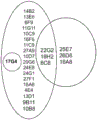
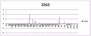
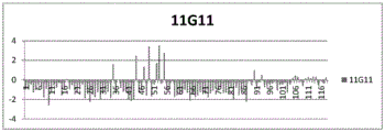
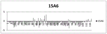
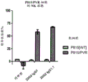

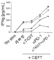
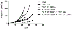
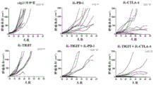
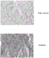
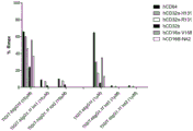
Classifications
-
- C—CHEMISTRY; METALLURGY
- C07—ORGANIC CHEMISTRY
- C07K—PEPTIDES
- C07K16/00—Immunoglobulins [IGs], e.g. monoclonal or polyclonal antibodies
- C07K16/18—Immunoglobulins [IGs], e.g. monoclonal or polyclonal antibodies against material from animals or humans
- C07K16/28—Immunoglobulins [IGs], e.g. monoclonal or polyclonal antibodies against material from animals or humans against receptors, cell surface antigens or cell surface determinants
-
- A—HUMAN NECESSITIES
- A61—MEDICAL OR VETERINARY SCIENCE; HYGIENE
- A61P—SPECIFIC THERAPEUTIC ACTIVITY OF CHEMICAL COMPOUNDS OR MEDICINAL PREPARATIONS
- A61P35/00—Antineoplastic agents
-
- C—CHEMISTRY; METALLURGY
- C07—ORGANIC CHEMISTRY
- C07K—PEPTIDES
- C07K16/00—Immunoglobulins [IGs], e.g. monoclonal or polyclonal antibodies
- C07K16/18—Immunoglobulins [IGs], e.g. monoclonal or polyclonal antibodies against material from animals or humans
- C07K16/28—Immunoglobulins [IGs], e.g. monoclonal or polyclonal antibodies against material from animals or humans against receptors, cell surface antigens or cell surface determinants
- C07K16/2803—Immunoglobulins [IGs], e.g. monoclonal or polyclonal antibodies against material from animals or humans against receptors, cell surface antigens or cell surface determinants against the immunoglobulin superfamily
-
- A—HUMAN NECESSITIES
- A61—MEDICAL OR VETERINARY SCIENCE; HYGIENE
- A61K—PREPARATIONS FOR MEDICAL, DENTAL OR TOILETRY PURPOSES
- A61K39/00—Medicinal preparations containing antigens or antibodies
- A61K2039/505—Medicinal preparations containing antigens or antibodies comprising antibodies
-
- C—CHEMISTRY; METALLURGY
- C07—ORGANIC CHEMISTRY
- C07K—PEPTIDES
- C07K2317/00—Immunoglobulins specific features
- C07K2317/20—Immunoglobulins specific features characterized by taxonomic origin
- C07K2317/21—Immunoglobulins specific features characterized by taxonomic origin from primates, e.g. man
-
- C—CHEMISTRY; METALLURGY
- C07—ORGANIC CHEMISTRY
- C07K—PEPTIDES
- C07K2317/00—Immunoglobulins specific features
- C07K2317/30—Immunoglobulins specific features characterized by aspects of specificity or valency
- C07K2317/31—Immunoglobulins specific features characterized by aspects of specificity or valency multispecific
-
- C—CHEMISTRY; METALLURGY
- C07—ORGANIC CHEMISTRY
- C07K—PEPTIDES
- C07K2317/00—Immunoglobulins specific features
- C07K2317/30—Immunoglobulins specific features characterized by aspects of specificity or valency
- C07K2317/34—Identification of a linear epitope shorter than 20 amino acid residues or of a conformational epitope defined by amino acid residues
-
- C—CHEMISTRY; METALLURGY
- C07—ORGANIC CHEMISTRY
- C07K—PEPTIDES
- C07K2317/00—Immunoglobulins specific features
- C07K2317/70—Immunoglobulins specific features characterized by effect upon binding to a cell or to an antigen
- C07K2317/76—Antagonist effect on antigen, e.g. neutralization or inhibition of binding
-
- C—CHEMISTRY; METALLURGY
- C07—ORGANIC CHEMISTRY
- C07K—PEPTIDES
- C07K2317/00—Immunoglobulins specific features
- C07K2317/90—Immunoglobulins specific features characterized by (pharmaco)kinetic aspects or by stability of the immunoglobulin
- C07K2317/92—Affinity (KD), association rate (Ka), dissociation rate (Kd) or EC50 value
-
- Y—GENERAL TAGGING OF NEW TECHNOLOGICAL DEVELOPMENTS; GENERAL TAGGING OF CROSS-SECTIONAL TECHNOLOGIES SPANNING OVER SEVERAL SECTIONS OF THE IPC; TECHNICAL SUBJECTS COVERED BY FORMER USPC CROSS-REFERENCE ART COLLECTIONS [XRACs] AND DIGESTS
- Y02—TECHNOLOGIES OR APPLICATIONS FOR MITIGATION OR ADAPTATION AGAINST CLIMATE CHANGE
- Y02A—TECHNOLOGIES FOR ADAPTATION TO CLIMATE CHANGE
- Y02A50/00—TECHNOLOGIES FOR ADAPTATION TO CLIMATE CHANGE in human health protection, e.g. against extreme weather
- Y02A50/30—Against vector-borne diseases, e.g. mosquito-borne, fly-borne, tick-borne or waterborne diseases whose impact is exacerbated by climate change
Abstract
The present invention provides antibodies or antigen binding fragments thereof that bind to human TIGIT (T cell immune receptor containing Ig and ITIM domains), and the use of these antibodies or fragments in therapeutic applications, for example in the treatment of cancer or chronic viral infections. Such methods of treatment include combination therapies with inhibitors of other immunomodulatory receptor interactions, such as PD-1/PD-L1 interactions. The invention further provides polynucleotides encoding the heavy and/or light chain variable regions of the antibodies, expression vectors comprising polynucleotides encoding the heavy and/or light chain variable regions of the antibodies, cells comprising the vectors, and methods of making the antibodies or fragments by expressing them from the cells.
Description
The present application is a divisional application of the invention patent with the application number 201580070988.1 and the name "antibody against TIGIT" on the application date 2015, 12, 22.
Background
TIGIT (T cell immune receptor containing Ig and ITIM domains) is a co-inhibitory receptor protein, also known as WUCAM, vstm3 or Vsig9.TIGIT was found in genomic studies of proteins specifically expressed on T cells, and has immunoglobulin variable domains, transmembrane domains, and an immune receptor tyrosine inhibitory motif (ITIM), and tag sequence elements comprising the PVR protein family. It is known to interact with poliovirus receptors (PVR; CD 155) and with nectin2 (CD 112). See, e.g., stengel et al (2012) Proc, nat' l Acad, sci (USA) 19:5399; WO 2006/124667; WO 2009/126688. Although PVR can interact with the co-activated receptor DNAM-1 (CD 226) to enhance tumor killing, high affinity TIGIT/PVR interactions inhibit such killing and can be used to prevent killing of normal (self) cells that also express PVR. Stanietsky et al (2009) Proc, nat' l Acad, sci (USA) 106:17858. The advantage of such inhibitory interactions may be important in inhibiting anti-autoimmune responses, but in the case of tumors it inhibits tumor eradication. As before.
TIGIT inhibits T cell activation by promoting the production of mature immunoregulatory dendritic cells. Yu (Yu)et al.(2009) Nat. Immunol.10:48.TIGIT and other such co-inhibitory molecules (e.g., CTLA-4, PD-1, lag3, and BTLA) can play a role in tumor cell escape immune surveillance. The experiment shows that the preparation method has the advantages of,PVR/CD155 on melanoma cells (Inozumeet al. (2014) J. Invest. Dermatol.134S 121-Abstract 693) and over-expression on various other tumors. It is possible that TIGIT/PVR interactions could protect such tumor cells from immune-mediated eradication by inhibiting the anti-tumor response of T and NK cells. Stanietskyet al. (2009) Proc. Nat’l Acad. Sci. (USA)106:17858 and Lozanoet al. (2012) J. Immunol.188:3869. Other experiments have identified regulatory T cells (T regs ) TIGIT of (2) + A subgroup which selectively inhibits Th1 and Th17 responses (Jolleret al.(2014) Immunity40:569), indicating that anti-TIGIT antibodies may enhance alternative mechanisms of anti-tumor immune responses.
Like other co-inhibitory receptors such as CTLA-4, PD-1 and BTLA, TIGIT can function to "shut down" the immune response. As before. Antibodies targeting CTLA-4 (ipilimumab) and PD-1 (nivolumab, pembrolizumab) have been approved for the treatment of human cancers, confirming this therapeutic pathway. Antibodies that bind to human TIGIT may also be used to treat cancer. See, for example, WO 2006/124667. In a mouse model, antibody blockade of both PD-L1 and TIGIT resulted in synergistic enhancement of CD8 + T cell mediated tumor rejection. Groganet al. (2014) J. Immunol. 192(1) Suppl. 203.15; Johnston et al. (2014) Cancer Cell26:1-15. Similar results were obtained in animal models of melanoma. Inozumeet al. (2014) J. Invest. Dermatol.134S 121-Abstract 693. Some experiments showed that TIGIT blockade was effective in enhancing anti-tumor CD8 only in the presence of the coactivated receptor DNAM-1/CD226 competing with TIGIT for binding to PVR/CD155 + T cell response. Johnstonet al. (2014) Cancer Cell 26:1-15。
Recent experiments have shown that the Fap2 protein expressed by intratumoral bacteria can inhibit NK cell mediated tumor killing by binding TIGIT (Guret al. (2015) Immunity42:344), indicating that clearing such bacteria, blocking TIGIT interaction with Fap2, or blocking TIGIT activity as a whole, may be useful in the treatment of cancer, e.g., colorectal cancer. Hampton (2015)JAMA 313:1305。
There is a need for improved methods of treating cancer and chronic viral infections, and medicaments, such as therapeutic monoclonal antibodies, for use in such methods. Medicaments for use in such improved methods of treatment may comprise antibodies or antibody fragments that specifically bind TIGIT and reverse or partially reverse TIGIT-mediated inhibition of anti-tumor or anti-viral immune responses.
Brief description of the invention
The present invention provides improved medicaments and methods of treatment for cancer and chronic viral infections comprising antibodies or antigen-binding fragments thereof that bind huTIGIT. Provided herein are isolated antibodies, such as monoclonal antibodies, particularly human monoclonal antibodies, that specifically bind huTIGIT and have desirable functional properties, such as high affinity for specifically binding huTIGIT, binding monkey TIGIT (e.g., cynomolgus monkey TIGIT), the ability to block TIGIT binding to PVR and/or Nectin-2, the ability to block TIGIT interaction with DNAM, or any combination of these properties.
The invention further provides improved methods of treating cancer and therapeutic antibodies for use in the methods, including cancers in which TIGIT-mediated signaling inhibits an anti-tumor immune response, tumors in which TIGIT interacts with the coactivated receptor DNAM-1/CD226 to inhibit an anti-tumor immune response, tumors in which regulatory T cells expressing TIGIT inhibit an anti-tumor immune response, or tumors in which TIGIT would otherwise inhibit an anti-tumor immune response. The invention also provides methods and therapeutic antibodies for treating chronic viral infections in which TIGIT inhibits an anti-viral immune response.
In another aspect, the invention relates to antibodies that compete for binding to huTIGIT and/or cross-block binding to huTIGIT with antibodies having the heavy and light chain variable domain sequences disclosed herein.
In certain embodiments, the anti-TIGIT antibodies of the present invention, or antigen-binding fragments thereof, enhance an anti-tumor immune response, e.g., an antigen-specific T cell response. In other embodiments, the anti-TIGIT antibodies or antigen-binding fragments thereof of the invention blockTIGIT-mediated inhibitory signaling, allowing PVR/DNAM to co-stimulate NK cells to increase NK-mediated killing of anti-tumor responses. In yet another embodiment, the anti-TIGIT antibodies or antigen-binding fragments thereof of the present invention reduce the population of regulatory T cells within a tumor that would otherwise inhibit an anti-tumor immune response. In yet another embodiment, an anti-TIGIT antibody of the invention in the form of IgG1 reduces CD8 + Depleted T cells and T regs Allowing inflow of new, unspent CD8 + T cells. In other embodiments, the anti-TIGIT antibodies of the invention or antigen binding fragments thereof function by one or more of the mechanisms described above, as these mechanisms are not necessarily mutually exclusive.
In certain embodiments, the anti-TIGIT antibodies of the invention, or antigen-binding fragments thereof, do not bind to activated fcγ receptors (fcγr), e.g., in embodiments that rely on increasing the anti-tumor activity of TIGIT-expressing cells. In alternative embodiments, the anti-TIGIT antibodies of the invention or antigen-binding fragments thereof bind to one or more activated fcγrs, e.g., in a manner dependent on killing TIGIT-expressing cells, e.g., depleting CD8 + T cells or T regs In an embodiment of (2).
The invention also provides an isolated monoclonal antibody (15 A6) or antigen binding fragment thereof which specifically binds huTIGIT and comprises heavy chain CDRH1, CDRH2 and CDRH3 sequences (comprising SEQ ID NOs: 14, 15 and 16, respectively) and/or light chain CDRL1, CDRL2 and CDRL3 sequences (comprising SEQ ID NOs: 17, 18 and 19, respectively).
The invention also provides an isolated monoclonal antibody (22G 2) or antigen binding fragment thereof which specifically binds huTIGIT and comprises heavy chain CDRH1, CDRH2 and CDRH3 sequences (comprising SEQ ID NOs: 20, 21 and 22, respectively) and/or light chain CDRL1, CDRL2 and CDRL3 sequences (comprising SEQ ID NOs: 23, 24 and 25, respectively).
The invention also provides an isolated monoclonal antibody (11G 11) or antigen binding fragment thereof which specifically binds huTIGIT and comprises heavy chain CDRH1, CDRH2 and CDRH3 sequences (comprising SEQ ID NOs: 26, 27 and 28, respectively) and/or light chain CDRL1, CDRL2 and CDRL3 sequences (comprising SEQ ID NOs: 29, 30 and 31, respectively).
The invention still further provides an isolated monoclonal antibody (10D 7) or antigen binding fragment thereof that specifically binds huTIGIT and comprises heavy chain CDRH1, CDRH2 and CDRH3 sequences (comprising SEQ ID NOs: 32, 33 and 34, respectively) and/or light chain CDRL1, CDRL2 and CDRL3 sequences (comprising SEQ ID NOs: 35, 36 and 37, respectively).
The invention also provides an isolated monoclonal antibody, or antigen-binding fragment thereof, which specifically binds huTIGIT and comprises the sequences set forth in SEQ ID NOs 2 (or 3, 4, 5) and 6; SEQ ID NOS 7 (or 8) and 9; SEQ ID NOS 10 and 11; and the variable heavy and variable light chain sequences disclosed in SEQ ID NOS 12 and 13.
The present invention provides an isolated monoclonal antibody, or antigen-binding fragment thereof, that binds huTIGIT and comprises heavy and light chain variable regions, wherein the heavy chain variable region comprises an amino acid sequence having at least 90%, 95% or 99% identity to an amino acid sequence selected from the group consisting of SEQ ID NOs 2, 3, 4, 5, 7, 8, 10 and 12.
The invention also provides an isolated monoclonal antibody, or antigen-binding fragment thereof, that binds huTIGIT and comprises heavy and light chain variable regions, wherein the light chain variable region comprises an amino acid sequence having at least 90%, 95% or 99% identity to an amino acid sequence selected from the group consisting of SEQ ID NOs 6, 9, 11 and 13.
In certain embodiments, the isolated monoclonal antibodies of the invention, or antigen-binding fragments thereof, (a) bind to the same epitope on huTIGIT as 15A6, 22G2, 11G11, and/or 10D7, and/or (b) inhibit binding of 15A6, 22G2, 11G11, and/or 10D7 to huTIGIT as measured by FACS or ELISA, for example.
In certain embodiments, the anti-huTIGIT antibodies or antigen binding fragments thereof of the invention bind to an epitope comprising or consisting of one or more of residues E60, I109, L65, N70, F107, T117, I68, H76, and N58 of huTIGIT (SEQ ID NO: 1) (antibody 22G 2), an epitope comprising or consisting of one or more of residues G74, N70, H76, L65, L73, Q56, I68, H111, and P114 (antibody 11G 11), or an epitope comprising or consisting of one or more of residues H76, G74, L65, N58, I68, Q139, G135, L73, F107, N70, E60, H134, a132, and I109 (antibody 15 A6).
Alternatively, the anti-huTIGIT antibodies or antigen binding fragments thereof of the invention bind to an epitope comprising or consisting of one or more sequences selected from NWEQQDQLLAICNADLGWH (SEQ ID NO: 38) and FCIYHTYPDGT (SEQ ID NO: 39) (antibody 22G 2), or selected from QVNWEQQDQLLAICNADLGWH (SEQ ID NO: 40) and HTYP (SEQ ID NO: 41) (antibody 11G 11), or selected from NWEQQDQLLAICNADLGWH (SEQ ID NO: 38), FCI and AEHGARFQ (SEQ ID NO: 43) (antibody 15 A6).
In still further embodiments, the anti-TIGIT antibodies or antigen-binding fragments thereof of the invention bind to a core epitope on huTIGIT (SEQ ID NO: 1) comprising or consisting of one or more of residues L65, I68, N70 and H76; and/or an epitope comprising or consisting of LLAICNADLGWH (SEQ ID NO: 44).
In some embodiments, the anti-huTIGIT antibodies or antigen-binding fragments thereof of the invention also bind cynomolgus TIGIT.
In various embodiments, the anti-TIGIT antibodies of the invention or antigen-binding fragments thereof are human IgG1, igG2, igG3, or IgG4 antibodies, or variants thereof. In certain embodiments, including but not limited to, methods of blocking TIGIT signaling in "depleted" tumor-specific T cells or blocking inhibitory signals on NK cells that allow DNAM-1/PVR-mediated co-stimulation, or methods of blocking TIGIT interaction with DNAM-1/CD226 to reduce DNAM-1 homodimerization, an anti-TIGIT antibody or antigen-binding fragment thereof comprises a null or nearly null Fc. Such Fc regions include, for example, human IgG2 or IgG4, or null variants of human IgG1, having one or more of the following mutations: L234A, L235E, G237A, A330S and P331S (EU numbering) comprising IgG1.1f (SEQ ID NO: 48) containing all 5 of the mutations listed.
In alternative embodiments, including but not limited to depleting TIGIT + Methods of modulating T cells, anti-TIGIT antibodies or antigen-binding fragments thereof comprise an Fc that preferentially binds to activated fcγr (fcγri, fcγriia, or fcγriiia), e.g., human IgG1, or a sequence variant that has enhanced binding to activated fcγr relative to wild-type IgG1 Fc. In relation to the use of the anti-TIGIT antibodies of the invention in the form of IgG1 to drive T reg In reduced s embodiments, intratumoral injection may optionally be used to administerThe localization to the tumor microenvironment minimizes potential side effects caused by activity in surrounding tissues.
In certain embodiments, a methionine residue in the CDR regions of an anti-TIGIT antibody of the invention (e.g., M115 in CDRH3 of SEQ ID NO: 34, 10D 7) or an antigen-binding fragment thereof is replaced with an amino acid residue that does not undergo oxidation.
In certain embodiments, the anti-huTIGIT antibody that competes for binding, cross-blocking, or binding to the same epitope as 15A6, 22G2, 11G11, or 10D7, or an antigen-binding fragment thereof, is a human or humanized antibody.
In some embodiments, the anti-huTIGIT antibodies of the invention are not antibodies described in U.S. patent application publication No. 2009/0258013, or do not bind the same epitope as the antibodies described in U.S. patent application publication No. 2009/0258013, e.g., they do not bind the same epitope as anti-huTIGIT mAb 10A7 or 1F 4. See also Johnston et al. (2014) Cancer Cell 26:1; Yu et al. (2009) Nat. Immunol. 10:48。
In other embodiments, the anti-huTIGIT antibodies comprise variable domains derived from the same human V domain germline sequences as the antibodies disclosed herein, including heavy chain V domains V4-39, V4-61, or V1-69. In a more specific embodiment, the anti-huTIGIT antibody comprises heavy and light chain variable domains derived from the same human heavy and light chain V domain germline sequences as the antibodies disclosed herein, such as V4-39/VA27 (15 A6), V4-61/VL6 (22G 2), V4-39/VL6 (11G 11), and V1-69/VL15 (10D 7).
In various embodiments, the anti-huTIGIT antibodies of the invention are raised against a K of less than 10nM, 5nM, 2nM, 1nM, 300pM or 100pM D Bind huTIGIT. In other embodiments, the anti-huTIGIT antibodies of the invention are administered at a K of 2nM to 100pM D Bind huTIGIT.
In other embodiments, the anti-huTIGIT antibodies of the invention consist essentially of, or comprise, some combination of CDRs of antibodies 15A6, 22G2, 11G11 and 10D7, e.g., CDRH1 (SEQ ID NOs: 14, 20, 26 and 32), CDRH2 (SEQ ID NOs: 15, 21, 27 and 34), CDRH3 (SEQ ID NOs: 16, 22, 28 and 34), CDRL1 (SEQ ID NOs: 17, 23, 29 and 35), CDRL2 (SEQ ID NOs: 18, 24, 30 and 36); and CDRL3 (SEQ ID NOS: 19, 25, 31 and 37). In other embodiments, the antibody consists essentially of, or comprises, a single specific combination of CDR sequences of antibodies 15A6, 22G2, 11G11 and 10D 7.
In a further embodiment, the anti-huTIGIT antibodies of the invention consist essentially of, or comprise, sequences of at least 80%, 85%, 90% and 95% sequence identity to the heavy and/or light chain variable domains of antibodies 15A6 (SEQ ID NOs: 2-5 and 6), 22G2 (SEQ ID NOs: 7-8 and 9), 11G11 (SEQ ID NOs: 10 and 11) and 10D7 (SEQ ID NOs: 12 and 13).
In still further embodiments, the anti-huTIGIT antibodies of the invention consist essentially of, or comprise, a heavy and/or light chain comprising the variable domain sequences of antibodies 15A6 (SEQ ID NOs: 2-5 and 6), 22G2 (SEQ ID NOs: 7-8 and 9), 11G11 (SEQ ID NOs: 10 and 11) and 10D7 (SEQ ID NOs: 12 and 13), or sequences sharing at least 80%, 85%, 90% and 95% sequence identity with these disclosed sequences.
In other embodiments, the antigen binding domain of the antibodies of the invention is present as a bispecific molecule that further comprises an antigen binding domain that specifically binds a different immunomodulatory receptor, including but not limited to PD-1, CTLA-4, or LAG 3.
The invention also provides nucleic acids encoding the heavy and/or light chain variable regions of the anti-huTIGIT antibodies or antigen binding fragments thereof of the invention, expression vectors comprising the nucleic acid molecules, cells transformed with the expression vectors, and methods of producing antibodies by expressing the cells transformed with the expression vectors and recovering the antibodies.
The invention also provides immunoconjugates comprising the anti-huTIGIT antibodies described herein linked to an agent, such as a detectable label or a cytotoxic agent.
The invention also provides pharmaceutical compositions comprising an anti-huTIGIT antibody of the invention or an antigen binding fragment thereof and a carrier. Also provided herein are kits comprising an anti-TIGIT antibody or antigen-binding fragment thereof and instructions for use.
In another aspect, the invention provides a method of enhancing an antigen-specific T cell response comprising contacting a T cell with an anti-huTIGIT antibody of the invention or an antigen binding fragment thereof such that the antigen-specific T cell response is enhanced, e.g., by reducing inhibitory signals that would otherwise inhibit the anti-tumor response. In some embodiments, the antigen-specific T cell is a tumor-antigen specific effector T cell, e.g., CD8 + T cells, and the enhancement, e.g., by blocking TIGIT-mediated inhibitory effects, lead to increased anti-tumor activity. The anti-huTIGIT antibodies or antigen-binding fragments thereof of the invention may also reduce inhibitory signals in NK cells, thus increasing their anti-tumor activity. Without intending to be limited by theory, the anti-huTIGIT antibodies of the present invention increase effector T cell or NK cell function by blocking TIGIT binding to PVR, thereby reducing or eliminating inhibitory signals that would otherwise be transmitted to the cell. Alternatively, or in addition, the anti-TIGIT antibodies of the present invention, or antigen-binding fragments thereof, may inhibit the interaction between TIGIT and DNAM-1/CD226, which would otherwise reduce DNAM-1-mediated immune activation.
The invention also provides a method of increasing IL-2 and/or IFN- γ production in a T cell, and/or proliferation of a T cell, comprising contacting the T cell with an effective amount of an anti-TIGIT antibody or antigen-binding fragment thereof.
In another aspect, the invention provides methods of reducing or depleting T in a tumor in a subject in need thereof regs Comprising administering an effective amount of an anti-huTIGIT antibody of the invention, wherein the antibody has effector function or enhanced effector function to reduce T in a tumor regs Is a number of (3).
The invention provides a method of enhancing an immune response in a subject comprising administering to the subject an effective amount of an anti-huTIGIT antibody of the invention or antigen binding fragment thereof such that the immune response of the subject is enhanced. In certain embodiments, the subject has a tumor, and the immune response against the tumor is enhanced. In another embodiment, the subject has a viral infection, and the anti-viral immune response is enhanced.
The invention also provides a method of inhibiting tumor growth in a subject comprising administering to the subject an anti-huTIGIT antibody of the invention or an antigen binding fragment thereof such that tumor growth is inhibited.
The invention also provides methods of treating cancer, e.g., by immunotherapy, comprising administering to a subject in need thereof a therapeutically effective amount of an anti-huTIGIT antibody of the invention or antigen-binding fragment thereof, e.g., as a pharmaceutical composition, thereby treating cancer. In certain embodiments, the cancer is bladder cancer, breast cancer, uterine/cervical cancer, ovarian cancer, prostate cancer, testicular cancer, esophageal cancer, gastrointestinal cancer, pancreatic cancer, colorectal cancer, colon cancer, renal cancer, head and neck cancer, lung cancer, gastric cancer, germ cell cancer, bone cancer, liver cancer, thyroid cancer, skin cancer, tumors of the central nervous system, lymphomas, leukemias, myelomas, sarcomas, and virus-related cancers. In certain embodiments, the cancer is a metastatic cancer, a refractory cancer, or a recurrent cancer.
In certain embodiments, the methods and therapeutic methods of modulating immune function described herein comprise administering an anti-huTIGIT antibody of the invention in combination with or as a bispecific agent containing one or more additional therapeutic agents, e.g., an anti-PD-1 antibody, an anti-PD-L1 antibody, an anti-LAG 3 antibody, an anti-GITR antibody, an anti-OX 40 antibody, an anti-CD 73 antibody, an anti-CD 40 antibody, an anti-CD 137 mAb, an anti-CD 27 mAb, an anti-CSF-1R antibody, and/or an anti-CTLA-4 antibody, a TLR agonist, or a small molecule antagonist of IDO or tgfβ. In particular embodiments, the anti-huTIGIT therapy is combined with anti-PD-1 and/or anti-PD-L1 therapy, e.g., with a treatment with an antibody or antigen-binding fragment thereof that binds human PD-1 or an antibody or antigen-binding fragment thereof that binds human PD-L1.
In some embodiments, samples from the patient, e.g., biopsied samples, are screened for DNAM-1 expression on T cells or NK cells to select patients most likely to respond to anti-TIGIT therapy, wherein the presence of DNAM-1 on T cells indicates that the patient will have a beneficial anti-tumor response when treated with anti-TIGIT therapy, e.g., an anti-huTIGIT antibody or fragment of the invention, and the absence of DNAM-1 identifies patients unlikely to benefit from anti-TIGIT therapy. In other embodiments, PVR and/or Nectin-2 expression on tumor cells or tumor infiltrating bone marrow cells in a sample from a patient is screened to select a patient most likely to respond to anti-TIGIT therapy, wherein the presence of PVR and/or Nectin-2 indicates that the patient will have a beneficial anti-tumor response when treated with anti-TIGIT therapy, e.g., an anti-huTIGIT antibody or fragment of the invention, and the absence of PVR and/or Nectin-2/CD112 identifies a patient that is unlikely to benefit from anti-TIGIT therapy. In various embodiments, cell-surface expression of TIGIT, DNAM, PVR and/or Nectin-2 is determined by FACS, IHC or LC-MS. In another aspect, the invention provides a method of treating a subject in need thereof, comprising determining TIGIT, DNAM, PVR and/or cell-surface expression of Nectin-2 as described herein, and preferentially or exclusively administering to those subjects most likely to obtain a therapeutic benefit an anti-TIGIT antibody of the invention.
In one embodiment, the level of soluble PVR and/or soluble Nectin-2 (sPVR, sNectin-2) is measured in a subject contemplated for treatment with an anti-TIGIT antibody of the invention, and only subjects exhibiting an increase in soluble PVR and/or Nectin-2 are treated with the antibody. In some embodiments, sPVR and/or sNectin-2 is detected in serum by ELISA or LC-MS.
The invention also provides methods of detecting the presence of TIGIT in a sample, on a cell within a sample (e.g., FACS) or at a specific location of a cell or tissue (e.g., IHC), or methods of sorting cells based on the presence or absence of TIGIT on the surface of a cell (e.g., FACS), comprising contacting a sample with an anti-huTIGIT antibody of the invention or an antigen-binding fragment thereof under conditions that allow for the formation of a complex between the antibody or antigen-binding fragment thereof and TIGIT, and detecting the formation of the complex. In some embodiments, the anti-TIGIT antibody used for detection is conjugated to a detectable label.
Other features and advantages of the present disclosure will be apparent from the following detailed description and examples (which should not be taken as limiting).
Brief Description of Drawings
FIG. 1 shows a schematic of a "frame-and-parallel (binding)" experiment in which pairs were tested for the ability of various anti-huTIGIT antibodies of the invention to block the binding of other antibodies to huTIGIT. The results indicate that antibodies fall into a limited number of categories or "boxes". See example 3.
FIGS. 2A, 2B and 2C show yeast display data for huTIGIT sequence variants binding to antibodies 22G2, 11G11 and 15A6, respectively. The residue numbers of the individual amino acid residues in the mature huTIGIT are provided along the abscissa. The residue number is 21 less than the number of SEQ ID NO. 1, since the sequence Listing includes signal peptides not included in the figure. As described in example 4, yeasts displaying sequence variants of huTIGIT were selected based on their inability to bind to the respective antibodies (22G 2, 11G11, 15 A6). Thus, positions along the huTIGIT sequence that are critical for antibody binding occur at high frequency (i.e., bars/lines rising on the ordinate) because of their over-expression in the pool of non-binding yeast clones. The frequency of occurrence of variant (non-wild type) residues at each residue is provided on the ordinate (logarithmic scale), one bar (line) per residue. Frequency data was normalized to the frequency of occurrence of variant residues at each position in the unselected library (i.e., the library that had not been selected based on being unable to bind the anti-huTIGIT antibodies of the present invention). See example 4.
FIG. 3 shows the effect of anti-TIGIT mAb 22G2 on lysis, expressed as a percentage of cells expressing human PVR that were specifically lysed by human NK cells. See example 5. For each antibody, the left bar is wild-type P815 cells and the right bar is P815 cells expressing human PVR.
Figure 4A shows that treatment of healthy human donor blood with a mixture of antigenic peptides (CETF = peptides from CMV, EBV, influenza and tetanus) induces CD8 + Upregulation of PD-1 and TIGIT on T cells. The "No Stim" sample was not treated with CEFT, whereas the "Stim" sample was treated with CEFT. FIG. 4B shows the effect of anti-TIGIT mAb and/or anti-PD-1 mAb on IFN gamma expression from 4 healthy human donor blood samples stimulated with CETF. See example 6.
Figures 5A and 5B show the effect of anti-TIGIT antibodies on tumor growth alone or in combination with other immunomodulatory therapies in a mouse model. Fig. 5A shows tumor volume (cubic millimeters) of a CT26 mouse colon cancer model of mice treated with the following antibodies, calculated by multiplying the square of tumor width times half the length: an anti-mouse TIGIT antibody with an effector function activated murine IgG2a Fc domain ("TIGIT G2 a"), an anti-mouse TIGIT antibody with an effector function deficient IgG 1D 265A Fc domain ("TIGIT G1D 265A"), an anti-mouse PD-1 antibody with an effector function deficient IgG 1D 265A Fc domain ("PD-1 G1D 265A"), a combination thereof, or a control IgG1 antibody. FIG. 5B shows the effect of anti-TIGIT monotherapy and combination therapy with anti-PD-1 and anti-CTLA-4 antibodies. Tumor volume and number of tumor-free (TF) mice in each group of 10 mice at the end of the experiment are provided. Each line represents one mouse. No tumor-free mice were obtained with the mIgG1 isotype control, as well as anti-TIGIT as monotherapy. anti-PD-1 was obtained as monotherapy in one tumor-free mouse, and when combined with anti-TIGIT, five were obtained. anti-CTLA-4 was obtained as monotherapy in three tumor-free mice and when combined with anti-TIGIT in six. See example 7.
FIGS. 6A and 6B show increased PVR expression in cancer tissues. Figure 6A shows PVR mRNA expression in each tumor type, as detected in the The Cancer Genome Atlas (TCGA) dataset. Data are provided for adrenocortical carcinoma (ACC), undyed renal cell carcinoma (KICH), liver hepatocellular carcinoma (LIHC), colon and rectal adenocarcinomas (COAD, READ), pancreatic ductal adenocarcinomas (PAAD), pheochromocytoma and gangliocytoma (PCPG), papillary renal carcinomas (KIRP), lung adenocarcinomas (LUAD), head and neck squamous cell carcinoma (HNSC), thyroid adenocarcinomas (PRAD), endometrial carcinoma (UCEC), cervical carcinoma (CESC), cutaneous melanoma (SKCM), mesothelioma (MESO), urothelial bladder carcinoma (BLCA), clear cell renal carcinoma (KIRC), lung squamous cell carcinoma (lucc), uterine Carcinoma Sarcoma (UCS), sarcoma (SARC), ovarian serous cystic adenocarcinoma (OV), papillary thyroid carcinoma (THCA), glioblastoma multiforme (GBM), breast cancer (BRCA), light glioma (LGG) and diffuse large B-cell lymphoma (DLBC). The results disclosed herein are based in whole or in part on the data generated by TCGA Research Network. Fig. 6B shows human PVR in colon adenocarcinoma tissue compared to normal colon epithelial tissue, darker areas in the adenocarcinoma sample indicating increased PVR expression. See example 9.
FIG. 7 shows Fcγ receptor binding of anti-TIGIT mAb 22G2 in the form of IgG1f (SEQ ID NO: 45) or IgG1.1f (SEQ ID NO: 48), expressed as the theoretical maximum receptor binding value (R max ) Is a percentage of (c). Data for 6 different fcγ receptors were provided for two different batches of igg1.1f antibody (used at 10 μm and 1 μm as indicated). In each column group, fcγ receptor data are provided in left to right order: hCD64 (FcgammaRI), hCD32a-H131 (FcgammaRIIA-H131), hCD32a-R131 (FcgammaRIIA-R131), hCD32b (FcgammaRIIB), hCD16a-V158 (FcgammaRIIIA-V158), and hCD16b-NA2 (FcgammaRIIIB-NA 2, wherein NA2 is expressed as an allotype variant). Although fcγ receptor pairs are represented in the same bars, their identity is clear from the order in which they are provided. The same decrease was observed for binding to cynomolgus monkey fcγ receptors CD64, CD32a, CD32b and CD16 (not shown).
Detailed Description
Isolated antibodies, particularly monoclonal antibodies, e.g., human monoclonal antibodies, that specifically bind to human TIGIT ("huTIGIT") and block binding to PVR/CD155, thereby reducing or eliminating immunosuppressive signals that would otherwise occur in TIGIT-expressing cells. The invention also provides isolated antibodies, particularly monoclonal antibodies, e.g., human or humanized monoclonal antibodies, that specifically bind to human TIGIT and block interactions of DNAM-1/CD226 with human TIGIT that would otherwise prevent DNAM-1 homodimerization and thus DNAM-1-mediated co-stimulation. Various sequences of human anti-huTIGIT monoclonal antibodies are provided. In certain embodiments, the antibodies described herein are derived from specific heavy and light chain germline sequences and/or comprise specific structural features such as CDR regions comprising specific amino acid sequences.
Also provided herein are methods of making such antibodies, immunoconjugates and bispecific molecules comprising such antibodies or antigen-binding fragments thereof, and pharmaceutical compositions formulated to comprise the antibodies or fragments. Also provided herein are methods of enhancing an immune response using the antibodies alone or in combination with other immunostimulants (e.g., antibodies) and/or cancer or anti-infective therapies. Thus, the anti-huTIGIT antibodies described herein may be used in therapy in a variety of therapeutic applications, including, for example, inhibiting tumor growth and treating chronic viral infections.
Definition of the definition
In order that the present description may be more readily understood, certain terms are first defined. Other definitions are set forth throughout the detailed description.
TIGIT is known as "Ig and ITIM domain-containing T cell immunoreceptor", which is a member of the PVR (poliovirus receptor) family of immunoglobulins that bind PVR/CD155 and Nectin-2/CD112.TIGIT is also known as TIGIT, WUCAM, vstm3 and Vsig9. Unless otherwise indicated, or clear from context, references herein to TIGIT refer to human TIGIT ("huTIGIT"), and anti-TIGIT antibodies refer to anti-human TIGIT antibodies. Human TIGIT is also known as GENE ID NO: 201633 and MIM (Mendelian Inheritance in Man): 612859. The sequence of human TIGIT (np_ 776160.2) comprising a 21 amino acid signal sequence is provided in SEQ ID No. 1. Unless otherwise indicated, or clear from the context, "inhibition" of TIGIT refers to blocking PVR binding and signaling. The anti-TIGIT antibodies of the invention may function by inhibiting TIGIT signaling, blocking TIGIT/DNAM-1 interactions, and/or other mechanisms, e.g., directing the reduction of regulatory T cells.
PVR (poliovirus receptor) interacts with TIGIT to produce an immunosuppressive signal. PVR is also known as PVS, HVED, CD155, NECL5, TAGE4, necl-5. Unless otherwise indicated, or clear from the context, reference herein to PVR/CD155 refers to a human PVR ("huPVR"). Human PVR is also known as GENE ID NO: 5817 and MIM: 173850. There are four known variants of human PVR transcripts: alpha (NP 006496.4), beta (NP 001129240.1), gamma (NP 001129241.1) and delta (NP 001129242.2), the sequences of which are provided in SEQ ID NO 50-53. Unless otherwise indicated, reference to PVR or human PVR relates to an alpha transcript polypeptide.
The term "antibody" as used herein may include whole antibodies and any antigen binding thereof, unless otherwise indicated or clear from the contextA synthetic fragment (i.e., an "antigen-binding portion") or a single strand. In one embodiment, an "antibody" refers to a glycoprotein comprising at least two heavy (H) chains and two light (L) chains, or antigen-binding fragments thereof, interconnected by disulfide bonds. Each heavy chain comprises a heavy chain variable region (abbreviated herein as V H ) And a heavy chain constant region. In certain naturally occurring IgG, igD, and IgA antibodies, the heavy chain constant region comprises three domains, CH1, CH2, and CH3. In certain naturally occurring antibodies, each light chain comprises a light chain variable region (abbreviated herein as V L ) And a light chain constant region. The light chain constant region comprises one domain, CL. V (V) H And V L The regions may be further subdivided into regions of hypervariability, termed Complementarity Determining Regions (CDRs), alternating with regions of greater conservation termed Framework Regions (FR). Each V is H And V L Comprising three CDRs and four Framework Regions (FR), arranged from amino-terminus to carboxy-terminus in the following order: FR1, CDR1, FR2, CDR2, FR3, CDR3, FR4. The variable regions of the heavy and light chains comprise binding domains that interact with antigens. The constant region of an antibody may mediate the binding of an immunoglobulin to host tissues or factors, including various cells of the immune system (e.g., effector cells) and the first component of the classical complement system (Clq).
Antibodies typically bind their associated antigen specifically with high affinity, reflected in 10 -7 -10 -11 M or less dissociation constant (K D ). Greater than about 10 -6 Any K of M D Are generally considered to indicate non-specific binding. As used herein, an antibody that "specifically binds" an antigen refers to an antibody that binds the antigen and substantially the same antigen with high affinity, meaning having 10 -7 M or less, preferably 10 -8 M or less, even more preferably 5 x 10 -9 M or less and most preferably 10 -8 M-10 -10 M or less K D But does not bind with high affinity to unrelated antigens. An antigen is given if it exhibits a high degree of sequence identity to the given antigen, e.g. if it exhibits at least 80%, at least 90%, preferably at least 95%, more preferably at least 97% or even more preferably at least 99% sequence identity to the sequence of the given antigen The defined antigens are "substantially identical". For example, antibodies that specifically bind human TIGIT may also cross-react with TIGIT from certain non-human primate species (e.g., cynomolgus monkey), but may not cross-react with TIGIT from other species, or with antigens other than TIGIT.
Antibodies may exhibit modification of the N-and/or C-terminal amino acid residues. For example, antibodies of the invention may be produced from constructs encoding, for example, C-terminal lysine residues on the heavy chain, but such C-terminal lysines may be partially or fully deleted in therapeutic antibodies that are sold or administered. Alternatively, antibodies may be produced from constructs that specifically do not encode a C-terminal lysine residue, even if such lysine is present in the parent antibody from which the therapeutic antibody is derived. In another example, the N-terminal glutamine or glutamic acid residue in an antibody of the invention can be partially or fully converted to pyroglutamic acid in a therapeutic antibody that is sold or administered. Any form of glutamine or glutamic acid, including pyroglutamic acid, present at the N-terminus of an antibody chain is included within the term "glutamine" as used herein. Thus, the antibody chain sequences provided herein having N-terminal glutamine or glutamic acid residues include antibody chains that do not take into account the level of pyroglutamic acid formation.
Unless otherwise indicated, the immunoglobulin may be from any commonly known isotype, including but not limited to IgA, secretory IgA, igG, and IgM. IgG isotypes fall into subclasses in certain species: igG1, igG2, igG3 and IgG4 in humans, and IgG1, igG2a, igG2b and IgG3 in mice. Immunoglobulins, e.g. human IgG1, exist as several allotypes, which differ from each other in at most several amino acids. Unless otherwise indicated, "antibody" may include, for example, monoclonal and polyclonal antibodies; chimeric and humanized antibodies; human and non-human antibodies; fully synthesizing an antibody; and single chain antibodies.
The term "antigen-binding portion" or "antigen-binding fragment" of an antibody, as used herein, refers to one or more fragments of an antibody that retain the ability to specifically bind an antigen (e.g., human TIGIT). Examples of binding fragments included within the term "antigen-binding portion/fragment" of an antibody include (i) Fab fragments-by V L , V H Monovalent fragments consisting of CL and CH1 domains; (ii) F (ab') 2 fragment-a bivalent fragment comprising two Fab fragments linked at the hinge region by a disulfide bridge; (iii) From V H And a CH1 domain; (iv) From V of a single arm of an antibody L And V H Fv fragment consisting of domain, and (V) consisting of V H dAb fragment (Ward)et al., (1989) Nature341:544-546). An isolated Complementarity Determining Region (CDR), or a combination of two or more isolated CDRs joined by a synthetic linker, may comprise an antigen binding domain of an antibody if capable of binding an antigen.
Single chain antibody constructs are also included in the invention. Although the two domains of the Fv fragment V L And V H Encoded by separate genes, but they can be joined by synthetic linkers using recombinant methods, enabling them to be prepared as a single protein chain, where V L And V H Regions pair to form monovalent molecules known as single chain Fv (scFv); see, for example,Bird et al. (1988) Science242:423-426, and Hustonet al. (1988) Proc. Natl. Acad. Sci. USA85:5879-5883). Such single chain antibodies are also intended to be encompassed within the term "antigen-binding portion/fragment" of an antibody. These and other possible constructs are described in Chan& Carter (2010) Nat. Rev. Immunol.10:301. These antibody fragments are obtained using conventional techniques known to those skilled in the art and the efficacy of the fragments are screened in the same manner as for intact antibodies. The antigen-binding portion/fragment may be produced by recombinant DNA techniques, or by enzymatic or chemical cleavage of the intact immunoglobulin.
Unless otherwise indicated, the word "fragment" when used in reference to an antibody, for example in the claims, refers to an antigen-binding fragment of an antibody such that "antibody or fragment" has the same meaning as "antibody or antigen-binding fragment thereof.
A "bispecific" or "bifunctional antibody" is an artificial hybrid antibody having two different heavy/light chain pairs, resulting in two antigen binding sites with specificity for different antigens. Bispecific antibody may be used as a drugGenerated by various methods, including fusion of hybridomas or ligation of Fab' fragments. See, e.g., songsivilai& Lachmann (1990) Clin. Exp. Immunol. 79:315; Kostelny et al. (1992) J. Immunol. 148:1547。
The term "monoclonal antibody" as used herein refers to an antibody that exhibits a single binding specificity and affinity for a particular epitope, or an antibody composition in which all antibodies exhibit a single binding specificity and affinity for a particular epitope. Typically, such monoclonal antibodies are derived from a single cell or nucleic acid encoding the antibody, and are passaged without deliberate introduction of any sequence alterations. Thus, the term "human monoclonal antibody" refers to a monoclonal antibody having variable and optionally constant regions derived from human germline immunoglobulin sequences. In one embodiment, the human monoclonal antibody is produced by a hybridoma, e.g., obtained by fusing B cells obtained from a transgenic or transchromosomal non-human animal (e.g., a transgenic mouse having a genome comprising a human heavy chain transgene and a light chain transgene) to immortalized cells.
The term "recombinant human antibody" as used herein includes all human antibodies produced, expressed, produced, or isolated by recombinant means, such as (a) antibodies isolated from a transgenic or transchromosomal animal (e.g., mouse) of a human immunoglobulin gene or from a hybridoma produced therefrom, (b) antibodies isolated from a host cell transformed to express the antibodies, e.g., a transfectoma, (c) antibodies isolated from a recombinant combinatorial human antibody library, and (d) antibodies produced, expressed, produced, or isolated by any other means involving splicing of a human immunoglobulin gene sequence to other DNA sequences. Such recombinant human antibodies comprise variable and constant regions that utilize specific human germline immunoglobulin sequences encoded by germline genes, but include subsequent rearrangements and mutations that occur, for example, during antibody maturation. As is known in the art (see, e.g., lonberg (2005)NatureBiotech23 (9): 1117-1125), the variable region comprises an antigen binding domain, which is encoded by various genes that are rearranged to form antibodies specific for foreign antigens. In addition to rearrangement, the variable regions may be further defined asA number of single amino acid changes are modified (known as somatic mutations or hypermutations) to increase the affinity of antibodies for foreign antigens. The constant region will change in further response to the antigen (i.e., isotype switching). Thus, the nucleic acid sequences encoding the light and heavy chain immunoglobulin polypeptides that rearrange and somatic mutations in response to an antigen may be different from the original germline sequence, but are substantially identical or similar (i.e., have at least 80% identity).
"human" antibody (HuMAb) refers to an antibody having variable regions, wherein both framework and CDR regions are derived from human germline immunoglobulin sequences. Furthermore, if the antibody comprises constant regions, the constant regions are also derived from human germline immunoglobulin sequences. The human antibodies of the invention may include amino acid residues that are not encoded by human germline immunoglobulin sequences (e.g., mutations introduced by random or site-directed mutagenesis in vitro or by somatic mutation in vivo). However, the term "human antibody" as used herein is not intended to include antibodies in which CDR sequences derived from the germline of another mammalian species, such as a mouse, have been grafted to human framework sequences. The terms "human" antibody and "fully human" antibody are used synonymously.
"humanized" antibodies refer to antibodies in which some, most, or all of the amino acids outside the CDR domains of a non-human antibody, e.g., a mouse antibody, are replaced with the corresponding amino acids derived from a human immunoglobulin. In one embodiment of the humanized form of the antibody, some, most or all of the amino acids outside the CDR domains are replaced with amino acids from a human immunoglobulin, while some, most or all of the amino acids within one or more CDR regions are unchanged. Small additions, deletions, insertions, substitutions or modifications of amino acids are permissible provided they do not abrogate the ability of the antibody to bind to a particular antigen. "humanized" antibodies retain antigen specificity similar to the original antibody.
"chimeric antibody" refers to an antibody in which the variable regions are derived from one species and the constant regions are derived from another species, e.g., an antibody in which the variable regions are derived from a mouse antibody and the constant regions are derived from a human antibody. "hybrid" antibodies refer to antibodies having different types of heavy and light chains, e.g., a mouse (parent) heavy chain and a humanized light chain, or vice versa.
As used herein, "isotype" refers to the type of antibody (e.g., igG1, igG2, igG3, igG4, igM, igA1, igA2, igD, and IgE antibodies) encoded by the heavy chain constant region gene.
"allotype" refers to a naturally occurring variant within a specific group of isoforms that differ in one or several amino acids. See, e.g., jefferiset al. (2009) mAbs 1:1。
The phrases "antibody that recognizes an antigen" and "antibody specific for an antigen" are used interchangeably herein with the term "antibody that specifically binds an antigen".
As used herein, "isolated antibody" refers to an antibody that is substantially free of other antibodies having different antigen specificities (e.g., an isolated antibody that specifically binds TIGIT is substantially free of antibodies that specifically bind to antigens other than TIGIT). However, an isolated antibody that specifically binds an epitope of human TIGIT may have cross-reactivity with other TIGIT proteins from different species.
As used herein, an antibody that "inhibits PVR binding to TIGIT" refers to an antibody that inhibits PVR binding to a TIGIT in a art-recognized method, e.g., in a FACS-based cell-binding assay, at about 1 μg/mL or less, e.g., about 0.9 μg/mL or less, about 0.85 μg/mL or less, about 0.8 μg/mL or less, about 0.75 μg/mL or less, about 0.7 μg/mL or less, about 0.65 μg/mL or less, about 0.6 μg/mL or less, about 0.55 μg/mL or less, about 0.5 μg/mL or less, about 0.45 μg/mL or less, about 0.4 μg/mL or less, about 0.35 μg/mL or less, about 0.3 μg/mL or less, about 0.25 μg/mL or less, about 0.6 μg/mL or less, about 0.45 μg/mL or less, about 50 g or less, about 50 g/mL of the antibody to the TIGIT.
"effector functions" derived from the interaction of an antibody Fc region with certain Fc receptors, including, but not necessarily limited to, clq binding, complement Dependent Cytotoxicity (CDC), fc receptor binding, fcγr-mediated effector functions such as ADCC and antibody dependent cell-mediated phagocytosis (ADCP), and down-regulation of cell surface receptors (e.g., B cell receptors; BCR). Such effector functions typically require the Fc region in combination with an antigen binding domain (e.g., an antibody variable domain).
An "Fc receptor" or "FcR" is a receptor that binds the Fc region of an immunoglobulin. FcR binding IgG antibodies comprise receptors of the fcγr family, including allelic variants and alternatively spliced forms of these receptors. The fcγr family consists of three activating receptors (fcγri, fcγriii and fcγriv in mice; fcγria, fcγriia and fcγriiia in humans) and one inhibitory receptor (fcγriib or equivalent). Various properties of human fcγr are summarized in table 1. Most innate effector cell types co-express one or more activated fcγrs and inhibit fcγriib, whereas Natural Killer (NK) cells selectively express one activated Fc receptor (fcγriii in mice and fcγriiia in humans), but not inhibit fcγriib in mice and humans. Human IgG1 binds to most human Fc receptors and is considered equivalent to murine IgG2a in terms of the type of activated Fc receptor to which it binds.
TABLE 1
Properties of human FcγR

"Fc region" (fragment crystallizable region) or "Fc domain" or "Fc" refers to the C-terminal region of the heavy chain of an antibody that mediates binding of immunoglobulins to host tissues or factors, including binding to Fc receptors located on various cells of the immune system (e.g., effector cells) or binding to the first component (C1 q) of the classical complement system. Accordingly, the Fc region comprises the constant region of the antibody, excluding the first constant region immunoglobulin domain (e.g., CH1 or CL). In IgG, igA and IgD antibody isotypes, the Fc region comprises the C of each of the two heavy chains of the antibody H2 And C H3 A constant domain; igM and IgE Fc regions comprise three heavy chain constant domains (C H Domains 2-4). For IgG, the Fc region comprises immunoglobulin domains cγ2 and cγ3 and a hinge between cγ1 and cγ2. Although the boundaries of the Fc region of an immunoglobulin heavy chain may vary, a human IgG heavy chain Fc region is generally defined as the amino acid residue from position C226 or P230(or an amino acid between these two amino acids) to the carboxy-terminal end of the heavy chain, wherein numbering is according to the EU index, e.g.Kabat.Kabatet al. (1991) Sequences of Proteins of Immunological Interest,National Institutes of Health Bethesda, MD; see also FIGS. 3c-3f of U.S. patent application publication No. 2008/024802428. C of human IgG Fc region H2 The domain extends from about amino acid 231 to about amino acid 340, while C H3 The domain is located in C in the Fc region H2 The C-terminal side of the domain, i.e., it extends from about amino acid 341 to about amino acid 447 (including the C-terminal lysine) of the IgG. As used herein, an Fc region may be a native sequence Fc, including any allotypic variant, or variant Fc (e.g., a non-naturally occurring Fc). Fc may also refer to this region of isolation, or in the case of a protein polypeptide comprising Fc, for example a "binding protein comprising an Fc region", also referred to as an "Fc fusion protein" (e.g., an antibody or immunoadhesin).
Unless otherwise indicated, or clear from the context, the amino acid residue numbering of the Fc region of an antibody is according to the EU numbering convention except when residues of sequences in the sequence listing are explicitly mentioned, wherein the letter numbering is necessarily continuous. For example, literature on the effect of amino acid substitutions in the Fc region generally uses EU numbering, which allows reference to any given residue in the Fc region of an antibody by the same number, regardless of the length of the variable domain to which it is attached. In rare cases, reference may be required to documents cited to confirm the exact Fc residues mentioned.
"native sequence Fc region" or "native sequence Fc" comprises an amino acid sequence which is identical to the amino acid sequence of an Fc region found in nature. The native sequence human Fc region includes a native sequence human IgG1 Fc region; a native sequence human IgG2 Fc region; a native sequence human IgG3 Fc region; and the native sequence human IgG4 Fc region and naturally occurring variants thereof. Native sequence Fc includes various allotypes of Fc. See, e.g., jefferiset al. (2009) mAbs 1:1。
The term "epitope" or "antigenic determinant" refers to a site on an antigen (e.g., TIGIT) to which an immunoglobulin or antibody specifically binds. Epitopes within a protein antigen may be formed from contiguous amino acids (typically linear epitopes) or non-contiguous amino acids juxtaposed by tertiary folding of the protein (typically conformational epitopes). Epitopes formed from consecutive amino acids are usually, but not always, maintained upon exposure to denaturing solvents, whereas epitopes formed by tertiary folding are usually lost upon treatment with denaturing solvents. Epitopes generally comprise at least 3, 4, 5, 6, 7, 8, 9, 10, 11, 12, 13, 14 or 15 amino acids in a unique spatial conformation.
The term "epitope mapping" refers to the process of identifying molecular determinants on antigens involved in antibody-antigen recognition. Methods for determining the epitope to which a given antibody binds are well known in the art and include, for example, immunoblotting and immunoprecipitation assays in which overlapping or consecutive peptides (e.g., from TIGIT) are tested for reactivity with a given antibody (e.g., an anti-TIGIT antibody); x-ray crystallography; 2-dimensional nuclear magnetic resonance; yeast display (see example 4 herein); and HDX-MS (see for example,Epitope Mapping Protocols in Methods in Molecular Biology, Vol. 66, G. E. Morris, Ed. (1996))。
the term "binds to the same epitope" in reference to two or more antibodies means that the antibodies bind to amino acid residues of the same segment, as determined by a given method. Techniques for determining whether an antibody binds to the same epitope on a TIGIT as the antibody described herein include, for example, epitope mapping methods such as x-ray analysis of crystals of antigen-antibody complexes that provide atomic resolution of the epitope, and hydrogen/deuterium exchange mass spectrometry (HDX-MS). Other methods monitor binding of antibodies to antigen fragments (e.g., proteolytic fragments) or to mutant variants of an antigen, wherein loss of binding due to modification of amino acid residues within the antigen sequence is generally regarded as an indication of epitope composition, such as alanine scanning mutagenesis (Cunningham) & Wells (1985) Science244:1081) or a mutant target sequence variant (see example 4 herein). Furthermore, computer combinatorial methods for epitope mapping may also be used. These methods rely on the ability of the antibody of interest to affinity isolate specific short peptides from a combinatorial phage display peptide library. It is contemplated to have identical or closely related VH and VL or identical CDRsAntibodies of the sequence bind the same epitope.
An antibody that "competes with another antibody for binding to a target" refers to an antibody that inhibits (partially or fully) the binding of the other antibody to the target. Whether two antibodies compete with each other for binding to a target, i.e., whether one antibody inhibits or to the extent of binding of the other antibody to the target, can be determined using known competition experiments. In certain embodiments, an antibody competes with another antibody for binding to a target, and inhibits such binding by at least 10%, 20%, 30%, 40%, 50%, 60%, 70%, 80%, 90%, or 100%. The level of inhibition or competition can vary depending on which antibody is the "blocking antibody" (i.e., the cold antibody that is first incubated with the target). Competition assays can be performed as described, for example, by Ed Harlow and David Lane, cold Spring Harb. Protoc.; 2006; doi:10.1101/pdb. Prot4277 or by Ed Harlow and David Lane, cold Spring Harbor Laboratory Press, cold Spring Harbor, NY, USA 1999 chapter 11 of "Using Antibodies". Competing antibodies bind the same epitope, overlapping epitope, or adjacent epitope (e.g., as evidenced by steric hindrance).
Other competitive binding assays include: solid phase direct or indirect Radioimmunoassay (RIA), solid phase direct or indirect Enzyme Immunoassay (EIA), sandwich competition assay (see Stahli)et al. (1983) Methods in Enzymology9:242); solid phase direct biotin-avidin EIA (see Kirklandet al. (1986) J. Immunol137:3614); solid phase direct labeling assays, solid phase direct labeling sandwich assays (see Harlow and Lane (1988),Antibodies: A Laboratory Manualcold Spring Harbor Press); direct labelling of RIA using I-125 labelled solid phase (see Morelet al. (1988) Mol. Immunol25 (1): 7); solid phase direct biotin-avidin EIA (Cheung)et al. (1990) Virology 176:546); and direct labelling of RIA (Moldenhaueret al. (1990) Scand. J. Immunol. 32:77)。
As used herein, the terms "specifically bind," "selectively bind," and "specifically bind" refer to binding to a predetermined antigen but not to a table on other antigensAn antibody at the position. Typically, the antibody (i) is as in BIACORE ® 2000 surface plasmon resonance apparatus using a predetermined antigen, e.g., recombinant human TIGIT, as the analyte and an antibody as the ligand, as determined by, e.g., surface Plasmon Resonance (SPR) techniques, or Scatchard analysis of antibody binding to antigen positive cells, to less than about 10 -7 M, e.g. less than about 10 -8 M、10 -9 M or 10 -10 M or even lower equilibrium dissociation constant (KD) binding; and (ii) binds to the predetermined antigen with an affinity that is at least 2-fold greater than its affinity for a non-specific antigen (e.g., BSA, casein) that binds to a non-predetermined antigen or a closely related antigen. Thus, an antibody that "specifically binds human TIGIT" is directed at 10 -7 M or less, e.g. less than about 10 -8 M、10 -9 M or 10 -10 M or even lower K D Antibodies that bind to soluble or cell-bound human TIGIT. An antibody that "cross-reacts with cynomolgus TIGIT" is expressed as 10 -7 M or less, e.g. less than about 10 -8 M、10 -9 M or 10 -10 M or even lower K D An antibody that binds cynomolgus TIGIT.
As used herein, the term "k assoc "or" k a "refers to the association rate constant of a particular antibody-antigen interaction, and as used herein, the term" k dis "or" k d "refers to the dissociation rate constant of a particular antibody-antigen interaction. As used herein, the term "K D "means equilibrium dissociation constant, which is obtained from k d And k is equal to a Ratio (i.e. k) d /k a ) And expressed as molar concentration (M). Antibody K D The values may be determined using methods well established in the art. Determination of antibody K D Including Biological Layer Interference (BLI) analysis, preferably using a Fortebio Octet RED device; surface plasmon resonance, preferably using a biosensor system such as BIACORE ® Surface plasmon resonance systems (see, e.g., example 2); or flow cytometry and Scatchard analysis.
As used herein, the term "high affinity" for IgG antibodiesThe sum force "means 10 against the target antigen -8 M or less, more preferably 10 -9 M or less and even more preferably 10 -10 M or less K D Is a human antibody. However, for other antibody isotypes, "high affinity" binding may vary. For example, for IgM isotype, "high affinity" binding refers to having 10 -7 M or less, more preferably 10 -8 M or less K D Is a human antibody.
In the case of in vitro or in vivo assays using antibodies or antigen-binding fragments thereof, the term "EC50" refers to the concentration of the antibody or antigen-binding fragment thereof that causes a reaction that is 50% of the maximum reaction, i.e., half way between the maximum reaction and baseline.
The term "binds to immobilized TIGIT" refers to the ability of an antibody described herein to bind TIGIT, e.g., TIGIT expressed on the cell surface or attached to a solid support.
As used herein, the term "cross-reactive" refers to the ability of an antibody described herein to bind TIGIT from a different species. For example, antibodies that bind human TIGIT described herein may also bind TIGIT from other species (e.g., cynomolgus TIGIT). As used herein, cross-reactivity can be measured by detecting specific reactivity with purified antigen in a binding assay (e.g., SPR, ELISA), or binding to or otherwise interacting with the function of cells that physiologically express TIGIT. Methods of determining cross-reactivity include standard binding assays described herein, e.g., by using BIACORE ® 2000 BIACORE of SPR instrument (Biacore AB, uppsala, sweden) ® Surface Plasmon Resonance (SPR) analysis, or flow cytometry techniques.
The term "naturally occurring" as used herein, when applied to a subject, refers to the fact that the subject may be found in nature. For example, a polypeptide or polynucleotide sequence that may be isolated from a natural source and that is present in an organism (including viruses) that has not been intentionally modified by man in the laboratory is naturally occurring.
"polypeptide" refers to a chain comprising at least two amino acid residues linked in series, the length of the chain having no upper limit. One or more amino acid residues in a protein may comprise modifications such as, but not limited to, glycosylation, phosphorylation, or disulfide bonds. A "protein" may comprise one or more polypeptides.
As used herein, the term "nucleic acid molecule" is intended to include DNA molecules and RNA molecules. The nucleic acid molecule may be single-stranded or double-stranded, and may be cDNA.
Also provided are "conservative sequence modifications" to the antibody sequences provided herein, i.e., nucleotide and amino acid sequence modifications that do not abrogate the binding of an antibody encoded by or comprising the nucleotide sequence to an antigen. For example, modifications may be introduced by standard techniques known in the art, such as site-directed mutagenesis and PCR-mediated mutagenesis. Conservative sequence modifications include conservative amino acid substitutions in which an amino acid residue is replaced with an amino acid residue having a similar side chain. Families of amino acid residues with similar side chains have been defined in the art. These families include amino acids with basic side chains (e.g., lysine, arginine, histidine), acidic side chains (e.g., aspartic acid, glutamic acid), uncharged polar side chains (e.g., glycine, asparagine, glutamine, serine, threonine, tyrosine, cysteine, tryptophan), nonpolar side chains (e.g., alanine, valine, leucine, isoleucine, proline, phenylalanine, methionine), beta-branched side chains (e.g., threonine, valine, isoleucine) and aromatic side chains (e.g., tyrosine, phenylalanine, tryptophan, histidine). Thus, the predicted nonessential amino acid residue in an anti-TIGIT antibody is preferably replaced with another amino acid residue from the same side chain family. Methods for identifying nucleotide and amino acid conservative substitutions that do not eliminate antigen binding are well known in the art. See, e.g., brummell et al., Biochem. 32:1180-1187 (1993); Kobayashi et al. Protein Eng12 (10): 879-884 (1999), and Burkset al. Proc. Natl. Acad. Sci. USA 94:412-417 (1997))。
Alternatively, in another embodiment, mutations may be randomly introduced along all or part of the anti-TIGIT antibody coding sequence, for example by saturation mutagenesis, and the resulting modified anti-TIGIT antibodies may be screened for improved binding activity.
For nucleic acids, the term "substantial homology" means that at least about 80% of the nucleotides, typically at least about 90% -95% and more preferably at least about 98% -99.5% of the nucleotides are identical with the appropriate nucleotide insertions or deletions when optimally aligned and compared to two nucleic acids or designated sequences thereof. Alternatively, substantial homology exists when segments hybridize to complementary strands under selective hybridization conditions.
For polypeptides, the term "substantial homology" means that at least about 80% of the amino acids, typically at least about 90% -95% and more preferably at least about 98% -99.5% of the amino acids, are identical with the appropriate amino acid insertions or deletions when optimally aligned and compared to two polypeptides or designated sequences thereof.
The percent identity between two sequences is a function of the number of identical positions shared by the sequences when optimally aligned (i.e.,% homology = number of identical positions/total number of positions x 100), the optimal alignment being determined by the number of empty positions and the length of each empty position, which require the introduction of a sequence to optimally align the two sequences. Sequence comparison and determination of percent identity between two sequences may be accomplished using a mathematical algorithm, as described in the non-limiting examples below.
The percent identity between two nucleotide sequences can be determined using the GAP program in the GCG software package, using nwsgapdna.cmp matrices and GAP weights of 40, 50, 60, 70 or 80 and length weights of 1, 2, 3, 4, 5 or 6. The percent identity between two nucleotide or amino acid sequences can also be determined using the algorithm of e.meyers and w.miller incorporating the ALIGN program (version 2.0) (CABIOS, 4:11-17 (1989)), using PAM120 weight residue table, gap length penalty 12 and gap penalty 4. In addition, percent identity between two amino acid sequences can be achieved using Needleman and Wunsch @ the GAP program incorporated into the GCG software packageJ. Mol. Biol.(48) 444-453 (1970)) algorithm, using either a Blossum 62 matrix or PAM250 matrix, and null weights 16, 14, 12, 10, 8, 6, or 4, and length weights 1, 2, 3, 4, 5, or 6.
The nucleic acid and protein sequences described herein may further be used as "query sequences" for retrieval of public databases, for example, to identify related sequences. Such a search may use Altschulet al. (1990) J. Mol. Biol.215:403-10 (version 2.0). BLAST nucleotide searches can be performed using the NBLAST program, scoring = 100, word length = 12, to obtain nucleotide sequences homologous to the nucleic acid molecules described herein. BLAST protein searches can be performed using the XBLAST program, scoring=50, word length=3, to obtain amino acid sequences homologous to the protein molecules described herein. To obtain a Gapped alignment for comparison purposes, gapped BLAST, e.g., altschul, is used et al.(1997) Nucleic Acids Res.25 (17) 3389-3402. When using BLAST and Gapped BLAST programs, default parameters for each program (e.g., XBLAST and NBLAST) can be used.
The nucleic acid may be present in whole cells, cell lysates or in partially purified or substantially purified form. Nucleic acids are "isolated" or "substantially pure" when purified from other cellular components or other impurities, e.g., other cellular nucleic acids (e.g., other parts of a chromosome) or proteins, by standard techniques, including alkali/SDS treatment, csCl banding, column chromatography, agarose gel electrophoresis, and other techniques well known in the art. See f. Ausubel,et al., ed. Current Protocols in Molecular Biology, Greene Publishing and Wiley Interscience, New York (1987)。
as used herein, the term "vector" means a nucleic acid molecule capable of transporting another nucleic acid to which it is attached. One type of vector is a "plasmid," which refers to a circular double-stranded DNA loop in which additional DNA segments may be ligated. Another type of vector is a viral vector, wherein additional DNA segments may be ligated into the viral genome. Certain vectors are capable of autonomous replication in a host cell into which they are introduced (e.g., bacterial vectors having a bacterial origin of replication, and episomal mammalian vectors). Other vectors (e.g., non-episomal mammalian vectors) can integrate into the genome of a host cell upon introduction into the host cell, thereby replicating with the host genome. In addition, certain vectors are capable of directing the expression of genes to which they are operably linked. Such vectors are referred to herein as "recombinant expression vectors" (or simply "expression vectors"). In general, expression vectors for recombinant DNA technology are typically in the form of plasmids. In this specification, "plasmid" and "vector" are used interchangeably as the plasmid is the most commonly used form of vector. However, other forms of expression vectors are also included, such as viral vectors (e.g., replication defective retroviruses, adenoviruses, and adeno-associated viruses), which serve equivalent functions.
As used herein, the term "recombinant host cell" (or simply "host cell") means a cell comprising nucleic acid that is not naturally occurring in the cell, and may be a cell into which a recombinant expression vector has been introduced. It is to be understood that such terms refer not only to the particular subject cell, but also to the progeny of such a cell. Because certain modifications may occur in succeeding passages due to mutation or environmental influences, such progeny may not, in fact, be identical to the parent cell, but are still included within the scope of the term "host cell" as used herein.
"immune response" refers to a biological response in a vertebrate to exogenous factors that protects the organism against these factors and the diseases caused by them. The immune response is mediated by the action of cells of the immune system (e.g., T lymphocytes, B lymphocytes, natural Killer (NK) cells, macrophages, eosinophils, mast cells, dendritic cells, or neutrophils) and soluble macromolecules (including antibodies, cytokines, and complement) produced by any of these cells or livers, which result in selective targeting, binding, destruction, and/or elimination of invasive pathogens, pathogen-infected cells or tissues, cancerous or other abnormal cells, or normal human cells or tissues in the case of autoimmune or pathological inflammation, from the vertebrate body. Immune responses include, for example, activation or inhibition of T cells, e.g., effector T cells or Th cells, e.g., CD8 + Or CD4 + T cells, or inhibit or deplete T reg And (3) cells. "T effect" ("T) eff ") cells refer to T cells (e.g., CD 4) having lytic activity + And CD8 + T cells) and T helper (Th) cells that secrete cytokines and activate and direct other immune cells, but do not include regulatory T cells (T reg Cells).
As used herein, the term "T cell-mediated response" refers to a response by a T cell, including effector T cells (e.g., CD8 + Cells) and helper T cells (e.g., CD 4) + Cells) mediated reactions. T cell mediated responses include, for example, T cell cytotoxicity and proliferation.
As used herein, the term "Cytotoxic T Lymphocyte (CTL) response" refers to an immune response caused by cytotoxic T cells. CTL responses are predominantly through CD8 + T-cell mediated.
An "immunomodulator" or "immunomodulator" refers to an agent, e.g., a component of a signaling pathway, that can be involved in modulating, regulating, or altering an immune response. "modulating," "regulating," or "altering" an immune response refers to any alteration in the activity of cells of the immune system or such cells (e.g., effector T cells). Such modulation includes stimulation or inhibition of the immune system, which may be manifested as an increase or decrease in the number of various cell types, an increase or decrease in the activity of these cells, or any other change that may occur within the immune system. Both inhibitory and stimulatory immunomodulators have been identified, some of which may have enhanced function in the tumor microenvironment. In a preferred embodiment, the immunomodulator is located on the surface of T cells. An "immunomodulatory target" is an immunomodulatory agent that is targeted to bind by a substance, agent, moiety, compound, or molecule, and whose activity is altered by binding of the substance, agent, moiety, compound, or molecule. Immunomodulatory targets include, for example, receptors on the cell surface ("immunomodulatory receptors") and receptor ligands ("immunomodulatory ligands").
"immunotherapy" refers to the treatment of a subject suffering from a disease, or at risk of infection, or suffering from a recurrence of a disease, by a method that includes inducing, enhancing, inhibiting, or otherwise altering an immune response.
"immunostimulatory therapy" or "immunostimulatory therapy" refers to a therapy that causes an increase (induction or enhancement) of an immune response in a subject, for example, to treat cancer.
By "enhancing an endogenous immune response" is meant increasing the efficacy or potency of an existing immune response in a subject. Such increases in efficacy and potency may be achieved, for example, by overcoming mechanisms that inhibit or by stimulating mechanisms that enhance an endogenous host immune response.
As used herein, the term "linked" refers to the association of two or more molecules. The linkage may be covalent or non-covalent. The ligation may also be genetic (i.e., recombinantly fused). Such attachment may be accomplished using a variety of well-known techniques, such as chemical conjugation and recombinant protein production.
As used herein, "administering" refers to physically introducing a composition comprising a therapeutic agent into a subject using any of a variety of methods and delivery systems known to those of skill in the art. For the antibodies described herein, preferred routes of administration include intravenous, intraperitoneal, intramuscular, subcutaneous, spinal or other parenteral routes of administration, for example by injection or infusion. The phrase "parenteral administration" as used herein means modes of administration other than enteral and topical administration, typically by injection, and includes, but is not limited to intravenous, intraperitoneal, intramuscular, intraarterial, intrathecal, intralymphatic, intralesional, intracapsular, intraocular, intracardiac, intradermal, transtracheal, subcutaneous, subcuticular, intraarticular, subcapsular, subarachnoid, intraspinal, epidural and intrasternal injection and infusion, and in vivo electroporation. Alternatively, the antibodies described herein may be administered via a non-parenteral route, such as a topical, epidermal, or mucosal route of administration, such as intranasal, oral, vaginal, rectal, sublingual, or topical. Administration may also be performed, for example, one, multiple times, and/or over one or more long periods of time.
As used herein, the terms "inhibit" or "block" (e.g., involving inhibition/blocking of binding of PVR to TIGIT on a cell) are used interchangeably and include both partial and complete inhibition/blocking by, for example, at least about 50%, 60%, 70%, 80%, 90%, 95%, 99% or 100%.
As used herein, "cancer" refers to a broad group of diseases characterized by uncontrolled growth of abnormal cells in the body. Unregulated cell differentiation can result in the formation of malignant tumors or cells that invade adjacent tissues and can metastasize to distal parts of the body through the lymphatic system or blood flow.
"hematological malignancy" includes lymphoma, leukemia, myeloma, or lymphoid malignancy, as well as cancers of the spleen and lymph nodes. Exemplary lymphomas include both B-cell lymphomas and T-cell lymphomas. B-cell lymphomas include hodgkin's lymphomas and most non-hodgkin's lymphomas. Non-limiting examples of B-cell lymphomas include diffuse large B-cell lymphomas, follicular lymphomas, mucosa-associated lymphocytic lymphomas, small cell lymphocytic lymphomas (overlapping chronic lymphocytic leukemia), mantle Cell Lymphomas (MCL), burkitt lymphomas, mediastinal large B-cell lymphomas, waldenstr m macroglobulinemia, nodular border zone B-cell lymphomas, splenic border zone lymphomas, intravascular large B-cell lymphomas, primary diffuse lymphomas, lymphomatoid granulomatosis. Non-limiting examples of T cell lymphomas include extranodal T cell lymphomas, cutaneous T cell lymphomas, malignant large cell lymphomas, and angioimmunoblastic T cell lymphomas. Hematological malignancies also include leukemias such as, but not limited to, secondary leukemia, chronic lymphocytic leukemia, acute myelogenous leukemia, chronic myelogenous leukemia, and acute lymphoblastic leukemia. Hematological malignancies also include myelomas, such as, but not limited to, multiple myeloma and smoldering multiple myeloma. Other hematological and/or B-cell-or T-cell-related cancers are included within the term hematological malignancy.
As used herein, the term "treatment" refers to any type of intervention or procedure performed on or upon administration of an active agent to a subject with the purpose of reversing, alleviating, ameliorating, inhibiting or slowing or preventing the progression, onset, severity or recurrence of symptoms, complications, conditions or biochemical indicators associated with a disease. Prevention refers to the administration to a subject not suffering from a disease to prevent the occurrence of the disease or to minimize its effect, if any.
The term "effective dose" is defined as an amount sufficient to achieve, or at least partially achieve, the desired effect. A "therapeutically effective amount" or "therapeutically effective dose" of a drug or therapeutic agent is any amount of drug that, when used alone or in combination with another therapeutic agent, promotes regression of the disease, as evidenced by: the severity of disease symptoms decreases, the frequency and duration of disease-free periods increases, or damage or disability due to disease affliction is prevented. A "prophylactically effective amount" or "prophylactically effective dose" of a drug is an amount of a drug that inhibits the occurrence or recurrence of a disease when administered alone or in combination with another therapeutic agent to a subject at risk of developing the disease or suffering from the recurrence of the disease. The ability of a therapeutic or prophylactic agent to promote regression of a disease or inhibit the occurrence or recurrence of a disease can be assessed using various methods known to the skilled artisan, such as in human subjects during a clinical trial, in animal model systems that predict efficacy in humans, or by assaying the activity of the agent in an in vitro assay.
For example, an anticancer agent is a drug that slows cancer progression or promotes cancer regression in a subject. In a preferred embodiment, a therapeutically effective amount of the drug promotes regression of the cancer to the point of elimination of the cancer. By "promoting regression of cancer" is meant that administration of an effective amount of the drug, alone or in combination with an anti-neoplastic agent, results in a reduction in tumor growth or size, tumor necrosis, a reduction in the severity of at least one disease symptom, an increase in the frequency and duration of disease-free symptomatic periods, prevention of damage or disability due to disease distress, or otherwise ameliorates the disease symptoms in the patient. Pharmacological effectiveness refers to the ability of a drug to promote regression of a patient's cancer. Physiological safety refers to acceptably low levels of toxicity, or other adverse physiological effects (side effects), at the cellular, organ and/or organism level caused by administration of a drug.
For example, for treating a tumor, a therapeutically effective amount or dose of the drug preferably inhibits cell growth or tumor growth by at least about 20%, more preferably at least about 40%, even more preferably at least about 60%, and still more preferably at least about 80% relative to an untreated subject. In a most preferred embodiment, a therapeutically effective amount or dose of the drug completely inhibits cell growth or tumor growth, i.e., preferably inhibits cell growth or tumor growth by up to 100%. The ability of a compound to inhibit tumor growth can be assessed using the assays described below. Inhibition of tumor growth may not occur immediately after treatment, and may occur only after a period of time or after repeated administration. Alternatively, such properties of the composition may be assessed by examining the ability of the compound to inhibit cell growth, such inhibition being measurable in vitro by assays known to the skilled artisan. In other preferred embodiments described herein, tumor regression may be observed, and may last for a period of at least about 20 days, more preferably at least about 40 days, or even more preferably at least about 60 days.
As used herein, unless otherwise indicated or clear from the context, "combination" therapy is intended to include administration of two or more therapeutic agents in a coordinated manner, and includes, but is not limited to, simultaneous administration. Specifically, combination therapies include co-administration (e.g., administration of co-formulations or simultaneous administration of separate therapeutic compositions) and sequential or serial administration, provided that administration of one therapeutic agent is conditional to administration of another therapeutic agent to some extent. For example, one therapeutic agent may be administered only after a different therapeutic agent has been administered and allowed to act for a prescribed period of time. See, e.g., kohrtet al. (2011) Blood117:2423。
The terms "patient" and "subject" refer to any human or non-human animal that is undergoing prophylactic or therapeutic treatment. For example, the methods and compositions described herein can be used to treat a subject having cancer. The term "non-human animal" includes all vertebrates, e.g., mammals and non-mammals, e.g., non-human primates, sheep, dogs, cows, chickens, amphibians, reptiles, and the like.
Various aspects described herein are described in greater detail in the subsections that follow.
I. anti-TIGIT antibodies
Disclosed herein are fully human anti-huTIGIT antibodies having desirable properties for use as therapeutic agents in the treatment of diseases such as cancer. These properties include one or more of the following: the ability to bind human TIGIT with high affinity, the ability to bind cynomolgus TIGIT, the ability to block PVR binding (and thus blocking signaling), and the absence of sequence liability that might reduce the chemical stability of antibodies (sequence liabilities).
The anti-TIGIT antibodies disclosed herein by sequence bind to a specific epitope on human TIGIT as determined in example 4 and shown in figures 2A-2C. Three specific antibodies that determine the epitope bind in similar regions of human TIGIT, but differ in the specific amino acid residues that are contacted. Antibodies share the property of binding human TIGIT with high affinity and have the ability to block PVR binding. Thus, other antibodies that bind the same or closely related epitopes may share these desirable properties and may be found by competition experiments.
Furthermore, antibody 22G2 binds cynomolgus TIGIT with substantially the same affinity as it binds human TIGIT, which is convenient when toxicity studies are required to support regulatory approval of the use of antibodies as human therapeutics. Other anti-TIGIT antibodies that bind the same or similar epitopes as 15A6 and 22G2 may share this advantageous property of binding cynomolgus TIGIT. Antibodies that bind similar epitopes can be found by performing competition experiments or by directly determining their epitopes.
anti-TIGIT antibodies that compete with the anti-huTIGIT antibodies disclosed herein
anti-huTIGIT antibodies, e.g., 15A6 and 22G2, that compete with antibodies of the invention for binding to huTIGIT can be generated using an immunization protocol similar to that described herein (example 1). Antibodies that compete for binding with the anti-huTIGIT antibodies described herein may also be generated by immunizing mice with human TIGIT or a construct comprising its extracellular domain (SEQ ID NO: 1; residues 22-141 of NP 776160.2) or by immunizing with a fragment of human TIGIT comprising the epitope (e.g., 15A6, 22G2 and 11G 11) to which the anti-huTIGIT antibodies disclosed herein bind. Blocking binding of fusion proteins to the extracellular domain of TIGIT and the immunoglobulin Fc domain, or blocking binding to cells expressing huTIGIT on their surface, e.g., by methods well known in the art, e.g., in ELISA ,Sieving by FACSThe antibodies selected blocked the ability of 15A6 or 22G2 to bind to human TIGIT. In various embodiments, the test antibody is contacted with TIGIT-Fc fusion protein (or with cells expressing huTIGIT on their surface) prior to, concurrently with, or after addition of 15A6 or 22G 2. For example, a "box-and-box" experiment (example 3) can be performed to determine whether an antibody falls within the same "box" as antibody 15A6 or 22G2, in which experiment antibody 15A6 or 22G2 is referred to as a "reference" antibody, and the antibody tested is referred to as a "test" antibody. Antibodies that reduce the binding of 15A6 and/or 22G2 to TIGIT (as Fc fusions or on cells), particularly at rough stoichiometric concentrations, may bind at the same, overlapping or nearby epitopes and thus may share the desired functional properties of 15A6 and 22G 2.
Competing antibodies can also be identified using other methods known in the art. For example, a standard ELISA assay or a competition ELISA assay can be used, wherein recombinant human TIGIT protein constructs are immobilized on plates, unlabeled test antibodies are added at various concentrations, the plates are washed, labeled reference antibodies are added, and the amount of bound label is washed and measured. If an increased concentration of unlabeled test antibody inhibits binding of the labeled reference antibody, the test antibody is considered to inhibit binding of the reference antibody to the target on the plate, or to compete with the reference antibody for binding. Additionally or alternatively, BIACORE ® SPR analysis can be used to assess the ability of antibodies to compete. The ability of the test antibody to inhibit the binding of the anti-huTIGIT antibodies described herein to TIGIT demonstrates that the test antibody can compete with the reference antibody for binding to TIGIT.
Accordingly, provided herein are anti-huTIGIT antibodies described herein that inhibit TIGIT binding to TIGIT on a cell (e.g., an activated T cell) by at least 10%, 20%, 30%, 40%, 50%, 55%, 60%, 65%, 70%, 75%, 80%, 85%, 90%, 91%, 92%, 93%, 94%, 95%, 96%, 97%, 98%, 99% or 100%, and/or that are inhibited by at least 10%, 20%, 30%, 40%, 50%, 55%, 60%, 65%, 70%, 75%, 80%, 85%, 90%, 91%, 92%, 93%, 94%, 95%, 96%, 97%, 98%, 99% or 100%, e.g., as determined by ELISA or FACS, e.g., using an assay described in the following paragraphs.
An exemplary competition experiment to determine whether a test antibody blocks binding of a reference antibody (i.e., "competes" with the reference antibody) can be performed as follows: activated human T cells were prepared as follows: peripheral Blood Mononuclear Cells (PBMC) were isolated from human whole blood using Ficoll gradient and activated with 10 μg/mL phytohemagglutinin (PHA-L) (USBiol#P3370-30) and 200IU/mL recombinant IL-2 (Peprotech#200-02) for 3 days. Activated T cells were resuspended in FACS buffer (PBS containing 5% fetal bovine serum) and at 10 5 Individual cells/sample wells were seeded in 96-well plates. Unconjugated test antibody was added to the plate at a concentration ranging from 0-50 μg/mL (triple titration starting from the highest concentration of 50 μg/mL). Irrelevant IgG can be used as isotype control for the test antibodies and added at the same concentration (triple titration starting from the highest concentration of 50 μg/mL). Samples pre-incubated with 50 μg/mL of unlabeled reference antibody can be included as positive controls for complete blocking (100% inhibition), and samples without antibody in the initial incubation can be used as negative controls (no competition; 0% inhibition). After incubation for 30 minutes, the labeled, e.g., biotinylated, reference antibody is added at a concentration of 2 μg/mL per well without washing. The samples were incubated for an additional 30 minutes. Unbound antibody was removed by washing the cells with FACS buffer. The cell-bound labeled reference antibody is detected with a detection-labeled reagent, such as PE conjugated streptavidin (Invitrogen, catalog #s 21388) for detection of biotin. Samples were obtained on FACS Calibur Flow Cytometer (BD, san Jose) and analyzed using Flowjo software (Tree Star, inc, ashland, OR). The results can be expressed as% inhibition (i.e., the amount of label at each concentration subtracted from 100% divided by the amount of label obtained without blocking antibody).
Typically, the same experiment is then reversed, i.e., the test antibody is the reference antibody and the reference antibody is the test antibody. In certain embodiments, the antibody blocks binding of other antibodies to the target, e.g., human TIGIT or fragment thereof, at least partially (e.g., at least 10%, 20%, 30%, 40%, 50%, 60%, 70%, 80% or 90%) or completely (100%), and whether or not inhibition occurs when one or the other antibodies is a test antibody. When antibodies compete with each other in two ways, i.e., in a competition experiment in which the test antibody was added first and in a competition experiment in which the reference antibody was added first, the test and reference antibodies "cross-block" binding to the target with each other.
When present at roughly equal concentrations, e.g., in competition experiments as described in example 3, anti-huTIGIT antibodies are considered to compete with the anti-huTIGIT antibodies disclosed herein if they inhibit 15A6 and/or 22G2 binding to human TIGIT by at least 10%, 20%, 30%, 40%, 50%, 60%, 70%, 80%, 90% or 100%. Unless otherwise indicated, if it reduces binding of a selected antibody to human TIGIT by at least 20% when used at a molar concentration roughly equal to the selected antibody, the antibody will be considered to compete with an antibody selected from the anti-huTIGIT antibodies of the invention, as measured in a competition ELISA experiment, as outlined in the two preceding paragraphs.
anti-TIGIT antibodies that bind to the same epitope
anti-huTIGIT antibodies that bind to the same or similar epitopes as the antibodies disclosed herein can be generated using immunization protocols similar to those described herein (example 1). The resulting antibodies can be screened for high affinity binding to human TIGIT (example 2). The selected antibodies can then be studied in a yeast display assay, in which sequence variants of huTIGIT are presented on the surface of yeast cells (example 4) to determine the exact epitope bound by the antibody.
Epitope determination may be performed by any method known in the art. The epitopes disclosed herein were determined by yeast display as described in example 4 and shown in figures 2A-2C. In various embodiments, anti-huTIGIT antibodies are considered to bind the same epitope as the anti-huTIGIT mabs disclosed herein, e.g., 15A6 and/or 22G2, if they are contacted with one or more of the same residues within at least one region of huTIGIT contacted by 15A6 or 22G 2; if they are contacted with a majority of residues within at least one region of huTIGIT contacted by 15A6 or 22G 2; if they are contacted with a majority of residues within each region of huTIGIT contacted by 15A6 or 22G 2; if they are in contact with most of the contact points of the entire length of huTIGIT that 15A6 or 22G2 is in contact with; if they are in contact with all the different regions of the human TIGIT contacted by 15A6 or 22G 2; if they are contacted with all the same residues in any of the regions of human TIGIT contacted with 15A6 or 22G 2; or if they are contacted with all residues of all regions contacted by 15A6 or 22G 2. The epitope "region" is a cluster of residues along the primary sequence contacted by antibody 15A6 or 22G2, e.g., as provided by SEQ ID NOS: 38-44.
Techniques for determining antibodies that bind to the same epitope on TIGIT as the antibodies described herein include x-ray analysis of crystals of antigen-antibody complexes that provide atomic resolution of the epitope. Other methods monitor binding of antibodies to antigen fragments or mutant variants of an antigen, wherein loss of binding due to modification of amino acid residues within the antigen sequence is considered an indication of epitope composition. The method may also rely on the ability of the antibody of interest to affinity isolate a specific short peptide (in native three-dimensional or denatured form) from a combinatorial phage display peptide library or from a protease digested target protein. The peptides are then considered to define a guide corresponding to the epitope of the antibody used to screen the peptide library. For epitope mapping, computational algorithms have also been developed which have been shown to map conformational discontinuous epitopes.
Epitopes or regions comprising epitopes can also be identified by screening for binding to a series of overlapping peptides spanning TIGIT. Alternatively, jesperset al. (1994) Biotechnology12:899 can be used to direct the selection of antibodies having the same epitopes and thus similar properties as the anti-TIGIT antibodies described herein. Using phage display, the heavy chain of an anti-TIGIT antibody is first paired with a (preferably, human) light chain repertoire to select TIGIT-binding antibodies, and then the new light chain is paired with a (preferably, human) heavy chain repertoire to select (preferably, human) TIGIT-binding antibodies having the same epitope or epitope region as the anti-huTIGIT antibodies described herein. Alternatively, variants of the antibodies described herein can be obtained by mutagenesis of cdnas encoding the heavy and light chains of the antibodies.
Ala scanning mutagenesis, e.g. Cunningham& Wells (1989) Science244:1081, or some other form of point mutagenesis of amino acid residues of TIGIT (e.g.)Yeast display method as provided in example 4), may also be used to determine the functional epitope of the anti-TIGIT antibody.
The epitope or epitope region to which a specific antibody binds (an "epitope region" is a region comprising or overlapping an epitope) can also be determined by evaluating the binding of the antibody to a peptide comprising a TIGIT fragment. A series of overlapping peptides comprising the sequence TIGIT (e.g., human TIGIT) can be synthesized and binding screened, e.g., in a direct ELISA, a competition ELISA (wherein the ability of the peptide to prevent binding of antibodies to TIGIT bound to wells of a microtiter plate) or on a chip. Such peptide screening methods may not be able to detect some discrete functional epitopes, i.e., functional epitopes that include amino acid residues along the TIGIT polypeptide chain that are not contiguous in their primary sequence.
Epitopes can also be identified by MS-based protein fingerprinting, such as hydrogen/deuterium exchange mass spectrometry (HDX-MS) and rapid photochemical oxidation of proteins (FPOP). HDX-MS can be performed, for example, as further described in Weiet al. (2014) Drug Discovery Today19:95, which method is expressly incorporated herein by reference. FPOP can be carried out as described, e.g., hambley & Gross (2005) J. American Soc. Mass Spectrometry16:2057, which method is expressly incorporated herein by reference.
Epitopes to which anti-TIGIT antibodies bind can also be determined by structural methods such as X-ray crystal structure determination (e.g., WO 2005/044853), molecular modeling and Nuclear Magnetic Resonance (NMR) spectroscopy, including NMR determination of H-D exchange rate of labile amide hydrogens of TIGIT when free and when bound as a complex to an antibody of interest (Zinn-just inet al. (1992) Biochemistry 31:11335; Zinn-Justin et al. (1993) Biochemistry 32:6884)。
With respect to X-ray crystallography, crystallization may be performed using any method known in the art (e.g., giegeet al. (1994) Acta Crystallogr. D50:339; McPherson (1990) Eur. J. Biochem189:1), including micro-lots (e.g., chayen (1997)Structure5:1269), hanging drop vapor diffusion (e.g., mcPherson (1976)J. Biol. Chem251:6300), seeding and dialysis. It is desirable to use protein preparations having a concentration of at least about 1 mg/mL and preferably from about 10 mg/mL to about 20 mg/mL. Crystallization may be optimally carried out in a precipitant solution comprising polyethylene glycol 1000-20,000 (PEG; average molecular weight in the range of about 1000 to about 20,000 Da), preferably about 5000 to about 7000 Da, more preferably about 6000 Da, in a concentration in the range of about 10% to about 30% (w/v). It is also desirable to include protein stabilizers, such as glycerol, at concentrations ranging from about 0.5% to about 20%. Suitable salts, such as sodium chloride, lithium chloride or sodium citrate, may also be desirable in the precipitant solution, preferably at a concentration ranging from about 1 mM to about 1000 mM. The precipitant is preferably buffered to a pH of about 3.0 to about 5.0, preferably about 4.0. Specific buffers useful in the precipitant solution may vary and are well known in the art (scope, protein Purification: principles and Practice, third ed., (1994) Springer-Verlag, new York). Examples of buffers that may be used include, but are not limited to, HEPES, tris, MES and acetate. The crystals can be grown at a wide range of temperatures, including 2 ℃, 4 ℃, 8 ℃ and 26 ℃.
Antibody crystals can be studied using well known X-ray diffraction techniques and computer software such as X-PLOR (Yale University, 1992, published by Molecular Simulations, inc.; see, e.g., bluntell)& Johnson (1985) Meth. Enzymol. 114 & 115, H. W. Wyckoff et al.Eds, academic Press, U.S. Patent Application Publication No. 2004/0014194) and BUSTER (Brinogne (1993) Acta Cryst. D49:37-60; brinogne (1997)Meth. Enzymol. 276A:361-423, Carter & Sweet, eds.; Roversi et al. (2000) Acta Cryst.D56:1313-1323), the disclosure of which is incorporated herein by reference in its entirety.
anti-TIGIT antibodies that bind with high affinity
In some embodiments, the anti-huTIGIT antibodies of the invention bind huTIGIT with high affinity, as the anti-huTIGIT antibodies disclosed herein, increasing their likelihood of being effective therapeutics. In various embodiments the anti-huTIGIT antibodies of the invention are administered at less than 10nM, 5K of nM, 2nM, 1nM, 300pM, 100pM or 60pM D Bind huTIGIT. In other embodiments, the anti-huTIGIT antibodies of the invention are raised against a K of 2nM to 60pM D Bind huTIGIT. Standard assays to evaluate the ability of antibodies to bind huTIGIT include ELISA, RIA, western blot, biological Layer Interferometry (BLI) and BIACORE ® SPR analysis (see example 2).
anti-TIGIT antibody sequence variants
Some of the variability of the antibody sequences disclosed herein can be tolerated and still retain the desired antibody properties. CDR regions were described using the Kabat system (Kabat, e.a.,et al.(1991) Sequences of Proteins of Immunological Interest Fifth Edition, U.S. Department of Health and Human Services, NIH Publication No. 91-3242). Accordingly, the invention further provides anti-huTIGIT antibodies comprising CDR sequences that are at least 70%, 75%, 80%, 85%, 90%, 95%, 96%, 97%, 98% or 99% identical to the CDR sequences of the antibodies disclosed herein (e.g., 15A6, 22G2 and 11G 11). The invention also provides anti-huTIGIT antibodies comprising heavy and/or light chain variable domain sequences that are at least 70%, 75%, 80%, 85%, 90%, 95%, 96%, 97%, 98% or 99% identical to the heavy and/or light chain variable domain sequences of the antibodies disclosed herein (e.g., 15A6, 22G2 and 11G 11).
anti-TIGIT antibodies derived from the same germline
Given that antigen-binding specificity is primarily determined by CDRs, antibodies sharing CDR sequences with the antibodies disclosed herein (e.g., 15A6, 22G2, and 11G 11) may share their desirable properties. In addition, the selected antibodies disclosed herein (15 A6, 22G2 and 11G 11) bind to similar regions along the primary sequence of huTIGIT, and some heavy and light chains are derived from the same germline sequence. Thus, antibodies combining ("mixing and matching") CDR regions from antibodies 15A6, 22G2 and 11G11 can also be expected to bind huTIGIT and retain its desired properties. Antibodies that have "mix and match" binding affinities, biological activities, and/or other properties equivalent to or better than the specific antibodies disclosed herein can be selected for use in the methods of the invention.
In certain embodiments, the anti-huTIGIT antibodies of the invention comprise heavy chain variable regions derived from a particular human germline heavy chain immunoglobulin gene and/or light chain variable regions derived from a particular human germline light chain immunoglobulin gene. Antibody 15A6 has heavy chains derived from human germline V4-39, D6-19 and JH4b, and light chains derived from germline VA27 and JK 2. Antibody 22G2 has heavy chains derived from human germline V4-61, D3-10 and JH6b, and light chains derived from germline VL6 and JK 3. Antibody 11G11 has heavy chains derived from human germline V4-39, D3-10 and JH4b, and light chains derived from germline VL6 and JK 2. Antibody 10D7 has heavy chains derived from human germline V1-69, D6-13 and JH6b, and light chains derived from germline VL15 and JK 5. Other antibodies that bind human TIGIT and that are derived from some or all of these germline sequences may be closely related in sequence, particularly those derived from the same V-region genes, and thus are expected to share the same desired properties.
As used herein, a human antibody comprises a heavy or light chain variable region "derived from" a particular germline sequence if the variable region of the antibody is obtained from a system using human germline immunoglobulin genes, and the antibody sequence is sufficiently related to that germline in that it is more likely to be derived from that given germline than any other germline. Such systems include immunization of transgenic mice carrying human immunoglobulin genes with an antigen of interest, or screening of libraries of human immunoglobulin genes displayed on phage with an antigen of interest. The human germline immunoglobulin sequence from which the antibody sequence is "derived" can be identified by comparing the amino acid sequence of a human antibody to the amino acid sequence of a human germline immunoglobulin and selecting the human germline immunoglobulin sequence that is closest in sequence (i.e., greatest% identity) to the sequence of the human antibody. Human antibodies "derived from" a particular human germline immunoglobulin sequence may comprise amino acid differences compared to such germline sequence due to, for example, naturally occurring somatic mutations or deliberate introduction of site-directed mutations. However, the selected human antibody typically has at least 90% identity in amino acid sequence to the amino acid sequence encoded by the human germline immunoglobulin gene (e.g., V-region) and comprises amino acid residues that identify the human antibody as human when compared to germline immunoglobulin amino acid sequences of other species (e.g., murine germline sequences). In certain instances, a human antibody may have at least 95% or even at least 96%, 97%, 98% or 99% amino acid sequence identity compared to the amino acid sequence encoded by the germline immunoglobulin gene (e.g., V-region). Typically, a human antibody derived from a particular human germline sequence will exhibit no more than 10 amino acid differences from the amino acid sequence (e.g., V-region) encoded by the human germline immunoglobulin gene. In certain instances, a human antibody may exhibit no more than 5, or even no more than 4, 3, 2, or 1 amino acid differences from the amino acid sequence encoded by the germline immunoglobulin gene (e.g., V-region).
Engineering and modified antibodies
VH and VL regions
Also provided are engineered and modified antibodies that can be used with one or more of the V disclosed herein H And/or V L Antibodies of the sequences are prepared as starting materials to engineer modified antibodies, which may have different properties than the starting antibodies. Antibodies can be produced by modifying one or both variable regions (i.e., V H And/or V L ) One or more residues within, for example, one or more CDR regions and/or one or more framework regions, are engineered. Additionally or alternatively, antibodies may be engineered by modifying residues within the constant region, for example, to alter the effector function of the antibody.
One type of variable region engineering that may be performed is CDR grafting. Such transplantation is particularly useful for humanizing non-human anti-TIGIT antibodies that compete for binding with the anti-huTIGIT antibodies disclosed herein and/or bind to the same epitope as the anti-huTIGIT antibodies disclosed herein. Antibodies interact with target antigens primarily through amino acid residues located in the 6 heavy and light chain Complementarity Determining Regions (CDRs). For this reason, the amino acid sequence within the CDRs is more variable between individual antibodies than the sequences outside the CDRs. Because CDR sequences are responsible for most antibody-antigen interactions, a particular reference can be expressed by constructing an expression vector comprising CDR sequences from a particular reference antibody grafted onto framework sequences from different antibodies with different properties Recombinant antibodies that are specific for the nature of the antibody (see, e.g,Riechmann, L. et al. (1998) Nature 332:323-327; Jones, P. et al. (1986) Nature 321:522-525; Queen, C. et al. (1989) Proc. Natl. Acad. See. U.S.A86:10029-10033, winter U.S. Pat. No. 5,225,539 and Queenet al.U.S. Pat. Nos. 5,530,101; 5,585,089; 5,693,762 and 6,180,370).
Such framework sequences may be obtained from published DNA databases or published references including germline antibody gene sequences. For example, germline DNA sequences for human heavy and light chain variable region genes are found in the "VBase" human germline sequence database, and Kabat, E.A.,et al. (1991) Sequences of Proteins of Immunological Interest, Fifth Edition, U.S. Department of Health and Human Services, NIH Publication No. 91-3242; Tomlinson, I. M., et al. (1992) "The Repertoire of Human Germline V H Sequences Reveals about Fifty Groups of V H Segments with Different Hypervariable Loops" J. Mol. Biol227:776-798, and Cox, J.P.L. et al. (1994) "A Directory of Human Germ-line V H Segments Reveals a Strong Bias in their Usage" Eur. J. Immunol24:827-836; the respective content of which is expressly incorporated herein by reference.
Preferred framework sequences for use in the antibodies described herein are those sequences that are structurally similar to the framework sequences used in the antibodies described herein. V (V) H CDR1, 2 and 3 sequences and V L CDR1, 2 and 3 sequences, can be grafted onto a framework region having the same sequence as that present in the germline immunoglobulin gene from which the framework sequence is derived, or CDR sequences can be grafted onto a framework region comprising up to 20, preferably conservative amino acid substitutions compared to the germline sequence. For example, it has been found that in some cases it is advantageous to mutate residues within the framework regions to maintain or enhance the antigen binding capacity of the antibody (see, e.g., queen et alU.S. Pat. Nos. 5,530,101; 5,585,089; 5,693,762 and 6,180,370).
Engineering changes as described hereinThe antibodies produced include antibodies in which V has been raised H And/or V L The framework residues within are modified, for example, to improve the properties of the antibody. Typically, such framework modifications are performed to reduce the immunogenicity of the antibody. For example, one approach is to "mutate back" one or more framework residues to the corresponding germline sequence. More particularly, antibodies that undergo somatic mutation may comprise framework residues that differ from the germline sequence from which the antibody is derived. Such residues can be identified by comparing the antibody framework sequences to the germline sequences from which the antibodies were derived. In order to bring the framework sequences back to their germline structure, somatic mutations can be "back mutated" to the germline sequences by, for example, site-directed mutagenesis or PCR-mediated mutagenesis. Such "back mutated" antibodies are also intended to be included.
Another type of framework modification includes mutating one or more residues within the framework region, or even within one or more CDR regions, to remove T cell epitopes, thereby reducing the potential immunogenicity of the antibody. This method is also known as "deimmunization" and is described in more detail in Carr et alUs patent application number 20030153043 of (a)。
Another type of variable region modification is mutation of amino acid residues within the CDR regions to improve one or more binding properties (e.g., affinity) of the antibody of interest. Site-directed mutagenesis or PCR-mediated mutagenesis may be performed to introduce mutations and effects on antibody binding, or other functional properties of interest. Preferably, conservative modifications are introduced. Mutations may be amino acid additions, deletions, or preferably substitutions. Furthermore, typically no more than 1, 2, 3, 4 or 5 residues within the CDR regions are altered.
Methionine residues of the CDRs of an antibody may be oxidized, leading to possible chemical degradation, thus reducing the efficacy of the antibody. Thus, also provided are anti-TIGIT antibodies in which one or more methionine residues in the heavy and/or light chain CDRs are replaced with an amino acid residue that does not undergo oxidative degradation.
Similarly, deamidated sites can be removed from anti-TIGIT antibodies, especially in CDRs.
The possible glycosylation sites within the antigen binding domain are preferably linked by a linkerElimination to prevent glycosylation, which can interfere with antigen binding. See, for example, U.S. Pat. No. 5,714,350。
Targeting antigen binding
In various embodiments, the antibodies of the invention are modified to selectively block antigen binding in tissues and environments where antigen binding is detrimental, but to allow antigen binding where it is beneficial. In one embodiment, a blocking peptide "mask" is generated that specifically binds to the antigen binding surface of the antibody and interferes with antigen binding, the mask being attached to each binding arm of the antibody by a peptidase-cleavable linker. See, for example, U.S. patent No. 8,518,404 to CytomX. Such constructs are useful in the treatment of cancer, where protease levels are greatly increased in the tumor microenvironment as compared to non-tumor tissue. Selective cleavage of the cleavable linker in the tumor microenvironment allows dissociation of the masking/blocking peptide, enabling selective antigen binding in the tumor, but not in peripheral tissues where antigen binding may lead to unwanted side effects.
Alternatively, in related embodiments, bivalent binding compounds ("masking ligands") comprising two antigen binding domains are developed that bind to both antigen binding surfaces of a (bivalent) antibody and interfere with antigen binding, wherein the two binding domains are masked from being linked to each other (rather than to the antibody) by a cleavable linker, e.g., cleavable by a peptidase. See, for example, international patent application publication No. WO 2010/077643 to Tegopharm Corp. The masking ligand may comprise or be derived from the antigen to which the antibody is intended to bind, or may be generated independently. Such masking ligands are useful in the treatment of cancer, where protease levels are greatly increased in the tumor microenvironment as compared to non-tumor tissue. Selective cleavage of the cleavable linker in the tumor microenvironment allows dissociation of the two binding domains from each other, reducing the affinity to the antigen-binding surface of the antibody. Dissociation of the generated masking ligand from the antibody enables selective antigen binding in the tumor, but not in peripheral tissues where antigen binding may lead to unwanted side effects.
Fcs and modified Fcs
In addition to the activity of therapeutic antibodies resulting from the binding of an antigen to an antigen (e.g., the blocking of a related ligand or receptor protein in the case of an antagonist antibody, or the signaling caused in the case of an agonist antibody), the Fc portion of an antibody also typically interacts with the immune system in a complex fashion to elicit any number of biological effects. Effector functions, such as the Fc region of immunoglobulins, are responsible for the need for important antibody functions, such as antigen-dependent cellular cytotoxicity (ADCC), complement-dependent cytotoxicity (CDC), and antibody-dependent cell-mediated phagocytosis (ADCP), leading to killing of target cells, albeit by different mechanisms. The heavy chain constant region exists in 5 major types or isoforms (IgA, igG, igD, igE, igM), each with characteristic effector functions. These isotypes can be further subdivided into subtypes, e.g., igG is divided into 4 subtypes, termed IgG1, igG2, igG3 and IgG4.IgG molecules interact with three classes of fcγreceptors (fcγr), namely fcγri, fcγrii and fcγriii, specific for antibodies of the IgG class. Important sequences for IgG binding to fcγr receptors have been reported to be located in the CH2 and CH3 domains. The serum half-life of an antibody is affected by the ability of the antibody to bind to neonatal Fc receptor (FcRn).
Antibodies of the invention may comprise a combination of a variable domain of the invention with a constant domain comprising a different Fc region, selected based on the biological activity (if any) of the antibody for the intended use. Salfeld (2007)Nat. Biotechnol. 25:1369. Human IgG, for example, can be classified into 4 subclasses, igG1, igG2, igG3, and IgG4, and each comprises an Fc region having unique characteristics that bind to one or more fcγ receptors (activating receptor fcγri (CD 64), fcγriia, fcγriic (CD 32); fcγriiia and fcγriiib (CD 16), and inhibitory receptor fcγriib) and the first component of complement (C1 q). Human IgG1 and IgG3 bind to all fcγ receptors; igG2 binding to FcgammaRIIA H131 And has a molecular structure corresponding to FcgammaRIIA R131 FcγRIIIA V158 Lower affinity; igG4 binds to FcgammaRI, fcgammaRIIA, fcgammaRIIB, fcgammaRIIC and FcgammaRIIIA V158 The method comprises the steps of carrying out a first treatment on the surface of the And the inhibitory receptor fcyriib has a lower affinity for IgG1, igG2 and IgG3 than all other fcyriib receptors. Bruhnset al. (2009) Blood113:3716. Studies have shown that fcyri does not bind IgG2 and fcyriiib does not bind IgG2 or IgG4. As above. Generally, regarding ADCC activity, human IgG 1. Gtoreq. IgG3 ≫ IgG4. Gtoreq. IgG2. Thus, for example, an IgG1 constant domain may be selected for use in a drug, rather than IgG2 or IgG4, where ADCC is required; igG3 may be selected if NK cells, monocytes or macrophages expressing FcgammaRIIIA are activated; and if the antibody is used to desensitize an allergic patient, igG4 may be selected. IgG4 may also be selected if the desired antibody lacks all effector functions.
The anti-TIGIT variable regions described herein can be linked (e.g., covalently linked or fused) to an Fc, e.g., an IgG1, igG2, igG3, or IgG4 Fc, which can have any allotype or allotype, e.g., for IgG1: G1m, G1m1 (a), G1m2 (x), G1m3 (f), G1m17 (z), for IgG2: G2m, G2m23 (n), for IgG3: G3m, G3m21 (G1), G3m28 (G5), G3m11 (b 0), G3m5 (b 1), G3m13 (b 3), G3m14 (b 4), G3m10 (b 5), G3m 15(s), G3m16 (t), G3m6 (c 3), G3m24 (c 5), G3m26 (u), G3m27 (v). SeeFor example, the number of the cells to be processed,Jefferis et al. (2009) mAbs1:1). The selection of allotypes may be affected by potential immunogenicity concerns, for example, to minimize the formation of anti-drug antibodies.
In certain embodiments, the anti-TIGIT variable regions described herein are linked to fcs that bind to one or more activated Fc receptors (fcyri/CD 64, fcyriia/CD 32, or fcyriiia/CD 16), and thereby stimulate ADCC and can lead to T cell depletion. In certain embodiments, the anti-TIGIT variable regions described herein are linked to human IgG1 or IgG3 Fc,that is to say,antibodies have either IgG1 or IgG3 isotypes. In certain embodiments, the anti-TIGIT antibodies are depleting antibodies, in particular, they deplete T in the tumor microenvironment reg Cells other than T eff Cells (thereby enhancing anti-tumor activity), but do not significantly deplete the tumor microenvironment, For example, the number of the cells to be processed,t in the periphery reg And T eff And (3) cells. In certain embodiments, the anti-TIGIT antibody has a naturally occurring or non-naturally occurring isotypeFor example, the number of the cells to be processed,including mutations), which stimulate T at the tumor site reg Cell depletion or depletion and simultaneous activation of T eff And (3) cells. In certain embodiments, the anti-TIGIT antibody results in T at the tumor site eff And T is reg Increased ratio, which indicates potent anti-tumor activity, preferably without significant depletion of tumor microenvironment, e.g., T in the periphery reg And T eff And (3) cells.
In other embodiments, the anti-TIGIT antibody blocks T regs Is effective in inhibiting immune activity. In certain embodiments, the anti-TIGIT antibody has an Fc and FcR binding is reduced or eliminated, e.g., binding to activated FcR is reduced.
The anti-TIGIT variable regions described herein may be linked to a non-naturally occurring Fc region, e.g., an non-effector or mostly non-effector Fc #For example, the number of the cells to be processed,human IgG2 or IgG 4), or Fc with enhanced binding to one or more activated Fc receptors (fcyri, fcyriia or fcyriiia), e.g., to enhance T in a tumor environment reg Depletion.
The variable regions described herein may be linked to an Fc comprising one or more modifications, typically to alter one or more functional properties of the antibody, such as serum half-life, complement fixation, fc receptor binding, and/or cytotoxicity of antigen-dependent cells. In addition, the antibodies described herein may be chemically modified For example, the number of the cells to be processed,one or more chemical moieties may be attached to the antibody) or it may be modified to alter its glycosylation to alter one or more functional properties of the antibody. Each of these embodiments is described in more detail below. The residue numbering of the Fc region is according to the EU index of Kabat. The sequence variants disclosed herein are provided with reference to the residue number, followed by the amino acid substituted in place of the naturally occurring amino acid, optionally followed by the naturally occurring residue at that position. Where multiple amino acids may be present at a given position, for example, if the sequences differ between naturally occurring isoforms, or if multiple mutations may be replaced at that position, they are separated by a diagonal line [ ]For example, the number of the cells to be processed,“X/Y/Z”)。
for example, modifications can be made in the Fc region to produce an Fc variant having (a) increased or decreased antibody-dependent cell-mediated cytotoxicity (ADCC), (b) increased or decreased complement-mediated cytotoxicity (CDC), (C) increased or decreased affinity for C1q and/or (d) increased or decreased affinity for Fc receptors relative to the parent Fc. Such Fc region variants typically comprise at least one amino acid modification in the Fc region. Combinatorial amino acid modifications are believed to be particularly desirable. For example, variant Fc regions may include therein, e.g., substitutions of 2, 3, 4,5, etc., at the particular Fc region positions identified therein. Exemplary Fc sequence variants are disclosed herein, and are also provided in U.S. Pat. Nos. 5,624,821, 6,277,375, 6,737,056, 6,194,551, 7,317,091, 8,101,720, PCT patent publication WO 00/42072, WO 01/58957, WO 04/016750, WO 04/029207, WO 04/035752, WO 04/074455, WO 04/099249, WO 04/063251, WO 05/070963, WO 05/040217, WO 05/092925 and WO 06/020114.
Reducing effector function
ADCC activity may be reduced by modification of the Fc region. In certain embodiments, the site affecting binding to the Fc receptor may be removed, preferably not the site of the salvage receptor binding site. In other embodiments, the Fc region may be modified to remove ADCC sites. ADCC sites are known in the art, see, e.g., saramay for an ADCC site for IgG1et al. (1992) Molec. Immunol.29 (5): 633-9. In one embodiment, the G236R and L328R variants of human IgG1 are effective to eliminate fcγr binding. Horton (R)et al. (2011) J. Immunol.186:4223 and Chuet al.(2008) Mol. Immunol. 45:3926. In other embodiments, fcs with reduced binding to fcγrs comprise the amino acid substitutions L234A, L235E and G237A. Grosset al. (2001) Immunity 15:289。
CDC activity may also be reduced by modification of the Fc region. Mutations at positions D270, K322, P329 and P331 of IgG1, in particular the alanine mutations D270A, K322A, P329A and P331A, significantly reduced the ability of the corresponding antibodies to bind C1q and activate complement. Idusogieet al. (2000) J. Immunol164:4178, WO 99/51642. Modification of position 331 of IgG1For example, the number of the cells to be processed,P331S) has been shown to reduce complement fixation. Taoet al. (1993) J. Exp. Med.178:661 and Canfield& Morrison (1991) J. Exp. Med.173:1483. In another example, an amino groupOne or more amino acid residues within acid positions 231-239 are altered to reduce the ability of the antibody to fix complement. WO 94/29351 。
In some embodiments, the Fc with reduced complement fixation has the amino acid substitutions a330S and P331S. Grosset al. (2001) Immunity 15:289。
For applications in which effector function is completely avoided, e.g., where antigen binding alone is sufficient to produce the desired therapeutic benefit, and effector function merely results in (or increases risk of) unwanted side effects, igG4 antibodies may be used, or antibodies or fragments lacking the Fc region or a substantial portion thereof may be designed, or Fc may be mutated to completely eliminate glycosylation [ ]For example, the number of the cells to be processed,N297A). Alternatively, human IgG2 (C H 1 domain and hinge region) and human IgG4 (C H 2 and C H 3 domain) that avoids effector function, lacks the ability to bind fcγrs (like IgG 2) and is unable to activate complement (like IgG 4). Rotheret al. (2007) Nat. Biotechnol.25:1256. See also Muelleret al. (1997) Mol. Immunol. 34:441; Labrijn et al. (2008) Curr. Op. Immunol. 20:479 (Fc modifications are discussed generally to reduce effector function).
In other embodiments, the Fc region is altered by replacing at least one amino acid residue with a different amino acid residue to reduce the overall effector function of the antibody. For example, one or more amino acids selected from amino acid residues 234, 235, 236, 237, 297, 318, 320 and 322 may be substituted with a different amino acid residue such that the affinity of the antibody for the effector ligand is reduced, but the antigen-binding capacity of the parent antibody is retained. The affinity-altering effector ligand may be, for example, an Fc receptor (residues 234, 235, 236, 237, 297) or the C1 component of complement (residues 297, 318, 320, 322). Winter et alU.S. Pat. nos. 5,624,821 and 5,648,260.
One early patent application proposed modification in the IgG Fc region to reduce binding to FcgammaRI to reduce ADCC (234A; 235E; 236A; G237A) or to block binding to complement component C1Q to eliminate CDC (E318A or V/K320A and K322A/Q). WO 88/007089.See also Duncan& Winter (1988) Nature 332:563; Chappel et al. (1991) Proc. Nat’l Acad. Sci.(USA)88:9036, and Sondermannet al. (2000) Nature406:267 (discussing the effect of these mutations on fcyriii binding).
Fc modifications that reduce effector function also include substitutions, insertions, and deletions at positions 234, 235, 236, 237, 267, 269, 325, and 328, e.g., 234G, 235G, 236R, 237K, 267R, 269R, 325L, and 328R. The Fc variant may comprise 236R/328R. Other modifications that reduce FcyR and complement interactions include substitutions 297A, 234A, 235A, 237A, 318A, 228P, 236E, 268Q, 309L, 330S, 331S, 220S, 226S, 229S, 238S, 233P and 234V. These and other modifications are discussed in Strohl (2009)Current Opinion in Biotechnology20:685-691. By mutating one or more of the IgG residues at positions 233-236 and 327-331, e.g., E233P, L234V, L235A, optionally g236 fated, a327G, a330S and P331S in IgG 1; E233P, F234V, L235A, optionally G236 in IgG 4; and a330S and P331S in IgG2, can reduce effector functions (both ADCC and complement activation) while maintaining neonatal FcR binding (maintaining half-life). See Armour et al. (1999) Eur. J. Immunol.29:2613; WO 99/58372. Other mutations that reduce effector function include L234A and L235A of IgG1 (alegret al. (1994) Transplantation57:1537), V234A and G237A of IgG2 (Coleet al.(1997) J. Immunol. 159:3613; See alsoU.S. Pat. No. 5,834,597), and S228P and L235E of IgG4 (Reddy)et al. (2000) J. Immunol. 164:1925). Another combination of mutations in human IgG1 that reduce effector function includes L234F, L235E and P331S. Oganesylanet al. (2008) Acta Crystallogr. D. Biol. Crystallogr.64:700. See Labrijn in generalet al. (2008) Curr. Op. Immunol. 20:479. Additional mutations found to reduce effector function in the case of Fc (IgG 1) fusion proteins (abatacept) were C226S, C229S and P238S (EU residue numbering). Daviset al. (2007) J. Immunol. 34:2204。
Other Fc variants with reduced ADCC and/or CDC are disclosed in Glaesneret al. (2010) Diabetes Metab. Res. Rev26:287 (F234A and L235A in IgG4 to reduce ADCC and ADCP); hutchinset al. (1995) Proc. Nat’l Acad. Sci. (USA)92:11980 (F234A, G237A and E318A in IgG 4); anet al. (2009) MAbs1:572 and U.S. patent application publication No. 2007/0148167 (H268Q, V309L, A330S and P331S in IgG 2); mcEarchonet al. (2007) Blood109:1185 (C226S, C229S, E233P, L234V, L235A in IgG 1), vafaet al. (2014) Methods65:114 (V234V, G237A, P238S, H268A, V309L, A330S, P331S in IgG 2).
In certain embodiments, an Fc is selected that has substantially no effector function, i.e., reduced binding to fcγr and reduced complement fixation. An exemplary null-response Fc, e.g., igG1 Fc, comprises the following 5 mutations: L234A, L235E, G237A, a330S and P331S. Gross et al. (2001) Immunity15:289. These 5 substitutions can be combined with N297A to also eliminate glycosylation.
Enhancing effector function
Alternatively, ADCC activity may be increased by modifying the Fc region. Regarding ADCC activity, human igg1+ IgG3 ≫ igg4+ IgG2, therefore the IgG1 constant domain can be selected instead of IgG2 or IgG4 for use in medicine, where ADCC is desired. Alternatively, the Fc region may be modified to increase Antibody Dependent Cellular Cytotoxicity (ADCC) and/or increase affinity for fcγ receptors by modifying one or more amino acids at the following positions: 234, 235, 236, 238, 239, 240, 241, 243, 244, 245, 247, 248, 249, 252, 254, 255, 256, 258, 262, 263, 264, 265, 267, 268, 269, 270, 272, 276, 278, 280, 283, 285, 286, 289, 290, 292, 293, 294, 295, 296, 298, 299, 301, 303, 305, 307, 309, 312, 313, 315, 320, 322, 324, 325, 326, 327, 329, 330, 331, 332, 333, 334, 335, 337, 338, 340, 360, 373, 376, 378, 382, 388, 389, 398, 414, 416, 419, 430, 433, 434, 435, 436, 437, 438 or 439.See WO 2012/142515; See alsoWO 00/42072. Exemplary substitutions include 236A, 239D, 239E, 268D, 267E, 268F, 324T, 332D, and 332E. Exemplary variants include 239D-332E, 236A-332E, 236A-239D-332 E, 268F-324T, 267E-268F, 267E-324T and 267E-268F-324T. For example, human IgG1Fcs comprising a G236A variant, optionally in combination with I332E, has been shown to increase fcyriia/fcyriib binding affinity ratio by about 15-fold. Richardset al. (2008) Mol. Cancer Therap. 7:2517; Moore et al. (2010) mAbs2:181. Other modifications that enhance FcyR and complement interactions include, but are not limited to, substitutions 298A, 333A, 334A, 326A, 247I, 339D, 339Q, 280H, 290S, 298D, 298V, 243L, 292P, 300L, 396L, 305I, and 396L. These and other modifications are discussed in Strohl (2009)Current Opinion in Biotechnology20:685-691. In particular, both ADCC and CDC may be enhanced by a change in position E333 of IgG1, e.g., E333A. Shieldset al. (2001) J. Biol. Chem.276:6591. The use of P247I and A339D/Q mutations in IgG1 to enhance effector function is disclosed in WO 2006/020114 and D280H, K290 S+ -S298D/V is disclosed in WO 2004/074455. K326A/W and E333A/S variants in human IgG1, and E333S in IgG2 have been shown to increase effector function. Idusogieet al. (2001) J. Immunol. 166:2571。
In particular, binding sites for fcγr1, fcγrii, fcγriii and FcRn on human IgG1 have been mapped and variants with improved binding have been described. Shieldset al. (2001) J. Biol. Chem. 276:6591-6604. Specific mutations at positions 256, 290, 298, 333, 334 and 339 indicate improved binding to fcyriiis, including combining mutants T256A-S298A, S298A-E333A, S298A-K224A and S298A-E333A-K334A (with enhanced fcyriiia binding and ADCC activity). Other IgG1 variants with enhanced binding to fcyriiia have been identified, including variants with the S239D-I332E and S239D-I332E-a330L mutations, which show maximal increase in affinity to fcyriiia, reduced fcyriib binding, and strong cytotoxic activity in cynomolgus monkeys. Lazar et al.(2006) Proc. Nat’l Acad. Sci. (USA) 103:4005; Awan et al. (2010) Blood 115:1204; Desjarlais & Lazar (2011) Exp. Cell Res.317:1278. Introduction of triple mutations into antibodies such as alemtuzumab (CD 52-specific), trastuzumab (HER 2/neu-specific), rituximab (CD 20-specific)Specific) and cetuximab (EGFR-specific) to greatly enhance ADCC activity in vitro, and the S239D-I332E variant shows enhanced B cell depletion ability in monkeys. Lazaret al. (2006) Proc. Nat’l Acad. Sci. (USA)103:4005. Furthermore, igG1 mutants comprising the L235V, F243L, R292P, Y300L, V305I and P396L mutations have been identified which show enhanced binding to fcyriiia and simultaneously enhanced ADCC activity in B cell malignancy and breast cancer models in transgenic mice expressing human fcyriiia. Stavenhagenet al. (2007) Cancer Res.67:8882, U.S. Pat. No. 8,652,466, nordstromet al. (2011) Breast Cancer Res. 13:R123。
Different IgG isotypes also showed different CDC activities (IgG 3>IgG1>>IgG2≈IgG4)。Dangl et al. (1988) EMBO J.7:1989. For applications in which enhanced CDC is desired, it is also possible to introduce mutations that increase binding to C1 q. The ability to recruit complement (CDC) may be enhanced by mutations in K326 and/or E333 in IgG2, such as K326W (which reduces ADCC activity) and E333S, to increase binding to C1q, the first component of the complement cascade. Idusogieet al.(2001) J. Immunol.166:2571. Introduction of S267E/H268F/S324T (alone or in any combination) to human IgG1 enhanced C1q binding. Moore et al. (2010) mAbs 2:181。Natsume et al. (2008) Cancer Res.The Fc region of the IgG1/IgG3 hybrid isotype antibody "113F" of 68:3863 (FIG. 1 therein) also provided enhanced CDC. See also Michaelsenet al. (2009) Scand. J. Immunol.70:553 and Redpathet al.(1998) Immunology 93:595。
Additional mutations that increase or decrease effector function are disclosed in Dall' Acquaet al. (2006) J. Immunol. 177:1129. See also Carter (2006)Nat. Rev. Immunol. 6:343; Presta (2008) Curr. Op. Immunol. 20:460。
Although not necessarily associated with the antagonist anti-TIGIT mabs of the invention, fc variants that increase affinity for the inhibitory receptor FcyRIIb may increase apoptosis-inducing or helper activity. Li (Li)& Ravetch (2011) Science333:1030; Li & Ravetch (2012) Proc. Nat’l Acad. Sci. (USA)109:10966; U.S. patent application publication No. 2014/0010812. Such variants may be provided having a sequence identical to FcyRIIB + Cells, including antibodies to immunomodulatory activity associated with, for example, B cells and monocytes. In one embodiment, the Fc variant provides a selectively enhanced affinity for FcyRIIb relative to one or more activating receptors. Modifications that alter FcyRIIb binding include one or more modifications selected from the group consisting of positions 234, 235, 236, 237, 239, 266, 267, 268, 325, 326, 327, 328, and 332 (according to the EU index). Exemplary substitutions that increase FcyRIIb affinity include, but are not limited to 234D, 234E, 234F, 234W, 235D, 235F, 235R, 235Y, 236D, 236N, 237D, 237N, 239D, 239E, 266M, 267D, 267E, 268D, 268E, 327D, 327E, 328F, 328W, 328Y, and 332E. Exemplary substitutions include 235Y, 236D, 239D, 266M, 267E, 268D, 268E, 328F, 328W and 328Y. Other Fc variants that increase binding to FcyRIIB include 235Y-267E, 236D-267E, 239D-268D, 239D-267E, 267E-268D, 267E-268E, and 267E-328F. In particular, the S267E, G236D, S239D, L328F and I332E variants of human IgG1, including the S267E-L328F double variant, are of particular value in specifically increasing affinity for the inhibitory FcyRIIb receptor. Chu (Chu) et al. (2008) Mol. Immunol45:3926; U.S. patent application publication No. 2006/024498; WO 2012/087928. Enhanced specificity for FcgammaRIIB (compared to FcgammaRIIa R131 Different) can be obtained by adding P238D substitutions and other mutations (Mimoto)et al. (2013) Protein. Eng. Des. & Selection26:589; WO 2012/115241), V262E and V264E (Yu)et al. (2013) J. Am. Chem. Soc.135:9723 and WO 2014/184545).
Half-life extension
In certain embodiments, the antibody is modified to increase its biological half-life. Various methods are possible. This may be done, for example, by increasing the binding affinity of the Fc region for FcRn. In one embodiment, the antibody is altered within the CH1 or CL region to include a salvage receptor binding epitope obtained from both loops of the CH2 domain of the Fc region of IgG, as described in Prestaet alIs specially used in the United states of AmericaU.S. Pat. Nos. 5,869,046 and 6,121,022. Other exemplary Fc variants that increase binding to FcRn and/or improve pharmacokinetic properties include substitutions at positions 259, 308, and 434, including, for example, 259I, 308F, 428L, 428M, 434S, 434H, 434F, 434Y, and 434M. Other variants that increase Fc binding to FcRn include: 250E, 250Q, 428L, 428F, 250Q/428L (Hinton)et al., 2004, J. Biol. Chem. 279(8): 6213-6216, Hinton et al. 2006 Journal of Immunology 176:346-356), 256A, 272A, 305A, 307A, 31 1A, 312A, 378Q, 380A, 382A, 434A (Shields et al, Journal of Biological Chemistry, 2001, 276(9):6591-6604), 252F, 252Y, 252W, 254T, 256Q, 256E, 256D, 433R, 434F, 434Y, 252Y/254T/256E, 433K/434F/436H (Dall’Acqua et al. Journal of Immunology, 2002, 169:5171-5180, Dall’Acqua et al.2006, journal of Biological Chemistry 281:23514-23524). See U.S. patent No. 8,367,805.
Modification of certain conserved residues (I253, H310, Q311, H433, N434) in IgG Fc, e.g.N 434A variants (Yeung et al. (2009) J. Immunol.182:7663) has been proposed as a way to increase FcRn affinity, thereby increasing the half-life of antibodies in the circulation. WO 98/023289. Combination Fc variants comprising M428L and N434S have been shown to increase FcRn binding and increase serum half-life by up to 5-fold. Zalevskyet al. (2010) Nat. Biotechnol.28:157. The combined Fc variants comprising T307A, E380A and N434A modifications also extend the half-life of IgG1 antibodies. Petkovaet al. (2006) Int. Immunol.18:1759. In addition, combination Fc variants comprising M252Y-M428L, M428L-N434H, M428L-N434F, M428L-N434Y, M428L-N434A, M428L-N434M and M428L-N434S variants have also been shown to increase half-life. WO 2009/086320.
Furthermore, the combined Fc variant comprising M252Y, S254T and T256E increased half-life by approximately 4-fold. Dall' Acquaet al. (2006) J. Biol. Chem. 281:23514. The related IgG1 modification (M252Y-S254T-T256E-H433K-N434F) that provides increased FcRn affinity but reduced pH dependence has been used to generate an IgG1 construct ("MST-HN Abdeg") that serves as a competitor to prevent binding of other antibodies to FcRn, resulting in other antibodies (endogenousIncreased clearance of IgG (e.g., in autoimmune cases) or other exogenous (therapeutic) mAb. Vaccaroet al. (2005) Nat. Biotechnol. 23:1283; WO 2006/130834。
Other modifications that increase FcRn binding are described in Yeunget al. (2010) J. Immunol. 182:7663-7671; 6,277,375; 6,821,505; WO 97/34631; WO 2002/060919。
In certain embodiments, the hybrid IgG isotype can be used to increase FcRn binding, and potentially increase half-life. For example, an IgG1/IgG3 hybrid variant can be constructed by substituting the IgG1 position in the CH2 and/or CH3 region for an amino acid from IgG3 at a position where the two isotypes differ. Thus, hybrid variant IgG antibodies can be constructed that comprise one or more substitutions, e.g., 274Q, 276K, 300F, 339T, 356E, 358M, 384S, 392N, 397M, 422I, 435R, and 436F. In other embodiments described herein, igG1/IgG2 hybrid variants can be constructed by substituting the IgG2 position in the CH2 and/or CH3 region for an amino acid from IgG1 at a position where the two isotypes differ. Thus, hybrid variant IgG antibodies can be constructed that comprise one or more substitutions, for example, one or more of the following amino acid substitutions: 233E, 234L, 235L, -236G (involving insertion of glycine at position 236) and 327A. See U.S. patent No. 8,629,113. IgG1/IgG2/IgG4 hybrid sequences have been generated that purportedly increase serum half-life and improve expression. Us patent No. 7,867,491 (serial No. 18 therein).
The serum half-life of the antibodies of the invention may also be increased by pegylation. Antibodies can be pegylated, for example, to increase the biological (e.g., serum) half-life of the antibody. To pegylate an antibody, the antibody or fragment thereof is typically reacted with a polyethylene glycol (PEG) reagent, such as an active ester or aldehyde derivative of PEG, under conditions wherein one or more PEG groups are attached to the antibody or antibody fragment. Preferably, the pegylation is performed by an acylation reaction or an alkylation reaction with a reactive PEG molecule (or a similar reactive water-soluble polymer). As used herein, the term "polyethylene glycol" is intended to include any form of PEG used to derive other proteins, such as mono (C1-C10) alkoxy-or aryloxy-)Polyethylene glycol or polyethylene glycol-maleimide. In certain embodiments, the antibody to be pegylated is an unglycosylated antibody. Methods for pegylating proteins are known in the art and can be applied to the antibodies described herein. See, e.g., nishimuraet alEP 0154316 and Ishikawaet alEP 0401384。
Alternatively, in some cases, it may be desirable to reduce the half-life of the antibodies of the invention, rather than increase. Modifications in the Fc of human IgG1, e.g.I253A (Hormick et al. (2000) J. Nucl. Med41:355) and H435A/R, I253A or H310A (Kim)et al. (2000) Eur. J. Immunol.29:2819) can reduce FcRn binding, thereby reducing half-life (increasing clearance), which is used in situations where rapid clearance is preferred, such as medical imaging. See also Kenanovaet al. (2005) Cancer Res.65:622. Other ways of enhancing clearance include formatting the antigen binding domains of the invention as antibody fragments, e.g., fab fragments, lacking the ability to bind FcRn. Such modifications may reduce the circulating half-life of the antibody from weeks to about several hours. Selective pegylation of antibody fragments can then be used to fine tune (increase) the half-life of the antibody fragments, if desired. Chapmanet al. (1999) Nat. Biotechnol.17:780. The antibody fragments may also be fused to human serum albumin,for exampleIn fusion protein constructs to increase half-life. Yehet al. (1992) Proc. Nat’l Acad. Sci.89:1904. Alternatively, bispecific antibodies can be constructed using a first antigen binding domain of the invention and a second antigen binding domain that binds Human Serum Albumin (HSA). See international patent application publication WO 2009/127691 and patent documents cited therein. Alternatively, a specialized polypeptide sequence can be added to the antibody fragment to increase half-life, e.g., an "XTEN" polypeptide sequence. Schelenberger et al. (2009) Nat. Biotechnol.27:1186; international patent application publication WO 2010/091122.
Additional Fc variants
When using an IgG4 constant domain, it is generally preferred to include the substitution S228P, which mimicsHinge sequences in IgG1, thereby stabilizing the IgG4 molecule, e.g., reducing Fab-arm exchange between therapeutic antibodies and endogenous IgG4 in the treated patient. Labrijnet al. (2009) Nat. Biotechnol. 27:767; Reddy et al. (2000) J. Immunol. 164:1925。
Potential protease cleavage sites in the hinge of IgG1 constructs can be eliminated by D221G and K222S modifications, increasing antibody stability. WO 2014/043344.
The affinity and binding properties of an Fc variant for its ligand (Fc receptor) can be determined by various in vitro assays known in the art (biochemical or immunological based assays), including but not limited to equilibration methods (e.g., enzyme-linked immunosorbent assay (ELISA) or Radioimmunoassay (RIA)) or kinetics (e.g., BIACORE) ® SPR analysis), and other methods, such as indirect binding assays, competitive inhibition assays, fluorescence Resonance Energy Transfer (FRET), gel electrophoresis, and chromatography (e.g., gel filtration). These and other methods may use labels on one or more components of the assay and/or use various detection methods including, but not limited to, chromogenic, fluorescent, luminescent, or isotopic labels. A detailed description of binding affinity and kinetics can be found in Paul, W.E., ed., fundamental Immunology, 4th Ed, lippincott-Raven, philadelphia (1999), which focuses on antibody-immunogen interactions.
In still other embodiments, glycosylation of the antibody is modified to increase or decrease effector function. For example, an unglycosylated antibody can be prepared that lacks all effector functions by mutating the conserved asparagine residue at position 297 (e.g., N297A), thus abrogating complement and fcyri binding. Boltet al. (1993) Eur. J. Immunol.23:403. See also Tao& Morrison (1989) J. Immunol.143:2595 (N297Q in IgG1 was used to eliminate glycosylation at position 297).
Although non-glycosylated antibodies typically lack effector function, mutations may be introduced to restore this function. Non-glycosylated antibodies, e.g.produced from N297A/C/D/H mutations or in systems where the protein is not glycosylated (e.g.E.coli)May be further mutated to restore fcγr binding, e.g., S298G and/or T299A/G/H (WO 2009/079242), or E382V and M428I (Jung)et al. (2010) Proc. Nat’l Acad. Sci. (USA) 107:604)。
In addition, antibodies with enhanced ADCC may be prepared by altering glycosylation. Removal of fucose from heavy chain Asn 297-linked oligosaccharides has been shown to enhance ADCC, e.g. based on improved binding to fcyriiia. Shieldset al.(2002) JBC 277:26733; Niwa et al. (2005) J. Immunol. Methods 306: 151; Cardarelli et al. (2009) Clin. Cancer Res.15:3376 (MDX-1401); Cardarelli et al. (2010) Cancer Immunol. Immunotherap.59:257 (MDX-1342). Such low fucose antibodies may, for example, be produced in the absence of fucosyltransferase @FUT8) In Chinese Hamster Ovary (CHO) cells (Yamane-Ohnuki) et al. (2004) Biotechnol. Bioeng. 87:614), or in other cells that produce afucosylated antibodies. See, e.g., zhanget al. (2011) mAbs3:289 and Liet al. (2006) Nat. Biotechnol.24:210 (all describe antibody production in sugar engineered Pichia pastoris); mossneret al. (2010) Blood 115:4393; Shields et al. (2002) J. Biol. Chem. 277:26733; Shinkawa et al. (2003) J. Biol. Chem.278:3466; EP 1176195B1.ADCC may also be enhanced as described in PCT publication No. WO 03/035835, which discloses the use of a variant CHO cell line, lec13, which has a reduced ability to link fucose to Asn (297) -linked sugars, also resulting in low fucosylation of antibodies expressed in host cells. See also Shields, r.l.et al. (2002) J. Biol. Chem. 277:26733-26740. Alternatively, fucose analogs may be added to the medium during antibody production to inhibit fucose incorporation into the sugars on the antibody. WO 2009/135181.
Increasing the bisected GlcNac structure of the antibody-linked oligosaccharides also enhances ADCC. Umanaet alPCT publication No. WO 99/54342 describes cell lines engineered to express glycoprotein-modified glycosyltransferases (e.gBeta (1, 4) -N-acetylglucosaminyl transferase III (GnTIII)) such that antibodies expressed on engineered cell lines exhibit increased bisecting GlcNac structure, resulting in increased ADCC activity of the antibodies (see also Umana)et al. (1999) Nat. Biotech. 17:176-180)。
Additional glycosylation variants have been developed which lack galactose, sialic acid, fucose and xylose residues (so-called GNGN glycoforms), which show enhanced ADCC and ADCP but reduced CDC, and other variants which lack sialic acid, fucose and xylose (so-called G1/G2 glycoforms), which show enhanced ADCC, ADCP and CDC. U.S. patent application publication No. 2013/0149300. Antibodies with these glycosylation patterns are optionally genetically modified N. benthamianaProduced in plants, wherein endogenous xylosyl and fucosyltransferase genes have been knocked out.
Sugar engineering can also be used to modify the anti-inflammatory properties of IgG constructs by altering the α2,6 sialic acid content of the Asn297 linked sugar chain of the Fc region, wherein an increased proportion of the α2,6 sialylated form results in an enhanced anti-inflammatory effect. See Nimmerjahnet al. (2008) Ann. Rev. Immunol. 26:513. In contrast, antibodies with a reduced proportion of alpha 2,6 sialylated saccharides may be used in cases where anti-inflammatory properties are not required. Methods of modifying the α2,6 sialylation content of antibodies, for example by selective purification of the α2,6 sialylation form or by enzymatic modification, are provided in U.S. patent application publication No. 2008/0206246. In other embodiments, the amino acid sequence of the Fc region may be modified to mimic the effects of α2,6 sialylation, for example by including a F241A modification. WO 2013/095966.
Physical Properties of antibodies
Antibodies described herein may comprise one or more glycosylation sites in the light chain or heavy chain variable region. Such glycosylation sites can lead to increased immunogenicity of the antibody or altered pK of the antibody due to altered antigen binding (Marshallet al. (1972) Ann. Rev. Biochem. 41:673-702; gala and Morrison (2004) J. Immunol. 172:5489-94; Wallick et al. (1988) J. Exp. Med. 168:1099-109; Spiro (2002) Glycobiology 12:43R-56R; Parekh et al. (1985) Nature 316:452-7; Mimura et al. (2000) Mol. Immunol. 37:697-706). Glycosylation is known to occur in motifs that comprise N-X-S/T sequences. In some cases, it is preferred to have an anti-TIGIT antibody that does not comprise variable region glycosylation. This can be accomplished by selecting antibodies that do not contain glycosylation motifs in the variable region or by mutating residues within the glycosylation region.
In certain embodiments, the antibodies described herein do not comprise an asparagine isomerisation site. Deamidation of asparagine can occur on either the N-G or D-G sequences, resulting in the production of isoaspartic acid residues, which introduce kinks (king) into the polypeptide chain and reduce its stability (isoaspartic effect).
Each antibody has a unique isoelectric point (pI), which typically falls within a pH range of 6-9.5. The pI of IgG1 antibodies typically falls within a pH range of 7-9.5 and the pI of IgG4 antibodies typically falls within a pH range of 6-8. Antibodies with pI outside the normal range are presumed to have some unfolding and instability under in vivo conditions. Thus, anti-TIGIT antibodies having pI values that fall within the normal range are preferred. This can be achieved by selecting antibodies with pI within the normal range or by mutating charged surface residues.
Each antibody has a characteristic melting temperature, a higher melting temperature indicating greater overall stability in vivo (Krishnamurthy R and Manning M C (2002) Curr Pharm Biotechnol 3:361-71). In general, T is preferred M1 (initial unfolding temperature) is greater than 60 ℃, preferably greater than 65 ℃, even more preferably greater than 70 ℃. The melting point of the antibodies can be determined using differential scanning calorimetry (Chen et al (2003)Pharm Res 20:1952-60; Ghirlando et al.(1999) Immunol Lett.68:47-52) or circular dichroism (Murray)et al. (2002) J. Chromatogr. Sci. 40:343-9) measurements.
In a preferred embodiment, antibodies are selected that do not degrade rapidly. Antibody degradation can be performed using Capillary Electrophoresis (CE) and MALDI-MS (Alexander A J and Hughes D E (1995)Anal Chem. 67:3626-32) measurements.
In another preferred embodiment, the antibody is selected to have minimal aggregation, which may result in triggering an unwanted immune response and/or altering or otherwise adverse pharmacokinetic properties. In general, antibodies having an aggregation of 25% or less, preferably 20% or less, even more preferably 15% or less, even more preferably 10% or less and even more preferably 5% or less are acceptable. Aggregation can be measured by several techniques, including Size Exclusion Column (SEC), high Performance Liquid Chromatography (HPLC), and light scattering.
Nucleic acid molecules
Another aspect described herein relates to nucleic acid molecules encoding the antibodies described herein. The nucleic acid may be provided in whole cells, cell lysates, or in partially purified or substantially purified form. Nucleic acids are "isolated" or "substantially pure" when purified from other cellular components or other impurities, e.g., other cellular nucleic acids (e.g., other chromosomal DNA, e.g., chromosomal DNA to which the isolated DNA is naturally linked) or proteins by standard techniques, including alkali/SDS treatment, csCl zoning, column chromatography, restriction enzymes, agarose gel electrophoresis, and other techniques well known in the art. See f. Ausubel, et al.Ed. (1987) Current Protocols in Molecular Biology, greene Publishing and Wiley Interscience, new York. The nucleic acids described herein may be, for example, DNA or RNA, and may or may not contain intronic sequences. In certain embodiments, the nucleic acid is a cDNA molecule.
The nucleic acids described herein can be obtained using standard molecular biology techniques. For hybridoma-expressed antibodies (e.g., hybridomas prepared from transgenic mice bearing human immunoglobulin genes, as described further below), cdnas encoding the light and heavy chains of the antibodies prepared from the hybridomas can be obtained by standard PCR amplification or cDNA cloning techniques. For antibodies obtained from an immunoglobulin gene library (e.g., using phage display techniques), nucleic acids encoding the antibodies can be recovered from the library.
Once the DNA fragments encoding the VH and VL segments are obtained, these DNA fragments can be further manipulated by standard recombinant DNA techniques, for example, to convert the variable region genes into full-length antibody chain genes, fab fragment genes or scFv genes. In these operations, a DNA fragment encoding a VL or VH is operably linked to another DNA fragment encoding another protein, such as an antibody constant region or flexible linker. The term "operably linked" as used in this context means that the two DNA fragments are linked such that the amino acid sequences encoded by the two DNA fragments remain in-frame.
The isolated DNA encoding the VH region may be converted to a full length heavy chain gene by operably linking the DNA encoding the VH with another DNA molecule encoding a heavy chain constant region (hinge, CH1, CH2 and/or CH 3). The sequences of human heavy chain constant region genes are known in the art (see, e.g., kabat, e.a., el. Al. (1991) Sequences of Proteins of Immunological Interest, fifth Edition, U.S. Department of Health and Human Services, NIH Publication No. 91-3242) and DNA fragments containing these regions can be obtained by standard PCR amplification. The heavy chain constant region may be an IgG1, igG2, igG3, igG4, igA, igE, igM or IgD constant region, e.g., an IgG1 region. For Fab fragment heavy chain genes, the DNA encoding VH is operably linked to another DNA molecule encoding only the heavy chain CH1 constant region.
The isolated DNA encoding the VL region can be converted to a full length light chain gene (as well as a Fab light chain gene) by operably linking the DNA encoding the VL to another DNA molecule encoding the light chain constant region CL. The sequence of a human light chain constant region gene is known in the art (see, e.g., kabat, e.a.,et al.(1991) Sequences of Proteins of Immunological Interest, fifth Edition, U.S. Department of Health and Human Services, NIH Publication No. 91-3242) and DNA fragments containing these regions can be obtained by standard PCR amplification. The light chain constant region may be a kappa or lambda constant region.
For the production of scFv genes, DNA fragments encoding VH and VL are operably linked to a DNA sequence encoding a flexible linker, e.g.encoding an amino acid sequence (Gly 4 -Ser) 3 Such that the VH and VL sequences can be expressed as adjacent single chain proteins, wherein the VL and VH regions are linked by a flexible linker (see, e.g., birdet al. (1988) Science 242:423-426; Huston et al. (1988) Proc. Natl. Acad. Sci. USA 85:5879-5883; McCafferty et al., (1990) Nature 348:552-554)。
V. antibody production
The various antibodies of the invention, e.g., those that compete for or bind to the same epitope as the anti-human TIGIT antibodies disclosed herein, can be generated using a variety of known techniques, such as standard somatic hybridization techniques described by Kohler and Milstein, nature 256:495 (1975). Although somatic hybridization procedures are preferred, in principle, other techniques for producing monoclonal antibodies, such as viral or oncogenic transformation of B lymphocytes, phage display techniques using human antibody gene libraries, may also be used.
A preferred animal system for preparing hybridomas is the murine system. Hybridoma production in mice is a well established procedure. Immunization protocols and techniques for isolating immunized spleen cells for fusion are known in the art. Fusion partner cells (e.g., murine myeloma cells) and fusion procedures are also known.
The chimeric or humanized antibodies described herein can be prepared based on the sequences of murine monoclonal antibodies prepared as described above. DNA encoding heavy and light chain immunoglobulins can be obtained from murine hybridomas of interest and engineered to contain non-murine (e.g., human) immunoglobulin sequences using standard molecular biology techniques. For example, to generate chimeric antibodies, the murine variable region can be linked to a human constant region using methods known in the art (see, e.g., cabill et alU.S. Pat. No. 4,816,567). To generate humanized antibodies, murine CDR regions can be inserted into a human framework using methods known in the art (see, e.g., winter, U.S. Pat. No. 5,225,539 and Queenet alU.S. Pat. nos. 5,530,101; 5,585,089; 5,693,762 and 6,180,370).
In one embodiment, the antibodies described herein are human monoclonal antibodies. Such human monoclonal antibodies to TIGIT may be made using transgenic or transchromosomal mice bearing a portion of the human immune system other than the mouse system. These transgenic and transchromosomal mice include mice referred to herein as HuMAb mice and KM mice, respectively, and are collectively referred to herein as "human Ig mice".
HuMAb mice (Medarex, inc.) contain human immunoglobulin genesMiniloci encoding unrearranged human heavy chain ([ mu ] and [ gamma ]) and kappa light chain immunoglobulin sequences, and targeted mutations that inactivate endogenous [ mu ] and kappa chain loci (see, e.g., lonberg,et al.(1994) Nature 368 (6474): 856-859). Thus, mice exhibit reduced mouse IgM or kappa expression, and in response to immunization, the introduced human heavy and light chain transgenes undergo type switching and somatic mutation to produce high affinity human IgG kappa monoclonal (Lonberg, N. et al.(1994) For review in Lonberg, N. (1994)Handbook of Experimental Pharmacology113:49-101; lonberg, N. and Huszar, D. (1995)Intern. Rev. Immunol13:65-93, and Harding, F. And Lonberg, N. (1995)Ann. N.Y. Acad. Sci. 764:536-546). Preparation and use of HuMab mice, and genomic modifications carried by the mice, are further described in Taylor, L.et al. (1992) Nucleic Acids Research20:6287-6295; Chen, J. et al. (1993) International Immunology 5: 647-656; Tuaillon et al. (1993) Proc. Natl. Acad. Sci. USA 90:3720-3724; Choi et al.(1993) Nature Genetics 4:117-123; Chen, J. et al. (1993) EMBO J. 12: 821-830; Tuaillon et al. (1994) J. Immunol. 152:2912-2920; Taylor, L. et al. (1994) International Immunology6:579-591, and Fishwild, D.et al. (1996) Nature Biotechnology14:845-851, all of which are expressly incorporated herein by reference in their entirety. See also U.S. Pat. Nos. 5,545,806 to Lonberg and Kay, 5,569,825, 5,625,126, 5,633,425, 5,789,650, 5,877,397, 5,661,016, 5,814,318, 5,874,299, and 5,770,429, suraniet alU.S. Pat. No. 5,545,807, PCT publication Nos. WO 92/03918, WO 93/12227, WO 94/25585, WO 97/13852, WO 98/24884 and WO 99/45962 to Lonberg and Kay, and Kormanet al.PCT publication No. WO 01/14424.
In certain embodiments, the antibodies described herein are produced using mice bearing human immunoglobulin sequences on transgenes and transchromosomes, e.g., mice bearing human heavy chain transgenes and human light chain transchromosomes. Such mice, referred to herein as "KM mice"mouse", described in detail in Ishidaet alPCT publication No. WO 02/43478.
Still further, alternative transgenic animal systems expressing human immunoglobulin genes are available in the art and can be used to produce the anti-TIGIT antibodies described herein. For example, an alternative transgenic system known as Xeno mice (Abgenix, inc.) may be used; such mice are described, for example, in kucherlpaati et al.U.S. Pat. nos. 5,939,598; 6,075,181; 6,114,598; 6, 150,584 and 6,162,963.
Furthermore, alternative transchromosomal animal systems expressing human immunoglobulin genes are available in the art and may be used to generate anti-TIGIT antibodies described herein. For example, mice with human heavy chain transchromosomes and human light chain transchromosomes, referred to as "TC mice", may be used; such mice are described in Tomizukaet al. (2000) Proc. Natl. Acad. Sci. USA97:722-727. Furthermore, cattle with human heavy and light chain transchromosomes have been described in the art (Kuroiwaet al. (2002) Nature Biotechnology20:889-894) and can be used to produce anti-TIGIT antibodies described herein.
Additional mouse systems described in the art for producing human antibodies, e.g., human anti-TIGIT antibodies, include (i) VelocImmune ® Mice (Regeneron Pharmaceuticals, inc.) in which endogenous mouse heavy and light chain variable regions have been replaced by human heavy and light chain variable regions by homologous recombination, operably linked to endogenous mouse constant regions such that chimeric antibodies (human V/mouse C) are produced in the mice, which are then subsequently converted to fully human antibodies using standard recombinant DNA techniques; and (ii) MeMo ® A mouse (Merus Biopharmaceuticals, inc.) wherein the mouse comprises an unrearranged human heavy chain variable region, but comprises a single rearranged human common light chain variable region. Such mice, and their use for producing antibodies, are described in, for example, WO 2009/15777, US 2010/0069614, WO 2011/072204, WO 2011/097603, WO 2011/163311, WO 2011/163314, WO 2012/148873, US 2012/0070861 and US 2012/007304.
The human monoclonal antibodies described herein may also be used for screening human immunoglobulin genesPhage display method of libraries. Such phage display methods for isolating human antibodies are established in the art. See, for example: ladneret al.U.S. Pat. Nos. 5,223,409, 5,403,484, and 5,571,698, doweret al.U.S. Pat. Nos. 5,427,908 and 5,580,717; mcCaffertyet alU.S. Pat. Nos. 5,969,108 and 6,172,197, and Griffithset alUs patent nos. 5,885,793; 6,521,404; 6,544,731; 6,555,313; 6,582,915 and 6,593,081.
The human monoclonal antibodies described herein can also be prepared using SCID mice in which human immune cells have been reconstituted so that upon immunization a human antibody response can be generated. Such mice are described, for example, in Wilsonet al.Us patent nos. 5,476,996 and 5,698,767.
Immunization
Transgenic or transchromosomal mice comprising human immunoglobulin genes for the production of fully human antibodies to human TIGITFor example, the number of the cells to be processed,HCo12, HCo7 or KM mice) may be immunized with TIGIT antigen and/or purified or enriched preparations of TIGIT expressing cells, as described for other antigens, e.g., lonberget al. (1994) Nature 368(6474): 856-859; Fishwild et al. (1996) Nature Biotechnology14:845-851 and WO 98/24884. Alternatively, mice may be immunized with DNA encoding human TIGIT. Preferably, the mice are 6-16 weeks old at the time of first infusion. For example, purified or enriched preparations (5-50 μg) of recombinant TIGIT antigen may be used to intraperitoneally immunize HuMAb mice. In cases where immunization with purified or enriched preparations of TIGIT antigen does not produce antibodies, the mice may also be immunized with TIGIT-expressing cells, e.g., cell lines, to promote an immune response. Exemplary cell lines include stable CHO and Raji cell lines that overexpress TIGIT.
Accumulation experience with various antigens shows that HuMAb transgenic mice respond best when they are initially immunized with antigen in Ribi adjuvant, either Intraperitoneally (IP) or Subcutaneously (SC), followed every other week with antigen IP/SC in Ribi adjuvant (up to 10 times total). The immune response was monitored during an immunization protocol, in which plasma samples were obtained by post orbital bleeding. Plasma can be screened by ELISA and FACS (described below), and mice with sufficient titers of anti-TIGIT human immunoglobulin can be used for fusion. Mice can be boosted intravenously with antigen 3 days before the spleen and lymph nodes are sacrificed and removed. It is expected that each immunization may require 2-3 fusions. 6-24 mice are typically immunized against each antigen. Typically, HCo7, HCo12 and KM lines are used. Furthermore, both HCo7 and HCo12 transgenes can be bred together into a single mouse (HCo 7/HCo 12) with two different human heavy chain transgenes.
Production of hybridomas producing monoclonal antibodies to TIGIT
To generate hybridomas producing monoclonal antibodies described herein, spleen cells and/or lymph node cells from immunized mice can be isolated and fused to a suitable immortal cell line, such as a mouse myeloma cell line. The resulting hybridomas can be screened for the production of antigen-specific antibodies. For example, a single cell suspension of splenic lymphocytes from immunized mice can be fused with 50% PEG to Sp2/0 non-secreting mouse myeloma cells (ATCC, CRL 1581). Cells at about 2 x 10 5 Each plate was plated in flat bottom microtiter plates and then incubated for two weeks in a selective medium containing 10% fetal bovine serum, 18% "653" conditioned medium, 5% origen (IGEN), 4 mM L-glutamine, 1 mM sodium pyruvate, 5mM HEPES, 0.055 mM 2-mercaptoethanol, 50 units/ml penicillin, 50 mg/ml streptomycin, 50 mg/ml gentamicin, and 1X HAT (Sigma). After about 2 weeks, the cells were cultured in medium in which HAT was replaced with HT. The individual wells were then screened for human monoclonal IgM and IgG antibodies by ELISA. Once extensive hybridoma growth occurs, the medium can be observed, typically after 10-14 days. Antibodies secreting hybridomas can be re-plated, re-screened, and if still positive for human IgG, monoclonal antibodies can be subcloned at least 2 times by limiting dilution. The stable subclones can then be cultured in vitro to produce small amounts of antibodies in tissue culture medium for characterization.
To purify monoclonal antibodies, selected hybridomas can be grown in 2 liter rotating flasks for monoclonal antibody purification. The supernatant may be filtered and concentrated and then subjected to affinity chromatography using protein a-Sepharose (Pharmacia, piscataway, n.j.). The eluted IgG can be checked by gel electrophoresis and high performance liquid chromatography to ensure purity. The buffer solution was exchanged for PBS and the concentration was determined by OD280 using an extinction coefficient of 1.43. Monoclonal antibodies can be aliquoted and stored at-80 ℃.
VI preparation of antibodies
Production of transfectomas producing monoclonal antibodies to TIGIT
Antibodies of the invention, including both sequence-providing specific antibodies and additional related anti-TIGIT antibodies, can be produced in host cell transfectomas using, for example, a combination of recombinant DNA techniques and gene transfection methods, as is well known in the art (Morrison, s. (1985) Science 229:1202).
For example, to express an antibody or antibody fragment thereof, DNA encoding a portion or the full length light and heavy chains can be obtained by standard molecular biology techniques (e.g., PCR amplification or cDNA cloning, using hybridomas expressing an antibody of interest) and the DNA can be inserted into an expression vector such that the gene is operably linked to transcriptional and translational control sequences. In this context, the term "operably linked" means that the antibody genes are linked to the vector such that transcriptional and translational control sequences within the vector perform their intended functions of regulating the transcription and translation of the antibody genes. The expression vector and expression control sequences are selected to be compatible with the expression host cell used. The antibody light chain gene and the antibody heavy chain gene may be inserted into separate vectors or both genes may be inserted into the same expression vector. The antibody gene is inserted into the expression vector by standard methods (e.g., ligating the antibody gene fragment to complementary restriction sites on the vector, or blunt-ended ligation if no restriction sites are present). The light and heavy chain variable regions of the antibodies described herein can be produced by inserting them into expression vectors that already encode the heavy and light chain constant regions of the desired isotype, such that V within the vector H The segments being operatively connected to C H Segment and V in vector L The segments being operably linked to C within the carrier L A segment for producing a full length antibody gene of any antibody isotype. Additionally or alternatively, the recombinant expression vector may encode a signal peptide that promotes self-hostingThe main cells secrete antibody chains. The antibody chain gene may be cloned into a vector such that the signal peptide is linked in-frame to the amino terminus of the antibody chain gene. The signal peptide may be an immunoglobulin signal peptide or a heterogeneous signal peptide (i.e., a signal peptide from a non-immunoglobulin).
In addition to the antibody chain genes, the recombinant expression vectors may also carry regulatory sequences that control the expression of the antibody chain genes in the host cell. The term "regulatory sequence" is intended to include promoters, enhancers and other expression control elements (e.g., polyadenylation signals) that control the transcription or translation of antibody chain genes. Such regulatory sequences are described, for example, in Goeddel (Gene Expression technology Methods in Enzymology 185, academic Press, san Diego, calif. (1990)). It will be appreciated by those skilled in the art that the design of the expression vector, including the selection of regulatory sequences, may depend on factors such as the choice of host cell to be transformed, the desired level of protein expression, and the like. Preferred regulatory sequences for mammalian host cell expression include viral elements that direct high level protein expression in mammalian cells, such as those derived from Cytomegalovirus (CMV), simian Virus 40 (SV 40), adenoviruses (e.g., adenovirus major late promoter (AdMLP) and polyoma promoters and/or enhancers alternatively, non-viral regulatory sequences such as ubiquitin promoter or beta-globin promoter may be used. et al. (1988) Mol. Cell. Biol. 8:466-472)。
In addition to the antibody chain gene and regulatory sequences, the recombinant expression vector may also carry additional sequences, such as sequences that regulate replication of the vector in a host cell (e.g., an origin of replication) and selectable marker genes. Selectable marker genes facilitate selection of host cells into which the vector is introduced (see, e.g., axelet al.Us patent nos. 4,399,216, 4,634,665 and 5,179,017). For example, selectable marker genes typically confer resistance to a host cell into which the vector is introduced, such as G418, hygromycin or methotrexate. Preferred cocoaSelectable marker genes include the dihydrofolate reductase (DHFR) gene (for methotrexate selection/amplification in DHFR-host cells) and the neo gene (for G418 selection).
For expression of the light and heavy chains, expression vectors encoding the heavy and light chains are transfected into host cells by standard techniques. The various forms of the term "transfection" are intended to include the various techniques commonly used to introduce exogenous DNA into prokaryotic or eukaryotic host cells, such as electroporation, calcium phosphate precipitation, DEAE-dextran transfection, and the like. Although it is theoretically possible to express the antibodies described herein in a prokaryotic or eukaryotic host cell, expression of the antibodies in a eukaryotic cell, most preferably a mammalian host cell, is most preferred because such eukaryotic cells and particularly mammalian cells are more likely than prokaryotic cells to assemble and secrete properly folded and immunocompetent antibodies. Prokaryotic expression of antibody genes has been reported to be ineffective for producing high yields of active antibodies (Boss, m.a. and Wood, c.r. (1985) Immunology Today6:12-13). The antibodies of the invention may also be produced in a sugar-engineered strain of the yeast pichia pastoris. Li (Li)et al. (2006) Nat. Biotechnol.24:210。
Preferred mammalian host cells for expression of the recombinant antibodies described herein include chinese hamster ovary (CHO cells) (including dhfr-CHO cells, described in Urlaub and Chasin, (1980)Proc. Natl. Acad. Sci. USA77:4216-4220 for DHFR selectable markers, e.g. as described in R.J. Kaufman and P.A. Sharp (1982)Mol. Biol. 159601-621), NSO myeloma cells, COS cells and SP2 cells. In particular, for NSO myeloma cells, another preferred expression system is the GS gene expression system disclosed in WO 87/04462, WO 89/01036 and EP 338,841. When a recombinant expression vector encoding an antibody gene is introduced into a mammalian host cell, the antibody is produced by culturing the host cell for a period of time sufficient to allow the antibody to be expressed in the host cell, or more preferably to secrete the antibody into the medium in which the host cell is grown. Antibodies can be recovered from the culture medium using standard protein purification methods.
Due to the post-translational modifications commonly observedThe N-and C-termini of the polypeptide chains of the antibodies of the invention may differ from the intended sequence. For example, the C-terminal lysine residue is typically lost from the antibody heavy chain. Dick et al (2008) Biotechnol. Bioeng. 100:1132. N-terminal glutamine residues, and to a lesser extent glutamate residues, are typically converted to pyroglutamate residues in the light and heavy chains of therapeutic antibodies. Dick (Dick) et al. (2007) Biotechnol. Bioeng. 97:544; Liu et al. (2011) JBC 28611211; Liu et al. (2011) J. Biol. Chem. 286:11211。
The amino acid sequences of the various agonist anti-huTIGIT antibodies of the invention are provided in the sequence listing, which is summarized in table 5. For the reasons stated above, the C-terminal lysine is not included in any of the sequences of the sequence listing for the heavy chain or heavy chain constant domain. However, in alternative embodiments, each heavy chain of an anti-huTIGIT antibody of the invention, and/or genetic constructs encoding such an antibody or heavy or light chain thereof, comprises such additional lysine residues at the C-terminus of the heavy chain.
VII assay
The binding of antibodies described herein to TIGIT can be tested by, for example, standard ELISA. Briefly, microtiter plates were coated with 1-2 μg/ml of purified TIGIT in PBS, then blocked with 5% bovine serum albumin in PBS. Diluted antibodies (e.g., diluted plasma from TIGIT immunized mice) are added to each well, and at 37 o C for 1-2 hours. The plate is washed with PBS/Tween and then washed with, for example, a second reagent for a human antibody or an antibody otherwise having a human heavy chain constant region, a goat-anti-human IgG Fc-specific polyclonal reagent conjugated to horseradish peroxidase (HRP) at 37 o C for 1 hour. After washing, the plates were developed with an ABTS substrate (Moss Inc, product: ABTS-1000) and analyzed by a spectrophotometer at OD 415-495. Serum from immunized mice was then further screened by flow cytometry for binding to cell lines expressing human TIGIT, but not to control cell lines not expressing TIGIT. Briefly, binding of anti-TIGIT antibodies was assessed by incubating TIGIT expressing CHO cells with anti-TIGIT antibodies diluted 1:20. Washing cells and detection with PE-labeled anti-human IgG Ab And (5) combining. Flow cytometry analysis was performed using a FACScan flow cytometer (Becton Dickinson, san Jose, CA). Preferably, the mice producing the highest titers are used for fusion. If a mouse anti-huTIGIT antibody is detected, a similar experiment can be performed using an anti-mouse detection antibody.
The ELISA assay described above can be used to screen antibodies that show positive reactivity with TIGIT immunogens and, therefore, hybridomas that produce the antibodies. Hybridomas producing antibodies that bind TIGIT, preferably with high affinity, can then be subcloned and further characterized. One clone from each hybridoma, which retains the reactivity of the parent cell (by ELISA), can then be selected for preparing the cell bank and for antibody purification.
To purify anti-TIGIT antibodies, selected hybridomas can be grown in 2 liter rotating flasks for monoclonal antibody purification. The supernatant may be filtered and concentrated and then subjected to affinity chromatography using protein A-Sepharose (Pharmacia, piscataway, N.J.). The eluted IgG can be checked by gel electrophoresis and high performance liquid chromatography to ensure purity. The buffer solution can be exchanged into PBS and can pass through OD 280 The concentration was determined using an extinction coefficient of 1.43. Monoclonal antibodies can be aliquoted and stored at-80 ℃.
To determine whether the selected anti-TIGIT monoclonal antibodies bind to a unique epitope, each antibody may be biotinylated using commercially available reagents (Pierce, rockford, IL). Biotinylated MAb binding can be detected with streptavidin-labeled probes. Competition studies using unlabeled monoclonal antibodies and biotinylated monoclonal antibodies can be performed using TIGIT-coated-ELISA plates, as described above.
To determine the isotype of a purified antibody, an isotype ELISA can be performed using reagents specific for antibodies of a particular isotype. For example, to determine the isotype of human monoclonal antibodies, each well of a microtiter plate can be coated overnight at 4 ℃ with 1 μg/ml of anti-human immunoglobulin. After blocking with 1% BSA, the plates were reacted with 1 μg/ml or less of the test monoclonal antibody or purified isotype control at ambient temperature for 1-2 hours. Each well is then reacted with a human IgG1 or human IgM-specific alkaline phosphatase conjugated probe. Plates were developed and analyzed as described above.
To test binding of monoclonal antibodies to live cells expressing TIGIT, flow cytometry can be used, as described in the examples. Briefly, cell lines expressing membrane-bound TIGIT (grown under standard growth conditions) were mixed with various concentrations of monoclonal antibodies in PBS containing 0.1% BSA at 4 ℃ for 1 hour. After washing, the cells were reacted with Phycoerythrin (PE) -labeled anti-IgG antibody under the same conditions as the primary antibody staining. Samples can be analyzed by a FACScan instrument using light and side scatter properties to gate single cells and determine binding of labeled antibodies. Alternative assays using fluorescence microscopy may be used in addition to or in lieu of flow cytometry assays. Cells can be stained exactly as described above and examined by fluorescence microscopy. This method allows visualization of individual cells, but may have reduced sensitivity depending on antigen density.
anti-TIGIT antibodies can be further tested for reactivity with TIGIT antigens by western blotting. Briefly, cell extracts from TIGIT expressing cells can be prepared and subjected to sodium dodecyl sulfate polyacrylamide gel electrophoresis. After electrophoresis, the isolated antigen was transferred to nitrocellulose membrane, blocked with 20% mouse serum, and probed with the monoclonal antibody tested. IgG binding can be detected using anti-IgG alkaline phosphatase and visualized with BCIP/NBT substrate tablets (Sigma chem. Co., st. Louis, MO).
Methods for assaying the binding affinity, cross-reactivity and binding kinetics of various anti-TIGIT antibodies include standard assays known in the art, e.g., biological Layer Interferometry (BLI) assays and BIACORE ® Surface Plasmon Resonance (SPR) analysis using BIACORE ® 2000 SPR instrument (Biacore AB, uppsala, sweden).
In one embodiment, the antibody specifically binds to an extracellular region of human TIGIT. The antibody may specifically bind to a particular domain (e.g., a functional domain) within the extracellular domain of TIGIT. In certain embodiments, the antibody specifically binds to a site on TIGIT to which PVR binds. In certain embodiments, the antibodies specifically bind to the extracellular region of human TIGIT and the extracellular region of cynomolgus TIGIT. Preferably, the antibody binds human TIGIT with high affinity.
Bispecific molecules
The antibodies described herein can be used to form bispecific molecules. The anti-TIGIT antibody or antigen-binding fragment thereof may be derivatized or linked to another functional molecule, e.g., another peptide or protein (e.g., another antibody or ligand to a receptor) to create a bispecific molecule that binds at least 2 different binding sites or target molecules. The antibodies described herein can in fact be derivatized or linked to more than 1 other functional molecule to produce multispecific molecules that bind to more than 2 different binding sites and/or target molecules; such multispecific molecules are also intended to be included within the scope of the term "bispecific molecule" as used herein. To produce the bispecific molecules described herein, the antibodies described herein can be functionally linked (e.g., by chemical coupling, gene fusion, non-covalent association, or other means) to one or more other binding molecules, such as another antibody, antibody fragment, peptide, or binding mimetic, such that the bispecific molecule is produced.
Accordingly, provided herein are bispecific molecules comprising at least one first binding specificity for TIGIT (binding specificity) and a second binding specificity for a second target epitope. In one embodiment described herein wherein the bispecific molecule is multispecific, the molecule may further comprise a third binding specificity.
In one embodiment, the bispecific molecules described herein comprise at least one antibody or antibody fragment thereof, including, for example,Fab, fab ', F (ab') 2, fv or single chain Fv as binding specificity. Antibodies may also be light chain or heavy chain dimers, or any minimal fragment thereof, such as Fv or single chain constructs, described in Ladneret alU.S. Pat. No. 4,946,778, the contents of which are expressly incorporated herein by reference.
Although human monoclonal antibodies are preferred, other antibodies useful for the bispecific molecules described herein are murine, chimeric and humanized monoclonal antibodies.
Bispecific fractions described hereinThe subunits may be prepared by conjugation component binding specificity using methods known in the art. For example, each binding specificity of a bispecific molecule can be generated separately and then conjugated to each other. When the binding specificity is a protein or peptide, various coupling or crosslinking agents may be used for covalent conjugation. Examples of crosslinking agents include protein a, carbodiimide, N-succinimidyl-S-acetyl-thioacetate (SATA), 5' -dithiobis (2-nitrobenzoic acid) (DTNB), o-phenylene bismaleimide (oPDM), N-succinimidyl-3- (2-pyridyldithio) propionate (SPDP), and sulfosuccinimidyl 4- (N-maleimidomethyl) cyclohexane-1-carboxylate (sulfo-SMCC) (see, e.g., karnovsky) et al. (1984) J. Exp. Med. 160:1686; Liu, MA et al. (1985) Proc. Natl. Acad. Sci. USA82:8648). Other methods include those described in Paul (1985) Behring ins. Mitt. No. 78, 118-132; brennanet al. (1985) Science229:81-83), and Glennieet al. (1987) J. Immunol.139:2367-2375). Preferred conjugation agents are SATA and sulfo-SMCC, both available from Pierce Chemical co. (Rockford, IL).
When the binding specificities are antibodies, they can be conjugated by sulfhydryl linkage of the C-terminal hinge regions of the two heavy chains. In a particularly preferred embodiment, the hinge region is modified to comprise an odd number of sulfhydryl residues, preferably one, followed by conjugation.
Alternatively, both binding specificities can be encoded in the same vector and expressed and assembled in the same host cell. This approach is particularly useful when the bispecific molecule is a mAb x mAb, mAb x Fab, fab x F (ab') 2 or ligand x Fab fusion protein. The bispecific molecules described herein may be single chain molecules comprising one single chain antibody and a binding determinant, or single chain bispecific molecules comprising two binding determinants. The bispecific molecule may comprise at least two single chain molecules. Methods of making bispecific molecules are described, for example, in U.S. Pat. No. 5,260,203, U.S. Pat. No. 5,455,030, U.S. Pat. No. 4,881,175, U.S. Pat. No. 5,132,405, U.S. Pat. No. 5,091,513, U.S. Pat. No. 5,476,786, U.S. Pat. No. 5,013,653, U.S. Pat. No. 5,258,498, and U.S. Pat. No. 5,482,858.
Binding of bispecific molecules to their specific targets can be confirmed using art-recognized methods, such as enzyme-linked immunosorbent assay (ELISA), radioimmunoassay (RIA), FACS analysis, bioassays (e.g., growth inhibition), or western blot assays. Each of these assays typically detects the presence of a protein-antibody complex of particular interest by using a labeling reagent (e.g., an antibody) specific for the complex of interest.
IX. compositions
Also provided are compositions, e.g., pharmaceutical compositions, comprising an anti-TIGIT antibody or antigen-binding fragment thereof described herein, formulated with a pharmaceutically acceptable carrier. Such a composition may comprise one antibody or a combination of (e.g., two or more different) antibodies described herein, or an immunoconjugate or bispecific molecule. For example, the pharmaceutical compositions described herein may comprise a combination of antibodies (or immunoconjugates or bispecific) that bind different epitopes on a target antigen or have complementary activity.
In certain embodiments, the composition comprises an anti-TIGIT antibody at a concentration of at least 1 mg/ml, 5 mg/ml, 10 mg/ml, 50 mg/ml, 100 mg/ml, 150 mg/ml, 200 mg/ml, or 1-300 mg/ml, or 100-300 mg/ml.
The pharmaceutical compositions described herein may also be administered in combination therapy, i.e., in combination with other agents. For example, combination therapies may include an anti-TIGIT antibody described herein in combination with at least one other anti-cancer and/or T-cell stimulating (e.g., activating) agent. Examples of therapeutic agents that can be used in combination therapies are described in more detail in the section of use of antibodies described herein below.
In some embodiments, the therapeutic compositions disclosed herein may include other compounds, drugs, and/or agents for treating cancer. Such compounds, drugs and/or agents may include, for example, chemotherapeutic drugs, small molecule drugs, or antibodies that stimulate an immune response to a given cancer. In some cases, the therapeutic composition may include, for example, one or more of an anti-CTLA-4 antibody, an anti-PD-1 antibody, an anti-PD-L1 antibody, an anti-CD 40 antibody, an anti-OX 40 (also known as CD134, TNFRSF4, ACT35, and/or TXGP 1L) antibody, an anti-LAG-3 antibody, an anti-CD 73 antibody, an anti-CD 137 antibody, an anti-CD 27 antibody, an anti-CSF-1R antibody, a TLR agonist, or a small molecule antagonist of IDO or tgfβ.
As used herein, "pharmaceutically acceptable carrier" includes any and all solvents, dispersion media, coatings, antibacterial and antifungal agents, isotonic and absorption delaying agents, and the like that are physiologically compatible. Preferably, the carrier is suitable for intravenous, intramuscular, subcutaneous, parenteral, spinal or epidermal administration (e.g., by injection or infusion). Depending on the route of administration, the active compound, i.e., antibody, immunoconjugate or bispecific molecule, may be coated with a material to protect the compound from acids and other natural conditions that may inactivate the compound.
The pharmaceutical compounds described herein may include one or more pharmaceutically acceptable salts. By "pharmaceutically acceptable salt" is meant a salt that retains the desired biological activity of the parent compound and does not produce any unwanted toxic effects (see, e.g., berge, s.m.,et al. (1977) J. Pharm. Sci. 66:1-19). Examples of such salts include acid addition salts and base addition salts. Acid addition salts include those derived from non-toxic inorganic acids such as hydrochloride, nitrate, phosphate, sulfate, hydrobromide, hydroiodide, phosphite, and the like, as well as those derived from non-toxic organic acids such as aliphatic mono-and dicarboxylic acids, phenyl-substituted alkanoic acids, hydroxyalkanoic acids, aromatic acids, aliphatic and aromatic sulfonic acids, and the like. Base addition salts include those derived from alkaline earth metals, such as sodium, potassium, magnesium, calcium, and the like, as well as those derived from non-toxic organic amines, such as N, N' -diphenylethylenediamine, N-methylglucamine, chloroprocaine, choline, diethanolamine, ethylenediamine, procaine, and the like.
The pharmaceutical compositions described herein may also include a pharmaceutically acceptable anti-oxidant. Examples of pharmaceutically acceptable antioxidants include: (1) Water-soluble antioxidants such as ascorbic acid, cysteine hydrochloride, sodium bisulfate, sodium metabisulfite, sodium sulfite, and the like; (2) Oil-soluble antioxidants such as ascorbyl palmitate, butylated Hydroxyanisole (BHA), butylated Hydroxytoluene (BHT), lecithin, propyl gallate, alpha-tocopherol, and the like; and (3) metal chelators such as citric acid, ethylenediamine tetraacetic acid (EDTA), sorbitol, tartaric acid, phosphoric acid, and the like.
Examples of suitable aqueous and non-aqueous carriers that can be used in the pharmaceutical compositions described herein include water, ethanol, polyols (e.g., glycerol, propylene glycol, polyethylene glycol, and the like) and suitable mixtures thereof, vegetable oils, such as olive oil, and injectable organic esters, such as ethyl oleate. Proper fluidity can be maintained, for example, by the use of coating materials, such as lecithin, by the maintenance of the required particle size in the case of dispersions and by the use of surfactants.
These compositions may also contain adjuvants, such as preserving, wetting, emulsifying and dispersing agents. The prevention of the presence of microorganisms can be ensured by the sterilization procedure described above and by the inclusion of various antibacterial and antifungal agents, such as parabens, chlorobutanol, phenol, sorbic acid, and the like. Isotonic agents, for example, sugars, sodium chloride, and the like may also be included in the compositions. In addition, long-term absorption of injectable pharmaceutical forms may be achieved by the inclusion of agents which delay absorption, such as aluminum monostearate and gelatin.
Pharmaceutically acceptable carriers include sterile aqueous solutions or dispersions, and sterile powders for the extemporaneous preparation of sterile injectable solutions or dispersions. The use of such media and agents for pharmaceutically active substances is known in the art. Except insofar as any conventional medium or agent is incompatible with the active compound, its use in the pharmaceutical compositions described herein is contemplated. Supplementary active compounds may also be incorporated into the compositions.
Therapeutic compositions must generally be sterile and stable under the conditions of manufacture and storage. The compositions may be formulated as solutions, microemulsions, liposomes or other ordered structures suitable for high drug concentrations. The carrier may be a solvent or dispersion medium comprising, for example, water, ethanol, polyols (e.g., glycerol, propylene glycol, and liquid polyethylene glycols, and the like), and suitable mixtures thereof. Proper fluidity can be maintained, for example, by the use of a coating such as lecithin, by the maintenance of the required particle size in the case of dispersions and by the use of surfactants. In many cases, it is preferred that the composition include isotonic agents, for example, sugars, polyalcohols such as mannitol, sorbitol, or sodium chloride. Long-term absorption of injectable compositions can be achieved by including agents in the composition that delay absorption, such as monostearates and gelatins.
Sterile injectable solutions can be prepared by incorporating the active compound in the required amount in a suitable solvent with one or a combination of ingredients as described above, as required, followed by sterile microfiltration. Generally, dispersions are prepared by incorporating the active compound into a sterile vehicle which contains a basic dispersion medium and the required other ingredients from those described above. In the case of sterile powders for the preparation of sterile injectable solutions, the preferred methods of preparation are vacuum drying and freeze-drying (lyophilization) which yield a powder of the active ingredient plus any additional desired ingredient from a sterile-filtered solution thereof.
The amount of active ingredient that can be combined with the carrier material to produce a single dosage form will vary depending upon the subject being treated and the particular mode of administration. The amount of active ingredient that can be combined with the carrier material to produce a single dosage form is generally the amount of the composition that produces a therapeutic effect. Typically, this amount ranges from about 0.01% to about 99% of the active ingredient, preferably from about 0.1% to about 70%, most preferably from about 1% to about 30% of the active ingredient, calculated as a percentage, in combination with a pharmaceutically acceptable carrier.
The dosing regimen is adjusted to provide the optimal desired response (e.g., therapeutic response). For example, a single bolus may be administered, several separate doses may be administered over time, or the doses may be reduced or increased in rate according to an urgent indication of the treatment condition. It is particularly advantageous to formulate parenteral compositions in dosage unit form for ease of administration and uniformity of dosage. Dosage unit form as used herein refers to physically discrete units suitable as unitary dosages for subjects being treated; each unit contains a predetermined amount of the active compound calculated to produce the desired therapeutic effect in combination with the desired pharmaceutically acceptable carrier. The specifications for the dosage unit forms described herein are specified by and directly dependent on: (a) Unique characteristics of the active compounds and the particular therapeutic effect to be achieved, and (b) inherent limitations in the art of formulating such active compounds to address individual sensitivity.
For administration of antibodies, the dosage range is about 0.0001-100 mg/kg and more typically 0.01-5 mg/kg of host body weight. For example, the dosage may be 0.3 mg/kg body weight, 1 mg/kg body weight, 3 mg/kg body weight, 5 mg/kg body weight, or 10 mg/kg body weight or in the range of 1-10 mg/kg. Exemplary treatment regimens require administration weekly, weekly for 2 weeks, weekly for 3 weeks, weekly for 4 weeks, monthly for 3 months, or for 3-6 months. In a preferred embodiment, the anti-TIGIT antibodies of the invention are administered every two weeks. For the anti-TIGIT antibodies described herein, other preferred dosage regimens include intravenous administration of 1 mg/kg, 3 mg/kg, or 5 mg/kg body weight, wherein the antibody is administered using one of the following dosage regimens: (i) every 4 weeks, for 6 doses, then every 3 months; (ii) every 3 weeks; (iii) 3 mg/kg body weight, followed by 1 mg/kg body weight every 3 weeks.
In some methods, two or more monoclonal antibodies having different binding specificities are administered simultaneously, in which case the dose of each antibody administered falls within the indicated range. Therapeutic antibodies are typically administered multiple times. The interval between single doses may be, for example, weekly, monthly, every 3 months, or yearly. The intervals may also be irregular, as indicated by measuring the blood level of antibodies to the target antigen of the patient. In some methods, the dose is adjusted to achieve a plasma antibody concentration of about 1-1000 μg/ml and in some methods about 25-300 μg/ml.
The antibody may be administered as a sustained release formulation, in which case less frequent administration is required. Dosages and frequencies will vary depending on the half-life of the antibody in the patient. Typically, human antibodies exhibit the longest half-life, followed by humanized, chimeric, and non-human antibodies. The dosage and frequency of administration may vary depending on whether the treatment is prophylactic or therapeutic. In prophylactic applications, relatively low doses are administered at relatively infrequent intervals for a prolonged period of time. Some patients continue to receive treatment for their remaining lives. In therapeutic applications, it is sometimes desirable to have relatively high doses at relatively short intervals until disease progression is reduced or terminated, and preferably until the patient exhibits a partial or complete improvement in disease symptoms. Thereafter, the patient may optionally be given a prophylactic regimen, although continued treatment is not required in many immune tumor indications.
The actual dosage level of the active ingredient in the pharmaceutical compositions described herein may be varied to achieve an amount of active ingredient that is effective to achieve the desired therapeutic response to the particular patient, composition, and mode of administration without toxicity to the patient. The dosage level selected will depend on various pharmacokinetic factors including the activity of the particular compositions or esters, salts, or amides thereof used as described herein, the route of administration, the number of times of administration, the rate of excretion of the particular compound being used, the duration of the treatment, other drugs, compounds, and/or materials used in combination with the particular composition being used, the age, sex, weight, condition, general health and prior medical history of the patient being treated, and other factors well known in the medical arts.
The "therapeutically effective dose" of anti-TIGIT antibodies described herein preferably results in a decrease in severity of disease symptoms, an increase in frequency and duration of disease-free symptoms periods, or prevention of injury or disability due to disease distress. In the case of cancer, a therapeutically effective dose preferably prevents further exacerbation of the physical symptoms associated with the cancer. Symptoms of cancer are well known in the art and include, for example, unusual block characteristics, changes in block appearance including asymmetry, border, color and/or diameter, newly stained skin areas, abnormal blocks, darkened subungual areas, breast clumping, nipple changes, breast cysts, breast pain, death, weightlessness, weakness, excessive fatigue, difficulty eating, loss of appetite, chronic cough, exacerbation of asthma, hemoptysis, hematuria, hematochezia, nausea, vomiting, liver metastasis, lung metastasis, bone metastasis, abdominal distention, bloating, peritoneal cavity effusion, vaginal bleeding, constipation, abdominal distention, colon perforation, acute peritonitis (infection, fever, pain), pain, hematemesis, heavy sweat, fever, hypertension, anemia, diarrhea, vigour, chills, muscle twitches, colon metastasis, lung metastasis, bladder metastasis, liver metastasis, bone metastasis, kidney metastasis, and pancreatic metastasis, dysphagia, and the like. Therapeutic efficacy may be observed directly after the first administration of the anti-huTIGIT mAb of the present invention, or it may be observed only after a period of time and/or after a series of doses. Such delayed efficacy may be observed only after months of treatment, up to 6, 9 or 12 months. In view of the delayed efficacy exhibited by some immunological tumor agents, it is important to not prematurely determine that the anti-huTIGIT mAb of the present invention lacks therapeutic efficacy.
A therapeutically effective dose may prevent or delay the onset of cancer, such as may be required when early or preliminary signs of disease are present. Laboratory tests for diagnosing cancer include chemistry (including measuring TIGIT levels), hematology, serology, and radiology. Thus, any clinical or biochemical assay that monitors any of the foregoing symptoms can be used to determine whether a particular treatment is a therapeutically effective dose for treating cancer. One of ordinary skill in the art will be able to determine such amounts based on factors such as the size of the subject, the severity of the subject's symptoms, and the particular composition or route of administration selected.
The compositions described herein may be administered by one or more routes of administration using one or more of a variety of methods known in the art. The skilled artisan will appreciate that the route and/or mode of administration will vary depending upon the desired outcome. Preferred routes of administration of the antibodies described herein include intravenous, intramuscular, intradermal, intraperitoneal, subcutaneous, spinal or other parenteral routes of administration, such as by injection or infusion. The term "parenteral administration" as used herein means modes of administration other than enteral and topical administration, typically by injection, and includes, but is not limited to intravenous, intramuscular, intraarterial, intrathecal, intracapsular, intraocular, intracardiac, intradermal, intraperitoneal, transtracheal, subcutaneous, subcuticular, intra-articular, subcapsular, subarachnoid, intraspinal, epidural and intrasternal injection and infusion.
Alternatively, the antibodies described herein may be administered by a non-parenteral route, such as a topical, epidermal, or mucosal route of administration, e.g., intranasal, oral, vaginal, rectal, sublingual, or topical.
The active compounds can be prepared with carriers that protect the compound from rapid release, such as controlled release formulations, including implants, transdermal patches, and microencapsulated delivery systems. Biodegradable, biocompatible polymers such as ethylene vinyl acetate, polyanhydrides, polyglycolic acid, collagen, polyorthoesters and polylactic acid may be used. Many methods for preparing such formulations are patented, or generally known to those skilled in the art. See for example,Sustained and Controlled Release Drug Delivery Systems, J.R. Robinson, ed., Marcel Dekker, Inc., New York, 1978。
the therapeutic composition may be administered with medical devices known in the art. For example, in a preferred embodiment, the therapeutic compositions described herein may be administered using a needleless subcutaneous injection device, such as the devices disclosed in U.S. Pat. No. 5,399,163, 5,383,851, 5,312,335, 5,064,413, 4,941,880, 4,790,824, or 4,596,556. Examples of well known implants and modules for anti-TIGIT antibodies described herein include: us patent No. 4,487,603, which discloses an implantable micro-infusion pump for dispensing a drug at a controlled rate; us patent No. 4,486,194, which discloses a therapeutic device for transdermal administration of a drug; U.S. Pat. No. 4,447,233, which discloses a drug infusion pump for delivering a drug at an accurate infusion rate; U.S. Pat. No. 4,447,224, which discloses a variable flow implantable infusion device for sustained drug delivery; U.S. Pat. No. 4,439,196, which discloses an osmotic drug delivery system having a plurality of compartments; and U.S. patent No. 4,475,196, which discloses an osmotic drug delivery system. These patents are incorporated herein by reference. Many other such implants, delivery systems and modules are known to those skilled in the art.
In certain embodiments, the anti-TIGIT antibodies described herein may be formulated to ensure proper in vivo distribution. For example, the blood-brain barrier (BBB) excludes many highly hydrophilic compounds. To ensure that the therapeutic compounds described herein cross the BBB (if desired), they can be formulated, for example, in liposomes. For methods of preparing liposomes, see, for exampleU.S. Pat. No. 4,522,811; 5,374,548; and 5,399,331. Liposomes may comprise one or more moieties that selectively transport to specific cells or organs, thereby enhancing targeted drug deliverySee for example,V.V. Ranade (1989) J. Clin. Pharmacol. 29:685). Exemplary targeting moieties include folic acid or biotin (seeFor example, the number of the cells to be processed,Low et alU.S. Pat. No. 5,416,016); mannosides (Umezawa)et al., (1988) Biochem. Biophys. Res.Commun153:1038); antibody (P.G. Bloeman)et al. (1995) FEBS Lett. 357:140; M. Owais et al.(1995) Antimicrob. Agents Chemother39:180); surfactant protein a receptor (Briscoeet al. (1995) Am. J. Physiol. 1233:134); p120 (Schreier et al. (1994) J. Biol. Chem.269:9090), also see K.Keinanen; M.L.Laukkanen (1994)FEBS Lett. 346:123; J.J. Killion; I.J. Fidler (1994) ImmunoMethods 4:273。
X, uses and methods
The antibodies, antibody compositions and methods described herein have a number of in vitro and in vivo uses, including, for example, enhancing immune responses by blocking TIGIT signaling or detecting TIGIT. In preferred embodiments, the antibodies described herein are human or humanized antibodies. For example, an anti-TIGIT antibody described herein may be administered to cells cultured in vitro or ex vivo, or to a human subject, For example, in vivo,to enhance immunity against various diseases. Accordingly, provided herein are methods of altering an immune response in a subject comprising administering to the subject an antibody or antigen-binding fragment thereof described herein such that the immune response in the subject is enhanced, stimulated, or upregulated.
Preferred subjects include human patients in need of an enhanced immune response. The methods are particularly suitable for treating human patients having conditions treatable by an enhanced immune response (e.g., T-cell mediated immune response). In particular embodiments, the methods are particularly suited for in vivo treatment of cancer。To achieve antigen-specific enhancement of immunity, the anti-TIGIT antibodies described herein may be administered with the antigen of interest, or the antigen may already be presentIn a subject to be treated (e.g., a tumor-bearing or virus-bearing subject). When an antibody to TIGIT is administered with another agent, the two may be administered separately or simultaneously.
Also included are methods for detecting the presence of human TIGIT antigen in a sample, or measuring the amount of human TIGIT antigen, comprising contacting a sample and a control sample with a human monoclonal antibody or antigen-binding fragment thereof that specifically binds to human TIGIT under conditions that allow a complex to form between the antibody or fragment thereof and human TIGIT. Complex formation is then detected, wherein a difference in complex formation in the sample as compared to the control sample is indicative of the presence of human TIGIT antigen in the sample. Furthermore, the anti-TIGIT antibodies described herein may be used to purify human TIGIT by immunoaffinity purification.
In view of the ability of the anti-TIGIT antibodies described herein to block T cell responses, e.g., inhibition or co-inhibition of antigen-specific T cell responses, provided herein are in vitro and in vivo methods of using the antibodies described herein to stimulate, enhance, or up-regulate antigen-specific T cell responses, e.g., anti-tumor T cell responses. In certain embodiments, CD3 stimulation (e.g., by co-incubation with cells expressing membrane CD 3) is also provided, which may be provided simultaneously, prior to, or subsequent to treatment with the anti-TIGIT antibody. For example, provided herein are methods of increasing an antigen-specific T cell response comprising contacting the T cell with an anti-TIGIT antibody described herein and optionally with CD3 such that the antigen-specific T cell response is enhanced, e.g., by removing TIGIT-mediated inhibitory effects. Any suitable indicator of antigen-specific T cell responses may be used to measure antigen-specific T cell responses. Non-limiting examples of such suitable indicators include increasing T cell proliferation in the presence of antibodies and/or increasing cytokine production in the presence of antibodies. In a preferred embodiment, antigen-specific T cells produce interleukin-2 and/or interferon-gamma is enhanced.
Further comprising increasing the immune response in the subjectFor example, the number of the cells to be processed,antigen-specific T cell response) comprising administering to a subject an anti-TIGIT antibody described herein such that the subject's immune response(For example, the number of the cells to be processed,antigen-specific T cell responses) are enhanced. In preferred embodiments, the subject is a tumor-bearing subject, and the immune response against the tumor is enhanced. The tumor may be a solid tumor or a liquid tumor, e.g., a hematological malignancy. In certain embodiments, the tumor is an immunogenic tumor. In certain embodiments, the tumor is non-immunogenic. In certain embodiments, the tumor is PD-L1 positive. In certain embodiments, the tumor is PD-L1 negative. The subject may also be a subject with a virus, and the immune response against the virus is enhanced.
Also provided are methods of inhibiting the growth of tumor cells in a subject comprising administering to the subject an anti-TIGIT antibody described herein such that tumor growth in the subject is inhibited. Also provided are methods of treating a chronic viral infection in a subject comprising administering to the subject an anti-TIGIT antibody described herein such that the chronic viral infection of the subject is treated.
Also included herein is a method for depleting T from a tumor microenvironment in a subject having a tumor, e.g., a cancerous tumor reg A method of cell comprising administering to a subject a therapeutically effective amount of an anti-TIGIT antibody described herein comprising stimulating T in a tumor microenvironment reg Cell depleted Fc. The Fc may be, for example, an Fc having effector function or enhanced effector function, e.g., an Fc that binds to or is enhanced with one or more activated Fc receptors. In a preferred embodiment, T reg Depletion occurs without significant depletion or inhibition of T in tumor microenvironment eff And T without significant depletion or inhibition of tumor microenvironment eff Cells and T reg And (3) cells. In certain embodiments, for example, in a tumor microenvironment, the subject is at T reg Cell to T ratio eff There were higher levels of TIGIT on the cells. In certain embodiments, the anti-TIGIT antibodies may deplete tregs in tumors and/or tregs in tumor-infiltrating lymphocytes (TILs). For example, in the CT26 tumor model, anti-mouse TIGIT antibodies formatted as mouse IgG2a (which display effector function) partially deplete T reg And CD8 + Both T cells, but not depleted of CD4 + T cells. The ineffective counterpart anti-TIGIT antibody formatted as mouse IgG 1D 265A did not deplete T cells. When considering whether to use anti-TIGIT antibodies with effector function or not, it is necessary to consider T that can enhance anti-tumor immune response reg s depletion and elimination of CD8 from some of the cells required to actually kill tumor cells + Trade-off between depletion of T cells. Although depleted of T reg s may be expected to enhance anti-tumor activity, but recent studies indicate that TIGIT is linked to TIGIT + T reg s promotes T reg Cell-mediated T eff Inhibition of cell proliferation (Joller)et al. (2014) Immunity40:569), indicating that blocking TIGIT signaling (e.g., using an antagonist of the invention anti-TIGIT antibody) may also enhance anti-tumor activity. Thus, it is probably most effective to use antagonist anti-TIGIT antibodies lacking effector function, which i) block T reg TIGIT signaling in s, thereby reducing their immunosuppressive activity, ii) activating anti-tumor CD8 by blocking the inhibitory effect of TIGIT while avoiding their effector function-mediated depletion + T cells, and iii) enhance DNAM-mediated activation by allowing DNAM to bind to PVRs that would otherwise bind TIGIT (and by reducing direct TIGIT-DNAM interactions) (Johnstonet al. (2014) CancerCell 26:923)。
In certain embodiments, the subject is administered an anti-TIGIT antibody as an adjuvant therapy. Treatment of a subject with cancer with an anti-TIGIT antibody may result in a long-lasting response relative to current standards of care; long term survival of at least 1, 2, 3, 4, 5, 10 or more years, relapse free survival of at least 1, 2, 3, 4, 5, or 10 or more years. In certain embodiments, treating a subject with cancer with an anti-TIGIT antibody prevents or delays cancer recurrence for, e.g., 1, 2, 3, 4, 5, or 10 or more years. anti-TIGIT treatment may be used as a first-line or second-line treatment.
These and other methods described herein are described in more detail below.
Cancer of the human body
Blocking PVR/Nectin-2 signaling through TIGIT by anti-TIGIT antibodies may enhance immune responses to cancer cells in patients. Provided herein are methods of treating a subject having cancer comprising administering to the subject an anti-TIGIT antibody described herein such that the subject is treated, e.g., such that growth of a cancerous tumor is inhibited or reduced, and/or the tumor regresses. anti-TIGIT antibodies can be used alone to inhibit the growth of cancer tumors. Alternatively, the anti-TIGIT antibody may be used in combination with another agent, e.g., other immunogenic agents, standard cancer treatments, or other antibodies, as described below. Also provided are combinations with inhibitors of PD-1, such as anti-PD-1 or anti-PD-L1 antibodies.
Thus, provided herein are methods of treating cancer, for example, by inhibiting tumor cell growth in a subject, comprising administering to the subject a therapeutically effective amount of an anti-TIGIT antibody described herein, e.g., 15A6, 22G2, 11G11 or 10D7, or antigen-binding fragment thereof. The antibody may be a human anti-TIGIT antibody (e.g., any of the anti-huTIGIT antibodies described herein) or it may be a chimeric or humanized non-human anti-huTIGIT antibody, e.g., a chimeric or humanized anti-TIGIT antibody that competes for binding with at least one anti-TIGIT antibody described herein or binds to the same epitope as at least one anti-TIGIT antibody described herein.
Cancers whose growth may be inhibited using the antibodies of the invention include cancers that are generally responsive to immunotherapy. Non-limiting examples of cancers treated include squamous cell carcinoma, small-cell lung carcinoma, non-small cell lung carcinoma, squamous non-small cell lung carcinoma (NSCLC), non-NSCLC, glioma, gastrointestinal cancer, renal cancer (e.g., clear cell carcinoma), ovarian cancer, liver cancer, colorectal cancer, endometrial cancer, renal cancer (e.g., renal Cell Carcinoma (RCC)), prostate cancer (e.g., hormone refractory prostate cancer), thyroid cancer, neuroblastoma, pancreatic cancer, glioblastoma (glioblastoma multiforme), cervical cancer, gastric cancer, bladder cancer, liver cancer, breast cancer, colon cancer, and head and neck cancer (or carcinoma), gastric cancer, germ cell tumor, pediatric sarcoma, natural nasal sinus killing (sinonasal natural killer), melanoma (e.g., metastatic malignant melanoma, such as cutaneous or intraocular malignant melanoma), bone cancer, skin cancer, uterine cancer, cancer of the anal region, testicular cancer, fallopian tube cancer, endometrial cancer, cervical cancer, vaginal cancer, vulvar cancer, esophageal cancer, small intestine cancer, cancer of the endocrine system, cancer of the parathyroid gland, cancer of the adrenal gland, soft tissue sarcoma, cancer of the urinary tract, cancer of the penis, solid tumors of children, ureter cancer, cancer of the renal pelvis, tumors of the Central Nervous System (CNS), primary CNS lymphoma, tumor angiogenesis, spinal axis tumors, brain stem glioma, pituitary adenoma, kaposi's sarcoma, epidermoid carcinoma, squamous cell carcinoma, T-cell lymphoma, environmentally induced cancers including asbestos-induced cancers, virus-related cancers (e.g., human Papillomavirus (HPV) -related tumors) and hematological malignancies derived from either of two major blood cell lineages, namely myeloid cell lines (which produce granulocytes, erythrocytes, platelets, macrophages and mast cells) or lymphoid cell lines (which produce B, T, NK and plasma cells), such as ALL types of leukemias, lymphomas and myelomas, e.g., acute, chronic lymphocytic and/or myelogenous leukemias, e.g., acute leukemia (ALL), acute Myelogenous Leukemia (AML), chronic Lymphocytic Leukemia (CLL) and Chronic Myelogenous Leukemia (CML), undifferentiated AML (M0), myeloblastic leukemia (M1), myeloblastic leukemia (M2; with cellular maturation), promyelocytic leukemia (M3 or M3 variant [ M3V ]), myelomonocytic leukemia (M4 or M4 variant, containing eosinophil [ M4E ]), monocytic leukemia (M5), erythroleukemia (M6), megakaryoblastic leukemia (M7), isolated granulocytosarcoma and green tumor; lymphomas such as Hodgkin's Lymphoma (HL), non-hodgkin's lymphoma (NHL), B-cell lymphoma, T-cell lymphoma, lymphoplasmacytoid lymphoma, monocytoid B-cell lymphoma, mucosa-associated lymphoid tissue (MALT) lymphoma, malignant (e.g., ki1+) large cell lymphoma, adult T-cell lymphoma/leukemia, mantle cell lymphoma, angioimmunoblastic T-cell lymphoma, central angiolymphoma, enteral T-cell lymphoma, primary mediastinal B-cell lymphoma, precursor T-lymphoblastic lymphoma, T-lymphoblastic lymphoma/leukemia (T-Lbly/T-ALL), peripheral T-cell lymphoma, lymphoblastic lymphoma, post-transplantation lymphoproliferative disease, genuine tissue cell lymphoma, primary central nervous system lymphoma, primary effusion lymphoma, lymphoblastic lymphoma (LBL), hematopoietic tumors of the lymphoid lineage, acute lymphoblastic leukemia, large B-cell lymphoma, ctly-lymphomatosis (perk-cell lymphoma), lymphomatosis (waldens-lymphomatosis), lymphomatosis (perk-granulomatosis), and lymphomatosis (perk-lymphomatosis), and lymphomatosis (perk-cell lymphoma; myelomas such as IgG myelomas, light chain myelomas, non-secretory myelomas, stasis myelomas (also known as painless myelomas), single and multiple myelomas, chronic Lymphocytic Leukemia (CLL), hairy cell lymphomas; hematopoietic tumors of myeloid lineage, tumors of mesenchymal origin, including fibrosarcoma and rhabdomyosarcoma; seminomas, teratocarcinomas, central and peripheral neurotumors, including astrocytomas, and schwannomas; tumors of mesenchymal origin, including fibrosarcoma, rhabdomyosarcoma and osteosarcoma; and other tumors, including melanoma, xeroderma pigmentosum, acanthoma keratolyticum, seminoma, thyroid follicular cancer and teratocarcinoma, hematopoietic tumors of lymphoid lineage, such as T-cell and B-cell tumors, including but not limited to T-cell diseases, such as T-pre-lymphocytic leukemia (T-PLL), including minicells and brain cell types; large granular lymphocytic leukemia (LGL), preferably with T-cell types; and T-NHL hepatosplenic lymphoma; peripheral/post-thymic T cell lymphoma (polymorphic and immunoblastic subtypes); vascular central (nasal) T-cell lymphomas; head and neck cancer, kidney cancer, rectal cancer, thyroid cancer; acute myelogenous lymphoma, and any combination of such cancers. The methods described herein can also be used to treat metastatic cancer, refractory cancer (e.g., refractory to previous immunotherapy, e.g., cancer refractory to blocking CTLA-4 or PD-1 antibodies), and recurrent cancer.
The anti-TIGIT antibodies may be administered as monotherapy, or as immunostimulatory therapy alone, or in a cancer vaccine strategyIn combination with an immunogenic agent, such as cancer cells, purified tumor antigens (including recombinant proteins, peptides and carbohydrate molecules), or transfected cells with a gene encoding an immunostimulatory cytokine (Heet al. (2004) J. Immunol.173:4919-28). Non-limiting examples of tumor vaccines that can be used include peptides of the melanoma antigen, such as the peptide gp100, MAGE antigen, trp-2, MART1 and/or tyrosinase, or tumor cells transfected to express the cytokine GM-CSF.
Many experimental strategies for vaccinating tumors have been devised (see Rosenberg, S., 2000, development of Cancer Vaccines, ASCO Educational Book Spring:60-62; logo, C., 2000, ASCO Educational Book Spring:300-302; khayat, D.2000, ASCO Educational Book Spring:414-428; foon, K.2000, ASCO Educational Book Spring:730-738; see also Restifo, N.and Sznol, M., cancer Vaccines, ch. 61, pp. 3023-3043 in DeVita)et al.(eds.), 1997, cancer: principles and Practice of Oncology, fifth Edition). In one of these strategies, autologous or allogeneic tumor cells are used to prepare the vaccine. These cell vaccines have been shown to be most effective when tumor cells are transduced to express GM-CSF. GM-CSF has been shown to be a potent activator of antigen presentation for tumor vaccination (Dranoff et al. (1993) Proc. Natl. Acad. Sci. U.S.A. 90: 3539-43)。
Research of gene expression and large-scale gene expression patterns in various tumors has resulted in the definition of so-called tumor-specific antigens (Rosenberg, S A (1999)Immunity 10: 281-7). In many cases, these tumor-specific antigens are differentiation antigens expressed in tumor and tumor-producing cells, such as the melanocyte antigen gp100, the MAGE antigen, and Trp-2. More importantly, many of these antigens may be indicative of targets for tumor-specific T cells found in the host. TIGIT inhibition may be used in combination with a range of recombinant proteins and/or peptides expressed in tumors to generate an immune response to these proteins. These proteins are normally regarded by the immune system as autoantigens and thus tolerate them. Tumor antigens may include protein telomerase, which is the telomere of synthetic chromosomesIs required and is expressed in more than 85% of human cancers and in only a limited number of body tissues (Kimet al. (1994) Science 266: 2011-2013). Because somatic mutations alter protein sequences or create fusion proteins between two unrelated sequences (i.e., bcr-abl in the Philadelphia chromosome) or the idiotype from a B cell tumor, a tumor antigen can also be a "neo-antigen" expressed in a cancer cell.
Other tumor vaccines may include proteins from viruses involved in human cancers, such as Human Papilloma Virus (HPV), hepatitis virus (HBV and HCV), and Kaposi's Herpes Sarcoma Virus (KHSV). Another form of tumor-specific antigen that can be used in conjunction with TIGIT inhibition is purified Heat Shock Proteins (HSPs) isolated from the tumor tissue itself. These heat shock proteins comprise protein fragments from tumor cells, and these HSPs are highly effective in delivering antigen presenting cells to elicit tumor immunity (Suot& Srivastava (1995) Science 269:1585-1588; Tamura et al. (1997) Science 278:117-120)。
Dendritic Cells (DCs) are potent antigen presenting cells that can be used to elicit antigen-specific responses. DCs can generate and load various protein and peptide antigens ex vivo and tumor cell extracts (Nestleet al. (1998) Nature Medicine 4: 328-332). DCs can also be transduced genetically to also express these tumor antigens. For immunization purposes, DCs have been fused directly with tumor cells (Kugleret al. (2000) Nature Medicine 6:332-336). As an vaccination approach, DC immunization can be effectively combined with TIGIT blocking to activate (release) more effective anti-tumor responses.
TIGIT inhibition may also be combined with standard cancer treatments (e.g., surgery, radiation, and chemotherapy). TIGIT inhibition may be effectively combined with chemotherapy regimens. In these cases, it is possible to reduce the dose of chemotherapeutic agent administered (Mokyr et al. (1998) Cancer Research 58: 5301-5304). An example of such a combination is an anti-TIGIT antibody in combination with dacarbazine for the treatment of melanoma. Another example of such a combination is an anti-TIGIT antibody in combination with interleukin-2 (IL-2) to treat melanoma. Combination of the combination ofThe scientific principle of inhibition and chemotherapy with TIGIT is cell death, which is the result of the cytotoxic effects of most chemotherapeutic compounds, which can lead to increased levels of tumor antigens in the antigen presentation pathway. Other combination therapies that may lead to synergy with TIGIT inhibition through cell death are radiation, surgery, and hormone deprivation. Each of these protocols produces a source of tumor antigen in the host. Angiogenesis inhibitors may also be combined with TIGIT inhibition. Inhibition of angiogenesis leads to tumor cell death, which can provide entry of tumor antigens into host antigen presentation pathways.
The anti-TIGIT antibodies described herein may also be used in combination with bispecific antibodies that target effector cells expressing fcα or fcγ receptors to tumor cells (see, e.g., U.S. patent nos. 5,922,845 and 5,837,243). Bispecific antibodies can be used to target two separate antigens. For example, anti-Fc receptor/anti-tumor antigen (e.g., her-2/neu) bispecific antibodies have been used to target macrophages to tumor sites. Such targeting can more effectively activate tumor-specific responses. T cell branching of these reactions can be enhanced by inhibition of TIGIT. Alternatively, the antigen may be delivered directly to the DCs by using bispecific antibodies that bind to tumor antigens and dendritic cell-specific cell surface markers.
Tumors evade host immune surveillance by various mechanisms. Many of these mechanisms can be overcome by inactivating immunosuppressive proteins expressed by tumors. These include in particular TGF-beta (Kehrl)et al. (1986) J. Exp. Med. 163: 1037-1050), IL-10 (Howard & O'Garra (1992) Immunology Today 13:198-200) and Fas ligand (Hahne)et al. (1996) Science 274: 1363-1365). Respective antibodies to these entities may be used in combination with anti-TIGIT antibodies to counteract the effects of immunosuppressants and to aid in the tumor immune response of the host.
Other antibodies that activate the host immune response may be used in combination with the anti-TIGIT antibody. These include molecules on the surface of dendritic cells that activate DC function and antigen presentation. anti-CD 40 antibodies can effectively replace T cell helper activity (Ridgeet al.(1998) Nature 393:474-478) and may be used in combination with anti-TIGIT antibodies. T cell costimulationMolecules such as OX-40 (Weinberg)et al. (2000) Immunol 164: 2160-2169)、CD137/4-1BB (Melero et al. (1997) Nature Medicine 3:682-685 (1997) and ICOS (Hutloff)et al.(1999) Nature 397:262-266) may also provide increased levels of T cell activation. PD-1, PD-L1 or CTLA-4For example, the number of the cells to be processed,U.S. patent No. 5,811,097) can also be used in combination with anti-TIGIT antibodies.
Bone marrow transplantation is currently used to treat various tumors of hematopoietic origin. Although graft versus host disease is the result of such treatment, therapeutic benefits may be obtained from graft versus tumor responses. TIGIT inhibition can be used to increase the efficacy of donor transplanted tumor-specific T cells.
There are several experimental treatment protocols including ex vivo activation and expansion of antigen-specific T cells and adoptive transfer of these cells to receptors to stimulate antigen-specific T cells against tumors (Greenberg& Riddell (1999) Science 285: 546-51). These methods can also be used to activate T cell responses to infectious agents such as CMV. Ex vivo activation in the presence of anti-TIGIT antibodies can increase the frequency and activity of adoptive transfer of T cells.
Chronic viral infection
In another aspect, the invention described herein provides a method of treating an infectious disease in a subject, comprising administering to the subject an anti-TIGIT antibody or antigen-binding fragment thereof, such that the infectious disease of the subject is treated.
Similar to its use for tumors as described above, antibody-mediated TIGIT inhibition may be used alone or as an adjuvant in combination with a vaccine to enhance immune responses to pathogens, toxins and autoantigens. Examples of pathogens for which such therapeutic methods may be particularly useful include, but are not limited to, HIV, hepatitis (a, B, & C), influenza, herpes, giardia, malaria, leishmania, staphylococcus aureus, pseudomonas aeruginosa. TIGIT inhibition may be particularly useful for infections established by pathogens such as HIV, which provide altered antigens during infection. These novel epitopes are recognized as foreign when anti-human TIGIT antibodies are administered, thus eliciting a strong T cell response.
Some examples of pathogenic viruses that cause infections treatable by the methods described herein include HIV, hepatitis (A, B, or C), herpes virusFor example, the number of the cells to be processed,VZV, HSV-1, HAV-6, HSV-II, and CMV, EB virus), adenovirus, influenza virus, flavivirus, epstein barr virus, rhinovirus, coxsackievirus, coronavirus, respiratory syncytial virus, mumps virus, rotavirus, measles virus, rubella virus, parvovirus, vaccinia virus, HTLV virus, dengue virus, papilloma virus, molluscum virus, polio virus, rabies virus, JC virus, and arbovirus encephalitis virus.
Some examples of pathogenic bacteria that cause infections treatable by the methods described herein include chlamydia, rickettsia bacteria, mycobacteria, staphylococci, streptococci, pneumococci, meningococci and gonococci, klebsiella, proteus, serratia, pseudomonas, legionella, diphtheria, salmonella, bacillus, cholera, tetanus, botulism, anthrax, plague, leptospirosis, and lyme disease bacteria.
Some examples of pathogenic fungi that cause infections treatable by the methods described herein include candida (candida albicans, candida krusei, candida glabrata, candida tropicalis, etc.), cryptococcus neoformans (Cryptococcus neoformans), aspergillus (aspergillus fumigatus, aspergillus niger, etc.), mucor (mucor, absidia, rhizopus), sporotrichosis sinicus Sporothrix schenkii)、Acidovorax faciens @Blastomyces dermatitidis)、Paracoccus Brazilian (Paracoccus Brazil.) A.BParacoccidioides brasiliensis)、Sporoborium spCoccidioides immitis) and Histoplasma capsulatum.
Some examples of pathogenic parasites that cause infections treatable by the methods described herein include Entamoeba histolyticaEntamoeba histolytica)、Insects in small bag of colonBalantidium coli)、Naegleriafowleri、AcanthamoebaAcanthamoeba sp.)、Giardia lambia、Cryptosporidium (Cryptosporidium) PreparatumCryptosporidium sp.. And cysticercus carinii ]Pneumocystis carinii)、Plasmodium vivax、Babesia fructicola (Bu et Bu.) KuntzeBabesia microti)、Trypanosoma brucei (L.) KummerTrypanosoma brucei)、Trypanosoma cruzi (L.) KirschnerTrypanosoma cruzi)、Leishmania donovani @Leishmania donovani)、Toxoplasma gondii (Botrytis cinerea)(Toxoplasma gondii)、Nippostrongylus brasiliensis。
In all of the above methods TIGIT inhibition can be combined with other forms of immunotherapy such as cytokine therapy (e.g., interferon, GM-CSF, G-CSF, IL-2) or bispecific antibody therapy, which provides enhanced tumor antigen presentation (see, e.g., holliger (1993)Proc. Natl. Acad. Sci. USA 90:6444-6448; Poljak (1994) Structure 2:1121-1123)。
Vaccine adjuvant
The anti-TIGIT antibodies described herein can be administered to an antigen of interest by co-administration with the anti-TIGIT antibody (e.g.,Vaccine) for enhancing antigen-specific immune responses. Accordingly, provided herein are methods of increasing an immune response to an antigen of interest comprising administering to a subject: (i) an antigen; and (ii) an anti-TIGIT antibody or antigen-binding fragment thereof such that the immune response to the antigen is enhanced in the subject. The antigen may be, for example, a tumor antigen, a viral antigen, a bacterial antigen, or an antigen from a pathogen. Non-limiting examples of such antigens include those discussed above, such as the tumor antigens (or tumor vaccines) discussed above, or antigens from the viruses, bacteria, or other pathogens described above.
In certain embodiments, a peptide or fusion protein comprising an epitope to which an anti-TIGIT antibody binds, in place of or in addition to an anti-TIGIT antibody, is used as a vaccine.
Suitable routes for in vivo and in vitro administration of the antibody compositions described herein (e.g., human monoclonal antibodies, multispecific and bispecific molecules and immunoconjugates) are well known in the art and can be selected by one of ordinary skill. For example, the antibody composition may be administered by injection (e.g., intravenously or subcutaneously). The appropriate dosage of the molecule to be used depends on the age and weight of the subject and the concentration and/or formulation of the antibody composition.
As previously described, the anti-TIGIT antibodies described herein may be co-administered with one or more other therapeutic agents, e.g., a cytotoxic, a radiotoxic, or an immunosuppressant. The antibody may be linked to the agent (as an immune complex) or may be administered separately from the agent. In the latter case (separate administration), the antibody may be administered before, after, or simultaneously with the agent, or may be co-administered with other known therapies, e.g., anti-cancer therapies, e.g., radiation. Such therapeutic agents include, inter alia, anti-neoplastic agents such as doxorubicin (doxorubicin), cisplatin, bleomycin sulfate, nitrosylurea, chlorambucil, dacarbazine and cyclophosphamide hydroxyurea, which are themselves effective only at levels that are toxic or sub-toxic to the patient. Cisplatin is administered intravenously at a dose of 100 mg/ml, once every four weeks, and doxorubicin is administered intravenously at a dose of 60-75 mg/ml, once every 21 days. Co-administration of an anti-TIGIT antibody described herein, or an antigen-binding fragment thereof, with a chemotherapeutic agent provides two anti-cancer agents that act through different mechanisms, resulting in a cytotoxic effect on human tumor cells. Such co-administration may solve the problem caused by the occurrence of drug resistance or antigenic changes in tumor cells, which may render them unreactive with antibodies.
Also included within the scope described herein are kits comprising an antibody composition described herein (e.g., a human antibody, a bispecific or multispecific molecule, or immunoconjugate) and instructions for use. The kit may further comprise at least one additional agent, or one or more additional human antibodies described herein (e.g., a human antibody having complementary activity that binds a TIGIT epitope different from the first human antibody). The kit typically includes indicia indicating the intended use of the kit contents. The term indicia includes any written or recorded material provided on or with the kit or otherwise along with the kit.
Combination therapy
In addition to the combination therapies provided above, the anti-TIGIT antibodies described herein may also be used in combination therapies, e.g., for the treatment of cancers described below.
The invention provides methods of combination therapy wherein an anti-TIGIT antibody is co-administered with one or more additional agents, e.g., antibodies effective to stimulate an immune response to further enhance, stimulate or up-regulate the immune response in a subject. As shown in example 7, and as shown in fig. 5A and 5B, administration of the antagonist anti-TIGIT antibody and the antagonist anti-PD-1 antibody to the mice had enhanced tumor growth inhibiting effects.
In general, the anti-TIGIT antibodies described herein may be combined with the following: (i) An agonist of a co-stimulatory receptor, and/or (ii) an antagonist of an inhibitory signal on a T cell, either of which results in an amplified antigen-specific T cell response (immune checkpoint modulator). Most co-stimulatory and co-inhibitory molecules are members of the immunoglobulin superfamily (IgSF), and the anti-TIGIT antibodies described herein may be administered with agents targeting members of the IgSF family to increase the immune response. An important family of membrane-bound ligands that bind to co-stimulatory or co-inhibitory receptors is the B7 family, which includes B7-1, B7-2, B7-H1 (PD-L1), B7-DC (PD-L2), B7-H2 (ICOS-L), B7-H3, B7-H4, B7-H5 (VISTA), and B7-H6. Another family of membrane-bound ligands that bind to co-stimulatory or co-inhibitory receptors are molecules of the TNF family that bind to related members of the TNF receptor family, including CD40 and CD40L, OX-40, OX-40L, CD70, CD27L, CD30, CD30L, 4-1BBL, CD137/4-1BB, TRAIL/Apo2-L, TRAILR1/DR4, TRAILR2/DR5, TRAILR3, TRAILR4, OPG, RANK, RANKL, TWEAKR/Fn14, TWEAK, BAFFR, EDAR, XEDAR, TACI, APRIL, BCMA, LT βR, LIGHT, dcR3, HVEM, VEGI/TL1A, TRAMP/DR3, EDAR, EDA1, XEDAR, EDA2, TNFR1, lymphotoxin α/TNF β, TNFR2, TNF α, LT βR, lymphotoxin α1β2, FAS, FASL, RELT, DR, TROY, NGFR (see, e.g., tansey (2009) Drug Discovery Today 00:1)。
T cell activation is also regulated by soluble cytokines. Thus, anti-TIGIT antibodies may be used in combination with: (i) Antagonists (or inhibitors or blockers) of proteins of the IgSF family or B7 family or TNF family that inhibit T cell activation, or antagonists of cytokines that inhibit T cell activation (e.g., IL-6, IL-10, TGF-beta, VEGF or other immunosuppressive cytokines), and/or (ii) agonists of stimulatory receptors of the IgSF family, B7 family or TNF family or cytokines that stimulate T cell activation, are useful for stimulating an immune response, e.g., for treating a proliferative disorder, such as cancer.
In one aspect, the T cell response may be stimulated by a combination of an anti-TIGIT mAb of the invention and one or more of the following: (i) Antagonists of proteins that inhibit T cell activation (e.g., immune checkpoint inhibitors), such as CTLA-4, PD-1, PD-L1, PD-L2, LAG-3, TIM-3, galectin 9, CEACAM-1, BTLA, CD69, galectin-1, CD113, GPR56, VISTA, 2B4, CD48, GARP, PD1H, LAIR1, TIM-1, CD96 (WO 2015/024060; bernhardt)et al. (2014) Nat. Immunol.15:406) and TIM-4, and (ii) agonists of proteins that stimulate T cell activation, such as B7-1, B7-2, CD28, 4-1BB (CD 137), 4-1BBL, ICOS, CD40, ICOS-L, OX40, OX40L, GITR, GITRL, CD70, CD27, CD40, DR3, and CD28H.
Exemplary agents that modulate one of the above proteins and that may be used in combination with an agonist anti-TIGIT antibody (e.g., those described herein) for treating cancer include: YERVOY ® Iipilimumab or tremelimumab (for CTLA-4), galiximab (for B7.1), OPDIVO ® Nivolumab/BMS-936558 (for PD-1), pidilizumab/CT-011 (for PD-1), KEYTRUDA ® Pembrolizumab/MK-3475 (for PD-1), AMP224 (for B7-DC/PD-L2), BMS-936559 (for B7-H1), MPDL3280A (for B7-H1), MEDI-570 (for ICOS), AMG557 (for B7H 2), MGA271 (for B7H3-WO 11/109400), IMP321 (for LAG-3), urelumab/BMS-663513 and PF-05082566 (for CD137/4-1 BB), CDX-1127 (for CD 27), MEDI-6383 and MEDI-6469 (for OX 40), RG-7888 (for OX40L-WO 06/029879), atacept (for TACI), CP-870893 (for CD 40), lucatumab (for CD 40), dacuzumab (for CD 40), and muromab-CD 3 (for CD 3).
Other molecules that may be used in combination with the antagonist anti-TIGIT antibodies for the treatment of cancer include antagonists of inhibitory receptors on NK cells or agonists of activating receptors on NK cells. For example, an antagonist anti-TIGT antibody may be combined with an antagonist of KIR (e.g., lirilumab).
Other agents for combination therapy include agents that inhibit or deplete macrophages or monocytes, including but not limited to CSF-1R antagonists, such as CSF-1R antagonist antibodies, including RG7155 (WO 11/70024, WO11/107553, WO11/131407, WO13/87699, WO13/119716, WO 13/132044) or FPA-008 (WO 11/140249; WO13169264; WO 14/036357).
In general, the antagonist anti-TIGIT antibodies described herein may be used with one or more of the following agents: agonists that bind positive co-stimulatory receptors, blockers that attenuate signaling through inhibitory receptors, and one or more agents that systematically increase the frequency of anti-tumor T cells, agents that overcome different immunosuppressive pathways within the tumor microenvironment (e.g., block inhibitory receptor engagement (e.g., PD-L1/PD-1 interaction), deplete or inhibit T reg s (e.g., using an anti-CD 25 monoclonal antibody (e.g., daclizumab) or by ex vivo depletion of anti-CD 25 beads), inhibiting metabolic enzymes such as IDO, or reversing/preventing cellular inefficiency or depletion) and agents that trigger innate immune activation and/or inflammation of tumor sites.
Provided herein are methods of stimulating an immune response in a subject comprising administering to the subject an antagonist anti-TIGIT molecule, e.g., an antibody, and one or more additional immunostimulatory antibodies, e.g., a PD-1 antagonist, e.g.,Antagonist antibodies, PD-L1 antagonists, e.g., antagonist antibodies, CTLA-4 antagonists, e.g., antagonist antibodies and/or LAG3 antagonists, e.g., antagonist antibodies, such that an immune response in a subject is stimulated, e.g., to inhibit tumor growth or to stimulate an anti-viral response. In one embodiment, the subject is administered an antagonist anti-TIGIT antibody and an antagonist anti-PD-1 antibody. In one embodiment, the subject is administered an antagonist anti-TIGIT antibody and an antagonist anti-PD-L1 antibody. In one embodiment, the subject is administered an antagonist anti-TIGIT antibody and an antagonist anti-CTLA-4 antibody. In one embodiment, at least one additional immunostimulatory antibody (e.g., anti-PD-1, anti-PD-L1, anti-CTLA-4 and/or anti-LAG 3) is a human antibody. Alternatively, the at least one additional immunostimulatory antibody may be, for example, a chimeric or humanized antibody, e.g., prepared from mouse anti-PD-1, anti-PD-L1, anti-CTLA-4, and/or anti-LAG 3 antibodies.
Provided herein are methods of treating a hyperproliferative disease (e.g., cancer) comprising administering to a subject an antagonist anti-TIGIT antibody and an antagonist PD-1 antibody. TIGIT and PD-1 are co-expressed in melanoma (Chauvinet al. (2015) J. Clin. Invest.125:2046), and also in CD8 from non-small cell lung cancer (NSCLC) and Renal Cell Carcinoma (RCC) patients + TILS is co-expressed at relatively high levels. See table 2 (showing for samples from several patients as total CD3 + CD8 + TIGIT as a percentage of TILS + / PD-1 + Percentage of cells).
TABLE 2
Percentage of tigit+/PD-1+ TILS in cancer
| Sample of | %TIGIT+ / PD-1+ |
| NSCLC-1 | 13 |
| NSCLC-3 | 5.8 |
| NSCLC-4 | 37.4 |
| NSCLC-5 | 14.6 |
| NSCLC-6 | 49.1 |
| NSCLC-7 | 57.8 |
| NSCLC-8 | 50.5 |
| NSCLC-9 | 21 |
| RCC-002 | 25.5 |
| RCC-003 | 65.6 |
| RCC-006 | 20.5 |
In certain embodiments, the anti-TIGIT antibody is administered at a sub-therapeutic dose, the anti-PD-1 antibody is administered at a sub-therapeutic dose, or both are administered at a sub-therapeutic dose. Also provided herein are methods of altering an adverse event associated with treatment of a hyperproliferative disease with an immunostimulant comprising administering to a subject an anti-TIGIT antibody and a sub-therapeutic dose of an anti-PD-1 antibody. In certain embodiments, the subject is a human. In certain embodiments, the anti-PD-1 antibody is a human sequence monoclonal antibody and the anti-TIGIT antibody is a human sequence monoclonal antibody, e.g., an antibody comprising the CDRs or variable regions of the antibodies disclosed herein.
In one embodiment, only subjects with tumors that show high expression of PVR and/or Nectin-2 and high expression of PD-L1 are selected for treatment with the anti-TIGIT antibody and PD-1 antagonist combination of the invention. In another embodiment, a subject having a tumor that exhibits high expression of PVR and/or Nectin-2 but low expression of PD-L1 is selected for monotherapy with an anti-TIGIT antibody of the invention, or combination therapy with another therapeutic agent that is not a PD-1 antagonist.
In other embodiments, the invention provides combination therapies wherein an anti-TIGIT antibody of the invention is administered after treatment with a PD-1/PD-L1 antagonist. In one embodiment, the anti-TIGIT antibody is administered only after treatment with the PD-1/PD-L1 antagonist fails, resulting in an incomplete therapeutic response, or the presence of tumor recurrence or recurrence (referred to herein as "PD-1 failure"). In further embodiments, the tumors in such PD-1 failure are screened for PVR and/or Nectin-2 expression, and those with only high levels of expression are treated with anti-TIGIT antibodies.
Suitable PD-1 antagonists for use in the methods described herein include, but are not limited to, ligands, antibodies (e.g., monoclonal antibodies and bispecific antibodies), and multivalent agents. In one embodiment, the PD-1 antagonist is a fusion protein, e.g., an Fc fusion protein, e.g., AMP-244. In one embodiment, the PD-1 antagonist is an anti-PD-1 or anti-PD-L1 antibody.
An exemplary anti-PD-1 antibody is OPDIVO ® Nivolumab (BMS-936558) or an antibody comprising the CDR or variable regions of one of the antibodies 17D8, 2D3, 4H1, 5C4, 7D3, 5F4 and 4a11 described in WO 2006/121168. In certain embodiments, the anti-PD-1 antibody is MK-3475 (keyruda) described in WO2012/145493 ® Pembrolizumab/older lambrolizumab); AMP-514/MEDI-0680 described in WO 2012/145493; and CT-011 (pidilizumab; previously CT-AcTibody or BAT, see e.g., rosenblatt)et al.(2011) J. immunology 34:409). Further known PD-1 antibodies and other PD-1 inhibitors include those described in WO 2009/014708, WO 03/099196, WO 2009/114335, WO 2011/066389, WO 2011/161699, WO2012/145493, U.S. patent nos. 7,635,757 and 8,217,149, and U.S. patent application No. 2009/0317368. Any anti-PD-1 antibody disclosed in WO2013/173223 may also be used. anti-PD-1 antibodies that compete for binding to one of these antibodies, and/or that bind to the same PD-1 epitope as one of these antibodies, may also be used in combination therapy.
Provided herein are methods of treating a hyperproliferative disease (e.g., cancer) comprising administering to a subject an antagonist anti-TIGIT antibody and an antagonist PD-L1 antibody. In certain embodiments, the anti-TIGIT antibody is administered at a sub-therapeutic dose, the anti-PD-L1 antibody is administered at a sub-therapeutic dose, or both are administered at a sub-therapeutic dose. Provided herein are methods of altering an adverse event associated with treatment of a hyperproliferative disease with an immunostimulant comprising administering to a subject an anti-TIGIT antibody and a sub-therapeutic dose of an anti-PD-L1 antibody. In certain embodiments, the subject is a human. In certain embodiments, the anti-PD-L1 antibody is a human sequence monoclonal antibody and the anti-TIGIT antibody is a human sequence monoclonal antibody, e.g., an antibody comprising the CDRs or variable regions of the antibodies disclosed herein.
In one embodiment, the anti-PD-L1 antibody is BMS-936559 (referred to as 12A4 in WO 2007/005874 and U.S. patent No. 7,943,743), MSB0010718C (WO 2013/79174), or an antibody comprising the CDR or variable region disclosed in PCT publication No. WO 07/005874 and U.S. patent No. 7,943,743 of 3G10, 12A4, 10A5, 5F8, 10H10, 1B12, 7H1, 11E6, 12B7 and 13G 4. In certain embodiments, the anti-PD-L1 antibody is MEDI4736 (also known as anti-B7-H1) or MPDL3280A (also known as RG 7446). Any anti-PD-L1 antibody disclosed in WO2013/173223, WO2011/066389, WO2012/145493, U.S. patent nos. 7,635,757 and 8,217,149 and U.S. publication No. 2009/145493 may also be used. anti-PD-L1 antibodies that compete with and/or bind to the same epitope as any of these antibodies may also be used in combination therapy.
In yet another embodiment, the agonist anti-huCD 40 antibodies of the invention are combined with an antagonist of PD-1/PD-L1 signaling, such as a PD-1 antagonist or a PD-L1 antagonist, and with a third immunotherapeutic agent. In one embodiment the third immunotherapeutic agent is a GITR antagonist or an OX-40 antagonist, such as an anti-GITR or anti-OX 40 antibody disclosed herein.
In another aspect, the immune tumor agent is a GITR agonist, e.g., an agonistic GITR antibody. Suitable antibodies to GITR include, for example, BMS-986153, BMS-986156, TRX-518 (WO 06/105021, WO 09/009116) and MK-4166 (WO 11/028683).
In another aspect, the immune tumor agent is an IDO antagonist. Suitable IDO antagonists include, for example, INCB-024360 (WO 2006/122150, WO 07/75598, WO 08/36653, WO 08/36642), indoximod or NLG-919 (WO 09/73620, WO 09/1156652, WO 11/56652, WO 12/142237).
Provided herein are methods of treating hyperproliferative diseases (e.gE.g., cancer), comprising administering to a subject an anti-TIGIT antibody and a CTLA-4 antagonist antibody described herein. In certain embodiments, the anti-TIGIT antibody is administered at a sub-therapeutic dose, the anti-CTLA-4 antibody is administered at a sub-therapeutic dose, or both are administered at a sub-therapeutic dose. Provided herein are methods of altering an adverse event associated with treatment of a hyperproliferative disease with an immunostimulant comprising administering to a subject an anti-TIGIT antibody and a sub-therapeutic dose of an anti-CTLA-4 antibody. In certain embodiments, the subject is a human. In certain embodiments, the anti-CTLA-4 antibody is an antibody selected from the group consisting of YERVOY ® (ipilimumab or antibody 10D1, described in PCT publication No. WO 01/14424), tremeliumab (older term ticlimumab, CP-675,206) and anti-CTLA-4 antibodies described in the following publications: WO 98/42752, WO 00/37504, U.S. Pat. No. 6,207,156, hurwitz et al.(1998) Proc. Natl. Acad. Sci. USA 95(17):10067-10071; Camacho et al. (2004) J. Clin. Oncology 22 (145) Abstract No. 2505 (antibody CP-675206), and Mokyret al.(1998) Cancer Res. 58:5301-5304. Any anti-CTLA-4 antibody disclosed in WO2013/173223 may also be used.
Provided herein are methods of treating hyperproliferative diseases(e.g.,cancer) comprising administering to a subject an anti-TIGIT antibody and an anti-LAG-3 antibody. In further embodiments, the anti-TIGIT antibody is administered at a sub-therapeutic dose, the anti-LAG-3 antibody is administered at a sub-therapeutic dose, or both. Provided herein are methods of altering an adverse event associated with treatment of a hyperproliferative disease with an immunostimulant comprising administering to a subject an anti-TIGIT antibody and a sub-therapeutic dose of an anti-LAG-3 antibody. In certain embodiments, the subject is a human. In certain embodiments, the anti-LAG-3 antibody is a human sequence monoclonal antibody and the anti-TIGIT antibody is a human sequence monoclonal antibody, e.g., an antibody comprising the CDRs or variable regions of the antibodies disclosed herein. Examples of anti-LAG 3 antibodies include antibodies comprising CDRs or variable regions of antibodies 25F7, 26H10, 25E3, 8B7, 11F2 or 17E5 described in U.S. patent application nos. US2011/0150892 and WO 2014/008218. In one embodiment, the anti-LAG-3 antibody is BMS-986016. Usable field Other anti-LAG-3 antibodies recognized by the domain include IMP731 as described in US 2011/007033. IMP-321 may also be used. anti-LAG-3 antibodies that compete with and/or bind to the same epitope as any of these antibodies may also be used in combination therapies.
Administration of an anti-TIGIT antibody described herein and an antagonist, e.g., an antagonist antibody, against one or more second target antigens, e.g., LAG-3 and/or CTLA-4 and/or PD-1 and/or PD-L1, can enhance an immune response to a cancer cell of a patient. Cancers whose growth may be inhibited using the antibodies of the present disclosure include cancers that are generally responsive to immunotherapy. Representative examples of cancers treated with the combination therapies of the present disclosure include those discussed in monotherapy with anti-TIGIT antibodies specifically listed above.
In certain embodiments, the combination of therapeutic antibodies described herein can be administered simultaneously as a single composition in a pharmaceutically acceptable carrier, or as separate compositions with the antibodies in separate pharmaceutically acceptable carriers. In another embodiment, the combination of therapeutic antibodies may be administered sequentially. For example, the anti-CTLA-4 antibody and the anti-TIGIT antibody can be administered sequentially, e.g., first the anti-CTLA-4 antibody, second the anti-TIGIT antibody, or first the anti-TIGIT antibody, second the anti-CTLA-4 antibody. Additionally or alternatively, the anti-PD-1 antibody and the anti-TIGIT antibody may be administered sequentially, e.g., first the anti-PD-1 antibody, second the anti-TIGIT antibody, or first the anti-TIGIT antibody, second the anti-PD-1 antibody. Additionally or alternatively, the anti-PD-L1 antibody and the anti-TIGIT antibody may be administered sequentially, e.g., first the anti-PD-L1 antibody, second the anti-TIGIT antibody, or first the anti-TIGIT antibody, second the anti-PD-L1 antibody. Additionally or alternatively, the anti-LAG-3 antibody and the anti-TIGIT antibody may be administered sequentially, e.g., first the anti-LAG-3 antibody, second the anti-TIGIT antibody, or first the anti-TIGIT antibody, second the anti-LAG-3 antibody.
Furthermore, if more than one dose of combination therapy is administered sequentially, the order of sequential administration may be reversed or maintained in the same order at each administration time point, sequential administration may be combined with simultaneous administration, or any combination thereof. For example, the first administration of the combination anti-CTLA-4 antibody and anti-TIGIT antibody may be simultaneous, the second administration may be sequential, wherein the anti-CTLA-4 antibody is administered first and the anti-TIGIT antibody is administered second, and the third administration may be sequential, wherein the anti-TIGIT antibody is administered first and the anti-CTLA-4 antibody is administered second, and so forth. Additionally or alternatively, the first administration of the combination anti-PD-1 antibody and anti-TIGIT antibody may be simultaneous, the second administration may be sequential, with the first administration of the anti-PD-1 antibody and the second administration of the anti-TIGIT antibody, and the third administration may be sequential, with the first administration of the anti-TIGIT antibody and the second administration of the anti-PD-1 antibody, and so forth. Additionally or alternatively, the first administration of the combination anti-PD-L1 antibody and anti-TIGIT antibody may be simultaneous, the second administration may be sequential, with the first administration of the anti-PD-L1 antibody and the second administration of the anti-TIGIT antibody, and the third administration may be sequential, with the first administration of the anti-TIGIT antibody and the second administration of the anti-PD-L1 antibody, and so forth. Additionally or alternatively, the first administration of the combination anti-LAG-3 antibody and anti-TIGIT antibody may be simultaneous, the second administration may be sequential, with the first administration of the anti-LAG-3 antibody and the second administration of the anti-TIGIT antibody, and the third administration may be sequential, with the first administration of the anti-TIGIT antibody and the second administration of the anti-LAG-3 antibody, and so forth. Another representative dosing regimen may include that the first administration is sequential, wherein the first administration of the anti-TIGIT and the second administration of the anti-CTLA-4 antibody (and/or anti-PD-1 antibody and/or anti-PD-L1 antibody and/or anti-LAG-3 antibody) may be simultaneous, and the subsequent administrations.
Optionally, the anti-TIGIT as a sole immunotherapeutic agent, or a combination of an anti-TIGIT antibody and one or more additional immunotherapeutic antibodies (e.g., anti-CTLA-4 and/or anti-PD-1 and/or anti-PD-L1 and/or anti-LAG-3 block) can be further combined with an immunogenic agent, such as cancer cells, purified tumor antigens (including recombinant proteins, peptides, and carbohydrate molecules), cells, and cells transfected with a gene encoding an immunostimulatory cytokine (Heet al. (2004) J. Immunol.173:4919-28). Non-limiting examples of tumor vaccines that can be used include melaninPeptides of tumor antigens, such as the peptides gp100, MAGE antigen, trp-2, MART1 and/or tyrosinase, or tumor cells transfected to express the cytokine GM-CSF (discussed further below). TIGIT inhibitors and one or more additional antibodies (e.g., CTLA-4 and/or PD-1 and/or PD-L1 and/or LAG-3 blocking) can be further combined with standard cancer therapies. For example, TIGIT inhibitors and one or more additional antibodies (e.g., CTLA-4 and/or PD-1 and/or PD-L1 and/or LAG-3 blocking) can be effectively combined with a chemotherapeutic regimen. In these cases, it is possible to reduce the dose of other chemotherapeutic agents administered in combination with the present disclosure (Mokyr et al. (1998) Cancer Research 58: 5301-5304). An example of such a combination is a combination of an anti-TIGIT agonist antibody with or without additional antibodies (e.g. anti-CTLA-4 antibody and/or anti-PD-1 antibody and/or anti-PD-L1 antibody and/or anti-LAG-3 antibody) further in combination with dacarbazine for the treatment of melanoma. Another example is the combination of an anti-TIGIT antibody with or without an anti-CTLA-4 antibody and/or an anti-PD-1 antibody and/or an anti-PD-L1 antibody and/or a LAG-3 antibody in further combination with interleukin-2 (IL-2) to treat melanoma. The scientific principle of combined use of TIGIT inhibition and CTLA-4 and/or PD-1 and/or PD-L1 and/or LAG-3 blocking and chemotherapy is cell death, which is the result of the cytotoxic effects of most chemotherapeutic compounds, can result in increased levels of tumor antigens in the antigen presentation pathway. Other combination therapies that may result in synergy of combined TIGIT inhibition with or without CTLA-4 and/or PD-1 and/or PD-L1 and/or LAG-3 blocking by cell death include radiation, surgery, or hormone deprivation. Each of these protocols produces a source of tumor antigen in the host. Angiogenesis inhibitors may also be combined with combined TIGIT inhibition and CTLA-4 and/or PD-1 and/or PD-L1 and/or LAG-3 blocking. Inhibition of angiogenesis results in tumor cell death, which may be the source of tumor antigen provided to the host antigen presentation pathway.
anti-TIGIT antagonist antibodies as sole immunotherapeutic agents, or combinations of TIGIT antagonists and CTLA-4 and/or PD-1 and/or PD-L1 and/or LAG-3 blocking antibodies, can also be used in combination with bispecific antibodies that target effector cells expressing fcα or fcγ receptors to tumor cells (see, e.g., U.S. patent nos. 5,922,845 and 5,837,243). Bispecific antibodies can be used to target two independent antigens. T cell branching of these reactions will be enhanced by the use of combined TIGIT inhibition and CTLA-4 and/or PD-1 and/or PD-L1 and/or LAG-3 blocking.
In another example, an anti-TIGIT antagonist antibody may be used in combination with an anti-tumor antibody, e.g., RITUXAN, as the sole immunotherapeutic agent or an anti-TIGIT antibody and an additional immunostimulatory agent, e.g., an anti-CTLA-4 antibody and/or an anti-PD-1 antibody and/or an anti-PD-L1 antibody and/or a LAG-3 agent, e.g., an antibody ® Rituximab, HERCEPTIN ® (trastuzumab), BEXXAR ® (tositumomab), ZEVALIN ® (ibritumomab), CAMPATH ® (alemtuzumab), LYMPHOCIDE ® (eprtuzumab), AVASTIN ® (bevacizumab), and TARCEVA ® (erlotinib), and the like. For example, without wishing to be bound by theory, treatment with an anti-cancer antibody or an anti-cancer antibody conjugated to a toxin may result in cancer cell death (e.g., tumor cells) that may potentiate immune responses mediated by immunostimulants such as TIGIT, CTLA-4, PD-1, PD-L1, or LAG-3 agents such as antibodies. In exemplary embodiments, the treatment of a hyperproliferative disease (e.g., a cancer tumor) can include an anti-cancer agent, such as an antibody, in combination with an anti-TIGIT and optionally an additional immunostimulatory agent, such as anti-CTLA-4 and/or anti-PD-1 and/or anti-PD-L1 and/or anti-LAG-3 agent, such as an antibody, simultaneously or sequentially or any combination thereof, which can enhance the anti-tumor immune response of the host.
Tumors escape host immune surveillance through various mechanisms. Many of these mechanisms can be overcome by inactivating proteins that are expressed by the tumor and that are immunosuppressive. These include in particular TGF-beta (Kehrl)et al. (1986) J. Exp. Med. 163: 1037-1050), IL-10 (Howard & O'Garra (1992) Immunology Today 13:198-200) and Fas ligand (Hahne)et al. (1996) Science 274: 1363-1365). The antibodies of each of these entities may be further combined with an anti-TIGIT antibody, with or without additional immunostimulatory agents, e.g., anti-CTLA-4 and/or anti-PD-1 and/or anti-PD-L1 and/or anti-LAG-3 agents, e.g., antibodiesTo counteract the effects of immunosuppressants and to facilitate an anti-tumor immune response in the host.
Other agents, such as antibodies, useful for activating the host immune response may further be used in combination with the anti-TIGIT antibodies, with or without additional immunostimulants, such as anti-CTLA-4 and/or anti-PD-1 and/or anti-PD-L1 and/or anti-LAG-3 antibodies. These include molecules on the surface of dendritic cells that activate DC function and antigen presentation. anti-CD 40 antibodies (Ridge)et al.As above) may be used in combination with an anti-TIGIT antibody and optionally an additional immunostimulant, e.g., an anti-CTLA-4 and/or anti-PD-1 and/or anti-PD-L1 and/or anti-LAG-3 agent, e.g., an antibody. T cell costimulatory molecule OX-40 (Weinberg)et al. (2000) Immunol 164:2160-2169), CD137/4-1BB (Melero et al.(1997) Nature Medicine 3:682-685 (1997) and ICOS (Hutloff) et al. (1999) Nature 397:262-266) may also provide increased levels of T cell activation.
As mentioned above, bone marrow transplantation is currently used to treat various tumors of hematopoietic origin. anti-TIGIT immunotherapy alone or in combination with CTLA-4 and/or PD-1 and/or PD-L1 and/or LAG-3 blocking can be used to increase the efficacy of donor transplanted tumor-specific T cells.
Several experimental treatment protocols include ex vivo activation and expansion of antigen-specific T cells and adoptive transfer of these cells to receptors for antigen-specific T cells against tumors (Greenberg & Riddell, supra). These methods can also be used to activate T cell responses to infectious agents such as CMV. Ex vivo activation in the presence of anti-TIGIT, with or without additional immunostimulatory therapies, e.g., anti-CTLA-4 and/or anti-PD-1 and/or anti-PD-L1 and/or anti-LAG-3 antibodies, can be expected to increase the frequency and activity of adoptive transfer T cells.
Provided herein are methods of altering and treating hyperproliferative diseases with immunostimulantsFor example, the number of the cells to be processed,cancer) comprising administering to a subject an anti-TIGIT antibody, with or without a subtherapeutic dose of an anti-CTLA-4 and/or anti-PD-1 and/or anti-PD-L1 and/or anti-LAG-3 agent, e.g., an antibody. For example, the methods described herein provide for the control of a plant by providing for Methods for reducing the incidence of immunostimulatory therapeutic antibody-induced colitis or diarrhea in a patient with non-absorbable steroids. As used herein, a "non-absorbable steroid" is a glucocorticoid that exhibits extensive first pass metabolism such that the bioavailability of the steroid is low, i.e., less than about 20%, after metabolism in the liver. In one embodiment described herein, the non-absorbable steroid is budesonide. Budesonide is a topically acting glucocorticoid which is extensively metabolized after oral administration, primarily in the liver. ENTOCORT EC ® (Astra-Zeneca) is a pH-and time-dependent oral formulation of budesonide that has been developed to optimize drug delivery to the ileum and through the entire colon. ENTOCORT EC ® Approved for the treatment of mild to moderate crohn's disease in the united states involves the ileum and/or ascending colon. ENTOCORT EC ® The usual oral dosage for the treatment of Crohn's disease is 6-9 mg/day. ENTOCORT EC ® Released in the intestine and then absorbed and retained in the intestinal mucosa. Once it passes through the intestinal mucosal target tissue, ENTOTCORT EC ® Is extensively metabolized to metabolites by the cytochrome P450 system in the liver, with negligible glucocorticoid activity. Therefore, the bioavailability is low (about 10%). The low bioavailability of budesonide results in improved therapeutic rates compared to other glucocorticoids with less extensive first pass metabolism. Budesonide results in fewer side effects than the systemic acting glucocorticoids, including less hypothalamic-pituitary inhibition. However, long-term administration of ENTCORT EC ® Can lead to systemic glucocorticoid effects such as adrenal hyperactivity and adrenal suppression. See PDR 58 th ed. 2004; 608-610。
In still further embodiments, TIGIT inhibition with or without CTLA-4 and/or PD-1 and/or PD-L1 and/or LAG-3 blocking (i.e., immunostimulatory therapeutic antibody anti-TIGIT and optionally anti-CTLA-4 and/or anti-PD-1 and/or anti-PD-L1 and/or anti-LAG-3 antibodies) is combined with a non-absorbable steroid, which may be further combined with a salicylate. The salicylate esters include 5-ASA agents such as: sulfasalazine (AZULFIDINE) ® , Pharmacia & UpJohn); olsalazine (DIPENTUM ® , Pharmacia & UpJohn); balsalazide (COLAZAL ® Salix Pharmaceuticals, inc.), and mesalamine (ASACOL) ® , Procter & Gamble Pharmaceuticals; PENTASA ® , Shire US; CANASA ® , Axcan Scandipharm, Inc.; ROWASA ® , Solvay)。
According to the methods described herein, the administration of salicylate in combination with anti-TIGIT with or without anti-CTLA-4 and/or anti-PD-1 and/or anti-PD-L1 and/or LAG-3 antibodies and non-absorbable steroids may include any overlapping or sequential administration of salicylate and non-absorbable steroid to reduce the incidence of colitis induced by immunostimulatory antibodies. Thus, for example, a method of reducing the incidence of colitis induced by an immunostimulatory antibody described herein comprises administering a salicylate and a non-absorbable steroid simultaneously or sequentially (e.g., administration of a salicylate 6 hours after a non-absorbable steroid), or any combination thereof. Furthermore, the salicylate and the non-absorbable steroid may be administered by the same route (e.g., both orally) or by different routes (e.g., oral administration of salicylate and rectal administration of the non-absorbable steroid), which may be different from the route used to administer the anti-TIGIT and anti-CTLA-4 and/or anti-PD-1 and/or anti-PD-L1 and/or anti-LAG-3 antibodies.
The anti-TIGIT antibodies and combination antibody therapies described herein may also be used in combination with other well-known therapies selected for their particular use for the indication (e.g., cancer) being treated. The combination of anti-TIGIT antibodies described herein may be used sequentially with known pharmaceutically acceptable agents.
For example, the anti-TIGIT antibodies and combination antibody therapies described herein can be used in combination (e.g., simultaneously or separately) with additional therapies, such as radiation, chemotherapy (e.g., using camptothecin (CPT-11), 5-fluorouracil (5-FU), cisplatin, doxorubicin, irinotecan, paclitaxel, gemcitabine, cisplatin, paclitaxel, carboplatin-paclitaxel (Taxol), doxorubicin, 5-FU, or camptothecin+apo2l/TRAIL (a 6X combo)), one or more proteasome inhibitors (e.g., bortezomib or MG 132), one or more Bcl-2 inhibitors (e.g., BH3I-2'(bcl-xl inhibitor), indoleamine dioxygenase-1 (IDO 1) inhibitors (e.g., INCB 24360), AT-101 (R- (-) -gossypol derivatives), ABT-263 (small molecule), GX-15-070 (obatocrax) or MCL-1 (myeloid leukemia cell differentiation protein-1) antagonists, iAP (inhibitor of apoptosis proteins) antagonists (e.g., smac7, smac4, small molecule smac mimetics, synthetic smac peptides (see Fulda) et al., Nat Med2002; 8:808-15), ISIS23722 (LY 2181308) or AEG-35156 (GEM-640)), HDAC (histone deacetylase) inhibitors, anti-CD 20 antibodies #For example, the number of the cells to be processed,rituximab), angiogenesis inhibitorFor example, the number of the cells to be processed,bevacizumab), anti-angiogenic agents targeting VEGF and VEGFRFor example, the number of the cells to be processed,AVASTIN ® ) Synthetic triterpenes (see Hyeret al.Cancer Research 2005; 65:4799-808), c-FLIP (cell FLICE-inhibitor protein) modulators (e.g., natural and synthetic ligands for PPARgamma (peroxisome proliferator-activated receptor gamma), 5809354 or 5569100), kinase inhibitors [For example, the number of the cells to be processed,sorafenib), trastuzumab, cetuximab, temsirolimus, mTOR inhibitors such as rapamycin and Temsirolimus, bortezomib, JAK2 inhibitors, HSP90 inhibitors, PI3K-AKT inhibitors, lenalildomide, GSK3 beta inhibitors, IAP inhibitors and/or genotoxic drugs.
The anti-TIGIT antibodies and combination antibody therapies described herein may be further used in combination with one or more anti-proliferative cytotoxic agents. Types of compounds that may be used as anti-proliferative cytotoxic agents include, but are not limited to, the following:
alkylating agents (including, but not limited to, nitrogen mustard, ethyleneimine derivatives, alkyl sulfonates, nitroureas, and triazines): uracil mustards, nitrogen mustards, cyclophosphamide (CYTOXANTM) fosfamide, melphalan, chlorambucil, pipobromine, trastulamine, triethylphosphorothioamine, busulfan, carmustine, lomustine, streptozocin, dacarbazine, and temozolomide.
Antimetabolites (including but not limited to folic acid antagonists, pyrimidine analogs, purine analogs, and adenosine deaminase inhibitors): methotrexate, 5-fluorouracil, fluorouridine, cytarabine, 6-mercaptopurine, 6-thioguanine, fludarabine phosphate, pentastatin and gemcitabine.
Suitable anti-proliferative agents in combination with the antagonist anti-TIGIT antibody include, but are not limited to, taxanes, paclitaxel (paclitaxel as TAXOL) TM Commercially available), docetaxel, discodermolide (DDM), dictyostatin (DCT), petriside A, epothilones A, epothilones B, epothilones C, epothilones D, epothilones E, epothilones F, furanoepothilone D, desoxyepothilone Bl, [17 ]]Deoxydeoxyepothilone B, [18 ]]Dehydrogenated epothilones B, C12, 13-cyclopropyl-epothilones A, C6-C8 bridged epothilones A, trans-9, 10-dehydrogenated epothilones D, cis-9, 10-dehydrogenated epothilones D, 16-demethylated epothilones B, epothilones B10, discodermolide, patupilone (EPO-906), KOS-862, KOS-1584, ZK-EPO, ABJ-789, XA 296A (Discodermolide), TZT-1027 (soltidin), ILX-651 (tasidotin hydrochloride), halichondrin B, aimidbrin mesylate (E-7389), hemiasterlin (HTI-286), E-7974, cyrptophilins, LY-355703, maytansinoid immunoconjugate (DM-1), MKC-1, ABT-751, T-38067, T-900607, SB-715992 (ispinestib), SB-743921, MK-0731, STA-5312, eleutherobin, 17β -acetoxy-2-ethoxy-6-oxo-B-homoestra-1, 3,5 (10) -trien-3-ol, cyclostatin, isolaulimalide, laulimide, 4-epi-7-dehydroxy-14, 16-dimethyl- (+) -discodermolide and cryptothine 1, and other microtubule stabilizers known in the art.
Hormones and steroids (including synthetic analogs) such as 17 a-ethinyl estradiol, diethylstilbestrol, testosterone, prednisone, fluoxymesterone, dromostanolone propionate, testosterone, megestrol acetate, methylprednisolone, methyl-testosterone, prednisolone, triamcinolone, clomestrol, hydroxyprogesterone, aminoglutethimide, estramustine, medroxyprogesterone, leuprolide, flutamide, ZOLADEX in the case of combined treatment with an anti-TIGIT antibody described herein or prior to its need to dormancy of abnormally proliferating cells TM Can also be administered to a patient. Other agents, such as anti-mimetics, for modulating tumor growth or metastasis under clinical conditions may also be administered as desired when using the methods or compositions described herein.
Methods for safe and effective administration of chemotherapeutic agents are known to those skilled in the art. Furthermore, their administration is described in the standard literature. For example, the administration of many chemotherapeutic agents is described in the Physics' Desk Reference (PDR), e.g., 1996 edition (Medical Economics Company, montvale, N.J. 07645-1742, USA), the disclosure of which is incorporated herein by Reference.
Chemotherapeutic agents and/or radiation therapy may be administered according to treatment regimens well known in the art. It will be appreciated by those skilled in the art that the administration of chemotherapeutic agents and/or radiation therapy may vary depending on the disease being treated and the known effects of the chemotherapeutic agents and/or radiation therapy on the disease. Furthermore, the treatment regimen (e.g., the dosage and number of administrations) may vary according to the observed effect of the administered therapeutic agent on the patient and according to the observed response of the disease to the administered therapeutic agent, according to the knowledge of the skilled clinician.
Patient selection
In various embodiments of the invention, prior to treatment with the anti-TIGIT antibodies of the invention, patients are tested to determine if they are likely to respond to anti-TIGIT therapy, and only those patients that display characteristics related to the therapeutic response are treated. Expression of proteins associated with the TIGIT pathway may be measured, including TIGIT, DNAM, PVR, nectin-2, soluble PVR (sPVR) and soluble Nectin-2 (sNectin-2), or a combination thereof. PVR and Nectin-2 mRNA are highly expressed in most human tumors. See example 9 and fig. 6A. In one embodiment, sPVR and/or sNectin-2 is detected in human serum, e.g., by ELISA, wherein an elevated level of sPVR and/or sNectin-2 indicates that a subject with cancer is likely to respond to treatment with an anti-TIGIT antibody of the invention.
In some embodiments, the expression of DNAM-1 on T cells in a sample from a patient is screened to select a patient most likely to respond to anti-TIGIT therapy, wherein the presence of DNAM-1 on T cells or NK cells indicates that the patient will have a beneficial anti-tumor response upon anti-TIGIT therapy, e.g., treatment with an anti-huTIGIT antibody or fragment of the invention; and the lack of DNAM-1 on T cells or NK cells identifies patients less likely to benefit from anti-TIGIT therapy. In other embodiments, the expression of PVR and/or Nectin-2/CD112 on tumor cells or tumor-infiltrating bone marrow cells in a sample from a patient is screened to select a patient most likely to respond to anti-TIGIT therapy, wherein the presence of PVR and/or Nectin-2/CD112 on tumor cells or tumor-infiltrating bone marrow cells indicates that the patient will have a beneficial anti-tumor response upon anti-TIGIT therapy, e.g., treatment with an anti-huTIGIT antibody or fragment of the invention; and lack of PVR and/or Nectin-2/CD112 on tumor cells or tumor-infiltrating bone marrow cells identifies patients less likely to benefit from anti-TIGIT therapy.
In one embodiment, the level of soluble PVR and/or Nectin-2 is measured in a subject contemplated for anti-TIGIT antibody treatment of the invention, and a subject showing only elevated soluble PVR and/or Nectin-2 is treated with the antibody. For example, highly soluble PVR and/or Nectin-2 can be used as patient selection biomarkers.
If the tumor cells express elevated levels of PVR and/or Nectin-2, and if such tumors have high levels of invasive TIGIT + CD8 + T cells, then tumor type, and tumors in individual subjects are most likely to respond with anti-TIGIT antibodies of the invention.
The present disclosure is further illustrated in the following examples, which should not be construed as further limiting. The contents of all figures and all references, genbank sequences, patents and published patent applications cited in this application are expressly incorporated herein by reference. In particular, the disclosures of PCT publication Nos. WO 09/045957, WO 09/073533, WO 09/073546, WO 09/054863 and PCT/US2013/072918 and U.S. patent application publication No. 2011/0150892 are expressly incorporated herein by reference.
Examples
Example 1
Production of anti-huTIGIT antibodies
Human anti-huTIGIT monoclonal antibodies were generated as follows using transgenic mice expressing human antibody genes.
Antigens
huTIGIT soluble recombinant proteins are used as antigens for immunization. The soluble fusion protein has a MW of 40.7 kD and an extracellular portion comprising huTIGIT linked at its C-terminus to mouse IgG2a Fc. Such fusion proteins are referred to herein as "huTIGIT-muFc fusion proteins". The fusion proteins occur by standard recombinant DNA methods and are expressed in transfected CHO cells that secrete the soluble fusion protein into the culture supernatant. CHO host cells for transfection were obtained from Invitrogen (cat# 11619-012). Secreted soluble fusion proteins are purified for use as immunogens. The sequence of the full length human TIGIT, including the signal sequence, is provided in SEQ ID NO. 1.
Transgenic mice
Fully human monoclonal antibodies to human TIGIT were derived from CHD, CKD2, cmd++, jkd++, KCo5 (9272) +and sc20+ genotype mice (hereinafter KM) ® Mice). The names of the individual transgenes are in brackets, followed by random integration of the transgene line number. The symbols ++ and + + represent homozygotes or hemizygotes; however, because mice are routinely screened using PCR-based assays that do not allow us to distinguish heterozygosity from homozygosity for randomly integrated human Ig transgenes, the + tag can be administered to mice that are in fact homozygous for these elements. In this strain, the endogenous mouse kappa light chain gene has been homozygous disrupted, e.g., chen et al. (1993) EMBO J12:811-820, and the endogenous mouse heavy chain gene has been homozygous disrupted as described in example 1 of WO 2001/09187. In addition, the mouse strain carries a human kappa light chain transgene, KCo5, e.g., fishwildet al. (1996) Nature Biotechnology14:845-851; yeast Artificial Chromosomes (YACs) with a large portion of the human kappa light chain loci as described in WO 2000/026373.
Immunization of mice
To generate fully human monoclonal antibodies to human TIGIT, KM mice were immunized with purified huTIGIT-muFc fusion protein. General immunization protocols are described in Lonberg, N.et al (1994) Nature 368(6474): 856-859; Fishwild, D. et al. (1996) Nature Biotechnology 14: 845-851 and WO 98/24884. The mice were approximately 4 months of age at the time of first infusion of antigen. Purified recombinant huTIGIT-muFc antigen preparations (10 μg purified from transfected mammalian cells expressing fusion proteins) or 300-19 cells transfected with human TIGIT were used for intraperitoneal and subcutaneous immunization of mice. The immunogen was mixed 1:1 with RIBI adjuvant (Sigma Cat#M6536).
Mice were immunized 5 times at 5-7 day intervals. The first and second immunizations were performed with recombinant proteins. The third immunization was performed with cells, the fourth immunization was performed with protein and the fifth immunization was performed with cells. One week after the last immunization, mice were bled to evaluate antigen-specific titers. Immune responses were monitored by posterior orbital sampling. Plasma was screened by FACS analysis using transfected 300-19 cells, and mice with highest titers of anti-human TIGIT human IgG were used for fusion. Mice received the last booster immunization by Intravenous (IV) and Intraperitoneal (IP) injections of soluble antigen (2 days) and transfected cells (3 days) before sacrifice and removal of spleen.
Production of human monoclonal antibody producing human TIGIT
Mouse spleen cells and mouse myeloma fusion partner cells isolated from high titer KM mice were fused using electric field-based electrofusion using a cytopulse large cell fusion electroporator (Cyto Pulse Sciences, inc., glen burn, MD). Single cell suspensions of spleen lymphocytes from immunized mice were fused with an equivalent amount of P3X63 Ag8.6.53 (ATCC CRL 1580) non-secreting mouse myeloma cells (fusion number: 2541). The cells produced were at 2.0X10 4 Individual cells/well were placed in flat bottom microtiter plates in selective DMEM medium containing high glucose (cellgro#10-013-CM) and 10% fetal bovine serum (hyclone#sh 30071.03) supplemented with β -mercaptoethanol (1000X, gibco#21985-023), 7 mM HEPES (Cellgro 25-060-Cl), additional 2 mM L-glutamine (Cellgro 25-005-Cl), HAT (50X, sigma#h-0262), 5% hybridoma cloning factor (bioveris# 210001), 10% P388DI (atcc#crl TIB-63) and penicillin-streptomycin (100X, cellgro#30-002-CI). After about 7 days, some of the medium containing HAT was replaced with medium containing HT (Cellgro # 25-047-CI).
After 10-12 days, individual wells were screened for the presence of human IgG/human kappa light chain antibodies using a homogeneous HTRF assay. In this assay, supernatant from the 96 Kong Rong plate was mixed with europium-cryptate labeled goat anti-human IgG (Fc fragment specific), biotinylated goat anti-human kappa light chain (Bethyl #a80-115B), streptavidin-XLent, and incubated for 1 hour. The plates were then read in a RUBYstar reader.
Hybridoma cells from wells positive for human IgG/human kappa light chain or human IgG/human lambda light chain antibodies were then screened by FACS using 300-19 cells transfected with human TIGIT and 300-19 untransfected cells as controls. FACS positive parental lines were transferred to 24-well plates. After a few days, the cell supernatants from each well were rescreened by FACS to confirm the specificity of IgG for human TIGIT.
Hybridomas were cloned by serial dilution and rescreened by FACS. 36 antibodies were selected for amplification and purification. The 4 antibodies (15 A6, 22G2, 11G11, 10D 7) were then selected for sequencing and further analysis.
Example 2
Binding of anti-huTIGIT antibodies to soluble human TIGIT
Binding of anti-huTIGIT antibodies to soluble human TIGIT is via BIACORE ® Surface Plasmon Resonance (SPR) analysis. anti-huTIGIT antibodies were captured on human kappa-coated chips (5 KRUs; southernbiotech cat # 2060-01) and recombinant human TIGIT (rhTIGIT/Fc) were flowed through the chips at concentrations 500 nM, 250 nM, 125 nM, 62 nM and 31 nM. The capture concentration of mAb/volume is 2-40 [ mu ] g/mL (5 [ mu ] L,10 [ mu ] L/min). The antigen association time was 5 minutes, 15 μl/min, the antigen dissociation time was 6 minutes, and regeneration was performed with 50 mM HCl/50 mM NaOH (12 μl,100 μl/min each). The results are shown in table 3.
TABLE 3 binding of anti-huTIGIT mAbs to human TIGIT
| Antibodies to | k a (M -1 s -1 ) (x10 5 ) | k d (s -1 ) (x10 -3 ) | K D (nM) |
| 22G2 | 23.7 | 0.403 | 0.17 |
| 24E8 | 2.19 | 0.704 | 3.22 |
| 10B8 | 4.19 | 4.27 | 10.2 |
| 26F7 | 1.11 | 1.30 | 11.8 |
| 13D1 | 1.12 | 1.44 | 12.8 |
| 19H2 | 1.66 | 2.42 | 14.6 |
| 15A6 | 2.02 | 4.04 | 19.9 |
| 16F6 | 1.38 | 3.75 | 27.2 |
| 11G11 | 0.503 | 1.44 | 28.7 |
| 25E7 | 1.33 | 4.00 | 30.1 |
| 24G1 | 1.25 | 4.37 | 35.0 |
| 10D7 | 1.71 | 6.51 | 38.1 |
| 17G4 | 2.19 | 8.42 | 38.4 |
| 4E4 | 7.35 | 37.3 | 50.7 |
| 5F4 | 0.561 | 3.14 | 56.0 |
| 20G6 | 3.18 | 18.3 | 57.6 |
| 6F9 | 4.68 | 31.9 | 68.2 |
| 6F9 | 2.99 | 20.6 | 69.0 |
| 11C9 | 0.4 | 2.94 | 73.5 |
| 9B11 | 1.23 | 11.7 | 94.9 |
| 27F1 | 0.777 | 8.56 | 110 |
| 13E6 | 2.03 | 22.5 | 111 |
| 27F1 | 0.544 | 8.38 | 154 |
| 11G2 | 1.74 | 33.4 | 192 |
| 10C9 | 1.52 | 29.4 | 194 |
| 8C8 | 0.582 | 33.1 | 568 |
The binding of antibodies 14B2, 19H2 and 26D8 was too weak to be reliably measured.
The preliminary binding constant assays shown in table 3 were used to aid in the selection of anti-huTIGIT antibodies for further study. The binding constants of subcloned and purified antibodies 15A6 and 22G2 were then determined using a full titer curve of 1.5 nM and 90 pM, respectively.
Additional SPR experiments were performed comparing 15A6 and 22G2 antibodies with modified framework residues and altered human IgG1 constant regions. The human IgG1f sequence is provided in SEQ ID NO. 45 and the allotype is provided in SEQ ID NO. 46 (which differs from SEQ ID NO. 45 in R97K, E239D and M241L). Human IgG1.3 is provided in SEQ ID NO. 47 (which is shown inL117A, L118E and G120A, L234A, L235E and G237A, corresponding to EU numbering, are different from SEQ ID NO: 45). Another null human IgG1 constant region, human IgG1.1f, is provided in SEQ ID NO: 48 (which is at L117A, L118E, G120A, A213S and P214S, corresponding to EU numbering L234A, L235E, G237A, A330S and P331S, different from SEQ ID NO: 45). Figure 7 shows a significant reduction in fcγ receptor binding in the igg1.1f construct. The use of such an "inert" Fc region is desirable because TIGIT is found in CD8 + High expression on TILs, and the anti-TIGIT antibody with effector function may deplete anti-tumor CD8 just needed to eradicate the tumor + TILs and/or NK cells.
Comparing the 15A6 with framework variants a72S and/or N112T and 22G2 antibodies with framework variants H3Q with their unmodified forms of SPR experiments demonstrated that neither framework changes nor isotypes significantly affected binding to human TIGIT. The results are provided in table 4.
TABLE 4 Table 4
Binding of selected anti-huTIGIT mabs to human TIGIT
| Antibodies to | Sequence variants | Isotype type | k a (M -1 s -1 ) (x10 6 ) | k d (s -1 ) (x10 -3 ) | K D (nM) |
| 15A6 | - | IgG1f | 1.0 | 1.5 | 1.5 |
| 15A6 | - | IgG1.1f | 1.1 | 1.6 | 1.5 |
| 15A6 | A72S | IgG1.1f | 1.1 | 1.7 | 1.5 |
| 15A6 | N112T | IgG1.1f | 1.1 | 1.9 | 1.7 |
| 15A6 | A72S & N112T | IgG1.1f | 1.0 | 2.0 | 2.0 |
| 22G2 | - | IgG1f | 1.9 | 0.17 | 0.09 |
| 22G2 | - | IgG1.1f | 2.0 | 0.13 | 0.07 |
| 22G2 | H3Q | IgG1 | 1.9 | 0.30 | 0.16 |
| 22G2 | H3Q | IgG1.1f | 2.0 | 0.16 | 0.08 |
Additional SPR experiments found that mAb 22G2 was found to be at a K of 0.06 nM D Bind human TIGIT and K at 0.09 nM D Binding cynomolgus TIGIT.
The amino acid residue numbering in the sequence listing is lower than the numbering in the literature 117 due to the use of the EU numbering and the lack of variable domains of IgG sequences in the sequence listing. The C-terminal lysine (K) residues present in the genetic construct of a human antibody are typically deleted from commercially available antibodies, such as the therapeutic antibodies of the invention. Dick (Dick)et al. (2008) Biotechnol. Bioeng.100:1132. Thus, this lysine is not included in any of SEQ ID NOS: 45-48, but in one embodiment the anti-huTIGIT antibodies of the invention are packaged at the C-terminus of the heavy chainIncluding the additional lysine residues. In some embodiments the antibodies of the invention are preparations comprising a mixture of heavy chains having a C-terminal lysine and heavy chains lacking a C-terminal lysine, e.g., due to unintended C-terminal cleavage. In some embodiments the anti-huTIGIT antibodies of the invention comprise one or more heavy chains and one or more light chains, e.g., two heavy chains and two light chains.
Example 3
anti-TIGIT antibody frameworks and to multiple groups
Antibody frames were performed and experiments were performed to determine which anti-human TIGIT antibodies competed with which other antibodies for binding to huTIGIT, thus binding to similar epitopes. Antibodies 14B2, 13E6, 6F9, 11G11, 10C9, 16F6, 11C9, 27A9, 10D7, 20G6, 24E8, 24G1, 27F1, 15A6, 4E4, 13D1, 9B11, 10B8, 22G2, 19H2, 8C8, 17G4, 25E7, 26D8 and 16A8 were studied.
Pairing competition between anti-huTIGIT antibodies was determined by an assay in which a first antibody bound to the surface of the sensor chip, a second antibody pre-incubated with TIGIT polypeptide constructs in a mixture, and the pre-incubated mixture flowed through the sensor chip to determine the extent to which the second antibody interfered with the TIGIT polypeptide constructs binding to the first antibody on the chip surface. Briefly, the first anti-huTIGIT antibody was immobilized on the surface of the sensor chip CM5 chip (Series S, GE Healthcare CAT #br-1005-30), flow cell 2, flow cell 3&Flow cell 4 (5000 RUs) and flow cell 1 served as negative controls. The second antibody was diluted to an initial concentration of 120 μg/mL (2X). Serial dilutions of the second antibody were performed by diluting the antibody concentration with buffer 1:3, resulting in 7 different concentrations and a control sample of 0.0 μg/ml to obtain a titration curve. Each antibody concentration series was split in half. In the first half-concentration series, TIGIT antigen (rhTIGIT/Fc) was added in each well 40 nM (2×) to prepare a final concentration series (60 μg/ml-0.0 μg/ml) and a final antigen concentration of 20 nM. In the second half-concentration series, the antigen was replaced, buffer was added to dilute the antibody to the same concentration, and this half was treated as blank. The complex of the second anti-TIGIT antibody and rhTIGIT/Fc was incubated for 2 hours. Injecting 40 [ mu ] L of the compound into the antibody (first) coating at a flow rate of 30 [ mu ] L/min Is a surface of the substrate. Using BIACORE ® The T200 surface plasmon resonance instrument and running buffer was HBE-EP, GE Healthcare CAT#BR-1001-88, filtered off, degassed, 0.01M HEPES, pH7.4, 0.15 NaCl, 3mM EDTA, 0.005% Surfactant P20. The surface was regenerated with 25 mM NaOH (Order code: BR-1003-58, GE Healthcare) at 100. Mu.L/min for 5 seconds. The data were analyzed using Microsoft Excel, where the concentration series of the secondary antibodies were plotted against the corresponding reaction units to obtain titration curves.
The results indicated that antibody frames were tested and to four groups. See fig. 1. Antibodies (14B 2, 13E6, 6F9, 11G11, 10C9, 16F6, 11C9, 27A9, 10D7, 20G6, 24E8, 24G1, 27F1, 15A6, 4E4, 13D1, 9B11, 10B 8) of frame 1 block binding of other antibodies within frame 1 and 22G2, 19H2, 8C8 and 17G 4. The antibodies of frame 2 (25E 7, 26D8, 16 A8) block binding of other antibodies within frame 2 as well as 22G2, 19H2 and 8C 8. Antibodies 22G2, 19H2 and 8C8 (box 3) block binding of other antibodies within box 3, as well as antibodies in both boxes 1 and 2, but do not block binding of 17G4 (box 4). Antibody 17G4 blocked binding of the antibodies within frame 1, but did not block binding of any other antibodies.
Example 4
Mapping by yeast display epitopes
The selected epitopes of the anti-huTIGIT antibodies of the invention (clones 22G2, 11G11 and 15 A6) were determined by displaying randomly mutagenized huTIGIT extracellular domain variants on yeasts and sorting these yeasts based on their inability to bind to specific antibodies. The selected yeast cells that do not bind are expanded and another cell is selected based on their inability to bind to the specific antibodies of the invention. See, e.g., chao et al. (2004) J. Mol. Biol.342:539. The sequence of huTIGIT variants was determined and the effect of each residue on antibody binding was analyzed for the resulting yeasts. The binding epitope of the antibodies of the invention is determined as a locus within the huTIGIT sequence, wherein a single amino acid mutation disrupts binding to the anti-huTIGIT antibodies of the invention.
Briefly, error-prone PCR was used to clone human TIGIT-encoding DNA into constructs, allowing expression of huTIGIT variants, as the amino-terminal portion of the fusion protein, which also contained the c-myc tag sequence and the yeast cell wall protein Ag.alpha.1p. Such constructs, when expressed in yeast (s.cerevisiae), display a variant huTIGIT polypeptide on the yeast cell surface, which is anchored to the cell surface by the Aga1p polypeptide. The c-myc tag can optionally be used as a positive control for the display of huTIGIT-fusion proteins on a given yeast cell. Yeast cells were sorted by FACS and those expressed as correctly folded huTIGIT-fusion protein (determined by binding of control mouse anti-huTIGIT antibody detected by Allophycocyanin (APC) -labeled goat anti-mouse IgG secondary antibody) without binding to the antibody of the invention (determined by detection of Phycoerythrin (PE) -labeled goat anti-human IgG as secondary antibody) were pooled, amplified and used for subsequent rounds of selection. The huTIGIT sequences from yeast constructs that remained after several rounds of selection were determined. Control experiments without selection of anti-huTIGIT antibodies demonstrated good mutant coverage at various sites along the huTIGIT sequence and provided a baseline to normalize the results obtained with the selected library.
Millions of high quality sequence reads were performed on each antibody selection population of huTIGIT mutant molecules. Residue 60 was found to be structurally tolerant to the mutation, but the mutant at this position did not bind to antibody 22G2, indicating that E60 is involved in the epitope. Similarly, residues I109, L65, N70, F107, T117, I68, H76 and N58 were also found to be important residues for the epitope of antibody 22G 2. Epitope residues of 22G2 cluster into a primary sequence region comprising residues 58-76 (NWEQQDQLLAICNADLGWH; SEQ ID NO: 38) and residues 107-117 (FCIYHTYPDGT; SEQ ID NO: 39). See fig. 2A. The epitope overlaps with the TIGIT/PVR binding interface as measured by the X-ray crystal structure. Stengelet al. (2012) Proc. Nat’ l Acad. Sci. (USA) 109:5399。
Similar experiments with antibody 11G11 showed important points of contact for residues G74, N70, H76, L65, L73, Q56, I68, H111 and P114. 11G11 to primary sequence regions comprising residues 56-76 (QVNWEQQDQLLAICNADLGWH; SEQ ID NO: 40) and residues 111-114 (HTYP; SEQ ID NO: 41), other possible contacts at residues 120-139 (GRIFLEVLESSVAEHGARFQ; SEQ ID NO: 42). See fig. 2B.
Similar experiments with antibody 15A6 showed important points of contact for residues H76, G74, L65, N58, I68, Q139, G135, L73, F107, N70, E60, H134, a132 and I109. 15A6 to primary sequence regions comprising residues 58-76 (NWEQQDQLLAICNADLGWH; SEQ ID NO: 38) and residues 107-109 (FCI), other possible contacts are at residues 132-139 (AEHGARFQ; SEQ ID NO: 43). See fig. 2C.
All three epitopes, including residues L65, I68, N70 and H76, indicate that these residues and this region, residues 65-76 (LLAICNADLGWH; SEQ ID NO: 44) or 58-76 considered only for 15A6 and 22G2, represent important regions of binding of the anti-huTIGIT antibodies of the invention ("core epitopes").
Similar experiments on binding of huTIGIT to human PVR/CD155 showed important contact points for residues Q56, N58, I68, N70, L73, G74, W75, H76, H111, T112, Y113, P114, D115 and G116, which fall predominantly into the epitope region to which antibodies 22G2, 11G11 and 15A6 bind.
Example 5
Enhancement of PVR by NK cells treated with anti-TIGIT antibodies
+
Cell lysis
Evaluation of anti-human TIGIT antibody 22G2 against NK-cell mediated PVR in vitro + Cell lysis. P815 cells wild-type and engineered to express human PVR (mouse mast cell line) were exposed to human NK cells in the presence of anti-huTIGIT mAb 22G2-IgG1, 22G2-IgG1.1 or isotype control.
Briefly, human NK cells, effectors, were isolated from whole blood and incubated overnight with IL-2. NK cells were plated with target cells, P815/PVR or P815[ wt ], at E:T cell ratios of 10:1 or 20:1. Antibody was added to each well at a final concentration of 5 μg/mL. Plates were incubated for 2-4 hours and cell supernatants were evaluated for release of Lactate Dehydrogenase (LDH), a product of dead or dying cells.
The results are provided in fig. 3. Percent (%) specific lysis was calculated as [ (test signal-average spontaneous lysis)/(average maximum lysis-average spontaneous lysis) ] x 100. TIGIT blocking using anti-TIGIT antibody 22G2 reduced inhibitory signaling on NK cells, resulting in increased lysis of target cells expressing PVR in DNAM-1 specific manner.
Example 6
CD8 by anti-TIGIT antibodies
+
Activation of T cells
Experiments were performed to determine the effect of anti-human TIGIT antibodies, alone and in combination with anti-human PD-1 antibodies, on human T cells stimulated with antigenic peptides. Preliminary observation of PD-1 + /TIGIT + CD8 + T cells are more prevalent in blood from healthy human donors exposed to antigen peptide mixtures (from CMV, EBV, influenza and tetanus) than from unexposed blood. See fig. 4A. Ifnγ production was measured in blood treated with CEFT. See fig. 4B. Although equally ineffective as monotherapy, anti-TIGIT mAb 22G2 enhances the ability of anti-PD-1 to stimulate ifnγ production from human T cells exposed to the antigenic peptide mixture. See also Chauvinet al. (2015) J. Clin. Invest.125:2046, wherein the anti-human TIGIT antibody 10D7 is specific for human tumor specific CD8 + The effect of T cells was determined in Peripheral Blood Mononuclear Cells (PBMC) from melanoma patients exposed to NY-ESO-1 peptide (with or without anti-huPD-1 antibody).
Example 7
Anti-tumor Activity of anti-TIGIT antibodies in murine CT26 tumor model
Anti-mouse TIGIT antibodies alone and in combination with anti-mouse PD-1 antibodies were tested for anti-tumor activity in an isogenic CT26 colon adenocarcinoma model. The anti-muTIGIT antibodies used in these experiments are mouse substitutes for the anti-huTIGIT antibodies of the invention.
Briefly, anti-muTIGIT mAb (clone 4B 1) was prepared with either a murine IgG2a or a murine IgG 1D 265A (with reduced effector function) Fc region, and anti-mutd-1 (clone 4H 2) was prepared with a murine IgG 1D 265A Fc region. These antibodies and combinations thereof, and a mouse IgGl isotype control, were administered to mice to determine anti-tumor activity in an isogenic CT26 colon adenocarcinoma model. The IgG1 control antibody used in the study was a recombinant human anti-diphtheria toxin antibody with the mouse IgGl isotype.
On day 0, 15 BALB/c mice/group (90 mice total) were subcutaneously injected 1X 10 6 CT26 tumor cells. Treatment was started on day 7 post-implantation. Tumors were measured and randomized into treatment groups to have comparable average tumor volumes (45-50 mm 3 ) Then treated Intraperitoneally (IP) with the designed antibody (200 μg/dose) and performed again on days 10 and 14. Each experiment also included a 200 μg/dose of control IgG1 antibody, so the control IgG1 experiment itself included 400 μg/dose. Tumor volumes were measured twice weekly.
The results are shown in fig. 5A, which provides the average tumor volume for each experiment as a function of time. The reduced effector functional form of the anti-TIGIT antibody (G1D 265A) did not affect tumor growth and did not remove any tumor mice, while IgG2a reduced tumor growth and resulted in 3 out of 15 mice being tumor-free on day 35. The combination of IgG2a anti-TIGIT antibody with anti-PD-1 antibody was highly effective in reducing tumor growth and resulted in 10 out of 15 mice, while the combination of anti-TIGIT G1D 265A with anti-PD-1 was slightly ineffective in reducing tumor growth and resulted in 7 out of 15 mice. Regardless, both IgG2a and IgG 1D 265A anti-TIGIT antibodies enhanced the activity of the anti-PD-1 antibodies, which alone resulted in only 2 out of 15 mice being tumor-free.
Similar experiments were performed to compare anti-TIGIT as monotherapy with anti-TIGIT/anti-PD-1 and anti-TIGIT/anti-CTLA-4 combination therapies. The anti-TIGIT and anti-PD-1 antibodies were made in the form of Fc-inert mouse IgG1-D265A isotype, and anti-CTLA-4 as mouse IgG2b. Transplantation of 1X 10 to female BALB/c mice on day 0 6 The individual tumor cells, and antibodies were administered at 10 mg/kg via IP on days 10, 14 and 17. The results are provided in fig. 5B. The addition of anti-TIGIT to treatment with anti-PD-1 and anti-CTLA-4 significantly enhanced Tumor Growth Inhibition (TGI), which is evident from the combined therapy profile, was 56% TGI for anti-TIGIT/anti-PD-1 and 49% TGI for anti-TIGIT/anti-CTLA-4, compared to 7%, 18% and 13% TGI for monotherapy with anti-TIGIT, anti-PD-1 and anti-CTLA-4, respectively. Combination therapy also increased tumor-free at the end of the experiment The number of mice increased from 1/10 to 5/10 for the combination with anti-PD-1 and from 3/10 to 6/10 for the combination with anti-CTLA-4.
Example 8
Other properties of anti-huTIGIT antibodies 15A6, 22G2, 11G11 and 10D7
Various other in vitro assays were performed to determine the nature of the antibodies of the invention selected. anti-huTIGIT mAbs 15A6, 22G2, 11G11 and 10D7 were found to bind to Jurkat cells expressing huTIGIT. Bioassays of Jurkat/hTIGIT cells demonstrated that antibodies 15A6, 11G11 and 10D7 all blocked PVR signaling with approximately equivalent potency, and that antibody 22G2 was more preferably about 2-fold potent (ic50=0.21 nM). Antibodies 15A6 and 22G2 were shown to bind TIGIT from cynomolgus monkeys with substantially the same affinity as human TIGIT when expressed on CHO cells, whereas 11G11 and 10D7 did not. For example, antibody 22G2 IgG1.1f was raised to K at 0.09 nM and 0.07 nM, respectively D Binds human and monkey TIGIT, and also has a bound CD8 of 0.55 nM and 0.28-0.58 nM, respectively + EC50 of T cells, but not bound rat or mouse TIGIT. However, subsequent experiments with primary cells demonstrated that 15A6 did not bind adequately to cynomolgus TIGIT in this context. Antibodies 22G2 and 15A6 stained human lymphocytes, but did not stain 22 other human tissues (brain, cerebellum, heart, liver, lung, kidney, spleen, tonsil, thymus, colon, small intestine, stomach, pancreas, skin, skeletal muscle, adrenal gland, thyroid, peripheral nerve, prostate, placenta, testis, and uterus) up to a concentration of 10 μg/ml. Antibody 22G2 did not increase expression of 75 different cytokines and chemokines (including GM-CSF, IL-10, IL-12, IL-13, IL-2, IFNγ, IP-10) from human whole blood from 8 donors when incubated at 10 μg/ml for 20 hours, indicating a low risk of cytokine release syndrome.
In a separate experiment, anti-TIGIT mAb 22g2igg1.1f, for blocking TIGIT-mFc binding to P815 cells overexpressing human PVR and human Nectin-2, respectively, showed IC50 of 15.4 nM and 5.72 nM when present at their EC90 concentrations (14.1 nM and 12.8 nM, respectively).
Antibody 22G2 IgG1.1f was found to have a single N-glycosylation site on the N310 of the heavy chain with glycan characteristics typical of monoclonal antibodies produced in CHO.
These results indicate that in the methods of the invention, antibody 22G2 has desirable properties for therapeutic use because it binds TIGIT when present on the cell surface, it inhibits PVR and Nectin-2 signaling, it does not bind to foreign human tissue or induce unwanted cytokine or chemokine release, and it binds cynomolgus TIGIT, which facilitates toxicology studies for supporting regulatory approval studies.
Example 9
Selection of patients based on TIGIT, DNAM, PVR and Nectin-2 expression
The expression levels of proteins associated with the TIGIT pathway (TIGIT, PVR/CD155, nectin-2/CD112, DNAM/CD 226) may be used to direct treatment with the anti-TIGIT antibodies of the invention. Soluble forms of PVR and Nectin-2 (sPVR and sNectin-2) can be detected in serum, for example, by ELISA or other conventional means. TIGIT, PVR, nectin-2 and DNAM can be detected on the cell surface, e.g. tumor cells, CD8 + T cells, regulatory T cells, NK cells or tumors infiltrate bone marrow cells, for example, by Immunohistochemistry (IHC), flow cytometry (FACS) or mass spectrometry methods, including liquid chromatography mass spectrometry (LC-MS).
Without intending to be limited by theory, treatment with the anti-TIGIT antibodies of the present invention is based on blocking TIGIT binding to its ligand, e.g., PVR or Nectin-2, on interacting cells and/or preventing TIGIT interaction with DNAM on the cells. Thus, the tumors or tumor types most likely to respond to such treatment are those in which TIGIT activity is important for tumor progression, such that blocking such TIGIT activity will enhance tumor eradication. In particular, high levels of TIGIT + TILs, e.g. TIGIT + CD8 + T cells, TIGIT + T regs Or TIGIT + NK cells will indicate that the tumor is likely to respond to antagonist anti-TIGIT treatment. These TIGIT + DNAM expression on TILs further indicated that tumors were responsive to anti-TIGIT treatment. Similarly, expression of high levels of TIGIT ligand PVR and on tumor cells themselves or on tumor-infiltrating bone marrow cellsAnd/or Nectin-2 will also be good candidates for treatment with the anti-TIGIT antibodies of the invention.
Screening for expression levels of a TIGIT pathway protein may be performed at the therapeutic indication selection level or at the individual patient level ("patient stratification"). For example, expression levels may be measured in tissue samples from a number of patients with each of a number of different cancers to determine which type of cancer shows a protein expression pattern that suggests that a particular type of cancer may be suitable for treatment with the-TIGIT mAb of the present invention. Once such a determination is made for a statistically sufficient number of samples, anti-TIGIT therapy may be recommended for any individual patient with the type of cancer for which an anti-TIGIT therapy response is expected. Alternatively, samples from individual patients may instead be tested to help guide treatment decisions specific to that patient.
Because the relevant cells for TIGIT pathway protein expression testing are those in the tumor and surrounding microenvironment, such screening may require obtaining a tumor sample, e.g., by biopsy or excision.
Thus, the present invention also provides for the measurement of on-infiltrating CD8 + T cells, T regs Or TIGIT levels in NK cells, and/or by measuring PVR and/or Nectin-2 expression in tumor cells or tumor infiltrating bone marrow cells, as good candidates for treatment with the antagonist anti-TIGIT antibodies of the invention.
In one example, anti-TIGIT therapy is used in place of, in combination with, or in addition to treatment with PD-1 or a PD-L1 inhibitor. Tumors exhibiting high PVR/Nectin-2 expression and low PD-L1 expression may be used as anti-TIGIT antibodies for monotherapy, or in combination therapy of anti-TIGIT antibodies with another immunooncology agent that is not a PD-1/PD-L1 antagonist if biological principles exist for such agents. The anti-TIGIT antibodies of the invention may be administered substantially simultaneously with the anti-PD-1/PD-L1 antibody in tumors that exhibit high PVR/Nectin-2 expression and also exhibit high PD-L1 expression. Alternatively, the anti-TIGIT antibodies of the invention may be administered to refractory patients, recurrent patients, or patients with any other incomplete or unsatisfactory response after anti-PD-1/PD-L1 therapy if their tumors show elevated PVR and/or Nectin-2 expression.
In experiments to identify tumor types that may be suitable for treatment with the antagonist anti-TIGIT antibodies of the present invention, the TCGA dataset was used to determine expression of human PVR mRNA for various human cancers. The results are shown in FIG. 6A. The results were presented in descending order with the highest level of PVR mRNA, so tumors most likely suitable for treatment with the anti-TIGIT antibodies of the invention were uppermost.
PVR was also detected by IHC at much higher levels in colon adenocarcinoma samples compared to colon epithelial control samples. See fig. 6B. Further IHC experiments showed an increase in PVR expression in 100% hepatocellular carcinoma samples (10/10), 90% colorectal carcinoma samples (9/10) and 44% ovarian carcinoma samples (4/9). These results indicate that patients with these cancers, particularly hepatocytes and colorectal cancer, are good candidates for treatment with the antagonist anti-TIGIT antibodies of the present invention.
The invention also provides methods of treatment, e.g., methods of treating colorectal cancer, comprising determining the presence or absence of the intratumoral bacterium fusobacterium nucleatum, the presence of which indicates that a tumor is likely to be a good candidate for treatment with an anti-TIGIT antibody of the invention. Such methods of treatment also optionally include administering to a subject having the intratumoral bacterium fusobacterium nucleatum an antibiotic, e.g., metronidazole, piperacillin/tazobactum, ticarcillin/clavulanate, amoxicillin/sulbauum, ampicillin/sulbactum, ertupenem, imipenem, meropenem, clindamycin, or cefoxil.
TABLE 5
Summary of the sequence Listing
| SEQ ID NO. | Description of the invention |
| 1 | Human TIGIT polypeptide (np_ 776160.2) |
| 2 | 15A6 VH domain |
| 3 | 15A6 VH Domain A72S |
| 4 | 15A6 VH domain N112T |
| 5 | 15A6 VH domain A72S N112T |
| 6 | 15A6 VL domain |
| 7 | 22G2 VH domain |
| 8 | 22G2 VH Domain H3Q |
| 9 | 22G2 VL domain |
| 10 | 11G11 VH domain |
| 11 | 11G11 VL domain |
| 12 | 10D7 VH domain |
| 13 | 10D7 VL domain |
| 14 | 15A6 CDRH1 |
| 15 | 15A6 CDRH2 |
| 16 | 15A6 CDRH3 |
| 17 | 15A6 CDRL1 |
| 18 | 15A6 CDRL2 |
| 19 | 15A6 CDRL3 |
| 20 | 22G2 CDRH1 |
| 21 | 22G2 CDRH2 |
| 22 | 22G2 CDRH3 |
| 23 | 22G2 CDRL1 |
| 24 | 22G2 CDRL2 |
| 25 | 22G2 CDRL3 |
| 26 | 11G11 CDRH1 |
| 27 | 11G11 CDRH2 |
| 28 | 11G11 CDRH3 |
| 29 | 11G11 CDRL1 |
| 30 | 11G11 CDRL2 |
| 31 | 11G11 CDRL3 |
| 32 | 10D7 CDRH1 |
| 33 | 10D7 CDRH2 |
| 34 | 10D7 CDRH3 |
| 35 | 10D7 CDRL1 |
| 36 | 10D7 CDRL2 |
| 37 | 10D7 CDRL3 |
| 38 | 22G2/15A6 epitope-huTIGIT residues 58-76 |
| 39 | 22G2 epitope-huTIGIT residues 107-117 |
| 40 | 11G11 epitope-huTIGIT residues 56-76 |
| 41 | 11G11 epitope-huTIGIT residues 111-114 |
| 42 | 11G11 epitope-huTIGIT residues 120-139 |
| 43 | 15A6 epitope-huTIGIT residues 132-139 |
| 44 | 22G2/11G11/15A6 core epitope-huTIGIT residues 65-76 |
| 45 | IgG1f constant domain (human) |
| 46 | IgG1 constant domain, allotypic variant (human) |
| 47 | IgG1.3 constant domain (human) |
| 48 | IgG1.1f constant domain (human) |
| 49 | K constant domain (human) |
| 50 | PVR/CD155 precursor alpha (human) NP 006496.4 |
| 51 | PVR/CD155 precursor beta (human) NP 001129240.1 |
| 52 | PVR/CD155 precursor gamma (human) NP 001129241.1 |
| 53 | PVR/CD155 precursor delta (human) NP-001129242.2 |
With respect to antibody sequences, the sequence listing provides the sequences of the mature variable regions of the heavy and light chains, i.e., the sequences do not include signal peptides.
The equivalent scheme is as follows:
Those skilled in the art will recognize, or be able to ascertain using no more than routine experimentation, many equivalents to the specific embodiments disclosed herein. Such equivalents are intended to be encompassed by the following claims.
Sequence listing
<110> Bai Shi Guibao Co
<120> antibodies to TIGIT
<130> 12437-WO-PCT
<150> 62/096,267
<151> 2014-12-23
<160> 53
<170> PatentIn version 3.5
<210> 1
<211> 244
<212> PRT
<213> person (Homo sapiens)
<400> 1
Met Arg Trp Cys Leu Leu Leu Ile Trp Ala Gln Gly Leu Arg Gln Ala
1 5 10 15
Pro Leu Ala Ser Gly Met Met Thr Gly Thr Ile Glu Thr Thr Gly Asn
20 25 30
Ile Ser Ala Glu Lys Gly Gly Ser Ile Ile Leu Gln Cys His Leu Ser
35 40 45
Ser Thr Thr Ala Gln Val Thr Gln Val Asn Trp Glu Gln Gln Asp Gln
50 55 60
Leu Leu Ala Ile Cys Asn Ala Asp Leu Gly Trp His Ile Ser Pro Ser
65 70 75 80
Phe Lys Asp Arg Val Ala Pro Gly Pro Gly Leu Gly Leu Thr Leu Gln
85 90 95
Ser Leu Thr Val Asn Asp Thr Gly Glu Tyr Phe Cys Ile Tyr His Thr
100 105 110
Tyr Pro Asp Gly Thr Tyr Thr Gly Arg Ile Phe Leu Glu Val Leu Glu
115 120 125
Ser Ser Val Ala Glu His Gly Ala Arg Phe Gln Ile Pro Leu Leu Gly
130 135 140
Ala Met Ala Ala Thr Leu Val Val Ile Cys Thr Ala Val Ile Val Val
145 150 155 160
Val Ala Leu Thr Arg Lys Lys Lys Ala Leu Arg Ile His Ser Val Glu
165 170 175
Gly Asp Leu Arg Arg Lys Ser Ala Gly Gln Glu Glu Trp Ser Pro Ser
180 185 190
Ala Pro Ser Pro Pro Gly Ser Cys Val Gln Ala Glu Ala Ala Pro Ala
195 200 205
Gly Leu Cys Gly Glu Gln Arg Gly Glu Asp Cys Ala Glu Leu His Asp
210 215 220
Tyr Phe Asn Val Leu Ser Tyr Arg Ser Leu Gly Asn Cys Ser Phe Phe
225 230 235 240
Thr Glu Thr Gly
<210> 2
<211> 118
<212> PRT
<213> person
<400> 2
Gln Leu Gln Leu Gln Glu Ser Gly Pro Gly Leu Val Lys Pro Ser Glu
1 5 10 15
Thr Leu Ser Leu Thr Cys Thr Val Ser Gly Gly Ser Ile Ser Ser Ser
20 25 30
Arg Tyr Phe Trp Gly Trp Ile Arg Gln Pro Pro Gly Lys Gly Leu Glu
35 40 45
Trp Ile Gly Tyr Ile Tyr Tyr Arg Gly Ser Thr Tyr Tyr Asn Pro Ser
50 55 60
Leu Lys Ser Arg Val Thr Ile Ala Val Asp Thr Ser Lys Asn Gln Phe
65 70 75 80
Ser Leu Lys Leu Ser Ser Val Thr Ala Ala Asp Thr Ala Val Tyr Tyr
85 90 95
Cys Ala Ser Ser Ser Ala Trp Tyr Phe Asp Tyr Trp Gly Gln Gly Asn
100 105 110
Leu Val Thr Val Ser Ser
115
<210> 3
<211> 118
<212> PRT
<213> artificial sequence
<220>
<223> human sequence with A72S modification
<400> 3
Gln Leu Gln Leu Gln Glu Ser Gly Pro Gly Leu Val Lys Pro Ser Glu
1 5 10 15
Thr Leu Ser Leu Thr Cys Thr Val Ser Gly Gly Ser Ile Ser Ser Ser
20 25 30
Arg Tyr Phe Trp Gly Trp Ile Arg Gln Pro Pro Gly Lys Gly Leu Glu
35 40 45
Trp Ile Gly Tyr Ile Tyr Tyr Arg Gly Ser Thr Tyr Tyr Asn Pro Ser
50 55 60
Leu Lys Ser Arg Val Thr Ile Ser Val Asp Thr Ser Lys Asn Gln Phe
65 70 75 80
Ser Leu Lys Leu Ser Ser Val Thr Ala Ala Asp Thr Ala Val Tyr Tyr
85 90 95
Cys Ala Ser Ser Ser Ala Trp Tyr Phe Asp Tyr Trp Gly Gln Gly Asn
100 105 110
Leu Val Thr Val Ser Ser
115
<210> 4
<211> 118
<212> PRT
<213> artificial sequence
<220>
<223> human sequence with N112Q modification
<400> 4
Gln Leu Gln Leu Gln Glu Ser Gly Pro Gly Leu Val Lys Pro Ser Glu
1 5 10 15
Thr Leu Ser Leu Thr Cys Thr Val Ser Gly Gly Ser Ile Ser Ser Ser
20 25 30
Arg Tyr Phe Trp Gly Trp Ile Arg Gln Pro Pro Gly Lys Gly Leu Glu
35 40 45
Trp Ile Gly Tyr Ile Tyr Tyr Arg Gly Ser Thr Tyr Tyr Asn Pro Ser
50 55 60
Leu Lys Ser Arg Val Thr Ile Ala Val Asp Thr Ser Lys Asn Gln Phe
65 70 75 80
Ser Leu Lys Leu Ser Ser Val Thr Ala Ala Asp Thr Ala Val Tyr Tyr
85 90 95
Cys Ala Ser Ser Ser Ala Trp Tyr Phe Asp Tyr Trp Gly Gln Gly Thr
100 105 110
Leu Val Thr Val Ser Ser
115
<210> 5
<211> 118
<212> PRT
<213> artificial sequence
<220>
<223> human sequence with A72S and N112T modifications
<400> 5
Gln Leu Gln Leu Gln Glu Ser Gly Pro Gly Leu Val Lys Pro Ser Glu
1 5 10 15
Thr Leu Ser Leu Thr Cys Thr Val Ser Gly Gly Ser Ile Ser Ser Ser
20 25 30
Arg Tyr Phe Trp Gly Trp Ile Arg Gln Pro Pro Gly Lys Gly Leu Glu
35 40 45
Trp Ile Gly Tyr Ile Tyr Tyr Arg Gly Ser Thr Tyr Tyr Asn Pro Ser
50 55 60
Leu Lys Ser Arg Val Thr Ile Ser Val Asp Thr Ser Lys Asn Gln Phe
65 70 75 80
Ser Leu Lys Leu Ser Ser Val Thr Ala Ala Asp Thr Ala Val Tyr Tyr
85 90 95
Cys Ala Ser Ser Ser Ala Trp Tyr Phe Asp Tyr Trp Gly Gln Gly Thr
100 105 110
Leu Val Thr Val Ser Ser
115
<210> 6
<211> 107
<212> PRT
<213> person
<400> 6
Glu Ile Val Leu Thr Gln Ser Pro Gly Thr Leu Ser Leu Ser Pro Gly
1 5 10 15
Glu Arg Ala Thr Leu Ser Cys Arg Ala Ser Gln Ser Val Ser Ser Ser
20 25 30
Tyr Leu Ala Trp Tyr Gln Gln Lys Pro Gly Gln Ala Pro Arg Leu Leu
35 40 45
Ile Tyr Gly Ala Ser Ser Arg Ala Thr Gly Ile Pro Asp Arg Phe Ser
50 55 60
Gly Ser Gly Ser Gly Thr Asp Phe Thr Leu Thr Ile Ser Arg Leu Glu
65 70 75 80
Pro Glu Asp Phe Ala Val Tyr Tyr Cys Gln Gln Tyr Gly Ser Leu Tyr
85 90 95
Thr Phe Gly Gln Gly Thr Lys Leu Glu Ile Lys
100 105
<210> 7
<211> 130
<212> PRT
<213> person
<400> 7
Gln Val His Leu Gln Glu Ser Gly Pro Gly Leu Val Lys Pro Ser Glu
1 5 10 15
Thr Leu Ser Leu Thr Cys Thr Val Ser Gly Gly Ser Val Ser Ser Gly
20 25 30
Ile Tyr Tyr Trp Ser Trp Ile Arg Gln Pro Pro Gly Lys Gly Leu Glu
35 40 45
Trp Ile Gly Tyr Ile Tyr Tyr Ser Gly Ser Thr Asn Tyr Asn Pro Ser
50 55 60
Leu Lys Ser Arg Val Thr Ile Ser Val Asp Thr Ser Lys Asn Gln Phe
65 70 75 80
Ser Leu Lys Leu Ser Ser Val Thr Ala Ala Asp Thr Ala Val Tyr Tyr
85 90 95
Cys Ala Arg Asp Tyr Tyr Val Ser Gly Asn Tyr Tyr Asn Val Asp Tyr
100 105 110
Tyr Phe Phe Gly Val Asp Val Trp Gly Gln Gly Thr Thr Val Thr Val
115 120 125
Ser Ser
130
<210> 8
<211> 130
<212> PRT
<213> artificial sequence
<220>
<223> human sequence with H3Q modification
<400> 8
Gln Val Gln Leu Gln Glu Ser Gly Pro Gly Leu Val Lys Pro Ser Glu
1 5 10 15
Thr Leu Ser Leu Thr Cys Thr Val Ser Gly Gly Ser Val Ser Ser Gly
20 25 30
Ile Tyr Tyr Trp Ser Trp Ile Arg Gln Pro Pro Gly Lys Gly Leu Glu
35 40 45
Trp Ile Gly Tyr Ile Tyr Tyr Ser Gly Ser Thr Asn Tyr Asn Pro Ser
50 55 60
Leu Lys Ser Arg Val Thr Ile Ser Val Asp Thr Ser Lys Asn Gln Phe
65 70 75 80
Ser Leu Lys Leu Ser Ser Val Thr Ala Ala Asp Thr Ala Val Tyr Tyr
85 90 95
Cys Ala Arg Asp Tyr Tyr Val Ser Gly Asn Tyr Tyr Asn Val Asp Tyr
100 105 110
Tyr Phe Phe Gly Val Asp Val Trp Gly Gln Gly Thr Thr Val Thr Val
115 120 125
Ser Ser
130
<210> 9
<211> 109
<212> PRT
<213> person
<400> 9
Glu Ile Val Leu Thr Gln Ser Pro Ala Thr Leu Ser Leu Ser Pro Gly
1 5 10 15
Glu Arg Ala Thr Leu Ser Cys Arg Ala Ser Gln Ser Val Ser Ser Tyr
20 25 30
Leu Ala Trp Tyr Gln Gln Lys Pro Gly Gln Ala Pro Arg Leu Leu Ile
35 40 45
Tyr Asp Ala Ser Asn Arg Ala Thr Gly Ile Pro Ala Arg Phe Ser Gly
50 55 60
Ser Gly Ser Gly Thr Asp Phe Thr Leu Thr Ile Ser Ser Leu Glu Pro
65 70 75 80
Glu Asp Phe Ala Val Tyr Tyr Cys Gln Gln Arg Ser Asn Trp Pro Pro
85 90 95
Leu Phe Thr Phe Gly Pro Gly Thr Lys Val Asp Ile Lys
100 105
<210> 10
<211> 126
<212> PRT
<213> person
<400> 10
Gln Leu Gln Leu Gln Glu Ser Gly Pro Gly Leu Val Lys Pro Ser Glu
1 5 10 15
Thr Leu Ser Leu Thr Cys Thr Val Ser Gly Gly Ser Ile Ser Ser Ser
20 25 30
Ser His Tyr Trp Gly Trp Ile Arg Gln Pro Pro Gly Lys Gly Leu Glu
35 40 45
Trp Ile Gly Asn Ile Phe Tyr Ser Gly His Thr Tyr Tyr Asn Pro Ser
50 55 60
Leu Lys Ser Arg Val Thr Ile Ser Val Asp Thr Ser Lys Asn Gln Phe
65 70 75 80
Ser Leu Lys Leu Ser Ser Val Thr Ala Ala Asp Thr Ala Val Tyr Tyr
85 90 95
Cys Ala Arg Gln Gly Leu Leu Trp Phe Gly Gly Leu Ser Pro Tyr Tyr
100 105 110
Phe Asp Tyr Trp Gly Gln Gly Thr Leu Val Thr Val Ser Ser
115 120 125
<210> 11
<211> 106
<212> PRT
<213> person
<400> 11
Glu Ile Val Leu Thr Gln Ser Pro Ala Thr Leu Ser Leu Ser Pro Gly
1 5 10 15
Glu Arg Ala Thr Leu Ser Cys Arg Ala Ser Gln Ser Val Ser Ser Tyr
20 25 30
Leu Ala Trp Tyr Gln Gln Lys Pro Gly Gln Ala Pro Arg Leu Leu Ile
35 40 45
Tyr Asp Ala Ser Asn Arg Ala Thr Gly Ile Pro Ala Arg Phe Ser Gly
50 55 60
Ser Gly Ser Gly Thr Asp Phe Thr Leu Thr Ile Ser Ser Leu Glu Pro
65 70 75 80
Glu Asp Phe Ala Val Tyr Tyr Cys Gln Gln Arg Ser Asn Trp Pro Thr
85 90 95
Phe Gly Gln Gly Thr Lys Leu Glu Ile Lys
100 105
<210> 12
<211> 128
<212> PRT
<213> person
<400> 12
Gln Gly Gln Leu Val Gln Ser Gly Ala Glu Val Lys Lys Pro Gly Ser
1 5 10 15
Ser Val Lys Val Ser Cys Lys Ala Ser Gly Gly Ile Phe Arg Asn Tyr
20 25 30
Ala Ile Ser Trp Val Arg Gln Ala Pro Gly Gln Gly Leu Glu Trp Met
35 40 45
Gly Gly Ile Ile Pro Phe Phe Gly Thr Ala Asn Tyr Ala Gln Lys Phe
50 55 60
Gln Gly Arg Val Thr Ile Thr Ala Asp Glu Ser Thr Ser Thr Ala Tyr
65 70 75 80
Met Glu Leu Ser Ser Leu Arg Ser Glu Asp Thr Ala Val Tyr Tyr Cys
85 90 95
Ala Arg Gly Gly Ala Ala Ala Gly Thr Thr Arg Tyr Gly Tyr Tyr Tyr
100 105 110
Tyr Gly Met Asp Val Trp Gly Gln Gly Thr Thr Val Thr Val Ser Ser
115 120 125
<210> 13
<211> 107
<212> PRT
<213> person
<400> 13
Asp Ile Gln Met Thr Gln Ser Pro Ser Ser Leu Ser Ala Ser Val Gly
1 5 10 15
Asp Arg Val Thr Ile Thr Cys Arg Ala Ser Gln Gly Ile Ser Ser Trp
20 25 30
Leu Ala Trp Tyr Gln Gln Lys Pro Glu Lys Ala Pro Lys Ser Leu Ile
35 40 45
Tyr Ala Ala Ser Ser Leu Gln Ser Gly Val Pro Ser Arg Phe Ser Gly
50 55 60
Ser Gly Ser Gly Thr Asp Phe Thr Leu Thr Ile Ser Ser Leu Gln Pro
65 70 75 80
Glu Asp Phe Ala Thr Tyr Tyr Cys Gln Gln Tyr Asn Ser Tyr Pro Ile
85 90 95
Thr Phe Gly Gln Gly Thr Arg Leu Glu Ile Lys
100 105
<210> 14
<211> 7
<212> PRT
<213> person
<400> 14
Ser Ser Arg Tyr Phe Trp Gly
1 5
<210> 15
<211> 16
<212> PRT
<213> person
<400> 15
Tyr Ile Tyr Tyr Arg Gly Ser Thr Tyr Tyr Asn Pro Ser Leu Lys Ser
1 5 10 15
<210> 16
<211> 8
<212> PRT
<213> person
<400> 16
Ser Ser Ala Trp Tyr Phe Asp Tyr
1 5
<210> 17
<211> 12
<212> PRT
<213> person
<400> 17
Arg Ala Ser Gln Ser Val Ser Ser Ser Tyr Leu Ala
1 5 10
<210> 18
<211> 7
<212> PRT
<213> person
<400> 18
Gly Ala Ser Ser Arg Ala Thr
1 5
<210> 19
<211> 8
<212> PRT
<213> person
<400> 19
Gln Gln Tyr Gly Ser Leu Tyr Thr
1 5
<210> 20
<211> 7
<212> PRT
<213> person
<400> 20
Ser Gly Ile Tyr Tyr Trp Ser
1 5
<210> 21
<211> 16
<212> PRT
<213> person
<400> 21
Tyr Ile Tyr Tyr Ser Gly Ser Thr Asn Tyr Asn Pro Ser Leu Lys Ser
1 5 10 15
<210> 22
<211> 20
<212> PRT
<213> person
<400> 22
Asp Tyr Tyr Val Ser Gly Asn Tyr Tyr Asn Val Asp Tyr Tyr Phe Phe
1 5 10 15
Gly Val Asp Val
20
<210> 23
<211> 11
<212> PRT
<213> person
<400> 23
Arg Ala Ser Gln Ser Val Ser Ser Tyr Leu Ala
1 5 10
<210> 24
<211> 7
<212> PRT
<213> person
<400> 24
Asp Ala Ser Asn Arg Ala Thr
1 5
<210> 25
<211> 11
<212> PRT
<213> person
<400> 25
Gln Gln Arg Ser Asn Trp Pro Pro Leu Phe Thr
1 5 10
<210> 26
<211> 7
<212> PRT
<213> person
<400> 26
Ser Ser Ser His Tyr Trp Gly
1 5
<210> 27
<211> 16
<212> PRT
<213> person
<400> 27
Asn Ile Phe Tyr Ser Gly His Thr Tyr Tyr Asn Pro Ser Leu Lys Ser
1 5 10 15
<210> 28
<211> 16
<212> PRT
<213> person
<400> 28
Gln Gly Leu Leu Trp Phe Gly Gly Leu Ser Pro Tyr Tyr Phe Asp Tyr
1 5 10 15
<210> 29
<211> 11
<212> PRT
<213> person
<400> 29
Arg Ala Ser Gln Ser Val Ser Ser Tyr Leu Ala
1 5 10
<210> 30
<211> 7
<212> PRT
<213> person
<400> 30
Asp Ala Ser Asn Arg Ala Thr
1 5
<210> 31
<211> 8
<212> PRT
<213> person
<400> 31
Gln Gln Arg Ser Asn Trp Pro Thr
1 5
<210> 32
<211> 5
<212> PRT
<213> person
<400> 32
Asn Tyr Ala Ile Ser
1 5
<210> 33
<211> 17
<212> PRT
<213> person
<400> 33
Gly Ile Ile Pro Phe Phe Gly Thr Ala Asn Tyr Ala Gln Lys Phe Gln
1 5 10 15
Gly
<210> 34
<211> 19
<212> PRT
<213> person
<400> 34
Gly Gly Ala Ala Ala Gly Thr Thr Arg Tyr Gly Tyr Tyr Tyr Tyr Gly
1 5 10 15
Met Asp Val
<210> 35
<211> 11
<212> PRT
<213> person
<400> 35
Arg Ala Ser Gln Gly Ile Ser Ser Trp Leu Ala
1 5 10
<210> 36
<211> 7
<212> PRT
<213> person
<400> 36
Ala Ala Ser Ser Leu Gln Ser
1 5
<210> 37
<211> 9
<212> PRT
<213> person
<400> 37
Gln Gln Tyr Asn Ser Tyr Pro Ile Thr
1 5
<210> 38
<211> 19
<212> PRT
<213> person
<400> 38
Asn Trp Glu Gln Gln Asp Gln Leu Leu Ala Ile Cys Asn Ala Asp Leu
1 5 10 15
Gly Trp His
<210> 39
<211> 11
<212> PRT
<213> person
<400> 39
Phe Cys Ile Tyr His Thr Tyr Pro Asp Gly Thr
1 5 10
<210> 40
<211> 21
<212> PRT
<213> person
<400> 40
Gln Val Asn Trp Glu Gln Gln Asp Gln Leu Leu Ala Ile Cys Asn Ala
1 5 10 15
Asp Leu Gly Trp His
20
<210> 41
<211> 4
<212> PRT
<213> person
<400> 41
His Thr Tyr Pro
1
<210> 42
<211> 20
<212> PRT
<213> person
<400> 42
Gly Arg Ile Phe Leu Glu Val Leu Glu Ser Ser Val Ala Glu His Gly
1 5 10 15
Ala Arg Phe Gln
20
<210> 43
<211> 8
<212> PRT
<213> person
<400> 43
Ala Glu His Gly Ala Arg Phe Gln
1 5
<210> 44
<211> 12
<212> PRT
<213> person
<400> 44
Leu Leu Ala Ile Cys Asn Ala Asp Leu Gly Trp His
1 5 10
<210> 45
<211> 329
<212> PRT
<213> person
<400> 45
Ala Ser Thr Lys Gly Pro Ser Val Phe Pro Leu Ala Pro Ser Ser Lys
1 5 10 15
Ser Thr Ser Gly Gly Thr Ala Ala Leu Gly Cys Leu Val Lys Asp Tyr
20 25 30
Phe Pro Glu Pro Val Thr Val Ser Trp Asn Ser Gly Ala Leu Thr Ser
35 40 45
Gly Val His Thr Phe Pro Ala Val Leu Gln Ser Ser Gly Leu Tyr Ser
50 55 60
Leu Ser Ser Val Val Thr Val Pro Ser Ser Ser Leu Gly Thr Gln Thr
65 70 75 80
Tyr Ile Cys Asn Val Asn His Lys Pro Ser Asn Thr Lys Val Asp Lys
85 90 95
Arg Val Glu Pro Lys Ser Cys Asp Lys Thr His Thr Cys Pro Pro Cys
100 105 110
Pro Ala Pro Glu Leu Leu Gly Gly Pro Ser Val Phe Leu Phe Pro Pro
115 120 125
Lys Pro Lys Asp Thr Leu Met Ile Ser Arg Thr Pro Glu Val Thr Cys
130 135 140
Val Val Val Asp Val Ser His Glu Asp Pro Glu Val Lys Phe Asn Trp
145 150 155 160
Tyr Val Asp Gly Val Glu Val His Asn Ala Lys Thr Lys Pro Arg Glu
165 170 175
Glu Gln Tyr Asn Ser Thr Tyr Arg Val Val Ser Val Leu Thr Val Leu
180 185 190
His Gln Asp Trp Leu Asn Gly Lys Glu Tyr Lys Cys Lys Val Ser Asn
195 200 205
Lys Ala Leu Pro Ala Pro Ile Glu Lys Thr Ile Ser Lys Ala Lys Gly
210 215 220
Gln Pro Arg Glu Pro Gln Val Tyr Thr Leu Pro Pro Ser Arg Glu Glu
225 230 235 240
Met Thr Lys Asn Gln Val Ser Leu Thr Cys Leu Val Lys Gly Phe Tyr
245 250 255
Pro Ser Asp Ile Ala Val Glu Trp Glu Ser Asn Gly Gln Pro Glu Asn
260 265 270
Asn Tyr Lys Thr Thr Pro Pro Val Leu Asp Ser Asp Gly Ser Phe Phe
275 280 285
Leu Tyr Ser Lys Leu Thr Val Asp Lys Ser Arg Trp Gln Gln Gly Asn
290 295 300
Val Phe Ser Cys Ser Val Met His Glu Ala Leu His Asn His Tyr Thr
305 310 315 320
Gln Lys Ser Leu Ser Leu Ser Pro Gly
325
<210> 46
<211> 329
<212> PRT
<213> person
<400> 46
Ala Ser Thr Lys Gly Pro Ser Val Phe Pro Leu Ala Pro Ser Ser Lys
1 5 10 15
Ser Thr Ser Gly Gly Thr Ala Ala Leu Gly Cys Leu Val Lys Asp Tyr
20 25 30
Phe Pro Glu Pro Val Thr Val Ser Trp Asn Ser Gly Ala Leu Thr Ser
35 40 45
Gly Val His Thr Phe Pro Ala Val Leu Gln Ser Ser Gly Leu Tyr Ser
50 55 60
Leu Ser Ser Val Val Thr Val Pro Ser Ser Ser Leu Gly Thr Gln Thr
65 70 75 80
Tyr Ile Cys Asn Val Asn His Lys Pro Ser Asn Thr Lys Val Asp Lys
85 90 95
Lys Val Glu Pro Lys Ser Cys Asp Lys Thr His Thr Cys Pro Pro Cys
100 105 110
Pro Ala Pro Glu Leu Leu Gly Gly Pro Ser Val Phe Leu Phe Pro Pro
115 120 125
Lys Pro Lys Asp Thr Leu Met Ile Ser Arg Thr Pro Glu Val Thr Cys
130 135 140
Val Val Val Asp Val Ser His Glu Asp Pro Glu Val Lys Phe Asn Trp
145 150 155 160
Tyr Val Asp Gly Val Glu Val His Asn Ala Lys Thr Lys Pro Arg Glu
165 170 175
Glu Gln Tyr Asn Ser Thr Tyr Arg Val Val Ser Val Leu Thr Val Leu
180 185 190
His Gln Asp Trp Leu Asn Gly Lys Glu Tyr Lys Cys Lys Val Ser Asn
195 200 205
Lys Ala Leu Pro Ala Pro Ile Glu Lys Thr Ile Ser Lys Ala Lys Gly
210 215 220
Gln Pro Arg Glu Pro Gln Val Tyr Thr Leu Pro Pro Ser Arg Asp Glu
225 230 235 240
Leu Thr Lys Asn Gln Val Ser Leu Thr Cys Leu Val Lys Gly Phe Tyr
245 250 255
Pro Ser Asp Ile Ala Val Glu Trp Glu Ser Asn Gly Gln Pro Glu Asn
260 265 270
Asn Tyr Lys Thr Thr Pro Pro Val Leu Asp Ser Asp Gly Ser Phe Phe
275 280 285
Leu Tyr Ser Lys Leu Thr Val Asp Lys Ser Arg Trp Gln Gln Gly Asn
290 295 300
Val Phe Ser Cys Ser Val Met His Glu Ala Leu His Asn His Tyr Thr
305 310 315 320
Gln Lys Ser Leu Ser Leu Ser Pro Gly
325
<210> 47
<211> 329
<212> PRT
<213> person
<400> 47
Ala Ser Thr Lys Gly Pro Ser Val Phe Pro Leu Ala Pro Ser Ser Lys
1 5 10 15
Ser Thr Ser Gly Gly Thr Ala Ala Leu Gly Cys Leu Val Lys Asp Tyr
20 25 30
Phe Pro Glu Pro Val Thr Val Ser Trp Asn Ser Gly Ala Leu Thr Ser
35 40 45
Gly Val His Thr Phe Pro Ala Val Leu Gln Ser Ser Gly Leu Tyr Ser
50 55 60
Leu Ser Ser Val Val Thr Val Pro Ser Ser Ser Leu Gly Thr Gln Thr
65 70 75 80
Tyr Ile Cys Asn Val Asn His Lys Pro Ser Asn Thr Lys Val Asp Lys
85 90 95
Arg Val Glu Pro Lys Ser Cys Asp Lys Thr His Thr Cys Pro Pro Cys
100 105 110
Pro Ala Pro Glu Ala Glu Gly Ala Pro Ser Val Phe Leu Phe Pro Pro
115 120 125
Lys Pro Lys Asp Thr Leu Met Ile Ser Arg Thr Pro Glu Val Thr Cys
130 135 140
Val Val Val Asp Val Ser His Glu Asp Pro Glu Val Lys Phe Asn Trp
145 150 155 160
Tyr Val Asp Gly Val Glu Val His Asn Ala Lys Thr Lys Pro Arg Glu
165 170 175
Glu Gln Tyr Asn Ser Thr Tyr Arg Val Val Ser Val Leu Thr Val Leu
180 185 190
His Gln Asp Trp Leu Asn Gly Lys Glu Tyr Lys Cys Lys Val Ser Asn
195 200 205
Lys Ala Leu Pro Ala Pro Ile Glu Lys Thr Ile Ser Lys Ala Lys Gly
210 215 220
Gln Pro Arg Glu Pro Gln Val Tyr Thr Leu Pro Pro Ser Arg Glu Glu
225 230 235 240
Met Thr Lys Asn Gln Val Ser Leu Thr Cys Leu Val Lys Gly Phe Tyr
245 250 255
Pro Ser Asp Ile Ala Val Glu Trp Glu Ser Asn Gly Gln Pro Glu Asn
260 265 270
Asn Tyr Lys Thr Thr Pro Pro Val Leu Asp Ser Asp Gly Ser Phe Phe
275 280 285
Leu Tyr Ser Lys Leu Thr Val Asp Lys Ser Arg Trp Gln Gln Gly Asn
290 295 300
Val Phe Ser Cys Ser Val Met His Glu Ala Leu His Asn His Tyr Thr
305 310 315 320
Gln Lys Ser Leu Ser Leu Ser Pro Gly
325
<210> 48
<211> 329
<212> PRT
<213> person
<400> 48
Ala Ser Thr Lys Gly Pro Ser Val Phe Pro Leu Ala Pro Ser Ser Lys
1 5 10 15
Ser Thr Ser Gly Gly Thr Ala Ala Leu Gly Cys Leu Val Lys Asp Tyr
20 25 30
Phe Pro Glu Pro Val Thr Val Ser Trp Asn Ser Gly Ala Leu Thr Ser
35 40 45
Gly Val His Thr Phe Pro Ala Val Leu Gln Ser Ser Gly Leu Tyr Ser
50 55 60
Leu Ser Ser Val Val Thr Val Pro Ser Ser Ser Leu Gly Thr Gln Thr
65 70 75 80
Tyr Ile Cys Asn Val Asn His Lys Pro Ser Asn Thr Lys Val Asp Lys
85 90 95
Arg Val Glu Pro Lys Ser Cys Asp Lys Thr His Thr Cys Pro Pro Cys
100 105 110
Pro Ala Pro Glu Ala Glu Gly Ala Pro Ser Val Phe Leu Phe Pro Pro
115 120 125
Lys Pro Lys Asp Thr Leu Met Ile Ser Arg Thr Pro Glu Val Thr Cys
130 135 140
Val Val Val Asp Val Ser His Glu Asp Pro Glu Val Lys Phe Asn Trp
145 150 155 160
Tyr Val Asp Gly Val Glu Val His Asn Ala Lys Thr Lys Pro Arg Glu
165 170 175
Glu Gln Tyr Asn Ser Thr Tyr Arg Val Val Ser Val Leu Thr Val Leu
180 185 190
His Gln Asp Trp Leu Asn Gly Lys Glu Tyr Lys Cys Lys Val Ser Asn
195 200 205
Lys Ala Leu Pro Ser Ser Ile Glu Lys Thr Ile Ser Lys Ala Lys Gly
210 215 220
Gln Pro Arg Glu Pro Gln Val Tyr Thr Leu Pro Pro Ser Arg Glu Glu
225 230 235 240
Met Thr Lys Asn Gln Val Ser Leu Thr Cys Leu Val Lys Gly Phe Tyr
245 250 255
Pro Ser Asp Ile Ala Val Glu Trp Glu Ser Asn Gly Gln Pro Glu Asn
260 265 270
Asn Tyr Lys Thr Thr Pro Pro Val Leu Asp Ser Asp Gly Ser Phe Phe
275 280 285
Leu Tyr Ser Lys Leu Thr Val Asp Lys Ser Arg Trp Gln Gln Gly Asn
290 295 300
Val Phe Ser Cys Ser Val Met His Glu Ala Leu His Asn His Tyr Thr
305 310 315 320
Gln Lys Ser Leu Ser Leu Ser Pro Gly
325
<210> 49
<211> 107
<212> PRT
<213> person
<400> 49
Arg Thr Val Ala Ala Pro Ser Val Phe Ile Phe Pro Pro Ser Asp Glu
1 5 10 15
Gln Leu Lys Ser Gly Thr Ala Ser Val Val Cys Leu Leu Asn Asn Phe
20 25 30
Tyr Pro Arg Glu Ala Lys Val Gln Trp Lys Val Asp Asn Ala Leu Gln
35 40 45
Ser Gly Asn Ser Gln Glu Ser Val Thr Glu Gln Asp Ser Lys Asp Ser
50 55 60
Thr Tyr Ser Leu Ser Ser Thr Leu Thr Leu Ser Lys Ala Asp Tyr Glu
65 70 75 80
Lys His Lys Val Tyr Ala Cys Glu Val Thr His Gln Gly Leu Ser Ser
85 90 95
Pro Val Thr Lys Ser Phe Asn Arg Gly Glu Cys
100 105
<210> 50
<211> 417
<212> PRT
<213> person
<220>
<221> signal
<222> (1)..(20)
<400> 50
Met Ala Arg Ala Met Ala Ala Ala Trp Pro Leu Leu Leu Val Ala Leu
1 5 10 15
Leu Val Leu Ser Trp Pro Pro Pro Gly Thr Gly Asp Val Val Val Gln
20 25 30
Ala Pro Thr Gln Val Pro Gly Phe Leu Gly Asp Ser Val Thr Leu Pro
35 40 45
Cys Tyr Leu Gln Val Pro Asn Met Glu Val Thr His Val Ser Gln Leu
50 55 60
Thr Trp Ala Arg His Gly Glu Ser Gly Ser Met Ala Val Phe His Gln
65 70 75 80
Thr Gln Gly Pro Ser Tyr Ser Glu Ser Lys Arg Leu Glu Phe Val Ala
85 90 95
Ala Arg Leu Gly Ala Glu Leu Arg Asn Ala Ser Leu Arg Met Phe Gly
100 105 110
Leu Arg Val Glu Asp Glu Gly Asn Tyr Thr Cys Leu Phe Val Thr Phe
115 120 125
Pro Gln Gly Ser Arg Ser Val Asp Ile Trp Leu Arg Val Leu Ala Lys
130 135 140
Pro Gln Asn Thr Ala Glu Val Gln Lys Val Gln Leu Thr Gly Glu Pro
145 150 155 160
Val Pro Met Ala Arg Cys Val Ser Thr Gly Gly Arg Pro Pro Ala Gln
165 170 175
Ile Thr Trp His Ser Asp Leu Gly Gly Met Pro Asn Thr Ser Gln Val
180 185 190
Pro Gly Phe Leu Ser Gly Thr Val Thr Val Thr Ser Leu Trp Ile Leu
195 200 205
Val Pro Ser Ser Gln Val Asp Gly Lys Asn Val Thr Cys Lys Val Glu
210 215 220
His Glu Ser Phe Glu Lys Pro Gln Leu Leu Thr Val Asn Leu Thr Val
225 230 235 240
Tyr Tyr Pro Pro Glu Val Ser Ile Ser Gly Tyr Asp Asn Asn Trp Tyr
245 250 255
Leu Gly Gln Asn Glu Ala Thr Leu Thr Cys Asp Ala Arg Ser Asn Pro
260 265 270
Glu Pro Thr Gly Tyr Asn Trp Ser Thr Thr Met Gly Pro Leu Pro Pro
275 280 285
Phe Ala Val Ala Gln Gly Ala Gln Leu Leu Ile Arg Pro Val Asp Lys
290 295 300
Pro Ile Asn Thr Thr Leu Ile Cys Asn Val Thr Asn Ala Leu Gly Ala
305 310 315 320
Arg Gln Ala Glu Leu Thr Val Gln Val Lys Glu Gly Pro Pro Ser Glu
325 330 335
His Ser Gly Met Ser Arg Asn Ala Ile Ile Phe Leu Val Leu Gly Ile
340 345 350
Leu Val Phe Leu Ile Leu Leu Gly Ile Gly Ile Tyr Phe Tyr Trp Ser
355 360 365
Lys Cys Ser Arg Glu Val Leu Trp His Cys His Leu Cys Pro Ser Ser
370 375 380
Thr Glu His Ala Ser Ala Ser Ala Asn Gly His Val Ser Tyr Ser Ala
385 390 395 400
Val Ser Arg Glu Asn Ser Ser Ser Gln Asp Pro Gln Thr Glu Gly Thr
405 410 415
Arg
<210> 51
<211> 372
<212> PRT
<213> person
<220>
<221> signal
<222> (1)..(27)
<400> 51
Met Ala Arg Ala Met Ala Ala Ala Trp Pro Leu Leu Leu Val Ala Leu
1 5 10 15
Leu Val Leu Ser Trp Pro Pro Pro Gly Thr Gly Asp Val Val Val Gln
20 25 30
Ala Pro Thr Gln Val Pro Gly Phe Leu Gly Asp Ser Val Thr Leu Pro
35 40 45
Cys Tyr Leu Gln Val Pro Asn Met Glu Val Thr His Val Ser Gln Leu
50 55 60
Thr Trp Ala Arg His Gly Glu Ser Gly Ser Met Ala Val Phe His Gln
65 70 75 80
Thr Gln Gly Pro Ser Tyr Ser Glu Ser Lys Arg Leu Glu Phe Val Ala
85 90 95
Ala Arg Leu Gly Ala Glu Leu Arg Asn Ala Ser Leu Arg Met Phe Gly
100 105 110
Leu Arg Val Glu Asp Glu Gly Asn Tyr Thr Cys Leu Phe Val Thr Phe
115 120 125
Pro Gln Gly Ser Arg Ser Val Asp Ile Trp Leu Arg Val Leu Ala Lys
130 135 140
Pro Gln Asn Thr Ala Glu Val Gln Lys Val Gln Leu Thr Gly Glu Pro
145 150 155 160
Val Pro Met Ala Arg Cys Val Ser Thr Gly Gly Arg Pro Pro Ala Gln
165 170 175
Ile Thr Trp His Ser Asp Leu Gly Gly Met Pro Asn Thr Ser Gln Val
180 185 190
Pro Gly Phe Leu Ser Gly Thr Val Thr Val Thr Ser Leu Trp Ile Leu
195 200 205
Val Pro Ser Ser Gln Val Asp Gly Lys Asn Val Thr Cys Lys Val Glu
210 215 220
His Glu Ser Phe Glu Lys Pro Gln Leu Leu Thr Val Asn Leu Thr Val
225 230 235 240
Tyr Tyr Pro Pro Glu Val Ser Ile Ser Gly Tyr Asp Asn Asn Trp Tyr
245 250 255
Leu Gly Gln Asn Glu Ala Thr Leu Thr Cys Asp Ala Arg Ser Asn Pro
260 265 270
Glu Pro Thr Gly Tyr Asn Trp Ser Thr Thr Met Gly Pro Leu Pro Pro
275 280 285
Phe Ala Val Ala Gln Gly Ala Gln Leu Leu Ile Arg Pro Val Asp Lys
290 295 300
Pro Ile Asn Thr Thr Leu Ile Cys Asn Val Thr Asn Ala Leu Gly Ala
305 310 315 320
Arg Gln Ala Glu Leu Thr Val Gln Val Lys Glu Gly Pro Pro Ser Glu
325 330 335
His Ser Gly Thr Glu His Ala Ser Ala Ser Ala Asn Gly His Val Ser
340 345 350
Tyr Ser Ala Val Ser Arg Glu Asn Ser Ser Ser Gln Asp Pro Gln Thr
355 360 365
Glu Gly Thr Arg
370
<210> 52
<211> 364
<212> PRT
<213> person
<220>
<221> signal
<222> (1)..(27)
<400> 52
Met Ala Arg Ala Met Ala Ala Ala Trp Pro Leu Leu Leu Val Ala Leu
1 5 10 15
Leu Val Leu Ser Trp Pro Pro Pro Gly Thr Gly Asp Val Val Val Gln
20 25 30
Ala Pro Thr Gln Val Pro Gly Phe Leu Gly Asp Ser Val Thr Leu Pro
35 40 45
Cys Tyr Leu Gln Val Pro Asn Met Glu Val Thr His Val Ser Gln Leu
50 55 60
Thr Trp Ala Arg His Gly Glu Ser Gly Ser Met Ala Val Phe His Gln
65 70 75 80
Thr Gln Gly Pro Ser Tyr Ser Glu Ser Lys Arg Leu Glu Phe Val Ala
85 90 95
Ala Arg Leu Gly Ala Glu Leu Arg Asn Ala Ser Leu Arg Met Phe Gly
100 105 110
Leu Arg Val Glu Asp Glu Gly Asn Tyr Thr Cys Leu Phe Val Thr Phe
115 120 125
Pro Gln Gly Ser Arg Ser Val Asp Ile Trp Leu Arg Val Leu Ala Lys
130 135 140
Pro Gln Asn Thr Ala Glu Val Gln Lys Val Gln Leu Thr Gly Glu Pro
145 150 155 160
Val Pro Met Ala Arg Cys Val Ser Thr Gly Gly Arg Pro Pro Ala Gln
165 170 175
Ile Thr Trp His Ser Asp Leu Gly Gly Met Pro Asn Thr Ser Gln Val
180 185 190
Pro Gly Phe Leu Ser Gly Thr Val Thr Val Thr Ser Leu Trp Ile Leu
195 200 205
Val Pro Ser Ser Gln Val Asp Gly Lys Asn Val Thr Cys Lys Val Glu
210 215 220
His Glu Ser Phe Glu Lys Pro Gln Leu Leu Thr Val Asn Leu Thr Val
225 230 235 240
Tyr Tyr Pro Pro Glu Val Ser Ile Ser Gly Tyr Asp Asn Asn Trp Tyr
245 250 255
Leu Gly Gln Asn Glu Ala Thr Leu Thr Cys Asp Ala Arg Ser Asn Pro
260 265 270
Glu Pro Thr Gly Tyr Asn Trp Ser Thr Thr Met Gly Pro Leu Pro Pro
275 280 285
Phe Ala Val Ala Gln Gly Ala Gln Leu Leu Ile Arg Pro Val Asp Lys
290 295 300
Pro Ile Asn Thr Thr Leu Ile Cys Asn Val Thr Asn Ala Leu Gly Ala
305 310 315 320
Arg Gln Ala Glu Leu Thr Val Gln Val Lys Gly Thr Glu His Ala Ser
325 330 335
Ala Ser Ala Asn Gly His Val Ser Tyr Ser Ala Val Ser Arg Glu Asn
340 345 350
Ser Ser Ser Gln Asp Pro Gln Thr Glu Gly Thr Arg
355 360
<210> 53
<211> 392
<212> PRT
<213> person
<220>
<221> signal
<222> (1)..(27)
<400> 53
Met Ala Arg Ala Met Ala Ala Ala Trp Pro Leu Leu Leu Val Ala Leu
1 5 10 15
Leu Val Leu Ser Trp Pro Pro Pro Gly Thr Gly Asp Val Val Val Gln
20 25 30
Ala Pro Thr Gln Val Pro Gly Phe Leu Gly Asp Ser Val Thr Leu Pro
35 40 45
Cys Tyr Leu Gln Val Pro Asn Met Glu Val Thr His Val Ser Gln Leu
50 55 60
Thr Trp Ala Arg His Gly Glu Ser Gly Ser Met Ala Val Phe His Gln
65 70 75 80
Thr Gln Gly Pro Ser Tyr Ser Glu Ser Lys Arg Leu Glu Phe Val Ala
85 90 95
Ala Arg Leu Gly Ala Glu Leu Arg Asn Ala Ser Leu Arg Met Phe Gly
100 105 110
Leu Arg Val Glu Asp Glu Gly Asn Tyr Thr Cys Leu Phe Val Thr Phe
115 120 125
Pro Gln Gly Ser Arg Ser Val Asp Ile Trp Leu Arg Val Leu Ala Lys
130 135 140
Pro Gln Asn Thr Ala Glu Val Gln Lys Val Gln Leu Thr Gly Glu Pro
145 150 155 160
Val Pro Met Ala Arg Cys Val Ser Thr Gly Gly Arg Pro Pro Ala Gln
165 170 175
Ile Thr Trp His Ser Asp Leu Gly Gly Met Pro Asn Thr Ser Gln Val
180 185 190
Pro Gly Phe Leu Ser Gly Thr Val Thr Val Thr Ser Leu Trp Ile Leu
195 200 205
Val Pro Ser Ser Gln Val Asp Gly Lys Asn Val Thr Cys Lys Val Glu
210 215 220
His Glu Ser Phe Glu Lys Pro Gln Leu Leu Thr Val Asn Leu Thr Val
225 230 235 240
Tyr Tyr Pro Pro Glu Val Ser Ile Ser Gly Tyr Asp Asn Asn Trp Tyr
245 250 255
Leu Gly Gln Asn Glu Ala Thr Leu Thr Cys Asp Ala Arg Ser Asn Pro
260 265 270
Glu Pro Thr Gly Tyr Asn Trp Ser Thr Thr Met Gly Pro Leu Pro Pro
275 280 285
Phe Ala Val Ala Gln Gly Ala Gln Leu Leu Ile Arg Pro Val Asp Lys
290 295 300
Pro Ile Asn Thr Thr Leu Ile Cys Asn Val Thr Asn Ala Leu Gly Ala
305 310 315 320
Arg Gln Ala Glu Leu Thr Val Gln Val Lys Glu Gly Pro Pro Ser Glu
325 330 335
His Ser Gly Met Ser Arg Asn Ala Ile Ile Phe Leu Val Leu Gly Ile
340 345 350
Leu Val Phe Leu Ile Leu Leu Gly Ile Gly Ile Tyr Phe Tyr Trp Ser
355 360 365
Lys Cys Ser Arg Glu Val Leu Trp His Cys His Leu Cys Pro Ser Ser
370 375 380
Glu His His Gln Ser Cys Arg Asn
385 390
Claims (11)
1. An isolated antibody or antigen binding fragment thereof that binds TIGIT (human T cell immune receptor containing Ig and ITIM domains) comprising heavy and light chain variable domains comprising CDRH1, CDRH2, CDRH3, CDRL1, CDRL2 and CDRL3 sequences of SEQ ID NOs 14-19.
2. The isolated antibody or antigen-binding fragment thereof of claim 1, comprising one or more heavy chains comprising a heavy chain variable region and one or more light chains comprising a light chain variable region, wherein:
the heavy chain variable region has at least 80% sequence identity to sequence SEQ ID NO. 2, 3, 4 or 5, and the light chain variable region has at least 80% sequence identity to sequence SEQ ID NO. 6.
3. The isolated antibody of claim 2, wherein the antibody is a human IgG1Fc variant with reduced or eliminated effector function.
4. The isolated antibody of claim 3, comprising the following mutations: L234A, L235E, G237A, A S and P331S, numbered EU.
5. The isolated antibody of claim 3, comprising the sequence SEQ ID NO. 48.
6. Nucleic acid encoding the heavy and light chain variable regions of the antibody or fragment of any one of claims 1-5.
7. An expression vector comprising the nucleic acid of claim 6.
8. A host cell transformed with the expression vector of claim 7.
9. A method of producing an anti-TIGIT antibody or antigen-binding fragment thereof comprising culturing the host cell of claim 8 under conditions allowing production of the antibody or fragment, and purifying the antibody or fragment from the cell.
10. Use of the antibody or fragment of any one of claims 1-5 in the manufacture of a medicament for treating TIGIT-mediated cancer, wherein an effective amount of the antibody or fragment of any one of claims 1-5 is administered to a subject in need thereof.
11. The use of claim 10, wherein one or more additional therapeutic agents selected from the group consisting of anti-PD-1 antibodies, anti-LAG-3 antibodies, anti-CTLA-4 antibodies, and anti-PD-L1 antibodies are also administered.
Priority Applications (1)
| Application Number | Priority Date | Filing Date | Title |
|---|---|---|---|
| CN201910247168.5A CN110256558B (en) | 2014-12-23 | 2015-12-22 | Antibodies to TIGIT |
Applications Claiming Priority (5)
| Application Number | Priority Date | Filing Date | Title |
|---|---|---|---|
| US201462096267P | 2014-12-23 | 2014-12-23 | |
| US62/096267 | 2014-12-23 | ||
| CN201910247168.5A CN110256558B (en) | 2014-12-23 | 2015-12-22 | Antibodies to TIGIT |
| PCT/US2015/067332 WO2016106302A1 (en) | 2014-12-23 | 2015-12-22 | Antibodies to tigit |
| CN201580070988.1A CN107207594B (en) | 2014-12-23 | 2015-12-22 | For the antibody of TIGIT |
Related Parent Applications (1)
| Application Number | Title | Priority Date | Filing Date |
|---|---|---|---|
| CN201580070988.1A Division CN107207594B (en) | 2014-12-23 | 2015-12-22 | For the antibody of TIGIT |
Publications (2)
| Publication Number | Publication Date |
|---|---|
| CN110256558A CN110256558A (en) | 2019-09-20 |
| CN110256558B true CN110256558B (en) | 2023-07-04 |
Family
ID=55168435
Family Applications (3)
| Application Number | Title | Priority Date | Filing Date |
|---|---|---|---|
| CN201910247168.5A Active CN110256558B (en) | 2014-12-23 | 2015-12-22 | Antibodies to TIGIT |
| CN202310623703.9A Pending CN116731176A (en) | 2014-12-23 | 2015-12-22 | Antibodies to TIGIT |
| CN201580070988.1A Active CN107207594B (en) | 2014-12-23 | 2015-12-22 | For the antibody of TIGIT |
Family Applications After (2)
| Application Number | Title | Priority Date | Filing Date |
|---|---|---|---|
| CN202310623703.9A Pending CN116731176A (en) | 2014-12-23 | 2015-12-22 | Antibodies to TIGIT |
| CN201580070988.1A Active CN107207594B (en) | 2014-12-23 | 2015-12-22 | For the antibody of TIGIT |
Country Status (24)
| Country | Link |
|---|---|
| US (3) | US10189902B2 (en) |
| EP (2) | EP4249066A3 (en) |
| JP (5) | JP6180663B2 (en) |
| KR (1) | KR102644115B1 (en) |
| CN (3) | CN110256558B (en) |
| AR (1) | AR103268A1 (en) |
| AU (2) | AU2015369683B2 (en) |
| BR (1) | BR112017013385A2 (en) |
| CA (1) | CA2971732A1 (en) |
| CL (1) | CL2017001660A1 (en) |
| CO (1) | CO2017006989A2 (en) |
| EA (1) | EA039219B1 (en) |
| IL (2) | IL253013B (en) |
| MA (1) | MA40662B1 (en) |
| MX (2) | MX2017007744A (en) |
| MY (1) | MY187045A (en) |
| PE (1) | PE20171244A1 (en) |
| PH (1) | PH12017501166A1 (en) |
| SG (2) | SG11201705063VA (en) |
| TN (1) | TN2017000267A1 (en) |
| TW (1) | TWI708786B (en) |
| UY (1) | UY36471A (en) |
| WO (1) | WO2016106302A1 (en) |
| ZA (2) | ZA201704981B (en) |
Families Citing this family (150)
| Publication number | Priority date | Publication date | Assignee | Title |
|---|---|---|---|---|
| EP3208612B1 (en) | 2008-04-09 | 2019-09-18 | Genentech, Inc. | Compositions and methods for the treatment of immune related diseases |
| US10053513B2 (en) * | 2009-11-30 | 2018-08-21 | Janssen Biotech, Inc. | Antibody Fc mutants with ablated effector functions |
| LT2506871T (en) | 2009-11-30 | 2016-12-12 | Janssen Biotech, Inc. | ANTIBODY Fc MUTANTS WITH ABLATED EFFECTOR FUNCTIONS |
| TWI583699B (en) | 2011-09-23 | 2017-05-21 | 安可美德藥物股份有限公司 | Vegf/dll4 binding agents and uses thereof |
| MY184154A (en) | 2013-12-12 | 2021-03-23 | Shanghai hengrui pharmaceutical co ltd | Pd-1 antibody, antigen-binding fragment thereof, and medical application thereof |
| GB201403775D0 (en) | 2014-03-04 | 2014-04-16 | Kymab Ltd | Antibodies, uses & methods |
| CN107148430B (en) | 2014-08-19 | 2021-08-27 | 默沙东公司 | anti-TIGIT antibody |
| EP3978929A1 (en) | 2015-02-19 | 2022-04-06 | Compugen Ltd. | Pvrig polypeptides and methods of treatment |
| PT3258951T (en) | 2015-02-19 | 2020-08-18 | Compugen Ltd | Anti-pvrig antibodies and methods of use |
| TWI715587B (en) | 2015-05-28 | 2021-01-11 | 美商安可美德藥物股份有限公司 | Tigit-binding agents and uses thereof |
| JP6976241B2 (en) | 2015-08-14 | 2021-12-08 | メルク・シャープ・アンド・ドーム・コーポレーションMerck Sharp & Dohme Corp. | Anti-TIGIT antibody |
| US10323091B2 (en) | 2015-09-01 | 2019-06-18 | Agenus Inc. | Anti-PD-1 antibodies and methods of use thereof |
| WO2017053705A1 (en) | 2015-09-23 | 2017-03-30 | Oncomed Pharmaceuticals, Inc. | Methods and compositions for treatment of cancer |
| MY192226A (en) | 2015-10-01 | 2022-08-10 | Potenza Therapeutics Inc | Anti-tigit antigen-binding proteins and methods of use thereof |
| JP7109382B2 (en) | 2016-03-04 | 2022-07-29 | ジェイエヌ バイオサイエンシーズ エルエルシー | Antibody to TIGIT |
| EP3432927A4 (en) | 2016-03-24 | 2019-11-20 | Gensun Biopharma Inc. | Trispecific inhibitors for cancer treatment |
| IL263834B2 (en) | 2016-06-20 | 2024-01-01 | Kymab Ltd | Anti-pd-l1 antibodies |
| US9567399B1 (en) | 2016-06-20 | 2017-02-14 | Kymab Limited | Antibodies and immunocytokines |
| CN107698684B (en) * | 2016-08-03 | 2021-09-28 | 广东东阳光药业有限公司 | GLP-1 fusion proteins comprising a mutated immunoglobulin Fc portion |
| CN117736325A (en) | 2016-08-09 | 2024-03-22 | 科马布有限公司 | Isolated antibodies and uses thereof |
| WO2018033798A1 (en) | 2016-08-17 | 2018-02-22 | Compugen Ltd. | Anti-tigit antibodies, anti-pvrig antibodies and combinations thereof |
| CN107815467B (en) | 2016-08-31 | 2021-03-16 | 百奥赛图(北京)医药科技股份有限公司 | Preparation method and application of humanized gene modified animal model |
| WO2018041120A1 (en) | 2016-08-31 | 2018-03-08 | Beijing Biocytogen Co., Ltd | Genetically modified non-human animal with human or chimeric tigit |
| EP3526256A1 (en) | 2016-10-11 | 2019-08-21 | Agenus Inc. | Anti-lag-3 antibodies and methods of use thereof |
| WO2018083248A1 (en) | 2016-11-03 | 2018-05-11 | Kymab Limited | Antibodies, combinations comprising antibodies, biomarkers, uses & methods |
| EP3548071A4 (en) * | 2016-11-30 | 2020-07-15 | OncoMed Pharmaceuticals, Inc. | Methods for treatment of cancer comprising tigit-binding agents |
| US10759855B2 (en) * | 2016-12-02 | 2020-09-01 | Rigel Pharmaceuticals, Inc. | Antigen binding molecules to TIGIT |
| US10232053B2 (en) | 2016-12-30 | 2019-03-19 | Trieza Therapeutics, Inc. | Immunomodulatory oncolytic adenoviral vectors, and methods of production and use thereof for treatment of cancer |
| WO2018128939A1 (en) * | 2017-01-05 | 2018-07-12 | Gensun Biopharma Inc. | Checkpoint regulator antagonists |
| SG10202103227YA (en) * | 2017-02-28 | 2021-04-29 | Seattle Genetics Inc | Anti-tigit antibodies |
| JOP20190203A1 (en) * | 2017-03-30 | 2019-09-03 | Potenza Therapeutics Inc | Anti-tigit antigen-binding proteins and methods of use thereof |
| KR20190137911A (en) | 2017-04-21 | 2019-12-11 | 신라젠(주) | Anticancer Vaccinia Virus and Gateway Inhibitor Combination Therapy |
| JP2020517699A (en) * | 2017-04-26 | 2020-06-18 | ブリストル−マイヤーズ スクイブ カンパニーBristol−Myers Squibb Company | Antibody production method that minimizes disulfide bond reduction |
| ES2952961T3 (en) | 2017-05-01 | 2023-11-07 | Agenus Inc | Anti-TIGIT antibodies and methods of their use |
| JOP20190260A1 (en) | 2017-05-02 | 2019-10-31 | Merck Sharp & Dohme | Stable formulations of programmed death receptor 1 (pd-1) antibodies and methods of use thereof |
| WO2018204374A1 (en) | 2017-05-02 | 2018-11-08 | Merck Sharp & Dohme Corp. | Formulations of anti-lag3 antibodies and co-formulations of anti-lag3 antibodies and anti-pd-1 antibodies |
| EP3618855A4 (en) * | 2017-05-02 | 2021-02-17 | Merck Sharp & Dohme Corp. | Stable formulations of anti-tigit antibodies alone and in combination with programmed death receptor 1 (pd-1) antibodies and methods of use thereof |
| AU2018272852A1 (en) * | 2017-05-25 | 2019-11-28 | Bristol-Myers Squibb Company | Antibodies comprising modified heavy constant regions |
| AU2018276140A1 (en) | 2017-06-01 | 2019-12-19 | Compugen Ltd. | Triple combination antibody therapies |
| WO2018229163A1 (en) | 2017-06-14 | 2018-12-20 | King's College London | Methods of activating v delta 2 negative gamma delta t cells |
| GB201709808D0 (en) * | 2017-06-20 | 2017-08-02 | Kymab Ltd | Antibodies |
| GB201712032D0 (en) | 2017-07-26 | 2017-09-06 | Bioinvent Int Ab | Antibodies and uses thereof |
| WO2019023504A1 (en) | 2017-07-27 | 2019-01-31 | Iteos Therapeutics Sa | Anti-tigit antibodies |
| BE1025333B1 (en) * | 2017-07-27 | 2019-01-23 | Iteos Therapeutics S.A. | ANTI-TIGIT ANTIBODIES |
| EP3719040A1 (en) * | 2017-07-27 | 2020-10-07 | iTeos Therapeutics SA | Anti-tigit antibodies |
| CN109425736B (en) * | 2017-08-25 | 2021-04-09 | 北京百普赛斯生物科技股份有限公司 | Method and kit for detecting blood concentration of PD-1antibody |
| WO2019051252A1 (en) | 2017-09-07 | 2019-03-14 | Bristol-Myers Squibb Company | Method of purity determination by capillary electrophoresis |
| BR112020004846A2 (en) * | 2017-09-13 | 2020-09-15 | Teneobio, Inc. | heavy chain antibodies that bind to ectoenzymes |
| CN111051347B (en) * | 2017-09-29 | 2021-12-21 | 江苏恒瑞医药股份有限公司 | TIGIT antibody, antigen-binding fragment thereof, and medical use thereof |
| US20190099506A1 (en) * | 2017-10-02 | 2019-04-04 | Bristol-Myers Squibb Company | Methods and compositions for deuterated biologics |
| JP2021506827A (en) * | 2017-12-15 | 2021-02-22 | シルバーバック セラピューティックス インコーポレイテッド | Antibody constructs for the treatment of hepatitis-drug conjugates |
| US11629189B2 (en) | 2017-12-19 | 2023-04-18 | Kymab Limited | Bispecific antibody for ICOS and PD-L1 |
| EP3732202A4 (en) | 2017-12-28 | 2022-06-15 | Nanjing Legend Biotech Co., Ltd. | Single-domain antibodies and variants thereof against tigit |
| TWI816729B (en) * | 2017-12-30 | 2023-10-01 | 英屬開曼群島商百濟神州有限公司 | Anti-tigit antibodies and their use as therapeutics and diagnostics |
| SG11202005595YA (en) | 2018-01-15 | 2020-07-29 | Nanjing Legend Biotech Co Ltd | Antibodies and variants thereof against tigit |
| KR102511268B1 (en) * | 2018-02-06 | 2023-03-20 | 아이-맵 바이오파마 (항저우) 컴퍼니 리미티드 | Antibodies to t cell immunoreceptor with ig and itim domains (tigit) and uses thereof |
| MX2020008795A (en) * | 2018-02-28 | 2020-10-08 | Yuhan Corp | Anti-tigit antibodies and uses thereof. |
| BR112020016499A2 (en) * | 2018-03-13 | 2021-01-26 | Tusk Therapeutics Ltd. | antibody or antigen-binding fragment thereof, antibody or antigen-binding fragment that specifically bind to a human cd25 epotype, affinity-matured variant, nucleic acid molecule and vector, host cell, method for producing an antibody or antigen binding fragment thereof, composition, pharmaceutical composition, use of an antibody or antigen binding fragment thereof, method for treating cancer in an individual, method for depleting regulatory t cells in a tumor in an individual, antibody or fragment antigen-binding compound that competes with an antibody, kit, method for preparing an anti-cd25 antibody and method for preparing a pharmaceutical composition |
| TW202003565A (en) * | 2018-03-23 | 2020-01-16 | 美商必治妥美雅史谷比公司 | Antibodies against MICA and/or MICB and uses thereof |
| KR20200135435A (en) | 2018-03-26 | 2020-12-02 | 리제너론 파아마슈티컬스, 인크. | Anti-PfRH5 antibody and antigen binding fragment thereof |
| PE20201343A1 (en) | 2018-04-02 | 2020-11-25 | Bristol Myers Squibb Co | ANTI-TREM-1 ANTIBODIES AND USES OF THEM |
| CA3097593A1 (en) | 2018-04-18 | 2019-10-24 | Xencor, Inc. | Pd-1 targeted heterodimeric fusion proteins containing il-15/il-15ra fc-fusion proteins and pd-1 antigen binding domains and uses thereof |
| IL278090B1 (en) | 2018-04-18 | 2024-03-01 | Xencor Inc | Il-15/il-15rα heterodimeric fc fusion proteins and uses thereof |
| JP2021525087A (en) | 2018-06-01 | 2021-09-24 | コンピュジェン リミテッド | Anti-PVRIG / anti-TIGIT bispecific antibody and usage |
| EP3814381A4 (en) | 2018-06-29 | 2022-08-10 | Gensun Biopharma Inc. | Trispecific antagonists |
| TWI754157B (en) * | 2018-07-25 | 2022-02-01 | 大陸商信達生物製藥(蘇州)有限公司 | Anti-tigit antibody and use thereof |
| CN110790837A (en) * | 2018-08-02 | 2020-02-14 | 上海君实生物医药科技股份有限公司 | anti-BTLA antibodies |
| CA3109702A1 (en) | 2018-08-13 | 2020-02-20 | Signablok, Inc. | Peptides and compositions for targeted treatment and imaging |
| JP2021534196A (en) | 2018-08-23 | 2021-12-09 | シージェン インコーポレイテッド | Anti-TIGIT antibody |
| WO2020069382A1 (en) * | 2018-09-28 | 2020-04-02 | Lyvgen Biopharma Co., Ltd. | Anti-cd137 binding molecules having engineered fc domains and therapeutic uses thereof |
| AU2019355971A1 (en) | 2018-10-03 | 2021-05-06 | Xencor, Inc. | IL-12 heterodimeric Fc-fusion proteins |
| WO2020077276A2 (en) | 2018-10-12 | 2020-04-16 | Xencor, Inc. | Pd-1 targeted il-15/il-15ralpha fc fusion proteins and uses in combination therapies thereof |
| CN111196852A (en) * | 2018-11-16 | 2020-05-26 | 四川科伦博泰生物医药股份有限公司 | anti-TIGIT antibodies and uses thereof |
| WO2020132646A1 (en) | 2018-12-20 | 2020-06-25 | Xencor, Inc. | Targeted heterodimeric fc fusion proteins containing il-15/il-15ra and nkg2d antigen binding domains |
| US20230071889A1 (en) | 2018-12-21 | 2023-03-09 | Ose Immunotherapeutics | Bifunctional anti-pd-1/il-7 molecule |
| SG11202107474RA (en) | 2019-01-07 | 2021-08-30 | iTeos Belgium SA | Anti-tigit antibodies |
| KR20230128134A (en) * | 2019-01-22 | 2023-09-01 | 브리스톨-마이어스 스큅 컴퍼니 | Antibodies against il-7r alpha subunit and uses thereof |
| SG11202108398YA (en) * | 2019-02-01 | 2021-08-30 | Novarock Biotherapeutics Ltd | Anti-claudin 18 antibodies and methods of use thereof |
| WO2020165374A1 (en) | 2019-02-14 | 2020-08-20 | Ose Immunotherapeutics | Bifunctional molecule comprising il-15ra |
| CN109833480B (en) * | 2019-03-22 | 2021-09-07 | 中国科学院上海巴斯德研究所 | Methods of treating infectious diseases targeting NK cell immune checkpoints |
| CN111744013B (en) * | 2019-03-29 | 2022-07-26 | 江苏恒瑞医药股份有限公司 | Methods and pharmaceutical combinations for treating diseases using anti-TIGIT antibodies in combination with PD-1 inhibitors |
| US20220213192A1 (en) * | 2019-04-26 | 2022-07-07 | WuXi Biologics Ireland Limited | Bispecific antibodies against pd-1 and lag-3 |
| TWI760751B (en) | 2019-05-29 | 2022-04-11 | 美商美國禮來大藥廠 | Tigit and pd-1/tigit-binding molecules |
| CN114174538A (en) | 2019-05-30 | 2022-03-11 | 百时美施贵宝公司 | Multiple tumor gene signatures suitable for immunooncology therapy |
| WO2020243570A1 (en) | 2019-05-30 | 2020-12-03 | Bristol-Myers Squibb Company | Cell localization signature and combination therapy |
| JP2022534982A (en) | 2019-05-30 | 2022-08-04 | ブリストル-マイヤーズ スクイブ カンパニー | Cellular localization signatures and their uses |
| US20220162310A1 (en) * | 2019-06-13 | 2022-05-26 | Green Cross Corporation | Antibody to tigit and use thereof |
| AU2020298324A1 (en) * | 2019-06-21 | 2022-01-27 | Single Cell Technology, Inc. | Anti-TIGIT antibodies |
| EP3990116A1 (en) | 2019-06-28 | 2022-05-04 | Gensun Biopharma Inc. | ANTITUMOR ANTAGONIST CONSISTING OF A MUTATED TGFß1 - RII EXTRACELLULAR DOMAIN AND AN IMMUNOGLOBULIN SCAFFOLD |
| CA3146986A1 (en) * | 2019-07-15 | 2021-01-21 | Shanghai Junshi Biosciences Co., Ltd. | Anti-tigit antibodies and application thereof |
| JP2022548292A (en) * | 2019-09-19 | 2022-11-17 | ブリストル-マイヤーズ スクイブ カンパニー | Antibodies that bind VISTA at acidic pH |
| EP3798235A1 (en) | 2019-09-24 | 2021-03-31 | Industrial Technology Research Institute | Anti-tigit antibodies and methods of use |
| US20220340660A1 (en) * | 2019-10-01 | 2022-10-27 | Merck Sharp & Dohme Corp. | Biomarkers for anti-tigit antibody treatment |
| AU2020358979A1 (en) | 2019-10-03 | 2022-04-21 | Xencor, Inc. | Targeted IL-12 heterodimeric Fc-fusion proteins |
| JP2022553129A (en) * | 2019-10-08 | 2022-12-22 | ネクチン セラピューティクス エルティーディー. | Antibodies against poliovirus receptor (PVR) and uses thereof |
| TW202128757A (en) | 2019-10-11 | 2021-08-01 | 美商建南德克公司 | Pd-1 targeted il-15/il-15ralpha fc fusion proteins with improved properties |
| WO2021093849A1 (en) * | 2019-11-14 | 2021-05-20 | Wuxi Biologics (Shanghai) Co., Ltd. | A novel antibody against tigit |
| WO2021113831A1 (en) | 2019-12-05 | 2021-06-10 | Compugen Ltd. | Anti-pvrig and anti-tigit antibodies for enhanced nk-cell based tumor killing |
| TW202136287A (en) | 2019-12-17 | 2021-10-01 | 法商Ose免疫治療公司 | Bifunctional molecules comprising an il-7 variant |
| CN110818795B (en) | 2020-01-10 | 2020-04-24 | 上海复宏汉霖生物技术股份有限公司 | anti-TIGIT antibodies and methods of use |
| BR112022013298A2 (en) * | 2020-01-10 | 2022-09-06 | Shanghai Henlius Biotech Inc | ANTI-TIGIT ANTIBODIES, MULTI-SPECIFIC ANTIBODIES THAT UNDERSTAND THEM AND METHODS OF USE THEREOF |
| CN115052622A (en) * | 2020-01-21 | 2022-09-13 | 信达生物制药(苏州)有限公司 | Recombinant fully human anti-TIGIT monoclonal antibody preparation, and preparation method and application thereof |
| EP4103615A1 (en) | 2020-02-12 | 2022-12-21 | Eli Lilly and Company | Crystallization of antibodies or antigen-binding fragments |
| WO2021178611A1 (en) * | 2020-03-05 | 2021-09-10 | Merck Sharp & Dohme Corp. | Methods for treating cancer or infection using a combination of an anti-pd-1 antibody, an anti-ctla4 antibody, and an anti-tigit antibody |
| CN115003333A (en) | 2020-03-13 | 2022-09-02 | 江苏恒瑞医药股份有限公司 | PVRIG binding protein and medical application thereof |
| CN113563470B (en) * | 2020-04-29 | 2023-02-10 | 广州昂科免疫生物技术有限公司 | Antibody combined with TIGIT antigen and preparation method and application thereof |
| EP4147053A1 (en) | 2020-05-07 | 2023-03-15 | Institut Curie | Antxr1 as a biomarker of immunosuppressive fibroblast populations and its use for predicting response to immunotherapy |
| CN111995681B (en) * | 2020-05-09 | 2022-03-08 | 华博生物医药技术(上海)有限公司 | anti-TIGIT antibody, preparation method and application thereof |
| US20230192867A1 (en) | 2020-05-15 | 2023-06-22 | Bristol-Myers Squibb Company | Antibodies to garp |
| IL298111A (en) | 2020-06-02 | 2023-01-01 | Arcus Biosciences Inc | Antibodies to tigit |
| MX2022015881A (en) | 2020-06-18 | 2023-01-24 | Genentech Inc | Treatment with anti-tigit antibodies and pd-1 axis binding antagonists. |
| CN111718415B (en) * | 2020-07-03 | 2021-02-23 | 上海洛启生物医药技术有限公司 | anti-TIGIT nano antibody and application thereof |
| TW202216778A (en) | 2020-07-15 | 2022-05-01 | 美商安進公司 | Tigit and cd112r blockade |
| JP2023538955A (en) | 2020-08-31 | 2023-09-12 | ブリストル-マイヤーズ スクイブ カンパニー | Cellular localization signatures and immunotherapy |
| TW202214705A (en) | 2020-09-22 | 2022-04-16 | 大陸商南京聖和藥業股份有限公司 | Anti-tigit antibody and double antibody and their application |
| EP4222172A1 (en) | 2020-09-30 | 2023-08-09 | Compugen Ltd. | Combination therapy with anti-pvrig antibodies formulations, anti-tigit antibodies, and anti-pd-1 antibodies |
| WO2022089392A1 (en) * | 2020-10-26 | 2022-05-05 | 中山康方生物医药有限公司 | Anti-tigit antibody, and pharmaceutical composition and use thereof |
| JP2023548538A (en) | 2020-11-08 | 2023-11-17 | シージェン インコーポレイテッド | combination therapy |
| US11028172B1 (en) * | 2020-11-10 | 2021-06-08 | Lepu Biopharma Co., Ltd. | Anti-TIGIT antibodies and uses thereof |
| TWI815220B (en) * | 2020-11-23 | 2023-09-11 | 大陸商廣東菲鵬制藥股份有限公司 | Anti-tigit antibody or antigen-binding fragment thereof, method of producing and application for the same, and nucleic acid, vector, cells, pharmacuetical composition and test kit comprising the same |
| WO2022112198A1 (en) | 2020-11-24 | 2022-06-02 | Worldwide Innovative Network | Method to select the optimal immune checkpoint therapies |
| WO2022111612A1 (en) * | 2020-11-26 | 2022-06-02 | 信达生物制药(苏州)有限公司 | Preparation comprising anti-tigit/pd-1 bispecific antibody, method for preparing same, and use thereof |
| US20240018248A1 (en) | 2020-12-02 | 2024-01-18 | Vib Vzw | An ltbr agonist in combination therapy against cancer |
| WO2022120179A1 (en) | 2020-12-03 | 2022-06-09 | Bristol-Myers Squibb Company | Multi-tumor gene signatures and uses thereof |
| CN112538108B (en) * | 2020-12-24 | 2022-06-28 | 郑州大学 | High affinity PVR mutants |
| US20220233689A1 (en) | 2020-12-28 | 2022-07-28 | Bristol-Myers Squibb Company | Methods of treating tumors |
| IL303648A (en) | 2020-12-28 | 2023-08-01 | Bristol Myers Squibb Co | Antibody compositions and methods of use thereof |
| WO2022148781A1 (en) | 2021-01-05 | 2022-07-14 | Institut Curie | Combination of mcoln activators and immune checkpoint inhibitors |
| WO2022179567A1 (en) * | 2021-02-24 | 2022-09-01 | 克莱格医学有限公司 | Tigit engineered cells and composition thereof |
| JP2024514530A (en) | 2021-04-02 | 2024-04-02 | ザ リージェンツ オブ ザ ユニバーシティ オブ カリフォルニア | Antibodies against truncated CDCP1 and uses thereof |
| EP4320156A1 (en) | 2021-04-09 | 2024-02-14 | Ose Immunotherapeutics | Scaffold for bifunctioanl molecules comprising pd-1 or cd28 and sirp binding domains |
| EP4320155A1 (en) | 2021-04-09 | 2024-02-14 | Ose Immunotherapeutics | New scaffold for bifunctional molecules with improved properties |
| JP2024514816A (en) | 2021-04-09 | 2024-04-03 | シージェン インコーポレイテッド | Cancer treatment method using anti-TIGIT antibody |
| AR125753A1 (en) | 2021-05-04 | 2023-08-09 | Agenus Inc | ANTI-TIGIT ANTIBODIES, ANTI-CD96 ANTIBODIES AND METHODS OF USE OF THESE |
| BR112023023453A8 (en) | 2021-05-10 | 2024-02-15 | Medimabbio Inc | Anti-tigit antibody or an antigen-binding fragment thereof, pharmaceutical compositions and uses of said anti-tigit antibody or an antigen-binding fragment thereof |
| TW202320848A (en) | 2021-07-28 | 2023-06-01 | 美商建南德克公司 | Methods and compositions for treating cancer |
| TW202328195A (en) | 2021-09-15 | 2023-07-16 | 大陸商江蘇恆瑞醫藥股份有限公司 | Proteins that specifically bind to pd-1 and the pharmaceutical use thereof |
| WO2023056403A1 (en) | 2021-09-30 | 2023-04-06 | Genentech, Inc. | Methods for treatment of hematologic cancers using anti-tigit antibodies, anti-cd38 antibodies, and pd-1 axis binding antagonists |
| WO2023064958A1 (en) | 2021-10-15 | 2023-04-20 | Compugen Ltd. | Combination therapy with anti-pvrig antibodies formulations, anti-tigit antibodies, and anti-pd-1 antibodies |
| WO2023086895A1 (en) * | 2021-11-10 | 2023-05-19 | Apeximmune Therapeutics Inc. | Anti-tigit constructs and uses thereof |
| WO2023149978A1 (en) * | 2022-02-04 | 2023-08-10 | Mereo Biopharma 5, Inc. | Cancer biomarkers and cancer treatments |
| WO2023170606A1 (en) | 2022-03-08 | 2023-09-14 | Alentis Therapeutics Ag | Use of anti-claudin-1 antibodies to increase t cell availability |
| WO2023178192A1 (en) | 2022-03-15 | 2023-09-21 | Compugen Ltd. | Il-18bp antagonist antibodies and their use in monotherapy and combination therapy in the treatment of cancer |
| WO2023178329A1 (en) | 2022-03-18 | 2023-09-21 | Bristol-Myers Squibb Company | Methods of isolating polypeptides |
| WO2023215719A1 (en) | 2022-05-02 | 2023-11-09 | Arcus Biosciences, Inc. | Anti-tigit antibodies and uses of the same |
| WO2023235847A1 (en) | 2022-06-02 | 2023-12-07 | Bristol-Myers Squibb Company | Antibody compositions and methods of use thereof |
| WO2023240058A2 (en) | 2022-06-07 | 2023-12-14 | Genentech, Inc. | Prognostic and therapeutic methods for cancer |
| WO2024003360A1 (en) | 2022-07-01 | 2024-01-04 | Institut Curie | Biomarkers and uses thereof for the treatment of neuroblastoma |
| WO2024026496A1 (en) | 2022-07-28 | 2024-02-01 | Compugen Ltd. | Combination therapy with anti-pvrig antibodies formulations and anti-pd-1 antibodies |
| WO2024028386A1 (en) | 2022-08-02 | 2024-02-08 | Ose Immunotherapeutics | Multifunctional molecule directed against cd28 |
Citations (1)
| Publication number | Priority date | Publication date | Assignee | Title |
|---|---|---|---|---|
| WO2009126688A2 (en) * | 2008-04-09 | 2009-10-15 | Genentech, Inc. | Novel compositions and methods for the treatment of immune related diseases |
Family Cites Families (186)
| Publication number | Priority date | Publication date | Assignee | Title |
|---|---|---|---|---|
| US5179017A (en) | 1980-02-25 | 1993-01-12 | The Trustees Of Columbia University In The City Of New York | Processes for inserting DNA into eucaryotic cells and for producing proteinaceous materials |
| US4634665A (en) | 1980-02-25 | 1987-01-06 | The Trustees Of Columbia University In The City Of New York | Processes for inserting DNA into eucaryotic cells and for producing proteinaceous materials |
| US4399216A (en) | 1980-02-25 | 1983-08-16 | The Trustees Of Columbia University | Processes for inserting DNA into eucaryotic cells and for producing proteinaceous materials |
| US4475196A (en) | 1981-03-06 | 1984-10-02 | Zor Clair G | Instrument for locating faults in aircraft passenger reading light and attendant call control system |
| US4447233A (en) | 1981-04-10 | 1984-05-08 | Parker-Hannifin Corporation | Medication infusion pump |
| US4439196A (en) | 1982-03-18 | 1984-03-27 | Merck & Co., Inc. | Osmotic drug delivery system |
| US4522811A (en) | 1982-07-08 | 1985-06-11 | Syntex (U.S.A.) Inc. | Serial injection of muramyldipeptides and liposomes enhances the anti-infective activity of muramyldipeptides |
| US4447224A (en) | 1982-09-20 | 1984-05-08 | Infusaid Corporation | Variable flow implantable infusion apparatus |
| US4487603A (en) | 1982-11-26 | 1984-12-11 | Cordis Corporation | Implantable microinfusion pump system |
| US4816567A (en) | 1983-04-08 | 1989-03-28 | Genentech, Inc. | Recombinant immunoglobin preparations |
| US4486194A (en) | 1983-06-08 | 1984-12-04 | James Ferrara | Therapeutic device for administering medicaments through the skin |
| DE3572982D1 (en) | 1984-03-06 | 1989-10-19 | Takeda Chemical Industries Ltd | Chemically modified lymphokine and production thereof |
| US4596556A (en) | 1985-03-25 | 1986-06-24 | Bioject, Inc. | Hypodermic injection apparatus |
| US5374548A (en) | 1986-05-02 | 1994-12-20 | Genentech, Inc. | Methods and compositions for the attachment of proteins to liposomes using a glycophospholipid anchor |
| MX9203291A (en) | 1985-06-26 | 1992-08-01 | Liposome Co Inc | LIPOSOMAS COUPLING METHOD. |
| GB8601597D0 (en) | 1986-01-23 | 1986-02-26 | Wilson R H | Nucleotide sequences |
| US5225539A (en) | 1986-03-27 | 1993-07-06 | Medical Research Council | Recombinant altered antibodies and methods of making altered antibodies |
| US4881175A (en) | 1986-09-02 | 1989-11-14 | Genex Corporation | Computer based system and method for determining and displaying possible chemical structures for converting double- or multiple-chain polypeptides to single-chain polypeptides |
| US5260203A (en) | 1986-09-02 | 1993-11-09 | Enzon, Inc. | Single polypeptide chain binding molecules |
| US4946778A (en) | 1987-09-21 | 1990-08-07 | Genex Corporation | Single polypeptide chain binding molecules |
| EP0307434B2 (en) | 1987-03-18 | 1998-07-29 | Scotgen Biopharmaceuticals, Inc. | Altered antibodies |
| US5013653A (en) | 1987-03-20 | 1991-05-07 | Creative Biomolecules, Inc. | Product and process for introduction of a hinge region into a fusion protein to facilitate cleavage |
| EP0318554B2 (en) | 1987-05-21 | 2005-01-12 | Micromet AG | Targeted multifunctional proteins |
| US5132405A (en) | 1987-05-21 | 1992-07-21 | Creative Biomolecules, Inc. | Biosynthetic antibody binding sites |
| US5258498A (en) | 1987-05-21 | 1993-11-02 | Creative Biomolecules, Inc. | Polypeptide linkers for production of biosynthetic proteins |
| US5091513A (en) | 1987-05-21 | 1992-02-25 | Creative Biomolecules, Inc. | Biosynthetic antibody binding sites |
| US4790824A (en) | 1987-06-19 | 1988-12-13 | Bioject, Inc. | Non-invasive hypodermic injection device |
| US4941880A (en) | 1987-06-19 | 1990-07-17 | Bioject, Inc. | Pre-filled ampule and non-invasive hypodermic injection device assembly |
| GB8717430D0 (en) | 1987-07-23 | 1987-08-26 | Celltech Ltd | Recombinant dna product |
| GB8809129D0 (en) | 1988-04-18 | 1988-05-18 | Celltech Ltd | Recombinant dna methods vectors and host cells |
| US5476996A (en) | 1988-06-14 | 1995-12-19 | Lidak Pharmaceuticals | Human immune system in non-human animal |
| US5223409A (en) | 1988-09-02 | 1993-06-29 | Protein Engineering Corp. | Directed evolution of novel binding proteins |
| GB8823869D0 (en) | 1988-10-12 | 1988-11-16 | Medical Res Council | Production of antibodies |
| CA2006596C (en) | 1988-12-22 | 2000-09-05 | Rika Ishikawa | Chemically-modified g-csf |
| US5530101A (en) | 1988-12-28 | 1996-06-25 | Protein Design Labs, Inc. | Humanized immunoglobulins |
| US5108921A (en) | 1989-04-03 | 1992-04-28 | Purdue Research Foundation | Method for enhanced transmembrane transport of exogenous molecules |
| US5312335A (en) | 1989-11-09 | 1994-05-17 | Bioject Inc. | Needleless hypodermic injection device |
| US5064413A (en) | 1989-11-09 | 1991-11-12 | Bioject, Inc. | Needleless hypodermic injection device |
| ES2087997T3 (en) | 1990-01-12 | 1996-08-01 | Cell Genesys Inc | GENERATION OF XENOGENIC ANTIBODIES. |
| US6075181A (en) | 1990-01-12 | 2000-06-13 | Abgenix, Inc. | Human antibodies derived from immunized xenomice |
| US6673986B1 (en) | 1990-01-12 | 2004-01-06 | Abgenix, Inc. | Generation of xenogeneic antibodies |
| US6150584A (en) | 1990-01-12 | 2000-11-21 | Abgenix, Inc. | Human antibodies derived from immunized xenomice |
| US5427908A (en) | 1990-05-01 | 1995-06-27 | Affymax Technologies N.V. | Recombinant library screening methods |
| GB9015198D0 (en) | 1990-07-10 | 1990-08-29 | Brien Caroline J O | Binding substance |
| US6172197B1 (en) | 1991-07-10 | 2001-01-09 | Medical Research Council | Methods for producing members of specific binding pairs |
| US5814318A (en) | 1990-08-29 | 1998-09-29 | Genpharm International Inc. | Transgenic non-human animals for producing heterologous antibodies |
| US5545806A (en) | 1990-08-29 | 1996-08-13 | Genpharm International, Inc. | Ransgenic non-human animals for producing heterologous antibodies |
| US5874299A (en) | 1990-08-29 | 1999-02-23 | Genpharm International, Inc. | Transgenic non-human animals capable of producing heterologous antibodies |
| US5661016A (en) | 1990-08-29 | 1997-08-26 | Genpharm International Inc. | Transgenic non-human animals capable of producing heterologous antibodies of various isotypes |
| US6300129B1 (en) | 1990-08-29 | 2001-10-09 | Genpharm International | Transgenic non-human animals for producing heterologous antibodies |
| US6255458B1 (en) | 1990-08-29 | 2001-07-03 | Genpharm International | High affinity human antibodies and human antibodies against digoxin |
| US5770429A (en) | 1990-08-29 | 1998-06-23 | Genpharm International, Inc. | Transgenic non-human animals capable of producing heterologous antibodies |
| US5877397A (en) | 1990-08-29 | 1999-03-02 | Genpharm International Inc. | Transgenic non-human animals capable of producing heterologous antibodies of various isotypes |
| US5789650A (en) | 1990-08-29 | 1998-08-04 | Genpharm International, Inc. | Transgenic non-human animals for producing heterologous antibodies |
| US5625126A (en) | 1990-08-29 | 1997-04-29 | Genpharm International, Inc. | Transgenic non-human animals for producing heterologous antibodies |
| WO1993012227A1 (en) | 1991-12-17 | 1993-06-24 | Genpharm International, Inc. | Transgenic non-human animals capable of producing heterologous antibodies |
| EP0546073B1 (en) | 1990-08-29 | 1997-09-10 | GenPharm International, Inc. | production and use of transgenic non-human animals capable of producing heterologous antibodies |
| US5633425A (en) | 1990-08-29 | 1997-05-27 | Genpharm International, Inc. | Transgenic non-human animals capable of producing heterologous antibodies |
| ATE408012T1 (en) | 1991-12-02 | 2008-09-15 | Medical Res Council | PRODUCTION OF AUTOANTIBODIES ON PHAGE SURFACES BASED ON ANTIBODIES SEGMENT LIBRARIES |
| US5714350A (en) | 1992-03-09 | 1998-02-03 | Protein Design Labs, Inc. | Increasing antibody affinity by altering glycosylation in the immunoglobulin variable region |
| US5383851A (en) | 1992-07-24 | 1995-01-24 | Bioject Inc. | Needleless hypodermic injection device |
| EP0754225A4 (en) | 1993-04-26 | 2001-01-31 | Genpharm Int | Transgenic non-human animals capable of producing heterologous antibodies |
| EP0714409A1 (en) | 1993-06-16 | 1996-06-05 | Celltech Therapeutics Limited | Antibodies |
| US5869046A (en) | 1995-04-14 | 1999-02-09 | Genentech, Inc. | Altered polypeptides with increased half-life |
| US6121022A (en) | 1995-04-14 | 2000-09-19 | Genentech, Inc. | Altered polypeptides with increased half-life |
| US6410690B1 (en) | 1995-06-07 | 2002-06-25 | Medarex, Inc. | Therapeutic compounds comprised of anti-Fc receptor antibodies |
| US5811097A (en) | 1995-07-25 | 1998-09-22 | The Regents Of The University Of California | Blockade of T lymphocyte down-regulation associated with CTLA-4 signaling |
| DE69731289D1 (en) | 1996-03-18 | 2004-11-25 | Univ Texas | IMMUNGLOBULIN-LIKE DOMAIN WITH INCREASED HALF-VALUE TIMES |
| US5834597A (en) | 1996-05-20 | 1998-11-10 | Protein Design Labs, Inc. | Mutated nonactivating IgG2 domains and anti CD3 antibodies incorporating the same |
| US5922845A (en) | 1996-07-11 | 1999-07-13 | Medarex, Inc. | Therapeutic multispecific compounds comprised of anti-Fcα receptor antibodies |
| WO1998023289A1 (en) | 1996-11-27 | 1998-06-04 | The General Hospital Corporation | MODULATION OF IgG BINDING TO FcRn |
| US6277375B1 (en) | 1997-03-03 | 2001-08-21 | Board Of Regents, The University Of Texas System | Immunoglobulin-like domains with increased half-lives |
| WO1998042752A1 (en) | 1997-03-21 | 1998-10-01 | Brigham And Women's Hospital Inc. | Immunotherapeutic ctla-4 binding peptides |
| EP1724282B1 (en) | 1997-05-21 | 2013-05-15 | Merck Patent GmbH | Method for the production of non-immunogenic proteins |
| US6194551B1 (en) | 1998-04-02 | 2001-02-27 | Genentech, Inc. | Polypeptide variants |
| EP1068241B1 (en) | 1998-04-02 | 2007-10-10 | Genentech, Inc. | Antibody variants and fragments thereof |
| EP2180007B2 (en) | 1998-04-20 | 2017-08-30 | Roche Glycart AG | Glycosylation engineering of antibodies for improving antibody-dependent cellular cytotoxicity |
| GB9809951D0 (en) | 1998-05-08 | 1998-07-08 | Univ Cambridge Tech | Binding molecules |
| GB9823930D0 (en) | 1998-11-03 | 1998-12-30 | Babraham Inst | Murine expression of human ig\ locus |
| SI2112166T1 (en) | 1998-12-23 | 2019-05-31 | Pfizer Inc. | Human monoclonal antibodies to ctla-4 |
| HU230769B1 (en) | 1999-01-15 | 2018-03-28 | Genentech Inc. | Polypeptide variants with altred effector function |
| US6737056B1 (en) | 1999-01-15 | 2004-05-18 | Genentech, Inc. | Polypeptide variants with altered effector function |
| EP2264166B1 (en) | 1999-04-09 | 2016-03-23 | Kyowa Hakko Kirin Co., Ltd. | Method for controlling the activity of immunologically functional molecule |
| EP1210372B1 (en) | 1999-07-29 | 2008-01-23 | Medarex, Inc. | Human monoclonal antibodies to her2/neu |
| DE60033293D1 (en) | 1999-08-23 | 2007-03-22 | Dana Farber Cancer Inst Inc | NEW B7-4 MOLECULES AND THEIR USES |
| CA2589418A1 (en) | 1999-08-24 | 2001-03-01 | Medarex, Inc. | Human ctla-4 antibodies and their uses |
| AU784634B2 (en) | 1999-11-30 | 2006-05-18 | Mayo Foundation For Medical Education And Research | B7-H1, a novel immunoregulatory molecule |
| AU4314801A (en) | 2000-02-11 | 2001-08-20 | Lexigen Pharm Corp | Enhancing the circulating half-life of antibody-based fusion proteins |
| AU2002239422B2 (en) | 2000-11-30 | 2006-12-07 | E. R. Squibb & Sons, L.L.C. | Transgenic transchromosomal rodents for making human antibodies |
| DK1355919T3 (en) | 2000-12-12 | 2011-03-14 | Medimmune Llc | Molecules with longer half-lives, compositions and uses thereof |
| MXPA04003798A (en) | 2001-10-25 | 2004-07-30 | Genentech Inc | Glycoprotein compositions. |
| US20040002587A1 (en) | 2002-02-20 | 2004-01-01 | Watkins Jeffry D. | Fc region variants |
| US7317091B2 (en) | 2002-03-01 | 2008-01-08 | Xencor, Inc. | Optimized Fc variants |
| US8188231B2 (en) | 2002-09-27 | 2012-05-29 | Xencor, Inc. | Optimized FC variants |
| US20040132101A1 (en) | 2002-09-27 | 2004-07-08 | Xencor | Optimized Fc variants and methods for their generation |
| US20040014194A1 (en) | 2002-03-27 | 2004-01-22 | Schering Corporation | Beta-secretase crystals and methods for preparing and using the same |
| IL149820A0 (en) | 2002-05-23 | 2002-11-10 | Curetech Ltd | Humanized immunomodulatory monoclonal antibodies for the treatment of neoplastic disease or immunodeficiency |
| EP2371389A3 (en) | 2002-08-14 | 2012-04-18 | MacroGenics, Inc. | FcgammaRIIB-specific antibodies and methods of use thereof |
| EP1539228B1 (en) | 2002-09-11 | 2010-12-29 | Genentech, Inc. | Novel composition and methods for the treatment of immune related diseases |
| CA2499816C (en) | 2002-09-27 | 2013-07-30 | Xencor, Inc. | Optimized fc variants and methods for their generation |
| AU2003286467B2 (en) | 2002-10-15 | 2009-10-01 | Abbvie Biotherapeutics Inc. | Alteration of FcRn binding affinities or serum half-lives of antibodies by mutagenesis |
| EP2368578A1 (en) | 2003-01-09 | 2011-09-28 | Macrogenics, Inc. | Identification and engineering of antibodies with variant Fc regions and methods of using same |
| US20100069614A1 (en) | 2008-06-27 | 2010-03-18 | Merus B.V. | Antibody producing non-human mammals |
| WO2005044853A2 (en) | 2003-11-01 | 2005-05-19 | Genentech, Inc. | Anti-vegf antibodies |
| US8101720B2 (en) | 2004-10-21 | 2012-01-24 | Xencor, Inc. | Immunoglobulin insertions, deletions and substitutions |
| GB0324368D0 (en) | 2003-10-17 | 2003-11-19 | Univ Cambridge Tech | Polypeptides including modified constant regions |
| CN1918178B (en) | 2004-01-12 | 2012-08-22 | 应用分子进化公司 | Fc region variants |
| AU2005227326B2 (en) | 2004-03-24 | 2009-12-03 | Xencor, Inc. | Immunoglobulin variants outside the Fc region |
| AU2005274905B2 (en) | 2004-08-04 | 2010-12-23 | Mentrik Biotech, Llc | Variant Fc regions |
| TWI380996B (en) | 2004-09-17 | 2013-01-01 | Hoffmann La Roche | Anti-ox40l antibodies |
| US8367805B2 (en) | 2004-11-12 | 2013-02-05 | Xencor, Inc. | Fc variants with altered binding to FcRn |
| CA2588613C (en) | 2004-11-23 | 2010-09-14 | Pip Co., Ltd. | Built-in wall water service box |
| US7700099B2 (en) | 2005-02-14 | 2010-04-20 | Merck & Co., Inc. | Non-immunostimulatory antibody and compositions containing the same |
| PT1866339E (en) | 2005-03-25 | 2013-09-03 | Gitr Inc | Gitr binding molecules and uses therefor |
| KR101339628B1 (en) | 2005-05-09 | 2013-12-09 | 메다렉스, 인코포레이티드 | Human monoclonal antibodies to programmed death 1 (pd-1) and methods for treating cancer using anti-pd-1 antibodies alone or in combination with other immunotherapeutics |
| DK2559690T3 (en) | 2005-05-10 | 2016-04-25 | Incyte Holdings Corp | Modulators of indoleamine 2,3-dioxygenase and methods of use thereof |
| EP2399932B1 (en) | 2005-05-12 | 2016-11-16 | ZymoGenetics, Inc. | Compositions and methods for modulating immune responses |
| EP1896503B1 (en) | 2005-05-31 | 2014-10-29 | Board of Regents, The University of Texas System | IgG1 ANTIBODIES WITH MUTATED Fc PORTION FOR INCREASED BINDING TO FcRn RECEPTOR AND USES TEHEREOF |
| NZ564592A (en) | 2005-07-01 | 2011-11-25 | Medarex Inc | Human monoclonal antibodies to programmed death ligand 1 (PD-L1) |
| US20080206246A1 (en) | 2006-04-05 | 2008-08-28 | Ravetch Jeffrey V | Polypeptides with enhanced anti-inflammatory and decreased cytotoxic properties and relating methods |
| US8450351B2 (en) | 2005-12-20 | 2013-05-28 | Incyte Corporation | N-hydroxyamidinoheterocycles as modulators of indoleamine 2,3-dioxygenase |
| CL2007002650A1 (en) | 2006-09-19 | 2008-02-08 | Incyte Corp | COMPOUNDS DERIVED FROM HETEROCICLO N-HIDROXIAMINO; PHARMACEUTICAL COMPOSITION, USEFUL TO TREAT CANCER, VIRAL INFECTIONS AND NEURODEGENERATIVE DISORDERS BETWEEN OTHERS. |
| EP2064207B1 (en) | 2006-09-19 | 2013-11-06 | Incyte Corporation | N-hydroxyamidinoheterocycles as modulators of indoleamine 2,3-dioxygenase |
| WO2008140603A2 (en) | 2006-12-08 | 2008-11-20 | Macrogenics, Inc. | METHODS FOR THE TREATMENT OF DISEASE USING IMMUNOGLOBULINS HAVING FC REGIONS WITH ALTERED AFFINITIES FOR FCγR ACTIVATING AND FCγR INHIBITING |
| CL2007003622A1 (en) | 2006-12-13 | 2009-08-07 | Medarex Inc | Human anti-cd19 monoclonal antibody; composition comprising it; and tumor cell growth inhibition method. |
| EP2162472B1 (en) | 2007-05-30 | 2013-02-27 | Postech Academy-Industry- Foundation | Immunoglobulin fusion proteins |
| WO2009009116A2 (en) | 2007-07-12 | 2009-01-15 | Tolerx, Inc. | Combination therapies employing gitr binding molecules |
| US20090028857A1 (en) | 2007-07-23 | 2009-01-29 | Cell Genesys, Inc. | Pd-1 antibodies in combination with a cytokine-secreting cell and methods of use thereof |
| DE102007036200A1 (en) | 2007-08-02 | 2009-02-05 | Knorr-Bremse Systeme für Nutzfahrzeuge GmbH | Inductive displacement or rotation angle sensor with shielding plate arranged between two coils |
| WO2009025846A2 (en) | 2007-08-22 | 2009-02-26 | The Regents Of The University Of California | Activatable binding polypeptides and methods of identification and use thereof |
| JP5535074B2 (en) | 2007-10-01 | 2014-07-02 | ブリストル−マイヤーズ スクウィブ カンパニー | Human antibodies that bind to mesothelin and uses thereof |
| WO2009086320A1 (en) | 2007-12-26 | 2009-07-09 | Xencor, Inc | Fc variants with altered binding to fcrn |
| CN101932325B (en) | 2007-11-30 | 2014-05-28 | 新联基因公司 | Ido inhibitors |
| CN101939028A (en) | 2007-11-30 | 2011-01-05 | 百时美施贵宝公司 | Monoclonal antibody partner molecule conjugates directed to protein tyrosine kinase 7 (PTK7) |
| US20110085970A1 (en) | 2007-11-30 | 2011-04-14 | Terrett Jonathan A | Anti-b7h4 monoclonal antibody-drug conjugate and methods of use |
| US8815237B2 (en) | 2007-12-05 | 2014-08-26 | Massachusetts Institute Of Technology | Aglycosylated immunoglobulin mutants |
| PL2234620T3 (en) | 2008-01-03 | 2016-09-30 | Tritherapy used to treat a patient infected by hiv | |
| WO2009114335A2 (en) | 2008-03-12 | 2009-09-17 | Merck & Co., Inc. | Pd-1 binding proteins |
| US8217140B2 (en) | 2008-04-17 | 2012-07-10 | Ablynx N.V. | Peptides capable of binding to serum proteins and compounds, constructs and polypeptides comprising the same |
| CA2723197C (en) | 2008-05-02 | 2017-09-19 | Seattle Genetics, Inc. | Methods and compositions for making antibodies and antibody derivatives with reduced core fucosylation |
| AR072999A1 (en) | 2008-08-11 | 2010-10-06 | Medarex Inc | HUMAN ANTIBODIES THAT JOIN GEN 3 OF LYMPHOCYTARY ACTIVATION (LAG-3) AND THE USES OF THESE |
| US20100189727A1 (en) | 2008-12-08 | 2010-07-29 | Tegopharm Corporation | Masking Ligands For Reversible Inhibition Of Multivalent Compounds |
| CN104479018B (en) | 2008-12-09 | 2018-09-21 | 霍夫曼-拉罗奇有限公司 | Anti- PD-L1 antibody and they be used to enhance the purposes of T cell function |
| HUE031840T2 (en) | 2009-02-03 | 2017-08-28 | Amunix Operating Inc | Extended recombinant polypeptides and compositions comprising same |
| US20110007023A1 (en) | 2009-07-09 | 2011-01-13 | Sony Ericsson Mobile Communications Ab | Display device, touch screen device comprising the display device, mobile device and method for sensing a force on a display device |
| IN2015DN02826A (en) | 2009-09-03 | 2015-09-11 | Merck Sharp & Dohme | |
| EP2493862B1 (en) | 2009-10-28 | 2016-10-05 | Newlink Genetics Corporation | Imidazole derivatives as ido inhibitors |
| KR101573109B1 (en) | 2009-11-24 | 2015-12-01 | 메디뮨 리미티드 | Targeted binding agents against b7-h1 |
| EP3504964A1 (en) | 2009-12-10 | 2019-07-03 | Regeneron Pharmaceuticals, Inc. | Mice that make heavy chain antibodies |
| CA2780692C (en) | 2009-12-10 | 2018-09-11 | F. Hoffmann-La Roche Ag | Antibodies binding preferentially human csf1r extracellular domain 4 and their use |
| US20120021409A1 (en) | 2010-02-08 | 2012-01-26 | Regeneron Pharmaceuticals, Inc. | Common Light Chain Mouse |
| PL3095871T3 (en) | 2010-02-08 | 2019-10-31 | Regeneron Pharma | Common light chain mouse |
| PL3053932T3 (en) | 2010-02-19 | 2021-03-08 | Xencor, Inc. | Novel ctla4-ig immunoadhesins |
| SG183847A1 (en) | 2010-03-04 | 2012-10-30 | Macrogenics Inc | Antibodies reactive with b7-h3, immunologically active fragments thereof and uses thereof |
| KR101656548B1 (en) | 2010-03-05 | 2016-09-09 | 에프. 호프만-라 로슈 아게 | Antibodies against human csf-1r and uses thereof |
| CN102918061B (en) | 2010-03-05 | 2016-06-08 | 霍夫曼-拉罗奇有限公司 | For antibody of people CSF-1R and uses thereof |
| TWI595008B (en) | 2010-05-04 | 2017-08-11 | 戊瑞治療有限公司 | Antibodies that bind csf1r |
| EP2580238A1 (en) | 2010-06-09 | 2013-04-17 | Zymogenetics, Inc. | Dimeric vstm3 fusion proteins and related compositions and methods |
| LT2905338T (en) | 2010-06-22 | 2017-12-27 | Regeneron Pharmaceuticals, Inc. | Trangenic mice with a modified endogenous lambda immunoglobulin locus |
| US8907053B2 (en) | 2010-06-25 | 2014-12-09 | Aurigene Discovery Technologies Limited | Immunosuppression modulating compounds |
| AU2011349502B2 (en) | 2010-12-20 | 2016-12-22 | The Rockefeller University | Modulating agonistic TNFR antibodies |
| CA2827923C (en) | 2011-02-25 | 2021-11-23 | Chugai Seiyaku Kabushiki Kaisha | Fc.gamma.riib-specific fc antibody |
| PT2697257T (en) | 2011-04-13 | 2016-12-28 | Bristol Myers Squibb Co | Fc fusion proteins comprising novel linkers or arrangements |
| NO2694640T3 (en) | 2011-04-15 | 2018-03-17 | ||
| RU2625034C2 (en) | 2011-04-20 | 2017-07-11 | МЕДИММЬЮН, ЭлЭлСи | Antibodies and other molecules binding b7-h1 and pd-1 |
| WO2013095738A2 (en) | 2011-09-27 | 2013-06-27 | Mapp Biopharmaceutical, Inc. | Monoclonal antibodies with altered affinities for human fcyri, fcyrllla, and c1q proteins |
| HUE051954T2 (en) | 2011-11-28 | 2021-03-29 | Merck Patent Gmbh | Anti-pd-l1 antibodies and uses thereof |
| CN104159921B (en) | 2011-12-15 | 2018-05-04 | 霍夫曼-拉罗奇有限公司 | Antibody for people CSF-1R and application thereof |
| JP6322872B2 (en) | 2011-12-19 | 2018-05-16 | ザ ロックフェラー ユニバーシティー | Non-sialylated anti-inflammatory peptide |
| CA2861122A1 (en) | 2012-02-06 | 2013-08-15 | Genentech, Inc. | Compositions and methods for using csf1r inhibitors |
| AR090263A1 (en) | 2012-03-08 | 2014-10-29 | Hoffmann La Roche | COMBINED ANTIBODY THERAPY AGAINST HUMAN CSF-1R AND USES OF THE SAME |
| EP2831245A1 (en) * | 2012-03-28 | 2015-02-04 | Kymab Limited | Transgenic non-human vertebrate for the expression of class - switched, fully human, antibodies |
| CA2871445C (en) | 2012-05-11 | 2020-07-07 | Five Prime Therapeutics, Inc. | Methods of treating conditions with antibodies that bind colony stimulating factor 1 receptor (csf1r) |
| CA3213528A1 (en) | 2012-05-15 | 2013-11-21 | Bristol-Myers Squibb Company | Cancer immunotherapy by disrupting pd-1/pd-l1 signaling |
| UY34887A (en) | 2012-07-02 | 2013-12-31 | Bristol Myers Squibb Company Una Corporacion Del Estado De Delaware | OPTIMIZATION OF ANTIBODIES THAT FIX THE LYMPHOCYTE ACTIVATION GEN 3 (LAG-3) AND ITS USES |
| MY172863A (en) | 2012-09-13 | 2019-12-13 | Bristol Myers Squibb Co | Fibronectin based scaffold domain proteins that bind to myostatin |
| CN116637183A (en) * | 2012-12-03 | 2023-08-25 | 百时美施贵宝公司 | Enhancing anti-cancer activity of immunomodulatory Fc fusion proteins |
| EP2928483A4 (en) | 2012-12-04 | 2016-06-08 | Oncomed Pharm Inc | Immunotherapy with binding agents |
| GB201308658D0 (en) | 2013-05-14 | 2013-06-26 | Isis Innovation | Antibodies |
| TW202100181A (en) | 2013-07-16 | 2021-01-01 | 美商建南德克公司 | Methods of treating cancer using pd-1 axis binding antagonists and tigit inhibitors |
| PL3036258T3 (en) | 2013-08-22 | 2023-12-11 | The Council Of The Queensland Institute Of Medical Research | Immunoreceptor modulation for treating cancer and viral infections |
| US20170107300A1 (en) * | 2014-03-21 | 2017-04-20 | The Brigham And Women's Hospital, Inc. | Methods and compositions for treatment of immune-related diseases or disorders and/or therapy monitoring |
| EA201790195A1 (en) | 2014-07-16 | 2017-09-29 | Дженентек, Инк. | METHODS OF TREATING CANCER WITH THE USE OF TIGIT INHIBITORS AND ANTI-CANCER AGENTS |
| CN107148430B (en) | 2014-08-19 | 2021-08-27 | 默沙东公司 | anti-TIGIT antibody |
| TWI715587B (en) | 2015-05-28 | 2021-01-11 | 美商安可美德藥物股份有限公司 | Tigit-binding agents and uses thereof |
| CN113956358A (en) | 2015-09-25 | 2022-01-21 | 豪夫迈·罗氏有限公司 | anti-TIGIT antibodies and methods of use |
-
2015
- 2015-12-22 SG SG11201705063VA patent/SG11201705063VA/en unknown
- 2015-12-22 PE PE2017001141A patent/PE20171244A1/en active IP Right Grant
- 2015-12-22 CN CN201910247168.5A patent/CN110256558B/en active Active
- 2015-12-22 MA MA40662A patent/MA40662B1/en unknown
- 2015-12-22 MY MYPI2017702296A patent/MY187045A/en unknown
- 2015-12-22 AU AU2015369683A patent/AU2015369683B2/en active Active
- 2015-12-22 AR ARP150104279A patent/AR103268A1/en unknown
- 2015-12-22 WO PCT/US2015/067332 patent/WO2016106302A1/en active Application Filing
- 2015-12-22 BR BR112017013385A patent/BR112017013385A2/en active Search and Examination
- 2015-12-22 CN CN202310623703.9A patent/CN116731176A/en active Pending
- 2015-12-22 KR KR1020177020134A patent/KR102644115B1/en active IP Right Grant
- 2015-12-22 UY UY0001036471A patent/UY36471A/en not_active Application Discontinuation
- 2015-12-22 SG SG10202006538TA patent/SG10202006538TA/en unknown
- 2015-12-22 US US14/977,789 patent/US10189902B2/en active Active
- 2015-12-22 EP EP23170440.4A patent/EP4249066A3/en active Pending
- 2015-12-22 CN CN201580070988.1A patent/CN107207594B/en active Active
- 2015-12-22 JP JP2016564332A patent/JP6180663B2/en active Active
- 2015-12-22 CA CA2971732A patent/CA2971732A1/en active Pending
- 2015-12-22 EA EA201791171A patent/EA039219B1/en unknown
- 2015-12-22 EP EP15823895.6A patent/EP3237448A1/en not_active Withdrawn
- 2015-12-22 TN TN2017000267A patent/TN2017000267A1/en unknown
- 2015-12-22 TW TW104143215A patent/TWI708786B/en active
- 2015-12-22 MX MX2017007744A patent/MX2017007744A/en unknown
-
2017
- 2017-06-13 MX MX2021012074A patent/MX2021012074A/en unknown
- 2017-06-19 IL IL253013A patent/IL253013B/en unknown
- 2017-06-21 PH PH12017501166A patent/PH12017501166A1/en unknown
- 2017-06-22 CL CL2017001660A patent/CL2017001660A1/en unknown
- 2017-07-12 CO CONC2017/0006989A patent/CO2017006989A2/en unknown
- 2017-07-18 JP JP2017138861A patent/JP6633032B2/en active Active
- 2017-07-21 ZA ZA2017/04981A patent/ZA201704981B/en unknown
-
2018
- 2018-09-20 ZA ZA2018/06307A patent/ZA201806307B/en unknown
- 2018-12-20 US US16/228,095 patent/US11008390B2/en active Active
-
2019
- 2019-12-11 JP JP2019223659A patent/JP2020062026A/en active Pending
-
2021
- 2021-02-25 AU AU2021201231A patent/AU2021201231B2/en active Active
- 2021-04-12 US US17/227,642 patent/US20210324072A1/en active Pending
- 2021-06-21 JP JP2021102155A patent/JP2021177759A/en active Pending
- 2021-08-01 IL IL285287A patent/IL285287B2/en unknown
-
2023
- 2023-07-06 JP JP2023111614A patent/JP2023139046A/en active Pending
Patent Citations (1)
| Publication number | Priority date | Publication date | Assignee | Title |
|---|---|---|---|---|
| WO2009126688A2 (en) * | 2008-04-09 | 2009-10-15 | Genentech, Inc. | Novel compositions and methods for the treatment of immune related diseases |
Non-Patent Citations (4)
| Title |
|---|
| Expression regulation of co-inhibitory molecules on human natural killer cells in response to cytokine stimulations;Sun, Haoyu等;《CYTOKINE》;20140131;第65卷(第1期);33-41 * |
| The Immunoreceptor TIGIT Regulates Antitumor and Antiviral CD8(+) T Cell Effector Function;Johnston,Robert J.等;《CANCER CELL》;20141008;第26卷(第6期);923-937 * |
| TIGIT mediated T cell exhaustion in cancer is dependent on TIGIT/CD226 interaction;Comps-Agrar,Laetitia等;《JOURNAL OF IMMUNOLOGY》;20140501;第192卷(第S1期) * |
| Treg Cells Expressing the Coinhibitory Molecule TIGIT Selectively Inhibit Proinflammatory Th1 and Th17 Cell Responses;Joller, Nicole;《IMMUNITY》;20140417;第40卷(第4期);569-581 * |
Also Published As
Similar Documents
| Publication | Publication Date | Title |
|---|---|---|
| AU2021201231B2 (en) | Antibodies to tigit | |
| JP7320555B2 (en) | Antibodies against glucocorticoid-induced tumor necrosis factor receptor (GITR) and uses thereof | |
| US10844130B2 (en) | Nucleic acids encoding an anti-CD40 antibody | |
| US10479838B2 (en) | Antibodies to CD40 with enhanced agonist activity | |
| CA3016534A1 (en) | Antibodies to cd40 with enhanced agonist activity | |
| US20230192867A1 (en) | Antibodies to garp | |
| MAURER et al. | Patent 2971732 Summary |
Legal Events
| Date | Code | Title | Description |
|---|---|---|---|
| PB01 | Publication | ||
| PB01 | Publication | ||
| SE01 | Entry into force of request for substantive examination | ||
| SE01 | Entry into force of request for substantive examination | ||
| GR01 | Patent grant | ||
| GR01 | Patent grant |