WO2021178611A1 - Methods for treating cancer or infection using a combination of an anti-pd-1 antibody, an anti-ctla4 antibody, and an anti-tigit antibody - Google Patents
Methods for treating cancer or infection using a combination of an anti-pd-1 antibody, an anti-ctla4 antibody, and an anti-tigit antibody Download PDFInfo
- Publication number
- WO2021178611A1 WO2021178611A1 PCT/US2021/020781 US2021020781W WO2021178611A1 WO 2021178611 A1 WO2021178611 A1 WO 2021178611A1 US 2021020781 W US2021020781 W US 2021020781W WO 2021178611 A1 WO2021178611 A1 WO 2021178611A1
- Authority
- WO
- WIPO (PCT)
- Prior art keywords
- seq
- amino acid
- set forth
- acid sequence
- antigen binding
- Prior art date
Links
- 238000000034 method Methods 0.000 title claims abstract description 370
- 206010028980 Neoplasm Diseases 0.000 title claims abstract description 352
- 201000011510 cancer Diseases 0.000 title claims abstract description 285
- 208000015181 infectious disease Diseases 0.000 title claims abstract description 50
- 239000012634 fragment Substances 0.000 claims abstract description 636
- 230000027455 binding Effects 0.000 claims abstract description 633
- 239000000427 antigen Substances 0.000 claims abstract description 632
- 108091007433 antigens Proteins 0.000 claims abstract description 632
- 102000036639 antigens Human genes 0.000 claims abstract description 632
- 239000008194 pharmaceutical composition Substances 0.000 claims abstract description 255
- 101000889276 Homo sapiens Cytotoxic T-lymphocyte protein 4 Proteins 0.000 claims abstract description 216
- 102100039498 Cytotoxic T-lymphocyte protein 4 Human genes 0.000 claims abstract description 209
- 102100024834 T-cell immunoreceptor with Ig and ITIM domains Human genes 0.000 claims abstract description 200
- 238000011282 treatment Methods 0.000 claims abstract description 82
- 101000831007 Homo sapiens T-cell immunoreceptor with Ig and ITIM domains Proteins 0.000 claims abstract description 51
- 230000001225 therapeutic effect Effects 0.000 claims abstract description 28
- 239000003795 chemical substances by application Substances 0.000 claims abstract description 19
- 125000003275 alpha amino acid group Chemical group 0.000 claims description 688
- 108010047041 Complementarity Determining Regions Proteins 0.000 claims description 129
- 229960002621 pembrolizumab Drugs 0.000 claims description 78
- 201000001441 melanoma Diseases 0.000 claims description 63
- 210000001744 T-lymphocyte Anatomy 0.000 claims description 40
- 239000003814 drug Substances 0.000 claims description 31
- 208000037265 diseases, disorders, signs and symptoms Diseases 0.000 claims description 29
- 206010003571 Astrocytoma Diseases 0.000 claims description 24
- 229940124597 therapeutic agent Drugs 0.000 claims description 24
- 238000001802 infusion Methods 0.000 claims description 22
- 206010006187 Breast cancer Diseases 0.000 claims description 20
- 208000026310 Breast neoplasm Diseases 0.000 claims description 20
- 238000001990 intravenous administration Methods 0.000 claims description 20
- 206010009944 Colon cancer Diseases 0.000 claims description 19
- 102000004127 Cytokines Human genes 0.000 claims description 19
- 108090000695 Cytokines Proteins 0.000 claims description 19
- 206010005003 Bladder cancer Diseases 0.000 claims description 18
- 208000001333 Colorectal Neoplasms Diseases 0.000 claims description 18
- 208000007097 Urinary Bladder Neoplasms Diseases 0.000 claims description 17
- 229940127089 cytotoxic agent Drugs 0.000 claims description 17
- 201000005112 urinary bladder cancer Diseases 0.000 claims description 17
- 229960003301 nivolumab Drugs 0.000 claims description 16
- 208000002154 non-small cell lung carcinoma Diseases 0.000 claims description 16
- 208000029729 tumor suppressor gene on chromosome 11 Diseases 0.000 claims description 16
- 208000003174 Brain Neoplasms Diseases 0.000 claims description 15
- 208000015914 Non-Hodgkin lymphomas Diseases 0.000 claims description 15
- 206010033128 Ovarian cancer Diseases 0.000 claims description 15
- 206010008342 Cervix carcinoma Diseases 0.000 claims description 14
- 208000031671 Large B-Cell Diffuse Lymphoma Diseases 0.000 claims description 14
- 206010035226 Plasma cell myeloma Diseases 0.000 claims description 14
- 206010039491 Sarcoma Diseases 0.000 claims description 14
- 208000005718 Stomach Neoplasms Diseases 0.000 claims description 14
- 208000006105 Uterine Cervical Neoplasms Diseases 0.000 claims description 14
- 150000001413 amino acids Chemical class 0.000 claims description 14
- 201000010881 cervical cancer Diseases 0.000 claims description 14
- 206010012818 diffuse large B-cell lymphoma Diseases 0.000 claims description 14
- 206010017758 gastric cancer Diseases 0.000 claims description 14
- 201000010536 head and neck cancer Diseases 0.000 claims description 14
- 208000014829 head and neck neoplasm Diseases 0.000 claims description 14
- 201000011549 stomach cancer Diseases 0.000 claims description 14
- 238000002560 therapeutic procedure Methods 0.000 claims description 14
- 208000005243 Chondrosarcoma Diseases 0.000 claims description 13
- 208000008839 Kidney Neoplasms Diseases 0.000 claims description 13
- 206010025323 Lymphomas Diseases 0.000 claims description 13
- 208000034578 Multiple myelomas Diseases 0.000 claims description 13
- 206010061535 Ovarian neoplasm Diseases 0.000 claims description 13
- 206010061902 Pancreatic neoplasm Diseases 0.000 claims description 13
- 206010060862 Prostate cancer Diseases 0.000 claims description 13
- 208000000236 Prostatic Neoplasms Diseases 0.000 claims description 13
- 206010038389 Renal cancer Diseases 0.000 claims description 13
- 230000000694 effects Effects 0.000 claims description 13
- 208000006359 hepatoblastoma Diseases 0.000 claims description 13
- 230000001965 increasing effect Effects 0.000 claims description 13
- 201000010982 kidney cancer Diseases 0.000 claims description 13
- 208000032839 leukemia Diseases 0.000 claims description 13
- 208000015486 malignant pancreatic neoplasm Diseases 0.000 claims description 13
- 201000002528 pancreatic cancer Diseases 0.000 claims description 13
- 208000008443 pancreatic carcinoma Diseases 0.000 claims description 13
- 206010044412 transitional cell carcinoma Diseases 0.000 claims description 13
- 206010005949 Bone cancer Diseases 0.000 claims description 12
- 208000018084 Bone neoplasm Diseases 0.000 claims description 12
- 208000004378 Choroid plexus papilloma Diseases 0.000 claims description 12
- 206010014733 Endometrial cancer Diseases 0.000 claims description 12
- 206010014759 Endometrial neoplasm Diseases 0.000 claims description 12
- 206010014967 Ependymoma Diseases 0.000 claims description 12
- 208000006168 Ewing Sarcoma Diseases 0.000 claims description 12
- 206010058467 Lung neoplasm malignant Diseases 0.000 claims description 12
- 208000000172 Medulloblastoma Diseases 0.000 claims description 12
- 206010029260 Neuroblastoma Diseases 0.000 claims description 12
- 201000010133 Oligodendroglioma Diseases 0.000 claims description 12
- 208000037064 Papilloma of choroid plexus Diseases 0.000 claims description 12
- 208000007913 Pituitary Neoplasms Diseases 0.000 claims description 12
- 201000005746 Pituitary adenoma Diseases 0.000 claims description 12
- 206010061538 Pituitary tumour benign Diseases 0.000 claims description 12
- 206010057846 Primitive neuroectodermal tumour Diseases 0.000 claims description 12
- 208000006265 Renal cell carcinoma Diseases 0.000 claims description 12
- 201000000582 Retinoblastoma Diseases 0.000 claims description 12
- 206010039019 Rhabdoid tumour of the kidney Diseases 0.000 claims description 12
- 208000021712 Soft tissue sarcoma Diseases 0.000 claims description 12
- 208000005485 Thrombocytosis Diseases 0.000 claims description 12
- 208000024770 Thyroid neoplasm Diseases 0.000 claims description 12
- 208000014070 Vestibular schwannoma Diseases 0.000 claims description 12
- 208000004064 acoustic neuroma Diseases 0.000 claims description 12
- 208000017733 acquired polycythemia vera Diseases 0.000 claims description 12
- 206010002224 anaplastic astrocytoma Diseases 0.000 claims description 12
- 208000002458 carcinoid tumor Diseases 0.000 claims description 12
- 208000005017 glioblastoma Diseases 0.000 claims description 12
- 206010073071 hepatocellular carcinoma Diseases 0.000 claims description 12
- 231100000844 hepatocellular carcinoma Toxicity 0.000 claims description 12
- 201000005202 lung cancer Diseases 0.000 claims description 12
- 208000020816 lung neoplasm Diseases 0.000 claims description 12
- 206010027191 meningioma Diseases 0.000 claims description 12
- 201000008968 osteosarcoma Diseases 0.000 claims description 12
- 208000021310 pituitary gland adenoma Diseases 0.000 claims description 12
- 208000037244 polycythemia vera Diseases 0.000 claims description 12
- 208000029340 primitive neuroectodermal tumor Diseases 0.000 claims description 12
- -1 radioactive label Substances 0.000 claims description 12
- 208000015347 renal cell adenocarcinoma Diseases 0.000 claims description 12
- 208000013860 rhabdoid tumor of the kidney Diseases 0.000 claims description 12
- 201000009410 rhabdomyosarcoma Diseases 0.000 claims description 12
- 206010041823 squamous cell carcinoma Diseases 0.000 claims description 12
- 206010042863 synovial sarcoma Diseases 0.000 claims description 12
- 201000002510 thyroid cancer Diseases 0.000 claims description 12
- 239000013543 active substance Substances 0.000 claims description 11
- 239000002246 antineoplastic agent Substances 0.000 claims description 9
- 239000002254 cytotoxic agent Substances 0.000 claims description 8
- 231100000599 cytotoxic agent Toxicity 0.000 claims description 8
- 229940088597 hormone Drugs 0.000 claims description 8
- 239000005556 hormone Substances 0.000 claims description 8
- 230000002708 enhancing effect Effects 0.000 claims description 7
- 102000004190 Enzymes Human genes 0.000 claims description 6
- 108090000790 Enzymes Proteins 0.000 claims description 6
- 229940121420 cemiplimab Drugs 0.000 claims description 6
- 230000016396 cytokine production Effects 0.000 claims description 6
- 239000002955 immunomodulating agent Substances 0.000 claims description 6
- 229940121354 immunomodulator Drugs 0.000 claims description 6
- 230000002584 immunomodulator Effects 0.000 claims description 6
- 230000001394 metastastic effect Effects 0.000 claims description 6
- 206010061289 metastatic neoplasm Diseases 0.000 claims description 6
- MZOFCQQQCNRIBI-VMXHOPILSA-N (3s)-4-[[(2s)-1-[[(2s)-1-[[(1s)-1-carboxy-2-hydroxyethyl]amino]-4-methyl-1-oxopentan-2-yl]amino]-5-(diaminomethylideneamino)-1-oxopentan-2-yl]amino]-3-[[2-[[(2s)-2,6-diaminohexanoyl]amino]acetyl]amino]-4-oxobutanoic acid Chemical compound OC[C@@H](C(O)=O)NC(=O)[C@H](CC(C)C)NC(=O)[C@H](CCCN=C(N)N)NC(=O)[C@H](CC(O)=O)NC(=O)CNC(=O)[C@@H](N)CCCCN MZOFCQQQCNRIBI-VMXHOPILSA-N 0.000 claims description 5
- 108010017213 Granulocyte-Macrophage Colony-Stimulating Factor Proteins 0.000 claims description 5
- 102100039620 Granulocyte-macrophage colony-stimulating factor Human genes 0.000 claims description 5
- 229940076838 Immune checkpoint inhibitor Drugs 0.000 claims description 5
- 108010002352 Interleukin-1 Proteins 0.000 claims description 5
- 102000000589 Interleukin-1 Human genes 0.000 claims description 5
- 102000013462 Interleukin-12 Human genes 0.000 claims description 5
- 108010065805 Interleukin-12 Proteins 0.000 claims description 5
- 102000013691 Interleukin-17 Human genes 0.000 claims description 5
- 108050003558 Interleukin-17 Proteins 0.000 claims description 5
- 108010002350 Interleukin-2 Proteins 0.000 claims description 5
- 102000000588 Interleukin-2 Human genes 0.000 claims description 5
- 102100030703 Interleukin-22 Human genes 0.000 claims description 5
- 108010065637 Interleukin-23 Proteins 0.000 claims description 5
- 108090001005 Interleukin-6 Proteins 0.000 claims description 5
- 102000004889 Interleukin-6 Human genes 0.000 claims description 5
- 108060008682 Tumor Necrosis Factor Proteins 0.000 claims description 5
- 102000000852 Tumor Necrosis Factor-alpha Human genes 0.000 claims description 5
- 239000003937 drug carrier Substances 0.000 claims description 5
- 239000012274 immune-checkpoint protein inhibitor Substances 0.000 claims description 5
- 108010074109 interleukin-22 Proteins 0.000 claims description 5
- 230000035755 proliferation Effects 0.000 claims description 5
- 206010061818 Disease progression Diseases 0.000 claims description 4
- 239000000074 antisense oligonucleotide Substances 0.000 claims description 4
- 238000012230 antisense oligonucleotides Methods 0.000 claims description 4
- 230000005750 disease progression Effects 0.000 claims description 4
- 102000006992 Interferon-alpha Human genes 0.000 claims description 3
- 108010047761 Interferon-alpha Proteins 0.000 claims description 3
- 102000013264 Interleukin-23 Human genes 0.000 claims description 3
- 108091034117 Oligonucleotide Proteins 0.000 claims description 3
- OHDXDNUPVVYWOV-UHFFFAOYSA-N n-methyl-1-(2-naphthalen-1-ylsulfanylphenyl)methanamine Chemical compound CNCC1=CC=CC=C1SC1=CC=CC2=CC=CC=C12 OHDXDNUPVVYWOV-UHFFFAOYSA-N 0.000 claims description 3
- 230000002285 radioactive effect Effects 0.000 claims description 3
- 238000009097 single-agent therapy Methods 0.000 claims description 3
- 208000035473 Communicable disease Diseases 0.000 abstract description 12
- 102100040678 Programmed cell death protein 1 Human genes 0.000 abstract description 6
- 101710089372 Programmed cell death protein 1 Proteins 0.000 abstract description 6
- 101710090983 T-cell immunoreceptor with Ig and ITIM domains Proteins 0.000 description 152
- 241001529936 Murinae Species 0.000 description 22
- 210000004027 cell Anatomy 0.000 description 21
- 235000001014 amino acid Nutrition 0.000 description 20
- 108090000765 processed proteins & peptides Proteins 0.000 description 19
- 238000006467 substitution reaction Methods 0.000 description 16
- 108060003951 Immunoglobulin Proteins 0.000 description 15
- 201000010099 disease Diseases 0.000 description 15
- 102000018358 immunoglobulin Human genes 0.000 description 15
- 239000003446 ligand Substances 0.000 description 15
- 229920001184 polypeptide Polymers 0.000 description 14
- 102000004196 processed proteins & peptides Human genes 0.000 description 14
- 108090000623 proteins and genes Proteins 0.000 description 14
- 241000699666 Mus <mouse, genus> Species 0.000 description 13
- 229940024606 amino acid Drugs 0.000 description 13
- 230000004044 response Effects 0.000 description 13
- 230000006870 function Effects 0.000 description 10
- 102000004169 proteins and genes Human genes 0.000 description 10
- 208000024891 symptom Diseases 0.000 description 9
- 102100024952 Protein CBFA2T1 Human genes 0.000 description 8
- 230000028993 immune response Effects 0.000 description 8
- 239000000203 mixture Substances 0.000 description 8
- 238000001356 surgical procedure Methods 0.000 description 8
- 239000012270 PD-1 inhibitor Substances 0.000 description 7
- 239000012668 PD-1-inhibitor Substances 0.000 description 7
- 238000002648 combination therapy Methods 0.000 description 7
- 102000043321 human CTLA4 Human genes 0.000 description 7
- 230000003993 interaction Effects 0.000 description 7
- 229940121655 pd-1 inhibitor Drugs 0.000 description 7
- 235000018102 proteins Nutrition 0.000 description 7
- 238000011160 research Methods 0.000 description 7
- 239000003981 vehicle Substances 0.000 description 7
- 102000008203 CTLA-4 Antigen Human genes 0.000 description 6
- 108010021064 CTLA-4 Antigen Proteins 0.000 description 6
- 238000007792 addition Methods 0.000 description 6
- 238000012217 deletion Methods 0.000 description 6
- 230000037430 deletion Effects 0.000 description 6
- 230000006872 improvement Effects 0.000 description 6
- 238000000338 in vitro Methods 0.000 description 6
- 238000001727 in vivo Methods 0.000 description 6
- 102000005962 receptors Human genes 0.000 description 6
- 108020003175 receptors Proteins 0.000 description 6
- 230000009870 specific binding Effects 0.000 description 6
- 229940045513 CTLA4 antagonist Drugs 0.000 description 5
- 230000000259 anti-tumor effect Effects 0.000 description 5
- 230000004071 biological effect Effects 0.000 description 5
- 229940079593 drug Drugs 0.000 description 5
- 230000014509 gene expression Effects 0.000 description 5
- 210000001165 lymph node Anatomy 0.000 description 5
- 230000002829 reductive effect Effects 0.000 description 5
- 238000002626 targeted therapy Methods 0.000 description 5
- 210000001519 tissue Anatomy 0.000 description 5
- NFGXHKASABOEEW-UHFFFAOYSA-N 1-methylethyl 11-methoxy-3,7,11-trimethyl-2,4-dodecadienoate Chemical compound COC(C)(C)CCCC(C)CC=CC(C)=CC(=O)OC(C)C NFGXHKASABOEEW-UHFFFAOYSA-N 0.000 description 4
- 241000700159 Rattus Species 0.000 description 4
- 206010070308 Refractory cancer Diseases 0.000 description 4
- 241000283984 Rodentia Species 0.000 description 4
- 239000002671 adjuvant Substances 0.000 description 4
- 230000003042 antagnostic effect Effects 0.000 description 4
- 238000011161 development Methods 0.000 description 4
- 230000018109 developmental process Effects 0.000 description 4
- 229940088598 enzyme Drugs 0.000 description 4
- 229940072221 immunoglobulins Drugs 0.000 description 4
- 230000002401 inhibitory effect Effects 0.000 description 4
- 230000007774 longterm Effects 0.000 description 4
- 238000012423 maintenance Methods 0.000 description 4
- 239000000463 material Substances 0.000 description 4
- 230000007246 mechanism Effects 0.000 description 4
- 230000001575 pathological effect Effects 0.000 description 4
- 230000002093 peripheral effect Effects 0.000 description 4
- 208000016691 refractory malignant neoplasm Diseases 0.000 description 4
- 230000001105 regulatory effect Effects 0.000 description 4
- 230000011664 signaling Effects 0.000 description 4
- 239000000126 substance Substances 0.000 description 4
- 230000004083 survival effect Effects 0.000 description 4
- 230000008685 targeting Effects 0.000 description 4
- 230000004614 tumor growth Effects 0.000 description 4
- 229940125557 BMS-986207 Drugs 0.000 description 3
- 201000009030 Carcinoma Diseases 0.000 description 3
- LFQSCWFLJHTTHZ-UHFFFAOYSA-N Ethanol Chemical compound CCO LFQSCWFLJHTTHZ-UHFFFAOYSA-N 0.000 description 3
- PEDCQBHIVMGVHV-UHFFFAOYSA-N Glycerine Chemical compound OCC(O)CO PEDCQBHIVMGVHV-UHFFFAOYSA-N 0.000 description 3
- 102000037982 Immune checkpoint proteins Human genes 0.000 description 3
- 108091008036 Immune checkpoint proteins Proteins 0.000 description 3
- 241000124008 Mammalia Species 0.000 description 3
- 206010059282 Metastases to central nervous system Diseases 0.000 description 3
- DNIAPMSPPWPWGF-UHFFFAOYSA-N Propylene glycol Chemical compound CC(O)CO DNIAPMSPPWPWGF-UHFFFAOYSA-N 0.000 description 3
- FAPWRFPIFSIZLT-UHFFFAOYSA-M Sodium chloride Chemical compound [Na+].[Cl-] FAPWRFPIFSIZLT-UHFFFAOYSA-M 0.000 description 3
- 230000008901 benefit Effects 0.000 description 3
- 238000004422 calculation algorithm Methods 0.000 description 3
- 230000004663 cell proliferation Effects 0.000 description 3
- 238000002591 computed tomography Methods 0.000 description 3
- 230000000875 corresponding effect Effects 0.000 description 3
- 239000012636 effector Substances 0.000 description 3
- 238000002474 experimental method Methods 0.000 description 3
- 102000049823 human TIGIT Human genes 0.000 description 3
- 230000003832 immune regulation Effects 0.000 description 3
- 238000009169 immunotherapy Methods 0.000 description 3
- 230000001771 impaired effect Effects 0.000 description 3
- 239000007928 intraperitoneal injection Substances 0.000 description 3
- 238000009099 neoadjuvant therapy Methods 0.000 description 3
- 238000011275 oncology therapy Methods 0.000 description 3
- 230000002611 ovarian Effects 0.000 description 3
- 230000005855 radiation Effects 0.000 description 3
- 230000019491 signal transduction Effects 0.000 description 3
- 230000002459 sustained effect Effects 0.000 description 3
- 238000012360 testing method Methods 0.000 description 3
- NHBKXEKEPDILRR-UHFFFAOYSA-N 2,3-bis(butanoylsulfanyl)propyl butanoate Chemical compound CCCC(=O)OCC(SC(=O)CCC)CSC(=O)CCC NHBKXEKEPDILRR-UHFFFAOYSA-N 0.000 description 2
- ARSRBNBHOADGJU-UHFFFAOYSA-N 7,12-dimethyltetraphene Chemical compound C1=CC2=CC=CC=C2C2=C1C(C)=C(C=CC=C1)C1=C2C ARSRBNBHOADGJU-UHFFFAOYSA-N 0.000 description 2
- 208000023275 Autoimmune disease Diseases 0.000 description 2
- 241000283690 Bos taurus Species 0.000 description 2
- 241000283707 Capra Species 0.000 description 2
- 241000282412 Homo Species 0.000 description 2
- 206010027480 Metastatic malignant melanoma Diseases 0.000 description 2
- 241001465754 Metazoa Species 0.000 description 2
- 101100519207 Mus musculus Pdcd1 gene Proteins 0.000 description 2
- 241000699670 Mus sp. Species 0.000 description 2
- 108700019961 Neoplasm Genes Proteins 0.000 description 2
- 102000048850 Neoplasm Genes Human genes 0.000 description 2
- 102100029740 Poliovirus receptor Human genes 0.000 description 2
- 208000026149 Primary peritoneal carcinoma Diseases 0.000 description 2
- 108010091528 Proto-Oncogene Proteins B-raf Proteins 0.000 description 2
- 102000018471 Proto-Oncogene Proteins B-raf Human genes 0.000 description 2
- 230000006044 T cell activation Effects 0.000 description 2
- 108091008874 T cell receptors Proteins 0.000 description 2
- 208000036142 Viral infection Diseases 0.000 description 2
- 230000002159 abnormal effect Effects 0.000 description 2
- 230000004913 activation Effects 0.000 description 2
- 239000004480 active ingredient Substances 0.000 description 2
- 238000009098 adjuvant therapy Methods 0.000 description 2
- 125000000539 amino acid group Chemical group 0.000 description 2
- 238000010171 animal model Methods 0.000 description 2
- 239000005557 antagonist Substances 0.000 description 2
- 239000003242 anti bacterial agent Substances 0.000 description 2
- 230000005809 anti-tumor immunity Effects 0.000 description 2
- 238000003782 apoptosis assay Methods 0.000 description 2
- 229960003852 atezolizumab Drugs 0.000 description 2
- 230000000903 blocking effect Effects 0.000 description 2
- 238000007469 bone scintigraphy Methods 0.000 description 2
- 239000000872 buffer Substances 0.000 description 2
- 238000004113 cell culture Methods 0.000 description 2
- 230000008859 change Effects 0.000 description 2
- 150000001875 compounds Chemical class 0.000 description 2
- 230000002596 correlated effect Effects 0.000 description 2
- 208000035475 disorder Diseases 0.000 description 2
- 239000002552 dosage form Substances 0.000 description 2
- 230000008030 elimination Effects 0.000 description 2
- 238000003379 elimination reaction Methods 0.000 description 2
- 208000037828 epithelial carcinoma Diseases 0.000 description 2
- 238000011156 evaluation Methods 0.000 description 2
- 230000007717 exclusion Effects 0.000 description 2
- 201000001343 fallopian tube carcinoma Diseases 0.000 description 2
- 238000009472 formulation Methods 0.000 description 2
- 102000037865 fusion proteins Human genes 0.000 description 2
- 108020001507 fusion proteins Proteins 0.000 description 2
- 238000001415 gene therapy Methods 0.000 description 2
- 230000012010 growth Effects 0.000 description 2
- 230000036541 health Effects 0.000 description 2
- 210000004408 hybridoma Anatomy 0.000 description 2
- 238000003384 imaging method Methods 0.000 description 2
- 230000002519 immonomodulatory effect Effects 0.000 description 2
- 230000005764 inhibitory process Effects 0.000 description 2
- 238000002347 injection Methods 0.000 description 2
- 239000007924 injection Substances 0.000 description 2
- 230000015788 innate immune response Effects 0.000 description 2
- 238000007918 intramuscular administration Methods 0.000 description 2
- 229960005386 ipilimumab Drugs 0.000 description 2
- 229940043355 kinase inhibitor Drugs 0.000 description 2
- 230000003902 lesion Effects 0.000 description 2
- 230000000670 limiting effect Effects 0.000 description 2
- 208000014018 liver neoplasm Diseases 0.000 description 2
- 238000002595 magnetic resonance imaging Methods 0.000 description 2
- 238000004519 manufacturing process Methods 0.000 description 2
- 208000021039 metastatic melanoma Diseases 0.000 description 2
- 230000009826 neoplastic cell growth Effects 0.000 description 2
- 239000000041 non-steroidal anti-inflammatory agent Substances 0.000 description 2
- 229940021182 non-steroidal anti-inflammatory drug Drugs 0.000 description 2
- 108020004707 nucleic acids Proteins 0.000 description 2
- 102000039446 nucleic acids Human genes 0.000 description 2
- 150000007523 nucleic acids Chemical class 0.000 description 2
- 210000003101 oviduct Anatomy 0.000 description 2
- 201000002524 peritoneal carcinoma Diseases 0.000 description 2
- 239000003757 phosphotransferase inhibitor Substances 0.000 description 2
- 229950010773 pidilizumab Drugs 0.000 description 2
- 108010048507 poliovirus receptor Proteins 0.000 description 2
- 238000010837 poor prognosis Methods 0.000 description 2
- 230000003389 potentiating effect Effects 0.000 description 2
- 230000005522 programmed cell death Effects 0.000 description 2
- 238000002271 resection Methods 0.000 description 2
- 239000000333 selective estrogen receptor modulator Substances 0.000 description 2
- 229940095743 selective estrogen receptor modulator Drugs 0.000 description 2
- 239000011780 sodium chloride Substances 0.000 description 2
- 229950007213 spartalizumab Drugs 0.000 description 2
- 241000894007 species Species 0.000 description 2
- 229940066453 tecentriq Drugs 0.000 description 2
- 230000001988 toxicity Effects 0.000 description 2
- 231100000419 toxicity Toxicity 0.000 description 2
- 238000011269 treatment regimen Methods 0.000 description 2
- 210000004881 tumor cell Anatomy 0.000 description 2
- 210000003171 tumor-infiltrating lymphocyte Anatomy 0.000 description 2
- 238000002604 ultrasonography Methods 0.000 description 2
- 230000009385 viral infection Effects 0.000 description 2
- VEEGZPWAAPPXRB-BJMVGYQFSA-N (3e)-3-(1h-imidazol-5-ylmethylidene)-1h-indol-2-one Chemical compound O=C1NC2=CC=CC=C2\C1=C/C1=CN=CN1 VEEGZPWAAPPXRB-BJMVGYQFSA-N 0.000 description 1
- AYRABHFHMLXKBT-UHFFFAOYSA-N 2,6-Dimethyl-anthracen Natural products C1=C(C)C=CC2=CC3=CC(C)=CC=C3C=C21 AYRABHFHMLXKBT-UHFFFAOYSA-N 0.000 description 1
- 102100033400 4F2 cell-surface antigen heavy chain Human genes 0.000 description 1
- 240000005020 Acaciella glauca Species 0.000 description 1
- 208000031261 Acute myeloid leukaemia Diseases 0.000 description 1
- 108010032595 Antibody Binding Sites Proteins 0.000 description 1
- 108020000948 Antisense Oligonucleotides Proteins 0.000 description 1
- 108091008875 B cell receptors Proteins 0.000 description 1
- 108700020463 BRCA1 Proteins 0.000 description 1
- 102000036365 BRCA1 Human genes 0.000 description 1
- 101150072950 BRCA1 gene Proteins 0.000 description 1
- 102000052609 BRCA2 Human genes 0.000 description 1
- 108700020462 BRCA2 Proteins 0.000 description 1
- 101150008921 Brca2 gene Proteins 0.000 description 1
- 238000011740 C57BL/6 mouse Methods 0.000 description 1
- 102100038077 CD226 antigen Human genes 0.000 description 1
- 210000001266 CD8-positive T-lymphocyte Anatomy 0.000 description 1
- 241000282465 Canis Species 0.000 description 1
- 241000282472 Canis lupus familiaris Species 0.000 description 1
- 102000014914 Carrier Proteins Human genes 0.000 description 1
- 241000557626 Corvus corax Species 0.000 description 1
- PMATZTZNYRCHOR-CGLBZJNRSA-N Cyclosporin A Chemical compound CC[C@@H]1NC(=O)[C@H]([C@H](O)[C@H](C)C\C=C\C)N(C)C(=O)[C@H](C(C)C)N(C)C(=O)[C@H](CC(C)C)N(C)C(=O)[C@H](CC(C)C)N(C)C(=O)[C@@H](C)NC(=O)[C@H](C)NC(=O)[C@H](CC(C)C)N(C)C(=O)[C@H](C(C)C)NC(=O)[C@H](CC(C)C)N(C)C(=O)CN(C)C1=O PMATZTZNYRCHOR-CGLBZJNRSA-N 0.000 description 1
- 229930105110 Cyclosporin A Natural products 0.000 description 1
- 108010036949 Cyclosporine Proteins 0.000 description 1
- FBPFZTCFMRRESA-FSIIMWSLSA-N D-Glucitol Natural products OC[C@H](O)[C@H](O)[C@@H](O)[C@H](O)CO FBPFZTCFMRRESA-FSIIMWSLSA-N 0.000 description 1
- FBPFZTCFMRRESA-JGWLITMVSA-N D-glucitol Chemical compound OC[C@H](O)[C@@H](O)[C@H](O)[C@H](O)CO FBPFZTCFMRRESA-JGWLITMVSA-N 0.000 description 1
- VFZRZRDOXPRTSC-UHFFFAOYSA-N DMBA Natural products COC1=CC(OC)=CC(C=O)=C1 VFZRZRDOXPRTSC-UHFFFAOYSA-N 0.000 description 1
- 206010061819 Disease recurrence Diseases 0.000 description 1
- 241000196324 Embryophyta Species 0.000 description 1
- 241000283086 Equidae Species 0.000 description 1
- 241000283073 Equus caballus Species 0.000 description 1
- 102000003951 Erythropoietin Human genes 0.000 description 1
- 108090000394 Erythropoietin Proteins 0.000 description 1
- 229940102550 Estrogen receptor antagonist Drugs 0.000 description 1
- 241000282324 Felis Species 0.000 description 1
- 241000282326 Felis catus Species 0.000 description 1
- 206010017993 Gastrointestinal neoplasms Diseases 0.000 description 1
- WQZGKKKJIJFFOK-GASJEMHNSA-N Glucose Natural products OC[C@H]1OC(O)[C@H](O)[C@@H](O)[C@@H]1O WQZGKKKJIJFFOK-GASJEMHNSA-N 0.000 description 1
- 101000800023 Homo sapiens 4F2 cell-surface antigen heavy chain Proteins 0.000 description 1
- 101000884298 Homo sapiens CD226 antigen Proteins 0.000 description 1
- 101000611936 Homo sapiens Programmed cell death protein 1 Proteins 0.000 description 1
- 101000914514 Homo sapiens T-cell-specific surface glycoprotein CD28 Proteins 0.000 description 1
- 206010020751 Hypersensitivity Diseases 0.000 description 1
- 102000009786 Immunoglobulin Constant Regions Human genes 0.000 description 1
- 108010009817 Immunoglobulin Constant Regions Proteins 0.000 description 1
- 108010021625 Immunoglobulin Fragments Proteins 0.000 description 1
- 102000008394 Immunoglobulin Fragments Human genes 0.000 description 1
- 238000001265 Jonckheere trend test Methods 0.000 description 1
- 238000012313 Kruskal-Wallis test Methods 0.000 description 1
- QNAYBMKLOCPYGJ-REOHCLBHSA-N L-alanine Chemical compound C[C@H](N)C(O)=O QNAYBMKLOCPYGJ-REOHCLBHSA-N 0.000 description 1
- FBOZXECLQNJBKD-ZDUSSCGKSA-N L-methotrexate Chemical compound C=1N=C2N=C(N)N=C(N)C2=NC=1CN(C)C1=CC=C(C(=O)N[C@@H](CCC(O)=O)C(O)=O)C=C1 FBOZXECLQNJBKD-ZDUSSCGKSA-N 0.000 description 1
- 241000283953 Lagomorpha Species 0.000 description 1
- 230000005723 MEK inhibition Effects 0.000 description 1
- 206010027476 Metastases Diseases 0.000 description 1
- 101100222220 Mus musculus Ctla4 gene Proteins 0.000 description 1
- 101100407308 Mus musculus Pdcd1lg2 gene Proteins 0.000 description 1
- 108091007491 NSP3 Papain-like protease domains Proteins 0.000 description 1
- 206010061309 Neoplasm progression Diseases 0.000 description 1
- 241000283973 Oryctolagus cuniculus Species 0.000 description 1
- 241001494479 Pecora Species 0.000 description 1
- 239000002202 Polyethylene glycol Substances 0.000 description 1
- 241000288906 Primates Species 0.000 description 1
- 241000282335 Procyon Species 0.000 description 1
- 241000282330 Procyon lotor Species 0.000 description 1
- 108700030875 Programmed Cell Death 1 Ligand 2 Proteins 0.000 description 1
- 102100024216 Programmed cell death 1 ligand 1 Human genes 0.000 description 1
- 101710094000 Programmed cell death 1 ligand 1 Proteins 0.000 description 1
- 102100024213 Programmed cell death 1 ligand 2 Human genes 0.000 description 1
- 108010076504 Protein Sorting Signals Proteins 0.000 description 1
- 108020004511 Recombinant DNA Proteins 0.000 description 1
- 208000002200 Respiratory Hypersensitivity Diseases 0.000 description 1
- 208000025747 Rheumatic disease Diseases 0.000 description 1
- 238000012300 Sequence Analysis Methods 0.000 description 1
- 231100000632 Spindle poison Toxicity 0.000 description 1
- 238000000692 Student's t-test Methods 0.000 description 1
- 241000282887 Suidae Species 0.000 description 1
- 102000016266 T-Cell Antigen Receptors Human genes 0.000 description 1
- 102100027213 T-cell-specific surface glycoprotein CD28 Human genes 0.000 description 1
- 238000001793 Wilcoxon signed-rank test Methods 0.000 description 1
- 239000003070 absorption delaying agent Substances 0.000 description 1
- 238000010521 absorption reaction Methods 0.000 description 1
- 230000009471 action Effects 0.000 description 1
- 230000004721 adaptive immunity Effects 0.000 description 1
- 230000002411 adverse Effects 0.000 description 1
- 230000032683 aging Effects 0.000 description 1
- 239000000556 agonist Substances 0.000 description 1
- 230000010085 airway hyperresponsiveness Effects 0.000 description 1
- 235000004279 alanine Nutrition 0.000 description 1
- 229930013930 alkaloid Natural products 0.000 description 1
- 229940100198 alkylating agent Drugs 0.000 description 1
- 239000002168 alkylating agent Substances 0.000 description 1
- 208000026935 allergic disease Diseases 0.000 description 1
- 230000007815 allergy Effects 0.000 description 1
- 230000003444 anaesthetic effect Effects 0.000 description 1
- 230000000202 analgesic effect Effects 0.000 description 1
- 238000004458 analytical method Methods 0.000 description 1
- 239000004037 angiogenesis inhibitor Substances 0.000 description 1
- 229940121369 angiogenesis inhibitor Drugs 0.000 description 1
- 230000000181 anti-adherent effect Effects 0.000 description 1
- 230000002280 anti-androgenic effect Effects 0.000 description 1
- 230000000844 anti-bacterial effect Effects 0.000 description 1
- 230000001093 anti-cancer Effects 0.000 description 1
- 229940124650 anti-cancer therapies Drugs 0.000 description 1
- 230000003092 anti-cytokine Effects 0.000 description 1
- 229940046836 anti-estrogen Drugs 0.000 description 1
- 230000001833 anti-estrogenic effect Effects 0.000 description 1
- 230000000340 anti-metabolite Effects 0.000 description 1
- 230000000845 anti-microbial effect Effects 0.000 description 1
- 230000002682 anti-psoriatic effect Effects 0.000 description 1
- 239000003911 antiadherent Substances 0.000 description 1
- 239000000051 antiandrogen Substances 0.000 description 1
- 229940030495 antiandrogen sex hormone and modulator of the genital system Drugs 0.000 description 1
- 229940088710 antibiotic agent Drugs 0.000 description 1
- 238000011319 anticancer therapy Methods 0.000 description 1
- 239000003429 antifungal agent Substances 0.000 description 1
- 229940121375 antifungal agent Drugs 0.000 description 1
- 210000000612 antigen-presenting cell Anatomy 0.000 description 1
- 230000000890 antigenic effect Effects 0.000 description 1
- 229940100197 antimetabolite Drugs 0.000 description 1
- 239000002256 antimetabolite Substances 0.000 description 1
- 239000004599 antimicrobial Substances 0.000 description 1
- 239000003963 antioxidant agent Substances 0.000 description 1
- 230000003078 antioxidant effect Effects 0.000 description 1
- 235000006708 antioxidants Nutrition 0.000 description 1
- 230000005975 antitumor immune response Effects 0.000 description 1
- 238000013459 approach Methods 0.000 description 1
- 239000003886 aromatase inhibitor Substances 0.000 description 1
- 229940046844 aromatase inhibitors Drugs 0.000 description 1
- 238000003556 assay Methods 0.000 description 1
- 208000006673 asthma Diseases 0.000 description 1
- 230000005784 autoimmunity Effects 0.000 description 1
- 210000003719 b-lymphocyte Anatomy 0.000 description 1
- 230000004888 barrier function Effects 0.000 description 1
- 239000011230 binding agent Substances 0.000 description 1
- 108091008324 binding proteins Proteins 0.000 description 1
- 230000008236 biological pathway Effects 0.000 description 1
- 239000000090 biomarker Substances 0.000 description 1
- 238000001574 biopsy Methods 0.000 description 1
- 201000001531 bladder carcinoma Diseases 0.000 description 1
- 201000000053 blastoma Diseases 0.000 description 1
- 210000004369 blood Anatomy 0.000 description 1
- 239000008280 blood Substances 0.000 description 1
- 210000000988 bone and bone Anatomy 0.000 description 1
- 201000008275 breast carcinoma Diseases 0.000 description 1
- 239000007975 buffered saline Substances 0.000 description 1
- 210000004899 c-terminal region Anatomy 0.000 description 1
- 229950007712 camrelizumab Drugs 0.000 description 1
- 150000001720 carbohydrates Chemical group 0.000 description 1
- 231100000504 carcinogenesis Toxicity 0.000 description 1
- 230000024245 cell differentiation Effects 0.000 description 1
- 230000008783 cell intrinsic mechanism Effects 0.000 description 1
- 230000000973 chemotherapeutic effect Effects 0.000 description 1
- 238000000546 chi-square test Methods 0.000 description 1
- 210000000349 chromosome Anatomy 0.000 description 1
- 230000001684 chronic effect Effects 0.000 description 1
- 229960001265 ciclosporin Drugs 0.000 description 1
- 239000011248 coating agent Substances 0.000 description 1
- 238000000576 coating method Methods 0.000 description 1
- 238000011284 combination treatment Methods 0.000 description 1
- 238000000205 computational method Methods 0.000 description 1
- 230000001276 controlling effect Effects 0.000 description 1
- 239000003246 corticosteroid Substances 0.000 description 1
- 208000035250 cutaneous malignant susceptibility to 1 melanoma Diseases 0.000 description 1
- 229930182912 cyclosporin Natural products 0.000 description 1
- 239000000430 cytokine receptor antagonist Substances 0.000 description 1
- 230000009089 cytolysis Effects 0.000 description 1
- 239000000824 cytostatic agent Substances 0.000 description 1
- 230000001085 cytostatic effect Effects 0.000 description 1
- 231100000433 cytotoxic Toxicity 0.000 description 1
- 230000001472 cytotoxic effect Effects 0.000 description 1
- 230000034994 death Effects 0.000 description 1
- 230000003247 decreasing effect Effects 0.000 description 1
- 230000007812 deficiency Effects 0.000 description 1
- 239000008121 dextrose Substances 0.000 description 1
- 239000003085 diluting agent Substances 0.000 description 1
- 239000007884 disintegrant Substances 0.000 description 1
- 229940121647 egfr inhibitor Drugs 0.000 description 1
- 201000008184 embryoma Diseases 0.000 description 1
- 239000003995 emulsifying agent Substances 0.000 description 1
- 210000002919 epithelial cell Anatomy 0.000 description 1
- 229940105423 erythropoietin Drugs 0.000 description 1
- 239000000328 estrogen antagonist Substances 0.000 description 1
- 102000015694 estrogen receptors Human genes 0.000 description 1
- 108010038795 estrogen receptors Proteins 0.000 description 1
- 230000003203 everyday effect Effects 0.000 description 1
- 239000000945 filler Substances 0.000 description 1
- 230000037406 food intake Effects 0.000 description 1
- 235000003599 food sweetener Nutrition 0.000 description 1
- 230000005714 functional activity Effects 0.000 description 1
- 210000001035 gastrointestinal tract Anatomy 0.000 description 1
- 208000024200 hematopoietic and lymphoid system neoplasm Diseases 0.000 description 1
- 230000003118 histopathologic effect Effects 0.000 description 1
- 102000048362 human PDCD1 Human genes 0.000 description 1
- 206010020718 hyperplasia Diseases 0.000 description 1
- 230000002390 hyperplastic effect Effects 0.000 description 1
- 230000004046 hyporesponsiveness Effects 0.000 description 1
- 239000012216 imaging agent Substances 0.000 description 1
- 210000002865 immune cell Anatomy 0.000 description 1
- 230000005931 immune cell recruitment Effects 0.000 description 1
- 230000005746 immune checkpoint blockade Effects 0.000 description 1
- 230000001900 immune effect Effects 0.000 description 1
- 230000037451 immune surveillance Effects 0.000 description 1
- 210000000987 immune system Anatomy 0.000 description 1
- 208000026278 immune system disease Diseases 0.000 description 1
- 230000006058 immune tolerance Effects 0.000 description 1
- 230000003053 immunization Effects 0.000 description 1
- 238000002649 immunization Methods 0.000 description 1
- 238000002513 implantation Methods 0.000 description 1
- 230000001976 improved effect Effects 0.000 description 1
- 230000001939 inductive effect Effects 0.000 description 1
- 230000008595 infiltration Effects 0.000 description 1
- 238000001764 infiltration Methods 0.000 description 1
- 239000004615 ingredient Substances 0.000 description 1
- 239000003112 inhibitor Substances 0.000 description 1
- 230000002452 interceptive effect Effects 0.000 description 1
- 238000001361 intraarterial administration Methods 0.000 description 1
- 238000007912 intraperitoneal administration Methods 0.000 description 1
- 238000007916 intrasternal administration Methods 0.000 description 1
- 238000007913 intrathecal administration Methods 0.000 description 1
- 239000007951 isotonicity adjuster Substances 0.000 description 1
- 231100000518 lethal Toxicity 0.000 description 1
- 230000001665 lethal effect Effects 0.000 description 1
- 201000007270 liver cancer Diseases 0.000 description 1
- 239000003589 local anesthetic agent Substances 0.000 description 1
- 210000004698 lymphocyte Anatomy 0.000 description 1
- 201000010893 malignant breast melanoma Diseases 0.000 description 1
- 230000003211 malignant effect Effects 0.000 description 1
- 239000003550 marker Substances 0.000 description 1
- 238000005259 measurement Methods 0.000 description 1
- 230000001404 mediated effect Effects 0.000 description 1
- 230000009401 metastasis Effects 0.000 description 1
- MYWUZJCMWCOHBA-VIFPVBQESA-N methamphetamine Chemical compound CN[C@@H](C)CC1=CC=CC=C1 MYWUZJCMWCOHBA-VIFPVBQESA-N 0.000 description 1
- 229960000485 methotrexate Drugs 0.000 description 1
- 239000002829 mitogen activated protein kinase inhibitor Substances 0.000 description 1
- 230000004048 modification Effects 0.000 description 1
- 238000012986 modification Methods 0.000 description 1
- 239000003607 modifier Substances 0.000 description 1
- 210000001616 monocyte Anatomy 0.000 description 1
- 239000000178 monomer Substances 0.000 description 1
- 230000035772 mutation Effects 0.000 description 1
- 210000000066 myeloid cell Anatomy 0.000 description 1
- 201000000050 myeloid neoplasm Diseases 0.000 description 1
- ZQBWQLJJMGJREB-UHFFFAOYSA-N n-[4-(5-nitrofuran-2-yl)-1,3-thiazol-2-yl]formamide Chemical compound O1C([N+](=O)[O-])=CC=C1C1=CSC(NC=O)=N1 ZQBWQLJJMGJREB-UHFFFAOYSA-N 0.000 description 1
- 230000003533 narcotic effect Effects 0.000 description 1
- 210000000581 natural killer T-cell Anatomy 0.000 description 1
- 210000000822 natural killer cell Anatomy 0.000 description 1
- 230000010309 neoplastic transformation Effects 0.000 description 1
- 239000000842 neuromuscular blocking agent Substances 0.000 description 1
- 231100000956 nontoxicity Toxicity 0.000 description 1
- 239000004006 olive oil Substances 0.000 description 1
- 235000008390 olive oil Nutrition 0.000 description 1
- 210000000056 organ Anatomy 0.000 description 1
- 230000003204 osmotic effect Effects 0.000 description 1
- 230000036961 partial effect Effects 0.000 description 1
- 230000007170 pathology Effects 0.000 description 1
- 230000037361 pathway Effects 0.000 description 1
- 239000003504 photosensitizing agent Substances 0.000 description 1
- 229920001223 polyethylene glycol Polymers 0.000 description 1
- 229920005862 polyol Polymers 0.000 description 1
- 150000003077 polyols Chemical class 0.000 description 1
- 231100000683 possible toxicity Toxicity 0.000 description 1
- OXCMYAYHXIHQOA-UHFFFAOYSA-N potassium;[2-butyl-5-chloro-3-[[4-[2-(1,2,4-triaza-3-azanidacyclopenta-1,4-dien-5-yl)phenyl]phenyl]methyl]imidazol-4-yl]methanol Chemical compound [K+].CCCCC1=NC(Cl)=C(CO)N1CC1=CC=C(C=2C(=CC=CC=2)C2=N[N-]N=N2)C=C1 OXCMYAYHXIHQOA-UHFFFAOYSA-N 0.000 description 1
- 230000003334 potential effect Effects 0.000 description 1
- 238000002360 preparation method Methods 0.000 description 1
- 239000003755 preservative agent Substances 0.000 description 1
- 230000002335 preservative effect Effects 0.000 description 1
- 230000003623 progesteronic effect Effects 0.000 description 1
- 230000000770 proinflammatory effect Effects 0.000 description 1
- 230000000644 propagated effect Effects 0.000 description 1
- 239000012217 radiopharmaceutical Substances 0.000 description 1
- 229940121896 radiopharmaceutical Drugs 0.000 description 1
- 230000002799 radiopharmaceutical effect Effects 0.000 description 1
- ZAHRKKWIAAJSAO-UHFFFAOYSA-N rapamycin Natural products COCC(O)C(=C/C(C)C(=O)CC(OC(=O)C1CCCCN1C(=O)C(=O)C2(O)OC(CC(OC)C(=CC=CC=CC(C)CC(C)C(=O)C)C)CCC2C)C(C)CC3CCC(O)C(C3)OC)C ZAHRKKWIAAJSAO-UHFFFAOYSA-N 0.000 description 1
- 235000003499 redwood Nutrition 0.000 description 1
- 210000003289 regulatory T cell Anatomy 0.000 description 1
- 239000003488 releasing hormone Substances 0.000 description 1
- 230000008439 repair process Effects 0.000 description 1
- 230000004043 responsiveness Effects 0.000 description 1
- 238000012216 screening Methods 0.000 description 1
- 208000011581 secondary neoplasm Diseases 0.000 description 1
- 239000000932 sedative agent Substances 0.000 description 1
- 230000001624 sedative effect Effects 0.000 description 1
- 229940121497 sintilimab Drugs 0.000 description 1
- 229960002930 sirolimus Drugs 0.000 description 1
- QFJCIRLUMZQUOT-HPLJOQBZSA-N sirolimus Chemical compound C1C[C@@H](O)[C@H](OC)C[C@@H]1C[C@@H](C)[C@H]1OC(=O)[C@@H]2CCCCN2C(=O)C(=O)[C@](O)(O2)[C@H](C)CC[C@H]2C[C@H](OC)/C(C)=C/C=C/C=C/[C@@H](C)C[C@@H](C)C(=O)[C@H](OC)[C@H](O)/C(C)=C/[C@@H](C)C(=O)C1 QFJCIRLUMZQUOT-HPLJOQBZSA-N 0.000 description 1
- 239000000600 sorbitol Substances 0.000 description 1
- 238000000528 statistical test Methods 0.000 description 1
- 230000004936 stimulating effect Effects 0.000 description 1
- 238000007920 subcutaneous administration Methods 0.000 description 1
- 235000000346 sugar Nutrition 0.000 description 1
- 150000005846 sugar alcohols Polymers 0.000 description 1
- 150000008163 sugars Chemical class 0.000 description 1
- 230000001629 suppression Effects 0.000 description 1
- 239000003765 sweetening agent Substances 0.000 description 1
- 231100001274 therapeutic index Toxicity 0.000 description 1
- 229950007121 tilsotolimod Drugs 0.000 description 1
- 229950007123 tislelizumab Drugs 0.000 description 1
- 239000003970 toll like receptor agonist Substances 0.000 description 1
- 238000011200 topical administration Methods 0.000 description 1
- 229940121514 toripalimab Drugs 0.000 description 1
- 231100000331 toxic Toxicity 0.000 description 1
- 230000002588 toxic effect Effects 0.000 description 1
- 230000026683 transduction Effects 0.000 description 1
- 238000010361 transduction Methods 0.000 description 1
- 238000013520 translational research Methods 0.000 description 1
- 230000005751 tumor progression Effects 0.000 description 1
- 210000004981 tumor-associated macrophage Anatomy 0.000 description 1
- 208000010570 urinary bladder carcinoma Diseases 0.000 description 1
- 208000023747 urothelial carcinoma Diseases 0.000 description 1
- 239000002525 vasculotropin inhibitor Substances 0.000 description 1
- 235000015112 vegetable and seed oil Nutrition 0.000 description 1
- 239000008158 vegetable oil Substances 0.000 description 1
- XLYOFNOQVPJJNP-UHFFFAOYSA-N water Substances O XLYOFNOQVPJJNP-UHFFFAOYSA-N 0.000 description 1
- 239000000080 wetting agent Substances 0.000 description 1
- 229940055760 yervoy Drugs 0.000 description 1
Classifications
-
- C—CHEMISTRY; METALLURGY
- C07—ORGANIC CHEMISTRY
- C07K—PEPTIDES
- C07K16/00—Immunoglobulins [IGs], e.g. monoclonal or polyclonal antibodies
- C07K16/18—Immunoglobulins [IGs], e.g. monoclonal or polyclonal antibodies against material from animals or humans
- C07K16/28—Immunoglobulins [IGs], e.g. monoclonal or polyclonal antibodies against material from animals or humans against receptors, cell surface antigens or cell surface determinants
- C07K16/2803—Immunoglobulins [IGs], e.g. monoclonal or polyclonal antibodies against material from animals or humans against receptors, cell surface antigens or cell surface determinants against the immunoglobulin superfamily
- C07K16/2818—Immunoglobulins [IGs], e.g. monoclonal or polyclonal antibodies against material from animals or humans against receptors, cell surface antigens or cell surface determinants against the immunoglobulin superfamily against CD28 or CD152
-
- A—HUMAN NECESSITIES
- A61—MEDICAL OR VETERINARY SCIENCE; HYGIENE
- A61P—SPECIFIC THERAPEUTIC ACTIVITY OF CHEMICAL COMPOUNDS OR MEDICINAL PREPARATIONS
- A61P35/00—Antineoplastic agents
-
- C—CHEMISTRY; METALLURGY
- C07—ORGANIC CHEMISTRY
- C07K—PEPTIDES
- C07K16/00—Immunoglobulins [IGs], e.g. monoclonal or polyclonal antibodies
- C07K16/18—Immunoglobulins [IGs], e.g. monoclonal or polyclonal antibodies against material from animals or humans
- C07K16/28—Immunoglobulins [IGs], e.g. monoclonal or polyclonal antibodies against material from animals or humans against receptors, cell surface antigens or cell surface determinants
- C07K16/2803—Immunoglobulins [IGs], e.g. monoclonal or polyclonal antibodies against material from animals or humans against receptors, cell surface antigens or cell surface determinants against the immunoglobulin superfamily
-
- C—CHEMISTRY; METALLURGY
- C07—ORGANIC CHEMISTRY
- C07K—PEPTIDES
- C07K16/00—Immunoglobulins [IGs], e.g. monoclonal or polyclonal antibodies
- C07K16/18—Immunoglobulins [IGs], e.g. monoclonal or polyclonal antibodies against material from animals or humans
- C07K16/28—Immunoglobulins [IGs], e.g. monoclonal or polyclonal antibodies against material from animals or humans against receptors, cell surface antigens or cell surface determinants
- C07K16/30—Immunoglobulins [IGs], e.g. monoclonal or polyclonal antibodies against material from animals or humans against receptors, cell surface antigens or cell surface determinants from tumour cells
-
- A—HUMAN NECESSITIES
- A61—MEDICAL OR VETERINARY SCIENCE; HYGIENE
- A61K—PREPARATIONS FOR MEDICAL, DENTAL OR TOILETRY PURPOSES
- A61K39/00—Medicinal preparations containing antigens or antibodies
- A61K2039/505—Medicinal preparations containing antigens or antibodies comprising antibodies
- A61K2039/507—Comprising a combination of two or more separate antibodies
-
- A—HUMAN NECESSITIES
- A61—MEDICAL OR VETERINARY SCIENCE; HYGIENE
- A61K—PREPARATIONS FOR MEDICAL, DENTAL OR TOILETRY PURPOSES
- A61K39/00—Medicinal preparations containing antigens or antibodies
- A61K2039/545—Medicinal preparations containing antigens or antibodies characterised by the dose, timing or administration schedule
-
- C—CHEMISTRY; METALLURGY
- C07—ORGANIC CHEMISTRY
- C07K—PEPTIDES
- C07K2317/00—Immunoglobulins specific features
- C07K2317/20—Immunoglobulins specific features characterized by taxonomic origin
- C07K2317/24—Immunoglobulins specific features characterized by taxonomic origin containing regions, domains or residues from different species, e.g. chimeric, humanized or veneered
-
- C—CHEMISTRY; METALLURGY
- C07—ORGANIC CHEMISTRY
- C07K—PEPTIDES
- C07K2317/00—Immunoglobulins specific features
- C07K2317/70—Immunoglobulins specific features characterized by effect upon binding to a cell or to an antigen
- C07K2317/73—Inducing cell death, e.g. apoptosis, necrosis or inhibition of cell proliferation
-
- C—CHEMISTRY; METALLURGY
- C07—ORGANIC CHEMISTRY
- C07K—PEPTIDES
- C07K2317/00—Immunoglobulins specific features
- C07K2317/70—Immunoglobulins specific features characterized by effect upon binding to a cell or to an antigen
- C07K2317/76—Antagonist effect on antigen, e.g. neutralization or inhibition of binding
Definitions
- the CRF entitled 24843WOPCT-SEQLIST-01FEB2021.txt which was created on February 1, 2021 and is 72.3 kilobytes in size, is incorporated herein by reference in its entirety.
- FIELD OF THE INVENTION Provided herein are methods for treating cancer or an infectious disease using a combination of (a) an antibody that binds to a Programmed Death 1 protein (PD-1) or antigen binding fragment thereof, (b) an antibody that binds to a cytotoxic T-lymphocyte-associated antigen 4 (CTLA4; also known as cytotoxic T-lymphocyte-associated protein 4) or antigen binding fragment thereof, and (c) an antibody that binds to a T cell immunoreceptor with Ig and ITIM domains (TIGIT) or antigen binding fragment thereof.
- CTLIT cytotoxic T-lymphocyte-associated antigen 4
- tumor antigen-specific CD4+ and CD8+ T cells display impaired effector function and an exhausted phenotype characterized by decreased production of pro-inflammatory cytokines and hypo-responsiveness to antigenic re-stimulation. This is mediated by cell extrinsic mechanisms, such as regulatory T cells (Treg), and cell intrinsic mechanisms, such as inhibitory molecules that are upregulated on exhausted, tumor-infiltrating lymphocytes. These inhibitory mechanisms represent a daunting barrier to effective antitumor immunity. PD-1 is recognized as an important player in immune regulation and the maintenance of peripheral tolerance.
- Immune checkpoint therapies targeting PD-1 or its ligand have resulted in technological improvements in clinical response in multiple human cancer types (Brahmer et al., N Engl J Med, 366: 2455-2465 (2012); Garon et al., N Engl J Med, 372:2018-2028 (2015); Hamid et al., N Engl J Med, 369:134-144 (2013); Robert et al., Lancet, 384:1109-1117 (2014); Robert et al., N Engl J Med, 372: 2521-2532 (2015); Robert et al., N Engl J Med, 372:320-330 (2015); Topalian et al., N Engl J Med, 366:2443-2454 (2012); Topalian et al., J Clin Oncol, 32:1020-1030 (2014); Wolchok et al., N Engl J Med, 369:122-133 (2013)).
- Immune therapies targeting the PD-1 axis include monoclonal antibodies directed to the PD-1 receptor (e.g., KEYTRUDA ® (pembrolizumab), Merck and Co., Inc., Kenilworth, NJ; OPDIVO ® (nivolumab), Bristol-Myers Squibb Company, Princeton, NJ) and those that bind to the PD-L1 ligand (e.g., TECENTRIQ ® (atezolizumab), Genentech, San Francisco, CA).
- KEYTRUDA ® pembrolizumab
- Merck and Co., Inc. Kenilworth, NJ
- OPDIVO ® nivolumab
- Bristol-Myers Squibb Company Princeton, NJ
- the present disclosure provides methods, pharmaceutical compositions, uses and kits of treating a cancer, an infectious disease, or an infection using a combination of therapeutic agents, e.g., a combination of antibodies or antigen binding fragments thereof.
- the present disclosure provides methods of treating a cancer (e.g., colorectal cancer), an infectious disease (e.g., a viral infection), or an infection using a combination of an anti-PD-1 antibody (e.g., monoclonal antibody) or antigen binding fragment thereof, an anti- CTLA4 antibody (e.g., monoclonal antibody) or antigen binding fragment thereof, and an anti- TIGIT antibody (e.g., monoclonal antibody) or antigen binding fragment thereof.
- the present disclosure also provides pharmaceutical compositions comprising an anti-PD-1 monoclonal antibody or antigen binding fragment thereof, an anti-CTLA4 monoclonal antibody or antigen binding fragment thereof, and an anti-TIGIT monoclonal antibody or antigen binding fragment thereof.
- kits including an anti-PD-1 antibody (e.g., monoclonal antibody) or antigen binding fragment thereof, an anti-CTLA4 antibody (e.g., monoclonal antibody) or antigen binding fragment thereof, and an anti-TIGIT antibody (e.g., monoclonal antibody) or antigen binding fragment thereof.
- an anti-PD-1 antibody e.g., monoclonal antibody
- an anti-CTLA4 antibody e.g., monoclonal antibody
- an anti-TIGIT antibody e.g., monoclonal antibody
- a therapeutic combination for treating cancer e.g., colorectal cancer
- the therapeutic combination includes an anti-PD-1 antibody (e.g., monoclonal antibody) or antigen binding fragment thereof, an anti-CTLA4 antibody (e.g., monoclonal antibody) or antigen binding fragment thereof, and an anti-TIGIT antibody (e.g., monoclonal antibody) or antigen binding fragment thereof.
- an anti-PD-1 antibody e.g., monoclonal antibody
- CTLA4 antibody e.g., monoclonal antibody
- an anti-TIGIT antibody e.g., monoclonal antibody
- therapeutic combinations for use in treating cancer e.g., colorectal cancer
- the therapeutic combination includes an anti-PD-1 antibody (e.g., monoclonal antibody) or antigen binding fragment thereof, an anti-CTLA4 antibody (e.g., monoclonal antibody) or antigen binding fragment thereof, and an anti-TIGIT antibody (e.g., monoclonal antibody) or antigen binding fragment thereof.
- an anti-PD-1 antibody e.g., monoclonal antibody
- CTLA4 antibody e.g., monoclonal antibody
- an anti-TIGIT antibody e.g., monoclonal antibody
- the anti-PD-1 antibody e.g., monoclonal antibody
- the anti-CTLA4 antibody e.g., monoclonal antibody
- the anti-TIGIT antibody e.g., monoclonal antibody
- the anti-PD-1 antibody e.g., monoclonal antibody
- the anti-CTLA4 antibody e.g., monoclonal antibody
- the anti-TIGIT antibody e.g., monoclonal antibody
- the anti-PD-1 antibody e.g., monoclonal antibody
- the anti-CTLA4 antibody e.g., monoclonal antibody
- the anti-TIGIT antibody e.g., monoclonal antibody or antigen binding fragment thereof is administered every day, every 2 days, every 3 days, every 4 days, every 5 days, every 6 days, every 7 days, every 8 days, every 9 days, or every 10 days.
- the anti-PD-1 antibody e.g., monoclonal antibody
- the anti-CTLA4 antibody e.g., monoclonal antibody
- the anti-TIGIT antibody e.g., monoclonal antibody
- the anti-PD-1 antibody e.g., monoclonal antibody
- the anti-CTLA4 antibody e.g., monoclonal antibody
- the anti-TIGIT antibody e.g., monoclonal antibody
- the anti-TIGIT antibody e.g., monoclonal antibody
- the anti-PD-1 antibody e.g., monoclonal antibody
- the anti-CTLA4 antibody e.g., monoclonal antibody
- the anti-TIGIT antibody e.g., monoclonal antibody
- compositions, kits, uses, or the combinations for use the anti-PD-1 antibody or antigen binding fragment thereof is administered at a dose of 5 mpk. In various embodiments, the anti-PD-1 antibody or antigen binding fragment thereof is administered Q5D. In various embodiments of the methods, pharmaceutical compositions, kits, uses, or the combinations for use, the anti-TIGIT antibody or antigen binding fragment thereof is administered at a dose of 10 mpk. In various embodiments, the anti-TIGIT antibody or antigen binding fragment thereof is administered Q5D. In various embodiments of the methods, pharmaceutical compositions, kits, uses, or the combinations for use, the anti-CTLA4 antibody or antigen binding fragment thereof is administered at a dose of 1 mpk.
- the anti-CTLA4 antibody or antigen binding fragment thereof is administered at a dose of 0.5 mpk. In various embodiments, the anti- CTLA4 antibody or antigen binding fragment thereof is administered Q5d. In various embodiments, the anti-CTLA4 antibody or antigen binding fragment thereof is administered Q10D.
- a method of treating cancer, an infection disease or an infection comprising administering to a subject (e.g., a human patient or a murine subject) in need thereof: (a) an effective amount of an anti-human PD-1 antibody (e.g., monoclonal antibody) or antigen binding fragment thereof; (b) an effective amount of an anti-human CTLA4 antibody (e.g., monoclonal antibody) or antigen binding fragment thereof; and (c) an effective amount of an anti-human TIGIT antibody (e.g., monoclonal antibody) or antigen binding fragment thereof.
- a subject e.g., a human patient or a murine subject
- an effective amount of an anti-human PD-1 antibody e.g., monoclonal antibody
- an anti-human CTLA4 antibody e.g., monoclonal antibody
- an anti-human TIGIT antibody e.g., monoclonal antibody
- the anti-PD-1 antibody e.g., monoclonal antibody or antigen binding fragment is administered via intravenous infusion.
- the anti-CTLA4 antibody e.g., monoclonal antibody or antigen binding fragment is administered via intravenous infusion.
- the anti-TIGIT antibody e.g., monoclonal antibody or antigen binding fragment is administered via intravenous infusion.
- the administering of one or more of the anti-PD-1, anti-CTLA4 and anti-TIGIT is performed by injection. In various embodiments of the methods, the administering is performed by infusion.
- the administering of (a), (b), and (c) is performed sequentially. In various embodiments of the method, (a) is administered , then (b) and (c). In various embodiments, (a) is administered, then (c) and (b). In various embodiments, (b) is administered, then (a) and (c). In various embodiments, (b) is administered, then (c) and (a). In various embodiments, (c) is administered, then (a) and (b). In various embodiments, (c) is administered, then (b) and (c). Alternatively, in various embodiments of the method, the administering of (a), (b), and (c) is performed concurrently. In various embodiments, the cancer is a solid tumor.
- the cancer is selected from a cancer disclosed in the Examples.
- the cancer is at least one from the group consisting of: non-small cell lung cancer (NSCLC), colorectal cancer, cervical cancer, gastric cancer, breast cancer, ovarian, epithelial, fallopian tube, or primary peritoneal carcinoma.
- NSCLC non-small cell lung cancer
- the cancer is NSCLC.
- the subject or patient has a cancer and expresses at least one Breast Cancer gene (e.g., BRCA).
- the cancer or a sample from the subject is found to have a level or to express at least one Breast Cancer gene (BRCA).
- the at least one BRCA gene is BRCA1 or BRCA2.
- the cancer is BRCA negative.
- the cancer for example breast cancer and ovarian cancer
- the cancer is BRCA negative cancer.
- the cancer is BRCA positive.
- the cancer is selected from the group consisting of osteosarcoma, rhabdomyosarcoma, neuroblastoma, kidney cancer, leukemia, renal transitional cell cancer, bladder cancer, Wilm’s cancer, ovarian cancer, pancreatic cancer, breast cancer, prostate cancer, bone cancer, lung cancer (e.g., non-small cell lung cancer), gastric cancer, colorectal cancer, cervical cancer, synovial sarcoma, head and neck cancer, squamous cell carcinoma, lymphoma (e.g., diffuse large B-cell lymphoma (DLBCL) or non-Hodgkin lymphoma (NHL)), multiple myeloma, renal cell cancer, retinoblastoma, hepatoblastoma, hepatocellular carcinoma, mela
- LLBCL diffuse large B-
- the cancer is a melanoma.
- the cancer is a treatment of PD-1 refractory melanoma.
- the subject has melanoma brain metastases.
- the cancer is refractory melanoma.
- the cancer is metastatic.
- the cancer is relapsed.
- the cancer is refractory.
- the cancer is relapsed and refractory.
- the relapsed and refractory cancer is any of the cancers described herein.
- the cancer is osteosarcoma.
- the cancer is rhabdomyosarcoma.
- the cancer is neuroblastoma.
- the cancer is kidney cancer.
- the cancer is leukemia.
- the cancer is renal transitional cell cancer.
- the cancer is bladder cancer.
- the cancer is Wilm’s cancer.
- the cancer is ovarian cancer.
- the cancer is pancreatic cancer.
- the cancer is breast cancer.
- the cancer is prostate cancer.
- the cancer is bone cancer.
- the cancer is lung cancer.
- the cancer is non-small cell lung cancer.
- the cancer is gastric cancer.
- the cancer is colorectal cancer.
- the cancer is cervical cancer.
- the cancer is synovial sarcoma. In still another embodiment, the cancer is head and neck cancer. In one embodiment, the cancer is squamous cell carcinoma. In another embodiment, the cancer is lymphoma. In one embodiment, the cancer is DLBCL. In another embodiment, the cancer is NHL. In yet another embodiment, the cancer is multiple myeloma. In still another embodiment, the cancer is renal cell cancer. In one embodiment, the cancer is retinoblastoma. In another embodiment, the cancer is hepatoblastoma. In yet another embodiment, the cancer is hepatocellular carcinoma. In still another embodiment, the cancer is melanoma. In one embodiment, the cancer is rhabdoid tumor of the kidney.
- the cancer is Ewing's sarcoma. In yet another embodiment, the cancer is chondrosarcoma. In still another embodiment, the cancer is brain cancer. In one embodiment, the cancer is glioblastoma. In another embodiment, the cancer is meningioma. In yet another embodiment, the cancer is pituitary adenoma. In still another embodiment, the cancer is vestibular schwannoma. In one embodiment, the cancer is primitive neuroectodermal tumor. In another embodiment, the cancer is medulloblastoma. In yet another embodiment, the cancer is astrocytoma. In still another embodiment, the cancer is anaplastic astrocytoma. In one embodiment, the cancer is oligodendroglioma.
- the cancer is ependymoma. In yet another embodiment, the cancer is choroid plexus papilloma. In still another embodiment, the cancer is polycythemia vera. In one embodiment, the cancer is thrombocythemia. In another embodiment, the cancer is idiopathic myelfibrosis. In yet another embodiment, the cancer is soft tissue sarcoma. In still another embodiment, the cancer is thyroid cancer. In one embodiment, the cancer is endometrial cancer. In another embodiment, the cancer is carcinoid cancer. In yet another embodiment, the cancer is refractory head and neck cancer. In still another embodiment, the cancer is relapsed/refractory NHL (rrNHL).
- rrNHL relapsed/refractory NHL
- the cancer is PD-1 refractory rrNHL.
- the cancer is a melanoma.
- the cancer is a treatment of PD-1 refractory melanoma.
- the cancer is melanoma and the subject (e.g., human patient) has melanoma brain metastases.
- the cancer is refractory melanoma.
- the cancer is locally advanced or resectable.
- the cancer has been treated with an adjuvant.
- the cancer has been treated with a neoadjuvant.
- a method of enhancing T cell activity comprising contacting the T cells with: (a) an anti-human PD-1 antibody (e.g., monoclonal antibody) or antigen binding fragment thereof; (b) an anti-human CTLA4 antibody (e.g., monoclonal antibody) or antigen binding fragment thereof; and (c) an anti-human TIGIT antibody (e.g., monoclonal antibody) or antigen binding fragment thereof.
- the enhancement of T cell activity occurs in vitro.
- the enhancement of T cell activity occurs in vivo.
- the enhancement is in a subject including but not limited to a human subject or human patient.
- the enhancement of T cell activity is measured by increased cytokine production. In other embodiments, the enhancement of T cell activity is measured by increased cell proliferation.
- a method of increasing cytokine production of T cells comprising contacting the T cells with: (a) an anti-human PD-1 antibody (e.g., monoclonal antibody) or antigen binding fragment thereof; (b) an anti-human CTLA4 antibody (e.g., monoclonal antibody) or antigen binding fragment thereof; and (c) an anti-human TIGIT antibody (e.g., monoclonal antibody) or antigen binding fragment thereof.
- the increased cytokine production of T cells occurs in vitro.
- the increased cytokine production of T cells occurs in vivo.
- the cytokine is selected from the group consisting of IL-1, IL-2, IL-6, IL-12, IL-17, IL-22, IL-23, GM-CSF, IFN- ⁇ , and TNF- ⁇ .
- the cytokine is IL-1.
- the cytokine is IL-2.
- the cytokine is IL-6.
- the cytokine is IL-12.
- the cytokine is IL-17.
- the cytokine is IL-22.
- the cytokine is IL-23.
- the cytokine is GM-CSF. In one embodiment, the cytokine is IFN- ⁇ . In another embodiment, the cytokine is TNF- ⁇ . In some embodiments, the cytokine is one, two, three, four, five, six, seven, eight, nine, or ten cytokines selected from the group consisting of IL-1, IL-2, IL-6, IL-12, IL-17, IL-22, IL-23, GM-CSF, IFN- ⁇ , and TNF- ⁇ .
- a method of increasing proliferation of T cells comprising contacting the T cells with: (a) an anti-human PD-1 antibody (e.g., monoclonal antibody) or antigen binding fragment thereof; (b) an anti-human CTLA4 antibody (e.g., monoclonal antibody) or antigen binding fragment thereof; and (c) an anti-human TIGIT antibody (e.g., monoclonal antibody) or antigen binding fragment thereof.
- an anti-human PD-1 antibody e.g., monoclonal antibody
- CTLA4 antibody e.g., monoclonal antibody
- an anti-human TIGIT antibody e.g., monoclonal antibody
- a pharmaceutical composition for treating cancer, an infection disease or an infection in a subject in need thereof comprising: (a) an anti-human PD-1 antibody (e.g., monoclonal antibody) or antigen binding fragment thereof; (b) an anti-human CTLA4 antibody (e.g., monoclonal antibody) or antigen binding fragment thereof; and (c) an anti-human TIGIT antibody (e.g., monoclonal antibody) or antigen binding fragment thereof.
- the pharmaceutical composition further comprises a pharmaceutically acceptable carrier.
- the pharmaceutical composition further comprises at least one additional therapeutic agent and/or active agent.
- kits for treating cancer, an infection disease or an infection in a subject in need thereof comprising: (a) an anti-human PD-1 antibody (e.g., monoclonal antibody) or antigen binding fragment thereof; (b) an anti-human CTLA4 antibody (e.g., monoclonal antibody) or antigen binding fragment thereof; and (c) an anti-human TIGIT antibody (e.g., monoclonal antibody) or antigen binding fragment thereof.
- an anti-human PD-1 antibody e.g., monoclonal antibody
- CTLA4 antibody e.g., monoclonal antibody
- TIGIT antibody e.g., monoclonal antibody
- the kit further comprises instructions for administering to a subject (e.g., a human patient) in need thereof effective amounts of the anti-human PD-1 monoclonal antibody or antigen binding fragment thereof, the anti-human CTLA4 monoclonal antibody or antigen binding fragment thereof, and the anti-human TIGIT monoclonal antibody or antigen binding fragment thereof.
- a therapeutic combination for treating cancer, an infection disease or an infection in a subject in need thereof in a subject (e.g., a human patient), wherein the therapeutic combination comprises effective amounts of: (a) an anti-human PD-1 antibody (e.g., monoclonal antibody) or antigen binding fragment thereof; (b) an anti-human CTLA4 antibody (e.g., monoclonal antibody) or antigen binding fragment thereof; and (c) an anti-human TIGIT antibody (e.g., monoclonal antibody) or antigen binding fragment thereof.
- the cancer is selected from a cancer disclosed in the Examples.
- the cancer is at least one from the group consisting of: NSCLC, colorectal cancer, cervical cancer, gastric cancer, breast cancer, ovarian, epithelial, fallopian tube, or primary peritoneal carcinoma.
- the cancer is selected from the group consisting of osteosarcoma, rhabdomyosarcoma, neuroblastoma, kidney cancer, leukemia, renal transitional cell cancer, bladder cancer, Wilm’s cancer, ovarian cancer, pancreatic cancer, breast cancer, prostate cancer, bone cancer, lung cancer (e.g., non-small cell lung cancer), gastric cancer, colorectal cancer, cervical cancer, synovial sarcoma, head and neck cancer, squamous cell carcinoma, lymphoma (e.g., DLBCL or NHL), multiple myeloma, renal cell cancer, retinoblastoma, hepatoblastoma, hepatocellular carcinoma, melanoma, rhabdoi
- the cancer is metastatic. In some embodiments, the cancer is relapsed. In other embodiments, the cancer is refractory. In yet other embodiments, the cancer is relapsed and refractory. In various embodiments, the cancer is a melanoma. In certain embodiments, the cancer is a treatment of PD-1 refractory melanoma. In yet another embodiment, the subject has melanoma brain metastases. In yet another embodiment, the cancer is refractory melanoma. In one embodiment, the cancer is osteosarcoma. In another embodiment, the cancer is rhabdomyosarcoma. In yet another embodiment, the cancer is neuroblastoma. In still another embodiment, the cancer is kidney cancer.
- the cancer is leukemia. In another embodiment, the cancer is renal transitional cell cancer. In yet another embodiment, the cancer is bladder cancer. In still another embodiment, the cancer is Wilm’s cancer. In one embodiment, the cancer is ovarian cancer. In another embodiment, the cancer is pancreatic cancer. In yet another embodiment, the cancer is breast cancer. In still another embodiment, the cancer is prostate cancer. In one embodiment, the cancer is bone cancer. In another embodiment, the cancer is lung cancer. In yet another embodiment, the cancer is non-small cell lung cancer. In still another embodiment, the cancer is gastric cancer. In one embodiment, the cancer is colorectal cancer. In another embodiment, the cancer is cervical cancer. In yet another embodiment, the cancer is synovial sarcoma.
- the cancer is head and neck cancer. In one embodiment, the cancer is squamous cell carcinoma. In another embodiment, the cancer is lymphoma. In one embodiment, the cancer is DLBCL. In another embodiment, the cancer is NHL. In yet another embodiment, the cancer is multiple myeloma. In still another embodiment, the cancer is renal cell cancer. In one embodiment, the cancer is retinoblastoma. In another embodiment, the cancer is hepatoblastoma. In yet another embodiment, the cancer is hepatocellular carcinoma. In still another embodiment, the cancer is melanoma. In one embodiment, the cancer is rhabdoid tumor of the kidney. In another embodiment, the cancer is Ewing's sarcoma.
- the cancer is chondrosarcoma. In still another embodiment, the cancer is brain cancer. In one embodiment, the cancer is glioblastoma. In another embodiment, the cancer is meningioma. In yet another embodiment, the cancer is pituitary adenoma. In still another embodiment, the cancer is vestibular schwannoma. In one embodiment, the cancer is primitive neuroectodermal tumor. In another embodiment, the cancer is medulloblastoma. In yet another embodiment, the cancer is astrocytoma. In still another embodiment, the cancer is anaplastic astrocytoma. In one embodiment, the cancer is oligodendroglioma. In another embodiment, the cancer is ependymoma.
- the cancer is choroid plexus papilloma.
- the cancer is polycythemia vera.
- the cancer is thrombocythemia.
- the cancer is idiopathic myelfibrosis.
- the cancer is soft tissue sarcoma.
- the cancer is thyroid cancer.
- the cancer is endometrial cancer.
- the cancer is carcinoid cancer.
- the cancer is refractory head and neck cancer.
- the cancer is relapsed/refractory NHL (rrNHL).
- the cancer is PD-1 refractory rrNHL.
- the subject is a human patient. In certain embodiments of various methods, pharmaceutical compositions, kits , or uses provided herein, the subject is a murine subject. In certain embodiments of various methods, pharmaceutical compositions, kits, or combinations of use provided herein, the subject is a primate. In certain embodiments the methods, pharmaceutical compositions, kits, uses, or the combinations for useprovided herein are for treating cancer. In certain embodiments of various methods, pharmaceutical compositions, kits, uses, or the combinations for useprovided herein, the anti-human PD-1 monoclonal antibody is a human antibody.
- the anti-human PD-1 monoclonal antibody is a humanized antibody.
- pharmaceutical compositions, kits, uses, or the combinations for useprovided herein the anti-human CTLA4 monoclonal antibody is a human antibody.
- pharmaceutical compositions, kits, uses, or the combinations for useprovided herein the anti-human CTLA4 monoclonal antibody is a humanized antibody.
- pharmaceutical compositions, kits, uses, or the combinations for useprovided herein is a human antibody.
- the anti-human TIGIT monoclonal antibody is a humanized antibody.
- the anti-PD-1 antibody is independently selected from pembrolizumab, nivolumab, cemiplimab, sintilimab, tislelizumab, camrelizumab and toripalimab.
- the anti-human PD-1 monoclonal antibody or antigen binding fragment thereof comprises a light chain variable region (VL) complementarity determining region 1 (CDR1), a V L CDR2, and a V L CDR3 comprising amino acid sequences as set forth in SEQ ID NOS:1, 2, and 3, respectively, and a heavy chain variable region (V H ) CDR1, a V H CDR2, and a V H CDR3 comprising amino acid sequences as set forth in SEQ ID NOS:6, 7, and 8, respectively.
- VL light chain variable region
- CDR1 complementarity determining region 1
- V H heavy chain variable region
- the anti-human PD-1 monoclonal antibody or antigen binding fragment thereof comprises a V L region comprising an amino acid sequence as set forth in SEQ ID NO:4, and a V H region comprising an amino acid sequence as set forth in SEQ ID NO:9.
- the anti-human PD-1 monoclonal antibody or antigen binding fragment thereof comprises a light chain comprising or consisting of an amino acid sequence as set forth in SEQ ID NO:5 and a heavy chain comprising or consisting of an amino acid sequence as set forth in SEQ ID NO:10.
- the anti-human PD-1 monoclonal antibody or antigen binding fragment thereof is pembrolizumab.
- pharmaceutical compositions, kits, uses, or the combinations for useprovided herein the anti-human PD-1 monoclonal antibody or antigen binding fragment thereof is nivolumab.
- pharmaceutical compositions, kits, uses, or the combinations for useprovided herein the anti-human PD-1 monoclonal antibody or antigen binding fragment thereof is cemiplimab.
- the anti-human PD-1 monoclonal antibody or antigen binding fragment thereof is pidilizumab (U.S. Pat. No.7,332,582).
- the anti-human PD-1 monoclonal antibody or antigen binding fragment thereof is AMP-514 (MedImmune LLC, Gaithersburg, MD).
- the anti-human PD-1 monoclonal antibody or antigen binding fragment thereof is PDR001 (U.S. Pat. No.9,683,048).
- the anti-human PD-1 monoclonal antibody or antigen binding fragment thereof is BGB-A317 (U.S. Pat. No.8,735,553).
- the anti-human PD-1 monoclonal antibody or antigen binding fragment thereof is MGA012 (MacroGenics, Rockville, MD).
- the anti-human CTLA4 antibody e.g., monoclonal antibody
- the anti-human CTLA4 antibody or antigen binding fragment thereof comprises a VL CDR1 comprising the amino acid sequence of SEQ ID NO: 14, a VL CDR2 comprising the amino acid sequence of SEQ ID NO: 15, and a V L CDR3 comprising the amino acid sequences as set forth in SEQ ID NOS:16, 17, or 18, and a V H CDR1, a V H CDR2, and a V H CDR3 comprising amino acid sequences as set forth in SEQ ID NOS:11, 12, and 13, respectively.
- the anti-human CTLA4 antibody e.g., monoclonal antibody
- the anti-human CTLA4 antibody or antigen binding fragment thereof comprises a VL CDR1 comprising an amino acid sequence comprising 80%, 85%, 90%, 95%, or 99% sequence identity to SEQ ID NO: 14, a V L CDR2 comprising an amino acid sequence 80%, 85%, 90%, 95%, or 99% sequence identity to SEQ ID NO: 15, and a VL CDR3 comprising an amino acid sequences 80%, 85%, 90%, 95%, or 99% sequence identity to SEQ ID NOS:16, 17, or 18, and a V H CDR1, a V H CDR2, and a V H CDR3 comprising amino acid sequences comprising 80%, 85%, 90%, 95%, or 99% sequence identity to SEQ ID NOS:11, 12, and 13, respectively.
- the anti-human CTLA4 antibody e.g., monoclonal antibody
- the antigen binding fragment thereof comprises a V L CDR1, a V L CDR2, and a VL CDR3 comprising the amino acid sequences as set forth in SEQ ID NOS:54, 55, and 56, respectively , and a V H CDR1, a V H CDR2, and a V H CDR3 comprising amino acid sequences as set forth in SEQ ID NOS:57, 105, and 106, respectively.
- the anti-human CTLA4 antibody e.g., monoclonal antibody
- the antigen binding fragment thereof comprises a V L CDR1, a V L CDR2, and a VL CDR3 comprising amino acid sequences comprising 80%, 85%, 90%, 95%, or 99% sequence identity to SEQ ID NOS:54, 55, and 56, respectively
- a V H CDR1, a V H CDR2, and a V H CDR3 comprising amino acid sequences comprising 80%, 85%, 90%, 95%, or 99% sequence identity to SEQ ID NOS:57, 105, and 106, respectively.
- the anti-human CTLA4 antibody e.g., monoclonal antibody
- the antigen binding fragment thereof comprises a V L region comprising an amino acid sequence as set forth in SEQ ID NO:20, and a V H region comprising an amino acid sequence as set forth in SEQ ID NO:19.
- the anti-human CTLA4 antibody e.g., monoclonal antibody
- the antigen binding fragment thereof comprises a VL region comprising an amino acid sequence as set forth in SEQ ID NO:42, and a V H region comprising an amino acid sequence as set forth in SEQ ID NO:41.
- the anti-human CTLA4 antibody e.g., monoclonal antibody
- the anti-human CTLA4 monoclonal antibody or antigen binding fragment thereof comprises a V L region comprising an amino acid sequence as set forth in SEQ ID NO:44, and a V H region comprising an amino acid sequence as set forth in SEQ ID NO: 43 or SEQ ID NO: 45.
- the anti-human CTLA4 monoclonal antibody or antigen binding fragment thereof comprises a V L region comprising an amino acid sequence as set forth in SEQ ID NO:44, and a V H region comprising an amino acid sequence as set forth in SEQ ID NO: 45.
- the anti-human CTLA4 antibody e.g., monoclonal antibody
- the antigen binding fragment thereof comprises a VL region comprising an amino acid sequence as set forth in SEQ ID NO:47, and a V H region comprising an amino acid sequence as set forth in SEQ ID NO:46.
- the anti-human CTLA4 antibody e.g., monoclonal antibody
- the antigen binding fragment thereof comprises a VL region comprising an amino acid sequence as set forth in SEQ ID NO:49, and a V H region comprising an amino acid sequence as set forth in SEQ ID NO:48.
- the anti-human CTLA4 antibody e.g., monoclonal antibody
- the antigen binding fragment thereof comprises a VL region comprising an amino acid sequence as set forth in SEQ ID NO:51, and a V H region comprising an amino acid sequence as set forth in SEQ ID NO:50.
- the anti-human CTLA4 antibody e.g., monoclonal antibody
- the antigen binding fragment thereof comprises a VL region comprising an amino acid sequence as set forth in SEQ ID NO:108, and a V H region comprising an amino acid sequence as set forth in SEQ ID NO:107.
- the anti-human CTLA4 antibody e.g., monoclonal antibody
- the antigen binding fragment thereof comprises a light chain comprising or consisting of an amino acid sequence as set forth in SEQ ID NO:53 and a heavy chain comprising or consisting of an amino acid sequence as set forth in SEQ ID NO:52.
- the anti-human CTLA4 antibody e.g., monoclonal antibody
- the antigen binding fragment thereof comprises a light chain comprising or consisting of an amino acid sequence as set forth in SEQ ID NO:110 and a heavy chain comprising or consisting of an amino acid sequence as set forth in SEQ ID NO:109.
- the anti-human CTLA4 antibody e.g., monoclonal antibody
- the antigen binding fragment thereof comprises a VL region comprising an amino acid sequence comprising 80%, 85%, 90%, 95%, or 99% sequence identity to SEQ ID NO:42, and a V H region comprising an amino acid sequence comprising 80%, 85%, 90%, 95%, or 99% sequence identity to SEQ ID NO:41.
- the anti-human CTLA4 antibody e.g., monoclonal antibody
- the antigen binding fragment thereof comprises a VL region comprising an amino acid sequence comprising 80%, 85%, 90%, 95%, or 99% sequence identity to SEQ ID NO:44, and a V H region comprising an amino acid sequence comprising 80%, 85%, 90%, 95%, or 99% sequence identity to SEQ ID NO: 43 or SEQ ID NO: 45.
- the anti-human CTLA4 antibody e.g., monoclonal antibody
- the antigen binding fragment thereof comprises a VL region comprising an amino acid sequence comprising 80%, 85%, 90%, 95%, or 99% sequence identity to SEQ ID NO:44, and a V H region comprising an amino acid sequence comprising 80%, 85%, 90%, 95%, or 99% sequence identity to SEQ ID NO: 45.
- the anti-human CTLA4 antibody e.g., monoclonal antibody
- the antigen binding fragment thereof comprises a V L region comprising an amino acid sequence comprising 80%, 85%, 90%, 95%, or 99% sequence identity to SEQ ID NO:47, and a V H region comprising an amino acid sequence comprising 80%, 85%, 90%, 95%, or 99% sequence identity to SEQ ID NO:46.
- the anti-human CTLA4 antibody e.g., monoclonal antibody
- the antigen binding fragment thereof comprises a VL region comprising an amino acid sequence as set forth in SEQ ID NO:49, and a V H region comprising an amino acid sequence comprising 80%, 85%, 90%, 95%, or 99% sequence identity to SEQ ID NO:48.
- the anti-human CTLA4 antibody e.g., monoclonal antibody
- the antigen binding fragment thereof comprises a V L region comprising an amino acid sequence as set forth in SEQ ID NO:51, and a V H region comprising an amino acid sequence comprising 80%, 85%, 90%, 95%, or 99% sequence identity to SEQ ID NO:50.
- the anti-human CTLA4 antibody e.g., monoclonal antibody
- the antigen binding fragment thereof comprises a VL region comprising an amino acid sequence comprising 80%, 85%, 90%, 95%, or 99% sequence identity to SEQ ID NO:108, and a V H region comprising an amino acid sequence comprising 80%, 85%, 90%, 95%, or 99% sequence identity to SEQ ID NO:107.
- the anti-human CTLA4 antibody e.g., monoclonal antibody
- the antigen binding fragment thereof comprises a light chain comprising or consisting of an amino acid sequence comprising 80%, 85%, 90%, 95%, or 99% sequence identity to SEQ ID NO:53 and a heavy chain comprising or consisting of an amino acid sequence comprising 80%, 85%, 90%, 95%, or 99% sequence identity to SEQ ID NO:52.
- the anti-human CTLA4 antibody e.g., monoclonal antibody
- the antigen binding fragment thereof comprises a light chain comprising or consisting of an amino acid sequence comprising 80%, 85%, 90%, 95%, or 99% sequence identity to SEQ ID NO:110 and a heavy chain comprising or consisting of an amino acid sequence comprising 80%, 85%, 90%, 95%, or 99% sequence identity to SEQ ID NO:109.
- the anti-human TIGIT antibody e.g., monoclonal antibody
- the antigen binding fragment thereof comprises a V L CDR1, a V L CDR2, and a VL CDR3 comprising amino acid sequences as set forth in SEQ ID NOS:21, 22, and 23, respectively, and a V H CDR1, a V H CDR2, and a V H CDR3 comprising amino acid sequences as set forth in SEQ ID NOS:26, 27, and 28, respectively.
- the anti-human TIGIT antibody e.g., monoclonal antibody
- the antigen binding fragment thereof comprises a V L region comprising an amino acid sequence as set forth in SEQ ID NO:24, and a V H region comprising an amino acid sequence as set forth in SEQ ID NO:29.
- the anti-human TIGIT antibody e.g., monoclonal antibody
- the anti-human TIGIT antibody comprises a light chain comprising or consisting of an amino acid sequence as set forth in SEQ ID NO:25 and a heavy chain comprising or consisting of an amino acid sequence as set forth in SEQ ID NO:30.
- the anti-human TIGIT antibody e.g., monoclonal antibody
- the antigen binding fragment thereof is described in international patent publication number WO2016/028656 (Merck Sharp & Dohme Corp, Rahway, NJ).
- the anti-human TIGIT monoclonal antibody or antigen binding fragment thereof is a humanized antibody.
- the anti-human TIGIT antibody (e.g., monoclonal antibody) or antigen binding fragment thereof comprises a VL CDR1, a VL CDR2, and a V L CDR3 comprising amino acid sequences comprising 80%, 85%, 90%, 95%, or 99% sequence identity to SEQ ID NOS:21, 22, and 23, respectively, and a V H CDR1, a V H CDR2, and a V H CDR3 comprising amino acid sequences comprising 80%, 85%, 90%, 95%, or 99% sequence identity to SEQ ID NOS:26, 27, and 28, respectively.
- the anti-human TIGIT antibody e.g., monoclonal antibody
- the antigen binding fragment thereof comprises a VL region comprising an amino acid sequence comprising 80%, 85%, 90%, 95%, or 99% sequence identity to SEQ ID NO:24, and a V H region comprising an amino acid sequence comprising 80%, 85%, 90%, 95%, or 99% sequence identity to SEQ ID NO:29.
- the anti-human TIGIT antibody e.g., monoclonal antibody
- the antigen binding fragment thereof comprises a light chain comprising or consisting of an amino acid sequence comprising 80%, 85%, 90%, 95%, or 99% sequence identity to SEQ ID NO:25 and a heavy chain comprising or consisting of an amino acid sequence comprising 80%, 85%, 90%, 95%, or 99% sequence identity to SEQ ID NO:30.
- the anti-human TIGIT monoclonal antibody or antigen binding fragment thereof is BMS-986207 (Bristol-Myers Squibb, New York, NY).
- the anti-human TIGIT monoclonal antibody or antigen binding fragment thereof is OMP-313M32 (OncoMed Pharmaceuticals, Redwood city, CA).
- the anti-human TIGIT monoclonal antibody or antigen binding fragment thereof is MTIG7192A (also known as RG6058, U.S. Publ. No. 2017/0088613).
- the anti-human TIGIT monoclonal antibody or antigen binding fragment thereof is PTZ-201 (Potenza Therapeutics, Cambridge, MA; also known as ASP8374, Astellas Pharma, Tokyo, Japan).
- the anti-human PD-1 antibody e.g., monoclonal antibody
- the anti-human PD-1 antibody or antigen binding fragment thereof comprises a V L CDR1, a V L CDR2, and a V L CDR3 comprising amino acid sequences as set forth in SEQ ID NOS:1, 2, and 3, respectively, and a V H CDR1, a V H CDR2, and a V H CDR3 comprising amino acid sequences as set forth in SEQ ID NOS:6, 7, and 8, respectively
- the anti-human CTLA4 antibody e.g., monoclonal antibody
- the antigen binding fragment thereof comprises a V L CDR1 comprising the amino acid sequence of SEQ ID NO: 14, a VL CDR2 comprising the amino acid sequence of SEQ ID NO: 15, and a VL CDR3 comprising the amino acid sequences as set forth in SEQ ID NOS:16, 17, or 18, and a V H CDR1, a V H CDR2, and
- the anti-human PD-1 antibody e.g., monoclonal antibody
- the anti-human PD-1 antibody or antigen binding fragment thereof comprises a VL CDR1, a VL CDR2, and a VL CDR3 comprising amino acid sequences as set forth in SEQ ID NOS:1, 2, and 3, respectively, and a V H CDR1, a V H CDR2, and a V H CDR3 comprising amino acid sequences as set forth in SEQ ID NOS:6, 7, and 8, respectively
- the anti-human CTLA4 antibody e.g., monoclonal antibody
- the antigen binding fragment thereof comprises a VL CDR1, a VL CDR2, and a V L CDR3 comprising the amino acid sequences as set forth in SEQ ID NOS:54, 55, and 56, respectively , and a V H CDR1, a V H CDR2, and a V H CDR3 comprising amino acid sequences as set forth in SEQ ID NOS:
- the anti-human PD-1 antibody (e.g., monoclonal antibody) or antigen binding fragment thereof comprises a VL region comprising an amino acid sequence as set forth in SEQ ID NO:4, and a V H region comprising an amino acid sequence as set forth in SEQ ID NO:9;
- the anti-human CTLA4 antibody (e.g., monoclonal antibody) or antigen binding fragment thereof comprises a VL region comprising an amino acid sequence as set forth in SEQ ID NO:20, and a V H region comprising an amino acid sequence as set forth in SEQ ID NO:19;
- the anti-human TIGIT antibody (e.g., monoclonal antibody) or antigen binding fragment thereof comprises a VL region comprising an amino acid sequence as set forth in SEQ ID NO:24, and a V H region comprising an amino acid sequence as set forth in SEQ ID NO:29.
- the anti-human PD-1 antibody (e.g., monoclonal antibody) or antigen binding fragment thereof comprises a V L region comprising an amino acid sequence as set forth in SEQ ID NO:4, and a V H region comprising an amino acid sequence as set forth in SEQ ID NO:9;
- the anti-human CTLA4 antibody (e.g., monoclonal antibody) or antigen binding fragment thereof comprises a VL region comprising an amino acid sequence as set forth in SEQ ID NO:44, and a V H region comprising an amino acid sequence as set forth in SEQ ID NO:43;
- the anti-human TIGIT antibody (e.g., monoclonal antibody) or antigen binding fragment thereof comprises a VL region comprising an amino acid sequence as set forth in SEQ ID NO:24, and a V H region comprising an amino acid sequence as set forth in SEQ ID NO:29.
- the anti-human PD-1 antibody (e.g., monoclonal antibody) or antigen binding fragment thereof comprises a V L region comprising an amino acid sequence as set forth in SEQ ID NO:4, and a V H region comprising an amino acid sequence as set forth in SEQ ID NO:9;
- the anti-human CTLA4 antibody (e.g., monoclonal antibody) or antigen binding fragment thereof comprises a V L region comprising an amino acid sequence as set forth in SEQ ID NO:42, and a V H region comprising an amino acid sequence as set forth in SEQ ID NO:41;
- the anti-human TIGIT antibody (e.g., monoclonal antibody) or antigen binding fragment thereof comprises a V L region comprising an amino acid sequence as set forth in SEQ ID NO:24, and a V H region comprising an amino acid sequence as set forth in SEQ ID NO:29.
- the anti-human PD-1 antibody (e.g., monoclonal antibody) or antigen binding fragment thereof comprises a VL region comprising an amino acid sequence as set forth in SEQ ID NO:4, and a V H region comprising an amino acid sequence as set forth in SEQ ID NO:9;
- the anti-human CTLA4 antibody (e.g., monoclonal antibody) or antigen binding fragment thereof comprises a VL region comprising an amino acid sequence as set forth in SEQ ID NO:44, and a V H region comprising an amino acid sequence as set forth in SEQ ID NO:43;
- the anti-human TIGIT antibody (e.g., monoclonal antibody) or antigen binding fragment thereof comprises a VL region comprising an amino acid sequence as set forth in SEQ ID NO:24, and a V H region comprising an amino acid sequence as set forth in SEQ ID NO:29.
- the anti-human PD-1 antibody (e.g., monoclonal antibody) or antigen binding fragment thereof comprises a VL region comprising an amino acid sequence as set forth in SEQ ID NO:4, and a V H region comprising an amino acid sequence as set forth in SEQ ID NO:9;
- the anti-human CTLA4 antibody (e.g., monoclonal antibody) or antigen binding fragment thereof comprises a VL region comprising an amino acid sequence as set forth in SEQ ID NO:47, and a V H region comprising an amino acid sequence as set forth in SEQ ID NO:46;
- the anti-human TIGIT antibody (e.g., monoclonal antibody) or antigen binding fragment thereof comprises a VL region comprising an amino acid sequence as set forth in SEQ ID NO:24, and a V H region comprising an amino acid sequence as set forth in SEQ ID NO:29.
- the anti-human PD-1 antibody (e.g., monoclonal antibody) or antigen binding fragment thereof comprises a V L region comprising an amino acid sequence as set forth in SEQ ID NO:4, and a V H region comprising an amino acid sequence as set forth in SEQ ID NO:9;
- the anti-human CTLA4 antibody (e.g., monoclonal antibody) or antigen binding fragment thereof comprises a V L region comprising an amino acid sequence as set forth in SEQ ID NO:49, and a V H region comprising an amino acid sequence as set forth in SEQ ID NO:48;
- the anti-human TIGIT antibody (e.g., monoclonal antibody) or antigen binding fragment thereof comprises a V L region comprising an amino acid sequence as set forth in SEQ ID NO:24, and a V H region comprising an amino acid sequence as set forth in SEQ ID NO:29.
- the anti-human PD-1 antibody (e.g., monoclonal antibody) or antigen binding fragment thereof comprises a VL region comprising an amino acid sequence as set forth in SEQ ID NO:4, and a V H region comprising an amino acid sequence as set forth in SEQ ID NO:9;
- the anti-human CTLA4 antibody (e.g., monoclonal antibody) or antigen binding fragment thereof comprises a VL region comprising an amino acid sequence as set forth in SEQ ID NO:51, and a V H region comprising an amino acid sequence as set forth in SEQ ID NO:50;
- the anti-human TIGIT antibody (e.g., monoclonal antibody) or antigen binding fragment thereof comprises a V L region comprising an amino acid sequence as set forth in SEQ ID NO:24, and a V H region comprising an amino acid sequence as set forth in SEQ ID NO:29.
- the anti-human PD-1 antibody (e.g., monoclonal antibody) or antigen binding fragment thereof comprises a VL region comprising an amino acid sequence as set forth in SEQ ID NO:4, and a V H region comprising an amino acid sequence as set forth in SEQ ID NO:9;
- the anti-human CTLA4 antibody (e.g., monoclonal antibody) or antigen binding fragment thereof comprises a VL region comprising an amino acid sequence as set forth in SEQ ID NO:108, and a V H region comprising an amino acid sequence as set forth in SEQ ID NO:107;
- the anti-human TIGIT antibody (e.g., monoclonal antibody) or antigen binding fragment thereof comprises a VL region comprising an amino acid sequence as set forth in SEQ ID NO:24, and a V H region comprising an amino acid sequence as set forth in SEQ ID NO:29.
- the anti-human PD-1 antibody (e.g., monoclonal antibody) or antigen binding fragment thereof comprises a light chain comprising or consisting of an amino acid sequence as set forth in SEQ ID NO:5 and a heavy chain comprising or consisting of an amino acid sequence as set forth in SEQ ID NO:10;
- the anti- human CTLA4 antibody (e.g., monoclonal antibody) or antigen binding fragment thereof comprises a light chain comprising or consisting of an amino acid sequence as set forth in SEQ ID NO:53 and a heavy chain comprising or consisting of an amino acid sequence as set forth in SEQ ID NO:52;
- the anti-human TIGIT antibody (e.g., monoclonal antibody) or antigen binding fragment thereof comprises a light chain comprising or consisting of an amino acid sequence as set forth in SEQ ID NO:25 and a heavy chain comprising or consisting of an amino acid sequence as set forth in SEQ ID
- the anti-human PD-1 antibody (e.g., monoclonal antibody) or antigen binding fragment thereof comprises a light chain comprising or consisting of an amino acid sequence as set forth in SEQ ID NO:5 and a heavy chain comprising or consisting of an amino acid sequence as set forth in SEQ ID NO:10;
- the anti- human CTLA4 antibody (e.g., monoclonal antibody) or antigen binding fragment thereof comprises a light chain comprising or consisting of an amino acid sequence as set forth in SEQ ID NO:110 and a heavy chain comprising or consisting of an amino acid sequence as set forth in SEQ ID NO:109;
- the anti-human TIGIT antibody (e.g., monoclonal antibody) or antigen binding fragment thereof comprises a light chain comprising or consisting of an amino acid sequence as set forth in SEQ ID NO:25 and a heavy chain comprising or consisting of an amino acid sequence as set forth in SEQ ID
- the anti-human PD-1 antibody (e.g., monoclonal antibody) or antigen binding fragment thereof comprises a V L CDR1, a V L CDR2, and a VL CDR3 comprising amino acid sequences as set forth in SEQ ID NOS:31, 32, and 33, respectively, and a V H CDR1, a V H CDR2, and a V H CDR3 comprising amino acid sequences as set forth in SEQ ID NOS:36, 37, and 38, respectively;
- the anti-human CTLA4 antibody (e.g., monoclonal antibody) or antigen binding fragment thereof comprises a VL CDR1 comprising the amino acid sequence of SEQ ID NO: 14, a VL CDR2 comprising the amino acid sequence of SEQ ID NO: 15, and a V L CDR3 comprising the amino acid sequences as set forth in SEQ ID NOS:16, 17, or 18, and a V H CDR1, a
- the anti-human PD-1 antibody e.g., monoclonal antibody
- the anti-human PD-1 antibody or antigen binding fragment thereof comprises a VL CDR1, a VL CDR2, and a V L CDR3 comprising amino acid sequences as set forth in SEQ ID NOS:31, 32, and 33, respectively, and a V H CDR1, a V H CDR2, and a V H CDR3 comprising amino acid sequences as set forth in SEQ ID NOS:36, 37, and 38, respectively
- the anti-human CTLA4 antibody e.g., monoclonal antibody
- the antigen binding fragment thereof comprises a V L CDR1, a V L CDR2, and a V L CDR3 comprising the amino acid sequences as set forth in SEQ ID NOS:54, 55, and 56, respectively , and a V H CDR1, a V H CDR2, and a V H CDR3 comprising amino acid sequences as set
- the anti-human PD-1 antibody (e.g., monoclonal antibody) or antigen binding fragment thereof comprises a V L region comprising an amino acid sequence as set forth in SEQ ID NO:34, and a V H region comprising an amino acid sequence as set forth in SEQ ID NO:39;
- the anti-human CTLA4 antibody (e.g., monoclonal antibody) or antigen binding fragment thereof comprises a VL region comprising an amino acid sequence as set forth in SEQ ID NO:20, and a V H region comprising an amino acid sequence as set forth in SEQ ID NO:19;
- the anti-human TIGIT antibody (e.g., monoclonal antibody) or antigen binding fragment thereof comprises a VL region comprising an amino acid sequence as set forth in SEQ ID NO:24, and a V H region comprising an amino acid sequence as set forth in SEQ ID NO:29.
- the anti-human PD-1 antibody (e.g., monoclonal antibody) or antigen binding fragment thereof comprises a V L region comprising an amino acid sequence as set forth in SEQ ID NO:34, and a V H region comprising an amino acid sequence as set forth in SEQ ID NO:39;
- the anti-human CTLA4 antibody (e.g., monoclonal antibody) or antigen binding fragment thereof comprises a V L region comprising an amino acid sequence as set forth in SEQ ID NO:42, and a V H region comprising an amino acid sequence as set forth in SEQ ID NO:41;
- the anti-human TIGIT antibody (e.g., monoclonal antibody) or antigen binding fragment thereof comprises a V L region comprising an amino acid sequence as set forth in SEQ ID NO:24, and a V H region comprising an amino acid sequence as set forth in SEQ ID NO:29.
- the anti-human PD-1 antibody (e.g., monoclonal antibody) or antigen binding fragment thereof comprises a VL region comprising an amino acid sequence as set forth in SEQ ID NO:34, and a V H region comprising an amino acid sequence as set forth in SEQ ID NO:39;
- the anti-human CTLA4 antibody (e.g., monoclonal antibody) or antigen binding fragment thereof comprises a VL region comprising an amino acid sequence as set forth in SEQ ID NO:44, and a V H region comprising an amino acid sequence as set forth in SEQ ID NO:43;
- the anti-human TIGIT antibody (e.g., monoclonal antibody) or antigen binding fragment thereof comprises a VL region comprising an amino acid sequence as set forth in SEQ ID NO:24, and a V H region comprising an amino acid sequence as set forth in SEQ ID NO:29.
- the anti-human PD-1 antibody (e.g., monoclonal antibody) or antigen binding fragment thereof comprises a V L region comprising an amino acid sequence as set forth in SEQ ID NO:34, and a V H region comprising an amino acid sequence as set forth in SEQ ID NO:39;
- the anti-human CTLA4 antibody (e.g., monoclonal antibody) or antigen binding fragment thereof comprises a V L region comprising an amino acid sequence as set forth in SEQ ID NO:44, and a V H region comprising an amino acid sequence as set forth in SEQ ID NO:45;
- the anti-human TIGIT antibody (e.g., monoclonal antibody) or antigen binding fragment thereof comprises a V L region comprising an amino acid sequence as set forth in SEQ ID NO:24, and a V H region comprising an amino acid sequence as set forth in SEQ ID NO:29.
- the anti-human PD-1 antibody (e.g., monoclonal antibody) or antigen binding fragment thereof comprises a VL region comprising an amino acid sequence as set forth in SEQ ID NO:34, and a V H region comprising an amino acid sequence as set forth in SEQ ID NO:39;
- the anti-human CTLA4 antibody (e.g., monoclonal antibody) or antigen binding fragment thereof comprises a VL region comprising an amino acid sequence as set forth in SEQ ID NO:47, and a V H region comprising an amino acid sequence as set forth in SEQ ID NO:46;
- the anti-human TIGIT antibody (e.g., monoclonal antibody) or antigen binding fragment thereof comprises a V L region comprising an amino acid sequence as set forth in SEQ ID NO:24, and a V H region comprising an amino acid sequence as set forth in SEQ ID NO:29.
- the anti-human PD-1 antibody (e.g., monoclonal antibody) or antigen binding fragment thereof comprises a VL region comprising an amino acid sequence as set forth in SEQ ID NO:34, and a V H region comprising an amino acid sequence as set forth in SEQ ID NO:39;
- the anti-human CTLA4 antibody (e.g., monoclonal antibody) or antigen binding fragment thereof comprises a VL region comprising an amino acid sequence as set forth in SEQ ID NO:49, and a V H region comprising an amino acid sequence as set forth in SEQ ID NO:48;
- the anti-human TIGIT antibody (e.g., monoclonal antibody) or antigen binding fragment thereof comprises a VL region comprising an amino acid sequence as set forth in SEQ ID NO:24, and a V H region comprising an amino acid sequence as set forth in SEQ ID NO:29.
- the anti-human PD-1 antibody (e.g., monoclonal antibody) or antigen binding fragment thereof comprises a V L region comprising an amino acid sequence as set forth in SEQ ID NO:34, and a V H region comprising an amino acid sequence as set forth in SEQ ID NO:39;
- the anti-human CTLA4 antibody (e.g., monoclonal antibody) or antigen binding fragment thereof comprises a V L region comprising an amino acid sequence as set forth in SEQ ID NO:51, and a V H region comprising an amino acid sequence as set forth in SEQ ID NO:50;
- the anti-human TIGIT antibody (e.g., monoclonal antibody) or antigen binding fragment thereof comprises a VL region comprising an amino acid sequence as set forth in SEQ ID NO:24, and a V H region comprising an amino acid sequence as set forth in SEQ ID NO:29.
- the anti-human PD-1 antibody (e.g., monoclonal antibody) or antigen binding fragment thereof comprises a VL region comprising an amino acid sequence as set forth in SEQ ID NO:34, and a V H region comprising an amino acid sequence as set forth in SEQ ID NO:39;
- the anti-human CTLA4 antibody (e.g., monoclonal antibody) or antigen binding fragment thereof comprises a V L region comprising an amino acid sequence as set forth in SEQ ID NO:108, and a V H region comprising an amino acid sequence as set forth in SEQ ID NO:107;
- the anti-human TIGIT antibody (e.g., monoclonal antibody) or antigen binding fragment thereof comprises a V L region comprising an amino acid sequence as set forth in SEQ ID NO:24, and a V H region comprising an amino acid sequence as set forth in SEQ ID NO:29.
- the anti-human PD-1 antibody (e.g., monoclonal antibody) or antigen binding fragment thereof comprises a light chain comprising or consisting of an amino acid sequence as set forth in SEQ ID NO:35 and a heavy chain comprising or consisting of an amino acid sequence as set forth in SEQ ID NO:40;
- the anti- human CTLA4 antibody (e.g., monoclonal antibody) or antigen binding fragment thereof comprises a light chain comprising or consisting of an amino acid sequence as set forth in SEQ ID NO:53 and a heavy chain comprising or consisting of an amino acid sequence as set forth in SEQ ID NO:52;
- the anti-human TIGIT antibody (e.g., monoclonal antibody) or antigen binding fragment thereof comprises a light chain comprising or consisting of an amino acid sequence as set forth in SEQ ID NO:25 and a heavy chain comprising or consisting of an amino acid sequence as set forth in SEQ ID
- the anti-human PD-1 antibody (e.g., monoclonal antibody) or antigen binding fragment thereof comprises a light chain comprising or consisting of an amino acid sequence as set forth in SEQ ID NO:5 and a heavy chain comprising or consisting of an amino acid sequence as set forth in SEQ ID NO:10;
- the anti- human CTLA4 antibody (e.g., monoclonal antibody) or antigen binding fragment thereof comprises a light chain comprising or consisting of an amino acid sequence as set forth in SEQ ID NO:53 and a heavy chain comprising or consisting of an amino acid sequence as set forth in SEQ ID NO:52;
- the anti-human TIGIT antibody (e.g., monoclonal antibody) or antigen binding fragment thereof comprises a light chain comprising or consisting of an amino acid sequence as set forth in SEQ ID NO:25 and a heavy chain comprising or consisting of an amino acid sequence as set forth in SEQ ID
- the anti-human CTLA4 antibody or antigen binding fragment thereof is administered every six weeks.
- the anti-human PD-1 monoclonal antibody is pembrolizumab, and 200 mg pembrolizumab is administered once every three weeks.
- the anti-human PD-1 monoclonal antibody is pembrolizumab, wherein and the subject (e.g., human patient) is administered 400 mg pembrolizumab, and wherein pembrolizumab is administered once every six weeks.
- pharmaceutical compositions, kits, uses, or the combinations for useprovided herein 200 mg of the anti-human TIGIT monoclonal antibody or antigen binding fragment thereof is administered.
- pharmaceutical compositions, kits, uses, or the combinations for useprovided herein 25 mg of the anti-human CTLA4 monoclonal antibody or antigen binding fragment thereof is administered.
- the anti-human TIGIT monoclonal antibody or antigen binding fragment thereof is administered every three weeks.
- pharmaceutical compositions, kits, uses, or the combinations for useprovided herein the anti-human CTLA4 monoclonal antibody or antigen binding fragment thereof is administered every six weeks.
- pharmaceutical compositions, kits, uses, or the combinations for useprovided herein the anti-human TIGIT monoclonal antibody or antigen binding fragment thereof is administered every three weeks.
- compositions, kits, uses, or the combinations for useprovided herein 25 mg of the anti-human CTLA4 monoclonal antibody or antigen binding fragment thereof is administered every six weeks.
- pharmaceutical compositions, kits, uses, or the combinations for useprovided herein the anti-human PD-1 monoclonal antibody or antigen binding fragment thereof is cemiplimab.
- pharmaceutical compositions, kits, uses, or the combinations for useprovided herein the anti-human PD-1 monoclonal antibody or antigen binding fragment thereof is pidilizumab.
- the anti-human PD-1 monoclonal antibody or antigen binding fragment thereof is AMP-514. In one specific embodiment of various methods, pharmaceutical compositions, kits, uses, or the combinations for useprovided herein, the anti-human PD-1 monoclonal antibody or antigen binding fragment thereof is PDR001. In one specific embodiment of various methods, pharmaceutical compositions, kits, uses, or the combinations for useprovided herein, the anti-human PD-1 monoclonal antibody or antigen binding fragment thereof is BGB-A317.
- the anti-human PD-1 monoclonal antibody or antigen binding fragment thereof is MGA012.
- pharmaceutical compositions, kits, uses, or the combinations for useprovided herein the anti-human TIGIT monoclonal antibody or antigen binding fragment thereof is BMS-986207.
- pharmaceutical compositions, kits, uses, or the combinations for useprovided herein the anti-human TIGIT monoclonal antibody or antigen binding fragment thereof is OMP-313M32.
- compositions, kits, uses, or the combinations for useprovided herein .
- the anti-human TIGIT monoclonal antibody or antigen binding fragment thereof is MTIG7192A (RG6058).
- pharmaceutical compositions, kits, uses, or the combinations for useprovided herein the anti-human TIGIT monoclonal antibody or antigen binding fragment thereof is PTZ-201 (ASP8374).
- pharmaceutical compositions, kits, uses, or the combinations for useprovided herein is BMS-986207.
- the anti-human TIGIT monoclonal antibody or antigen binding fragment thereof is OMP-313M32.
- pharmaceutical compositions, kits, uses, or the combinations for useprovided herein anti-human TIGIT monoclonal antibody or antigen binding fragment thereof is MTIG7192A (RG6058).
- pharmaceutical compositions, kits, uses, or the combinations for useprovided herein is PTZ-201 (ASP8374).
- the subject e.g., a human patient
- the subject is administered: (a) 200 mg or 2 mg/kg pembrolizumab or an antigen binding fragment thereof; (b) an anti-human CTLA4 antibody or antigen binding fragment thereof; and (c) an anti-human TIGIT antibody or antigen binding fragment thereof; wherein pembrolizumab is administered once every three weeks.
- the subject e.g., a human patient
- the subject e.g., a human patient
- the subject is administered: (a) 2 mg/kg pembrolizumab or an antigen binding fragment thereof; (b) an anti-human CTLA4 antibody or antigen binding fragment thereof; and (c) an anti-human TIGIT antibody or antigen binding fragment thereof; wherein pembrolizumab is administered once every three weeks.
- the subject e.g., a human patient
- the subject e.g., a human patient
- the subject e.g., a human patient
- the subject e.g., a human patient
- the subject e.g., a human patient
- pharmaceutical compositions, kits, uses, or the combinations for useprovided herein 200 mg of the anti-human TIGIT antibody (e.g., monoclonal antibody) or antigen binding fragment thereof is administered.
- compositions, kits, uses, or the combinations for useprovided herein 25 mg of the anti-human CTLA4 antibody (e.g., monoclonal antibody) or antigen binding fragment thereof is administered.
- the anti-human TIGIT antibody e.g., monoclonal antibody
- antigen binding fragment thereof is administered every three weeks.
- pharmaceutical compositions, kits, uses, or the combinations for useprovided herein anti-human CTLA4 antibody (e.g., monoclonal antibody) or antigen binding fragment thereof is administered every six weeks.
- compositions, kits, uses, or the combinations for useprovided herein 200 mg of the anti-human TIGIT antibody (e.g., monoclonal antibody) or antigen binding fragment thereof is administered every three weeks. In one specific embodiment of various methods, pharmaceutical compositions, kits, uses, or the combinations for useprovided herein, 25 mg of the anti-human CTLA4 antibody (e.g., monoclonal antibody) or antigen binding fragment thereof is administered every six weeks. In various embodiments of the method, the anti-PD-1 antibody (e.g., monoclonal antibody) or antigen binding fragment is administered via intravenous infusion.
- the anti-CTLA4 antibody e.g., monoclonal antibody
- the anti-TIGIT antibody e.g., monoclonal antibody
- the antigen binding fragment is administered via intravenous infusion.
- the subject e.g., a human patient
- the subject is administered: (a) 200 mg, 400 mg, or 2 mg/kg pembrolizumab or an antigen binding fragment thereof; (b) an anti-human CTLA4 antibody or antigen binding fragment thereof comprising a V L region comprising an amino acid sequence as set forth in SEQ ID NO:44, and a V H region comprising an amino acid sequence as set forth in SEQ ID NO:45; and (c) an anti-human TIGIT antibody or antigen binding fragment thereof comprising a V L region comprising an amino acid sequence as set forth in SEQ ID NO:24, and a V H region comprising an amino acid sequence as set forth in SEQ ID NO:29.
- the subject e.g., a human patient
- the subject is administered (a) 200 mg, 400 mg, or 2 mg/kg pembrolizumab or an antigen binding fragment thereof;
- an anti-human CTLA4 antibody or antigen binding fragment thereof consisting of a VL region comprising an amino acid sequence as set forth in SEQ ID NO:44, and a V H region comprising an amino acid sequence as set forth in SEQ ID NO:45;
- an anti-human TIGIT antibody or antigen binding fragment thereof consisting of a V L region comprising an amino acid sequence as set forth in SEQ ID NO:24, and a V H region comprising an amino acid sequence as set forth in SEQ ID NO:29.
- the anti-PD-1 antibody e.g., monoclonal antibody or antigen binding fragment is administered via intravenous infusion.
- the anti-CTLA4 antibody e.g., monoclonal antibody or antigen binding fragment is administered via intravenous infusion.
- the anti-TIGIT antibody e.g., monoclonal antibody or antigen binding fragment is administered via intravenous infusion.
- pharmaceutical compositions, kits, uses, or the combinations for useprovided herein 200 mg of pembrolizumab is administered every three weeks.
- pharmaceutical compositions, kits, uses, or the combinations for useprovided herein 400 mg of pembrolizumab is administered every six weeks.
- pharmaceutical compositions, kits, uses, or the combinations for useprovided herein 25 mg of the anti-human CTLA4 antibody (e.g., monoclonal antibody) or antigen binding fragment thereof is administered every six weeks.
- the subject e.g., a human patient
- the subject is administered: (a) 200 mg, 400 mg, or 2 mg/kg pembrolizumab or an antigen binding fragment thereof; (b) an anti-human CTLA4 antibody or antigen binding fragment thereof comprising an amino acid sequence as set forth in SEQ ID NO:53 and a heavy chain comprising an amino acid sequence as set forth in SEQ ID NO:52; and (c) an anti-human TIGIT antibody or antigen binding fragment thereof comprising an amino acid sequence as set forth in SEQ ID NO:25 and a heavy chain comprising an amino acid sequence as set forth in SEQ ID NO:30.
- the subject e.g., a human patient
- the subject is administered (a) 200 mg, 400 mg, or 2 mg/kg pembrolizumab or an antigen binding fragment thereof;
- an anti-human CTLA4 antibody or antigen binding fragment thereof consisting of an amino acid sequence as set forth in SEQ ID NO:53 and a heavy chain comprising an amino acid sequence as set forth in SEQ ID NO:52;
- an anti-human TIGIT antibody or antigen binding fragment thereof consisting of an amino acid sequence as set forth in SEQ ID NO:25 and a heavy chain comprising an amino acid sequence as set forth in SEQ ID NO:30.
- the anti-PD-1 antibody (e.g., monoclonal antibody) or antigen binding fragment is administered via intravenous infusion.
- the anti-CTLA4 antibody (e.g., monoclonal antibody) or antigen binding fragment is administered via intravenous infusion.
- the anti-TIGIT antibody (e.g., monoclonal antibody) or antigen binding fragment is administered via intravenous infusion.
- 200 mg of pembrolizumab is administered every three weeks. In various embodiments, 400 mg of pembrolizumab is administered every six weeks.
- the methods comprise treating cancer.
- the cancer comprises at least one tumor.
- the cancer is selected from the group consisting of osteosarcoma, rhabdomyosarcoma, neuroblastoma, kidney cancer, leukemia, renal transitional cell cancer, bladder cancer, Wilm’s cancer, ovarian cancer, pancreatic cancer, breast cancer, prostate cancer, bone cancer, lung cancer, non-small cell lung cancer, gastric cancer, colorectal cancer, cervical cancer, synovial sarcoma, head and neck cancer, squamous cell carcinoma, lymphoma, diffuse large B-cell lymphoma, non-Hodgkin lymphoma, multiple myeloma, renal cell cancer, retinoblastoma, hepatoblastoma, hepatocellular carcinoma, melanoma, rhabdoid tumor of the kidney, Ewing's sarcoma, chondrosarcoma, brain cancer, glioblastoma, meningioma, pituitary adenoma, vestibular schwan
- the cancer is metastatic. In various embodiments, the cancer is relapsed. In various embodiments, the cancer is refractory.
- the refractory cancer is any of the cancers described herein.
- a method of treating a cancer comprising administering to a subject in need thereof: (a) 200 mg, 400 mg, or 2 mg/kg pembrolizumab or antigen binding fragment thereof; (b) an effective amount of an anti-human CTLA4 antibody or antigen binding fragment thereof comprising a VL region comprising an amino acid sequence as set forth in SEQ ID NO:44, and a V H region comprising an amino acid sequence as set forth in SEQ ID NO:45; and (c) an effective amount of an anti-human TIGIT antibody or antigen binding fragment thereof comprising a V L region comprising an amino acid sequence as set forth in SEQ ID NO:24, and a V H region comprising an amino acid sequence as set forth in SEQ ID NO:29.
- a method of treating a cancer comprising administering to a subject in need thereof (a) 200 mg, 400 mg, or 2 mg/kg pembrolizumab or antigen binding fragment thereof; (b) an effective amount of an anti-human CTLA4 antibody or antigen binding fragment thereof comprising a VL region consisting of an amino acid sequence as set forth in SEQ ID NO:44, and a V H region consisting of an amino acid sequence as set forth in SEQ ID NO:45; and (c) an effective amount of an anti-human TIGIT antibody or antigen binding fragment thereof comprising a VL region consisting of an amino acid sequence as set forth in SEQ ID NO:24, and a V H region consisting of an amino acid sequence as set forth in SEQ ID NO:29.
- the pembrolizumab or antigen binding fragment thereof, the anti-CTLA4 antibody or antigen binding fragment thereof, and/or the anti-TIGIT antibody or antigen binding fragment thereof is administered via intravenous infusion.
- the anti-CTLA4 antibody is a monoclonal antibody.
- the anti-TIGIT antibody is a monoclonal antibody.
- 200 mg of pembrolizumab is administered every three weeks.
- 400 mg of pembrolizumab is administered every six weeks.
- 200 mg of the anti-human TIGIT antibody or antigen binding fragment thereof is administered every three weeks.
- 25 mg of the anti-human CTLA4 antibody or antigen binding fragment thereof is administered every six weeks.
- a method of treating a cancer comprising administering to a subject in need thereof: (a) 200 mg, 400 mg, or 2 mg/kg pembrolizumab or antigen binding fragment thereof; (b) an effective amount of an anti-human CTLA4 antibody or antigen binding fragment thereof comprising a light chain comprising an amino acid sequence as set forth in SEQ ID NO:53 and a heavy chain comprising an amino acid sequence as set forth in SEQ ID NO:52; and (c) an effective amount of an anti-human TIGIT antibody or antigen binding fragment thereof comprising a light chain comprising an amino acid sequence as set forth in SEQ ID NO:25 and a heavy chain comprising an amino acid sequence as set forth in SEQ ID NO:30.
- a method of treating a cancer comprising administering to a subject in need thereof: (a) 200 mg, 400 mg, or 2 mg/kg pembrolizumab or antigen binding fragment thereof; (b) an effective amount of an anti-human CTLA4 antibody or antigen binding fragment thereof consisting of a light chain comprising an amino acid sequence as set forth in SEQ ID NO:53 and a heavy chain comprising an amino acid sequence as set forth in SEQ ID NO:52; and (c) an effective amount an anti-human TIGIT antibody or antigen binding fragment thereof consisting of a light chain comprising an amino acid sequence as set forth in SEQ ID NO:25 and a heavy chain comprising an amino acid sequence as set forth in SEQ ID NO:30.
- the pembrolizumab or antigen binding fragment thereof, the anti-CTLA4 antibody or antigen binding fragment thereof, and/or the anti-TIGIT antibody or antigen binding fragment thereof is administered via intravenous infusion.
- the anti-CTLA4 antibody is a monoclonal antibody.
- the anti-TIGIT antibody is a monoclonal antibody.
- 200 mg of pembrolizumab is administered every three weeks.
- 400 mg of pembrolizumab is administered every six weeks.
- 200 mg of the anti-human TIGIT antibody or antigen binding fragment thereof is administered every three weeks.
- the method is for treating cancer.
- the cancer comprises at least one tumor.
- the cancer is selected from the group consisting of osteosarcoma, rhabdomyosarcoma, neuroblastoma, kidney cancer, leukemia, renal transitional cell cancer, bladder cancer, Wilm’s cancer, ovarian cancer, pancreatic cancer, breast cancer, prostate cancer, bone cancer, lung cancer, non-small cell lung cancer, gastric cancer, colorectal cancer, cervical cancer, synovial sarcoma, head and neck cancer, squamous cell carcinoma, lymphoma, diffuse large B-cell lymphoma, non-Hodgkin lymphoma, multiple myeloma, renal cell cancer, retinoblastoma, hepatoblastoma, hepatocellular carcinoma, melanoma, rhabdoid tumor of the kidney
- the cancer is metastatic. In certain embodiments, the cancer is relapsed. In certain embodiments, the cancer is refractory. In certain embodiments, the refractory cancer (e.g., melanoma) has progressed on treatment with an anti-PD-1/L1 mAb administered either as monotherapy, or in combination with a checkpoint inhibitor or other therapy. In certain embodiments, the subject has received at least 2 doses of an approved anti-PD-1/L1 monoclonal antibody. In certain embodiments, the subject has demonstrated disease progression after PD-1/L1 treatment as defined by RECIST 1.1.
- the anti-PD-1 antibody or antigen binding fragment, the anti-TIGIT antibody or antigen binding fragment, and the anti-CTLA4 antibody or antigen binding fragment are administered contemporaneously.
- the order of administration is the anti-PD-1 antibody followed by the anti-TIGIT antibody and then the anti-CTLA4 antibody.
- the subject has at least one melanoma selected from the group consisting of: PD-1 refractory melanoma, first-line advanced melanoma, and neoadjuvant melanoma.
- the cancer is locally advanced or resectable.
- the cancer the cancer has been treated or is being treated with an adjuvant.
- the cancer has been treated or is being treated with a neoadjuvant.
- the method, pharmaceutical composition, kit, or combinations for use further comprises at least one therapeutic agent and/or active agent.
- the therapeutic agent and/or active agent comprises an antibody or fragment thereof, an immunomodulator, a hormone, a cytotoxic agent, an enzyme, a radionuclide, a second antibody conjugated to at least one immunomodulator, enzyme, radioactive label, hormone, antisense oligonucleotide, biotherapeutic agent, chemotherapeutic agent, cytotoxic agent, or a combination thereof.
- FIGS.1A-1G are a series of graphs showing tumor volume (mm 3 ) and progression as a function of time (days) for murine subjects in an EMT6 tumor model treated with murine and/or rat-mouse chimeric antibodies.
- Subjects in the EMT6 (150 mm 3 ) tumor model were treated with either: vehicle (A); rat-mouse chimeric anti-TIGIT antibody 18G10-IgG2a (B); anti- mouse CTLA4 antibody (C); anti-mouse PD-1 monoclonal antibody (muDX400) (D); a combination of rat-mouse chimeric anti-TIGIT antibody 18G10-IgG2a and anti-mouse CTLA4 antibody (E); a combination of rat-mouse chimeric anti-TIGIT antibody 18G10-IgG2a and anti- mouse PD-1 monoclonal antibody muDX400 (F); and a combination of rat-mouse chimeric anti- TIGIT antibody 18G10-IgG2a, anti-CTLA4 antibody anti-mouse PD-1 monoclonal antibody muDX400.
- vehicle A
- FIG.2A and FIG.2B are two graphs showing tumor volume (mm 3 ) and progression as a function of time (days) for murine subjects in an EMT6 tumor model treated with murine and/or rat-mouse chimeric antibodies binding to either PD-1, TIGIT, or CTLA4.
- Subjects in the EMT6 tumor model of FIG.2A were treated with either: vehicle; anti-mouse PD-1 monoclonal antibody muDX400 at a dose of 5 mpk every 5 days for up to 4 separate treatments (i.e., over 20 days) by intraperitoneal injection (5 mpk, Q5D x 4, IP); rat- mouse chimeric anti-TIGIT antibody 18G10-IgG2a at a dose of 10 mpk every 5 days for up to 4 separate treatments by intraperitoneal injection (10 mpk, Q5D x 4, IP); an anti-mouse CTLA4 antibody (1 mpk, Q5d x 4, IP); a combination of anti-PD-1 antibody (5 mpk, Q5D x 4, IP); and anti-TIGIT antibody (10 mpk, Q5D x 4, IP); a combination of anti-PD-1 antibody (5 mpk, Q5D x 4, IP) and anti-CTLA4 antibody (1 mpk, Q5dx4, IP); a combination of
- Subjects in the EMT6 tumor model of FIG.2B were treated with either: vehicle; anti-mouse PD-1 monoclonal antibody muDX400 (5 mpk, Q5D x 4, IP); rat-mouse chimeric anti- TIGIT antibody 18G10-IgG2a (10 mpk, Q5D x 4, IP); an anti-mouse CTLA4 antibody at a dose of 1 mpk every 10 days for up to 2 separate treatments by intraperitoneal injection (1 mpk, Q10D x 2, IP); a combination of anti-PD-1 antibody (5 mpk, Q5D x 4, IP) and anti-TIGIT antibody (10 mpk, Q5D x 4, IP); a combination of anti-PD-1 antibody (5 mpk, Q5D x 4, IP) and anti-CTLA4 antibody (1 mpk, Q10D x 2, IP); a combination of anti-TIGIT antibody (10 mpk, Q5D x 4, IP) and anti-CTLA4 antibody (1 mpk,
- FIGS.3A-3H are a series of graphs showing individual tumor volume over a period of days for the murine subjects in FIG.2A.
- FIGS.4A-4H are a series of graphs showing individual tumor volume over a period of days for the murine subjects in FIG.2B.
- FIG.5A and FIG.5B are two graphs showing tumor volume (mm 3 ) and progression as a function of time (days) for murine subjects in a MB49 tumor model treated with murine and/or rat-mouse chimeric antibodies directed to either PD-1, TIGIT, or CTLA4.
- Subjects in the MB49 tumor model of FIG.5A were treated with either: vehicle; anti-mouse PD-1 monoclonal antibody muDX400 (5 mpk, Q5D x 4, IP); rat-mouse chimeric anti- TIGIT antibody 18G10-IgG2a (10 mpk, Q5D x 4, IP); an anti-mouse CTLA4 antibody (1 mpk, Q5D x 4, IP); a combination of anti-PD-1 antibody and anti-TIGIT antibody; a combination of anti-PD-1 antibody (5 mpk, Q5D x 4, IP) and anti-TIGIT antibody (10 mpk, Q5D x 4, IP); a combination of anti-PD-1 antibody (5 mpk, Q5D x 4, IP) and anti-CTLA4 antibody (1 mpk, Q5d x 4, IP); a combination of anti-TIGIT antibody (10 mpk, Q5D x 4, IP) and anti-CTLA4 antibody (1 mpk, Q5D x 4, IP
- Subjects in the MB49 tumor model of FIG.5B were treated with either: vehicle; anti-mouse PD-1 monoclonal antibody muDX400 (5 mpk, Q5D x 4, IP); rat-mouse chimeric anti- TIGIT antibody 18G10-IgG2a (10 mpk, Q5D x 4, IP); an anti-mouse CTLA4 antibody (1 mpk, Q10D x 2, IP); a combination of anti-PD-1 antibody (5 mpk, Q5D x 4, IP) and anti-TIGIT antibody (10 mpk, Q5D x 4, IP); a combination of anti-PD-1 antibody (5 mpk, Q5D x 4, IP) and anti-CTLA4 antibody (1 mpk, Q10D x 2, IP); a combination of anti-TIGIT antibody (10 mpk, Q5D x 4, IP) and anti-CTLA4 antibody (1 mpk, Q10D x 2, IP); or a combination of anti-PD-1 antibody (5 mpk, Q
- FIGS.6A-6H are a series of graphs showing individual tumor volume over a period of days for the murine subjects in FIG.5A.
- FIGS.7A-7H are a series of graphs showing individual tumor volume over a period of days for the murine subjects in FIG.5B.
- FIG.8A and FIG.8B are two graphs showing tumor volume (mm 3 ) and progression as a function of time (days) for murine subjects in a MBT2 tumor model treated with murine and/or rat/mouse chimeric antibodies directed to either PD-1, TIGIT, or CTLA4.
- Subjects in the MBT2 tumor model of FIG.8A were treated with either: vehicle; anti-mouse PD-1 monoclonal antibody muDX400 (5 mpk, Q5D x 3, IP); rat-mouse chimeric anti- TIGIT antibody 18G10-IgG2a (10 mpk, Q5D x 5, IP); an anti-mouse CTLA4 antibody (1 mpk, Q7d x 2, IP); a combination of anti-PD-1 antibody (5 mpk, Q5D x 3, IP) and anti-TIGIT antibody (10 mpk, Q5D x 5, IP); a combination of anti-PD-1 antibody (5 mpk, Q5D x 3, IP) and anti- CTLA4 antibody (1 mpk, Q7dx2, IP); a combination of anti-TIGIT antibody (10 mpk, Q5D x 5, IP) and anti-CTLA4 antibody (1 mpk, Q7Dx2, IP); or a combination of anti-PD-1 antibody (5 mpk, Q5D
- a dose of about 2 mg/kg may vary between 1.6 mg/kg and 2.4 mg/kg.
- a dose of about 25 mg may vary between 20 mg and 30 mg.
- a dose of about 200 mg may vary between 160 mg and 240 mg.
- a dose of about 400 mg may vary between 320 mg and 480 mg.
- administer refers to the act of injecting or otherwise physically delivering a substance as it exists outside the body (e.g., an anti-PD-1 antibody, an anti-CTLA4 antibody, and an anti-TIGIT antibody as described herein) into a subject (e.g., a patient), such as by mucosal, intradermal, intravenous, intramuscular delivery, and/or any other methods of physical delivery described herein or known in the art.
- a subject e.g., a patient
- the anti- PD-1 antibody, the anti-CTLA4 antibody, and the anti-TIGIT antibody are each administered intravenously or by infusion.
- antibody refers to any form of immunoglobulin molecule that exhibits the desired biological or binding activity.
- monoclonal antibodies including full length monoclonal antibodies
- polyclonal antibodies include full length monoclonal antibodies
- multispecific antibodies e.g., bispecific antibodies
- humanized fully human antibodies
- chimeric antibodies are antibodies obtained by exposure of an immune system to an antigen prior to modification of the antibodies for an intended use, such as humanization of an antibody for use as a human therapeutic.
- the term “antibody” encompasses not only intact polyclonal or monoclonal antibodies, but also, unless otherwise specified, any antigen binding portion thereof that competes with the intact antibody for specific binding, fusion proteins comprising an antigen binding portion, and any other modified configuration of the immunoglobulin molecule that comprises an antigen recognition site.
- the basic antibody structural unit comprises a tetramer. Each tetramer includes two identical pairs of polypeptide chains, each pair having one “light” (about 25 kDa) and one “heavy” chain (about 50-70 kDa). The amino-terminal portion of each chain includes a variable region of about 100 to 110 or more amino acids primarily responsible for antigen recognition.
- variable regions of each light/heavy chain pair form the antibody binding site.
- the carboxy-terminal portion of the heavy chain may define a constant region primarily responsible for effector function.
- human light chains are classified as kappa and lambda light chains.
- human heavy chains are typically classified as mu, delta, gamma, alpha, or epsilon, and define the antibody’s isotype as IgM, IgD, IgG, IgA, and IgE, respectively.
- the variable and constant regions are joined by a “J” region of about 12 or more amino acids, with the heavy chain also including a “D” region of about 10 more amino acids.
- variable regions or “V region” or “V chain” as used herein means the segment of IgG chains which is variable in sequence between different antibodies.
- a “variable region” of an antibody refers to the variable region of the antibody light chain or the variable region of the antibody heavy chain, either alone or in combination.
- the variable region of the heavy chain may be referred to as “V H .”
- the variable region of the light chain may be referred to as “V L .”
- the variable regions of both the heavy and light chains comprise three hypervariable regions, also called complementarity determining regions (CDRs), which are located within relatively conserved framework regions (FR).
- CDRs complementarity determining regions
- the CDRs are usually aligned by the framework regions, enabling binding to a specific epitope.
- both light and heavy chains variable domains comprise FR1, CDR1, FR2, CDR2, FR3, CDR3, and FR4.
- the assignment of amino acids to each domain is, generally, in accordance with the definitions of Sequences of Proteins of Immunological Interest, Kabat, et al.; National Institutes of Health, Bethesda, Md.; 5th ed.; NIH Publ. No.91-3242 (1991); Kabat (1978) Adv. Prot. Chem. 32:1-75; Kabat, et al., (1977) J. Biol.
- CDR refers to one of three hypervariable regions (H1, H2, or H3) within the non-framework region of the antibody V H ⁇ -sheet framework, or one of three hypervariable regions (L1, L2, or L3) within the non-framework region of the antibody V L ⁇ -sheet framework. Accordingly, CDRs are variable region sequences interspersed within the framework region sequences.
- CDR regions are well known to those skilled in the art and have been defined by, for example, Kabat as the regions of most hypervariability within the antibody variable domains. CDR region sequences also have been defined structurally by Chothia as those residues that are not part of the conserved b-sheet framework, and thus are able to adapt to different conformation. Both terminologies are well recognized in the art. CDR region sequences have also been defined by AbM, Contact, and IMGT. The positions of CDRs within a canonical antibody variable region have been determined by comparison of numerous structures (Al-Lazikani et al., 1997, J. Mol. Biol.273:927-48; Morea et al., 2000, Methods 20:267-79).
- the CDRs are as defined by the AbM numbering system. In still other embodiments, the CDRs are as defined by the Chothia numbering system. In yet other embodiments, the CDRs are as defined by the Contact numbering system. Table 1. Correspondence between the CDR Numbering Systems
- “Chimeric antibody” refers to an antibody in which a portion of the heavy and/or light chain contains sequences derived from a particular species (e.g., human) or belonging to a particular antibody class or subclass, while the remainder of the chain(s) is derived from another species (e.g., mouse) or belonging to another antibody class or subclass, as well as fragments of such antibodies, so long as they exhibit the desired biological activity.
- “Human antibody” refers to an antibody that comprises human immunoglobulin protein sequences or derivatives thereof. A human antibody may contain murine carbohydrate chains if produced in a mouse, in a mouse cell, or in a hybridoma derived from a mouse cell.
- humanized antibody refers to forms of antibodies that contain sequences from non-human (e.g., murine) antibodies as well as human antibodies. Such antibodies contain minimal sequence derived from non-human immunoglobulin. In general, the humanized antibody will comprise substantially all of at least one, and typically two, variable domains, in which all or substantially all of the hypervariable loops correspond to those of a non-human immunoglobulin and all or substantially all of the FR regions are those of a human immunoglobulin sequence.
- the humanized antibody optionally also will comprise at least a portion of an immunoglobulin constant region (Fc), typically that of a human immunoglobulin.
- Fc immunoglobulin constant region
- the prefix “hum”, “hu” or “h” may be added to antibody clone designations when necessary to distinguish humanized antibodies from parental rodent antibodies.
- the humanized forms of rodent antibodies will generally comprise the same CDR sequences of the parental rodent antibodies, although certain amino acid substitutions may be included to increase affinity, increase stability of the humanized antibody, or for other reasons.
- conventional (polyclonal) antibody preparations typically include a multitude of different antibodies having different amino acid sequences in their variable domains, particularly their CDRs, which are often specific for different epitopes.
- the modifier “monoclonal” indicates the character of the antibody as being obtained from a substantially homogeneous population of antibodies and is not to be construed as requiring production of the antibody by any particular method.
- the monoclonal antibodies to be used in accordance with the present disclosure may be made by the hybridoma method first described by Kohler et al. (1975) Nature 256: 495, or may be made by recombinant DNA methods (see, e.g., U.S. Pat. No.4,816,567).
- the “monoclonal antibodies” may also be isolated from phage antibody libraries using the techniques described in Clackson et al. (1991) Nature 352: 624-628 and Marks et al. (1991) J. Mol. Biol.222: 581-597, for example. See also Presta (2005) J. Allergy Clin. Immunol.116:731.
- antibody fragment or “antigen binding fragment” or “antigen binding fragment thereof” refers to a fragment of an antibody that retains the ability to bind specifically to the antigen, e.g., fragments that retain one or more CDR regions.
- An antibody that “specifically binds to” PD-1, CTLA4, or TIGIT is an antibody that exhibits preferential binding to PD-1, CTLA4, or TIGIT (as appropriate) as compared to other proteins, but this specificity does not require absolute binding specificity.
- An antibody is considered “specific” for its intended target if its binding is determinative of the presence of the target protein in a sample, e.g., without producing undesired results such as false positives.
- Antibodies, or binding fragments thereof will bind to the target protein with an affinity that is at least two-fold greater, preferably at least ten times greater, more preferably at least 20-times greater, and most preferably at least 100-times greater than the affinity with non-target proteins.
- Antigen binding portions include, for example, Fab, Fab’, F(ab’)2, Fd, Fv, fragments including CDRs, and single chain variable fragment antibodies (scFv), and polypeptides that contain at least a portion of an immunoglobulin that is sufficient to confer specific antigen binding to the antigen (e.g., PD-1, CTLA4, or TIGIT).
- An antibody includes an antibody of any class, such as IgG, IgA, or IgM (or sub-class thereof), and the antibody need not be of any particular class.
- immunoglobulins can be assigned to different classes. There are five major classes of immunoglobulins: IgA, IgD, IgE, IgG, and IgM, and several of these may be further divided into subclasses (isotypes), e.g., IgG1, IgG2, IgG3, IgG4, IgA1, and IgA2.
- the heavy- chain constant regions that correspond to the different classes of immunoglobulins are called alpha, delta, epsilon, gamma, and mu, respectively.
- the subunit structures and three-dimensional configurations of different classes of immunoglobulins are well known.
- the terms “at least one” item or “one or more” item each include a single item selected from the list as well as mixtures of two or more items selected from the list.
- immune response relates to any one or more of the following: specific immune response, non-specific immune response, both specific and non- specific response, innate response, primary immune response, adaptive immunity, secondary immune response, memory immune response, immune cell activation, immune cell-proliferation, immune cell differentiation, and cytokine expression.
- pharmaceutically acceptable carrier refers to any inactive substance that is suitable for use in a formulation for the delivery of a therapeutic agent.
- a carrier may be an anti-adherent, binder, coating, disintegrant, filler or diluent, preservative (such as antioxidant, antibacterial, or antifungal agent), sweetener, absorption delaying agent, wetting agent, emulsifying agent, buffer, and the like.
- suitable pharmaceutically acceptable carriers include water, ethanol, polyols (such as glycerol, propylene glycol, polyethylene glycol, and the like), dextrose, vegetable oils (such as olive oil), saline, buffer, buffered saline, and isotonic agents such as sugars, polyalcohols, sorbitol, and sodium chloride.
- subject refers to a mammal that has been the object of treatment, observation, or experiment.
- the mammal may be male or female.
- the mammal may be one or more selected from the group consisting of humans, bovine (e.g., cows), porcine (e.g., pigs), ovine (e.g., sheep), capra (e.g., goats), equine (e.g., horses), canine (e.g., domestic dogs), feline (e.g., house cats), lagomorphs (e.g., rabbits), rodents (e.g., rats or mice), Procyon lotor (e.g., raccoons).
- bovine e.g., cows
- porcine e.g., pigs
- ovine e.g., sheep
- capra e.g., goats
- equine e.g., horses
- canine e.g., domestic dogs
- the subject is human.
- the human subject is suffering from cancer, an infection or an infectious disease as defined herein.
- the term “subject in need thereof” as used herein refers to a subject diagnosed with or suspected of having cancer or an infectious disease as defined herein.
- Biotherapeutic agent means a biological molecule, such as an antibody or fusion protein, that blocks ligand/receptor signaling in any biological pathway that supports tumor maintenance and/or growth or suppresses the anti-tumor immune response.
- “Chemotherapeutic agent” refers to a chemical or biological substance that can cause death of cancer cells, or interfere with growth, division, repair, and/or function of cancer cells.
- chemotherapeutic agents include those that are disclosed in WO2006/129163, and US20060153808.
- Classes of chemotherapeutic agents include, but are not limited to: alkylating agents, antimetabolites, kinase inhibitors, spindle poison, plant alkaloids, cytoxic/antitumor antibiotics, topisomerase inhibitors, photosensitizers, anti-estrogens and selective estrogen receptor modulators (SERMs), anti-progesterones, estrogen receptor down- regulators (ERDs), estrogen receptor antagonists, leutinizing hormone-releasing hormone agonists, anti-androgens, aromatase inhibitors, EGFR inhibitors, VEGF inhibitors, and anti-sense oligonucleotides that inhibit expression of genes implicated in abnormal cell-proliferation or tumor growth.
- SERMs selective estrogen receptor modulators
- ESDs estrogen receptor down- regulators
- estrogen receptor antagonists leutinizing hormone-releasing hormone agonists, anti-androg
- Chemotherapeutic agents useful in the treatment methods of the present disclosure include cytostatic and/or cytotoxic agents.
- the therapeutic agents and compositions provided by the present disclosure can be administered via any suitable enteral route or parenteral route of administration.
- enteral route refers to the administration via any part of the gastrointestinal tract. Examples of enteral routes include oral, mucosal, buccal, and rectal route, or intragastric route.
- Parenteral route refers to a route of administration other than enteral route.
- parenteral routes of administration examples include intravenous, intramuscular, intradermal, intraperitoneal, intratumor, intravesical, intraarterial, intrathecal, intracapsular, intraorbital, intracardiac, transtracheal, intraarticular, subcapsular, subarachnoid, intraspinal, epidural and intrasternal, subcutaneous, or topical administration.
- the therapeutic agents and compositions of the disclosure can be administered using any suitable method, such as by oral ingestion, nasogastric tube, gastrostomy tube, injection, infusion, implantable infusion pump, and osmotic pump.
- the suitable route and method of administration may vary depending on a number of factors such as the specific therapeutic agent being used, the rate of absorption desired, specific formulation or dosage form used, type or severity of the disorder being treated, the specific site of action, and conditions of the patient, and can be readily selected by a person skilled in the art.
- variant when used in relation to an antibody (e.g., an anti-PD-1 antibody, an anti-CTLA4 antibody, or an anti-TIGIT antibody) or an amino acid region within the antibody may refer to a peptide or polypeptide comprising one or more (such as, for example, about 1 to about 25, about 1 to about 20, about 1 to about 15, about 1 to about 10, or about 1 to about 5) amino acid sequence substitutions, deletions, and/or additions as compared to a native or unmodified sequence.
- an antibody e.g., an anti-PD-1 antibody, an anti-CTLA4 antibody, or an anti-TIGIT antibody
- amino acid region within the antibody may refer to a peptide or polypeptide comprising one or more (such as, for example, about 1 to about 25, about 1 to about 20, about 1 to about 15, about 1 to about 10, or about 1 to about 5) amino acid sequence substitutions, deletions, and/or additions as compared to a native or unmodified sequence.
- a variant of an anti-PD-1 antibody may result from one or more (such as, for example, about 1 to about 25, about 1 to about 20, about 1 to about 15, about 1 to about 10, or about 1 to about 5) changes to an amino acid sequence of a native or previously unmodified anti-PD-1 antibody.
- Variants may be naturally occurring or may be artificially constructed.
- Polypeptide variants may be prepared from the corresponding nucleic acid molecules encoding the variants.
- an antibody variant e.g., an anti-PD-1 antibody variant, an anti-CTLA4 antibody variant, or an anti-TIGIT antibody variant
- an anti-PD-1 antibody variant binds to PD-1 and/or is antagonistic to PD-1 activity.
- an anti-CTLA4 antibody variant binds to CTLA4 and/or is antagonistic to CTLA4 activity.
- an anti-TIGIT antibody variant binds to TIGIT and/or is antagonistic to TIGIT activity.
- Constantly modified variants or “conservative substitution” refers to substitutions of amino acids in a protein with other amino acids having similar characteristics (e.g., charge, side-chain size, hydrophobicity/hydrophilicity, backbone conformation and rigidity, etc.), such that the changes can frequently be made without altering the biological activity or other desired property of the protein, such as antigen affinity and/or specificity.
- “Anti-CTLA4 antibody ” means a monoclonal antibody that specifically binds to human CTLA4.
- Human CTLA4 comprises the amino acid sequence: AMHVAQPAVVLASSRGIASFVCEYASPGKATEVRVTVLRQADSQVTEVCA ATYMMGNELTFLDDSICTGTSSGNQVNLTIQGLRAMDTGLYICKVELMYP PPYYLGIGNGTQIYVIDPEPCPDSD (SEQ ID NO: 112) Therefore, in the present invention, the term "CTLA4 protein" should include all such sequences, including the sequence as set forth in SEQ ID NO: 112, as well as its native or artificial variants. Furthermore, when reference is made to a sequence fragment of the CTLA4 protein, it not only includes a sequence fragment of SEQ ID NO: 112, but also includes the corresponding sequence fragments of its native or artificial variants.
- the term "effective amount” refer to an amount of an anti-PD-1 antibody or antigen binding fragment, an anti-TIGIT or antigen-binding fragment thereof, and/or an anti-CTLA4 antibody or antigen binding fragment of the invention (e.g., described in Table 16) that, when administered alone or in combination with an additional therapeutic agent to a cell, tissue, or subject, is effective to cause a measurable improvement in one or more symptoms of an infection or a disease, for example cancer or the progression of cancer.
- An effective dose further refers to that amount of the antibody or fragment sufficient to result in at least partial amelioration of symptoms, e.g., tumor shrinkage or elimination, lack of tumor growth, increased survival time.
- an effective dose refers to that ingredient alone.
- an effective dose refers to combined amounts of the active ingredients that result in the therapeutic effect, whether administered in combination, serially or simultaneously.
- An effective amount of a therapeutic may result in an improvement of a diagnostic measure or parameter by at least 10%; usually by at least 20%; preferably at least about 30%; more preferably at least 40%, and most preferably by at least 50%.
- An effective amount can also result in an improvement in a subjective measure in cases where subjective measures are used to assess disease severity. Toxicity and therapeutic efficacy of the antibodies or antigen binding fragments of the invention, administered alone or in combination with another therapeutic agent, can be determined by any number of systems or means.
- the toxicity and therapeutic efficacy of the antibodies or antigen binding fragments of the invention can be determined by standard pharmaceutical procedures in cell cultures or experimental animals, e.g., for determining the LD50 (the dose lethal to 50% of the population) and the ED50 (the dose therapeutically effective in 50% of the population).
- the dose ratio between toxic and therapeutic effects is the therapeutic index (LD50/ ED50).
- the data obtained from these cell culture assays and animal studies can be used in formulating a range of dosage for use in human.
- the dosage of such compounds lies preferably within a range of circulating concentrations that include the ED50 with little or no toxicity.
- the dosage may vary within this range depending upon the dosage form employed and the route of administration.
- “Homology” refers to sequence similarity between two polypeptide sequences when they are optimally aligned. When a position in both of the two compared sequences is occupied by the same amino acid monomer subunit, e.g., if a position in a light chain CDR of two different Abs is occupied by alanine, then the two Abs are homologous at that position. The percent of homology is the number of homologous positions shared by the two sequences divided by the total number of positions compared ⁇ 100. For example, if 8 of 10 of the positions in two sequences are matched when the sequences are optimally aligned then the two sequences are 80% homologous. Generally, the comparison is made when two sequences are aligned to give maximum percent homology.
- the comparison can be performed by a BLAST algorithm wherein the parameters of the algorithm are selected to give the largest match between the respective sequences over the entire length of the respective reference sequences.
- BLAST ALGORITHMS Altschul, S.F., et al., (1990) J. Mol. Biol.215:403-410; Gish, W., et al., (1993) Nature Genet.3:266-272; Madden, T.L., et al., (1996) Meth.
- Anti-PD-1 antibody means a monoclonal antibody that specifically binds to human PD-1.
- PD-1 is recognized as an important molecule in immune regulation and the maintenance of peripheral tolerance.
- PD-1 is moderately expressed on naive T, B and NKT cells and up- regulated by T/B cell receptor signaling on lymphocytes, monocytes and myeloid cells (Sharpe, A.H, Wherry, E.J., Ahmed R., and Freeman G.J. The function of programmed cell death 1 and its ligands in regulating autoimmunity and infection. Nature Immunology (2007); 8:239- 245).
- PD-L1 Two known ligands for PD-1, PD-L1 (B7-H1) and PD-L2 (B7-DC), are expressed in human cancers arising in various tissues.
- PD-L1 expression correlated with poor prognosis and reduced overall survival irrespective of subsequent treatment (Dong H et al. Nat Med.2002 Aug;8(8):793-800; Yang et al. Invest Ophthalmol Vis Sci.2008 Jun;49(6 (2008): 49: 2518-2525; Ghebeh et al. Neoplasia (2006) 8: 190-198; Hamanishi J et al.
- Pembrolizumab is a potent humanized immunoglobulin G4 (IgG4) mAb with high specificity of binding to the programmed cell death 1 (PD 1) receptor, thus inhibiting its interaction with programmed cell death ligand 1 (PD-L1) and programmed cell death ligand 2 (PD-L2).
- IgG4 immunoglobulin G4
- pembrolizumab Based on preclinical in vitro data, pembrolizumab has high affinity and potent receptor blocking activity for PD-1. Keytruda® (pembrolizumab) is indicated for the treatment of patients across a number of indications.
- RECIST 1.1 Response Criteria as used herein means the definitions set forth in Eisenhauer, E.A. et al., Eur. J. Cancer 45:228-247 (2009) for target lesions or nontarget lesions, as appropriate based on the context in which response is being measured.
- sustained response means a sustained therapeutic effect after cessation of treatment as described herein.
- the sustained response has a duration that is at least the same as the treatment duration, or at least 1.5, 2.0, 2.5 or 3 times longer than the treatment duration.
- “Treat” or “treating” cancer as used herein means to administer a therapeutic combination of an anti-human PD-1 monoclonal antibody or antigen binding fragment thereof, an anti-human CTLA4 monoclonal antibody or antigen binding fragment thereof, and an anti-human TIGIT monoclonal antibody or antigen binding fragment thereof, to a subject having cancer or diagnosed with cancer to achieve at least one positive therapeutic effect, such as, for example, reduced number of cancer cells, reduced tumor size, reduced rate of cancer cell infiltration into peripheral organs, or reduced rate of tumor metastasis or tumor growth.
- Such “treatment” may result in a slowing, interrupting, arresting, controlling, or stopping of the progression of cancer as described herein but does not necessarily indicate a total elimination of the cancer or the symptoms of the cancer.
- Positive therapeutic effects in cancer can be measured in a number of ways (See, W. A. Weber, J. Nucl. Med.50:1S-10S (2009)).
- a T/C ⁇ 42% is the minimum level of anti-tumor activity.
- the treatment achieved by a combination therapy of the disclosure is any of PR, CR, OR, PFS, DFS, and OS.
- PFS also referred to as “Time to Tumor Progression” indicates the length of time during and after treatment that the cancer does not grow, and includes the amount of time patients have experienced a CR or PR, as well as the amount of time patients have experienced SD.
- DFS refers to the length of time during and after treatment that the patient remains free of disease.
- OS refers to a prolongation in life expectancy as compared to naive or untreated individuals or patients.
- response to a combination therapy of the disclosure is any of PR, CR, PFS, DFS, or OR that is assessed using RECIST 1.1 response criteria.
- the treatment regimen for a combination therapy of the disclosure that is effective to treat a cancer patient may vary according to factors such as the disease state, age, and weight of the patient, and the ability of the therapy to elicit an anti-cancer response in the subject. While an embodiment of any of the aspects of the disclosure may not be effective in achieving a positive therapeutic effect in every subject, it should do so in a statistically significant number of subjects as determined by any statistical test known in the art such as the Student’s t-test, the chi 2 -test, the U-test according to Mann and Whitney, the Kruskal-Wallis test (H-test), Jonckheere-Terpstra-test and the Wilcoxon- test.
- any statistical test known in the art such as the Student’s t-test, the chi 2 -test, the U-test according to Mann and Whitney, the Kruskal-Wallis test (H-test), Jonckheere-Terpstra-test and the Wilcoxon- test.
- Treating or “treating” an infection and/or an infectious disease means to administer a therapeutic agent, such as a composition containing any of the antibodies or antigen-binding fragments of the present invention, internally or externally to a subject or patient having one or more disease symptoms of the infection or the infectious disease, or being suspected of having the infection and/or the infectious disease, for which the agent has therapeutic activity.
- a therapeutic agent such as a composition containing any of the antibodies or antigen-binding fragments of the present invention
- the agent is administered in an amount effective to alleviate one or more disease symptoms in the treated subject or population, whether by inducing the regression of or inhibiting the progression of such symptom(s) by any clinically measurable degree.
- the amount of a therapeutic agent that is effective to alleviate any particular disease symptom may vary according to factors such as the disease state, age, and weight of the patient, and the ability of the drug to elicit a desired response in the subject. Whether a disease symptom has been alleviated can be assessed by any clinical measurement typically used by physicians or other skilled healthcare providers to assess the severity or progression status of that symptom.
- the terms “combination therapy” and “therapeutic combination” refer to treatments in which at least one anti-human PD-1 monoclonal antibody or antigen-binding fragment thereof, at least one anti-human CTLA4 monoclonal antibody or antigen-binding fragment thereof, and at least one anti-human TIGIT monoclonal antibody or antigen-binding fragment thereof, and optionally additional therapeutic agents, each are administered to a patient in a coordinated manner, over an overlapping period of time.
- the period of treatment with the at least one anti-human PD-1 monoclonal antibody (or antigen-binding fragment thereof) is the period of time that a patient undergoes treatment with the anti-human PD- 1 monoclonal antibody (or antigen-binding fragment thereof); that is, the period of time from the initial dosing with the anti-human PD-1 monoclonal antibody (or antigen-binding fragment thereof) through the final day of a treatment cycle.
- the period of treatment with the at least one anti-human CTLA4 monoclonal antibody (or antigen-binding fragment thereof) is the period of time that a patient undergoes treatment with the anti- human CTLA4 monoclonal antibody (or antigen-binding fragment thereof); that is, the period of time from the initial dosing with the anti-human CTLA4 monoclonal antibody (or antigen-binding fragment thereof) through the final day of a treatment cycle.
- the period of treatment with the at least one anti-human TIGIT monoclonal antibody (or antigen-binding fragment thereof) is the period of time that a patient undergoes treatment with the anti- human TIGIT monoclonal antibody (or antigen-binding fragment thereof); that is, the period of time from the initial dosing with the anti-human TIGIT monoclonal antibody (or antigen-binding fragment thereof) through the final day of a treatment cycle.
- the anti-PD-1 treatment overlaps by at least one day with the anti- CTLA4 treatment and overlaps by at least one day with the anti-TIGIT treatment.
- the anti-PD-1 treatment, the anti-CTLA4 treatment, and the anti-TIGIT treatment are the same period of time.
- the anti-PD-1 treatment begins prior to the anti-CTLA4 and/or the anti-TIGIT treatment.
- the anti-PD-1 treatment begins after the anti-CTLA4 and/or the anti-TIGIT treatment.
- the anti-CTLA4 treatment begins prior to the anti-PD-1 and/or the anti-TIGIT treatment.
- the anti-CTLA4 treatment begins after the anti-PD-1 and/or the anti-TIGIT treatment.
- the anti-TIGIT treatment begins prior to the anti-CTLA4 and/or the anti-PD-1 treatment.
- the anti-TIGIT treatment begins after the anti- CTLA4 and/or the anti-PD-1 treatment. In certain embodiments, the anti-PD-1 treatment is terminated prior to termination of the anti-CTLA4 and/or the anti-TIGIT treatment. In other embodiments, the anti-PD-1 treatment is terminated after termination of the anti-CTLA4 and/or the anti-TIGIT treatment. In yet other embodiments, the anti-CTLA4 treatment is terminated prior to termination of the anti-PD-1 and/or the anti-TIGIT treatment. In still other embodiments, the anti-CTLA4 treatment is terminated after termination of the anti-PD-1 and/or the anti-TIGIT treatment.
- the anti-TIGIT treatment is terminated prior to termination of the anti-CTLA4 and/or the anti-PD-1 treatment. In other embodiments, the anti-TIGIT treatment is terminated after termination of the anti-CTLA4 and/or the anti-PD-1 treatment.
- auxiliary or supplementary therapeutic agents and/or active agents e.g., compounds or molecules
- the claimed treatment i.e., administering the anti-PD-1 antibody or antigen-binding fragment thereof, the anti-CTLA4 antibody or antigen-binding fragment thereof, and the anti-TIGIT antibody or antigen-binding fragment thereof) disclosed herein is coformulated with and/or coadministered with one or more additional therapeutic agents and/or active agents that are useful for treating disorders.
- the anti-PD-1 antibody or antigen-binding fragment thereof, the anti-CTLA4 antibody or antigen-binding fragment thereof, and the anti-TIGIT antibody or antigen-binding fragment thereof disclosed herein may be coformulated and/or coadministered with one or more additional therapeutic agents and/or active agents that bind other targets (e.g., antibodies that bind other cytokines, cancer antigens, or that bind cell surface molecules).
- additional therapeutic agents and/or active agents that bind other targets e.g., antibodies that bind other cytokines, cancer antigens, or that bind cell surface molecules.
- binding proteins disclosed herein may be used in combination with two or more of the foregoing therapeutic agents and/or active agents: a second antibody or fragment thereof, an immunomodulator, a hormone, a cytotoxic agent, an enzyme, a radionuclide, a second antibody conjugated to at least one immunomodulator, enzyme, radioactive label, hormone, antisense oligonucleotide, biotherapeutic, chemotherapeutic, cytotoxic agent, or a combination thereof.
- the one or more additional therapeutic agents and/or active agents comprise at least one from the group consisting of: an imaging agent; a cytotoxic agent; an angiogenesis inhibitor; a kinase inhibitor; a co-stimulation molecule blocker; an adhesion molecule blocker; an anti-cytokine antibody or functional fragment thereof; methotrexate; cyclosporin; rapamycin; FK506; a detectable label or reporter; a narcotic, a non- steroid anti-inflammatory drug (NSAID); an analgesic; an anesthetic; a sedative; a local anesthetic; a neuromuscular blocker; an antimicrobial; an antipsoriatic; a corticosteroid; an erythropoietin; an immunization; an immunoglobulin; a hormone replacement drug; a radiopharmaceutical; a cytokine; and a cytokine antagonist.
- an imaging agent a cytotoxic agent
- Such combination therapies may advantageously utilize lower dosages of the administered therapeutic agents, thus improving efficacy and/or avoiding possible toxicities or complications associated with the treatment.
- treatment regimen “dosing protocol,” and “dosing regimen” are used interchangeably to refer to the dose and timing of administration of each therapeutic agent in a combination therapy of the disclosure.
- Anti-TIGIT antibody means a monoclonal antibody that specifically binds to human TIGIT.
- Human TIGIT comprises the amino acid sequence: MRWCLLLIWA QGLRQAPLAS GMMTGTIETT GNISAEKGGS IILQCHLSST TAQVTQVNWE QQDQLLAICN ADLGWHISPS FKDRVAPGPG LGLTLQSLTV NDTGEYFCIY HTYPDGTYTG RIFLEVLESS VAEHGARFQI PLLGAMAATL VVICTAVIVV VALTRKKKAL RIHSVEGDLR RKSAGQEEWS PSAPSPPGSC VQAEAAPAGL CGEQRGEDCA ELHDYFNVLS YRSLGNCSFF TETG (SEQ ID NO: 111); See also amino acid residues 25-244 of Genbank Accession Number NP_776160.2 (SEQ ID NO: 111) (amino acid residues 1-24 of SEQ ID NO:111 correspond to a leader peptide).
- TIGIT protein should include all such sequences, including the sequence as set forth in SEQ ID NO: 111, as well as its native or artificial variants. “Tumor” as it applies to a subject diagnosed with, or suspected of having, a cancer refers to a malignant or potentially malignant neoplasm or tissue mass of any size, and includes primary tumors and secondary neoplasms.
- tumors include solid tumor (e.g., sarcoma (such as chondrosarcoma), carcinoma (such as colon carcinoma), blastoma (such as hepatoblastoma), etc.) and blood tumor (e.g., leukemia (such as acute myeloid leukemia (AML)), lymphoma (such as DLBCL), multiple myeloma (MM), etc.).
- leukemia such as acute myeloid leukemia (AML)
- lymphoma such as DLBCL
- MM multiple myeloma
- Tumor burden also referred to as “tumor load” refers to the total amount of tumor material distributed throughout the body. Tumor burden refers to the total number of cancer cells or the total size of tumor(s), throughout the body, including lymph nodes and bone narrow.
- Tumor burden can be determined by a variety of methods known in the art, such as, e.g., by measuring the dimensions of tumor(s) upon removal from the subject, e.g., using calipers, or while in the body using imaging techniques, e.g., ultrasound, bone scan, computed tomography (CT) or magnetic resonance imaging (MRI) scans.
- imaging techniques e.g., ultrasound, bone scan, computed tomography (CT) or magnetic resonance imaging (MRI) scans.
- CT computed tomography
- MRI magnetic resonance imaging
- Tumor size may be determined by a variety of methods known in the art, such as, e.g., by measuring the dimensions of tumor(s) upon removal from the subject, e.g., using calipers, or while in the body using imaging techniques, e.g., bone scan, ultrasound, CT or MRI scans. It is understood that wherever embodiments are described herein with the language “comprising,” otherwise analogous embodiments described in terms of “consisting of” and/or “consisting essentially of” are also provided. Unless expressly stated to the contrary, all ranges cited herein are inclusive; i.e., the range includes the values for the upper and lower limits of the range as well as all values in between.
- temperature ranges, percentages, ranges of equivalents, and the like described herein include the upper and lower limits of the range and any value in the continuum there between. Numerical values provided herein, and the use of the term “about”, may include variations of ⁇ 1%, ⁇ 2%, ⁇ 3%, ⁇ 4%, ⁇ 5%, ⁇ 10%, ⁇ 15%, and ⁇ 20% and their numerical equivalents. All ranges also are intended to include all included sub-ranges, although not necessarily explicitly set forth. For example, a range of 3 to 7 days is intended to include 3, 4, 5, 6, and 7 days.
- the term “or,” as used herein, denotes alternatives that may, where appropriate, be combined; that is, the term “or” includes each listed alternative separately as well as their combination.
- the present disclosure encompasses not only the entire group listed as a whole, but each member of the group individually and all possible subgroups of the main group, but also the main group absent one or more of the group members.
- the present disclosure also envisages the explicit exclusion of one or more of any of the group members in the claims. Unless otherwise defined, all technical and scientific terms used herein have the same meaning as commonly understood by one of ordinary skill in the art to which this disclosure relates.
- a method of treating cancer, an infection disease or an infection comprising administering to a subject in need thereof effective amounts of: (a) an anti-human PD-1 monoclonal antibody or antigen binding fragment thereof; (b) an anti-human CTLA4 monoclonal antibody or antigen binding fragment thereof; and (c) an anti-human TIGIT monoclonal antibody or antigen binding fragment thereof.
- the cancer is selected from the group consisting of osteosarcoma, rhabdomyosarcoma, neuroblastoma, kidney cancer, leukemia, renal transitional cell cancer, bladder cancer, Wilm’s cancer, ovarian cancer, pancreatic cancer, breast cancer, prostate cancer, bone cancer, lung cancer, non-small cell lung cancer, gastric cancer, colorectal cancer, cervical cancer, synovial sarcoma, head and neck cancer, squamous cell carcinoma, lymphoma, diffuse large B-cell lymphoma, non-Hodgkin lymphoma, multiple myeloma, renal cell cancer, retinoblastoma, hepatoblastoma, hepatocellular carcinoma, melanoma, rhabdoid tumor of the kidney, Ewing's sarcoma, chondrosarcoma, brain cancer, glioblastoma, meningioma, pituitary adenoma, vestibular
- Embodiment 3 A method of enhancing T cell activity, comprising contacting the T cells with: (a) an anti-human PD-1 monoclonal antibody or antigen binding fragment thereof; (b) an anti-human CTLA4 monoclonal antibody or antigen binding fragment thereof; and (c) an anti-human TIGIT monoclonal antibody or antigen binding fragment thereof.
- Embodiment 4. A method of increasing cytokine production of T cells, comprising contacting the T cells with: (a) an anti-human PD-1 monoclonal antibody or antigen binding fragment thereof; (b) an anti-human CTLA4 monoclonal antibody or antigen binding fragment thereof; and (c) an anti-human TIGIT monoclonal antibody or antigen binding fragment thereof.
- cytokine is selected from the group consisting of IL-1, IL-2, IL-6, IL-12, IL-17, IL-22, IL-23, GM-CSF, IFN- ⁇ , and TNF- ⁇ .
- Embodiment 6. A method of increasing proliferation of T cells, comprising contacting the T cells with: (a) an anti-human PD-1 monoclonal antibody or antigen binding fragment thereof; (b) an anti-human CTLA4 monoclonal antibody or antigen binding fragment thereof; and (c) an anti-human TIGIT monoclonal antibody or antigen binding fragment thereof.
- a pharmaceutical composition for treating cancer, an infection disease or an infection in a subject in need thereof comprising: (a) an anti-human PD-1 monoclonal antibody or antigen binding fragment thereof; (b) an anti-human CTLA4 monoclonal antibody or antigen binding fragment thereof; and (c) an anti-human TIGIT monoclonal antibody or antigen binding fragment thereof.
- Embodiment 8 The pharmaceutical composition of embodiment 7, further comprising a pharmaceutically acceptable carrier.
- a kit for treating cancer, an infection disease or an infection in a subject in need thereof comprising: (a) an anti-human PD-1 monoclonal antibody or antigen binding fragment thereof; (b) an anti-human CTLA4 monoclonal antibody or antigen binding fragment thereof; and (c) an anti-human TIGIT monoclonal antibody or antigen binding fragment thereof.
- Embodiment 10 The kit of embodiment 9, further comprising instructions for administering to a subject in need thereof effective amounts of the anti-human PD-1 monoclonal antibody or antigen binding fragment thereof, the anti-human CTLA4 monoclonal antibody or antigen binding fragment thereof, and the anti-human TIGIT monoclonal antibody or antigen binding fragment thereof.
- a therapeutic combination for treating cancer, an infection disease or an infection in a subject in need thereof, wherein the therapeutic combination comprises effective amounts of: (a) an anti-human PD-1 monoclonal antibody or antigen binding fragment thereof; (b) an anti-human CTLA4 monoclonal antibody or antigen binding fragment thereof; and (c) an anti-human TIGIT monoclonal antibody or antigen binding fragment thereof.
- an anti-human PD-1 monoclonal antibody or antigen binding fragment thereof an anti-human CTLA4 monoclonal antibody or antigen binding fragment thereof
- an anti-human TIGIT monoclonal antibody or antigen binding fragment thereof an anti-human TIGIT monoclonal antibody or antigen binding fragment thereof.
- embodiment 11 wherein the cancer is selected from the group consisting of osteosarcoma, rhabdomyosarcoma, neuroblastoma, kidney cancer, leukemia, renal transitional cell cancer, bladder cancer, Wilm’s cancer, ovarian cancer, pancreatic cancer, breast cancer, prostate cancer, bone cancer, lung cancer, non-small cell lung cancer, gastric cancer, colorectal cancer, cervical cancer, synovial sarcoma, head and neck cancer, squamous cell carcinoma, lymphoma, diffuse large B-cell lymphoma, non-Hodgkin lymphoma, multiple myeloma, renal cell cancer, retinoblastoma, hepatoblastoma, hepatocellular carcinoma, melanoma, rhabdoid tumor of the kidney, Ewing's sarcoma, chondrosarcoma, brain cancer, glioblastoma, meningioma, pituitary adenoma, vestibular
- Embodiment 13 The method, pharmaceutical composition, kit, or use of any one of embodiments 1-12, wherein the subject is a human patient.
- Embodiment 14 The method, pharmaceutical composition, kit, or use of any one of embodiments 1-13 for treating cancer.
- Embodiment 15. The method, pharmaceutical composition, kit, or use of any one of embodiments 1-14, wherein the anti-human PD-1 monoclonal antibody is a human antibody.
- Embodiment 16 The method, pharmaceutical composition, kit, or use of any one of embodiments 1-14, wherein the anti-human PD-1 monoclonal antibody is a humanized antibody.
- Embodiment 18 The method, pharmaceutical composition, kit, or use of any one of embodiments 1-16, wherein the anti-human CTLA4 monoclonal antibody is a human antibody.
- Embodiment 19 The method, pharmaceutical composition, kit, or use of any one of embodiments 1-18, wherein the anti-human TIGIT monoclonal antibody is a human antibody.
- Embodiment 20 The method, pharmaceutical composition, kit, or use of any one of embodiments 1-18, wherein the anti-human TIGIT monoclonal antibody is a humanized antibody.
- Embodiment 21 The method, pharmaceutical composition, kit, or use of any one of embodiments 1-18, wherein the anti-human TIGIT monoclonal antibody is a humanized antibody.
- the anti-human PD-1 monoclonal antibody or antigen binding fragment thereof comprises a light chain variable region VL complementarity determining region 1 (CDR1), a V L CDR2, and a V L CDR3 comprising amino acid sequences as set forth in SEQ ID NOS:1, 2, and 3, respectively, and a heavy chain variable region V H CDR1, a V H CDR2, and a V H CDR3 comprising amino acid sequences as set forth in SEQ ID NOS:6, 7, and 8, respectively.
- CDR1 light chain variable region
- V L CDR2 V L CDR3
- V H CDR1 heavy chain variable region
- V H CDR2 a V H CDR2
- V H CDR3 comprising amino acid sequences as set forth in SEQ ID NOS:6, 7, and 8, respectively.
- the anti-human PD-1 monoclonal antibody or antigen binding fragment thereof comprises a VL region comprising an amino acid sequence as set forth in SEQ ID NO:4, and a V H region comprising an amino acid sequence as set forth in SEQ ID NO:9.
- Embodiment 23 The method, pharmaceutical composition, kit, or use of any of embodiments 1-14, wherein the anti-human PD-1 monoclonal antibody or antigen binding fragment thereof comprises a light chain comprising or consisting of an amino acid sequence as set forth in SEQ ID NO:5 and a heavy chain comprising or consisting of an amino acid sequence as set forth in SEQ ID NO:10.
- Embodiment 24 Embodiment 24.
- the anti-human PD-1 monoclonal antibody or antigen binding fragment thereof comprises a light chain variable region VL complementarity determining region 1 (CDR1), a VL CDR2, and a VL CDR3 comprising amino acid sequences as set forth in SEQ ID NOS:31, 32, and 33, respectively, and a heavy chain variable region V H CDR1, a V H CDR2, and a V H CDR3 comprising amino acid sequences as set forth in SEQ ID NOS:36, 37, and 38, respectively.
- CDR1 light chain variable region
- VL CDR2 VL complementarity determining region 1
- VL CDR3 comprising amino acid sequences as set forth in SEQ ID NOS:31, 32, and 33, respectively
- a heavy chain variable region V H CDR1, a V H CDR2, and a V H CDR3 comprising amino acid sequences as set forth in SEQ ID NOS:36, 37, and 38, respectively.
- Embodiment 25 Embodiment 25.
- the anti-human PD-1 monoclonal antibody or antigen binding fragment thereof comprises a VL region comprising an amino acid sequence as set forth in SEQ ID NO:34, and a V H region comprising an amino acid sequence as set forth in SEQ ID NO:39.
- Embodiment 26 The method, pharmaceutical composition, kit, or use of any of embodiments 1-14, wherein the anti-human PD-1 monoclonal antibody or antigen binding fragment thereof comprises a light chain comprising or consisting of an amino acid sequence as set forth in SEQ ID NO:35 and a heavy chain comprising or consisting of an amino acid sequence as set forth in SEQ ID NO:40.
- Embodiment 27 Embodiment 27.
- Embodiment 28 The method, pharmaceutical composition, kit, or use of any of embodiments 1-14, wherein the anti-human PD-1 monoclonal antibody or antigen binding fragment thereof is pembrolizumab.
- Embodiment 28 The method, pharmaceutical composition, kit, or use of any of embodiments 1-14, wherein the anti-human PD-1 monoclonal antibody or antigen binding fragment thereof is nivolumab.
- Embodiment 29 The method, pharmaceutical composition, kit, or use of any of embodiments 1-14, wherein the anti-human PD-1 monoclonal antibody or antigen binding fragment thereof is cemiplimab.
- Embodiment 30 The method, pharmaceutical composition, kit, or use of any of embodiments 1-14, wherein the anti-human PD-1 monoclonal antibody or antigen binding fragment thereof is cemiplimab.
- the anti-human CTLA4 monoclonal antibody or antigen binding fragment thereof comprises: (a) a VL CDR1 comprising the amino acid sequence of SEQ ID NO: 14, a VL CDR2 comprising the amino acid sequence of SEQ ID NO: 15, and a VL CDR3 comprising the amino acid sequences as set forth in SEQ ID NOS:16, 17, or 18, and a V H CDR1, a V H CDR2, and a V H CDR3 comprising amino acid sequences as set forth in SEQ ID NOS:11, 12, and 13, respectively; or (b) a V L CDR1, a V L CDR2, and a V L CDR3 comprising the amino acid seque nces as set forth in SEQ ID NOS:54, 55, and 56, respectively , and a V H CDR1, a V H CDR2, and a V H CDR3 comprising amino acid sequences as set forth in SEQ ID NOS:57
- Embodiment 31 The method, pharmaceutical composition, kit, or use of any of embodiments 1-12, wherein the anti-human CTLA4 monoclonal antibody or antigen binding fragment thereof comprises: (a) a V L region comprising an amino acid sequence as set forth in SEQ ID NO:20, and a V H region comprising an amino acid sequence as set forth in SEQ ID NO:19; (b) a VL region comprising an amino acid sequence as set forth in SEQ ID NO:42, and a V H region comprising an amino acid sequence as set forth in SEQ ID NO:41; (c) a V L region comprising an amino acid sequence as set forth in SEQ ID NO:44, and a V H region comprising an amino acid sequence as set forth in SEQ ID NO: 43; (d) a VL region comprising an amino acid sequence as set forth in SEQ ID NO:44, and a V H region comprising an amino acid sequence as set forth in SEQ ID NO: 45; (e) a VL region comprising an amino acid sequence as set forth
- Embodiment 32 The method, pharmaceutical composition, kit, or use of any of embodiments 1-14, wherein the anti-human CTLA4 monoclonal antibody or antigen binding fragment thereof comprises: (a) a light chain comprising or consisting of an amino acid sequence as set forth in SEQ ID NO:53 and a heavy chain comprising or consisting of an amino acid sequence as set forth in SEQ ID NO:52; or (b) a light chain comprising or consisting of an amino acid sequence as set forth in SEQ ID NO:110 and a heavy chain comprising or consisting of an amino acid sequence as set forth in SEQ ID NO:109.
- Embodiment 33 Embodiment 33.
- the anti-human TIGIT monoclonal antibody or antigen binding fragment thereof comprises a V L CDR1, a V L CDR2, and a V L CDR3 comprising amino acid sequences as set forth in SEQ ID NOS:21, 22, and 23, respectively, and a V H CDR1, a V H CDR2, and a V H CDR3 comprising amino acid sequences as set forth in SEQ ID NOS:26, 27, and 28, respectively.
- Embodiment 34 Embodiment 34.
- the anti-human TIGIT monoclonal antibody or antigen binding fragment thereof comprises a V L region comprising an amino acid sequence as set forth in SEQ ID NO:24, and a V H region comprising an amino acid sequence as set forth in SEQ ID NO:29.
- Embodiment 35 The method, pharmaceutical composition, kit, or use of any of embodiments 1-14, wherein the anti-human TIGIT monoclonal antibody or antigen binding fragment thereof comprises a light chain comprising or consisting of an amino acid sequence as set forth in SEQ ID NO:25 and a heavy chain comprising or consisting of an amino acid sequence as set forth in SEQ ID NO:30.
- Embodiment 36 Embodiment 36.
- the anti-human PD-1 monoclonal antibody or antigen binding fragment thereof comprises a V L CDR1, a V L CDR2, and a V L CDR3 comprising amino acid sequences as set forth in SEQ ID NOS:1, 2, and 3, respectively, and a V H CDR1, a V H CDR2, and a V H CDR3 comprising amino acid sequences as set forth in SEQ ID NOS:6, 7, and 8, respectively;
- the anti-human CTLA4 monoclonal antibody or antigen binding fragment thereof comprises a VL CDR1 comprising the amino acid sequence of SEQ ID NO: 14, a VL CDR2 comprising the amino acid sequence of SEQ ID NO: 15, and a VL CDR3 comprising the amino acid sequences as set forth in SEQ ID NOS:16, 17, or 18, and a V H CDR1, a V H CDR2, and a V H CDR3 comprising amino acid sequences as set
- Embodiment 37 The method, pharmaceutical composition, kit, or use of any of embodiments 1-14, wherein: (a) the anti-human PD-1 monoclonal antibody or antigen binding fragment thereof comprises a V L CDR1, a V L CDR2, and a V L CDR3 comprising amino acid sequences as set forth in SEQ ID NOS:1, 2, and 3, respectively, and a V H CDR1, a V H CDR2, and a V H CDR3 comprising amino acid sequences as set forth in SEQ ID NOS:6, 7, and 8, respectively; (b) the anti-human CTLA4 monoclonal antibody or antigen binding fragment thereof comprises a VL CDR1, a VL CDR2, and a VL CDR3 comprising the amino acid seque nces as set forth in SEQ ID NOS:54, 55, and 56, respectively , and a V H CDR1, a V H CDR2, and a V H CDR3 comprising amino acid sequences as set forth in SEQ ID NOS:57, 105
- Embodiment 38 The method, pharmaceutical composition, kit, or use of any of embodiments 1-14, wherein: (a) the anti-human PD-1 monoclonal antibody or antigen binding fragment thereof comprises a VL region comprising an amino acid sequence as set forth in SEQ ID NO:4, and a V H region comprising an amino acid sequence as set forth in SEQ ID NO:9; (b) the anti-human CTLA4 monoclonal antibody or antigen binding fragment thereof comprises a VL region comprising an amino acid sequence as set forth in SEQ ID NO:20, and a V H region comprising an amino acid sequence as set forth in SEQ ID NO:19; and (c) the anti-human TIGIT monoclonal antibody or antigen binding fragment thereof comprises a VL region comprising an amino acid sequence as set forth in SEQ ID NO:24, and a V H region comprising an amino acid sequence as set forth in SEQ ID NO:29.
- Embodiment 39 The method, pharmaceutical composition, kit, or use of any of embodiments 1-14, wherein: (a) the anti-human PD-1 monoclonal antibody or antigen binding fragment thereof comprises a V L region comprising an amino acid sequence as set forth in SEQ ID NO:4, and a V H region comprising an amino acid sequence as set forth in SEQ ID NO:9; (b) the anti-human CTLA4 monoclonal antibody or antigen binding fragment thereof comprises a V L region comprising an amino acid sequence as set forth in SEQ ID NO:44, and a V H region comprising an amino acid sequence as set forth in SEQ ID NO:43; and (c) the anti-human TIGIT monoclonal antibody or antigen binding fragment thereof comprises a VL region comprising an amino acid sequence as set forth in SEQ ID NO:24, and a V H region comprising an amino acid sequence as set forth in SEQ ID NO:29.
- Embodiment 40 The method, pharmaceutical composition, kit, or use of any of embodiments 1-14, wherein: (a) the anti-human PD-1 monoclonal antibody or antigen binding fragment thereof comprises a V L region comprising an amino acid sequence as set forth in SEQ ID NO:4, and a V H region comprising an amino acid sequence as set forth in SEQ ID NO:9; (b) the anti-human CTLA4 monoclonal antibody or antigen binding fragment thereof comprises a V L region comprising an amino acid sequence as set forth in SEQ ID NO:42, and a V H region comprising an amino acid sequence as set forth in SEQ ID NO:41; and (c) the anti-human TIGIT monoclonal antibody or antigen binding fragment thereof comprises a VL region comprising an amino acid sequence as set forth in SEQ ID NO:24, and a V H region comprising an amino acid sequence as set forth in SEQ ID NO:29.
- Embodiment 41 The method, pharmaceutical composition, kit, or use of any of embodiments 1- 14, wherein: (a) the anti-human PD-1 monoclonal antibody or antigen binding fragment thereof comprises a VL region comprising an amino acid sequence as set forth in SEQ ID NO:4, and a V H region comprising an amino acid sequence as set forth in SEQ ID NO:9; (b) the anti-human CTLA4 monoclonal antibody or antigen binding fragment thereof comprises a V L region comprising an amino acid sequence as set forth in SEQ ID NO:44, and a V H region comprising an amino acid sequence as set forth in SEQ ID NO: NO:45; and (c) the anti-human TIGIT monoclonal antibody or antigen binding fragment thereof comprises a V L region comprising an amino acid sequence as set forth in SEQ ID NO:24, and a V H region comprising an amino acid sequence as set forth in SEQ ID NO:29.
- Embodiment 42 The method, pharmaceutical composition, kit, or use of any of embodiments 1- 14, wherein: (a) the anti-human PD-1 monoclonal antibody or antigen binding fragment thereof comprises a VL region comprising an amino acid sequence as set forth in SEQ ID NO:4, and a V H region comprising an amino acid sequence as set forth in SEQ ID NO:9; (b) the anti-human CTLA4 monoclonal antibody or antigen binding fragment thereof comprises a VL region comprising an amino acid sequence as set forth in SEQ ID NO:47, and a V H region comprising an amino acid sequence as set forth in SEQ ID NO:46; and (c) the anti-human TIGIT monoclonal antibody or antigen binding fragment thereof comprises a VL region comprising an amino acid sequence as set forth in SEQ ID NO:24, and a V H region comprising an amino acid sequence as set forth in SEQ ID NO:29.
- Embodiment 43 The method, pharmaceutical composition, kit, or use of any of embodiments 1- 14, wherein: (a) the anti-human PD-1 monoclonal antibody or antigen binding fragment thereof comprises a V L region comprising an amino acid sequence as set forth in SEQ ID NO:4, and a V H region comprising an amino acid sequence as set forth in SEQ ID NO:9; (b) the anti-human CTLA4 monoclonal antibody or antigen binding fragment thereof comprises a VL region comprising an amino acid sequence as set forth in SEQ ID NO:49, and a V H region comprising an amino acid sequence as set forth in SEQ ID NO:48; and (c) the anti-human TIGIT monoclonal antibody or antigen binding fragment thereof comprises a VL region comprising an amino acid sequence as set forth in SEQ ID NO:24, and a V H region comprising an amino acid sequence as set forth in SEQ ID NO:29.
- Embodiment 44 The method, pharmaceutical composition, kit, or use of any of embodiments 1- 14, wherein: (a) the anti-human PD-1 monoclonal antibody or antigen binding fragment thereof comprises a V L region comprising an amino acid sequence as set forth in SEQ ID NO:4, and a V H region comprising an amino acid sequence as set forth in SEQ ID NO:9; (b) the anti-human CTLA4 monoclonal antibody or antigen binding fragment thereof comprises a V L region comprising an amino acid sequence as set forth in SEQ ID NO:51, and a V H region comprising an amino acid sequence as set forth in SEQ ID NO:50; and (c) the anti-human TIGIT monoclonal antibody or antigen binding fragment thereof comprises a V L region comprising an amino acid sequence as set forth in SEQ ID NO:24, and a V H region comprising an amino acid sequence as set forth in SEQ ID NO:29.
- Embodiment 45 The method, pharmaceutical composition, kit, or use of any of embodiments 1- 14, wherein: (a) the anti-human PD-1 monoclonal antibody or antigen binding fragment thereof comprises a VL region comprising an amino acid sequence as set forth in SEQ ID NO:4, and a V H region comprising an amino acid sequence as set forth in SEQ ID NO:9; (b) the anti-human CTLA4 monoclonal antibody or antigen binding fragment thereof comprises a VL region comprising an amino acid sequence as set forth in SEQ ID NO:108, and a V H region comprising an amino acid sequence as set forth in SEQ ID NO:107; and (c) the anti-human TIGIT monoclonal antibody or antigen binding fragment thereof comprises a VL region comprising an amino acid sequence as set forth in SEQ ID NO:24, and a V H region comprising an amino acid sequence as set forth in SEQ ID NO:29.
- Embodiment 46 The method, pharmaceutical composition, kit, or use of any of embodiments 1-14, wherein: (a) the anti-human PD-1 monoclonal antibody or antigen binding fragment thereof comprises a light chain comprising or consisting of an amino acid sequence as set forth in SEQ ID NO:5 and a heavy chain comprising or consisting of an amino acid sequence as set forth in SEQ ID NO:10; (b) the anti-human CTLA4 monoclonal antibody or antigen binding fragment thereof comprises a light chain comprising or consisting of an amino acid sequence as set forth in SEQ ID NO:53 and a heavy chain comprising or consisting of an amino acid sequence as set forth in SEQ ID NO:52; and (c) the anti-human TIGIT monoclonal antibody or antigen binding fragment thereof comprises a light chain comprising or consisting of an amino acid sequence as set forth in SEQ ID NO:25 and a heavy chain comprising or consisting of an amino acid sequence as set forth in SEQ ID NO:30.
- Embodiment 47 The method, pharmaceutical composition, kit, or use of any of embodiments 1-14, wherein: (a) the anti-human PD-1 monoclonal antibody or antigen binding fragment thereof comprises a light chain comprising or consisting of an amino acid sequence as set forth in SEQ ID NO:5 and a heavy chain comprising or consisting of an amino acid sequence as set forth in SEQ ID NO:10; (b) the anti-human CTLA4 monoclonal antibody or antigen binding fragment thereof comprises a light chain comprising or consisting of an amino acid sequence as set forth in SEQ ID NO:110 and a heavy chain comprising or consisting of an amino acid sequence as set forth in SEQ ID NO:109; and (c) the anti-human TIGIT monoclonal antibody or antigen binding fragment thereof comprises a light chain comprising or consisting of an amino acid sequence as set forth in SEQ ID NO:25 and a heavy chain comprising or consisting of an amino acid sequence as set forth in SEQ ID NO:30.
- Embodiment 48 The method, pharmaceutical composition, kit, or use of any of embodiments 1-14, wherein: (a) the anti-human PD-1 monoclonal antibody or antigen binding fragment thereof comprises a VL CDR1, a VL CDR2, and a VL CDR3 comprising amino acid sequences as set forth in SEQ ID NOS:31, 32, and 33, respectively, and a V H CDR1, a V H CDR2, and a V H CDR3 comprising amino acid sequences as set forth in SEQ ID NOS:36, 37, and 38, respectively; (b) a VL CDR1 comprising the amino acid sequence of SEQ ID NO: 14, a VL CDR2 comprising the amino acid sequence of SEQ ID NO: 15, and a VL CDR3 comprising the amino acid sequences as set forth in SEQ ID NOS:16, 17, or 18, and a V H CDR1, a V H CDR2, and a V H CDR3 comprising amino acid sequences as set forth in SEQ ID NOS
- Embodiment 49 The method, pharmaceutical composition, kit, or use of any of embodiments 1- 14, wherein: (a) the anti-human PD-1 monoclonal antibody or antigen binding fragment thereof comprises a V L CDR1, a V L CDR2, and a V L CDR3 comprising amino acid sequences as set forth in SEQ ID NOS:31, 32, and 33, respectively, and a V H CDR1, a V H CDR2, and a V H CDR3 comprising amino acid sequences as set forth in SEQ ID NOS:36, 37, and 38, respectively; (b) a V L CDR1, a V L CDR2, and a V L CDR3 comprising the amino acid seque nces as set forth in SEQ ID NOS:54, 55, and 56, respectively , and a V H CDR1, a V H CDR2, and a V H CDR3 comprising amino acid sequences as set forth in SEQ ID NOS:57, 105, and 106, respectively; and (c) the anti-
- Embodiment 50 The method, pharmaceutical composition, kit, or use of any of embodiments 1-14, wherein: (a) the anti-human PD-1 monoclonal antibody or antigen binding fragment thereof comprises a VL region comprising an amino acid sequence as set forth in SEQ ID NO:34, and a V H region comprising an amino acid sequence as set forth in SEQ ID NO:39; (b) the anti-human CTLA4 monoclonal antibody or antigen binding fragment thereof comprises a VL region comprising an amino acid sequence as set forth in SEQ ID NO:20, and a V H region comprising an amino acid sequence as set forth in SEQ ID NO:19; and (c) the anti-human TIGIT monoclonal antibody or antigen binding fragment thereof comprises a VL region comprising an amino acid sequence as set forth in SEQ ID NO:24, and a V H region comprising an amino acid sequence as set forth in SEQ ID NO:29.
- Embodiment 51 The method, pharmaceutical composition, kit, or use of any of embodiments 1-14, wherein: (a) the anti-human PD-1 monoclonal antibody or antigen binding fragment thereof comprises a VL region comprising an amino acid sequence as set forth in SEQ ID NO:34, and a V H region comprising an amino acid sequence as set forth in SEQ ID NO:39; (b) the anti-human CTLA4 monoclonal antibody or antigen binding fragment thereof comprises a a VL region comprising an amino acid sequence as set forth in SEQ ID NO:42, and a V H region comprising an amino acid sequence as set forth in SEQ ID NO:41; and (c) the anti-human TIGIT monoclonal antibody or antigen binding fragment thereof comprises a VL region comprising an amino acid sequence as set forth in SEQ ID NO:24, and a V H region comprising an amino acid sequence as set forth in SEQ ID NO:29.
- Embodiment 52 The method, pharmaceutical composition, kit, or use of any of embodiments 1-14, wherein: (a) the anti-human PD-1 monoclonal antibody or antigen binding fragment thereof comprises a V L region comprising an amino acid sequence as set forth in SEQ ID NO:34, and a V H region comprising an amino acid sequence as set forth in SEQ ID NO:39; (b) the anti-human CTLA4 monoclonal antibody or antigen binding fragment thereof comprises a V L region comprising an amino acid sequence as set forth in SEQ ID NO:44, and a V H region comprising an amino acid sequence as set forth in SEQ ID NO:43; and (c) the anti-human TIGIT monoclonal antibody or antigen binding fragment thereof comprises a V L region comprising an amino acid sequence as set forth in SEQ ID NO:24, and a V H region comprising an amino acid sequence as set forth in SEQ ID NO:29.
- Embodiment 53 The method, pharmaceutical composition, kit, or use of any of embodiments 1-14, wherein: (a) the anti-human PD-1 monoclonal antibody or antigen binding fragment thereof comprises a VL region comprising an amino acid sequence as set forth in SEQ ID NO:34, and a V H region comprising an amino acid sequence as set forth in SEQ ID NO:39; (b) the anti-human CTLA4 monoclonal antibody or antigen binding fragment thereof comprises a VL region comprising an amino acid sequence as set forth in SEQ ID NO:44, and a V H region comprising an amino acid sequence as set forth in SEQ ID NO: 45 or SEQ ID NO: 45; and (c) the anti-human TIGIT monoclonal antibody or antigen binding fragment thereof comprises a VL region comprising an amino acid sequence as set forth in SEQ ID NO:24, and a V H region comprising an amino acid sequence as set forth in SEQ ID NO:29 Embodiment 54.
- the anti-human PD-1 monoclonal antibody or antigen binding fragment thereof comprises a V L region comprising an amino acid sequence as set forth in SEQ ID NO:34, and a V H region comprising an amino acid sequence as set forth in SEQ ID NO:39;
- the anti-human CTLA4 monoclonal antibody or antigen binding fragment thereof comprises a VL region comprising an amino acid sequence as set forth in SEQ ID NO:47, and a V H region comprising an amino acid sequence as set forth in SEQ ID NO:46;
- the anti-human TIGIT monoclonal antibody or antigen binding fragment thereof comprises a VL region comprising an amino acid sequence as set forth in SEQ ID NO:24, and a V H region comprising an amino acid sequence as set forth in SEQ ID NO:29.
- Embodiment 55 The method, pharmaceutical composition, kit, or use of any of embodiments 1-14, wherein: (a) the anti-human PD-1 monoclonal antibody or antigen binding fragment thereof comprises a V L region comprising an amino acid sequence as set forth in SEQ ID NO:34, and a V H region comprising an amino acid sequence as set forth in SEQ ID NO:39; (b) the anti-human CTLA4 monoclonal antibody or antigen binding fragment thereof comprises a VL region comprising an amino acid sequence as set forth in SEQ ID NO:49, and a V H region comprising an amino acid sequence as set forth in SEQ ID NO:48; and (c) the anti-human TIGIT monoclonal antibody or antigen binding fragment thereof comprises a VL region comprising an amino acid sequence as set forth in SEQ ID NO:24, and a V H region comprising an amino acid sequence as set forth in SEQ ID NO:29.
- Embodiment 56 The method, pharmaceutical composition, kit, or use of any of embodiments 1-14, wherein: (a) the anti-human PD-1 monoclonal antibody or antigen binding fragment thereof comprises a V L region comprising an amino acid sequence as set forth in SEQ ID NO:34, and a V H region comprising an amino acid sequence as set forth in SEQ ID NO:39; (b) the anti-human CTLA4 monoclonal antibody or antigen binding fragment thereof comprises a V L region comprising an amino acid sequence as set forth in SEQ ID NO:51, and a V H region comprising an amino acid sequence as set forth in SEQ ID NO:50; and (c) the anti-human TIGIT monoclonal antibody or antigen binding fragment thereof comprises a V L region comprising an amino acid sequence as set forth in SEQ ID NO:24, and a V H region comprising an amino acid sequence as set forth in SEQ ID NO:29.
- Embodiment 57 The method, pharmaceutical composition, kit, or use of any of embodiments 1-14, wherein: (a) the anti-human PD-1 monoclonal antibody or antigen binding fragment thereof comprises a VL region comprising an amino acid sequence as set forth in SEQ ID NO:34, and a V H region comprising an amino acid sequence as set forth in SEQ ID NO:39; (b) the anti-human CTLA4 monoclonal antibody or antigen binding fragment thereof comprises a VL region comprising an amino acid sequence as set forth in SEQ ID NO:108, and a V H region comprising an amino acid sequence as set forth in SEQ ID NO:107; and (c) the anti-human TIGIT monoclonal antibody or antigen binding fragment thereof comprises a VL region comprising an amino acid sequence as set forth in SEQ ID NO:24, and a V H region comprising an amino acid sequence as set forth in SEQ ID NO:29.
- Embodiment 58 The method, pharmaceutical composition, kit, or use of any of embodiments 1-14, wherein: (a) the anti-human PD-1 monoclonal antibody or antigen binding fragment thereof comprises a light chain comprising or consisting of an amino acid sequence as set forth in SEQ ID NO:35 and a heavy chain comprising or consisting of an amino acid sequence as set forth in SEQ ID NO:40; (b) the anti-human CTLA4 monoclonal antibody or antigen binding fragment thereof comprises a light chain comprising or consisting of an amino acid sequence as set forth in SEQ ID NO:53 and a heavy chain comprising or consisting of an amino acid sequence as set forth in SEQ ID NO:52; and (c) the anti-human TIGIT monoclonal antibody or antigen binding fragment thereof comprises a light chain comprising or consisting of an amino acid sequence as set forth in SEQ ID NO:25 and a heavy chain comprising or consisting of an amino acid sequence as set forth in SEQ ID NO:30.
- Embodiment 59 The method, pharmaceutical composition, kit, or use of any of embodiments 1-14, wherein: (a) the anti-human PD-1 monoclonal antibody or antigen binding fragment thereof comprises a light chain comprising or consisting of an amino acid sequence as set forth in SEQ ID NO:5 and a heavy chain comprising or consisting of an amino acid sequence as set forth in SEQ ID NO:10; (b) the anti-human CTLA4 monoclonal antibody or antigen binding fragment thereof comprises a light chain comprising or consisting of an amino acid sequence as set forth in SEQ ID NO:53 and a heavy chain comprising or consisting of an amino acid sequence as set forth in SEQ ID NO:52; and (c) the anti-human TIGIT monoclonal antibody or antigen binding fragment thereof comprises a light chain comprising or consisting of an amino acid sequence as set forth in SEQ ID NO:25 and a heavy chain comprising or consisting of an amino acid sequence as set forth in SEQ ID NO:30.
- Embodiment 60 The method of any of embodiments 1-59, wherein the anti-human PD-1 monoclonal antibody is pembrolizumab, and 200 mg pembrolizumab is administered once every three weeks.
- Embodiment 61 The method of any of embodiments 1-59, wherein the anti-human PD-1 monoclonal antibody is pembrolizumab, and the subject is administered 400 mg pembrolizumab, and wherein pembrolizumab is administered once every six weeks.
- Embodiment 62 The method, pharmaceutical composition, kit, or use of any of embodiments 1-61, wherein 200 mg of the anti-human TIGIT monoclonal antibody or antigen binding fragment thereof is administered.
- Embodiment 63 The method, pharmaceutical composition, kit, or use of any of embodiments 1-61, wherein 200 mg of the anti-human TIGIT monoclonal antibody or antigen binding fragment thereof is administered.
- Embodiment 64 The method, pharmaceutical composition, kit, or use of any of embodiments 1-63, wherein the anti-human TIGIT monoclonal antibody or antigen binding fragment thereof is administered every three weeks.
- Embodiment 65 The method, pharmaceutical composition, kit, or use of any of embodiments 1-64, wherein the anti-human CTLA4 monoclonal antibody or antigen binding fragment thereof is administered every six weeks.
- Embodiment 66 The method, pharmaceutical composition, kit, or use of any of embodiments 1-62, wherein 25 mg of the anti-human CTLA4 monoclonal antibody or antigen binding fragment thereof is administered.
- Embodiment 64 The method, pharmaceutical composition, kit, or use of any of embodiments 1-63, wherein the anti-human TIGIT monoclonal antibody or antigen binding fragment thereof is administered every three weeks.
- Embodiment 65 The method, pharmaceutical composition, kit, or use of any of embodiments 1-64, wherein the anti-human CTLA4 monoclonal antibody or antigen binding fragment thereof is administered every six weeks.
- Embodiment 67 The method, pharmaceutical composition, kit, or use of any of embodiments 1-65, wherein 25 mg of the anti-human CTLA4 monoclonal antibody or antigen binding fragment thereof is administered every six weeks.
- Embodiment 68 The method, pharmaceutical composition, kit, or use of any of embodiments 1-64, wherein 200 mg of the anti-human TIGIT monoclonal antibody or antigen binding fragment thereof is administered every three weeks.
- Embodiment 67 The method, pharmaceutical composition, kit, or use of any of embodiments 1-65, wherein 25 mg of the anti-human CTLA4 monoclonal antibody or antigen binding fragment thereof is administered every six weeks.
- Embodiment 68 Embodiment 68.
- a method of treating a cancer comprising administering to a subject in need thereof: (a) 200 mg, 400 mg, or 2 mg/kg pembrolizumab or antigen binding fragment thereof; (b) an effective amount of an anti-human CTLA4 antibody or antigen binding fragment thereof comprising a VL region comprising an amino acid sequence as set forth in SEQ ID NO:44, and a V H region comprising an amino acid sequence as set forth in SEQ ID NO:45; and (c) an effective amount of an anti-human TIGIT antibody or antigen binding fragment thereof comprising a VL region comprising an amino acid sequence as set forth in SEQ ID NO:24, and a V H region comprising an amino acid sequence as set forth in SEQ ID NO:29.
- Embodiment 69 Embodiment 69.
- a method of treating a cancer comprising administering to a subject in need thereof (a) 200 mg, 400 mg, or 2 mg/kg pembrolizumab or antigen binding fragment thereof; (b) an effective amount of an anti-human CTLA4 antibody or antigen binding fragment thereof comprising a V L region consisting of an amino acid sequence as set forth in SEQ ID NO:44, and a V H region consisting of an amino acid sequence as set forth in SEQ ID NO:45; and (c) an effective amount of an anti-human TIGIT antibody or antigen binding fragment thereof comprising a VL region consisting of an amino acid sequence as set forth in SEQ ID NO:24, and a V H region consisting of an amino acid sequence as set forth in SEQ ID NO:29.
- Embodiment 70 Embodiment 70.
- Embodiment 71 The method of any of embodiments 68-70, wherein the anti-CTLA4 antibody is a monoclonal antibody.
- Embodiment 72 The method of any of embodiments 68-71, wherein the anti-TIGIT antibody is a monoclonal antibody.
- Embodiment 73 The method of any of embodiments 68-72, wherein 200 mg of pembrolizumab is administered every three weeks.
- Embodiment 75 The method of any of embodiments 68-74, wherein 200 mg of the anti-human TIGIT antibody or antigen binding fragment thereof is administered every three weeks.
- Embodiment 76 The method of any of embodiments 68-75, wherein 25 mg of the anti-human CTLA4 antibody or antigen binding fragment thereof is administered every six weeks.
- Embodiment 77 The method of any of embodiments 68-72, wherein 400 mg of pembrolizumab is administered every six weeks.
- Embodiment 75 The method of any of embodiments 68-74, wherein 200 mg of the anti-human TIGIT antibody or antigen binding fragment thereof is administered every three weeks.
- Embodiment 76 The method of any of embodiments 68-75, wherein 25 mg of the anti-human CTLA4 antibody or antigen binding fragment thereof is administered every six weeks.
- Embodiment 78 Embodiment 78.
- Embodiment 80 The method of any of embodiments 77-79, wherein the anti-CTLA4 antibody is a monoclonal antibody.
- Embodiment 81 The method of any of embodiments 77-80, wherein the anti-TIGIT antibody is a monoclonal antibody.
- Embodiment 82 The method of any of embodiments 77-81, wherein 200 mg of pembrolizumab is administered every three weeks.
- Embodiment 84 The method of any of embodiments 77-83, wherein 200 mg of the anti-human TIGIT antibody or antigen binding fragment thereof is administered every three weeks.
- Embodiment 85 The method of any of embodiments 77-84, wherein 25 mg of the anti-human CTLA4 antibody or antigen binding fragment thereof is administered every six weeks.
- Embodiment 86 The method of any of embodiments 68-85 wherein the method is for treating cancer.
- Embodiment 87 The method of embodiment86, wherein the cancer comprises at least one tumor.
- Embodiment 88 The method of any of embodiments 77-81, wherein 400 mg of pembrolizumab is administered every six weeks.
- the cancer is selected from the group consisting of osteosarcoma, rhabdomyosarcoma, neuroblastoma, kidney cancer, leukemia, renal transitional cell cancer, bladder cancer, Wilm’s cancer, ovarian cancer, pancreatic cancer, breast cancer, prostate cancer, bone cancer, lung cancer, non-small cell lung cancer, gastric cancer, colorectal cancer, cervical cancer, synovial sarcoma, head and neck cancer, squamous cell carcinoma, lymphoma, diffuse large B-cell lymphoma, non-Hodgkin lymphoma, multiple myeloma, renal cell cancer, retinoblastoma, hepatoblastoma, hepatocellular carcinoma, melanoma, rhabdoid tumor of the kidney, Ewing's sarcoma, chondrosarcoma, brain cancer, glioblastoma, meningioma, pituitary adenoma
- Embodiment 89 The method of any of embodiments 86-88, wherein the cancer is metastatic.
- Embodiment 90 The method of any of embodiments 86-89, wherein the cancer is locally advanced or resectable.
- Embodiment 91 The method of any of embodiments 86-88, wherein the cancer is relapsed.
- Embodiment 92 The method of any of embodiments 86-88, wherein the cancer is refractory.
- Embodiment 93 The method of embodiment 92, wherein the refractory cancer (e.g., melanoma) has progressed on treatment with an anti-PD-1/L1 mAb administered either as monotherapy, or in combination with a checkpoint inhibitor or other therapy.
- the refractory cancer e.g., melanoma
- Embodiment 94 The method of any of embodiments 68-93, wherein the subject has received at least 2 doses of an approved anti-PD-1/L1 monoclonal antibody.
- Embodiment 95 The method of any of embodiments 65-94, wherein the subject has demonstrated disease progression after PD-1/L1 treatment as defined by RECIST 1.1.
- Embodiment 96 The method of any of embodiments 65-94, wherein the subject has at least one melanoma selected from the group consisting of: PD-1 refractory melanoma, first-line advanced melanoma, and neoadjuvant melanoma.
- Embodiment 97 Embodiment 97.
- Cancer survival rates are improving for many types of cancer, however further improvements in cancer screening and cancer treatment are needed, including in treatment of sarcoma, melanoma, breast cancer, pancreatic cancer, etc.
- PD-1 is recognized as an important player in immune regulation and the maintenance of peripheral tolerance.
- Immune checkpoint therapies targeting PD-1 or its ligand have resulted in technological improvements in clinical response in multiple human cancer types (Brahmer et al., N Engl J Med, 366: 2455-2465 (2012); Garon et al., N Engl J Med, 372:2018-2028 (2015); Hamid et al., N Engl J Med, 369:134-144 (2013); Robert et al., Lancet, 384:1109-1117 (2014); Robert et al., N Engl J Med, 372: 2521-2532 (2015); Robert et al., N Engl J Med, 372:320-330 (2015); Topalian et al., N Engl J Med, 366:2443-2454 (2012); Topalian et al., J Clin Oncol, 32:1020-1030 (2014); Wolchok et al., N Engl J Med, 369:122-133 (2013)).
- Immune therapies targeting the PD-1 axis include monoclonal antibodies directed to the PD-1 receptor (e.g., KEYTRUDA® (pembrolizumab), Merck and Co., Inc., Kenilworth, NJ; OPDIVO® (nivolumab), Bristol-Myers Squibb Company, Princeton, NJ) and those that bind to the PD-L1 ligand (e.g., TECENTRIQ® (atezolizumab), Genentech, San Francisco, CA).
- KEYTRUDA® pembrolizumab
- Merck and Co., Inc. Kenilworth, NJ
- OPDIVO® nivolumab
- Bristol-Myers Squibb Company Princeton, NJ
- PD-1 Refractory Melanoma Analyses of clinical study data have identified 2 broad patterns of resistance to PD-1 inhibitors in patients with advanced melanoma: patients who fail to respond to PD-1 inhibitors administered as 1L therapy (also known as innate resistance), and patients who initially respond to therapy but eventually develop disease progression (also known as acquired resistance) [Pitt, J. M., et al 2016] [Restifo, N. P., et al 2016] [O'Donnell, J. S., et al 2017] [Sharma, P., et al 2017].
- Phase 2 data of pembrolizumab plus ipilimumab immediately following progression on anti- PD-1 therapy in 17 evaluable melanoma patients showed an ORR of 47% and a disease control rate of 76% [Olsonm, D., et al 2018].
- First-line (1L) Advanced Melanoma Unresectable Stage III or Stage IV melanoma has historically been resistant to anticancer therapies and was associated with a median OS of less than 1 year.
- new therapies including targeted therapies such as BRAF and/or MEK inhibition for BRAF-mutant melanoma and immune-checkpoint-based immunotherapy (anti-CTLA-4 and anti-PD-1 inhibitors)
- OS for patients with metastatic melanoma has tripled [Robert, C., et al 2015] [Robert, C., et al 2015] [Robert, C., et al 2015].
- Neoadjuvant Melanoma The International Neoadjuvant Melanoma Consortium has recently standardized the pathologic evaluation of resected melanoma following neoadjuvant therapy including the assessment of pretreatment biopsies, initial specimen handling, histopathologic assessment of excised tumor-involved lymph nodes, and definition of pathologic response, including the definition of pCR described above [Tetzlaff, M. T., et al 2018].
- the FDA has designated pCR as a surrogate endpoint to justify accelerated approval of a particular agent administered in the neoadjuvant (and adjuvant) setting for breast cancer [Center for Drug Evaluation and Research 2014] [Fumagalli, D., et al 2012].
- Stage II melanoma tumor limited to the primary site
- Stage III melanoma tumor invading regional lymph nodes
- necrosis factor-1 inhibitors pembrolizumab and nivolumab and the combination of BRAF and MEK inhibitors is standard- of-care in patients with high-risk Stage III melanoma with lymph node involvement following complete resection. While adequate and effective surgery is the goal of early treatment of primary melanoma, some cases with bulky nodal involvement are at high risk of local and distant recurrence despite upfront surgical resection.
- Neoadjuvant treatment offers the benefit of an early on-treatment pathological specimen that can be profiled for additional biomarkers and correlated with survival (pathological response as a surrogate marker for RFS and OS).
- Neoadjuvant treatment also allows to determine the efficacy of a treatment-of-interest within an individual patient and reduce the tumor burden before surgery, facilitating the procedure.
- Recent preclinical data also provide support for the superior activity of T-cell checkpoint blockade when given before surgery [Liu, J., et al.2016].
- Patients with bulky resectable disease have been treated in pilot studies using a neoadjuvant approach with targeted therapy, and more recently, checkpoint inhibitors. However, improved treatments are still needed.
- CTLA4 has a very close relationship with the CD28 molecule in gene structure, chromosome location, sequence homology and gene expression.
- CTLA4 can inhibit the activation of mouse and human T cells, playing a negative regulating role in the activation of T cells.
- CTLA4 mAbs or CTLA4 ligands can prevent CTLA4 from binding to its native ligands, thereby blocking the transduction of the T cell negative regulating signal by CTLA4 and enhancing the responsiveness of T cells to various antigens. In this aspect, results from in vivo and in vitro studies are substantially in concert.
- CTLA4 mAbs being tested in clinical trials for treating prostate cancer, bladder cancer, colorectal cancer, cancer of gastrointestinal tract, liver cancer, malignant melanoma, etc.
- CTLA4 and CTLA4 mAbs can produce specific therapeutic effects on diseases by interfering with the immune microenvironment in the body. They have high efficacy and remedy the deficiency of traditional medication, opening a novel pathway of gene therapy.
- CTLA4 and CTLA4 mAbs are being tested in experiments and various stages of clinical trials.
- autoimmune diseases have been shown to effectively inhibit airway hyperresponsiveness in an animal model of asthma, prevent the development of rheumatic diseases, mediate immune tolerance to an allograft in the body, and the like.
- biological gene therapy has not shown any adverse effect in short term clinical trials, attention should be paid to the potential effect after long term application.
- excessive blockade of CTLA4-B7 signaling by CTLA4 mAbs may result in the development of autoimmune diseases.
- antibodies can specifically bind to their antigens and induce the lysis of target cells or block the progress of pathology, development and utilization of drugs based on antibodies, especially humanized antibodies have important significance in the clinical treatment of malignant tumors and other immune diseases in humans.
- TIGIT is an immunomodulatory receptor expressed primarily on activated T cells and NK cells (Yu et al., Nat Immunol, 10(1):48-57 (2009)). TIGIT forms part of a co-stimulatory network that consists of positive (such as CD226) and negative (such as TIGIT) immunomodulatory receptors on T cells, and ligands expressed on antigen-presenting cells (such as CD155 and CD112). Ligation of TIGIT by its ligands CD155 and CD112 expressed on tumor cells or tumor-associated macrophages may contribute to the suppression of T cell receptor signaling and T cell activation, which is essential for mounting effective anti-tumor immunity.
- a combination of an ant-PD-1 antibody or antigen binding fragment thereof, an anti-TIGIT antibody or antigen binding fragment thereof, and an anti-CTLA4 antibody or antigen binding fragment thereof is a promising and effective treatment for treatment of a cancer (e.g., melanoma), an infection and/or an infectious disease.
- a cancer e.g., melanoma
- Developing therapeutic agents for treating cancer encompasses using different experiments and models, e.g., ex vivo and in vivo models. Different human and murine cell lines have been used and can be purchased from various sources for these models.
- Murine tumor models e.g., EMT6 and MBT-2 are described herein and were used to show the efficacy of the claimed combination treatment.
- the EMT6 cell line was established from a transplantable murine mammary carcinoma that arose in a BALB/cCRGL mouse after implantation of a hyperplastic mammary alveolar nodule.
- the resulting tumor line (named KHJJ) was propagated in BALB/cKa mice and adapted to tissue culture after the 25th animal passage, and the cell line was named EMT.
- EMT6 is a clonal isolate of EMT isolated in 1971 at Stanford University. See Rockwell et al., 1977, Lab. Anim. Sci.27: 831-851. PubMed: 338981 and Palom et al., 2000, Chem. Res. Toxicol.13: 479-488. PubMed: 10858321.
- MBT-2 is a poorly differentiated murine bladder cancer cell line derived from a transplantable N-[4-(5-nitro-2-furyl)-2-thiazolyl] formamide-induced bladder cancer in a female C3H/He mouse.
- the mouse bladder carcinoma cell line MBT-2 (originally described by Soloway et al.1977 Cancer Res., 37: 2918-2929, and Soloway et al., 1980 Cancer (Phila.), 46: 1158-1158), is distributed and used by many research groups.
- This cell line which can be orthotopically transplanted in syngeneic C3H/He mice2 has been and is still used to study various aspects of bladder cancer. Warner et al. Neoplasia 2008; 10: 131–9.
- MB49 is mouse urothelial carcinoma cell line that is widely used as an in vitro and in vivo models of bladder cancer.
- MB49 cells derived from an adult C57BL/6 mouse by exposure of primary bladder epithelial cell explants to 7,12-dimethylbenz[a]anthracene (DMBA) for 24 hr followed by long-term culture. See Alberto et al., 2019 Oncol Lett; 17(3): 3141–3150; and Summerhayes et al., 1979 J Natl Cancer Inst;62:1017–1023.
- the MB49 cell line is used to demonstrate the effects of ageing on neoplastic transformation in long term primary cultures of the bladder.
- Anti-human PD-1 Monoclonal Antibodies and Antigen Binding Fragments Thereof Provided herein are anti-human PD-1 monoclonal antibodies or antigen binding fragments thereof that can be used in the various methods, pharmaceutical compositions, kits, and uses disclosed herein.
- any monoclonal antibodies that bind to a PD-1 polypeptide, a PD-1 polypeptide fragment, a PD-1 peptide, or a PD-1 epitope and block the interaction between PD-1 and its ligand PD-L1 or PD-L2 can be used.
- the anti-human PD-1 monoclonal antibody binds to a PD-1 polypeptide, a PD-1 polypeptide fragment, a PD-1 peptide, or a PD-1 epitope and blocks the interaction between PD-1 and PD-L1.
- the anti-human PD-1 monoclonal antibody binds to a PD-1 polypeptide, a PD-1 polypeptide fragment, a PD-1 peptide, or a PD-1 epitope and blocks the interaction between PD-1 and PD-L2.
- the anti-human PD-1 monoclonal antibody binds to a PD-1 polypeptide, a PD-1 polypeptide fragment, a PD-1 peptide, or a PD-1 epitope and blocks the interaction between PD-1 and PD-L1 and the interaction between PD-1 and PD-L2.
- the anti-human PD-1 monoclonal antibody or antigen binding fragment thereof comprises a V L CDR1, a V L CDR2, and a V L CDR3 comprising amino acid sequences as set forth in SEQ ID NOS:1, 2, and 3, respectively, and a V H CDR1, a V H CDR2, and a V H CDR3 comprising amino acid sequences as set forth in SEQ ID NOS:6, 7, and 8, respectively.
- the anti-human PD-1 monoclonal antibody or antigen binding fragment thereof comprises a VL region comprising an amino acid sequence as set forth in SEQ ID NO:4, and a V H region comprising an amino acid sequence as set forth in SEQ ID NO:9.
- the anti-human PD-1 monoclonal antibody or antigen binding fragment thereof comprises a light chain comprising or consisting of an amino acid sequence as set forth in SEQ ID NO:5 and a heavy chain comprising or consisting of an amino acid sequence as set forth in SEQ ID NO:10.
- the anti-human PD-1 monoclonal antibody or antigen binding fragment thereof comprises a V L CDR1, a V L CDR2, and a V L CDR3 comprising amino acid sequences as set forth in SEQ ID NOS:31, 32, and 33, respectively, and a V H CDR1, a V H CDR2, and a V H CDR3 comprising amino acid sequences as set forth in SEQ ID NOS:36, 37, and 38, respectively.
- the anti-human PD-1 monoclonal antibody or antigen binding fragment thereof comprises a VL region comprising an amino acid sequence as set forth in SEQ ID NO:34, and a V H region comprising an amino acid sequence as set forth in SEQ ID NO:39.
- the anti-human PD-1 monoclonal antibody or antigen binding fragment thereof comprises a light chain comprising or consisting of an amino acid sequence as set forth in SEQ ID NO:35 and a heavy chain comprising or consisting of an amino acid sequence as set forth in SEQ ID NO:40.
- the anti-human PD-1 monoclonal antibody or antigen binding fragment thereof comprises a variant of the amino acid sequences disclosed herein.
- a variant amino acid sequence is identical to the reference sequence except having one, two, three, four, or five amino acid substitutions, deletions, and/or additions.
- the substitutions, deletions and/or additions are in the CDRs.
- the substitutions, deletions and/or additions are in the framework regions.
- the one, two, three, four, or five of the amino acid substitutions are conservative substitutions.
- the anti-human PD-1 monoclonal antibody or antigen binding fragment thereof has a V L domain with at least 95%, 90%, 85%, 80%, 75% or 50% sequence homology to one of the VL domains described herein, and exhibits specific binding to PD-1.
- the anti-human PD-1 monoclonal antibody or antigen binding fragment thereof has a V H domain with at least 95%, 90%, 85%, 80%, 75% or 50% sequence homology to one of the V H domains described herein, and exhibits specific binding to PD-1.
- the anti-human PD-1 monoclonal antibody or antigen binding fragment thereof has a V L domain with at least 95%, 90%, 85%, 80%, 75% or 50% sequence homology to one of the V L domains described herein and a V H domain with at least 95%, 90%, 85%, 80%, 75% or 50% sequence homology to one of the V H domains described herein, and exhibits specific binding to PD-1.
- the anti-human PD-1 monoclonal antibody or antigen binding fragment thereof has a VL domain having up to 1, 2, 3, 4, 5 or more amino acid substitutions, deletions and/or additions in one of the VL domains described herein, and exhibits specific binding to PD-1.
- the anti-human PD-1 monoclonal antibody or antigen binding fragment thereof has a V H domain having up to 1, 2, 3, 4, 5 or more amino acid substitutions, deletions, and/or additions in one of the V H domains described herein, and exhibits specific binding to PD-1.
- the anti-human PD-1 monoclonal antibody or antigen binding fragment thereof has a V L domain having up to 1, 2, 3, 4, 5 or more amino acid substitutions, deletions, and/or additions in one of the VL domains described herein and a V H domain having up to 1, 2, 3, 4, 5 or more amino acid substitutions, deletions, and/or additions in one of the V H domains described herein, and exhibits specific binding to PD-1.
- the anti-human PD-1 monoclonal antibody or antigen binding fragment thereof is selected from any class of immunoglobulins, including IgM, IgG, IgD, IgA, and IgE.
- the antibody is an IgG antibody. Any isotype of IgG can be used, including IgG1, IgG2, IgG3, and IgG4. Different constant domains may be appended to the VL and V H regions provided herein. For example, if a particular intended use of an antibody (or fragment) of the present invention were to call for altered effector functions, a heavy chain constant domain other than IgG1 may be used.
- IgG1 antibodies provide for long half- life and for effector functions, such as complement activation and antibody-dependent cellular cytotoxicity, such activities may not be desirable for all uses of the antibody.
- an IgG4 constant domain may be used.
- the heavy chain constant domain contains one or more amino acid mutations (e.g., IgG4 with S228P mutation) to generate desired characteristics of the antibody. These desired characteristics include but are not limited to modified effector functions, physical or chemical stability, half-life of antibody, etc.
- amino acid sequence variants of the anti-PD-1 monoclonal antibodies and antigen binding fragments thereof disclosed herein will have an amino acid sequence having at least 75% amino acid sequence identity with the amino acid sequence of a reference antibody or antigen binding fragment (e.g., heavy chain, light chain, V H , V L , or humanized sequence), more preferably at least 80%, more preferably at least 85%, more preferably at least 90%, and most preferably at least 95, 98, or 99%.
- a reference antibody or antigen binding fragment e.g., heavy chain, light chain, V H , V L , or humanized sequence
- Identity or homology with respect to a sequence is defined herein as the percentage of amino acid residues in the candidate sequence that are identical with the reference sequence, after aligning the sequences and introducing gaps, if necessary, to achieve the maximum percent sequence identity, and not considering any conservative substitutions as part of the sequence identity. None of N-terminal, C-terminal, or internal extensions, deletions, or insertions into the antibody sequence shall be construed as affecting sequence identity or homology. Sequence identity refers to the degree to which the amino acids of two polypeptides are the same at equivalent positions when the two sequences are optimally aligned.
- Sequence identity can be determined using a BLAST algorithm wherein the parameters of the algorithm are selected to give the largest match between the respective sequences over the entire length of the respective reference sequences.
- the following references relate to BLAST algorithms often used for sequence analysis: BLAST ALGORITHMS: Altschul, S.F., et al., (1990) J. Mol. Biol.215:403-410; Gish, W., et al., (1993) Nature Genet.3:266-272; Madden, T.L., et al., (1996) Meth. Enzymol.266:131-141; Altschul, S.F., et al., (1997) Nucleic Acids Res.
- the anti-human PD-1 monoclonal antibody is a human antibody. In other embodiments, the anti-human PD-1 monoclonal antibody is a humanized antibody. In some embodiments, the heavy chain of the anti-human PD-1 monoclonal antibody has a human IgG1 backbone. In other embodiments, the heavy chain of the anti-human PD-1 monoclonal antibody has a human IgG2 backbone. In yet other embodiments, the heavy chain of the anti-human PD-1 monoclonal antibody has a human IgG3 backbone. In still other embodiments, the heavy chain of the anti-human PD-1 monoclonal antibody has a human IgG4 backbone.
- the heavy chain of the anti-human PD-1 monoclonal antibody has a human IgG1 variant backbone. In other embodiments, the heavy chain of the anti- human PD-1 monoclonal antibody has a human IgG2 variant backbone. In yet other embodiments, the heavy chain of the anti-human PD-1 monoclonal antibody has a human IgG3 variant backbone. In still other embodiments, the heavy chain of the anti-human PD-1 monoclonal antibody has a human IgG4 variant (e.g., IgG4 with S228P mutation) backbone.
- a human IgG4 variant e.g., IgG4 with S228P mutation
- the anti-human PD-1 monoclonal antibody is selected from the group consisting of pembrolizumab, nivolumab, cemiplimab, sintilimab, tislelizumab, camrelizumab, toripalimab, pidilizumab (U.S. Pat. No.7,332,582), AMP-514 (MedImmune LLC, Gaithersburg, MD), PDR001 (U.S. Pat. No.9,683,048), BGB-A317 (U.S. Pat. No.8,735,553), and MGA012 (MacroGenics, Rockville, MD).
- the anti-human PD-1 monoclonal antibody is pembrolizumab. In another embodiment, the anti-human PD-1 monoclonal antibody is nivolumab. In another embodiment, the anti-human PD-1 monoclonal antibody is cemiplimab. In yet another embodiment, the anti-human PD-1 monoclonal antibody is pidilizumab. In one embodiment, the anti-human PD-1 monoclonal antibody is AMP-514. In another embodiment, the anti-human PD-1 monoclonal antibody is PDR001. In yet another embodiment, the anti-human PD-1 monoclonal antibody is BGB-A317. In still another embodiment, the anti-human PD-1 monoclonal antibody is MGA012.
- the anti-human PD-1 monoclonal antibody can be any antibody, antigen binding fragment thereof, or variant thereof disclosed in US patent number 7488802, US patent number 7521051, US patent number 8008449, US patent number 8354509, US patent number 8168757, international patent publication WO/2004/004771, international patent publication WO/2004/072286, international patent publication WO/2004/056875, US patent number 2011/0271358, and international patent publication WO/2008/156712, the disclosures of which are incorporated by reference herein in their entireties. 4.
- anti-human CTLA4 monoclonal antibodies or antigen binding fragments thereof that can be used in the various methods, pharmaceutical compositions, kits, and uses disclosed herein. Any monoclonal antibodies that bind to a CTLA4 polypeptide, a CTLA4 polypeptide fragment, a CTLA4 peptide, or a CTLA4 epitope and block the interaction between CTLA4 and its ligand Class II MHC can be used.
- the anti-human CTLA4 monoclonal antibody or antigen binding fragment thereof comprises a variant of the amino acid sequences disclosed herein, for example in a claim or a Table 16 herein (e.g., at least one amino acid sequence of SEQ ID NOs: 11-20, 41-57, and 105-110).
- a variant amino acid sequence is identical to the reference sequence except having one, two, three, four, or five amino acid substitutions, deletions, and/or additions.
- the substitutions, deletions and/or additions are in the CDRs.
- the substitutions, deletions and/or additions are in the framework regions.
- the one, two, three, four, or five of the amino acid substitutions are conservative substitutions.
- the anti-human CTLA4 monoclonal antibody or antigen binding fragment thereof has a VL domain with at least 95%, 90%, 85%, 80%, 75% or 50% sequence homology to one of the VL domains described herein (e.g., Table 16), and exhibits specific binding to CTLA4.
- the anti-human CTLA4 monoclonal antibody or antigen binding fragment thereof has a V H domain with at least 95%, 90%, 85%, 80%, 75% or 50% sequence homology to one of the V H domains described herein (e.g., Table 16), and exhibits specific binding to CTLA4.
- the anti-human CTLA4 monoclonal antibody or antigen binding fragment thereof has a V L domain with at least 95%, 90%, 85%, 80%, 75% or 50% sequence homology to one of the VL domains described herein (e.g., Table 16) and a V H domain with at least 95%, 90%, 85%, 80%, 75% or 50% sequence homology to one of the V H domains described herein, and exhibits specific binding to CTLA4.
- the anti-human CTLA4 monoclonal antibody or antigen binding fragment thereof has a VL domain having up to 1, 2, 3, 4, 5 or more amino acid substitutions, deletions and/or additions in one of the V L domains described herein (e.g., Table 16), and exhibits specific binding to CTLA4.
- the anti-human CTLA4 monoclonal antibody or antigen binding fragment thereof has a V H domain having up to 1, 2, 3, 4, 5 or more amino acid substitutions, deletions, and/or additions in one of the V H domains described herein (e.g., Table 16), and exhibits specific binding to CTLA4.
- the anti-human CTLA4 monoclonal antibody or antigen binding fragment thereof has a VL domain having up to 1, 2, 3, 4, 5 or more amino acid substitutions, deletions, and/or additions in one of the V L domains described herein (e.g., Table 16)and a V H domain having up to 1, 2, 3, 4, 5 or more amino acid substitutions, deletions, and/or additions in one of the V H domains described herein (e.g., Table 16), and exhibits specific binding to CTLA4.
- the anti-human CTLA4 monoclonal antibody or antigen binding fragment thereof is selected from any class of immunoglobulins, including IgM, IgG, IgD, IgA, and IgE.
- the antibody is an IgG antibody.
- IgG antibody Any isotype of IgG can be used, including IgG1, IgG2, IgG3, and IgG4.
- Different constant domains may be appended to the VL and V H regions provided herein.
- a heavy chain constant domain other than IgG1 may be used.
- IgG1 antibodies provide for long half- life and for effector functions, such as complement activation and antibody-dependent cellular cytotoxicity, such activities may not be desirable for all uses of the antibody.
- an IgG4 constant domain for example, may be used.
- the heavy chain constant domain contains one or more amino acid mutations (e.g., IgG4 with S228P mutation) to generate desired characteristics of the antibody.
- desired characteristics include but are not limited to modified effector functions, physical or chemical stability, half-life of antibody, etc.
- amino acid sequence variants of the anti-CTLA4 monoclonal antibodies and antigen binding fragments thereof disclosed herein will have an amino acid sequence having at least 75% amino acid sequence identity with the amino acid sequence of a reference antibody or antigen binding fragment (e.g., heavy chain, light chain, V H , V L , or humanized sequence), more preferably at least 80%, more preferably at least 85%, more preferably at least 90%, and most preferably at least 95, 98, or 99%.
- a reference antibody or antigen binding fragment e.g., heavy chain, light chain, V H , V L , or humanized sequence
- Identity or homology with respect to a sequence is defined herein as the percentage of amino acid residues in the candidate sequence that are identical with the reference sequence, after aligning the sequences and introducing gaps, if necessary, to achieve the maximum percent sequence identity, and not considering any conservative substitutions as part of the sequence identity. None of N-terminal, C-terminal, or internal extensions, deletions, or insertions into the antibody sequence shall be construed as affecting sequence identity or homology. Sequence identity refers to the degree to which the amino acids of two polypeptides are the same at equivalent positions when the two sequences are optimally aligned.
- Sequence identity can be determined using a BLAST algorithm wherein the parameters of the algorithm are selected to give the largest match between the respective sequences over the entire length of the respective reference sequences.
- the following references relate to BLAST algorithms often used for sequence analysis: BLAST ALGORITHMS: Altschul, S.F., et al., (1990) J. Mol. Biol.215:403-410; Gish, W., et al., (1993) Nature Genet.3:266-272; Madden, T.L., et al., (1996) Meth. Enzymol.266:131-141; Altschul, S.F., et al., (1997) Nucleic Acids Res.
- the anti-human CTLA4 monoclonal antibody is a human antibody. In other embodiments, the anti-human CTLA4 monoclonal antibody is a humanized antibody. In some embodiments, the heavy chain of the anti-human CTLA4 monoclonal antibody has a human IgG1 backbone. In other embodiments, the heavy chain of the anti-human CTLA4 monoclonal antibody has a human IgG2 backbone. In yet other embodiments, the heavy chain of the anti-human CTLA4 monoclonal antibody has a human IgG3 backbone. In still other embodiments, the heavy chain of the anti-human CTLA4 monoclonal antibody has a human IgG4 backbone.
- the heavy chain of the anti-human CTLA4 monoclonal antibody has a human IgG1 variant backbone. In other embodiments, the heavy chain of the anti- human CTLA4 monoclonal antibody has a human IgG2 variant backbone. In yet other embodiments, the heavy chain of the anti-human CTLA4 monoclonal antibody has a human IgG3 variant backbone. In still other embodiments, the heavy chain of the anti-human CTLA4 monoclonal antibody has a human IgG4 variant (e.g., IgG4 with S228P mutation) backbone. 5.
- anti-human TIGIT monoclonal antibodies or antigen binding fragments thereof that can be used in the various methods, pharmaceutical compositions, kits, and uses disclosed herein (e.g., Table 16). Any monoclonal antibodies that bind to a TIGIT polypeptide, a TIGIT polypeptide fragment, a TIGIT peptide, or a TIGIT epitope and block the interaction between TIGIT and its ligand CD155 and/or CD112 can be used.
- the anti-human TIGIT monoclonal antibody binds to a TIGIT polypeptide, a TIGIT polypeptide fragment, a TIGIT peptide, or a TIGIT epitope and blocks the interaction between TIGIT and CD155. In other embodiments, the anti-human TIGIT monoclonal antibody binds to a TIGIT polypeptide, a TIGIT polypeptide fragment, a TIGIT peptide, or a TIGIT epitope and blocks the interaction between TIGIT and CD112.
- the anti-human TIGIT monoclonal antibody binds to a TIGIT polypeptide, a TIGIT polypeptide fragment, a TIGIT peptide, or a TIGIT epitope and blocks the interaction between TIGIT and CD155 and the interaction between TIGIT and CD112.
- the anti-human TIGIT monoclonal antibody or antigen binding fragment thereof comprises a V L CDR1, a V L CDR2, and a V L CDR3 comprising amino acid sequences as set forth in SEQ ID NOS:21, 22, and 23, respectively, and a V H CDR1, a V H CDR2, and a V H CDR3 comprising amino acid sequences as set forth in SEQ ID NOS:26, 27, and 28, respectively.
- the anti-human TIGIT monoclonal antibody or antigen binding fragment thereof comprises a VL region comprising an amino acid sequence as set forth in SEQ ID NO:24, and a V H region comprising an amino acid sequence as set forth in SEQ ID NO:29.
- the anti-human TIGIT monoclonal antibody or antigen binding fragment thereof comprises a light chain comprising or consisting of an amino acid sequence as set forth in SEQ ID NO:25 and a heavy chain comprising or consisting of an amino acid sequence as set forth in SEQ ID NO:30.
- the anti-human TIGIT monoclonal antibody or antigen binding fragment thereof comprises a V L CDR1 comprising the amino acid sequence as set forth in SEQ ID NO:21, a VL CDR2 comprising the amino acid sequence as set forth in one of SEQ ID NOS:22 or 92-104, a VL CDR3 comprising the amino acid sequence as set forth in SEQ ID NO:23, and a V H CDR1 comprising the amino acid sequence as set forth in SEQ ID NO:26, a V H CDR2 comprising the amino acid sequence as set forth in one of SEQ ID NOS:27 or 58-76, a V H CDR3 comprising the amino acid sequence as set forth in one of SEQ ID NOS:28 or 77-91.
- the anti-human TIGIT monoclonal antibody or antigen binding fragment thereof comprises a VL CDR1 comprising the amino acid sequence as set forth in SEQ ID NO:21; a VL CDR2 comprising the amino acid sequence as set forth in SEQ ID NO:104 (X1X2KTLAE), wherein X 1 is N, A, V, W, S, T, R, H, G, I, or V, and wherein X 2 is A, N, I, L, T, or V; a V L CDR3 comprising the amino acid sequence as set forth in SEQ ID NO:23; a V H CDR1 comprising the amino acid sequence as set forth in SEQ ID NO:26; a V H CDR2 comprising the amino acid sequence as set forth in SEQ ID NO:76 (YIDPYNX 7 X 8 AKYX 12 X 13 KFX 16 G), wherein X 7 is D, R, L, K, F
- the anti-human TIGIT monoclonal antibody or antigen binding fragment thereof comprises a VL that comprises a VL CDR1, a VL CDR2, and a VL CDR3, and a V H that comprises a V H CDR1, a V H CDR2, and a V H CDR3;
- the VL CDR1 comprising the amino acid sequence as set forth in SEQ ID NO:21;
- the V L CDR2 comprising the amino acid sequence as set forth in one of SEQ ID NOS:22 or 92-104;
- the VL CDR3 comprising the amino acid sequence as set forth in SEQ ID NO:23;
- the V H CDR1 comprising the amino acid sequence as set forth in SEQ ID NO:26;
- the V H CDR2 comprising the amino acid sequence as set forth in one of SEQ ID NOS:27 or 58-76;
- the V H CDR3 comprising the amino acid sequence as set forth in one of SEQ ID NOS:28 or
- the anti-human TIGIT monoclonal antibody or antigen binding fragment thereof comprises a VL that comprises a VL CDR1, a VL CDR2, and a VL CDR3, and a V H that comprises a V H CDR1, a V H CDR2, and a V H CDR3;
- the VL CDR1 comprising the amino acid sequence as set forth in SEQ ID NO:21;
- the V L CDR2 comprising the amino acid sequence as set forth in SEQ ID NO:104 (X1X2KTLAE), wherein X1 is N, A, V, W, S, T, R, H, G, I, or V, and wherein X2 is A, N, I, L, T, or V;
- the VL CDR3 comprising the amino acid sequence as set forth in SEQ ID NO:23;
- the V H CDR1 comprising the amino acid sequence as set forth in SEQ ID NO:26;
- the V H CDR2 comprising the
- the anti-human TIGIT monoclonal antibody or antigen binding fragment thereof comprises a light chain that comprises a VL CDR1, a VL CDR2, and a VL CDR3, and a heavy chain that comprises a V H CDR1, a V H CDR2, and a V H CDR3;
- the VL CDR1 comprising the amino acid sequence as set forth in SEQ ID NO:21;
- the V L CDR2 comprising the amino acid sequence as set forth in one of SEQ ID NOS:22 or 92-104;
- the V L CDR3 comprising the amino acid sequence as set forth in SEQ ID NO:23;
- the V H CDR1 comprising the amino acid sequence as set forth in SEQ ID NO:26;
- the V H CDR2 comprising the amino acid sequence as set forth in one of SEQ ID NOS:27 or 58-76;
- the V H CDR3 comprising the amino acid sequence as set forth in one of SEQ ID NOS:28 or
- the anti-human TIGIT monoclonal antibody or antigen binding fragment thereof comprises a light chain that comprises a VL CDR1, a VL CDR2, and a VL CDR3, and a heavy chain that comprises a V H CDR1, a V H CDR2, and a V H CDR3;
- the VL CDR1 comprising the amino acid sequence as set forth in SEQ ID NO:21;
- the V L CDR2 comprising the amino acid sequence as set forth in SEQ ID NO:104 (X 1 X 2 KTLAE), wherein X 1 is N, A, V, W, S, T, R, H, G, I, or V, and wherein X2 is A, N, I, L, T, or V;
- the VL CDR3 comprising the amino acid sequence as set forth in SEQ ID NO:23;
- the V H CDR1 comprising the amino acid sequence as set forth in SEQ ID NO:26;
- the V H CDR2 comprising
- the V H CDR2 of the anti-human TIGIT monoclonal antibody or antigen binding fragment thereof comprises the amino acid sequence as set forth in SEQ ID NO:58. In some embodiments, the V H CDR2 of the anti-human TIGIT monoclonal antibody or antigen binding fragment thereof comprises the amino acid sequence as set forth in SEQ ID NO:59. In other embodiments, the V H CDR2 of the anti-human TIGIT monoclonal antibody or antigen binding fragment thereof comprises the amino acid sequence as set forth in SEQ ID NO:60.
- the V H CDR2 of the anti-human TIGIT monoclonal antibody or antigen binding fragment thereof comprises the amino acid sequence as set forth in SEQ ID NO:61. In certain embodiments, the V H CDR2 of the anti-human TIGIT monoclonal antibody or antigen binding fragment thereof comprises the amino acid sequence as set forth in SEQ ID NO:62. In some embodiments, the V H CDR2 of the anti-human TIGIT monoclonal antibody or antigen binding fragment thereof comprises the amino acid sequence as set forth in SEQ ID NO:63. In other embodiments, the V H CDR2 of the anti-human TIGIT monoclonal antibody or antigen binding fragment thereof comprises the amino acid sequence as set forth in SEQ ID NO:64.
- the V H CDR2 of the anti-human TIGIT monoclonal antibody or antigen binding fragment thereof comprises the amino acid sequence as set forth in SEQ ID NO:65. In certain embodiments, the V H CDR2 of the anti-human TIGIT monoclonal antibody or antigen binding fragment thereof comprises the amino acid sequence as set forth in SEQ ID NO:66. In some embodiments, the V H CDR2 of the anti-human TIGIT monoclonal antibody or antigen binding fragment thereof comprises the amino acid sequence as set forth in SEQ ID NO:67. In other embodiments, the V H CDR2 of the anti-human TIGIT monoclonal antibody or antigen binding fragment thereof comprises the amino acid sequence as set forth in SEQ ID NO:68.
- the V H CDR2 of the anti-human TIGIT monoclonal antibody or antigen binding fragment thereof comprises the amino acid sequence as set forth in SEQ ID NO:69. In certain embodiments, the V H CDR2 of the anti-human TIGIT monoclonal antibody or antigen binding fragment thereof comprises the amino acid sequence as set forth in SEQ ID NO:70. In some embodiments, the V H CDR2 of the anti-human TIGIT monoclonal antibody or antigen binding fragment thereof comprises the amino acid sequence as set forth in SEQ ID NO:71. In other embodiments, the V H CDR2 of the anti-human TIGIT monoclonal antibody or antigen binding fragment thereof comprises the amino acid sequence as set forth in SEQ ID NO:72.
- the V H CDR2 of the anti-human TIGIT monoclonal antibody or antigen binding fragment thereof comprises the amino acid sequence as set forth in SEQ ID NO:73. In certain embodiments, the V H CDR2 of the anti-human TIGIT monoclonal antibody or antigen binding fragment thereof comprises the amino acid sequence as set forth in SEQ ID NO:74. In some embodiments, the V H CDR2 of the anti-human TIGIT monoclonal antibody or antigen binding fragment thereof comprises the amino acid sequence as set forth in SEQ ID NO:75.
- the V H CDR3 of the anti-human TIGIT monoclonal antibody or antigen binding fragment thereof comprises the amino acid sequence as set forth in SEQ ID NO:77. In some embodiments, the V H CDR3 of the anti-human TIGIT monoclonal antibody or antigen binding fragment thereof comprises the amino acid sequence as set forth in SEQ ID NO:78. In other embodiments, the V H CDR3 of the anti-human TIGIT monoclonal antibody or antigen binding fragment thereof comprises the amino acid sequence as set forth in SEQ ID NO:79.
- the V H CDR3 of the anti-human TIGIT monoclonal antibody or antigen binding fragment thereof comprises the amino acid sequence as set forth in SEQ ID NO:80. In certain embodiments, the V H CDR3 of the anti-human TIGIT monoclonal antibody or antigen binding fragment thereof comprises the amino acid sequence as set forth in SEQ ID NO:81. In some embodiments, the V H CDR3 of the anti-human TIGIT monoclonal antibody or antigen binding fragment thereof comprises the amino acid sequence as set forth in SEQ ID NO:82. In other embodiments, the V H CDR3 of the anti-human TIGIT monoclonal antibody or antigen binding fragment thereof comprises the amino acid sequence as set forth in SEQ ID NO:83.
- the V H CDR3 of the anti-human TIGIT monoclonal antibody or antigen binding fragment thereof comprises the amino acid sequence as set forth in SEQ ID NO:84. In certain embodiments, the V H CDR3 of the anti-human TIGIT monoclonal antibody or antigen binding fragment thereof comprises the amino acid sequence as set forth in SEQ ID NO:85. In some embodiments, the V H CDR3 of the anti-human TIGIT monoclonal antibody or antigen binding fragment thereof comprises the amino acid sequence as set forth in SEQ ID NO:86. In other embodiments, the V H CDR3 of the anti-human TIGIT monoclonal antibody or antigen binding fragment thereof comprises the amino acid sequence as set forth in SEQ ID NO:87.
- the V H CDR3 of the anti-human TIGIT monoclonal antibody or antigen binding fragment thereof comprises the amino acid sequence as set forth in SEQ ID NO:88. In certain embodiments, the V H CDR3 of the anti-human TIGIT monoclonal antibody or antigen binding fragment thereof comprises the amino acid sequence as set forth in SEQ ID NO:89. In some embodiments, the V H CDR3 of the anti-human TIGIT monoclonal antibody or antigen binding fragment thereof comprises the amino acid sequence as set forth in SEQ ID NO:90.
- the VL CDR2 of the anti-human TIGIT monoclonal antibody or antigen binding fragment thereof comprises the amino acid sequence as set forth in SEQ ID NO:92. In some embodiments, the VL CDR2 of the anti-human TIGIT monoclonal antibody or antigen binding fragment thereof comprises the amino acid sequence as set forth in SEQ ID NO:93. In other embodiments, the V L CDR2 of the anti-human TIGIT monoclonal antibody or antigen binding fragment thereof comprises the amino acid sequence as set forth in SEQ ID NO:94.
- the VL CDR2 of the anti-human TIGIT monoclonal antibody or antigen binding fragment thereof comprises the amino acid sequence as set forth in SEQ ID NO:95. In certain embodiments, the V L CDR2 of the anti-human TIGIT monoclonal antibody or antigen binding fragment thereof comprises the amino acid sequence as set forth in SEQ ID NO:96. In some embodiments, the VL CDR2 of the anti-human TIGIT monoclonal antibody or antigen binding fragment thereof comprises the amino acid sequence as set forth in SEQ ID NO:97. In other embodiments, the VL CDR2 of the anti-human TIGIT monoclonal antibody or antigen binding fragment thereof comprises the amino acid sequence as set forth in SEQ ID NO:98.
- the V L CDR2 of the anti-human TIGIT monoclonal antibody or antigen binding fragment thereof comprises the amino acid sequence as set forth in SEQ ID NO:99. In certain embodiments, the VL CDR2 of the anti-human TIGIT monoclonal antibody or antigen binding fragment thereof comprises the amino acid sequence as set forth in SEQ ID NO:100. In some embodiments, the V L CDR2 of the anti-human TIGIT monoclonal antibody or antigen binding fragment thereof comprises the amino acid sequence as set forth in SEQ ID NO:101. In other embodiments, the VL CDR2 of the anti-human TIGIT monoclonal antibody or antigen binding fragment thereof comprises the amino acid sequence as set forth in SEQ ID NO:102.
- the VL CDR2 of the anti-human TIGIT monoclonal antibody or antigen binding fragment thereof comprises the amino acid sequence as set forth in SEQ ID NO:103.
- the anti-human TIGIT monoclonal antibody or antigen binding fragment thereof comprises a variant of the amino acid sequences disclosed herein.
- a variant amino acid sequence is identical to the reference sequence except having one, two, three, four, or five amino acid substitutions, deletions, and/or additions.
- the substitutions, deletions and/or additions are in the CDRs.
- the substitutions, deletions and/or additions are in the framework regions.
- the one, two, three, four, or five of the amino acid substitutions are conservative substitutions.
- the anti-human TIGIT monoclonal antibody or antigen binding fragment thereof has a VL domain with at least 95%, 90%, 85%, 80%, 75% or 50% sequence homology to one of the VL domains described herein (e.g., Table 16), and exhibits specific binding to TIGIT.
- the anti-human TIGIT monoclonal antibody or antigen binding fragment thereof has a V H domain with at least 95%, 90%, 85%, 80%, 75% or 50% sequence homology to one of the V H domains described herein (e.g., Table 16), and exhibits specific binding to TIGIT.
- the anti-human TIGIT monoclonal antibody or antigen binding fragment thereof has a V L domain with at least 95%, 90%, 85%, 80%, 75% or 50% sequence homology to one of the VL domains described herein and a V H domain with at least 95%, 90%, 85%, 80%, 75% or 50% sequence homology to one of the V H domains described herein (e.g., Table 16), and exhibits specific binding to TIGIT.
- the anti-human TIGIT monoclonal antibody or antigen binding fragment thereof has a VL domain having up to 1, 2, 3, 4, 5 or more amino acid substitutions, deletions and/or additions in one of the V L domains described herein (e.g., Table 16), and exhibits specific binding to TIGIT.
- the anti-human TIGIT monoclonal antibody or antigen binding fragment thereof has a V H domain having up to 1, 2, 3, 4, 5 or more amino acid substitutions, deletions, and/or additions in one of the V H domains described herein, and exhibits specific binding to TIGIT.
- the anti-human TIGIT monoclonal antibody or antigen binding fragment thereof has a VL domain having up to 1, 2, 3, 4, 5 or more amino acid substitutions, deletions, and/or additions in one of the VL domains described herein and a V H domain having up to 1, 2, 3, 4, 5 or more amino acid substitutions, deletions, and/or additions in one of the V H domains described herein (e.g., Table 16), and exhibits specific binding to TIGIT.
- the anti-human TIGIT monoclonal antibody or antigen binding fragment thereof is selected from any class of immunoglobulins, including IgM, IgG, IgD, IgA, and IgE.
- the antibody is an IgG antibody.
- IgG any isotype of IgG can be used, including IgG1, IgG2, IgG3, and IgG4.
- Different constant domains may be appended to the VL and V H regions provided herein.
- a heavy chain constant domain other than IgG1 may be used.
- IgG1 antibodies provide for long half- life and for effector functions, such as complement activation and antibody-dependent cellular cytotoxicity, such activities may not be desirable for all uses of the antibody.
- an IgG4 constant domain for example, may be used.
- the heavy chain constant domain contains one or more amino acid mutations (e.g., IgG4 with S228P mutation) to generate desired characteristics of the antibody.
- desired characteristics include but are not limited to modified effector functions, physical or chemical stability, half-life of antibody, etc.
- amino acid sequence variants of the anti-TIGIT monoclonal antibodies and antigen binding fragments thereof disclosed herein will have an amino acid sequence having at least 75% amino acid sequence identity with the amino acid sequence of a reference antibody or antigen binding fragment (e.g., heavy chain, light chain, V H , VL, or humanized sequence), more preferably at least 80%, more preferably at least 85%, more preferably at least 90%, and most preferably at least 95, 98, or 99%.
- a reference antibody or antigen binding fragment e.g., heavy chain, light chain, V H , VL, or humanized sequence
- Identity or homology with respect to a sequence is defined herein as the percentage of amino acid residues in the candidate sequence that are identical with the reference sequence, after aligning the sequences and introducing gaps, if necessary, to achieve the maximum percent sequence identity, and not considering any conservative substitutions as part of the sequence identity. None of N-terminal, C-terminal, or internal extensions, deletions, or insertions into the antibody sequence shall be construed as affecting sequence identity or homology. Sequence identity refers to the degree to which the amino acids of two polypeptides are the same at equivalent positions when the two sequences are optimally aligned.
- Sequence identity can be determined using a BLAST algorithm wherein the parameters of the algorithm are selected to give the largest match between the respective sequences over the entire length of the respective reference sequences.
- the following references relate to BLAST algorithms often used for sequence analysis: BLAST ALGORITHMS: Altschul, S.F., et al., (1990) J. Mol. Biol.215:403-410; Gish, W., et al., (1993) Nature Genet.3:266-272; Madden, T.L., et al., (1996) Meth. Enzymol.266:131-141; Altschul, S.F., et al., (1997) Nucleic Acids Res.
- the anti-human TIGIT monoclonal antibody is a human antibody. In other embodiments, the anti-human TIGIT monoclonal antibody is a humanized antibody. In some embodiments, the heavy chain of the anti-human TIGIT monoclonal antibody has a human IgG1 backbone. In other embodiments, the heavy chain of the anti-human TIGIT monoclonal antibody has a human IgG2 backbone. In yet other embodiments, the heavy chain of the anti-human TIGIT monoclonal antibody has a human IgG3 backbone. In still other embodiments, the heavy chain of the anti-human TIGIT monoclonal antibody has a human IgG4 backbone.
- the heavy chain of the anti-human TIGIT monoclonal antibody has a human IgG1 variant backbone. In other embodiments, the heavy chain of the anti- human TIGIT monoclonal antibody has a human IgG2 variant backbone. In yet other embodiments, the heavy chain of the anti-human TIGIT monoclonal antibody has a human IgG3 variant backbone. In still other embodiments, the heavy chain of the anti-human TIGIT monoclonal antibody has a human IgG4 variant (e.g., IgG4 with S228P mutation) backbone.
- a human IgG4 variant e.g., IgG4 with S228P mutation
- the anti-human TIGIT monoclonal antibody is selected from the group consisting of BMS-986207, OMP-313M32, MTIG7192A (RG6058) and PTZ-201 (ASP8374).
- the anti-human TIGIT monoclonal antibody is BMS-986207.
- the anti-human TIGIT monoclonal antibody is OMP-313M32.
- the anti-human TIGIT monoclonal antibody is MTIG7192A (RG6058).
- the anti-human TIGIT monoclonal antibody is PTZ-201 (ASP8374).
- the anti-human TIGIT monoclonal antibody can be any antibody, antigen binding fragment thereof, or variant thereof disclosed in WO 2016/028656 and WO 2017/030823, the disclosures of which are incorporated by reference herein in their entireties. 6.
- an Anti-PD-1 Monoclonal Antibody e.g., colorectal cancer
- an infectious disease e.g., a viral infection
- methods of treating cancer comprising administering to a human patient in need thereof: (a) an anti-human PD-1 monoclonal antibody or antigen binding fragment thereof; (b) an anti-human CTLA4 monoclonal antibody or antigen binding fragment thereof; and (c) an anti-human TIGIT monoclonal antibody or antigen binding fragment thereof.
- the cancer is selected from the group consisting of osteosarcoma, rhabdomyosarcoma, neuroblastoma, kidney cancer, leukemia, renal transitional cell cancer, bladder cancer, Wilm’s cancer, ovarian cancer, pancreatic cancer, breast cancer, prostate cancer, bone cancer, lung cancer (e.g., non-small cell lung cancer), gastric cancer, colorectal cancer, cervical cancer, synovial sarcoma, head and neck cancer, squamous cell carcinoma, lymphoma (e.g., DLBCL or NHL), multiple myeloma, renal cell cancer, retinoblastoma, hepatoblastoma, hepatocellular carcinoma, melanoma, rhabdoid tumor of the kidney, Ewing's sarcoma, chondrosarcoma, brain cancer, glioblastoma, meningioma, pituitary adenoma, vestibular schwannoma, primitive neurode
- the cancer is metastatic. In various embodiments, the cancer is locally advanced or resectable. In various embodiments, the cancer has been treated or is being treated with an adjuvant. In various embodiments, the cancer has been treated or is being treated with a neoadjuvant. In some embodiments, the cancer is relapsed. In other embodiments, the cancer is refractory. In yet other embodiments, the cancer is relapsed and refractory. For example, the cancer is a melanoma. In certain embodiments, the cancer is a treatment of PD-1 refractory melanoma. In one embodiment, the cancer is osteosarcoma. In another embodiment, the cancer is rhabdomyosarcoma.
- the cancer is neuroblastoma.
- the cancer is kidney cancer.
- the cancer is leukemia.
- the cancer is renal transitional cell cancer.
- the cancer is bladder cancer.
- the cancer is Wilm’s cancer.
- the cancer is ovarian cancer.
- the cancer is pancreatic cancer.
- the cancer is breast cancer.
- the cancer is prostate cancer.
- the cancer is bone cancer.
- the cancer is lung cancer.
- the cancer is non-small cell lung cancer.
- the cancer is gastric cancer.
- the cancer is colorectal cancer.
- the cancer is cervical cancer.
- the cancer is synovial sarcoma. In still another embodiment, the cancer is head and neck cancer. In one embodiment, the cancer is squamous cell carcinoma. In another embodiment, the cancer is lymphoma. In one embodiment, the cancer is DLBCL. In another embodiment, the cancer is NHL. In yet another embodiment, the cancer is multiple myeloma. In still another embodiment, the cancer is renal cell cancer. In one embodiment, the cancer is retinoblastoma. In another embodiment, the cancer is hepatoblastoma. In yet another embodiment, the cancer is hepatocellular carcinoma. In still another embodiment, the cancer is melanoma. In one embodiment, the cancer is rhabdoid tumor of the kidney.
- the cancer is Ewing's sarcoma. In yet another embodiment, the cancer is chondrosarcoma. In still another embodiment, the cancer is brain cancer. In one embodiment, the cancer is glioblastoma. In another embodiment, the cancer is meningioma. In yet another embodiment, the cancer is pituitary adenoma. In still another embodiment, the cancer is vestibular schwannoma. In one embodiment, the cancer is primitive neuroectodermal tumor. In another embodiment, the cancer is medulloblastoma. In yet another embodiment, the cancer is astrocytoma. In still another embodiment, the cancer is anaplastic astrocytoma. In one embodiment, the cancer is oligodendroglioma.
- the cancer is ependymoma. In yet another embodiment, the cancer is choroid plexus papilloma. In still another embodiment, the cancer is polycythemia vera. In one embodiment, the cancer is thrombocythemia. In another embodiment, the cancer is idiopathic myelfibrosis. In yet another embodiment, the cancer is soft tissue sarcoma. In still another embodiment, the cancer is thyroid cancer. In one embodiment, the cancer is endometrial cancer. In another embodiment, the cancer is carcinoid cancer. In yet another embodiment, the cancer is refractory head and neck cancer. In still another embodiment, the cancer is relapsed/refractory NHL (rrNHL).
- rrNHL relapsed/refractory NHL
- the cancer is na ⁇ ve rrNHL. In yet still another embodiment, the cancer is PD-1 refractory rrNHL.
- a method of treating colorectal cancer comprising administering to a human patient in need thereof: (a) an anti-human PD-1 monoclonal antibody or antigen binding fragment thereof; (b) an anti-human CTLA4 monoclonal antibody or antigen binding fragment thereof; and (c) an anti-human TIGIT monoclonal antibody or antigen binding fragment thereof.
- provided herein is a method of treating gastric cancer, comprising administering to a human patient in need thereof: (a) an anti-human PD-1 monoclonal antibody or antigen binding fragment thereof; (b) an anti-human CTLA4 monoclonal antibody or antigen binding fragment thereof; and (c) an anti-human TIGIT monoclonal antibody or antigen binding fragment thereof.
- a method of treating head and neck cancer comprising administering to a human patient in need thereof: (a) an anti-human PD-1 monoclonal antibody or antigen binding fragment thereof; (b) an anti-human CTLA4 monoclonal antibody or antigen binding fragment thereof; and (c) an anti-human TIGIT monoclonal antibody or antigen binding fragment thereof.
- a method of treating refractory head and neck cancer comprising administering to a human patient in need thereof: (a) an anti-human PD-1 monoclonal antibody or antigen binding fragment thereof; (b) an anti-human CTLA4 monoclonal antibody or antigen binding fragment thereof; and (c) an anti-human TIGIT monoclonal antibody or antigen binding fragment thereof.
- a method of treating lung cancer comprising administering to a human patient in need thereof: (a) an anti-human PD-1 monoclonal antibody or antigen binding fragment thereof; (b) an anti-human CTLA4 monoclonal antibody or antigen binding fragment thereof; and (c) an anti-human TIGIT monoclonal antibody or antigen binding fragment thereof.
- provided herein is a method of treating non-small cell lung cancer, comprising administering to a human patient in need thereof: (a) an anti-human PD-1 monoclonal antibody or antigen binding fragment thereof; (b) an anti-human CTLA4 monoclonal antibody or antigen binding fragment thereof; and (c) an anti-human TIGIT monoclonal antibody or antigen binding fragment thereof.
- provided herein is a method of treating breast cancer, comprising administering to a human patient in need thereof: (a) an anti-human PD-1 monoclonal antibody or antigen binding fragment thereof; (b) an anti-human CTLA4 monoclonal antibody or antigen binding fragment thereof; and (c) an anti-human TIGIT monoclonal antibody or antigen binding fragment thereof.
- a method of treating cervical cancer comprising administering to a human patient in need thereof: (a) an anti-human PD-1 monoclonal antibody or antigen binding fragment thereof; (b) an anti-human CTLA4 monoclonal antibody or antigen binding fragment thereof; and (c) an anti-human TIGIT monoclonal antibody or antigen binding fragment thereof.
- a method of treating ovarian cancer comprising administering to a human patient in need thereof: (a) an anti-human PD-1 monoclonal antibody or antigen binding fragment thereof; (b) an anti-human CTLA4 monoclonal antibody or antigen binding fragment thereof; and (c) an anti-human TIGIT monoclonal antibody or antigen binding fragment thereof.
- a method of treating DLBCL comprising administering to a human patient in need thereof: (a) an anti-human PD-1 monoclonal antibody or antigen binding fragment thereof; (b) an anti-human CTLA4 monoclonal antibody or antigen binding fragment thereof; and (c) an anti-human TIGIT monoclonal antibody or antigen binding fragment thereof.
- provided herein is a method of treating NHL, comprising administering to a human patient in need thereof: (a) an anti-human PD-1 monoclonal antibody or antigen binding fragment thereof; (b) an anti-human CTLA4 monoclonal antibody or antigen binding fragment thereof; and (c) an anti-human TIGIT monoclonal antibody or antigen binding fragment thereof.
- a method of treating rrNHL comprising administering to a human patient in need thereof: (a) an anti-human PD-1 monoclonal antibody or antigen binding fragment thereof; (b) an anti-human CTLA4 monoclonal antibody or antigen binding fragment thereof; and (c) an anti-human TIGIT monoclonal antibody or antigen binding fragment thereof.
- a method of treating na ⁇ ve rrNHL comprising administering to a human patient in need thereof: (a) an anti-human PD-1 monoclonal antibody or antigen binding fragment thereof; (b) an anti-human CTLA4 monoclonal antibody or antigen binding fragment thereof; and (c) an anti-human TIGIT monoclonal antibody or antigen binding fragment thereof.
- a method of treating PD-1 refractory rrNHL comprising administering to a human patient in need thereof: (a) an anti-human PD-1 monoclonal antibody or antigen binding fragment thereof; (b) an anti-human CTLA4 monoclonal antibody or antigen binding fragment thereof; and (c) an anti-human TIGIT monoclonal antibody or antigen binding fragment thereof.
- the method of treating cancer comprises administering to a human patient in need thereof: (a) an anti-human PD-1 monoclonal antibody or antigen binding fragment thereof as disclosed in Table 15 or Table 16; (b) an anti-human CTLA4 monoclonal antibody or antigen binding fragment thereof as disclosed in Table 15 or Table 16; and (c) an anti-human TIGIT antibody or antigen binding fragment thereof as disclosed in Table 16.
- any combination of an anti-PD-1 monoclonal antibody or antigen binding fragment thereof disclosed in Table 15 or Table 16, an anti-CTLA4 monoclonal antibody or antigen binding fragment thereof disclosed in Table 15 or Table 16, and an anti-TIGIT monoclonal antibody or antigen binding fragment thereof disclosed in Table 15 or Table 16 are contemplated herein in methods of treating cancer (e.g., colorectal cancer) or an infectious disease (e.g., a viral infection, a bacterial infection, etc.).
- cancer e.g., colorectal cancer
- infectious disease e.g., a viral infection, a bacterial infection, etc.
- any combination of an anti-PD-1 monoclonal antibody or antigen binding fragment thereof disclosed in Table 16, an anti-CTLA4 monoclonal antibody or antigen binding fragment thereof disclosed in Table 16, and an anti-TIGIT monoclonal antibody or antigen binding fragment thereof disclosed in Table 16 are contemplated herein in methods of treating cancer (e.g., colorectal cancer) or an infectious disease (e.g., a viral infection, a bacterial infection, etc.).
- cancer e.g., colorectal cancer
- an infectious disease e.g., a viral infection, a bacterial infection, etc.
- the administering of one or more of the anti-PD-1, anti-CTLA4 and anti-TIGIT is performed by injection.
- the administering of one or more of the anti-PD-1, anti-CTLA4 and anti-TIGIT is performed by infusion.
- the administering of (a), (b), and (c) is performed sequentially.
- (a) is administered , then (b) and (c).
- (a) is administered, then (c) and (b).
- (b) is administered, then (a) and (c).
- (b) is administered, then (c) and (a).
- (c) is administered, then (a) and (b).
- (c) is administered, then (b) and (c).
- the administering of (a), (b), and (c) is performed concurrently. 7.
- the antibodies or antigen binding fragments of disclosed herein can enhance the activity of an immune cell.
- the increase of the activity of an immune cell can be detected using any methods known in the art.
- the increase in activity of an immune cell can be detected by measuring the proliferation of the immune cell.
- an increase in activity of a T cell can be detected by measuring the proliferation of the T cell or signal transduction events such as tyrosine phosphorylation of immune receptors or downstream kinases that transmit signals to transcriptional regulators.
- the increase in activity of an immune cell can be detected by measuring CTL or NK cell cytotoxic function on specific target cells or IFN- ⁇ cytokine responses, which are associated with stimulation of immunity.
- the increase in activity of an immune cell can be detected by measuring T cell activation ex vivo in a sample derived from the subject.
- the increase in T cell activity is determined by measuring production of one or more cytokines selected from the group consisting of: IL-1, IL-2, IL-6, IL-12, IL-17, IL-22, IL-23, GM-CSF, IFN- ⁇ , and TNF- ⁇ .
- the ability of the antibodies or antigen binding fragments of the invention to increase the activity of an immune cell can be detected by CD25 and CD69 upregulation by flow cytometry.
- methods of enhancing T cell activity comprising contacting the T cells with: (a) an anti-human PD-1 antibody or antigen binding fragment thereof; (b) an anti-human CTLA4 antibody or antigen binding fragment thereof; and (c) an anti-human TIGIT antibody or antigen binding fragment thereof.
- the enhancement of T cell activity occurs in vitro. In other embodiments, the enhancement of T cell activity occurs in vivo.
- the enhancement of T cell activity is measured by increased cytokine production. In other embodiments, the enhancement of T cell activity is measured by increased cell proliferation.
- a method of increasing cytokine production of T cells comprising contacting the T cells with: (a) an anti-human PD-1 antibody or antigen binding fragment thereof; (b) an anti-human CTLA4 antibody or antigen binding fragment thereof; and (c) an anti-human TIGIT antibody or antigen binding fragment thereof.
- the increased cytokine production of T cells occurs in vitro. In other embodiments, the increased cytokine production of T cells occurs in vivo.
- a method of increasing proliferation of T cells comprising contacting the T cells with: (a) an anti-human PD-1 antibody or antigen binding fragment thereof; (b) an anti-human CTLA4 antibody or antigen binding fragment thereof; and (c) an anti-human TIGIT antibody or antigen binding fragment thereof.
- the increased proliferation of T cells occurs in vitro. In other embodiments, the increased proliferation of T cells occurs in vivo.
- a method of increasing cytokine production and proliferation of T cells comprising contacting the T cells with: (a) an anti-human PD-1 antibody or antigen binding fragment thereof; (b) an anti-human CTLA4 antibody or antigen binding fragment thereof; and (c) an anti-human TIGIT antibody or antigen binding fragment thereof.
- the increased cytokine production and the increased proliferation of T cells occur in vitro. In other embodiments, the increased cytokine production and the increased proliferation of T cells occur in vivo.
- the cytokine is selected from the group consisting of IL-1, IL-2, IL-6, IL-12, IL-17, IL-22, IL-23, GM-CSF, IFN- ⁇ , and TNF- ⁇ .
- the cytokine is IL-1.
- the cytokine is IL-2.
- the cytokine is IL-6.
- the cytokine is IL-12.
- the cytokine is IL-17.
- the cytokine is IL-22.
- the cytokine is IL-23.
- the cytokine is GM-CSF.
- the cytokine is IFN- ⁇ . In another embodiment, the cytokine is TNF- ⁇ . In some embodiments, the cytokine is one, two, three, four, five, six, seven, eight, nine, or ten cytokines selected from the group consisting of IL-1, IL-2, IL-6, IL-12, IL-17, IL-22, IL-23, GM-CSF, IFN- ⁇ , and TNF- ⁇ .
- the maximal enhancement of T cell activity is at least about 10%, 20%, 30%, 40%, 45%, 50%, 55%, 60%, 65%, 70%, 75%, 80%, 85%, 90%, 95%, 100%, 2- fold, 3-fold, 4-fold, 5-fold, 10-fold, 15-fold, 20-fold, 50-fold, 75-fold, or 100-fold.
- the maximal increase of cytokine production is at least about 10%, 20%, 30%, 40%, 45%, 50%, 55%, 60%, 65%, 70%, 75%, 80%, 85%, 90%, 95%, 100%, 2-fold, 3-fold, 4-fold, 5-fold, 10-fold, 15-fold, 20-fold, 50-fold, 75-fold, or 100-fold.
- the maximal increase of T cell proliferation is at least about 10%, 20%, 30%, 40%, 45%, 50%, 55%, 60%, 65%, 70%, 75%, 80%, 85%, 90%, 95%, 100%, 2-fold, 3-fold, 4-fold, 5-fold, 10-fold, 15- fold, 20-fold, 50-fold, 75-fold, or 100-fold.
- the maximal enhancement of T cell activity is relative to the T cell activity in cells that are not contacted with a combination of an anti-PD-1 monoclonal antibody or antigen binding fragment thereof, an anti-CTLA4 monoclonal antibody or antigen binding fragment thereof, and an anti-TIGIT monoclonal antibody or antigen binding fragment thereof.
- the maximal enhancement of T cell activity is relative to the T cell activity in cells that are not contacted with any antibody at all. In some embodiments, the maximal enhancement of T cell activity is relative to the T cell activity in cells that are contacted with an unrelated antibody (e.g., an antibody that does not specifically bind to PD-1, CTLA4, or TIGIT). In some embodiments, the maximal enhancement of T cell activity is relative to the T cell activity in cells that are contacted with one or two antibodies selected from the group consisting of an anti-PD-1 monoclonal antibody or antigen binding fragment thereof, an anti- CTLA4 monoclonal antibody or antigen binding fragment thereof, and an anti-TIGIT monoclonal antibody or antigen binding fragment thereof.
- the maximal enhancement of T cell activity is relative to the T cell activity in cells that are contacted with an anti-PD-1 monoclonal antibody or antigen binding fragment thereof. In some embodiments, the maximal enhancement of T cell activity is relative to the T cell activity in cells that are contacted with an anti-CTLA4 monoclonal antibody or antigen binding fragment thereof. In some embodiments, the maximal enhancement of T cell activity is relative to the T cell activity in cells that are contacted with an anti-TIGIT monoclonal antibody or antigen binding fragment thereof. In some embodiments, the maximal enhancement of T cell activity is relative to the T cell activity in cells that are contacted with an anti-PD-1 monoclonal antibody or antigen binding fragment thereof and an anti-CTLA4 monoclonal antibody or antigen binding fragment thereof.
- the maximal enhancement of T cell activity is relative to the T cell activity in cells that are contacted with an anti-PD-1 monoclonal antibody or antigen binding fragment thereof and an anti-TIGIT monoclonal antibody or antigen binding fragment thereof. In some embodiments, the maximal enhancement of T cell activity is relative to the T cell activity in cells that are contacted with an anti-CTLA4 monoclonal antibody or antigen binding fragment thereof and an anti-TIGIT monoclonal antibody or antigen binding fragment thereof.
- the maximal increase of cytokine production is relative to the cytokine production in cells that are not contacted with a combination of an anti-PD-1 monoclonal antibody or antigen binding fragment thereof, an anti-CTLA4 monoclonal antibody or antigen binding fragment thereof, and an anti-TIGIT monoclonal antibody or antigen binding fragment thereof.
- the maximal increase of cytokine production is relative to the cytokine production in cells that are not contacted with any antibody at all.
- the maximal increase of cytokine production is relative to the cytokine production in cells that are contacted with an unrelated antibody (e.g., an antibody that does not specifically bind to PD-1, CTLA4, or TIGIT).
- the maximal increase of cytokine production is relative to the cytokine production in cells that are contacted with one or two antibodies selected from the group consisting of an anti-PD-1 monoclonal antibody or antigen binding fragment thereof, an anti-CTLA4 monoclonal antibody or antigen binding fragment thereof, and an anti-TIGIT monoclonal antibody or antigen binding fragment thereof.
- the maximal increase of cytokine production is relative to the cytokine production in cells that are contacted with an anti-PD-1 monoclonal antibody or antigen binding fragment thereof.
- the maximal increase of cytokine production is relative to the cytokine production in cells that are contacted with an anti-CTLA4 monoclonal antibody or antigen binding fragment thereof.
- the maximal increase of cytokine production is relative to the cytokine production in cells that are contacted with an anti-TIGIT monoclonal antibody or antigen binding fragment thereof. In some embodiments, the maximal increase of cytokine production is relative to the cytokine production in cells that are contacted with an anti-PD-1 monoclonal antibody or antigen binding fragment thereof and an anti-CTLA4 monoclonal antibody or antigen binding fragment thereof. In some embodiments, the maximal increase of cytokine production is relative to the cytokine production in cells that are contacted with an anti-PD-1 monoclonal antibody or antigen binding fragment thereof and an anti-TIGIT monoclonal antibody or antigen binding fragment thereof.
- the maximal increase of cytokine production is relative to the cytokine production in cells that are contacted with an anti-CTLA4 monoclonal antibody or antigen binding fragment thereof and an anti-TIGIT monoclonal antibody or antigen binding fragment thereof.
- the maximal increase of T cell proliferation is relative to the proliferation of T cells that are not contacted with a combination of an anti-PD-1 monoclonal antibody or antigen binding fragment thereof, an anti-CTLA4 monoclonal antibody or antigen binding fragment thereof, and an anti-TIGIT monoclonal antibody or antigen binding fragment thereof.
- the maximal increase of T cell proliferation is relative to the proliferation of T cells that are not contacted with any antibody at all.
- the maximal increase of T cell proliferation is relative to the proliferation of T cells that are contacted with an unrelated antibody (e.g., an antibody that does not specifically bind to PD-1, CTLA4, or TIGIT).
- the maximal increase of T cell proliferation is relative to the proliferation of T cells that are contacted with one or two antibodies selected from the group consisting of an anti-PD-1 monoclonal antibody or antigen binding fragment thereof, an anti- CTLA4 monoclonal antibody or antigen binding fragment thereof, and an anti-TIGIT monoclonal antibody or antigen binding fragment thereof.
- the maximal increase of T cell proliferation is relative to the proliferation of T cells that are contacted with an anti-PD-1 monoclonal antibody or antigen binding fragment thereof.
- the maximal increase of T cell proliferation is relative to the proliferation of T cells that are contacted with an anti-CTLA4 monoclonal antibody or antigen binding fragment thereof. In some embodiments, the maximal increase of T cell proliferation is relative to the proliferation of T cells that are contacted with an anti-TIGIT monoclonal antibody or antigen binding fragment thereof. In some embodiments, the maximal increase of T cell proliferation is relative to the proliferation of T cells that are contacted with an anti-PD-1 monoclonal antibody or antigen binding fragment thereof and an anti-CTLA4 monoclonal antibody or antigen binding fragment thereof.
- the maximal increase of T cell proliferation is relative to the proliferation of T cells that are contacted with an anti-PD-1 monoclonal antibody or antigen binding fragment thereof and an anti-TIGIT monoclonal antibody or antigen binding fragment thereof. In some embodiments, the maximal increase of T cell proliferation is relative to the proliferation of T cells that are contacted with an anti-CTLA4 monoclonal antibody or antigen binding fragment thereof and an anti-TIGIT monoclonal antibody or antigen binding fragment thereof.
- the method of enhancing T cell activity comprises contacting the T cells with: (a) an anti-human PD-1 monoclonal antibody or antigen binding fragment thereof as disclosed in Table 16; (b) an anti-human CTLA4 monoclonal antibody or antigen binding fragment thereof as disclosed in Table 16; and (c) an anti-human TIGIT monoclonal antibody or antigen binding fragment thereof as disclosed in Table 16.
- the enhancement of T cell activity occurs in vitro. In other embodiments, the enhancement of T cell activity occurs in vivo.
- the method of increasing cytokine production of T cells comprises contacting the T cells with: (a) an anti-human PD-1 monoclonal antibody or antigen binding fragment thereof as disclosed in Table 16; (b) an anti-human CTLA4 monoclonal antibody or antigen binding fragment thereof as disclosed in Table 16; and (c) an anti-human TIGIT monoclonal antibody or antigen binding fragment thereof as disclosed in Table 16.
- the increased cytokine production of T cells occurs in vitro. In other embodiments, the increased cytokine production of T cells occurs in vivo.
- the method of increasing T cell proliferation comprises contacting the T cells with: (a) an anti-human PD-1 monoclonal antibody or antigen binding fragment thereof as disclosed in Table 16; (b) an anti-human CTLA4 monoclonal antibody or antigen binding fragment thereof as disclosed in Table 16; and (c) an anti-human TIGIT monoclonal antibody or antigen binding fragment thereof as disclosed in Table 16.
- the increased proliferation of T cells occurs in vitro. In other embodiments, the increased proliferation of T cells occurs in vivo.
- the method of enhancing T cell activity comprises contacting the T cells with: (a) an anti-human PD-1 monoclonal antibody or antigen binding fragment thereof that comprises a VL CDR1, a VL CDR2, and a VL CDR3 comprising amino acid sequences as set forth in SEQ ID NOS:1, 2, and 3, respectively, and a V H CDR1, a V H CDR2, and a V H CDR3 comprising amino acid sequences as set forth in SEQ ID NOS:6, 7, and 8, respectively; (b) an anti-human CTLA4 monoclonal antibody or antigen binding fragment thereof that comprises a VL CDR1, a VL CDR2, and a VL CDR3 comprising amino acid sequences as set forth in SEQ ID NOS:11, 12, and 13, respectively, and a V H CDR1, a V H CDR2, and a V H CDR3 comprising amino acid sequences as set forth in SEQ ID NOS:16, 17, and 18, respectively; and (c) an anti-human PD-1 mono
- the method of increasing cytokine production of T cells comprises contacting the T cells with: (a) an anti-human PD-1 monoclonal antibody or antigen binding fragment thereof that comprises a VL CDR1, a VL CDR2, and a VL CDR3 comprising amino acid sequences as set forth in SEQ ID NOS:1, 2, and 3, respectively, and a V H CDR1, a V H CDR2, and a V H CDR3 comprising amino acid sequences as set forth in SEQ ID NOS:6, 7, and 8, respectively; (b) an anti-human CTLA4 monoclonal antibody or antigen binding fragment thereof that comprises a V L CDR1, a V L CDR2, and a V L CDR3 comprising amino acid sequences as set forth in SEQ ID NOS:11, 12, and 13, respectively, and a V H CDR1, a V H CDR2, and a V H CDR3 comprising amino acid sequences as set forth in SEQ ID NOS:16, 17, and 18, respectively; and (c)
- the method of enhancing T cell activity comprises contacting the T cells with: (a) an anti-human PD-1 monoclonal antibody or antigen binding fragment thereof that comprises a V L region comprising an amino acid sequence as set forth in SEQ ID NO:4, and a V H region comprising an amino acid sequence as set forth in SEQ ID NO:9; (b) an anti-human CTLA4 monoclonal antibody or antigen binding fragment thereof that comprises a VL region comprising an amino acid sequence as set forth in SEQ ID NO:14, and a V H region comprising an amino acid sequence as set forth in SEQ ID NO:19; and (c) an anti- human TIGIT monoclonal antibody or antigen binding fragment thereof that comprises a VL region comprising an amino acid sequence as set forth in SEQ ID NO:24, and a V H region comprising an amino acid sequence as set forth in SEQ ID NO:29.
- the method of increasing cytokine production of T cells comprises contacting the T cells with: (a) an anti-human PD-1 monoclonal antibody or antigen binding fragment thereof that comprises a V L region comprising an amino acid sequence as set forth in SEQ ID NO:4, and a V H region comprising an amino acid sequence as set forth in SEQ ID NO:9; (b) an anti-human CTLA4 monoclonal antibody or antigen binding fragment thereof that comprises a VL region comprising an amino acid sequence as set forth in SEQ ID NO:14, and a V H region comprising an amino acid sequence as set forth in SEQ ID NO:19; and (c) an anti-human TIGIT monoclonal antibody or antigen binding fragment thereof that comprises a VL region comprising an amino acid sequence as set forth in SEQ ID NO:24, and a V H region comprising an amino acid sequence as set forth in SEQ ID NO:29.
- the method of increasing T cell proliferation comprises contacting the T cells with: (a) an anti-human PD-1 monoclonal antibody or antigen binding fragment thereof that comprises a V L region comprising an amino acid sequence as set forth in SEQ ID NO:4, and a V H region comprising an amino acid sequence as set forth in SEQ ID NO:9; (b) an anti-human CTLA4 monoclonal antibody or antigen binding fragment thereof that comprises a VL region comprising an amino acid sequence as set forth in SEQ ID NO:14, and a V H region comprising an amino acid sequence as set forth in SEQ ID NO:19; and (c) an anti- human TIGIT monoclonal antibody or antigen binding fragment thereof that comprises a VL region comprising an amino acid sequence as set forth in SEQ ID NO:24, and a V H region comprising an amino acid sequence as set forth in SEQ ID NO:29.
- the method of enhancing T cell activity comprises contacting the T cells with: (a) an anti-human PD-1 monoclonal antibody or antigen binding fragment thereof that comprises a light chain comprising or consisting of an amino acid sequence as set forth in SEQ ID NO:5 and a heavy chain comprising or consisting of an amino acid sequence as set forth in SEQ ID NO:10; (b) an anti-human CTLA4 monoclonal antibody or antigen binding fragment thereof that comprises a light chain comprising or consisting of an amino acid sequence as set forth in SEQ ID NO:15 and a heavy chain comprising or consisting of an amino acid sequence as set forth in SEQ ID NO:20; and (c) an anti-human TIGIT monoclonal antibody or antigen binding fragment thereof that comprises a light chain comprising or consisting of an amino acid sequence as set forth in SEQ ID NO:25 and a heavy chain comprising or consisting of an amino acid sequence as set forth in SEQ ID NO:30.
- the method of increasing cytokine production of T cells comprises contacting the T cells with: (a) an anti-human PD-1 monoclonal antibody or antigen binding fragment thereof that comprises a light chain comprising or consisting of an amino acid sequence as set forth in SEQ ID NO:5 and a heavy chain comprising or consisting of an amino acid sequence as set forth in SEQ ID NO:10; (b) an anti-human CTLA4 monoclonal antibody or antigen binding fragment thereof that comprises a light chain comprising or consisting of an amino acid sequence as set forth in SEQ ID NO:15 and a heavy chain comprising or consisting of an amino acid sequence as set forth in SEQ ID NO:20; and (c) an anti-human TIGIT monoclonal antibody or antigen binding fragment thereof that comprises a light chain comprising or consisting of an amino acid sequence as set forth in SEQ ID NO:25 and a heavy chain comprising or consisting of an amino acid sequence as set forth in SEQ ID NO:30.
- the method of increasing T cell proliferation comprises contacting the T cells with: (a) an anti-human PD-1 monoclonal antibody or antigen binding fragment thereof that comprises a light chain comprising or consisting of an amino acid sequence as set forth in SEQ ID NO:5 and a heavy chain comprising or consisting of an amino acid sequence as set forth in SEQ ID NO:10; (b) an anti-human CTLA4 monoclonal antibody or antigen binding fragment thereof that comprises a light chain comprising or consisting of an amino acid sequence as set forth in SEQ ID NO:15 and a heavy chain comprising or consisting of an amino acid sequence as set forth in SEQ ID NO:20; and (c) an anti-human TIGIT monoclonal antibody or antigen binding fragment thereof that comprises a light chain comprising or consisting of an amino acid sequence as set forth in SEQ ID NO:25 and a heavy chain comprising or consisting of an amino acid sequence as set forth in SEQ ID NO:30.
- the method of enhancing T cell activity comprises contacting the T cells with: (a) an anti-human PD-1 monoclonal antibody or antigen binding fragment thereof that comprises a VL CDR1, a VL CDR2, and a VL CDR3 comprising amino acid sequences as set forth in SEQ ID NOS:31, 32, and 33, respectively, and a V H CDR1, a V H CDR2, and a V H CDR3 comprising amino acid sequences as set forth in SEQ ID NOS:36, 37, and 38, respectively; (b) an anti-human CTLA4 monoclonal antibody or antigen binding fragment thereof that comprises a V L CDR1, a V L CDR2, and a V L CDR3 comprising amino acid sequences as set forth in SEQ ID NOS:11, 12, and 13, respectively, and a V H CDR1, a V H CDR2, and a V H CDR3 comprising amino acid sequences as set forth in SEQ ID NOS:16, 17, and 18, respectively; and (a) an anti
- the method of increasing cytokine production of T cells comprises contacting the T cells with: (a) an anti-human PD-1 monoclonal antibody or antigen binding fragment thereof that comprises a VL CDR1, a VL CDR2, and a VL CDR3 comprising amino acid sequences as set forth in SEQ ID NOS:31, 32, and 33, respectively, and a V H CDR1, a V H CDR2, and a V H CDR3 comprising amino acid sequences as set forth in SEQ ID NOS:36, 37, and 38, respectively; (b) an anti-human CTLA4 monoclonal antibody or antigen binding fragment thereof that comprises a V L CDR1, a V L CDR2, and a V L CDR3 comprising amino acid sequences as set forth in SEQ ID NOS:11, 12, and 13, respectively, and a V H CDR1, a V H CDR2, and a V H CDR3 comprising amino acid sequences as set forth in SEQ ID NOS:16, 17, and 18,
- the method of increasing T cell proliferation comprises contacting the T cells with: (a) an anti-human PD-1 monoclonal antibody or antigen binding fragment thereof that comprises a V L CDR1, a V L CDR2, and a V L CDR3 comprising amino acid sequences as set forth in SEQ ID NOS:31, 32, and 33, respectively, and a V H CDR1, a V H CDR2, and a V H CDR3 comprising amino acid sequences as set forth in SEQ ID NOS:36, 37, and 38, respectively; (b) an anti-human CTLA4 monoclonal antibody or antigen binding fragment thereof that comprises a V L CDR1, a V L CDR2, and a V L CDR3 comprising amino acid sequences as set forth in SEQ ID NOS:11, 12, and 13, respectively, and a V H CDR1, a V H CDR2, and a V H CDR3 comprising amino acid sequences as set forth in SEQ ID NOS:16, 17, and 18, respectively; and (c) an anti-
- the method of enhancing T cell activity comprises contacting the T cells with: (a) an anti-human PD-1 monoclonal antibody or antigen binding fragment thereof that comprises a V L region comprising an amino acid sequence as set forth in SEQ ID NO:34, and a V H region comprising an amino acid sequence as set forth in SEQ ID NO:39; (b) an anti-human CTLA4 monoclonal antibody or antigen binding fragment thereof that comprises a V L region comprising an amino acid sequence as set forth in SEQ ID NO:14, and a V H region comprising an amino acid sequence as set forth in SEQ ID NO:19; and (c) an anti- human TIGIT monoclonal antibody or antigen binding fragment thereof that comprises a VL region comprising an amino acid sequence as set forth in SEQ ID NO:24, and a V H region comprising an amino acid sequence as set forth in SEQ ID NO:29.
- the method of increasing cytokine production of T cells comprises contacting the T cells with: (a) an anti-human PD-1 monoclonal antibody or antigen binding fragment thereof that comprises a V L region comprising an amino acid sequence as set forth in SEQ ID NO:34, and a V H region comprising an amino acid sequence as set forth in SEQ ID NO:39; (b) an anti-human CTLA4 monoclonal antibody or antigen binding fragment thereof that comprises a V L region comprising an amino acid sequence as set forth in SEQ ID NO:14, and a V H region comprising an amino acid sequence as set forth in SEQ ID NO:19; and (c) an anti-human TIGIT monoclonal antibody or antigen binding fragment thereof that comprises a V L region comprising an amino acid sequence as set forth in SEQ ID NO:24, and a V H region comprising an amino acid sequence as set forth in SEQ ID NO:29.
- the method of increasing T cell proliferation comprises contacting the T cells with: (a) an anti-human PD-1 monoclonal antibody or antigen binding fragment thereof that comprises a VL region comprising an amino acid sequence as set forth in SEQ ID NO:34, and a V H region comprising an amino acid sequence as set forth in SEQ ID NO:39; (b) an anti-human CTLA4 monoclonal antibody or antigen binding fragment thereof that comprises a VL region comprising an amino acid sequence as set forth in SEQ ID NO:14, and a V H region comprising an amino acid sequence as set forth in SEQ ID NO:19; and (c) an anti- human TIGIT monoclonal antibody or antigen binding fragment thereof that comprises a V L region comprising an amino acid sequence as set forth in SEQ ID NO:24, and a V H region comprising an amino acid sequence as set forth in SEQ ID NO:29.
- the method of enhancing T cell activity comprises contacting the T cells with: (a) an anti-human PD-1 monoclonal antibody or antigen binding fragment thereof that comprises a light chain comprising or consisting of an amino acid sequence as set forth in SEQ ID NO:35 and a heavy chain comprising or consisting of an amino acid sequence as set forth in SEQ ID NO:40; (b) an anti-human CTLA4 monoclonal antibody or antigen binding fragment thereof that comprises a light chain comprising or consisting of an amino acid sequence as set forth in SEQ ID NO:15 and a heavy chain comprising or consisting of an amino acid sequence as set forth in SEQ ID NO:20; and (c) an anti-human TIGIT monoclonal antibody or antigen binding fragment thereof that comprises a light chain comprising or consisting of an amino acid sequence as set forth in SEQ ID NO:25 and a heavy chain comprising or consisting of an amino acid sequence as set forth in SEQ ID NO:30.
- the method of increasing cytokine production of T cells comprises contacting the T cells with: (a) an anti-human PD-1 monoclonal antibody or antigen binding fragment thereof that comprises a light chain comprising or consisting of an amino acid sequence as set forth in SEQ ID NO:35 and a heavy chain comprising or consisting of an amino acid sequence as set forth in SEQ ID NO:40; (b) an anti-human CTLA4 monoclonal antibody or antigen binding fragment thereof that comprises a light chain comprising or consisting of an amino acid sequence as set forth in SEQ ID NO:15 and a heavy chain comprising or consisting of an amino acid sequence as set forth in SEQ ID NO:20; and (c) an anti-human TIGIT monoclonal antibody or antigen binding fragment thereof that comprises a light chain comprising or consisting of an amino acid sequence as set forth in SEQ ID NO:25 and a heavy chain comprising or consisting of an amino acid sequence as set forth in SEQ ID NO:30.
- the method of increasing T cell proliferation comprises contacting the T cells with: (a) an anti-human PD-1 monoclonal antibody or antigen binding fragment thereof that comprises a light chain comprising or consisting of an amino acid sequence as set forth in SEQ ID NO:35 and a heavy chain comprising or consisting of an amino acid sequence as set forth in SEQ ID NO:40; (b) an anti-human CTLA4 monoclonal antibody or antigen binding fragment thereof that comprises a light chain comprising or consisting of an amino acid sequence as set forth in SEQ ID NO:15 and a heavy chain comprising or consisting of an amino acid sequence as set forth in SEQ ID NO:20; and (c) an anti-human TIGIT monoclonal antibody or antigen binding fragment thereof that comprises a light chain comprising or consisting of an amino acid sequence as set forth in SEQ ID NO:25 and a heavy chain comprising or consisting of an amino acid sequence as set forth in SEQ ID NO:30.
- the anti-human PD-1 monoclonal antibody is a human antibody. In other embodiments, the anti-human PD-1 monoclonal antibody is a humanized antibody. In some embodiments, the anti-human CTLA4 monoclonal antibody is a human antibody. In other embodiments, the anti-human CTLA4 monoclonal antibody is a humanized antibody. In some embodiments, the anti-human TIGIT monoclonal antibody is a human antibody. In other embodiments, the anti-human TIGIT monoclonal antibody is a humanized antibody.
- a method for enhancing T cell activity comprising contacting the T cells with: (a) a human or humanized anti-human PD-1 monoclonal antibody or antigen binding fragment thereof; (b) a human or humanized anti-human CTLA4 monoclonal antibody or antigen binding fragment thereof; and (c) a human or humanized anti-human TIGIT monoclonal antibody or antigen binding fragment thereof.
- the enhancement of T cell activity occurs in vitro. In other embodiments, the enhancement of T cell activity occurs in vivo.
- a method for increasing cytokine production of T cells comprising contacting the T cells with: (a) a human or humanized anti-human PD-1 monoclonal antibody or antigen binding fragment thereof; (b) a human or humanized anti-human CTLA4 monoclonal antibody or antigen binding fragment thereof; and (c) a human or humanized anti-human TIGIT monoclonal antibody or antigen binding fragment thereof.
- the increased cytokine production of T cells occurs in vitro. In other embodiments, the increased cytokine production of T cells occurs in vivo.
- a method for increasing proliferation of T cells comprising contacting the T cells with: (a) a human or humanized anti-human PD-1 monoclonal antibody or antigen binding fragment thereof; (b) a human or humanized anti-human CTLA4 monoclonal antibody or antigen binding fragment thereof; and (c) a human or humanized anti-human TIGIT monoclonal antibody or antigen binding fragment thereof.
- the increased proliferation of T cells occurs in vitro. In other embodiments, the increased proliferation of T cells occurs in vivo.
- any combination of an anti-PD-1 monoclonal antibody or antigen binding fragment thereof disclosed in Table 16, an anti-CTLA4 monoclonal antibody or antigen binding fragment thereof disclosed in Table 16, and an anti-TIGIT monoclonal antibody or antigen binding fragment thereof disclosed in Table 16 are contemplated herein in methods of enhancing T cell activity (e.g., increasing cytokine production or increasing T cell proliferation).
- the anti-PD-1 monoclonal antibody or antigen binding fragment thereof is pembrolizumab.
- the anti-PD-1 monoclonal antibody or antigen binding fragment thereof is nivolumab.
- the anti-PD-1 monoclonal antibody or antigen binding fragment thereof is cemiplimab.
- the cytokine is selected from the group consisting of IL-1, IL-2, IL-6, IL-12, IL-17, IL-22, IL-23, GM-CSF, IFN- ⁇ , and TNF- ⁇ .
- the cytokine is IL-1.
- the cytokine is IL-2.
- the cytokine is IL-6.
- the cytokine is IL-12.
- the cytokine is IL-17.
- the cytokine is IL-22.
- the cytokine is IL-23.
- the cytokine is GM-CSF. In one embodiment, the cytokine is IFN- ⁇ . In another embodiment, the cytokine is TNF- ⁇ . In some embodiments, the cytokine is one, two, three, four, five, six, seven, eight, nine, or ten cytokines selected from the group consisting of IL- 1, IL-2, IL-6, IL-12, IL-17, IL-22, IL-23, GM-CSF, IFN- ⁇ , and TNF- ⁇ . 8.
- dosing and Administration Further provided herein are dosing regimens and routes of administration for treating cancer (e.g., colorectal cancer) or an infectious disease (e.g., a viral infection) using a combination of an anti-human PD-1 monoclonal antibody or antigen binding fragment thereof, an anti-human CTLA4 monoclonal antibody or antigen binding fragment thereof, and an anti-human TIGIT monoclonal antibody or antigen binding fragment thereof.
- cancer e.g., colorectal cancer
- infectious disease e.g., a viral infection
- the anti-PD-1 monoclonal antibody or antigen binding fragment thereof, the anti- CTLA4 monoclonal antibody or antigen binding fragment thereof, and anti-TIGIT monoclonal antibody or antigen binding fragment thereof disclosed herein may be administered by continuous infusion, or by doses administered, e.g., daily, 1-7 times per week, weekly, bi-weekly, tri-weekly, every four weeks, every five weeks, every 6 weeks, monthly, bimonthly, quarterly, semiannually, annually, etc.
- Doses may be administered, e.g., intravenously, subcutaneously, topically, orally, nasally, rectally, intramuscular, intracerebrally, intraspinally, or by inhalation. In certain embodiments, the doses are administered intravenously.
- a total dose for a treatment interval is generally at least 0.05 ⁇ g/kg body weight, more generally at least 0.2 ⁇ g/kg, 0.5 ⁇ g/kg, 1 ⁇ g/kg, 10 ⁇ g/kg, 100 ⁇ g/kg, 0.25 mg/kg, 1.0 mg/kg, 2.0 mg/kg, 5.0 mg/ml, 10 mg/kg, 25 mg/kg, 50 mg/kg or more.
- Doses may also be provided to achieve a pre-determined target concentration of the antibody (e.g., anti-PD-1 antibody) or antigen binding fragment thereof in the subject’s serum, such as 0.1, 0.3, 1, 3, 10, 30, 100, 300 ⁇ g/mL or more.
- the anti-PD-1 monoclonal antibody or antigen binding fragment thereof is administered subcutaneously or intravenously, on a weekly, biweekly, triweekly, every 3 weeks, every 4 weeks, every 5 weeks, every 6 weeks, monthly, bimonthly, or quarterly basis at 10, 20, 50, 80, 100, 200, 300, 400, 500, 1000 or 2500 mg/subject.
- the dose of the anti-PD-1 monoclonal antibody or antigen binding fragment thereof is from about 0.01 mg/kg to about 50 mg/kg, from about 0.05 mg/kg to about 25 mg/kg, from about 0.1 mg/kg to about 10 mg/kg, from about 0.2 mg/kg to about 9 mg/kg, from about 0.3 mg/kg to about 8 mg/kg, from about 0.4 mg/kg to about 7 mg/kg, from about 0.5 mg/kg to about 6 mg/kg, from about 0.6 mg/kg to about 5 mg/kg, from about 0.7 mg/kg to about 4 mg/kg, from about 0.8 mg/kg to about 3 mg/kg, from about 0.9 mg/kg to about 2 mg/kg, from about 1.0 mg/kg to about 1.5 mg/kg, from about 1.0 mg/kg to about 2.0 mg/kg, from about 1.0 mg/kg to about 3.0 mg/kg, from about 2.0 mg/kg to about 4.0 mg/kg.
- the dose of the anti- PD-1 monoclonal antibody or antigen binding fragment thereof is from about 10 mg to about 500 mg, from about 25 mg to about 500 mg, from about 50 mg to about 500 mg, from about 100 mg to about 500 mg, from about 200 mg to about 500 mg, from about 150 mg to about 250 mg, from about 175 mg to about 250 mg, from about 200 mg to about 250 mg, from about 150 mg to about 240 mg, from about 175 mg to about 240 mg, from about 200 mg to about 240 mg.
- the dose of the anti-PD-1 monoclonal antibody or antigen binding fragment thereof is 50 mg, 75 mg, 100 mg, 125 mg, 150 mg, 175 mg, 200 mg, 225 mg, 240 mg, 250 mg, 300 mg, 400 mg, or 500 mg.
- the anti-CTLA4 monoclonal antibody or antigen binding fragment thereof is administered subcutaneously or intravenously, on a weekly, biweekly, triweekly, every 4 weeks, every 5 weeks, every 6 weeks, monthly, bimonthly, or quarterly basis at 10, 20, 50, 80, 100, 200, 500, 1000 or 2500 mg/subject.
- the dose of the anti-CTLA4 monoclonal antibody or antigen binding fragment thereof is from about 0.01 mg/kg to about 50 mg/kg, from about 0.05 mg/kg to about 25 mg/kg, from about 0.1 mg/kg to about 10 mg/kg, from about 0.2 mg/kg to about 9 mg/kg, from about 0.3 mg/kg to about 8 mg/kg, from about 0.4 mg/kg to about 7 mg/kg, from about 0.5 mg/kg to about 6 mg/kg, from about 0.6 mg/kg to about 5 mg/kg, from about 0.7 mg/kg to about 4 mg/kg, from about 0.8 mg/kg to about 3 mg/kg, from about 0.9 mg/kg to about 2 mg/kg, from about 1.0 mg/kg to about 1.5 mg/kg, from about 1.0 mg/kg to about 2.0 mg/kg, from about 1.0 mg/kg to about 3.0 mg/kg, from about 2.0 mg/kg to about 4.0 mg/kg.
- the dose of the anti-CTLA4 monoclonal antibody or antigen binding fragment thereof is from about 1 mg to about 500 mg, from about 25 mg to about 500 mg, from about 50 mg to about 500 mg, from about 100 mg to about 500 mg, from about 200 mg to about 500 mg, from about 150 mg to about 250 mg, from about 175 mg to about 250 mg, from about 200 mg to about 250 mg, from about 150 mg to about 240 mg, from about 175 mg to about 240 mg, from about 200 mg to about 240 mg.
- the dose of the anti-CTLA4 monoclonal antibody or antigen binding fragment thereof is 1 mg, 5 mg, 10 mg, 15 mg, 20 mg, 30 mg, 40 mg, 45 mg, 50 mg, 75 mg, 100 mg, 125 mg, 150 mg, 175 mg, 200 mg, 225 mg, 240 mg, 250 mg, 300 mg, 400 mg, or 500 mg.
- the anti-TIGIT monoclonal antibody or antigen binding fragment thereof is administered subcutaneously or intravenously, on a weekly, biweekly, triweekly, every 4 weeks, every 5 weeks, every 6 weeks, monthly, bimonthly, or quarterly basis at 10, 20, 50, 80, 100, 200, 500, 1000 or 2500 mg/subject.
- the dose of the anti-TIGIT monoclonal antibody or antigen binding fragment thereof is from about 0.01 mg/kg to about 50 mg/kg, from about 0.05 mg/kg to about 25 mg/kg, from about 0.1 mg/kg to about 10 mg/kg, from about 0.2 mg/kg to about 9 mg/kg, from about 0.3 mg/kg to about 8 mg/kg, from about 0.4 mg/kg to about 7 mg/kg, from about 0.5 mg/kg to about 6 mg/kg, from about 0.6 mg/kg to about 5 mg/kg, from about 0.7 mg/kg to about 4 mg/kg, from about 0.8 mg/kg to about 3 mg/kg, from about 0.9 mg/kg to about 2 mg/kg, from about 1.0 mg/kg to about 1.5 mg/kg, from about 1.0 mg/kg to about 2.0 mg/kg, from about 1.0 mg/kg to about 3.0 mg/kg, from about 2.0 mg/kg to about 4.0 mg/kg.
- the dose of the anti-TIGIT monoclonal antibody or antigen binding fragment thereof is from about 10 mg to about 500 mg, from about 25 mg to about 500 mg, from about 50 mg to about 500 mg, from about 100 mg to about 500 mg, from about 200 mg to about 500 mg, from about 150 mg to about 250 mg, from about 175 mg to about 250 mg, from about 200 mg to about 250 mg, from about 150 mg to about 240 mg, from about 175 mg to about 240 mg, from about 200 mg to about 240 mg.
- the dose of the anti-TIGIT monoclonal antibody or antigen binding fragment thereof is 50 mg, 75 mg, 100 mg, 125 mg, 150 mg, 175 mg, 200 mg, 225 mg, 240 mg, 250 mg, 300 mg, 400 mg, or 500 mg.
- the anti-human PD-1 monoclonal antibody or antigen binding fragment thereof is pembrolizumab.
- the anti-human PD- 1 monoclonal antibody or antigen binding fragment thereof is nivolumab.
- the anti-human PD-1 monoclonal antibody or antigen binding fragment thereof is cemiplimab.
- the human patient is administered: (a) 200 mg of pembrolizumab; (b) an anti-human CTLA4 monoclonal antibody or antigen binding fragment thereof; and (c) an anti-human TIGIT monoclonal antibody or antigen binding fragment thereof; wherein pembrolizumab is administered once every three weeks.
- the human patient is administered: (a) 2 mg/kg of pembrolizumab; (b) an anti-human CTLA4 monoclonal antibody or antigen binding fragment thereof; and (c) an anti-human TIGIT monoclonal antibody or antigen binding fragment thereof; wherein pembrolizumab is administered once every three weeks.
- the human patient is administered: (a) 400 mg of pembrolizumab; (b) an anti-human CTLA4 monoclonal antibody or antigen binding fragment thereof; and (c) an anti-human TIGIT monoclonal antibody or antigen binding fragment thereof; wherein pembrolizumab is administered once every six weeks.
- the human patient is administered: (a) 240 mg of nivolumab; (b) an anti-human CTLA4 monoclonal antibody or antigen binding fragment thereof; and (c) an anti-human TIGIT monoclonal antibody or antigen binding fragment thereof; wherein nivolumab is administered once every two weeks.
- the human patient is administered: (a) 3 mg/kg of nivolumab; (b) an anti-human CTLA4 monoclonal antibody or antigen binding fragment thereof; and (c) an anti-human TIGIT monoclonal antibody or antigen binding fragment thereof; wherein nivolumab is administered once every two weeks.
- the human patient is administered: (a) 350 mg of cemiplimab; (b) an anti-human CTLA4 monoclonal antibody or antigen binding fragment thereof; and (c) an anti-human TIGIT monoclonal antibody or antigen binding fragment thereof; wherein pembrolizumab is administered once every three weeks.
- the human patient is administered: (a) an anti-human PD-1 monoclonal antibody or antigen binding fragment thereof that comprises a VL CDR1, a VL CDR2, and a VL CDR3 comprising amino acid sequences as set forth in SEQ ID NOS:1, 2, and 3, respectively, and a V H CDR1, a V H CDR2, and a V H CDR3 comprising amino acid sequences as set forth in SEQ ID NOS:6, 7, and 8, respectively; (b) an anti-human CTLA4 monoclonal antibody or antigen binding fragment thereof that comprises a V L CDR1, a V L CDR2, and a V L CDR3 comprising amino acid sequences as set forth in SEQ ID NOS:11, 12, and 13, respectively, and a V H CDR1, a V H CDR2, and a V H CDR3 comprising amino acid sequences as set forth in SEQ ID NOS:16, 17, and 18, respectively; and (c) an anti-human TIGIT monoclonal antibody or antigen binding fragment thereof
- the human patient is administered: (a) 200 mg of an anti-human PD-1 monoclonal antibody or antigen binding fragment thereof that comprises a VL CDR1, a VL CDR2, and a VL CDR3 comprising amino acid sequences as set forth in SEQ ID NOS:1, 2, and 3, respectively, and a V H CDR1, a V H CDR2, and a V H CDR3 comprising amino acid sequences as set forth in SEQ ID NOS:6, 7, and 8, respectively; (b) an anti-human CTLA4 monoclonal antibody or antigen binding fragment thereof that comprises a V L CDR1, a V L CDR2, and a V L CDR3 comprising amino acid sequences as set forth in SEQ ID NOS:11, 12, and 13, respectively, and a V H CDR1, a V H CDR2, and a V H CDR3 comprising amino acid sequences as set forth in SEQ ID NOS:16, 17, and 18, respectively; and (c) an anti-human TIGIT monoclonal
- the human patient is administered: (a) 2 mg/kg of an anti-human PD-1 monoclonal antibody or antigen binding fragment thereof that comprises a VL CDR1, a VL CDR2, and a VL CDR3 comprising amino acid sequences as set forth in SEQ ID NOS:1, 2, and 3, respectively, and a V H CDR1, a V H CDR2, and a V H CDR3 comprising amino acid sequences as set forth in SEQ ID NOS:6, 7, and 8, respectively; (b) an anti-human CTLA4 monoclonal antibody or antigen binding fragment thereof that comprises a VL CDR1, a VL CDR2, and a VL CDR3 comprising amino acid sequences as set forth in SEQ ID NOS:11, 12, and 13, respectively, and a V H CDR1, a V H CDR2, and a V H CDR3 comprising amino acid sequences as set forth in SEQ ID NOS:16, 17, and 18, respectively; and (c) an anti-human TIGIT monoclo
- the human patient is administered: (a) 400 mg of an anti-human PD-1 monoclonal antibody or antigen binding fragment thereof that comprises a V L CDR1, a V L CDR2, and a V L CDR3 comprising amino acid sequences as set forth in SEQ ID NOS:1, 2, and 3, respectively, and a V H CDR1, a V H CDR2, and a V H CDR3 comprising amino acid sequences as set forth in SEQ ID NOS:6, 7, and 8, respectively; (b) an anti-human CTLA4 monoclonal antibody or antigen binding fragment thereof that comprises a VL CDR1, a VL CDR2, and a VL CDR3 comprising amino acid sequences as set forth in SEQ ID NOS:11, 12, and 13, respectively, and a V H CDR1, a V H CDR2, and a V H CDR3 comprising amino acid sequences as set forth in SEQ ID NOS:16, 17, and 18, respectively; and (c) an anti-human TIGIT monoclonal
- the human patient is administered: (a) 200 mg of an anti-human PD-1 monoclonal antibody or antigen binding fragment thereof that comprises a VL CDR1, a VL CDR2, and a VL CDR3 comprising amino acid sequences as set forth in SEQ ID NOS:31, 32, and 33, respectively, and a V H CDR1, a V H CDR2, and a V H CDR3 comprising amino acid sequences as set forth in SEQ ID NOS:36, 37, and 38, respectively; (b) an anti-human CTLA4 monoclonal antibody or antigen binding fragment thereof that comprises a V L CDR1, a V L CDR2, and a V L CDR3 comprising amino acid sequences as set forth in SEQ ID NOS:11, 12, and 13, respectively, and a V H CDR1, a V H CDR2, and a V H CDR3 comprising amino acid sequences as set forth in SEQ ID NOS:16, 17, and 18, respectively; and (c) an anti-human T
- the human patient is administered: (a) about 2 mg/kg of an anti-human PD-1 monoclonal antibody or antigen binding fragment thereof that comprises a VL CDR1, a VL CDR2, and a VL CDR3 comprising amino acid sequences as set forth in SEQ ID NOS:31, 32, and 33, respectively, and a V H CDR1, a V H CDR2, and a V H CDR3 comprising amino acid sequences as set forth in SEQ ID NOS:36, 37, and 38, respectively; (b) an anti-human CTLA4 monoclonal antibody or antigen binding fragment thereof that comprises a V L CDR1, a V L CDR2, and a V L CDR3 comprising amino acid sequences as set forth in SEQ ID NOS:11, 12, and 13, respectively, and a V H CDR1, a V H CDR2, and a V H CDR3 comprising amino acid sequences as set forth in SEQ ID NOS:16, 17, and 18, respectively; and (c) an anti-human PD
- the human patient is administered: (a) about 200 mg of pembrolizumab; (b) an anti-human CTLA4 monoclonal antibody or antigen binding fragment thereof that comprises a V L CDR1, a V L CDR2, and a V L CDR3 comprising amino acid sequences as set forth in SEQ ID NOS:11, 12, and 13, respectively, and a V H CDR1, a V H CDR2, and a V H CDR3 comprising amino acid sequences as set forth in SEQ ID NOS:16, 17, and 18, respectively; and (c) an anti-human TIGIT monoclonal antibody or antigen binding fragment thereof that comprises a VL CDR1, a VL CDR2, and a VL CDR3 comprising amino acid sequences as set forth in SEQ ID NOS:21, 22, and 23, respectively, and a V H CDR1, a V H CDR2, and a V H CDR3 comprising amino acid sequences as set forth in SEQ ID NOS:26, 27, and 28, respectively; where
- the human patient is administered: (a) 200 mg of pembrolizumab; (b) an anti-human CTLA4 monoclonal antibody or antigen binding fragment thereof that comprises a VL CDR1, a VL CDR2, and a VL CDR3 comprising amino acid sequences as set forth in SEQ ID NOS:11, 12, and 13, respectively, and a V H CDR1, a V H CDR2, and a V H CDR3 comprising amino acid sequences as set forth in SEQ ID NOS:16, 17, and 18, respectively; and (c) an anti-human TIGIT monoclonal antibody or antigen binding fragment thereof that comprises a V L CDR1, a V L CDR2, and a V L CDR3 comprising amino acid sequences as set forth in SEQ ID NOS:21, 22, and 23, respectively, and a V H CDR1, a V H CDR2, and a V H CDR3 comprising amino acid sequences as set forth in SEQ ID NOS:26, 27, and 28, respectively; wherein
- the human patient is administered: (a) 2 mg/kg of pembrolizumab; (b) an anti-human CTLA4 monoclonal antibody or antigen binding fragment thereof that comprises a VL CDR1, a VL CDR2, and a VL CDR3 comprising amino acid sequences as set forth in SEQ ID NOS:11, 12, and 13, respectively, and a V H CDR1, a V H CDR2, and a V H CDR3 comprising amino acid sequences as set forth in SEQ ID NOS:16, 17, and 18, respectively; and (c) an anti-human TIGIT monoclonal antibody or antigen binding fragment thereof that comprises a V L CDR1, a V L CDR2, and a V L CDR3 comprising amino acid sequences as set forth in SEQ ID NOS:21, 22, and 23, respectively, and a V H CDR1, a V H CDR2, and a V H CDR3 comprising amino acid sequences as set forth in SEQ ID NOS:26, 27, and 28, respectively;
- the human patient is administered: (a) 400 mg of pembrolizumab; (b) an anti-human CTLA4 monoclonal antibody or antigen binding fragment thereof that comprises a V L CDR1, a V L CDR2, and a V L CDR3 comprising amino acid sequences as set forth in SEQ ID NOS:11, 12, and 13, respectively, and a V H CDR1, a V H CDR2, and a V H CDR3 comprising amino acid sequences as set forth in SEQ ID NOS:16, 17, and 18, respectively; and (c) an anti-human TIGIT monoclonal antibody or antigen binding fragment thereof that comprises a VL CDR1, a VL CDR2, and a VL CDR3 comprising amino acid sequences as set forth in SEQ ID NOS:21, 22, and 23, respectively, and a V H CDR1, a V H CDR2, and a V H CDR3 comprising amino acid sequences as set forth in SEQ ID NOS:26, 27, and 28, respectively; wherein
- the human patient is administered: (a) 240 mg of nivolumab; (b) an anti-human CTLA4 monoclonal antibody or antigen binding fragment thereof that comprises a VL CDR1, a VL CDR2, and a VL CDR3 comprising amino acid sequences as set forth in SEQ ID NOS:11, 12, and 13, respectively, and a V H CDR1, a V H CDR2, and a V H CDR3 comprising amino acid sequences as set forth in SEQ ID NOS:16, 17, and 18, respectively; and (c) an anti-human TIGIT monoclonal antibody or antigen binding fragment thereof that comprises a VL CDR1, a VL CDR2, and a VL CDR3 comprising amino acid sequences as set forth in SEQ ID NOS:21, 22, and 23, respectively, and a V H CDR1, a V H CDR2, and a V H CDR3 comprising amino acid sequences as set forth in SEQ ID NOS:26, 27, and 28, respectively;
- the human patient is administered: (a) 3 mg/kg of nivolumab; (b) an anti-human CTLA4 monoclonal antibody or antigen binding fragment thereof that comprises a VL CDR1, a VL CDR2, and a VL CDR3 comprising amino acid sequences as set forth in SEQ ID NOS:11, 12, and 13, respectively, and a V H CDR1, a V H CDR2, and a V H CDR3 comprising amino acid sequences as set forth in SEQ ID NOS:16, 17, and 18, respectively; and (c) an anti-human TIGIT monoclonal antibody or antigen binding fragment thereof that comprises a V L CDR1, a V L CDR2, and a V L CDR3 comprising amino acid sequences as set forth in SEQ ID NOS:21, 22, and 23, respectively, and a V H CDR1, a V H CDR2, and a V H CDR3 comprising amino acid sequences as set forth in SEQ ID NOS:26, 27, and 28, respectively
- the human patient is administered: (a) 350 mg of cemiplimab; (b) an anti-human CTLA4 monoclonal antibody or antigen binding fragment thereof that comprises a VL CDR1, a VL CDR2, and a VL CDR3 comprising amino acid sequences as set forth in SEQ ID NOS:11, 12, and 13, respectively, and a V H CDR1, a V H CDR2, and a V H CDR3 comprising amino acid sequences as set forth in SEQ ID NOS:16, 17, and 18, respectively; and (c) an anti-human TIGIT monoclonal antibody or antigen binding fragment thereof that comprises a VL CDR1, a VL CDR2, and a VL CDR3 comprising amino acid sequences as set forth in SEQ ID NOS:21, 22, and 23, respectively, and a V H CDR1, a V H CDR2, and a V H CDR3 comprising amino acid sequences as set forth in SEQ ID NOS:26, 27, and 28, respectively; where
- the anti-TIGIT antibody or antigen binding fragment thereof is administered in an effective amount. In various embodiments of the methods, the anti-CTLA4 antibody or antigen binding fragment thereof is administered in an effective amount. In some embodiments, at least one of the therapeutic agents (e.g., the anti-PD-1 monoclonal antibody or binding fragment thereof, the anti-CTLA4 monoclonal antibody or binding fragment thereof, or the anti-TIGIT monoclonal antibody or binding fragment thereof) in the combination therapy is administered using the same dosage regimen (dose, frequency, and duration of treatment) that is typically employed when the agent is used as monotherapy for treating the same condition.
- dose regimen dose, frequency, and duration of treatment
- the patient receives a lower total amount of at least one of the therapeutic agents (e.g., the anti-PD-1 monoclonal antibody or binding fragment thereof, the anti-CTLA4 monoclonal antibody or binding fragment thereof, or the anti-TIGIT monoclonal antibody or binding fragment thereof) in the combination therapy than when the agent is used as monotherapy, e.g., smaller doses, less frequent doses, and/or shorter treatment duration.
- the therapeutic agents e.g., the anti-PD-1 monoclonal antibody or binding fragment thereof, the anti-CTLA4 monoclonal antibody or binding fragment thereof, or the anti-TIGIT monoclonal antibody or binding fragment thereof
- a combination therapy disclosed herein may be used prior to or following surgery to remove a tumor and may be used prior to, during, or after radiation treatment.
- a combination therapy disclosed herein is administered to a patient who has not previously been treated with a biotherapeutic or chemotherapeutic agent, i.e., is treatment-na ⁇ ve.
- the combination therapy is administered to a patient who failed to achieve a sustained response after prior therapy with the biotherapeutic or chemotherapeutic agent, i.e., is treatment-experienced.
- the therapeutic combination disclosed herein may be used in combination with one or more other active agents, including but not limited to, other anti-cancer agents that are used in the prevention, treatment, control, amelioration, or reduction of risk of a particular disease or condition (e.g., cancer).
- Such other active agents may be administered, by a route and in an amount commonly used therefor, contemporaneously or sequentially with a therapeutic combination of the present disclosure.
- the one or more additional active agents may be co-administered with the anti-PD- 1 monoclonal antibody or antigen binding fragment thereof, the anti-CTLA4 monoclonal antibody or antigen binding fragment thereof, or the anti-TIGIT monoclonal antibody or antigen binding fragment thereof.
- the additional active agent(s) can be administered in a single dosage form with one or more co-administered agent selected from the anti-PD-1 monoclonal antibody or antigen binding fragment thereof, the anti-CTLA4 monoclonal antibody or antigen binding fragment thereof, and the anti-TIGIT monoclonal antibody or antigen binding fragment thereof.
- the additional active agent(s) can also be administered in separate dosage form(s) from the dosage forms containing the anti-PD-1 monoclonal antibody or antigen binding fragment thereof, the anti-CTLA4 monoclonal antibody or antigen binding fragment thereof, and/or the anti-TIGIT monoclonal antibody or antigen binding fragment thereof.
- the additional active agent can be, e.g., a chemotherapeutic, a biotherapeutic agent (including but not limited to antibodies or antigen binding fragments thereof that specifically bind to an antigen selected from the group consisting of: PD-L1, PD-L2, CTLA4, BTLA, TIM3, HVEM, GITR, CD27, ILT2, ILT3, ILT4, ILT5, SIRP ⁇ , NKG2A, NKG2C, NKG2E, TSLP, IL10, VISTA, VEGF, EGFR, Her2/neu, VEGF receptors, other growth factor receptors, CD20, CD28, CD40, CD-40L, CD70, OX-40, 4-1BB, and ICOS).
- a chemotherapeutic including but not limited to antibodies or antigen binding fragments thereof that specifically bind to an antigen selected from the group consisting of: PD-L1, PD-L2, CTLA4, BTLA, TIM3, HVEM, GITR, CD27, I
- the additional active agent can be selected from the group consisting of STING agonists, poly ADP ribose polymerase (PARP) inhibitors, mitogen-activated protein kinase (MEK) inhibitors, cyclin-dependent kinase (CDK) inhibitors, indoleamine 2,3-dioxygenase (IDO) inhibitors, tryptophan 2,3-dioxygenase (TDO) selective inhibitors, anti-viral compounds, antigens, adjuvants, anti-cancer agents, CTLA-4 pathway antagonists, lipids, liposomes, peptides, cytotoxic agents, chemotherapeutic agents, immunomodulatory cell lines, checkpoint inhibitors, vascular endothelial growth factor (VEGF) receptor inhibitors, topoisomerase II inhibitors, smoothen inhibitors, alkylating agents, anti-tumor antibiotics, anti-metabolites, retinoids, and immunomodulatory agents including but not limited to anti-cancer vaccines.
- the additional active agent can be an anti-viral compound, including but not limited to, hepatitis B virus (HBV) inhibitors, hepatitis C virus (HCV) protease inhibitors, HCV polymerase inhibitors, HCV NS4A inhibitors, HCV NS5A inhibitors, HCV NS5b inhibitors, and human immunodeficiency virus (HIV) inhibitors.
- HBV hepatitis B virus
- HCV hepatitis C virus
- HCV hepatitis C virus
- HCV hepatitis C virus
- HCV polymerase inhibitors HCV NS4A inhibitors
- HCV NS5A inhibitors HCV NS5b inhibitors
- HCV NS5b inhibitors human immunodeficiency virus
- the additional active agent can be a cytotoxic agent, including but not limited to, arsenic trioxide (sold under the tradename TRISENOX ® ), asparaginase (also known as L- asparaginase, and Erwinia L-asparaginase, sold under the tradenames ELSPAR ® and KIDROLASE ® ).
- cytotoxic agent including but not limited to, arsenic trioxide (sold under the tradename TRISENOX ® ), asparaginase (also known as L- asparaginase, and Erwinia L-asparaginase, sold under the tradenames ELSPAR ® and KIDROLASE ® ).
- the additional active agent can be an chemotherapeutic agent, including but not limited to, abiraterone acetate, altretamine, anhydrovinblastine, auristatin, bexarotene, bicalutamide, BMS 184476, 2,3,4,5,6-pentafluoro-N-(3-fluoro-4-methoxyphenyl)benzene sulfonamide, bleomycin, N,N-dimethyl-L-valyl-L-valyl-N-methyl-L-valyl-L-prolyl- 1-Lproline-t- butylamide, cachectin, cemadotin, chlorambucil, cyclophosphamide, 3',4'-didehydro-4'deoxy-8'- norvin-caleukoblastine, dinaciclib, docetaxol, doxetaxel, cyclophosphamide, carboplatin, carmustine, cisp
- the additional active agent can be a vascular endothelial growth factor (VEGF) receptor inhibitors, including but not limited to, bevacizumab (sold under the trademark AVASTIN by Genentech/Roche), axitinib (described in PCT International Patent Publication No.
- VEGF vascular endothelial growth factor
- Brivanib Alaninate ((S)-((R)-1-(4-(4-Fluoro-2-methyl-1H-indol-5-yloxy)-5- methylpyrrolo[2,1-f][1,2,4]triazin-6-yloxy)propan-2-yl)2-aminopropanoate, also known as BMS- 582664), motesanib (N-(2,3-dihydro-3,3-dimethyl-1H-indol-6-yl)-2-[(4-pyridinylmethyl)amino]- 3-pyridinecarboxamide. and described in PCT International Patent Application Publication No.
- pasireotide also known as SO 230, and described in PCT International Patent Publication No. WO02/010192
- sorafenib can be a topoisomerase II inhibitor, including but not limited to, etoposide and teniposide.
- the additional active agent can be an alkylating agent, including but not limited to, 5-azacytidine, decitabine, temozolomide, dactinomycin (also known as actinomycin-D, melphalan, altretamine, carmustine, bendamustine, busulfan, carboplatin, lomustine, cisplatin, chlorambucil, cyclophosphamide, dacarbazine, altretamine, ifosfamide, procarbazine, mechlorethamine, streptozocin , thiotepa, and pharmaceutically acceptable salts thereof.
- alkylating agent including but not limited to, 5-azacytidine, decitabine, temozolomide, dactinomycin (also known as actinomycin-D, melphalan, altretamine, carmustine, bendamustine, busulfan, carboplatin, lomustine, cisplatin, chlorambucil, cycl
- the additional active agent can be an anti-tumor antibiotic, including but not limited to, doxorubicin, bleomycin , daunorubicin daunorubicin liposomal (daunorubicin citrate liposome), mitoxantrone, epirubicin , idarubicin, and mitomycin C.
- an anti-tumor antibiotic including but not limited to, doxorubicin, bleomycin , daunorubicin daunorubicin liposomal (daunorubicin citrate liposome), mitoxantrone, epirubicin , idarubicin, and mitomycin C.
- the additional active agent can be an anti-metabolite, including but not limited to, claribine , 5-fluorouracil, 6-thioguanine, pemetrexed (sold under the tradename ALIMTA ® ), cytarabine (also known as arabinosylcytosine (Ara-C)), cytarabine liposomal (also known as Liposomal Ara-C, sold under the tradename DEPOCYTTM), decitabine (sold under the tradename DACOGEN ® ), hydroxyurea and fludarabine, floxuridine, cladribine (also known as 2- chlorodeoxyadenosine (2-CdA), methotrexate (also known as amethopterin, methotrexate sodium (MTX)), and pentostatin.
- claribine 5-fluorouracil, 6-thioguanine
- pemetrexed sold under the tradename ALIMTA ®
- cytarabine also known as arab
- the additional active agent can be a retinoid, including but not limited to, alitretinoin, tretinoin, isotretinoin, and bexarotene.
- a retinoid including but not limited to, alitretinoin, tretinoin, isotretinoin, and bexarotene.
- pharmaceutical compositions comprising: (a) an anti-human PD-1 monoclonal antibody or antigen binding fragment thereof; (b) an anti-human CTLA4 monoclonal antibody or antigen binding fragment thereof; and (c) an anti-human TIGIT monoclonal antibody or antigen binding fragment thereof.
- the pharmaceutical composition further comprises a pharmaceutically acceptable carrier.
- compositions comprising an anti-human PD-1 monoclonal antibody or antigen binding fragment thereof, an anti-human CTLA4 monoclonal antibody or antigen binding fragment thereof, and an anti-human TIGIT monoclonal antibody or antigen binding fragment thereof can be prepared for storage by mixing the antibodies having the desired degree of purity with optionally physiologically acceptable carriers, excipients, or stabilizers (see, e.g., Remington, Remington’s Pharmaceutical Sciences (18 th ed.1980)) in the form of aqueous solutions or lyophilized or other dried forms.
- the pharmaceutically acceptable carriers, excipients, or stabilizers are non-toxic to the cell or mammalian being exposed thereto at the dosage and concentrations employed.
- the pharmaceutically acceptable carrier is an aqueous pH buffered solution.
- pharmaceutically acceptable carriers include buffers, such as phosphate, citrate, acetate, and other organic acids; antioxidants, such as ascorbic acid; low molecular weight (e.g., fewer than about 10 amino acid residues) polypeptide; proteins, such as serum albumin, gelatin, or immunoglobulin; hydrophilic polymers, such as polyvinylpyrrolidone; amino acids, such as glycine, glutamine, asparagine, arginine, or lysine; monosaccharides, disaccharides, and other carbohydrates, including glucose, mannose, or dextrins; chelating agents, such as EDTA; sugar alcohols, such as mannitol or sorbitol; salt-forming counterions, such as sodium; and/or nonionic surfactants, such as TWEEN TM , polyethylene glycol (PEG), and PLURONICS TM .
- buffers such as
- the pharmaceutically acceptable carriers can also refer to a diluent, adjuvate (e.g., Freund’s adjuvate (complete or incomplete)), excipient, or vehicle.
- adjuvate e.g., Freund’s adjuvate (complete or incomplete)
- excipient or vehicle.
- Such carriers can be sterile liquids, such as water and oils, including those of petroleum, animal, vegetable, or synthetic origin, such as peanut oil, soybean oil, mineral oil, sesame oil, and the like. Water is an exemplary carrier when a composition (e.g., a pharmaceutical composition) is administered intravenously.
- Saline solutions and aqueous dextrose and glycerol solutions can also be employed as liquid carriers, particularly for injectable solutions.
- Suitable excipients include starch, glucose, lactose, sucrose, gelatin, malt, rice, flour, chalk, silica gel, sodium stearate, glycerol monostearate, talc, sodium chloride, dried skim milk, glycerol, propylene, glycol, water, ethanol, and the like.
- the composition if desired, can also contain minor amounts of wetting or emulsifying agents, or pH buffering agents.
- Compositions can take the form of solutions, suspensions, emulsions, tablets, pills, capsules, powders, sustained-release formulations, and the like. 10.
- kits comprising a therapeutic combination of the antibodies or antigen binding fragments thereof disclosed herein (e.g., an anti- PD-1 monoclonal antibody or antigen binding fragment thereof, an anti-CTLA4 monoclonal antibody or antigen binding fragment thereof, and an anti-TIGIT monoclonal antibody or antigen binding fragment thereof) or a pharmaceutical composition thereof, packaged into suitable packaging material.
- a kit optionally includes a label or packaging insert that include a description of the components or instructions for use in vitro, in vivo, or ex vivo, of the components therein.
- the kit comprises (a) an anti-human PD-1 monoclonal antibody or antigen binding fragment thereof; (b) an anti-human CTLA4 monoclonal antibody or antigen binding fragment thereof; and (c) an anti-human TIGIT monoclonal antibody or antigen binding fragment thereof.
- the kit further comprises instructions for administering to a human patient the anti-human PD-1 monoclonal antibody or antigen binding fragment thereof, the anti-human CTLA4 monoclonal antibody or antigen binding fragment thereof, and the anti- human TIGIT monoclonal antibody or antigen binding fragment thereof.
- the kit comprises: (a) a dosage of an anti-PD-1 monoclonal antibody or antigen binding fragment thereof; (b) a dosage of an anti-CTLA4 monoclonal antibody or antigen binding fragment thereof; (c) a dosage of an anti-TIGIT monoclonal antibody or antigen binding fragment thereof; and (d) instructions for administering to a human patient the anti-human PD-1 monoclonal antibody or antigen binding fragment thereof, the anti-human CTLA4 monoclonal antibody or antigen binding fragment thereof, and the anti-human TIGIT monoclonal antibody or antigen binding fragment thereof.
- the anti-PD-1 monoclonal antibody or antigen binding fragment thereof is pembrolizumab.
- the anti-PD-1 monoclonal antibody or antigen binding fragment thereof is nivolumab. In some embodiments, the anti-PD-1 monoclonal antibody or antigen binding fragment thereof is cemiplimab.
- the kit comprises: (a) a dosage of about 200 mg of pembrolizumab; (b) a dosage of an anti-CTLA4 monoclonal antibody or antigen binding fragment thereof; (c) a dosage of an anti-TIGIT monoclonal antibody or antigen binding fragment thereof; and (d) instructions for administering to a human patient the anti-human PD-1 monoclonal antibody or antigen binding fragment thereof, the anti-human CTLA4 monoclonal antibody or antigen binding fragment thereof, and the anti-human TIGIT monoclonal antibody or antigen binding fragment thereof.
- the kit comprises: (a) a dosage of 200 mg or 2 mg/kg of pembrolizumab; (b) a dosage of an anti-CTLA4 monoclonal antibody or antigen binding fragment thereof; (c) a dosage of an anti-TIGIT monoclonal antibody or antigen binding fragment thereof; and (d) instructions for administering to a human patient the anti-human PD-1 monoclonal antibody or antigen binding fragment thereof, the anti-human CTLA4 monoclonal antibody or antigen binding fragment thereof, and the anti-human TIGIT monoclonal antibody or antigen binding fragment thereof.
- the kit comprises: (a) a dosage of 400 mg of pembrolizumab; (b) a dosage of an anti-CTLA4 monoclonal antibody or antigen binding fragment thereof; (c) a dosage of an anti-TIGIT monoclonal antibody or antigen binding fragment thereof; and (d) instructions for administering to a human patient the anti-human PD-1 monoclonal antibody or antigen binding fragment thereof, the anti-human CTLA4 monoclonal antibody or antigen binding fragment thereof, and the anti-human TIGIT monoclonal antibody or antigen binding fragment thereof.
- the kit comprises: (a) a dosage of 240 mg of nivolumab; (b) a dosage of an anti-CTLA4 monoclonal antibody or antigen binding fragment thereof; (c) a dosage of an anti-TIGIT monoclonal antibody or antigen binding fragment thereof; and (d) instructions for administering to a human patient the anti-human PD-1 monoclonal antibody or antigen binding fragment thereof, the anti-human CTLA4 monoclonal antibody or antigen binding fragment thereof, and the anti-human TIGIT monoclonal antibody or antigen binding fragment thereof.
- the kit comprises: (a) a dosage of 350 mg of cemiplimab; (b) a dosage of an anti-CTLA4 monoclonal antibody or antigen binding fragment thereof; (c) a dosage of an anti-TIGIT monoclonal antibody or antigen binding fragment thereof; and (d) instructions for administering to a human patient the anti-human PD-1 monoclonal antibody or antigen binding fragment thereof, the anti-human CTLA4 monoclonal antibody or antigen binding fragment thereof, and the anti-human TIGIT monoclonal antibody or antigen binding fragment thereof.
- the kit comprises means for separately retaining the components, such as a container, divided bottle, or divided foil packet.
- a kit of this disclosure can be used for administration of different dosage forms, for example, oral and parenteral, for administration of the separate compositions at different dosage intervals, or for titration of the separate compositions against one another.
- a therapeutic combination for treating cancer e.g., colorectal cancer
- an infectious disease e.g., a viral infection
- the therapeutic combination comprises: (a) an anti-human PD-1 monoclonal antibody or antigen binding fragment thereof; (b) an anti-human CTLA4 monoclonal antibody or antigen binding fragment thereof; and (c) an anti-human TIGIT monoclonal antibody or antigen binding fragment thereof.
- the cancer is selected from the group consisting of osteosarcoma, rhabdomyosarcoma, neuroblastoma, kidney cancer, leukemia, renal transitional cell cancer, bladder cancer, Wilm’s cancer, ovarian cancer, pancreatic cancer, breast cancer, prostate cancer, bone cancer, lung cancer (e.g., non-small cell lung cancer), gastric cancer, colorectal cancer, cervical cancer, synovial sarcoma, head and neck cancer, squamous cell carcinoma, lymphoma (e.g., DLBCL or NHL), multiple myeloma, renal cell cancer, retinoblastoma, hepatoblastoma, hepatocellular carcinoma, melanoma, rhabdoid tumor of the kidney, Ewing's sarcoma, chondrosarcoma, brain cancer, glioblastoma, meningioma, pituitary adenoma, vestibular schwannoma, primitive neurode
- the cancer is metastatic. In some embodiments, the cancer is relapsed. In other embodiments, the cancer is refractory. In yet other embodiments, the cancer is relapsed and refractory. For example, the cancer is a melanoma. In certain embodiments, the cancer is a treatment of PD-1 refractory melanoma. In one embodiment, the cancer is osteosarcoma. In another embodiment, the cancer is rhabdomyosarcoma. In yet another embodiment, the cancer is neuroblastoma. In still another embodiment, the cancer is kidney cancer. In one embodiment, the cancer is leukemia. In another embodiment, the cancer is renal transitional cell cancer. In yet another embodiment, the cancer is bladder cancer.
- the cancer is Wilm’s cancer.
- the cancer is ovarian cancer.
- the cancer is pancreatic cancer.
- the cancer is breast cancer.
- the cancer is prostate cancer.
- the cancer is bone cancer.
- the cancer is lung cancer.
- the cancer is non-small cell lung cancer.
- the cancer is gastric cancer.
- the cancer is colorectal cancer.
- the cancer is cervical cancer.
- the cancer is synovial sarcoma.
- the cancer is head and neck cancer.
- the cancer is squamous cell carcinoma.
- the cancer is lymphoma.
- the cancer is DLBCL. In another embodiment, the cancer is NHL. In yet another embodiment, the cancer is multiple myeloma. In still another embodiment, the cancer is renal cell cancer. In one embodiment, the cancer is retinoblastoma. In another embodiment, the cancer is hepatoblastoma. In yet another embodiment, the cancer is hepatocellular carcinoma. In still another embodiment, the cancer is melanoma. In one embodiment, the cancer is rhabdoid tumor of the kidney. In another embodiment, the cancer is Ewing's sarcoma. In yet another embodiment, the cancer is chondrosarcoma. In still another embodiment, the cancer is brain cancer. In one embodiment, the cancer is glioblastoma.
- the cancer is meningioma. In yet another embodiment, the cancer is pituitary adenoma. In still another embodiment, the cancer is vestibular schwannoma. In one embodiment, the cancer is primitive neuroectodermal tumor. In another embodiment, the cancer is medulloblastoma. In yet another embodiment, the cancer is astrocytoma. In still another embodiment, the cancer is anaplastic astrocytoma. In one embodiment, the cancer is oligodendroglioma. In another embodiment, the cancer is ependymoma. In yet another embodiment, the cancer is choroid plexus papilloma. In still another embodiment, the cancer is polycythemia vera.
- the cancer is thrombocythemia. In another embodiment, the cancer is idiopathic myelfibrosis. In yet another embodiment, the cancer is soft tissue sarcoma. In still another embodiment, the cancer is thyroid cancer. In one embodiment, the cancer is endometrial cancer. In another embodiment, the cancer is carcinoid cancer. In yet another embodiment, the cancer is refractory head and neck cancer. In still another embodiment, the cancer is relapsed/refractory NHL (rrNHL). In yet still another embodiment, the cancer is PD-1 refractory rrNHL. For example, the cancer is a melanoma. In certain embodiments, the cancer is a treatment of PD-1 refractory melanoma.
- a therapeutic combination for treating colorectal cancer in a human patient wherein the therapeutic combination comprises: (a) an anti-human PD-1 monoclonal antibody or antigen binding fragment thereof; (b) an anti-human CTLA4 monoclonal antibody or antigen binding fragment thereof; and (b) an anti-human TIGIT monoclonal antibody or antigen binding fragment thereof.
- a therapeutic combination for treating gastric cancer in a human patient wherein the therapeutic combination comprises: (a) an anti-human PD-1 monoclonal antibody or antigen binding fragment thereof; (b) an anti-human CTLA4 monoclonal antibody or antigen binding fragment thereof; and (c) an anti-human TIGIT monoclonal antibody or antigen binding fragment thereof.
- a therapeutic combination for treating head and neck cancer in a human patient wherein the therapeutic combination comprises: (a) an anti-human PD-1 monoclonal antibody or antigen binding fragment thereof; (b) an anti-human CTLA4 monoclonal antibody or antigen binding fragment thereof; and (c) an anti-human TIGIT monoclonal antibody or antigen binding fragment thereof.
- a therapeutic combination for treating refractory head and neck cancer in a human patient wherein the therapeutic combination comprises: (a) an anti-human PD-1 monoclonal antibody or antigen binding fragment thereof; (b) an anti-human CTLA4 monoclonal antibody or antigen binding fragment thereof; and (c) an anti-human TIGIT monoclonal antibody or antigen binding fragment thereof.
- a therapeutic combination for treating lung cancer in a human patient wherein the therapeutic combination comprises: (a) an anti-human PD-1 monoclonal antibody or antigen binding fragment thereof; (b) an anti-human CTLA4 monoclonal antibody or antigen binding fragment thereof; and (c) an anti-human TIGIT monoclonal antibody or antigen binding fragment thereof.
- a therapeutic combination for treating non-small cell lung cancer in a human patient wherein the therapeutic combination comprises: (a) an anti-human PD-1 monoclonal antibody or antigen binding fragment thereof; (b) an anti-human CTLA4 monoclonal antibody or antigen binding fragment thereof; and (c) an anti-human TIGIT monoclonal antibody or antigen binding fragment thereof.
- a therapeutic combination for treating breast cancer in a human patient wherein the therapeutic combination comprises: (a) an anti-human PD-1 monoclonal antibody or antigen binding fragment thereof; (b) an anti-human CTLA4 monoclonal antibody or antigen binding fragment thereof; and (c) an anti-human TIGIT monoclonal antibody or antigen binding fragment thereof.
- a therapeutic combination for treating cervical cancer in a human patient wherein the therapeutic combination comprises: (a) an anti-human PD-1 monoclonal antibody or antigen binding fragment thereof; (b) an anti-human CTLA4 monoclonal antibody or antigen binding fragment thereof; and (c) an anti-human TIGIT monoclonal antibody or antigen binding fragment thereof.
- a therapeutic combination for treating ovarian cancer in a human patient wherein the therapeutic combination comprises: (a) an anti-human PD-1 monoclonal antibody or antigen binding fragment thereof; (b) an anti-human CTLA4 monoclonal antibody or antigen binding fragment thereof; and (c) an anti-human TIGIT monoclonal antibody or antigen binding fragment thereof.
- a therapeutic combination for treating DLBCL in a human patient wherein the therapeutic combination comprises: (a) an anti-human PD-1 monoclonal antibody or antigen binding fragment thereof; (b) an anti-human CTLA4 monoclonal antibody or antigen binding fragment thereof; and (c) an anti-human TIGIT monoclonal antibody or antigen binding fragment thereof.
- a therapeutic combination for treating NHL in a human patient wherein the therapeutic combination comprises: (a) an anti-human PD-1 monoclonal antibody or antigen binding fragment thereof; (b) an anti-human CTLA4 monoclonal antibody or antigen binding fragment thereof; and (c) an anti-human TIGIT monoclonal antibody or antigen binding fragment thereof.
- a therapeutic combination for treating rrNHL in a human patient wherein the therapeutic combination comprises: (a) an anti-human PD-1 monoclonal antibody or antigen binding fragment thereof; (b) an anti-human CTLA4 monoclonal antibody or antigen binding fragment thereof; and (c) an anti-human TIGIT monoclonal antibody or antigen binding fragment thereof.
- a therapeutic combination for treating na ⁇ ve rrNHL in a human patient wherein the therapeutic combination comprises: (a) an anti-human PD-1 monoclonal antibody or antigen binding fragment thereof; (b) an anti-human CTLA4 monoclonal antibody or antigen binding fragment thereof; and (c) an anti-human TIGIT monoclonal antibody or antigen binding fragment thereof.
- a therapeutic combination for treating PD-1 refractory rrNHL in a human patient wherein the therapeutic combination comprises: (a) an anti-human PD-1 monoclonal antibody or antigen binding fragment thereof; (b) an anti-human CTLA4 monoclonal antibody or antigen binding fragment thereof; and (c) an anti-human TIGIT monoclonal antibody or antigen binding fragment thereof.
- a therapeutic combination for treating an infectious disease comprising: (a) an anti-human PD-1 monoclonal antibody or antigen binding fragment thereof; (b) an anti-human CTLA4 monoclonal antibody or antigen binding fragment thereof; and (c) an anti-human TIGIT monoclonal antibody or antigen binding fragment thereof.
- the infectious disease is viral infection.
- the infectious disease is bacterial infection.
- the infectious disease is parasitic infection.
- the infectious disease is fungal infection.
- the viral infection is infection with a virus selected from the group consisting of human immunodeficiency virus (HIV), ebola virus, hepatitis virus A (HAV), hepatitis virus B (HBV), hepatitis virus C (HCV), herpes virus (e.g., VZV, HSV-I, HAV- 6, HSV-II, CMV, Epstein Barr virus), adenovirus, influenza virus, flavivirus, echovirus, rhinovirus, coxsackie virus, coronavirus, respiratory syncytial virus, mumps virus, rotavirus, measles virus, rubella virus, parvovirus, vaccinia virus, HTLV virus, dengue virus, papillomavirus, molluscum virus, poliovirus, rabies virus, JC virus, and arboviral encephalitis virus.
- HCV human immunodeficiency virus
- HAV hepatitis virus A
- HBV hepatitis virus B
- the bacterial infection is infection with a bacteria selected from the group consisting of chlamydia, rickettsia, mycobacteria, staphylococci, streptococci, pneumonococci, meningococci, gonococci, klebsiella, proteus, serratia, pseudomonas, legionella, salmonella, bacilli, borriella, Corynebacterium diphtheriae, Vibrio cholerae, Clostridium tetan, Clostridium botulinum, Bacillus anthricis, Yersinia pestis, Mycobacterium leprae, and Mycobacterium lepromatosis.
- a bacteria selected from the group consisting of chlamydia, rickettsia, mycobacteria, staphylococci, streptococci, pneumonococci, meningococci, gonococci, klebsiella, proteus, serratia
- the fungal infection is infection with a fungus selected from the group consisting of Candida (albicans, krusei, glabrata, tropicalis, etc.), Cryptococcus neoformans, Aspergillus (fumigatus, niger, etc.), Genus Mucorales (mucor, absidia, rhizopus), Sporothrix schenkii, Blastomyces dermatitidis, Paracoccidioides brasiliensis, Coccidioides immitis and Histoplasma capsulatum.
- Candida albicans, krusei, glabrata, tropicalis, etc.
- Cryptococcus neoformans Aspergillus (fumigatus, niger, etc.)
- Genus Mucorales micor, absidia, rhizopus
- Sporothrix schenkii Blastomyces dermatitidis
- Paracoccidioides brasiliensis Coccidioides immitis
- the parasitic infection is infection with a parasite selected from the group consisting of Entamoeba histolytica, Balantidium coli, Naegleria fowleri, Acanthamoeba, Giardia lambia, Cryptosporidium, Pneumocystis carinii, Plasmodium vivax, Babesia microti, Trypanosoma brucei, Trypanosoma cruzi, Leishmania donovani, Toxoplasma gondii, Nippostrongylus brasiliensis.
- a parasite selected from the group consisting of Entamoeba histolytica, Balantidium coli, Naegleria fowleri, Acanthamoeba, Giardia lambia, Cryptosporidium, Pneumocystis carinii, Plasmodium vivax, Babesia microti, Trypanosoma brucei, Trypanosoma cruzi, Le
- the anti-PD-1 monoclonal antibody or antigen binding fragment thereof is an anti-human PD-1 monoclonal antibody or antigen binding fragment thereof as disclosed in Table 16.
- the anti-CTLA4 monoclonal antibody or antigen binding fragment thereof is an anti-human CTLA4 monoclonal antibody or antigen binding fragment thereof as disclosed in Table 16.
- the anti-TIGIT monoclonal antibody or antigen binding fragment thereof is an anti-human TIGIT monoclonal antibody or antigen binding fragment thereof as disclosed in Table 16.
- the anti-PD-1 monoclonal antibody or antigen binding fragment thereof is pembrolizumab.
- the anti-PD-1 monoclonal antibody or antigen binding fragment thereof is nivolumab. In some embodiments, the anti-PD-1 monoclonal antibody or antigen binding fragment thereof is cemiplimab. In still another aspect is the use of any therapeutic combination disclosed herein that comprises: (a) an anti-human PD-1 monoclonal antibody or antigen binding fragment thereof; (b) an anti-human CTLA4 monoclonal antibody or antigen binding fragment thereof; and (c) an anti-human TIGIT monoclonal antibody or antigen binding fragment thereof, for the manufacture of a medicament for the treatment of cancer.
- Example 1 A Combination of an Anti-mouse PD-1 Antibody with an Anti-mouse CTLA4 Antibody, and/or an Anti-mouse TIGIT Antibody Exhibited Anti-Tumor Effect in a Syngeneic Mouse Carcinoma Model
- An EMT6 syngeneic mouse carcinoma model was used for studying the anti-tumor efficacy of a combination of three anti-mouse monoclonal antibodies (anti-mouse PD-1, anti- mouse CTLA4, and anti-mouse TIGIT mAbs).
- Antitumor efficacy of anti-TIGIT 18G10-IgG2a as a single agent was seen in an MC38 colon carcinoma mouse model and also as a single agent
- Antibodies Surrogate anti-mouse PD-1, anti-mouse CTLA4, and anti-mouse TIGIT monoclonal antibodies and isotype control antibodies are described in Table 3. Table 3. Antibodies and Formulation of Antibodies Stocks of the antibodies were stored frozen at -80°C, and dosing solutions were prepared by diluting the antibodies in their respective formulation solutions. Animals Used for Mouse Carcinoma Model Studies Murine subjects mice were purchased for these studies. Conventional animal chow and water were provided ad libitum. Animals were housed for a period of time prior to the start of the study.
- EMT6 cancer cell line (ATCC, Manassas, VA) was cultured as is well known in the art. EMT6 was established from a transplantable murine mammary carcinoma that arose in a BALB/cCRGL mouse after implantation of a hyperplastic mammary alveolar nodule. The resulting tumor line (named KHJJ) was propagated in BALB/cKa mice and adapted to tissue culture after the 25th animal passage, and the cell line was named EMT. EMT6 is a clonal isolate of EMT isolated in 1971 at Stanford University. The EMT6 cell line can be grown either in animals as a tumor or in tissue culture. See Rockwell S.
- mice were subcutaneously implanted into the right hind flank with 0.5 x 10 EMT6 breast cancer cells. Tumors were grown until an average size of 150 mm 3 at which point tumor-bearing animals were randomized to 7 groups of 10 animals each, and dosed intraperitoneally on days 0, 4, 8, 12, or 16 according to the following Table 4 and FIGs.1A-G. Table 4.
- a distribution function for each day and treatment group was estimated by the Kaplan-Meier method, with confidence band obtained using the beta product confidence procedure (Fay et al., Biostatistics, 14:723-736 (2013)).
- the median was estimated as the 50th percentile of the distribution function, with confidence interval obtained by inverting the confidence band.
- a 68% confidence level was used, to be comparable to the common “mean ⁇ SE” format for summarizing data, since the latter is approximately a 68% confidence interval for the mean.
- FIG.1A vehicle
- FIG.1B anti-TIGIT mIg2a (10 mpk)
- FIG.1C anti-CTLA4 mIgG2a (5 mpk)
- FIG.1D anti-mPD-1 mIgG1 (5 mpk)
- FIG.1E anti-TIGIT mIg2a + anti-CTLA4 mIgG2a
- FIG.1F anti-TIGIT mIg2a + anti-mPD-1 mIgG1
- FIG.1G anti-TIGIT mIg2a + anti-CTLA4 mIgG2a + anti-mPD-1 mIgG1.
- the tri-combination of antibodies blocking PD-1, TIGIT and CTLA4 reduced tumor volumes more than a single antibody treatment blocking PD-1 alone, a single antibody treatment blocking TIGIT alone, a single antibody treatment blocking CTLA4 alone, a combination treatment including an anti-PD-1 antibody and an anti-TIGIT antibody, and a combination treatment including an TIGIT antibody and an anti-CTLA4 antibody.
- a combination treatment targeting TIGIT, PD-1 and CTLA4 is superior to a combination treatment targeting both TIGIT and PD-1 and is superior to a combination treatment targeting both CTLA4 and PD-1.
- the animals were 7 to 8 weeks of age and weigh approximately 18 to 22g at time of tumor cell inoculation. A total of 220 (80% spare) animals were needed for the each study (per strain vendor).
- Cell Culture The cell culture condition for each line is listed in Table 5.
- MBT2 mouse bladder cancer cell line was maintained as monolayer culture in DMEM supplemented with 10% heat inactivated fetal bovine serum (FBS) at 37°C in an atmosphere with 5% CO2 according to ATCC culture information.
- the tumor cells were sub-cultured 2 - 3 times per week depending on the growth rate and split ratio.
- the cells growing in an exponential growth phase were harvested and centrifuged at 335xg RCF in a refrigerated centrifuge and the medium aspirated.
- cell pellet were re-suspended in 10x volume serum-free DMEM, filtered through a 70 ⁇ m nylon mesh cell strainer and counted. The cell suspension was centrifuged again as above and re-suspended in serum-free DMEM to obtain 5 x 10 6 cells per mL. The cell density was different depending on the number of cells for each inoculation (10X inoculation density, so each 0.1 mL delivered the amount of cells needed per inoculation of different cell line). Cells used for inoculation were passaged no more five times in culture after seeding from a frozen vial.
- Table 5 Cell Culture, Mouse Strain/Vendor and Inoculation Information Tumor Cell Inoculation and Randomization Murine subjects were shaved in the lower right flank (near the dorsal thigh area) at least 24 hours prior to cell inoculation. Each animal was inoculated subcutaneously into the right lower flank with the single cell suspension of ⁇ 95% viable tumor cells (the number cells required for each inoculation is 0.5 ⁇ 10 6 MBT2 cells) in 0.1 mL of serum-free DMEM. Mice were lightly anesthetized before implantation. Care was taken to ensure subcutaneous delivery of cells by lifting up the fold of skin with one hand and injection of cells with the other.
- T-C tumor growth delay
- T/C tumor growth inhibition
- T-C is calculated with T as the median time (in days) required for the treatment group tumors to reach a predetermined size (e.g., 1000 mm 3 ), and C as the median time (in days) for the control group tumors to reach the same size.
- the T/C value (in percent) is an indication of antitumor effectiveness; T and C are the mean volume of the treated and control groups, respectively, on a given day.
- RECIST 1.1 refers to the modification of RECIST 1.1 to include a maximum of 10 target lesions and a maximum of 5 target lesions per organ.
- an investigational treatment arm refers to a unique investigational agent or a combination of investigational agents with or without pembrolizumab. Investigational agents will only be added to this substudy protocol after an initial evaluation of safety and tolerability when administered alone and in combination with pembrolizumab has been completed. This substudy protocol will include participants with PD-1 refractory melanoma.
- PD-1 refractory melanoma must have progressed on treatment with an anti-PD-1/L1 mAb administered either as monotherapy, or in combination with other checkpoint inhibitors or other therapies. Participants must have received at least 2 doses of an approved anti-PD-1/L1 mAb and have demonstrated disease progression after PD-1/L1 as defined by RECIST 1.1.
- RECIST 1.1 refers to the modification of RECIST 1.1 to include a maximum of 10 target lesions and a maximum of 5 target lesions per organ. Additional criteria are described herein.
- the EORTC QLQ-C30 is the most widely used cancer-specific HRQoL instrument, which contains 30 items and measures 5 functional dimensions (physical, role, emotional, cognitive, and social), 3 symptom items (fatigue, nausea/vomiting, and pain), 6 single items (dyspnea, sleep disturbance, appetite loss, constipation, diarrhea, and financial impact), and a global health status/QoL scale (items 29 and 30).
- the EORTC QLQ-C30 is a psychometrically and clinically validated instrument appropriate for assessing QoL in oncology studies.
- the EQ-5D-5L questionnaire is a standardized instrument for use as a measure of health outcome.
- the EQ-5D-5L will provide data for use in economic models and analyses including developing health utilities or quality-adjusted life years.
- the 5 health state dimensions in this instrument include the following: mobility, self-care, usual activities, pain/discomfort, and anxiety/depression. Each dimension is rated on a 5-point scale from 1 (no problem) to 5 (unable to/extreme problems).
- the EQ-5D-5L also includes a graded (0 to 100) vertical visual analog scale on which the participant rates their general state of health at the time of the assessment.
- An exploratory objective of this substudy protocol is to characterize the PK profile of each investigational agent with or without pembrolizumab. The PK profile will be done for each agent separately.
- the serum concentrations of each investigational agent will be used to determine PK parameters (e.g., Cmax, AUC) for different investigational agents with or without pembrolizumab. Furthermore, the results of these analyses may be used in conjunction with safety and ADA endpoints to facilitate dosing strategies for the treatment combination. Formation of ADAs can potentially confound drug exposures at therapeutic doses, and prime for subsequent infusion-related toxicity. Antidrug antibody response at the beginning of some selected cycles of treatment will be determined to potentially understand drug metabolism, exposure, and safety. Antidrug antibody response to different investigational antibody agents and pembrolizumab will evaluated in validated immunogenicity assays.
- PK parameters e.g., Cmax, AUC
- biospecimens i.e., blood components, tumor material
- cellular components e.g., protein, DNA, RNA, metabolites
- Investigations may include but are not limited to: germline (blood) genetic analyses (e.g., SNP analyses, whole exome sequencing, whole genome sequencing); genetic (DNA) analyses from any tumors; tumor and blood RNA analyses; proteomics and IHC using blood or tumor; and blood derived biomarkers.
- Subjects are allocated into the following treatment arms: Investigational Treatment Arm A Subjects are to be administered pembrolizumab (200 mg Q3W IV), anti- CTLA4 antibody 8D2H2L2 VARIANT 1 (light chain of SEQ ID NO: 53; heavy chain of SEQ ID NO: 52; 25 mg Q6W IV), and anti-TIGIT antibody (light chain of SEQ ID NO: 25; heavy chain of SEQ ID NO: 30; 200 mg Q3W IV). Treatment period is approximately 2 years. When more than 1 agent is to be given on the same day, the order of administration is pembrolizumab followed by anti-TIGIT antibody and then anti-CTLA4 antibody.
- pembrolizumab, the anti-TIGIT antibody and the anti-CTLA4 antibody were by intravenous route of administration.
- Investigational Treatment Arm B Subjects will receive pembrolizumab (at a fixed dose of 200 mg Q3W) in combination with anti-TIGIT antibody (at a fixed dose of 200 mg Q3W). Each of pembrolizumab, and the anti-TIGIT antibody were by intravenous route of administration.
- Investigational Treatment Arm C Participants will receive 2 administrations of pembrolizumab (at a fixed dose of 200 mg Q3W) C1D1 and C2D1 in combination with anti-TIGIT antibody (at a fixed dose of 200 mg Q3W) C1D1 and C2D1 followed by surgical resection of the tumor.
- pembrolizumab monotherapy at a fixed dose of 400 mg Q6W
- total treatment duration including neoadjuvant and adjuvant therapy of approximately 1 year.
- pembrolizumab, and the anti-TIGIT antibody were by intravenous route of administration. Timing of Dose Administration Study intervention will be administered on Day 1 of every treatment cycle.
- Pembrolizumab will be administered as a 30-minute IV infusion on Day 1 of every treatment cycle if given Q3W or on Day 1 of every other treatment cycle if given Q6W.
- Objectives and endpoints A listing of objectives and endpoints for the clinical trial substudy A, B, and C (described above) are shown in Table 10A, Table 10B, and Table 10C, respectively. Table 10A. Objectives and endpoints for the clinical trial studyA
- Table 10B Objectives and endpoints for the clinical trial substudy B
- Table 10B CONT’D Table 10C.
- a repeat screening CT must be obtained post biopsy and measurable disease confirmed by BICR.
- Lesions that are in an area that has been previously irradiated should not be considered measurable unless there has been documented growth of the lesions since the completion of radiation.
- PD-1 treatment progression is defined by meeting all of the following criteria: a. Received at least 2 doses of an approved anti-PD-1/L1 mAb. b. Demonstrated disease progression after PD-1/L1 as defined by RECIST 1.1.
- the initial evidence of disease progression is to be confirmed by a second assessment no less than 4 weeks from the date of the first documented progressive disease, in the absence of rapid clinical progression. This determination is made by the investigator. Once progressive disease is confirmed per iRECIST by the investigator, the initial date of progressive disease documentation will be considered the date of disease progression.
- the CIV must have received these scans and have confirmed that they are of acceptable diagnostic quality prior to treatment randomization/allocation in this study for a retrospective analysis of Inclusion Criterion 4: participants must be refractory to anti-PD-1/L1 agents. The CIV will not be confirming this eligibility criterion prior to treatment randomization/allocation. 6. Has not received more than 3 lines of therapy for their advanced melanoma. 7. Has provided a tumor biopsy. a. Participants must submit tumor sample during Screening for confirmation of adequacy of tumor tissue at a central pathology laboratory. Participants who do not submit a tumor tissue sample will not be randomized/allocated. b. Tumor sample should be freshly obtained (strongly preferred).
- an archival sample may be acceptable after discussion with the Sponsor. Only archival samples that have been obtained after progression on a PD-1/L1 agent will be accepted. c. If a fresh tissue sample is submitted, it is preferred that the tumor biopsy is not obtained from a lone target lesion. If the biopsy specimen was obtained from a lone target lesion, a repeat screening CT must be obtained post-biopsy and measurable disease confirmed by BICR. Exclusion Criteria The participant must be excluded from the substudy if the participant: Medical Conditions 1.
- the first on-study imaging assessment should be performed at 9 weeks (63 days ⁇ 7 days) from the date of randomization. Subsequent tumor imaging should be performed every 9 weeks (63 days ⁇ 7 days) for the first year (until Week 52), after which the imaging interval increases to every 12 weeks (84 days ⁇ 7 days) for ⁇ 2 years (until Week 104); and Q24W thereafter or sooner if clinically indicated.
- Tumor flare may manifest as any factor causing radiographic progression per RECIST 1.1, including: ⁇ Increase in the sum of diameters of target lesion(s) identified at baseline to ⁇ 20% and ⁇ 5 mm from nadir Note: The iRECIST publication uses the terminology “sum of measurements,” but “sum of diameters” will be used in this protocol, consistent with the original RECIST 1.1 terminology.
- iRECIST defines new response categories, including iUPD (unconfirmed progressive disease) and iCPD (confirmed progressive disease).
- iUPD unconfirmed progressive disease
- iCPD confirmed progressive disease
- the first visit showing progression according to RECIST 1.1 will be assigned a visit (overall) response of iUPD, regardless of which factors caused the progression.
- target and nontarget lesions identified at baseline by RECIST 1.1 will be assessed as usual. New lesions will be classified as measurable or non-measurable using the same size thresholds and rules as for baseline lesion assessment in RECIST 1.1.
- New Lesions – Target From measurable new lesions, up to 5 lesions total (up to 2 per organ), may be selected as New Lesions – Target. The sum of diameters of these lesions will be calculated and kept distinct from the sum of diameters for target lesions at baseline. All other new lesions will be followed qualitatively as New Lesions – Nontarget. Assessment at the Confirmatory Imaging On the confirmatory imaging, the participant will be classified as progression confirmed (with an overall response of iCPD), or as showing persistent unconfirmed progression (with an overall response of iUPD), or as showing disease stability or response (iSD/iPR/iCR).
- the assessment of the subsequent confirmation imaging proceeds in an identical manner, with possible outcomes of iCPD, iUPD, and iSD/iPR/iCR. Resolution of iUPD Progression is considered not confirmed, and the overall response becomes iSD/iPR/iCR, if: ⁇ None of the progression-confirming factors identified above occurs, AND ⁇ The target lesion sum of diameters (initial target lesions) is not above the initial progressive disease threshold. The response is classified as iSD or iPR (depending on the sum of diameters of the target lesions), or iCR if all lesions resolve.
- the initial iUPD is considered to be pseudo-progression, and the level of suspicion for progression is “reset.” This means that the next visit that shows radiographic progression, whenever it occurs, is again classified as iUPD by iRECIST, and the confirmation process is repeated before a response of iCPD can be assigned. Management Following the Confirmatory Imaging If repeat imaging does not confirm progressive disease per iRECIST, as assessed by the investigator, and the participant continues to be clinically stable, study intervention may continue and follow the regular imaging schedule. If progressive disease is confirmed, participants will be discontinued from study intervention.
- iUPD Detection of Progression at Visits After Pseudo-progression Resolves After resolution of pseudo-progression (i.e., achievement of iSD/iPR/iCR), iUPD is indicated by any of the following events: ⁇ Target lesions - Sum of diameters reaches the progressive disease threshold ( ⁇ 20% and ⁇ 5 mm increase from nadir) either for the first time, or after resolution of previous pseudo- progression. The nadir is always the smallest sum of diameters seen during the entire substudy, either before or after an instance of pseudo- progression. ⁇ Nontarget lesions - If nontarget lesions have never shown unequivocal progression, their doing so for the first-time results in iUPD.
- iUPD results from any significant further growth of non-target lesions, taken as a whole.
- ⁇ New lesions - New lesions appear for the first time - Additional new lesions appear - Previously identified new target lesions show an increase of ⁇ 5 mm in the new lesion sum of diameters, from the nadir value of that sum - Previously identified nontarget lesions show any significant growth
- the overall response for that visit is iUPD, and the iUPD evaluation process (see Assessment at the Confirmatory Imaging above) is repeated. Progression must be confirmed before iCPD can occur.
- the decision process is identical to the iUPD confirmation process for the initial progressive disease, with 1 exception: If new lesions occurred at a prior instance of iUPD, and at the confirmatory imaging the burden of new lesions has increased from its smallest value (for new target lesions, the sum of diameters is ⁇ 5 mm increased from its nadir), then iUPD cannot resolve to iSD or iPR. It will remain iUPD until either a decrease in the new lesion burden allows resolution to iSD or iPR, or until a confirmatory factor causes iCPD. Additional details about iRECIST are provided in the iRECIST publication [Seymour, L., et al 2017].
- N regional lymph node
- SLN sentinel lymph node.
- CNS central nervous system
- M distant metastasi
- Safety Assessments include the collection of AEs and SAEs, monitoring of vital signs, physical examinations, performance of electrocardiograms (ECGs), a MUGA scan (using technetium-based tracer) or an ECHO at screening to assess LVEF), and pregnancy tests, among others.
- Physical Examinations Complete physical examinations and brief directed physical examinations will be conducted by an investigator or medically qualified designee (consistent with local requirements) as per institutional standard at pre-determined timepoints. Clinical signs related to previous serious illnesses will be analyzed. Vital Signs The investigator or qualified designee will measure vital signs at pre-determined timepoints. Vital signs include temperature, pulse, respiratory rate, and blood pressure. Weight will be monitored as per vital signs. Height will be measured at Screening only.
- Electrocardiograms A standard 12-lead ECG will be performed during Screening using local standard procedures. Clinically important abnormal findings should be recorded as medical history. Additional ECGs may be performed as clinically necessary.
- Multiple-gated Acquisition Scan or Echocardiogram All participants will undergo a MUGA scan (using technetium-based tracer) or an ECHO at screening to assess LVEF. MUGA or echocardiogram scans will be performed locally in accordance with the institution’s standard practice.
- Clinical Safety Laboratory Assessments Urine Dipstick All participants will undergo urine dipstick testing at Screening (within 3 days prior to the administration of the first dose of study intervention); testing will be performed locally. Participants with >1+ proteinuria on urine dipstick at Screening will undergo 24-hour urine collection for quantitative assessment of proteinuria.
- Adverse Events, Serious Adverse Events, and Other Reportable Safety Events Adverse events, SAEs, and other reportable safety events will be reported by the participant (or, when appropriate, by a caregiver, surrogate, or the participant's legally authorized representative).
- the investigator who is a qualified physician, will assess events that meet the definition of an AE or SAE as well as other reportable safety events with respect to seriousness, intensity/toxicity and causality.
- Safety Endpoints The safety endpoints include AEs, SAEs, and study intervention discontinuation due to AEs. In addition, safety and tolerability will be assessed by clinical review of all relevant parameters including AEs, laboratory tests, and vital signs.
- An AE is any untoward medical occurrence in a clinical study participant, temporally associated with the use of study intervention, whether or not considered related to the study intervention.
- An AE can therefore be any unfavorable and unintended sign (including an abnormal laboratory finding), symptom, or disease (new or exacerbated) temporally associated with the use of a study intervention.
- the following are included as AEs: ⁇ Any abnormal laboratory test results (hematology, clinical chemistry, or urinalysis) or other safety assessments (e.g., ECG, radiological scans, vital signs measurements), including those that worsen from baseline, considered clinically significant in the medical and scientific judgment of the investigator.
- ⁇ Exacerbation of a chronic or intermittent pre-existing condition including either an increase in frequency and/or intensity of the condition.
- New conditions detected or diagnosed after study intervention administration even though it may have been present before the start of the study.
- the AE term should reflect the clinical symptoms or abnormal test result.
- SAE Serious Adverse Event
- a pre-existing condition is a clinical condition that is diagnosed prior to the use of an MSD product and is documented in the participant’s medical history.
- ⁇ Results in persistent or significant disability/incapacity.
- disability means a substantial disruption of a person’s ability to conduct normal life functions. This definition is not intended to include experiences of relatively minor medical significance such as uncomplicated headache, nausea, vomiting, diarrhea, influenza, and accidental trauma (e.g., sprained ankle) that may interfere with or prevent everyday life functions but do not constitute a substantial disruption.
- ORR Objective Response Rate
- the ORR is defined as the percentage of participants who achieve a confirmed CR or PR per RECIST 1.1 as assessed by BICR. Participants without follow-up scans will be considered non-responders. ORR as assessed by investigator per RECIST 1.1 and iRECIST are considered exploratory endpoints.
- Duration of Response (DOR) For participants who demonstrate confirmed CR or PR per RECIST 1.1 as assessed by BICR, duration of response is defined as the time from the first documented evidence of CR or PR until disease progression or death due to any cause, whichever occurs first.
- Progression-free survival is defined as the time from date of randomization/allocation to the first documented progressive disease per RECIST 1.1 by BICR, or death due to any cause, whichever occurs first.
- Overall Survival (OS) Overall survival is defined as the time from date of randomization/allocation to date of death from any cause.
- the triplet combination of pembrolizumab 200 mg Q3W +MK-130825 mg Q6W + MK- 7684200 mg Q3W is currently being tested in a Phase 1/2 study in participants with unresectable or metastatic melanoma that is refractory to anti-PD-1/L1 therapy. As of October 28, 2020, 3 participants had randomized to this triplet combination, and received their first dose.
- Table 16 SEQUENCE LISTING Table 16. below summarizes all sequences disclosed in the present specification. Table 16. SEQ ID NOS and Corresponding Sequences REFERENCES Amaria RN, Prieto PA, Tetzlaff MT, Reuben A, Andrews MC, Ross MI, et al. Neoadjuvant plus adjuvant dabrafenib and trametinib versus standard of care in patients with high-risk, surgically resectable melanoma: a single- centre, open-label, randomised, phase 2 trial. Lancet Oncol.2018 Feb;19:181-93. Amaria RN, Reddy SM, Tawbi HA, Davies MA, Ross MI, Glitza IC, et al.
- Neoadjuvant immune checkpoint blockade in high-risk resectable melanoma Nat Med. 2018 Nov;24:1649-54. American Cancer Society. Cancer facts and figures 2016 [Internet].
- Blank CU Rozeman EA, Fanchi LF, Sikorska K, van de Wiel B, Kvistborg P, et al. Neoadjuvant versus adjuvant ipilimumab plus nivolumab in macroscopic stage III melanoma [letter].
- CD226 is specifically expressed on the surface of Th1 cells and regulates their expansion and effector functions.
- Adjuvant pembrolizumab versus placebo in resected stage III melanoma N Engl J Med.2018 May 10;378(19):1789-801. Eggermont AMM, et al. Adjuvant Pembrolizumab versus Placebo in Resected State III Melanoma. N Engl J Med 2018; 378:1789-1801. DOI: 10.1056/NEJMoa1802357. Ellis LM, Hicklin DJ. VEGF-targeted therapy: mechanisms of anti-tumour activity. Nat Rev Cancer.2008 Aug;8:579-91. Faries MB, Thompson JF, Cochran AJ, Andtbacka RH, Mozzillo N, Zager JS, et al.
- Modified toxicity probability interval design a safer and more reliable method than the 3 + 3 design for practical phase I trials. J Clin Oncol 2013;31:1-12.
- the immunoreceptor TIGIT regulates antitumor and antiviral CD8(+) T cell effector function. Cancer Cell. 2014 Dec 8;26(6):923-37.
- Standard-dose pembrolizumab in combination with reduced-dose ipilimumab for patients with advanced melanoma (KEYNOTE-029): an open-label, phase 1b trial. Lancet Oncol. 2017 Sep;18:1202-10. Makker V, Rasco D, Vogelzang NJ, Messing M, Brose MS, Cohn AL, et al. Lenvatinib + Pembrolizumab in patients with advanced endometrial cancer: updated results. Poster session presented at: 2018 American Society of Clinical Oncology (ASCO) Annual Meeting; 2018 Jun 1-5; Chicago, IL. Poster no. 5596.
- PD-1 immunoreceptor inhibits B cell receptor-mediated signaling by recruiting src homology 2-domain-containing tyrosine phosphatase 2 to phosphotyrosine.
- Robert C Karaszewska B, Schachter J, Rutkowski P, Mackiewicz A, Stroiakovski D, et al. Improved overall survival in melanoma with combined dabrafenib and trametinib.
- Robert C Long GV, Brady B, Dutriaux C, Maio M, Mortier L, et al. Nivolumab in previously untreated melanoma without BRAF mutation.
- Robert C Schachter J, Long GV, Arance A, Grob JJ, Mortier L, et al.
Abstract
Provided herein are methods of treating a cancer, an infectious disease or infection, which comprise administering to a subject in need thereof a combination of agents: (a) an anti-human PD-1 antibody or antigen binding fragment thereof; (b) an anti-human CTLA4 antibody or antigen binding fragment thereof; and (c) an anti-human TIGIT antibody or antigen binding fragment thereof. Also provided are therapeutic combinations for use, pharmaceutical compositions and kits containing such agents for the treatment of a cancer, an infectious disease, or an infection.
Description
TITLE OF THE INVENTION METHODS FOR TREATING CANCER OR INFECTION USING A COMBINATION OF AN ANTI-PD-1 ANTIBODY, AN ANTI-CTLA4 ANTIBODY, AND AN ANTI-TIGIT ANTIBODY RELATED APPLICATIONS This application claims the benefit of U.S. Provisional Patent Application No. 62/985645, filed March 5, 2020, and U.S. Provisional Patent Application No.63/043888, filed June 25, 2020, each of which is incorporated by reference herein in its entirety. REFERENCE TO SEQUENCE LISTING SUBMITTED ELECTRONICALLY The present specification is being filed with a computer readable form (CRF) copy of the Sequence Listing. The CRF entitled 24843WOPCT-SEQLIST-01FEB2021.txt which was created on February 1, 2021 and is 72.3 kilobytes in size, is incorporated herein by reference in its entirety. FIELD OF THE INVENTION Provided herein are methods for treating cancer or an infectious disease using a combination of (a) an antibody that binds to a Programmed Death 1 protein (PD-1) or antigen binding fragment thereof, (b) an antibody that binds to a cytotoxic T-lymphocyte-associated antigen 4 (CTLA4; also known as cytotoxic T-lymphocyte-associated protein 4) or antigen binding fragment thereof, and (c) an antibody that binds to a T cell immunoreceptor with Ig and ITIM domains (TIGIT) or antigen binding fragment thereof. BACKGROUND OF THE INVENTION As with chronic viral infection, tumor antigen-specific CD4+ and CD8+ T cells display impaired effector function and an exhausted phenotype characterized by decreased production of pro-inflammatory cytokines and hypo-responsiveness to antigenic re-stimulation. This is mediated by cell extrinsic mechanisms, such as regulatory T cells (Treg), and cell intrinsic mechanisms, such as inhibitory molecules that are upregulated on exhausted, tumor-infiltrating
lymphocytes. These inhibitory mechanisms represent a formidable barrier to effective antitumor immunity. PD-1 is recognized as an important player in immune regulation and the maintenance of peripheral tolerance. Immune checkpoint therapies targeting PD-1 or its ligand (e.g., PD-L1) have resulted in groundbreaking improvements in clinical response in multiple human cancer types (Brahmer et al., N Engl J Med, 366: 2455-2465 (2012); Garon et al., N Engl J Med, 372:2018-2028 (2015); Hamid et al., N Engl J Med, 369:134-144 (2013); Robert et al., Lancet, 384:1109-1117 (2014); Robert et al., N Engl J Med, 372: 2521-2532 (2015); Robert et al., N Engl J Med, 372:320-330 (2015); Topalian et al., N Engl J Med, 366:2443-2454 (2012); Topalian et al., J Clin Oncol, 32:1020-1030 (2014); Wolchok et al., N Engl J Med, 369:122-133 (2013)). Immune therapies targeting the PD-1 axis include monoclonal antibodies directed to the PD-1 receptor (e.g., KEYTRUDA® (pembrolizumab), Merck and Co., Inc., Kenilworth, NJ; OPDIVO® (nivolumab), Bristol-Myers Squibb Company, Princeton, NJ) and those that bind to the PD-L1 ligand (e.g., TECENTRIQ® (atezolizumab), Genentech, San Francisco, CA). It has been proposed that the efficacy of such antibodies might be enhanced if administered in combination with other approved or experimental cancer therapies, e.g., radiation, surgery, chemotherapeutic agents, targeted therapies, agents that inhibit other signaling pathways that are disregulated in tumors, and other immune enhancing agents. One such agent that has been tested in combination with antagonists of PD-1 is an antagonist of CTLA4. It has been proposed that the efficacy of anti-PD-1 or anti-PD-L1 antibodies might be enhanced if administered in combination with other approved or experimental cancer therapies, e.g., radiation, surgery, chemotherapeutic agents, targeted therapies, agents that inhibit other signaling pathways that are disregulated in tumors, and other immune enhancing agents. However, there are no clear guidelines as to which agent combined with the anti-PD-1 or anti-PD- L1 antibodies may be effective or in which cancer types the combination may enhance the efficacy of treatment. Thus, there is an unmet need in the art for high efficacy therapeutic combinations that can generate a robust immune response to cancer.
SUMMARY OF THE INVENTION The present disclosure provides methods, pharmaceutical compositions, uses and kits of treating a cancer, an infectious disease, or an infection using a combination of therapeutic agents, e.g., a combination of antibodies or antigen binding fragments thereof. The present disclosure provides methods of treating a cancer (e.g., colorectal cancer), an infectious disease (e.g., a viral infection), or an infection using a combination of an anti-PD-1 antibody (e.g., monoclonal antibody) or antigen binding fragment thereof, an anti- CTLA4 antibody (e.g., monoclonal antibody) or antigen binding fragment thereof, and an anti- TIGIT antibody (e.g., monoclonal antibody) or antigen binding fragment thereof. The present disclosure also provides pharmaceutical compositions comprising an anti-PD-1 monoclonal antibody or antigen binding fragment thereof, an anti-CTLA4 monoclonal antibody or antigen binding fragment thereof, and an anti-TIGIT monoclonal antibody or antigen binding fragment thereof. The present disclosure further provides kits including an anti-PD-1 antibody (e.g., monoclonal antibody) or antigen binding fragment thereof, an anti-CTLA4 antibody (e.g., monoclonal antibody) or antigen binding fragment thereof, and an anti-TIGIT antibody (e.g., monoclonal antibody) or antigen binding fragment thereof. Also provided herein are uses of a therapeutic combination for treating cancer (e.g., colorectal cancer), and the therapeutic combination includes an anti-PD-1 antibody (e.g., monoclonal antibody) or antigen binding fragment thereof, an anti-CTLA4 antibody (e.g., monoclonal antibody) or antigen binding fragment thereof, and an anti-TIGIT antibody (e.g., monoclonal antibody) or antigen binding fragment thereof. Also provided herein are therapeutic combinations for use in treating cancer (e.g., colorectal cancer), and the therapeutic combination includes an anti-PD-1 antibody (e.g., monoclonal antibody) or antigen binding fragment thereof, an anti-CTLA4 antibody (e.g., monoclonal antibody) or antigen binding fragment thereof, and an anti-TIGIT antibody (e.g., monoclonal antibody) or antigen binding fragment thereof. In various embodiments of the methods, pharmaceutical compositions, kits, uses, or the combinations for use, the anti-PD-1 antibody (e.g., monoclonal antibody) or antigen binding fragment thereof, and/or the anti-CTLA4 antibody (e.g., monoclonal antibody) or antigen
binding fragment thereof, and/or the anti-TIGIT antibody (e.g., monoclonal antibody) or antigen binding fragment thereof is administered using a regimen that encompasses administering at least one of the antibodies during specific hours, days or weeks. In various embodiments of the methods, pharmaceutical compositions, kits, uses, or the combinations for use, the anti-PD-1 antibody (e.g., monoclonal antibody) or antigen binding fragment thereof, and/or the anti-CTLA4 antibody (e.g., monoclonal antibody) or antigen binding fragment thereof, and/or the anti-TIGIT antibody (e.g., monoclonal antibody) or antigen binding fragment thereof is administered Q5D, Q7D, or Q10D. In various embodiments of the methods, pharmaceutical compositions, kits, uses, or the combinations for use, the anti-PD-1 antibody (e.g., monoclonal antibody) or antigen binding fragment thereof, and/or the anti-CTLA4 antibody (e.g., monoclonal antibody) or antigen binding fragment thereof, and/or the anti-TIGIT antibody (e.g., monoclonal antibody) or antigen binding fragment thereof is administered every day, every 2 days, every 3 days, every 4 days, every 5 days, every 6 days, every 7 days, every 8 days, every 9 days, or every 10 days. In various embodiments of the methods, pharmaceutical compositions, kits, uses, or the combinations for use, the anti-PD-1 antibody (e.g., monoclonal antibody) or antigen binding fragment thereof, and/or the anti-CTLA4 antibody (e.g., monoclonal antibody) or antigen binding fragment thereof, and/or the anti-TIGIT antibody (e.g., monoclonal antibody) or antigen binding fragment thereof is administered at a dose of at least 0.5 mpk, at least 1 mpk, at least 2 mpk, at least 3 mpk, at least 4 mpk, at least 5 mpk, at least 6 mpk, at least 7 mpk, at least 8 mpk, at least 9 mpk, at least 10 mpk, at least 11 mpk, at least 12 mpk, at least 13 mpk, at least 14 mpk, or at least 15 mpk. In various embodiments of the methods, pharmaceutical compositions, kits, uses, or the combinations for use, the anti-PD-1 antibody or antigen binding fragment thereof is administered at a dose of 5 mpk. In various embodiments, the anti-PD-1 antibody or antigen binding fragment thereof is administered Q5D. In various embodiments of the methods, pharmaceutical compositions, kits, uses, or the combinations for use, the anti-TIGIT antibody or antigen binding fragment thereof is administered at a dose of 10 mpk. In various embodiments, the anti-TIGIT antibody or antigen binding fragment thereof is administered Q5D.
In various embodiments of the methods, pharmaceutical compositions, kits, uses, or the combinations for use, the anti-CTLA4 antibody or antigen binding fragment thereof is administered at a dose of 1 mpk. In various embodiments, the anti-CTLA4 antibody or antigen binding fragment thereof is administered at a dose of 0.5 mpk. In various embodiments, the anti- CTLA4 antibody or antigen binding fragment thereof is administered Q5d. In various embodiments, the anti-CTLA4 antibody or antigen binding fragment thereof is administered Q10D. In one aspect, provided herein is a method of treating cancer, an infection disease or an infection comprising administering to a subject (e.g., a human patient or a murine subject) in need thereof: (a) an effective amount of an anti-human PD-1 antibody (e.g., monoclonal antibody) or antigen binding fragment thereof; (b) an effective amount of an anti-human CTLA4 antibody (e.g., monoclonal antibody) or antigen binding fragment thereof; and (c) an effective amount of an anti-human TIGIT antibody (e.g., monoclonal antibody) or antigen binding fragment thereof. In various embodiments of the method, the anti-PD-1 antibody (e.g., monoclonal antibody) or antigen binding fragment is administered via intravenous infusion. In various embodiments of the method, the anti-CTLA4 antibody (e.g., monoclonal antibody) or antigen binding fragment is administered via intravenous infusion. In various embodiments of the method, the anti-TIGIT antibody (e.g., monoclonal antibody) or antigen binding fragment is administered via intravenous infusion. In various embodiments of the method, the administering of one or more of the anti-PD-1, anti-CTLA4 and anti-TIGIT is performed by injection. In various embodiments of the methods, the administering is performed by infusion. In various embodiments of the method, the administering of (a), (b), and (c) is performed sequentially. In various embodiments of the method, (a) is administered , then (b) and (c). In various embodiments, (a) is administered, then (c) and (b). In various embodiments, (b) is administered, then (a) and (c). In various embodiments, (b) is administered, then (c) and (a). In
various embodiments, (c) is administered, then (a) and (b). In various embodiments, (c) is administered, then (b) and (c). Alternatively, in various embodiments of the method, the administering of (a), (b), and (c) is performed concurrently. In various embodiments, the cancer is a solid tumor. In various embodiments of the method, the cancer is selected from a cancer disclosed in the Examples. In various embodiments of the method, the cancer is at least one from the group consisting of: non-small cell lung cancer (NSCLC), colorectal cancer, cervical cancer, gastric cancer, breast cancer, ovarian, epithelial, fallopian tube, or primary peritoneal carcinoma. For example, the cancer is NSCLC. In various embodiments, the subject or patient has a cancer and expresses at least one Breast Cancer gene (e.g., BRCA). In various embodiments, the cancer or a sample from the subject is found to have a level or to express at least one Breast Cancer gene (BRCA). In various embodiments, the at least one BRCA gene is BRCA1 or BRCA2. In an embodiment, the cancer is BRCA negative. For example, the cancer (for example breast cancer and ovarian cancer) is a BRCA negative cancer. In an embodiment, the cancer is BRCA positive. In some embodiments, the cancer is selected from the group consisting of osteosarcoma, rhabdomyosarcoma, neuroblastoma, kidney cancer, leukemia, renal transitional cell cancer, bladder cancer, Wilm’s cancer, ovarian cancer, pancreatic cancer, breast cancer, prostate cancer, bone cancer, lung cancer (e.g., non-small cell lung cancer), gastric cancer, colorectal cancer, cervical cancer, synovial sarcoma, head and neck cancer, squamous cell carcinoma, lymphoma (e.g., diffuse large B-cell lymphoma (DLBCL) or non-Hodgkin lymphoma (NHL)), multiple myeloma, renal cell cancer, retinoblastoma, hepatoblastoma, hepatocellular carcinoma, melanoma, rhabdoid tumor of the kidney, Ewing's sarcoma, chondrosarcoma, brain cancer, glioblastoma, meningioma, pituitary adenoma, vestibular schwannoma, primitive neuroectodermal tumor, medulloblastoma, astrocytoma, anaplastic astrocytoma, oligodendroglioma, ependymoma, choroid plexus papilloma, polycythemia vera, thrombocythemia, idiopathic myelfibrosis, soft tissue sarcoma, thyroid cancer, endometrial cancer, and carcinoid cancer. For example, the cancer is a melanoma. In certain embodiments, the cancer is a treatment of PD-1 refractory melanoma. In yet another embodiment, the subject has melanoma brain metastases. In yet another embodiment, the cancer is refractory melanoma.
In certain embodiments, the cancer is metastatic. In some embodiments, the cancer is relapsed. In other embodiments, the cancer is refractory. In yet other embodiments, the cancer is relapsed and refractory. For example, the relapsed and refractory cancer is any of the cancers described herein. In one embodiment, the cancer is osteosarcoma. In another embodiment, the cancer is rhabdomyosarcoma. In yet another embodiment, the cancer is neuroblastoma. In still another embodiment, the cancer is kidney cancer. In one embodiment, the cancer is leukemia. In another embodiment, the cancer is renal transitional cell cancer. In yet another embodiment, the cancer is bladder cancer. In still another embodiment, the cancer is Wilm’s cancer. In one embodiment, the cancer is ovarian cancer. In another embodiment, the cancer is pancreatic cancer. In yet another embodiment, the cancer is breast cancer. In still another embodiment, the cancer is prostate cancer. In one embodiment, the cancer is bone cancer. In another embodiment, the cancer is lung cancer. In yet another embodiment, the cancer is non-small cell lung cancer. In still another embodiment, the cancer is gastric cancer. In one embodiment, the cancer is colorectal cancer. In another embodiment, the cancer is cervical cancer. In yet another embodiment, the cancer is synovial sarcoma. In still another embodiment, the cancer is head and neck cancer. In one embodiment, the cancer is squamous cell carcinoma. In another embodiment, the cancer is lymphoma. In one embodiment, the cancer is DLBCL. In another embodiment, the cancer is NHL. In yet another embodiment, the cancer is multiple myeloma. In still another embodiment, the cancer is renal cell cancer. In one embodiment, the cancer is retinoblastoma. In another embodiment, the cancer is hepatoblastoma. In yet another embodiment, the cancer is hepatocellular carcinoma. In still another embodiment, the cancer is melanoma. In one embodiment, the cancer is rhabdoid tumor of the kidney. In another embodiment, the cancer is Ewing's sarcoma. In yet another embodiment, the cancer is chondrosarcoma. In still another embodiment, the cancer is brain cancer. In one embodiment, the cancer is glioblastoma. In another embodiment, the cancer is meningioma. In yet another embodiment, the cancer is pituitary adenoma. In still another embodiment, the cancer is vestibular schwannoma. In one embodiment, the cancer is primitive neuroectodermal tumor. In another embodiment, the cancer is medulloblastoma. In yet another embodiment, the cancer is astrocytoma. In still another embodiment, the cancer is anaplastic astrocytoma. In one
embodiment, the cancer is oligodendroglioma. In another embodiment, the cancer is ependymoma. In yet another embodiment, the cancer is choroid plexus papilloma. In still another embodiment, the cancer is polycythemia vera. In one embodiment, the cancer is thrombocythemia. In another embodiment, the cancer is idiopathic myelfibrosis. In yet another embodiment, the cancer is soft tissue sarcoma. In still another embodiment, the cancer is thyroid cancer. In one embodiment, the cancer is endometrial cancer. In another embodiment, the cancer is carcinoid cancer. In yet another embodiment, the cancer is refractory head and neck cancer. In still another embodiment, the cancer is relapsed/refractory NHL (rrNHL). In yet still another embodiment, the cancer is PD-1 refractory rrNHL. For example, the cancer is a melanoma. In certain embodiments, the cancer is a treatment of PD-1 refractory melanoma. In yet another embodiment, the cancer is melanoma and the subject (e.g., human patient) has melanoma brain metastases. In yet another embodiment, the cancer is refractory melanoma. In various embodiments, the cancer is locally advanced or resectable. In various embodiments, the cancer has been treated with an adjuvant. In various embodiments, the cancer has been treated with a neoadjuvant. In another aspect, provided herein is a method of enhancing T cell activity, comprising contacting the T cells with: (a) an anti-human PD-1 antibody (e.g., monoclonal antibody) or antigen binding fragment thereof; (b) an anti-human CTLA4 antibody (e.g., monoclonal antibody) or antigen binding fragment thereof; and (c) an anti-human TIGIT antibody (e.g., monoclonal antibody) or antigen binding fragment thereof. In some embodiments, the enhancement of T cell activity occurs in vitro. In other embodiments, the enhancement of T cell activity occurs in vivo. For example, the enhancement is in a subject including but not limited to a human subject or human patient. In certain embodiments, the enhancement of T cell activity is measured by increased cytokine production. In other embodiments, the enhancement of T cell activity is measured by increased cell proliferation.
In some embodiments, provided herein is a method of increasing cytokine production of T cells, comprising contacting the T cells with: (a) an anti-human PD-1 antibody (e.g., monoclonal antibody) or antigen binding fragment thereof; (b) an anti-human CTLA4 antibody (e.g., monoclonal antibody) or antigen binding fragment thereof; and (c) an anti-human TIGIT antibody (e.g., monoclonal antibody) or antigen binding fragment thereof. In some embodiments, the increased cytokine production of T cells occurs in vitro. In other embodiments, the increased cytokine production of T cells occurs in vivo. In certain embodiments, the cytokine is selected from the group consisting of IL-1, IL-2, IL-6, IL-12, IL-17, IL-22, IL-23, GM-CSF, IFN-α, and TNF-α. In one embodiment, the cytokine is IL-1. In another embodiment, the cytokine is IL-2. In yet another embodiment, the cytokine is IL-6. In still another embodiment, the cytokine is IL-12. In one embodiment, the cytokine is IL-17. In another embodiment, the cytokine is IL-22. In yet another embodiment, the cytokine is IL-23. In still another embodiment, the cytokine is GM-CSF. In one embodiment, the cytokine is IFN- α. In another embodiment, the cytokine is TNF-α. In some embodiments, the cytokine is one, two, three, four, five, six, seven, eight, nine, or ten cytokines selected from the group consisting of IL-1, IL-2, IL-6, IL-12, IL-17, IL-22, IL-23, GM-CSF, IFN- α, and TNF-α. In other embodiments, provided herein is a method of increasing proliferation of T cells, comprising contacting the T cells with: (a) an anti-human PD-1 antibody (e.g., monoclonal antibody) or antigen binding fragment thereof; (b) an anti-human CTLA4 antibody (e.g., monoclonal antibody) or antigen binding fragment thereof; and (c) an anti-human TIGIT antibody (e.g., monoclonal antibody) or antigen binding fragment thereof. In some embodiments, the increased proliferation of T cells occurs in vitro. In other embodiments, the increased proliferation of T cells occurs in vivo. In another aspect, provided herein is a pharmaceutical composition for treating
cancer, an infection disease or an infection in a subject in need thereof comprising: (a) an anti-human PD-1 antibody (e.g., monoclonal antibody) or antigen binding fragment thereof; (b) an anti-human CTLA4 antibody (e.g., monoclonal antibody) or antigen binding fragment thereof; and (c) an anti-human TIGIT antibody (e.g., monoclonal antibody) or antigen binding fragment thereof. In certain embodiments, the pharmaceutical composition further comprises a pharmaceutically acceptable carrier. In various embodiments, the pharmaceutical composition further comprises at least one additional therapeutic agent and/or active agent. In yet another aspect, provided herein is a kit for treating cancer, an infection disease or an infection in a subject in need thereof comprising: (a) an anti-human PD-1 antibody (e.g., monoclonal antibody) or antigen binding fragment thereof; (b) an anti-human CTLA4 antibody (e.g., monoclonal antibody) or antigen binding fragment thereof; and (c) an anti-human TIGIT antibody (e.g., monoclonal antibody) or antigen binding fragment thereof. In certain embodiments, the kit further comprises instructions for administering to a subject (e.g., a human patient) in need thereof effective amounts of the anti-human PD-1 monoclonal antibody or antigen binding fragment thereof, the anti-human CTLA4 monoclonal antibody or antigen binding fragment thereof, and the anti-human TIGIT monoclonal antibody or antigen binding fragment thereof. In still another aspect, provided herein is use of a therapeutic combination for treating cancer, an infection disease or an infection in a subject in need thereof in a subject (e.g., a human patient), wherein the therapeutic combination comprises effective amounts of: (a) an anti-human PD-1 antibody (e.g., monoclonal antibody) or antigen binding fragment thereof; (b) an anti-human CTLA4 antibody (e.g., monoclonal antibody) or antigen binding fragment thereof; and
(c) an anti-human TIGIT antibody (e.g., monoclonal antibody) or antigen binding fragment thereof. In various embodiments of the method, the cancer is selected from a cancer disclosed in the Examples. In various embodiments of the method, the cancer is at least one from the group consisting of: NSCLC, colorectal cancer, cervical cancer, gastric cancer, breast cancer, ovarian, epithelial, fallopian tube, or primary peritoneal carcinoma. In some embodiments, the cancer is selected from the group consisting of osteosarcoma, rhabdomyosarcoma, neuroblastoma, kidney cancer, leukemia, renal transitional cell cancer, bladder cancer, Wilm’s cancer, ovarian cancer, pancreatic cancer, breast cancer, prostate cancer, bone cancer, lung cancer (e.g., non-small cell lung cancer), gastric cancer, colorectal cancer, cervical cancer, synovial sarcoma, head and neck cancer, squamous cell carcinoma, lymphoma (e.g., DLBCL or NHL), multiple myeloma, renal cell cancer, retinoblastoma, hepatoblastoma, hepatocellular carcinoma, melanoma, rhabdoid tumor of the kidney, Ewing's sarcoma, chondrosarcoma, brain cancer, glioblastoma, meningioma, pituitary adenoma, vestibular schwannoma, primitive neuroectodermal tumor, medulloblastoma, astrocytoma, anaplastic astrocytoma, oligodendroglioma, ependymoma, choroid plexus papilloma, polycythemia vera, thrombocythemia, idiopathic myelfibrosis, soft tissue sarcoma, thyroid cancer, endometrial cancer, and carcinoid cancer. In certain embodiments, the cancer is metastatic. In some embodiments, the cancer is relapsed. In other embodiments, the cancer is refractory. In yet other embodiments, the cancer is relapsed and refractory. In various embodiments, the cancer is a melanoma. In certain embodiments, the cancer is a treatment of PD-1 refractory melanoma. In yet another embodiment, the subject has melanoma brain metastases. In yet another embodiment, the cancer is refractory melanoma. In one embodiment, the cancer is osteosarcoma. In another embodiment, the cancer is rhabdomyosarcoma. In yet another embodiment, the cancer is neuroblastoma. In still another embodiment, the cancer is kidney cancer. In one embodiment, the cancer is leukemia. In another embodiment, the cancer is renal transitional cell cancer. In yet another embodiment, the cancer is bladder cancer. In still another embodiment, the cancer is Wilm’s cancer. In one embodiment, the cancer is ovarian cancer. In another embodiment, the cancer is pancreatic
cancer. In yet another embodiment, the cancer is breast cancer. In still another embodiment, the cancer is prostate cancer. In one embodiment, the cancer is bone cancer. In another embodiment, the cancer is lung cancer. In yet another embodiment, the cancer is non-small cell lung cancer. In still another embodiment, the cancer is gastric cancer. In one embodiment, the cancer is colorectal cancer. In another embodiment, the cancer is cervical cancer. In yet another embodiment, the cancer is synovial sarcoma. In still another embodiment, the cancer is head and neck cancer. In one embodiment, the cancer is squamous cell carcinoma. In another embodiment, the cancer is lymphoma. In one embodiment, the cancer is DLBCL. In another embodiment, the cancer is NHL. In yet another embodiment, the cancer is multiple myeloma. In still another embodiment, the cancer is renal cell cancer. In one embodiment, the cancer is retinoblastoma. In another embodiment, the cancer is hepatoblastoma. In yet another embodiment, the cancer is hepatocellular carcinoma. In still another embodiment, the cancer is melanoma. In one embodiment, the cancer is rhabdoid tumor of the kidney. In another embodiment, the cancer is Ewing's sarcoma. In yet another embodiment, the cancer is chondrosarcoma. In still another embodiment, the cancer is brain cancer. In one embodiment, the cancer is glioblastoma. In another embodiment, the cancer is meningioma. In yet another embodiment, the cancer is pituitary adenoma. In still another embodiment, the cancer is vestibular schwannoma. In one embodiment, the cancer is primitive neuroectodermal tumor. In another embodiment, the cancer is medulloblastoma. In yet another embodiment, the cancer is astrocytoma. In still another embodiment, the cancer is anaplastic astrocytoma. In one embodiment, the cancer is oligodendroglioma. In another embodiment, the cancer is ependymoma. In yet another embodiment, the cancer is choroid plexus papilloma. In still another embodiment, the cancer is polycythemia vera. In one embodiment, the cancer is thrombocythemia. In another embodiment, the cancer is idiopathic myelfibrosis. In yet another embodiment, the cancer is soft tissue sarcoma. In still another embodiment, the cancer is thyroid cancer. In one embodiment, the cancer is endometrial cancer. In another embodiment, the cancer is carcinoid cancer. In yet another embodiment, the cancer is refractory head and neck cancer. In still another embodiment, the cancer is relapsed/refractory NHL (rrNHL). In yet still another embodiment, the cancer is PD-1 refractory rrNHL.
In certain embodiments of various methods, pharmaceutical compositions, kits, uses, or the combinations for useprovided herein, the subject is a human patient. In certain embodiments of various methods, pharmaceutical compositions, kits , or uses provided herein, the subject is a murine subject. In certain embodiments of various methods, pharmaceutical compositions, kits, or combinations of use provided herein, the subject is a primate. In certain embodiments the methods, pharmaceutical compositions, kits, uses, or the combinations for useprovided herein are for treating cancer. In certain embodiments of various methods, pharmaceutical compositions, kits, uses, or the combinations for useprovided herein, the anti-human PD-1 monoclonal antibody is a human antibody. In some embodiments of various methods, pharmaceutical compositions, kits, uses, or the combinations for useprovided herein, the anti-human PD-1 monoclonal antibody is a humanized antibody. In other embodiments of various methods, pharmaceutical compositions, kits, uses, or the combinations for useprovided herein, the anti-human CTLA4 monoclonal antibody is a human antibody. In yet other embodiments of various methods, pharmaceutical compositions, kits, uses, or the combinations for useprovided herein, the anti-human CTLA4 monoclonal antibody is a humanized antibody. In still other embodiments of various methods, pharmaceutical compositions, kits, uses, or the combinations for useprovided herein, the anti-human TIGIT monoclonal antibody is a human antibody. In still other embodiments of various methods, pharmaceutical compositions, kits, uses, or the combinations for useprovided herein, the anti-human TIGIT monoclonal antibody is a humanized antibody. In still other embodiments of various methods, pharmaceutical compositions, kits, uses, or the combinations for useprovided herein, the anti-PD-1 antibody is independently selected from pembrolizumab, nivolumab, cemiplimab, sintilimab, tislelizumab, camrelizumab and toripalimab.
In certain embodiments of various methods, pharmaceutical compositions, kits, uses, or the combinations for useprovided herein, the anti-human PD-1 monoclonal antibody or antigen binding fragment thereof comprises a light chain variable region (VL) complementarity determining region 1 (CDR1), a VL CDR2, and a VL CDR3 comprising amino acid sequences as set forth in SEQ ID NOS:1, 2, and 3, respectively, and a heavy chain variable region (VH) CDR1, a VH CDR2, and a VH CDR3 comprising amino acid sequences as set forth in SEQ ID NOS:6, 7, and 8, respectively. In some embodiments of various methods, pharmaceutical compositions, kits, uses, or the combinations for useprovided herein, the anti-human PD-1 monoclonal antibody or antigen binding fragment thereof comprises a VL region comprising an amino acid sequence as set forth in SEQ ID NO:4, and a VH region comprising an amino acid sequence as set forth in SEQ ID NO:9. In other embodiments of various methods, pharmaceutical compositions, kits, uses, or the combinations for use provided herein, the anti-human PD-1 monoclonal antibody or antigen binding fragment thereof comprises a light chain comprising or consisting of an amino acid sequence as set forth in SEQ ID NO:5 and a heavy chain comprising or consisting of an amino acid sequence as set forth in SEQ ID NO:10. In one embodiment of various methods, pharmaceutical compositions, kits, uses, or the combinations for useprovided herein, the anti-human PD-1 monoclonal antibody or antigen binding fragment thereof is pembrolizumab. In another embodiment of various methods, pharmaceutical compositions, kits, uses, or the combinations for useprovided herein, the anti-human PD-1 monoclonal antibody or antigen binding fragment thereof is nivolumab. In another embodiment of various methods, pharmaceutical compositions, kits, uses, or the combinations for useprovided herein, the anti-human PD-1 monoclonal antibody or antigen binding fragment thereof is cemiplimab. In yet another embodiment of various methods, pharmaceutical compositions, kits, uses, or the combinations for useprovided herein, the anti-human PD-1 monoclonal antibody or antigen binding fragment thereof is pidilizumab (U.S. Pat. No.7,332,582).
In one embodiment of various methods, pharmaceutical compositions, kits, uses, or the combinations for useprovided herein, the anti-human PD-1 monoclonal antibody or antigen binding fragment thereof is AMP-514 (MedImmune LLC, Gaithersburg, MD). In another embodiment of various methods, pharmaceutical compositions, kits, uses, or the combinations for useprovided herein, the anti-human PD-1 monoclonal antibody or antigen binding fragment thereof is PDR001 (U.S. Pat. No.9,683,048). In yet another embodiment of various methods, pharmaceutical compositions, kits, uses, or the combinations for useprovided herein, the anti-human PD-1 monoclonal antibody or antigen binding fragment thereof is BGB-A317 (U.S. Pat. No.8,735,553). In still another embodiment of various methods, pharmaceutical compositions, kits, uses, or the combinations for useprovided herein, the anti-human PD-1 monoclonal antibody or antigen binding fragment thereof is MGA012 (MacroGenics, Rockville, MD). In certain embodiments of various methods, pharmaceutical compositions, kits, uses, or the combinations for useprovided herein, the anti-human CTLA4 antibody (e.g., monoclonal antibody)or antigen binding fragment thereof comprises a VL CDR1 comprising the amino acid sequence of SEQ ID NO: 14, a VL CDR2 comprising the amino acid sequence of SEQ ID NO: 15, and a VL CDR3 comprising the amino acid sequences as set forth in SEQ ID NOS:16, 17, or 18, and a VH CDR1, a VH CDR2, and a VH CDR3 comprising amino acid sequences as set forth in SEQ ID NOS:11, 12, and 13, respectively. In certain embodiments of various methods, pharmaceutical compositions, kits, uses, or the combinations for useprovided herein, the anti-human CTLA4 antibody (e.g., monoclonal antibody)or antigen binding fragment thereof comprises a VL CDR1 comprising an amino acid sequence comprising 80%, 85%, 90%, 95%, or 99% sequence identity to SEQ ID NO: 14, a VL CDR2 comprising an amino acid sequence 80%, 85%, 90%, 95%, or 99% sequence identity to SEQ ID NO: 15, and a VL CDR3 comprising an amino acid sequences 80%, 85%, 90%, 95%, or 99% sequence identity to SEQ ID NOS:16, 17, or 18, and a VH CDR1, a VH CDR2, and a VH CDR3 comprising amino acid sequences comprising 80%, 85%, 90%, 95%, or 99% sequence identity to SEQ ID NOS:11, 12, and 13, respectively. In certain embodiments of various methods, pharmaceutical compositions, kits, uses, or the combinations for useprovided herein, the anti-human CTLA4 antibody (e.g.,
monoclonal antibody) or antigen binding fragment thereof comprises a VL CDR1, a VL CDR2, and a VL CDR3 comprising the amino acid sequences as set forth in SEQ ID NOS:54, 55, and 56, respectively , and a VH CDR1, a VH CDR2, and a VH CDR3 comprising amino acid sequences as set forth in SEQ ID NOS:57, 105, and 106, respectively. In certain embodiments of various methods, pharmaceutical compositions, kits, uses, or the combinations for useprovided herein, the anti-human CTLA4 antibody (e.g., monoclonal antibody) or antigen binding fragment thereof comprises a VL CDR1, a VL CDR2, and a VL CDR3 comprising amino acid sequences comprising 80%, 85%, 90%, 95%, or 99% sequence identity to SEQ ID NOS:54, 55, and 56, respectively , and a VH CDR1, a VH CDR2, and a VH CDR3 comprising amino acid sequences comprising 80%, 85%, 90%, 95%, or 99% sequence identity to SEQ ID NOS:57, 105, and 106, respectively. In some embodiments of various methods, pharmaceutical compositions, kits, uses, or the combinations for useprovided herein, the anti-human CTLA4 antibody (e.g., monoclonal antibody) or antigen binding fragment thereof comprises a VL region comprising an amino acid sequence as set forth in SEQ ID NO:20, and a VH region comprising an amino acid sequence as set forth in SEQ ID NO:19. In some embodiments of various methods, pharmaceutical compositions, kits, uses, or the combinations for useprovided herein, the anti-human CTLA4 antibody (e.g., monoclonal antibody) or antigen binding fragment thereof comprises a VL region comprising an amino acid sequence as set forth in SEQ ID NO:42, and a VH region comprising an amino acid sequence as set forth in SEQ ID NO:41. In some embodiments of various methods, pharmaceutical compositions, kits, uses, or the combinations for useprovided herein, the anti-human CTLA4 antibody (e.g., monoclonal antibody) or antigen binding fragment thereof comprises a VL region comprising an amino acid sequence as set forth in SEQ ID NO:44, and a VH region comprising an amino acid sequence as set forth in SEQ ID NO: 43 or SEQ ID NO: 45. For example, the anti-human CTLA4 monoclonal antibody or antigen binding fragment thereof comprises a VL region comprising an amino acid sequence as set forth in SEQ ID NO:44, and a VH region comprising an amino acid sequence as set forth in SEQ ID NO: 45.
In some embodiments of various methods, pharmaceutical compositions, kits, uses, or the combinations for useprovided herein, the anti-human CTLA4 antibody (e.g., monoclonal antibody) or antigen binding fragment thereof comprises a VL region comprising an amino acid sequence as set forth in SEQ ID NO:47, and a VH region comprising an amino acid sequence as set forth in SEQ ID NO:46. In some embodiments of various methods, pharmaceutical compositions, kits, uses, or the combinations for useprovided herein, the anti-human CTLA4 antibody (e.g., monoclonal antibody) or antigen binding fragment thereof comprises a VL region comprising an amino acid sequence as set forth in SEQ ID NO:49, and a VH region comprising an amino acid sequence as set forth in SEQ ID NO:48. In some embodiments of various methods, pharmaceutical compositions, kits, uses, or the combinations for useprovided herein, the anti-human CTLA4 antibody (e.g., monoclonal antibody) or antigen binding fragment thereof comprises a VL region comprising an amino acid sequence as set forth in SEQ ID NO:51, and a VH region comprising an amino acid sequence as set forth in SEQ ID NO:50. In some embodiments of various methods, pharmaceutical compositions, kits, uses, or the combinations for useprovided herein, the anti-human CTLA4 antibody (e.g., monoclonal antibody) or antigen binding fragment thereof comprises a VL region comprising an amino acid sequence as set forth in SEQ ID NO:108, and a VH region comprising an amino acid sequence as set forth in SEQ ID NO:107. In other embodiments of various methods, pharmaceutical compositions, kits, uses, or the combinations for useprovided herein, the anti-human CTLA4 antibody (e.g., monoclonal antibody) or antigen binding fragment thereof comprises a light chain comprising or consisting of an amino acid sequence as set forth in SEQ ID NO:53 and a heavy chain comprising or consisting of an amino acid sequence as set forth in SEQ ID NO:52. In other embodiments of various methods, pharmaceutical compositions, kits, uses, or the combinations for useprovided herein, the anti-human CTLA4 antibody (e.g., monoclonal antibody) or antigen binding fragment thereof comprises a light chain comprising or consisting of an amino acid sequence as set forth in SEQ ID NO:110 and a heavy chain comprising or consisting of an amino acid sequence as set forth in SEQ ID NO:109.
In some embodiments of various methods, pharmaceutical compositions, kits, uses, or the combinations for useprovided herein, the anti-human CTLA4 antibody (e.g., monoclonal antibody) or antigen binding fragment thereof comprises a VL region comprising an amino acid sequence comprising 80%, 85%, 90%, 95%, or 99% sequence identity to SEQ ID NO:42, and a VH region comprising an amino acid sequence comprising 80%, 85%, 90%, 95%, or 99% sequence identity to SEQ ID NO:41. In some embodiments of various methods, pharmaceutical compositions, kits, uses, or the combinations for useprovided herein, the anti-human CTLA4 antibody (e.g., monoclonal antibody) or antigen binding fragment thereof comprises a VL region comprising an amino acid sequence comprising 80%, 85%, 90%, 95%, or 99% sequence identity to SEQ ID NO:44, and a VH region comprising an amino acid sequence comprising 80%, 85%, 90%, 95%, or 99% sequence identity to SEQ ID NO: 43 or SEQ ID NO: 45. For example, in some embodiments of various methods, pharmaceutical compositions, kits, uses, or the combinations for useprovided herein, the anti-human CTLA4 antibody (e.g., monoclonal antibody) or antigen binding fragment thereof comprises a VL region comprising an amino acid sequence comprising 80%, 85%, 90%, 95%, or 99% sequence identity to SEQ ID NO:44, and a VH region comprising an amino acid sequence comprising 80%, 85%, 90%, 95%, or 99% sequence identity to SEQ ID NO: 45. In some embodiments of various methods, pharmaceutical compositions, kits, uses, or the combinations for useprovided herein, the anti-human CTLA4 antibody (e.g., monoclonal antibody) or antigen binding fragment thereof comprises a VL region comprising an amino acid sequence comprising 80%, 85%, 90%, 95%, or 99% sequence identity to SEQ ID NO:47, and a VH region comprising an amino acid sequence comprising 80%, 85%, 90%, 95%, or 99% sequence identity to SEQ ID NO:46. In some embodiments of various methods, pharmaceutical compositions, kits, uses, or the combinations for useprovided herein, the anti-human CTLA4 antibody (e.g., monoclonal antibody) or antigen binding fragment thereof comprises a VL region comprising an amino acid sequence as set forth in SEQ ID NO:49, and a VH region comprising an amino acid sequence comprising 80%, 85%, 90%, 95%, or 99% sequence identity to SEQ ID NO:48. In some embodiments of various methods, pharmaceutical compositions, kits, uses, or the combinations for useprovided herein, the anti-human CTLA4 antibody (e.g., monoclonal
antibody) or antigen binding fragment thereof comprises a VL region comprising an amino acid sequence as set forth in SEQ ID NO:51, and a VH region comprising an amino acid sequence comprising 80%, 85%, 90%, 95%, or 99% sequence identity to SEQ ID NO:50. In some embodiments of various methods, pharmaceutical compositions, kits, uses, or the combinations for useprovided herein, the anti-human CTLA4 antibody (e.g., monoclonal antibody) or antigen binding fragment thereof comprises a VL region comprising an amino acid sequence comprising 80%, 85%, 90%, 95%, or 99% sequence identity to SEQ ID NO:108, and a VH region comprising an amino acid sequence comprising 80%, 85%, 90%, 95%, or 99% sequence identity to SEQ ID NO:107. In other embodiments of various methods, pharmaceutical compositions, kits, uses, or the combinations for useprovided herein, the anti-human CTLA4 antibody (e.g., monoclonal antibody) or antigen binding fragment thereof comprises a light chain comprising or consisting of an amino acid sequence comprising 80%, 85%, 90%, 95%, or 99% sequence identity to SEQ ID NO:53 and a heavy chain comprising or consisting of an amino acid sequence comprising 80%, 85%, 90%, 95%, or 99% sequence identity to SEQ ID NO:52. In other embodiments of various methods, pharmaceutical compositions, kits, uses, or the combinations for useprovided herein, the anti-human CTLA4 antibody (e.g., monoclonal antibody) or antigen binding fragment thereof comprises a light chain comprising or consisting of an amino acid sequence comprising 80%, 85%, 90%, 95%, or 99% sequence identity to SEQ ID NO:110 and a heavy chain comprising or consisting of an amino acid sequence comprising 80%, 85%, 90%, 95%, or 99% sequence identity to SEQ ID NO:109. In certain embodiments of various methods, pharmaceutical compositions, kits, uses, or the combinations for useprovided herein, the anti-human TIGIT antibody (e.g., monoclonal antibody) or antigen binding fragment thereof comprises a VL CDR1, a VL CDR2, and a VL CDR3 comprising amino acid sequences as set forth in SEQ ID NOS:21, 22, and 23, respectively, and a VH CDR1, a VH CDR2, and a VH CDR3 comprising amino acid sequences as set forth in SEQ ID NOS:26, 27, and 28, respectively. In some embodiments of various methods, pharmaceutical compositions, kits, uses, or the combinations for useprovided herein, the anti-human TIGIT antibody (e.g., monoclonal antibody) or antigen binding fragment thereof comprises a VL region comprising an amino acid
sequence as set forth in SEQ ID NO:24, and a VH region comprising an amino acid sequence as set forth in SEQ ID NO:29. In other embodiments of various methods, pharmaceutical compositions, kits, uses, or the combinations for useprovided herein, the anti-human TIGIT antibody (e.g., monoclonal antibody) or antigen binding fragment thereof comprises a light chain comprising or consisting of an amino acid sequence as set forth in SEQ ID NO:25 and a heavy chain comprising or consisting of an amino acid sequence as set forth in SEQ ID NO:30. In one embodiment of various methods, pharmaceutical compositions, kits, uses, or the combinations for useprovided herein, the anti-human TIGIT antibody (e.g., monoclonal antibody) or antigen binding fragment thereof is described in international patent publication number WO2016/028656 (Merck Sharp & Dohme Corp, Rahway, NJ). In various embodiments, the anti-human TIGIT monoclonal antibody or antigen binding fragment thereof is a humanized antibody. In certain embodiments of various methods, pharmaceutical compositions, kits, uses, or the combinations for useprovided herein, the anti-human TIGIT antibody (e.g., monoclonal antibody) or antigen binding fragment thereof comprises a VL CDR1, a VL CDR2, and a VL CDR3 comprising amino acid sequences comprising 80%, 85%, 90%, 95%, or 99% sequence identity to SEQ ID NOS:21, 22, and 23, respectively, and a VH CDR1, a VH CDR2, and a VH CDR3 comprising amino acid sequences comprising 80%, 85%, 90%, 95%, or 99% sequence identity to SEQ ID NOS:26, 27, and 28, respectively. In some embodiments of various methods, pharmaceutical compositions, kits, uses, or the combinations for useprovided herein, the anti-human TIGIT antibody (e.g., monoclonal antibody) or antigen binding fragment thereof comprises a VL region comprising an amino acid sequence comprising 80%, 85%, 90%, 95%, or 99% sequence identity to SEQ ID NO:24, and a VH region comprising an amino acid sequence comprising 80%, 85%, 90%, 95%, or 99% sequence identity to SEQ ID NO:29. In other embodiments of various methods, pharmaceutical compositions, kits, uses, or the combinations for useprovided herein, the anti-human TIGIT antibody (e.g., monoclonal antibody) or antigen binding fragment thereof comprises a light chain comprising or consisting of an amino acid sequence comprising 80%, 85%, 90%, 95%, or 99% sequence identity to SEQ
ID NO:25 and a heavy chain comprising or consisting of an amino acid sequence comprising 80%, 85%, 90%, 95%, or 99% sequence identity to SEQ ID NO:30.In one embodiment of various methods, pharmaceutical compositions, kits, or uses provided herein, the anti-human TIGIT monoclonal antibody or antigen binding fragment thereof is BMS-986207 (Bristol-Myers Squibb, New York, NY). In another embodiment of various methods, pharmaceutical compositions, kits, uses, or the combinations for useprovided herein, the anti-human TIGIT monoclonal antibody or antigen binding fragment thereof is OMP-313M32 (OncoMed Pharmaceuticals, Redwood city, CA). In yet another embodiment of various methods, pharmaceutical compositions, kits, uses, or the combinations for useprovided herein, the anti-human TIGIT monoclonal antibody or antigen binding fragment thereof is MTIG7192A (also known as RG6058, U.S. Publ. No. 2017/0088613). In still another embodiment of various methods, pharmaceutical compositions, kits, uses, or the combinations for useprovided herein, the anti-human TIGIT monoclonal antibody or antigen binding fragment thereof is PTZ-201 (Potenza Therapeutics, Cambridge, MA; also known as ASP8374, Astellas Pharma, Tokyo, Japan). In one specific embodiment of various methods, pharmaceutical compositions, kits, uses, or the combinations for useprovided herein, the anti-human PD-1 antibody (e.g., monoclonal antibody) or antigen binding fragment thereof comprises a VL CDR1, a VL CDR2, and a VL CDR3 comprising amino acid sequences as set forth in SEQ ID NOS:1, 2, and 3, respectively, and a VH CDR1, a VH CDR2, and a VH CDR3 comprising amino acid sequences as set forth in SEQ ID NOS:6, 7, and 8, respectively; the anti-human CTLA4 antibody (e.g., monoclonal antibody) or antigen binding fragment thereof comprises a VL CDR1 comprising the amino acid sequence of SEQ ID NO: 14, a VL CDR2 comprising the amino acid sequence of SEQ ID NO: 15, and a VL CDR3 comprising the amino acid sequences as set forth in SEQ ID NOS:16, 17, or 18, and a VH CDR1, a VH CDR2, and a VH CDR3 comprising amino acid sequences as set forth in SEQ ID NOS:11, 12, and 13, respectively; and the anti-human TIGIT antibody (e.g., monoclonal antibody) or antigen binding fragment thereof comprises a VL CDR1, a VL CDR2, and a VL CDR3 comprising amino acid sequences as set forth in SEQ ID NOS:21, 22, and 23,
respectively, and a VH CDR1, a VH CDR2, and a VH CDR3 comprising amino acid sequences as set forth in SEQ ID NOS:26, 27, and 28, respectively. In one specific embodiment of various methods, pharmaceutical compositions, kits, uses, or the combinations for useprovided herein, the anti-human PD-1 antibody (e.g., monoclonal antibody) or antigen binding fragment thereof comprises a VL CDR1, a VL CDR2, and a VL CDR3 comprising amino acid sequences as set forth in SEQ ID NOS:1, 2, and 3, respectively, and a VH CDR1, a VH CDR2, and a VH CDR3 comprising amino acid sequences as set forth in SEQ ID NOS:6, 7, and 8, respectively; the anti-human CTLA4 antibody (e.g., monoclonal antibody) or antigen binding fragment thereof comprises a VL CDR1, a VL CDR2, and a VL CDR3 comprising the amino acid sequences as set forth in SEQ ID NOS:54, 55, and 56, respectively , and a VH CDR1, a VH CDR2, and a VH CDR3 comprising amino acid sequences as set forth in SEQ ID NOS:57, 105, and 106, respectively; and the anti-human TIGIT antibody (e.g., monoclonal antibody) or antigen binding fragment thereof comprises a VL CDR1, a VL CDR2, and a VL CDR3 comprising amino acid sequences as set forth in SEQ ID NOS:21, 22, and 23, respectively, and a VH CDR1, a VH CDR2, and a VH CDR3 comprising amino acid sequences as set forth in SEQ ID NOS:26, 27, and 28, respectively. In another specific embodiment of various methods, pharmaceutical compositions, kits, uses, or the combinations for useprovided herein, the anti-human PD-1 antibody (e.g., monoclonal antibody) or antigen binding fragment thereof comprises a VL region comprising an amino acid sequence as set forth in SEQ ID NO:4, and a VH region comprising an amino acid sequence as set forth in SEQ ID NO:9; the anti-human CTLA4 antibody (e.g., monoclonal antibody) or antigen binding fragment thereof comprises a VL region comprising an amino acid sequence as set forth in SEQ ID NO:20, and a VH region comprising an amino acid sequence as set forth in SEQ ID NO:19; and the anti-human TIGIT antibody (e.g., monoclonal antibody) or antigen binding fragment thereof comprises a VL region comprising an amino acid sequence as set forth in SEQ ID NO:24, and a VH region comprising an amino acid sequence as set forth in SEQ ID NO:29. In another specific embodiment of various methods, pharmaceutical compositions, kits, uses, or the combinations for useprovided herein, the anti-human PD-1 antibody (e.g., monoclonal antibody) or antigen binding fragment thereof comprises a VL region comprising an
amino acid sequence as set forth in SEQ ID NO:4, and a VH region comprising an amino acid sequence as set forth in SEQ ID NO:9; the anti-human CTLA4 antibody (e.g., monoclonal antibody) or antigen binding fragment thereof comprises a VL region comprising an amino acid sequence as set forth in SEQ ID NO:44, and a VH region comprising an amino acid sequence as set forth in SEQ ID NO:43; and the anti-human TIGIT antibody (e.g., monoclonal antibody) or antigen binding fragment thereof comprises a VL region comprising an amino acid sequence as set forth in SEQ ID NO:24, and a VH region comprising an amino acid sequence as set forth in SEQ ID NO:29. In another specific embodiment of various methods, pharmaceutical compositions, kits, uses, or the combinations for useprovided herein, the anti-human PD-1 antibody (e.g., monoclonal antibody) or antigen binding fragment thereof comprises a VL region comprising an amino acid sequence as set forth in SEQ ID NO:4, and a VH region comprising an amino acid sequence as set forth in SEQ ID NO:9; the anti-human CTLA4 antibody (e.g., monoclonal antibody) or antigen binding fragment thereof comprises a VL region comprising an amino acid sequence as set forth in SEQ ID NO:42, and a VH region comprising an amino acid sequence as set forth in SEQ ID NO:41; and the anti-human TIGIT antibody (e.g., monoclonal antibody) or antigen binding fragment thereof comprises a VL region comprising an amino acid sequence as set forth in SEQ ID NO:24, and a VH region comprising an amino acid sequence as set forth in SEQ ID NO:29. In another specific embodiment of various methods, pharmaceutical compositions, kits, uses, or the combinations for useprovided herein, the anti-human PD-1 antibody (e.g., monoclonal antibody) or antigen binding fragment thereof comprises a VL region comprising an amino acid sequence as set forth in SEQ ID NO:4, and a VH region comprising an amino acid sequence as set forth in SEQ ID NO:9; the anti-human CTLA4 antibody (e.g., monoclonal antibody) or antigen binding fragment thereof comprises a VL region comprising an amino acid sequence as set forth in SEQ ID NO:44, and a VH region comprising an amino acid sequence as set forth in SEQ ID NO:43; and the anti-human TIGIT antibody (e.g., monoclonal antibody) or antigen binding fragment thereof comprises a VL region comprising an amino acid sequence as set forth in SEQ ID NO:24, and a VH region comprising an amino acid sequence as set forth in SEQ ID NO:29.
In another specific embodiment of various methods, pharmaceutical compositions, kits, uses, or the combinations for useprovided herein, the anti-human PD-1 antibody (e.g., monoclonal antibody) or antigen binding fragment thereof comprises a VL region comprising an amino acid sequence as set forth in SEQ ID NO:4, and a VH region comprising an amino acid sequence as set forth in SEQ ID NO:9; the anti-human CTLA4 antibody (e.g., monoclonal antibody) or antigen binding fragment thereof comprises a VL region comprising an amino acid sequence as set forth in SEQ ID NO:47, and a VH region comprising an amino acid sequence as set forth in SEQ ID NO:46; and the anti-human TIGIT antibody (e.g., monoclonal antibody) or antigen binding fragment thereof comprises a VL region comprising an amino acid sequence as set forth in SEQ ID NO:24, and a VH region comprising an amino acid sequence as set forth in SEQ ID NO:29. In another specific embodiment of various methods, pharmaceutical compositions, kits, uses, or the combinations for useprovided herein, the anti-human PD-1 antibody (e.g., monoclonal antibody) or antigen binding fragment thereof comprises a VL region comprising an amino acid sequence as set forth in SEQ ID NO:4, and a VH region comprising an amino acid sequence as set forth in SEQ ID NO:9; the anti-human CTLA4 antibody (e.g., monoclonal antibody) or antigen binding fragment thereof comprises a VL region comprising an amino acid sequence as set forth in SEQ ID NO:49, and a VH region comprising an amino acid sequence as set forth in SEQ ID NO:48; and the anti-human TIGIT antibody (e.g., monoclonal antibody) or antigen binding fragment thereof comprises a VL region comprising an amino acid sequence as set forth in SEQ ID NO:24, and a VH region comprising an amino acid sequence as set forth in SEQ ID NO:29. In another specific embodiment of various methods, pharmaceutical compositions, kits, uses, or the combinations for useprovided herein, the anti-human PD-1 antibody (e.g., monoclonal antibody) or antigen binding fragment thereof comprises a VL region comprising an amino acid sequence as set forth in SEQ ID NO:4, and a VH region comprising an amino acid sequence as set forth in SEQ ID NO:9; the anti-human CTLA4 antibody (e.g., monoclonal antibody) or antigen binding fragment thereof comprises a VL region comprising an amino acid sequence as set forth in SEQ ID NO:51, and a VH region comprising an amino acid sequence as set forth in SEQ ID NO:50; and the anti-human TIGIT antibody (e.g., monoclonal antibody) or
antigen binding fragment thereof comprises a VL region comprising an amino acid sequence as set forth in SEQ ID NO:24, and a VH region comprising an amino acid sequence as set forth in SEQ ID NO:29. In another specific embodiment of various methods, pharmaceutical compositions, kits, uses, or the combinations for useprovided herein, the anti-human PD-1 antibody (e.g., monoclonal antibody) or antigen binding fragment thereof comprises a VL region comprising an amino acid sequence as set forth in SEQ ID NO:4, and a VH region comprising an amino acid sequence as set forth in SEQ ID NO:9; the anti-human CTLA4 antibody (e.g., monoclonal antibody) or antigen binding fragment thereof comprises a VL region comprising an amino acid sequence as set forth in SEQ ID NO:108, and a VH region comprising an amino acid sequence as set forth in SEQ ID NO:107; and the anti-human TIGIT antibody (e.g., monoclonal antibody) or antigen binding fragment thereof comprises a VL region comprising an amino acid sequence as set forth in SEQ ID NO:24, and a VH region comprising an amino acid sequence as set forth in SEQ ID NO:29. In yet another specific embodiment of various methods, pharmaceutical compositions, kits, uses, or the combinations for useprovided herein, the anti-human PD-1 antibody (e.g., monoclonal antibody) or antigen binding fragment thereof comprises a light chain comprising or consisting of an amino acid sequence as set forth in SEQ ID NO:5 and a heavy chain comprising or consisting of an amino acid sequence as set forth in SEQ ID NO:10; the anti- human CTLA4 antibody (e.g., monoclonal antibody) or antigen binding fragment thereof comprises a light chain comprising or consisting of an amino acid sequence as set forth in SEQ ID NO:53 and a heavy chain comprising or consisting of an amino acid sequence as set forth in SEQ ID NO:52; and the anti-human TIGIT antibody (e.g., monoclonal antibody) or antigen binding fragment thereof comprises a light chain comprising or consisting of an amino acid sequence as set forth in SEQ ID NO:25 and a heavy chain comprising or consisting of an amino acid sequence as set forth in SEQ ID NO:30. In yet another specific embodiment of various methods, pharmaceutical compositions, kits, uses, or the combinations for useprovided herein, the anti-human PD-1 antibody (e.g., monoclonal antibody) or antigen binding fragment thereof comprises a light chain comprising or consisting of an amino acid sequence as set forth in SEQ ID NO:5 and a heavy
chain comprising or consisting of an amino acid sequence as set forth in SEQ ID NO:10; the anti- human CTLA4 antibody (e.g., monoclonal antibody) or antigen binding fragment thereof comprises a light chain comprising or consisting of an amino acid sequence as set forth in SEQ ID NO:110 and a heavy chain comprising or consisting of an amino acid sequence as set forth in SEQ ID NO:109; and the anti-human TIGIT antibody (e.g., monoclonal antibody) or antigen binding fragment thereof comprises a light chain comprising or consisting of an amino acid sequence as set forth in SEQ ID NO:25 and a heavy chain comprising or consisting of an amino acid sequence as set forth in SEQ ID NO:30. In one specific embodiment of various methods, pharmaceutical compositions, kits, uses, or the combinations for useprovided herein, the anti-human PD-1 antibody (e.g., monoclonal antibody) or antigen binding fragment thereof comprises a VL CDR1, a VL CDR2, and a VL CDR3 comprising amino acid sequences as set forth in SEQ ID NOS:31, 32, and 33, respectively, and a VH CDR1, a VH CDR2, and a VH CDR3 comprising amino acid sequences as set forth in SEQ ID NOS:36, 37, and 38, respectively; the anti-human CTLA4 antibody (e.g., monoclonal antibody) or antigen binding fragment thereof comprises a VL CDR1 comprising the amino acid sequence of SEQ ID NO: 14, a VL CDR2 comprising the amino acid sequence of SEQ ID NO: 15, and a VL CDR3 comprising the amino acid sequences as set forth in SEQ ID NOS:16, 17, or 18, and a VH CDR1, a VH CDR2, and a VH CDR3 comprising amino acid sequences as set forth in SEQ ID NOS:11, 12, and 13, respectively; and the anti-human TIGIT antibody (e.g., monoclonal antibody) or antigen binding fragment thereof comprises a VL CDR1, a VL CDR2, and a VL CDR3 comprising amino acid sequences as set forth in SEQ ID NOS:21, 22, and 23, respectively, and a VH CDR1, a VH CDR2, and a VH CDR3 comprising amino acid sequences as set forth in SEQ ID NOS:26, 27, and 28, respectively. In one specific embodiment of various methods, pharmaceutical compositions, kits, uses, or the combinations for useprovided herein, the anti-human PD-1 antibody (e.g., monoclonal antibody) or antigen binding fragment thereof comprises a VL CDR1, a VL CDR2, and a VL CDR3 comprising amino acid sequences as set forth in SEQ ID NOS:31, 32, and 33, respectively, and a VH CDR1, a VH CDR2, and a VH CDR3 comprising amino acid sequences as set forth in SEQ ID NOS:36, 37, and 38, respectively; the anti-human CTLA4 antibody (e.g., monoclonal antibody) or antigen binding fragment thereof comprises a VL CDR1, a VL CDR2,
and a VL CDR3 comprising the amino acid sequences as set forth in SEQ ID NOS:54, 55, and 56, respectively , and a VH CDR1, a VH CDR2, and a VH CDR3 comprising amino acid sequences as set forth in SEQ ID NOS:57, 105, and 106, respectively; and the anti-human TIGIT antibody (e.g., monoclonal antibody) or antigen binding fragment thereof comprises a VL CDR1, a VL CDR2, and a VL CDR3 comprising amino acid sequences as set forth in SEQ ID NOS:21, 22, and 23, respectively, and a VH CDR1, a VH CDR2, and a VH CDR3 comprising amino acid sequences as set forth in SEQ ID NOS:26, 27, and 28, respectively. In another specific embodiment of various methods, pharmaceutical compositions, kits, uses, or the combinations for useprovided herein, the anti-human PD-1 antibody (e.g., monoclonal antibody) or antigen binding fragment thereof comprises a VL region comprising an amino acid sequence as set forth in SEQ ID NO:34, and a VH region comprising an amino acid sequence as set forth in SEQ ID NO:39; the anti-human CTLA4 antibody (e.g., monoclonal antibody) or antigen binding fragment thereof comprises a VL region comprising an amino acid sequence as set forth in SEQ ID NO:20, and a VH region comprising an amino acid sequence as set forth in SEQ ID NO:19; and the anti-human TIGIT antibody (e.g., monoclonal antibody) or antigen binding fragment thereof comprises a VL region comprising an amino acid sequence as set forth in SEQ ID NO:24, and a VH region comprising an amino acid sequence as set forth in SEQ ID NO:29. In another specific embodiment of various methods, pharmaceutical compositions, kits, uses, or the combinations for useprovided herein, the anti-human PD-1 antibody (e.g., monoclonal antibody) or antigen binding fragment thereof comprises a VL region comprising an amino acid sequence as set forth in SEQ ID NO:34, and a VH region comprising an amino acid sequence as set forth in SEQ ID NO:39; the anti-human CTLA4 antibody (e.g., monoclonal antibody) or antigen binding fragment thereof comprises a VL region comprising an amino acid sequence as set forth in SEQ ID NO:42, and a VH region comprising an amino acid sequence as set forth in SEQ ID NO:41; and the anti-human TIGIT antibody (e.g., monoclonal antibody) or antigen binding fragment thereof comprises a VL region comprising an amino acid sequence as set forth in SEQ ID NO:24, and a VH region comprising an amino acid sequence as set forth in SEQ ID NO:29.
In another specific embodiment of various methods, pharmaceutical compositions, kits, uses, or the combinations for useprovided herein, the anti-human PD-1 antibody (e.g., monoclonal antibody) or antigen binding fragment thereof comprises a VL region comprising an amino acid sequence as set forth in SEQ ID NO:34, and a VH region comprising an amino acid sequence as set forth in SEQ ID NO:39; the anti-human CTLA4 antibody (e.g., monoclonal antibody) or antigen binding fragment thereof comprises a VL region comprising an amino acid sequence as set forth in SEQ ID NO:44, and a VH region comprising an amino acid sequence as set forth in SEQ ID NO:43; and the anti-human TIGIT antibody (e.g., monoclonal antibody) or antigen binding fragment thereof comprises a VL region comprising an amino acid sequence as set forth in SEQ ID NO:24, and a VH region comprising an amino acid sequence as set forth in SEQ ID NO:29. In another specific embodiment of various methods, pharmaceutical compositions, kits, uses, or the combinations for useprovided herein, the anti-human PD-1 antibody (e.g., monoclonal antibody) or antigen binding fragment thereof comprises a VL region comprising an amino acid sequence as set forth in SEQ ID NO:34, and a VH region comprising an amino acid sequence as set forth in SEQ ID NO:39; the anti-human CTLA4 antibody (e.g., monoclonal antibody) or antigen binding fragment thereof comprises a VL region comprising an amino acid sequence as set forth in SEQ ID NO:44, and a VH region comprising an amino acid sequence as set forth in SEQ ID NO:45; and the anti-human TIGIT antibody (e.g., monoclonal antibody) or antigen binding fragment thereof comprises a VL region comprising an amino acid sequence as set forth in SEQ ID NO:24, and a VH region comprising an amino acid sequence as set forth in SEQ ID NO:29. In another specific embodiment of various methods, pharmaceutical compositions, kits, uses, or the combinations for useprovided herein, the anti-human PD-1 antibody (e.g., monoclonal antibody) or antigen binding fragment thereof comprises a VL region comprising an amino acid sequence as set forth in SEQ ID NO:34, and a VH region comprising an amino acid sequence as set forth in SEQ ID NO:39; the anti-human CTLA4 antibody (e.g., monoclonal antibody) or antigen binding fragment thereof comprises a VL region comprising an amino acid sequence as set forth in SEQ ID NO:47, and a VH region comprising an amino acid sequence as set forth in SEQ ID NO:46; and the anti-human TIGIT antibody (e.g., monoclonal antibody) or
antigen binding fragment thereof comprises a VL region comprising an amino acid sequence as set forth in SEQ ID NO:24, and a VH region comprising an amino acid sequence as set forth in SEQ ID NO:29. In another specific embodiment of various methods, pharmaceutical compositions, kits, uses, or the combinations for useprovided herein, the anti-human PD-1 antibody (e.g., monoclonal antibody) or antigen binding fragment thereof comprises a VL region comprising an amino acid sequence as set forth in SEQ ID NO:34, and a VH region comprising an amino acid sequence as set forth in SEQ ID NO:39; the anti-human CTLA4 antibody (e.g., monoclonal antibody) or antigen binding fragment thereof comprises a VL region comprising an amino acid sequence as set forth in SEQ ID NO:49, and a VH region comprising an amino acid sequence as set forth in SEQ ID NO:48; and the anti-human TIGIT antibody (e.g., monoclonal antibody) or antigen binding fragment thereof comprises a VL region comprising an amino acid sequence as set forth in SEQ ID NO:24, and a VH region comprising an amino acid sequence as set forth in SEQ ID NO:29. In another specific embodiment of various methods, pharmaceutical compositions, kits, uses, or the combinations for useprovided herein, the anti-human PD-1 antibody (e.g., monoclonal antibody) or antigen binding fragment thereof comprises a VL region comprising an amino acid sequence as set forth in SEQ ID NO:34, and a VH region comprising an amino acid sequence as set forth in SEQ ID NO:39; the anti-human CTLA4 antibody (e.g., monoclonal antibody) or antigen binding fragment thereof comprises a VL region comprising an amino acid sequence as set forth in SEQ ID NO:51, and a VH region comprising an amino acid sequence as set forth in SEQ ID NO:50; and the anti-human TIGIT antibody (e.g., monoclonal antibody) or antigen binding fragment thereof comprises a VL region comprising an amino acid sequence as set forth in SEQ ID NO:24, and a VH region comprising an amino acid sequence as set forth in SEQ ID NO:29. In another specific embodiment of various methods, pharmaceutical compositions, kits, uses, or the combinations for useprovided herein, the anti-human PD-1 antibody (e.g., monoclonal antibody) or antigen binding fragment thereof comprises a VL region comprising an amino acid sequence as set forth in SEQ ID NO:34, and a VH region comprising an amino acid sequence as set forth in SEQ ID NO:39; the anti-human CTLA4 antibody (e.g., monoclonal
antibody) or antigen binding fragment thereof comprises a VL region comprising an amino acid sequence as set forth in SEQ ID NO:108, and a VH region comprising an amino acid sequence as set forth in SEQ ID NO:107; and the anti-human TIGIT antibody (e.g., monoclonal antibody) or antigen binding fragment thereof comprises a VL region comprising an amino acid sequence as set forth in SEQ ID NO:24, and a VH region comprising an amino acid sequence as set forth in SEQ ID NO:29. In yet another specific embodiment of various methods, pharmaceutical compositions, kits, uses, or the combinations for useprovided herein, the anti-human PD-1 antibody (e.g., monoclonal antibody) or antigen binding fragment thereof comprises a light chain comprising or consisting of an amino acid sequence as set forth in SEQ ID NO:35 and a heavy chain comprising or consisting of an amino acid sequence as set forth in SEQ ID NO:40; the anti- human CTLA4 antibody (e.g., monoclonal antibody) or antigen binding fragment thereof comprises a light chain comprising or consisting of an amino acid sequence as set forth in SEQ ID NO:53 and a heavy chain comprising or consisting of an amino acid sequence as set forth in SEQ ID NO:52; and the anti-human TIGIT antibody (e.g., monoclonal antibody) or antigen binding fragment thereof comprises a light chain comprising or consisting of an amino acid sequence as set forth in SEQ ID NO:25 and a heavy chain comprising or consisting of an amino acid sequence as set forth in SEQ ID NO:30. In yet another specific embodiment of various methods, pharmaceutical compositions, kits, uses, or the combinations for useprovided herein, the anti-human PD-1 antibody (e.g., monoclonal antibody) or antigen binding fragment thereof comprises a light chain comprising or consisting of an amino acid sequence as set forth in SEQ ID NO:5 and a heavy chain comprising or consisting of an amino acid sequence as set forth in SEQ ID NO:10; the anti- human CTLA4 antibody (e.g., monoclonal antibody) or antigen binding fragment thereof comprises a light chain comprising or consisting of an amino acid sequence as set forth in SEQ ID NO:53 and a heavy chain comprising or consisting of an amino acid sequence as set forth in SEQ ID NO:52; and the anti-human TIGIT antibody (e.g., monoclonal antibody) or antigen binding fragment thereof comprises a light chain comprising or consisting of an amino acid sequence as set forth in SEQ ID NO:25 and a heavy chain comprising or consisting of an amino acid sequence as set forth in SEQ ID NO:30.
In some embodiments of various methods, pharmaceutical compositions, kits, or the combinations of use provided herein, the anti-human CTLA4 antibody or antigen binding fragment thereof is administered every six weeks. In yet another specific embodiment of various methods, pharmaceutical compositions, kits, uses, or the combinations for useprovided herein, the anti-human PD-1 monoclonal antibody is pembrolizumab, and 200 mg pembrolizumab is administered once every three weeks. In yet another specific embodiment of various methods, pharmaceutical compositions, kits, uses, or the combinations for useprovided herein, the anti-human PD-1 monoclonal antibody is pembrolizumab, wherein and the subject (e.g., human patient) is administered 400 mg pembrolizumab, and wherein pembrolizumab is administered once every six weeks. In yet another specific embodiment of various methods, pharmaceutical compositions, kits, uses, or the combinations for useprovided herein, 200 mg of the anti-human TIGIT monoclonal antibody or antigen binding fragment thereof is administered. In yet another specific embodiment of various methods, pharmaceutical compositions, kits, uses, or the combinations for useprovided herein, 25 mg of the anti-human CTLA4 monoclonal antibody or antigen binding fragment thereof is administered. In yet another specific embodiment of various methods, pharmaceutical compositions, kits, uses, or the combinations for useprovided herein, the anti-human TIGIT monoclonal antibody or antigen binding fragment thereof is administered every three weeks. In yet another specific embodiment of various methods, pharmaceutical compositions, kits, uses, or the combinations for useprovided herein, the anti-human CTLA4 monoclonal antibody or antigen binding fragment thereof is administered every six weeks. In yet another specific embodiment of various methods, pharmaceutical compositions, kits, uses, or the combinations for useprovided herein, the anti-human TIGIT monoclonal antibody or antigen binding fragment thereof is administered every three weeks. In yet another specific embodiment of various methods, pharmaceutical compositions, kits, uses, or the combinations for useprovided herein, 25 mg of the anti-human
CTLA4 monoclonal antibody or antigen binding fragment thereof is administered every six weeks. In one specific embodiment of various methods, pharmaceutical compositions, kits, uses, or the combinations for useprovided herein, the anti-human PD-1 monoclonal antibody or antigen binding fragment thereof is cemiplimab. In one specific embodiment of various methods, pharmaceutical compositions, kits, uses, or the combinations for useprovided herein, the anti-human PD-1 monoclonal antibody or antigen binding fragment thereof is pidilizumab. In one specific embodiment of various methods, pharmaceutical compositions, kits, uses, or the combinations for useprovided herein, the anti-human PD-1 monoclonal antibody or antigen binding fragment thereof is AMP-514. In one specific embodiment of various methods, pharmaceutical compositions, kits, uses, or the combinations for useprovided herein, the anti-human PD-1 monoclonal antibody or antigen binding fragment thereof is PDR001. In one specific embodiment of various methods, pharmaceutical compositions, kits, uses, or the combinations for useprovided herein, the anti-human PD-1 monoclonal antibody or antigen binding fragment thereof is BGB-A317. In one specific embodiment of various methods, pharmaceutical compositions, kits, uses, or the combinations for useprovided herein, the anti-human PD-1 monoclonal antibody or antigen binding fragment thereof is MGA012. In one specific embodiment of various methods, pharmaceutical compositions, kits, uses, or the combinations for useprovided herein, the anti-human TIGIT monoclonal antibody or antigen binding fragment thereof is BMS-986207. In one specific embodiment of various methods, pharmaceutical compositions, kits, uses, or the combinations for useprovided herein, the anti-human TIGIT monoclonal antibody or antigen binding fragment thereof is OMP-313M32. In one specific embodiment of various methods, pharmaceutical compositions, kits, uses, or the combinations for useprovided herein, .the anti-human TIGIT monoclonal antibody or antigen binding fragment thereof is MTIG7192A (RG6058).
In one specific embodiment of various methods, pharmaceutical compositions, kits, uses, or the combinations for useprovided herein, the anti-human TIGIT monoclonal antibody or antigen binding fragment thereof is PTZ-201 (ASP8374). In one specific embodiment of various methods, pharmaceutical compositions, kits, uses, or the combinations for useprovided herein, the anti-human TIGIT monoclonal antibody or antigen binding fragment thereof is BMS-986207. In one specific embodiment of various methods, pharmaceutical compositions, kits, uses, or the combinations for useprovided herein, the anti-human TIGIT monoclonal antibody or antigen binding fragment thereof is OMP-313M32. In one specific embodiment of various methods, pharmaceutical compositions, kits, uses, or the combinations for useprovided herein, anti-human TIGIT monoclonal antibody or antigen binding fragment thereof is MTIG7192A (RG6058). In one specific embodiment of various methods, pharmaceutical compositions, kits, uses, or the combinations for useprovided herein, the anti-human TIGIT monoclonal antibody or antigen binding fragment thereof is PTZ-201 (ASP8374). In certain embodiments, the subject (e.g., a human patient) is administered: (a) 200 mg or 2 mg/kg pembrolizumab or an antigen binding fragment thereof; (b) an anti-human CTLA4 antibody or antigen binding fragment thereof; and (c) an anti-human TIGIT antibody or antigen binding fragment thereof; wherein pembrolizumab is administered once every three weeks. In certain embodiments, the subject (e.g., a human patient) is administered: (a) 200 mg pembrolizumab or an antigen binding fragment; (b) an anti-human CTLA4 antibody or antigen binding fragment thereof; and (c) an anti-human TIGIT antibody or antigen binding fragment thereof; wherein pembrolizumab is administered once every three weeks. In other embodiments, the subject (e.g., a human patient) is administered: (a) 2 mg/kg pembrolizumab or an antigen binding fragment thereof;
(b) an anti-human CTLA4 antibody or antigen binding fragment thereof; and (c) an anti-human TIGIT antibody or antigen binding fragment thereof; wherein pembrolizumab is administered once every three weeks. In certain embodiments, the subject (e.g., a human patient) is administered: (a) 400 mg pembrolizumab or an antigen binding fragment; (b) an anti-human CTLA4 antibody or antigen binding fragment thereof; and (c) an anti-human TIGIT antibody or antigen binding fragment thereof; wherein pembrolizumab is administered once every six weeks. In some embodiments, the subject (e.g., a human patient) is administered: (a) 240 mg or 3 mg/kg nivolumab or an antigen binding fragment thereof; (b) an anti-human CTLA4 antibody or antigen binding fragment thereof; and (c) an anti-human TIGIT antibody or antigen binding fragment thereof; wherein nivolumab is administered once every two weeks. In some embodiments, the subject (e.g., a human patient) is administered: (a) 240 mg nivolumab or an antigen binding fragment thereof; (b) an anti-human CTLA4 antibody or antigen binding fragment thereof; and (c) an anti-human TIGIT antibody or antigen binding fragment thereof; wherein nivolumab is administered once every two weeks. In some embodiments, the subject (e.g., a human patient) is administered: (a) 3 mg/kg nivolumab or an antigen binding fragment thereof; (b) an anti-human CTLA4 antibody or antigen binding fragment thereof; and (c) an anti-human TIGIT antibody or antigen binding fragment thereof; wherein nivolumab is administered once every two weeks. In some embodiments, the subject (e.g., a human patient) is administered: (a) 350 mg cemiplimab or an antigen binding fragment thereof;
(b) an anti-human CTLA4 antibody or antigen binding fragment thereof; and (c) an anti-human TIGIT antibody or antigen binding fragment thereof; wherein nivolumab is administered once every three weeks. In one specific embodiment of various methods, pharmaceutical compositions, kits, uses, or the combinations for useprovided herein, 200 mg of the anti-human TIGIT antibody (e.g., monoclonal antibody) or antigen binding fragment thereof is administered. In one specific embodiment of various methods, pharmaceutical compositions, kits, uses, or the combinations for useprovided herein, 25 mg of the anti-human CTLA4 antibody (e.g., monoclonal antibody) or antigen binding fragment thereof is administered. In one specific embodiment of various methods, pharmaceutical compositions, kits, uses, or the combinations for useprovided herein, the anti-human TIGIT antibody (e.g., monoclonal antibody) or antigen binding fragment thereof is administered every three weeks. In one specific embodiment of various methods, pharmaceutical compositions, kits, uses, or the combinations for useprovided herein, anti-human CTLA4 antibody (e.g., monoclonal antibody) or antigen binding fragment thereof is administered every six weeks. In one specific embodiment of various methods, pharmaceutical compositions, kits, uses, or the combinations for useprovided herein, 200 mg of the anti-human TIGIT antibody (e.g., monoclonal antibody) or antigen binding fragment thereof is administered every three weeks. In one specific embodiment of various methods, pharmaceutical compositions, kits, uses, or the combinations for useprovided herein, 25 mg of the anti-human CTLA4 antibody (e.g., monoclonal antibody) or antigen binding fragment thereof is administered every six weeks. In various embodiments of the method, the anti-PD-1 antibody (e.g., monoclonal antibody) or antigen binding fragment is administered via intravenous infusion. In various embodiments of the method, the anti-CTLA4 antibody (e.g., monoclonal antibody) or antigen binding fragment is administered via intravenous infusion. In various embodiments of the method, the anti-TIGIT antibody (e.g., monoclonal antibody) or antigen binding fragment is administered via intravenous infusion.
In one specific embodiment of various methods, pharmaceutical compositions, kits, uses, or the combinations for useprovided herein, the subject (e.g., a human patient) is administered: (a) 200 mg, 400 mg, or 2 mg/kg pembrolizumab or an antigen binding fragment thereof; (b) an anti-human CTLA4 antibody or antigen binding fragment thereof comprising a VL region comprising an amino acid sequence as set forth in SEQ ID NO:44, and a VH region comprising an amino acid sequence as set forth in SEQ ID NO:45; and (c) an anti-human TIGIT antibody or antigen binding fragment thereof comprising a VL region comprising an amino acid sequence as set forth in SEQ ID NO:24, and a VH region comprising an amino acid sequence as set forth in SEQ ID NO:29. In one specific embodiment of various methods, pharmaceutical compositions, kits, uses, or the combinations for useprovided herein, the subject (e.g., a human patient) is administered (a) 200 mg, 400 mg, or 2 mg/kg pembrolizumab or an antigen binding fragment thereof; (b) an anti-human CTLA4 antibody or antigen binding fragment thereof consisting of a VL region comprising an amino acid sequence as set forth in SEQ ID NO:44, and a VH region comprising an amino acid sequence as set forth in SEQ ID NO:45; and (c) an anti-human TIGIT antibody or antigen binding fragment thereof consisting of a VL region comprising an amino acid sequence as set forth in SEQ ID NO:24, and a VH region comprising an amino acid sequence as set forth in SEQ ID NO:29. In various embodiments, the anti-PD-1 antibody (e.g., monoclonal antibody) or antigen binding fragment is administered via intravenous infusion. In various embodiments, the anti-CTLA4 antibody (e.g., monoclonal antibody) or antigen binding fragment is administered via intravenous infusion. In various embodiments, the anti-TIGIT antibody (e.g., monoclonal antibody) or antigen binding fragment is administered via intravenous infusion.
In one specific embodiment of various methods, pharmaceutical compositions, kits, uses, or the combinations for useprovided herein, 200 mg of pembrolizumab is administered every three weeks. In one specific embodiment of various methods, pharmaceutical compositions, kits, uses, or the combinations for useprovided herein, 400 mg of pembrolizumab is administered every six weeks. In one specific embodiment of various methods, pharmaceutical compositions, kits, uses, or the combinations for useprovided herein, 200 mg of the anti-human TIGIT antibody (e.g., monoclonal antibody) or antigen binding fragment thereof is administered every three weeks. In one specific embodiment of various methods, pharmaceutical compositions, kits, uses, or the combinations for useprovided herein, 25 mg of the anti-human CTLA4 antibody (e.g., monoclonal antibody) or antigen binding fragment thereof is administered every six weeks. In one specific embodiment of various methods, pharmaceutical compositions, kits, uses, or the combinations for useprovided herein, the subject (e.g., a human patient) is administered: (a) 200 mg, 400 mg, or 2 mg/kg pembrolizumab or an antigen binding fragment thereof; (b) an anti-human CTLA4 antibody or antigen binding fragment thereof comprising an amino acid sequence as set forth in SEQ ID NO:53 and a heavy chain comprising an amino acid sequence as set forth in SEQ ID NO:52; and (c) an anti-human TIGIT antibody or antigen binding fragment thereof comprising an amino acid sequence as set forth in SEQ ID NO:25 and a heavy chain comprising an amino acid sequence as set forth in SEQ ID NO:30. In one specific embodiment of various methods, pharmaceutical compositions, kits, uses, or the combinations for useprovided herein, the subject (e.g., a human patient) is administered (a) 200 mg, 400 mg, or 2 mg/kg pembrolizumab or an antigen binding fragment thereof;
(b) an anti-human CTLA4 antibody or antigen binding fragment thereof consisting of an amino acid sequence as set forth in SEQ ID NO:53 and a heavy chain comprising an amino acid sequence as set forth in SEQ ID NO:52; and (c) an anti-human TIGIT antibody or antigen binding fragment thereof consisting of an amino acid sequence as set forth in SEQ ID NO:25 and a heavy chain comprising an amino acid sequence as set forth in SEQ ID NO:30. In various embodiments, the anti-PD-1 antibody (e.g., monoclonal antibody) or antigen binding fragment is administered via intravenous infusion. In various embodiments, the anti-CTLA4 antibody (e.g., monoclonal antibody) or antigen binding fragment is administered via intravenous infusion. In various embodiments, the anti-TIGIT antibody (e.g., monoclonal antibody) or antigen binding fragment is administered via intravenous infusion. In various embodiments, 200 mg of pembrolizumab is administered every three weeks. In various embodiments, 400 mg of pembrolizumab is administered every six weeks. In various embodiments, 200 mg of the anti-human TIGIT antibody (e.g., monoclonal antibody) or antigen binding fragment thereof is administered every three weeks. In various embodiments, 25 mg of the anti-human CTLA4 antibody (e.g., monoclonal antibody) or antigen binding fragment thereof is administered every six weeks. In various embodiments, the methods comprise treating cancer. In various embodiments, the cancer comprises at least one tumor. In various embodiments, the cancer is selected from the group consisting of osteosarcoma, rhabdomyosarcoma, neuroblastoma, kidney cancer, leukemia, renal transitional cell cancer, bladder cancer, Wilm’s cancer, ovarian cancer, pancreatic cancer, breast cancer, prostate cancer, bone cancer, lung cancer, non-small cell lung cancer, gastric cancer, colorectal cancer, cervical cancer, synovial sarcoma, head and neck cancer, squamous cell carcinoma, lymphoma, diffuse large B-cell lymphoma, non-Hodgkin lymphoma, multiple myeloma, renal cell cancer, retinoblastoma, hepatoblastoma, hepatocellular carcinoma, melanoma, rhabdoid tumor of the kidney, Ewing's sarcoma, chondrosarcoma, brain cancer, glioblastoma, meningioma, pituitary
adenoma, vestibular schwannoma, primitive neuroectodermal tumor, medulloblastoma, astrocytoma, anaplastic astrocytoma, oligodendroglioma, ependymoma, choroid plexus papilloma, polycythemia vera, thrombocythemia, idiopathic myelfibrosis, soft tissue sarcoma, thyroid cancer, endometrial cancer, and carcinoid cancer. In various embodiments, the cancer is metastatic. In various embodiments, the cancer is relapsed. In various embodiments, the cancer is refractory. For example, the refractory cancer is any of the cancers described herein. In one aspect, provided herein is a method of treating a cancer comprising administering to a subject in need thereof: (a) 200 mg, 400 mg, or 2 mg/kg pembrolizumab or antigen binding fragment thereof; (b) an effective amount of an anti-human CTLA4 antibody or antigen binding fragment thereof comprising a VL region comprising an amino acid sequence as set forth in SEQ ID NO:44, and a VH region comprising an amino acid sequence as set forth in SEQ ID NO:45; and (c) an effective amount of an anti-human TIGIT antibody or antigen binding fragment thereof comprising a VL region comprising an amino acid sequence as set forth in SEQ ID NO:24, and a VH region comprising an amino acid sequence as set forth in SEQ ID NO:29. In one aspect, provided herein is a method of treating a cancer comprising administering to a subject in need thereof (a) 200 mg, 400 mg, or 2 mg/kg pembrolizumab or antigen binding fragment thereof; (b) an effective amount of an anti-human CTLA4 antibody or antigen binding fragment thereof comprising a VL region consisting of an amino acid sequence as set forth in SEQ ID NO:44, and a VH region consisting of an amino acid sequence as set forth in SEQ ID NO:45; and (c) an effective amount of an anti-human TIGIT antibody or antigen binding fragment thereof comprising a VL region consisting of an amino acid sequence as set
forth in SEQ ID NO:24, and a VH region consisting of an amino acid sequence as set forth in SEQ ID NO:29. In certain embodiments, the pembrolizumab or antigen binding fragment thereof, the anti-CTLA4 antibody or antigen binding fragment thereof, and/or the anti-TIGIT antibody or antigen binding fragment thereof is administered via intravenous infusion. In certain embodiments, the anti-CTLA4 antibody is a monoclonal antibody. In certain embodiments, the anti-TIGIT antibody is a monoclonal antibody. In certain embodiments, 200 mg of pembrolizumab is administered every three weeks. In certain embodiments, 400 mg of pembrolizumab is administered every six weeks. In certain embodiments, 200 mg of the anti-human TIGIT antibody or antigen binding fragment thereof is administered every three weeks. In certain embodiments, 25 mg of the anti-human CTLA4 antibody or antigen binding fragment thereof is administered every six weeks. In one aspect, provided herein is a method of treating a cancer comprising administering to a subject in need thereof: (a) 200 mg, 400 mg, or 2 mg/kg pembrolizumab or antigen binding fragment thereof; (b) an effective amount of an anti-human CTLA4 antibody or antigen binding fragment thereof comprising a light chain comprising an amino acid sequence as set forth in SEQ ID NO:53 and a heavy chain comprising an amino acid sequence as set forth in SEQ ID NO:52; and (c) an effective amount of an anti-human TIGIT antibody or antigen binding fragment thereof comprising a light chain comprising an amino acid sequence as set forth in SEQ ID NO:25 and a heavy chain comprising an amino acid sequence as set forth in SEQ ID NO:30. In one aspect, provided herein is a method of treating a cancer comprising administering to a subject in need thereof:
(a) 200 mg, 400 mg, or 2 mg/kg pembrolizumab or antigen binding fragment thereof; (b) an effective amount of an anti-human CTLA4 antibody or antigen binding fragment thereof consisting of a light chain comprising an amino acid sequence as set forth in SEQ ID NO:53 and a heavy chain comprising an amino acid sequence as set forth in SEQ ID NO:52; and (c) an effective amount an anti-human TIGIT antibody or antigen binding fragment thereof consisting of a light chain comprising an amino acid sequence as set forth in SEQ ID NO:25 and a heavy chain comprising an amino acid sequence as set forth in SEQ ID NO:30. In certain embodiments, the pembrolizumab or antigen binding fragment thereof, the anti-CTLA4 antibody or antigen binding fragment thereof, and/or the anti-TIGIT antibody or antigen binding fragment thereof is administered via intravenous infusion. In certain embodiments, the anti-CTLA4 antibody is a monoclonal antibody. In certain embodiments, the anti-TIGIT antibody is a monoclonal antibody. In certain embodiments, 200 mg of pembrolizumab is administered every three weeks. In certain embodiments, 400 mg of pembrolizumab is administered every six weeks. In certain embodiments, 200 mg of the anti-human TIGIT antibody or antigen binding fragment thereof is administered every three weeks. In certain embodiments, 25 mg of the anti-human CTLA4 antibody or antigen binding fragment thereof is administered every six weeks. In certain embodiments, the method is for treating cancer. In certain embodiments, the cancer comprises at least one tumor. In certain embodiments, the cancer is selected from the group consisting of osteosarcoma, rhabdomyosarcoma, neuroblastoma, kidney cancer, leukemia, renal transitional cell cancer, bladder cancer, Wilm’s cancer, ovarian cancer, pancreatic cancer, breast cancer, prostate cancer, bone cancer, lung cancer, non-small cell lung cancer, gastric cancer, colorectal cancer, cervical cancer, synovial sarcoma, head and neck cancer, squamous cell carcinoma, lymphoma,
diffuse large B-cell lymphoma, non-Hodgkin lymphoma, multiple myeloma, renal cell cancer, retinoblastoma, hepatoblastoma, hepatocellular carcinoma, melanoma, rhabdoid tumor of the kidney, Ewing's sarcoma, chondrosarcoma, brain cancer, glioblastoma, meningioma, pituitary adenoma, vestibular schwannoma, primitive neuroectodermal tumor, medulloblastoma, astrocytoma, anaplastic astrocytoma, oligodendroglioma, ependymoma, choroid plexus papilloma, polycythemia vera, thrombocythemia, idiopathic myelfibrosis, soft tissue sarcoma, thyroid cancer, endometrial cancer, and carcinoid cancer. In certain embodiments, the cancer is metastatic. In certain embodiments, the cancer is relapsed. In certain embodiments, the cancer is refractory. In certain embodiments, the refractory cancer (e.g., melanoma) has progressed on treatment with an anti-PD-1/L1 mAb administered either as monotherapy, or in combination with a checkpoint inhibitor or other therapy. In certain embodiments, the subject has received at least 2 doses of an approved anti-PD-1/L1 monoclonal antibody. In certain embodiments, the subject has demonstrated disease progression after PD-1/L1 treatment as defined by RECIST 1.1. In certain embodiments, the anti-PD-1 antibody or antigen binding fragment, the anti-TIGIT antibody or antigen binding fragment, and the anti-CTLA4 antibody or antigen binding fragment are administered contemporaneously. In certain embodiments, the order of administration is the anti-PD-1 antibody followed by the anti-TIGIT antibody and then the anti-CTLA4 antibody. In certain embodiments, the subject has at least one melanoma selected from the group consisting of: PD-1 refractory melanoma, first-line advanced melanoma, and neoadjuvant melanoma. In various embodiments, the cancer is locally advanced or resectable. In various embodiments, the cancer the cancer has been treated or is being treated with an adjuvant. In various embodiments, the cancer has been treated or is being treated with a neoadjuvant. In certain embodiments, the method, pharmaceutical composition, kit, or combinations for use further comprises at least one therapeutic agent and/or active agent. For example, the therapeutic agent and/or active agent comprises an antibody or fragment thereof, an
immunomodulator, a hormone, a cytotoxic agent, an enzyme, a radionuclide, a second antibody conjugated to at least one immunomodulator, enzyme, radioactive label, hormone, antisense oligonucleotide, biotherapeutic agent, chemotherapeutic agent, cytotoxic agent, or a combination thereof. BRIEF DESCRIPTION OF THE DRAWINGS FIGS.1A-1G are a series of graphs showing tumor volume (mm3) and progression as a function of time (days) for murine subjects in an EMT6 tumor model treated with murine and/or rat-mouse chimeric antibodies. Subjects in the EMT6 (150 mm3) tumor model were treated with either: vehicle (A); rat-mouse chimeric anti-TIGIT antibody 18G10-IgG2a (B); anti- mouse CTLA4 antibody (C); anti-mouse PD-1 monoclonal antibody (muDX400) (D); a combination of rat-mouse chimeric anti-TIGIT antibody 18G10-IgG2a and anti-mouse CTLA4 antibody (E); a combination of rat-mouse chimeric anti-TIGIT antibody 18G10-IgG2a and anti- mouse PD-1 monoclonal antibody muDX400 (F); and a combination of rat-mouse chimeric anti- TIGIT antibody 18G10-IgG2a, anti-CTLA4 antibody anti-mouse PD-1 monoclonal antibody muDX400. FIG.2A and FIG.2B are two graphs showing tumor volume (mm3) and progression as a function of time (days) for murine subjects in an EMT6 tumor model treated with murine and/or rat-mouse chimeric antibodies binding to either PD-1, TIGIT, or CTLA4. Subjects in the EMT6 tumor model of FIG.2A were treated with either: vehicle; anti-mouse PD-1 monoclonal antibody muDX400 at a dose of 5 mpk every 5 days for up to 4 separate treatments (i.e., over 20 days) by intraperitoneal injection (5 mpk, Q5D x 4, IP); rat- mouse chimeric anti-TIGIT antibody 18G10-IgG2a at a dose of 10 mpk every 5 days for up to 4 separate treatments by intraperitoneal injection (10 mpk, Q5D x 4, IP); an anti-mouse CTLA4 antibody (1 mpk, Q5d x 4, IP); a combination of anti-PD-1 antibody (5 mpk, Q5D x 4, IP); and anti-TIGIT antibody (10 mpk, Q5D x 4, IP); a combination of anti-PD-1 antibody (5 mpk, Q5D x 4, IP) and anti-CTLA4 antibody (1 mpk, Q5dx4, IP); a combination of anti-TIGIT antibody (10 mpk, Q5D x 4, IP) and anti-CTLA4 antibody (1 mpk, Q5Dx4, IP); or a combination of anti-PD-1 antibody (5 mpk, Q5D x 4, IP), anti-TIGIT antibody (10 mpk, Q5D x 4, IP), and anti-CTLA4 antibody (1 mpk, Q5Dx4, IP).
Subjects in the EMT6 tumor model of FIG.2B were treated with either: vehicle; anti-mouse PD-1 monoclonal antibody muDX400 (5 mpk, Q5D x 4, IP); rat-mouse chimeric anti- TIGIT antibody 18G10-IgG2a (10 mpk, Q5D x 4, IP); an anti-mouse CTLA4 antibody at a dose of 1 mpk every 10 days for up to 2 separate treatments by intraperitoneal injection (1 mpk, Q10D x 2, IP); a combination of anti-PD-1 antibody (5 mpk, Q5D x 4, IP) and anti-TIGIT antibody (10 mpk, Q5D x 4, IP); a combination of anti-PD-1 antibody (5 mpk, Q5D x 4, IP) and anti-CTLA4 antibody (1 mpk, Q10D x 2, IP); a combination of anti-TIGIT antibody (10 mpk, Q5D x 4, IP) and anti-CTLA4 antibody (1 mpk, Q10D x 2, IP); or a combination of anti-PD-1 antibody (5 mpk, Q5D x 4, IP), anti-TIGIT antibody (10 mpk, Q5D x 4, IP), and anti-CTLA4 antibody (1 mpk, Q10D x 2, IP). FIGS.3A-3H are a series of graphs showing individual tumor volume over a period of days for the murine subjects in FIG.2A. FIGS.4A-4H are a series of graphs showing individual tumor volume over a period of days for the murine subjects in FIG.2B. FIG.5A and FIG.5B are two graphs showing tumor volume (mm3) and progression as a function of time (days) for murine subjects in a MB49 tumor model treated with murine and/or rat-mouse chimeric antibodies directed to either PD-1, TIGIT, or CTLA4. Subjects in the MB49 tumor model of FIG.5A were treated with either: vehicle; anti-mouse PD-1 monoclonal antibody muDX400 (5 mpk, Q5D x 4, IP); rat-mouse chimeric anti- TIGIT antibody 18G10-IgG2a (10 mpk, Q5D x 4, IP); an anti-mouse CTLA4 antibody (1 mpk, Q5D x 4, IP); a combination of anti-PD-1 antibody and anti-TIGIT antibody; a combination of anti-PD-1 antibody (5 mpk, Q5D x 4, IP) and anti-TIGIT antibody (10 mpk, Q5D x 4, IP); a combination of anti-PD-1 antibody (5 mpk, Q5D x 4, IP) and anti-CTLA4 antibody (1 mpk, Q5d x 4, IP); a combination of anti-TIGIT antibody (10 mpk, Q5D x 4, IP) and anti-CTLA4 antibody (1 mpk, Q5D x 4, IP); or a combination of anti-PD-1 antibody (5 mpk, Q5D x 4, IP), anti-TIGIT antibody (10 mpk, Q5D x 4, IP), and anti-CTLA4 antibody (1 mpk, Q5D x 4, IP). Subjects in the MB49 tumor model of FIG.5B were treated with either: vehicle; anti-mouse PD-1 monoclonal antibody muDX400 (5 mpk, Q5D x 4, IP); rat-mouse chimeric anti- TIGIT antibody 18G10-IgG2a (10 mpk, Q5D x 4, IP); an anti-mouse CTLA4 antibody (1 mpk, Q10D x 2, IP); a combination of anti-PD-1 antibody (5 mpk, Q5D x 4, IP) and anti-TIGIT
antibody (10 mpk, Q5D x 4, IP); a combination of anti-PD-1 antibody (5 mpk, Q5D x 4, IP) and anti-CTLA4 antibody (1 mpk, Q10D x 2, IP); a combination of anti-TIGIT antibody (10 mpk, Q5D x 4, IP) and anti-CTLA4 antibody (1 mpk, Q10D x 2, IP); or a combination of anti-PD-1 antibody (5 mpk, Q5D x 4, IP), anti-TIGIT antibody (10 mpk, Q5D x 4, IP), and anti-CTLA4 antibody (1 mpk, Q10D x 2, IP). FIGS.6A-6H are a series of graphs showing individual tumor volume over a period of days for the murine subjects in FIG.5A. FIGS.7A-7H are a series of graphs showing individual tumor volume over a period of days for the murine subjects in FIG.5B. FIG.8A and FIG.8B are two graphs showing tumor volume (mm3) and progression as a function of time (days) for murine subjects in a MBT2 tumor model treated with murine and/or rat/mouse chimeric antibodies directed to either PD-1, TIGIT, or CTLA4. Subjects in the MBT2 tumor model of FIG.8A were treated with either: vehicle; anti-mouse PD-1 monoclonal antibody muDX400 (5 mpk, Q5D x 3, IP); rat-mouse chimeric anti- TIGIT antibody 18G10-IgG2a (10 mpk, Q5D x 5, IP); an anti-mouse CTLA4 antibody (1 mpk, Q7d x 2, IP); a combination of anti-PD-1 antibody (5 mpk, Q5D x 3, IP) and anti-TIGIT antibody (10 mpk, Q5D x 5, IP); a combination of anti-PD-1 antibody (5 mpk, Q5D x 3, IP) and anti- CTLA4 antibody (1 mpk, Q7dx2, IP); a combination of anti-TIGIT antibody (10 mpk, Q5D x 5, IP) and anti-CTLA4 antibody (1 mpk, Q7Dx2, IP); or a combination of anti-PD-1 antibody (5 mpk, Q5D x 3, IP), anti-TIGIT antibody (10 mpk, Q5D x 5, IP), and anti-CTLA4 antibody (1 mpk, Q7Dx2, IP) Subjects in the MBT2 tumor model of FIG.8B were treated with either: vehicle; anti-mouse PD-1 monoclonal antibody muDX400 (5 mpk, Q5D x 3, IP); rat-mouse chimeric anti- TIGIT antibody 18G10-IgG2a (10 mpk, Q5D x 5, IP); an anti-mouse CTLA4 antibody (0.5 mpk, Q7d x 2, IP); ); a combination of anti-PD-1 antibody (5 mpk, Q5D x 3, IP) and anti-TIGIT antibody (10 mpk, Q5D x 5, IP); a combination of anti-PD-1 antibody (5 mpk, Q5D x 3, IP) and anti-CTLA4 antibody (0.5 mpk, Q7dx2, IP); a combination of anti-TIGIT antibody (10 mpk, Q5D x 5, IP) and anti-CTLA4 antibody (0.5 mpk, Q7Dx2, IP); or a combination of anti-PD-1 antibody (5 mpk, Q5D x 3, IP), anti-TIGIT antibody (10 mpk, Q5D x 5, IP), and anti-CTLA4 antibody (0.5 mpk, Q7Dx2, IP)
FIGS.9A-9H are a series of graphs showing individual tumor volume over a period of days for the murine subjects in FIG.8A. FIGS.10A-10H are a series of graphs showing individual tumor volume over a period of days for the murine subjects in FIG.8B. DETAILED DESCRIPTION OF THE INVENTION 1. Abbreviations with their expanded terms


e


and in combination with anti-PD-1 (muDX400) therapy in a murine subcutaneous CT26 colon carcinoma-tumor bearing model (data not shown). Antibodies Surrogate anti-mouse PD-1, anti-mouse CTLA4, and anti-mouse TIGIT monoclonal antibodies and isotype control antibodies are described in Table 3. Table 3. Antibodies and Formulation of Antibodies
 Stocks of the antibodies were stored frozen at -80°C, and dosing solutions were prepared by diluting the antibodies in their respective formulation solutions. Animals Used for Mouse Carcinoma Model Studies Murine subjects mice were purchased for these studies. Conventional animal chow and water were provided ad libitum. Animals were housed for a period of time prior to the start of the study.
Tumor cell line(s) EMT6 cancer cell line (ATCC, Manassas, VA) was cultured as is well known in the art. EMT6 was established from a transplantable murine mammary carcinoma that arose in a BALB/cCRGL mouse after implantation of a hyperplastic mammary alveolar nodule. The resulting tumor line (named KHJJ) was propagated in BALB/cKa mice and adapted to tissue culture after the 25th animal passage, and the cell line was named EMT. EMT6 is a clonal isolate of EMT isolated in 1971 at Stanford University. The EMT6 cell line can be grown either in animals as a tumor or in tissue culture. See Rockwell S. In vivo-in vitro tumor systems: new models for studying the response of tumors to therapy. Lab. Anim. Sci.27: 831-851, 1977 and Collingridge DR, Rockwell S. Pentoxifylline improves the oxygenation and radiation response of BA1112 rat rhabdomyosarcomas and EMT6 mouse mammary carcinomas. Int. J. Cancer 90: 256- 264, 2000. PubMed: 11091349. Tumor measurements and body weights Tumor length and width were measured using electronic calipers, and tumor volume was determined using the formula Volume (mm3) = 0.5 × Length × Width2, where length is the longer dimension. Animals were weighed and tumors were measured twice a week. To prevent bias, any outliers by weight or tumor volume were removed and the remaining mice were assigned into groups of 10 based on tumor volumes. If tumor volume reached or exceeded 2000 mm3 before the scheduled take down, animals were euthanized. Tumor challenge experiment(s) EMT6 colon carcinoma challenge Briefly, Balb/c mice were subcutaneously implanted into the right hind flank with 0.5 x 10 EMT6 breast cancer cells. Tumors were grown until an average size of 150 mm3 at which point tumor-bearing animals were randomized to 7 groups of 10 animals each, and dosed intraperitoneally on days 0, 4, 8, 12, or 16 according to the following Table 4 and FIGs.1A-G.
Table 4. Antibody and dosing information
Stocks of the antibodies were stored frozen at -80°C, and dosing solutions were prepared by diluting the antibodies in their respective formulation solutions. Animals Used for Mouse Carcinoma Model Studies Murine subjects mice were purchased for these studies. Conventional animal chow and water were provided ad libitum. Animals were housed for a period of time prior to the start of the study.
Tumor cell line(s) EMT6 cancer cell line (ATCC, Manassas, VA) was cultured as is well known in the art. EMT6 was established from a transplantable murine mammary carcinoma that arose in a BALB/cCRGL mouse after implantation of a hyperplastic mammary alveolar nodule. The resulting tumor line (named KHJJ) was propagated in BALB/cKa mice and adapted to tissue culture after the 25th animal passage, and the cell line was named EMT. EMT6 is a clonal isolate of EMT isolated in 1971 at Stanford University. The EMT6 cell line can be grown either in animals as a tumor or in tissue culture. See Rockwell S. In vivo-in vitro tumor systems: new models for studying the response of tumors to therapy. Lab. Anim. Sci.27: 831-851, 1977 and Collingridge DR, Rockwell S. Pentoxifylline improves the oxygenation and radiation response of BA1112 rat rhabdomyosarcomas and EMT6 mouse mammary carcinomas. Int. J. Cancer 90: 256- 264, 2000. PubMed: 11091349. Tumor measurements and body weights Tumor length and width were measured using electronic calipers, and tumor volume was determined using the formula Volume (mm3) = 0.5 × Length × Width2, where length is the longer dimension. Animals were weighed and tumors were measured twice a week. To prevent bias, any outliers by weight or tumor volume were removed and the remaining mice were assigned into groups of 10 based on tumor volumes. If tumor volume reached or exceeded 2000 mm3 before the scheduled take down, animals were euthanized. Tumor challenge experiment(s) EMT6 colon carcinoma challenge Briefly, Balb/c mice were subcutaneously implanted into the right hind flank with 0.5 x 10 EMT6 breast cancer cells. Tumors were grown until an average size of 150 mm3 at which point tumor-bearing animals were randomized to 7 groups of 10 animals each, and dosed intraperitoneally on days 0, 4, 8, 12, or 16 according to the following Table 4 and FIGs.1A-G.
Table 4. Antibody and dosing information
 Statistical methods Comparison of tumor volumes between treatments were made at each day of follow-up, and also collectively over all time points using area under the curve (AUC) as a summary measure for each tumor. Analysis of tumor volumes by day Follow-up of individual animals could be terminated early because of excessive tumor burden or other reasons. Depending on the reason and tumor size at the last measurement, the last observed tumor volume was treated as a lower bound on volume at all later days for that animal (right-censored data). To compare two treatment groups on a given day, the Peto & Peto version of the Gehan-Breslow test for right-censored data was used (Klein and Moeschberger, Survival Analysis, 2nd ed. Springer (2003)). In the absence of censoring, this reduces to the nonparametric Mann- Whitney (or Wilcoxon rank sum) test. Two-sided p-values were estimated from 20,000 random reassignments of animals between the two treatment groups being compared. To control the familywise error rate across all time points for a given pair of treatment groups, p-values were
multiplicity adjusted by Holm’s method. A p-value of less than 0.05 was used to define statistical significance. For descriptive purposes, volumes for each day and treatment group were summarized by their median. To allow for censoring, a distribution function for each day and treatment group was estimated by the Kaplan-Meier method, with confidence band obtained using the beta product confidence procedure (Fay et al., Biostatistics, 14:723-736 (2013)). The median was estimated as the 50th percentile of the distribution function, with confidence interval obtained by inverting the confidence band. A 68% confidence level was used, to be comparable to the common “mean ± SE” format for summarizing data, since the latter is approximately a 68% confidence interval for the mean. When follow-up of an animal was terminated early, the reason was categorized and the animal’s data were handled as follows: (1) tumor burden: right-censor at last measured value; (2) tumor ulceration; found dead, metastases found; found dead, unknown reason; protocol deviation; administrative; accident: right-censor at last measured value, provided this exceeded a threshold (1000 mm3); otherwise omit animal at later times; (3) weight loss/ill; acute treatment toxicity; found dead, with evidence of illness: omit animal at later times. Treatment results EMT6 tumor-bearing murine subjects were grouped into seven treatment groups on Day 0 when the mean volume of tumors reached approximately 150 mm3. The subjects were then treated with the previously described materials: (FIG.1A) vehicle; (FIG.1B) anti-TIGIT mIg2a (10 mpk); (FIG.1C) anti-CTLA4 mIgG2a (5 mpk); (FIG.1D) anti-mPD-1 mIgG1 (5 mpk); (FIG.1E) anti-TIGIT mIg2a + anti-CTLA4 mIgG2a; (FIG.1F) anti-TIGIT mIg2a + anti-mPD-1 mIgG1; and (FIG.1G) anti-TIGIT mIg2a + anti-CTLA4 mIgG2a + anti-mPD-1 mIgG1. Complete regression (CR) of a tumor was defined as the absence of a measurable tumor. These results are shown in FIGs.1 A-G. It was observed that a tri-combination of anti-PD-1 mIgG1, anti-TIGIT mIgG2a, and anti- CTLA4 mIgG2a (FIG.1G) resulted in reduced tumor volumes by Day 32 and Day 36. Most importantly, the tri-combination of antibodies blocking PD-1, TIGIT and CTLA4 reduced tumor volumes more than a single antibody treatment blocking PD-1 alone, a single antibody treatment
blocking TIGIT alone, a single antibody treatment blocking CTLA4 alone, a combination treatment including an anti-PD-1 antibody and an anti-TIGIT antibody, and a combination treatment including an TIGIT antibody and an anti-CTLA4 antibody. See FIGs.1A-G. In addition, the data showed that a combination treatment targeting TIGIT, PD-1 and CTLA4 is superior to a combination treatment targeting both TIGIT and PD-1 and is superior to a combination treatment targeting both CTLA4 and PD-1. Example 2. Anti-tumor effect of anti-TIGIT mIgG2a+/-anti-CTLA-4 mIgG2a+/- anti-PD1 mIgG1 in the MBT2, Renca (200mm3), EMT6 (150mm3), MB49 syngeneic tumor models Further studies were performed to evaluate the anti-tumor effect of a combination treatment encompassing anti-TIGIT +/- anti-PD1/anti-CTLA-4 antibodies. MBT2, Renca (200mm3), EMT6 (200mm3), MB49 syngeneic tumor models were utilized in the studies. Study Design Animal strains and Supplier/Vendors Female mice of various strains were purchased from corresponding suppliers/vendors listed in Table 5. The animals were 7 to 8 weeks of age and weigh approximately 18 to 22g at time of tumor cell inoculation. A total of 220 (80% spare) animals were needed for the each study (per strain vendor). Cell Culture The cell culture condition for each line is listed in Table 5. For example, MBT2 mouse bladder cancer cell line was maintained as monolayer culture in DMEM supplemented with 10% heat inactivated fetal bovine serum (FBS) at 37°C in an atmosphere with 5% CO2 according to ATCC culture information. The tumor cells were sub-cultured 2 - 3 times per week depending on the growth rate and split ratio. The cells growing in an exponential growth phase were harvested and centrifuged at 335xg RCF in a refrigerated centrifuge and the medium aspirated. For cell inoculation, cell pellet were re-suspended in 10x volume serum-free DMEM, filtered through a 70 ^m nylon mesh cell strainer and counted. The cell suspension was centrifuged again as above and re-suspended in serum-free DMEM to obtain 5 x 10⁶ cells per mL. The cell density was different depending on the number of cells for each inoculation (10X
inoculation density, so each 0.1 mL delivered the amount of cells needed per inoculation of different cell line). Cells used for inoculation were passaged no more five times in culture after seeding from a frozen vial. Table 5: Cell Culture, Mouse Strain/Vendor and Inoculation Information
Statistical methods Comparison of tumor volumes between treatments were made at each day of follow-up, and also collectively over all time points using area under the curve (AUC) as a summary measure for each tumor. Analysis of tumor volumes by day Follow-up of individual animals could be terminated early because of excessive tumor burden or other reasons. Depending on the reason and tumor size at the last measurement, the last observed tumor volume was treated as a lower bound on volume at all later days for that animal (right-censored data). To compare two treatment groups on a given day, the Peto & Peto version of the Gehan-Breslow test for right-censored data was used (Klein and Moeschberger, Survival Analysis, 2nd ed. Springer (2003)). In the absence of censoring, this reduces to the nonparametric Mann- Whitney (or Wilcoxon rank sum) test. Two-sided p-values were estimated from 20,000 random reassignments of animals between the two treatment groups being compared. To control the familywise error rate across all time points for a given pair of treatment groups, p-values were
multiplicity adjusted by Holm’s method. A p-value of less than 0.05 was used to define statistical significance. For descriptive purposes, volumes for each day and treatment group were summarized by their median. To allow for censoring, a distribution function for each day and treatment group was estimated by the Kaplan-Meier method, with confidence band obtained using the beta product confidence procedure (Fay et al., Biostatistics, 14:723-736 (2013)). The median was estimated as the 50th percentile of the distribution function, with confidence interval obtained by inverting the confidence band. A 68% confidence level was used, to be comparable to the common “mean ± SE” format for summarizing data, since the latter is approximately a 68% confidence interval for the mean. When follow-up of an animal was terminated early, the reason was categorized and the animal’s data were handled as follows: (1) tumor burden: right-censor at last measured value; (2) tumor ulceration; found dead, metastases found; found dead, unknown reason; protocol deviation; administrative; accident: right-censor at last measured value, provided this exceeded a threshold (1000 mm3); otherwise omit animal at later times; (3) weight loss/ill; acute treatment toxicity; found dead, with evidence of illness: omit animal at later times. Treatment results EMT6 tumor-bearing murine subjects were grouped into seven treatment groups on Day 0 when the mean volume of tumors reached approximately 150 mm3. The subjects were then treated with the previously described materials: (FIG.1A) vehicle; (FIG.1B) anti-TIGIT mIg2a (10 mpk); (FIG.1C) anti-CTLA4 mIgG2a (5 mpk); (FIG.1D) anti-mPD-1 mIgG1 (5 mpk); (FIG.1E) anti-TIGIT mIg2a + anti-CTLA4 mIgG2a; (FIG.1F) anti-TIGIT mIg2a + anti-mPD-1 mIgG1; and (FIG.1G) anti-TIGIT mIg2a + anti-CTLA4 mIgG2a + anti-mPD-1 mIgG1. Complete regression (CR) of a tumor was defined as the absence of a measurable tumor. These results are shown in FIGs.1 A-G. It was observed that a tri-combination of anti-PD-1 mIgG1, anti-TIGIT mIgG2a, and anti- CTLA4 mIgG2a (FIG.1G) resulted in reduced tumor volumes by Day 32 and Day 36. Most importantly, the tri-combination of antibodies blocking PD-1, TIGIT and CTLA4 reduced tumor volumes more than a single antibody treatment blocking PD-1 alone, a single antibody treatment
blocking TIGIT alone, a single antibody treatment blocking CTLA4 alone, a combination treatment including an anti-PD-1 antibody and an anti-TIGIT antibody, and a combination treatment including an TIGIT antibody and an anti-CTLA4 antibody. See FIGs.1A-G. In addition, the data showed that a combination treatment targeting TIGIT, PD-1 and CTLA4 is superior to a combination treatment targeting both TIGIT and PD-1 and is superior to a combination treatment targeting both CTLA4 and PD-1. Example 2. Anti-tumor effect of anti-TIGIT mIgG2a+/-anti-CTLA-4 mIgG2a+/- anti-PD1 mIgG1 in the MBT2, Renca (200mm3), EMT6 (150mm3), MB49 syngeneic tumor models Further studies were performed to evaluate the anti-tumor effect of a combination treatment encompassing anti-TIGIT +/- anti-PD1/anti-CTLA-4 antibodies. MBT2, Renca (200mm3), EMT6 (200mm3), MB49 syngeneic tumor models were utilized in the studies. Study Design Animal strains and Supplier/Vendors Female mice of various strains were purchased from corresponding suppliers/vendors listed in Table 5. The animals were 7 to 8 weeks of age and weigh approximately 18 to 22g at time of tumor cell inoculation. A total of 220 (80% spare) animals were needed for the each study (per strain vendor). Cell Culture The cell culture condition for each line is listed in Table 5. For example, MBT2 mouse bladder cancer cell line was maintained as monolayer culture in DMEM supplemented with 10% heat inactivated fetal bovine serum (FBS) at 37°C in an atmosphere with 5% CO2 according to ATCC culture information. The tumor cells were sub-cultured 2 - 3 times per week depending on the growth rate and split ratio. The cells growing in an exponential growth phase were harvested and centrifuged at 335xg RCF in a refrigerated centrifuge and the medium aspirated. For cell inoculation, cell pellet were re-suspended in 10x volume serum-free DMEM, filtered through a 70 ^m nylon mesh cell strainer and counted. The cell suspension was centrifuged again as above and re-suspended in serum-free DMEM to obtain 5 x 10⁶ cells per mL. The cell density was different depending on the number of cells for each inoculation (10X
inoculation density, so each 0.1 mL delivered the amount of cells needed per inoculation of different cell line). Cells used for inoculation were passaged no more five times in culture after seeding from a frozen vial. Table 5: Cell Culture, Mouse Strain/Vendor and Inoculation Information
 Tumor Cell Inoculation and Randomization Murine subjects were shaved in the lower right flank (near the dorsal thigh area) at least 24 hours prior to cell inoculation. Each animal was inoculated subcutaneously into the right lower flank with the single cell suspension of ^ 95% viable tumor cells (the number cells required for each inoculation is 0.5×10⁶ MBT2 cells) in 0.1 mL of serum-free DMEM. Mice were lightly anesthetized before implantation. Care was taken to ensure subcutaneous delivery of cells by lifting up the fold of skin with one hand and injection of cells with the other. During implantation, a new syringe and needle was used for every mouse inoculated, to minimize tumor ulceration. Any droplets on the skin were removed after injection to further minimize chance for tumor ulceration. Animals were weighed and assigned to treatment groups using a randomization procedure. Animals were randomized into treatment groups when the mean tumor size reached approximately 100 mm3. The study was designed to last for approximately 21-28 days. Each efficacy group consisted of 10 mice. Mice were assigned to different treatment groups with equivalent mean tumor volume as shown in Table 6 under ‘Groups and Treatment’. The acceptable range of EMT6 and Renca model tumor volume for enrolment into study was 120-220 mm3. Since tumor size can affect the effectiveness of any given treatment, animals were randomized into groups based upon their tumor sizes. Any tumors that completely
or partially grew intradermally (ID) or intramuscularly (IM) were not be used for an efficacy study. Also, irregularly shaped (W or U-shaped) tumors were not used for an efficacy study. Group Designation and Dosing Schedule All groups consisted of 10 animals per each group, and the test articles were administered according to the predetermined regimen listed in Table 6. Order and timing of dose delivery After dosing was completed, scientists remained with the mice for at least 1 hour. Any signs of lethargy, coldness to touch, slowed activity, or moribund status were noted along with any observations. Any moribund (not moving upon gentle prodding) mice were euthanized. Study Conduct and Animal Welfare This protocol and/or procedures involving the care and use of animals in this study was reviewed and approved by the Institutional Animal Care and Use Committee (IACUC) prior to conduct. During the study, the care and use of animals was conducted in accordance with the regulations of the Association for Assessment and Accreditation of Laboratory Animal Care (AAALAC). Animal Housing An acclimation period of approximately one week was allowed between animal receipt and tumor inoculation in order to accustom the animals to the laboratory environment. Animals were kept in a special pathogen-free environment in micro isolator cages (5 animals per cage). All cages, bedding, and water were sterilized before use. The targeted conditions for animal room environment and photoperiod will be as follows: ● Temperature: 22°C ± 3°C ● Humidity: 50% ± 20% ● Light cycle: 12 hours light, 12 hours dark; (+/-2 hr)
Food and Water All animals had free access to a standard certified commercial laboratory diet. Maximum allowable concentrations of contaminants in the diet are controlled and routinely analyzed by the manufacturers. Autoclaved municipal tap water suitable for human consumption were available to the animals ad libitum. Table 6: Efficacy groups and treatments IP =
Tumor Cell Inoculation and Randomization Murine subjects were shaved in the lower right flank (near the dorsal thigh area) at least 24 hours prior to cell inoculation. Each animal was inoculated subcutaneously into the right lower flank with the single cell suspension of ^ 95% viable tumor cells (the number cells required for each inoculation is 0.5×10⁶ MBT2 cells) in 0.1 mL of serum-free DMEM. Mice were lightly anesthetized before implantation. Care was taken to ensure subcutaneous delivery of cells by lifting up the fold of skin with one hand and injection of cells with the other. During implantation, a new syringe and needle was used for every mouse inoculated, to minimize tumor ulceration. Any droplets on the skin were removed after injection to further minimize chance for tumor ulceration. Animals were weighed and assigned to treatment groups using a randomization procedure. Animals were randomized into treatment groups when the mean tumor size reached approximately 100 mm3. The study was designed to last for approximately 21-28 days. Each efficacy group consisted of 10 mice. Mice were assigned to different treatment groups with equivalent mean tumor volume as shown in Table 6 under ‘Groups and Treatment’. The acceptable range of EMT6 and Renca model tumor volume for enrolment into study was 120-220 mm3. Since tumor size can affect the effectiveness of any given treatment, animals were randomized into groups based upon their tumor sizes. Any tumors that completely
or partially grew intradermally (ID) or intramuscularly (IM) were not be used for an efficacy study. Also, irregularly shaped (W or U-shaped) tumors were not used for an efficacy study. Group Designation and Dosing Schedule All groups consisted of 10 animals per each group, and the test articles were administered according to the predetermined regimen listed in Table 6. Order and timing of dose delivery After dosing was completed, scientists remained with the mice for at least 1 hour. Any signs of lethargy, coldness to touch, slowed activity, or moribund status were noted along with any observations. Any moribund (not moving upon gentle prodding) mice were euthanized. Study Conduct and Animal Welfare This protocol and/or procedures involving the care and use of animals in this study was reviewed and approved by the Institutional Animal Care and Use Committee (IACUC) prior to conduct. During the study, the care and use of animals was conducted in accordance with the regulations of the Association for Assessment and Accreditation of Laboratory Animal Care (AAALAC). Animal Housing An acclimation period of approximately one week was allowed between animal receipt and tumor inoculation in order to accustom the animals to the laboratory environment. Animals were kept in a special pathogen-free environment in micro isolator cages (5 animals per cage). All cages, bedding, and water were sterilized before use. The targeted conditions for animal room environment and photoperiod will be as follows: ● Temperature: 22°C ± 3°C ● Humidity: 50% ± 20% ● Light cycle: 12 hours light, 12 hours dark; (+/-2 hr)
Food and Water All animals had free access to a standard certified commercial laboratory diet. Maximum allowable concentrations of contaminants in the diet are controlled and routinely analyzed by the manufacturers. Autoclaved municipal tap water suitable for human consumption were available to the animals ad libitum. Table 6: Efficacy groups and treatments IP =
 intraperitoneal.1 Administration volume: 100ul/mouse
Animal Observation Procedures The protocol and any amendment(s) or procedures involving the care and use of animals in this study were reviewed and approved by the Institutional Animal Care and Use Committee (IACUC) of concerned institutions prior to conduct. During the study, the care and use of animals were conducted in accordance with the regulations of the Association for Assessment and Accreditation of Laboratory Animal Care (AAALAC). After inoculation, the animals were checked daily for morbidity and mortality. At the time of routine monitoring, the animals were checked for any effects of tumor growth on normal behavior such as mobility, food and water consumption, body weight gain/loss, eye/hair matting and any other abnormal effect. Death and observed clinical signs were recorded. Animals that are observed to be in a continuing deteriorating condition or bearing a tumor exceeding 2,000 mm3 in size were euthanized prior to death or before reaching a comatose state. Body Weights and Tumor Volumes Body weight were measured 2-times per week. Tumor size was measured 3-times per week in 2 dimensions using a caliper (with one decimal points recorded). Tumor volumes, expressed in mm3, were calculated using the following formula: V (mm3) = (a × b2)/2, where a and b are long and short diameters of a tumor, respectively. The tumor size was then used for calculations of both tumor growth delay (T-C) and tumor growth inhibition (T/C) values (where T is the treatment group and C is the control group). T-C is calculated with T as the median time (in days) required for the treatment group tumors to reach a predetermined size (e.g., 1000 mm3), and C as the median time (in days) for the control group tumors to reach the same size. The T/C value (in percent) is an indication of antitumor effectiveness; T and C are the mean volume of the treated and control groups, respectively, on a given day. Efficacy study endpoints. Tumor caliper and body weight data were captured at least twice per week until Day 30 relative to the start of dosing (+/- 4 days). If some animals exited early study due to tumor
size or other reason, the study for other animals in that group were continued. If some groups exited early study due to tumor size (for example, the isotype treated group), the study for the other groups was continued. Treatment results It was observed that a tri-combination of anti-PD-1 mIgG1, anti-TIGIT mIgG2a, and anti- CTLA4 mIgG2a resulted in significant reduced tumor volumes compared to all the single agent treatments. See Table 7 and Table 8. In addition, the data showed that a combination treatment targeting TIGIT, PD-1 and CTLA4 was at least as effective as, and in many cases superior to, a combination treatment targeting both TIGIT and PD-1, combination treatment targeting both CTLA4 and PD-1 and combination treatment targeting both TIGIT and CTLA4. In the MBT-2 mouse model, the triple combination treatment targeting TIGIT, PD-1 and CTLA4 was observed to be effective in multiple comparison dates to the dual combination treatments. See FIGs.9A-H, FIGs.10A-H and Table 9 for statistical analysis.
intraperitoneal.1 Administration volume: 100ul/mouse
Animal Observation Procedures The protocol and any amendment(s) or procedures involving the care and use of animals in this study were reviewed and approved by the Institutional Animal Care and Use Committee (IACUC) of concerned institutions prior to conduct. During the study, the care and use of animals were conducted in accordance with the regulations of the Association for Assessment and Accreditation of Laboratory Animal Care (AAALAC). After inoculation, the animals were checked daily for morbidity and mortality. At the time of routine monitoring, the animals were checked for any effects of tumor growth on normal behavior such as mobility, food and water consumption, body weight gain/loss, eye/hair matting and any other abnormal effect. Death and observed clinical signs were recorded. Animals that are observed to be in a continuing deteriorating condition or bearing a tumor exceeding 2,000 mm3 in size were euthanized prior to death or before reaching a comatose state. Body Weights and Tumor Volumes Body weight were measured 2-times per week. Tumor size was measured 3-times per week in 2 dimensions using a caliper (with one decimal points recorded). Tumor volumes, expressed in mm3, were calculated using the following formula: V (mm3) = (a × b2)/2, where a and b are long and short diameters of a tumor, respectively. The tumor size was then used for calculations of both tumor growth delay (T-C) and tumor growth inhibition (T/C) values (where T is the treatment group and C is the control group). T-C is calculated with T as the median time (in days) required for the treatment group tumors to reach a predetermined size (e.g., 1000 mm3), and C as the median time (in days) for the control group tumors to reach the same size. The T/C value (in percent) is an indication of antitumor effectiveness; T and C are the mean volume of the treated and control groups, respectively, on a given day. Efficacy study endpoints. Tumor caliper and body weight data were captured at least twice per week until Day 30 relative to the start of dosing (+/- 4 days). If some animals exited early study due to tumor
size or other reason, the study for other animals in that group were continued. If some groups exited early study due to tumor size (for example, the isotype treated group), the study for the other groups was continued. Treatment results It was observed that a tri-combination of anti-PD-1 mIgG1, anti-TIGIT mIgG2a, and anti- CTLA4 mIgG2a resulted in significant reduced tumor volumes compared to all the single agent treatments. See Table 7 and Table 8. In addition, the data showed that a combination treatment targeting TIGIT, PD-1 and CTLA4 was at least as effective as, and in many cases superior to, a combination treatment targeting both TIGIT and PD-1, combination treatment targeting both CTLA4 and PD-1 and combination treatment targeting both TIGIT and CTLA4. In the MBT-2 mouse model, the triple combination treatment targeting TIGIT, PD-1 and CTLA4 was observed to be effective in multiple comparison dates to the dual combination treatments. See FIGs.9A-H, FIGs.10A-H and Table 9 for statistical analysis.
24 Table Mode anti- Anti-TIGIT + anti- Anti-PD-1 + Anti- k CTLA-41mpk TIGIT + anti- Q5d CTLA-41mpk Q5d EMT TGI: 94% R: 70% 150m CR: 8/10 CR: 10/10 (day1 Renc R: 35% R: 7% 200m CR: 9/10 CR: 9/10 (day PR: 1/10 PR: 1/10 MB4 TGI: 99% TGI: 99% 100m CR: 8/10 CR: 10/10 (day **MB TGI: 38% TGI: 70% 150m CR: 1/10 (Day PR: 3/10 *Days ** Ant ctively).
 24 Table Mod A-4 Anti-PD-1 + anti- Anti-TIGIT + Anti-PD-1 + Anti- d CTLA-4 1mpk anti-CTLA-4 TIGIT + anti- Q10d 1mpk Q10d CTLA-41mpk Q10d EM R: 81% TGI: 98% R: 67% 150 CR: 9/10 CR: 9/10 CR: 9/10 (da PR: 1/10 Ren R: 81% TGI: 98% R: 67% 200 CR: 9/10 CR: 9/10 CR: 9/10 (da PR: 1/10 MB R: 2% TGI: 95% TGI: 99% 100 CR: 9/10 CR: 5/10 CR: 8/10 (da PR: 1/10 PR: 1/10 PR: 1/10 **M TGI: 89% TGI: 55% TGI: 82% 150 CR: 2/10 CR: 1/10 CR: 2/10 (Da PR: 1/10 PR: 1/10 *Day **An Q7d respectively.
 24 Table 9: Statistical a Anti-TIGIT MBT-2 s for comparisons of Tumor volume between treatments. Double vs Triple Groups being co Day 4 Day 7 Day 11 Day 14 Day 18 Day 21 Day 25 Day 28 Day 32 Day 35 Day 39 mDX400+anti- CTLA41mpk 0.05350 0.00235 0.00160 0.01440 0.02765 0.16185 0.39055 0.81310 0.48410 0.43685 0.25960 mDX400+anti- CTLA40.5mpk 0.97355 0.14410 0.02835 0.10475 0.14260 0.48280 0.67010 0.97175 0.63325 0.53315 0.47660 anti-TIGIT+ant Unadjusted CTLA41mpk 0.35835 0.31535 0.01985 0.00730 0.00065 0.00075 0.00085 0.00105 0.00090 0.00185 0.00480 p-value* anti-TIGIT+ant CTLA40.5mpk 0.12330 0.07285 0.00415 0.00340 0.00150 0.00185 0.00170 0.00295 0.00330 0.00295 0.01270 mDX400+anti-T 0.25200 0.48390 0.63135 0.85350 0.35000 0.12775 0.10990 0.10705 0.10610 0.18165 0.28120 mDX400+anti-T 0.57690 0.63345 0.79690 0.47490 0.05365 0.03450 0.02275 0.02535 0.06675 0.12625 0.42585
 Example 3: Clinical Studies of anti-PD-1, anti-TIGIT antibody, and anti-CTLA-4 antibody in subjects with cancer This study is a phase 1/2, rolling arm, multicenter, open-label, adaptive design / /umbrella platform study to evaluate within this substudy protocol the efficacy of an anti-TIGIT investigational agent and an anti-CTLA4 investigational agent with or without pembrolizumab for the treatment of PD-1 refractory melanoma. Male and female participants with melanoma who are at least 18 years of age will be enrolled in this substudy protocol. When administered as doublets with pembrolizumab, anti-TIGIT (31C6) antibody and anti-CTLA4 antibody (8D2H2L2 variant) have both demonstrated increased anti-tumor efficacy compared to pembrolizumab monotherapy in preliminary studies of NSCLC. This is significant because NSCLC, like melanoma, is characterized by high responsiveness to immunotherapy relative to other solid tumor types. In an expansion cohort of the 31C6 antibody, ORR per RECIST1.1 by Investigator was 30.8% (4/13) confirmed and 46.2% (6/13) subjects with PD- L1+(TPS>1) PD1 naïve NSCLC. By contrast, the ORR observed for PDl1+ 2L NSCLC patients treated with pembrolizumab monotherapy on Keynote-010 was 18%. Reck et al. Journal of Clinical Oncology 37, no.7 (March 01, 2019) 537-546. In a Phase 1 study of anti-CTLA4 antibody in advanced solid tumors, efficacy was similarly observed in patients treated with anti- CTLA4 antibody plus pembrolizumab in PD1 naïve 1L NSCLC (n= 134). Efficacy was observed all doses and intervals tested, with confirmed ORR per RECIST 1.1 by central review was 39% at 25 mg Q3W, 33% at 25 mg Q6W, 22% at 75 mg Q6W, and 25% at 75 mg Q3W. Given the previously described murine data and the promising activity observed with immunotherapy doublet regimens in the IO responsive indication of NSCLC, it is believed that a triplet immunotherapy could provide additional clinical benefit over doublet therapy. Preliminary efficacy from this substudy protocol will be evaluated by using ORR per RECIST 1.1, as determined by BICR. Throughout this protocol, the term RECIST 1.1 refers to the modification of RECIST 1.1 to include a maximum of 10 target lesions and a maximum of 5 target lesions per organ. In this substudy protocol, an investigational treatment arm refers to a unique investigational agent or a combination of investigational agents with or without pembrolizumab. Investigational
agents will only be added to this substudy protocol after an initial evaluation of safety and tolerability when administered alone and in combination with pembrolizumab has been completed. This substudy protocol will include participants with PD-1 refractory melanoma. PD-1 refractory melanoma must have progressed on treatment with an anti-PD-1/L1 mAb administered either as monotherapy, or in combination with other checkpoint inhibitors or other therapies. Participants must have received at least 2 doses of an approved anti-PD-1/L1 mAb and have demonstrated disease progression after PD-1/L1 as defined by RECIST 1.1. Throughout this protocol, the term RECIST 1.1 refers to the modification of RECIST 1.1 to include a maximum of 10 target lesions and a maximum of 5 target lesions per organ. Additional criteria are described herein. The EORTC QLQ-C30 is the most widely used cancer-specific HRQoL instrument, which contains 30 items and measures 5 functional dimensions (physical, role, emotional, cognitive, and social), 3 symptom items (fatigue, nausea/vomiting, and pain), 6 single items (dyspnea, sleep disturbance, appetite loss, constipation, diarrhea, and financial impact), and a global health status/QoL scale (items 29 and 30). The EORTC QLQ-C30 is a psychometrically and clinically validated instrument appropriate for assessing QoL in oncology studies. The EQ-5D-5L questionnaire is a standardized instrument for use as a measure of health outcome. The EQ-5D-5L will provide data for use in economic models and analyses including developing health utilities or quality-adjusted life years. The 5 health state dimensions in this instrument include the following: mobility, self-care, usual activities, pain/discomfort, and anxiety/depression. Each dimension is rated on a 5-point scale from 1 (no problem) to 5 (unable to/extreme problems). The EQ-5D-5L also includes a graded (0 to 100) vertical visual analog scale on which the participant rates their general state of health at the time of the assessment. An exploratory objective of this substudy protocol is to characterize the PK profile of each investigational agent with or without pembrolizumab. The PK profile will be done for each agent separately. The serum concentrations of each investigational agent will be used to determine PK parameters (e.g., Cmax, AUC) for different investigational
agents with or without pembrolizumab. Furthermore, the results of these analyses may be used in conjunction with safety and ADA endpoints to facilitate dosing strategies for the treatment combination. Formation of ADAs can potentially confound drug exposures at therapeutic doses, and prime for subsequent infusion-related toxicity. Antidrug antibody response at the beginning of some selected cycles of treatment will be determined to potentially understand drug metabolism, exposure, and safety. Antidrug antibody response to different investigational antibody agents and pembrolizumab will evaluated in validated immunogenicity assays. To identify novel biomarkers, biospecimens (i.e., blood components, tumor material) will be collected to support analyses of cellular components (e.g., protein, DNA, RNA, metabolites) and other circulating molecules. Investigations may include but are not limited to: germline (blood) genetic analyses (e.g., SNP analyses, whole exome sequencing, whole genome sequencing); genetic (DNA) analyses from any tumors; tumor and blood RNA analyses; proteomics and IHC using blood or tumor; and blood derived biomarkers. Subjects are allocated into the following treatment arms: Investigational Treatment Arm A Subjects are to be administered pembrolizumab (200 mg Q3W IV), anti- CTLA4 antibody 8D2H2L2 VARIANT 1 (light chain of SEQ ID NO: 53; heavy chain of SEQ ID NO: 52; 25 mg Q6W IV), and anti-TIGIT antibody (light chain of SEQ ID NO: 25; heavy chain of SEQ ID NO: 30; 200 mg Q3W IV). Treatment period is approximately 2 years. When more than 1 agent is to be given on the same day, the order of administration is pembrolizumab followed by anti-TIGIT antibody and then anti-CTLA4 antibody. Each of pembrolizumab, the anti-TIGIT antibody and the anti-CTLA4 antibody were by intravenous route of administration.
Investigational Treatment Arm B Subjects will receive pembrolizumab (at a fixed dose of 200 mg Q3W) in combination with anti-TIGIT antibody (at a fixed dose of 200 mg Q3W). Each of pembrolizumab, and the anti-TIGIT antibody were by intravenous route of administration. Investigational Treatment Arm C Participants will receive 2 administrations of pembrolizumab (at a fixed dose of 200 mg Q3W) C1D1 and C2D1 in combination with anti-TIGIT antibody (at a fixed dose of 200 mg Q3W) C1D1 and C2D1 followed by surgical resection of the tumor. After surgical resection of the tumor, participants will receive 8 cycles of adjuvant therapy with pembrolizumab monotherapy (at a fixed dose of 400 mg Q6W) on Day 1 of every cycle (total treatment duration including neoadjuvant and adjuvant therapy of approximately 1 year). Each of pembrolizumab, and the anti-TIGIT antibody were by intravenous route of administration. Timing of Dose Administration Study intervention will be administered on Day 1 of every treatment cycle. Pembrolizumab will be administered as a 30-minute IV infusion on Day 1 of every treatment cycle if given Q3W or on Day 1 of every other treatment cycle if given Q6W. Objectives and endpoints A listing of objectives and endpoints for the clinical trial substudy A, B, and C (described above) are shown in Table 10A, Table 10B, and Table 10C, respectively. Table 10A. Objectives and endpoints for the clinical trial studyA
Example 3: Clinical Studies of anti-PD-1, anti-TIGIT antibody, and anti-CTLA-4 antibody in subjects with cancer This study is a phase 1/2, rolling arm, multicenter, open-label, adaptive design / /umbrella platform study to evaluate within this substudy protocol the efficacy of an anti-TIGIT investigational agent and an anti-CTLA4 investigational agent with or without pembrolizumab for the treatment of PD-1 refractory melanoma. Male and female participants with melanoma who are at least 18 years of age will be enrolled in this substudy protocol. When administered as doublets with pembrolizumab, anti-TIGIT (31C6) antibody and anti-CTLA4 antibody (8D2H2L2 variant) have both demonstrated increased anti-tumor efficacy compared to pembrolizumab monotherapy in preliminary studies of NSCLC. This is significant because NSCLC, like melanoma, is characterized by high responsiveness to immunotherapy relative to other solid tumor types. In an expansion cohort of the 31C6 antibody, ORR per RECIST1.1 by Investigator was 30.8% (4/13) confirmed and 46.2% (6/13) subjects with PD- L1+(TPS>1) PD1 naïve NSCLC. By contrast, the ORR observed for PDl1+ 2L NSCLC patients treated with pembrolizumab monotherapy on Keynote-010 was 18%. Reck et al. Journal of Clinical Oncology 37, no.7 (March 01, 2019) 537-546. In a Phase 1 study of anti-CTLA4 antibody in advanced solid tumors, efficacy was similarly observed in patients treated with anti- CTLA4 antibody plus pembrolizumab in PD1 naïve 1L NSCLC (n= 134). Efficacy was observed all doses and intervals tested, with confirmed ORR per RECIST 1.1 by central review was 39% at 25 mg Q3W, 33% at 25 mg Q6W, 22% at 75 mg Q6W, and 25% at 75 mg Q3W. Given the previously described murine data and the promising activity observed with immunotherapy doublet regimens in the IO responsive indication of NSCLC, it is believed that a triplet immunotherapy could provide additional clinical benefit over doublet therapy. Preliminary efficacy from this substudy protocol will be evaluated by using ORR per RECIST 1.1, as determined by BICR. Throughout this protocol, the term RECIST 1.1 refers to the modification of RECIST 1.1 to include a maximum of 10 target lesions and a maximum of 5 target lesions per organ. In this substudy protocol, an investigational treatment arm refers to a unique investigational agent or a combination of investigational agents with or without pembrolizumab. Investigational
agents will only be added to this substudy protocol after an initial evaluation of safety and tolerability when administered alone and in combination with pembrolizumab has been completed. This substudy protocol will include participants with PD-1 refractory melanoma. PD-1 refractory melanoma must have progressed on treatment with an anti-PD-1/L1 mAb administered either as monotherapy, or in combination with other checkpoint inhibitors or other therapies. Participants must have received at least 2 doses of an approved anti-PD-1/L1 mAb and have demonstrated disease progression after PD-1/L1 as defined by RECIST 1.1. Throughout this protocol, the term RECIST 1.1 refers to the modification of RECIST 1.1 to include a maximum of 10 target lesions and a maximum of 5 target lesions per organ. Additional criteria are described herein. The EORTC QLQ-C30 is the most widely used cancer-specific HRQoL instrument, which contains 30 items and measures 5 functional dimensions (physical, role, emotional, cognitive, and social), 3 symptom items (fatigue, nausea/vomiting, and pain), 6 single items (dyspnea, sleep disturbance, appetite loss, constipation, diarrhea, and financial impact), and a global health status/QoL scale (items 29 and 30). The EORTC QLQ-C30 is a psychometrically and clinically validated instrument appropriate for assessing QoL in oncology studies. The EQ-5D-5L questionnaire is a standardized instrument for use as a measure of health outcome. The EQ-5D-5L will provide data for use in economic models and analyses including developing health utilities or quality-adjusted life years. The 5 health state dimensions in this instrument include the following: mobility, self-care, usual activities, pain/discomfort, and anxiety/depression. Each dimension is rated on a 5-point scale from 1 (no problem) to 5 (unable to/extreme problems). The EQ-5D-5L also includes a graded (0 to 100) vertical visual analog scale on which the participant rates their general state of health at the time of the assessment. An exploratory objective of this substudy protocol is to characterize the PK profile of each investigational agent with or without pembrolizumab. The PK profile will be done for each agent separately. The serum concentrations of each investigational agent will be used to determine PK parameters (e.g., Cmax, AUC) for different investigational
agents with or without pembrolizumab. Furthermore, the results of these analyses may be used in conjunction with safety and ADA endpoints to facilitate dosing strategies for the treatment combination. Formation of ADAs can potentially confound drug exposures at therapeutic doses, and prime for subsequent infusion-related toxicity. Antidrug antibody response at the beginning of some selected cycles of treatment will be determined to potentially understand drug metabolism, exposure, and safety. Antidrug antibody response to different investigational antibody agents and pembrolizumab will evaluated in validated immunogenicity assays. To identify novel biomarkers, biospecimens (i.e., blood components, tumor material) will be collected to support analyses of cellular components (e.g., protein, DNA, RNA, metabolites) and other circulating molecules. Investigations may include but are not limited to: germline (blood) genetic analyses (e.g., SNP analyses, whole exome sequencing, whole genome sequencing); genetic (DNA) analyses from any tumors; tumor and blood RNA analyses; proteomics and IHC using blood or tumor; and blood derived biomarkers. Subjects are allocated into the following treatment arms: Investigational Treatment Arm A Subjects are to be administered pembrolizumab (200 mg Q3W IV), anti- CTLA4 antibody 8D2H2L2 VARIANT 1 (light chain of SEQ ID NO: 53; heavy chain of SEQ ID NO: 52; 25 mg Q6W IV), and anti-TIGIT antibody (light chain of SEQ ID NO: 25; heavy chain of SEQ ID NO: 30; 200 mg Q3W IV). Treatment period is approximately 2 years. When more than 1 agent is to be given on the same day, the order of administration is pembrolizumab followed by anti-TIGIT antibody and then anti-CTLA4 antibody. Each of pembrolizumab, the anti-TIGIT antibody and the anti-CTLA4 antibody were by intravenous route of administration.
Investigational Treatment Arm B Subjects will receive pembrolizumab (at a fixed dose of 200 mg Q3W) in combination with anti-TIGIT antibody (at a fixed dose of 200 mg Q3W). Each of pembrolizumab, and the anti-TIGIT antibody were by intravenous route of administration. Investigational Treatment Arm C Participants will receive 2 administrations of pembrolizumab (at a fixed dose of 200 mg Q3W) C1D1 and C2D1 in combination with anti-TIGIT antibody (at a fixed dose of 200 mg Q3W) C1D1 and C2D1 followed by surgical resection of the tumor. After surgical resection of the tumor, participants will receive 8 cycles of adjuvant therapy with pembrolizumab monotherapy (at a fixed dose of 400 mg Q6W) on Day 1 of every cycle (total treatment duration including neoadjuvant and adjuvant therapy of approximately 1 year). Each of pembrolizumab, and the anti-TIGIT antibody were by intravenous route of administration. Timing of Dose Administration Study intervention will be administered on Day 1 of every treatment cycle. Pembrolizumab will be administered as a 30-minute IV infusion on Day 1 of every treatment cycle if given Q3W or on Day 1 of every other treatment cycle if given Q6W. Objectives and endpoints A listing of objectives and endpoints for the clinical trial substudy A, B, and C (described above) are shown in Table 10A, Table 10B, and Table 10C, respectively. Table 10A. Objectives and endpoints for the clinical trial studyA

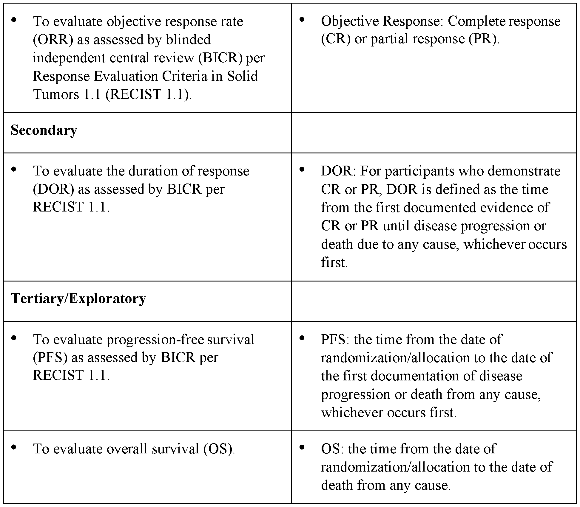


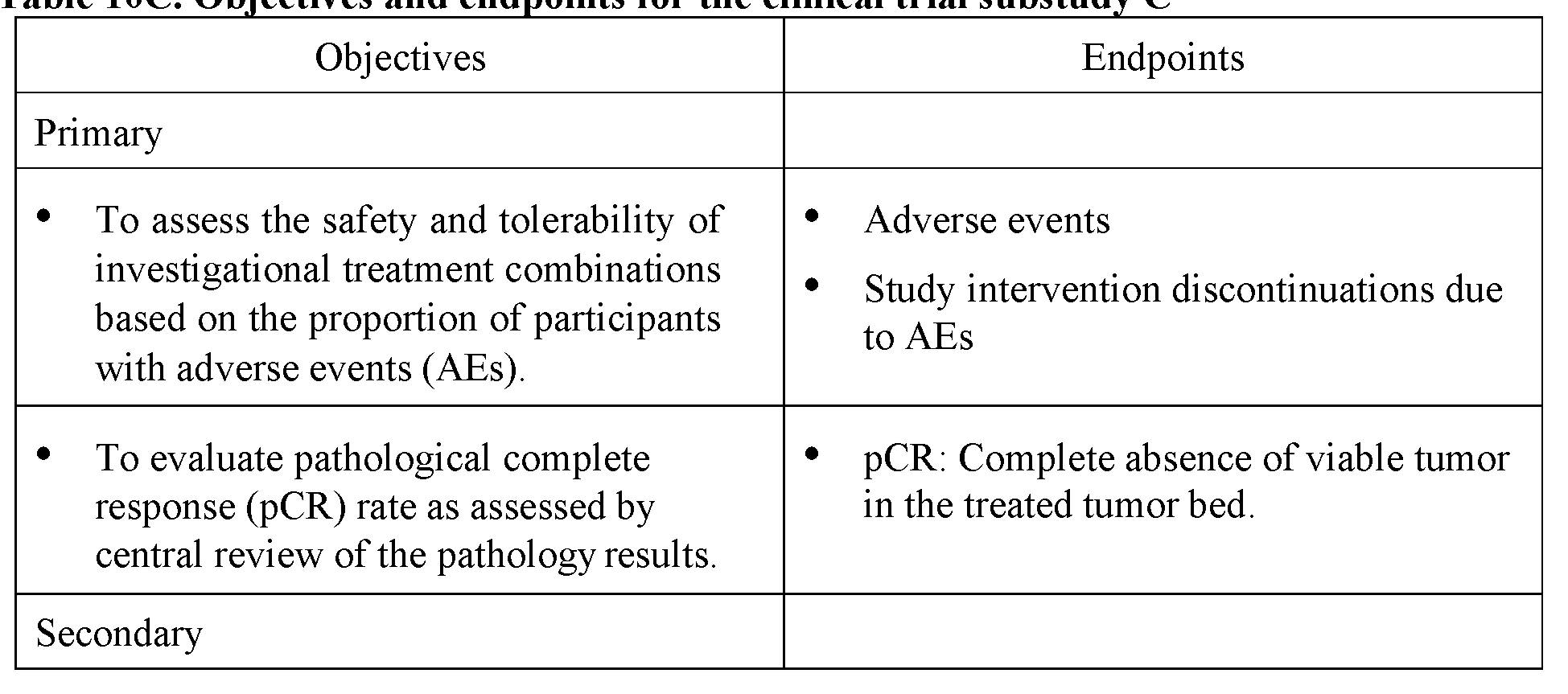


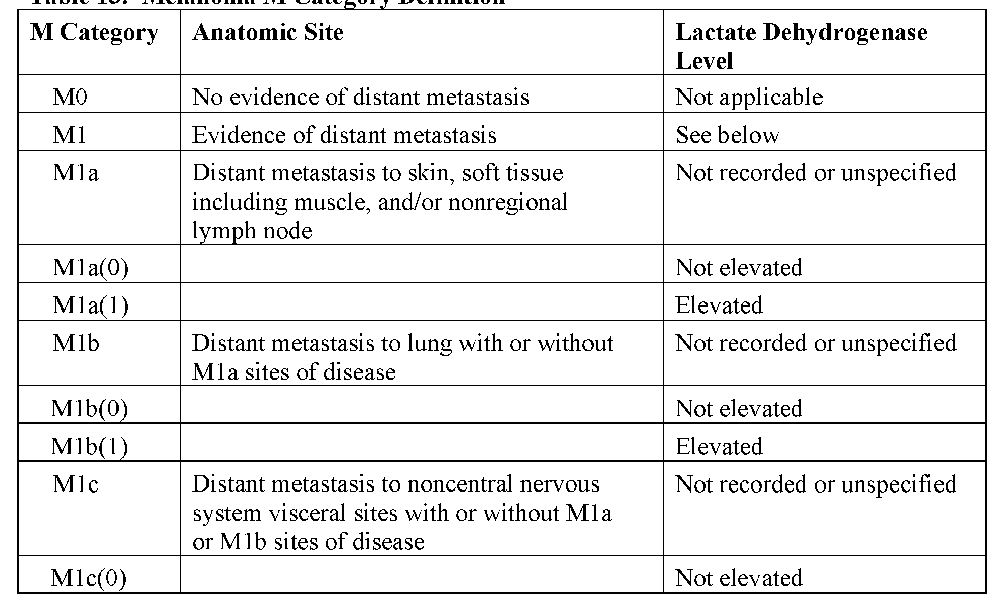

Table 14. AJCC Pathological (pTNM) Staging Groups
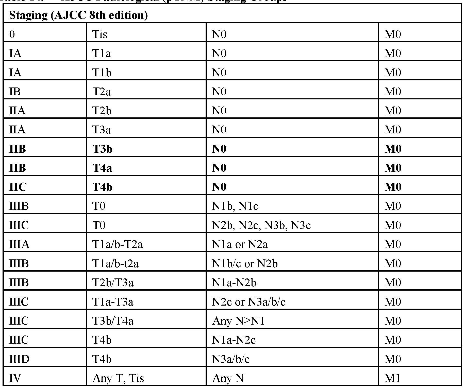 Abbreviations: M0 = No evidence of distant metastases; N0 = No regional metastasis detected including no tumor-involved nodes and no in-transit, satellite, and/or microsatellite metastasis; pTNM = pathological tumor, nodes, and metastasis. Table 15. ECOG Performance Status


Claims
WHAT IS CLAIMED IS: 1. A method of treating cancer, an infection disease or an infection comprising administering to a subject in need thereof effective amounts of: (a) an anti-human PD-1 monoclonal antibody or antigen binding fragment thereof; (b) an anti-human CTLA4 monoclonal antibody or antigen binding fragment thereof; and (c) an anti-human TIGIT monoclonal antibody or antigen binding fragment thereof.
2. The method of claim 1, wherein the cancer is selected from the group consisting of osteosarcoma, rhabdomyosarcoma, neuroblastoma, kidney cancer, leukemia, renal transitional cell cancer, bladder cancer, Wilm’s cancer, ovarian cancer, pancreatic cancer, breast cancer, prostate cancer, bone cancer, lung cancer, non-small cell lung cancer, gastric cancer, colorectal cancer, cervical cancer, synovial sarcoma, head and neck cancer, squamous cell carcinoma, lymphoma, diffuse large B-cell lymphoma, non-Hodgkin lymphoma, multiple myeloma, renal cell cancer, retinoblastoma, hepatoblastoma, hepatocellular carcinoma, melanoma, rhabdoid tumor of the kidney, Ewing's sarcoma, chondrosarcoma, brain cancer, glioblastoma, meningioma, pituitary adenoma, vestibular schwannoma, primitive neuroectodermal tumor, medulloblastoma, astrocytoma, anaplastic astrocytoma, oligodendroglioma, ependymoma, choroid plexus papilloma, polycythemia vera, thrombocythemia, idiopathic myelfibrosis, soft tissue sarcoma, thyroid cancer, endometrial cancer, and carcinoid cancer.
3. A method of enhancing T cell activity, comprising contacting the T cells with: (a) an anti-human PD-1 monoclonal antibody or antigen binding fragment thereof; (b) an anti-human CTLA4 monoclonal antibody or antigen binding fragment thereof; and (c) an anti-human TIGIT monoclonal antibody or antigen binding fragment thereof.
4. A method of increasing cytokine production of T cells, comprising contacting the T cells with: (a) an anti-human PD-1 monoclonal antibody or antigen binding fragment thereof; (b) an anti-human CTLA4 monoclonal antibody or antigen binding fragment thereof; and (c) an anti-human TIGIT monoclonal antibody or antigen binding fragment thereof.
5. The method of claim 4, wherein the cytokine is selected from the group consisting of IL-1, IL-2, IL-6, IL-12, IL-17, IL-22, IL-23, GM-CSF, IFN-α, and TNF-α.
6. A method of increasing proliferation of T cells comprising contacting the T cells with: (a) an anti-human PD-1 monoclonal antibody or antigen binding fragment thereof; (b) an anti-human CTLA4 monoclonal antibody or antigen binding fragment thereof; and (c) an anti-human TIGIT monoclonal antibody or antigen binding fragment thereof.
7. A pharmaceutical composition for treating cancer, an infection disease, or an infection in a subject in need thereof comprising: (a) an anti-human PD-1 monoclonal antibody or antigen binding fragment thereof; (b) an anti-human CTLA4 monoclonal antibody or antigen binding fragment thereof; and (c) an anti-human TIGIT monoclonal antibody or antigen binding fragment thereof.
8. The pharmaceutical composition of claim 7, further comprising a pharmaceutically acceptable carrier.
9. A kit for treating cancer, an infection disease or an infection in a subject in need thereof comprising: (a) an anti-human PD-1 monoclonal antibody or antigen binding fragment thereof; (b) an anti-human CTLA4 monoclonal antibody or antigen binding fragment thereof; and (c) an anti-human TIGIT monoclonal antibody or antigen binding fragment thereof.
10. The kit of claim 9, further comprising instructions for administering to a subject in need thereof effective amounts of the anti-human PD-1 monoclonal antibody or antigen binding fragment thereof, the anti-human CTLA4 monoclonal antibody or antigen binding fragment thereof, and the anti-human TIGIT monoclonal antibody or antigen binding fragment thereof.
11. A therapeutic combination for use in treating cancer, an infection disease or an infection in a subject in need thereof, wherein the therapeutic combination comprises effective amounts of: (a) an anti-human PD-1 monoclonal antibody or antigen binding fragment thereof; (b) an anti-human CTLA4 monoclonal antibody or antigen binding fragment thereof; and (c) an anti-human TIGIT monoclonal antibody or antigen binding fragment thereof.
12. The combination for use of claim 11, wherein the cancer is selected from the group consisting of osteosarcoma, rhabdomyosarcoma, neuroblastoma, kidney cancer, leukemia, renal transitional cell cancer, bladder cancer, Wilm’s cancer, ovarian cancer, pancreatic cancer, breast cancer, prostate cancer, bone cancer, lung cancer, non-small cell lung cancer, gastric cancer, colorectal cancer, cervical cancer, synovial sarcoma, head and neck cancer, squamous cell carcinoma, lymphoma, diffuse large B-cell lymphoma, non-Hodgkin lymphoma, multiple myeloma, renal cell cancer, retinoblastoma, hepatoblastoma, hepatocellular carcinoma, melanoma, rhabdoid tumor of the kidney, Ewing's sarcoma, chondrosarcoma, brain
cancer, glioblastoma, meningioma, pituitary adenoma, vestibular schwannoma, primitive neuroectodermal tumor, medulloblastoma, astrocytoma, anaplastic astrocytoma, oligodendroglioma, ependymoma, choroid plexus papilloma, polycythemia vera, thrombocythemia, idiopathic myelfibrosis, soft tissue sarcoma, thyroid cancer, endometrial cancer, and carcinoid cancer.
13. The method, pharmaceutical composition, kit, or the combination for use of any one of claims 1-12, wherein the subject is a human patient.
14. The method, pharmaceutical composition, kit, or the combination for use of any one of claims 1-13 for treating cancer.
15. The method, pharmaceutical composition, kit, or the combination for use of any one of claims 1-14, wherein the anti-human PD-1 monoclonal antibody is a human antibody.
16. The method, pharmaceutical composition, kit, or the combination for use of any one of claims 1-14, wherein the anti-human PD-1 monoclonal antibody is a humanized antibody.
17. The method, pharmaceutical composition, kit, or the combination for use of any one of claims 1-16, wherein the anti-human CTLA4 monoclonal antibody is a human antibody.
18. The method, pharmaceutical composition, kit, or the combination for use of any one of claims 1-16, wherein the anti-human CTLA4 monoclonal antibody is a humanized antibody.
19. The method, pharmaceutical composition, kit, or the combination for use of any one of claims 1-18, wherein the anti-human TIGIT monoclonal antibody is a human antibody.
20. The method, pharmaceutical composition, kit, or the combination for use of any one of claims 1-18, wherein the anti-human TIGIT monoclonal antibody is a humanized antibody.
21. The method, pharmaceutical composition, kit, or the combination for use of any one of claims 1-14, wherein the anti-human PD-1 monoclonal antibody or antigen binding fragment thereof comprises a light chain variable region VL complementarity determining region 1 (CDR1), a VL CDR2, and a VL CDR3 comprising amino acid sequences as set forth in SEQ ID NOS:1, 2, and 3, respectively, and a heavy chain variable region VH CDR1, a VH CDR2, and a VH CDR3 comprising amino acid sequences as set forth in SEQ ID NOS:6, 7, and 8, respectively.
22. The method, pharmaceutical composition, kit, or the combination for use of any of claims 1-14, wherein the anti-human PD-1 monoclonal antibody or antigen binding fragment thereof comprises a VL region comprising an amino acid sequence as set forth in SEQ ID NO:4, and a VH region comprising an amino acid sequence as set forth in SEQ ID NO:9.
23. The method, pharmaceutical composition, kit, or the combination for use of any of claims 1-14, wherein the anti-human PD-1 monoclonal antibody or antigen binding fragment thereof comprises a light chain comprising or consisting of an amino acid sequence as set forth in SEQ ID NO:5 and a heavy chain comprising or consisting of an amino acid sequence as set forth in SEQ ID NO:10.
24. The method, pharmaceutical composition, kit, or the combination for use of any one of claims 1-14, wherein the anti-human PD-1 monoclonal antibody or antigen binding fragment thereof comprises a light chain variable region VL complementarity determining region 1 (CDR1), a VL CDR2, and a VL CDR3 comprising amino acid sequences as set forth in SEQ ID NOS:31, 32, and 33, respectively, and a heavy chain variable region VH CDR1, a VH CDR2, and a VH CDR3 comprising amino acid sequences as set forth in SEQ ID NOS:36, 37, and 38, respectively.
25. The method, pharmaceutical composition, kit, or the combination for use of any of claims 1-14, wherein the anti-human PD-1 monoclonal antibody or antigen binding
fragment thereof comprises a VL region comprising an amino acid sequence as set forth in SEQ ID NO:34, and a VH region comprising an amino acid sequence as set forth in SEQ ID NO:39.
26. The method, pharmaceutical composition, kit, or the combination for use of any of claims 1-14, wherein the anti-human PD-1 monoclonal antibody or antigen binding fragment thereof comprises a light chain comprising or consisting of an amino acid sequence as set forth in SEQ ID NO:35 and a heavy chain comprising or consisting of an amino acid sequence as set forth in SEQ ID NO:40.
27. The method, pharmaceutical composition, kit, or the combination for use of any of claims 1-14, wherein the anti-human PD-1 monoclonal antibody or antigen binding fragment thereof is pembrolizumab.
28. The method, pharmaceutical composition, kit, or the combination for use of any of claims 1-14, wherein the anti-human PD-1 monoclonal antibody or antigen binding fragment thereof is nivolumab.
29. The method, pharmaceutical composition, kit, or the combination for use of any of claims 1-14, wherein the anti-human PD-1 monoclonal antibody or antigen binding fragment thereof is cemiplimab.
30. The method, pharmaceutical composition, kit, or the combination for use of any of claims 1-14, wherein the anti-human CTLA4 monoclonal antibody or antigen binding fragment thereof comprises: (a) a VL CDR1 comprising the amino acid sequence of SEQ ID NO: 14, a VL CDR2 comprising the amino acid sequence of SEQ ID NO: 15, and a VL CDR3 comprising the amino acid sequences as set forth in SEQ ID NOS:16, 17, or 18, and a VH CDR1, a VH CDR2, and a VH CDR3 comprising amino acid sequences as set forth in SEQ ID NOS:11, 12, and 13, respectively; or (b) a VL CDR1, a VL CDR2, and a VL CDR3 comprising the amino acid seque nces as set forth in SEQ ID NOS:54, 55, and 56, respectively , and a VH CDR1, a VH CDR2, and a VH CDR3 comprising amino acid sequences as set forth in SEQ ID NOS:57, 105, and 106, respectively;.
31. The method, pharmaceutical composition, kit, or the combination for use of any of claims 1-12, wherein the anti-human CTLA4 monoclonal antibody or antigen binding fragment thereof comprises: (a) a VL region comprising an amino acid sequence as set forth in SEQ ID NO:20, and a VH region comprising an amino acid sequence as set forth in SEQ ID NO:19; (b) a VL region comprising an amino acid sequence as set forth in SEQ ID NO:42, and a VH region comprising an amino acid sequence as set forth in SEQ ID NO:41; (c) a VL region comprising an amino acid sequence as set forth in SEQ ID NO:44, and a VH region comprising an amino acid sequence as set forth in SEQ ID NO: 43; (d) a VL region comprising an amino acid sequence as set forth in SEQ ID NO:44, and a VH region comprising an amino acid sequence as set forth in SEQ ID NO: 45; (e) a VL region comprising an amino acid sequence as set forth in SEQ ID NO:47, and a VH region comprising an amino acid sequence as set forth in SEQ ID NO:46; (f) a VL region comprising an amino acid sequence as set forth in SEQ ID NO:49, and a VH region comprising an amino acid sequence as set forth in SEQ ID NO:48; (g) a VL region comprising an amino acid sequence as set forth in SEQ ID NO:51, and a VH region comprising an amino acid sequence as set forth in SEQ ID NO:50; or (h) a VL region comprising an amino acid sequence as set forth in SEQ ID NO:108, and a VH region comprising an amino acid sequence as set forth in SEQ ID NO:107.
32. The method, pharmaceutical composition, kit, or the combination for use of any of claims 1-14, wherein the anti-human CTLA4 monoclonal antibody or antigen binding fragment thereof comprises: (a) a light chain comprising or consisting of an amino acid sequence as set forth in SEQ ID NO:53 and a heavy chain comprising or consisting of an amino acid sequence as set forth in SEQ ID NO:52; or (b) a light chain comprising or consisting of an amino acid sequence as set forth in SEQ ID NO:110 and a heavy chain comprising or consisting of an amino acid sequence as set forth in SEQ ID NO:109.
33. The method, pharmaceutical composition, kit, or the combination for use of any of claims 1-14, wherein the anti-human TIGIT monoclonal antibody or antigen binding fragment thereof comprises a VL CDR1, a VL CDR2, and a VL CDR3 comprising amino acid
sequences as set forth in SEQ ID NOS:21, 22, and 23, respectively, and a VH CDR1, a VH CDR2, and a VH CDR3 comprising amino acid sequences as set forth in SEQ ID NOS:26, 27, and 28, respectively.
34. The method, pharmaceutical composition, kit, or the combination for use of any of claims 1-14, wherein the anti-human TIGIT monoclonal antibody or antigen binding fragment thereof comprises a VL region comprising an amino acid sequence as set forth in SEQ ID NO:24, and a VH region comprising an amino acid sequence as set forth in SEQ ID NO:29.
35. The method, pharmaceutical composition, kit, or use of any of claims 1- 14, wherein the anti-human TIGIT monoclonal antibody or antigen binding fragment thereof comprises a light chain comprising or consisting of an amino acid sequence as set forth in SEQ ID NO:25 and a heavy chain comprising or consisting of an amino acid sequence as set forth in SEQ ID NO:30.
36. The method, pharmaceutical composition, kit, or the combination for use of any of claims 1-14, wherein: (a) the anti-human PD-1 monoclonal antibody or antigen binding fragment thereof comprises a VL CDR1, a VL CDR2, and a VL CDR3 comprising amino acid sequences as set forth in SEQ ID NOS:1, 2, and 3, respectively, and a VH CDR1, a VH CDR2, and a VH CDR3 comprising amino acid sequences as set forth in SEQ ID NOS:6, 7, and 8, respectively; (b) the anti-human CTLA4 monoclonal antibody or antigen binding fragment thereof comprises a VL CDR1 comprising the amino acid sequence of SEQ ID NO: 14, a VL CDR2 comprising the amino acid sequence of SEQ ID NO: 15, and a VL CDR3 comprising the amino acid sequences as set forth in SEQ ID NOS:16, 17, or 18, and a VH CDR1, a VH CDR2, and a VH CDR3 comprising amino acid sequences as set forth in SEQ ID NOS:11, 12, and 13, respectively; and (c) the anti-human TIGIT monoclonal antibody or antigen binding fragment thereof comprises a VL CDR1, a VL CDR2, and a VL CDR3 comprising amino acid sequences as set forth in SEQ ID NOS:21, 22, and 23, respectively, and a VH CDR1, a VH CDR2, and a VH CDR3 comprising amino acid sequences as set forth in SEQ ID NOS:26, 27, and 28, respectively.
37. The method, pharmaceutical composition, kit, or the combination for use of any of claims 1-14, wherein: (a) the anti-human PD-1 monoclonal antibody or antigen binding fragment thereof comprises a VL CDR1, a VL CDR2, and a VL CDR3 comprising amino acid sequences as set forth in SEQ ID NOS:1, 2, and 3, respectively, and a VH CDR1, a VH CDR2, and a VH CDR3 comprising amino acid sequences as set forth in SEQ ID NOS:6, 7, and 8, respectively; (b) the anti-human CTLA4 monoclonal antibody or antigen binding fragment thereof comprises a VL CDR1, a VL CDR2, and a VL CDR3 comprising the amino acid seque nces as set forth in SEQ ID NOS:54, 55, and 56, respectively , and a VH CDR1, a VH CDR2, and a VH CDR3 comprising amino acid sequences as set forth in SEQ ID NOS:57, 105, and 106, respectively; and (c) the anti-human TIGIT monoclonal antibody or antigen binding fragment thereof comprises a VL CDR1, a VL CDR2, and a VL CDR3 comprising amino acid sequences as set forth in SEQ ID NOS:21, 22, and 23, respectively, and a VH CDR1, a VH CDR2, and a VH CDR3 comprising amino acid sequences as set forth in SEQ ID NOS:26, 27, and 28, respectively.
38. The method, pharmaceutical composition, kit, or the combination for use of any of claims 1-14, wherein: (a) the anti-human PD-1 monoclonal antibody or antigen binding fragment thereof comprises a VL region comprising an amino acid sequence as set forth in SEQ ID NO:4, and a VH region comprising an amino acid sequence as set forth in SEQ ID NO:9; (b) the anti-human CTLA4 monoclonal antibody or antigen binding fragment thereof comprises a VL region comprising an amino acid sequence as set forth in SEQ ID NO:20, and a VH region comprising an amino acid sequence as set forth in SEQ ID NO:19; and (c) the anti-human TIGIT monoclonal antibody or antigen binding fragment thereof comprises a VL region comprising an amino acid sequence as set forth in SEQ ID NO:24, and a VH region comprising an amino acid sequence as set forth in SEQ ID NO:29.
39. The method, pharmaceutical composition, kit, or the combination for use of any of claims 1-14, wherein:
(a) the anti-human PD-1 monoclonal antibody or antigen binding fragment thereof comprises a VL region comprising an amino acid sequence as set forth in SEQ ID NO:4, and a VH region comprising an amino acid sequence as set forth in SEQ ID NO:9; (b) the anti-human CTLA4 monoclonal antibody or antigen binding fragment thereof comprises a VL region comprising an amino acid sequence as set forth in SEQ ID NO:44, and a VH region comprising an amino acid sequence as set forth in SEQ ID NO:43; and (c) the anti-human TIGIT monoclonal antibody or antigen binding fragment thereof comprises a VL region comprising an amino acid sequence as set forth in SEQ ID NO:24, and a VH region comprising an amino acid sequence as set forth in SEQ ID NO:29.
40. The method, pharmaceutical composition, kit, or the combination for use of any of claims 1-14, wherein: (a) the anti-human PD-1 monoclonal antibody or antigen binding fragment thereof comprises a VL region comprising an amino acid sequence as set forth in SEQ ID NO:4, and a VH region comprising an amino acid sequence as set forth in SEQ ID NO:9; (b) the anti-human CTLA4 monoclonal antibody or antigen binding fragment thereof comprises a VL region comprising an amino acid sequence as set forth in SEQ ID NO:42, and a VH region comprising an amino acid sequence as set forth in SEQ ID NO:41; and (c) the anti-human TIGIT monoclonal antibody or antigen binding fragment thereof comprises a VL region comprising an amino acid sequence as set forth in SEQ ID NO:24, and a VH region comprising an amino acid sequence as set forth in SEQ ID NO:29.
41. The method, pharmaceutical composition, kit, or the combination for use of any of claims 1-14, wherein: (a) the anti-human PD-1 monoclonal antibody or antigen binding fragment thereof comprises a VL region comprising an amino acid sequence as set forth in SEQ ID NO:4, and a VH region comprising an amino acid sequence as set forth in SEQ ID NO:9; (b) the anti-human CTLA4 monoclonal antibody or antigen binding fragment thereof comprises a VL region comprising an amino acid sequence as set forth in SEQ ID NO:44, and a VH region comprising an amino acid sequence as set forth in SEQ ID NO: NO:45; and (c) the anti-human TIGIT monoclonal antibody or antigen binding fragment thereof comprises a VL region comprising an amino acid sequence as set forth in SEQ ID NO:24, and a VH region comprising an amino acid sequence as set forth in SEQ ID NO:29.
42. The method, pharmaceutical composition, kit, or the combination for use of any of claims 1-14, wherein: (a) the anti-human PD-1 monoclonal antibody or antigen binding fragment thereof comprises a VL region comprising an amino acid sequence as set forth in SEQ ID NO:4, and a VH region comprising an amino acid sequence as set forth in SEQ ID NO:9; (b) the anti-human CTLA4 monoclonal antibody or antigen binding fragment thereof comprises a VL region comprising an amino acid sequence as set forth in SEQ ID NO:47, and a VH region comprising an amino acid sequence as set forth in SEQ ID NO:46; and (c) the anti-human TIGIT monoclonal antibody or antigen binding fragment thereof comprises a VL region comprising an amino acid sequence as set forth in SEQ ID NO:24, and a VH region comprising an amino acid sequence as set forth in SEQ ID NO:29.
43. The method, pharmaceutical composition, kit, or the combination for use of any of claims 1-14, wherein: (a) the anti-human PD-1 monoclonal antibody or antigen binding fragment thereof comprises a VL region comprising an amino acid sequence as set forth in SEQ ID NO:4, and a VH region comprising an amino acid sequence as set forth in SEQ ID NO:9; (b) the anti-human CTLA4 monoclonal antibody or antigen binding fragment thereof comprises a VL region comprising an amino acid sequence as set forth in SEQ ID NO:49, and a VH region comprising an amino acid sequence as set forth in SEQ ID NO:48; and (c) the anti-human TIGIT monoclonal antibody or antigen binding fragment thereof comprises a VL region comprising an amino acid sequence as set forth in SEQ ID NO:24, and a VH region comprising an amino acid sequence as set forth in SEQ ID NO:29.
44. The method, pharmaceutical composition, kit, or the combination for use of any of claims 1-14, wherein: (a) the anti-human PD-1 monoclonal antibody or antigen binding fragment thereof comprises a VL region comprising an amino acid sequence as set forth in SEQ ID NO:4, and a VH region comprising an amino acid sequence as set forth in SEQ ID NO:9; (b) the anti-human CTLA4 monoclonal antibody or antigen binding fragment thereof comprises a VL region comprising an amino acid sequence as set forth in SEQ ID NO:51, and a VH region comprising an amino acid sequence as set forth in SEQ ID NO:50; and
(c) the anti-human TIGIT monoclonal antibody or antigen binding fragment thereof comprises a VL region comprising an amino acid sequence as set forth in SEQ ID NO:24, and a VH region comprising an amino acid sequence as set forth in SEQ ID NO:29.
45. The method, pharmaceutical composition, kit, or the combination for use of any of claims 1-14, wherein: (a) the anti-human PD-1 monoclonal antibody or antigen binding fragment thereof comprises a VL region comprising an amino acid sequence as set forth in SEQ ID NO:4, and a VH region comprising an amino acid sequence as set forth in SEQ ID NO:9; (b) the anti-human CTLA4 monoclonal antibody or antigen binding fragment thereof comprises a VL region comprising an amino acid sequence as set forth in SEQ ID NO:108, and a VH region comprising an amino acid sequence as set forth in SEQ ID NO:107; and (c) the anti-human TIGIT monoclonal antibody or antigen binding fragment thereof comprises a VL region comprising an amino acid sequence as set forth in SEQ ID NO:24, and a VH region comprising an amino acid sequence as set forth in SEQ ID NO:29.
46. The method, pharmaceutical composition, kit, or the combination for use of any of claims 1-14, wherein: (a) the anti-human PD-1 monoclonal antibody or antigen binding fragment thereof comprises a light chain comprising or consisting of an amino acid sequence as set forth in SEQ ID NO:5 and a heavy chain comprising or consisting of an amino acid sequence as set forth in SEQ ID NO:10; (b) the anti-human CTLA4 monoclonal antibody or antigen binding fragment thereof comprises a light chain comprising or consisting of an amino acid sequence as set forth in SEQ ID NO:53 and a heavy chain comprising or consisting of an amino acid sequence as set forth in SEQ ID NO:52; and (c) the anti-human TIGIT monoclonal antibody or antigen binding fragment thereof comprises a light chain comprising or consisting of an amino acid sequence as set forth in SEQ ID NO:25 and a heavy chain comprising or consisting of an amino acid sequence as set forth in SEQ ID NO:30.
47. The method, pharmaceutical composition, kit, or the combination for use of any of claims 1-14, wherein: (a) the anti-human PD-1 monoclonal antibody or antigen binding fragment thereof comprises a light chain comprising or consisting of an amino acid sequence as set forth in SEQ ID NO:5 and a heavy chain comprising or consisting of an amino acid sequence as set forth in SEQ ID NO:10; (b) the anti-human CTLA4 monoclonal antibody or antigen binding fragment thereof comprises a light chain comprising or consisting of an amino acid sequence as set forth in SEQ ID NO:110 and a heavy chain comprising or consisting of an amino acid sequence as set forth in SEQ ID NO:109; and (c) the anti-human TIGIT monoclonal antibody or antigen binding fragment thereof comprises a light chain comprising or consisting of an amino acid sequence as set forth in SEQ ID NO:25 and a heavy chain comprising or consisting of an amino acid sequence as set forth in SEQ ID NO:30.
48. The method, pharmaceutical composition, kit, or the combination for use of any of claims 1-14, wherein: (a) the anti-human PD-1 monoclonal antibody or antigen binding fragment thereof comprises a VL CDR1, a VL CDR2, and a VL CDR3 comprising amino acid sequences as set forth in SEQ ID NOS:31, 32, and 33, respectively, and a VH CDR1, a VH CDR2, and a VH CDR3 comprising amino acid sequences as set forth in SEQ ID NOS:36, 37, and 38, respectively; (b) a VL CDR1 comprising the amino acid sequence of SEQ ID NO: 14, a VL CDR2 comprising the amino acid sequence of SEQ ID NO: 15, and a VL CDR3 comprising the amino acid sequences as set forth in SEQ ID NOS:16, 17, or 18, and a VH CDR1, a VH CDR2, and a VH CDR3 comprising amino acid sequences as set forth in SEQ ID NOS:11, 12, and 13, respectively; and (c) the anti-human TIGIT monoclonal antibody or antigen binding fragment thereof comprises a VL CDR1, a VL CDR2, and a VL CDR3 comprising amino acid sequences as set forth in SEQ ID NOS:21, 22, and 23, respectively, and a VH CDR1, a VH CDR2, and a VH CDR3 comprising amino acid sequences as set forth in SEQ ID NOS:26, 27, and 28, respectively.
49. The method, pharmaceutical composition, kit, or the combination for use of any of claims 1-14, wherein: (a) the anti-human PD-1 monoclonal antibody or antigen binding fragment thereof comprises a VL CDR1, a VL CDR2, and a VL CDR3 comprising amino acid sequences as set forth in SEQ ID NOS:31, 32, and 33, respectively, and a VH CDR1, a VH CDR2, and a VH CDR3 comprising amino acid sequences as set forth in SEQ ID NOS:36, 37, and 38, respectively; (b) a VL CDR1, a VL CDR2, and a VL CDR3 comprising the amino acid seque nces as set forth in SEQ ID NOS:54, 55, and 56, respectively , and a VH CDR1, a VH CDR2, and a VH CDR3 comprising amino acid sequences as set forth in SEQ ID NOS:57, 105, and 106, respectively; and (c) the anti-human TIGIT monoclonal antibody or antigen binding fragment thereof comprises a VL CDR1, a VL CDR2, and a VL CDR3 comprising amino acid sequences as set forth in SEQ ID NOS:21, 22, and 23, respectively, and a VH CDR1, a VH CDR2, and a VH CDR3 comprising amino acid sequences as set forth in SEQ ID NOS:26, 27, and 28, respectively.
50. The method, pharmaceutical composition, kit, or the combination for use of any of claims 1-14, wherein: (a) the anti-human PD-1 monoclonal antibody or antigen binding fragment thereof comprises a VL region comprising an amino acid sequence as set forth in SEQ ID NO:34, and a VH region comprising an amino acid sequence as set forth in SEQ ID NO:39; (b) the anti-human CTLA4 monoclonal antibody or antigen binding fragment thereof comprises a VL region comprising an amino acid sequence as set forth in SEQ ID NO:20, and a VH region comprising an amino acid sequence as set forth in SEQ ID NO:19; and (c) the anti-human TIGIT monoclonal antibody or antigen binding fragment thereof comprises a VL region comprising an amino acid sequence as set forth in SEQ ID NO:24, and a VH region comprising an amino acid sequence as set forth in SEQ ID NO:29.
51. The method, pharmaceutical composition, kit, or the combination for use of any of claims 1-14, wherein:
(a) the anti-human PD-1 monoclonal antibody or antigen binding fragment thereof comprises a VL region comprising an amino acid sequence as set forth in SEQ ID NO:34, and a VH region comprising an amino acid sequence as set forth in SEQ ID NO:39; (b) the anti-human CTLA4 monoclonal antibody or antigen binding fragment thereof comprises a a VL region comprising an amino acid sequence as set forth in SEQ ID NO:42, and a VH region comprising an amino acid sequence as set forth in SEQ ID NO:41; and (c) the anti-human TIGIT monoclonal antibody or antigen binding fragment thereof comprises a VL region comprising an amino acid sequence as set forth in SEQ ID NO:24, and a VH region comprising an amino acid sequence as set forth in SEQ ID NO:29.
52. The method, pharmaceutical composition, kit, or the combination for use of any of claims 1-14, wherein: (a) the anti-human PD-1 monoclonal antibody or antigen binding fragment thereof comprises a VL region comprising an amino acid sequence as set forth in SEQ ID NO:34, and a VH region comprising an amino acid sequence as set forth in SEQ ID NO:39; (b) the anti-human CTLA4 monoclonal antibody or antigen binding fragment thereof comprises a VL region comprising an amino acid sequence as set forth in SEQ ID NO:44, and a VH region comprising an amino acid sequence as set forth in SEQ ID NO:43; and (c) the anti-human TIGIT monoclonal antibody or antigen binding fragment thereof comprises a VL region comprising an amino acid sequence as set forth in SEQ ID NO:24, and a VH region comprising an amino acid sequence as set forth in SEQ ID NO:29.
53. The method, pharmaceutical composition, kit, or the combination for use of any of claims 1-14, wherein: (a) the anti-human PD-1 monoclonal antibody or antigen binding fragment thereof comprises a VL region comprising an amino acid sequence as set forth in SEQ ID NO:34, and a VH region comprising an amino acid sequence as set forth in SEQ ID NO:39; (b) the anti-human CTLA4 monoclonal antibody or antigen binding fragment thereof comprises a VL region comprising an amino acid sequence as set forth in SEQ ID NO:44, and a VH region comprising an amino acid sequence as set forth in SEQ ID NO: 45 or SEQ ID NO: 45; and
(c) the anti-human TIGIT monoclonal antibody or antigen binding fragment thereof comprises a VL region comprising an amino acid sequence as set forth in SEQ ID NO:24, and a VH region comprising an amino acid sequence as set forth in SEQ ID NO:29.
54. The method, pharmaceutical composition, kit, or the combination for use of any of claims 1-14, wherein: (a) the anti-human PD-1 monoclonal antibody or antigen binding fragment thereof comprises a VL region comprising an amino acid sequence as set forth in SEQ ID NO:34, and a VH region comprising an amino acid sequence as set forth in SEQ ID NO:39; (b) the anti-human CTLA4 monoclonal antibody or antigen binding fragment thereof comprises a VL region comprising an amino acid sequence as set forth in SEQ ID NO:47, and a VH region comprising an amino acid sequence as set forth in SEQ ID NO:46; and (c) the anti-human TIGIT monoclonal antibody or antigen binding fragment thereof comprises a VL region comprising an amino acid sequence as set forth in SEQ ID NO:24, and a VH region comprising an amino acid sequence as set forth in SEQ ID NO:29.
55. The method, pharmaceutical composition, kit, or the combination for use of any of claims 1-14, wherein: (a) the anti-human PD-1 monoclonal antibody or antigen binding fragment thereof comprises a VL region comprising an amino acid sequence as set forth in SEQ ID NO:34, and a VH region comprising an amino acid sequence as set forth in SEQ ID NO:39; (b) the anti-human CTLA4 monoclonal antibody or antigen binding fragment thereof comprises a VL region comprising an amino acid sequence as set forth in SEQ ID NO:49, and a VH region comprising an amino acid sequence as set forth in SEQ ID NO:48; and (c) the anti-human TIGIT monoclonal antibody or antigen binding fragment thereof comprises a VL region comprising an amino acid sequence as set forth in SEQ ID NO:24, and a VH region comprising an amino acid sequence as set forth in SEQ ID NO:29.
56. The method, pharmaceutical composition, kit, or the combination for use of any of claims 1-14, wherein: (a) the anti-human PD-1 monoclonal antibody or antigen binding fragment thereof comprises a VL region comprising an amino acid sequence as set forth in SEQ ID NO:34, and a VH region comprising an amino acid sequence as set forth in SEQ ID NO:39;
(b) the anti-human CTLA4 monoclonal antibody or antigen binding fragment thereof comprises a VL region comprising an amino acid sequence as set forth in SEQ ID NO:51, and a VH region comprising an amino acid sequence as set forth in SEQ ID NO:50; and (c) the anti-human TIGIT monoclonal antibody or antigen binding fragment thereof comprises a VL region comprising an amino acid sequence as set forth in SEQ ID NO:24, and a VH region comprising an amino acid sequence as set forth in SEQ ID NO:29.
57. The method, pharmaceutical composition, kit, or the combination for use of any of claims 1-14, wherein: (a) the anti-human PD-1 monoclonal antibody or antigen binding fragment thereof comprises a VL region comprising an amino acid sequence as set forth in SEQ ID NO:34, and a VH region comprising an amino acid sequence as set forth in SEQ ID NO:39; (b) the anti-human CTLA4 monoclonal antibody or antigen binding fragment thereof comprises a VL region comprising an amino acid sequence as set forth in SEQ ID NO:108, and a VH region comprising an amino acid sequence as set forth in SEQ ID NO:107; and (c) the anti-human TIGIT monoclonal antibody or antigen binding fragment thereof comprises a VL region comprising an amino acid sequence as set forth in SEQ ID NO:24, and a VH region comprising an amino acid sequence as set forth in SEQ ID NO:29.
58. The method, pharmaceutical composition, kit, or the combination for use of any of claims 1-14, wherein: (a) the anti-human PD-1 monoclonal antibody or antigen binding fragment thereof comprises a light chain comprising or consisting of an amino acid sequence as set forth in SEQ ID NO:35 and a heavy chain comprising or consisting of an amino acid sequence as set forth in SEQ ID NO:40; (b) the anti-human CTLA4 monoclonal antibody or antigen binding fragment thereof comprises a light chain comprising or consisting of an amino acid sequence as set forth in SEQ ID NO:53 and a heavy chain comprising or consisting of an amino acid sequence as set forth in SEQ ID NO:52; and (c) the anti-human TIGIT monoclonal antibody or antigen binding fragment thereof comprises a light chain comprising or consisting of an amino acid sequence as set forth in
SEQ ID NO:25 and a heavy chain comprising or consisting of an amino acid sequence as set forth in SEQ ID NO:30.
59. The method, pharmaceutical composition, kit, or the combination for use of any of claims 1-14, wherein: (a) the anti-human PD-1 monoclonal antibody or antigen binding fragment thereof comprises a light chain comprising or consisting of an amino acid sequence as set forth in SEQ ID NO:5 and a heavy chain comprising or consisting of an amino acid sequence as set forth in SEQ ID NO:10; (b) the anti-human CTLA4 monoclonal antibody or antigen binding fragment thereof comprises a light chain comprising or consisting of an amino acid sequence as set forth in SEQ ID NO:53 and a heavy chain comprising or consisting of an amino acid sequence as set forth in SEQ ID NO:52; and (c) the anti-human TIGIT monoclonal antibody or antigen binding fragment thereof comprises a light chain comprising or consisting of an amino acid sequence as set forth in SEQ ID NO:25 and a heavy chain comprising or consisting of an amino acid sequence as set forth in SEQ ID NO:30.
60. The method, pharmaceutical composition, kit, or the combination for use of any of claims 1-59, wherein the anti-human PD-1 monoclonal antibody is pembrolizumab, and 200 mg pembrolizumab is administered once every three weeks.
61. The method, pharmaceutical composition, kit, or the combination for use of any of claims 1-59, wherein the anti-human PD-1 monoclonal antibody is pembrolizumab, and the subject is administered 400 mg pembrolizumab, and wherein pembrolizumab is administered once every six weeks.
62. The method, pharmaceutical composition, kit, or the combination for use of any of claims 1-61, wherein 200 mg of the anti-human TIGIT monoclonal antibody or antigen binding fragment thereof is administered.
63. The method, pharmaceutical composition, kit, or the combination for use of any of claims 1-62, wherein 25 mg of the anti-human CTLA4 monoclonal antibody or antigen binding fragment thereof is administered.
64. The method, pharmaceutical composition, kit, or the combination for use of any of claims 1-63, wherein the anti-human TIGIT monoclonal antibody or antigen binding fragment thereof is administered every three weeks.
65. The method, pharmaceutical composition, kit, or the combination for use of any of claims 1-64, wherein the anti-human CTLA4 monoclonal antibody or antigen binding fragment thereof is administered every six weeks.
66. The method, pharmaceutical composition, kit, or the combination for use of any of claims 1-64, wherein 200 mg of the anti-human TIGIT monoclonal antibody or antigen binding fragment thereof is administered every three weeks.
67. The method, pharmaceutical composition, kit, or the combination for use of any of claims 1-65, wherein 25 mg of the anti-human CTLA4 monoclonal antibody or antigen binding fragment thereof is administered every six weeks.
68. A method of treating a cancer comprising administering to a subject in need thereof: (a) 200 mg, 400 mg, or 2 mg/kg pembrolizumab or antigen binding fragment thereof; (b) an effective amount of an anti-human CTLA4 antibody or antigen binding fragment thereof comprising a VL region comprising an amino acid sequence as set forth in SEQ ID NO:44, and a VH region comprising an amino acid sequence as set forth in SEQ ID NO:45; and (c) an effective amount of an anti-human TIGIT antibody or antigen binding fragment thereof comprising a VL region comprising an amino acid sequence as set forth in SEQ ID NO:24, and a VH region comprising an amino acid sequence as set forth in SEQ ID NO:29.
69. A method of treating a cancer comprising administering to a subject in need thereof (a) 200 mg, 400 mg, or 2 mg/kg pembrolizumab or antigen binding fragment thereof; (b) an effective amount of an anti-human CTLA4 antibody or antigen binding fragment thereof comprising a VL region consisting of an amino acid sequence as set forth in SEQ ID NO:44, and a VH region consisting of an amino acid sequence as set forth in SEQ ID NO:45; and (c) an effective amount of an anti-human TIGIT antibody or antigen binding fragment thereof comprising a VL region consisting of an amino acid sequence as set forth in SEQ ID NO:24, and a VH region consisting of an amino acid sequence as set forth in SEQ ID NO:29.
70. The method of claim 68 or claim 69, wherein the pembrolizumab or antigen binding fragment thereof, the anti-CTLA4 antibody or antigen binding fragment thereof, and/or the anti-TIGIT antibody or antigen binding fragment thereof is administered via intravenous infusion.
71. The method of any of claims 68-70, wherein the anti-CTLA4 antibody is a monoclonal antibody.
72. The method of any of claims 68-71, wherein the anti-TIGIT antibody is a monoclonal antibody.
73. The method of any of claims 68-72, wherein 200 mg of pembrolizumab is administered every three weeks.
74. The method of any of claims 68-72, wherein 400 mg of pembrolizumab is administered every six weeks.
75. The method of any of claims 68-74, wherein 200 mg of the anti-human TIGIT antibody or antigen binding fragment thereof is administered every three weeks.
76. The method of any of claims 68-75, wherein 25 mg of the anti-human CTLA4 antibody or antigen binding fragment thereof is administered every six weeks.
77. A method of treating a cancer comprising administering to a subject in need thereof: (a) 200 mg, 400 mg, or 2 mg/kg pembrolizumab or antigen binding fragment thereof; (b) an effective amount of an anti-human CTLA4 antibody or antigen binding fragment thereof comprising a light chain comprising an amino acid sequence as set forth in SEQ ID NO:53 and a heavy chain comprising an amino acid sequence as set forth in SEQ ID NO:52; and (c) an effective amount of an anti-human TIGIT antibody or antigen binding fragment thereof comprising a light chain comprising an amino acid sequence as set forth in SEQ ID NO:25 and a heavy chain comprising an amino acid sequence as set forth in SEQ ID NO:30.
78. A method of treating a cancercomprising administering to a subject in need thereof: (a) 200 mg, 400 mg, or 2 mg/kg pembrolizumab or antigen binding fragment thereof; (b) an effective amount of an anti-human CTLA4 antibody or antigen binding fragment thereof consisting of a light chain comprising an amino acid sequence as set forth in SEQ ID NO:53 and a heavy chain comprising an amino acid sequence as set forth in SEQ ID NO:52; and (c) an effective amount an anti-human TIGIT antibody or antigen binding fragment thereof consisting of a light chain comprising an amino acid sequence as set forth in SEQ ID NO:25 and a heavy chain comprising an amino acid sequence as set forth in SEQ ID NO:30.
79. The method of claim 77 or claim 78, wherein the pembrolizumab or antigen binding fragment thereof, the anti-CTLA4 antibody or antigen binding fragment thereof, and/or the anti-TIGIT antibody or antigen binding fragment thereof is administered via intravenous infusion.
80. The method of any of claims 77-79, wherein the anti-CTLA4 antibody is a monoclonal antibody.
81. The method of any of claims 77-80, wherein the anti-TIGIT antibody is a monoclonal antibody.
82. The method of any of claims 77-81, wherein 200 mg of pembrolizumab is administered every three weeks.
83. The method of any of claims 77-81, wherein 400 mg of pembrolizumab is administered every six weeks.
84. The method of any of claims 77-83, wherein 200 mg of the anti-human TIGIT antibody or antigen binding fragment thereof is administered every three weeks.
85. The method of any of claims 77-84, wherein 25 mg of the anti-human CTLA4 antibody or antigen binding fragment thereof is administered every six weeks.
86. The method of any of claims 68-85 wherein the method is for treating cancer.
87. The method of claim 86, wherein the cancer comprises at least one tumor.
88. The method of claim 86 or 87, wherein the cancer is selected from the group consisting of osteosarcoma, rhabdomyosarcoma, neuroblastoma, kidney cancer, leukemia, renal transitional cell cancer, bladder cancer, Wilm’s cancer, ovarian cancer, pancreatic cancer, breast cancer, prostate cancer, bone cancer, lung cancer, non-small cell lung cancer, gastric cancer, colorectal cancer, cervical cancer, synovial sarcoma, head and neck cancer, squamous cell carcinoma, lymphoma, diffuse large B-cell lymphoma, non-Hodgkin lymphoma, multiple myeloma, renal cell cancer, retinoblastoma, hepatoblastoma, hepatocellular carcinoma, melanoma, rhabdoid tumor of the kidney, Ewing's sarcoma, chondrosarcoma, brain cancer, glioblastoma, meningioma, pituitary adenoma, vestibular schwannoma, primitive neuroectodermal tumor, medulloblastoma, astrocytoma, anaplastic astrocytoma,
oligodendroglioma, ependymoma, choroid plexus papilloma, polycythemia vera, thrombocythemia, idiopathic myelfibrosis, soft tissue sarcoma, thyroid cancer, endometrial cancer, and carcinoid cancer.
89. The method of any of claims 86-88, wherein the cancer is metastatic.
90. The method of any of claims 86-89, wherein the cancer is locally advanced or resectable.
91. The method of any of claims 86-88, wherein the cancer is relapsed.
92. The method of any of claims 86-88, wherein the cancer is refractory.
93. The method of claim 92, wherein the refractory melanoma has progressed on treatment with an anti-PD-1/L1 mAb administered either as monotherapy, or in combination with a checkpoint inhibitor or other therapy.
94. The method of any of claims 68-93, wherein the subject has received at least 2 doses of an approved anti-PD-1/L1 monoclonal antibody.
95. The method of any of claims 65-94, wherein the subject has demonstrated disease progression after PD-1/L1 treatment as defined by RECIST 1.1.
96. The method of any of claims 65-94, wherein the subject has at least one melanoma selected from the group consisting of: PD-1 refractory melanoma, first-line advanced melanoma, and neoadjuvant melanoma.
97. The method, pharmaceutical composition, kit, or the combination for use of any of claims 1-96, wherein the anti-PD-1 antibody or antigen binding fragment, the anti- TIGIT antibody or antigen binding fragment, and the anti-CTLA4 antibody or antigen binding fragment are administered contemporaneously.
98. The method, pharmaceutical composition, kit, or the combination for use of any of claims 1-96, wherein the order of administration is the anti-PD-1 antibody followed by the anti-TIGIT antibody and then the anti-CTLA4 antibody.
99. The method, pharmaceutical composition, kit, or the combination for use of any of claims 1-98 further comprising at least one therapeutic agent and/or active agent.
100. The method, pharmaceutical composition, kit, or the combination for use of claim 99, wherein the therapeutic agent and/or active agent comprises an antibody or fragment thereof, an immunomodulator, a hormone, a cytotoxic agent, an enzyme, a radionuclide, a second antibody conjugated to at least one immunomodulator, enzyme, radioactive label, hormone, antisense oligonucleotide, biotherapeutic agent, chemotherapeutic agent, cytotoxic agent, or a combination thereof.
Priority Applications (2)
| Application Number | Priority Date | Filing Date | Title |
|---|---|---|---|
| US17/801,454 US20230092707A1 (en) | 2020-03-05 | 2021-03-04 | Methods for treating cancer or infection using a combination of an anti-pd-1 antibody, an anti-ctla4 antibody, and an anti-tigit antibody |
| EP21765135.5A EP4114863A4 (en) | 2020-03-05 | 2021-03-04 | Methods for treating cancer or infection using a combination of an anti-pd-1 antibody, an anti-ctla4 antibody, and an anti-tigit antibody |
Applications Claiming Priority (4)
| Application Number | Priority Date | Filing Date | Title |
|---|---|---|---|
| US202062985645P | 2020-03-05 | 2020-03-05 | |
| US62/985,645 | 2020-03-05 | ||
| US202063043888P | 2020-06-25 | 2020-06-25 | |
| US63/043,888 | 2020-06-25 |
Publications (1)
| Publication Number | Publication Date |
|---|---|
| WO2021178611A1 true WO2021178611A1 (en) | 2021-09-10 |
Family
ID=77613734
Family Applications (1)
| Application Number | Title | Priority Date | Filing Date |
|---|---|---|---|
| PCT/US2021/020781 WO2021178611A1 (en) | 2020-03-05 | 2021-03-04 | Methods for treating cancer or infection using a combination of an anti-pd-1 antibody, an anti-ctla4 antibody, and an anti-tigit antibody |
Country Status (3)
| Country | Link |
|---|---|
| US (1) | US20230092707A1 (en) |
| EP (1) | EP4114863A4 (en) |
| WO (1) | WO2021178611A1 (en) |
Cited By (2)
| Publication number | Priority date | Publication date | Assignee | Title |
|---|---|---|---|---|
| WO2023086895A1 (en) * | 2021-11-10 | 2023-05-19 | Apeximmune Therapeutics Inc. | Anti-tigit constructs and uses thereof |
| WO2023235236A1 (en) * | 2022-06-02 | 2023-12-07 | Merck Sharp & Dohme Llc | Methods for treating cancer, or von-hippel lindau disease using a combination of a tigit antagonist, a pd-1 antagonist, and a hif-2-alpha inhibitor |
Citations (3)
| Publication number | Priority date | Publication date | Assignee | Title |
|---|---|---|---|---|
| WO2016015675A1 (en) * | 2014-08-01 | 2016-02-04 | 中山康方生物医药有限公司 | Anti-ctla4 monoclonal antibody or antigen binding fragment thereof, medicinal composition and use |
| US20160176963A1 (en) * | 2014-12-23 | 2016-06-23 | Bristol-Myers Squibb Company | Antibodies to tigit |
| US20180037654A1 (en) * | 2016-08-02 | 2018-02-08 | Aduro Biotech Holdings, Europe B.V. | New anti-hctla-4 antibodies |
-
2021
- 2021-03-04 EP EP21765135.5A patent/EP4114863A4/en active Pending
- 2021-03-04 WO PCT/US2021/020781 patent/WO2021178611A1/en unknown
- 2021-03-04 US US17/801,454 patent/US20230092707A1/en active Pending
Patent Citations (3)
| Publication number | Priority date | Publication date | Assignee | Title |
|---|---|---|---|---|
| WO2016015675A1 (en) * | 2014-08-01 | 2016-02-04 | 中山康方生物医药有限公司 | Anti-ctla4 monoclonal antibody or antigen binding fragment thereof, medicinal composition and use |
| US20160176963A1 (en) * | 2014-12-23 | 2016-06-23 | Bristol-Myers Squibb Company | Antibodies to tigit |
| US20180037654A1 (en) * | 2016-08-02 | 2018-02-08 | Aduro Biotech Holdings, Europe B.V. | New anti-hctla-4 antibodies |
Non-Patent Citations (1)
| Title |
|---|
| YOSHIDA, K ET AL.: "Clinical outcome of osteosarcoma and its correlation with programmed death-ligand 1 and T cell activation markers", ONCOTARGETS AND THERAPY, vol. 12, 2019, pages 2513 - 2518, XP055855172, DOI: 10.2147/OTT.S198421 * |
Cited By (2)
| Publication number | Priority date | Publication date | Assignee | Title |
|---|---|---|---|---|
| WO2023086895A1 (en) * | 2021-11-10 | 2023-05-19 | Apeximmune Therapeutics Inc. | Anti-tigit constructs and uses thereof |
| WO2023235236A1 (en) * | 2022-06-02 | 2023-12-07 | Merck Sharp & Dohme Llc | Methods for treating cancer, or von-hippel lindau disease using a combination of a tigit antagonist, a pd-1 antagonist, and a hif-2-alpha inhibitor |
Also Published As
| Publication number | Publication date |
|---|---|
| EP4114863A4 (en) | 2024-04-24 |
| US20230092707A1 (en) | 2023-03-23 |
| EP4114863A1 (en) | 2023-01-11 |
Similar Documents
| Publication | Publication Date | Title |
|---|---|---|
| US20240092902A1 (en) | Methods for treating cancer or infection using a combination of an anti-pd-1 antibody, an anti-lag3 antibody, and an anti-tigit antibody | |
| US11866509B2 (en) | Humanized antibodies against CEACAM1 | |
| WO2021228178A1 (en) | Compositions and methods for treating cancer | |
| JP2024038250A (en) | Method for treating cancer with anti-PD-1 antibody and anti-CTLA4 antibody | |
| US20230092707A1 (en) | Methods for treating cancer or infection using a combination of an anti-pd-1 antibody, an anti-ctla4 antibody, and an anti-tigit antibody | |
| US20210347889A1 (en) | Dosing regimen of anti-lag3 antibody and combination therapy with anti-pd-1 antibody for treating cancer | |
| US20230242663A1 (en) | Combination therapy comprising anti-cd137 antibodies | |
| WO2021227326A1 (en) | Compositions and methods for treating cancer | |
| US20240092934A1 (en) | Assessment of ceacam1 expression on tumor infiltrating lymphocytes | |
| US11427647B2 (en) | Polynucleotides encoding humanized antibodies against CEACAM1 | |
| US20220169746A1 (en) | Antibodies to neoantigens and uses thereof | |
| TW202333783A (en) | Methods of treating cancer with a combination of an anti-pd-1 antibody and an anti-cd30 antibody-drug conjugate | |
| KR20240038769A (en) | How to Treat Acute Myeloid Leukemia Using Anti-ILT3 Antibodies |
Legal Events
| Date | Code | Title | Description |
|---|---|---|---|
| 121 | Ep: the epo has been informed by wipo that ep was designated in this application |
Ref document number: 21765135 Country of ref document: EP Kind code of ref document: A1 |
|
| NENP | Non-entry into the national phase |
Ref country code: DE |
|
| ENP | Entry into the national phase |
Ref document number: 2021765135 Country of ref document: EP Effective date: 20221005 |