WO2025100731A1 - 전하 전환이 가능한 나노입자 및 이의 의학적 용도 - Google Patents
전하 전환이 가능한 나노입자 및 이의 의학적 용도 Download PDFInfo
- Publication number
- WO2025100731A1 WO2025100731A1 PCT/KR2024/014460 KR2024014460W WO2025100731A1 WO 2025100731 A1 WO2025100731 A1 WO 2025100731A1 KR 2024014460 W KR2024014460 W KR 2024014460W WO 2025100731 A1 WO2025100731 A1 WO 2025100731A1
- Authority
- WO
- WIPO (PCT)
- Prior art keywords
- zwnir
- nps
- tumor
- compound
- cancer
- Prior art date
- Legal status (The legal status is an assumption and is not a legal conclusion. Google has not performed a legal analysis and makes no representation as to the accuracy of the status listed.)
- Pending
Links
Images


Classifications
-
- C—CHEMISTRY; METALLURGY
- C08—ORGANIC MACROMOLECULAR COMPOUNDS; THEIR PREPARATION OR CHEMICAL WORKING-UP; COMPOSITIONS BASED THEREON
- C08B—POLYSACCHARIDES; DERIVATIVES THEREOF
- C08B37/00—Preparation of polysaccharides not provided for in groups C08B1/00 - C08B35/00; Derivatives thereof
- C08B37/0006—Homoglycans, i.e. polysaccharides having a main chain consisting of one single sugar, e.g. colominic acid
- C08B37/0009—Homoglycans, i.e. polysaccharides having a main chain consisting of one single sugar, e.g. colominic acid alpha-D-Glucans, e.g. polydextrose, alternan, glycogen; (alpha-1,4)(alpha-1,6)-D-Glucans; (alpha-1,3)(alpha-1,4)-D-Glucans, e.g. isolichenan or nigeran; (alpha-1,4)-D-Glucans; (alpha-1,3)-D-Glucans, e.g. pseudonigeran; Derivatives thereof
- C08B37/0012—Cyclodextrin [CD], e.g. cycle with 6 units (alpha), with 7 units (beta) and with 8 units (gamma), large-ring cyclodextrin or cycloamylose with 9 units or more; Derivatives thereof
-
- A—HUMAN NECESSITIES
- A61—MEDICAL OR VETERINARY SCIENCE; HYGIENE
- A61K—PREPARATIONS FOR MEDICAL, DENTAL OR TOILETRY PURPOSES
- A61K41/00—Medicinal preparations obtained by treating materials with wave energy or particle radiation ; Therapies using these preparations
-
- A—HUMAN NECESSITIES
- A61—MEDICAL OR VETERINARY SCIENCE; HYGIENE
- A61K—PREPARATIONS FOR MEDICAL, DENTAL OR TOILETRY PURPOSES
- A61K41/00—Medicinal preparations obtained by treating materials with wave energy or particle radiation ; Therapies using these preparations
- A61K41/0057—Photodynamic therapy with a photosensitizer, i.e. agent able to produce reactive oxygen species upon exposure to light or radiation, e.g. UV or visible light; photocleavage of nucleic acids with an agent
-
- A—HUMAN NECESSITIES
- A61—MEDICAL OR VETERINARY SCIENCE; HYGIENE
- A61K—PREPARATIONS FOR MEDICAL, DENTAL OR TOILETRY PURPOSES
- A61K47/00—Medicinal preparations characterised by the non-active ingredients used, e.g. carriers or inert additives; Targeting or modifying agents chemically bound to the active ingredient
- A61K47/50—Medicinal preparations characterised by the non-active ingredients used, e.g. carriers or inert additives; Targeting or modifying agents chemically bound to the active ingredient the non-active ingredient being chemically bound to the active ingredient, e.g. polymer-drug conjugates
- A61K47/69—Medicinal preparations characterised by the non-active ingredients used, e.g. carriers or inert additives; Targeting or modifying agents chemically bound to the active ingredient the non-active ingredient being chemically bound to the active ingredient, e.g. polymer-drug conjugates the conjugate being characterised by physical or galenical forms, e.g. emulsion, particle, inclusion complex, stent or kit
-
- A—HUMAN NECESSITIES
- A61—MEDICAL OR VETERINARY SCIENCE; HYGIENE
- A61K—PREPARATIONS FOR MEDICAL, DENTAL OR TOILETRY PURPOSES
- A61K47/00—Medicinal preparations characterised by the non-active ingredients used, e.g. carriers or inert additives; Targeting or modifying agents chemically bound to the active ingredient
- A61K47/50—Medicinal preparations characterised by the non-active ingredients used, e.g. carriers or inert additives; Targeting or modifying agents chemically bound to the active ingredient the non-active ingredient being chemically bound to the active ingredient, e.g. polymer-drug conjugates
- A61K47/69—Medicinal preparations characterised by the non-active ingredients used, e.g. carriers or inert additives; Targeting or modifying agents chemically bound to the active ingredient the non-active ingredient being chemically bound to the active ingredient, e.g. polymer-drug conjugates the conjugate being characterised by physical or galenical forms, e.g. emulsion, particle, inclusion complex, stent or kit
- A61K47/6949—Medicinal preparations characterised by the non-active ingredients used, e.g. carriers or inert additives; Targeting or modifying agents chemically bound to the active ingredient the non-active ingredient being chemically bound to the active ingredient, e.g. polymer-drug conjugates the conjugate being characterised by physical or galenical forms, e.g. emulsion, particle, inclusion complex, stent or kit inclusion complexes, e.g. clathrates, cavitates or fullerenes
- A61K47/6951—Medicinal preparations characterised by the non-active ingredients used, e.g. carriers or inert additives; Targeting or modifying agents chemically bound to the active ingredient the non-active ingredient being chemically bound to the active ingredient, e.g. polymer-drug conjugates the conjugate being characterised by physical or galenical forms, e.g. emulsion, particle, inclusion complex, stent or kit inclusion complexes, e.g. clathrates, cavitates or fullerenes using cyclodextrin
-
- A—HUMAN NECESSITIES
- A61—MEDICAL OR VETERINARY SCIENCE; HYGIENE
- A61K—PREPARATIONS FOR MEDICAL, DENTAL OR TOILETRY PURPOSES
- A61K49/00—Preparations for testing in vivo
-
- A—HUMAN NECESSITIES
- A61—MEDICAL OR VETERINARY SCIENCE; HYGIENE
- A61K—PREPARATIONS FOR MEDICAL, DENTAL OR TOILETRY PURPOSES
- A61K49/00—Preparations for testing in vivo
- A61K49/001—Preparation for luminescence or biological staining
- A61K49/0013—Luminescence
- A61K49/0017—Fluorescence in vivo
- A61K49/0019—Fluorescence in vivo characterised by the fluorescent group, e.g. oligomeric, polymeric or dendritic molecules
- A61K49/0021—Fluorescence in vivo characterised by the fluorescent group, e.g. oligomeric, polymeric or dendritic molecules the fluorescent group being a small organic molecule
- A61K49/0032—Methine dyes, e.g. cyanine dyes
-
- A—HUMAN NECESSITIES
- A61—MEDICAL OR VETERINARY SCIENCE; HYGIENE
- A61K—PREPARATIONS FOR MEDICAL, DENTAL OR TOILETRY PURPOSES
- A61K49/00—Preparations for testing in vivo
- A61K49/001—Preparation for luminescence or biological staining
- A61K49/0013—Luminescence
- A61K49/0017—Fluorescence in vivo
- A61K49/005—Fluorescence in vivo characterised by the carrier molecule carrying the fluorescent agent
- A61K49/0054—Macromolecular compounds, i.e. oligomers, polymers, dendrimers
-
- A—HUMAN NECESSITIES
- A61—MEDICAL OR VETERINARY SCIENCE; HYGIENE
- A61P—SPECIFIC THERAPEUTIC ACTIVITY OF CHEMICAL COMPOUNDS OR MEDICINAL PREPARATIONS
- A61P35/00—Antineoplastic agents
Definitions
- the present invention relates to charge-switchable nanoparticles and medical uses thereof.
- Solid tumors such as colorectal cancer, including colon cancer and rectum cancer, have a tumor microenvironment characterized by a dense extracellular matrix (ECM) and high interstitial fluid pressure.
- ECM extracellular matrix
- This tumor microenvironment hinders the penetration and homogeneous distribution of anticancer drug-delivering nanoparticles (NPs) into the deep tumor, which is known to be the main cause of the inhibition of the anticancer efficacy of anticancer drug-delivering nanoparticles. Therefore, in order to enhance the anticancer efficacy of anticancer therapeutic nanoparticles, it is important to improve the tumor penetration ability of nanoparticles, and various nanoparticle design strategies for this purpose are continuously being studied.
- the tumor penetration strategy using transcytosis of positively charged nanoparticles has recently attracted attention as an effective strategy for increasing the tumor penetration of nanoparticles because it can bypass the high-density extracellular matrix and high interstitial fluid pressure.
- positive surface charge has the advantage of increasing the tumor penetration ability of nanoparticles, it has a fatal disadvantage in that it causes particle accumulation in normal tissues due to strong nonspecific interaction with biomolecules compared to negative or neutral surface charge. Therefore, it is desirable that the surface charge of nanoparticles maintains a negative or neutral state during blood circulation while selectively converting to positive charge inside the tumor.
- the purpose of the present invention is to provide a new charge-convertible material that can overcome the problems of existing charge-conversion strategies.
- Another object of the present invention is to provide an anticancer therapeutic material and treatment method having improved tumor penetration ability and enhanced anticancer therapeutic efficacy by using the novel charge conversion material described above.
- the present invention provides a cyclodextrin derivative represented by the following chemical formula 1, to which a heptamethine cyanine-based zwitterionic near-infrared fluorophore is bound:
- X and Y may be the same or different and may be selected from ASO 3 - , ACO 2 - , or a salt thereof, wherein A is selected from aromatic or alkyl corresponding to (CH 2 ) a , a is selected from an integer from 1 to 8, m is selected from an integer from 6 to 8, and n may be selected from an integer from 1 to 40.
- the present invention provides a cyclodextrin derivative represented by the following chemical formula 2, wherein a heptamethine cyanine-based zwitterionic near-infrared fluorophore and a tumor-targeting moiety are bound to cyclodextrin:
- X and Y can be the same or different and can be selected from ASO 3 - , ACO 2 - , or a salt thereof, wherein A is selected from aromatic or alkyl corresponding to (CH 2 ) a , a is selected from an integer from 1 to 8, R is a tumor targeting moiety, selected from compounds having a molecular weight of less than 10,000 Da used for active targeting, m is selected from an integer from 6 to 8, and n can be selected from an integer from 1 to 40.
- the present invention provides a compound in which a photosensitizer is incorporated into the cyclodextrin derivative according to the above.
- the present invention provides nanoparticles formed by self-assembly of the above-described incorporated compound.
- the present invention provides a composition for photodynamic therapy of cancer, comprising the above nanoparticles as an active ingredient.
- the present invention provides a composition for combined photodynamic and chemotherapy treatment of cancer, comprising the above nanoparticles as an active ingredient.
- the present invention provides a composition for near-infrared fluorescence imaging for cancer diagnosis, comprising the above nanoparticles as an active ingredient.
- the novel cyclodextrin derivative according to the present invention has excellent tumor targeting ability by introducing a tumor-targeting moiety, and can enhance tumor penetration ability by introducing a zwitterionic fluorescent dye as a new charge-switching strategy and converting it into a positive charge through near-infrared irradiation.
- the nanoparticles formed by self-assembly under aqueous conditions of a compound in which a photosensitizer is incorporated into a cyclodextrin derivative according to the present invention have improved tumor targeting ability and tumor penetration ability, and in addition, can enhance anticancer efficacy through the synergistic effect of photodynamic and chemodynamic activities by the photosensitizer.
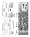
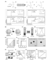
- FIG. 1 is a schematic representation of the design and mechanism of novel nanoparticles (P-ZWNIR NPs) for tumor targeting and penetration.
- P-ZWNIR NPs novel nanoparticles
- FIG. 1 shows the chemical structures of cyclodextrin derivative (P-ZWNIR CD) conjugated with phenylboronic acid (PBA) and a zwitterionic near-infrared fluorophore and pheophorbide (Pheo) modified with ferrocene (Fc) (Pheo-Fc), and the photobleaching-mediated charge transfer mechanism of P-ZWNIR NPs
- PBA phenylboronic acid
- Fc ferrocene
- B schematically shows the delivery cascade and phototherapy regimen of P-ZWNIR NPs.
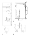
- Figure 2 shows the results of analyzing compound 6 synthesized according to Scheme 1.
- A is the 1 H-NMR spectrum of compound 6
- B is the process for preparing a complex of compound 6 and 2,5-dihydroxybenzoic acid (DHB) for MS analysis
- C is the MALDI-TOF spectrum of the complex prepared according to (B).
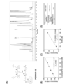
- Figure 3 shows the analysis of compound 7 (P-ZWNIR NP),
- A) is the 1 H NMR spectrum of P-ZWNIR CD in DMSO-d 6 /D 2 O (90:10, v/v),
- B) and (C) are standard calibration curves for quantifying the PBA moiety and the near-infrared fluorophore moiety, respectively, using 3-acetamidoPBA and IR-78 as quantification standards.
- (D) compares the conjugated number of the near-infrared fluorophore moiety in P-ZWNIR CD with the 7-conjugated PBA moiety, and the ratio of PBA to the near-infrared fluorophore is 1.01, confirming that the near-infrared fluorophore moiety is also 7-conjugated.
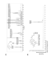
- Figure 4 shows the analysis of compound 10 (Pheo-Fc).
- A is a 1 H NMR spectrum and
- B is an electrospray ionization-mass spectrometry (ESI-MS) spectrum.
- Figure 5 shows the characteristics of P-ZWNIR NPs.
- A The photo-oxidative degradation mechanism of heptamethine cyanine was confirmed using Gly-S-IR783 (a heptamethine cyanine fluorophore model).
- B A schematic diagram of the photobleaching-mediated charge conversion of P-ZWNIR NPs.
- C Digital images of P-ZWNIR NP suspension before and after near-infrared laser irradiation (808 nm, 0.8 W cm -2 , 5 min).
- D UV-vis spectra of P-ZWNIR NPs in phosphate buffer solution (PBS, pH 7.4) before and after laser irradiation.
- (I) shows the singlet oxygen ( 1 O 2 ) generation ability of Pheo-Fc under 660 nm laser irradiation (350 mW, 2 mW cm -2 )
- (J) shows the hydroxyl radical (OH) generation ability of Pheo-Fc (500 ⁇ M) in PBS/DMSO cosolvent (9:1, v/v), which shows the UV-Vis spectra of various mixtures
- ROS total reactive oxygen species
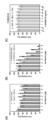
- Figure 6 shows the results of an in vitro cytotoxicity experiment, where (A) and (B) show the cell viability of Pheo-Fc and P-ZWNIR NPs in HT-29 cells under dark and 660 nm laser irradiation conditions, respectively, and (C) shows the cell viability of blank P-ZWNIR NPs according to the incubation time.
- Figure 7 shows (A) flow cytometry results and (B) laser scanning confocal microscopy (LSCM) images of HT-29 cells treated with P-ZWNIR NPs and 2′,7′-dichlorofluorescein diacetate (DCFH-DA), respectively.
- LSCM laser scanning confocal microscopy
- Figure 8 shows the results of (A) H 2 O 2 production in HT-29 and NIH3T3 cells and (B) cell viability in response to dark toxicity of P-ZWNIR NPs.
- Figure 9 shows the ZWNIR CD and ZWNIR NP as negative controls.
- A Schematic diagram showing the synthesis process of ZWNIR CD
- B 1 H-NMR spectrum of ZWNIR CD (DMSO-d 6 /D 2 O (90:10, v/v))
- C hydrodynamic diameter and
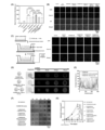
- Figure 10 shows the in vitro cellular uptake, ROS production, transcytosis, and penetration characteristics of P-ZWNIR NPs.
- A Flow cytometry and
- B LSCM images of HT-29 cells after 4 h of incubation with Pheo-Fc, ZWNIR NPs, ZWNIR NPs + 808 nm (laser-pretreated ZWNIR NPs, 0.8 W cm -2 , 5 min), P-ZWNIR NPs, and P-ZWNIR NPs + 808 nm (laser-pretreated P-ZWNIR NPs, 0.8 W cm -2 , 5 min).
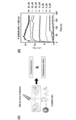
- Figure 11 shows the photothermal effect of P-ZWNIR NPs.
- A is a schematic diagram of near-infrared (808 nm) light-mediated photobleaching and photothermal effect
- B is the temperature increase results of P-ZWNIR NPs at various concentrations under irradiation with an 808 nm laser (0.8 W cm -2 ).
- P-ZWNIR NPs showed dose-dependent hyperthermia due to the photothermal effect.
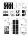
- Figure 12 analyzes the tumor targeting and penetration potential of P-ZWNIR NPs.
- A In vivo real-time near-infrared fluorescence (NIRF) imaging after intravenous injection of ZWNIR NPs or P-ZWNIR NPs into orthotopic (rectal) HT-29 tumor-bearing mice (white dotted circle indicates rectal tumor area),
- B temporal changes in the average radiant efficiency in the tumor area,
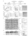
- Figure 13 shows P-ZWNIR NP-assisted combined phototherapy in a rectal cancer (RC) mouse model
- A is a schematic diagram of P-ZWNIR NP-assisted phototherapy
- C is the excised tumor weight and (D) the tumor image on day 20
- E is the tumor growth profile of HT-29 subcutaneous tumor-bearing mice after various laser treatment regimens
- (F) is the excised tumor weight and (G) the tumor image on the last day of the experiment.
- the inventors of the present invention have completed the present invention by confirming that the charge of a substance is converted to a positive charge when heptamethine cyanine, a near-infrared fluorescent dye, is asymmetrically decomposed when photobleaching occurs with a near-infrared laser by modifying a cyclodextrin derivative with heptamethine cyanine in a zwitterionic state and irradiating the cyclodextrin derivative with a near-infrared laser to cause photobleaching.
- the present invention provides a novel cyclodextrin derivative.
- the cyclodextrin derivative according to the present invention may have a zwitterionic near-infrared fluorescent moiety bound thereto.
- a cyclodextrin derivative represented by the following chemical formula 1, to which a heptamethine cyanine-based zwitterionic near-infrared fluorophore using heptamethine cyanine as a target molecule is bound can be provided:
- X and Y may be the same or different and may be selected from ASO 3 - , ACO 2 - , or a salt thereof, wherein A is selected from aromatic or alkyl corresponding to (CH 2 ) a , a is selected from an integer from 1 to 8, m is selected from an integer from 6 to 8, and n may be selected from an integer from 1 to 40.
- the heptamethine cyanine-based zwitterionic near-infrared fluorophore may include, but is not limited to, a heptamethine cyanine-based dye selected from IR-783, IR-780, MHI-148, IR-820, or IR-825.
- cyclodextrin is a circularly bonded cyclic substance and may be selected from cyclodextrins in which m is 6 to 8, and preferably may be ⁇ -cyclodextrin in which m is 7, but is not limited thereto.
- the cyclodextrin derivative containing the above zwitterionic near-infrared fluorophore can be converted into a positive charge by photobleaching upon near-infrared laser irradiation.
- the cyclodextrin derivative according to the present invention may be in a form in which a zwitterionic near-infrared fluorescent moiety and a tumor-targeting moiety are combined with cyclodextrin.
- the present invention provides a cyclodextrin derivative represented by the following chemical formula 2, wherein a heptamethine cyanine-based zwitterionic near-infrared fluorophore and a tumor-targeting moiety are bound to cyclodextrin:
- X and Y can be the same or different and can be selected from ASO 3 - , ACO 2 - , or a salt thereof, wherein A is selected from aromatic or alkyl corresponding to (CH 2 ) a , a is selected from an integer from 1 to 8, R is a tumor targeting moiety, selected from compounds having a molecular weight of less than 10,000 Da used for active targeting, m is selected from an integer from 6 to 8, and n can be selected from an integer from 1 to 40.
- the above-mentioned zwitterionic near-infrared fluorescent moiety can be converted into a positive charge by photobleaching upon near-infrared laser irradiation, and the above-mentioned tumor-targeting moiety can have tumor-targeting ability.
- the heptamethine cyanine-based zwitterionic near-infrared fluorophore may include, but is not limited to, a heptamethine cyanine-based dye selected from IR-783, IR-780, MHI-148, IR-820, or IR-825.
- the heptamethine cyanine-based zwitterionic near-infrared fluorophore may be a heptamethine cyanine dye converted into a zwitterionic state having both positive and negative charges through a ring-opening reaction with 2-iminothiolane.
- the tumor targeting moiety may be at least one compound selected from the group consisting of phenylboronic acid (PBA), peptides containing Arginine-Glycine-Aspartate (RGD), IL-4 receptor targeting peptide (IL4RPep-1), biotin, folate, glucose, galactose, mannose, fructose, sorbitol, N-acetylglucosamine, N-acetylmannosamine, adenosine, thiamine, riboflavin, methotrexate, anisamide, and anacardic acid, but is not limited thereto. It is not.
- PBA phenylboronic acid
- RGD peptides containing Arginine-Glycine-Aspartate
- IL4RPep-1 IL-4 receptor targeting peptide
- biotin folate, glucose, galactose, mannose, fructose, sorbitol, N-acetylglucosamine
- the above phenylboronic acid has binding affinity to sialic acid overexpressed in tumor cells, and thus can target tumor cells.
- cyclodextrin may be selected from cyclodextrins in which m is 6 to 8, and preferably may be ⁇ -cyclodextrin in which m is 7, but is not limited thereto.
- cyclodextrin derivative represented by the following chemical formula 2-1:
- the above near-infrared fluorophore and tumor-targeting moiety can be coupled to cyclodextrin via a linker.
- the above linker may include, but is not limited to, glutamic acid.
- the above cyclodextrin derivative may be one in which a tumor-targeting moiety and a zwitterionic near-infrared fluorophore are conjugated to each terminal of the pegylated cyclodextrin derivative at a molar ratio of 1:1, and preferably, may be heptaconjugated to each terminal of the ⁇ -cyclodextrin, but is not limited thereto.
- the cyclodextrin derivative can be imparted with charge conversion function and tumor targeting ability by simultaneously introducing zwitterionic heptamethine cyanine and phenylboronic acid.
- the present invention provides a compound in which a photosensitizer is incorporated into the novel cyclodextrin derivative.
- the above cyclodextrin derivative is a cyclic substance bonded in a circular shape, and the photosensitizer can be incorporated into the circular shape.
- the photosensitizer may be selected from pheophorbide or a derivative thereof, and is preferably, but not limited to, pheophorbide a bound to ferrocene.
- the ferrocene-bound pheophorbide a can be represented by the following chemical formula 3:
- the above ferrocene has a high affinity for the cyclodextrin cavity, so it can enhance the inclusion effect for cyclodextrin, and at the same time, it can have a chemodynamic effect, so it can convert H2O2 generated by photodynamic therapy with a photosensitizer into hydroxyl radicals, thereby enhancing cytotoxicity.
- the above compound may be one in which ferrocene is bound in a host-guest form within the cyclodextrin.
- the above compound may be a cyclodextrin derivative in which the photosensitizer is incorporated at a molar ratio of 1:1.
- the above compound can be utilized in cancer treatment using the synergistic effect of photodynamic or photodynamic and chemodynamic or combined therapy of photodynamic and chemotherapy by introducing the above photosensitizer.
- the present invention provides nanoparticles formed by self-assembly of a compound in which a photosensitizer is incorporated into the novel cyclodextrin derivative.
- the above compound has amphiphilic properties, in which a hydrophobic photosensitizer is entrapped in a hydrophilic cyclodextrin derivative, and can spontaneously self-assemble under aqueous conditions to form nanoparticles.
- the above nanoparticles may have a zwitterionic charge, but may be converted to a positive charge by photobleaching upon near-infrared irradiation.
- the above near-infrared light can be irradiated in a wavelength range of 600 to 900 nm and an intensity range of 0.5 to 4 mW cm -2 , but is not limited thereto.
- the nanoparticles converted to positive charge by the above near-infrared irradiation can promote cellular uptake and enhance tumor penetration ability through the transcytosis pathway.
- the photothermal effect caused by near-infrared irradiation can also help enhance tumor penetration ability.
- the present invention provides a composition for photodynamic therapy of cancer, comprising the above nanoparticles as an active ingredient.
- the above nanoparticles can be converted into positive charges through near-infrared irradiation (first near-infrared irradiation), thereby enhancing tumor targeting ability and tumor penetration ability, and can be utilized for photodynamic therapy of cancer by activating a photosensitizer through additional near-infrared irradiation (second near-infrared irradiation).
- first near-infrared irradiation near-infrared irradiation
- second near-infrared irradiation additional near-infrared irradiation
- the efficiency of photodynamic therapy can be further enhanced as the tumor penetration ability is enhanced.
- the first near-infrared ray may have a wavelength of 800 to 900 nm and an intensity range of 0.5 to 1 mW cm -2
- the second near-infrared ray may be selected from a wavelength of 600 to 700 nm and an intensity range of 1 to 3 mW cm -2 , but is not limited thereto.
- the cancer may be selected from solid cancers such as colon cancer, liver cancer, and lung cancer, and preferably colon cancer including rectal cancer and squamous cell carcinoma, but is not limited thereto.
- the present invention provides a composition for combined photodynamic and chemotherapy treatment of cancer, comprising the above nanoparticles as an active ingredient.
- the photosensitizer bound to ferrocene constituting the above nanoparticles has excellent photodynamic and chemodynamic activities, and the tumor penetration ability is enhanced by the nanoparticles converted to positive charges through near-infrared irradiation, and then the photosensitizer bound to ferrocene is activated through additional near-infrared irradiation, thereby enhancing the cancer treatment efficiency through the synergistic effect of photodynamic and chemodynamic effects.
- the present invention provides a composition for near-infrared fluorescence imaging for cancer diagnosis or treatment, comprising the above nanoparticles as an effective ingredient.
- the above composition can be used as an optical imaging composition for identifying the extent of tumor cell distribution in a living body, tumor cell targeting of a drug, etc. using near-infrared fluorescence imaging, and thus can be utilized for cancer diagnosis and understanding the extent of cancer treatment.
- the present invention provides a method for treating cancer, comprising: a step of administering the nanoparticle or a cancer treatment composition containing the nanoparticle to a subject; a step of irradiating first near-infrared rays; and a step of irradiating second near-infrared rays.
- the step of administering the nanoparticle or the composition to the subject may be administered to all subjects requiring cancer treatment, or all subjects excluding humans, and the nanoparticle may accumulate at a tumor site in the body.
- the administration may be performed in the form of intravenous injection or intratumoral injection.
- the step of irradiating the first near-infrared ray is such that by irradiating the near-infrared ray with a wavelength of 800 to 900 nm and an intensity range of 0.5 to 1 mW cm -2 , the nanoparticles accumulated at the tumor site can be converted to positive charges by photobleaching and penetrate into the tumor.
- the step of irradiating the second near-infrared ray can treat cancer by irradiating the near-infrared ray with a wavelength of 600 to 700 nm and an intensity range of 1 to 3 mW cm -2 , thereby exhibiting anticancer efficacy through photodynamic or photodynamic/chemodynamic activity by the photosensitizer of the nanoparticles that have penetrated inside the tumor.
- O-(Benzotriazol-1-yl)-N,N,N′,N′-tetramethyluronium hexafluorophosphate (HBTU), 1-Hydroxybenzotriazole hydrate (HOBt), 1-Ethyl-3-(3-dimethylaminopropyl)carbodiimide (EDC), trifluoroacetic acid (TFA), triethylamine (TEA), N,N-Diisopropylethylamine (DIEPA), IR-783, 2-iminothiolane hydrochloride (Traut’s reagent), ferrocenecarboxylic acid, anhydrous N,N-dimethylformamide (DMF), anhydrous dimethylsulfoxide (DMSO), 9,10-anthracenediyl-bis(methylene)-dimalonic acid (ABDA), and 2′,7′-dichlorofluorescein diacetate (DCFH-DA) were purchased from Sigma-
- 3-Aminophenylboronic acid (3-APBA), N-Boc-ethylenediamine, 3,3',5,5'-tetramethylbenzidine (TMB), and 2,5-dihydroxybenzoic acid (DHB) were purchased from Tokyo Chemical (Tokyo, Japan).
- Boc-L-glutamic acid ⁇ -methyl ester was purchased from Santa Cruz Biotechnology (Dallas, TX, USA), and T-boc-N-amido-PEG8-amine was purchased from Broadpharm (San Diego, CA, USA).
- Heptakis-(6-amino-6-deoxy)- ⁇ -cyclodextrin heptahydrochloride was purchased from Cyclodextrin-shop (Tilburg, Netherlands), and pheophorbide a was purchased from Frontier Scientific (Newark, DE, USA).
- NMR solvents including deuterium oxide ( D2O ), dimethyl sulfoxide- d6 (DMSO- d6 ), chloroform-d ( CDCl3 ), and methanol- d4 ( CD3OD ) were purchased from Zeochem AG (R The dialysis membrane (Spectra/Por® 7 Dialysis Tubing, MWCO 1000 Da) was purchased from Repligen Corporation (Rancho Dominguez, CA, USA). RPMI 1640 and phosphate buffered saline (PBS) were purchased from Corning (Manassas, VA, USA).
- D2O deuterium oxide
- DMSO- d6 dimethyl sulfoxide- d6
- CDCl3 chloroform-d
- CD3OD methanol- d4
- Penicillin-streptomycin solution 100X
- FBS fetal bovine serum
- trypsin solution 10X
- Welgene Welgene (Gyeongsangbuk-do, Republic of Korea).
- D-luciferin was purchased from Perkin Elmer (Boston, MA, USA). All other reagents were of analytical grade.
- HT-29 human colorectal adenocarcinoma
- NIH3T3 cell lines fibroblasts
- a 660 nm laser (CYL20ADJ) was purchased from Laser Solution Korea (Gyeonggi-do, Republic of Korea), and a continuous wave near-infrared (808 nm) diode laser beam was purchased from CNILaser (Changchun, China).
- Phenylboronic acid (PBA) and zwitterionic near-infrared (NIR) fluorophore-modified nanoparticles are composed of an inclusion compound formed by a hydrophilic ⁇ -cyclodextrin (CD) derivative (host) moiety and a hydrophobic photosensitizer (guest) moiety (Fig. 1A).
- CD hydrophilic ⁇ -cyclodextrin
- guest hydrophobic photosensitizer
- Cyclodextrin derivatives conjugated with phenylboronic acid and a zwitterionic near-infrared fluorophore (P-ZWNIR CD) were synthesized by tethering phenylboronic acid and heptamethine cyanine fluorophores to the cyclodextrin core using glutamic acid as a tri-arm linker. Phenylboronic acid was used to target sialic acid-overexpressing cancer cells, and the heptamethine cyanine fluorophore was used as a charge-switching moiety.
- reaction scheme 1 shows the specific synthetic process of P-ZWNIR CD.
- the PBA moiety (compound 2) was first introduced into the PEGylated CD (compound 4) to obtain compound 5, which was then deprotected under acidic conditions to obtain compound 6.
- the heptaconjugation of the PBA moiety of compound 6 can be confirmed by 1 H-NMR and MALDI-TOF MS analyses (Fig. 2).
- a heptamethine cyanine (IR-783) moiety was introduced into compound 6.
- the nanomaterial In order for the nanomaterial to be converted from a zwitterionic state to a positively charged state through photobleaching, it is important that the heptamethine cyanine moiety is linked to the cyclodextrin structure as a zwitterion.
- the compound 6 was synthesized by conjugating IR-783 with the above compound using 2-iminothiolane, a Traut reagent.
- 2-Iminothiolane was used as a linker to convert negatively charged IR-783 into a zwitterionic heptamethine fluorophore (ZWNIR) by generating a positive charge at the amidine bond through a ring-opening reaction.
- ZWNIR zwitterionic heptamethine fluorophore
- P-ZWNIR CD (compound 7).
- the degree of ZWNIR moiety conjugation in compound 6 was calculated by comparing the integrated values of the anomeric protons of CD ( ⁇ 4.7 ppm) and the distinct signal of IR-783 ( ⁇ 6.2 ppm) in the P-ZWNIR CD, and NMR analysis showed that the degree of ZWNIR moiety in the P-ZWNIR CD was about 7.
- the molar ratio of ZWNIR moiety to PBA moiety in the P-ZWNIR CD was determined to be about 1:1 by UV-Vis spectroscopy using alizarin red S, which also indicates the heptaconjugation of ZWNIR.
- Pheophorbide a was selected as a phototherapy agent and modified with ferrocene (Fc) to utilize the synergistic effect of photodynamic/chemodynamic therapy and the binding affinity of Fc for CD cavities.
- reaction scheme 2 shows the specific synthetic process of ferrocene-linked pheophorbide a (Pheo-Fc).
- compound 8 was synthesized according to a previously reported method. Ferrocene carboxylic acid (250 mg, 1.08 mmol), HOBt (175 mg, 1.30 mmol), and EDC (225 mg, 1.30 mmol) were dissolved in anhydrous DMF (10 mL), followed by addition of DIPEA (0.84 mL, 5.40 mmol). After stirring at room temperature for 30 min under a nitrogen atmosphere, N-Boc-ethylenediamine (0.34 g, 2.17 mmol) was added and stirred at room temperature for 12 h. The reaction mixture was diluted with double distilled water (DDW) and extracted with ethyl acetate (EtOAc) (3 ⁇ 50 mL).
- DDW double distilled water
- EtOAc ethyl acetate
- Boc-deprotection of compound 8 was carried out using 50% TFA in DCM at room temperature for 2 h. After the reaction, the reaction solvent was evaporated in vacuo, and the crude product was further purified by flash column chromatography using EtOAc:MeOH to give compound 9 (yield 83%).
- P-ZWNIR NPs The fabrication of P-ZWNIR NPs consists of two steps: (1) formation of inclusion complexes and (2) self-assembly of the inclusion complexes in an aqueous medium.
- ZWNIR NPs which were prepared by synthesizing ZWNIR CDs lacking PBA in the terminal structure for use as a negative control, were also prepared by the same process.
- P-ZWNIR CD 0.013 mmol
- 13 mg of ZWNIR CD 0.013 mmol
- Pheo-Fc 0.013 mmol
- the mixture of CD derivative and Pheo-Fc was vortexed overnight to form an inclusion complex.
- the formed inclusion complex was slowly dropwise added to 4 mL DDW with constant stirring for 1 h.
- the resulting P-ZWNIR NP or ZWNIR NP solution was placed in a dialysis bag (MWCO 12-16 kDa, Membra-Cel, Viskase Corporation, Chicago, IL, USA) and dialyzed against DDW for 6 h. The DDW was replaced every h to completely remove the residual DMSO.
- the concentration of NIR dye in the NPs was analyzed by UV-Vis spectrometry (Orion AquaMate 8100, Thermo Fisher Scientific, Waltham, MA, USA) at 780 nm absorbance.
- Pheo-Fc was loaded onto P-ZWNIR CD to form an inclusion complex by utilizing the host–guest interaction between the Fc and CD cavities.
- the P-ZWNIR CD/Pheo-Fc inclusion complex was prepared in dimethyl sulfoxide (DMSO) with a molar ratio of P-ZWNIR CD to Pheo-Fc of 1:1, and the prepared P-ZWNIR CD/Pheo-Fc solution was dropped into DDW and stirred to prepare P-ZWNIR NPs by self-assembly.
- DMSO dimethyl sulfoxide
- the outer ZWNIR structure of P-ZWNIR NPs can be asymmetrically decomposed by oxidative decomposition under NIR ( ⁇ 808 nm) laser irradiation, the zwitterionic P-ZWNIR NPs can be converted into positively charged NPs after NIR laser irradiation.
- P-ZWNIR NPs manufactured in this way can be utilized as nanoparticles for intravenous injection.
- P-ZWNIR NPs once they circulate in the bloodstream in a zwitterionic state, and (2) progressively accumulate in tumor tissues through extravasation and active targeting. However, they penetrate only a short distance and mainly remain in the perivascular region.
- the NPs in the tumor Upon irradiation of the tumor site with an 808 nm laser, (3) the NPs in the tumor are charge-converted to cationic NPs, which (4) enhances tumor penetration through transcytosis.
- NPs The transcytosis-mediated deep tumor penetration of NPs enables intracellular delivery and uniform distribution of a photosensitizer (PS), which then, upon irradiation with a 660 nm laser, (5) the anticancer efficacy will be enhanced by the combined photodynamic and chemodynamic therapy of pheophorbide (Pheo) and ferrocene (Fc), respectively.
- PS photosensitizer
- the mobile phase consisted of MeOH and DDW, and the gradient elution procedure was as follows: 70 mL of MeOH:DDW (50:50, v/v) was used to remove hydrophilic impurities, and then the MeOH percentage was increased to 60% to obtain Gly-S-IR-783. The product was dried under reduced pressure (yield 56%).
- the Gly-S-IR783 prepared by the above method was dissolved in PBS and irradiated with a NIR (808 nm) laser (0.8 W cm -2 ) for 5 min to induce photobleaching.
- the resulting solution was analyzed by liquid chromatography-electrospray ionization-mass spectrometry (LC-ESI-MS).
- LC-ESI-MS liquid chromatography-electrospray ionization-mass spectrometry
- the laser irradiation induced the asymmetric decomposition of Gly-S-IR783, generating a combination of two ketone and aldehyde fragments (a + b or c + d) ( Figure 5A).
- both pathways left a cation at the amidine bond conjugation moiety. Since the amidine bond is connected to the cyclodextrin (CD) core in P-ZWNIR CD, only the cation moiety may remain after NIR laser irradiation of the
- the average hydrodynamic diameter, size distribution, polydispersity index (PI), and zeta potential of NPs were determined using a Zetasizer (Malvern Panalytical, Worcestershire, UK), and the morphologies of P-ZWNIR NPs before and after irradiation with an 808 nm laser were observed by a transmission electron microscope (TEM, JEM1010, JEOL, Tokyo, Japan) operating at an acceleration voltage of 80 kV. As shown in Fig. 5(E), the zeta potential of P-ZWNIR NPs gradually increased upon NIR laser irradiation and reached a plateau at 5 min, indicating that the photobleaching-mediated charge conversion of NPs was completed within 5 min.
- TEM transmission electron microscope
- P-ZWNIR NPs and cationic NPs showed similar size distribution profiles in dynamic light scattering (DLS) analysis (Fig. 5F), and transmission electron microscopy (TEM) images also showed corresponding particle sizes (Fig. 5G).
- the absorption spectrum of the mixture was measured every 2 min in the range of 300 to 500 nm for 10 min using a UV-Vis spectrometer. Controls were performed with the same procedure with Pheo-Fc solution or ABDA solution alone in a darkroom or under irradiated conditions. Referring to Fig. 5(I), the absorbance of ABDA rapidly decreased upon irradiation with a 660 nm laser, suggesting a reaction with singlet oxygen generated from Pheo-Fc.
- TMB 3,3',5,5'-tetramethylbenzidine
- 500 ⁇ L Pheo-Fc solution (4 mmol L -1 in DMSO) was mixed with 4 mL of test solution.
- the compositions of the test solutions were as follows: 1) TMB, H 2 O 2 , PBS, pH 5.0; 2) TMB, H 2 O 2 , PBS, pH 7.4; 3) TMB, PBS, pH 5.0; 4) H 2 O 2 , PBS, pH 5.0.
- the total ROS generating capacity of Pheo-Fc and P-ZWNIR NPs was compared using dichlorodihydrofluorescein (DCFH) as an indicator.
- DCFH-DA 2′,7′-Dichlorofluorescein diacetate
- I 0 and I represent the fluorescence intensities of the mixture at the initial time point and every 5 seconds for 1 min, respectively. Referring to Fig.
- HT-29 cells were cultured in RPMI 1640 supplemented with 10% FBS (v/v) and 1% penicillin-streptomycin (v/v) at 37°C, 5% CO2, and 95% relative humidity.
- FBS v/v
- penicillin-streptomycin v/v
- 5,000 cells were seeded per well of a 96-well plate (SPL Life Sciences, Gyeonggi-do, Republic of Korea). After 24 h of incubation, the cells were treated with Pheo-Fc or P-ZWNIR NPs and incubated for 4 h.
- the complete medium was removed and replaced with fresh serum-free RPMI 1640 medium, and then irradiated with a 660 nm laser (350 mW; 2 mW/ cm2 ) at a distance of 2 cm for 1 min per well, and the cells were further incubated for 24 h.
- Cell viability was assessed using 3-(4,5-dimethylthiazol-2-yl)-5(3-carboxymethoxyphenyl)-2-(4-sulfophenyl)-2H-tetrazolium [3-(4,5-dimethylthiazol-2-yl)-5(3-carboxymethoxyphenyl)-2-(4-sulfophenyl)-2H-tetrazolium, MTS] reagent (CellTiter 96 Aqueous One Solution Cell Proliferation Assay; Promega, Milan, Italy) according to the manufacturer's instructions. The absorbance at 490 nm was measured with a microplate reader (Multiskan Microplate Spectrophotometer; Thermo Fisher Scientific, Massachusetts, USA).
- HT-29 cells were cultured with P-ZWNIR CD solution (0.2-100 ⁇ g mL -1 ) for 24, 48, and 72 h, and cell viability was evaluated by the method described above.
- Intracellular ROS detection was performed using DCFH-DA as a dye reagent.
- Intracellular esterase cleaves the diacetate group of DCFH-DA to generate DCFH, which can be converted to highly fluorescent fluorescein (DCF) when exposed to ROS.
- HT-29 cells were cultured with P-ZWNIR NPs (2 ⁇ mol mL -1 as Pheo-Fc) in a cell culture medium without phenol red for 4 h, washed three times with PBS, and then DCFH-DA solution was added (10 ⁇ mol mL -1 in PBS). After 15 min of incubation, the culture medium was replaced with a cell culture medium without phenol red.
- a 660 nm laser 350 mW was irradiated for 5 min.
- the treated cells were analyzed using a flow cytometer (FACSCalibur, BD Biosciences).
- the flow cytometer measured the mean fluorescence intensity of DCF converted within the cells.
- HT-29 cells treated in the above manner were observed using a confocal microscope (Leica TCS SP8), and fluorescence was measured at an excitation wavelength of 488 nm with a GFP filter.
- H 2 O 2 released from HT-29 or NIH3T3 cells was measured using the Amplex Red® Hydrogen Peroxide/Peroxidase assay kit (Thermo Fisher Scientific, Waltham, MA, US) according to the manufacturer's instructions.
- HT-29 and NIH3T3 cells were cultured in RPMI 1640 and DMEM medium supplemented with 10% FBS (v/v) and 1% penicillin-streptomycin (v/v), respectively.
- a reaction mixture containing 50 ⁇ M Amplex Red reagent and 0.1 U/mL horseradish peroxidase (HRP) in Krebs-Ringer phosphate (KRPG) buffer (pH 7.35) was prepared.
- reaction mixture 100 ⁇ L was filled into each well of a 96-well microplate and incubated at 37 °C for 10 min. 2 ⁇ 10 6 HT-29 or NIH3T3 cells (20 ⁇ L) in KRPG buffer were added to each well, and the amount of H 2 O 2 produced was quantified by measuring the absorbance of the reaction mixture at 560 nm.
- the dark toxicity of P-ZWNIR NPs could be mainly attributed to ⁇ OH generated from Pheo-Fc.
- the significant difference in efficacy between Pheo-Fc and P-ZWNIR NPs can be explained by the lower total ROS production of Pheo-Fc in PBS (Fig. 5K).
- the higher cytotoxicity observed with P-ZWNIR NPs (+660 nm) compared to P-ZWNIR NPs (dark) could be attributed to the activation of the photodynamic agent by 660 nm laser irradiation (Fig. 7).
- HT-29 cells cancer cell line
- NIH3T3 cells normal cell line
- HT-29 cells showed significantly higher H 2 O 2 production than NIH-3T3 cells
- NIH3T3 cells with relatively lower H 2 O 2 production showed lower dark chamber toxicity of P-ZWNIR NPs compared to HT-29 cells, which was evidenced by their higher IC 50 value (Pheo-Fc of 15.4 ⁇ 0.5 ⁇ M) (Fig. 8B).
- ZWNIR NPs Prior to cellular uptake studies, ZWNIR NPs were prepared by synthesizing ZWNIR CDs lacking PBA in the terminal structure and used as a negative control (Fig. 9). ZWNIR CDs were synthesized by conjugating NIR dye to the PEGylated CD core using the same reaction used for the synthesis of P-ZWNIR CDs. The degree of NIR dye conjugation in ZWNIR CDs was calculated to be about 7 by UV-vis spectroscopy using Alizarin Red S and 1 H NMR spectrum, indicating heptaconjugation of ZWNIR moiety within the CD core. ZWNIR NPs were prepared from ZWNIR CD/Pheo-Fc inclusion complex, and the NPs also exhibited NIR photo-responsive charge-switching properties without significant change in particle size distribution.
- the cellular uptake efficiency of P-ZWNIR NPs was evaluated in HT-29 cells using flow cytometry and laser scanning confocal microscopy (LSCM).
- HT-29 cells For flow cytometry, 1 ⁇ 10 5 HT-29 cells were seeded into each well of a 6-well cell culture plate (SPL Life Sciences, Gyeonggi-do, Republic of Korea) and cultured at 37 °C for 24 h. Then, HT-29 cells were treated with Pheo-Fc, ZWNIR NPs, ZWNIR NPs + 808 nm (laser-pretreated ZWNIR NPs), P-ZWNIR NPs, and P-ZWNIR NPs + 808 nm (laser-pretreated P-ZWNIR NPs) (2 ⁇ M of Pheo-Fc) and incubated for 4 h.
- Positively charged NPs were prepared by irradiating P-ZWNIR NPs with an 808 nm laser and were prepared before treatment on HT-29. Free sialic acid (500 ⁇ mol L -1 ) or 3-aminophenylboronic acid (3-APBA, 500 ⁇ mol L -1 ), which acts as a competitor for PBA-sialic acid interaction, was incubated with the formulations for 15 min prior to cell treatment. Cells treated with nanoparticles were analyzed after 4 h of incubation. The mean fluorescence intensity of Pheo-Fc was analyzed and processed using FACSCalibur and CellQuest Pro Software (BD Biosciences, San Jose, CA, USA).
- the average fluorescence intensities of cells treated with P-ZWNIR NPs and P-ZWNIR NPs + 808 nm were significantly higher than those of cells treated with ZWNIR NPs and ZWNIR NPs + 808 nm, respectively.
- the cellular uptake of photobleached P-ZWNIR NPs (P-ZWNIR NPs + 808 nm) was inhibited in the presence of competitive inhibitors (free sialic acid or free 3-aminophenylboronic acid (3-APBA)).
- the photobleached ZWNIR NPs and P-ZWNIR NPs show higher cellular uptake than ZWNIR NPs and P-ZWNIR NPs themselves, indicating that cation conversion promotes the cellular uptake of the NPs.
- HT-29 cells For confocal microscopy, 1 ⁇ 10 5 HT-29 cells were seeded into each well of a 4-well cell culture plate (SPL Life Sciences, Gyeonggi-do, Republic of Korea), cultured at 37°C for 24 h, and then treated in the same manner as for flow cytometry described above. After washing three times with PBS (pH 7.4), the cells were fixed with 4% (w/v) formaldehyde solution, stained with 4′,6-diamidino-2-phenylindole (DAPI; ThemoFisher Scientific, Eugene, OR, USA), and mounted in Fluoroshield (Sigma-Alrich, Saint Louis, MO, USA).
- DAPI 4′,6-diamidino-2-phenylindole
- HT-29 cells (1 ⁇ 10 5 ) were seeded in a 12-well cell culture plate without a round coverslip (SPL Life Sciences, Gyeonggi-do, Republic of Korea) (Plate 1) or in a plate with a round coverslip (Plate 2) and cultured for 24 h to allow cell attachment. After 24 h, the cells in Plate 1 were treated with P-ZWNIR NPs or 808 nm-pretreated P-ZWNIR NPs (2 ⁇ mol L -1 as Pheo-Fc) and cultured for 4 h to induce cellular uptake.
- the cells that had uptaken NPs were washed three times with PBS, detached with trypsin-EDTA solution, centrifuged at 250 ⁇ g for 5 min, and resuspended in RPMI 1640.
- the cells were transferred to a transwell insert and placed in the apical compartment of Plate 2, and the transwell system was further cultured at 37 °C for 4 h. Cells on round coverslips from the basolateral compartment were washed and fixed. The fixed cells were stained with DAPI and observed using a Leica TCS SP8 confocal microscope.
- the fluorescence of Pheo-Fc taken up by the attached cells in the basolateral compartment was observed in this experiment.
- the fluorescence signal of the cells in the basolateral compartment indicates that the NPs were excreted from the HT-29 cells in the apical compartment, moved across the membrane, and then taken up by the basolateral compartment cells.
- the group treated with P-ZWNIR NPs showed weak fluorescence, and the fluorescence intensity decreased in the presence of Exo1, an exocytic inhibitor.
- the tumor penetration ability of NPs was investigated using 3D HT-29 multicellular tumor spheroids (MTS). Considering that the photothermal effect of P-ZWNIR NPs under 808 nm may affect the tumor penetration of NPs, the tumor penetration ability of the following groups was tested and compared:
- HT-29 3D multicellular spheroids were seeded at 500 cells per well in a 96-well SPL3D Cell Floater (SPL Life Sciences, Gyeonggi-do, Republic of Korea) containing complete RPMI 1640 medium at 37°C, 5% CO 2 , and 95% relative humidity. Spheroids were grown with fresh cell culture medium every 2 days until the average diameter of the spheroids reached 300 ⁇ m, and then treated with P-ZWNIR NPs or 808 nm laser-pretreated P-ZWNIR NPs (2 ⁇ mol L -1 as Pheo-Fc).
- the P-ZWNIR NPs + 808 nm (pretreated) group were irradiated with an 808 nm laser (0.8 W cm -2 ) for 5 min before treatment of the HT-29 spheroids, while for the P-ZWNIR NPs + 808 nm (in situ) group, the HT-29 spheroids were irradiated with an 808 nm laser (0.8 W cm -2 , 5 min) immediately after treatment of the spheroids with the P-ZWNIR NPs.
- the zwitterionic P-ZWNIR NPs were mainly located at the periphery of the spheroids.
- the cationic NPs P-ZWNIR NP + 808 nm (pretreated)
- the penetration ability of P-ZWNIR NP + 808 nm (in situ) was higher than that of P-ZWNIR NP + 808 nm (pretreated).
- the fluorescence intensity-distance plot corresponding to 150 ⁇ m z-stacks showed that the average fluorescence intensity was the highest at P-ZWNIR NP + 808 nm (in situ) (Fig. 10E).
- MTS were cultured according to the protocol described above. After growing until the average diameter of the spheroids reached 300 ⁇ m, they were randomly divided into six groups: untreated, P-ZWNIR NP (dark condition), P-ZWNIR NP + 808 nm (in situ), P-ZWNIR NP + 660 nm, P-ZWNIR NP + 808 nm (pretreatment) + 660 nm, and P-ZWNIR NP + 808 nm (in situ) + 660 nm. The treatment concentration was 5 ⁇ M with Pheo-Fc. The laser irradiation schedule was the same as that described in the penetration efficiency evaluation experiment of 2-3.
- V (mm 3 ) 0.5 ⁇ longest diameter ⁇ (shortest diameter) 2
- P-ZWNIR NPs without laser treatment slightly inhibited the growth of MTS. Irradiation at 660 nm for P-ZWNIR NPs significantly inhibited the spheroid growth compared with P-ZWNIR NPs (dark), and especially, the P-ZWNIR NP + 808 nm (pretreatment) + 660 nm group inhibited the growth of MTS more effectively compared with the P-ZWNIR NP + 660 nm group (three times lower spheroid volume). This indicates that the deeper penetration of NPs enhanced the antitumor efficacy.
- the in vivo tumor-targeting ability of P-ZWNIR NPs was evaluated in a HT-29 tumor xenograft mouse model using near-infrared fluorescence (NIRF) imaging.
- NIRF near-infrared fluorescence
- mice Male nude mice (5 weeks old) were purchased from Nara Biotech (Seoul, Republic of Korea). The animal experiment protocol (SNU-200630-1-1) was approved by the Animal Care and Use Committee of Seoul National University (Seoul, Republic of Korea). Mice were maintained at 22 ⁇ 2°C in a light-controlled room with a relative humidity of 55 ⁇ 5% (Animal Center for Pharmaceutical Research, College of Pharmacy, Seoul National University, Republic of Korea). Mice were anesthetized by intraperitoneal (IP) injection of 250 mg kg -1 avertin or inhaled isoflurane (induction 3% and maintenance 1.5%).
- IP intraperitoneal
- An ectopic (subcutaneous) HT-29 tumor xenograft mouse model was established by subcutaneously injecting 100 ⁇ L of HT-29 cell suspension (2 ⁇ 10 6 cells) into the right hind limb.
- the tumor volume (V) was calculated according to Equation 1.
- An ear tumor transplantation mouse model was used for in vivo tumor invasion experiments, which was established by subcutaneously injecting 20 ⁇ L of HT-29 cell suspension (2 ⁇ 10 6 cells) into the mouse ear.
- the orthotopic (rectal) xenograft mouse model was established by carefully exposing the rectum of the mouse and inoculating 20 ⁇ L of HT-29 cell suspension (2 ⁇ 10 6 cells) into the submucosal layer of the rectal wall.
- HT-29/Luc-GFP luciferase/green fluorescent protein-expressing HT-29
- the transfected HT-29/Luc-GFP cells were then cloned by serial dilution in 96-well plates. Single clones were selected based on the GFP fluorescence signal observed by a biomolecular imaging system (AmershamTM ImageQuantTM 800, Cytiva Life Sciences, Marlborough, MA, US).
- the NIR fluorescence intensity in the tumors was observed to increase up to 8 h and then decrease in both orthotopic and ectopic tumor-bearing mice, indicating that NPs gradually accumulated in the tumors during the first 8 h and were subsequently cleared from the tumors.
- major organs and tissues were resected for ex vivo NIRF imaging, which revealed a higher mean fluorescence intensity at 8 h than at 24 h.
- ZWNIR NPs and P-ZWNIR NPs were injected into the tail vein of mice bearing rectal HT-29 tumors, and real-time NIRF imaging was performed for 8 h.
- the results showed that ZWNIR NPs exhibited a similar accumulation trend to P-ZWNIR NPs, but the fluorescence intensity in the tumor region of P-ZWNIR NPs was 1.5 times higher than that of ZWNIR NPs (Figs. 12A and 12B).
- ex vivo NIRF imaging showed that the accumulation of P-ZWNIR NPs in the tumor was 1.6 times higher than that of ZWNIR NPs, but the off-target accumulation profiles of both NPs were similar except for the kidney (Fig. 12C).
- P-ZWNIR NPs exhibited a near-infrared fluorescence imaging capability specific to rectal tumors.
- the tumor-to-background ratio (TBR) values of P-ZWNIR NPs for adjacent normal tissues were 6.3, 8.0, 10.1, and 7.8, respectively, which were significantly higher than that of ZWNIR NPs (Fig. 12E). Since contrast agents with a TBR value of 2.5 or higher can be applied to tumor image-guided surgery under near-infrared light, P-ZWNIR NPs can be used to reduce the size of the resection margin in tumor resection.
- P-ZWNIR NPs 1.5 mg kg -1 as Pheo-Fc
- P-ZWNIR NPs were intravenously injected into tumor-bearing mice.
- In vivo whole-body NIRF imaging was performed 8 h after injection using a Visque InVivo Smart-LF imaging system (Vieworks, Gyeonggi-do, Republic of Korea).
- the rectal tumor area of the mice was irradiated with an 808 nm laser (0.8 W cm -2 ) and the change in fluorescence intensity was monitored. Photobleaching of P-ZWNIR NPs in the tumor was further evaluated using a MALDI-MSI system.
- Tumor-bearing mice were intravenously injected with P-ZWNIR NPs, and the mice were divided into two groups: before and 8 h after 808 nm laser irradiation (0.8 W cm -2 , 5 min). Rectal tumors were resected from the mice, immediately frozen, and stored at -70 °C until use. The frozen tumor tissues were sectioned to 10 ⁇ m thickness (CM3050S; Leica Biosystems) and mounted on indium tin oxide (ITO)-coated glass slides. The samples were coated with DHB matrix solution (40 mg mL -1 DHB in MeOH/DDW (50:50, v/v) with 0.1% TFA). Mass spectrometry images of the tumor tissue sections were acquired using a RapifleX MALDI Tissuetyper (Bruker Daltonics, Billerica, MA, USA).
- the fluorescence intensity of the tumor area gradually decreased during the first 4 min of 808 nm laser irradiation (0.8 W cm -2 ) and then maintained at a similar level of fluorescence intensity.
- the generation of ketone fragments (c in Fig. 5A) by photobleaching of P-ZWNIR NPs in the dissected tumor was observed using MALDI-MSI.
- the ketone fragment signals in the tumor tissue significantly increased, suggesting that the photobleaching of P-ZWNIR NPs in the tumor followed the mechanism presented in Fig. 2(A).
- the ketone fragment signals within the tumor were evenly distributed throughout the tumor mass, indicating that uniform charge conversion was achieved in the rectal tumor tissue.
- the in vivo tumor penetration efficiency of P-ZWNIR NPs was evaluated by two-photon microscopy in HT-29 ear tumor-bearing mice before and after 5 min irradiation with an 808 nm laser (0.8 W cm -2 ).
- P-ZWNIR NPs 1.5 mg kg -1 as Pheo-Fc
- the fluorescence from Pheo-Fc in the tumor tissue was monitored at predetermined time points (0, 5, 10, 15, 30, 45, and 60 min) using a two-photon laser scanning microscope (FVMPE, Olympus Corporation, Tokyo, Japan).
- FVMPE two-photon laser scanning microscope
- the tumor site was irradiated with an 808 nm laser (0.8 W cm -2 ) for 5 min to induce photobleaching and tumor penetration of P-ZWNIR NPs.
- the fluorescence signal was recorded for an additional 1 h, and the fluorescence intensity was normalized to the vascular fluorescence intensity at the initial time point at each step.
- Graphs were plotted as normalized fluorescence intensity of the region of interest (ROI) versus distance from the blood vessel using ImageJ (version 1.53t; National Institutes of Health, USA).
- ROI region of interest
- ImageJ version 1.53t; National Institutes of Health, USA.
- Temperature changes in the ear tumor during and after NIR irradiation were recorded using an infrared thermal imaging camera (FLIR ONE Pro; Teledyne FLIR, Wilsonville, OR, USA).
- the tumor tissue transplanted into the ear of the mouse was observed under a two-photon microscope 4 hours after intravenous administration of P-ZWNIR NPs. Since the near-infrared fluorescence of P-ZWNIR NPs disappears after 808 nm irradiation, the fluorescence signal of Pheo-Fc contained in P-ZWNIR NPs was observed before and after laser irradiation. Before 808 nm laser irradiation, the fluorescence signal of P-ZWNIR NPs was mainly confined to the blood vessels (Fig. 12I), and a weak fluorescence signal was observed within a distance of about 30 ⁇ m (Fig. 12J).
- the photothermal effect of P-ZWNIR NPs can affect the diffusion and penetration of NPs.
- the temperature of the tumor site only increases to about 41°C, and after 5 minutes, the tumor temperature quickly decreases to the body temperature range. Therefore, the enhanced tumor penetration of P-ZWNIR NPs can be mainly attributed to the transcytosis of photobleached cationic NPs rather than the hyperthermic effect.
- Subcutaneous HT-29 tumor xenograft mouse models were prepared as described in the In Vivo Infiltration and Tumor Penetration sections.
- mice When the tumor volume reached about 250 mm 3 (day 0), the mice were randomly divided into six groups: PBS, PBS + 808 nm + 660 nm, P-ZWNIR NPs (darkroom), P-ZWNIR NPs + 660 nm (photodynamic/chemodynamic effect), P-ZWNIR NPs + 808 nm (photothermal effect and enhanced tumor penetration by photobleaching), and P-ZWNIR NPs + 808 nm + 660 nm (photothermal effect combined with enhanced tumor penetration).
- PBS or P-ZWNIR NPs were administered intravenously at a Pheo-Fc dose of 1.5 mg kg -1 .
- the laser intensities of 808 nm and 660 nm were 0.8 W cm -2 and 2 mW cm -2 , respectively, and both lasers were irradiated to the tumors for 5 min.
- the 808 nm laser (the first laser) was irradiated for 5 minutes, and the temperature of the tumor site was monitored every minute during the irradiation.
- the 660 nm laser (the second laser) was irradiated 10 hours after injection (Fig. 13A).
- the treatment interval between the two lasers was set to 2 hours to ensure sufficient tumor penetration of P-ZWNIR NPs after 808 nm irradiation (photobleaching).
- Plasma samples were analyzed using Fuji Dri-Chem 3500s (Fujifilm Holdings Corp., Tokyo, Japan), and aspartate transaminase (AST), alanine transaminase (ALT), alkaline phosphatase (ALP), blood urea nitrogen (BUN), creatinine (CRE), total protein (TP), and lactate dehydrogenase (LDH) were recorded.
- AST aspartate transaminase
- ALT alanine transaminase
- ALP alkaline phosphatase
- BUN blood urea nitrogen
- CRE creatinine
- TP total protein
- LDH lactate dehydrogenase
- the maximum temperature observed during 808 nm laser irradiation (P-ZWNIR NP + 808 nm and P-ZWNIR NP + 808 nm + 660 nm) was less than 45°C, which did not significantly damage the surrounding normal tissues.
- the tumor growth rate of the PBS + 808 nm + 660 nm group was similar to that of the PBS group, suggesting that the laser treatment itself did not exhibit a significant inhibitory effect on tumor growth.
- P-ZWNIR NP (without laser treatment) and P-ZWNIR NP-adjuvanted single laser treatment (808 nm or 660 nm) significantly inhibited tumor growth compared with the PBS group, but did not lead to tumor shrinkage.
- P-ZWNIR NP + 808 nm + 660 nm treatment completely ablated clinically relevant sized tumors ( ⁇ 250 mm 3 ) after single treatment on day 0. Five of the six mice in the group achieved complete remission, and only one showed a very small tumor recurrence after 14 days.
- ZWNIR NPs or P-ZWNIR NPs were intravenously injected on day 0 and two different laser radiation schedules were applied as follows:
- mice were randomly divided into five groups: PBS, ZWNIR NPs + 808 nm (8 h post-injection) + 660 nm (10 h post-injection), ZWNIR NPs + 660 nm (8 h post-injection) + 660 nm (10 h post-injection), P-ZWNIR NPs + 808 nm (8 h post-injection) + 660 nm (10 h post-injection), and P-ZWNIR NPs + 660 nm (8 h post-injection) + 660 nm (10 h post-injection), and the tumor volumes and body weights were monitored every other day.
- the organs and tissues of interest including tumors, heart, lungs, liver, kidneys, and spleen, were dissected. Tumor weights were measured, normal organs were stained with hematoxylin and eosin, and tissue images were observed using Vectra (v3.0.5).
- the P-ZWNIR NP phototherapy group showed a higher antitumor effect in both irradiation schedules compared to ZWNIR NPs of the same irradiation schedule, which can be attributed to the fact that P-ZWNIR NPs accumulated more in the tumor site than ZWNIR NPs.
- the 808 nm laser (first) + 660 nm (second) irradiation schedule showed significantly higher antitumor efficacy compared to the 660 nm laser (first) + 808 nm (second) irradiation schedule.
- Orthotopic rectal tumor models were prepared by inoculating male BALB/c nude mice (5 weeks old) with luciferase/green fluorescent protein-expressing HT-29 (HT-29/Luc-GFP) cells. Seven days after inoculation, the mice were randomly divided into three groups: PBS, P-ZWNIR NPs (darkroom), and P-ZWNIR NPs + 808 nm + 660 nm, and intravenously injected with Pheo-Fc at a dose of 1.5 mg kg -1 . The ray treatment schedule was the same as that for the orthotopic tumor model. Tumor growth (via bioluminescence) and body weight were measured on days 0, 4, 7, 11, 14, and 20.
- D-luciferin 150 mg kg -1
- bioluminescence intensity was measured using an IVIS Spectrum In Vivo Imaging System (PerkinElmer).
- the mouse rectal tissue was dissected, then 300 ⁇ g mL -1 D-luciferin solution was added, cultured, and the rectal tissue was transferred to the stage and imaged with the IVIS Spectrum In Vivo Imaging System.
- the bioluminescence intensities of the PBS and P-ZWNIR NP-treated groups gradually increased over the experimental period, and multiple signal foci were observed in some mice, indicating tumor growth with metastasis in the orthotopic rectal tumor model.
- the bioluminescence intensity of the P-ZWNIR NP + 808 nm + 660 nm group decreased to the self-bioluminescence level on day 4 after treatment, which remained the same throughout the experimental period.
- bioluminescence signals were observed only in the PBS and P-ZWNIR NP groups, whereas no bioluminescence signal was observed in the rectal tissue in the P-ZWNIR NP + 808 nm + 660 nm group.
- the bioluminescence intensities of the rectal tissue were similar between the PBS and P-ZWNIR NP groups, indicating that P-ZWNIR NPs alone did not inhibit rectal tumor growth.
- the bioluminescence intensity of the P-ZWNIR NP-assisted phototherapy group was at the same level as the autobioluminescence, suggesting complete remission of rectal tumors after phototherapy (Fig. 13L). Toxicity studies including blood biochemistry and complete blood count analysis showed no significant difference among the three groups, suggesting that the toxicity of phototherapy using P-ZWNIR NPs in the orthotopic rectal tumor mouse model is negligible.
Landscapes
- Health & Medical Sciences (AREA)
- Life Sciences & Earth Sciences (AREA)
- Chemical & Material Sciences (AREA)
- General Health & Medical Sciences (AREA)
- Veterinary Medicine (AREA)
- Public Health (AREA)
- Animal Behavior & Ethology (AREA)
- Medicinal Chemistry (AREA)
- Epidemiology (AREA)
- Engineering & Computer Science (AREA)
- Pharmacology & Pharmacy (AREA)
- Organic Chemistry (AREA)
- Chemical Kinetics & Catalysis (AREA)
- Molecular Biology (AREA)
- Biochemistry (AREA)
- Biomedical Technology (AREA)
- General Chemical & Material Sciences (AREA)
- Bioinformatics & Cheminformatics (AREA)
- Nuclear Medicine, Radiotherapy & Molecular Imaging (AREA)
- Materials Engineering (AREA)
- Polymers & Plastics (AREA)
- Pharmaceuticals Containing Other Organic And Inorganic Compounds (AREA)
Abstract
본 발명은 전하 전환이 가능한 나노입자 및 이의 의학적 용도에 관한 것으로, 보다 상세하게는, 양쪽이온성 근적외선 형광단이 결합된 사이클로덱스트린 유도체에 광감작제가 포접된 화합물이 자가조립하여 형성된 나노입자 및 이의 암 진단 또는 치료용 광역학 또는 광역학/화학요법 조성물을 제공하여, 이들의 시너지 효과를 통해 항암 효능을 증진시킬 수 있다.
Description
본 발명은 전하 전환이 가능한 나노입자 및 이의 의학적 용도에 관한 것이다.
결장암(colon cancer)과 직장암(rectum cancer)을 포함하는 대장암(colorectal cancer)과 같은 고형암은 고밀도의 세포외기질(extracellular matrix, ECM)과 높은 간질액 압력(interstitial fluid pressure)을 특징으로 하는 종양 미세환경을 가지고 있다. 이러한 종양 미세환경은 항암제 전달 나노입자(nanoparticle, NP)가 종양 심부로 침투하고 균질하게 분포하는 것을 방해하는데, 이는 항암제 전달 나노입자의 항암 효능을 저해하는 주요 원인으로 알려져 있다. 따라서 항암 치료용 나노입자의 항암 효능을 높이기 위해서는 나노입자의 종양 침투능을 향상시키는 것이 중요하며, 이를 위한 다양한 나노입자 설계 전략들이 지속적으로 연구되고 있다.
그 중 양전하성 나노입자의 트랜스사이토시스(transcytosis)를 이용한 종양 침투 전략은 고밀도의 세포외기질과 높은 간질액 압력을 우회할 수 있기 때문에 나노입자의 종양 침투를 상승시키기 위한 효율적인 전략으로써 최근 주목받고 있다. 양전하성 표면 전하는 나노입자의 종양 침투능을 높일 수 있다는 장점이 있지만, 음전하나 중성인 표면 전하에 비해 생체분자에 대한 비특이적인 상호작용이 강해서 정상 조직으로의 입자 축적을 유발한다는 치명적인 단점이 존재한다. 그러므로 나노입자의 표면 전하가 혈류 순환 중에는 음전하나 중성인 상태를 유지하면서 종양 내부에서는 선택적으로 양전하로 전환되는 것이 바람직하다.
나노입자의 선택적인 전하 전환(charge conversion)을 위해서 기존의 연구들은 종양의 낮은 pH, 특이적 효소, 저산소 상태 등에 감응하여 구조가 변화하는 나노입자들을 개발하였다. 그러나 종양 미세환경을 전하 전환의 자극으로써 이용하는 이러한 전략들은 낮은 종양 선택성, 느린 전하 전환 속도, 그리고 종양 내부에서 불균일한 전하 전환 등과 같은 문제점들을 가진다.
본 발명의 목적은 기존의 전하 전환 전략의 문제점을 극복할 수 있는, 새로운 전하 전환이 가능한 물질을 제공하는 데에 있다.
본 발명의 다른 목적은 상기의 새로운 전하 전환 물질을 이용하여 종양 침투능이 향상되고 항암 치료 효능이 증진된 항암 치료용 물질 및 치료 방법을 제공하는 데에 있다.
상기의 목적을 달성하기 위하여, 본 발명은 헵타메틴 시아닌계 양쪽이온성 근적외선 형광단이 결합된, 하기 화학식 1로 표시되는 사이클로덱스트린 유도체를 제공한다:
<화학식 1>

상기 식에서, X 및 Y는 같거나 다를 수 있고, ASO3
-, ACO2
-, 또는 이의 염에서 선택될 수 있으며, 여기서 A는 방향족 또는 (CH2)a 에 해당하는 알킬에서 선택되고, a는 1 내지 8의 정수에서 선택되며, m은 6 내지 8의 정수에서 선택되고, n은 1 내지 40의 정수에서 선택될 수 있다.
본 발명은 헵타메틴 시아닌계 양쪽이온성 근적외선 형광단 및 종양 표적성 모이어티가 사이클로덱스트린에 결합된 하기 화학식 2로 표시되는 사이클로덱스트린 유도체를 제공한다:
<화학식 2>

상기 식에서, X 및 Y는 같거나 다를 수 있고, ASO3
-, ACO2
-, 또는 이의 염에서 선택될 수 있으며, 여기서 A는 방향족 또는 (CH2)a 에 해당하는 알킬에서 선택되고, a는 1 내지 8의 정수에서 선택되며, R은 종양 표적성 모이어티로, 능동적 표적화에 사용되는 분자량 10,000 Da 미만의 화합물에서 선택되고, m은 6 내지 8의 정수에서 선택되며, n은 1 내지 40의 정수에서 선택될 수 있다.
본 발명은 상기에 따른 사이클로덱스트린 유도체에 광감작제가 포접된 화합물을 제공한다.
본 발명은 상기의 포접된 화합물이 자가조립하여 형성된, 나노입자를 제공한다.
본 발명은 상기의 나노입자를 유효성분으로 포함하는, 암의 광역학 치료용 조성물을 제공한다.
본 발명은 상기의 나노입자를 유효성분으로 포함하는, 암의 광역학 및 화학 항암요법 병용 치료용 조성물을 제공한다.
또한, 본 발명은 상기의 나노입자를 유효성분으로 포함하는, 암 진단을 위한 근적외선 형광이미징용 조성물을 제공한다.
본 발명에 따른 신규한 사이클로덱스트린 유도체는 종양 표적성 모이어티를 도입하여 우수한 종양 표적능을 가지고, 새로운 전하 전환 전략으로 양쪽이온성 형광 염료를 도입하여 근적외선 조사를 통해 양전하로 전환함으로써 종양 침투능을 향상시킬 수 있다.
본 발명에 따른 사이클로덱스트린 유도체에 광감작제가 포접된 화합물이 수성 조건에서 자가조립되어 형성된 나노입자는 향상된 종양 표적능 및 종양 침투능을 가지며, 이와 더불어, 광감작제에 의한 광역학 및 화학역학 활성의 시너지 효과를 통해 항암 효능을 증진시킬 수 있다.
도 1은 종양 타겟 및 침투를 위한 신규한 나노입자 (P-ZWNIR NP)의 설계 및 매커니즘에 관한 것으로, (A)는 페닐보론산(PBA) 및 양쪽이온성 근적외선 형광단이 결합된 사이클로덱스트린 유도체 (P-ZWNIR CD) 및 페로센(Fc)으로 개질된 페오포르바이드(Pheo) (Pheo-Fc)의 화학 구조, 그리고 P-ZWNIR NP의 광표백(photobleaching) 매개 전하 전환 매커니즘을 나타낸 것이고, (B)는 P-ZWNIR NP의 전달 캐스케이드 및 광치료 요법을 개략적으로 나타낸 것이다.
도 2는 반응식 1에 따라 합성된 화합물 6을 분석한 것으로, (A)는 화합물 6의 1H-NMR 스펙트럼이고, (B)는 MS 분석을 위해 화합물 6과 2,5-디하이드록시벤조산(DHB)의 복합체 제조 과정이며, (C)는 (B)에 따라 제조된 복합체의 MALDI-TOF 스펙트럼이다.
도 3은 화합물 7 (P-ZWNIR NP)을 분석한 것으로, (A)는 DMSO-d6/D2O (90:10, v/v) 내 P-ZWNIR CD의 1H NMR 스펙트럼이고, (B) 및 (C)는 각각 PBA 모이어티 및 근적외선 형광단 모이어티를 정량화하기 위한 표준 검량선으로, 3-아세트아미도 PBA(3-acetamidoPBA) 및 IR-78을 정량용 표준물질로 사용하였다. (D)는 P-ZWNIR CD에서 근적외선 형광단 모이어티의 접합 수(conjugated number)를 7 접합된 PBA 모이어티와 비교한 것으로, 근적외선 형광단 대비 PBA의 비율은 1.01로, 근적외선 형광단 모이어티 또한 7 접합임을 확인할 수 있다.
도 4는 화합물 10 (Pheo-Fc)을 분석한 것으로, (A)는 1H NMR 스펙트럼이고 (B)는 전자분무 이온화-질량분석(ESI-MS) 스펙트럼이다.
도 5는 P-ZWNIR NP의 특성을 분석한 것으로, (A)는 Gly-S-IR783 (헵타메틴 시아닌 형광단 모델)을 이용하여 헵타메틴 시아닌의 광-산화 분해 매커니즘을 확인한 것이고, (B)는 P-ZWNIR NP의 광표백 매개 전하 변환의 개략도이며, (C)는 P-ZWNIR NP 현탁액의 근적외선 레이저 조사 (808 nm, 0.8 W cm-2, 5 min) 전 후의 디지털 이미지, (D)는 레이저 조사 전 후의 인산완충용액 (PBS, pH 7.4) 내 P-ZWNIR NP의 UV-vis 스펙트럼, (E)는 레이저 조사 하 P-ZWNIR NP의 제타 전위 변화를 나타낸 것으로, 데이터는 평균 ± 표준편차(SD)로 표시되었다 (n = 3). (F)는 근적외선 레이저 조사 전 후의 크기 분포 프로파일 및 (G)는 P-ZWNIR NP의 투과전자현미경(TEM) 이미지이다. (H)는 Pheo-Fc의 광역학 및 화학역학 효과를 개략적으로 나타낸 것이며, (I)는 660 nm 레이저 조사 (350 mW, 2 mW cm-2) 하 Pheo-Fc의 일중항 산소 (1O2) 생성 능력을 나타낸 것이고, (J)는 PBS/DMSO 공용매 (9:1, v/v) 내 Pheo-Fc (500 μM)의 하이드록실 라디칼 (·OH) 생성 능력을 나타낸 것으로, 다양한 혼합물의 UV-Vis 스펙트럼을 보여주며, (K)는 660 nm 레이저 조사 하 Pheo-Fc (PBS 내 4 μM Pheo-Fc) 및 P-ZWNIR NP의 총 반응산소종 (ROS) 생성 능력을 나타낸 것이다.
도 6은 시험관 내 (in vitro) 세포 독성 실험 결과로, (A) 및 (B)는 각각 암실(dark) 및 660 nm 레이저 조사 조건 하에서의 HT-29 세포에서 Pheo-Fc 및 P-ZWNIR NP의 세포 생존율을 나타낸 것이고, (C)는 공(blank) P-ZWNIR NP의 배양 시간에 따른 세포 생존율을 나타낸 것이다.
도 7은 각각 P-ZWNIR NP 및 2′,7′-디클로로플루오레세인 디아세테이트(DCFH-DA) 처리된 HT-29 세포의 (A) 유세포 분석 결과 및 (B) 레이저 스캐닝 공초점 현미경(LSCM) 이미지이다.
도 8은 HT-29 및 NIH3T3 세포에서의 (A) H2O2 생성 결과, 및 (B) P-ZWNIR NP의 암실 독성에 대한 세포 생존율을 나타낸 것이다.
도 9는 음성 대조군인 ZWNIR CD 및 ZWNIR NP에 관한 것으로, (A)는 ZWNIR CD의 합성 과정을 나타낸 개략도, (B)는 ZWNIR CD의 1H-NMR 스펙트럼 (DMSO-d6/D2O (90:10, v/v)), (C)는 808 nm 레이저 (0.8 W cm-2, 5 min) 조사 전 후의 ZWNIR NP의 유체역학적 직경 및 (D) 제타전위를 나타낸 것으로, 데이터는 평균 ± SD (n = 3)로 나타내었다.
도 10은 P-ZWNIR NPs의 시험관 내 세포 흡수, ROS 생성, 트랜스사이토시스 및 침투 특성에 관한 것으로, Pheo-Fc, ZWNIR NP, ZWNIR NP + 808 nm (레이저 전처리된 ZWNIR NP, 0.8 W cm-2, 5 min), P-ZWNIR NP, 및 P-ZWNIR NP + 808 nm (레이저 전처리된 P-ZWNIR NP, 0.8 W cm-2, 5 min)로 처리하여 4시간 배양한 후의 HT-29 세포의 (A) 유세포 분석 및 (B) LSCM 이미지이다. 유세포 분석의 데이터는 평균 ± SD (n = 3, *p < 0.05, **p < 0.01, ***p < 0.001, and ****p < 0.0001)로 표시되며, LSCM 이미지의 스케일 바는 250 μm 이다. (C)는 Transwell® 시스템을 사용한 트랜스사이토시스 실험의 개략도이며 (Scale bar = 250 μm), (D)는 HT-29 다세포 종양 스페로이드 (MTS) 모델에서 P-ZWNIR NP의 종양 침투 실험 결과이다 (Scale bar = 100 μm). (E)는 스페로이드의 왼쪽 가장자리로부터의 거리의 함수로 플롯된 150 μm 깊이에서의 형광 강도를 나타낸 것이고, (F)는 HT-29 MTS 모델에서 항종양 효능 연구의 대표적인 MTS 이미지이며, (G)는 0일째에서 레이저 처리 후 해당 MTS의 성장 프로파일로, 데이터는 평균 ± SD (n = 8, **p < 0.01, and ****p < 0.0001)로 나타내었다.
도 11은 P-ZWNIR NP의 광열 효과에 관한 것으로, (A)는 근적외선 (808 nm) 광 매개 광표백 및 광열 효과의 개략도이며, (B)는 808 nm 레이저 (0.8 W cm-2) 조사 하 다양한 농도에서 P-ZWNIR NP의 온도 상승 결과로, P-ZWNIR NP는 광열 효과에 의해 용량 의존적 고열을 보였다.
도 12는 P-ZWNIR NP의 종양 표적 가능성 및 침투 가능성을 분석한 것으로, (A)는 동소 (직장) HT-29 종양 보유 마우스 (흰색 점선 원은 직장 종양 영역을 나타냄)에 ZWNIR NP 또는 P-ZWNIR NP의 정맥 주사 후 생체 내 실시간 근적외선 형광 (NIRF) 이미징을 나타내고, (B)는 종양 영역에서 평균 복사 효율의 시간 경과 변화, (C)는 주사 후 8시간에서의 HT-29 직장 종양 보유 마우스로부터 절제된 종양 및 주요 장기의 생체 외 (ex vivo) NIRF 복사 효율로, 데이터는 평균 ± SD (n = 3)로 나타내었다 [He : 심장(heart), Lu : 폐(lung), Li : 간(liver), Sp : 비장(spleen), Ki : 신장(kidney), Tu : 종양(tumor)]. (D)는 직장(rectum, Rec), 근육(muscle, Mu), 소장(small intestine, S.I.) 및 대장(large intestine, L.I.)에 위치한 HT-29 종양의 생체외 NIRF 이미지이고, (E)는 ZWNIR NP 및 P-ZWNIR NP의 종양 대 배경 비율(tumor-to-background ratio, TBR)이며, 데이터는 평균 ± SD (n = 3)로 나타내었다. (F)는 직장 종양에 808 nm 레이저 조사 전 후 생체 내 NIRF 이미지이고, (G)는 808 nm 레이저 조사 전 후에 해부된 직장 종양 조직의 매트릭스 보조 레이저 탈착 이온화 질량 분석 이미징(MALDI-MSI)으로, 스케일 바의 길이는 2 mm 이다. (H)는 이광자 현미경을 사용하여 생체 내 종양 침투 연구의 개략도이며, (I)는 808 nm 레이저 조사 전 후 종양 조직에서의 P-ZWNIR NP의 이광자 현미경 이미지이고, (J)는 형광 강도를 혈관으로부터의 거리의 함수로 플롯한 것으로, 각 이미지의 형광 강도 값은 각각 레이저 조사 전 후 5 μm에서 혈관의 형광 강도에 의해 정규화되었다.
도 13은 직장암(rectal cancer, RC) 마우스 모델에서 P-ZWNIR NP 보조 결합된 광치료법을 나타낸 것으로, (A)는 P-ZWNIR NP 보조된 광치료 요법의 개략도이고, (B)는 다양한 개입 후 피하 HT-29 종양 보유 마우스의 종양 성장 프로파일로, 데이터는 평균 ± SD (n = 6)로 나타내었다. (C)는 절제된 종양 무게 및 (D) 20일차의 종양 이미지이며, (E)는 다양한 레이저 치료 요법 후 HT-29 피하 종양 보유 마우스의 종양 성장 프로파일이고, (F)는 절제된 종양 무게 및 (G) 실험 마지막 날의 종양 이미지이다. (H)는 다양한 개입 후 실험 기간 동안의 생물발광 이미지이고, (I)는 생물발광 강도 및 (J)는 각 개입 후 마우스의 무게 프로파일로, 데이터는 평균 ± SD (n = 6)로 나타내었다. (K)는 20일차에서 생체 외 생물발광 이미지 및 (L) 각 군에 대응하는 생물발광 강도로, 데이터는 평균 ± SD (n = 6)로 나타내었다 ( *p < 0.05, **p < 0.01, ***p < 0.001, and ****p < 0.0001).
이하, 본 발명을 상세하게 설명하기로 한다.
본 발명자는 기존의 전하 전환 전략의 문제점을 극복할 수 있는 방안으로, 근적외선 형광 염료인 헵타메틴 시아닌(heptamethine cyanine)이 근적외선 레이저에 의해 광표백(photobleaching)이 발생할 때 비대칭적으로 구조가 분해된다는 점을 이용하여 사이클로덱스트린 유도체에 헵타메틴 시아닌을 양쪽이온성(zwitterionic) 상태로 수식하고 근적외선 레이저를 조사하여 광표백이 발생하면서 물질의 전하가 양전하로 전환됨을 확인함으로써, 본 발명을 완성하였다.
본 발명은 신규한 사이클로덱스트린 유도체를 제공한다.
본 발명에 따른 사이클로덱스트린 유도체는 양쪽이온성(zwitterionic) 근적외선 형광단 모이어티(moiety)가 결합된 것일 수 있다.
바람직하게는, 헵타메틴 시아닌을 모획으로 하는 헵타메틴 시아닌계 양쪽이온성 근적외선 형광단이 결합된, 하기 화학식 1로 표시되는 사이클로덱스트린 유도체를 제공할 수 있다:
<화학식 1>

상기 식에서, X 및 Y는 같거나 다를 수 있고, ASO3
-, ACO2
-, 또는 이의 염에서 선택될 수 있으며, 여기서 A는 방향족 또는 (CH2)a 에 해당하는 알킬에서 선택되고, a는 1 내지 8의 정수에서 선택되며, m은 6 내지 8의 정수에서 선택되고, n은 1 내지 40의 정수에서 선택될 수 있다.
구체적으로, 상기 헵타메틴 시아닌계 양쪽이온성 근적외선 형광단은 IR-783, IR-780, MHI-148, IR-820, 또는 IR-825 에서 선택된 헵타메틴 시아닌계 염료를 포함할 수 있으나, 이에 제한되는 것은 아니다.
상기 사이클로덱스트린(cyclodextrin)은 원형으로 결합된 환상 물질로, m이 6 내지 8인 사이클로덱스트린에서 선택될 수 있고, 바람직하게는 상기 m이 7인 β-사이클로덱스트린일 수 있으나, 이에 제한되는 것은 아니다.
보다 바람직하게는, 하기 화학식 1-1로 표시되는 사이클로덱스트린 유도체일 수 있다:
<화학식 1-1>

상기 양쪽이온성 근적외선 형광단을 포함하는 사이클로덱스트린 유도체는 근적외선 레이저 조사에 의해 광표백이 발생하여 양전하로 전환될 수 있다.
본 발명에 따른 사이클로덱스트린 유도체는 사이클로덱스트린에 양쪽이온성 근적외선 형광단 모이어티 및 종양 표적성 모이어티를 결합한 형태일 수 있다.
바람직하게는, 본 발명은 헵타메틴 시아닌계 양쪽이온성 근적외선 형광단 및 종양 표적성 모이어티가 사이클로덱스트린에 결합된, 하기 화학식 2로 표시되는 사이클로덱스트린 유도체를 제공한다:
<화학식 2>

상기 식에서, X 및 Y는 같거나 다를 수 있고, ASO3
-, ACO2
-, 또는 이의 염에서 선택될 수 있으며, 여기서 A는 방향족 또는 (CH2)a 에 해당하는 알킬에서 선택되고, a는 1 내지 8의 정수에서 선택되며, R은 종양 표적성 모이어티로, 능동적 표적화에 사용되는 분자량 10,000 Da 미만의 화합물에서 선택되고, m은 6 내지 8의 정수에서 선택되며, n은 1 내지 40의 정수에서 선택될 수 있다.
상기 양쪽이온성 근적외선 형광단 모이어티는 근적외선 레이저 조사에 의해 광표백이 발생하여 양전하로 전환될 수 있고, 상기 종양 표적성 모이어티는 종양 표적능을 가질 수 있다.
구체적으로, 상기 헵타메틴 시아닌계 양쪽이온성 근적외선 형광단은 IR-783, IR-780, MHI-148, IR-820, 또는 IR-825에서 선택된 헵타메틴 시아닌계 염료를 포함할 수 있으나, 이에 제한되는 것은 아니다.
본 발명의 일 실시예에 따르면, 상기 헵타메틴 시아닌계 양쪽이온성 근적외선 형광단은 헵타메틴 시아닌 염료가 2-이미노티올란(2-iminothiolane)과의 개환 반응을 통해 양전하와 음전하를 모두 갖는 양쪽이온성 상태로 전환된 것일 수 있다.
구체적으로, 상기 종양 표적성 모이어티는 페닐보론산(phenylboronic acid, PBA), RGD를 포함하는 펩타이드(peptides containing Arginine-Glycine-Aspartate (RGD), IL-4 수용체 표적 펩타이드(IL4RPep-1), 비오틴(biotin), 엽산(folate), 글루코스(glucose), 갈락토스(galactose), 만노스(mannose), 과당(fructose) 소르비톨(sorbitol), N-아세틸글루코사민(N-acetylglucosamine), N-아세틸만노사민(N-acetylmannosamine), 아데노신(adenosine), 티아민(thiamine), 리보플라빈(riboflavin) 메토트렉세이트(methotrexate), 아니스아마이드(anisamide) 및 아나카딕산(anacardic acid)으로 이루어진 군에서 선택되는 어느 하나 이상의 화합물일 수 있으나, 이에 제한되는 것은 아니다.
상기 페닐보론산은 종양 세포에 과발현된 시알산에 결합 친화력을 가져, 종양 세포를 표적할 수 있다.
상기 사이클로덱스트린은 m이 6 내지 8인 사이클로덱스트린에서 선택될 수 있고, 바람직하게는 상기 m이 7인 β-사이클로덱스트린일 수 있으나, 이에 제한되는 것은 아니다.
보다 바람직하게는, 하기 화학식 2-1로 표시되는 사이클로덱스트린 유도체일 수 있다:
<화학식 2-1>

상기 근적외선 형광단 및 종양 표적성 모이어티는 링커를 통해 사이클로덱스트린과 결합될 수 있다.
상기 링커는 글루탐산(glutamic acid)을 포함할 수 있으나, 이에 제한되는 것은 아니다.
상기 사이클로덱스트린 유도체는 종양 표적성 모이어티 및 양쪽이온성 근적외선 형광단이 1 : 1의 몰비로 페길화된 사이클로덱스트린 유도체 각 말단에 접합된 것일 수 있고, 바람직하게는, β-사이클로덱스트린 각 말단에 7 접합 (heptaconjugation)된 것일 수 있으나, 이에 제한되는 것은 아니다.
바람직하게는, 상기 사이클로덱스트린 유도체는 양쪽이온성 헵타메틴 시아닌과 페닐보론산을 동시에 도입함으로써, 전하 전환 기능 및 종양 표적능을 부여할 수 있다.
본 발명은 상기 신규한 사이클로덱스트린 유도체에 광감작제가 포접된 화합물을 제공한다.
상기 사이클로덱스트린 유도체는 원형으로 결합된 환상 형태의 물질로, 그 원형에 상기 광감작제가 포접될 수 있다.
상기 광감작제는 페오포르바이드(pheophorbide) 또는 이의 유도체에서 선택될 수 있고, 바람직하게는, 페로센(ferrocene)이 결합된 페오포르바이드 a일 수 있으나, 이에 제한되는 것은 아니다.
보다 바람직하게는, 상기 페로센이 결합된 페오포르바이드 a는 하기 화학식 3으로 표시될 수 있다:
<화학식 3>

상기 페로센은 사이클로덱스트린 공동(cavity)에 친화력이 높아, 사이클로덱스트린에 대한 포접 효과를 높일 수 있고, 동시에 화학역학 효과(chemodynamic effect)를 가질 수 있어, 광감작제에 의한 광역학 치료(photodynamic therapy)로 생성된 H2O2를 하이드록시 라디칼로 전환하여 세포 독성을 높일 수 있다.
상기 화합물은 상기 사이클로덱스트린 내에 페로센이 호스트-게스트 형태로 결합된 것일 수 있다.
상기 화합물은 상기 사이클로덱스트린 유도체에 상기 광감작제가 1 : 1의 몰비로 포접된 것일 수 있다.
상기 화합물은 상기의 광감작제를 도입함으로써, 광역학 또는 광역학과 화학역학의 시너지 효과 또는 광역학 및 화학 항암요법의 병용요법을 이용한 암치료 요법에 활용될 수 있다.
본 발명은 상기 신규한 사이클로덱스트린 유도체에 광감작제가 포접된 화합물이 자가조립하여 형성된, 나노입자를 제공한다.
상기 화합물은 친수성인 사이클로덱스트린 유도체에 소수성인 광감작제가 포집되어 양친매성(amphiphilic)을 가져, 수성 조건에서 자발적으로 자가조립하여 나노입자를 형성할 수 있다.
상기 나노입자는 양쪽이온성 전하를 가질 수 있으나, 근적외선 조사로 광표백이 발생하여 양전하로 전환될 수 있다.
상기 근적외선은 600 내지 900 nm 파장 및 0.5 내지 4 mW cm-2 강도 범위에서 조사될 수 있으나, 이에 제한되는 것은 아니다.
상기 근적외선 조사로 양전하로 전환된 나노입자는 세포 흡수를 촉진하고, 트랜스사이토시스(transcytosis) 경로를 통해 종양 내부 침투능을 향상시킬 수 있다. 근적외선 조사로 인한 광열 효과 또한, 종양 내부 침투능 향상에 도움을 줄 수 있다.
본 발명은 상기의 나노입자를 유효성분으로 포함하는, 암의 광역학 치료용 조성물을 제공한다.
상기 나노입자는 근적외선 조사(제1 근적외선 조사)를 통해 양전하로 전환되어 종양 표적능 및 종양 내부 침투능을 향상시킬 수 있고, 이후 추가의 근적외선 조사(제2 근적외선 조사)를 통해 광감작제를 활성화하여 암의 광역학 치료에 활용할 수 있으며, 특히, 종양 내부 침투능 향상에 따라 광역학 치료 효율을 더욱 증진시킬 수 있다.
상기 제1 근적외선은 800 내지 900 nm 파장 및 0.5 내지 1 mW cm-2 강도 범위일 수 있고, 제2 근적외선은 600 내지 700 nm의 파장 및 1 내지 3 mW cm-2 강도 범위에서 선택될 수 있으나, 이에 제한되는 것은 아니다.
상기 암은 대장암, 간암, 폐암 등의 고형암에서 선택될 수 있고, 바람직하게는 직장암, 결정암을 포함하는 대장암일 수 있으나, 이에 제한되는 것은 아니다.
본 발명은 상기의 나노입자를 유효성분으로 포함하는, 암의 광역학 및 화학 항암요법의 병용 치료용 조성물을 제공한다.
상기 나노입자를 구성하는 페로센이 결합된 광감작제는 우수한 광역학 및 화학역학 활성을 가지는 바, 근적외선 조사를 통해 양전하로 전환된 나노입자에 의해 종양 내부 침투능이 향상되고, 이후 추가의 근적외선 조사를 통해 상기 페로센이 결합된 광감작제가 활성화되어 광역학 및 화학역학의 시너지 효과를 통해 암 치료 효율을 증진시킬 수 있다.
본 발명은 상기의 나노입자를 유효성분으로 포함하는, 암 진단 또는 치료를 위한 근적외선 형광이미징용 조성물을 제공한다.
상기 조성물은 근적외선 형광 이미징을 이용하여, 생체 내 종양 세포 분포 정도, 약물의 종양 세포 표적성 등을 확인할 수 있어, 암 진단, 암 치료 정도를 파악하기 위한 광이미징용 조성물로 활용할 수 있다.
또한, 본 발명은 상기의 나노입자 또는 이를 포함하는 암 치료용 조성물을 대상체에 투여하는 단계; 제1 근적외선을 조사하는 단계; 및 제2 근적외선을 조사하는 단계를 포함하는, 암 치료 방법을 제공한다.
상기 치료 방법에서, 상기의 나노입자 또는 상기 조성물을 대상체에 투여하는 단계는, 암 치료가 필요한 대상체 모두, 또는 사람을 제외한 모든 대상체에 투여될 수 있고, 상기 나노입자는 체내 종양 부위에 축적될 수 있다. 상기 투여는 정맥 주사(intravenous injection) 또는 종양내투여(intratumoral injection) 형태로 수행될 수 있다.
상기 제1 근적외선을 조사하는 단계는, 800 내지 900 nm 파장 및 0.5 내지 1 mW cm-2 강도 범위의 근적외선을 조사함으로써, 종양 부위에 축적된 상기 나노입자가 광표백에 의해 양전하로 전환되어 종양 내부로 침투될 수 있다.
상기 제2 근적외선을 조사하는 단계는, 600 내지 700 nm의 파장 및 1 내지 3 mW cm-2 강도 범위의 근적외선을 조사함으로써, 종양 내부에 침투된 나노입자의 광감작제에 의해 광역학 또는 광역학/화학역학 활성을 통해 항암 효능을 나타냄으로써, 암을 치료할 수 있다.
이하, 본 발명의 이해를 돕기 위하여 실시예를 들어 상세하게 설명하기로 한다. 다만 하기의 실시예는 본 발명의 내용을 예시하는 것일 뿐 본 발명의 범위가 하기 실시예에 한정되는 것은 아니다. 본 발명의 실시예는 당업계에서 평균적인 지식을 가진 자에게 본 발명을 보다 완전하게 설명하기 위해 제공되는 것이다.
<실험물질>
O-(벤조트리아졸-1-일)-N,N,N′,N′-테트라메틸우로늄 헥사플루오로포스페이트[O-(Benzotriazol-1-yl)-N,N,N′,N′-tetramethyluronium hexafluorophosphate, HBTU], 1-하이드록시벤조트리아졸 수화물[1-Hydroxybenzotriazole hydrate,HOBt], 1-에틸-3-(3-디메틸아미노프로필)카르보디이미드[1-Ethyl-3-(3-dimethylaminopropyl)carbodiimide, EDC], 트리플루오로아세트산[trifluoroacetic acid, TFA], 트리에틸아민[triethylamine, TEA], N,N-디이소프로필에틸아민[N,N-Diisopropylethylamine, DIEPA], IR-783, 2-이미노티올란 하이드로클로라이드[2-iminothiolane hydrochloride] (Traut’s reagent), 페로센카르복실산[ferrocenecarboxylic acid], 무수 N,N-디메틸포름아미드[anhydrous N,N-dimethylformamide, DMF], 무수 디메틸설폭사이드[anhydrous dimethylsulfoxide, DMSO], 9,10-안트라센디일-비스(메틸렌)-디말론산[9,10-anthracenediyl-bis(methylene)-dimalonic acid, ABDA] 및 2′,7′-디클로로플루오레세인 디아세테이트[2′,7′-Dichlorofluorescein diacetate, DCFH-DA]는 Sigma-Aldrich (St. Louis, MO, USA)에서 구입하였다. 3-아미노페닐보론산[3-aminophenylboronic acid, 3-APBA], N-Boc-에틸렌디아민[N-Boc-ethylenediamine], 3,3',5,5'-테트라메틸벤지딘[3,3',5,5'-tetramethylbenzidine, TMB], 및 2,5-디하이드록시벤조산[2,5-Dihydroxybenzoic acid, DHB]은 Tokyo Chemical (Tokyo, Japan)에서 구입하였다. Boc-L-글루탐산 α-메틸 에스터[Boc-L-glutamic acid α-methyl ester]는 Santa Cruz Biotechnology (Dallas, TX, USA)에서 구입하였고, T-boc-N-아미도-PEG8-아민[T-boc-N-amido-PEG8-amine]은 Broadpharm (San Diego, CA, USA)에서 구입하였다. 헵타키스-(6-아미노-6-데옥시)-β-사이클로덱스트린 헵타하이드로클로라이드[Heptakis-(6-amino-6-deoxy)-β-Cyclodextrin heptahydrochloride]는 Cyclodextrin-shop (Tilburg, Netherlands)에서 구입하였고, 페오포르바이드 a[pheophorbide a]는 Frontier Scientific (Newark, DE, USA)에서 구입하였다.
중수소 산화물(D2O), 디메틸 설폭사이드-d6(DMSO-d6), 클로로포름-d (CDCl3) 및 메탄올-d4 (CD3OD)를 포함하는 NMR 용매는 Zeochem AG (R ti, Switzerland)에서 구입하였다. 투석막 (Spectra/Por® 7 Dialysis Tubing, MWCO 1000 Da)은 Repligen Corporation (Rancho Dominguez, CA, USA)에서 구입하였다. RPMI 1640 및 인산완충용액(PBS)은 Corning (Manassas, VA, USA)에서 구입하였고, 페니실린-스트렙토마이신 용액(100X), 소태아혈청(FBS) 및 트립신 용액(10X)은 Welgene (Gyeongsangbuk-do, Republic of Korea)에서 구입하였으며, D-루시페린[D-luciferin]은 Perkin Elmer (Boston, MA, USA)에서 구입하였다. 다른 모든 시약들은 분석 등급이었다.
HT-29 (인간 결장직장 선암종) 및 NIH3T3 세포주 (섬유아세포)는 한국세포주은행 (Seoul, Republic of Korea)에서 구입하였다. A 660 nm 레이저 (CYL20ADJ)는 Laser Solution Korea (Gyeonggi-do, Republic of Korea)에서 구입하고, 연속파 근적외선 (808 nm) 다이오드 레이저 빔은 CNILaser (Changchun, China)에서 구입하였다.
<실시예 1> 페닐보론산 및 양쪽이온성 근적외선 형광단 수식된 나노입자 (P-ZWNIR NP)의 설계
페닐보론산(phenylboronic acid, PBA) 및 양쪽이온성(zwitterionic) 근적외선(near-infrared, NIR) 형광단이 수식된 나노입자 (P-ZWNIR NP)는 친수성 β-사이클로덱스트린(cyclodextrin, CD) 유도체(host) 부분과 소수성의 광감작제(photosensitizer) (guest) 부분으로 형성된 포접 화합물로 구성된다 (도 1A). 현재 임상에서 유기 광감작제만이 사용되고 있다는 점을 감안할 때, 코어 물질로 사이클로덱스트린을 사용하여 유기 전하 전환(charge-conversion)이 가능한 나노 물질을 설계하였다.
양성자 및 탄소 NMR 분석은 JNM-ECZ400s (JEOL, Tokyo, Japan)를 사용하여 분석되었고, 합성된 저분자 화합물의 분자량은 전자분무 이온화-질량분석기 (Agilent Technologies 6430 Triple Quad LC/ESI-MS system; Agilent Technologies, Santa Clara, CA, USA)로 측정되었다. 합성된 사이클로덱스트린 유도체의 분자량은 Voyager-DE STR Biospectrometry Workstation (matrix-assisted laser desorption/ionization time-of-flight MS; Applied Biosystems Inc., Waltham, MA, USA)로 결정되었다. 화합물은 2,5-디하이드록시벤조산 (DHB)으로 매트릭스화되었다.
1. 사이클로덱스트린 유도체의 합성
페닐보론산 및 양쪽이온성 근적외선 형광단이 결합된 사이클로덱스트린 유도체 (P-ZWNIR CD)는 글루탐산(glutamic acid)을 세팔 링커(tri-arm linker)로 사용하여 페닐보론산 및 헵타메틴 시아닌(heptamethine cyanine) 형광단을 사이클로덱스트린 코어에 테더링함으로써 합성되었다. 페닐보론산은 시알산(sialic acid) 과발현 암 세포를 표적화하는데 사용되었으며, 헵타메틴 시아닌 형광단은 전하 전환 모이어티로 사용되었다.
하기 반응식 1은 P-ZWNIR CD의 구체적인 합성 과정을 나타낸 것이다.
<반응식 1>

상기 반응식 1에서 합성 조건은 하기와 같다:
a) HATU, DMF, DIPEA, RT, overnight; b) LiOH in MeOH/ DDW (50:50, v/v), RT, 4 h; c) HBTU, DMF/DMSO, DIPEA, RT, 48 h; d) TFA/ACN (50:50, v/v), RT, 2 h; e) DMSO, TEA, 24 h.
상기 반응식 1을 참조하면, PBA 모이어티 (화합물 2)를 먼저 페길화된(PEGylated) CD (화합물 4)에 도입하여 화합물 5를 수득하고, 산성 조건에서 탈보호하여 화합물 6을 얻었다. 화합물 6의 PBA 모이어티의 헵타콘쥬게이션(heptaconjugation)은 1H-NMR 및 MALDI-TOF MS 분석을 통해 확인할 수 있다 (도 2). 다음, 화합물 6에 헵타메틴 시아닌(IR-783) 부분을 도입하였다. 광표백(photobleaching)을 통해 나노 물질이 양쪽이온성 상태에서 양전하 상태로 전환되기 위해서는, 헵타메틴 시아닌 부분이 양쪽이온성으로 사이클로덱스트린 구조에 연결되는 것이 중요하다. 이에, 트라우트 시약인 2-이미노티올란(2-iminothiolane)을 사용하여 상기 화합물 6과 IR-783을 접합하여 합성하였다. 2-이미노티올란은 개환 반응을 통해 아미딘 결합에서 양전하를 생성함으로써 음전하 IR-783을 양쪽이온성의 헵타메틴 형광단(ZWNIR)으로 전환시키는 링커로 사용되었다.
간략하게, 화합물 6 (0.0034 mmol) 및 IR-783 (0.0714 mmol)을 DMSO (3 mL)에 용해시킨 후, TEA (0.1666 mmol) 교반하면서 첨가하였다. DMSO (0.0238 mmol mL-1)에 용해된 2-이미노티올란 용액 1 mL을 반응 혼합물에 천천히 떨어뜨렸다. 24시간 동안 교반한 후, 반응 혼합물을 투석백 (MWCO: 1000 Da; Cellu Sep, Membrane Filtration Products, Seguin, TX, USA)으로 옮기고 MeOH/DDW (1:1, v/v)에 대해 48시간 동안 투석하였다. 생성된 화합물 7은 추가 실험을 위해 동결 건조되어 -20℃에서 보관되었다. 한편, ZWNIR CD의 경우는 상술한 동일한 절차로 화합물 4를 2-이미노티올란을 이용하여 IR-783에 접합하였다.
페길화된 CD 코어로의 PBA 및 ZWNIR 부분의 순차적 접합은 P-ZWNIR CD (화합물 7)을 생성하였다. 도 3을 참조하면, P-ZWNIR CD에서 CD의 아노머 양성자 (~ 4.7 ppm) 및 IR-783의 뚜렷한 신호 (~ 6.2 ppm)의 통합 값과 비교하여 화합물 6에서 ZWNIR 부분 접합 정도를 계산한 결과, NMR 분석은 P-ZWNIR CD에서 ZWNIR 부분 접합도가 약 7 정도임을 보여주었다. P-ZWNIR CD에서 PBA 모이어티에 대한 ZWNIR 모이어티의 몰 비를 알리자린 레드 S(alizarin red S)를 사용한 UV-Vis 분광 분석으로 확인한 결과, 약 1:1로 결정되었고, 이 또한 ZWNIR의 헵타콘쥬게이션을 나타낸다.
2. 광감작제 합성
광치료 요법으로서 페오포르바이드 a(pheophorbide a, Pheo)를 선택하고 페로센(ferrocene, Fc)으로 개질하여 광역학/화학역학 요법의 시너지 효과와 CD 공동(cavity)에 대한 Fc의 결합 친화도를 활용하였다.
하기 반응식 2는 페로센 결합된 페오포르바이드 a (Pheo-Fc)의 구체적인 합성 과정을 나타낸 것이다.
<반응식 2>

상기 반응식 2에서 합성 조건은 하기와 같다:
a) HOBt, EDC, DIPEA in 무수 DMF, RT, N2 하, 24 h; b) TFA/DCM (40:60), RT, 2 h. c) EDC, HOBt, DIPEA in 무수 DMF, RT, N2 하, 48 h.
구체적으로, 화합물 8은 이전에 보고된 방법에 따라 합성되었다. 페로센 카르복실산 (250 mg, 1.08 mmol), HOBt (175 mg, 1.30 mmol), 및 EDC (225 mg, 1.30 mmol)를 무수 DMF (10 mL)에 용해시킨 후 DIPEA (0.84 mL, 5.40 mmol)를 첨가하였다. 질소 분위기 하의 실온에서 30분 동안 교반한 후, N-Boc-에틸렌디아민 (0.34 g, 2.17 mmol)을 첨가하고 실온에서 12시간 동안 교반하였다. 반응 혼합물을 이차 증류수(double distilled water, DDW)로 희석하고 에틸아세테이트 (EtOAc) (3×50 mL)로 추출하였다. EtOAc 층을 무수 황산나트륨 (Na2SO4)으로 건조시키고 진공에서 농축시켜 조 생성물을 얻었다. 헥산 : EtOAc (1:3)을 사용한 플래시 컬럼 크로마토그래피로 정제하여 화합물 8 (수율 66%)을 수득하였다.
1H NMR (400 MHz, CDCl3) δ 6.62 (s, 1H), 5.09 (s, 1H), 4.72 - 4.67 (m, 2H), 4.34 - 4.32 (m, 2H), 4.21 (dd, J = 3.9, 2.1 Hz, 2H), 4.20 (s, 3H), 3.51 - 3.46 (m, 2H), 3.40 - 3.33 (m, 2H), 1.45 (s, 9H). 13C NMR (100 MHz, CDCl3) δ 171.18, 79.94, 75.96, 70.52, 69.86, 68.23, 41.20, 41.11, 40.72, 28.51.
이어서, 화합물 8의 Boc-탈보호가 DCM에서 50% TFA를 사용하여 실온에서 2시간 동안 수행되었다. 반응 후, 반응 용매를 진공에서 증발시키고, 조 생성물을 EtOAc:MeOH을 사용한 플래시 컬럼 크로마토그래피로 추가 정제하여 화합물 9 (수율 83%)를 수득하였다.
1H NMR (400 MHz, CD3OD) δ 4.83 - 4.76 (m, 2H), 4.44 (t, J = 1.9 Hz, 2H), 4.21 (s, 5H), 4.10 (q, J = 7.1 Hz, 1H), 3.58 (t, J = 6.1 Hz, 2H), 3.11 (t, J = 6.1 Hz, 2H), 2.00 (d, J = 10.6 Hz, 2H), 1.24 (t, J = 7.1 Hz, 2H).
마지막으로, 화합물 10의 Pheo-Fc를 합성하기 위해, 페오포르바이드 a (Pheo) (50 mg, 0.084 mg), HOBt (13.5 mg, 0.10 mmol), EDC (20 mg, 0.10 mmol), 및 DIPEA (74 μL, 0.425 mmol)를 무수 DMF (5 mL)에 용해시켰다. 반응 혼합물을 질소 분위기 하의 실온에서 30분 동안 교반하였다. 화합물 9 (38.6 mg, 0.10 mmol)를 DIPEA (74 μL, 0.425 mmol) 첨가된 2 mL 무수 DMF에 용해한 다음, 반응 혼합물에 첨가하였다. 반응은 실온에서 48시간 동안 수행되었다. 진공에서 농축시켜 조 생성물을 수득하고, 헥산 : EtOAc (1:1, v/v)을 사용한 플래시 컬럼 크로마토그래피로 정제하여 화합물 10 (수율 80%)을 수득하였다.
1H NMR (400 MHz, CDCl3) δ 9.46 - 9.24 (m, 2H), 8.48 (d, J = 21.3 Hz, 1H), 8.07 - 7.85 (m, 1H), 6.55 (d, J = 6.9 Hz, 1H), 6.39 - 6.11 (m, 4H), 4.59 - 4.41 (m, 3H), 4.24 - 4.08 (m, 3H), 3.97 (s, 4H), 3.89 (s, 1H), 3.83 (s, 2H), 3.69 - 3.55 (m, 3H), 3.48 (s, 3H), 3.35 (d, J = 5.7 Hz, 4H), 3.31 - 3.13 (m, 7H), 2.96 - 2.85 (m, 1H), 2.72 - 2.55 (m, 1H), 2.51 - 2.27 (m, 2H), 2.14 - 1.98 (m, 1H), 1.77 (d, J = 7.2 Hz, 3H), 1.72 - 1.57 (m, 5H), 1.27 (h, J = 5.1 Hz, 1H). ESI-MS: m/z [M+H]+ found 847.6. (도 4).
3. 나노입자 (P-ZWNIR NP)의 제조
P-ZWNIR NP의 제조는 (1) 포접 복합체의 형성 및 (2) 수성 매질에서 포접 복합체의 자가 조립 단계로 이루어진다.
이의 음성 대조군으로 사용하기 위한, 말단 구조에 PBA가 결여된 ZWNIR CD를 합성하여 제조한 ZWNIR NP 또한 동일한 과정으로 제조된다.
요약하면, 15 mg의 P-ZWNIR CD (0.013 mmol) 또는 13 mg의 ZWNIR CD (0.013 mmol)을 0.5 mL DMSO 내 1.12 mg의 Pheo-Fc (0.013 mmol)와 혼합하였다. CD 유도체 및 Pheo-Fc 혼합물을 밤새 볼텍싱하여 포접 복합체를 형성하였다. 형성된 포접 복합체를 1시간 동안 일정하게 교반하면서 4 mL DDW에 천천히 떨어뜨렸다. 생성된 P-ZWNIR NP 또는 ZWNIR NP 용액을 투석 백 (MWCO 12-16 kDa, Membra-Cel, Viskase Corporation, Chicago, IL, USA)에 넣고 DDW에 대해 6시간 동안 투석하였다. 잔류 DMSO를 완전히 제거하기 위해 DDW는 1시간마다 교체하였다.
NP 내 Pheo-Fc의 농도는 HPLC-Fluorescence (Ex/Em wavelength = 400/685 nm)로 측정되었다. NP 내 NIR 염료의 농도는 780 nm 흡광도에서 UV-Vis spectrometry (Orion AquaMate 8100, Thermo Fisher Scientific, Waltham, MA, USA)로 분석되었다.
도 1(A)에 나타난 바와 같이, Pheo-Fc는 Fc 및 CD 공동 사이의 호스트-게스트 상호작용을 이용하여 P-ZWNIR CD에 로딩되어 포접 화합물을 형성하였다. P-ZWNIR CD/Pheo-Fc 포접 화합물은 P-ZWNIR CD 대 Pheo-Fc가 1:1 몰 비로 디메틸 설폭사이드(DMSO)에서 제조되었고, 제조된 P-ZWNIR CD/Pheo-Fc 용액을 DDW에 떨어뜨려 교반하여 자가조립으로 P-ZWNIR NP가 제조되었다. P-ZWNIR NP의 외부 ZWNIR 구조는 NIR (~ 808 nm) 레이저 조사하에서 산화 분해에 의해 비대칭적으로 분해될 수 있으므로, 양쪽이온성 P-ZWNIR NP는 NIR 레이저 조사 후 양전하를 띤 NP로 전환될 수 있다.
이와같이 제조된 P-ZWNIR NP는 정맥 주입용 나노입자로 활용될 수 있다.
도 1(B)를 참조하면, 일단 P-ZWNIR NP가 정맥 내 투여되면, (1) 이들은 양쪽이온 상태로 혈류에서 순환하고, (2) 혈관외 유출 및 활성 표적화를 통해 종양 조직에 점진적으로 축적된다. 그러나, 그들은 짧은 거리만 침투하며, 주로 혈관 주변에 머무른다. 종양 부위에 808 nm 레이저를 조사하면, (3) 종양의 NP가 양이온성 NP로 전하 전환되어 (4) 트랜스사이토시스(transcytosis)를 통해 종양 침투가 향상된다. NP의 트랜스사이토시스-매개 심부 종양 침투는 광감작제(photosensitizer, PS)의 세포 내 전달 및 균일한 분포를 가능하게 하며, 이어서 660 nm 레이저 조사 시, (5) 항암 효능은 각각 페오포르바이드 (Pheo) 및 페로센(Fc)의 결합된 광역학 및 화학역학 요법에 의해 증진될 것이다.
<분석예 1> P-ZWNIR NP의 특성 분석
1-1. 전하 전환(charge-conversion) 메커니즘 확인
전하 전환 매커니즘을 조사하기 위하여, Gly-S-IR783의 근적외선 형광단 모델을 합성하였다.
글리신(glycine, 5 mg, 0.067 mmol), 2-이미노티올란 하이드로클로라이드 (9.2 mg, 0.067 mmol), 및 IR-783 (50 mg, 0.067 mmol)을 1 mL MeOH 및 20 mM 중탄산나트륨 수용액 (pH 8.5) (1:1, v/v)에 용해시키고 4시간 동안 실온에서 볼텍스 혼합하였다. 반응 혼합물을 Sep-Pak C18 20 cc Vac Cartridge (Waters Co., Milford, MA, USA)에 로딩시켜 구배 용리로 정제하였다. 이동상은 MeOH 및 DDW로 구성되며, 구배 용리 절차는 다음과 같다: 70 mL MeOH:DDW (50:50, v/v)를 사용하여 친수성 불순물을 제거한 다음, MeOH 퍼센트를 60%로 증가시켜 Gly-S-IR-783을 수득하였다. 생성물을 감압 건조하였다 (수율 56%). ESI-MS: m/z [M]+ found 866.5.
상기의 방법으로 제조된 Gly-S-IR783을 PBS에 용해시키고 NIR (808 nm) 레이저 (0.8 W cm-2)를 5분 동안 조사하여 광표백을 유도하였다. 생성된 용액을 액체 크로마토그래피-전기분무 이온화-질량분석기(liquid chromatography-electrospray ionization-mass spectrometry, LC-ESI-MS)로 분석하였다. 그 결과, 레이저 조사는 Gly-S-IR783의 비대칭 분해를 유도하여, 두 개의 케톤 및 알데하이드 단편의 조합 (a + b 또는 c + d)을 생성하였다 (도 5A). 두 경로 모두 아미딘(amidine) 결합 공액 부분에서 양이온(cation)을 남겼다는 점에 주목해야 한다. 아미딘 결합이 P-ZWNIR CD에서 사이클로덱스트린(CD) 코어에 연결되기 때문에, NP의 NIR 레이저 조사 후 양이온 부분만이 남을 수 있다 (도 1A).
다음으로, 동일한 조사 조건에서 P-ZWNIR NP의 광표백 특성을 조사하였다 (도 5B). NIR 레이저는 P-ZWNIR NP의 ZWNIR 부분을 표백하여 녹색의 P-ZWNIR NP를 광 흡수가 감소된 어두운 노란색 현탁액으로 변화시키고, 장파장에서의 흡광을 사라지게 하였다 (도 5C 및 D). 이는 광표백으로 인한 헵타메틴 접합 시스템의 절단을 의미한다.
NP의 평균 유체역학적 직경, 크기 분포, 다분산 지수(PI) 및 제타전위(zeta potential)는 Zetasizer (Malvern Panalytical, Worcestershire, UK)를 사용하여 결정되었고, 808 nm 레이저 조사 전 후의 P-ZWNIR NP의 형태는 80 kV 가속 전압에서 작동하는 투과전자현미경(TEM, JEM1010, JEOL, Tokyo, Japan)으로 관찰하였다. 도 5(E)를 참조하면, P-ZWNIR NP의 제타 전위는 NIR 레이저 조사시 점진적으로 증가하고 5분에 안정기에 도달하는 것으로 나타나, NP의 광표백-매개 전하 전환이 5분 이내에 완료되었음을 보여준다. P-ZWNIR NP 및 양이온성 NP (광표백된 P-ZWNIR NP)는 동적 광산란(DLS) 분석에서 유사한 크기 분포 프로파일 보여주었고 (도 5F), 투과전자현미경(TEM) 이미지도 이에 상응하는 입자 크기를 나타내었다 (도 5G).
1-2. Pheo-Fc의 광역학 및 화학역학 활성 확인
이어서, Pheo-Fc의 광역학 및 화학역학 활성을 평가하였다 (도 5H).
일중항 산소(singlet oxygen, 1O2) 및 하이드록실 라디칼(·OH) 생성 실험에서, P-ZWNIR CD 없이 Pheo-Fc 만을 사용하였는데, 이는 활성 산소 분석에 사용되는 파장 범위에서 P-ZWNIR CD가 강한 흡광을 보여 분석을 방해하기 때문이다. 활성산소종(ROS)의 측정에서, Pheo-Fc의 물에 대한 용해도가 낮아 DMSO/PBS 공용매를 용매로 사용하였다.
1O2 생성 능력은 9,10-안트라센디일-비스(메틸렌)-디말론산(ABDA)을 사용하여 Pheo-Fc (50 μM) 및 ABDA (100 μM)를 PBS/DMSO 공용매 (50:50, v/v)에 용해시켜 흡광도를 분석하였다. 구체적으로, Pheo-Fc 용액 (DMSO 내 100 μmol L-1, 1 mL)을 ABDA 용액 (PBS 내 200 μmol L-1, 1 mL)을 혼합하고, 660 nm 레이저를 350 mW 강도로 2 cm 거리에서 조사하였다. 혼합물의 흡수 스펙트럼은 UV-Vis 분광계를 사용하여 10분 동안 300에서 500 nm 범위에서 매 2분마다 측정하였다. 대조군은 암실 또는 조사 조건에서 Pheo-Fc 용액 또는 ABDA 용액 단독으로 동일한 절차로 수행되었다. 도 5(I)를 참조하면, ABDA의 흡광도는 660 nm 레이저 조사에서 빠르게 감소했으며, 이는 Pheo-Fc로부터 생성된 일중항 산소와의 반응을 시사한다.
Pheo-Fc의 페로센 부분으로부터 ·OH 생성은 기질로 3,3',5,5'-테트라메틸벤지딘(TMB)을 사용하여 평가하였다. 500 μL Pheo-Fc 용액 (DMSO 내 4 mmol L-1)을 4 mL의 테스트 용액과 혼합하였다. 테스트 용액의 조성은 다음과 같다: 1) TMB, H2O2, PBS, pH 5.0; 2) TMB, H2O2, PBS, pH 7.4; 3) TMB, PBS, pH 5.0; 4) H2O2, PBS, pH 5.0. 반응 혼합물을 37℃에서 15분 동안 진탕조에서 배양하고, UV-Vis 분광계를 사용하여 500 - 800 nm 범위에서 혼합물의 흡수 스펙트럼을 측정하였다. 무색 TMB는 Fc 및 H2O2 사이의 반응으로부터 ·OH 생성에 의해 산화되어 청색 생성물로 3,3’,5,5’-테트라메틸벤지딘 디아민(3,3’,5,5’-tetramethylbenzidine diamine)을 형성한다. 도 5J를 참조하면, PBS/DMSO 공용매 (9:1, v/v) 내 Pheo-Fc (500 μM)의 ·OH 생성은 pH 5.0에서 H2O2가 존재하는 경우에만 두드러졌다. 이러한 ·OH의 선택적 생성은 암 세포의 산성 세포 내 구획 내에서 더 높은 ·OH 생성으로 변환될 수 있다.
Pheo-Fc와 P-ZWNIR NP의 총 ROS 생성 능력은 디클로로디하이드로플루오레세인(DCFH)을 지표로 하여 비교하였다.
2′,7′-디클로로플루오레세인 디아세테이트(DCFH-DA)를 실온의 염기성 조건(NaOH 10 mM)에서 가수분해하여 DCFH를 생성하였다. PBS 내 8 μL DCFH 용액(500 μM)을 8 μL Pheo-Fc 용액 또는 P-ZWNIR NP (500 μM as Pheo-Fc)와 혼합한 다음, 984 μL PBS를 혼합물에 첨가하였다. 혼합물 내 Pheo-Fc의 최종 농도는 4 μM 이었다. 혼합물을 660 nm 레이저로 조사하였다. 레이저 처리 후 샘플의 형광 강도는 FP-8350 Spectrofluorometer (λex/λem = 480/525 nm; JASCO Corp.)로 측정되었고, Pheo-Fc 및 P-ZWNIR NP의 ROS 생성 프로파일은 시간과 형광 강도(I/I0 - 1)의 함수로 나타내었다. I0 및 I는 각각 초기 시점 및 1분 동안 5초마다의 혼합물의 형광 강도를 의미한다. 도 5(K)를 참조하면, 레이저 조사 시, P-ZWNIR NP에서 디클로로플루오레세인(dichlorofluorescein, DCF)의 형광 강도는 시간이 지남에 따라 증가한 반면, Pheo-Fc의 형광 강도는 PBS에서와 거의 동일하게 유지되었다. 이 차이는 주로 Pheo-Fc의 낮은 수용해성과 P-ZWNIR NP에 의한 Pheo-Fc의 가용화에 기인할 수 있다.
1-3. 세포에서의 광역학 및 화학역학 활성 확인
P-ZWNIR NP의 높은 ROS 생성 능력을 바탕으로, P-ZWNIR NP의 광역학/화학역학 효능을 HT-29 세포에서 추가로 평가하였다.
HT-29 세포를 37℃, 5% CO2, 95% 상대습도 조건에서 10% FBS (v/v) 및 1% 페니실린-스트렙토마이신 (v/v)이 보충된 RPMI 1640에서 배양하였다. 세포 독성 실험을 위해, 96-웰 플레이트(SPL Life Sciences, Gyeonggi-do, Republic of Korea)의 각 웰에 5000개의 세포를 접종하였다. 24시간 배양 후, 세포에 Pheo-Fc 또는 P-ZWNIR NP를 처리하고 4시간 동안 배양하였다. 완전 배지를 제거하고 새로운 무혈청 RPMI 1640 배지로 교체한 다음, 660 nm 레이저 (350 mW; 2 mW/cm2)를 2 cm 거리에서 각 웰당 1분동안 조사 후, 세포를 24시간 추가로 배양하였다. 세포의 생존율은 3-(4,5-디메틸티아졸-2-일)-5(3-카르복시메톡시페닐)-2-(4-설포페닐)-2H-테트라졸리움[3-(4,5-dimethylthiazol-2-yl)-5(3-carboxymethoxyphenyl)-2-(4-sulfophenyl)-2H-tetrazolium, MTS] 시약 (CellTiter 96 Aqueous One Solution Cell Proliferation Assay; Promega, Milan, Italy)을 사용하여, 제조사의 지침에 따라 평가되었다. 490 nm의 흡광도는 마이크로플레이트 판독기(Multiskan Microplate Spectrophotometer; Thermo Fisher Scientific, Massachusetts, USA)로 측정되었다. 공(blank) P-ZWNIR CD의 in vitro 세포 독성 또한 HT-29 세포에서 유사하게 평가되었다. HT-29 세포를 P-ZWNIR CD 용액 (0.2-100 μg mL-1)과 함께 24, 48, 및 72시간 동안 배양한 후, 상술한 방법으로 세포 생존율을 평가하였다.
세포 내 ROS 검출은 DCFH-DA를 염색 시약으로 사용하였다. 세포 내 에스테라제가 DCFH-DA의 디아세테이트 기를 절단하여 DCFH를 생성하고 이는 ROS에 노출되면 고형광 플루오레세인(DCF)로 전환될 수 있다. HT-29 세포를 P-ZWNIR NP (2 μmol mL-1 as Pheo-Fc)와 함께 페놀레드가 없는 세포 배양배지에서 4시간 동안 배양한 다음, PBS로 3회 세척 후, DCFH-DA 용액을 첨가하였다 (PBS 내 10 μmol mL-1). 15분 배양 후, 배양액을 페놀 레드가 없는 세포 배양 배지로 교체하였다. 레이저 처리군에서는 660 nm 레이저(350 mW)를 5분 동안 조사하였다. 처리가 끝난 세포는 유세포 분석기(FACSCalibur, BD Biosciences)를 사용하여 분석되었다. 유세포 분석기는 세포 내에서 전환된 DCF의 평균 형광 강도를 측정하였다. 위와 같은 방식으로 처리된 HT-29 세포는 공초점 현미경(Leica TCS SP8)을 사용하여 관찰되었으며, 형광은 GFP 필터로 488 nm의 여기 파장에서 측정되었다.
HT-29 또는 NIH3T3 세포로부터 방출되는 H2O2는 제조사의 지침에 따라, Amplex Red® Hydrogen Peroxide/Peroxidase assay kit (Thermo Fisher Scientific, Waltham, MA, US)를 사용하여 측정되었다. HT-29 및 NIH3T3 세포는 각각 10% FBS (v/v)와 1% 페니실린-스트렙토마이신 (v/v)으로 보충된 RPMI 1640 및 DMEM 배지에서 배양되었다. H2O2 생성 분석을 위해, Krebs-Ringer phosphate (KRPG) buffer (pH 7.35) 내 50 μM Amplex Red 시약 및 0.1 U/mL horseradish peroxidase (HRP)를 포함한 반응 혼합물을 제조하였다. 그런 다음, 96-웰 마이크로플레이트의 각 웰에 100 μL의 반응 혼합물을 채우고, 37℃에서 10분 동안 배양하였다. KRPG 완충액 내 2×10⁶의 HT-29 또는 NIH3T3 세포(20 μL)를 각 웰에 첨가하고, 560 nm에서의 반응 혼합물의 흡광도를 측정하여 H2O2 생성량을 정량하였다.
실험 농도에서, Pheo-Fc는 암실(dark) 조건 및 660 nm 레이저 조사 조건에서 암 세포 성장을 억제하였고 효능은 제한적이었으며, 두 조건 사이에서 유사하였다 (도 6A). 대조적으로, P-ZWNIR NP는 암실 조건 및 660 nm 레이저 조사 조건 하에서 Pheo-Fc가 없는 것 (IC50, 5.3 ± 0.1 및 3.5 ± 0.2 μM, 각각 암실 조건 및 660 nm 하에서의 Pheo-Fc)과 비교하여 상당히 높은 세포 독성을 보였다 (도 6B). P-ZWNIR CD가 더 높은 농도에서 HT-29 세포에 대해 무시할만한 세포 독성을 보였기 때문에 (도 6C) P-ZWNIR NP의 암실 독성은 주로 Pheo-Fc로부터 생성된 ·OH에 기인했다고 볼 수 있다. 효능에서 Pheo-Fc 및 P-ZWNIR NP 사이의 중요한 차이는 PBS에서 Pheo-Fc의 낮은 총 ROS 생성량에 의해 설명될 수 있다 (도 5K). P-ZWNIR NP (암실)와 비교하여 P-ZWNIR NP (+ 660 nm)에서 관찰된 높은 세포 독성은 660 nm 레이저 조사에 의한 광역학 물질의 활성화에 의한 것이라 할 수 있다 (도 7).
P-ZWNIR NP의 H2O2-의존성 암실 독성과 암세포 특이적인 세포 독성을 확인하기 위해, HT-29 세포 (암 세포주)와 NIH3T3 세포 (정상 세포주) 사이의 암실 독성을 비교하였다. 먼저, HT-29 세포는 NIH-3T3 세포보다 현저히 높은 H2O2 생성을 보였다 (도 8A). H2O2 생성량이 비교적 낮은 NIH3T3 세포는 HT-29 세포와 비교하여 P-ZWNIR NP의 암실 독성이 낮았으며, 이는 그들의 더 높은 IC50 값 (15.4 ± 0.5 μM의 Pheo-Fc)에 의해 증명되었다 (도 8B). 이러한 결과는 P-ZWNIR NP의 암실 독성이 정상 세포보다 암 세포에서 현저히 더 높을 수 있으며, 이로 인해 정상 조직에서 P-ZWNIR NP의 잠재적인 독성을 최소화할 수 있음을 시사한다.
<분석예 2> P-ZWNIR NP의 세포 흡수(uptake), 트랜스사이토시스, 침투 및 효능 분석
세포 흡수 연구에 앞서, 말단 구조에 PBA가 결여된 ZWNIR CD를 합성하여 ZWNIR NP를 제조하고, 이를 음성 대조군으로 사용하였다 (도 9). ZWNIR CD는 P-ZWNIR CD의 합성에 사용된 것과 동일한 반응을 사용하여 NIR 염료를 페길화된 CD 코어에 접합하여 합성하였다. ZWNIR CD에서 NIR 염료 접합 정도는 알리자린 레드 S를 이용한 UV-vis 분광 분석 및 1H NMR 스펙트럼으로 계산하여 약 7로 계산되었고 이는 CD 코어 내 ZWNIR 부분의 헵타컨쥬게이션을 나타낸다. ZWNIR NP는 ZWNIR CD/Pheo-Fc 포접 화합물로부터 제조되었고, NP 또한 입자 크기 분포에서 큰 변화 없이 NIR 광-반응성 전하 전환 특성을 나타내었다.
2-1. 세포 흡수 효율 분석
P-ZWNIR NP의 세포 흡수 효율은 유세포 분석법 및 레이저 스캐닝 공초점 현미경(LSCM)을 사용하여 HT-29 세포에서 평가되었다.
유세포 분석을 위해, 1×10⁵의 HT-29 세포를 6-웰 세포 배양 플레이트 (SPL Life Sciences, Gyeonggi-do, Republic of Korea)의 각 웰에 접종하고, 37℃에서 24시간 동안 배양한 다음, Pheo-Fc, ZWNIR NP, ZWNIR NP + 808 nm (레이저 전처리된 ZWNIR NP), P-ZWNIR NP 및 P-ZWNIR NP + 808 nm (레이저 전처리된 P-ZWNIR NP)(2 μM의 Pheo-Fc) 를 HT-29 세포에 처리하고 4시간 동안 배양하였다. 양전하성 NP는 808 nm 레이저를 P-ZWNIR NP에 조사하여 제조하였으며, HT-29에 처리하기 전에 준비되었다. PBA-시알산 상호작용의 경쟁자로 작용하는 유리 시알산(500 μmol L-1) 또는 3-아미노페닐보론산(3-aminophenylboronic acid, 3-APBA, 500 μmol L-1)을 세포 처리 전에 제형과 함께 15분간 배양하였다. 나노 입자가 처리된 세포는 4시간 동안 배양한 후에 분석되었다. Pheo-Fc의 평균 형광 강도는 FACSCalibur 및 CellQuest Pro Software (BD Biosciences, San Jose, CA, USA)를 사용하여 분석되고 처리되었다.
도 10(A)를 참조하면, P-ZWNIR NP 및 P-ZWNIR NP + 808 nm로 처리된 세포의 평균 형광 강도는 각각 ZWNIR NP 및 ZWNIR NP + 808 nm로 처리된 세포의 평균 형광 강도보다 유의하게 높았다. 광표백된 P-ZWNIR NP (P-ZWNIR NP + 808 nm)의 세포 흡수는 경쟁적 저해제 (유리 시알산 또는 유리 3-아미노페닐보론산(3-APBA))의 존재 하에서 억제되었다. 이러한 결과는 PBA-시알산 상호작용이 P-ZWNIR NP 및 광표백된 P-ZWNIR NP에서 세포 흡수를 촉진시킴을 의미한다. 광표백된 ZWNIR NP 및 P-ZWNIR NP는 ZWNIR NP 및 P-ZWNIR NP 그 자체에 비해 더 높은 세포 흡수를 보여주며, 이는 양이온 전환이 NP의 세포 흡수를 촉진시킴을 나타낸다.
공초점 현미경 분석을 위해, 1×10⁵의 HT-29 세포를 4-웰 세포 배양 플레이트 (SPL Life Sciences, Gyeonggi-do, Republic of Korea)의 각 웰에 접종하고, 37℃에서 24시간 동안 배양한 다음, 상술한 유세포 분석법과 동일하게 처리하였다. PBS (pH 7.4)로 세번 세척한 후, 세포를 4% (w/v) 포름알데히드 용액으로 고정하고 4′,6-디아미디노-2-페닐인돌(4′,6-diamidino-2-phenylindole, DAPI; ThemoFisher Scientific, Eugene, OR, USA)로 염색하여 Fluoroshield (Sigma-Alrich, Saint Louis, MO, USA)에 장착시켰다. Leica TCS SP8 공초점 현미경을 사용하여 형광 이미지를 획득하고, Leica Application Suite X (Leica Microsystems, Wetzlar, Germany)를 사용하여 분석하였다. 그 결과, 유세포 분석 결과와 유사하게, LSCM 분석은 P-ZWNIR NP + 808 nm 군에서 강한 형광 신호를 보였다 (도 10B).
2-2. 트랜스사이토시스(transcytosis) 확인
다음으로, Pheo-Fc의 형광을 관찰하여 중첩된 Transwell® 배양 시스템(1 μm-직경 미세다공성 폴리에스터 멤브레인; Corning, Manassas, VA, USA)에서 P-ZWNIR NP의 트랜스사이토시스(transcytosis) 능력을 조사하였다.
1×10⁵의 HT-29 세포를 둥근 커버슬립이 없는 12-웰 세포 배양 플레이트 (SPL Life Sciences, Gyeonggi-do, Republic of Korea) (플레이트 1) 또는 둥근 커버슬립을 가진 플레이트 (플레이트 2)에 접종하고 24시간 배양하여 세포가 부착되도록 하였다. 24시간 후, 플레이트 1에서 세포를 P-ZWNIR NP 또는 808 nm-전처리된 P-ZWNIR NP (2 μmol L-1 as Pheo-Fc)로 처리하고 4시간 동안 배양하여 세포 흡수를 유도하였다. NP를 흡수한 세포를 PBS로 3회 세척한 다음, 트립신-EDTA 용액으로 탈착시킨 후, 250 × g에서 5분간 원심분리하여 RPMI 1640에 재현탁시켰다. 세포를 transwell insert로 옮기고 플레이트 2의 정단 구획에 놓은 다음, transwell system을 37℃에서 4시간 동안 추가 배양하였다. 기저측면(basolateral) 구획의 둥근 커버슬립상의 세포를 세척하고 고정시켰다. 고정된 세포를 DAPI로 염색하고 Leica TCS SP8 공초점 현미경으로 관찰하였다.
도 10(C)를 참조하면, 기저측면 구획의 부착된 세포로 흡수된 Pheo-Fc의 형광이 본 실험에서 관찰되었다. 기저측면 구획에 있는 세포의 형광 신호는 NP가 정단 구획에 있는 HT-29 세포로부터 배설되어 막을 가로질러 이동한 다음, 기저측면 구획 세포에 의해 흡수되었음을 나타낸다. P-ZWNIR NP 처리된 군은 약한 형광을 나타내었고, 세포외배출(exocytic) 억제제인 Exo1의 존재 하에서 형광 강도가 감소하였다. P-ZWNIR NP + 808 nm (레이저로 전처리된 P-ZWNIR NP)로 처리된 군의 형광 강도는 P-ZWNIR NP로 처리된 군보다 훨씬 강했고, Exo1의 존재 하에서 또한 감소하였다. 이러한 결과는 광표백 매개 전하 전환에 의해 P-ZWNIR NP의 트랜스사이토시스 효율이 향상되었음을 나타낸다.
2-3. 종양 침투 능력 확인
광표백된 NP의 더 높은 트랜스사이토시스 효율을 바탕으로, 3차원 HT-29 다세포 종양 스페로이드(multicellular tumor spheroid, MTS)를 사용하여 NP의 종양 침투 능력을 조사하였다. 808 nm 하 P-ZWNIR NP의 광열 효과가 NP의 종양 침투에 영향을 미칠 수 있음을 고려하여, 다음 군의 종양 침투 능력을 테스트하고 비교하였다:
1) P-ZWNIR NP
2) P-ZWNIR NPs + 808 nm (전처리됨)
3) P-ZWNIR NPs (in situ).
HT-29 3D 다세포 스페로이드는 37℃, 5% CO2, 및 95% 상대적 습도 조건에서 완전 RPMI 1640 배지가 있는 96-웰 SPL3D Cell Floater (SPL Life Sciences, Gyeonggi-do, Republic of Korea) 내 각 웰당 500 세포를 접종하였다. 스페로이드는 2일마다 새로운 세포 배양 배지를 교환하여 스페로이드의 평균 직경이 300 μm에 도달할 때까지 성장시킨 후, P-ZWNIR NP 또는 808 nm 레이저 전처리된 P-ZWNIR NP (2 μmol L-1 as Pheo-Fc)로 처리하였다. P-ZWNIR NPs + 808 nm (전처리됨) 군의 경우, P-ZWNIR NP는 HT-29 스페로이드에 처리되기 전에 5분 동안 808 nm 레이저 (0.8 W cm-2)로 조사되었고, P-ZWNIR NP + 808 nm (in situ) 군의 경우, P-ZWNIR NP를 스페로이드에 처리한 직후, 808 nm 레이저 (0.8 W cm-2, 5 min)를 HT-29 스페로이드에 조사하였다. 6시간 후, 스페로이드를 PBS로 조심스럽게 세척한 다음, 4% 포름알데히드 용액 (w/v)을 이용하여 고정하고, Ø13 mm 공초점 접시의 Fluoroshield에 장착하였다. Pheo-Fc의 형광은 스페로이드의 표면에서 150 μm 깊이에 이르는 z-stack 범위까지 Leica TCS SP8 confocal scope로 관찰되었다.
도 10(D)를 참조하면, 양쪽이온성 P-ZWNIR NP는 주로 스페로이드의 주변에 위치하였다. 대조적으로, 양이온성 NP (P-ZWNIR NP + 808 nm (전처리됨))는 스페로이드의 더 깊은 위치까지 침투하였다. 특히, P-ZWNIR NP + 808 nm (in situ)의 침투 능력은 P-ZWNIR NP + 808 nm (전처리됨)의 것보다 더 높았다. 150 μm z-스택에 상응하는 형광 강도-거리 플롯은 P-ZWNIR NP + 808 nm (in situ)에서 평균 형광 세기가 가장 높음을 보여주었다 (도 10E). 이 결과는 양이온성 전하 뿐만 아니라, P-ZWNIR NP의 광열 효과도 NP의 종양 침투에 기여할 수 있음을 의미한다 (도 11). 종합하면, 세포 흡수, 트랜스사이토시스 그리고 종양 침투에 대한 시험관 내(in vitro) 실험은 광표백 매개 전하 전환이 P-ZWNIR NP의 트랜스사이토시스 효율을 향상시켜 P-ZWNIR NP의 종양 침투를 촉진할 수 있음을 보여주었다.
2-4. 항종양 효능 확인
P-ZWNIR NP를 이용한 광치료의 항종양 효능을 HT-29 MTS 모델에서 조사하였다.
MTS는 상술한 내용과 같은 프로토콜에 따라 배양되었다. 스페로이드의 평균 직경이 300 μm에 도달할 때까지 성장시킨 후, 6개의 군으로 무작위화하였다: 무처리, P-ZWNIR NP (암실 조건), P-ZWNIR NP + 808 nm (in situ), P-ZWNIR NP + 660 nm, P-ZWNIR NP + 808 nm (전처리) + 660 nm, 및 P-ZWNIR NP + 808 nm (in situ) + 660 nm. 처리 농도는 Pheo-Fc로 5 μM 였다. 레이저 조사 일정은 2-3의 침투 효율 평가 실험에서 설명한 것과 같다. P-ZWNIR NP 처리된 스페로이드를 6시간 배양 후, 660 nm 조사를 적용하였다. 660 nm 또는 808 nm 레이저는 5분 동안 조사되었다. 레이저 처리 후, 세포 배양 배지는 새로운 배지로 교체하였고, 그 후 3일마다 배양 배지를 교체하였다. 스페로이드는 Thunder Imaging System (Leica Microsystems, Wetzlar, Germany)을 사용하여 관찰되었고, 스페로이드의 부피는 하기 식 1에 따라 계산되었다:
<식 1>
V (mm3) = 0.5 × 가장 긴 직경 × (가장 짧은 직경)2
도 10(F) 및 10(G)를 참조하면, 레이저 처리 없는 P-ZWNIR NP (암실, dark)는 MTS의 성장을 약간 억제하였다. P-ZWNIR NP에 대한 660 nm 조사는 P-ZWNIR NP (dark)와 비교하여 스페로이드 성장을 유의하게 억제하였고, 특히 P-ZWNIR NP + 808 nm (전처리) + 660 nm 군은 P-ZWNIR NP + 660 nm 군 (세배 더 낮은 스페로이드 부피)과 비교하여 MTS의 성장을 더 효과적으로 억제하였다. 이는 NP의 더 깊은 침투가 항종향 효능을 증가시켰음을 나타낸다. 스페로이드 침투 실험에서 가장 높은 종양 침투 효율을 보인 P-ZWNIR NP + 808 nm (in situ) + 660 nm 군은 가장 우수한 항암 효능을 보였다. 해당 요법을 처리한 MTS는 실험 기간 동안 성장하지 않았으며, 이와 같은 우수한 치료 효능은 향상된 종양 침투, 광역학/화학역학 효과 및 광열 효과의 복합 효과에 기인할 수 있다. 이 결과는 이중 레이저를 사용한 P-ZWNIR NP 보조 광치료 요법이 심부 종양 침투 및 병용 광치료 요법을 통해 치료 효능을 극대화할 수 있는 가능성이 있음을 시사한다.
<분석예 3> 직장암(rectal cancer, RC) 모델에서 P-ZWNIR NP의 생체 내(in vivo) 생체 분포 및 종양 침투 분석
근적외선 형광 (near-infrared fluorescence, NIRF) 이미징을 사용하여 HT-29 종양 이종이식 마우스 모델에서 P-ZWNIR NP의 생체 내 종양 표적능을 평가하였다.
3-1. 마우스 모델
수컷 누드 마우스 (5 주령)을 Nara Biotech (Seoul, Republic of Korea)에서 구매하였다. 동물 실험 프로토콜 (SNU-200630-1-1)은 서울대학교 동물관리위원회 (Seoul National University, Seoul, Republic of Korea)의 승인을 받았다. 마우스는 상대습도 55 ± 5%로 빛이 조절되는 방에서 22 ± 2℃로 유지되었다 (Animal Center for Pharmaceutical Research, College of Pharmacy, Seoul National University, Republic of Korea). 마우스의 마취는 250 mg kg-1 아베르틴(avertin) 또는 흡입제 이소플루란(isoflurane)의 용량으로 복강(IP) 내 주사로 수행되었다 (유도 3% 및 유지 1.5%).
이소성(피하) HT-29 종양 이종이식 마우스 모델은 오른쪽 뒷다리 부위에 100 μL의 HT-29 세포 현탁액 (2×10⁶ cells)을 피하 주사하여 확립하였다. 종양 부피(V)는 상기 식 1에 따라 계산되었다. 생체 내(in vivo) 종양 침투 실험에는 귀 종양 이식 마우스 모델을 사용하였으며, 이는 20 μL의 HT-29 세포 현탁액 (2×10⁶ cells)을 마우스 귀의 피하에 주사하여 확립하였다.
동소성(직장) 이종이식 마우스 모델은 마우스의 직장을 조심스럽게 노출시킨 후 20 μL의 HT-29 세포 현탁액 (2×10⁶ cells)을 직장 벽의 점막하층에 접종하여 확립하였다. 생체 내(in vivo) 항종양 효능 실험을 위해, 루시퍼라제/녹색 형광 단백질 발현 HT-29 (HT-29/Luc-GFP) 세포를 사용하여 생물발광 이미징으로 종양 성장을 모니터링하였다. HT-29/Luc-GFP 세포주를 확립하기 위해, HT-29 세포주를 Luc-GFP 유전자를 코딩하는 렌티바이러스 플라스미드로 형질감염시켰다. 그런 다음, 형질감염된 HT-29/Luc-GFP 세포를 96-웰 플레이트에서 연속 희석시켜 클로닝하였다. 단일클론은 생물분자 이미징 시스템(Amersham™ ImageQuant™ 800, Cytiva Life Sciences, Marlborough, MA, US)에 의해 관찰된 GFP 형광 신호에 따라 선택되었다.
3-2. HT-29 종양 이종이식 마우스 모델에서 NIRF 이미징 분석
마우스의 전신 형광 이미지는 Visque InVivo Smart-LF imaging system (Vieworks, Gyeonggi-do, Republic of Korea)의 ICG 필터를 사용하여, 주입 후 0, 2, 4, 6, 8 및 24시간에서 획득되었다. 나노입자 투여 후 24시간에 실험 동물로부터 관심 장기 기관을 적출하여 생체 외(ex vivo) 이미지징을 시행하였다. 형광 이미지의 평균 복사 효율(radiant efficiency)은 CleVue software version 3.1.3 (Vieworks, Gyeonggi-do, Republic of Korea)로 측정되었다.
종양에서의 NIR 형광 강도는 최대 8시간까지 증가한 다음, 이소성 및 동소성 종양 보유 마우스 모두에서 감소하는 것으로 관찰되었는데, 이는 NP가 처음 8시간 동안 점진적으로 종양에 축적되고, 그 후에는 종양으로부터 제거됨을 의미한다. 주사 후 8시간 및 24시간에, 주요 장기 및 조직을 생체 외(ex vivo) NIRF 이미징을 위해 절제하였고, 그 결과 24시간 보다 8시간에서 더 높은 평균 형광 강도를 나타냄을 확인할 수 있었다.
상기 결과로부터 P-ZWNIR NP 축적이 8시간에서 가장 높았기 때문에, P-ZWNIR NP의 생체 분포 프로파일 및 종양 표적능을 나노입자 투여 후 8시간에서 확인하였으며, 대조군인 ZWNIR NP 투여 그룹과 비교하였다.
도 12에 나타난 바와 같이, ZWNIR NP 및 P-ZWNIR NP를 직장 HT-29 종양 보유 마우스의 꼬리 정맥에 주사하고 실시간 NIRF 이미징을 8시간 동안 수행한 결과, ZWNIR NP가 P-ZWNIR NP와 유사한 축적 경향을 보였지만, P-ZWNIR NP의 종양 영역에서의 형광 강도가 ZWNIR NP 보다 1.5배 더 높았다 (도 12A 및 12B). 주사 후 8시간에서 생체 외 NIRF 이미징은 종양에서 P-ZWNIR NP의 축적이 ZWNIR NP의 축적보다 1.6배 더 높음을 보여주었으나, 두 NP 모두 비표적 축적 프로파일은 신장을 제외하고는 유사하게 나타났다 (도 12C). 이 결과는 종양 표적화 작용기인 PBA의 도입이 나노입자의 비표적 축적을 유발하지 않고, 종양 표적화 효율만을 향상시켰음을 의미한다.
도 12(D)에 나타난 바와 같이, P-ZWNIR NP는 직장 종양 특이적인 근적외선 형광 이미징 능력을 보여주었다. 인접 정상 조직 (근육, 소장, 대장 및 직장)에 대한 P-ZWNIR NP의 TBR (tumor-to-background ratio) 값이 각각 6.3, 8.0, 10.1 및 7.8 이었고, 이는 ZWNIR NP의 경우보다 상당히 높았다 (도 12E). TBR 값이 2.5 이상인 조영 물질은 근적외선 하에서 종양 영상 유도 수술에 적용할 수 있으므로, P-ZWNIR NP는 종양 절제술에서 절제면의 크기를 줄이기 위해 사용될 수 있다.
3-3. HT-29 종양 이종이식 마우스 모델에서 P-ZWNIR NP의 광표백 확인
P-ZWNIR NP의 현장(in situ) 광표백은 P-ZWNIR NP를 HT-29 직장 종양 보유 마우스에 정맥 주사한 후, 생체 내(in vivo) NIRF 이미징 및 생체 외(ex vivo) 매트릭스 보조 레이저 탈착 이온화 질량 분석 이미징(matrix-assisted laser desorption ionization-mass spectrometry imaging, MALDI-MSI)으로 조사되었다.
종양 부피가 약 250 mm3에 도달하면, P-ZWNIR NP (1.5 mg kg-1 as Pheo-Fc)를 종양을 가진 마우스에 정맥 주사하였다. 생체 내 전신 NIRF 이미징은 Visque InVivo Smart-LF imaging system (Vieworks, Gyeonggi-do, Republic of Korea)을 이용하여 주입 후 8시간에 수행되었다. NIRF 이미징 후, 마우스의 직장 종양 부위에 808 nm 레이저 (0.8 W cm-2)를 조사하고 형광 세기 변화를 모니터링 하였다. 종양에서 P-ZWNIR NP의 광표백은 MALDI-MSI 시스템을 사용하여 추가로 평가되었다.
종양 보유 마우스에 P-ZWNIR NP를 미정맥 주사하고, 마우스를 808 nm 레이저 조사 (0.8 W cm-2, 5 min) 전과 8시간 조사 후의 두 군으로 나누었다. 직장 종양을 마우스에서 절제하고, 즉시 냉동하여 사용하기 전까지 -70℃에서 보관하였다. 냉동된 종양 조직을 10 μm 두께(CM3050S; Leica Biosystems)로 절단하고 인듐 주석 산화물(ITO) 코팅된 유리 슬라이드에 부착시켰다. 시료는 DHB 매트릭스 용액 (40 mg mL-1 DHB in MeOH/DDW (50:50, v/v) with 0.1 % TFA)으로 코팅하였다. 종양 조직 절편의 질량 분석 이미지는 RapifleX MALDI Tissuetyper (Bruker Daltonics, Billerica, MA, USA)를 사용하여 획득하였다.
도 12(F)를 참조하면, 종양 부위의 형광 강도는 808 nm 레이저 조사 (0.8 W cm-2) 중 처음 4분 동안은 점진적으로 감소하였으며 이후 비슷한 수준의 형광 세기가 유지되었다. 다음으로, 해부된 종양에서 P-ZWNIR NP의 광표백에 의해 케톤 단편 (도 5A의 c) 생성을 MALDI-MSI를 사용하여 관찰한 결과, 도 12(G)에 나타난 바와 같이, 레이저 조사 5분 후, 종양 조직에서 케톤 단편 신호가 상당히 증가하였으며, 이는 종양에서 P-ZWNIR NP의 광표백이 도 2(A)에 제시된 메커니즘을 따름을 시사한다. 게다가, 808 nm 레이저 처리된 종양에서 종양 내 케톤 단편 신호가 종양 덩어리 전체에 걸쳐 고르게 분포되어 있는데, 이는 직장 종양 조직에서 균일한 전하 전환이 달성되었음을 나타낸다.
3-4. HT-29 종양 이종이식 마우스 모델에서 P-ZWNIR NP의 생체 내 침투 확인
808 nm 레이저 (0.8 W cm-2) 5분 조사 전 후 P-ZWNIR NP의 생체 내(in vivo) 종양 침투 효율을 HT-29 귀 종양 보유 마우스에서 이광자(2-photon) 현미경에 의해 평가하였다.
마우스 귀의 HT-29 종양이 약 50 μmm3에 도달했을 때, P-ZWNIR NP (1.5 mg kg-1 as Pheo-Fc)를 마우스의 꼬리 정맥에 주사하고 4시간 후, 이광자 레이저 스캐닝 현미경(FVMPE, Olympus Corporation, Tokyo, Japan)을 사용하여 종양 조직 내 Pheo-Fc로부터 형광을 미리 결정된 시점(0, 5, 10, 15, 30, 45, 및 60분)에서 모니터링하였다. 그리고나서, 종양 부위에 808 nm 레이저 (0.8 W cm-2)를 5분 동안 조사하여, P-ZWNIR NP의 광표백 및 종양 침투를 유도하였다. 조사 후, 형광 신호는 추가로 1시간 기록되었고, 형광 강도는 각 단계의 초기 시점에서 혈관 형광 강도로 정규화하였다. 그래프는 ImageJ (version 1.53t; National Institutes of Health, USA)를 사용하여 혈관으로부터의 거리와 관심 영역(ROI)의 정규화된 형광 세기로 플롯되었다. NIR 조사 동안 및 조사 후에, 귀 종양에서의 온도 변화를 적외선 열화상 카메라 (FLIR ONE Pro; Teledyne FLIR, Wilsonville, OR, USA)를 사용하여 기록하였다.
도 12(H)에 나타난 바와 같이, 마우스의 귀에 이식된 종양 조직은 P-ZWNIR NP를 미정맥 투여하고 4시간 후부터 이광자 현미경으로 관찰되었다. P-ZWNIR NP의 근적외선 형광은 808 nm 조사 후에 소실되기 때문에, P-ZWNIR NP에 포함된 Pheo-Fc의 형광 신호가 레이저 조사 전후로 관찰되었다. 808 nm 레이저 조사 전, P-ZWNIR NP의 형광 신호는 주로 혈관에 국한되었으며 (도 12I), 약한 형광 신호가 약 30 μm 거리 내에서 관찰되었다 (도 12J). 혈관 주변의 형광 강도는 5분과 비교하여 다음 1시간 동안 약간 증가하였다. 대조적으로, 808 nm 레이저 조사 후 혈관 주변의 형광 신호는 시간에 따라 더 강해지는 것으로 확인되었다 (도 12I). 레이저 조사 후 평균 형광 강도는 조사 전보다 유의미하게 높았으며, 형광 신호는 혈관에서 약 100 μm 까지 관찰되었다. 이러한 형광 이미징 결과는 808 nm 레이저에 의한 광표백이 P-ZWNIR NP의 종양 침투를 촉진시켰음을 나타낸다.
도 10(D)를 참조하면, P-ZWNIR NP의 광열 효과는 NP의 확산 및 침투에 영향을 줄 수 있다. 그러나 808 nm 레이저 조사 시, 종양 부위의 온도가 약 41℃까지만 상승하고, 5분 후에는 빠르게 체온 범위까지 종양의 온도가 내려가기 때문에, P-ZWNIR NP의 향상된 종양 침투는 온열 효과(hyperthermic effect) 보다 광표백된 양이온성 NP의 트랜스사이토시스에 주로 기인한다고 할 수 있다.
<분석예 4> RC 마우스 모델에서 P-ZWNIR NP을 이용한 광치료 요법
4-1. 이소성 (피하) HT-29 종양 이종이식 마우스에서 P-ZWNIR NP의 광치료 효능 확인
피하 HT-29 종양 이종이식 마우스 모델은 생체 내 침투 및 종양 침투 영역에서 설명된 대로 제조되었다.
종양 부피가 약 250 mm3에 도달하면 (0 일), 마우스를 무작위로 PBS, PBS + 808 nm + 660 nm, P-ZWNIR NP (암실), P-ZWNIR NP + 660 nm (광역학/화학역학 효과), P-ZWNIR NP + 808 nm (광표백에 의한 광열 효과 및 향상된 종양 침투), 및 P-ZWNIR NP + 808 nm + 660 nm (향상된 종양 침투와 결합된 광치료 효과)의 6군으로 나누었다. PBS 또는 P-ZWNIR NP는 1.5 mg kg-1의 Pheo-Fc 용량으로 정맥 내 투여되었다. 808 nm 및 660 nm의 레이저 강도는 각각 0.8 W cm-2 및 2 mW cm-2 이고, 두 레이저 모두 5분 동안 종양에 조사되었다. 주입 8시간 후, 808 nm 레이저 (제1 레이저)를 5분 동안 조사하고, 조사하는 동안 종양 부위의 온도를 1분 마다 모니터링 하였다. 660 nm 레이저 (제2 레이저)는 주입 후 10시간에 조사하였다 (도 13A). 이 실험의 종양 부피가 귀 종양 보유 마우스 모델에서 생체 내 침투 실험 보다 약 5배 더 컸음을 고려하여 (도 12H-J), 808 nm 조사 후 (광표백) P-ZWNIR NP의 충분한 종양 침투를 위해 두 레이저의 처리 간격을 2시간으로 설정하였다.
실험 마지막 날, 각 마우스의 혈액 (~0.5 mL)을 수집하여, 전혈구 수 검사를 위해 50μL의 혈액 시료를 사용하고, 나머지는 16,000 × g에서 5분 동안 원심분리하여 혈장 시료를 채취하였다. 전체 혈구 수는 Scil Vet abc Plus (HORIBA, Kyoto, Japan)를 사용하여 분석되었고, 백혈구수, 림프구수, 단핵구수, 호염기구수, 호산구수, 적혈구수, 혈색소, 헤마토크리트, 평균적혈구용적, 평균적혈구혈색소, 평균적혈구혈색소농도, 적혈구분포폭, 혈소판수, 평균혈소판용적을 측정하였다. 혈장 시료는 Fuji Dri-Chem 3500s (Fujifilm Holdings Corp., Tokyo, Japan)를 사용하여 분석되었고, AST (aspartate transaminase), ALT (alanine transaminase), ALP (alkaline phosphatase), BUN (blood urea nitrogen), CRE (creatinine), TP (total protein), 및 LDH (lactate dehydrogenase)가 기록되었다.
그 결과, 808 nm 레이저 조사 동안 (P-ZWNIR NP + 808 nm 및 P-ZWNIR NP + 808 nm + 660 nm) 관찰된 최대 온도는 주변 정상 조직에 대한 열 손상이 크지 않은 45℃ 미만이었다. 도 13(B) 내지 13(D)를 참조하면, PBS + 808 nm + 660 nm 군의 종양 성장 속도는 PBS 군과 비슷하여, 레이저 처리 자체가 종양 성장에서 유의한 억제 효과를 나타내지 않았음을 시사한다. P-ZWNIR NP (레이저 처리 없이) 및 P-ZWNIR NP 보조 단일 레이저 치료 (808 nm 또는 660 nm)는 PBS 군과 비교하여 종양 성장을 상당히 억제하였지만, 종양 수축으로 이어지진 않았다. 반면, 단일 레이저 요법의 한계 종양 성장 억제와 달리, P-ZWNIR NP + 808 nm + 660 nm 처리는 0일 단일 처리 후 임상적으로 관련된 크기의 종양 (~250 mm3)을 완전히 제거하였다. 군 내 여섯 마우스 중 5 마리는 완전한 관해(remission)에 도달하였고 한 마리만이 14일 후에 매우 작은 종양 재발을 보였다. 이러한 결과는 808 nm 레이저 유도 심부 종양 침투가 660 nm 유도 광역학/화학역학 요법의 치료 효능을 상당히 향상시켰음을 시사한다. 심부 종양 부위에 NP를 공간적으로 전달하는 것 외에도, NP 침투를 위한 트랜스사이토시스를 활용하면 광역학/화학역학 요법의 주요 단점을 극복할 수 있는 세포내 공간으로 광감작제를 전달할 수 있기 때문에 우수한 효능에 기여할 수 있다 (예: 짧은 반감기 및 ROS의 확산 거리).
한편, 다른 군과 비교하여, P-ZWNIR NP + 808 nm + 660 nm 처리 군에서는 유의한 체중 감소가 없었다. 혈액 생화학 매개변수 및 전체 혈구 수 분석 또한 P-ZWNIR NP + 808 nm + 660 nm 군에서 가장 낮은 혈청 젖산 탈수소효소 (LDH) 수치를 제외하고는 처리 군 사이에 유의한 차이가 없었다. 높은 혈청 LDH 수치는 종양 성장 및 괴사와 양의 상관관계에 있기 때문에, LDH 수치의 차이는 PBS 군의 광범위한 종양 괴사 및 P-ZWNIR NP + 808 nm + 660 nm 군의 종양 관해에 기여한다. 심장, 폐, 간, 비장 및 신장을 포함한 주요 장기의 조직학적 분석에서 다른 군에 비해 P-ZWNIR NP + 808 nm + 660 nm 군에서 눈에 띄는 변화가 없었다. 이러한 결과는 종합적으로 P-ZWNIR NP을 이용한 광치료 요법의 안전성을 시사한다.
P-ZWNIR NP + 808 nm + 660 nm의 강력한 효능이 광열 및 광역학/화학역학 치료 요법의 복합 효과 때문인지 또는 심부 종양 침투를 통한 병용의 시너지 효과 때문인지 조사하기 위해 동일한 종양 모델에서 다음 항종양 효능 연구를 수행하였다.
ZWNIR NP 또는 P-ZWNIR NP는 0일차에 정맥 주입되고 하기와 같이 두 가지 다른 레이저 방사선 일정을 적용하였다:
1) 주입 후 8시간에 첫번째 660 nm 레이저 조사에 이어 주입 후 10시간에 두번째 808 nm 레이저 조사
2) 주입 후 8시간에 첫번째 808 nm 레이저 조사에 이어 주입 후 10시간에 두번째 660 nm 레이저 조사
구체적으로, 종양 부피가 약 ~250 mm3에 도달하면, 마우스를 무작위로 PBS, ZWNIR NP + 808 nm (주입 후 8시간) + 660 nm (주입 후 10시간), ZWNIR NP + 660 nm (주입 후 8시간) + 660 nm (주입 후 10시간), P-ZWNIR NP + 808 nm (주입 후 8시간) + 660 nm (주입 후 10시간), P-ZWNIR NP + 660 nm (주입 후 8시간) + 660 nm (주입 후 10시간)의 5개의 군으로 나누고, 종양 부피 및 체중을 격일로 모니터링 하였다. 20일 째에는 종양, 심장, 폐, 간, 신장 및 비장을 포함하는 관심 장기 및 조직을 해부하였다. 종양 무게를 측정하고 정상 장기를 헤마톡실린 및 에오신으로 염색하여 조직 이미지를 Vectra (v3.0.5)를 사용하여 관찰하였다.
도 13(E) 내지 13(G)를 참조하면, P-ZWNIR NP 광치료 군은 동일한 방사선 스케줄의 ZWNIR NP와 비교하여 두 가지 방사선 조사 스케줄 모두에서 더 높은 항종향 효과를 보였으며, 이는 종양 부위에 P-ZWNIR NP이 ZWNIR NP에 비해 더 많이 축적되었기 때문이라고 할 수 있다. 게다가, ZWNIR NP 및 P-ZWNIR NP 군에서, 808 nm 레이저 (첫번째) + 660 nm (두번째) 조사 스케줄은 660 nm 레이저 (첫번째) + 808 nm (두번째) 조사 스케줄과 비교하여 상당히 더 높은 항종양 효능을 보여주었다. 이러한 결과는 P-ZWNIR NP + 808 nm (첫번째) + 660 nm (두번째) 요법의 강력한 항종향 효능이 NP의 종양 심부로의 침투 상승에 의한 결과임을 시사한다.
4-2. 동소성 (직장) HT-29 종양 이종이식 마우스에서 P-ZWNIR NP의 광치료 효능 확인
마지막으로, P-ZWNIR NP을 이용한 광치료 요법의 광치료 효과를 동소 직장 종양 모델에서 평가하였다.
동소성 직장 종양 모델은 수컷 BALB/c 누드 마우스 (5주령)에 루시퍼라제/녹색 형광 단백질 발현 HT-29 (HT-29/Luc-GFP) 세포를 접종하여 제조하였다. 접종 7일 후, 마우스는 무작위로 PBS, P-ZWNIR NP (암실), 및 P-ZWNIR NP + 808 nm + 660 nm의 3개의 군으로 나누고, 1.5 mg kg-1의 Pheo-Fc 용량으로 정맥 주사하였다. 레이처 처리 일정은 이소성 종양 모델과 동일하게 진행하였다. 종양 성장 (생물 발광을 통한) 및 체중은 0, 4, 7, 11, 14, 및 20일에 측정되었고, 종양 성장을 모니터링하기 위해, D-루시페린 (150 mg kg-1) 을 복강 내 주입한 다음, IVIS Spectrum In Vivo Imaging System (PerkinElmer)을 사용하여 생물 발광 강도를 측정하였다. 실험 종료 후, 마우스 직장 조직을 절개한 다음, 300 μg mL-1 D-루시페린 용액을 넣고 배양 후, 직장 조직을 스테이지로 옮겨 IVIS Spectrum In Vivo Imaging System으로 이미지화하였다.
도 13(H) 및 13(I)를 참조하면, PBS 및 P-ZWNIR NP-처리된 군의 생물발광 강도는 실험 기간에 걸쳐서 점차적으로 증가하였고, 일부 마우스에서 다중 신호 초점이 관찰되었는데, 이는 동소 직장 종양 모델에서 전이와 함께 종양 성장을 의미한다. 그러나, P-ZWNIR NP + 808 nm + 660 nm 군의 생물발광 강도는 처리 후 4일차에 자가 생물발광 수준으로 감소하였고, 이는 실험 기간에 걸쳐서 동일하게 유지되었다. 이러한 결과는 P-ZWNIR NP을 이용한 광치료 요법이 종양의 완전 관해를 달성하였음을 시사한다. 세 군의 체중 프로파일은 실험 기간 동안 유의미한 차이가 없었으며, 이는 P-ZWNIR NP의 전신 암실 독성 및 광독성을 무시할 수 있음을 의미한다 (도 13J).
도 13(K)를 참조하면, 생물발광 신호는 PBS 및 P-ZWNIR NP 군에서만 관찰된 반면, P-ZWNIR NP + 808 nm + 660 nm 군에서는 직장 조직에서 어떠한 생물발광 신호가 나타나지 않았다. 직장 조직의 생물발광 강도는 PBS 및 P-ZWNIR NP군 사이에 유사하여, P-ZWNIR NP는 단독으로 직장 종양 성장을 억제하지 않았음을 나타낸다. 대조적으로, P-ZWNIR NP 보조 광치료 군의 생물발광 강도는 자가생물발광(autobioluminescence)과 동일한 수준이었고, 이는 광치료 후 직장 종양의 완전 관해를 시사한다 (도 13L). 혈액 생화학 및 전 혈구 분석을 포함하는 독성 연구에서 세 군 사이에는 유의한 차이가 없는 것으로 나타나, 동소 직장 종양 마우스 모델에서 P-ZWNIR NP을 이용한 광치료의 독성은 무시할만함을 시사한다.
이상으로 본 발명 내용의 특정한 부분을 상세히 기술하였는 바, 당업계의 통상의 지식을 가진 자에게 있어서, 이러한 구체적 기술은 단지 바람직한 실시양태일 뿐이며, 이에 의해 본 발명의 범위가 제한되는 것이 아닌 점은 명백하다. 즉, 본 발명의 실질적인 범위는 첨부된 청구항들과 그것들의 등가물에 의하여 정의된다.
Claims (16)
- 헵타메틴 시아닌계 양쪽이온성 근적외선 형광단이 결합된, 하기 화학식 1로 표시되는 사이클로덱스트린 유도체:<화학식 1>
 상기 식에서,X 및 Y는 같거나 다를 수 있고, ASO3 -, ACO2 -, 또는 이의 염에서 선택되며, 여기서 A는 방향족 또는 (CH2)a 에 해당하는 알킬에서 선택되고, a는 1 내지 8의 정수에서 선택되며,m은 6 내지 8의 정수에서 선택되고,n은 1 내지 40의 정수에서 선택됨.
 - 헵타메틴 시아닌계 양쪽이온성 근적외선 형광단 및 종양 표적성 모이어티가 사이클로덱스트린에 결합된, 하기 화학식 2로 표시되는 사이클로덱스트린 유도체:<화학식 2>
 상기 식에서,X 및 Y는 같거나 다를 수 있고, ASO3 -, ACO2 -, 또는 이의 염에서 선택되며, 여기서 A는 방향족 또는 (CH2)a 에 해당하는 알킬에서 선택되고, a는 1 내지 8의 정수에서 선택되며,R은 종양 표적성 모이어티로, 능동적 표적화에 사용되는 분자량 10,000 Da 미만의 화합물에서 선택되고,m은 6 내지 8의 정수에서 선택되며,n은 1 내지 40의 정수에서 선택됨.
 - 제 2 항에 있어서,상기 종양 표적성 모이어티는,페닐보론산(phenylboronic acid, PBA), RGD를 포함하는 펩타이드(peptides containing Arginine-Glycine-Aspartate (RGD), IL-4 수용체 표적 펩타이드(IL4RPep-1), 비오틴(biotin), 엽산(folate), 글루코스(glucose), 갈락토스(galactose), 만노스(mannose), 과당(fructose) 소르비톨(sorbitol), N-아세틸글루코사민(N-acetylglucosamine), N-아세틸만노사민(N-acetylmannosamine), 아데노신(adenosine), 티아민(thiamine), 리보플라빈(riboflavin) 메토트렉세이트(methotrexate), 아니스아마이드(anisamide) 및 아나카딕산(anacardic acid)으로 이루어진 군에서 선택되는 어느 하나 이상의 화합물인 것을 특징으로 하는, 사이클로덱스트린 유도체.
- 제 2 항에 있어서,상기 유도체는,상기 종양 표적성 모이어티 및 양쪽이온성 근적외선 형광단이 1 : 1의 몰비로, 페길화된 사이클로덱스트린 유도체 각 말단과 접합된 것을 특징으로 하는, 사이클로덱스트린 유도체.
- 제 1 항 또는 제 2 항에 따른 사이클로덱스트린 유도체에 광감작제가 포접된 화합물.
- 제 5 항에 있어서,상기 광감작제는,페오포르바이드 (pheophorbide) 또는 이의 유도체인 것을 특징으로 하는, 화합물.
- 제 6 항에 있어서,상기 광감작제는,페로센(ferrocene)이 결합된 페오포르바이드 a 인 것을 특징으로 하는, 화합물.
- 제 7 항에 있어서,상기 화합물은,상기 사이클로덱스트린 내에 페로센이 호스트-게스트 형태로 결합한 것을 특징으로 하는, 화합물.
- 제 5 항에 있어서,상기 화합물은,상기 사이클로덱스트린 유도체 및 상기 광감작제가 1 : 1의 몰비로 포접된 것을 특징으로 하는, 화합물.
- 제 5 항에 따른 화합물이 자가조립하여 형성된, 나노입자.
- 제 10 항에 있어서,상기 나노입자는,근적외선 조사로 광표백이 발생하여 양쪽이온성 나노입자에서 양전하 나노입자로 전환되는 것을 특징으로 하는, 나노입자.
- 제 10 항에 따른 나노입자를 유효성분으로 포함하는, 암의 광역학 치료용 조성물.
- 제 12 항에 있어서,상기 조성물은,종양 침투능이 향상된 것을 특징으로 하는, 암의 광역학 치료용 조성물.
- 제 12 항에 있어서,상기 암은,고형암인 것을 특징으로 하는, 암의 광역학 치료용 조성물.
- 제 10 항에 따른 나노입자를 유효성분으로 포함하는, 암의 광역학 및 화학 항암요법의 병용 치료용 조성물.
- 제 10 항에 따른 나노입자를 유효성분으로 포함하는, 암 진단을 위한 근적외선 형광이미징용 조성물.
Applications Claiming Priority (2)
| Application Number | Priority Date | Filing Date | Title |
|---|---|---|---|
| KR1020230152794A KR102925337B1 (ko) | 2023-11-07 | 2023-11-07 | 전하 전환이 가능한 나노입자 및 이의 의학적 용도 |
| KR10-2023-0152794 | 2023-11-07 |
Publications (1)
| Publication Number | Publication Date |
|---|---|
| WO2025100731A1 true WO2025100731A1 (ko) | 2025-05-15 |
Family
ID=95695552
Family Applications (1)
| Application Number | Title | Priority Date | Filing Date |
|---|---|---|---|
| PCT/KR2024/014460 Pending WO2025100731A1 (ko) | 2023-11-07 | 2024-09-25 | 전하 전환이 가능한 나노입자 및 이의 의학적 용도 |
Country Status (2)
| Country | Link |
|---|---|
| KR (1) | KR102925337B1 (ko) |
| WO (1) | WO2025100731A1 (ko) |
Cited By (1)
| Publication number | Priority date | Publication date | Assignee | Title |
|---|---|---|---|---|
| CN121287911A (zh) * | 2025-12-15 | 2026-01-09 | 核工业总医院 | 肺癌双重靶向的光动力协同铁死亡诊疗一体化探针及其制备方法与应用 |
Citations (1)
| Publication number | Priority date | Publication date | Assignee | Title |
|---|---|---|---|---|
| KR20180011423A (ko) * | 2016-07-22 | 2018-02-01 | 이화여자대학교 산학협력단 | 카텝신 b에 의해 활성화되는 암의 진단 또는 치료용 조성물, 이를 이용한 암의 근적외선 형광 이미징 및 광선 치료 |
Family Cites Families (3)
| Publication number | Priority date | Publication date | Assignee | Title |
|---|---|---|---|---|
| KR101452819B1 (ko) | 2012-11-01 | 2014-10-23 | 국립암센터 | 환원제 반응형 이황화물 연결자 함유 고분자-광감각제 결합체 및 이를 포함하는 형광 영상 진단 및 광역학 치료용 조성물 |
| DK4168492T3 (da) * | 2020-06-23 | 2026-03-16 | Bracco Imaging Spa | Nær-infrarøde cyaninfarvestoffer og konjugater deraf |
| KR102752373B1 (ko) * | 2021-08-11 | 2025-01-10 | 국립부경대학교 산학협력단 | 글루타티온을 표적으로 하는 신규한 화합물, 이를 이용한 생체 내 형광 이미징 및 광역학 치료 방법 |
-
2023
- 2023-11-07 KR KR1020230152794A patent/KR102925337B1/ko active Active
-
2024
- 2024-09-25 WO PCT/KR2024/014460 patent/WO2025100731A1/ko active Pending
Patent Citations (1)
| Publication number | Priority date | Publication date | Assignee | Title |
|---|---|---|---|---|
| KR20180011423A (ko) * | 2016-07-22 | 2018-02-01 | 이화여자대학교 산학협력단 | 카텝신 b에 의해 활성화되는 암의 진단 또는 치료용 조성물, 이를 이용한 암의 근적외선 형광 이미징 및 광선 치료 |
Non-Patent Citations (4)
| Title |
|---|
| LIM WONBONG, JO GAYOUNG, LEE BO YOUNG, PARK MIN HO, HYUN HOON: "Rapid Clearance of IR783 and Methyl‐β‐cyclodextrin Complex for Improved Tumor Imaging", PARTICLE AND PARTICLE SYSTEMS CHARACTERIZATION, vol. 38, no. 6, 1 June 2021 (2021-06-01), DE , pages 1 - 7, XP093314225, ISSN: 0934-0866, DOI: 10.1002/ppsc.202100068 * |
| WANG SHAOCHEN, ZHOU QIAOQIAO, YU SHULING, ZHAO SHUANG, SHI JIAHUA, YUAN JINTAO: "Rod-like hybrid nanomaterial with tumor targeting and pH-responsive for cancer chemo/photothermal synergistic therapy", JOURNAL OF NANOBIOTECHNOLOGY, vol. 20, no. 1, 1 January 2022 (2022-01-01), pages 1 - 21, XP093314228, ISSN: 1477-3155, DOI: 10.1186/s12951-022-01527-1 * |
| XINGHAI NING, SEUNGJUN LEE, ZHIRUI WANG, DONGIN KIM, BRYAN STUBBLEFIELD, ERIC GILBERT, NIREN MURTHY: "Maltodextrin-based imaging probes detect bacteria in vivo with high sensitivity and specificity", NATURE MATERIALS, vol. 10, no. 8, pages 602 - 607, XP055139105, ISSN: 14761122, DOI: 10.1038/nmat3074 * |
| YAU C. M., PASCU SOFIA I., ODOM SUSAN A., WARREN JOHN E., KLOTZ ERIC J. F., FRAMPTON MICHAEL J., WILLIAMS CHARLOTTE C., COROPCEANU: "Stabilisation of a heptamethine cyanine dye by rotaxane encapsulation", CHEMICAL COMMUNICATIONS, no. 25, 1 January 2008 (2008-01-01), UK , pages 2897 - 2899, XP093314230, ISSN: 1359-7345, DOI: 10.1039/b802728e * |
Cited By (1)
| Publication number | Priority date | Publication date | Assignee | Title |
|---|---|---|---|---|
| CN121287911A (zh) * | 2025-12-15 | 2026-01-09 | 核工业总医院 | 肺癌双重靶向的光动力协同铁死亡诊疗一体化探针及其制备方法与应用 |
Also Published As
| Publication number | Publication date |
|---|---|
| KR102925337B1 (ko) | 2026-02-10 |
| KR20250066811A (ko) | 2025-05-14 |
Similar Documents
| Publication | Publication Date | Title |
|---|---|---|
| WO2010126178A1 (ko) | 신규한 클로린 e6-엽산 결합 화합물, 이의 제조방법 및 이를 함유하는 암 치료용 약학적 조성물 | |
| KR101938800B1 (ko) | 피롤로벤조디아제핀 이량체 전구체 및 이의 리간드-링커 접합체 화합물 | |
| WO2018186725A1 (ko) | 암 치료용 약학 조성물 | |
| WO2013089411A1 (ko) | 유전자 나노 복합체 및 이를 이용한 유전자의 세포 내재화 방법 | |
| JP7620991B2 (ja) | 標的送達及び活性化した免疫刺激性複合体の調製と使用 | |
| KR102065711B1 (ko) | 종양 인식형 광감각제-약물 접합체, 이의 제조방법 및 이를 포함하는 종양 예방 또는 치료용 약학 조성물 | |
| Yao et al. | Synthesis, characterization and targeting chemotherapy for ovarian cancer of trastuzumab-SN-38 conjugates | |
| WO2011049256A1 (ko) | 광감각제 - 금속나노입자 전하 복합체 및 이를 함유하는 광역학 치료 또는 진단을 위한 조성물 | |
| WO2020096318A1 (ko) | pH 민감성 탄소 나노입자, 이의 제조방법 및 이를 이용한 약물전달 | |
| WO2015170878A1 (ko) | 엽산을 포함하는 광역학 진단 또는 치료용 결합체 및 그를 포함하는 광역학 진단 또는 치료용 조성물 | |
| WO2022169329A1 (ko) | 초음파 감응성 리포좀을 유효성분으로 포함하는 혈액-뇌 장벽 투과용 조성물 | |
| WO2010151074A2 (ko) | 양자점-클로린 유도체의 접합체를 함유하는 광감작제 및 이를 포함하는 광역학 치료에 사용하기 위한 암 치료 및 진단용 조성물 | |
| JP2025527495A (ja) | STINGアゴニストのpHLIP(登録商標)介在送達 | |
| WO2023287111A1 (ko) | 마이셀 복합체 및 이를 포함하는 약물전달체 | |
| WO2022191431A1 (ko) | Kras 및 p53 유전자의 편집 기능이 있는 나노 리포좀 전달체 조성물 | |
| WO2025121943A1 (ko) | 캄토테신-irgd 접합체를 포함하는 암세포 표적화 약물전달체 및 이의 제조방법 | |
| WO2020256287A2 (ko) | 베타-사이클로덱스트린을 통한 호스트-게스트 상호작용에 의해 온도감응성 폴리포스파젠에 결합된 생리활성 물질을 포함하는 하이드로젤 포접 복합체 및 이의 용도 | |
| KR102925337B1 (ko) | 전하 전환이 가능한 나노입자 및 이의 의학적 용도 | |
| WO2015169784A1 (de) | Verfahren zur gezielten konjugation von peptiden und proteinen mittels paarweiser c2-verbrückung von cystein-aminosäuren | |
| WO2021221287A1 (ko) | 세포사멸 유도용 기체 전구체를 담지한 나노입자 및 이를 유효성분으로 포함하는 암의 예방 또는 치료용 약학적 조성물 | |
| WO2022035262A1 (ko) | 세포내 종양 유발 단백질을 타겟하는 항체 또는 이의 단일가닥 가변 단편과 암세포 투과성 펩타이드의 융합 단백질 및 이의 용도 | |
| WO2024019441A1 (ko) | 세포 유래 베지클 및 리포좀 융합 하이브리드 나노입자 및 이의 용도 | |
| WO2023287009A1 (ko) | 초음파 감작제가 탑재된 종양 표적화 엑소좀 및 이의 제조방법 | |
| WO2010126179A1 (ko) | 클로린 e6-엽산 결합 화합물 및 키토산을 함유하는 암 치료용 약학적 조성물 | |
| WO2023068423A1 (ko) | 광반응성을 이용한 복합 항암제 조성물 및 이의 제조방법 |
Legal Events
| Date | Code | Title | Description |
|---|---|---|---|
| 121 | Ep: the epo has been informed by wipo that ep was designated in this application |
Ref document number: 24888927 Country of ref document: EP Kind code of ref document: A1 |