WO2025009486A1 - 細胞外小胞の検出方法 - Google Patents
細胞外小胞の検出方法 Download PDFInfo
- Publication number
- WO2025009486A1 WO2025009486A1 PCT/JP2024/023680 JP2024023680W WO2025009486A1 WO 2025009486 A1 WO2025009486 A1 WO 2025009486A1 JP 2024023680 W JP2024023680 W JP 2024023680W WO 2025009486 A1 WO2025009486 A1 WO 2025009486A1
- Authority
- WO
- WIPO (PCT)
- Prior art keywords
- extracellular vesicles
- derived
- blood
- platelet
- target
- Prior art date
- Legal status (The legal status is an assumption and is not a legal conclusion. Google has not performed a legal analysis and makes no representation as to the accuracy of the status listed.)
- Ceased
Links
Images



Classifications
-
- G—PHYSICS
- G01—MEASURING; TESTING
- G01N—INVESTIGATING OR ANALYSING MATERIALS BY DETERMINING THEIR CHEMICAL OR PHYSICAL PROPERTIES
- G01N33/00—Investigating or analysing materials by specific methods not covered by groups G01N1/00 - G01N31/00
- G01N33/48—Biological material, e.g. blood, urine; Haemocytometers
-
- G—PHYSICS
- G01—MEASURING; TESTING
- G01N—INVESTIGATING OR ANALYSING MATERIALS BY DETERMINING THEIR CHEMICAL OR PHYSICAL PROPERTIES
- G01N33/00—Investigating or analysing materials by specific methods not covered by groups G01N1/00 - G01N31/00
- G01N33/48—Biological material, e.g. blood, urine; Haemocytometers
- G01N33/50—Chemical analysis of biological material, e.g. blood, urine; Testing involving biospecific ligand binding methods; Immunological testing
- G01N33/53—Immunoassay; Biospecific binding assay; Materials therefor
Definitions
- the present invention relates to a method for detecting target extracellular vesicles in a blood-derived sample.
- Body fluids such as the blood of cancer patients and culture fluids of cultured tumor cells contain proteins derived from tumor cells.
- Tumor cell-derived proteins may be localized within the cells of circulating tumor cells (or dead cell debris), may be secreted extracellularly from tumor cells and exist as free proteins in the blood, or may exist as proteins within extracellular vesicles. By detecting these proteins, information such as early detection of tumors and prediction of the patient's condition after treatment can be obtained.
- extracellular vesicles are colloidal particles covered with a lipid bilayer membrane, and their function as a mediator of intercellular communication in the body and their relationship to diseases such as cancer and physiological phenomena have been reported in recent years, and research is being conducted to clarify their physiological functions and apply them to disease testing.
- Patent Document 1 discloses a method for detecting proteins present in plasma and localized on the surface of extracellular vesicles released from tumor cells using the Enzyme-Linked Immunosorbent Assay (ELISA) method. This method makes it possible to distinguish between cancer patients and healthy donors.
- ELISA Enzyme-Linked Immunosorbent Assay
- the objective of the present invention is to provide a method for detecting target extracellular vesicles in blood-derived samples with high sensitivity.
- the inventors discovered that when detecting target extracellular vesicles in blood-derived samples, platelet-derived extracellular vesicles cause problems such as increased variability in measurement values and increased non-specific signals (background).
- target extracellular vesicles in a blood-derived sample can be detected with high sensitivity by removing platelet-derived extracellular vesicles in the blood-derived sample or by suppressing the generation of platelet-derived extracellular vesicles in the blood-derived sample, thereby completing the present invention.
- a method for detecting target extracellular vesicles in a blood-derived sample comprising: A step A1 of removing platelet-derived extracellular vesicles in the blood-derived sample or a step A2 of suppressing the generation of platelet-derived extracellular vesicles in the blood-derived sample, and a step B of detecting the target extracellular vesicles in the blood-derived sample.
- a method comprising: [2] A method for improving the detection sensitivity of target extracellular vesicles in a blood-derived sample, comprising: Step A1 of removing platelet-derived extracellular vesicles in the blood-derived sample or step A2 of suppressing the generation of platelet-derived extracellular vesicles in the blood-derived sample A method comprising: [3] A method for pre-treating a blood-derived sample for detecting target extracellular vesicles in the blood-derived sample, comprising: Step A1 of removing platelet-derived extracellular vesicles in the blood-derived sample or step A2 of suppressing the generation of platelet-derived extracellular vesicles in the blood-derived sample A method comprising: [4] The method (specifically, the method described in any one of [1] to [3]) wherein the target extracellular vesicles are extracellular vesicles other than platelet-derived extracellular vesicles.
- step B includes a step B1 of capturing the target extracellular vesicles on a carrier, and a step B2 of detecting the captured target extracellular vesicles.
- step B1 is carried out using a first substance that binds to the target extracellular vesicles
- step B2 is carried out using a second substance that binds to the target extracellular vesicles.
- the present invention enables highly sensitive detection of target extracellular vesicles in blood-derived samples.
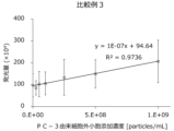
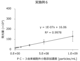
- FIG. 1 shows the results of detection of PC-3-derived extracellular vesicles (Example 6).
- FIG. 1 shows the results of detection of PC-3-derived extracellular vesicles (Example 6).
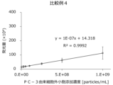
- FIG. 1 shows the results of detection of PC-3-derived extracellular vesicles (Example 7).
- cancer and “cancer” may be used interchangeably.
- the method of the present invention is a method comprising a step of removing platelet-derived extracellular vesicles in a blood-derived sample or a step of suppressing the occurrence of platelet-derived extracellular vesicles in a blood-derived sample.
- the method of the present invention may further comprise a step of detecting target extracellular vesicles in the blood-derived sample.
- the step of removing platelet-derived extracellular vesicles in a blood-derived sample is also referred to as a "removal step”.
- the step of suppressing the occurrence of platelet-derived extracellular vesicles in a blood-derived sample is also referred to as a "suppression step”.
- one or both of the removal step and the suppression step may be performed.
- the step of detecting target extracellular vesicles in a blood-derived sample is also referred to as a "detection step”. The detection step is performed after the removal step or the suppression step.
- Target extracellular vesicles refers to extracellular vesicles to be detected and are to be distinguished from platelet-derived extracellular vesicles.
- target extracellular vesicles refers to extracellular vesicles other than platelet-derived extracellular vesicles that may be contained in a blood-derived sample.
- the target extracellular vesicles may be one or more specific types of extracellular vesicles selected from extracellular vesicles other than platelet-derived extracellular vesicles, or may be all extracellular vesicles other than platelet-derived extracellular vesicles.
- Specific extracellular vesicles include extracellular vesicles derived from tumor cells.
- the type of tumor from which the target extracellular vesicles can be derived is not particularly limited and may include malignant tumors and benign tumors.
- Malignant tumors may be primary cancers or metastatic cancers.
- Tumors include hematopoietic cell malignant tumors, head and neck cancer, brain tumors, breast cancer, uterine cancer, cervical cancer, ovarian cancer, esophageal cancer, gastric cancer, appendix cancer, colon cancer, liver cancer, gallbladder cancer, bile duct cancer, pancreatic cancer, kidney cancer, adrenal cancer, gastrointestinal stromal tumors, mesothelioma, thyroid cancer, lung cancer, osteosarcoma, bone cancer, prostate cancer, testicular tumors, bladder cancer, skin cancer, and anal cancer.
- Hematopoietic cell malignant tumors include leukemia, lymphoma, and multiple myeloma.
- Lymphomas include Hodgkin's lymphoma and non-Hodgkin's lymphoma.
- Head and neck cancers include floor of the mouth cancer, gingival cancer, tongue cancer, buccal mucosa cancer, salivary gland cancer, and paranasal sinus cancer.
- Extracellular vesicles refers to lipid-covered vesicles with a diameter of 1 nm to 1 ⁇ m that are released by cells, whether actively or passively.
- extracellular vesicles include exosomes, microvesicles, ectosomes, membrane particles, exosome-like vesicles, and apoptotic vesicles (Nature Reviews Immunology, 9, 581-593 (2009)).
- extracellular vesicles are composed of lipids and proteins with a different composition from that of the cell membrane (Bioscience, 65, 783-797 (2015)), but proteins that reflect the characteristics of the cell from which they were released are localized in the extracellular vesicles. For example, when the source of release is a tumor cell, the presence or absence of extracellular vesicles released from the cell can be used to distinguish between cancer patients and healthy donors (Publication of International Publication No. 2011-510309).
- one aspect of the method of the present invention is a method for improving the detection sensitivity of target extracellular vesicles in a blood-derived sample, which includes a removal step or suppression step.
- This method is also referred to as the "detection sensitivity improving method of the present invention.”
- the detection sensitivity improving method of the present invention may further include a detection step. Examples of improved detection sensitivity include reducing the variation in measured values and reducing non-specific signals (background).
- the removal step or the suppression step can be carried out as a pretreatment of the blood-derived sample. That is, one aspect of the method of the present invention is a method of pretreatment of a blood-derived sample, including a removal step or a suppression step. This method is also referred to as the "pretreatment method of the present invention.” The removal step and the suppression step are also collectively referred to as the "pretreatment step.” The removal step or the suppression step can be specifically carried out as a pretreatment of the blood-derived sample when carrying out the detection step. That is, the pretreatment method of the present invention may specifically be a method of pretreatment of the blood-derived sample in order to detect target extracellular vesicles in the blood-derived sample.
- the removal step or the suppression step can be specifically carried out as a pretreatment for improving the detection sensitivity of target extracellular vesicles in the blood-derived sample. That is, the pretreatment method of the present invention may specifically be a method of pretreatment of the blood-derived sample in order to improve the detection sensitivity of target extracellular vesicles in the blood-derived sample.
- one aspect of the method of the present invention is a method for detecting target extracellular vesicles in a blood-derived sample, which includes a removal or suppression step and a detection step.
- This method is also referred to as the "detection method of the present invention.”
- the detection method of the present invention is also a method for detecting target extracellular vesicles in a blood-derived sample, which includes a step of pretreating a blood-derived sample by the pretreatment method of the present invention, and a detection step.
- the blood-derived sample is not particularly limited as long as it can contain target extracellular vesicles.
- blood-derived samples include whole blood, serum, plasma, and fractions thereof.
- blood-derived samples include serum and plasma in particular.
- Blood-derived samples such as serum and plasma may be treated with an anticoagulant such as citric acid, heparin, or EDTA before carrying out the removal or inhibition step.
- the blood-derived sample may be obtained from a subject.
- the subject is not particularly limited.
- the subject may be a human or a non-human animal.
- Non-human animals include mice, rats, guinea pigs, rabbits, dogs, cats, cows, horses, pigs, monkeys, chimpanzees, and birds.
- the subject may particularly be a human.
- the subject may be, for example, a subject suspected of having cancer.
- the subject may be, for example, a subject to be tested for cancer.
- the embodiment of the removal step is not particularly limited as long as the platelet-derived extracellular vesicles in the blood-derived sample are removed to the desired extent.
- the removal step may be carried out, for example, using a substance that specifically binds to platelet-derived extracellular vesicles.
- a substance that specifically binds to platelet-derived extracellular vesicles is also called a "platelet-derived extracellular vesicle-specific binding substance.”
- “Specifically binds to platelet-derived extracellular vesicles” may mean that it binds to platelet-derived extracellular vesicles but does not substantially bind to target extracellular vesicles.
- platelet-derived extracellular vesicle-specific binding substances include substances that bind to markers that are specifically present on the surface of platelet-derived extracellular vesicles.
- a marker that is specifically present on the surface of platelet-derived extracellular vesicles is also called a "platelet-derived extracellular vesicle-specific marker.”
- “Specifically present on the surface of platelet-derived extracellular vesicles” may mean that it is present on the surface of platelet-derived extracellular vesicles but is substantially not present on the surface of target extracellular vesicles.
- platelet-derived extracellular vesicle-specific markers include proteins that are specifically present on the surface of platelet-derived extracellular vesicles.
- proteins that are specifically present on the surface of platelet-derived extracellular vesicles include CD41, CD42a, CD42b, CD61, and CD62P.
- proteins that are specifically present on the surface of platelet-derived extracellular vesicles include CD41.
- substances that bind to platelet-derived extracellular vesicles-specific markers include antibodies and aptamers against platelet-derived extracellular vesicles-specific markers.
- substances that bind to platelet-derived extracellular vesicles-specific markers include antibodies against platelet-derived extracellular vesicles-specific markers.
- antibodies against platelet-derived extracellular vesicles-specific markers include anti-CD41 antibodies, anti-CD42a antibodies, anti-CD42b antibodies, anti-CD61 antibodies, and anti-CD62P antibodies.
- antibodies against platelet-derived extracellular vesicles-specific markers include anti-CD41 antibodies.
- the antibodies may be, for example, whole immunoglobulins or parts of immunoglobulins.
- Antibodies include Fab, Fab', F(ab')2, Fv, dAb, complementarity determining region (CDR) fragments, single chain antibodies (scFv), single domain antibodies, bispecific antibodies, chimeric antibodies, humanized antibodies, and diabodies.
- Antibodies may be monoclonal or polyclonal. Antibodies may in particular be monoclonal antibodies.
- the removal step may be carried out by, for example, binding a platelet-derived extracellular vesicles-specific binding substance to the platelet-derived extracellular vesicles. That is, the removal step may include a step of binding a platelet-derived extracellular vesicles-specific binding substance to the platelet-derived extracellular vesicles.
- the binding of the platelet-derived extracellular vesicles-specific binding substance to the platelet-derived extracellular vesicles can be carried out by contacting the platelet-derived extracellular vesicles-specific binding substance with the platelet-derived extracellular vesicles.
- the binding of the platelet-derived extracellular vesicles-specific binding substance to the platelet-derived extracellular vesicles can be carried out, for example, by contacting the platelet-derived extracellular vesicles-specific binding substance with a blood-derived sample (which may contain platelet-derived extracellular vesicles).
- the contact of the platelet-derived extracellular vesicles-specific binding substance with the platelet-derived extracellular vesicles can be carried out in a liquid medium.
- the liquid medium include aqueous media such as water and aqueous buffer solutions, and organic solvents such as dimethyl sulfoxide.
- the platelet-derived extracellular vesicles-specific binding substance can be brought into contact with the platelet-derived extracellular vesicles by mixing the blood-derived sample with the platelet-derived extracellular vesicles-specific binding substance in a liquid medium.
- the platelet-derived extracellular vesicles-specific binding substance can be brought into contact with the platelet-derived extracellular vesicles by passing a blood-derived sample prepared in liquid form through the column.
- the platelet-derived extracellular vesicles-specific binding substance may be bound to a carrier and used in the removal step. By binding the platelet-derived extracellular vesicles-specific binding substance to a carrier, the platelet-derived extracellular vesicles can be captured on the carrier via the platelet-derived extracellular vesicles-specific binding substance.
- the order of binding between the platelet-derived extracellular vesicles-specific binding substance and the carrier is not particularly limited.
- the platelet-derived extracellular vesicles-specific binding substance may typically be bound to the carrier before binding to the platelet-derived extracellular vesicles.
- the platelet-derived extracellular vesicles-specific binding substance may typically be used for binding to the platelet-derived extracellular vesicles in a state in which it is bound to the carrier in advance.
- the mode of binding between the platelet-derived extracellular vesicles-specific binding substance and the carrier is not particularly limited.
- the platelet-derived extracellular vesicles-specific binding substance and the carrier may be bound by a covalent bond or a non-covalent bond. Examples of non-covalent bonds include hydrogen bonds and hydrophobic bonds.
- the bond between the platelet-derived extracellular vesicles-specific binding substance and the carrier may be, in particular, a covalent bond.
- the carrier may be a water-insoluble carrier.
- the carrier may be hydrophilic or hydrophobic.
- Specific examples of the carrier include polysaccharides such as agarose, dextran, chitosan, and cellulose; organic synthetic polymers such as polyacrylamide, polyvinyl alcohol, polyvinylpyrrolidone, polyacrylonitrile, styrene-divinylbenzene copolymer, polystyrene, acrylic acid ester, methacrylic acid ester, polyethylene, polypropylene, poly4-ethylene fluoride, ethylene-vinyl acetate copolymer, polyamide, polycarbonate, polyvinylidene fluoride, polyvinyl formal, polyarylate, and polyethersulfone; ceramics such as glass, activated carbon, alumina, silica, and hydroxyapatite; and metals such as iron oxide, titanium, and gold.
- the carrier may be in any shape, such as particles, fibers, hollow fibers, membranes, and flat plates.
- the carrier may be
- the platelet-derived extracellular vesicles can be removed from the blood-derived sample. That is, the removal step may include a step of separating the carrier that has captured the platelet-derived extracellular vesicles from the blood-derived sample. Separation of the carrier and the blood-derived sample can be performed, for example, by a normal solid-liquid separation means.
- the solid-liquid separation means can be appropriately selected depending on various conditions, for example, the shape of the carrier.
- the carrier that has captured the platelet-derived extracellular vesicles when dispersed in a liquid containing a blood-derived sample, the carrier can be separated from the blood-derived sample by subjecting the liquid to centrifugation, magnetic separation, or filtration.
- the carrier is an antibody-immobilized magnetic particle
- the blood-derived sample and the antibody-immobilized magnetic particle are mixed to capture the platelet-derived extracellular vesicles on the carrier, and then the antibody-immobilized magnetic particle that has captured the platelet-derived extracellular vesicles is removed using a magnet, thereby separating the carrier (i.e., the antibody-immobilized magnetic particle that has captured the platelet-derived extracellular vesicles) from the blood-derived sample. That is, the blood-derived sample can be recovered as a non-adsorbed fraction (i.e., the supernatant).
- the blood-derived sample when a column is filled with a carrier, the blood-derived sample can be passed through the column to capture platelet-derived extracellular vesicles on the carrier, and then the blood-derived sample can be discharged from the column, thereby separating the carrier from the blood-derived sample. That is, the blood-derived sample can be recovered from the column as a non-adsorbed fraction.
- the embodiment of the suppression step is not particularly limited as long as the generation of platelet-derived extracellular vesicles in the blood-derived sample is suppressed to a desired degree.
- Platelet-derived extracellular vesicles can be generated from platelets contained in the blood-derived sample. Therefore, the suppression step can be achieved by removing platelets from the blood-derived sample.
- the removal of platelets from the blood-derived sample may be performed, for example, by performing centrifugation two or more times during the preparation of a blood-derived sample (particularly, plasma).
- the conditions of the centrifugation are not particularly limited as long as the conditions allow the separation of platelets and target extracellular vesicles.
- the conditions of the centrifugation may be, for example, the conditions of normal centrifugation used in the preparation of plasma.
- Platelet-derived extracellular vesicles can be generated when a blood-derived sample containing platelets is frozen. Therefore, the suppression step may be performed, for example, by not freezing the blood-derived sample.
- the method of the present invention includes a suppression step, and the suppression step is performed by not freezing the blood-derived sample” may mean that the method of the present invention does not include a step of freezing the blood-derived sample.
- the embodiment of the detection step is not particularly limited as long as it can detect target extracellular vesicles to the desired degree. Detection of target extracellular vesicles may be qualitative detection (e.g., detection of the presence or absence of target extracellular vesicles) or quantitative detection (e.g., detection of the amount of target extracellular vesicles present). Detection of target extracellular vesicles may particularly be quantitative detection.
- the method of detecting target extracellular vesicles can be appropriately selected depending on various conditions such as the desired detection sensitivity. "Measurement of target extracellular vesicles" and “detection of target extracellular vesicles" may be used interchangeably.
- the detection step is performed after the removal step or the suppression step.
- the detection step may be carried out, for example, using a substance that binds to the target extracellular vesicles.
- a substance that binds to the target extracellular vesicles is also referred to as a "target extracellular vesicle binding substance.”
- target extracellular vesicle-specific binding substances include substances that bind to markers present on the surface of target extracellular vesicles. Markers present on the surface of target extracellular vesicles are also referred to as "target extracellular vesicle markers.”
- the target extracellular vesicle binding substance may preferably be a substance that specifically binds to the target extracellular vesicles.
- a substance that specifically binds to the target extracellular vesicles is also referred to as a "target extracellular vesicle specific binding substance.”
- “Specifically binds to target extracellular vesicles” may mean that the substance binds to the target extracellular vesicles but does not substantially bind to platelet-derived extracellular vesicles.
- Examples of target extracellular vesicle-specific binding substances include substances that bind to markers specifically present on the surface of target extracellular vesicles.
- target extracellular vesicle specific markers Markers specifically present on the surface of target extracellular vesicles are also referred to as "target extracellular vesicle specific markers.” “Specifically present on the surface of target extracellular vesicles” may mean that it is present on the surface of target extracellular vesicles but is substantially absent on the surface of platelet-derived extracellular vesicles.
- Target extracellular vesicle markers include proteins and lipids present on the surface of target extracellular vesicles.
- Proteins present on the surface of target extracellular vesicles include tetraspanins such as TM4SF1, CD9, CD63, and CD81; antigen presentation-related proteins such as Major Histocompatibility Complex (MHC) I and MHCII; adhesion molecules such as integrins, cadherins, InterCellular Adhesion Molecule 1 (ICAM-1), and Epithelial Cell Adhesion Molecule (EpCAM); cytokines or growth factors such as EGFR (Epidermal Growth Factor Receptor) vIII and TGF (Transforming Growth Factor)- ⁇ ; receptors for cytokines or growth factors; and enzymes.
- Lipids present on the surface of target extracellular vesicles include phosphatidylserine (PS) and sphingomyelin.
- Substances that bind to target extracellular vesicle markers include antibodies and aptamers (peptide aptamers and nucleic acid aptamers) against the target extracellular vesicle markers.
- the antibody may be, for example, an entire immunoglobulin or a part of an immunoglobulin.
- antibodies include Fab, Fab', F(ab')2, Fv, dAb, complementarity determining region (CDR) fragments, single chain antibodies (scFv), single domain antibodies, bispecific antibodies, chimeric antibodies, humanized antibodies, and diabodies.
- Substances that bind to target extracellular vesicle markers include, in particular, antibodies against the target extracellular vesicle markers.
- the detection step may be, in particular, performed by immunoassay (i.e., an assay using antibodies).
- Substances that bind to target extracellular vesicle markers include, in particular, antibodies against target extracellular vesicle-specific markers. Examples of antibodies against target extracellular vesicle-specific markers include anti-TM4SF1 antibodies and anti-CD81 antibodies.
- the antibody may be a monoclonal antibody or a polyclonal antibody.
- the antibody may be, in particular, a monoclonal antibody.
- the antibody or aptamer may be appropriately modified with a labeling substance.
- the labeling substance examples include fluorescent substances such as fluorescein isothiocyanate (FITC), phycoerythrin (PE), and Alexa Fluor (trade name); enzymes such as luciferase, alkaline phosphatase, and horseradish peroxidase; and radioactive substances.
- the binding mode between the antibody or aptamer and the labeling substance is not particularly limited.
- the antibody or aptamer may or may not be directly bound to the labeling substance.
- the antibody may be directly bound to the labeling substance by chemical bonding or the like, or may be indirectly bound to the labeling substance by binding to a secondary antibody (labeled secondary antibody) bound to the labeling substance.
- Examples of immunological measurement methods include EIA (Enzyme Immunoassay) and Western blotting.
- EIA method include the ELISA (Enzyme-Linked Immunosorbent Assay) method, the CLEIA (Chemiluminescent Enzyme Immunoassay) method, and the FEIA (Fluorescence Enzyme Immunoassay) method.
- a detection substrate may be used as appropriate.
- the detection substrate include colorimetric substrates, fluorescent substrates, and luminescent substrates.
- Examples of the detection substrate include, in particular, luminescent substrates.
- the luminescent substrate include substrates that generate luminescence through an enzyme reaction such as horseradish peroxidase.
- an enzyme corresponding to the luminescent substrate is used as a labeling substance, and luminescence can be generated by reacting with the luminescent substrate.
- the target extracellular vesicles may be detected manually or automatically using a measuring device.
- measuring devices include enzyme immunoassay devices such as the AIA-900 (Tosoh) and chemiluminescent enzyme immunoassay devices such as the AIA-CL2400 (Tosoh).
- the target extracellular vesicles may be collected and the collected target extracellular vesicles may be detected. That is, the detection step may include, for example, a step of collecting the target extracellular vesicles on a carrier and a step of detecting the collected target extracellular vesicles.
- the method for collecting the target extracellular vesicles include ultracentrifugation, sedimentation, size exclusion chromatography, affinity method, and polymer precipitation method.
- the method for collecting the target extracellular vesicles include, in particular, affinity method.
- the collection of the target extracellular vesicles may be specifically carried out by capturing the target extracellular vesicles on a carrier.
- the detection step may specifically include, for example, a step of capturing the target extracellular vesicles on a carrier and a step of detecting the captured target extracellular vesicles.
- the target extracellular vesicles can be captured on a carrier by an affinity method.
- the target extracellular vesicles can be captured on a carrier via a target extracellular vesicle-binding substance by an affinity method.
- the capture of target extracellular vesicles on a carrier can be carried out in the same manner as the capture of platelet-derived extracellular vesicles on a carrier described above, except that a target extracellular vesicles-binding substance is used instead of a platelet-derived extracellular vesicles-specific binding substance.
- the carrier is as described above in the description of the capture of platelet-derived extracellular vesicles on a carrier.
- Specific examples of the carrier include magnetic particles and microplates.
- the carrier may be, for example, magnetic particles.
- the step of capturing the target extracellular vesicles on the carrier may be carried out, for example, using a first substance that binds to the target extracellular vesicles.
- the step of detecting the captured target extracellular vesicles may be carried out, for example, using a second substance that binds to the target extracellular vesicles.
- Both the first substance and the second substance are target extracellular vesicles-binding substances.
- Both the first substance and the second substance may be, for example, an antibody against a target extracellular vesicle marker. That is, the first substance may be an antibody (also called a first antibody) against a first marker present in the target extracellular vesicles.
- the second substance may be an antibody (also referred to as a second antibody) against a second marker present in the target extracellular vesicles.
- the first antibody and the second antibody may be, for example, antibodies against different target extracellular vesicle markers.
- a sandwich EIA method such as a sandwich CLEIA method can be performed using the first antibody and the second antibody.
- One or both of the first substance and the second substance may be a target extracellular vesicle-specific binding substance (e.g., an antibody against a target extracellular vesicle-specific marker).
- one or both of the first marker and the second marker may be a target extracellular vesicle-specific marker.
- the first substance may be a target extracellular vesicle-specific binding substance (e.g., an antibody against a target extracellular vesicle-specific marker), and further, the second substance may be a target extracellular vesicle-specific binding substance (e.g., an antibody against a target extracellular vesicle-specific marker).
- the first marker may be a target extracellular vesicle-specific marker
- the second marker may be a target extracellular vesicle-specific marker.
- the second substance may be a target extracellular vesicle-specific binding substance (e.g., an antibody against a target extracellular vesicle-specific marker), and the first substance may be a target extracellular vesicle-specific binding substance (e.g., an antibody against a target extracellular vesicle-specific marker).
- the second marker may be a target extracellular vesicle-specific marker
- the first marker may be a target extracellular vesicle-specific marker.
- the first marker may be, for example, a target extracellular vesicle-specific marker such as TM4SF1 or CD81, among others.
- the second marker may be, for example, CD9 or CD81, among others.
- the use of the detection results of target extracellular vesicles is not particularly limited.
- the detection results of the target extracellular vesicles can be used to test for cancer.
- the detection results of the target extracellular vesicles can be used to test for cancer in the subject.
- Tests for cancer in a subject include testing whether the subject has cancer, testing the presence or absence or degree of the possibility that the subject has cancer, and testing the progression (e.g., stage) of cancer if the subject has cancer. "Cancer testing” may be read as "cancer diagnosis.”
- kit of the present invention is a kit for pretreatment of a blood-derived sample.
- This kit is also referred to as the "pretreatment kit of the present invention.”
- the pretreatment of the blood-derived sample is as described above in the method of the present invention.
- kit of the present invention is a kit for detecting target extracellular vesicles in a blood-derived sample.
- This kit is also referred to as the "detection kit of the present invention.” Detection of target extracellular vesicles in a blood-derived sample is as described above in the method of the present invention.
- the kit of the present invention may, for example, include a reagent for use in the method of the present invention.
- the kit of the present invention may, for example, include laboratory equipment for use in the method of the present invention.
- the kit of the present invention may, for example, include instructions for the kit of the present invention.
- the kit of the present invention may include, for example, a platelet-derived extracellular vesicles-specific binding substance.
- the kit of the present invention may include, for example, a carrier for binding to the platelet-derived extracellular vesicles-specific binding substance.
- the kit of the present invention may include, for example, a pre-bound platelet-derived extracellular vesicles-specific binding substance and a carrier.
- the kit of the present invention may, for example, include a target extracellular vesicle binding substance.
- the kit of the present invention may, for example, include a first substance and/or a second substance.
- the kit of the present invention may, for example, include a carrier for binding to the first substance.
- the kit of the present invention may, for example, include a first substance pre-bound and a carrier.
- the kit of the present invention may, for example, include a labeling substance for labeling the target extracellular vesicle binding substance (e.g., the second substance).
- the kit of the present invention may, for example, include a target extracellular vesicle binding substance pre-bound (e.g., the second substance) and a labeling substance.
- the kit of the present invention may, for example, include a detection substrate.
- the kit of the present invention can be used, for example, to carry out the method of the present invention.
- Example 1 Preparation of antibody-immobilized magnetic particles (1) 600 ⁇ L of 0.01 M MES (2-morpholinoethanesulfonic acid) buffer solution (pH 6.0) (hereinafter referred to as "buffer A”) was added to 400 ⁇ L of a magnetic particle solution having carboxy groups on the surface (Magnosphere MS300/Carboxyl, 1.0% (w/v) slurry, manufactured by JSR Life Sciences Corporation).
- buffer A 0.01 M MES (2-morpholinoethanesulfonic acid) buffer solution having carboxy groups on the surface
- Buffer A containing 1 mg/mL anti-human TM4SF1 antibody (R&D Systems)
- Buffer A containing 0.5 mg/mL anti-human CD41 antibody (BioLegend)
- Buffer solution A containing 1 mg/mL mouse IgG (manufactured by Fujifilm Wako Pure Chemical Industries, Ltd.)
- Example 2 Preparation of biotinylated modified antibody (1) 200 ⁇ L of a solution containing any of the antibodies shown in [d] and [e] below was added to a filtration tube (Dojin Chemical Laboratory Co., Ltd.) and centrifuged at 8,000 ⁇ g for 10 minutes at room temperature to immobilize the antibody on the filter.
- [d] 1 mg/mL anti-human CD9 antibody (manufactured by Frontier Research Institute)
- [e] 1 mg/mL anti-human CD81 antibody manufactured by Frontier Research Institute, Inc.
- Example 3 Detection of background in plasma measurement
- PC-3-derived extracellular vesicles are used as target extracellular vesicles.
- TM4SF1 a member of the four-transmembrane protein (Transmembrane 4 superfamily, tetraspanin), a protein that is highly expressed in many tumor cells including PC-3 and extracellular vesicles derived from tumor cells) was selected as a marker protein for detecting PC-3-derived extracellular vesicles.
- TM4SF1 is not expressed in extracellular vesicles derived from blood cells contained in plasma.
- plasma is used as a specimen to be used as a sample. In this example, detection of background in plasma measurement was performed.
- background refers to the measurement value (signal) of a specimen that does not contain target extracellular vesicles, and if a large amount of impurities are contained, the noise signal increases, resulting in a high background and making it impossible to perform accurate measurement.
- Plasma Samples (1-1) 5 mL of blood was collected from one healthy subject who had given informed consent into an EDTA-2K blood collection tube (VP-DK050K, manufactured by Terumo Corporation). The blood collection tube was centrifuged at 1,500 ⁇ g for 20 minutes at 4° C. to separate blood cells, and the supernatant was collected. The supernatant was centrifuged at 1,500 ⁇ g for 20 minutes at 4° C. to separate remaining blood cells, and the supernatant was collected to obtain healthy subject plasma.
- EDTA-2K blood collection tube VP-DK050K, manufactured by Terumo Corporation
- BSA buffer bovine serum albumin
- 10 ⁇ L of the anti-TM4SF1 antibody-immobilized magnetic particles prepared in Example 1 were mixed in a 2 mL tube and stirred by inversion for 3 minutes.
- the 2 mL tube was brought close to a magnet and left for 1 minute, the solution was removed, and then 1 mL of BSA buffer was added and stirred by inversion for 10 minutes or more.
- the 2 mL tube was brought close to a magnet and left for 1 minute, the solution was removed, and then 50 ⁇ L of BSA buffer was added to prepare an anti-TM4SF1 antibody-immobilized magnetic particle suspension.
- washing buffer PBS containing 0.05% (v/v) Tween 20 (trade name)
- Example 3 (1-1) Plasma samples were obtained in the same manner as in Example 3 (1), except that the blood collection tubes were each centrifuged at 1,500 ⁇ g for 10 minutes at 4°C to separate blood cells, and the supernatant was collected to obtain plasma from healthy subjects.
- Comparative Example 2 Detection of background in plasma measurement when pretreatment was performed using mouse IgG-immobilized magnetic particles (1) Preparation of plasma sample Plasma samples were prepared in the same manner as in Comparative Example 1 (1).
- Example 3(2) Measurement of Background Background in plasma measurement was detected in the same manner as in Example 3(2), except that the plasma sample in Example 3(2) was the pretreated plasma sample obtained in (2-3).
- Example 4 Detection of background in plasma measurement when pretreatment was performed using anti-CD41 antibody-immobilized magnetic particles
- background in plasma measurement was detected in the same manner as in Comparative Example 2, except that anti-CD41 antibody-immobilized magnetic particles were used.
- Example 3 and 4 and Comparative Examples 1 and 2 are shown in Table 1.
- Example 3 almost all blood cells were removed from the plasma by two centrifugations, while in Comparative Example 1, a large number of blood cells remained in the plasma due to milder conditions and one centrifugation.
- the remaining blood cells are thought to release extracellular vesicles through freezing and thawing.
- platelets are known to be activated by physical stimuli such as freezing and thawing, and release extracellular vesicles as a result of activation.
- platelet-derived extracellular vesicles which are particularly abundant among blood cell-derived extracellular vesicles, were removed by pretreatment using magnetic particles immobilized with antibodies against CD41 expressed on platelets, and it was confirmed whether the background would be reduced.
- a pretreatment was performed in which a plasma sample was reacted with magnetic particles immobilized with mouse IgG protein as an antibody control, and then background measurement was performed (Comparative Example 2).
- the amount of luminescence was equivalent to that of Comparative Example 1 in which no pretreatment was performed (1 million), and therefore no reduction in the background due to nonspecific adsorption to the antibody-immobilized magnetic particles was observed.
- a pretreatment was performed in which anti-CD41 antibody-immobilized magnetic particles were reacted with the plasma sample, and then background measurement was performed (Example 4).
- the amount of luminescence was reduced to 380,000 compared to Comparative Example 1 and Comparative Example 2. Therefore, it was found that the background was reduced by specifically binding and removing platelet-derived extracellular vesicles contained in the plasma to the anti-CD41 antibody-immobilized magnetic particles.
- PC-3 cells which are human prostate cancer cells, were selected as diseased cells to be used in the following examples.
- PC-3 cells were suspended in Ham's F-12K (Fujifilm Wako Pure Chemical Industries, Ltd.) medium containing 15% (v/v) FBS (fetal bovine serum) to a concentration of 1 x 10⁵ cells/mL, and then seeded in five 15 cm dishes at 30 mL each and cultured at 37°C in a 5% CO2 environment .
- Ham's F-12K Flujifilm Wako Pure Chemical Industries, Ltd.
- FBS fetal bovine serum
- PC-3 cells obtained in (3) were cultured for an additional 3 days, and the entire culture supernatant (approximately 125 mL) was then collected.
- the cells were centrifuged at 300 ⁇ g for 10 minutes at room temperature to remove floating cells, and 120 mL of the supernatant was collected.
- the supernatant obtained in (4) was diluted to a uniform volume and dispensed into ultracentrifuge tubes, centrifuged at 300,000 x g for 70 minutes at 4°C, and 120 mL of the supernatant was removed.
- the remaining precipitate was suspended in 4 mL of PBS, and the resulting suspension was centrifuged at 259,000 x g for 70 minutes at 4°C, after which 4 mL of the supernatant was removed. 1 mL of the remaining precipitate was used as the PC-3-derived extracellular vesicle solution.
- Plasma samples containing PC-3-derived extracellular vesicles at six concentrations shown below in [f] to [k] were prepared by adding 100 ⁇ L of a solution obtained by diluting the PC-3-derived extracellular vesicles obtained in Example 5 with PBS to 250 ⁇ L of plasma from a healthy subject, and the plasma samples were stored frozen at ⁇ 80° C.
- PC-3-derived extracellular vesicles were detected in the same manner as in Example 3(2), except that the plasma sample obtained in (1) was used as the plasma sample in Example 3(2).
- Example 6 Detection of PC-3-Derived Extracellular Vesicles in Plasma of Healthy Subjects from Which Platelet-Derived Extracellular Vesicles Have Been Removed (1) Preparation of Plasma Samples Plasma samples were prepared in the same manner as in Comparative Example 3(1).
- PC-3-derived extracellular vesicles in plasma were detected in the same manner as in Example 3(2), except that the plasma sample was the pretreated plasma sample obtained in (2).
- Comparative Example 3 (1-2) the healthy subject's plasma was the healthy subject's plasma obtained in (1-1), and a plasma sample was prepared in the same manner as in Comparative Example 3 (1-2), except that the plasma was not frozen and stored at -80°C, and the plasma was immediately subjected to step (2) after preparation.
- Example 3 (1-3) the healthy subject's plasma was the healthy subject's plasma obtained in (1-1), and a plasma sample not containing PC-3-derived extracellular vesicles was prepared in the same manner as in Comparative Example 3 (1-3), except that the healthy subject's plasma was not frozen and stored at -80°C, and the process was immediately carried out after preparation in step (2).
- PC-3-derived extracellular vesicles were detected in the same manner as in Example 3(2), except that the plasma sample obtained in (1) was used as the plasma sample in Example 3(2).
- Comparative Example 3, Example 6, and Reference Example 1 are shown in Table 2 and Figures 1 to 3.
- the "variation coefficient” is the standard deviation of the luminescence amount divided by the average luminescence amount, and the lower this value is, the smaller the variation in the measured values between samples is, and the higher the measurement accuracy is.
- the “threshold value” is the value obtained by adding twice the standard deviation of the luminescence amount in a plasma sample that does not contain cell-derived extracellular vesicles to the average luminescence amount.
- the "lower detection limit concentration” refers to the lowest concentration of added PC-3-derived extracellular vesicles at which the luminescence amount is higher than the threshold value, and the lower the luminescence amount and standard deviation in a plasma sample that does not contain PC-3 cell-derived extracellular vesicles is, the lower the lower detection limit concentration is, and the higher the detection sensitivity is.
- Example 6 In Comparative Example 3, Example 6, and Reference Example 1, the amount of luminescence increased depending on the concentration of PC-3-derived extracellular vesicles added, confirming that PC-3-derived extracellular vesicles can be detected in plasma.
- Comparative Example 3 when focusing on the average luminescence intensity of plasma samples not containing PC-3 cell-derived extracellular vesicles, Comparative Example 3 was significantly higher than Example 6 and Reference Example 1. This result, like the result of Comparative Example 1, is considered to be due to an increase in background luminescence intensity caused by platelet-derived extracellular vesicles released by freezing and thawing from platelets remaining in the plasma. Furthermore, in Comparative Example 3, the noise signal amount differed between samples, resulting in a high coefficient of variation (0.50), and therefore a high detection limit concentration (1.0 x 10⁹ particles/mL).
- the freeze-thawed plasma was pretreated with anti-CD41 antibody-immobilized magnetic particles (Example 6), which reduced the background luminescence and reduced the coefficient of variation (0.23) and the detection limit concentration (2.5 x 10⁸ particles/mL).
- anti-CD41 antibody-immobilized magnetic particles By removing platelet-derived extracellular vesicles from plasma by pretreatment with anti-CD41 antibody-immobilized magnetic particles, it became possible to detect the target PC-3-derived extracellular vesicles with higher accuracy and sensitivity.
- the concentration of disease cell-derived extracellular vesicles contained in the plasma of a patient with a disease, including cancer is generally estimated to be 10 8 particles/mL or less
- the measurement method for detecting the disease cell-derived extracellular vesicles in the plasma must have a detection limit concentration of 10 8 particles/mL or more. Therefore, the method for measuring plasma in a state containing platelet-derived extracellular vesicles as shown in Comparative Example 3 has a detection limit concentration that is significantly higher than the concentration of the disease cell-derived extracellular vesicles, making it difficult to apply to detection of disease cell-derived extracellular vesicles from a plasma sample of a patient with a disease.
- Example 6 and Reference Example 1 it is suggested that the method for measuring plasma including a step of removing platelet-derived extracellular vesicles or suppressing the generation of platelet-derived extracellular vesicles can be applied to a plasma sample of a patient with a disease.
- Example 7 Detection of PC-3-derived extracellular vesicles in healthy subject plasma from which platelet-derived extracellular vesicles have been removed PC-3-derived extracellular vesicles in plasma were detected in the same manner as in Example 6 (3), except that 0.5 ⁇ g/mL biotin-modified anti-CD81 antibody was used instead of 0.5 ⁇ g/mL biotin-modified anti-CD9 antibody.
- Comparative Example 4, Example 7, and Reference Example 2 are shown in Table 3 and Figures 4 to 6.
- the "variation coefficient” is the standard deviation of the amount of luminescence divided by the average amount of luminescence, and the lower this value is, the smaller the variation in the measured values between samples, and the higher the measurement accuracy.
- the “threshold” is the value obtained by adding twice the standard deviation of the amount of luminescence in plasma samples that do not contain cell-derived extracellular vesicles to the average amount of luminescence.
- the “lower detection limit concentration” refers to the lowest concentration of PC-3-derived extracellular vesicles added at which the amount of luminescence is higher than the threshold, and the lower the lower detection limit concentration, the higher the detection sensitivity. Therefore, the detection sensitivity decreases as the amount of luminescence and standard deviation increase in plasma samples that do not contain PC-3 cell-derived extracellular vesicles.
- Example 7 In Comparative Example 4, Example 7, and Reference Example 2, the amount of luminescence increased depending on the concentration of added PC-3-derived extracellular vesicles, confirming that PC-3-derived extracellular vesicles can be detected in plasma. In addition, in all results, the average amount of luminescence in plasma samples that did not contain PC-3 cell-derived extracellular vesicles was equally low. This is presumably because, while Comparative Example 3, Example 6, and Reference Example 1 used CD9 as a detection marker, which is also expressed in platelets, Comparative Example 4, Example 7, and Reference Example 2 used CD81 as a detection marker, which is not expressed in platelets, suppressing the generation of noise signals.
- Example 7 When paying attention to the lower detection limit concentration, Example 7 had a lower value compared to Comparative Example 4 and Reference Example 2. This is presumably because, compared to Comparative Example 4 and Reference Example 2, the total amount of extracellular vesicles contained in the plasma was reduced by the process of removing platelet-derived extracellular vesicles in Example 7, and the reactivity between the target extracellular vesicles (PC-3-derived extracellular vesicles) and the anti-TM4SF1 antibody-immobilized magnetic particles was improved, which increased the measurement stability and reduced the variability in the measurement values between samples.
- the detection method of the present invention can detect target extracellular vesicles from plasma with high sensitivity and high accuracy, suggesting that the method is also applicable to the detection of disease cell-derived extracellular vesicles in plasma samples from disease patients.
Landscapes
- Health & Medical Sciences (AREA)
- Life Sciences & Earth Sciences (AREA)
- Engineering & Computer Science (AREA)
- Immunology (AREA)
- Chemical & Material Sciences (AREA)
- Molecular Biology (AREA)
- Urology & Nephrology (AREA)
- Hematology (AREA)
- Biomedical Technology (AREA)
- Medicinal Chemistry (AREA)
- Physics & Mathematics (AREA)
- Analytical Chemistry (AREA)
- Biochemistry (AREA)
- General Health & Medical Sciences (AREA)
- General Physics & Mathematics (AREA)
- Food Science & Technology (AREA)
- Pathology (AREA)
- Biotechnology (AREA)
- Cell Biology (AREA)
- Microbiology (AREA)
- Investigating Or Analysing Biological Materials (AREA)
Abstract
血液由来試料中の標的細胞外小胞を高感度に検出する方法を提供することを課題とする。血液由来試料中の標的細胞外小胞の検出前に、血液由来試料中の血小板由来細胞外小胞を除去すること、または血液由来試料中の血小板由来細胞外小胞の発生を抑制することにより、前記課題を解決する。
Description
本発明は、血液由来試料中の標的細胞外小胞の検出方法に関する。
がん患者の血液等の体液や培養腫瘍細胞の培養液には、腫瘍細胞由来のタンパク質が含まれている。腫瘍細胞由来のタンパク質は、例えば、血中循環腫瘍細胞(もしくは死細胞デブリス)の細胞内に局在する場合や、腫瘍細胞から細胞外に分泌された結果、血中に遊離タンパクとして存在する場合、および細胞外小胞内のタンパク質として存在する場合がある。これらのタンパク質を検出することによって、腫瘍の早期発見や治療後の患者の状態の予測などの情報を得ることができる。特に、細胞外小胞は、脂質二重膜で覆われたコロイド状の粒子であり、生体内の細胞間コミュニケーションの媒介役としての機能や、がん等の疾患や生理現象との関連性が近年報告されており、生理学的な機能の解明や疾患検査への応用に向けた研究が進められている。
細胞外小胞を利用した疾患検査として、特許文献1には、血漿中に存在する、腫瘍細胞から放出された細胞外小胞表面に局在するタンパク質を、ELISA(Enzyme-Linked Immuno Sorbent Assay)法を用いて検出する方法を開示している。当該方法によれば、がん患者と健常ドナーとを判別できる。
本発明の課題は、血液由来試料中の標的細胞外小胞を高感度に検出する方法を提供することにある。
本発明者らは、血液由来試料中の標的細胞外小胞の検出の際に、血小板由来細胞外小胞が測定値のバラつきの増大や非特異シグナル(バックグラウンド)の増大等の問題を引き起こすことを見出した。
上記課題を解決するために、本発明者らは鋭意検討を重ねた結果、血液由来試料中の血小板由来細胞外小胞を除去すること、または血液由来試料中の血小板由来細胞外小胞の発生を抑制することにより、血液由来試料中の標的細胞外小胞を高感度に検出できることを見出し、本発明を完成させた。
すなわち本発明は以下の通り例示できる。
[1]
血液由来試料中の標的細胞外小胞を検出する方法であって、
前記血液由来試料中の血小板由来細胞外小胞を除去する工程A1または前記血液由来試料中の血小板由来細胞外小胞の発生を抑制する工程A2、および
前記血液由来試料中の前記標的細胞外小胞を検出する工程B
を含む、方法。
[2]
血液由来試料中の標的細胞外小胞の検出感度を向上させる方法であって、
前記血液由来試料中の血小板由来細胞外小胞を除去する工程A1または前記血液由来試料中の血小板由来細胞外小胞の発生を抑制する工程A2
を含む、方法。
[3]
血液由来試料中の標的細胞外小胞を検出するために血液由来試料を前処理する方法であって、
前記血液由来試料中の血小板由来細胞外小胞を除去する工程A1または前記血液由来試料中の血小板由来細胞外小胞の発生を抑制する工程A2
を含む、方法。
[4]
前記標的細胞外小胞が、血小板由来細胞外小胞以外の細胞外小胞である、前記(具体的には、[1]~[3]のいずれかに記載の)方法。
[5]
前記血小板由来細胞外小胞以外の細胞外小胞が、腫瘍細胞由来の細胞外小胞である、前記(具体的には、[4]に記載の)方法。
[6]
前記腫瘍細胞由来の細胞外小胞が、前立腺癌由来の細胞外小胞である、前記(具体的には、[5]に記載の)方法。
[7]
前記血液由来試料が、血漿または血清である、前記(具体的には、[1]~[3]のいずれかに記載の)方法。
[8]
前記工程A1が、前記血小板由来細胞外小胞に特異的に結合する物質を用いて実施される、前記(具体的には、[1]~[7]のいずれかに記載の)方法。
[9]
前記物質が、前記血小板由来細胞外小胞に特異的に存在するマーカーに対する抗体である、前記(具体的には、[8]に記載の)方法。
[10]
前記マーカーが、CD41である、前記(具体的には、[9]に記載の)方法。
[11]
前記工程A2が、前記血液由来試料中の血小板を除去することにより実施される、前記(具体的には、[1]~[10]のいずれかに記載の)方法。
[12]
前記血小板の除去が、前記血液由来試料の調製時に遠心分離を2回以上実施することにより実施される、前記(具体的には、[11]に記載の)方法。
[13]
前記工程A2が、前記血液由来試料を凍結しないことにより実施される、前記(具体的には、[1]~[10]のいずれかに記載の)方法。
[14]
前記工程Bが、免疫学的測定法により実施される、前記(具体的には、[1]および[4]~[13]のいずれかに記載の)方法。
[15]
前記工程Bが、前記標的細胞外小胞を担体に捕捉する工程B1と、捕捉された前記標的細胞外小胞を検出する工程B2を含む、前記(具体的には、[1]および[4]~[14]のいずれかに記載の)方法。
[16]
前記工程B1が、前記標的細胞外小胞に結合する第1の物質を用いて実施され、且つ
前記工程B2が、前記標的細胞外小胞に結合する第2の物質を用いて実施される、前記(具体的には、[15]に記載の)方法。
[17]
前記第1の物質が、前記標的細胞外小胞に存在する第1のマーカーに対する抗体であり、且つ
前記第2の物質が、前記標的細胞外小胞に存在する第2のマーカーに対する抗体である、前記(具体的には、[16]に記載の)方法。
[18]
前記第1のマーカーが、前記標的細胞外小胞に特異的に存在するマーカーである、前記(具体的には、[17]に記載の)方法。
[19]
前記第1のマーカーが、TM4SF1またはCD81である、前記(具体的には、[17]または[18]に記載の)方法。
[20]
前記第2のマーカーが、CD9またはCD81である、前記(具体的には、[17]~[19]のいずれかに記載の)方法。
[1]
血液由来試料中の標的細胞外小胞を検出する方法であって、
前記血液由来試料中の血小板由来細胞外小胞を除去する工程A1または前記血液由来試料中の血小板由来細胞外小胞の発生を抑制する工程A2、および
前記血液由来試料中の前記標的細胞外小胞を検出する工程B
を含む、方法。
[2]
血液由来試料中の標的細胞外小胞の検出感度を向上させる方法であって、
前記血液由来試料中の血小板由来細胞外小胞を除去する工程A1または前記血液由来試料中の血小板由来細胞外小胞の発生を抑制する工程A2
を含む、方法。
[3]
血液由来試料中の標的細胞外小胞を検出するために血液由来試料を前処理する方法であって、
前記血液由来試料中の血小板由来細胞外小胞を除去する工程A1または前記血液由来試料中の血小板由来細胞外小胞の発生を抑制する工程A2
を含む、方法。
[4]
前記標的細胞外小胞が、血小板由来細胞外小胞以外の細胞外小胞である、前記(具体的には、[1]~[3]のいずれかに記載の)方法。
[5]
前記血小板由来細胞外小胞以外の細胞外小胞が、腫瘍細胞由来の細胞外小胞である、前記(具体的には、[4]に記載の)方法。
[6]
前記腫瘍細胞由来の細胞外小胞が、前立腺癌由来の細胞外小胞である、前記(具体的には、[5]に記載の)方法。
[7]
前記血液由来試料が、血漿または血清である、前記(具体的には、[1]~[3]のいずれかに記載の)方法。
[8]
前記工程A1が、前記血小板由来細胞外小胞に特異的に結合する物質を用いて実施される、前記(具体的には、[1]~[7]のいずれかに記載の)方法。
[9]
前記物質が、前記血小板由来細胞外小胞に特異的に存在するマーカーに対する抗体である、前記(具体的には、[8]に記載の)方法。
[10]
前記マーカーが、CD41である、前記(具体的には、[9]に記載の)方法。
[11]
前記工程A2が、前記血液由来試料中の血小板を除去することにより実施される、前記(具体的には、[1]~[10]のいずれかに記載の)方法。
[12]
前記血小板の除去が、前記血液由来試料の調製時に遠心分離を2回以上実施することにより実施される、前記(具体的には、[11]に記載の)方法。
[13]
前記工程A2が、前記血液由来試料を凍結しないことにより実施される、前記(具体的には、[1]~[10]のいずれかに記載の)方法。
[14]
前記工程Bが、免疫学的測定法により実施される、前記(具体的には、[1]および[4]~[13]のいずれかに記載の)方法。
[15]
前記工程Bが、前記標的細胞外小胞を担体に捕捉する工程B1と、捕捉された前記標的細胞外小胞を検出する工程B2を含む、前記(具体的には、[1]および[4]~[14]のいずれかに記載の)方法。
[16]
前記工程B1が、前記標的細胞外小胞に結合する第1の物質を用いて実施され、且つ
前記工程B2が、前記標的細胞外小胞に結合する第2の物質を用いて実施される、前記(具体的には、[15]に記載の)方法。
[17]
前記第1の物質が、前記標的細胞外小胞に存在する第1のマーカーに対する抗体であり、且つ
前記第2の物質が、前記標的細胞外小胞に存在する第2のマーカーに対する抗体である、前記(具体的には、[16]に記載の)方法。
[18]
前記第1のマーカーが、前記標的細胞外小胞に特異的に存在するマーカーである、前記(具体的には、[17]に記載の)方法。
[19]
前記第1のマーカーが、TM4SF1またはCD81である、前記(具体的には、[17]または[18]に記載の)方法。
[20]
前記第2のマーカーが、CD9またはCD81である、前記(具体的には、[17]~[19]のいずれかに記載の)方法。
本発明によれば、血液由来試料中の標的細胞外小胞を高感度に検出できる。
以下、本発明を詳細に説明する。本明細書において、「癌」と「がん」は、代替可能に用いられてよい。
<1>本発明の方法
本発明の方法は、血液由来試料中の血小板由来細胞外小胞を除去する工程または血液由来試料中の血小板由来細胞外小胞の発生を抑制する工程を含む方法である。本発明の方法は、さらに、血液由来試料中の標的細胞外小胞を検出する工程を含んでいてよい。
本発明の方法は、血液由来試料中の血小板由来細胞外小胞を除去する工程または血液由来試料中の血小板由来細胞外小胞の発生を抑制する工程を含む方法である。本発明の方法は、さらに、血液由来試料中の標的細胞外小胞を検出する工程を含んでいてよい。
血液由来試料中の血小板由来細胞外小胞を除去する工程を「除去工程」ともいう。血液由来試料中の血小板由来細胞外小胞の発生を抑制する工程を「抑制工程」ともいう。本発明の方法においては、除去工程および抑制工程の一方または両方が実施されてよい。血液由来試料中の標的細胞外小胞を検出する工程を「検出工程」ともいう。検出工程は、除去工程または抑制工程の後に実施される。「標的細胞外小胞」とは、検出対象である細胞外小胞を意味し、血小板由来細胞外小胞とは区別されるものとする。言い換えると、「標的細胞外小胞」とは、血液由来試料中に含有され得る、血小板由来細胞外小胞以外の細胞外小胞を意味する。標的細胞外小胞は、血小板由来細胞外小胞以外の細胞外小胞から選択される特定の1種またはそれ以上の細胞外小胞であってもよいし、血小板由来細胞外小胞以外の全ての細胞外小胞であってもよい。特定の細胞外小胞としては、腫瘍細胞に由来する細胞外小胞が挙げられる。
標的細胞外小胞の由来となり得る腫瘍細胞にかかる腫瘍の種類は、特に制限されず、悪性腫瘍と良性腫瘍を含んでよい。悪性腫瘍は、原発癌でもよく、転移癌であってもよい。腫瘍としては、造血細胞悪性腫瘍、頭頚部癌、脳腫瘍、乳癌、子宮体癌、子宮頚癌、卵巣癌、食道癌、胃癌、虫垂癌、大腸癌、肝癌、胆嚢癌、胆管癌、膵癌、腎癌、副腎癌、消化管間質腫瘍、中皮腫、甲状腺癌、肺癌、骨肉腫、骨癌、前立腺癌、精巣腫瘍、膀胱癌、皮膚癌、肛門癌が挙げられる。造血細胞悪性腫瘍としては、白血病、リンパ腫、多発性骨髄腫が挙げられる。リンパ腫としては、ホジキンリンパ腫や非ホジキンリンパ腫が挙げられる。頭頚部癌としては、口腔底癌、歯肉癌、舌癌、頬粘膜癌、唾液腺癌、副鼻腔癌が挙げられる。
「細胞外小胞」とは、能動的または受動的であるかに関わらず細胞が放出した直径1nmから1μmの脂質で覆われた小胞を意味する。細胞外小胞としては、エクソソーム(exosomes)、マイクロベシクル(microvesicles)、エクトソーム(ectosomes)、メンブレンパーティクル(membrane particles)、エクソソーム様小胞(exosome like vesicles)、アポトーシス小胞(apoptotic vesicles)が挙げられる(Nature Reviews Immunology,9,581-593(2009))。一般的に、細胞外小胞は、細胞膜とは異なる組成の脂質やタンパク質で構成されているが(Bioscience,65,783-797(2015))、放出元の細胞の特徴を反映したタンパク質等が細胞外小胞に局在する。例えば、放出元が腫瘍細胞である場合、当該細胞から放出される細胞外小胞の有無によって、がん患者と健常ドナーとを判別できる(特表2011-510309号公報)。
除去工程または抑制工程の実施により、血液由来試料中の標的細胞外小胞の検出感度を向上させることができる。すなわち、本発明の方法の一態様は、除去工程または抑制工程を含む、血液由来試料中の標的細胞外小胞の検出感度を向上させる方法である。同方法を「本発明の検出感度向上方法」ともいう。除去工程または抑制工程の実施により、具体的には、検出工程を実施する際の血液由来試料中の標的細胞外小胞の検出感度を向上させることができる。本発明の検出感度向上方法は、さらに、検出工程を含んでいてもよい。検出感度の向上としては、測定値のバラつきの低減や非特異シグナル(バックグラウンド)の低減が挙げられる。
除去工程または抑制工程は、血液由来試料の前処理として実施できる。すなわち、本発明の方法の一態様は、除去工程または抑制工程を含む、血液由来試料を前処理する方法である。同方法を「本発明の前処理方法」ともいう。除去工程および抑制工程を総称して「前処理工程」ともいう。除去工程または抑制工程は、具体的には、検出工程を実施する際の血液由来試料の前処理として実施できる。すなわち、本発明の前処理方法は、具体的には、血液由来試料中の標的細胞外小胞を検出するために血液由来試料を前処理する方法であってよい。除去工程または抑制工程は、具体的には、血液由来試料中の標的細胞外小胞の検出感度を向上させるための前処理として実施できる。すなわち、本発明の前処理方法は、具体的には、血液由来試料中の標的細胞外小胞の検出感度を向上させるために血液由来試料を前処理する方法であってよい。
除去工程または抑制工程、および検出工程の実施により、血液由来試料中の標的細胞外小胞を検出することができる。すなわち、本発明の方法の一態様は、除去工程または抑制工程、および検出工程を含む、血液由来試料中の標的細胞外小胞を検出する方法である。同方法を「本発明の検出方法」ともいう。本発明の検出方法は、言い換えると、本発明の前処理方法により血液由来試料を前処理する工程、および検出工程を含む、血液由来試料中の標的細胞外小胞を検出する方法でもある。除去工程または抑制工程、および検出工程の実施により、具体的には、向上した検出感度で血液由来試料中の標的細胞外小胞を検出することができる。
なお、「本発明の検出方法」、「本発明の検出感度向上方法」、「本発明の前処理方法」の方法はいずれも、通常はin vitroで実施される。
血液由来試料は、標的細胞外小胞を含有し得るものであれば、特に制限されない。血液由来試料としては、全血、血清、血漿、それらの分画物が挙げられる。血液由来試料としては、特に、血清や血漿が挙げられる。血清や血漿等の血液由来試料は、例えば、除去工程または抑制工程の実施前に、クエン酸、ヘパリン、EDTA等の抗凝固剤で処理されていてもよい。
血液由来試料は、被験体から取得されたものであってよい。被験体は、特に制限されない。被験体は、ヒトであってもよく、ヒト以外の動物であってもよい。ヒト以外の動物としては、マウス、ラット、モルモット、ウサギ、イヌ、ネコ、ウシ、ウマ、ブタ、サル、チンパンジー、鳥が挙げられる。被験体は、特に、ヒトであってよい。被験体は、例えば、癌への罹患が疑われる被験体であってよい。被験体は、例えば、癌の検査の対象となる被験体であってよい。
除去工程の実施態様は、所望の程度に血液由来試料中の血小板由来細胞外小胞が除去される限り、特に制限されない。除去工程は、例えば、血小板由来細胞外小胞に特異的に結合する物質を用いて実施されてよい。血小板由来細胞外小胞に特異的に結合する物質を「血小板由来細胞外小胞特異的結合物質」ともいう。「血小板由来細胞外小胞に特異的に結合する」とは、血小板由来細胞外小胞には結合するが、標的細胞外小胞には実質的に結合しないことを意味してよい。血小板由来細胞外小胞特異的結合物質としては、血小板由来細胞外小胞の表面に特異的に存在するマーカーに結合する物質が挙げられる。血小板由来細胞外小胞の表面に特異的に存在するマーカーを「血小板由来細胞外小胞特異的マーカー」ともいう。「血小板由来細胞外小胞の表面に特異的に存在する」とは、血小板由来細胞外小胞の表面には存在するが、標的細胞外小胞の表面には実質的に存在しないことを意味してよい。血小板由来細胞外小胞特異的マーカーとしては、血小板由来細胞外小胞の表面に特異的に存在するタンパク質が挙げられる。血小板由来細胞外小胞の表面に特異的に存在するタンパク質としては、CD41、CD42a、CD42b、CD61、CD62Pなどが挙げられる。血小板由来細胞外小胞の表面に特異的に存在するタンパク質としては、特に、CD41が挙げられる。血小板由来細胞外小胞特異的マーカーに結合する物質としては、血小板由来細胞外小胞特異的マーカーに対する抗体やアプタマーが挙げられる。血小板由来細胞外小胞特異的マーカーに結合する物質としては、特に、血小板由来細胞外小胞特異的マーカーに対する抗体が挙げられる。血小板由来細胞外小胞特異的マーカーに対する抗体としては、抗CD41抗体、抗CD42a抗体、抗CD42b抗体、抗CD61抗体、抗CD62P抗体などが挙げられる。血小板由来細胞外小胞特異的マーカーに対する抗体としては、特に、抗CD41抗体が挙げられる。抗体は、例えば、免疫グロブリンの全体であってもよく、免疫グロブリンの一部であってもよい。抗体としては、Fab、Fab’、F(ab’)2、Fv、dAb、相補性決定領域(CDR)フラグメント、一本鎖抗体(scFv)、単一ドメイン抗体、二重特異性抗体、キメラ抗体、ヒト化抗体、ダイアボディも挙げられる。抗体は、モノクローナル抗体であってもよく、ポリクローナル抗体であってもよい。抗体は、特に、モノクローナル抗体であってよい。
除去工程は、具体的には、例えば、血小板由来細胞外小胞特異的結合物質と血小板由来細胞外小胞を結合させることにより実施されてよい。すなわち、除去工程は、血小板由来細胞外小胞特異的結合物質と血小板由来細胞外小胞を結合させる工程を含んでいてよい。血小板由来細胞外小胞特異的結合物質と血小板由来細胞外小胞の結合は、血小板由来細胞外小胞特異的結合物質と血小板由来細胞外小胞を接触させることにより実施できる。血小板由来細胞外小胞特異的結合物質と血小板由来細胞外小胞の結合は、具体的には、血小板由来細胞外小胞特異的結合物質と血液由来試料(これは血小板由来細胞外小胞を含有し得る)を接触させることにより実施できる。血小板由来細胞外小胞特異的結合物質と血小板由来細胞外小胞の接触は、液体媒体中で実施することができる。液体媒体としては、水や水性緩衝液等の水性媒体や、ジメチルスルホキシド等の有機溶媒が挙げられる。例えば、液体媒体中で血液由来試料と血小板由来細胞外小胞特異的結合物質を混合することにより、血小板由来細胞外小胞特異的結合物質と血小板由来細胞外小胞を接触させることができる。また、例えば、血小板由来細胞外小胞特異的結合物質が固相に結合しており、固相がカラムに充填されている場合、液体の形態で準備された血液由来試料をカラムに通液することにより、血小板由来細胞外小胞特異的結合物質と血小板由来細胞外小胞を接触させることができる。
血小板由来細胞外小胞特異的結合物質は、担体に結合させて除去工程に利用されてよい。血小板由来細胞外小胞特異的結合物質を担体に結合させることにより、血小板由来細胞外小胞特異的結合物質を介して血小板由来細胞外小胞を担体に捕捉することができる。血小板由来細胞外小胞特異的結合物質と担体の結合は、血小板由来細胞外小胞特異的結合物質と血小板由来細胞外小胞の結合の実施順は、特に制限されない。血小板由来細胞外小胞特異的結合物質と担体の結合は、典型的には、血小板由来細胞外小胞特異的結合物質と血小板由来細胞外小胞の結合の前に実施されてよい。すなわち、血小板由来細胞外小胞特異的結合物質は、典型的には、予め担体に結合した状態で血小板由来細胞外小胞との結合に用いられてよい。血小板由来細胞外小胞特異的結合物質と担体の結合様式は、特に制限されない。血小板由来細胞外小胞特異的結合物質と担体の結合は、共有結合であってもよく、非共有結合であってもよい。非共有結合としては、水素結合や疎水結合が挙げられる。血小板由来細胞外小胞特異的結合物質と担体の結合は、特に、共有結合であってよい。担体としては、水に不溶性の担体が挙げられる。担体は、親水性であってもよく、疎水性であってもよい。担体として、具体的には、例えば、アガロース、デキストラン、キトサン、セルロース等の多糖類;ポリアクリルアミド、ポリビニルアルコール、ポリビニルピロリドン、ポリアクリロニトリル、スチレン-ジビニルベンゼン共重合体、ポリスチレン、アクリル酸エステル、メタクリル酸エステル、ポリエチレン、ポリプロピレン、ポリ4-フッ化エチレン、エチレン-酢酸ビニル共重合体、ポリアミド、ポリカーボネート、ポリフッ化ビニリデン、ポリビニルホルマール、ポリアリレート、ポリエーテルスルホン等の有機合成高分子;ガラス、活性炭、アルミナ、シリカ、ヒドロキシアパタイト等のセラミックス;酸化鉄、チタン、金等の金属が挙げられる。担体は、例えば、粒子、繊維、中空糸、膜、平板等のいずれの形状であってもよい。担体は、特に、粒子であってよい。粒子としては、磁性粒子が挙げられる。磁性粒子としては、磁性体を含有させて製造した粒子が挙げられる。
血小板由来細胞外小胞を捕捉した担体を血液由来試料から分離することにより、血小板由来細胞外小胞を血液由来試料から除去することができる。すなわち、除去工程は、血小板由来細胞外小胞を捕捉した担体を血液由来試料から分離する工程を含んでいてよい。担体と血液由来試料の分離は、例えば、通常の固液分離手段により実施できる。固液分離手段は、例えば、担体の形状等の諸条件に応じて適宜選択できる。例えば、血液由来試料を含有する液体中に血小板由来細胞外小胞を捕捉した担体が分散している場合、同液体を遠心分離、磁気分離、または濾過に供することにより、担体を血液由来試料と分離することができる。具体的には、例えば、担体が抗体固定化磁性粒子の場合、血液由来試料と抗体固定化磁性粒子を混合して血小板由来細胞外小胞を担体に捕捉させた後に、磁石を用いて血小板由来細胞外小胞を捕捉した抗体固定化磁性粒子を除去することにより、担体(すなわち血小板由来細胞外小胞を捕捉した抗体固定化磁性粒子)を血液由来試料と分離することができる。すなわち、血液由来試料は、非吸着画分(すなわち上清)として回収できる。また、例えば、担体がカラムに充填されている場合、血液由来試料をカラムに通液して血小板由来細胞外小胞を担体に捕捉させた後に血液由来試料をカラムから排出することにより、担体を血液由来試料と分離することができる。すなわち、血液由来試料は、非吸着画分としてカラムから回収できる。
抑制工程の実施態様は、所望の程度に血液由来試料中の血小板由来細胞外小胞の発生が抑制される限り、特に制限されない。血小板由来細胞外小胞は、血液由来試料中に含有される血小板から発生し得る。よって、抑制工程は、血液由来試料から血小板を除去することにより達成できる。血液由来試料からの血小板の除去は、例えば、血液由来試料(特に、血漿)の調製の際に遠心分離を2回以上実施することにより実施されてよい。遠心分離の条件は、血小板と標的細胞外小胞とを分離できる条件であれば、特に制限されない。遠心分離の条件は、例えば、血漿の調製に利用される通常の遠心分離の条件であってよい。血小板由来細胞外小胞は、血小板を含有する血液由来試料を凍結した際に発生し得る。よって、抑制工程は、例えば、血液由来試料を凍結しないことにより実施されてよい。言い換えると、「本発明の方法が抑制工程を含み、抑制工程が血液由来試料を凍結しないことにより実施される」とは、本発明の方法が血液由来試料を凍結する工程を含まないことを意味してよい。
検出工程の実施態様は、所望の程度に標的細胞外小胞を検出できる限り、特に制限されない。標的細胞外小胞の検出は、定性的な検出(例えば、標的細胞外小胞の有無の検出)であってもよく、定量的な検出(例えば、標的細胞外小胞の存在量の検出)であってもよい。標的細胞外小胞の検出は、特に、定量的な検出であってよい。標的細胞外小胞の検出方法は、所望の検出感度等の諸条件に応じて、適宜選択できる。「標的細胞外小胞の測定」と「標的細胞外小胞の検出」は、代替可能に用いられてよい。検出工程は、除去工程または抑制工程の後に実施される。
検出工程は、例えば、標的細胞外小胞に結合する物質を用いて実施されてよい。標的細胞外小胞に結合する物質を「標的細胞外小胞結合物質」ともいう。標的細胞外小胞特異的結合物質としては、標的細胞外小胞の表面に存在するマーカーに結合する物質が挙げられる。標的細胞外小胞の表面に存在するマーカーを「標的細胞外小胞マーカー」ともいう。標的細胞外小胞結合物質は、好ましくは、標的細胞外小胞に特異的に結合する物質であってよい。標的細胞外小胞に特異的に結合する物質を「標的細胞外小胞特異的結合物質」ともいう。「標的細胞外小胞に特異的に結合する」とは、標的細胞外小胞には結合するが、血小板由来細胞外小胞には実質的に結合しないことを意味してよい。標的細胞外小胞特異的結合物質としては、標的細胞外小胞の表面に特異的に存在するマーカーに結合する物質が挙げられる。標的細胞外小胞の表面に特異的に存在するマーカーを「標的細胞外小胞特異的マーカー」ともいう。「標的細胞外小胞の表面に特異的に存在する」とは、標的細胞外小胞の表面には存在するが、血小板由来細胞外小胞の表面には実質的に存在しないことを意味してよい。
標的細胞外小胞マーカーとしては、標的細胞外小胞の表面に存在するタンパク質や脂質が挙げられる。標的細胞外小胞の表面に存在するタンパク質としては、TM4SF1、CD9、CD63、CD81等のテトラスパニン;MHC(Major Histocompatibility Complex)I、MHCII等の抗原提示関連タンパク質;インテグリン、カドヘリン、ICAM-1(InterCellular Adhesion Molecule 1)、EpCAM(Epithelial Cell Adhesion Molecule)等の接着分子;EGFR(Epidermal Growth Factor Receptor)vIII、TGF(Transforming Growth Factor)-β等のサイトカインまたは増殖因子;サイトカインまたは増殖因子の受容体;酵素が挙げられる。標的細胞外小胞の表面に存在する脂質としては、ホスファチジルセリン(PS)やスフィンゴミエリンが挙げられる。標的細胞外小胞特異的マーカーとしては、TM4SF1やCD81が挙げられる。
標的細胞外小胞マーカーに結合する物質としては、標的細胞外小胞マーカーに対する抗体やアプタマー(ペプチドアプタマーや核酸アプタマー)が挙げられる。抗体は、例えば、免疫グロブリンの全体であってもよく、免疫グロブリンの一部であってもよい。抗体としては、Fab、Fab’、F(ab’)2、Fv、dAb、相補性決定領域(CDR)フラグメント、一本鎖抗体(scFv)、単一ドメイン抗体、二重特異性抗体、キメラ抗体、ヒト化抗体、ダイアボディも挙げられる。標的細胞外小胞マーカーに結合する物質としては、特に、標的細胞外小胞マーカーに対する抗体が挙げられる。すなわち、検出工程は、特に、免疫学的測定法(Immunoassay;すなわち、抗体を利用した測定法)により実施されてよい。標的細胞外小胞マーカーに結合する物質としては、さらに特には、標的細胞外小胞特異的マーカーに対する抗体が挙げられる。標的細胞外小胞特異的マーカーに対する抗体としては、抗TM4SF1抗体や抗CD81抗体が挙げられる。抗体は、モノクローナル抗体であってもよく、ポリクローナル抗体であってもよい。抗体は、特に、モノクローナル抗体であってよい。抗体やアプタマーは、いずれも、適宜、標識物質で修飾されていてよい。標識物質としては、フルオレセインイソチオシアネート(FITC)、フィコエリスリン(PE)、Alexa Fluor(商品名)等の蛍光物質;ルシフェラーゼ、アルカリホスファターゼ、ホースラディッシュペルオキシダーゼ等の酵素;放射性物質が挙げられる。抗体またはアプタマーと標識物質との結合態様は、特に制限されない。抗体またはアプタマーは標識物質と直接結合していてもよく、そうでなくてもよい。例えば、抗体は、標識物質と化学結合等により直接結合していてもよく、標識物質と結合した二次抗体(標識二次抗体)と結合することによって間接的に標識物質と結合していてもよい。免疫学的測定法としては、EIA(Enzyme Immunoassay)法やウェスタンブロット法が挙げられる。EIA法としては、ELISA(Enzyme-Linked ImmunoSorbent Assay)法、CLEIA(Chemiluminescent Enzyme Immunoassay)法、FEIA(Fluorescence Enzyme Immunoassay)法が挙げられる。検出工程においては、適宜、検出基質を利用してよい。検出基質としては、比色基質、蛍光基質、発光基質が挙げられる。検出基質としては、特に、発光基質が挙げられる。発光基質としては、ホースラディッシュペルオキシダーゼ等の酵素反応により発光を生じる基質が挙げられる。すなわち、例えば、発光基質に対応した酵素を標識物質として用い、発光基質と反応させることにより発光が生じ得る。標的細胞外小胞は、手動で検出してもよく、測定装置を用いて自動的に検出してもよい。測定装置としては、AIA-900(東ソー)等のエンザイムイムノアッセイ装置やAIA-CL2400(東ソー)等の化学発光酵素免疫測定装置が挙げられる。
検出工程においては、例えば、標的細胞外小胞を回収し、回収した標的細胞外小胞を検出してもよい。すなわち、検出工程は、例えば、標的細胞外小胞を担体に回収する工程と、回収した標的細胞外小胞を検出する工程を含んでいてもよい。標的細胞外小胞の回収方法としては、超遠心法、沈降法、サイズ排除クロマトグラフィー法、アフィニティー法、ポリマー沈殿法が挙げられる。標的細胞外小胞の回収方法としては、特に、アフィニティー法が挙げられる。標的細胞外小胞の回収は、具体的には、標的細胞外小胞を担体に捕捉させることにより実施してよい。すなわち、検出工程は、具体的には、例えば、標的細胞外小胞を担体に捕捉する工程と、捕捉された標的細胞外小胞を検出する工程を含んでいてよい。例えば、アフィニティー法により標的細胞外小胞を担体に捕捉させることができる。具体的には、例えば、アフィニティー法により標的細胞外小胞結合物質を介して標的細胞外小胞を担体に捕捉させることができる。担体への標的細胞外小胞の捕捉については、血小板由来細胞外小胞特異的結合物質に代えて標的細胞外小胞結合物質を用いること以外は、上述した、担体への血小板由来細胞外小胞の捕捉と同様に実施することができる。すなわち、例えば、担体については、担体への血小板由来細胞外小胞の捕捉の説明で上述した通りである。また、担体として、具体的には、磁性粒子やマイクロプレートが挙げられる。担体は、例えば、磁性粒子であってよい。標的細胞外小胞を担体に捕捉する工程は、例えば、標的細胞外小胞に結合する第1の物質を用いて実施されてよい。捕捉された標的細胞外小胞を検出する工程は、例えば、標的細胞外小胞に結合する第2の物質を用いて実施されてよい。第1の物質および第2の物質は、いずれも、標的細胞外小胞結合物質である。第1の物質および第2の物質は、いずれも、例えば、標的細胞外小胞マーカーに対する抗体であってよい。すなわち、第1の物質は、標的細胞外小胞に存在する第1のマーカーに対する抗体(第1の抗体ともいう)であってよい。第2の物質は、標的細胞外小胞に存在する第2のマーカーに対する抗体(第2の抗体ともいう)であってよい。第1の抗体および第2の抗体は、例えば、それぞれ異なる標的細胞外小胞マーカーに対する抗体であってよい。例えば、第1の抗体および第2の抗体を用いて、サンドイッチCLEIA法等のサンドイッチEIA法を実施することができる。第1の物質および第2の物質の一方または両方は、標的細胞外小胞特異的結合物質(例えば、標的細胞外小胞特異的マーカーに対する抗体)であってよい。言い換えると、第1のマーカーおよび第2のマーカーの一方または両方は、標的細胞外小胞特異的マーカーであってよい。例えば、少なくとも、第1の物質が標的細胞外小胞特異的結合物質(例えば、標的細胞外小胞特異的マーカーに対する抗体)であってよく、さらに、第2の物質が標的細胞外小胞特異的結合物質(例えば、標的細胞外小胞特異的マーカーに対する抗体)であってもよい。言い換えると、少なくとも、第1のマーカーが標的細胞外小胞特異的マーカーであってよく、さらに、第2のマーカーが標的細胞外小胞特異的マーカーであってもよい。また、例えば、少なくとも、第2の物質が標的細胞外小胞特異的結合物質(例えば、標的細胞外小胞特異的マーカーに対する抗体)であってよく、さらに、第1の物質が標的細胞外小胞特異的結合物質(例えば、標的細胞外小胞特異的マーカーに対する抗体)であってもよい。言い換えると、少なくとも、第2のマーカーが標的細胞外小胞特異的マーカーであってよく、さらに、第1のマーカーが標的細胞外小胞特異的マーカーであってもよい。第1のマーカーは、例えば、特に、TM4SF1やCD81等の標的細胞外小胞特異的マーカーであってよい。第2のマーカーは、例えば、特に、CD9またはCD81であってよい。
標的細胞外小胞の検出結果の用途は、特に制限されない。
例えば、標的細胞外小胞が癌細胞に由来する細胞外小胞である場合、標的細胞外小胞の検出結果を利用して癌を検査することができる。具体的には、血液由来試料が被験体から得られたものであり、且つ標的細胞外小胞が癌細胞に由来する細胞外小胞である場合、標的細胞外小胞の検出結果を利用して被験体における癌を検査することができる。被験体における癌の検査としては、被験体が癌に罹患しているかの検査、被験体が癌に罹患している可能性の有無または程度の検査、被験体が癌に罹患している場合の癌の進行度(例えばステージ)の検査が挙げられる。「癌の検査」は、「癌の診断」と読み替えてもよい。
<2>本発明のキット
本発明のキットの一態様は、血液由来試料の前処理用のキットである。同キットを「本発明の前処理キット」ともいう。血液由来試料の前処理については、本発明の方法で上述した通りである。
本発明のキットの一態様は、血液由来試料の前処理用のキットである。同キットを「本発明の前処理キット」ともいう。血液由来試料の前処理については、本発明の方法で上述した通りである。
本発明のキットの一態様は、血液由来試料中の標的細胞外小胞の検出用のキットである。同キットを「本発明の検出キット」ともいう。血液由来試料中の標的細胞外小胞の検出については、本発明の方法で上述した通りである。
本発明のキットは、例えば、本発明の方法で用いられる試薬を含んでいてよい。本発明のキットは、例えば、本発明の方法で用いられる実験器具を含んでいてよい。本発明のキットは、例えば、本発明のキットについての説明書を含んでいてよい。
本発明のキットは、例えば、血小板由来細胞外小胞特異的結合物質を含んでいてよい。本発明のキットは、例えば、血小板由来細胞外小胞特異的結合物質と結合するための担体を含んでいてよい。本発明のキットは、例えば、予め結合した血小板由来細胞外小胞特異的結合物質と担体を含んでいてもよい。
本発明のキットは、例えば、標的細胞外小胞結合物質を含んでいてよい。本発明のキットは、例えば、第1の物質および/または第2の物質を含んでいてよい。本発明のキットは、例えば、第1の物質と結合するための担体を含んでいてよい。本発明のキットは、例えば、予め結合した第1の物質と担体を含んでいてもよい。本発明のキットは、例えば、標的細胞外小胞結合物質(例えば第2の物質)を標識するための標識物質を含んでいてよい。本発明のキットは、例えば、予め結合した標的細胞外小胞結合物質(例えば第2の物質)と標識物質を含んでいてもよい。本発明のキットは、例えば、検出基質を含んでいてよい。
本発明のキットは、例えば、本発明の方法を実施するために利用することができる。
以下、実施例を用いて本発明をさらに具体的に説明するが、本発明は実施例に限定されるものではない。なお、「実施例」、「比較例」、および「参考例」の用語は相対的なものであり、必ずしも本発明に該当するか否かを示すものではない。
実施例1 抗体固定化磁性粒子の作製
(1)表面にカルボキシ基を有した磁性粒子溶液(Magnosphere MS300/Carboxyl、1.0%(w/v)スラリー、JSRライフサイエンス社製)400μLに0.01M MES(2-Morpholinoethanesulfonic acid)緩衝液(pH6.0)(以下、「緩衝液A」と表記する)600μLを添加した。
(1)表面にカルボキシ基を有した磁性粒子溶液(Magnosphere MS300/Carboxyl、1.0%(w/v)スラリー、JSRライフサイエンス社製)400μLに0.01M MES(2-Morpholinoethanesulfonic acid)緩衝液(pH6.0)(以下、「緩衝液A」と表記する)600μLを添加した。
(2)(1)で得られた磁性粒子懸濁液を磁石に近接後1分放置し、上清を除去後、緩衝液Aを1mL添加して磁性粒子を再懸濁する洗浄操作を2回行なった。洗浄後の磁性粒子は450μLの緩衝液Aに再懸濁した。
(3)(2)で得られた磁性粒子懸濁液に、10mg/mL EDC(1-(3-ジメチルアミノプロピル)-3-エチルカルボジイミド塩酸塩)を含む緩衝液Aを50μL添加し、37℃で1時間振とうすることで磁性粒子表面のカルボキシ基にEDCを結合させた。当該操作により、磁性粒子表面のカルボン酸とタンパク質などが有するアミノ基とがEDCを介して共有結合できる。
(4)(3)で得られたEDC結合磁性粒子を緩衝液A1mLで1回洗浄し、緩衝液A180μLに再懸濁後、以下[a]~[c]に示すいずれかのタンパク質を含む溶液20μLを添加し、37℃で3時間振とうすることでEDCを介してタンパク質と磁性粒子とを共有結合させた(すなわち磁性粒子にタンパク質を固定化させた)。固定化後は、0.4M塩化ナトリウムを含む緩衝液A1mLで1回洗浄した。
[a]1mg/mL抗ヒトTM4SF1抗体(R&D Systems社製)を含む緩衝液A
[b]0.5mg/mL抗ヒトCD41抗体(BioLegend社製)を含む緩衝液A
[c]1mg/mLマウスIgG(富士フイルム和光純薬社製)を含む緩衝液A
[a]1mg/mL抗ヒトTM4SF1抗体(R&D Systems社製)を含む緩衝液A
[b]0.5mg/mL抗ヒトCD41抗体(BioLegend社製)を含む緩衝液A
[c]1mg/mLマウスIgG(富士フイルム和光純薬社製)を含む緩衝液A
(5)(4)で得られたタンパク質固定化磁性粒子を、0.9%(w/v)Blockmaster CE210(タンパク質低吸着ポリマー、JSRライフサイエンス社製)を含む緩衝液A1mLで2回洗浄し、緩衝液A500μLで再懸濁後、37℃で一晩(17時間から24時間)振とうすることでブロッキングを行なった。
(6)ブロッキング終了後、0.2%(w/v)Blockmaster CE210を含む0.1M Tris-HCl緩衝液(pH8.0)1mLで1回洗浄後、緩衝液A200μLで再懸濁することにより、抗TM4SF1抗体固定化磁性粒子、抗CD41抗体固定化磁性粒子およびコントロールとしてマウスIgG固定化磁性粒子を得た。
実施例2 ビオチン化修飾抗体の作製
(1)以下[d]および[e]に示すいずれかの抗体を含む溶液200μLをフィルトレーションチューブ(同人化学研究所社製)に添加し、8,000×gで10分間、室温で遠心分離し、フィルター上に抗体を固定化した。
[d]1mg/mL抗ヒトCD9抗体(フロンティア研究所社製)
[e]1mg/mL抗ヒトCD81抗体(フロンティア研究所社製)
(1)以下[d]および[e]に示すいずれかの抗体を含む溶液200μLをフィルトレーションチューブ(同人化学研究所社製)に添加し、8,000×gで10分間、室温で遠心分離し、フィルター上に抗体を固定化した。
[d]1mg/mL抗ヒトCD9抗体(フロンティア研究所社製)
[e]1mg/mL抗ヒトCD81抗体(フロンティア研究所社製)
(2)Reaction Buffer(同人化学研究所社製)100μLをフィルターチューブに添加した。さらにジメチルスルホキシド10μLに懸濁したNH2-Reactive Biotin(同人化学研究所社製)をフィルターチューブに添加し、懸濁後、37℃で10分間インキュベーションすることにより抗体のビオチン修飾を行った。
(3)WS Buffer(同人化学研究所社製)100μLをフィルターチューブに添加し、8,000×gで10分間、室温で遠心分離した後、フロースルーを除去した。
(4)WS Buffer(同人化学研究所社製)200μLをフィルターチューブに添加し、8,000×gで10分間、室温で遠心分離した後、フロースルーを除去した。
(5)(4)を繰り返し行った。
(6)WS Buffer(同人化学研究所社製)200μLをフィルターチューブに添加し、懸濁することによりビオチン修飾抗CD9抗体溶液およびビオチン修飾抗CD81抗体溶液を得た。
実施例3 血漿測定におけるバックグラウンドの検出
以降の比較例、実施例および参考例では、標的細胞外小胞としてPC-3由来細胞外小胞を使用する。PC-3由来細胞外小胞を検出するためのマーカータンパク質として、TM4SF1(4回膜貫通タンパク質(Transmembrane 4 superfamily、tetraspanin)の一つであり、PC-3を含む多くの腫瘍細胞および腫瘍細胞由来細胞外小胞で高発現しているタンパク質)を選択した。なおTM4SF1は血漿中に含まれる血球由来の細胞外小胞には発現していない。また、サンプルに使用する検体として血漿を使用する。本実施例では、血漿測定におけるバックグラウンドの検出を実施した。ここで、「バックグラウンド」とは標的細胞外小胞を含まない検体の測定値(シグナル)のことであり、夾雑物等が多く含まれるとノイズシグナルが増加することでバックグラウンドが高くなり、正確な測定ができない。
以降の比較例、実施例および参考例では、標的細胞外小胞としてPC-3由来細胞外小胞を使用する。PC-3由来細胞外小胞を検出するためのマーカータンパク質として、TM4SF1(4回膜貫通タンパク質(Transmembrane 4 superfamily、tetraspanin)の一つであり、PC-3を含む多くの腫瘍細胞および腫瘍細胞由来細胞外小胞で高発現しているタンパク質)を選択した。なおTM4SF1は血漿中に含まれる血球由来の細胞外小胞には発現していない。また、サンプルに使用する検体として血漿を使用する。本実施例では、血漿測定におけるバックグラウンドの検出を実施した。ここで、「バックグラウンド」とは標的細胞外小胞を含まない検体の測定値(シグナル)のことであり、夾雑物等が多く含まれるとノイズシグナルが増加することでバックグラウンドが高くなり、正確な測定ができない。
(1)血漿サンプルの作製
(1-1)インフォームドコンセントを取得した健常者1名から、EDTA-2K採血管(テルモ社製VP-DK050K)にそれぞれ5mL採血した。採血管を1,500×gで20分間、4℃で遠心分離して血球を分離後、上清を回収した。当該上清を1,500×gで20分間、4℃で遠心分離して残存する血球を分離後、上清を回収することで健常者血漿を得た。
(1-1)インフォームドコンセントを取得した健常者1名から、EDTA-2K採血管(テルモ社製VP-DK050K)にそれぞれ5mL採血した。採血管を1,500×gで20分間、4℃で遠心分離して血球を分離後、上清を回収した。当該上清を1,500×gで20分間、4℃で遠心分離して残存する血球を分離後、上清を回収することで健常者血漿を得た。
(1-2)得られた健常者血漿250μLを-80℃にて凍結保存した。
(1-3)凍結保存した健常者血漿を4℃で1時間程度融解し、PBS(Phosphate Buffered Saline)1mLを添加し希釈した後、10,000×gで20分間、4℃で遠心分離して血漿試料中に残存していた血球を分離後、上清を回収することで血漿サンプルを得た。
(2)バックグラウンドの測定
(2-1)3%(w/v)ウシ血清アルブミン(BSA)を含むPBS(以下、「BSAバッファー」と表記する)1mLと実施例1で作製した抗TM4SF1抗体固定化磁性粒子10μLとを2mLチューブにて混合し、3分間転倒撹拌した。2mLチューブを磁石に近接後1分放置し、溶液を除去した後、BSAバッファー1mLを添加して10分以上転倒撹拌した。2mLチューブを磁石に近接後1分放置し、溶液を除去した後、BSAバッファー50μLを添加して抗TM4SF1抗体固定化磁性粒子懸濁液を調製した。
(2-1)3%(w/v)ウシ血清アルブミン(BSA)を含むPBS(以下、「BSAバッファー」と表記する)1mLと実施例1で作製した抗TM4SF1抗体固定化磁性粒子10μLとを2mLチューブにて混合し、3分間転倒撹拌した。2mLチューブを磁石に近接後1分放置し、溶液を除去した後、BSAバッファー1mLを添加して10分以上転倒撹拌した。2mLチューブを磁石に近接後1分放置し、溶液を除去した後、BSAバッファー50μLを添加して抗TM4SF1抗体固定化磁性粒子懸濁液を調製した。
(2-2)(2-1)で得た抗TM4SF1抗体固定化磁性粒子懸濁液50μLと(2-1)で得た血漿サンプル100μLとを96ウェルプレートのウェル内で混合し、90分間撹拌した。血漿中に目的の細胞外小胞(がん細胞由来細胞外小胞など)が含まれる場合、当該操作により目的の細胞外小胞を抗TM4SF1抗体を介して磁性粒子上に結合できる。
(2-3)撹拌後、96ウェルプレート下部に磁石を近接後1分放置し、溶液を除去後、0.05%(v/v)Tween 20(商品名)を含むPBS(以下、「洗浄バッファー」と表記する)を150μL添加して混合した。再度96ウェルプレート下部に磁石を近接後1分放置し、溶液を除去することにより磁性粒子を洗浄した。当該洗浄操作を合計3回実施した。
(2-4)洗浄液を除去後、BSAバッファー90μLとFcR Blocking Reagent(Miltenyi Biotec社製)10μLとの混合液をブロッキング試薬として加え、10分間撹拌した。
(2-5)ブロッキング試薬を除去後、0.5μg/mLビオチン修飾抗CD9抗体を含むBSAバッファー100μLを加え、30分間撹拌した。(2-3)と同様に磁石を用いて上清を除去し、洗浄バッファーで3回洗浄した。
(2-6)洗浄液を除去後、HRP(Horseradish Peroxidase)を多価に結合させたストレプトアビジンを含むBSAバッファー100μLを加え、20分間撹拌した。(2-3)と同様に磁石を用いて上清を除去し、洗浄バッファーで3回洗浄した。
(2-7)洗浄液を除去後、発光基質であるSuperSignal ELISA Pico Chemiluminescent Substrate(ThermoFisher社製)の基質反応液150μLを加え、5分間撹拌した。磁石を含む基質反応液100μLを回収し、新たな96ウェルプレートに移した後、プレートリーダー(Tecan社製)で発光量を測定することで、血漿測定におけるバックグラウンドを検出した。
比較例1 血漿測定におけるバックグラウンドの検出
(1)血漿サンプルの作製
実施例3(1-1)において、採血管をそれぞれ1,500×gで10分間、4℃で遠心分離して血球を分離後、上清を回収することで健常者血漿を得た以外は、実施例3(1)と同様の方法で血漿サンプルを得た。
(1)血漿サンプルの作製
実施例3(1-1)において、採血管をそれぞれ1,500×gで10分間、4℃で遠心分離して血球を分離後、上清を回収することで健常者血漿を得た以外は、実施例3(1)と同様の方法で血漿サンプルを得た。
(2)バックグラウンドの測定
実施例3(2)において、血漿サンプルを(1)で得た血漿サンプルとした以外は、実施例3(2)と同様の方法で血漿測定におけるバックグラウンドを検出した。
実施例3(2)において、血漿サンプルを(1)で得た血漿サンプルとした以外は、実施例3(2)と同様の方法で血漿測定におけるバックグラウンドを検出した。
比較例2 マウスIgG固定化磁性粒子を用いて前処理を行った際の血漿測定におけるバックグラウンドの検出
(1)血漿サンプルの作製
比較例1(1)と同様に、血漿サンプルを作製した。
(1)血漿サンプルの作製
比較例1(1)と同様に、血漿サンプルを作製した。
(2)マウスIgG固定化磁性粒子を用いた前処理
(2-1)BSAバッファー1mLと実施例1で作製したマウスIgG固定化磁性粒子10μLとを2mLチューブにて混合し、3分間転倒撹拌した。2mLチューブを磁石に近接後1分放置し、溶液を除去した後、BSAバッファー1mLを添加して10分以上転倒撹拌した。2mLチューブを磁石に近接後1分放置し、溶液を除去した。
(2-1)BSAバッファー1mLと実施例1で作製したマウスIgG固定化磁性粒子10μLとを2mLチューブにて混合し、3分間転倒撹拌した。2mLチューブを磁石に近接後1分放置し、溶液を除去した後、BSAバッファー1mLを添加して10分以上転倒撹拌した。2mLチューブを磁石に近接後1分放置し、溶液を除去した。
(2-2)(2-1)で調製したマウスIgG固定化磁性粒子を(1)で作製した血漿サンプル100μLに懸濁し、90分間転倒撹拌した。
(2-3)撹拌後、マウスIgG固定化磁性粒子を含む血漿サンプルを磁石に近接後1分放置し、上清を回収することにより、前処理後の血漿サンプルを得た。
(3)バックグラウンドの測定
実施例3(2)において、血漿サンプルを(2-3)で得た前処理後の血漿サンプルとした以外は、実施例3(2)と同様の方法で血漿測定におけるバックグラウンドを検出した。
実施例3(2)において、血漿サンプルを(2-3)で得た前処理後の血漿サンプルとした以外は、実施例3(2)と同様の方法で血漿測定におけるバックグラウンドを検出した。
実施例4 抗CD41抗体固定化磁性粒子を用いて前処理を行った際の血漿測定におけるバックグラウンドの検出
比較例2(2)において、抗CD41抗体固定化磁性粒子を使用した以外は、比較例2と同様の方法で血漿測定におけるバックグラウンドを検出した。
比較例2(2)において、抗CD41抗体固定化磁性粒子を使用した以外は、比較例2と同様の方法で血漿測定におけるバックグラウンドを検出した。
実施例3および4ならびに比較例1および2の結果を表1に示す。実施例3において16万であったバックグラウンドの発光量は、比較例1で100万まで増加した。実施例3は遠心分離2回により血漿中からほとんどすべての血球細胞が除去された一方、比較例1はより緩い条件かつ1回の遠心分離であったため、血漿中に多数の血球細胞が残存した状態であった。残存した血球細胞は、凍結融解を経ることで細胞外小胞を放出すると考えられる。特に血小板は凍結融解等の物理的刺激により活性化し、活性化に伴い細胞外小胞を放出することが知られている。比較例1では融解後の遠心分離(10,000×g)により残存した血球細胞自体は除去されたものの、血球細胞由来の細胞外小胞は沈降せず上清中に残存したと推測される。今回、検出マーカーとして使用したTM4SF1は血球細胞上で発現していないため、血球由来細胞外小胞をTM4SF1特異的に検出したことは考えにくいが、何らかの形で血球由来細胞外小胞がバックグラウンドの発光量増加に関与した可能性を検証することとした。
そこで、血球細胞由来細胞外小胞の中でも特に存在量が多い血小板由来細胞外小胞を、血小板上で発現しているCD41に対する抗体を固定化した磁性粒子を用いた前処理により除去し、バックグラウンドが低下するか確認した。まず、血小板由来細胞外小胞が抗体固定化磁性粒子に非特異吸着しないことを確認するため、抗体の対照コントロールとしてマウスIgGタンパク質を固定化した磁性粒子と血漿サンプルとを反応させる前処理を行った後、バックグラウンド測定を行った(比較例2)。その結果、前処理を行わなかった比較例1と同等の発光量(100万)となったため、抗体固定化磁性粒子対する非特異吸着によるバックグラウンドの低下は認められなかった。続いて、抗CD41抗体固定化磁性粒子と血漿サンプルとを反応させる前処理を行った後、バックグラウンド測定を行った結果(実施例4)、比較例1および比較例2と比較して発光量が38万に減少した。したがって、血漿中に含まれる血小板由来細胞外小胞が抗CD41抗体固定化磁性粒子に特異的に結合し、除去されたことによりバックグラウンドが低下したことが分かった。
以上の結果から、今回、血小板由来細胞外小胞が血漿測定におけるバックグラウンドの発光量増加の主要因であることを初めて明らかにした。さらに、抗CD41抗体固定化磁性粒子を用いた前処理により、血漿中から血小板由来細胞外小胞が除去され、バックグラウンドを低減可能であることを確認した。
[表1]

実施例5 疾患細胞由来細胞外小胞の回収
以降の実施例で使用する疾患細胞として、ヒト前立腺癌細胞であるPC-3細胞を選択した。
以降の実施例で使用する疾患細胞として、ヒト前立腺癌細胞であるPC-3細胞を選択した。
(1)PC-3細胞を1×10⁵cells/mLとなるように15%(v/v)FBS(ウシ胎児血清)を含むHam’s F-12K(富士フイルム和光純薬社製)培地に懸濁後、15cmディッシュ5枚に30mLずつ播種し、5%CO2環境下、37℃で培養した。
(2)FBSからウシ由来細胞外小胞を除去するため、遠心式限外ろ過フィルターであるアミコンウルトラ-15(100K)(メルク社製)にFBS15mLを添加し、3,500×gで10分間、室温で遠心分離した。限外ろ過フィルターを通過した溶液を回収後、シリンジフィルター(ポアサイズ0.22μm)に通液し、ウシ由来細胞外小胞除去FBSを得た。
(3)(1)で得られたPC-3細胞を2日間培養後、培養上清を15%(v/v)ウシ由来細胞外小胞除去FBSを含むHam’s F-12K培地に交換し、培養した。
(4)(3)で得られたPC-3細胞をさらに3日間培養した後、培養上清を全量(約125mL)回収した。300×gで10分間、室温で遠心分離して浮遊細胞を除去後、上清120mLを回収した。
(5)(4)で得られた上清を、希釈により液量を揃えた上で超遠心用のチューブに分注し、300,000×gで70分間、4℃で遠心分離し、上清120mLを除去した。残った沈殿物をPBS4mLで懸濁し、得られた懸濁液を259,000×gで70分間、4℃で遠心分離後、上清4mLを除去した。残った沈殿物を含む1mLをPC-3由来細胞外小胞溶液とした。
比較例3 血漿中のPC-3由来細胞外小胞の検出
(1)血漿サンプルの作製
(1-1)実施例3(1-1)において、健常者3名から採血し、採血管をそれぞれ1,500×gで10分間、4℃で遠心分離して血球を分離後、上清を回収した以外は実施例3(1-1)と同様の方法で健常者血漿を得た。
(1)血漿サンプルの作製
(1-1)実施例3(1-1)において、健常者3名から採血し、採血管をそれぞれ1,500×gで10分間、4℃で遠心分離して血球を分離後、上清を回収した以外は実施例3(1-1)と同様の方法で健常者血漿を得た。
(1-2)実施例5で得たPC-3由来細胞外小胞をPBSで希釈した溶液100μLを健常者血漿250μLにそれぞれ添加することで、PC-3由来細胞外小胞を以下[f]~[k]に示す6種類の濃度で含む血漿試料を調製し、-80℃にて凍結保存した。
[f]2.5×10⁷particles/mL
[g]5.0×10⁷particles/mL
[h]1.0×10⁸particles/mL
[i]2.5×10⁸particles/mL
[j]5.0×10⁸particles/mL
[k]1.0×10⁹particles/mL
[f]2.5×10⁷particles/mL
[g]5.0×10⁷particles/mL
[h]1.0×10⁸particles/mL
[i]2.5×10⁸particles/mL
[j]5.0×10⁸particles/mL
[k]1.0×10⁹particles/mL
(1-3)PC-3由来細胞外小胞を含まない血漿試料を調製するため、PBS100μLを健常者血漿250μLに添加し、-80℃にて凍結保存した。
(1-4)(1-2)および(1-3)で凍結保存した血漿試料をそれぞれ4℃で1時間程度融解後、PBS900μLを添加し希釈した。希釈した血漿試料を10,000×gで20分間、4℃で遠心分離して血漿試料中に残存していた血球を分離後、上清を回収することで血漿サンプルを得た。
(2)PC-3由来細胞外小胞の測定
実施例3(2)において、血漿サンプルを(1)で得た血漿サンプルとした以外は、実施例3(2)と同様の方法でPC-3由来細胞外小胞を検出した。
実施例3(2)において、血漿サンプルを(1)で得た血漿サンプルとした以外は、実施例3(2)と同様の方法でPC-3由来細胞外小胞を検出した。
実施例6 血小板由来細胞外小胞を除去した健常者血漿中のPC-3由来細胞外小胞の検出
(1)血漿サンプルの作製
比較例3(1)と同様に、血漿サンプルを作製した。
(1)血漿サンプルの作製
比較例3(1)と同様に、血漿サンプルを作製した。
(2)血小板由来細胞外小胞の除去
実施例4において、血漿サンプルを(1)で得た血漿サンプルとした以外は、実施例4と記載の方法で前処理後の血漿サンプルを得た。
実施例4において、血漿サンプルを(1)で得た血漿サンプルとした以外は、実施例4と記載の方法で前処理後の血漿サンプルを得た。
(3)PC-3由来細胞外小胞の測定
実施例3(2)において、血漿サンプルを(2)で得た前処理後の血漿サンプルとした以外は、実施例3(2)と同様の方法で血漿中のPC-3由来細胞外小胞を検出した。
実施例3(2)において、血漿サンプルを(2)で得た前処理後の血漿サンプルとした以外は、実施例3(2)と同様の方法で血漿中のPC-3由来細胞外小胞を検出した。
参考例1 血小板を除去した健常者血漿中のPC-3由来細胞外小胞の検出
(1)血漿サンプルの作製
(1-1)実施例3(1-1)と同様の方法で健常者血漿を得た。
(1)血漿サンプルの作製
(1-1)実施例3(1-1)と同様の方法で健常者血漿を得た。
(1-2)比較例3(1-2)において、健常者血漿を(1-1)で得た健常者血漿とし、-80℃にて凍結保存せず調製後速やかに(2)の工程に進んだ以外は比較例3(1-2)と同様に血漿試料を調製した。
(1-3)実施例3(1-3)において、健常者血漿を(1-1)で得た健常者血漿とし、-80℃にて凍結保存せず調製後速やかに(2)の工程に進んだ以外は比較例3(1-3)と同様にPC-3由来細胞外小胞を含まない血漿試料を調製した。
(1-4)比較例3(1-4)において、融解操作を行わなかった以外は比較例3(1-4)と同様に血漿サンプルを得た。
(2)PC-3由来細胞外小胞の測定
実施例3(2)において、血漿サンプルを(1)で得た血漿サンプルとした以外は、実施例3(2)と同様の方法でPC-3由来細胞外小胞を検出した。
実施例3(2)において、血漿サンプルを(1)で得た血漿サンプルとした以外は、実施例3(2)と同様の方法でPC-3由来細胞外小胞を検出した。
比較例3、実施例6および参考例1の結果を表2および図1~3に示す。なお、表2における「変動計数」とは、発光量の標準偏差を発光量の平均値で除した値であり、当該値が低値であるほど検体間の測定値のバラつきが小さく測定精度が高いといえる。また、表2における「閾値」とは、細胞由来細胞外小胞を含まない血漿サンプルにおける発光量の標準偏差を2倍した値を発光量の平均値に加えた値である。表2における「検出下限濃度」とは、発光量が閾値より高値となるPC-3由来細胞外小胞添加濃度のうち最も低い濃度のことをいい、PC-3細胞由来細胞外小胞を含まない血漿サンプルにおける発光量および標準偏差が低値であるほど検出下限濃度が低下し検出感度が高いといえる。
比較例3、実施例6および参考例1はいずれもPC-3由来細胞外小胞添加濃度に応じて発光量が増加したことから、血漿中からPC-3由来細胞外小胞を検出可能であることを確認した。
しかし、PC-3細胞由来細胞外小胞を含まない血漿サンプルの発光量の平均値に注目すると、比較例3は実施例6および参考例1と比較して大幅に高値となった。当該結果は比較例1の結果と同様に、血漿中に残存した血小板から凍結融解により放出された血小板由来細胞外小胞によるバックグラウンドの発光量増加が要因であると考えられる。さらに、比較例3では検体間でノイズシグナル量が異なったため変動計数が高値となり(0.50)、それに伴い検出下限濃度が高値(1.0×10⁹particles/mL)となった。
そこで、凍結融解後の血漿に対して抗CD41抗体固定化磁性粒子を用いた前処理を行ったところ(実施例6)、バックグラウンドの発光量を低減し、変動計数(0.23)および検出下限濃度(2.5×10⁸particles/mL)を低減させることができた。したがって、抗CD41抗体固定化磁性粒子を用いた前処理により血漿中から血小板由来細胞外小胞を除去することにより、目的のPC-3由来細胞外小胞をより高精度かつ高感度に検出することが可能となった。
また、血漿中からあらかじめほとんどすべての血小板を除去した血漿を凍結融解せずに測定に供することにより(参考例1)、実施例6と同様にバックグラウンドの発光量を低減し、変動計数(0.36)および検出下限濃度(1.0×10⁸particles/mL)を低下させることができた。したがって、血小板由来細胞外小胞の発生を抑制することによっても、目的の細胞外小胞の検出感度および測定精度を向上可能であることを明らかにした。
ここで、一般的に癌を含む疾患患者血漿中に含まれる疾患細胞由来細胞外小胞濃度は108particles/mL以下と推測されるため、当該疾患細胞由来細胞外小胞を血漿中から検出するための測定方法は検出下限濃度が108particles/mL以上である必要がある。したがって、比較例3に示すような血小板由来細胞外小胞を含む状態での血漿測定方法は、検出下限濃度が当該疾患細胞由来細胞外小胞濃度を大きく上回るため、疾患患者血漿検体中からの疾患細胞由来細胞外小胞検出への適用が困難である。一方、実施例6および参考例1に示すように、血小板由来細胞外小胞を除去する工程を含むあるいは血小板由来細胞外小胞の発生を抑制した血漿測定方法は疾患患者血漿検体への適用が可能であると示唆される。
[表2]
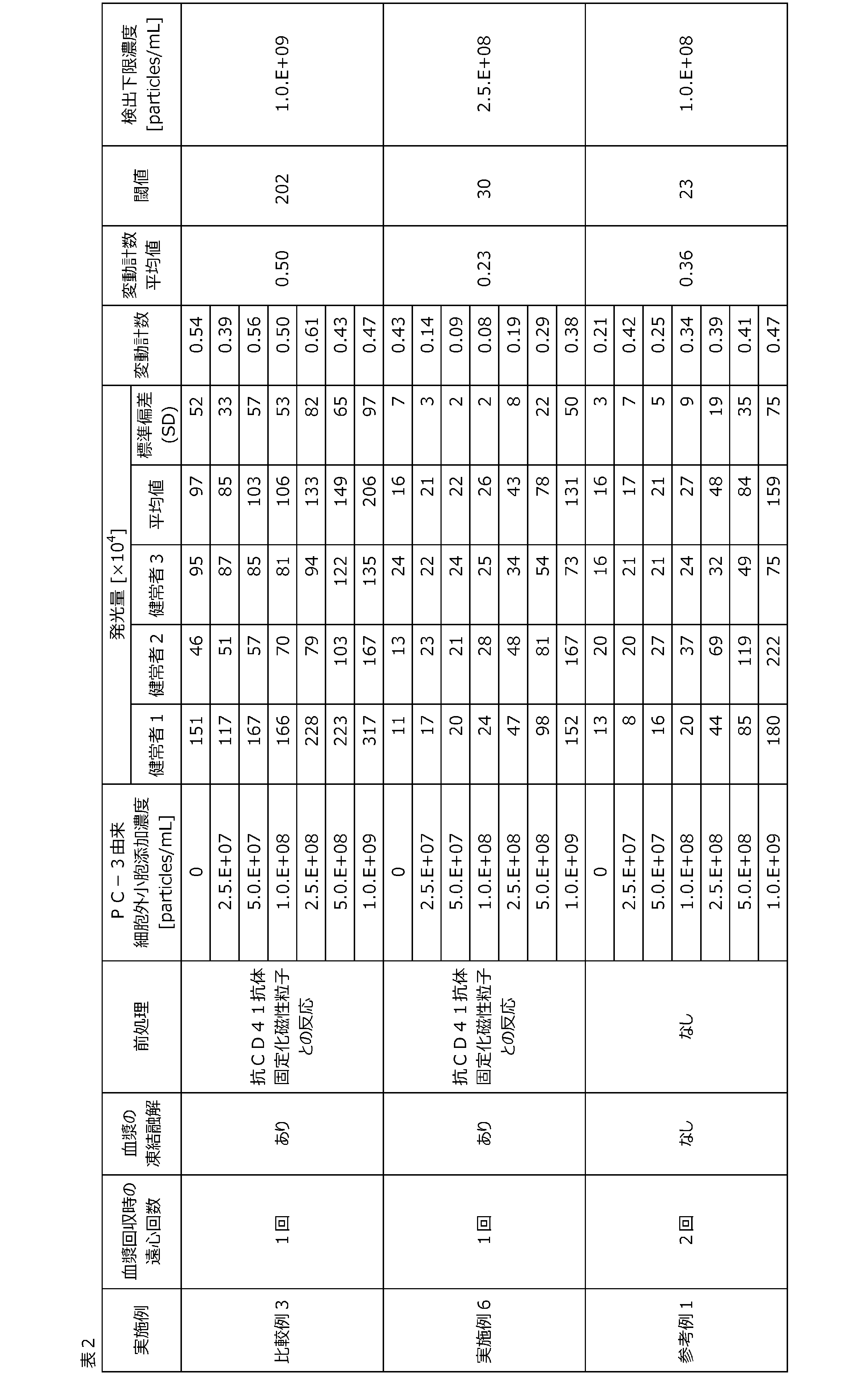
比較例4 血漿中のPC-3由来細胞外小胞の検出
比較例3(2)において、0.5μg/mLビオチン修飾抗CD9抗体の代わりに0.5μg/mLビオチン修飾抗CD81抗体を使用した以外は、比較例3と同様の方法で血漿中のPC-3由来細胞外小胞を検出した。
比較例3(2)において、0.5μg/mLビオチン修飾抗CD9抗体の代わりに0.5μg/mLビオチン修飾抗CD81抗体を使用した以外は、比較例3と同様の方法で血漿中のPC-3由来細胞外小胞を検出した。
実施例7 血小板由来細胞外小胞を除去した健常者血漿中のPC-3由来細胞外小胞の検出
実施例6(3)において、0.5μg/mLビオチン修飾抗CD9抗体の代わりに0.5μg/mLビオチン修飾抗CD81抗体を使用した以外は、実施例6と同様の方法で血漿中のPC-3由来細胞外小胞を検出した。
実施例6(3)において、0.5μg/mLビオチン修飾抗CD9抗体の代わりに0.5μg/mLビオチン修飾抗CD81抗体を使用した以外は、実施例6と同様の方法で血漿中のPC-3由来細胞外小胞を検出した。
参考例2 血小板を除去した健常者血漿中のPC-3由来細胞外小胞の検出
参考例1(2)において、0.5μg/mLビオチン修飾抗CD9抗体の代わりに0.5μg/mLビオチン修飾抗CD81抗体を使用した以外は、参考例1と同様の方法で血漿中のPC-3由来細胞外小胞を検出した。
参考例1(2)において、0.5μg/mLビオチン修飾抗CD9抗体の代わりに0.5μg/mLビオチン修飾抗CD81抗体を使用した以外は、参考例1と同様の方法で血漿中のPC-3由来細胞外小胞を検出した。
比較例4、実施例7および参考例2の結果を表3および図4~6に示す。なお、表3における「変動計数」とは発光量の標準偏差を発光量の平均値で除した値であり、当該値が低値であるほど検体間における測定値のバラつきが小さく測定精度が高いといえる。また、表3における「閾値」とは細胞由来細胞外小胞を含まない血漿サンプルにおける発光量の標準偏差を2倍した値を発光量の平均値に加えた値である。また、表3における「検出下限濃度」とは発光量が閾値より高値となるPC-3由来細胞外小胞添加濃度のうち最も低い濃度のことをいい、検出下限濃度が低いほど検出感度が高いといえる。したがって、PC-3細胞由来細胞外小胞を含まない血漿サンプルにおける発光量および標準偏差の増加に応じて検出感度が低下する。
比較例4、実施例7および参考例2はいずれもPC-3由来細胞外小胞添加濃度依存的に発光量が増加したことから、血漿中からPC-3由来細胞外小胞を検出可能であることを確認した。また、いずれの結果もPC-3細胞由来細胞外小胞を含まない血漿サンプルの発光量の平均値が同等に低値となった。これは、比較例3、実施例6、参考例1においては検出マーカーとして血小板においても発現するCD9を使用した一方、比較例4、実施例7および参考例2では検出マーカーとして血小板において発現が認められないCD81を使用したため、ノイズシグナル発生が抑制されたことが要因と推測される。
検出下限濃度に注目すると、実施例7において比較例4および参考例2と比較してより低値となった。これは比較例4および参考例2と比較して、実施例7において血小板由来細胞外小胞を除去する工程により血漿中に含まれる細胞外小胞の総量が減少し、目的の細胞外小胞(PC-3由来細胞外小胞)と抗TM4SF1抗体固定化磁性粒子との反応性が向上したことにより測定安定性が増し、検体間の測定値のバラつきが小さくなったためと推測される。
以上の結果より、CD81を検出マーカーとして使用した場合においても、血小板由来細胞外小胞を除去する工程により、目的の細胞外小胞の検出感度および測定精度を向上可能であり、疾患患者血漿検体中からの疾患細胞由来細胞外小胞検出に適用可能な測定方法であることが示唆される。
[表3]

実施例6および実施例7において示されたように、本発明の検出方法は血漿中から目的の細胞外小胞を高感度かつ高精度に検出できることから、疾患患者血漿検体中においても疾患細胞由来細胞外小胞検出に適用可能な測定方法であることが示唆される。
Claims (20)
- 血液由来試料中の標的細胞外小胞を検出する方法であって、
前記血液由来試料中の血小板由来細胞外小胞を除去する工程A1または前記血液由来試料中の血小板由来細胞外小胞の発生を抑制する工程A2、および
前記血液由来試料中の前記標的細胞外小胞を検出する工程B
を含む、方法。 - 血液由来試料中の標的細胞外小胞の検出感度を向上させる方法であって、
前記血液由来試料中の血小板由来細胞外小胞を除去する工程A1または前記血液由来試料中の血小板由来細胞外小胞の発生を抑制する工程A2
を含む、方法。 - 血液由来試料中の標的細胞外小胞を検出するために血液由来試料を前処理する方法であって、
前記血液由来試料中の血小板由来細胞外小胞を除去する工程A1または前記血液由来試料中の血小板由来細胞外小胞の発生を抑制する工程A2
を含む、方法。 - 前記標的細胞外小胞が、血小板由来細胞外小胞以外の細胞外小胞である、請求項1~3のいずれか1項に記載の方法。
- 前記血小板由来細胞外小胞以外の細胞外小胞が、腫瘍細胞由来の細胞外小胞である、請求項4に記載の方法。
- 前記腫瘍細胞由来の細胞外小胞が、前立腺癌由来の細胞外小胞である、請求項5に記載の方法。
- 前記血液由来試料が、血漿または血清である、請求項1~3のいずれか1項に記載の方法。
- 前記工程A1が、前記血小板由来細胞外小胞に特異的に結合する物質を用いて実施される、請求項1~3のいずれか1項に記載の方法。
- 前記物質が、前記血小板由来細胞外小胞に特異的に存在するマーカーに対する抗体である、請求項8に記載の方法。
- 前記マーカーが、CD41である、請求項9に記載の方法。
- 前記工程A2が、前記血液由来試料中の血小板を除去することにより実施される、請求項1~3のいずれか1項に記載の方法。
- 前記血小板の除去が、前記血液由来試料の調製時に遠心分離を2回以上実施することにより実施される、請求項11に記載の方法。
- 前記工程A2が、前記血液由来試料を凍結しないことにより実施される、請求項1~3のいずれか1項に記載の方法。
- 前記工程Bが、免疫学的測定法により実施される、請求項1に記載の方法。
- 前記工程Bが、前記標的細胞外小胞を担体に捕捉する工程B1と、捕捉された前記標的細胞外小胞を検出する工程B2を含む、請求項1に記載の方法。
- 前記工程B1が、前記標的細胞外小胞に結合する第1の物質を用いて実施され、且つ
前記工程B2が、前記標的細胞外小胞に結合する第2の物質を用いて実施される、請求項15に記載の方法。 - 前記第1の物質が、前記標的細胞外小胞に存在する第1のマーカーに対する抗体であり、且つ
前記第2の物質が、前記標的細胞外小胞に存在する第2のマーカーに対する抗体である、請求項16に記載の方法。 - 前記第1のマーカーが、前記標的細胞外小胞に特異的に存在するマーカーである、請求項17に記載の方法。
- 前記第1のマーカーが、TM4SF1またはCD81である、請求項17に記載の方法。
- 前記第2のマーカーが、CD9またはCD81である、請求項17~19のいずれか1項に記載の方法。
Priority Applications (1)
| Application Number | Priority Date | Filing Date | Title |
|---|---|---|---|
| JP2025531538A JPWO2025009486A1 (ja) | 2023-07-05 | 2024-06-28 |
Applications Claiming Priority (2)
| Application Number | Priority Date | Filing Date | Title |
|---|---|---|---|
| JP2023110882 | 2023-07-05 | ||
| JP2023-110882 | 2023-07-05 |
Publications (1)
| Publication Number | Publication Date |
|---|---|
| WO2025009486A1 true WO2025009486A1 (ja) | 2025-01-09 |
Family
ID=94171540
Family Applications (1)
| Application Number | Title | Priority Date | Filing Date |
|---|---|---|---|
| PCT/JP2024/023680 Ceased WO2025009486A1 (ja) | 2023-07-05 | 2024-06-28 | 細胞外小胞の検出方法 |
Country Status (2)
| Country | Link |
|---|---|
| JP (1) | JPWO2025009486A1 (ja) |
| WO (1) | WO2025009486A1 (ja) |
Citations (6)
| Publication number | Priority date | Publication date | Assignee | Title |
|---|---|---|---|---|
| WO2010072410A2 (en) * | 2008-12-23 | 2010-07-01 | Universiteit Leiden | Methods for immobilizing microvesicles, means and methods for detecting them, and uses thereof |
| US20170146542A1 (en) * | 2015-06-09 | 2017-05-25 | The Board Of Regents Of The University Of Texas System | Diagnostic test for early stage cancer |
| JP2018179957A (ja) * | 2017-04-17 | 2018-11-15 | 日本光電工業株式会社 | 脂質二重膜粒子またはその断片の検出方法 |
| US20190049438A1 (en) * | 2016-02-01 | 2019-02-14 | The Board Of Trustees Of The Leland Stanford Junior University | Exosome-Total-Isolation-Chip (ExoTIC) Device for Isolation of Exosome-Based Biomarkers |
| WO2023004082A1 (en) * | 2021-07-21 | 2023-01-26 | Mercy Bioanalytics, Inc. | Compositions and methods for detection of prostate cancer |
| WO2023112980A1 (ja) * | 2021-12-16 | 2023-06-22 | 東ソー株式会社 | 腫瘍細胞の検出方法および癌の検査方法 |
-
2024
- 2024-06-28 WO PCT/JP2024/023680 patent/WO2025009486A1/ja not_active Ceased
- 2024-06-28 JP JP2025531538A patent/JPWO2025009486A1/ja active Pending
Patent Citations (6)
| Publication number | Priority date | Publication date | Assignee | Title |
|---|---|---|---|---|
| WO2010072410A2 (en) * | 2008-12-23 | 2010-07-01 | Universiteit Leiden | Methods for immobilizing microvesicles, means and methods for detecting them, and uses thereof |
| US20170146542A1 (en) * | 2015-06-09 | 2017-05-25 | The Board Of Regents Of The University Of Texas System | Diagnostic test for early stage cancer |
| US20190049438A1 (en) * | 2016-02-01 | 2019-02-14 | The Board Of Trustees Of The Leland Stanford Junior University | Exosome-Total-Isolation-Chip (ExoTIC) Device for Isolation of Exosome-Based Biomarkers |
| JP2018179957A (ja) * | 2017-04-17 | 2018-11-15 | 日本光電工業株式会社 | 脂質二重膜粒子またはその断片の検出方法 |
| WO2023004082A1 (en) * | 2021-07-21 | 2023-01-26 | Mercy Bioanalytics, Inc. | Compositions and methods for detection of prostate cancer |
| WO2023112980A1 (ja) * | 2021-12-16 | 2023-06-22 | 東ソー株式会社 | 腫瘍細胞の検出方法および癌の検査方法 |
Non-Patent Citations (1)
| Title |
|---|
| MOITA BRENDA, SHARMA VIKRAM: "Applications of Prostate Cancer Proteomics: A Review ", JYI, JOURNAL OF YOUNG INVESTIGATORS, XP093258952, DOI: 10.22186/jyi.39.4.45-53 * |
Also Published As
| Publication number | Publication date |
|---|---|
| JPWO2025009486A1 (ja) | 2025-01-09 |
Similar Documents
| Publication | Publication Date | Title |
|---|---|---|
| JP6029112B2 (ja) | エクソソームの分析方法、エクソソーム分析用試薬およびエクソソーム分析装置 | |
| JP6255035B2 (ja) | 分離方法、検出方法、シグナル測定方法、疾患の判定方法、疾患治療薬の薬効評価方法、キット及び液状組成物 | |
| Iaccino et al. | Monitoring multiple myeloma by idiotype-specific peptide binders of tumor-derived exosomes | |
| JP5025724B2 (ja) | 循環腫瘍性細胞からのタンパク質の検出 | |
| CN101036055B (zh) | 循环癌细胞上Her-2/neu蛋白的增加水平的检测以及治疗 | |
| US20010000751A1 (en) | Efficient enrichment and detection of disseminated tumor cells | |
| EP2460874B1 (en) | Method of separating target cell in biological sample | |
| JPWO2016186215A1 (ja) | 分離方法、検出方法、シグナル測定方法、疾患の判定方法、薬効評価方法、キット、液状組成物及び検体希釈液 | |
| JP2010281595A (ja) | リガンド分子の検出方法 | |
| US9624474B2 (en) | Method of separating target cell in biological sample | |
| CN111175488A (zh) | 一种应用流式细胞术检测血小板抗体特异性的方法及检测试剂盒 | |
| JP6637290B2 (ja) | Ck19に特異的なモノクローナル抗体およびこれを産生するハイブリドーマ、癌の検出キット、癌の検出方法および癌の転移の判定方法 | |
| US20140011192A1 (en) | Particle complex and method of isolating target cell | |
| US20140377779A1 (en) | Composition for diagnosing breast cancer including material specifically binding to polymeric immunoglobulin receptor protein or fragment thereof, and method of diagnosing breast cancer by using the composition | |
| JP2009080019A (ja) | 蛍光ビーズを用いた免疫複合体検出による抗体解析法 | |
| WO2023112980A1 (ja) | 腫瘍細胞の検出方法および癌の検査方法 | |
| JP6707505B2 (ja) | 腫瘍細胞を検出するためのステロイド受容体アッセイ | |
| US7615358B2 (en) | Method for determining the concentration of vital epithelial tumor cells in a body fluid | |
| CN108139400B (zh) | 确定血液样本或抽吸样本中上皮细胞浓度的方法 | |
| TW202120696A (zh) | 胞外體的分離與檢測方法、以及分離與檢測套組 | |
| CN1836165B (zh) | 用于检测低水平融合蛋白的方法 | |
| WO2021193802A1 (ja) | 肺がん患者に対する免疫チェックポイント阻害剤による治療の有効性を予測するための方法 | |
| US20140302526A1 (en) | Zap-70 detection in chronic lymphocytic leukemia | |
| NL2016301B1 (en) | Method for detecting a marker for active tuberculosis. | |
| WO2026038520A1 (ja) | 細胞外小胞による疾患の検査方法、及び診断方法 |
Legal Events
| Date | Code | Title | Description |
|---|---|---|---|
| 121 | Ep: the epo has been informed by wipo that ep was designated in this application |
Ref document number: 24835999 Country of ref document: EP Kind code of ref document: A1 |
|
| ENP | Entry into the national phase |
Ref document number: 2025531538 Country of ref document: JP Kind code of ref document: A |
|
| WWE | Wipo information: entry into national phase |
Ref document number: 2025531538 Country of ref document: JP |
|
| NENP | Non-entry into the national phase |
Ref country code: DE |