WO2025005232A1 - 軟組織修復用のインプラント医療機器及びその製造方法 - Google Patents
軟組織修復用のインプラント医療機器及びその製造方法 Download PDFInfo
- Publication number
- WO2025005232A1 WO2025005232A1 PCT/JP2024/023482 JP2024023482W WO2025005232A1 WO 2025005232 A1 WO2025005232 A1 WO 2025005232A1 JP 2024023482 W JP2024023482 W JP 2024023482W WO 2025005232 A1 WO2025005232 A1 WO 2025005232A1
- Authority
- WO
- WIPO (PCT)
- Prior art keywords
- medical device
- monomer
- layer
- implant medical
- less
- Prior art date
- Legal status (The legal status is an assumption and is not a legal conclusion. Google has not performed a legal analysis and makes no representation as to the accuracy of the status listed.)
- Ceased
Links
Images


Classifications
-
- A—HUMAN NECESSITIES
- A61—MEDICAL OR VETERINARY SCIENCE; HYGIENE
- A61L—METHODS OR APPARATUS FOR STERILISING MATERIALS OR OBJECTS IN GENERAL; DISINFECTION, STERILISATION OR DEODORISATION OF AIR; CHEMICAL ASPECTS OF BANDAGES, DRESSINGS, ABSORBENT PADS OR SURGICAL ARTICLES; MATERIALS FOR BANDAGES, DRESSINGS, ABSORBENT PADS OR SURGICAL ARTICLES
- A61L27/00—Materials for grafts or prostheses or for coating grafts or prostheses
- A61L27/14—Macromolecular materials
- A61L27/18—Macromolecular materials obtained otherwise than by reactions only involving carbon-to-carbon unsaturated bonds
Definitions
- the present invention relates to an implant medical device for soft tissue repair and a method for manufacturing the same.
- Tendons, the rotator cuff (a plate-like collection of tendons), and ligaments are tissues that connect muscles to bones or bones to bones, and play an important role in allowing the smooth movement of joints such as the shoulder, elbow, knee, wrist, and ankle.
- Tendons, rotator cuffs, and ligaments can become damaged or ruptured due to degeneration caused by aging, trauma, or overuse in sports, etc., interfering with daily movements and causing pain not only during exercise but also at rest and at night.
- meshes made of polylactic acid fibers with a controlled number of warp and weft threads and foams made of polylactic acid have been disclosed, which are reinforced with meshes that have improved resistance to dissolution in organic solvents by annealing and then rapidly cooling (Patent Documents 1 and 2).
- Patent Document 3 a biodegradable polyester made of lactic acid and caprolactone has been described as a synthetic polymer with flexibility
- Patent Document 4 a medical molded body using the biodegradable polyester described in Patent Document 3 has been disclosed that is intended to be placed in the body, and when there is no hard tissue around the placement site, the Young's modulus of the molded body is controlled to be low so that it can be treated in the same way as soft tissue
- the implant medical devices described in Patent Documents 1 and 2 use polylactic acid, a hard synthetic polymer, and so although it has excellent strength, it lacks flexibility and there are still issues with respect to conformity to soft tissue. Even when such a hard synthetic polymer is used, it may be possible to impart flexibility by making the thickness and density very small, but because the polymer is easily degraded, it will degrade and be absorbed before the soft tissue repair is complete.
- Patent Document 3 describes a biodegradable polyester made of lactic acid and caprolactone, but does not specifically describe its application to implant devices for soft tissue repair.
- Patent Document 4 describes a medical molded body, medical device, and nerve regeneration guide tube that use the biodegradable polyester described in Patent Document 3, but does not specifically describe its application to implant devices for soft tissue repair.
- the shape (particularly the thickness and bulk density) described in Patent Documents 1 and 2 is used, the strength will be insufficient. Therefore, even if the soft synthetic polymers described in Patent Documents 3 and 4 are used, the thickness and bulk density must be controlled within an appropriate range in order to apply them to implant devices for soft tissue repair.
- the present invention aims to provide an implant medical device for soft tissue repair that has excellent strength and flexibility by using a biodegradable polyester copolymer containing polyhydroxycarboxylic acid, a soft synthetic polymer whose main structural unit is two types of hydroxycarboxylic acid monomers, and by controlling the thickness and bulk density within an appropriate range.
- An implant medical device for soft tissue repair comprising a polyester copolymer containing polyhydroxycarboxylic acid having two types of hydroxycarboxylic acid monomers as main constituent units, and having a thickness of 1.0 mm or more and 20.0 mm or less and a bulk density of 0.40 g/ cm3 or more and 1.50 g/ cm3 or less.
- the implant medical device according to (1) which has a first layer and a second layer, and the bulk density of the first layer is 0.20 g/cm 3 or more and 0.95 g/cm 3 or less.
- An implant medical device for soft tissue repair comprising a first layer and a second layer, the first layer and the second layer having different bulk densities, a thickness of 1.0 mm or more and 20.0 mm or less, and an overall bulk density of 0.40 g/ cm3 or more and 1.50 g/cm3 or less.
- the implant medical device according to (3) which has a polyester copolymer containing a polyhydroxycarboxylic acid having two kinds of hydroxycarboxylic acid monomers as main structural units.
- An implant medical device for soft tissue repair comprising a polyester copolymer containing polyhydroxycarboxylic acid having two types of hydroxycarboxylic acid monomers as main constituent units, and having a thickness of 1.0 mm or more and 20.0 mm or less and a bulk density of 0.40 g/ cm3 or more and 1.50 g/ cm3 or less.
- the implant medical device according to (20) which has a first layer and a second layer, and the bulk density of the first layer is 0.20 g/cm3 or more and 0.95 g/ cm3 or less.
- Formula 1 [A] Molar fraction (%) of monomer A residues in polyhydroxycarboxylic acid
- B] Molar fraction (%) of monomer B residues in polyhydroxycarboxylic acid
- AB] mole fraction (%) of structures (AB and BA) in which monomer A residue and monomer B residue are adjacent to each other in polyhydroxycarboxylic acid
- the present invention can provide an implantable medical device suitable for soft tissue repair applications.
- FIG. 2 is a schematic diagram showing a rectangular parallelepiped having a curved attachment surface as viewed from the side.
- FIG. 2 is a schematic diagram showing a rectangular parallelepiped having projections and recesses on the attachment surface as viewed from the side.
- the implant medical device for soft tissue repair of the present invention is characterized in that it has a polyester copolymer containing polyhydroxycarboxylic acid whose main constituent units are two types of hydroxycarboxylic acid monomers, has a thickness of 1.0 mm or more and 20.0 mm or less, and has a bulk density of 0.40 g/cm3 or more and 1.50 g/cm3 or less.
- the implant medical device for soft tissue repair of the present invention is characterized in that it has a first layer and a second layer, the first layer and the second layer have different bulk densities, the thickness is 1.0 mm or more and 20.0 mm or less, and the overall bulk density is 0.40 g/cm3 or more and 1.50 g/cm3 or less.
- soft tissue refers to tissues of the human body excluding bones, including cartilage, tendons, rotator cuffs, and ligaments, and in particular tendons, rotator cuffs, and ligaments.
- the polyester copolymer refers to a polymer consisting of two or more types of monomers, among which the monomers are linked mainly by ester bonds.
- the term polyester also refers to a polymer in which some of the monomers are linked by bonds other than ester bonds, such as urethane bonds or amide bonds.
- the number of bonds other than ester bonds among the bonds used in monomer linkages is preferably 30% or less, more preferably 10% or less, even more preferably 5% or less, and most preferably 0%, i.e., no bonds other than ester bonds, when the number of bonds used in monomer linkages is taken as 100%.
- the polyester copolymer in the present invention contains polyhydroxycarboxylic acid.
- Polyhydroxycarboxylic acid refers to a polymer composed of hydroxycarboxylic acid.
- Hydroxycarboxylic acid refers to a compound that has both a hydroxy group and a carboxy group in the same molecule.
- polyester copolymers containing polyhydroxycarboxylic acids can be biodegradable.
- the implant medical device of the present invention containing a polyester copolymer is also biodegradable.
- the implant medical device of the present invention is biodegradable.
- the polyester copolymer of the present invention preferably contains 50% by mass or more of polyhydroxycarboxylic acid relative to 100% by mass of the polyester copolymer, more preferably 70% by mass or more, even more preferably 90% by mass or more, and most preferably contains 100% by mass, i.e., is composed only of polyhydroxycarboxylic acid.
- the implant medical device used in the present invention preferably contains 50% by mass or more of polyester copolymer relative to 100% by mass of the implant medical device, more preferably 70% by mass or more, even more preferably 90% by mass or more, and most preferably 100% by mass, i.e., the implant medical device is composed only of polyester copolymer.
- biodegradability refers to the property of being broken down in the body or in the environment. More specifically, it refers to a degree of biodegradation determined according to Measurement Example 1 described below of 60% or more.
- Terms that can be used interchangeably with biodegradability include bioabsorbability and biocompatibility, but these terms often refer primarily to the property of being broken down in the body.
- Polyhydroxycarboxylic acids can be obtained by polymerization of hydroxycarboxylic acids, polymerization of lactones, which are cyclic compounds formed by intramolecular dehydration condensation of the hydroxyl and carboxyl groups of hydroxycarboxylic acids, or polymerization of lactides, which are cyclic compounds formed by dehydration condensation of the hydroxyl and carboxyl groups of two molecules of hydroxycarboxylic acids.
- hydroxycarboxylic acids, lactones, or lactides can be used as hydroxycarboxylic acid monomers.
- Hydroxycarboxylic acids used in the hydroxycarboxylic acid monomer include lactic acid, glycolic acid, hydroxybutyric acid, hydroxyvaleric acid, hydroxycaproic acid, hydroxyheptanoic acid, hydroxyoctanoic acid, hydroxynonanoic acid, hydroxydecanoic acid, hydroxyundecanoic acid, hydroxydodecanoic acid, and (2-hydroxyethoxy)acetic acid.
- lactic acid, glycolic acid, hydroxyvaleric acid, and hydroxycaproic acid are preferred, and lactic acid and hydroxycaproic acid are particularly preferred.
- the lactone used in the hydroxycarboxylic acid monomer may be butyrolactone, valerolactone, caprolactone (hereinafter, CL), dioxepanone, ethylene oxalate, p-dioxanone, trimethylene carbonate, ⁇ -propiolactone, or pivalolactone, with butyrolactone, valerolactone, CL, p-dioxanone, or trimethylene carbonate being particularly preferred, and valerolactone or CL being even more preferred.
- CL caprolactone
- the lactide used in the hydroxycarboxylic acid monomer may be dilactide (hereinafter referred to as LA) which is formed by the dehydration condensation of two molecules of lactic acid, or glycolide or tetramethylglycolide which is formed by the dehydration condensation of two molecules of glycolic acid, with LA or glycolide being particularly preferred.
- LA dilactide
- the polyhydroxycarboxylic acid of the present invention has two types of hydroxycarboxylic acid monomers as its main constituent units. That a polyhydroxycarboxylic acid has two types of hydroxycarboxylic acid monomers as its main constituent units means that the sum of the number of the two types of monomer residues is 50 mol % or more when the sum of the number of all monomer residues contained in the entire polyhydroxycarboxylic acid, including other monomer residues, is taken as 100 mol %, and each of the two types of monomer residues is 10 mol % or more when the sum of the number of all monomer residues contained in the entire polyhydroxycarboxylic acid is taken as 100 mol %.
- the term "monomer residue” refers, in principle, to a repeating unit of a chemical structure derived from a monomer in the chemical structure of a polyhydroxycarboxylic acid obtained by polymerizing two or more monomers including the monomer.
- “having monomer A residues and monomer B residues as the main constituent units” means that the sum of the number of monomer A residues and monomer B residues is 50 mol % or more, and the monomer A residues are 10 mol % or more, and the monomer B residues are 10 mol % or more, when the sum of the number of all monomer residues contained in the entire polyhydroxycarboxylic acid is 100 mol %.
- the molar fractions (%) of monomer A residue, monomer B residue, and other residues can be determined from the area values of the signals derived from each residue by nuclear magnetic resonance (hereinafter referred to as "NMR") measurement.
- NMR nuclear magnetic resonance
- the monomer A residue is an LA residue
- the monomer B residue is a CL residue
- they can be determined by the measurement example 2 described below.
- the molecular weight of the repeating unit of the chemical structure derived from the monomer is used. For example, if the monomer A residue is an LA residue and the monomer B residue is a CL residue, the molecular weight of the repeating unit of monomer A residue is 144, and the molecular weight of the repeating unit of monomer B residue is 114.
- the molar fraction (%) of the sum of monomer A residues and monomer B residues is, based on the above definition, 50 mol % or more, preferably 75 mol % or more, and more preferably 90 mol % or more, when the sum of the number of all monomer residues contained in the entire polymer, including other monomer residues, is 100 mol %.
- the molar fractions (%) of monomer A residues and monomer B residues are each, similarly as defined above, 10 mol% or more, preferably 20 mol% or more, more preferably 30 mol% or more, even more preferably 40 mol% or more, and most preferably 50 mol% or more.
- a particularly preferred embodiment is one in which the sum of monomer A residues and monomer B residues is 100 mol% of the entire polymer, i.e., a polymer consisting only of monomer A and monomer B.
- a more preferred embodiment of the present invention is one in which the R value represented by the following formula 1 is 0.25 or more and 0.99 or less (hereinafter, a polyhydroxycarboxylic acid having an R value of 0.25 or more and 0.99 or less is referred to as a "random polymer").
- R value [AB] / (2 [A] [B]) x 100 ...
- Formula 1 [A] Molar fraction (%) of monomer A residues in polyhydroxycarboxylic acid
- B] Molar fraction (%) of monomer B residues in polyhydroxycarboxylic acid
- AB] mole fraction (%) of structures (AB and BA) in which monomer A residue and monomer B residue are adjacent to each other in polyhydroxycarboxylic acid
- the lower limit of the R value is preferably 0.25 or more, and more preferably 0.45 or more.
- the monomers A and B used in the present invention are preferably monomers selected from lactic acid, glycolic acid, hydroxybutyric acid, hydroxyvaleric acid, hydroxycaproic acid, hydroxyheptanoic acid, hydroxyoctanoic acid, hydroxynonanoic acid, hydroxydecanoic acid, hydroxyundecanoic acid, hydroxydodecanoic acid, (2-hydroxyethoxy)acetic acid, butyrolactone, valerolactone, CL, dioxepanone, ethylene oxalate, p-dioxanone, trimethylene carbonate, ⁇ -propiolactone, pivalolactone, LA, glycolide, and tetramethylglycolide.
- the monomer A is at least one monomer selected from the group consisting of lactic acid, glycolic acid, LA, and glycolide. It is also particularly preferred that the monomer B is at least one monomer selected from the group consisting of hydroxyvaleric acid, hydroxycaproic acid, valerolactone, and CL.
- the weight average molecular weight of the polyester copolymer in the present invention can be measured by the method described in Measurement Example 3. From the viewpoint of controlling the strength and biodegradability within a suitable range, the weight average molecular weight of the polyester copolymer is preferably 50,000 or more, more preferably 100,000 or more, even more preferably 150,000 or more, and most preferably 200,000 or more. As the weight average molecular weight increases, the strength increases and the number of days required for biodegradation increases. From the viewpoint of moldability, the weight average molecular weight of the polyester copolymer is preferably 1,000,000 or less, more preferably 800,000 or less, and even more preferably 500,000 or less.
- the polyester copolymer in the present invention may be a multi-polyester copolymer in which polyester copolymers are linked together.
- the polyester copolymer before linking is referred to as a "macromer" for convenience.
- the average number of macromer molecules constituting one molecule of the polyester copolymer is called the linkage number.
- the linkage number refers to the value obtained by dividing the weight average molecular weight of the polyester copolymer by the weight average molecular weight of the macromer.
- Multipolyester copolymers can be produced by linking polyester copolymers directly or by linking them using linker molecules. Whether the multipolyester copolymer has a linear or branched structure is determined by the structure of the polyester copolymer and the structure of the linker molecule.
- the multi-polyester copolymer will have a branched structure regardless of the method of linking. If the polyester copolymer has a linear structure, the multi-polyester copolymer will have a linear structure when linked as is or using a linker molecule with a linear structure, and the multi-polyester copolymer will have a branched structure when linked using a linker molecule with a branched structure.
- a linear linker molecule a compound having two functional groups, such as a carboxyl group, a hydroxyl group, or an amino group, can be used.
- examples of such compounds include oxalic acid, malonic acid, succinic acid, glutaric acid, adipic acid, pimelic acid, suberic acid, azelaic acid, sebacic acid, dodecanedioic acid, ethylene glycol, propanediol, butanediol, pentanediol, hexanediol, heptanediol, octanediol, polyethylene glycol, polypropylene glycol, polyethylene glycol-polypropylene glycol copolymer, ethylenediamine, propanediamine, butanediamine, diaminopentane, hexamethylenediamine, diaminoheptane, diaminooctane, lactic acid, glycolic acid,
- branched linker molecule a compound having two or more functional groups, such as a carboxyl group, a hydroxyl group, or an amino group, within the molecule can be used, and examples of such compounds include malic acid, tartaric acid, citric acid, tyrosine, serine, threonine, aspartic acid, glutamic acid, and lysine.
- the crystallization rate of at least one of the monomer A residue or monomer B residue is less than 14%. If the crystallization rate is less than 14%, the increase in the tensile modulus of the implant medical device is suppressed, and an implant medical device suitable for soft tissue repair applications can be obtained. It is more preferable that the crystallization rate of the monomer A residue or monomer B residue is 10% or less, and even more preferable that it is 5% or less. It is preferable that the crystallization rate of all monomer residues is low, but it is also acceptable for only the crystallization rate of some of the monomer residues to be low.
- the crystallization rate of a monomer residue is the ratio of the heat of fusion per unit weight of a monomer residue in the polyhydroxycarboxylic acid of the present invention to the product of the heat of fusion per unit weight of a homopolymer consisting of only a certain monomer residue and the weight fraction of that monomer residue in the polyhydroxycarboxylic acid of the present invention.
- the crystallization rate of a monomer A residue is the ratio of the heat of fusion per unit weight of a monomer A residue in the polyhydroxycarboxylic acid to the product of the heat of fusion per unit weight of a homopolymer consisting of only monomer A and the weight fraction of the monomer A residue in the polyhydroxycarboxylic acid of the present invention.
- the crystallization rates of monomer A residues and monomer B residues indicate the proportion of monomer A residues or monomer B residues in the polyhydroxycarboxylic acid of the present invention that form a crystalline structure.
- the crystallization rate of the lactic acid residue is preferably less than 14%, and more preferably 10% or less. Specifically, the crystallization rate is determined by the method described in Measurement Example 4 below.
- the implant medical device in Figure 1 has a rectangular parallelepiped structure formed by stacking a film-like layer (first layer 2) made of a polyester copolymer and a film-like layer (second layer 3) made of a polyester copolymer, each of which has a different thickness and bulk density.
- the implant medical device is a rectangular parallelepiped
- the surface with the largest area and the surface facing it that is included in the layer with the lower bulk density are referred to as the attachment surface for convenience.
- the implant medical device of the present invention is a cylinder
- the bottom surface of the layer with the lower bulk density of the two bottom surfaces is referred to as the attachment surface for convenience.
- the outer surface with the largest area of the first layer 2 is the attachment surface 1.
- a rectangular parallelepiped structure is shown as the external structure of the implant medical device of the present invention, but this is not limited to this.
- Specific examples of the external structure include a rectangular parallelepiped, pyramid, sphere, cylinder, and cone, but a rectangular parallelepiped or cylinder is preferable.
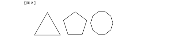
- the shape of the rectangular attachment surface is not limited to the quadrilateral shape of the implant device in FIG. 1, but also includes polygonal shapes such as a triangle, pentagon, or decagon as shown in FIG. 2, and a cross shape as shown in FIG. 3, etc.
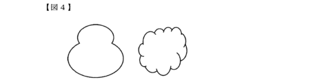
- the attachment surface of the cylinder is not limited to a circle, but may also be, for example, an ellipse, a gourd-shaped or cloud-shaped shape as shown in FIG. 4, a fan-shaped, half-moon-shaped or teardrop-shaped shape as shown in FIG. 5, a broach-shaped shape as shown in FIG. 6, a lens-shaped shape as shown in FIG. 7, etc.
- the surfaces that make up the external structure may have one curved surface as shown in FIG. 8, or one concave or convex shape as shown in FIG. 9.
- the thickness of an implant medical device refers to the distance between the attachment surface and the surface facing it when the implant medical device is a rectangular parallelepiped or cylinder.
- the thickness is measured at multiple locations, including the maximum and minimum thicknesses, and is averaged to ensure that there is no deviation within the surface.
- the implant medical device is a pyramid
- the thickness is half the length of the short side of the base, when it is a sphere, the radius, and when it is a cone, the radius of the base.
- the distance between the apex of the curve in the curved surface and the other surface is the minimum thickness 4, and the distance between the end of the curved surface and the other surface is the maximum thickness 5.
- the distance between the apex of the concave surface and the other surface is the minimum thickness 4, and the distance between the surfaces without any unevenness is the maximum thickness 5.
- the thickness of these implant medical devices can be measured by the method described in Measurement Example 5.
- the thickness of the implant medical device of the present invention is preferably 1.0 to 20.0 mm, more preferably 1.2 to 10.0 mm, even more preferably 1.5 to 5.0 mm, and most preferably 2.0 to 3.0 mm.
- the direction perpendicular to the thickness direction is defined as the vertical direction
- the direction perpendicular to the thickness direction and the vertical direction is defined as the horizontal direction
- the vertical length of the implant medical device of the present invention defined as the vertical length and the horizontal length defined as the horizontal length.
- the vertical length and horizontal length are selected so that their sums are maximized.
- the range in which it can be placed in the affected area can be selected from ranges of 50 cm or less, 30 cm or less, 15 cm or less, or 5 cm or less.
- the lower limit is 0.1 cm or more.
- the thickness of the implant medical device is always smaller than the vertical and horizontal lengths of the implant medical device, and the horizontal length is the same as or smaller than the vertical length.
- the bulk density of the implant medical device refers to the value obtained by dividing the weight of the implant medical device by the volume including the pores and the internal voids, and can be measured by the method described in Measurement Example 6. From the viewpoint of controlling the strength and flexibility within a suitable range, the bulk density of the implant medical device is preferably 0.40 g/cm 3 or more and 1.50 g/cm 3 or less, more preferably 0.45 g/cm 3 or more and 1.40 g/cm 3 or less, even more preferably 0.60 g/cm 3 or more and 1.30 g/cm 3 or less, and most preferably 0.80 g/cm 3 or more and 1.20 g/cm 3 or less.
- PRP platelet-rich plasma
- the implant medical device of the present invention preferably includes at least two layers, a first layer and a second layer.
- a first layer with excellent PRP carrying ability and a second layer with high strength it is possible to exhibit functions suitable for soft tissue repair applications.
- layers other than the first and second layers may be included, but the thickness of these layers must be smaller than that of the first and second layers.
- the bulk density of the first layer and the bulk density of the second layer can be measured by the method described in Measurement Example 6.
- the bulk density of the first layer is preferably 0.20 g/cm3 or more and 0.95 g/cm3 or less , more preferably 0.25 g/cm3 or more and 0.85 g/cm3 or less , even more preferably 0.30 g/cm3 or more and 0.75 g/cm3 or less , and most preferably 0.35 g/cm3 or more and 0.60 g/cm3 or less .
- the bulk density of the second layer is preferably greater than 0.95 g/ cm3 and less than 2.00 g/cm3, more preferably greater than 1.00 g/ cm3 and less than 1.80 g/ cm3 , even more preferably greater than 1.05 g/cm3 and less than 1.65 g/ cm3 , and most preferably greater than 1.10 g/ cm3 and less than 1.50 g/cm3.
- the combination of bulk densities of the first and second layers may be any combination so long as the bulk density of each layer is within the above range and the bulk density of the implant medical device as a whole is within the above range; however, from the viewpoint of controlling the PRP loading capacity within a suitable range, the difference between the bulk density of the first layer and the bulk density of the second layer is preferably 0.10 g/cm3 or more and 0.90 g/cm3 or less, more preferably 0.15 g/cm3 or more and 0.80 g/cm3 or less , and even more preferably 0.20 g/cm3 or more and 0.75 g/ cm3 or less.
- the following method can be used as a pretreatment for measuring the bulk density of the first layer and the second layer.
- the cross section of the implant medical device is observed with an optical microscope, a scanning electron microscope, or the like to confirm the boundary surface between the first layer and the second layer, and the implant medical device is cut along the boundary surface at a plane sufficiently distant from the boundary surface to obtain all or part of the first layer and the second layer. All or part of the obtained first layer and second layer are measured using the method described in Measurement Example 6.
- the ratio of the thickness of the second layer to the overall thickness of the implant medical device can be measured by the method described in Measurement Example 7. From the viewpoint of controlling the bulk density of the implant medical device within a suitable range, the ratio of the thickness of the second layer to the thickness of the implant medical device is preferably 5% or more and 40% or less, more preferably 7% or more and 35% or less, and even more preferably 10% or more and 30% or less.
- the vertical and horizontal lengths of the first and second layers may be different, but it is preferable that the vertical and horizontal lengths of the first and second layers are the same.
- the first and second layers are preferably composed of the same components.
- the decomposition rates of the first and second layers are equal, and it is expected that the entire implant medical device will be decomposed and absorbed uniformly.
- the first and second layers it is possible to intentionally change the decomposition period and maximize the therapeutic effect on soft tissue.
- the present invention is used for soft tissue regeneration applications, it is preferable that the first layer is used as the application surface.
- the maximum tensile load of the implant medical device of the present invention can be measured by the method described in Measurement Example 8. Specifically, the maximum tensile load refers to the maximum load observed in a single measurement.
- the maximum tensile load of the implant medical device is preferably 20 N or more, more preferably 50 N or more, and even more preferably 70 N or more. There is no particular upper limit, but as a feasible range, 2,000 N or less, 1,000 N or less, or 500 N or less can be selected.
- the tensile modulus of the implant medical device of the present invention can be measured by the method described in Measurement Example 8.
- the tensile modulus of the implant medical device is preferably 100 MPa or less, more preferably 50 MPa or less, even more preferably 20 MPa or less, and most preferably 10 MPa or less. There is no particular lower limit, but the practicable range can be selected to be 0.01 MPa or more, 0.05 MPa or more, or 0.10 MPa or more.
- the storage length of the implant medical device of the present invention can be measured by the method described in Measurement Example 9.
- the storage length of the implant medical device of the present invention is preferably 50 mm or less, more preferably 30 mm or less, even more preferably 15 mm or less, and most preferably 10 mm or less.
- the smaller the storage length the smaller the tube that can be stored in, so by setting the storage length within the above range, it is possible to quickly deliver the device to the affected area during surgery using an endoscope.
- the implant medical device of the present invention After surgical treatment for soft tissue repair, platelet-rich plasma (PRP) may be injected into the affected area to promote tissue repair. For this reason, it is preferable that the implant medical device of the present invention has the ability to carry PRP.
- PRP platelet-rich plasma
- the PRP carrying capacity can be evaluated by the amount of PRP carried per unit weight of the implant medical device, and can be measured by the method described in Measurement Example 10.
- the amount of PRP injected in a typical treatment is 1 mL, and in order for the implant medical device to carry the entire amount of injected PRP, the PRP carrying capacity must be 0.20 g/g or more. Therefore, the PRP carrying capacity of the implant medical device of the present invention is preferably 0.20 g/g or more, more preferably 0.40 g/g or more, even more preferably 0.60 g/g or more, and most preferably 0.80 g/g or more.
- the implant medical device of the present invention can be produced by various methods such as injection molding, extrusion molding, freeze-drying, electrospinning, or 3D printing. From the viewpoint of precisely controlling the bulk density, however, it is preferable to produce it by freeze-drying, electrospinning, or 3D printing, and more preferably by 3D printing.
- 3D printers include, for example, fused deposition modeling (FDM), stereolithography (SLA or DLP), inkjet, powder sintering (SLS or SLM), and powder bonding.
- the implant medical device of the present invention is preferably produced by the fused deposition modeling (FDM), stereolithography (SLA or DLP) or powder sintering (SLS or SLM), and more preferably produced by the fused deposition modeling (FDM) or powder sintering (SLS or SLM).
- the thickness of each layer can be set.
- any layer pitch can be selected, for example, between 0.01 mm and 1 mm.
- Infill patterns include line, grid, triangle, trihexagon, cubic subdivision, octet, quarter cubic, zigzag, cross, cross 3D, star, cubic, honeycomb, 3D honeycomb, gyroid, Himbert curve, Archimedes code, and octagram spiral, but when producing the implant medical device of the present invention, line, grid, triangle, trihexagon, zigzag, star, cubic, concentric circle, honeycomb, 3D honeycomb, or gyroid are preferred.
- the implant medical device of the present invention is used to repair soft tissue, and is particularly suitable for repairing ruptured soft tissue. When used to repair ruptured soft tissue, it is preferably fixed to muscle and bone or bone and bone. In a method for repairing a ruptured tendon, rotator cuff, or ligament, it is preferable to have a fixing step of fixing the implant medical device of the present invention to muscle and bone or bone and bone.
- Biodegradability (%) (BOD-B)/TOD ⁇ 100...Formula 2
- BOD Oxygen consumption of polyester copolymer or implant medical device
- B Blank oxygen consumption
- TOD Theoretical oxygen consumption required when polyester copolymer or implant medical device is completely oxidized
- polyhydroxycarboxylic acid has two types of hydroxycarboxylic acid monomers as main constituent units
- the two types of hydroxycarboxylic acid monomers are designated as "monomer A” and “monomer B", respectively, and the molar fractions (%) of monomer A residues and monomer B residues were calculated.
- the peaks of monomer A and monomer B were separated by the signal derived from monomer A or monomer B of adjacent monomer residues by 1H homospin decoupling method, and the respective peak areas were quantified. [AB] was calculated from the respective peak area ratios, and the R value was calculated from the following formula 1.
- R value [AB] / (2 [A] [B]) x 100 ...
- Formula 1 [A]: Molar fraction (%) of monomer A residue in polyhydroxycarboxylic acid
- [AB] mole fraction (%) of structures in which monomer A residue and monomer B residue are adjacent to each other (AB and BA) in polyhydroxycarboxylic acid
- [AB] is the molar fraction (%) of structures (A-B and B-A) in which a monomer A residue and a monomer B residue are adjacent to each other in the polyhydroxycarboxylic acid, and specifically, is the ratio of the number of A-B and B-A to the total number of A-A, A-B, B-A, and B-B.
- 1H homospin decoupling method was performed under the following condition 1, and adjacent monomer residues were separated by signals derived from LA or CL for the methine group of LA (near 5.10 ppm), the ⁇ -methylene group of CL (near 2.35 ppm), and the ⁇ -methylene group (near 4.10 ppm), and the respective peak areas were quantified.
- Condition 1 Device name: JNM-ECZ400R (manufactured by JEOL Ltd.) 1 H homospin decoupling irradiation position: 1.66 ppm
- Solvent deuterated chloroform Measurement temperature: room temperature
- the obtained film (about 10 mg) was collected on an alumina PAN and subjected to DSC measurement under the following condition 3.
- the crystallization rate was calculated from the measurement results under temperature conditions (D) to (E) using the following formulas 3 and 4.
- the heat of fusion per unit mass of the homopolymer was determined as follows.
- the homopolymer was dissolved in chloroform to a concentration of 5% by mass, and the solution was transferred onto a PTFE petri dish and dried overnight at normal pressure and room temperature. This was dried under reduced pressure to obtain a film.
- the obtained film was collected on an alumina PAN and subjected to DSC measurement under the following condition 4, and the heat of fusion was read from the melting peak area of the graph obtained from the measurement results under temperature conditions (D) to (E).
- Condition 4 Device name: EXSTAR 6000 (Seiko Instruments Inc.) Temperature conditions: (A) 25°C ⁇ (B) 250°C (10°C/min) ⁇ (C) 250°C (5 minutes) ⁇ (D) -70°C (10°C/min) ⁇ (E) 250°C (10°C/min) ⁇ (F) 250°C (5 minutes) ⁇ (G) 25°C (100°C/min) Standard material: Alumina
- the thickness of the implant medical device was measured at 10 points, including the maximum and minimum thicknesses, without causing any in-plane bias, using a constant pressure thickness measuring device, and the average value was regarded as the thickness of the implant medical device.
- the bulk density of the implant medical device, first layer or second layer (hereafter referred to as the sample) was measured using the following procedure. To dry the sample, vacuum drying at room temperature may be performed, but no other pretreatment is required.
- Step 1 The mass of the sample in a dry state was measured using an electronic balance, and the displayed value was designated as W1.
- Step 2 Prepare a container filled with water on an electronic balance, and immerse the sample in the water so that the entire sample is immersed but does not touch the bottom. The value displayed at this time was W1-W2. The value of W2 was calculated from the value of W1 calculated in step 1 and the value of W1-W2.
- Step 3 The sample was taken out of the water, and the mass of the wet sample was measured using an electronic balance. The displayed value was designated as W3.
- PRP PRP Supporting Ability
- PRP was prepared as follows: 25 mL of bovine whole blood was placed in a 50 mL centrifuge tube, centrifuged under the following condition (1), and the supernatant was collected. The remaining liquid was then centrifuged under the following condition (2), and the supernatant was collected and mixed with the previous supernatant to prepare PRP.
- Condition (1) 140 ⁇ g, 10 min, 4° C. ⁇ 200 ⁇ g, 10 min, 4° C. ⁇ 300 ⁇ g, 10 min, 4° C.
- Conditions (2) 3000 rpm, 10 minutes, 4°C
- the weight of the implant medical device in a dry state was measured and designated as Wi.
- 15 mL of PRP was placed in a polystyrene container, and the implant medical device was immersed and left to stand for 5 minutes. The implant medical device was then removed and its weight was measured.
- the weight of PRP supported on the implant medical device was calculated from the weight difference of the implant medical device before and after immersion in PRP, and designated as Wp.
- the PRP support capacity was calculated from the following formula 7.
- PRP carrying capacity Wp/Wi...Formula 7
- the obtained crude copolymer was dissolved in 260 mL of chloroform and added dropwise to 3000 mL of hexane in a stirred state to obtain a precipitate.
- the precipitate was dried under reduced pressure at 50° C. to obtain macromer 1.
- the reaction mixture was diluted with 240 mL of chloroform, and 10.0 g of D,L-lactic acid (manufactured by Fujifilm Wako Pure Chemical Industries, Ltd.) was added and stirred for 30 minutes.
- 500 mL of 0.5 M hydrochloric acid was added, and the mixture was stirred for 20 minutes, and the aqueous layer was removed by decantation.
- 500 mL of ion-exchanged water was added, and the mixture was stirred for 10 minutes, and the process of removing the aqueous layer by decantation was repeated until the pH of the removed aqueous layer reached 7.
- the remaining organic layer was dropped into 3,000 mL of methanol while stirring to obtain a precipitate. This precipitate was dried under reduced pressure at 50°C to obtain polyester copolymer 1.
- the obtained crude copolymer was dissolved in 100 mL of chloroform and added dropwise to 1,400 mL of stirred methanol to obtain a precipitate. This procedure was repeated three times, and the precipitate was dried under reduced pressure at 70°C to obtain Macromer 2.
- dichloromethane dehydrated
- dicyclohexylcarbodiimide manufactured by Sigma-Aldrich Co., Ltd.
- the obtained crude copolymer was dissolved in 100 mL of chloroform and added dropwise to 1,400 mL of stirred methanol to obtain a precipitate. This operation was repeated three times, and the obtained precipitate was dried under reduced pressure at 50°C to obtain polyester copolymer 3.
- the obtained crude copolymer was dissolved in 100 mL of chloroform and added dropwise to 1,400 mL of stirred methanol to obtain a precipitate. This procedure was repeated three times, and the precipitate was dried under reduced pressure at 70°C to obtain Macromer 3.
- dichloromethane dehydrated
- dicyclohexylcarbodiimide manufactured by Sigma-Aldrich Co., Ltd.
- polyester copolymers 1 to 4 were subjected to the measurements described in Measurement Examples 1 to 4. The results are shown in Table 1.
- Example 1 An implant medical device was produced by 3D printing using polyester copolymer 1 so that the length and width were 35 mm, the thickness was 2 mm, the bulk density of the first layer was 0.55 g/ cm3 , the bulk density of the second layer was 1.10 g/ cm3 , and the thickness ratio of the second layer was 30%.
- a fused deposition model (FDM) 3D printer was used to produce the device, with the layer pitch set to 0.2 mm and the infill pattern set to a grid.
- Example 2 An implant medical device was produced by 3D printing using polyester copolymer 1 so that the length and width were 35 mm, the thickness was 2 mm, the bulk density of the first layer was 0.35 g/ cm3 , the bulk density of the second layer was 1.10 g/ cm3 , and the thickness ratio of the second layer was 10%.
- a fused deposition model (FDM) 3D printer was used to produce the device, with the layer pitch set to 0.2 mm and the infill pattern set to a grid.
- Example 3 An implant medical device was produced by 3D printing using polyester copolymer 1 so that the length and width were 35 mm, the thickness was 2 mm, the bulk density of the first layer was 0.85 g/ cm3 , the bulk density of the second layer was 1.00 g/ cm3 , and the thickness ratio of the second layer was 10%.
- a fused deposition model (FDM) 3D printer was used to produce the device, with the layer pitch set to 0.2 mm and the infill pattern set to a grid.
- Example 4 An implant medical device was produced by 3D printing using polyester copolymer 1 so that the length and width were 35 mm, the thickness was 2 mm, and the bulk density was 1.10 g/ cm3 . Note that a fused deposition model (FDM) 3D printer was used, and the layer pitch was set to 0.2 mm and the infill pattern was set to a grid.
- FDM fused deposition model
- Example 5 Using polyester copolymer 2, an implant medical device was produced in the same manner as in Example 1.
- Example 6 Using polyester copolymer 3, an implant medical device was produced in the same manner as in Example 1.
- Example 7 Using polyester copolymer 4, an implant medical device was produced in the same manner as in Example 1.
- An implant medical device was produced by 3D printing using polyester copolymer 1 so that the length and width were 35 mm, the thickness was 0.4 mm, the first layer bulk density was 0.55 g/ cm3 , the second layer bulk density was 1.10 g/ cm3 , and the thickness ratio of the second layer was 50%.
- a fused deposition model (FDM) 3D printer was used to produce the device, with the layer pitch set to 0.2 mm and the infill pattern set to a grid.
- An implant medical device was produced by 3D printing using polyester copolymer 1 so that the length and width were 35 mm, the thickness was 2 mm, and the bulk density was 0.35 g/cm 3.
- a fused deposition model (FDM) 3D printer was used, and the layer pitch was set to 0.2 mm and the infill pattern was set to a grid.
- An implant medical device was produced by 3D printing using polyester copolymer 1 so that the length and width were 35 mm, the thickness was 20 mm, the bulk density of the first layer was 0.55 g/ cm3 , the bulk density of the second layer was 1.10 g/ cm3 , and the thickness ratio of the second layer was 30%.
- a fused deposition model (FDM) 3D printer was used to produce the device, with the layer pitch set to 0.2 mm and the infill pattern set to a grid.
- An implant medical device was produced by 3D printing using polyester copolymer 1 so that the length and width were 35 mm, the thickness was 2 mm, the bulk density of the first layer was 0.20 g/ cm3 , the bulk density of the second layer was 1.10 g/ cm3 , and the thickness ratio of the second layer was 30%.
- a fused deposition model (FDM) 3D printer was used to produce the device, with the layer pitch set to 0.2 mm and the infill pattern set to a grid.
- Example 10 An implant medical device was produced by 3D printing using polyester copolymer 1 so that the length and width were 35 mm, the thickness was 2 mm, the bulk density of the first layer was 0.15 g/ cm3 , the bulk density of the second layer was 1.10 g/ cm3 , and the thickness ratio of the second layer was 10%.
- a fused deposition model (FDM) 3D printer was used to produce the device, with the layer pitch set to 0.2 mm and the infill pattern set to a grid.
- Example 1 and Comparative Example 1 From the results of Example 1 and Comparative Example 1, it was revealed that a maximum tensile load suitable for soft tissue repair applications can be obtained by setting the thickness of the implant medical device to 1.0 mm or more . Also, from the results of Example 1 and Comparative Example 2, it was revealed that a maximum tensile load suitable for soft tissue repair applications can be obtained by setting the bulk density of the implant medical device to 0.40 g/cm3 or more.
- the implant medical device of the present invention can be used for repairing soft tissues, particularly as a repair material for cartilage, tendons, rotator cuffs, and ligaments.
Landscapes
- Health & Medical Sciences (AREA)
- Chemical & Material Sciences (AREA)
- Transplantation (AREA)
- Dermatology (AREA)
- Medicinal Chemistry (AREA)
- Oral & Maxillofacial Surgery (AREA)
- Chemical Kinetics & Catalysis (AREA)
- Epidemiology (AREA)
- Life Sciences & Earth Sciences (AREA)
- Animal Behavior & Ethology (AREA)
- General Health & Medical Sciences (AREA)
- Public Health (AREA)
- Veterinary Medicine (AREA)
- Materials For Medical Uses (AREA)
Priority Applications (1)
| Application Number | Priority Date | Filing Date | Title |
|---|---|---|---|
| JP2024540020A JPWO2025005232A1 (https=) | 2023-06-30 | 2024-06-28 |
Applications Claiming Priority (2)
| Application Number | Priority Date | Filing Date | Title |
|---|---|---|---|
| JP2023108093 | 2023-06-30 | ||
| JP2023-108093 | 2023-06-30 |
Publications (1)
| Publication Number | Publication Date |
|---|---|
| WO2025005232A1 true WO2025005232A1 (ja) | 2025-01-02 |
Family
ID=93939106
Family Applications (1)
| Application Number | Title | Priority Date | Filing Date |
|---|---|---|---|
| PCT/JP2024/023482 Ceased WO2025005232A1 (ja) | 2023-06-30 | 2024-06-28 | 軟組織修復用のインプラント医療機器及びその製造方法 |
Country Status (2)
| Country | Link |
|---|---|
| JP (1) | JPWO2025005232A1 (https=) |
| WO (1) | WO2025005232A1 (https=) |
Citations (6)
| Publication number | Priority date | Publication date | Assignee | Title |
|---|---|---|---|---|
| JPH08206191A (ja) * | 1994-10-18 | 1996-08-13 | Ethicon Inc | 軟組織修復および増補用の注射可能微細分散液 |
| CN1396192A (zh) * | 2001-07-13 | 2003-02-12 | 浙江大学 | 生物降解性聚碳酸酯三元共聚物及制备方法 |
| CN1396193A (zh) * | 2001-07-13 | 2003-02-12 | 浙江大学 | 生物降解性聚碳酸酯/聚酯/聚醚三元共聚物及制备方法 |
| WO2006028244A1 (ja) * | 2004-09-07 | 2006-03-16 | Teijin Limited | 生体吸収性多孔体 |
| JP2009506811A (ja) * | 2005-09-01 | 2009-02-19 | ポリメディクス イノベーション ゲーエムベーハー | 創傷を医療処置するための造形体 |
| JP2017527341A (ja) * | 2014-09-07 | 2017-09-21 | オッシオ リミテッド | 異方性バイオ複合材料、異方性バイオ複合材料を含む医療用インプラント、および係る医療用インプラントの治療方法 |
-
2024
- 2024-06-28 JP JP2024540020A patent/JPWO2025005232A1/ja active Pending
- 2024-06-28 WO PCT/JP2024/023482 patent/WO2025005232A1/ja not_active Ceased
Patent Citations (6)
| Publication number | Priority date | Publication date | Assignee | Title |
|---|---|---|---|---|
| JPH08206191A (ja) * | 1994-10-18 | 1996-08-13 | Ethicon Inc | 軟組織修復および増補用の注射可能微細分散液 |
| CN1396192A (zh) * | 2001-07-13 | 2003-02-12 | 浙江大学 | 生物降解性聚碳酸酯三元共聚物及制备方法 |
| CN1396193A (zh) * | 2001-07-13 | 2003-02-12 | 浙江大学 | 生物降解性聚碳酸酯/聚酯/聚醚三元共聚物及制备方法 |
| WO2006028244A1 (ja) * | 2004-09-07 | 2006-03-16 | Teijin Limited | 生体吸収性多孔体 |
| JP2009506811A (ja) * | 2005-09-01 | 2009-02-19 | ポリメディクス イノベーション ゲーエムベーハー | 創傷を医療処置するための造形体 |
| JP2017527341A (ja) * | 2014-09-07 | 2017-09-21 | オッシオ リミテッド | 異方性バイオ複合材料、異方性バイオ複合材料を含む医療用インプラント、および係る医療用インプラントの治療方法 |
Also Published As
| Publication number | Publication date |
|---|---|
| JPWO2025005232A1 (https=) | 2025-01-02 |
Similar Documents
| Publication | Publication Date | Title |
|---|---|---|
| Oh et al. | Hydrophilization of synthetic biodegradable polymer scaffolds for improved cell/tissue compatibility | |
| Ying et al. | Scaffolds from electrospun polyhydroxyalkanoate copolymers: fabrication, characterization, bioabsorption and tissue response | |
| JP2005533148A5 (https=) | ||
| CA2636817A1 (en) | Biodegradable elastomers | |
| WO2005089778A1 (en) | Biodegradable polyurethane and polyurethane ureas | |
| US8236350B2 (en) | Polymer for tissue engineering applications and drug delivery | |
| Liu et al. | Novel biodegradable poly (propylene fumarate)-co-poly (l-lactic acid) porous scaffolds fabricated by phase separation for tissue engineering applications | |
| Oh et al. | Degradation behavior of 3D porous polydioxanone-b-polycaprolactone scaffolds fabricated using the melt-molding particulate-leaching method | |
| Vacaras et al. | Understanding the basis of medical use of poly-lactide-based resorbable polymers and composites–a review of the clinical and metabolic impact | |
| Hu et al. | In vitro degradation behavior of shape memory PLLA-TMC random copolymers | |
| JP4899152B2 (ja) | 医療用樹脂組成物とその製造方法および成形体 | |
| CN105237714A (zh) | 水响应形状记忆聚氨酯及其制备方法 | |
| HILL | Biomedical polymers | |
| Revati et al. | Biodegradable poly (lactic acid) scaffold for tissue engineering: A brief review | |
| Biswal | Recent progress in PLA‐based composite and their application to biomedical and cosmetic fields | |
| WO2025005232A1 (ja) | 軟組織修復用のインプラント医療機器及びその製造方法 | |
| CN1253217C (zh) | 聚l-乳酸在制备做为医用形状记忆材料中的用途 | |
| Lipsa et al. | Poly (α-hydroxyacids) in biomedical applications: synthesis and properties of lactic acid polymers | |
| Bai et al. | In vitro hydrolytic degradation of poly (para-dioxanone)/poly (D, L-lactide) blends | |
| Ang et al. | Tailoring the mechanical and biodegradable properties of binary blends of biomedical thermoplastic elastomer | |
| Ferrer et al. | Tailoring bulk and surface composition of polylactides for application in engineering of skeletal tissues | |
| JP5258189B2 (ja) | 柔軟性生分解性ポリマー | |
| JP7484167B2 (ja) | 医療用成形体、医療機器、神経再生誘導チューブ | |
| WO2021232004A1 (en) | Functionalized poly(glycerol sebacate)s and uses thereof | |
| Mulchandani et al. | Synthesis strategies for biomedical grade polymers |
Legal Events
| Date | Code | Title | Description |
|---|---|---|---|
| WWE | Wipo information: entry into national phase |
Ref document number: 2024540020 Country of ref document: JP |
|
| 121 | Ep: the epo has been informed by wipo that ep was designated in this application |
Ref document number: 24832090 Country of ref document: EP Kind code of ref document: A1 |
|
| NENP | Non-entry into the national phase |
Ref country code: DE |