WO2024154682A1 - キメラ非ヒト哺乳動物を製造する方法、心臓または心臓組織を製造する方法 - Google Patents
キメラ非ヒト哺乳動物を製造する方法、心臓または心臓組織を製造する方法 Download PDFInfo
- Publication number
- WO2024154682A1 WO2024154682A1 PCT/JP2024/000732 JP2024000732W WO2024154682A1 WO 2024154682 A1 WO2024154682 A1 WO 2024154682A1 JP 2024000732 W JP2024000732 W JP 2024000732W WO 2024154682 A1 WO2024154682 A1 WO 2024154682A1
- Authority
- WO
- WIPO (PCT)
- Prior art keywords
- cells
- heart
- mesp
- gfp
- mice
- Prior art date
- Legal status (The legal status is an assumption and is not a legal conclusion. Google has not performed a legal analysis and makes no representation as to the accuracy of the status listed.)
- Ceased
Links
Images





















Classifications
-
- A—HUMAN NECESSITIES
- A01—AGRICULTURE; FORESTRY; ANIMAL HUSBANDRY; HUNTING; TRAPPING; FISHING
- A01K—ANIMAL HUSBANDRY; AVICULTURE; APICULTURE; PISCICULTURE; FISHING; REARING OR BREEDING ANIMALS, NOT OTHERWISE PROVIDED FOR; NEW BREEDS OF ANIMALS
- A01K67/00—Rearing or breeding animals, not otherwise provided for; New or modified breeds of animals
- A01K67/027—New or modified breeds of vertebrates
- A01K67/0271—Chimeric vertebrates, e.g. comprising exogenous cells
-
- A—HUMAN NECESSITIES
- A61—MEDICAL OR VETERINARY SCIENCE; HYGIENE
- A61L—METHODS OR APPARATUS FOR STERILISING MATERIALS OR OBJECTS IN GENERAL; DISINFECTION, STERILISATION OR DEODORISATION OF AIR; CHEMICAL ASPECTS OF BANDAGES, DRESSINGS, ABSORBENT PADS OR SURGICAL ARTICLES; MATERIALS FOR BANDAGES, DRESSINGS, ABSORBENT PADS OR SURGICAL ARTICLES
- A61L27/00—Materials for grafts or prostheses or for coating grafts or prostheses
- A61L27/36—Materials for grafts or prostheses or for coating grafts or prostheses containing ingredients of undetermined constitution or reaction products thereof, e.g. transplant tissue, natural bone, extracellular matrix
-
- C—CHEMISTRY; METALLURGY
- C07—ORGANIC CHEMISTRY
- C07K—PEPTIDES
- C07K14/00—Peptides having more than 20 amino acids; Gastrins; Somatostatins; Melanotropins; Derivatives thereof
- C07K14/435—Peptides having more than 20 amino acids; Gastrins; Somatostatins; Melanotropins; Derivatives thereof from animals; from humans
-
- C—CHEMISTRY; METALLURGY
- C12—BIOCHEMISTRY; BEER; SPIRITS; WINE; VINEGAR; MICROBIOLOGY; ENZYMOLOGY; MUTATION OR GENETIC ENGINEERING
- C12N—MICROORGANISMS OR ENZYMES; COMPOSITIONS THEREOF; PROPAGATING, PRESERVING, OR MAINTAINING MICROORGANISMS; MUTATION OR GENETIC ENGINEERING; CULTURE MEDIA
- C12N15/00—Mutation or genetic engineering; DNA or RNA concerning genetic engineering, vectors, e.g. plasmids, or their isolation, preparation or purification; Use of hosts therefor
- C12N15/09—Recombinant DNA-technology
- C12N15/87—Introduction of foreign genetic material using processes not otherwise provided for, e.g. co-transformation
- C12N15/873—Techniques for producing new embryos, e.g. nuclear transfer, manipulation of totipotent cells or production of chimeric embryos
-
- C—CHEMISTRY; METALLURGY
- C12—BIOCHEMISTRY; BEER; SPIRITS; WINE; VINEGAR; MICROBIOLOGY; ENZYMOLOGY; MUTATION OR GENETIC ENGINEERING
- C12N—MICROORGANISMS OR ENZYMES; COMPOSITIONS THEREOF; PROPAGATING, PRESERVING, OR MAINTAINING MICROORGANISMS; MUTATION OR GENETIC ENGINEERING; CULTURE MEDIA
- C12N5/00—Undifferentiated human, animal or plant cells, e.g. cell lines; Tissues; Cultivation or maintenance thereof; Culture media therefor
- C12N5/06—Animal cells or tissues; Human cells or tissues
-
- C—CHEMISTRY; METALLURGY
- C12—BIOCHEMISTRY; BEER; SPIRITS; WINE; VINEGAR; MICROBIOLOGY; ENZYMOLOGY; MUTATION OR GENETIC ENGINEERING
- C12N—MICROORGANISMS OR ENZYMES; COMPOSITIONS THEREOF; PROPAGATING, PRESERVING, OR MAINTAINING MICROORGANISMS; MUTATION OR GENETIC ENGINEERING; CULTURE MEDIA
- C12N5/00—Undifferentiated human, animal or plant cells, e.g. cell lines; Tissues; Cultivation or maintenance thereof; Culture media therefor
- C12N5/10—Cells modified by introduction of foreign genetic material
Definitions
- the present invention relates to a method for producing a chimeric non-human mammal and the chimeric non-human mammal.
- the present invention also relates to a method for producing a heart or cardiac tissue and the heart or cardiac tissue obtained by the method.
- the present invention further relates to a kit for producing a heart or cardiac tissue.
- the blastocyst complementation method is a method of transplanting stem cells into a genetically modified animal embryo that is missing a specific organ, biological tissue, or cell group (hereinafter, when the word "organ” is used, it is used to mean biological tissue and cell group as well), and regenerating an organ, biological tissue, or cell group derived from stem cells in the living body.
- organ when the word "organ” is used, it is used to mean biological tissue and cell group as well, and regenerating an organ, biological tissue, or cell group derived from stem cells in the living body.
- Non-Patent Documents 1 and 2 describe organ regeneration by the blastocyst complementation method.
- Non-Patent Documents 3 to 6 describe cardiac regeneration using the blastocyst complementation method. Specifically, Non-Patent Document 3 describes the results of verification when the Mesp1 gene and the Mesp2 gene are double knocked out. Non-Patent Document 4 describes the results of verification when the Mesp1 gene is knocked out. Non-Patent Document 5 describes the results of verification when the expression of the Nkx2.5 gene, which is located downstream of the Mesp1 gene and the Mesp2 gene, is manipulated.
- Non-Patent Document 7 describes how Mesp1 gene abnormalities cause cardiac abnormalities.
- Non-Patent Document 8 describes how Mesp2 gene abnormalities do not cause cardiac abnormalities.
- One aspect of the present invention aims to provide a complementary technology to regenerate a normally functioning heart.
- the inventors conducted extensive research to achieve the above-mentioned objective. As a result, they discovered a technique for complementing the cardiomyocytes, endothelial cells, smooth muscle cells, and fibroblasts that make up the heart using the blastocyst complementation method in mammalian embryos in which the Mesp1 and Mesp2 genes had been knocked out. They then succeeded in regenerating a normally functioning heart using this complementation technique, which led to the present invention.
- the method for producing a chimeric non-human mammal includes a stem cell preparation step of preparing stem cells derived from a mammal; a transplantation step of transplanting the stem cells into a fertilized egg of a non-human mammal different from the mammal, the fertilized egg having the Mesp1 gene and the Mesp2 gene knocked out; and a litter obtaining step of developing the fertilized egg in the womb of the non-human mammal to obtain a litter, and is a method for producing a chimeric non-human mammal in which the heart of the litter is complemented by cardiomyocytes, endothelial cells, smooth muscle cells, and fibroblasts derived from the stem cells.
- the chimeric non-human mammal is a chimeric non-human mammal in which the cardiomyocytes, endothelial cells, smooth muscle cells, and fibroblasts constituting the heart comprise cells derived from a mammal, and the organs and tissues other than the heart comprise cells derived from the mammal and cells derived from a non-human mammal different from the mammal, the cells derived from the mammal have the Mesp1 gene and the Mesp2 gene, and the cells derived from the non-human mammal have the Mesp1 gene and the Mesp2 gene knocked out.
- the method for producing a heart or cardiac tissue includes a stem cell preparation step of preparing stem cells derived from a mammal; a transplantation step of transplanting the stem cells into a fertilized egg of a non-human mammal different from the mammal, the fertilized egg having the Mesp1 gene and the Mesp2 gene knocked out; a litter obtaining step of developing the fertilized egg in the womb of the non-human mammal to obtain a litter; and a heart obtaining step of obtaining a heart or cardiac tissue from the litter, the heart or cardiac tissue being complemented by cardiomyocytes, endothelial cells, smooth muscle cells, and fibroblasts differentiated from the stem cells.
- a kit for producing a heart or cardiac tissue is a kit for producing a heart or cardiac tissue, which includes stem cells derived from a mammal and a fertilized egg of a non-human mammal different from the mammal, in which the Mesp1 gene and the Mesp2 gene have been knocked out.
- One aspect of the present invention provides a complementary technology that regenerates a normally functioning heart.
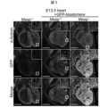
- the results of immunostaining of E13.5 hearts from Mesp +/ ⁇ , Mesp +/ ⁇ + GFP-blastomere, and Mesp ⁇ / ⁇ + GFP-blastomere mice are shown.
- the results of immunostaining of the hearts of Mesp ⁇ / ⁇ +GFP-blastomere mice at E13.5 are shown.
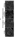
- the figure shows the results of observing GFP emission in organs other than the heart of Mesp ⁇ / ⁇ +GFP-blastomere mice at E13.5. Shown are 8-week-old Mesp +/- +GFP-blastomere mice and Mesp -/- +GFP-blastomere mice.
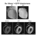
- the results of immunostaining of the heart of an 8-week-old Mesp +/ ⁇ +GFP-blastomere mouse are shown.
- the results of immunostaining of the heart of an 8-week-old Mesp ⁇ / ⁇ +GFP-blastomere mouse are shown.
- the results of immunostaining of the heart of an 8-week-old Mesp +/ ⁇ +GFP-blastomere mouse are shown.
- the results of immunostaining of the heart of an 8-week-old Mesp ⁇ / ⁇ +GFP-blastomere mouse are shown.
- the photographs in the second row from the left show the enlarged view of the area enclosed in a square in the photographs in the leftmost row.
- the results of measuring the cells that make up the hearts of 8-week-old Mesp +/- +GFP-blastomere mice and Mesp -/- +GFP-blastomere mice are shown.
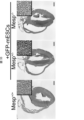
- the results of observation of Mesp +/ ⁇ , Mesp +/ ⁇ +GFP-mESCs, and Mesp ⁇ / ⁇ +GFP-mESCs mice at E9.5 are shown.
- the figures show the observation results of Mesp +/ ⁇ , Mesp +/ ⁇ +GFP-mESCs, and Mesp ⁇ / ⁇ +GFP-mESCs mice at E13.5.
- the results of immunostaining of E13.5 hearts from Mesp +/ ⁇ , Mesp +/ ⁇ + GFP-mESCs, and Mesp ⁇ / ⁇ + GFP-mESCs mice are shown.
- 1 shows the results of observing GFP emission in organs other than the heart of Mesp ⁇ / ⁇ +GFP-mESCs mice at E13.5.
- the results of immunostaining of the hearts of 8-week-old Mesp -/- +GFP-mESCs mice and Mesp +/- +GFP-mESCs mice are shown.
- the results of immunostaining of the hearts of 8-week-old Mesp ⁇ / ⁇ +GFP-mESCs mice are shown.
- the results of immunostaining of the hearts of 8-week-old Mesp +/ ⁇ +GFP-mESCs mice are shown.
- the results of HE staining of the hearts of 8-week-old Mesp +/+ , Mesp +/ ⁇ +GFP-mESCs, and Mesp ⁇ / ⁇ +GFP-mESCs mice are shown.
- the results of echocardiography of 7- to 8-week-old Mesp +/+ , Mesp +/- +GFP-mESCs, and Mesp -/- +GFP-mESCs mice are shown.
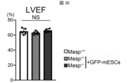
- a graph summarizing the results of FIG. 34 is shown.
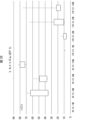
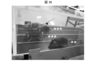
- Photographs of the treadmill fatigue test are shown.
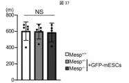
- 1 shows a graph summarizing the results of the treadmill fatigue test.
- the results of electrocardiograms from 7-8 week-old Mesp +/+ , Mesp +/- +GFP-mESCs, and Mesp -/- +GFP-mESCs mice are shown.
- 13 is a graph showing the ratio of heart weight to body weight in 7-8 week-old Mesp +/ ⁇ +GFP-mESCs and Mesp ⁇ / ⁇ +GFP-mESCs mice.
- FIG. 13 is a graph showing the ratio of heart weight to tibia length in 7-8 week-old Mesp +/- +GFP-mESCs and Mesp -/- +GFP-mESCs mice.
- the results of evaluating the chimerism of all surviving mice in the construction of heterologous chimeric mice are shown.
- the results of evaluation of chimerism in surviving chimeric mice constructed from heterologous chimeric mice are shown below.
- the observation results of wild-type, heterozygous and homozygous individuals produced in the construction of heterologous chimeric mice are shown.
- the results of immunostaining of the heart of a homozygous individual produced in the construction of a heterologous chimeric mouse are shown.
- the results of immunostaining of the atria and ventricles of homozygous individuals produced in the construction of heterologous chimeric mice are shown.
- a method for producing a chimeric non-human mammal according to one aspect of the present invention includes a stem cell preparation step, a transplantation step, and a litter obtaining step. Hereinafter, this method may be abbreviated as "a method for producing a chimeric non-human mammal according to this embodiment.”
- chimeric non-human mammal refers to a non-human mammal composed of two or more types of cells with different genetic backgrounds. "Different genetic backgrounds” means that the cells are of different species (heterogeneous), or that the cells are of the same species but are allogeneic (allogenic). Examples of non-human mammals include mice, rats, rabbits, pigs, cows, dogs, sheep, goats, monkeys, chimpanzees, gorillas, orangutans, marmosets, etc.
- stem cell preparation process stem cells derived from a mammal are prepared.
- the mammal is selected according to the animal species of the target heart. Examples of the mammal include human, mouse, rat, rabbit, pig, cow, dog, sheep, goat, monkey, chimpanzee, gorilla, orangutan, marmoset, etc.
- stem cells examples include embryonic stem cells (ES cells) or induced pluripotent stem cells (iPS cells).
- ES cells or iPS cells may be of human or non-human origin.
- ES cells or iPS cells can be prepared by known techniques.
- the stem cells may be prepared by known techniques to express a fluorescent protein such as GFP (green fluorescent protein). By expressing the fluorescent protein, it is possible to easily detect whether the heart in the chimeric non-human mammal is derived from the transplanted stem cells.
- a fluorescent protein such as GFP (green fluorescent protein).
- transplantation process the stem cells prepared in the stem cell preparation step are transplanted into a fertilized egg in which the Mesp1 gene and the Mesp2 gene have been knocked out.
- This transplantation allows a chimeric cell mixture to be formed in the lumen of the fertilized egg by the internal cells derived from the fertilized egg and the transplanted stem cells.
- Transplantation of stem cells into the fertilized egg may be performed by known techniques.
- the Mesp1 gene is the mesoderm posterior 1 gene in mice and the mesoderm posterior bHLH transcription factor 1 gene in humans.
- the Mesp2 gene is the mesoderm posterior 2 gene in mice and the mesoderm posterior bHLH transcription factor 2 gene in humans.
- the method of knocking out the Mesp1 gene and the Mesp2 gene may be carried out by known techniques.
- the Mesp1 gene and the Mesp2 gene can be knocked out by gene editing techniques such as the CRISPR-Cas9 system.
- the Mesp1 gene is described, for example, in Non-Patent Document 7.
- the Mesp2 gene is described, for example, in Non-Patent Document 8.
- the fertilized egg is a fertilized egg of a non-human mammal (recipient) different from the mammal (donor) from which the stem cells are derived.
- the stem cells and the fertilized egg are in a heterogeneous or allogeneic relationship.
- An example of a combination of a mammal (donor) and a non-human mammal (recipient) is as follows: ⁇ Combination of mouse (donor) and mouse (recipient) ⁇ Combination of mouse (donor) and rat (recipient) ⁇ Combination of rat (donor) and mouse (recipient) ⁇ Combination of human (donor) and mouse (recipient) ⁇ Combination of human (donor) and rat (recipient) ⁇ Combination of human (donor) and pig (recipient) ⁇ Combination of human (donor) and cow (recipient) ⁇ Combination of human (donor) and monkey (recipient) ⁇ Combination of monkey (donor) and monkey (recipient) ⁇ Combination of monkey (donor) and mouse (recipient) ⁇ Combination of monkey (donor) and mouse (recipient) ⁇ Combination of monkey (donor) and mouse (recipient)
- the above fertilized eggs are preferably fertilized eggs at the early embryo (embryo after fertilization to the 8-cell stage) to blastocyst stage, and more preferably fertilized eggs at the 8-cell stage to blastocyst stage, in that the chimerism of the offspring obtained in the offspring obtaining process described below will be 50% to 90%.
- offspring chimerism refers to the proportion of cells with different genetic backgrounds within the offspring.
- chimerism of donor cells (recipient cells) refers to the proportion of donor cells (recipient cells) within the chimeric animal body.
- the heart may not be fully complemented. If the chimerism of the offspring is more than 90%, there will be an excess of stem cells, and the entire offspring may be derived from stem cells.
- Stem cells expressing fluorescent proteins such as GFP are transplanted into fertilized eggs, and the number of GFP-positive cells in the hearts of the offspring obtained in the offspring acquisition process described below is counted. The ratio of the number of GFP-positive cells to the number of cells that make up the offspring heart is then evaluated as the chimerism of the offspring.
- the number and type of stem cells to be transplanted into the fertilized egg so that the chimerism of the offspring obtained in the offspring obtaining process described below is between 50% and 90%.
- the fertilized egg may be a fresh embryo or a frozen embryo. It is preferable that the fertilized egg is a frozen embryo, since the chimerism of the offspring obtained in the offspring obtaining process described below will be 50% or more and 90% or less.
- the fertilized egg after the transplantation step is developed in the womb of the non-human mammal to obtain offspring.
- the fertilized egg after the transplantation step can be developed in the womb of the non-human mammal by transplanting the fertilized egg after the transplantation step into the uterus of a pseudopregnant or pregnant female animal of the same species as the non-human mammal from which the fertilized egg was derived. Then, offspring can be obtained by complementing the heart derived from the stem cells transplanted in the transplantation step with the recipient non-human mammal.
- the method for producing a chimeric non-human mammal of this embodiment allows the offspring's heart to be complemented with the above-mentioned stem cell-derived cardiomyocytes, endothelial cells, smooth muscle cells, and fibroblasts.
- the heart complemented by the method for producing a chimeric non-human mammal of this embodiment is a heart that functions normally.
- a chimeric non-human mammal produced by the method for producing a chimeric non-human mammal of this embodiment is also included in one aspect of the present invention.
- the chimeric non-human mammal has the following four characteristics.
- the cardiomyocytes, endothelial cells, smooth muscle cells, and fibroblasts that make up the heart include cells of mammalian origin.
- Organs and tissues other than the heart include cells derived from the above mammals and cells derived from non-human mammals different from the above mammals.
- the mammal-derived cells have the Mesp1 and Mesp2 genes. Therefore, the cardiomyocytes, endothelial cells, smooth muscle cells, and fibroblasts that compose the heart of the chimeric non-human mammal have the Mesp1 and Mesp2 genes functioning normally.
- the cells derived from the chimeric non-human mammal have the Mesp1 gene and the Mesp2 gene knocked out.
- the chimeric non-human mammal described above can be used as a heart disease model animal.
- a chimeric non-human mammal in which the heart has been regenerated from human-derived stem cells can be used as a humanized heart disease model animal.
- chimeric non-human mammal can be used to screen or evaluate the efficacy of drugs related to heart disease.
- the method for producing a heart or cardiac tissue includes a heart obtaining step in addition to the above-mentioned stem cell preparation step, transplantation step, and offspring obtaining step.
- this method may be abbreviated as "a method for producing a heart according to this embodiment.”
- cardiac tissue include cardiac structures such as the right atrium, left atrium, right ventricle, left ventricle, and valve tissue, and cardiac cells such as cardiomyocytes or non-cardiomyocytes that constitute these structures.
- a heart or heart tissue is obtained from the offspring obtained in the offspring obtaining step.
- the heart is a stem cell-derived heart, and is substantially derived from the donor mammal.
- substantially derived from the donor mammal means that the heart does not contain any cells derived from the recipient non-human chimeric mammal, or contains only a very small amount of cells that does not show a rejection reaction in transplantation, etc.
- the obtained heart or heart tissue can be used as a transplant heart for a mammal with a heart disease.
- Whether the obtained heart or cardiac tissue functions normally can be analyzed, for example, by visual observation, morphological analysis using tissue staining or fluorescence microscopy, gene expression analysis, etc.
- a kit for producing a heart or cardiac tissue includes stem cells derived from a mammal and a fertilized egg of a non-human mammal different from the mammal, in which the Mesp1 gene and the Mesp2 gene have been knocked out.
- the kit can be used to produce a heart for transplantation.
- the stem cells derived from a mammal can be prepared in the same manner as in the stem cell preparation step of the method for producing a chimeric non-human mammal.
- the method for knocking out the Mesp1 gene and the Mesp2 gene in the fertilized egg of the non-human mammal may be performed by a known technique.
- the production kit may further comprise an instruction manual or manual including a description of a transplantation step of transplanting the mammal-derived stem cells into the fertilized egg.
- the mammal-derived stem cells can be transplanted in the same manner as in the transplantation step of the method for producing the chimeric non-human mammal.
- the method for producing a chimeric non-human mammal according to aspect 1 of the present invention comprises: a stem cell preparation step of preparing stem cells derived from a mammal; a transplantation step of transplanting the stem cells into a fertilized egg of a non-human mammal different from the mammal, the fertilized egg having a knockout of Mesp1 gene and Mesp2 gene; and a litter obtaining step of developing the fertilized egg in a womb of the non-human mammal to obtain a litter, wherein the heart of the litter is complemented by cardiomyocytes, endothelial cells, smooth muscle cells and fibroblasts derived from the stem cells.
- the method for producing a chimeric non-human mammal according to aspect 2 of the present invention is the above-mentioned aspect 1, in which the fertilized egg of the non-human mammal is a fertilized egg at the early embryo to blastocyst stage.
- the method for producing a chimeric non-human mammal according to aspect 3 of the present invention is the above-mentioned aspect 1 or 2, in which the chimerism of the offspring is 50% or more and 90% or less.
- a method for producing a chimeric non-human mammal according to aspect 4 of the present invention is any one of aspects 1 to 3 above, in which the stem cells are embryonic stem cells (i.e., ES cells) or induced pluripotent stem cells (i.e., iPS cells).
- the stem cells are embryonic stem cells (i.e., ES cells) or induced pluripotent stem cells (i.e., iPS cells).
- the method for producing a chimeric non-human mammal according to aspect 5 of the present invention is any one of aspects 1 to 4 above, in which the stem cells are stem cells derived from a human, a mouse, or a rat.
- the cardiomyocytes, endothelial cells, smooth muscle cells and fibroblasts constituting the heart comprise cells derived from a mammal
- the organs and tissues other than the heart comprise cells derived from the mammal and cells derived from a non-human mammal different from the mammal
- the cells derived from the mammal have the Mesp1 gene and the Mesp2 gene
- the cells derived from the non-human mammal have the Mesp1 gene and the Mesp2 gene knocked out.
- the method for producing a heart or cardiac tissue according to aspect 7 of the present invention includes a stem cell preparation step of preparing stem cells derived from a mammal; a transplantation step of transplanting the stem cells into a fertilized egg of a non-human mammal different from the mammal, the fertilized egg having the Mesp1 gene and the Mesp2 gene knocked out; a litter obtaining step of developing the fertilized egg in the womb of the non-human mammal to obtain a litter; and a heart obtaining step of obtaining a heart or cardiac tissue from the litter, the heart or cardiac tissue being complemented by cardiomyocytes, endothelial cells, smooth muscle cells and fibroblasts differentiated from the stem cells.
- the method for producing a heart or cardiac tissue according to aspect 8 of the present invention is the same as in aspect 7 above, in which the fertilized egg of the non-human mammal is a fertilized egg at the early embryo to blastocyst stage.
- the method for producing a heart or cardiac tissue according to aspect 9 of the present invention is the above-mentioned aspect 7 or 8, in which the chimerism of the offspring is 50% or more and 90% or less.
- the method for producing a heart or cardiac tissue according to aspect 10 of the present invention is the method according to any one of aspects 7 to 9 above, in which the stem cells are embryonic stem cells (ES cells) or induced pluripotent stem cells (iPS cells).
- ES cells embryonic stem cells
- iPS cells induced pluripotent stem cells
- the method for producing a heart or cardiac tissue according to aspect 11 of the present invention is any one of aspects 7 to 10 above, in which the stem cells are derived from a human, mouse, or rat.
- the heart or cardiac tissue according to aspect 12 of the present invention is obtained by the method for producing a heart or cardiac tissue according to any one of aspects 7 to 11 above.
- the kit for producing a heart or cardiac tissue according to aspect 13 of the present invention includes stem cells derived from a mammal and a fertilized egg of a non-human mammal different from the mammal, in which the Mesp1 gene and the Mesp2 gene have been knocked out.
- mice Mesp1 and Mesp2 double knockout (MespDKO) heterogeneous mice (Mesp +/- ) were provided by the RIKEN BioResource Research Center through the MEXT/AMED National BioResource Project. Homogeneous MespDKO (Mesp -/- ) mice were obtained by intercrossing heterogeneous Mesp +/- mice. The production of transgenic mice overexpressing green fluorescent protein (GFP) controlled by the CAG promoter at the ROSA26 locus was performed as described in Hasegawa, Y. et al. Exp Anim 62, 295-304 (2013). ICR (Crl:CD1(ICR)) mice were purchased from Jackson Laboratory Japan.
- GFP green fluorescent protein
- ICR mice stand for Institute of Cancer Research mice. No immunodeficiency or other health-related problems were observed in the transgenic mice. All animals were used in the experiments and were drug naive before use. All animals were group-housed and maintained in a dedicated specific pathogen-free (SPF) facility with a 14/10 h light/dark cycle. Food and water were provided ad libitum and checked daily. Animal health was routinely checked to maintain SPF grade. Animals undergoing surgical procedures were transferred to a satellite SPF facility with the same conditions.
- SPPF pathogen-free
- GRR mouse mouse embryonic stem cells
- SGE2 mESCs SGE2 mouse embryonic stem cells
- GRR mESCs and SGE2 mESCs were maintained in mitomycin C-treated mouse embryonic fibroblasts (MEFs) established from ICR mice.
- Culture medium for GRR mESCs prepared using standard procedures, consisted of Dulbecco's modified Eagle's medium (DMEM, 11995, Thermo Fisher Scientific) supplemented with 10% KnockOut serum replacement (KSR; 10828, Thermo Fisher Scientific), 1% non-essential amino acids (1681049, MP Biomedicals), 0.1 mM 2-mercaptoethanol (M3148, Merck), 1000 U/mL LIF (ESG1107, Merck), 3 ⁇ M CHIR99021 (04-0004-02, Reprocell) and 1 ⁇ M PD0325901 (04-0006-02, Reprocell).
- Culture medium for SGE2 mESCs prepared using standard procedures, consisted of NDiff 227 (Y40002, Takara) supplemented with 1000 U/mL LIF (ESG1107, Merck), 3 ⁇ M CHIR99021 (04-0004-02, Reprocell) and 1 ⁇ M PD0325901 (04-0006-02, Reprocell).
- Mutant chimeras were generated by aggregating one of the embryos from Mesp +/- (including Mesp -/- , Mesp +/- , or wild-type Mesp +/+ ) with one or two GRR blastomeres (developmental blastomere stage embryos obtained from GRR mice). Twenty successful aggregated chimeric blastocysts were surgically transferred into the uterine horns of pseudopregnant ICR female mice (2.5 dpc) and allowed to develop to the desired stage.
- Embryos were frozen 2 hours after IVF and used for blastocyst complementation with ES cells. Host embryos were prepared by thawing frozen embryos and incubating them in M16 medium (M7292, Sigma-Aldrich, Merck) for 24 hours. Frozen ES cells were thawed and placed on a feeder layer 3 days before embryo manipulation. Proliferated ES cells were dissociated with 0.25% trypsin immediately before injection.
- MEFs and tail fibroblasts were isolated with reference to the following literature: Kurotsu, S. et al., STAR Protoc 3, 101122 (2022); Muraoka, N. et al. Nat Commun 10, 674 (2019); Khan, M. et al., J Vis Exp (2016). Briefly, for MEF isolation, embryos isolated from pregnant mice were washed with phosphate-buffered saline (PBS). Each tissue of the embryo was washed with fresh PBS and minced using scissors.
- PBS phosphate-buffered saline
- EDTA ethylenediaminetetraacetic acid
- MEF medium DMEM with 20% fetal bovine serum (FBS) per embryo
- FBS fetal bovine serum
- TTF isolation 1 cm of tails from 4-week-old mice were minced to a size of less than 3 mm. The minced tails were transferred to an enzymatic digestion solution, which was 0.4 mL PBS containing 1 mg collagenase D (11088866001, Merck), and incubated at 37°C for 90 min. The digested tail tissue was transferred to a 70 ⁇ m cell strainer placed in a 10 cm cell culture dish.
- the medium in a 10 cm cell culture dish was 10 ml of TTF medium (Iscove's modified Dulbecco's medium (IMDM; 12440053, Gibco TM , Thermo Fischer Scientific) containing 20% FBS) and the tissue was forcibly triturated using a syringe plunger. The supernatant was transferred to a fresh tube, and the cells were collected by centrifugation, resuspended in TTF medium, and cultured at 37°C under 5% CO2 .
- TTF medium Iscove's modified Dulbecco's medium (IMDM; 12440053, Gibco TM , Thermo Fischer Scientific) containing 20% FBS
- Genotyping of chimeric mice was performed using genomic DNA extracted from GFP-negative fibroblasts isolated by fluorescence-activated cell sorting (cell sorter used: Beckman Coulter, MoFlo XDP model). MEFs were obtained from whole fetal tissues at embryonic day 9 (E9) to embryonic day 9.5 (E9.5) and head tissues after embryonic day 10.5 (E10.5).
- Adult TTFs were obtained from tails after 4 weeks of age.
- Detection of Mesp -/- mutant alleles by genomic PCR was performed using the following four primer sets to amplify WT (PCR products of 439 and 829 bp) and KO (PCR product of 1.8 kbp):
- Embryos were fixed in 4% paraformaldehyde (PFA) overnight at 4°C, followed by 30% sucrose overnight at 4°C. They were then embedded in optimal cutting temperature compound for freezing in liquid nitrogen.
- PFA paraformaldehyde
- Adult mouse hearts, intestines, kidneys, livers, and lungs were fixed overnight in 0.4% PFA and embedded in optimal cutting temperature compound for freezing in liquid nitrogen. These samples were cut into 7 ⁇ m sections using a cryostat microtome (CM3050S, Leica).
- Sections were stained with primary antibodies against ⁇ -actinin (A7811, Sigma, 1:800), ⁇ -SMA (A2547, Sigma, 1:400), CD31 (ab28364, Abcam, 1:10), cTnT (MS-295-P1, Thermo Fischer Scientific, 1:400), GFP (598, MBL, 1:500 and D153-3, MBL, 1:200), and vimentin (GP53, Progen, 1:100 and ab45939, Abcam, 1:100), followed by Alexa488- or 546-conjugated secondary antibodies and DAPI (D1306, Invitrogen, 1:100, Thermo Fischer Scientific).
- ⁇ -actinin A7811, Sigma, 1:800
- ⁇ -SMA A2547, Sigma, 1:400
- CD31 ab28364, Abcam, 1:10
- cTnT MS-295-P1, Thermo Fischer Scientific, 1:400
- GFP 598, MBL, 1:500 and D
- mice Tereadmill fatigue test in mice After three consecutive days of treadmill exercise, a treadmill fatigue test was performed. As a warm-up, mice were run up a 15° treadmill at 10 m/min for 5 min. Then, the speed was increased by 2 m/min every 2 min according to the protocol. Mice were considered to be fatigued when they remained on the shock grid for 10 consecutive seconds. The running time to fatigue was then measured, and the running distance was calculated.
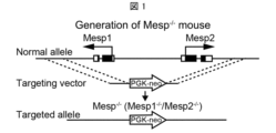
- FIG. 1 A schematic diagram of the targeting vector and targeted allele used in the generation of MespDKO (Mesp -/- ) mice is shown in Figure 1.
- Normal allele is a normal allele.
- Photographs of Mesp +/+ , Mesp +/- , and Mesp -/- mice at E9.0 are shown in Figure 2.
- Figure 2 Mesp -/- mice died during the fetal stage due to impaired differentiation of the mesoderm, from which the heart originates.
- the GFP-blastomere (GFP-positive blastomere) is an embryo at the blastomere stage during the development of a fertilized egg from a GRR mouse genetically engineered to express Green Fluorescent Protein.
- Figure 4 shows the observation results of Mesp +/- , Mesp +/- + GFP-blastomere, and Mesp -/- + GFP-blastomere mice at E9.5.
- BF indicates the observation results in a bright field
- GFP indicates the observation results of GFP emission.
- Hearts developed from GFP-positive embryos emit green light due to GFP.
- no GFP emission was observed in the hearts of Mesp +/- + GFP-blastomere mice.
- GFP emission was observed in the hearts of Mesp -/- + GFP-blastomere mice, confirming that heart regeneration by GFP-positive embryos was successful and embryonic death was avoided.
- Figure 5 shows the results of immunostaining of the hearts of Mesp +/- , Mesp +/- + GFP-blastomere, and Mesp -/- + GFP-blastomere mice at E13.5.
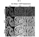
- Figure 6 shows the results of immunostaining of the hearts of Mesp -/- + GFP-blastomere mice at E13.5, where RV stands for right ventricle, IVS stands for interatrial septum, and LV stands for left ventricle.
- ⁇ -actinin is a marker for cardiomyocytes.
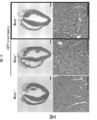
- Figure 7 shows the results of observing GFP emission in organs other than the heart (lungs, liver, intestines, and kidneys) of Mesp -/- + GFP-blastomere mice at E13.5. As shown in Figure 7, the cells in the organs other than the heart were found to be chimeras containing GFP +/- cells.
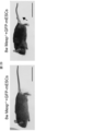
- Fig. 8 shows an 8-week-old Mesp +/- +GFP-blastomere mouse (control), and the right side shows an 8-week-old Mesp -/- + GFP-blastomere mouse.
- the dorsal side of the Mesp -/- +GFP-blastomere mouse has black hair, which is thought to be derived from cells of GFP-positive embryos.
- the ventral side of the Mesp -/- +GFP-blastomere mouse has gray hair, which is thought to be derived from cells of Mesp - /- embryos.
- the tail of the Mesp -/- +GFP-blastomere mouse was deformed. This deformation was due to the mixture of embryos with heart defects (Mesp -/- ) and normal embryos (GFP-blastomere).
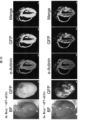
- FIGS 9 and 11 show the results of immunostaining of the heart of an 8-week-old Mesp +/- +GFP-blastomere mouse.
- Figures 10 and 12 show the results of immunostaining of the heart of an 8-week-old Mesp -/- +GFP-blastomere mouse.
- the heart of the Mesp +/- +GFP-blastomere mouse contained a mixture of cardiomyocytes derived from Mesp +/- cells, whereas the cardiomyocytes in the heart of the Mesp -/- +GFP-blastomere mouse were found to be almost entirely regenerated by cells derived from GFP-positive embryos.
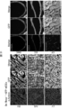
- FIG. 13 shows the results of observation of the heart of an 8-week-old Mesp ⁇ / ⁇ +GFP-blastomere mouse. Immunostaining was performed with a marker for smooth muscle cells ( ⁇ -SMA), endothelial cells (CD31), and fibroblasts (vimentin).
- FIG. 14 is a graph showing the chimerism of each cell constituting the heart of an 8-week-old Mesp +/ ⁇ +GFP-blastomere mouse and a Mesp ⁇ / ⁇ +GFP-blastomere mouse. As shown in FIGS.
- Figure 15 shows the results of hematoxylin-eosin staining (HE staining) of the hearts of 8-week-old Mesp +/+ , Mesp +/- +GFP-blastomere, and Mesp -/- +GFP-blastomere mice. As shown in Figure 15, no abnormalities were observed in the cardiac structure of the Mesp -/- +GFP-blastomere mice.
- Figure 16 shows the results of echocardiography of 7-8 week old Mesp +/+ , Mesp +/- +GFP-blastomere and Mesp -/- +GFP-blastomere mice.
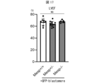
- Figure 17 shows a graph summarizing the results of Figure 16.
- the vertical axis of Figure 17 shows LVEF, which indicates left ventricular ejection fraction.
- the function of the regenerated heart in the Mesp -/- +GFP-blastomere mouse was found to be normal.
- Figure 18 shows photographs of the treadmill fatigue test.
- the mice running in the two central lanes are Mesp -/- +GFP-blastomere mice.
- Figure 19 is a graph summarizing the results of the treadmill fatigue test. The vertical axis of Figure 19 shows the running distance of the mice. As shown in Figure 19, Mesp -/- +GFP-blastomere mice showed normal exercise tolerance.
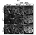
- Fig. 22 shows the results of immunostaining of the hearts of Mesp +/- , Mesp +/- + GFP-mESCs, and Mesp -/- + GFP-mESCs mice at E13.5. As shown in Fig. 22, most of the cardiomyocytes in the hearts regenerated in Mesp -/- + GFP-mESCs mice were found to be derived from mouse ES cells expressing GFP.
- Fig. 23 shows the results of observing GFP emission in organs other than the heart of Mesp -/- + GFP-mESCs mice at E13.5.
- the cells in the organs other than the heart were a mixture of Mesp -/- derived cells and mouse ES cell-derived cells.
- FIG 24 shows the observation results of Mesp -/- +GFP-mESCs mice at P0.
- Mesp -/- +GFP-mESCs mice were able to be born. However, although some mice survived at birth, all of them died early. Malformed mice were also observed among WT/heterozygous mice.
- Mesp -/- +GFP-blastomere mice survived to adulthood, suggesting that the early death of Mesp -/- +GFP-mESCs mice may be due to the quality of ES cells.
- SGE2 mESCs as stem cells for blastocyst complementation, optimized chimerism to 50-90%, and verified heart regeneration through blastocyst complementation. Four mice derived from these SGE2 mESCs survived.
- FIG. 29 The left side of Figure 29 shows an 8-week-old Mesp +/- +GFP-mESCs mouse (control), and the right side shows an 8-week-old Mesp -/- +GFP-mESCs mouse.
- the tail of the Mesp -/- +GFP-mESCs mouse was deformed, as indicated by the arrow.
- the upper panel of Fig. 30 and Fig. 31 show the results of immunostaining of the heart of an 8-week-old Mesp -/- +GFP-mESCs mouse.
- the lower panel of Fig. 30 and Fig. 32 show the results of immunostaining of the heart of an 8-week-old Mesp +/- +GFP-mESCs mouse.
- SMCs represent smooth muscle cells
- ECs represent endothelial cells
- CFs represent fibroblasts.
- the hearts of Mesp +/- +GFP-mESCs mice contained a mixture of cardiomyocytes derived from Mesp -/ - and mouse ES cells.
- the cells that make up the hearts of Mesp -/- +GFP-mESCs mice including not only cardiomyocytes but also smooth muscle cells, endothelial cells, and fibroblasts, which are representative of non-cardiomyocytes, were almost all regenerated from mouse ES cells expressing GFP.
- Figure 33 shows the results of HE staining of the hearts of 8-week-old Mesp +/+ , Mesp +/- +GFP-mESCs, and Mesp -/- +GFP-mESCs mice. As shown in Figure 33, no abnormalities were observed in the cardiac structure of Mesp -/- +GFP-mESCs mice.
- Figure 34 shows the results of echocardiographic measurements of 7-8 week-old Mesp +/+ , Mesp +/- +GFP-mESCs, and Mesp -/- +GFP-mESCs mice.
- Figure 35 shows a graph summarizing the results of Figure 34. The vertical axis of Figure 35 shows LVEF. As shown in Figures 34 and 35, the function of the regenerated heart in the Mesp -/- +GFP-mESCs mice was found to be normal.
- Figure 36 is a photograph of the treadmill fatigue test.
- the mice running in the two lanes in the middle are Mesp -/- +GFP-mESCs mice.
- Figure 37 is a graph summarizing the results of the treadmill fatigue test. The vertical axis of Figure 37 indicates the running distance of the mice. As shown in Figure 37, the Mesp -/- +GFP-mESCs mice showed normal exercise tolerance.
- Figure 38 shows the electrocardiogram results of 7-8 week-old Mesp +/+ , Mesp +/- +GFP-mESCs, and Mesp -/- +GFP-mESCs mice. As shown in Figure 38, no arrhythmia was observed in the regenerated hearts of Mesp -/- +GFP-mESCs mice, indicating that the heart functions normally.
- Figure 39 is a graph showing the ratio of heart weight (HW, mg) to body weight (BW, g) in 7-8 week old Mesp +/- +GFP-mESCs and Mesp -/- +GFP-mESCs mice.
- Figure 40 is a graph showing the ratio of heart weight (HW, mg) to tibia length (TL, mm). The results in Figures 39 and 40 show that Mesp -/- +GFP-mESCs mice do not have heart failure and have normal cardiac function.
- Chimeric embryos were generated using mutant mouse embryos and rat ES cells. Briefly, host embryos used for blastocyst complementation with ES cells were frozen 2 hours after IVF. Frozen embryos were prepared by thawing and incubating in M16 medium (M7292, Sigma-Aldrich, Merck) for 24 hours. Frozen ES cells were thawed and placed on a feeder layer 3 days before embryo manipulation. Proliferated ES cells were dissociated with 0.25% trypsin immediately before injection.
- Figure 41 shows the results of evaluating the chimerism of all surviving mice (E11.5).
- Figure 42 shows the results of evaluating the chimerism of surviving chimeric mice (E11.5).
- the chimerism (%) on the vertical axis in Figures 41 and 42 indicates the ratio of rat ES cells to the cells that make up the offspring. "Fresh” indicates fresh embryos, and "Frozen” indicates frozen embryos.
- Figure 43 shows the results of observing wild-type, heterozygous, and homozygous individuals at E11.5.
- the chimerism of the individuals observed in Figure 43 was 59.7% for wild-type individuals, 34.1% for heterozygous individuals, and 79.5% for homozygous individuals.
- FIG. 44 shows the results of immunostaining the heart of an E11.5 homozygous individual (Mesp ⁇ / ⁇ +GFP-ratESCs mouse).
- the upper part of FIG. 45 shows the results of immunostaining the atrium of an E11.5 homozygous individual, and the lower part shows the results of immunostaining the ventricle. From FIG. 44, it was found that the heart of the homozygous mouse was regenerated by cells derived from GFP-positive rat ES cells. Furthermore, from FIG. 45, it was found that the cardiomyocytes that make up the atrium and ventricle of the homozygous mouse were regenerated by cells derived from GFP-positive rat cells.
- the present invention makes it possible to produce hearts of a desired mammalian species.
- the produced hearts can be used in the clinical field as transplant hearts.
Landscapes
- Health & Medical Sciences (AREA)
- Life Sciences & Earth Sciences (AREA)
- Chemical & Material Sciences (AREA)
- Engineering & Computer Science (AREA)
- Genetics & Genomics (AREA)
- Zoology (AREA)
- Organic Chemistry (AREA)
- Biomedical Technology (AREA)
- Biotechnology (AREA)
- Bioinformatics & Cheminformatics (AREA)
- Wood Science & Technology (AREA)
- General Health & Medical Sciences (AREA)
- General Engineering & Computer Science (AREA)
- Biochemistry (AREA)
- Microbiology (AREA)
- Cell Biology (AREA)
- Molecular Biology (AREA)
- Medicinal Chemistry (AREA)
- Biophysics (AREA)
- Environmental Sciences (AREA)
- Animal Behavior & Ethology (AREA)
- Dermatology (AREA)
- Epidemiology (AREA)
- Toxicology (AREA)
- Proteomics, Peptides & Aminoacids (AREA)
- Botany (AREA)
- Chemical Kinetics & Catalysis (AREA)
- Plant Pathology (AREA)
- Oral & Maxillofacial Surgery (AREA)
- Transplantation (AREA)
- Gastroenterology & Hepatology (AREA)
- Developmental Biology & Embryology (AREA)
- Public Health (AREA)
- Veterinary Medicine (AREA)
- Animal Husbandry (AREA)
- Biodiversity & Conservation Biology (AREA)
- Physics & Mathematics (AREA)
- Micro-Organisms Or Cultivation Processes Thereof (AREA)
- Materials For Medical Uses (AREA)
- Medicines Containing Material From Animals Or Micro-Organisms (AREA)
Abstract
本発明の一態様に係るキメラ非ヒト哺乳動物を製造する方法は、哺乳動物由来の幹細胞を、哺乳動物とは異なる非ヒト哺乳動物の受精卵(Mesp1遺伝子およびMesp2遺伝子がノックアウトされている)に移植する工程を含む。幹細胞由来の心筋細胞、内皮細胞、平滑筋細胞および線維芽細胞によって産仔の心臓が補完される。
Description
本発明は、キメラ非ヒト哺乳動物を製造する方法および当該キメラ非ヒト哺乳動物に関する。本発明はまた、心臓または心臓組織を製造する方法および当該方法によって得られる心臓または心臓組織に関する。本発明はさらに、心臓または心臓組織の製造用キットに関する。
世界では年間4000件を超える心臓移植が行われている。例えば、日本では年間約50件の心臓移植が行われている。一方、心臓移植手術までの待機期間が約3年となっているため、心臓移植手術までの待機期間の短縮が求められている。当該待機期間の短縮のために、多くのドナー心臓の確保が必要とされ、ドナー心臓の作製に関する研究開発が進められている。当該研究開発において、胚盤胞補完法による臓器の再生が注目されている。胚盤胞補完法とは、特定の臓器、生体組織、細胞群(以下、臓器と記載する場合、生体組織、細胞群をも含む意味で用いる)が欠損する遺伝子組換え動物胚に幹細胞を移植し、生体内で幹細胞由来の臓器、生体組織または細胞群を再生する方法である。例えば、非特許文献1および2には、胚盤胞補完法による臓器の再生について記載されている。
非特許文献3~6には、胚盤胞補完法による心臓の再生について記載されている。具体的には、非特許文献3には、Mesp1遺伝子およびMesp2遺伝子をダブルノックアウトしたときの検証結果が記載されている。非特許文献4には、Mesp1遺伝子をノックアウトしたときの検証結果が記載されている。非特許文献5には、Mesp1遺伝子およびMesp2遺伝子の下流に存在するNkx2.5遺伝子発現を操作したときの検証結果が記載されている。
非特許文献7には、Mesp1遺伝子異常により心臓に異常が発生したことが記載されている。非特許文献8には、Mesp2遺伝子異常により心臓に異常が発生しなかったことが記載されている。
Cell 142, 787-799, September 3, 2010
Nature 2017 Feb 9;542(7640):191-196
Development 127, 3215-3226 (2000)
Nat Cell Biol. 2014 Sep;16(9):829-40
Circulation 2015 Jul 14; 132(2):109-21
Cell 168, 473-486, January 26, 2017
Development 126, 3437-34447 (1999)
Genes Dev. 1997 Jul 15;11(14):1827-39
しかしながら、上述の従来技術では、臓器補完による正常に動作する心臓の再生までには至っていない。特に、心臓を構成する心筋細胞、内皮細胞、平滑筋細胞および線維芽細胞によって、心臓が補完されたことは報告されておらず、心臓の補完に関するさらなる研究開発が望まれている。
本発明の一態様は、正常に動作する心臓を再生する補完技術の提供を目的とする。
本発明者らは上記課題を達成するために、鋭意研究を重ねた。その結果、Mesp1遺伝子およびMesp2遺伝子をノックアウトされた哺乳動物胚において、胚盤胞補完法によって心臓を構成する心筋細胞、内皮細胞、平滑筋細胞および線維芽細胞を補完する技術を見出した。そして、当該補完技術によって、正常に動作する心臓を再生することに成功し、本発明に至った。
本発明の一態様に係るキメラ非ヒト哺乳動物を製造する方法は、哺乳動物由来の幹細胞を調製する幹細胞調製工程;上記哺乳動物とは異なる非ヒト哺乳動物の受精卵であって、Mesp1遺伝子およびMesp2遺伝子がノックアウトされている受精卵へ上記幹細胞を移植する移植工程;および、上記受精卵を上記非ヒト哺乳動物の母胎中で発生させて、産仔を得る産仔取得工程、を含み、上記幹細胞由来の心筋細胞、内皮細胞、平滑筋細胞および線維芽細胞によって上記産仔の心臓が補完される、キメラ非ヒト哺乳動物を製造する方法である。
本発明の一態様に係るキメラ非ヒト哺乳動物は、心臓を構成する心筋細胞、内皮細胞、平滑筋細胞および線維芽細胞は、哺乳動物由来の細胞を含み、心臓以外の臓器および組織は、上記哺乳動物由来の細胞と上記哺乳動物とは異なる非ヒト哺乳動物由来の細胞とを含み、上記哺乳動物由来の細胞は、Mesp1遺伝子およびMesp2遺伝子を有し、上記非ヒト哺乳動物由来の細胞はMesp1遺伝子およびMesp2遺伝子がノックアウトされている、キメラ非ヒト哺乳動物である。
本発明の一態様に係る心臓または心臓組織を製造する方法は、哺乳動物由来の幹細胞を調製する幹細胞調製工程;上記哺乳動物とは異なる非ヒト哺乳動物の受精卵であって、Mesp1遺伝子およびMesp2遺伝子がノックアウトされている受精卵へ上記幹細胞を移植する移植工程;上記受精卵を非ヒト哺乳動物の母胎中で発生させて、産仔を得る産仔取得工程;および、上記産仔から心臓または心臓組織を取得する心臓取得工程を含み、上記心臓または心臓組織は、上記幹細胞から分化した心筋細胞、内皮細胞、平滑筋細胞および線維芽細胞によって補完された心臓または心臓組織である、心臓または心臓組織を製造する方法である。
本発明の一態様に係る心臓または心臓組織の製造用キットは、哺乳動物由来の幹細胞と、上記哺乳動物とは異なる非ヒト哺乳動物の受精卵であって、Mesp1遺伝子およびMesp2遺伝子がノックアウトされている受精卵と、を含む、心臓または心臓組織の製造用キットである。
本発明の一態様によれば、正常に動作する心臓を再生する補完技術を提供することができる。
本明細書において、「含有する(comprise)」は、「実質的にのみからなる(consist essentially of)」、及び「のみからなる(consist of)」も包含する概念である。
本明細書に記載されている数値範囲において、その数値範囲の上限値又は下限値は、実施例に示されている値又は実施例から一義的に導き出せる値に置き換えてもよい。更に、本明細書において、「~」で結ばれた数値は、「~」の前後の数値を下限値及び上限値として含む数値範囲を意味する。
〔キメラ非ヒト哺乳動物を製造する方法〕
本発明の一態様に係るキメラ非ヒト哺乳動物を製造する方法は、幹細胞調製工程、移植工程、および産仔取得工程を含む。以下、当該方法を、「本実施形態のキメラ非ヒト哺乳動物の製造方法」と略記する場合がある。
本明細書に記載されている数値範囲において、その数値範囲の上限値又は下限値は、実施例に示されている値又は実施例から一義的に導き出せる値に置き換えてもよい。更に、本明細書において、「~」で結ばれた数値は、「~」の前後の数値を下限値及び上限値として含む数値範囲を意味する。
〔キメラ非ヒト哺乳動物を製造する方法〕
本発明の一態様に係るキメラ非ヒト哺乳動物を製造する方法は、幹細胞調製工程、移植工程、および産仔取得工程を含む。以下、当該方法を、「本実施形態のキメラ非ヒト哺乳動物の製造方法」と略記する場合がある。
本明細書において、用語「キメラ非ヒト哺乳動物」とは、遺伝的背景が異なる2種類以上の細胞で構成された、非ヒト哺乳動物を指す。「遺伝的背景が異なる」とは、細胞の種が異なる(異種である)こと、または、細胞の種が同じであってもアロである(同種異系である)ことを意味する。非ヒト哺乳動物の例としては、マウス、ラット、ウサギ、ブタ、ウシ、イヌ、ヒツジ、ヤギ、サル、チンパンジー、ゴリラ、オラウータン、マーモセット等が挙げられる。
(幹細胞調製工程)
上記幹細胞調製工程では、哺乳動物由来の幹細胞を調製する。哺乳動物は、製造対象の心臓の動物種に応じて選択する。当該哺乳動物の例として、ヒト、マウス、ラット、ウサギ、ブタ、ウシ、イヌ、ヒツジ、ヤギ、サル、チンパンジー、ゴリラ、オラウータン、マーモセット等が挙げられる。
上記幹細胞調製工程では、哺乳動物由来の幹細胞を調製する。哺乳動物は、製造対象の心臓の動物種に応じて選択する。当該哺乳動物の例として、ヒト、マウス、ラット、ウサギ、ブタ、ウシ、イヌ、ヒツジ、ヤギ、サル、チンパンジー、ゴリラ、オラウータン、マーモセット等が挙げられる。
上記幹細胞の例として、胚性幹細胞(ES細胞)または誘導型多能性幹細胞(iPS細胞)が挙げられる。ES細胞またはiPS細胞は、ヒト由来であってもよいし、非ヒト由来であってもよい。ES細胞またはiPS細胞の調製は、公知の技術によって調製することができる。
上記幹細胞は、公知の技術によって、GFP(green fluorescent protein)等の蛍光タンパク質が発現するように調製されていてもよい。当該蛍光タンパク質の発現によって、キメラ非ヒト哺乳動物において、心臓が移植された幹細胞由来であるか否かを容易に検出することができる。
(移植工程)
上記移植工程では、Mesp1遺伝子およびMesp2遺伝子がノックアウトされている受精卵へ、幹細胞調製工程で調製した幹細胞を移植する。当該移植によって、受精卵の内腔において、受精卵由来の内部細胞と移植された幹細胞とによるキメラの細胞混合物を形成させることができる。受精卵への幹細胞の移植は公知の技術によって行えばよい。
上記移植工程では、Mesp1遺伝子およびMesp2遺伝子がノックアウトされている受精卵へ、幹細胞調製工程で調製した幹細胞を移植する。当該移植によって、受精卵の内腔において、受精卵由来の内部細胞と移植された幹細胞とによるキメラの細胞混合物を形成させることができる。受精卵への幹細胞の移植は公知の技術によって行えばよい。
Mesp1遺伝子は、マウスではmesoderm posterior 1遺伝子のことであり、ヒトではmesoderm posterior bHLH transcription factor 1遺伝子のことである。Mesp2遺伝子は、マウスではmesoderm posterior 2遺伝子のことであり、ヒトではmesoderm posterior bHLH transcription factor 2遺伝子のことである。
Mesp1遺伝子およびMesp2遺伝子のノックアウト方法は、公知の技術によって行えばよい。例えば、CRISPR-Cas9システム等の遺伝子編集技術によって、Mesp1遺伝子およびMesp2遺伝子をノックアウトすることができる。Mesp1遺伝子については、例えば、非特許文献7に記載されている。Mesp2遺伝子については、例えば、非特許文献8に記載されている。Mesp1遺伝子およびMesp2遺伝子がノックアウトされている受精卵を発生させると、心臓の発生が生じず、胎生死が誘導される。
上記受精卵は、上記幹細胞の由来となる哺乳動物(ドナー)とは異なる非ヒト哺乳動物(レシピエント)の受精卵である。幹細胞と受精卵とは、異種の関係または同種異系の関係にある。哺乳動物(ドナー)と非ヒト哺乳動物(レシピエント)との組み合わせの一例は以下のとおりである。
・マウス(ドナー)とマウス(レシピエント)との組み合わせ
・マウス(ドナー)とラット(レシピエント)との組み合わせ
・ラット(ドナー)とマウス(レシピエント)との組み合わせ
・ヒト(ドナー)とマウス(レシピエント)との組み合わせ
・ヒト(ドナー)とラット(レシピエント)との組み合わせ
・ヒト(ドナー)とブタ(レシピエント)との組み合わせ
・ヒト(ドナー)とウシ(レシピエント)との組み合わせ
・ヒト(ドナー)とサル(レシピエント)との組み合わせ
・サル(ドナー)とサル(レシピエント)との組み合わせ
・サル(ドナー)とマウス(レシピエント)との組み合わせ
・サル(ドナー)とラット(レシピエント)との組み合わせ
・サル(ドナー)とブタ(レシピエント)との組み合わせ
・サル(ドナー)とウシ(レシピエント)との組み合わせ
・マウス(ドナー)とマウス(レシピエント)との組み合わせ
・マウス(ドナー)とラット(レシピエント)との組み合わせ
・ラット(ドナー)とマウス(レシピエント)との組み合わせ
・ヒト(ドナー)とマウス(レシピエント)との組み合わせ
・ヒト(ドナー)とラット(レシピエント)との組み合わせ
・ヒト(ドナー)とブタ(レシピエント)との組み合わせ
・ヒト(ドナー)とウシ(レシピエント)との組み合わせ
・ヒト(ドナー)とサル(レシピエント)との組み合わせ
・サル(ドナー)とサル(レシピエント)との組み合わせ
・サル(ドナー)とマウス(レシピエント)との組み合わせ
・サル(ドナー)とラット(レシピエント)との組み合わせ
・サル(ドナー)とブタ(レシピエント)との組み合わせ
・サル(ドナー)とウシ(レシピエント)との組み合わせ
上記受精卵は、後述する産仔取得工程で得られる産仔のキメリズムが50%以上90%以下になる点で、初期胚(受精後~8細胞期の胚)から胚盤胞期の受精卵であることが好ましく、8細胞期から胚盤胞期の受精卵であることがより好ましい。本明細書において、「産仔のキメリズム」とは、産仔内での遺伝的背景が異なる細胞の割合を指す。また、「ドナー細胞(レシピエント細胞)のキメリズム」はキメラ動物体内のドナー細胞(レシピエント細胞)の割合を示す。
産仔のキメリズムが50%未満であると、心臓を十分に補完できない可能性がある。産仔のキメリズムが90%超であると幹細胞が過剰となり、産仔全体が幹細胞由来になる可能性がある。
産仔のキメリズムの評価方法の一例を説明する。GFP等の蛍光タンパク質を発現する幹細胞を受精卵へ移植し、後述する産仔取得工程で得られる産仔の心臓におけるGFP陽性細胞の数を計測する。そして、産仔の心臓を構成する細胞数に対するGFP陽性細胞の数の割合を産仔のキメリズムと評価する。
後述する産仔取得工程で得られる産仔のキメリズムが50%以上90%以下になるように、受精卵に移植する幹細胞の数および幹細胞の種類を選定することが好ましい。
受精卵は、新鮮胚であってもよく、凍結胚であってもよい。後述する産仔取得工程で得られる産仔のキメリズムが50%以上90%以下になる点で、受精卵は凍結胚であることが好ましい。
(産仔取得工程)
上記産仔取得工程では、上記移植工程後の受精卵を上記非ヒト哺乳動物の母胎中で発生させて、産仔を得る。例えば、移植工程後の受精卵を、受精卵の由来である非ヒト哺乳動物と同種の偽妊娠または妊娠メス動物の子宮内に移植することによって、受精卵を当該非ヒト哺乳動物の母胎中で発生させることができる。そして、上記移植工程で移植された幹細胞由来の心臓が、レシピエントである非ヒト哺乳動物で補完されることによって、産仔を得ることができる。
上記産仔取得工程では、上記移植工程後の受精卵を上記非ヒト哺乳動物の母胎中で発生させて、産仔を得る。例えば、移植工程後の受精卵を、受精卵の由来である非ヒト哺乳動物と同種の偽妊娠または妊娠メス動物の子宮内に移植することによって、受精卵を当該非ヒト哺乳動物の母胎中で発生させることができる。そして、上記移植工程で移植された幹細胞由来の心臓が、レシピエントである非ヒト哺乳動物で補完されることによって、産仔を得ることができる。
本実施形態のキメラ非ヒト哺乳動物の製造方法によって、上記幹細胞由来の心筋細胞、内皮細胞、平滑筋細胞および線維芽細胞によって、産仔の心臓が補完される。すなわち、本実施形態のキメラ非ヒト哺乳動物の製造方法によって補完された心臓は、正常に動作する心臓である。
〔キメラ非ヒト哺乳動物〕
本実施形態のキメラ非ヒト哺乳動物の製造方法によって製造されたキメラ非ヒト哺乳動物も本発明の一態様に含まれる。当該キメラ非ヒト哺乳動物は以下の4つの特徴を有する。
本実施形態のキメラ非ヒト哺乳動物の製造方法によって製造されたキメラ非ヒト哺乳動物も本発明の一態様に含まれる。当該キメラ非ヒト哺乳動物は以下の4つの特徴を有する。
(1)心臓を構成する心筋細胞、内皮細胞、平滑筋細胞および線維芽細胞は、哺乳動物由来の細胞を含む。
(2)心臓以外の臓器および組織は、上記哺乳動物由来の細胞と上記哺乳動物とは異なる非ヒト哺乳動物由来の細胞とを含む。
(3)上記哺乳動物由来の細胞は、Mesp1遺伝子およびMesp2遺伝子を有する。したがって、キメラ非ヒト哺乳動物の心臓を構成する心筋細胞、内皮細胞、平滑筋細胞および線維芽細胞はMesp1遺伝子およびMesp2遺伝子が正常に機能している。
(4)上記キメラ非ヒト哺乳動物由来の細胞はMesp1遺伝子およびMesp2遺伝子がノックアウトされている。
上記キメラ非ヒト哺乳動物は心疾患モデル動物として使用することができる。例えば、ヒト由来の幹細胞由来による心臓が再生されたキメラ非ヒト哺乳動物はヒト化心疾患モデル動物として使用することができる。
また、上記キメラ非ヒト哺乳動物を使用して、心疾患に関する薬剤のスクリーニングまたは薬効評価を行うことができる。
〔心臓または心臓組織の製造方法〕
本発明の一態様に係る心臓または心臓組織の製造方法は、上記幹細胞調製工程、移植工程、および産仔取得工程に加え、心臓取得工程を含む。以下、当該方法を、「本実施形態の心臓の製造方法」と略記する場合がある。心臓組織の一例として、右心房、左心房、右心室、左心室、弁組織等の心臓構造、当該構造を構成する心筋細胞または非心筋細胞等の心臓細胞、等が挙げられる。
本発明の一態様に係る心臓または心臓組織の製造方法は、上記幹細胞調製工程、移植工程、および産仔取得工程に加え、心臓取得工程を含む。以下、当該方法を、「本実施形態の心臓の製造方法」と略記する場合がある。心臓組織の一例として、右心房、左心房、右心室、左心室、弁組織等の心臓構造、当該構造を構成する心筋細胞または非心筋細胞等の心臓細胞、等が挙げられる。
(心臓取得工程)
上記心臓取得工程では、上記産仔取得工程によって得られた産仔から心臓または心臓組織を取得する。当該心臓は、幹細胞由来の心臓であり、実質的にドナーである哺乳動物由来である心臓である。本明細書において、「実質的にドナーである哺乳動物由来である」とは、レシピエントである非ヒトキメラ哺乳動物由来細胞が含まれていない、または、移植等で拒絶反応を示さない程度の極僅かな量しか含まれないことを意味する。
上記心臓取得工程では、上記産仔取得工程によって得られた産仔から心臓または心臓組織を取得する。当該心臓は、幹細胞由来の心臓であり、実質的にドナーである哺乳動物由来である心臓である。本明細書において、「実質的にドナーである哺乳動物由来である」とは、レシピエントである非ヒトキメラ哺乳動物由来細胞が含まれていない、または、移植等で拒絶反応を示さない程度の極僅かな量しか含まれないことを意味する。
取得した心臓または心臓組織は、心臓に疾患を有する哺乳動物の移植用心臓として使用することができる。
取得した心臓または心臓組織が正常に機能するか否かは、例えば、肉眼による観察、組織染色または蛍光を利用した顕微鏡観察等による形態学的解析;遺伝子発現解析等によって解析することができる。
〔心臓または心臓組織の製造キット〕
本発明の一態様に係る心臓または心臓組織の製造キットは、哺乳動物由来の幹細胞と、当該哺乳動物とは異なる非ヒト哺乳動物の受精卵であって、Mesp1遺伝子およびMesp2遺伝子がノックアウトされている受精卵と、を含む。当該製造キットは、移植用心臓を製造するために使用することができる。哺乳動物由来の幹細胞は、上記キメラ非ヒト哺乳動物を製造する方法の上記幹細胞調製工程と同様に調製することができる。非ヒト哺乳動物の受精卵のMesp1遺伝子およびMesp2遺伝子のノックアウト方法は、公知の技術によって行えばよい。
当該製造キットは、上記受精卵へ上記哺乳動物由来の幹細胞を移植する移植工程の記載を含む使用説明書又はマニュアルをさらに備えてもよい。上記哺乳動物由来の幹細胞は、上記キメラ非ヒト哺乳動物を製造する方法の上記移植工程と同様に移植することができる。
本発明の一態様に係る心臓または心臓組織の製造キットは、哺乳動物由来の幹細胞と、当該哺乳動物とは異なる非ヒト哺乳動物の受精卵であって、Mesp1遺伝子およびMesp2遺伝子がノックアウトされている受精卵と、を含む。当該製造キットは、移植用心臓を製造するために使用することができる。哺乳動物由来の幹細胞は、上記キメラ非ヒト哺乳動物を製造する方法の上記幹細胞調製工程と同様に調製することができる。非ヒト哺乳動物の受精卵のMesp1遺伝子およびMesp2遺伝子のノックアウト方法は、公知の技術によって行えばよい。
当該製造キットは、上記受精卵へ上記哺乳動物由来の幹細胞を移植する移植工程の記載を含む使用説明書又はマニュアルをさらに備えてもよい。上記哺乳動物由来の幹細胞は、上記キメラ非ヒト哺乳動物を製造する方法の上記移植工程と同様に移植することができる。
〔まとめ〕
本発明の態様1に係るキメラ非ヒト哺乳動物を製造する方法は、哺乳動物由来の幹細胞を調製する幹細胞調製工程;前記哺乳動物とは異なる非ヒト哺乳動物の受精卵であって、Mesp1遺伝子およびMesp2遺伝子がノックアウトされている受精卵へ前記幹細胞を移植する移植工程;および、前記受精卵を前記非ヒト哺乳動物の母胎中で発生させて、産仔を得る産仔取得工程、を含み、前記幹細胞由来の心筋細胞、内皮細胞、平滑筋細胞および線維芽細胞によって前記産仔の心臓が補完される。
本発明の態様1に係るキメラ非ヒト哺乳動物を製造する方法は、哺乳動物由来の幹細胞を調製する幹細胞調製工程;前記哺乳動物とは異なる非ヒト哺乳動物の受精卵であって、Mesp1遺伝子およびMesp2遺伝子がノックアウトされている受精卵へ前記幹細胞を移植する移植工程;および、前記受精卵を前記非ヒト哺乳動物の母胎中で発生させて、産仔を得る産仔取得工程、を含み、前記幹細胞由来の心筋細胞、内皮細胞、平滑筋細胞および線維芽細胞によって前記産仔の心臓が補完される。
本発明の態様2に係るキメラ非ヒト哺乳動物を製造する方法は、上記の態様1において、前記非ヒト哺乳動物の受精卵が初期胚から胚盤胞期の受精卵である。
本発明の態様3に係るキメラ非ヒト哺乳動物を製造する方法は、上記の態様1または2において、前記産仔のキメリズムが50%以上90%以下である。
本発明の態様4に係るキメラ非ヒト哺乳動物を製造する方法は、上記態様1~3のいずれかにおいて、前記幹細胞が胚性幹細胞(すなわちES細胞)または誘導型多能性幹細胞(すなわちiPS細胞)である。
本発明の態様5に係るキメラ非ヒト哺乳動物を製造する方法は、上記態様1~4のいずれかにおいて、前記幹細胞が、ヒト、マウスまたはラット由来の幹細胞である。
本発明の態様6に係るキメラ非ヒト哺乳動物は、心臓を構成する心筋細胞、内皮細胞、平滑筋細胞および線維芽細胞は、哺乳動物由来の細胞を含み、心臓以外の臓器および組織は、前記哺乳動物由来の細胞と前記哺乳動物とは異なる非ヒト哺乳動物由来の細胞とを含み、前記哺乳動物由来の細胞は、Mesp1遺伝子およびMesp2遺伝子を有し、前記非ヒト哺乳動物由来の細胞はMesp1遺伝子およびMesp2遺伝子がノックアウトされている。
本発明の態様7に係る心臓または心臓組織を製造する方法は、哺乳動物由来の幹細胞を調製する幹細胞調製工程;前記哺乳動物とは異なる非ヒト哺乳動物の受精卵であって、Mesp1遺伝子およびMesp2遺伝子がノックアウトされている受精卵へ前記幹細胞を移植する移植工程;前記受精卵を非ヒト哺乳動物の母胎中で発生させて、産仔を得る産仔取得工程;および、前記産仔から心臓または心臓組織を取得する心臓取得工程を含み、前記心臓または心臓組織は、前記幹細胞から分化した心筋細胞、内皮細胞、平滑筋細胞および線維芽細胞によって補完された心臓または心臓組織である。
本発明の態様8に係る心臓または心臓組織を製造する方法は、上記態様7において、前記非ヒト哺乳動物の受精卵が初期胚から胚盤胞期の受精卵である。
本発明の態様9に係る心臓または心臓組織を製造する方法は、上記の態様7または8において、前記産仔のキメリズムが50%以上90%以下である。
本発明の態様10に係る心臓または心臓組織を製造する方法は、上記態様7~9のいずれかにおいて、前記幹細胞が胚性幹細胞(ES細胞)または誘導型多能性幹細胞(iPS細胞)である。
本発明の態様11に係る心臓または心臓組織を製造する方法は、上記態様7~10のいずれかにおいて、前記幹細胞が、ヒト、マウスまたはラット由来の幹細胞である。
本発明の態様12に係る心臓または心臓組織は、上記態様7~11のいずれかの心臓または心臓組織を製造する方法によって得られる。
本発明の態様13に係る心臓または心臓組織の製造用キットは、哺乳動物由来の幹細胞と、前記哺乳動物とは異なる非ヒト哺乳動物の受精卵であって、Mesp1遺伝子およびMesp2遺伝子がノックアウトされている受精卵と、を含む。
以下に実施例を示し、本発明の実施の形態についてさらに詳しく説明する。もちろん、本発明の以下の実施例に限定されるものではなく、細部については様々な態様が可能であることはいうまでもない。さらに、本発明は上述した実施形態に限定されるものではなく、請求項に示した範囲で種々の変更が可能であり、それぞれ開示された技術的手段を適宜組み合わせて得られる実施形態についても本発明の技術的範囲に含まれる。また、本明細書中に記載された文献の全てが参考として援用される。
以下に示す実験はすべて、筑波大学の動物実験倫理委員会の承認を受けて実施された実験である。
〔材料および方法〕
(マウス)
Mesp1およびMesp2ダブルノックアウト(MespDKO)異種マウス(Mesp+/-)は、MEXT/AMEDナショナルバイオリソースプロジェクトを通じて、理化学研究所バイオリソース研究センターから提供を受けた。同種MespDKO(Mesp-/-)マウスは、異種Mesp+/-マウスをインタークロスすることによって得た。ROSA26遺伝子座のCAGプロモーターによって制御されたGFP(green fluorescent protein)が過剰発現するトランスジェニックマウスの産生は、Hasegawa, Y. et al. Exp Anim 62, 295-304 (2013)を参照した。ICR(Crl:CD1(ICR))マウスは、ジャクソン・ラボラトリー・ジャパンから購入した。ICRマウスは、Institute of Cancer Researchマウスの略である。トランスジェニックマウスには、免疫不全または他の健康に関する問題が観察されなかった。すべての動物は実験に使用し、使用前はドラッグナイーブであった。すべての動物を集団飼育し、専用の飼育SPF(Specific Pathogen-Free;特定の病原体がいない)施設で14時間/10時間の明/暗サイクルで飼育した。給餌および水の供給は任意の時間に行い、毎日状態を確認した。動物の健康状態は日常的に確認し、SPFグレードを維持した。外科処置に供する動物は、同じ条件であるサテライトのSPF施設に移動させた。
(マウス)
Mesp1およびMesp2ダブルノックアウト(MespDKO)異種マウス(Mesp+/-)は、MEXT/AMEDナショナルバイオリソースプロジェクトを通じて、理化学研究所バイオリソース研究センターから提供を受けた。同種MespDKO(Mesp-/-)マウスは、異種Mesp+/-マウスをインタークロスすることによって得た。ROSA26遺伝子座のCAGプロモーターによって制御されたGFP(green fluorescent protein)が過剰発現するトランスジェニックマウスの産生は、Hasegawa, Y. et al. Exp Anim 62, 295-304 (2013)を参照した。ICR(Crl:CD1(ICR))マウスは、ジャクソン・ラボラトリー・ジャパンから購入した。ICRマウスは、Institute of Cancer Researchマウスの略である。トランスジェニックマウスには、免疫不全または他の健康に関する問題が観察されなかった。すべての動物は実験に使用し、使用前はドラッグナイーブであった。すべての動物を集団飼育し、専用の飼育SPF(Specific Pathogen-Free;特定の病原体がいない)施設で14時間/10時間の明/暗サイクルで飼育した。給餌および水の供給は任意の時間に行い、毎日状態を確認した。動物の健康状態は日常的に確認し、SPFグレードを維持した。外科処置に供する動物は、同じ条件であるサテライトのSPF施設に移動させた。
(マウスES細胞)
幹細胞として、マウスの胚性幹細胞(mESC)を用いた。具体的には、R26GRRマウス(以後、GRRマウスと略記)の胚性幹細胞(以後、GRR mESC と略記)およびSGE2マウス胚性幹細胞(以後, SGE2 mESCと略記)である。GRR mESCの産生は、Hasegawa, Y. et al. Exp Anim 62, 295-304 (2013)を参照し、SGE2 mESC の産生はYamaguchi, T. et al. Nature 542, 191-196 (2017)を参照した。GRR mESCおよびSGE2 mESCともに、高感度緑色蛍光タンパク質(EGFP)発現mES細胞(EGFP-expressing mES cell)である。
幹細胞として、マウスの胚性幹細胞(mESC)を用いた。具体的には、R26GRRマウス(以後、GRRマウスと略記)の胚性幹細胞(以後、GRR mESC と略記)およびSGE2マウス胚性幹細胞(以後, SGE2 mESCと略記)である。GRR mESCの産生は、Hasegawa, Y. et al. Exp Anim 62, 295-304 (2013)を参照し、SGE2 mESC の産生はYamaguchi, T. et al. Nature 542, 191-196 (2017)を参照した。GRR mESCおよびSGE2 mESCともに、高感度緑色蛍光タンパク質(EGFP)発現mES細胞(EGFP-expressing mES cell)である。
ICRマウスから確立したマイトマイシンC処理マウス胚線維芽細胞(MEF)で、未分化のGRR mESCおよびSGE2 mESCを維持した。標準の手順を使用して調製したGRR mESC用の培地は、10%のKnockOut serum replacement(KSR; 10828, Thermo Fisher Scientific社)、1%の非必須アミノ酸(1681049, MP Biomedicals社)、0.1mMの2-メルカプトエタノール(M3148 Merck社)、1000U/mLのLIF(ESG1107, Merck社)、3μMのCHIR99021(04-0004-02, Reprocell社)および1μMのPD0325901(04-0006-02, Reprocell社)が補充された、ダルベッコ改変イーグル培地(DMEM, 11995, Thermo Fisher Scientific社)から成る。標準の手順を使用して調製したSGE2 mESC用の培地は、1000U/mLのLIF(ESG1107, Merck)、3μMのCHIR99021(04-0004-02, Reprocell社)および1μMのPD0325901(04-0006-02, Reprocell社)が補充された、NDiff 227(Y40002, Takara社)から成る。
(GRR割球によるMespDKO胚補完)
凝集キメラの生成のため、変異マウスの4細胞期の胚とGRRマウスの8細胞期の胚とを凝集させてキメラ胚を生成した。簡単に説明する。4細胞期および8細胞期の胚をそれぞれ、体外受精(IVF)のMesp+/-およびGRRマウスから得た。これらの胚をKSOM培地(MR-121-D, Merck社)中で培養した。Mesp+/-から得た胚(Mesp-/-、Mesp+/-、または野生型Mesp+/+を含む)の1つと、1~2つのGRR割球(GRRマウスから得られた発生過程で割球期の胚のこと)とを凝集させることによって変異キメラを生成した。成功した20個の凝集キメラ胚盤胞を疑妊娠ICR雌マウス(2.5dpc)の子宮角(uterine horn)に外科的に移植し、所望のステージまで発生させた。
凝集キメラの生成のため、変異マウスの4細胞期の胚とGRRマウスの8細胞期の胚とを凝集させてキメラ胚を生成した。簡単に説明する。4細胞期および8細胞期の胚をそれぞれ、体外受精(IVF)のMesp+/-およびGRRマウスから得た。これらの胚をKSOM培地(MR-121-D, Merck社)中で培養した。Mesp+/-から得た胚(Mesp-/-、Mesp+/-、または野生型Mesp+/+を含む)の1つと、1~2つのGRR割球(GRRマウスから得られた発生過程で割球期の胚のこと)とを凝集させることによって変異キメラを生成した。成功した20個の凝集キメラ胚盤胞を疑妊娠ICR雌マウス(2.5dpc)の子宮角(uterine horn)に外科的に移植し、所望のステージまで発生させた。
(ES細胞によるMespDKO胚補完)
IVFから2時間後に胚を凍結し、ES細胞による胚盤胞補完に使用した。ホスト胚は、凍結した胚を解凍し、M16培地(M7292, Sigma-Aldrich、メルク社)中で24時間インキュベートすることによって調製した。胚操作の3日前に凍結ES細胞を解凍し、フィーダー層に配置した。増殖したES細胞を注入直前に0.25%トリプシンで解離させた。注入方法を使用するときに、顕微鏡下でピエゾ駆動マイクロマニピュレーター(PMM-150FU, PrimeTech社)を使用して、透明帯および栄養外胚葉に穴を開け、内部細胞塊付近の胚盤胞腔内に10~20個のES細胞を移植した。操作されたキメラ胚を、胚盤胞期において、疑妊娠レシピエントであるICR雌マウス(2.5dpc)の子宮角に移植した。
IVFから2時間後に胚を凍結し、ES細胞による胚盤胞補完に使用した。ホスト胚は、凍結した胚を解凍し、M16培地(M7292, Sigma-Aldrich、メルク社)中で24時間インキュベートすることによって調製した。胚操作の3日前に凍結ES細胞を解凍し、フィーダー層に配置した。増殖したES細胞を注入直前に0.25%トリプシンで解離させた。注入方法を使用するときに、顕微鏡下でピエゾ駆動マイクロマニピュレーター(PMM-150FU, PrimeTech社)を使用して、透明帯および栄養外胚葉に穴を開け、内部細胞塊付近の胚盤胞腔内に10~20個のES細胞を移植した。操作されたキメラ胚を、胚盤胞期において、疑妊娠レシピエントであるICR雌マウス(2.5dpc)の子宮角に移植した。
(ジェノタイピングのためのマウス線維芽細胞)
MEFおよび尾部線維芽細胞(TTF)を以下に示す文献を参照して単離した:Kurotsu, S. et al., STAR Protoc 3, 101122 (2022);Muraoka, N. et al. Nat Commun 10, 674 (2019);Khan, M. et al., J Vis Exp (2016)。簡単に説明する。MEF単離のために、妊娠マウスから単離した胚をリン酸緩衝生理食塩水(PBS)で洗浄した。胚の各組織を新鮮なPBSで洗浄し、ハサミを使用して細切りにした。細切りにした胚の各組織を0.25%トリプシン/エチレンジアミン四酢酸(EDTA)溶液(25200-072, GibcoTM、サーモフィッシャー社)に移し、37℃において5分間インキュベートした。トリプシン消化後、2倍量のMEF培地(胚当たり20%ウシ胎児血清(FBS)を含むDMEM)を加えて、数回ピペッティングすることによって組織を解離させた。上清を新鮮なチューブに移し、細胞を遠心分離によって回収し、DMEM/20%FBS(CCP-FBS-BR-500; COSMO Bio社)中に再懸濁し、5%CO2下37℃において培養した。TTF単離のために、4週齢マウスの1cmの尾部を3mm未満の大きさになるように細切りにした。細切りにした尾部を、1mgのコラゲナーゼD(11088866001, Merck社)を含む0.4mLのPBSである、酵素消化溶液に移し、37℃において90分間インキュベートした。消化された尾部組織を、10cmの細胞培養皿中に配置された70μmのセルストレーナーに移した。10cmの細胞培養皿中の培地は、10mlのTTF培地(20% FBSを含む、イスコフ改変(Iscove's modified)ダルベッコ培地(IMDM; 12440053, GibcoTM、Thermo Fischer Scientific社))を使用した。そして、シリンジプランジャーを使用して組織を強制的にすりつぶした。上清を新鮮なチューブに移し、細胞を遠心分離によって回収し、TTF培地中に再懸濁し、5%CO2下37℃において培養した。
MEFおよび尾部線維芽細胞(TTF)を以下に示す文献を参照して単離した:Kurotsu, S. et al., STAR Protoc 3, 101122 (2022);Muraoka, N. et al. Nat Commun 10, 674 (2019);Khan, M. et al., J Vis Exp (2016)。簡単に説明する。MEF単離のために、妊娠マウスから単離した胚をリン酸緩衝生理食塩水(PBS)で洗浄した。胚の各組織を新鮮なPBSで洗浄し、ハサミを使用して細切りにした。細切りにした胚の各組織を0.25%トリプシン/エチレンジアミン四酢酸(EDTA)溶液(25200-072, GibcoTM、サーモフィッシャー社)に移し、37℃において5分間インキュベートした。トリプシン消化後、2倍量のMEF培地(胚当たり20%ウシ胎児血清(FBS)を含むDMEM)を加えて、数回ピペッティングすることによって組織を解離させた。上清を新鮮なチューブに移し、細胞を遠心分離によって回収し、DMEM/20%FBS(CCP-FBS-BR-500; COSMO Bio社)中に再懸濁し、5%CO2下37℃において培養した。TTF単離のために、4週齢マウスの1cmの尾部を3mm未満の大きさになるように細切りにした。細切りにした尾部を、1mgのコラゲナーゼD(11088866001, Merck社)を含む0.4mLのPBSである、酵素消化溶液に移し、37℃において90分間インキュベートした。消化された尾部組織を、10cmの細胞培養皿中に配置された70μmのセルストレーナーに移した。10cmの細胞培養皿中の培地は、10mlのTTF培地(20% FBSを含む、イスコフ改変(Iscove's modified)ダルベッコ培地(IMDM; 12440053, GibcoTM、Thermo Fischer Scientific社))を使用した。そして、シリンジプランジャーを使用して組織を強制的にすりつぶした。上清を新鮮なチューブに移し、細胞を遠心分離によって回収し、TTF培地中に再懸濁し、5%CO2下37℃において培養した。
(キメラマウスのジェノタイピング)
蛍光活性化セルソーティング(使用したセルソーター;Beckman Coulter社, MoFlo XDP型)によって単離されたGFP陰性線維芽細胞から抽出したゲノムDNAを称して、キメラマウスのジェノタイピングを行った。MEFは胎生9日目(E9)~胎生9.5日目(E9.5)の全胎児組織および胎生10.5日目(E10.5)後の頭部組織から得た。成体TTFは4週齢後の尾部から得た。ゲノムPCRによるMesp-/-変異対立遺伝子の検出は、WT(439および829bpのPCR産物)およびKO(1.8kbpのPCR産物)を増幅するための以下に示す4つのプライマーセットを使用して実施した。
F1:5'-GCCACCTGGCCCTTGCTACTCC-3' (配列番号1)
F2:5'-GAGCTGCACCTGGCCTGCAGTG-3' (配列番号2)
R1:5'-CTGAGTTCATTGACGTGGACCC-3 (配列番号3)
R2:5'-GCTTTGGTCTTGGGCTTCCAGTG-3' (配列番号4)
蛍光活性化セルソーティング(使用したセルソーター;Beckman Coulter社, MoFlo XDP型)によって単離されたGFP陰性線維芽細胞から抽出したゲノムDNAを称して、キメラマウスのジェノタイピングを行った。MEFは胎生9日目(E9)~胎生9.5日目(E9.5)の全胎児組織および胎生10.5日目(E10.5)後の頭部組織から得た。成体TTFは4週齢後の尾部から得た。ゲノムPCRによるMesp-/-変異対立遺伝子の検出は、WT(439および829bpのPCR産物)およびKO(1.8kbpのPCR産物)を増幅するための以下に示す4つのプライマーセットを使用して実施した。
F1:5'-GCCACCTGGCCCTTGCTACTCC-3' (配列番号1)
F2:5'-GAGCTGCACCTGGCCTGCAGTG-3' (配列番号2)
R1:5'-CTGAGTTCATTGACGTGGACCC-3 (配列番号3)
R2:5'-GCTTTGGTCTTGGGCTTCCAGTG-3' (配列番号4)
(蛍光活性化セルソーティング)
解析およびセルソーティングは、MoFlo XDPを使用して実施した。線維芽細胞はAPCコンジュゲート抗CD45抗体(102410, Biolegend社)を染色し、GFP発現が抑制された(GFP-silenced)血液細胞を排除した。取得したデータすべてをFlowJoソフトウェア(Tomy Digital Biology社)を使用して解析した。
解析およびセルソーティングは、MoFlo XDPを使用して実施した。線維芽細胞はAPCコンジュゲート抗CD45抗体(102410, Biolegend社)を染色し、GFP発現が抑制された(GFP-silenced)血液細胞を排除した。取得したデータすべてをFlowJoソフトウェア(Tomy Digital Biology社)を使用して解析した。
(免疫組織化学)
胚を4%パラホルムアルデヒド(PFA)中で4℃において一晩、続いて30%スクロースによって4℃において一晩固定した。次に、液体窒素中で凍結するために、OCTコンパウンド(optimal cutting temperature compound)中に包埋した。成体マウスの心臓、腸、腎臓、肝臓および肺を0.4%PFA中で一晩固定し、液体窒素中で凍結するために、OCTコンパウンドに包埋した。これらのサンプルをクライオスタットミクロトーム(CM3050S, Leica社)を使用して7μm切片に切断した。切片を、α-アクチニン(A7811, Sigma, 1:800)、α-SMA(A2547, Sigma, 1:400)、CD31(ab28364, Abcam社, 1:10)、cTnT(MS-295-P1, Thermo Fischer Scientific社, 1:400)、GFP(598, MBL社, 1:500、および、D153-3, MBL社, 1:200)、およびビメンチン(GP53, Progen社, 1:100、および、ab45939, Abcam社, 1:100)に対する一次抗体によって染色した。続いて、Alexa488または546コンジュゲート二次抗体およびDAPI(D1306, Invitrogen, 1:100、Thermo Fischer Scientific社)によって染色した。オールインワン蛍光顕微鏡(BZX810, Keyence)または共焦点顕微鏡(LSM800, Carl Zeiss社)を使用して画像を取得した。マウス切片当たり10の領域(重複せず、無作為に選択された領域)中のGFP+細胞の割合を算出し、各マウスにおいて1000を超える細胞をカウントした。測定および計算は盲検法において実施した。
胚を4%パラホルムアルデヒド(PFA)中で4℃において一晩、続いて30%スクロースによって4℃において一晩固定した。次に、液体窒素中で凍結するために、OCTコンパウンド(optimal cutting temperature compound)中に包埋した。成体マウスの心臓、腸、腎臓、肝臓および肺を0.4%PFA中で一晩固定し、液体窒素中で凍結するために、OCTコンパウンドに包埋した。これらのサンプルをクライオスタットミクロトーム(CM3050S, Leica社)を使用して7μm切片に切断した。切片を、α-アクチニン(A7811, Sigma, 1:800)、α-SMA(A2547, Sigma, 1:400)、CD31(ab28364, Abcam社, 1:10)、cTnT(MS-295-P1, Thermo Fischer Scientific社, 1:400)、GFP(598, MBL社, 1:500、および、D153-3, MBL社, 1:200)、およびビメンチン(GP53, Progen社, 1:100、および、ab45939, Abcam社, 1:100)に対する一次抗体によって染色した。続いて、Alexa488または546コンジュゲート二次抗体およびDAPI(D1306, Invitrogen, 1:100、Thermo Fischer Scientific社)によって染色した。オールインワン蛍光顕微鏡(BZX810, Keyence)または共焦点顕微鏡(LSM800, Carl Zeiss社)を使用して画像を取得した。マウス切片当たり10の領域(重複せず、無作為に選択された領域)中のGFP+細胞の割合を算出し、各マウスにおいて1000を超える細胞をカウントした。測定および計算は盲検法において実施した。
(qRT-PCR)
標準プロトコールを使用して、胚全体からトータルRNA(total RNA)を単離した。qRT-PCRは、TaqManプローブによるStepOnePlus Real-Time PCR system(Applied Biosystems、Thermo Fischer Scientific社)を使用して実施した。mRNA量はGapbhの量で正規化した。表1にはTaqMan gene expression assay(Applied Biosystems、Thermo Fischer Scientific社)、表2にはUniversal Probe Library System(Roche社)の詳細を示す。
標準プロトコールを使用して、胚全体からトータルRNA(total RNA)を単離した。qRT-PCRは、TaqManプローブによるStepOnePlus Real-Time PCR system(Applied Biosystems、Thermo Fischer Scientific社)を使用して実施した。mRNA量はGapbhの量で正規化した。表1にはTaqMan gene expression assay(Applied Biosystems、Thermo Fischer Scientific社)、表2にはUniversal Probe Library System(Roche社)の詳細を示す。


(心エコー法)
7~8週齢のマウスについて、経胸壁心エコー法(Visual Sonics 社, Vevo 2100型)を使用して、心臓の機能を解析した。マウスは心エコー法の試験のために、低用量のイソフルランを使用して麻酔をかけた。乳頭筋レベルにおいて2次元標的M-modeトレースを得た。ティッシュホルツの式を使用して、駆出率(EF)を以下の式1によって算出した。
EF= (LVEDV - LVESV) / LVEDV ×100 (%) ・・・(式1)
また、LVEDV(左室拡張終(末)期容積)およびLVESV(左室収縮終(末)期容積)はそれぞれ、以下の式2および式3によって算出した。
LVEDV = [{7.0 / (2.4+LVDd)} ×(LVDd)3] ・・・(式2)
LVESV = [{7.0 / (2.4+LVDs)} ×(LVDs)3] ・・・(式3)
7~8週齢のマウスについて、経胸壁心エコー法(Visual Sonics 社, Vevo 2100型)を使用して、心臓の機能を解析した。マウスは心エコー法の試験のために、低用量のイソフルランを使用して麻酔をかけた。乳頭筋レベルにおいて2次元標的M-modeトレースを得た。ティッシュホルツの式を使用して、駆出率(EF)を以下の式1によって算出した。
EF= (LVEDV - LVESV) / LVEDV ×100 (%) ・・・(式1)
また、LVEDV(左室拡張終(末)期容積)およびLVESV(左室収縮終(末)期容積)はそれぞれ、以下の式2および式3によって算出した。
LVEDV = [{7.0 / (2.4+LVDd)} ×(LVDd)3] ・・・(式2)
LVESV = [{7.0 / (2.4+LVDs)} ×(LVDs)3] ・・・(式3)
(マウスのトレッドミル疲労試験)
3日間連続のトレッドミルによる運動負荷後、トレッドミル疲労試験を実施した。準備運動として、10m/分で5分間、15°トレッドミルを上に向かってマウスを走らせた。続いて、プロトコールに従って、2分毎に速度を2m/分上げた。マウスが10秒間連続してショックグリッドにとどまったときに疲労していると判断した。そして、疲労までの走行時間を測定し、走行距離を算出した。
3日間連続のトレッドミルによる運動負荷後、トレッドミル疲労試験を実施した。準備運動として、10m/分で5分間、15°トレッドミルを上に向かってマウスを走らせた。続いて、プロトコールに従って、2分毎に速度を2m/分上げた。マウスが10秒間連続してショックグリッドにとどまったときに疲労していると判断した。そして、疲労までの走行時間を測定し、走行距離を算出した。
(統計解析)
サンプル数(n)、記述統計(平均値および平均値の標準誤差)および有意性を含む統計パラメータを算出した。一般的に、各試験の各時点について、少なくともn=3を使用した。各群の差に関する統計学的優位性は、スチューデントのt検定または一元配置分散分析(ANOVA)に続いてDunnettの事後検定を使用して検定を行った。p値<0.05のときに差に有意性があるとみなした。統計解析はGraphPad社 Prism softwareを使用して実施した。
サンプル数(n)、記述統計(平均値および平均値の標準誤差)および有意性を含む統計パラメータを算出した。一般的に、各試験の各時点について、少なくともn=3を使用した。各群の差に関する統計学的優位性は、スチューデントのt検定または一元配置分散分析(ANOVA)に続いてDunnettの事後検定を使用して検定を行った。p値<0.05のときに差に有意性があるとみなした。統計解析はGraphPad社 Prism softwareを使用して実施した。
〔結果〕
(MespDKOマウスの観察)
MespDKO(Mesp-/-)マウスの作製における標的ベクター(Targeting vector)および標的対立遺伝子(Targeted allele)の模式図を図1に示す。Normal alleleは正常な対立遺伝子である。また、Mesp+/+、Mesp+/-、Mesp-/-マウスのE9.0の写真を図2に示す。図2に示すように、Mesp-/-マウスは心臓の由来となる中胚葉の分化障害で胎生期での死亡が確認された。
(MespDKOマウスの観察)
MespDKO(Mesp-/-)マウスの作製における標的ベクター(Targeting vector)および標的対立遺伝子(Targeted allele)の模式図を図1に示す。Normal alleleは正常な対立遺伝子である。また、Mesp+/+、Mesp+/-、Mesp-/-マウスのE9.0の写真を図2に示す。図2に示すように、Mesp-/-マウスは心臓の由来となる中胚葉の分化障害で胎生期での死亡が確認された。
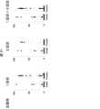
また、Mesp+/+、Mesp+/-、Mesp-/-マウスのE8.5におけるqRT-PCRによる測定結果を図3に示す。図3中、Mesp1/2+/+、Mesp1/2+/-、Mesp1/2-/-はそれぞれ、Mesp+/+、Mesp+/-、Mesp-/-マウスの結果を示す。また、「ns」はnon-significantを示し、「*」はP=0.01を示し、「***」はP<0.005を示し、「****」はP<0.0001を示す。
図3に示すように、Mesp-/-マウスでは、標的遺伝子であるMesp1およびMesp2の発現が欠損し、心臓関連遺伝子であるMyh6およびTnnt2の発現が低下していた。
なお、図3,14,17,19,25,28,35,37,39~42において、点はサンプルの個別値を示す。
なお、図3,14,17,19,25,28,35,37,39~42において、点はサンプルの個別値を示す。
(Mesp-/-+GFP-blastomereマウスの構築)
心臓欠損胚(Mesp-/-)と正常胚(Mesp+/-のGFP陽性胚、GFP-blastomere)とのキメラマウス(以後、Mesp-/-+GFP-blastomereマウスとよぶ)を構築し、心臓再生が可能か否かを検証した。当該キメラマウスの観察結果を図4~19に示す。GFP-blastomere(GFP陽性の割球)とは、Green Fluorescent Proteinを発現するように遺伝子操作したGRRマウスの受精卵の発生過程で割球期の胚である。
心臓欠損胚(Mesp-/-)と正常胚(Mesp+/-のGFP陽性胚、GFP-blastomere)とのキメラマウス(以後、Mesp-/-+GFP-blastomereマウスとよぶ)を構築し、心臓再生が可能か否かを検証した。当該キメラマウスの観察結果を図4~19に示す。GFP-blastomere(GFP陽性の割球)とは、Green Fluorescent Proteinを発現するように遺伝子操作したGRRマウスの受精卵の発生過程で割球期の胚である。
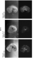
図4は、Mesp+/-、Mesp+/-+GFP-blastomereおよびMesp-/-+GFP-blastomereマウスのE9.5における観察結果である。図4中、BFは明視野での観察結果、GFPはGFP発光の観察結果を示す。GFP陽性胚から発生した心臓はGFPにより緑色に発光する。図4に示すように、Mesp+/-+GFP-blastomereマウスの心臓ではGFPの発光が観察されなかった。一方、Mesp-/-+GFP-blastomereマウスの心臓ではGFPの発光が観察され、GFP陽性胚による心臓再生に成功し胎生死が回避されたことを確認した。
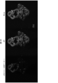
図5は、Mesp+/-、Mesp+/-+GFP-blastomere、Mesp-/-+GFP-blastomereマウスのE13.5の心臓の免疫染色結果を示す。図6は、Mesp-/-+GFP-blastomereマウスのE13.5の心臓の免疫染色結果を示し、RVは右心室、IVSは心房中隔、LVは左心室を示す。α-actininは心筋細胞のマーカーである。
図5および6に示すように、Mesp-/-+GFP-blastomereマウスで再生された心臓のほぼすべての心筋細胞がGFP陽性胚由来であることが分かった。
図7は、Mesp-/-+GFP-blastomereマウスのE13.5の心臓以外の臓器(肺、肝臓、腸および腎臓)のGFP発光の観察結果を示す。図7に示すように、心臓以外の臓器の細胞はGFP+/-細胞が混在したキメラであることが分かった。
図8の左側は、8週齢のMesp+/-+GFP-blastomereマウス(対照)、右側は8週齢のMesp-/-+GFP-blastomereマウスを示す。Mesp-/-+GFP-blastomereマウスの背側は黒毛であり、GFP陽性胚の細胞に由来することが考えられる。また、Mesp-/-+GFP-blastomereマウスの腹側は灰毛であり、Mesp-/-胚の細胞に由来することが考えられる。また、図8の右の写真の矢印に示すように、Mesp-/-+GFP-blastomereマウスの尾は変形していた。この変形は、心臓欠損胚(Mesp-/-)と正常胚(GFP-blastomere)とが混在していることに起因する。
図9および11は、8週齢のMesp+/-+GFP-blastomereマウスの心臓の免疫染色結果を示す。図10および12は、8週齢のMesp-/-+GFP-blastomereマウスの心臓の免疫染色結果を示す。図9~12に示すように、Mesp+/-+GFP-blastomereマウスの心臓がMesp+/-細胞由来の心筋細胞が混在している一方で、Mesp-/-+GFP-blastomereマウスの心臓の心筋細胞は、ほぼ全てがGFP陽性胚由来の細胞によって再生していることが分かった。
図13は、8週齢のMesp-/-+GFP-blastomereマウスの心臓の観察結果を示す。平滑筋細胞のマーカー(α-SMA)、内皮細胞のマーカー(CD31)および線維芽細胞のマーカー(ビメンチン、Vimentin)で免疫染色を行った。図14は、8週齢のMesp+/-+GFP-blastomereマウスとMesp-/-+GFP-blastomereマウスの心臓を構成する各細胞のキメリズムを示すグラフである。図13および14に示すように、Mesp-/-+GFP-blastomereマウスの心臓を構成する細胞について、心筋細胞に加え、非心筋細胞の代表である平滑筋細胞、内皮細胞および線維芽細胞についても、ほぼGFP陽性胚由来の細胞によって再生していることが分かった。
図15は、8週齢の、Mesp+/+、Mesp+/-+GFP-blastomereおよびMesp-/-+GFP-blastomereマウスの心臓のヘマトキシリン-エオジン染色(HE染色)による結果を示す。図15に示すように、Mesp-/-+GFP-blastomereマウスの心臓の構造に異常は認められなかった。
図16は、7~8週齢の、Mesp+/+、Mesp+/-+GFP-blastomereおよびMesp-/-+GFP-blastomereマウスの心エコー法による測定結果を示す。図17は、図16の結果をグラフにまとめた結果を示す。図17の縦軸はLVEFを示し、LVEFは左室駆出率を示す。図16および17に示すように、Mesp-/-+GFP-blastomereマウスにおいて再生された心臓の機能は正常であることが分かった。
図18は、トレッドミル疲労試験の写真である。中央の2つのレーンを走るマウス(図18中の*のマウス)がMesp-/-+GFP-blastomereマウスである。図19は、トレッドミル疲労試験の結果をまとめたグラフである。図19の縦軸はマウスの走行距離を示す。図19に示すように、Mesp-/-+GFP-blastomereマウスは正常な運動耐用能を示した。
(GRR mESCを幹細胞として使用したMesp-/-+GFP-mESCsマウスの構築)
心臓欠損胚(Mesp-/-)に、GFPを発現するSGE2マウスから樹立したES細胞(GFP-mESCs)を移植することによって、キメラマウスにおいて心臓再生が可能化か否かを検証した。マウスES細胞として、GRR mESCを使用した。当該キメラマウスの観察結果を図20~24に示す。
心臓欠損胚(Mesp-/-)に、GFPを発現するSGE2マウスから樹立したES細胞(GFP-mESCs)を移植することによって、キメラマウスにおいて心臓再生が可能化か否かを検証した。マウスES細胞として、GRR mESCを使用した。当該キメラマウスの観察結果を図20~24に示す。
Mesp+/-、Mesp+/-+GFP-mESCsおよびMesp-/-+GFP-mESCsマウスのE9.5における観察結果を図20、E13.5における観察結果を図21に示す。図20中の矢印および図21中の四角枠部は心臓を示す。図20および21に示すように、Mesp-/-+GFP-mESCsマウスの心臓では全体的なGFPの発光が観察され、心臓再生に成功したことを確認した。
図22は、Mesp+/-、Mesp+/-+GFP-mESCs、Mesp-/-+GFP-mESCsマウスのE13.5の心臓の免疫染色結果を示す。図22に示すように、Mesp-/-+GFP-mESCsマウスで再生された心臓の大部分の心筋細胞がGFPを発現するマウスES細胞由来であることが分かった。
図23は、Mesp-/-+GFP-mESCsマウスのE13.5の心臓以外の臓器のGFP発光の観察結果を示す。図23に示すように、心臓以外の臓器の細胞はMesp-/-由来の細胞およびマウスES細胞由来細胞が混在していた。
図24は、Mesp-/-+GFP-mESCsマウスのP0の観察結果を示す。胚盤胞補完による心臓再生の結果、Mesp-/-+GFP-mESCsマウスは出生することができた。一方、出生時点では生存個体は存在するものの、いずれも早期の死亡が確認された。また、WT/ヘテロ個体でも奇形個体が認められた。Mesp-/-+GFP-blastomereマウスは成獣まで生存したことから、Mesp-/-+GFP-mESCsマウスの早期の死亡はES細胞の品質が原因である可能性が示唆された。
(産仔のキメリズムの検討)
Mesp+/+、Mesp+/-+mESCsおよびMesp-/-+GFP-blastomereマウスにおいて生存したマウスのキメリズムを評価した。GFP-blastomereマウスとしてGRRを使用した。成獣期の評価結果を図25に示す。図25の縦軸のキメリズム(%)は、産仔を構成する細胞に対するGFP陽性細胞(GRR-mESCs)の割合を示す。
Mesp+/+、Mesp+/-+mESCsおよびMesp-/-+GFP-blastomereマウスにおいて生存したマウスのキメリズムを評価した。GFP-blastomereマウスとしてGRRを使用した。成獣期の評価結果を図25に示す。図25の縦軸のキメリズム(%)は、産仔を構成する細胞に対するGFP陽性細胞(GRR-mESCs)の割合を示す。
図25に示すように、生存したMesp-/-+GFP-blastomereマウスのキメリズムは60~90%前後であった。その他のマウスは50%未満のキメリズムが存在するが、Mesp-/-+GFP-blastomereマウスには存在しなかった。これらの結果から、心臓補完によるキメリズムは50~90%であることが分かった。
次に、野生型マウス胚にGFPを発現するES細胞を移植し、良好なキメリズム(GFP陽性細胞が50~90%)になる移植条件を検討した。E13.5期の検討結果を図26に示す。図26の縦軸のキメリズム(%)は、産仔を構成する細胞に対するGFP陽性細胞(GRR-mESCs)の割合を示す。
図26に示すように、B6マウスよりもICRマウスの方がキメラになり難いことがわかった。B6マウスは日本クレア社から購入したマウスである。多くの論文では、胚に移植するES細胞数は5~10個で行われている。図26の結果から、ICRマウスの胚に移植するES細胞を20個にしてキメリズムの比較を行うことにした。
ICRマウスの8細胞期、桑実胚期および胚盤胞期の胚に20個のES細胞を移植し、キメリズムの比較を行った。結果を図27に示す。
図27に示すように、移植するES細胞を20個とすることで高いキメリズムとなった。また、移植される胚の成熟度もキメリズムに影響することが分かった。移植するES細胞が20個であり、キメリズムが50~90%前後となる条件としては、移植される胚が桑実胚期の胚であるときがもっともキメリズムが高いことが分かった。
次に、GFPを発現するES細胞の種類をSGE2 mESCに変更して、Mesp-/-+mESCsマウスにおいて生存したマウスのキメリズムを評価した。E13.5期の評価結果を図28に示す。図28の縦軸は、産仔を構成する細胞に対するGFP陽性細胞(SGE2 mESC)の割合を示す。
図28に示すように、胚に移植するES細胞を20個にすると、最適なキメリズムである50~90%となる個体が少ないだけでなく、生存する個体数自体が少なかった。また、図28から、SGE2においては、胚に移植するES細胞数は10個が最適であることが分かった。胚に移植したES細胞数は10個のときに生存していた3匹のキメラマウス(図28中の丸印)のキメリズムは50~90%前後であった。これらの結果から、移植細胞(幹細胞)の種類に応じて最適なキメリズムとなる移植細胞数の最適化が必要であることが分かった。また、移植細胞の種類に関わらず、心臓補完に必要なキメリズムは50~90%前後であることが分かった。
また、胚盤胞補完に幹細胞としてSGE2 mESCを使用し、キメリズムを50~90%に最適化して、胚盤胞補完による心臓再生を検証した。このSGE2 mESC由来のマウスは4例生存した。
(SGE2 mESCを幹細胞として使用したMesp-/-+GFP-mESCsマウスの構築)
心臓欠損胚(Mesp-/-)に、GFPを発現するマウスES細胞(GFP-mESCs)を移植することによって、キメラマウスにおいて心臓再生が可能化か否かを検証した。マウスES細胞として、SGE2 mESCを使用した。当該キメラマウスの観察結果を図29~40に示す。
心臓欠損胚(Mesp-/-)に、GFPを発現するマウスES細胞(GFP-mESCs)を移植することによって、キメラマウスにおいて心臓再生が可能化か否かを検証した。マウスES細胞として、SGE2 mESCを使用した。当該キメラマウスの観察結果を図29~40に示す。
図29の左側は、8週齢のMesp+/-+GFP-mESCsマウス(対照)、右側は8週齢のMesp-/-+GFP-mESCsマウスを示す。図9のMesp-/-+GFP-blastomereマウスと同様に、矢印で示すようにMesp-/-+GFP-mESCsマウスの尾は変形していた。
図30の上側および図31は8週齢のMesp-/-+GFP-mESCsマウスの心臓の免疫染色結果を示す。図30の下側および図32は8週齢のMesp+/-+GFP-mESCsマウスの心臓の免疫染色結果を示す。図31および32中、SMCは平滑筋細胞、ECは内皮細胞、CFは繊維芽細胞を示す。
図30~32に示すように、Mesp+/-+GFP-mESCsマウスの心臓は、Mesp-/-由来およびマウスES細胞由来の心筋細胞が混在していた。一方、Mesp-/-+GFP-mESCsマウスの心臓の心筋細胞は、ほぼ全てがGFPを発現するマウスES細胞によって再生していることが分かった。また、Mesp-/-+GFP-mESCsマウスの心臓を構成する細胞について、心筋細胞に加え、非心筋細胞の代表である平滑筋細胞、内皮細胞および繊維芽細胞についても、ほぼGFPを発現するマウスES細胞によって再生していることが分かった。
図33は、8週齢の、Mesp+/+、Mesp+/-+GFP-mESCsおよびMesp-/-+GFP-mESCsマウスの心臓のHE染色による結果を示す。図33に示すように、Mesp-/-+GFP-mESCsマウスの心臓の構造に異常は認められなかった。
図34は、7~8週齢の、Mesp+/+、Mesp+/-+GFP-mESCsおよびMesp-/-+GFP-mESCsマウスの心エコー法による測定結果を示す。図35は、図34の結果をグラフにまとめた結果を示す。図35の縦軸はLVEFを示す。図34および35に示すように、Mesp-/-+GFP-mESCsマウスにおいて再生された心臓の機能は正常であることが分かった。
図36は、トレッドミル疲労試験の写真である。中央の2つのレーンを走るマウス(図36中の***のマウス)がMesp-/-+GFP-mESCsマウスである。図37は、トレッドミル疲労試験の結果をまとめたグラフである。図37の縦軸はマウスの走行距離を示す。図37の縦軸は図37に示すように、Mesp-/-+GFP-mESCsマウスは正常な運動耐用能を示した。
図38は、7~8週齢の、Mesp+/+、Mesp+/-+GFP-mESCsおよびMesp-/-+GFP-mESCsマウスの心電図の結果を示す。図38に示すように、Mesp-/-+GFP-mESCsマウスにおいて再生された心臓の不整脈が見られず、心臓の機能は正常であることが分かった。
図39は、7~8週齢の、Mesp+/-+GFP-mESCsおよびMesp-/-+GFP-mESCsマウスの体重(BW、g)に対する心臓重量(HW、mg)の割合を示すグラフである。図40は脛骨長(TL、mm)に対する心臓重量(HW、mg)の割合を示すグラフである。図39および40の結果から、Mesp-/-+GFP-mESCsマウスに心不全がなく、心臓の機能は正常であることが分かった。
(異種キメラマウスの構築)
次に、ラットES細胞を使用して、Mesp-/-+GFP-ratESCsマウスの構築を行った。ラットES細胞の由来となるドナーとして、GFPを発現するSD-Tg(CAG-EGFP)ラットを使用した。EGFP rESC(EGFP-expressing rES cell;EGFP発現rES細胞)の産生は、Ping Li, et al. Cell. 2008 Dec 26;135(7):1299-310.およびKazushi Kawaharada, et al. World J Stem Cellsを参照した。
次に、ラットES細胞を使用して、Mesp-/-+GFP-ratESCsマウスの構築を行った。ラットES細胞の由来となるドナーとして、GFPを発現するSD-Tg(CAG-EGFP)ラットを使用した。EGFP rESC(EGFP-expressing rES cell;EGFP発現rES細胞)の産生は、Ping Li, et al. Cell. 2008 Dec 26;135(7):1299-310.およびKazushi Kawaharada, et al. World J Stem Cellsを参照した。
変異マウス胚とラットES細胞を用いてキメラ胚を生成した。簡単に説明する。ES細胞による胚盤胞補完に使用するホスト胚は、IVFから2時間後に凍結した。凍結した胚を解凍し、M16培地(M7292, Sigma-Aldrich、メルク社)中で24時間インキュベートすることによって調製した。胚操作の3日前に凍結ES細胞を解凍し、フィーダー層に配置した。増殖したES細胞を注入直前に0.25%トリプシンで解離させた。注入方法を使用するときに、顕微鏡下でピエゾ駆動マイクロマニピュレーター(PMM-150FU, PrimeTech社)を使用して、透明帯および栄養外胚葉に穴を開け、内部細胞塊付近の胚盤胞腔内に10のES細胞を移植した。操作されたキメラ胚を、胚盤胞期において、疑妊娠レシピエントであるICR雌マウス(2.5dpc)の子宮角に外科的に移植し、所望のステージまで発生させた。
マウス胚として新鮮胚または凍結胚を使用したときの評価結果を表3にまとめる。表3中、「WT/hetero/homo」は、キメラ中の野生型個体(WT、Mesp+/+)、ヘテロ個体(hetero、Mesp+/-)およびホモ個体(Mesp-/-)の数を示す。

表3に示すように、1回目で得られた正常胚のマウス50匹(新鮮胚15、凍結胚35)のうち1匹がホモ個体(Mesp-/-+GFP-ratESCsマウス)であることが確認された(E11.5)。表3中の「+na1」はnot availableを示す。2回目で得られた正常胚のマウス34匹(新鮮胚16、凍結胚18)ではホモ個体の存在は確認されなかった(E11.5)。
図41は、生存した全個体のマウス(E11.5)のキメリズムの評価結果を示す。図42は、生存したキメラ個体のマウス(E11.5)のキメリズムの評価結果を示す。図41および42の縦軸のキメリズム(%)は、産仔を構成する細胞に対するラットES細胞の割合を示す。「Fresh」は新鮮胚、「Frozen」は凍結胚を示す。
図41および42に示すように、凍結胚を使用したときに、同種キメラマウスのキメリズムと同様の高いキメリズムの個体が得られることが分かった。また、凍結胚を使用したときにホモ個体(Mesp-/-+GFP-ratESCsマウス)が得られたことから、凍結胚が異種キメラの作成に適していることが分かった。
図43は、E11.5の野生型個体、ヘテロ個体およびホモ個体の観察結果を示す。図43で観察した個体のキメリズムは、野生型個体が59.7%、ヘテロ個体が34.1%、ホモ個体が79.5%であった。
図44は、E11.5のホモ個体(Mesp-/-+GFP-ratESCsマウス)の心臓の免疫染色結果を示す。図45の上側はE11.5のホモ個体の心房、下側は心室の免疫染色結果を示す。図44から、ホモ個体のマウスの心臓は、GFP陽性ラットES細胞由来の細胞によって再生していることが分かった。また、図45から、ホモ個体のマウスの心房および心室を構成する心筋細胞はGFP陽性ラット細胞由来の細胞によって再生していることが分かった。
本発明によって、目的の哺乳動物種の心臓を製造することが可能である。製造された心臓は移植用心臓として、臨床分野において利用可能である。
Claims (13)
- 哺乳動物由来の幹細胞を調製する幹細胞調製工程;
前記哺乳動物とは異なる非ヒト哺乳動物の受精卵であって、Mesp1遺伝子およびMesp2遺伝子がノックアウトされている受精卵へ前記幹細胞を移植する移植工程;および、
前記受精卵を前記非ヒト哺乳動物の母胎中で発生させて、産仔を得る産仔取得工程、を含み、
前記幹細胞由来の心筋細胞、内皮細胞、平滑筋細胞および線維芽細胞によって前記産仔の心臓が補完される、キメラ非ヒト哺乳動物を製造する方法。 - 前記非ヒト哺乳動物の受精卵が初期胚から胚盤胞期の受精卵である、請求項1に記載の方法。
- 前記産仔のキメリズムが50%以上90%以下である、請求項1に記載の方法。
- 前記幹細胞が胚性幹細胞または誘導型多能性幹細胞である、請求項1に記載の方法。
- 前記幹細胞が、ヒト、マウスまたはラット由来の幹細胞である、請求項1に記載の方法。
- 心臓を構成する心筋細胞、内皮細胞、平滑筋細胞および線維芽細胞は、哺乳動物由来の細胞を含み、
心臓以外の臓器および組織は、前記哺乳動物由来の細胞と前記哺乳動物とは異なる非ヒト哺乳動物由来の細胞とを含み、
前記哺乳動物由来の細胞は、Mesp1遺伝子およびMesp2遺伝子を有し、
前記非ヒト哺乳動物由来の細胞はMesp1遺伝子およびMesp2遺伝子がノックアウトされている、キメラ非ヒト哺乳動物。 - 哺乳動物由来の幹細胞を調製する幹細胞調製工程;
前記哺乳動物とは異なる非ヒト哺乳動物の受精卵であって、Mesp1遺伝子およびMesp2遺伝子がノックアウトされている受精卵へ前記幹細胞を移植する移植工程;
前記受精卵を非ヒト哺乳動物の母胎中で発生させて、産仔を得る産仔取得工程;および、
前記産仔から心臓または心臓組織を取得する心臓取得工程を含み、
前記心臓または心臓組織は、前記幹細胞から分化した心筋細胞、内皮細胞、平滑筋細胞および線維芽細胞によって補完された心臓または心臓組織である、心臓または心臓組織を製造する方法。 - 前記非ヒト哺乳動物の受精卵が初期胚から胚盤胞期の受精卵である、請求項7に記載の方法。
- 前記産仔のキメリズムが50%以上90%以下である、請求項7に記載の方法。
- 前記幹細胞が胚性幹細胞(ES細胞)または誘導型多能性幹細胞(iPS細胞)である、請求項7に記載の方法。
- 前記幹細胞が、ヒト、マウスまたはラット由来の幹細胞である、請求項7に記載の方法。
- 請求項7~11のいずれか1項に記載される方法によって得られる、心臓または心臓組織。
- 哺乳動物由来の幹細胞と、
前記哺乳動物とは異なる非ヒト哺乳動物の受精卵であって、Mesp1遺伝子およびMesp2遺伝子がノックアウトされている受精卵と、を含む、心臓または心臓組織の製造用キット。
Applications Claiming Priority (2)
| Application Number | Priority Date | Filing Date | Title |
|---|---|---|---|
| JP2023-004673 | 2023-01-16 | ||
| JP2023004673A JP2026034875A (ja) | 2023-01-16 | 2023-01-16 | キメラ非ヒト哺乳動物を製造する方法、心臓または心臓組織を製造する方法 |
Publications (1)
| Publication Number | Publication Date |
|---|---|
| WO2024154682A1 true WO2024154682A1 (ja) | 2024-07-25 |
Family
ID=91956127
Family Applications (1)
| Application Number | Title | Priority Date | Filing Date |
|---|---|---|---|
| PCT/JP2024/000732 Ceased WO2024154682A1 (ja) | 2023-01-16 | 2024-01-15 | キメラ非ヒト哺乳動物を製造する方法、心臓または心臓組織を製造する方法 |
Country Status (2)
| Country | Link |
|---|---|
| JP (1) | JP2026034875A (ja) |
| WO (1) | WO2024154682A1 (ja) |
Citations (2)
| Publication number | Priority date | Publication date | Assignee | Title |
|---|---|---|---|---|
| WO2016163386A1 (ja) * | 2015-04-08 | 2016-10-13 | 全国農業協同組合連合会 | 異個体由来の配偶子を生産する非ヒト大型哺乳動物又は魚類の作出方法 |
| JP2018191579A (ja) * | 2017-05-17 | 2018-12-06 | 国立大学法人 新潟大学 | 肺の製造方法 |
-
2023
- 2023-01-16 JP JP2023004673A patent/JP2026034875A/ja active Pending
-
2024
- 2024-01-15 WO PCT/JP2024/000732 patent/WO2024154682A1/ja not_active Ceased
Patent Citations (2)
| Publication number | Priority date | Publication date | Assignee | Title |
|---|---|---|---|---|
| WO2016163386A1 (ja) * | 2015-04-08 | 2016-10-13 | 全国農業協同組合連合会 | 異個体由来の配偶子を生産する非ヒト大型哺乳動物又は魚類の作出方法 |
| JP2018191579A (ja) * | 2017-05-17 | 2018-12-06 | 国立大学法人 新潟大学 | 肺の製造方法 |
Non-Patent Citations (6)
| Title |
|---|
| ABE YUTO, SADAHIRO TAKETARO, FUJITA RYO, YAMADA YU, AKIYAMA TATSUYA, NAKANO KOJI, HONDA SEIICHIRO, TANIMOTO YOKO, SUZUKI HAYATE, M: "Abstract 12159: Heart Generation via Blastocyst Complementation in Mesp1/2-Deficient Mice", CIRCULATION, AMERICAN HEART ASSOCIATION, US, vol. 148, no. Suppl_1, 7 November 2023 (2023-11-07), US , XP093192819, ISSN: 0009-7322, DOI: 10.1161/circ.148.suppl_1.12159 * |
| KITAJIMA SATOSHI, TAKAGI ATSUYA, INOUE TOHRU, SAGA YUMIKO: "MesP1 and MesP2 are essential for the development of cardiac mesoderm", DEVELOPMENT, THE COMPANY OF BIOLOGISTS LTD., GB, vol. 127, no. 15, 1 August 2000 (2000-08-01), GB , pages 3215 - 3226, XP093192806, ISSN: 0950-1991, DOI: 10.1242/dev.127.15.3215 * |
| TAKAHASHI YU, HIRAOKA SHUICHI, KITAJIMA SATOSHI, INOUE TOHRU, KANNO JUN, SAGA YUMIKO: "Differential contributions of Mesp1 and Mesp2 to the epithelialization and rostro-caudal patterning of somites", DEVELOPMENT, THE COMPANY OF BIOLOGISTS LTD., GB, vol. 135, no. 23, 1 December 2008 (2008-12-01), GB , pages 3969 - 3969, XP093192809, ISSN: 0950-1991, DOI: 10.1242/dev.022061 * |
| TOMOYUKI YAMAGUCHI, ET AL.: "Interspecies organogenesis generates autologous functional islets", NATURE, vol. 542, no. 7640, 9 February 2017 (2017-02-09), pages 191 - 196, XP055530982, DOI: 10.1038/nature21070 * |
| TOSHIHIRO KOBAYASHI, TOMOYUKI YAMAGUCHI, SANAE HAMANAKA, MEGUMI KATO-ITOH, YUJI YAMAZAKI, MAKOTO IBATA, HIDEYUKI SATO, YOUN-SU LEE: "Generation of Rat Pancreas in Mouse by Interspecific Blastocyst Injection of Pluripotent Stem Cells", CELL, CELL PRESS, vol. 142, no. 5, 1 September 2010 (2010-09-01), pages 787 - 799, XP055053670, ISSN: 00928674, DOI: 10.1016/j.cell.2010.07.039 * |
| WU JUN; PLATERO-LUENGO AIDA; SAKURAI MASAHIRO; SUGAWARA ATSUSHI; GIL MARIA ANTONIA; YAMAUCHI TAKAYOSHI; SUZUKI KEIICHIRO; BOGLIOTT: "Interspecies Chimerism with Mammalian Pluripotent Stem Cells", CELL, ELSEVIER, AMSTERDAM NL, vol. 168, no. 3, 26 January 2017 (2017-01-26), Amsterdam NL , pages 473 - 486+14pp, XP029897815, ISSN: 0092-8674, DOI: 10.1016/j.cell.2016.12.036 * |
Also Published As
| Publication number | Publication date |
|---|---|
| JP2026034875A (ja) | 2026-03-04 |
Similar Documents
| Publication | Publication Date | Title |
|---|---|---|
| Mori et al. | Generation of functional lungs via conditional blastocyst complementation using pluripotent stem cells | |
| Usui et al. | Generation of kidney from pluripotent stem cells via blastocyst complementation | |
| Hayashi et al. | Targeted deletion of BMK1/ERK5 in adult mice perturbs vascular integrity and leads to endothelial failure | |
| JP5841322B2 (ja) | フマリルアセト酢酸ヒドロラーゼ(fah)欠損性ブタおよびその使用 | |
| JP2022091993A (ja) | iPS細胞とBLASTOCYST COMPLEMENTATIONを利用した臓器再生法 | |
| JP6573924B2 (ja) | フマリルアセト酢酸ヒドロラーゼ(fah)欠損及び免疫不全ラット、並びにそれらの使用 | |
| JP5588405B2 (ja) | ラット胚性幹細胞 | |
| JP2002511732A (ja) | トランスジェニック動物を発生させるための組成物および方法 | |
| JP6619462B2 (ja) | キメラ動物の作製方法 | |
| US20110003327A1 (en) | Methods for production of atrial progenitors and their differentiation into smooth muscle cells and cardiomyocytes | |
| Drukker | Immunogenicity of embryonic stem cells and their progeny | |
| WO2018228534A1 (zh) | 制备免疫缺陷的大鼠的方法及其应用 | |
| Matsui et al. | Caspase 9-induced apoptosis enables efficient fetal cell ablation and disease modeling | |
| JP2008532474A (ja) | 胚性幹(es)細胞系での組織モデリング | |
| JP2018506984A (ja) | Etv2およびその利用 | |
| JP2026034875A (ja) | キメラ非ヒト哺乳動物を製造する方法、心臓または心臓組織を製造する方法 | |
| Olsson et al. | Live births from urine derived cells | |
| JP2001231549A (ja) | 不死化血管内皮細胞株 | |
| WO2020226070A1 (ja) | ヒト非アルコール性脂肪性肝炎モデル | |
| US20070204357A1 (en) | Process for producing normal parenchymal cells, tissues or organs by bioincubator | |
| Abe et al. | Heart generation via blastocyst complementation in Mesp1/2-deficient mice | |
| JPWO2005040361A1 (ja) | 幹細胞の簡易調製法およびそれに使用するフィーダー細胞 | |
| 조호연 | Studies on application of spermatogonial stem cell for quail genome modification | |
| JP2009159878A (ja) | 非ヒトes動物の新規作成方法 | |
| Coppiello et al. | Generation of heart and vascular system in rodents by blastocyst complementation |
Legal Events
| Date | Code | Title | Description |
|---|---|---|---|
| 121 | Ep: the epo has been informed by wipo that ep was designated in this application |
Ref document number: 24744605 Country of ref document: EP Kind code of ref document: A1 |
|
| NENP | Non-entry into the national phase |
Ref country code: DE |
|
| 122 | Ep: pct application non-entry in european phase |
Ref document number: 24744605 Country of ref document: EP Kind code of ref document: A1 |
|
| NENP | Non-entry into the national phase |
Ref country code: JP |