WO2023282281A1 - Peptide ayant une activité antivirale, agent antiviral comprenant ledit peptide, et procédé de production dudit agent antiviral - Google Patents
Peptide ayant une activité antivirale, agent antiviral comprenant ledit peptide, et procédé de production dudit agent antiviral Download PDFInfo
- Publication number
- WO2023282281A1 WO2023282281A1 PCT/JP2022/026804 JP2022026804W WO2023282281A1 WO 2023282281 A1 WO2023282281 A1 WO 2023282281A1 JP 2022026804 W JP2022026804 W JP 2022026804W WO 2023282281 A1 WO2023282281 A1 WO 2023282281A1
- Authority
- WO
- WIPO (PCT)
- Prior art keywords
- amino acid
- residue
- cov
- sars
- acid sequence
- Prior art date
Links
- 108090000765 processed proteins & peptides Proteins 0.000 title claims abstract description 253
- 230000000840 anti-viral effect Effects 0.000 title description 22
- 238000004519 manufacturing process Methods 0.000 title description 7
- 239000003443 antiviral agent Substances 0.000 title description 4
- 241001678559 COVID-19 virus Species 0.000 claims abstract description 507
- 230000027455 binding Effects 0.000 claims abstract description 427
- 108020003175 receptors Proteins 0.000 claims abstract description 327
- 102000005962 receptors Human genes 0.000 claims abstract description 327
- 125000000539 amino acid group Chemical group 0.000 claims description 306
- 150000001413 amino acids Chemical class 0.000 claims description 269
- 125000003588 lysine group Chemical group [H]N([H])C([H])([H])C([H])([H])C([H])([H])C([H])([H])C([H])(N([H])[H])C(*)=O 0.000 claims description 81
- 125000000637 arginyl group Chemical group N[C@@H](CCCNC(N)=N)C(=O)* 0.000 claims description 78
- CKLJMWTZIZZHCS-REOHCLBHSA-N aspartic acid group Chemical group N[C@@H](CC(=O)O)C(=O)O CKLJMWTZIZZHCS-REOHCLBHSA-N 0.000 claims description 60
- 125000000291 glutamic acid group Chemical group N[C@@H](CCC(O)=O)C(=O)* 0.000 claims description 50
- FWMNVWWHGCHHJJ-SKKKGAJSSA-N 4-amino-1-[(2r)-6-amino-2-[[(2r)-2-[[(2r)-2-[[(2r)-2-amino-3-phenylpropanoyl]amino]-3-phenylpropanoyl]amino]-4-methylpentanoyl]amino]hexanoyl]piperidine-4-carboxylic acid Chemical compound C([C@H](C(=O)N[C@H](CC(C)C)C(=O)N[C@H](CCCCN)C(=O)N1CCC(N)(CC1)C(O)=O)NC(=O)[C@H](N)CC=1C=CC=CC=1)C1=CC=CC=C1 FWMNVWWHGCHHJJ-SKKKGAJSSA-N 0.000 claims description 48
- 125000000613 asparagine group Chemical group N[C@@H](CC(N)=O)C(=O)* 0.000 claims description 42
- 125000001493 tyrosinyl group Chemical group [H]OC1=C([H])C([H])=C(C([H])=C1[H])C([H])([H])C([H])(N([H])[H])C(*)=O 0.000 claims description 40
- 125000003607 serino group Chemical group [H]N([H])[C@]([H])(C(=O)[*])C(O[H])([H])[H] 0.000 claims description 37
- 125000003295 alanine group Chemical group N[C@@H](C)C(=O)* 0.000 claims description 24
- 125000001909 leucine group Chemical group [H]N(*)C(C(*)=O)C([H])([H])C(C([H])([H])[H])C([H])([H])[H] 0.000 claims description 23
- 125000003630 glycyl group Chemical group [H]N([H])C([H])([H])C(*)=O 0.000 claims description 22
- 239000008194 pharmaceutical composition Substances 0.000 claims description 22
- 125000000487 histidyl group Chemical group [H]N([H])C(C(=O)O*)C([H])([H])C1=C([H])N([H])C([H])=N1 0.000 claims description 21
- COLNVLDHVKWLRT-QMMMGPOBSA-N phenylalanine group Chemical group N[C@@H](CC1=CC=CC=C1)C(=O)O COLNVLDHVKWLRT-QMMMGPOBSA-N 0.000 claims description 21
- 210000004899 c-terminal region Anatomy 0.000 claims description 20
- 125000000404 glutamine group Chemical group N[C@@H](CCC(N)=O)C(=O)* 0.000 claims description 18
- 208000001528 Coronaviridae Infections Diseases 0.000 claims description 15
- 229940024606 amino acid Drugs 0.000 claims description 15
- 235000001014 amino acid Nutrition 0.000 claims description 15
- 125000000341 threoninyl group Chemical group [H]OC([H])(C([H])([H])[H])C([H])(N([H])[H])C(*)=O 0.000 claims description 14
- 125000001360 methionine group Chemical group N[C@@H](CCSC)C(=O)* 0.000 claims description 12
- 239000004475 Arginine Substances 0.000 claims description 11
- ODKSFYDXXFIFQN-UHFFFAOYSA-N arginine Natural products OC(=O)C(N)CCCNC(N)=N ODKSFYDXXFIFQN-UHFFFAOYSA-N 0.000 claims description 11
- KDXKERNSBIXSRK-UHFFFAOYSA-N Lysine Natural products NCCCCC(N)C(O)=O KDXKERNSBIXSRK-UHFFFAOYSA-N 0.000 claims description 10
- 239000004472 Lysine Substances 0.000 claims description 10
- 125000001500 prolyl group Chemical group [H]N1C([H])(C(=O)[*])C([H])([H])C([H])([H])C1([H])[H] 0.000 claims description 10
- ODKSFYDXXFIFQN-BYPYZUCNSA-P L-argininium(2+) Chemical compound NC(=[NH2+])NCCC[C@H]([NH3+])C(O)=O ODKSFYDXXFIFQN-BYPYZUCNSA-P 0.000 claims description 9
- 108091005634 SARS-CoV-2 receptor-binding domains Proteins 0.000 claims description 8
- 208000015181 infectious disease Diseases 0.000 claims description 4
- 239000004480 active ingredient Substances 0.000 claims description 3
- 239000007864 aqueous solution Substances 0.000 claims description 2
- 125000003275 alpha amino acid group Chemical group 0.000 claims 76
- 239000002253 acid Substances 0.000 claims 1
- 125000002924 primary amino group Chemical group [H]N([H])* 0.000 claims 1
- 229910021642 ultra pure water Inorganic materials 0.000 abstract description 13
- 239000012498 ultrapure water Substances 0.000 abstract description 13
- 208000025721 COVID-19 Diseases 0.000 abstract description 12
- 239000003814 drug Substances 0.000 abstract description 7
- 239000007853 buffer solution Substances 0.000 abstract description 6
- 229940124597 therapeutic agent Drugs 0.000 abstract description 3
- 230000035772 mutation Effects 0.000 description 120
- 238000010586 diagram Methods 0.000 description 78
- 230000003993 interaction Effects 0.000 description 64
- 238000002962 plaque-reduction assay Methods 0.000 description 47
- 102000004196 processed proteins & peptides Human genes 0.000 description 43
- 230000005764 inhibitory process Effects 0.000 description 37
- 230000000694 effects Effects 0.000 description 22
- 239000000243 solution Substances 0.000 description 19
- 101000823955 Homo sapiens Serine palmitoyltransferase 1 Proteins 0.000 description 18
- 102100022068 Serine palmitoyltransferase 1 Human genes 0.000 description 18
- 210000004027 cell Anatomy 0.000 description 18
- 125000000741 isoleucyl group Chemical group [H]N([H])C(C(C([H])([H])[H])C([H])([H])C([H])([H])[H])C(=O)O* 0.000 description 15
- 238000000034 method Methods 0.000 description 14
- 238000005259 measurement Methods 0.000 description 11
- YBJHBAHKTGYVGT-ZKWXMUAHSA-N (+)-Biotin Chemical compound N1C(=O)N[C@@H]2[C@H](CCCCC(=O)O)SC[C@@H]21 YBJHBAHKTGYVGT-ZKWXMUAHSA-N 0.000 description 10
- 241000494545 Cordyline virus 2 Species 0.000 description 9
- 235000018102 proteins Nutrition 0.000 description 9
- 102000004169 proteins and genes Human genes 0.000 description 9
- 108090000623 proteins and genes Proteins 0.000 description 9
- 230000006641 stabilisation Effects 0.000 description 9
- 238000011105 stabilization Methods 0.000 description 9
- 230000015572 biosynthetic process Effects 0.000 description 8
- 238000001142 circular dichroism spectrum Methods 0.000 description 8
- 230000009897 systematic effect Effects 0.000 description 8
- 201000003176 Severe Acute Respiratory Syndrome Diseases 0.000 description 7
- 239000001257 hydrogen Substances 0.000 description 7
- 229910052739 hydrogen Inorganic materials 0.000 description 7
- JKMHFZQWWAIEOD-UHFFFAOYSA-N 2-[4-(2-hydroxyethyl)piperazin-1-yl]ethanesulfonic acid Chemical compound OCC[NH+]1CCN(CCS([O-])(=O)=O)CC1 JKMHFZQWWAIEOD-UHFFFAOYSA-N 0.000 description 6
- WSFSSNUMVMOOMR-UHFFFAOYSA-N Formaldehyde Chemical compound O=C WSFSSNUMVMOOMR-UHFFFAOYSA-N 0.000 description 6
- WHUUTDBJXJRKMK-UHFFFAOYSA-N Glutamic acid Natural products OC(=O)C(N)CCC(O)=O WHUUTDBJXJRKMK-UHFFFAOYSA-N 0.000 description 6
- 239000007995 HEPES buffer Substances 0.000 description 6
- 235000003704 aspartic acid Nutrition 0.000 description 6
- OQFSQFPPLPISGP-UHFFFAOYSA-N beta-carboxyaspartic acid Natural products OC(=O)C(N)C(C(O)=O)C(O)=O OQFSQFPPLPISGP-UHFFFAOYSA-N 0.000 description 6
- 238000010494 dissociation reaction Methods 0.000 description 6
- 230000005593 dissociations Effects 0.000 description 6
- 235000013922 glutamic acid Nutrition 0.000 description 6
- 239000004220 glutamic acid Substances 0.000 description 6
- 238000002198 surface plasmon resonance spectroscopy Methods 0.000 description 6
- 238000003786 synthesis reaction Methods 0.000 description 6
- 125000002987 valine group Chemical group [H]N([H])C([H])(C(*)=O)C([H])(C([H])([H])[H])C([H])([H])[H] 0.000 description 6
- IAZDPXIOMUYVGZ-UHFFFAOYSA-N Dimethylsulphoxide Chemical compound CS(C)=O IAZDPXIOMUYVGZ-UHFFFAOYSA-N 0.000 description 5
- 229960002685 biotin Drugs 0.000 description 5
- 235000020958 biotin Nutrition 0.000 description 5
- 239000011616 biotin Substances 0.000 description 5
- 239000000872 buffer Substances 0.000 description 5
- 230000009467 reduction Effects 0.000 description 5
- 239000012146 running buffer Substances 0.000 description 5
- 230000003612 virological effect Effects 0.000 description 5
- WHUUTDBJXJRKMK-VKHMYHEASA-N L-glutamic acid Chemical compound OC(=O)[C@@H](N)CCC(O)=O WHUUTDBJXJRKMK-VKHMYHEASA-N 0.000 description 4
- FAPWRFPIFSIZLT-UHFFFAOYSA-M Sodium chloride Chemical compound [Na+].[Cl-] FAPWRFPIFSIZLT-UHFFFAOYSA-M 0.000 description 4
- 241000700605 Viruses Species 0.000 description 4
- 201000010099 disease Diseases 0.000 description 4
- 208000037265 diseases, disorders, signs and symptoms Diseases 0.000 description 4
- 229940079593 drug Drugs 0.000 description 4
- 230000009878 intermolecular interaction Effects 0.000 description 4
- 230000002265 prevention Effects 0.000 description 4
- 230000008929 regeneration Effects 0.000 description 4
- 238000011069 regeneration method Methods 0.000 description 4
- 101000674278 Homo sapiens Serine-tRNA ligase, cytoplasmic Proteins 0.000 description 3
- 101000674040 Homo sapiens Serine-tRNA ligase, mitochondrial Proteins 0.000 description 3
- ZDXPYRJPNDTMRX-VKHMYHEASA-N L-glutamine Chemical compound OC(=O)[C@@H](N)CCC(N)=O ZDXPYRJPNDTMRX-VKHMYHEASA-N 0.000 description 3
- KDXKERNSBIXSRK-YFKPBYRVSA-N L-lysine Chemical compound NCCCC[C@H](N)C(O)=O KDXKERNSBIXSRK-YFKPBYRVSA-N 0.000 description 3
- 102100040597 Serine-tRNA ligase, mitochondrial Human genes 0.000 description 3
- 230000002378 acidificating effect Effects 0.000 description 3
- 239000003153 chemical reaction reagent Substances 0.000 description 3
- 238000002983 circular dichroism Methods 0.000 description 3
- 230000004927 fusion Effects 0.000 description 3
- ZDXPYRJPNDTMRX-UHFFFAOYSA-N glutamine Natural products OC(=O)C(N)CCC(N)=O ZDXPYRJPNDTMRX-UHFFFAOYSA-N 0.000 description 3
- 238000012482 interaction analysis Methods 0.000 description 3
- 235000012054 meals Nutrition 0.000 description 3
- 229920000609 methyl cellulose Polymers 0.000 description 3
- 239000001923 methylcellulose Substances 0.000 description 3
- 238000012986 modification Methods 0.000 description 3
- 230000004048 modification Effects 0.000 description 3
- 238000010532 solid phase synthesis reaction Methods 0.000 description 3
- 238000006467 substitution reaction Methods 0.000 description 3
- 229940126585 therapeutic drug Drugs 0.000 description 3
- 238000005406 washing Methods 0.000 description 3
- QKNYBSVHEMOAJP-UHFFFAOYSA-N 2-amino-2-(hydroxymethyl)propane-1,3-diol;hydron;chloride Chemical compound Cl.OCC(N)(CO)CO QKNYBSVHEMOAJP-UHFFFAOYSA-N 0.000 description 2
- 102100035765 Angiotensin-converting enzyme 2 Human genes 0.000 description 2
- 108090000975 Angiotensin-converting enzyme 2 Proteins 0.000 description 2
- IJGRMHOSHXDMSA-UHFFFAOYSA-N Atomic nitrogen Chemical compound N#N IJGRMHOSHXDMSA-UHFFFAOYSA-N 0.000 description 2
- 102100031673 Corneodesmosin Human genes 0.000 description 2
- 101710139375 Corneodesmosin Proteins 0.000 description 2
- 102000004961 Furin Human genes 0.000 description 2
- 108090001126 Furin Proteins 0.000 description 2
- 101000638154 Homo sapiens Transmembrane protease serine 2 Proteins 0.000 description 2
- HNDVDQJCIGZPNO-YFKPBYRVSA-N L-histidine Chemical compound OC(=O)[C@@H](N)CC1=CN=CN1 HNDVDQJCIGZPNO-YFKPBYRVSA-N 0.000 description 2
- FFEARJCKVFRZRR-BYPYZUCNSA-N L-methionine Chemical compound CSCC[C@H](N)C(O)=O FFEARJCKVFRZRR-BYPYZUCNSA-N 0.000 description 2
- KZSNJWFQEVHDMF-BYPYZUCNSA-N L-valine Chemical compound CC(C)[C@H](N)C(O)=O KZSNJWFQEVHDMF-BYPYZUCNSA-N 0.000 description 2
- 208000037847 SARS-CoV-2-infection Diseases 0.000 description 2
- 102100031989 Transmembrane protease serine 2 Human genes 0.000 description 2
- KZSNJWFQEVHDMF-UHFFFAOYSA-N Valine Natural products CC(C)C(N)C(O)=O KZSNJWFQEVHDMF-UHFFFAOYSA-N 0.000 description 2
- 235000004279 alanine Nutrition 0.000 description 2
- 125000003118 aryl group Chemical group 0.000 description 2
- 238000012575 bio-layer interferometry Methods 0.000 description 2
- 230000008859 change Effects 0.000 description 2
- 239000013078 crystal Substances 0.000 description 2
- 230000007423 decrease Effects 0.000 description 2
- 239000013024 dilution buffer Substances 0.000 description 2
- 239000002552 dosage form Substances 0.000 description 2
- 239000003937 drug carrier Substances 0.000 description 2
- 238000002474 experimental method Methods 0.000 description 2
- 238000011194 good manufacturing practice Methods 0.000 description 2
- HNDVDQJCIGZPNO-UHFFFAOYSA-N histidine Natural products OC(=O)C(N)CC1=CN=CN1 HNDVDQJCIGZPNO-UHFFFAOYSA-N 0.000 description 2
- 108700021021 mRNA Vaccine Proteins 0.000 description 2
- 229940126582 mRNA vaccine Drugs 0.000 description 2
- 229930182817 methionine Natural products 0.000 description 2
- 239000012460 protein solution Substances 0.000 description 2
- 210000002966 serum Anatomy 0.000 description 2
- 239000011780 sodium chloride Substances 0.000 description 2
- 238000012916 structural analysis Methods 0.000 description 2
- 230000002459 sustained effect Effects 0.000 description 2
- -1 t-butyloxycarbonyl Chemical group 0.000 description 2
- 125000000430 tryptophan group Chemical group [H]N([H])C(C(=O)O*)C([H])([H])C1=C([H])N([H])C2=C([H])C([H])=C([H])C([H])=C12 0.000 description 2
- OUYCCCASQSFEME-UHFFFAOYSA-N tyrosine Natural products OC(=O)C(N)CC1=CC=C(O)C=C1 OUYCCCASQSFEME-UHFFFAOYSA-N 0.000 description 2
- 239000004474 valine Substances 0.000 description 2
- 125000003088 (fluoren-9-ylmethoxy)carbonyl group Chemical group 0.000 description 1
- 108010053481 Antifreeze Proteins Proteins 0.000 description 1
- DCXYFEDJOCDNAF-UHFFFAOYSA-N Asparagine Natural products OC(=O)C(N)CC(N)=O DCXYFEDJOCDNAF-UHFFFAOYSA-N 0.000 description 1
- 208000031504 Asymptomatic Infections Diseases 0.000 description 1
- 241000894006 Bacteria Species 0.000 description 1
- 241000283690 Bos taurus Species 0.000 description 1
- 125000001433 C-terminal amino-acid group Chemical group 0.000 description 1
- 241000282472 Canis lupus familiaris Species 0.000 description 1
- 241000283707 Capra Species 0.000 description 1
- 241000700198 Cavia Species 0.000 description 1
- 241000282693 Cercopithecidae Species 0.000 description 1
- 241000711573 Coronaviridae Species 0.000 description 1
- 241000699800 Cricetinae Species 0.000 description 1
- 241000255925 Diptera Species 0.000 description 1
- 241000283086 Equidae Species 0.000 description 1
- 241000588724 Escherichia coli Species 0.000 description 1
- 241000282326 Felis catus Species 0.000 description 1
- 241000238631 Hexapoda Species 0.000 description 1
- 241000282412 Homo Species 0.000 description 1
- QNAYBMKLOCPYGJ-REOHCLBHSA-N L-alanine Chemical compound C[C@H](N)C(O)=O QNAYBMKLOCPYGJ-REOHCLBHSA-N 0.000 description 1
- DCXYFEDJOCDNAF-REOHCLBHSA-N L-asparagine Chemical compound OC(=O)[C@@H](N)CC(N)=O DCXYFEDJOCDNAF-REOHCLBHSA-N 0.000 description 1
- 241000124008 Mammalia Species 0.000 description 1
- 241001465754 Metazoa Species 0.000 description 1
- 208000034486 Multi-organ failure Diseases 0.000 description 1
- 208000010718 Multiple Organ Failure Diseases 0.000 description 1
- 241000699670 Mus sp. Species 0.000 description 1
- 241000283973 Oryctolagus cuniculus Species 0.000 description 1
- 241001494479 Pecora Species 0.000 description 1
- 108091005804 Peptidases Proteins 0.000 description 1
- 239000004365 Protease Substances 0.000 description 1
- 229940096437 Protein S Drugs 0.000 description 1
- 241000700159 Rattus Species 0.000 description 1
- 102100037486 Reverse transcriptase/ribonuclease H Human genes 0.000 description 1
- 241000315672 SARS coronavirus Species 0.000 description 1
- 240000004808 Saccharomyces cerevisiae Species 0.000 description 1
- 101000629318 Severe acute respiratory syndrome coronavirus 2 Spike glycoprotein Proteins 0.000 description 1
- 101710198474 Spike protein Proteins 0.000 description 1
- 241000282887 Suidae Species 0.000 description 1
- 208000036142 Viral infection Diseases 0.000 description 1
- 238000002441 X-ray diffraction Methods 0.000 description 1
- 230000009471 action Effects 0.000 description 1
- 125000003277 amino group Chemical group 0.000 description 1
- 239000012491 analyte Substances 0.000 description 1
- 238000004458 analytical method Methods 0.000 description 1
- 210000004102 animal cell Anatomy 0.000 description 1
- 239000003242 anti bacterial agent Substances 0.000 description 1
- 230000002528 anti-freeze Effects 0.000 description 1
- 229940088710 antibiotic agent Drugs 0.000 description 1
- 235000009582 asparagine Nutrition 0.000 description 1
- 229960001230 asparagine Drugs 0.000 description 1
- 230000037396 body weight Effects 0.000 description 1
- 244000309466 calf Species 0.000 description 1
- 238000006243 chemical reaction Methods 0.000 description 1
- 238000002425 crystallisation Methods 0.000 description 1
- 230000008025 crystallization Effects 0.000 description 1
- 238000002447 crystallographic data Methods 0.000 description 1
- 238000012258 culturing Methods 0.000 description 1
- 239000012895 dilution Substances 0.000 description 1
- 238000010790 dilution Methods 0.000 description 1
- RDYMFSUJUZBWLH-UHFFFAOYSA-N endosulfan Chemical compound C12COS(=O)OCC2C2(Cl)C(Cl)=C(Cl)C1(Cl)C2(Cl)Cl RDYMFSUJUZBWLH-UHFFFAOYSA-N 0.000 description 1
- 238000005516 engineering process Methods 0.000 description 1
- 230000029142 excretion Effects 0.000 description 1
- 230000037406 food intake Effects 0.000 description 1
- 238000009472 formulation Methods 0.000 description 1
- 238000000338 in vitro Methods 0.000 description 1
- 230000002401 inhibitory effect Effects 0.000 description 1
- 230000000977 initiatory effect Effects 0.000 description 1
- 238000002347 injection Methods 0.000 description 1
- 239000007924 injection Substances 0.000 description 1
- 230000000670 limiting effect Effects 0.000 description 1
- 239000007788 liquid Substances 0.000 description 1
- 239000007791 liquid phase Substances 0.000 description 1
- 239000012528 membrane Substances 0.000 description 1
- 230000034217 membrane fusion Effects 0.000 description 1
- 239000000203 mixture Substances 0.000 description 1
- 208000029744 multiple organ dysfunction syndrome Diseases 0.000 description 1
- 229910052757 nitrogen Inorganic materials 0.000 description 1
- 238000010647 peptide synthesis reaction Methods 0.000 description 1
- 239000000546 pharmaceutical excipient Substances 0.000 description 1
- 239000012071 phase Substances 0.000 description 1
- 239000000843 powder Substances 0.000 description 1
- 238000012545 processing Methods 0.000 description 1
- 125000006239 protecting group Chemical group 0.000 description 1
- 238000001243 protein synthesis Methods 0.000 description 1
- 230000005180 public health Effects 0.000 description 1
- 230000002829 reductive effect Effects 0.000 description 1
- 230000035945 sensitivity Effects 0.000 description 1
- 208000026425 severe pneumonia Diseases 0.000 description 1
- 238000001228 spectrum Methods 0.000 description 1
- 239000011550 stock solution Substances 0.000 description 1
- 239000000126 substance Substances 0.000 description 1
- 239000006228 supernatant Substances 0.000 description 1
- 208000024891 symptom Diseases 0.000 description 1
- 230000005469 synchrotron radiation Effects 0.000 description 1
- 238000001308 synthesis method Methods 0.000 description 1
- 239000008399 tap water Substances 0.000 description 1
- 235000020679 tap water Nutrition 0.000 description 1
- 238000002560 therapeutic procedure Methods 0.000 description 1
- 230000014616 translation Effects 0.000 description 1
- 239000013638 trimer Substances 0.000 description 1
- 230000009385 viral infection Effects 0.000 description 1
Images


Classifications
-
- C—CHEMISTRY; METALLURGY
- C07—ORGANIC CHEMISTRY
- C07K—PEPTIDES
- C07K14/00—Peptides having more than 20 amino acids; Gastrins; Somatostatins; Melanotropins; Derivatives thereof
- C07K14/005—Peptides having more than 20 amino acids; Gastrins; Somatostatins; Melanotropins; Derivatives thereof from viruses
-
- A—HUMAN NECESSITIES
- A61—MEDICAL OR VETERINARY SCIENCE; HYGIENE
- A61K—PREPARATIONS FOR MEDICAL, DENTAL OR TOILETRY PURPOSES
- A61K38/00—Medicinal preparations containing peptides
- A61K38/04—Peptides having up to 20 amino acids in a fully defined sequence; Derivatives thereof
- A61K38/10—Peptides having 12 to 20 amino acids
-
- A—HUMAN NECESSITIES
- A61—MEDICAL OR VETERINARY SCIENCE; HYGIENE
- A61K—PREPARATIONS FOR MEDICAL, DENTAL OR TOILETRY PURPOSES
- A61K38/00—Medicinal preparations containing peptides
- A61K38/16—Peptides having more than 20 amino acids; Gastrins; Somatostatins; Melanotropins; Derivatives thereof
-
- A—HUMAN NECESSITIES
- A61—MEDICAL OR VETERINARY SCIENCE; HYGIENE
- A61K—PREPARATIONS FOR MEDICAL, DENTAL OR TOILETRY PURPOSES
- A61K9/00—Medicinal preparations characterised by special physical form
- A61K9/0012—Galenical forms characterised by the site of application
- A61K9/0019—Injectable compositions; Intramuscular, intravenous, arterial, subcutaneous administration; Compositions to be administered through the skin in an invasive manner
-
- A—HUMAN NECESSITIES
- A61—MEDICAL OR VETERINARY SCIENCE; HYGIENE
- A61K—PREPARATIONS FOR MEDICAL, DENTAL OR TOILETRY PURPOSES
- A61K9/00—Medicinal preparations characterised by special physical form
- A61K9/08—Solutions
-
- A—HUMAN NECESSITIES
- A61—MEDICAL OR VETERINARY SCIENCE; HYGIENE
- A61P—SPECIFIC THERAPEUTIC ACTIVITY OF CHEMICAL COMPOUNDS OR MEDICINAL PREPARATIONS
- A61P31/00—Antiinfectives, i.e. antibiotics, antiseptics, chemotherapeutics
- A61P31/12—Antivirals
- A61P31/14—Antivirals for RNA viruses
-
- C—CHEMISTRY; METALLURGY
- C07—ORGANIC CHEMISTRY
- C07K—PEPTIDES
- C07K14/00—Peptides having more than 20 amino acids; Gastrins; Somatostatins; Melanotropins; Derivatives thereof
-
- C—CHEMISTRY; METALLURGY
- C07—ORGANIC CHEMISTRY
- C07K—PEPTIDES
- C07K7/00—Peptides having 5 to 20 amino acids in a fully defined sequence; Derivatives thereof
- C07K7/04—Linear peptides containing only normal peptide links
- C07K7/08—Linear peptides containing only normal peptide links having 12 to 20 amino acids
-
- C—CHEMISTRY; METALLURGY
- C12—BIOCHEMISTRY; BEER; SPIRITS; WINE; VINEGAR; MICROBIOLOGY; ENZYMOLOGY; MUTATION OR GENETIC ENGINEERING
- C12N—MICROORGANISMS OR ENZYMES; COMPOSITIONS THEREOF; PROPAGATING, PRESERVING, OR MAINTAINING MICROORGANISMS; MUTATION OR GENETIC ENGINEERING; CULTURE MEDIA
- C12N2770/00—MICROORGANISMS OR ENZYMES; COMPOSITIONS THEREOF; PROPAGATING, PRESERVING, OR MAINTAINING MICROORGANISMS; MUTATION OR GENETIC ENGINEERING; CULTURE MEDIA ssRNA viruses positive-sense
- C12N2770/00011—Details
- C12N2770/20011—Coronaviridae
- C12N2770/20022—New viral proteins or individual genes, new structural or functional aspects of known viral proteins or genes
-
- C—CHEMISTRY; METALLURGY
- C12—BIOCHEMISTRY; BEER; SPIRITS; WINE; VINEGAR; MICROBIOLOGY; ENZYMOLOGY; MUTATION OR GENETIC ENGINEERING
- C12N—MICROORGANISMS OR ENZYMES; COMPOSITIONS THEREOF; PROPAGATING, PRESERVING, OR MAINTAINING MICROORGANISMS; MUTATION OR GENETIC ENGINEERING; CULTURE MEDIA
- C12N2770/00—MICROORGANISMS OR ENZYMES; COMPOSITIONS THEREOF; PROPAGATING, PRESERVING, OR MAINTAINING MICROORGANISMS; MUTATION OR GENETIC ENGINEERING; CULTURE MEDIA ssRNA viruses positive-sense
- C12N2770/00011—Details
- C12N2770/20011—Coronaviridae
- C12N2770/20033—Use of viral protein as therapeutic agent other than vaccine, e.g. apoptosis inducing or anti-inflammatory
Definitions
- the present invention provides a peptide that interacts with the receptor binding domain (RBD) in severe acute respiratory syndrome coronavirus 2 (SARS-CoV-2), an antiviral agent containing the peptide, and a method for producing the antiviral agent.
- RBD receptor binding domain
- SARS-CoV-2 severe acute respiratory syndrome coronavirus 2
- SARS-CoV-2 severe acute respiratory syndrome coronavirus 2
- SARS-CoV-2 wild-type SARS-CoV-2
- SARS-CoV-2 found in Wuhan, China has mutated SARS-CoV-2, so-called British variant, South African variant and Brazilian variant, etc. Multiple types of mutant strains have appeared, making it even more difficult to converge on the new coronavirus infection.
- wild-type SARS-CoV-2 is a glycine residue at position 339, a serine residue at position 371, and a serine residue at position 373 in the amino acid sequence that constitutes the receptor binding domain (RBD).
- 375 is a serine residue
- 376 is a threonine residue
- 405 is an aspartic acid residue
- 408 is an arginine residue
- 417 is a lysine residue
- 440 446th is an asparagine residue
- 446th is a glycine residue
- 452nd is a leucine residue
- 477th is a serine residue
- 478th is a threonine residue
- 484th is a glutamic acid residue.
- 493 is a glutamine residue
- 496 is a glycine residue
- 498 is a glutamine residue
- 501 is an asparagine residue
- 505 is a tyrosine residue.
- - means CoV-2.
- mutant SARS-CoV-2 is a mutation in which the 339th amino acid sequence constituting the receptor binding domain (RBD) is an aspartic acid residue, and a mutation in which the 371st is a leucine residue or a phenylalanine residue, 373rd is a proline residue mutation, 375th is a phenylalanine residue mutation, 376th is an alanine residue mutation, 405th is an asparagine residue mutation, 408th is a serine residue mutation, Mutation of 417th asparagine residue or threonine residue, mutation of 440th lysine residue, mutation of 446th serine residue, mutation of 452nd arginine residue, 477th asparagine residue Mutation, 478th becomes a lysine residue, 484th becomes a lysine residue, glutamine residue or alanine residue, 493rd becomes an arginine residue, 496th becomes a serine residue SARS-CoV
- the mutant SARS-CoV-2 has a mutation in which the 501st amino acid sequence constituting the receptor binding domain (RBD) is a tyrosine residue, 339th, 371st, 373rd, 375th, 376th, 405th, 408th, 417th, 440th, 446th, 452nd, 477th, 478th, 484th, 493rd, 496th, 498th and 505th amino acid residues are wild type SARS-CoV-2 (PANGO systematic name: B.1.1.7, sometimes commonly referred to as UK variant SARS-CoV-2).
- the 417th in the amino acid sequence constituting the receptor binding domain (RBD) becomes an asparagine residue
- the 484th becomes a lysine residue
- the 501st becomes a tyrosine residue.
- a type of SARS-CoV-2 in which the 498th and 505th amino acid residues are wild type can be mentioned (PANGO systematic name: B.1.351, commonly referred to as South African mutant SARS-CoV-2). be).
- the 417th in the amino acid sequence constituting the receptor binding domain (RBD) becomes a threonine residue
- the 484th becomes a lysine residue
- the 501st becomes a tyrosine residue.
- a type of SARS-CoV-2 whose 498th and 505th amino acid residues are wild type can be mentioned (PANGO system name: P.1, commonly referred to as Brazilian mutant SARS-CoV-2). be).
- the mutant SARS-CoV-2 has a mutation in which the 452nd amino acid sequence constituting the receptor binding domain (RBD) becomes an arginine residue and a mutation in which the 478th becomes a lysine residue, 339 th, 371st, 373rd, 375th, 376th, 405th, 408th, 417th, 440th, 446th, 477th, 484th, 493rd, 496th, 498th, 501st and 505th
- RBD receptor binding domain
- the mutant SARS-CoV-2 has a mutation in which the 452nd amino acid sequence constituting the receptor binding domain (RBD) becomes an arginine residue and a mutation in which the 484th becomes a glutamine residue, and 339 th, 371st, 373rd, 375th, 376th, 405th, 408th, 417th, 440th, 446th, 477th, 478th, 493rd, 496th, 498th, 501st and 505th
- RBD receptor binding domain
- 484th becomes a glutamine residue
- the 339th mutation in the amino acid sequence that constitutes the receptor binding domain (RBD) becomes an aspartic acid residue
- the 371st mutation becomes a leucine residue
- the 373rd is A mutation to a proline residue, a mutation to a phenylalanine residue at 375th, a mutation to an asparagine residue at 417th, a mutation to a lysine residue at 440th, a mutation to a serine residue at 446th, and a mutation at 477th Asparagine residue mutation, 478th lysine residue mutation, 484th alanine residue mutation, 493rd arginine residue mutation, 496th serine residue mutation, 498th mutation Has a mutation that becomes an arginine residue, a mutation that the 501st becomes a tyrosine residue, and a mutation that the 505th becomes a histidine residue, and the 376th, 405th, 408th
- the 339th mutation in the amino acid sequence that constitutes the receptor binding domain (RBD) becomes an aspartic acid residue
- the 371st mutation becomes a phenylalanine residue
- the 373rd is A mutation to a proline residue, a mutation to a phenylalanine residue at 375th, a mutation to an alanine residue at 376th, a mutation to an asparagine residue at 405th, a mutation to a serine residue at 408th, and a mutation at 417th
- RBD receptor binding domain
- Non-Patent Document 1 discloses a mini-protein of 56 amino acid residues or 64 amino acid residues that exhibits high affinity for the receptor binding domain (RBD) of wild-type SARS-CoV-2.
- a specific protein consisting of residues (termed LCB1) has been shown to have the highest affinity for the receptor binding domain (RBD) in wild-type SARS-CoV-2.
- This miniprotein, called LCB1 adopts a predetermined conformation that can bind to the receptor binding domain (RBD) of wild-type SARS-CoV-2 by interacting with each other in the three helices.
- Non-Patent Document 1 As disclosed in Non-Patent Document 1, a drug discovery strategy targeting the receptor binding domain (RBD) in SARS-CoV-2 and using a protein (peptide) that interacts with it as a therapeutic drug for COVID-19 is being developed. Be expected.
- the protein disclosed in Non-Patent Document 1 (specifically, the protein identified as LCB1 in Non-Patent Document 1, etc.) has a very long sequence to be chemically synthesized, and is inexpensive with current technology. Since it is impossible to synthesize it, the feasibility of marketing a therapeutic drug containing the protein disclosed in Non-Patent Document 1 (specifically, the protein identified as LCB1 in Non-Patent Document 1, etc.) as a main component is extremely high. poor.
- LCB1 that most strongly binds to the receptor binding domain (RBD) in wild-type SARS-CoV-2 is the UK mutant SARS-CoV-2 (B.1.1 .7) and other mutant SARS-CoV-2 cannot maintain the binding state with the receptor-binding domain (RBD).
- the present invention provides a receptor binding domain (RBD) in wild-type SARS-CoV-2 and a receptor in mutant SARS-CoV-2 such as UK mutant SARS-CoV-2 (B.1.1.7)
- RBD receptor binding domain
- mutant SARS-CoV-2 such as UK mutant SARS-CoV-2 (B.1.1.7)
- RBD receptor binding domain
- B.1.1.7 UK mutant SARS-CoV-2
- the present inventors have made intensive studies and found that, including two helix structures, the receptor binding domain (RBD) in wild-type SARS-CoV-2 and the British mutant SARS-CoV-2 (B.1.1.7), which is easily chemically synthesizable, has high binding activity to the receptor-binding domain (RBD) of mutant SARS-CoV-2, maintains a stable conformation, and , and succeeded in developing a group of peptides with extremely high solubility in ultrapure water or buffer solutions, leading to the present invention.
- the present invention includes the following.
- the sequence of the site that binds to the domain contains the following amino acid sequences (a), (b), (c) or (d), (a) the amino acid sequence of SEQ ID NO: 1, (b) the amino acid sequence of SEQ ID NO:61; (c) the amino acid sequence of SEQ ID NO: 59, or (d) an amino acid sequence having 80% or more sequence identity to the amino acid sequence of SEQ ID NO: 1, 61 or 59.
- the sequence of the site that binds to the body-binding domain (RBD) contains the following amino acid sequence (e), (f) or (g), (e) the amino acid sequence of SEQ ID NO:2; (f) the amino acid sequence of SEQ ID NO: 57, or (g) an amino acid sequence having 80% or more sequence identity to the amino acid sequence of SEQ ID NO: 2 or 57, the receptor binding in SARS-CoV-2 of the first region Amino acid residues within 5 residues on the N-terminal side in the sequence of the site that binds to the domain (RBD) and the C-terminal in the sequence of the site that binds to the receptor binding domain (RBD) in SARS-CoV-2 in the second region A peptide that forms a bond between amino acid residues within the first five residues and binds to the receptor binding domain (RBD) in SARS-CoV-2.
- [2] has a first region containing the first helix and a second region containing the second helix in the direction from the N-terminus to the C-terminus, and the first region binds to the SARS-CoV-2 receptor
- the sequence of the site that binds to the domain (RBD) contains the following amino acid sequence (a) or (d1), (a) the amino acid sequence of SEQ ID NO: 1, or (d1) an amino acid sequence having 80% or more sequence identity to the amino acid sequence of SEQ ID NO: 1, the receptor-binding domain in SARS-CoV-2 of the second region ( RBD) containing the amino acid sequence of (e) or (g1) below, (e) the amino acid sequence of SEQ ID NO: 2, or (g1) an amino acid sequence having 80% or more sequence identity to the amino acid sequence of SEQ ID NO: 2, the receptor-binding domain in SARS-CoV-2 of the first region ( RBD) amino acid residues within 5 residues on the N-terminal side in the sequence of the site that
- [3] has a first region containing the first helix and a second region containing the second helix in the direction from the N-terminus to the C-terminus, and the first region binds to the receptor in SARS-CoV-2
- the sequence of the site that binds to the domain (RBD) contains the following amino acid sequence (b) or (d2), (b) the amino acid sequence of SEQ ID NO: 61, or (d2) an amino acid sequence having 80% or more sequence identity to the amino acid sequence of SEQ ID NO: 61, the receptor-binding domain in SARS-CoV-2 of the second region ( RBD) containing the amino acid sequence of (f) or (g2) below, (f) the amino acid sequence of SEQ ID NO: 57, or (g2) an amino acid sequence having 80% or more sequence identity to the amino acid sequence of SEQ ID NO: 57, the receptor-binding domain in SARS-CoV-2 of the first region ( RBD) amino acid residues within 5 residues on the N-terminal
- [4] has a first region containing the first helix and a second region containing the second helix in the direction from the N-terminus to the C-terminus, and the first region binds to the receptor in SARS-CoV-2
- the sequence of the site that binds to the domain (RBD) contains the following (c) or (db3) amino acid sequence, (c) the amino acid sequence of SEQ ID NO: 59, or (d3) an amino acid sequence having 80% or more sequence identity to the amino acid sequence of SEQ ID NO: 59, the receptor-binding domain in SARS-CoV-2 of the second region ( RBD) containing the amino acid sequence of (f) or (g2) below, (f) the amino acid sequence of SEQ ID NO: 57, or (g2) an amino acid sequence having 80% or more sequence identity to the amino acid sequence of SEQ ID NO: 57, the receptor-binding domain in SARS-CoV-2 of the first region ( RBD) amino acid residues within 5 residues on the N-termin
- the sequence of the site that binds to the receptor binding domain (RBD) in SARS-CoV-2 in the first region is the following (h), (i), (j), (k) or (l) comprising an amino acid sequence; (h) the amino acid sequence of SEQ ID NO:56; (i) the amino acid sequence of SEQ ID NO:59; (j) the amino acid sequence of SEQ ID NO:61; (k) the amino acid sequence of SEQ ID NO:63, or (l) the amino acid sequence of SEQ ID NO:65,
- the sequence of the site that binds to the receptor binding domain (RBD) in SARS-CoV-2 of the second region comprises the amino acid sequence of SEQ ID NO: 57, The peptide according to any one of [1] to [4].
- amino acid residues within 5 residues on the N-terminal side and the amino acid residues within 5 residues on the C-terminal side are a combination of a lysine residue and an aspartic acid residue, a lysine residue and a glutamic acid residue. , a combination of an arginine residue and a glutamic acid residue, or a combination of an arginine residue and an aspartic acid residue, [1] to [4].
- amino acid sequence is the 1st amino acid residue, the 8th amino acid residue, the 11th amino acid residue and the 18th amino acid residue in the amino acid sequence of SEQ ID NO: 1
- amino acid sequence (g) or (g1) above is at least selected from the group consisting of the 2nd amino acid residue, the 14th amino acid residue and the 18th amino acid residue in the amino acid sequence of SEQ ID NO: 2
- amino acid sequence (d) or (d1) above is an amino acid in which the 15th arginine residue and the 19th glutamic acid residue in the amino acid sequence of SEQ ID NO: 1 are substituted with lysine and aspartic acid residues, respectively.
- amino acid sequence (g) or (g1) above is an amino acid sequence in which the second glycine residue in the amino acid sequence of SEQ ID NO: 2 is replaced with an alanine residue [1] or [ 2].
- the above (g) or (g1) amino acid sequence is an amino acid sequence in which the 18th arginine residue in the amino acid sequence of SEQ ID NO: 2 is replaced with a lysine residue [1] or [ 2].
- amino acid sequence (d) or (d1) above is characterized by being an amino acid sequence in which the 10th tyrosine residue and the 13th methionine residue in the amino acid sequence of SEQ ID NO: 1 are further conserved.
- amino acid sequence is the 1st histidine residue, the 9th serine residue, the 10th aspartic acid residue and the 13th tyrosine residue in the amino acid sequence of SEQ ID NO: 2
- [22] having a first region and a second region in the direction from the N-terminus to the C-terminus; wherein the first region comprises an amino acid sequence selected from (h), (i), (j), (k) or (l) below, (h) the amino acid sequence of SEQ ID NO:56; (i) the amino acid sequence of SEQ ID NO:59; (j) the amino acid sequence of SEQ ID NO:61; (k) the amino acid sequence of SEQ ID NO: 63, or (l) the amino acid sequence of SEQ ID NO: 65; wherein the second region comprises the amino acid sequence of SEQ ID NO: 57; A peptide that binds to the receptor binding domain (RBD) in SARS-CoV-2.
- RBD receptor binding domain
- a peptide that binds to the receptor binding domain (RBD) in SARS-CoV-2 consisting of the amino acid sequence of SEQ ID NO:58.
- a peptide that binds to the receptor binding domain (RBD) in SARS-CoV-2 consisting of the amino acid sequence of SEQ ID NO:60.
- a peptide that binds to the receptor binding domain (RBD) in SARS-CoV-2 consisting of the amino acid sequence of SEQ ID NO:62.
- a peptide that binds to the receptor binding domain (RBD) in SARS-CoV-2 consisting of the amino acid sequence of SEQ ID NO:64.
- a peptide that binds to the receptor binding domain (RBD) in SARS-CoV-2 consisting of the amino acid sequence of SEQ ID NO:66.
- a method of treating novel coronavirus infection comprising administering a therapeutically effective amount of any of the peptides of [1] to [33] above to a patient in need thereof.
- the 339th amino acid residue that constitutes the receptor binding domain (RBD) is a glycine residue
- the 371st amino acid residue is a serine residue
- the 373rd amino acid residue The residue is a serine residue
- the 375th amino acid residue is a serine residue
- the 376th amino acid residue is a threonine residue
- the 405th amino acid residue is an aspartic acid residue
- 408 th amino acid residue is an arginine residue
- the 417th amino acid residue is a lysine residue
- the 440th amino acid residue is an asparagine residue
- the 446th amino acid residue is a glycine residue
- the 452nd amino acid residue is a leucine residue
- the 477th amino acid residue is a serine residue
- the 478th amino acid residue is a threonine residue
- the 484th amino acid residue is a glutamic acid residue.
- wild-type SARS-CoV-2 wherein amino acid residue 505 is a tyrosine residue; first mutant SARS-CoV-2, wherein amino acid residue 501 is a tyrosine residue; A second mutant SARS-CoV-2 in which the amino acid residue of is a lysine residue; amino acid residue 417 is an asparagine residue; amino acid residue 484 is a lysine residue; A third mutant SARS-CoV-2 whose amino acid residue is a tyrosine residue; amino acid residue 417 is a threonine residue, amino acid residue 484 is a lysine residue, and amino acid residue 501 A fourth mutant SARS-CoV-2 in which the residue is a tyrosine residue; a fifth mutant SARS-CoV-2 in which the residue is a tyrosine residue; a fifth mutant SARS-CoV-2 in which the residue is a tyrosine residue; a fifth mutant SARS-CoV-2 in which the residue is a ty
- the 484th amino acid residue is an alanine residue
- the 493rd amino acid residue is an arginine residue
- the 496th amino acid residue is a serine residue
- the 498th amino acid residue is an arginine residue and the 7th mutant SARS-CoV-2 in which the 501st amino acid residue is a tyrosine residue and the 505th amino acid residue is a histidine residue
- the 339th amino acid residue is an aspartic acid residue and the 371st amino acid residue is a phenylalanine residue
- the 373rd amino acid residue is a proline residue
- the 375th amino acid residue is a phenylalanine residue
- the 376th amino acid residue is alanine
- Amino acid residue 405 is an asparagine residue
- amino acid residue 408 is a serine residue
- amino acid residue 417 is an asparagine residue
- amino acid residue 440 is is a lysine residue
- the peptide according to the present invention has excellent binding ability to the receptor binding domain (RBD) of wild-type SARS-CoV-2 or mutant SARS-CoV-2, is easily chemically synthesized, and It has extremely high solubility in solution, and has a feature that is highly feasible as a therapeutic drug for COVID-19. Therefore, the peptide of the present invention can prevent wild-type SARS-CoV-2 and mutant SARS-CoV-2 from infecting cells in the body.
- RBD receptor binding domain
- the drug for treating novel coronavirus infection according to the present invention has excellent binding ability to the receptor binding domain (RBD) of wild-type SARS-CoV-2 or mutant SARS-CoV-2, and is easy to chemically synthesize.
- the active ingredient is a peptide having extremely high solubility in ultrapure water or buffer solution. Therefore, the drug for treating novel coronavirus infection according to the present invention can prevent wild-type SARS-CoV-2 and mutant SARS-CoV-2 from infecting cells in the body, and is effective against COVID-19. It can be expected as a therapeutic drug.
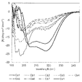
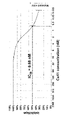
- FIG. 2 is a characteristic diagram showing CD spectra of Ca1, Cb1, Cb2, Cb3, Cb4, Cb5, Cb6, Cb7 and Ce1 produced in Examples.
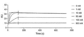
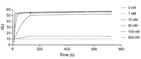
- FIG. 2 is a characteristic diagram showing the results of measuring the interaction of Ce1 produced in Examples with the receptor-binding domain (RBD) of wild-type SARS-CoV-2.
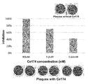
- FIG. 2 is a photograph showing the results of a plaque reduction assay performed using wild-type SARS-CoV-2 for Ce1 prepared in Example, and a diagram showing the concentration conditions of Ce1.
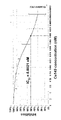
- FIG. 4 is a characteristic diagram showing the relationship between the Ce1 peptide concentration shown in FIG. 3 and the inhibition rate.
- FIG. 10 is a photograph showing the results of a plaque reduction assay performed using wild-type SARS-CoV-2 for the LCB1 peptide, and a diagram showing the concentration conditions of LCB1.
- FIG. 1 is a photograph showing the results of a plaque reduction assay performed using wild-type SARS-CoV-2 for Ca1, and a diagram showing Ca1 concentration conditions.
- FIG. 6 is a characteristic diagram showing the relationship between the LCB1 peptide concentration shown in FIG. 5 and the inhibition rate.
- FIG. 7 is a characteristic diagram showing the relationship between Ca1 peptide concentration and inhibition rate shown in FIG.
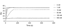
- FIG. 2 is a characteristic diagram showing the results of measuring the interaction of Ce4 prepared in Example with the receptor-binding domain (RBD) of wild-type SARS-CoV-2.
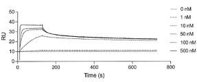
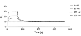
- FIG. 10 is a characteristic diagram showing the results of measuring the interaction of Ce4 prepared in Example with the receptor binding domain (RBD) of wild-type SARS-CoV-2 after 3 hours and 2 minutes from the start of binding.
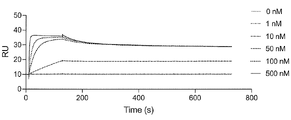
- FIG. 2 is a characteristic diagram showing the results of measuring the interaction of Ce9 prepared in Example with the receptor-binding domain (RBD) of wild-type SARS-CoV-2.
- FIG. 2 is a characteristic diagram showing the results of measuring the interaction of Ce6 produced in Examples with the receptor-binding domain (RBD) of wild-type SARS-CoV-2.
- FIG. 2 is a characteristic diagram showing the results of measuring the interaction of Ce5 produced in Examples with the receptor-binding domain (RBD) of wild-type SARS-CoV-2.
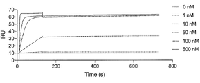
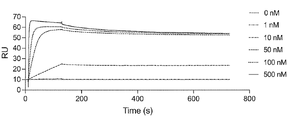
- FIG. 2 is a characteristic diagram showing the results of measuring the interaction of Ce14 prepared in Example with the receptor binding domain (RBD) of wild-type SARS-CoV-2.
- FIG. 2 is a characteristic diagram showing the results of measuring the interaction of Ce15 produced in Examples with the receptor-binding domain (RBD) of wild-type SARS-CoV-2.
- FIG. 2 is a characteristic diagram showing the results of measuring the interaction of Ce16 produced in Example with the receptor-binding domain (RBD) of wild-type SARS-CoV-2.
- FIG. 2 is a characteristic diagram showing the results of measuring the interaction of Ce41 prepared in Examples with the receptor-binding domain (RBD) of wild-type SARS-CoV-2.
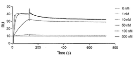
- FIG. 2 is a characteristic diagram showing the results of measuring the interaction of Ce41 produced in Example with the receptor-binding domain (RBD) of UK mutant SARS-CoV-2 (B.1.1.7).
- FIG. 10 is a characteristic diagram showing the results of measuring the interaction of Ce41 produced in Example with the receptor binding domain (RBD) of Brazilian mutant SARS-CoV-2 (P.1).
- FIG. 10 is a characteristic diagram showing the results of measuring the interaction of Ce41 produced in Examples with the receptor-binding domain (RBD) of the South African mutant SARS-CoV-2 (B.1.351).
- FIG. 10 is a characteristic diagram showing the results of measuring the interaction of Ce41 prepared in Example with the receptor-binding domain (RBD) of Indian mutant ( ⁇ type) SARS-CoV-2 (B.1.617.2).
- FIG. 10 is a characteristic diagram showing the results of measuring the interaction of Ce41 prepared in Example with the receptor-binding domain (RBD) of Indian mutant ( ⁇ -type) SARS-CoV-2 (B.1.617.1).
- FIG. 10 is a photograph showing the results of a plaque reduction assay performed using UK mutant SARS-CoV-2 (B.1.1.7) for Ce41 prepared in Example, and a diagram showing Ce41 concentration conditions.
- FIG. 24 is a characteristic diagram showing the relationship between the Ce41 peptide concentration and the inhibition rate shown in FIG. 23; Characteristics showing the results of measuring the interaction with the receptor binding domain (RBD) of UK mutant SARS-CoV-2 (B.1.1.7) for 3 hours and 2 minutes from the start of binding for Ce41 produced in the example It is a diagram.
- FIG. 10 is a characteristic diagram showing the results of measuring the interaction of Ce41 prepared in Example with the receptor-binding domain (RBD) of Indian mutant ( ⁇ -type) SARS-CoV-2 (B.1.617.1).
- FIG. 10
- FIG. 2 is a characteristic diagram showing the results of measuring the interaction of Ce59 produced in Examples with the receptor binding domain (RBD) of wild-type SARS-CoV-2.
- FIG. 10 is a characteristic diagram showing the results of measuring the interaction of Ce59 produced in Example with the receptor-binding domain (RBD) of UK mutant SARS-CoV-2 (B.1.1.7).
- FIG. 10 is a characteristic diagram showing the results of measuring the interaction of Ce59 prepared in Example with the receptor binding domain (RBD) in Brazilian mutant SARS-CoV-2 (P.1).
- FIG. 2 is a characteristic diagram showing the results of measuring the interaction of Ce59 produced in Example with the receptor-binding domain (RBD) of South African mutant SARS-CoV-2 (B.1.351).
- FIG. 10 is a characteristic diagram showing the results of measuring the interaction of Ce59 produced in Example with the receptor-binding domain (RBD) of Indian mutant ( ⁇ type) SARS-CoV-2 (B.1.617.2).
- FIG. 10 is a characteristic diagram showing the results of measuring the interaction of Ce59 prepared in Example with the receptor-binding domain (RBD) of Indian mutant ( ⁇ -type) SARS-CoV-2 (B.1.617.1).
- FIG. 33 is a characteristic diagram showing the relationship between the Ce59 peptide concentration and the inhibition rate shown in FIG. 32;
- FIG. 33 is a characteristic diagram showing the relationship between the Ce59 peptide concentration and the inhibition rate shown in FIG. 32;
- FIG. 33 is a characteristic diagram showing the relationship between the Ce59 peptide concentration and the inhibition rate shown in FIG. 32;
- FIG. 10 is a photograph showing the results of a plaque reduction assay performed using UK mutant SARS-CoV-2 (B.1.1.7) for Ce59 prepared in Example, and a diagram showing Ce59 concentration conditions.
- FIG. 35 is a characteristic diagram showing the relationship between the Ce59 peptide concentration and the inhibition rate shown in FIG. 34; Characteristics showing the results of measuring the interaction with the receptor binding domain (RBD) of UK mutant SARS-CoV-2 (B.1.1.7) for 3 hours and 2 minutes from the start of binding for Ce59 produced in the example It is a diagram.
- FIG. 10 is a characteristic diagram showing the results of measuring the interaction of Ce113 prepared in Example with the receptor binding domain (RBD) of UK mutant SARS-CoV-2 (B.1.1.7).
- FIG. 10 is a characteristic diagram showing the results of measuring the interaction of Ce113 prepared in Example with the receptor-binding domain (RBD) of Indian mutant ( ⁇ type) SARS-CoV-2 (B.1.617.1).
- RBD receptor-binding domain
- For Ce113 produced in the example the results of measuring the interaction with the receptor binding domain (RBD) in Omicron mutant (BA.1 strain) SARS-CoV-2 (B.1.1.529/BA.1) It is a characteristic diagram showing.
- FIG. 10 is a characteristic diagram showing the results of measuring the interaction of Ce172 produced in Example with the receptor-binding domain (RBD) of UK mutant SARS-CoV-2 (B.1.1.7).
- FIG. 10 is a characteristic diagram showing the results of measuring the interaction of Ce172 prepared in Example with the receptor-binding domain (RBD) of Indian mutant ( ⁇ type) SARS-CoV-2 (B.1.617.1).
- FIG. 10 is a photograph showing the results of a plaque reduction assay performed using UK mutant SARS-CoV-2 (B.1.1.7) for Ce172 prepared in Example, and a diagram showing the concentration conditions of Ce172.
- FIG. 46 is a characteristic diagram showing the relationship between the Ce172 peptide concentration shown in FIG. 45 and the inhibition rate.
- FIG. 48 is a characteristic diagram showing the relationship between the Ce172 peptide concentration and the inhibition rate shown in FIG. 47;
- FIG. 4 is a diagram showing density conditions;
- FIG. 50 is a characteristic diagram showing the relationship between the Ce172 peptide concentration and inhibition rate shown in FIG.
- FIG. 10 is a characteristic diagram showing the results of measuring the interaction of Ce173 prepared in Example with the receptor-binding domain (RBD) of UK mutant SARS-CoV-2 (B.1.1.7).
- FIG. 10 is a characteristic diagram showing the results of measuring the interaction of Ce173 produced in Example with the receptor-binding domain (RBD) of Indian mutant ( ⁇ type) SARS-CoV-2 (B.1.617.1).
- RBD receptor-binding domain
- For Ce173 produced in the example the results of measuring the interaction with the receptor binding domain (RBD) in Omicron mutant (BA.1 strain) SARS-CoV-2 (B.1.1.529/BA.1) It is a characteristic diagram showing.
- Photographs showing the results of a plaque reduction assay performed using the British mutant SARS-CoV-2 (B.1.1.7) for Ce173 prepared in Example, and a diagram showing the relationship between the Ce173 peptide concentration and the inhibition rate. is. Photographs showing the results of a plaque reduction assay performed using Indian mutant ( ⁇ type) SARS-CoV-2 (B.1.617.1) for Ce173 prepared in Example, and Ce173 peptide concentration and inhibition rate
- FIG. 4 is a diagram showing relationships; Photographs showing the results of a plaque reduction assay performed using Omicron mutant (BA.1 strain) SARS-CoV-2 (B.1.1.529/BA.1) for Ce173 prepared in Example, and Ce173 peptide
- FIG. 4 is a diagram showing relationships; Photographs showing the results of a plaque reduction assay performed using Omicron mutant (BA.1 strain) SARS-CoV-2 (B.1.1.529/BA.1) for Ce173 prepared in Example, and Ce173 peptide
- FIG. 10 is a characteristic diagram showing the results of measuring the interaction of Ce174 prepared in Example with the receptor binding domain (RBD) of UK mutant SARS-CoV-2 (B.1.1.7).
- FIG. 10 is a characteristic diagram showing the results of measuring the interaction of Ce174 produced in Example with the receptor-binding domain (RBD) of Indian mutant ( ⁇ type) SARS-CoV-2 (B.1.617.1).
- the results of measuring the interaction with the receptor binding domain (RBD) in Omicron mutant (BA.1 strain) SARS-CoV-2 (B.1.1.529/BA.1) It is a characteristic diagram showing.
- FIG. 4 is a diagram showing relationships; Photographs showing the results of a plaque reduction assay performed using Omicron mutant (BA.1 strain) SARS-CoV-2 (B.1.1.529/BA.1) for Ce174 prepared in Example, and Ce174 peptide
- FIG. 4 is a diagram showing the relationship between concentration and inhibition rate.
- FIG. 10 is a characteristic diagram showing the results of measuring the interaction of Ce149 produced in Example with the receptor-binding domain (RBD) of UK mutant SARS-CoV-2 (B.1.1.7).
- FIG. 10 is a characteristic diagram showing the results of measuring the interaction of Ce149 prepared in Example with the receptor-binding domain (RBD) of Indian mutant ( ⁇ type) SARS-CoV-2 (B.1.617.1).
- FIG. 10 is a photograph showing the results of a plaque reduction assay performed using UK mutant SARS-CoV-2 (B.1.1.7) for Ce149 prepared in Example, and a diagram showing the concentration conditions of Ce149.
- FIG. 10 is a photograph showing the results of a plaque reduction assay performed using UK mutant SARS-CoV-2 (B.1.1.7) for Ce149 prepared in Example, and a diagram showing the concentration conditions of Ce149.
- FIG. 70 is a characteristic diagram showing the relationship between the Ce149 peptide concentration and the inhibition rate shown in FIG. 69; A photograph showing the results of a plaque reduction assay performed using Indian mutant ( ⁇ type) SARS-CoV-2 (B.1.617.2) for Ce149 prepared in Example and a diagram showing the concentration conditions of Ce149. be.
- FIG. 72 is a characteristic diagram showing the relationship between the Ce149 peptide concentration and the inhibition rate shown in FIG. 71; A photograph showing the results of a plaque reduction assay performed using Omicron mutant (BA.1 strain) SARS-CoV-2 (B.1.1.529/BA.1) for Ce149 prepared in the example, and Ce149
- FIG. 4 is a diagram showing density conditions; FIG.
- FIG. 74 is a characteristic diagram showing the relationship between the Ce149 peptide concentration and the inhibition rate shown in FIG. 73;
- FIG. 2 is a characteristic diagram showing CD spectra of Ce4, Ce9, Ce41, Ce59, Ce113, Ce149, Ce172, Ce173 and Ce174 produced in Examples.
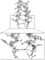
- FIG. 4 is a characteristic diagram showing interactions between the second lysine residue in the first region and the five C-terminal residues in the second region in the three-dimensional structural model of Ce41 prepared in Examples.
- Ce41 prepared in the example the results of measuring the interaction with the receptor binding domain (RBD) in Omicron mutant (BA.1 strain) SARS-CoV-2 (B.1.1.529/BA.1) It is a characteristic diagram showing.
- SARS-CoV-2 B.1.1.529/BA.1
- It is a characteristic diagram showing.
- Ce59 produced in the example the results of measuring the interaction with the receptor binding domain (RBD) in Omicron mutant (BA.1 strain) SARS-CoV-2 (
- Peptides of the present disclosure can bind to the receptor binding domain (RBD) in wild-type SARS-CoV-2 or mutant SARS-CoV-2.
- the peptide according to the present disclosure inhibits binding between ACE2 (angiotensin-converting enzyme 2) expressed on cells and the receptor binding domain (RBD) in the SARS-CoV-2, It can prevent infection with SARS-CoV-2.
- ACE2 angiotensin-converting enzyme 2
- the S protein is known to form a trimer and have a basic amino acid sequence that is cleaved by furin, a host protease.
- the S protein is cleaved by furin into the S1 subunit responsible for receptor binding and the S2 subunit responsible for membrane fusion.
- Peptides of the present disclosure can bind to the receptor binding domain (RBD) present in the S1 subunit.
- receptor binding domain RBD
- receptor binding domain (RBD) means the receptor binding domain (RBD) present in the S1 subunit of the Spike protein.
- the peptide according to the present disclosure has two helical structures (for convenience, the N-terminal side is called the first helix and the C-terminal side is called the second helix).
- the peptide according to the present disclosure includes a region having the first helix on the N-terminal side (referred to as the first region for convenience) and a region having the second helix on the C-terminal side (referred to as the second region for convenience).
- the peptide according to the present disclosure is composed of amino acid residues within 5 residues on the N-terminal side contained in the first region and amino acid residues within 5 residues on the C-terminal side contained in the second region.
- One of the characteristics is that the bonds formed between them can maintain the desired three-dimensional structure.
- the bond between the amino acid residues within 5 residues on the N-terminal side contained in the first region and the amino acid residues within 5 residues on the C-terminal side contained in the second region is a salt bridge
- the bond may be specifically referred to as a salt bridge or a hydrogen bond hereinafter, and the salt bridge or the hydrogen bond can be referred to as a bond.
- amino acid residues within 5 residues on the N-terminal side means amino acid residues located between 1 to 5 amino acid residues counting from the N-terminus, and amino acid residues involved in the formation of the above bonds. It is not meant to limit the position of the group to the N-terminus.
- amino acid residues within 5 residues on the C-terminal side means amino acid residues located between 1 to 5 amino acid residues counting from the C-terminus, and amino acids involved in the formation of the bond It is not meant to limit residue positions to the C-terminus.
- the peptide according to the present disclosure is produced by introducing a hydrophilic amino acid residue in the first helix (specifically, for example, the 15th arginine residue and the 19th glutamic acid residue of SEQ ID NO: 1).
- a hydrophilic amino acid residue in the first helix specifically, for example, the 15th arginine residue and the 19th glutamic acid residue of SEQ ID NO: 1.
- the first region and the second region each have an amino acid sequence that can bind to the receptor binding domain (RBD) of SARS-CoV-2 in the above-described three-dimensional structure. and can be defined by their respective amino acid sequences.
- the sequence of the site that binds to the receptor binding domain (RBD) in SARS-CoV-2 in the first region is defined as the following (a), (b), (c) or (d) amino acid sequence can do.
- sequence of the site that binds to the receptor binding domain (RBD) in SARS-CoV-2 in the second region shall be defined as the following (e), (f) or (g) amino acid sequence can be done.
- e amino acid sequence of SEQ ID NO: 2
- f amino acid sequence of SEQ ID NO: 57
- sequence of the site that binds to the receptor binding domain (RBD) in SARS-CoV-2 in the first region can be defined as the following amino acid sequence (a) or (d1).
- the second region SARS-CoV-2 receptor binding domain (RBD ) can be defined as the amino acid sequence of (e) or (g1) below.
- sequence of the site that binds to the receptor binding domain (RBD) in SARS-CoV-2 in the first region can be defined as the following amino acid sequence (b) or (d2).
- amino acid sequence of SEQ ID NO: 61 (d2) an amino acid sequence having a sequence identity of 80% or more to the amino acid sequence of SEQ ID NO: 61; ) can be defined as the amino acid sequence of (f) or (g2) below.
- sequence of the site that binds to the receptor binding domain (RBD) in SARS-CoV-2 in the first region can be defined as the following amino acid sequence (c) or (d3).
- (c) an amino acid sequence of SEQ ID NO: 59 (d3) an amino acid sequence having a sequence identity of 80% or more to the amino acid sequence of SEQ ID NO: 59; ) can be defined as the amino acid sequence of (f) or (g2) below.
- the amino acid sequence (a), (b) or (c) defining the first region has a lysine residue (the second amino acid residue from the N-terminus in SEQ ID NO: 1, 59 or 61), and the second The amino acid sequence (e) or (f) defining the region has an aspartic acid residue (the first amino acid residue from the C-terminus in SEQ ID NO: 2 or 57).
- the peptides according to the present disclosure defined by SEQ ID NO: 1, 59 or 61 and SEQ ID NO: 2 or 57 have the desired conformation by forming the salt bridge between these lysine residues and aspartic acid residues. can be stabilized.
- the salt bridge formed between the first region and the second region is not limited to the lysine residue and the aspartic acid residue.
- the second amino acid residue from the N-terminus in SEQ ID NO: 1, 59 or 61 is an aspartic acid residue
- the first amino acid residue from the C-terminus in SEQ ID NO: 2 or 57 is a lysine residue
- a salt bridge is formed between these aspartic acid residues and lysine residues, which can stabilize the desired conformation.
- the combination of amino acid residues forming a salt bridge is not limited to the combination of the lysine residue and the aspartic acid residue described above. and combinations of arginine and aspartic acid residues. That is, as an example, even if the second from the N-terminus in SEQ ID NO: 1, 59 or 61 is an arginine residue and the first from the C-terminus in SEQ ID NO: 2 or 57 is a glutamic acid residue, these arginine residues and glutamic acid A salt bridge is formed between the residues and can stabilize the desired conformation.
- the second from the N-terminus in SEQ ID NO: 1, 59 or 61 is the glutamic acid residue, and the first from the C-terminus in SEQ ID NO: 2 or 57. may be used as an arginine residue.
- the amino acid sequence defined in (d), (d1), (d2) or (d3) above is the amino acid sequence of the site that binds to the receptor binding domain (RBD) in SARS-CoV-2, the first region 80% or more, 85% or more, 86% or more, 87% or more relative to the amino acid sequence of SEQ ID NO: 1, 59 or 61, if the amino acid residues involved in forming the bond between the second region and the second region are conserved % or greater, 88% or greater, 89% or greater, 90% or greater, 91% or greater, 92% or greater, 93% or greater, 94% or greater, or 95% or greater sequence identity.
- amino acid sequence defined in (d), (d1), (d2) or (d3) above is the amino acid sequence of the site that binds to the receptor binding domain (RBD) in SARS-CoV-2, 1 to 4 amino acid residues were substituted for the amino acid sequence of SEQ ID NO: 1, 59 or 61, provided that the amino acid residues involved in forming the bond between the 1st region and the 2nd region were conserved It can be an amino acid sequence.
- An amino acid sequence obtained by substituting one amino acid residue for the amino acid sequence of SEQ ID NO: 1, 59 or 61 has 95% sequence identity with the amino acid sequence of SEQ ID NO: 1, 59 or 61.
- Amino acid sequences in which 2, 3 or 4 amino acid residues are substituted for the amino acid sequence of SEQ ID NO: 1, 59 or 61 are 90% and 85% of the amino acid sequence of SEQ ID NO: 1, 59 or 61, respectively. % or 80% sequence identity.
- amino acid residue to be substituted is not particularly limited, but the amino acid sequence of SEQ ID NO: 1, 59 or 61 1st amino acid residue (aspartic acid residue), 8th amino acid residue (lysine residue), 11th amino acid residue (glutamic acid residue) and 18th amino acid residue (glutamic acid residue) You can choose from a group.
- amino acid residue 1st amino acid residue (aspartic acid residue), 8th amino acid residue (lysine residue), 11th amino acid residue (glutamic acid residue) and 18 in the amino acid sequences of SEQ ID NO: 1, 59 or 61
- the second amino acid residue contributes to the stabilization of the helix structure, but interacts poorly with the receptor-binding domain (RBD) in SARS-CoV-2, resulting in similar properties. It is possible to make substitutions with compatible amino acid residues.
- the first aspartic acid residue in SEQ ID NO: 1, 59 or 61 is not particularly limited, but can be replaced with an asparagine residue or a glutamic acid residue.
- Aspartic acid, asparagine, etc. have the property of initiating a helix called N-cap, and are similar in property.
- Both aspartic acid and glutamic acid are acidic amino acids and have similar properties. Therefore, even if the first aspartic acid residue in SEQ ID NO: 1, 59 or 61 is replaced with an asparagine residue or a glutamic acid residue, the effect on the structure of the first helix and the receptor binding domain in SARS-CoV-2 (RBD) interaction is very small, and the function of the peptide according to the present disclosure can be maintained.
- RBD SARS-CoV-2
- the 8th lysine residue in SEQ ID NO: 1, 59 or 61 can be replaced with an arginine residue or a histidine residue, although not particularly limited. Lysine, arginine and histidine are basic amino acids and have similar properties. Therefore, even if the 8th lysine residue in SEQ ID NO: 1, 59 or 61 is replaced with an arginine residue or a histidine residue, the effect on the structure of the first helix and the receptor binding domain in SARS-CoV-2 ( RBD) has very little effect, and the function of the peptide according to the present disclosure can be maintained.
- RBD SARS-CoV-2
- the 11th glutamic acid residue and the 18th glutamic acid residue in SEQ ID NO: 1, 59 or 61 can be substituted with aspartic acid residues, although not particularly limited. Both glutamic acid and aspartic acid are acidic amino acids and have similar properties. Therefore, even if the 11th glutamic acid residue and / or 18th glutamic acid residue in SEQ ID NO: 1, 59 or 61 is substituted with an aspartic acid residue, the effect on the structure of the first helix and SARS-CoV-2 It has very little effect on the interaction with the receptor binding domain (RBD) in , and the function of the peptides according to the present disclosure can be maintained.
- RBD receptor binding domain
- amino acid sequence defined in (g), (g1) or (g2) above is the amino acid sequence of the site that binds to the receptor binding domain (RBD) in SARS-CoV-2, the first region and the second region If the amino acid residues involved in the formation of the bond between , 89% or greater, 90% or greater, 91% or greater, 92% or greater, 93% or greater, 94% or greater, or 95% or greater sequence identity.
- amino acid sequence defined in (g), (g1) or (g2) above is the amino acid sequence of the site that binds to the receptor binding domain (RBD) in SARS-CoV-2, As long as the amino acid residues involved in the formation of the bond between the two regions are conserved, the amino acid sequence of SEQ ID NO: 2 or 57 can be replaced by 1 to 3 amino acid residues. can.
- the amino acid sequence obtained by substituting one amino acid residue with respect to the amino acid sequence of SEQ ID NO: 2 or 57 has 94.7% sequence identity with the amino acid sequence of SEQ ID NO: 2 or 57.
- the amino acid sequence in which 2 or 3 amino acid residues are substituted for the amino acid sequence of SEQ ID NO: 2 or 57 is 89.5% or 84.2% of the amino acid sequence of SEQ ID NO: 2 or 57, respectively. have identity.
- the amino acid residue to be substituted is not particularly limited, but the second amino acid in the amino acid sequence of SEQ ID NO: 2 or 57 It can be selected from the group consisting of the residue (glycine residue), the 14th amino acid residue (glutamic acid residue) and the 18th amino acid residue (arginine residue).
- the second glycine residue in SEQ ID NO: 2 or 57 can be replaced with an alanine residue.
- the second glycine residue in SEQ ID NO: 2 or 57 is all receptor binding domains in which the 501st asparagine residue in the amino acid sequence of the receptor binding domain (RBD) in SARS-CoV-2 is mutated to a tyrosine residue (RBD), but binding to the receptor-binding domain (RBD) in wild-type SARS-CoV-2 is even stronger due to substitution with an alanine residue.
- the first amino acid residue in SEQ ID NO: 2 or 57 is preferably a histidine residue.
- the structure is formed by interacting with the 19th amino acid residue in SEQ ID NO: 1, 59 or 61, such as a glutamic acid residue. It can contribute to stabilization. Furthermore, this histidine residue contributes to the stabilization of the helical bundle structure.
- the 14th glutamic acid residue in SEQ ID NO: 2 or 57 can be replaced with an aspartic acid residue, although it is not particularly limited. Both glutamic acid and aspartic acid are acidic amino acids and have similar properties. Therefore, even if the 14th glutamic acid residue in SEQ ID NO: 2 or 57 is replaced with an aspartic acid residue, the effect on the structure of the second helix and the interaction with the receptor binding domain (RBD) in SARS-CoV-2 It has very little effect on the action and can maintain the function of the peptide according to the present disclosure.
- RBD receptor binding domain
- the 18th arginine residue in SEQ ID NO: 2 or 57 can be substituted with a lysine residue or a histidine residue, although not particularly limited.
- Arginine, lysine and histidine are basic amino acids and have similar properties. Therefore, even if the 18th arginine residue in SEQ ID NO: 2 or 57 is replaced with a lysine residue or a histidine residue, the effect on the structure of the second helix and the receptor binding domain (RBD) in SARS-CoV-2 It has very little effect on the interaction with , and the function of the peptide according to the present disclosure can be maintained.
- Examples of amino acid residues that can be substituted for the sequence of the site that binds to the domain (RBD) and the sequence of the site that binds to the receptor binding domain (RBD) in SARS-CoV-2 in the second region were explained.
- the sequence of the site that binds to the receptor binding domain (RBD) in SARS-CoV-2 in the first region and the sequence of the site that binds to the receptor binding domain (RBD) in SARS-CoV-2 in the second region are the above The description is not limiting.
- the sequence of the site that binds to the receptor binding domain (RBD) in SARS-CoV-2 in the first region of the peptide according to the present disclosure is the first aspartic acid residue in SEQ ID NO: 1, 59 or 61, the eighth An amino acid sequence in which amino acid residues other than the lysine residue, the 11th glutamic acid residue and the 18th glutamic acid residue are substituted with other amino acid residues, or the amino acid sequence of SEQ ID NO: 1, 59 or 61 It may be an amino acid sequence in which 1 to 4 amino acid residues are deleted and/or inserted.
- sequence of the site that binds to the receptor binding domain (RBD) in SARS-CoV-2 in the first region is not limited to SEQ ID NO: 1, 59 or 61 consisting of 20 amino acid residues, 16 to 24 amino acid residues groups, preferably an amino acid sequence consisting of 18 to 22 amino acid residues.
- sequence of the site that binds to the receptor binding domain (RBD) in SARS-CoV-2 in the second region of the peptide according to the present disclosure is the second glycine residue in SEQ ID NO: 2 or 57, the 14th glutamic acid It may be an amino acid sequence in which amino acid residues other than the residue and the 18th arginine residue are substituted with other amino acid residues, or 1 to 3 amino acid residues are deleted from the amino acid sequence of SEQ ID NO: 2 or 57. It may be a deleted and/or inserted amino acid sequence.
- sequence of the site that binds to the receptor binding domain (RBD) in SARS-CoV-2 in the second region is not limited to SEQ ID NO: 2 or 57 consisting of 19 amino acid residues, 16 to 23 amino acid residues, An amino acid sequence consisting of 18 to 22 amino acid residues is preferred.
- the peptide according to the present disclosure may consist of 36 to 44 amino acid residues in total in the first region and the second region, but most preferably consists of 39 amino acid residues in total. preferable.
- the N-terminal lysine residue in the first region and the C-terminal aspartic acid residue in the second region It can form a salt bridge between them and is of suitable size for binding to the receptor binding domain (RBD) in SARS-CoV-2.
- RBD receptor binding domain
- the sequence of the site that binds to the receptor binding domain (RBD) in SARS-CoV-2 in the first region consists of an amino acid sequence different from SEQ ID NO: 1, 59 or 61, and SARS-CoV in the second region
- the receptor binding domain (RBD) in SARS-CoV-2 in the first region Amino acid residues within 5 residues on the N-terminal side in the sequence of the binding site (the second lysine residue from the N-terminus in SEQ ID NO: 1), and the receptor-binding domain in SARS-CoV-2 in the second region ( RBD) by forming a salt bridge with amino acid residues within 5 residues on the C-terminal side in the sequence of the site that binds to the desired
- the three-dimensional structure of can be maintained.
- the sequence of the site that binds to the receptor binding domain (RBD) in SARS-CoV-2 in the first region consists of an amino acid sequence different from SEQ ID NO: 1, 59 or 61, and SARS-CoV-2 in the second region
- the sequence of the site that binds to the receptor binding domain (RBD) in SARS-CoV-2 consists of an amino acid sequence different from SEQ ID NO: 2 or 57
- the peptide according to the present disclosure binds to the receptor binding domain (RBD) in SARS-CoV-2. can be similarly bound to
- Tables 1 and 2 below summarize the role of each amino acid residue in the first region consisting of the amino acid sequence of SEQ ID NO: 1 and the second region consisting of the amino acid sequence of SEQ ID NO: 2.
- Tables 3 and 4 below summarize the role of each amino acid residue in the first region consisting of the amino acid sequence of SEQ ID NO:59 and the second region consisting of the amino acid sequence of SEQ ID NO:57.
- RBD S477 means the 477th serine residue in the amino acid sequence of the receptor binding domain (RBD) in SARS-CoV-2 (similar meaning other than S477).
- N501Y of RBD with N501Y mutation refers to SARS-CoV-2 having a mutation in which the 501st position in the amino acid sequence of the receptor binding domain (RBD) in SARS-CoV-2 is a tyrosine residue means the 501st tyrosine residue in the amino acid sequence that constitutes the receptor binding domain (RBD) in .
- the sequence of the site that binds to the receptor binding domain (RBD) in SARS-CoV-2 in the first region and the receptor binding domain (RBD) in SARS-CoV-2 in the second region The amino acid sequences of the binding site sequences are not limited to SEQ ID NOS: 1, 59, 61, 2 and 57, respectively, and can be changed as appropriate according to the role of each amino acid residue.
- the first aspartic acid residue from the N-terminus which constitutes the sequence of the site that binds to the receptor-binding domain (RBD) in SARS-CoV-2 in the first region, has no specific interaction with the RBD.
- a glutamic acid residue having a chemical structure similar to that of aspartic acid can be used.
- the 15th arginine residue from the N-terminus which constitutes the sequence of the site that binds to the receptor binding domain (RBD) in SARS-CoV-2 in the first region, is the receptor in SARS-CoV-2 in the first region.
- the second glycine residue from the N-terminus which constitutes the sequence of the site that binds to the receptor binding domain (RBD) in SARS-CoV-2 in the second region, is the British mutant SARS-CoV-2 (B.1.1. Wild-type SARS-CoV- For the receptor binding domain (RBD) in 2, a stronger binding ability is acquired by substituting an alanine residue.
- the third glutamic acid residue from the N-terminus that constitutes the sequence of the site that binds to the receptor binding domain (RBD) in SARS-CoV-2 in the second region can be a glutamine residue.
- the sixth leucine residue from the N-terminus that constitutes the sequence of the site that binds to the receptor binding domain (RBD) in SARS-CoV-2 in the second region can be a methionine residue.
- the 16th methionine residue from the N-terminus that constitutes the sequence of the site that binds to the receptor binding domain (RBD) in SARS-CoV-2 in the second region can be a tryptophan residue.
- the 3rd glutamic acid residue, 6th leucine residue, and 16th methionine residue from the N-terminus that constitute the sequence of the site that binds to the receptor binding domain (RBD) in SARS-CoV-2 in the second region
- RBD receptor binding domain
- the first amino acid residue from the N-terminus is preferably an aspartic acid residue.
- sequence of the site that binds to the receptor binding domain (RBD) in SARS-CoV-2 in the first region preferably has an isoleucine residue at the 5th amino acid residue from the N-terminus.
- the receptor in SARS-CoV-2 in the first region Constructing the sequence of the site that binds to the binding domain (RBD)
- the ninth isoleucine residue from the N-terminus and the second region of SARS-CoV-2 Construct the sequence of the site that binds to the receptor binding domain (RBD)
- a stable interaction can be formed with the 15th phenylalanine residue from the N-terminus.
- sequence of the site that binds to the receptor binding domain (RBD) in SARS-CoV-2 in the first region preferably has an isoleucine residue at the ninth amino acid residue from the N-terminus.
- a stable interaction can be formed between the 15th phenylalanine residue from the N-terminus that constitutes the binding site sequence.
- sequence of the site that binds to the receptor binding domain (RBD) in SARS-CoV-2 in the first region has a tyrosine residue at the 10th amino acid residue and a methionine at the 13th amino acid residue from the N-terminus. A residue is preferred.
- the sequence of the site that binds to the receptor binding domain (RBD) in SARS-CoV-2 in the first region preferably has an isoleucine residue at the 12th amino acid residue from the N-terminus.
- the receptor in SARS-CoV-2 in the first region By setting the 12th isoleucine residue from the N-terminus of the sequence of the site that binds to the receptor binding domain (RBD) in SARS-CoV-2 in the first region, the receptor in SARS-CoV-2 in the first region
- the 16th leucine residue from the N-terminus that constitutes the sequence of the site that binds to the binding domain (RBD) and the sequence of the site that binds to the receptor binding domain (RBD) in SARS-CoV-2 in the second region A stable interaction can be formed with the 8th valine residue from the N-terminus.
- the sequence of the site that binds to the receptor binding domain (RBD) in SARS-CoV-2 in the first region preferably has a leucine residue at the 16th amino acid residue from the N-terminus.
- the receptor in SARS-CoV-2 in the first region The 12th isoleucine residue from the N-terminus that constitutes the sequence of the site that binds to the binding domain (RBD), and the sequence of the site that binds to the receptor binding domain (RBD) in SARS-CoV-2 in the second region Stable between the 4th alanine residue from the N-terminus and the 8th valine residue from the N-terminus that constitutes the sequence of the site that binds to the receptor binding domain (RBD) in SARS-CoV-2 in the second region interaction can be formed.
- the first from the N-terminus is preferably a histidine residue.
- the first histidine residue from the N-terminus of the sequence of the site that binds to the receptor binding domain (RBD) in SARS-CoV-2 in the second region it contributes to the stabilization of the helix bundle structure, It can form a salt bridge with the 19th glutamic acid residue in the sequence of the site that binds to the receptor binding domain (RBD) in SARS-CoV-2 in one region and stabilize the turn structure.
- the 10th amino acid residue from the N-terminus is preferably an aspartic acid residue.
- the receptor binding domain in SARS-CoV-2 ( RBD) can form a salt bridge with the 403rd arginine residue and the 417th lysine residue in the amino acid sequence.
- the glutamic acid residue and SARS-CoV-2 can not form a salt bridge with the 403rd arginine residue and 417th lysine residue in the amino acid sequence of the receptor binding domain (RBD), binding activity with the receptor binding domain (RBD) in SARS-CoV-2 is likely to decrease.
- the 13th amino acid residue from the N-terminus in the sequence of the site that binds to the receptor binding domain (RBD) in SARS-CoV-2 in the second region is a tyrosine residue.
- the receptor binding domain (RBD) in SARS-CoV-2 can form a hydrogen bond with the 420th aspartic acid residue in the amino acid sequence.
- sequence of the site that binds to the receptor binding domain (RBD) in SARS-CoV-2 in the second region preferably has an alanine residue at the 4th amino acid residue from the N-terminus.
- the receptor in SARS-CoV-2 in the first region A stable interaction can be formed between the 16th leucine residue from the N-terminus that constitutes the sequence of the site that binds to the binding domain (RBD).
- the sequence of the site that binds to the receptor binding domain (RBD) in SARS-CoV-2 in the second region preferably has a valine residue at the eighth amino acid residue from the N-terminus.
- the receptor in SARS-CoV-2 in the first region The ninth isoleucine residue from the N-terminus, which constitutes the sequence of the site that binds to the binding domain (RBD), and the sequence of the site that binds to the receptor binding domain (RBD) in SARS-CoV-2 in the first region
- the 12th isoleucine residue from the N-terminus, the 13th methionine residue from the N-terminus that constitutes the sequence of the site that binds to the receptor binding domain (RBD) in SARS-CoV-2 in the first region and the first region A stable interaction can be formed with the 16th leucine residue
- sequence of the site that binds to the receptor binding domain (RBD) in SARS-CoV-2 in the second region preferably has a leucine residue at the 11th amino acid residue from the N-terminus.
- the receptor in SARS-CoV-2 in the first region Constructing the sequence of the site that binds to the binding domain (RBD)
- the ninth isoleucine residue from the N-terminus and the second region of SARS-CoV-2 Construct the sequence of the site that binds to the receptor binding domain (RBD)
- a stable interaction can be formed with the 8th valine residue from the N-terminus.
- the sequence of the site that binds to the receptor binding domain (RBD) in SARS-CoV-2 in the second region preferably has a phenylalanine residue at the 15th amino acid residue from the N-terminus.
- the receptor in SARS-CoV-2 in the first region The 5th isoleucine residue from the N-terminus, which constitutes the sequence of the site that binds to the binding domain (RBD), and the sequence of the site that binds to the receptor binding domain (RBD) in SARS-CoV-2 in the first region.
- Stable between the 6th leucine residue from the N-terminus and the 9th isoleucine residue from the N-terminus that constitutes the sequence of the site that binds to the receptor binding domain (RBD) in SARS-CoV-2 in the first region interaction can be formed.
- the 14th amino acid residue from the N-terminus in the sequence of the site that binds to the receptor binding domain (RBD) in SARS-CoV-2 in the second region may be any amino acid residue, but glutamic acid A residue is preferred.
- the hydrophilic properties of the peptide can be kept high. .
- the 17th amino acid residue from the N-terminus is preferably a lysine residue.
- the receptor binding domain (RBD) in SARS-CoV-2 can form a salt bridge with the 420th aspartic acid residue in the amino acid sequence of ), and form a hydrogen bond between the 421st tyrosine residue and the 460th asparagine residue.
- the 18th amino acid residue from the N-terminus in the sequence of the site that binds to the receptor binding domain (RBD) in SARS-CoV-2 in the second region may be any amino acid residue, but an arginine residue It is preferable to By making the 18th arginine residue from the N-terminus of the second helix, the hydrophilic properties of the peptide can be kept high, and the second lysine residue from the N-terminus of the first helix and hydrogen bonding and contributes to the stabilization of the helix bundle structure.
- the hydrophilic properties of the peptide can be maintained at a high level.
- one of the characteristics of the peptide according to the present disclosure is that the 15th position in the amino acid sequence of SEQ ID NO: 1, 59 or 61 is an arginine residue and the 19th position is a glutamic acid residue.
- the formation of a salt bridge between the 15th arginine residue and the 19th glutamic acid residue in the amino acid sequence of SEQ ID NO: 1, 59 or 61 contributes to the structural stabilization of the first helix. can. This can improve the binding rate between the peptides of the present disclosure and the receptor binding domain (RBD) in SARS-CoV-2.
- RBD receptor binding domain
- amino acid residues 15 and 19 in the amino acid sequence of SEQ ID NO: 1, 59 or 61 are a combination of lysine and aspartic acid residues, or arginine Any combination of a residue and a glutamic acid residue may be used, but a combination of an arginine residue and a glutamic acid residue is particularly desirable.
- the second amino acid sequence of SEQ ID NO: 2 or 57 is a glycine residue. Since the peptide according to the present disclosure has a glycine residue at the second position in the amino acid sequence of SEQ ID NO: 2 or 57, both wild-type SARS-CoV-2 and mutant SARS-CoV-2 have receptor It has excellent binding activity to the binding domain (RBD).
- wild-type SARS-CoV-2 is a glycine residue at position 339, a serine residue at position 371, and a serine residue at position 373 in the amino acid sequence that constitutes the receptor binding domain (RBD).
- 375 is a serine residue
- 376 is a threonine residue
- 405 is an aspartic acid residue
- 408 is an arginine residue
- 417 is a lysine residue
- 440 446th is an asparagine residue
- 446th is a glycine residue
- 452nd is a leucine residue
- 477th is a serine residue
- 478th is a threonine residue
- 484th is a glutamic acid residue.
- mutant SARS-CoV-2 is a mutation in which the 339th amino acid sequence constituting the receptor binding domain (RBD) is an aspartic acid residue, and a mutation in which the 371st is a leucine residue or a phenylalanine residue, 373rd is a proline residue mutation, 375th is a phenylalanine residue mutation, 376th is an alanine residue mutation, 405th is an asparagine residue mutation, 408th is a serine residue mutation, Mutation of 417th asparagine residue or threonine residue, mutation of 440th lysine residue, mutation of 446th serine residue, mutation of 452nd arginine residue, 477th asparagine residue Mutation,
- the mutant SARS-CoV-2 has a mutation in which the 501st amino acid sequence constituting the receptor binding domain (RBD) is a tyrosine residue, 339th, 371st, 373rd, 375th, 376th, 405th, 408th, 417th, 440th, 446th, 452nd, 477th, 478th, 484th, 493rd, 496th, 498th and 505th amino acid residues are wild type SARS-CoV-2 (PANGO systematic name: B.1.1.7, sometimes commonly referred to as UK variant SARS-CoV-2).
- the 417th in the amino acid sequence constituting the receptor binding domain (RBD) becomes an asparagine residue
- the 484th becomes a lysine residue
- the 501st becomes a tyrosine residue.
- a type of SARS-CoV-2 in which the 498th and 505th amino acid residues are wild type can be mentioned (PANGO systematic name: B.1.351, commonly referred to as South African mutant SARS-CoV-2). be).
- the 417th in the amino acid sequence constituting the receptor binding domain (RBD) becomes a threonine residue
- the 484th becomes a lysine residue
- the 501st becomes a tyrosine residue.
- a type of SARS-CoV-2 whose 498th and 505th amino acid residues are wild type can be mentioned (PANGO system name: P.1, commonly referred to as Brazilian mutant SARS-CoV-2). be).
- the mutant SARS-CoV-2 has a mutation in which the 452nd amino acid sequence constituting the receptor binding domain (RBD) becomes an arginine residue and a mutation in which the 478th becomes a lysine residue, 339 th, 371st, 373rd, 375th, 376th, 405th, 408th, 417th, 440th, 446th, 477th, 484th, 493rd, 496th, 498th, 501st and 505th
- RBD receptor binding domain
- the mutant SARS-CoV-2 has a mutation in which the 452nd amino acid sequence constituting the receptor binding domain (RBD) becomes an arginine residue and a mutation in which the 484th becomes a glutamine residue, and 339 th, 371st, 373rd, 375th, 376th, 405th, 408th, 417th, 440th, 446th, 477th, 478th, 493rd, 496th, 498th, 501st and 505th
- RBD receptor binding domain
- 484th becomes a glutamine residue
- the 339th mutation in the amino acid sequence that constitutes the receptor binding domain (RBD) becomes an aspartic acid residue
- the 371st mutation becomes a leucine residue
- the 373rd is A mutation to a proline residue, a mutation to a phenylalanine residue at 375th, a mutation to an asparagine residue at 417th, a mutation to a lysine residue at 440th, a mutation to a serine residue at 446th, and a mutation at 477th Asparagine residue mutation, 478th lysine residue mutation, 484th alanine residue mutation, 493rd arginine residue mutation, 496th serine residue mutation, 498th mutation Has a mutation that becomes an arginine residue, a mutation that the 501st becomes a tyrosine residue, and a mutation that the 505th becomes a histidine residue, and the 376th, 405th, 408th
- the 339th mutation in the amino acid sequence that constitutes the receptor binding domain (RBD) becomes an aspartic acid residue
- the 371st mutation becomes a phenylalanine residue
- the 373rd is A mutation to a proline residue, a mutation to a phenylalanine residue at 375th, a mutation to an alanine residue at 376th, a mutation to an asparagine residue at 405th, a mutation to a serine residue at 408th, and a mutation at 417th
- RBD receptor binding domain
- the sequence of the site that binds to the receptor binding domain (RBD) in SARS-CoV-2 in the second region of the peptide according to the present disclosure is that the second glycine residue in the amino acid sequence of SEQ ID NO: 2 or 57 is alanine. It may also be an amino acid sequence that is fragmented.
- 501 such as wild-type SARS-CoV-2 It has excellent binding activity to the SARS-CoV-2 receptor binding domain (RBD) of the type in which the third amino acid residue is the wild type.
- one of the characteristics of the peptide according to the present disclosure is that the 18th position in the amino acid sequence of SEQ ID NO: 2 or 57 is an arginine residue. Since the peptide according to the present disclosure has an arginine residue at position 18 in the amino acid sequence of SEQ ID NO: 2 or 57, It has excellent binding activity to the receptor binding domain (RBD) of SARS-CoV-2.
- RBD receptor binding domain
- peptides of the present disclosure are The sequence of the site that binds to the receptor binding domain (RBD) in SARS-CoV-2 in the first region contains the following (h), (i), (j), (k) or (l) amino acid sequence , (h) the amino acid sequence of SEQ ID NO:56; (i) the amino acid sequence of SEQ ID NO:59; (j) the amino acid sequence of SEQ ID NO:61; (k) the amino acid sequence of SEQ ID NO:63, or (l) the amino acid sequence of SEQ ID NO:65,
- a peptide in which the sequence of the site that binds to the receptor binding domain (RBD) in SARS-CoV-2 in the second region includes the amino acid sequence of SEQ ID NO:57.
- the peptides of the present disclosure are The sequence of the site that binds to the receptor binding domain (RBD) in SARS-CoV-2 of the first region contains the following (m), (n), (o) or (p) amino acid sequence, (m) the amino acid sequence of SEQ ID NO: 4; (n) the amino acid sequence of SEQ ID NO: 6; (o) the amino acid sequence of SEQ ID NO: 7, or (p) the amino acid sequence of SEQ ID NO: 8,
- a peptide in which the sequence of the site that binds to the receptor binding domain (RBD) in SARS-CoV-2 in the second region includes the amino acid sequence of SEQ ID NO: 2, 3, 5 or 9.
- these specific peptides have been found to exhibit excellent binding activity to the receptor binding domain (RBD) of SARS-CoV-2.
- the binding activity can be comprehensively judged from the rate of binding and the degree of dissociation.
- the system for evaluating the binding activity of a specific peptide to the receptor binding domain (RBD) in SARS-CoV-2 is not particularly limited, but a system using surface plasmon resonance (SPR) can be used.
- the receptor binding domain (RBD) in SARS-CoV-2 is immobilized on the surface of the sensor chip, a sample containing various analytes (peptides) is brought into contact with the surface, and the receptor binding domain in SARS-CoV-2 Binding kinetics can be measured based on the change in SPR angle due to binding of (RBD) to the peptide.
- a buffer containing no analyte was flowed on the surface of the sensor chip with the receptor binding domain (RBD) of SARS-CoV-2 bound to the peptide, and the receptor binding domain (RBD) of SARS-CoV-2 and The degree of dissociation can be measured based on the change in SPR angle due to dissociation with the peptide.
- the binding rate and degree of dissociation between the peptide and the receptor binding domain (RBD) in SARS-CoV-2 are not limited to systems using surface plasmon resonance (SPR), such as biolayer interferometry (BLI). may be measured by a system that analyzes the interaction of biomolecules.
- SPR surface plasmon resonance
- BLI biolayer interferometry
- the peptide according to the present disclosure has one or more amino acids at at least one end of its N-terminus and C-terminus, as long as it has excellent binding activity to the receptor binding domain (RBD) in SARS-CoV-2 may be a peptide to which is bound, a fusion peptide fused with another peptide or protein, a peptide with an unnatural amino acid inserted, a peptide with a tag or label added, and other modifications It may also be in the form of an applied peptide.
- fusion peptides include, but are not limited to, those fused with a membrane permeable peptide (CPP) so that they are introduced into cells when administered.
- the peptide of the present disclosure may be a peptide in which one or more amino acids are bound to at least one of the N-terminus and C-terminus, a fusion peptide fused with another peptide or protein, or a non-natural amino acid inserted.
- Peptides, peptides attached with tags or labels, and peptides with other modifications are also included in the peptides of the present disclosure as long as they include the peptides of the present disclosure.
- peptide synthesis methods include solid-phase synthesis methods and liquid-phase synthesis methods.
- solid-phase synthesis methods include a method using Boc (t-butyloxycarbonyl) or Fmoc (9-fluorenylmethoxycarbonyl) as a protective group for amino groups.
- the peptides described above can be synthesized using a commercially available peptide synthesizer to which the solid-phase synthesis method is applied.
- a DNA encoding the above-described peptide and express the above-described peptide in a host cell into which the DNA has been introduced.
- host cells bacteria such as yeast and Escherichia coli, insect cells, animal cells, and plant cells can be used.
- the peptides described above can be synthesized in vitro using cell-free protein synthesis systems.
- the peptide according to the present disclosure is preferably produced by chemical synthesis, and the reason for this is that the peptide according to the present disclosure has a total length of about 39 amino acid residues, so it is chemically synthesized by a general chemical synthesis method. In the case of chemical synthesis, it is relatively easy to manufacture under conditions that comply with GMP (Good Manufacturing Practice).
- the peptides according to the present disclosure described above can be used as pharmaceutical compositions.
- the peptides according to the present disclosure have remarkably high solubility in ultrapure water or buffer solutions, and have excellent binding activity to the receptor binding domain (RBD) of SARS-CoV-2. Therefore, the peptide according to the present disclosure can be used as a pharmaceutical composition for treatment and/or prevention of novel coronavirus infection (Coronavirus Disease 2019 (COVID-19)) caused by SARS-CoV-2.
- the disclosure is the use of said peptides in the manufacture of a pharmaceutical composition for the treatment and/or prevention of Coronavirus Disease 2019 (COVID-19).
- Subjects for administration of such pharmaceutical compositions are not particularly limited, but are animals including mammals, including humans, monkeys, mice, rats, hamsters, rabbits, guinea pigs, cows, pigs, dogs, horses, cats, goats, and sheep. , preferably human.
- the route of administration of the pharmaceutical composition of the present disclosure may be either oral or parenteral.
- the dosage of the pharmaceutical composition of the present disclosure may be prescribed variously depending on factors such as formulation method, administration mode, administration time, age, body weight, sex, disease state, excretion rate, and reaction sensitivity of the administration subject. can be done.
- the timing of administering the pharmaceutical composition of the present disclosure is not particularly limited and may be arbitrary. Also, it may be taken before meals, after meals, or between meals. Also, the administration/ingestion period is not particularly limited.
- the pharmaceutical composition of the present disclosure can be prepared in unit dosage form by formulating using a pharmaceutically acceptable carrier and/or excipient by a method commonly practiced by a person of ordinary skill in the art. , or can be manufactured within a multi-dose container.
- the pharmaceutical composition of the present disclosure can be appropriately formulated into a desired dosage form depending on the administration method.
- Pharmaceutical compositions of the present disclosure can be manufactured according to known methods, such as those described in the Japanese Pharmacopoeia (JP), United States Pharmacopeia (USP) or European Pharmacopoeia (EP).
- JP Japanese Pharmacopoeia
- USP United States Pharmacopeia
- EP European Pharmacopoeia
- drugs for the treatment and/or prevention of novel coronavirus infections that are known or discovered in the future (including mRNA vaccines) and drugs for the treatment and/or prevention of other diseases is also possible.
- Example 1 the LCB1 peptide disclosed in L Cao et al., Science 370, 426-431 (2020) (SEQ ID NO: 10: DKEWILQKIYEIMRLLDELGHAEASMRVSDLIYEFMKKGDERLLEEAERLLEEVER in DKEWILQKIYEIMRLLDELGHAEASMRVSDLIYEFMKKG (SEQ ID NO: 11) peptide (referred to as Ca1)) was compared.
- Ca1 DKEWILQKIYEIMRLLDELGHAEASMRVSDLIYEFMKKG
- RBD receptor binding domain
- LCB1 is a peptide having three helices
- Ca1 is a peptide having two helices counted from the N-terminal side of LCB1.
- DKEWILQKIYEIMRLLDELG (SEQ ID NO: 4) of Ca1 was called the first region
- HAEASMRVSDLIYEFMKKG SEQ ID NO: 12
- Table 7 summarizes the amino acid sequences of the Ca1 peptide and the peptide designed in this example. In the amino acid sequences shown in Table 7, the amino acid residues changed from the amino acid sequence of the Ca1 peptide are boxed.
- Cb1, Cb2, Cb3, Cb4, Cb5, Cb6 and Cb7 are peptides in which the amino acid sequences of the first region and the amino acid sequences of the second region do not form a helix structure. became.
- Ca1 was found to form a helical structure, as expected from the results of the LCB1 peptide disclosed in L Cao et al., Science 370, 426-431 (2020).
- Ce1 which is the C-terminal of the second region and has an aspartic acid residue at a position that can form a salt bridge between the N-terminal and the second lysine residue in the first region, is shown in FIG.
- the first helix contained in the first region and the second helix contained in the second region formed a helix bundle structure, and acquired a more stable structure than Ca1. I found out that I do. Therefore, for Ce1, the binding activity to the receptor binding domain (RBD) of wild-type SARS-CoV-2 was measured according to the protocol (intermolecular interaction analysis protocol) described in detail later, and as shown in Figure 2, It was found to have excellent characteristics of high binding rate and low degree of dissociation.
- RBD receptor binding domain
- Example 2 In this example, for Ce1 designed in Example 1, the wild-type SARS-CoV-2 infectivity reduction effect was measured by a plaque reduction assay according to a protocol (plaque reduction assay protocol) described in detail later, IC50 values were calculated based on the results. For comparison, the LCB1 peptide and Ca1 were also subjected to a plaque reduction assay using wild-type SARS-CoV-2, and IC 50 values were calculated.
- FIG. 4 shows the relationship between the Ce1 peptide concentration and the inhibition rate, which was calculated based on the number of plaques formed on each plate. As a result, the IC 50 value of Ce1 was calculated to be 1.5 nM.
- FIGS. 5 and 6 The results of plaque reduction assays performed with the LCB1 peptide and Ca1 are shown in FIGS. 5 and 6, respectively, and the relationship between inhibition rate and peptide concentration is shown in FIGS. 7 and 8, respectively.
- the IC50 values of LCB1 peptide and Ca1 were calculated to be 5.7 nM and 106.7 nM, respectively.
- Example 3 the Ce1 designed in Example 1 was further improved to develop a peptide that exhibits superior binding activity to the receptor binding domain (RBD) of wild-type SARS-CoV-2.
- RBD receptor binding domain
- Table 8 summarizes the amino acid sequences of the peptides designed in this example. In addition, in the amino acid sequences shown in Table 8, amino acid residues changed from the amino acid sequence of the Ce1 peptide are boxed.
- FIG. 12 shows the results of measuring the binding activity of Ce6, which is a representative example of a group of peptides whose binding activity to the receptor-binding domain (RBD) of wild-type SARS-CoV-2 is equivalent to that of Ce1.
- FIG. 13 shows the results of measuring the binding activity of Ce5 as a representative example of a group of peptides having lower binding activity to the receptor binding domain (RBD) of wild-type SARS-CoV-2 than Ce1.
- the 15th and 19th amino acid residues in the first helix are a pair of lysine and aspartic acid residues, and a pair of arginine and glutamic acid residues, respectively.
- a salt bridge is formed between the 15th amino acid residue and the 19th amino acid residue in the first helix, and the salt bridge is intended to stabilize the structure of the first helix.
- the full-length amino acid sequences of Ce4 and Ce9 are shown in SEQ ID NOS:52 and 53, respectively.
- FIG. 9 and 10 show the binding activity of Ce4 to the wild-type SARS-CoV-2 receptor binding domain (RBD), and the binding activity of Ce9 to the wild-type SARS-CoV-2 receptor binding domain (RBD).
- the results of activity measurement are shown in FIG. 9 and 10 show the results of measuring the binding activity of Ce4 to the receptor binding domain (RBD) of wild-type SARS-CoV-2.
- FIG. 10 shows the measurement results from the start of binding to 3 hours and 2 minutes.
- Ce1 designed in Example 1 replaces the 15th amino acid residue and the 19th amino acid residue in the first region with a pair of lysine and aspartic acid residues, an arginine residue and a glutamic acid residue, respectively.
- a pair of groups is preferred, and a pair of arginine and glutamic acid residues is particularly preferred.
- Example 4 we developed a peptide that exhibits excellent binding activity to the receptor binding domain (RBD) of mutant SARS-CoV-2.
- Ce9 which showed the best binding activity to the receptor binding domain (RBD) in wild-type SARS-CoV-2, was determined from the plaque reduction assay results using wild-type SARS-CoV-2. , has been found to exhibit an excellent antiviral activity with an IC 50 value of 0.4 nM (not shown).
- Example 5 In this example, for Ce41 designed in Example 4, the effect of reducing viral infectivity was measured by a plaque reduction assay performed using the UK mutant SARS-CoV-2 (B.1.1.7). The IC50 value was calculated based on
- Example 6 the Ce41 designed in Example 4 was measured for its binding activity to the receptor binding domain (RBD) in the UK mutant SARS-CoV-2 (B.1.1.7), and the Ce41 UK mutant SARS -The binding state to the receptor binding domain (RBD) in CoV-2 (B.1.1.7) was examined.
- Ce41 was not dissociated from the once-bound receptor-binding domain (RBD) of the UK mutant SARS-CoV-2 (B.1.1.7) even after washing with buffer for 3 hours. , found that excellent binding activity to the receptor-binding domain (RBD) of the UK mutant SARS-CoV-2 (B.1.1.7) could be sustained over a long period of time.
- This characteristic of Ce41 which can maintain excellent binding activity to the receptor binding domain (RBD) in the UK mutant SARS-CoV-2 (B.1.1.7) over a long period of time, was demonstrated in Example 5. It was considered to be of critical importance due to its remarkably high antiviral activity against mutant SARS-CoV-2 (B.1.1.7).
- the LCB1 peptide fails to sustain binding activity to the receptor binding domain (RBD) in UK mutant SARS-CoV-2 (B.1.1.7) even for a short period of time.
- Example 7 a peptide was developed that exhibits even better binding activity than Ce41 developed in the above example.
- Ce59 was designed with an arginine residue (boxed in Table 10) as the 18th amino acid residue (lysine residue) in the second region of Ce41.
- the full-length amino acid sequence of Ce59 is shown in SEQ ID NO:55.
- Ce59 wild-type SARS-CoV-2, UK mutated SARS-CoV-2 (B.1.1.7), Brazilian mutated SARS-CoV-2 (P.1), South African mutated SARS-CoV-2 ( B.1.351), the receptor-binding domain in India mutant (delta) SARS-CoV-2 (B.1.617.2) and India mutant (kappa) SARS-CoV-2 (B.1.617.1)
- the results of measuring the binding activity of Ce59 to RBD) are shown in Figures 26, 27, 28, 29, 30 and 31, respectively. From these experimental results, Ce59 is a peptide with excellent binding activity to the receptor binding domain (RBD) of wild-type SARS-CoV-2 and all mutant SARS-CoV-2 tested. found out. It is also superior to Ce41, especially against the receptor binding domain (RBD) in Brazilian mutated SARS-CoV-2 (P.1) and South African mutated SARS-CoV-2 (B.1.351). (Figs. 28 and 29).
- the IC50 value of Ce59 against India mutant (delta type) SARS-CoV-2 (B.1.617.2) was calculated to be 16 nM.
- Ce59 was found to have excellent antiviral activity against India mutant (delta type) SARS-CoV-2 (B.1.617.2).
- Ce41 had an IC 50 value of 0.68 nM against the UK variant SARS-CoV-2 (B.1.1.7), as shown in Example 5, while Ce59 had an IC 50 value of 0.2 The result shows nM, and it was revealed that it has better antiviral activity than Ce41 against the UK variant SARS-CoV-2 (B.1.1.7).
- Fig. 36 shows the results of measuring the binding activity of Ce59 to the receptor binding domain (RBD) of the British mutant SARS-CoV-2 (B.1.1.7) for 3 hours and 2 minutes from the start of binding.
- Ce59 was not dissociated from the receptor-binding domain (RBD) of the UK mutant SARS-CoV-2 (B.1.1.7) once bound, even after washing with buffer for 3 hours.
- excellent binding activity to the receptor-binding domain (RBD) of the UK mutant SARS-CoV-2 (B.1.1.7) could be sustained over a long period of time.
- Example 8 Ce41 and Ce59 against the receptor binding domain (RBD) in the Omicron variant (BA.1 strain) SARS-CoV-2 (B.1.1.529/BA.1) for Ce41 and Ce59 developed in the example above.
- Ce41 and Ce59 bind to the receptor binding domain (RBD) in Omicron mutant (BA.1 strain) SARS-CoV-2 (B.1.1.529/BA.1) turned out to be unsustainable.
- FIGS. 77 and 78 FIG. Therefore, in this example, a peptide was developed that exhibits a more excellent binding activity than the Ce59 developed in the above example.
- Ce113 was designed by replacing the 14th amino acid residue (arginine residue) in the first region of Ce59 and the 7th amino acid residue (arginine residue) in the second region with a glutamine residue and a methionine residue, respectively. .
- the amino acid sequence of Ce113 is shown in Table 11, with the amino acid residues changed from the amino acid sequence of Ce59 boxed.
- the full-length amino acid sequence of Ce113 is shown in SEQ ID NO:58.
- Ce113 is also particularly useful for omicron-mutant (BA.1 strain) SARS-CoV-2 (B.1.1.529/BA.1) and omicron-mutant (BA.2 strain) SARS-CoV-2 (B.1.1). .529/BA.2) showed significantly higher binding activity than Ce59 for the receptor binding domain (RBD).
- Example 9 a peptide was developed that exhibits even better binding activity than the Ce59 developed in the above example.
- the 3rd amino acid residue (glutamic acid residue) in the first region of Ce59, the 14th amino acid residue (arginine residue) and the 7th amino acid residue (arginine residue) in the second region of Ce59 are replaced with leucine residues, respectively.
- Ce172 was designed with substitutions of 1, valine and methionine residues.
- the amino acid sequence of Ce172 is shown in Table 12 and the amino acid residues that were changed from the amino acid sequence of Ce59 are boxed.
- the full-length amino acid sequence of Ce172 is shown in SEQ ID NO:60.
- Ce172 is also particularly susceptible to omicron-mutant (BA.1 strain) SARS-CoV-2 (B.1.1.529/BA.1) and omicron-mutant (BA.2 strain) SARS-CoV-2 (B.1.1). .529/BA.2) showed significantly higher binding activity than Ce59 for the receptor binding domain (RBD).
- the IC50 value of Ce172 against UK variant SARS-CoV-2 (B.1.1.7) was calculated to be 0.0022 nM.
- Ce41 had an IC50 value of 0.68 nM against UK mutant SARS-CoV-2 (B.1.1.7), as shown in Example 5, and the UK mutant Ce172 resulted in an IC 50 value of 0.0022 nM compared to Ce59's IC 50 value of 0.2 nM against type SARS-CoV-2 (B.1.1.7), the UK variant It was found to have better antiviral activity than Ce41 and Ce59 against SARS-CoV-2 (B.1.1.7).
- FIG. 47 shows the results of a plaque reduction assay performed with the India mutant (delta type) SARS-CoV-2 (B.1.617.2).
- the relationship between the peptide concentration and the inhibition rate, which was calculated based on the number of plaques formed on each plate, is shown in FIG.
- the IC50 value of Ce172 against India mutant (delta type) SARS-CoV-2 (B.1.617.2) was calculated to be 0.53 nM.
- Ce172 had an IC 50 value of 16 nM against the Indian variant ( ⁇ -type) SARS-CoV-2 (B.1.617.2), compared to the IC 50 value of 16 nM shown in Example 7.
- the result shows that the value is 0.53 nM, and it was revealed that it has much better antiviral activity than Ce59 against Indian mutant ( ⁇ type) SARS-CoV-2 (B.1.617.2) .
- FIG. 49 shows the results of a plaque reduction assay performed using the Omicron mutant (BA.1 strain) SARS-CoV-2 (B.1.1.529/BA.1).
- the relationship between the peptide concentration and the inhibition rate, which was calculated based on the number of plaques formed on each plate, is shown in FIG.
- the IC 50 value of Ce172 against Omicron mutant (BA.1 strain) SARS-CoV-2 (B.1.1.529/BA.1) was calculated to be 0.038 nM.
- Ce172 was found to have excellent antiviral activity against omicron mutant (BA.1 lineage) SARS-CoV-2 (B.1.1.529/BA.1).
- Example 10 In this example, a peptide was developed that exhibits even better binding activity than the Ce59 developed in the above example.
- the 3rd amino acid residue (glutamic acid residue) in the first region of Ce59, the 14th amino acid residue (arginine residue) and the 7th amino acid residue (arginine residue) in the second region of Ce59 are replaced with leucine residues, respectively.
- Ce173 was designed with substituting groups, glutamine and methionine residues.
- the amino acid sequence of Ce173 is shown in Table 13 and the amino acid residues that were changed from the amino acid sequence of Ce59 are boxed.
- the full-length amino acid sequence of Ce173 is shown in SEQ ID NO:64.
- Ce173 has also been shown to be particularly effective against omicron-mutant (BA.1 strain) SARS-CoV-2 (B.1.1.529/BA.1) and omicron-mutant (BA.2 strain) SARS-CoV-2 (B.1.1). .529/BA.2) showed significantly higher binding activity than Ce59 for the receptor binding domain (RBD).
- FIG. 57 shows the inhibition rate calculated based on the above. As a result, it was revealed that Ce173 has excellent antiviral activity against Omicron mutant (BA.1 lineage) SARS-CoV-2 (B.1.1.529/BA.1).
- Example 11 a peptide was developed that exhibits even better binding activity than the Ce59 developed in the above example.
- the 7th amino acid residue (glutamine residue) in the first region of Ce59, the 14th amino acid residue (arginine residue) in the first region and the 7th amino acid residue (arginine residue) in the second region designed Ce174 substituted with tyrosine, glutamine and methionine residues, respectively.
- the amino acid sequence of Ce174 is shown in Table 14 and the amino acid residues that were changed from the amino acid sequence of Ce59 are boxed.
- the full-length amino acid sequence of Ce174 is shown in SEQ ID NO:66.
- Ce174 UK mutant SARS-CoV-2 (B.1.1.7), Indian mutant (delta type) SARS-CoV-2 (B.1.617.2), Omicron mutant (BA.1 lineage) SARS- Ce174 against receptor binding domain (RBD) in CoV-2 (B.1.1.529/BA.1) and Omicron variant (BA.2 strain) SARS-CoV-2 (B.1.1.529/BA.2) 58, 59, 60 and 61 show the results of measuring the binding activity of . These experimental results show that Ce174 is a peptide with excellent binding activity to the receptor binding domain (RBD) of all mutant SARS-CoV-2 tested. Ce174 is also associated with, among other .529/BA.2) showed significantly higher binding activity than Ce59 for the receptor binding domain (RBD).
- FIG. 64 shows the inhibition rate calculated based on the above. As a result, it was revealed that Ce174 has excellent antiviral activity against Omicron mutant (BA.1 lineage) SARS-CoV-2 (B.1.1.529/BA.1).
- Example 12 a peptide was developed that exhibits even better binding activity than the Ce59 developed in the above example.
- the 7th amino acid residue (glutamine residue) in the first region of Ce59, the 14th amino acid residue (arginine residue) in the first region and the 7th amino acid residue (arginine residue) in the second region designed Ce149 substituted with tyrosine, valine and methionine residues, respectively.
- the amino acid sequence of Ce149 is shown in Table 15 and the amino acid residues that were changed from the amino acid sequence of Ce59 are boxed.
- the full-length amino acid sequence of Ce149 is shown in SEQ ID NO:62.
- Ce149 UK mutant SARS-CoV-2 (B.1.1.7), Indian mutant (delta type) SARS-CoV-2 (B.1.617.2), Omicron mutant (BA.1 lineage) SARS- Ce149 against the receptor binding domain (RBD) in CoV-2 (B.1.1.529/BA.1) and omicron mutant (BA.2 strain) SARS-CoV-2 (B.1.1.529/BA.2) 65, 66, 67 and 68 show the results of measuring the binding activity of .
- These experimental results show that Ce149 is a peptide with excellent binding activity to the receptor binding domain (RBD) of all mutant SARS-CoV-2 tested.
- Ce149 is also associated with, among other .529/BA.2) showed significantly higher binding activity than Ce59 for the receptor binding domain (RBD).
- the IC50 value of Ce149 against UK variant SARS-CoV-2 (B.1.1.7) was calculated to be 0.0021 nM.
- Ce41 had an IC50 value of 0.68 nM against UK mutant SARS-CoV-2 (B.1.1.7), as shown in Example 5, and the UK mutant Ce149 resulted in an IC 50 value of 0.0021 nM compared to Ce59's IC 50 value of 0.2 nM against type SARS-CoV-2 (B.1.1.7), the UK variant It was found to have better antiviral activity than Ce41 and Ce59 against SARS-CoV-2 (B.1.1.7).
- FIG. 71 shows the results of a plaque reduction assay performed with the Indian mutant (delta type) SARS-CoV-2 (B.1.617.2).
- the relationship between the peptide concentration and the inhibition rate, which was calculated based on the number of plaques formed on each plate, is shown in FIG.
- the IC50 value of Ce149 against India mutant (delta type) SARS-CoV-2 (B.1.617.2) was calculated to be 0.15 nM.
- Ce149 had an IC 50 value of 16 nM against India mutant ( ⁇ -type) SARS-CoV-2 (B.1.617.2), as shown in Example 7.
- the result shows that the value is 0.15 nM, and it was revealed that it has much better antiviral activity than Ce59 against India mutant ( ⁇ type) SARS-CoV-2 (B.1.617.2) .
- FIG. 73 shows the results of a plaque reduction assay performed using the omicron mutant (BA.1 strain) SARS-CoV-2 (B.1.1.529/BA.1).
- the relationship between the peptide concentration and the inhibition rate, which was calculated based on the number of plaques formed on each plate, is shown in FIG.
- the IC50 value of Ce149 against Omicron mutant (BA.1 strain) SARS-CoV-2 (B.1.1.529/BA.1) was calculated to be 1 nM.
- Ce149 was found to have excellent antiviral activity against omicron mutant (BA.1 lineage) SARS-CoV-2 (B.1.1.529/BA.1).
- Example 13 CD spectra were measured for Ce4, Ce9, Ce41, Ce59, Ce113, Ce149, Ce172, Ce173 and Ce174 developed in the above examples, and the results are shown in FIG. As can be seen from FIG. 75, Ce4, Ce9, Ce41, Ce59, Ce113, Ce149, Ce172, Ce173 and Ce174 all have the first helix contained in the first region and the second helix contained in the second region. Formation of a helix bundle structure was revealed, indicating that a stable structure was obtained.
- Example 14 we performed three-dimensional structural analysis of the complex of the receptor binding domain (RBD) in Ce41 and the UK mutant SARS-CoV-2 (B.1.1.7) developed in the above example. .
- a purified protein solution of the complex of the receptor binding domain (RBD) in Ce41 and UK mutant SARS-CoV-2 (B.1.1.7) was concentrated using a MerckMillipore Amicon ultra.
- the resulting purified protein solution was set in a mosquito LCP manufactured by SPT Labtech, and crystallized drops were prepared using a crystallization plate manufactured by Violamo.
- the three-dimensional crystals obtained were anti-freeze treated and frozen using liquid nitrogen.
- X-ray diffraction experiments were carried out at the automatic measurement beamline of SPring-8, a large synchrotron radiation facility (Sayo-gun, Hyogo Prefecture), and diffraction data were collected using X-rays with a wavelength of 1 ⁇ .
- the initial phase was determined using Phaser for the data set processed using the automatic data processing system KAMO.
- Atomic models were constructed using COOT, refined using Refmac5 and PHENIX, and the three-dimensional stereogram of the receptor binding domain (RBD) in Ce41 and the UK mutant SARS-CoV-2 (B.1.1.7). determined the structure.
- This Ce41 structural model was displayed using PyMOL, and the interaction between the second lysine residue in the first region and the C-terminal 5 residues in the second region was analyzed, and the results are shown in FIG.
- the second lysine residue in the first region, the 15th phenylalanine residue in the second region (35th amino acid residue for Ce41 as a whole) and the 18th lysine residue (Ce41 as a whole 38th amino acid residue) formed hydrogen bonds.
- the second lysine residue in the first region and the 19th aspartic acid residue in the second region (the 39th amino acid residue for Ce41 as a whole) formed a salt bridge. interaction contributes to the stabilization of the helical bundle structure of Ce41.
- Example 15 In this example, Ce4, Ce9, Ce41 and Ce59 developed in the above examples were dissolved in ultrapure water, and the solubility in ultrapure water was calculated.
- Ce4, Ce9, Ce41 and Ce59 having the above amino acid sequences were chemically synthesized and dissolved in ultrapure water. Specifically, a peptide prepared by chemical synthesis was added to 250 ⁇ L of ultrapure water, pipetted a sufficient number of times using a micropipette, and allowed to stand at room temperature. When there was no undissolved residue, more peptide was added and the same procedure was repeated. When undissolved portions were left, after standing for 10 minutes, pipetting was repeated a sufficient number of times using a micropipette, and the solution was allowed to stand at room temperature. After this step was repeated twice, the concentration was measured using a microvolume spectrophotometer NanoPhotometer NP80 manufactured by IMPLEN, and the solubility in ultrapure water was evaluated. The results obtained are shown in Table 16.
- Example 16 In this example, Ce113, Ce149, Ce172, Ce173 and Ce174 developed in the above example were dissolved in a 50 mM HEPES buffer solution (pH 8.0), and the solubility in a 50 mM HEPES buffer solution (pH 8.0) was calculated.
- Ce113, Ce149, Ce172, Ce173 and Ce174 having the above amino acid sequences were chemically synthesized and dissolved in a 50 mM HEPES buffer solution (pH 8.0). Specifically, a peptide prepared by chemical synthesis was added to 250 ⁇ L of a 50 mM HEPES buffer solution (pH 8.0), pipetted a sufficient number of times using a micropipette, and allowed to stand at room temperature. When there was no undissolved residue, more peptide was added and the same procedure was repeated. When undissolved portions were left, after standing for 10 minutes, pipetting was repeated a sufficient number of times using a micropipette, and the solution was allowed to stand at room temperature.
- a 50 mM HEPES buffer solution pH 8.0
- the Sensor Chip CAP was inserted into the Biacore S200, and the Running Buffer was run at a flow rate of 65 mL/day for 12 hours.
- the running buffer used was Cytiva's HBS-EP+ buffer 10x diluted 10-fold with ultrapure water.
- injection of Regeneration solution for 1 minute was performed three times. After that, add Biotin CAPture Reagent at a flow rate of 2 ⁇ L/min for 300 seconds, biotinylated RBD adjusted to 1.0 ⁇ g/mL at a flow rate of 10 ⁇ L/min for 80 seconds, and Running Buffer at a flow rate of 30 ⁇ L/min for 720 seconds (the binding state is maintained for a long time). 3 hours and 2 minutes during the study), the Regeneration solution was flowed at a flow rate of 10 ⁇ L/min for 120 seconds, and this series of four steps was repeated three times.
- Biotin CAPture Reagent at a flow rate of 2 ⁇ L/min for 300 seconds, biotinylated RBD adjusted to 1.0 ⁇ g/mL at a flow rate of 10 ⁇ L/min for 80 seconds, target peptide at a flow rate of 30 ⁇ L/min for 120 seconds, and apply Running Buffer.
- the Regeneration solution was flowed at a flow rate of 10 ⁇ L/min for 120 seconds at 30 ⁇ L/min for 600 seconds (3 hours when examining the binding state over a long period of time). This series of five steps was repeated according to the number of target peptides to be measured, and intermolecular interaction analysis between the receptor binding domain (RBD) in SARS-CoV-2 and each peptide was performed.
- RBD receptor binding domain
- the target peptide was diluted with a solution (dilution buffer) in which dimethyl sulfoxide was added to the Running Buffer to a final concentration of 0.1% (v/v) to prepare it at an arbitrary concentration.
- a 0 nM solution of the target peptide was used in dilution buffer.
- ⁇ CD spectrum measurement protocol This protocol measures the far-ultraviolet circular dichroism (CD) spectrum of a given peptide designed and chemically synthesized in this example. Based on the CD spectrum measured according to this protocol, it can be evaluated whether the measured peptide forms the target helix structure and helix bundle structure.
- CD far-ultraviolet circular dichroism
- the target peptide powder was dissolved in TBS (20 mM Tris-HCl pH 8.0, 150 mM NaCl) and adjusted to 20 ⁇ M, then 200 ⁇ L of the peptide solution was added to a 1 mm cuvette.
- CD spectra were measured using a circular dichroism spectrometer J-720 manufactured by JASCO Corporation, and recorded at 25° C. at a scanning speed of 20 nm/min over a wavelength range of 250 nm to 195 nm at intervals of 0.5 nm.
- ⁇ Plaque Reduction Assay Protocol> This protocol counts cells infected with SARS-CoV-2. Based on the number of cells measured according to this protocol, the effect of suppressing SARS-CoV-2 infection by the peptides produced in this example can be evaluated.
- the SARS-CoV-2 NIID strain was diluted to 100 TCID 50 /180 ⁇ L in medium without serum and antibiotics.
- TBS (20 mM Tris-HCl pH 8.0, 150 mM NaCl)
- a 10-fold dilution series was made from the stock solution of the peptide solution, and 20 ⁇ L was mixed with the virus solution (20 ⁇ L of TBS was used as a control).
- final peptide concentrations of 2 ⁇ M, 400 nM, 80 nM, 16 nM, 3.2 nM, 640 pM and 128 pM were prepared.
- Control was 2% DMSO.
- VeroE6/TMPRSS2 cells seeded on the previous day was removed, virus/peptide solution (200 ⁇ L) was added, and the cells were incubated at 37° C. for 1 hour. After that, the supernatant was removed, the cells were washed twice with PBS, and 500 ⁇ L of methylcellulose viscous medium (E-MEM medium containing 1% methylcellulose and 2% calf serum) was added. Then, it was cultured for 2 days in a CO 2 incubator.
- E-MEM medium containing 1% methylcellulose and 2% calf serum
- the methylcellulose viscous medium was removed, and 3.7% formaldehyde was added to the cells to fix them for 2 hours. After that, the formaldehyde was removed and the plate was washed with tap water. Viable cells were then stained with 1% crystal violet and the number of plaques (a mass of dead cells caused by viral infection) was counted. Then, the number of plaques in the virus-infected area treated only with TBS and the peptide-treated area at each concentration was calculated, and the 50% inhibitory concentration ( IC50 value) was determined.
Landscapes
- Health & Medical Sciences (AREA)
- Chemical & Material Sciences (AREA)
- Life Sciences & Earth Sciences (AREA)
- Medicinal Chemistry (AREA)
- General Health & Medical Sciences (AREA)
- Organic Chemistry (AREA)
- Proteomics, Peptides & Aminoacids (AREA)
- Public Health (AREA)
- Animal Behavior & Ethology (AREA)
- Pharmacology & Pharmacy (AREA)
- Veterinary Medicine (AREA)
- Epidemiology (AREA)
- Molecular Biology (AREA)
- Gastroenterology & Hepatology (AREA)
- Biophysics (AREA)
- Virology (AREA)
- Genetics & Genomics (AREA)
- Biochemistry (AREA)
- Immunology (AREA)
- Bioinformatics & Cheminformatics (AREA)
- Engineering & Computer Science (AREA)
- Communicable Diseases (AREA)
- Oncology (AREA)
- Chemical Kinetics & Catalysis (AREA)
- General Chemical & Material Sciences (AREA)
- Nuclear Medicine, Radiotherapy & Molecular Imaging (AREA)
- Dermatology (AREA)
- Peptides Or Proteins (AREA)
- Medicines That Contain Protein Lipid Enzymes And Other Medicines (AREA)
Abstract
Priority Applications (2)
| Application Number | Priority Date | Filing Date | Title |
|---|---|---|---|
| EP22837695.0A EP4368631A1 (fr) | 2021-07-07 | 2022-07-06 | Peptide ayant une activité antivirale, agent antiviral comprenant ledit peptide, et procédé de production dudit agent antiviral |
| JP2023533162A JPWO2023282281A1 (fr) | 2021-07-07 | 2022-07-06 |
Applications Claiming Priority (6)
| Application Number | Priority Date | Filing Date | Title |
|---|---|---|---|
| JP2021113006 | 2021-07-07 | ||
| JP2021-113006 | 2021-07-07 | ||
| JP2021-145370 | 2021-09-07 | ||
| JP2021145370 | 2021-09-07 | ||
| JP2022-103949 | 2022-06-28 | ||
| JP2022103949 | 2022-06-28 |
Publications (1)
| Publication Number | Publication Date |
|---|---|
| WO2023282281A1 true WO2023282281A1 (fr) | 2023-01-12 |
Family
ID=84801690
Family Applications (1)
| Application Number | Title | Priority Date | Filing Date |
|---|---|---|---|
| PCT/JP2022/026804 WO2023282281A1 (fr) | 2021-07-07 | 2022-07-06 | Peptide ayant une activité antivirale, agent antiviral comprenant ledit peptide, et procédé de production dudit agent antiviral |
Country Status (4)
| Country | Link |
|---|---|
| EP (1) | EP4368631A1 (fr) |
| JP (1) | JPWO2023282281A1 (fr) |
| TW (1) | TW202317599A (fr) |
| WO (1) | WO2023282281A1 (fr) |
Citations (3)
| Publication number | Priority date | Publication date | Assignee | Title |
|---|---|---|---|---|
| JP2021113006A (ja) | 2020-01-21 | 2021-08-05 | 本田技研工業株式会社 | 車両のフロア構造 |
| JP2021145370A (ja) | 2015-09-25 | 2021-09-24 | チャイナ アカデミー オブ テレコミュニケーションズ テクノロジー | アップリンク伝送リソースのスケジューリング及びアップリンク伝送のための方法、装置 |
| JP2022103949A (ja) | 2020-12-28 | 2022-07-08 | セイコーエプソン株式会社 | インクセット及び記録方法 |
-
2022
- 2022-07-06 EP EP22837695.0A patent/EP4368631A1/fr active Pending
- 2022-07-06 TW TW111125356A patent/TW202317599A/zh unknown
- 2022-07-06 WO PCT/JP2022/026804 patent/WO2023282281A1/fr active Application Filing
- 2022-07-06 JP JP2023533162A patent/JPWO2023282281A1/ja active Pending
Patent Citations (3)
| Publication number | Priority date | Publication date | Assignee | Title |
|---|---|---|---|---|
| JP2021145370A (ja) | 2015-09-25 | 2021-09-24 | チャイナ アカデミー オブ テレコミュニケーションズ テクノロジー | アップリンク伝送リソースのスケジューリング及びアップリンク伝送のための方法、装置 |
| JP2021113006A (ja) | 2020-01-21 | 2021-08-05 | 本田技研工業株式会社 | 車両のフロア構造 |
| JP2022103949A (ja) | 2020-12-28 | 2022-07-08 | セイコーエプソン株式会社 | インクセット及び記録方法 |
Non-Patent Citations (9)
| Title |
|---|
| BIBILASHVILI R. SH.; SIDOROVA M. V.; DUDKINA U. S.; PALKEEVA M. E.; MOLOKOEDOV A. S.; KOZLOVSKAYA L. I.; EGOROV A. M.; ISHMUKHAMET: "Peptide Inhibitors of the Interaction of the SARS-CoV-2 Receptor-Binding Domain with the ACE2 Cell Receptor", BIOCHEMISTRY (MOSCOW). SUPPLEMENT SERIES B: BIOMEDICAL CHEMISTRY, vol. 15, no. 4, 1 October 2021 (2021-10-01), RU , pages 274 - 280, XP037606700, ISSN: 1990-7508, DOI: 10.1134/S199075082104003X * |
| CAO LONGXING, GORESHNIK INNA, COVENTRY BRIAN, CASE JAMES BRETT, MILLER LAUREN, KOZODOY LISA, CHEN RITA E, CARTER LAUREN, WALLS ALE: "De novo design of picomolar SARS-CoV-2 miniprotein inhibitors", SCIENCE (AMERICAN ASSOCIATION FOR THE ADVANCEMENT OF SCIENCE), AMERICAN ASSOCIATION FOR THE ADVANCEMENT OF SCIENCE, UNITED STATES, 23 October 2020 (2020-10-23), United States , pages 426 - 431, XP055808389, [retrieved on 20210527], DOI: 10.1126/science.abd9909 * |
| CHOWDHURY SURID MOHAMMAD, TALUKDER SHAFI AHMAD, KHAN AKIB MAHMUD, AFRIN NADIA, ALI MD ACKAS, ISLAM RAJIB, PARVES RIMON, AL MAMUN A: "Antiviral Peptides as Promising Therapeutics against SARS-CoV-2", JOURNAL OF PHYSICAL CHEMISTRY PART B, vol. 124, no. 44, 5 November 2020 (2020-11-05), US , pages 9785 - 9792, XP055938120, ISSN: 1520-6106, DOI: 10.1021/acs.jpcb.0c05621 * |
| DE CAMPOS LUANA JANAíNA, PALERMO NICHOLAS Y., CONDA-SHERIDAN MARTIN: "Targeting SARS-CoV-2 Receptor Binding Domain with Stapled Peptides: An In Silico Study", JOURNAL OF PHYSICAL CHEMISTRY PART B, vol. 125, no. 24, 24 June 2021 (2021-06-24), US , pages 6572 - 6586, XP055827049, ISSN: 1520-6106, DOI: 10.1021/acs.jpcb.1c02398 * |
| MATSUYAMA ET AL., PROC NATL ACAD SCI USA, vol. 117, 2020, pages 7001 - 7003 |
| SITTHIYOTHA THASSANAI, CHUNSRIVIROT SURASAK: "Computational Design of 25-mer Peptide Binders of SARS-CoV-2", JOURNAL OF PHYSICAL CHEMISTRY PART B, vol. 124, no. 48, 3 December 2020 (2020-12-03), US , pages 10930 - 10942, XP055866822, ISSN: 1520-6106, DOI: 10.1021/acs.jpcb.0c07890 * |
| VALIENTE PEDRO A., WEN HAN, NIM SATRA, LEE JINAH, KIM HYEON JU, KIM JINHEE, PEREZ-RIBA ALBERT, PAUDEL YAGYA PRASAD, HWANG INSU, KI: "Computational Design of Potent D-Peptide Inhibitors of SARS-CoV-2", JOURNAL OF MEDICINAL CHEMISTRY, vol. 64, no. 20, 28 October 2021 (2021-10-28), US , pages 14955 - 14967, XP093021663, ISSN: 0022-2623, DOI: 10.1021/acs.jmedchem.1c00655 * |
| WRAPP, D. ET AL.: "Cryo-EM structure of the 2019-nCoV spike in the prefusion conformation", SCIENCE, vol. 367, 2020, pages 1260 - 1263, XP055829062, DOI: 10.1126/science.abb2507 |
| YANXIAO HAN, PETR KRAL: "Computational Design of ACE2-Based Peptide Inhibitors of SARS-CoV-2", ACS NANO, vol. 14, no. 4, 28 April 2020 (2020-04-28), US , pages 5143 - 5147, XP055732485, ISSN: 1936-0851, DOI: 10.1021/acsnano.0c02857 * |
Also Published As
| Publication number | Publication date |
|---|---|
| EP4368631A1 (fr) | 2024-05-15 |
| JPWO2023282281A1 (fr) | 2023-01-12 |
| TW202317599A (zh) | 2023-05-01 |
Similar Documents
| Publication | Publication Date | Title |
|---|---|---|
| Wu et al. | A potent bispecific nanobody protects hACE2 mice against SARS-CoV-2 infection via intranasal administration | |
| EP3564258B1 (fr) | Échafaudages des domaines de la protéine de fibronectine se liant à la myostatine | |
| WO2023019724A1 (fr) | Anticorps monoclonal 35b5, son procédé de préparation et son utilisation | |
| AU2017322523A1 (en) | Stable peptides and methods of use thereof | |
| US20220235103A1 (en) | Novel ankyrin repeat binding proteins and their uses | |
| AU2014212268B2 (en) | Respiratory Syncytial Virus F protein epitopes | |
| JP2015522576A (ja) | 血小板由来成長因子に結合する設計アンキリンリピートタンパク質 | |
| WO2023019723A1 (fr) | Anticorps monoclonal 32c7, son procédé de préparation et son utilisation | |
| JP6046493B2 (ja) | プロミニン−1の血管新生促進フラグメントおよびその使用 | |
| JP2007531738A (ja) | IL−1raの凝集を低下させる方法 | |
| Tang et al. | Small molecule inhibits respiratory syncytial virus entry and infection by blocking the interaction of the viral fusion protein with the cell membrane | |
| JP2023055875A (ja) | Hiv融合体を標的とするポリペプチド | |
| CN116693673B (zh) | 广谱抗SARS和SARS-CoV-2的纳米抗体、多价纳米抗体及其应用 | |
| JP5806112B2 (ja) | デング出血熱およびデングショック症候群を含む免疫介在性デング熱感染症およびデング熱感染症の抗体依存性増強を処置するための方法 | |
| WO2023282281A1 (fr) | Peptide ayant une activité antivirale, agent antiviral comprenant ledit peptide, et procédé de production dudit agent antiviral | |
| US20220195016A1 (en) | Biological and synthetic molecules inhibiting respiratory syncytial virus infection | |
| AU2017415108A1 (en) | Peptides and uses therefor as antiviral agents | |
| WO2018175518A1 (fr) | Mini-protéines immunogènes présentant des épitopes de neutralisation du virus respiratoire syncytial (vrs) | |
| JP6800591B2 (ja) | インフルエンザ治療剤 | |
| JP2009511510A (ja) | 肝不全の予防・治療用薬品の調製における効果の長いヒト組換え可溶性腫瘍壊死因子α受容体の使用 | |
| US20010043931A1 (en) | Human respiratory syncytial virus | |
| US20160152676A1 (en) | Helix-grafted proteins as inhibitors of disease-relevant protein-protein interactions | |
| WO2023061264A1 (fr) | Conception et utilisation d'un anticorps entièrement humain pour neutraliser le virus respiratoire syncytial | |
| US20230039851A1 (en) | Serum albumin-binding fibronectin type iii domains and uses thereof | |
| EP4267248A2 (fr) | Peptide destiné à être utilisé dans le traitement ou la prévention de la covid-19 |
Legal Events
| Date | Code | Title | Description |
|---|---|---|---|
| 121 | Ep: the epo has been informed by wipo that ep was designated in this application |
Ref document number: 22837695 Country of ref document: EP Kind code of ref document: A1 |
|
| WWE | Wipo information: entry into national phase |
Ref document number: 2023533162 Country of ref document: JP |
|
| WWE | Wipo information: entry into national phase |
Ref document number: 2022837695 Country of ref document: EP |
|
| ENP | Entry into the national phase |
Ref document number: 2022837695 Country of ref document: EP Effective date: 20240207 |
|
| NENP | Non-entry into the national phase |
Ref country code: DE |