WO2023210899A1 - 다중 변이 코로나바이러스 치료용 약물 스크리닝 방법 - Google Patents
다중 변이 코로나바이러스 치료용 약물 스크리닝 방법 Download PDFInfo
- Publication number
- WO2023210899A1 WO2023210899A1 PCT/KR2022/018583 KR2022018583W WO2023210899A1 WO 2023210899 A1 WO2023210899 A1 WO 2023210899A1 KR 2022018583 W KR2022018583 W KR 2022018583W WO 2023210899 A1 WO2023210899 A1 WO 2023210899A1
- Authority
- WO
- WIPO (PCT)
- Prior art keywords
- engineered
- amyloid
- mcap
- pro
- coronavirus
- Prior art date
- Legal status (The legal status is an assumption and is not a legal conclusion. Google has not performed a legal analysis and makes no representation as to the accuracy of the status listed.)
- Ceased
Links
Images


Classifications
-
- G—PHYSICS
- G01—MEASURING; TESTING
- G01N—INVESTIGATING OR ANALYSING MATERIALS BY DETERMINING THEIR CHEMICAL OR PHYSICAL PROPERTIES
- G01N33/00—Investigating or analysing materials by specific methods not covered by groups G01N1/00 - G01N31/00
- G01N33/48—Biological material, e.g. blood, urine; Haemocytometers
- G01N33/50—Chemical analysis of biological material, e.g. blood, urine; Testing involving biospecific ligand binding methods; Immunological testing
- G01N33/68—Chemical analysis of biological material, e.g. blood, urine; Testing involving biospecific ligand binding methods; Immunological testing involving proteins, peptides or amino acids
-
- C—CHEMISTRY; METALLURGY
- C12—BIOCHEMISTRY; BEER; SPIRITS; WINE; VINEGAR; MICROBIOLOGY; ENZYMOLOGY; MUTATION OR GENETIC ENGINEERING
- C12Q—MEASURING OR TESTING PROCESSES INVOLVING ENZYMES, NUCLEIC ACIDS OR MICROORGANISMS; COMPOSITIONS OR TEST PAPERS THEREFOR; PROCESSES OF PREPARING SUCH COMPOSITIONS; CONDITION-RESPONSIVE CONTROL IN MICROBIOLOGICAL OR ENZYMOLOGICAL PROCESSES
- C12Q1/00—Measuring or testing processes involving enzymes, nucleic acids or microorganisms; Compositions therefor; Processes of preparing such compositions
- C12Q1/34—Measuring or testing processes involving enzymes, nucleic acids or microorganisms; Compositions therefor; Processes of preparing such compositions involving hydrolase
- C12Q1/37—Measuring or testing processes involving enzymes, nucleic acids or microorganisms; Compositions therefor; Processes of preparing such compositions involving hydrolase involving peptidase or proteinase
-
- C—CHEMISTRY; METALLURGY
- C12—BIOCHEMISTRY; BEER; SPIRITS; WINE; VINEGAR; MICROBIOLOGY; ENZYMOLOGY; MUTATION OR GENETIC ENGINEERING
- C12Q—MEASURING OR TESTING PROCESSES INVOLVING ENZYMES, NUCLEIC ACIDS OR MICROORGANISMS; COMPOSITIONS OR TEST PAPERS THEREFOR; PROCESSES OF PREPARING SUCH COMPOSITIONS; CONDITION-RESPONSIVE CONTROL IN MICROBIOLOGICAL OR ENZYMOLOGICAL PROCESSES
- C12Q1/00—Measuring or testing processes involving enzymes, nucleic acids or microorganisms; Compositions therefor; Processes of preparing such compositions
- C12Q1/70—Measuring or testing processes involving enzymes, nucleic acids or microorganisms; Compositions therefor; Processes of preparing such compositions involving virus or bacteriophage
-
- G—PHYSICS
- G01—MEASURING; TESTING
- G01N—INVESTIGATING OR ANALYSING MATERIALS BY DETERMINING THEIR CHEMICAL OR PHYSICAL PROPERTIES
- G01N21/00—Investigating or analysing materials by the use of optical means, i.e. using sub-millimetre waves, infrared, visible or ultraviolet light
- G01N21/75—Systems in which material is subjected to a chemical reaction, the progress or the result of the reaction being investigated
- G01N21/77—Systems in which material is subjected to a chemical reaction, the progress or the result of the reaction being investigated by observing the effect on a chemical indicator
- G01N21/78—Systems in which material is subjected to a chemical reaction, the progress or the result of the reaction being investigated by observing the effect on a chemical indicator producing a change of colour
-
- C—CHEMISTRY; METALLURGY
- C12—BIOCHEMISTRY; BEER; SPIRITS; WINE; VINEGAR; MICROBIOLOGY; ENZYMOLOGY; MUTATION OR GENETIC ENGINEERING
- C12Q—MEASURING OR TESTING PROCESSES INVOLVING ENZYMES, NUCLEIC ACIDS OR MICROORGANISMS; COMPOSITIONS OR TEST PAPERS THEREFOR; PROCESSES OF PREPARING SUCH COMPOSITIONS; CONDITION-RESPONSIVE CONTROL IN MICROBIOLOGICAL OR ENZYMOLOGICAL PROCESSES
- C12Q2521/00—Reaction characterised by the enzymatic activity
- C12Q2521/50—Other enzymatic activities
- C12Q2521/537—Protease
-
- G—PHYSICS
- G01—MEASURING; TESTING
- G01N—INVESTIGATING OR ANALYSING MATERIALS BY DETERMINING THEIR CHEMICAL OR PHYSICAL PROPERTIES
- G01N2333/00—Assays involving biological materials from specific organisms or of a specific nature
- G01N2333/005—Assays involving biological materials from specific organisms or of a specific nature from viruses
- G01N2333/08—RNA viruses
- G01N2333/165—Coronaviridae, e.g. avian infectious bronchitis virus
-
- G—PHYSICS
- G01—MEASURING; TESTING
- G01N—INVESTIGATING OR ANALYSING MATERIALS BY DETERMINING THEIR CHEMICAL OR PHYSICAL PROPERTIES
- G01N2333/00—Assays involving biological materials from specific organisms or of a specific nature
- G01N2333/435—Assays involving biological materials from specific organisms or of a specific nature from animals; from humans
- G01N2333/46—Assays involving biological materials from specific organisms or of a specific nature from animals; from humans from vertebrates
- G01N2333/47—Assays involving proteins of known structure or function as defined in the subgroups
- G01N2333/4701—Details
- G01N2333/4709—Amyloid plaque core protein
-
- G—PHYSICS
- G01—MEASURING; TESTING
- G01N—INVESTIGATING OR ANALYSING MATERIALS BY DETERMINING THEIR CHEMICAL OR PHYSICAL PROPERTIES
- G01N2500/00—Screening for compounds of potential therapeutic value
- G01N2500/02—Screening involving studying the effect of compounds C on the interaction between interacting molecules A and B (e.g. A = enzyme and B = substrate for A, or A = receptor and B = ligand for the receptor)
-
- G—PHYSICS
- G01—MEASURING; TESTING
- G01N—INVESTIGATING OR ANALYSING MATERIALS BY DETERMINING THEIR CHEMICAL OR PHYSICAL PROPERTIES
- G01N2500/00—Screening for compounds of potential therapeutic value
- G01N2500/04—Screening involving studying the effect of compounds C directly on molecule A (e.g. C are potential ligands for a receptor A, or potential substrates for an enzyme A)
Definitions
- the present invention relates to a drug screening method for treating multiple mutant coronaviruses.
- the first is a drug that targets the spike protein of SARS-CoV-2. These drugs bind to the spike protein, preventing the virus from binding to the ACE-2 receptor on the surface of the host cell, thereby blocking the entry of the virus into the host cell.
- SARS-CoV-2 a drug that targets the spike protein of SARS-CoV-2.
- These drugs bind to the spike protein, preventing the virus from binding to the ACE-2 receptor on the surface of the host cell, thereby blocking the entry of the virus into the host cell.
- the efficacy of neutralizing antibodies is reduced due to frequent mutations in the spike protein.
- the highly infectious delta variant has 16 mutations within the spike protein, and the omicron variant has 32 mutations, making the action of neutralizing antibodies more difficult.
- the second is a drug that targets viral RNA. They interfere with the SARS-CoV-2 RNA replication process and inhibit viral replication by causing fatal RNA mutations.
- Remdesivir and Molnupiravir are representative drugs, but in addition to controversy over the efficacy of the drug, remdesivir has been reported to have side effects such as deterioration of liver and kidney function, and Molnupiravir is a mammalian RNA Fatal side effects have also been reported that can cause mutations and cause deformities or congenital genetic diseases due to genome damage.
- the third is a drug that targets SARS-CoV-2-derived proteolytic enzymes.
- This is a method of suppressing the proliferation of the virus by inhibiting the activity of M pro , the main protease of SARS-CoV-2. Specifically, it initiates replication by cutting the pp1a/ab protein of the virus that has entered the cell. It is a virus-derived proteolytic enzyme. Meanwhile, the viral replication mechanism by M pro is shared by various types of coronaviruses (SARS-CoV, MERS-CoV, HCoV-HKU1, etc.).
- the drug has a low risk of side effects on the human body and is expected to have a high possibility of being used as a treatment for coronaviruses that may occur in the future.
- the present invention provides a method for screening M pro inhibitors that inhibit the M pro activity of coronaviruses.
- the existing M pro inhibitor screening method not only uses a high concentration of M pro despite the high unit price of M pro , but also requires more than two types of fluorescent substances and overcomes the limitations of several days of screening time.
- the present invention seeks to provide a drug screening method targeting the M pro active site that does not change even with mutations in coronaviruses.
- the present invention seeks to provide a composition for screening coronavirus treatments.
- the present invention includes the steps of preparing an engineered amyloid peptide comprising the sequences represented by SEQ ID NO: 1 and SEQ ID NO: 2; Step (b) of producing gold nanoparticles by mixing gold chloride (HAuCl 4 ) and sodium citrate; and step (c) of preparing an engineered amyloid nanocomposite by mixing the engineered amyloid peptide of step (a) with the gold nanoparticles of step (b). .
- the engineered amyloid peptide of step (a) may be ⁇ -sheet, but is not limited thereto.
- step (b) may be performed by mixing chloroauric acid and ultrapure water and heating, then adding sodium citrate and heating, but is not limited to this.
- the engineered amyloid nanocomposite of step (c) may be prepared by mixing gold nanoparticles and engineered amyloid peptide in a volume ratio of 1:0.5 to 10, but is not limited thereto.
- the engineered amyloid nanocomposite of step (c) may be red, but is not limited thereto.
- the present invention provides a composition for screening coronavirus therapeutics prepared by the above production method.
- the present invention provides a screening method for a coronavirus treatment comprising treating the composition with a coronavirus treatment candidate drug.
- the screening method may further include, but is not limited to, selecting the candidate drug as a coronavirus treatment when the color changes to blue after treatment with the candidate drug.
- the present invention provides a method for screening coronavirus therapeutic agents in a short time using low concentrations of M pro .
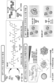
- Figure 1 is a diagram briefly showing the principles of amyloid peptide synthesis and screening of the present invention.
- Figure 2 is a diagram showing the results of analyzing fibrillation of amyloid peptide in one embodiment of the present invention.
- Figure 2(a) is the AFM (atomic force microscopy) image result of MCAP monomer
- Figure 2(b) is a graph showing the height of the white dotted line in Figure 2(a)
- Figure 2(c) is analysis through AFM graph.
- Figure 2(d) is the AFM image result of the fibrillated amyloid peptide
- Figure 2(e) is a graph showing the height of the white dotted line in Figure 2(d)
- Figure 2(f) is the AFM
- Figure 2(g) is a TEM (transmission electron microscope) image result of fibrillated amyloid peptide and an enlarged view thereof.
- Figure 3 is a diagram confirming the ⁇ -sheet structure of fibrillated amyloid peptide in one embodiment of the present invention.
- Figure 3(a) is the dynamic light scattering (DLS) analysis result
- Figure 3(b) is the CD (Circular dichroism) analysis result
- Figure 3(c) is the Thioflavin T fluorescence analysis result
- Figure 3(d) is the FT-IR analysis result. This diagram shows the analysis results.
- Figure 4 is a diagram confirming that the fibrillated amyloid peptide of the present invention is decomposed upon treatment with M pro .
- Figure 5 is a diagram analyzing the structural stability of engineered amyloid nanocomposite (MCAP-AuNP) in one embodiment of the present invention.
- Figure 5(a) is a diagram showing the absorbance peak according to the mixing amount of amyloid peptide solution.
- Figure 5(b) shows the results of UV-vis spectrum analysis over time of a nanocomposite prepared with 50 ⁇ L amyloid peptide solution.
- Figure 6 is a diagram analyzing the freeze-thaw performance of engineered amyloid nanocomposite (MCAP-AuNP) in one embodiment of the present invention.
- Figure 6(a) is the UV-vis spectrum analysis result before freezing
- Figure 6(b) is the UV-vis spectrum analysis result after freezing and thawing.
- Figures 6(c) and 6(d) show gold nanoparticles (bare AuNPs, left: before freezing, right: after freezing and thawing) and engineered amyloid nanocomposites (MCAP-AuNPs, left: frozen) before and after freezing and thawing, respectively.
- Figure 7 is a diagram analyzing the physical and chemical properties of gold nanoparticles (bare AuNP) and engineered amyloid nanocomposite (MCAP-AuNP) in one embodiment of the present invention.
- Figure 7(a) is the result of measuring the hydrodynamic diameter
- Figure 7(b) is the result of analyzing the zeta potential
- Figure 7(c) is the result of the gold nano-engineered amyloid nanocomposite (MCAP-AuNP).
- Figure 7(d) shows a TEM image of the engineered amyloid nanocomposite (MCAP-AuNP).
- Figure 8(a) is a diagram analyzing the UV-vis spectrum of engineered amyloid nanocomposite (MCAP-AuNP) according to M pro concentration in one embodiment of the present invention
- Figure 8(b) is a diagram showing the denatured M pro
- Figure 8 ( c) which shows the results of the same analysis using Figure 8(d) is a diagram showing the TEM image results of MCAP-AuNPs depending on whether M pro was modified or not treated with MCAP-AuNPs.
- FIG. 9 is a diagram showing the analysis of the UV-vis spectrum of engineered amyloid nanocomposite (MCAP-AuNP) by screening previously known M pro inhibitors in one embodiment of the present invention.
- MCAP-AuNP engineered amyloid nanocomposite
- Figure 10(a) is a diagram showing an image observed by TEM of the reaction of engineered amyloid nanocomposite (MCAP-AuNP) according to M pro treatment in one embodiment of the present invention.
- Figure 10(b) shows the reaction results according to leupeptin treatment
- Figure 10(c) shows the reaction results according to lopinavir treatment
- Figure 10(d) shows the reaction results according to hesperetin treatment.
- Figure 11 is a diagram showing the difference between engineered amyloid peptide (MCAP) and MCRP, an engineered peptide that does not contain an amyloid sequence, in one embodiment of the present invention.
- MCAP engineered amyloid peptide
- MCRP engineered peptide that does not contain an amyloid sequence
- FIG 12 is a diagram analyzing ThT signals of engineered amyloid peptide (MCAP) and MCRP in one embodiment of the present invention.
- MCAP engineered amyloid peptide
- Figure 13 is a diagram analyzing the enzyme activity of M pro according to treatment with an M pro inhibitor in one embodiment of the present invention.
- the present invention relates to an engineered amyloid nanocomposite (MCAP-AuNP) in which an amyloid peptide containing an M pro cleavage sequence (LQS) is coated on gold nanoparticles, which inhibits M pro , a proteolytic enzyme derived from coronavirus. It relates to a method for screening M pro inhibitors.
- M pro main protease
- the present invention can inhibit the proliferation of the virus by inhibiting the protease derived from the virus.
- the coronavirus refers to viruses belonging to the coronavirus family. Coronaviruses are known to cause respiratory or digestive infectious diseases depending on the characteristics of the virus and the host.
- the viral replication mechanism by M pro is shared by various types of coronaviruses (SARS-CoV, MERS-CoV, HCoV-HKU1, etc.).
- SARS-CoV coronaviruses
- MERS-CoV MERS-CoV
- HCoV-HKU1 coronaviruses
- Lyophilized main protease (M pro ) of SARS-CoV-2 was purchased from Biosynth Carbosynth (UK). ebselen, hesperetin, leupeptin, lopinavir, hesperidin, chlorauric acid trihydrate (HAuCl 4 3H 2 O) and ginseng citrate. Sodium (trisodium citrate) was purchased from Sigma-Aldrich (USA). Distilled water (DW) and phosphate buffered saline (PBS) were purchased from Gibco (USA).
- the main protease (M pro ) of SARS-CoV-2 cleaves the LQS, LQA, and LQG sequences.
- the prion protein-derived amyloid sequence (GNNQQNY) and the M pro cleavage sequence (LQS) were fused to produce an engineered amyloid peptide (M pro cleavage site embedded amyloid peptide, MCAP, LQGNLQSNQQNY, Peptron, Korea) was used (Figure 1).
- Engineered amyloid peptide (MCAP) containing prion protein-derived amyloid sequence (GNNQQNY) and M pro cleavage sequence (LQS) was dissolved in distilled water to prepare a 1 mg/mL MCAP solution.
- 50 ⁇ L of the MCAP solution and 150 ⁇ L of distilled water at pH 2 were mixed and fibrillated by sufficiently reacting at 1,000 rpm for 5 days in a shaking incubator (Eppendorf, Germany) at 37°C. Fibrillated amyloid peptide was confirmed as follows.
- AFM analysis was performed with NX10 (Park systems, South Korea) using a silicon tip (NCHR, Park Systems, South Korea) with a radius of less than 10 nm.
- AFM measurements were performed in NCM mode at a scan rate of 0.4 Hz and an image size of 5 ⁇ m ⁇ 5 ⁇ m.
- Image flattening and topological analysis were performed using Smart Scan (Park systems, South Korea) software. Images obtained through AFM analysis were used to measure the persistence length of fibrillated amyloid peptide using Easyworm software.
- Figure 2(a) shows an AFM image analyzing the persistence length of MCAP monomer.
- FIG. 2(b) is a diagram showing the height of the white dotted line in FIG. 2(a).
- Figure 2(c) shows the approximate average size of MCAP monomer analyzed through AFM images, which is 330.19 ⁇ 73.9 pm.
- Figure 2(d) shows an AFM image analyzing the persistence length of fibrillated amyloid peptide.
- Figure 2(e) is a diagram showing the height of the white dotted line in Figure 2(d).
- Figure 2(f) shows the approximate average size of fibrillated amyloid peptide analyzed through AFM images, which is 2.64 ⁇ 0.74 nm.
- TEM analysis was performed by reacting a 0.25 mg/mL MCAP solution at 37°C at 1,000 rpm for 96 hours, centrifuging at 12,000 rpm for 1 hour, and removing the supernatant.
- Figure 2g is a TEM analysis image and its enlarged shape. Through this, it was confirmed that fibrillation of MCAP had progressed.
- the hydrodynamic diameter was measured using dynamic light scattering (DLS) while reacting a 0.25 mg/mL MCAP solution at 37°C and 1,000 rpm for 3 days. 1 mL of the reacted MCAP solution was placed in a disposable cuvette (10 mm light path, standard type, Kartell, Italy) and measured with Zetasizer Nano S90 (Malvern Panalytical, United Kingdom) for 4 cycles, 10 times per cycle, for a total of 40 times. As shown in Figure 3(a), fibrillation progresses from the monomeric form of MCAP depending on the reaction time, and the diameter and standard deviation increase (1h: 0.20 ⁇ 0.073 ⁇ m, 12h: 0.30 ⁇ 0.03 ⁇ m, 24h: 0.98 ⁇ 0.13). ⁇ m, 36h: 3.40 ⁇ 1.37 ⁇ m, 48h: 3.62 ⁇ 2.02 ⁇ m).
- CD can confirm the secondary structure of a material, and in the form of a random coil, the y-axis value shows a negative value at a wavelength ⁇ 210 nm.
- the 0.25 mg/mL MCAP solution was reacted at 37°C and 1,000 rpm for 12, 24, 36, 48, 60, and 96 h, respectively, and then placed in a quartz glass cuvette (1 mm path length, 10 mm inside wide, Aireka Cells, USA). Measurements were taken after injection. Circular dichroism spectra were measured with J-815 (Jasco, Japan), which has a spectral detection range of 190-300 nm and a scanning speed of 10 nm/min.
- Spectra had a resolution of 8 nm and were processed with CDTool software.
- fibrillated amyloid peptide was confirmed to have a negative value at a wavelength of 210 to 240 nm, and a peak showing a positive value at a wavelength ⁇ 210 nm.
- the signal due to the ⁇ -sheet structure increased with reaction time, which means that MCAP is gradually fibrillated into a ⁇ -sheet structure.
- Thioflavin T is a fluorescent substance that detects ⁇ -sheet structures.
- a 0.25 mg/mL MCAP solution was reacted with a 20 ⁇ M ThT solution and a pH 2 solution at 37°C and 1,000 rpm for 3 days, and the ThT fluorescence signal was measured.
- ThT fluorescence signals were measured at 30-minute intervals for 99 hours. The experimental results are shown in Figure 3(c). The ThT fluorescence signal value increased with reaction time, indicating the formation of fibrillated amyloid peptide in the form of a ⁇ -sheet.
- Example 2 decomposition of fibrils was confirmed by reacting M pro with engineered amyloid peptide (MCAP). Fibrillated amyloid peptide was prepared by incubating 0.25 mg/mL MCAP solution at pH 2 and 37°C for 5 days. 0.01 mg/mL of M pro was reacted with the prepared fibrillated amyloid peptide for 12 hours.
- MCAP engineered amyloid peptide
- Gold nanoparticles were synthesized using citrate reduction. 2.5 mL of 38.8 mM chloroauric acid (HAuCl 4 ) solution and 45 mL Millipore water were mixed in a round beaker and heated while stirring at 1,200 rpm. After boiling the mixture, 1 mL of 80 mM sodium citrate solution was added and heated for 1 hour while stirring at 1,200 rpm to prepare gold nanoparticles stabilized with citric acid. Gold nanoparticles were manufactured with a size of 20 nm. The final gold nanoparticle solution was cooled to room temperature and stored at 4°C.
- a 0.1 mg/mL MCAP solution was prepared by adding a mixture of distilled water and PBS in a 10:1 volume ratio to MCAP.
- the 20 nm gold nanoparticle solution was centrifuged at 13,000 rpm for 20 minutes and the supernatant was removed.
- 40 ⁇ L of the gold nanoparticle solution was mixed with 25, 50, 100, 200, 300, and 400 ⁇ L of Example 2 (0.09 mg/mL), respectively, and reacted at 37°C and 1,000 rpm to synthesize an engineered amyloid nanocomposite.
- a salt resistance test was performed for 24 hours by adding 600 ⁇ L of 1 ⁇ PBS, and the UV-vis spectrum was measured.
- the UV-vis spectrum was measured with a spectrophotometer (scan range 400 ⁇ 800 nm, scan rate 600 nm/min, Perkin Elmer, USA). In the UV-vis spectrum of dispersed gold nanoparticles of 20 nm or less, a peak was detected around 525 nm (A 525 ). If the fibrillated amyloid peptide does not coat the surface of the gold nanoparticle well, the gold nanoparticles are aggregated by PBS, causing a color change in the solution, and the peak moves to near 650 nm (A 650 ). Therefore, the degree of aggregation of gold nanoparticles can be quantified by relative absorbance (A 650 /A 525 ).
- Example 5 the freeze-thaw performance of the gold nanoparticle solution (bare AuNP) prepared in Example 5 and the engineered amyloid nanocomposite (MCAP-AuNP) prepared with 50 ⁇ L MCAP solution was analyzed.
- Figure 6(a) shows the UV-vis spectrum measurement results before freezing
- Figure 6(b) shows the UV-vis spectrum measurement results after freezing and thawing.
- the peak change before and after freezing and in Figure 6(c) after freezing and thawing, aggregation of gold nanoparticles occurred and a color change was observed, whereas the engineered amyloid nanocomposite (MCAP-AuNP) had constant peaks before and after freezing.
- MCAP-AuNP engineered amyloid nanocomposite
- Example 3 the results of measuring the hydrodynamic diameters of bare AuNPs and MCAP-AuNPs using dynamic light scattering (DLS) are shown in FIG. 7(a). Bare AuNP was 19.01 ⁇ 0.53 nm, MCAP-AuNP was 21.96 ⁇ 0.67 nm, and MCAP-AuNP coated with engineered amyloid peptide was confirmed to be about 3 nm thicker than the gold nanoparticle before coating.
- DLS dynamic light scattering
- Figure 7(b) shows the zeta potential value of MCAP-AuNP.
- a 0.4 mg/mL MCAP-AuNP solution was placed in the Zetasizer Cuvette, and the accumulation time for each point was measured 15 times.
- the zeta potential of MCAP-AuNP was -19 ⁇ 3.1 mV, confirming that MCAP-AuNP had excellent structural stability.
- XPS X-ray photoelectron spectroscopy
- the range from 0 to 800 eV was scanned using a 40 eV pass energy with a 0.1 eV step size.
- the nitrogen (N) 1s peak was measured by the protein, and from this, it was confirmed that the surface of the gold nanoparticle of the engineered amyloid nanocomposite (MCAP-AuNP) was coated with MCAP solution, that is, amyloid peptide. I was able to confirm.
- FIG. 7(d) is a TEM image result of engineered amyloid nanocomposite (MCAP-AuNP). These results also indicate that engineered amyloid peptides are coated on the surface of the gold nanoparticles of the engineered amyloid nanocomposite (MCAP-AuNP).
- M pro was dissolved in PBS at various concentrations (0.925-18.5 nM) for 20 minutes to prepare each volume at 0.8 mL.
- the M pro solution of each concentration was mixed with the gold nanoparticle solution (bare AuNP) prepared in Example 5 and the engineered amyloid nanocomposite (MCAP-AuNP) prepared with 50 ⁇ L amyloid peptide solution, so that the total volume was 1 mL. It was made to be.
- the UV-vis spectrum was measured with a spectrophotometer.
- FIG. 8(a) As shown in Figure 8(a), as the M pro concentration increased, the UV peak shifted to the right. Meanwhile, as a result of denaturing M pro at 90°C for 4 hours and reacting in the same manner as above, it was confirmed that there was no change in the UV spectrum, as shown in Figure 8(b).
- a sigmoidal dose-response curve quantifying the degree of aggregation (A 650 /A 525 ) of MCAP-AuNPs depending on M pro denaturation is shown in Figure 8(c).
- Figure 8(d) is a TEM image result of MCAP-AuNP depending on whether or not M pro is modified.
- M pro 50 ng of M pro (1.4 nM) was added to M pro inhibitors (ebselen, hesperidin, hesperetin, lopinavir, leupeptin) dissolved in various concentrations in PBS (1.5% DSMO) to prepare a total solution volume of 800 ⁇ L.
- M pro inhibitors ebselen, hesperidin, hesperetin, lopinavir, leupeptin
- PBS 1.5% DSMO
- 600 ⁇ L of the final solution was added to 200 ⁇ L engineered amyloid nanocomposite (MCAP-AuNP) solution and reacted at 37°C for 1 hour. After reaction, the UV-vis spectrum was measured with a spectrophotometer. The degree of particle aggregation was analyzed by relative absorbance (A 650 /A 525 ).
- the IC 50 values of each drug were confirmed to be 0.39 ⁇ M for ebselen, 43.14 ⁇ M for hesperetin, 28.57 ⁇ M for leupeptin, 10.7 ⁇ M for lopinavir, and 369.4 ⁇ M for hesperidin.
- M pro inhibitors (leupeptin, lopinavir, hesperetin) dissolved at various concentrations in PBS (10% DSMO) to increase the total volume of the solution. Prepared in 500 ⁇ L. The mixture was reacted at room temperature for 20 minutes and then filtered through a 200 ⁇ m PVDF syringe filter (Biopil, China) to remove unreacted substances. Afterwards, 70 ⁇ L of the mixture was added to MCAP solutions of 1.88, 3.7, 5.66, 7.55, 9.43, and 11.32 ⁇ M to prepare a final volume of 220 ⁇ L.
- the relative absorbance (A 650 /A 525 ) by the M pro inhibitor according to the concentration of the engineered amyloid nanocomposite (MCAP-AuNP) solution was analyzed using a microplate reader (Molecular Device, USA). As shown in Figure 13, enzyme activity according to inhibitor treatment was analyzed using the Michaelis-Menten equation.
- Figure 10 shows an image observed by TEM of the reaction of the engineered amyloid nanocomposite (MCAP-AuNP) according to M pro treatment.
- Figure 10(a) shows the engineered amyloid nanocomposite (MCAP-AuNP) reacted with M pro
- Figure 10(b) shows the reaction results according to leupeptin treatment
- Figure 10(c) shows the reaction results according to lopinavir treatment
- Figure 10(d) shows an image observed by TEM of the reaction results according to hesperetin treatment.
- a peptide with the LQSLQALQGLQSS sequence (M pro cleavage-site-embedded repeated peptide, MCRP) was prepared by linking LQS, LQA, and LQG, which are sequences cleaved by M pro .
- Figures 11(a) and 11(b) show the differences from the engineered amyloid peptide (M pro cleavage site embedded amyloid peptide, MCAP, LQGNLQSNQQNY) of the present invention.
- a nanocomposite was prepared by mixing 50 ⁇ L MCRP solution and 40 ⁇ L gold nanoparticle solution, and 600 ⁇ L of 1 ⁇ PBS was added to perform a salt resistance test for 24 hours to measure the UV-vis spectrum.
- the results are shown in Figure 11(c).
- MCRP was unable to coat the surface of the gold nanoparticles, and the absorbance shifted to the right as the gold nanoparticles aggregated.
- MCRP did not have the amyloid properties of ⁇ -sheet, so it could not coat the surface of gold nanoparticles.
Landscapes
- Life Sciences & Earth Sciences (AREA)
- Chemical & Material Sciences (AREA)
- Health & Medical Sciences (AREA)
- Engineering & Computer Science (AREA)
- Immunology (AREA)
- Organic Chemistry (AREA)
- Proteomics, Peptides & Aminoacids (AREA)
- Molecular Biology (AREA)
- Physics & Mathematics (AREA)
- General Health & Medical Sciences (AREA)
- Analytical Chemistry (AREA)
- Zoology (AREA)
- Biochemistry (AREA)
- Wood Science & Technology (AREA)
- Biotechnology (AREA)
- Microbiology (AREA)
- Biomedical Technology (AREA)
- Hematology (AREA)
- Urology & Nephrology (AREA)
- Bioinformatics & Cheminformatics (AREA)
- General Physics & Mathematics (AREA)
- Pathology (AREA)
- General Engineering & Computer Science (AREA)
- Biophysics (AREA)
- Genetics & Genomics (AREA)
- Medicinal Chemistry (AREA)
- Food Science & Technology (AREA)
- Cell Biology (AREA)
- Virology (AREA)
- Chemical Kinetics & Catalysis (AREA)
- Plasma & Fusion (AREA)
- Peptides Or Proteins (AREA)
- Investigating Or Analysing Biological Materials (AREA)
Abstract
Description
| 서열번호 1 | GNNQQNY |
| 서열번호 2 | LQS |
Claims (8)
- 서열번호 1 및 서열번호 2로 표시되는 서열을 포함하는 엔지니어드 아밀로이드 펩타이드를 제조하는 단계 (a);염화금산(HAuCl4)과 시트르산 나트륨을 혼합하여 금나노입자를 제조하는 단계 (b); 및상기 단계 (a)의 아밀로이드 펩타이드와 상기 단계 (b)의 금나노입자를 혼합하여 엔지니어드 아밀로이드 나노복합체를 제조하는 단계 (c);를 포함하는 코로나바이러스 치료제 스크리닝용 조성물의 제조방법.
- 제1항에 있어서,상기 단계 (a)의 엔지니어드 아밀로이드 펩타이드는 β-sheet인 것인, 코로나바이러스 치료제 스크리닝용 조성물의 제조방법.
- 제1항에 있어서,상기 단계 (b)는, 염화금산과 초순수를 혼합하여 가열한 뒤, 시트르산 나트륨을 첨가하여 가열하는 것인, 코로나바이러스 치료제 스크리닝용 조성물의 제조방법.
- 제1항에 있어서,상기 단계 (c)의 엔지니어드 아밀로이드 나노복합체는,금나노입자와 엔지니어드 아밀로이드 펩타이드를 부피비 1 : 0.5~10으로 혼합하여 제조된 것인, 코로나바이러스 치료제 스크리닝용 조성물의 제조방법.
- 제1항에 있어서,상기 단계 (c)의 엔지니어드 아밀로이드 나노복합체는 붉은색인, 코로나바이러스 치료제 스크리닝용 조성물의 제조방법.
- 제1항의 제조방법에 의해 제조된 코로나바이러스 치료제 스크리닝용 조성물.
- 제6항의 조성물에 코로나바이러스 치료제 후보 약물을 처리하는 단계;를 포함하는 코로나바이러스 치료제 스크리닝 방법.
- 제7항에 있어서,후보 약물 처리 후 푸른색으로 색이 변하는 경우 상기 후보 약물을 코로나바이러스 치료제로 선정하는 단계;를 더욱 포함하는 것인, 코로나바이러스 치료제 스크리닝 방법.
Priority Applications (1)
| Application Number | Priority Date | Filing Date | Title |
|---|---|---|---|
| US18/860,404 US20250298028A1 (en) | 2022-04-28 | 2022-11-23 | Method for screening drugs for treating multiple-variant coronavirus disease |
Applications Claiming Priority (2)
| Application Number | Priority Date | Filing Date | Title |
|---|---|---|---|
| KR1020220053001A KR102834342B1 (ko) | 2022-04-28 | 2022-04-28 | 다중 변이 코로나바이러스 치료용 약물 스크리닝 방법 |
| KR10-2022-0053001 | 2022-04-28 |
Publications (1)
| Publication Number | Publication Date |
|---|---|
| WO2023210899A1 true WO2023210899A1 (ko) | 2023-11-02 |
Family
ID=88519137
Family Applications (1)
| Application Number | Title | Priority Date | Filing Date |
|---|---|---|---|
| PCT/KR2022/018583 Ceased WO2023210899A1 (ko) | 2022-04-28 | 2022-11-23 | 다중 변이 코로나바이러스 치료용 약물 스크리닝 방법 |
Country Status (3)
| Country | Link |
|---|---|
| US (1) | US20250298028A1 (ko) |
| KR (1) | KR102834342B1 (ko) |
| WO (1) | WO2023210899A1 (ko) |
Citations (2)
| Publication number | Priority date | Publication date | Assignee | Title |
|---|---|---|---|---|
| WO2021160688A1 (en) * | 2020-02-10 | 2021-08-19 | Instituto de Medicina Molecular João Lobo Antunes | Biomaterials |
| WO2021198228A1 (en) * | 2020-03-30 | 2021-10-07 | Original G B.V. | Diagnostic peptide for use in a method of diagnosis of viral infection, kit and system |
Family Cites Families (1)
| Publication number | Priority date | Publication date | Assignee | Title |
|---|---|---|---|---|
| KR20220002156A (ko) | 2020-06-30 | 2022-01-06 | 포항공과대학교 산학협력단 | 코로나-19 바이러스의 스파이크 단백질의 에스터가수분해효소 활성을 이용한 코로나 바이러스 감염증 치료제 스크리닝 키트 및 상기 키트를 이용하여 스크리닝된 물질을 포함하는 코로나 바이러스 감염증의 예방 또는 치료용 약학 조성물 |
-
2022
- 2022-04-28 KR KR1020220053001A patent/KR102834342B1/ko active Active
- 2022-11-23 US US18/860,404 patent/US20250298028A1/en active Pending
- 2022-11-23 WO PCT/KR2022/018583 patent/WO2023210899A1/ko not_active Ceased
Patent Citations (2)
| Publication number | Priority date | Publication date | Assignee | Title |
|---|---|---|---|---|
| WO2021160688A1 (en) * | 2020-02-10 | 2021-08-19 | Instituto de Medicina Molecular João Lobo Antunes | Biomaterials |
| WO2021198228A1 (en) * | 2020-03-30 | 2021-10-07 | Original G B.V. | Diagnostic peptide for use in a method of diagnosis of viral infection, kit and system |
Non-Patent Citations (3)
| Title |
|---|
| EZZAT KARIEM, PERNEMALM MARIA, PÅLSSON SANDRA, ROBERTS THOMAS C., JÄRVER PETER, DONDALSKA ALEKSANDRA, BESTAS BURCU, SOBKOWIAK MICH: "The viral protein corona directs viral pathogenesis and amyloid aggregation", NATURE COMMUNICATIONS, vol. 10, no. 1, 27 May 2019 (2019-05-27), pages 2331, XP093103250, DOI: 10.1038/s41467-019-10192-2 * |
| JIN ZHICHENG, YEUNG JUSTIN, ZHOU JIAJING, CHENG YONG, LI YI, MANTRI YASH, HE TENGYU, YIM WONJUN, XU MING, WU ZHUOHONG, FAJTOVA PAV: "Peptidic Sulfhydryl for Interfacing Nanocrystals and Subsequent Sensing of SARS-CoV-2 Protease", CHEMISTRY OF MATERIALS, AMERICAN CHEMICAL SOCIETY, US, vol. 34, no. 3, 8 February 2022 (2022-02-08), US , pages 1259 - 1268, XP093103254, ISSN: 0897-4756, DOI: 10.1021/acs.chemmater.1c03871 * |
| PRAMANIK AVIJIT, GAO YE, PATIBANDLA SHAMILY, GATES KALEIN, RAY PARESH CHANDRA: "Bioconjugated Nanomaterial for Targeted Diagnosis of SARS-CoV-2", ACCOUNTS OF MATERIALS RESEARCH, vol. 3, no. 2, 25 February 2022 (2022-02-25), pages 134 - 148, XP093103720, ISSN: 2643-6728, DOI: 10.1021/accountsmr.1c00177 * |
Also Published As
| Publication number | Publication date |
|---|---|
| US20250298028A1 (en) | 2025-09-25 |
| KR102834342B1 (ko) | 2025-07-18 |
| KR20230153147A (ko) | 2023-11-06 |
Similar Documents
| Publication | Publication Date | Title |
|---|---|---|
| KR100700483B1 (ko) | 단백질의 빠른 탈수법 | |
| JP4536918B2 (ja) | 免疫調整機能を有するγ−グルタミルとβ−アスパルチル化合物および免疫調節剤 | |
| Qiu et al. | Auricularia auriculajudae polysaccharide-cisplatin complexes conjugated with folic acid as new tumor targeting agents | |
| CN115745830A (zh) | 联萘二甲酸衍生物及其制备方法和应用 | |
| WO2025190329A1 (zh) | Aβ药物筛选靶标及筛选方法 | |
| CN115120608A (zh) | 一种siRNA药物、药物组合物、siRNA-小分子药物偶联物及其应用 | |
| US9393286B2 (en) | Pan-antiviral peptides and uses thereof | |
| Liao et al. | An in situ transformable orally administrated system for targeted intestinal barrier protection and inflammatory microenvironment modulation in sepsis therapy | |
| CN117205176A (zh) | 一种荷载中药的工程化外泌体纳米粒及其制备方法和应用 | |
| CN114573660B (zh) | 一种自组装短肽及其诱导局部麻醉药生物矿化制得的长效局部麻醉和镇痛药物制剂 | |
| Taheri-Ledari et al. | An effective antimicrobial complex of nanoscale β-cyclodextrin and ciprofloxacin conjugated to a cell adhesive dipeptide | |
| KR102834342B1 (ko) | 다중 변이 코로나바이러스 치료용 약물 스크리닝 방법 | |
| CN113274484A (zh) | 小扁豆凝集素在制备预防和治疗冠状病毒引起的感染性疾病的药物中的应用 | |
| CN118845775A (zh) | Iacs-010759在制备预防和/或治疗原发性纤毛不动综合征的药物中的用途 | |
| Liu et al. | Machine learning-assisted identification of potential cancer inhibitors: Multifunctional biomineralized CaCO3-polyethyleneimine nanoparticle carriers and their application in lung cancer therapy | |
| Ling et al. | Sevoflurane exposure in postnatal rats induced long-term cognitive impairment through upregulating caspase-3/cleaved-poly (ADP-ribose) polymerase pathway | |
| Jin et al. | Nose‐to‐Brain Delivery of Chlorella vulgaris Extracellular Vesicles for Antidepressant Effects | |
| CN118059248A (zh) | 一种纳米铁补充剂及其制备方法和在制备抗抑郁与增强认知效应制剂中的应用 | |
| Ye et al. | Synergistic polyphenol–amino acid nanoparticles: a new strategy for reactive oxygen species management | |
| Attar et al. | Electron microscopic study of the progeny of ependymal stem cells in the normal and injured spinal cord | |
| WO2022255827A1 (ko) | 알파시뉴클레인-금나노입자 복합체, 이의 제조방법 및 이를 이용한 약물 스크리닝 방법 | |
| CN116570724A (zh) | 一种基于肿瘤微环境激活型高尔基体靶向干扰剂的纳米粒 | |
| CN103772487B (zh) | 一种抑制人胰淀素聚集或毒性的多肽、试剂及其应用 | |
| WO2017051989A1 (en) | Method of selectively releasing nitrogen monoxide using mesoporous core-shell nanoparticle and calcium phosphate | |
| WO2012133997A1 (ko) | 폴리소르비톨계 삼투압적 활성 전달체 및 이를 이용한 유전자 치료 |
Legal Events
| Date | Code | Title | Description |
|---|---|---|---|
| 121 | Ep: the epo has been informed by wipo that ep was designated in this application |
Ref document number: 22940393 Country of ref document: EP Kind code of ref document: A1 |
|
| WWE | Wipo information: entry into national phase |
Ref document number: 18860404 Country of ref document: US |
|
| NENP | Non-entry into the national phase |
Ref country code: DE |
|
| 122 | Ep: pct application non-entry in european phase |
Ref document number: 22940393 Country of ref document: EP Kind code of ref document: A1 |
|
| WWP | Wipo information: published in national office |
Ref document number: 18860404 Country of ref document: US |