WO2023199943A1 - コロナウイルス感染症を処置することに用いられる抗体 - Google Patents
コロナウイルス感染症を処置することに用いられる抗体 Download PDFInfo
- Publication number
- WO2023199943A1 WO2023199943A1 PCT/JP2023/014855 JP2023014855W WO2023199943A1 WO 2023199943 A1 WO2023199943 A1 WO 2023199943A1 JP 2023014855 W JP2023014855 W JP 2023014855W WO 2023199943 A1 WO2023199943 A1 WO 2023199943A1
- Authority
- WO
- WIPO (PCT)
- Prior art keywords
- seq
- amino acid
- acid sequence
- antibody
- set forth
- Prior art date
- Legal status (The legal status is an assumption and is not a legal conclusion. Google has not performed a legal analysis and makes no representation as to the accuracy of the status listed.)
- Ceased
Links
Images













Classifications
-
- C—CHEMISTRY; METALLURGY
- C07—ORGANIC CHEMISTRY
- C07K—PEPTIDES
- C07K16/00—Immunoglobulins [IG], e.g. monoclonal or polyclonal antibodies
- C07K16/40—Immunoglobulins [IG], e.g. monoclonal or polyclonal antibodies against enzymes
-
- A—HUMAN NECESSITIES
- A61—MEDICAL OR VETERINARY SCIENCE; HYGIENE
- A61P—SPECIFIC THERAPEUTIC ACTIVITY OF CHEMICAL COMPOUNDS OR MEDICINAL PREPARATIONS
- A61P31/00—Antiinfectives, i.e. antibiotics, antiseptics, chemotherapeutics
- A61P31/12—Antivirals
- A61P31/14—Antivirals for RNA viruses
-
- C—CHEMISTRY; METALLURGY
- C12—BIOCHEMISTRY; BEER; SPIRITS; WINE; VINEGAR; MICROBIOLOGY; ENZYMOLOGY; MUTATION OR GENETIC ENGINEERING
- C12N—MICROORGANISMS OR ENZYMES; COMPOSITIONS THEREOF; PROPAGATING, PRESERVING, OR MAINTAINING MICROORGANISMS; MUTATION OR GENETIC ENGINEERING; CULTURE MEDIA
- C12N9/00—Enzymes; Proenzymes; Compositions thereof; Processes for preparing, activating, inhibiting, separating or purifying enzymes
- C12N9/14—Hydrolases (3)
- C12N9/48—Hydrolases (3) acting on peptide bonds (3.4)
- C12N9/50—Proteinases, e.g. Endopeptidases (3.4.21-3.4.25)
- C12N9/64—Proteinases, e.g. Endopeptidases (3.4.21-3.4.25) derived from animal tissue
- C12N9/6421—Proteinases, e.g. Endopeptidases (3.4.21-3.4.25) derived from animal tissue from mammals
- C12N9/6424—Serine endopeptidases (3.4.21)
-
- A—HUMAN NECESSITIES
- A61—MEDICAL OR VETERINARY SCIENCE; HYGIENE
- A61K—PREPARATIONS FOR MEDICAL, DENTAL OR TOILETRY PURPOSES
- A61K39/00—Medicinal preparations containing antigens or antibodies
- A61K2039/505—Medicinal preparations containing antigens or antibodies comprising antibodies
-
- C—CHEMISTRY; METALLURGY
- C07—ORGANIC CHEMISTRY
- C07K—PEPTIDES
- C07K2317/00—Immunoglobulins specific features
- C07K2317/20—Immunoglobulins specific features characterized by taxonomic origin
- C07K2317/24—Immunoglobulins specific features characterized by taxonomic origin containing regions, domains or residues from different species, e.g. chimeric, humanized or veneered
-
- C—CHEMISTRY; METALLURGY
- C07—ORGANIC CHEMISTRY
- C07K—PEPTIDES
- C07K2317/00—Immunoglobulins specific features
- C07K2317/30—Immunoglobulins specific features characterized by aspects of specificity or valency
- C07K2317/33—Crossreactivity, e.g. for species or epitope, or lack of said crossreactivity
-
- C—CHEMISTRY; METALLURGY
- C07—ORGANIC CHEMISTRY
- C07K—PEPTIDES
- C07K2317/00—Immunoglobulins specific features
- C07K2317/30—Immunoglobulins specific features characterized by aspects of specificity or valency
- C07K2317/34—Identification of a linear epitope shorter than 20 amino acid residues or of a conformational epitope defined by amino acid residues
-
- C—CHEMISTRY; METALLURGY
- C07—ORGANIC CHEMISTRY
- C07K—PEPTIDES
- C07K2317/00—Immunoglobulins specific features
- C07K2317/50—Immunoglobulins specific features characterized by immunoglobulin fragments
- C07K2317/55—Fab or Fab'
-
- C—CHEMISTRY; METALLURGY
- C07—ORGANIC CHEMISTRY
- C07K—PEPTIDES
- C07K2317/00—Immunoglobulins specific features
- C07K2317/70—Immunoglobulins specific features characterized by effect upon binding to a cell or to an antigen
- C07K2317/73—Inducing cell death, e.g. apoptosis, necrosis or inhibition of cell proliferation
- C07K2317/732—Antibody-dependent cellular cytotoxicity [ADCC]
-
- C—CHEMISTRY; METALLURGY
- C07—ORGANIC CHEMISTRY
- C07K—PEPTIDES
- C07K2317/00—Immunoglobulins specific features
- C07K2317/70—Immunoglobulins specific features characterized by effect upon binding to a cell or to an antigen
- C07K2317/76—Antagonist effect on antigen, e.g. neutralization or inhibition of binding
-
- C—CHEMISTRY; METALLURGY
- C07—ORGANIC CHEMISTRY
- C07K—PEPTIDES
- C07K2317/00—Immunoglobulins specific features
- C07K2317/90—Immunoglobulins specific features characterized by (pharmaco)kinetic aspects or by stability of the immunoglobulin
- C07K2317/92—Affinity (KD), association rate (Ka), dissociation rate (Kd) or EC50 value
Definitions
- the present invention relates to antibodies used to treat coronavirus infections.
- the coronavirus pandemic has had a major impact on the global economy.
- the threat of coronavirus infections seemed to have subsided for a time.
- variants of Coronaiurus have emerged and are rapidly replacing previous original strains and variants.
- the alpha strain was identified in the UK in September 2020.
- the ⁇ strain was confirmed in South America in May 2020.
- the ⁇ strain was confirmed in Brazil in November 2020.
- the ⁇ strain was confirmed in India in October 2020.
- the Omicron strain was confirmed in multiple countries.
- the ⁇ strain was confirmed in Peru in December 2020, and the ⁇ strain was confirmed in Colombia in January 2021.
- VOC Variants of Concern
- VOI Variants of Interest
- mRNA vaccine encoding the spike (S) protein of the coronavirus has been administered by intramuscular injection all over the world, and its extremely high effectiveness has been confirmed.
- This mRNA vaccine contains as an active ingredient lipid nanoparticles (LNP) encapsulating mRNA containing pseudouridine (Patent Documents 1 and 2).
- the present invention provides antibodies against coronaviruses that are expected to be effective regardless of virus mutations.
- TMPRSS2 a human protein required for activation of the S protein used by coronaviruses for infection, can broadly prevent coronavirus infection regardless of the mutant strain. It revealed that.
- the antibody or antigen-binding fragment thereof according to [1].
- Binds to human TMPRSS2 having the amino acid sequence set forth in SEQ ID NO: 5 ( ⁇ ) The chimeric TMPRSS2 has an amino acid sequence in which the amino acid sequence of human TMPRSS2 (WNENYGRAACRDMGYKNNFY) set forth in SEQ ID NO: 75 is replaced with the corresponding sequence of cat (i.e., the amino acid sequence set forth in SEQ ID NO: 94: WSEGYGRAACQDMGYRNSFY).
- the chimeric TMPRSS2 has an amino acid sequence in which the amino acid sequence of the human TMPRSS2 set forth in SEQ ID NO: 89 (VTAAHCVEKPLNNPWHWT) is replaced with the corresponding sequence of cat (i.e., the amino acid sequence set forth in SEQ ID NO: 95: VTAAHCVEEPLNNPRHWT).
- HCDR1 having the amino acid sequence set forth in SEQ ID NO: 27, HCDR2 having the amino acid sequence set forth in SEQ ID NO: 28, and HCDR3 having the amino acid sequence set forth in SEQ ID NO: 29.
- a heavy chain variable region having LCDR1 having the amino acid sequence set forth in SEQ ID NO: 30, LCDR2 having the amino acid sequence set forth in SEQ ID NO: 31, and LCDR3 having the amino acid sequence set forth in SEQ ID NO: 32.
- HCDR1 having the amino acid sequence set forth in SEQ ID NO: 35
- HCDR2 having the amino acid sequence set forth in SEQ ID NO: 36
- HCDR3 having the amino acid sequence set forth in SEQ ID NO: 37
- a heavy chain variable region having LCDR1 having the amino acid sequence set forth in SEQ ID NO: 38
- LCDR2 having the amino acid sequence set forth in SEQ ID NO: 39
- LCDR3 having the amino acid sequence set forth in SEQ ID NO: 40.
- HCDR1 having the amino acid sequence set forth in SEQ ID NO: 100
- HCDR2 having the amino acid sequence set forth in SEQ ID NO: 101
- HCDR3 having the amino acid sequence set forth in SEQ ID NO: 102.
- a heavy chain variable region having LCDR1 having the amino acid sequence set forth in SEQ ID NO: 103, LCDR2 having the amino acid sequence set forth in SEQ ID NO: 104, and LCDR3 having the amino acid sequence set forth in SEQ ID NO: 105.
- HCDR1 having the amino acid sequence set forth in SEQ ID NO: 108
- HCDR2 having the amino acid sequence set forth in SEQ ID NO: 109
- HCDR3 having the amino acid sequence set forth in SEQ ID NO: 110.
- a heavy chain variable region having LCDR1 having the amino acid sequence set forth in SEQ ID NO: 111, LCDR2 having the amino acid sequence set forth in SEQ ID NO: 112, and LCDR3 having the amino acid sequence set forth in SEQ ID NO: 113.
- an antibody or an antigen-binding fragment thereof having a light chain variable region (7)
- a composition comprising the antibody or antigen-binding fragment thereof according to any one of [1] to [8] above.
- composition according to [9] above for use in preventing infection by coronavirus in a subject in need thereof.
- [21] The antibody or antigen-binding fragment thereof according to any one of [1] to [8] above, which is a humanized antibody.
- [22] The antibody or antigen-binding fragment thereof according to any one of [1] to [8] above, which is a human chimeric antibody.
- a composition comprising the antibody or antigen-binding fragment thereof according to [21] or [22] above.
- the composition according to [23] above for use in preventing infection by coronavirus in a subject in need thereof.
- [26] The composition according to [23] above, for use in treating coronavirus infection in a subject infected with betacoronavirus.
- coronavirus is a betacoronavirus.
- coronavirus is SARS-CoV, MERS, or SARS-CoV-2.
- coronavirus is one or more selected from the alpha strain, beta strain, gamma strain, delta strain, and omicron strain of SARS-CoV-2.
- TMPRSS2 is human TMPRSS2 and the cells are human cells.
- TMPRSS2 is human TMPRSS2 and the cells are human cells.
- TMPRSS2 is human TMPRSS2 and the cells are human cells.
- TMPRSS2 is human TMPRSS2 and the cells are human cells.
- [121] The antibody or antigen-binding fragment thereof according to [1] above, wherein the antibody or antigen-binding fragment has a binding affinity (KD) for human TMPRSS2 of 10 ⁇ 8 M or less, and is An antibody or an antigen-binding fragment thereof having an IC 90 for inhibiting infection of human cells of 10 ⁇ g/mL or less.
- KD binding affinity
- SEQ ID NO: 85 An antibody or an antigen-binding fragment thereof that binds to the amino acid sequence (GVYGNVMVFTDWIY) described in .
- An antibody that binds to the extracellular domain of TMPRSS2 is capable of inhibiting coronavirus infection of cells, and is capable of inhibiting the action of TMPRSS2 on S protein, or an antigen-binding antibody thereof sexual fragment.
- An antibody that binds to the extracellular domain of TMPRSS2 is capable of inhibiting coronavirus infection of cells, and is capable of inhibiting the interaction of TMPRSS2 with the S protein, or its antigen Associative fragment.
- a composition comprising any of the above antibodies or antigen-binding fragments thereof.
- the composition according to [151] above which is used for inhibiting coronavirus infection of cells.
- the composition according to [151] above which is used for preventing coronavirus infection.
- the composition according to [151] above which is used to treat a subject infected with a coronavirus.
- [161] A method of treating a subject, the method comprising administering to the subject an effective amount of any of the above antibodies or antigen-binding fragments thereof.
- [162] The method according to [161] above, wherein the subject has a coronavirus.
- [163] The method according to [161] or [162] above, wherein the administration suppresses further infection of coronavirus cells in the subject's body.
- a method of treating a subject infected with a coronavirus the method comprising administering to the subject an effective amount of any of the above antibodies or antigen-binding fragments thereof.
- FIG. 1 shows that TMPRSS2, which is essential for betacoronavirus cell infection, is expressed on the membrane surface of infected cells (host cells), and that the antibody of the present invention is TMPRSS2, which is expressed on the membrane surface of infected cells. Indicates that it is combined with Since the antibodies of the present invention bind to antigens expressed in host cells, they are theoretically unaffected by viral mutations.
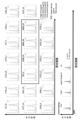
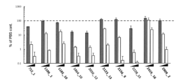
- FIG. 2 shows the binding to human TMPRSS2 of the monoclonal antibody that binds to human TMPRSS2 newly obtained in the example. Human TMPRSS2 was expressed in the Daudi cell line, and binding between the antibody and cells was detected by flow cytometry. Data for antibodies that showed particularly good binding are enclosed in a square frame.
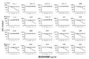
- FIG. 3A shows the results of a test system for testing whether the monoclonal antibody that binds to human TMPRSS2 newly obtained in the example competes with the 752_1 antibody for binding to human TMPRSS2.
- FIG. 3B shows the results of a test system to test whether the monoclonal antibody that binds to human TMPRSS2 newly obtained in the example competes with the 2228_15 antibody for binding to human TMPRSS2.
- FIG. 3C shows the results of a test system to test whether the monoclonal antibody that binds to human TMPRSS2 newly obtained in the example competes with the 1831_15 antibody for binding to human TMPRSS2.
- FIG. 3D shows the results of a test system to test whether the monoclonal antibody that binds to human TMPRSS2 newly obtained in the example competes with the 1864_10 antibody for binding to human TMPRSS2.
- FIG. 4 shows data showing the binding properties of various monoclonal antibodies to a 20 amino acid long peptide designed by shifting two amino acids each over the entire region of human TMPRSS2.
- FIG. 5A shows the binding properties of each monoclonal antibody to each peptide.
- FIG. 5B shows the binding of each monoclonal antibody to each peptide.
- Figure 6 shows the epitope regions of antibodies on human TMPRSS2 predicted from the results of Figures 5A and 6B.
- FIG. 7A shows data showing the binding properties of each monoclonal antibody to human TMPRSS2 variants.
- FIG. 7B shows data showing the binding properties of each monoclonal antibody to human TMPRSS2 variants.
- FIG. 7C shows data showing the binding properties of each monoclonal antibody to human TMPRSS2 variants.
- FIG. 7D shows data showing the binding properties of each monoclonal antibody to human TMPRSS2 variants.
- FIG. 8 shows a three-dimensional structure model of the epitope region obtained by observing the complex of the Fab fragment of the 752_1 antibody, the Fab fragment of the 2228 antibody, and human TMPRSS2 by cryo-electron microscopy.
- the string-like graphic shows human TMPRSS2, and the surface model drawn in the back shows the antibody.
- FIG. 9 shows the binding properties of the various antibodies obtained to other TMPRSS family members.
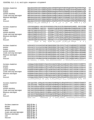
- FIG. 10A shows the alignment results of the amino acid sequences of TMPRSS2 of each animal species.
- FIG. 10B shows the alignment results of the amino acid sequences of TMPRSS2 of each animal species.
- FIG. 10C shows the binding of each monoclonal antibody to mouse, cynomolgus monkey, or human TMPRSS2.
- FIG. 10D shows the binding of each monoclonal antibody to mouse or human TMPRSS2.
- FIG. 10A shows the alignment results of the amino acid sequences of TMPRSS2 of each animal species.
- FIG. 10B shows the alignment results of the amino acid sequences of TMPRSS2 of each animal species.
- FIG. 10C shows the binding of each monoclonal antibody to mouse, cynomolgus monkey, or human TMPRSS2.
- FIG. 11A shows the binding to human TMPRSS2 of a human chimeric antibody obtained by replacing the Fc region of the obtained monoclonal antibody with the Fc region of human IgG4.
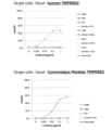
- FIG. 11B shows the binding to human and cynomolgus monkey TMPRSS2 of a human chimeric antibody derived from the 752_1 antibody obtained by replacing the Fc region with the Fc region of human IgG4.
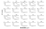
- FIG. 12 shows the effect of inhibiting virus infection by the various monoclonal antibodies obtained.
- FIG. 13A shows the infection-inhibiting effects of the obtained various monoclonal antibodies against various mutant viruses.
- FIG. 13B shows the infection inhibiting effects of the obtained various monoclonal antibodies against various mutant viruses.
- FIG. 13A shows the infection-inhibiting effects of the obtained various monoclonal antibodies against various mutant viruses.
- FIG. 13B shows the infection inhibiting effects of the obtained various monoclonal antibodies against various mutant viruses.
- FIG. 13C shows the infection-inhibiting effects of the obtained various monoclonal antibodies against various mutant viruses.
- FIG. 13D shows the infection inhibiting effects of the obtained various monoclonal antibodies against various mutant viruses.
- FIG. 14 shows the inhibitory effect on cell fusion (fusion of TMPRSS2/ACE-2-expressing 293T cells and spike protein-expressing 293T cells) by the various monoclonal antibodies obtained. Inhibition of cell fusion suggests that virus entry into cells is inhibited.
- Figure 15 shows the measurement results of antibody-dependent cytotoxicity (ADCC) of the obtained recombinant antibody with antibody-dependent enhancement (ADE)-deficient substitution (L244A/E245A/P339A) in the Fc region of the monoclonal antibody. show.
- FIG. 16 shows the measurement results of antibody-dependent cytotoxicity (ADCC) of the obtained human chimeric antibody.
- FIG. 17 shows the results of a SARS-CoV-2 infection inhibition experiment on cells expressing human or macaque TMPRSS2.
- FIG. 18A shows an experimental scheme for the inhibitory effect of antibody administration on SARS-CoV-2 infection of cynomolgus monkeys.
- Figure 18B shows body temperature and weight changes of individuals after administration of antibodies against infection of cynomolgus monkeys with SARS-CoV-2.
- FIG. 19A shows the measurement results (by the titration method) of the amount of virus in the respiratory tract swab and bronchial swab of the cynomolgus monkey.
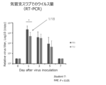
- FIG. 19B shows the measurement results (by RT-PCR method) of the amount of virus in the bronchial swab of the cynomolgus monkey.
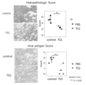
- FIG. 20 shows the lung histopathological diagnosis score by hematoxylin and eosin (HE) staining of the cynomolgus monkey and the results of immunohistochemical staining of SARS-CoV-2 N protein.
- FIG. 21 shows the results of a binding test of the prepared humanized antibody to TMPRSS2-expressing cells.
- FIG. 22 shows the results of an experiment to inhibit infection of pseudoviruses into cells using the prepared humanized antibodies.
- a "subject” is a vertebrate, e.g., a mammal, including a human, e.g., a mammal infected with a coronavirus (e.g., SARS-CoV-2) (cats, ferrets, bats, and pangolins). It can be.
- a coronavirus e.g., SARS-CoV-2
- the subject can be a subject infected with a coronavirus (e.g., SARS-CoV-2), can be an asymptomatic carrier infected with a coronavirus (e.g., SARS-CoV-2), can be an asymptomatic carrier infected with a coronavirus (e.g., The subject may be infected with SARS-CoV-2) and develop COVID-19.
- a coronavirus e.g., SARS-CoV-2
- SARS-CoV-2 a coronavirus
- SARS-CoV-2 a coronavirus
- the subject may be infected with SARS-CoV-2) and develop COVID-19.
- the subject can be a subject who has a possibility (risk) of being infected with a coronavirus (e.g., SARS-CoV-2) or a subject who has a possibility (risk) of being infected with a coronavirus (e.g., SARS-CoV-2)
- a coronavirus e.g., SARS-CoV-2
- It can be an object that has The target can be a child (for example, an infant (1-6 years old), a schoolchild (6-12 years old), an adolescent (12 years old and up), an adult (20 years old and up).
- Adults are 30 years old and older. , 40 years of age or older, 50 years of age or older, 60 years of age or older, or 70 years of age or older.
- coronavirus is a virus of the order Nidovirales, family Coronaviridae, subfamily Orthocoronavirus, and is a single-stranded positive-strand RNA virus. Coronaviruses are named coronaviruses because they have spike protein protrusions (S protein) on the virus surface, and their appearance resembles the sun's corona. In humans, it causes respiratory infections, including the common cold. Coronaviruses of the subfamily Orthocoronavirus are broadly divided into alphacoronaviruses, betacoronaviruses, gammacoronaviruses, and deltacoronaviruses. SARS-related coronaviruses are classified as betacoronaviruses.
- SARS-related coronaviruses include SARS coronavirus (SARS-CoV) and SARS coronavirus 2 (SARS-CoV-2).
- SARS-CoV-2 has been causing the novel coronavirus disease (COVID-19) pandemic since late 2019.
- SARS-related coronaviruses infect host cells by binding to the host cell's ACE2 receptor through the S protein.
- SARS-related coronaviruses have a common infection mechanism in that they use the ACE2 receptor to infect cells.
- the S protein of SARS-CoV-2 has an internal furin cleavage site that increases infectivity and pathogenicity (Andersen et al., Nature Medicine, 26, 450-452, 2020).
- Coronaviruses are known to cause respiratory infections such as the common cold in humans, and include SARS coronavirus (SARS-CoV), MERS coronavirus (MERS-CoV), and the 2019 novel coronavirus ( SARS-CoV-2) is deadly.
- SARS-CoV SARS coronavirus
- MERS-CoV MERS coronavirus
- SARS-CoV-2 2019 novel coronavirus
- Other known coronaviruses include deadly viruses such as mouse hepatitis virus (MHV) and feline infectious peritonitis virus (FIPV).
- Coronaviruses infect cells by binding to the surface of target cells through the binding of the spike protein exposed on the envelope surface to the cell surface molecule angiotensin converting enzyme 2 (ACE2), and then being taken into the cells by endocytosis. do.
- ACE2 angiotensin converting enzyme 2
- coronaviruses examples include coronaviruses of the Coronavirinae subfamily, alphacoronaviruses (e.g., canine coronavirus, alphacoronavirus 1, human coronavirus 229E, human coronavirus NL63, porcine epidemic diarrhea virus), and betacoronaviruses.
- alphacoronaviruses e.g., canine coronavirus, alphacoronavirus 1, human coronavirus 229E, human coronavirus NL63, porcine epidemic diarrhea virus
- betacoronaviruses e.g. subgenus Enbecovirus, subgenus Sarbecovirus, subgenus Merbecovirus, subgenus Nobecovirus, e.g.
- human enteric coronavirus 4408 human coronavirus OC43, mouse coronavirus, human coronavirus HKU1, SARS-related coronavirus
- Viruses e.g., SARS coronavirus (SARS-CoV), 2019 novel coronavirus (SARS-CoV-2), MERS coronavirus, equine coronavirus), gamma coronaviruses (e.g., avian coronavirus, beluga coronavirus SW1) ), and delta coronaviruses (e.g., bulbul coronavirus HKU11, munia coronavirus HKU13, thrush coronavirus HKU12).
- Vaccines against coronaviruses have been developed, but vaccines against antigens possessed by coronaviruses such as S protein Therefore, in theory, it cannot be said that it can maintain its effectiveness against mutations of the coronavirus.
- SARS-CoV-2 is the coronavirus that caused the pandemic that occurred in 2020.
- WHO World Health Organization
- 2019-nCoV the World Health Organization
- ICTV International Committee on Taxonomy of Viruses
- Coronaviruses can cause serious respiratory illnesses ranging from the common cold to severe acute respiratory syndrome (SARS) and Middle East respiratory syndrome (MERS).
- SARS severe acute respiratory syndrome
- MERS Middle East respiratory syndrome
- the WHO has named the disease caused by this new coronavirus COVID-19.
- SARS-CoV-2 belongs to the genus Betacoronavirus and is the same species as SARS-CoV (or its sister strain).
- the complete genome sequence of SARS-CoV-2 has been registered with the US National Center for Biotechnology Information (NCBI) as GenBank accession number: MN908947.3.
- Virus particles have a particle size of about 50 to 200 nm and, like common coronaviruses, contain spike protein, nucleocapsid protein, membrane protein, envelope protein, and viral genome RNA. Nucleocapsid proteins form a complex with RNA, surrounded by lipid-bound spike proteins, membrane proteins, and envelope proteins to form the virion envelope.
- the spike protein located on the outermost surface of the envelope is thought to bind to the ACE2 receptor on the cell surface and promote infection of the cell.
- SARS-CoV-2 There are people who do not show symptoms of the disease even if they are infected with SARS-CoV-2, and these are called asymptomatic carriers. It has been pointed out that asymptomatic pathogen carriers may transmit the virus they carry to others. It has been pointed out that infection with SARS-CoV-2 causes a decrease or loss of the sense of smell and/or taste. SARS-CoV-2 can cause severe acute respiratory syndrome. The main symptoms of severe acute respiratory syndrome are reported to be fever of around 40°C, cough, and shortness of breath. A notable complication is pneumonia. The presence or absence of SARS-CoV-2 infection is mainly determined by PCR testing.
- This PCR test evaluates whether or not the SARS-CoV-2 gene is present in the body, depending on whether or not a band is amplified specifically for SARS-CoV-2.
- Treatments against SARS-CoV-2 include antiviral drugs against SARS-CoV-2 (e.g., remdesivir), steroidal anti-inflammatory drugs (e.g., dexamethasone), and inhibitors of inflammatory cytokines (e.g., IL-6 inhibition). agents, such as anti-IL-6 antibodies, TNF- ⁇ inhibitors, such as etanercept).
- spike protein refers to the protein encoded at positions 21563 to 25384 of the SARS-CoV-2 genome registered with the National Center for Biotechnology Information (NCBI) as GenBank accession number: MN908947.3 or It may be a protein having the amino acid sequence set forth in SEQ ID NO: 1, and the SARS-CoV-2 spike protein has an amino acid sequence registered with NCBI as GenBank accession number: QHD43416.1.
- the spike protein includes S1 and S2, S1 is present at positions 13 to 541 of the above amino acid sequence, and S2 is present at positions 543 to 1208 of the above amino acid sequence.
- S1 further has an N-terminal domain (NTD) and a receptor binding domain (RBD), the NTD is present at positions 13-304 of the above amino acid sequence, and the RBD is present at positions 319-541.
- NTD N-terminal domain
- RBD receptor binding domain
- S1 and S2 are cleaved within the cell, produced as separate peptides, and form a complex during virus particle formation.
- Spike protein is also called S protein.
- any spike protein (having an amino acid sequence corresponding to the amino acid sequence registered with NCBI as GenBank Registration Number: QHD43416.1) possessed by natural viruses (e.g., including mutant viruses) may be used. I can do it.
- TMPRSS2 is a cell membrane protein called transmembrane protease, serin 2 (type II transmembrane serine protease), and is expressed in the prostate and airway epithelium. TMPRSS2 is expressed as an inactive precursor, but is converted into an active precursor by self-cleavage at the QSR sequence. It has been confirmed that active TMPRSS2 cleaves and activates respiratory viruses such as influenza A virus, influenza B virus, Sendai virus, human parainfluenza types 1 to 4, and human metapneumovirus. However, TMPRSS2 is involved in the formation of virus particles produced within cells, but is not involved in the invasion of these viruses into cells.
- TMPRSS2 does not particularly act against coronaviruses within cells, but it acts on the S protein of coronaviruses that try to enter cells from outside the cell, cleaving and activating the S protein. promotes entry into cells.
- TMPRSS2 knockout mice exhibit a healthy phenotype similar to normal mice (Kim TS. et al., Mol. Cell Bio., 26: 965-975, 2006, and Sakai K. et al., J. Viol., 88: 5608-5616, 2014). Therefore, even if the function of TMPRSS2 is inhibited, there will be little or no adverse effect on the living body.
- Ferret TMPRSS2 has, for example, the amino acid sequence set forth in SEQ ID NO: 1 or an amino acid sequence corresponding thereto.
- Hamster TMPRSS2 has, for example, the amino acid sequence set forth in SEQ ID NO: 2 or an amino acid sequence corresponding thereto.
- Mouse TMPRSS2 has, for example, the amino acid sequence set forth in SEQ ID NO: 3 or an amino acid sequence corresponding thereto.
- Feline TMPRSS2 has, for example, the amino acid sequence set forth in SEQ ID NO: 4 or an amino acid sequence corresponding thereto.
- Human TMPRSS2 has, for example, the amino acid sequence set forth in SEQ ID NO: 5 or an amino acid sequence corresponding thereto.
- Green monkey TMPRSS2 has, for example, the amino acid sequence set forth in SEQ ID NO: 6 or an amino acid sequence corresponding thereto.
- Cynomolgus monkey TMPRSS2 has, for example, the amino acid sequence set forth in SEQ ID NO: 7 or an amino acid sequence corresponding thereto.
- Rhesus monkey TMPRSS2 has, for example, the amino acid sequence set forth in SEQ ID NO: 8 or an amino acid sequence corresponding thereto.
- Human TMPRSS2 has a cytoplasmic region on the N-terminal side, a transmembrane region consisting of amino acids 85 to 105, an extracellular domain after that, an LDLRA region consisting of amino acids 112 to 149, and amino acids 150 to 242.
- the th amino acid is the SRCR region, and the 255th to 492nd amino acids are the serine protease region.
- the serine protease region contains the catalytic triad of H296, D345, and S441.
- amino acid sequence corresponding to the amino acid sequence of SEQ ID NO: n means a sequence that corresponds to the amino acid sequence of SEQ ID NO: n ⁇ where n is a natural number ⁇ when two amino acid sequences are aligned. means.
- peptide refers to a polymer of amino acids. Polymers usually have no branches.
- Partial peptide means a part of a specific peptide. Peptides and partial peptides can be produced from nucleic acids encoding the peptides. Peptides and partial peptides can also be chemically synthesized. Peptides and partial peptides have also been isolated, concentrated, or purified. Isolation means separating the peptide and partial peptide from at least other components, and purification means separating the peptide and partial peptide at least selectively. Enriched means that the concentration of peptides and partial peptides is increased.
- the term "antibody” refers to an immunoglobulin, which has a structure in which two heavy chains (H chains) and two light chains (L chains) are stabilized by a pair of disulfide bonds. This refers to the protein that is taken.
- the heavy chain consists of a heavy chain variable region VH, heavy chain constant regions CH1, CH2, CH3, and a hinge region located between CH1 and CH2, and the light chain consists of a light chain variable region VL, a light chain constant region CL, and a hinge region located between CH1 and CH2. Consisting of Among these, the variable region fragment (Fv) consisting of VH and VL is a region that is directly involved in antigen binding and gives diversity to antibodies.
- the antigen-binding region consisting of VL, CL, VH, and CH1 is called the Fab region, and the region consisting of the hinge region, CH2, and CH3 is called the Fc region.
- the regions that directly contact antigens are particularly variable and are called complementarity-determining regions (CDRs).
- CDRs complementarity-determining regions
- FR framework regions
- the antibody may be a monoclonal antibody or a polyclonal antibody.
- Antibodies may also be of any isotype: IgG, IgM, IgA, IgD, IgE.
- the antibody may be produced by immunizing non-human animals such as mice, rats, hamsters, guinea pigs, rabbits, and chickens.
- the antibody may be a recombinant antibody.
- the antibody may be a chimeric antibody, a humanized antibody, a fully humanized antibody, a human antibody, or the like.
- a chimeric antibody refers to an antibody in which fragments of antibodies derived from different species are linked.
- the chimeric antibody may be a human chimeric antibody in which at least the constant region is derived from a human antibody.
- Humanized antibody refers to an antibody in which the corresponding position of a non-human antibody is substituted with an amino acid sequence (framework region, etc.) characteristic of a human-derived antibody.
- the produced antibodies have heavy chain CDR1-3 and light chain CDR1-3, and all other regions including four framework regions (FR) of each of the heavy chain and light chain are derived from human antibodies. It will be done. Such antibodies are sometimes referred to as CDR-grafted antibodies.
- a “human chimeric antibody” is an antibody derived from a non-human, in which the constant region of the non-human antibody is replaced with the constant region of a human antibody. In a human chimeric antibody, from the viewpoint of enhancing ADCC activity, the subtype of the human antibody used for the constant region can be, for example, IgG1.
- composition is a mixture of one or more components.
- the composition can include, for example, a partial peptide and an aqueous solvent (eg, water).
- the composition may further include pharmaceutically acceptable additives (eg, excipients, carriers, etc.).
- pharmaceutically acceptable additives eg, excipients, carriers, etc.
- Compositions used to treat a subject are called pharmaceutical compositions.
- Antibodies are usually present in the form of compositions. Isolated antibodies can be included in compositions along with other pharmaceutically acceptable excipients.
- treatment includes prophylactic treatment and therapeutic treatment.
- Therapeutic treatment may be directed against the infected virus, prophylactic treatment to prevent future infection or delay the infection from developing a coronavirus infection (e.g., COVID-19), or It may be done to reduce the symptoms of a coronavirus infection (eg, COVID-19) that has developed, to delay worsening of symptoms, to stop worsening of symptoms, or to prevent worsening of symptoms.
- Therapeutic treatment can be given to symptomatic patients or asymptomatic carriers of the pathogen.
- Prophylactic treatment may be given to uninfected individuals.
- Therapeutic treatment can be given to infected individuals.
- an antibody that binds to the extracellular domain of TMPRSS2 and is capable of inhibiting coronavirus infection of cells, or an antigen-binding fragment thereof is provided.
- the antibodies of the present invention in some embodiments, can inhibit the interaction between TMPRSS2 and S protein, thereby inhibiting coronavirus infection of cells.
- TMPRSS2 can be human TMPRSS2. Inhibition of TMPRSS2 by the antibody of the present invention is considered not to affect the healthy phenotype of an individual, and does not cause unacceptable side effects to the living body.
- the antibodies of the invention in addition to (ii) binding to human TMPRSS2, also bind to (i) one or more TMPRSS2 selected from the group consisting of mouse, green monkey, cat, hamster, and macaque. can bind to the extracellular domain of In certain preferred embodiments, the antibodies of the invention are capable of binding mouse TMPRSS2 and human TMPRSS2. In certain preferred embodiments, the antibodies of the invention are capable of binding to macaque TMPRSS2 and human TMPRSS2. In certain preferred embodiments, the antibodies of the invention are capable of binding to green monkey TMPRSS2 and human TMPRSS2.
- the antibodies of the invention have a binding affinity (KD) for human TMPRSS2 of 10 ⁇ 6 M or less, 10 ⁇ 7 M or less, 10 ⁇ 8 M or less, 10 ⁇ 9 M or less, or 10 ⁇ It can be less than 10M .
- KD binding affinity
- the antibodies of the invention have an IC 90 for inhibition of coronavirus infection of human cells of 100 ⁇ g/mL or less, 90 ⁇ g/mL or less, 80 ⁇ g/mL or less, 70 ⁇ g/mL or less, 60 ⁇ g/mL or less.
- mL or less 50 ⁇ g/mL or less, 40 ⁇ g/mL or less, 30 ⁇ g/mL or less, 20 ⁇ g/mL or less, 10 ⁇ g/mL or less, 9 ⁇ g/mL or less, 8 ⁇ g/mL or less, 7 ⁇ g/mL or less, 6 ⁇ g/mL or less, 5 ⁇ g/mL mL or less, 4 ⁇ g/mL or less, 3 ⁇ g/mL or less, 2 ⁇ g/mL or less, or 1 ⁇ g/mL or less.
- the antibody of the present invention can bind to a peptide having an amino acid sequence of any of SEQ ID NOs: 41-93. In certain aspects of the invention, antibodies of the invention can bind to peptides having an amino acid sequence of any of SEQ ID NOs: 85-93. In certain aspects of the invention, antibodies of the invention are capable of binding a peptide having the amino acid sequence of SEQ ID NO:85.
- the antibody of the present invention binds to human TMPRSS2 having the amino acid sequence set forth in SEQ ID NO: 5, and binds to human TMPRSS2 having the amino acid sequence set forth in SEQ ID NO: 75 (WNENYGRAACRDMGYKNNFY), and the amino acid sequence set forth in SEQ ID NO: 89 (VTAAHCVEKPLNNPWHWT). ), and the amino acid sequence set forth in SEQ ID NO: 96 (RQSFMF).
- the antibody of the invention binds to human TMPRSS2 having the amino acid sequence set forth in SEQ ID NO: 5, ( ⁇ )
- the chimeric TMPRSS2 has an amino acid sequence in which the amino acid sequence of human TMPRSS2 (WNENYGRAACRDMGYKNNFY) set forth in SEQ ID NO: 75 is replaced with the corresponding sequence of cat (i.e., the amino acid sequence set forth in SEQ ID NO: 94: WSEGYGRAACQDMGYRNSFY).
- the chimeric TMPRSS2 has an amino acid sequence in which the amino acid sequence of the human TMPRSS2 set forth in SEQ ID NO: 89 (VTAAHCVEKPLNNPWHWT) is replaced with the corresponding sequence of cat (i.e., the amino acid sequence set forth in SEQ ID NO: 95: VTAAHCVEEPLNNPRHWT).
- weaker binding affinity than for human TMPRSS2 having the amino acid sequence set forth in SEQ ID NO: 5 ⁇ e.g., less than 1-fold, 1/2 or less, 1/3 or less, 1/5 or less, 1/10 or less, 1/100 or ( ⁇ ) the amino acid sequence (RQSFMF) set forth in SEQ ID NO: 96 of human TMPRSS2 is the same as the corresponding sequence of hamster. (i.e., the amino acid sequence set forth in SEQ ID NO: 97: SQSLMF) has a weaker binding affinity ⁇ e.g., 1 binds with a KD value of less than 1/2, 1/2 or less, 1/3 or less, 1/5 or less, 1/10 or less, 1/100 or less, or does not bind significantly .
- the antibody of the present invention binds to human TMPRSS2 having the amino acid sequence set forth in SEQ ID NO: 5, ( ⁇ )
- the chimeric TMPRSS2 has an amino acid sequence in which the amino acid sequence of human TMPRSS2 (WNENYGRAACRDMGYKNNFY) set forth in SEQ ID NO: 75 is replaced with the corresponding sequence of cat (i.e., the amino acid sequence set forth in SEQ ID NO: 94: WSEGYGRAACQDMGYRNSFY).
- weaker binding affinity than for human TMPRSS2 having the amino acid sequence set forth in SEQ ID NO: 5 ⁇ e.g., less than 1-fold, 1/2 or less, 1/3 or less, 1/5 or less, 1/10 or less, 1/100 or 1/1,000 or less ⁇ or do not bind significantly.
- the antibody of the present invention binds to human TMPRSS2 having the amino acid sequence set forth in SEQ ID NO: 5, ( ⁇ )
- the chimeric TMPRSS2 has an amino acid sequence in which the amino acid sequence of the human TMPRSS2 set forth in SEQ ID NO: 89 (VTAAHCVEKPLNNPWHWT) is replaced with the corresponding sequence of cat (i.e., the amino acid sequence set forth in SEQ ID NO: 95: VTAAHCVEEPLNNPRHWT).
- weaker binding affinity than for human TMPRSS2 having the amino acid sequence set forth in SEQ ID NO: 5 ⁇ e.g., less than 1-fold, 1/2 or less, 1/3 or less, 1/5 or less, 1/10 or less, 1/100 or 1/1,000 or less ⁇ or do not bind significantly.
- the antibody of the present invention binds to human TMPRSS2 having the amino acid sequence set forth in SEQ ID NO: 5, ( ⁇ )
- the chimeric TMPRSS2 has an amino acid sequence in which the amino acid sequence set forth in SEQ ID NO: 96 (RQSFMF) of the human TMPRSS2 is replaced with the corresponding sequence of hamster (namely, the amino acid sequence set forth in SEQ ID NO: 97: SQSLMF).
- weaker binding affinity than for human TMPRSS2 having the amino acid sequence set forth in SEQ ID NO: 5 ⁇ e.g., less than 1-fold, 1/2 or less, 1/3 or less, 1/5 or less, 1/10 or less, 1/100 or 1/1,000 or less ⁇ or do not bind significantly.
- the antibody of the invention binds to human TMPRSS2 having the amino acid sequence set forth in SEQ ID NO: 5,
- the human TMPRSS2 variant having the N179A point mutation has a binding affinity that is weaker than that for human TMPRSS2 having the amino acid sequence set forth in SEQ ID NO: 5 ⁇ e.g., less than 1-fold, 1/2 or less, 1/3 or less , 1/5 or less, 1/10 or less, 1/100 or less, or 1/1,000 or less ⁇ or do not bind significantly
- the human TMPRSS2 variant with the W306A point mutation has a lower binding affinity ⁇ e.g., less than 1-fold, 1/2 or less, 1/3 1/5 or less, 1/10 or less, 1/100 or less, or 1/1,000 or less ⁇ or does not bind significantly.
- the antibody of the present invention may be an antagonist antibody against TMPRSS2.
- the antibodies of the invention or antigen-binding fragments thereof are (1) HCDR1 having the amino acid sequence set forth in SEQ ID NO: 11, HCDR2 having the amino acid sequence set forth in SEQ ID NO: 12, and HCDR3 having the amino acid sequence set forth in SEQ ID NO: 13. a heavy chain variable region having LCDR1 having the amino acid sequence set forth in SEQ ID NO: 14, LCDR2 having the amino acid sequence set forth in SEQ ID NO: 15, and LCDR3 having the amino acid sequence set forth in SEQ ID NO: 16.
- HCDR1 having the amino acid sequence set forth in SEQ ID NO: 19
- HCDR2 having the amino acid sequence set forth in SEQ ID NO: 20
- HCDR3 having the amino acid sequence set forth in SEQ ID NO: 21
- LCDR1 having the amino acid sequence set forth in SEQ ID NO: 22
- LCDR2 having the amino acid sequence set forth in SEQ ID NO: 23
- LCDR3 having the amino acid sequence set forth in SEQ ID NO: 24.
- HCDR1 having the amino acid sequence set forth in SEQ ID NO: 27, HCDR2 having the amino acid sequence set forth in SEQ ID NO: 28, and HCDR3 having the amino acid sequence set forth in SEQ ID NO: 29.
- a heavy chain variable region having LCDR1 having the amino acid sequence set forth in SEQ ID NO: 30, LCDR2 having the amino acid sequence set forth in SEQ ID NO: 31, and LCDR3 having the amino acid sequence set forth in SEQ ID NO: 32.
- HCDR1 having the amino acid sequence set forth in SEQ ID NO: 35
- HCDR2 having the amino acid sequence set forth in SEQ ID NO: 36
- HCDR3 having the amino acid sequence set forth in SEQ ID NO: 37
- a heavy chain variable region having LCDR1 having the amino acid sequence set forth in SEQ ID NO: 38
- LCDR2 having the amino acid sequence set forth in SEQ ID NO: 39
- LCDR3 having the amino acid sequence set forth in SEQ ID NO: 40.
- HCDR1 having the amino acid sequence set forth in SEQ ID NO: 100
- HCDR2 having the amino acid sequence set forth in SEQ ID NO: 101
- HCDR3 having the amino acid sequence set forth in SEQ ID NO: 102.
- a heavy chain variable region having LCDR1 having the amino acid sequence set forth in SEQ ID NO: 103, LCDR2 having the amino acid sequence set forth in SEQ ID NO: 104, and LCDR3 having the amino acid sequence set forth in SEQ ID NO: 105.
- HCDR1 having the amino acid sequence set forth in SEQ ID NO: 108
- HCDR2 having the amino acid sequence set forth in SEQ ID NO: 109
- HCDR3 having the amino acid sequence set forth in SEQ ID NO: 110.
- a heavy chain variable region having LCDR1 having the amino acid sequence set forth in SEQ ID NO: 111, LCDR2 having the amino acid sequence set forth in SEQ ID NO: 112, and LCDR3 having the amino acid sequence set forth in SEQ ID NO: 113.
- an antibody or an antigen-binding fragment thereof having a light chain variable region (7)
- the antibody of the invention or antigen-binding fragment thereof is (7) having a heavy chain variable region having the amino acid sequence set forth in SEQ ID NO: 9 and a light chain variable region having the amino acid sequence set forth in SEQ ID NO: 10 ⁇ for example, the heavy chain variable region and light chain variable region of the 752_1 antibody ⁇ , (8) having a heavy chain variable region having the amino acid sequence set forth in SEQ ID NO: 17 and a light chain variable region having the amino acid sequence set forth in SEQ ID NO: 18 ⁇ for example, the heavy chain variable region and light chain variable region of the 1864_10 antibody ⁇ , (9) having a heavy chain variable region having the amino acid sequence set forth in SEQ ID NO: 25 and a light chain variable region having the amino acid sequence set forth in SEQ ID NO: 26 ⁇ for example, the heavy chain variable region and light chain variable region of the 2020_12 antibody ⁇ , (10) having a heavy chain variable region having the amino acid sequence set forth in SEQ ID NO: 33 and a light chain
- the antibody of the present invention binds to human TMPRSS2 and competes with the antibody described in (7) above for binding to TMPRSS2. Thereby, infection of human cells by betacoronaviruses including SARS-CoV-2 can be suppressed.
- the antibody of the present invention binds to human TMPRSS2 and competes with the antibody described in (10) above for binding to TMPRSS2. Thereby, infection of human cells by betacoronaviruses including SARS-CoV-2 can be suppressed.
- the antibody of the present invention binds to human TMPRSS2 and competes with the antibody described in (11) above for binding to TMPRSS2. Thereby, infection of human cells by betacoronaviruses including SARS-CoV-2 can be suppressed.
- the antibody of the present invention may be an antagonist antibody against TMPRSS2.
- the antibodies of the invention inhibit the activating effect of TMPRSS2 on coronavirus S protein.
- the antibodies of the invention inhibit the interaction of TMPRSS2 and coronavirus S protein.
- the antibodies of the invention do not inhibit the interaction of TMPRSS2 and coronavirus S protein.
- the antibodies of the invention may be at least one of IgG1, IgG2, IgG3, IgG4, IgE, IgA, IgD, and IgM.
- antibodies of the invention comprise a human kappa light chain constant region and/or a human heavy chain.
- antibodies of the invention comprise a human lambda light chain constant region and/or a human heavy chain.
- the antibodies of the invention comprise a human kappa light chain constant region and/or a human IgG2 heavy chain constant region.
- antibodies of the invention comprise a human lambda light chain and/or a human IgG4 heavy chain constant region.
- antibodies of the invention comprise a human lambda light chain constant region and/or a human IgG2 heavy chain constant region. In certain embodiments, antibodies of the invention comprise a human kappa light chain and/or a human IgG4 heavy chain constant region.
- antibodies of the present invention does not have significant ADCC activity.
- antibodies of the invention can be human chimeric, humanized, or human antibodies.
- the human chimeric antibody, humanized antibody, or subtype of human antibody can be IgG4.
- the 108th serine from the N-terminus of the CH1 region of IgG4 may be substituted with proline.
- the 120th serine from the N-terminus of the CH1 region of IgG4 may be substituted with proline.
- the humanized antibody comprises framework region 1 having the amino acid sequence of SEQ ID NO: 115 or 120, framework region 2 having the amino acid sequence of SEQ ID NO: 116 or 121, and framework region 2 having the amino acid sequence of SEQ ID NO: 117 or 122. region 3, and framework region 4 having the amino acid sequence of SEQ ID NO: 118 or 123.
- framework region 1 has the amino acid sequence of SEQ ID NO: 115
- framework region 2 has the amino acid sequence of SEQ ID NO: 116
- framework region 3 has the amino acid sequence of SEQ ID NO: 117
- framework region 1 has the amino acid sequence of SEQ ID NO: 120
- framework region 2 has the amino acid sequence of SEQ ID NO: 121
- framework region 3 has the amino acid sequence of SEQ ID NO: 122
- amino acid sequence of SEQ ID NO: 123 It has a light chain variable region including framework region 4.
- the humanized antibody comprises framework region 1 having the amino acid sequence of SEQ ID NO: 125 or 130, framework region 2 having the amino acid sequence of SEQ ID NO: 126 or 131, and framework region 2 having the amino acid sequence of SEQ ID NO: 127 or 132. It has a heavy chain variable region comprising region 3 and framework region 4 having the amino acid sequence of SEQ ID NO: 128 or 133.
- framework region 1 has the amino acid sequence of SEQ ID NO: 125
- framework region 2 has the amino acid sequence of SEQ ID NO: 126
- framework region 3 has the amino acid sequence of SEQ ID NO: 127
- the amino acid sequence of SEQ ID NO: 128 It has a heavy chain variable region including framework region 4.
- framework region 1 has the amino acid sequence of SEQ ID NO: 130
- framework region 2 has the amino acid sequence of SEQ ID NO: 131
- framework region 3 has the amino acid sequence of SEQ ID NO: 132
- amino acid sequence of SEQ ID NO: 133 It has a heavy chain variable region including framework region 4.
- the humanized antibody is Framework region 1 having the amino acid sequence of SEQ ID NO: 125 or 130, framework region 2 having the amino acid sequence of SEQ ID NO: 126 or 131, framework region 3 having the amino acid sequence of SEQ ID NO: 127 or 132, and SEQ ID NO: 128 or 133.
- a heavy chain variable region comprising framework region 4 having an amino acid sequence of Framework region 1 having the amino acid sequence of SEQ ID NO: 115 or 120, framework region 2 having the amino acid sequence of SEQ ID NO: 116 or 121, framework region 3 having the amino acid sequence of SEQ ID NO: 117 or 122, and SEQ ID NO: 118 or 123. It has a light chain variable region including framework region 4 having the amino acid sequence of
- Any one or more of the above framework regions 1 to 4 may have one to several amino acid mutations as long as the binding affinity to the antigen is not significantly reduced.
- Amino acid mutations in certain embodiments, are amino acid substitutions.
- the humanized antibody is (1) a heavy chain variable region having the amino acid sequence of SEQ ID NO: 124 and a light chain variable region having the amino acid sequence of SEQ ID NO: 114; (2) a heavy chain variable region having the amino acid sequence of SEQ ID NO: 129 and a light chain variable region having the amino acid sequence of SEQ ID NO: 114; (3) a heavy chain variable region having the amino acid sequence of SEQ ID NO: 124 and a light chain variable region having the amino acid sequence of SEQ ID NO: 119; (4) a heavy chain variable region having the amino acid sequence of SEQ ID NO: 129 and a light chain variable region having the amino acid sequence of SEQ ID NO: 119; (5) a heavy chain variable region having the amino acid sequence of SEQ ID NO: 144 and a light chain variable region having the amino acid sequence of SEQ ID NO: 134; (6) a heavy chain variable region having the amino acid sequence of SEQ ID NO: 149 and a light chain variable region having the amino acid sequence of SEQ ID NO: 114;
- the antibody is not the specific antibody disclosed in WO2019/147831A (which is incorporated herein by reference in its entirety). More specifically, the antibody comprises any one or more or all of the light chain CDRs contained in the amino acid sequence of SEQ ID NO: 4 or 18 of WO2019/147831A, or the amino acid sequence of SEQ ID NO: 2, 17, or 19 of WO2019/147831A. It does not contain any one or more or all of the heavy chain CDRs contained in the sequence. The antibody also does not contain any or all of the CDRs set forth in SEQ ID NOs: 25-27 or any or all of SEQ ID NOs: 29-31 as set forth in WO2019/147831A.
- the antibody also does not contain any or all of the CDRs set forth in SEQ ID NOs: 33-35 or any or all of SEQ ID NOs: 37-39 as set forth in WO2019/147831A. It does not contain any or all of the CDRs set forth in SEQ ID NOs: 41 to 43 or any or all of SEQ ID NOs: 45 to 47 described in WO2019/147831A.
- the antibody is not the specific antibody described in WO2021/163076A (which is incorporated herein by reference in its entirety). Specifically, the antibody contains any one or more or all of the CDRs included in the heavy chain variable region set forth in SEQ ID NO: 2 of WO2021/163076A, or the CDRs included in the light chain variable region set forth in SEQ ID NO: 10. Does not include one or more or all of them. The antibody contains any one or more or all of the CDRs included in the heavy chain variable region set forth in SEQ ID NO: 22 of WO2021/163076A, or any one or more CDRs included in the light chain variable region set forth in SEQ ID NO: 30. Does not include everything.
- the antibody contains any one or more or all of the CDRs included in the heavy chain variable region set forth in SEQ ID NO: 42 of WO2021/163076A, or any one or more CDRs included in the light chain variable region set forth in SEQ ID NO: 50. Does not include everything.
- the antibody is not the specific antibody described in WO2021/211406 (which is incorporated herein by reference in its entirety).
- the antibody is not the specific antibody described in WO2021/211416, which is incorporated herein by reference in its entirety.
- a specific antibody refers to, for example, an antibody whose whole or part (for example, all or part of CDR) is defined by an amino acid sequence.
- the antibody in certain embodiments, is monospecific. Antibodies, in certain embodiments, are multispecific. Antibodies, in certain embodiments, are bispecific.
- Antibodies can be obtained by methods well known to those skilled in the art. For example, a TMPRSS2 KO animal can be immunized with a recombinant protein or cells expressing the recombinant protein, and then antibodies that bind to TMPRSS2 can be obtained. Monoclonal antibodies can be obtained as culture supernatants of hybridomas obtained by fusing splenocytes of immunized animals with myeloma. Monoclonal antibodies can also be obtained from the ascites of animals transplanted with hybridomas. Monoclonal antibodies may also be obtained from B cells by gene cloning. Alternatively, antibodies can be obtained by well-known methods such as phage display.
- Antibodies can be modified to become chimeric antibodies (eg, human chimeric antibodies) or humanized antibodies, if necessary. Antibodies can also be produced from animals (eg, mice) that have human antibody loci (eg, variable regions). The obtained antibodies were tested in terms of whether they had the property of binding to TMPRSS2 and whether they had the property of inhibiting infection of coronavirus cells, and from the obtained antibody pool, the antibodies having the properties were tested. Antibodies can be selected. The obtained antibody has one or more binding affinity (KD) to TMPRSS2, IC90 for inhibition of infection, and one or more properties selected from the group consisting of the property of binding to a specific amino acid, and the property of competing with a reference antibody for binding to TMPRSS2. The antibody may be further tested for additional properties and selected from the antibody pool as having additional useful properties. If useful antibodies are obtained, they can be produced industrially from antibody-producing cells, such as Chinese hamster ovary (CHO) cells, which carry the genes encoding the antibodies.
- KD
- Competition can be determined, for example, by binding a labeled antibody to TMPRSS2-expressing cells and incubating them in the presence of various concentrations of unlabeled antibody, and determining the decrease in the amount of label bound to the cells. For example, when one antibody competes with another antibody, the amount of label bound to cells can be reduced, eg, by 50% or less, 40% or less, 30% or less, 20% or less, or 10% or less.
- KD The KD of an antibody can be determined as appropriate by those skilled in the art.
- KD can be determined from K on and K off by, for example, a surface plasmon resonance (SPR) method.
- SPR surface plasmon resonance
- SPR surface plasmon resonance
- the IC 90 for inhibition of infection is the concentration of the test antibody that can inhibit infection by 90% in the absence of the test antibody.
- the smaller the IC 90 the greater the infection inhibiting effect of the test antibody.
- IC 90 can be determined by examining the infection inhibition effect in the presence of the test antibody at various concentrations. Usually, IC 90 can be determined more accurately by conducting experiments using test antibody solutions with concentrations lower and higher than IC 90 .
- the property of binding to a specific amino acid depends, for example, on whether the test antibody binds to a peptide having a specific amino acid sequence (for example, a peptide having an amino acid sequence of any of SEQ ID NOs: 41 to 93 and 96). It can be confirmed.
- This confirmation method can be appropriately carried out by those skilled in the art using well-known techniques such as ELISA.
- the antibody of the present invention is an antibody that binds to the extracellular domain of TMPRSS2, and is capable of inhibiting coronavirus infection of cells.
- an antibody that binds to the extracellular domain of human TMPRSS2 is capable of inhibiting coronavirus infection of human cells.
- the coronavirus may be a betacoronavirus, and in preferred embodiments a betacoronavirus such as SARS-CoV, MERS, SARS-CoV-2.
- the coronavirus in particularly preferred embodiments, can be the alpha, beta, gamma, delta, omicron, etc. strains of SARS-CoV-2.
- the antibody of the present invention is an antibody that binds to the extracellular domain of human TMPRSS2, has a binding affinity (KD) for human TMPRSS2 of 10 ⁇ 8 M or less, and has a binding affinity (KD) of 10 ⁇ 8 M or less for human TMPRSS2, and Can inhibit infection of cells.
- the antibody of the invention is an antibody that binds to the extracellular domain of human TMPRSS2, and has an IC 90 of 10 ⁇ g/mL or less for inhibition of coronavirus infection of human cells. It can be.
- the antibody of the present invention is an antibody that binds to the extracellular domain of human TMPRSS2, has a binding affinity (KD) for human TMPRSS2 of 10 ⁇ 8 M or less, and has a binding affinity (KD) of 10 ⁇ 8 M or less for human TMPRSS2, and
- the antibody may have an IC 90 for inhibition of infection of cells of 10 ⁇ g/mL or less.
- the coronavirus may be a betacoronavirus, and in preferred embodiments a betacoronavirus such as SARS-CoV, MERS, SARS-CoV-2.
- the coronavirus in particularly preferred embodiments, can be the alpha, beta, gamma, delta, omicron, etc. strains of SARS-CoV-2.
- the U.S. Centers for Disease Control and Prevention has designated Alpha (B.1.1.7, Q.1-Q) as a variant of concern (VOC) and variant of interest (VOI) for SARS-CoV-2. .8), Beta (B.1.351, B.1.351.2, B.1.351.3), Gamma (P.1, P.1.1, P.1.2), Epsilon ( B.1.427 and B.1.429), Eta (B.1.525), Iota (B.1.526), Kappa (B.1.617.1), B. 1.617.3, Mu (B.1.621, B.1.621.1) Zeta (P.2) was designated. Additionally, Omicron strain (B.1.1.259, BA strain) was added as a VOC in November 2021. Antibodies of the invention may be effective against all these mutant strains.
- the antibody of the present invention can be used to express extracellular TMPRSS2 in one or more experimental animals selected from the group consisting of mice, green monkeys, cats, hamsters, and macaques, with a view to conducting animal tests before human clinical trials. Preferably, it also shows binding to the domain.
- composition comprising the antibody of the present invention.
- the invention also provides a pharmaceutical composition comprising the antibody of the invention.
- the pharmaceutical composition of the present invention may further contain a pharmaceutically acceptable additive in addition to the antibody.
- compositions or pharmaceutical compositions of the present invention By administering the composition or pharmaceutical composition of the present invention to a subject, it can be used to prevent coronavirus from entering cells in the subject. Therefore, the composition or pharmaceutical composition of the present invention can be used to prevent coronavirus infection in subjects who are not infected with coronavirus.
- the compositions or pharmaceutical compositions of the invention may also be used in subjects infected with a coronavirus to prevent further spread of the coronavirus from infected cells. Accordingly, the compositions or pharmaceutical compositions of the invention can also be used to treat subjects infected with coronavirus.
- the coronavirus may be a beta coronavirus, a coronavirus such as SARS-CoV, MERS, SARS-CoV-2, and in particular a coronavirus strain designated as a VOC or VOI by the World Health Organization (WHO). .
- WHO World Health Organization
- composition or pharmaceutical composition of the present invention is administered is preferably a human.
- coronavirus infections are known to be zoonotic.
- coronaviruses that are highly pathogenic in animals are known, such as mouse hepatitis virus, feline infectious peritonitis virus, and porcine epidemic diarrhea virus. Therefore, the composition or pharmaceutical composition of the present invention may be used to prevent infection and treat symptoms of these coronaviruses.
- the composition or pharmaceutical composition of the present invention needs to contain an antibody capable of binding to TMPRSS2 of the target animal species and inhibiting the infection of the target coronavirus to cells of the animal species. .
- an antibody of the present invention or an antigen-binding fragment thereof in the manufacture of a medicament for inhibiting the entry of coronavirus into cells in a subject.
- the present invention there is provided the use of the antibody of the present invention or an antigen-binding fragment thereof to prevent coronavirus from entering cells in a subject.
- compositions or a pharmaceutical composition of the present invention for use in a method of preventing coronavirus from entering cells in a subject.
- an antibody of the present invention or an antigen-binding fragment thereof for use in a method of preventing coronavirus from entering cells in a subject.
- a method for preventing coronavirus infection in a subject comprising administering to the subject an effective amount of the antibody of the present invention or an antigen-binding fragment thereof.
- a method of treating a subject infected with a coronavirus comprising administering to the subject an effective amount of an antibody of the present invention or an antigen-binding fragment thereof.
- a method for preventing aggravation and/or severity of coronavirus infection in a subject infected with a coronavirus comprising administering to the subject an effective amount of the antibody of the present invention or an antigen-binding fragment thereof.
- a method is provided, including.
- the synthesized DNA was amplified by PCR using a primer set with a restriction digestion site added instead of the 15 bp additional sequence for In-Fusion TM cloning. did.
- a retrovirus expression vector was inserted using the DNA fragments digested with each restriction enzyme. The correct sequence of the cDNA in the expression vector was confirmed by DNA sequencing.
- Serine protease domain of human TMPRSS2 against E. coli-derived immunization antigen A DNA fragment encoding the serine protease domain of human TMPRSS (containing a His sequence at the C-terminus) was prepared using the In-Fusion TM cloning system (Takara Bio Inc.). It was inserted into pE-SUMOstar Amp (Life Sensors) and pMAL-p5X Vector (BioLabs).
- the plasmid vector was introduced into Escherichia coli strain 21BL by heat shock, and the colonies grown by culturing on a nutrient agar medium containing ampicillin at 37°C for 24 hours were cultured on a nutrient broth medium containing ampicillin for 24 hours.
- the cultured bacteria were transferred to induction medium and placed in a shaker incubator at 24 rpm. After the bacterial count reached an appropriate level (absorbance 0.6), 1 mM IPTG solution was added to the bacterial cell suspension. Two and four hours after the addition of the IPTG solution, centrifugation was performed at 4000 rpm for 8 minutes to sediment the bacteria.
- the bacterial cells were pelleted by centrifugation (6000g x 15 minutes) and washed with PBS.
- the pellet was suspended in 10 mL of lysis buffer (50 mM potassium phosphate buffer, pH 7.8, 0.5 M NaCl, 5 mM MgCl 2 , 1 mg/mL lysozyme, 10 ⁇ g/mL DNase) and incubated in a water bath (Branson 200) for more than 5 minutes. After sonication, the lysate was incubated for 30 minutes on a shaker at room temperature. Ultracentrifugation (150,000 ⁇ g for 35 minutes) was performed and the supernatant was collected. Fractions from the sucrose gradient were collected by bending the Pasteur pipette.
- the recombinant protein was expressed in HighFive insect cells (Thermo Fisher Scientific) using a Bac-to-bac baculovirus expression system (Thermo Fisher Scientific), and the cultured cells were homogenized and disrupted. The membrane fraction was separated and solubilized with 1% dodecyl maltoside (DDM, Anatrace, Maumee, OH).
- DDM dodecyl maltoside
- Recombinant proteins for ELISA and BIACore assays were harvested from mammalian cells.
- the pcDNA3.4 expression vector was purchased from ThermoFisher.
- a PCR fragment encoding Strep-tag II (WSHPQFEK) at the C-terminus and His-tag at the N-terminus of the extracellular region of the human TMPRSS family was inserted into an expression vector.
- Expi293F cells were cultured at 37°C and 120 rpm in a humidified 8% CO2 incubator using Expi293 Expression Medium (Thermo Fisher, Cat. A1435101). On the day of transfection, the density of Expi293 cells was approximately 4.0 x 10 6 cells/mL. Cells were centrifuged at 200 ⁇ g for 15 minutes at room temperature, the medium was decanted, and then cells were resuspended in 100% fresh medium to a density of 2.5 ⁇ 10 6 cells/mL. Expi293 transfection was performed according to the manufacturer's protocol, except for 100% exchange of medium before transfection as described above. The Expi293 Expression System Kit (Thermo Fisher, Cat.
- ExpiFectamine 293 transfection reagent containing a transfection enhancer and ExpiFectamine 293 transfection reagent was used for all Expi293 transfections.
- ExpiFectamine 293 transfection reagent and plasmid DNA were diluted separately in OptiMEM complex medium (Thermo Fisher, Cat. 31985062) and used. After a 5 minute incubation, the ExpiFectamine 293 and DNA mixture was combined and incubated for an additional 20 minutes. ExpiFectamine 293-DNA-OptiMEM mixture was then added to the cells. Enhancer 1 and Enhancer 2 were added to transfected cultures 16-18 hours post-transfection. Recombinant protein was purified by Ni-NTA affinity chromatography.
- Recombinant protein was purified by Ni-NTA affinity chromatography. Resin was added and allowed to bind to the supernatant at 4°C. The next day, the mixture was transferred to a column, the pass-through fraction was removed and washed with Tris buffer (pH 7.0) containing 0.05% DDM, 0.002% Cholesteryl Hemisuccinate (CHS Anatrace Cat no CH210) and 5mM DTT. . The recombinant protein was then eluted from the resin using wash buffer supplemented with 2.5mM Desthiobiotin.
- Retroviral Vector and Transfection Cell Line The full-length TMPRSS family gene having an HA-tagged sequence (amino acid sequence: YPYDVPDYA) at the C-terminus was inserted into the retroviral expression vector pMys-IRES-GFP. Daudi lymphoma cell line was obtained from JCRB Cell Bank (JCRB9071). A20 and EL4 lymphoma cell lines were obtained from RIKEN-BRC Cell Bank. These cell lines were introduced using retroviral vectors as follows. The retrovirus expression vector and envelope expression vector p10A1 or pAmpho were co-introduced into GP2-293 cells using Retro-X Universal Packaging system (Takara Bio Inc., #631530).
- the above cell lines were transfected using 10 ⁇ g/ml polybrene (Sigma-Aldrich, #P8155) after filtering the culture supernatant through a 0.45 ⁇ m filter.
- the cells were expressing the introduced molecules on their surface. Cells were finally sorted with a purity of 98% or higher using FACS Aria III (BD Biosciences).
- TMPRSS2 KO mice were provided by Dr. Takeda (National Institute of Infectious Diseases) and were injected subcutaneously with TMPRSS2-serine protease domain recombinant protein (50 ⁇ g/mouse) using the adjuvant TiterMax Gold (TiterMax USA, Inc.). Primed (TiterMax USA, Inc.) mice were immunized intraperitoneally with TMPRSS2 transfectant (2x10 7 cells/mouse) and recombinant protein (50 ⁇ g/mouse) each alternately at 2-week intervals. The transfectant cells were treated with gamma rays (10 Gy) before immunization.
- ODN 1826 Vaccin Grade (Nacalai tesque, Japan) was used to immunize the TMPRSS2 transfectant cells. A day earlier, the mice were boosted with full-length TMPRSS2 recombinant protein (10 ⁇ g/mouse) via tail vein injection. Splenic B cells were purified from the immunized mice and used for hybridoma preparation.
- IgG+ B cells were isolated by negative selection using the MASC system (Miltenyi Biotec, Bergisch Gladbach, Germany). Unwanted mononuclear cells (here: T cells, NK cells, monocytes, dendritic cells, granulocytes, immature B cells before the B cell receptor class switch) are exposed to the cell surface receptors of these cell types. targeted using a specific biotinylated antibody cocktail. Also, mature B cells were specifically enriched from the sample without being retained on the LS column in the presence of a magnetic field.
- the culture supernatant was tested for reactivity with TMPRSS2 recombinant protein by ELISA.
- a portion of the hybridoma cells obtained from the positive culture supernatant were frozen in CELLBANKER 1 plus (Takara Bio Inc.). After subculturing the remaining cells to confluence, each supernatant was used for flow cytometry screening and inhibition screening for cell fusion analysis.
- ELISA Screening Nunc MaxiSorp flat bottom 96-well microplates were coated with 100 ⁇ L of 50 mM carbonate coating buffer, pH 9.6, containing 0.2 ⁇ g/mL human TMPRSS2 recombinant protein and incubated overnight at 4°C. Thereafter, the plate was washed three times with PBST washing buffer (0.05% Tween 20/PBS), 200 ⁇ L/well of 1% BSA blocking buffer was added, and the plate was incubated at room temperature for 2 hours. After washing, 100 ⁇ L of hybridoma supernatant was added, incubated for 2 hours, and then washed.
- PBST washing buffer 0.05% Tween 20/PBS
- 200 ⁇ L/well of 1% BSA blocking buffer was added, and the plate was incubated at room temperature for 2 hours. After washing, 100 ⁇ L of hybridoma supernatant was added, incubated for 2 hours, and then washed.
- DSP dual split protein reporter assay to monitor membrane fusion
- S protein spike protein
- TMPRSS2 cell fusion assay
- 293T cells introduced with VSV-G expression plasmid to express these proteins.
- pseudotyped retroviruses that express either of the following: 293FT-derived reporter cells infected with pseudotyped virus were selected with 1 ⁇ g/mL puromycin, 10 ⁇ g/mL blastidine, 300 ⁇ g/mL hygromycin for at least one week. Fusion assays were performed using these bulk selected cells.
- effector cells expressing S protein using DSP8-11 and target cells expressing ACE2 and TMPRSS2 using DSP1-7 were added to 12 wells of cells one day before the assay. It was seeded on a culture plate (2 ⁇ 10 5 cells/500 ⁇ L). Two hours before the DSP assay, cells were treated with 6 ⁇ M EnduRen (Promega, Madison, WI, USA), a substrate for Renilla luciferase, to activate EnduRen.
- ⁇ L of target cells expressing ACE2 and TMPRSS2 were seeded into a 384-well plate using a Multidrop dispenser (Thermo Scientific, Waltham, MA, USA), and hybridoma supernatant was added to the wells. After incubation for 1 hour at 37°C, a suspension of effector cells expressing S protein was added to the wells using a Multidrop dispenser. After 4 hours of incubation at 37°C, RL activity was measured using a Centro xS960 luminometer (Berthold, Germany). Percentage inhibition, calculated as relative light units (RLU) of luciferase activity compared to control, was reported as neutralizing antibody sensitivity of hybridoma supernatants in the DSP reporter assay.
- RLU relative light units
- Hybridomas of interest from 3,723 candidates were recovered from frozen cell stocks and cloned at limiting dilution (0.3 cells/well) after overnight culture in HT medium. Clones appeared in approximately 8-15 days, and 120 clones obtained from limiting dilution were analyzed by flow cytometry and 293FT effector cells expressing S protein and 293FT target cells expressing ACE2 and TMPRSS2 or DSP1-7. Antibody reactivity was screened again using the inhibitory function for DSP assay with Calu-3 target cells introduced with Calu-3.
- mice IgG concentrations in 120 hybridoma supernatants were analyzed by ELISA, and mAb concentrations that reduced RLU by 50% (IC50) compared to controls were determined using neutralizing antibodies in DSP reporter assays of 293FT target cells and 293 effector cells. reported as value. Titers were calculated using a nonlinear regression curve fit (GraphPad Prism Software Inc., La Jolla, Calif.).
- Hybridomas From the independent hybridomas selected in the above functional screening targeting 120 clones, the desired 20 clones were recovered from the frozen cell stock and cultured overnight in HT and BM-Condimed H1 medium. Cloning was performed again by limiting dilution (0.3 cells/well), and the reactivity to TMPRSS2 was confirmed by FACS analysis.
- FCS FCS
- HT HT
- BM-Condimed H1 concentrations stepwise from the HT medium and repeat the process to confirm cell proliferation, and finally add 10% without adding HT and BM-Condimed H1.
- %FCS RPMI-1640 medium %FCS RPMI-1640 medium.
- Hybridomas were passaged in 10% FCS RPMI-1640 medium and Hybridoma-SFM containing 6%, 4%, 2%, and 1% fetal bovine serum (FBS). After each was passaged three times in a medium (manufactured by Thermo Fisher Scientific), it was continuously cultured in a serum-free medium. A sample of 5 ⁇ 10 7 cells was collected and transferred to CELLine flask culture (BD Biosciences) using serum-free medium according to the manufacturer's instructions. After 14 days of culture at 37 °C and 5% CO2 , cells and medium were collected from the cell culture chamber.
- FCS RPMI-1640 medium and Hybridoma-SFM containing 6%, 4%, 2%, and 1% fetal bovine serum (FBS). After each was passaged three times in a medium (manufactured by Thermo Fisher Scientific), it was continuously cultured in a serum-free medium. A sample of 5 ⁇ 10 7 cells was collected and transferred to CELLine fla
- SARS-CoV-2 pseudovirus and neutralization assay of anti-TMPRSS2 mAbs To establish stable cell lines expressing SARS-CoV-2 pseudotyped S protein, lentiviral transfer plasmid expressing S protein , psPAX2 packaging plasmid, and vesicular stomatitis virus (VSV)-G expression plasmid were used to generate lentivirus using HEK293T cells.
- VSV vesicular stomatitis virus
- Neutralization assays were performed on Calu-3 cell line. Pseudoviruses with luciferase activity titers of approximately 10 6 RLU/ml were incubated with antibodies for 1 hour at 37°C. The pseudovirus and antibody mixture (100 ⁇ l) was then inoculated into 96-well plates seeded with 3.0 ⁇ 10 4 cells/well one day before infection. The infectivity of the pseudovirus was scored by luciferase activity after 48 hours.
- SARS-CoV-2 VeroE6 ATCC CRL-1586 cells were maintained in Eagle's minimal essential media (MEM) containing 10% FBS. Respiratory swabs were taken from patients admitted to the National Center for Global Health and Medicine Central Hospital (Tokyo) with laboratory-confirmed COVID-19. SARS-CoV-2 virus was tested in Opti-MEM I (Invitrogen VeroE6 cells were grown at 37°C using the following methods. All experiments with the SARS-CoV-2 virus were performed at enhanced biosafety level 3 (BSL3).
- MEM Eagle's minimal essential media
- SARS-CoV-2 infection assay Calu-3 cells were cultured in MEM containing 10% FBS at 37°C in an environment of 5% CO2. In vitro infection experiments using Calu-3 cells as target cells were conducted in two groups: "pretreatment” and "no pretreatment.” In the 'with pretreatment' group, cells were pretreated with nafamostat mesylate (10-fold serial dilutions from 100 ⁇ M to 1 nM, 4 wells for each dose) or anti-TMPRSS2 mAb 1 hour before infection. SARS-CoV-2 was then added at a multiplicity of infection (MOI) of 0.1 in the absence of TPCK-trypsin, and cells were further incubated for 24 hours to promote virus entry.
- MOI multiplicity of infection
- the plots of SARS-CoV-2 N and rpl13a were evaluated in the green channel (excitation wavelength 470 nm, detection wavelength 510 nm, fluorescent FAM-labeled probe) and orange channel (excitation wavelength 585 nm, detection wavelength 610 nm, fluorescent ROX-labeled probe), respectively.
- the results were analyzed taking into account the cycle threshold (Ct) values.
- the mAb concentration reduced by 90% (IC90) compared to the control was reported as the neutralizing antibody titer. Titers were calculated using a nonlinear regression curve fit (GraphPad Prism software Inc., La Jolla, Calif.).
- ELISA binding assay for cross-reactivity of anti-TMPRSS2 mAbs to extracellular regions of human TMPRSS family proteins The reactivity of anti-TMPRSS2 mAbs to extracellular regions of human TMPRSS family proteins was analyzed by ELISA method. 100 ⁇ l each of 78 nM Strep-tagged recombinant protein in PBS was captured in a streptavidin-coated 96-well plate (Thermo Scientific, 436014) for 2 hours at room temperature, and then incubated with PBST (0.1% Tween 20/PBS) for 5 hours. Washed twice.
- Anti-TMPRSS2 mAbs (10 ⁇ g/mL), control mouse IgG (Equitech-Bio, SLM66), or Ni-NTA-HRP (KPL, 24-01-01) diluted in 0.2% BSA in PBST. and incubated for 2 hours. After washing with PBST, bound IgG was detected with 100 ⁇ l of horseradish peroxidase Goat anti-mouse IgG antibody (BioLegend) diluted 1:15,000 in PBST for 1 hour at room temperature and washed with PBST. 100 ⁇ l of TMB substrate solution (SeraCare Life Sciences 5120-0077) was added to the wells and color was allowed to develop for several minutes at room temperature. The reaction was stopped with 100 ⁇ l of 2M H2SO4. Optical density (OD) was read at 450 nm with reference to OD on a Microplate reader (ARVO x3, PerkinElmer, Inc.).
- BIAcore Binding Assay For kinetic binding analysis of the extracellular region of anti-TMPRSS2 mAbs and human TMPRSS2, a BIAcore surface plasmon resonance system (BIAcore, Piscataway, NJ) was used at 25°C. A sensor chip with rabbit anti (mouse IgG) covalently attached by amino coupling was prepared according to the manufacturer's instructions (BIAcore AB, Stevenage, Hertfordshire, UK).
- TMPRSS2 recombinant protein various concentrations of human TMPRSS2 recombinant protein were injected into PBS containing 0.05% Tween 20 at a flow rate of 35 ⁇ l/min. The change in refractive index upon binding was used for kinetic measurements. The affinity of TMPTSS2 binding to mAb was calculated from the on and off kinetics. Samples were injected at increasing concentrations to obtain stable readings, and the net increase in background corrected response units (RU) was recorded. Data analysis was performed by methods such as the Scatchard method, where RU is plotted against the concentration of injected VEGF165, or RU divided by concentration (bound/total) is plotted against RU (bound). (Affinity as Kd is obtained from -(1/slope)).
- Epitope mapping with peptide arrays Epitopes of mAbs were defined with purified synthetic peptides using microarrays (JPT Peptide Technologies, Berlin, Germany). A 20-mer peptide library covering the human TMPRSS2 extracellular region was synthesized, immobilized on microarray glass slides, and then detected with anti-TMPSSS2 mAbs for this series of antibody profiling experiments (Table 2). Profiling experiments were performed at JPT Peptide Technologies (Berlin, Germany).
- TMPRSS2 Mutagenesis of human TMPRSS2 and its transfectants
- the inserted human TMPRSS2 cDNA was ligated into the expression vector pIRES2-ZsGreen using the unique XhoI and EcoRI sites (Takara 632478).
- Mutant human TMPRSS2 cDNA was obtained using the KOD-Plus-Mutagenesis Kit (TOYOBO, #SMK-101) using the primers shown in Table 3. All constructs were confirmed by DNA sequencing. Transfection was performed using COS7 cells. Expression of wild type and mutant TMPRSS2 was detected by staining with the indicated monoclonal antibodies.
- VH and VL variable heavy and light genes of mouse mAbs were obtained by PCR using single-stranded cDNA libraries from each of the hybridoma cell lines.
- Total RNA was extracted from the hybridoma cells using TRIzol Reagent (Invitrogen, 15596026) according to the manufacturer's instructions. This was performed using SuperScript TM III First-Strand Synthesis System (ThermoFisher Science, 18080-051).
- Amplification of the VH-CH1 and VL-CL genes was performed using 12 or 10 forward primers designed to complement each N-terminal sequence of the FVH- or FVL-encoding region and each of the CH1 or CL regions. This was done using a mixture of reverse primers designed to complement the C-terminal sequence.
- the PCR product was cloned into pCR2.1-TOPO vector and sequence analysis was performed.
- a mouse/human chimeric antibody was prepared in which the heavy chain constant region was replaced with the human IgG4 S108P variant (Silva et al., J Biol Chem, 2015, vol. 290: 5462-5469).
- ADCC Antibody-dependent cytotoxicity assay
- TMPRSS2 is a human protein expressed in human airway cells.
- TMPRSS2 requires that the viral spike (S) protein be cleaved by TMPRSS2 in order for coronaviruses such as betacoronaviruses (e.g. SARS-CoV-2) to infect human respiratory tract cells ( Tomita et al., Journal of Virology, 95(12), 2021, doi/10.1128/JVI.00434-21).
- coronaviruses e.g. SARS-CoV-2
- DSP dual split protein reporter assay
- ACE2-expressing cells and S protein-expressing cells fuse due to the interaction between ACE2 and S protein.
- ACE2-expressing cells express DSP1-7
- S protein-expressing cells express DSP9-11. When the two are fused, DSP1-7 and DSP8-11 associate within the cell and produce GFP. and begins to emit fluorescence (Yamamoto et al., Viruses, 2020).
- Inhibition of the above cell fusion in the presence of the resulting monoclonal antibody indicates the ability of the antibody to block viral entry into ACE2-expressing cells. In this example, it was tested whether the above cell fusion was inhibited in the presence of a monoclonal antibody.
- the concentration of monoclonal antibody was 0.08 ⁇ g/mL, 0.31 ⁇ g/mL, 1.25 ⁇ g/mL, or 5 ⁇ g/mL.
- the infection inhibitory effect of the antibody was evaluated using the cell fusion inhibitory activity (%) as the vertical axis and the antibody concentration as the horizontal axis. The results were as shown in FIG. As shown in FIG. 14, all 20 types of antibodies were able to inhibit cell fusion between ACE2-expressing cells and S protein-expressing cells in a concentration-dependent manner.
- a monoclonal antibody that binds to human TMPRSS2 was obtained, and its binding to TMPRSS2-expressing cells was confirmed under various concentration conditions. Some of the results were as shown in FIG. As shown in FIG. 1, it bound to TMPRSS2-expressing cells. Furthermore, the binding affinity of various antibodies to TMPRSS2 was measured using BIACore, and the results were as shown in Table 1.
- the 2228_15 antibody competed with the 2020_12, 1864_10, 617, 3723, and 2909 antibodies.
- the 1831_15 antibody competed with the 2123 and 2114 antibodies.
- the 1831_15 antibody competed weakly with the 2114 antibody.
- the 2576 antibody competes only weakly with the 752_1 antibody. Based on these results, the obtained antibodies having an infection-inhibiting effect were classified into three competitive groups. In Table 2 below, items included in the same competitive group are classified into bins 1 to 3. Note that the results shown in Figure 3D were very similar to those shown in Figure 3B. It is thought that the 2228_15 antibody and the 1864_10 antibody probably bind to almost the same location.
- Competing antibodies are likely to bind to the same or overlapping epitopes.
- Antibody groups that can strongly inhibit SARS-CoV-2 infection can be classified into three competing groups, suggesting that the epitopes that these antibodies bind have important technical significance in relation to infection inhibition.
- the 2576 antibody is an antibody that can strongly inhibit SARS-CoV-2 infection, but it did not strongly compete with either 1864_10 or 2114, and weakly competed with 752_1. This result means that although the 2576 antibody has a small epitope overlap with the 752_1 antibody, it is still sufficient to inhibit SARS-CoV-2 infection. suggests possibility. Furthermore, the 2123 antibody strongly competed with 752_1.
- a peptide with a length of 20 amino acids was created by shifting the extracellular region (387 amino acids) of TMPRSS2 by 2 amino acids. The total number of peptides obtained was 185. Twenty types of monoclonal antibodies were reacted with this antibody, and epitope mapping was performed based on the information on which peptides it shows binding properties. The results were as shown in FIG.
- the epitope candidates for the four monoclonal antibodies were as shown in FIGS. 5A to 6B.
- the 2228_15, 1864_10, and 2020_12 antibodies specifically bound to the amino acid sequence set forth in SEQ ID NO: 85 (GVYGNVMVFTDWIY).
- the circled region is the epitope.
- peptide 3 is shown in the bottom circle and peptide 4 is shown in the top circle.
- peptides 1 and 2 are shown as circles.
- peptide 5 is shown as a circle.
- peptide 7 is shown in the right circle
- peptide 8 is shown in the left circle.
- TMPRSS2 mutants having three amino acid mutations were created and binding to antibodies was confirmed.
- the produced TMPRSS2 mutant was linked to GFP via IRES and forcedly expressed in COS7 cells. GFP-positive cells are thought to express TMPRSS2 mutants, and the binding of each antibody to these cells was analyzed by flow cytometry. The results were as shown in Figures 7A-7D.
- the 752_1 antibody lost binding to the TMPRSS2 chimera (human/176-195cat) in which the amino acid sequence from positions 176 to 195 of human TMPRSS2 was replaced with the corresponding region of cat TMPRSS2. . Therefore, N179 of human TMPRSS2 was shown to be important for the binding of the 752_1 antibody to human TMPRSS2.
- the 1831_15 antibody showed binding ability to a TMPRSS2 chimera (human/292-309cat) in which the amino acid sequence from positions 292 to 309 of human TMPRSS2 was replaced with the corresponding region of cat TMPRSS2. Lost.
- the 2020_12 antibody, the 1864_10 antibody, and the 2228_15 antibody are TMPRSS2 chimeras (human/316-321cat) in which the amino acid sequence from positions 316 to 321 of human TMPRSS2 is replaced with the corresponding region of cat TMPRSS2. loss of connectivity. This showed that the 316th to 321st amino acid sequences of human TMPRSS2 are important for the binding of the 2020_12 antibody, 1864_10 antibody, and 2228_15 antibody to human TMPRSS2.
- a Fab fragment of the 752_1 antibody and a Fab fragment of the 2228_15 antibody described above were prepared, formed into a complex with human TMPRSS2, and observed by cryo-electron microscopy.
- the electron microscope image was analyzed on a computer, and the structure of the binding site between TMPRSS2 and each Fab fragment as shown in FIG. 8 was obtained.
- the 752 Fab fragment was predicted to be in close proximity to N177, N179, R182, R186, and I221 of TMPRSS2 in the complex and bind to these amino acids. Moreover, this result is consistent with the binding data with the TMPRSS2 mutant shown in FIG. 7A. Furthermore, as shown in FIG.
- the 2228 Fab fragment was predicted to be close to R316, F319, and F321 of TMPRSS2 in the complex and bind to these amino acids. Moreover, this result is consistent with the binding data with the TMPRSS2 mutant shown in FIG. 7C. According to the epitope binning described above, many antibodies were found to bind to these sites, and these epitopes may be useful binding sites for antibodies to inhibit SARS-CoV-2 infection of cells. There is a high possibility that it is.
- the N-terminal side is a cytoplasmic region
- the 84th to 106th amino acids are a transmembrane region
- the 133rd to 148th amino acids are a LDLRA region
- the 149th to 242nd amino acids are an SRCR region
- Positions 255 to 492 are the serine protease region.
- the serine protease region contains the catalytic triad of H296, D345, and S441.
- TMPRSS family Binding property to TMPRSS family The binding property of four monoclonal antibodies (752_1 antibody, 2228_15 antibody, 1864_10 antibody, and 2020_12 antibody) to TMPRSS family was confirmed. The results were as shown in FIG. As shown in Figure 9, these antibodies specifically bound only to TMPRSS2.
- Antibody Sequencing The amino acid sequences of the heavy and light chains of the four antibodies were determined and the heavy chain CDRs 1-3 and light chain CDRs 1-3 were estimated (see Table 4).
- human IgG4 chimeric antibody was prepared from four monoclonal antibodies (752_1 antibody, 2228_15 antibody, 1864_10 antibody, and 2020_12 antibody) and human IgG4 antibody.
- TMPRSS2 monoclonal antibodies
- human IgG4 antibody was evaluated by flow cytometry using TMPRSS2 and GFP-expressing Daudi cells, all human IgG4 chimeric antibodies maintained good binding ability to TMPRSS2 (see Figure 11A). ).
- the S108P (here, 108 means the 108th amino acid as the first amino acid in the CH1 region) mutation or the S120P mutation was further introduced into the CH1 region of the human IgG4 chimeric antibody obtained from 752_1. did. This mutation is expected to prevent Fab arm exchange.
- the binding between monkey TMPRSS2 and 752_1 human IgG4 chimeric antibody (S108P) was confirmed in the same manner as above. The results were as shown in FIG. 11B. As shown in FIG. 11B, the 752_1 human IgG4 chimeric antibody (S108P) showed better binding to human and monkey TMPRSS2 than the parent mouse antibody.
- the inhibitory efficiency of the antibody was estimated by determining the ratio of the S/G ratio to a negative control containing no antibody (ie, no inhibitory effect). IC90 was determined as the antibody concentration achieving 90% inhibition. The results were as shown in FIG. 12 and Table 5.
- SARS-CoV-2 from Wuhan
- SARS_CoV_2 S VOC202012/01 B.1.1.7: ⁇ strain
- SARS_CoV_2 S 501Y Pseudotype virus having S protein derived from V2 (B.1.351: ⁇ strain).
- Test samples include a commercially available anti-spike protein RBD antibody (Sino Biological, 40592-MM57, hereinafter referred to as "MM57”), nafamostat, and the antibodies of this example (752_1 antibody, 2228_15 antibody, 1864_10 antibody, and 2020_12 antibody). antibody) was used.
- the MM57 antibody is an antibody that can bind to the spike protein and neutralize the S protein of SARS-CoV-2.
- Nafamostat is a serine protease inhibitor known as an active ingredient in anticoagulants. Recently, nafamostat has been shown to have an inhibitory effect on the infection of cells by SARS-CoV-2.
- MM57 showed a strong inhibitory effect on WT virus and ⁇ strain virus, but hardly any significant inhibitory effect on ⁇ strain virus.
- Nafamostat showed infection inhibitory effects against all of the WT virus, ⁇ strain virus, and ⁇ strain virus.
- all the antibodies of this example showed infection inhibiting effects against all of the WT virus, ⁇ strain virus, and ⁇ strain virus.
- MM57 is an antibody against S protein, and greatly reduced or eliminated its infection-inhibiting effect against ⁇ -type viruses.
- the antibody of this example which binds to TMPRSS2, a protein of human cells, showed a robust infection-inhibiting effect even against the mutant.
- Coronavirus proteins can induce mutations. Therefore, antibodies targeted to the coronavirus S protein may reduce or eliminate binding to the S protein variant, thereby reducing or eliminating the infection-inhibiting effect.
- the effectiveness of antibodies against human proteins is unlikely to depend on coronavirus mutations, as human proteins are less likely to mutate. The results of this example were consistent with this logic.
- the effect of inhibiting infection against a pseudotype virus having V2 (B.1.617.1, ⁇ strain) was also investigated.
- FIGS. 13B and 13C all the antibodies of this example exhibited strong infection-inhibiting effects against the ⁇ strain virus and the ⁇ strain virus.
- the infection inhibiting effect against the pseudotype virus having the S protein of the Omicron strain ( ⁇ strain) was also investigated. Then, as shown in FIG. 13D, the antibody showed a strong infection-inhibiting effect against the pseudotype virus having the S protein derived from the O strain, similar to the ⁇ strain, ⁇ strain, and ⁇ strain.
- ADCC activity was tested for the 1864_10 antibody (mIgG2b) and the 2020_12 antibody (mIgG2c).
- ADE-substituted recombinant antibodies (1864_10ADE- and 2020_12 antibody ADE-) having mutations L244A/E245A/P339A and L244A/E245A/P338A were produced for these antibodies (parent antibodies), respectively.
- Daudi cells (Daudi; TM2-HA-k13-LD10, mouse TM2 2nd LD1, 1 ⁇ 10 4 cells/well) in which human TMPRSS2 or mouse TMPRSS2 was forcibly expressed were prepared. The above Daudi cells were incubated at 37° C. for 1 hour in the presence of Calcein-AM.
- KHYG-1 mFcR ⁇ 3 transfectant (hNK cell line) 1 ⁇ 10 5 /well was used. The ratio of effector cells to Daudi cells was 10:1.
- rituximab was used as an antibody having ADCC activity.
- a mouse IgG isotype control antibody was used as a negative control.
- 1864_10 antibody (mIgG2b), 2020_12 antibody (mIgG2c), and ADE-substituted recombinant antibodies thereof were used.
- the lysis rate (%) of Daudi cells by ADCC activity was measured in the presence of various antibodies. The results were as shown in FIG. As shown in FIG. 15, the ADE-substituted recombinant antibody had no detectable ADCC activity.
- hIgG4 chimeric antibody Furthermore, the presence or absence of ADCC activity of the hIgG4 chimeric antibody was confirmed. Daudi cells expressing human TMPRSS2 or cynomolgus monkey TMPRSS2 in the same manner as above were used, and KHYG-1 hFcR ⁇ 3 transfectant (hNK cell line) 1 ⁇ 10 5 /well was used as the effector cell. The results were as shown in FIG. As a result, antibodies other than rituximab did not show significant cell lysis (%) and no significant ADCC activity was observed.
- the amount of virus in the respiratory tract swab and bronchial swab was measured by the titration method. As a result, as shown in FIG. 19A, the amount of virus in the respiratory tract swab and bronchial swab was reduced in the antibody-administered group. Furthermore, as shown in FIG. 19B, the amount of virus in the bronchial swab was also reduced in the antibody-administered group by RT-PCR. The viral load was approximately 1/18 of that in the control group.
- lung tissue was removed from the cynomolgus monkey and the lung tissue was observed.
- the structure was evaluated based on Table 6 below. The average value across all fields was used as the visual lung injury score.
- the histopathological diagnosis score of the lungs of cynomolgus monkeys infected with SARS-CoV-2 was improved.
- a viral antigen test was also conducted. 8G8A, a mAb against the SARS nucleocapsid protein, was used to evaluate the percentage of viral antigen-positive cells (0: none, 1: hardly observed, 2: moderately observed, 3: often observed).
- the amount of virus antigen was significantly reduced in the antibody-administered group.
- the histopathological diagnosis score of the lungs of cynomolgus monkeys infected with SARS-CoV-2 on day 7 of infection was improved by antibody administration, and the amount of viral antigen was significantly reduced.
- the 752 antibody and the 2228 antibody were humanized.
- CDRs were grafted onto two different types of frameworks, resulting in two VHs and two VLs for one antibody clone (see Table 7).
- Four humanized antibody prototypes were obtained by appropriately combining the VH and VL shown above.
- the amino acid sequence of human IgG4 (S108P) was used as the constant region.
- the binding affinity of the obtained humanized antibody was measured by BIACore.
- Sequence listing contents Sequence number 1: Example of ferret TMPRSS2 MALNSGSPPG IGPYYENNHGF QSEHIYPPRP PVAPDVYNPY PPQNYPPPVP QYFPRVTTQA 60 STTVTHTQPH SSGKLCTSTS KTKKSLCFAL SLGIVLVGAA VAAVLLWKFL PGCSTSEMEC 120 MSSGTCISSS LWCDGTSHCP NGEDENRCAV RLYGPSFTLQ VYSSQRKAWY PVCQDDWNDS 180 YGRAACKDMG YKNNFYYTQG IPDSSGATSF MKLNISAGNI DLYKKLYHSD SCSSRMVVSL 240 RCIQCGVRSA TRQSRIVGGS NASPGDWPWQ VSLHVQGVHV CGGSIITPEW IVTAAHCVEE 300 PLNSPRYWTA FAGILSQSLM FYGSRHQVEK VISHPNYNSE TKNNDIALMK LQTPLTFNDL 360 V
Landscapes
- Health & Medical Sciences (AREA)
- Life Sciences & Earth Sciences (AREA)
- Chemical & Material Sciences (AREA)
- Organic Chemistry (AREA)
- Molecular Biology (AREA)
- Medicinal Chemistry (AREA)
- General Health & Medical Sciences (AREA)
- Virology (AREA)
- Genetics & Genomics (AREA)
- Biochemistry (AREA)
- Engineering & Computer Science (AREA)
- Veterinary Medicine (AREA)
- Communicable Diseases (AREA)
- Animal Behavior & Ethology (AREA)
- Nuclear Medicine, Radiotherapy & Molecular Imaging (AREA)
- Public Health (AREA)
- General Chemical & Material Sciences (AREA)
- Immunology (AREA)
- Chemical Kinetics & Catalysis (AREA)
- Biophysics (AREA)
- Oncology (AREA)
- Proteomics, Peptides & Aminoacids (AREA)
- Pharmacology & Pharmacy (AREA)
- Biomedical Technology (AREA)
- Bioinformatics & Cheminformatics (AREA)
- Wood Science & Technology (AREA)
- Zoology (AREA)
- Biotechnology (AREA)
- Microbiology (AREA)
- General Engineering & Computer Science (AREA)
- Peptides Or Proteins (AREA)
- Medicines Containing Antibodies Or Antigens For Use As Internal Diagnostic Agents (AREA)
Priority Applications (3)
| Application Number | Priority Date | Filing Date | Title |
|---|---|---|---|
| JP2024514983A JPWO2023199943A1 (https=) | 2022-04-12 | 2023-04-12 | |
| EP23788361.6A EP4509529A1 (en) | 2022-04-12 | 2023-04-12 | Antibody used to treat coronavirus infection |
| US18/855,890 US20250277059A1 (en) | 2022-04-12 | 2023-04-12 | Antibody used to treat coronavirus infection |
Applications Claiming Priority (2)
| Application Number | Priority Date | Filing Date | Title |
|---|---|---|---|
| JP2022-065547 | 2022-04-12 | ||
| JP2022065547 | 2022-04-12 |
Publications (1)
| Publication Number | Publication Date |
|---|---|
| WO2023199943A1 true WO2023199943A1 (ja) | 2023-10-19 |
Family
ID=88329820
Family Applications (1)
| Application Number | Title | Priority Date | Filing Date |
|---|---|---|---|
| PCT/JP2023/014855 Ceased WO2023199943A1 (ja) | 2022-04-12 | 2023-04-12 | コロナウイルス感染症を処置することに用いられる抗体 |
Country Status (4)
| Country | Link |
|---|---|
| US (1) | US20250277059A1 (https=) |
| EP (1) | EP4509529A1 (https=) |
| JP (1) | JPWO2023199943A1 (https=) |
| WO (1) | WO2023199943A1 (https=) |
Citations (6)
| Publication number | Priority date | Publication date | Assignee | Title |
|---|---|---|---|---|
| WO2007024708A2 (en) | 2005-08-23 | 2007-03-01 | The Trustees Of The University Of Pennsylvania | Rna containing modified nucleosides and methods of use thereof |
| WO2009127060A1 (en) | 2008-04-15 | 2009-10-22 | Protiva Biotherapeutics, Inc. | Novel lipid formulations for nucleic acid delivery |
| WO2019147831A1 (en) * | 2018-01-26 | 2019-08-01 | Regeneron Pharmaceuticals, Inc. | Anti-tmprss2 antibodies and antigen-binding fragments |
| WO2021163076A1 (en) | 2020-02-10 | 2021-08-19 | Regeneron Pharmaceuticals, Inc. | ANTl-TMPRSS2 ANTIBODIES AND ANTIGEN-BINDING FRAGMENTS |
| WO2021211416A1 (en) | 2020-04-13 | 2021-10-21 | Maddon Advisors Llc | Ace2- and tmprss2-targeted compositions and methods for treating covid-19 |
| CN116003611A (zh) * | 2022-08-17 | 2023-04-25 | 中南大学湘雅医院 | 抗tmprss2抗体及其用途 |
-
2023
- 2023-04-12 US US18/855,890 patent/US20250277059A1/en active Pending
- 2023-04-12 JP JP2024514983A patent/JPWO2023199943A1/ja active Pending
- 2023-04-12 EP EP23788361.6A patent/EP4509529A1/en active Pending
- 2023-04-12 WO PCT/JP2023/014855 patent/WO2023199943A1/ja not_active Ceased
Patent Citations (7)
| Publication number | Priority date | Publication date | Assignee | Title |
|---|---|---|---|---|
| WO2007024708A2 (en) | 2005-08-23 | 2007-03-01 | The Trustees Of The University Of Pennsylvania | Rna containing modified nucleosides and methods of use thereof |
| WO2009127060A1 (en) | 2008-04-15 | 2009-10-22 | Protiva Biotherapeutics, Inc. | Novel lipid formulations for nucleic acid delivery |
| WO2019147831A1 (en) * | 2018-01-26 | 2019-08-01 | Regeneron Pharmaceuticals, Inc. | Anti-tmprss2 antibodies and antigen-binding fragments |
| WO2021163076A1 (en) | 2020-02-10 | 2021-08-19 | Regeneron Pharmaceuticals, Inc. | ANTl-TMPRSS2 ANTIBODIES AND ANTIGEN-BINDING FRAGMENTS |
| WO2021211416A1 (en) | 2020-04-13 | 2021-10-21 | Maddon Advisors Llc | Ace2- and tmprss2-targeted compositions and methods for treating covid-19 |
| WO2021211406A1 (en) | 2020-04-13 | 2021-10-21 | Maddon Advisors Llc | Tmprss2-targeted compositions and methods for treating covid-19 |
| CN116003611A (zh) * | 2022-08-17 | 2023-04-25 | 中南大学湘雅医院 | 抗tmprss2抗体及其用途 |
Non-Patent Citations (9)
| Title |
|---|
| "NCBI", Database accession no. QHD43416.1 |
| ANDERSEN ET AL., NATURE MEDICINE, vol. 26, 2020, pages 450 - 452 |
| HARADA, M. ET AL.: "WS28-09-P Establishment of anti-TMPRSS2 mAbs that potently inhibit infection of any variants of SARS-CoV-2", PROCEEDINGS OF THE JAPANESE SOCIETY FOR IMMUNOLOGY, NIHON MEN'EKI GAKKAI // JAPANESE SOCIETY FOR IMMUNOLOGY (JSI), JP, vol. 51, 7 December 2022 (2022-12-07) - 9 December 2022 (2022-12-09), JP , pages WS28 - 09, XP009550303, ISSN: 0919-1984 * |
| KIM TS. ET AL., MOL. CELL BIO., vol. 26, 2006, pages 965 - 975 |
| SAKAI K. ET AL., J. VIOL., vol. 88, 2014, pages 5608 - 5616 |
| SILVA ET AL., J BIOL CHEM, vol. 290, 2015, pages 5462 - 5469 |
| TOMITA ET AL., JOURNAL OF VIROLOGY, vol. 95, no. 12, 2021 |
| TOMITA YURIKO, MATSUYAMA SHUTOKU, FUKUHARA HIDEO, MAENAKA KATSUMI, KATAOKA HIROAKI, HASHIGUCHI TAKAO, TAKEDA MAKOTO: "The Physiological TMPRSS2 Inhibitor HAI-2 Alleviates SARS-CoV-2 Infection", JOURNAL OF VIROLOGY, THE AMERICAN SOCIETY FOR MICROBIOLOGY, US, vol. 95, no. 12, 24 May 2021 (2021-05-24), US , XP093057300, ISSN: 0022-538X, DOI: 10.1128/JVI.00434-21 * |
| YAMAMOTO ET AL., VIRUSES, 2020 |
Also Published As
| Publication number | Publication date |
|---|---|
| US20250277059A1 (en) | 2025-09-04 |
| EP4509529A1 (en) | 2025-02-19 |
| JPWO2023199943A1 (https=) | 2023-10-19 |
Similar Documents
| Publication | Publication Date | Title |
|---|---|---|
| JP7801721B2 (ja) | ヒト免疫不全ウイルスを無毒化する抗体、およびその使用方法 | |
| US20240150439A1 (en) | Neutralizing Anti-SARS-CoV-2 Antibodies and Methods of Use Thereof | |
| KR101732056B1 (ko) | 중화 항-인플루엔자 a 바이러스 항체 및 이의 용도 | |
| TW202146442A (zh) | 抗sars-cov-2抗體及使用其之方法 | |
| Pan et al. | Screening of potent neutralizing antibodies against SARS-CoV-2 using convalescent patients-derived phage-display libraries | |
| EP2516463B1 (en) | Binding members for human cytomegalovirus | |
| AU2017345786B2 (en) | Anti-respiratory syncytial virus antibodies, and methods of their generation and use | |
| US20240336672A1 (en) | Human neutralizing monoclonal antibodies against sars-cov-2 and uses thereof | |
| CA3177766A1 (en) | Single domain antibodies binding to sars-cov-2 spike protein | |
| US20220017604A1 (en) | ANTI-SARS-CoV-2 MONOCLONAL ANTIBODIES | |
| CN114174331B (zh) | 结合人类偏肺病毒融合蛋白的抗体及其用途 | |
| KR20240142594A (ko) | 코로나바이러스 sars-cov-2의 스파이크 단백질에 결합할 수 있는 항체 | |
| US20240101645A1 (en) | Monoclonal antibodies against coronaviruses and uses thereof | |
| US10266584B2 (en) | Antibodies specific to glycoprotein (GP) of Ebolavirus and uses for the treatment and diagnosis of ebola virus infection | |
| US11623949B2 (en) | Anti-spike glycoprotein antibodies and the therapeutic use thereof | |
| WO2021216876A2 (en) | Antibodies to coronavirus spike protein and methods of use thereof | |
| EP4442276A1 (en) | Combined antibodies against sarbecoviruses and uses thereof | |
| WO2023199943A1 (ja) | コロナウイルス感染症を処置することに用いられる抗体 | |
| US10435463B2 (en) | Hepatitis C virus specific antibody | |
| WO2021254403A1 (en) | Methods and compositions related to neutralizing antibodies against human coronavirus | |
| CN117836322A (zh) | 针对SARS-CoV-2的人中和单克隆抗体及其用途 | |
| US20250188153A1 (en) | Hmpv antibodies and their use | |
| RU2779105C2 (ru) | Антитела к респираторно-синцитиальному вирусу и способы их получения и применения | |
| WO2025024431A1 (en) | Broadly reactive antibodies to influenza b viruses | |
| WO2025184137A1 (en) | Influenza antibodies and methods of use thereof |
Legal Events
| Date | Code | Title | Description |
|---|---|---|---|
| 121 | Ep: the epo has been informed by wipo that ep was designated in this application |
Ref document number: 23788361 Country of ref document: EP Kind code of ref document: A1 |
|
| WWE | Wipo information: entry into national phase |
Ref document number: 2024514983 Country of ref document: JP |
|
| WWE | Wipo information: entry into national phase |
Ref document number: 18855890 Country of ref document: US |
|
| WWE | Wipo information: entry into national phase |
Ref document number: 2023788361 Country of ref document: EP |
|
| NENP | Non-entry into the national phase |
Ref country code: DE |
|
| ENP | Entry into the national phase |
Ref document number: 2023788361 Country of ref document: EP Effective date: 20241112 |
|
| WWP | Wipo information: published in national office |
Ref document number: 18855890 Country of ref document: US |