WO2023171577A1 - ニューロンの移動促進剤およびその利用 - Google Patents
ニューロンの移動促進剤およびその利用 Download PDFInfo
- Publication number
- WO2023171577A1 WO2023171577A1 PCT/JP2023/008130 JP2023008130W WO2023171577A1 WO 2023171577 A1 WO2023171577 A1 WO 2023171577A1 JP 2023008130 W JP2023008130 W JP 2023008130W WO 2023171577 A1 WO2023171577 A1 WO 2023171577A1
- Authority
- WO
- WIPO (PCT)
- Prior art keywords
- ala
- rada
- peptide
- self
- asp
- Prior art date
- Legal status (The legal status is an assumption and is not a legal conclusion. Google has not performed a legal analysis and makes no representation as to the accuracy of the status listed.)
- Ceased
Links
Images








Classifications
-
- C—CHEMISTRY; METALLURGY
- C07—ORGANIC CHEMISTRY
- C07K—PEPTIDES
- C07K14/00—Peptides having more than 20 amino acids; Gastrins; Somatostatins; Melanotropins; Derivatives thereof
-
- C—CHEMISTRY; METALLURGY
- C07—ORGANIC CHEMISTRY
- C07K—PEPTIDES
- C07K14/00—Peptides having more than 20 amino acids; Gastrins; Somatostatins; Melanotropins; Derivatives thereof
- C07K14/435—Peptides having more than 20 amino acids; Gastrins; Somatostatins; Melanotropins; Derivatives thereof from animals; from humans
-
- C—CHEMISTRY; METALLURGY
- C07—ORGANIC CHEMISTRY
- C07K—PEPTIDES
- C07K7/00—Peptides having 5 to 20 amino acids in a fully defined sequence; Derivatives thereof
- C07K7/04—Linear peptides containing only normal peptide links
- C07K7/08—Linear peptides containing only normal peptide links having 12 to 20 amino acids
-
- C—CHEMISTRY; METALLURGY
- C12—BIOCHEMISTRY; BEER; SPIRITS; WINE; VINEGAR; MICROBIOLOGY; ENZYMOLOGY; MUTATION OR GENETIC ENGINEERING
- C12N—MICROORGANISMS OR ENZYMES; COMPOSITIONS THEREOF; PROPAGATING, PRESERVING, OR MAINTAINING MICROORGANISMS; MUTATION OR GENETIC ENGINEERING; CULTURE MEDIA
- C12N5/00—Undifferentiated human, animal or plant cells, e.g. cell lines; Tissues; Cultivation or maintenance thereof; Culture media therefor
- C12N5/06—Animal cells or tissues; Human cells or tissues
Definitions
- the present disclosure relates to a neuron migration promoter.
- This application is based on Japanese Patent Application No. 2022-034331 filed on March 7, 2022, and the contents thereof are incorporated herein.
- Cerebral infarction is one of the brain diseases mainly affecting adults, and is known as brain damage caused by occlusion or narrowing of arteries that supply blood and oxygen to the brain.
- hypoxic-ischemic encephalopathy is a brain injury caused by hypoxia and hypoglycemia in brain nerve cells due to blood flow blockage, and it also occurs in newborns due to blood flow blockage to the brain at birth.
- techniques that can repair damaged brain tissue for various brain injuries such as these.
- Patent Document 1 discloses a technique for promoting the migration of neurons (nerve cells) in damaged tissue occurring in the surface layer of the brain by using a sponge-like material fixed or coated with N-cadherin or the like.
- Patent Document 1 The material described in Patent Document 1 is formed from a solid material such as gelatin sponge. For this reason, it has been difficult to transplant them deep into the brain due to concerns about damaging brain tissue. That is, it has been difficult to regenerate damaged areas deep within the brain. Furthermore, since most of the internal pores of the sponge-like material are dead ends, there is a risk that nerve cells that have entered the pores will not be able to migrate. As described above, in the technique described in Patent Document 1, there is room for further improvement from the viewpoint of promoting neuron migration. In view of this, the inventors of the present application conducted extensive research and came to invent a technique that can promote the migration of neurons.
- the present invention can be realized as the following forms.
- a neuron migration promoting agent includes a self-assembling peptide and a composition fused to the self-assembling peptide, the composition comprising a cadherin extracellular domain and a heparan sulfate proteoglycan extracellular domain.
- the self-assembling peptide comprises at least one of (i) m (Arg-Ala-Asp-Ala), and n (Arg-X-Asp-Ala) or (Arg-Ala-Asp -X) in any order, where X is Gly or Pro, 3 ⁇ m ⁇ 6, 1 ⁇ n ⁇ 2 and 2n ⁇ m, and the C-terminus is (Arg-X-Asp-Ala) or (Arg-Ala-Asp-X), or a peptide whose N-terminus is (Arg-X-Asp-Ala), or (ii) p pieces of (Arg-Ala -Asp-Ala), which satisfies 3 ⁇ p ⁇ 8.
- a scaffold can be formed to assist neuron migration, and therefore neuron migration can be promoted.
- the self-assembling peptide may be a RADA-A16G peptide consisting of the amino acid sequence shown by SEQ ID NO: 1. According to this form of neuron migration promoting agent, neuron migration can be effectively promoted.
- the cadherin extracellular domain may include an N-cadherin extracellular domain. According to this form of neuron migration promoting agent, neuron migration can be effectively promoted.
- the neuron migration promoting agent of the above form may be used by being administered to a region including the subventricular zone. According to this form of neuron migration promoting agent, since it is administered to a region including the subventricular zone where newborn neurons are produced, it is possible to effectively promote the migration of newborn neurons.
- a method for promoting neuron migration includes contacting a neuron with a self-assembling peptide and a composition fused to the self-assembling peptide, the composition comprising a cadherin extracellular domain, a heparan sulfate proteoglycan extracellular domain,
- the self-assembling peptide comprises at least one of (i) m (Arg-Ala-Asp-Ala), and n (Arg-X-Asp-Ala) or (Arg-Ala).
- X is Gly or Pro, 3 ⁇ m ⁇ 6, 1 ⁇ n ⁇ 2 and 2n ⁇ m, and A peptide whose terminal end is (Arg-X-Asp-Ala) or (Arg-Ala-Asp-X), or whose N-terminal end is (Arg-X-Asp-Ala), or (ii) p number of (Arg -Ala-Asp-Ala), which satisfies 3 ⁇ p ⁇ 8.
- this method of promoting neuron migration it is possible to form a scaffold that assists neuron migration, thereby promoting neuron migration.
- a kit for promoting neuron migration includes a self-assembling peptide and a composition fused to the self-assembling peptide, the composition comprising a cadherin extracellular domain and a heparan sulfate proteoglycan extracellular domain.
- the self-assembling peptide comprises at least one of (i) m (Arg-Ala-Asp-Ala), and n (Arg-X-Asp-Ala) or (Arg-Ala-Asp -X) in any order, where X is Gly or Pro, 3 ⁇ m ⁇ 6, 1 ⁇ n ⁇ 2 and 2n ⁇ m, and the C-terminus is (Arg-X-Asp-Ala) or (Arg-Ala-Asp-X), or a peptide whose N-terminus is (Arg-X-Asp-Ala), or (ii) p pieces of (Arg-Ala -Asp-Ala), wherein 3 ⁇ p ⁇ 8.
- this type of neuron migration promotion kit it is possible to form a scaffold that assists neuron migration, so that neuron migration can be promoted.
- the present disclosure can be realized in various forms, such as an agent for promoting migration of newborn neurons, a kit for promoting migration of neurons, a method for producing an agent for promoting migration of neurons, a method for promoting migration of newborn neurons, and a method for promoting migration of newborn neurons.
- methods for aiding in the regeneration of damaged tissue in neurons methods for aiding in the treatment of brain diseases; use of self-assembling peptides and compositions fused to self-assembling peptides as neuron migration promoters; This can be realized by using a self-assembling peptide and a composition fused to the self-assembling peptide to produce a migration promoter.
- Microscope image showing the results of experiment 2 Explanatory diagram showing a comparison of the percentage of Iba1-positive area in Experiment 2.
- Ventricular zone - Explanatory diagram showing the number of newly born neurons distributed in the subventricular zone. Explanatory diagram showing the distribution and number of newborn neurons in the brain striatum. Microscope image showing the results of Experiment 6. Ventricular zone - Explanatory diagram showing the number of newly born neurons distributed in the subventricular zone. Explanatory diagram showing the distribution and number of newborn neurons in the brain striatum. Microscope image showing the results of Experiment 7. Explanatory diagram showing the density of macrophages and the density of leukocytes excluding macrophages in Experiment 7. Microscope image showing the results of Experiment 8. Microscope image showing the results of Experiment 9. Explanatory diagram showing the results of Experiment 10. Explanatory diagram showing the results of Experiment 11. Microscope image showing the results of Experiment 12. Graph showing a comparison of expression levels of markers of cells that serve as endogenous scaffolds for newborn neurons in Experiment 12.
- a neuron migration promoting agent includes a self-assembling peptide and a composition fused to the self-assembling peptide.
- the self-assembling peptide is the following peptide (i) or (ii), and the composition includes at least one of a cadherin extracellular domain and a heparan sulfate proteoglycan extracellular domain.
- self-assembling peptide means a peptide capable of self-assembling.
- Self-assembly refers to the spontaneous assembly of small molecules in a dispersion medium due to intermolecular interactions and the like to form a three-dimensional structure.
- the self-assembling peptide in this embodiment can solidify from a sol state dissolved in water or an aqueous solution to a gel state under specific temperature, pressure, pH, and ion concentration conditions, that is, undergo a sol-gel transition.
- Solid refers to a liquid state in which colloidal particles are dispersed in a dispersion medium and has fluidity.
- a colloid made of a gelling agent is fluidized in a dispersion medium by heating the gel.
- Colloid refers to a state in which molecules and ions aggregate into fine particles and are dispersed in a medium. Microparticles that form colloids are called “colloid particles.”
- the term “sol state” refers to a fluid state in which colloidal particles are dispersed in a dispersion medium and have fluidity. Generally, this is a state in which the gel is fluidized by raising its temperature.
- Solization refers to a change from a gel state to a sol state.
- “Gel” refers to colloidal particles that self-organize in a dispersion medium, lose fluidity, and solidify into a solid state.
- “Gel state” refers to a state in which colloidal particles self-organize in a dispersion medium, lose fluidity, and solidify. Generally, this is a state in which the sol is solidified by lowering its temperature.
- “Gelification” refers to a change from a sol state to a gel state.
- “Sol-gel transition” refers to a reversible phase transition phenomenon between sol and gel. Generally, the sol-gel transition is temperature dependent under isobaric conditions.
- sol-gel transition in this specification means either or both of sol-to-gel transition (gelation) and gel-to-sol transition (solization).
- the self-assembling peptide of the present embodiment is in a sol state before being administered to a living body, but is preferably configured to become a gel when administered to a living body.
- the self-assembling peptide in this embodiment has biocompatibility.
- “having biocompatibility” refers to a property that allows introduction into a living body. In particular, it refers to the property that a certain material has no toxicity or side effects to the living body, or even if it does have it, it is extremely slight, or the property that foreign substances are not recognized or eliminated in the living body. For example, but not limited to, because there is no contamination of biological origin, there is no or very little risk of causing allergies or unknown infectious diseases to living organisms.
- “living body” means a cell (including cultured cells), tissue, organ, or individual. The living body is not particularly limited, but preferably human-derived cells, tissues or organs made of human-derived cells, or human individuals.
- the self-assembling peptide of this embodiment may be composed of a polypeptide in which a plurality of peptide units are connected to each other by peptide bonds.
- the "peptide unit” in this embodiment corresponds to the structural unit of the self-assembling peptide in this embodiment.
- One peptide unit consists of an oligopeptide in which four peptides of at least three types of amino acids are bonded.
- the self-assembling peptide of this embodiment is the following peptide (i) or (ii).
- amino acid residues may be expressed with a single letter.
- Arg (arginine) residue is R
- Ala (alanine) residue is A
- Asp (aspartic acid) residue is D
- Gly (glycine) residue is G
- Pro (proline) residue is P, respectively. represent. Therefore, "Arg-Ala-Asp-Ala” may also be written as "RADA.”
- amino acids other than glycine can be used regardless of their optical isomers. That is, either the D-form or the L-form may be used.
- the peptide (i) above is composed of m RADAs and n RXDAs or RADXs connected by peptide bonds.
- X is Gly or Pro
- m is an integer of 3 or more and 6 or less
- n is 1 or 2
- m and n have a relationship of 2n ⁇ m. Therefore, in the peptide (i) above, the combination (m, n) is substantially (3,1), (4,1), (5,1), (6,1), (5,2 ), or (6,2).
- the peptide units are linked in any order, but the C-terminal peptide unit is either RXDA or RADX, or the N-terminal peptide unit is RXDA.
- the peptide (ii) above is composed of p RADAs connected by peptide bonds.
- p is an integer of 3 or more and 8 or less.
- the self-assembling peptides of this embodiment include (RADA) 3 -RADG (SEQ ID NO: 1), (RADA) 4 (SEQ ID NO: 2), (RADA) 5 (SEQ ID NO: 3), (RADA) 6 (SEQ ID NO: 4), RXDA-(RADA) 3 (SEQ ID NO: 5), (RADA) 3 -RXDA (SEQ ID NO: 6), (RADA) 3 -RADX (SEQ ID NO: 7), RXDA-(RADA) 4 ( SEQ ID NO: 8), (RADA) 4 -RXDA (SEQ ID NO: 9), (RADA) 4 -RADX (SEQ ID NO: 10), RXDA-(RADA) 5 (SEQ ID NO: 11), (RADA) 5 -RXDA (SEQ ID NO: 12), (RADA) 5 -RADX (SEQ ID NO: 13), RXDA-(RADA) 6 (SEQ ID NO: 14), (RADA) 6 -RXDA (SEQ
- the self-assembling peptide contains two or more Xs (where X represents Gly or Pro), the two or more Xs may be the same amino acid or different amino acids.
- Self-assembling peptides can be synthesized chemically or genetically. The synthesis of self-assembling peptides is also described in Ishida et al., Chem. Eur. J. 2019, 25, 13523-13530.
- the self-assembling peptide may be composed of one type of peptide, or may contain two or more types of self-assembling peptides.
- the self-assembling peptide is preferably the peptide (i) above from the viewpoint of further promoting neuron migration, and the C-terminus of the peptide (i) is (Arg-Ala-Asp-X). More preferably, the C-terminus of the peptide (i) is (Arg-Ala-Asp-Gly).
- RADA-A16G peptide consisting of the amino acid sequence shown by SEQ ID NO: 1 from the viewpoint of further promoting neuron migration.
- the RADA-A16G peptide exhibits similar self-assembly and cell adhesion abilities to the (RADA) 4 peptide, and has lower viscoelasticity than the (RADA) 4 peptide. Note that RADA-A16G peptide is also expressed as (RADA) 3 -RADG peptide, A16G peptide, RADA16(A16G) peptide, and RADA-RADA-RADA-RADG peptide.
- composition fused to self-assembling peptide is a composition that promotes neuronal migration, and is composed of a cadherin extracellular domain and a heparan sulfate proteoglycan extracellular domain. including at least one of them.
- “fused” means that the self-assembling peptide and the composition are connected by a peptide bond.
- the composition is linked to at least one of the N-terminus and C-terminus of the self-assembling peptide.
- the composition preferably contains a cadherin extracellular domain from the viewpoint of further promoting neuron migration.
- cadherin extracellular domain means an extracellular region possessed by cadherin.
- the cadherin extracellular domain is also called an EC (Extracellular Cadherin) domain or a cadherin repeat, and has five repeating structures each consisting of about 110 amino acid residues.
- Cadherin is one of the calcium-dependent cell adhesion molecules, and is a molecule involved in adhesion between cells or between cells and extracellular matrix.
- Cadherin extracellular domains include, but are not particularly limited to, extracellular domains such as N-cadherin, E-cadherin, P-cadherin, R-cadherin, M-cadherin, VE-cadherin, K-cadherin, and OB-cadherin. can be mentioned. From the viewpoint of further promoting the migration of neurons, it is more preferable that the extracellular domain of N-cadherin is included among the extracellular domains of cadherin.
- the nucleotide sequence information and amino acid sequence information of genes encoding human N-cadherin and human E-cadherin, respectively can be obtained from known databases (GenBank, etc.) using the accession numbers listed in Table 1.
- cadherin extracellular domain includes 80% or more, preferably 90% or more, more preferably 90% or more of the amino acid sequence of the extracellular domain of the cadherin proteins indicated by the above accession numbers. Polypeptides having 95% or more amino acid sequence identity are also intended to be included.
- heparan sulfate proteoglycan refers to a group of molecules in which heparan sulfate is bound to a core protein.
- examples of heparan sulfate proteoglycans include, but are not limited to, glypican, syndecan, agrin, perlecan, and the like.
- Heparan sulfate proteoglycan extracellular domain means an extracellular region possessed by heparan sulfate proteoglycan.
- the extracellular domain of glypican is included among the extracellular domains of heparan sulfate proteoglycans, and it is more preferable that the extracellular domain of human glypican is included.
- the nucleotide sequence information and amino acid sequence information of the genes encoding human glypicans 1 to 6, syndecans 1 to 4, agrin, and perlecan, respectively are stored in known databases (GenBank, etc.) using the accession numbers listed in Table 2. It can be obtained from.
- heparan sulfate proteoglycan extracellular domain refers to 80% or more, preferably 90% or more of the amino acid sequence of the extracellular domain of the heparan sulfate proteoglycan indicated by the above accession number, More preferably, polypeptides having 95% or more amino acid sequence identity are also included.
- a self-assembling peptide and a cadherin extracellular domain or a heparan sulfate proteoglycan extracellular domain can be synthesized as a single fusion polypeptide by linking them through a peptide bond.
- the fusion polypeptide in this embodiment has a relatively high yield when expressed using a gene expression system.
- the term "gene expression system” refers to an expression system capable of expressing a foreign gene within a host cell, or a cell-free gene expression system. Specifically, examples thereof include plasmids and expression vectors capable of autonomous replication such as Bacmid.
- the expression vector may be a shuttle vector capable of replicating in multiple types of host cells. Examples of the host include, but are not limited to, E. coli, insect cells, cultured cells, and the like.
- the molar ratio (A:B) of the self-assembling peptide (A) and the composition (B) fused to the self-assembling peptide is not particularly limited.
- A:B may be, for example, 100:1 to 10:1, 10:1 to 1:1, 1:1 to 1:10, 1:10 to 1:100, etc.
- the neuron migration promoting agent in this embodiment is a minimally invasive material, it can be administered deep into the brain using a thin needle. For example, compared to a configuration that uses a solid material like a sponge as a scaffold for neuron migration, damage to brain tissue can be suppressed, and it can be easily administered deep into the brain. .
- the molecular aggregate formed in the brain by the neuron migration promoter in this embodiment functions as a scaffold to assist neuron migration, and neurons can efficiently pass through the molecular aggregate.
- a solid object such as a sponge
- the configuration using a sponge-like carrier does not have a sufficient effect of promoting neuron migration.
- the neuron migration promoting agent of the present embodiment it is possible to suppress the movement of neurons from being hindered by the structure of the scaffold, so that the movement of neurons can be effectively promoted.
- the neuron migration promoting agent in this embodiment may be used for the treatment or prevention of a disease or condition.
- Diseases or conditions include, but are not particularly limited to, various central nervous system diseases such as hypoxic-ischemic encephalopathy, cerebrovascular disorders such as cerebral infarction and cerebral hemorrhage, neurodegenerative diseases such as Alzheimer's disease, and traumatic diseases such as cerebral contusions. Examples include diseases, and the motor and sensory dysfunctions and cognitive dysfunctions caused by the diseases.
- the neuron migration promoting agent in this embodiment may include a pharmaceutically acceptable carrier.
- pharmaceutically acceptable carrier refers to additives commonly used in the field of pharmaceutical formulation technology. Such carriers include, but are not particularly limited to, solvents, excipients, fillers, emulsifiers, flow additives, lubricants, human serum albumin, and the like.
- the solvent may be, for example, water or another pharmaceutically acceptable aqueous solution, or a pharmaceutically acceptable organic solvent. Examples of the aqueous solution include, but are not limited to, physiological saline, isotonic solutions containing glucose and other adjuvants, phosphate buffers, sodium acetate buffers, and the like.
- adjuvants include, but are not limited to, D-sorbitol, D-mannose, D-mannitol, sodium chloride, low concentration nonionic surfactants, polyoxyethylene sorbitan fatty acid esters, etc. It will be done.
- excipients include, but are not limited to, sugars such as monosaccharides, disaccharides, cyclodextrins, and polysaccharides, metal salts, citric acid, tartaric acid, glycine, polyethylene glycol, Pluronic (registered trademark), kaolin, Silicic acid, or combinations thereof.
- the filler include, but are not limited to, petrolatum, the above-mentioned sugars and/or calcium phosphate, and the like.
- emulsifiers include, but are not limited to, sorbitan fatty acid esters, glycerin fatty acid esters, sucrose fatty acid esters, propylene glycol fatty acid esters, and the like.
- Flow additives and lubricants include, but are not limited to, silicates, talc, stearates, polyethylene glycols, and the like.
- humectants, humectants, adsorbents, flavoring agents, disintegration inhibitors, coating agents, coloring agents, preservatives, preservatives, antioxidants, fragrances, flavoring agents, sweeteners, buffering agents, tonicity agents, etc. may be included as appropriate.
- Such a carrier is mainly used to facilitate the formation of a dosage form, to maintain the dosage form and drug effect, and to make the composition fused to the self-assembling peptide difficult to be degraded by enzymes in the living body. It can be used as needed.
- the dosage form of the neuron migration promoting agent according to the present embodiment is not particularly limited, and may be a form that can be delivered to a target site without deactivating its properties in the brain of a subject.
- the dosage form of the neuron migration promoting agent is not particularly limited, but it is preferably a liquid dosage form that can be directly administered to a subject.
- liquid preparations include, but are not limited to, injections. Injections may be formulated by appropriately combining the above-mentioned excipients, stabilizers, pH regulators, and the like. Since injections are generally liquids, the neuron migration promoting agent according to this embodiment as an injection is assumed to be in a sol state.
- the dosage form of the neuron migration promoting agent may be a gel-like solid or semi-solid dosage that can be introduced into a subject.
- the method of applying the neuron migration promoting agent according to this embodiment is not particularly limited, but local administration is preferable.
- the neuron migration promoting agent in a sol state may be directly administered to the subject by injection or the like, and may be allowed to gel at the site of administration.
- the neuron migration promoting agent according to this embodiment may be introduced into the administration target site in a gel state.
- the neuron migration promoting agent in this embodiment may be used by contacting neurons, for example, by being administered into the brain of a subject.
- Neurons include, but are not particularly limited to, newborn neurons. Examples of other neurons include neurons induced to differentiate from pluripotent stem cells such as iPS cells (induced pluripotent stem cells) and ES cells (embryonic stem cells).
- Subjects include, but are not particularly limited to, mammals including humans, and non-human mammals such as monkeys, mice, rats, rabbits, dogs, cats, sheep, goats, horses, cows, and pigs.
- the administration timing and dosage of the neuron migration promoting agent in this embodiment can be determined as appropriate depending on the subject to be administered and the disease or condition.
- the timing of administration is preferably the timing at which neurons migrate or the timing before neurons migrate.
- the timing of administration is not too early.
- the administration area in the brain is not particularly limited, but includes, for example, the subventricular zone (SVZ), the ventricular zone (VZ), the hippocampal dentate gyrus (DG), and the striatum. Examples include the corpus callosum (CC), the cerebral cortex (Ctx), and the like.
- the neuron migration promoting agent is preferably administered to a region in the brain where a nerve route is desired to be formed, and may be administered to a region containing damaged brain tissue, for example. Furthermore, from the viewpoint of effectively promoting the migration of newborn neurons produced in the brain, the neuron migration promoting agent is preferably administered to a region including the subventricular zone where newborn neurons are produced. In the following description, the region extending from the ventricular zone to the subventricular zone is also referred to as the ventricular zone-subventricular zone (V-SVZ).
- a method of promoting neuron migration includes contacting a neuron with a self-assembling peptide and a composition fused to the self-assembling peptide.
- the self-assembling peptide is the above-mentioned peptide (i) or (ii), and the composition includes at least one of the above-mentioned cadherin extracellular domain and heparan sulfate proteoglycan extracellular domain.
- the drug may be administered to, for example, the above-mentioned region in the subject's brain, although it is not particularly limited.
- the drug when administered into the brain, it is preferably administered to a region in the brain where a nerve route is desired to be formed, and for example, it may be administered to a region containing damaged brain tissue.
- it is preferable to administer to a region including the subventricular zone where new neurons are produced.
- the step of contacting neurons may not include a step of administering into the subject's brain, and may not include medical treatment for humans.
- a kit for promoting neuron migration includes a self-assembling peptide and a composition fused to the self-assembling peptide.
- the self-assembling peptide is the above-mentioned peptide (i) or (ii), and the composition includes at least one of the above-mentioned cadherin extracellular domain and heparan sulfate proteoglycan extracellular domain.
- the form of the neuron migration promotion kit is not particularly limited, but may include, for example, a first agent containing a self-assembling peptide and a second agent containing a composition fused to the self-assembling peptide.
- the self-assembling peptide contained in the first agent may be synthesized chemically or genetically as described above.
- the composition fused to the self-assembling peptide contained in the second agent may be a fusion polypeptide expressed using a gene expression system as described above.
- the first agent and the second agent may each contain a pharmaceutically acceptable carrier as described above. It is assumed that the first agent and the second agent are mixed before use.
- This method of treating a brain disease includes the step of administering a self-assembling peptide and a composition fused to the self-assembling peptide into the brain of a subject.
- the self-assembling peptide is the above-mentioned peptide (i) or (ii)
- the composition includes at least one of the above-mentioned cadherin extracellular domain and heparan sulfate proteoglycan extracellular domain.
- a method for treating a brain disease may be used in accordance with the method for applying a neuron migration promoting agent as described above.
- the administration timing, dosage, administration area, etc. of the self-assembling peptide and the self-assembling peptide fused with the above composition are the same as the method described for the neuron migration promoter. It may be similar.
- Fmoc-NH-SA resin (Watanabe Chemical Co., Ltd.) was placed in a solid-phase synthesis tube (manufactured by Hi-Pep Laboratory Co., Ltd., solid-phase synthesis tube polypropylene LibraTube main body tube 5 mL, solid-phase synthesis cap polypropylene LibraTube upper cap).
- a solid-phase synthesis tube manufactured by Hi-Pep Laboratory Co., Ltd., solid-phase synthesis tube polypropylene LibraTube main body tube 5 mL, solid-phase synthesis cap polypropylene LibraTube upper cap.
- Kishida Chemical Co., Ltd. 250 mg, 0.10 mmol
- DMF N,N'-dimethylformamide
- Piperidine (manufactured by Kishida Chemical Co., Ltd.) (20% in DMF, 2 mL) was added, stirred by vortexing for 1 minute, and then the reaction solution was removed. Piperidine (20% in DMF, 2 mL) was added and shaken at room temperature for 10 minutes, then the reaction solution was removed. The solvent was removed by washing 5 times with DMF (2 mL). A small amount of the resin was taken out, and it was confirmed that the resin changed color using a TNBS Test Kit (manufactured by Tokyo Chemical Industry Co., Ltd.). The solvent was removed by washing three times each with methylene chloride (manufactured by Gordo Co., Ltd.) (2 mL) and DMF (2 mL).
- DIEA N,N-diisopropylethylamine
- NMP N-methyl-2-pyrrolidone
- the condensing agent cocktail was prepared by pre-mixing 3.05 g of HBTU (manufactured by Watanabe Chemical Co., Ltd.), 1.25 g of HOBt ⁇ H 2 O (manufactured by Watanabe Chemical Co., Ltd.), and 16 mL of DMF. Shake at room temperature for 15 minutes, then remove the reaction solution. The solvent was removed by washing 5 times with DMF (2 mL). A small amount of the resin was taken out, and using a TNBS Test Kit (manufactured by Tokyo Chemical Industry Co., Ltd.), it was confirmed that the resin did not change color. The solvent was removed by washing three times each with methylene chloride (2 mL) and DMF (2 mL).
- the deprotection cocktail was added to the resin dried in a desiccator, gently shaken every 30 minutes at room temperature, and allowed to stand for 90 minutes.
- the deprotection cocktail was prepared by pre-mixing 2375 ⁇ L of trifluoroacetic acid (TFA) (manufactured by Kishida Chemical Co., Ltd.), 62.5 ⁇ L of triisopropylsilane (TIS) (manufactured by Tokyo Chemical Industry Co., Ltd.), and 62.5 ⁇ L of water. used.
- the filtrate was collected into a 15 mL centrifuge tube.
- TFA 500 ⁇ L
- Diethyl ether (Kishida Chemical Co., Ltd.) (40 mL) was added to the centrifuge tube in which the filtrate was collected, and the mixture was thoroughly stirred. It was centrifuged (4° C., 3500 ⁇ g, 5 minutes) and the supernatant was removed. This operation was repeated three times. After being allowed to stand in a fume hood for 10 minutes to dry, it was dried in a desiccator for 2 hours or more. The dried sample was dispersed in ion-exchanged water and freeze-dried.
- N-Cadherin-RADA-A16G A fusion peptide in which the mouse N-cadherin extracellular domain was linked to the RADA-A16G peptide was prepared.
- a pCAG-Ncad-Fc-His plasmid was created that expresses a fusion protein of N-cadherin, Fc protein, and 6xHis tag (Ncad-Fc-His) under the control of the CAG promoter.
- a pCAG-Ncad-Fc-His-RADA16 plasmid was created that expresses a fusion protein (Ncad-Fc-His-RADA16) in which a RADA16 sequence is added to the C-terminus of Ncad-Fc-His under the control of the CAG promoter.
- Ta pCAG-Ncad-Fc-His-RADA16
- the Fc domain was removed to create a pCAG-Ncad-His-A16G plasmid that expresses a fusion protein of N-cadherin, a 6xHis tag, and A16G (Ncad-His-A16G) under the control of the CAG promoter.
- Excise Ncad cDNA from the NheI/NotI site of pCAG-Ncad-Fc-His-RADA16 and insert it into the NheI/NotI site of pCAG-CST-A16G plasmid (patent application 2020-045109) to create pCAG-Ncad-His-A16G plasmid. I got it.
- Ncad-His-A16G protein was obtained according to the method previously reported by the inventors (Oshikawa et al., Adv Healthc Mater. 2017, PMID: 28488337).
- the pCAG-Ncad-His-A16G plasmid was introduced into 293T cells and cultured for 7 days to obtain a culture supernatant. This fraction was filtered with 0.1PB-S (137mM NaCl, 2.70mM KCl, 0.810mM Na 2 HPO 4 , 0.147mM KH 2 PO 4 (pH 7) using an ultrafiltration column (manufactured by Merck, Amicon Ultra 10K). .4)) Displace the solution and concentrate.
- the concentration of Ncad-His-A16G protein was measured by Western blotting using Ncad-Fc protein of known concentration as a standard.
- N-cadherin-Fc A fusion protein (N-cadherin-Fc) was prepared by linking the extracellular domain of mouse-derived N-cadherin and the Fc domain of mouse-derived IgG.
- N-cadherin-Fc was produced according to a known method (Yue et al., Biomaterials 2010, PMID: 20398934).
- the cDNA of the extracellular domain of mouse-derived N-cadherin was amplified by PCR using KOD plus (Toyobo).
- the first PCR was performed using an adult mouse brain as a template and using 5'-TTACCAACTCGCTCTCATTGG-3' (SEQ ID NO: 95) and 5'-TCGTCTAGCCGTCTGATTCC-3' (SEQ ID NO: 96) as primers. I did it.
- a second PCR was performed using the amplified product as a template and 5'-AAGCTTTCCGCCTCCATGTGCCGG-3' (SEQ ID NO: 97) and 5'-GCGGCCGCTCCTGTCCACGTCCGTGC-3' (SEQ ID NO: 98) as primers to amplify. I got the fragment.
- the fragment was treated with NotI and inserted into pRc/CMV (Invitrogen) vector together with mouse IgG1 Fc domain (mutated in T252M-T254S) (Nagaoka et al., Protein Eng 2003).
- This plasmid was transfected into CHO-KI cells using Lipofectamine 2000 reagent (Invitrogen). 500 mg/mL G418 (Invitrogen) was added to the medium to select gene-introduced clones, and the medium in which the clones were cultured was collected. The medium was purified to obtain N-cadherin-Fc protein.
- the medium was poured into an rProtein A FF column (GE Healthcare Life Science), and the column was washed with 20 mM phosphate buffer (pH 7.0). Proteins were eluted using 0.1M sodium citrate (pH 2.7). 1/5 volume of 1.0 M Tris-HCl (pH 9.0) of the eluate was added to the protein solution to neutralize it. The eluate was dialyzed against PBS (containing 0.9mM CaCl2 , 0.9mM MgCl2 ) for 3 days.
- PBS containing 0.9mM CaCl2 , 0.9mM MgCl2
- N-Cadherin-RADA-A16G aqueous solution by mixing mRADA and 1% N-Cadherin-RADA-A16G aqueous solution at a ratio of 1:1, an aqueous solution containing RADA-A16G peptide and a fusion peptide of RADA-A16G peptide and N-cadherin can be prepared. (Also referred to as "NCad-mRADA" in the following description of Examples) was obtained.
- ⁇ Experiment 1 Experiment on migration mode of newborn neurons> mRADA, Ncad-mRADA, or mRADA+Ncad-Fc was each applied to a glass surface, and newborn neurons were cultured. By acquiring time-lapse images and calculating various parameters related to the migration of newborn neurons using these images, the migration mode of newborn neurons when using mRADA, Ncad-mRADA, or mRADA + Ncad-Fc was compared. Note that newborn neurons move while repeating a mobile phase and a resting phase.
- FIG. 1 is an explanatory diagram that compares and shows the migration patterns of newborn neurons in Experiment 1.
- Figure 1 (A) shows the average migration speed ( ⁇ m/min) of newborn neurons
- Figure 1 (B) shows the distance ( ⁇ m) that newborn neurons travel in one migration cycle
- Figure 1 (C) 1 shows the time (minutes/cycle) required for one movement cycle
- FIG. 1(D) shows the time (minutes) of the rest phase in one movement.
- the newborn neurons in contact with Ncad-mRADA and the newborn neurons in contact with mRADA+Ncad-Fc moved faster than the newborn neurons in contact with mRADA, and a significant difference was observed at the 5% level for each. Furthermore, the newborn neurons in contact with Ncad-mRADA and the newborn neurons in contact with mRADA+Ncad-Fc moved longer distances in one cycle than the newborn neurons in contact with mRADA, and a significant difference was observed in each. Furthermore, newborn neurons in contact with Ncad-mRADA and newborn neurons in contact with mRADA+Ncad-Fc required a shorter time for one migration cycle than newborn neurons in contact with mRADA, and a significant difference was observed between each. .
- Ncad-mRADA and mRADA+Ncad-Fc had a shorter resting phase time during one movement compared to newborn neurons in contact with mRADA, and a significant difference was observed in each. Ta. These results suggested that Ncad-mRADA and mRADA+Ncad-Fc may be able to promote the migration of newborn neurons more than mRADA.
- ⁇ Experiment 2 Biocompatibility confirmation experiment> RADA, mRADA, or Ncad-mRADA was injected into the striatum of 8- to 16-week-old wild-type mice, respectively. Immunostaining was performed on brain sections fixed 5 days later and histologically analyzed. Microglia (Iba1-positive cells) within 300 ⁇ m of the injected self-assembling peptide (visualized with Azide647) were photographed using a confocal microscope, and the percentage of the Iba1-positive area was measured. Note that the injected self-assembling peptide contains 1% of it with an alkyne added thereto, and is visualized by combining the alkyne with Azide647 to which Alexa647 is added by a Click reaction.
- FIG. 2 is a microscope image showing the results of Experiment 2, and a 20 ⁇ m scale bar is also shown.
- FIG. 3 is an explanatory diagram showing a comparison of the ratio (%) of Iba1-positive area in Experiment 2.
- RADA the results of Experiment 2
- mRADA the inflammatory response caused by injection of mRADA or Ncad-mRADA was equivalent to that caused by injection of RADA. From this, it was found that mRADA and Ncad-mRADA, like RADA, have biocompatibility.
- ⁇ Experiment 3 Experiment on freezing injury of the cerebral cortex in young mice> Cryoinjury was applied to the cerebral cortex of 2-day-old mice. Then, at 5 days of age, mRADA, Ncad-mRADA, or mRADA+Ncad-Fc was injected into the injury site. Brains were fixed at 9 days of age and subjected to immunostaining. New neurons (Dcx-positive cells) within the injected self-assembling peptide (visualized with Azide647) were imaged using a confocal microscope, and the density of the newborn neurons within the self-assembling peptide was analyzed.
- the number of newly born neurons distributed in the injured tissue injected with mRADA or Ncad-mRADA was calculated from the cerebral cortex (Ctx), the corpus callosum (CC) just below the cerebral cortex, and the ventricular zone-subventricular zone (V-SVZ). ) was quantified separately.
- the cerebral cortex was divided into three layers: Upper, Middle, and Lower, and quantification was performed in each layer.
- FIG. 4 is a microscopic image showing the results of Experiment 3, showing how the self-assembling peptide and newborn neurons were labeled. Further, in FIG. 4, a scale bar of 50 ⁇ m is also shown.
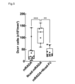
- FIG. 5 is an explanatory diagram showing the density of newborn neurons (Dcx-positive cells/mm 3 ) within the self-assembling peptide in Experiment 3.
- FIG. 6 is an explanatory diagram showing the number of newly generated neurons (Dcx-positive cells/mm 3 ) distributed in the injured tissue in Experiment 3.
- the lower side of the paper shows the subventricular zone side
- the upper side of the paper shows the superficial layer side of the cerebral cortex.
- the density of newborn neurons distributed within Ncad-mRADA is extremely high compared to the density of newborn neurons distributed within mRADA and mRADA+Ncad-Fc, each at a level of 0.1%. A significant difference was observed at the 1% level. Furthermore, as shown in Figure 6, there was no significant difference in the density of newborn neurons distributed in the ventricular zone-subventricular zone (V-SVZ) between the mRADA and Ncad-mRADA experimental groups. . On the other hand, in the Ncad-mRADA-injected group, the density of newly born neurons distributed in the superficial layer (Upper) and middle layer (Middle) of the cerebral cortex, which is the injury site, was significantly increased.
- Ncad-mRADA is effective as a scaffold for promoting the migration of newborn neurons in the brain after injury.
- mRADA+Ncad-Fc which is a mixture of a self-assembly consisting of mRADA without the extracellular domain of N-cadherin and a fusion protein containing the extracellular domain of N-cadherin, is newly generated in the brain after injury. It was not effective as a scaffold to promote neuron migration.
- EmGFP was gene introduced into 0-day-old mice using electroporation, and cells in the subventricular zone were labeled. Thereafter, at the age of 2 days after birth, cryoinjury was applied to the cerebral cortex of the right hemisphere. Then, at 5 days of age, mRADA or Ncad-mRADA was injected into the injury site. After that, a walking function test (foot-fault-test) was performed at 29 days of age, and on the next day, using fixed brain sections, immunization of GFP (Green Fluorescent Protein) and NeuN, a mature neuron marker, was performed. Staining was performed. Brain sections were imaged using a confocal microscope, and NeuN-positive cells (hereinafter also referred to as "EmGFP/NeuN double-positive cells”) among the subventricular zone-derived cells labeled with EmGFP were quantified.
- GFP Green Fluorescent Protein
- NeuN a mature neuron marker
- FIG. 7 is a microscopic image showing the results of Experiment 4, showing a wide range image including the corpus callosum (CC) and the cerebral cortex (Ctx).
- the lower side of the paper shows the corpus callosum side
- the upper side of the paper shows the superficial side of the cerebral cortex
- a scale bar of 50 ⁇ m is also shown.
- FIG. 8 is a partially enlarged view of FIG. 7.
- FIG. 8 shows an enlarged view of the area surrounded by a rectangle in FIG. 7, and the same area is shown in the image on the upper side of the paper and the image on the lower side of the paper. Furthermore, in FIG.
- EmGFP/NeuN double-positive cells ie, mature neurons derived from the subventricular zone, are indicated by white arrows, and a scale bar of 25 ⁇ m is also indicated.
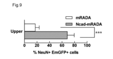
- FIG. 9 is an explanatory diagram showing the density (%) of EmGFP/NeuN double-positive cells in the superficial layer of the cerebral cortex. Note that FIG. 9 shows the ratio of the number of EmGFP/NeuN double-positive cells present in the superficial layer of the cerebral cortex to the total number of EmGFP/NeuN double-positive cells present in the cerebral cortex.
- FIG. 10 is an explanatory diagram showing the results of the walking function test in Experiment 4.
- the vertical axis indicates the slippage rate of the left hindlimb in the walking function test.
- a comparison between the Intact group and the Injury group showed that cryoinjury to the right hemisphere caused motor dysfunction in the left hindlimb and significantly increased the slip rate.
- a comparison between the Injury group and the Ncad-mRADA group showed that the slippage rate was significantly reduced by injecting Ncad-mRADA.
- the comparison between the Injury group and the mRADA group and the comparison between the Intact group and the mRADA group no improvement in the slippage rate was observed even when mRADA was injected.
- Ncad-mRADA mature neurons derived from the subventricular zone were widely distributed to the surface layer of the cerebral cortex, and neurological function was improved.
- a self-assembly in which the extracellular domain of N-cadherin is incorporated can promote the migration of newborn neurons compared to a self-assembly in which the extracellular domain of N-cadherin is not added. It can be said that mature neurons were distributed over a wide range up to the surface layer of the cerebral cortex, and as a result, neurological function was improved.
- ⁇ Experiment 5 Experiment on the migration of new neurons after cerebral infarction in the striatum> Wild-type mice aged 8 to 16 weeks were subjected to transient middle cerebral artery occlusion to create an infarct centered outside the striatum. Thirteen days later, mRADA or Ncad-mRADA was injected into the area extending from the subventricular zone to the infarct. Brains were fixed 18 days after cerebral infarction, and histological immunostaining was performed. Note that the self-assembling peptide was visualized using Azide647.
- FIG. 11 is a microscope image showing the results of Experiment 5.
- the image at the center of the paper is an enlarged view of a part of the image on the left side of the paper, and the image on the right side of the paper is a cross-sectional view of the cross section indicated by the broken line in the enlarged view. It is. Further, in FIG. 11, scale bars of 100 ⁇ m (weakly enlarged), 20 ⁇ m (strongly enlarged), and 10 ⁇ m (cross-sectional view) are also shown.
- FIG. 12 is an explanatory diagram showing the number of newborn neurons distributed between the ventricular zone and the subventricular zone.
- FIG. 13 is an explanatory diagram showing the distribution and number of newborn neurons in the brain striatum.
- FIG. 11 shows that in the Ncad-mRADA injection group, more newborn neurons were observed moving within the self-assembling peptide than in the mRADA injection group, indicating that the migration of newborn neurons was promoted. .
- FIG. 12 shows that no significant difference was observed between the mRADA-injected group and the Ncad-mRADA-injected group in the number of newborn neurons distributed between the ventricular zone and the subventricular zone.
- Figure 13 shows the results of an analysis of newly born neurons in the striatum, focusing on the relationship between the distance from the ventricular zone to the subventricular zone and the distance from the injected self-assembling peptide. ing. According to FIG.
- Ncad-mRADA can promote the migration of newborn neurons to the striatum after cerebral infarction.
- the self-assembly in which the extracellular domain of N-cadherin is incorporated is more effective in neonatal growth in the striatum after cerebral infarction than the self-assembly in which the extracellular domain of N-cadherin is not added. It was shown to be effective as a scaffold to promote neuron migration. It should be noted that the presumed mechanism by which the number of new neurons increases in the Ncad-mRADA injection group even in areas distant from the Ncad-mRADA injection site is not clear. However, since Azide647 visualizes only mRADA and Ncad-mRADA that are highly concentrated, there is a possibility that Ncad-mRADA is diffusely distributed in the brain tissue beyond the visualized range. There is a possibility that newborn neurons migrate beyond Ncad-mRADA.
- Example 6 Experiment on freezing injury of the cerebral cortex in adult mice> Cryoinjury was performed on the cerebral cortex of wild-type mice aged 8 to 16 weeks to create lesions limited only to the cerebral cortex. Two days after cryoinjury, mRADA or Ncad-mRADA was injected into the area extending from the subventricular zone to the injury site, and 7 days after cryoinjury, the mice were fixed and subjected to histological immunostaining. Note that the self-assembling peptide was visualized using Azide647.
- FIG. 14 is a microscope image showing the results of Experiment 6, showing the same area in the image on the left side of the page, the image in the center of the page, and the image on the right side of the page. Further, in FIG. 14, a scale bar of 20 ⁇ m is also shown.
- FIG. 15 is an explanatory diagram showing the number of newborn neurons distributed between the ventricular zone and the subventricular zone.
- FIG. 16 is an explanatory diagram showing the distribution and number of newborn neurons in the brain striatum.
- FIG. 14 shows that in the Ncad-mRADA injection group, more newborn neurons were observed moving within the self-assembling peptide than in the mRADA injection group, indicating that the migration of newborn neurons was promoted. .
- FIG. 15 shows that no significant difference was observed between the mRADA-injected group and the Ncad-mRADA-injected group in the number of newborn neurons distributed between the ventricular zone and the subventricular zone.
- Figure 16 shows the results of an analysis of newly born neurons present in the cortex after injury, focusing on the relationship between the distance from the ventricular zone to the subventricular zone and the distance from the injected self-assembling peptide. ing. According to FIG.
- Ncad-mRADA can promote the migration of newborn neurons derived from the subventricular zone to the injured area in the cerebral cortex after injury.
- the cerebral cortex had a lower level of subventricular activity in the cerebral cortex after injury. This suggests that it is effective as a scaffold for promoting the migration of newborn neurons derived from the zona to the injured area.
- Example 7 Biocompatibility confirmation experiment> As an additional experiment to Experiment 2 above, an experiment was conducted to confirm biocompatibility using an injured brain. Wild-type mice aged 8 to 16 weeks were subjected to transient middle cerebral artery occlusion to create an infarct centered outside the striatum. Thirteen days after the cerebral infarction, mRADA or Ncad-mRADA (hereinafter also referred to as "biomaterial") was injected into the region extending from the subventricular zone to the infarcted area. Brain sections fixed 18 days after cerebral infarction were subjected to histological immunostaining using CD45, a marker for leukocytes, and Iba1, a marker for macrophages and microglia.
- CD45 a marker for leukocytes
- Iba1 a marker for macrophages and microglia.
- Macrophages (CD45 high Iba1+ cells) and white blood cells excluding macrophages (CD45 high Iba1- cells) within 300 ⁇ m from the injection site of the biomaterial (visualized with Azide647) were imaged using a confocal microscope, and macrophages and macrophages were imaged using a confocal microscope. The density of white blood cells removed was compared quantitatively.
- the injected biomaterial contains 1% of it with alkyne added thereto, and is visualized by combining the alkyne with Azide647 to which Alexa647 is added by a Click reaction.
- FIG. 17 is a microscope image showing the results of Experiment 7.
- a scale bar of 50 ⁇ m is also shown, and macrophages are indicated by arrowheads, and white blood cells other than macrophages are indicated by arrows.
- FIG. 18 is an explanatory diagram showing the density of macrophages (number of CD45 high Iba1-positive cells/mm 3 ) and the density of white blood cells excluding macrophages (number of CD45 high Iba1-negative cells/mm 3 ) in Experiment 7.
- FIGS. 17 and 18 there was no statistically significant difference in the number of infiltrating immune cells between the two groups. That is, there was no significant difference between the injection groups of mRADA and Ncad-mRADA into the injured brain, and it was found that no excessive immune response was induced by the Ncad-mRADA peptide.
- ⁇ Experiment 8 Experiment to confirm the contact state between newborn neurons and biomaterial>
- Cryoinjury was applied to the cerebral cortex of 2-day-old Dcx-EGFP mice.
- selenomethionine-labeled Ncad-mRADA was injected into the injury site. Note that selenomethionine-labeled Ncad-mRADA was prepared by mixing selenomethionine-labeled mRADA and Ncad-mRADA.
- the brains were fixed at 9 days of age, and sections with a thickness of 200 ⁇ m were prepared to prepare samples for transmission electron microscopy.
- a biomaterial-injected area containing a large number of Dcx-EGFP cells migrating toward the injured area was cut out from the section embedded in epoxy resin.
- Semi-ultra-thin sections with a thickness of 1.5 ⁇ m were prepared from the excised samples, and ultra-thin sections with a thickness of 60 to 70 nm were prepared from the semi-ultra-thin sections in which newborn neurons were confirmed by toluidine blue staining. Then, using a transmission electron microscope, the ultrathin sections were observed and images of the newborn neurons and biomaterial were taken.
- Figure 19 is a microscope image showing the results of Experiment 8, with a scale bar of 5 ⁇ m in images A and F, a scale bar of 1 ⁇ m in images B and G, and a scale bar of 0.2 ⁇ m in images CE and H. A scale bar is also shown.
- Image B is a partially enlarged image of image A
- images CE are partially enlarged images of image B
- image G is a partially enlarged image of image F
- image H is a partially enlarged image of image G.
- It is an image.
- “Nb" in each image indicates a newborn neuron. As shown in images AC, the newborn neurons within the biomaterial were in contact with each other. Additionally, as shown in image C, there were spaces between the newborn neurons.
- Ncad-mRADA functions as a scaffold when newborn neurons migrate in chains or individually.
- Experiment 9 Experiment on the function of subventricular zone-derived cells> As an additional experiment to Experiment 4 above, an experiment regarding the function of cells derived from the subventricular zone was conducted. EmGFP was gene introduced into 0-day-old mice using electroporation, and cells in the subventricular zone were labeled. Cryoinjury was applied to the cerebral cortex at 2 days of age. At 5 days of age, Ncad-mRADA was injected into the injury site. Subsequently, using brain sections fixed at 30 days old, we investigated the relationship between GFP, MAP2, a neuron dendrite marker, GAD67, a GABAergic neuron marker, and Gephyrin, an inhibitory synapse marker. Immunostaining was performed.
- FIG. 20 is a microscope image showing the results of Experiment 9, with images A and B showing a 10 ⁇ m scale bar, and image C showing a 1 ⁇ m scale bar. Furthermore, in image C, co-positive synapses between EmGFP and Gephyrin are indicated by arrows. As shown in FIG. 20, cells derived from the subventricular zone were found to coexist with both markers. From the above, it has become clear that functional mature neurons regenerate after injury.
- FIG. 21 is an explanatory diagram showing the results of Experiment 10.
- the footprint area (Max contact area, cm 2 ) of the left forelimb (Fore) and left hindlimb (Hind) at maximum ground contact in the walking test is shown on the vertical axis, respectively.
- the vertical axis shows the percentage (%) of shaking the trunk to the right during EBST.
- FIGS. 21(A) and 21(B) a comparison between the Intact group and the Injury group showed that freezing injury to the right hemisphere of the brain increased the maximum ground contact in both the left forelimb and the left hindlimb.
- the footprint area at the time decreased significantly.
- NSE-DTA mice diphtheria toxin is expressed specifically in NSE-expressing cells, and cell death is induced.
- Cryoinjury was applied to the cerebral cortex at 2 days of age.
- Ncad-mRADA was injected into the injury site.
- a walking function test was conducted on the 29th day after birth. Immunostaining for GFP and NeuN was performed using brain sections fixed at 30 days of age. Brain sections were imaged using a confocal microscope, and NeuN-positive cells (EmGFP/NeuN double-positive cells) among the subventricular zone-derived cells labeled with EmGFP were quantified.
- FIG. 22 is an explanatory diagram showing the results of Experiment 11.
- Figure 22 (A) the number of EmGFP/NeuN double-positive cells per individual is shown on the vertical axis
- Figure 22 (B) the vertical axis shows the sliding rate (%) of the left hindlimb in the walking function test. is shown.
- FIG. 22(A) it was found that the number of mature neurons was significantly decreased in NSE-DTA mice compared to wild type.
- FIG. 22(B) even when Ncad-mRADA was injected into NSE-DTA mice, the rate of slippage of the left hindlimb was higher than in the wild type, indicating that no improvement in motor dysfunction was observed. Understood. From the above results, the number of regenerated neurons was significantly higher in the wild-type group than in the NSE-DTA group, and the slippage rate was significantly lower, indicating that neurons regenerated after injury are involved in improving neurological function. It was suggested that
- ⁇ Experiment 12 Confirmation experiment regarding cells that serve as endogenous scaffolds for newborn neurons> As an additional experiment to Experiment 5 above, a confirmation experiment regarding cells that serve as endogenous scaffolds for newborn neurons was conducted. Wild-type mice aged 8 to 16 weeks were subjected to transient middle cerebral artery occlusion to create an infarct centered outside the striatum. Thirteen days later, mRADA or Ncad-mRADA was injected into the area extending from the subventricular zone to the infarct. Eighteen days after the cerebral infarction, the brains were fixed, immunostained, and histologically analyzed.
- GFAP-positive cells Astrocytes (GFAP-positive cells) and vascular endothelial cells (CD31-positive cells) within 100 ⁇ m from the injection site of the injected biomaterial (visualized with Azide 647) were imaged using a confocal microscope. The ratio of GFAP-positive cells to the measured area and the total length of blood vessels identified from CD31-positive vascular endothelial cells in the measured area were calculated and compared between the biomaterials.
- FIG. 23 is a microscope image showing the results of Experiment 12.
- the left side of the page shows GFAP-positive cells
- the right side of the page shows CD31-positive cells
- the top side of the page shows the mRADA injection group
- the bottom side of the page shows the Ncad-mRADA injection group.
- a scale bar of 20 ⁇ m is also shown
- blood vessels are shown with arrowheads.
- FIG. 24 is a graph showing a comparison of the expression levels of markers of cells that serve as endogenous scaffolds of newborn neurons in Experiment 12.
- the proportion (%) of GFAP-positive cells in the measured area is shown on the vertical axis
- FIG. 24(A) the proportion (%) of GFAP-positive cells in the measured area is shown on the vertical axis
- the migration of neurons can be promoted, the number of neurons migrating to the injury site can be increased, and brain function can be improved by the differentiated and matured neurons that reach the injury site.
- Applications of the present invention include, for example, devices used to treat neonatal brain diseases such as hypoxic-ischemic encephalopathy, devices used to treat adult brain diseases such as cerebral infarction, and transplantation of neurons derived from iPS cells. Devices that enhance the therapeutic effects of regenerative medicine are envisioned.
- the present invention is not limited to the embodiments described above, and can be realized in various configurations without departing from the spirit thereof.
- the technical features in the embodiments and examples that correspond to the technical features in each form described in the summary column of the invention may be used to solve some or all of the above-mentioned problems, or to solve the above-mentioned problems. In order to achieve some or all of the effects, it is possible to replace or combine them as appropriate. Further, unless the technical feature is described as essential in this specification, it can be deleted as appropriate.
Landscapes
- Chemical & Material Sciences (AREA)
- Health & Medical Sciences (AREA)
- Organic Chemistry (AREA)
- Life Sciences & Earth Sciences (AREA)
- Genetics & Genomics (AREA)
- General Health & Medical Sciences (AREA)
- Engineering & Computer Science (AREA)
- Biochemistry (AREA)
- Medicinal Chemistry (AREA)
- Biomedical Technology (AREA)
- Biophysics (AREA)
- Molecular Biology (AREA)
- Proteomics, Peptides & Aminoacids (AREA)
- Zoology (AREA)
- Gastroenterology & Hepatology (AREA)
- Biotechnology (AREA)
- Bioinformatics & Cheminformatics (AREA)
- Wood Science & Technology (AREA)
- Toxicology (AREA)
- Cell Biology (AREA)
- Microbiology (AREA)
- General Engineering & Computer Science (AREA)
- Medicines That Contain Protein Lipid Enzymes And Other Medicines (AREA)
Priority Applications (1)
| Application Number | Priority Date | Filing Date | Title |
|---|---|---|---|
| JP2024506289A JPWO2023171577A1 (https=) | 2022-03-07 | 2023-03-03 |
Applications Claiming Priority (2)
| Application Number | Priority Date | Filing Date | Title |
|---|---|---|---|
| JP2022034331 | 2022-03-07 | ||
| JP2022-034331 | 2022-03-07 |
Publications (1)
| Publication Number | Publication Date |
|---|---|
| WO2023171577A1 true WO2023171577A1 (ja) | 2023-09-14 |
Family
ID=87935356
Family Applications (1)
| Application Number | Title | Priority Date | Filing Date |
|---|---|---|---|
| PCT/JP2023/008130 Ceased WO2023171577A1 (ja) | 2022-03-07 | 2023-03-03 | ニューロンの移動促進剤およびその利用 |
Country Status (2)
| Country | Link |
|---|---|
| JP (1) | JPWO2023171577A1 (https=) |
| WO (1) | WO2023171577A1 (https=) |
Citations (4)
| Publication number | Priority date | Publication date | Assignee | Title |
|---|---|---|---|---|
| JP2020018796A (ja) * | 2018-08-03 | 2020-02-06 | 公立大学法人名古屋市立大学 | 脳障害の治療用材料、脳障害の治療方法、脳の神経細胞の再生用材料、及び、脳の神経細胞の再生方法 |
| WO2020171161A1 (ja) * | 2019-02-20 | 2020-08-27 | 国立大学法人東京農工大学 | 自己組織化ペプチド |
| WO2021132157A1 (ja) * | 2019-12-23 | 2021-07-01 | 公立大学法人名古屋市立大学 | 脳疾患治療剤及びその利用 |
| JP2021147319A (ja) * | 2020-03-16 | 2021-09-27 | 国立大学法人 東京医科歯科大学 | 自己組織化ペプチドを含む組成物 |
-
2023
- 2023-03-03 JP JP2024506289A patent/JPWO2023171577A1/ja active Pending
- 2023-03-03 WO PCT/JP2023/008130 patent/WO2023171577A1/ja not_active Ceased
Patent Citations (4)
| Publication number | Priority date | Publication date | Assignee | Title |
|---|---|---|---|---|
| JP2020018796A (ja) * | 2018-08-03 | 2020-02-06 | 公立大学法人名古屋市立大学 | 脳障害の治療用材料、脳障害の治療方法、脳の神経細胞の再生用材料、及び、脳の神経細胞の再生方法 |
| WO2020171161A1 (ja) * | 2019-02-20 | 2020-08-27 | 国立大学法人東京農工大学 | 自己組織化ペプチド |
| WO2021132157A1 (ja) * | 2019-12-23 | 2021-07-01 | 公立大学法人名古屋市立大学 | 脳疾患治療剤及びその利用 |
| JP2021147319A (ja) * | 2020-03-16 | 2021-09-27 | 国立大学法人 東京医科歯科大学 | 自己組織化ペプチドを含む組成物 |
Non-Patent Citations (1)
| Title |
|---|
| OHNO YUYA, NAKAJIMA CHIKAKO, AJIOKA ITSUKI, MURAOKA TAKAHIRO, YAGUCHI ATSUYA, FUJIOKA TEPPEI, AKIMOTO SAORI, MATSUO MISAKI, LOTFY : "Amphiphilic peptide-tagged N-cadherin forms radial glial-like fibers that enhance neuronal migration in injured brain and promote sensorimotor recovery", BIOMATERIALS, ELSEVIER, AMSTERDAM, NL, vol. 294, 1 March 2023 (2023-03-01), AMSTERDAM, NL , pages 122003, XP093089567, ISSN: 0142-9612, DOI: 10.1016/j.biomaterials.2023.122003 * |
Also Published As
| Publication number | Publication date |
|---|---|
| JPWO2023171577A1 (https=) | 2023-09-14 |
Similar Documents
| Publication | Publication Date | Title |
|---|---|---|
| AU2022201894B2 (en) | Peptide for inducing regeneration of tissue and use thereof | |
| JP7537678B2 (ja) | 自己組織化ペプチドを含む組成物 | |
| US11739131B2 (en) | Growth differentiation factor 15 (GDF-15) polypeptides | |
| US20120231014A1 (en) | Neural Regeneration | |
| EP1919944B1 (en) | Modified vegf and pdgf with improved angiogenic properties | |
| JP2026041880A (ja) | 細胞透過性ペプチド変異体およびその用途 | |
| JP7641025B2 (ja) | 神経損傷及び関連障害を治療するための方法 | |
| JP2023072043A (ja) | 脳障害の治療用材料、脳障害の治療方法、脳の神経細胞の再生用材料、及び、脳の神経細胞の再生方法 | |
| JP7805589B2 (ja) | 自己組織化ペプチド | |
| JP2023523262A (ja) | 新規な細胞透過性ペプチド及びその用途 | |
| WO2023171577A1 (ja) | ニューロンの移動促進剤およびその利用 | |
| US20110251128A1 (en) | THYMOSIN Beta4 PEPTIDES PROMOTE TISSUE REGENERATION | |
| CN101503458B (zh) | 预防和治疗血管新生的小分子多肽及其应用 | |
| KR20190050277A (ko) | 허혈성 심혈관 질환의 치료용 또는 예방용 약학 조성물 | |
| JP4988711B2 (ja) | 神経再生ペプチド及びそれを含む処方物 | |
| KR20210127618A (ko) | 섬유질환 예방 또는 치료용 재조합 융합 단백질 | |
| US20110092427A1 (en) | Polypeptide and pharmaceutical composition containing the polypeptide | |
| CN104004057B (zh) | 一类抑制新生血管的小肽及其应用 | |
| JP2011529452A (ja) | 抗血管新生薬としての可溶性igf受容体 | |
| WO2007032396A1 (ja) | 脊髄損傷治療剤 | |
| US20110034387A1 (en) | Polypeptide inhibiting angiogenesis and application thereof |
Legal Events
| Date | Code | Title | Description |
|---|---|---|---|
| 121 | Ep: the epo has been informed by wipo that ep was designated in this application |
Ref document number: 23766753 Country of ref document: EP Kind code of ref document: A1 |
|
| DPE1 | Request for preliminary examination filed after expiration of 19th month from priority date (pct application filed from 20040101) | ||
| ENP | Entry into the national phase |
Ref document number: 2024506289 Country of ref document: JP Kind code of ref document: A |
|
| NENP | Non-entry into the national phase |
Ref country code: DE |
|
| 122 | Ep: pct application non-entry in european phase |
Ref document number: 23766753 Country of ref document: EP Kind code of ref document: A1 |
|
| 122 | Ep: pct application non-entry in european phase |
Ref document number: 23766753 Country of ref document: EP Kind code of ref document: A1 |