WO2023090381A1 - 宿主因子dock11をターゲットとした抗b型肝炎ウイルス剤 - Google Patents
宿主因子dock11をターゲットとした抗b型肝炎ウイルス剤 Download PDFInfo
- Publication number
- WO2023090381A1 WO2023090381A1 PCT/JP2022/042642 JP2022042642W WO2023090381A1 WO 2023090381 A1 WO2023090381 A1 WO 2023090381A1 JP 2022042642 W JP2022042642 W JP 2022042642W WO 2023090381 A1 WO2023090381 A1 WO 2023090381A1
- Authority
- WO
- WIPO (PCT)
- Prior art keywords
- seq
- polypeptide
- sequence
- amino acid
- acid sequence
- Prior art date
- Legal status (The legal status is an assumption and is not a legal conclusion. Google has not performed a legal analysis and makes no representation as to the accuracy of the status listed.)
- Ceased
Links
Images















Classifications
-
- A—HUMAN NECESSITIES
- A61—MEDICAL OR VETERINARY SCIENCE; HYGIENE
- A61K—PREPARATIONS FOR MEDICAL, DENTAL OR TOILETRY PURPOSES
- A61K38/00—Medicinal preparations containing peptides
- A61K38/04—Peptides having up to 20 amino acids in a fully defined sequence; Derivatives thereof
- A61K38/10—Peptides having 12 to 20 amino acids
-
- A—HUMAN NECESSITIES
- A61—MEDICAL OR VETERINARY SCIENCE; HYGIENE
- A61K—PREPARATIONS FOR MEDICAL, DENTAL OR TOILETRY PURPOSES
- A61K45/00—Medicinal preparations containing active ingredients not provided for in groups A61K31/00 - A61K41/00
-
- A—HUMAN NECESSITIES
- A61—MEDICAL OR VETERINARY SCIENCE; HYGIENE
- A61K—PREPARATIONS FOR MEDICAL, DENTAL OR TOILETRY PURPOSES
- A61K47/00—Medicinal preparations characterised by the non-active ingredients used, e.g. carriers or inert additives; Targeting or modifying agents chemically bound to the active ingredient
- A61K47/50—Medicinal preparations characterised by the non-active ingredients used, e.g. carriers or inert additives; Targeting or modifying agents chemically bound to the active ingredient the non-active ingredient being chemically bound to the active ingredient, e.g. polymer-drug conjugates
- A61K47/51—Medicinal preparations characterised by the non-active ingredients used, e.g. carriers or inert additives; Targeting or modifying agents chemically bound to the active ingredient the non-active ingredient being chemically bound to the active ingredient, e.g. polymer-drug conjugates the non-active ingredient being a modifying agent
- A61K47/68—Medicinal preparations characterised by the non-active ingredients used, e.g. carriers or inert additives; Targeting or modifying agents chemically bound to the active ingredient the non-active ingredient being chemically bound to the active ingredient, e.g. polymer-drug conjugates the non-active ingredient being a modifying agent the modifying agent being an antibody, an immunoglobulin or a fragment thereof, e.g. an Fc-fragment
-
- A—HUMAN NECESSITIES
- A61—MEDICAL OR VETERINARY SCIENCE; HYGIENE
- A61P—SPECIFIC THERAPEUTIC ACTIVITY OF CHEMICAL COMPOUNDS OR MEDICINAL PREPARATIONS
- A61P1/00—Drugs for disorders of the alimentary tract or the digestive system
- A61P1/16—Drugs for disorders of the alimentary tract or the digestive system for liver or gallbladder disorders, e.g. hepatoprotective agents, cholagogues, litholytics
-
- A—HUMAN NECESSITIES
- A61—MEDICAL OR VETERINARY SCIENCE; HYGIENE
- A61P—SPECIFIC THERAPEUTIC ACTIVITY OF CHEMICAL COMPOUNDS OR MEDICINAL PREPARATIONS
- A61P31/00—Antiinfectives, i.e. antibiotics, antiseptics, chemotherapeutics
- A61P31/12—Antivirals
- A61P31/20—Antivirals for DNA viruses
-
- A—HUMAN NECESSITIES
- A61—MEDICAL OR VETERINARY SCIENCE; HYGIENE
- A61P—SPECIFIC THERAPEUTIC ACTIVITY OF CHEMICAL COMPOUNDS OR MEDICINAL PREPARATIONS
- A61P43/00—Drugs for specific purposes, not provided for in groups A61P1/00-A61P41/00
-
- C—CHEMISTRY; METALLURGY
- C07—ORGANIC CHEMISTRY
- C07K—PEPTIDES
- C07K14/00—Peptides having more than 20 amino acids; Gastrins; Somatostatins; Melanotropins; Derivatives thereof
- C07K14/435—Peptides having more than 20 amino acids; Gastrins; Somatostatins; Melanotropins; Derivatives thereof from animals; from humans
-
- C—CHEMISTRY; METALLURGY
- C07—ORGANIC CHEMISTRY
- C07K—PEPTIDES
- C07K16/00—Immunoglobulins [IG], e.g. monoclonal or polyclonal antibodies
-
- C—CHEMISTRY; METALLURGY
- C07—ORGANIC CHEMISTRY
- C07K—PEPTIDES
- C07K16/00—Immunoglobulins [IG], e.g. monoclonal or polyclonal antibodies
- C07K16/18—Immunoglobulins [IG], e.g. monoclonal or polyclonal antibodies against material from animals or humans
- C07K16/28—Immunoglobulins [IG], e.g. monoclonal or polyclonal antibodies against material from animals or humans against receptors, cell surface antigens or cell surface determinants
-
- C—CHEMISTRY; METALLURGY
- C07—ORGANIC CHEMISTRY
- C07K—PEPTIDES
- C07K7/00—Peptides having 5 to 20 amino acids in a fully defined sequence; Derivatives thereof
- C07K7/04—Linear peptides containing only normal peptide links
- C07K7/08—Linear peptides containing only normal peptide links having 12 to 20 amino acids
-
- C—CHEMISTRY; METALLURGY
- C12—BIOCHEMISTRY; BEER; SPIRITS; WINE; VINEGAR; MICROBIOLOGY; ENZYMOLOGY; MUTATION OR GENETIC ENGINEERING
- C12N—MICROORGANISMS OR ENZYMES; COMPOSITIONS THEREOF; PROPAGATING, PRESERVING, OR MAINTAINING MICROORGANISMS; MUTATION OR GENETIC ENGINEERING; CULTURE MEDIA
- C12N15/00—Mutation or genetic engineering; DNA or RNA concerning genetic engineering, vectors, e.g. plasmids, or their isolation, preparation or purification; Use of hosts therefor
- C12N15/09—Recombinant DNA-technology
- C12N15/11—DNA or RNA fragments; Modified forms thereof; Non-coding nucleic acids having a biological activity
- C12N15/62—DNA sequences coding for fusion proteins
Definitions
- the present invention relates to an anti-hepatitis B virus agent that targets the host factor DOCK11.
- the present invention also relates to antibodies, antibody fragments, or single-chain antibodies that bind to asialoglycoprotein receptors that are excellent carriers for drug delivery into hepatocytes.
- HBV hepatitis B virus
- rcDNA incomplete double-stranded DNA
- cccDNA covalently closed circular DNA
- HBV utilizes the host's DNA repair mechanism, particularly the ATR signaling pathway, when synthesizing cccDNA from rcDNA (Non-Patent Document 3).
- HBV can remain latent in cells as stably inactivated cccDNA.
- HBV reactivation occurs when the immune system is compromised by chemotherapy or hematopoietic stem cell transplantation.
- interactions between the host's immune system and the virus influence the development of advanced hepatocellular carcinoma (HCC). Treatments that successfully reduce viral replicas can delay the development of advanced liver disease and hepatocellular carcinoma.
- targeting cccDNA is necessary to eliminate latent HBV.
- HBV-infected hepatocytes to HCC cells express reduced amounts of HBV protein and mRNA intracellularly. Except for cells containing HBV DNA integrated into the host genome, cell lines established from HCC usually do not express HBV transcripts. Cell lines that continue to express HBV transcripts are very useful for identifying host genes required for HBV maintenance. Therefore, Hashimoto et al. established an HC1 cell line that is maintained at a very low rate of HBV infection (about 1 in 3000 cells), and performed single-cell transcriptome analysis (Nx1-Seq) to detect HBV infection. Four host genes, LIPG, DOCK11, DENND2A, and HECW2, which are highly expressed in cells, were identified (Non-Patent Document 4).
- DOCK11 (dedicator of cytokinesis 11, NCBI Gene ID: 139818), also called Zizimin2
- DOCK11 is a member of the DOCK-D subfamily and has a large molecular size ( ⁇ 240 kDa).
- Cells use cytoskeleton such as actin filaments and microtubules to change and maintain their morphology.
- the cytoskeleton is strictly controlled by intracellular signal transduction triggered by external stimuli, and Rho family low-molecular-weight GTP-binding proteins (G proteins) play an important role in the signal transduction.
- Rho family G proteins act as molecular switches for intracellular signal transduction by being activated by GTP binding and inactivated by GDP binding.
- Rho family G proteins are activated by the GDP-GTP exchange reaction caused by the action of guanine nucleotide exchange factor (GEF).
- GEFs that activate Rho family G proteins are roughly classified into two groups: a group having a common Dbl homology domain (DH domain) and a group having a unique activation region called the DOCK family (Non-Patent Document 5. ). About 80 GEFs have been reported so far in mammals, including both groups. Since DOCK 180 was first reported in 1996, 11 types of DOCK family proteins have been confirmed in mammals. It is divided into C and DOCK-D (Non-Patent Document 6). DOCK11 belongs to the DOCK-D family.
- DOCK family proteins have two regions, Dock homology region 1 (DHR1) and DHR2, whose amino acid sequences are well conserved among families, and each activates a specific Rho family G protein through DHR2. It is known to do (Non-Patent Document 6). DOCK11 associates with CDC42, a G protein, and is a molecule involved in cytoskeleton and transport.
- DOCK11 has also been reported as one of the molecules that function to maintain the HBV gene in host cells (Patent Document 1).
- Patent Document 1 discloses that suppression of DOCK11 expression by siRNA and lenti-shRNA reduces HBV replication.
- a DOCK11 inhibitor applicable to HBV-infected patients has not yet been developed.
- DDS drug delivery systems
- the purpose of the present invention is to provide a novel anti-HBV drug that targets DOCK11.
- DOCK11-binding peptides have shown effects such as inhibition of DOCK11-Ack1 binding to inhibit Ack1 activation, inhibition of DOCK11 guanine nucleotide exchange factor (GEF) activity, and inhibition of DOCK11-mediated activation of the ATR signaling pathway.
- GEF DOCK11 guanine nucleotide exchange factor
- the present invention provides an anti-hepatitis B virus agent (anti-HBV agent) containing, as an active ingredient, a substance that binds to DOCK11 and inhibits the function of DOCK11.
- anti-HBV agent anti-hepatitis B virus agent
- the substance used as an active ingredient binds to the region of residues 1516 to 2073 of DOCK11.
- the inhibition of DOCK11 function is selected from inhibition of Ack1 activation by inhibition of binding of DOCK11 to Ack1, inhibition of guanine nucleotide exchange factor activity of DOCK11, and inhibition of activation of the ATR signaling pathway by DOCK11. is at least one
- the substance used as an active ingredient is at least one polypeptide selected from the following polypeptides (1) to (16).
- a polypeptide of the sequence IITPGTEVLNSDLQAS (SEQ ID NO: 1).
- a polypeptide of the sequence HNVLSVYNPAWGKYFH (SEQ ID NO:2).
- a polypeptide of the sequence NFPPNPMHNTDSCICA (SEQ ID NO:3).
- a polypeptide of the sequence TEKRRLMKPVLLTYNP SEQ ID NO:4
- a polypeptide of the sequence IICPGAEVLNGDLVAS SEQ ID NO:5
- a polypeptide of the sequence TEYRRCVTPVLLTYNN (SEQ ID NO:6).
- a polypeptide of the sequence TEEHRGLLPVLMTYNV (SEQ ID NO:7).
- a polypeptide of the sequence TEFCRWTWPVLCTYNA (SEQ ID NO:8).
- a polypeptide of the sequence TEQARPTPPPVLDTYNL (SEQ ID NO:9).
- a polypeptide of sequence PEQARPPPPLEDNLFL (SEQ ID NO: 10).
- a polypeptide of sequence HEEHRGMLREDSMMEYLK SEQ ID NO: 11
- a polypeptide of the sequence AEEHRGLLTIRYPMEH (SEQ ID NO: 12).
- a partial polypeptide of Ack1 containing the region of PEQARPPPPLEDNLFL (SEQ ID NO: 10).
- a partial polypeptide of radixin containing the region of HEEHRGMLREDSMMEYLK (SEQ ID NO: 11).
- a partial polypeptide of ⁇ -centractin containing the region of AEEHRGLLTIRYPMEH (SEQ ID NO: 12).
- the polypeptide is in a form linked to a carrier molecule for delivery into hepatocytes.
- the carrier molecule can be, for example, an antibody, antibody fragment or single-chain antibody that binds to the asialoglycoprotein receptor.
- the polypeptide is in a form linked to a cell membrane permeabilization molecule.
- a cell membrane permeabilization molecule can be, for example, a polypeptide having the amino acid sequence shown in SEQ ID NO:38 or 39.
- the polypeptide is in a form linked to a nuclear localization signal.
- the present invention also provides the use of at least one polypeptide selected from the polypeptides (1) to (16) above as a DOCK11-binding peptide, and from the polypeptides (1) to (16) above.
- DOCK11-binding peptides are provided that consist of at least one selected polypeptide.
- a heavy chain CDR1 comprising an amino acid sequence shown in SEQ ID NO: 13, 19, 25 or 31, or an amino acid sequence in which some residues are substituted in the amino acid sequence and has an identity of 80% or more with the amino acid sequence
- a heavy chain CDR2 comprising an amino acid sequence shown in SEQ ID NO: 14, 20, 26 or 32, or an amino acid sequence in which some residues are substituted and having 80% or more identity with the amino acid sequence
- a heavy chain CDR3 comprising an amino acid sequence shown in SEQ ID NO: 15, 21, 27 or 33, or an amino acid sequence in which some residues are substituted in the amino acid sequence and has an identity of 80% or more with the amino acid sequence
- a light chain CDR1 comprising an amino acid sequence shown in SEQ ID NO: 16, 22, 28 or 34, or an amino acid sequence in which some residues are substituted and having 80% or more identity with the amino acid sequence
- a light chain CDR2 comprising an amino acid sequence shown in SEQ ID NO: 17, 23, 29 or 35, or an amino acid sequence
- the present invention provides a drug delivery carrier for delivering a drug into hepatocytes, containing the antibody, antibody fragment, or single-chain antibody of the present invention.
- the present invention provides a pharmaceutical composition comprising a complex of the drug delivery carrier of the present invention and a drug to be delivered into hepatocytes.
- the present invention provides for the first time an anti-HBV agent that binds to DOCK11 and has the action of inhibiting the function of DOCK11. Since the anti-HBV agent of the present invention can reduce intracellular HBV DNA and cccDNA and inhibit re-invasion of HBV particles into cells, it is expected to cure hepatitis B completely.
- the anti-ASGR antibody, antibody fragment or single-chain antibody having a specific CDR sequence or modified sequence thereof provided by the present invention is very excellent as a carrier for delivering drugs into hepatocytes, and is an anti-HVB agent. In addition, it is useful as a delivery carrier for various drugs to act in the liver.
- DNA library design for IVV screening Structure of the human DOCK180 superfamily. Explanatory diagram of the construction procedure of biotinylated full-length DOCK11. Explanatory drawing of selection experiments for peptides that bind to DOCK11 by the IVV method.
- Day 18 Transfection with plasmid expressing peptide (1 ⁇ g with Lipofectamine 3000).
- Day 20 Measure HBV-DNA, cccDNA.
- Day 1 Seed cells in 12 well plates.
- Day 2 Transfection with plasmid expressing peptide (1 ⁇ g with Lipofectamine 3000).
- Day 3 HBV infection (medium change after 12 hours).
- Day 5 Measure HBV-DNA and cccDNA. Anti-HBV activity of DOCK11-binding peptides in HepG2-NTCP-C4 cells (long-term administration).
- A Schedule of HBV infection and sample administration.
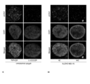
- B Early endosomes, peptides (GGP), actin, and nuclei were observed with a confocal microscope. Functional verification of membrane fusion-promoting peptides S28 and S39: Comparison of nuclear uptake of peptides by S28 and S39 Validation of the function of nuclear localization signals.
- A Experimental procedure.
- B Peptides (GGP), actin, and nuclei were observed with a confocal microscope.
- DOCK1516-2073-Bio-His containing the DHR2 domain was added to a streptavidin-coated 96-well plate, fixed overnight at 4°C, reacted with 0-1.0 mol of DCS8-42TN at room temperature for 1 hour, and then subjected to GEF assay. gone.
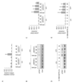
- A HepG2 cells were transfected with DOCK11 siRNA #1-3 using Lipofectamine 3000, and real-time RT-PCR was performed 72 hours later. GAPDH mRNA levels were used to normalize DOCK11 mRNA levels.
- B HepG2 cells were transfected with DOCK11 or Ack1 siRNA #1-3 using Lipofectamine 3000.
- HepG2 cells were transfected with DOCK11 or Ack1 siRNA #1-3 using Lipofectamine 3000. After 72 hours, the cells were fixed and permeabilized, and the actin filaments were stained with fluorescent phalloidin, followed by nuclear staining with DAPI.
- A HepG2 cells were treated with 100 nM N-10M-D42TN for 0-48 hours, washed, and cultured for 24-72 hours. Then, the cells were fixed and permeabilized, and the actin filaments were stained with fluorescent phalloidin, followed by nuclear staining with DAPI.
- C HepG2 cells were transfected with EYFP-NLS-Actin and treated 48 hours later with 100 nM N-10M-D42TN for 20 hours. After fixing and permeabilizing the cells and staining the nuclei with DAPI, the fluorescence of EYFP and GFP was observed.
- Ack1 binds to and activates DOCK11-activated Cdc42, and binds to and phosphorylate EGFR.
- both EGFR-Ack1 complexes are endocytosed and degraded, but by inhibiting DOCK11 function by N-10M-D42TN, phosphorylated EGFR is not degraded and Ack1 phosphorylation and Phosphorylation of WASP, which is phosphorylated by Ack1, is also inhibited.
- Huh7 cells were treated with 100 nM N-10M-D42TN for 24 hours and then treated with 10 ng/ml EGF for 0-10 minutes, and Western blotting was performed using the cell lysate.
- Ack1, pAck1, EGFR, pEGFR(Tyr845), pEGFR(Tyr1068), pEGFR(Tyr1045), WASP, pWASP, and GAPDH antibodies were used as primary antibodies.
- HepG2 cells were treated with 100 nM N-10M-D42TN for 24 hours and then treated with 100 ng/ml EGF for 1 hour. Early endosomes were stained with CellLight Early Endosomes-RFP, immunostained with anti-Ack1 antibody, and nuclear stained with DAPI. White arrows indicate early endosomes where Ack1 is localized, and black arrows indicate early endosomes where Ack1 is not localized.
- (B) shows the ratio of all pixels representing early endosomes to those colocalizing with Ack1 in the cells shown in Panel A (Colocalization coefficients).
- A HepG2 cells were transfected with DOCK11 siRNA #1-3 using Lipofectamine 3000, and DNA damage was induced by UV irradiation 72 hours later. RNA was extracted from these cells and the amount of DOCK11 mRNA was measured by real-time RT-PCR. GAPDH mRNA was similarly measured and normalized.
- This cell lysate was prepared and Western blotting was performed using anti-Chk1 and pChk1 antibodies as primary antibodies.
- C In Fig. B, ImageLab was used to calculate the expression level ratio of pChk1 and Chk1, and the phosphorylation level of Chk1 in each cell was calculated. As a control, the amount of phosphorylation in cells that were not irradiated with UV after transfection with siEmpty was used.
- D HepG2 cells were treated with 100 nM N-10M-D42TN for 24 hours, and DNA damage was induced by UV irradiation. This cell lysate was prepared and Western blotting was performed using anti-Chk1 and pChk1 antibodies as primary antibodies.
- PXB cells were treated with 100 nM N-10M-D42TN for 24 hours and then UV-irradiated to induce DNA damage. Immunostaining was performed using an anti-DOCK11 antibody and nuclear staining was performed with DAPI.
- B The fluorescence intensity of DOCK11 that showed a threshold value or higher inside the nucleus was calculated and normalized using the fluorescence intensity of DAPI.
- DNA damage was induced by UV irradiation in PXB cells transfected with siRNA targeting DOCK11 and treated with 100 nM N-10M-D42TN for 24 h.
- C, D Fluorescence intensity of DOCK11 (C) and ⁇ H2AX (D) in each cell in FIG. 37A. Both show the ratio of DAPI fluorescence intensity, and the results of cells transfected with siEmpty without UV exposure were used as a control. *p ⁇ 0.05, **p ⁇ 0.005.
- N-10M-D42TN and control Measurement of blood HBV DNA in Example 16. Comparison of N-10M-D42TN and control (PBS). Measurement of h-Alb in Example 16. Comparison of N-10M-D42TN and control (PBS). Measurement of ALT in Example 16. Comparison of N-10M-D42TN and control (PBS). Measurement of HBV DNA in liver in Example 16. Comparison of N-10M-D42TN and control (PBS). Measurement of HBV DNA in liver in Example 16. Comparison of N-10M-D42TN and control (PBS).
- anti-hepatitis B virus includes treatment of HBV infection, prevention of HBV infection, suppression of HBV proliferation, treatment of hepatitis B, and prevention of hepatitis B. be.
- hepatitis B can be treated by suppressing the proliferation of HBV in the patient's body (inside the liver).
- an anti-HBV agent to an HBV carrier before the onset of hepatitis B, the proliferation of HBV in the carrier can be prevented and the onset of hepatitis B can be prevented (prevention of hepatitis B).
- the anti-HBV agent of the present invention contains as an active ingredient a substance that binds to DOCK11 and inhibits the function of DOCK11.
- Human DOCK11 is a protein having the structure shown in FIG. 2, and has Dock homology region 1 (DHR1) and DHR2, which are domains whose amino acid sequences are well conserved among DOCK families.
- DHR1 located at the N-terminus binds to phosphatidylinositol 3-phosphate.
- DHR2 present on the C-terminal side is a domain that activates a specific Rho family G protein, and DHR2 of DOCK11 activates a small G protein Cdc42.
- sequences shown in SEQ ID NOS: 41 and 42 in the Sequence Listing are the nucleotide sequence of the coding region of human DOCK11 mRNA and the amino acid sequence of DOCK11 encoded by this registered in GenBank of NCBI under NM_144658.4.
- the region from 1st to 2036th residues is the DHR2 domain.
- the substance used as the active ingredient of the anti-HBV agent of the present invention may be a substance that binds to the region of residues 1516 to 2073 (C-terminal region) of DOCK11, eg, the DHR2 domain.
- Cdc42 is replaced from inactive (GDP-bound) to active (GTP-bound), and unlike other GEF proteins, active Cdc42 is known to bind to and further activate and induce positive feedback (Lin, Q. et al. J. Biol. Chem., 281, 35253-35262, 2006; Nishikimi, A. et al. Exp Cell Res 319, 2343-2349, 2013).
- GEF guanine nucleotide exchange factor
- Ack1 activated CDC42 kinase 1
- GTP-bound Cdc42 Primary phosphate-maleic anhydride-semiconduct-binding protein
- the present inventors demonstrated that DOCK11 and Ack1 bind intracellularly, and that Cdc42 and Ack1 competitively bind to DOCK11.
- Inhibition of DOCK11 function is, for example, at least one selected from inhibition of Ack1 activation by inhibition of binding of DOCK11 and Ack1, inhibition of GEF activity of DOCK11, and inhibition of activation of ATR signaling pathway by DOCK11. It's okay.
- the following examples demonstrate that a DOCK11-binding peptide with anti-HBV activity inhibits the binding of DOCK11 and Ack1 to inhibit Ack1 activation, inhibits the GEF activity of DOCK11, and activates the ATR signaling pathway by DOCK11.
- Substances that bind to the region of residues 1516 to 2073 (C-terminal region) of DOCK11 or the DHR2 domain and inhibit the function of DOCK11 include, for example, recognizing and binding to the C-terminal region or DHR2 domain.
- the term "antibody fragment” is synonymous with the term "antigen-binding fragment of antibody” and includes Fab, Fab', F(ab') 2 and the like. There is no particular limitation as long as it is an antibody fragment that maintains antigen-binding properties.
- the anti-HBV agent of the present invention may contain, as an active ingredient, at least one polypeptide selected from the following polypeptides (1) to (16), for example.
- polypeptides (1) to (16) are the names of each polypeptide used in the following examples. Hereinafter, these polypeptides may be referred to as "DOCK11-binding peptides".
- DCS3-1] (2) A polypeptide of the sequence HNVLSVYNPAWGKYFH (SEQ ID NO:2).
- DCS5-4] (3) A polypeptide of the sequence NFPPNPMHNTDSCICA (SEQ ID NO:3).
- [DCS5-5] (4) A polypeptide of the sequence TEKRRLMKPVLLTYNP (SEQ ID NO:4).
- [DCS5-15] (5) A polypeptide of the sequence IICPGAEVLNGDLVAS (SEQ ID NO:5).
- [DCS8-6] (6) A polypeptide of the sequence TEYRRCVTPVLLTYNN (SEQ ID NO:6).
- [DCS8-29] (7) A polypeptide of the sequence TEEHRGLLPVLMTYNV (SEQ ID NO:7).
- [DCS8-59] (8) A polypeptide of the sequence TEFCRWTWPVLCTYNA (SEQ ID NO:8).
- [DCS8-72] (9) A polypeptide of the sequence TEQARPTPPPVLDTYNL (SEQ ID NO:9).
- [DCS8-42] (10) A polypeptide of sequence PEQARPPPPLEDNLFL (SEQ ID NO: 10).
- [DCS8-42TN] (11) A polypeptide of sequence HEEHRGMLREDSMMEYLK (SEQ ID NO: 11).
- [DCS8-59R] (12) A polypeptide of the sequence AEEHRGLLTIRYPMEH (SEQ ID NO: 12).
- [DCS8-59C] (13) A partial polypeptide of Ack1 containing the region of PEQARPPPPLEDNLFL (SEQ ID NO: 10).
- a partial polypeptide of radixin containing the region of HEEHRGMLREDSMMEYLK (SEQ ID NO: 11).
- a partial polypeptide of ⁇ -centractin containing the region of AEEHRGLLTIRYPMEH (SEQ ID NO: 12).
- Polypeptides (1) to (9) are polypeptides selected from an IVV library by a selection experiment for peptides that bind to DOCK11 using Biacore in the following examples, and confirmed to have anti-HBV activity by pull-down assay or the like.
- (10) to (12) are polypeptides designed based on the homology search results of the sequences of (7) and (9) and confirmed to have anti-HBV activity by pull-down assays and the like.
- the test substance prepared using DCS8-42TN in (10) was used in an in vivo administration experiment to confirm the anti-HBV activity. Agents other than 42TN are also useful as anti-HBV agents like DCS8-42TN.
- the partial polypeptide of (13) is a partial region of Ack1 (NP_001374642.1, SEQ ID NO: 50), which is a region having high homology with DOCK11-binding peptide DCS8-42 PEQARPPPPLEDNLFL (SEQ ID NO: 10; SEQ ID NO: 50 674th to 689th amino acids in the amino acid sequence of Ack1 shown in )).
- SEQ ID NO: 49 shows the nucleotide sequence encoding the amino acid sequence of SEQ ID NO: 50 (nucleotide sequence of the coding region of NM_001387713.1).
- the partial polypeptide of (14) is a partial region of radixin (NP_001247421.1, SEQ ID NO: 92) and has high homology with DOCK11-binding peptide DCS8-59 HEEHRGMLREDSMMEYLK (SEQ ID NO: 11; SEQ ID NO: 92 176th to 193rd amino acids in the radixin amino acid sequence shown in )).
- SEQ ID NO:91 shows the nucleotide sequence encoding the amino acid sequence of SEQ ID NO:92 (nucleotide sequence of the coding region of NM_001260492.2).
- the partial polypeptide of (15) is a partial region of ⁇ -centractin (NP_005726.1, SEQ ID NO: 94) and is a region having high homology with DOCK11-binding peptide DCS8-59 AEEHRGLLTIRYPMEH (SEQ ID NO: 12; It is a polypeptide composed of a partial region containing the 62nd to 77th amino acid region in the amino acid sequence of ⁇ -centractin shown in SEQ ID NO:94).
- SEQ ID NO:93 shows the nucleotide sequence encoding the amino acid sequence of SEQ ID NO:94 (nucleotide sequence of the coding region of NM_005735.4).
- DCS8-42TN a polypeptide of SEQ ID NO: 10, DCS8-59R, a polypeptide of SEQ ID NO: 11, and DCS8-59C, a polypeptide of SEQ ID NO: 12, have anti-HBV activity.
- polypeptides composed of partial regions of each protein containing these regions also have anti-HBV activity and are useful as anti-HBV agents.
- the chain length of the polypeptides (13) to (15) is not particularly limited. The size may be within 10 groups, within 50 residues, within 40 residues, within 30 residues, within 25 residues, or within 20 residues.
- the polypeptide of (16) is a polypeptide having a sequence identity of 80% or more with the original polypeptide in which a part of the residues is modified in any of the polypeptides of (1) to (15). . It is well known in the art that a very small number of residues can be altered while maintaining equivalent or better activity of the polypeptide. Thus, the original sequence in which a few residues (eg, 1-4 residues, 1-3 residues, 1 or 2 residues, or 1 residue) of the polypeptides (1)-(15) are altered Polypeptides having 80% or more identity, such as 85% or more, or 90% or more identity with , can exhibit anti-HBV activity like the original polypeptide and are useful as anti-HBV agents. Modifications of residues herein are substitutions, deletions, insertions or additions, typically substitutions.
- the identity of amino acid sequences means that two amino acid sequences to be compared are aligned so that as many amino acid residues as possible match each other, and the number of matched amino acid residues is divided by the total number of amino acid residues. It is expressed as a percentage.
- gaps are appropriately inserted in one or both of the two sequences to be compared, if necessary.
- sequence alignment can be performed using well-known programs such as BLAST, FASTA, CLUSTAL W, and the like.
- the above total number of amino acid residues is the number of residues obtained by counting one gap as one amino acid residue. If the total number of amino acid residues counted in this way differs between the two sequences being compared, then the % identity is the total number of amino acid residues in the longer sequence and the number of matching amino acid residues. calculated by dividing
- amino acids with similar side chains have similar chemical properties.
- Grouping amino acids by side chain similarity includes, for example, the group of amino acids with aliphatic side chains (glycine, alanine, valine, leucine, isoleucine), the group of amino acids with aliphatic hydroxyl side chains (serine, threonine).
- the group of amino acids with amide-containing side chains (asparagine, glutamine), the group of amino acids with aromatic side chains (phenylalanine, tyrosine, tryptophan), the group of amino acids with basic side chains (arginine, lysine, histidine), They can be classified into groups of amino acids with acidic side chains (aspartic acid, glutamic acid), groups of amino acids with sulfur-containing side chains (cysteine, methionine), and the like.
- a substitution for another amino acid belonging to the same group is a conservative substitution.
- polyethylene glycol (PEG) chains are added for the purpose of improving the stability of peptides in vivo and increasing the half-life in blood (Clin Nephrol. 2006 Mar;65(3): 180-90. and Proc Natl Acad Sci USA. 2005 Sep 6;102(36):12962-7.), mainly adding sugar chains to the N-terminus or C-terminus (J Am Chem Soc. 2004 Nov 3;126 (43): 14013-22 and Angew Chem Int Ed Engl. 2004 Mar 12;43(12): 1516-20), at least part of the amino acid residues are in the D form (J Pharmacol Exp Ther.
- PEG polyethylene glycol
- the Fc region of the antibody is appropriately modified and added (e.g., J.Immunol., 154( 10), 5590-5600 (1995), Nature, 332, 563-564 (1998), Nature, 332, 738-740 (1998), BioDrugs. 2008;22:11-26, etc.), C-terminal amidation , N-terminal acetylation, etc. are used.
- the polypeptide used as an active ingredient in the anti-HBV agent of the present invention may be one to which such techniques are applied.
- polypeptide having the sequence of SEQ ID NO: X refers to a polypeptide having the amino acid sequence shown in SEQ ID NO: X and having a total length of N residues (such a polypeptide is referred to as polypeptide X for convenience), and the Fc region and It includes polypeptides having a structure in which other functional molecules are added or linked, such as polypeptides having the effect of enhancing delivery to the liver.
- polypeptide having the sequence of SEQ ID NO: X means that the full length is N residues.
- polypeptide having a structure in which another functional molecule is added to polypeptide X is included as an active ingredient. Any functional molecule may be linked as long as it does not impair the anti-HBV activity of the polypeptide.
- polypeptides of (1) to (12) and the polypeptides of (16) that have 80% or more and less than 100% sequence identity with any of (1) to (12) among the polypeptides of (16) have a chain length Since it is short, it can be easily prepared by conventional chemical synthesis.
- the polypeptides of (13) to (15) and the polypeptides of (16) that have 80% or more and less than 100% sequence identity with any of (13) to (15) among the polypeptides of (16) have a chain length Short ones can be easily prepared by chemical synthesis, and long chain ones that are difficult to prepare by chemical synthesis can be produced by genetic engineering techniques.
- Polypeptides having a structure in which other functional molecules are linked are also produced by chemical synthesis when the functional molecule is a short-sized polypeptide, and by genetic engineering when the functional molecule is a long-sized polypeptide. can be manufactured by
- chemical synthesis methods include the Fmoc method (fluorenylmethyloxycarbonyl method) and the tBoc method (t-butyloxycarbonyl method). Moreover, it can also synthesize
- a cDNA encoding a full-length DOCK11-binding peptide with a long size or a full-length DOCK11-binding peptide linked to a functional molecule is prepared, the cDNA is incorporated into an appropriate expression vector, and the expression is transformed into an appropriate host cell.
- the polypeptide of interest can be obtained by introducing it, producing the polypeptide in the host cell, and extracting and purifying it.
- the DOCK11-binding peptide may have a tag sequence such as a Flag tag or His tag added for convenience of purification and detection.
- a tag sequence can also be regarded as one example of a functional molecule.
- SEQ ID NO: 90 for example, in the "cleaved sequence + DCS8-42TN peptide + S28 + nuclear localization signal" portion (SEQ ID NO: 90) of N-10M-D42TN prepared in the following example, Flag tag and His tag is added.
- addition of such a tag sequence is optional, and it is also possible to prepare the anti-HBV agent of the present invention using a DOCK11-binding peptide without a tag sequence.
- a functional molecule is a carrier molecule for delivery into hepatocytes. That is, the DOCK11-binding peptide may be in a form linked to a carrier molecule for delivery into hepatocytes.
- ASGR asialoglycoprotein receptor
- ApoE receptor which are abundant in hepatocytes
- carrier molecules that deliver drugs into hepatocytes using ASGR or ApoE receptors can be used.
- ASGR asialoglycoprotein receptor
- ASGR is a type II single-pass transmembrane protein with the N-terminus directed intracellularly and the C-terminal carbohydrate recognition sites (CRDs) directed extracellularly, as shown in FIG.
- CCDs carbohydrate recognition sites
- ASGR-specific binding molecule used as a drug delivery carrier may bind to the extracellular domain of ASGR.
- a specific binding molecule that can bind to both ASGR1 and ASGR2 can be particularly preferably used, but even a specific binding molecule that binds to only one of them can be used as a drug delivery carrier.
- a specific binding molecule that binds only to either one it may be used alone as a drug delivery carrier, or a combination of a specific binding molecule that binds to one and a specific binding molecule that binds to the other may be used.
- a DOCK11-binding peptide linked to an ASGR1-specific binding molecule and a DOCK11-binding peptide linked to an ASGR2-specific binding molecule may be prepared, mixed and used as an anti-HBV agent.
- the base sequence of the coding region of human ASGR1 mRNA registered under NM_001671.5 in GenBank and the amino acid sequence of ASGR1 encoded by this are shown in SEQ ID NOs: 43 and 44, and the code of human ASGR2 mRNA registered under NM_001181.4.
- the nucleotide sequence of the region and the amino acid sequence of ASGR2 encoded by this are shown in SEQ ID NOS: 45 and 46, respectively.
- the 41st to 61st amino acids of SEQ ID NO: 44 (ASGR1) and the 59th to 79th amino acids of SEQ ID NO: 46 (ASGR2) are transmembrane domains, the N-terminal side of which is the intracellular domain, and the C-terminal side of which is the cell.
- ASGR extracellular domain includes hetero-oligomerized extracellular domains.
- hetero-oligomer is meant a hetero-oligomerized ASGR extracellular domain.
- Anti-ASGR polyclonal antibodies can be obtained by immunizing a non-human animal using the ASGR extracellular domain as an immunogen, collecting blood, separating the serum, and collecting and purifying the antibody that binds to the ASGR extracellular domain from the serum. can be done.
- Anti-ASGR monoclonal antibodies are produced by collecting antibody-producing cells such as splenocytes and lymphocytes from non-human animals immunized with the ASGR extracellular domain, fusing them with myeloma cells to prepare hybridomas, and binding to the ASGR extracellular domain.
- Anti-ASGR monoclonal antibodies can be obtained from culture supernatants by selecting hybridomas that produce antibodies against the anti-ASGR and proliferating them.
- An anti-ASGR antibody fragment can be obtained by treating an anti-ASGR antibody with a proteolytic enzyme such as papain or pepsin.
- a proteolytic enzyme such as papain or pepsin.
- the definition of the antibody fragment is as described above, and includes antibody fragments such as Fab, Fab', F(ab') 2 that maintain the binding ability to the antigen (ASGR extracellular domain).
- cDNA is prepared by extracting mRNA from the hybridoma produced as described above, and PCR is performed using primers specific for immunoglobulin H chain and L chain to obtain immunoglobulin H chain gene and L chain.
- PCR is performed using primers specific for immunoglobulin H chain and L chain to obtain immunoglobulin H chain gene and L chain.
- anti-ASGR scFv can be obtained from scFv libraries by techniques such as the phage display method and IVV method.
- a phage library with scFv displayed on the phage surface is prepared as an scFv library.
- a naive scFv phage library may be prepared by the following procedure. mRNA is extracted from antibody-producing cells such as splenocytes and lymphocytes collected from healthy humans or non-human animals, cDNA is synthesized by reverse transcription reaction, and cDNA encoding the region containing the heavy chain variable region (VH) (VH cDNA) and cDNA encoding a region containing the light chain variable region (VL) (VL cDNA) are comprehensively amplified by PCR.
- VH heavy chain variable region
- VL light chain variable region
- the amplified VH cDNA and VL cDNA are randomly ligated via a suitable linker (for example, a linker consisting of three repeated GGGGS units, etc.) by standard assembly PCR or Fusion PCR to obtain scFv-encoding cDNA.
- a suitable linker for example, a linker consisting of three repeated GGGGS units, etc.
- Fusion PCR Fusion PCR
- Plasmid vectors for phage include all phage genes necessary for forming phage particles and capable of forming phage particles independently, and phage vectors containing the g3p gene but no other phage protein genes and containing phage Although there are two types of phagemid vectors that require helper phage for particle formation, phagemid vectors are preferably used.
- a scFv cDNA library prepared using a phagemid vector is introduced into Escherichia coli, and the E. coli is superinfected with a helper phage to package each vector of the scFv cDNA library, and the scFv expressed by the vector is placed on the surface. Libraries of displaying phage can be generated.
- a phage library of mutant scFv may be prepared by randomly introducing mutations into the prepared VH cDNA and VL cDNA or scFv cDNA.
- phages displaying scFv that bind to the ASGR extracellular domain are selected (panning).
- This panning step can be performed using a solid-phase carrier (chip, plate, magnetic bead, etc.) on which the extracellular domain of ASGR is immobilized, and a carrier on which a hetero-oligomer is immobilized is particularly preferably used.
- the extracellular domain of ASGR1 (ASGR1ex) and the extracellular domain of ASGR2 (ASGR2ex) are biotinylated, and by contacting the biotinylated ASGR1ex and ASGR2ex with a carrier coated with avidins, ASGR1ex and ASGR2ex are hetero-oligomerized on the carrier surface.
- the phage library is brought into contact with the ASGR extracellular domain-immobilized carrier, and after washing, the phages bound on the carrier are recovered.
- the recovered phages are lysed, the packaged vector is recovered, introduced again into E. coli, and superinfected with helper phages to form phage particles again.
- These phage particles are again brought into contact with the ASGR extracellular domain-immobilized carriers.
- Phages enriched through multiple rounds of panning can be obtained as anti-ASGR scFv candidate clones, but clone selection may be performed to further narrow down the candidates.
- the scFv expression vector is collected from the phage after concentration and introduced into an appropriate host cell such as E. coli to prepare host cell clones expressing scFv. and select clones with high specific binding to the ASGR extracellular domain. Reactivity can be confirmed by an immunoassay such as ELISA using the ASGR extracellular domain, preferably a hetero-oligomer, as an antigen.
- clones may be grouped by confirming duplication of clones based on the base sequences of VH and VL. Through these clone selection activities, it is possible to select clones with high specific binding to the ASGR extracellular domain and further narrow down the candidates.
- the scFv expression vector is recovered from the candidate clone, the scFv cDNA is amplified from the scFv expression vector, incorporated into an appropriate plasmid expression vector to express the scFv in an appropriate host cell, recovered and purified.
- the reactivity with ASGR1ex and ASGR2ex or the reactivity with heterooligomers is finally confirmed for the scFv after purification, and scFv that specifically binds to the ASGR extracellular domain can be obtained.
- IVV method (Nemoto, N. et al., FEBS Lett., 414:405-408, 1997; Miyamoto-Sato, E. et al., Nucleic Acids Res., 28:1176-1182, 2000; WO 03/106675 A1) is a technique developed by Yanagawa, one of the inventors of the present application, and his collaborators.
- puromycin a type of antibiotic
- PEG polyethylene glycol
- An mRNA-protein junction molecule (in vitro virus; IVV) is formed covalently linked to the mRNA molecule via puromycin. From the IVV library constructed in this way, IVVs containing proteins that bind to baits (proteins, peptides, antigens, etc.) are picked in vitro, and the genes (mRNA) linked to them are amplified by reverse transcription PCR. Then, by deciphering the base sequence with a DNA sequencer, interacting proteins, peptides and antibodies can be easily identified. Competitive elution with free bait is the common method for eluting and recovering IVV bound to bait.
- IVV in vitro virus
- the spacer can be cleaved by UV irradiation at 365 nm, and the mRNA portion can be eluted and recovered (Doi, N. et al., J. Biotechnol., 131:231-239, 2007).
- mRNA is synthesized from the cDNA library by reverse transcription reaction, puromycin is bound to the 3' end of the mRNA via a PEG spacer, and scFv mRNA-puromycin library.
- a cell-free translation reaction is carried out to construct a library of mRNA-scFv linking molecules (IVV) in which scFv molecules and mRNA molecules encoding them are covalently bound via puromycin.
- a scFv that binds to the ASGR extracellular domain is selected from the scFv IVV library.
- this selection step can also be performed using a solid-phase carrier (chip, plate, magnetic beads, etc.) on which ASGR extracellular domains, preferably hetero-oligomers, are immobilized as bait.
- the scFv IVV library is brought into contact with the ASGR extracellular domain-immobilized carrier, and after washing, the IVV bound to the carrier is eluted and collected. IVV can be eluted and recovered by competitive elution using ASGR1ex or ASGR2ex.
- Reverse transcription PCR is performed using the recovered IVV or mRNA part as a template to prepare a scFv cDNA library after the 1st round of selection.
- An IVV library can be prepared again from this cDNA library and a second round of selection can be performed. It is preferable to perform multiple rounds of selection while sequentially increasing the selection pressure (contact time between IVV library and bait, amount of bait immobilized on carrier, etc.).
- This vector is introduced into a suitable host cell such as E. coli to obtain a library of scFv clones after selection.
- the resulting clone library is evaluated for reactivity to the ASGR extracellular domain, analyzed for the variable region sequence, and grouped.
- Vectors are collected from the selected clones and scFv and final confirmation of reactivity with ASGR1ex and ASGR2ex or reactivity with heterooligomers, scFv with high specific binding to ASGR extracellular domain can be obtained.
- anti-ASGR antibody, antibody fragment or scFv are a heavy chain CDR1 comprising the amino acid sequence shown in SEQ ID NO: 13, 19, 25 or 31; a heavy chain CDR2 comprising the amino acid sequence shown in SEQ ID NO: 14, 20, 26 or 32; a heavy chain CDR3 comprising the amino acid sequence shown in SEQ ID NO: 15, 21, 27 or 33; a light chain CDR1 comprising the amino acid sequence shown in SEQ ID NO: 16, 22, 28 or 34; a light chain CDR2 comprising the amino acid sequence shown in SEQ ID NO: 17, 23, 29 or 35;
- An antibody, antibody fragment or scFv having a light chain CDR3 comprising the amino acid sequence shown in SEQ ID NO: 18, 24, 30 or 36 can be mentioned.
- anti-ASGR scFv having heavy chain CDRs 1-3 of the amino acid sequences shown in SEQ ID NOS: 13-15 and light chain CDRs 1-3 of the amino acid sequences shown in SEQ ID NOS: 16-18, amino acids shown in SEQ ID NOS: 19-21 an anti-ASGR scFv having the heavy chain CDRs 1-3 of the sequences shown in SEQ ID NOS: 22-24 and the light chain CDRs 1-3 of the amino acid sequences shown in SEQ ID NOS: 22-24, the heavy chain CDRs 1-3 of the amino acid sequences shown in SEQ ID NOS: 25-27 and SEQ ID NOS: 28-30 and anti-ASGR scFv with heavy chain CDRs 1-3 shown in SEQ ID NOS: 31-33 and light chain CDRs 1-3 shown in SEQ ID NOS: 34-36.
- the CDR sequence is not limited to the specific amino acid sequence described above, and a CDR containing an amino acid sequence having 80% or more identity with the original amino acid sequence by substituting a part of the bases in the above amino acid sequence. It may be an antibody or the like having For example, heavy and light chain CDR1 allows substitution of 1 residue, heavy and light chain CDR2 allows substitution of 1-3, 1-2 or 1 residue, Heavy and light chain CDR3s tolerate substitutions of 1-2 or 1 residue.
- An antibody, antibody fragment or scFv having such modified CDRs can also be used as an antibody, antibody fragment or scFv that specifically binds to ASGR.
- Antibodies and the like having CDRs of a given sequence encode the amino acid sequences described above by, for example, introducing mutations into the CDR regions of the VH and VL genes of any cloned antibody or the scFv gene encoding any scFv. It can be easily prepared by modifying as follows. Any antibody and any scFv as a base for introducing the above CDR sequence may be an anti-ASGR antibody and anti-ASGR scFv having a CDR sequence different from the above, or an antibody and scFv against other antigens There may be.
- the anti-ASGR antibody, antibody fragment or scFv having CDRs or modified CDRs containing the specific amino acid sequence described above can be used not only as a carrier for delivery of the anti-HVB agent of the present invention, but also as various drugs to be delivered into hepatocytes. It is an excellent delivery carrier for This anti-ASGR antibody, antibody fragment, or scFv can be used as a carrier for drug delivery into hepatocytes, and a drug to be delivered into hepatocytes can be complexed with the carrier by techniques known in the pharmaceutical field and used as a pharmaceutical composition. .
- the anti-ASGR antibody may be a human antibody, a humanized antibody, a human-non-human animal chimeric antibody, or a non-human animal antibody.
- anti-ASGR scFv antibodies may be derived from human antibodies, humanized antibodies, chimeric antibodies between human and non-human animals, or non-human animal antibodies.
- those derived from human antibodies, humanized antibodies or chimeric antibodies, particularly those derived from human antibodies or humanized antibodies, particularly those derived from human antibodies are preferred.
- DOCK11-binding peptide When linking a DOCK11-binding peptide to a carrier molecule for intrahepatocyte delivery such as an anti-ASGR antibody, it may be linked to either end of the DOCK11-binding peptide. If one or more other functional molecules are also used, they may be linked to the DOCK11-binding peptide so as to function properly in conjunction with those other functional molecules.
- the carrier molecule for intrahepatic delivery is preferably linked to the DOCK11-binding peptide via a cleavage sequence that is cleaved by endogenous enzymes present in the cells of patients receiving the anti-HBV agent of the present invention.
- An example of a cleavage sequence is the sequence RVRR (SEQ ID NO: 37) that Furin recognizes and cleaves, but the cleavage sequence is not limited to this.
- a functional molecule is a cell membrane permeation promoting molecule. That is, the DOCK11-binding peptide may be in a form linked to a cell membrane permeabilization molecule.
- a cell membrane permeation-enhancing molecule is a molecule that promotes release of a DOCK11-binding peptide that has been taken up by endocytosis into the cytoplasm from the endosome through the membrane.
- S19 Sudo, K. et al., J. Control. Release, 255: 1-11, 2017, Sudo, K. et al., J. Control. Release, 255: 1-11, 2017, WO 2016/199674 A1).
- 28-residue syntisin 1 partial peptide S28 (PFVIGAGVLGALGTGIGGITTSTQFYYK, SEQ ID NO: 38) and 39-residue syntisin 1 partial peptide S39 (PFVIGAGVLGALGTGIGGITTSTQFYYKLSQELNGDMER, SEQ ID NO: 39), which were developed as peptides that can function more efficiently, can be mentioned.
- These peptides, particularly S28 and S39 are cell membrane permeation promoting molecules that can be preferably used in the present invention, but usable cell membrane permeation promoting molecules are not limited to these.
- the cell membrane permeation promoting molecule may be linked to either end of the DOCK11-binding peptide. or to the end opposite to the carrier molecule.
- a nuclear localization signal can be mentioned as a further example of a functional molecule. That is, the DOCK11-binding peptide may be in a form linked to a nuclear localization signal.
- Various nuclear localization signals are known, and any of them may be used.
- PAAKRVKLD SEQ ID NO: 40
- the nuclear localization signal may be ligated to either end of the DOCK11-binding peptide. , or at the end opposite to the carrier molecule.
- both ends may be linked individually, or both may be linked to one end.
- the delivery carrier + cleavage sequence, cell membrane permeabilization molecule and nuclear localization signal are ligated to the DOCK11-binding peptide, the delivery carrier + cleavage sequence is at one end and the cell membrane permeabilization molecule and nuclear localization signal are at the other end. Link.
- HBV-infected patients including hepatitis B patients and HBV-infected patients who have not developed hepatitis B (HBV carriers).
- Patients are typically, but not limited to, mammals, particularly humans.
- the dosage of the anti-HBV agent of the present invention may be any amount that provides an anti-HBV effect in the patient to whom it is administered.
- An effective dose can be appropriately selected according to the patient's symptoms, viral load, age, body weight and the like.
- the dosage of the anti-HBV agent of the present invention may be about 1 ⁇ g to 10000 mg, for example about 100 ⁇ g to 1000 mg per 1 kg body weight of the active ingredient per day for the subject.
- the amount of active ingredient referred to here is the amount of the peptide portion only, and when using a DOCK11-binding peptide linked to one or more other functional molecules as an active ingredient.
- the amount of active ingredients is the total amount.
- the daily dose may be administered once or divided into several doses. Administration may be daily or every few days.
- the administration route of the anti-HBV agent of the present invention may be either oral administration or parenteral administration, but parenteral administration such as intramuscular administration, subcutaneous administration, intravenous administration, and intraarterial administration is generally preferred.
- the active ingredient of the anti-HBV agent of the present invention contains pharmaceutically acceptable carriers, diluents, excipients, binders, lubricants, disintegrants, sweeteners, suspending agents suitable for each administration route. , an emulsifier, a coloring agent, a corrigent, a stabilizer, and the like.
- Formulations include oral agents such as tablets, capsules, granules, powders and syrups, and parenteral agents such as inhalants, injections, suppositories and liquid agents.
- Formulation methods and usable excipients are well known in the field of pharmaceutical formulations, and any method and excipients can be used.
- An anti-HBV agent containing two or more substances as active ingredients may be a combination drug containing all two or more active ingredients in the same formulation, or a single agent containing each active ingredient independently. may include a combination of In embodiments involving a combination of single agents, the single agents are usually administered simultaneously or sequentially, although each single agent may be administered at appropriate intervals.
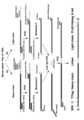
- FIG. 1 shows the design of a DNA library for IVV screening.
- IVV in vitro virus
- FEBS Lett., 414:405-408, 1997; Miyamoto-Sato, E. et al., Nucleic Acids Res., 28:1176-1182, 2000 WO 03/106675 A1 is a technique proposed by Yanagawa, one of the inventors of the present application, and his co-researchers, who succeeded in constructing it for the first time in the world.
- puromycin a type of antibiotic
- a PEG polyethylene glycol
- a cell-free translation reaction is performed using this as a template to bind puromycin to the protein and mRNA.
- IVV is formed, a simple mRNA-protein linking molecule covalently linked via (Miyamoto-Sato, E. et al., Nucleic Acids Res., 31: e78, 2003). From the IVV library constructed in this way, IVV containing proteins that bind to baits (proteins, peptides, antigens, etc.) are picked in vitro, and the genes (mRNA) linked to them are analyzed by reverse transcription and PCR.
- baits proteins, peptides, antigens, etc.
- RNA portion can be eluted and recovered by cleaving the spacer by irradiation with UV at 365 nm (Doi, N. et al., J. Biotechnol., 131:231-239, 2007).
- PCR was carried out by reacting at 94°C for 5 minutes, followed by 16 cycles of 94°C for 30 seconds, 58°C for 30 seconds, and 68°C for 2 minutes, followed by reaction at 68°C for 5 minutes.
- DNA was purified with Wizard SV Gel PCR Clean-Up System (Promega) and collected as 50 ⁇ l of GSP6-GFP-DNA solution.
- Atail sequence 1 ⁇ l, 10 ⁇ KOD plus buffer (TOYOBO) 10 ⁇ l, 2 mM dNTPs (TOYOBO) 10 ⁇ l, 25 mM MgSO 4 4 ⁇ l
- forward primer Flag-His-F (10 pmol/ ⁇ l) 3 ⁇ l
- reverse primer Add RNase-free water to 3 ⁇ l of
- PCR was carried out by reacting at 94°C for 5 minutes, followed by 16 cycles of 94°C for 30 seconds, 58°C for 30 seconds, and 68°C for 2 minutes, followed by reaction at 68°C for 5 minutes.
- DNA was purified with Wizard SV Gel PCR Clean-Up System (Promega) and recovered as 50 ⁇ l of Flag-His Atail-DNA solution.
- Atail-DNA solution 1 ⁇ l, 10 ⁇ KOD plus buffer (TOYOBO) 10 ⁇ l, 2 mM dNTPs (TOYOBO) 10 ⁇ l, 25 mM MgSO 4 4 ⁇ l, forward primer: 16NNS-F or 9NNS-F (10 pmol/ ⁇ l) 3 ⁇ l , reverse primer: Atail (R) (10 pmol/ ⁇ l) 3 ⁇ l, and KOD plus polymerase (TOYOBO) 2 ⁇ l, add RNase-free water to make the total volume 100 ⁇ l, put this in one tube, total 300 ⁇ l (tube 3) were subjected to PCR reaction.
- PCR was carried out by reacting at 94°C for 5 minutes, followed by 12 cycles of 94°C for 30 seconds, 58°C for 30 seconds, and 68°C for 2 minutes, followed by reaction at 68°C for 5 minutes.
- the cDNA library was purified with Wizard SV Gel PCR Clean-Up System (Promega) and recovered as 50 ⁇ l of 16NNS Atail-DNA solution or 9NNS Atail-DNA solution.
- cDNA library was purified with Wizard SV Gel PCR Clean-Up System (Promega) and recovered as 50 ⁇ l of cDNA library 16NNSLib and 9NNSLib.
- Example 2 Preparation of IVV library (2-1) Transcription of library 2 pmol each of cDNA libraries 16NNSLib and 9NNSLib, 8 ⁇ l of 5 ⁇ SP6 buffer, 2 ⁇ l of ATP (100 mM), 2 ⁇ l of CTP (100 mM), UTP (100 mM) 2 ⁇ l, GTP (10 mM) 4 ⁇ l, cap analog (m7G(5')PPP(5')G) (Thermo Fisher Scientific) (40 mM) 5 ⁇ l, Enzyme Mix SP6 RNA polymerase (Promega) 4 ⁇ l, RNase-Free water. After 3 hours of reaction at 37°C, 10 ⁇ l of RQ1 RNase-Free DNase (Promega) was added and further reacted at 37°C for 1 hour.
- RNase-free water was added to the transcription reaction solution to bring the total volume to 100 ⁇ l, and 350 ⁇ l of RLT buffer (Qiagen), 3.5 ⁇ l of 2-mercaptoethanol, and 250 ⁇ l of (100%) ethanol were added to the RNeasy mini spin column.
- Example 3 Preparation of biotinylated DOCK11
- DOCK family proteins have two regions, Dock homology region 1 (DHR1) and DHR2, whose amino acid sequences are highly conserved among families.
- DHR1 binds to phosphatidylinositol 3-phosphate.
- DHR2 domains activate their own specific Rho family G proteins.
- DOCK11 belongs to the DOCK-D group and has a PH domain in the N-terminal region. DOCK11 activates the small G protein Cdc42 and acts as a guanine nucleotide exchange factor (GEF).
- GEF guanine nucleotide exchange factor
- a biotinylation sequence, Flag-tag and His-tag were added to the C-terminus of the amino acid sequence of full-length DOCK11 (aa1-2073, SEQ ID NO: 42).
- a biotinylation sequence, Flag-tag and His-tag were added to the C-terminus of the amino acid sequence of the DOCK11 C-terminal region peptide (aa1516-2073 in SEQ ID NO: 42) containing the DHR2 domain.
- Bio-tag (Nucleic Acids Research, 2009 , Vol. 37, No. 8, page e64) was added with Flag-tag and His-tag.
- Bio-tag 2.37 ⁇ l, 10 ⁇ KOD plus buffer solution (TOYOBO) 40 ⁇ l, 2 mM dNTPs (TOYOBO) 40 ⁇ l, 25 mM MgSO 4 16 ⁇ l, KstartBio-F (10 pmol/ ⁇ l) 12 ⁇ l, Bio-Flag-Histag A stop (10 pmol/ ⁇ l) and 8 ⁇ l of KOD plus polymerase (TOYOBO), RNase-free water was added to make the total volume 200 ⁇ l, and PCR reaction was performed.
- PCR was carried out by reacting at 94°C for 5 minutes, followed by 20 or 25 cycles of 94°C for 30 seconds, 58°C for 30 seconds, and 68°C for 2 minutes, followed by reaction at 68°C for 5 minutes. After confirming the DNA band by agarose gel electrophoresis, the PCR product was purified by Wizard SV Gel PCR Clean-Up System (Promega) and recovered as 50 ⁇ l of DNA solution to obtain KstartBio-Flag-His.
- KstartBio-Flag-His was introduced into the pcDNA 3.3 vector using the TOPO cloning kit (Invitrogen) according to the procedure. After confirming that the resulting clone had the correct sequence, a colony was inoculated and cultured at 37°C for 16 hours. Plasmid pcDNA3.3 TOPO KBioFlagHis was purified from the cell pellet with the PureYield TM Plasmid Maxiprep System (Promega).
- DOCK11 vector pF1KE2360 (Kazusa DNA Res.Inst.) (1 ng/ ⁇ l) 0.5 ⁇ l, KAPA HiFi HS RM 12.5 ⁇ l, 10 ⁇ M TOPOKstartDOCK11-IF-F 0.75 ⁇ l, and 10 ⁇ M DOCK11Bio-IF-R 0.75 ⁇ l in RNase-Free water was added to make the total volume 25 ⁇ l, and the PCR reaction was performed. PCR was carried out at 95°C for 5 minutes, followed by 25 cycles of 98°C for 20 seconds, 60°C for 15 seconds, and 72°C for 1 minute, followed by reaction at 72°C for 1 minute. After confirming the DNA band by agarose gel electrophoresis, the PCR product was purified by Wizard SV Gel PCR Clean-Up System (Promega) and recovered as 30 ⁇ l of DNA solution to obtain Kstart-DOCK11-IF.
- RNase-free water was added to 0.5 ⁇ l of pcDNA3.3 TOPO KBioFlagHis, 10 ⁇ l of KAPA HiFi HS RM, 0.6 ⁇ l of 10 ⁇ M F-Bio, and 0.6 ⁇ l of 10 ⁇ M TOPOKstartINV-R to make the total volume 20 ⁇ l, and a PCR reaction was performed. PCR was carried out by reacting at 95°C for 3 minutes, followed by 25 cycles of 98°C for 20 seconds, 60°C for 15 seconds, and 72°C for 3 minutes, followed by reaction at 72°C for 1 minute. After confirming the PCR product by agarose gel electrophoresis, it was purified by Wizard SV Gel PCR Clean-Up System (Promega) and recovered as 30 ⁇ l of DNA solution to obtain TOPO KBioFlagHisINV.
- Kstart-DOCK11-IF 1.0 ⁇ l, TOPO KBioFlagHisINV 1.0 ⁇ l, 5x infusion HD Enzyme premix 1.0 ⁇ l (Takara) was added with RNase-free water to make the total volume 5 ⁇ l, and reacted at 50°C for 15 minutes. 2.5 ⁇ l was transformed into One Shot TOP10 competent cells and cultured overnight at 37° C. to obtain clones. Sequence analysis of the clone confirmed the plasmid pcDNA3.3 TOPO KDOCK11-BioFLAGHis.
- RIPA buffer 25 mM Tris-HCl, 150 mM NaCl, 1% NP-40, 1% sodium deoxycholate, 0.1% SDS, pH 7.6
- 800 ⁇ l protease inhibitor
- x50 16 ⁇ l of (4-(2-aminoethyl)fluoride, benzenesulfonyl hydrochloride (AEBSF), aprotinin, E-64, leupeptin hemisulfate monohydrate, bestatin, pepstatin A) and 8 ⁇ l of 0.1M PMSF were added to the cells.
- Sample buffer LDS (4x) and 6.8 ⁇ l of 0.2 mM DTT were added to 15 ⁇ l of the eluted fraction E1, heated at 70° C. for 10 minutes, and subjected to SDS-PAGE.
- SDS-PAGE was performed on a 4-12% Bis-Tris NuPAGE gel with MES electrophoresis buffer (Invitrogen) at 200V, 400mA, 35 minutes, followed by Mini Format, 0.2 ⁇ m PVDF, Single application (BIORAD) Trans-Blot Turbo. transcribed.
- the membrane was blocked with Blocking One Buffer: TBST (1:9) and reacted with anti-Flag-HRP (Sigma: A8592) diluted 2:3000 with Blocking One Buffer: TBST (1:9).
- Detection was performed using ChemiDoc (BIORAD) using ECL (Enhanced ChemiLuminescence). Biotinylated full-length DOCK11 was identified as a band with a molecular weight of 246,762 Da, and biotinylated C-terminal region DOCK11 was identified as a band with a molecular weight of 73,404 Da.
- Example 4 Selection of peptides that bind to DOCK11 A selection experiment for peptides that bind to DOCK11 was performed according to the procedure shown in Fig. 4 . (4-1) Immobilization of DOCK11 Biacore immobilized the biotinylated C-terminal region DOCK11 on the sensor chip SA using the Biacore 3000 system. Flow was performed at 10 ⁇ l/min in buffer HBS-P (10 mM HEPES-NaOH, pH 7.4, 150 mM NaCl, 0.005% Tween-20). Pretreatment for immobilization was performed by injecting 10 ⁇ l of a solution containing 50 mM NaOH and 1 M NaCl into flow cells 1 to 4 three times.
- Biotinylated C-terminal region DOCK11 (1 nM) was used to immobilize to flow cells 1-4.
- Flow was in buffer HBS-P, 20 ⁇ l/min. Manual injection of 23 ⁇ l resulted in 1267.4 RU (average) binding to flow cells 1-4.
- an extra wash was performed with a buffer solution HBS-P at 10 ⁇ l/min using 50% Isopropanol, 50 mM NaOH and 1M NaCl.
- the sensor chip was extracted from the machine. 55 ⁇ l of RNase-free water was gently added to the gold film of the sensor chip, and UV light of 365 nm was applied for 20 minutes to cleave the spacer, and the mRNA part was eluted and collected.
- Example 5 Cloning and nucleotide sequence determination (5-1) Cloning and nucleotide sequence Creation of an in-fusion cloning library insert from a peptide library that binds to DOCK11 Library subjected to 8 rounds of selection experiment 1 ⁇ l, 10 ⁇ KOD plus buffer (TOYOBO) 100 ⁇ l, 2 mM dNTPs (TOYOBO) 100 ⁇ l, 25 mM MgSO 4 40 ⁇ l, GFP-F in (10 pmol/ ⁇ l) 30 ⁇ l, His-S28-R in (10 pmol/ ⁇ l) 30 ⁇ l, RNase-free water was added to 20 ⁇ l of KOD plus polymerase (TOYOBO) to bring the total volume to 1000 ⁇ l, followed by PCR reaction.
- TOYOBO 10 ⁇ KOD plus buffer
- TOYOBO 2 mM dNTPs
- PCR was carried out by reacting at 94°C for 5 minutes, followed by 8 cycles of 94°C for 30 seconds, 58°C for 30 seconds, and 68°C for 2 minutes, followed by reaction at 68°C for 5 minutes.
- the cDNA library was purified with Wizard SV Gel PCR Clean-Up System (Promega) and recovered as 50 ⁇ l of DNA solution to obtain GFP-DC-S8in.
- PCR was carried out by reacting at 94°C for 2 minutes, followed by 25 cycles of 98°C for 10 seconds and 68°C for 5 minutes and 11 seconds.
- the cDNA library was purified with Wizard SV Gel PCR Clean-Up System (Promega) and recovered as 40 ⁇ l of DNA solution to obtain KGFP-S28 3.3 vector.
- a nuclear localization signal sequence gene CCTGCTGCCAAGAGGGGTCAAGTTGGAC (SEQ ID NO: 47) was introduced upstream of the GFP sequence, and 8 ⁇ l of pcDNA 3.3 vector containing a stop codon, 80 ⁇ l of 10 ⁇ KOD plus buffer (TOYOBO), 80 ⁇ l of 2 mM dNTPs (TOYOBO), 25 mM MgSO. 4 Add RNase-free water to 32 ⁇ l, S28iv-25F (10 pmol/ ⁇ l) 24 ⁇ l, GFPiv-71R (10 pmol/ ⁇ l) 24 ⁇ l, and KOD plus polymerase (TOYOBO) 16 ⁇ l to make the total volume 800 ⁇ l, and perform PCR reaction.
- PCR was carried out by reacting at 94°C for 2 minutes, followed by 25 cycles of 98°C for 10 seconds and 68°C for 5 minutes and 11 seconds.
- the cDNA library was purified with Wizard SV Gel PCR Clean-Up System (Promega) and recovered as 40 ⁇ l of DNA solution to obtain NLS-GFP-S28 3.3 vector.
- PCR was carried out by reacting at 94°C for 5 minutes, followed by 8 cycles of 94°C for 30 seconds, 58°C for 30 seconds, and 68°C for 2 minutes, followed by reaction at 68°C for 5 minutes.
- the cDNA library was purified with Wizard SV Gel PCR Clean-Up System (Promega) and recovered as 50 ⁇ l of DNA solution to obtain NLS-GFP-DC-S8in.
- NLS-GFP-DC-S8 in 0.08 ⁇ l, NLS-GFP-S28 3.3 vector 0.14 ⁇ l, 5x infusion HD Enzyme premix 1.0 ⁇ l (Takara), add RNase-Free water to bring the total volume to 5 ⁇ l, and heat at 50°C. React for 15 minutes. 2.5 ⁇ l of the reaction solution was transformed into One Shot TOP10 competent cells and cultured overnight at 37° C. to obtain NLS-GFP-DCS8 clones. Sequence analysis of the obtained clones was performed by Eurofin DNA sequence contract service, ValueRead Premix.
- Example 6 Evaluation of activity of DOCK11-binding peptide clones (6-1) Preparation of protein In-frame clones were inoculated from a master plate onto LB medium containing 20 ⁇ g/ml of carbenicillin and cultured at 37°C for 16 hours. bottom. Plasmids were purified from cell pellets with the PureLink HiPure Plasmid Maxiprep Kit (Invitrogen).
- the resin was transferred to a new tube, and 1000 ⁇ l of the culture supernatant expressing DCS3, DCS5 or DCS8 was mixed with 40 ⁇ l of biotinylated full-length DOCK11, biotinylated C-terminal region DOCK11, or bait-free treated resin, and mixed in a mini disc rotor. (Bio craft) and combined at 4° C. for 1 hour 30 minutes. After removing the solution, washing was performed three times with 500 ⁇ l of TBST (0.1% Tween 20), and the recovered resin was subjected to Western blotting.
- a pull-down assay (Fig. 5) was performed against C-terminal DOCK11 containing the DHR2 region by DOCK11-binding peptides.
- Three of the clones subjected to three rounds of selection experiments were evaluated and positive for DCS3-1 (SEQ ID NO: 1), DCS3-2 and DCS3-3.
- DCS3-1 (SEQ ID NO: 1), DCS5-15 (SEQ ID NO: 4), DCS8-42 (SEQ ID NO: 9) and DCS8-42TN (SEQ ID NO: 10) among clones of 3 rounds, 5 rounds, and 8 rounds was subjected to a pull-down assay against the C-terminal DOCK11 containing the DHR2 region with a DOCK11-binding peptide (Fig. 6).
- DCS3-1 SEQ ID NO: 1
- DCS8-42 SEQ ID NO: 9
- DCS8-42TN (SEQ ID NO: 10) obtained from 8 rounds strongly bind to DOCK11. I understand.
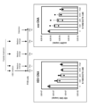
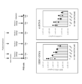
- Example 7 Anti-HBV activity of screened DOCK11-binding peptide (7-1) Anti-HBV activity of DOCK11G-binding peptide in HepG2-NTCP-C4 cells Regarding clones that were positive in pull-down experiments against C-terminal DOCK11 containing DHR2 region , DOCK11-binding peptides in HepG2-NTCP-C4 cells (HepG2 cells overexpressing the HBV receptor NTCP, Iwamoto, M. et al. Biochem. Biophys. Res. Commun. 443:808-813, 2014) was assessed for anti-HBV activity (Fig. 7).
- a peptide-containing plasmid (DCS3-1) for intracellular expression and a plasmid (N-DCS3-1) added with a nuclear localization signal PAAKRVKLD (SEQ ID NO: 40) were prepared.
- the HBV used was HBV derived from HepG2.2.15 cells in which a helper plasmid lacking the packaging signal ( ⁇ ) present upstream of the HBV core was stably expressed in HepG2 cells. After HepG2-NTCP-C4 cells were infected with HepG2.2.15-derived HBV, plasmids encoding various peptides were transfected with Lipofectamine 3000, respectively. Samples were collected 3-5 days after transfection and evaluated for HBV-DNA and cccDNA.
- DCS5-1, DCS5-7, DCS5 among DCS5-1, DCS5-4 (SEQ ID NO: 2), DCS5-5 (SEQ ID NO: 3), DCS5-15 (SEQ ID NO: 4), DCS5-7, DCS5-33 -33 did not decrease HBV DNA copy number and cccDNA copy number compared to controls, but DCS5-4 (SEQ ID NO: 2), DCS5-5 (SEQ ID NO: 3), DCS5-15 (SEQ ID NO: 3), DCS5-15 (SEQ ID NO: 3) number 4) decreased the copy number of HBV DNA compared to controls. In addition, the copy number of cccDNA was also decreased.
- DCS8-42TN having a sequence homologous to DCS8-42 (peptide consisting of a partial region of TNK2 (Ack1) showing high homology with DCS8-42, SEQ ID NO: 10)
- DCS8-59R having a sequence homologous to DCS8-59 (peptide consisting of a partial region of radixin showing high homology with DCS8-59, SEQ ID NO: 11)
- DCS8-59C peptide consisting of a partial region of ⁇ -Centractin showing high homology with DCS8-59, SEQ ID NO: 12
- HepG2-NTCP-C4 cells were infected with HBV for 20 days and evaluated. No. 8) and DCS8-42TN (SEQ ID NO: 10) also decreased the HBV DNA copy number and the cccDNA copy number (Fig. 9).
- the liver has asialoglycoprotein receptors for specifically recognizing and taking up asialoglycoprotein in the blood.
- Anti-asialoglycoprotein receptor antibodies are also thought to be taken up into liver cells after binding to the receptor.
- selection experiments for single-chain antibodies that bind to asialoglycoprotein receptors were conducted using the IVV method. did.
- Example 8 Preparation of biotinylated ASGR First, a biotinylated asialoglycoprotein receptor as an antigen was prepared.
- Asialoglycoprotein receptor (ASGR) is a type II single-pass transmembrane protein with the N-terminus directed intracellularly and the carbohydrate recognition sites (CRDs) directed extracellularly, as shown in FIG.
- CTDs carbohydrate recognition sites
- a construct was prepared by removing the transmembrane domain and adding a biotinylated sequence and the like. Since human hepatocytes have receptors ASGR1 and ASGR2 and form hetero-oligomers, we created biotinylated ASGR1ex and biotinylated ASGR2ex by adding biotin to their extracellular domains.
- the extracellular domains of ASGR1 and ASGR2 were cloned as shown in FIG. cDNA Library, Human Liver (1 ng/ ⁇ l) (Takara Bio) 1 ⁇ l, KAPA HiFi HS RM 10 ⁇ l, 10 ⁇ M ASGR1-ex-if-F1 0.6 ⁇ l or ASGR2-ex-if-F1 0.6 ⁇ l, and 10 ⁇ M ASGR1-if- RNase-free water was added to 0.6 ⁇ l of R2 or 0.6 ⁇ l of ASGR2-if-R2 to make the total volume 20 ⁇ l, and PCR reaction was performed.
- PCR was carried out at 95°C for 5 minutes, followed by 25 cycles of 98°C for 20 seconds, 60°C for 15 seconds, and 72°C for 1 minute, followed by reaction at 72°C for 1 minute. After confirming the DNA band by agarose gel electrophoresis, the PCR product was purified by Wizard SV Gel PCR Clean-Up System (Promega) and collected as a 30 ⁇ l DNA solution to obtain ASGR1-if or ASGR2-if.
- TBS-washed M2 agarose beads (ANTI-FLAG (registered trademark) M2 Affinity Gel, Sigma, A2220) were added to the obtained cell extract and mixed at 4°C for 16 hours. After washing three times with TBST (10x TBS-t 1% Tween-20, Nakarai, 12749-21), competitive elution was performed with 150 ⁇ g/mL 3x FLAG peptide (Sigma, F4799) diluted with TBS.
- EKMax buffer 500 mM Tris-HCl, pH 8.0, 10 mM CaCl 2 , 1% Tween-20.
- EKMax TM Enterokinase (Thermo Fisher Scientific) was added, EKMax digestion was performed by rotating at 37°C O/N (16.5h), SA beads were adsorbed on a magnet stand, and the supernatant was recovered.
- Pretreated EK Away resin was added to the recovered supernatant, and the mixture was rotated at room temperature for 15 minutes to remove EKMax. After centrifugation, the supernatant was recovered (5000 rcf 2 min x 2) and detected by Western blotting using SDS-PAGE and Flag antibody, and an ASGR1ex molecular weight 26394 Da band could be confirmed.
- Example 9 Construction of single-chain antibody cDNA library (9-1) DNA library design ).
- V H chain and V L chain are composed of three CDRs (Complementarity-determining regions) and four FRs (Framework regions) (Fig. 15 right).
- CDRs are composed of a variety of amino acid sequences for each antibody, enabling them to bind to various antigens.
- Artificial antibody fragments include single-chain antibodies (scFv), in which VH and VL chains are linked by a peptide linker (Fig. 16). A linker ( sequence 48) is widely used.
- Example 10 Construction of mouse-derived single-chain antibody cDNA library As shown in Fig. 17, a mouse-derived single-chain antibody cDNA library was constructed using mouse spleen Poly A+ RNA as a starting material. Preparation of H chain DNA solution, preparation of L chain DNA solution, and unification PCR of H chain and L chain were carried out. (Nucleic Acids Research, 2009, Vol. 37, No. 8 e64).
- H-chain DNA solution For preparation of a single-chain antibody cDNA library, first, an H-chain DNA solution was prepared. 11 ⁇ l of mouse spleen Poly A+ RNA (5 ⁇ g/ ⁇ l) (DEPC-treated water) (CLONTECH) diluted 100-fold with RNase-free water, 22 ⁇ l of 5 ⁇ RT buffer (TOYOBO), (10 mM) Mix 11 ⁇ l of dNTPs (TOYOBO), 27.5 ⁇ l of forward primer MulgG1/2 (1 pmol/ ⁇ l), and 27.5 ⁇ l of forward primer MulgG3 (1 pmol/ ⁇ l), react at 65°C for 9 minutes, then immediately cool to 4°C and leave at 4°C for 2 minutes.
- TOYOBO 5 ⁇ RT buffer
- PCR reaction was carried out. PCR was performed at 96°C for 5 minutes, followed by 25 cycles of 96°C for 30 seconds, 50°C for 30 seconds, and 72°C for 1 minute, followed by reaction at 72°C for 5 minutes.
- each DNA (19 types) was dissolved in 20 ⁇ l of RNase-free water.
- 1 ⁇ l of each synthesized DNA solution (19 types) 2 ⁇ l of each corresponding HB primer (10 pmol/ ⁇ l) shown in HB primer, 10 ⁇ l of 10 ⁇ PCR buffer (TOYOBO), 10 ⁇ l of (2 mM) dNTPs (TOYOBO) , VH forward primer HF1:HF2:HF3:HF4 (1:1:1:1) mixture (10pmol/ ⁇ l) 2 ⁇ l, KOD DASH polymerase (TOYOBO) 0.5 ⁇ l, and RNase-free water were added to HF primer.
- the total volume was 100 ⁇ l, and each was subjected to PCR reaction. PCR was performed at 96°C for 5 minutes, followed by 20 cycles of 96°C for 30 seconds, 50°C for 30 seconds, and 72°C for 1 minute, followed by reaction at 72°C for 5 minutes.
- the amplified gene was subjected to 2% agarose gel electrophoresis to confirm bands of 500-900 bp, followed by phenol/chloroform treatment and ethanol precipitation. After centrifugation for about 15 minutes, each DNA (19 types) was dissolved in 10 ⁇ l of RNase-free water. The obtained 19 DNAs were subjected to 2% low melting point agarose gel (Sigma) electrophoresis, and respective bands were excised.
- PCR was performed at 96°C for 5 minutes, followed by 25 cycles of 96°C for 30 seconds, 48°C for 30 seconds, and 72°C for 1 minute, followed by reaction at 72°C for 5 minutes. Bands of 500-900 bp were confirmed for each amplified gene by 2% agarose gel electrophoresis, and treated with phenol/chloroform. Ethanol precipitation was performed on the resulting solution. After centrifugation for about 15 minutes, each DNA (18 types) was dissolved in 20 ⁇ l of RNase-free water.
- PCR was performed at 96°C for 5 minutes, followed by 20 cycles of 96°C for 30 seconds, 48°C for 30 seconds, and 72°C for 1 minute, followed by reaction at 72°C for 5 minutes.
- the amplified gene was subjected to 2% agarose gel electrophoresis to confirm bands of 500-900 bp, followed by phenol/chloroform treatment and ethanol precipitation. After centrifugation for about 15 minutes, each DNA (18 types) was dissolved in 10 ⁇ l of RNase-free water. The obtained 18 DNAs were subjected to 2% low melting point agarose gel (Sigma) electrophoresis, and respective bands were excised. Each DNA (18 species) was dissolved in 10 ⁇ l of RNase-free water.
- PCR was performed at 96°C for 5 minutes, followed by 15 cycles of 96°C for 30 seconds, 58°C for 30 seconds, and 72°C for 1 minute, followed by reaction at 72°C for 5 minutes.
- the resulting DNA was electrophoresed on a 1% low melting point agarose gel (Sigma) and each band was excised.
- the DNA was dissolved in 10 ⁇ l of RNase-free water to obtain mouse-derived single-chain antibody library DNA.
- Example 11 Construction of human-derived single-chain antibody cDNA library
- human bone-marrow-derived Poly A+ RNA was used as a starting material for construction of a single-chain antibody cDNA library from human bone marrow-derived lymphocytes. went to Using multiple specific primers as in the case of mice, H-chain DNA solution preparation, L-chain DNA solution preparation, and H- and L-chain unification PCR were performed to obtain a single human sample for IVV selection experiments.
- a chain antibody cDNA library was constructed. (J. Mol. Biol. 1991, 222, 581-597, Nature Biotechnology, 2005, 23, 344-348, Antibody Engineering, Springer Lab. Manual (2001) 93-108).
- mouse H-chain DNA and mouse L-chain DNA or human H-chain DNA and human L-chain DNA prepared as shown in Fig. 17 were each subjected to random mutagenesis by error-prone PCR using Mutazyme II.
- a cDNA library of mouse mutated single-chain antibodies was prepared by PCR that unifies the H and L chains of mutated mouse H-chain DNA and mutated mouse L-chain DNA.
- a cDNA library of human mutated single-chain antibodies was prepared by PCR for unifying H and L chains of human mutated H-chain DNA and human mutated L-chain DNA.
- Example 12 Selection of single-chain antibodies that bind to ASGR A selection experiment for single-chain antibodies that bind to ASGR was performed according to the procedure shown in Fig. 19 .
- RNA was purified by RNeasy Mini kit (Qiagen). That is, RNase-free water was added to the transcription reaction solution to bring the total volume to 100 ⁇ l, and 350 ⁇ l of RLT buffer (Qiagen), 5 ⁇ l of 2-mercaptoethanol, and 250 ⁇ l of (100%) ethanol were added and applied to an RNeasy mini spin column.
- RPE buffer Qiagen
- the biotinylated extracellular domain ASGR2ex was then used to immobilize to flow cells 3-4.
- Flow was in buffer HBS-P, 20 ⁇ l/min.
- the fixed amount of bait is shown in Table 7.
- an extra wash was performed with a buffer solution HBS-P at 10 ⁇ l/min using 50% Isopropanol, 50 mM NaOH and 1M NaCl.
- Biacore was used to increase the selection pressure in the selection experiment step by step as shown in Table 7.
- Example 13 Cloning and base sequence determination (13-1) Cloning and base sequence An in-fusion cloning library was inserted from a single-chain antibody library that binds to ASGR. 1 ⁇ l of library subjected to three rounds of selection experiments, 100 ⁇ l of 10 ⁇ KOD plus buffer (TOYOBO), 100 ⁇ l of 2 mM dNTPs (TOYOBO), 40 ⁇ l of 25 mM MgSO 4 , 30 ⁇ l of T7-long-F in (10 pmol/ ⁇ l), RNase-free water was added to 30 ⁇ l of Flag (Histag) in R (10 pmol/ ⁇ l) and 20 ⁇ l of KOD plus polymerase (TOYOBO) to bring the total volume to 1000 ⁇ l, and a PCR reaction was performed.
- TOYOBO 10 ⁇ KOD plus buffer
- TOYOBO 2 mM dNTPs
- 40 ⁇ l of 25 mM MgSO 4 40 ⁇ l of 25 mM M
- PCR was carried out by reacting at 94°C for 5 minutes, followed by 8 cycles of 94°C for 30 seconds, 58°C for 30 seconds, and 68°C for 2 minutes, followed by reaction at 68°C for 5 minutes.
- the cDNA library was purified with Wizard SV Gel PCR Clean-Up System (Promega) and recovered as 50 ⁇ l of DNA solution to obtain T7-ASGR3.
- PCR was carried out by reacting at 94°C for 2 minutes, followed by 25 cycles of 98°C for 10 seconds and 68°C for 5 minutes and 11 seconds.
- the cDNA library was purified with Wizard SV Gel PCR Clean-Up System (Promega) and recovered as 40 ⁇ l of DNA solution to obtain KIgk-stop 3.3 vector.
- T7-ASGR3 0.08 ⁇ l, KIgk-stop 3.3 vector 0.14 ⁇ l, 5x infusion HD Enzyme premix 1.0 ⁇ l (Takara) were mixed, RNase-Free water was added to make the total volume 5 ⁇ l, and the mixture was reacted at 50°C for 15 minutes.
- 2.5 ⁇ l of the reaction solution was transformed into One Shot TOP10 competent cells and cultured overnight at 37° C. to obtain a KIgk-ASGR3 clone. Sequence analysis of the obtained clones was performed by Eurofin DNA sequence contract service, ValueRead Premix.
- Example 14 Activity evaluation of ASGR-binding single-chain antibody clone (14-1) Protein preparation Clones were inoculated from a master plate onto an LB medium containing 20 ⁇ g/ml of carbenicillin and cultured at 37°C for 16 hours. bottom. Plasmids were purified from cell pellets with the PureLink HiPure Plasmid Maxiprep Kit (Invitrogen).
- a pull-down assay (Figs. 20, 21) was performed using an ASGR-binding single-chain antibody.
- ASGR3-10M recognized both antigens, and ASGR3-39D recognized the ASGR1 antigen particularly strongly.
- Clones ASGR4-70D, ASGR5-24M subjected to 4 and 5 rounds of selection experiments recognized the antigen compared to controls.
- ASGR4-70D and ASGR5-24M particularly strongly recognized the ASGR1 antigen.
- Fig. 22 shows a schematic diagram of a method for selectively delivering DOCK11-binding peptides to hepatocytes using an anti-ASGR single-chain antibody as a carrier for intrahepatocyte delivery.
- DOCK11-binding peptides conjugated with anti-ASGR antibodies are packaged into early endosomes by endocytosis after binding to receptors.
- a cleavage sequence that is cleaved by an enzyme in the endosome upstream of the DOCK11-binding peptide, it is cleaved and the peptide is dissociated.
- a membrane permeabilization peptide is added downstream of the DOCK11-binding peptide, thereby releasing the peptide into the cytoplasm. Since the target DOCK11 is a protein localized in cells or nuclear fractions, a nuclear localization signal is added when delivered to the nucleus.
- the sequence recognized by the endogenous protease Furin was used as the cleavage sequence, the membrane fusion promoting peptide S28 or S39 as the membrane permeation promoting peptide, and PAAKRVKLD (SEQ ID NO: 40) as the nuclear localization signal.
- the DOCK11-binding peptide DCS8-42TN causes actin morphology changes when the plasmid is introduced into cells by the transfection method and the protein is expressed. It was verified whether or not the fusion of the construct shown in FIG. 23A would also cause a similar morphological change in actin when it was directly extracellularly incorporated into HepG2 cells. At the same time, it was also verified whether the cleaved sequence was cleaved by Furin and functioned. Using the construct of FIG. 23B, it was verified whether the membrane fusion-promoting peptide was functioning. Using the construct in Figure 23C, it was verified whether the nuclear localization signal was functioning.
- SEQ ID NO:90 shows the amino acid sequence of the cleavage sequence+DCS8-42TN peptide+S28+nuclear localization signal portion of N-10M-D42TN.
- the 21st to 28th amino acids are the Flag tag
- the 29th to 35th amino acids are the His tag.
- a single-chain antibody-peptide fusion was added to HepG2 cells with or without the DCS8-42TN peptide.
- actin fragmentation occurred in the cells with the peptide applied, whereas actin fragmentation was not observed in the cells without the peptide.
- GFP signal indicating the localization of the peptide was observed in the cytoplasm, and it was found that the cleavage sequence was cleaved by Furin and functioned, and the fusion-promoting peptide functioned.
- Example 15 Anti-HBV activity of DOCK11-binding peptide in PXB cells Liver cells, persistently infected with hepatitis B virus (HBV). Therefore, we evaluated the anti-HBV activity of DOCK11-binding peptides using PXB cells. Since the DOCK11-binding peptide was added from the outside of the cell, the anti-ASGR single-chain antibody-peptide fusion N-10M-D42TN was used and subjected to HBV assay in PXB cells.
- HBV hepatitis B virus
- Figures 27A and 27B show the anti-HBV activity of DOCK11-binding peptides in PXB cells.
- N-10M-D42TN markedly decreased the copy numbers of HBV-DNA and cccDNA, and showed high anti-HBV activity in a dose-dependent manner. It was found to exhibit very high anti-HBV activity.
- results with good reproducibility were obtained as shown in FIG. 27B.
- the DOCK11-binding peptide DCS8-42TN is the sequence of residues 674-689 of non-receptor tyrosine kinase Ack1 (NM_001387713.1, SEQ ID NOS: 49, 50). Therefore, we examined whether DOCK11 and Ack1 interact in cells by co-immunoprecipitation experiments.
- the peptide DCS8-42TN was fused with an anti-ASGR single-chain antibody so that it was taken up into cells.
- the fusion of DCS8-42TN and a single chain antibody is hereinafter referred to as N-10M-D42TN.
- DOCK11 is a member of the DOCK-D subfamily and is known to be a guanine nucleotide exchange factor (GEF) that converts Cdc42 from an inactive (GDP-bound) to an active (GTP-bound) form.
- GEF guanine nucleotide exchange factor
- Ack1 is an active Cdc42-binding protein and is activated by binding specifically to GTP-bound Cdc42 (Prieto-Echague, V., Miller, J., Signal Transduct, 1-9, 2011).
- Ack1 is known to phosphorylate WASP and promote actin polymerization (Yokoyama, N., Lougheed, J., and Miller, W.T. (2005). Phosphorylation of WASP by the Cdc42-associated Kinase ACK1. Journal of Biological Chemistry 280, 42219-42226.). Transfection of HepG2 cells with siRNAs targeting DOCK11 and Ack1 and staining of actin filaments with fluorescent phalloidin showed fragmentation of actin filaments and increased microprojections in the cytoplasm (FIGS. 30A-C).
- HBV binds to the receptor NTCP on the cell membrane and then enters the cell by endocytosis together with the epidermal growth factor receptor EGFR (Iwamoto, M. et al., Proc Natl Acad Sci USA, 116 , 8487-8492, 2019).
- Ack1 activated by Cdc42 interacts with EGFR in response to EGF stimulation and contributes to EGFR endocytosis (Shen, F. et al., Mol. Biol. Cell, 18, 732-742, 2007; Lin, Q. et al., J. Biol. Chem.
- FIG. 32A shows a schematic of this mechanism. That is, when EGFR is activated by EGF stimulation, it undergoes phosphorylation and ubiquitination. Ack1 binds to and activates DOCK11-activated Cdc42, and binds to and phosphorylate EGFR. As a result, both EGFR-Ack1 complexes are endocytosed and degraded. On the other hand, inhibition of DOCK11 function by N-10M-D42TN inhibits Ack1 activation, preventing Ack1 and phosphorylated EGFR from being endocytosed and degraded.
- N-10M-D42TN does not affect EGFR phosphorylation and inhibits Ack1 activation, thereby inhibiting Ack1 and EGFR endocytosis.
- HBV is known to utilize the host's DNA repair mechanism, especially the ATR signaling pathway, when synthesizing cccDNA from rcDNA (Luo, J. et al. mBio, 11, e03423-19, 2020). Since ATR is recruited according to actin accumulation at sites of DNA damage (Wang, Y-H. et al. Nat Commun, 8, 2118-2133, 2017), DOCK11 affects the ATR signaling pathway through actin polymerization. Possibility of contributing to cccDNA synthesis is conceivable. UV irradiation of HepG2 cells increased DOCK11 mRNA (FIG. 34A), suggesting that DOCK11 contributes to the DNA repair mechanism. In addition, knockdown of DOCK11 suppressed the activation of Chk1 during DNA repair (Fig. 34B,C), indicating that DOCK11 is required for the activation of the ATR signaling pathway.
- N-10M-D42TN suppressed Chk1 phosphorylation in a time-dependent manner (Fig. 34D,E), indicating that inhibiting DOCK11 function inhibits the ATR signaling pathway. It has been suggested. Immunostaining of HepG2 cells with anti-pChk1 antibody showed that knockdown of DOCK11 suppressed the phosphorylation of Chk1 in the nucleus (FIG. 35A). In addition, phosphorylation of Chk1 in the nucleus was similarly suppressed by N-10M-D42TN treatment (Fig. 35B). These results also suggest that DOCK11 is essential for activation of the ATR signaling pathway in the nucleus and that N-10M-D42TN inhibits it.
- ⁇ H2AX is a phosphorylated histone H2AX and a marker for sites of DNA damage, suggesting that DOCK11 accumulates at sites of DNA damage and activates the ATR signaling pathway.
- N-10M-D42TN reduced the expression of ⁇ H2AX similarly to DOCK11 knockdown (Fig. 37D), but ⁇ H2AX did not co-localize with DOCK11 (Fig. 37F,G; Wang, Y-H. et al. Nat Commun, 8, 2118, 2017). This suggests that N-10M-D42TN inhibits DOCK11 activation of the ATR signaling pathway at sites of DNA damage.
- HBV cannot utilize the DNA repair mechanism when synthesizing cccDNA from rcDNA, and it is thought that infection is suppressed.
- the DOCK11-binding peptide N-10M-D42TN inhibited the binding of DOCK11 and Ack1 and inhibited the GEF activity of DOCK11.
- actin filaments contribute to endocytosis (Toshima, JY. et al., eLife 5, e10276, 2016)
- N-10M-D42TN inhibits actin polymerization to promote HBV activation. It may be blocking intrusions.
- the DOCK11-binding peptide N-10M-D42TN may also inhibit the activation of the ATR signaling pathway by DOCK11 assembled and accumulated at sites of DNA damage, thus inhibiting the repair process from rcDNA to cccDNA. Conceivable.
- the DOCK11-binding peptide N-10M-D42TN is thought to suppress HBV infection by inhibiting EGFR endocytosis and the repair process from rcDNA to cccDNA.
- Example 16 Efficacy test using HBV-infected PXB mice PXB mice were infected with HBV, and the following experiment was performed for the purpose of confirming the efficacy of administration of the test substance (N-10M-D42TN).
- Virus used The virus used was HBV provided by Phoenix Bio Co., Ltd.
- Virus name Hepatitis B virus Strain name: PBB004 (Genotype C)
- BSL 2 Viral titer: 1.1E+09 copies/mL (2 tubes of 10 ⁇ L/tube)
- Storage Store in an ultra-low temperature freezer
- Preparation method Thaw one bottle of frozen virus solution and adjust to 1.0E+06 copies/mL using physiological saline (Otsuka Pharmaceutical Factory Co., Ltd.).
- Animal care conditions 16-4.1 Husbandry Conditions (SOP/Environment/504) Room temperature 24 ⁇ 3 °C, humidity 50 ⁇ 20%, ventilation (10 to 25 times/hour), lighting 12 hours (8:00 to 20:00)
- mice 16-4.6 Grouping (SOP/Study/002) Six weeks after virus inoculation, blood is collected, and based on the amount of HBV DNA in the blood, mice are sorted into groups at eight weeks after virus inoculation (day of first administration of the test substance).
- Virus Inoculation HBV is inoculated into the mouse tail vein at 1.0E+05 copies/100 ⁇ L/body.
- Test Substance From the 8th week after virus inoculation (Day 0) to the day before sample collection (Day 27), 300 ⁇ L/body is intraperitoneally administered to mice once every 2 to 3 days (Mon, Wed, Fri).
- livers are collected and weighed. Livers are partially cryopreserved and the rest formalin-fixed for histopathological examination for HBV DNA and cccDNA. Lungs, spleens and kidneys are also fixed in formalin.
- HBsAg, HBeAg and HBcrAg Serum HBsAg concentration measurement was performed by SRL Co., Ltd. (Tokyo).
- Lumipulse registered trademark
- Presto II Flujirebio Co., Ltd., Tokyo
- CLIA ChemiLuminescence Enzyme ImmunoAssay
- the measurement range was 0.005 to 150 IU/mL.
- the sample to be measured in this test was diluted 30 times, and the measurement range at the same dilution was 0.15 to 4500 IU/mL.
- Serum HBsAg concentrations were measured by SRL Co., Ltd. (Tokyo).
- Lumipulse registered trademark
- Presto II Flujirebio Co., Ltd., Tokyo
- ChemiLuminescence Enzyme ImmunoAssay (CLEIA)
- the measurement range was from 0.1 to 1590 COI.
- the sample to be measured in this test was diluted 30 times, and the measurement range at the same dilution was 3 to 47,700 COI.
- Serum HBc-rAg concentrations were measured by SRL Co., Ltd. (Tokyo).
- LUMIPULSE HBcrAg, LUMIPULSE F (Fujirebio Co., Ltd., Tokyo) using ChemiLuminescence Enzyme ImmunoAssay (CLEIA) was used.
- the lower limit of measurement was 3.0 log U/mL.
- the dilution ratio of the sample to be measured in this test was 300 times, and the lower limit of measurement at the same dilution ratio was 5.5 log U/mL.
- h-Alb concentration was measured using a latex agglutination immunoturbidimetric assay (LZ test 'Eiken' U-ALB, Eiken Chemical Co., Ltd., Tokyo) using an automatic analyzer BioMajestyTM (JCA-BM6050, JEOL, Tokyo). measured in
- ALT ALT was measured by PhoenixBio using serum at autopsy. Plasma ALT activity was measured using 10 ⁇ L of collected plasma.
- the object to be measured is a diarylimidazole leuco dye (diarylimidazole leuco dye is colored blue by hydrogen peroxide generated by pyruvate oxidase and peroxidase), and DRYCHEM 7000/NX500sV was used for the measurement.
- HBV DNA in Liver DNA was extracted from RNAlater-immersed liver samples using DNeasy (registered trademark) Blood & Tissue Kits (Qiagen Co., Ltd., Tokyo), and the DNA was dissolved in Nuclease-free water. After DNA concentration was measured with BioPhotometer (registered trademark) 6131 (Eppendorf Co., Ltd.), the final concentration was adjusted to 20 ng/ ⁇ L using Nuclease-free water.
- a PCR reaction solution was prepared using 5 ⁇ L of dissolved DNA stock solution or diluted DNA and TaqMan (registered trademark) Fast Advanced Master Mix.
- the CFX96 Touch TM Real-Time PCR Detection System was used for PCR reaction and analysis.
- the PCR reaction was carried out as follows: 50°C 2 minutes ⁇ 95°C 20 seconds ⁇ (95°C 3 seconds ⁇ 60°C 32 seconds) ⁇ 53 cycles.
- HBV DNA concentration in liver was calculated by averaging 2 wells.
- the sequences of the primers and probes used are listed in Table 12 below.
- the lower limit of detection by the quantitation method is 50 copies/100 ng DNA.
- the HBV DNA standard used serum obtained from HBV-infected PXB mice. The HBV DNA concentration contained in this serum was quantified by digital PCR.
- cccDNA When measuring HBV DNA in liver, 5 ⁇ L of purified DNA stock solution or diluted DNA was prepared using TaqMan® Fast Advanced Master Mix. Using the PCR Detection System, the PCR reaction was performed as follows: 50°C for 2 minutes ⁇ 95°C for 20 seconds ⁇ (95°C for 3 seconds ⁇ 60°C for 32 seconds) ⁇ 55 cycles. The HBV cccDNA concentration in the liver was the average of 2 wells. The sequences of the primers (Takara Bio Inc., Shiga) and the probes (Takara Bio Inc.) used are shown in Table 13. The lower limit of detection by the quantification method is 1.0 ⁇ 10 2 . copies/100 ng DNA, and the HBV cccDNA standard utilized a plasmid containing the entire HBV genome sequence.
- HsAg immunostaining Liver tissue was fixed in 10% neutral buffered formalin solution and then replaced with 70% ethanol. These samples were requested to Nara Pathological Research Institute (Nara) to prepare paraffin-embedded blocks by standard methods, and then sliced. got After deparaffinization of paraffin sections, they were subjected to antigen retrieval by microwave. Primary antibody (HBsAg (anti-HBsAg antibody, Code: OBT0990, Bio-Rad AbD Serotec Limited, Oxford, UK) or HBcAg (anti-HBcAg antibody, Code: PAB14506, Abnova, Taipei City, Taiwa)) was incubated at 4°C.
- HBsAg anti-HBsAg antibody, Code: OBT0990, Bio-Rad AbD Serotec Limited, Oxford, UK
- HBcAg anti-HBcAg antibody, Code: PAB14506, Abnova, Taipei City, Taiwa
- the primary antibody was reacted with a biotin-avidin-peroxidase complex and then developed with DAB. After staining cell nuclei with hematoxylin, these sections were dehydrated, cleared and mounted. After that, a microscopic examination was performed using an optical microscope at Hamley Co., Ltd.
- N-10M-D42TN did not exacerbate hepatitis compared to control (PBS).
- PBS control
- N-10M-D42TN reduced the copy number of HBV DNA compared to the control administration group.
- N-10M-D42TN decreased the copy number of HBV DNA compared to the control administration group.
- DOCK11-binding peptide (N-10M-D42TN) reduced HBV-DNA and cccDNA in the liver in animal experiments using HBV-infected human hepatocyte chimeric mice, demonstrating a clear anti-HBV effect. has been proven to have Moreover, in the in vitro and in vivo assay systems, DOCK11-binding peptide (N-10M-D42TN) was administered after sufficient HBV infection. After being secreted outside the cell once, it is highly likely that it prevents the process of re-infection, in which it reenters the cell.
- the DOCK11-binding peptide exhibited an anti-HBV effect by inhibiting the repair process from rcDNA to cccDNA and inhibiting re-entry of HBV particles into cells, such as by inhibiting EGFR endocytosis. It is suggested that
Landscapes
- Health & Medical Sciences (AREA)
- Chemical & Material Sciences (AREA)
- Life Sciences & Earth Sciences (AREA)
- Organic Chemistry (AREA)
- General Health & Medical Sciences (AREA)
- Medicinal Chemistry (AREA)
- Genetics & Genomics (AREA)
- Engineering & Computer Science (AREA)
- Molecular Biology (AREA)
- Pharmacology & Pharmacy (AREA)
- Veterinary Medicine (AREA)
- Bioinformatics & Cheminformatics (AREA)
- Animal Behavior & Ethology (AREA)
- Public Health (AREA)
- Biophysics (AREA)
- Proteomics, Peptides & Aminoacids (AREA)
- Immunology (AREA)
- Biochemistry (AREA)
- Gastroenterology & Hepatology (AREA)
- Epidemiology (AREA)
- Zoology (AREA)
- Biomedical Technology (AREA)
- Biotechnology (AREA)
- Chemical Kinetics & Catalysis (AREA)
- General Chemical & Material Sciences (AREA)
- Nuclear Medicine, Radiotherapy & Molecular Imaging (AREA)
- Virology (AREA)
- Wood Science & Technology (AREA)
- General Engineering & Computer Science (AREA)
- Oncology (AREA)
- Physics & Mathematics (AREA)
- Toxicology (AREA)
- Plant Pathology (AREA)
- Microbiology (AREA)
- Communicable Diseases (AREA)
- Peptides Or Proteins (AREA)
Priority Applications (1)
| Application Number | Priority Date | Filing Date | Title |
|---|---|---|---|
| JP2023562394A JPWO2023090381A1 (https=) | 2021-11-18 | 2022-11-17 |
Applications Claiming Priority (2)
| Application Number | Priority Date | Filing Date | Title |
|---|---|---|---|
| JP2021-187544 | 2021-11-18 | ||
| JP2021187544 | 2021-11-18 |
Publications (1)
| Publication Number | Publication Date |
|---|---|
| WO2023090381A1 true WO2023090381A1 (ja) | 2023-05-25 |
Family
ID=86397120
Family Applications (1)
| Application Number | Title | Priority Date | Filing Date |
|---|---|---|---|
| PCT/JP2022/042642 Ceased WO2023090381A1 (ja) | 2021-11-18 | 2022-11-17 | 宿主因子dock11をターゲットとした抗b型肝炎ウイルス剤 |
Country Status (2)
| Country | Link |
|---|---|
| JP (1) | JPWO2023090381A1 (https=) |
| WO (1) | WO2023090381A1 (https=) |
Cited By (2)
| Publication number | Priority date | Publication date | Assignee | Title |
|---|---|---|---|---|
| JPWO2023008337A1 (https=) * | 2021-07-26 | 2023-02-02 | ||
| WO2025023312A1 (ja) * | 2023-07-26 | 2025-01-30 | 株式会社高研 | コラーゲン結合型膜透過性ペプチド、並びに、該ペプチド及びコラーゲン若しくはコラーゲン誘導体を含む運搬体 |
Citations (2)
| Publication number | Priority date | Publication date | Assignee | Title |
|---|---|---|---|---|
| WO2017082202A1 (ja) * | 2015-11-09 | 2017-05-18 | Idacセラノスティクス株式会社 | 抗ウイルス薬 |
| KR20200101075A (ko) * | 2019-02-19 | 2020-08-27 | 대한민국(관리부서 질병관리본부장) | HBV enhancer 억제인자 ACK1을 포함하는 B형 간염 치료용 조성물 |
-
2022
- 2022-11-17 WO PCT/JP2022/042642 patent/WO2023090381A1/ja not_active Ceased
- 2022-11-17 JP JP2023562394A patent/JPWO2023090381A1/ja active Pending
Patent Citations (2)
| Publication number | Priority date | Publication date | Assignee | Title |
|---|---|---|---|---|
| WO2017082202A1 (ja) * | 2015-11-09 | 2017-05-18 | Idacセラノスティクス株式会社 | 抗ウイルス薬 |
| KR20200101075A (ko) * | 2019-02-19 | 2020-08-27 | 대한민국(관리부서 질병관리본부장) | HBV enhancer 억제인자 ACK1을 포함하는 B형 간염 치료용 조성물 |
Non-Patent Citations (1)
| Title |
|---|
| IDE MAYUKO, TABATA NORIKO, YONEMURA YUKO, SHIRASAKI TAKAYOSHI, MURAI KAZUHISA, WANG YING, ISHIDA ATSUYA, OKADA HIKARI, HONDA MASAO: "Guanine nucleotide exchange factor DOCK11-binding peptide fused with a single chain antibody inhibits hepatitis B virus infection and replication", JOURNAL OF BIOLOGICAL CHEMISTRY, AMERICAN SOCIETY FOR BIOCHEMISTRY AND MOLECULAR BIOLOGY, US, vol. 298, no. 7, 1 July 2022 (2022-07-01), US , pages 102097, XP093067061, ISSN: 0021-9258, DOI: 10.1016/j.jbc.2022.102097 * |
Cited By (5)
| Publication number | Priority date | Publication date | Assignee | Title |
|---|---|---|---|---|
| JPWO2023008337A1 (https=) * | 2021-07-26 | 2023-02-02 | ||
| JP7851625B2 (ja) | 2021-07-26 | 2026-04-27 | ピューロテックバイオ株式会社 | 宿主因子lipgをターゲットとした抗b型肝炎ウイルス剤 |
| WO2025023312A1 (ja) * | 2023-07-26 | 2025-01-30 | 株式会社高研 | コラーゲン結合型膜透過性ペプチド、並びに、該ペプチド及びコラーゲン若しくはコラーゲン誘導体を含む運搬体 |
| JPWO2025023312A1 (https=) * | 2023-07-26 | 2025-01-30 | ||
| JP7725041B2 (ja) | 2023-07-26 | 2025-08-19 | 株式会社高研 | コラーゲン結合型膜透過性ペプチド、並びに、該ペプチド及びコラーゲン若しくはコラーゲン誘導体を含む運搬体 |
Also Published As
| Publication number | Publication date |
|---|---|
| JPWO2023090381A1 (https=) | 2023-05-25 |
Similar Documents
| Publication | Publication Date | Title |
|---|---|---|
| US12466884B2 (en) | Tissue-specific WNT signal enhancing molecules and uses | |
| CN107098970B (zh) | 结合细胞内prl-1多肽或prl-3多肽的抗体 | |
| AU2018274932B2 (en) | Cancer cell-specific antibody, anticancer drug and cancer testing method | |
| SA518390904B1 (ar) | مستقبلات مولد مضاد خيمرية تقوم على أجسام مضادة أحادية الحقل وطرق لاستخدامها | |
| EA030418B1 (ru) | ТЕРАПЕВТИЧЕСКАЯ КОМПОЗИЦИЯ И СПОСОБ ДЛЯ ЛЕЧЕНИЯ ВОСПАЛИТЕЛЬНОГО ЗАБОЛЕВАНИЯ, ПОЛИПЕПТИД АМИНОАЦИЛ-тРНК-СИНТЕТАЗЫ (AARS) | |
| US20100297664A1 (en) | Paratope and epitope of anti-mortalin antibody | |
| UA128575C2 (uk) | Способи та композиції для прогнозування терапевтичної ефективності лікування онкологічних захворювань та прогнозу онкологічних захворювань | |
| WO2023090381A1 (ja) | 宿主因子dock11をターゲットとした抗b型肝炎ウイルス剤 | |
| KR20120051603A (ko) | 항카드헤린 항체 | |
| US12168685B2 (en) | Phage-displayed accessible recombinant targeted antibodies and methods of making same | |
| TW200813231A (en) | Methods of treating, diagnosing or detecting cancer | |
| HK1244512A1 (zh) | 突变型smoothened及其使用方法 | |
| JP5962996B2 (ja) | Clec14a阻害剤 | |
| US20100247539A1 (en) | Antibodies that Inhibit Transport Activity of Peptide Transporters | |
| EP3992205A1 (en) | Sars coronavirus-2 spike protein binding compounds | |
| WO2023176861A1 (ja) | 宿主因子dennd2aをターゲットとした抗b型肝炎ウイルス剤 | |
| JP7851625B2 (ja) | 宿主因子lipgをターゲットとした抗b型肝炎ウイルス剤 | |
| CN112812179B (zh) | 高亲和力高特异性抗cmtm6单克隆抗体及其用途 | |
| US20160200804A1 (en) | Compositions and methods for treating anaplastic thyroid cancer | |
| EP3810648A1 (en) | Antibodies anti tumor associated antigens and method for obtaining them | |
| CN120248126B (zh) | 一种针对nupr1的单域抗体及其筛选方法和应用 | |
| CN108456249B (zh) | 变异型α-辅肌动蛋白-4的抗体 | |
| CN118922445A (zh) | 抗b7-h3化合物及使用方法 | |
| Douguchi et al. | Construction of human monoclonal single‐chain Fv antibodies against small‐cell lung cancer by phage display libraries derived from cell‐immunized SCID mice engrafted with human peripheral blood lymphocytes | |
| CN121293370A (zh) | 基于靶向结直肠癌标志物CEA的p53融合蛋白及其在制备抑制结直肠癌药物中的应用 |
Legal Events
| Date | Code | Title | Description |
|---|---|---|---|
| 121 | Ep: the epo has been informed by wipo that ep was designated in this application |
Ref document number: 22895662 Country of ref document: EP Kind code of ref document: A1 |
|
| ENP | Entry into the national phase |
Ref document number: 2023562394 Country of ref document: JP Kind code of ref document: A |
|
| 122 | Ep: pct application non-entry in european phase |
Ref document number: 22895662 Country of ref document: EP Kind code of ref document: A1 |