WO2023074648A1 - 腸内分泌細胞を製造する方法、多能性幹細胞由来の腸内分泌細胞、培地又は培地キット、およびそれらの利用 - Google Patents
腸内分泌細胞を製造する方法、多能性幹細胞由来の腸内分泌細胞、培地又は培地キット、およびそれらの利用 Download PDFInfo
- Publication number
- WO2023074648A1 WO2023074648A1 PCT/JP2022/039602 JP2022039602W WO2023074648A1 WO 2023074648 A1 WO2023074648 A1 WO 2023074648A1 JP 2022039602 W JP2022039602 W JP 2022039602W WO 2023074648 A1 WO2023074648 A1 WO 2023074648A1
- Authority
- WO
- WIPO (PCT)
- Prior art keywords
- cells
- medium
- inhibitor
- enteroendocrine
- stem cells
- Prior art date
- Legal status (The legal status is an assumption and is not a legal conclusion. Google has not performed a legal analysis and makes no representation as to the accuracy of the status listed.)
- Ceased
Links
Images
Classifications
-
- C—CHEMISTRY; METALLURGY
- C12—BIOCHEMISTRY; BEER; SPIRITS; WINE; VINEGAR; MICROBIOLOGY; ENZYMOLOGY; MUTATION OR GENETIC ENGINEERING
- C12Q—MEASURING OR TESTING PROCESSES INVOLVING ENZYMES, NUCLEIC ACIDS OR MICROORGANISMS; COMPOSITIONS OR TEST PAPERS THEREFOR; PROCESSES OF PREPARING SUCH COMPOSITIONS; CONDITION-RESPONSIVE CONTROL IN MICROBIOLOGICAL OR ENZYMOLOGICAL PROCESSES
- C12Q1/00—Measuring or testing processes involving enzymes, nucleic acids or microorganisms; Compositions therefor; Processes of preparing such compositions
- C12Q1/02—Measuring or testing processes involving enzymes, nucleic acids or microorganisms; Compositions therefor; Processes of preparing such compositions involving viable microorganisms
-
- C—CHEMISTRY; METALLURGY
- C12—BIOCHEMISTRY; BEER; SPIRITS; WINE; VINEGAR; MICROBIOLOGY; ENZYMOLOGY; MUTATION OR GENETIC ENGINEERING
- C12N—MICROORGANISMS OR ENZYMES; COMPOSITIONS THEREOF; PROPAGATING, PRESERVING, OR MAINTAINING MICROORGANISMS; MUTATION OR GENETIC ENGINEERING; CULTURE MEDIA
- C12N5/00—Undifferentiated human, animal or plant cells, e.g. cell lines; Tissues; Cultivation or maintenance thereof; Culture media therefor
- C12N5/06—Animal cells or tissues; Human cells or tissues
-
- C—CHEMISTRY; METALLURGY
- C12—BIOCHEMISTRY; BEER; SPIRITS; WINE; VINEGAR; MICROBIOLOGY; ENZYMOLOGY; MUTATION OR GENETIC ENGINEERING
- C12N—MICROORGANISMS OR ENZYMES; COMPOSITIONS THEREOF; PROPAGATING, PRESERVING, OR MAINTAINING MICROORGANISMS; MUTATION OR GENETIC ENGINEERING; CULTURE MEDIA
- C12N5/00—Undifferentiated human, animal or plant cells, e.g. cell lines; Tissues; Cultivation or maintenance thereof; Culture media therefor
- C12N5/06—Animal cells or tissues; Human cells or tissues
- C12N5/0602—Vertebrate cells
- C12N5/0676—Pancreatic cells
-
- C—CHEMISTRY; METALLURGY
- C12—BIOCHEMISTRY; BEER; SPIRITS; WINE; VINEGAR; MICROBIOLOGY; ENZYMOLOGY; MUTATION OR GENETIC ENGINEERING
- C12N—MICROORGANISMS OR ENZYMES; COMPOSITIONS THEREOF; PROPAGATING, PRESERVING, OR MAINTAINING MICROORGANISMS; MUTATION OR GENETIC ENGINEERING; CULTURE MEDIA
- C12N5/00—Undifferentiated human, animal or plant cells, e.g. cell lines; Tissues; Cultivation or maintenance thereof; Culture media therefor
- C12N5/06—Animal cells or tissues; Human cells or tissues
- C12N5/0602—Vertebrate cells
- C12N5/0679—Cells of the gastro-intestinal tract
-
- C—CHEMISTRY; METALLURGY
- C12—BIOCHEMISTRY; BEER; SPIRITS; WINE; VINEGAR; MICROBIOLOGY; ENZYMOLOGY; MUTATION OR GENETIC ENGINEERING
- C12N—MICROORGANISMS OR ENZYMES; COMPOSITIONS THEREOF; PROPAGATING, PRESERVING, OR MAINTAINING MICROORGANISMS; MUTATION OR GENETIC ENGINEERING; CULTURE MEDIA
- C12N5/00—Undifferentiated human, animal or plant cells, e.g. cell lines; Tissues; Cultivation or maintenance thereof; Culture media therefor
- C12N5/10—Cells modified by introduction of foreign genetic material
-
- G—PHYSICS
- G01—MEASURING; TESTING
- G01N—INVESTIGATING OR ANALYSING MATERIALS BY DETERMINING THEIR CHEMICAL OR PHYSICAL PROPERTIES
- G01N33/00—Investigating or analysing materials by specific methods not covered by groups G01N1/00 - G01N31/00
- G01N33/15—Medicinal preparations ; Physical properties thereof, e.g. dissolubility
-
- G—PHYSICS
- G01—MEASURING; TESTING
- G01N—INVESTIGATING OR ANALYSING MATERIALS BY DETERMINING THEIR CHEMICAL OR PHYSICAL PROPERTIES
- G01N33/00—Investigating or analysing materials by specific methods not covered by groups G01N1/00 - G01N31/00
- G01N33/48—Biological material, e.g. blood, urine; Haemocytometers
- G01N33/50—Chemical analysis of biological material, e.g. blood, urine; Testing involving biospecific ligand binding methods; Immunological testing
-
- C—CHEMISTRY; METALLURGY
- C12—BIOCHEMISTRY; BEER; SPIRITS; WINE; VINEGAR; MICROBIOLOGY; ENZYMOLOGY; MUTATION OR GENETIC ENGINEERING
- C12N—MICROORGANISMS OR ENZYMES; COMPOSITIONS THEREOF; PROPAGATING, PRESERVING, OR MAINTAINING MICROORGANISMS; MUTATION OR GENETIC ENGINEERING; CULTURE MEDIA
- C12N2501/00—Active agents used in cell culture processes, e.g. differentation
- C12N2501/10—Growth factors
- C12N2501/15—Transforming growth factor beta (TGF-β)
-
- C—CHEMISTRY; METALLURGY
- C12—BIOCHEMISTRY; BEER; SPIRITS; WINE; VINEGAR; MICROBIOLOGY; ENZYMOLOGY; MUTATION OR GENETIC ENGINEERING
- C12N—MICROORGANISMS OR ENZYMES; COMPOSITIONS THEREOF; PROPAGATING, PRESERVING, OR MAINTAINING MICROORGANISMS; MUTATION OR GENETIC ENGINEERING; CULTURE MEDIA
- C12N2501/00—Active agents used in cell culture processes, e.g. differentation
- C12N2501/40—Regulators of development
- C12N2501/415—Wnt; Frizzeled
-
- C—CHEMISTRY; METALLURGY
- C12—BIOCHEMISTRY; BEER; SPIRITS; WINE; VINEGAR; MICROBIOLOGY; ENZYMOLOGY; MUTATION OR GENETIC ENGINEERING
- C12N—MICROORGANISMS OR ENZYMES; COMPOSITIONS THEREOF; PROPAGATING, PRESERVING, OR MAINTAINING MICROORGANISMS; MUTATION OR GENETIC ENGINEERING; CULTURE MEDIA
- C12N2501/00—Active agents used in cell culture processes, e.g. differentation
- C12N2501/40—Regulators of development
- C12N2501/42—Notch; Delta; Jagged; Serrate
-
- C—CHEMISTRY; METALLURGY
- C12—BIOCHEMISTRY; BEER; SPIRITS; WINE; VINEGAR; MICROBIOLOGY; ENZYMOLOGY; MUTATION OR GENETIC ENGINEERING
- C12N—MICROORGANISMS OR ENZYMES; COMPOSITIONS THEREOF; PROPAGATING, PRESERVING, OR MAINTAINING MICROORGANISMS; MUTATION OR GENETIC ENGINEERING; CULTURE MEDIA
- C12N2501/00—Active agents used in cell culture processes, e.g. differentation
- C12N2501/70—Enzymes
- C12N2501/72—Transferases [EC 2.]
- C12N2501/727—Kinases (EC 2.7.)
-
- C—CHEMISTRY; METALLURGY
- C12—BIOCHEMISTRY; BEER; SPIRITS; WINE; VINEGAR; MICROBIOLOGY; ENZYMOLOGY; MUTATION OR GENETIC ENGINEERING
- C12N—MICROORGANISMS OR ENZYMES; COMPOSITIONS THEREOF; PROPAGATING, PRESERVING, OR MAINTAINING MICROORGANISMS; MUTATION OR GENETIC ENGINEERING; CULTURE MEDIA
- C12N2501/00—Active agents used in cell culture processes, e.g. differentation
- C12N2501/999—Small molecules not provided for elsewhere
-
- C—CHEMISTRY; METALLURGY
- C12—BIOCHEMISTRY; BEER; SPIRITS; WINE; VINEGAR; MICROBIOLOGY; ENZYMOLOGY; MUTATION OR GENETIC ENGINEERING
- C12N—MICROORGANISMS OR ENZYMES; COMPOSITIONS THEREOF; PROPAGATING, PRESERVING, OR MAINTAINING MICROORGANISMS; MUTATION OR GENETIC ENGINEERING; CULTURE MEDIA
- C12N2503/00—Use of cells in diagnostics
- C12N2503/02—Drug screening
-
- C—CHEMISTRY; METALLURGY
- C12—BIOCHEMISTRY; BEER; SPIRITS; WINE; VINEGAR; MICROBIOLOGY; ENZYMOLOGY; MUTATION OR GENETIC ENGINEERING
- C12N—MICROORGANISMS OR ENZYMES; COMPOSITIONS THEREOF; PROPAGATING, PRESERVING, OR MAINTAINING MICROORGANISMS; MUTATION OR GENETIC ENGINEERING; CULTURE MEDIA
- C12N2506/00—Differentiation of animal cells from one lineage to another; Differentiation of pluripotent cells
- C12N2506/23—Differentiation of animal cells from one lineage to another; Differentiation of pluripotent cells from cells of the gastro-intestinal tract
Definitions
- the present invention relates to a method for producing enteroendocrine cells by culturing intestinal stem cells or intestinal epithelial cells.
- the present invention further relates to a method for analyzing disease mechanisms, a method for evaluating test substances, and a method for screening therapeutic agents for gastrointestinal diseases using the enteroendocrine cells produced by the method of the present invention.
- the invention further relates to enteroendocrine cells derived from pluripotent stem cells.
- the present invention relates to media or media kits as well as kits for producing enteroendocrine cells.
- the enteroendocrine system controls the body's response to nutrients by secreting a variety of hormones to fine-tune various physiological responses in the body.
- Enteroendocrine cells (hereinafter also referred to as EEC) form the largest endocrine system in the body, and thus play an important role in diseases such as gastrointestinal disorders, diabetes, and obesity. Enteroendocrine cells are discretely dispersed along the crypt-villus axis throughout the intestinal epithelium, but are present in only a minor proportion ( ⁇ 1%) in vivo.
- enteroendocrine cells An important function of enteroendocrine cells is to sense luminal contents, especially nutrients, and secrete various hormones (such as GLP-1) that regulate food intake, energy homeostasis, and glucose tolerance. Enteroendocrine cells have also been suggested to play an important role in gastric bypass surgery by secreting hormones such as GLP-1, PYY and GLP-2. Enteroendocrine cells are therefore therapeutic targets for diabetes and obesity. Furthermore, enteroendocrine cells have also been reported to have immunoregulatory functions in innate immunity. Enteroendocrine cells express functional Toll-like receptors (TLRs) and respond directly to metabolites produced by commensal bacteria.
- TLRs Toll-like receptors
- enteroendocrine cells may directly modulate immune cell function through fluctuations in cell numbers and changes in hormone secretion during intestinal inflammation. Enteroendocrine cells are also believed to play an important role in gastrointestinal pathologies such as metabolic diseases (such as diabetes and obesity), irritable bowel syndrome, infectious enteritis, and inflammatory bowel disease. Therefore, there is great interest in enteroendocrine cells for investigation and development of disease interventions.
- US Pat. No. 5,900,002 discloses a method for obtaining a population of enteroendocrine cells from a mammalian postnatal cell population, comprising treating the mammalian postnatal cell population with a plurality of small molecules that upregulate ChgA, A method is described that includes differentiating post-cells.
- Patent Document 2 describes a step of differentiating pluripotent stem cells into endoderm-like cells, a step of differentiating the obtained endoderm-like cells into intestinal stem cell-like cells, and treating the obtained intestinal stem cell-like cells with culturing in the presence of epidermal growth factor, fibroblast growth factor, TGF ⁇ receptor inhibitor, GSK-3 ⁇ inhibitor and ROCK inhibitor, culturing the resulting cells to form spheroids, formed
- a method for producing intestinal organoids is described that includes differentiating spheroids to form intestinal organoids.
- enteroendocrine cells For the effective use of enteroendocrine cells, detailed analysis of enteroendocrine cells and elucidation of the differentiation mechanism are necessary, but enteroendocrine cells exist in only a small percentage (-1%) in vivo.
- Patent Document 1 A method for producing enteroendocrine cells in vitro is reported in Patent Document 1.
- the method described in Patent Document 1 differentiates enteroendocrine cells from a mammalian postnatal cell population containing somatic stem cells such as intestinal stem cells. It is a method of induction, and in the first place, the frequency of existence of intestinal stem cells and somatic stem cells based on them in vivo is very low. Moreover, especially in humans, it is difficult to isolate intestinal stem cells from the living body.
- An object of the present invention is to provide a method for producing enteroendocrine cells using intestinal stem cells derived from pluripotent stem cells.
- a further problem to be solved by the present invention is to provide a method for analyzing disease mechanisms, a method for evaluating test substances, and a method for screening therapeutic agents for gastrointestinal diseases, using the enteroendocrine cells produced by the method of the present invention.
- a further object of the present invention is to provide enteroendocrine cells derived from pluripotent stem cells, a medium or a medium kit, and a kit for producing enteroendocrine cells.
- intestinal stem cells or intestinal epithelial cells derived from pluripotent stem cells are substantially free of ligands for epidermal growth factor receptors and cAMP activators and a culture medium containing a DNA methyltransferase inhibitor, enteroendocrine cells can be produced.
- the present invention has been completed based on the above findings.
- Intestinal stem cells or intestinal epithelial cells induced from pluripotent stem cells are cultured using a culture medium containing a cAMP activator and a DNA methyltransferase inhibitor and substantially free of epidermal growth factor receptor ligands.
- a method of producing enteroendocrine cells comprising: ⁇ 2> The method according to ⁇ 1>, wherein the cAMP activator is Forskolin and the DNA methyltransferase inhibitor is 5-Aza-2'-deoxycytidine.
- ⁇ 3> The method according to ⁇ 1> or ⁇ 2>, wherein the culture medium contains one or more of a transforming growth factor- ⁇ signal inhibitor and a MEK inhibitor.
- ⁇ 4> The method of ⁇ 3>, wherein the transforming growth factor- ⁇ signal inhibitor is A83-01 and the MEK inhibitor is PD98059 and/or PD0325901.
- ⁇ 5> The method according to any one of ⁇ 1> to ⁇ 4>, wherein the culture medium contains a Notch inhibitor.
- ⁇ 6> The method of ⁇ 5>, wherein the Notch inhibitor is LY411575.
- ⁇ 7> The method according to any one of ⁇ 1> to ⁇ 6>, wherein the culture medium does not contain any one or more of a Wnt activator and a Wnt inhibitor.
- ⁇ 8> The method according to any one of ⁇ 1> to ⁇ 7>, wherein the culture medium does not contain any one or more of a retinoic acid signaling pathway activator and a Sonic hedgehog signaling pathway inhibitor.
- ⁇ 9> The method according to any one of ⁇ 1> to ⁇ 8>, wherein the pluripotent stem cells are human induced pluripotent stem cells.
- the culture step using the culture medium does not include a suspension culture step.
- ⁇ 11> The method according to any one of ⁇ 1> to ⁇ 10>, wherein spheroids are not formed in the culture step using the culture medium.
- ⁇ 12> The method according to any one of ⁇ 1> to ⁇ 11>, wherein the produced enteroendocrine cells are sheet-shaped.
- ⁇ 13> A method for analyzing a disease mechanism, comprising a step of producing an enteroendocrine cell by the method according to any one of ⁇ 1> to ⁇ 12>, and a step of analyzing the disease mechanism using the enteroendocrine cell.
- a test comprising a step of producing enteroendocrine cells by the method according to any one of ⁇ 1> to ⁇ 12>, and a step of quantifying hormones secreted by the enteroendocrine cells in the presence of a test substance Substance evaluation method.
- Digestion comprising the step of producing enteroendocrine cells by the method according to any one of ⁇ 1> to ⁇ 12>, and the step of culturing the enteroendocrine cells in the presence of a therapeutic agent for digestive disorders. Screening method for therapeutic agents for organ diseases.
- Pluripotent stem cell-derived enteroendocrine cells that express TPH1, PYY, CHGA, REG4 and NEUROG3 but do not express NKX6.1.
- a kit for producing enteroendocrine cells comprising the medium or medium kit according to any one of ⁇ 17> to ⁇ 19> and intestinal stem cells or intestinal epithelial cells derived from pluripotent stem cells.
- enteroendocrine cells can be produced using pluripotent stem cells that have uniform properties and can be produced in large quantities as starting materials.
- the ability to produce gastrointestinal hormones such as test substances can be evaluated by using the enteroendocrine cells produced by the method of the present invention.
- the medium or medium kit according to the present invention, as well as the kit for producing enteroendocrine cells can be used as a drug discovery support tool.
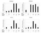
- FIG. 1 shows gene expression of EEC markers in F-hiSIEC cells cultured in various media in Table 1.
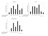
- FIG. FIG. 2 shows gene expression of EEC markers in F-hiSIEC cells cultured under the conditions of adding 0.5 to 2.0 ⁇ mol/L of LY411575 or 5 to 10 ⁇ mol/L of DAPT to the medium in Table 2.
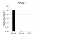
- Figure 3 shows the relationship between Caco-2 cells, F-hiSIEC cells cultured in F-hiSIEC Culture Medium (F-hiSIEC), and F-hiSIEC cells cultured in the medium of Table 3 and induced to EEC (EEC). 1 gene expression is shown.
- a method for producing an enteroendocrine cell according to the present invention comprises producing intestinal stem cells or intestinal epithelial cells derived from pluripotent stem cells substantially free of epidermal growth factor receptor ligands, cAMP activators and DNA methyltransferase inhibitors.
- a method comprising culturing using a culture medium comprising
- enteroendocrine cells can be efficiently induced by using a culture medium containing a cAMP activator and a DNA methyltransferase inhibitor that is substantially free of epidermal growth factor receptor ligands.
- intestinal stem cells or intestinal epithelial cells derived from pluripotent stem cells are used.
- Pluripotent stem cells have the ability to differentiate into all cells that make up the body (pluripotency) and the ability to produce daughter cells with the same differentiation potential as the self through cell division (self-renewal ability). ) refers to cells that have both Pluripotency can be evaluated by transplanting the cells to be evaluated into nude mice and examining the presence or absence of teratoma formation containing cells of each of the three germ layers (ectoderm, mesoderm, and endoderm). can.
- pluripotent stem cells examples include embryonic stem cells (ES cells), embryonic germ cells (EG cells), induced pluripotent stem cells (iPS cells), etc., which have both differentiation pluripotency and self-renewal ability. As long as it is a cell, it is not limited to this. ES cells or iPS cells are preferably used. More preferably, iPS cells are used. Pluripotent stem cells are preferably mammalian (eg, primates such as humans and chimpanzees, rodents such as mice and rats) cells, particularly preferably human cells. Therefore, in the most preferred embodiment of the present invention, human induced pluripotent stem cells (human iPS cells) are used as pluripotent stem cells.
- ES cells embryonic stem cells
- EG cells embryonic germ cells
- iPS cells induced pluripotent stem cells
- Pluripotent stem cells are preferably mammalian (eg, primates such as humans and chimpanzees, rodents such as mice and rats)
- ES cells can be established, for example, by culturing an early embryo before implantation, an inner cell mass constituting the above-mentioned early embryo, a single blastomere, etc. (Manipulating the Mouse Embryo A Laboratory Manual, Second Edition, Cold Spring Harbor Laboratory Press (1994); Thomson, J. A. et al., Science, 282, 1145-1147 (1998)).
- an early embryo an early embryo produced by nuclear transfer of the nucleus of a somatic cell may be used (Wilmut et al. (Nature, 385, 810 (1997)), Cibelli et al. (Science, 280, 1256 (1998)), Akira Iriya et al.
- ES cells are available from archives or are commercially available.
- human ES cells are available from Kyoto University Institute for Frontier Medical Sciences (eg, KhES-1, KhES-2 and KhES-3), WiCell Research Institute, ESI BIO, and the like.
- EG cells can be established by culturing primordial germ cells in the presence of LIF, bFGF, SCF, etc. (Matsui et al., Cell, 70, 841-847 (1992), Shamblott et al., Proc. Natl. Acad. Sci. USA, 9523, 13726-13731 (1998), Turnpenny et al., Stem Cells, 21(5), 598-609, (2003)).
- iPS cells are cells with pluripotency (multipotency) and proliferative potential, which are produced by reprogramming somatic cells through the introduction of reprogramming factors. Induced pluripotent stem cells exhibit properties similar to ES cells.
- the somatic cells used for producing iPS cells are not particularly limited, and may be differentiated somatic cells or undifferentiated stem cells.
- the origin is not particularly limited, but somatic cells of mammals (for example, primates such as humans and chimpanzees, and rodents such as mice and rats), particularly preferably somatic cells of humans, are used.
- somatic cells of mammals for example, primates such as humans and chimpanzees, and rodents such as mice and rats
- somatic cells of humans are used.
- somatic cells of mammals for example, primates such as humans and chimpanzees, and rodents such as mice and rats
- somatic cells of humans are used.
- fetal somatic cells adult-derived somatic cells (that is, mature somatic
- somatic cells include (1) tissue stem cells (somatic stem cells) such as neural stem cells, hematopoietic stem cells, mesenchymal stem cells, dental pulp stem cells, (2) tissue progenitor cells, and (3) fibroblasts (skin cells, etc.). ), epithelial cells, hepatocytes, lymphocytes (T cells, B cells), endothelial cells, muscle cells, hair cells, gastric mucosa cells, enterocytes, splenocytes, pancreatic cells (pancreatic exocrine cells, etc.), brain cells, lung Differentiated cells such as cells, renal cells, skin cells and the like can be mentioned.
- tissue stem cells such as neural stem cells, hematopoietic stem cells, mesenchymal stem cells, dental pulp stem cells, (2) tissue progenitor cells, and (3) fibroblasts (skin cells, etc.).
- epithelial cells epithelial cells
- hepatocytes lymphocytes (T cells, B cells)
- the most basic method for producing iPS cells is to introduce four transcription factors, Oct3/4, Sox2, Klf4 and c-Myc, into cells using viruses (Takahashi K, Yamanaka S : Cell 126 (4), 663-676, 2006; Takahashi, K, et al.: Cell 131 (5), 861-72, 2007).
- human iPS cells were established by introducing four factors, Oct4, Sox2, Lin28 and Nonog (Yu J, et al: Science 318(5858), 1917-1920, 2007).
- 3 factors excluding c-Myc excluding c-Myc (Nakagawa M, et al: Nat. Biotechnol.
- Transformation to iPS cells that is, cells that have undergone reprogramming (reprogramming) express pluripotent stem cell markers (undifferentiated markers) such as Fbxo15, Nanog, Oct/4, Fgf-4, Esg-1 and Cript etc. can be selected as an index. Selected cells are collected as iPS cells.
- pluripotent stem cell markers such as Fbxo15, Nanog, Oct/4, Fgf-4, Esg-1 and Cript etc.
- iPS cells can also be provided by, for example, the National University Corporation Kyoto University or the RIKEN BioResource Center.
- inducing differentiation refers to working to differentiate along a specific cell lineage.
- intestinal stem cells or intestinal epithelial cells differentiated from pluripotent stem cells are used.
- Intestinal stem cells can be produced by culturing pluripotent stem cells under conditions that induce differentiation into intestinal stem cells. Culture conditions are not particularly limited as long as the pluripotent stem cells differentiate into intestinal stem cells. Typically, the two stages of induction of differentiation described below, i.e., differentiation of pluripotent stem cells into endoderm-like cells, such that pluripotent stem cells differentiate into intestinal stem cells via endoderm-like cells (Step (1-1)) and differentiation of endoderm-like cells into intestinal stem cells (Step (1-2)) are performed.
- Step (1-1) Differentiation into Endoderm-like Cells pluripotent stem cells are cultured and differentiated into endoderm-like cells.
- pluripotent stem cells are cultured under conditions that induce endoderm differentiation.
- Culture conditions are not particularly limited as long as the pluripotent stem cells differentiate into endoderm-like cells.
- it is cultured in a medium supplemented with activin A according to a conventional method.
- the concentration of activin A in the medium is, for example, 10 ng/mL to 200 ng/mL, preferably 20 ng/mL to 150 ng/mL.
- serum or a serum substitute KnockOut TM Serum Replacement (KSR), etc.
- KSR KnockOut TM Serum Replacement
- Serum is not limited to fetal bovine serum, and human serum, sheep serum, and the like can also be used.
- the amount of serum or serum substitute added is, for example, 0.1% (v/v) to 10% (v/v).
- Inhibitors of the Wnt/ ⁇ -catenin signaling pathway may be added to the medium to promote differentiation into endoderm-like cells.
- One or more of BMP4, VEGF, and FGF2 may be added to the medium to promote differentiation into endoderm-like cells.
- This step can also be performed by the method described in International Publication No. 2014/165663 or a method based thereon.
- two stages of culture are performed as step (1-1).
- the first-stage culture is performed in a medium supplemented with a relatively low concentration of serum (e.g., 0.1% (v/v) to 1% (v/v)), and the subsequent second-stage culture is performed in the first stage.
- culture in a medium with a higher serum concentration eg, 1% (v/v) to 10% (v/v)
- Adopting a two-stage culture in this manner is preferable in that the first stage culture suppresses the growth of undifferentiated cells, and the second stage allows the proliferation of differentiated cells.
- the period (culture period) of step (1-1) is, for example, 1 to 10 days, preferably 2 to 7 days.
- the first stage culture period is, for example, 1 to 7 days, preferably 2 to 5 days
- the second stage culture period is, for example, 1 day. to 6 days, preferably 1 to 4 days.
- the endoderm-like cells obtained in step (1-1) are cultured and differentiated into intestinal stem cells.
- endoderm-like cells are cultured under conditions that induce differentiation into intestinal stem cells.
- Culture conditions are not particularly limited as long as the endoderm-like cells differentiate into intestinal stem cells.
- culture is performed in the presence of FGF2 (fibroblast growth factor 2) or in the presence of a GSK-3 ⁇ inhibitor.
- FGF2 fibroblast growth factor 2
- Human FGF2 eg, human recombinant FGF2
- FGF2 fibroblast growth factor 2
- step (1-2) may be performed after selecting endoderm-like cells from the cell population obtained through step (1-1). Selection of endoderm-like cells may be performed, for example, using a flow cytometer (cell sorter) using a cell surface marker as an indicator.
- a flow cytometer cell sorter
- FGF2 In the presence of FGF2 is synonymous with the condition in which FGF2 was added to the medium. Therefore, in order to perform culture in the presence of FGF2, a medium supplemented with FGF2 may be used.
- An example of the concentration of FGF2 added is 100 ng/mL to 500 ng/mL.
- a medium supplemented with FGF2 may be used for culturing in the presence of a GSK-3 ⁇ inhibitor.
- the concentration of the GSK-3 ⁇ inhibitor added may be 0.1 nmol/L to 30 ⁇ mol/L, preferably 1 nmol/L to 10 ⁇ mol/L, more preferably 10 nmol/L to 10 ⁇ mol/L, More preferably, it is 50 nmol/L to 5 ⁇ mol/L.
- the period (culture period) of step (1-2) is, for example, 2 to 10 days, preferably 3 to 7 days. If the culture period is too short, the expected effects (increase in differentiation efficiency, promotion of acquisition of functions as intestinal stem cells) cannot be sufficiently obtained. On the other hand, if the culture period is too long, the differentiation efficiency will be lowered.
- the differentiation into intestinal stem cells can be determined or evaluated, for example, by using the expression of intestinal stem cell markers as an index.
- intestinal stem cell markers are G protein-coupled receptor 5 (LGR5) containing leucine-rich repeats, ephrin B2 receptor (EphB2).
- the cell density at the end of the first differentiation step may be between 5.0 ⁇ 10 4 cells/cm 2 and 20 ⁇ 10 4 cells/cm 2 , between 6.0 ⁇ 10 4 cells/cm 2 and 16 cells/cm 2 . It is preferably ⁇ 10 4 cells/cm 2 , more preferably 7.0 ⁇ 10 4 cells/cm 2 to 14 ⁇ 10 4 cells/cm 2 .
- the intestinal stem cells obtained in step (1-2) can also be cryopreserved.
- the cryopreserved intestinal stem cells may be thawed and subsequent steps (proliferation step, etc.) may be performed.
- a solution generally used for cryopreservation of cultured cells can be used, and may contain medium, DMSO, serum, general cryopreservation reagents, and the like.
- common cryopreservation reagents include CryoStor® Freeze Media, CellBanker®, FreSRTM-S, NutriFreezTM D10 Cryopreservation Medium, Cellvation, Cryoscarless® and Bambanker ( registered trademark).
- the step of differentiating intestinal stem cells into intestinal epithelial cells includes a step of differentiating intestinal stem cells into intestinal epithelial cells in the presence of a MEK1 inhibitor, a DNA methyltransferase inhibitor, a TGF ⁇ receptor inhibitor, EGF and a cAMP activator. is preferred.
- a cAMP activator By using a cAMP activator, intracellular cAMP levels can be positively increased.
- a MEK1 inhibitor, a DNA methyltransferase inhibitor, a TGF ⁇ receptor inhibitor, an EGF and a cAMP activator means one or more cultures that constitute the second differentiation step. It means that all compounds are required, and all these compounds are used at the same time, that is, it requires as an essential condition that culture is performed using a medium to which all these compounds are added. isn't it.
- Differentiation into intestinal epithelial cells can be constituted by one or more cultures.
- a medium supplemented with EGF as an essential ingredient for example, A medium supplemented with EGF as an essential ingredient; Medium supplemented with EGF and ROCK inhibitors (eg Y-27632) as essential ingredients; media supplemented with EGF and cAMP activators (e.g. Forskolin) as essential components; Medium supplemented with EGF, MEK1 inhibitors, DNA methyltransferase inhibitors, TGF ⁇ receptor inhibitors as essential ingredients; Media supplemented with EGF, cAMP activators (e.g. Forskolin), MEK1 inhibitors, DNA methyltransferase inhibitors, TGF ⁇ receptor inhibitors as essential ingredients; etc. can be used.
- Examples of MEK1 inhibitors include PD98059, PD184352, PD184161, PD0325901, U0126, MEK inhibitor I, MEK inhibitor II, MEK1/2 inhibitor II, and SL327.
- DNA methyltransferase inhibitors include 5-aza-2'-deoxycytidine, 5-azacytidine, RG108, and Zebularine.
- TGF ⁇ receptor inhibitors considering that A8301 used in the examples below exhibits inhibitory activity on TGF- ⁇ receptors ALK4, ALK5 and ALK7, preferably TGF- ⁇ receptors ALK4, ALK5, It is preferable to use one that exhibits inhibitory activity against one or more of ALK7.
- A8301, SB431542, SB-505124, SB525334, D4476, ALK5 inhibitor, LY2157299, LY364947, GW788388, and RepSox satisfy the above conditions.
- cAMP activators forskolin, indomethacin, NKH477 (colforsin daropate), cell-derived toxin proteins (pertussis toxin, cholera toxin), PACAP-27, PACAP-38, SKF83822 and the like can be used.
- NKH477 colforsin daropate
- cholera toxin PACAP-27
- PACAP-38 PACAP-38
- SKF83822 exhibit an adenylyl cyclase activating action and promote intracellular cAMP synthesis.
- concentration of the MEK1 inhibitor added is 4 ⁇ mol/L to 100 ⁇ mol/L, preferably 10 to 40 ⁇ mol/L.
- concentration of the DNA methyltransferase inhibitor in the case of 5-aza-2'-deoxycytidine
- concentration of the TGF ⁇ receptor inhibitor added in the case of A8301
- concentration of the TGF ⁇ receptor inhibitor added is 0.1 ⁇ mol/L to 2.5 ⁇ mol/L, preferably 0.2 ⁇ mol/L to 1 ⁇ mol/L.
- concentrations of EGF added are 5 ng/mL to 100 ng/mL, preferably 10 ng/mL to 50 ng/mL.
- concentration of the cAMP activator added is 1 ⁇ mol/L to 200 ⁇ mol/L, preferably 5 ⁇ mol/L to 100 ⁇ mol/L.
- the concentration to be added depends on the characteristics of the compound used and the exemplified compound (PD98059 , 5-aza-2′-deoxycytidine, A8301, and forskolin), a person skilled in the art can determine the concentration range according to the above range.
- additional condition 1 conditions in which cAMP was supplied to the cells
- additional condition 2 conditions in which a cAMP degrading enzyme inhibitor was present
- Additional condition 1 conditions under which cAMP is supplied to cells
- a compound capable of being taken up into cells and acting as cAMP upon being taken up into cells is present. Therefore, in order to satisfy the additional condition 1, for example, a medium supplemented with a cAMP derivative that can be taken up into cells may be used.
- PKA activators as cAMP derivatives e.g., 8-Br-cAMP (8-Bromoadenosine-3', 5'-cyclic monophosphate sodium salt, CAS Number: 76939-46-3), 6-Bnz-cAMP (N6-Benzoyladenosine- 3′,5′-cyclic monophosphate sodium salt salt, CAS Number: 1135306-29-4), cAMPS-Rp ((R)-adenosine, cyclic 3′,5′-(hydrogenphosphorothioate) triethyl ammonium salt, CAS Number: 151837- 09-1), cAMPS-Sp ((S)-Adenosine, cyclic 3', 5'-(hydrogenphosphorothioate) triethylammonium salt, CAS Number: 93602-66-5), Dibutylyl-cAMP (N6, O2'-D ibutylyl adenosine 3 ', 5'-cyclic
- concentration of the cAMP derivative added is 0.1 mmol/L to 10 mmol/L, preferably 0.2 mmol/L to 5 mmol/L, more preferably 0.5 mmol/L. ⁇ 2mmol/L.
- concentration to be added is determined by the difference between the characteristics of the compound used and the characteristics of the exemplified compound (8-Br-cAMP) (especially Considering the difference in activity), a person skilled in the art can set the concentration range according to the above.
- Additional condition 2 (condition in which cAMP degrading enzyme inhibitor is present) is synonymous with the condition in which cAMP degrading enzyme inhibitor is added to the medium.
- additional condition 2 When the additional condition 2 is adopted, the decrease in intracellular cAMP concentration is suppressed by inhibiting the degradation of cAMP, and it can be expected that the induction of differentiation into the intestinal epithelium, particularly the acquisition of functions as intestinal epithelial cells, will be promoted. That is, the above conditions may enable the preparation of more functional intestinal epithelial cells. If additional condition 1 and additional condition 2 are used together, it is possible to suppress the decrease in intracellular cAMP concentration while supplying cAMP to cells. Therefore, it can be expected that this will be an effective condition for maintaining intracellular cAMP at a high level, and that efficient induction of differentiation into intestinal epithelial cells will be promoted.
- IBMX 3-isobutyl-1-methylxanthine
- MIX Theophylline
- Papaverine Pentoxifyline
- KS-505, 8-Methoxymethyl-IBMX Vinpocetine
- EHNA Trequinsin
- HL-725 Lixazinone
- RS-82856 Lixazinone
- LY-186126 Cilostamide
- OPC3689 Cilostamide
- Bemoradan RWJ-22867
- Anergrelide BL4162A
- Indolidan LY195115
- Cilostazol OPC-13 013
- Milrinone WIN47203
- Siguazodan SKF-94836
- 5-Methyl-imazodan CI 930
- SKF-95654 Pirilobendan
- UD-CG 115 BS Enoximone
- Imazodan Imazodan
- CL 914 Imazodan
- SKF-94120 Vesnarinone A H- 21-
- concentration of the cAMP degrading enzyme inhibitor added is 0.05 mmol/L to 5 mmol/L, preferably 0.1 mmol/L to 3 mmol/L, more preferably 0.2 mmol/L to 1 mmol/L.
- the period of the process of differentiation into intestinal epithelial cells is, for example, 7 to 40 days, preferably 7 to 30 days. If the culture period is too short, the expected effects (increase in differentiation efficiency, promotion of acquisition of functions as intestinal epithelial cells) cannot be sufficiently obtained. On the other hand, too long a culture period causes a decrease in differentiation efficiency.
- Differentiation into intestinal epithelial cells can be determined or evaluated using, for example, the expression of intestinal epithelial cell markers, the uptake of peptides, or the induction of expression of drug-metabolizing enzymes via vitamin D receptors as indicators.
- intestinal epithelial cell markers include ATP-binding cassette transporter B1/multidrug resistance protein 1 (ABCB1/MDR1), ATP-binding cassette transporter G2/breast cancer resistance protein (ABCG2/BCRP), cytochrome P450 3A4 (CYP3A4).
- FABP fatty acid binding protein 2
- PXR pregnane X receptor
- SLC synthetic carrier family member 5A1/sodium-coupled glucose transporter 1 (SLC5A1/SGLT1)
- SLC serum-coupled glucose transporter 1
- SLC synthetic carrier family member 15A1/peptide trans Porter 1 (SLC15A1/PEPT1)
- SLC synthetic carrier organic anion transporter 2B1 (SLCO2B1/OATP2B1)
- sucrase-isomaltase sucrase-isomaltase
- uridine diphosphate-glucuronyltransferase 1A1 UGT1A1
- UGT1A4 uridine diphosphate-glucuronic acid transferase 1A4
- villin 1 Villin 1
- CRIS2A1 carboxylesterase 2A1
- sucrase-isomaltase and villin 1 which are highly specific to the intestinal epithelium, CYP3A4, which is a major drug-metabolizing enzyme in the small intestine, SLC15A1/PEPT1, which is involved in the absorption of peptides in the small intestine, and the apical membranous side of the small intestine.
- SLC5A1/SGLT1 which is a glucose transporter expressed in the small intestine
- SLCO2B1/OATP2B1 which is involved in the absorption of organic anions in the small intestine
- CES2A1 which is a hydrolase highly expressed in the small intestine
- a cell population consisting only of the cells of interest (intestinal epithelial cells) or a cell population containing a high proportion (high purity) of the cells of interest
- a cell surface marker characteristic of the cells of interest as an index. After the culture, the cell population can be sorted and sorted.
- intestinal stem cells or intestinal epithelial cells derived from pluripotent stem cells as described above are substantially free of epidermal growth factor receptor ligands and contain cAMP activators and DNA methyltransferase inhibitors. Cultivate using culture medium.
- Epidermal growth factor receptor (EGFR) ligands include EGF, betacelluin, amphiregulin, TNF- ⁇ , and HB-EGF.
- cAMP activators forskolin, indomethacin, NKH477 (colforsin daropate), cell-derived toxin proteins (pertussis toxin, cholera toxin), PACAP-27, PACAP-38, SKF83822, etc. can be used.
- NKH477 colforsin daropate
- cholera toxin PACAP-27, PACAP-38 and SKF83822
- concentration of the cAMP activator added is 1 ⁇ mol/L to 200 ⁇ mol/L, preferably 5 ⁇ mol/L to 100 ⁇ mol/L.
- addition concentration when using a compound different from forskolin considering the characteristics of the compound used and the difference in the characteristics of forskolin (especially the difference in activity), a person skilled in the art should follow the above concentration range. can be set.
- 5-aza-2'-deoxycytidine 5-azacytidine
- RG108 5-azacytidine
- Zebularine etc.
- an example of the added concentration of the DNA methyltransferase inhibitor is 0.1 ⁇ mol/L to 50 ⁇ mol/L, preferably 0.5 ⁇ mol/L to 20 ⁇ mol/L.
- the concentration to be added should be determined by taking into account the difference in the properties of the compound used and the difference in properties of 5-Aza-2'-deoxycytidine (especially the difference in activity). For example, a person skilled in the art can set the concentration according to the above concentration range.
- the culture medium substantially free of epidermal growth factor receptor ligands and containing a cAMP activator and a DNA methyltransferase inhibitor contains any one or more of a transforming growth factor- ⁇ signaling inhibitor and a MEK inhibitor.
- transforming growth factor- ⁇ signal inhibitors for example, A83-01, SB431542, SB-505124, SB525334, D4476, ALK5 inhibitor, LY2157299, LY364947, GW788388, RepSox, etc. can be used.
- the concentration of the transforming growth factor- ⁇ signal inhibitor (in the case of A83-01) added may be 0.1 nmol/L to 30 ⁇ mol/L, preferably 1 nmol/L to 10 ⁇ mol/L, more preferably , 10 nmol/L to 10 ⁇ mol/L, more preferably 50 nmol/L to 5 ⁇ mol/L.
- concentration range when using a compound different from A83-01, considering the characteristics of the compound used and the difference in characteristics of A83-01 (especially the difference in activity), a person skilled in the art can use the above concentration range. can be set according to
- MEK inhibitors PD98059, PD184352, PD184161, PD0325901, U0126, MEK inhibitor I, MEK inhibitor II, MEK1/2 inhibitor II, SL327, etc. can be used.
- concentration of the MEK inhibitor added is 1 ⁇ mol/L to 200 ⁇ mol/L, preferably 5 ⁇ mol/L to 100 ⁇ mol/L.
- addition concentration when using a compound different from PD98059 considering the difference in the characteristics of the compound used and the characteristics of PD98059 (especially the difference in activity), a person skilled in the art can set according to the above concentration range. can do.
- a culture medium that is substantially free of epidermal growth factor receptor ligands and contains a cAMP activator and a DNA methyltransferase inhibitor may contain a Notch inhibitor.
- Notch inhibitors include LY411575, DAPT, ⁇ -Secretase Inhibitor XXI (Compound E) (CAS number: 209986-17-4), ⁇ -Secretase Inhibitor I (CAS number: 133407-83-7), dibenzazepine , RO4929097, etc. can be used.
- the Notch inhibitor is preferably LY411575.
- concentration of the Notch inhibitor added is 0.1 ⁇ mol/L to 10 ⁇ mol/L, preferably 0.5 ⁇ mol/L to 5 ⁇ mol/L.
- addition concentration when using a compound different from LY411575 considering the difference in the characteristics of the compound used and the characteristics of LY411575 (especially the difference in activity), a person skilled in the art can set it according to the above concentration range. can do.
- the culture medium substantially free of epidermal growth factor receptor ligands and containing cAMP activators and DNA methyltransferase inhibitors preferably does not contain any one or more of Wnt activators and Wnt inhibitors.
- Wnt activators include CHIR99021, LY2090314, NP031112 (tideglusib), lithium, A1070722, SKL2001, R-Spondin 1, Wnt3a, Gsk inhibitors, and the like.
- Wnt inhibitors include, for example, Wnt-C59, Dkk family proteins (eg, at least one of Dkk-1, Dkk2, Dkk3 and Dkk4), sFRP (eg, at least one of sFRP-1 and sFRP-2), OMP- antibodies against Wnt receptors such as 18R5, LGK974, CWP232291, PRI-724, IQR-1, IWP2, IWP-L6, ICG-001, WIKI4, Ky02111, FH535, XAV939, NSC668036, FJ9, 3289-8625, etc. .
- the culture medium that is substantially free of epidermal growth factor receptor ligands and contains a cAMP activator and a DNA methyltransferase inhibitor may or may not contain a histone deacetylase inhibitor.
- Histone deacetylase inhibitors include Tubastatin A, ACY1215, valproic acid, SAHA, trichostatin A, SHBA, CBHA, LAQ-824, PDX-101, LBH-589, ITF2357, PCI-24781, Compound 7 (ChemieTek), JNK-24681585 (Xinostat) SB939 (Pracinostat), 4SC-201 (Resminostat), Tefinostat (CHR-2845), CHR-3996, CG200745, Depsipeptide (Romidepsin ), butyrate, MS-275, MGCD0103 and CI994.
- the culture medium substantially free of epidermal growth factor receptor ligands and containing a cAMP activator and a DNA methyltransferase inhibitor contains one or more of a retinoic acid signaling pathway activator and a sonic hedgehog signaling pathway inhibitor. preferably does not contain
- Retinoic acid signaling pathway activators include retinoic acid, retinoic acid derivatives, all-trans retinoic acid, synthetic retinoids ec23, Ch55, TTNPB, fenretinide, AC261066, adapalene, AC55649, AM80, AM580, BMS753 and tazarotene.
- Sonic hedgehog signaling pathway inhibitors include SANT-1, SANT-2, JK184, or jervine.
- the period of culturing using a culture medium containing a cAMP activator and a DNA methyltransferase inhibitor substantially free of epidermal growth factor receptor ligands is, for example, 2 to 20 days, preferably 4 to 4 days. 15 days.
- the culture process using a culture medium that does not substantially contain epidermal growth factor receptor ligands and contains a cAMP activator and a DNA methyltransferase inhibitor preferably does not include a suspension culture process.
- the cells do not form spheroids in the culture step using a culture medium that does not substantially contain epidermal growth factor receptor ligands and contains a cAMP activator and a DNA methyltransferase inhibitor.
- ROCK inhibitors Rost-associated coiled-coil forming kinase / Rho binding kinase
- Y-27632 Rost-associated coiled-coil forming kinase / Rho binding kinase
- other culturing conditions may be those generally employed in culturing animal cells. That is, it may be cultured in an environment of 37° C. and 5% CO 2 , for example.
- IMDM Iscove's modified Dulbecco's medium
- GBCO Ham F12 medium
- DMEM Dulbecco's modified Eagle's medium
- Gibco, etc. Glasgow basal medium
- RPMI1640 medium Two or more basal media may be used in combination.
- components that can be added to the medium include serum or serum substitutes, antibiotics, 2-mercaptoethanol, PVA, non-essential amino acids (NEAA), insulin, transferrin, and selenium.
- serum examples include bovine serum albumin (BSA), fetal bovine serum (FBS), and the like.
- Serum substitutes include Gibco® B-27® Supplement and the like.
- enteroendocrine cells are typically cultured two-dimensionally using a culture dish.
- enteroendocrine cells can be produced by two-dimensional culture (preferably adherent culture).
- the form of enteroendocrine cells to be produced is not particularly limited, but is preferably sheet-like.
- culturing can be performed on culture vessels coated with 3% to 100% extracellular matrix.
- extracellular matrices include Matrigel TM , fibronectin, laminin, vitronectin, tenascin, entactin, thrombospontin, elastin, gelatin, collagen, fibrin, merosin, ancholine, chondronectin, link protein, bone sialoprotein, osteocalcin, osteopontin, Epinectin, hyaluronectin, angulin, epiligrin and kalinin can be used, preferably Matrigel TM .
- Matrigel® is the trade name for a gelatinous protein mixture secreted by Engelbreth-Holm-Swarm mouse sarcoma cells produced by Corning Life Sciences.
- human iPS cell-derived intestinal epithelial cells (containing intestinal stem cell LGR5+ cells) are seeded in a cell culture insert or a 96-well plate, and from the next day, FBS, B27, HepExtend TM , Enteroendocrine cells can be produced by culturing in DMEM/F12 medium containing Forskolin, A83-01, 5-Aza-2'-deoxycytidine, PD98059, and LY41157.
- enteroendocrine cells derived from pluripotent stem cells produced by the above-described method of the present invention are provided. Differentiation into enteroendocrine cells can be determined or evaluated using, for example, the expression of an enteroendocrine cell marker as an indicator.
- enteroendocrine cell markers are CHGA, REG4.
- TPH1 and PYY which are factors involved in hormone secretion, which is the function of enteroendocrine cells
- NEUROG3 which is a factor involved in the development of endocrine cells in the intestine
- the enteroendocrine cells of the present invention are characterized by expressing TPH1, PYY, CHGA, REG4 and NEUROG3 and not expressing NKX6.1.
- TPH1, PYY, CHGA, REG4, NEUROG3 and NKX6.1 can be measured by mRNA or protein level.
- Expressing TPH1, PYY, CHGA, REG4 and NEUROG3 means that mRNA or protein that is the expression product of TPH1, PYY, CHGA, REG4 and NEUROG3 can be detected by a known method.
- Does not express NKX6.1 means that mRNA or protein that is the expression product of NKX6.1 cannot be detected by a known method. However, it is not limited to the case where the expression level is 0, and the case where the expression level is very small is also included.
- the case of "not expressing NKX6.1” is when the relative expression level with respect to the expression level of NKX6.1 in human colon cancer-derived cell line CaCo-2 is 0.1 or less, preferably 0.05 or less. , and more preferably 0.01 or less.
- the present invention relates to uses of enteroendocrine cells produced by the method of the present invention.
- a first aspect includes a method for analyzing a disease mechanism, comprising a step of producing enteroendocrine cells by the method of the present invention, and a step of analyzing the disease mechanism using the enteroendocrine cells.
- screening for a therapeutic agent for digestive disorders comprising the steps of producing enteroendocrine cells by the method of the present invention, and culturing the enteroendocrine cells in the presence of a therapeutic agent for digestive disorders.
- a third embodiment includes a method for evaluating a test substance, comprising the steps of producing enteroendocrine cells by the method of the present invention, and quantifying hormones secreted by the enteroendocrine cells in the presence of the test substance. .
- endocrine cell markers and expression/production levels of gastrointestinal hormones are biomarkers and can be used as indicators of various diseases. Examples include a method of analyzing the disease mechanism by measuring the morphology, cell count, and biomarkers described above when a disease model is created by adding drugs or gene editing to the induced enteroendocrine cells.
- a therapeutic drug or test substance can be screened or evaluated by contacting the enteroendocrine cell with the therapeutic drug or test substance.
- the enteroendocrine cells produced by the method of the present invention can be used in model systems of the intestinal tract, especially the small intestine, and can be used to evaluate pharmacokinetics (absorption, metabolism, etc.) and toxicity in the intestinal tract, especially the small intestine, and response in the small intestine.
- Useful for evaluation That is, the enteroendocrine cells produced by the method of the present invention are intended to be used for evaluation of pharmacokinetics and toxicity of therapeutic drugs or test substances, and evaluation of responses to test substances.
- the present invention provides one of the uses of enteroendocrine cells, which include absorption or membrane permeability of therapeutic drugs or test substances, drug interaction, induction of drug-metabolizing enzymes, induction of drug transporters, toxicity, immune response, and enteroendocrine cells Production of secreted hormones, etc. can be tested. That is, the present invention provides one of the uses of enteroendocrine cells, which include absorption or membrane permeability of therapeutic drugs or test substances, drug interaction, induction of drug-metabolizing enzymes, induction of drug transporters, toxicity, immune response, It also provides a method for evaluating the production of hormones secreted by enteroendocrine cells.
- contacting the enteroendocrine cells with a therapeutic agent or test substance contacting the enteroendocrine cells with a therapeutic agent or test substance; and (iii) the therapeutic agent or test substance permeating the enteroendocrine cells.
- a step of quantifying the amount of hormone production is performed.
- hormones secreted by enteroendocrine cells include serotonin, GLP-1 (Glucagon-like peptide-1), GIP (Gastoric inhibitory polypeptide), gastrin, PYY and the like.
- the absorbability of the therapeutic drug or test substance can also be evaluated by the method described below.
- enteroendocrine cells are typically cultured on a semipermeable membrane (porous membrane) to form a cell layer.
- a semipermeable membrane porous membrane
- a culture vessel equipped with a cell culture insert e.g., Transwell (registered trademark) provided by Corning
- cells are seeded and cultured in the cell culture insert to obtain an intestinal A cell layer composed of endocrine cells is obtained.
- Contacting in step (ii) is typically performed by adding a therapeutic drug or test substance to the medium.
- the timing of addition of the therapeutic drug or test substance is not particularly limited. Therefore, even if the therapeutic drug or test substance is added at a certain point after culturing in a medium containing no therapeutic drug or test substance, culture should be started in a medium containing the therapeutic drug or test substance in advance. may
- Organic or inorganic compounds of various molecular sizes can be used as therapeutic drugs or test substances, and bacteria (intestinal bacteria, lactic acid bacteria, etc.), viruses, and cells (immune cells, fibroblasts) can also be used.
- examples of organic compounds include nucleic acids, peptides, proteins, lipids (simple lipids, complex lipids, phosphoglycerides, sphingolipids, glycosylglycerides, cerebrosides, etc.), prostaglandins, isoprenoids, terpenes, steroids, polyphenols, catechins, vitamins (B1, B2, B3, B5, B6, B7, B9, B12, C, A, D, E, etc.) can be exemplified.
- test substances Existing ingredients or candidate ingredients of pharmaceuticals, nutritional foods, supplements, food additives, pesticides, cosmetics (cosmetics), etc. are also preferred test substances.
- Plant extracts, cell extracts, culture supernatants, etc. may be used as test substances. By adding two or more therapeutic agents or test substances simultaneously, interactions, synergistic effects, etc. between therapeutic agents or test substances may be investigated.
- the therapeutic agent or test substance may be of natural origin or synthetic. In the latter case, for example, a combinatorial synthesis technique can be used to construct an efficient assay system.
- the period of contact with the therapeutic drug or test substance can be set arbitrarily.
- the contact period is, for example, 10 minutes to 3 days, preferably 1 hour to 1 day.
- the contact may be divided into multiple times.
- the therapeutic drug or test substance that permeates the cell layer is quantified.
- a culture vessel equipped with a cell culture insert such as Transwell (registered trademark)
- the therapeutic agent or test substance that has permeated the cell culture insert that is, through the cell layer into the upper or lower vessel mass spectrometry, liquid chromatography, immunological methods (e.g., fluorescence immunoassay (FIA method), enzyme immunoassay (EIA method)) Quantify by a measuring method such as. Absorption or membrane permeability, drug interaction, drug interaction, Determine/evaluate induction of drug-metabolizing enzymes, induction of drug transporters, toxicity, or immune response.
- FIA method fluorescence immunoassay
- EIA method enzyme immunoassay
- the residual amount of the therapeutic drug or test substance in the culture medium may be measured.
- An appropriate measurement method may be selected according to the test substance. For example, mass spectrometry, liquid chromatography, immunological techniques (for example, fluorescence immunoassay (FIA method), enzyme immunoassay (EIA method)) and the like can be employed.
- FFA method fluorescence immunoassay
- EIA method enzyme immunoassay
- the absorption amount or absorption efficiency of the therapeutic drug or test substance can be determined and evaluated according to the degree of decrease. Absorption can also be evaluated by measuring the amount of therapeutic drug or test substance taken up into cells.
- hormones secreted by enteroendocrine cells are quantified.
- Hormones secreted by enteroendocrine cells can be quantified by measurement methods such as mass spectrometry, liquid chromatography, and immunological techniques (e.g., fluorescence immunoassay (FIA method) and enzyme immunoassay (EIA method)). .
- FIA method fluorescence immunoassay
- EIA method enzyme immunoassay
- the metabolism of therapeutic drugs or test substances can be evaluated.
- (I) the step of contacting enteroendocrine cells with a therapeutic drug or test substance, and (II) the step of measuring and evaluating the metabolism of the therapeutic drug or test substance are performed.
- Step (I) that is, contacting enteroendocrine cells with a therapeutic drug or test substance, can be performed in the same manner as in step (ii) above.
- forming a cell layer in advance is not essential.
- step (II) measure and evaluate the metabolism of the therapeutic drug or test substance.
- step (II) i.e., after contact with the therapeutic agent or test substance, metabolism may be measured and evaluated without a substantial time interval, or for a certain period of time (eg, 10 minutes to 5 hours) , the metabolism may be measured and evaluated.
- Metabolic measurements can be made, for example, by detecting metabolites. In this case, the expected metabolites are usually measured qualitatively or quantitatively using the culture solution after step (I) as a sample.
- An appropriate measurement method may be selected depending on the metabolite, and examples thereof include mass spectrometry, liquid chromatography, immunological techniques (e.g., fluorescence immunoassay (FIA method), enzyme immunoassay (EIA method), ), etc. can be adopted.
- FIA method fluorescence immunoassay
- EIA method enzyme immunoassay
- the metabolic rate of a therapeutic drug or test substance can be evaluated according to the amount of metabolites.
- the metabolic efficiency of the therapeutic drug or test substance may be calculated based on the metabolite detection results and the amount of the therapeutic drug or test substance used (typically, the amount added to the medium).
- a therapeutic agent or a test substance using the expression of drug-metabolizing enzymes (cytochrome P450 (especially CYP3A4), uridine diphosphate-glucuronyltransferase (especially UGT1A8, UGT1A10), sulfotransferase (especially SULT1A3, etc.)) in enteroendocrine cells as an indicator
- drug-metabolizing enzymes cytochrome P450 (especially CYP3A4), uridine diphosphate-glucuronyltransferase (especially UGT1A8, UGT1A10), sulfotransferase (especially SULT1A3, etc.)
- cytochrome P450 especially CYP3A4
- uridine diphosphate-glucuronyltransferase especially UGT1A8, UGT1A10
- sulfotransferase especially SULT1A3, etc.
- the measurement/evaluation of metabolism and the measurement/evaluation of absorption may be performed at the same time or in parallel.
- enteroendocrine cells provide cell preparations containing enteroendocrine cells.
- Cell preparations are applicable for treatment of various intestinal diseases.
- use as a material for regeneration/reconstruction of impaired (including dysfunctional) intestinal epithelial tissue is envisioned. That is, it can be expected to contribute to regenerative medicine.
- Cell preparations are prepared, for example, by suspending enteroendocrine cells in physiological saline or buffers (e.g., phosphate buffers), or by preparing three-dimensional tissues (organoids or spheroids) using the above cells.
- can be prepared by A single dose may contain, for example, 1 ⁇ 10 5 to 1 ⁇ 10 10 cells so that a therapeutically effective amount of cells can be administered.
- the content of cells can be appropriately adjusted in consideration of the purpose of use, target disease, sex, age, body weight, condition of the affected area, condition of cells, etc. of the target (recipient).
- Dimethyl sulfoxide (DMSO), serum albumin, etc. for the purpose of protecting cells, antibiotics for the purpose of preventing bacterial contamination, and various ingredients (vitamin , cytokines, growth factors, steroids, etc.) may be included in the cell preparations of the present invention.
- other pharmaceutically acceptable components e.g., carriers, excipients, disintegrants, buffers, emulsifiers, suspending agents, soothing agents, stabilizers, preservatives, preservatives, physiological saline, etc. It may be included in cell preparations.
- enteroendocrine cells include culturing microorganisms (bacteria, fungi, viruses, etc.) or parasites using enteroendocrine cells.
- culturing microorganisms or parasites using enteroendocrine cells the infectivity of microorganisms or parasites to enteroendocrine cells can be analyzed. Also, if infectious, the ability to maintain or grow microorganisms or parasites can be confirmed.
- enteroendocrine cells it is possible to obtain microorganisms or parasites that grow repeatedly.
- microorganisms or parasites include Escherichia coli, lactic acid bacteria, bifidobacteria, Vibrio parahaemolyticus, salmonella, various intestinal bacteria, influenza virus, rotavirus, adenovirus, astrovirus, Sapporo virus, norovirus, HIV, coronavirus, etc. include, but are not limited to.
- enteroendocrine cells Another application of enteroendocrine cells is a co-culture model using enteroendocrine cells. According to the co-culture model described above, it is possible to reproduce the structure of living tissue and analyze interactions with other cells. In addition, according to the co-culture model described above, it is possible to reproduce the pathology of a disease and to screen or evaluate a therapeutic agent or test substance.
- Cells to be co-cultured are cell types that exist in the body of primates or rodents, and are particularly preferable to be cells that exist around the small intestine. Examples include, but are not limited to, hepatocytes, pancreas, vascular endothelial cells, immune cells, nerves, fibroblasts, muscle cells, stromal cells, and the like.
- the co-cultured cells may be cell lines of primate or rodent origin. Examples include, but are not limited to, immortalized cell lines such as Hela cells and THP-1 cells, pluripotent stem cells such as ES cells and iPS cells, and cells differentiated therefrom.
- Organic or inorganic compounds of various molecular sizes can be used as therapeutic agents or test substances that can be tested in the co-culture model.
- Existing ingredients or candidate ingredients such as pharmaceuticals and nutritional foods are also preferred test substances.
- Plant extracts, cell extracts, culture supernatants, etc. may be used as test substances. Also, two or more of these may be used in combination.
- a basal medium, serum or serum substitute, a cAMP activator, a DNA methyltransferase inhibitor, a transforming growth factor- ⁇ signal inhibitor and a MEK inhibitor, substantially a ligand of the epidermal growth factor receptor is provided.
- Media or media kits are provided that do not typically contain
- the medium or medium kit of the present invention may further contain a Notch inhibitor.
- the medium or medium kit of the invention can be used to produce enteroendocrine cells.
- basal media serum or serum replacements
- cAMP activators DNA methyltransferase inhibitors
- transforming growth factor- ⁇ signaling inhibitors MEK inhibitors
- ligands for epidermal growth factor receptor and Notch inhibitors are provided herein. It is as described above in the specification.
- the present invention further provides a kit for producing enteroendocrine cells, comprising the medium or medium kit of the present invention described above and intestinal stem cells or intestinal epithelial cells derived from pluripotent stem cells. Details of intestinal stem cells or intestinal epithelial cells derived from pluripotent stem cells are as described above in this specification.
- Example 1 Human iPS cell-derived intestinal epithelial cells (containing intestinal stem cells LGR5+ cells) F-hiSIEC TM (Fujifilm) were thawed on day 0 and F-hiSIEC TM Seeding on cell culture inserts coated with Matrigel TM (CORNING). The cells were seeded at 1.0 ⁇ 10 6 cells/well using Medium (Fuji Film), and the medium was replaced with each medium (medium compositions 1 to 6) shown in Table 1 every other day from the next day. Coating, seeding and medium exchange were carried out according to the instruction manual of F-hiSIEC to obtain a cell sheet. Total RNA was extracted from the cells on Day 9.
- RNA extraction For RNA extraction, cells were lysed with Qiazol (QIAGEN), 20% chloroform was added, mixed, and centrifuged at 12,000 g for 10 minutes to collect the supernatant of the solution. Total RNA was extracted from the supernatant using RNeasy mini kit (QIAGEN) according to the manual. Using High Capacity RNA-to-cDNA kit (Applied Biosystems), cDNA was synthesized from total RNA according to the manual. The gene expression level of the obtained cDNA was measured using the Taqman Gene Expression Assay system and Taqman Gene Expression Master Mix (Applied Biosystems) using the ViiA7 real-time PCR system (Applied Biosystems).
- the expression level of the internal standard gene 18S was corrected by the ⁇ CT method, and expressed as a relative value when the expression in the control group cultured in F-hiSIEC Culture Medium was set to 1.
- composition 4 shows that when LY411575 is used, composition 4 not only expresses the EEC markers CHGA and REG4, but also intestinal Expression of TPH1 and PYY, factors involved in hormone secretion, which is a function of endocrine cells, was remarkably high.
- Example 2 Human iPS cell-derived intestinal epithelial cells (containing intestinal stem cells LGR5+ cells) F-hiSIEC TM (Fujifilm) were thawed on day 0 and F-hiSIEC TM Seeding on cell culture inserts coated with Matrigel TM (CORNING). The cells were seeded at 1.0 ⁇ 10 6 cells/well using Medium (Fuji Film), and the medium was replaced with each medium (medium compositions 7 to 13) shown in Table 2 every other day from the next day. Coating, seeding and medium exchange were carried out according to the instruction manual of F-hiSIEC to obtain a cell sheet. Total RNA was extracted from the cells on Day 9.
- RNA extraction For RNA extraction, cells were lysed with Qiazol (QIAGEN), 20% chloroform was added, mixed, and centrifuged at 12,000 g for 10 minutes to collect the supernatant of the solution. Total RNA was extracted from the supernatant using RNeasy mini kit (QIAGEN) according to the manual. Using High Capacity RNA-to-cDNA kit (Applied Biosystems), cDNA was synthesized from total RNA according to the manual. The gene expression level of the obtained cDNA was measured using the Taqman Gene Expression Assay system and Taqman Gene Expression Master Mix (Applied Biosystems) using the ViiA7 real-time PCR system (Applied Biosystems). Taqman Assay used 18s: 03003631_g1, CHGA: Hs00900370_m1, PYY: Hs00373890_g1, NEUROG3: Hs01875204_s1.
- the expression level of the internal standard gene 18S was corrected by the ⁇ CT method, and expressed as a relative value when the expression in the control group cultured in F-hiSIEC Culture Medium was set to 1.
- Example 3 Human iPS cell-derived intestinal epithelial cells (containing intestinal stem cells LGR5+ cells) F-hiSIEC TM (Fujifilm) were thawed on day 0 and F-hiSIEC TM Seeding on cell culture inserts coated with Matrigel TM (CORNING). The cells were seeded at 1.0 ⁇ 10 6 cells/well using Medium (Fuji Film), and the medium was replaced with the medium shown in Table 3 every other day from the next day. The obtained cells were used as EEC. In addition, cells in which the medium was exchanged with F-hiSIEC Culture Medium on the same schedule were used as a control.
- Human colon adenocarcinoma cell line Caco-2 was purchased from KAC Co., Ltd. and cultured in a medium (MEM-E, 1% NEAA, 10% FBS) according to the manual.
- MEM-E 1% NEAA, 10% FBS
- RNA extraction cells were lysed with Qiazol (QIAGEN), 20% chloroform was added, mixed, and centrifuged at 12,000 g for 10 minutes to collect the supernatant of the solution.
- cDNA was synthesized from total RNA according to the manual using High Capacity RNA-to-cDNA kit (Applied Biosystems).
- the gene expression level of the obtained cDNA was measured using the Taqman Gene Expression Assay system and Taqman Gene Expression Master Mix (Applied Biosystems) using the ViiA7 real-time PCR system (Applied Biosystems).
- the expression level of the internal standard gene 18S was corrected by the ⁇ CT method, and the expression in Caco-2 was taken as 1, and the relative values were expressed.
Landscapes
- Health & Medical Sciences (AREA)
- Engineering & Computer Science (AREA)
- Life Sciences & Earth Sciences (AREA)
- Chemical & Material Sciences (AREA)
- Biomedical Technology (AREA)
- Organic Chemistry (AREA)
- Zoology (AREA)
- Biotechnology (AREA)
- Bioinformatics & Cheminformatics (AREA)
- Genetics & Genomics (AREA)
- Wood Science & Technology (AREA)
- General Health & Medical Sciences (AREA)
- Biochemistry (AREA)
- Microbiology (AREA)
- General Engineering & Computer Science (AREA)
- Cell Biology (AREA)
- Immunology (AREA)
- Molecular Biology (AREA)
- Physics & Mathematics (AREA)
- Analytical Chemistry (AREA)
- Hematology (AREA)
- Medicinal Chemistry (AREA)
- Food Science & Technology (AREA)
- General Physics & Mathematics (AREA)
- Biophysics (AREA)
- Pathology (AREA)
- Urology & Nephrology (AREA)
- Proteomics, Peptides & Aminoacids (AREA)
- Pharmacology & Pharmacy (AREA)
- Gastroenterology & Hepatology (AREA)
- Micro-Organisms Or Cultivation Processes Thereof (AREA)
Priority Applications (4)
| Application Number | Priority Date | Filing Date | Title |
|---|---|---|---|
| CN202280071828.9A CN118159644A (zh) | 2021-10-25 | 2022-10-25 | 制造肠内分泌细胞的方法、来源于多能干细胞的肠内分泌细胞、培养基或培养基试剂盒及它们的利用 |
| EP22886975.6A EP4424817A4 (en) | 2021-10-25 | 2022-10-25 | METHOD FOR PRODUCING INTESTINAL ENDOCRINE CELLS, INTESTINAL ENDOCRINE CELLS DERIVED FROM PLURIPOTENT STEM CELLS, MEDIUM OR MEDIUM KIT, AND USE THEREOF |
| JP2023556444A JPWO2023074648A1 (cg-RX-API-DMAC7.html) | 2021-10-25 | 2022-10-25 | |
| US18/645,071 US20240279613A1 (en) | 2021-10-25 | 2024-04-24 | Method of producing enteroendocrine cell, enteroendocrine cell derived from pluripotent stem cell, culture medium or culture medium kit, and use thereof |
Applications Claiming Priority (2)
| Application Number | Priority Date | Filing Date | Title |
|---|---|---|---|
| JP2021-173827 | 2021-10-25 | ||
| JP2021173827 | 2021-10-25 |
Related Child Applications (1)
| Application Number | Title | Priority Date | Filing Date |
|---|---|---|---|
| US18/645,071 Continuation US20240279613A1 (en) | 2021-10-25 | 2024-04-24 | Method of producing enteroendocrine cell, enteroendocrine cell derived from pluripotent stem cell, culture medium or culture medium kit, and use thereof |
Publications (1)
| Publication Number | Publication Date |
|---|---|
| WO2023074648A1 true WO2023074648A1 (ja) | 2023-05-04 |
Family
ID=86157909
Family Applications (1)
| Application Number | Title | Priority Date | Filing Date |
|---|---|---|---|
| PCT/JP2022/039602 Ceased WO2023074648A1 (ja) | 2021-10-25 | 2022-10-25 | 腸内分泌細胞を製造する方法、多能性幹細胞由来の腸内分泌細胞、培地又は培地キット、およびそれらの利用 |
Country Status (5)
| Country | Link |
|---|---|
| US (1) | US20240279613A1 (cg-RX-API-DMAC7.html) |
| EP (1) | EP4424817A4 (cg-RX-API-DMAC7.html) |
| JP (1) | JPWO2023074648A1 (cg-RX-API-DMAC7.html) |
| CN (1) | CN118159644A (cg-RX-API-DMAC7.html) |
| WO (1) | WO2023074648A1 (cg-RX-API-DMAC7.html) |
Cited By (1)
| Publication number | Priority date | Publication date | Assignee | Title |
|---|---|---|---|---|
| WO2025018366A1 (ja) * | 2023-07-18 | 2025-01-23 | 公立大学法人名古屋市立大学 | 腸管上皮細胞を作製する方法 |
Citations (7)
| Publication number | Priority date | Publication date | Assignee | Title |
|---|---|---|---|---|
| JP2008206510A (ja) * | 2007-02-02 | 2008-09-11 | Institute Of Physical & Chemical Research | 腸管幹/前駆細胞の取得方法 |
| JP2011517560A (ja) * | 2008-03-17 | 2011-06-16 | ザ スクリプス リサーチ インスティチュート | 人工多能性幹細胞を作製するための化学的手法と遺伝的手法の組み合わせ法 |
| WO2014165663A1 (en) | 2013-04-03 | 2014-10-09 | Cellular Dynamics International, Inc. | Methods and compositions for culturing endoderm progenitor cells in suspension |
| WO2017120543A1 (en) | 2016-01-08 | 2017-07-13 | Massachusetts Institute Of Technology | Production of differentiated enteroendocrine cells and insulin producing cells |
| WO2019156200A1 (ja) * | 2018-02-09 | 2019-08-15 | 公立大学法人名古屋市立大学 | 多能性幹細胞から腸管上皮細胞への分化誘導方法 |
| JP2019527068A (ja) * | 2016-06-20 | 2019-09-26 | コーニンクレッカ ネザーランド アカデミー ヴァン ウェテンシャッペン | 改善された分化方法 |
| WO2020091020A1 (ja) | 2018-11-02 | 2020-05-07 | 公立大学法人名古屋市立大学 | 多能性幹細胞由来腸管オルガノイドの作製法 |
Family Cites Families (1)
| Publication number | Priority date | Publication date | Assignee | Title |
|---|---|---|---|---|
| GB202004706D0 (en) * | 2020-03-31 | 2020-05-13 | Koninklijke Nederlandse Akademie Van Wetenschappen | Methods and tools for studying enteroendocrine cells |
-
2022
- 2022-10-25 JP JP2023556444A patent/JPWO2023074648A1/ja not_active Abandoned
- 2022-10-25 WO PCT/JP2022/039602 patent/WO2023074648A1/ja not_active Ceased
- 2022-10-25 EP EP22886975.6A patent/EP4424817A4/en not_active Withdrawn
- 2022-10-25 CN CN202280071828.9A patent/CN118159644A/zh active Pending
-
2024
- 2024-04-24 US US18/645,071 patent/US20240279613A1/en not_active Abandoned
Patent Citations (7)
| Publication number | Priority date | Publication date | Assignee | Title |
|---|---|---|---|---|
| JP2008206510A (ja) * | 2007-02-02 | 2008-09-11 | Institute Of Physical & Chemical Research | 腸管幹/前駆細胞の取得方法 |
| JP2011517560A (ja) * | 2008-03-17 | 2011-06-16 | ザ スクリプス リサーチ インスティチュート | 人工多能性幹細胞を作製するための化学的手法と遺伝的手法の組み合わせ法 |
| WO2014165663A1 (en) | 2013-04-03 | 2014-10-09 | Cellular Dynamics International, Inc. | Methods and compositions for culturing endoderm progenitor cells in suspension |
| WO2017120543A1 (en) | 2016-01-08 | 2017-07-13 | Massachusetts Institute Of Technology | Production of differentiated enteroendocrine cells and insulin producing cells |
| JP2019527068A (ja) * | 2016-06-20 | 2019-09-26 | コーニンクレッカ ネザーランド アカデミー ヴァン ウェテンシャッペン | 改善された分化方法 |
| WO2019156200A1 (ja) * | 2018-02-09 | 2019-08-15 | 公立大学法人名古屋市立大学 | 多能性幹細胞から腸管上皮細胞への分化誘導方法 |
| WO2020091020A1 (ja) | 2018-11-02 | 2020-05-07 | 公立大学法人名古屋市立大学 | 多能性幹細胞由来腸管オルガノイドの作製法 |
Non-Patent Citations (36)
| Title |
|---|
| "Manipulating the Mouse Embryo A Laboratory Manual", 1994, COLD SPRING HARBOR LABORATORY PRESS |
| BAGUISI ET AL., NATURE BIOTECHNOLOGY, vol. 17, 1999, pages 456 |
| CHUNG Y ET AL., CELL STEM CELL, vol. 2, 2008, pages 113 - 117 |
| CIBELLI ET AL., SCIENCE, vol. 280, 1998, pages 1256 - 1147 |
| IRIYA ET AL., PROTEIN NUCLEIC ACID ENZYME, vol. 44, 1999, pages 892 |
| KIM DKIM C HMOON J I ET AL., CELL STEM CELL, vol. 4, 2009, pages 472 - 476 |
| KIM ET AL., CELL STEM CELL, vol. 1, 2007, pages 346 - 352 |
| KIM J B ET AL., CELL, vol. 136, no. 3, 2009, pages 411 - 419 |
| KIM J B ET AL., NATURE, vol. 454, no. 7204, 2008, pages 646 - 650 |
| KLIMANSKAYA I. ET AL., NATURE, vol. 444, 2006, pages 481 - 485 |
| MATSUI ET AL., CELL, vol. 70, 1992, pages 841 - 847 |
| NAKAGAWA M ET AL., NAT. BIOTECHNOL., vol. 26, no. 11, 2008, pages 1269 - 1275 |
| NAKAJIMA ET AL., STEM CELLS, vol. 25, 2007, pages 983 - 985 |
| NATURE GENETICS, vol. 22, 1999, pages 127 |
| no. 1135306-29-4 |
| PROC. NATL. ACAD. SCI. USA, vol. 96, 1999, pages 14984 |
| REVAZOVA ET AL., CLONING STEM CELLS, vol. 10, 2008, pages 11 - 24 |
| REVAZOVA ET AL., CLONING STEM CELLS, vol. 9, 2007, pages 432 - 449 |
| RIDEOUT III ET AL., NATURE GENETICS, vol. 24, 2000, pages 109 |
| See also references of EP4424817A4 |
| SHAMBLOTT ET AL., PROC. NATL. ACAD. SCI. USA, vol. 9523, 1998, pages 13726 - 13731 |
| SILVA J ET AL., PLOS. BIOL., vol. 6, no. 10, 2008, pages e253 |
| STADTFELD M ET AL., SCIENCE, vol. 322, no. 5903, 2008, pages 949 - 953 |
| STRELCHENKO N. ET AL., REPROD BIOMED ONLINE, vol. 9, 2004, pages 623 - 629 |
| TACHIBANA ET AL.: "Human Embryonic Stem Cells Derived by Somatic Cell Nuclear Transfer", CELL, 2013 |
| TAKAHASHI KYAMANAKA S, CELL, vol. 126, no. 4, 2006, pages 663 - 676 |
| TAKAHASHI, K ET AL., CELL, vol. 131, no. 5, 2007, pages 861 - 72 |
| TURNPENNY ET AL., STEM CELLS, vol. 21, no. 5, 2003, pages 598 - 609 |
| WAKAYAMA ET AL., NATURE, vol. 394, 1998, pages 369 |
| WASSARMAN, P. M. ET AL., METHODS IN ENZYMOLOGY, vol. 365, 2003 |
| WILMUT ET AL., NATURE, vol. 385, 1997, pages 810 |
| WOLTJEN KMICHAEL I PMOHSENI P ET AL., NATURE, vol. 458, 2009, pages 771 - 775 |
| YU J ET AL., SCIENCE, vol. 318, no. 5858, 2007, pages 1917 - 1920 |
| YU JHU KSMUGA-OTTO KTIAN S ET AL., SCIENCE, vol. 324, 2009, pages 797 - 801 |
| YUSA KRAD RTAKEDA J ET AL., NAT METHODS, vol. 6, 2009, pages 363 - 369 |
| ZHANG X. ET AL., STEM CELLS, vol. 24, 2006, pages 2669 - 2676 |
Cited By (1)
| Publication number | Priority date | Publication date | Assignee | Title |
|---|---|---|---|---|
| WO2025018366A1 (ja) * | 2023-07-18 | 2025-01-23 | 公立大学法人名古屋市立大学 | 腸管上皮細胞を作製する方法 |
Also Published As
| Publication number | Publication date |
|---|---|
| JPWO2023074648A1 (cg-RX-API-DMAC7.html) | 2023-05-04 |
| EP4424817A4 (en) | 2025-03-26 |
| EP4424817A1 (en) | 2024-09-04 |
| US20240279613A1 (en) | 2024-08-22 |
| CN118159644A (zh) | 2024-06-07 |
Similar Documents
| Publication | Publication Date | Title |
|---|---|---|
| JP7367992B2 (ja) | 多能性幹細胞由来腸管オルガノイドの作製法 | |
| JP7174426B2 (ja) | 多能性幹細胞由来腸管オルガノイドの作製法 | |
| JP6949336B2 (ja) | 人工多能性幹細胞由来腸管幹細胞の維持培養 | |
| JP7317323B2 (ja) | 多能性幹細胞から腸管上皮細胞への分化誘導方法 | |
| WO2022168908A1 (ja) | 多能性幹細胞由来の陰窩-絨毛様構造を有する腸管細胞の製造方法及びその用途 | |
| JP6937036B2 (ja) | 人工多能性幹細胞の腸管上皮細胞への分化誘導 | |
| US20230126568A1 (en) | Production method for intestinal epithelial cells and utilization thereof | |
| US20240279613A1 (en) | Method of producing enteroendocrine cell, enteroendocrine cell derived from pluripotent stem cell, culture medium or culture medium kit, and use thereof | |
| JP2022019411A (ja) | 人工多能性幹細胞由来の内胚葉細胞を製造する方法及びその利用 | |
| JP7058330B2 (ja) | 腸管上皮細胞の製造方法および腸管上皮細胞 | |
| WO2025018366A1 (ja) | 腸管上皮細胞を作製する方法 | |
| WO2020095879A1 (ja) | 多能性幹細胞の腸管上皮細胞への分化誘導 |
Legal Events
| Date | Code | Title | Description |
|---|---|---|---|
| 121 | Ep: the epo has been informed by wipo that ep was designated in this application |
Ref document number: 22886975 Country of ref document: EP Kind code of ref document: A1 |
|
| ENP | Entry into the national phase |
Ref document number: 2023556444 Country of ref document: JP Kind code of ref document: A |
|
| WWE | Wipo information: entry into national phase |
Ref document number: 202280071828.9 Country of ref document: CN |
|
| WWE | Wipo information: entry into national phase |
Ref document number: 2022886975 Country of ref document: EP |
|
| NENP | Non-entry into the national phase |
Ref country code: DE |
|
| ENP | Entry into the national phase |
Ref document number: 2022886975 Country of ref document: EP Effective date: 20240527 |
|
| WWW | Wipo information: withdrawn in national office |
Ref document number: 2022886975 Country of ref document: EP |